Ehlers-Danlos Syndrome: A Multidisciplinary Approach
370
Edited by J.W.G. Jacobs L.J.M. Cornelissens M.C. Veenhuizen B.C.J. Hamel Ehlers-Danlos Syndrome: A Multidisciplinary Approach
-
Upload
khangminh22 -
Category
Documents
-
view
1 -
download
0
Transcript of Ehlers-Danlos Syndrome: A Multidisciplinary Approach
Edited byJ.W.G. JacobsL.J.M. CornelissensM.C. VeenhuizenB.C.J. Hamel
Ehlers-Danlos Syndrome:A Multidisciplinary Approach

ISBN 978-1-61499-877-8 (print)ISBN 978-1-61499-878-5 (online)
Generalized hypermobility has been known since ancient times, and a �������������� ������������������������������������� �������� �been recorded by Hippocrates in 400 BC. Hypermobility syndromes occur frequently, but the wide spectrum of possible symptoms, coupled with a relative lack of awareness and recognition, are the reason that they are frequently not recognized, or remain undiagnosed.
This book is an international, multidisciplinary guide to hypermobility syndromes, and EDS in particular. It aims to create better awareness of hypermobility syndromes among health professionals, including medical specialists, and to be a guide to the management of such syndromes for patients and practitioners. It is intended for use in daily clinical practice rather than as a reference book for research or the latest developments, and has been written to be understandable for any healthcare worker ��� ���� �� �� �� � �� ��� � ��������� �� �� ��� ���� ��� � �� ������!�����"���#����������$����� ������������ ��������"� ������followed by chapters on individual types, organ (system) manifestations ������������ ���%������������ �������� ����� ��� �� "�%��� �����appendix on surgery and the precautions which should attend it. A special effort has been made to take account of the perspective of the patient; two of the editors have EDS.
The book will be of interest to patients with hypermobility syndromes and their families, as well as to all those healthcare practitioners who may encounter such syndromes in the course of their work.
Ehlers-Danlos Syndrome:A Multidisciplinary Approach

EHLERS-DANLOS SYNDROME: A MULTIDISCIPLINARY
APPROACH

This page intentionally left blank

Ehlers-Danlos Syndrome: A Multidisciplinary
Approach
Edited by
J.W.G. Jacobs
Associate Professor in Rheumatology
L.J.M. Cornelissens
Psychotherapist and Medical Ethicist, Hypermobile EDS (hEDS) Patient
M.C. Veenhuizen
Nurse Tutor, Hypermobile EDS (hEDS) Patient
and
B.C.J. Hamel
Professor Emeritus of Clinical Genetics
Amsterdam • Berlin • Washington, DC

© 2018 The authors and IOS Press.
This book is published online with Open Access and distributed under the terms of the Creative Commons Attribution Non-
Commercial License 4.0 (CC BY-NC 4.0).
ISBN 978-1-61499-877-8 (print)
ISBN 978-1-61499-878-5 (online)
Library of Congress Control Number: 2018947523
doi: 10.3233/978-1-61499-878-5-i
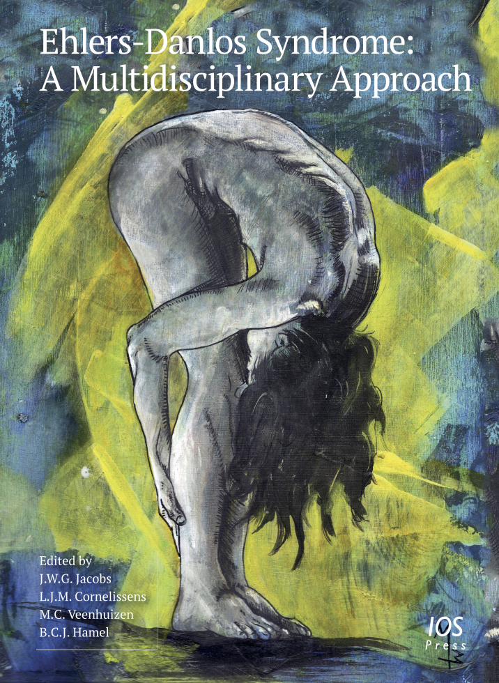
Cover by MANDEM: “Hypermobility: Small scale Work #3”. MANDEM is a family of artists, one of whom has hypermobile
Ehlers-Danlos syndrome (hEDS). Their work can be seen at http://MANDEMart.com/hypermobility
Keywords Ehlers-Danlos, EDS, heritable connective tissue disorders, connective tissue, collagen, lax joints, hypermobility, lax
skin, skin fragility; skin hyperextensibility, Beighton score, pain, fatigue, arterial rupture, aneurism, genetic testing, diagnostics,
nosology, classification, therapy
Publisher
IOS Press BV
Nieuwe Hemweg 6B
1013 BG Amsterdam
Netherlands
fax: +31 20 687 0019
e-mail: [email protected]
For book sales in the USA and Canada:
IOS Press, Inc.
6751 Tepper Drive
Clifton, VA 20124
USA
Tel.: +1 703 830 6300
Fax: +1 703 830 2300
LEGAL NOTICE
The publisher is not responsible for the use which might be made of the following information.
PRINTED IN THE NETHERLANDS

Preface
We are a team of Dutch authors and editors, two of whom are Ehlers-Danlos syndrome (EDS)
patients. In 2005, along with several co-authors and with financial support from the Dutch
EDS support group “VED”, we published a book entitled: “Ehlers-Danlos syndroom. Een
multidisciplinaire benadering” (Ehlers-Danlos syndrome. A multidisciplinary approach). The
aim of this multidisciplinary, practical book was to create more awareness for and increase the
knowledge of hypermobility syndromes, especially EDS and benign joint hypermobility
syndrome, among health professionals, including medical specialists, and to be a guide for
patients. Hypermobility syndromes often are characterised by extra-articular signs and
symptoms that often go unrecognised or are only recognized at a late stage. The ultimate goal
was to improve care for patients with hypermobility syndromes, and this proved to be a great
success.
Realising that this book indeed filled a gap in the health care system, some years ago the idea
arose to publish an international multidisciplinary book on hypermobility syndromes with the
help of international authors, with the same aims as those for the Dutch book. This has proven
not to be a simple endeavour, but we think we eventually have succeeded. We hope it will
meet our aims, described above, and your hopes and expectations. To make this book easily
accessible to patients and health care workers, we decided to publish it as a freely available e-
book. Financial support was given by many organisations (see the acknowledgements on the
next page), for which we are very grateful.
JWG Jacobs, editor-in-chief, Associate Professor in Rheumatology
BCJ Hamel, editor, Professor Emeritus of Clinical Genetics
MC Veenhuizen, editor, Nurse Tutor, hypermobile EDS (hEDS) patient
LJM Cornelissens, editor, Psychotherapist and Medical Ethicist, hEDS patient
v

Acknowledgements
Financial support Unrestricted grants from the Dutch federation of patients with Ehlers-
Danlos syndrome VED, Lewis Seating Systems (a Dutch manufacturer of seating orthoses,
especially for wheel chairs), and University Medical Center Utrecht, the Netherlands.
�
vi
Table of contents
Preface ........................................................................................................................................ v
Chapter 1. History and introduction ........................................................................................... 1
Chapter 2. Classification and nosology of Ehlers-Danlos syndrome ......................................... 5
Chapter 3. Genetics and testing of Ehlers-Danlos syndrome and of differential diagnostic
diseases ..................................................................................................................................... 33
Chapter 4. Classical Ehlers-Danlos syndrome ......................................................................... 47
Chapter 5. Generalised joint hypermobility and joint hypermobility syndromes: the clinical
perspective ................................................................................................................................ 61
Chapter 6. Vascular Ehlers-Danlos syndrome ......................................................................... 79
Chapter 7. Kyphoscoliotic, arthrochalasia and dermatosparaxis Ehlers-Danlos syndrome ..... 97
Chapter 8. Orthopaedic issues in Ehlers-Danlos syndrome ................................................... 127
Chapter 9. The role of plastic surgery in Ehlers-Danlos syndrome ....................................... 139
Chapter 10. Gastrointestinal complications of Ehlers-Danlos syndromes and
hypermobility spectrum disorders .......................................................................................... 145
Chapter 11. Bleeding tendency in Ehlers-Danlos syndrome .................................................. 167
Chapter 12. Cardiac abnormalities and complications in Ehlers-Danlos syndrome .............. 177
Chapter 13. Neurological complications of Ehlers-Danlos syndromes and hypermobility
spectrum disorders .................................................................................................................. 187
Chapter 14. Ehlers-Danlos syndrome, generalised joint hypermobility and the masticatory
system ..................................................................................................................................... 209
Chapter 15. Ehlers-Danlos and hypermobility syndromes and the eye ................................. 225
Chapter 16. Ehlers-Danlos syndrome in urology: nobody is perfect ..................................... 235
Chapter 17. Gynaecological and obstetrical problems and management dilemmas in
women with Ehlers-Danlos syndrome ................................................................................... 241
Chapter 18. The child with Ehlers-Danlos syndrome ............................................................ 255
vii

Chapter 19. Ehlers-Danlos syndrome, generalised hypermobility and ethics: reflections
from the ethics of care ............................................................................................................ 271
Chapter 20. Stretched beyond the limit: well-being and functioning in patients with
Ehlers-Danlos syndrome and other hypermobility syndromes .............................................. 281
Chapter 21. Causes and treatment of chronic pain associated with Ehlers-Danlos
syndrome ................................................................................................................................ 297
Chapter 22. Clinical profiling and tailored non-pharmacological treatment in
hypermobility spectrum disorders/hypermobile Ehlers-Danlos syndrome ............................ 311
Chapter 23. Nursing care for patients with Ehlers-Danlos syndrome .................................... 331
Chapter 24. The role of occupational therapy in Ehlers-Danlos syndrome ........................... 339
Addendum. Considerations, precautions and measures in case of surgery in patients with
Ehlers-Danlos syndrome ........................................................................................................ 349
Index ....................................................................................................................................... 351
viii

Chapter 1. History and introduction
Generalised hypermobility has been known since ancient history; for instance Peruvian
ceramic figures of 1200–200 B.C. visualize hypermobile individuals. The clinical picture of
Ehlers-Danlos syndrome (EDS) is said to be first described by Hippocrates in 400 B.C. The
Dutch surgeon Job Janszoon van Meek'ren in 1657 and the Russian dermatologist
Tschernogobow in 1892 described the clinical picture of EDS, but EDS was named after the
Danish dermatologist Edvard Lauritz Ehlers (1863–1937), who recognized the condition as a
distinct entity in 1901 and after Henri-Alexandre Danlos, a French physician (1844–1912),
who suggested in 1908 that skin extensibility and fragility are cardinal features of EDS.1
Since about the first half of last century, several case reports of EDS and other hypermobility
syndromes were published. In the earliest publications, several signs and symptoms have been
described as associated with EDS, which later proved to be not to be related with EDS, such
as mental retardation, bleeding disorders and anatomical cardiac anomalies.
In the second half of last century, also classifications were formulated,2,3
based on patterns of
inheritance and clinical features. With the development of genetic techniques and more
systematic research, new genetic defects and subsequently several new types of EDS and
other hypermobility syndromes have been recognised and very likely more will be
distinguished in the future, necessitating continued updating of existing classification systems.
This will probably take place during international congresses, such as that in New York City
May 2016. Laboratory testing for most of the hypermobility syndromes has become possible.
However, there is no curative treatment for these syndromes, though in vascular EDS
celiprolol and in Marfan syndrome losartan have been proven to diminish the risk of vascular
complications, likely due to mechanisms beyond that of lowering blood pressure.4,5
In contrast with these developments and the relatively frequent occurrence of hypermobility
syndromes is the relative lack of awareness and recognition of these syndromes in daily
clinical practice with underdiagnosis as result. This could be due to the wide spectrum of
possible signs and symptoms. Although - as said - therapeutic options are meagre, recognition
and diagnosis of a hypermobility syndrome in a patient are beneficial, ending an often year-
long quest for the cause of signs and symptoms and guiding management. When the quest
ends, finding a way of coping for patient and because of the genetic nature of these syndromes
also for his/her family can start. For hypermobility syndromes with the risk of life-threatening
complications, recognition and diagnosis might even be life-saving.
The aim of this book is creating more awareness for hypermobility syndromes, especially
EDS, among health professionals, including medical specialists, and being a guide for
patients, in order to improve the care for patients with hypermobility syndromes. This book is
not meant as a reference book for basic research or the newest developments; it is meant for
usage in daily clinical practice, with a practical, clinical scope, as much as possible in a
reading level of text, health care worker or educated patient can understand, without
compromise to the scientific content. Bearing our purpose in mind, we believe this book
should be widely available, which we have attempted to achieve by publishing it as an E-
book.
Patients with hypermobility syndromes, having a wide spectrum of signs, symptoms,
manifestations and complications, often get to deal with a wide array of medical specialists
and other health care providers, who each have a special interest in and knowledge about a
specific organ or physical system. The description of these symptoms by only one person with
one specialism is bound to be a bit one-dimensional (see figure).
1

A generalist with extensive knowledge of virtually all organ systems possibly affected or
influenced by hypermobility syndromes does not exist. Next best for an individual patient
would be a clinician acting as case manager. We would welcome development of
multidisciplinary outpatient clinics for patients with hypermobility syndromes.
Both a case manager and a multidisciplinary outpatient clinic should take the whole spectrum
of possible manifestations into account. This is the reason we chose for our book the scope
and vision from different specialisms on the subject, trying to describe the whole spectrum of
manifestations, as much as possible at the persons level, rather than at the organ level. The
drawback of this choice is some heterogeneity in the medical level and length of chapters, and
some overlap between chapters, which we accepted. The outline of the book is as follows:
first there are chapters on classifications and genetics, then chapters on individual types, organ
(system) manifestations and complications, and at the end ethics and therapeutic strategies,
with an appendix on (precautions at) surgery. With the efforts of two of our editors, who
suffer from EDS, we tried to incorporate also the patient’s perspective.
In May 2016 in New York an international symposium of the Ehlers-Danlos Society took
place, with propositions of changes regarding classification and nosology.6
We incorporated
these changes into this book. Given the fast developments in research, particularly in genetic
testing through the widespread introduction of next generation sequencing, it might be that
criteria sets, classifications and genetic tests, will be further updated in the near future.
However, we feel that for the purpose of the book this is not a major obstacle, and can update
the text, given this is an e-publication. So, if our book leads to earlier recognition of patients
with hypermobility syndromes and better management, and functions as a practical guide for
patients and their families and friends in daily practice, we feel we have met our aim. We
hope you will enjoy reading it.
JWG Jacobs, editor-in-chief
BCJ Hamel, editor
MC Veenhuizen, editor
LJM Cornelissens, editor
Chapter 12

References
1. Parapia LA, Jackson C. Ehlers-Danlos syndrome--a historical review. Br J Haematol
2008;141:32-5.
2. Beighton P, De Paepe A, Danks D, et al. International Nosology of Heritable Disorders
of Connective Tissue, Berlin, 1986. Am J Med Genet 1988;29:581-94.
3. Beighton P, De Paepe A, Steinmann B, Tsipouras P, Wenstrup RJ. Ehlers-Danlos
syndromes: revised nosology, Villefranche, 1997. Ehlers- Danlos National Foundation
(USA) and Ehlers-Danlos Support Group (UK). Am J Med Genet 1998;77:31-7.
4. Ong KT, Perdu J, De Backer J, et al. Effect of celiprolol on prevention of cardiovascular
events in vascular Ehlers-Danlos syndrome: a prospective randomised, open, blinded-
endpoints trial. Lancet 2010;376:1476-84.
5. Franken R, den Hartog AW, Radonic T, et al. Beneficial outcome of losartan therapy
depends on type of FBN1 mutation in Marfan syndrome. Circ Cardiovasc Genet
2015;8:383-8.
6. Malfait F, Francomano C, Byers P, et al. The 2017 international classification of the
Ehlers-Danlos syndromes. Am J Med Genet C Semin Med Genet 2017;175:8-26.
History and introduction 3

This page intentionally left blank

Chapter 2. Classification and nosology of Ehlers-Danlos syndrome
BCJ Hamel
Ben C.J. Hamel, MD, PhD
Clinical geneticist
Professor emeritus of Clinical Genetics
Department of Human Genetics
Radboud university medical center
Nijmegen, The Netherlands
Telephone +31243239426
E-mail [email protected]; [email protected]
5
1. Introduction
Ehlers-Danlos syndrome (EDS) comprises a clinically and genetically heterogeneous group of
heritable connective tissue disorders (HCTD), mainly characterized by a variable degree of
generalized joint hypermobility, skin hyperextensibility, easy bruising and skin fragility.
When in 1956 the first edition of McKusick’s book entitled “Heritable disorders of connective
tissue” was published, less than 100 papers had been devoted to EDS, mainly case reports.1 At
about the same time it became clear that EDS basically was an autosomal dominant, clinically
and probably also genetically heterogeneous disorder. In his classical monograph on EDS,
published in 1970, Beighton described 5 EDS types: I = gravis (severe), II = mitis (mild), III =
hypermobility, IV = ecchymotic, and V = X-linked.2 The Berlin classification listed 11 EDS
types.3 Revision became necessary because of new biochemical, molecular and clinical data,
leading to the Villefranche nosology of 1997, in which 6 EDS types were recognized.4 New
clinical and molecular data required another revision, which was initiated during the Ehlers-
Danlos Society International Symposium in New York, May 2016, the results of which have
been published in the March 2017 issue of the American Journal of Medical Genetics Part C,
Seminars in Medical Genetics. The most striking changes were:
- incorporating EDS types which were published since the Villefranche nosology, leading
to a total number of 13 types.5
- deciding - not unexpectedly though - that EDS hypermobility type and benign joint
hypermobility syndrome (BJHS; also called joint hypermobility syndrome or
hypermobility syndrome) are in fact part of one and the same clinical spectrum ranging
from apparently symptomatic generalized joint hypermobility to the most disabled
individuals fitting the new diagnostic criteria. These new criteria are more strict than the
Villefranche criteria and the Brighton criteria for BJHS in order to define a homogeneous
phenotype for management and scientific purposes. Its name is hypermobile EDS.
It always has been, and still is, a challenge to classify individual patients in one of the existing
EDS types. Often this is not possible. This is, among other things, due to:
- the clinical overlap between many of these EDS types
- absence of a pathogenic variant in any of the known EDS associated genes in an
important proportion of EDS patients.
- the presence of associated features which do not fit into one of the existing types.
- the absence of a laboratory test for hypermobile EDS.
EDS is not a rare disorder; the prevalence is estimated to be about 1:5000. The hypermobile
type - by far the most common - and the classical type comprise more than 90% of all cases.6
2. Classification and nosology
The New York classification is based on clinical, biochemical and molecular data.5 Table 2-1
shows the New York nosology alongside the previous nosologies with inheritance patterns,
genetic bases and proteins, while in table 2-2 EDS types are grouped according to underlying
genetic and pathogenetic mechanisms; OMIM numbers (see glossary) are added.
In clinical practice, the clinical manifestations guide the choice for further investigations. The
major clinical manifestations of EDS need some clarification, however.
Skin hyperextensibility should be tested at specific sites, e.g. the volar side of the non-
dominant forearm or the dorsum of the hand by pulling up the skin until resistance is felt. In
contrast to cutis laxa, a group of clinically and genetically heterogeneous disorders
characterised by redundant, sagging and inelastic skin, with or without joint hypermobility, in
EDS the skin snaps back after release.7 The upper limit of normal for the forearm and dorsum
of the hand is about 1 ½ cm.8 In young children it is difficult to assess hyperextensibility due
Chapter 26

to the abundance of subcutaneous fat. Skin hyperextensibility can also be assessed at the
dorsal aspect of the elbow in 900 flection, where the upper limit of normal is 3 cm.
Joint hypermobility is scored using the Beighton mobility scale (table 2-3). In the New York
nosology, a score of 5/9 or more defines generalized hypermobility in both sexes, though it is
known that joint mobility depends, apart from age, family and ethnic background, also on
gender. Since laxity decreases with age, patients with a Beighton score <5/9 may be
considered positive based on their historical observations (five-point questionnaire = 5PQ; see
footnote with table 2-4). For the diagnosis of hypermobile EDS different age and sex specific
cut-off points were proposed (see table 2-4). In children under the age of about 5 years, the
Beighton scale is less useful. Not infrequently, the Bulbena mobility score is also used (table
2-4), in which a score of 5/10 or more defines generalized hypermobility in females and 4/10
or more in males. It is more easily applicable in children. Generalised hypermobility is not
rare: 5-10% of – mainly female – secondary school age Caucasian children is hypermobile.8,9
Easy bruising is seen as spontaneous ecchymoses, frequently recurring in the same bodily
regions, of which long-term signs are often visible as brownish discoloration (haemosiderin),
in particular on knees and shins. If it is the predominant presenting sign, child abuse and
bleeding disorders need to be considered first.
Tissue fragility is manifested in the skin as easy bruising and impaired wound healing with
dystrophic scars, which are usually found over pressure points like forehead, chin, elbow,
knee and shin and which may have a wide and papyraceous appearance. Internal organs like
arteries, lungs, intestines, liver, spleen and uterus may also show fragility, predominantly in
the vascular type.
Some features are regularly observed, but are not criteria of generalised hypermobility
syndromes. One example is the ineffectiveness of local anaesthetics in hypermobile EDS.10
In table 2-5 the major and minor diagnostic criteria are shown, minimal criteria for diagnosis
and how to verify the diagnosis.
A major diagnostic criterion predominantly has high diagnostic specificity, which means that
it is present in the vast majority of the affected individuals and/or it is characteristic for the
disorder and allows differentiation from other EDS types and/or other HCTD. A minor
criterion is a sign of lesser diagnostic specificity, but its presence supports the diagnosis.
However, in the absence of major criteria, minor criteria are not sufficient for a given
diagnosis. Because of the vast genetic heterogeneity and phenotypic variability of the EDS
types and the clinical overlap between many of these, the definite diagnosis relies for all
types, except the hypermobile type, on molecular confirmation.
In older publications features like facial dysmorphisms and mental retardation/intellectual
disability were attributed to EDS, whereas nowadays these features are not any longer
considered characteristics of EDS, except for facial dysmorphisms in vascular,
dermatosparaxis, spondylodysplastic and musculocontractural EDS and intellectual disability
in spondylodysplastic EDS (see table 2-5). Explanation for this could be that other syndromes
associated with these features were erroneously diagnosed as EDS because of overlapping
features with EDS. Another explanation is that these other features are not rare and
consequently are found associated with EDS in a low percentage of cases.
The classical EDS has all the skin and joint characteristics of EDS, though in variable range
of severity. Minimal diagnostic criteria are the presence of skin hyperextensibility and
atrophic scars, plus either generalized joint hypermobility and/or 3 minor criteria (see table 2-
5).11
A recent paper reviews 62 molecularly confirmed cases from Italy.12
It is inherited in an
autosomal dominant fashion (see glossary), implying that each child (be it a boy or a girl) of
an affected parent (be it father or mother) has a chance of 50% (= ½) of also having the
classical type of which the expression (see glossary) cannot be predicted. However, sporadic
Classification and nosology of Ehlers-Danlos syndrome 7

cases (i.e. without an affected parent) do occur due to a spontaneous mutation. Genetically, it
is heterogeneous since in over 90% of patients, who fulfil all major criteria, a defect can be
found in (products of) one of the two genes that are up to now known to be involved, COL5A1
and COL5A2. An EDS type resembling the classical type but clinically characterized by a
propensity to arterial rupture and molecularly by a specific mutation in COL1A1 (c.934C>T;
p.Arg312Cys) is regarded as a variant type of classical EDS.13,14
Once the causative mutation
has been found in a proband (see glossary), mutation analysis in relatives of the proband is
possible. The major differential diagnosis of the classical type, at least in a sporadic case, is
the classical-like EDS. Also, in mild cases of the classical type (partial expression),
differentiation from the hypermobile type might be difficult, if not impossible.
Classical-like EDS resembles the classical type, however with normal wound healing and scar
formation.14,15
Minimal diagnostic criteria are the presence of all 3 major criteria, i.e. skin
hyperextensibility, generalized joint hypermobility and easy bruisability and a family history
compatible with autosomal recessive inheritance. It is characterised by generalized
hypermobility, with a remarkable laxity of finger joints. In contrast with the classical type, its
inheritance is autosomal recessive, so most cases are sporadic and some occur in sibships. It is
due to tenascin-X deficiency. In serum, tenascin-X is completely absent and mutation analysis
(TNX-B) is performed in blood.
The cardiac-valvular EDS is rare.14,16
Apart from typical EDS features, it is associated with
severe aortic and/or mitral valve insufficiency, necessitating valve replacement at relatively
young age. Minimal diagnostic criteria are the presence of severe progressive cardiac-valvular
problems, family history compatible with AR inheritance plus either one other major criterion
and/or at least 2 minor (see table 2-4). The inheritance is autosomal recessive. It is due to
homozygous or compound heterozygous COL1A2 null mutations (see glossary).
The vascular EDS is the most severe form of EDS.17
Minimal diagnostic criteria are the
presence of of a family history of vascular EDS, arterial rupture/dissection <40 years,
unexplained sigmoid colon rupture or spontaneous pneumothorax in the presence of other
features consistent with vascular EDS. These and a combination of minor criteria warrant
verifying diagnostic tests, i.e. DNA analysis (see table 2-5). Diagnosis in children is difficult,
particularly in the absence of a family history. The vascular type is inherited in an autosomal
dominant fashion. Arterial rupture is the most common cause of sudden death and has its peak
incidence in the 3rd or 4
th decade. Acute abdominal and flank pain is a common presentation
of an arterial or intestinal rupture and needs urgent investigation and treatment. Recently,
Frank et al. showed that the type of COL3A1 mutation is associated with the phenotype and
severity: patients with glycine substitutions and splice-site and in-frame insertions–deletions
have a more severe phenotype, including digestive events, compared to e.g. mutations leading
to non-glycine missense variants or haplo-insufficiency, due to a null allele. The latter may
delay onset of complications by almost 2 decades.17,18
For women with the vascular type, pregnancy and delivery pose specific risks, which warrant
pre-conceptional counselling with an experienced obstetrician and clinical geneticist.19
There is considerable clinical overlap between the vascular type and Loeys-Dietz syndrome
type 1 and 2 (OMIM 609192 and 610168 respectively; see chapters 5 and 6 for details and 3
more types), which are due to TGFBR1 (type 1) and TGFBR2 (type 2) mutations.20
Also other
aortic aneurysm syndromes, such as Marfan syndrome, Thoracic Aortic Aneurysm and
Dissection (TAAD), annulo-aortic ectasia should be included in the differential diagnosis.21
As said above, when extensive bruising is the initial presentation and the only sign/symptom,
bleeding disorders and child abuse have to be considered.
Chapter 28

The hypermobile EDS, incorporating BJHS, is dominated by generalized joint hypermobility
and its possible sequelae, in particular chronic pain, which can be severe and invalidating, and
possibly early osteoarthritis.22
As said, the new diagnostic criteria are more strict than those of
the Villefranche criteria and the Brighton criteria (see table 2-5 and chapter 5). The clinical
diagnosis of hypermobile EDS needs the simultaneous presence of 3 criteria: criterion 1 =
generalized joint hypermobility, criterion 2 = 2 or more of the features A, B and C (A =
systemic manifestations of a more generalized connective tissue disorder; B = positive family
history; C = musculoskeletal complications) and criterion 3 = absence of unusual skin
fragility and exclusion of alternative diagnosis (for details see table 2-5 and chapter 5).
Recently, also cardiovascular dysautonomia (mainly postural tachycardia syndrome = POTS),
functional gastro-intestinal manifestations, sleep disturbance, fatigue, depression and anxiety
disorders have been attributed to hypermobile EDS, but these are at the moment not
sufficiently sensitive nor specific. Basically, there is no confirmative laboratory test for the
hypermobility type, meaning that it is a pure clinical diagnosis. Recently, Syx et al. reported
linkage to chromosome 8p22-8p21.1 in a 3 generation Belgian family with EDS
hypermobility type, whereby whole exome sequencing revealed a possibly involved gene.23
Up to recently, BJHS was considered a separate entity with its own diagnostic criteria.24
It
was already argued earlier that the hypermobility type and BJHS are in fact one and the same
disorder with variable expression. Arguments put forward for this were among others the fact
that haplo-insufficiency of TNX-B, assessed as about half of the normal activity of tenascin-X
in blood, and/or heterozygosity for a pathogenic TNX-B mutation, is found both in cases with
EDS hypermobility type and cases in whom BJHS is the more likely diagnosis.25
Also, a
changing phenotype from one diagnosis into the other in one individual and in some pedigrees
the occurrence of both diagnoses argued for this statement.26
Castori et al. proposed a frame for the classification of joint hypermobility and related
syndromes, which is worth reading.27
The arthrochalasia EDS is also rare, but diagnosable at birth.14
Minimal criteria suggestive
for arthrochalasia EDS are congenital bilateral hip dislocation plus either skin
hyperextensibility or severe generalized joint hypermobility with multiple
dislocations/subluxations and at least 2 other minor criteria It is inherited in an autosomal
dominant fashion. It is due to specific mutations in COL1A1or COL1A2. Larsen syndrome,
which also features congenital luxations, should be in the differential diagnosis.
The dermatosparaxis EDS derives its name from a similar phenotype and biochemical defect
in cattle, sheep, and other animals.14
It is the EDS type which has the closest resemblance to
cutis laxa. However, in cutis laxa there is neither fragility nor bruising. Its mode of
inheritance is autosomal recessive. It is one of the rarest of all types and since only very few
cases have been described, possibly this type is characterised by other - as yet unrecognised -
features. Recently, Van Damme et al. expanded the phenotype and suggested new diagnostic
criteria.28
The New York nosology requires for its diagnosis extreme skin fragility with
congenital or postnatal skin tears AND characteristic craniofacial features plus either 1 other
major and/or 3 minor criteria (for details see table 2-5). It is due to mutations in ADAMTS2.
The mode of inheritance is autosomal recessive (see glossary), whereby both parents of a
patient are healthy carriers and the recurrence risk for siblings is 25% (= ¼).
The kyphoscoliotic EDS is a rare but severe form of EDS, manifesting itself often at or shortly
after birth.14,29
The presence of congenital muscle hypotonia AND congenital or early onset
kyphoscoliosis plus either generalized joint hypermobility and/or 3 minor criteria (either
Classification and nosology of Ehlers-Danlos syndrome 9

general or PLOD1 and FKBP14 gene-specific) warrants laboratory testing. Kyphoscoliotic
EDS is genetically heterogeneous and can be caused by mutations in PLOD1 and FKBP14.
Laboratory tests should start with measurement of the urinary lysyl and hydroxy-lysyl
pyridinoline ratio. An increased ratio has a very high degree of sensitivity and specificity for
PLOD1 mutations, but not for FKBP14 mutations. For molecular tests: see table 2-5. The
mode of inheritance is autosomal recessive (see glossary), whereby both parents of a patient
are healthy carriers and the recurrence risk for siblings is 25% (= ¼). Because of severe
hypotonia, patients very often undergo a full scale neuromuscular work-up, including a
muscle biopsy before the diagnosis is established. The differential diagnosis comprises all
other causes of severe hypotonia, including neonatal Marfan syndrome.
The even rarer brittle cornea syndrome (formerly EDS VIB) resembles the kyphoscoliotic
type, but is generally milder.14,30
Minimal diagnostic criteria are thin cornea with or without
rupture plus either at least one other major criterion and/or 3 minor criteria (see table 2-5). It
shows a normal urinary lysyl and hydroxy-lysyl pyridinoline ratio, and is characterised by
mutations in the genes ZNF469 or PRDM5.
Spondylodysplastic EDS is genetically heterogeneous and is due to bi-allelic mutations in
either B4GALT7 (former name progeroid type 131
), B3GALT6 (former name progeroid type
232
) or SLC39A13 (former name spondylocheirodysplastic type33
).14
There is considerable
overlap with kyphoscoliotic EDS. Minimal diagnostic criteria are short stature AND muscle
hypotonia plus characteristic radiographic abnormalities and at least 3 other minor criteria
(general or gene-specific; see table 2-5).
The urinary lysysl and hydroxylysyl pyridinoline ratio is moderately increased (to
approximately 1 compared to normal values of ~ 0.2) with HLPC for SLC39A13 mutations.
The musculocontractural EDS (formerly EDS VIB) is due to bi-allelic mutations in either
CHST14 gene (type 1) or more rarely DSE gene (type 2) and has also considerable clinical
overlap with the kyphoscoliotic type.14,34
Minimal diagnostic criteria are at birth or in early
childhood congenital multiple contractures AND characteristic craniofacial features, while in
adolescence and adulthood these are congenital multiple contractures AND characteristic
cutaneous features.
Myopathic EDS is caused by heterozygous or bi-allelic mutations in COL12A1.14,35
Minimal
diagnostic criteria are congenital muscle hypotonia and/or muscle atrophy that improves with
age plus either one other major criterion and/or three minor criteria. The phenotype highly
overlaps with collagen VI type related myopathies, i.e. Bethlem myopathy and Ullrich
Congenital Muscular Dystrophy.
The periodontal EDS has some overlap with the classical type, but has progressive and
aggressive periodontitis as a distinguishing feature.14
Minimal diagnostic criteria are severe,
intractable periodontitis of early onset (childhood or adolescence) OR lack of attached gingiva
plus at least 2 other major criteria and one minor criterion. Recently, it was found that gain-
of-function mutations in the C1R gene or the C1S gene, encoding serine proteinases, cause
periodontal EDS.36
The former EDS type V (X-linked) has been described in only 2 families and is not any longer
accepted as belonging to EDS spectrum, the same holds true for the former fibronectin
deficient type X, familial articular hypermobility EDS XI and Filamin A related EDS with
heterotopia. The former type IX is an X-linked cutis laxa disorder and is renamed occipital
Chapter 210

horn syndrome; it is due to mutations in the gene ATP7A, the same gene as is mutated in
Menkes syndrome (disorder of copper metabolism)
For further reading, the excellent review by Byers and Murray is highly recommended,
together with the more recent paper by Malfait et al.14,37
In fact, the whole March 2017 issue
of the American Journal of Medical Genetics Part C, Seminars in Medical Genetics provides a
very good update not only about EDS nosology and diagnostic criteria, but also about
management aspects of the various types of EDS.
Joint hypermobility is a symptom of large variety of syndromes.
A search in the London Medical Databases (suite.face2gene.com/lmd-library-london-medical-
database-dysmorphology/) with “joint laxity or multiple joint dislocations” as key words gives
more than 290 hits. Among these, one finds - not surprisingly - other heritable connective
tissue disorders like cutis laxa, osteogenesis imperfecta, Stickler syndrome (see chapter 5),
Loeys-Dietz syndrome and Marfan syndrome, but also skeletal dysplasias, inborn errors of
metabolism, neuromuscular disorders, chromosomal abnormalities and syndromes like Larsen
syndrome, Fragile X syndrome and Langer-Giedion syndrome.
3. How to achieve the diagnosis Ehlers-Danlos syndrome, including correct typing?
Like always in clinical practice, the results of history taking, including a family history, and
physical examination are the basis for planning additional investigations and finally reaching
a diagnosis. As mentioned above, additional investigations are often biochemical as a first
screen, followed by targeted DNA analysis. However, with the introduction of new DNA
technologies, like next generation sequencing, rapid search for the disease causing mutation
by molecular analysis of (all) known EDS genes (“the EDS panel”) at once has become
possible. This is already standard routine diagnostic practice in some laboratories and will
become routine in all in the near future (see chapter 3). Copy number variation analysis for
large deletions and duplications has also a place in the molecular analysis in cases where NGS
did not reveal a mutation in AD types and only one mutation in AR types.
As history taking and physical examination in relation to EDS are very important, they will be
discussed below.
Good history taking starts with identifying the exact symptoms and complaints, which
compelled the patient to see a physician: when and how did they start and evolve, how were
they treated (what were the results, what was advised/prescribed and by whom?). Specific
questions should elucidate the presence or absence of:
- Hypermobility and/or (sub)luxations. If (sub)luxation occurred: which joint(s) was/were
involved, how often did it occur, was it spontaneous (also the first time) and painful? Was
it seen/treated by doctors? If necessary also the Five-Point Questionnaire (5PQ; see
footnote in table 2-5). Contractures? Congenital hip dislocation? (see chapters 5, 8, 22
and 24).38,39
- Painful joints.40
If so: which ones, when, under which circumstances, exercise related,
warm and swollen, if so, for how long? Use of analgesics? Sprains? What are the major
limitations in daily life? (see chapters 20, 21, 22 and 24)
- Temporomandibular joint problems (see chapter 14).41
- Problems with bursae/tendons.
- (Spontaneous) fractures.
- Skin fragility and abnormal wound healing with wide atrophic scars (see chapter 9).
- Surgery, e.g. for inguinal hernia. If so: complications?
- Easy bruising and/or abnormal menstrual bleeding (see chapter 11).
Classification and nosology of Ehlers-Danlos syndrome 11

- Abnormal exercise tolerance and/or fatigue.42
Sports performed? Type of work: blue or
white collar?
- Pneumothorax?
- Cardiac problems? Cardiovascular autonomic dysfunction?43
(see chapter 12)?
- Genito-urinary tract problems, e.g. uterine prolapse, voiding dysfunction (see chapter 16).
- Gastro-intestinal tract problems, e.g. constipation, diverticula, rectal prolapse (see chapter
10).44
- For female patients with children: pregnancy and delivery problems (see chapter 17).19
- Rupture of internal organs (arteries, lungs, intestines, spleen, uterus).
- Psychiatric problems, like anxiety, depression, ADHD?45
- Neurological problems?46,47
Headache? Migraine? (see chapter 13)
- Eye problems, e.g. refractive errors, abnormal vision (see chapter 15).
- Hearing?
- Growth?
- Motor and cognitive development?
- Miscellaneous: Gingivitis? Varicose veins? Abnormal effect of local anaesthesia?
The family history includes drawing a three generation pedigree with specific enquiry
regarding hypermobility, easy bruising, abnormal scarring, arterial dissections and organ
ruptures.
The physical examination is focused on signs relevant for connective tissue disorders:
- Build and biometry: height, weight, span and others when indicated. Marfanoid?
- Facial features: among others, Gorlin sign (ability to touch the tip of the nose with the tip
of tongue)? High palate? Absence of subcutaneous fat? Prominent eyes? Thin, “pinched”
nose? Normal earlobes? Epicanthic folds? Low-set ears? Midfacial hypoplasia?
Micrognathia? Down-slanting palpebral fissures? Gingival recession?
- Teeth: dental crowding? Discoloured? Dysplastic? Periodontitis?
- Thorax: deformity?
- Back: (kypho)scoliosis?
- Extremities: Beighton score,; Bulbena score; arachnodactyly (wrist and thumb sign)?
Brachydactyly? Clinodactyly? Contractures? Flat feet? Joints? Muscle strength? Edema?
Hallux valgus?
- Skin: extensibility? Texture? Thickness and venous pattern? Striae distensae? Varicose
veins? Piezogenic papules? Molluscoid pseudotumors? Spheroids? Scars? Herniae?
- Eyes: Blue sclerae? Microcornea? Strabismus? Clouded cornea? Glaucoma?
Scleral/ocular fragility? Keratoconus?
- Neurological examination: muscle weakness? Reduction in vibration sense? Reduction of
tendon reflexes?
Then a differential diagnosis will be established, on which basis additional investigations,
such as biochemical and/or DNA analysis in blood and/or cultured fibroblasts, derived from a
skin biopsy, are planned, if clinically relevant and available for the suspected type.
Morphological examination of a skin biopsy is of limited value, except in some types,
particularly in dermatosparaxis EDS. On indication, the patient will be referred to an
ophthalmologist, cardiologist, orthopaedic surgeon, neurologist and/or others.
DNA analyses are available as diagnostic services in most of the developed countries. As
already indicated, there is no DNA test available for hypermobile EDS. If there is any reason
to believe the phenotype could be the classical-like, tenascin-X deficient type, then there is an
indication to perform tenascin-X analysis in serum. If there is suspicion of vascular EDS, the
Chapter 212

threshold to do DNA analysis should be very low, because of the consequences of that
diagnosis in terms of management and genetic counselling. For some of the other EDS types
the same holds true, because of their rareness, overlapping features with other EDS types
and/or different modes of inheritance. In fact, for definite diagnosis molecular confirmation is
needed for all types, except the hypermobile type.
4. Genetic counselling (see also chapter 19)
Since all the disorders which have been discussed have a genetic background, genetic
counselling is an indispensable part of the management of patients and their families. During
genetic counselling, information will be given about the mode of inheritance, recurrence risk,
variability and penetrance of the disorder, the possibilities of prenatal diagnosis and diagnosis
in relatives at risk and management. Prenatal diagnosis and diagnosis in relatives at risk is
only possible if the causative DNA defect is known. A social worker should be available to
assist whenever a need is perceived or requested. When there is a patient/parent support
group, patients/parents should be informed. In case of a proband (see glossary) of non-EDS
parents, it is essential to differentiate between an autosomal dominant (e.g. classical type) and
an autosomal recessive type (e.g. classical-like, tenascin-deficient type): in the classical type
the recurrence risk for a next child of these non-EDS parents is low (less than 1%) and for a
child of the affected patient high (50%), while in the tenascin-deficient type the recurrence
risk for a next child is high (25%) and for a child of the affected patient generally low (1% or
less).
5. Areas of uncertainties/research agenda
- An international consensus on methods for measuring joint hypermobility and on more
accurate tools for classifying patients with hypermobile EDS is needed.
- Controlled studies are necessary to establish the role of tenascin-X in hypermobile EDS.
- Controlled studies are needed to establish whether hypermobile EDS is a multisystem
disorder, including among others cardiovascular dysautonomia (mainly postural
tachycardia syndrome = POTS), functional gastro-intestinal manifestations, sleep
disturbance, fatigue, depression and anxiety disorders. If that is the case, diagnostic
criteria need to be adjusted.
- Research into the hypothesis: joint mobility is the end result of the contribution of many
genes with each (probably) having a small effect, and exogeneous factors (multifactorial).
The degree of joint mobility in a population shows a continuous distribution, comparable
to body length and IQ. At the one extreme there is severe stiffness, and at the other severe
hypermobility. At the extremes it is likely to find - rare - monogenic forms, but in
between the extremes chances are high to find the more common forms of multifactorial
origin. This could be the reason, that so far little has come out of genetic studies in EDS
hypermobility type.
- Is classical-like, tenascin deficient EDS more frequent than thought up till now?
- Education of the general public and health care providers is needed to increase awareness
of EDS with early diagnosis and proper management as a consequence. Particularly, it is
important to understand that hypermobile EDS and benign joint hypermobility syndrome
are one and the same disorder.
- International registries need to be established, particularly for the more rare types, in
order to increase knowledge regarding the natural history, the phenotype and
management.
- Provide evidence that management of EDS patients needs to be provided in
multidisciplinary teams in expertise centres.
Classification and nosology of Ehlers-Danlos syndrome 13

6. Summary
EDS comprises a group of heritable connective tissue disorders which has as cardinal features
varying degrees of skin hyperextensibility, joint hypermobility, easy bruising and skin
fragility. The 2017 New York nosology distinguishes 13 types of EDS, which all, except
hypermobile EDS, have a known molecular basis. Hypermobile EDS is recognized as a
common and often disabling disorder, incorporating benign joint hypermobility syndrome.
EDS needs to be differentiated from other connective tissue disorders, in particular Marfan
syndrome, Loeys-Dietz syndrome and cutis laxa. The frequent types of EDS can be diagnosed
after careful history taking and clinical examination, but for definite diagnosis molecular
confirmation is needed in all types. Management for EDS patients preferably is provided by
multidisciplinary teams in expertise centres. After diagnosing EDS genetic counselling is an
essential part of the management of patients and their family.
Chapter 214

Table 2-1 Classification of Ehlers-Danlos syndrome (adapted from5
)
Berlin
classification
(1988)
11 types
Villefranche
classification
(1997)
6 types
International
classification
(2017)
13 types
IP Genetic basis Protein
Type I (gravis)
and type II
(mitis)
Classical type Classical EDS
cEDS
AD Major: COL5A1, COL5A1
Rare*: COL1A1
c.934C>T, p.(Arg312)
Type V collagen
Type I collagen
Classical-like EDS
clEDS
AR TNXB Tenascin XB
Cardiac-valvular EDS
cvEDS
AR COL1A2 (biallelic mutations
that lead to COL1A2 NMD
and absence of pro a2(I)
collagen chains)
Type I collagen
Type
IVA,B,C,D
Vascular type Vascular EDS
vEDS
AD Major: COL3A1
Rare: COL1A1
c.934C>T, p.(Arg312Cys)
c.1720C>T, p.(Arg574Cys)
c.3227C>T, p.(Arg1093Cys)
Type III collagen
Type I collagen
Type III Hypermobility
type
Hypermobile EDS hEDS AD Unknown Unknown
Type VIIA and
B
Arthrochalasia
type
Arthrochalasia EDS
aEDS
AD COL1A1, COL1A2 Type I collagen
Type VIIC Dermatosparaxis
type
Dermatosparaxis EDS
cEDS
AR ADAMTS2 ADAMTS-2
Type VIA Kyphoscoliotic
type
Kyphoscoliotic EDS
kEDS
AR PLOD1
FKBP14
LH1
FKBP22
(continued on next page)
Classification and nosology of E
hlers-Danlos syndrom
e15

Type VIB Brittle cornea syndrome
BCS
AR ZNF469
PRDM5
ZNF469
PRDM5
Spondylodysplastic EDS
spEDS
AR B4GALT7
B3GALT6
SLC39A13
b4GalT7
b3GalT6
ZIP13
Type VIB Musculocontractural
EDS
mcEDS
AR CHST14
DSE
D4ST1
DSE
Myopathic EDS
mEDS
AD/
AR
COL12A1 Type XII collagen
EDS VIII Periodontal EDS
pEDS
AD C1R
C1S
C1r
C1s
EDS V# XL Unknown
EDS IX
= cutis laxa
syndrome
XL ATP7A Cu-transporting
alpha polypeptide
EDS X#
Fibronectin
abnormality
AR Unknown
EDS XI#
familial joint
instability
AD Unknown
IP: inheritance pattern
NMD: nonsense mediated decay
* EDS classical type with (propensity to) arterial rupture.13
#
No longer accepted as separate entities
Chapter 2
16

Table 2-2 EDS grouping according to underlying genetic and pathogenetic mechanisms
(adapted from5
)
Berlin or earlier
name
Villefranche
name
New York name OMIM Gene
GROUP A: Disorders of collagen primary structure and collagen processing
EDS I
EDS II
Classical type Classical EDS (cEDS) 130000 COL5A1
COL5A2
EDS IV Vascular type Vascular EDS (vEDS) 130010 COL3A1
EDS VIIA
EDS VIIB
Arthrochalasia
type
Arthrochalasia EDS
(aEDS)
130060
130080
Type I
collagen
EDS VIIC Dermatosparaxis
type
Dermatosparaxis EDS
(dEDS)
225410 ADAMTS2
Cardiac-valvular EDS ----- Cardiac-valvular EDS
(cvEDS)
225320 Type 1
collagen
GROUP B: Disorders of collagen folding and collagen cross-linking
Ocular-scoliotic EDS
EDS VI/VIA
Kyphoscoliotic
type
Kyphoscoliotic EDS
(kEDS-PLOD1)
225400 PLOD1
---- ---- Kyphoscoliotic EDS
(kEDS-FKBP14)
614557 FKBP14
GROUP C: Disorders of structure and function of myomatrix, the interface between
muscle and ExtraCellular Matrix
---- ---- Classical-like EDS
(clEDS)
606408 TNXB
---- ---- Myopathic EDS (mEDS) 616471 COL12A1
GROUP D: Disorders of glycosaminoglycan biosynthesis
EDS progeroid type 1 ---- Spondylodysplastic EDS
(spEDS-B4GALT7)
130070 B4GALT7
EDS progeroid type 2 ---- Spondylodysplastic EDS
(spEDS-B3GALT6)
615349 B3GALT6
Adducted thumb-
clubfoot syndrome
Musculocontractural
type
EDS Kosho type
D4ST1 deficient EDS
---- Musculocontractural EDS
(mcEDS-CHST14)
601776 CHST14
---- ---- Musculocontractural EDS
(mcEDS-DSE)
615539 DSE
GROUP E: Disorders of complement pathway
EDS VIII Periodontitis
type
Periodontal EDS (pEDS) 130080 C1R, C1S
GROUP F: Disorders of intracellular processes (provisional)
Spondylocheirodyspl
astic EDS
---- Spondylodysplastic EDS
(spEDS-SLC39A13)
612350 SLC39A13
Brittle cornea
syndrome
---- Brittle cornea syndrome
(BCS)
229200
614170
ZNF469
PRDM5
Unresolved form of EDS
EDS III Hypermobility
type
Hypermobile EDS
(hEDS)
130020 ??
Classification and nosology of Ehlers-Danlos syndrome 17

Table 2-3 Beighton mobility scoring scale*
Joint Negative Unilateral Bilateral
Passive dorsiflexion of
the 5th
finger > 90o
0 1 2
Passive flexion of thumbs
to the forearm
0 1 2
Hyperextension of the
elbows > 10o
0 1 2
Hyperextension of the
knees > 10o
0 1 2
Forward flexion of the
trunk with knees fully
extended and palms
resting on the floor
0
1
Total score (maximum=9):
* a score of 5/9 or more defines generalized joint hypermobility for both sexes (for
hypermobile EDS, age and sex related cut-off points are used; see table 2-5)
Chapter 218

Table 2-4 Bulbena mobility scoring scale*
Joint Negative Positive
Passive exorotation of shoulder > 85o
0 1
Hyperextension of the elbow > 10o
0 1
Passive flexion of thumb to the forearm < 21
mm
0 1
Passive dorsiflexion of the 5th
finger > 90o
0 1
Passive abduction of both hips > 85o
0 1
Heel touches nates on passive flexion of knee
while lying on the abdomen
0 1
On passive movement of patella laterally it
crosses the line between the anterior superior
iliac spine and the medial malleolus
0 1
Passive dorsiflexion of ankle joint > 20o
0 1
Passive dorsiflexion of first
metatarsophalangeal joint > 90o
0 1
Visible ecchymoses (on minimal trauma) 0 1
Total score (maximum=10):
* a score of 5/10 or more defines generalized hypermobility in females and 4/10 or more in
males.
Classification and nosology of Ehlers-Danlos syndrome 19

Table 2-5 Diagnostic criteria, minimal criteria and verification of Ehlers-Danlos syndromes (data extracted from5
)
Types of EDS Major diagnostic criteria Minor diagnostic criteria Suggestive minimal criteria
for diagnosis
Verification of clinical
diagnosis
Classical EDS Skin hyperextensibility and
atrophic scars
Generalized joint hypermobility
Easy bruising
Smooth, velvety skin
Skin fragility (or traumatic splitting)
Molluscoid pseudotumors
Subcutaneous spheroids
Hernia (or history thereof)
Epicanthic folds
Complications of joint hypermobility e.g.
sprains, (sub)luxations, pain, pes planus
First degree relative fulfilling clinical criteria
Skin hyperextensibility and
atrophic scars, plus
either generalized joint
hypermobility and/or 3 minor
criteria
Molecular screening of a
targeted EDS gene panel,
including at least COL5A1,
COL5A2, COL1A1 and
COL1A2.
When not available
transmission electron
microscopy (TEM) of skin
biopsy (collagen flowers)
might be supportive
Classical-like EDS Skin hyperextensibility with velvety
skin texture and absence of
atrophic scarring
Generalized joint hypermobility with
or without recurrent dislocations
Easy bruisable skin/spontaneous
ecchymoses
Foot deformities: broad/plump forefoot,
brachydactyly, pes planus, hallux valgus,
piezogenic papules
Edema of legs
Mild proximal and distal muscle weakness
Axonal polyneuropathy
Atrophy of hand and foot muscles
Acrogeric hands, mallet finger(s),
clinodactyly, brachydactyly
Vaginal/uterus/rectal prolapse
All 3 major criteria and a family
history compatible with autosomal
recessive inheritance
Molecular analysis of TNXB
gene. If necessary CNV
analysis for deletions.
Complete absence of TNX in
serum.
Cardiac-valvular EDS Severe progressive cardiac-
valvular problems
Skin hyperextensibility, atrophic
scars, thin skin, easy bruising
Generalized or small joints
hypermobility
Inguinal hernia
Pectus deformity (mostly excavatum)
Joint dislocations
Foot deformities: pes (plano)valgus, hallux
valgus
Severe progressive cardiac-valvular
problems AND family history
compatible with AR inheritance
plus either one other major
criterion and/or at least 2 minor
Molecular screening by
Sanger sequencing of
COL1A2 or targeted
resequencing of a EDS gene
panel. If necessary CNV
analysis for deletions and
duplications.
Total absence of (pro)α2(I)
with SDS PAGE
Vascular EDS
(continued on
next page)
Family history of vEDS with
molecular confirmation
Arterial rupture at young age
Bruising without trauma and/or in unusual
sites (cheeks, back)
Thin, translucent skin with increased venous
A family history of vEDS,
rupture/dissection <40 years,
unexplained sigmoid colon rupture
Molecular screening by
Sanger sequencing of
COL3A1 or targeted
Chapter 2
20

Spontaneous sigmoid colon
perforation
3rd
Trimester uterine rupture
Carotid-cavernous sinus fistula
(Last 3: in the absence of other
explanations)
visibility
Characteristic facial appearance
Spontaneous pneumothorax
Acrogeria
Talipes equinovarus
Congenital hip dislocation
Hypermobility of small joints
Tendon and muscle rupture
Keratoconus
Gingival recession and fragility
Early onset varicose veins (<30 years and
nulliparous if female)
or spontaneous pneumothorax in
the presence of other features
consistent with vEDS, a
combination of other minor criteria
should all lead to verifying
diagnostic tests.
resequencing of a EDS gene
panel, including COL3A1
and COL1A1.
If necessary CNV analysis
for deletions and
duplications.
Hypermobile EDS
(continued on
next page)
Generalized joint hypermobility
(GHJ) assessed by Beighton score
≥ 6 for prepubertal children and
adolescents
≥ 5 for pubertal men and women
up to the age of 50
≥ 4 for those > 50 years of age
If the Beighton score is 1 point
below the age- and sex-specific
cut-off AND the 5-point
questionnaire (5PQ)* is positive
then a diagnosis of GJH can be
made
A: systemic manifestations of a more
generalized connective tissue disorder:
1. unusually soft or velvety skin
2. mild skin hyperextensibility
3. unexplained striae distensae
4. bilateral piezogenic papules (heel)
5. recurrent or multiple abdominal hernias
6. atrophic scarring at 2 or more sites without
truly papyraceous and/or hemosideric scars
7. pelvic floor, rectal and/or uterine prolapse
in children, men or nulliparous women
without obesity or other explanation
8. dental crowding and high/narrow palate
9. arachnodactyly (bilateral positive wrist or
thumb sign)
10. arm span-to-height ≥ 1.05
11. mitral valve prolapse (echocardiographic
criterion)
12. aortic root dilatation with Z-score > +2
B: one or more first degree relatives
independently meeting the criteria for hEDS
C: musculoskeletal complications:
1. Generalized joint hypermobility
AND
2. Two or more among features A-
C (A+B, A+C, B+C, A+B+C) must
be present
A: total of 5 must be present
B: must be present
C: 1 or more must be present
AND
3. absence of unusual skin fragility
AND exclusion of other heritable
and acquired connective tissue
disorders, including autoimmune
rheumatologic disorders# AND
exclusion of alternative diagnoses
e.g. neuromuscular disorders, other
Heritable Connective Tissue
Disorders, and skeletal dysplasias
Not possible; hEDS is a
clinical diagnosis.
Sleep disturbance, fatigue,
postural orthostatic
tachycardia, functional
gastro-intestinal disorders,
dysautonomia, anxiety and
depression are not part of
the diagnostic criteria, but its
presence may prompt
consideration of hEDS in the
differential diagnosis.
Classification and nosology of E
hlers-Danlos syndrom
e21

1. pain in 2 or more limbs, recurring daily for
at least 3 months
2. chronic widespread pain for ≥ 3 months
3. recurrent joint dislocations or frank joint
instability in the absence of trauma (a or b)
a. 3 or more dislocations in the same joint or
2 or more dislocations in 2 different joints
occurring at different times
b. medical confirmation of joint instability at 2
or more sites
Arthrochalasia EDS Congenital bilateral hip dislocation
Severe generalized joint
hypermobility with multiple
dislocations/ subluxations
Skin hyperextensibility
Muscle hypotonia
Kyphoscoliosis
Mild osteopenia (X-ray)
Tissue fragility, including atrophic scars
Easy bruisable skin
Congenital bilateral hip dislocation
plus either skin hyperextensibility
or severe generalized joint
hypermobility with multiple
dislocations/ subluxations and at
least 2 other minor criteria
Molecular screening by
Sanger sequencing of
COL1A1/A2 or targeted
resequencing of a EDS gene
panel, including COL1A1/A2.
If necessary CNV analysis
for deletions and
duplications.
Supportive might be SDS
PAGE analysis of cultured
skin fibroblasts and TEM of
skin biopsies
Dermatosparaxis
EDS
(continued on
next page)
Extreme skin fragility with
congenital or postnatal skin tears
Characteristic craniofacial features
Redundant, almost lax skin,
Increased palmar wrinkling
Severe bruisability
Umbilical hernia
Postnatal growth retardation
Short limbs, hands and feet
Perinatal complications due to
connective tissue fragility
Soft and doughy skin texture
Skin hyperextensibility
Atrophic scars
Generalized joint hypermobility
Complications of visceral fragility
(bladder/diaphragmatic rupture, rectal
prolapse)
Delayed motor development
Osteopenia
Hirsutism
Tooth abnormalities
Refractive errors (myopia, astigmatism)
Strabismus
Extreme skin fragility with
congenital or postnatal skin tears
AND characteristic craniofacial
features plus either 1 other major
and/or 3 minor criteria
Molecular screening by
Sanger sequencing of
ADAMTS2 or targeted
resequencing of a EDS gene
panel, including ADAMTS2.
If necessary CNV analysis
for deletions and
duplications.
Supportive might be SDS
PAGE analysis of cultured
skin fibroblasts and TEM of
skin biopsies
Chapter 2
22

Kyphoscoliotic EDS Congenital muscle hypotonia
Congenital or early onset
kyphoscoliosis
Generalized joint hypermobility with
multiple dislocations/ subluxations
Skin hyperextensibility
Easy bruisable skin
Rupture/aneurysm of a medium-sized artery
Osteopenia/osteoporosis
Blue sclerae
Hernia (umbilical or inguinal)
Pectus deformity
Marfanoid habitus
Talipes equinovarus
Refractive errors (myopia, hypermetropia)
PLOD1 specific minor criteria
Skin fragility (e.g. atrophic scarring, friable
skin)
Scleral/ocular fragility/rupture
Microcornea
Facial dysmorphology (e.g. low-set ears,
epicanthal folds, down-slanting fissures,
synophrys, high palate)
FKBP14 specific minor criteria
Congenital sensorineural, conductive or
mixed hearing impairment
Follicular hyperkeratosis
Muscle atrophy
Bladder diverticula
Congenital muscle hypotonia AND
congenital or early onset
kyphoscoliosis plus either
generalized joint hypermobility
and/or 3 minor criteria (general or
gene-specific)
Increased Dpyr/pyr (=
LP/HP) ratio in urine by
HPLC is highly sensitive for
PLOD1 kEDS. Molecular
analysis: MLPA of PLOD1
(duplication); if negative
MLPA, targeted
resequencing of a EDS gene
panel, including PLOD1 and
FKBP14, but also ZNF469,
PRDM5, B4GALT7,
B3GALT6, SLC39A13,
CHST14 and DSE, because
of overlapping phenotypes.
If necessary CNV analysis
for deletions and
duplications.
Supportive might be TEM of
skin biopsies
Brittle Cornea
syndrome
(continued on
next page)
Thin cornea with or without rupture
Early onset progressive
keratoconus
Early onset progressive
keratoglobus
Blue sclerae
Enucleation or corneal scarring
Progressive loss of corneal stroma depth
High myopia
Retinal detachment
Progressive high frequency often mixed
deafness
Hypercompliant tympanic membranes
Developmental hip dysplasia
Mild hypotonia in infancy
Scoliosis
Arachnodactyly
Thin cornea with or without rupture
plus either at least one other major
criterion and/or 3 minor criteria
Molecular screening by
targeted resequencing of a
EDS gene panel, including
ZNF469 and PRDM5, but
also PLOD1, FKBP14,
B4GALT7, B3GALT6,
SLC39A13, CHST14 and
DSE, because of overlapping
phenotypes.
If necessary CNV analysis
for deletions and
Classification and nosology of E
hlers-Danlos syndrom
e23

Hypermobility of distal joints
Pes planus, hallux valgus
Mild finger contractures (esp. 5th)
duplications.
Spondylodysplastic
EDS
(continued on
next page)
Progressive short stature
Muscle hypotonia (ranging severe
congenital to mild later-onset)
Bowing of limbs
Skin hyperextensibility, soft doughy, thin
translucent skin
Pes planus
Delayed motor development
Osteopenia
Delayed cognitive development
B4GALT7 specific minor criteria
Radioulnar synostosis
Bilateral elbow contractures or limited
elbow movement
Generalized joint hypermobility
Single transverse palmar crease
Characteristic facial features
Characteristic radiographic findings
Severe hypermetropia
Clouded cornea
B3GALT6 specific minor criteria
Kyphoscoliosis
Joint hypermobility, generalized or
restricted to distal joints
Joint contractures (esp. hands)
Peculiar fingers (e.g. slender, tapered,
spatulate, broad distal phalanges)
Talipes equinovarus
Characteristic facial features
Tooth discoloration, dysplastic teeth
Characteristic radiographic findings
Osteoporosis (spontaneous fractures)
Ascending aortic aneurysm
Lung hypoplasia, restrictive lung disease
SLC39A13 specific minor criteria
Protuberant eyes with bluish sclerae
Short stature AND muscle
hypotonia plus characteristic
radiographic abnormalities and at
least 3 other minor criteria (general
or gene-specific)
Molecular screening by
targeted resequencing of a
EDS gene panel, including
B4GALT7, B3GALT6 and
SLC39A13 but also PLOD1,
FKBP14, ZNF469, PRDM5,
CHST14 and DSE, because
of overlapping phenotypes.
If necessary CNV analysis
for deletions and
duplications.
GAG deficiency with
B4GALT7 and B3GALT6
mutations in cultured
fibroblasts.
Moderately increased LP/HP
ratio (to approximately 1
compared to normal values
of ~ 0.2) with HLPC for
SLC39A13 mutations.
Chapter 2
24

Hands with finely wrinkled palms
Atrophy of thenar muscles, tapering fingers
Hypermobility of distal joints
Characteristic radiographic findings
Musculocontractural
EDS
Congenital multiple contractures
(adduction-flexion and/or clubfoot)
Characteristic craniofacial features
Characteristic cutaneous features
(hyperextensibility, bruising,
fragility, atrophic scars, increased
palmar wrinkling)
Recurrent/chronic dislocations
Pectus deformities (flat, excavatum)
(Kypho)scoliosis
Tapering, slender, cylindrical fingers
Progressive talipes deformities (valgus,
planus, cavum)
Large subcutaneous hematomas
Chronic constipation
Colonic diverticula
Pneumo(hemo)thorax
Nephro/cystolithiasis
Hydronephrosis
Cryptorchidism
Strabismus
Refractive errors (myopia, astigmatism)
Glaucoma/elevated intraocular pressure
At birth or in early childhood
Congenital multiple contractures
AND
characteristic craniofacial features
In adolescence and adulthood
Congenital multiple contractures
AND
characteristic cutaneous features
Molecular screening by
targeted resequencing of a
EDS gene panel, including
CHST14 and DSE but also
PLOD1, FKBP14, ZNF469,
PRDM5, B4GALT7,
B3GALT6 and SLC39A13 ,
because of overlapping
phenotypes.
If necessary CNV analysis
for deletions and
duplications.
Myopathic EDS Congenital muscle hypotonia
and/or muscle atrophy that
improves with age
Proximal joint contractures (knee,
hip, elbow)
Hypermobility of distal joints
Soft, doughy skin
Atrophic scarring
Delayed motor developmental
Myopathy on muscle biopsy
Congenital muscle hypotonia
and/or muscle atrophy that
improves with age plus either one
other major criterion and/or three
minor criteria
Molecular screening by
targeted resequencing of a
EDS gene panel, including
COL12A1, and
COL6A1/A2/A3, because of
overlapping phenotypes
(Bethlem and Ullrich) .
If necessary CNV analysis
for deletions and
duplications.
Periodontal EDS
(continued on
next page)
Severe, intractable periodontitis of
early onset (childhood or
adolescence)
Lack of attached gingiva
Pretibial plaques
Easy bruising
Joint hypermobility, mostly distal
Skin hyperextensibility and fragility, wide or
atrophic scarring
Increased rate of infection
Severe, intractable periodontitis of
early onset (childhood or
adolescence) OR lack of attached
gingiva
plus at least 2 other major criteria
Identification of known or
compatible gain-of-function
mutations by sequence
analysis of C1R and C1S
Classification and nosology of E
hlers-Danlos syndrom
e25

First degree relative who meets
clinical criteria
Hernias
Marfanoid facial features
Acrogeria
Prominent vasculature
and one minor criterion
* The Five-Point Questionnaire (5PQ).
1. Can you now (or could you ever) place your hands flat on the floor without bending your knees?
2. Can you now (or could you ever) bend your thumb to touch your forearm? C
3. As a child, did you amuse your friends by contorting your body into strange shapes or could you do the splits?
4. As a child or teenager, did your shoulder or kneecap dislocate on more than one occasion?
5. Do you consider yourself “double-jointed”?
A “yes” answer to two or more questions (= positive 5PQ) suggests joint hypermobility with 80–85% sensitivity and 80–90% specificity.
Adapted from Hakim AJ, Grahame R. Int J Clin Pract 57:163-166, 2003.
# In patients with an acquired connective tissue disorder (e.g. lupus, rheumatoid arthritis, etc.) additional diagnosis of hEDS requires meeting
both features A and B of criterion 2. Feature C of criterion 2 (chronic pain and/or instability) cannot counted towards a diagnosis of hEDS in this
situation.
Chapter 2
26

Glossary
Autosomal dominant Mode of inheritance whereby the disorder often occurs in multiple generations and both sexes are equally affected, because the affected gene is located on an autosome (= chromosomes 1 – 22; non-sex chromosomes). Generally, in autosomal dominant disorders one allele of a gene pair is mutated and the other allele is normal (heterozygosity). Since a child inherits one allele of a gene pair, the chance for an affected individual of transmitting the mutated allele and having an affected child is 50% (= ½).
Autosomal recessive Mode of inheritance whereby the disorder occurs most often in a single nuclear family and both sexes are equally affected. In autosomal recessive disorders, both alleles of a gene pair are mutated (homozygosity/compound heterozygosity). Both parents of an affected child are healthy carriers (heterozygosity). Their chance of having another affected child is 25% (= ¼), because there is a fifty-fifty chance that one of the 2 alleles that is transmitted via the parents is the mutated one, and both mutated alleles have to be transmitted to cause disease (½ times ½ = ¼). For an affected individual the chance of having an affected child is usually low (= 1/100 or less), unless the sexual partner is at least carrier of the same abnormal gene. The chance this being the case, is higher when the partner is a relative (consanguinity)
Bi-allelic Both alleles (members of a gene pair) of the involved gene are mutated, as occurs in autosomal recessive inheritance.
Compound heterozygous/heterozygosity For an autosomal recessive disorder to occur both alleles (members of a gene pair) of the involved gene have to be mutated. Compound heterozygosity exists when these 2 mutations (changes) of the involved gene are not exactly the same.
Expression The degree in which an individual is clinically affected by a genetic disease or disorder. Often expression is variable, i.e. ranges from mild or partial features to the full scale clinical picture.
Haemosiderin A protein of red blood cells which presents as brownish iron-containing pigment. Haplo-insufficiency
Generally speaking, genes produce proteins. Haplo-insufficiency relates to the activity of a protein and it means that its activity is reduced to - on average - 50%.
Heterozygous/heterozygosity This is the state where only one of the 2 alleles of a gene pair is mutated, which is the case in patients suffering from an autosomal dominant disorder and in healthy parents (carriers) of a child with an autosomal recessive disorder.
Homozygous/homozygosity This is the state where the 2 alleles of a gene have exactly the same mutation, which means disease for the individual in both autosomal dominant and recessive disorders.
Multifactorial disorder Disorder which is caused by the contribution of - mostly - many genes, each having a small effect, and exogenous = non-genetic factors.
Null mutation Any mutant allele where the normal gene product is not made or is completely non-functional.
Classification and nosology of Ehlers-Danlos syndrome 27

OMIM numbers OMIM = Online Mendelian Inheritance in Man = catalogue of heritable traits which have got numbers (www.ncbi.nlm.nih.gov/omim).
Penetrance This denotes the percentage of mutation carriers who do have clinical features of the given disorder, i.e. who do have the given genetic disorder.
Proband The first patient in the family in whom a particular disorder has been diagnosed X-linked
Mode of inheritance whereby the disorder often occurs in multiple generations and only males are affected. Mothers of affected males are carriers, unless a spontaneous mutation has occurred. Though the majority of carriers are healthy, some do have features of the involved X-linked disorder. For a carrier mother, the chance of having an(other) affected son is 50% (= ½). An affected male will have healthy children though all his daughters will be carriers, since the causative mutation is on his X chromosome.
Chapter 228

References 1. McKusick VA. Heritable disorders of connective tissue tissue.1st ed. St Lious, USA:
Mosby, 1956. 2. Beighton P. The Ehlers-Danlos syndrome. London, England: William Heinemann
Medical Books, 1970. 3. Beighton P, De Paepe A, Danks D, et al. International nosology of heritable disorders of
connective tissue, Berlin 1986. Am J Med Genet 1988;29:581-594. 4. Beighton P, De Paepe A, Steinmann B, Tsipouras P, Wenstrup RJ. Ehlers-Danlos
syndromes: revised nosology, Villefranche, 1997. Am J Med Genet 1998;77:31-37. 5. Malfait F, Francomano C, Byers P, et al. The 2017 International classification of the
Ehlers-Danlos syndromes. Am J Med Genet Part C Semin Med Genet 2017;175C:8-26. 6. Steinmann B, Royce PM, Superi-Furga A. The Ehlers-Danlos syndrome. In: Royce Pm,
Steinmann B, eds. Connective tissue and its heritable disorders. 2nd ed. New York, USA: Wiley-Liss, 2002:431-525.
7. Berk DR, Bentley DD, Bayliss SJ, Lind A, Urban Z. Cutis laxa: a review. J Am Acad Dermatol 2012;66:842.e1-17.
8. Firth H, Hurst J. Oxford Desk reference: Clinical Genetics. 1st ed Oxford, England: Oxford University Press, 2005:138-139.
9. Rikken-Bultman DG, Wellink L, van Dongen PW. Hypermobility in two Dutch school populations. Eur J Obstet Gynecol Reprod Biol 1997;73:89-192.
10. Hakim AJ, Grahame R, Norris P, Hopper C. Local anaesthetic failure in joint hypermobility syndrome. J R Soc Med 2005;98:84-85.
11. Bowen JM, Sobey GJ, Burrows NP, et al. Ehlers-Danlos syndrome, classical type. Am J Med Genet Part C Semin Med Genet 2017;175C:27-39.
12. Colombi M, Dordoni C, Venturini M, et al. Spectrum of mucocutaneous, ocular and facial features and delineation of novel presentations in 62 classical Ehlers-Danlos syndrome patients. Clin Genet 2017 May 9. [Epub ahead of print]
13. Gaines R, Tinkle BT, Halandras PM, Al-Nouri O, Crisostomo P, Cho JS. Spontaneous Ruptured Dissection of the Right Common Iliac Artery in a Patient with Classic Ehlers-Danlos Syndrome Phenotype. Ann Vasc Surg 2015;29:595.e11-4.
14. Brady AF, Demirdas S, Fournel-Gigleux S, et al. The Ehlers-Danlos syndromes, rare types. Am J Med Genet Part C Semin Med Genet 2017;175C:70-115.
15. Demirdas S, Dulfer E, Robert L, et al. Recognizing the tenascin-X deficient type of Ehlers-Danlos syndrome: a cross-sectional study in 17 patients. Clin Genet 2017; 91:411-425.
16. Schwarze U, Hata R, McKusick VA, et al. Rare autosomal recessive cardiac valvular form of Ehlers-Danlos syndrome results from mutations in the COL1A2 gene that activate the nonsense-mediated RNA decay pathway. Am J Hum Genet 2004;74:917-930.
17. Byers PH, Belmont J, Black J, et al. Diagnosis, natural history, and management in vascular Ehlers-Danlos syndrome. Am J Med Genet Part C Semin Med Genet 2017;175C:40-47.
18. Frank M, Albuisson J, Ranque B, et al. The type of variants at the COL3A1 gene associates with the phenotype and severity of vascular Ehlers-Danlos syndrome. Eur J Hum Genet 2015;23:1657-64.
19. Murray ML, Pepin M, Peterson S, Byers PH. Pregnancy-related deaths and complications in women with vascular Ehlers-Danlos syndrome. Genet Med 2014;16:874-880.
20. Loeys BL, Schwarze U, Holm T, et al. Aneurysm syndromes caused by mutations in the TGF-β receptor. N Engl J Med 2006;355:788-798.
21. Pyeritz RE. Heritable thoracic aortic disorders. Curr Opin Cardiol 2014;29:97-102.
Classification and nosology of Ehlers-Danlos syndrome 29

22. Tinkle B, Castori M, Berglund B, et al. Hypermobile Ehlers-Danlos syndrome (a.k.a. Ehlers-Danlos syndrome type III and Ehlers-Danlos syndrome hypermobility type): clinical description and natural history. Am J Med Genet Part C Semin Med Genet 2017;175C:48-69.
23. Syx D, Symoens S, Steyaert W, De Paepe A, Coucke PJ, Malfait F. Ehlers-Danlos syndrome, hypermobility type, is linked to chromosome 8p22-8p21.1 in an extended Belgian family. Dis Markers 2015;2015:828970.
24. Grahame R, Bird HA, Child A. The revised (Brighton 1998) criteria for the diagnosis of benign joint hypermobility syndrome. J Rheumatol 2000;27:1777-1779.
25. Zweers MC, Bristow J, Steijlen PM, et al. Haplo-insufficiency of TNXB is associated with hypermobility type of Ehlers-Danlos syndrome. Am J Hum Genet 2003;73:214-217.
26. Castori M, Morlino S, Grammatico P. Towards a re-thinking of the clinical significance of generalized joint hypermobility, joint hypermobility syndrome, and Ehlers-Danlos syndrome, hypermobility type. Am J Med Genet A 2014;164A:588-590.
27. Castori M, Tinkle B, Levy H, Grahame R, Malfait F, Hakim A. A framework for the classification of joint hypermobility and related conditions. Am J Med Genet Part C Semin Med Genet 2017;175C:148-157.
28. Van Damme T, Colige A, Syx D, Giunta C, Lindert U, Rohrbach M, Aryani O, Alanay Y, Simsek-Kiper PÖ, Kroes HY, Devriendt K, Thiry M, Symoens S, De Paepe A, Malfait F. Expanding the clinical and mutational spectrum of the Ehlers-Danlos syndrome, dermatosparaxis type. Genet Med 2016;18:882-91.
29. Giunta C, Baumann M, Fauth C, et al. A cohort of 17 patients with kyphoscoliotic Ehlers-Danlos syndrome caused by biallelic mutations in FKBP14: expansion of the clinical and mutational spectrum and description of the natural history. Genet Med 2017 June 15. [Epub ahead of print]
30. Burkitt Wright EM, Porter LF, Spencer HL, et al. Brittle cornea syndrome: recognition, molecular diagnosis and management. Orphanet J Rare Dis 2013;8:68.
31. Okajima T, Fukumoto S, Furukawa K, Urano T, Furukawa K. Molecular basis for the progeroid variant of Ehlers-Danlos syndrome: identification and characterization of two mutations in galactosyltransferase I gene. J Biol Chem 1999;274:28841-28844.
32. Nakajima M, Mizumoto S, Miyake N, et al. Mutations in B3GALT6, which encodes a glycosaminoglycan linker region enzyme, cause a spectrum of skeletal and connective tissue disorders. Am J Hum Genet 2013;92:927-934.
33. Giunta C, Elcioglu NH, Albrecht B, et al. Spondylocheiro dysplastic form of the Ehlers-Danlos syndrome – an autosomal recessive entity caused by mutations in the zinc transporter gene SLC39A13. Am J Hum Genet 2008;82:1290-1305.
34. Janecke AR, Li B, Boehm M, et al. The phenotype of the musculocontractural type of Ehlers-Danlos syndrome due to CHST14 mutations. Am J Med Genet A 2016;170:103-15.
35. Zou Y, Zwolanek D, Izu Y, et al. Recessive and dominant mutations in COL12A1 cause a novel EDS/myopathy overlap syndrome in humans and mice. Hum Mol Genet 2014;23:2339-2352.
36. Kapferer-Seebacher I, Pepin M, Werner R, et al. Periodontal Ehlers-Danlos Syndrome is caused by mutations in C1R and C1S, which encode subcomponents C1r and C1s of complement. Am J Hum Genet 2016;99:1005-1014.
37. Byers PH, Murray ML. Ehlers–Danlos syndrome: A showcase of conditions that lead to understanding matrix biology. Matrix Biol 2014;33:10-15.
38. Ericson W, Wolman R. Orthopaedic management of the Ehlers-Danlos syndromes. Am J Med Genet Part C Semin Med Genet 2017;175C:188-194.
Chapter 230

39. Engelbert RH, Juul-Kristensen B, Pacey V, et al. The evidence-based rationale for physical therapy treatment of children, adolescents, and adults diagnosed with joint hypermobility syndrome/hypermobile Ehlers-Danlos syndrome. Am J Med Genet Part C Semin Med Genet 2017;175C:158-167.
40. Chopra P, Tinkle B, Hamonet C, et al. Pain management in the Ehlers-Danlos syndromes. Am J Med Genet Part C Semin Med Genet 2017;175C:212-219.
41. Mitakides J, Tinkle B. Oral and mandibular manifestations in the Ehlers-Danlos syndromes. Am J Med Genet Part C Semin Med Genet 2017;175C:220-225.
42. Hakim A, de Wandele I, O’Callaghan C, Pocinki A, Rowe P. Chronic fatigue in Ehlers-Danlos syndrome, hypermobile type. Am J Med Genet Part C Semin Med Genet 2017;175C:175-180.
43. Hakim A, O’Callaghan C, de Wandele I, Stiles L, Pocinki A, Rowe P. Cardiovascular autonomic dysfunction in Ehlers-Danlos syndrome, hypermobile type. Am J Med Genet Part C Semin Med Genet 2017;175C:168-174.
44. Fikree A, Chelimsky G, Collins H, Kovacic K, Aziz Q. Gastrointestinal involvement in the Ehlers-Danlos syndromes. Am J Med Genet Part C Semin Med Genet 2017;175C:181-187.
45. Bulbena A, Baeza-Velasco C, Bulbena-Cabré A, et al. Psychiatric and psychological aspects in the Ehlers-Danlos syndromes. Am J Med Genet Part C Semin Med Genet 2017;175C:237-245.
46. Voermans NC, van Alfen N, Pillen S et al. Neuromuscular involvement in various types of Ehlers-Danlos syndrome. Ann Neurol 2009;65:687-697.
47. Henderson FC, Austin C, Benzel E, et al. Neurological and spinal manifestations of the Ehlers-Danlos syndromes. Am J Med Genet Part C Semin Med Genet 2017;175C:195-211.
Classification and nosology of Ehlers-Danlos syndrome 31

This page intentionally left blank

Chapter 3. Genetics and testing of Ehlers-Danlos syndrome and of differential
diagnostic diseases
A Maugeri, A De Paepe, F Malfait
Alessandra Maugeri, PhD
Center for Connective Tissue Disorders
Department of Clinical Genetics
VU University Medical Center
De Boelelaan 1118
1081 HZ Amsterdam, The Netherlands
Telephone +31 20 4448346
E-mail [email protected]
Anne De Paepe, MD, PhD
Professor of Human and Medical Genetics
Center for Medical Genetics
Ghent University Hospital
De Pintelaan 185
9000 Ghent, Belgium
Telephone +32 9 2403602
E-mail [email protected]
Fransiska Malfait, MD, PhD
Clinical geneticist
Center for Medical Genetics
Ghent University Hospital
De Pintelaan 185
9000 Ghent, Belgium
Telephone +32 9 332 35 80
E-mail [email protected]
33

1. Introduction
The Ehlers-Danlos syndrome (EDS) comprises a group of inherited disorders, many of which
involve a genetic defect in collagen and connective-tissue synthesis and structure.
The condition is extremely heterogeneous, both at the clinical and the molecular level. The
diagnosis of all EDS types is based primarily on clinical evaluation and family history.
Laboratory testing is available for a number of types, but is rather complex. This complexity
results from the underlying genetic heterogeneity. In order to understand the link between
EDS and genes that cause these disorders, an introduction to the basic principles of genetics
will be given. An overview of the diagnostic testing possibilities that are currently available
will be provided.
2. From genes to proteins
Most living organisms consist of cells that originate from one fertilized egg cell through cell
division. The nucleus of each cell contains a substance called deoxyribonucleic acid (DNA).
DNA is wrapped together to form structures called chromosomes. Most cells in the human
body have 23 pairs of chromosomes, making a total of 46. One of these 23 pairs contains the
sex chromosomes, XX in females and XY in males. Individual sperm and egg cells, however,
have just 23 unpaired chromosomes. Each individual receives half of the chromosomes from
the mother's egg and the other half from the father's sperm cell. A male child receives an X
chromosome from his mother and a Y chromosome from his father whereas females get an X
chromosome from each parent. At each cell division, the two chromosomes are duplicated and
one copy of each chromosome separates into the daughter-cells during a process called
“mitosis”. In this way, at the end of the cell division, each of the daughter cells has exactly the
same 23 pair of chromosomes as the mother cell. During egg and sperm formation, DNA
duplication is followed by two consecutive cell divisions, at the end of which each egg or
sperm contains only one copy of the 23 chromosomes. This process is called “meiosis”.
DNA consists of a string made of sequences of four building blocks, called bases (adenine,
thymine, cytosine, and guanine: A, T, C, and G for short) that are strung in patterns on
extremely thin, coiled strands in the cell nucleus. Each nucleated cell in the human body
contains about 2 meters of DNA thread or a total of about 4.8 billion kilometres of DNA, if
all the DNA strands were stretched out straight. Two complimentary strings of DNA form the
famous “double helix”.
Genes are sections or segments of DNA that are present on the chromosomes. Genes are
transmitted from parents to offspring and are considered to be the basic unit of inheritance.
Through the transmission of genes physical traits, such as eye colour, are inherited in families.
Because each parent gives a child one chromosome of each pair, in general a child has two
copies of every gene (except for most of the genes on the X and Y chromosomes in males
because males have only one of each). Some characteristics come from a single gene, whereas
others come from gene combinations. Because every person has 20,000 to 25,000 different
genes, accounting for less than 2% of the total DNA, there is an almost endless number of
possible combinations.
Genes hold the instructions for making protein products, e.g. pigment that gives eyes their
colour. The sequence of the bases within a gene forms the code that determines which protein
is produced. Within a gene, these coding sequences, called exons, are interrupted by non-
coding DNA-sequences, called introns. Also in between genes, non-coding stretches of DNA
are present. These sometimes have a regulatory function.
Chapter 334

The information contained in the DNA dictates the composition of the proteins. This involves
two processes, called transcription and translation, see figure 3-1.
Figure 3-1 From DNA to protein: schematic overview of transcription and translation
courtesy of proto-col.com
Briefly, when a gene is active, the DNA-code in the nucleus is first transcribed to a single-
stranded messenger ribonucleic acid (mRNA) that is very similar to DNA. The mRNA then
migrates form the cell nucleus into the cell cytoplasm to be translated into protein. During this
process, the non-coding introns are removed by nuclear enzymes (splicing). Via a few more
steps, this mRNA is subsequently translated into protein. A protein is composed of amino
acids. The body contains 20 different amino acids and the amino acid sequences that make up
a protein must somehow be designated by the DNA after transcription into mRNA. Individual
amino acids are encoded by units of three mRNA bases, termed codons. There are start and
stop codons to designate the beginning and the end of a gene. Each gene also contains a code
that determines in which cell-type the gene is active. This code, called the promoter, is
involved in the regulation and the production of the mRNA and the protein. Finally, before a
newly synthesized protein can begin its existence as a functional protein, it often undergoes
further processing, termed posttranslational modification. These modifications can take a
variety of forms such as, for example, combination with other polypeptides to form a larger
protein, addition of carbohydrate side chains to the polypeptides etc. An example of a
clinically important protein that undergoes considerable posttranslational modification is type
I collagen.
3. Genes and hereditary disorders
Cells sometimes contain errors in the information in their genes. These errors are called gene
mutations, and can spontaneously occur during the process of DNA replication or due to
exposure to certain chemicals or radiation. Fortunately, cells usually recognize these
mutations and repair them. If not, however, these mutations may cause disorders or illnesses.
intron
exon
DNA code
transcription
intron
exon
RNA code
translation
splicing
mRNA code
protein
posttranslational modification
Genetics and testing of Ehlers-Danlos syndrome and of differential diagnostic diseases 35

As cells duplicate, they pass the genetic information to new cells. If a pathogenic, clinically
relevant gene mutation exists in the DNA of an egg or a sperm cell, this will result in a
hereditary disease, and children may inherit the mutated gene from their parents. The errors
(mutations) that cause hereditary diseases are usually very small, often confined to the change
of only one base in a gene. This then is called a point mutation.
The human DNA code contains approximately 3 billion bases and 20-25,000 genes. Each
gene encodes one or more proteins. Although the DNA-code has been almost completely
unravelled, the functions of most of the genes (i.e. the function of the protein they encode)
and the genetic cause of many hereditary diseases remain to be elucidated.
4. Heritability
A hereditary disorder can be dominant or recessive and autosomal or X-linked (see glossary
chapter 2). For a person to have an autosomal dominant disease or disorder, a mutation in
only one copy of a gene suffices. However, a person with an autosomal recessive disease or
disorder has a mutation on both copies of the gene.
If a person has a dominant mutation in a gene implicated in a disease, he or she will usually
have the disease. Moreover, each of that person's children will have a 1 in 2 (50%) chance of
inheriting the gene and getting the disease. Sometimes a dominant disease occurs in a child of
healthy parents; this is caused by a new mutation, which can then be transmitted to the
children of that diseased child.
People who have a mutation in only one copy of a gene implicated in a recessive disease, are
called carriers, and they usually do not have the disease because they still have a normal gene
of that pair that can do the job. When two carriers of the same disease have a child together,
however, the child has a 1 in 4 (25%) chance of getting the mutated gene from both parents,
which results in the child having the disease. This is the basis of the higher prevalence of
recessive diseases in children of blood-related or consanguineous (i.e. belonging to the same
family), clinically healthy parents, who have more gene abnormalities in common, compared
to not blood-related parents.
A peculiar situation arises if a disease gene is located on chromosome X (X-linked disorder),
because this chromosome is present in one copy in males and two copies in females. In this
case, a single mutation will suffice for a male person to get the disorder. Moreover, the
mutated gene has always been transmitted to an affected male from his carrier mother, unless
the affected male has a new mutation. An affected male will transmit the mutated gene to all
his daughters. Females have two copies of chromosome X, of which only one is active in each
cell of the organism. Depending on the proportion of cells with an active, mutated gene and
the dominant or recessive nature of the disorder, females with a mutation in one copy of a X-
linked gene will be clinically unaffected or have (a milder presentation of) the disorder.
Certain disorders are not caused by mutations in a single gene, but can be triggered by
combinations of several variant genes, which interact with each other and often with the
environment. These are called “complex or multifactorial disorders”. In case of “complex
inheritance”, transmission of a “variant” in a gene can increase or decrease the susceptibility
to develop a disease.
5. Connective tissue, genes and EDS
EDS is a group of related conditions that share a common decrease in the tensile strength and
integrity of the skin, joints and other organs with connective tissues. This is mainly caused by
various abnormalities in the structure of collagen, an important component of the connective
tissues.
Chapter 336

Collagens are the most abundant proteins in humans and the animal kingdom. They are
multimeric and are characterized by the presence of triple-helical domains (so called
collageneous domains, see figure 3-2).
Figure 3-2 The multimeric and triple-helical domains of collagen.
(www.proto-col.com)
To date, 43 collagen genes have been described, the products of which combine to form at
least 28 different collagen molecules. Besides collagen, connective tissues also contain elastic
fibres, proteoglycans, and a variety of other different glycoproteins. Elastic fibres are
composed of two distinguishable components: an amorphous component, “elastin”, which
contributes to the elasticity of the fibre, and a surrounding sheet of microfibrils. An example
of a microfibrillar protein is fibrillin, which is abnormal in patients with Marfan syndrome.
Elastin and other structural proteins are woven onto the microfibrillar array to provide the
basic meshwork for the connective tissue matrix. Abnormalities of elastin have been
associated with other connective tissue disorders, such as cutis laxa. Proteoglycans are core
proteins that are bound to one or more sugar chains (glycans). Essentially, proteoglycans are
the glue of the connective tissue protein that seal and cement the underlying connective tissue
matrix. Recently, abnormal proteoglycan metabolism has been shown to underlie a few types
of EDS. Other glycoproteins of the extracellular matrix include fibronectin, fibulins, laminins
and tenascins. Tenascin-X has also been implicated in some EDS types (classical-like and
hypermobile EDS).
EDS is classified into different types, based on the clinical characteristics, the mode of
inheritance and the underlying molecular and genetic defect . At first, mutations have been
identified in the genes encoding collagens type I, type III and type V, as well as in genes
encoding proteins that are involved in posttranslational modifications processes of these
collagens, such lysyl hydroxylases (PLOD1) and procollagen I-N-proteinase (ADAMTS2).
The Villefranche classification (1997) of EDS into 6 clinical types was based on this
knowledge.1
Later, also mutations were identified in non-collagenous proteins, showing that
EDS can also be caused by defects in proteins other than collagen. In the last decade, several
forms of EDS have been recognised which fall outside the Villefranche classification; this led
to the 2017 International Classification of EDS (see table 2-1 of chapter 2).2
For most of these
EDS types, one or more underlying genetic defects have been identified. For some of them,
Genetics and testing of Ehlers-Danlos syndrome and of differential diagnostic diseases 37

the causal molecular defect remains to be discovered. Thus, many different proteins may be
involved in the pathogenesis of EDS and others still remain to be discovered. Moreover, even
if the abnormal gene is known, the condition is in almost every family with EDS caused by a
unique mutation in that gene.
Due to the variability in clinical presentation and the overlap in clinical features among
different EDS types, the search for the causal mutation in a EDS patient can be a cumbersome
and time-consuming task. Till a few years ago, a combination of biochemical and molecular
assays represented the gold-standard- first-step in the diagnostics of EDS. In this “traditional”
diagnostic approach, biochemical findings are used to confirm or guide molecular analysis of
the specific collagen gene involved. However, with the introduction of new DNA
technologies (e.g. Next Generation Sequencing, NGS), rapid search for the disease causing
mutation by molecular analysis of (all) known EDS genes at once has become possible. This
technology has been recently introduced in several diagnostic laboratories and will become
standard routine diagnostic practice in the future.
6. Guidance for diagnostic biochemical and molecular testing
Clinical diagnosis is the primary means of identifying all EDS types. In many cases, a
comprehensive clinical evaluation can be sufficient to establish a correct clinical diagnosis.
However, in a growing subset of EDS types, molecular and, to a lesser extent, biochemical
and/or ultrastructural analyses can be used in order to confirm the diagnosis.
Traditionally, the first step in the laboratory diagnosis of many EDS types was the
biochemical analysis of collagen type I, III and V, performed on cultured fibroblasts obtained
from a skin biopsy. This step could guide molecular diagnostics for several EDS types,
pinpointing the causative gene, therefore simplifying the search for the underlying genetic
defect. This approach could help confirming the diagnosis in patients with vascular EDS, in a
subset of patients with classical EDS, and in patients with arthrochalasia, dermatosparaxis and
kyphoscoliotic EDS.
However, biochemical analysis of collagen cannot identify defects in non-collageneous
related types of EDS. For all these EDS types, molecular analysis of the causative gene is
necessary to confirm the diagnosis.
With the introduction of new DNA technologies (NGS), the molecular approach is becoming
the first tool to confirm a clinical diagnosis of EDS. These new technologies allow testing of
all known genes involved in EDS at once, allowing rapid achievement of a molecular
diagnosis in important subsets of EDS patients.
However, not all genes involved in EDS have been discovered as yet. In particular, this is not
(yet) the case for hypermobile EDS. Haplo-insufficiency of tenascin-X (encoded by TNXB)
has been associated with hypermobile EDS in a small subset of affected individuals. In most
individuals with this type of EDS, the underlying genetic defect is unknown and unmapped.
Also, hypermobile EDS is not associated with biochemical defects of collagen proteins.
Therefore, the diagnosis of hypermobile EDS is based entirely on clinical evaluation and
family history.
The most recent classification of Ehlers-Danlos syndrome types, the inheritance patterns,
genetic causes and defective proteins, is given in table 2-1 of chapter 2.2
Table 3-1 summarizes the most important differential diagnoses of EDS, their distinguishing
features, underlying protein and gene defects and their diagnostic work-up.
Chapter 338

6.1 Ultrastructural and biochemical analyses in EDS
6.1.1 Ultrastructural examination of the skin
Ultrastructural examination of the skin, performed by electron microscopy, usually reveals
abnormalities of collagen fibrils in EDS. These include irregular, disrupted collagen fibrils
(”collagen flowers”) and variability of the diameters of the collagen fibrils. However, these
abnormalities are found in several EDS variants and are usually insufficient to discriminate
between EDS types.
Only in dermatosparaxis EDS, a pathognomonic ultrastructural aspect of the collagen fibril
architecture is observed. Collagen fibrils in this type lose their normal cross-sectional circular
aspect and have an irregular, branched, “hieroglyphic” appearance instead.3
6.1.2 Skin biopsy for fibroblast culture and biochemical analysis
Traditionally, the first step in the laboratory diagnosis of EDS, when possible and indicated,
consisted of a skin biopsy to obtain a fibroblast culture (a culture of the cells that synthesize
all elements of the connective tissue, including collagen). Biochemical protein-based analysis
of the collagens type I, III and V, by means of gel electrophoresis, can detect abnormalities of
these collagen proteins, regardless of the underlying cause.
In case of vascular EDS, biochemical analysis of type III collagen is highly sensitive and
probably identifies more than 95% of individuals with structural alterations in the proteins
synthesized. It allows to detect quantitative abnormalities (reduced amounts of type III
collagen) or qualitative abnormalities (abnormal type III collagen with altered electrophoretic
mobility) of type III collagen.
Biochemical analysis of the fibrillar collagens is also helpful in the diagnosis of the
arthrochalasia, kyphoscoliotic and dermatosparaxis EDS. For the majority of patients with
classical EDS, biochemical analysis of type V collagen, which is abnormal in this EDS type,
is an ineffective method for routine diagnostic evaluation.4
The strands of type V collagen are
synthesized by fibroblasts at a low rate, causing poorly reproducible quantification and
evaluation of alterations in electrophoretic mobility of this collagen.
In hypermobile EDS biochemical analysis of the fibrillar collagens shows no abnormalities
and is therefore not indicated.5
6.1.3 Other available biochemical assays
For kyphoscoliotic EDS , a highly specific and highly sensitive urinary assay is available for
clinical testing.6
A serum-based screening assay has been successfully used to detect tenascin-X-deficiency in
classical-like EDS.7
6.2 Molecular genetic testing
6.2.1 Classical EDS and related phenotypes
In approximately 90% of the patients with classical EDS, mutations in one of the genes
encoding type V collagen (COL5A1 and COL5A2) are detected.4
The most common types of
defects that are observed in type V collagen lead to the production of about half of the amount
of type V collagen (quantitative defect) and are caused by a mutation in the COL5A1 gene.4,8,9
Only a minority of identified defects results in the production of a structurally abnormal type
V collagen (qualitative defect).
More recently, mutations in a non-collagenous protein, tenascin-X, have been shown to cause
a condition with great similarities to classical EDS and is known as classical-like EDS. A
Genetics and testing of Ehlers-Danlos syndrome and of differential diagnostic diseases 39

complete lack of tenascin-X in the serum was observed in some patients with a classical EDS-
like phenotype, with hyperextensible skin, hypermobile joints and easy bruising, but without
atrophic scarring.7,10
In contrast to classical EDS, this condition shows recessive inheritance
and is caused by mutations in both copies of the TNXB gene. These mutations, including a
deletion which abolishes a large part of the TNXB gene, prevent production of tenascin-X.10
The TNXB gene is located in a very complex genomic region on chromosome 6. This
complexity has hindered molecular diagnostic of this gene in large groups of patients.
However, several diagnostic centres are currently offering DNA testing of TNXB.
In a small subsets of patients with clinical symptoms (partially) resembling the classical EDS,
biochemical defects of collagen type I and/or mutations in the COL1A1 and COL1A2 genes
have been identified.
Complete deficiency of the pro-alpha2(I) collagen chains, due to mutations preventing protein
production in both paternal and maternal COL1A2 genes, has been associated with a rare
autosomal recessive form of EDS, named cardiac-valvular EDS.11,12
Furthermore, mutations in the COL1A1 gene specifically causing the substitution of an
arginine residue by a cysteine residue in the triple collagen helix, have been described in a
rare classical EDS variant, with a propensity for arterial rupture.13,14
In the remaining patients with classical EDS, no molecular defect is found in any of these
genes, suggesting that also other genes are involved in this phenotype.
6.2.2 Hypermobile EDS
To date, the genetic basis of hypermobile EDS remains largely unknown. The diagnosis is
based entirely on clinical evaluation and family history.
Carriership of mutations in tenascin-X have been identified in a small subset (~5%) of
patients with hypermobile EDS.15
As previously reported in this chapter, a serum-based
screening assay to detect tenascin-X-deficiency / haplo-insufficiency is available and several
centres are offering DNA testing of TNXB.
6.2.3 Vascular EDS
The diagnosis of vascular EDS is based on clinical findings and confirmed by identification of
a causative mutation in COL3A1, the only gene in which mutations are known to cause this
type of EDS. Traditionally, molecular analysis of COL3A1 was performed if an abnormal
migration pattern of type III collagen was identified by biochemical analysis. With progress
of DNA technology, molecular analysis of the COL3A1 gene can be considered a rapid and
highly sensitive first-line diagnostic tool in all cases in whom clinical findings support a
diagnosis of vascular EDS. This is especially important in clinical life-threatening situations,
when confirmation of a clinical diagnosis of vascular EDS can have immediate consequences
for the treatment of the patient. With the introduction in diagnostics of the NGS technology,
which allows testing of all genes of a panel of genes at once, molecular analysis of the
COL3A1 gene is also becoming part of the routine diagnostic test in patients presenting with
vascular symptoms as arterial aneurysms and dissections. As a matter of fact, a few patients
with COL3A1 mutations have been described, whose clinical manifestations in contrast to
characteristic vascular EDS phenotype seemed to be limited almost entirely to vascular
events. In these patients, COL3A1 “null” mutations, having no collagen protein production as
a result, were found.16
Chapter 340

6.2.4 EDS types due to defective processing of collagen type I
The arthrochalasia EDS is a rare autosomal dominant condition caused by a deletion of a
well-defined part of one of the genes that code for type I collagen (COL1A1 and COL1A2).
This deletion inhibits the normal cleavage of the aminoterminal non-helical part of type I
procollagen. Consequently, type I procollagen, which is a precursor of type I collagen, will
not be processed correctly. This results in specific abnormalities in the electrophoretic
mobility of type I procollagen, which can be detected on biochemical analysis.17,18
Molecular
analysis of the COL1A1/A2 genes, targeted to the region containing the cleavage site of the
aminoterminal non-helical part of type I procollagen, is available.
Deficient activity of the protein responsible for cleavage of the N-terminal propeptide in type
I, II and III collagen, due to mutations in the ADAMTS2 gene, causes a rare autosomal
recessive condition, dermatosparaxis EDS.19,20
Molecular analysis of ADAMTS2 is available.
Moreover, pathognomonic abnormalities of dermal collagen fibril architecture can be detected
by ultrastructural skin examination, while biochemical collagen protein analysis shows a
characteristic aberrant migration pattern.
6.2.5 Kyphoscoliotic EDS and related phenotypes
The kyphoscoliotic EDS is an autosomal recessive disorder caused by deficient activity of the
lysyl-hydroxylase 1 protein, due to mutations in the PLOD1 gene.21,22
This protein is involved
in the formation of intermolecular cross-links, which provide tensile strength and stability to
the collagen fibrils. Historically, kyphoscoliotic EDS is the first type of EDS for which the
molecular defect has been elucidated. For kyphoscoliotic EDS, a highly specific and highly
sensitive urinary assay is also available for clinical testing i.e. measurement of the urinary
hydroxy-lysyl and lysyl pyridinoline ratio. Moreover, the activity of the lysyl hydroxylase-1
activity can be measured in cultured fibroblasts.
Mutations in the FKBP14 gene, encoding a chaperone protein, thought to be involved in
protein folding, have been identified in patients with kyphoscoliotic EDS, characterized by
kyphoscoliosis, myopathy, and hearing impairment.23,24
Recently, search of the underlying genetic defect in consanguineous families with a variety of
rare phenotypes which show overlapping features with the kyphoscoliotic EDS, led to the
identification of new EDS related genes and biological pathways.
One of these conditions, spondylodysplastic EDS, was shown to be caused by mutations in
the SLC39A13 gene. This gene encodes a protein involved in the trafficking of zinc, an
element which is necessary for normal lysyl hydroxylase-1 activity. A slightly abnormal
hydroxy-lysyl and lysyl pyridinoline ratio due to defective lysyl hydroxylase-1 activity can be
detected in the urine of patients.25
Defects of proteoglycan metabolism also underlie
spondylodysplastic EDS.26
This rare recessive condition has recently been associated with
mutations in the B4GALT727,28
and in the B3GALT6 gene.29,30
These genes are involved in the
correct biosynthesis of the glycosaminoglycan (‘sugar’) side chains of proteoglycans.
Another of these rare disorders, Brittle Cornea Syndrome, has been associated with mutations
in two different genes which probably are involved in the same biological pathway: ZNF469,
which encode a protein of unknown function belonging to the Zinc-finger protein family31
and
PRMD5, which code for a protein involved in the regulation of expression of extracellular
matrix components.32
In the last years, defects of proteoglycan metabolism have been shown to be responsible for
some of these rare EDS conditions. One of these is musculocontractural EDS , which can be
caused by mutations in the CHST1433,34
or the DSE gene.35
Genetics and testing of Ehlers-Danlos syndrome and of differential diagnostic diseases 41

6.2.6 Other EDS types
Recently, recessive and dominant COL12A1 mutations have been identified in affected
individuals from two families with a novel overlapping syndrome, which combines
generalised joint hypermobility with myopathy, myopathic EDS.36
COL12A1, coding for
collagen XII, has been shown to be expressed in collagen I containing tissues and to interact
with other extracellular matrix components, such as tenascin-X.
Heterozygous missense or in-frame insertion/deletion gain-of-function variants in C1R and
C1S cause periodontal EDS, which is characterised by early onset, aggressive periodontitis
and skin and joint features. C1R and C1S encode subunits C1r and C1s of the first component
of the classical complement pathway. The exact mechanism of periodontal EDS remains to be
clarified.37,38
7. Research topics
While a growing number of the EDS types has been characterized at the molecular level, the
genetic basis of some of the most frequent forms, such as the hypermobility type and a subset
of the classical type have not or not fully been elucidated. Recently, it has been shown that
other, non-collagenous molecules may be causally involved in the pathogenesis of EDS.
There are several features which make the search for the genetic causes of hypermobile EDS a
challenging task. First of all the clinical variability and second, the possible contribution of
other, genetic and non-genetic, factors in the phenotypic expression of the disorder (e.g.
hormones may explain the apparently different phenotypic expression between females and
males); and at last, the disorder is likely to be genetically heterogeneous.
Elucidation of the genetic bases of these hypermobility syndromes will give better insight into
the natural history and the biochemical and molecular pathogenesis of these disorders. This
will improve early recognition and diagnosis of these conditions, lead to a more logical
classification and possibly will allow development of more effective preventive and
management strategies.
8. Summary
EDS is a clinically and genetically heterogeneous group of connective tissue diseases, mainly
characterised by a decrease in the tensile strength and integrity of the skin, joints and other
connective tissues.
Abnormalities in the structure of collagen have long been recognised as being involved in the
aetiology of this condition. However, in the past years, a growing number of different types
has been identified, the underlying molecular defect of some of which involves other, non
collageneous, connective tissue molecules.
The diagnosis of EDS was traditionally based on a combination of clinical, biochemical and
ultrastructural examinations which guided molecular analyses of involved genes. With the
introduction of new DNA technologies (e.g. NGS), rapid search for the disease causing
mutation by molecular analysis of (all) known EDS genes at once has become possible. This
technology has been recently introduced in several diagnostic laboratories and will become
standard routine diagnostic practice in the near future.
However, in many cases of EDS the underlying molecular defect still is unknown, and the
clinician as well as the patient must rely on the clinical diagnosis. This holds particularly true
for hypermobile EDS.
The ongoing elucidation of the biochemical and molecular basis of EDS together with
increasing clinical experience, will add value to diagnosis, genetic counselling and
management of these heritable disorders. Future research will further increase the
understanding of the molecular mechanisms involved and possibly open perspectives for gene
therapy.
Chapter 342

Table 3-1 Conditions to be considered in the differential diagnosis of EDS.
Condition Discriminating signs Protein Gene Laboratory Testing
Osteogenesis
imperfecta
History of multiple fractures
Short stature
Blue sclerae
Type I collagen COL1A1
COL1A2
DNA sequencing and quantitative
analysis of COL1A1/COL1A2
Marfan syndrome Marfanoid habitus
Ectopia lentis
Aortic dilatation/aneurysm
Fibrillin 1 FBN1 DNA sequencing and quantitative analysis of
FBN1
Loeys-Dietz
syndrome
Aortic root and/or arterial
dilatation/dissection
Arterial tortuosity
Hypertelorism
Bifid uvula
Cleft palate
TGFβ receptor 1
TGFβ receptor 2
SMAD3
TGFβ2
TGFβ3
TGFβR1
TGFβR2
SMAD3
TGFB2
TGFB3
DNA sequencing analysis of
TGFβR1, TGFβR2,
SMAD3, TGFB2, TGFB3
Stickler syndrome Sensorineural hearing loss
Vitreoretinal abnormalities
Cleft palate
Type II collagen
Type XI collagen
Type IX collagen
COL2A1
COL11A1,
COL11A2
COL9A1,
COL9A2,
COL9A3
DNA sequencing analysis of COL2A1,
COL11A1, COL11A2, COL9A1, COL9A2,
COL9A3
Cutis laxa
syndromes
Cutis laxa: redundant skin, hangs in
loose folds, returns slowly to former
position
Normal wound healing
Absence of skin fragility
Elastin
Fibulin 4
Fibulin 5
A2V- ATPase
Pyrroline-5-carboxylate reductase
ELN
FBLN4
FBLN5
ATP6V0A2
PYCR1
DNA sequencing analysis of
ELN, FBLN4, FBLN5, ATP6V0A2,
PYCR1
Larsen syndrome
and related
disorders
Craniofacial dysmorphism
Short stature
Filamin B
Beta-1,3-glucuronyltransferase 3
Chondroitin 6-sulfotransferase
FLNB
B3GAT3
CHST3
DNA sequencing analysis of FLNB, B3GAT3,
CHST3
Occipital horn
syndrome
Presence of “occipital horns”
(parasagittal bone exostoses arising
from the occipital bone)
Bladder diverticulae
Inguinal herniae
Vascular tortuosity
Cu++* transporting, α polypeptide
(*Cu++ = copper)
ATP7A DNA sequencing analysis of ATP7A
Genetics and testing of E
hlers-Danlos syndrom
e and of differential diagnostic diseases43

References
1. Beighton P, De Paepe A, Steinmann B, et al. Ehlers-Danlos syndromes: revised
nosology, Villefranche, 1997. Ehlers- Danlos National Foundation (USA) and Ehlers-
Danlos Support Group (UK). Am J Med Genet 1998;77:31-7.
2. Malfait F, Francomano C, Byers P, et al. The 2017 International classification of the
Ehlers-Danlos syndromes. Am J Med Genet Part C Semin Med Genet 2017;175C:8-26.
3. Pierard GE, Lapiere M. Skin in dermatosparaxis. Dermal microarchitecture and
biomechanical properties. J Invest Dermatol 1976;66:2-7.
4. Symoens S, Syx D, Malfait F, et al. Comprehensive molecular analysis demonstrates
type V collagen mutations in over 90% of patients with classic EDS and allows to refine
diagnostic criteria. Hum Mutat 2012;33:1485-93.
5. Malfait F, Hakim AJ, De Paepe A, et al. The genetic basis of the joint hypermobility
syndromes. Rheumatology 2006;45:502-7.
6. Steinmann B, Eyre DR, Shao P. Urinary pyridinoline cross-links in Ehlers-Danlos
syndrome type VI. (Letter) Am J Hum Genet 1995;57:1505-8.
7. Schalkwijk J, Zweers MC, Steijlen P,et al. A recessive form of the Ehlers-Danlos
syndrome caused by tenascin-X deficiency. New Eng J Med 2001;345:1167-75.
8. Wenstrup RJ, Florer JB, Willing MC, et al. COL5A1 haploinsufficiency is a common
molecular mechanism underlying the classical form of EDS. Am J Hum Genet 2000;
66:1766-76.
9. Schwarze U, Atkinson M, Hoffman GG, et al. Null alleles of the COL5A1 gene of type
V collagen are a cause of the classical forms of Ehlers-Danlos syndrome (types I and
II). Am J Hum Genet 2000;66:1757-65.
10. Burch GH, Gong Y, Liu W, et al. Tenascin-X deficiency is associated with Ehlers-
Danlos syndrome. Nat Genet 1997;17:104-8.
11. Schwarze U, Hata R, McKusick VA, et al. Rare autosomal recessive cardiac valvular
form of Ehlers-Danlos syndrome results from mutations in the COL1A2 gene that
activate the nonsense-mediated RNA decay pathway. Am J Hum Genet 2004;74:917-
30.
12. Malfait F, Symoens S, Coucke P, et al. Total absence of the alpha2(I) chain of collagen
type I causes a rare form of Ehlers-Danlos syndrome with hypermobility and propensity
to cardiac valvular problems. J Med Genet 2006;43:e36.
13. Nuytinck L, Freund M, Lagae L, et al. Classical Ehlers-Danlos syndrome caused by a
mutation in type I collagen. Am J Hum Genet 2000;66:1398-1402.
14. Malfait F, Symoens S, De Backer J, et al. Three arginine to cysteine substitutions in the
pro-alpha (I)-collagen chain cause Ehlers-Danlos syndrome with a propensity to arterial
rupture in early adulthood. Hum Mutat 2007;28:387-95.
15. Zweers MC, Bristow J, Steijlen PM et al. Haploinsufficiency of TNXB is associated
with hypermobility type of Ehlers-Danlos syndrome. Am J Hum Genet 2003;73:214-7.
16. Leistritz DF, Pepin MG, Schwarze U, et al. COL3A1 haploinsufficiency results in a
variety of Ehlers-Danlos syndrome type IV with delayed onset of complications and
longer life expectancy. Genet Med 2011;13:717-22.
17. Wirtz MK, Glanville RW, Steinmann B, et al. Ehlers-Danlos syndrome type VIIB:
deletion of 18 amino acids comprising the N-telopeptide region of a pro-alpha-2(I)
chain. J Biol Chem 1987;262:16376-85.
18. Weil D, D'Alessio M, Ramirez F, et al. A base substitution in the exon of a collagen
gene causes alternative splicing and generates a structurally abnormal polypeptide in a
patient with Ehlers-Danlos syndrome type VII. EMBO J 1989;8:1705-10.
Chapter 344

19. Colige A, Sieron AL, Li S-W, et al. Human Ehlers-Danlos syndrome type VII C and
bovine dermatosparaxis are caused by mutations in the procollagen I N-proteinase gene.
Am J Hum Genet 1999;65:308-17.
20. Colige A, Nuytinck L, Hausser I, et al. Novel types of mutation responsible for the
dermatosparactic type of Ehlers-Danlos syndrome (Type VIIC) and common
polymorphisms in the ADAMTS2 gene. J Invest Dermatol 2004;123:656-63.
21. Pinnell SR, Krane SM, Kenzora JE, et al. A heritable disorder of connective tissue.
Hydroxylysine-deficient collagen disease. New Engl J Med 1972;286:1013-20.
22. Rohrbach M, Vandersteen A, Yiş U, et al. Phenotypic variability of the kyphoscoliotic
type of Ehlers-Danlos syndrome (EDS VIA): clinical, molecular and biochemical
delineation. Orphanet J Rare Dis 2011;6:46.
23. Baumann M, Giunta C, Krabichler B, et al. Mutations in FKBP14 cause a variant of
Ehlers-Danlos syndrome with progressive kyphoscoliosis, myopathy, and hearing loss.
Am J Hum Genet 2012;90:201-16.
24. Murray ML, Yang M, Fauth C, et al. FKBP14-related Ehlers-Danlos syndrome:
expansion of the phenotype to include vascular complications. Am J Med Genet A
2014;164A:1750-5.
25. Giunta C, Elcioglu NH, Albrecht B et al. Spondylocheiro dysplastic form of the Ehlers-
Danlos syndrome--an autosomal-recessive entity caused by mutations in the zinc
transporter gene SLC39A13. Am J Hum Genet 2008;82:1290-305.
26. Quentin E, Gladen A, Rodén L, et al. A genetic defect in the biosynthesis of dermatan
sulfate proteoglycan: galactosyltransferase I deficiency in fibroblasts from a patient with
a progeroid syndrome. Proc Natl Acad Sci U S A 1990;87:1342-6.
27. Faiyaz-Ul-Haque M, Zaidi SH, Al-Ali M, et al. A novel missense mutation in the
galactosyltransferase-I (B4GALT7) gene in a family exhibiting facioskeletal anomalies
and Ehlers-Danlos syndrome resembling the progeroid type. Am J Med Genet A
2004;128A:39-45.
28. Guo MH, Stoler J, Lui J, et al. Redefining the progeroid form of Ehlers-Danlos
syndrome: report of the fourth patient with B4GALT7 deficiency and review of the
literature. Am J Med Genet A 2013;161A:2519-27.
29. Malfait F, Kariminejad A, Van Damme T, et al. Defective initiation of
glycosaminoglycan synthesis due to B3GALT6 mutations causes a pleiotropic Ehlers-
Danlos-syndrome-like connective tissue disorder. Am J Hum Genet 2013;92:935-45.
30. Nakajima M, Mizumoto S, Miyake N, et al. Mutations in B3GALT6, which encodes a
glycosaminoglycan linker region enzyme, cause a spectrum of skeletal and connective
tissue disorders. Am J Hum Genet 2013;92:927-34.
31. Abu A, Frydman M, Marek D et al. Deleterious mutations in the Zinc-Finger 469 gene
cause brittle cornea syndrome. Am J Hum Genet 2008;82:1217-22.
32. Burkitt Wright EM, Spencer HL, et al. Mutations in PRDM5 in brittle cornea syndrome
identify a pathway regulating extracellular matrix development and maintenance. Am J
Hum Genet 2011;88:767-77. Erratum in: Am J Hum Genet 2011;89:346.
33. Miyake N, Kosho T, Mizumoto S, et al. Loss-of-function mutations of CHST14 in a
new type of Ehlers-Danlos syndrome. Hum Mutat 2010;31:966-74.
34. Malfait F, Syx D, Vlummens P, et al. Musculocontractural Ehlers-Danlos Syndrome
(former EDS type VIB) and adducted thumb clubfoot syndrome (ATCS) represent a
single clinical entity caused by mutations in the dermatan-4-sulfotransferase 1 encoding
CHST14 gene. Hum Mutat 2010;31:1233-9.
35. Muller T, Mizumoto S, Suresh I, et al. Loss of dermatan sulfate epimerase (DSE)
function results in musculocontractural Ehlers-Danlos syndrome. Hum Mol Genet
2013;22:3761-72.
Genetics and testing of Ehlers-Danlos syndrome and of differential diagnostic diseases 45

36. Zou Y, Zwolanek D, Izu Y, et al. Recessive and dominant mutations in COL12A1 cause
a novel EDS/myopathy overlap syndrome in humans and mice. Hum Mol Genet
2014;23:2339-52.
37. Kapferer-Seebacher I, Pepin M, Werner R, et al. Periodontal Ehlers-Danlos Syndrome
is caused by mutations in C1R and C1S, which encode subcomponents C1r and C1s of
complement. Am J Hum Genet 2016;99:1005-1014.
38. Brady AF, Demirdas S, Fournel-Gigleux S, et al. The Ehlers-Danlos syndromes, rare
types. Am J Med Genet Part C Semin Med Genet 2017;175C:70-115.
Chapter 346

Chapter 4. Classical Ehlers-Danlos syndrome
JH Sillevis Smitt and FM Pope
J. Henk Sillevis Smitt, MD, PhD, dermatologist, consultant in paediatric dermatology,
deceased,
Department of Dermatology
Academic Medical Centre
University of Amsterdam
Meibergdreef 9
1105 AZ Amsterdam, The Netherlands
F. Michael Pope, MD, FRCP, Consultant Dermatologist
West Middlesex University Hospital
Twickenham Rd
Isleworth, Middlesex , TW76AF, UK
Consultant in Connective Tissue Genetics,
Hospital of St John and St Elizabeth,
60 Grove End Rd,
London NW8 9NH
Email [email protected]
47
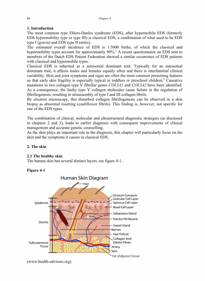
1. Introduction
The most common type Ehlers-Danlos syndrome (EDS), after hypermobile EDS (formerly
EDS hypermobility type or type III) is classical EDS, a combination of what used to be EDS
type I (gravis) and EDS type II (mitis),
The estimated overall incidence of EDS is 1:5000 births, of which the classical and
hypermobility types account for approximately 90%.1
A recent questionnaire on EDS sent to
members of the Dutch EDS Patient Federation showed a similar occurrence of EDS patients
with classical and hypermobile types.
Classical EDS is inherited as a autosomal dominant trait. Typically for an autosomal
dominant trait, it affects males and females equally often and there is interfamilial clinical
variability. Skin and joint symptoms and signs are often the most common presenting features
so that early skin fragility is especially typical in toddlers or preschool children.2
Causative
mutations in two collagen type V fibrillar genes COL5A1 and COL5A2 have been identified.
As a consequence, the faulty type V collagen molecules cause failure in the regulation of
fibrillogenesis, resulting in misassembly of type I and III collagen fibrils.
By electron microscopy, this disturbed collagen fibrillogenesis can be observed in a skin
biopsy as abnormal rosetting (cauliflower fibrils). This finding is, however, not specific for
one of the EDS types.
The combination of clinical, molecular and ultrastructural diagnostic strategies (as discussed
in chapters 2 and 3), leads to earlier diagnosis with consequent improvements of clinical
management and accurate genetic counselling.
As the skin plays an important role in the diagnosis, this chapter will particularly focus on the
skin and the symptoms it causes in classical EDS.
2. The skin
2.1 The healthy skin
The human skin has several distinct layers, see figure 4-1.
Figure 4-1
(www.health-advisors.org)
Chapter 448

- the epidermis, containing the keratinocytes, pigment cells and cells involved
in the immunological defence of the body such as the Langerhans cells,
- the dermis which contains, apart from blood vessels, lymphatic vessels and nerves, the
connective tissue, and
- the hypodermis or subcutis, which contains fat-containing adipocytes in addition to blood
and lymphatic vessels, loosely arranged elastic fibres and fibrous bands of connective tissue.
The latter anchors the dermis to the hypodermis and the skin to the deep fascia.
For normal skin mechanics, dermal connective tissue is of major importance. It contains
elastic fibres, collagen fibres and ground substance, an amorphous substance that fills the
spaces between the various fibres and bundles. The ground substance contains various
glycoproteins such as tenascin and various sugar containing proteoglycans, such as hyaluronic
acid and chondroitin sulphate. Fibrillar collagen fibres are arranged in a wave-like pattern.
Flattening of these fibres allows normal skin to extend, even though the constituent collagen
fibres themselves, are relatively inextensible. Extreme extension may damage the skin, which
is subsequently visible as stretch marks.3,4
Dermal elastic fibres are much sparser and are generally entwined among the collagen
bundles. Unlike collagen, these fibres are rubber-like and extensible. After stretching they
return to their original shape.
Translating this to the mechanical properties of normal skin: the collagen is responsible for
the strength and stiffness of the skin, consequently limiting skin extension if the collagen is
normal. Similarly, when extended, return to its normal shape is mediated by elastic fibres. In
the presence of abnormal collagen, the skin becomes abnormally extensible whilst the normal
elastic fibres will still have the mechanical properties to make the skin return to its normal
shape after extension. When elastic fibres are abnormal, as in Cutis Laxa, the skin will not
return to its normal shape after extension: instead it will be hanging in lax folds. Second, since
in Cutis Laxa collagens are normal, the skin is not hyperextensible. Furthermore, joint
hypermobility is the exception rather than the rule.
2.2 The skin in classical EDS
In short, in classical EDS the skin collagen fibrillogenesis is visibly abnormal. This
subsequently leads to hyperextensibility of the skin. Both the specific cutaneous and other
generalised clinical features of classical EDS are caused by the faulty collagen and are
described below.
2.2.1 Hyperextensibility
The skin in classical EDS is hyperextensible and seems to be hyperelastic. In the clinical
setting standard tests to determine this may be used. Beighton suggested an arbitrary score for
skin extensibility.5
He scored cutaneous stretching of the ventral left forearm on a numeric
rating scale of 0 to 5 in a range of 4 to 8 centimetres. Only in significantly abnormal
hyperextensibility a score higher than zero will be given. Skin extensibility is easily estimated
by testing at several sites of the skin such as the elbows, knees, neck or face, or the presternal
thoracic skin.
These tests are easy to apply and to interpret in adults, but are less discriminating in children
(see also chapter 18) since their skin is by nature more extensible than that of adults;
moreover, the amount of subcutaneous fat is higher in the very young children making tests
more difficult to perform adequately. To objectively measure the skin extensibility in
children, the properties of volar forearm skin of 13 children (3-10 years of age) with classical
EDS were recently studied using a cutometer equipped with a suction head (rheologic
evaluation), to assess dermal and hypodermal mechanics.6
Data of patients with classical EDS
were compared both to those of patients with hypermobile EDS and to those of age-matched
Classical Ehlers-Danlos syndrome 49

healthy controls. Skin of patients with classical EDS showed both abnormal
hyperextensibility as well as hyperelasticity when compared to the two other groups. Because
the dermis is thin in EDS and histology shows rarefaction of connective tissue septae in the
hypodermis, it was hypothesized by the authors that the “slipping mobility” of the dermis onto
the hypodermis was the main factor yielding these data, whereas the contribution of the
dermal component was present, but of less importance.
2.2.2 Reduced skin thickness
While skin dermal thickness is diminished by 50-66% in all patients with vascular EDS, in
classical EDS, there is a less impressive reduction in skin thickness. In the latter almost 50%
of patients is affected, though to a lesser degree. Skin thickness in classical EDS and healthy
controls was compared on the chest and the lower leg, with a 20MHz ultrasound probe.
Overall, in 21 patients with classical EDS dermal thickness was significantly lower compared
to that in controls.7
2.2.3 Skin fragility (figure 4-2)
The skin of classical EDS is abnormally fragile, with gaping wounds following very minor
traumas, which produce the characteristic fish-mouth or tissue paper scars. Beighton
suggested the use of an arbitrary analogue score of 1-5 based upon the extent of scarring,
using five bony sites e.g. left elbow and forearm, right elbow and forearm, left knee, right
knee and forehead as reference points. Other typical scarring sites are the chin and shins,
which are not included in the scoring calculations. Because of tissue fragility patients often
suffer from repetitive hernia, such as inguinal, umbilical, hiatal, or incisional hernia.
Figure 4-2 Skin fragility: cigarette paper aspect
�
2.2.4 Easy bruisability
Classical EDS patients bruise very easily and sometimes extensively, often after trivial
injuries. In general, bleeding and clotting tests are normal, and no consistent haematological
abnormalities are found. Beighton suggested an analogue scoring system, ranging from 0, for
no bruising, to 5, for gross changes. In the absence of clotting or bleeding abnormalities,
explanations for the bruising include faulty platelet binding to the abnormal underlying
collagen fibres, or fragility of connective tissue round small venous and arterial capillaries.
Chapter 450

2.2.5 Varicosities
Due to the disturbed type I and III fibril packing, venous walls have a collagen defect. This,
together with weakened perivenous connective tissue and faulty venous valve formation,
predisposes to venous varicosities (an abnormal condition of a vein, characterized by swelling
and tortuosity).
2.2.6 Molluscoid pseudotumors (figure 4-3)
Fleshy spongy lumps of 2 to 3 cm in diameter over pressure points, form so-called molluscoid
pseudotumors. These are usually concentrated at sites such as the Achilles tendons, knees and
elbows. These sites are easiest identified when the patient is standing. Molluscoid
pseudotumors probably are benign reactive fibrous hypertrophy, in response to recurrent
microtrauma and repair.
Figure 4-3 Molluscoid pseudotumors
�
(www.regionalderm.com, courtesy of RegionalDerm.com)
2.2.7 Spheroids
Small, mobile, easily palpable, subcutaneous papules are common over the forearms and
shins. When calcified they may be detected by X-ray examination. The origin of these
spheroids is not clear: some think they represent subcutaneous fat globules that have lost their
blood supply, becoming fibrosed and calcified.
2.2.8 Soft skin
Contact with the skin of patients with classical EDS reveals the interesting clinical sign of soft
and velvety peach-like skin. Moreover, when shaking hands one directly notices the very
hypermobile hand joints. The skin softness is presumably related to the misaggregated and
abnormally packed collagen fibres, characteristic for classical EDS.
2.2.9 Striae distensae
The occurrence of these linear atrophic depressions of the skin (cutaneous striae) is influenced
by many factors including hormones (particularly glucocorticoids), mechanical stress, and
genetic predisposition. Light microscopy shows epidermal atrophy, with some loss of the rete
ridges and sharply demarcated dense packing of thin, eosinophilic collagen bundles in a
parallel array, horizontal to the skin surface. Elastic fibres appear to be diminished in a stretch
mark as well.4
Classical Ehlers-Danlos syndrome 51

Striae are most common in areas of rapid growth, such as breasts and belly in pregnant
females, lumbar spine in adolescents or upper legs and arms in active sportsmen and
sportswomen. Caused by easy tearing of collagenous connective tissue, in particular collagen,
striae are not restricted to classical EDS, but also occur in hypermobile EDS, vascular EDS
and Marfan syndrome.
2.2.10 Piezogenic papules (figure 4-4)
Piezogenic papules are small, sometimes painful, reversible herniations of underlying adipose
tissue globules through the fascia into the dermis, such as on medial and lateral aspects of the
feet on standing.
Figure 4-4 Piezogenic papules on lateral side of the foot
(www.pediatricsconsultant)
3. Joint hypermobility
Joint hypermobility is common in classical EDS, just as it is in most other types, and will be
dealt with specifically in chapter 5. Just as mentioned earlier for scarring and bruising
assessments, a similar analogue scoring system exists for joint hypermobility.
4. Other features
Classical EDS is generally not characterized by vascular complications, though arterial
rupture does occur.7
However, recently, a subtype of classical EDS has been identified which
is clinically also characterized by a propensity to arterial rupture and molecularly - as it seems
- by a specific mutation in COL1A1 (c.934C>T; p.R134C).8,9,10
For other features, like gastro-
intestinal, neurological, urological and gynaecological/obstetric: see the respective chapters
10, 13, 16 and 17.
5. Objective criteria for diagnosing classical EDS
When the adapted scoring system of Beighton is used, the sum of the skin hyperextensibility
score, the scarring and bruising score adds up with the hypermobility score (see table 2-3 of
chapter 2). If this total score is > 7, further diagnostic procedures should start to confirm or
reject the diagnosis of classical EDS.1
However, most clinicians will require the presence of
the three major Villefranche criteria, i.e. skin hyperextensibility, dystrophic scarring and joint
hypermobility, in order to establish the diagnosis classical EDS. In 2017 a new international
classification of EDS has been published with new diagnostic criteria for the different EDS
types, including classical EDS and with management related aspects of the different types.11,12
Minimal diagnostic criteria for classical EDS are the presence of skin hyperextensibility and
atrophic scars, plus either generalized joint hypermobility and/or 3 minor criteria (see also
chapter 2, table 2-5)
Chapter 452

6. The laboratory diagnosis of classical EDS
6.1 Histopathology of the skin.
Skin biopsies of classical EDS may show minor increases of collagen/elastin ratios.
Sometimes dermal collagen is more intensely stained and shows an irregular pattern.11
However, diagnostically, it is of no use.
6.2 Electron microscopy of the skin
Ultrastructural examination of dermal collagen in classical EDS typically shows variable
diameters of collagen bundles and irregular contours in cross-section, some showing flower-
like appearances.13
These changes are best visualised at a print magnification of 50-70,000.
They are caused by longitudinal mispacking and unravelling of type I and III collagen fibres,
which is the result of the faulty type V collagen. Haplo-insufficiency or other mutations of
collagen type V have this effect. Cauliflower fibrils are a consistent feature of classical EDS,
but are not specific for it, as they may be seen in other inherited defects of connective tissue
as well, such as pseudoxanthoma elasticum (PXE), osteogenesis imperfecta, and scleredema
adultorum of Buschke.13
Therefore, it is of very limited diagnostic use.
6.3 Collagen protein and DNA analysis
Collagen type V, which was first purified from placenta,14
is a typical fibrillar collagen, in
which a central perfect uninterrupted triple helix, of general formula (Gly-X-Y)333, has both
specific N- and C-terminal extensions.15
These fibrillar triple helices co-assemble in a so-
called quarter staggered format. Compound fibrils of for example types I, III and V collagen
are formed in skin, whilst in the vitreous there are heterogeneous associations of collagens
types II, Valpha2 and XIalpha1. Unlike collagens types I, II and III, which have small N-
terminal extensions, those of type V collagen, coded by the COL5A1 and COL5A2 genes, are
much larger. Collagen type V proteins form four different types of alpha chains, alpha1(V),
alpha2(V), alpha3(V) and alpha4(V), of which the most common is, especially in the skin, a
heterotrimer consisting of two alpha1(V) chains and one alpha2(V) chain. Whilst types I, III
and V fibrils co-assemble, the N- terminus of type V collagen has a special role in regulating
fibril growth, length and lateral associations. This has been convincingly demonstrated by
Birk and colleagues, both in vivo and in vitro, in tissues such as skin, ligaments and cornea, in
mice made haplo-insufficient for the equivalent mouse Col5a1 gene.16
Such animals had signs
very similar to classical EDS in humans, with decreased tensile strength of normal or
wounded skin and vasculature. The decreased dosage of collagen type V allowed the
formation of both relatively normal and highly abnormal type I collagen fibrils. The latter was
caused by faulty nucleation, lateral aggregation and longitudinal growth, resulting in the
formation of abnormal rosettes, triggered by the unrestrained growth of type I collagen fibrils.
This is caused by the absence of the restraining type V protein.17
Eyelids and corneal tissues18
,
in which type V collagen forms 10-20% of corneal collagens, were studied in the Col5a1
mouse and compared to similar studies of defined human COL5A1 or COL5A2 mutants. In
brief, affected patients had thin, steep and transparent corneas, with lax eyelids, blue sclerae,
scleral fragility, microcornea, keratoconus, keratoglobus, ectopia lentis and retinal
detachment. The mouse model showed significantly enlarged and distorted corneal collagen
fibrils, however, less severely disrupted than dermal fibrils.
The recognition that collagen type V mutations caused classical EDS lagged more than 20
years behind similar discoveries of the role of collagen III in vascular EDS. Retrospectively,
this was due to the rarity of collagen V protein abnormalities. Nevertheless in 199419
a
shortened alpha1(V) chain in a classical EDS (mitis) variant was discovered, which showed
impressively disorganised collagen fibrils in dermis and various ligamentous tissues, as well
Classical Ehlers-Danlos syndrome 53

as corneal flattening.18
Later it became apparent that this was caused by a 54bp deletion
leading to heterozygous skipping of exon 65 of the COL5A1 gene.20
Other mutations quickly
followed, including a translocation, which interrupted the COL5A1 gene.21
Gene linkage
confirmed that the COL5A1 gene is involved autosomal dominant classical EDS.22,23
At least
one of the mutations of COL5A2, causing both , the gravis and mitis variants of classical
EDS,24,25
showed deficiency of collagen alpha2 chains, in vitro. Later, haplo-insufficiency of
COL5A1 gene dosage, caused by a variety of mechanisms,26, 27
proved to be the most common
cause of EDS classical type. Furthermore, this caused a misdirection of collagen
fibrillogenesis. Very occasionally, and rather puzzlingly28
, homozygosity for a helical glycine
mutation can also cause a mild form of classical EDS. A 2005 study of 48 patients with
classical EDS, showed abnormal collagen protein profiles in only 2/48 (4.4%), whilst ten
times as many (43%) were haplo-insufficient. In these 48 patients, 22 mutations of either
COL5A1 or COL5A2 were detected, including 17 stop codons and 5 structural mutations of
the types mentioned above. As three of the patients with a positive null allele test carried no
detectable COL5A1 mutation, only 52% of classical EDS patients have detectable COL5A1 or
COL5A2 defects.29
Ritelli et al. also found in 50% of their 40 cases a mutation in COL5A1
(18) or COL5A2 (2).30
However, Symoens et al. detected in over 90% of patients, who fulfil
all three major Villefranche criteria for classical EDS (n = 102), a defect in (products of)
either COL5A1 (80) or COL5A2 (13).31
It remains to be seen whether other elements in
fibrillogenesis, such as COL5A3, decorin, tenascin X, thrombospondin or unidentified
extracellular matrix constituents contribute to the molecular pathology of classical EDS.
The recently identified subtype of classical EDS which is clinically characterized by a
propensity to arterial rupture, is caused - as mentioned earlier - by a specific mutation in
COL1A1 (c.934C>T; p.R134C).8,9,10
7. Differential diagnosis
Here we address only those inherited defects of connective tissue, in which cutaneous
symptoms and signs predominate (see32
).
7.1 Tenascin-X (TNX) deficient EDS
TNX deficient EDS is an autosomal recessive variant of classical EDS, named classical-like
EDS (see chapter 2).11
Clinically it shows elastic, velvety skin, hypermobile joints and easy
bruising, without abnormal wound healing or atrophic scars. In 3 of the 8 patients described,
multiple subluxations were part of the clinical picture and in 2 cases joint pain was
mentioned. Moreover, the patients were also reported to have a range of additional clinical
problems not frequently associated with EDS, including spina bifida occulta, mitral-valve
prolapse, stroke, gastrointestinal bleeding and premature arteriosclerosis. It is not clear
whether these findings are related to the TNX deficiency. Skin biopsy shows both abnormal
elastic fibres and reduced collagen deposition. TNX may contribute to matrix stability by
interacting with other matrix components, such as proteoglycans. This might also regulate
collagen fibrillogenesis.33
TNX is undetectable in serum in affected homozygotes, whilst it is
50% of normal control levels in heterozygotes carriers of whom some had signs and
symptoms of hypermobile EDS.
7.2 Other similar EDS types
Classical EDS must be distinguished from other EDS types, especially hypermobile EDS,
with which there may be clinical overlap. Particularly in mild cases of classical EDS,
hypermobile EDS can be difficult to distinguish.
Chapter 454

7.3 The Occipital Horn syndrome
The Occipital Horn syndrome is a very rare disorder of copper metabolism which, in the old
Berlin classification of 1988, was included as a curious form of EDS, but it is no longer part
of the EDS classification. It is an X-linked recessive syndrome. Clinical features include soft,
mildly extensible, loose and redundant skin with easy bruisability, but without fragility. Other
clinical signs include a persistent anterior fontanel, frequent loose stools, obstructive uropathy
and mild mental retardation. Increased joint hypermobility may occur, except for the knee and
elbow joints which show only limited extension. Radiologically, occipital horns, wedge
shaped calcifications at the attachment sites of the trapezius and sternocleidomastoid muscles,
are characteristic. It is caused by a mutation in the Cu(2+)-transporting ATPase, alpha
polypeptide. The concentrations of copper and ceruloplasmin in serum are low and probably
impair collagen cross-linking.
7.4 Cutis Laxa
Cutis Laxa (CL) refers to the redundant inelastic quality of the skin, which, in contrast to
EDS- types, is not (hyper)elastic. Original descriptions of EDS often did not readily
distinguish between the easily stretched skin of EDS and the ill-fitting, overstretched
integument of true CL. Autosomal dominant and autosomal recessive as well as X-linked
subtypes of primary CL, frequently seen with accompanying lung and aortic pathology, are
caused by abnormalities of elastin or elastic microfibrils. Secondary CL complicates
conditions such as PXE and EDS.
7.5 Child abuse
Child abuse is certainly included in the differential diagnosis of classical EDS. Not
surprisingly, the predisposition to bruising and cutaneous damage sometimes raises this
possibility. Similarly, EDS is included in the differential diagnosis of child abuse. Of course,
it must be realised that EDS and child abuse are not necessarily mutually exclusive diagnoses.
In distinguishing the one from the other, familiarity with the various clinical criteria included
in the most recent classification is essential, as is a thorough family history, clinical
examination and, where indicated, laboratory tests, such as electron microscopy, protein
electrophoresis, metabolic analysis and DNA analysis. Similarly, there are very well
established clinical criteria which deal with bruise distribution and morphology, and other
accompanying clinical signs, likely to indicate child abuse.34
For example, in child abuse, bruising mostly occurs over relatively protected sites of the skin
(trunk, buttocks, genital area), while in traumas and in classical EDS and other types, the
knees, the anterior tibial area and bony prominences are mostly affected.35
8. Protection and preventative measures
Here we focus mainly upon measures which protect the skin or promote cutaneous wound
healing. For measures concerning other problems associated with classical EDS, one is
referred to other chapters, like 10, 13, 16 and 17. Unfortunately, in the absence of specific
therapy for classical EDS or any other type, only simple preventive measures to protect the
skin are applicable. All are anecdotal, common sense measures, rather than evidence based
upon appropriate clinical trials.
8.1 Protection
Particularly in younger children, protective bandages or pads applied at sites of repetitive
trauma such as the forehead, elbows, knees and shins, can minimise wounds and lacerations.
Older children might prefer protection by more fashionable sports devices, such as those used
for knees, elbows and shins in skiing, skating or football rather than protection by medical
Classical Ehlers-Danlos syndrome 55

bandages. Head protection for certain contact sports or cycling is also desirable, whilst for
games such as tennis and squash eye protection with fragmentation resistant goggles is
desirable.
At home the corners of tables and other furniture can be protected and the tables or other
barriers should not be put in the centre of the room, in order to prevent trauma.
Contact sports are to be discouraged.
In case more specific advice is needed, one may consider rheologic evaluations of the skin
(see 2.2.1), since these tests may detect patients with very vulnerable skin more reliably; it
also may be used to predict the difficulty in suturing.3
8.2 Bleeding prevention
In case of bruising, a pressure bandage can be used, or elevation of the affected limb will help
to prevent further cutaneous bleeding. In case of tooth extractions, surgery and a history of
heavy bleeds, further bleeding and clotting tests are indicated and, if abnormal, measures can
be taken, see chapter 11.
8.3 Sun protection
Skin thinning and increased fragility, together with reduced subcutaneous fat formation, is
part of normal ageing. Although this occurs also in sun protected areas, it is especially evident
in sun-exposed areas. Sunlight damages the epidermis, leading to a higher risk of skin
tumours, but also damages the dermis. In the latter, it causes loss of elasticity in elastic fibres
(elastosis). Also sunlight causes collagen-fibres to decrease and degenerate. This effect is
magnified in classical and other EDS types, in which the underlying collagen is already
abnormal. Sun protection is therefore particularly important in EDS patients.
8.4 Smoking
Smoking is also associated with elastosis (see 7.3) among both sexes,36
so it is wise to refrain
from smoking in case of EDS.
8.5 Medication
Suggestions made in the literature that the use of 1-2 grams of ascorbic acid (vitamin C) a day
may promote collagen synthesis, thereby assisting collagen repair and wound healing or even
reduce vascular fragility, should be regarded as unproven.
Generally avoiding aspirin (acetylsalicylic acid) or non-steroidals such as ibuprofen, all of
which enhance bleeding and bruising due to their negative effect on platelet function, is
sensible. Similarly, skin atrophy as adverse effect of chronic inhaled or systemic
glucocorticoid treatment is more outspoken in EDS than in otherwise normal individuals.
9. Surgical advice
All surgical procedures have additional risks in classical EDS, since the skin is fragile: it heals
slowly and sutures are more damaging and less stable than in normal skin. Specific advice on
how to treat wounds is given in the addendum on surgery. Bearing these facts in mind, major
elective surgery can be surprisingly uncomplicated. This contrasts with vascular EDS, in
which the repair of hollow organs and vascular anastomoses can be very problematic.
Procedures such as dermabrasion or skin filling are probably contraindicated and laser
treatment carries the risk of hypertrophic scarring. See also the addendum.
Chapter 456

10. Areas of uncertainty
10.1 Absence of inferior labial or lingual frenulum
As the labial and lingual frenula consist mainly of ligament-like connective tissue structures,
the morphology of these structures has been examined in EDS.37
Opinions vary as to whether
the fraenum of the lower lip and the sublingual frenulum are missing in classical EDS. 37,38,39
Further studies are required to resolve these disparities.
Previously, a positive Gorlin sign was regarded as common in EDS. The sign is positive when
the tip of the nose can be touched by the tip of the tongue.40
Presently, its exact frequency in
classical EDS is unclear and further studies are needed.
10.2 Knowledge of the aetiology
As described earlier in section 6, mutations of COL5A1 or COL5A2 explain 50 to over 90% of
classical EDS. It remains to be seen which other matrix genes and proteins are involved in the
remainder of the patients.
10.3 Treatment possibilities
So far, no double blinded, placebo controlled trials have been done to test the efficacy of
substances such as ascorbic acid, glucosamine, Coenzyme Q10, pycnogenol and may be
others.41
Because the expected effect will most probably be not too big, multicentre trials of
adequate numbers of EDS patients would be necessary to prove therapeutic efficacy.
Classical Ehlers-Danlos syndrome 57

References
1. Steinmann B, Royce P, Superti-Furga A. The Ehlers-Danlos Syndrome. In: Royce P,
Steinmann B, eds. Connective tissue and its heritable disorders. New York, USA:
Wiley-Liss, 1993:351–407.
2. Malfait F, Wenstrup RJ, De Paepe A. Clinical and genetic aspects of Ehlers-Danlos
syndrome, classic type. Genet Med 2010;12:597-605.
3. Ergün SS, Ozcan RH, Kural YB. Striae distensae: a rare complication resulting from
overinflation of the tissue expander. Aesthetic Plast Surg 2007;31:606-7.
4. Suh DH, Chang KY, Son HC, Ryu JH, Lee SJ, Song KY. Radiofrequency and 585-nm
pulsed dye laser treatment of striae distensae: a report of 37 Asian patients. Dermatol
Surg 2007;33:29-34.
5. Holzberg M, Hewan-Lowe KO, Olansky AJ. The Ehlers-Danlos syndrome: recognition,
characterization, and importance of a milder variant of the classic form. J Am Acad
Dermatol 1988;19:56-66.
6. Henry F, Goffin V, Piérard-Franchimont C, Piérard GE. Mechanical properties of skin
in Ehlers-Danlos syndrome, types I, II, and III. Ped Dermatol 1996;13:464-7.
7. Eisenbeiss C, Martinez A, Hagedorn-Greiwe M, Reinhardt DP, Bätges B, Brinckmann
J. Reduced skin thickness: a new minor diagnostic criterion for the classical and
hypermobility types of Ehlers-Danlos syndrome. Brit J Dermatol 2003;149:850-2.
8. Borck G, Beighton P, Wilhelm C, Kohlhase J, Kubisch C. Arterial rupture in classic
Ehlers-Danlos syndrome with COL5A1 mutation. Am J Med Genet A 2010;152A:2090-
3.
9. Malfait F, Symoens S, De Backer J, Hermanns-Lê T, Sakalihasan N, Lapière CM,
Coucke P, De Paepe A. Three arginine to cysteine substitutions in the pro-alpha (I)-
collagen chain cause Ehlers-Danlos syndrome with a propensity to arterial rupture in
early adulthood. Hum Mutat 2007;28:387-95.
10. Gaines R, Tinkle BT, Halandras PM, Al-Nouri O, Crisostomo P, Cho JS. Spontaneous
Ruptured Dissection of the Right Common Iliac Artery in a Patient with Classic Ehlers-
Danlos Syndrome Phenotype. Ann Vasc Surg 2015;29:595.e11-4.
11. Malfait F, Francomano C, Byers P, et al. The 2017 International classification of the
Ehlers-Danlos syndromes. Am J Med Genet Part C Semin Med Genet 2017;175C:8-26.
12. Bowen JM, Sobey GJ, Burrows NP, et al. Ehlers-Danlos syndrome, classical type. Am J
Med Genet Part C Semin Med Genet 2017;175C:27-39.
13. Hausser I, Anton-Lamprecht I. Differential ultrastructural aberrations of collagen fibrils
in Ehlers-Danlos syndrome types I-IV as means of diagnostics and classification. Hum
Genet 1994;3:394-407.
14. Burgeson RE, El Adli FA, Kaitila II, Hollister DW. Fetal membrane collagens:
identification of two new collagen alpha chains. Proc Nat Acad Sciences USA
1976;73:2579-83.
15. Prockop DJ. Mutations in collagen genes. J Clin Invest 1985;75:783-7.
16. Birk DE, Fitch JM, Barbiaz JP, Doane KJ, Linsenmayer TF. Collagen fibrillogenesis in
vitro: interaction of types I and V collagen regulates fibril diameter. J Cell Science
1990;95:649-57.
17. Birk DE Fitch JM , Barbiaz JP, Linsenmayer TF. Collagen type I and type V are present
in the same fibril in avian corneal stroma. J Cell Biol 1988;106:999-1008.
18. Segev F, Heon E, Cole WG, et al. Structural abnormalities of the cornea and lid
resulting from collagen V mutations. Invest Ophthalmol Visual Science 2006;47:565-
73.
19. Nicholls AC, McCarron Narcisi P, Pope FM. Molecular abnormalities of type V
collagen in Ehlers-Danlos Syndrome. Am J Hum Genet 1994;55:A233.
Chapter 458

20. Nicholls AC, Oliver JE, McCarron S, Harrison JB, Greenspan DS, Pope FM. An exon
skipping mutation of a type V collagen gene (COL5A1) in Ehlers-Danlos Syndrome. J
Med Genet 1996;33:940-6.
21. Toriello HV, Glover TW, Takahara K, et al. A translocation interrupts the COL5A1
gene in a patient with Ehlers-Danlos Syndrome and Hypomelanosis of Ito. Nature Genet
1996;13:361-5.
22. Loughlin J, Irwen C, Butcher S, et al. Linkage of the gene that encodes the alpha 1
chain of type V collagen, (COL5A1) to type II Ehlers-Danlos syndrome (EDSII). Hum
Mol Genet 1995;4:1649-51.
23. Burrows NP, Nicholls AC, Yates JR, et al. The gene encoding collagen alpha1(V)
(COL5A1) is linked to mixed Ehlers-Danlos type I/II. J Invest Dermatol
1995;106:1273-6.
24. Mikchalikova K, Susic M, Willing MC, Wenstrup RJ, Cole WG. Mutations of the
alpha2(V) chain of type V collagen impair matrix assembly and produce Ehlers-Danlos
syndrome type I. Hum Mol Genet 1998;7:249-55.
25. Richards AJ, Martin M, Nicholls AC, Harrison JB, Pope FM, Burrows NP. A single
base mutation in COL5A2 causes Ehlers-Danlos syndrome type II. J Med Genet
1998;35:846-8.
26. Wenstrup RJ, Florer JB, Willing MC, et al. COL5A1 haploinsufficiency is a common
molecular mechanism underlying the classical form of EDS. Am J Hum Genet
2000;66:1766-76.
27. Wenstrup RJ, Florer JB, Davidson JM, et al. Murine model of the Ehlers-Danlos
syndrome: col5a1 haploinsufficiency disrupts collagen fibril assembly at multiple
stages. J Biol Chem 2006;281:12888-95.
28. Giunta C, Nuytinck L, Raghunath M, Hausser I, De Paepe A, Steinmann B.
Homozygous Gly539Ser substitution in COL5A1 causes mild classical Ehlers-Danlos
syndrome. Am J Med Genet 2002;109:284-90.
29. Malfait F,Coucke P, Symoens S, Loeys B, Nuytinck L, De Paepe A. The molecular
basis of classic Ehlers-Danlos syndrome: a comprehensive study of biochemical and
molecular findings in 48 unrelated patients. Hum Mutation 2005;25:28-37.
30. Ritelli M, Dordoni C, Venturini M, Chiarelli N, Quinzani S, Traversa M, Zoppi N,
Vascellaro A, Wischmeijer A, Manfredini E, Garavelli L, Calzavara-Pinton P, Colombi
M. Clinical and molecular characterization of 40 patients with classic Ehlers-Danlos
syndrome: identification of 18 COL5A1 and 2 COL5A2 novel mutations. Orphanet J
Rare Dis 2013;8:58.
31. Symoens S, Syx D, Malfait F, Callewaert B, De Backer J, Vanakker O, Coucke P, De
Paepe A. Comprehensive molecular analysis demonstrates type V collagen mutations in
over 90% of patients with classic EDS and allows to refine diagnostic criteria. Hum
Mutat 2012;33:1485-93.
32. Callewaert B, Malfait F, Loeys B, de Paepe A. Ehlers-Danlos syndrome and Marfan
syndrome. Best Pract Res Clin Rheumatol 2008;22:165-89.
33. Egging D, van den Berkmortel F, Taylor G, Bristow J, Schalkwijk J. Interactions of
human tenascin-X domains with dermal extracellular matrix molecules. Arch Dermatol
Res 2007;298:389-96.
34. Kemp AM, Dunstan F, Harrison S, et al. Patterns of skeletal fractures in child abuse:
systematic review. BMJ 2008;337:a1518.
35. Kos L, Shwayder T. Cutaneous manifestations of child abuse. Ped Dermatol
2006;23:311-20.
36. Kennedy C, Bastiaens MT, Bajdik CD, Willemze R, Westendorp RG, Bouwes Bavinck
JN. Leiden Skin Cancer Study. J Invest Dermatol 2003;120:548-54.
Classical Ehlers-Danlos syndrome 59

37. De Felice C, Toti P, Di Maggio G, Parrini S, Bagnoli F. Absence of the inferior labial
and lingual frenula in Ehlers-Danlos syndrome. Lancet 2001;357:1500-2.
38. Böhm S, Martinez-Schram A, Gille J, Behrens P. Missing inferior labial and lingual
frenula in Ehlers-Danlos syndrome. Lancet 2001;358:11647-8.
39. Shankar S, Shirley E, Burrows NP. Absence of the inferior labial or lingual frenula is
not a useful clinical marker for Ehlers-Danlos syndrome. J Eur Acad Dermatol
2006;20:1328-9.
40. Horii KA, Nopper AJ, Sharma V. Picture of the month. Arch Pediatr Adolesc Med
2006;160:705-6.
41. Mantle D, Wilkins RM, Preedy V. A novel therapeutic strategy for Ehlers-Danlos
syndrome based on nutritional supplements. Medical Hypotheses 2005;64:279-83.
�
Chapter 460

Chapter 5. Generalised joint hypermobility and joint hypermobility syndromes:
the clinical perspective
JWG Jacobs, M Castori and JAP Da Silva
Johannes WG Jacobs, MD, PhD, Rheumatologist
Department of Rheumatology & Clinical Immunology
University Medical Center Utrecht
Heidelberglaan 100
3584 CX Utrecht, The Netherlands
Telephone +31887557357
E-mail [email protected]
Marco Castori, MD, PhD, Consultant Medical Geneticist & Chief
Division of Medical Genetics
IRCCS-Casa Sollievo della Sofferenza
Poliambulatorio, 2nd Floor
Viale Padre Pio snc
71013 San Giovanni Rotondo (FG), Italy
Telephone: +0882416288
Email: [email protected]
José AP Da Silva, MD, PhD, Professor of Rheumatology
Clinica Universitária de Reumatologia
Faculty of Medicine
University of Coimbra
Portugal
Telephone + 351239400547
E-mail: [email protected]
A version of this chapter has been published: Jacobs JWG, da Silva JAP. Hypermobility syndromes
from the clinician’s perspective: an overview. Acta Reumatol Port 2014; 39: 124-36.
61

1. Introduction
Joint hypermobility is defined as an increased range of movement in one or more joints.
When many joints are hypermobile in an individual, we speak of generalised joint
hypermobility, a relatively common condition. There are several classification criteria and
scores sets for this physical trait - scores of Beighton and Bulbena are presented in chapter
2;1,2
classification criteria are discussed in other paragraphs.3
While the Beighton score is
limited to the assessment of the range of motion of a pre-determined set of joints/groups of
joints, the Bulbena score also scores ecchymosis. For this reason, the Beighton score is now
considered the most appropriate tool for assessing generalised joint hypermobility in adults
and children, though with different cut-offs.4
The adjective “generalised” implies that joint
hypermobility is present at multiple sites, but distribution and extent of joint hypermobility
are quite variable and influenced by constitutional and acquired factors. A scoring system
ideally accounting for all variables has not yet been developed and scoring with available
tools is influenced by the practitioner’s experience.
Individuals with (generalised) joint hypermobility may be asymptomatic. Often, however,
joint hypermobility is accompanied by musculoskeletal symptoms such as pain in joints,
ligaments, entheses (attachment sites of tendons or ligaments to the bone) and muscles - this
may be referred to as symptomatic joint hypermobility. In addition, (generalised) joint
hypermobility can be associated with a wider spectrum of symptoms and signs reflecting
involvement of other tissues, organs, and organ systems, attributable to the pleiotropic nature
of underlying defects. In such cases the term “joint hypermobility syndromes” seems
appropriate. The term “syndrome” refers to group of signs and symptoms recurring in
association and defining a cluster of clinical relevance, known or thought to be causally
(etiologically) related. Joint hypermobility syndromes are conditions with a strong genetic
basis, in which (generalised) joint hypermobility is the shared congenital trait.
Although many individuals with (generalised) joint hypermobility do not stand out clinically,
and do not meet criteria of a specific syndrome, others meet criteria of a specific defined
syndrome, potentially associated with chronic disabilities or life-threatening complications.
This makes it important not to overlook these diagnoses.
The aim of this chapter is to provide the rheumatologist and other health professionals an
overview of the broad spectrum of symptomatic joint hypermobility and joint hypermobility
syndromes they may be confronted with, as guidance in daily practice. Clinical aspects, with
emphasis on musculoskeletal problems, are described and clues for diagnosis and
management are provided.
2. Epidemiological aspects
Generalised joint hypermobility may actually be a variant of normal joint mobility, along the
upper tail of the Gaussian curve describing the range of motion of normal joints in the
population, just as individuals can be short or tall. In general, the range of motion of most
joints and thus the chance of observing generalised joint hypermobility decreases with
increasing age. Generalised joint hypermobility is more common in females than males and
occurs more often in some racial groups, e.g. Asians when compared to Caucasians.5
Many
healthy young individuals, especially females performing ballet or gymnastics, would
probably meet the criteria of generalised joint hypermobility according to the Beighton set, if
they were screened. In some sports, generalised joint hypermobility may be an advantage, but
it may also be a liability due to the increased risk of injuries.6,7
Reassuringly, a study among
professional dancers concluded that generalised joint hypermobility was not associated with a
higher risk of injuries when assessed prospectively.8
However, this observation does not rule
out an increased risk of injury associated with generalised joint hypermobility in the (less
trained) general population. In fact, those more prone to injuries may have fallen short of a
Chapter 562

desired professional sports career.
People presenting for medical care with symptomatic joint hypermobility are most probably a
subgroup of those with generalised joint hypermobility in the population. Furthermore, not all
cases of this subgroup will meet criteria of available scoring systems for generalised joint
hypermobility. An even smaller number will meet criteria for a specific joint hypermobility
syndrome, such as Ehlers-Danlos syndrome (EDS), a group of syndromes with as key features
multi-site joint hypermobility, abnormal skin texture and a variable range of dysfunctions or
fragility of vessels and internal organs. A description of all EDS types is beyond the scope of
this chapter.
Exact prevalence estimates for EDS are difficult to make for two main reasons. First, there is
the problem of recognition: not all individuals with joint hypermobility have relevant
symptoms and reach medical attention. If generalised joint hypermobility is borderline and
skin manifestations are mild, they might stay unrecognized. As a consequence, estimates on
the prevalence of EDS types depend on whether they are based on clinical reports of the
syndromes or on screening studies in populations and, in this case, on methodological
properties of screening methods. In the latter case, the prevalence estimates will be probably
more accurate and higher. A study in a general dermatology population revealed that common
variants (phenotypes, see chapter 2) of EDS might be present in up to 9% of this population.9
Second, there is the problem of older and current classifications: significant overlap exists
between the signs, symptoms and classification criteria of hypermobile EDS and those of the
benign joint hypermobility syndrome (BJHS),10
both of which may show familial aggregation.
In the past, the presence of generalised joint hypermobility and its secondary musculoskeletal
manifestations was thought sufficient for defining a “genetic syndrome”; e.g. BJHS may be
diagnosed, if a Beighton score ≥4 and joint pain at multiple sites (i.e. the two major Brighton
criteria) are present. But, familial clustering of generalised joint hypermobility (either
symptomatic or not) is not always the marker of a hereditary syndrome affecting multiple soft
connective tissues. This was in 2016 the rationale to mend the double, overlapping
terminologies and keep a single syndromic entity (i.e. hypermobile EDS), which would define
only patients with a likely multisystem disorder. Alternative names (not including the term
“syndrome”) should be preferably used for patients showing symptomatic generalised joint
hypermobility, but not meeting the criteria for hypermobile EDS or any other partially
overlapping hereditary soft connective tissue disorder. Recently, the term “hypermobility
spectrum disorders” has been proposed as an exclusion diagnosis for those individuals who
show some range of joint hypermobility and related musculoskeletal complaints, but who do
not meet the criteria for presently defined joint hypermobility syndromes, including
hypermobile EDS.11
The prevalence of what was classed as generalised joint hypermobility was estimated to range
from 10–30% in adults, varying from 10-15% in male youngsters between 11–17 years and up
to 20–40% in girls of this age group,12
which seem high percentages. The prevalence of EDS
is probably higher than the current estimate of 1: 5,000,13
although the most recent and stricter
criteria for hypermobile EDS will impact such an assumption.
3. General symptoms, signs and complications of generalised joint hypermobility and
joint hypermobility syndromes
Patients with symptomatic generalised joint hypermobility may share many clinical
characteristics, typically including chronic musculoskeletal pain, fatigue, signs of autonomic
dysfunction, functional gastro-intestinal manifestations and joint (sub)luxation. Pain and
fatigue are the dominant symptoms. In the following section, we discuss the most prevalent
symptoms and signs the clinician and health professional are confronted with, and their
associations. Among these general symptoms, the non-musculoskeletal functional ones (e.g.
Generalised joint hypermobility and joint hypermobility syndromes: the clinical perspective 63

fatigue, cardiovascular dysautonomia, gastrointestinal motility issues) are now termed as
“joint hypermobility-related co-morbidities”.11
Joint hypermobility-related co-morbidities
may be common in individuals with non-syndromic, symptomatic joint hypermobility (i.e.
hypermobility spectrum disorders) as well as in the various joint hypermobility syndromes.
Hence, joint hypermobility-related co-morbidities are not specific enough for etiological or
nosologic discrimination, although their assessment is crucial for prognostication and
management.
3.1 Pain
There is an increased prevalence of arthralgia (joint pain), chronic generalised myalgia and
fibromyalgia (chronic generalised pain in muscles and joints) in children and adults with
generalised joint hypermobility.5,10,14-20
In adults, joint hypermobility is also associated with
back pain.18,21
Perhaps surprisingly, the basis of the association of joint hypermobility and
pain is not established. The most consensual view is that pain is due to repetitive strain, sprain
and microtraumas of muscles and ligaments by the abnormal range-of-motion permitted by
hypermobile joints, aggravated by diminished joint proprioception, position sense,22-24
and
decreased passive muscle tension.25
Pain is also related with anxiety and depression, both of
which seem to be more prevalent in EDS, especially hypermobile EDS, as well as in the
former BJHS.26-28
It is commonly believed that chronic pain elicits depressive feelings and
that depression has an amplifying effect on chronic pain and fatigue, leading to a vicious
circle. These latter symptoms could lead to reduced physical activity and thus physical
deconditioning (reduced physical condition), increasing the propensity to injury, chronic pain
and fatigue and initiating a downward negative spiral.
Furthermore, there seems to be a primary involvement of muscle in hypermobile EDS: muscle
weakness has been found in the absence of reduced muscles mass, which would have been
present if muscle weakness was the results of reduced physical activity only.29
A hypothetic
model of pain, including fatigue and physical deconditioning is depicted in figure 5-1.
3.1.1 Insufficient effect of local analgesics
An insufficient effect of local analgesics either by intradermal injection or as topical cream
application has been reported in patients with hypermobile EDS. A similar ineffectiveness has
been observed in patients with generalised joint hypermobility.30
This was thought to be due
to the lax connective tissues in the skin allowing too much dispersal of the analgesic.31,32
However, the dispersal of a radioisotope labelled solution following deep dermal injection did
not differ between EDS patients and healthy controls in a small study.33
So, the reason for the
insufficient effect of local analgesics is not yet known.
3.2 Fatigue
Generalised joint hypermobility syndromes, particularly hypermobile EDS, are associated
with increased fatigue.34-36
A hypothesis is that fatigue is a symptom of autonomic
dysfunction or dysautonomia, described below.35,36
Others suggest that the ligamentous laxity
demands increased vigilance, muscle tension and coordination to maintain adequate joint
position and body balance,24,37,38
thus leading to fatigue. In addition, these patients often show
muscle weakness,29
reduced exercise tolerance, physical deconditioning and pulmonary
symptoms (see below),39
commonly attributed to reduced exercise because of chronic pain
(figure 5-1). Finally, fatigue could be associated with depression and other psychological
problems associated with generalised joint hypermobility.36,40
Most probably, fatigue is
caused by a constellation of these factors, of which the individual impact will vary between
patients.
Chapter 564

Figure 5-1 Vicious circle in generalised hypermobility of chronic pain and fatigue
Via several mechanisms, pain and chronic fatigue may ensue, leading to less physical
exercise. This leads to physical deconditioning, associated with chronic pain and fatigue, and
a vicious circle. The way to break this vicious circle and often downward spiral is to prevent
or treat (further) physical deconditioning by daily physical training (exercises). Not all
features have to be clearly present in all patients.
Fatigue and generalised pain are paramount among the symptoms that establish a clinical
similarity and diagnostic confusion between joint hypermobility syndromes and
fibromyalgia14-17,41
. Some authors even argue whether fibromyalgia is merely a description of
the symptoms of EDS or a separate disease entity in these patients.14-17
Generalised joint hypermobility and joint hypermobility syndromes: the clinical perspective 65

3.3 Autonomic dysfunction
Autonomic dysfunction comprises disturbances in a variety of functions dependent on the
autonomic nervous system, leading for instance to orthostatic hypotension or tachycardia,
leading to (pre)syncope and palpitations. Such symptoms have been reported in higher than
expected frequency in patients with BJHS and EDS.35,36
Lower urinary tract dysfunction
associated with generalised joint hypermobility might also be related to autonomic
dysfunction, alongside laxity of the connective tissue of the pelvic floor and the sphincter.42,43
Gastro-intestinal disturbances, such as gastro-oesophageal reflux, constipation and irritable
bowel syndrome or malabsorption, are more common in BJHS and EDS, and might share a
similar pathophysiology.44
The prevalence of both urinary and faecal incontinence has been
described as significantly higher in women with joint hypermobility syndromes than in
women without these conditions.42,43,45-48
3.4 Joint (sub)luxation
Joint luxation or subluxation are not specific features of generalised joint hypermobility and
generalised joint hypermobility syndromes. Probably (sub)luxation reflects the severity of the
joint laxity and impaired local muscle strength and coordination. If (sub)luxation occurs
frequently in a specific joint, this often becomes less painful and sometimes (sub)luxation can
be demonstrated by the patient on request.
3.5 Decreased bone mass and increased risk of fractures
In EDS and Marfan syndrome, a higher prevalence of low bone mineral density, osteopenia or
osteoporosis are reported in most studies,49-53
but not all.54
In a study with 23 patients with
hypermobile EDS and 23 matched controls, EDS subjects had a significantly lower bone
mineral density at the femoral neck, but this difference disappeared after adjustment for body
height, weight and physical activity levels.55
Thus, reduced exercise or immobility induced by
generalised joint hypermobility may be important in determining osteopenia, probably in
association with the inherited structural deficit. Furthermore, an increased incidence of falls
(and thus increased risk of fractures) has been reported in hypermobile EDS,38
due to
impaired balance and muscle weakness. This might be at least partially preventable by
appropriate exercise programs. Early-onset osteoporosis and increased risk of fractures are
features of rare EDS types with marked bone involvement, such as the EDS/osteogenesis
imperfecta overlap.
3.6 Osteoarthritis
A relationship between joint hypermobility syndromes and osteoarthritis would be expected
especially as long-term complication in patients with frequent (sub)luxations. However, the
literature data are equivocal: while some papers describe a relation between joint
hypermobility and osteoarthritis56,57
, others even indicate an inverse relation.54,58,59
3.7 Life-threatening manifestations and complications
Some specific joint hypermobility syndromes, e.g. vascular EDS, Marfan syndrome and
Loeys-Dietz syndromes may have life-threatening manifestations, such as aneurysms and
arterial ruptures. Next to vascular complications, life-threatening ruptures of the bowel and of
the pregnant uterus are also manifestations of vascular EDS.60,61
Fortunately, these
manifestations or complications do not happen in the more frequently occurring joint
hypermobility syndromes.
4. Classification into a specific joint hypermobility syndrome
For some of the joint hypermobility syndromes, the diagnosis can be made early based on
Chapter 566

evident signs and symptoms, although because of profound muscle hypotonia, patients
initially are suspected of having a muscle disorder. However, more frequently, the clinician
will be faced with previously undiagnosed generalised joint hypermobility in a patient
presenting with symptoms; in such cases, it most often concerns hypermobile EDS.62
More
rarely, such patients may present with another syndrome, such as classical EDS, Marfan
syndrome or vascular EDS (yet without clear vascular complications).
So, the first and most important step in eliciting such diagnosis is awareness of the broad
spectrum of joint hypermobility syndromes.
Would classification of such cases into a specific syndrome be clinically important? And, if
so, how could we best do that?
4.1 Why classify generalised joint hypermobility?
It is important to identify and classify generalised joint hypermobility, not only to optimise
management, but also because some syndromes are associated with life-threatening risks
outside the musculoskeletal system, as described above. Although these diseases cannot be
cured and complications cannot be totally prevented, awareness and appropriate measures will
diminish the risks of such events. For patients with these severe disorders, family planning
and management are also indicated.
4.2 How to go about classification?
Clinical diagnosis was in the past, for most of the syndromes, based on clinical recognition of
reported symptoms and signs at physical exam, but nowadays the diagnosis of most
syndromes is supported by molecular testing, which is especially important for syndromes
with life-threatening risks, like vascular EDS, Marfan syndrome and Loeys-Dietz syndromes.
Hence, a formal diagnosis of a recognized syndrome with joint hypermobility should be
always supported by a positive molecular testing and/or strict adherence to available
diagnostic criteria. The hypermobile EDS, presumably the most common clinical variant of
EDS, still remains without any confirmatory molecular testing. In order to support clinicians
in the assessment of this condition, the most recent classification of EDS includes a detailed
description of the procedural diagnostics and stricter diagnostic criteria for the hypermobile
EDS (see chapter 2),3
compared to the former classification of EDS hypermobility type.
For nomenclature, it is recommended that specific joint hypermobility syndromes are only
diagnosed or referred to if published classification criteria are satisfied. Patients who do not
satisfy such classification criteria should be described simply as having generalised joint
hypermobility, no specific syndrome or hypermobility spectrum disorders, according to the
most recent EDS classification. Clinicians should refrain from using supposed synonyms for
specific syndromes, composed of various combinations of terms ‘benign’, ‘familial’,
‘generalised’, ‘articular’, ‘joint’, ‘hypermobility’ and ‘syndrome’, leading to abbreviations
like AHS, BFHS, BHS, BJHS, BJFHS, FAH, FGAH, FHS, JHS and HS. Such terms do not
correspond to distinctive features and their use adds to confusion. Given the overlap of former
EDS hypermobility type and BJHS, many clinicians considered these two disorders one and
the same, sometimes referred to with the acronym (B)JHS/ EDSht. This approach has been
demonstrated realistic in familial cases with a convincing Mendelian pattern of inheritance,
although doubts still remain for sporadic/simplex cases.
5. Specific hypermobility syndromes presenting to the clinician
It can be challenging to classify a patient with generalised joint hypermobility. Note that
specific joint hypermobility syndromes differ most in the non-musculoskeletal symptoms and
signs. In fact, if such non-musculoskeletal symptoms and signs are absent or scarce, clinical
classification is difficult given that the skeletal manifestations are very similar. In the face of
Generalised joint hypermobility and joint hypermobility syndromes: the clinical perspective 67

such uncertainty, it is important to remind what the key objective is: to make sure that
syndromes with a high risk of life threatening, especially vascular, complications are not
overlooked.
Pattern recognition and evaluation of discriminating features (table 5-1) help making the right
diagnosis. Important clues come from the patient’s history. For example, uncomplicated
bowel and vascular surgery and uncomplicated vaginal delivery are arguments against
vascular EDS, even if they do not exclude this diagnosis completely.63,64
Specific
discriminating signs or symptoms include the specific physique (phenotype) and lens
dislocations in Marfan syndrome and the appearance of the skin,65,66
for instance in EDS. A
family history of people dying relatively young of cardio-vascular complications, especially
vascular ruptures, are clues for vascular EDS, the related Loeys-Dietz syndromes and Marfan
syndrome.
5.1 Joint hypermobility syndromes with potentially life-threatening complications
Recognizing these conditions early in their course is important, as they lead to chronic
disabilities with loss of many working days, force patients to multiple consultations and a
myriad of ineffective therapies, and decrease quality of life. This is especially true for joint
hypermobility syndromes with potentially life-threatening complications. These include
vascular EDS, Loeys-Dietz syndrome,67,68
and Marfan syndrome.
Vascular EDS is associated with a severe prognosis: patients often die prematurely from
rupture of arteries and/or hollow organs, such as intestines and uterus during pregnancy.69
The
typical phenotype consists of thin and translucent skin, showing underling veins, giving
especially the hands an aged appearance (‘acrogeria’, see figure 5-2) and nonspecific
dysmorphic features of the face. However, typical features may be absent.
Figure 5-2 Acrogeria in a 32-year old woman with vascular EDS: the hand looks much older
than the patient really is
Chapter 568
The family history may reveal cases of ruptures of arteries and/or hollow organs, but the
presenting patient could also be the only one in the family with vascular EDS, due to a novel
point-mutation in the COL3A1 gene. So, making a clinical diagnosis is not always easy;
luckily there are genetic tests for this EDS type.
Measures to prevent complications are possible, including life-style measures: avoiding sports
with risk of trauma and with elevations of blood pressure, stop smoking, meticulous
monitoring and control of blood pressure to low-normal values. In a study of 5-years duration,
therapy with the beta-blocker celiprolol prevented major complications in patients with
vascular EDS, compared to EDS patients randomly assigned no drug therapy.70,71
The phenotype of Loeys-Dietz syndromes partially overlaps that of vascular EDS (vascular
ruptures and extensive easy bruising) and of Marfan syndrome (aortic aneurysm/dissection,
dolichostenomelia and arachnodactyly). The syndrome is caused by TGFBR1, TGFBR2,
TGFB2, TGFB3 and SMAD3 mutations. TGFBR1 or TGFBR2 mutations are in the genes
encoding for transforming growth factor β receptor type 1 or type 2, respectively.
Transforming growth factor β is a cytokine that exerts diverse roles in cell proliferation and
differentiation, apoptosis (programmed cell death), and extracellular matrix formation.72
Patients with TGFBR1 or TGFBR2 tend to have aortic dissections at smaller aortic-root
diameters compared to patients with Marfan syndrome, but compared to patients with
vascular EDS , outcomes after aortic surgery are better.68,73
Patients with a mutation in the
SMAD3 gene, next to vascular complications, have osteoarthritis at a relatively young age.74
Patients with mutations in TGFB2 and TGFB3 are at the moment phenotypically not
distinguishable from those with mutations in TGFBR1 and TGFBR2.
Marfan syndrome is an autosomal dominant hereditary connective tissue disorder with a
prevalence of about 1:5000; about 25% of the cases is caused by a new mutation.72,75
The
1996 Ghent criteria for diagnosis of Marfan syndrome76
have been updated in 2010,77
and
include involvement of the skeletal system (generalised hypermobility and marfanoid habitus),
of the ocular system (lens dislocation), the cardiovascular system (increase aortic diameter or
dissection), results of genetic testing and the skin.78
However, among individual patients,
considerable heterogeneity of phenotype, signs and symptoms is present.72
Genetic testing of Marfan is directed at finding a causative mutation in the FBN1 gene,
encoding the structural protein fibrillin-1. Less than 10% of patients fulfilling the revised
Ghent criteria for Marfan syndrome have no mutation in this gene. The angiotensin II receptor
blocker losartan seems to inhibit progressive aortic root dilation in patients with Marfan
syndrome.75,79
5.2 Stickler syndrome
Stickler syndrome (hereditary progressive arthro-ophthalmopathy) is another likely under-
recognized hereditary connective tissue disorder which may show generalised joint
hypermobility. It shares a leptosomic/dolichostenomelic habitus with Marfan syndrome and
joint pain/arthralgia with EDS. However, skin laxity and severe vascular complications are
not features of this syndrome (table 5-2).80,81
It is usually distinguished by the typical facial
features, cleft palate, severe and early-onset myopia, and deafness. Although typical cases
may be recognized at a young age, the diagnosis often is delayed due to the variability of the
phenotype.82
Different variants of Stickler syndrome exist, distinguishable by the
presence/absence of key clinical features and/or inheritance pattern.83
Genetic testing of the
COL2A1, COL11A1, COL11A2, COL9A1, COL9A2, COL9A3 and LOXL3 genes can confirm
the clinical diagnosis.
Generalised joint hypermobility and joint hypermobility syndromes: the clinical perspective 69

6. Principles of management of musculoskeletal problems in patients with symptomatic
joint hypermobility and joint hypermobility syndromes
Although there are no randomized controlled studies regarding the effects of existing
treatments for musculoskeletal problems in patients with symptomatic hypermobility, this
does not mean that certain therapeutic strategies could not be helpful.84
They are mentioned
only briefly below.
6.1 Education is the first step after the diagnosis. It should tackle the feelings of frustration
with misunderstanding of the complaints by the medical profession and social entourage,
often during many years;85
and the frustration about the absence of clear physical signs and
laboratory abnormalities. It also comprises life-style advices (e.g. on rest, sleep hygiene,
activities, prevention of trauma and of overweight), directions on self-help, redirecting false
cognitions and other cognitive-behavioural strategies, coping, improving patient self-esteem
and self-efficacy, genetic counselling.
6.2 Physical therapy and daily exercises at home should be performed: toning exercises for
stabilization of joints, exercises improving proprioception, exercises diminishing physical
deconditioning (figure 5-1) and improving posture. Heavy resistance training seems feasible
and effective in classical EDS patients, improving tendon and skeletal muscle properties.86
In
patients with osteoarthritis of the hip or knee (without a joint hypermobility syndrome)
exercise therapy has demonstrated efficacy in reducing pain and disability;87
in hypermobile
children also a beneficial effect of physiotherapy on pain was found.88
It seems prudent to
advice exercises to improve muscle strength and proprioception of joints, although in patients
with joint hypermobility syndromes there is lack of data.89
The prospective study showing no
relationship between joint hypermobility and injury in dancers,8
who often have a trained
body, could also be an argument for exercises. There seems to be no contra-indication against
prudent stretching exercises, but prolonged joint hyperextension, e.g. standing with
hyperextended knees, should be avoided.
Modalities like heat or cold application, electrical stimulation to alleviate pain, acupuncture,
acupressure, biofeedback, yoga and conscious relaxation are not evidence-based, but might
have a beneficial effect on pain in some patients.
It seems prudent to advise weight-bearing exercises for the long bones and spine and an
adequate intake of calcium and vitamin D in patients with symptomatic joint hypermobility,
to prevent fractures.
6.3 Adaptations and assistive devices (braces, grasps, waterbed, mattress, wheelchair,
electric scooter), adapted shoes, adaptations to living and working environment could all have
a place in the management strategy. Care should be put in avoiding that adaptations and
assistive devices lead to less physical exercises or activities, as this could potentially be
harmful by increasing physical deconditioning and ensuing complaints. However, adaptations
and assistive devices may lead to increased activities and there can be medical and social
reasons to prescribe them.
6.4 Drugs, such as medications for pain, disturbed sleep, depression and fatigue could have a
place in the management of selected patients with symptomatic hypermobility. However, in
chronic diseases, these drugs usually only have a mild, often temporarily symptomatic effect.
Apart from treating hypertension, which is always necessary, in Marfan syndrome losartan
has a place and celiprolol in vascular EDS,71
to diminish the risk of vascular complications.
Chapter 570

6.5 Surgical procedures on joints should be avoided, if possible. For example, in patients
with joint hypermobility and repetitive luxation of the shoulder, surgery often is ineffective,
according to clinical experience.
7. Areas of uncertainty
The exact incidence of most of the joint hypermobility syndromes is not known. Possibly
specific (types of) joint hypermobility syndromes are genetically based on different genetic
defects; most new types of EDS have been recognized on the basis of genetic defects over the
past years. Although specific drugs have been shown beneficial in Marfan syndrome and
vascular EDS, it has not been investigated whether (other) antihypertensive medications
would have a similar effect. Although scarce literature data indicate a beneficial effect of
physiotherapy and exercises, the real long-term effect is not known, nor the effects, pros and
cons of specific physiotherapeutic modalities.
8. Summary
Symptomatic joint hypermobility is a frequent occurring condition among patients referred to
the rheumatologist or seen by other health care professionals. In a subset of patients, a further
classifying diagnosis of a specific syndrome can (and should) be made, based on pattern
recognition and knowledge of the spectrum of hypermobility syndromes. Diagnostic clues are
the patient’s and family history and signs at physical examination, including skin
abnormalities. It is especially important to recognize hypermobility syndromes with
potentially life threatening complications. Genetic testing is available for many syndromes
and is now indicated to confirm/exclude the diagnosis in most circumstances. Referral to a
specialized centre for diagnosis and long-term management is recommended.
The therapy is for the major part of conditions featuring joint hypermobility only
symptomatic; key features of management are education and physical exercises; joint surgery
is to be avoided.
Generalised joint hypermobility and joint hypermobility syndromes: the clinical perspective 71

Table 5-1 Classification of generalised joint hypermobility syndromes*
Classification Possibly distinguishing feature(s)
Generalised joint hypermobility, no
specific syndrome
None, associated/secondary musculoskeletal
manifestations only
Ehlers-Danlos syndromes * Abnormal skin texture, fragility and/or
dysfunctions of internal organs and vessels,
additional features by EDS type
Osteogenesis imperfecta Liability to fractures, intensely blue sclera, bone
deformities, dentinogenesis imperfecta, deafness
Loeys-Dietz syndromes Vascular tortuosity and dissections, progressive
aortic dilatation, facial dysmorphism,
dolichostenomelia, arachnodactyly
Stickler syndrome Early-onset myopia, vitreous degeneration,
deafness, facial dysmorphism, cleft palate,
spondyloepiphyseal dysplasia
Marfan syndrome Progressive aortic dilatation, lens dislocation,
dolichostenomelia, arachnodactyly
* not comprehensive; see related chapters
Chapter 572

Table 5-2 Involved organ systems with their manifestations in Stickler syndrome* 81
• Orofacial abnormalities:
o cleft palate (open cleft, submucous cleft, or bifid uvula), highly arched palate
o characteristic face (malar / midfacial hypoplasia, broad and/ or flat nasal
bridge, micro / retrognathia)
• Ocular abnormalities: vitreous and retinal degeneration (lattice degeneration, retinal
hole, detachment or tear), high myopia
• Auditory abnormalities:
o high frequency (4–8 kHz) sensorineural hearing loss: at age<20: ≥ 20 dB, at
age 20–40: ≥ 30 dB and at age >40: ≥ 40 dB
o hypermobile tympanic membranes
• Skeletal abnormalities and symptoms:
o generalised joint hypermobility
o chronic musculoskeletal pain
o femoral head disorders (slipped epiphysis or Legg–Perthes-like disease)
o radiographically demonstrated osteoarthritis before age 40
o scoliosis, spondylolisthesis, or Scheuermann-like kyphotic deformity
o mild spondyloepiphyseal dysplasia
o pectus excavatum or carinatum
* This list is by no means exhaustive; there is variable intra- and interfamilial heterogeneity in the
involvement of these organ systems
Generalised joint hypermobility and joint hypermobility syndromes: the clinical perspective 73

References
1. Beighton P, De Paepe A, Steinmann B, Tsipouras P, Wenstrup RJ. Ehlers-Danlos
syndromes: revised nosology, Villefranche, 1997. Ehlers- Danlos National Foundation
(USA) and Ehlers-Danlos Support Group (UK). Am J Med Genet 1998;77:31-7.
2. Bulbena A, Duro JC, Porta M, Faus S, Vallescar R, Martin-Santos R. Clinical
assessment of hypermobility of joints: assembling criteria. J Rheumatol 1992;19:115-
22.
3. Malfait F, Francomano C, Byers P, et al. The 2017 international classification of the
Ehlers-Danlos syndromes. Am J Med Genet C Semin Med Genet 2017;175:8-26.
4. Juul-Kristensen B, Schmedling K, Rombaut L, Lund H, Engelbert RH. Measurement
properties of clinical assessment methods for classifying generalized joint
hypermobility-A systematic review. Am J Med Genet C Semin Med Genet
2017;175:116-47.
5. Remvig L, Jensen DV, Ward RC. Epidemiology of general joint hypermobility and
basis for the proposed criteria for benign joint hypermobility syndrome: review of the
literature. J Rheumatol 2007;34:804-9.
6. Klemp P, Stevens JE, Isaacs S. A hypermobility study in ballet dancers. J Rheumatol
1984;11:692-6.
7. Collinge R, Simmonds JV. Hypermobility, injury rate and rehabilitation in a
professional football squad--a preliminary study. Phys Ther Sport 2009;10:91-6.
8. Roussel NA, Nijs J, Mottram S, Van Moorsel A, Truijen S, Stassijns G. Altered
lumbopelvic movement control but not generalized joint hypermobility is associated
with increased injury in dancers. A prospective study. Man Ther 2009;14:630-5.
9. Holzberg M, Hewan-Lowe KO, Olansky AJ. The Ehlers-Danlos syndrome:
recognition, characterization, and importance of a milder variant of the classic form. A
preliminary study. J Am Acad Dermatol 1988;19:656-66.
10. Bridges AJ, Smith E, Reid J. Joint hypermobility in adults referred to rheumatology
clinics. Ann Rheum Dis 1992;51:793-6.
11. Castori M, Tinkle B, Levy H, Grahame R, Malfait F, Hakim A. A framework for the
classification of joint hypermobility and related conditions. Am J Med Genet C Semin
Med Genet 2017;175:148-57.
12. Hakim AJ, Grahame R. Joint hypermobility syndrome: an update for clinicians. Intern
J Adv Rheumatol 2003;1:131-8.
13. Steinmann B, Royce PM, Superti-Furga A. The Ehlers-Danlos syndrome. In: Royce
PM, Steinmann B, eds.Connective tissue and its heritable disorders. Molecular,
genetic and medical aspects. New York: Wiley-Liss, 1993: 351-407.
14. Gedalia A, Press J, Klein M, Buskila D. Joint hypermobility and fibromyalgia in
schoolchildren. Ann Rheum Dis 1993;52:494-6.
15. Ofluoglu D, Gunduz OH, Kul-Panza E, Guven Z. Hypermobility in women with
fibromyalgia syndrome. Clin Rheumatol 2006;25:291-3.
16. Karaaslan Y, Haznedaroglu S, Ozturk M. Joint hypermobility and primary
fibromyalgia: a clinical enigma. J Rheumatol 2000;27:1774-6.
17. Acasuso-Diaz M, Collantes-Estevez E. Joint hypermobility in patients with
fibromyalgia syndrome. Arthritis Care Res 1998;11:39-42.
18. Larsson LG, Baum J, Mudholkar GS, Kollia GD. Benefits and disadvantages of joint
hypermobility among musicians. N Engl J Med 1993;329:1079-82.
19. Sacheti A, Szemere J, Bernstein B, Tafas T, Schechter N, Tsipouras P. Chronic pain is
a manifestation of the Ehlers-Danlos syndrome. J Pain Symptom Manage 1997;14:88-
93.
Chapter 574

20. Rombaut L, Malfait F, Cools A, De Paepe A, Calders P. Musculoskeletal complaints,
physical activity and health-related quality of life among patients with the Ehlers-
Danlos syndrome hypermobility type. Disabil Rehabil 2010;32:1339-45.
21. Larsson LG, Mudholkar GS, Baum J, Srivastava DK. Benefits and liabilities of
hypermobility in the back pain disorders of industrial workers. J Intern Med
1995;238:461-7.
22. Hall MG, Ferrell WR, Sturrock RD, Hamblen DL, Baxendale RH. The effect of the
hypermobility syndrome on knee joint proprioception. Br J Rheumatol 1995;34:121-5.
23. Mallik AK, Ferrell WR, McDonald AG, Sturrock RD. Impaired proprioceptive acuity
at the proximal interphalangeal joint in patients with the hypermobility syndrome. Br J
Rheumatol 1994;33:631-7.
24. O'Connor BL, Vilensky JA. Peripheral and central nervous system mechanisms of
joint protection. Am J Orthop 2003;32:330-6.
25. Rombaut L, Malfait F, De Wandele I, et al. Muscle-tendon tissue properties in the
hypermobility type of Ehlers-Danlos syndrome. Arthritis Care Res (Hoboken)
2012;64:766-72.
26. Lumley MA, Jordan M, Rubenstein R, Tsipouras P, Evans MI. Psychosocial
functioning in the Ehlers-Danlos syndrome. Am J Med Genet 1994;53:149-52.
27. Bulbena A, Duro JC, Porta M, et al. Anxiety disorders in the joint hypermobility
syndrome. Psychiatry Res 1993;46:59-68.
28. Martin-Santos R, Bulbena A, Porta M, Gago J, Molina L, Duro JC. Association
between joint hypermobility syndrome and panic disorder. Am J Psychiatry
1998;155:1578-83.
29. Rombaut L, Malfait F, De Wandele I, et al. Muscle mass, muscle strength, functional
performance, and physical impairment in women with the hypermobility type of
Ehlers-Danlos syndrome. Arthritis Care Res (Hoboken) 2012;64:1584-92.
30. Hakim AJ, Grahame R, Norris P, Hopper C. Local anaesthetic failure in joint
hypermobility syndrome. J R Soc Med 2005;98:84-5.
31. Arendt-Nielsen L, Kaalund S, Bjerring P, Hogsaa B. Insufficient effect of local
analgesics in Ehlers Danlos type III patients (connective tissue disorder). Acta
Anaesthesiol Scand 1990;34:358-61.
32. Arendt-Nielsen L, Kaalund S, Hogsaa B, Bjerring P, Grevy C. The response to local
anaesthetics (EMLA-cream) as a clinical test to diagnose between hypermobility and
Ehlers Danlos type III syndrome. Scand J Rheumatol 1991;20:190-5.
33. Oliver DW, Balan KK, Burrows NP, Hall PN. Dispersal of radioisotope labelled
solution following deep dermal injection in Ehlers-Danlos syndrome. Br J Plast Surg
2000;53:308-12.
34. Barron DF, Cohen BA, Geraghty MT, Violand R, Rowe PC. Joint hypermobility is
more common in children with chronic fatigue syndrome than in healthy controls. J
Pediatr 2002;141:421-5.
35. Gazit Y, Nahir AM, Grahame R, Jacob G. Dysautonomia in the joint hypermobility
syndrome. Am J Med 2003;115:33-40.
36. Rowe PC, Barron DF, Calkins H, Maumenee IH, Tong PY, Geraghty MT. Orthostatic
intolerance and chronic fatigue syndrome associated with Ehlers-Danlos syndrome. J
Pediatr 1999;135:494-9.
37. Rombaut L, De Paepe A, Malfait F, Cools A, Calders P. Joint position sense and
vibratory perception sense in patients with Ehlers-Danlos syndrome type III
(hypermobility type). Clin Rheumatol 2010;29:289-95.
Generalised joint hypermobility and joint hypermobility syndromes: the clinical perspective 75

38. Rombaut L, Malfait F, De Wandele I, et al. Balance, gait, falls, and fear of falling in
women with the hypermobility type of Ehlers-Danlos syndrome. Arthritis Care Res
(Hoboken) 2011;63:1432-9.
39. Morgan AW, Pearson SB, Davies S, Gooi HC, Bird HA. Asthma and airways collapse
in two heritable disorders of connective tissue. Ann Rheum Dis 2007;66:1369-73.
40. Voermans NC, Knoop H, van de Kamp N, Hamel BC, Bleijenberg G, van Engelen
BG. Fatigue is a frequent and clinically relevant problem in Ehlers-Danlos syndrome.
Semin Arthritis Rheum 2010;40:267-74.
41. Rombaut L, Malfait F, De Paepe A, et al. Impairment and impact of pain in female
patients with Ehlers-Danlos syndrome: a comparative study with fibromyalgia and
rheumatoid arthritis. Arthritis Rheum 2011;63:1979-87.
42. de Kort LM, Verhulst JA, Engelbert RH, Uiterwaal CS, de Jong TP. Lower urinary
tract dysfunction in children with generalized hypermobility of joints. J Urol
2003;170:1971-4.
43. McIntosh LJ, Stanitski DF, Mallett VT, Frahm JD, Richardson DA, Evans MI. Ehlers-
Danlos syndrome: relationship between joint hypermobility, urinary incontinence, and
pelvic floor prolapse. Gynecol Obstet Invest 1996;41:135-9.
44. McLean AM, Paul RE, Jr., Kritzman J, Farthing MJ. Malabsorption in Marfan
(Ehlers-Danlos) syndrome. J Clin Gastroenterol 1985;7:304-8.
45. Arunkalaivanan AS, Morrison A, Jha S, Blann A. Prevalence of urinary and faecal
incontinence among female members of the Hypermobility Syndrome Association
(HMSA). J Obstet Gynaecol 2009;29:126-8.
46. Carley ME, Schaffer J. Urinary incontinence and pelvic organ prolapse in women with
Marfan or Ehlers Danlos syndrome. Am J Obstet Gynecol 2000;182:1021-3.
47. Manning J, Korda A, Benness C, Solomon M. The association of obstructive
defecation, lower urinary tract dysfunction and the benign joint hypermobility
syndrome: a case-control study. Int Urogynecol J Pelvic Floor Dysfunct 2003;14:128-
32.
48. ter Meulen PH, Berghmans LC, Nieman FH, van Kerrebroeck PE. Effects of
Macroplastique Implantation System for stress urinary incontinence and urethral
hypermobility in women. Int Urogynecol J Pelvic Floor Dysfunct 2009;20:177-83.
49. Dolan AL, Arden NK, Grahame R, Spector TD. Assessment of bone in Ehlers Danlos
syndrome by ultrasound and densitometry. Ann Rheum Dis 1998;57:630-3.
50. Le Parc JM, Plantin P, Jondeau G, Goldschild M, Albert M, Boileau C. Bone mineral
density in sixty adult patients with Marfan syndrome. Osteoporos Int 1999;10:475-9.
51. Giampietro PF, Peterson M, Schneider R, et al. Assessment of bone mineral density in
adults and children with Marfan syndrome. Osteoporos Int 2003;14:559-63.
52. Moura B, Tubach F, Sulpice M, et al. Bone mineral density in Marfan syndrome. A
large case-control study. Joint Bone Spine 2006;73:733-5.
53. Yen JL, Lin SP, Chen MR, Niu DM. Clinical features of Ehlers-Danlos syndrome. J
Formos Med Assoc 2006;105:475-80.
54. Dolan AL, Hart DJ, Doyle DV, Grahame R, Spector TD. The relationship of joint
hypermobility, bone mineral density, and osteoarthritis in the general population: the
Chingford Study. J Rheumatol 2003;30:799-803.
55. Carbone L, Tylavsky FA, Bush AJ, Koo W, Orwoll E, Cheng S. Bone density in
Ehlers-Danlos syndrome. Osteoporos Int 2000;11:388-92.
56. Moriatis WJ, Cameron KL, Owens BD. Impact of joint laxity and hypermobility on
the musculoskeletal system. J Am Acad Orthop Surg 2011;19:463-71.
Chapter 576

57. Jonsson H, Eliasson GJ, Jonsson A, et al. High hand joint mobility is associated with
radiological CMC1 osteoarthritis: the AGES-Reykjavik study. Osteoarthritis Cartilage
2009;17:592-5.
58. Kraus VB, Li YJ, Martin ER, et al. Articular hypermobility is a protective factor for
hand osteoarthritis. Arthritis Rheum 2004;50:2178-83.
59. Chen HC, Shah SH, Li YJ, Stabler TV, Jordan JM, Kraus VB. Inverse association of
general joint hypermobility with hand and knee osteoarthritis and serum cartilage
oligomeric matrix protein levels. Arthritis Rheum 2008;58:3854-64.
60. Yamashita M, Narita M, Ishihara H, Matsuki A, Oyama T. Uterine rupture in a case
with Ehlers-Danlos syndrome type IV-- anesthetic considerations. Middle East J
Anesthesiol 1987;9:277-81.
61. Fuchs JR, Fishman SJ. Management of spontaneous colonic perforation in Ehlers-
Danlos syndrome type IV. J Pediatr Surg 2004;39:e1-e3.
62. Tinkle B, Castori M, Berglund B, et al. Hypermobile Ehlers-Danlos syndrome (a.k.a.
Ehlers-Danlos syndrome Type III and Ehlers-Danlos syndrome hypermobility type):
Clinical description and natural history. Am J Med Genet C Semin Med Genet
2017;175:48-69.
63. Palmquist M, Pappas JG, Petrikovsky B, Blakemore K, Roshan D. Successful
pregnancy outcome in Ehlers-Danlos syndrome, vascular type. J Matern Fetal
Neonatal Med 2009;22:924-7.
64. Lind J, Wallenburg HC. Pregnancy and the Ehlers-Danlos syndrome: a retrospective
study in a Dutch population. Acta Obstet Gynecol Scand 2002;81:293-300.
65. Remvig L, Duhn PH, Ullman S, et al. Skin extensibility and consistency in patients
with Ehlers-Danlos syndrome and benign joint hypermobility syndrome. Scand J
Rheumatol 2009;38:227-30.
66. Catala-Petavy C, Machet L, Georgesco G, Petavy F, Maruani A, Vaillant L.
Contribution of skin biometrology to the diagnosis of the Ehlers-Danlos syndrome in a
prospective series of 41 patients. Skin Res Technol 2009;15:412-7.
67. Drera B, Tadini G, Barlati S, Colombi M. Identification of a novel TGFBR1 mutation
in a Loeys-Dietz syndrome type II patient with vascular Ehlers-Danlos syndrome
phenotype. Clin Genet 2008;73:290-3.
68. Loeys BL, Schwarze U, Holm T, et al. Aneurysm syndromes caused by mutations in
the TGF-beta receptor. N Engl J Med 2006;355:788-98.
69. Pepin M, Schwarze U, Superti-Furga A, Byers PH. Clinical and genetic features of
Ehlers-Danlos syndrome type IV, the vascular type. N Engl J Med 2000;342:673-80.
70. Ong KT, Perdu J, De Backer J, et al. Effect of celiprolol on prevention of
cardiovascular events in vascular Ehlers-Danlos syndrome: a prospective randomised,
open, blinded-endpoints trial. Lancet 2010;376:1476-84.
71. Nawarskas JJ, Cheng-Lai A, Frishman WH. Celiprolol: A Unique Selective
Adrenoceptor Modulator. Cardiol Rev 2017;25:247-53.
72. Bird HA. Lessons from Marfan syndrome. Rheumatology (Oxford) 2007;46:902-3.
73. Gelb BD. Marfan's syndrome and related disorders--more tightly connected than we
thought. N Engl J Med 2006;355:841-4.
74. van de Laar IM, van der Linde D, Oei EH, et al. Phenotypic spectrum of the SMAD3-
related aneurysms-osteoarthritis syndrome. J Med Genet 2012;49:47-57.
75. Brooke BS, Habashi JP, Judge DP, Patel N, Loeys B, Dietz HC, III. Angiotensin II
blockade and aortic-root dilation in Marfan's syndrome. N Engl J Med 2008;358:2787-
95.
76. De Paepe A, Devereux RB, Dietz HC, Hennekam RC, Pyeritz RE. Revised diagnostic
criteria for the Marfan syndrome. Am J Med Genet 1996;62:417-26.
Generalised joint hypermobility and joint hypermobility syndromes: the clinical perspective 77

77. Loeys BL, Dietz HC, Braverman AC, et al. The revised Ghent nosology for the
Marfan syndrome. J Med Genet 2010;47:476-85.
78. Grahame R, Pyeritz RE. The Marfan syndrome: joint and skin manifestations are
prevalent and correlated. Br J Rheumatol 1995;34:126-31.
79. Chiu HH, Wu MH, Wang JK, et al. Losartan added to beta-blockade therapy for aortic
root dilation in Marfan syndrome: a randomized, open-label pilot study. Mayo Clin
Proc 2013;88:271-6.
80. Stickler GB, Belau PG, Farrell FJ, et al. Hereditary progressive arthro-ophtalmopathy.
Mayo Clin Proc 1965;40:433-55.
81. Rose PS, Levy HP, Liberfarb RM, et al. Stickler syndrome: clinical characteristics and
diagnostic criteria. Am J Med Genet A 2005;138:199-207.
82. Bowling EL, Brown MD, Trundle TV. The Stickler syndrome: case reports and
literature review. Optometry 2000;71:177-82.
83. Liberfarb RM, Levy HP, Rose PS, et al. The Stickler syndrome: Genotype/phenotype
correlation in 10 families with Stickler syndrome resulting from seven mutations in
the type II collagen gene locus COL2A1. Genet Med 2003;5:21-7.
84. Castori M, Morlino S, Celletti C, et al. Management of pain and fatigue in the joint
hypermobility syndrome (a.k.a. Ehlers-Danlos syndrome, hypermobility type):
principles and proposal for a multidisciplinary approach. Am J Med Genet A
2012;158A:2055-70.
85. Berglund B, Anne-Cathrine M, Randers I. Dignity not fully upheld when seeking
health care: Experiences expressed by individuals suffering from Ehlers-Danlos
syndrome. Disabil Rehabil 2010;32:1-7.
86. Moller MB, Kjaer M, Svensson RB, Andersen JL, Magnusson SP, Nielsen RH.
Functional adaptation of tendon and skeletal muscle to resistance training in three
patients with genetically verified classic Ehlers Danlos Syndrome. Muscles Ligaments
Tendons J 2014;4:315-23.
87. van Baar ME, Dekker J, Oostendorp RA, et al. The effectiveness of exercise therapy
in patients with osteoarthritis of the hip or knee: a randomized clinical trial. J
Rheumatol 1998;25:2432-9.
88. Kemp S, Roberts I, Gamble C, et al. A randomized comparative trial of generalized vs
targeted physiotherapy in the management of childhood hypermobility. Rheumatology
(Oxford) 2010;49:315-25.
89. Sahin N, Baskent A, Cakmak A, Salli A, Ugurlu H, Berker E. Evaluation of knee
proprioception and effects of proprioception exercise in patients with benign joint
hypermobility syndrome. Rheumatol Int 2008;28:995-1000.
Chapter 578

Chapter 6. Vascular Ehlers-Danlos syndrome
B. Loeys, J. De Backer and F.M. Pope
Bart Loeys, MD, PhD, Professor of Clinical Genetics
Center for Medical Genetics
Antwerp University Hospital/University of Antwerp
Prins Boudewijnlaan 43
2650 Antwerp, Belgium
Telephone +32 3 275 97 74
E-mail [email protected]
Department of Human Genetics
Radboud university medical center
PO Box 9101
6500 HB Nijmegen, The Netherlands
Telephone +31 24 3613946
E-mail [email protected]
Julie De Backer, MD, PhD
Department of Cardiology
Ghent University Hospital
De Pintelaan 185
9000 Ghent, Belgium
Telephone +32 9 332 35 80
E-mail [email protected]
F. Michael Pope, MD, FRCP, Consultant Dermatologist
West Middlesex University Hospital
Twickenham Rd
Isleworth, Middlesex , TW76AF, UK
Consultant in Connective Tissue Genetics,
Hospital of St John and St Elizabeth,
60 Grove End Rd,
London NW8 9NH
Email [email protected]
79

1. Introduction and historical perspective
Although first described as a form of Cutis Laxa in 1892 by Tschernogobov, Ehlers-Danlos
syndrome (EDS) vascular type was later recognized as autosomal dominantly inherited by
Weiner in 1924 and finally categorized as a specific disorder of connective tissue by
McKusick in 1956. A single patient with a vascular EDS phenotype was also clearly
described by Sack (1936); the disorder was more formally defined by Barabas, as a separate
type in 1967.1
He recognized that the combination of extensive easy bruising and bleeding
was associated with potentially lethal arterial fragility. Barabas and then Beighton (1968),
almost simultaneously, proposed three or four distinctive types,2
followed by McKusick’s
1972 proposal of 5 types including the rare X-linked form first recognized by Beighton.3
Later
this was followed by the Berlin (1988), the Villefranche nosologies (1997), and then the 2017
International classification.4,5,6
Similarly, the nosology of vascular EDS has varied, including
terms such as EDS IV, EDS vascular type, acrogeria and ecchymotic EDS. As regards the role
of defective connective tissue, by 1955, Jansen suggested that a genetic defect of the collagen
“wickerwork” of the connective tissue would explain the mutant phenotypes.7
In 1975, Pope
et al. described type III collagen deficiency in the aorta and skin of three patients, including a
teenage boy with “acrogeric EDS type IV”, who had died of a dissecting subclavian aneurysm
and ruptured aorta.8
The family and biochemical data strongly suggested that the defect was a
homozygous deficiency of procollagen type III.9
Later, Byers et al. (1981) confirmed that,
although procollagen type III secretion was indeed diminished, there also was intracellular
accumulation, due to altered post-translational modification.10
Furthermore, in common with
COL1A1 and COL1A2 defects, this phenomenon became explicable by the dominant-negative
effect of single mutations upon triple-helical assembly.11
The first COL3A1 mutations were
published in 1989 by Superti-Furga et al.12
A very recent review on vascular EDS was
published by Byers et al.13
2. Epidemiology
The prevalence of vascular EDS has been estimated at 1/50.000 to 1/150.000.14
Actually, the
true rate is unknown and estimates based on ascertained cases might be serious
underestimates of the prevalence in the population. Vascular EDS has been estimated to
account for 5-10% of the six Villefranche classified EDS types.15
3. Clinical presentation
Vascular EDS (old designation EDS vascular type or type IV) is a member of the so-called
inherited defects of connective tissue diseases family, which include disorders such as Marfan
Syndrome, osteogenesis imperfecta, cutis laxa, pseudoxanthoma elasticum and EDS. Unlike
other forms of EDS, the skin hyperextensibility and joint hypermobility in vascular EDS are
variable, often being mild or even absent. In 1997, a medical advisory group sponsored by the
Ehlers-Danlos Foundation (USA) and the Ehlers-Danlos support group (UK) defined at
Villefranche a modified set of diagnostic criteria which refined the previous Berlin
classification, and more clearly distinguished the various EDS types.4
In this Villefranche
nosology, the clinical categorization of vascular EDS is based on four major criteria: (1) a
characteristic facial aspect (Madonna facies, see below) (2) thin and translucent skin with
visible subcutaneous veins, (3) easy bruising with haematomas and ecchymoses and finally
(4) arterial, intestinal and uterine ruptures. The combination of any of the two major
diagnostic criteria is highly specific for vascular EDS. Further biochemical and genetic testing
is recommended to confirm the diagnosis. The presence of one or more minor criteria
supports the clinical diagnosis of vascular EDS vascular type but is not sufficient to establish
the diagnosis. Minor criteria include a positive family history of sudden death from vascular
or intestinal rupture in a close relative, acrogeria (aged aspect of the hands and/or feet),
Chapter 680

hypermobility of small joints, tendon and muscle rupture, club foot (talipes equinovarus),
early onset varicose veins and spontaneous pneumothorax or haemothorax. In the 2017
International classification the minimal criteria for diagnosis are a family history of vascular
EDS, arterial rupture or dissection <40 years, unexplained sigmoid colon rupture or
spontaneous pneumothorax in the presence of other features consistent with vascular EDS
should each lead to verifying diagnostic tests as does a combination of other minor criteria
(for details see chapter 2, table 2-5).6
3.1 Facial appearance
Characteristic acrogeric facial appearance of vascular EDS patients is present only in about
30% of patients.16
Acrogeria defines features such as an emaciated facies with prominent
cheek bones and sunken, hollow cheeks. The eyes may be sunken, (perhaps due to a lack of
orbital fat), or more commonly, bulging often with darkly coloured orbits, and capillary
telangiectasia of the eyelids.17
The nose is pinched, delicate and thin, as are the lips,
particularly the upper lip. Sometimes tightness of the skin over the face may lead to a more
youthful appearance, similar to a face-lift and this is known as Madonna facies. The ears are
firm and tight, frequently with small ear lobes. There is sometimes accompanying diffuse
alopecia and such patients have been categorized as having metageria.18
3.2 Cutaneous changes
Unlike in other types of EDS, affected individuals often have inelastic, thin, translucent
skin.19
The skin is smooth and soft (and sometimes referred to as “velvety”). The capillaries
and subcutaneous veins under the skin are easily visible, especially over the chest, shoulders,
thighs and calves, or less often, the abdomen. The dorsal skin of the hands and feet is often
very thin with atrophic fine wrinkling: acrogeria.15
Bazex and Dupre noted that forward
flexion of the trunk easily reveals subcutaneous veins on the lower back, although it might
also be seen in other EDS types.20
Fragility of the skin, especially of knees and shins, is less
severe than in classical EDS. Poor quality wound healing and pretibial deposition of orange
blue haemosiderin occurs in vascular EDS, as well as in classical and periodontal EDS. Scar
formation can be abnormal, leading to atrophic, widened scars, referred to as cigarette paper
scarring. Keloid formation and elastosis perforans serpiginosa are suggestive of vascular EDS,
but not specific for it. Typically, elastosis perforans serpiginosa presents with well marginated
circular lesions with central atrophy, typically distributed over the neck and upper and lower
extremities. Biopsy with staining of elastic fibres distinguishes this from granuloma annulare,
perforating collagenosis and fungal infections.
3.3 Easy bruising
Excessive bruising is a common sign and is often the first presenting complaint, but is not
specific for vascular EDS, as it is also common in classical and arthrochalasia EDS.21
Gum
fragility following normal dental hygiene is common, but also complicates the hypermobile
and periodontal (formerly type VIII) EDS , whilst profuse bleeding after tooth extraction can
also occur. Bleeding and clotting studies are usually normal, as in other EDS types, whilst the
Rumpel-Leede or Hess test may be positive, suggesting capillary fragility (see Chapter 11).22
3.4 Vascular fragility
Vascular fragility in vascular EDS is manifested both in the arterial and the venous vascular
bed.
Arterial complications include arterial dissection and rupture, aneurysm formation, carotid-
cavernous and other arterio-venous fistulae. Arterial dissection or rupture is the most common
cause of death in vascular EDS, but the vascular pathology varies widely among individuals.
Vascular Ehlers-Danlos syndrome 81

In many patients ruptures occur at locations that appear normal by angiography.23
In a
literature review, Berqvist et al. reported arterial rupture without underlying aneurysm in 33%
of patients with a serious haemorrhagic complication.23
Aneurysmal dilatation may occur at
any site, with ensuing risk for dissection and arterio-venous fistulae, especially of the carotid
cavernous sinuses. The visceral branches of the aorta, especially the renal, mesenteric, iliac
and femoral arteries are particularly affected.24
In series by Pepin et al. about half of the
arterial complications in vascular EDS involved the thoracic and abdominal arteries and the
rest was equally divided between the head, neck and limbs.25
The distribution in the Mayo
clinic series showed a higher proportion of complications in the thoraco-abdominal region.26
There may also be widespread multifocal segmental arterial abnormalities.16
Histological
changes include thinning of the medial layer of the arteries, collagen depletion and elastic
fragmentation and hypertrophy.
Intracranial haemorrhages occur in 4% of cases and although uncommon, vascular EDS is a
cause of stroke in young adults.27
In a series on neurological complications inpatients with
vascular EDS, the mean age of onset of intracranial aneurysmal rupture, spontaneous carotid-
cavernous sinus fistula and cervical artery aneurysm was 28 years.28
Carotid-cavernous
aneurysms commonly present with acute unilateral exophthalmos and cranial nerve palsies,
manifested by tinnitus, thrill, headaches or pulsating exophthalmos with Horner
syndrome.28,29
Although acute myocardial infarction is a rare complication, a number of case reports has
been published.30,31,32
Specific pathology includes coronary artery dissection, saccular,
fusiform or other coronary aneurysms, or pericardial tamponade from ventricular rupture.33
In
one case this was accompanied by coincidental dextrocardia.34
In our own series of vascular
EDS, some of which is unpublished, there were three cases of vascular EDS with myocardial
infarction, two of which were caused by coronary artery dissection. One of these had
previously presented with a carotid-cavernous sinus aneurysm35
, another is described in
Soonaawale et al.36
, whilst a third, a 28 year old female declined further investigation,
because of insurance pressures.
Early onset varicose veins have been documented in up to 1 out of five patients in the Mayo
clinic cohort26
and venous aneurysms have also been reported.
3.5 Intestinal rupture
Since intestinal walls are rich in collagen type III, intestinal fragility is also an important risk
in vascular EDS patients. Most perforations occur in the sigmoid colon but the small intestine
can also occasionally be affected.15
Spontaneous ruptures of the spleen and the liver have also
been described.37
Exceptionally, oesophageal rupture during vomiting (Boerhaave syndrome)
has been observed.38
There is a high risk (50%) of multiple colonic perforations and leakage
from the anastomosis in case of simple segmental resection with immediate re-establishment
of continuity.39
3.6 Uterine ruptures
Uterine rupture in the last trimester of pregnancy, during labour or soon after delivery is a
well-known complication of vascular EDS. Maternal mortality has historically been estimated
as high as 12%.25
Pregnancy is also associated an increased risk of vascular ruptures either at
delivery or post-delivery, although many women with vascular EDS have uncomplicated
pregnancies.40
Recent observations in a large study by Murray et al. reported slightly lower
percentages of pregnancy-related deaths in 5.3% of pregnancies and found no difference in
survival between parous and nulliparous women, suggesting that age is the main risk factor
and not pregnancy itself.41
The most common pregnancy-related complications in this study
were third-/fourth-degree lacerations (20%), arterial dissection/rupture (9.2%), uterine rupture
Chapter 682

(2.6%), and surgical complications (2.6%). Preterm delivery (occurring in up to 19% of
cases), occurs more frequently when the foetus is affected, owing to increased fragility of the
membranes.42
3.7 Other findings and complications
Although earlier reports have suggested an association with mitral valve prolapse and aortic
root dilatation, more recent studies could not confirm these finding.40,43,44
Mitral valve
prolapse reports are limited to case reports. There are other published examples of mitral
valve pathology in vascular EDS such as that confirmed at post-mortem examination,
including that following papillary muscle rupture by Seve et al. and the case report of
Watanabe et al.45,46
Mitral valve prolapse should therefore be regarded as an nonspecific but
possible complication of vascular EDS. Whether the nature of aortic root dilatation is
progressive is also a matter of debate.
Pneumothorax and haemorrhagic cavitary lesions of the pulmonary parenchyma have
occurred in vascular EDS.47
Recurrent haemoptysis was also reported.46
Low birth weight, club foot and congenital hip dislocation can be early presentations of
vascular EDS. In childhood, nonspecific complications such inguinal hernia or joint
hyperlaxity with dislocations may be present, just as they do in other EDS types and other
inherited defects of connective tissue.
4. Natural history
Out of a large survey of patients with vascular EDS, 220 patients with biochemically proven
vascular EDS and 199 of their affected relatives have been described.25
Among the 220 index
patients, 154 had at least one complication. Overall complications were rare in childhood but
25% of patients had a first complication by the age of 20 years and more than 80% had
suffered from at least one complication by the age of 40 years. The average age at the time of
a first complication was 23.5 years. The median survival was 48 year meaning that 50% of
patients had died before that age. There were no differences between men and women.
Survival ranged from 6 to 73 year, showing extensive individual variability. Arterial and
organ rupture are associated with a higher mortality rate than intestinal rupture (estimated
only 2%), which was more often amenable to surgical treatment. Also, the nature of the first
complication does not seem to predict the nature of the next complication.
This large series essentially reported on the natural evolution. A more recent smaller scale
study on 31 patients treated for vascular events showed slightly better survival rates with 68%
of patients surviving at the age of 50 years.26
As both of these reported surveys were retrospective, caution in the interpretation is
warranted. Prospective large scale studies are essential for the correct interpretation of
survival rates.
5. Biochemical and molecular diagnosis
5.1 Biochemical diagnosis
The gold-standard for confirmation of a clinical diagnosis of vascular EDS, still is the
biochemical testing of cultured dermal fibroblasts. Collagen proteins synthesized by these
cells are labelled with radioactive-labelled proline and separated by SDS-PAGE (sodium
dodecyl-sulphate polyacrylamide gel electrophoresis) (see chapter 3). This technique
quantitatively measures both the proportion of secreted type III collagen, secreted into the
medium, or retained intracellularly, together with its electrophoretic migration. Vascular EDS
fibroblasts have abnormal type III procollagen production, intracellular retention, reduced
secretion and/or altered mobility. Biochemical testing identifies an estimated 95% of vascular
Vascular Ehlers-Danlos syndrome 83

EDS mutants. Only patients with more rare and subtle quantitative decreases of collagen type
III may be missed by this technique.25,48
5.2 Molecular genetics
Having preselected those affected by vascular EDS by protein screening, direct sequencing
either of cDNA or gDNA COL3A1 sequences should follow. The latter approach, although
generally efficient, can miss whole exon, multiple exon or whole gene deletions. To detect
these, a COL3A1 null allele test and/or MLPA testing can be performed. MLPA testing will
allow to detect intragenic duplications and deletions. More recently, next generation
sequencing is evolving as the new sequencing methodology. In this approach COL3A1 might
be part of a larger set of genes involved in aortic/arterial aneurysm and dissection or vascular
fragility (see chapter 3).
6. Genetics
6.1 COL3A1 gene
Type III collagen is coded by a unique fibrillar collagen gene, COL3A1, located on
chromosome 2q24.3-q31 (www.le.ac.uk/ge/collagen/nomenclature.html). In keeping with
other fibrillar collagens, there are N and C terminal propeptides, coded respectively by 5
exons for the N propeptide and 4 exons for the C propeptide and in between an
uninterrupted perfect triple helix coding for Gly XY triplets, in which X or Y are frequently
lysine (4%) or proline (10%). COL3A1 has close analogies with the various other fibrillar
collagens, especially the COL1A1 and COL1A2 genes of type I collagen. Like COL1A1 it has
52 exons, but unlike COL1A1 and COL1A2, exons 4 and 5 combine in a single exon (4/5).
6.2 Types of mutations
Mutations of the triple helix are generally caused by missense point mutations converting
glycine to some other larger amino acid. Such errors distort the dimensions of the triple helix,
such that helical winding and therefore incorporation of mutant alpha chains into mature triple
helices, is impaired. This leads to diminished collagen secretion and assembly, such that
tissues which contain the mutant molecules become seriously weakened. Similar effects arise
from exon skips in which shortened triple helices are similarly disruptive. In the case of stop
codon mutations or large deletions, dosage effects are exerted, by virtual haplo-insufficiency.
Unlike the COL1A1 and COL1A2 genes in which exon 6 mutations which disrupt the N
terminal protease cleavage site, so disrupting collagen fibrillogenesis, no such mutations have
been detected in EDS vascular type. By analogy however, this may be explicable by
phenotypical variability, such that a syndrome other than EDS vascular type occurs.
Alternatively this may be genetically lethal. Similar to COL1A1 and COL1A2 genes in which
homozygous mutations causing autosomal recessive osteogenesis imperfecta or
valvular/arterial fragility49
, a recent report also suggests the existence of rare cases of
compound heterozygous COL3A1 mutations with more severe clinical presentations.50
It is
not unconceivable that COL3A1 homozygotes are mostly genetic lethals, as they often are in
mice explaining the paucity of this observation.51
An extensive list of COL3A1 mutations and polymorphisms can be found at
www.le.ac.uk/ge/collagen/col3a1.html.
6.3 Genotype/phenotype correlation
Two recent studies have suggested that individuals with glycine substituting missense
mutations and splice site or in frame insertions-deletions have a more severe and earlier onset
than COL3A1 null mutations, non-glycine mutations or mutations in the N- or C-terminal part
Chapter 684

of COL3A1.52,53
The latter groups also have less digestive complications.53
Within the glycine
substituting group, substitutions for serine and arginine seem to have a better outcome than
those for valine and aspartic acid.52
Others have suggested that the acrogeroid appearance is
more common in patients with mutations that affect the carboxy-terminal of the type III
collagen triple helix.54
In one family with clinical findings compatible with hypermobile EDS, a COL3A1 mutation
was identified but it is likely that this family was a mild presentation of vascular EDS.55
In
many other families with hypermobile EDS, no type III collagen abnormalities were detected
on collagen biochemistry. COL3A1 mutations are occasionally encountered in patients
presenting with thoracic aortic dissections.56
Despite the fact that vascular EDS is known for a long time and the identification of the
underlying genetic defect dates from the early 1990’s, there has been limited progress in
understanding the disease mechanism beyond that of connective tissue weakness due to
structural defects or reduced amounts of type III procollagen. Based on the recent established
role of the altered TGFβ-signalling in Marfan syndrome and related thoracic aortic aneurysm
disorders, the role of this mechanism was studied by Morissette et al. They observed that
mutations in COL3A1 do not seem to alter the TGFβ-signalling pathway in dermal fibroblasts
of vascular EDS patients.57
Data regarding TGFβ-signalling in arterial tissue are unfortunately
not yet available.
6.4 Mosaicism
Somatic and germline mosaicism, although rare, may occur in vascular EDS.58
7. Differential diagnosis
7.1 Classical EDS (MIM 130000, ORPHA 287 – COL5A1, COL5A2, COL1A1)
This type of EDS can be differentiated from vascular EDS by the presence of skin
hyperelasticity, more pronounced abnormal scarring and significant large-joint hypermobility
(with dislocations) without accompanying arterial, bowel and organ rupture. In addition, both
types are usually distinguishable by light microscopy and electron microscopy (EM). In
vascular EDS light microscopy shows striking depletion of collagen dermal thickness with
elastin hypertrophy. At the EM level the cauliflower rosettes of classical EDS are easily
distinguished from the irregularity of collagen fibril diameters which characterize vascular
EDS.59
7.2 Kyphoscoliotic EDS (MIM 225400, ORPHA 1900 – PLOD1, FKBP14)
This autosomal recessive form of EDS can be distinguished from vascular EDS by the
presence of congenital kyphoscoliosis, severe neonatal hypotonia and ocular problems (scleral
fragility, globe rupture, microcornea). These patients can present with vascular ruptures, but
show no distinguishable histological features.
7.3 Periodontal EDS (MIM 130080, ORPHA 75392 – C1R, C1S)
Patients with this type of EDS share the easy bruising with EDS vascular type patients, but
differ in some respects. Very obvious cases can be distinguished by the presence of early
onset severe periodontal disease leading to early teeth loss and the typical appearance of
chronically inflamed pretibial plaques. In families with more subtle periodontal fragility,
formal testing is necessary to distinguish them. It should also be noted that mild premature
periodontal recession and pretibial haemosiderosis can also complicate some families with
classical EDS.
Vascular Ehlers-Danlos syndrome 85

7.4 Loeys-Dietz syndrome (MIM 609192, 610380, 610168, 608967 - ORPHA 60030 –
TGFBR1/2, SMAD3, TGFB2/3)
In its most typical presentation Loeys-Dietz syndrome (LDS) presents with the triad of
hypertelorism, cleft palate/bifid uvula and arterial aneurysm with tortuosity. Other systemic
findings include craniosynostosis, joint hypermobility, bicuspid aortic valve, blue sclerae. At
the other end of the clinical spectrum patients with vascular EDS like presentation (but
normal collagen type III biochemistry) have been recognized and identified with mutations in
the TGFBR1/2 genes (transforming growth factor beta receptor 1 or 2 genes). These patients
also present with widespread vascular involvement (aneurysms throughout the arterial tree)
and thin, velvety skin. In contrast to vascular EDS, vascular complications are mostly
preceded by aneurysm formation and hence regular imaging of the vascular tree is a crucial
screening tool. Although the arterial dissections in LDS tend to occur at an earlier age
compared to those in vascular EDS they are amenable for surgery and therefore mandate early
and extensive arterial imaging.60
The presence of bifid uvula or hypertelorism may allow to
suspect TGFBR1/2 mutations in patients with a vascular-like EDS presentation. More
recently, three other genes (SMAD3, TGFB2, TGFB3) have been identified in patients with
phenotypes that show many similarities to LDS.61,62,63
7.5 Marfan syndrome (MIM 154700, ORPHA 558 – FBN1)
Cardinal clinical manifestations in Marfan syndrome are ectopia lentis and aortic root
dilatation. Other important features include long bone overgrowth (dolichostenomelia, pectus
deformities and arachnodactyly). The diagnosis of Marfan syndrome is based on the
identification of typical clinical manifestations as defined in the revised Ghent nosology.64
Arterial dissection in Marfan syndrome is confined to the aorta and is nearly always preceded
by aortic dilatation. Most dissections (>85%) are type A dissections. The risk of aortic rupture
or dissection is not only influenced by the degree of aortic dilation.65
Silverman et al.
demonstrated that a family history of severe cardiovascular complications in MFS is
associated with increased aortic diameter and decreased survival.66
Unlike in patients with vascular EDS, surgical outcome in Marfan syndrome patients is
excellent in an elective setting and prophylactic aortic root replacement is generally
recommended at diameters exceeding 50 mm.67,68
Marfan syndrome also has hypermobility
and pneumothorax in common with vascular EDS.
7.6 Arterial tortuosity syndrome (MIM 208050, ORPHA 3342 – SLC2A10)
This autosomal recessive disorder is characterized by generalized tortuosity of arteries, but
can also present with other connective tissue findings such as cutis laxa, joint hypermobility
or skin hyperextensibility. Arterial stenoses may occur in the systemic as well as in the
pulmonic vascular bed and mild aortic root dilatation has occasionally been reported. So far,
no vascular ruptures have been reported in patients with this syndrome.69,70
7.7 Pseudoxanthoma elasticum (MIM 264800, ORPHA 758 – ABCC6)
Isolated gastrointestinal bleeding, with haematemis and melaena are relatively rare but typical
features of pseudoxanthoma elasticum (PXE). In PXE, haematemesis and melaena are due to
a variety of causes, including microaneurysms, arterio-venous malformations, ulceration and
increased susceptibility to bleeding induced by the use of non-steroidal anti-inflammatory
drugs. Other key features of PXE include angioid streaks and related retinal abnormalities,
elastic cutaneous flexural infiltrates of the neck, axillae and flexural skin with either peau
d’orange or macular, ivory-yellow cutaneous deposits. In common with vascular EDS, PXE is
also associated with elastosis perforans serpiginosa and premature coronary artery disease.
Chapter 686
Skin biopsy with collagen and elastic fibre staining will unambiguously discriminate vascular
EDS from PXE, as will collagen protein analysis, if necessary.71
7.8 Other inherited disorders with vascular fragility
Other conditions that can present with aortic and arterial aneurysms are autosomal dominant
polycystic kidney disease (ADPKD), familial thoracic aortic aneurysm disease (FTAAD,
ORPHA 91387), a heterogeneous group of diseases with predominant aortic
aneurysm/dissection as the presenting symptom and fibromuscular dysplasia (FMD). ADPKD
can be easily diagnosed by ultrasound of the kidneys which reveals the presence of multiple
cysts. FTAAD usually presents with no or minor systemic involvement. Several genes
causing FTAAD have been identified: ACTA2, MYH11, MYLK, MAT2A, MFAP5.72
In
addition, mutations in nearly all genes that may underlie LDS (TGFBR1/2, SMAD3,
TGFB2/3) and the gene causing Marfan syndrome (FBN1) have been identified in patients
presenting FTAAD.73,74,75
Importantly, aortic aneurysms in FTAAD can also occur distally from the sinuses of Valsalva
warranting imaging of the entire aorta. Clinical presentation is extremely variable with
regards to the age of onset and degree of progression of the dilation.
FMD is a non-atherosclerotic, non-inflammatory vascular disease that can affect almost every
artery, but most frequently affects the renal and internal carotid arteries. Most commonly
medial hyperplasia leads to a classic “strings of beads” stenotic arterial appearance. Macro-
aneurysms and dissections are complications.
8. Management and treatment
8.1 Preventive measures
Given the known tissue and especially vascular fragility it is sensible to limit exposure to
vigorous contact sports and isometric exercise, such as weight lifting, the carrying of
unusually heavy loads, or sudden changes of acceleration because of risk of bruising and
increases in blood pressure, respectively. Thus whilst jogging is acceptable, sprinting is
contraindicated and similar considerations apply to tennis as contrasted with squash rackets.
Similarly, because of the adverse effects of vascular overload, regular monitoring of blood
pressure and meticulous control to normal values is sensible in both young and older adults.
Because of significant risks of arterial pathology and fragility, any sudden onset of unusual
pain needs prompt and meticulous investigation, by both clinical examination and appropriate
non-invasive imaging.
Anti-platelets and anticoagulants should be used only after careful consideration of the risks
and benefits. This also applies to the use of NSAID’s. A Medical Alert Bracelet or the
carrying of a note with essential medical information which briefly notifies attending
physicians of potential vascular EDS complications. When at home affected patients should
identify a specific medical attendant, such as a general practitioner, paediatrician, adult
physician or clinical geneticist, who can co-ordinate information for emergency treatment, if
required. Some general guidelines for anaesthesia and surgery have been suggested.76
These
include cross-matching of adequate amounts of blood for transfusion, avoid intramuscular
premedication, establish adequate peripheral venous access and the avoidance of arterial lines
and central venous lines whenever possible.
8.2 Vascular management
In general, the management of a vascular dissection or rupture should be conservative,
whenever possible. If surgery is unavoidable, minimal and very gentle vessel manipulation is
essential and anastomoses should be strengthened with Teflon pledgets (non-absorbable
Vascular Ehlers-Danlos syndrome 87

fabrics that act as bolsters when placed on a suture) and carried out without tension.26
Surgery
is more likely to be successful if the surgeon is well-informed about the condition. The types
of technical difficulties in vascular emergencies have been well illustrated by Ascione et al.,
who clearly describe the problems associated with venous ligature and arterial repairs.77
Endovascular repair has also been successful in this context.76
Ideally and wherever
practicable the management and imaging of such complex and potentially lethal problems
should be centralized in designated specialist vascular centres. The outcome of surgical
management in such highly specialized centres is better than the average natural evolution,
but remains associated with high morbidity as demonstrated by complication in 46% in a
series of 31 patients from the Mayo clinic and in 33% in 9 patients from the Johns Hopkins
hospital.26,79
In a recent systematic review by Bergqvist, including 231 patients, arterial
aneurysms (often multiple) were reported in 40% of cases, arterial ruptures without preceding
aneurysms in 1/3 of the patients and carotid-cavernous fistulae in 18%.23
Mortality of open
surgery and endovascular procedures was 30% and 24% respectively and the overall mortality
was 39%. As with all retrospective analyses, and especially in the case of rare disorders,
selection bias cannot be excluded.
Intracranial and carotid-cavernous aneurysms are very acute medical emergencies, which may
often be managed successfully by interventional neuroradiology, with coil embolization.
Generally, apart from these particular intracranial aneurysms, conservative vascular imaging
by MRI is preferable to the more invasive angiography because of the intrinsic arterial
fragility of vascular EDS.
Once the acute event has stabilized, more extended non-invasive vascular imaging (echo
doppler, angio-MRI, CT-angioscan) may be indicated for detection of arterial aneurysms or
dissections in the remainder of the arterial tree. Invasive vascular procedures, requiring
catheterization should be avoided because of the risk of vascular ruptures. Arterial thrombosis
after dissection may require conservative and carefully controlled anti-coagulation.15
Recombinant Factor VIIa fraction proved useful in controlling persistent intra-abdominal
haematoma formation in one case.80
The pros and cons of serial vascular imaging are so far unclear, but are probably at least
potentially beneficial. One should balance the risk of causing anxiety against the potential
benefits of detecting previously unknown aneurysms or progressive dilatations that are
potentially treatable and potentially life-saving.23,26
So far, the reduction of mortality or
morbidity by serial imaging capable of predicting potential early signs of arterial wall
weakness has not been systematically explored in vascular EDS .
Boutouyrie et al. demonstrated that abnormally low intima-media thickness, potentially
increases the risk of dissection and rupture, by generating higher mechanical mural stress.81
This led to the hypothesis that treatment by celiprolol, a cardioselective β1 blocker with β2
agonist vasodilatory properties could decrease the continuous and pulsatile mechanical stress
on collagen fibres within the arterial wall. A multicentre randomized open label controlled
trial with celiprolol in 53 patients was ended prematurely due to treatment benefit with a 36%
reduction in vascular events in the treated group as compared to the untreated group.82
It
needs to be acknowledged though that the occurrence rate of vascular events remained high at
20% in the treated patient group.
8.3 Intestinal management
Colonic perforations are a very serious complication, with a high mortality are common in
vascular EDS families. Because of the high risk of leakage and new perforations at the site of
anastomosis, a de-functioning colostomy, with or without partial colectomy, is a very safe
emergency procedure. The re-establishment of secondary continuity also has significant risks
Chapter 688

of re-perforation and leakage, but is sometimes successful.39,83
Alternatively, total colectomy
with closure of the rectal stump or ileo-rectal anastomosis have both been suggested.84
8.4 Pregnancy and embryo selection
Because of the multiple risks (uterine and arterial), it is prudent to follow pregnant vascular
EDS women in a high-risk obstetrical program (see Chapter 17). Although no randomized
trial data are available, prudent clinical management includes early pre-delivery hospital
admission, followed by elective caesarean section in a tertiary centre, with access to vascular
surgical assistance and a department experienced in vascular coiling. Routine caesarean
section is generally surgically uncomplicated. It is also prudent to have adequate supplies of
fresh frozen plasma available and occasionally the administration of desmopressin is a useful
option.85
Given the 50% chance of transmitting vascular EDS to future children, some families opt
either for adoption or to remain childless. However, modern techniques of embryo selection
should certainly be considered by the spouses of affected vascular EDS men. Such
manoeuvres are potentially much more hazardous for affected females, as laparoscopic ovum
collection itself may be risky, the well-known pregnancy complications, notwithstanding.
Similar considerations also apply to the technically simpler techniques of foetal blood
sampling, testing for vascular EDS, followed by selective termination of the pregnancy, if the
test is positive for vascular EDS (see chapter 17).
9. Areas of uncertainty regarding medication
9.1 Beta-blockers or angiotensin receptor blockers
A trial comparing the effect of beta-blockers or angiotensin receptor blockers is currently
testing the benefit of the use of these agents to prevent complications of the disease (primary
prevention).
9.2 Desmopressin
A recent retrospective study of 26 EDS children suggested that desmopressin which is known
to stimulate the release of endothelial von Willebrand factor and of aminocaproic acid to
counteract fibrinolysis can normalize bleeding times, when abnormal.86
This had been
suggested earlier but needs specific confirmation in vascular EDS.87
9.3 Vitamin C
Although vitamin C has been suggested as a possible therapeutic agent, helping to decrease
bruising, no firm proof for its effect has been provided.
10. Summary
In addition to marked tissue fragility, patients with vascular EDS most often present with a
thin, translucent skin, easy bruising and a family history of premature death whereas the
characteristic facial appearance with low amounts of subcutaneous fat is less prevalent.
Neonates may present with clubfoot and/or congenital dislocation of the hips. In childhood,
inguinal hernia, pneumothorax, and recurrent joint dislocation or subluxation can occur.
Typically, vascular involvement affects the middle-sized, predominantly thoracic or
abdominal arteries, but aortic complications have been reported. In a substantial subset of
patients, arterial ruptures or dissections are not preceded by progressive dilatation. This,
together with the need to stay as conservative as possible in case of vascular signs and
symptoms (see above), questions the use of standard screening for vascular abnormalities.
Vascular Ehlers-Danlos syndrome 89

The vascular complications of vascular EDS are difficult to manage and cause premature
death, with a median age of death being 48 years in a retrospective series and slightly better in
a cohort of treated patients. The role for beta-blockade or prophylactic repair of un-ruptured
aneurysms is unclear. Moreover, surgery is frequently complicated by severe bleeding and
problems related tissue fragility. Although arterial ruptures are considered the landmark of
this type of EDS, about one quarter of all major complications include spleen, uterus but most
often bowel rupture. Colostomy is the preferred treatment in case of bowel rupture. The
diagnosis of vascular EDS is based on clinical findings and confirmed by abnormal type III
collagen biosynthesis as evidenced by qualitative and quantitative electrophoretic evaluation
of the type III collagen production by skin fibroblasts. Subsequently, a mutation in COL3A1
may be identified that is autosomal dominantly inherited. In individuals with a clinical
suspicion of vascular EDS and normal collagen biochemistry, the diagnosis of LDS should be
considered and molecular analysis of the TGFβ-pathway related genes should be performed.
Chapter 690

References
1 Barabas AP. Heterogeneity of the Ehlers-Danlos syndrome: description of three clinical
types and a hypothesis to explain the basic defect(s). Br Med J 1967;2:612-3.
2 Beighton P. Lethal complications of the Ehlers-Danlos syndrome. Br Med J 1968;3:656-
9.
3 Beighton P. X-linked recessive inheritance in the Ehlers-Danlos syndrome. Br Med J
1968;3:409-11.
4 Beighton P, de Paepe A, Danks D, et al. International Nosology of Heritable Disorders of
Connective Tissue, Berlin, 1986. Am J Med Genet 1988;29:581-94.
5 Beighton P, De Paepe A, Steinmann B, et al. Ehlers-Danlos syndromes: revised nosology,
Villefranche, 1997. Ehlers-Danlos National Foundation (USA) and Ehlers-Danlos
Support Group (UK). Am J Med Genet 1998;77:31-7.
6 Malfait F, Francomano C, Byers P, et al. The 2017 International classification of the
Ehlers-Danlos syndromes. Am J Med Genet Part C Semin Med Genet 2017;175C:8-26.
7 Jansen LH. The structure of the connective tissue, an explanation of the symptoms of the
Ehlers-Danlos syndrome. Dermatologica 1955;110:108-20.
8 Pope FM, Martin GR, Lichtenstein JR, et al. Patients with Ehlers-Danlos syndrome type
IV lack type III collagen. Proc Natl Acad Sci U S A 1975;72:1314-6.
9 Pope FM, Martin GR, McKusick VA. Inheritance of Ehlers-Danlos type IV syndrome. J
Med Genet 1977;14:200-4.
10 Byers PH, Holbrook KA, Barsh GS, et al. Altered secretion of type III procollagen in a
form of type IV Ehlers-Danlos syndrome. Biochemical studies in cultured fibroblasts.
Lab Invest 1981;44:336-41.
11 Tromp G, Kuivaniemi H, Shikata H, et al. A single base mutation that substitutes serine
for glycine 790 of the alpha 1 (III) chain of type III procollagen exposes an arginine and
causes Ehlers-Danlos syndrome IV. J Biol Chem 1989;264:1349-52.
12 Superti-Furga A, Steinmann B, Ramirez F, et al. Molecular defects of type III
procollagen in Ehlers-Danlos syndrome type IV. Hum Genet 1989;82:104-8.
13 Byers PH, Belmont J, Black J, et al. Diagnosis, natural history, and management in
vascular Ehlers-Danlos syndrome. Am J Med Genet Part C Semin Med Genet
2017;175C:40-7.
14 Perdu J, Boutouyrie P, Lahlou-Laforet K, et al. [Vascular Ehlers-Danlos syndrome].
Presse Med 2006;35:1864-75.
15 Germain DP. Ehlers-Danlos syndrome type IV. Orphanet J Rare Dis 2007;2:32.
16 Zilocchi M, Macedo TA, Oderich GS, et al. Vascular Ehlers-Danlos syndrome: imaging
findings. AJR Am J Roentgenol 2007;189:712-9.
17 Germain DP, Herrera-Guzman Y. Vascular Ehlers-Danlos syndrome. Ann Genet
2004;47:1-9.
18 Gilkes JJ, Sharvill DE, Wells RS. The premature ageing syndromes. Report of eight cases
and description of a new entity named metageria. Br J Dermatol 1974;91:243-62.
19 Heidbreder AE, Ringelstein EB, Dittrich R, et al. Assessment of skin extensibility and
joint hypermobility in patients with spontaneous cervical artery dissection and Ehlers-
Danlos syndrome. J Clin Neurosci 2008;15:650-3.
20 Bazex A, Dupre A. [Case of acrogeria of Gottron; place of acrogeria among various types
of congenital skin atrophy]. Ann Dermatol Syphiligr (Paris) 1955;82:604-25.
21 De Paepe A, Malfait F. Bleeding and bruising in patients with Ehlers-Danlos syndrome
and other collagen vascular disorders. Brit J Haematol 2004;127:491-500.
22 Anstey A, Mayne K, Winter M, et al. Platelet and coagulation studies in Ehlers-Danlos
syndrome. Br J Dermatol 1991;125:155-63.
Vascular Ehlers-Danlos syndrome 91

23 Bergqvist D, Bjorck M, Wanhainen A. Treatment of vascular Ehlers-Danlos syndrome: a
systematic review. Ann Surg 2013;258:257-61.
24 Cikrit DF, Miles JH, Silver D. Spontaneous arterial perforation: the Ehlers-Danlos
specter. J Vasc Surg 1987;5:248-55.
25 Pepin M, Schwarze U, Superti-Furga A, et al. Clinical and genetic features of Ehlers-
Danlos syndrome type IV, the vascular type. N Engl J Med 2000;342:673-80.
26 Oderich GS, Panneton JM, Bower TC, et al. The spectrum, management and clinical
outcome of Ehlers-Danlos syndrome type IV: a 30-year experience. J Vasc Surg
2005;42:98-106.
27 Schievink WI, Limburg M, Oorthuys JW, et al. Cerebrovascular disease in Ehlers-Danlos
syndrome type IV. Stroke 1990;21:626-32.
28 North KN, Whiteman DA, Pepin MG, et al. Cerebrovascular complications in Ehlers-
Danlos syndrome type IV. Ann Neurol 1995;38:960-4.
29 Schievink WI, Piepgras DG, Earnest Ft, et al. Spontaneous carotid-cavernous fistulae in
Ehlers-Danlos syndrome Type IV. Case report. J Neurosurg 1991;74:991-8.
30 Ades LC, Waltham RD, Chiodo AA, et al. Myocardial infarction resulting from coronary
artery dissection in an adolescent with Ehlers-Danlos syndrome type IV due to a type III
collagen mutation. Brit Heart J 1995;74:112-6.
31 Kitazono T, Imaizumi T, Imayama S, et al. Two cases of myocardial infarction in type 4
Ehlers-Danlos syndrome. Chest 1989;95:1274-7.
32 Catanese V, Venot P, Lemesle F, et al. [Myocardial infarction by spontaneous dissection
of coronary arteries in a subject with type IV Ehlers-Danlos syndrome]. Presse Med
1995;24:1345-7.
33 Athanassiou AM, Turrentine MA. Myocardial infarction and coronary artery dissection
during pregnancy associated with type IV Ehlers-Danlos syndrome. Am J Perinatol
1996;13:181-3.
34 Jazayeri S, Gomez MC, Tatou E, et al. Fatal cardiovascular complications in a patient
with Ehlers-Danlos syndrome type IV and dextrocardia. Cardiovasc Surg 2002;10:640-3.
35 Fox R, Pope FM, Narcisi P, et al. Spontaneous carotid cavernous fistula in Ehlers Danlos
syndrome. J Neurol Neurosurg Psychiatry 1988;51:984-6.
36 Soonawalla Z, Pope FM, Puntis M. Type IV Ehlers-Danlos syndrome: a surgical
emergency. Postgrad Med J 2002;78:501-2, 6.
37 Harris SC, Slater DN, Austin CA. Fatal splenic rupture in Ehlers-Danlos syndrome.
Postgrad Med J 1985;61:259-60.
38 Reis ED, Martinet OD, Mosimann F. Spontaneous rupture of the oesophagus in an
adolescent with type IV Ehlers-Danlos syndrome. Ehlers-Danlos and spontaneous
oesophageal rupture. Eur J Surg 1998;164:313-6.
39 Freeman RK, Swegle J, Sise MJ. The surgical complications of Ehlers-Danlos syndrome.
Am Surg 1996;26:869-73.
40 Pope FM, Nicholls AC. Pregnancy and Ehlers-Danlos syndrome type IV. Lancet
1983;1:249-50.
41 Murray ML, Pepin M, Peterson S, et al. Pregnancy-related deaths and complications in
women with vascular Ehlers-Danlos syndrome. Genet Med 2014;16:874-80.
42 Lind J, Wallenburg HC. Pregnancy and the Ehlers-Danlos syndrome: a retrospective
study in a Dutch population. Acta Obstet Gynecol Scand 2002;81:293-300.
43 Wenstrup RJ, Meyer RA, Lyle JS, et al. Prevalence of aortic root dilation in the Ehlers-
Danlos syndrome. Genet Med 2002;4:112-7.
44 Dolan AL, Mishra MB, Chambers JB, et al. Clinical and echocardiographic survey of the
Ehlers-Danlos syndrome. Br J Rheumatol 1997;36:459-62.
Chapter 692

45 Seve P, Dubreuil O, Farhat F, et al. Acute mitral regurgitation caused by papillary muscle
rupture in the immediate postpartum period revealing Ehlers-Danlos syndrome type IV. J
Thorac Cardiovasc Surg 2005;129:680-1.
46 Watanabe S, Ishimitsu T, Inoue K, et al. [Type IV Ehlers-Danlos syndrome associated
with mitral valve prolapse: a case report]. J Cardiol Suppl 1988;18:97-105, discussion 6.
47 Herman TE, McAlister WH. Cavitary pulmonary lesions in type IV Ehlers-Danlos
syndrome. Pediatr Radiol 1994;24:263-5.
48 Schwarze U, Schievink WI, Petty E, et al. Haploinsufficiency for one COL3A1 allele of
type III procollagen results in a phenotype similar to the vascular form of Ehlers-Danlos
syndrome, Ehlers-Danlos syndrome type IV. Am J Hum Genet 2001;69:989-1001.
49 Schwarze U, Hata R, McKusick VA, et al. Rare autosomal recessive cardiac valvular
form of Ehlers-Danlos syndrome results from mutations in the COL1A2 gene that
activate the nonsense-mediated RNA decay pathway. Am J Hum Genet 2004;74:917-30.
50 Joergensen A, Fagerheim T, Rand-Hendriksen S, et al. Vascular Ehlers-Danlos syndrome
in siblings with biallelic COL3A1 sequence variants and marked clinical variability in the
extended family. Eur J Hum Genet 2015;23:796-802.
51 Liu X, Wu H, Byrne M, et al. Type III collagen is crucial for collagen I fibrillogenesis
and for normal cardiovascular development. Proc Natl Acad Sci U S A 1997;94:1852-6.
52 Pepin MG, Schwarze U, Rice KM, et al. Survival is affected by mutation type and
molecular mechanism in vascular Ehlers-Danlos syndrome (EDS type IV). Genet Med
2014;16:881-8.
53 Frank M, Albuisson J, Ranque B, et al. The type of variants at the COL3A1 gene
associates with the phenotype and severity of vascular Ehlers-Danlos syndrome. Eur J
Hum Genet 2015 [Epub ahead of print].
54 Johnson PH, Richards AJ, Lloyd JC, et al. Efficient strategy for the detection of
mutations in acrogeric Ehlers-Danlos syndrome type IV. Hum Mutat 1995;6:336-42.
55 Narcisi P, Richards AJ, Ferguson SD, et al. A family with Ehlers-Danlos syndrome type
III/articular hypermobility syndrome has a glycine 637 to serine substitution in type III
collagen. Hum Mol Genet 1994;3:1617-20.
56 Tromp G, Wu Y, Prockop DJ, et al. Sequencing of cDNA from 50 unrelated patients
reveals that mutations in the triple-helical domain of type III procollagen are an
infrequent cause of aortic aneurysms. J Clin Invest 1993;91:2539-45.
57 Morissette R, Schoenhoff F, Xu Z, et al. Transforming growth factor-beta and
inflammation in vascular (type IV) Ehlers-Danlos syndrome. Circ Cardiovasc Genet
2014;7:80-8.
58 Milewicz DM, Witz AM, Smith AC, et al. Parental somatic and germ-line mosaicism for
a multiexon deletion with unusual endpoints in a type III collagen (COL3A1) allele
produces Ehlers-Danlos syndrome type IV in the heterozygous offspring. Am J Hum
Genet 1993;53:62-70.
59 Pope FM, Narcisi P, Nicholls AC, et al. COL3A1 mutations cause variable clinical
phenotypes including acrogeria and vascular rupture. Br J Dermatol 1996;135:163-81.
60 Loeys BL, Schwarze U, Holm T, et al. Aneurysm syndromes caused by mutations in the
TGF-beta receptor. N Eng J Med 2006;355:788-98.
61 van de Laar IM, Oldenburg RA, Pals G, et al. Mutations in SMAD3 cause a syndromic
form of aortic aneurysms and dissections with early-onset osteoarthritis. Nat Genet
2011;43:121-6.
62 Lindsay ME, Schepers D, Bolar NA, et al. Loss-of-function mutations in TGFB2 cause a
syndromic presentation of thoracic aortic aneurysm. Nat Genet 2012;44:922-7.
63 Bertoli-Avella AM, Gillis E, Morisaki H, et al. Mutations in a TGF-beta ligand, TGFB3,
cause syndromic aortic aneurysms and dissections. J Am Coll Cardiol 2015;65:1324-36.
Vascular Ehlers-Danlos syndrome 93

64 Loeys BL, Dietz HC, Braverman AC, et al. The revised Ghent nosology for the Marfan
syndrome. J Med Genet 2010;47:476-85.
65 Groenink M, de Roos A, Mulder BJ, et al. Changes in aortic distensibility and pulse wave
velocity assessed with magnetic resonance imaging following beta-blocker therapy in the
Marfan syndrome. Am J Cardiol 1998;82:203-8.
66 Silverman DI, Burton KJ, Gray J, et al. Life expectancy in the Marfan syndrome. Am J
Cardiol 1995;75:157-60.
67 Gott VL, Greene PS, Alejo DE, et al. Replacement of the aortic root in patients with
Marfan's syndrome. N Eng J Med 1999;340:1307-13.
68 Erbel R, Aboyans V, Boileau C, et al. 2014 ESC Guidelines on the diagnosis and
treatment of aortic diseases: Document covering acute and chronic aortic diseases of the
thoracic and abdominal aorta of the adult. The Task Force for the Diagnosis and
Treatment of Aortic Diseases of the European Society of Cardiology (ESC). Eur Heart J
2014;35:2873-926.
69 Coucke PJ, Willaert A, Wessels MW, et al. Mutations in the facilitative glucose
transporter GLUT10 alter angiogenesis and cause arterial tortuosity syndrome. Nat Genet
2006;38:452-7.
70 Callewaert BL, Willaert A, Kerstjens-Frederikse WS, et al. Arterial tortuosity syndrome:
clinical and molecular findings in 12 newly identified families. Hum Mutat 2008;29:150-
8.
71 Uitto J, Bercovitch L, Terry SF, et al. Pseudoxanthoma elasticum: progress in diagnostics
and research towards treatment : Summary of the 2010 PXE International Research
Meeting. Am J Med Genet A 2011;155A:1517-26.
72 Luyckx I, Loeys BL, Curriculum topic: Disease of the a, et al. The genetic architecture of
non-syndromic thoracic aortic aneurysm. Heart 2015;101:1678-84.
73 Milewicz DM, Michael K, Fisher N, et al. Fibrillin-1 (FBN1) mutations in patients with
thoracic aortic aneurysms. Circulation 1996;94:2708-11.
74 Attias D, Stheneur C, Roy C, et al. Comparison of clinical presentations and outcomes
between patients with TGFBR2 and FBN1 mutations in Marfan syndrome and related
disorders. Circulation 2009;120:2541-9.
75 Regalado ES, Guo DC, Villamizar C, et al. Exome sequencing identifies SMAD3
mutations as a cause of familial thoracic aortic aneurysm and dissection with intracranial
and other arterial aneurysms. Circ Res 2011;109:680-6.
76 Dolan P, Sisko F, Riley E. Anesthetic considerations for Ehlers-Danlos syndrome.
Anesthesiology 1980;52:266-9.
77 Ascione R, Gomes WJ, Bates M, et al. Emergency repair of type A aortic dissection in
type IV Ehlers-Danlos syndrome. Cardiovasc Surg 2000;8:75-8.
78 Bade MA, Queral LA, Mukherjee D, et al. Endovascular abdominal aortic aneurysm
repair in a patient with Ehlers-Danlos syndrome. J Vasc Surg 2007;46:360-2.
79 Brooke BS, Arnaoutakis G, McDonnell NB, et al. Contemporary management of vascular
complications associated with Ehlers-Danlos syndrome. J Vasc Surg 2010;51:131-8;
discussion 138-9.
80 Faber P, Craig WL, Duncan JL, et al. The successful use of recombinant factor VIIa in a
patient with vascular-type Ehlers-Danlos syndrome. Acta Anaesthesiol Scand
2007;51:1277-9.
81 Boutouyrie P, Germain DP, Fiessinger JN, et al. Increased carotid wall stress in vascular
Ehlers-Danlos syndrome. Circulation 2004;109:1530-5.
82 Ong KT, Perdu J, De Backer J, et al. Effect of celiprolol on prevention of cardiovascular
events in vascular Ehlers-Danlos syndrome: a prospective randomised, open, blinded-
endpoints trial. Lancet 2010;376:1476-84.
Chapter 694

83 Berney T, La Scala G, Vettorel D, et al. Surgical pitfalls in a patient with type IV Ehlers-
Danlos syndrome and spontaneous colonic rupture. Report of a case. Dis Colon Rectum
1994;37:1038-42.
84 Germain DP. Clinical and genetic features of vascular Ehlers-Danlos syndrome. Ann
Vasc Surg 2002;16:391-7.
85 Erez Y, Ezra Y, Rojansky N. Ehlers-Danlos type IV in pregnancy. A case report and a
literature review. Fetal Diagn Ther 2008;23:7-9.
86 Mast KJ, Nunes ME, Ruymann FB, et al. Desmopressin responsiveness in children with
Ehlers-Danlos syndrome associated bleeding symptoms. Brit J Haematol 2009;144:230-
3.
87 Stine KC, Becton DL. DDAVP therapy controls bleeding in Ehlers-Danlos syndrome. J
Pediatr Hematol Oncol 1997;19:156-8.
Vascular Ehlers-Danlos syndrome 95

This page intentionally left blank

Chapter 7. Kyphoscoliotic, arthrochalasia and dermatosparaxis Ehlers-Danlos
syndrome
C Giunta, HN Yeowell and B Steinmann
Cecilia Giunta, PhD
University Children's Hospital and Children’s Research Centre
Division of Metabolism, Connective Tissue Unit
Steinwiesstrasse 75
CH-8032 Zürich, Switzerland
Phone ++41-(0)44-266 7758
e-mail: [email protected]
Heather N. Yeowell, PhD
Professor emerita of Medicine
Department of Dermatology
PO Box 3135
Duke University Medical Center
Durham, NC 27710
Phone (919) 684 6242
e-mail: [email protected]
Beat Steinmann, MD
Professor emeritus
Division of Metabolism and Children’s Research Centre
University Children's Hospital
Steinwiesstrasse 75
CH-8032 Zurich, Switzerland
Phone ++41-(0)44-266 7111
e-mail : [email protected]
97

1. Introduction
This chapter describes three types of Ehlers-Danlos syndrome (EDS): kyphoscoliotic EDS
(formerly EDS type VI), arthrochalasia EDS (formerly EDS types VIIA and B), and
dermatosparaxis EDS (formerly EDS type VIIC). Each of these types manifests
characteristics of EDS with affected skin and joints. Kyphoscoliosis is a combination of
kyphosis and scoliosis in which the spine respectively makes an exaggerated curve backward
and sideward; arthrochalasia means abnormal relaxation or floppiness of joints; and
dermatosparaxis stands for sagging of skin and very fragile skin. Clinically, the appearance or
phenotype of the kyphoscoliotic type is severe muscle hypotonia (floppiness) at birth, fragile,
easily bruisable skin and generalized joint laxity, accompanied by kyphoscoliosis. Patients
with arthrochalasia EDS have severe generalized joint hypermobility with relatively
unaffected skin, whereas patients with dermatosparaxis EDS do not have lax joints but are
characterized by extremely fragile skin which tears easily.
The aim of this chapter is to describe these three types of EDS in terms of their clinical and
biochemical characteristics, their prevalence and demographics, and their genetic defects and
pathomechanisms. The diagnostic tests for these types will be described, together with a
differential diagnosis. Finally, the management and treatment of each type will be addressed.
2. Kyphoscoliotic EDS
The kyphoscoliotic EDS (OMIM 225400 and 614557) is caused 1. by the deficiency of the
collagen-modifying enzyme procollagen-lysine, 2-oxoglutarate 5-dioxygenase 1 (PLOD1 or
LH1 [lysyl hydroxylase 1], OMIM 153454), due to homozygosity or compound
heterozygosity for mutant PLOD1 allele(s) and 2. by the deficiency of FKBP22 (OMIM
614505), due to homozygosity or compound heterozygosity for mutant FKBP14 allele(s). It is
clinically characterized as follows1,2
:
Major diagnostic criteria
o Congenital muscle hypotonia
o Congenital or early onset kyphoscoliosis
o Generalized joint hypermobility with multiple dislocations/subluxations
Minor diagnostic criteria
o Skin hyperextensibility
o Easy bruisable skin
o Rupture/aneurysm of a medium-sized artery
o Osteopenia/osteoporosis
o Blue sclerae
o Hernia (umbilical or inguinal)
o Pectus deformity
o Marfanoid habitus
o Talipes equinovarus
o Refractive errors (myopia, hypermetropia)
PLOD1 specific minor criteria
o Skin fragility (e.g. atrophic scarring, friable skin)
o Scleral/ocular fragility/rupture
o Microcornea
o Facial dysmorphology (e.g. low-set ears, epicanthal folds, down-slanting fissures,
synophrys, high palate)
Chapter 798

FKBP14 specific minor criteria
o Congenital sensorineural, conductive or mixed hearing impairment
o Follicular hyperkeratosis
o Muscle atrophy
o Bladder diverticula
Minimal criteria for diagnosis are congenital muscle hypotonia and congenital or early onset
kyphoscoliosis plus either generalized joint hypermobility and/or 3 minor criteria (general or
gene-specific). When these minimal criteria are met, verifying laboratory testing is warranted.
In the majority of cases, the condition is caused by the LH1 enzyme deficiency. This enzyme
deficiency - and not FKBP14 deficiency - leads to an abnormal pattern of urinary pyridinoline
cross-links and thus to an increased ratio of lysyl-pyridinoline (LP) to hydroxylysyl-
pyridinolines (HP).2,3
2.1 Background
EDS comprises a group of inherited disorders of connective tissue that is clinically,
biochemically and genetically heterogeneous.1,3
The kyphoscoliotic EDS caused by LH1
enzyme deficiency was the first type of EDS to be characterized at the biochemical level. In a
1972 study, a family was described with two sisters with the now-known clinical
characteristics of kyphoscoliotic EDS.4
Their parents and an older sister were unaffected.
Amino-acid analysis of skin collagen showed a marked decrease of 5-hydroxylysine to
approximately 5% of control and an assay of lysyl hydroxylase activity showed it to be
decreased to 10% of control. As these amino-acid and enzyme levels were normal in the
clinically unaffected family members, an autosomal recessively inherited disorder was
suspected. Several years later following cloning of the gene coding for lysyl hydroxylase,5-7
now described as PLOD1, it was shown that this type of EDS was caused by mutations in one
of the three LH genes, PLOD1, coding for LH1, that normally hydroxylates specific Lys
residues in the –Y- position of the –Gly-X-Y- tripeptide repeat in the collagen triple helix.3,8
Lysyl hydroxylase plays an important role as a post-translational modifying enzyme in
collagen biosynthesis exemplified by the fact that the hydroxylated side chains can both serve
as attachment sites for galactose or glucosylgalactose and can also participate in cross-link
formation with hydroxylysine and lysine side chains in collagen telopeptide (non-helical)
sequences.
Over the past twenty years, it has become clear that many individuals with a clinical diagnosis
of kyphoscoliotic EDS but a normal ratio of urinary total LP to HP do exist. In 2012, linkage
analysis in one family identified bi-allelic nonsense variants in FKBP14 in 2 affected
individuals, and in an additional 4 individuals confirmed FKBP14 mutations as the cause of
an autosomal-recessive variant of Ehlers-Danlos syndrome (EDS) characterized by severe
muscle hypotonia at birth, progressive scoliosis, myopathy, hearing impairment, and normal
pyridinoline excretion in urine.9
Due to the major clinical overlap of this phenotype with
kEDS-PLOD1, both conditions are now grouped within the kyphoscoliotic EDS in the 2017
EDS classification.1
2.2 Clinical findings
A range of clinical severity is observed in individuals with the kyphoscoliotic EDS.3,9,10,11
No
relationship has been reported between the location/type of mutation in either the PLOD1 or
the FKBP14 genes and the severity of the clinical phenotype. Figures 7-1 and 7-2 below show
all the mutations identified in the PLOD1 and FKBP14 genes, respectively and their locations
in the gene.
Kyphoscoliotic, arthrochalasia and dermatosparaxis Ehlers-Danlos syndrome 99
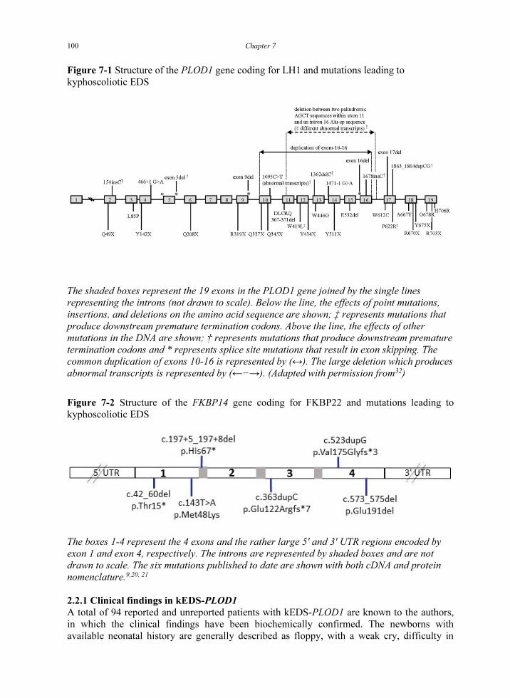
Figure 7-1 Structure of the PLOD1 gene coding for LH1 and mutations leading to
kyphoscoliotic EDS
The shaded boxes represent the 19 exons in the PLOD1 gene joined by the single lines
representing the introns (not drawn to scale). Below the line, the effects of point mutations,
insertions, and deletions on the amino acid sequence are shown; ‡ represents mutations that
produce downstream premature termination codons. Above the line, the effects of other
mutations in the DNA are shown; † represents mutations that produce downstream premature
termination codons and * represents splice site mutations that result in exon skipping. The
common duplication of exons 10-16 is represented by (↔). The large deletion which produces
abnormal transcripts is represented by (←−→). (Adapted with permission from32
)
Figure 7-2 Structure of the FKBP14 gene coding for FKBP22 and mutations leading to
kyphoscoliotic EDS
The boxes 1-4 represent the 4 exons and the rather large 5' and 3' UTR regions encoded by
exon 1 and exon 4, respectively. The introns are represented by shaded boxes and are not
drawn to scale. The six mutations published to date are shown with both cDNA and protein
nomenclature.9,20, 21
2.2.1 Clinical findings in kEDS-PLOD1
A total of 94 reported and unreported patients with kEDS-PLOD1 are known to the authors,
in which the clinical findings have been biochemically confirmed. The newborns with
available neonatal history are generally described as floppy, with a weak cry, difficulty in
Chapter 7100

sucking, and delayed motor development. Sometimes poor foetal movements were reported
by the mothers. In infants, the observed muscle hypotonia accompanied by muscle weakness
contributes to the phenotype of the floppy infant syndrome. As this has the characteristics of a
neuromuscular disorder, extensive neuromuscular work-ups are often initially carried out to
give normal results. It is only at this late stage that a diagnosis of kyphoscoliotic EDS is
considered.12,13
Intellect is unaffected. All individuals with kyphoscoliotic EDS have
hyperelastic and easily stretched skin, with an estimated 60% of individuals subject to
abnormal scarring, characterized by thinness and widening. Bruising occurs easily in all
patients and, in about 50% of cases, bruising is severe.
Kyphoscoliosis is generally present at birth and is progressive and severe in most cases
examined, Figure 7-3. As the vertebral bodies are structurally normal, the scoliosis is
attributed to muscular hypotonia together with laxity of ligaments and is often resistant to
external bracing. As reported in 3 cases, adults with severe kyphoscoliosis are at risk for
complications from restrictive lung disease, recurrent pneumonia, cardiac insufficiency and
early death. A Marfanoid habitus is often striking. Osteoporosis occurs in all individuals but
without tendency to fracture. Recurrent joint dislocations are a common serious problem.
Ocular fragility, which was observed in the original reports of individuals with LH1
deficiency is found in a minority of individuals.4
It can lead to retinal detachment and
bleeding and rupture of the globe, rather than of the cornea as in the brittle cornea syndrome
after minor trauma.14
High myopia (near sightedness) is common. Most patients have
microcornea. The sclerae often have a bluish hue.
Vascular rupture is the major life-threatening complication in this disorder. One patient died
at 51 years with symptoms typical of dissecting aneurysm of the aorta and her brother had a
cerebral accident in the distribution of the right middle cerebral artery at age 19,15
and a third
patient died of arterial rupture at age 14.16
Mitral valve prolapse is common. Maternal and
perinatal mortality in pregnancy has been reported in a case of kyphoscoliosis. Autopsy of the
mother showed a spontaneous rupture of the right iliac artery;17
on the other side, two affected
cousins with the homozygous PLOD1 mutation (c.2198C>T; p.Arg703X) had a total of seven
pregnancies resulting in three miscarriages and four healthy children, three of whom were
born vaginally at term and the fourth was born at 24 weeks; there were no maternal
complications . Steinmann, Rosser and Giunta, unpublished
Life span is markedly shortened because of arterial rupture and cardiopulmonary insufficiency
due to severe kyphoscoliosis.
Interfamilial variability of kEDS-PLOD1 is quite considerable, and intrafamilial variability
has been observed in al least 8 pairs of siblings (one pair of siblings ‘a’ and ‘e’ are shown in
Figure 7-3). 8,10,own observation
This suggests the influence of other factors such as environmental
and genetic factors other than, or in addition to, mutations in the PLOD1 gene in determining
the clinical phenotype.
Kyphoscoliotic, arthrochalasia and dermatosparaxis Ehlers-Danlos syndrome 101
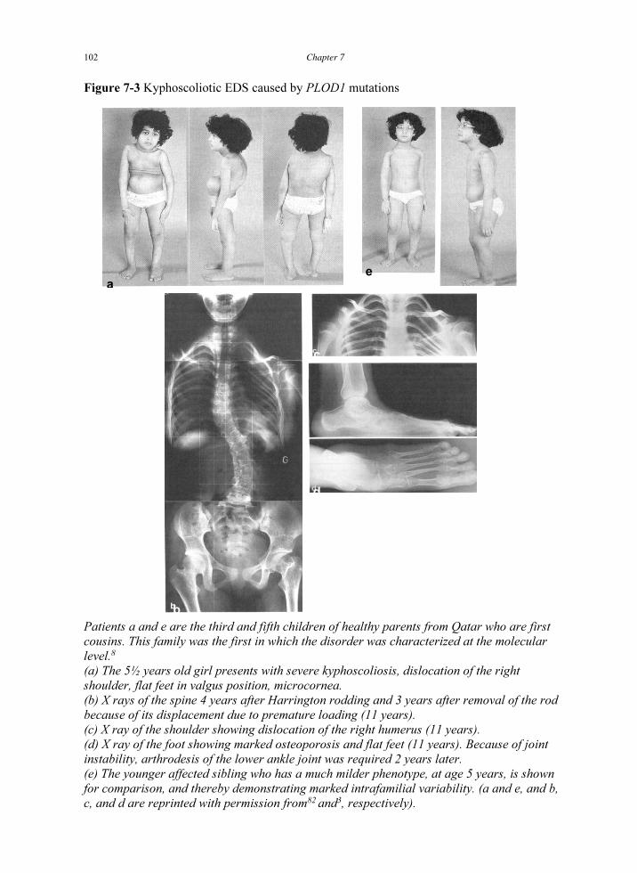
Figure 7-3 Kyphoscoliotic EDS caused by PLOD1 mutations
Patients a and e are the third and fifth children of healthy parents from Qatar who are first
cousins. This family was the first in which the disorder was characterized at the molecular
level.8
(a) The 5½ years old girl presents with severe kyphoscoliosis, dislocation of the right
shoulder, flat feet in valgus position, microcornea.
(b) X rays of the spine 4 years after Harrington rodding and 3 years after removal of the rod
because of its displacement due to premature loading (11 years).
(c) X ray of the shoulder showing dislocation of the right humerus (11 years).
(d) X ray of the foot showing marked osteoporosis and flat feet (11 years). Because of joint
instability, arthrodesis of the lower ankle joint was required 2 years later.
(e) The younger affected sibling who has a much milder phenotype, at age 5 years, is shown
for comparison, and thereby demonstrating marked intrafamilial variability. (a and e, and b,
c, and d are reprinted with permission from82
and3
, respectively).
a
e
c
d
b
Chapter 7102

2.2.2 Clinical findings in kEDS-FKBP14
Individuals with kEDS-FKBP14 present with a clinical phenotype almost indistinguishable
from kEDS-PLOD1 but with a normal ratio of total urinary LP and HP (Figure 7-4 shows 6
patients with kEDS-FKBP14). There are currently 30 patients diagnosed with this disorder on
a molecular level which are known to the authors (26 of them have been published).18,19,20,21
All affected individuals are reported to have muscle hypotonia at birth and delayed gross
motor development, progressive scoliosis either congenital or of later onset, joint
hypermobility, hyperelastic skin, myopathy and hearing impairment.21
Because of severe
early onset muscle hypotonia and delayed motor development, affected children usually
undergo extensive neuromuscular workup in the neonatal period. However, symptoms of
congenital hypotonia and weakness may improve with age during childhood. Unassisted
walking has been shown to be delayed ranging from 2 to 7 years.21
Reduced motor function
and fatigue in adulthood was only reported in 2 adult patients 9,19
and might represent an age-
dependent feature in the natural course of the disorder.
In 7 patients prominent congenital contractures of fingers, wrist, elbows or knees have been
described
and amelioration was documented in half of the affected.21
A cleft of the soft palate was observed in two patients9
, a bifid uvula in one patient20
. and a
cleft palate, defined as bifid uvula with submucous cleft palate or wide cleft palate was found
in five patients.21
Vascular complications, for instance a dissection of the celiac artery at age 41 years19
, a
hypogastric artery pseudoaneurysm rupture at age 6 years20
, an internal carotid artery
dissection at age 50 years21
and tortuosity of the arterial tree have been described. Moreover,
in a baby at one month of age echocardiogram showed a pseudo bicuspid valve, small
muscular ventricular septum defect (VSD), and dilatation of the ascending aorta, but normal
cardiac function. A later echocardiogram at 10 months in addition showed a small atrial
septum defect (ASD) and aortic dilatation with a Z score of +3.75 just beyond the tubular
junction.21
Occasional features underlying systemic connective tissue involvement have been described
such as subdural hygroma (potentially due to subdural bleeding or spontaneous intracranial
hypotension), bluish sclerae, bladder diverticula, inguinal or umbilical herniae, and premature
rupture of membranes.21
Kyphoscoliotic, arthrochalasia and dermatosparaxis Ehlers-Danlos syndrome 103

Figure 7-4 Kyphoscoliotic EDS caused by FKBP14 mutations: clinical findings
The clinical phenotype of six patients with kEDS-FKBP14. (a) 8 year-old patient with the
p.Met48Lys mutation after surgical correction of the progressive severe scoliosis; joint
hypermobility, congenital talipes and pes planus; (b) 4 year-old boy with severe
kyphoscoliosis (onset at 6 months of age) and postural talipes; (c) Micrognathia at age 1
year, skin hyperextensibility valgus position of the foot and hallux varus; (d) Pes planovalgus
and rocker bottom feet; small joint hypermobility; (e) 8 year-old girl presenting with pectus
excavatum; (f) Girl aged 9 years with contracture of the elbows, kyphoscoliosis, pes
planovalgus and follicular hyperkeratosis.
Chapter 7104

2.3 Prevalence and demographics
The kyphoscoliotic EDS is rare; the exact prevalence is unknown. A disease incidence of
approximately 1:100,000 live births is a reasonable estimate. Prevalence does not vary by
gender, race or ethnicity. The carrier frequency is estimated to be 1:150.
2.4 Genetic defect
2.4.1 The normal PLOD1 gene product
Lysyl hydroxylase 1 (LH1; EC 1.14.11.4; procollagen-lysine, 2-oxoglutarate 5-dioxygenase;
PLOD1; OMIM 153454) exists as a dimer of identical subunits of molecular weight
approximately 80-85 kDa, depending on the state of glycosylation.22
Each unit is comprised
of 709 amino acids with a signal sequence of 18 residues. The C-terminal region is very well
conserved among species; residues 570-709 of the processed polypeptide are 94% identical
between chicken and human LH1 and the region between residues 621 and 697 is even more
highly conserved (99%) and is thought to contain the active site of the enzyme. In the human
polypeptide, two histidine residues (H638 and H690) and one aspartate (D640) that bind to
Fe2+
, together with an arginine (R700) that binds to α-ketoglutarate, have been shown to be
critical residues in the active site of the LH isoforms.23,24
Human LH1 has four potential
attachment sites for asparagine-linked oligosaccharide units.23
Unlike prolyl 4-hydroxylase (P
4-H), LH1 does not have endoplasmic reticulum (ER)-specific retention motifs in the primary
structure, but a single 40 amino acid C-terminal peptide segment has been identified in LH1
that mediates both membrane association and localization of LH in the ER.25
2.4.2 Enzyme and cofactors
The ascorbate-dependent enzyme LH requires Fe2+
, α-ketoglutarate, and O2 as cofactors and
catalyses the formation of hydroxylysines in collagen chains and other proteins with collagen-
like amino-acid sequences.26,27
The hydroxylysine groups of collagen have at least two
important functions: their hydroxy groups are essential for the stability of the intermolecular
collagen cross-links that provide tensile strength to most soft tissues and bone, and they serve
as attachment sites for galactose or glucosylgalactose. This enzymatic glycosylation event is
catalysed by another member of the LH family, the multifunctional enzyme LH3.28
2.4.3 Collagen cross-linking
The strength and type of collagen cross-links formed depend on telopeptide (non-helical)
lysines and whether or not they are hydroxylated. The cross-links that are formed between
lysyl and hydroxylysyl residues can be formed via two major related routes: the allysine route
in which the cross-link is formed from telopeptide lysine, and the hydroxyallysine route in
which the cross-link is formed from telopeptide hydroxylysine. The bifunctional cross-links
formed via the hydroxyallysine route (in which the hydroxylysine-derived aldehyde reacts
with a specific peptidyl lysyl or hydroxylysyl residue in the triple helical domain of a
neighbouring collagen molecule) mature into either the trifunctional crosslinks, hydroxylysyl-
pyridinoline (HP; formed from three hydroxylysyl residues) or lysyl-pyridinoline (LP; formed
from two hydroxylysyl and one lysyl residue). The relative expression of these two types of
cross-links therefore demonstrates the degree of lysyl hydroxylation which has occurred and
provides the basis for a diagnostic test for lysyl hydroxylase deficiency in which a highly
increased LP : HP ratio is indicative of kyphoscoliotic EDS.29
2.4.4 PLOD1 gene
The 40kb PLOD1 gene coding for human LH1 is located in the p36.2→p36.3 region of
chromosome 1 and includes 19 exons and an unusually large first intron of 12.5kb.6
The gene
Kyphoscoliotic, arthrochalasia and dermatosparaxis Ehlers-Danlos syndrome 105

has a 5’ flanking region with characteristics shared by housekeeping genes.30
The constitutive
expression of the gene in different tissues suggests that LH1 has an essential function.7
The
presence of five and eight Alu-sequences located in introns 9 and 16, respectively, result in
extensive homology thereby offering potential sites for recombination.31
2.4.5 Mutations in the gene coding for lysyl hydroxylase
The mutations in the PLOD1 gene responsible for decreased enzyme activity that result in the
clinical phenotype of kyphoscoliotic EDS are described in Figure 7-1. More than 30 different
mutations in PLOD1 have been associated with kyphoscoliotic EDS.10,11,32-35
These mutations
are located throughout the gene. The most common mutation, an 8.9-kb duplication of seven
exons (exons 10 to 16), is caused by a homologous recombination event between identical 44-
bp Alu sequences in introns 9 and 16.36
The allele frequency of the duplication is 20% in
probands with kyphoscoliotic EDS, from 94 families.37
Two other common mutations in
PLOD1 occur in exon 9 (R319X) and exon 14 (Y511X) at allelic frequencies of 10% and 5%,
respectively. Other mutations are as described in Figure 7-1.
2.4.6 FKBP14 gene and FKBP22 protein function
The 16kb FKBP14 gene coding for human FKBP22 maps to the minus strand of chromosome
7p14.3 and consists of 4 small exons and 3 rather large introns. It is conserved among
chimpanzee, Rhesus monkey, dog, cow, mouse, rat, chicken, zebrafish, fruit fly, mosquito,
and frog. FKBP22 belongs to the FK506-binding protein (FKBP) class of immunophilins,
which have been implicated in catalysing cis-trans-isomerization of peptidyl-prolyl peptide
bonds and are supposed to accelerate protein folding. It has been shown that FKBP22
catalyses the folding of type III collagen and interacts with type III collagen, type VI collagen,
and type X collagen, but not with type I collagen, type II collagen, or type V collagen.38
Remarkably, mutations in type III and type VI collagens are the genetic hallmark of the
vascular type of EDS (vEDS) and of COL6-related dystrophies, respectively. Therefore, the
clinical features of vascular abnormalities and myopathy documented in the affected
individuals clearly correlates with the interaction of FKBP14 with type III and VI collagens.21
2.4.7 Mutations in the gene coding for FKBP22
The mutations in the FKBP14 that result in the clinical phenotype of kyphoscoliotic EDS are
described in Figure 7-2. Nucleotide numbering starts with +1, which corresponds to the first
ATG translation initiation codon at position 294 of the reference sequence NM_017946.3,
whereas amino acid residues are numbered from the first methionine residue of the reference
sequence NP_060416.1
Since its first description in 20129
only six different mutations have been associated with the
disorder.21
These disease causing variants are located throughout the gene. The mutation
c.362dupC p.(Glu122Argfs*7) which is found with a frequency of approximately 70% and it
is thus considered to be a common disease variant, has been linked to the same haplotype in
all individuals tested.9,19
2.5 Inheritance
The kyphoscoliotic EDS is inherited as an autosomal recessive trait. This is supported by the
occurrence of affected siblings from unaffected parents and by the birth of affected children
from consanguineous parents.
2.6 Diagnostic tests
The clinical diagnosis of kyphoscoliotic EDS is generally suspected very late and often only
after a negative workup of neuromuscular disorders. For kEDS-PLOD1, the clinical diagnosis
Chapter 7106

is confirmed biochemically by measuring the ratio of collagen-derived pyridinoline cross-
links in the urine. Whereas, for kEDS-FKBP14 molecular genetic analysis by either Sanger
sequencing of FKBP14 or NGS sequencing of a gene panel including FKBP14 is mandatory
for confirming the clinical diagnosis.
1) Deficiency of the enzyme LH1 results in a significant decrease in hydroxylysine-based
pyridinoline cross-links in collagens. Detection of an increased ratio of lysyl-pyridinoline
(LP) to hydroxylysyl-pyridinoline (HP) cross-links in urine, quantitated by high-performance
liquid chromatography (HPLC) is a non-invasive, highly sensitive and specific test for kEDS-
PLOD1. The normal ratio of cross-links LP : HP is approximately 1 : 4 (~0.2), whereas in
kyphoscoliotic EDS due to PLOD1 mutations the ratio is approximately 6 : 1 (10-40 times
increase, range 2-9).3
Such testing is clinically available.39
Obligate heterozygous parents
have normal values.
2) Mutation analysis of the PLOD1 gene may be carried out to identify the mutation(s)
responsible for the enzyme deficiency.2,10,32
As the most common mutation in the PLOD1
gene is the large seven exon duplication caused by an Alu-Alu recombination in introns 9 and
16 in the PLOD1 gene occurring at a frequency of approximately 30%,2
the gene should be
initially screened for this mutation. The duplication can be readily detected/confirmed in
genomic DNA by MLPA of PLOD1 or PCR using duplication-specific primers.11,34
Detection
of other mutations may involve sequence analysis of all exons and flanking intronic
sequences.13
Otherwise, mutation analysis can be carried out by targeted resequencing of a
gene panel that includes PLOD1, a method that also allows the detection of the common
duplication.
3) The determination of LH activity in patients’ fibroblasts as well as the measurement of
hydroxylysine content of dermis is now used for research purposes, only.40,41
It should be noted here that carriers of a PLOD1 mutation can only be identified by mutation
analysis and not by biochemical testing or enzyme assay.
2.7 Prenatal testing
Prenatal diagnosis for pregnancies at risk is possible by analysis of DNA extracted from foetal
cells obtained by amniocentesis usually performed at approximately 15-18 weeks' gestation or
chorionic villus sampling (CVS) at approximately 10 to 12 weeks' gestation. Both disease-
causing alleles must be identified before prenatal testing can be performed.32,42
2.8 Differential diagnosis
The kyphoscoliotic EDS has some overlapping clinical features with other types of EDS,
particularly classical and vascular EDS. Abnormal wound healing and joint laxity are present
in many EDS types. Although all types of EDS are characterized by a relatively high risk of
scoliosis compared to the general population, scoliosis in kyphoscoliotic EDS is usually more
severe and of earlier onset than that seen in other EDS types. Most congenital myopathies
present with poor muscle tone and increased range of motion of small and large joints. Joint
laxity can be difficult to distinguish from muscle hypotonia, particularly in infants and
children. In kyphoscoliotic EDS, in which both hypotonia and joint laxity are present, the
increased range of motion is often striking. Primary myopathies caused by mutations in
collagen, such as the type VI (COL6) collagenopathy Ullrich congenital muscular dystrophy
(UCMD; MIM 254090) may show substantial connective tissue involvement including
kyphoscoliosis, joint hypermobility, joint contractures and abnormal scar formation, however
velvety and hyperextensible skin is absent and motor function does not improve with age.
Unlike spinal muscular atrophy, kyphoscoliotic EDS is characterized by normal tendon
reflexes. Many syndromic and metabolic disorders include early-onset hypotonia. In these
Kyphoscoliotic, arthrochalasia and dermatosparaxis Ehlers-Danlos syndrome 107
disorders, however, the other manifestations of kyphoscoliotic EDS are generally absent, and
additional features usually present. Kyphoscoliotic EDS needs also to be differentiated from
myopathic EDS, a new entry in the 2017 International classification and due to heterozygous
or biallelic COL12A1 mutations, since it also features congenital muscle hypotonia (see also
chapter 2, table 2-5).1,2
The Nevo syndrome, first reported in 1974, has been identified as an allelic condition to the
kyphoscoliotic EDS and is clinically indistinguishable from it.43
Recently, rare autosomal recessive entities with distinct molecular and biochemical
abnormalities such as Brittle cornea syndrome, musculocontractural EDS, and the
spondylodysplastic EDS have been described which should be considered in the differential
diagnosis of the kyphoscoliotic EDS.2
These disorders
have been classified among the types
of EDS because of shared common characteristics, such as joint hypermobility, skin
hyperextensibility, and tissue fragility. These disease entities except for spondylodysplastic
EDS, due to SLC39A13 mutations, are biochemically distinct from kyphoscoliotic EDS since
the urinary pyridinoline ratios measured in the patients are within the normal range.
Brittle cornea syndrome (BCS; MIM 229200) is an autosomal recessive generalized
connective tissue disorder caused by mutations in ZNF469 and PRDM5.44,45
It is characterized
by extreme thinning and fragility of the cornea that may rupture in the absence of significant
trauma leading to blindness. Keratoconus or keratoglobus, high myopia, blue sclerae,
hyperelasticity of the skin without excessive fragility, and hypermobility of the small joints
are additional features of BCS.
The musculocontractural EDS is due to mutations in CHST14 (OMIM 601776) and DSE
(OMIM 615539), respectively.46
Neonates with mc-EDS present with distal arthrogryposis
and muscular hypotonia. Musculocontractural EDS patients develop a Marfanoid habitus with
narrow shoulders and spinal and pectus deformities, brachycephaly, a characteristic facial
appearance, hyperextensible and bruisable skin, a tendency to atrophic scars, tapering fingers,
instability of large joints, hyperalgesia to pressure, and recurrent subcutaneous haemorrhages
are consistent clinical findings. Musculocontractural EDS patients show muscular hypoplasia
and weakness.
The spondylodysplastic EDS (OMIM 612350)47,48
is a very rare, recessive connective tissue
disorder caused by mutations in SLC39A13 (OMIM 608735) encoding the zinc transporter
ZIP13. Clinically it resembles kyphoscoliotic EDS, with some distinct phenotypic
manifestations including platyspondyly, osteopenia, short stature, widened metaphyses,
tapered fingers, thenar muscle atrophy and a tendency to develop contractures of the small
joints. An elevated ratio of urinary pyridinolines LP : HP (but to a lesser degree than in
kyphoscoliotic EDS), and underhydroxylated collagens in culture despite normal in vitro
activities of lysyl-and prolyl 4-hydroxylases, respectively, are characteristic biochemical
findings.47
Spondylodysplastic EDS can also be caused by mutations in 2 other genes,
B4GALT7 and B3GALT6 (see also chapter 2, table 2-5), in which the LP : HP ratios are
normal.1,2
Establishing a specific diagnosis of patients with features resembling the kyphoscoliotic EDS
currently relies on the analysis of urinary pyridinoline cross-links, and upon exclusion of this
form, on the accessibility of modern genomic technologies based on gene targeted exome-
sequencing.
Chapter 7108

2.9 Management
2.9.1 Evaluations following initial diagnosis
To establish the extent of disease in an individual diagnosed with kyphoscoliotic EDS, the
following evaluations are recommended:
*Musculoskeletal: To evaluate kyphoscoliosis, photographic and radiologic documentation of
the spine are recommended in view of the often progressive character of the kyphoscoliosis.
Physical therapy evaluation is needed to develop a plan for ongoing therapy.
*Cardiovascular: Measurement of aortic root size and assessment of heart valves by
echocardiogram should be carried out at the time of diagnosis or by age five years. Awareness
for possible serious vascular complications in different age groups in patients with kEDS-
FKBP14 should be raised, and vascular screening should start at pediatric age.
*Ophthalmologic: Formal ophthalmologic evaluation is needed at diagnosis for myopia,
astigmatism, and potential for retinal detachment.
2.9.2 Treatment of manifestations
*Musculoskeletal: Referral to an orthopaedic surgeon for management of kyphoscoliosis is
appropriate. Orthopaedic surgery is not absolutely contraindicated in individuals with
kyphoscoliotic EDS and can be performed if necessary. Physical therapy is recommended for
older children, adolescents, and adults to strengthen large muscle groups, particularly at the
shoulder girdle, and to prevent recurrent shoulder dislocation. Swimming is recommended.
*Cardiovascular: Vigilant observation and control of blood pressure can reduce the risk of
arterial rupture. Vascular surgery is fraught with danger. Although virtually no surgical
literature exists on kyphoscoliotic EDS, a review on surgical complications of vascular EDS
is relevant.33
Individuals with aortic dilation may require treatment with beta blockers or
losartan to prevent further expansion.
*Ophthalmologic: Myopia and/or astigmatism may be corrected by glasses or contact lenses.
Laser treatment of the retina is indicated in case of imminent detachment.
2.9.3 Surveillance
The following routine examinations are recommended: 1. Ophthalmologic examination for
management of myopia and early detection of glaucoma or retinal detachment. 2. Routine
examination for inguinal hernia and surgical referral as necessary. 3. Vigilant observation of
blood pressure. 4. Regular follow-up by an orthopaedic surgeon for management of
kyphoscoliosis. 5. Echocardiogram at five-year intervals (or less, depending on the severity of
findings), even if the initial echocardiogram is normal by ultrasound measurements.
2.9.4 Other
Ascorbate has been suggested as a treatment, but its effectiveness has not been biochemically
proven.2
3. Arthrochalasia EDS
3.1 Diagnostic criteria
The arthrochalasia EDS is a rare inherited autosomal dominant trait caused by mutations
leading to the entire or partial loss of exon 6 of COL1A1 (formerly EDS type VIIA) and
COL1A2 (formerly EDS type VIIB) during pre-mRNA maturation, and thus affecting the
processing of the amino terminal end of proα1(I) or proα2(I) chains of collagen I,
respectively. It is characterized as follows:1
Kyphoscoliotic, arthrochalasia and dermatosparaxis Ehlers-Danlos syndrome 109

Major diagnostic criteria
o Congenital bilateral hip dislocation
o Severe generalized joint hypermobility with multiple dislocations/ subluxations
o Skin hyperextensibility
Minor diagnostic criteria
o Muscle hypotonia
o Kyphoscoliosis
o Mild osteopenia (X-ray)
o Tissue fragility, including atrophic scars
o Easy bruisable skin
Minimal criteria for diagnosis are congenital bilateral hip dislocation plus either skin
hyperextensibility or severe generalized joint hypermobility with multiple dislocations/
subluxations and at least 2 other minor criteria. When these minimal criteria are met,
verifying laboratory testing is warranted.1,2
3.2 Background
Hass and Hass suggested that there is a distinct entity of loose-jointedness, which they called
arthrochalasis multiplex congenita (congenital flaccidity of the joints), which represents, to
some extent, the antithesis of “arthrogryposis multiplex congenita” (a rare congenital disorder
characterized by multiple joint contractures) and which may occur with or without skin
changes.49
This disorder was classified as EDS type VII.50
In 1973, three patients with EDS
type VII were reported who accumulated procollagen in their skin and tendons, and it was
thought, therefore, that their disorder resembled dermatosparaxis in cattle.51
Since it was felt
that the accumulation of procollagen was most likely to be due to a defect in the conversion of
procollagen to collagen, the activity of the converting proteinase was determined in the
culture medium of fibroblasts from these patients and found apparently to be reduced to
between 10 and 40% of normal.51,52
However, when Steinmann and colleagues reinvestigated
the youngest of the three reported patients52
they found the activity of the procollagen
N-proteinase to be normal in cell extracts, and the presence of mutant pNα2(I)-like chains in
collagen extracted from skin or produced by fibroblasts.53
The authors concluded that there
was a structural abnormality in the portion of the proα2(I) chain that is normally cleaved by
N-proteinase (and other proteinases). The authors speculated further, that for sterical reasons
molecules containing pNα2(I) chains would interfere with fibrillogenesis and cross-linking,
thus leading to abnormal collagen fibrils and increased solubility of collagen. After the
description of a similar patient, in whom the condition was due to a mutant pNα1(I) rather
than pNα2(I) chain, the classification of EDS VII was subdivided into types VIIA and VIIB,
respectively, depending on whether the α1(I) or α2(I) chain was affected.2,3,54
3.3 Clinical findings
The hallmark of arthrochalasia EDS is the involvement of ligaments and joint capsules. The
disorder is characterized by severe multiple joint hypermobility and recurrent subluxations
and luxations of both small and large joints, and ligamentous tears. To date, there are at least
51 patients from 38 families published or known to the authors. All patients are ascertainable
in the newborn period, although the correct diagnosis is still frequently only made years later.
Congenital bilateral hip dislocation is the rule (Figure 7-5a and 7-6), and muscular hypotonia
is prominent; both factors predispose to the high incidence of breech presentation and delayed
gross motor development. Short stature, if present, is due to severe thoracolumbar scoliosis
Chapter 7110

and hip dislocation. In striking contrast to dermatosparaxis EDS, the skin is only moderately
affected; it is usually thin, velvety, and moderately extensible, doughy and redundant over the
limbs. An unusual criss-cross patterning of the skin on the hands and feet has been observed
(Figure 7-5e and 7-5f)55
, and occasionally the skin is affected by poor healing of wounds with
atrophic and haemosiderotic scars, especially in the adult. The face appears to be normal,
however facial dysmorphic features with frontal bossing, hypertelorism, depressed nasal
bridge, macrostomia and bluish sclerae have been described (Figure 7-5a and 7-5b).55
Judging
by six affected members from a three-generation family, intrafamilial variability is slight.
The occurrence of recurrent fractures, Wormian bones (also known as sutural bones or Inca
bones: abnormal extra bone plates in the skull), and abnormally wide fontanels indicate that
the arthrochalasia EDS phenotype includes bone changes and fragility similar to those
reported in mild osteogenesis imperfecta (OI).56
Wormian bones have been observed in
several patients. X-rays of one of our patients56
disclosed bone abnormalities such as
exostoses, e.g. discrete occipital horns (parasagittal bone exostoses arising from the occipital
bone), a thickened calvarium (skullcap), and Wormian bones.
Figure 7-5 Arthrochalasia EDS. Clinical photos before and after open reduction of the hip
dislocation 55
a, c, e, f: The patient at 12 months of age presents with bilateral hip dislocation with
lateralisation and elevation of both femurs, and anterolateral subluxation of the knees. The
skin is doughy and redundant mostly over the abdomen, the limbs, the fingers and the toes. A
marked criss-cross patterning of the skin on the hands and feet, and contractures of the first,
Kyphoscoliotic, arthrochalasia and dermatosparaxis Ehlers-Danlos syndrome 111

second and fourth digits of both hands are present (e, f). Note the excess skin folds and the
large umbilical hernia (c, circle).
b, d: The patient at 23 months of age, photographed 6 months after successful open reduction
of bilateral hip dislocation. Postural thoracolumbar kyphosis due to hypotonia and ligament
laxity is shown (d). From 55
with permission.
Figure 7-6 Arthrochalasia EDS, due to COL1A1 mutation. Radiographs before and after open
reduction of the hip dislocation 55
a) Radiograph of the pelvis from the patient at 12 months of age shows bilateral luxation of the
hips, and anterolateral subluxation of the knees due to severe ligamentous instability.
b) Radiograph of the pelvis from the patient at 23 months of age, 6 months after open reduction,
shows successful correction of bilateral dislocation of the hips.
c) A radiograph of the right foot shows the disorganization of the metatarsal bones. Please note
the striking osteopenia which is more marked than in arthrochalasia EDS due to COL1A2
mutation. From 55
with permission.
3.4 Defect and pathogenesis
The molecular defect in all cases elucidated to date is remarkably homogeneous: entire or
partial loss of exon 6 during pre-mRNA processing causes lack of the N-telopeptide linking
the N-propeptide to the major triple helical domain of the α1(I) and the α2(I) chain,
respectively. Deletion of the N-telopeptides in the pro-α1(I) (24 amino acid residues) and the
pro-α2(I) chain (18 amino acid residues57
) results in loss of the small globular region of the
Chapter 7112

N-propeptide (present only in the pro-α1(I) chain), the N-proteinase cleavage site (Pro-Gln
and Ala-Gln), the cross-linking lysine residue of the N-telopeptide, the cleavage site for
proteinases (for example, pepsin and a-chymotrypsin) and the first triplet of the main helical
Gly-X-Y domain. The loss of the N-proteinase cleavage site causes the retention of the N-
propeptide in the mature α1(I) and α2(I) molecules, which are referred to as pNα1(I)- and
pNα2(I)-like chains, respectively.
In two patients with arthrochalasia EDS, a G to C change at the splice acceptor site of intron 5
in COL1A2 activates a cryptic splice site in exon 6, removing 5 amino acid residues including
the N-proteinase cleavage site, but preserving the cross-linking lysine residue.58, Giunta &
Steinmann, unpublished
Both mother and son, and father and daughter, respectively, were affected
with typical arthrochalasia EDS.
Analysis by SDS-polyacrylamide gel electrophoresis (SDS-PAGE) of pepsin-digested
collagens produced by fibroblasts of affected patients reveals, in addition to normal α1(I) and
the α2(I) chains, the presence of mutant pNα1(I)- or pNα2(I)-like chains, respectively. The
pNα2(I)-like chains migrate between the α1(I) and the α2(I) chains of collagen I, whereas the
pNα1(I)-like chains migrate as a faint band just above the normal α1(I) chains (Figure 7-7).
Studies by Wirtz et al.59
showed that in arthrochalasia EDS (type VIIB), the α1(I)
N-propeptide, although cleaved from the α1(I) chain by the N-proteinase or (less likely) by
non-specific proteases, is retained by non-covalent association with the mutant pNα2(I) chain
in native mutant collagen molecules, both in vivo and in vitro; the α1(I) N-propeptide was
readily demonstrable by Western blotting of skin extracts, and rotary shadowing of
pepsin-treated collagen produced in culture disclosed kinked molecules which were longer
than their normal counterparts.59
These data suggest that the retention of a partially cleaved,
but essentially intact N-propeptide in mutant collagen may play a critical role in the
pathogenesis of this disease.
Figure 7-7 SDS–PAGE gel electrophoresis of collagens produced by skin fibroblasts from
patients with EDS VIIA (arthrochalasia EDS, due to COL1A1 mutation) and EDS VIIB
(arthrochalasia EDS, due to COL1A2 mutation) and controls (C)
Legend: Collagens extracted from the medium fraction (A) and the cell layer (B) are shown.
The strong pNα1(I) band in the EDS VIIA patient is barely detectable in the control (C).
Similarly, the pNα2(I) band is present in the EDS VIIB patient only. From 55
with permission.
Kyphoscoliotic, arthrochalasia and dermatosparaxis Ehlers-Danlos syndrome 113

3.5 Morphology
The skin collagen fibrils have a smaller and more variable diameter than normal. They are
irregular in outline, and also appear to be more loosely and randomly organized into fibrils
resembling a loosely wound rope (Figure 7-8).55,60
These changes are more pronounced in the
patients affected with arthrochalasia EDS, due to mutation in COL1A1 (Figure 7-8a and 7-8c)
compared to those observed in arthrochalasia EDS, due to COL1A2 mutation (Figure 7-8b and
7-8d), but less severe than those present in dermatosparaxis EDS (Figure 7-9).
3.6 Inheritance
Arthrochalasia EDS is inherited as an autosomal dominant trait, and some of the sporadic
cases have been shown to be heterozygous for a new mutation. Gonadic mosaicism may
occur. From published results, it appears that the relative incidence of arthrochalasia EDS,
due to COL1A2 mutation is higher compared to that of arthrochalasia EDS due to COL1A1
mutation, although the risk of a spontaneous mutation affecting exon 6 is expected to be
similar for both COL1A1 and COL1A2 genes.2
This may have to do with the difficulty in
detecting the latter type by routine standard biochemical techniques, as shown recently.55
3.7 Genotype-phenotype correlation and locus heterogeneity
Interfamilial and intrafamilial phenotypic variability seems to be slight. In arthrochalasia
EDS, due to COL1A2 mutation, the heterotrimeric collagen I molecules consist of one
population of normal collagen I and one population of abnormal collagen I containing the
mutant α2(I) chain. In contrast, in arthrochalasia EDS, due to COL1A1 mutation, three
quarters of collagen I molecules would be expected to be abnormal because they contain one
or two mutant α1(I) chains and, thus, the phenotype would be expected to be more severe
than that due to COL1A2 mutation.
The clinical documentations demonstrate the severity of COL1A1 mutation type and clearly
suggests that the COL1A1 mutation type is more severe than the COL1A2 mutation type, as
expected by the different stoichiometry of the α(I)-chains.55
This statement is supported by
the more extensive ultrastructural changes of the collagen fibrils55
, as well as by the
comparison of the five personally examined patients with the COL1A2 mutation type.
However, among the reported cases of the COL1A1 mutation type, three involve substitutions
for the last nucleotide of exon 6. This results in two alternative splice variants, one in which
the exon 6 sequence from mRNA is completely deleted, and another in which normal splicing
is permitted, and the protein product contains an isoleucine rather than a methionine at
position 3 of the first Gly-X-Y triplet of the triple helix. The normally spliced product
containing an amino acid substitution diminishes the dysfunctional effect of the three quarters
of abnormal molecules; this results in a less severe clinical phenotype which is
indistinguishable from that of the COL1A2 mutation type. In contrast, complete skipping of
exon 6 from mutant proα1(I) chains leads to three quarters of the collagen I molecules being
abnormal and dysfunctional, thus explaining the more severe phenotype, which almost
resembles dermatosparaxis EDS, in which none of the collagen I molecules are processed.56
Locus heterogeneity (trait caused by mutations in genes at different chromosomal loci) is
exemplified by several patients who are clinically similar to arthrochalasia EDS. However,
they have smooth, hyperelastic skin but without skin fragility. Their fibroblasts produce
normal collagens53
, the procollagen N-proteinase is active and the electron microscopic
appearance of dermal collagen fibrils is normal. It remains uncertain whether patients such as
these are examples of EDS types of unknown cause, or whether they represent the extreme
end of severity of hypermobile EDS.
Chapter 7114

Figure 7-8 Ultrastructural appearance of collagen fibril cross sections in the dermis of two
patients with arthrochalasia EDS: one due to COL1A1 mutation (VIIA; a and c) and one due to
COL1A2 mutation (VIIB; b and d)
Legend: The skin collagen fibrils of the EDS VIIA patients (a55
; c83
) are more loosely and
randomly organized, and have highly irregular contours (note the “angel-like” shape of the
fibril in the left inset) and have more variable diameters than normal (right inset). Two
patients with EDS VIIB (b56
; d (figure 323
) have loosely organized fibrils with irregular
contours and more variable diameters than controls; however the contours of the fibrils are
far less abnormal than those of arthrochalasia EDS, due to COL1A1 mutation (VIIA) . Please
note that the EM photographs are at different scales (the black bars correspond to 500 nm,
while the white bars correspond to 600nm). From 55
with permission.
Kyphoscoliotic, arthrochalasia and dermatosparaxis Ehlers-Danlos syndrome 115

Figure 7-9 Structure of collagen fibrils: of normal human controls, of mouse models of
dermatosparaxis EDS, of patients with dermatosparaxis EDS
Collagen fibrils from skin of human (A, D, E, F) and mouse (B, C) studied by transmission
electron microscopy. In normal skin (A, B), collagen fibrils appear cylindrical in cross
section and elongated with a typical banded pattern in longitudinal section. In the animal
model for the disease (C, ADAMTS2 null mouse), and in dermatosparaxis EDS patients (D:
P7; E, and F: P8), fibrils are hieroglyphic in cross section and disorganized in longitudinal
section. The level of observed hieroglyphic pattern is influenced by the angle of the section
and can vary slightly between different regions within a single skin biopsy (C-F). Scale:
lengths of black bars in right lower corners = 0.3μm.
3.8 Diagnosis and differential diagnosis – EDS-OI overlapping syndrome
The diagnosis is first made on clinical grounds, supported by the abnormal collagen fibril
morphology, as well as by protein data pertaining to collagen extracted from skin or produced
by fibroblasts, and proven by the demonstration of complete or partial loss of exon 6 in cDNA
and of the mutation in genomic DNA. The existence of individuals with a clinical picture of
arthrochalasia EDS but normal collagen I structure, normal procollagen N-proteinase activity
and normal electron microscopic findings in skin indicates further heterogeneity.
Congenital hip dislocation is also found in Larsen syndrome (OMIM 150250, 245600), and as
an isolated disorder (OMIM 142700), and in an as yet less well defined disorder, i.e.,
dislocation of the hip, congenital, with hyperextensibility of fingers and facial dysmorphism
(OMIM 601450).
Assuming paternity, the risk to healthy parents of having another affected child is small, and
due to the possibility of parental germline mosaicism. The mother of the four patients
reported61
was the least severely affected individual within the family and may either
represent a symptomatic mosaicism of the mutation in COL1A2, or may simply be the
expression of intrafamilial variability (G. Wallis, personal communication 1991). The risk of
Chapter 7116

an affected person transmitting the disorder is 50%, and prenatal diagnosis is possible by
either DNA or protein studies on a chorionic villus biopsy if the defect in the index case has
been characterized.62
Glycine substitutions in the first 90 residues of the α1(I) helical region disrupt a stable
N-anchor domain and prevent or delay N-propeptide removal. The pNα1(I) chain is
incorporated into the matrix, decreases fibril size and causes a phenotype with characteristics
of both OI and EDS.63,64
Disruption of N-propeptide processing by defects in the helical part
of the α2(I) chain also leads to this phenotype.65-70
In particular, one patient70
presented with
generalized osteoporosis, platyspondyly, Wormian bones, blue sclerae, but no fractures, and
carried a 19 bp deletion that caused skipping of exon 11 of COL1A2,67
thus deleting one of the
lysyl residues involved in intermolecular cross-link formation. The presence of the shortened
proα2(I) chain in procollagen synthesized by the proband's fibroblasts both lowered the thermal
stability of the molecules and prevented or delayed their processing by procollagen
N-proteinase.65
Thus, the deletion disturbed both the helical domain and the cleavage site of the
molecule, thereby resulting in the mixed EDS-OI phenotype, which is not included in the 2017
International EDS classification.
3.9 Management
The orthopaedic outcome is often unsatisfactory as premature degenerative arthritis of the
hips and other joints occurs.
The principal orthopaedic problem in arthrochalasia EDS patients is bilateral congenital
dislocation of the hips. To gain an insight into the management of this problem, Giunta et al.
reviewed the treatments and their outcomes in 16 patients from 12 families.56
Stable
reductions were infrequently achieved following closed reduction with orthoses or hip spicas.
Anterolateral open reductions with capsular plication, even in infancy, were also inadequate
as most of the patients continued to have subluxated or dislocated hips. The poor outcome of
the latter procedure is likely to be due to early stretching of the capsulorrhaphy sutures. In
contrast, in few patients the addition of an iliac osteotomy of the Pemberton or Salter type
with or without femoral osteotomy achieved some good results.
From published results and the findings of Giunta et al., it appears that open reduction with
capsulorrhaphy and iliac osteotomy, with the addition of femoral osteotomy in some cases, is
required to achieve and maintain stable reduction.9,47
These requirements are similar to those
shown to be necessary to achieve and maintain stable reduction in other laxity conditions such
as Down and Larsen syndromes. As with the latter conditions, careful planning of osteotomy
is required to ensure that adequate acetabular coverage of the femoral head is achieved in the
functional positions of the limb.56
Generalized joint hypermobility is worst in infancy when marked muscular hypotonia
accompanies the severe ligamentous laxity. Motor development is consequently slow, and
aids such as knee-ankle-foot and ankle-foot orthoses are often required to stabilize the lower
limb joints for standing and walking. As muscle tone improves, the knee-ankle-foot orthoses
may be reduced to ankle-foot orthoses. Surgical procedures to stabilize the knees and the
patello-femoral joints have been used occasionally. Orthotic stabilization is likely to be more
reliable. Ankle or subtalar fusions have been undertaken in a few cases but the arthrodesed
joints need to be in an optimal position for this to be successful.56
Recurrent and/or persistent dislocations, as well as hypermobility of upper limb joints, are
also frequently disabling. Operative procedures appear rarely to have been undertaken in the
upper limbs and one would predict that capsulorrhaphy and osteotomy would not stabilize
Kyphoscoliotic, arthrochalasia and dermatosparaxis Ehlers-Danlos syndrome 117

these joints.56
Arthrodesis may be useful in stabilizing some small joints, such as the
metacarpophalangeal joints of the thumbs, although fusion rates cannot be predicted.
Postural thoracolumbar kyphosis due to hypotonia and ligamentous laxity is a feature of
infants (Figure 7-5d). The spinal posture improves as the children gain in muscle power.
However, structural scoliosis has been reported in eight cases. Spinal fusion was undertaken
in two patients, although few details are available concerning curve patterns and surgery.
Spondylolisthesis of L5-S1 was noted in one patient only.
The findings in 20 molecularly proven cases of arthrochalasia EDS reviewed show how
difficult the management is and how important early diagnosis may be.47
The homogeneous
nature of the molecular defects allows laboratories with the appropriate expertise rapidly to
establish the diagnosis, after which the clinical problems, in particular those relating to the
dislocated hips, can be predicted. Adequate physical and occupational therapy and orthotic
management can be given to assist with standing, walking and activities of daily living.
Appropriate surgical treatment of the dislocated hips should also diminish the frequency of
hip re-dislocation, recurrent dislocation, avascular necrosis and premature osteoarthritis.
However, more experience correlating detailed orthopaedic management and long-term
outcome is needed before sound recommendations can be made.56
4. Dermatosparaxis EDS
The dermatosparaxis EDS (OMIM 225410) is caused by deficiency of procollagen N-terminal
proteinase as a result of homozygosity or compound heterozygosity of mutant alleles coding
for the enzyme procollagen N-proteinase. This disorder is inherited as an autosomal recessive
trait and is clinically characterized as follows:1
Major diagnostic criteria
o Extreme skin fragility with congenital or postnatal skin tears
o Characteristic craniofacial features
o Redundant, almost lax skin,
o Increased palmar wrinkling
o Severe bruisability
o Umbilical hernia
o Postnatal growth retardation
o Short limbs, hands and feet
o Perinatal complications due to connective tissue fragility
Minor diagnostic criteria
o Soft and doughy skin texture
o Skin hyperextensibility
o Atrophic scars
o Generalized joint hypermobility
o Complications of visceral fragility (bladder/diaphragmatic rupture, rectal prolapse)
o Delayed motor development
o Osteopenia
o Hirsutism
o Tooth abnormalities
o Refractive errors (myopia, astigmatism)
o Strabismus
Chapter 7118

Minimal criteria for diagnosis are extreme skin fragility with congenital or postnatal skin tears
and characteristic craniofacial features plus either 1 other major and/or 3 minor criteria. When
these minimal criteria are met, verifying laboratory testing is warranted.1,2
4.1 Background
Dermatosparaxis (torn skin) in cattle was the first defect in collagen metabolism to be
elucidated, whereas it has only relatively recently been defined in man.71,72
The hallmark of
dermatosparaxis EDS is the excessive fragility of the skin with mild to absent involvement of
tendons and ligaments; this distinguishes this disorder from arthrochalasia EDS in man, which
is characterized by extreme joint laxity.
4.2 Clinical findings
Currently, only 15 patients from 14 unrelated families have been identified to date. Nine of
the patients were born prematurely after rupture of the membranes, two of them by Caesarean
section one because of breech presentation and the other at 33 weeks gestation because of
foetal heart decelerations.73
. Two additional patients were delivered at term by Caesarean
section, one because of large size, another due to breech presentation. One patient died shortly
after birth (39 weeks of gestation) due to severe hemorrhage and shock.74
The premature boy
born with a gestational age of 33 weeks presented at birth with excessive skin folds, multiple
skull fractures and extensive subgaleal hemorrhage.73
He died at 145 days from septic shock
due to a secondary infection. Most patients display a severe phenotype that is clinically
recognizable at birth and is characterized by premature rupture of the membranes, extreme
fragility and laxity of the skin, soft and doughy skin texture, excessive and severe bruising
with the formation of large subcutaneous hematomas, delayed closure of the fontanels,
umbilical hernia, postnatal growth retardation and characteristic dysmorphic features. These
include prominent and protuberant eyes with puffy, edematous eyelids and excessive
periorbital skin, large fontanels and/or wide cranial sutures, a hypoplastic chin, and blue or
gray sclerae. Nonrhizomelic shortening of the limbs and short, plump hands and feet with
stubby fingers and toes have been reported in eleven patients. Joint hypermobility is common
but often mild at birth and becomes more pronounced with age. A history of fractures has
been reported in four patients, including (congenital) skull fractures in three, whereas
osteopenia has been reported in only two patients. Delayed ossification of the cranial vault in
three individuals, Wormian bones and delayed bone age in two patients, and persistence of
woven bone in the ribs of one patient74
have been described. Increased palmar wrinkling and
atrophic scarring has been reported in half of the patients. In three patients described
recently,75
at birth and in infancy the typical facial features were absent and skin fragility was
quite mild. However, their clinical presentation become more pronounced and similar to the
reported phenotype during childhood.75
4.3 Genetic defect
The defect is due to an autosomal recessively inherited deficiency of procollagen N-
proteinase due to loss-of-function bi-allelic mutations in the ADAMTS2 gene. Only eight
different ADAMTS2 mutations have been reported to date.75
The enzyme is mainly
responsible for cleavage of the N-terminal propeptide of procollagen type I. This has been
demonstrated by the direct measurement of enzyme activity. Indirect evidence is given by the
extraction of pN-collagen from fibroblast cultures of skin from patients with dermatosparaxis
EDS which could be cleaved by the addition of N-proteinase activity or normal cell
extracts.72,76
In addition, cultured fibroblasts from dermatosparaxis EDS patients failed to
cleave the amino-terminal propeptides from the proα1(I) and proα2(I) chains of type I
collagen.78
These findings confirmed that the enzyme, not the substrate, was defective.
Kyphoscoliotic, arthrochalasia and dermatosparaxis Ehlers-Danlos syndrome 119

Procollagen N-proteinase (ADAMTS-2, a disintegrin and metalloproteinase with
thrombospondin type I motifs, OMIM 604539) is a large, neutral zinc metalloproteinase
which is expressed in all tissues rich in type I collagen. This enzyme, isolated by affinity
chromatography on immobilized collagen XIV cleaves the amino-propeptides in the
processing of pN-collagens I and II to collagens I and II, respectively.77
The N-proteinase
includes 1,205 amino acid residues with two sequences in the N-terminal domain that are
substrates for furin-like enzymes. This suggests that the proenzyme is processed to its mature
form as is generally observed for metalloproteinases.77
This has been confirmed in more
recent work showing that the activation of the ADAMTS-2 zymogen involves several
cleavages and generates at least seven distinct processed forms with different levels of
activity.78
In addition, the C-terminal domain was shown to negatively regulate enzyme
activity, whereas two thrombospondin repeats were enhancer regulators. Results from this
study suggested that ADAMTS-2 may act as a multifunctional enzyme, acting via different
cleavage specificities.
4.4 Diagnosis and differential diagnosis
The diagnosis is suspected on a clinical basis. As a consequence of the uncleaved pN-
propeptides, their incorporation disturbs the normal collagen fibril formation and leads to a
decrease in the normal tensile strength of tissues. Striking abnormalities of the collagen fibril
architecture are observed in skin, with fibrils that have lost their normal cross-sectional
circular appearance and have an irregular, branched, hieroglyphic appearance instead (Figure
7-9c-f). Longitudinally, the fibrils resemble ribbons twisting in both directions (Figure 7-9d
and 7-9e).76,79
Although distinctive, these abnormal collagen fibrils may be almost
indistinguishable from those in certain patients with the arthrochalasia EDS.75,80
Furthermore,
the collagen fibril abnormalities can be mild and their degree of severity may differ upon
region within the same skin biopsy. Biochemical confirmation is based on the electrophoretic
analysis showing the accumulation of pNα1(I) and pNα2(I) chains of type I collagen extracted
from dermis in the presence of protease inhibitors.71
Determination of N-proteinase activity is
performed on a research basis only. Direct mutational analysis of the ADAMTS2 can be
performed79,81
otherwise, targeted resequencing of a gene panel that includes ADAMTS2 is
indicated.
5. Areas of uncertainty
- Comprehensive prevalence and natural history studies are needed for each type.
- Comprehensive genotype-phenotype studies for each type are needed.
- Due to their rarity, management of the types discussed in this chapter should be centralized
in centres of excellence.
- For the same reasons international registers are needed for all types.
- What is the frequency of kyphoscoliotic EDS among cases of neonatal hypotonia and/or
those suspected of having a myopathy?
- How should patients, who want to become pregnant, be counselled and how to manage
pregnancies?
- Is there a place for Celiprolol in the management of kyphoscoliotic EDS?
- Next generation sequencing techniques using targeted EDS panels needs to be used in order
to allow early diagnosis and management of these rare types.
Acknowledgements
The authors would like to thank Linda Walker of Duke University Medical Center for her
help in compiling the chapter and the bibliography.
Chapter 7120

References
1. Malfait F, Francomano C, Byers P, et al. The 2017 International classification of the
Ehlers-Danlos syndromes. Am J Med Genet Part C Semin Med Genet 2017;175C:8-26.
2. Brady AF, Demirdas S, Fournel-Gigleux S, et al. The Ehlers-Danlos syndromes, rare
types. Am J Med Genet Part C Semin Med Genet 2017;175C:70-115.
3. Steinmann B, Royce P, Superti-Furga A. The Ehlers-Danlos Syndrome. In: Royce P,
Steinmann B, eds. Connective Tissue and Its Heritable Disorders: Molecular, Genetic,
and Medical Aspects. New York, USA: Wiley-Liss, 2002:431-523.
4. Pinnell SR, Krane SM, Kenzora JE, Glimcher MJ. A heritable disorder of connective
tissue. Hydroxylysine-deficient collagen disease. N Engl J Med 1972;286:1013-20.
5. Yeowell HN, Ha V, Walker LC, Murad S, Pinnell SR. Characterization of a partial cDNA
for lysyl hydroxylase from human skin fibroblasts; lysyl hydroxylase mRNAs are
regulated differently by minoxidil derivatives and hydralazine. J Invest Dermatol
1992;99:864-9.
6. Hautala T, Byers MG, Eddy RL, Shows TB, Kivirikko KI, Myllylä R. Cloning of human
lysyl hydroxylase: complete cDNA-derived amino acid sequence and assignment of the
gene (PLOD) to chromosome 1p36.3 to p36.2. Genomics 1992;13:62-9.
7. Yeowell HN, Ha VT, Clark WL, Marshall MK, Pinnell SR. Sequence analysis of a cDNA
for lysyl hydroxylase isolated from human skin fibroblasts from a normal donor:
differences from human placental lysyl hydroxylase cDNA. J Invest Dermatol
1994;102:382-4.
8. Hyland J, Ala-Kokko L, Royce P, Steinmann B, Kivirikko KI, Myllylä R. A homozygous
stop codon in the lysyl hydroxylase gene in two siblings with Ehlers-Danlos syndrome
type VI. Nat Genet 1992;2:228-31.
9. Baumann M, Giunta C, Krabichler B, et al. Mutations in FKBP14 cause a variant of
Ehlers-Danlos syndrome with progressive kyphoscoliosis, myopathy, and hearing loss.
Am J Hum Genet 2012;90:201-216.
10. Giunta C, Randolph A, Steinmann B. Mutation analysis of the PLOD1 gene: an efficient
multistep approach to the molecular diagnosis of the kyphoscoliotic type of Ehlers-
Danlos syndrome (EDS VIA). Mol Genet Metab 2005;86:269-76.
11. Rohrbach M, Vandersteen A, Yiş U, et al. Phenotypic variability of the kyphoscoliotic
type of Ehlers-Danlos syndrome (EDS VIA): clinical, molecular and biochemical
delineation. Orphanet J Rare Dis 2011;6:46.
12. Yis U, Dirik E, Chambaz C, Steinmann B, Giunta C. Differential diagnosis of muscular
hypotonia in infants: the kyphoscoliotic type of Ehlers-Danlos syndrome (EDS VI).
Neuromuscul Disord 2008;18:210-4.
13. Voermans NC, van Engelen BG. Differential diagnosis of muscular hypotonia in infants:
the kyphoscoliotic type of Ehlers-Danlos syndrome (EDS VI). Neuromuscul Disord
2008;18:906; author reply 907.
14. Al Hussain H, Zeisberger SM, Huber PR, Giunta C, Steinmann B. Brittle cornea
syndrome and its delineation from the kyphoscoliotic type of Ehlers-Danlos syndrome
(EDS VI): report on 23 patients and review of the literature. Am J Med Genet A
2004;124:28-34.
15. Beighton P. Serious ophthalmological complications in the Ehlers–Danlos syndrome. Br
J Ophthalmol 1970 54:263–268.
16. Yeowell HN, Allen JD, Walker LC, Overstreet MA, Murad S, Thai SF. Deletion of
cysteine 369 in lysyl hydroxylase 1 eliminates enzyme activity and causes Ehlers–Danlos
syndrome type VI. Matrix Biol 2000a 19:37–46.
Kyphoscoliotic, arthrochalasia and dermatosparaxis Ehlers-Danlos syndrome 121

17. Esaka EJ, Golde SH, Stever MR, Thomas RL. A maternal and perinatal mortality in
pregnancy complicated by the kyphoscoliotic form of Ehlers-Danlos syndrome. Obstet
Gynecol 2009;113:515-8.
18. Aldeeri AA, Alazami AM, Hijazi H, Alzahrani F, Alkuraya FS. Excessively redundant
umbilical skin as a potential early clinical feature of Morquio syndrome and FKBP14-
related Ehlers-Danlos syndrome. Clin Genet 2014;86:469-472.
19. Murray ML, Yang M, Fauth C, Byers PH. FKBP14-related Ehlers-Danlos syndrome:
Expansion of the phenotype to include vascular complications. Am J Med Genet A
2014;164:1750–1755.
20. Dordoni C, Ciaccio C, Venturini M, et al. Further delineation of FKBP14-related Ehlers-
Danlos syndrome: A patient with early vascular complications and non-progressive
kyphoscoliosis, and literature review. Am J Med Genet A 2016;170:2031-2038.
21. Giunta C, Baumann M, Fauth C, et al. A cohort of 17 patients with kyphoscoliotic Ehlers-
Danlos syndrome caused by biallelic mutations in FKBP14: expansion of the clinical and
mutational spectrum and description of the natural history. Genet Med. 2018;20:42-54.
22. Myllylä R, Pajunen L, Kivirikko KI. Polyclonal and monoclonal antibodies to human
lysyl hydroxylase and studies on the molecular heterogeneity of the enzyme. Biochem J
1988;253:489-96.
23. Pirskanen A, Kaimio AM, Myllylä R, Kivirikko KI. Site-directed mutagenesis of human
lysyl hydroxylase expressed in insect cells. J Biol Chem 1996;271:9398-402.
24. Passoja K, Myllyharju J, Pirskanen A, Kivirikko KI. Identification of arginine-700 as the
residue that binds the C-5 carboxyl group of 2-oxoglutarate in human lysyl hydroxylase
1. FEBS 1998;434:145-8.
25. Suokas M, Myllylä R, Kellokumpu S. A single COOH-terminal peptide segment
mediates both membrane-association and localization of lysyl hydroxylase in the
endoplasmic reticulum. J Biol Chem 2000;275:17863-8.
26. Kivirikko KI, Myllylä R. Posttranslational enzymes in the biosynthesis of collagen:
intracellular enzymes. Methods Enzymol 1982;82:245-304.
27. Kivirikko KI, Myllylä R, Pihlajaniemi T. Hydroxylation of proline and lysine residues in
collagens and other animal and plant proteins. In: Harding JJ, Crabbe MJC, eds. Focus on
post-translational modifications of proteins. Boca Raton, USA: CRC Press, 1992:2-51.
28. Wang C, Risteli M, Heikkinen J, Hussa A, Uitto L, Myllylä R. Identification of amino
acids important for the catalytic activity of the collagen glucosyltransferase associated
with the multifunctional lysyl hydroxylase 3 (LH3). J Biol Chem 2002;277:18568-73.
29. Steinmann B, Eyre DR, Shao P. Urinary pyridinoline cross-links in Ehlers-Danlos
syndrome type VI. Am J Hum Genet 1995;57:1505-8.
30. Heikkinen J, Hautala T, Kivirikko KI, Myllylä R. Structure and expression of the human
lysyl hydroxylase gene (PLOD): introns 9 and 16 contain Alu sequences at the sites of
recombination in Ehlers-Danlos syndrome type VI patients. Genomics 1994;24:464-71.
31. Heikkinen J, Toppinen T, Yeowell HN, et al. Duplication of seven exons in the lysyl
hydroxylase gene is associated with longer forms of a repetitive sequence within the gene
and is a common cause for the type VI variant of Ehlers-Danlos syndrome. Am J Hum
Genet 1997;60:48-56.
32. Yeowell HN, Walker LC. Mutations in the lysyl hydroxylase gene that result in enzyme
deficiency and the clinical phenotype of Ehlers-Danlos syndrome type VI. Mol Genet
Metab 2000;71:212-24.
33. Walker LC, Overstreet MA, Siddiqui A, et al. A novel mutation in the lysyl hydroxylase
1 (LH1) gene causes decreased LH activity in an Ehlers-Danlos VIA patient. J Invest
Dermatol 2005;124:914-8.
Chapter 7122

34. Tosun A, Kurtgoz S, Dursun S, Bozkurt G. A case of Ehlers-Danlos syndrome type VIA
with a novel PLOD1 gene mutation. Pediatr Neurol 2014;51:566-9.
35. Abdalla EM, Rohrbach M, Bürer C, et al. Kyphoscoliotic type of Ehlers-Danlos
Syndrome (EDS VIA) in six Egyptian patients presenting with a homogeneous clinical
phenotype. Eur J Pediatr 2015;174:105-12.
36. Pousi B, Hautala T, Heikkinen J, Pajunen L, Kivirikko KI, Myllylä R. Alu-Alu
recombination results in a duplication of seven exons in the lysyl hydroxylase gene in a
patient with the type VI variant of Ehlers-Danlos syndrome. Am J Hum Genet
1994;55:899-906.
37. Yeowell HN, Walker LC, Neumann LM. An Ehlers-Danlos syndrome type VIA patient
with cystic malformations of the meninges. Eur J Dermatol 2005;15:353-8.
38. Ishikawa Y, Bächinger HP. A substrate preference for the rough endoplasmic reticulum
resident protein FKBP22 during collagen biosynthesis. J Biol Chem 2014;289:18189-
18201.
39. Yeowell HN, Steinmann B. PLOD1-related kyphoscoliotic Ehlers-Danlos Syndrome.
GeneReviews at GeneTests 2018. University of Washington, Seattle, USA.
40. Steinmann B, Gitzelmann R, Vogel A, Grant ME, Harwood R, Sear CH. Ehlers-Danlos
syndrome in two siblings with deficient lysyl hydroxylase activity in cultured skin
fibroblasts but only mild hydroxylysine deficit in skin. Helv Paediatr Acta. 1975;30:255-
274.
41. Murad S, Pinnell SR. Suppression of fibroblast proliferation and lysyl hydroxylase
activity by minoxidil. J Biol Chem. 1987;262:11973-11878.
42. Yeowell HN, Walker LC. Prenatal exclusion of Ehlers-Danlos syndrome type VI by
mutational analysis. Proc Assoc Am Phys 1999;111:57-62.
43. Giunta C, Randolph A, Al Gazali LI, Brunner HG, Kraenzlin ME, Steinmann B. Nevo
syndrome is allelic to the kyphoscoliotic type of the Ehlers-Danlos syndrome (EDS VIA).
Am J Med Genet A 2005;133:158-64.
44. Burkitt Wright EMM., Spencer HL, Daly SB, et al. Mutations in PRDM5 in brittle cornea
syndrome identify a novel pathway regulating extracellular matrix development and
maintenance. Am J Hum Genet 2011;88:767-777.
45. Rohrbach M, Spencer HL, Porter LF, et al. ZNF469 frequently mutated in the brittle
cornea syndrome (BCS) is a single exon gene possibly regulating the expression of
several extracellular matrix components. Mol Genet Metab 2013;109:289-295.
46. Janecke AR, Li B, Boehm M, et al. The phenotype of the musculocontractural type of
Ehlers-Danlos syndrome due to CHST14 mutations. Am J Med Genet A 2016;170:103-
15.
47. Giunta C, Elcioglu NH, Albrecht B, et al. Spondylocheiro dysplastic form of the Ehlers-
Danlos syndrome--an autosomal-recessive entity caused by mutations in the zinc
transporter gene SLC39A13. Am J Hum Genet 2008;82:1290-305.
48. Fukada T, Civic N, Furuichi T, et al. The Zinc Transporter SLC39A13/ZIP13 Is Required
for Connective Tissue Development; Its Involvement in BMP/TGF-β Signalling
Pathways. PLoS One 2008;3:e3642.
49. Hass J, Hass R. Arthrochalasis multiplex congenita. J Bone Joint Surg Am 1958;40-
A:663-74.
50. McKusick VA. On the naming of clinical disorders, with particular reference to eponyms.
Medicine 1998;77:1-2.
51. Lichtenstein JR, Kohn LD, Martin GR, McKusick VA. Procollagen peptidase deficiency
in a form of the Ehlers-Danlos syndrome. Trans Assoc Am Physicians 1973;86:333-9.
Kyphoscoliotic, arthrochalasia and dermatosparaxis Ehlers-Danlos syndrome 123

52. Lichtenstein JR, Martin GR, Kohn LD, Byers PH, McKusick VA. Defect in conversion
of procollagen to collagen in a form of Ehlers-Danlos syndrome. Science 1973;182:298-
300.
53. Steinmann B, Tuderman L, Peltonen L, et al. Evidence for a structural mutation of
procollagen type I in a patient with the Ehlers-Danlos syndrome type VII. J Biol Chem
1980;255:8887-93.
54. Cole WG, Chan D, Chambers GW, et al. Deletion of 24 amino acids from the pro-alpha
1(I) chain of type I procollagen in a patient with the Ehlers-Danlos syndrome type VII. J
Biol Chem 1986;261:5496-503.
55. Giunta C, Chambaz C, Pedemonte M, Scapolan S, Steinmann B. The arthrochalasia type
of Ehlers-Danlos syndrome (EDS VIIA and VIIB): the diagnostic value of collagen fibril
ultrastructure. Am J Med Genet A 2008;146A:1341-6.
56. Giunta C, Superti-Furga A, Spranger S, Cole WG, Steinmann B. Ehlers-Danlos syndrome
type VII: clinical features and molecular defects. J Bone Joint Surg Am 1999;81225-38.
57. Wirtz MK, Glanville RW, Steinmann B, et al. Ehlers-Danlos syndrome type VIIB.
Deletion of 18 amino acids comprising the N-telopeptide region of a pro-alpha 2(I) chain
J Biol Chem 1987;262:16376-85.
58. Chiodo AA, Hockey A, Cole WG A base substitution at the splice acceptor site of intron
5 of the COL1A2 gene activates a cryptic splice site within exon 6 and generates
abnormal type I procollagen in a patient with Ehlers-Danlos syndrome type VII. J Biol
Chem 1992;267:6361-9.
59. Wirtz MK, Keene DR, Hori H, et al. In vivo and in vitro noncovalent association of
excised alpha 1 (I) amino-terminal propeptides with mutant pN alpha 2(I) collagen chains
in native mutant collagen in a case of Ehlers-Danlos syndrome, type VII. J Biol Chem
1990;265:6312-7.
60. Eyre DR, Shapiro FD, Aldridge JF. A heterozygous collagen defect in a variant of the
Ehlers-Danlos syndrome type VII. Evidence for a deleted amino-telopeptide domain in
the pro-alpha 2(I) chain. J Biol Chem 1985;260:11322-9.
61. Viljoen D, Goldblatt J, Thompson D, et al. Ehlers-Danlos syndrome: yet another type?
Clin Genet 1987;32:196-201.
62. Raghunath M, Steinmann B, Delozier-Blanchet C, Extermann P, Superti-Furga A.
Prenatal diagnosis of collagen disorders by direct biochemical analysis of chorionic villus
biopsies. Pediatr Res 1994;36:441-8.
63. Makareeva E, Cabral WA, Marini JC, Leikin S. Molecular mechanism of alpha 1(I)-
osteogenesis imperfecta/Ehlers-Danlos syndrome: unfolding of an N-anchor domain at
the N-terminal end of the type I collagen triple helix. J Biol Chem 2006;281:6463-70.
64. Cabral WA, Makareeva E, Colige A, et al. Mutations near amino end of alpha1(I)
collagen cause combined osteogenesis imperfecta/Ehlers-Danlos syndrome by
interference with N-propeptide processing. J Biol Chem 2005;280:19259-69.
65. Dombrowski KE, Vogel BE, Prockop DJ. Mutations that alter the primary structure of
type I procollagen have long-range effects on its cleavage by procollagen N-proteinase.
Biochemistry 1989;28:7107-12.
66. Feshchenko S, Brinckmann J, Lehmann HW, Koch HG, Muller PK, Kugler S.
Identification of a new heterozygous point mutation in the COL1A2 gene leading to
skipping of exon 9 in a patient with joint laxity, hyperextensibility of skin and blue
sclerae. Mutations in brief no. 166. Online. Hum Mutat 1998;12:138.
67. Kuivaniemi H, Sabol C, Tromp G, Sippola-Thiele M, Prockop DJ. A 19-base pair
deletion in the pro-alpha 2(I) gene of type I procollagen that causes in-frame RNA
splicing from exon 10 to exon 12 in a proband with atypical osteogenesis imperfecta and
in his asymptomatic mother. J Biol Chem1988;263:11407-13.
Chapter 7124

68. Nicholls AC, Oliver J, Renouf DV, Heath DA, Pope FM. The molecular defect in a
family with mild atypical osteogenesis imperfecta and extreme joint hypermobility: exon
skipping caused by an 11-bp deletion from an intron in one COL1A2 allele. Hum Genet
1992;88:627-33.
69. Raff ML, Craigen WJ, Smith LT, Keene DR, Byers PH. Partial COL1A2 gene
duplication produces features of osteogenesis imperfecta and Ehlers-Danlos syndrome
type VII. Hum Genet 2000;106:19-28.
70. Sippola M, Kaffe S, Prockop DJ. A heterozygous defect for structurally altered pro-alpha
2 chain of type I procollagen in a mild variant of osteogenesis imperfecta. The altered
structure decreases the thermal stability of procollagen and makes it resistant to
procollagen N-proteinase. J Biol Chem 1984;259:14094-100.
71. Lapiere CM, Lenaers A, Kohn LD. Procollagen peptidase: an enzyme excising the
coordination peptides of procollagen. Proc Natl Acad Sci. 1971;68:3054-8.
72. Nusgens BV, Verellen-Dumoulin C, Hermanns-Le T, et al. Evidence for a relationship
between Ehlers-Danlos type VII C in humans and bovine dermatosparaxis. Nat Genet
1992;1:214-7.
73. Bar-Yosef O, Polak-Charcon S, Hoffman C, Feldman ZP, Frydman M, Kuint J. Multiple
congenital skull fractures as a presentation of Ehlers-Danlos syndrome type VIIC. Am J
Med Genet A 2008;146A:3054-7.
74. Solomons J, Coucke P, Symoens S, et al. Dermatosparaxis (Ehlers-Danlos type VIIC):
prenatal diagnosis following a previous pregnancy with unexpected skull fractures at
delivery. Am J Med Genet A 2013;161A:1122–1125.
75. Van Damme T, Colige A, Syx D, et al. Expanding the clinical and mutational spectrum of
the Ehlers-Danlos syndrome, dermatosparaxis type. Genet Med 2016;18:882–891.
76. Smith LT, Wertelecki W, Milstone LM, et al. Human dermatosparaxis: a form of Ehlers-
Danlos syndrome that results from failure to remove the amino-terminal propeptide of
type I procollagen. Am J Hum Genet 1992;51:235-44.
77. Colige A, Li SW, Sieron AL, Nusgens BV, Prockop DJ, Lapiere CM. cDNA cloning and
expression of bovine procollagen I N-proteinase: a new member of the superfamily of
zinc-metalloproteinases with binding sites for cells and other matrix components. Proc
Natl Acad Sci 1997;94:2374-9.
78. Colige A, Ruggiero F, Vandenberghe I, et al. Domains and maturation processes that
regulate the activity of ADAMTS-2, a metalloproteinase cleaving the aminopropeptide of
fibrillar procollagens types I-III and V. J Biol Chem 2005;280:34397-408.
79. Colige A, Nuytinck L, Hausser I, et al. Novel types of mutation responsible for the
dermatosparactic type of Ehlers-Danlos syndrome (Type VIIC) and common
polymorphisms in the ADAMTS2 gene. J Invest Dermatol 2004;123:656-63.
80. Fujimoto A, Wilcox WR, Cohn DH. Clinical, morphological, and biochemical phenotype
of a new case of Ehlers-Danlos syndrome type VIIC. Am J Med Genet 1997;68:25-8.
81. Colige A, Sieron AL, Li SW, et al. Human Ehlers-Danlos syndrome type VII C and
bovine dermatosparaxis are caused by mutations in the procollagen I N-proteinase gene.
Am J Hum Genet 1999;65:308-17.
82. Steinmann B. Ehlers-Danlos Syndrom. In: Adler G, Burg G, Kunze J, Pongratz D,
Schinzel A, Spranger J, eds. Leiber – Die Klinischen Syndrome: Syndrome, Sequenzen
und Symptomenkomplexe. 8th ed. München, Germany: Urban & Schwarzenberg,
1996:236-240.
83. Byers PH, Duvic M, Atkinson M, et al. Ehlers-Danlos syndrome type VIIA and VIIB
result from splice-junction mutations or genomic deletions that involve exon 6 in the
COL1A1 and COL1A2 genes of type I collagen. Am J Med Genet 1997;72:94-105.
Kyphoscoliotic, arthrochalasia and dermatosparaxis Ehlers-Danlos syndrome 125

This page intentionally left blank

Chapter 8. Orthopaedic issues in Ehlers-Danlos syndrome
JR van Horn
J.R. van Horn, MD, PhD, orthopaedic surgeon, deceased,
Professor emeritus
Department of Orthopaedics
Groningen University Medical Centre
Groningen, The Netherlands
127

1. Introduction
Although most physicians and paramedics at some time have heard of Ehlers-Danlos
syndrome (EDS) and can generally visualise patients with loose skin and joints that are prone
to subluxations and dislocation, it is not always clear how such patients present and what is
required to treat his or her complaints.1,2,3
A much more critical view than that of the past has
been adopted regarding the correct diagnosis of EDS and a formal diagnosis of EDS is
therefore made less frequently. On further investigation several patients diagnosed with EDS
in the past were found not to have this condition, but benign joint hypermobility syndrome
(BJHS). Obviously this does not lessen the problem of painful joints prone to dislocation or of
other related complaints such as fatigue.
Recently, the two conditions BJHS and EDS hypermobility type have been recognized as one
and the same clinical spectrum ranging from apparently symptomatic generalized joint
hypermobility to the most disabled individuals fitting the new diagnostic criteria. These new
criteria are more strict than the Villefranche criteria and the Brighton criteria for BJHS in
order to define a homogeneous phenotype for management and scientific purposes. Within the
new EDS nosology, its name is hypermobile EDS (see chapters 2 and 5).
This chapter begins by describing a case with hypermobility and providing a summary of the
history and classification of EDS. Some of the basic aspects of anatomy that are relevant to a
better understanding of EDS are discussed. The complaints and symptoms that can
accompany hypermobility will subsequently be reviewed, and, finally, consideration will be
given to the various conservative and surgical treatment options available. Although these
paragraphs discuss the joints of patients with classical and hypermobile EDS, generally the
text applies as well to those patients who do not have EDS, but another hypermobility
disorder.
Patients with a hypermobility syndrome can easily be missed in orthopaedic practice. This
will be illustrated by describing a patient with EDS.
2. Case study
In 1985, a woman of approximately 25 years of age presented at an orthopaedic clinic. She
had dislocated her right shoulder whilst sporting; it was an injury she had sustained several
times before, each time with increasing ease. The shoulder dislocated when she raised her
right arm above 60 degrees. A surgical intervention (capsular shift) was proposed with which
the surgeon had obtained good results in the past. No complications occurred during the
operation and the patient left the hospital in good health with her arm in a sling and the advice
to see a physiotherapist for guided active exercises. While the shoulder had dislocated
anteriorly (towards the front) before the operation, after some time, the humeral head was
seen to descend caudally (downward). The shoulder became increasingly painful as the
humeral head progressively dislocated caudally and, finally, again anteriorly. The patient
could not move her arm now due to the pain. Even bending her elbow caused pain in her
shoulder.
At that time, EDS was becoming better known. An EDS patient organisation had been
established and although the Internet did not yet exist, this patient and her doctor had done
their ‘homework’ and studied the available literature. Together, they came to the conclusion
that EDS must be the condition involved, due to the hypermobility, also of other joints and
the soft velvet-like skin. After some time, when the shoulder condition had become
unbearable, the patient underwent a shoulder arthrodesis, an operation in which the joint
between the arm and the shoulder blade is immobilised permanently (as example arthrodisis
Chapter 8128

in another patient is depicted in figure 8-1). After consolidation of the arthrodesis, the patient
was completely pain-free and although her shoulder joint was immobile, due to powered
motion she enjoyed increased movement of the arm as a whole, which she had not
experienced for many years. This movement occurs between the shoulder blade and the
thoracic cage. Powered motion is purposeful movement using muscle contraction (muscle
power), in contrast to undirected movement, which occurs not by means of muscle power but
by gravitational force.
Figure 8-1 Possible arm movement following shoulder arthrodesis
Left image: patient with right shoulder arthrodesis (the joint between the arm and shoulder
blade has been permanently immobilised) with the arm at maximum abduction. The
remaining stable and pain-free movement occurs between the shoulder blade and the thoracic
cage: abduction of the arm causes the shoulder blade to tilt.
Right image: accompanying x-ray image: arm at maximum abduction.
Some years later, the patient presented again to her orthopaedic surgeon, explaining that her
other shoulder now also dislocated regularly. The doctor subsequently explained that although
it is possible to immobilise both shoulders and live well with two arthrodesed joints, the
advantages of this would not outweigh the disadvantages. She was given a supportive
orthosis, which allowed her to move the shoulder without dislocating it.
The lesson to be learned from this case is that an hidden, underlying condition may exist even
if the cause of the dislocation (in this case a sports injury) appears to be obvious. This case
also teaches us that whilst arthrodesis can indeed result in a limited range of movement in a
part of the body, overall function can sometimes increase. Finally, it shows that similar
problems do not always require the same solution.
3. History
In 1682, Van Meek’ren first described a patient with EDS, a Spaniard who could pull the skin
of the right part of his chest up to his left ear and the skin from under his chin over his head.
In 1891,Tschernogobow published the first clinical (i.e. the first medico-technical) description
of fragility and hyperelasticity of the skin (see chapter 4), the inability of the skin to maintain
sutures and the presence of molluscoid pseudotumours (wart-like bumps) around the knees
and elbows. Furthermore, he described the hypermobility of joints and their enhanced
susceptibility to dislocation. It was only in 1901 that Ehlers described the first clinical case of
patients with loose joints and subcutaneous bleedings, while Danlos described patients with
Orthopaedic issues in Ehlers-Danlos syndrome 129

subcutaneous pseudotumours in 1908. In later medical literature, the syndrome acquired the
name it is known by today: Ehlers-Danlos syndrome.
EDS is a group of hereditary conditions affecting the connective tissue,. They share
characteristics such as hyperelastic, thin and fragile skin, loose joints, and easy bruising.3,4
A
significant number of EDS patients cannot be classified in one single type. There is a wide
variability of expression and a significant clinical heterogenicity.3,5
Due to the nature of the underlying pathology, in particular the inherited congenital defects of
the collagen, EDS belong to the class of diseases for which there is, as yet, no cure. It is only
possible to relieve the symptoms, but not to eradicate the underlying cause.
4. Classification
Until some years ago, EDS had been classified into nine types.6
Increasing knowledge and
availability of diagnostics tests led in 1997 to a classification system, which recognised only
six types.7
Recently, a new classification, recognizing 13 EDS types was published.8
The
classification shows that the key feature, hypermobility, is accompanied by others, which are
characteristic for each type. Bearing this in mind is of importance in determining the type and
prognosis of the disease, particularly with regard to the therapeutic options available and the
quality of life experienced by the patient.
In order to diagnose hypermobility, a score can be used that was published by Beighton and
Horan in 1969 and by Beighton et al. in 1973.2,9
They gave the first systematic description of
the anomalies of the musculoskeletal system in EDS and formulated the score, which
comprises five tests (see chapters 2 and 5). The Beighton score only proves the existence of
generalised hypermobility. However, the diagnosis of EDS remains exclusive to those patients
with hypermobility that is accompanied by abnormalities of the skin. Diagnosis is mainly
clinical, but laboratory tests on tissue samples increasingly play an important role, though not
so in hypermobile EDS (see chapters 2 and 3).
5. Anatomy
In order to assess the effect that collagen defects have on the structure and function of the
musculoskeletal system, it is important to understand some of the basic aspects of the
components that form a joint. The short description covers those aspects that are relevant to
understanding how EDS can affect joints and it is not intended as a comprehensive guide.
Bone in EDS is normal in shape and function10
and is usually normal in strength. Osteoporosis
(decalcification of bone) can occur, but this is rare, and fractures heal normally and can be
treated according to protocol. Also the hyaline cartilage is normal in EDS. The bony and
cartilaginous aspects of joints in EDS are therefore morphologically normal.
Some joints, such as the hip, have deep sockets; the hip socket, for example, surrounds over
half of the femoral head. Simply as a result of its shape, an adult hip joint is extremely stable
and is therefore unlikely to dislocate. However, the shoulder joint, with its often very shallow
socket, makes the shoulder a naturally unstable joint. As a consequence, dislocations of the
shoulder occur frequently, even in patients who do not have EDS.
Ball and socket joints are held together within a joint capsule, which is composed largely of
densely packed collagen fibres forming a joint plate, and are sometimes further supported by
bands (ligaments). The capsule is firmly attached to both ends of the connecting bones along
the border of the cartilage, forming a fully encapsulated joint cavity. The inner surface of the
capsule (the fibrous capsule) is lined with a mucous membrane (the synovial capsule), which
produces synovial fluid. If the synovial capsule becomes irritated due, for example, to
dislocation of the joint, other injuries or as a result of inflammation, extra synovial fluid is
produced which leads to oedema (accumulation of fluid) . This occurs in what is known as
‘footballer’s knee’. Oedema is an unequivocal sign of damage to or inflammation of the joint.
Chapter 8130

The strength of the capsule is of great importance in maintaining joint stability, particularly in
those joints that have shallow sockets, such as the shoulder, wrist, ankle, jaw and finger joints.
These joints therefore lose their stability in EDS patients, because the capsule is weak and
often ‘stretched’ as a result of multiple dislocations. Dislocations contribute towards
increasing instability in joints. Each new dislocation becomes less painful, and more easy to
reduce. When this affects the shoulder, it is referred to as ‘habitual shoulder dislocation’.
Once the capsule has become stretched due to repeated dislocations, normal joint function
becomes more and more difficult, due to the joint’s tendency to dislocate and the difficulty
experienced in exerting pressure. Each dislocation causes damage, not only to the capsule, but
also to the cartilage. Following damage, cartilage generally lacks the ability to recover.
Damage caused by a dislocation is therefore additional to any damage sustained previously.
The cartilage becomes severely worn and the joint gradually becomes arthritic, known as
degenerative ‘wear and tear’. The more often a joint dislocates, the sooner arthritis will occur
and the more serious it will be, particularly when it affects weight-bearing joints such as the
knees. Arthritis therefore, occurs more frequently in patients with EDS than in patients who
do not have these conditions.5
6. Signs and symptoms
EDS patients usually consult an orthopaedic surgeon due to pain or instability in one or
several joints. Joint and back pain is very frequent and appears to increase with advancing
age. On the other hand, instability and hypermobility appear to decrease with age as do
dislocations, unless these are already habitual. Over half of EDS patients over the age of forty
suffer from varying degrees of degenerative arthritis,3,5
with joints that are severely
hypermobile being particularly susceptible. There is a direct relationship therefore between
the incidence of degenerative arthritis and the degree of hypermobility. Persistent joint
oedema occurs in 20 percent of EDS patients. Dislocations of the shoulder, ankle, jaw and
patella (knee cap) occur most frequently. Instability of the ankles (sprains) and of the knees in
children is often accompanied by delayed motor development3,5
as a result of poor limb
control. Pectus excavatum (sunken or funnel chest) also occurs frequently 3,5
, as do flat feet
(pes planus). Scoliosis (lateral curvature of the spine) occurs in 50 percent of cases,
particularly in adults (see figure 8-2).
A distinction can be made between scoliosis that can be corrected, for example, by a brace,
resulting in a normal-shaped spine, and those in which this correction is not possible. In
young children, scoliosis can sometimes be corrected easily but, if a brace is not applied over
a long period, it will progress rapidly nevertheless. Later in life, this abnormal curvature of
the spine tends to become increasingly rigid and thus more difficult to correct. Therefore,
particularly in young patients, frequent follow-up visits are required in order to determine
whether the scoliosis is progressing, and if the scoliosis has exceeded the critical angle of 30
degrees. Then the body becomes unstable and the vertebral column should be surgically
stabilised (see figure 8-2). The risk of the original angle of the curve progressing quickly is
less frequent in older patients, in whom the potential increase in secondary spinal curves
presents a greater problem. Although in most patients treatment of scoliosis is not necessary,
frequent follow-up examinations are advisable.11,12
Extreme mobility of the vertebral column can sometimes result in dislocation of vertebrae,
which occurs in the small intervertebral joints behind the vertebral bodies. Excessive pressure
to the spine as a result of a fall, for example, can cause a spinal cord lesion or even complete
paraplegia.
Orthopaedic issues in Ehlers-Danlos syndrome 131

Figure 8-2 Scoliosis in EDS and X-ray showing surgical treatment
Left image: a 35-year-old woman with a right-sided convex scoliosis as a result of EDS.
Notice the trunk asymmetry demonstrated by the left and right-sided asymmetrical spaces
between the arms and trunk.
Middle image: an X-ray of the vertebral column in an adult EDS patient with severe scoliosis,
which was progressive despite conservative treatment, causing vertebral column instability
and pain, and therefore necessitating surgical treatment.
Right image: an X-ray of the patient shown in the middle image, in whom the vertebral
column had undergone major correction and fixation (= spondylodesis = arthrodesis of the
spine) using bars and screws.
The ribs are attached to the spine and the sternum (breastbone) by small joints. The capsule of
these joints can become increasingly slack, which can result in subluxations and dislocations.
No effective therapy is available for these often very painful subluxations and dislocations, as
mobility of these joints is required to facilitate breathing. Over time, the abnormal mobility
can cause the breastbone to drop forwards and downwards, decreasing the distance between
the first rib and the clavicle, thereby putting pressure on important structures such as the
blood vessels and nerves that pass from the thorax to the arm. This can result in symptoms
typical of ‘thoracic outlet syndrome’, which include pain radiating from the neck and the
shoulder to the arm and hand.
Hypermobility of the hand and wrist are very frequent. Dislocation of the hip is rare in adults
without dysplasia of the hip thanks to the unusual shape of the joint, as said before. Only as a
result of force being exerted, such as in an accident, luxation takes place. In young children
dislocation of the hip occurs but it is usually caused by factors such as the abnormal and
Chapter 8132

EDS-unrelated development of the hip (hip dysplasia). In combination with EDS-related
collagen defects, the joint’s tendency to dislocate increases. Bone fractures are not more
frequent in EDS patients than in healthy persons and, if a fracture does occur, healing is
usually normal.
Chronic pain is a frequent symptom of EDS and is usually localised in the shoulders, hands
and knees.13,14
Nocturnal cramps are also very frequent. These may be the result of poor blood
circulation at night or excessive exertion during the day. In the nocturnal recuperation phase,
when the muscles are not helping pump blood to the legs, circulation is inadequate and
accumulated lactic acid can cause spastic contractions of the muscle fibres. Muscular
weakness also is a common symptom as the muscles become deconditioned.5
It is not clear
whether this is a direct result of EDS or a result of decreased muscle use, due either to pain or
weakening of the tendons and ligaments, whereby muscle tone diminishes (see chapter 13).
Fragility makes the skin highly prone to injury and bruising and adversely affects healing of
wounds following trauma or surgery.
Fatigue has a crippling effect on many EDS patients15
, although the exact way in which this is
related to EDS is still unclear. One possible explanation is, that chronic pain in the muscles
and joints causes fatigue, as does the use and coordination of weakened muscles. Orthostatic
problems such as venous insufficiency, particularly in the legs, may cause excessive fatigue.
7. Management
Not all patients with hypermobility have a hypermobility syndrome; hypermobility can also
be localised, and likewise not all patients with generalised hypermobility have EDS. There are
many different treatment options, depending particularly on the severity of the complaints and
the stage of the disease. Many patients with hypermobile EDS have relatively few symptoms
and do not require any treatment at all. These patients can often benefit from a well
maintained exercise regimen at home, particularly in the early stages of the disease, provided
the regimen has been explained adequately and patients have the option for counselling when
necessary.
Orthopaedic treatment options are either non-surgical (conservative) or surgical, although
conservative treatment remains the primary option of choice where possible.3
If this proves
inadequate, surgery may then be considered. The results of surgery are never entirely
predictable; recurrence and complications can occur and the results of surgical intervention
are usually irreversible.12
The conservative and surgical treatment options available are
discussed below.
7.1 Conservative treatment
It is always essential to fully inform patients, particularly those with non-curable, chronic
conditions such as EDS. It is essential that patients learn to recognise and accept their own
limitations and adopt a suitable lifestyle that includes a good balance between physical load
and their capacity for load-bearing with regard to daily activities, occupation and sport. For
the same reason overweight has to be avoided. It cannot be stressed enough that the limited
reserve capacity of the musculoskeletal system is what determines the limitations experienced
by EDS patients. It is important for patients with EDS to realise that their body has a limited
capacity to withstand physical load. Physical loads which are too heavy, too protracted and
too frequent can lead to the early onset of chronic fatigue and pain due to unstable joints and
arthritis.
7.1.1 Physiotherapy – exercise therapy (see also chapter 22)
It is important to realise that active exercise therapy16
should be carried out in carefully
controlled way. Following physiotherapy, patients should not experience more pain than
Orthopaedic issues in Ehlers-Danlos syndrome 133

beforehand. If this occurs, the therapy was too strenuous, exercises too heavy and weight
training too intense. Physiotherapy should be used in moderation and the intensity increased
only very gradually. If the pain or cramp increases nevertheless, the therapy should be
adapted in intensity, force and duration. Particular care must be taken with weight training.
The force exerted by muscles is transferred to the tendons, ligaments and joint capsules,
which are highly prone to ‘stretching’. It is not possible to strengthen these structures through
exercise or weight training. Weight training can cause joints to become unstable and painful if
they were not so already.
It is possible, however, to train muscle coordination and in some cases proprioception (the
sense of how one's own limbs are oriented in space).16
Proprioception coordinates muscle
activity, or the subtle interplay between agonists and antagonists, allowing for smooth joint
movements (see chapter 13). A well-trained muscular system can help to stabilise the function
of unstable joints. It is more important for patients to be able to make smooth, well-
coordinated movements than to be able to exert great force. Moreover, it is rarely necessary
for the patient to exert great force in their daily activities, and many assistive devices are
available to help patients to, quite literally, save their strength. However, this does not mean
patients no longer have to exercise actively. On the contrary, home exercises must be carried
out twice a day for at least thirty minutes to prevent further muscle deconditioning and loss of
the muscles’ capacity for coordination.
EDS patients who regularly experience pain, must be taught how to cope with the pain. They
should be discouraged to become passive, becoming bedridden or chair/wheelchair bound
without moving. The motto ‘to move is to live’ also applies for EDS patients. Almost all
rehabilitation centres have a programme dedicated to dealing with pain. If necessary,
particularly in cases of acute pain, temporary analgesia may be given to relieve the symptoms,
although it is important that these are prescribed with caution, partly due to the risk of side-
effects. EDS patients must continue to walk as much as possible as this (more than any other
activity) helps to maintain good physical condition. Unlike in the past, it is nowadays
accepted that fatigue, which is a common complaint in EDS patients, must not be treated with
long-term rest. Acute fatigue may be treated with short periods of rest. Chronic fatigue must
be treated using periods of rest alternated with controlled periods of movement and walking
or exercise.
7.1.2 Mensendieck therapy and Cesar therapy
It can often help patients to determine a daily routine of movement tailored to their own
capacity, and help them find a balance between physical load and the patient’s physical
capacity.
7.1.3 Occupational therapy (see also chapter 24)
It can be helpful in treating patients to cope with the limitations experienced in day-to-day
activities and to help protect joints using, for example, assistive devices and instruments.
Crutches may be required for patients with leg problems although care must be taken in
ensuring that this does not cause new problems in the shoulders and wrists, which are highly
prone to instability. A wheelchair may be useful in some cases, although it is important that,
where possible, wheelchair use is alternated with walking, even when only for short distances.
Patients with EDS may benefit in the early stages of the disease from intermittent wheelchair
use for long distances and long-term physical load in order to use their physical capacity
reserves as efficiently as possible. In later stages wheelchair use can be necessary more
frequently. Unstable joints can be stabilised externally, using braces, which allow movement
of the joints. Braces can also be made in such a way that they only allow limited joint
movement, or only a slight range of movements. These braces are sometimes available over-
Chapter 8134

the-counter but can also be made to measure by an orthopaedic instrument technician. Spinal
braces have only a very limited application for back complaints such as scoliosis. Scoliosis is
progressive in some patients, often despite the use of a brace. Evidence has shown that many
patients, adults in particular, discard the brace after a short time as they find it highly
inconvenient. It is essential that the progression of the scoliosis is checked regularly (see
above).
7.1.4 Orthopaedic shoes
Arch supports and orthopaedic shoes can be tailor-made (see chapter 24), often preventing or
postponing the need for surgery. Surprisingly, patients often prefer to walk in shapeless sports
shoes that no longer offer any support. This is acceptable in the short term, provided they do
not result in further problems, but this type of footwear does not help to maintain correct
shape and therefore function of the foot in the long term. Often, it is only later in life that
patients realise how much pain can be caused by malformed feet, for which it is also more
difficult to find proper shoes. Nowadays, orthopaedic footwear by using modern materials are
much lighter and more elegant than they used to be.
7.2 Surgical treatment methods
Orthopaedic surgery, and joint replacement surgery in particular, can be successful and
contribute to the patient's quality of life. Unfortunately, this does not always apply for patients
with EDS. The specific collagen abnormalities in EDS patients can result in complications of
the healing process in the various layers of the surgical wound. Joint surgery always involves
wounds to the skin, the underlying fibrous tissue and tendons, muscles, ligaments and the
joint capsule. Efficient healing of these layers requires normal collagen and scar tissue
formation. Scar tissue is known to be naturally inferior to normal tissue in its ability to
withstand wear and tear, as the collagen in scar tissue is different and often of poorer quality
than in normal tissue. The collagen in EDS patients is abnormal in composition and patients
therefore have an increased risk of experiencing problems with wound healing. Problems
range from, sometimes very severe bleeding17
, particularly during and following surgery, the
‘bursting’ open of surgical scars that would normally have healed (wound dehiscence), wide
and distorted scar formation, and defects in subcutaneous fibrous and adipose tissue, causing
wound herniation. The scar itself can be as thin as cigarette paper (see figure in chapter 4) and
therefore remain vulnerable to damage. Tendons, ligaments and capsules that have been cut
during the operation and which already had the tendency to overstretch, might weaken and
stretch due to their poor healing quality and the inferior scar tissue, even after healing well
initially. The intended effect of tightening the capsule and ligaments is therefore often lost
entirely within several months. Attempts to stabilise loose and unstable joints in EDS patients
by ‘soft tissue operations’ are therefore generally ineffective. The joints usually become
unstable again, and sometimes even more unstable, soon after the operation. Operations to
strengthen capsules and ligaments using artificial or donor ligaments are usually also
unsuccessful. Artificial ligaments have a limited lifespan, particularly in physically active
patients, and often rupture within two years on average due to material fatigue.
Those who advocate the use of artificial ligaments maintain that these have usually become
infiltrated with the patient’s own collagen already. Even if this is the case, the infiltrated
fibres are composed mainly of inferior scar collagen which also contains EDS abnormalities
and are therefore inherently weaker than normal tissue. This also applies for donor ligaments.
Replacing unstable, painful joints with artificial joints in EDS patients is usually unsuccessful
and therefore not recommended. Artificial joints are made up of two parts, a ball and a socket,
both of which have to be attached to bone. They are usually held in place by the patient’s own
joint capsule. The capsule is opened surgically in order for the two joint sections to be
Orthopaedic issues in Ehlers-Danlos syndrome 135

attached to the bone and must be repaired and closed up once the two components have been
inserted. This again results in the formation of scar tissue containing EDS collagen in the
inferior quality capsule. Moreover, there is a high risk that the artificial joint will not function
adequately due to instability. The only type of operation with reasonable potential for success
is arthrodesis, in which the unstable joint is fixed permanently by removing the cartilage and
fusing the two bones with metal plates and screws (see figure 8-1 and 8-2). A plaster cast can
subsequently provide temporary support for several weeks or months. When the two bones
have fused firmly, the metal can then be removed surgically (methods vary for the different
types of arthrodesis). Scoliosis can be treated relatively successfully in this way, although
sometimes the metal components can become loose. Arthrodesis of the hand, wrist, ankle and
foot can also be successful, providing good stability and pain relief.
Nevertheless, the indications for arthrodesis must be considered very carefully. It is important
to realise that eliminating the movement in one joint results in extra load to both adjacent
joints in the movement chain, increasing the risk of instability and pain. For this reason, it is
also advisable to further discuss the balance between physical load and physical capacity
following surgical stabilisation of a joint.
8. Conclusion
The main orthopaedic treatment options comprise conservative measures with moderate and
well-balanced exercise therapy, the use of braces, and adaptive measures in a patient’s daily
routine and environment with the aim of achieving a balance between physical load and
physical capacity. Surgical intervention, such as arthrodesis, can also be successful provided
the indications are correct. On the other hand, reconstructive surgery, such as soft tissue
procedures (tightening or shortening the ligaments and capsules, and inserting artificial and
donor ligaments, and joint replacement surgery are not usually recommended. These
interventions are likely to fail within a short time. The indication for conservative treatment
becomes more urgent as the degree of instability and hypermobility becomes more severe.
The use of a wheelchair, particularly for long distances, to preserve the patient’s remaining
mobility may be unavoidable in such cases.
Addendum by the editors
In the March 2017 issue of the American Journal of Medical Genetics Part C Seminars in
Medical Genetics, all papers were devoted to EDS, covering a new EDS nosology,8
new
diagnostic criteria of the different types and also management related topics (see also chapter
2). One of these papers entitled “Orthopaedic management of the Ehlers-Danlos sydromes”, is
recommended for further reading.18
Chapter 8136

References
1. Beighton P. Articular manifestations of the Ehlers-Danlos Syndrome. Semin Arthritis
Rheum 1972;1:246-61.
2. Beighton P, Horan F. Orthopaedic aspects of the Ehlers-Danlos syndrome. J Bone Joint
Surg [Br] 1969;51:444-53.
3. Shirley ED, Demaio M, Bodurtha J. Ehlers-Danlos syndrome in orthopaedics: etiology,
diagnosis, and treatment implications. Sports Health 2012;4:394-403.
4. urassich S, Rocco D, Aurilia A. Type III Ehlers-Danlos syndrome: correlations among
clinical signs, ultrasound, and histologic findings in a study of 35 cases. Int J Dermatol
2001;40:175-8.
5. Ainsworth SR, Aulicino PL. A survey of patients with Ehlers-Danlos syndrome. Clin
Orthop 1993;250-6.
6. Stanitski DF, Nadjarian R, Stanitski CL, Bawle E, Tsipouras P. Orthopaedic
manifestations of Ehlers-Danlos syndrome. Clin Orthop 2000;213-21.
7. Beighton P, Paepe A de, Steinmann B, Tsipouras P, Wenstrup RJ. Ehlers-Danlos
syndromes: revised nosology, Villefranche, 1997. Ehlers-Danlos National Foundation
(USA) and Ehlers-Danlos Support Group (UK). Am J Med Genet 1998;77:31-7.
8. Malfait F, Francomano C, Byers P, et al. The 2017 International classification of the
Ehlers-Danlos syndromes. Am J Med Genet Part C Semin Med Genet 2017;175C:8-26.
9. Beighton P, Solomon L, Soskolne CL. Articular mobility in an African population. Ann
Rheum Dis 1973;32:413-8.
10. Carbone L, Tylavsky FA, Bush AJ, Koo W, Orwoll E, Cheng S. Bone density in Ehlers-
Danlos syndrome. Osteoporos Int 2000;11:388-92.
11. Natarajan D, Samartzis D, Wong YW, Luk KD, Cheung KM. Natural history of spinal
deformity in a patient with Ehlers-Danlos syndrome: case report with 20-year follow-up.
Spine J 2011;11:e1-4.
12. Rabenhorst BM, Garg S, Herring JA. Posterior spinal fusion in patients with Ehlers-
Danlos syndrome: a report of six cases. J Child Orthop 2012;6:131-6.
13. Sacheti A, Szemere J, Bernstein B, Tafas T, Schechter N, Tsipouras P. Chronic pain is a
manifestation of the Ehlers-Danlos syndrome. J Pain Symptom Manage 1997;14:88-93.
14. Gurley-Green S. Living with the hypermobility syndrome. Rheumatology (Oxford)
2001;40:487-9.
15. Rowe PC, Barron DF, Calkins H, Maumenee IH, Tong PY, Geraghty MT. Orthostatic
intolerance and chronic fatigue syndrome associated with Ehlers-Danlos syndrome. J
Pediatr 1999;135:494-9.
16. Grahame R. Time to take hypermobility seriously (in adults and children). Rheumatology
(Oxford) 2001;40:485-7.
17. Akpinar S, Gogus A, Talu U, Hamzaoglu A, Dikici F. Surgical management of the spinal
deformity in Ehlers-Danlos syndrome type VI. Eur Spine J 2003;12:135-40.
18. Ericson WB, Wolman R. Orthopaedic management of the Ehlers-Danlos sydromes. Am J
Med Genet Part C Semin Med Genet 2017;175C:188-94.
Orthopaedic issues in Ehlers-Danlos syndrome 137

This page intentionally left blank

Chapter 9. The role of plastic surgery in Ehlers-Danlos syndrome
AH Schuurman and FC Peart
AH Schuurman, MD, PhD
Plastic surgeon
Plastic Reconstructive and Hand surgery Department
University Medical Center Utrecht
PO Box 85500
3508 GA Utrecht, The Netherlands
Telephone +31887558058
E-mail [email protected]
FC Peart, BSc, MBBCh, FRCS, FCS
Plastic reconstructive surgeon
Department of Plastic Surgery
University Hospital Birmingham
Selly Oak Hospital
Raddlebam Road
Birmingham, B296JD, UK
Telephone 0044 121 3714885
E-mail [email protected]
139

1. Introduction
For a plastic surgeon, the skin is the central issue of the Ehlers-Danlos syndrome (EDS); skin
abnormalities often occur in EDS patients.1
The skin often is thin (likened to cigarette paper),
hyperextensible, vulnerable and prone to easy injury. The skin consists of two layers, the
epidermis (upper layer) and dermis. In EDS the dermal collagen is abnormal, in particular
collagen type 1, 3, and 5. A slight trauma, e.g. on the shin, elbow, or knee may result in a
wound. Once sutured, the stitches tear easily and the scar may widen. With the application of
plastic surgical techniques the risk of such problems can be reduced.2,3,4
Of course, not all
wounds of EDS patients have to be treated by a plastic surgeon; practitioners should be
familiar with EDS and have mastered basis plastic surgical techniques. One of these
techniques is the use of so called Lange’s skin lines or lines of relaxed skin tension. When an
incision is made in one of these skin lines, it is possible to close it with minimal tension which
will result in better healing. For surgery, another problem is the diminished effect of local
anaesthetics,5,6
which cannot be explained by rapid dispersal of the solution because of the lax
skin.7
Typical complaints of EDS patients visiting a plastic surgeon are poor and disfiguring scars
after an operation or injury, and permanent skin pigmentation after haemorrhages. Additional
problems are ligament injuries of the wrist and finger joints, as well as general cosmetic
interventions like breast enlargement or reduction and facial improvements. Ruptures of scar
tissue are also treated by general surgeons.
General guidelines and recommendations for operations on EDS patients will be discussed as
well as surgery for specific EDS abnormalities, and general cosmetic procedures.
2. General measures and precautions concerning surgery of EDS patients
2.1 Surgery in general
Medically necessary surgery can and should take place as normal, but regarding
"rejuvenations" and other cosmetic operations, the risks of poor and disfiguring scar
formation and the likelihood of other complications should be weighed against the possible
benefits. EDS patients are also at greater risk of haemorrhaging and bleeding.
2.2 Acute lacerations (injuries)
The patient with classical EDS, with "cigarette paper skin" is at risk of wide disfiguring scar
tissue, often with brown pigment spots in or around the injured area. Lacerations of the skin
should be closed in 2 layers, with minimal tension on the stitches. Usage of adhesive
bandages should be dissuaded, as blisters will appear quickly while the skin is pulled to bring
the edges of the wound together. After stitching the wound, local immobilisation by means of
a plaster cast is recommended, as well as leaving the stitches in for a few extra days compared
to the normal situation. This is necessary since the wound is less resistant to stretch compared
to stitched wounds in normal individuals, and therefore there is a risk of wound dehiscence
after removing the stitches too soon. Normally, stitches in the face are removed after 5-7 days
and elsewhere in the body after 10-14 days, depending on the location. The use of skin glue in
EDS patients has thus far not been shown to be of benefit.
2.3 Scars
Correcting a wide scar may be necessary if the wound has healed “per secundam” or by
secondary intention, i.e. the wound has not been stitched, but has healed over time after the
formation of granulation tissue. Also a wide scar as a result of stitching the skin under tension
or without local immobilization can be corrected. Scar tissue running over the joints often
becomes wide due to movement of the joint and is therefore unlikely to be improved
Chapter 9140

permanently by revision. Brown pigment spots in the skin are the result of iron disposition,
due to bruising. It may not be possible to remove them.
3. Correction of abnormalities related to EDS
3.1 Molluscoid pseudotumours
These are benign swellings (tumours) that originate in pressure locations such as elbows or
knees. They can be 2 to 3 cm in diameter and are probably the result of repeated
haemorrhages with scarring, or herniations of subcutaneous fat tissue through the weak
dermal fibrous tissue. Removal is possible, but should only be done in case of complaints, e.g.
if it results in discomfort or injury of the skin when wearing shoes. Such a pseudotumour can
be removed by dissecting the swelling with a surgical incision parallel to the direction of the
collagen fibres.
3.2 Epicanthic eye folds
These are symmetric extra skin folds in the inner corners of the eye giving the impression of
the nose being widened. In case of the classical type this is often diagnosed during childhood
and frequently spontaneously disappears with age.
4. Joints and tendons
Painful wrist or finger joints of EDS patients are not necessarily the result of a trauma
(injury). The ability to overstretch the joints when using the joints, which are ‘designed’ for a
normal range-of-motion, may lead to abnormal strain, resulting in inflammation, swelling,
cartilage damage and eventually osteoarthritis with joint degeneration.8
Joints may become even more unstable, eventually resulting in (sub)luxations. Instability of
the wrist is not very common, however instability is common in finger joints, making e.g.
typing and opening bottles difficult. In general, splint therapy would be indicated, but it
should be mentioned that this is not always satisfactory and sometimes arthrodesis (fixation of
the joint) is necessary. A disadvantage of arthrodesis is that as a result the adjoining joints
have to compensate for the loss of function, and therefore more strain is put on the adjoining
joints. Plastic surgery of tendons for stabilization of joints is not meaningful in EDS patients
as the effect is short-lived, as we learned in the past, especially if it was unclear that we were
dealing with EDS.
EDS patients may also develop luxation of tendons, due to stretching of the sheaths or
grooves the tendons run through. Correction is not easy; furthermore in time the reconstructed
tendon will stretch again and will thus become unstable again. Another complication of EDS
is tendon rupture.9
5. Aesthetic surgery
5.1 Correction of droopy eyelids
Correction of bags of the upper and/or lower eyelids takes place on a daily basis in hospitals
and private clinics. In classical or vascular EDS, this problem might occur more frequently
compared to other types of EDS. The “excess” and/or droopy skin is removed, often including
removal of the small surplus fat lumps in the eyelids. In theory, this is a safe operation;
however, it does have its challenges. If too much skin is removed the eye will remain open. A
bleeding could occur in the eye socket which should be treated urgently due to risk of
blindness. Evidently this should be taken into account when planning to undergo such an
operation; probably EDS patients are at higher risk for complications of this type of surgery.
The role of plastic surgery in Ehlers-Danlos syndrome 141

5.2 Facelift
Due to the deficiency of collagen support in the skin, premature skin ageing often is a feature
of EDS. A facelift might be desirable, but can have significant complications.10
The skin
cannot be excessively tightened due to danger of tearing; scars in front of the ear may become
wide and noticeable while the greater chance of bleeding may result in haematomas. Smoking
increases the risk of premature skin ageing via a negative effect on collagen and probably also
the risk of complications of this kind of surgery in EDS patients. For wrinkles, laser therapy
could be an option.11
5.3 Breast enlargement
Although no literature concerning breast enlargements in EDS patients is available, a few
comments can be made. Breast enlargement, also called mamma augmentation, is performed
by placing a (silicone) prosthesis below the mammary gland or - deeper - below the pectoral
muscle. To be able to do this an incision should be made in the fold below the breast or in the
armpit. Subsequently, an opening is made to fit the prosthesis. The risk of haemorrhage is
greater, which, if not drained, could result in infection and extra scarring. This increases the
risk of a “capsular contracture”, where scar tissue around the implant contracts to form a firm
ball. This may be symptomatic as well as disfiguring. In addition as a result of tension on the
wound, disturbance to the healing process can occur with the possibility of wound dehiscence.
If this occurs, the prosthesis has to be removed. Finally, a warning should be given for the risk
that the skin will generate “striae”, because of the weight of the prostheses and the tension on
the lax skin.
5.4 Breast reduction
Breast or mamma reductions are often done because of functional considerations. Patients are
weighed down by the heavy breasts, with neck and shoulder pain and sometimes complaints
of chafing the skin under the breasts. A breast reduction could then be the solution. During the
operation a reasonably large wound is made that has to be stitched, so haemorrhaging could
be a complication. When stitching the breast, no tension should occur, to prevent tearing of
the wound or development of wide scars. After a successful operation a well supporting bra
should be worn day and night for at least 6 weeks in order for the breasts to heal steadily.
6. Summary
Although the risk of complications of surgery is increased, with careful surgical techniques
and after weighing risks and benefits of surgery (especially whether the improvement will be
lasting),12
much is possible.13
Chapter 9142

References
1. Burrows NP, Pope FM. The skin in Ehlers-Danlos syndrome. In: Ehlers-Danlos Support
Group, eds.The management of Ehlers-Danlos syndrome, 1998:6-9.
2. Wesley JR, Mahour H, Woolley MM. Multiple surgical problems in two patients with
Ehlers-Danlos syndrome. Surgery 1980;87:319-24.
3. Pandeya NK. Operating on patients with Ehlers-Danlos syndrome. Plast Reconstr Surg
2000;105:1904-5.
4. Barabas AP. Surgery in Ehlers Danlos syndrome. In: Ehlers-Danlos Support Group,
eds.The management of Ehlers-Danlos syndrome, 1998:65-70.
5. Arendt-Nielsen L, Kaalund S, Bjerring P, Hogsaa B. Insufficient effect of local
analgesics in Ehlers Danlos type III patients (connective tissue disorder). Acta
Anaesthesiol Scand 1990;34:358-61.
6. Hakim AJ, Grahame R, Norris P, Hopper C. Local anaesthetic failure in joint
hypermobility syndrome. J R Soc Med 2005;98:84-5.
7. Oliver DW, Balan KK, Burrows NP, Hall PN. Dispersal of radioisotope labelled solution
following deep dermal injection in Ehlers-Danlos syndrome. Br J Plast Surg
2000;53:308-12.
8. Badia A, Riano F, Young LC. Bilateral arthroscopic tendon interposition arthroplasty of
the thumb carpometacarpal joint in a patient with Ehlers-Danlos syndrome: a case report.
J Hand Surg [Am ] 2005;30:673-6.
9. Iacono V, Cigala F, Fazioli F, Rosa D, Maffulli N. Reconstruction of chronic patellar
tendon tear with allograft in a patient with Ehlers-Danlos syndrome. Knee Surg Sports
Traumatol Arthrosc 2010;18:1116-8.
10. Guerrerosantos J, Dicksheet S. Cervicofacial rhytidoplasty in Ehlers-Danlos syndrome:
hazards on healing. Plast Reconstr Surg 1985;75:100-3.
11. Mueller DF, Zimmermann A, Borelli C. The efficiency of laser for the treatment of
Ehlers-Danlos syndrome. Lasers Surg Med 2005;36:76-8.
12. Rombaut L, Malfait F, De Wandele I, et al. Medication, surgery, and physiotherapy
among patients with the hypermobility type of Ehlers-Danlos syndrome. Arch Phys Med
Rehabil 2011;92:1106-12.
13. Fogel S. Surgical failures: is it the surgeon or the patient? The all too often missed
diagnosis of Ehlers-Danlos syndrome. Am Surg 2013;79:608-13.
The role of plastic surgery in Ehlers-Danlos syndrome 143

This page intentionally left blank

Chapter 10. Gastrointestinal complications of Ehlers-Danlos syndromes and
hypermobility spectrum disorders
S Morlino, D de Vries, M Veenhuizen, and M Castori
Silvia Morlino, MD, Medical Geneticist, PhD Student
Laboratory of Medical Genetics
San Camillo-Forlanini Hospital
Circonvallazione Gianicolense 87
I-00152 Rome, Italy
Telephone 0039 065870-4355/4622/4556
E-mail [email protected]
Durk de Vries, MD, PhD, Gastroenterologist
Division of Internal Medicine
Department of Gastroenterology and Hepatology
St Antonius Hospital
Koekoekslaan 1
3435 CM Nieuwegein, The Netherlands
Telephone 0031883203000
E-mail [email protected]
Margrieth Veenhuizen, Nursing Instructor
Former affiliation: Bronovo Hospital
The Hague, The Netherlands
Telephone +3138402763
E-mail [email protected]
Marco Castori, MD, PhD, Consultant Medical Geneticist & Chief
Division of Medical Genetics
IRCCS-Casa Sollievo della Sofferenza
Poliambulatorio, 2nd Floor
Viale Padre Pio snc
71013 San Giovanni Rotondo (FG), Italy
Telephone: +0882416288
Email: [email protected]
145

1. Introduction
Gastrointestinal (GI) involvement in Ehlers-Danlos syndrome (EDS) is well known from
decades, since the seminal work by Beighton and co-workers1
consisting of a survey of GI
manifestations in 125 patients with various clinical forms of EDS. Vascular EDS is the best
known EDS type associating with GI manifestations. Since its actual clinical and molecular
definition,2
patients with vascular EDS usually present abdominal complaints acutely in form
of vascular or internal organ ruptures. These complications are often severe and may need
surgery. Abdominal and GI manifestations are also registered in other EDS types, mostly
classical and hypermobile EDS (hEDS). Although hEDS and the (benign) joint hypermobility
syndrome have been considered one and the same entity by many researchers and
clinicians3,4
, the 2017 International Classification of EDS and related disorders put more order
around the concept of “joint hypermobility”. In particular, it is now accepted that “joint
hypermobility” is not a disorder per se, but it is just a clinical sign. Joint hypermobility is a
common feature of many genetic disorders and, among them, EDS is a prototype.
Nevertheless, many patients suffering from musculoskeletal complaints secondary to joint
hypermobility are not affected by a recognizable syndrome. Hence, a spectrum of phenotypes
is now defined ranging from isolated, non-syndromic joint hypermobility to hEDS. The gap is
filled by the hypermobility spectrum disorders (HSDs), which are transitory, exclusion
diagnoses for all individuals manifesting features related to joint hypermobility but who do
not carry mutations in known genes and do not respect the new hEDS criteria.5,6
For
conservative reasons, in this chapter the term hEDS/HSDs is used for all papers published
before the 2017 classification and dealing with patients with hEDS and/or benign joint
hypermobility syndrome according to the old diagnostic criteria.
In this chapter, the connections between an inherited defect of collagen biogenesis and gut
involvement are presented by an extensive literature review concerning GI manifestations
associated to the following phenotypes: vascular EDS, hEDS/HSDs, classical EDS,
unspecified/non-syndromic generalized joint hypermobility, and EDS of unclassified variants.
Along with a detailed report on available data concerning type and rate of a variety of GI
features in the studied phenotypic categories, considerations on presumed pathogenesis as
well as reasonable assessment and treatment approaches are also reported.
2. Gastrointestinal manifestations in vascular EDS
Numerous case reports have been published about the, often disastrous, gastrointestinal
manifestations of vascular EDS. The twenty-seven case reports or small case series reported
below show considerable overlap, but one feature is striking: in eighteen papers the described
GI problem caused the first presenting symptoms leading to the diagnosis of vascular EDS. In
nine reports GI manifestations in known vascular EDS patients are described. Eleven papers
describe a spontaneous rupture or perforation of the sigmoid colon,7-17
while a iatrogenic
perforation of the sigmoid colon during colonoscopy is described in one case report.18
Dunn
and colleagues described a sigmoid hematoma that was surgically resected under the
impression that it was a neoplasm; histopathologic evaluation, however, pointed in the
direction of vascular EDS.19
Rupture of solid organs has been described as well, both splenic
rupture20,21
and liver plus vena cava rupture.22
Van Bon et al. described peliosis (e.g. presence
of benign, irregular cystic blood-filled cavities within the parenchyma of solid organs) of the
spleen preceding the rupture.20
Isolated vascular ruptures leading to massive haemorrhage can
occur anywhere in the body in vascular EDS, and the abdomen is not spared. Rupture of
splenic artery aneurysm,23
ileocolic artery24
and other mesenterial vessels7,11,25
have all been
described as the presenting symptom of vascular EDS.
In known vascular EDS patients rare abdominal vascular abnormalities have been described:
hepatoportal fistula,26,27
hepatic artery aneurysm,8,28
rupture of a splenic artery aneurysm,29
Chapter 10146

aneurysm of the celiac trunk30
and spontaneous retroperitoneal haemorrhage.31
Diverticula of
various organs have been described as well: biliary diverticula, sigmoid diverticula and
bladder diverticula all in the same patient,26
presumably these can also lead to perforation, as
has been described in the small bowel.32
3. Gastrointestinal manifestations in hEDS/HSDs
Studies exploring GI manifestations in hEDS/HSDs include (i) case-series showing rough
rates of selected GI features in hEDS/HSDs,33-37
(ii) case-control studies comparing rates of
selected GI features between hEDS/HSDs and general population,38-41
(iii) studies
investigating the variability of GI features and their relationships with other
manifestations/characteristics of hEDS/HSDs,42-44
and (iv) case reports on surgical abdominal
and GI findings in hEDS/HSDs.45-56
Hakim and Grahame33
first noted GI complaints in 37% of hEDS/HSDs women. In this study,
the value was cumulative for nausea, stomach ache, diarrhoea and constipation, and separated
rates were not available. By describing obstetric and gynaecological findings in 82 women
with hEDS/HSDs, Castori et al. found GI complaints in 72% and rectal prolapse in 11% of
cases.36
Three further works reported prevalences for selected GI features,34,35,37
including
dysphagia (14%), dyspepsia/chronic gastritis (8-67%), gastro-oesophageal reflux (20-74%),
bloating (57%), nausea (57%), vomiting (57%), recurrent abdominal pain (26-86%),
constipation/diarrhoea (33-76%), abdominal hernia (5-20%). The rate of many of them seems
to significantly increase with age,33
often with an identifiable natural history.57
Zarate et al.
offered more details on a series of GI physiology investigations in HEDS/HSDS patients,
including oesophageal manometry, 24 h pH-metry, gastric emptying study, small bowel
manometry and colorectal physiology study, with positive findings in 33% (4/12), 42%
(5/12), 80% (12/15), 44% (4/9) and 100% (6/6) of cases, respectively.37
Findings of the three case-control and one cross-sectional studies are summarized in table 10-
1.38-41
In the paper by Manning et al. women with lower urinary tract dysfunction were
investigated for clustering of specific features and a clear relationship between hEDS/HSDs
and defecatory problems emerged in this patient subgroup.38
Danese et al. reported a small
study suggesting a relationship between celiac disease and hEDS/HSDs comparing data
between 31 patients of both sexes and with various ages,39
to previously published data on
rate of celiac disease in the general population.58,59
Mastoroudes et al. demonstrated a
significant excess of various defecatory problems in 60 women compared to highly matched
controls.40
The largest study is that by Fikree et al. on 187 adults compared to 372 controls, all
attending a gastroenterology clinics.41
In this work, patients were more commonly females
and younger than controls and tended to display more commonly several upper GI
complaints. An association of extra-GI autonomic complaints, fibromyalgia and chronic pain
with hEDS/HSDs was also confirmed.
De Wandele et al. by carrying out a questionnaire study on 78 HEDS/HSDS adults, found
three phenotypic clusters by an agglomerative hierarchical cluster analysis. GI complaints
occurred more commonly in cluster 2, which showed the highest rate of fatigue, sleeping
disorders, orthostatic intolerance, thermoregulatory problems, inflammatory signs and
cardiovascular symptoms, as well as the largest functional impairment and the most severe
pain.42
In a further work, the group compared the rate and impact on quality of life of selected
“autonomic” complaints in hEDS/HSDs with classical and vascular EDS, fibromyalgia and
healthy controls. Among the EDS groups, hEDS/HSDs showed the highest rate of autonomic
features, and the burden was comparable with fibromyalgia.43
Pacey et al. presented the
results of a questionnaire study in 89 children with HEDS/HSDS.44
Analysis of phenotypic
data identified five subtypes by a loading matrix generated displaying the extent of the
correlation between each of the included measures and the identified factors. One of these
Gastrointestinal complications of Ehlers-Danlos syndromes and hypermobility spectrum disorders 147

subtypes (called “systemic BJHS”) was characterized by the unique symptoms of skin
involvement and urinary stress incontinence, as well as a high rate of recurrent joint instability
and GI involvement. GI involvement was defined by the presence of recurrent constipation,
diarrhoea or abdominal pain, or the diagnosis of slow transit constipation or irritable bowel
syndrome.
All case reports describing abdominal and GI surgical manifestations in hEDS/HSDs are
reported in table 10-2.45-56
In hEDS/HSDs, surgery appeared repeatedly successful for treating
diaphragmatic defects leading to a variety of clinical presentations,46,47,49
while on the
contrary, surgery was often ineffective for the correction of visceroptosis and pelvic organ
prolapse.51,53
Laparoscopic subtotal colectomy for bowel ptosis had positive results in one
instance,52
as well as the repair of a recto-vaginal fascia with porcine small intestinal
submucosa mesh in a woman with pelvic organ discomfort for multiple prolapses.55
The
injection of 5 ml of 5% phenol in almond oil resulted in effectively treating recurrent rectal
prolapse in a 2-year-old infant.45
4. Gastrointestinal manifestations in classical EDS
Our knowledge concerning GI manifestations in classical EDS is actually limited to single
clinical reports, mainly focused on surgical aspects. All available case reports are summarized
in table 10-3.60-70
Considering the presumed rate of classical EDS in the general population as
one of the most common EDS types,69
GI involvement seems relatively rare in this condition.
Among the eleven reported patients with (presumed) classical EDS and GI manifestations,
bowel dilatation with or without malrotation occurred four times,61,65,68,70
gut diverticula and
related complications (i.e. diverticulitis, gut perforation) trice,60,61,68
spontaneous gut
perforation (apparently, not secondary to diverticula) twice,64,65
eventration of the diaphragm
twice,63,64
multiple mucosal erosions (possibly leading to anaemia) twice,65,68
haemorrhoids
once,61
rectal redundancy and inguinal hernia once,66
and lethal haemorrhage due to
intrabdominal vascular fragility once.67
Surgery was indicated for reasons unrelated to the
underlying connective tissue disorder in two cases, appendectomy due to acute appendicitis62
and liver transplantation for end-stage liver insufficiency.69
Hence, compared to hEDS/HSDs,
GI involvement in classical EDS appears less common and most patients were referred for
(acquired) structural anomalies rather than functional symptoms. Whether GI involvement in
classical EDS also comprises a spectrum of functional GI symptoms similar to (or perhaps
overlapping) hEDS/HSDs is still a matter of debate and needs dedicated studies. The 9-year-
old boy with classical EDS due to mutation in COL5A1, reported by de Leeuw et al., who
died because of a rupture of an intra-abdominal vessel, shows that an acute presentation
resembling vascular EDS is possible in classical EDS.67
5. Gastrointestinal manifestations in unspecified generalized joint hypermobility
Fifteen case-control studies compared the rate of specific GI features in subjects with
unspecific/non-syndromic generalized joint hypermobility. Twelve studies (80.0%) yielded
positive results and are summarized in table 10-4.37,72-82
The remaining gave negative
results.83-85
However, while in one of these studies a relationship between unspecified
hypermobility and pelvic organ prolapse was not identified, the authors found an association
between pelvic organ prolapse and other “soft” markers, such as easy bruising and varicose
veins, of an underlying connective tissue disorder. Of the twelve studies with positive results,
four studied adult females only,73,76,78,81
five adult males and females,37,74,75,77,79
two children
and adolescents from both sexes,80,82
and one children, adolescents and adults from both
sexes.72
These studies were strikingly heterogeneous for the assessing method for unspecified
hypermobility, which was the Beighton score with a positive cut-off of >4 in four,37,76,77,78
Beighton score with a positive cut-off of ≥4 in two,75,82
Beighton score with an undefined
Chapter 10148

positive cut-off in one,79
the self-reported 5-point questionnaire in one,80
and a self-developed
screening method in four.72,73,74,81
Association between unspecified hypermobility and chronic constipation, alternatively termed
chronic intestinal pseudoobstruction74
or slow transit constipation,80
appeared the most
consistent, being observed four times in both sexes from all ages.74,77,78,80
Also the association
between unspecified hypermobility and rectal/pelvic prolapse and anal/faecal incontinence
appeared strong in women.72,73,76,77,81
A study pointed out a relationship between constipation
and a past history of pelvic prolapse in females.80
Three further papers highlighted the
association between unspecified hypermobility and gastro-oesophageal reflux and bloating,37
hiatus hernia,75
and Crohn disease.79
The net prevalence of an absent precipitating factor for
constipation and upper GI complaints was demonstrated twice in individuals with unspecified
hypermobility compared to non-hypermobile subjects.37,80
This evidence further supports the
“functional” nature of these complaints in association with unspecified joint hypermobility. A
further, not tabulated paper analysed the prevalence of specific anamnestic features in 568
women at 12 months postpartum after a high risk delivery (i.e. instrumental delivery and/or
high birth weight infant), and found a relationship between faecal incontinence and
unspecified hypermobility in the patient group.86
6. Gastrointestinal manifestations in unclassified EDS
A handful of papers reports large data on various GI aspects in EDS without a clear separation
of results by clinical type. In 1969, Beighton et al.1
reported a retrospective study on GI
complications in 125 EDS patients. Stratification was not available and vascular
complications are likely related to the vascular EDS (see below). However, in this work, the
authors pointed out a non-stochastic association between EDS and a series of GI and
chronic/recurrent abdominal features, including diverticula at different points of the gut, rectal
prolapse, and various abdominal and diaphragmatic hernias. A more recent study found
swallowing difficulties in 39% of 411 EDS patients affected by the types I and II (classical
EDS), III (hEDS/HSDs), IV (vascular EDS) and VI (kyphoscoliotic EDS).87
Carley and
Schaffer, by reporting data on urinary incontinence and pelvic organ prolapse in 12 Marfan
and 8 EDS women, described rectal prolapse in 2 (25%) EDS patients.88
More recently,
Zeitoun et al. reported the results of a questionnaire study on 134 patients with various EDS
types and found a high rate of symptoms of dyspepsia and gastro-oesophageal reflux, irritable
bowel syndrome and functional constipation.89
The Gastrointestinal Quality of Life index was
significantly lower in the EDS cohort compared to controls. Based on their experience, the
authors considered endoscopy of the upper gut relatively safe, while they were more sensitive
in performing colonoscopy due to organ fragility in vascular EDS and the risk of mucosal
bleeding in most EDS types. Abonia et al. described a 8-fold risk of eosinophilic esophagitis
in patients with hereditary connective tissue disorders, like EDS, Marfan and Loeys-Dietz
syndromes, compared to the general population.90
A peculiar face in patients with the
combination of connective tissue disorder and eosinophilic esophagitis was also proposed.
Although literature is scanty on this argument, GI manifestations could be encountered also in
other EDS types included in the 2017 International classification.5
Among them,
dermatosparaxis EDS deserves mention as umbilical hernia is included as a major criterion for
the clinical diagnosis of this EDS type.
7. Summarising considerations
Actual knowledge concerning GI manifestations in EDS chiefly concerns the commonest
clinical types, namely hEDS/HSDs, classical and vascular EDS. In particular, most studies are
focused on hypermobile and vascular EDS, while GI involvement in classical EDS is, at the
moment, reported in a dozen of single case reports. The spectrum of GI manifestations linked
Gastrointestinal complications of Ehlers-Danlos syndromes and hypermobility spectrum disorders 149

to EDS is wide and ranges from (i) acquired and congenital structural anomalies of the
abdominal wall, diaphragm, pelvis and gut, to (ii) chronic functional symptoms and related
disability, to (iii) acute presentations due to vascular or hollow organ spontaneous (or
iatrogenic) ruptures. While manifestations of (i) and (ii) are typical of hEDS/HSDs, (iii) is
specific of vascular EDS. Concerning classical EDS, accumulated data are too scarce to depict
a recurrent GI phenotype. However, classical EDS patients seem to present manifestations
mostly belonging to acquired and congenital structural anomalies of the abdominal wall,
diaphragm, pelvis and gut, although also spontaneous vascular ruptures may rarely occur. In
addition, mucosal fragility manifesting as multiple mucosal erosions possibly leading to
occult haemorrhages and chronic anaemia is a further possibly underestimated GI
manifestation of classical EDS.
8. Pathogenic considerations
Pathogenesis of GI manifestations in EDS is still partly unknown and the existence of specific
factors often remains speculative. On a wider perspective, connective tissue is strongly
represented in various components of the GI tract, such as peritoneal ligaments, gut wall and
splanchnic vessels. Peritoneal ligamentous laxity leading to hypermobility of the intra-
abdominal viscera is considered a predisposing factor to abdominal twists and torsions,91
and
could also facilitate visceral prolapse or hernias under the additive effect of gravity and
factors increasing intra-abdominal pressure, such as pregnancy and chronic constipation.
Accordingly, Curci et al. found subtle alterations of the elastic fibres in the supporting
ligaments of the gastro-oesophageal junction in patients with gastro-oesophageal reflux and
hiatus hernia.92
An abnormal connective tissue content within the gut wall may affect its functions by
increasing the compliance of hollow viscera with excessive distension, as well as by directly
interfering with gut mechano-receptors embedded in the connective tissue-rich muscularis
externa.93
Summative effects of this process may include influences on pain thresholds and
gut motility, both known contributors to various functional GI complaints, such as gastro-
oesophageal reflux, abdominal pain, bloating, diarrhoea and constipation.94
Furthermore, a
defect of the extracellular matrix in the lamina propria and secondary alterations of luminal
microbiota may affect permeability of gut mucosa, a mechanism which may explain, in part,
the associations with celiac disease,39
Crohn disease80
and eosinophilic esophagitis.90
Capillary fragility is a well-known cutaneous and oral manifestation of hEDS/HSDs.95
Although accurate data are lacking, an extension of this feature to the entire GI mucosa is
reasonable and may explain a presumed propensity to minor haemorrhages. A reduced
capillary and small vessels resilience may also contribute to peripheral blood steal, which may
exacerbate various autonomic manifestations, such as nausea and bloating. This may account
for the repeated evidence of apparently spontaneous mucosal erosions possibly leading to
chronic occult haemorrhages in classical EDS.66,68
Increased vascular resilience to external
forces may also exacerbate the transitory effects of mesenteric tractions and compressions on
peripheral blood supply, which, in turn, is related to peritoneal ligamentous hypermobility.
Acute presentation due to (spontaneous) ruptures of intra-abdominal vessels and/or organs is
typical of EDS patients with mutations in COL3A1 (i.e. vascular EDS). This is thought to be
related to the peculiar excessive fragility of the connective tissue surrounding medium and
small size vessels, hollow viscera and spleen in this EDS type. Theoretically, similar GI
complications could be observed in other EDS types with vascular fragility, including
classical EDS with vascular ruptures due to mutations in COL1A1 and COL1A2, and
kyphoscoliotic EDS due to mutations in PLOD1. However, maybe also patients with classical
EDS due to mutations in COL5A1 need to be followed cautiously.68
Chapter 10150

Interestingly, particular attention has been posed on dysautonomia as a major contributor to
onset and/or progression of a wide spectrum of functional GI complaints in
hEDS/HSDs.37,42,43,96,97
The strength of this hypothesis, though promising, is actually
hampered by the descriptive nature of published studies and the difficulties encountered in
investigating its underlying pathophysiology objectively. In hEDS/HSDs, dysautonomia as a
contributor to other GI manifestations, such as internal organ prolapse and mucosal bleeding,
is, however, unlikely. The tight relationship between GI involvement and additional
dysautonomic complaints42
strongly suggests an influence of autonomic disturbance in the
onset and/or manifestations of various functional GI complaints.
9. Impact of gastrointestinal complaints in EDS
In order to assess the impact the GI complaints on the daily lives of EDS patients, two of the
authors98
carried out a questionnaire investigation. Three questionnaires were sent to all
members of the Dutch EDS patient support organisation. The first questionnaire inquired
about socio-demographics, medication and need of a wheelchair. The second questionnaire
inquired about GI symptoms and included questions about five domains: gastro-oesophageal
reflux symptoms, functional dyspepsia (FD) and irritable bowel syndrome (IBS) according to
Rome III criteria, and diarrhoea and constipation. The RAND-36 health-related quality of life
(HRQoL) questionnaire, which assesses HRQoL across nine scales (physical functioning
(PF), social functioning (SF) physical role limits (PR) emotional role limits, mental health,
vitality (VI), pain, general health experience (GH) and health change (HC)), concluded the
inquiry. Multiple linear regression, correcting for age, sex, BMI, need of wheelchair and
medication with an effect on the GI-tract (opiates, antidepressants, medication for neuropathic
pain, benzodiazepines, non-steroidal anti-inflammatory drugs) was used to analyse impact of
GI symptoms on HRQoL. Of 650 questionnaires, 287 were sent back completed (response
rate 44.2%). Fourteen subjects were excluded because EDS diagnosis had not been
established by a medical specialist. Overall, 235 subjects (86.1%) had one or more
gastrointestinal symptoms in one or more of the five domains. 157 (57.5%) had one or more
reflux symptoms, 56 (20.5%) FD, 117 (42.9%) IBS, 159 (58.2%) diarrhoea and 144 (52.7%)
constipation. The presence of one or more symptoms in any of the five domains had a
negative impact on four HRQoL scales: PF (p=.049), PR (p=.005), VI (p=.005) and pain
(p<.0005). When the five GI-symptom domains were analysed separately, reflux symptoms
had a negative impact on the score on the pain scale only (p=.038). FD had no impact on any
scale. IBS symptoms had a negative impact on the pain (p=.02) and GH (p=.004) scales.
Diarrhoea had a negative impact on the GH scale (p=.001). Constipation had a negative
impact on PF (p=.013), SF (p=.004), PR (p=.036), VI (p=.029), pain (p=.033) and HC
(p=.011) scales. We concluded that GI symptoms are common in EDS and have an
independent negative impact on HRQoL. Constipation appears to be the symptom domain
with the largest impact on HRQoL.
10. Assessment and treatment strategies
In general, the assessment of GI complaints/complications in the various types of EDS is
performed according to standard procedures, i.e. there is not an universally accepted flow-
chart for patients affected by EDS. Therefore, the overall approach to the EDS patient and the
non-EDS patient attending gastroenterology clinic is fairly similar concerning the repertoire
use of available investigations. However, tissue fragility and the risk of iatrogenic vascular
and visceral rupture should be always taken into consideration, though with different degrees
in the various EDS types. This risk seems higher in vascular EDS, in which any endoscopic,
laparoscopic and endovascular investigation should be carried out with great caution and used
only in presence of a high clinical suspicion and/or in an emergency setting. Prudence is
Gastrointestinal complications of Ehlers-Danlos syndromes and hypermobility spectrum disorders 151

envisaged also for rarer EDS variants with marked soft tissue fragility, such as kyphoscoliotic
and dermatosparaxis EDS. Conversely, in classical and hEDS/HSDs, such a risk is moderate
to mild. Therefore, many invasive procedures are considered relatively safe in these patients,
with the exception of colonoscopy, which should be considered potentially dangerous in all
EDS patients.89
Surgery in EDS is indicated in disease-related complications such as
abdominal vascular complications, abdominal organ ruptures, hernias and pelvic prolapses, as
well as for treating unrelated diseases. Recommendations for anaesthesia and perioperative
procedures are available in the Appendix and in the recent literature.99,100,101
All EDS patients
and, in particular, those affected by the vascular EDS should be informed that the risk of
surgery is substantially greater than in patients without EDS. This informed consent, however,
can be hard or even impossible to obtain when a hitherto unknown vascular EDS patient
presents with a life-threatening complication. In patients with a previously defined EDS type,
the informed consent procedure can be tailored to the estimated risk of that type. Single case
reports successfully demonstrate that when the physical condition of the patient allows for
conservative treatment of surgical problems, this may be the preferred choice.16,32
In light of
these considerations, principles of best practice suggest to postpone decision-making on
clinical issues after stringent definition of the EDS type. This is usually carried out in
specialized clinics with the support of customized molecular testing.
Assessment and treatment of functional complaints, as mostly reported in hEDS/HSDs, are a
hard task due to the absence of sensitive investigations and the general unawareness on the
wider consequences that a defective connective tissue can have on the GI system. Actually,
non-pharmacologic and pharmacologic treatment of GI functional complaints in EDS follow
standard procedures.
11. Areas of uncertainty
GI involvement is a clinically relevant feature of many EDS types, but actual knowledge is
fragmented and remains largely unsupported by evidence. Hypermobile and vascular EDS are
the most studied EDS variants concerning this issue. Available data typically relate to referred
patient-referred symptoms without consistent investigative findings in the former, while the
range of GI events is mainly depicted by case reports or small case series in the latter.
Treatment and prevention of GI complications in vascular EDS are not still object of formal
research due to the rarity of this EDS types and the high lethality rate of such complications in
this condition. Further research is also expected aimed at clarifying the functional basis of the
various chronic disabling GI symptoms in hEDS/HSDs in order to find more specific lifestyle,
dietary and pharmacologic treatments. Multicentric studies could be designed in order to
investigate whether GI involvement is clinically relevant also in rarer EDS types.
12. Summary
GI involvement is a well-known complication of EDS. Historically, GI manifestations are
best known when associated with vascular EDS, in which abdominal complaints usually
present acutely by sudden vascular or internal organ rupture. In this EDS type, investigations
and surgical procedures should be carried out with caution due to the extreme tissue fragility
and, hence, the increased risk of serious iatrogenic complications. Other EDS types, chiefly
classical and hEDS/HSDs, most typically present GI involvement in form of non-life-
threatening manifestations, such as abdominal and diaphragmatic hernias and functional GI
disorders. In this chapter, the entire spectrum of GI manifestations in EDS is reviewed with
separate sections dedicated to vascular, classical and hEDS/HSDs, unspecified generalized
joint hypermobility, as well as patient cohorts with unclassified EDS types. Pathogenesis of
GI manifestations in EDS is likely heterogeneous and can include peritoneal ligamentous
laxity, hypotonia/hyperextensibility of the abdominal wall, pelvis and diaphragm, and
Chapter 10152

capillary, larger vessels and mucosal fragility. GI immune system dysregulation and
cardiovascular dysautonomia may be further major contributors. A summary of available
assessment and treatment approaches is presented.
Addendum by the editors
In the March 2017 issue of the American Journal of Medical Genetics Part C Seminars in
Medical Genetics all papers were devoted to EDS, covering a new EDS nosology,5
new
diagnostic criteria of the different types and also management related topics (see also chapter
2). One of these papers is entitled “Gastrointestinal involvement in the Ehlers-Danlos
syndromes”, which is recommended for further reading.102
Gastrointestinal complications of Ehlers-Danlos syndromes and hypermobility spectrum disorders 153

Table 10-1 Case-control/cross-sectional studies comparing selected gastrointestinal features
between hypermobile Ehlers-Danlos syndrome/hypermobility spectrum disorders and
controls
Reference1
Characteristics of
patients
Characteristics of
controls
Investigated
GI feature(s)
Rate in
patients
Rate in
controls
p value
Manning et
al., 2003 (ref.
38)
404 women with
LUTD and obstructed
defecation
397 women with
LUTD and without
obstructed defecation
hEDS/HSDs
“features”
70.6% 50.0% <0.0001
499 women with
LUTD and
hEDS/HSDs
339 women with
LUTD and without
hEDS/HSDs
Childhood
constipation
7.7% 3.2% 0.01
Frequent loose
stools
29.5% 22.6% 0.03
Frequent hard
stools
36.7% 25.0% 0.0005
Frequent hard
and loose
stools
8.8% 4.9% 0.04
Danese et al.,
2011 (ref. 39)
25 females and 6
males (years) with
hEDS/HSDs
attending clinical
genetics clinic
Italian general
population;
previously published
data2
Celiac disease
(Marsh
classification)
16.1% 1.0% 0.002
Mastoroudes
et al., 2013
(ref. 40)
60 females (18-60
years) with
hEDS/HSDs
attending
hypermobility clinic
60 females (18-60
years) without
hEDS/HSDs
recruited from
hospital staff
Vaginal bulge
interfering
defecation
23.0% 5.0% 0.007
Straining for
defecation
61.7% NA <0.001
Incomplete
emptying after
defecation
63% NA 0.001
Need of
digitation for
defecation
33.3% NA <0.001
Fikree et al.,
2014 (ref. 41)
123 females and 57
males with
hEDS/HSDs
attending
gastroenterology
clinic
203 females and 169
males without
hEDS/HSDs
attending
gastroenterology
clinic
Age (mean) 40.6
years
44.2
years
0.003
Gender
(female)
68.3% 54.6% 0.002
Heartburn 33.0% 23.5% 0.01
Water brash 30.9% 18.5% 0.001
Postprandial
fullness
41.4% 27.1% 0.006
GI: gastrointestinal; hEDS/HSDs: hypermobile Ehlers-Danlos syndrome/hypermobility spectrum disorders.
LUTD: lower urinary tract dysfunction; NA: not available. 1
: only features with statistically significant
differences between patient and control groups are reported in the table (i.e. p value <0.05). 2
: from Menardo et
al.59
and Dubé et al.58
Chapter 10154

Table 10-2 Case reports on GI-surgical findings in hypermobile Ehlers-Danlos
syndrome/hypermobility spectrum disorders
Reference Age, sex GI-related
symptom(s) at
presentation
Anatomical
finding/feature(s)
Treatment
Douglas and
Douglas, 1973
(ref. 45)
2 years, NA Recurrent rectal
prolapse
Rectal prolapse Effective treatment with
the injection of 5 ml of 5%
phenol in almond oil
Shaikh and
Turner, 1988
(ref. 46)
17 years, F Epigastric pain,
emesis and an
history of
generalized
abdominal pain
Strangulation and infarction
of the stomach through the
diaphragm
Effective gastrectomy with
pyloroplasty
Leung, 1989
(ref. 47)
22 years, M Epigastric pain,
emesis
Strangulation of the stomach Effective repair of the
diaphragmatic defect
Defuentes et al.,
2004 (ref. 48)
25 years, F Abdominal pain
with fever
Multiple diverticula of
descendent and transverse
colon
None
Levine and
Adler, 2005 (ref.
49)
22 years, F Emesis after
pharmacologic
therapy for a
dislocation
Rupture of diaphragm, para-
oesophageal hernia
Effective repair of the
diaphragmatic defect and
Nissen fundoplication
Chen and Jao,
2007 (ref. 50)
20 years, M Defecation
problems
Rectal prolapse Effective conservative
treatment
de Weerd et al.,
2012 (ref. 51)
47 years, F Incomplete
evacuation,
constipation, pelvic
pain and discomfort
Hiatal hernia, anal mucosal
prolapse, recto-anal
intussusception, small
rectocele and large
enterocele
Abdominal plastic surgery,
previous unsuccessful
operation for anal prolapse
and recto-anal intus-
susception, Nissen
fundoplication,
hysterectomy
Reinstein et al.,
2012 (ref. 52)
28 years, M Disabling
abdominal
distension and
bloating
Prolapse of the small bowel
and transverse colon,
increased bowel mobility
under direct manipulation
Effective laparoscopic
subtotal colectomy
Dordoni et al.,
2013 (ref. 53)
38 years, F Dyspepsia and
constipation
Prolapse of stomach, liver,
small and large bowel, left
kidney, ovaries
Recurrence after repeated
gastropexy and
nephropexy
70 years, M Irritable bowel
syndrome
Mild prolapse of the small
bowel, inguinal hernia
None
Fogel, 2013
(ref. 54)
35 years, F NA Multiple sepsis with
microperforations of the
colon, small bowel
obstruction
Cholecystectomy,
appendectomy, total
abdominal colectomy and
ileostomy, lysis of
adhesions
Sardeli et al.,
2013 (ref. 55)
57 years, F Inability to
evacuate,
constipation and
mass sensation in
the vagina
Rectocele Successful correction of
the defect in the recto-
vaginal fascia with porcine
small intestinal submucosa
mesh
Plackett et al.,
2014 (ref. 56)
34 years, F Rectal bleeding Haemorrhoids Conservative treatment
(outcome not available)
F: female; GI: gastrointestinal; M: male; NA: not available.
Gastrointestinal complications of Ehlers-Danlos syndromes and hypermobility spectrum disorders 155

Table 10-3 Case reports on GI-surgical findings in classical Ehlers-Danlos syndrome
Reference Age, sex GI-related
symptom(s) at
presentation
Anatomical
finding/feature(s)
Treatment
Green et al.,
1966 (ref. 60)
64 years, M Fever and abdominal
cramps
Sigmoid diverticulitis Persistent drainage and
subsequent surgery;
recurrent post-operative
haemorrhage and post-
operative death
Harris, 1974
(ref. 61)
53 years, M Chronic anaemia Small bowel dilatation,
single diverticulum of
the ascending colon,
haemorrhoids
Conservative treatment
Hulme and
Wilmshurst,
1976 (ref. 62)
15 years, M Abdominal pain and
vomiting
Acute appendicitis Successful standard
surgical repair; purulent
swelling under the
incision with normal
subsequent wound repair
Phadke, 1978
(ref. 63)
71 years, F Recurrent emesis Eventration of left
diaphragm and torsion of
stomach
Effective repair of the
diaphragmatic defect
Iwama et al.,
1989 (ref. 64)
41 years, F Sudden dyspnoea Eventration of the
diaphragm
Successful placation of
the diaphragm with
residual hiatus hernia
Subsequent years,
F (same patient)
Multiple events of
acute abdominal pain
Diverticulitis of colon
with recurrent
perforations;
spontaneous perforation
of the jejunum
Successful treatment by
various surgical
techniques and
uneventful wound
healing
Leake et al.,
2010 (ref. 65)
53 years, F Abdominal pain and
vomiting
Malrotation of the large
bowel, multiple loops
and dilated jejunum and
ileum, occult and
multiple small bowel
perforations
Successful resection of
the gut affected segment
Ratan et al.,
2011 (ref. 66)
1 month, M Inguinal mass Right inguinal hernia Successful surgical
treatment; post-operative
scrotal wound dehiscence
Post-operative
abdominal distension
Rectal redundancy with
anterior displacement of
the anus
Successful rectal
resection
de Leeuw et al.,
2012 (ref. 67)
9 years, M Severe abdominal
cramps and pain
Retroperitoneal
hematoma and aneurysm
of the superior
mesenteric artery
An attempt of
endovascular treatment
caused rupture of the
aneurysm, disintegration
of the common femoral
artery and necrosis of
small bowel and cecum;
the patient died shortly
after
Kishikawa et
al., 2012 (ref.
68)
40 years, M Dizziness and melena Multiple diverticula of
oesophagus, distal
jejunum and sigmoid
colon; multiple erosions
of the distal jejunum and
ileum
Not performed
(continued on next page)
Chapter 10156

Bohorquez et
al., 2013 (ref.
69)
39 years, F End-stage liver
disease
Non-alcoholic
steatohepatitis
Successful liver
transplantation; peri-
operative retroperitoneal
haematoma and post-
operative small bowel
perforation
Pelizzo et al.,
2013 (ref. 70)
14 years, F Chronic intestinal
pseudo-obstruction
symptoms
Dilatation of the
ascending colon and
terminal ileum
Recurrence after
repeated ileostomy
F: female; GI: gastrointestinal; M: male.
Gastrointestinal complications of Ehlers-Danlos syndromes and hypermobility spectrum disorders 157

Table 10-4 Association studies exploring the relationship between selected gastrointestinal
features and generalized joint hypermobility
(continued on next page)
Reference1
Characteristics of
patients
Characteristics of
controls
Investigated
feature(s)
Rate in
patients
Rate in
controls
p value
Marshman et
al.,1987 (ref. 72)
4 females and 21
males (6-93 years)
who undergone
surgery for
complete rectal
prolapse
4 females and 21
males (mean 67
years) admitted for
surgery but
without rectal
prolapse
Fifth finger
extension
81 ± 2.2
degrees
68 ± 1.7
degrees
0.001
Norton et al.,
1995 (ref. 73)
39 females (49-57
years) with JH
69 females (51-59
years) without JH
Rectocele (any
grade)
84% 48% 0.0002
Rectocele
(grade 2&3)
34% 13% 0.009
Pulliam and
Schuster, 1995
(ref. 74)
39 females and 4
males (18-62
years) with chronic
intestinal pseudo-
obstruction
1566 unselected
individuals with
GI symptoms
JH 46.5% 13.9% < 0.001
Al-Rawi et al.,
2004 (ref. 75)
28 men and 22
women with hiatus
hernia at
endoscopy
30 men and 20
women with
normal endoscopy
JH 22% 6% 0.001
Jha et al., 2007
(ref. 76)
30 females (20-58
years) with JH
attending
rheumatologic
clinic; Caucasians,
Asian,
Afrocaribbean
30 females (22-56
years) without JH
attending a
rheumatologic
clinic; Caucasians,
Asian,
Afrocaribbean
Anal
incontinence
23% 0% 0.01
Reilly et al., 2008
(ref. 77)
13 females and 26
males (7-17 years)
with slow transit
constipation
18 females and 23
males (7-17 years)
without
constipation
requiring medical
treatment
GJH (males) 38% 4% 0.004
Arunkalaivanan
et al., 2009 (ref.
78)
148 fault females
with JH members
of the
Hypermobility
Syndrome
Association;
Caucasians (98%)
General adult
population;
previously
published data2
Faecal
incontinence
14.9% 2.2% <0.05
Vounotrypidis et
al., 2009 (ref. 79)
32 females and 37
males (18-50
years) with
inflammatory
bowel disease;
Greek Caucasians
29 females and 38
males (18-50
years); Greek
Caucasians
JH2
70.3%
(Crohn
disease)
25.4% <0.0001
Chapter 10158

GI: gastrointestinal; GJH: generalized joint hypermobility. JH: joint hypermobility. 1
: only features with
statistically significant differences between patient and control groups are reported in the table (i.e. p value
<0.05). 2
: from Nelson et al.103
Mohammed et
al., 2010 (ref. 80)
63 females and 2
males (15-80
years) with JH and
intractable
constipation
116 females and
19 males (20-83
years) with
intractable
constipation and
without JH
Gender
(female/male)
96.9%/3.1
%
85.9%/14.1
%
0.02
Previous
surgery for
pelvic organ
prolapse
30.7% 17.0% 0.04
Incomplete
rectal
evacuation
80% 59% 0.004
Functional
rectocele
58% 39% 0.01
Extrinsic
compression
of the anterior
rectal wall
11 1 0.006
Zarate et al.,
2010 (ref. 37)
54 females and 9
males (16-71
years) with JH and
unexplained GI
symptoms
43 females and 23
males (18-78
years)
without JH and
unexplained GI
symptoms
Known
aetiology
19% 59% <0.0001
Age 37 years
(mean)
44 years
(mean)
0.01
Gender
(female)
86% 65% 0.008
Gastro-oeso-
phageal reflux
56% 30% 0.005
Bloating 62% 46% 0.05
Lammers et al.,
2012 (ref. 81)
110 females (51-
89 years) with JH
attending
gynaecological
clinic
110 females (51-
95 years) without
JH attending
gynaecological
clinic
Pelvic organ
prolapse
19% 2% <0.01
Kajbafzadeh et
al., 2014 (ref. 82)
113 children (5-14
years) with
voiding
dysfunction;
Iranians
113 healthy
schoolchildren (5-
14 years); Iranians
JH (total) 45% 17% 0.001
JH (females) 44% 23% 0.017
JH (males) 34% 5% 0.04
Gastrointestinal complications of Ehlers-Danlos syndromes and hypermobility spectrum disorders 159

References
1. Beighton PH, Murdoch JL, Votteler T. Gastrointestinal complications of the Ehlers-
Danlos syndrome. Gut 1969;10:1004-8.
2. Beighton P, De Paepe A, Steinmann B, Tsipouras P, Wenstrup RJ. 1998. Ehlers-Danlos
syndromes: revised nosology, Villefranche, Ehlers-Danlos National Foundation (USA)
and Ehlers-Danlos Support Group (UK). Am J Med Genet 1997;77:31-7.
3. Tinkle BT, Bird HA, Grahame R, Lavallee M, Levy HP, Sillence D. The lack of clinical
distinction between the hypermobility type of Ehlers-Danlos syndrome and the joint
hypermobility syndrome (a.k.a. hypermobility syndrome). Am J Med Genet A
2009;149A:2368-70.
4. Castori M, Dordoni C, Valiante M et al. Nosology and Inheritance Pattern(s) of Joint
Hypermobility Syndrome and Ehlers-Danlos Syndrome, Hypermobility Type: a Study
of Intrafamilial and Interfamilial Variability in 23 Italian Pedigrees. Am J Med Genet A
2014;164A:3010-20.
5. Malfait F, Francomano C, Byers P, et al. The 2017 International classification of the
Ehlers-Danlos syndromes. Am J Med Genet Part C Semin Med Genet 2017;175C:8-26.
6. Tinkle B, Castori M, Berglund B, et al. Hypermobile Ehlers-Danlos syndrome (a.k.a.
Ehlers-Danlos syndrome type III and Ehlers-Danlos syndrome hypermobility type):
clinical description and natural history. Am J Med Genet Part C Semin Med Genet
2017;175C:48-69.
7. Silva R, Cogbill TH, Hansbrough JF, Zapata-Sirvent RL, Harrington DS. Intestinal
perforation and vascular rupture in Ehlers-Danlos syndrome. Int Surg 1986;71:48-50.
8. Nosher JL, Trooskin SZ, Amorosa JK. Occlusion of a hepatic arterial aneurysm with
gianturco coils in a patient with the Ehlers-Danlos syndrome. Am J Surg 1986;152:326-
8.
9. Kinnane J, Priebe C, Caty M, Kuppermann N. Perforation of the colon in an adolescent
girl. Pediatr Emerg Care 1995;11:230-2.
10. Henry C, Geiss S, Wodey E, Pennerath A, Zabot MT, Peyrol S, Plauchu H.
Spontaneous colonic perforations revealing Ehlers-Danlos syndrome type IV. Arch
Pediatr 1995;2:1067-72.
11. Collins MH, Schwarze U, Carpentieri DF, Kaplan P, Nathanson K, Meyer JS, Byers
PH. Multiple vascular and bowel ruptures in an adolescent male with sporadic Ehlers-
Danlos syndrome type IV. Pediatr Dev Pathol 1999;2:86-93.
12. Bedda S, Radovanovic A, Fajardy A, Bataille N, Montariol T. [Ehlers-Danlos syndrome
revealed by sigmoid perforation]. Ann Chir 2005;130:44-6.
13. Demirogullari B, Karabulut R, Demirtola A, Karabulut B, Gol IH, Aybay C, Symoens
S, Sonmez K, Basaklar AC, Kale N. A novel mutation in the vascular Ehlers-Danlos
syndrome: a case presenting with colonic perforations. J Pediatr Surg 2006;41:e27-30.
14. Bläker H, Funke B, Hausser I, Hackert T, Schirmacher P, Autschbach F. Pathology of
the large intestine in patients with vascular type Ehlers-Danlos syndrome. Virchows
Arch 2007;450:713-7.
15. Omori H, Hatamochi A, Koike M, Sato Y, Kosho T, Kitakado Y, Oe T, Mukai T, Hari
Y, Takahashi Y, Takubo K. Sigmoid colon perforation induced by the vascular type of
Ehlers-Danlos syndrome: report of a case. Surg Today 2011;41:733-6.
16. Duthie G, Singh M, Jester I. Laparoscopic management of colonic complications in
Ehlers-Danlos syndrome type IV. J Pediatr Surg 2012;47:e1-3.
17. Yoneda A, Okada K, Okubo H, Matsuo M, Kishikawa H, Naing BT, Watanabe A,
Shimada T. Spontaneous colon perforations associated with a vascular type of Ehlers-
Danlos syndrome. Case Rep Gastroenterol 2014;8:175-81.
Chapter 10160

18. Rana M, Aziz O, Purkayastha S, Lloyd J, Wolfe J, Ziprin P. Colonoscopic perforation
leading to a diagnosis of Ehlers Danlos syndrome type IV: a case report and review of
the literature. J Med Case Rep 2011;5:229.
19. Dunn JJ, Fallaize R, Pullyblank AM. Haematoma of the sigmoid colon secondary to
Ehlers Danlos Syndrome presenting as a colonic tumour. Colorectal Dis 2009;11:786-7.
20. van Bon AC, Kristinsson JO, van Krieken JH, Wanten GJ. Concurrent splenic peliosis
and vascular Ehlers-Danlos syndrome. Ann Vasc Surg 2009;23:256.e1-4.
21. Privitera A, Milkhu C, Datta V, Sayegh M, Cohen R, Windsor A. Spontaneous rupture
of the spleen in type IV Ehlers-Danlos syndrome: report of a case. Surg Today
2009;39:52-4.
22. Combeer EL, Combeer AD. A rare cause of maternal death: liver and inferior vena cava
rupture due to previously undiagnosed Ehlers-Danlos Syndrome type IV. Eur J
Anaesthesiol 2008;25:765-7.
23. Nanayakkara PW, van Bunderen CC, Simsek S, Smulders YM, Pals G. Vascular type of
Ehlers-Danlos syndrome in a patient with ruptured aneurysm of the splenic artery. Neth
J Med 2006;64:374-6.
24. Hassan I, Rasmussen TE, Schwarze U, Rose PS, Whiteman DA, Gloviczki P. Ehlers-
Danlos syndrome type IV and a novel mutation of the type III procollagen gene as a
cause of abdominal apoplexy. Mayo Clin Proc 2002;77:861-3.
25. Hosaka A, Miyata T, Shigematsu H, Deguchi JO, Kimura H, Nagawa H, Sato O,
Sakimoto T, Mochizuki T. Spontaneous mesenteric hemorrhage associated with Ehlers-
Danlos syndrome. J Gastrointest Surg 2006;10:583-5.
26. Kahn T, Reiser M, Gmeinwieser J, Heuck A. The Ehlers-Danlos syndrome, type IV,
with an unusual combination of organ malformations. Cardiovasc Intervent Radiol
1988;11:288-91.
27. Sherry RM, Fisch A, Grey DP, Lubbock CA. Embolization of a hepatoportal fistula in a
patient with Ehlers-Danlos syndrome and colon perforation. Surgery 1992;111:475-8.
28. Stone WM, Fowl RJ, Money SR. Ruptured hepatic artery aneurysm: a novel approach
to distal control. J Vasc Surg 2007;46:574-5.
29. Rattay T, Shrivastava A, Higman DJ, Francombe J. Spontaneously ruptured splenic
aneurysm in a young patient with Ehlers-Danlos syndrome. BMJ Case Rep 2011;2011;
pii: bcr0120113753
30. Parfitt J, Chalmers RT, Wolfe JH. Visceral aneurysms in Ehlers-Danlos syndrome: case
report and review of the literature. J Vasc Surg 2000;31:1248-51.
31. Chun SG, Pedro P, Yu M, Takanishi DM. Type IV Ehlers-Danlos Syndrome: A
Surgical Emergency? A Case of Massive Retroperitoneal Hemorrhage. Open
Cardiovasc Med J 2011;5:210-1.
32. Allaparthi S, Verma H, Burns DL, Joyce AM. Conservative management of small
bowel perforation in Ehlers-Danlos syndrome type IV. World J Gastrointest Endosc
2013;5:398-401.
33. Hakim AJ, Grahame R. Non-musculoskeletal symptoms in joint hypermobility
syndrome. Indirect evidence for autonomic dysfunction? Rheumatology (Oxford)
2004;43:1194-5.
34. Castori M, Camerota F, Celletti C et al. 2010. Natural history and manifestations of the
hypermobility type Ehlers-Danlos syndrome: a pilot study on 21 patients. Am J Med
Genet A 2010;152A:556-64.
35. Castori M, Sperduti I, Celletti C, Camerota F, Grammatico P. Symptom and joint
mobility progression in the joint hypermobility syndrome (Ehlers-Danlos syndrome,
hypermobility type). Clin Exp Rheumatol 2011;29:998-1005.
Gastrointestinal complications of Ehlers-Danlos syndromes and hypermobility spectrum disorders 161

36. Castori M, Morlino S, Dordoni C et al. Gynecologic and obstetric implications of the
joint hypermobility syndrome (a.k.a. Ehlers-Danlos syndrome hypermobility type) in 82
Italian patients. Am J Med Genet A 2012;158A:2176-82.
37. Zarate N, Farmer AD, Grahame R et al. Unexplained gastrointestinal symptoms and
joint hypermobility: is connective tissue the missing link? Neurogastroenterol Motil
2010;22:252-e78.
38. Manning J, Korda A, Benness C, Solomon M. The association of obstructive defecation,
lower urinary tract dysfunction and the benign joint hypermobility syndrome: a case-
control study. Int Urogynecol J Pelvic Floor Dysfunct 2003;14:128-32.
39. Danese C, Castori M, Celletti C et al. Screening for celiac disease in the joint
hypermobility syndrome/Ehlers-Danlos syndrome hypermobility type. Am J Med Genet
A 2011;155A:2314-6.
40. Mastoroudes H, Giarenis I, Cardozo L et al. Prolapse and sexual function in women
with benign joint hypermobility syndrome. BJOG 2013;120:187-92.
41. Fikree A, Grahame R, Aktar R et al. A Prospective Evaluation of Undiagnosed Joint
Hypermobility Syndrome in Patients With Gastrointestinal Symptoms. Clin
Gastroenterol Hepatol 2014;12:1680-7.e2
42. De Wandele I, Rombaut L, Malfait F, De Backer T, De Paepe A, Calders P. Clinical
heterogeneity in patients with the hypermobility type of Ehlers-Danlos syndrome. Res
Dev Disabil 2013;34:873-81.
43. De Wandele I, Calders P, Peersman W et al. Autonomic symptom burden in the
hypermobility type of Ehlers-Danlos syndrome: A comparative study with two other
EDS types, fibromyalgia, and healthy controls. Semin Arthritis Rheum 2014;44:353-61.
44. Pacey V, Adams RD, Tofts L, Munns CF, Nicholson LL. Joint hypermobility syndrome
subclassification in paediatrics: a factor analytic approach. Arch Dis Child 2015;100:8-
13.
45. Douglas BS, Douglas HM. Rectal prolapse in the Ehlers-Danlos syndrome. Aust
Paediatr J 1973;9:109-10.
46. Shaikh NA, Turner DT. Ehlers-Danlos syndrome presenting with infarction of stomach.
J R Soc Med 1988;81:611.
47. Leung AK. Ehlers-Danlos syndrome with infarction of stomach. J R Soc Med
1989;82:123.
48. Defuentes G, Damiano J, Moulin O, Hervouet M, Zing E, Berets O. Colite diverticulaire
droite révélatrice d’un syndrome d’Ehlers-Danlos. Presse Med 2004;33:1591-2.
49. Levine M, Adler J. Acute diaphragmatic rupture in a patient with Ehlers-Danlos
syndrome. J Emerg Med 2011;41:366-8.
50. Chen CW, Jao SW. Images in clinical medicine. Ehlers-Danlos syndrome. N Engl J
Med 2007;357:e12.
51. de Weerd L, Kjæve J, Gurgia L, Weum S. A large abdominal intercostal hernia in a
patient with vascular type Ehlers-Danlos syndrome: a surgical challenge. Hernia
2012;16:117-20.
52. Reinstein E, Pimentel M, Pariani M, Nemec S, Sokol T, Rimoin DL. Visceroptosis of
the bowel in the hypermobility type of Ehlers-Danlos syndrome: presentation of a rare
manifestation and review of the literature. Eur J Med Genet 2012;55:548-51.
53. Dordoni C, Ritelli M, Venturini M et al. Recurring and generalized visceroptosis in
Ehlers-Danlos syndrome hypermobility type. Am J Med Genet A 2013;161A:1143-7.
54. Fogel S. Surgical failures: is it the surgeon or the patient? The all too often missed
diagnosis of Ehlers-Danlos syndrome. Am Surg 2013;79:608-13.
Chapter 10162

55. Sardeli C, Axelsen SM, Bek KM. Use of porcine small intestinal submucosa in the
surgical treatment of recurrent rectocele in a patient with Ehlers-Danlos syndrome type
III. Int Urogynecol J Pelvic Floor Dysfunct 2005;16:504-5.
56. Plackett TP, Kwon E, Gagliano RA Jr, Oh RC. Ehlers-Danlos syndrome-hypermobility
type and hemorrhoids. Case Rep Surg 2014;2014:171803.
57. Castori M, Morlino S, Celletti C et al. Re-writing the natural history of pain and related
symptoms in the joint hypermobility syndrome/Ehlers-Danlos syndrome, hypermobility
type. Am J Med Genet A 2013;161:2989-3004.
58. Dubé C, Rostom A, Sy R et al. The prevalence of celiac disease in average-risk and at-
risk Western European populations: a systematic review. Gastroenterology
2005;128:S57-7.
59. Menardo G, Brizzolara R, Bonassi S et al. Population screening for coeliac disease in a
low prevalence area in Italy. Scand J Gastroenterol 2006;41:1414-20.
60. Green GJ, Schuman BM, Barron J. Ehlers-Danlos syndrome complicated by acute
hemorrhagic sigmoid diverticulitis, with an unusual mitral valve abnormality. Am J
Med 1966;41:622-5.
61. Harris RD. Small bowel dilatation in Ehlers-Danlos syndrome--an unreported
gastrointestinal manifestation. Br J Radiol 1974;47:623-7.
62. Hulme JR, Wilmshurst CC. Acute appendicitis in the Ehlers-Danlos syndrome. Am J
Surg 1976;132:103-4.
63. Phadke JG, Johnson VW, Young HB. Ehlers-Danlos syndrome with surgical repair of
eventration of diaphragm and torsion of stomach. J R Soc Med 1979;72:781-3.
64. Iwama T, Sato H, Matsuzaki T, Mitaka S, Deguchi K, Mishima Y. Ehlers-Danlos
syndrome complicated by eventration of the diaphragm, colonic perforation and jejunal
perforation--a case report. Jpn J Surg 1989;19:376-80.
65. Leake TF, Singhal T, Chandra A et al. Occult small bowel perforation in a patient with
Ehlers Danlos syndrome: a case report and review of the literature. Cases J 2010;3:57.
66. Ratan J, Mukattash G, Hariharan G, Savla B, Saleem SM, Hegab SM. Rectal
redundancy leading to intestinal obstruction in a boy with Ehlers-Danlos syndrome.
Trop Gastroenterol 2011;32:150-3.
67. de Leeuw K, Goorhuis JF, Tielliu IF et al. Superior mesenteric artery aneurysm in a 9-
year-old boy with classical Ehlers-Danlos syndrome. Am J Med Genet A
2012;158A:626-9.
68. Kishikawa H, Nishida J, Takarabe S et al. Video capsule endoscopy findings in Ehlers-
Danlos syndrome with recurrent gastrointestinal bleeding. Endoscopy 2012;44 Suppl 2
UCTN:E416.
69. Bohorquez HE, Gomez D, Cohen AJ, Bruce DS, Carmody IC, Reichman TW, Loss GE.
Liver transplantation in a patient with Ehlers-Danlos syndrome. Liver Transpl
2013;19:565-6.
70. Pelizzo G, Villanacci V, Salemme M, Nakib G, Calcaterra V, Bassotti G. Intestinal
pseudo-obstruction due to small bowel α-actin deficiency in a child with Ehlers-Danlos
syndrome. Tech Coloproctol 2013;17:673-4.
71. Steinmann B, Royce PM, Superti-Furga A. The Ehlers-Danlos syndrome. In:
Connective tissue and its heritable disorders, 2nd edition. Royce PM and Steinmann B
(eds). New York: Wiley-Liss 2002:431-524.
72. Marshman D, Percy J, Fielding I, Delbridge L. Rectal prolapse: relationship with joint
mobility. Aust N Z J Surg 1987;57:827-9.
73. Norton PA, Baker JE, Sharp HC, Warenski JC. Genitourinary prolapse and joint
hypermobility in women. Obstet Gynecol 1995;85:225-8.
Gastrointestinal complications of Ehlers-Danlos syndromes and hypermobility spectrum disorders 163

74. Pulliam TJ, Schuster MM. Congenital markers for chronic intestinal pseudoobstruction.
Am J Gastroenterol 1995;90:922-6.
75. Al-Rawi ZS, Al-Dubaikel KY, Al-Sikafi H. Joint mobility in people with hiatus hernia.
Rheumatology (Oxford) 2004;43:574-6.
76. Jha S, Arunkalaivanan AS, Situnayake RD. Prevalence of incontinence in women with
benign joint hypermobility syndrome. Int Urogynecol J Pelvic Floor Dysfunct
2007;18:61-4.
77. Reilly DJ, Chase JW, Hutson JM et al. Connective tissue disorder--a new subgroup of
boys with slow transit constipation? J Pediatr Surg 2008;43:1111-4.
78. Arunkalaivanan AS, Morrison A, Jha S, Blann A. Prevalence of urinary and faecal
incontinence among female members of the Hypermobility Syndrome Association
(HMSA). J Obstet Gynaecol 2009;29:126-8.
79. Vounotrypidis P, Efremidou E, Zezos P et al. Prevalence of joint hypermobility and
patterns of articular manifestations in patients with inflammatory bowel disease.
Gastroenterol Res Pract 2009;2009:924138.
80. Mohammed SD, Lunniss PJ, Zarate N et al. Joint hypermobility and rectal evacuatory
dysfunction: an etiological link in abnormal connective tissue? Neurogastroenterol
Motil 2010;22:1085-e283.
81. Lammers K, Lince SL, Spath MA et al. Pelvic organ prolapse and collagen-associated
disorders. Int Urogynecol J 2012;23:313-9.
82. Kajbafzadeh AM, Sharifi-Rad L, Seyedian SS, Mozafarpour S, Paydary K. Generalized
joint hypermobility and voiding dysfunction in children: is there any relationship? Eur J
Pediatr 2014;173:197-201.
83. Braekken IH, Majida M, Ellström Engh M, Holme IM, Bø K. Pelvic floor function is
independently associated with pelvic organ prolapse. BJOG 2009;116:1706-14.
84. Hafizi L, Mirfeizi Z, Razmjoo N et al. The association between women's pelvic organ
prolapse and joint hypermobility. J Pak Med Assoc 2013;63:1152-6.
85. Derpapas A, Cartwright R, Upadhyaya P, Bhide AA, Digesu AG, Khullar V. Lack of
Association of Joint Hypermobility with Urinary Incontinence Subtypes and Pelvic
Organ Prolapse. BJU Int 2015;115:639-43.
86. Chiarelli P, Murphy B, Cockburn J. Fecal incontinence after high-risk delivery. Obstet
Gynecol 2003;102:1299-305.
87. Hunter A, Morgan AW, Bird HA. A survey of Ehlers-Danlos syndrome: hearing, voice,
speech and swallowing difficulties. Is there an underlying relationship? Br J Rheumatol
1998;37: 803-4.
88. Carley ME, Schaffer J. Urinary incontinence and pelvic organ prolapse in women with
Marfan or Ehlers Danlos syndrome. Am J Obstet Gynecol 2000;182:1021-3.
89. Zeitoun JD, Lefèvre JH, de Parades V, Séjourné C, Sobhani I, Coffin B, Hamonet C.
Functional digestive symptoms and quality of life in patients with Ehlers-Danlos
syndromes: results of a national cohort study on 134 patients. PLoS One
2013;8:e80321.
90. Abonia JP, Wen T, Stucke EM et al. High prevalence of eosinophilic esophagitis in
patients with inherited connective tissue disorders. J Allergy Clin Immunol 2013;132:
378-86.
91. Timpone VM, Lattin GE Jr, Lewis RB, Azuar K, Tubay M, Jesinger RA. Abdominal
twists and turns: part I, gastrointestinal tract torsions with pathologic correlation. AJR
Am J Roentgenol 2011;197:86-96.
92. Curci JA, Melman LM, Thompson RW, Soper NJ, Matthews BD. Elastic fiber depletion
in the supporting ligaments of the gastroesophageal junction: a structural basis for the
development of hiatal hernia. J Am Coll Surg 2008;207:191-6.
Chapter 10164

93. Grundy D, Schemann M. Enteric nervous system. Curr Opin Gastroenterol
2006;22:102-10.
94. Farmer AD, Aziz Q. Mechanisms and management of functional abdominal pain. J R
Soc Med 2014;107:347-54.
95. Castori M, Dordoni C, Morlino S et al. Spectrum of mucocutaneous manifestations in
277 patients with joint hypermobility syndrome/Ehlers-Danlos syndrome,
hypermobility type. Am J Med Genet C 2015;169:43-53.
96. Castori M, Celletti C, Camerota F. Ehlers-Danlos syndrome hypermobility type: a
possible unifying concept for various functional somatic syndromes. Rheumatol Int
2013;33:819-21.
97. Farmer AD, Fikree A, Aziz Q. Addressing the confounding role of joint hypermobility
syndrome and gastrointestinal involvement in postural orthostatic tachycardia
syndrome. Clin Auton Res 2014;24:157-8.
98. De Vries D, Veenhuizen M, Smout A. High prevalence of gastrointestinal symptoms in
Ehlers-Danlos syndrome patients decreases health-related quality of life. UEGW
meeting Paris 2007 oral abstract presentation.
99. Burcharth J, Rosenberg J. Gastrointestinal surgery and related complications in patients
with Ehlers-Danlos syndrome: a systematic review. Dig Surg 2012;29:349-57.
100. Castori M. Surgical recommendations in Ehlers-Danlos syndrome(s) need patient
classification: the example of Ehlers-Danlos syndrome hypermobility type (a.k.a. joint
hypermobility syndrome). Dig Surg 2012;29:453-5.
101. Wiesmann T, Castori M, Malfait F, Wulf H. Recommendations for anesthesia and
perioperative management in patients with Ehlers-Danlos syndrome(s). Orphanet J Rare
Dis 2014;9:109.
102. Fikree A, Chelimsky G, Collins H, Kovacic K, Aziz Q. Gastrointestinal involvement in
the Ehlers-Danlos syndromes. Am J Med Genet Part C Semin Med Genet
2017;175C:181-187.
103. Nelson R, Norton N, Cautley E, Furner S. Community-based prevalence of anal
incontinence. JAMA 1995;274:559-61.
Gastrointestinal complications of Ehlers-Danlos syndromes and hypermobility spectrum disorders 165

This page intentionally left blank

Chapter 11. Bleeding tendency in Ehlers-Danlos syndrome
J. Banga, F Malfait, A De Paepe
Jan Dirk Banga, MD, PhD, internist, retired consultant in vascular medicine
Former affiliation: Department of Internal Medicine
Leiden University Medical Centre
Leiden, The Netherlands
E-mail [email protected]
Fransiska Malfait, MD, PhD
Clinical geneticist
Center for Medical Genetics
Ghent University Hospital
De Pintelaan 185
9000 Ghent, Belgium
Telephone +32 9 332 35 80
E-mail [email protected]
Anne De Paepe, MD, PhD
Professor of Human and Medical Genetics
Center for Medical Genetics
Ghent University Hospital
De Pintelaan 185
9000 Ghent, Belgium
Telephone +32 9 2403602
E-mail [email protected]
167

1. Introduction
Ehlers-Danlos syndrome (EDS) is probably one of oldest known causes of bruises and
bleeding and takes a prominent place among the syndromes and affections associated with a
bleeding tendency. An abnormal tendency to bleed – be it spontaneously, or provoked by
minimal trauma – in which the degree of bleeding, bruising or haematoma formation is out of
proportion to the mildness of the trauma – is called bleeding tendency. Easy bruising
constitutes one of the official criteria by which most of the EDS types are defined; however,
not so in the commonest one, hypermobile EDS. Easy bruising and bleeding in EDS is not so
much due to disorders in clotting and platelet function, but a prominent feature of this
heritable collagen disorder.1,2
Easy bruising and vascular vulnerability, sometimes leading to
life threatening bleeding, are a hallmark of vascular EDS. Vascular fragility affects medium-
sized and large arteries and veins in this type of EDS. In other types of EDS, bleeding
tendencies have been described in isolated cases, and fragility of capillaries and perivascular
connective tissues may cause those. There are descriptions of clotting or platelet disorders
associated with bleeding and bruising in EDS. Although these various and scanty observations
do not suggest a definite link between EDS and platelet or clotting abnormalities, we will
describe if, and how, specific derangements in haemostasis could affect patients with EDS,
covering also diagnostic and therapeutic aspects.
2. EDS and haemostasis
Haemostasis (the clotting of blood) involves the interaction of three components: the vessel
wall, the clotting cascade and platelets.
2.1 The vessel wall
Easy bruising, bleeding gums, prolonged bleeding after dental procedures, operations and
menstrual periods are manifestations of a bleeding tendency that may contribute to a diagnosis
of EDS. The key problem in EDS is the formation of structurally abnormal collagen.2
The
collagen defect not only causes abnormal stretching of skin and tendons and disturbed wound
healing. In vascular EDS, it also affects the normal architecture of the vessel wall. All arterial
blood vessels are made up of three layers: the inner intima, the interface between blood and
the vessel wall; the media, containing single or more layers of smooth muscle cells and
collagen fibres; and the adventitia, mainly collagen fibres locking in place the vessel in the
surrounding tissue. Collagen defects may affect the strength of the media and adventitia and
facilitate the occurrence of tears in the walls of smaller vessels such as capillaries. This may
become visible as petechiae or bruises of skin and mucous membranes: ‘easy bruising’.
Larger vessels may also be affected, and this could lead to life threatening bleeding. Vascular
EDS is characterized by extreme vulnerability not only of blood vessels but also of internal
organs.3,4
The bleeding tendency in vascular EDS thus is a direct consequence of the collagen
defect in the vessel wall and not caused by abnormalities in the haemostatic process.5
In other
types of EDS, structurally abnormal collagen in perivascular connective tissues result in less
firm pressure on damaged vessels in case of traumas and thus prolonged and more extensive
bleeds. Easy bruising, bleeding after dental extractions or surgical procedures or heavy
menstrual blood loss are not specific traits of vascular EDS, but rupture of internal organs
(bowel, uterus), leading to life threatening internal bleeding are. A problem is that vascular
EDS may reveal minimal clinical signs until a life threatening internal bleeding occurs.
2.2 The clotting cascade
The haemostatic process is an interaction of blood platelets, clotting proteins, natural clotting
inhibiting proteins, and fibrinolytic (clot lysing) proteins. A cut through the skin will activate
this process. Damage to capillaries and larger vessels will expose collagen and other factors in
Chapter 11168
the vessel wall to blood. This will activate the clotting mechanism in which both platelets and
clotting factors will be involved in a natural response of the organism to the inflicted trauma,
aimed at ‘damage control’. As a result, damaged vessels will constrict, thereby minimizing
blood loss, and platelets will adhere to the damaged vessel wall and aggregate together
forming a primary haemostatic response. The activation of the clotting cascade will result in
the formation of a fibrin network that stabilizes the platelet aggregate. This is described as
secondary haemostatic response. Simultaneously, natural clotting inhibition will restrict the
haemostatic response to the site of vessel wall damage. The wound healing process involves
the fibrinolytic system that contributes to eventual clot solution and restoration of the normal
vessel wall and tissue (skin) structure.
Bleeding disorders are usually described as a ‘platelet type’ or a ‘haemophilia type’ bleeding
tendency. The platelet type is characterized by point-like skin bleedings (petechiae and
ecchymoses), bleeding at puncture sites, skin cuts and after dental extractions. This type
occurs with either a defect in the normal function of platelets (thrombocytopathy), critically
reduced platelet counts (thrombocytopenia), or in von Willebrands disease (defects or
deficiencies in von Willebrand factor (vWf), a protein that binds platelets). Haemophilia-type
bleeding is caused by a defect or deficiency of a clotting protein, causing joint and muscle
haematomas. These are usually inherited diseases. Haemophilia A is characterized by
deficiency of clotting Factor VIII, and haemophilia B by a deficiency of Factor IX.
Different defects in the haemostatic process have been described in patients with several EDS
types. These descriptions are mostly isolated case histories. This gives, apart from the well-
established association of vascular EDS with easy bruising and vascular tears, a rather
inconsistent picture of any further association of EDS with bleeding. In some cases
consanguinity is involved, and this may bring to expression both clotting defects and EDS in
parallel. Such cases may suggest an association between separate hereditary diseases, while a
causal relationship remains difficult to establish and may have to be refuted eventually. For
instance, the old assumption of a causal relation between EDS and mental retardation,
probably is based on the same association due to consanguinity and mental retardation as a
characteristic of EDS has been refuted. Case studies of EDS patients with clotting defects are
reviewed here.
A large study involving 51 patients with EDS describes potential haemostatic defects.6
In only
eight of these 51 patient histories there was no bleeding tendency. In nine patients a
significant clotting defect was established: four had a platelet defect, three had a deficiency in
clotting Factor XI, and two had a Factor XIII deficiency. Minimal defects were detected in 16
patients, and the significance of these findings remained unclear. Of 24 patients with a normal
laboratory profile, 20 had a bleeding tendency. It was impossible to establish a pattern that
could identify certain types of EDS with the observed bleeding tendencies. The authors
concluded that the strictly normal laboratory findings in 83 percent of patients with a
clinically manifest bleeding tendency constituted a strong argument pointing towards a defect
in collagen structure of skin and connective tissue around vessel walls as the bleeding cause,
rather than an intrinsic haemostatic defect.
Unexplained haematomas and bruises in children may lead to consulting a paediatrician. A
study of unexplained haematomas described 44 children, who were further examined with
regard to clotting defects.7
No haemostatic abnormalities were detected in the laboratory,
while in all 44 children signs suggestive of collagen structural defects were detected, such as
increases in thumb extensibility. Therefore, also in these children, bruising tendency appeared
to be caused by a collagen defect rather than by intrinsic haemostatic abnormalities. Overall,
the studies suggest that bleeding in EDS may result primarily from defects in perivascular
collagen structure rather than from systematic associations of clotting defects.
Bleeding tendency in Ehlers-Danlos syndrome 169

2.3 Blood platelets
A disturbance in platelet function could theoretically result from either a defect in factors that
activate platelets, or intrinsic platelet function defects. Both mechanisms could therefore play
a role in observed bleeding tendencies in EDS patients, but evidence so far does not support
this hypothesis. Collagen is an important agonist of platelet activation. Collagen exposure to
blood, as occurs with cutting of a blood vessel, leads to platelet activation and subsequent
haemostatic response. Theoretically, structurally abnormal collagen (such as in EDS) could
cause a diminished activation of platelets in the primary haemostatic response. Structural
defects in collagen therefore could incur a bleeding tendency by increased vulnerability of the
vessel wall, the perivascular connective tissue and by a weakened platelet response. Studies in
patients do not support this hypothesis so far. Platelet function tests in several families with a
platelet type bleeding tendency were carried out.8
A family with classical EDS, a family with
hypermobile EDS, and a patient with an isolated, not classified type of EDS all had a slightly
prolonged bleeding time. None of them ever had experienced bleeding complications after
surgical procedures. In vitro platelet activation and aggregation by collagen from these
patients was completely normal. Another family with a platelet function defect and vascular
EDS was examined and showed independent inheritance of the platelet defect and of EDS: so
the platelet defect was not a disease manifestation of EDS.9
Storage pool deficiency is a mild bleeding tendency caused by a diminution of substances that
are stored within platelet granules. Normally, upon platelet activation these substances are
secreted and cause platelet aggregation, therefore a deficiency leads to a defect in platelet
aggregation. This leads to easy bruising, bleeding after dental extractions and surgical
procedures, and increased menstrual blood loss. It is the most frequently described platelet
function defect among patients with EDS. It has been observed in patients with vascular
EDS10
and in classical EDS.11
It is not unique to EDS and is observed in several other rare
diseases and also in otherwise healthy persons.
Another platelet aggregation defect was described in a family with a potentially separate form
of mild EDS with recessive inheritance pattern. The platelet defect disappeared in the
presence of fibronectin.12
Interestingly, fibronectin is a protein that contributes to vessel wall
structure. In this family, both the mild bleeding tendency and generalized hypermobility were
ascribed to a defect in fibronectin.
Taken together, these observations do not indicate a causal association of platelet function
defects and EDS. Only in the family with a defect in fibronectin both EDS and the bleeding
tendency can be ascribed to one common defect. However, this fibronectin deficient EDS is
not any longer recognized in the 2017 International EDS classification.13
3. Diagnostic work-up of bleeding tendency
An observed bleeding tendency usually leads to a search to establish the cause. The medical
history and clinical evaluation of the patient are the first and most important clues towards a
diagnosis of a bleeding disorder: platelet type or haemophilia type. It includes the Rumpel-
Leede test, which determines capillary fragility and might be positive in EDS patients.
Laboratory testing may corroborate the clinical diagnosis.14
After screening with general
clotting tests and platelet function analysis to establish an overall picture of the haemostatic
system, more specific assays may form the next step.
Traditionally, platelet function testing consisted of a bleeding time and platelet aggregation
studies. A bleeding time is performed by application of a standardized skin wound and
measuring the time until blood clotting occurs. If the bleeding time exceeds the normal range
(usually 4-8 min, depending on the equipment used) further platelet analysis involves in vitro
platelet aggregation studies. Platelet concentrates ex vivo can be exposed to several agonists,
such as collagen and adenosine diphosphate, in a light transmitting cuvette. The pattern of
Chapter 11170

light transmission is indicative of the function of platelets (ability to aggregate and remain
aggregated). In routine clinical practice, platelet function is often assessed by an overall
function test called platelet function analysis. It is at best an alternative to the standardized
skin bleeding time. The presence and function of coagulation proteins can be tested by several
clotting assays mainly based on principles of dilution and reconstitution. It is also possible to
measure concentrations of individual clotting factors by special assays.
In EDS, testing of the haemostatic system may be appropriate if surgical procedures are
indicated. However, as mentioned before, a causative association between platelet and
specific clotting abnormalities and EDS has not been established. Careful history taking in the
individual, a complete family history including information on past generations, and complete
physical examination provide the most important information. Subsequently, an approach of
the problem in an individual patient, tailored to the specific clinical situation appears useful.
For instance, in an otherwise healthy individual who has to undergo a major operation, careful
history taking and physical examination will practically eliminate the possibility of a bleeding
tendency that may complicate a surgical procedure, and this may well apply to the individual
with EDS without any signs and symptoms indicative of a bleeding tendency. In other
individuals, such as patients with vascular EDS, a thorough examination of platelet function
and clotting may be more appropriate, as these persons are already subject to bleeding from
ruptured vessels or organs and an intrinsic clotting defect may complicate clinical outcome. In
summary, routine laboratory clotting testing in individuals with EDS is not advocated. The
importance of individual and family history taking together with complete physical
examination cannot be stressed enough. It is important to consider the diagnosis of vascular
EDS in patients with otherwise unexplained bleeding in internal organs. Though easy bruising
is not a major or minor criterion for the diagnosis of hypermobile EDS, it has been reported
that 39/91 (43%) of children with joint hypermobility syndrome had a history of easy
bruising.15
Jackson et al. tried to answer the question whether collagen disorders are more prevalent
among patients seen at a bleeding clinic. In a relatively small case-control study it was found
that 13/55 (24%) of selected bleeding patients had a suspected collagen disorder compared to
1/50 (2%) among controls, while 10/13 of suspected collagen disorder patients had a personal
or family history of EDS, benign joint hypermobility syndrome or osteogenesis imperfecta.16
4. Therapeutic approach to bleeding tendency and vascular complications in EDS
4.1 General measures
In EDS as in other conditions it remains important to control the risk of vascular damage by
elimination or improving the well-known risk factors of atherosclerotic cardiovascular
disease. These risk factors include smoking, hypertension, dyslipidaemia, overweight and
obesity, and diabetes mellitus. Smoking also has a deleterious effect on collagen, clinically
visible as premature aging of the skin with wrinkles. A healthy life style applies to all in every
aspect and this includes a balanced diet with ample fruit and vegetables, a reduced intake of
saturated fats, daily physical exercise, and avoidance of overweight and stopping of smoking.
Only moderation of alcohol consumption is appropriate (1-2 units per day), as daily intake of
3 units or more of alcoholic beverages increases the blood pressure. Strictly speaking, no
information is available on development of atherosclerotic vascular disease in patients with
EDS, as this condition remains too rare to establish any associations. As such,
recommendations of a healthy life style have a general character applicable to the general
population, including individuals with EDS.
Bleeding tendencies in EDS, including storage pool deficiency, are mild, with the exception
of bleeding complications in vascular EDS. The only reasonable recommendation that can be
Bleeding tendency in Ehlers-Danlos syndrome 171

made is to avoid circumstances that may provoke bleeding, such as contact and group sports.
These sports are also contraindicated because of the risk of skin wounds and subsequent scar
formation, and (sub)luxations.
4.2 Potential hazards from drugs or clinical procedures
Drugs that interfere with the haemostatic process should be avoided by patients with any type
of EDS. Drugs that interfere with platelet function include aspirin (acetylsalicylic acid, ASA),
dipyridamole, clopidogrel, and non-steroidal anti-inflammatory drugs (NSAIDs) such as
ibuprofen and diclofenac. These drugs affect platelet function thereby prolonging the bleeding
time. Aspirin, clopidogrel and dipyridamole are being prescribed on a large scale as
therapeutic agents in patients with coronary artery disease, cerebrovascular disease, and
peripheral arterial disease. In these cases, pros and cons of these medications will have to be
weighed by the treating clinician. Ibuprofen and diclofenac are popular as pain killers and
anti-inflammatory agents. Paracetamol (acetaminophen) and COX-1 sparing NSAIDS do not
influence haemostasis and can be considered safe, albeit that COX-1 sparing NSAIDS
increase the risk of cardio-vascular disease. Use of anticoagulant drugs by definition will
increase the bleeding tendency. This group includes the traditional oral vitamin K antagonists
(coumarins) such as acenocoumarol, fenprocoumon and warfarin; heparin and low molecular
weight heparins (LMWH), pentasaccharides, and the newer oral thrombin inhibitors and
pentasaccharides. Use of these drugs should be avoided if possible, especially in individuals
with vascular EDS. Vascular procedures such as arterial punctures, catheterization,
intravenous or intra-arterial lines should also be avoided in these persons.
4.3 DDAVP
The vasopressin analogue DDAVP (desmopressin acetate, 1-Desamino-8-D-Arginine
Vasopressin) may help to reduce a bleeding tendency temporarily in subjects undergoing a
dental or surgical procedure. DDAVP increases the concentrations of Factor VIII-vWf
temporarily, whether administered intravenously, subcutaneously, or by nasal spray. This
effect is ascribed to induced release of these factors from storage sites in vascular endothelial
cells.17
Intravenous administration of vasopressin to subjects with mild haemophilia or von
Willebrand disease has enabled dental procedures, tonsillectomies and even larger surgical
procedures to be carried out without abnormal bleeding. It has been successfully applied in
other patients with a variety of qualitative platelet abnormalities such as uraemia or liver
cirrhosis. It is important to test the effect of vasopressin before a procedure is carried out, to
be certain of its effect. Vasopressin can be of use for patients with EDS who do not have a
haemostatic defect but who have a prolonged bleeding time due to an abnormality in the
vessel wall. Two children, one with periodontal EDS (formerly type VIII) and one with
kyphoscoliotic EDS, both with a prolonged bleeding time, had a normal bleeding time after
administration of vasopressin. Both children successfully underwent surgical procedures
without bleeding complications. One patient continued using intranasal vasopressin to avoid
nasal and gum bleedings. The authors suggest considering vasopressin administration in other
individuals with EDS with a bleeding tendency.18
Mast et al confirmed the usefulness of
DDAVP in children with EDS.19
Vasopressin is an antidiuretic hormone causing water
retention in the body, leading to high blood pressure and decreased serum sodium levels. The
use of its analogue DDAVP has these same properties and because of potential dangers
associated, should only be used in specialized settings and care centres.
4.4 Vitamin C
Vitamin C is involved in collagen synthesis. Two sites of action on collagen metabolism have
been proposed. First, it is a cofactor for collagen hydroxylating enzymes and regulates the
Chapter 11172

posttranslational hydroxylation of collagen lysyl and prolyl residues. Furthermore, it also
stimulates collagen synthesis at a transcriptional, or mRNA processing level. Vitamin C may
reduce easy bruising but has no effect on the primary findings of skin hyperextensibility,
atrophic scarring, and joint hypermobility. In general, a dose of two grams per day is
recommended for adults, with proportionally reduced doses for children; however, there is no
limitation.20
4.5 Celiprolol
Ong et al. have reported on a positive effect of celiprolol on prevention of cardiovascular
events in vascular EDS.21
Further studies are needed in order to judge more specifically the
quantitative and qualitative aspects.
4.6 Recombinant factor VIIa
Perioperative use of recombinant factor VIIa may be useful in management of intractable
bleeding after surgical repair of ruptured medium-sized vessels. A case report describes the
successful use of recombinant factor VIIa in a patient with vascular EDS, in whom continued
bleeding was successfully halted after intravenously administered recombinant factor VIIa. Of
note, the platelet count, prothrombin time and activated partial thromboplastin time were all
normal.22
5. Conclusion
A bleeding tendency in EDS may result from structural collagen abnormalities in the skin and
supporting tissues around vessels and in vascular EDS, in the vessel wall proper. Observed
bleeding tendencies in infants and children may lead to a diagnosis of EDS or a defect in the
haemostatic system. In subjects with EDS abnormalities in the haemostatic system are
detected more often than in the general population (an increased relative risk), but in most
cases of EDS (80 percent in one study) the haemostatic system proved to be normal
(indicating a low absolute risk). A mild bleeding tendency in EDS may have clinical
significance. Preoperative attention for the haemostatic system is therefore recommended in
all subjects with EDS. Individuals with a platelet type bleeding tendency whether or not due
to a qualitative platelet function defect, may receive a test dose of DDAVP to assess whether
they may benefit from this compound, especially in preventing perioperative bleeding. There
is only anecdotal evidence for the use of recombinant factor VIIa in prolonged bleeding
despite surgical haemostasis in vascular EDS.
Bleeding tendency in Ehlers-Danlos syndrome 173

References
1. De Paepe A, Malfait F. Bleeding and bruising in patients with Ehlers-Danlos syndrome
and other collagen vascular disorders. Br J Haematol 2004;127:491-500.
2. Malfait F, De Paepe A. Bleeding in the heritable connective tissue disorders:
mechanisms, diagnosis and treatment. Blood Rev 2009;23:191-7.
3. Pepin M, Schwarze U, Superti-Furga A, Byers PH. Clinical and genetic features of
Ehlers-Danlos syndrome type IV, the vascular type. N Engl J Med 2000;342:673-80.
4. Pyeritz RE. Ehlers-Danlos syndrome. N Engl J Med 2000;342:730-1.
5. De Paepe A. The Ehlers Danlos Syndrome: a heritable collagen disorder as a cause of
bleeding. Thromb Haemostas 1996;75:379-86.
6. Anstey A, Mayne K, Winter M, de Pette JV, Popes FM. Platelet and collagen studies in
Ehlers-Danlos syndrome. Br J Dermatol 1991;125:155-63.
7. Kaplinsky C, Kenet G, Seligsohn U, Rechavi G. Association between hyperflexibility of
the thumb and an unexplained bleeding tendency: is it a rule of thumb? Br J Haematol
1998;101:260-3
8. Uden A. Collagen and bleeding diathesis in Ehlers-Danlos syndrome. Scand J Haematol
1982;28:425-30.
9. Grenko RT, Burns SL, Golden EA, Byers PH, Bovill EG. Type IV Ehlers-Danlos
syndrome with aspirin sensitivity. A family study. Arch Pathol Lab Med 1993; 117: 989-
92.
10. Espanol I, Hernandez A, Pujol RM, Urrutia T, Pujol-Moix N. Ehlers-Danlos syndrome
with platelet delta-storage pool disease. Ann Hematol 1998;77:47-50.
11. Chouza C, Caamano JL, De Medina O, et al. Familial spastic ataxia associated with
Ehlers-Danlos syndrome with platelet dysfunction. Can J Neurol Sci 1984;11:541-9.
12. Arneson MA, Hammerschmidt DE, Furcht LT, King RA. A new form of Ehlers-Danlos
syndrome. Fibronectin corrects defective platelet function. JAMA 1980;244:144-7.
13. Malfait F, Francomano C, Byers P, et al. The 2017 International classification of the
Ehlers-Danlos syndromes. Am J Med Genet Part C Semin Med Genet 2017;175C:8-26.
14. Greaves M, Watson G. Approach to the diagnosis and management of mild bleeding
disorders. J Thromb Haemost 2007;5:167-74.
15. Adib N, Davies K, Grahame R, Woo P, Murray KJ. Joint hypermobility syndrome in
childhood. A not so benign multisystem disorder? Rheumatology 2005;44:744-50.
16. Jackson SC, Odiaman L, Card RT, van der Bom JG, Poon MC. Suspected collagen
disorders in the bleeding disorder clinic: a case-control study. Haemophilia 2013;19:246-
50.
17. Balduini CL, Noris P, Belletti S, Spedini P, Gamba G. In vitro and in vivo effects of
desmopressin on platelet function. Haematologica 1999;84:891-6.
18. Stine KC, Becton DL. DDAVP controls bleeding in Ehlers-Danlos syndrome. J Pediatr
Hematol Oncol 1997;19:156-8.
19. Mast KJ, Nunes ME, Ruymann FB, Kerlin B. Desmopressin responsiveness in children
with Ehlers-Danlos syndrome associated bleeding symptoms. Br J Haematol 2009;144:
230–3.
20. Malfait F, Wenstrup R, De Paepe A. Ehlers-Danlos Syndrome, Classic Type. In: Pagon
RA, Adam MP, Ardinger HH, Bird TD, Dolan CR, Fong CT, Smith RJH, Stephens K,
eds. GeneReviews. Seattle, USA: University of Washington, Seattle; 2011:1993-2014.
21. Ong KT, Perdu J, De Backer J, et al. Effect of celiprolol on prevention of cardiovascular
events in vascular Ehlers-Danlos syndrome: a prospective randomised, open, blinded-
endpoints trial. Lancet 2010;376:1476-84.
Chapter 11174

22. Faber P, Craig WL, Duncan JL, Holliday K. The successful use of recombinant factor
VIIa in a patient with vascular-type Ehlers-Danlos syndrome. Acta Anaesthesiol Scand
2007;51:1277-9.
Bleeding tendency in Ehlers-Danlos syndrome 175

This page intentionally left blank

Chapter 12. Cardiac abnormalities and complications in Ehlers-Danlos
syndrome
WL Mosterd
Willem L. Mosterd, MD, PhD, cardiologist, sports physician
Prof emeritus of Cardiology
Department of Cardiology
Meander Medical Center
Amersfoort, The Netherlands
E-mail [email protected]
177

1. Introduction
The Ehlers-Danlos syndrome (EDS) is a relatively rare example of a group of comparable
genetic disorders of connective tissue (collagen) with consequent hypermobile joints,
distensile and fragile skin and ruptures in arteries, intestine and uterus. Although there are no
hard data, the prevalence of EDS is estimated at 1:5000 without sex or ethnic bias.1
In the
Netherlands this would translate into approximately 3000 persons with EDS. The most
common types of EDS are classical (formerly denoted as types I and II), hypermobile
(formerly EDS type III) and vascular (formerly EDS type IV) EDS.2,3
In a number of case reports and in older textbooks an association is suggested between EDS
in general and various cardiac disorders.4-16
The described abnormalities include congenital
anomalies of the heart, valvular abnormalities, heart rhythm and conduction disorders, rupture
of the ascending aorta, widening of the coronary arteries and myocardial infarction at a young
age. This chapter will specifically address the question of cardiac abnormalities in classical
and hypermobile EDS and their prevalence in a comparable normal population.
The discussion will be mainly limited to the classical and hypermobile EDS as the
abnormalities and complications of the vascular type are discussed elsewhere in this book (see
chapter 6). However, some attention will be given to the secondary prevention of
complications in EDS, especially vascular EDS.
Next to vascular EDS, also in kyphoscoliotic EDS major vascular complications occur;17,18
in
arthrochalasia EDS these have been described as well.19
In 2004, Schwarze et al. and later Malfait et al. described a new and rare EDS type: cardiac-
valvular EDS. Apart from typical EDS features, it is associated with severe aortic and/or
mitral valve insufficiency, necessitating valve replacement at relatively young age. The
inheritance is autosomal recessive. It is due to homozygous or compound heterozygous
COL1A2 null mutations.20,21
2. Cardiac abnormalities in EDS
2.1 Classical and hypermobile EDS
In classical EDS, occasionally cardiac disorders have been described, including mitral valve
prolapse (figure 12-1) and mild cardiac arrhythmias.22
In generalized hypermobility, cardiac
arrhythmias seem to be based on derangement of the autonomic regulation of heart rhythm.23
To what extent there is a direct rather than a coincidental relationship between heart disease
and EDS in these types is unclear. This issue has stimulated research interest that has been
hampered by the very low incidence of EDS.
In a group of 71 patients with EDS, the diameter of the aortic root was measured by
echocardiography. In 28% of these patients, the diameter was found to be larger than average
by a factor twice the standard deviation from the mean of normal. This means that the
enlarged cross section cannot be attributed to chance, but constitutes a real anomaly. Fourteen
of these patients (14/42 = 33%) had classical EDS and six patients (6/29 = 17%) hypermobile
EDS. If the aortic root increases in diameter during the following years, the aortic valves,
located in the root, might not properly close, resulting in aortic valve insufficiency (figure 12-
2). A surprising finding was that dilatation of the aortic root was found more often than mitral
valve prolapse, an abnormality previously considered to be closely associated with EDS.24
Chapter 12178

Figure 12-1 Mitral valve prolapse
Echocardiographic image of posterior mitral valve leaflet (PMVL) prolapse in the left atrium
(LA).
Apical 3-chamber view with left ventricle (LV) in the upper and middle left parts of the
picture, the LA in the lower part. The right part of the view shows the right ventricle with at
the bottom the outflow tract of the LV, aortic valve and the origin of the aorta. The small
vertical line in the ECG lead indicates that it is a late systolic contraction of the heart. It is
clearly visible that during this contraction the PMVL protrudes in the LA.
A similar study was performed in a cohort of 38 EDS patients in 2006.25
That study also
demonstrated mild abnormalities in these patients but unfortunately insufficient follow-up
data are available to provide any conclusive opinion about the possible progression of these
abnormalities. However, given the normal life expectancy of individuals with classical and
hypermobile EDS in contrast to those with the vascular type, it may be assumed that this
progression is unlikely to be serious. But in a few recent case reports, occasionally more
severe complications including aortic root aneurysm and myocardial infarction have been
reported.26
These findings might, however, reflect EDS types with a slightly different genetic
basis compared to classical EDS. For the time being the evidence would suggest that classical
and hypermobile EDS are unlikely to result in serious cardiac or vascular clinical sequels. In
2011, Atzinger et al. did a similar study in 252 patients and similarly concluded that, although
aortic root size is increased and mitral valve prolapse is more frequent in patients with
classical and hypermobile EDS, they tend to be of little clinical consequence.27
Recently, a subtype of classical EDS has been identified which is clinically characterized by a
propensity to arterial rupture and molecularly by a specific mutation in COL1A1 (classical
EDS is caused by mutations in COL5A1 and COL5A2).28
Cardiac abnormalities and complications in Ehlers-Danlos syndrome 179

Figure 12-2 Dilated aortic root
A B
C D
CT angiography of the thoracic aorta. (A) Axial 2 mm multiplanar reconstruction of the
enlarged aortic root at the level of the left coronary artery (arrow). (B) coronal 3 mm
maximum intensity projection (MIP) image showing a dilated aortic root and proximal part of
the ascending aorta. 20mm sagittal oblique MIP without (C) and with (D) aortic diameter
measurements.
2.2 Vascular EDS
This EDS type is probably present in less than 1:100.000. Reduced life expectancy and
frequency of complications clearly demonstrate the seriousness of the disease. Complications
in childhood are rare; 25% of the patients having their first complication in their twenties and
more than 80% experiencing one complication by the age of 40. More than 80% of these
patients have one or two serious complications during their lifetime.29
Translated to a
population of 16 million in the Netherlands this would account for 200 serious lifetime
incidents, 25% of which involve the bowel or uterus. There are approximately 150
complications involving the aorta or the medium-sized arteries, but occasionally also cardiac
complications might occur.30
By an estimated average life expectancy of 50 years, this means
that only three serious vascular complications will happen per year in the Netherlands.
Chapter 12180

Despite this low yearly prevalence there is still the need for adequate diagnosis and urgent
referral to experienced vascular surgeons.
It is suggested that complications in patients with vascular EDS are related to the COL3A1
mutation type involved. In patients with mutations leading to haplo-insufficiency (with
production of 50% of the normal amount of type III collagen), in contrast with mutations with
production of 10% or less of the normal amount, life span is extended, the age of first
complication is delayed by almost 15 years and major complications are limited to vascular
events.31,32
A recent large study confirmed this association between COL3A1 mutation and
phenotype and severity.33
Recently, Monroe et al. reported a family with an lethal EDS vascular phenotype and a
COL5A1 mutation, including a review of 4 other published cases.34
The literature also reflects that in vascular EDS, the occurrence at a young age of coronary
artery aneurysms or myocardial infarction is very infrequent7,8,10
and that (in a study of 220
patients) there is absence of any clear proof of frequent, clinically-relevant cardiac disease
other than coronary artery aneurysms or myocardial infarction.25
3. Secondary prevention of EDS
3.1 Classical and hypermobile EDS
Authors who have reported cardiac disorders associated with these EDS types suggest
screening these patients for evidence of aortic root abnormalities and if detected, to introduce
close monitoring. However, the practicality of such a routine intervention is questionable
given the limited evidence. A more acceptable recommendation might be to screen these
groups before any surgery for cardiac valve abnormalities using echocardiography, to
minimalize the risk of complications such as bacterial endocarditis, frequently associated with
cardiac valve abnormalities.
3.2 Vascular EDS
Patients and doctors must remain vigilant for any signs or symptoms that may suggest
complications associated with this EDS type. Early intervention is essential but this may be
avoided by the implementation of preventive strategies including the optimal regulation of
blood pressure, the avoidance of certain medication such as anticoagulants and regular,
physical activity avoiding peak muscle loading and intense competitive sports.
Cardiologists and other physicians, consulted by patients with vascular EDS should be
cautious when dealing with these individuals. Box 12-1 highlights some of the risks they
might encounter in the course of additional investigations. Invasive techniques such as venous
or arterial puncture, all forms of endoscopy and heart catheterization are inducing a high risk
of complications. If possible, noninvasive alternatives such as echocardiography, computer
tomography (CT), magnetic resonance imaging (MRI) and magnetic resonance angiography
(MRA) should be used in preference to standard invasive procedures.
At the end of the day, the need for any invasive examination needs to be thoroughly
questioned, particularly if the risk of the procedure outweighs the potential for any diagnostic
benefit. And in the light of current developments in non-invasive imaging techniques utilizing
smaller equipment to rapidly produce high quality images, one questions the place of invasive
techniques that carry a risk to patients, particularly the group we are currently discussing.
Today, echocardiographic techniques can be enhanced and CT and MRI scanned images of
the heart and major vessels are of the highest quality.
Cardiac abnormalities and complications in Ehlers-Danlos syndrome 181
Box 12-1 Advices for guiding and examination patients with vascular EDS
1. Competitive sports are contraindicated. The load in aerobic exercises must be
submaximal. Peak loads and press-ups should be avoided.
2. Regular control of blood pressure is necessary and optimal treatment of high blood
pressure is indicated. To lower the pressure in the arteries one should try to reach values
beyond those of healthy persons of the same age.
3. Medication which influences coagulation and can promote bleeding, anticoagulants,
acetylsalicylic acid and conventional NSAIDs, are contraindicated.
4. Punctures of arteries and catheterizations for diagnostic reasons should not be
undertaken and a noninvasive alternative should be sought.
5. Any form of endoscopic examination should be avoided and replaced by appropriate
non-invasive techniques. This includes esophageal echocardiography and examination
of the gastro-intestinal tract.
Surveillance of the arterial tree is justified if interventions have been proven to be effective in
decreasing risks of arterial dissection or rupture and prolong life. As endovascular approaches
to management of aneurysms and dissection become more available, intervention is
considered earlier and surveillance is seen to have greater benefit. There are, however, no
published data that assess the efficacy of screening strategies to identify the regions in the
arterial vasculature at highest risk.35
In a clinical trial among 53 patients with vascular EDS investigating the effect of celiprolol on
the occurrence over 5 years of arterial events (rupture or dissection, fatal or not), these
complications occurred in five patients (20%) in the group treated with celiprolol and in 14
patients (50%) not treated with celiprolol, a promising result that warrants further
investigation.36
4. Centre of expertise for patients with vascular EDS
More than 10% of patients with vascular EDS die as the consequence of their first
complication, without prior knowledge of the actual diagnosis. Although the vascular type is
very rare, the dramatic course of the disease regularly draws much attention, and rightly so.
Regularly case reports are published by physicians who describe their once-in-a-lifetime
experience with such a patient. The rarity of vascular EDS makes every vascular surgeon,
neurosurgeon or neurologist an “expert”, if exceptional circumstances or coincidence
confronts him or her with two or three patients in a few years. Cardiologists less frequently
encounter these patients in the role of first responsible physician and in the Dutch National
Registry of Congenital Cardiac Malformations (CONCOR) there is no facility to register
vascular EDS, although Marfan syndrome is included. However, cardiologists are often
involved in additional examinations, once the diagnosis has been made.
There is a great need for further research that will, with a greater number of patients, provide
valuable insight into the prevalence of (cardio-)vascular abnormalities and complications of
this type of EDS.
The realization of a Centre of Expertise, perhaps involving international collaboration of
universities with multi-disciplinary input, would contribute to our understanding of this
condition and optimize patient management. The development of psychological support and
guidance for EDS patients should also be part of such an initiative. However, in the present
economic climate, the fear remains that insufficient funding will prevent such a development;
Chapter 12182

this leaves clinicians to rely on case reports and anecdotal comment rather than benefit our
patients through sound, evidence-based knowledge.
5. Conclusion
In classical, hypermobile and vascular EDS no indications could be found for the presence of
anatomical cardiac disorders other than coronary artery aneurysms in vascular EDS, which
could lead to clinical manifest cardiac problems, when compared with a healthy population.
However, more research is needed to distinguish the varieties of EDS, to concentrate
knowledge of such a rare disease with small numbers of patients and to share this information
in a multidisciplinary clinical setting. The optimal environment for this initiative would be a
collaborative Centre of Expertise. Patients would then gain the maximum benefit of the
collective wisdom of clinicians and scientists working in the area.
6. Areas of uncertainty
Due to the lack of systemic investigations among sufficient numbers of patients, it is not
certain to which extent the present information on certain functional and anatomic cardiac
abnormalities in EDS is really giving the complete picture. It could well be that systemic
investigations would indicate higher prevalences of cardiac morbidity in patients with
different types of EDS.
The author wants to thank Dr. David Gerrard of Otago University, Dunedin, New Zealand for his comments and
suggestions for English text.
Cardiac abnormalities and complications in Ehlers-Danlos syndrome 183

References
1. Pyeritz RE. Ehlers-Danlos syndrome. N Engl J Med 2000;342:730-2.
2. Beighton P, De Paepe A , Tsipouras P, Steinmann B, Wenstrup RJ. Ehlers-Danlos
Syndrome: revised nosology, Villefranche, 1997. Ehlers-Danlos Syndrome National
Foundation(USA) and Ehlers-Danlos Syndrome Support Group (UK). Am J Med Genet
1998;77:31-7.
3. Malfait F, Francomano C, Byers P, et al. The 2017 International classification of the
Ehlers-Danlos syndromes. Am J Med Genet Part C Semin Med Genet 2017;175C:8-26.
4. Beighton P. Cardiac abnormalities in the Ehlers-Danlos syndrome. Br Heart J
1969;31:227-32.
5. Cabeen WR Jr, Reza MJ, Kovick RB, Stern MS. Mitral valve prolapse and conduction
defects in Ehlers-Danlos syndrome. Arch Intern Med 1977;137:1227-31.
6. Carvalho G, Silva AA, Bestetti RB, Leme-Neto AC. Long-term survival in truncus
arteriosus communis type A1 associated with Ehlers-Danlos syndrome --a case report.
Angiology 2002;53:363-5.
7. Di Mario C, Zanchetta M, Maiolino P. Coronary aneurysms in a case of Ehlers-Danlos
syndrome. Jpn Heart J 1988;29:491-6.
8. Eriksen UH, Aunsholt NA, Nielsen TT. Enormous right coronary arterial aneurysm in a
patient with type IV Ehlers-Danlos syndrome. Int J Cardiol 1992;35:259-61.
9. Goodman RM, Baba N, Wooley CF. Observations on the heart in a case of combined
Ehlers-Danlos syndrome and Marfan syndromes. Am J Cardiol 1969;24:734-42.
10. Kitazono T, Imaizumi T, Imayama S, Shinkai H, Takeshita A, Nakamura M. Two cases
of myocardial infarction in type 4 Ehlers-Danlos syndrome. Chest 1989;95:1274-7.
11. Leier CV, Call TD, Fulkerson PK, Wooley CF. The spectrum of cardiac defects in the
Ehlers-Danlos syndrome, types I and III. Ann Intern Med 1980;92:171-8.
12. Mishra M, Chambers JB, Grahame R. Ventricular septal aneurysm in a case of Ehlers-
Danlos syndrome. Int J Cardiol 1992;36:369-70.
13. Schwarz ER, Buettgen S, vom Dahl J, et al. . A Case of Ehlers-Danlos Syndrome Type
IV with dilated coronary sclerosis and myocardial infarction in a young man. J Invasive
Cardiol 1997;9:274-7.
14. Takahashi T, Koide T, Yamaguchi H, et al. Ehlers-Danlos syndrome with aortic
regurgitation, dilation of the sinuses of Valsalva,and abnormal dermal collagen fibrils.
Am Heart J 1992;123:1709-12.
15. Tsai-Goodman B, Martin RP, Pope FM, Stuart AG. Pulmonary valvuloplasty in a case of
vascular Ehlers-Danlos syndrome. Catheter Cardiovasc Interv 2002;57:92-4.
16. Koukis I, Kingston J, Dedeilias P, Prendergast B. Mitral valve endocarditis in a patient
with Ehlers-Danlos syndrome. Gen Thorac Cardiovasc Surg 2014;62:119-21.
17. Busch A, Suellner J, Anger F, Meir M, Kickuth R, Lorenz U, Wildenauer R. Critical care
of kyphoscoliotic type Ehlers-Danlos syndrome with recurrent vascular emergencies.
Vasa 2014;43:216-21.
18. Takano H, Miyamoto Y, Sawa Y, Fukushima N, Matsumiya G, Fujita T, Matsuda H.
Successful mitral valve replacement in a patient with Ehlers-Danlos syndrome type VI.
Ann Thorac Surg 2005;80:320-2.
19. Melis D, Cappuccio G, Ginocchio VM, Minopoli G, Valli M, Corradi M, Andria G.
Cardiac valve disease: an unreported feature in Ehlers Danlos syndrome arthrocalasia
type? Ital J Pediatr 2012;38:65.
20. Schwarze U, Hata R, McKusick VA, et al. Rare autosomal recessive cardiac valvular
form of Ehlers-Danlos syndrome results from mutations in the COL1A2 gene that
activate the nonsense-mediated RNA decay pathway. Am J Hum Genet 2004;74:917-30.
Chapter 12184

21. Malfait F, Symoens S, Coucke P, Nunes L, De Almeida S, De Paepe A. Total absence of
the alpha2(I) chain of collagen type I causes a rare form of Ehlers-Danlos syndrome with
hypermobility and propensity to cardiac valvular problems. J Med Genet 2006;43:e36.
22. Sauer M, Borger MA, Seeburger J, Mohr FW. Successful surgical treatment of atrial
fibrillation, mitral regurgitation, and aortic root aneurysm in a patient with classical type
Ehlers-Danlos syndrome. Ann Thorac Surg 2010;89:273-5.
23. Gazit Y, Lisette AM, Grahame R, Jacob G. Dysautonomia in the joint hypermobility
syndrome. Am J Med 2003;115:33-40.
24. Wenstrup RJ, Meyer RA, Lyle JS, et al. Prevalence of aortic root dilation in the Ehlers-
Danlos syndrome. Genet Med 2002;4:112-7.
25. McDonnell NB, Gorman BL, Mandel KW, et al. Echocardiographic findings in classical
and hypermobile Ehlers-Danlos syndromes. Am J Med Genet A 2006;140:129-36.
26. Sauer M, Borger MA, Seeburger J, Mohr FW. Successful surgical treatment of atrial
fibrillation, mitral regurgitation, and aortic root aneurysm in a patient with classical type
Ehlers-Danlos syndrome. Ann Thorac. Surg 2010;89:273-5.
27. Atzinger CL, Meyer RA, Khoury PR, Gao Z, Tinkle BT. Cross-sectional and longitudinal
assessment of aortic root dilation and valvular anomalies in hypermobile and classic
Ehlers-Danlos syndrome. J Pediatr 2011;158:826-830.e1.
28. Gaines R, Tinkle BT, Halandras PM, Al-Nouri O, Crisostomo P, Cho JS. Spontaneous
Ruptured Dissection of the Right Common Iliac Artery in a Patient with Classic Ehlers-
Danlos Syndrome Phenotype. Ann Vasc Surg 2015;29:595.e11-4.
29. Pepin M, Schwarze U, Superti-Furga A, Byers PH. Clinical and genetic features of
Ehlers-Danlos syndrome type IV, the vascular type. N Engl J Med 2000;342:673-80.
30. Jiang S, Gao C, Ren C, Zhang T. Successful surgical treatment of intramural aortoatrial
fistula, severe aortic regurgitation, mitral prolapse, and tricuspid insufficiency in a patient
with Ehlers-Danlos syndrome type IV. Heart Surg Forum 2012;15:E156-7.
31. Leistritz DF, Pepin MG, Schwarze U, Byers PH. COL3A1 haploinsufficiency results in a
variety of Ehlers-Danlos syndrome type IV with delayed onset of complications and
longer life expectancy. Genet Med 2011;13:717-22.
32. Shalhub S, Black JH 3rd, Cecchi AC, Xu Z, Griswold BF, Safi HJ, Milewicz DM,
McDonnell NB. Molecular diagnosis in vascular Ehlers-Danlos syndrome predicts
pattern of arterial involvement and outcomes. J Vasc Surg 2014;60:160-9.
33. Frank M, Albuisson J, Ranque B, et al. The type of variants at the COL3A1 gene
associates with the phenotype and severity of vascular Ehlers-Danlos syndrome. Eur J
Hum Genet 2015;Mar 11 [Epub ahead of print].
34. Monroe GR, Harakalova M, van der Crabben SN, et al. Familial Ehlers-Danlos syndrome
with lethal arterial events caused by a mutation in COL5A1. Am J Med Genet A
2015;167:1196-203.
35. Pepin MG, Byers PH. Ehlers-Danlos Syndrome Type IV. In: Pagon RA, Adam MP,
Ardinger HH, et al., eds. GeneReviews® [Internet]. Seattle, USA: University of
Washington, Seattle, 1999:1993-2015, [updated 2011 May 3].
36. Ong KT, Perdu J, De Backer J, et al. Effect of celiprolol on prevention of cardiovascular
events in vascular Ehlers-Danlos syndrome: a prospective randomised, open, blinded-
endpoints trial. Lancet 2010;376:1476-84.
Cardiac abnormalities and complications in Ehlers-Danlos syndrome 185

This page intentionally left blank

Chapter 13. Neurological complications of Ehlers-Danlos syndromes and
hypermobility spectrum disorders
NC Voermans and M Castori
Nicol C. Voermans, MD, PhD, Neurologist
Department of Neurology
Radboud university medical center
PO Box 9101
6500 HB, Nijmegen, The Netherlands
Telephone 0031 (0)24 3613396
E-mail [email protected]
Marco Castori, MD, PhD, Consultant Medical Geneticist & Chief
Division of Medical Genetics
IRCCS-Casa Sollievo della Sofferenza
Poliambulatorio – 2nd
Floor
Viale Padre Pio snc
I 71013 San Giovanni Rotondo (FG), Italy
Telephone 0039 0882 416288
E-mail [email protected]
187

1. Introduction
Ehlers-Danlos syndromes (EDS) are heritable connective tissue disorders primarily affecting
skin, joints, vessels and internal organs. Nevertheless, their consequences seem to span much
beyond these tissues and also extend to the central and peripheral nervous system and
muscles. In fact, recurrent muscle cramps have been reported in EDS since the late sixties.1
After this early paper, which actually focused on orthopaedic aspects of EDS, many other
reports have highlighted a possibly prominent involvement of the nervous system and muscles
in this condition. However, an increasing attention on neurological aspects of EDS has been
posed only in 2009 by Voermans et al., who reported their findings in 40 EDS patients and
demonstrated a subtle, but significant neuromuscular involvement in this disorder.2
A recent
review, primarily intended for a readership of child neurologists, emphasized a heterogeneous
and potentially disabling neurological involvement in EDS with a wide range of apparently
low-frequency, but possibly type-specific anomalies of the nervous system.3
Additionally, at
least for the hypermobile EDS (hEDS) , musculoskeletal pain, fatigue and headache represent
the master contributors to patients’ referral to the practitioner and quality of life
deterioration.4
In this chapter, neurological complications of EDS are illustrated in separated sections,
including pain, fatigue, cardiovascular dysautonomia (as discussed in Chapter 5), headache
and head pain, cerebrovascular disease, brain and spine structural abnormalities, epilepsy,
muscular features, neuropathy, and developmental features. Before the publication of the
2017 international classification of EDS, the prevalent hypothesis has been put forward to
consider hEDS and benign joint hypermobility syndrome (BJHS) one and the same condition
at the clinical level (see also the discussion in chapter 5).5,6
The contemporary nosology of
EDS and joint hypermobility disorders has removed the term “benign joint hypermobility
syndrome” (and synonyms). Now, the existence of a continuous spectrum ranging from
isolated, non-syndromic joint generalized joint hypermobility to hEDS is recognized. Within
the spectrum, the term hEDS is now restricted to the most systemic/Mendelian phenotypes,
while the term hypermobility spectrum disorders (HSDs) is introduced to fill the gap between
the two ends. For conservative reasons, in this chapter the term hEDS/HSDs is used for all
papers published before the 2017 classification and dealing with patients with hEDS and/or
benign joint hypermobility syndrome according to the old diagnostic criteria. The following
contents are partly extracted from a recent review on neurological manifestations in EDS
written by the authors of this chapter.7
2. Pain
Pain is now accepted as a common and potentially disabling feature of various forms of EDS,
especially hEDS/HSDs.4
This is testified by the inclusion of chronic musculoskeletal pain as
minor feature in the 2017 International EDS classification criteria for hEDS (see chapter 2,
table 2-5 and chapter 5).6
Conversely, in the early EDS literature, pain was recognized as a
marginal finding, usually occurring acutely due to (sub)luxations, spontaneous rupture of
vessels8
and internal organs,9
or recurring in relation to piezogenic pedal papules10
or
gynaecologic/obstetric complications.11
In 1994, Lumley et al. first pointed out a possible role
of chronic pain in the quality of life of EDS patients by studying psychosocial functioning in
48 subjects with various forms of EDS.12
Furthermore, the repeated evidence of resistance to
local anaesthetics in hEDS/HSDs13
suggests a more complex effect on pain perception and
modulation processes in EDS. Scientific awareness of the clinical significance of chronic pain
in EDS increased in 2010, when Voermans et al. published the results of their questionnaire
study on 273 patients with various forms of EDS (mainly, classical, hypermobile and vascular
types) and found pain as common, severe, and associated with functional impairment.
Interestingly, pain seemed more severe in hEDS/HSDs than classical and vascular EDS, and
Chapter 13188
pain severity was correlated with hypermobility, dislocations and previous surgery.14
This
preliminary observation prompted the same research group to state that pain and fatigue are
possible important determinants of disability of hEDS/HSDs.15
On a clinical perspective,
chronic-recurrent joint pain is the most common presentation of pain in hEDS/HSDs, which is
reflected in the diagnostic criteria.6
Additional manifestations of musculoskeletal pain in
hEDS/HSDs, and perhaps other EDS types, include muscle cramps,1
fibromyalgia16,17
and
compression/peripheral neuropathies.18,19
Pain is not limited to the musculoskeletal system
and often presents with visceral involvement, in the form of various gastrointestinal chronic
and recurrent symptoms,20
as well as dyspareunia, vulvodynia and dysmenorrhea.21,22
Headache is also common in EDS23
and is discussed in a separate section. The study of the
natural history of hEDS/HSDs tells us that pain changes in time and among individuals in
EDS, and its functional consequences are influenced by the adaptation strategies that affected
individuals develop, as a whole, to face repetitive pain perception.4
Accordingly and
following previous postulations,4
a recent work by Rombaut et al. demonstrated the presence
of hyperalgesia in hEDS/HSDs.24
This finding may represent a preliminary proof for central
sensitization processes in those EDS patients, who develop the most severe disabilities
associated to pain. A more extensive description of pain manifestations and management in
EDS is reported in chapter 21 (“Causes and treatment of chronic pain in EDS”).
3. Fatigue
Though largely ignored in the past, severe fatigue is now considered a common
accompanying feature of EDS, particularly hEDS/HSDs, as it has for the first time been
reported in up to 84% of the patients.25
Similar results are obtained by other research
groups.26,27
More specifically, the frequency (and, perhaps, severity) of fatigue is influenced
by age with a rate of 28% in the first decade of life to 90% in adults over 40 years of age.27
In
hEDS/HSDs, the impact of fatigue on daily life is often equal to or more dramatic than the
impact of pain;25
a fact that underscores the importance of fatigue for both assessment and
treatment planning in these patients. A complex presentation of fatigue resembling chronic
fatigue syndrome according to Fukuda et al.28
is reported in most adults with hEDS/HSDs.29
Some possible contributors to fatigue-related disability in EDS have been identified and
include sleep disturbances, concentration problems, impaired social functioning and self-
efficacy concerning fatigue, and pain severity.25
A few experimental studies demonstrated that
fatigue associates with muscle weakness,30,31
worsens with exercise32
and affects gait
pattern.33
Recently, dysautonomia was recognized as one of the most relevant pathogenic
factors influencing fatigue onset and evolution. This argument is discussed in extension in a
following section.
4. Headache and head pain
In 1996, Spranger et al. first reported headache, drop attacks and white matter abnormalities
in a 37-year-old woman with EDS with periodontitis.34
One year later, a larger study on 50
individuals with different forms of EDS (including 13 patients with classical EDS, 28 with
hEDS/HSDs, 7 with vascular EDS, and 2 with unclassified type) showed that neck pain and
headache are present in 30-40% of cases.23
A subsequent case series reported 9 EDS patients
presenting with various forms of headache, including (i) migraine with aura, (ii) migraine
without aura, (iii) tension-type headache, (iv) a combination of tension-type headache and
migraine, and (v) post-traumatic headache.35
Additional papers on small case series confirmed
the high prevalence of headache in hEDS/HSDs.36,37
More specifically, Bendik et al. showed
that migraine (with or without aura) was approximately three times more common among a
group of 28 women with hEDS/HSDs compared to 232 controls, with a cumulative frequency
of 75%.38
In a 36-year-old woman with orthostatic headache and idiopathic intracranial
Neurological complications of Ehlers-Danlos syndromes and hypermobility spectrum disorders 189
hypertension, the diagnosis of BJHS - now considered hEDS/HSDs - was reached two years
after the onset of head pain.39
This patient displayed increased insulin growth factor 1 (IGF-1)
plasma levels and the authors speculated on a possible pathogenic link. The elevation of IGF-
1, in association with elevations of growth hormone and insulin, has been described
previously in 31 BJHS patients.40
The relationship between elevated IGF-1 plasma levels and
headache in BJHS is not clear, but the presence of IGF-1 receptors in the choroid plexus41
may suggest an overstimulation of cerebrospinal fluid production by the excess of circulating
IGF-1 molecules.
Head pain is not limited to headache in EDS. In a cohort of 31 EDS patients (including 16
with hEDS/HSDs, nine with classical and six with vascular EDS), De Coster et al.
demonstrated temporomandibular joint dysfunction in 100% of the cases, unilateral
myofascial pain (i.e. temple headache) in 83%, and unilateral or bilateral temporomandibular
joint pain in 28% and 51% of the patients, respectively.42
Another cause of headache in EDS is occipital neuralgia is a distinct type of headache
characterized by piercing, throbbing, or electric-shock-like chronic pain in the upper neck,
back of the head, and behind the ears, usually on one side of the head. Typically, the pain of
occipital neuralgia begins in the neck and then spreads upwards. Some individuals will also
experience pain in the scalp, forehead, and behind the eyes. Their scalp may also be tender to
the touch, and their eyes especially sensitive to light. The location of pain is related to the
areas supplied by the greater and lesser occipital nerves, which run from the area where the
spinal column meets the neck, up to the scalp at the back of the head. The pain is caused by
irritation or injury to the nerves, which can be the result of trauma to the back of the head,
pinching of the nerves by overly tight neck muscles, compression of the nerve as it leaves the
spine due to osteoarthritis, or tumours or other types of lesions in the neck. A positive
response (relief from pain) after an anaesthetic nerve block will confirm the diagnosis.
Treatment is generally symptomatic and includes massage and rest. In some cases,
antidepressants may be used when the pain is particularly severe. Other treatments may
include local nerve blocks and injections of steroids directly into the affected area. Occipital
neuralgia is not a life-threatening condition. Many individuals will improve with therapy
involving heat, rest, anti-inflammatory medications, and muscle relaxants. Recovery is
usually complete after the bout of pain has ended and the nerve damage repaired or lessened.
While many EDS patients may develop chronic or recurrent headache, individuals with EDS
types with vascular fragility are at increased risk of sudden headache secondary to acute
cerebrovascular disease. In particular, acute headache may occur together with
ophthalmoplegia or tinnitus due to spontaneous direct cavernous-carotid fistula in vascular
EDS.43,44
Additional observations point out possible associations between localized or
generalized joint hypermobility and specific subsets of primary and secondary types of
headache, including new daily persistent headache45
and headache attributed to spontaneous
(idiopathic) cerebrospinal fluid leakage.46,47
Cervical spine hypermobility/dysfunction is also
anecdotally considered a predisposing factor for cervicogenic headache48,49
and neck-tongue
syndrome.50-52
In line with this, Di Palma and Cronin reported a 27-year-old woman with
classical EDS with a long-lasting pulsating headache associated with C2 dislocation.53
5. Cardiovascular dysautonomia
The first evidence for a tight link between EDS and autonomic dysfunction was obtained in
1999, when Rowe and co-authors studied 11 young patients (6 with classical and 6 with
hEDS/HSDs) all showing either postural tachycardia (a.k.a. postural orthostatic tachycardia
syndrome - POTS) or neurally mediated hypotension.54
Four years later, Gazit et al. found
orthostatic hypotension, POTS, and uncategorized orthostatic intolerance in 21 out of 27
(78%) hEDS/HSDs patients.55
More specifically, this study revealed a greater drop in systolic
Chapter 13190

blood pressure during hyperventilation and a greater increase in systolic blood pressure after a
cold pressure test in patients compared to controls. The authors suggested the existence of
increased alpha-adrenergic and beta-adrenergic hyper-responsiveness in hEDS/HSDs.55
These
preliminary findings pointed out the need of paying more attention to non-musculoskeletal
symptoms of hEDS/HSDs (and other EDS types) which, in turn, may be linked to
dysautonomia as the underlying pathogenic mechanism.56
Accordingly, non-musculoskeletal
symptoms are now accepted as an important manifestation of EDS and their clustering
determines a worse physical and psychosocial health within the wider clinical spectrum of
hEDS/HSDs.57
Accordingly, the autonomic burden in EDS, and more particularly in
hEDS/HSDs than in classical and vascular EDS, seems comparable to that in fibromyalgia.58
The same research group further investigated the dysautonomic profile of hEDS/HSDs with
autonomic functional testing and found a higher low frequency/high frequency ratio (i.e. an
increase of the physiological heart rate variability), a greater blood pressure fall during
Valsalva manoeuvre and a smaller initial systolic blood pressure increase during tilt in a
cohort of 39 affected women compared to controls.59
They also found POTS as the most
prevalent autonomic profile in hEDS/HSDs and identified sympathetic neurogenic
dysfunction as the most likely explanation for dysautonomia in this condition.
The list of direct and remote clinical implications of dysautonomia, mostly in form of POTS,
in EDS is wide (box 13-1). In presence of constellation of symptoms suggestive of
dysautonomia, a head-up tilt-test is indicated (Table 13-1). If confirmed, the treatment of
dysautonomia is challenging especially in EDS due to a possibly increased rate of adverse
effects for many standard treatments. Management recommendations for POTS in general are
summarized in Mathias et al.,60
while some suggestions tailored to hEDS/HSDs are proposed
by Castori et al.61
The mechanisms underlying cardiovascular dysautonomia in EDS are not
well understood. A sympathetic neurogenic dysfunction is actually considered the major
primary effector, but connective tissue laxity and vasoactive medication may also play a
role.59
Box 13-1. Spectrum of EDS features which may be related to an underlying dysautonomia.
• Cardiovascular manifestations: postural or paroxysmal tachycardias, palpitations,
(atypical) chest pain, shortness of breath, flushing, livedo reticularis.
• Neurologic manifestations: syncope, dizziness, fainting, light-headedness.
• Sudomotor/exocrine manifestations: acral hyperhidrosis, night sweating, dry eyes, dry
mouth, dry vaginal mucosa.
• Gastrointestinal and urinary manifestations: oesophageal dysmotility, nausea, stomach
ache, abdominal pains, diarrhoea, constipation, underactive/overactive bladder.
• Indirect manifestations: fatigue, poor sleep quality, fever, lymph node pain, anxiety,
depression, panic attacks, headache, concentration/memory troubles, visual disturbances.
6. Cerebrovascular disease
Most reports on stroke and cerebrovascular disease in EDS concern vascular EDS and include
intracranial aneurysms, subarachnoid haemorrhage, spontaneous arterial dissection and
cavernous sinus fistula. Vascular EDS is characterized by thin but often not hyperextensible
skin, acrogeria, extensive easy bruising, hypermobility of the small joints, and spontaneous
vascular and hollow viscera ruptures. The diagnosis can be confirmed by molecular testing of
the COL3A1 gene. In patients with classical EDS, aneurysms occur occasionally whereas
other vascular abnormalities are rare.62
Accordingly, the presence of stroke in patients with
cutaneous features of classical EDS should also prompt molecular investigation of COL1A1,
COL5A1 and COL5A2. As kyphoscoliotic EDS features vascular fragility,6
PLOD1 molecular
Neurological complications of Ehlers-Danlos syndromes and hypermobility spectrum disorders 191

testing is a further investigation to be considered in presence of other findings suggestive of
this rarer EDS variant.
In 1990, Schievink et al. reported two patients with vascular EDS, one with a spontaneous
internal carotid artery dissection and one with an aneurysmal subarachnoid haemorrhage and
multiple aortic dissections.63
Both patients were deficient in collagen type III, analysed in
cultured skin fibroblasts. Subsequently, the same author reported a 20-year-old woman with
vascular EDS who presented with a spontaneous carotid-cavernous fistulae and a cervical
artery dissection.64
The clinical features of 19 vascular EDS patients with ages ranging from
17 to 48 years were also reviewed.65
The spectrum of cephalic vascular complications
included intracranial aneurysms with secondary haemorrhage, spontaneous carotid-cavernous
sinus fistula, and cervical artery dissection. Since collagen type III deficiency plays a role in
the pathogenesis of intracranial saccular aneurysms, it is likely to be involved in the
pathogenesis of carotid cavernous fistulas and dissections of cervical arteries.63
In 1995,
North et al. studied cerebrovascular complications in a cohort of 202 patients with vascular
EDS.66
Nineteen patients had cerebrovascular complications, including intracranial aneurysms
with secondary haemorrhage, spontaneous carotid-cavernous sinus fistula and cervical artery
dissection. Aneurysms typically develop in the cavernous sinus or directly adjacent to it, and
bilateral and recurrent carotid aneurysms have also been reported.67-71
Aneurysmal rupture
can occur spontaneously or during vigorous activity.63,66
A carotid-cavernous fistula can
develop after minor head trauma but in most cases it develops spontaneously after a rupture of
the internal carotid artery aneurysm within the cavernous sinus.64
These carotid-cavernous
fistulae often present with exophthalmos, chemosis, pain, ophthalmoplegia and bruits, which
result from their high blood flow which allows pressurized arterial blood to connect directly
to the venous cavernous sinus, resulting in venous hypertension and reversal of venous
drainage.72
Although it is a relatively uncommon clinical type of EDS (compared to classical and
hEDS/HSDs), vascular EDS is a potential cause of stroke in young people.73-75
In typical
cases, the disorder is readily identifiable by clinical evaluation focused in assessing joint
mobility and checking for typical cutaneous and historical features and, then, it can be
confirmed by targeted molecular testing. However, the phenotypic variability of vascular EDS
could lead to underdiagnosis, especially in an emergency setting.74
Hence, a full clinical
history and examination and a complete familial history should be emphasized, as often only
the combination of all these data will raise suspicion of vascular EDS. This is crucial since the
diagnosis has important implications for acute and long-term management and, potentially,
for other family members.74
In vascular EDS, the traditional approach has been to treat such complications conservatively
unless they are life threatening. Recent reports challenge this treatment paradigm.76
In a
randomized study, treatment with the beta-blocker celiprolol was shown to be associated with
a three-fold decrease in arterial rupture in vascular EDS.77
Novel approaches using
endovascular therapy with coil embolization have shown good results in the treatment of
ruptured pseudo-aneurysms and carotid-cavernous fistulas.72,76
Nevertheless, complications
are frequently reported. Anticoagulation therapy may result in increased bruising or bleeding
and should be used with caution.62,78
Because conventional angiography may exacerbate
severe complications, non-invasive magnetic resonance angiography is the investigation of
choice.
7. Brain and spine structural anomalies
Brain MRI is usually unremarkable for major structural anomalies in EDS. Therefore, with
the exception of acute presentations where cerebrovascular disease is suspected, such an
investigation usually is not indicated in the baseline assessment of EDS patients.
Chapter 13192

Nevertheless, the presence of clear-cut X-linked dominant transmission and/or the coexistence
of seizures should elicit the exclusion of the rare form of EDS with periventricular
heterotopias often due to mutations in FLNA (not any longer included in the 2017
International EDS classification).6,79,80
Single reports also associate EDS (undefined type)
with polymycrogyria,81
agenesis of the corpus callosum,82
dilatation of the 4th
ventricle,
supracerebellar cistern and lateral ventricle, or disproportional enlargement of anterior horn of
lateral ventricle,83
but the consistency of such associations awaits confirmation.
Conversely, clinical practice suggests a high rate of degenerative findings, in form of
generalized, early-onset spondylosis and disc hernias, at spine MRI. However, this evidence
remains anecdotal84
and needs systematic confirmation. Spine MRI may also show meningeal
cysts, as reported in a 7-year-old girl with kyphoscoliotic EDS,85
in an adult with classical
EDS86
and with a low frequency in hEDS/HSDs.61
In EDS, meningeal (or Tarlov) cysts are
apparently rare and very few or single. In contrast, Marfan syndrome often associates with a
more homogeneous dilatation of the meninges particularly at the lumbosacral vertebrae,
which is termed “dural ectasias”. The 40-year-old woman with multiple and bilateral Tarlov
cysts along the entire spine and originally diagnosed as suffering from (unclassified) EDS by
Isono et al.,87
can be best labelled with the diagnosis of lateral meningocele syndrome,88
an
apparently distinct form of hereditary connective tissue disorder with severe thecal
involvement.89
The clinical significance of meningeal cysts is still unknown in EDS, although
a link with orthostatic headache due to spontaneous cerebrospinal fluid leaks may be
inferred,90,91
similarly as in Marfan syndrome. Meningeal involvement is a diagnostic feature
of Marfan, in which it is usually referred to as “dural ectasia”, indicating a multidirectional
enlargement of spinal canal particularly at the lumbosacral level with possible herniations of
the meninges at the intervertebral holes (for more details on the different meningeal
involvements in EDS types, Loeys-Dietz syndromes and Marfan syndrome see ref. no. 88).
The concurrence of Chiari malformation (type I) is a further apparently underreported feature
of EDS, as to date observed in single patients only (box 13-2 and figure 13-1).35,37
Figure 13-1 Chiari Malformation type I in hypermobile Ehlers-Danlos syndrome
MRI of a Chiari malformation type I in a 33-year-old woman with hEDS/HSDs. The diagnosis
was made after exacerbation of her long-lasting occipital headache. The asterisk indicates the
downward displacement of the cerebellar tonsils through the occipital hole.
Neurological complications of Ehlers-Danlos syndromes and hypermobility spectrum disorders 193

Box 13-2. Illustrative case report.
A 33-year-old woman presented with chronic headache, which had progressed from
previously recurrent occipital headache in association with abrupt onset of dysesthesias
extended below the umbilicus, tremors of the upper limbs and weakness of the lower limbs.
Such a worsening of the pre-existing headache followed a new employment as an archivist,
which caused repetitive flexion movements at the cervical spine. Her past medical history
included clumsiness, two dislocations of the left shoulder, chronic-recurrent
musculoskeletal pain at hips, temporomandibular joints, shoulders and calves, orthostatic
intolerance, chronic constipation, easy bruising and gingival fragility. A brain MRI
revealed type I Chiari malformation (figure 13-1).
At examination, she displayed positive Romberg test, brisk deep tendon reflexes at all
limbs, generalized mild hypotonia, paraesthesia at the oral cavity and external ear canals
with exacerbation of the neck pain during active and passive flexion and rotation of the
neck (neck-tongue syndrome), generalized joint hypermobility with a Beighton score of
6/9, bilateral hallux valgus, soft and velvety skin, and short lingual frenulum with tongue
dyspraxia. The diagnosis of hypermobile Ehlers-Danlos syndrome was proposed once any
other partially overlapping hereditary connective tissue disorders is excluded.
However, in a surgically-oriented paper on hundreds of patients originally ascertained for
symptomatic Chiari malformation, a subgroup of them showed a high rate of recurrence after
surgery and multiple features of an underlying hereditary connective tissue disorder. This
cohort presented minor, but measurable neuroradiological features of occipitoatlantoaxial
instability: posterior gliding of the occipital condyles, and reduction of the clivus-axis angle,
clivus-atlas angle, and atlas-axis angle in the upright position.92
In these patients a congenital
laxity of the ligaments surrounding the occipitoatlantoaxial joints is presumably the cause of
herniation of the cerebellar tonsils within a wider spectrum of features suggestive of a
hereditary connective tissue disorder.
In line with this, an early report described 2 vascular EDS patients with radiologically evident
atlantoaxial subluxation.93
Hence, investigating for cerebellar tonsils herniation and an occult
occipitoatlantoaxial instability is relevant during the assessment of patients with occipital or
postural/orthostatic headache, and/or additional unexplained neurological symptoms, such as
paresthesias and weakness. Assessing non-traumatic occipitoatlantoaxial instability is a hard
task with standard imaging and need meticulous measuring of the changes in spatial
relationships between the basiocciput and the first two cervical vertebrae (i.e. C0-C2) at
different positions (e.g. anterior flexion of the neck, posterior extension, lateral flexion).
Major limitations of this approach include the lack of easily available parameters and
nomograms for such measurements, and the limited applications of second-line investigations
(such as MRI and CT) in assessing vertebral mobility. In addition, cervical spine instability is
not limited to the upper vertebrae in EDS and can also extend to other parts of the spine. Both
upper and lower cervical spine instability may also predispose to traumas and the congenital
laxity of the surrounding tissues should be considered in the subsequent management
procedures (Figure 13-2).94
Chapter 13194

Figure 13-2 Musculocontractural Ehlers-Danlos syndrome due to a homozygous CHST14
mutation
Pre- and postsurgery X-ray of a luxation of C5-C6 (spondylolisthesis) in a patient previously
been described by Voermans et al,94
with a fractured fragment of corpus C6. This occurred at
a fall while walking on a frozen street. Most likely, the distal and proximal muscle weakness
had made her more vulnerable to falling. The fracture was stabilized (C4-6 spondylodesis) by
posterior instrumentation, but a post-operative kyphosis of the adjacent proximal level
occurred. During a six-month follow up the junctional kyphosis of the cervical spine was
stabile despite the swan neck deformity. An MRI scan did not show severe compression of any
of the neurological structures.
8. Epilepsy
The mechanisms explaining the possible association between EDS and seizures are likely
heterogeneous. In 1981, Cupo et al. reported a 30-year-old woman with EDS who died due to
an intractable ventricular fibrillation due to myocardial infarction. She also presented seizures,
aneurysms of the sinus of Valsalva and severe panacinar emphysema. Necropsy finding
included cerebral heterotopias.95
Since this early description, additional reports suggested the
existence of a distinct form of EDS with cutaneous features resembling the classical type,
coupled with periventricular subependymal heterotopias.79
In most cases, this presentation is
transmitted in a X-linked dominant pattern and is associated with mutations in the FLNA
gene.80,96
Furthermore, Jacome reported 7 EDS patients, two of them affected by occipital-
horn disease, which was considered a type of EDS but which is actually removed from the
EDS classification, with epilepsy and various concomitant neurological complications.97
Of
the 5 remaining individuals with a more stringent diagnosis of (unclassified) EDS, possible
nervous system determinants for seizures include basilar artery hypoplasia, hemispheric
atrophy, venous parietal angioma, previous intracranial bleed and previous stroke. Additional
reports of EDS with seizures included a 29-year-old man with unclassified EDS and bilateral
frontocentral and frontoposterior polymicrogyria with hypoplasia of the cerebellar vermis, a
20-year-old woman with hEDS/HSDs and bilateral perisylvian polymicrogyria,81
and a 16-
Neurological complications of Ehlers-Danlos syndromes and hypermobility spectrum disorders 195

year-old man with classical EDS and agenesis of the corpus callosum.82
These reports might
suggest a small increased rate of epilepsy in EDS, but this could also be positive publication
bias. Although in many cases structural brain anomalies (particularly, periventricular
heterotopias) may be identified, mechanisms linking seizures to a hereditary defect of the
connective tissue remain poorly studied.
9. Muscular features
EDS is associated with a variety of neuromuscular features (see figure 13-3 and chapter 5),
and vice versa, a variety of congenital myopathies are associated with joint hypermobility
and, to a lesser extent skin changes.98
Awareness of this clinical overlap between inherited
connective tissue disorders and certain congenital myopathies might be helpful in recognition
of these rare disorders.99
The initial reports of Beighton already included a high prevalence of
myalgias and nocturnal muscle cramps involving the calves, especially in hEDS/HSDs.1,100
This author also detected hypotonia, progressive muscle weakness, poorly developed
musculature, and scapular winging, all without signs of concomitant myopathy, see figure 13-
3. It was suggested that the muscle hypotonia might have a basis in the abnormality of the
collagen in muscle sheaths rather than in the muscle fibres itself, but this was deemed
unlikely. In contrast, these symptoms were considered to be secondary to avoidance of
exercise because of the hypermobility, joint pain and instability of joints.
This might have contributed to the relative neglect of muscle features in EDS, and for years
only few case reports pointed to these EDS manifestations. Banerjee et al. reported a nine year
old boy presenting with delayed walking and abnormal gait.101
The presence of marked
muscle hypoplasia with hypotonia led to the initial diagnosis of primary muscle disease; later,
he was found to have hyperextensible and fragile skin and hypermobile joints and EDS was
diagnosed. A second case was reported by Bertin et al. showing combination of EDS and
muscular dystrophy in a 16-year old male patient.102
Furthermore, Palmeri et al. reported the
association of chronic muscle pain and cramps and vascular EDS.103
The first study on the
physiological basis of muscle weakness in EDS was performed by Bilkey et al. suggesting
that muscle weakness was due to the adaptation of learned motor patterns in response to the
alterations in the connective tissue surrounding the muscle cells.104
In 2009, Voermans et al. performed a prospective study in 40 genetically or biochemically
confirmed patients with various forms of EDS (vascular, classical, hypermobile and classical-
like TNXB-deficient EDS), showing that mild-to-moderate neuromuscular involvement is
common in these types.2
Patients reported muscle weakness, myalgia, easy fatigability, and
limited walking distance. Physical examination revealed mild to moderate muscle weakness,
reduction of vibration sense, and mild impairment of mobility and daily activities. Ancillary
investigations showed only mild, aspecific myopathic signs, see figure 13-4. The findings of
this study have increased awareness of neuromuscular symptoms in EDS patients. Muscle
biopsies to exclude presence of a co-existent myopathy might not be necessary if typical
clinical and neurophysiological findings are encountered and the EDS diagnosis is confirmed
by an expert in the field.
Another finding in this study was the remarkable relation between residual TNX levels and
degree of neuromuscular involvement, compatible with a "dose"-effect relation. This points to
a role of the extracellular matrix defect in muscle and peripheral nerve dysfunction in EDS2
This was confirmed in physiological studies in TNXB-deficient patients and tnxb knock-out
mice.105,106
Muscle involvement is probably most pronounced in kyphoscolitic EDS due to PLOD1
mutations and musculocontractural EDS due to CHST14 mutations, and is often evident in the
neonatal period. These conditions should be considered in the initial differential diagnosis of
the floppy infant.98,99,107,108
Chapter 13196

Figure 13-3 Neuromuscular features in various Ehlers-Danlos syndrome types
First row: Distal weakness and atrophy in hands of classical-like TNX-deficient type EDS.
Limited flexion of fingers after a brachial plexus lesion after a complicated relocation of a
shoulder dislocation, which occurred after stretching out her arm while cleaning the house.
Second row: incomplete eye closure in patient with vascular EDS. Distal atrophy and
weakness in a patient with musculocontractural Ehlers-Danlos syndrome due to a
homozygous CHST14 mutation (2 pictures).
Third row: Limited active arm abduction in a patient with kyphoscoliotic EDS, mild winging
of scapulae, and bilateral pes planus in a patient with kyphoscoliotic EDS, respectively.
Right image: Patient with musculocontractural Ehlers-Danlos syndrome due to a homozygous
CHST14 mutation with distal muscle weakness and wasting.
A recent case report on a patient with classical-like TNX-deficient EDS with progressive axial
and proximal limb muscle weakness and atrophy further supported this. This patient had only
minimal skin hyperextensibility, no joint abnormalities, and a history of easy bruising.
Skeletal muscle biopsy disclosed strikingly weak muscle consistency at biopsy.109
Furthermore, myopathic EDS is a new entry in the 2017 International EDS classification and
represents probably the most myopathic form of EDS. As such it is generally recognized as a
congenital myopathy which should be included in the differential diagnosis of the collagen VI
spectrum (Bethlem myopathy/Ullrich congenital muscular dystrophy)110
. It is featured by
congenital muscle hypotonia and/or muscle atrophy which improves with age, and minor joint
and skin anomalies. Motor development is delayed. Muscle biopsy shows myopathic features,
and in a minority of patients also dystrophic changes. Myopathic EDS is apparently very rare
and caused by heterozygous or biallelic mutations in COL12A1.6, 111,112
A mouse model with
inactivation of the Col12a1 gene showed decreased grip strength and other features indicating
Neurological complications of Ehlers-Danlos syndromes and hypermobility spectrum disorders 197

a role for a matrix-based passive force-transducing elastic element contributing to
weakness.111
Figure 13-4 Muscle histology in EDS and the muscular connective tissue
Left image: Mild myopathic changes in the muscle biopsy (lateral vastus muscle) of a
hypermobile EDS patient due to TNXB haploinsufficiency: mildly increased variation in fibre
diameter and increase of number of central nuclei (5%; normal: ≤3%)(arrow). Bar= 100 µm.
Reproduced with permission of Wiley InterScience, New York.2
Right image: Structure of intramuscular connective tissue. The skeletal muscle (bovine
semitendinosus muscle) extracellular network is shown by scanning electron micrographs
after removal of skeletal muscle fibres. A: The endomysium surrounding one individual
skeletal muscle fibre; B: The epimysium (EP) surrounding the entire muscle; C: The
perimysium (P), surrounding a fascicle, as well as the endomysium (E). This intrinsic network
of connective tissue helps to understand the important role that it plays in muscle function,
and that various EDS types are associated with mild to moderate neuromuscular symptoms
such as muscle weakness, exercise intolerance and muscle hypotonia. Reproduced with
permission from S. Karger AG, Basel.131
10. Neuropathy
Brachial and/or lumbosacral plexus neuropathies and compression mononeuropathies have
been reported in several types of EDS.18,113-116
Although the study of Voermans et al. focused
primarily on muscle, signs of peripheral nerve involvement in EDS were also observed. Nerve
conduction studies demonstrated axonal polyneuropathy in 13% of patients, mainly of
vascular and classical-like TNXB-deficient EDS.2
Recently, a study on entrapment
neuropathies and polyneuropathies in hEDS/HSDs showed a higher prevalence of neuropathic
symptoms (paraesthesia /numbness in hands and/or feet) than of neurophysiological and
ultrasound evidence of focal or diffuse nerve involvement. This might be due to the presence
of radiculopathy or of small fibre neuropathy, which were not further addressed in this study.
Additionally, a high prevalence of ulnar nerve subluxation/luxation at the elbow was detected
on dynamic ultrasound investigation.117
The pathophysiological mechanism of peripheral neuropathy in hEDS/HSDs seems related to
joint dislocations and subluxations due to ligament and capsular laxity which may cause
abnormal stretching of or pressure on peripheral nerves, and, thus can result in neuropathy or
plexopathy. Increased vulnerability of peripheral nerves to stretching or pressure directly
linked to the underlying genetic defect might also be involved. TNXB or collagen I, III or V
A B
C
Chapter 13198

deficient epi-, peri- and endoneurium (i.e. the connective tissue of peripheral nerves) might
fail to resist increased mechanical stress.18
11. Developmental features
Global developmental delay in children evolving in intellectual disability is rare in EDS, apart
from some cases of spondylodysplastic EDS.6,112
Given the high frequency of joint
hypermobility in the general population as well as in disabling genetic conditions, the
presence of a true global developmental delay should prompt the practitioner to search for
diagnoses other than EDS. For example, an EDS-like phenotype has been described in
association with 6q27 chromosome deletion,118
which may be detected by CGH-array, one of
the first-line investigations for subjects with intellectual disability (defined as an IQ below 70
and significant limitations in adaptive functioning). Conversely, relatively robust data support
a tight link between joint hypermobility and reduced motor performance in children, as is also
seen in kyphoscoliotic, dermatosparaxis, spondylodysplastic and myopathic EDS.6
In fact,
while an early work failed to detect an association between joint hypermobility and
neurodevelopmental attributes,119
three more recent studies demonstrated that joint
hypermobility is more common among children with developmental coordination
disorders.120-122
Complementarily, abnormal gait, clumsiness and poor coordination are
common findings in children with a diagnosis of hEDS/HSDs.123,124
Such an apparently
selective influence of congenital joint hypermobility on coordination could be the
consequence of an impairment of proprioception in critical phases of motor development.
This hypothesis is based on the repeated evidence of a mildly defective proprioception in
hEDS/HSDs.125-129
The generalized joint hypermobility of hEDS/HSDs children explains why
they perform well at sports, such as gymnastics, ballet and dancing, while their deficient
motor skills often improve by exercise. The natural propensity to specific sports, which
contrasts the poor coordination skills at school reported by many patients, is a primary feature
of the paediatric presentation of hEDS/HSDs, as recently outlined.4,37,39
This
neurodevelopmental profile seems extremely common in hEDS/HSDs and, presumably, in
other hereditary connective tissue disorders as well.
12. Principles of practice and future directions
In this chapter a wide spectrum of neurological manifestations was illustrated in many EDS
types. Although accumulated data is fragmented in single case reports and a few more
relevant studies, it shows sufficiently that the nervous system is commonly involved in
individuals with EDS. Whether neurological manifestations are primarily related to the
underlying molecular defect or rather emerge as the consequences of peripheral dysfunctions
eventually reflecting to the nervous system remains unclear for many of the manifestations
presented. Given the ubiquity of many structural components of the connective tissue,
pleiotropy (i.e. the expression of the mutated gene in many tissues, including the various
components of the nervous system) is a promising explanation for many neurological
manifestations of EDS. The presence of various specific structural brain and spine anomalies
in a few patients well testifies for this theory. On the other hand, the tight link between lack of
proprioception and joint hypermobility, and the recurrent neurodevelopmental profile in
“double-jointed” children suggest the existence of a complex pathogenesis for other features,
which could result from the far-reaching effects of lax joints and tissues on the development
of motor schemas in critical phases of brain maturation.
From a practical perspective, the emerging body of evidence indicates the utility to consider
neurological involvement in every patient with a diagnosis of EDS. Referral to the neurologist
with experience in EDS should be considered every time a patient describes the worsening of
any pre-existing “neurological” symptom or in case of novel neurogenic features. In addition
Neurological complications of Ehlers-Danlos syndromes and hypermobility spectrum disorders 199

to history gathering and physical examination, a set of investigations could be considered in
specific conditions in order to substantiate the hypothesis of an underlying nervous system
dysfunction/anomaly (Table 13-1). Although accumulated evidence is weak for many of
them, their application is indicated particularly during treatment planning. In fact, the
management of most neurological complications of EDS is still without tailored
recommendations. Hence, their treatment lays on the experience of the single practitioner or,
at best, of the dedicated multidisciplinary team. It is expected that, in the future, more
attention will be offered to the reverberations of EDS on the nervous system with the aim of
better understanding the underlying pathogenesis and, hopefully, to identify more efficacious
treatments.
13. Summary
EDS are hereditary connective tissue disorders primarily affecting skin, joints, vessels and
internal organs. Nevertheless, their consequences seem to span much beyond these tissues and
also extend to central, peripheral and autonomous nervous and muscular systems. Although
muscle cramps have been reported in EDS since the sixties of the last century, for decades,
available data on neurological manifestations in this condition were scattered in single case
reports and a few more relevant studies. Much attention has been put in the last five years
particularly on neuromuscular involvement, and the emerging body of evidence indicates a
role of nervous system involvement for prognostication and management of many EDS.
Particular attention has been posed on dysautonomia, occipitoatlantoaxial instability and
neuromuscular involvement, which may be considered the major contributors to neurological
complaints and disability in the chronic patient with joint hypermobility syndrome and
various EDS. A summary for the use of ancillary investigations in the field of neurology has
also put forward for the practicing physician, as well as some indications for the treatment of
the related symptoms.
Addendum by the editors
In the March 2017 issue of the American Journal of Medical Genetics Part C Seminars in
Medical Genetics all papers were devoted to EDS, covering a new EDS nosology, new
diagnostic criteria of the different types and also management related topics (see also chapter
2). One of these papers is entitled “Neurological and spinal manifestations of the Ehlers-
Danlos syndromes”.130
Chapter 13200

Table 13-1 Standard investigations to consider in the EDS patient
Referring symptom/indication Possible cause Investigation
(Proximal) muscle weakness, Myopathy Electromyography
Distal muscle weakness, distal
sensory disturbances, absence of
deep tendon reflexes, suspected
peripheral or compression
neuropathy
Axonal
polyneuropathy,
compression
neuropathy
(mononeuropathy,
plexopathy)
Nerve conduction
studies
Paraesthesia, pain in hands and
feet, reduced vital sensation (pain,
temperature) but normal gnostic
sensation (pressure, etc)
Small fibre neuropathy Skin wrinkle test and/or
quantitative sensory
testing and/or skin
biopsy
Orthostatic intolerance and/or
hypotension, chronic fatigue
(syndrome) with recurrent
tachycardias/palpitations/dizziness,
or other presentations suggestive
for dysautonomia (see box 13-1)
Dysautonomia Head-up tilt-test
Disabling neck pain and/or
occipital headache, suspected
upper cervical spine instability
(e.g. abrupt painful limitation of
the cervical spine, neck-tongue
syndrome)
Spondylosis, disc
hernia, instability
Static and dynamic X-
rays of the cervical
spine and MRI of the
cervical spine
Disabling occipital/postural
headache, seizures, pure X-linked
dominant transmission, or
confirmed FLNA mutation
Brain structural
anomaly
Brain MRI
Any EDS type with vascular
fragility at first examination and,
subsequently, in any case of focal
symptom or thunderclap headache
Cerebrovascular
disease
Brain MRA
Disabling back pain with or
without suspected radiculopathy,
recurrent orthostatic/postural
headache
Degenerative disc
disease, disc hernia
Total spine MRI
Neurological complications of Ehlers-Danlos syndromes and hypermobility spectrum disorders 201

References
1. Beighton P, Horan F. Orthopaedic aspects of the Ehlers-Danlos syndrome. J Bone Joint
Surg Br 1969;51:444-53.
2. Voermans NC, van Alfen N, Pillen S, et al. Neuromuscular involvement in various types
of Ehlers-Danlos syndrome. Ann Neurol 2009;65:687-97.
3. Savasta S, Merli P, Ruggieri M, Bianchi L, Spartà MV. Ehlers-Danlos syndrome and
neurological features: a review. Childs Nerv Syst 2011;27:365-71.
4. Castori M, Morlino S, Celletti C, et al. Re-writing the natural history of pain and related
symptoms in the joint hypermobility syndrome/Ehlers-Danlos syndrome, hypermobility
type. Am J Med Genet A 2013;161A:2989-3004.
5. Tinkle BT, Bird HA, Grahame R, Lavallee M, Levy HP, Sillence D. The lack of clinical
distinction between the hypermobility type of Ehlers-Danlos syndrome and the joint
hypermobility syndrome (a.k.a. hypermobility syndrome). Am J Med Genet A
2009;149A:2368-70.
6. Malfait F, Francomano C, Byers P, et al. The 2017 International classification of the
Ehlers-Danlos syndromes. Am J Med Genet Part C Semin Med Genet 2017;175C:8-26.
7. Castori M, Voermans NC. Neurological manifestations of Ehlers-Danlos syndrome(s): a
review. Iran J Neurol 2014;13:190-208.
8. Schmalzried TP, Eckardt JJ. Spontaneous gluteal artery rupture resulting in compartment
syndrome and sciatic neuropathy. Report of a case in Ehlers-Danlos syndrome. Clin
Orthop Relat Res 1992;275:253-7.
9. Harris SC, Slater DN, Austin CA. Fatal splenic rupture in Ehlers-Danlos syndrome.
Postgrad Med J 1985;61:259-60.
10. Kahana M, Levy A, Ronnen M, Cohen M, Schewach-Millet M. Painful piezogenic pedal
papules on a child with Ehlers-Danlos syndrome. Pediatr Dermatol 1985;3:45-7.
11. Sakala EP, Harding MD. Ehlers-Danlos syndrome type III and pregnancy. A case report.
J Reprod Med 1991;36:622-4.
12. Lumley MA, Jordan M, Rubenstein R, Tsipouras P, Evans MI. Psychosocial functioning
in the Ehlers-Danlos syndrome. Am J Med Genet 1994;53:149-52.
13. Arendt-Nielsen L, Kaalund S, Bjerring P, Høgsaa B. Insufficient effect of local
analgesics in Ehlers Danlos type III patients (connective tissue disorder). Acta
Anaesthesiol Scand 1990;34:358-61.
14. Voermans NC, Knoop H, Bleijenberg G, van Engelen BG. Pain in Ehlers-Danlos
syndrome is common, severe, and associated with functional impairment. J Pain
Symptom Manage 2010;40:370-8.
15. Voermans NC, Knoop H. Both pain and fatigue are important possible determinants of
disability in patients with the Ehlers-Danlos syndrome hypermobility type. Disabil
Rehabil 2011;33:706-7.
16. Ofluoglu D, Gunduz OH, Kul-Panza E, Guven Z. Hypermobility in women with
fibromyalgia syndrome. Clin Rheumatol 2006;25:291-3.
17. Sendur OF, Gurer G, Bozbas GT. The frequency of hypermobility and its relationship
with clinical findings of fibromyalgia patients. Clin Rheumatol 2007;26:485-7.
18. Voermans NC, Drost G, van Kampen A, et al. Recurrent neuropathy associated with
Ehlers-Danlos syndrome. J Neurol 2006;253:670-1.
19. Voermans NC, Knoop H, van Engelen BG. High frequency of neuropathic pain in Ehlers-
Danlos syndrome: an association with axonal polyneuropathy and compression
neuropathy? J Pain Symptom Manage 2011a;41:e4-6.
20. Fikree A, Aziz Q, Grahame R. Joint hypermobility syndrome. Rheum Dis Clin North Am
2013;39:419-30.
Chapter 13202

21. McIntosh LJ, Mallett VT, Frahm JD, Richardson DA, Evans MI. Gynecologic disorders
in women with Ehlers-Danlos syndrome. J Soc Gynecol Investig 1995;2:559-64.
22. Castori M, Morlino S, Dordoni C, et al.. Gynecologic and obstetric implications of the
joint hypermobility syndrome (a.k.a. Ehlers-Danlos syndrome hypermobility type) in 82
Italian patients. Am J Med Genet A 2012;158A:2176-82.
23. Sacheti A, Szemere J, Bernstein B, Tafas T, Schechter N, Tsipouras P. Chronic pain is a
manifestation of the Ehlers-Danlos syndrome. J Pain Symptom Manage 1997;14:88-93.
24. Rombaut L, Scheper M, De Wandele I, et al. Chronic pain in patients with the
hypermobility type of Ehlers-Danlos syndrome: evidence for generalized hyperalgesia.
Clin Rheumatol 2014 (in press).
25. Voermans NC, Knoop H, van de Kamp N, Hamel BC, Bleijenberg G, van Engelen BG.
Fatigue is a frequent and clinically relevant problem in Ehlers-Danlos Syndrome. Semin
Arthritis Rheum 2010;40:267-74.
26. Rombaut L, Malfait F, Cools A, De Paepe A, Calders P. Musculoskeletal complaints,
physical activity and health-related quality of life among patients with the Ehlers-Danlos
syndrome hypermobility type. Disabil Rehabil 2010;32:1339-45.
27. Castori M, Sperduti I, Celletti C, Camerota F, Grammatico P. Symptom and joint
mobility progression in the joint hypermobility syndrome (Ehlers-Danlos syndrome,
hypermobility type). Clin Exp Rheumatol 2011;29:998-1005.
28. Fukuda K, Straus SE, Hickie I, Sharpe MC, Dobbins JG, Komaroff A. The chronic
fatigue syndrome: a comprehensive approach to its definition and study. International
Chronic Fatigue Syndrome Study Group. Ann Intern Med 1994;121:953-9.
29. Castori M, Celletti C, Camerota F, Grammatico P. Chronic fatigue syndrome is
commonly diagnosed in patients with Ehlers-Danlos syndrome hypermobility type/joint
hypermobility syndrome. Clin Exp Rheumatol 2011;29:597-8.
30. Voermans NC, Knoop H, Bleijenberg G, van Engelen BG. Fatigue is associated with
muscle weakness in Ehlers-Danlos syndrome: an explorative study. Physiotherapy
2011;97:170-4.
31. Gerrits KH, Voermans NC, de Haan A, van Engelen BG. Neuromuscular properties of
the thigh muscles in patients with Ehlers-Danlos syndrome. Muscle Nerve 2013;47:96-
104.
32. Rombaut L, Malfait F, De Wandele I, et al. Muscle mass, muscle strength, functional
performance, and physical impairment in women with the hypermobility type of Ehlers-
Danlos syndrome. Arthritis Care Res (Hoboken) 2012;64:1584-92.
33. Celletti C, Galli M, Cimolin V, Castori M, Albertini G, Camerota F. Relationship
between fatigue and gait abnormality in joint hypermobility syndrome/Ehlers-Danlos
syndrome hypermobility type. Res Dev Disabil 2012;33:1914-8.
34. Spranger S, Spranger M, Kirchhof K, Steinmann B. Ehlers-Danlos syndrome type VIII
and leukodystrophy. Am J Med Genet 1996;66:239-40.
35. Jacome DE. Headache in Ehlers-Danlos syndrome. Cephalalgia 1999;19:791-6.
36. Rombaut L, Malfait F, Cools A, De Paepe A, Calders P. Musculoskeletal complaints,
physical activity and health-related quality of life among patients with the Ehlers-Danlos
syndrome hypermobility type. Disabil Rehabil 2010;32:1339-45.
37. Castori M, Camerota F, Celletti C, Danese C, Santilli V, Saraceni VM, Grammatico P.
Natural history and manifestations of the hypermobility type Ehlers-Danlos syndrome: a
pilot study on 21 patients. Am J Med Genet A 2010;152A:556-64.
38. Bendik EM, Tinkle BT, Al-Shuik E, et al. Joint hypermobility syndrome: A common
clinical disorder associated with migraine in women. Cephalalgia 2011;31:603-13.
39. Kurian M, Solomon GD. Can elevated IGF-1 levels among patients with Ehlers-Danlos
syndrome cause idiopathic intracranial hypertension? Headache 2013;53:1666-9.
Neurological complications of Ehlers-Danlos syndromes and hypermobility spectrum disorders 203

40. Denko CW, Boja B. Growth hormone, insulin, and insulin-like growth factor-1 in
hypermobility syndrome. J Rheumatol 2001;28:1666-9.
41. Ichimiya Y, Emson PC, Northrop AJ, Gilmour RS. Insulin-like growth factor II in the rat
choroid plexus. Brain Res 1988;464:167-70.
42. De Coster PJ, Van den Berghe LI, Martens LC. Generalized joint hypermobility and
temporomandibular disorders: inherited connective tissue disease as a model with
maximum expression. J Orofac Pain 2005;19:47-57.
43. Chuman H, Trobe JD, Petty EM, et al. Spontaneous direct carotid-cavernous fistula in
Ehlers-Danlos syndrome type IV: two case reports and a review of the literature. J
Neuroophthalmol 2002;22:75-81.
44. Tanaka T, Hayakawa M, Sadato A, et al. Transvenous embolization for carotid-cavernous
fistula in a patient with vascular type of Ehlers-Danlos syndrome--direct superior
ophthalmic vein approach: case report. Neurol Med Chir (Tokyo) 2014;54:155-60.
45. Rozen TD, Roth JM, Denenberg N. Cervical spine joint hypermobility: a possible
predisposing factor for new daily persistent headache. Cephalalgia 2006;26:1182-5.
46. Schievink WI, Meyer FB, Atkinson JL, Mokri B. Spontaneous spinal cerebrospinal fluid
leaks and intracranial hypotension. J Neurosurg 1996;84:598-605.
47. Schievink WI, Gordon OK, Tourje J. Connective tissue disorders with spontaneous spinal
cerebrospinal fluid leaks and intracranial hypotension: a prospective study. Neurosurgery
2004;54:65-70.
48. Hall T, Briffa K, Hopper D. Clinical evaluation of cervicogenic headache: a clinical
perspective. J Man Manip Ther 2008;16:73-80.
49. Mathers KS, Schneider M, Timko M. Occult hypermobility of the craniocervical
junction: a case report and review. J Orthop Sports Phys Ther 2011;41:444-57.
50. Orrell RW, Marsden CD. The neck-tongue syndrome. J Neurol Neurosurg Psychiatry
1994;57:348-52.
51. Sjaastad O, Bakketeig LS. Neck-tongue syndrome and related (?) conditions. Cephalalgia
2006;26:233-40.
52. González de la Aleja Tejera J, Porta-Etessam J. Neck-tongue syndrome. Possible joint
hypermobility as an aetiopathogenic process. An Pediatr (Barc) 2008;69:484-5.
53. Di Palma F, Cronin AH. Ehlers-Danlos syndrome: correlation with headache disorders in
a young woman. J Headache Pain 2005;6:474-5.
54. Rowe PC, Barron DF, Calkins H, Maumenee IH, Tong PY, Geraghty MT. Orthostatic
intolerance and chronic fatigue syndrome associated with Ehlers-Danlos syndrome. J
Pediatr 1999;135:494-9.
55. Gazit Y, Nahir AM, Grahame R, Jacob G. Dysautonomia in the joint hypermobility
syndrome. Am J Med 2003;115:33-40.
56. Hakim AJ, Grahame R. Non-musculoskeletal symptoms in joint hypermobility syndrome.
Indirect evidence for autonomic dysfunction? Rheumatology (Oxford) 2004;43:1194-5.
57. De Wandele I, Rombaut L, Malfait F, De Backer T, De Paepe A, Calders P. Clinical
heterogeneity in patients with the hypermobility type of Ehlers-Danlos syndrome. Res
Dev Disabil 2013;34:873-81.
58. De Wandele I, Calders P, Peersman W, et al. Autonomic symptom burden in the
hypermobility type of Ehlers-Danlos syndrome: A comparative study with two other EDS
types, fibromyalgia, and healthy controls. Semin Arthritis Rheum 2014;44:353-61.
59. De Wandele I, Rombaut L, Leybaert L, et al. Dysautonomia and its underlying
mechanisms in the hypermobility type of Ehlers-Danlos syndrome. Semin Arthritis
Rheum 2014;44:93-100.
60. Mathias CJ, Low DA, Iodice V, Owens AP, Kirbis M, Grahame R. Postural tachycardia
syndrome--current experience and concepts. Nat Rev Neurol 2011;8:22-34.
Chapter 13204

61. Castori M, Morlino S, Celletti C, et al. Management of pain and fatigue in the joint
hypermobility syndrome (a.k.a. Ehlers-Danlos syndrome, hypermobility type): principles
and proposal for a multidisciplinary approach. Am J Med Genet A 2012;158A:2055-70.
62. Malfait F, Wenstrup RJ, De Paepe A. Clinical and genetic aspects of Ehlers-Danlos
syndrome, classic type. Genet Med 2010;12;597-605.
63. Schievink WI, Limburg M, Oorthuys JW, Fleury P, Pope FM. Cerebrovascular disease in
Ehlers-Danlos syndrome type IV. Stroke 1990;21:626-32.
64. Schievink WI, Piepgras DG, Earnest F 4th, Gordon H. Spontaneous carotid-cavernous
fistulae in Ehlers-Danlos syndrome Type IV. Case report. J Neurosurg 1991;74:991-8.
65. Schievink WI, Michels VV, Piepgras DG.Neurovascular manifestations of heritable
connective tissue disorders. A review. Stroke 1994;25:889-903.
66. North KN, Whiteman DA, Pepin MG, Byers PH. Cerebrovascular complications in
Ehlers-Danlos syndrome type IV. Ann Neurol 1995;38:960-4.
67. Schoolman A, Kepes JJ. Bilateral spontaneous carotid-cavernous fistulae in Ehlers-
Danlos syndrome. Case report. J Neurosurg 1967;26:82-6.
68. Mirza FH, Smith PL, Lim WN. Multiple aneurysms in a patient with Ehlers-Danlos
syndrome: angiography without sequelae.AJR Am J Roentgenol 1979;132: 993-995.
69. Gdynia HJ, Kühnlein P, Ludolph AC, Huber R. Connective tissue disorders in dissections
of the carotid or vertebral arteries. J Clin Neurosci 2008;15:489-94.
70. Dohle C, Baehring JM. Multiple strokes and bilateral carotid dissections: a fulminant
case of newly diagnosed Ehlers-Danlos syndrome type IV. J Neurol Sci 2012;318:168-
70.
71. Ellis RJ, Salehin M, Zhou R, Somauroo M. Type IV Ehlers-Danlos syndrome presenting
as recurrent, bilateral carotid dissections. BMJ Case Rep 2012;2012. pii: bcr2012007435.
72. Linfante I, Lin E, Knott E, Katzen B, Dabus G. Endovascular repair of direct carotid-
cavernous fistula in Ehlers-Danlos type IV. J Neurointerv Surg 2015;7:e3.
73. Ballabio E, Bersano A, Bresolin N, Candelise L. Monogenic vessel diseases related to
ischemic stroke: a clinical approach. J Cereb Blood Flow Metab 2007;27:1649-1662.
74. Vanakker OM, Hemelsoet D, De Paepe A. Hereditary connective tissue diseases in young
adult stroke: a comprehensive synthesis. Stroke Res Treat 2011;2011:712903.
75. Bersano A, Borellini L, Motto C, et al. Molecular basis of young ischemic stroke. Curr
Med Chem 2013;20:3818-39.
76. Lum YW, Brooke BS, Black JH 3rd. Contemporary management of vascular Ehlers-
Danlos syndrome. Curr Opin Cardiol 2011;26:494-501.
77. Ong KT, Perdu J, De Backer J, et al. Effect of celiprolol on prevention of cardiovascular
events in vascular Ehlers-Danlos syndrome: a prospective randomised, open, blinded-
endpoints trial. Lancet 2010;376:1476-84.
78. Germain DP. Ehlers-Danlos syndrome type IV. Orphanet J Rare Dis 2007;2:32.
79. Thomas P, Bossan A, Lacour JP, Chanalet S, Ortonne JP, Chatel M. Ehlers-Danlos
syndrome with subependymal periventricular heterotopias. Neurology 1996;46:1165-7.
80. Sheen VL, Jansen A, Chen MH, et al. Filamin A mutations cause periventricular
heterotopia with Ehlers-Danlos syndrome. Neurology 2005;64:254-62.
81. Echaniz-Laguna A, de Saint-Martin A, Lafontaine AL, et al. Bilateral focal
polymicrogyria in Ehlers-Danlos syndrome. Arch Neurol 2000;57:123-7.
82. Dotti MT, De Stefano N, Mondillo S, Agricola E, Federico A. Neurological involvement
and quadricuspid aortic valve in a patient with Ehlers-Danlos syndrome. J Neurol
1999;246:612-3.
83. Hagino H, Eda I, Takashima S, Takeshita K, Sugitani A. Computed tomography in
patients with Ehlers-Danlos syndrome. Neuroradiology 1985;27:443-5.
Neurological complications of Ehlers-Danlos syndromes and hypermobility spectrum disorders 205

84. Pretorius ME, Butler IJ. Neurologic manifestations of Ehlers-Danlos syndrome.
Neurology 1983;33:1087-9.
85. Brunk I, Stöver B, Ikonomidou C, Brinckmann J, Neumann LM. Ehlers-Danlos
syndrome type VI with cystic malformations of the meninges in a 7-year-old girl. Eur J
Pediatr 2004;163:214-7.
86. Grosveld WJ, Gilhuis HJ, Voermans NC. Spontaneous intracranial hypotension in a
patient with classical type Ehlers-Danlos syndrome. Neurol India 2011;59:633-4.
87. Isono M, Hori S, Konishi Y, Kinjo H, Kakisako K, Hirose R, Yoshida T. Ehlers-Danlos
syndrome associated with multiple spinal meningeal cysts--case report. Neurol Med Chir
(Tokyo) 1999;39:380-3.
88. Castori M, Morlino S, Ritelli M, et al. Late diagnosis of lateral meningocele syndrome in
a 55-year-old woman with symptoms of joint instability and chronic musculoskeletal
pain. Am J Med Genet A 2014;164A:528-34.
89. Chen KM, Bird L, Barnes P, Barth R, Hudgins L. Lateral meningocele syndrome: vertical
transmission and expansion of the phenotype. Am J Med Genet A 2005;133A:115-21.
90. Schievink WI. Spontaneous spinal cerebrospinal fluid leaks and intracranial hypotension.
JAMA 2006;295:2286-96.
91. Reinstein E, Pariani M, Bannykh S, Rimoin DL, Schievink WI. Connective tissue
spectrum abnormalities associated with spontaneous cerebrospinal fluid leaks: a
prospective study. Eur J Hum Genet 2013;21:386-90.
92. Milhorat TH, Bolognese PA, Nishikawa M, McDonnell NB, Francomano CA. Syndrome
of occipitoatlantoaxial hypermobility, cranial settling, and chiari malformation type I in
patients with hereditary disorders of connective tissue. J Neurosurg Spine 2007;7:601-9.
93. Halko GJ, Cobb R, Abeles M. Patients with type IV Ehlers-Danlos syndrome may be
predisposed to atlantoaxial subluxation. J Rheumatol 1995;22:2152-5.
94. Voermans NC, Kempers M, Lammens M, et al. Myopathy in a 20-year-old female patient
with D4ST-1 deficient Ehlers-Danlos syndrome due to a homozygous CHST14 mutation.
Am J Med Genet A 2012;158A:850-5.
95. Cupo LN, Pyeritz RE, Olson JL, McPhee SJ, Hutchins GM, McKusick VA. Ehlers-
Danlos syndrome with abnormal collagen fibrils, sinus of Valsalva aneurysms,
myocardial infarction, panacinar emphysema and cerebral heterotopias. Am J Med
1981;71:1051-8.
96. Gómez-Garre P, Seijo M, Gutiérrez-Delicado E, et al. Ehlers-Danlos syndrome and
periventricular nodular heterotopia in a Spanish family with a single FLNA mutation. J
Med Genet 2006;43:232-7.
97. Jacome DE. Epilepsy in Ehlers-Danlos syndrome. Epilepsia 1999;40:467-73.
98. Voermans NC, Bonnemann CG, Hamel BC, Jungbluth H, van Engelen BG. Joint
hypermobility as a distinctive feature in the differential diagnosis of myopathies. J Neurol
2009;256:13-27.
99. Voermans NC, Bönnemann CG, Huijing PA, et al. Clinical and molecular overlap
between myopathies and inherited connective tissue diseases. Neuromuscul Disord
2008;18:843-856.
100. Beighton P, Price A, Lord J, Dickson E. Variants of the Ehlers-Danlos syndrome.
Clinical, biochemical, haematological, and chromosomal features of 100 patients. Ann
Rheum Dis 1969;28:228-45.
101. Banerjee G, Agarwal RK, Shembesh NM, el Mauhoub M. Ehlers Danlos syndrome--
masquerading as primary muscle disease. Postgrad Med J 1988;64:126-7.
102. Bertin P, Treves R, Julia A, Gaillard S, Desproges-Gotteron R. Ehlers-Danlos syndrome,
clotting disorders and muscular dystrophy. Ann Rheum Dis 1989;48:953-6.
Chapter 13206

103. Palmeri S, Mari F, Meloni I, et al. Neurological presentation of Ehlers-Danlos syndrome
type IV in a family with parental mosaicism. Clin Genet 2003;63:510-5.
104. Bilkey WJ, Baxter TL, Kottke FJ, Mundale MO. Muscle formation in Ehlers-Danlos
syndrome. Arch Phys Med Rehabil 1981;62:444-8.
105. Gerrits KH, Voermans NC, de Haan A, van Engelen BG. Neuromuscular properties of
the thigh muscles in patients with Ehlers-Danlos syndrome. Muscle Nerve 2013; 47: 96-
104.
106. Huijing PA, Voermans NC, Baan GC, Busé TE, van Engelen BG, de Haan A. Muscle
characteristics and altered myofascial force transmission in tenascin-X-deficient mice, a
mouse model of Ehlers-Danlos syndrome. J Appl Physiol (1985) 2010;109:986-95.
107. Voermans NC, van Engelen BG. Differential diagnosis of muscular hypotonia in infants:
the kyphoscoliotic type of Ehlers-Danlos syndrome (EDS VI). Neuromuscul Disord
2008;18:906.
108. Yiş U, Dirik E, Chambaz C, Steinmann B, Giunta C. Differential diagnosis of muscular
hypotonia in infants: the kyphoscoliotic type of Ehlers-Danlos syndrome (EDS VI).
Neuromuscul Disord 2008;18:210-4.
109. Pénisson-Besnier I, Allamand V, Beurrier P, et al. Compound heterozygous mutations of
the TNXB gene cause primary myopathy. Neuromuscul Disord 2013;23:664-9.
110. Punetha J, Kesari A, Hoffman EP, et al. Novel Col12A1 variant expands the clinical
picture of congenital myopathies with extracellular matrix defects. Muscle Nerve
2017;55:277-81.
111. Zou Y, Zwolanek D, Izu Y, et al. Recessive and dominant mutations in COL12A1 cause
a novel EDS/myopathy overlap syndrome in humans and mice. Hum Mol Genet
2014;23:2339-52.
112. Brady AF, Demirdas S, Fournel-Gigleux S, et al. The Ehlers-Danlos syndromes, rare
types. Am J Med Genet Part C Semin Med Genet 2017;175C:70-115.
113. de Graaf AS. Neuralgic amyotrophy in identical twins with Ehlers-Danlos syndrome. Eur
Neurol 1973;9:190-6.
114. Kayed K, Kåss B. Acute multiple brachial neuropathy and Ehlers-Danlos syndrome.
Neurology 1979;29:1620-1.
115. el-Shaker M, Watts HG. Acute brachial plexus neuropathy secondary to halo-gravity
traction in a patient with Ehlers-Danlos syndrome. Spine 1991;16:385-6.
116. Galan E, Kousseff BG. Peripheral neuropathy in Ehlers-Danlos syndrome. Pediatr Neurol
1995;12:242-5.
117. Granata G, Padua L, Celletti C, Castori M, Saraceni VM, Camerota F. Entrapment
neuropathies and polyneuropathies in joint hypermobility syndrome/Ehlers-Danlos
syndrome. Clin Neurophysiol 2013;124:1689-94.
118. Mosca AL, Callier P, Masurel-Paulet A, et al. Cytogenetic and array-CGH
characterization of a 6q27 deletion in a patient with developmental delay and features of
Ehlers-Danlos syndrome. Am J Med Genet A 2010;152A:1314-7.
119. Davidovitch M, Tirosh E, Tal Y. The relationship between joint hypermobility and
neurodevelopmental attributes in elementary school children. J Child Neurol 1994;9:417-
9.
120. Kirby A, Davies R. Developmental Coordination Disorder and Joint Hypermobility
Syndrome--overlapping disorders? Implications for research and clinical practice. Child
Care Health Dev 2007;33:513-9.
121. Jelsma LD, Geuze RH, Klerks MH, Niemeijer AS, Smits-Engelsman BC. The
relationship between joint mobility and motor performance in children with and without
the diagnosis of developmental coordination disorder. BMC Pediatr 2013;13:35.
Neurological complications of Ehlers-Danlos syndromes and hypermobility spectrum disorders 207

122. Morrison SC, Ferrari J, Smillie S. Assessment of gait characteristics and orthotic
management in children with Developmental Coordination Disorder: preliminary
findings to inform multidisciplinary care. Res Dev Disabil 2013;34:3197-201.
123. Adib N, Davies K, Grahame R, Woo P, Murray KJ. Joint hypermobility syndrome in
childhood. A not so benign multisystem disorder? Rheumatology (Oxford) 2005;44:744-
50.
124. Easton V, Bale P, Bacon H, Jerman E, Armon K, Macgregor AJ. A89: the relationship
between benign joint hypermobility syndrome and developmental coordination disorders
in children. Arthritis Rheumatol 2014;66 (Suppl 11):S124.
125. Mallik AK, Ferrell WR, McDonald AG, Sturrock RD. Impaired proprioceptive acuity at
the proximal interphalangeal joint in patients with the hypermobility syndrome. Br J
Rheumatol 1994;33:631-7.
126. Hall MG, Ferrell WR, Sturrock RD, Hamblen DL, Baxendale RH. The effect of the
hypermobility syndrome on knee joint proprioception. Br J Rheumatol 1995;34:121-5.
127. Fatoye F, Palmer S, Macmillan F, Rowe P, van der Linden M. Proprioception and muscle
torque deficits in children with hypermobility syndrome. Rheumatology (Oxford)
2009;48:152-7.
128. Rombaut L, De Paepe A, Malfait F, Cools A, Calders P. Joint position sense and
vibratory perception sense in patients with Ehlers-Danlos syndrome type III
(hypermobility type). Clin Rheumatol 2010;29:289-95.
129. Celletti C, Castori M, Galli M et al. Evaluation of balance and improvement of
proprioception by repetitive muscle vibration in a 15-year-old girl with joint
hypermobility syndrome. Arthritis Care Res (Hoboken) 2011;63:775-9.
130. Henderson FC, Austin C, Benzel E, et al. Neurological and spinal manifestations of the
Ehlers-Danlos syndromes. Am J Med Genet Part C Semin Med Genet 2017;175C:195-
211.
131. Nishimura T, Hattori A, Takahashi K. Ultrastructure of the intramuscular connective
tissue in bovine skeletal muscle. A demonstration using the cell-maceration/scanning
electron microscope method. Acta Anat (Basel) 1994;151:250-7.
Chapter 13208

Chapter 14. Ehlers-Danlos syndrome, generalised joint hypermobility and the
masticatory system
MH Steenks
Michel H. Steenks DDS, PhD
Department of Oral-Maxillofacial Surgery
Prosthodontics and Special Dental Care
University Medical Center Utrecht
PO Box 85500
3508 GA Utrecht, The Netherlands
Telephone +31887557751
E-mail [email protected]
209

1. Introduction
The Ehlers-Danlos syndrome (EDS) groups a variety of heritable connective tissue disorders
with - amongst other signs and symptoms - joint hypermobility. Recently, the two conditions
benign joint hypermobility syndrome (BJHS) and EDS hypermobility type have been
recognized as one and the same clinical spectrum ranging from apparently symptomatic
generalized joint hypermobility to the most disabled individuals fitting the new diagnostic
criteria. These new criteria are stricter than the Villefranche criteria and the Brighton criteria
for BJHS in order to define a homogeneous phenotype for management and scientific
purposes. Within the new EDS nosology, its name is hypermobile EDS (see chapters 2 and 5).
EDS and other generalised joint hypermobility syndromes may have impact on the
masticatory system. In this chapter the masticatory and oral sequels of hypermobility
syndromes are described. The masticatory system consists of the teeth and periodontium
(tissues around the teeth), jaws, the temporomandibular joint (TMJ), the tongue, the mucosa
and the innervation and vascularisation of these tissues. Temporomandibular disorders
(TMDs) is a generic term, embracing a number of clinical conditions that involve the
masticatory muscles, the TMJ and associated structures.1
It is a musculoskeletal condition and
as a consequence, TMD patients experience an increase of the known complaints when
loading the masticatory system, e.g. by chewing. We will first briefly describe the system
with regard to EDS; many of the mechanisms also apply other hypermobility syndromes. The
clinical examination, assessment, diagnosis and management are part of the first section of
this chapter. The description of the intra-oral aspects of the hypermobility syndromes such as
the teeth and oral mucosa is part of the second section.
2. The TMJ: function
The TMJ consists of the mandibular fossa and the articular eminence, the mandibular condyle
and the articular disc (see figure 14-1). The lining of the mandibular condyle and part of the
temporal component of the TMJ consist of fibrous cartilage, contrary to other synovial joints.
In between these two joint components, the articular disc divides the joint space in two joint
compartments. The actual articular disc is neither vascularised nor innervated, contrary to its
attachments to the condyle (especially posterior attachments), the temporal component of the
joint and the capsule. Figure 14-2 depicts sagittal sections in a closed mouth and an open
mouth position of a normal TMJ, and in a TMJ with disc displacement and degenerative joint
disease. When opening the mouth, the condyle rotates and slides in anterior direction along
the articular eminence. Throughout this movement the thin portion of the disc (the
intermediate zone) remains located between the condyle and the eminence, serving as the
actual contact point between the two other joint components. Note that at maximum mouth
opening, the condyle slides beyond the crest of the articular eminence, i.e. out of the
mandibular fossa (figure 14-2 and figure 14-3). In 89 % of a non-selected healthy population
this happens without any clinical consequence.2
The maximum mouth opening is measured
between the front teeth while opening wide. Maximum mouth opening in the general
population ranges from 45 to 60 mm. The median maximum mouth opening in a population
of 1011 adolescents aged 10 to 17 years was 51 mm. Ten percent of this population could
open the mouth ≥ 59 mm. Ten percent could open the mouth 43 mm or less.3
Chapter 14210

Figure 14-1 Anatomy of the TMJ
a: mandible (mandibular ramus ); b: mandibular condyle ; c: articular disk; d: mandibular
fossa ; e: articular eminence or tubercule; f: acoustical meatus
3. The TMJ: dysfunction
The main symptoms and signs of TMDs are pain in the muscles and joints of the masticatory
system, impairment of the mandibular movements and joint noises. In the widely used
classification of the American Academy of Orofacial Pain, causes of TMDs are classed into
myofascial pain, displacements of the articular disc with and without reduction, arthritis and
osteoarthritis, as well as hypermobility of the joints (dislocations). Reduction is normalization
of the position of the displaced intra-articular disc in between the mandibular condyle and the
fossa/eminence when opening the mouth (figure 14-2). To arrive at one of these diagnoses,
the clinician always needs to differentiate other diseases mimicking TMDs. This section will
not touch upon the differential diagnoses of TMD.
Overloading may introduce pain in the joint (pre-auricular pain) and secondary in the muscles
of mastication (cheek and temple). In patients with EDS and other hypermobility syndromes,
TMDs are prevalent. Since signs and symptoms of TMDs are very prevalent in the general
population (50 % of the general population) as well, they are not a hallmark of EDS. In the
group of individuals with TMD signs and symptoms, 5 % asks for treatment, whereas only in
1.5% of this latter group a treatment indication exists.4
The risk factors for TMDs are various
and in chronic TMD patients, often TDM is multifactorial: neuromuscular factors,
psychosocial factors and overloading may play a role. Overloading of the articular and
muscular structures can be the result of EDS but it can also result of other factors like trauma,
habits like clenching and grinding, nail biting or biting on the cheek or lip. Psychological
stress may influence mandibular behaviour and the pain system. In severe arthrogenous
TMDs, the TMJ may be swollen (synovitis, capsulitis), leading to a disturbance of dental
occlusion (the patient then is not able to bring in contact the back teeth of upper and lower
jaw on the affected side).
Ehlers-Danlos syndrome, generalised joint hypermobility and the masticatory system 211

Figure 14-2 Sagittal section through the TMJ
C, mandibular condyle T, articular eminence or tubercle (see also figure 14-3), fossa; G,
acoustical meatus; MPL, attachment of the lateral pterygoid muscle to the condyle and
articular disc: (P, posterior band, I intermediate zone, A anterior band); Bs en Bi: the
posterior connective tissue attachments of the articular disc (Bs, superior stratum; Bi, inferior
stratum). The double arrowheads indicate the degree of sliding of the mandibular condyle on
opening. a), c), e), and g) closed mouth; b), d), f) and h) open mouth (not maximal).
a) and b) represent the physiological movements, c) en d) anterior disc displacement with
reduction; e) and f) anterior disc displacement without reduction (limiting forward sliding of
the mandibular condyle, and thus a limited mouth opening); g) and h) osteoarthritis with
remodelling of the disc and mandibular condyle, enabling the condyle to move anterior a bit
further than in f) and thus increased mouth opening.
Chapter 14212

3.1 Disc displacement with reduction
The most prevalent symptoms are joint noises, which may be the result of an anterior disc
displacement. Anterior disc displacement with reduction is characterized by clicking of the
TMJ, both when opening and closing the mouth and moving the mandible forward, and
shifting the mandible to the contra-lateral side. The click on opening is produced when the
disc position normalises; during closing the TMJ clicks again on the moment the disc is
displaced again just before teeth make contact. Anterior disc displacement and the resulting
clicking may exist without any further signs or symptoms of TMD for many years and does
not necessarily lead to osteoarthritis.
Figure 14-3 The degree of sliding of the mandibular condyle on maximum mouth opening
Sagittal section. c, mandibular condyle; t, tubercle or articular eminence; mf: mandibular
fossa
a: no sliding, like in closed mouth; b - d: variation of a physiological sliding depending on the
degree of mouth opening; situation b in case of maximal opening of the mouth indicates
limitation of sliding; e: dislocation: the mandibular condyle slides beyond the eminence and
moved cranially, to be seen on an X-ray (panoramic, see figure 14-4).
3.2 Disc displacement without reduction
An anterior disc displacement without reduction is a mechanical obstruction in the TMJ
(figure 14-2e and 14-2f), also known as closed lock. The condylar movement in a normal
TMJ can be described as a combination of rotation and sliding. The anterior displaced disc
severely limits the normal sliding in the affected joint , especially in acute cases. Rotary
movements are still possible although painful: the mouth opening is limited to 25 – 35 mm,
compared to 45 – 60 mm in the normal situation. In these acute situations pain from the joint
may be caused by nociception originating in the capsule or the innervated posterior disc
attachment which is now in between the condyle and the eminence, instead of in the
intermediate zone. The masticatory muscles can become painful secondary due to muscle
splinting. In chronic anterior disc displacement without reduction, the mouth opening may
increase gradually up to 35-40 mm, still limited in comparison to the pre-existing mouth
opening.
Ehlers-Danlos syndrome, generalised joint hypermobility and the masticatory system 213

Figure 14-4 Panoramic radiograph
a) Image as seen on a panoramic radiograph; the arrowheads indicate the left and right
temporomandibular joint
b) An example of a panoramic radiograph
3.3 Myofascial pain
Myofascial pain is chronic, mostly fluctuating local or regional pain in the face and the jaw.
Pain may be referred to other areas such as the teeth, the head and the neck. Pressing on the
painful areas elicits more pain. In rare cases pressing these tender or trigger points in muscles
may refer pain to other areas nearby but anatomically not related. Toothache is one of the
examples for which the dentist must differentiate between myofascial pain or a real toothache.
Patients with myofascial pain experience their muscles as fatigued, stiff and tired, and less
powerful. Movement and especially stretch of the muscle tissues causes pain. Patients tend to
decrease their pain by continuous low level contraction of the jaw muscles, which may give
b
mandible
a
Chapter 14214

rise to a limitation of the mouth opening. Myofascial pain may also be associated with
hypermobility of the TMJ.
3.4 Dislocation
Dislocation of the TMJ is defined as a condition in which the mandibular condyle slides
beyond the articular eminence and is fixed at the anterior side of this eminence of the TMJ .
The patient is not able to close the wide open mouth (open lock of the mandible). In unilateral
dislocations the mandible (chin) is shifted to the contralateral not-dislocated side in
combination with the wide open mouth. The TMJ capsule is comparable to that of the
shoulder: it is a loose capsule with only minor reinforcements by ligaments on the lateral side
of the joint. If dislocations occur frequently, the term habitual dislocation is used. The
repetitive overloading of the articular surfaces may induce degenerative changes, especially in
the case the patients use heavy force to correct the condition. In habitual dislocation, patients
are often able to close their mouth by manual manipulation.
3.5 Osteoarthritis
In chronic overloading of the TMJ surfaces, its fibrocartilage lining may decompensate:
chondromalacia can be the result. Osteoarthritis results in friction between the joint surfaces,
with ensuing secondary inflammatory reactions. Mostly these degenerative processes do not
have an underlying disease cause: primary osteoarthritis. In recurrent dislocations e.g. by EDS
or another hypermobility syndrome, secondary osteoarthritis of the TMJ’s may be the result.
Osteoarthritis may give rise to an impaired mandibular range of movement and arthralgia
(joint pain). The articular disc may become displaced in osteoarthritis as part of the
pathophysiology. Finally, due to remodelling of both the articular surfaces and the articular
disc, sliding of the mandibular condyle may gradually increase, although limited with regard
to the original range of motion of the particular joint. The changes in other movement patterns
will be less pronounced as well when remodelling has taken place over the months and years.
A more gnashing sound (crepitation) can be perceived by the patient and sometimes heard by
others.
4. EDS, generalised hypermobility and TMDs
EDS as a connective tissue disorder characterised by joint hypermobility also has an impact
on the TMJ and masticatory muscles. Recurrent dislocations in combination with progressive
range of motion limitation are frequently observed. In a group of 144 adult patients with EDS
and 331 healthy controls, at physical examination the TMJ was hypermobile significantly
more frequently in the patients than in controls, but the maximum mouth opening was
significantly more reduced and progressively more so with age, compared to the controls.5,6
Patient with EDS more often had clicking and gnashing TMJs. Chronic pain in the
masticatory muscles was mentioned by 50% of the patients with EDS, occurring daily in one
third of the patients. Another striking finding was the frequent use of analgesics by patients
with EDS.6
In a recent Belgian study in a population with hereditary joint hypermobility (15 hypermobile
EDS, three classical EDS, and a control group) earlier findings indicating a positive
correlation between joint hypermobility and TMDs,7
were confirmed. In 89% of the EDS
patients myofascial pain was diagnosed, whereas in 81% bilateral disc displacement with
reduction was observed. Bilateral arthralgia of TMJs was present in 42% of EDS patients. In
this study the combination of myofascial pain, anterior disc displacement and arthralgia was
present in 64%. Recurrent TMJ dislocations were reported by 100% of the EDS patients.
Patients with a high frequency and duration of dislocations reported significantly higher pain
scores. These patients suffered most frequently from dislocation of the TMJs when opening
Ehlers-Danlos syndrome, generalised joint hypermobility and the masticatory system 215

the mouth very wide. Their mouth opening had decreased over time progressively. Chewing
hard and on big portions of food, and yawning were the most frequently reported risk factors.
Over 60% of the patients reported spontaneous dislocations.
Generalized joint hypermobility and its association with signs and symptoms of TMD was
further investigated in a general population study; generalized joint hypermobility was found
to be associated with non-painful subtypes of TMD.8
A larger number of hypermobile joints
(according to the Beighton score9
) was associated with a larger mouth opening.8
However, in
the chosen categories (no hypermobile joints, 0-3 and 4-9 hypermobile joints per subject) the
mean differences (standard deviation) in mouth opening were clinically not relevant: 45 mm
(7.7), 46 mm (7.3) and 47 mm (6.7), respectively8
. The smallest detectable difference for
mouth opening in arthrogenous TMD is 6 mm.10
Individuals with 4 hypermobile joints or
more (Beighton score ≥ 4) had a higher risk for reproducible reciprocal clicking sounds
(representing TMJ disc displacement with reduction) and concurrently lower risk for a mouth
opening <35 mm.8
However, the influence of other factors such as female gender and
generalized joint conditions other than hypermobility was stronger than that of hypermobility
as such. No association was found between hypermobility and arthralgia or myalgia of the
masticatory system. In a systematic review on generalized hypermobility and TMDs it was
concluded that it is not yet clear whether generalized hypermobility is associated with TMD.11
TMJ hypermobility could not be predicted on the basis of generalized hypermobility.
Dislocation of the TMJ may occur without systemic hypermobility. In a study to test whether
or not there is an association between generalized joint hypermobility (measured using the
Beighton score) and temporomandibular joint disk displacement in women who had sought
medical attention for TMDs, 66 women who were attending the clinic for TMD were
examined for joint hypermobility, and Beighton scores were calculated. If it was suspected
that a patient suffered arthropathic complaints, magnetic resonance imaging of both
temporomandibular joints was performed with the mouth closed and at maximal opening.
There was no association between generalized joint hypermobility and temporomandibular
joint disk displacement.12
One of the signs indicating hypermobility in the masticatory system is ‘Gorlin sign’: if
positive, the individual is able to touch the tip of the nose by the tip of the tongue.13,14
In the
general population only 8-10% is able to do so, whereas in EDS this is 50%. The absence of
an inferior labial of lingual frenulum is an easy to check sign of EDS, helpful in the early
diagnosis of the disease in affected families.15
5. Diagnostic process and physical examination
In the diagnostic process, history taking and physical examination contribute most to the final
diagnosis. The diagnostic process starts with the differentiation of TMD and non-TMD. Other
conditions, causing dental pain or referred pain, such as neoplasm of the pharynx or the
tongue may mimic signs and symptoms of TMD. A rule of thumb is that any pain in the
masticatory system has a dental origin until proven otherwise, so a dental cause has to be
ruled out. All other causes of TMDs need to be ruled out as well, such as neoplasms, growth
disturbances and systemic diseases. A third diagnostic step is the differentiation between
mainly arthrogenous and mainly myogenous TMDs (table 14-1). Provocation of the pain
known to the patient at physical examination is essential for diagnosing TMD. TMD pain by
definition is influenced by loading of the masticatory system like in mandibular movements
and mandibular function such as chewing and yawning. If the mandible is locked in a
submaximal mouth opening, it is essential to know whether the mouth cannot be opened any
further (arthrogenous cause of limited opening: closed lock, adhesions) or whether the mouth
cannot be fully closed (the teeth of upper and lower jaw can hardly touch each other: posterior
disc displacement or TM joint inflammation), and at what extent of mandibular opening
Chapter 14216

locking occurs (at wide open indicates TMJ dislocation). The physical examination consists of
active (by the patient) mandibular movements (opening / closing, lateral left and right side
and protrusion), passive (by the physician) opening, and palpation of the TMJ’s and the
masticatory muscles (temporalis and masseter muscles). Signs and symptoms to be assessed
are provocation of the pain known to the patient, range of motion, and movement pattern. In
patients with a history of TMJ dislocation, passive opening beyond the comfortable maximum
mouth opening is not advised, to prevent reoccurrence.
6. Imaging
The first choice imaging technique is the panoramic radiograph (figure 14-4). This technique
offers a broad view of the upper and lower jaw, the teeth and periodontium, the sinuses and
the TMJs. The left and right TMJs, the length of the ascending mandibular ramus (figure 14-
1) and the mandibular base can be compared. Asymmetries, infections around the roots of the
teeth and signs of periodontal inflammation with bony involvement, growth disturbances and
neoplasm can be detected as well as the contours of the condyles, their lining and the
spongeous bone. In osteoarthritis of the TMJ, osteophytes (bony spurs) are mostly located
near the anterior part of the mandibular condyle. In case of doubt as to the findings of the
panoramic radiograph, the clinician may consider additional imaging techniques such as CT
or MRI. A mandibular dislocation is only very seldom an indication for imaging, since it is
obvious from history and physical examination alone (section 5). An exception may be
persistent dislocations in elderly persons or in post-traumatic patients. The panoramic
radiograph may reveal such dislocations that were not expected. In such cases, the mandibular
condyle is located anterior of the articular eminence even in the closed mouth position.
7. Management of TMDs and myofascial pain
Signs and symptoms of TMD in patients with EDS or other hypermobility syndromes are
managed similarly as in TDM patients without these syndromes. An important first step is
counselling, consisting of explanation of the findings, the diagnosis, the management strategy
and prognosis, the expected management period, including the lag-time of alleviation of the
complaints after start of therapy. Mostly reassurance as to the benign character of the
mandibular condition as well as the responsibilities of the patient are discussed as well. In
case of overloading of the masticatory system and myofascial pain, after explanation and
advice as to a correct use of the masticatory system (box 14-1), the clinician may wait for 4-6
weeks, before evaluating and deciding upon another or additional strategy.
Box 14-1 Advice and instructions to avoid overloading of the TMJ and masticatory muscles
A) Be aware of / avoid:
• clenching jaws and grinding teeth: keep the teeth apart, lips together and the tongue
up (the tip touching the palate right behind the incisors, like in pronouncing “ N ”)
• nail biting, and biting on lips, cheek or tongue or other mouth behaviour
• chewing gum and biting on pens and pencils
• opening the mouth wide, like in yawning
B) While chewing hard food (French bread or apple) make small pieces and use the back
teeth.
C) Do not take a bite with the front teeth.
If pain is a main concern, medication is indicated. If inflammatory pain exists, classic non-
steroidal anti-inflammatory drugs are part of the treatment, which are contra-indicated in
vascular EDS because these drugs increase the bleeding time, in contrast to the so-called anti-
inflammatory coxibs. In myofascial pain, low dose anti-depressants as pain modulators can
Ehlers-Danlos syndrome, generalised joint hypermobility and the masticatory system 217

help to modify and alleviate the neuropathic component of pain. If counselling and advice are
not sufficiently effective, the difference between arthrogenous and myofascial pain will guide
the choice of a further treatment strategy. Physiotherapy can be very successful in myofascial
pain, especially if pain is felt not only in the masticatory system but also in the neck and
shoulder girdle. Home exercise programs consist of correction of posture (mandible, tongue,
head), habit reversal techniques, muscle stretching, automassage, relaxation and muscle
strengthening techniques.16
In clearly arthrogenous TMDs an occlusal appliance (stabilization
splint) is used (figure 14-5). In case of missing teeth, the support of the mandible against the
maxilla can be improved because the appliance can be used as a prosthetic device and offers
orthopaedic stability to the mandible: similar contact between the back teeth on the left and
right side. Bad habits unconsciously performed by the patient can be influenced as well by
wearing the splint during parts of the day. The working mechanism of an occlusal appliance is
still part of debate, although its influence on muscle length and change in condylar loading is
widely accepted. As true for many other treatments, the patient-doctor relationship,
expectations in this partnership as well as good communication are paramount for success. In
chronic TMJ involvement, a cognitive behavioural approach may be part of the management
strategy. Coping style and locus of control are predictive for the outcome of treatment in
chronic TMDs.17
8. Dislocation of the mandible
In cases of acute dislocation, the health professional will correct this by pressure in a caudal
direction on the lower most posterior teeth in combination with a backwards directed
pressure. The sooner this condition is addressed the easier the procedure can be executed
because muscle spasm will develop. Provoking a gag reflex by touching the posterior part of
the tongue is also reported to correct the dislocation.
In patients with EDS and other hypermobility patients, it is highly probable that a dislocation
of the mandible is based on their lax connective tissue. In these patients the dislocation is
expected to be more recurrent than in individuals without hypermobility. The management is
not different in both cases. Counselling, including advice as to the use and abuse of mandible
is part of this approach (prevent yawning, biting on firm objects e.g. an apple with the front
teeth, nail-biting). Exercises with the aim to open the mouth without the sliding of the
mandibular condyle (figure 14-1 and 14-2) are part of the management as well. Opening of
the mouth in this way allows for 30-35 mm interincisal distance, enough for daily activities
without the occurrence of a dislocation. Small rubber bands between the upper and lower
posterior teeth (through brackets attached to their buccal side) may help the patient to become
aware of the forward thrust of the mandible on opening. If non-surgical approaches of
dislocation have proven not to be successful, the first choice surgical procedure is the
downsizing of articular eminence in order to facilitate the backward sliding of the mandibular
condyle even when it has moved forward maximally.
8.1 Disc dislocation in the TMJ
In acute disc dislocation without reduction, it is worthwhile to perform a manual correction of
the mechanical intra-articular obstruction (figure 14-2e and 14-2f) and to achieve a disc
displacement with reduction (clicking, but not locking of the TMJ, figure 14-2c and 14-2d). If
a disc dislocation exists for longer period of time (several weeks or months), the procedure
becomes less feasible. This is mainly due to remodelling processes of the intra-articular
tissues. In such cases the dislocation of the disc is accepted, while management aims at
minimizing (over)loading of the joint (counselling, advice box 14-1) and stabilization splint).
Unilateral disc displacement without reduction may overload the contralateral joint. Due to
Chapter 14218

the restrictions caused by the disc displacement, this seldom results in dislocation of the
contralateral joint.
8.2 Degenerative joint disease of the TMJ
Degenerative joint disease of the TMJ has a favourable prognosis. The signs and symptoms
often decrease within 2-3 years. Patients with degenerative joint disease at younger age
cannot be differentiated from patients who did not have these complaints at younger age with
respect to yawning, mastication and speech.18
Management aims at acceleration of resolution
of pain and dysfunction and guiding the patient through the period of impairment.
Counselling, medication (analgesics, non-steroidal anti-inflammatory drugs), exercises and
the stabilization splint (figure 14-5) may be part of this approach.
Figure 14-5 Mandibular stabilisation splint used to stabilize the mandible during sleep (not a
regular splint)
A: the stabilization splint for the mandible, inferior view and B: superior view. The anchors
add to the retention of the splint. Mandibular splints are easier accepted than maxillary
splints, leading to better compliance. C By exception, in this patient extra anchors position
and fix the mandible against the maxilla in order to prevent locking op the TMJ during the
night: when the chewing muscles are relaxed during sleep, the mandible cannot slide into a
lateral position when sleeping on the side.
Ehlers-Danlos syndrome, generalised joint hypermobility and the masticatory system 219

9. Oral and dental aspects of EDS
In patients with EDS the teeth, periodontal tissues, and the oral mucosa are involved because
of the defective connective tissue. The literature only relates to case reports, rather than
patient groups; moreover in these reports, it is not always clear what the EDS type is.
9.1 Teeth
Various tooth abnormalities have been described in association with EDS, such as
hypodontia, microdontia, abnormal root formation and tooth morphology. In a group of 31
Belgian patients with EDS, 6% showed abnormal root structures.19
Changes of the hard
tissues or microdontia (small teeth) have been described as well.20,21
Examples are high cusps,
deep fossae and vertical clefts in enamel. In the pulp, chamber stones can be detected on X-
rays. The root of the teeth can be deformed, shorter than normal and excessive dentine
formation may occur, making it more difficult to perform endodontic treatment. Stones in the
pulp chamber in de molars of the primary dentition may be indicative for EDS. A smaller pulp
chamber (94%) as well as the presence of pulp stones (78%) occur more often in patients with
classical EDS.19
In patients with hypermobile EDS, pulp stones occurred in 19%, and they
were practically absent in vascular EDS.19
The dentist is advised to refer the patient to the
general medical practitioner with regard to EDS related non-dental conditions. A higher
prevalence of decayed, missing or filled teeth could be related to an impaired hygiene as a
result of vulnerability of the oral tissues (patients try to avoid inducing lesions of the mucosa
and gums) and the limitation of mobility of the wrist and shoulder in some of the patients.19
9.2 Periodontal tissues and oral mucosa
Periodontal disease is an inflammatory condition of the supporting tissues of the teeth. Dental
plaque causes chronic inflammation resulting in migration of attachment and pocket
formation. Periodontal abnormalities, not related to dental plaque at an unusual young age, in
combination with tooth loss raises suspicion of the rare periodontal EDS.22-24
The classical
type and the vascular type are known regarding the vulnerability of the oral mucosa. Gingiva
recession as a result of periodontal involvement was not a strong predictor of vascular EDS.
The combination of increased root length, modified dental pulp shape and arthralgia had high
sensitivity and specificity for vascular EDS in a scoring system of maximum 4 points with a
positive score of 2 or higher.25
Orthodontic treatment (moving the teeth) is feasible in patients
with EDS,26,27
although not without risks.28
Low force and a longer active treatment time than
usual will help to respect the metabolic processes and preventing excessive resorption of the
supporting bony tissues of the teeth. This should be especially the approach in patients with
periodontal EDS. After finishing the treatment, fixation of the teeth in their acquired position
is mandatory. During treatment with orthodontic appliances or brackets fixed to the teeth, the
vulnerability of the oral tissues should be taken into account. In a recent case study, it was
found that dental implants survived in 6 patients with classical EDS. The follow-up time was
5.5 years (range 2-12 years), mean age at implantation 41 (range 19-68). Only mild bone loss
(2 mm) was measured around one implant in two patients.29
9.3 Recommendations for the dentist
The duration of dental treatment should not challenge the ability of the patient to keep the
mouth open without dislocation of the TMJ, or other sequels of dysfunction of the masticatory
system. In case of longer treatment the patient should be allowed to close the mouth regularly;
single instead of multiple treatments per session are advised. The dentist is also advised
during the treatment of a patient with EDS or generalised hypermobility to pay extra attention
to have the patient position the head and neck in a comfortable way at the cost of a
compromised working posture for the dentist.
Chapter 14220

Anaesthesia for dental treatment may be less effective than expected.. In case of cardiac valve
prolapse, as in individuals without hypermobility, antibiotic prophylaxis should be considered
preceding surgical procedures. Careful manipulation in the oral cavity and the use of non-
traumatic procedures will help to prevent trauma to the tissues with a delayed healing time
and increased bleeding tendency.
10. Summary
TMDs are highly prevalent in the general population. However, dislocations of the
mandibular condyle are less prevalent than other TM conditions such as disc displacement,
myofascial pain and osteoarthritis. To differentiate the non-specific TM condition from TMDs
specific for EDS, the dentist should be informed regarding the specific criteria of EDS. In
case of pulp stones and deviant tooth and root morphology and periodontal involvement at an
unusual young age, the dentist may be the first health care professional to diagnose EDS.
Referral to the general medical practitioner is advised as first step. The panoramic radiograph
is the first choice imaging modality. Management of the sequels of EDS regarding the
masticatory system is identical to that of non-specific TMDs in otherwise healthy individuals.
11. Areas of uncertainties
Temporomandibular joint signs and symptoms are relatively common in the general
population. It is therefore a challenge to differentiate non-specific temporomandibular
problems from those that are specifically related to EDS and other hypermobility syndromes.
Acknowledgements
Thanks to Prof. Dr. P. de Coster for advice on section 4
Addendum by the editors
In the March 2017 issue of the American Journal of Medical Genetics Part C Seminars in
Medical Genetics all papers were devoted to EDS, covering a new EDS nosology, new
diagnostic criteria of the different types and also management related topics (see also chapter
2). One of these papers is entitled “Oral and mandibular manifestations of the Ehlers-Danlos
syndromes”, which is recommended for further reading.30
Ehlers-Danlos syndrome, generalised joint hypermobility and the masticatory system 221

Table 14-1 Characteristics of arthrogenous versus myogenous temporomandibular disorders
Arthrogenous Myogenous
Maximum mouth opening 25 - 30 mm 30 - 40 mm
Difference between active and passive
maximum mouth opening capacity
0 - 2 mm > 4 mm
Horizontal range of mandibular motion limited not limited
Movement pattern / chin position deviation no deviation
Pain location TMJ cheek / temporalis
muscle
Pain on palpation TMJ (dorsal, lateral) cheek / temporalis
muscle
Auscultation clicking, crepitation no noise
Chapter 14222

References
1. Shaffer SM, Brismee JM, Sizer PS, Courtney CA. Temporomandibular disorders. Part 1:
anatomy and examination/diagnosis. J Man Manip Ther 2014;22:2-12.
2. Derksen AA, Buchner R. [Some observations concerning dislocations of the
temporomandibular joint]. Ned Tijdschr Tandheelkd 1966;73:846-56.
3. Hirsch C, John MT, Lautenschlager C, List T. Mandibular jaw movement capacity in 10-
17-yr-old children and adolescents: normative values and the influence of gender, age,
and temporomandibular disorders. Eur J Oral Sci 2006;114:465-70.
4. De Kanter RJ, Truin GJ, Burgersdijk RC, et al. Prevalence in the Dutch adult population
and a meta-analysis of signs and symptoms of temporomandibular disorder. J Dent Res
1993;72:1509-18.
5. Hagberg C, Berglund B, Korpe L, Andersson-Norinder J. Ehlers-Danlos Syndrome
(EDS) focusing on oral symptoms: a questionnaire study. Orthod Craniofac Res
2004;7:178-85.
6. De Coster PJ, Martens LC, Van den BL. Prevalence of temporomandibular joint
dysfunction in Ehlers-Danlos syndromes. Orthod Craniofac Res 2004;7:237-40.
7. De Coster PJ, Van den Berghe LI, Martens LC. Generalized joint hypermobility and
temporomandibular disorders: inherited connective tissue disease as a model with
maximum expression. J Orofac Pain 2005;19:47-57.
8. Hirsch C, John MT, Stang A. Association between generalized joint hypermobility and
signs and diagnoses of temporomandibular disorders. Eur J Oral Sci 2008;116:525-30.
9. Beighton P, Solomon L, Soskolne CL. Articular mobility in an African population. Ann
Rheum Dis 1973;32:413-8.
10. Kropmans TJ, Dijkstra PU, Stegenga B, Stewart R, de Bont LG. Smallest detectable
difference in outcome variables related to painful restriction of the temporomandibular
joint. J Dent Res 1999;78:784-9.
11. Dijkstra PU, Kropmans TJ, Stegenga B. The association between generalized joint
hypermobility and temporomandibular joint disorders: a systematic review. J Dent Res
2002;81:158-63.
12. Saez-Yuguero MR, Linares-Tovar E, Calvo-Guirado JL, Bermejo-Fenoll A, Rodriguez-
Lozano FJ. Joint hypermobility and disk displacement confirmed by magnetic resonance
imaging: a study of women with temporomandibular disorders. Oral Surg Oral Med Oral
Pathol Oral Radiol Endod 2009;107:e54-e57.
13. Premalatha S, Sarveswari KN, Lahiri K. Reverse-Namaskar: a new sign in Ehlers-Danlos
syndrome: a family pedigree study of four generations. Indian J Dermatol 2010;55:86-91.
14. Abel MD, Carrasco LR. Ehlers-Danlos syndrome: classifications, oral manifestations,
and dental considerations. Oral Surg Oral Med Oral Pathol Oral Radiol Endod
2006;102:582-90.
15. Machet L, Huttenberger B, Georgesco G, et al. Absence of inferior labial and lingual
frenula in Ehlers-Danlos syndrome: a minor diagnostic criterion in French patients. Am J
Clin Dermatol 2010;11:269-73.
16. Michelotti A, de WA, Steenks M, Farella M. Home-exercise regimes for the management
of non-specific temporomandibular disorders. J Oral Rehabil 2005;32:779-85.
17. de Leeuw JR, Ros WJ, Steenks MH, Lobbezoo-Scholte AM, Bosman F, Winnubst JA.
Craniomandibular dysfunction: patient characteristics related to treatment outcome. J
Oral Rehabil 1994;21:667-78.
18. de Leeuw JR, Steenks MH, Ros WJ, Lobbezoo-Scholte AM, Bosman F, Winnubst JA.
Assessment of treatment outcome in patients with craniomandibular dysfunction. J Oral
Rehabil 1994;21:655-66.
Ehlers-Danlos syndrome, generalised joint hypermobility and the masticatory system 223

19. De Coster PJ, Martens LC, De Paepe A. Oral health in prevalent types of Ehlers-Danlos
syndromes. J Oral Pathol Med 2005;34:298-307.
20. Ooshima T, Abe K, Kohno H, Izumitani A, Sobue S. Oral manifestations of Ehlers-
Danlos syndrome type VII: histological examination of a primary tooth. Pediatr Dent
1990;12:102-6.
21. Pope FM, Komorowska A, Lee KW, et al. Ehlers Danlos syndrome type I with novel
dental features. J Oral Pathol Med 1992;21:418-21.
22. Reinstein E, DeLozier CD, Simon Z, Bannykh S, Rimoin DL, Curry CJ. Ehlers-Danlos
syndrome type VIII is clinically heterogeneous disorder associated primarily with
periodontal disease, and variable connective tissue features. Eur J Hum Genet
2013;21:233-6.
23. Slootweg PJ, Beemer FA. Gingival fibrinoid deposits in Ehlers-Danlos syndrome. J Oral
Pathol 1987;16:150-2.
24. Meyle J, Gonzales JR. Influences of systemic diseases on periodontitis in children and
adolescents. Periodontol 2000 2001;26:92-112.
25. Ferré FC, Frank M, Gogly B, et al. Oral phenotype and scoring of vascular Ehlers-Danlos
syndrome: a case-control study. BMJ Open 2012;2:e000705.
26. Norton LA. Orthodontic tooth movement response in Ehlers-Danlos syndrome: report of
case. J Am Dent Assoc 1984;109:259-62.
27. Norton LA, Assael LA. Orthodontic and temporomandibular joint considerations in
treatment of patients with Ehlers-Danlos syndrome. Am J Orthod Dentofacial Orthop
1997;111:75-84.
28. Karrer S, Landthaler M, Schmalz G. Ehlers-Danlos syndrome type VIII with severe
periodontitis and apical root resorption after orthodontic treatment. Acta Derm Venereol
2000;80:56-7.
29. Jensen JL, Storhaug K. Dental implants in patients with Ehlers-Danlos syndrome: a case
series study. Int J Prosthodont 2012;25:60-2.
30. Mitakides J, Tinkle B. Oral and mandibular manifestations in the Ehlers-Danlos
syndromes. Am J Med Genet Part C Semin Med Genet 2017;175C:220-225.
Chapter 14224

Chapter 15. Ehlers-Danlos and hypermobility syndromes and the eye
MB van Egmond – Ebbeling and CB Cosar
Michelle B van Egmond - Ebbeling, MD, FEBOphth
Department of Ophthalmology
University Medical Center Utrecht
Heidelberglaan 100
3584 CX Utrecht, The Netherlands
Telephone +31 88 7571314
E-mail [email protected]
C Banu Cosar, MD, Professor of Ophthalmology
Department of Ophthalmology
Acibadem University, Faculty of Medicine
Istanbul, Turkey
Telephone: +90 505 6731840
E-mail [email protected]
225

1. Introduction
Ehlers-Danlos syndrome (EDS) is a heterogeneous group of heritable connective tissue
disorders (HCTD) characterized by hypermobility of joints, hyperextensibility of the skin and
easy bruisability with formation of peculiar “cigarette paper” scars. A number of distinct
clinical types are recognized on the basis of biochemical, genetic, and clinical findings.1
They
are discussed in other chapters.
EDS is caused by an underlying genetic defect in connective tissue. The main protein of
connective tissue is collagen. Different types of collagen molecules exist in the eye, therefore
EDS can, in addition to the more widely recognized phenotypic features involving the
skeleton and skin, also have adverse structural and functional implications for the eye.1,2
For most types of EDS molecular defects have been found in fibrillar collagens and collagen-
modifying enzymes. However, that EDS is not solely a disease of the collagens became clear
by the identification of a clinically distinct, autosomal recessive classical-like EDS caused by
deficiency of tenascin-X (TNX). Deficiency of TNX is associated with fragmentation of
elastic fibres and reduction of collagen.3
In this chapter, after a brief summary of eye anatomy, eye defects in different types of EDS
will be described followed by ophthalmological screening / monitoring and surgery in these
syndromes.
Recently, the two conditions benign joint hypermobility syndrome (BJHS) and EDS
hypermobility type have been recognized as one and the same clinical spectrum ranging from
apparently symptomatic generalized joint hypermobility to the most disabled individuals
fitting the new diagnostic criteria. These new criteria are more strict than the Villefranche
criteria and the Brighton criteria for BJHS in order to define a homogeneous phenotype for
management and scientific purposes. Within the new EDS nosology, its name is hypermobile
EDS (see chapters 2 and 5).
2. Anatomy of the eye
In the eye, fibrillar collagen exists in the cornea, sclera and vitreous (figure 15-1). Also,
immunoreactivity for TNX was found in the anterior one-third of the corneal stroma as thin
fibrils and in the stroma of limbus and conjunctiva, and in blood vessels.4
The cornea, the transparent front part of the fibrous tissue covering of the eye, makes it
possible for the individual to see and for the physician to examine the inside of the eye. The
cornea is the most important refractive medium of the eye. The diameter of the cornea is
about 9.5-10.5 mm at birth. In the second year of life, the cornea reaches its adult size with a
diameter of about 11.5 mm. The corneal thickness is centrally about 0.52 mm and peripherally
about 0.67 mm.5
Abnormalities of the shape of the cornea can have a negative influence on
vision. The cornea contains type I collagen and type V collagen, which accounts for 10% -
20% of the total collagen in the cornea.6
Type V collagen probably plays a regulating role in
the production of type I collagen.2
The sclera is opaque; its thickness ranges from 0.3 to 1.0 mm. The sclera is thinnest just
behind the insertions of the extraocular muscles and at the equator of the eye ball (0.3-0.4
mm).5
Traumatic rupture is therefore often at these locations. The sclera also contains type I
collagen, but here it is associated with type XII (FACIT-) collagen that probably stabilizes the
collagen bundles and prevents shifting.2
The vitreous humour is a transparent gel that keeps the eye in shape. An abnormal
composition of the vitreous increases the risk of retinal detachment, mostly by traction at the
retina. Vitreous contains thin type II collagen fibres associated with type XI (fibrillar) and IX
(FACIT-) collagen. The collagen composition of the vitreous is identical with cartilage. Type
XI collagen probably regulates the diameter of the collagen fibres. Type IX molecules
probably stabilize the fibrous structure of collagen type II.2
Chapter 15226

Figure 15-1 Anatomy of the eye, interior view
Collagen is responsible for the tremendous tensile strength of cornea and sclera, protecting
them against perforating accidents and anchoring the eyeball in the orbit.2
The shape of the
eye depends much on the intraocular pressure, since cornea and sclera poorly keep their
shape, vitreous contributes to a considerable extent in maintaining the eye pressure.5
Because EDS comprises disorders with abnormalities in collagen types I, II, III, V, XII and in
TNX, theoretically one can expect abnormalities of the eyelids, cornea, conjunctiva, sclera
and vitreous. Systematic ophthalmological examinations have seldom been performed in
patients suffering from EDS, but case series are described below.
Eye disorders can be part of all types of EDS but especially occur in kyphoscoliotic
EDS,although only a small number of patients with symptomatic eye abnormalities is reported
in the literature. Particularly, eyelid and conjunctiva abnormalities, keratoconus, keratoglobus
(a global deformation of the cornea), blue sclerae, pathologic myopia, angioid streaks (breaks
in Bruch’s membrane, a membrane under the retina), abnormal retinal vessels7-11
and retinal
detachment are mentioned.12
Spontaneous luxation of the lens is not to be expected, because
the fibres of the suspension apparatus of the lens do not consist of collagen, but of fibrillin.2
Spontaneous luxation of the lens is one of the most characteristic ophthalmological features of
Marfan syndrome, in which mutations are found in the FBN-1 gene, located on chromosome
Ehlers-Danlos and hypermobility syndromes and the eye 227

15.13
The lens can, however, end up outside the eye with rupturing of the cornea or the sclera
in EDS: a traumatic luxation.14,15
3. Eye abnormalities in different types of EDS
3.1. Classical EDS
Considering what is known about classical EDS and the anatomy of the eye, corneal
abnormalities could be possible, since there is a defect in the COL5A1 and the COL5A2 gene,
coding for type V collagen. In the “Revised nosology, Villefrache 1997”, no corneal
abnormalities were described in this type.16
However, Giunta and Steinmann, described
“microcornea” in patients with 2 different point mutations (small gene abnormalities) in the
COL5A1 gene.17
These mutations seem to negatively influence the maturation of the cornea.
In 2000, 2 patients were described with a phenotype resembling classical EDS and a COL1A1
(Arg to Cys) mutation. Both had normal white (i.e. not blue) sclerae. One of these patients
underwent strabismus surgery uneventfully.18
In 2006, Seveg et al.7
, described thinning and steepness of the cornea in 16 eyes of 8 subjects
form three unrelated families with classical EDS, who were heterozygous for mutations in
either COL5A1 or COL5A2. All subjects had floppy eyelids. The corneal abnormalities did
not affect corneal transparency and the corneal thinning did not impair vision.
In 2013, Villani et al. examined 50 patients with classical EDS, compared to controls and
confirmed the thinning and steeper corneas in the EDS patients, which also had increased
irregularity of the corneal surface. 19
However, these changes did not cause increased
refractive errors nor increased prevalence of keratoconus. This patient group also had
increased symptoms and signs of tear film dysfunction. The high prevalence of dry eye in
patients with EDS has so far been reported only in patients with hypermobile EDS.8,20
3.2. Hypermobile EDS
EDS hypermobility type and benign joint hypermobility syndrome (BJHS) are now
considered as one and the same entity, named hypermobile EDS.21
However, in this section
they are still discussed separately.
In hypermobile EDS, a small subset of patients suffers from a heterozygous defect in the TNX
gene22
, localized on chromosome 6q21.3.23
Tenascin X and collagen XII act as adaptor
molecules to interconnect collagen I-containing fibrils with each other and therefore might
modulate the distances between fibrils. This would in turn influence the biomechanical
properties of tissues.24
Veit et al. speculate on account of their studies that the restricted
spatial co-localization of collagen XII and tenascin-X plays a role in generating specific
mechanical properties through collagen XII acting in concert with tenascin-X as a regulator of
fibril deposition and spacing.24
It is not yet clear whether this is also true for the cornea and/or
sclera, since tenascin has only been found in cornea and collagen XII only in sclera.
For the vast majority of patients with hypermobile EDS the gene defect is not yet known.
In 2012, Gharbiya et al.8
, investigated a group of 44 eyes of 22 consecutive patients with joint
hypermobility syndrome/EDS hypermobility type (JHS/EDS-HT) and 44 controls. The ocular
anomalies consisted of xerophthalmia, steeper corneas, pathologic myopia, vitreous
abnormalities, and minor lens opacities. However no definite case of keratoconus and no
significant differences in corneal thickness between patients and controls were found. These
findings are consistent with the results of McDermott et al.25
, who studied a cohort of 36 EDS
patients, including 17 JHS/EDS-HT subjects, by using ultrasound pachymetry and
topography. Similar results also were obtained by Segev et al. in classical EDS.7
Mishra et al.
examined the eyes of 34 consecutive patients with BJHS who visited their
rheumatology clinic. The eyelids were judged to be abnormal in 23 (68%) patients. The most
Chapter 15228

common abnormality, seen in 14 (41%) patients, was excess lid laxity. An additional eight
patients (24%) had prominent horizontal folds of upper lid skin. One patient had congenital
unilateral ptosis. No significant abnormalities of sclera, cornea or iris were found on slit-lamp
examination. All lenses were stable with no subluxation. No abnormalities of the retina were
found, but three patients had tilted optic discs.26
Tilted optic disc is a term used to describe an optic nerve head appearance in which the
vertical meridian of the disc is at an oblique angle or one pole of the optic disc is tilted
anteriorly or posteriorly. Tilted discs are often associated with astigmatism and myopia.27
The prevalence of tilted discs in the normal population is 1,6% and in patients with BJHS
seems to be higher than expected. Interestingly, Forman (1979) reported on a patient with
EDS type III (hypermobile EDS) with tilted discs.28
The pathobiology of tilted discs is not
known.
3.3. Vascular EDS
The vascular EDS is caused by mutations in COL3A1, which codes for collagen type III.
Since type III collagen is not present in the eye, no primary eye abnormalities have been
described in vascular EDS, as would be expected. However, there can be problems with the
eyes caused by aneurysms of brain vessels or by haemorrhage in or behind the eye.29
In those
cases, proptosis (protruding of the eye), motility disturbances of the eye or redness and
oedema of the conjunctiva may be seen.30
3.4. Kyphoscoliotic EDS
Kyphoscoliotic EDS is caused by a defect of lysine hydroxylation of collagen type I. It has an
autosomal recessive inheritance.
The kyphoscoliotic EDS is the only type of EDS in which eye problems are a key feature, but
serious complications such as retinal detachment or rupture of the eyeball are rare.14
The
estimated prevalence of this type of EDS is less than 1: 500.000.31
The most frequent ocular complications are corneal and scleral abnormalities: microcornea,
flat cornea (cornea plana), abnormal bulging of the entire cornea (keratoglobus), the central
part of the cornea (keratoconus) or the posterior part of the central cornea ((keratoconus
posticus) with (rare) or without spontaneous rupture32-36
, scleromalacia (thinning of the
sclera)33,37
, angioid streaks9
and spontaneous (rare) and traumatic rupture of the sclera.14,38
Cameron described 11 patients with kyphoscoliotic EDS with corneal abnormalities and blue
sclerae. The diagnosis was, however, not proven biochemically or by molecular genetics. All
patients had hypermobile joints and descended from consanguineous marriages. Only one
patient also had scoliosis and skin problems. This patient had keratoglobus of both eyes and a
spontaneous rupture of the cornea of one eye.35
Symoens et al. also reported a case of a girl with a rapidly progressing kyphoscoliosis, a
converging strabismus and retinal detachment in the right eye with type V collagen defects.
Mutations in the type V collagen genes may cause EDS phenotypes that differ from classical
EDS.39
Retinal detachment and high myopia are also part of the kyphoscoliotic EDS, yet only a few
patients have been described with these complications.14,38
Bodanowitz et al. reported on a
kyphoscoliotic EDS patient who suffered from progressive myopia caused by change of the
refractive power of the cornea as a consequence of progressive keratoconus. This patient also
had scleromalacia with localized retinal detachment.14
The high myopia could theoretically
also be caused by progressive lengthening of the eye because of diminished tensile strength of
the scleral tissue. Measurements of the axial length of the eye are, however, not routinely
performed in EDS. Retinal surgery in the reported cases was complicated by serious choroidal
haemorrhage, retinal neovascularization with vitreous haemorrhage, recurrent retinal
Ehlers-Danlos and hypermobility syndromes and the eye 229

detachment and scleral rupture.14,40,41
Bodanowitz therefore recommended right from the start
to combine retinal surgery with vitrectomy to prevent these complications.14
Patients with
kyphoscoliotic EDS have a marfanoid habitus, but they have no spontaneous luxation of the
lens because the fibres of the suspension apparatus of the lens do not consist of collagen, but
of fibrillin.
3.5. Arthrochalasia EDS
In arthrochalasia EDS, no ophthalmological abnormalities have been described.
Abnormalities of cornea and sclera are theoretically possible, since this type involves a defect
of the COL1A1 or COL1A2 gene42
, and type I collagen is prevalent in the ocular structures.
3.6. Dermatosparaxis EDS
Hypertelorism and epicanthic folds are common in the dermatosparaxis EDS.1
In this type, blue sclerae have also been described. Theoretically, corneal and scleral
abnormalities can be expected because the disorder is caused by an enzyme defect in type I
collagen metabolism43
, but these abnormalities have not been reported to date.
4. Ophthalmological Screening and Monitoring
Based on the data in the literature, routine ophthalmological screening is necessary in patients
with classical, hypermobile and especially in kyphoscoliotic EDS. However the thinning and
steeper corneas described in the classical and hypermobile EDS did not cause increased
refractive errors nor increased prevalence of keratoconus, considering that cornea thickness
influences interpretation of ocular pressure measurements and decisions about refractive
surgery, it seems to be likely to do an ophthalmological exam. In kyphoscoliotic EDS these
corneal problems are symptomatic and keratoconus and keratoglobus do occur. It is important
to detect these corneal abnormalities as well as well as high myopia or vitreous abnormalities
as these can be risk factors for retinal detachment. After the initial ophthalmological
examination at the time of diagnosis, the frequency of follow-up examinations depends upon
the eye abnormalities found and has to be planned accordingly.
5. Surgery
Corneal and scleral rupture, keratoconus, keratoglobus and retinal detachment are difficult to
treat in EDS because the sclera is so thin and fragile that stitches rip through the tissue and
haemorrhages occur.14
Biglan et al. reported corneal perforations in 15 of 20 patients before
the age of 18 years. Repair proved difficult, and many eyes needed to be eviscerated or
enucleated.44
Cosar et al. reported successful revision of the repair of the corneal perforation
with conjunctival flap45
. Cameron and co-workers performed prophylactic epikeratoplasty
(adding donor tissue to the corneal surface) in six patients with good tectonic results in five.
All their patients had keratoglobus and joint hypermobility.46
Macsai et al. applied a novel
surgical technique to treat a ruptured globe in a patient, successfully combining onlay
epikeratoplasty with a delayed full-thickness corneal graft.36
Nakazawa and colleagues used
preserved sclera to patch a post-traumatic scleral staphyloma in a patient with kyphoscoliotic
EDS .37
In the literature no data are found concerning the response of the eye to cataract surgery,
glaucoma surgery and refractive surgery in patients with EDS. Repair of strabismus may be
complicated by severe thinning of the sclera at the site of the original muscle insertion.1
A
growing number of people seek aesthetic surgical treatment of the skin to look younger or
excimer laser treatment of the cornea to get rid of glasses or contact lenses. Patients with EDS
have to realize that surgery or laser treatment of skin, eyelids, and eyes can have serious
Chapter 15230
complications. Ongoing research will reveal whether these complications are relevant to what
account and in which type of EDS.
6. Summary
EDS comprises a number of HDCT, caused by defects in fibrillar collagen (type I, II, III, V)
and other connective tissue molecules. In the eye, fibrillar collagen is present in cornea, sclera
(type I and type V collagen) and vitreous (type II collagen).
A number of case series of different types of EDS with eye problems are described in the
literature, especially in kyphoscoliotic, classical and hypermobile EDS.
Theoretically, based on knowledge of their aetiology and pathogenesis, abnormalities can be
expected of the cornea in the classical type and of the cornea and sclera in kyphoscoliotic,
arthrochalasis and dermatosparaxis EDS. In vascular EDS, no primary eye abnormalities are
present, but eye symptoms caused by vascular abnormalities in brain and orbit can occur.
Little is known of the reaction of the eyes to surgery and laser treatment. Till proven
otherwise, it seems sensible for patients with EDS to abstain from this kind of treatment.
Acknowledgement
Special thanks go to Dr. Serraris-Post (formerly known as Dr. Wittebol-Post) who wrote the
Dutch chapter of ‘Ehlers-Danlos syndroom, Een multidisciplinaire benadering’ and also
contributed to this chapter.
Ehlers-Danlos and hypermobility syndromes and the eye 231

References
1. Baylin EB, Traboulsi EI. Skeletal and connective tissue disorders. In: Krachmer JH,
Mannis MJ, Holland EJ, eds. Cornea. 2nd ed. Philadelphia, USA: Elsevier Mosby,
2005:777-96.
2. Albert DM, Jakobiec FA, Robinson NL. Principles and practice of ophthalmology. 2nd
ed. Philadelphia, USA: W.B. Saunders, 2000. Vol.5.
3. Zweers MC, Dean WB, van Kuppevelt TH, Bristow J, Schalkwijk J. Elastic fiber
abnormalities in hypermobility type Ehlers-Danlos syndrome patients with tenascin-X
mutations. Clin Genet 2005;67:330-4.
4. Tuori A, Uusitalo H, Thornell L-E, Yoshida T, Virtanen I. The expression of tenascin-X
in developing and adult rat and human eye. Histochem J 1999;31:245-52.
5. Wittebol-Post D. Corneadystrofieën opnieuw belicht [A new look at cornea dystrophies].
Thesis. Dordrecht: ICG Printing, 1987.
6. Birk DE. Type V collagen: heterotypic type I/V collagen interactions in the regulation of
fibril assembly. Micron 2001:32:223-37.
7. Segev F, Héon E, Cole WG, Wenstrup RJ, Young F, Slomovic AR, Rootman DS,
Whitaker-Menezes D, Chervoneva I, Birk DE. Structural abnormalities of the cornea and
lid resulting from collagen V mutations. Invest Ophthalmol Vis Sci 2006;47:565-73.
8. Gharbiya M, Moramarco A, Castori M, et al. Ocular features in joint hypermobility
syndrome/Ehlers-Danlos syndrome hypermobility type: a clinical and in vivo confocal
microscopy study. Am J Ophthalmol 2012;154:593-600.
9. Green WR, Friedman-Kien A, Banfield WG. Angioid streaks in Ehlers-Danlos syndrome.
Arch Ophthalmol 1966;76:197-204.
10. Neri P, Salvolini S, Mariotti C, Mercanti L, Celani S, Giovanni A. Long-term control of
choroidal neovascularisation secondary to angioid streaks treated with intravitreal
bevacizumab (Avastin). Br J Ophthalmol 2009;93:155-8.
11. Chikamoto N, Teranishi S, Chikama T, Nishida T, Ohshima K, Hatsukawa Y. Abnormal
retinal blood vessels in Ehlers-Danlos syndrome type VI. Jpn J Ophthalmol 2007;51:453-
5.
12. Burrows NP. The molecular genetics of the Ehlers-Danlos syndrome. Clin Exp Dermatol
1999;24:99-106.
13. De Nijs Bik H, Schrander JJP, Schrander-Stumpel CTRM. Klinische genetica (33): het
Marfansyndroom. Patient Care 2002;29:33-9.
14. Bodanowitz S, Hesse L, Postgens H, Kroll P. Netzhautablösung bei Ehlers-Danlos-
Syndrom. Behandlung durch pars-plana-Vitrektomie [Retinal detachment in Ehlers-
Danlos syndrome. Treatment by pars plana vitrectomy]. Ophthalmologe 1997;94:634-7.
15. Sharma Y, Sudan R, Gaur A. Posttraumatic subconjunctival dislocation of lens in Ehlers-
Danlos syndrome. Indian J Ophthalmol 2003;51:185-6.
16. Beighton P, De Paepe A, Steinmann B, Tsipouras P, Wenstrup RJ. Ehlers-Danlos
syndromes: revised nosology, Villefranche, 1997. Ehlers- Danlos National foundation
(USA) and Ehlers-Danlos Support Group (UK). Am J Med Genet 1998;77:31-7.
17. Giunta C, Steinmann B. Compound heterozygosity for a disease-causing G1489D and
disease- modifying G530S substitution in COL5A1 of a patient with the classical type of
Ehlers-Danlos syndrome: an explanation of intrafamilial variability? Am J Med Genet
2000;90:72-9. Erratum: Am J Med Genet 2000;93:342.
18. Nuytinck L, Freund M, Lagae L, Pierard GE, Hermanns-Le T, De Paepe A. Classical
Ehlers-Danlos syndrome caused by a mutation in type I collagen. Am J Hum Genet
2000;66:1398-402.
Chapter 15232

19. Villani E, Garoli E, Bassotti A, Magnani F, Tresoldi L, Nucci P, Ratiglia R. The cornea
in classic type Ehlers-Danlos syndrome: macro- and microstructural changes. Invest
Ophthalmol Vis Sci 2013;54:8062-8.
20. Bravo JF, Wolff C. Clinical study of hereditary disorders of connective tissues in a
Chilean population: joint hypermobility syndrome and vascular Ehlers-Danlos syndrome.
Arthritis Rheum 2006;54:515-23.
21. Tinkle BT, Bird HA, Grahame R, Lavallee M, Levy HP, Sillence D. The lack of clinical
distinction between the hypermobility type of Ehlers-Danlos syndrome and the joint
hypermobility syndrome (a.k.a. hypermobility syndrome). Am J Med Genet A
2009;149A:2368-70.
22. Zweers MC, Kucharekova M, Schalkwijk J. Tenascin-X: a candidate gene for benign
joint hypermobility syndrome and hypermobility type Ehlers-Danlos syndrome? Ann
Rheum Dis 2005; 64:504-5.
23. Groenen MA, Cheng HH, Bumstead N, et al. A consensus linkage map of the chicken
genome. Genome Res 2000;10: 137-47.
24. Veit G, Hansen U, Keene DR, et al. Collagen XII interacts with avian Tenascin-X
through its NC3 domain. J Biol Chem 2006;281:27461-70.
25. McDermott ML, Holladay J, Liu D, Puklin JE, Shin DH, Cowden JW. Corneal
topography in Ehlers-Danlos syndrome. J Cataract Refract Surg 1998;24:1212-5.
26. Mishra MB, Ryan P, Atkinson P, et al. Extra-articular features of benign joint
hypermobility syndrome. Br J Rheumatol 1996;35:861-6.
27. Vongphanit J, Mitchell P, Wang JJ. Population prevalence of tilted optic disks and the
relationship of this sign to refractive error. Am J Ophthalmol 2002;133:679-85.
28. Forman AR. Situs inversus of the disc in the Ehlers Danlos syndrome. Ophthalmology
1979; 86:844-6.
29. Shaikh S, Braun M, Eliason J. Spontaneous retrobulbar hemorrhage in type IV Ehlers-
Danlos syndrome. Am J Ophthalmol 2002;133:422-4.
30. Pollack JS, Custer PL, Hart WM, Smith ME, Fitzpatrick MM. Ocular complications in
Ehlers-Danlos syndrome type IV [see comments]. Arch Ophthalmol 1997;115:416-9.
31. De Nijs Bik H, Schrander-Stumpel CTRM. Klinische genetica (36): het Ehlers-
Danlossyndroom. Patient Care 2003;30:17-21.
32. Steinmann B, Gitzelmann R, Vogel A, Grant ME, Harwood R, Sear CH. Ehlers- Danlos
syndrome in two siblings with deficient lysyl hydroxylase activity in cultured skin
fibroblasts but only mild hydroxylysine deficit in skin. Helv Paediatr Acta 1975;30:255-
74.
33. Judisch GF, Waziri M, Krachmer JH. Ocular Ehlers-Danlos syndrome with normal lysyl
hydroxylase activity. Arch Ophthalmol 1976; 94:1489-91.
34. May MA, Beauchamp GR. Collagen maturation defects in Ehlers-Danlos keratopathy. J
Pediatr Ophthalmol Strabismus 1987;24:78-82.
35. Cameron JA. Corneal abnormalities in Ehlers-Danlos syndrome type VI. Cornea
1993;12:54-9.
36. Macsai MS, Lemley HL, Schwartz T. Management of oculus fragilis in Ehlers-Danlos
type VI. Cornea 2000;19:104-7.
37. Nakazawa M, Tamai M, Kiyosawa M, Watanabe Y. Homograft of preserved sclera for
post-traumatic scleral staphyloma in Ehlers-Danlos syndrome. Graefes Arch Clin Exp
Ophthalmol 1986; 224:247-50.
38. Sussman M, Lichtenstein JR, Nigra TP, Martin GR, McKusick VA. Hydroxylysine
deficient skin collagen in a patient with a form of the Ehlers-Danlos syndrome. J Bone
Joint Surg [Am ] 1974;56:1228-34.
Ehlers-Danlos and hypermobility syndromes and the eye 233

39. Symoens S, Malfait F, Vlummens P, Hermanns-Lê T, Syx D, De Paepe A. A novel splice
variant in the N-propeptide of COL5A1 causes an EDS phenotype with severe
kyphoscoliosis and eye involvement. PLoS One 2011;6:e20121.
40. Pemberton JW, Freeman HM, Schepens CL. Familial retinal detachment and the Ehlers-
Danlos syndrome. Arch Ophthalmol 1966;76:817-24.
41. Beighton P. Serious ophthalmological complications in the Ehlers-Danlos syndrome. Br J
Ophthalmol 1970;54:263-8.
42. Byers PH, Duvic M, Atkinson M, et al. Ehlers-Danlos syndrome type VIIA and VIIB
result from splice-junction mutations or genomic deletions that involve exon 6 in the
COL1A1 and COL1A2 genes of type I collagen. Am J Med Genet 1997;72:94–105.
43. Malfait F, De Coster P, Hausser I, et al. The natural history, including orofacial features
of three patients with Ehlers-Danlos syndrome, dermatosparaxis type (EDS type VIIC).
Am J Med Genet 2004;131:18-28.
44. Biglan AW, Brown SI, Johnson BL. Keratoglobus and blue sclera. Am J Ophthalmol
1977; 83:225.
45. Cosar CB, Ceyhan N, Sevim S, et al. Corneal perforation with minor trauma: Ehlers-
Danlos syndrome type VI. Ophthalmic Surg Lasers Imaging 2005;36:350-1.
46. Cameron JA, Cotter JB, Risco JM, Alvarez H. Epikeratoplasty for keratoglobus
associated with blue sclera. Ophthalmology 1991;98:446-52.
Chapter 15234

Chapter 16. Ehlers-Danlos syndrome in urology: nobody is perfect
MTWT Lock, D Duijvesz and JM Fitzpatrick
M.T.W.T Lock, MD, FEBU, consultant urologist/andrologist
Department of Urology
Central Military Hospital
University Medical Center Utrecht
PO Box 85500
3508 GA Utrecht, The Netherlands
Telephone +31887553233
E-mail [email protected]
D. Duijvesz, MD, PhD, resident in urology
Department of Urology
Josephine Nefkens Institute
Erasmus Medical Center
PO Box 2040
3000 CA Rotterdam, The Netherlands
Telephone +31-10-704 3672
E-mail [email protected]
J.M. Fitzpatrick, MCh, FRCSI, FCUrol(SA), FRCSGlas, deceased
Professor of Urology
Former affiliation: Department of Surgery
Mater Misericordiae Hospital
University College Dublin
Dublin, Ireland
235

1. Introduction
Ehlers-Danlos syndrome (EDS) is fairly unknown amongst urologists. The syndrome is only
mentioned in a small paragraph in Campbell’s’ Urology, which counts more than 4000 pages,
and has established its universal reputation as the “bible” of urology. EDS is a group of
genetic connective tissue disorders that affects approximately 1 in 5,000 live births, and is
evenly spread among males and females of all racial and ethnic groups.1
Although the exact
prevalence of EDS is unknown, it is estimated to be about 1.3 million worldwide. In 10% of
those, EDS is of the type vascular EDS(formerly type IV), which is associated with severe
urologic complications. Worldwide there are - roughly estimated - about 75.000 practicing
urologists. Assuming that all vascular EDS patients have urological problems, this implicates
that every urologist should have personal contact with about 2 of these patients . In contrast
with these numbers, EDS seems almost non-existing in international urology. This is in
accordance with the results of an extensive urological literature search with only 20 articles
mentioning EDS. This chapter contains some urological aspects of EDS, illustrated in 3
patients. These illustrative cases show the importance of recognizing typical urological
consequences of EDS for (paediatric) urologists. Thereafter we will systematically discuss the
problems that can occur in the upper and lower urinary tract.
The diagnosis of EDS is primarily based on a precise (family) history and physical
examination. The typical history mentions recurrent (sub)luxations of joints, and
hypermobility of joints, as defined in the Beighton and Bulbena scoring systems.2,3
There
could also be problems with wound healing, abnormal bruises, voiding and defecation.
Physical examination may also reveal hyperextensibility of the skin, widened and atrophic
scars, facial aspects as the sign of Gorlin (point of tongue can reach the tip of the nose),
scoliosis, and muscular hypotonia.
Recently, the two conditions benign joint hypermobility syndrome (BJHS) and EDS
hypermobility type have been recognized as one and the same clinical spectrum ranging from
apparently symptomatic generalized joint hypermobility to the most disabled individuals
fitting the new diagnostic criteria. These new criteria are more strict than the Villefranche
criteria and the Brighton criteria for BJHS in order to define a homogeneous phenotype for
management and scientific purposes. Within the new EDS nosology, its name is hypermobile
EDS (see chapters 2 and 5).
2. Cases
2.1 Patient A
A 27-year old woman with kyphoscoliotic EDS presented at the urology outpatient clinic. She
has been referred by her nephrologist, who is treating her for severe hypertension. As a result
of her EDS, she had multiple orthopaedic surgeries in childhood for spondylolisthesis.
Eventually spondylodesis of L5 up to the sacrum had been performed. Additionally, she is
known with a mitral valve prolapse and periodic supraventricular tachycardia. At the age of
25 hydronephrosis on the right side was diagnosed, which was surgically treated by an
urologist elsewhere. Indication for this surgery was a suspected ureteropelvic junction
obstruction. Post-operatively she developed a new obstruction of the upper urinary tract, as a
result of a radiographically invisible kidney stone in the ureter. Eventually this stone was
surgically removed. Shortly thereafter, a renogram showed that the function of the right
kidney was only 27% and of the left kidney 63% of the total kidney function, probably as a
result of the previous problems.
For urologic analysis of the upper urinary tract, a retrograde urography on the right side was
performed. This showed a dilated pyelum and ureter with a malrotated kidney.
Chapter 16236

Video-urodynamic investigation showed a small capacity bladder and a bladder diverticulum.
At voiding the diverticulum did not empty completely. Because of recurrent urinary tract
infection an antibiotic (Nitrofurantoin) for continuous use was prescribed.
She was also seen by her gynaecologist for pregnancy counselling. After this consult, which
included evaluation of the risks of having a child affected with EDS, she and her partner
decided to refrain from having their own children and to start with an adoption procedure.
After starting with Tamsulosin, drinking 200 ml of cranberry juice twice a day and stopping
antibiotic treatment, urinary tract infection did not recur. Her hypertension responded well on
ACE (angiotensin converting enzyme) blockade. Ultrasonic and renographic imaging at
follow up revealed no further functional decrease of the right kidney and ureter. She is still
intermittently seen at the urology outpatient clinic.
2.2 Patient B
A 26-year old woman with hypermobile EDS has been visiting her urologist for more than
three years. As a result of her EDS she became wheelchair dependent . Her medical history
included analysis for voiding problems in another university hospital. By pressing just
cranially of her pubic bone, she was able to empty her bladder (method of Credé), but feelings
of residual volume remained. She never had episodes of urinary tract infections. Urodynamic
investigation showed a bladder capacity of 800 mls without an urge to void at that volume and
absence of normal contractions of detrusor muscle when striking the abdomen just cranially of
the pubic bone or applying abdominal pressure. Furthermore closure pressures of bladder
neck and urethra were too high. Based on these findings patient was taught to catheterise
intermittently. With this procedure she could socially function well and was continent. When
she went on vacation, she used a permanent urinary catheter. Investigation of the upper
urinary tract showed normal kidneys without signs of hydronephrosis. Signs of an urinary
tract infection were never found.
2.3 Patient C
A 37 year old woman with hypermobile EDS was referred by an urologist from another
hospital because of bilateral ureteral obstruction. A CT-scan made because of abdominal pain
and fever, showed dilatation of the left ureter. Since this problem seemed to have been
existing for a long time, it was not considered to be the cause of the fever. Because of
remittance of fever, a double-J catheter was placed in the left ureter. A renogram showed for
the left kidney 31% of the total kidney function. Bilateral dilatation of the total ureter was
seen on a repeated CT-scan. Earlier, in another hospital she underwent a diagnostic
laparoscopy for dysmenorrhoea which revealed endometriosis and extensive adhesions
(“frozen pelvis”). Consecutively, hysterectomy and bilateral ovariectomy was performed,
while preoperatively another double-J catheter was placed on the right side. Postoperatively
she developed a new episode of fever which was treated with antibiotics. As soon as the
patient was free of fever, the double-J catheters were removed. After this procedure she had
flank pain and retrograde urography showed a complete obstruction of the right ureter. This
was treated by performing a right sided nephrostomy and placement of a double J-catheter on
the left side. Antegrade urography (the contrast medium is introduced by percutaneous needle
puncture into the renal pelvis) showed a long narrow stenosis on the right side with a minimal
amount of contrast in the bladder. Because of this ureteral stenosis and her complicated EDS
history she was referred to our clinic where a so-called psoas hitch re-implantation of both
ureters was performed. Postoperative antegrade urography showed no stenosis anymore and
no dilatation was found at follow-up ultrasound investigations of kidneys and ureters. A
renogram showed no progressive loss of kidney function on the left side. She never had any
Ehlers-Danlos syndrome in urology: nobody is perfect 237

urological complaints anymore after the re-implantation and could void completely normal,
during an evaluation time of more than 5 years.
2.4 Discussion
It could be noted that if the urologist had been aware of EDS in patient A, he would not have
started with a surgical intervention. Even treatment of the kidney stone would preferably have
been done via ureterorenoscopy. However, it is hard to determine if another treatment strategy
would have prevented deterioration of the right kidney function.
Regarding patient B, it would have been better if there had been a clinical evaluation of the
lower urinary tract in an earlier stage. But even here it is hard to determine if this could have
prevented worsening of the bladder function.
The case of patient C also shows that surgical intervention should be avoided if possible, or
should at least be discussed interdisciplinary.
3. EDS and urological problems
3.1 Lower urinary tract
An association between hypermobility and poor function of the pelvic floor muscles has been
described by paediatricians and paediatric urologists.4
A group of 89 paediatric patients
without EDS, but with generalised hypermobility which partially met the criteria of Bulbena
and Beighton, showed an abnormally high frequency of signs and symptoms caused by non-
neurogenic detrusor sphincter dyssynergia.5
The main complaints were urinary incontinence,
frequent urinary tract infections, constipation and faecal soiling. This was attributed to
insufficient supportive connective tissue in the pelvic floor due to abnormal composition of
collagen. Especially girls are predisposed to incontinence and infections, while boys generally
suffer from constipation and faecal soiling.6
Although the prevalence of urinary tract
infections in EDS patients is unknown, it is plausible that it also occurs is higher among
patients with EDS, but this has not yet been described in literature. Another urologic problem
in patients with EDS, as shown in patient A, is (congenital) bladder diverticula. Some articles
describe cases of bladder diverticula in children and adults.7-11
The bladder diverticulum in
EDS is associated with other congenital malformations12
and
also malignancies.13
The bladder
wall of a patient with EDS is structurally different from that of a healthy person; the same
applies for the diverticulum.14
Its pathogenesis is still unknown but it is plausible that
hyperelasticity and weakness of the bladder wall and high voiding pressure induced by
coexistent bladder outflow obstruction play a role.9
The combination of these two factors is
probably the etiological basis for bladder diverticula. These eventually hamper emptying the
bladder properly, which leads to urinary tract infections. Dependent on the localisation of the
diverticulum it can also cause voiding impairments by dyssynergia of the pelvic floor, bladder
and bladder neck. If the diverticulum is located close to the ureteral orifice, reflux can ensue.
In this case a surgical intervention is indicated,15,16
because persistent reflux may damage the
kidney. Rupture of bladder diverticula is rare, occurring more frequently in boys than in girls,
and needs to be treated conservatively.17
3.2 Upper urinary tract
Functioning of the upper urinary tract in EDS patients has not extensively been described in
the literature. There have been some case reports of renal cystic lesions, which have to be
distinguished from classical renal cysts.18
Furthermore there is a small number of reports
about an abnormal ureteric function, which led to an extended pyelum and ureter in these
cases. This complication could also result from reflux caused by a diverticulum. Recently,
bilateral kidney prolapse was described in a patient with hypermobile EDS as part of a
Chapter 16238

recurring and generalized visceroptosis.19
Renal infarction in cases with vascular EDS has
also been reported.20,21
Prior to surgery, an intravenous contrast study of the upper urinary
tract always should be performed to know the exact anatomy. If surgical intervention is
indicated, the surgeon should be aware of the moderate quality of skin and connective tissue.
Patients with EDS are prone to have bleeding during and after surgery, poor wound healing
and incisional hernias.22
In case of urolithiasis, an endoscopic procedure by an experienced
urologist is the treatment of choice. Extracorporeal shockwave lithotripsy (ESWL) should not
be applied in patients with vascular EDS; a massive bleeding occurring 24 hours after ESWL
due to a rupture of the superior mesenteric vein has been described.23
4. Conclusion
Events affecting the function and anatomical variation of the urinary tract due to EDS have
not comprehensively been described in the literature. Starting an urological (surgical)
treatment of a patient who has not yet been diagnosed with EDS could lead to a non-optimal
treatment result and possibly severe complications. It is very important that every patient with
EDS should mention the disease to his or her urologist. The general practitioner should be
coordinating care for the EDS patient and should inform every specialist about the diagnosis.
5. Summary
Especially in vascular EDS, urological complications may occur which need special attention.
Unfortunately, EDS is a fairly unknown disease amongst urologists. With 3 illustrative cases
we hope to have demonstrated the clinical impact of EDS in the urology clinic. EDS causes
functional voiding problems by detrusor sphincter dyssynergia, bladder diverticula and loss of
renal function due to reflux and hydronephrosis. Surgical intervention should be minimal
because of the high complication risk.
Ehlers-Danlos syndrome in urology: nobody is perfect 239
References
1. Germain DP. Ehlers-Danlos syndrome type IV. Orph J of Rare Dis 2007;32:1-9.
2. Beighton PH, Solomon L, Soskolne CL. Articular mobility in an African population. Ann
Rheum Dis 1973;32:413-8.
3. Bulbena A, Duro JC, Porta M, Faus S, Vallescar R, Martin-Santos R. Clinical assessment
of hypermobility of the joints: assembling criteria. J Rheumatol 1992;19:115-21.
4. McIntosh LJ, Stanitski DF, Mallett VT, Frahm JD, Richardson DA, Evans MI. Ehlers-
Danlos syndrome: relationship between joint hypermobility, urinary incontinence, and
pelvic floor prolapse. Gynecol Obstet Invest 1996;41:135-9.
5. de Kort LM, Verhulst JA, Engelbert RH, Uiterwaal CS, de Jong TP. Lower urinary tract
dysfunction in children with generalized hypermobility of joints. J Urol 2003;170:1971-4.
6. Garat JM, Angerri O, Caffaratti J, Moscatiello P, Villavicencio HP. Primary congenital
bladder diverticula in children. Urology 2007;70:984-8.
7. Bade JJ, Ypma AF, van Elk P, Mensink HJ. A pelvic mass: bladder diverticulum with
haemorrhage in Ehlers-Danlos patient. Scand J Urol Nephrol 1994;28:319-21.
8. Burrows NP, Monk BE, Harrison JB, Pope FM. Giant bladder diverticulum in Ehlers-
Danlos syndrome type I causing outflow obstruction. Clin Exp Dermatol 1998;23:109-12.
9. Levard G, Aigrain Y, Ferkadji L, Elghoneimi A, Pichon J, Boureau M. Urinary bladder
diverticula and the Ehlers-Danlos syndrome in children. J Pediatr Surg 1989;24:1184-6.
10. Shukla AR, Bellah RA, Canning DA, Carr MC, Snyder HM, Zderic SA. Giant bladder
diverticula causing bladder outlet obstruction in children. J Urol 2004;172:1977-9.
11. Cuckow PM, Blackhall RJ, Mouriquand PD. Huge bladder diverticula associated with
Ehlers-Danlos syndrome. J R Soc Med 1994;87:290-1.
12. Zalis EG, Roberts DC. Ehlers-Danlos syndrome with a hypoplastic kidney, bladder
diverticulum, and diaphragmatic hernia. Arch Dermatol 1967;96:540-4.
13. Rabin JM, Hirschfield L, Badlani GH. Type IX Ehlers-Danlos syndrome: bladder
diverticula with transitional cell carcinoma. Urology 1991;38:563-6.
14. Deveaud CM, Kennedy WA 2nd, Zderic SA, Howard PS. Biochemical and physiological
characterization of the urinary bladder in Ehlers-Danlos syndrome. Adv Exp Med Biol
1999;462:201-14; discussion 225-33.
15. Eadie DG, Wilkins JL. Bladder-neck obstruction and the Ehlers-Danlos syndrome. Br J
Urol 1967;39:353-8.
16. Stage KH, Tank ES Primary congenital bladder diverticula in boys. Urology
1992;40:536-8.
17. Jorion JL, Michel M. Spontaneous rupture of bladder diverticula in a girl with Ehlers-
Danlos syndrome. J Pediatr Surg 1999;34:483-4.
18. Mauseth R, Lieberman E, Heuser ET. Infantile polycystic disease of the kidneys and
Ehlers-Danlos syndrome in an 11-year-old patient. J Pediatr 1977;90:81-3.
19. Dordoni C, Ritelli M, Venturini M, et al. Recurring and generalized visceroptosis in
Ehlers-Danlos syndrome hypermobility type. Am J Med Genet A 2013;161A:1143-7.
20. Conway R, Bergin D, Coughlan RJ, Carey JJ. Renal infarction due to spontaneous renal
artery dissection in Ehlers-Danlos syndrome type IV. J Rheumatol 2012;39:199-200.
21. Hassan R, Milford S, Griffiths A. Renal infarction in a postpartum woman with vascular
type Ehlers-Danlos syndrome. J Obstet Gynaecol 2011;31:341.
22. Beighton P, Horan FT. Surgical aspects of the Ehlers-Danlos syndrome. A survey of 100
cases. Br J Surg 1969;56:255-9.
23. Van der Eecken H, Schatteman P, Carpentier P, Mottrie A, Fonteyne E. Major intra-
abdominal complications following extracorporeal shockwave lithotripsy (ESWL) in a
patient with Ehlers-Danlos syndrome. Eur Urol 2002;42:635-6.
Chapter 16240

Chapter 17. Gynaecological and obstetrical problems and management
dilemmas in women with Ehlers-Danlos syndrome
N Volkov, J Lind, R Gonen
Natalie Volkov, MD, gynaecologist
Department of Obstetrics and Gynecology
Bnai Zion Medical Center
Faculty of Medicine
Technion – Israel Institute of Technology
Haifa, Israel
Telephone 972-4-8371973
E-mail [email protected]
Jan Lind, MD, PhD, gynaecologist
Former affiliation: Department of Obstetrics and Gynaecology
Medisch Centrum Haaglanden
Den Haag, The Netherlands
E-mail [email protected]
Ron Gonen, MD, gynaecologist
Department of Obstetrics and Gynecology
Bnai Zion Medical Center
Faculty of Medicine
Technion – Israel Institute of Technology
Haifa, Israel
E-mail [email protected]
241
1. Introduction
In gynaecological and obstetric care, women with Ehlers-Danlos syndrome (EDS) may
present challenging management dilemmas, which vary depending on the type and severity of
the disease and the specific gynaecological or obstetrical situation. As expected in an
uncommon disease with many types, data in the literature are scarce, and if present,
classification according types is often lacking. This makes it very difficult to compare
management schemes, especially because they are reported by different authors.
The aim of this chapter is to highlight the most common gynaecological and obstetrical
problems that patients and clinicians may face, and to suggest the optimal management, based
on available literature. Because the spectrum of EDS manifestations is very broad, even for a
given type, no strict management protocols can be established. In general, management
should be based on the type of EDS, but individualization is mandatory and a multi-
disciplinary team approach is required. Recently, the two conditions benign joint
hypermobility syndrome (BJHS) and EDS hypermobility type have been recognized as one
and the same clinical spectrum ranging from apparently symptomatic generalized joint
hypermobility to the most disabled individuals fitting the new diagnostic criteria. These new
criteria are more strict than the Villefranche criteria and the Brighton criteria for BJHS in
order to define a homogeneous phenotype for management and scientific purposes. Within the
new EDS nosology, its name is hypermobile EDS (see chapters 2 and 5). The first part of this
chapter deals with gynaecological problems and their management, while the second part
outlines obstetrical complications and management dilemmas of pregnant EDS patients.
2. Gynaecological disorders in EDS
In patients with EDS, many medical problems may occur due to malfunctioning connective
tissue. As outlined in the previous chapters, several types of EDS can be identified, all of
which could have a different impact. However, no specified data regarding the impact of these
different types on the genital tract are available in literature. Therefore, the gynaecological
disorders discussed in this chapter are generally without differentiation in EDS types.
The gynaecological items that are discussed in this section include preconception counseling,
infertility, contraception, menstrual disorders, urinary incontinence and prolapse, and other
common gynaecological disorders. Many women with EDS need intensive medical care for
these problems. Treatment is often difficult and may lead to other complications. So, in many
cases a multidisciplinary approach is essential.
As the underlying defect results in abnormal connective tissue and bleeding disorders (see
chapter 11), it may be clear that menstrual disorders, uterine or bladder prolapse, urinary
incontinence and the surgical procedures to handle these problems, are the main points of
concern.
2.1 Preconception counselling
Before starting a pregnancy or an infertility work-up, a couple with one partner having EDS
should seek counseling regarding general aspects of inheritance, prenatal diagnosis, potential
pregnancy complications and maternal and neonatal outcomes.
The genetics of EDS, including the mode(s) of inheritance, are discussed in chapters 2 and 3.
It is important that the diagnosis of EDS is confirmed by a clinical geneticist or another
medical expert in the field. The specialist will counsel the couple regarding general aspects of
EDS, possibilities for prenatal diagnostic tests, and coordinate the necessary consultations
with other disciplines such as ophthalmology and cardiology.
Before treatment starts, a bleeding/coagulation screening must be performed, though bleeding
in EDS is mostly due to problems in skin, subcutis and/or vessels and not so much in
coagulation disorders.
Chapter 17242

Prenatal diagnostic procedures are available in those types of EDS, in which the genetic
defect is known in the proband and/or affected parent (see chapters 2 and 3). In case of an
affected woman and no prenatal diagnostic tests available, the only way to assure that the
offspring will not be affected is by in vitro fertilization (IVF) with egg-cell donation. Pre-
implantation genetic diagnostic procedures in selected cases may also be possible. If the male
is affected, donor insemination could be a solution. Donor egg and insemination procedures
may be obstructed by national regulations. Anonymous donor egg/embryos are available in
some countries; in other countries most often a close family member or acquaintance is the
only possible donor.
Invasive diagnostic procedures may be associated with more complications in women with
EDS. Theoretically, they may be more prone to bleeding, and if the fetus is affected, the
chance of premature rupture of membranes is increased.1
Non-invasive prenatal tests are now rapidly developping and available in many countries, but
they mostly only screen for chromosomal abnormalities.2
Untill now, there are no
publications reporting the use of these non-invasive tests in EDS.
In most countries IVF procedures have a multi-gestational rate of about 25%. In case of EDS
this is an undesirable situation. So, if an IVF procedure is performed, single embryo transfer
is advised. Though not enough data are available, literature suggests a very high rate of
extreme premature delivery in twin pregnancies. In addition, other complications associated
with multiple gestation, like postpartum haemorrhage and perineal lacerations, may occur
more frequently in women affected by EDS.3
2.2 Infertility
There are no consistent data suggesting decreased fertility in women with EDS and no sperm
abnormalities in relation to EDS have been reported, though Hurst et al. mentioned an
infertility rate of 44,1% through a questionnaire study among members of the USA Ehlers-
Danlos National Foundation.4
The abortion rate is slightly increased (20-25% of
pregnancies).3,4
All routine examinations should and can be performed in a couple with EDS,
like basal temperature checks for ovulation detection, semen analysis, bloodtests, post
coitumtest, hysterosalpingography (the conductance of the tubes is checked by filling the
uterus and tubes with a contrast medium while making a X-ray) and laparoscopy with
chromopertubation (the uterus and tubes are filled with dye and the conductance of the tubes
and possible abnormalities are confirmed by direct laparoscopic inspection).
Infertility in EDS women is mentioned by some authors and it is mainly due to anovulation
(the ovary not releasing a ripened egg each month as part of a woman's normal cycle in her
reproductive years), which may be as high as 41%.5
This high percentage could, to some
extent, be the side effect of non-steroidal anti-inflammatory medication, which is commonly
used for pain relief. All ovulation induction drugs are permitted in women with EDS,
however, care must be taken to prevent twin pregnancies. Drugs that are given by injections
(skin, muscle) may produce haematomas and no clear data are available if there is also
abnormal absorption of the injected drugs in the subcutaneous tissue. IVF can be performed in
all types, but in vascular EDS it may be risky, as pregnancy itself also is in patients with
vascular EDS (see section below on obstetric care).
2.3 Contraception
Almost all contraception methods can be used in women with EDS.
The reliability of different contraception methods is generally given by the Pearl index. This
is the number of pregnancies that 100 women using a specific contraception method will have
after one year. The reliability index shows at first the number of pregnancies, which would be
Gynaecological and obstetrical problems and management dilemmas in women with Ehlers-Danlos syndrome 243
expected in theory. Secondly, the number will be shown as seen in daily practice, given the
use of the same specific contraceptive. In table 17-1 the different numbers are summarized.
2.3.1 Non-permanent methods
Condoms, femal condoms, pessaria with sperm-killing substances can all used according to
one’s preferences.
Oral hormonal contraception may have the advantage of regulating menstrual bleeding
disorders apart from birth control.
Hormonal contraception by progesterone depots by injection (intramuscular) are often used,
but can produce local haematomas at the site of injection.
Other kinds of hormonal contraception. In some countries, the use of small rods of
progesterone, which are subcutaneously inserted in the arm, is common. No data in relation to
EDS are available, but they could migrate theoretically more easily in patients with EDS and
their removal may be associated with more problems, secondary to poor wound healing,
bleeding and extensive scarring.
It could be disputed whether hormonal contraception may have a bad influence on blood
lipids (cholesterol) and hypertension. These factors must be balanced against risks of
pregnancy, bleeding disorders and general condition. In some cases individual counselling is
appropriate.
Two kinds of intrauterine contraceptive device (IUCD) are available: the copper IUCD and
the IUCD with slow release of progesterone (Mirena®). The insertion of the IUCD should be
performed with special care to avoid cervical lacerations and uterine perforations, due to the
increased vulnerability of the tissue. Both kinds are applicable to all types of EDS.
In general population menorrhagia (excessive menstrual bleeding) is more often reported in
women with a copper IUCD. The IUCD with progesterone may play, apart from its
contraceptive action, an important role in the treatment of menorrhagia; it reduces the amount
of menstrual blood loss remarkably. These IUCD’s are a little bit more difficult to insert than
those with copper, and extra care should be taken.
2.3.2 Permanent method of contraception
Classical laparoscopic sterilization has the disadvantage of general anaesthesia; potentially,
more complications can be expected, as in all surgical procedures in EDS patients, but no
clear data are available in the medical literature.
A new kind of permanent sterilization are the recently introduced hysteroscopic sterilization
procedures. It has the advantage that it can be performed under local anaesthesia. However, in
±5% of cases the procedure cannot be carried out successfully, due to technical and
anatomical factors.
Sterilization of the (non) affected male is also an option. Care should be taken with tissue
handling to prevent (painful) haematomas in affected males.
2.4 Uterine bleeding disorders
Uterine bleeding disorders are common in EDS affected women. In the literature, it is
reported to be present in about 28-59% of EDS cases.4,5,6
The most common causes of severe
bleeding are adenomyosis, uterine myomas, polyps, and hormonal dysfunction; sometimes no
cause can be identified (idiopathic bleeding). Adenomyosis is a medical condition
characterized by the presence of ectopic endometrial tissue (the inner lining of the uterus)
within the myometrium (the thick, muscular layer of the uterus). Myoma is a benign tumour,
which grow from the myometrium.
Chapter 17244

In a report by Anstey et al., 51 EDS patients were examined of which 83% had a tendency to
bleed.7
Since myomas and polyps are not found more frequently in EDS women, bleeding disorders
could be attributed to the more frequently found menstrual disorders. Hormonal dysfunction
and anovulation are reported in about 20-40%, however medical intervention is usually not
indicated.
Adenomyosis and endometriosis are reported in about 15-77%.5,6
As this is a diagnosis that is
mainly made by invasive procedures (laparotomy and laparoscopy), it is difficult to evaluate
the accuracy of these reports. In some cases endometriosis can be seen (cysts) or suspected by
ultrasound.
Options for treatment of uterine bleeding disorders include:
- Use of oral contraconception. Continuous use, or a cycle with a 3 monthly withdrawal
bleeding could be beneficial or acceptable
- Tranexamic acid. This drug prevents the resolution of clots (fibrinolysis) and results
generally in a 30% reduction of blood loss.
- Coxibs: prostaglandin inhibitors. Usually available over the counter in drugstores. A Cox-
1 saving non-steroidal anti inflammatory drugs (NSAID) or Coxib (like etoricoxib) is
advised in women with EDS because these drugs have no influence on the bleeding
tendency, in contrast to classical NSAIDs. A 30 % reduction of blood loss is reported in a
general population. The mode-of-action is the forming of a better hemostatic plug in the
bleeding surface of the endometrium.
- IUCD with slow release of progesterone (discussed above). This usually results in
acceptable menstrual blood loss and less dysmenorrhea (severe painful menstrual
periods)
- Endometrial ablation: this is the destruction of the inner lining of the uterine cavity with
thermal balloons or electrical loops. Results: 30 % of women have no periods at all after
treatment, in 30 % the periods are less severe and acceptable, and in about 20% -30% the
procedure is not felt to be successful.
- Hysterectomy (removal of the uterus). This is the only definitive solution for the problem,
but it may be associated with more peri-operative complications in EDS. Nevertheless,
hysterectomy is performed more frequently in EDS patients compared to general
population (19-44%).6
2.5 Prolapse and urinary incontinence in women
Since EDS are disorders of the structure and function of connective tissue, it is understandable
that uterine and bladder prolapse and urinary incontinence occur more commonly in women
with EDS. Although literature data are scarce and the sample sizes small, these problems are
reported to be present in about 30-60% of EDS cases.5,6,8
Prolapse can be manifest as prolapse of the anterior or posterior vagina wall and/or prolapse
of the uterus (see figure 17-1). Surgical correction is the preferred treatment in most cases.
However, in EDS, surgery must be balanced against the possible higher surgical complication
rate, present discomfort, recurrence rate and possible anaesthetic complications. For all these
issues the female patient must be carefully counselled. Less invasive surgical techniques with
slings and foreign material are developed. These techniques could be beneficial for female
patients with EDS. However, at present no data are available concerning the effects of these
techniques in women with EDS. Potential complications are bleeding of the vagina, due to
insertion of the slings and abrasions for which sometimes after weeks or months removal of
remnants is necessary. This has been described several times in non-EDS affected women and
could be a more serious problem in EDS affected women. The same holds true for surgical
Gynaecological and obstetrical problems and management dilemmas in women with Ehlers-Danlos syndrome 245

techniques for urinary stress incontinence. Several studies describing techniques with mesh
have been published, but none in relation to EDS. In elderly women, who have an increased
surgical risk, a pessarium may be used. This necessitates an examination every 3 months for
cleaning and to check for the presence of vaginal pressure ulcers.
Figure 17-1 Normal and abnormal anatomy
Top left: normal anatomy
Top right: vagina wall is prolapsed:
- if the bladder is involved, it is a cystocele
- if the rectum is involved, it is a rectocele
- if both are involved, it is a recto-cystocele
Bottom left: if the intestine prolapses between the rectum and vaginal wall, it is an enterocele
Bottom right: The whole uterus is prolapsed (descensus uteri)
(Source: merckmanuals.com)
Chapter 17246

Urge incontinence is usually treated with medication and the treatment is not different in
women with EDS compared to women in the general population.
Most pelvic floor abnormalities need a multidisciplinary approach and in most hospitals a
team of specialists, consisting of a urologist, gynaecologist, proctologist, physiotherapist,
psychologist, dietist and a specialized nurse physician, is present for the care of patients
struggling with these problems.
Recurrent prolapse or fragile tissue, noted during surgical procedures, should raise suspicion
of EDS.
2.6 Other common gynaecological disorders
Other gynaecological disorders, which may be more frequently seen in women with EDS,
include endometriosis (30-60%), dyspareunia (60%), atrophy of the vagina, more frequent
need for hormonal replacement therapy (HRT) and abnormal Pap smears (19% versus 5 % in
a normal population).5,6
The first four disorders could be related to one another since
dyspareunia and sexual dysfunction may be due to prolapse, atrophy, and pain due to
endometriosis. Castori et al. gave a comprehensive overview of gynaecological complications
in joint hypermobility syndrome, which nowadays is considered one and the same disorder as
EDS hypermobility type and named hypermobile EDS (see chapter 2 and 5).9
Atrophy of the vagina can be the result of high doses progesterone, which are often given in
cases of endometriosis or bleeding disorders. Atrophy is as in normal population usually
treated with locally vaginal applied estrogen.
Endometriosis is often difficult to treat. Sometimes hormonal therapy is used, in other cases
surgical therapy is necessary. There is no good explanation why HRT is more often used in
women with EDS and why more Pap smears are found to be abnormal.5,6
Sexual dysfunction is often a difficult problem and may need in many cases a
multidisciplinary approach or consultation of multiple specialists such as a gynaecologist,
psychologist and/or sexuologist.
There are some case reports about vaginal lacerations after sexual intercourse that occurred in
women with vascular EDS, but these events are rare and can occur in non-EDS affected
women as well. Physically, in vascular EDS normal sexual activity is not too much a burden
for heart and bloodvessels.
In male patients with vascular EDS, the use of viagra is theoretically permitted, if they have
no medical history of cerebral vascular accident or heart failure. In these cases an individual
counseling is necessary.
2.7 Conclusion regarding gynaecologic disorders
EDS may be associated with specific gynaecological complications. Bleeding disorders, pain,
endometriosis and prolapse are the main problems.
This calls for special care for women with EDS, which is often intensive and involves in
many cases several specialists. However, there still exists several areas of uncertainty: for
example the effects of application of new surgical techniques in the treatment of incontinence
and prolapse in EDS patients.
3. Obstetric care of patients with EDS
The second part of this chapter is on obstetric care of patients with EDS. EDS may be
associated with numerous complications during pregnancy, ranging from mild articular
discomfort to maternal death. The likelihood and severity of pregnancy complications appear
to vary according to the EDS type.
The features of EDS with the most impact on the obstetrical and associated anaesthetic
management include, depending on type, fragile and poorly healing skin, excessive bleeding,
Gynaecological and obstetrical problems and management dilemmas in women with Ehlers-Danlos syndrome 247

increased risk for spontaneous pneumothorax, easy joint dislocation, cardiac valvular prolapse
and spontaneous dissections or ruptures of major vessels and viscera.10
Although various
obstetric complications have been reported in patients with EDS, the vast majority of the
published reports does not distinguish between the different types of the syndrome, thus type
specific complication data are scarce, except for classical and vascular EDS.
This section outlines obstetric complications in parturients (women who are about to give
birth) with different types of EDS, based on data from current medical literature, and
recommendations for the management of pregnancy and labour in these patients. Foetal
complications and relevant anaesthetic considerations are summarized.
3.1 Pregnancy and EDS
Reports of complications of pregnancy in patients with EDS have appeared in literature since
1950. These include abdominal hernias, bruising, varicose veins, antepartum and postpartum
haemorrhage, joint subluxations, back pain, separation of the symphysis pubis, haematoma
formation, and uterine prolapse. Early reports and even some of the later reports did not
differentiate between the various types of EDS. Thus, it is difficult to create a complete
database of type-specific obstetric complications in patients with EDS.
In 1981 the first observed maternal death due to EDS had been reported. A patient developed
spontaneous pulmonary artery rupture during the seventh month of pregnancy and died before
arrival in the hospital.11
During the last three decades, additional type-specific cases of EDS
in pregnancy were reported, and several attempts to summarize obstetric complications of this
disorder in type-specific manner were made. They are described below.
3.1.1 Classical EDS
This type is among the most common types of EDS. Smith et al. reported on pregnancies of
two patients with classical EDS.12
In one patient the delivery was complicated by a large
vaginal laceration, and the other suffered from the dehiscence of the episiotomy, a surgical cut
made at the opening of the vagina during childbirth, to aid a difficult delivery and prevent
rupture of tissues. Kiilholma et al. reported two pregnancies in patients with this type of EDS:
one resulting in premature rupture of membranes at 26 weeks, and the other resulting in a
small-for-gestational-age (SGA) infant at 38 weeks, but no other complications were
reported.13
Taylor et al. described a patient who underwent a caesarean delivery because of
hip joint related problems.14
However, her delivery was uncomplicated. Georgy et al.
described a parturient who developed a large central perineal tear after active pushing during
the second stage of labour (period of expulsion).15
The increased tissue elasticity and skin
fragility allowed the foetal head to take the pass of least tissue resistance (i.e. through the
posterior wall and perineum, between the fourchette - a thin fold of skin at the back of the
vulva - and anus).
3.1.2 Hypermobile EDS
Morales-Rosello et al. summarized the outcome of pregnancy in 39 women with hypermobile
EDS.16
Of those 39 patients, 30 (77%) had vaginal delivery, and 26 (67%) pregnancies were
carried to term. 32 Patients (82%) delivered liveborn infants, whereas seven subjects (18%)
had a miscarriage. The incidence of intrapartum and postpartum haemorrhage was 10% and
5%, respectively. No additional obstetric complications were mentioned.
In general, during the progress of pregnancy, there may be an aggravation of symptoms,
mainly in joints, which may neccessitate a caesarean section. Such symptoms have been
reported to disappear after delivery.17,18
Some women may have an uncomplicated pregnancy
and delivery.16,19
Recently, we reported on pregnancy course and outcome of a patient with
hypermobile EDS, who had an uneventful pregnancy and delivery.20
To our knowledge,
Chapter 17248

severe complications in this type of EDS have never been reported. However, serious pelvic
instability and symphysiolysis are reported.9
Classical and hypermobile EDS may be associated with increased risk of premature birth
which may be the result of cervical insufficiency and spontaneous rupture of the foetal
membranes, both due to the intrinsic weakness of the connective tissue.14,21,22
In pregnancies
in which the foetus is affected with EDS, a reduction or structural defect of the extracellular
matrix components (collagen) may account for a general decrease in thickness of the foetal
membranes, causing premature rupture of these membranes and premature delivery.23
To date, no EDS specific data are available on the possible benefits of cervical length
measurements to estimate the chance of preterm delivery or on fibronectin determinations in
vaginal or cervical fluids. Nowadays, we know that if the protein fibronectin is absent in
vaginal or cervical mucus, the chance of premature delivery is very small. However, if
fibronectin is absent the predictive value is small too. Progesterone can be used to prevent
premature delivery, but with regard to EDS no specific data are available and normal criteria
can be followed: premature delivery of 34 weeks or less in history and/or a shortened cervix.
It is questionable whether a prophylactic cerclage, a minor surgical procedure in which the
opening to the uterus (the cervix) is closed in order to prevent a miscarriage or premature
birth, is indicated in patients with EDS, since these patients are already predisposed to
preterm premature rupture of membranes in case the foetus is also affected with EDS. Data
concerning cervical cerclage in EDS patients are scarce: a patient with classical EDS, who
underwent a prophylactic cervical cerclage and had an uneventful pregnancy, delivered at 41
weeks’ gestation, has been described.21
Insufficient data are available to predict course and outcome of twin pregnancies in women
with EDS, though case reports warned against extreme premature delivery
Overall, in classical and hypermobile EDS, pregnancy is generally well-tolerated, with
favourable maternal and neonatal outcome. However, maternal complications related to
connective tissue dysfunction such as pelvic instability and obstetric problems such as
preterm delivery, postpartum haemorrhage, complicated perineal lacerations and poor healing
of the episiotomy wound (the surgical cut made at the opening of the vagina during delivery)
occur more often than in general population.24
It may therefore be concluded that special care
should be given to such patients to avoid perineal trauma. Furthermore, instrumental delivery
should be avoided as much as possible.
3.1.3 Vascular EDS
This type of EDS is associated with a risk of as much as 25% maternal death, mainly due to
rupture of the aorta or the uterus. These complications seem to be worse during labour and in
the early postpartum period.25
Lurie et al. reviewed 50 pregnancies in 26 patients with well
documented characteristics of vascular EDS.26
Ten of these women (38.5%) died during
pregnancy, labour or in the immediate postpartum period: two of major vessel rupture prior to
the onset of labour, one of uterine rupture during labour, one of uterine rupture and aorta
rupture at delivery, five in the postpartum period after vessel rupture, and in one case, the
cause of death was unknown. Among the 16 women who survived, five experienced
complications such as major lacerations, haemorrhage, liver rupture, uterine rupture, and one
woman underwent a hysterectomy after abortion.
During delivery, women with vascular EDS are at risk for severe vaginal or perineal
lacerations, haematoma formations, postpartum haemorrhage, wound dehiscence, or a weak
uterine scar if caesarean delivery is performed.25,26
It has been estimated that vascular EDS is
associated with a pregnancy related maternal mortality of 20% for each pregnancy and 38.5%
for each pregnant women. The majority of deaths occurs in the peripartum period and is
usually associated with vaginal delivery.25
Pepin et al. described 81 women with vascular
Gynaecological and obstetrical problems and management dilemmas in women with Ehlers-Danlos syndrome 249

EDS, who had a total of 183 pregnancies, with 167 deliveries of live-born infants at term,
three stillbirths, ten spontaneous abortions and three voluntary terminations.27
Twelve women
(11.5%) died during the peripartum period or within two weeks after delivery (five of uterine
rupture during labour, two of vessel rupture at delivery, and five in the postpartum period
after vessel rupture). Based on these results, women with vascular EDS should be advised
against pregnancy. If they do become pregnant, they should be advised against continuing the
pregnancy.25,26
A more recent study showed a less dramatic outcome: 30 pregnancy related
deaths in 565 deliveries (5,3%) of 256 women and life threatening complications in 14,5%.
Surprisingly, there was no significant difference in survival (Kaplan-Meier curve) between
parous and nulliparous women. They concluded that women with vascular EDS should not be
advised against preganacy, but should be engaged in a shared decisionmaking process when
contemplating pregnancy and pregnancy management.28
Elective termination of pregnancy
before 16 weeks has been successfully accomplished without significant sequelae.29
However,
it is not without risk, and an emergency hysterectomy was required in one case.30
If a woman
chooses to continue the pregnancy, close follow-up is mandatory, and elective hospitalization
is recommended during the third trimester with restriction of physical activity. Before the
onset of labour approximately at 32 weeks, delivery should take place by planned caesarean
section after administration of steroids to stimulate maturation of the lungs. This approach is
recommended for the following reasons:
(1) maternal plasma volume peaks at 32 weeks, and plasma volume might have a role in the
severity of vascular complications,
(2) it may minimize the fluctuations in maternal cardiac output and blood pressure associated
with labour, a factor that may augment arterial rupture,
(3) it may reduce the risk of significant accidental perineal trauma,
(4) the majority of spontaneous deliveries of EDS patients occurs between 32 and 35 weeks of
gestation. Prior to a caesarean section, the woman should be counselled about tubal ligation, a
surgical procedure for female sterilization that involves severing and tying the fallopian tubes,
should be offered.18
Caesarean delivery is associated with increased risk of perioperative
haemorrhage.31
Wound healing may be impaired, and there is an increased risk of wound
dehiscence.32
Whether there is a place for desmopressin in the management of bleeding is not
yet clear.33
3.2 Foetal complications
As a predominantly autosomal dominant disorder, EDS has a 50% risk of transmission to the
offspring. The foetus of a woman with vascular EDS is at increased risk for prematurity
(mainly due to premature rupture of membranes), adverse outcome associated with maternal
vascular collapse or uterine rupture, compression of a friable umbilical cord, or
musculoskeletal abnormalities (pes planus, talipes equinovarus, amniotic bands, etc.).26
The
infants may be small-for-gestational-age, which would be favourable for their mother at
delivery.13,34
Hernial defects and joint luxations may occur. A floppy infant syndrome is a
serious complication observed in about 13% of the newborns with EDS. A high incidence of
abnormal foetal presentations as breech delivery and face presentation was found in the
women with EDS (12%), the highest occurrence was observed among women with
hypermobile EDS (19%).24
3.3 Anaesthetic considerations
The type of anaesthesia, general, epidural or spinal, and the type of EDS may have a major
effect on the risks imposed by anaesthesia during delivery. In some types skeletal
abnormalities of the spine may make regional anaesthesia difficult or impossible. Although
bruising, bleeding and haematomas are common, no consistent coagulation disorder has been
Chapter 17250

identified in association with EDS. Nevertheless, the bleeding tendency, although not
stemming from a coagulation disorder, can complicate arterial, peripheral, central line and/or
neuraxial needle placement.10
Dolan et al. warned about increased risk of spinal haematoma in
parturients with EDS and advised against regional anaesthesia in these patients.35
However,
there are numerous reports in literature of successful use of regional techniques in parturients
with classical and other types of EDS, both for labour analgesia and anaesthesia for a
caesarean section.31,32,36
Subarachnoid block (injection of a local anaesthetic into the
cerebrospinal fluid) with a small gauge needle (27 gauge) may minimize the risk of bleeding
within the epidural space (the space inside the bony spinal canal but outside the membrane
called the dura mater), but the finite duration of spinal anaesthesia may make this technique
inappropriate as the surgery (and delivery) may be protracted because of bleeding and
difficulty in securing haemostasis. Neuraxial techniques using epidural anaesthesia and
combined spinal-epidural anaesthesia offer the advantage of extending the anaesthesia for the
duration of the surgery and thus avoiding the need to switch to general anaesthesia.31
It has
been observed that patients with EDS may have a reduced response to local anaesthetic
agents, particularly in patients with hypermobile EDS.37,38
General anaesthesia exposes the parturient to the risk of aspiration and difficult tracheal
intubation. Some patients with EDS have an unstable cervical spine, which would make
laryngoscopy and intubation hazardous. The hypertensive response to intubation may
predispose to vessel wall damage because of the increased intraluminal pressure.32
If general
anaesthesia is selected for these parturients, the airway must be gently managed in view of the
possible presence of spine involvement, periodontal disease, propensity for gingival bleeding
and oropharyngeal tissue fragility.39,40
Cardiac function should be evaluated preoperatively
and anaesthetic implications considered. Intraoperatively, low airway pressures are needed
because of the increased risk of pneumothorax.37
If possible, spontaneous ventilation is
recommended., such as positive end expiratory pressure and pressure support ventialtion.
We can conclude that if bleeding tendency is not extreme, regional anaesthesia can also be
used. Caution, however, is necessary, since several patients experienced extension of the
regional block beyond the intended level. Provided that general anaesthesia is carefully
prepared and monitored, it can be applied. However, preventive measurements have to be
taken.
3.4 Conclusion regarding obstetric care
In classical and hypermobile EDS, pregnancy is generally well tolerated, with favourable
maternal and neonatal outcome. Women with vascular EDS should be engaged in a shared
decisionmaking process when contemplating pregnancy and pregnancy management. Women
with EDS are at increased risks for complications during pregnancy and delivery, such as
pelvic instability during pregnancy, preterm delivery, postpartum haemorrhage and
complicated perineal lacerations.
Since a multi-organ involvement and varied presentations of the disease are noted, no uniform
or routine obstetric and anaesthetic recommendations for peripartum care of these patients can
be made.
All clinical decisions should be based on knowledge of the specific EDS type. A plan of
management should be tailored to each individual patient.
Gynaecological and obstetrical problems and management dilemmas in women with Ehlers-Danlos syndrome 251

Table 17-1 Advantages, disadvantages and reliability (Pearl index) of different types of
contraception
Type of
Contraception
Advantages Disadvantages Reliability %
(Pearl index),
theoretically-
practically
Condom Protects also against sexually
acquired disease
Discipline, lack of
reliability
3-14
Pessarium Protects also against sexually
acquired disease
Lack of reliability 6-20
Progesterone depot Only 4 times a year an injection Takes up to a year
before normal
cycle resumes.
Hematoma
(injections)
0.3-0.3
Progesterone implant Reliability In and out: small
surgical procedure.
Risk of migration.
Scar
0.05-0.05
Oral contraception pill Less bleeding Thrombosis risk
slightly increased.
Discipline
0.5-4
IUCD ( Copper) For 5 year reliable contraception Insertion, Infection
risk
0.6-0.8
IUCD ( hormonal) Less bleeding
For 5 year reliable contraception
Insertion,
Infection risk
0.1-0.1
Sterilization
Female (laparoscopic)
Female (hysteroscopic)
Male
Reliability
Small invasive procedure (local
anesthesia). Reliability
Reliability
All: ≈ irreversible
Invasive procedure
In ±5% procedure
technically
impossible
Invasive procedure
0.5-0.5
0.32
0.1-0.15
Chapter 17252

References
1. Parry S, Strauss JS. Premature rupture of membranes. N Engl J Med 1998;338:663-70.
2. Allyse M, Minear MA, Berson E, et al. Non-invasive prenatal testing: a review of
international implementation and challenges. Int J Womens Health 2015;7:113-26.
3. Lind J. The Marfan and Ehlers-Danlos syndromes and pregnancy. Thesis 2000. ISBN 90-
901307-3.
4. Hurst BS, Lange SS, Kullstam SM, et al. Obstetric and gynecologic challenges in women
with Ehlers-Danlos syndrome. Obstet Gynecol 2014;123:506-13.
5. Sorokin Y, Johnson MP, Rogowski N, Richardson DA, Evans MI. Obstetric and
gynecologic dysfunction in the Ehlers-Danlos syndrome. J Reprod Med 1994;39:281-4.
6. McIntosh LJ, Mallet VT, Frahm JD, Richardson DA, Evans MI. Gynecologic Disorders
in Women wit the Ehlers-Danlos Syndrome. J Soc Gynecol Invest 1995;2:559-64.
7. Anstey A, Mayne K, Winter M, Van de Pette J, Pope FM. Platelet and coagulation
studies in Ehlers-Danlos syndrome. Br J Dermatol 1991;125:155-63.
8. Carley ME, Schaffer J. Urinary incontinence and pelvic organ prolapse in women with
Marfan or Ehlers-Danlos syndrome. Am J Obstet Gynecol 2000;182:1021-3.
9. Castori M, Morlino S, Dordoni C, et al. Gynecologic and obstetric implications of the
joint hypermobility syndrome (a.k.a. Ehlers-Danlos syndrome hypermobility type) in 82
Italian patients. Am J Med Genet A 2012;158A:2176-82.
10. Krzysztof M, Kuczkowski MD.� Labor analgesia for the parturient with an uncommon
disorder: a common dilemma in the delivery suite. Obstet Gynecol Surv 2003;58:800-3.
11. Pearl W, Spicer M. Ehlers-Danlos syndrome. South Med J 1981;74:80.
12. Smith CV, Phelan JP.Pregnancy and the Ehler-Danlos syndrome. J Reprod Med
1982;27:757-60.
13. Kiilholma P, Gronroos M, Nanto V, et al. Pregnancy and delivery in Ehlers-Danlos
syndrome. Acta Obstet Gynecol Scand 1984;63:437-9.
14. Taylor DJ, Wilcox I, Russell JK. Ehlers–Danlos syndrome during pregnancy: a case report
and review of the literature. Obstet Gynecol Surv 1981;36: 277–81.
15. Georgy MS, Anwar K, Oates SE, et al. Perineal delivery in Ehlers-Danlos syndrome. Br J
Obstet Gynaecol 1997;104:505–6.
16. Morales-Rosello J, Hernandez-Yago J, Pope M. Type III Ehlers-Danlos syndrome and
pregnancy. Arch Gynecol Obstet 1997;261:39-43.
17. Atalla A, Page J. Ehler-Danlos syndrome type III in pregnancy. Obstet Gynecol
1988;71:508-9.
18. Golfier F, Peyrol S, Attia-Sobol J, et al. Hypermobility type of Ehler-Danlos syndrome:
influence of pregnancies. Clin Genet 2001;60:240-1.
19. Sakala EP, Harding MD. Ehlers–Danlos syndrome type III and pregnancy: a case report. J
Reprod Med 1991;36:622–4.
20. Volkov N, Nisenblat V, Ohel G, Gonen R. Ehlers-Danlos syndrome: insights on obstetric
aspects. Obstet Gynecol Surv 2007;62:51-7.
21. Ploeckinger B, Ulm MR, Chalubinski K. Ehlers- Danlos syndrome type II in pregnancy.
Am J Perinatol 1997;14:99–101.
22. De Vos M, Nuytinck L, Verellen C, et al. Preterm premature rupture of membranes in a
patient with the hypermobility type of the Ehlers–Danlos syndrome. Fetal Diagn Ther
1999;14:244–7.
23. Parry S, Strauss JF III. Premature rupture of the fetal membranes. N Engl J Med
1988;338:633-70.
24. Lind J, Wallenburg HC. Pregnancy and the Ehlers-Danlos syndrome: a retrospective study
in a Dutch population. Acta Obstet Gynecol Scand 2002;81:293–300.
Gynaecological and obstetrical problems and management dilemmas in women with Ehlers-Danlos syndrome 253

25. Peaceman AM, Cruikshank DP. Ehlers- Danlos syndrome and pregnancy: association of
type IV disease with maternal death. Obstet Gynecol 1987;69:428-31.
26. Lurie S, Manor M, Hagay ZJ. The threat of type IV Ehlers-Danlos syndrome on maternal
well-being during pregnancy: early delivery may make the difference. J Obstet Gynaecol
1998;18:245-8.
27. Pepin M, Schwarze U, Superti-Furga A et al.Clinical and genetic features of Ehler-Danlos
syndrome type IV, the vascular type. New Engl J Med 2000;342:673-80.
28. Murray ML, Pepin M, Peterson S, Byers PH. Pregnancy-related deaths and complications
in women with vascular Ehlers-Danlos syndrome. Genet Med 2014;16:874-80.
29. Klipple GL, Riordan KK. Rare inflammatory and hereditary tissue diseases. Rheum Dis
Clin North Am 1989;15:383-98.
30. Beighton P. Lethal complications of the Ehler-Danlos syndrome. Br Med J 1968;3:656-9.
31. Brighouse D, Guard B. Anaesthesia for Caesarean section in a patient with Ehlers–Danlos
syndrome type IV. Br J Anaesth 1992;69:517–9.
32. Campbell N, Rosaeg OP. Anesthetic management of a parturient with Ehlers - Danlos
syndrome type IV. Can J Anaesth 2002;49:493-6.
33. Rochelson B, Caruso R, Davenport D, Kaelber A. The use of prophylactic desmopressin
(DDAVP) in labor to prevent hemorrhage in a patient with Ehlers-Danlos syndrome. N Y
State J Med 1991;91:268-9.
34. Munz W, Schlembach D, Beinder E, et al. Ehlers-Danlos syndrome type I in pregnancy: a
case report. Eur J Obstet Gynecol Reprod Biol 2001;99:126-8.
35. Dolan P, Sisko F, Riley E. Anesthetic considerations for Ehlers–Danlos syndrome.
Anesthesiology 1980;52:266–9.
36. Abouleish E. Obstetric anesthesia and Ehlers–Danlos syndrome. Br J Anaesth
1980;52:1283–6.
37. Millar WL. Other hereditary disorders. In: Katz J, Benumof JL, Kadis LB, eds. Anesthesia
and uncommon diseases. Philadelphia, USA: Saunders, 1990:146–9.
38. Arendt-Nielson L, Kaalund S, Bjerring P, et al. Insufficient effect of local analgetics in
Ehler- Danlos type III patients (connective tissue disorder). Acta Anaesthesiol Scand
1990;34:358-61.
39. Garahan MB, Licata A. Dermatoses. In: Gambling DR, Douglas MJ, eds. Obstetric
anesthesia and uncommon disorders. Philadelphia, USA: Saunders, 1998:353–72.
40. Kuczkowski KM, Benumof JL. Cesarean section and Ehlers-Danlos syndrome: choice of
anesthesia. Int J Obstet Anesth 2002;11:222–4.
Chapter 17254

Chapter 18. The child with Ehlers-Danlos syndrome
MAJ van Rossum and A Kirby
Marion A.J. van Rossum, MD, PhD
Paediatric Rheumatologist/Immunologist
Reade, Center for Rehabilitation and Rheumatology
Department of Rheumatology, Amsterdam Rheumatology & Immunology Center
EULAR center of excellence in rheumatology
Dr Jan van Breemenstraat 2
1056 AB Amsterdam, The Netherlands
Telephone + 31 20 5896 502
E-mail [email protected]
Amanda Kirby, Professor of Developmental Disorders
University of South Wales
Caerleon Campus
The Dyscovery Centre
Felthorpe House
Lodge Road
Newport, NP18 3QT, UK
Telephone +44 16 33 432330
E-mail [email protected]
255

1. Introduction
Ehlers-Danlos syndrome (EDS) is a group of inheritable disorders of connective tissue
characterized by connective tissue laxity and fragility leading to hyperextensible skin,
dystrophic scarring, easy bruising, and joint hypermobility (see chapter 2).1
Hypermobile and
classical EDS are the most common forms of the disorder; however, they are difficult to
diagnose at a young age. Recently, the two conditions benign joint hypermobility syndrome
(BJHS) and EDS hypermobility type have been recognized as one and the same clinical
spectrum ranging from apparently symptomatic generalized joint hypermobility to the most
disabled individuals fitting the new diagnostic criteria. These new criteria are more strict than
the Villefranche criteria and the Brighton criteria for BJHS in order to define a homogeneous
phenotype for management and scientific purposes. Within the new EDS nosology, its name
is hypermobile EDS (see chapters 2 and 5). Children and adolescents possess, compared to
adults, an inherently greater range of motion in their joints that gradually decreases as they
age. The prevalence of hypermobility in children as a phenomenon, i.e. symptomatic
hypermobility, has been measured in a number of studies and, depending on the age or
ethnicity of the study population or the inclusion criteria, has been reported to be between 2.3
and 30%.2,3,4
Such high prevalence rates imply that hypermobility as a measured phenomenon
in many children will most often not lead to symptoms requiring medical attention.
However, in a significant number of children, joint hypermobility coincides with adverse
symptoms and is associated with other health complaints, which may be harder to recognize
and classify, particularly if the health professionals are not aware of the manifestations of the
joint hypermobility syndromes in childhood.
This chapter will describe the signs and symptoms of EDS and consider the diagnostic
approaches. It will describe current understanding of intervention and supportive approaches
at home and school for the child, and will consider future considerations for potential
research.
2. Case report
A 5-year-old girl is referred by a general physician to the paediatric rheumatologist because of
persisting complaints of fatigue and pain in her legs. In recent years the parents frequently
sought medical attention for their daughter because of concern relating to her motor
development. She learned to walk late, had a poor coordination, was reported to be clumsy
and refused to walk longer distances as she was easily tired. She is reported now to fall
frequently and she often has bruises. At children’s parties she is the first one who is tired and
often has to be collected before the parties are finished. During family outings, the parents
need to use a pushchair for her that she physically has outgrown.
Her energy levels vary widely each day; aside weariness and lassitude she fidgets a lot and
can show uncontrolled excessive physical activity, e.g. getting up when others are sitting such
as at story time. At the end of a day where there may have been a lot of physical activities, she
complains of bodily pain and has difficulties falling asleep. At school she refuses to take part
in all class activities and requires extra rest time. She is often moody to others and tired, and
the teachers have questioned the parents about her behaviour and bruises. Aside these signs
and symptoms, she often complains about abdominal pain and has constipation. The child’s
behaviour causes additional distress within the family.
The parents and the older daughter are healthy. No other family members have joint
complaints though her mother was supple and a good gymnast when she was young and her
maternal grandmother had diffuse chronic joint and muscle pain and has seen multiple
physicians for her complaints. She has been given various diagnoses including osteoarthritis
of the hips, knees, sacro-iliac dysfunction and lumbar disk herniation for her lower-extremity
Chapter 18256

pain. She has had hip and knee replacements at relatively young age and suffers from severe
osteoporosis. There were no reported sudden vascular deaths in early adulthood in the family.
Physical examination shows a healthy, timid girl with dark shadows under her eyes. She
walks rigidly and undresses slowly and with caution. Her physiognomy, height, weight and
blood pressure are normal for her age. She has a smooth, supple and soft skin with several
pretibial hematomas but no abnormal scars. On the volar surface of the distal forearm the skin
shows some hyperextensibility. Internal physical examination does not show abnormalities. In
standing position, joint examination shows an increased lumbar lordosis and a thoracic
scoliosis that corrects by bending forward. She stands with hyperextended knee joints, has a
valgus position of both ankles and flat feet. Her spontaneous muscle tension is low but during
examination she shows normal muscle strength. Joint examination shows hypermobility in
almost all joints of the arms and legs; the Beighton score is 6 (range 0-9; hypermobility is
defined as a score ≥5 for both sexes) and the Bulbena score is 8 points (total range 0-10; score
defining hypermobility is ≥4/10 in men/boys and ≥5/10 in women/girls).
Further specialized examination by the paediatric physical therapist shows that in general, her
motor development is in line with her age but that there is a discrepancy between her static
and dynamic balance.
Additional laboratory investigation is normal and radiographs of the hips, knees, ankles and
feet show no bony abnormalities.
Ophthalmological examination showed no abnormalities and cardiac evaluation including
echocardiography for the evaluation of mitral valve and the aortic root was also normal.
Based on the history, including family history, clinical symptoms and additional
investigations the clinical diagnosis hypermobile EDS was made.
3. Symptoms of children with EDS
Signs and symptoms of children with EDS are highly variable, can become apparent at any
age and may differ between individuals, also within a family.5,6,7,8
Hypermobility is frequently
associated with intermittent pains that occur after bouts of excessive or unusual activity.
Children between 3 and 10 years seem to be most strongly affected, because the prevalence of
hypermobility decreases with age.9
Because of the genetic component of the syndromes, other
family members are often affected too. The symptoms are complex and seem to be related to
the severity of disease. In children with EDS almost all tissues can be involved in the disease,
the skin and joints particularly. Among the types of EDS, classical and hypermobile EDS are
most commonly seen. One should always be aware of the possibility of vascular EDS since
patients with this type are prone to develop life threatening complications. Vascular EDS
patients can be recognized by a thin translucent skin with visible underlying vessels and
specific facial features (including a thin pinched nose, prominent eyes and lobeless ears).
Repeated pneumothoraces or an other organ rupture can also be the initial presentation of
vascular EDS, while joint hypermobilitity, in this type of EDS, is mostly limited to the small
joints of the hands. The other EDS types such as kyphoscoliotic, arthrochalasia and
dermatosparaxis EDS are extremely rare and may present at very young age with severe
hypermobility, joint dislocations, skin hyperextensibility, easy bruising, hypotonia and gross
motor delay.10,11,12,13
The symptoms of these latter EDS type patients need to be differentiated
from patients with neuromuscular disorders who also may present as “floppy infants”.
In EDS, easy bruising, especially seen at sites not prone to trauma, can be a presenting sign in
childhood. It does occur that families of children with EDS have had contact with child
protection agencies because of recurrent hospital visits for lacerated skin and concomitant
bruising.14
The child with Ehlers-Danlos syndrome 257

The most prevalent signs of the hypermobility syndromes are described per symptom and per
age group in respectively table 18-1 and 18-2.
Children and adolescents with hypermobility often “crack” the knuckles of their fingers
purposely to feel more comfortable. Most parents are concerned that this activity leads to joint
damage. So far, there is no indication that this results in earlier osteoarthritis.35
Although hypermobility may enable a child to be a good gymnast or ballet dancer, injures
may be more frequent in hypermobile athletes.36
It is described that hypermobile females
show decreased propriocepsis of the knees leading to poorer biomechanical loading and
microtrauma.37
Therefore, having a hypermobility syndrome may argue against a successful
professional career in ballet dancing38
or against strenuous training such as in military
recruits.39
Many of the signs and symptoms associated with the hypermobility syndromes have a great
impact on the daily life of children and adolescents, and interfere with development, family
life, school activities, sports, social activities and peer group participation. In young children
with EDS, the symptoms may be seen as fractious and troublesome because often these
children are not able to clearly express their feelings of discomfort. Awareness of unusual
behaviours as a potential sign of discomfort can help the parents, health professionals and
school teachers to recognize symptoms and support the children to cope with their
frustrations. In older children and adolescents, fatigue, pain, and other inconvenient symptoms
may result in development of anxiety, depression or generalized somatic complaints. A focus
and appropriate support for psychological symptoms and family distress are essential in the
management of these complex diseases.
4. Diagnostic evaluation
The diagnoses of classical and hypermobile EDS are clinical diagnoses, based on the
combination of a typical history with signs and symptoms as described above in table 18-1
and 18-2, together with the core clinical findings: joint hypermobility (figure 18-1A, B and C)
and soft, fragile and hyperextensible skin. Components of these two are always present but
may vary considerably between individuals and the different types of EDS. In patients with
vascular EDS loss of subcutaneous fat and skin symptoms are more prominent, whilst in
kyphoscoliotic, arthrochalasia and dermatosparaxis EDS, hypotonia and motor developmental
delay at very young age aside the cutaneous symptoms may alert physicians to start a
diagnostic evaluation for a (inherited) connective tissue disorder.
Musculoskeletal pain is a major problem in patients with significant hypermobility and joint
effusions are common in those with recurrent dislocations or instability, especially of the
ankles and knees. A family history of hypermobility is common in EDS.1,2,3,4,5,7,8,40
In case the diagnosis EDS is suspected, additional investigations need to be done depending
on the concomitant symptoms to exclude pathology in other organ systems, e.g. the heart.
Children with EDS are at an increased risk for the presence of aortic dilatation or mitral valve
prolapse.41
For most of the EDS types diagnostic tests are available, though not for
hypermobile EDS and for around 10% of cases with classical EDS.7,8
(see chapter 2, 3 and 5)
A comparison of the diagnostic criteria of classical and hypermobile EDS is shown in chapter
2, 4 and 5.
Chapter 18258

Figure 18-1 Some features of EDS in children
Hypermobility in arms, shoulders, fingers (A and B) and flat feet in a child (C)
The diagnosis of generalized hypermobility is difficult to make in infants and young toddlers
due to the general flexibility that all children possess. There are no specific tests to evaluate
hypermobility in children or adolescents, therefore the Beighton and Bulbena scores are
performed and rated as in adults.26,42,43
However, it is unclear whether these are the most
appropriate measures and specific child based tests need to be developed. In addition to these
A
B
C
The child with Ehlers-Danlos syndrome 259

hypermobility tests, age specific tests are performed to evaluate motor development. At young
age gross motor development is often delayed in the first 2 years, but normal development
does not preclude the diagnosis. Thereafter, the results of the motor development test are
generally normal but a discrepancy between the static (balance in stable position is normal)
and dynamic balance (balance during activity is delayed) is often observed. In addition,
children often perform poorly on other motor tasks such as ball skills. Poor handwriting may
be related to pain rather than poor control. An example of a diagnostic evaluation procedure is
provided in table 18-3.
Recognition of the clinical signs and symptoms of EDS may prevent the burden of
unnecessary investigations in children and adolescents.
5. Management
After taking a full history the referring professional should undertake a general physical
examination, including assessment of movement and of the skin, and confirm the diagnosis,
excluding differential diagnoses such as Marfan syndrome or cutis laxa syndromes.5
The
pattern of difficulties, nature and severity of the problems should be established. With this
information a treatment/educational support plan can be designed for the child and the family.
Families, school, and if appropriate, employers need to be aware of the condition and
appropriate emergency management. This tailored care should be based on the individual
patients’ complaints and needs.43
The first stage in treatment of children with classical and hypermobile EDS is the recognition
that the symptoms are significant and relevant to the child, to discuss the diagnosis with the
family, and to inform and reassure the parents and the child about the condition. The
commonest triggers to see a doctor will be because of pain and/ or injury. A starting point is
to explore with the family their current life style and the limitations and difficulties the
complaints impose on the child in school and at home. This exploration also includes the
limitation and restriction in activities the child’s fatigue imposes on the rest of the family.
In order to support the family, it is necessary for them to recognise the pattern of difficulties
in their child and to see what triggers (such as prolonged exercise, or writing and poor sitting
position in class) additional pain for their child. Some children get into a chronic pain cycle
and avoid movement in case of further injury.17
Providing a personal manual or diary for
parents and the child to complete, allows them to map out symptoms and signs and to monitor
their improvement or deterioration, and can allow the child to gain some control. The family
can then see if there are triggers for injury or pain such as particular sporting activities in
school or in the classroom. These observations can act as a focus for discussion with the
physical therapist as well as the educational professionals in school where adjustments may
need to be made. This can be useful also to monitor an exercise programme.
In addition to above mentioned issues focus needs also to be given to specific considerations
depending on EDS type and severity of involvement of the skin, joints and other organs. All
patients should be aware of their skin fragility and take adequate measures to protect the skin
and contact expert (plastic) surgeons when suturing of any wounds is needed. Surgical
procedures must be as conservative as possible due to the complications which may arise
when suturing of wounds is needed. In classical EDS, regular cardiology follow-up, including
echocardiography, is recommended. In children with vascular EDS, specific information
regarding life-threatening risks of blood vessel and organ rupture, which sometimes can occur
already during childhood, needs to be provided. In children with kyphoscoliotic,
arthrochalasia and dermatosparaxis EDS specific issues may arise because of the rarity of
their disease and treatment of the symptoms of the individual patients may be complex.
Chapter 18260

5.1 Physical exercise
Appropriate physical exercise can offer the child increasing confidence with his or her
movements as well as improving motor co-ordination, core-stability and muscle strength.43,44
However, some children may have given up for example ballet or gymnastics in the past
because of increasing pain or injury; then discussion of the type of exercise they like and
could do is needed.
Discussion with the family is important to ensure that the child has opportunities to try out,
sporting and leisure pursuits that he or she can participate in, and also to encourage social
opportunities for integration to limit the risk of isolation. The child may be nervous of
participating in sports in school in case of injury, but there remains a need to build muscle
strength in order to attempt to prevent future damage. Parents may also be concerned about
their child participating and may not encourage exercise, because of their fear of injuries. A
psychologist may need to be involved to tease out the family dynamics, especially where for
example both parent and child have symptoms and the child is mirroring the parent’s
symptoms.
A physiotherapist can advise the family about appropriate exercises in order to build up core
stability in the child, muscle strength and stamina. Teaching the family how to build a gradual
exercise programme balanced by rest and exercise is essential, whereas overuse can lead to
complaints or injury. Specific home exercises using a ‘Swiss’ ball (a large ball that the child
can sit and do other exercises on) at the correct size for the child can be used to improve core
stability. Additionally, balance or wobble boards can be used. Sports such as swimming,
yoga, Pilates, riding a bike and horse riding are also useful to improve stability and stamina.
Taking part in sports with heavy joint strains such as contact sports, football, running and
weight lifting needs to be discouraged because of increased chances for injuries. Care should
be taken with contact sports, particularly when the child is hypermobile in the cervical spine
(extension range 90º or more). Also knee injuries and finger injuries do occur more in contact
sports and ball sports such as netball.45,46
Therefore individual non-contact activities are often
recommended; however many children prefer team sports. There is evidence that where there
is reduced exercise this may lead to reduced stamina and then this becomes a downward spiral
leading to reducing function.47
5.2 Feet and ankle support and propiocepsis training
Assessment of gait is essential and where there are problems identified correction of the
biomechanical abnormalities need to be made. Advice and guidance about footwear should be
sought from an experienced podiatrist, and appropriate supports such as arch supports in in
soles and heel cups should be considered where there is pes planus. Supportive footwear may
be needed if ankles are unstable. By addressing the biomechanics, this can have a knock on
effect on gait and position of the other joints and can reduce the hyperextension at the knees.48
In children with frequent sprains, e.g. in ankles, the initial traumatic cartilage damage can lead
to degenerative changes, especially if there is persistent or recurrent instability. Every further
sprain has the potential to add new damage. There is reasonable evidence that immobilization
is more effective at reducing the time taken to return to sports compared with surgery.49,50
The
supports can be divided according to rigidity into elastic bandage, tape, lace-up ankle support,
and semi-rigid ankle support. Functional treatment may involve strapping, bracing, use of an
orthosis, tubigrips, bandages, elastic bandages, and the use of shoes fitted with support. In all
prescribed shoe devices the fragility of the EDS skin has to be taken into account.
The child with Ehlers-Danlos syndrome 261

Some children with EDS show poor joint awareness and have e.g. frequent ankle sprains or
recurrent hip or knee pain. Propiocepsis training, to enhance joint stability may reduce these
complaints. Challenging propriocepsis by making use of unstable surfaces can lead to
increased demands on trunk muscles, thereby improving core stability and balance.47
5.3 The school environment
Adaptations may be required in school, especially when the child has poor handwriting or
pain on prolonged writing or typing. Escalation of symptoms of hand or wrist pain may occur
as the child moves through secondary school, as there is an increasing amount of handwritten
or computer work for example. The child may be able to cope in the morning but have
difficulties in the afternoon. Avoidance of writing and the use of alternative methods of
written communication may need to be considered. Use of pen grips may take some of the
pressure off, but some grips may act to further destabilise the handwriting and may not be
very helpful. Advice from an Occupational Therapist can be beneficial to guide the teacher.
Use of an angle board can aid support of the arm and improve positioning for the child and
should be tried.
Use of a roller ball mouse to minimise keyboard usage and a laptop or desktop computer can
reduce the need for recording. In cases where there is continuing pain, speech to text software
may be useful. Children may find they have increased back and neck pain if they are
extensively working at a computer, and the table and chair are not at the correct height.
Posture and positioning are essential to ensure that symptoms of neck and back pain are not
exacerbated. An ergonomic assessment will consider table height, chair height, sitting depth
and type of back and arm support. If the child is using a laptop then a laptop stand to position
the laptop at the correct height may also be useful.
The child’s position in the class may also be important. If the child has to turn excessively
when copying from the board, it may be helpful if the child is facing the teacher where
possible, so there is less rotation.
Specific lessons may cause the student to have difficulty, such as balancing on stools in the
science lab. Suitable seating should be considered to ensure stability. This should also be
considered at home doing homework.
The child travelling to and from school should only take with him or her the books needed for
the homework, store the others in a locker and use a well-supported ‘back pack’ to carry the
books needed at home to limit strain on any one particular joint and the back. Also the
transportation to and from school needs attention whereas travelling can be very tiresome for
the children thereby reducing the energy for the school activities.
5.4 Psychosocial support
Some children may have lowered self-esteem and confidence as they recognise they are
different from their peers and they have limitations in participation in physical activities in
school.51
In some cases the support from a psychologist using psycho-educational
interventions such as cognitive-behavioural therapy can be a useful approach to limit the risk
of depression and for the child to gain control over their pain and cope with the limitations in
participation.52
Use of relaxation techniques can also help the child manage their pain.
5.5 Physical support
Local heat or cooling can have a temporary effect to reduce pain. The need to teach the child
to pace their day and week is an important aspect of support; otherwise the child may over do
activities one day with the consequence of not be able to do anything for several days after
that because of fatigue and pain. Hydrotherapy can have a beneficial effect because of pain
Chapter 18262

relief from warm water, a safe environment to minimise risk of further trauma and can be
used to work on multiple joints in a single session. The use of splints and braces have been
advocated where there is pain and discomfort to provide a more functional position and to
prevent potential further damage of joints.53
To avoid skin lacerations, young children with
skin fragility can wear pads or bandages over the forehead, knees and shins to avoid skin
tears. Older children can wear soccer pads or ski stockings with shin padding during activities
to reduce bruises or skin problems.
6. General considerations and future research
Little work has been undertaken in educating the educators about this common condition in
schools. Some children may have functional difficulties in school that are not addressed or
may be mislabeled. Lack of awareness of the condition may result in less support than for
more recognized learning difficulties such as Dyslexia or Developmental Co-ordination
Disorder. Recognition, early diagnosis and appropriate management of the signs and
symptoms of children with hypermobility syndromes will improve their quality of life, coping
strategies and future perspectives.
Areas of further investigation include strategies to standardize the classification and
assessment of hypermobility syndromes such as age-specific tests. Further research into genes
involved in tissue laxity and the impact of specific patterns of difficulties is important as well
as the long-term follow-up outcome of musculoskeletal symptoms and associated symptoms
in hypermobility disorders. In addition, there is a need to focus some attention on research
into the effectiveness of therapeutic interventions in symptomatic hypermobile children and
adolescents.
7. Summary
This chapter has highlighted the different presentations of a child with EDS and has
demonstrated the different routes to diagnosis. Pain or injury may be a presenting symptom or
the consequences of the physical difficulties spilling into education as difficulties in every day
tasks such as writing or in participating in age appropriate physical activities. It also has
shown the secondary but no less significant psychosocial consequences. Positive recognition
and avoidance of unnecessary investigations are the first steps in the management of the
children with hypermobility syndromes. It is important to take a holistic approach in
supporting the child and planning to prevent injury. As the child moves into adolescence, it is
essential that they have a good understanding of their condition and have a voice in their
planning.
The child with Ehlers-Danlos syndrome 263

Table 18-1 Signs and symptoms of children with a hypermobility syndrome: classical and
hypermobile EDS
Presenting
symptoms3,4,
7,8,15,16,17
Clinical signs and
symptoms
Associated features and remarks
Joint
symptoms
Recurrent knee, ankle
and foot pain
Occurs in the evening and at night, often waking
the child from sleep, increases after unusual or
intense activities
Frequent falls,
problems with gait and
coordination
Clumsiness, rigid movements, delay in motor
development
Brief episodes of joint
swelling
Can occur after activities and resolve
spontaneously within a few days
Clicky joints Irritating or painful clicking especially of the
mandibular joints, hips, elbows, fingers,
shoulders and spine
Exercise-related pain Especially anterior knee pain. All exercise
related pain occurs within 24 hours after
activity. Some children experience pain directly
after the exercise, others report pain in the late
evening or morning thereafter.
Recurrent injuries
related to sporting
activities i.e. joint
sprains
Especially at the time where there is increased
demand in training and frequency of
competition
(Sub)luxation of joints Occurs most often in elbows, shoulders or hips18
Pain and fatigue in
hand and wrist
Interference with school activities such as
handwriting
Neck- or back pain Muscle spasm with lumbar lordosis, thoracic
kyphosis, scoliosis or spondylolysis; acute pain
may represent facet joint subluxation; wry neck
or torticollis often occurs19
Temporomandibular
joint dysfunction or
abnormal dentition20
Painful or (sub) luxation of temporomandibular
joints. Headaches associated.
Pes planus In combination with valgus deformity of the
ankles
Joint stiffness Joint stiffness most often observed in the
thoracolumbar region and after prolonged
period of joint pain
Diffuse
musculoskele
tal pain7,8,16,21
Muscle pain Sometimes development of a pain amplification
syndrome
Pain at muscle
insertion sites and
bursa
Overuse often results in pain at the enthesis
(continued on next page)
Chapter 18264
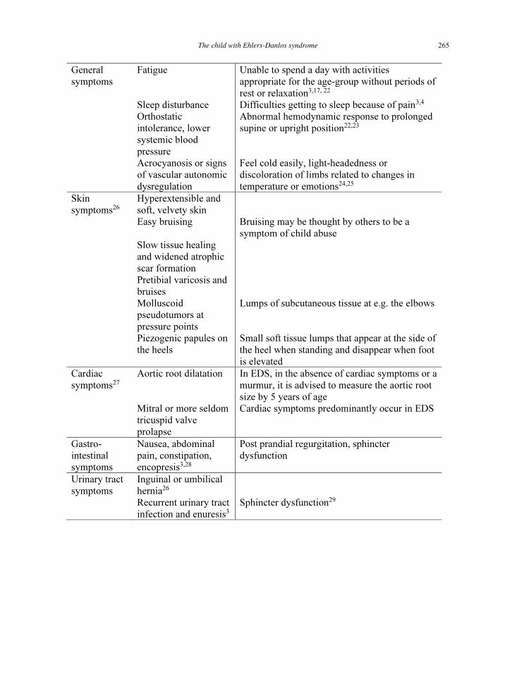
General
symptoms
Fatigue Unable to spend a day with activities
appropriate for the age-group without periods of
rest or relaxation3,17, 22
Sleep disturbance Difficulties getting to sleep because of pain3,4
Orthostatic
intolerance, lower
systemic blood
pressure
Abnormal hemodynamic response to prolonged
supine or upright position22,23
Acrocyanosis or signs
of vascular autonomic
dysregulation
Feel cold easily, light-headedness or
discoloration of limbs related to changes in
temperature or emotions24,25
Skin
symptoms26
Hyperextensible and
soft, velvety skin
Easy bruising Bruising may be thought by others to be a
symptom of child abuse
Slow tissue healing
and widened atrophic
scar formation
Pretibial varicosis and
bruises
Molluscoid
pseudotumors at
pressure points
Lumps of subcutaneous tissue at e.g. the elbows
Piezogenic papules on
the heels
Small soft tissue lumps that appear at the side of
the heel when standing and disappear when foot
is elevated
Cardiac
symptoms27
Aortic root dilatation In EDS, in the absence of cardiac symptoms or a
murmur, it is advised to measure the aortic root
size by 5 years of age
Mitral or more seldom
tricuspid valve
prolapse
Cardiac symptoms predominantly occur in EDS
Gastro-
intestinal
symptoms
Nausea, abdominal
pain, constipation,
encopresis3,28
Post prandial regurgitation, sphincter
dysfunction
Urinary tract
symptoms
Inguinal or umbilical
hernia26
Recurrent urinary tract
infection and enuresis3
Sphincter dysfunction29
The child with Ehlers-Danlos syndrome 265
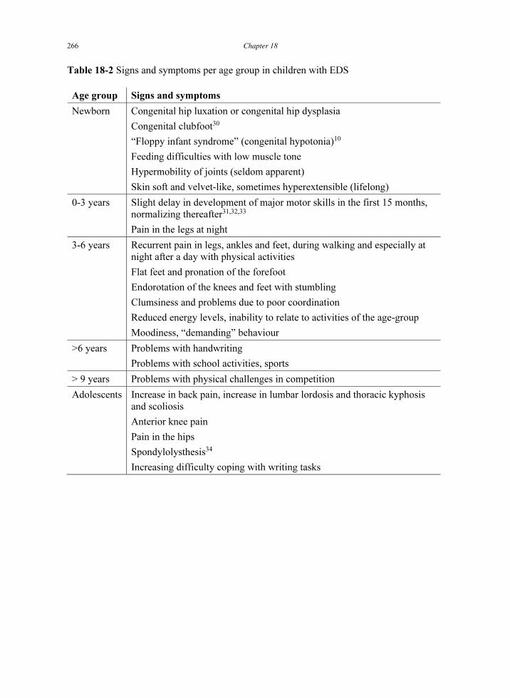
Table 18-2 Signs and symptoms per age group in children with EDS
Age group Signs and symptoms
Newborn Congenital hip luxation or congenital hip dysplasia
Congenital clubfoot30
“Floppy infant syndrome” (congenital hypotonia)10
Feeding difficulties with low muscle tone
Hypermobility of joints (seldom apparent)
Skin soft and velvet-like, sometimes hyperextensible (lifelong)
0-3 years Slight delay in development of major motor skills in the first 15 months,
normalizing thereafter31,32,33
Pain in the legs at night
3-6 years Recurrent pain in legs, ankles and feet, during walking and especially at
night after a day with physical activities
Flat feet and pronation of the forefoot
Endorotation of the knees and feet with stumbling
Clumsiness and problems due to poor coordination
Reduced energy levels, inability to relate to activities of the age-group
Moodiness, “demanding” behaviour
>6 years Problems with handwriting
Problems with school activities, sports
> 9 years Problems with physical challenges in competition
Adolescents Increase in back pain, increase in lumbar lordosis and thoracic kyphosis
and scoliosis
Anterior knee pain
Pain in the hips
Spondylolysthesis34
Increasing difficulty coping with writing tasks
Chapter 18266

Table 18-3 Diagnostic evaluation form for use in clinical practice
Milestones of motor development
Roll-over
Crawl
Stand alone
Walk-supported
Walk alone
5-6 Months
6-10 Months
9-12 Months
10-15 Months
10-16 Months
Signs of generalized hypermobility syndromes
Hypermobility
Hyperextensibility of the skin
Easy bruising
Hypertrophic skin scars
(Sub)luxations
Positive family history
Yes / no
Yes / no
Yes / no
Yes / no
Yes / no
Yes / no
Beighton hypermobility score*26
Bulbena hypermobility score**42
General evaluation
Motor development
Muscle tone
Muscle strength
Delayed / according to
age/ advanced
Hypotonic / normotonic /
hypertonic
Decreased / normal /
increased
* The Beighton score (see chapter 2) is used both in children and adults to assess 5
different joint performances with a maximum score of 9. A total score of 5 is the
minimum score defining joint generalized hypermobility. For hypermobile EDS, age and
sex related cut-off points are used (see chapter 2, table 2-5).
** For the Bulbena score (see chapter 2), a total score defining generalized hypermobility is
≥ 4 in men/boys and ≥ 5 in women/girls.
The child with Ehlers-Danlos syndrome 267

References
1. Callewaert B, Malfait F, Loeys B, de Paepe A. Ehlers-Danlos syndromes and Marfan
syndrome. Best Pract Clin Rheumatol 2008;22:165-89.
2. Grahame R, Bird H, Child A, et al. The British Society for Rheumatology Special Interest
Group on Heritable Disorders of Connective Tissue criteria for the benign joint
hypermobility syndrome. The Revised (Brighton 1998) criteria for the diagnosis of BJHS.
J Rheumatol 2000;27:1777-9.
3. Adib N, Davies K, Grahame R, Woo P, Murray KJ. Joint hypermobility syndrome in
childhood. A not so benign multisystem disorder? Rheumatology 2005;44:744-50.
4. Murray KJ. Hypermobility disorders in children and adolescents. Best Pract Res Clin
Rheumatol 2006;20:329-51.
5. Tofts LJ, Elliott EJ, Munns C, Pacey V, Sillence DO. The differential diagnosis of
children with joint hypermobility: a review of the literature. Pediatr Rheumatol Online J
2009;7:1.
6. Sobey G. Ehlers-Danlos syndrome - a commonly misunderstood group of conditions.
Clin Med 2014;14:432-6.
7. Sobey G. Ehlers-Danlos syndrome: how to diagnose and when to perform genetic tests.
Arch Dis Child 2015;100:57-61.
8. Pacey V, Tofts L, Wesley A, Collins F, Singh-Grewal D. Joint hypermobility syndrome:
A review for clinicians. J Paediatr Child Health 2015;51:373-80.
9. Giessen LJ van der, Liekens D, Rutgers KJ, et al. Validation of the Beighton score and
prevalence of connective tissue signs in 773 Dutch children. J Rheumatol 2001;28:2726-
30.
10. Yiş U, Dirik E, Chambaz C, Steinmann B, Giunta C. Differential diagnosis of muscular
hypotonia in infants: the kyphoscoliotic type of Ehlers-Danlos syndrome (EDS VI).
Neuromuscul Disord 2008;18:210-4.
11. Tosun A, Kurtgoz S, Dursun S, Bozkurt G. A case of Ehlers-Danlos syndrome type VIA
with a novel PLOD1 gene mutation. Pediatr Neurol 2014;51:566-9.
12. Amjad B, Hajivassiliou CA, Pope FM. Paediatric surgical complications of ehlers-Danlos
syndrome. Eur J Pediatr Surg 2014;24:e1-2.
13. Abdalla EM, Rohrbach M, Bürer C, et al. Kyphoscoliotic type of Ehlers-Danlos
Syndrome (EDS VIA) in six Egyptian patients presenting with a homogeneous clinical
phenotype. Eur J Pediatr 2015;174:105-12.
14. Abtahi-Naeini B, Shapouri J, Masjedi M, Saffaei A, Pourazizi M. Unexplained Facial
Scar: Child Abuse or Ehlers-Danlos Syndrome? N Am J Med Sci 2014;6:595-8.
15. Gedalia A, Person DA, Brewer EJ Jr, Giannini EH. Hypermobility of the joints in
juvenile episodic arthritis/arthralgia. J Pediatr 1985;107:873-6.
16. Sacheti A, Szemere J, Bernstein B, Tafas T, Schechter N, Tsipouras P. Chronic pain is a
manifestation of the Ehlers-Danlos syndrome. J Pain Sympt Manage 1997;14:88-93.
17 Murray KJ, Woo P. Benign joint hypermobility in childhood. Rheumatology
2001;40:489-91.
18. Amir D, Frankl U, Progrund H. Pulled elbow and hypermobility of joints. Clin Orthop
1990;257:94-7.
19. Harreby M, Nygaard B, Jessen T, et al. Risk factors for low back pain in a cohort of 1389
Danish school children: an epidemiologic study. Eur Spine J 1999;8:444-50.
20. Adair SM, Hecht C. Association of generalized joint hypermobility with history, signs,
and symptoms of temporomandibular joint dysfunction in children. Pediatr Dent
1993;15:323-6.
21. Gedalia A, Press J, Klein M, Buskila D. Joint hypermobility and fibromyalgia in
schoolchildren. Ann Rheum Dis 1993;52:494-6.
Chapter 18268

22. Rowe PC, Barron DF, Calkins H, Maumenee IH, Tong PY, Geraghty MT. Orthostatic
intolerance and chronic fatigue syndrome associated with Ehlers-Danlos syndrome. J
Pediatr 1999;135:494-9.
23. Engelbert RHH, Bank RA, Sakkers RJB, Helders PJM, Beemer FA, Uiterwaal CSPM.
Pediatric generalized joint hypermobility with and without musculoskeletal complaints: a
localized or systemic disorder? Pediatrics 2003;11:e248-54.
24. Hakim A, Grahame R. Joint hypermobility. Best Pract Res Clin Rheumatol 2003;17:989-
1004.
25. Gazit Y, Nahir AM, Graham R, Jacob G. Dysautonomia in the joint hypermobility
syndrome. Am J Med 2003:115:33-40.
26. Beighton P, Paepe A de, Steinmann B, Tsipouras P, Wenstrup RJ. Ehlers-Danlos
syndromes: revised nosology, Villefranche 1997. Ehlers-Danlos National Foundation
(USA) and Ehlers-Danlos Support Group (UK). Am J Med Genet 1998;77:31-7.
27. Wenstrup RJ, Meyer RA, Lyle JS, Hoechstetter L, Rose PS, Levy HP, Francomano CA.
Prevalence of aortic root dilatation in the Ehlers-Danlos syndrome. Genet Med
2002;3:112-7.
28. Solomon JA, Abrams L, Lichtensteien GR. GI manifestations of Ehlers Danlos
syndrome. Am J Gastroenterol 1996;91:2282-8.
29. Kort LM de, Verhulst JA, Engelbert RH, et al. Lower urinary tract dysfunction in
children with generalized hypermobility of the joints. J Urol 2003;170:1971-4.
30. Olshan AF, Schroeder JC, Alderman BW, Mosca VS. Joint laxity and the risk of
clubfoot. Birth Defects Res A, Clin Mol Teratol 2003;67:585-90.
31. Benady S, Ivanans T. Hypermobile joints: a benign cause of transitory motor delay in
infancy. Clin Pediatr 1978;17:790-5.
32. Tirosh D, Jaffe M, Marmur R, Taub Y, Rosenberg Z. Prognosis of motor development
and joint hypermobility. Arch Dis Child 1991;66:931-3.
33. Davidovitch M, Tirosh E, Tal Y. The relationship between joint hypermobility and
neurodevelopmental attributes in elementary school children. J Child Neurol 1994;9:417-
9.
34. Dyrek DA, Micheli LJ, Magee DJ. Injuries to the thoracolumbar spine and pelvis. In:
Zachazewski JE, Magee, DJ, Quillen WS, eds. Athletic Injuries and Rehabilitation.
Philadelphia, USA: Saunders, 1996; 474.
35. Unger DL. Does knuckle cracking lead to arthritis of the fingers? Arthritis Rheum
1998;41:949-50.
36. Decoster LC, Vailas JC, Lindsay RH, Williams GR. Prevalence and features of joint
hypermobility among adolescent athletes. Arch Ped Adolesc Med 1997;151:989-92.
37. Hall MG, Ferrell WR, Sturrock RD et al. The effect of the hypermobility syndrome on
knee joint proprioception. Br J Rheumatol 1995;34:121-5.
38. McCormack M, Briggs J, Hakim A, Grahame R. Joint laxity and the benign joint
hypermobility syndrome in student and professional ballet dancers. J Rheumatol
2004;31:171-8.
39. Cowan DN, Jones BH, Frykman PN. Lower limb morphology and risk of overuse injury
among male infantry trainees. Med Sci Sports Exerc 1996;28:945-52.
40. Remvig L, Flycht L, Christensen KB, Juul-Kristensen B. Lack of consensus on tests and
criteria for generalized joint hypermobility, Ehlers-Danlos syndrome: hypermobile type
and joint hypermobility syndrome. Am J Med Genet A 2014;164A:591-6.
41. Mishra MB, Ryan P, Atkinson P. Extra-articular features of benign hypermobility
syndrome. Br J Rheumatol 1996;35:861-6.
42. Bulbena A, Duro JC, Porta M, Faus S, Vallescar R, Martin-Santos R. Clinical assessment
of hypermobility of joints: assembling criteria. J Rheumatol 1992:19:115-22.
The child with Ehlers-Danlos syndrome 269

43. Scheper MC, Engelbert RH, Rameckers EA, Verbunt J, Remvig L, Juul-Kristensen B.
Children with generalised joint hypermobility and musculoskeletal complaints: state of
the art on diagnostics, clinical characteristics, and treatment. Biomed Res Int
2013;2013:121054.
44. Barton LM, Bird HA. Improving pain by the stabilization of hyperlax joints. J Orthoped
Rheumatol 1996;9:46-51.
45. Smith R, Damodaran AK, Swaminathan S, Campbell R, Barnsley L. Hypermobility and
sports injuries in junior netball players. Br J Sports Med 2005;39:628-31.
46. Pacey V, Nicholson LL, Adams RD, Munn J, Munns CF. Generalized joint hypermobility
and risk of lower limb joint injury during sport: a systematic review with meta-analysis.
Am J Sports Med 2010;38:1487-97.
47. Maillard SM, Murray KJ. Hypermobility syndrome in children. In: Keer RJ, Grahame R,
eds. Hypermobility Syndrome. Recognition and Management for Phsyiotherapists.
London, England: Elsevier Science, 2003; 33-50.
48. Agnew P. Evaluation of the child with ligamentous laxity. Clin Podiatr Med Surg
1997;14:117-30.
49. Karlsson J, Eriksson BI, Sward L, et al. Early functional treatment for acute ligament
injuries of the ankle joint. Scand J Med Sci Sports 1996;6:341-5.
50. Beynnon BD, Renstrom PA, Haugh L, et al. A prospective, randomized clinical
investigation of the treatment of first-time ankle sprains. Am J Sports Med 2006;34:1401-
12.
51. Skinner RA, Piek JP. Psychosocial implications of poor motor coordination in children
and adolescents. Hum Mov Sci 2001;20:73-94.
52. Monga S, Young A, Owen M. Evaluating a cognitive behavioral therapy group program
for anxious five to seven year old children: a pilot study. Depress Anxiety 2009; 26:243-
50.
53. Catchpole J, Turner J. Splinting and bracing. In: Bird HA, ed. The management of
Ehlers-Danlos syndrome. Hertfordshire, England: Ehlers-Danlos Support Group, 1998.
Chapter 18270

Chapter 19. Ehlers-Danlos syndrome, generalised hypermobility and ethics:
reflections from the ethics of care
L Cornelissens and C Leget
Lidwien Cornelissens PhD, psychotherapist/psychoanalyst and medical ethicist
Department of Medical Psychology
Alrijne Hospital
Simon Smitweg 1
2353 GA Leiderdorp, The Netherlands
Telephone +31715828282
E-mail [email protected]
Carlo Leget, PhD
Professor of ethics of care and spiritual counselling sciences
University of Humanistic Studies
Kromme Nieuwegracht 29
3512 HD Utrecht, The Netherlands
Telephone +3130239 01 00
E-mail [email protected]
271

1. Introduction
Ethics is a branch of philosophy that involves thinking about human acts and asking questions
about the moral rights and/or wrongs of those acts. There is no single theory of or approach to
ethics. Ethical theories reflect the time and cultural setting in which they came into being. The
approach one chooses has a great impact on one’s view about what is ethically right or wrong.
In this chapter, the Ethics of Care will be the framework for reflecting on the ethical dilemmas
related to living with generalised hypermobility or Ehlers-Danlos syndrome (EDS). Ethics of
Care is an approach that focuses on the phenomenon of care. Care can be viewed as
everything we do to maintain, sustain and repair our world so that we can live in it as well as
possible.1
Ethics of Care focuses on the way people try to find out what is good in specific
conditions and circumstances. It does not look for generalisation of normative standpoints, but
focuses on the particularity of situations, is suspicious of power relationships with regard to
dependent people, accepts vulnerability as a universal human characteristic and conceives
human beings as inherently relational beings who need each other to be happy and to
flourish.2
EDS confronts people with ethical problems that may be related to different life stages. In this
chapter we will look at these dilemmas and try to find out if there are any opportunities
related to that specific stage of life or to a specific personal character and how these can be
found. This will empower people so that they can pursue ‘the good life, with and for others, in
just institutions’.3
2. Ethical dilemmas in parenthood
The technological possibilities in our society have caused a tendency towards rationalisation.
This is especially true for health care, which confronts people with dilemmas even before they
give birth to their child(ren). If people want to have a child, they usually hope to give birth to
a mentally and physically healthy child. People with EDS who wish to have children bear a
special responsibility as the aetiology of EDS is genetic (see Box 19-1 for reproductive
options for parents-to-be in case of a genetic disorder). They know they run a 50% risk of
giving birth to a child with EDS, assuming the given EDS type is inherited in an autosomal
dominant way. The child they will give birth to may (eventually) suffer from the same type of
EDS, though the severity of the symptoms may vary. Furthermore, the child is at risk of
losing its mother during or shortly after delivery if she suffers from vascular EDS.
�
Box 19-1 Reproductive options for parents-to-be in case of a risk of having a child with a
genetic disorder:
- Acceptance of the risk
- Refrain from having own children
- Non-acceptance and prenatal diagnosis and termination of pregnancy when affected
- Non-acceptance and pre-implantation genetic diagnosis
- Non-acceptance and germ cell donation
- Non-acceptance and adoption
At the start of a life, people really do not want to think of this when it comes to their newborn
child. But also if people suffer from an EDS type other than vascular EDS or from generalised
hypermobility, the delivery of the child and taking care of it may still be problematic. People
suffering from hypermobile EDS and generalised hypermobility usually have very mobile
joints and ligaments. This may cause severe perinatal problems for the mother, like
(sub)luxations. One of the problems of this EDS type is that the joint hypermobility may
worsen due to the hormonal changes during pregnancy.4
This may cause more or less severe
Chapter 19272

problems for the mother, which may hinder her ability to take care of herself and her newborn
child. Therefore the question may arise: why should people take those risks? Would it not be
better for people to stop thinking about having a child if one of the parents is suffering from
EDS?
From an Ethics of Care perspective, questions like these do not make much sense. It is
important to look at the situation from the perspective of the people involved in order to
discover what is at stake for them. There is a possibility parents do not even know that one
of them has EDS. Patients are not always aware of having the syndrome, due to the wide
variety of phenotypes and the varying degrees of severity. E.g. luxation of the shoulder joint,
can happen to anyone. The same can be said about ligament distortion. This will only raise
questions if it happens frequently and/or it happens also in close relatives. People may not be
aware of the fragility of their body, especially when they do not take part in sports (very
often), particularly if they suffer from mild EDS. Vascular EDS may be even more
complicated: incidentally, people with vascular EDS hardly experience problems until they
reach the age of 40. So, not infrequently, people have made their important life choices, such
as having children, before they find out they are suffering from a type of EDS. In this case,
the next generation will have to deal with the ethical dilemma of making a responsible choice,
which may be a heavy burden.
2.1 Motivations
People suffering from EDS who nonetheless choose to give birth to a child, even though they
are aware of the risks and consequences, mention a variety of reasons as to why they make
this choice (see Box 19-2 for factors playing a role around these decisions).
Box 19-2 Factors playing a role in deciding whether or not to have a child, when parents-
to-be are facing a risk of having a child with a genetic disorder:
- Strength of their child wish
- Magnitude of the risk
- Severity of the genetic disorder
- Family composition
- Their ethical/religious norms and values
- Acceptability of prenatal diagnosis and termination of pregnancy
- Acceptability of pre-implantation genetic diagnosis
- Acceptability of germ cell donation
- Socio-economical context
- Legal context
One of these reasons is that they find themselves capable of living a meaningful life, so they
expect their children if they also have EDS, to be able to do the same. According to them,
thinking your child will not succeed in this, means you are raising it without hope or trust.
According to this pessimistic point of view, no child would flourish, regardless of whether
they have an illness or not. Parents also suppose that their knowledge of and experience with
EDS will benefit in helping their child(ren) with EDS to create its/their own opportunities in
life, better than non-EDS parents would be able to do. That could be one of the reasons why,
although for most EDS types (not hypermobile EDS) prenatal diagnosis is possible, when the
pathogenic mutations are known (see chapter 2), in our experience few parents-to-be, who are
facing a risk of having a child with EDS, opt for prenatal diagnosis.
They motivate their choice to become parents by pointing to the fact that all children are at
risk of diseases and that people accept them as being unavoidable. ‘Why would I be so afraid
Ehlers-Danlos syndrome, generalised hypermobility and ethics: reflections from the ethics of care 273

of a known risk?’ They say that they themselves, unlike most parents, are highly aware of the
risks of starting a family, which allows them to be better prepared to handle certain
consequences. The stories of these parents show their remarkable faith and acceptance of their
limited abilities.
There are also individuals with EDS who, when the woman is affected, decide that based on
the many limitations in their daily life, pregnancy would be too much of a burden. Thus, there
will also be parents who among others based on the magnitude of the risk to have an affected
child, the severity of the EDS as they have or perceive it, decide to refrain from having own
children. They still have some options (see above). Adopting a foster child is such an option,
which of course is only available to those who are aware of their limitations and handicaps
and have the emotional and physical strength to guide this foster child, who often has had a
bad start in its life. Although the law does not prohibit irresponsible choices to reproduce, it
can be difficult for EDS patients to persuade government institutions of their parental
potential and to put parenthood into practice by becoming foster-parents. Difficult as it may
be, it is possible and might be the best option in some situations. But there will also be parents
who refrain from having children at all whether own or adopted based on their own situation
and maybe other factors.
2.2 Different norms and values
Thus far, we have spoken of three different scenarios regarding impending parenthood. Each
of these scenarios expresses various norms and values, which sometimes overlap. For those
who take their own life as reference-point (the first scenario described above), equality is an
important value. ‘Because life with its serious limitations is still valuable for me, it must be
possible for our child to achieve the same.’ In that way, their life vision speaks of trust as an
important value, and the fact that their child is not yet born does not make any difference to
them. As well as equality and trust, there is in this scenario an acceptance of the vulnerability
and imperfection of human life, which we all share to a certain extent. Being connected and
related to one’s own child and sharing a life together is seen as being more important than
giving birth to a perfect baby.
For the second scenario, the parents take EDS as being one risk out of many. Equality is also
important for them, but here this value is seen more as equality in life, understood as an
experienced adventure, an adventure we share with every human being. In this belief system
the awareness of the undeniable fact that life and its consequences are beyond the control of
human beings, has been transformed in values like: ‘trust’, ‘hope’, ‘endurance’ and
‘forbearance’. These all have to be earned along the path of life. For them, life, handicapped
or not, is no more special than it is for other people. Also in this scenario, being connected
and related to one’s own child and sharing a life together is seen as being more important than
giving birth to a perfect baby.
In the third scenario – the couple who will not give birth to a child themselves although they
have a strong desire to have a child – the stress on rationality in autonomous decision-making
is a central value. In this case, the values of ‘trust’ and ‘hope’ do not temper the awareness of
their limitations. They are aware of their burdens and their reduced load capacity and make
the decision that it would be wiser not to overload themselves by going through a
pregnancy/delivery with all the short and long-term complications. Some of these people
experience severe limitations in their daily life and they do not want to be responsible for
knowingly creating the same life circumstances for their own child. Trust and hope may be
valued by them, but these values are expressed in their opinion that their lives will be of value
even if their wish of a child of their own is not fulfilled. They trust that they will come to a
Chapter 19274

point where they have overcome the grieving for their unfulfilled wish and can have a new
life framework without a child of their own, maybe in the role of foster-parents.
We have seen that values differ and that, even if values seem similar at first glance, they
express different belief systems. Here we do not cling on to right or wrong along the lines of
one specific ethical theory of human life. People make their own choices, seen in our western
society as the expression of autonomy, a very – perhaps even the most – important value. But
does that imply that there is nothing we can say about the three stories? Can we perhaps learn
something from the experiences of others?
2.3 Risks and problems
Let us think of the risks or problems that may easily be overlooked in these stories. Our first
group of parents experiences life as meaningful and worthwhile, irrespective of its limitations.
Can they really think about their newborn child dispassionately? Will this child be allowed to
be angry about living with a handicapped father or mother and will it be allowed to conceive
life , as a worst case scenario, as unbearable and hopeless if itself is handicapped too? Will
these parents be able to handle this reaction of their child, recognising it as a phase which they
have already had to struggle through themselves and which their child can also overcome with
their love and help? Or are these parents in denial of their own feelings about their limitations,
expecting that their child will develop the same denial? Are they afraid of their own jealousy
which is connected with life itself, and even more so if they are handicapped themselves and
have to deal with seeing other people dancing, running and not always struggling with a lack
of energy and with pain? If this is the case, their values would operate as a shield against their
indigested psychological distress about their illness. It will be very difficult for them to guide
their child through the phase of mourning, because their child might be dealing with its
handicaps by mourning actively instead of denying its feelings like its parents do.
Life is uncontrollable for the second group of parents, so they can set the EDS to one side.
Although their lifestyle can foster their child’s inner powers to behave just like others and not
to mystify a handicapped life, this child stays at risk of carrying too heavy a burden. ‘You are
like everyone else so don’t worry and get a life’ A child in this position might not only
compensate but take the risk of overcompensation, preferring short term gains over long term
benefit. Children with EDS can often do the same things as other children of their age but
they may be physically hurt easily, because their condition makes them easily tired, their
joints are so flexible and their skin so fragile. Once hurt, they will have to rest. This rest
reduces muscle power, which in turn heightens the risk of new lesions. So, in a short time
they might become imprisoned in a vicious circle that can easily convert into a downwards
spiral.
As said earlier, people may be unaware of their own genetic problem until labour and/or after
the birth of their affected child. Sometimes the baby is a typical ‘floppy child’. There is little
or no muscle tone. In the worst case, the child will never start crawling. However, eventually
most of these children will succeed in crawling and walking, although often later than other
children of similar age. Depending on the type of EDS, parents must be aware of different
problems. If it is EDS with skin problems, like in classical EDS, they will need to be very
careful with everything which puts the child at risk of skin damage. Their child can do fewer
of the things a normal healthy child would do and which will give great joy, like bumping,
crawling, jumping and running around wildly. These active explorations will readily lead to
first aid in a hospital, with a nasty open wound and the possibility of a need for plastic
surgery.
Ehlers-Danlos syndrome, generalised hypermobility and ethics: reflections from the ethics of care 275

These physical wounds are visible but, again , it is the job of the parents to choose between
the risk of physical wounds and the risk of psychological wounds or emotional damage to the
child. Children do not like special treatment and normally they want to play without getting
into too much trouble, so it is up to the parents to find a balance between the pro and cons.
A child with spontaneous joint dislocations or luxations needs a lot of guidance to optimise its
muscle power and proprioception. Sometimes, guiding this process will demand so much of
the parents’ energy and medical knowledge that they are forced to make a difficult choice
between regular and special school education. Do you have to train your child as much as you
can, while without this, it can play with children who are also limited in their play by their
own physical problems? And do you hope that, with this stable physical and mental basis,
your child will develop into a strong and assertive adult? Or does it work the other way
around? Is it better to opt to treat the child as being as ‘normal’ as possible, which would
mean letting it grow up among fully physically able children so that it can learn to survive
from the beginning in a world that is often not well adapted and even not accessible to the
disabled?
There are arguments for both ways of reasoning. The most important thing, however, is not to
idealise a certain vision or to try and find universal rules. Instead of looking at the impact of
the illness on daily life, we have to consider carefully the possibilities and limitations of the
specific child. A very social and outgoing child will survive more easily in an unadapted
environment, or one with few adaptations, than a more introvert child.
Another very important factor is how much the parents can bear. A child at regular school
needs a lot more guidance and support, improvisation (e.g. a resting place at school and
planning of medical visits), and explanations to teachers and other children than healthy
pupils do. Physiotherapy and adaptation of braces and other medical equipment (like a
wheelchair, if necessary, and so on) require visits that have to take place after school.
Children grow quickly during their school years, so the necessary changes in medical
equipment often seem endless, and so do the bureaucratic procedures stemming from these
changes.
Apart from this, the role of a parent is also burdensome for another reason. When your child is
easily wounded or has a lot of spontaneous dislocations that require medical treatment, the
frequent visits to the emergency department of the hospital can start to raise suspicions, not
least about the parents themselves. And to make it worse, parents can become suspects of
child abuse, not just in hospital but also at school. Although it will not take away the
suspicions at the emergency department, such suspicions can make life much more difficult
and encourage a choice for a special school.
�
3. Living a (good) life with EDS
According to an influential definition by Joan Tronto and Berenice Fischer, “caring can be
viewed as a species activity that includes everything we do to maintain, continue and repair
our world so that we can live in it as well as possible. That world includes our bodies,
ourselves, and our environment, all of which we seek to interweave in a complex life-
sustaining web”.1
The healthier and richer people are, the less time they will be forced to
spend on caring for themselves and the more they can get others to care for them, up to what
Tronto has called ‘privileged irresponsibility’.1
People suffering from EDS are not among
those privileged irresponsibles as we have seen in the previous section. Being confronted with
EDS means being called to care for oneself and being responsible for those who are part of
one’s complex life-sustaining web.
Chapter 19276

This life of care – for oneself and others – is an ethical enterprise through and through. Ethics
is not an activity we only need when the course of normal life is hindered by unexpected
problems or dilemmas. The ethical intention aims for “the good life, with and for others, in
just institutions”.3
This good life is nothing other than a happy life; happiness being not just a
feeling or sensation of wellbeing, but a flourishing of one’s capacities and possibilities. This
search for happiness is a social activity – hence with and for others – within the context of a
society that is built on structures that transcend and contain relationships among individuals –
hence the “just institutions”. For instance, people with EDS can still flourish in their
professions, experiencing their professional work as an important aspect of living a
meaningful life, while at the same time they have to be looked after by a nurse in their daily
lives. It all depends on the balance between the strengths and weaknesses of the patients and
the willingness of the social system around them, whether this is possible or not.
When we look at ethics in this way, a new perspective opens up. Underneath the moral
dilemmas related to the choice by people suffering from EDS to have children or not, we see
people struggling to lead a good and happy life in a society that is not ready to accept the
vulnerability of human existence. This struggle is hallmarked by practices of caring for one’s
fragile body and for those who are helping one to sustain a life with EDS. From this ethical
and caring perspective, people living with EDS are entitled to ask some critical questions to
the society they live in and about how their care is organised.
3.1 Holistic approach
A main critical question raised by people who live with EDS pertains to the tension between
general and specialist medicine. Human existence has many dimensions – physical, psycho-
social and spiritual – that are intrinsically related and together form what we describe as a
human being. Medicine, however, still focuses on diseases and syndromes rather than on
beings.5
In the case of EDS, this means that patients are often treated by many different care-
givers, many of whom are specialists. Even though this division of care into specialisms may
be understandable from the perspective of professional caregivers, from the patient’s
perspective it leads to a reductionist approach, which is time-consuming and adds extra
burdens to lives that are already burdened by so many inconveniencies.
The fact that we have organised care as it is now, and the ever-increasing specialisation in
medical science, are not facts of nature. They are the result of human action and have an
ethical dimension. In fact, they are the result of medical power which is so accepted and self-
evident in our culture that it often hinders our capacity to look at things differently. Adopting
a patient’s perspective would do more justice to the person who is at the centre of care: the
person suffering from EDS who is not an object to be looked after but a human individual
who is both being cared for and caring. A holistic approach would not only entail an approach
in which not only the physical, psycho-social and spiritual dimensions are integrated, but also
an approach in which the patient is co-carer and involved in shared decision making. At the
same time it would make sense to appoint a consultant medical specialist – one who is
familiar with EDS – as a case-manager to help streamlining the medical processes.
3.2 Vulnerability
EDS like any disease can confront us with the vulnerability of human existence. This
vulnerability is one of the things our society has great problems with, since it is associated
with an unpleasant and unhappy life. The cases we have described above show that, even in
vulnerable positions, people often find possibilities of developing resilience and an art of
living with fragility that will open up new horizons. Confronting EDS thus questions many
“self-evident” suppositions. It helps us to see that life can still be worth living, even if it is
Ehlers-Danlos syndrome, generalised hypermobility and ethics: reflections from the ethics of care 277

compromised by the prospects of increasing morbidity and fragility. Instead of fighting and/or
hiding vulnerability, vulnerability may be accepted as a fact of life which is helpful in
overcoming self-centeredness and isolation. It forces us to reach out to our environment and
share our humanity. This is the very essence of the relationships and friendships that help us
to experience life as a meaningful enterprise.
3.3 Dependency
People who are faced with EDS can be confronted with their own dependency. Living with
EDS increasingly means accepting that you need other people to make (social) life possible.
That means that people living with EDS are forced into developing a new sense of autonomy.
The concept of liberal autonomy as a value according to which people get to lead their lives
how they want to (related to the privileged irresponsibility mentioned above) is redefined into
a more realistic kind of relational autonomy. It is still the individual person to whom justice
must be done, by granting them the maximum of space and freedom. But this space and
freedom is constituted by the relational network of people around the patient. EDS affects not
only the life of the person suffering from the disease, but also the lives of all those people who
are related to and sustaining that person in a web of relations.
The need for a holistic approach, dependency and vulnerability reveal dimensions of human
existence that are present in every life, but come to the fore more clearly in the case of EDS.
In this way, confronting EDS is confronting aspects of life that our culture often hides or
denies or classifies as being confined to the elderly. Patients with EDS can live like the
elderly for a very long time – even for decades – and, because of the burden that this can
impose, not all patients wish to live to a great age. Non-treatment decisions or a desire for
euthanasia can materialise and must be treated with care and compassion. However, the Ethics
of Care perspective can help reveal these aspects and encourage ethical reflection. This
reflection is not just needed when moral dilemmas are at stake or emerge. Ethical reflection
should begin at the very moment we start thinking about care. In this way ethical reflection
can help to develop a richer and more diverse view on living a meaningful life. People
suffering from EDS, their next of kin and their caregivers would benefit from such a reservoir
of meaning.
Chapter 19278

References
1. Tronto JC. Moral boundaries. A political argument for an ethic of care. New York, USA:
Routledge, 1993.
2. Held V. The Ethics of Care. In: Copp D. ed. Oxford Handbook of Ethical Theory. Oxford,
England: Oxford University Press, 2006:537-566.
3. Ricoeur P. Oneself as Another. Chicago, USA: University of Chicago Press, 1992.
4. Marnach ML, Ramin KD, Ramsey PS, Song SW, Stensland JJ, An KN. Characterization
of the relationship between joint laxity and maternal hormones in pregnancy. Obstet
Gynecol 2003;101:331-5.
5. Cassell E. The Nature of Suffering and the Goals of Medicine. 2th ed. Oxford, England:
Oxford University Press, 2004.
Ehlers-Danlos syndrome, generalised hypermobility and ethics: reflections from the ethics of care 279

This page intentionally left blank

Chapter 20. Stretched beyond the limit: well-being and functioning in patients
with Ehlers-Danlos syndrome and other hypermobility syndromes
R Geenen and MA Lumley
Rinie Geenen, PhD, psychologist
Professor and head of the Psycho-rheumatology research unit
Department of Clinical and Health Psychology
Utrecht University
Department of Rheumatology and Clinical Immunology
University Medical Center Utrecht
PO Box 85500
3508 GA Utrecht, The Netherlands
Telephone +31302534916
E-mail [email protected]
Mark A. Lumley
Professor and director of clinical psychology training
Department of Psychology
Wayne State University
Detroit, Mi, USA
Telephone 313-577-2247
E-mail [email protected]
281

1. Case: Michelle
Michelle is 34 years of age. She was raised in a farming family and had a generally positive
and supportive childhood. At the age of 18, she moved to the city to attend university. For the
past ten years, she has been happily married and has 2 young daughters. She has long realized
that she has very flexible joints, and she regularly dislocated her fingers, but she was not that
concerned and did not consider it to be a disorder. Two years ago, however, her situation
worsened when she dislocated her shoulder while playing tennis, and the subsequent
operation left her with various complications. The surgeon referred her to other specialists,
and while still in the hospital, Michelle was diagnosed with Ehlers-Danlos syndrome (EDS).
She was mostly pleased to learn of this diagnosis, because it explained her frail joints as well
as the pain and fatigue that had been interfering with her functioning at home and work during
the past few years. These symptoms had been invisible, but now others could understand why
she had not been feeling well or functioning optimally. Michelle also was thankful that the
hospital physiotherapist helped her learn to use her joints in the appropriate way and manage
dislocations.
Lately, however, Michelle has become somewhat depressed and worried. She has come to
realize that having this disease could greatly change her life. Although she has many abilities
and loves her work, she fears that she will not be able to fulfil her high ambitions. She had
always been able to hike in the mountains while on holiday, but this has become more
difficult, and in the near future, she expects that she will no longer be able to walk even the
easy trails. She needs to change so many things and is anxious that her condition will become
dramatically worse in the future. She is also worried that her children have EDS and she needs
to remind herself to avoid raising them too cautiously or fearfully. Although she expects that
her optimistic nature will finally help her to deal with the consequences of EDS, a
psychologist might help her with her current negative mood, and she is considering seeking
cognitive-behavioural therapy.
2. Introduction
Connective tissues maintain normal bodily integrity, but when connective tissues fail in this
role, multiple problems occur. EDS is a heterogeneous group of heritable connective tissue
disorders characterized by articular hypermobility, skin extensibility, and tissue fragility.1
This disorder is chronic, varies in severity from mild to very serious, and can be lethal.
Hypermobility syndromes featuring joints that easily move beyond the normal range expected
for a particular joint, are a threat to well-being and functioning. What is said about EDS in
this chapter mostly applies to hypermobility syndromes as well.
Recently, the two conditions benign joint hypermobility syndrome (BJHS) and EDS
hypermobility type have been recognized as one and the same clinical spectrum ranging from
apparently symptomatic generalized joint hypermobility to the most disabled individuals
fitting the new diagnostic criteria. These new criteria are more strict than the Villefranche
criteria and the Brighton criteria for BJHS in order to define a homogeneous phenotype for
management and scientific purposes. Within the new EDS nosology, its name is hypermobile
EDS (see chapters 2 and 5).
2.1 Burden of EDS
The psychosocial burden of EDS is substantial2-4
, and can be summarized by the central
theme of “living a restricted life”.5
First, EDS is a relatively rare condition, which means that the diagnostic process is often
protracted and confusing, and that healthcare providers often have insufficient knowledge of
the medical problems and the possibilities for treatment and care.
Chapter 20282

Second, people with EDS have fewer opportunities in daily life than healthy people. For
example, pain, fatigue and other symptoms hamper functioning, and people with EDS may
fear articular dislocations or skin damage, which further limits activities.
Third, patients may experience invalidation, or stigmatization and lack of understanding from
family and friends, health care providers, co-workers and others in daily life.
Finally, given the heritable nature of EDS, the pros and cons of parenthood are often
evaluated, and decisions to refrain from childbearing can be highly disappointing.
Psychological evaluations and interventions for patients as well as their families are vital to
help them cope effectively with this disorder.6
Self-care, counselling and therapy are directed
at both the prevention and treatment of adverse consequences. Because most bodily organs
have connective tissue potentially affected by EDS, multiple bodily system problems can
occur, and virtually all medical and allied disciplines play a role in education and treatment.
2.2 Aim
The aim of this chapter is to review the somatic and psychosocial consequences of EDS; that
is, the effects of EDS on physical, psychological, and social well-being and functioning.
Somatic problems, psychological distress, and social functioning are reviewed, and
suggestions for coping with these problems in everyday life are given. We base our discussion
and recommendations on the relatively few empirical articles on psychosocial functioning in
EDS, supplemented by our knowledge of experiences of individual patients, and our
knowledge of the somatic, emotional, and social consequences of chronic disease more
generally.
3. Somatic problems
In a Swedish survey using the Subjective Health Complaint Inventory (SHCI), 99% of 250
participants with EDS reported health complaints with a mean number of sixteen complaints
for the whole sample.7
The somatic problems reported most often were musculoskeletal by
246 (98%), pseudoneurological by 241 (96%), gastrointestinal by 236 (94%), allergic by 182
(73%) and influenza-like by 144 (58%) persons. These specific somatic problems as well as
those of vascular EDS are discussed in other chapters of the book. In the current chapter, pain,
fatigue, joint dislocations, fragile skin, and sleeping problems are discussed.
3.1 Pain
In a questionnaire survey of 41 patients with EDS, 63% of the patients reported relatively
elevated pain (above the scale mid-point), and only two patients reported having no pain.8
Another study used structured interviews of 51 patients with EDS and found that 90%
experienced pain for at least six months.9
In a larger questionnaire study among 250 patients
(contacted through the Dutch patient association for EDS), pain during the past week was
reported on a 0 to 100 mm visual analogue scale (VAS), 100 being the maximal level of pain:
29% of the patients reported pain between 0 and 25, 24 % between 25 and 50, 28% between
50 and 75, and 18% between 75 and 100.2
Of this sample, 41% reported chronic, widespread
pain; that is, pain existing for more than three months in the upper and lower, left and right,
and axial parts (i.e. spine) of the body. Of the respondents, 55% commonly used analgesics—
mostly paracetamol (acetaminophen), non-steroidal anti-inflammatory agents, and tramadol.
One study found that pain is more prevalent and severe in hypermobile EDS than in classical
EDS, and that pain is associated with hypermobility, joint dislocations, previous surgery, poor
sleeping quality, and functional impairment in daily life.10
Thus, pain clearly is a prevalent problem in EDS, and has consequences for well-being and
functioning. Reduced well-being and functioning, in turn, may contribute to the persistence
and severity of pain. Education and self-management techniques likely can help to address
Stretched beyond the limit: well-being and functioning in patients with Ehlers-Danlos syndrome 283

relatively low levels of pain, but more serious pain should be treated in cognitive-behavioural
therapy (for explanation, see Box 20-1) and physical rehabilitation programmes. Such therapy
is especially indicated if pharmacological treatment is ineffective to deal with pain and its
consequences.
Box 20-1 Cognitive-behavioural therapy
A main premise of cognitive-behavioural therapy is that negative, dysfunctional thoughts
have a perpetuating role in health problems, and that people need to learn skills or
techniques to change their thoughts and feelings. Cognitive-behavioural therapy is directed
at reduction of symptoms like depression, anxiety, pain, and physiological responses by
changing maladaptive thoughts and actions. Examples are interventions with one specific
aim—for example, relaxation, stress reduction or overcoming of fear-avoidance beliefs to
support an exercise intervention and, more commonly, the incorporation of various
methods—for example, cognitive restructuring of dysfunctional beliefs or “worry”
thoughts, pain coping skills training, activity pacing, stress management training, relaxation
exercises, exposure to anxious situations, thoughts and worries, and positive self-talk.
3.2 Fatigue
The survey of Dutch patients with EDS asked respondents to note the five most serious
symptoms or problems of EDS.2
Fatigue (64%), pain (54%), skin fragility (33%), and
problems with the back (29%), wrist, hand, and fingers (27%) were mentioned most
frequently. It is striking that so many patients reported fatigue among their five most serious
problems. This survey also compared general fatigue scores in 250 patients with EDS to those
of healthy people from the general population using the Multidimensional Fatigue Index.11
The occurrence of ‘general fatigue’ above a specified cut-off point was five times larger for
patients with EDS than for people in the general population.2
A similar survey of 274 Dutch
patients with EDS, which was conducted five years later in the same population, found that
77% of the patients reported severe fatigue as compared to 11% and 17% in two
predominantly female samples of the Dutch general population.12
One possible way to address the elevated fatigue experienced by so many patients with EDS
is to identify specific patient and disease characteristics that contribute to fatigue, and to
change targeted lifestyle behaviours. For example, it has been proposed that aerobic fitness or
strength training to deal with muscle weakness, or using weight management techniques to
reduce obesity to treat sleeping apnoea, might be useful approaches to reduce fatigue in joint
hypermobility syndromes.13
Unfortunately, the physiological origins of chronic fatigue are
unknown, and there are no effective pharmacological treatments. Thus, although fatigue is
indisputably an adverse consequence of EDS, it is most sensible to consider behavioural
means, such as life-style adjustment and cognitive-behavioural, physical exercise, and sleep
hygiene interventions.14
However, the effects of such behavioural interventions have not been investigated in EDS.
Caution is needed; for example, physical exercise programmes need to take account of
possible joint dislocations as potential complicating factors. Moreover, to enhance their sense
of vitality, patients will need to find a balance between physical effort and rest. Some patients
may avoid all physical exercise to prevent pain and fatigue, but this will promote physical
deconditioning. Other patients may persist in exercising despite pain and fatigue and stop only
when pain or fatigue are extreme. However, this may over time result in dramatically reduced
physical activity, as stopping exercise is stimulated because it takes away pain. Often a
gradual build-up of the intensity of physical activity is important to prevent relapse. As in
other chronic symptomatic conditions, activity-rest cycling can be valuable, with patients
finding the optimal timing of activity and rest. A predetermined activity duration—rather than
Chapter 20284

pain or fatigue—should determine the timing of the rest-activity cycle. More generally, some
patients will need encouragement to exercise, whereas others will need to learn to stop
exercising in time.
3.3 Joint dislocations
In some forms of EDS, from 90 to 100% of the patients, report hypermobility of the joints15
,
but the severity of joint problems differs considerably among individuals and EDS types.
Joint problems hamper work, sports, and many other important daily activities. Patients need
to find an optimal balance between living passively to avoid joint dislocations and a more
active - but risky - lifestyle, which can trigger joint dislocations. The challenge for patients is
to learn to use their body without pushing their joints beyond their limits. Most patients learn
this through trial and error. It is important that patients do not avoid physical activity, but
instead use assistive technology, adaptations at home and at work, and aids such as braces and
appropriate shoes to remain physically active. Fellow patients, rehabilitation physicians, and
occupational therapists can help the patient to find adjustments that help in optimal
functioning.
3.4 Fragile skin
Fragile skin is a problem in 40% (hypermobility EDS) to 100% (classical EDS) of the patients
with EDS.15
Wound healing may be problematic, and wide, ugly scars may result. Our society
values beautiful skin, and physical appearance is important not only for self-esteem but also
for interpersonal communication, especially first contacts. Patients can try to prevent skin
damage by wearing appropriate clothes and by avoiding risky situations. In case of wounds,
appropriate care is important. In serious cases, wound care nurses, dermatologists, or plastic
and reconstructive surgeons can be helpful.
3.5 Sleeping problems
The prevalence of sleeping problems in EDS has rarely been studied. In a small sample of
nine patients with EDS, five reported sleeping problems, and six reported periodic limb
movements.16
In contrast to this seemingly high prevalence, the Dutch survey of 250 patients
found that only 4% reported sleeping problems as among their top five most common
problems, but this report is likely biased by the method of requiring patients to pick a
maximum of five major problems.2
Potential sleeping problems should be assessed in patients, and if present, differential
diagnosis of the aetiology of the problem should be pursued, because treatment for sleeping
problems should preferably be aimed at the cause. In the differential diagnosis, sleeping
apnoea has to be taken into consideration, because as soft tissues weaken by becoming older,
especially in EDS patients sleeping apnoea can become an extra problem.
Sleeping medication is generally not recommended for patients with EDS because of the
resulting muscle relaxation and the ensuing increased risk of joint dislocations. Insomnia can
be treated with stimulus control therapy, relaxation training, and cognitive-behavioural
therapy.17
Such interventions may include techniques such as avoiding the consumption of
coffee, alcohol, and heavy meals within several hours before sleep; making the bedroom dark,
comfortable, and quiet; using the bed only to sleep (and for sex), thus not for reading and
watching TV; having a regular pattern of effort and rest throughout the day as well as a fixed
sleeping time; and - if needed - using stress-reduction, relaxation or thought-stopping
techniques. Insomnia can also be treated with sleep restriction therapy, which reduces the
patient’s time in bed to the total number of hours of sleep that is being obtained, and forces
patients to be up and out of bed when not sleeping; as sleep efficiency increases, the sleep
period can be lengthened.
Stretched beyond the limit: well-being and functioning in patients with Ehlers-Danlos syndrome 285

4. Psychological well-being
Psychological well-being is a core indicator of quality of life. The severity of one’s disease
influences psychological well-being, but it is only one factor, and the relationship between
disease severity and well-being is weak in most chronic diseases.18
Coping, resilience,
emotional and social factors play larger roles in adaptation and well-being.
4.1 Emotional consequences
Negative emotional consequences of living with EDS are common. Interviews show that
some people with EDS live with constant fear of skin damage, physical deterioration because
of hypermobility, unemployment, and problematic pregnancies.5
Patients with joint
hypermobility syndrome present with greater prevalence of panic disorder and
agoraphobia.19,20
Parents who have children with EDS, live with the fear that authorities,
medical personnel, or others will suspect them of bad parenting or abuse when their child
presents with a joint dislocation or bleeding21
, and even if no accusation are made, parents
may still sense suspicion.5
Another problem mentioned in interviews is the reduced possibility
of developing one’s abilities.5
The great effort needed to achieve something substantial in
education or work is often not possible in case of EDS. This especially holds for people with
vascular EDS who often know at a young age that, although new medication has improved the
prognosis, their life expectancy is shortened, which has implications for important life choices
in education, work and family planning; and can impede life satisfaction.
A questionnaire study of 41 patients with EDS from North America found that anxiety,
depression, anger and relationship worries were quite high.8
Indeed, these problems were seen
three to four times more frequent than among the general population. Interviews of these
patients revealed that patients attributed their emotional problems to chronic pain, impaired
task performance, reduced social contacts, sexual problems, worries about having children,
and frustration with the medical system. Of the 41 patients, 56% had had psychotherapy, and
46% (had) used antidepressants or anxiolytics. These estimates of emotional difficulties in
EDS may be relatively high, probably because this sample was recruited for a special research
study that likely attracted very motivated or distressed patients from across the continent.
The Dutch survey showed less emotional disturbances among people with EDS than did the
North American study. In analyses using the RAND SF-36 questionnaire, the cut-off defining
unhealthy functioning was set at the 10th percentile of functioning of the general population.2
According to this admittedly arbitrary criterion, 10% of the general population shows poor
functioning on a dimension of quality of life. Among patients with EDS, the percentage of
poor functioning on emotional well-being was 18%, whereas 30% reported experiencing role
problems secondary to emotional disturbances (figure 20-1). Overall, the risk of poor
emotional well-being was twice as high as normal.
Chapter 20286

Figure 20-1 The percentage of 250 patients with Ehlers-Danlos syndrome having a low score
on eight aspects of quality of life *
Role limitations due to emotional problems
Emotional well-being
Energy/fatigue
Pain
General health
Role limitation due to physical health
Social functioning
Physical functioning
percentage in general population: 10 %
75 %
50 %
71 %
29 %
18 %
48 %
61 %
60 %
* The criterion for a low score was set at the 10th percentile of the general population.
According to this criterion, 10% of the Dutch population obtains a low score on the RAND
SF-36 questionnaire.46
4.1.1 Depression
This Dutch survey also included the Zung Self-rating Depression Scale.22
The scores were
compared with scores of a healthy control group (although this is not reflective of the general
population, because patients with a chronic disease were purposely excluded). As compared
to this healthy group, 48% of the patients with EDS were classified as having a depressive
mood if the 10th percentile of the control group was taken as cut-off criterion.
The approach to treating depression should depend on its severity. Antidepressant
medications and more extensive psychological therapies, such as long-term cognitive-
behavioural therapy, are recommended to treat depression after simpler methods (e.g., guided
self-help or exercise) have failed to produce adequate responses. Combining pharmacotherapy
with evidence-based psychotherapy typically provides a modest increment over either
treatment alone.23,24
In both severe and mild depression, mental health specialists, particularly
clinical psychologists and psychotherapists, should provide cognitive-behavioural or
interpersonal psychotherapy to prevent relapse or recurrence.23,25,26
4.1.2 Anxiety
For patients with elevated anxiety, selective serotonin reuptake inhibitors (SSRIs) generally
are the most common pharmacological treatment.27
Of the non-pharmacological therapies
available, cognitive–behavioural therapy is the preferred first-line treatment for anxiety
disorders.28
This approach employs a variety of techniques including cognitive restructuring,
exposure to anxiety-generating stimuli, and behavioural experiments.29
The rationale behind
cognitive restructuring is that intense, persistent negative emotions (including anxiety) and
maladaptive coping behaviours (e.g. avoidance or safety-seeking behaviours) follow from
how these situations and events are appraised or perceived. Accordingly, to alleviate
emotional suffering and foster constructive coping, it is important to: (a) identify the
maladaptive cognitions that underlie a person’s anxiety in particular situations and
Stretched beyond the limit: well-being and functioning in patients with Ehlers-Danlos syndrome 287

circumstances; (b) examine the validity and utility of these cognitions; and (c) change these
cognitions into those that are less anxiety-generating and that facilitate rather than block
constructive action. In the treatment of panic symptoms, for example, cognitive restructuring
is often targeted at altering catastrophic misinterpretations of benign bodily symptoms, which
the patient otherwise appraises as dangerous. Likewise, in a person with social anxiety,
restructuring could focus on excessively negative inferences about how one appears to
others.30
4.2 Coping
Chronic illnesses such as EDS are often accompanied by a helpless or passive stance by
patients, who feel unable to influence or master their well-being. Helplessness is often
expressed in global negative beliefs such as “because of my illness, I miss the things I like to
do most,” “my illness controls my life,” and “my illness prevents me from doing what I really
like to do”.31
These thoughts are often automatic in response to one’s illness and negative
events. Such helplessness and avoidance of attempts to influence outcomes not only
contribute to suffering but also increase the risk of anxiety and depression.
In contrast, effective coping affects the impact that EDS has on psychological well-being.
Some research of coping and related constructs with EDS has been conducted. Psychological
well-being is increased in people who demonstrate either acceptance or a sense of
coherence.32
The latter refers to having confidence that bodily reactions as well as
environmental stressors are predictable, and that one can handle the negative consequences of
these stimuli. In general, low helplessness and high acceptance reflect that a patient is able to
deal with the consequences of a chronic disease.31
For helplessness and negative automatic thoughts, cognitive-behavioural therapy (Box 20-1)
can help in recognizing, discussing, and restructuring of these thoughts, setting of goals, and
practicing steps to reach these goals. The dual-process coping model stresses the fit between
characteristics of the situation and the specific coping strategy used.33
The restructuring of
cognitions and behaviour can help to deal with situations that can be changed, whereas
acceptance of the inevitable consequences of the disease needs to be part of patients’ coping
repertoire to deal with situations that cannot be changed. The ideal response of a patient is to
use both assimilative ways of coping (i.e. active attempts to alter an unsatisfactory situation in
a way that fits personal goals and aspirations) and accommodative ways of coping (i.e. the
adjustment of personal goals and aspirations to current situational limitations in order to
accept the situation or interpret the given situation less negatively). Such an approach will
help the patient to change life circumstances as needed while maintaining a satisfying life
perspective.
In recent years, mindfulness-based stress reduction therapy and acceptance-based therapies
have been applied in the treatment of mental problems that may accompany chronic
diseases.34
These therapies add mindfulness and acceptance to traditional cognitive-
behavioural techniques. Mindfulness meditation focuses on becoming aware and accepting all
thoughts, feelings, and sensations instead of trying to avoid or fight them. Reviews have
found statistically significant, small to moderate effects of mindfulness-based therapies on
psychological distress, pain, and coping behaviour.35-37
Integrated mindfulness and cognitive-
behavioural therapy may enhance the treatment efficacy.35
Traditionally, psychologists and other mental health providers have focused on patients who
were unable to adequately adjust to the consequences of chronic diseases. In recent years, the
emphasis on the reasons why people fail to achieve a healthy adjustment has shifted to the
identification of factors that help patients make that adjustment. To promote psychological
adjustment, patients with chronic diseases are encouraged to remain active, to acknowledge
and express their emotions in a way that allows them to take control of their lives, to engage
Chapter 20288

in self-management, and to focus on potential positive outcomes of their illness.38
Patients
who use these strategies have the best chance of successfully adjusting to the challenges
posed by a chronic illness like EDS.
5. Social functioning
Support from friends and family is potentially an important influence on how well people
manage and cope with their disease. In research, this response of others has been termed
“social support”39
, and it has been conceptualized both structurally—that is, the size and
composition of a person’s social network, and functionally. Functional support consists of
actually “received” support (also called enacted support), which refers to what the others
actually do or provide for the patient during times of need, and the “perceived” social support.
It is the perceived support, which appears to be most closely related to health. According to
social support theory, receiving support from others is generally beneficial to mental and
physical health, and it may blunt the harmful impact of stressful experiences, including the
disease itself.40
Empirical confirmation of this “buffering hypothesis of social support” has
been found among patients with various rheumatic diseases.41
Irrespective of the disease, the
presence of a spouse or intimate partner, having many close social relationships, being
socially active, and perceiving others being supportive have been found to have favourable
effects on psychological and physical functioning.42
5.1 Social activities
Illnesses such as EDS typically have various social consequences (Box 20-2). In a Dutch
survey, five times as many patients with EDS reported problems with social functioning than
did people from the general population, and the chance of experiencing problems with role
functioning due to physical problems was seven times as large as in the general population
(figure 20-1). Interview studies also show that impaired physical functioning in EDS hampers
social life.8,32
Box 20-2 Possible social consequences of having a rare chronic disease
- Problems with work and pursuing social activities.
- A change of role patterns, household tasks, and leisure activities in the family; difficulty
to decide to get offspring; difficulty deciding on the way to raise children with EDS.
- Sexual problems.
- Problems due to lack of knowledge of health care workers.
- Experiencing lack of understanding and acknowledgement.
5.2 Family
Having a chronic disease affects one’s family. For example, the kind and number of leisure
activities and roles and household tasks of family members often change as a consequence of
illness. Yet, not all consequences are negative. A clinical consultation with a man with EDS
revealed that he had to quit his work due to EDS, but he stayed at home and did household
tasks, which also allowed him the privilege of seeing his children more frequently.
Parents of children with EDS may be conflicted between protecting their children as much as
possible, or encouraging them to face the challenges of life and not be hindered by fear of
joint dislocations and other EDS consequences.8
Moreover, parents need to decide when and
how they will inform their children about EDS, which is especially difficult for parents of
children with vascular EDS. The decision about whether to have children in the first place and
when so, whether or not to pursue prenatal diagnosis can be very difficult. During a
Stretched beyond the limit: well-being and functioning in patients with Ehlers-Danlos syndrome 289

pregnancy, worry over the foetus is common. If the child does have EDS, guilt among parents
may occur.
5.3 Sexuality
Sexuality is closely related to other dimensions of quality of life. The sexual life of patients
with EDS may be hampered by pain, fear for damage to the vaginal tissues, and the need to
avoid certain positions because of the risk of joint dislocation and pain.8
In a small and non-
representative sample, 60% of the women with EDS reported experiencing pain during sexual
intercourse.43
Pain, fatigue, and depressed mood can impair the various phases of the sexual
response cycle: desire, excitement, plateau, orgasm, and resolution. Ideally, patients and their
partners should discuss sexual problems and mutually find behaviours and positions that
reduce pain or risk. Such communication is difficult for many couples, however, and
consultation from a physician or a psychologist may be helpful.
5.4 Health care
Patients with EDS may experience problems with both the lack of recognition and lack of
knowledge of EDS by health care providers.44
The disease may be diagnosed only after a long
time of fruitless contacts with physicians and other health care workers. Patients may have the
feeling that others suspect them of hypochondria or simply that they are complaining too
much. Patients may feel that they have to educate health care providers about EDS, rather
than the other way around.8
Patients with EDS were interviewed about their health care
experiences, and five categories of problems were identified: being ignored and belittled by
health-care professionals, being assigned psychological or psychiatric explanations, being
treated and considered merely as an object, being intruded in one's personal sphere when care
is given, and being suspected of family violence.45
Unfortunately, the consequences of these
experiences were that patients did not fully trust their physicians, which risked increasing
their health problems. These authors advised health care professionals to base their actions on
norms that protect human dignity and confirm each patient as a unique human being with
resources and abilities to master his or her own life.
5.5 Society
These interviews of patients with EDS also revealed that they experienced a lack of
understanding from society at large.5
For example, people may experience shame because
they use a wheelchair when other people do not understand the need for it (“You can walk,
but you use a wheelchair?”). Patients must often demonstrate great perseverance to obtain the
accommodations or aids that they need, such as a disabled parking permit or disability status.
Because symptoms such as pain, fatigue, and the chance of joint dislocations are not visible, it
can be problematic to have to repeatedly explain that is difficult to perform specific tasks at
work. Prevention of social isolation and social problems is a shared responsibility of the
patient with EDS and the people who are in contact with the patient. Fellow-sufferers of EDS
can be of great support.
6. Conclusion
There are few studies of psychosocial factors in EDS. Moreover, those studies that are
available are often in small and non-representative samples. Nevertheless, the data uniformly
suggest that reduced quality of life is common. Fatigue, pain, joint dislocations, skin
problems, ‘weak’ vessels and organs, and sleeping disturbances threaten both psychological
well-being and physical and social functioning. Yet, the effects of EDS on quality of life
differ widely among individuals. Although some people with EDS have serious disease and
poor quality of life, others are able to live a happy and satisfying life despite the serious
Chapter 20290

disease, and for still others the disease is milder and self-care and quality of life are good.
Furthermore, improvements in one aspect of quality of life (i.e. symptoms, emotional well-
being, physical, or social functioning) often yield favourable consequences for other aspects
(figure 20-2). Coping abilities and social support can buffer the adverse consequences of the
disease. For a patient with severe EDS, a single physician as case manager should coordinate
care. This case manager knows the medical file of the patient and can help the patient to
decide about medical, psychological, or occupational therapies. Patients who are having
trouble adapting successfully to their condition, should be encouraged to seek behavioural
help, especially from someone trained in cognitive-behavioural interventions. Health care
workers, patient associations, and people in the close environment of patients can help them
find the appropriate education, counselling, and multidisciplinary interventions aimed at
dealing with the adverse consequences of EDS.
Figure 20-2 The consequences of a chronic disease such as Ehlers-Danlos syndrome for
various mutually dependent aspects of well-being and functioning
The disease process and its symptoms threaten emotional well-being and physical and social
functioning. Yet, the effects of Ehlers-Danlos syndrome on quality of life differ widely among
individuals. Although some people have serious disease and poor life quality, others are able
to life a happy and satisfying life despite the serious disease, and for still others the disease is
milder and self-care and life quality are good. Furthermore, improvement in one aspect of life
quality (i.e., symptoms, emotional well-being, physical, or social functioning) often yield
favourable consequences for other aspects. Coping abilities and social support can buffer the
adverse consequences of the disease.
7. Areas of uncertainty
Studies of somatic problems, psychological distress, and social functioning in EDS are scarce,
but quite consistent in the conclusion that quality of life is, on average, impaired in these
domains. Our discussion and recommendation in the area of coping and therapeutic
possibilities were almost fully based on our knowledge of experiences of individual patients,
and our knowledge of the somatic, emotional, and social consequences of chronic disease
Stretched beyond the limit: well-being and functioning in patients with Ehlers-Danlos syndrome 291

more generally. Disease-specific knowledge, especially on the effects of cognitive-
behavioural interventions, is urgently needed.
8. Summary
This chapter describes the somatic, psychological, and social impact of EDS. Research
indicates that pain and fatigue are common problems in EDS. In addition, patients may fear
damage to their joints, skin, and other organs; progressive disability; and even death. Other
psychological and social dilemmas or challenges often occur, including whether or not to
have a child, finding or keeping work that is commensurate with abilities and interests, altered
roles in the family, a lack of understanding by others in one’s social environment, and
obtaining appropriate health care. Yet patients vary widely with respect to the severity of the
disease and its impact on their psychological, social, and physical functioning and well-being;
individual assessment is needed. It is probably optimal to have one physician manage each
patient and coordinate the many aspects of his or her care. Patients should have access to
education, counselling, and multidisciplinary interventions aimed at reducing the adverse
consequences of the disease.
Chapter 20292

References
1. Beighton P, De Paepe A, Steinmann B, Tsipouras P, Wenstrup RJ. Ehlers-Danlos
syndromes: Revised nosology, Villefranche, 1997. Am J Med Genet seventy 998;77:31-
7.
2. Geenen R, Baakman WBE, Boluijt H, Veenhuizen MC, Cornelissens LJM, Jacobs JWG.
De last van lenigheid. Kwaliteit van leven bij patiënten met Ehlers-Danlos syndroom
[Quality of life in patients with Ehlers-Danlos syndrome]. Psychol Gezondh
2005;33:223-36.
3. Rombaut L, Malfait F, Cools A, De Paepe A, Calders P. Musculoskeletal complaints,
physical activity and health-related quality of life among patients with the Ehlers-Danlos
syndrome hypermobility type. Disabil Rehabil 2010;32:1339-45.
4. Rombaut L, Malfait F, De Paepe A, Rimbaut S, Verbruggen G, De Wandele I, et al.
Impairment and impact of pain in female patients with Ehlers-Danlos syndrome: a
comparative study with fibromyalgia and rheumatoid arthritis. Arthritis Rheum
2011;63:1979-87.
5. Berglund B, Nordstrom G, Lutzen K. Living a restricted life with Ehlers-Danlos
Syndrome (EDS). Int J Nurs Stud 2000;37:111-8.
6. Perdu J, Boutouyrie P, Lahlou-Laforet K, Van Kien PK, Denarie N, Mousseaux E, et al.
Vascular Ehlers-Danlos syndrome. Presse Med 2006;35:1864-75.
7. Maeland S, Assmus J, Berglund B. Subjective health complaints in individuals with
Ehlers-Danlos syndrome: A questionnaire study. Int J Nurs Stud 2011;48:720-4.
8. Lumley MA, Jordan M, Rubenstein R, Tsipouras P, Evans MI. Psychosocial functioning
in the Ehlers-Danlos syndrome. Am J Med Genet 1994;53:149-52.
9. Sacheti A, Szemere J, Bernstein B, Tafas T, Schechter N, Tsipouras P. Chronic pain is a
manifestation of the Ehlers-Danlos syndrome. J Pain Symptom Manage 1997;14:88-93.
10. Voermans NC, Knoop H, Bleijenberg G, van Engelen BG. Pain in Ehlers-Danlos
Syndrome is common, severe, and associated with functional impairment. J Pain
Symptom Manage 2010;40:370-8.
11. Smets EMA, Garssen B, Bonke B, Dehaes J. The Multidimensional Fatigue Inventory
(MFI): Psychometric qualities of an instrument to assess fatigue. J Psychosom Res
1995;39:315-25.
12. Voermans NC, Knoop H, van de Kamp N, Hamel BC, Bleijenberg G, van Engelen BG.
Fatigue is a frequent and clinically relevant problem in Ehlers-Danlos syndrome. Semin
Arthritis Rheum 2010;40:267-74.
13. Castori M, Morlino S, Celletti C, Celli M, Morrone A, Colombi M, et al. Management of
pain and fatigue in the joint hypermobility syndrome (a.k.a. Ehlers-Danlos syndrome,
hypermobility type): Principles and proposal for a multidisciplinary approach. Am J Med
Genet A 2012;158A:2055-70.
14. Prins JB, Bleijenberg G, Bazelmans E, Elving LD, de Boo TM, Severens JL, et al.
Cognitive behaviour therapy for chronic fatigue syndrome: a multicentre randomised
controlled trial. Lancet 2001;357:841-7.
15. Ainsworth SR, Aulicino PL. A survey of patients with Ehlers-Danlos syndrome. Clin
Orthop Rel Res 1993:250-6.
16. Verbraecken J, Declerck A, Van de Heyning P, De Backer W, Wouters EFM. Evaluation
for sleep apnea in patients with Ehlers-Danlos syndrome and Marfan: a questionnaire
study. Clin Genet 2001;60:360-5.
17. Morgenthaler T, Kramer M, Alessi C, Friedman L, Boehlecke B, Brown T, et al. Practice
parameters for the psychological and behavioral treatment of insomnia: An update. An
American Academy of Sleep Medicine Report. Sleep 2006;29:1415-9.
Stretched beyond the limit: well-being and functioning in patients with Ehlers-Danlos syndrome 293

18. Geenen R, Finset A. Psycho-social approaches in rheumatic diseases. In Bijlsma JWJ, Da
Silva JAP, Hachulla E, et al., editors. EULAR textbook on rheumatic diseases, p.
London: BMJ Group, 2012; 139-62.
19. Garcia-Campayo J, Asso E, Alda M. Joint hypermobility and anxiety: The state of the art.
Curr Psychiatry Rep 2011;13:18-25.
20. Sanches SHB, Osorio FD, Udina M, Martin-Santos R, Crippa JAS. Anxiety and joint
hypermobility association: a systematic review. Rev Bras Psiquiatr 2012;34:S53-S68.
21. Wardinsky TD, Vizcarrondo FE, Cruz BK. The mistaken diagnosis of child abuse: a
three-year USAF Medical Center analysis and literature review. Mil Med 1995;160:15-
20.
22. Zung WWK. A self-rating depression scale. Arch Gen Psychiatry 1965;12:63-70.
23. Hollon SD, Thase ME, Markowitz JC. Treatment and prevention of depression. Psychol
Sci 2002:39-77.
24. Cuijpers P, van Straten A, Schuurmans J, van Oppen P, Hollon SD, Andersson G.
Psychotherapy for chronic major depression and dysthymia: A meta-analysis. Clin
Psychol Rev 2010;30:51-62.
25. Anderson IM, Ferrier IN, Baldwin RC, Cowen PJ, Howard L, Lewis G, et al. Evidence-
based guidelines for treating depressive disorders with antidepressants: A revision of the
2000 British Association for Psychopharmacology guidelines. J Psychopharmacol
2008;22:343-96.
26. Ebmeier KP, Donaghey C, Steele JD. Recent developments and current controversies in
depression. Lancet 2006;367:153-67.
27. Baldwin DS, Anderson IM, Nutt DJ, Bandelow B, Bond A, Davidson JRT, et al.
Evidence-based guidelines for the pharmacological treatment of anxiety disorders:
recommendations from the British Association for Psychopharmacology. J
Psychopharmacol 2005;19:567-96.
28. Falsetti SA, Davis J. The nonpharmacologic treatment of generalized anxiety disorder.
Psychiatr Clin North Amer 2001;24:99-117.
29. Mitte K. Meta-analysis of cognitive-behavioral treatments for generalized anxiety
disorder: A comparison with pharmacotherapy. Psychol Bull 2005;131:785-95.
30. Geenen R, Newman S, Bossema ER, Vriezekolk JE, Boelen PA. Psychological
interventions for patients with rheumatic diseases and anxiety or depression. Best Pract
Res Clin Rheumatol 2012;26:305-19.
31. Evers AWM, Kraaimaat FW, van Lankveld W, Jongen PJH, Jacobs JWG, Bijlsma JWJ.
Beyond unfavorable thinking: The illness cognition questionnaire for chronic diseases. J
Consult Clin Psychol 2001;69:1026-36.
32. Berglund B, Mattiasson AC, Nordstrom G. Acceptance of disability and sense of
coherence in individuals with Ehlers-Danlos syndrome. J Clin Nurs 2003;12:770-7.
33. Brandstädter J, G. R. Tenacious goal pursuit and flexible goal adjustment: explication and
age-related analysis of assimilative and accommodative strategies of coping. Psychol
Aging 1990;5:58-67.
34. Hayes SC, Luoma JB, Bond FW, Masuda A, Lillis J. Acceptance and commitment
therapy: Model, processes and outcomes. Behav Res Ther 2006;44:1-25.
35. Bohlmeijer E, Prenger R, Taal E, Cuijpers P. The effects of mindfulness-based stress
reduction therapy on mental health of adults with a chronic medical disease: A meta-
analysis. J Psychosomat Res 2010;68:539-44.
36. Merkes M. Mindfulness-based stress reduction for people with chronic diseases. Aust J
Prim Health 2010;16:200-10.
Chapter 20294

37. Veehof MM, Oskam MJ, Schreurs KMG, Bohlmeijer ET. Acceptance-based
interventions for the treatment of chronic pain: A systematic review and meta-analysis.
Pain 2011;152:533-42.
38. De Ridder D, Geenen R, Kuijer R, van Middendorp H. Psychological adjustment to
chronic disease. Lancet 2008;372:246-55.
39. McNally ST, Newman S. Objective and subjective conceptualizations of social support. J
Psychosom Res 1999;46:309-14.
40. Cohen S, Wills TA. Stress, social support, and the buffering hypothesis. Psychol Bull
1985;98:310-57.
41. Montoya P, Larbig W, Braun C, Preissl H, Birbaumer N. Influence of social support and
emotional context on pain processing and magnetic brain responses in fibromyalgia.
Arthritis Rheum 2004;50:4035-44.
42. Penninx B, VanTilburg T, Deeg DJH, Kriegsman DMW, Boeke AJP, VanEijk JTM.
Direct and buffer effects of social support and personal coping resources in individuals
with arthritis. Soc Sci Med 1997;44:393-402.
43. Sorokin Y, Johnson MP, Rogowski N, Richardson DA, Evans MI. Obstetric and
gynecologic dysfunction in the Ehlers-Danlos syndrome. J Reprod Med 1994;39:281-4.
44. Baeza-Velasco C, Gely-Nargeot MC, Vilarrasa AB, Bravo JF. Joint hypermobility
syndrome: problems that require psychological intervention. Rheumatol Int
2011;31:1131-6.
45. Berglund B, Anne-Cathrine M, Randers I. Dignity not fully upheld when seeking health
care: Experiences expressed by individuals suffering from Ehlers-Danlos syndrome.
Disabil Rehab 2010;32:1-7.
46. van der Zee KI, Sanderman R, Heyink JW, deHaes H. Psychometric qualities of the
RAND 36-item health survey 1.0: A multidimensional measure of general health status.
Int J Behav Med 1996;3:104-22.
Stretched beyond the limit: well-being and functioning in patients with Ehlers-Danlos syndrome 295

This page intentionally left blank

Chapter 21. Causes and treatment of chronic pain associated with Ehlers-
Danlos syndrome
AJM van Wijck
Albert J. M. van Wijck, MD
Anaesthesiologist
Department of Anaesthesiology
Pain Clinic
University Medical Center Utrecht
PO Box 85500
3508 GA Utrecht, Netherlands
Telephone: +31 88 75 561 63
E-mail [email protected]
297
1. Introduction
Patients with Ehlers-Danlos syndrome (EDS) and hypermobility syndromes have an increased
risk of chronic pain, as shown by clinical experience and publications.1
This increased risk
comes from many different sources, not only the increased risk of painful injuries and joint
degenerations, but also from increased exposure to surgery and invasive procedures. Limited
data link EDS to increased risk for neuropathic pain as well, due to reduced integrity of the
connective-tissue sheaths around nerves and spinal roots. Recently, the two conditions benign
joint hypermobility syndrome (BJHS) and EDS hypermobility type have been recognized as
one and the same clinical spectrum ranging from apparently symptomatic generalized joint
hypermobility to the most disabled individuals fitting the new diagnostic criteria. These new
criteria are more strict than the Villefranche criteria and the Brighton criteria for BJHS in
order to define a homogeneous phenotype for management and scientific purposes. Within the
new EDS nosology, its name is hypermobile EDS (see chapters 2 and 5).
This chapter considers the pain suffered by EDS patients from a multidisciplinary perspective.
In the first section we discuss the theory and pathophysiology of pain. Then we discuss
specific chronic pain syndromes that have increased prevalence in EDS. We then discuss
drugs, nerve stimulation, and the psychological approach as possible forms of treatment.
2. Theory of pain
2.1 Definition
Pain is an unpleasant, sensory and emotional experience which accompanies actual or
potential tissue damage or is described in terms of such damage. This definition was
formulated by the International Association for the Study of Pain (IASP). The first part of the
definition touches upon the function of pain. In the event of imminent damage to the tissues,
pain ensures that a person limits the damage. An example of this is a hand placed on a hot
stove. The pain ensures that the hand is withdrawn before it is burnt. In the case of existing
damage, for example a broken leg, pain has an important function that makes recovery
possible: pain forces a person to take rest. The second part of the definition has been added
because tissue damage cannot always be demonstrated. This is often the case for chronic pain.
For chronic pain the relationship with tissue damage is far less clear than in the case of acute
pain. Resting or reducing the burden on the painful body part is then no longer an adequate
response. Prolonged rest leads to atrophy and the load tolerance is reduced. Minimal exertion
then soon gives rise to overloading, which in turn can lead to damage. Pain is referred to as
chronic when it exists for longer than 3 months after any possible tissue damage is considered
healed. Pain cannot be measured objectively. The degree of nociception can sometimes be
estimated if the source of the pain is known. A physician often knows from experience how
much pain a given condition causes, but experimental studies show considerable variability in
the perception of pain, which has been found to be genetically influenced.2,3
A limitation on
clinical care and pain research is the difficulty quantitating pain severity. Most commonly,
pain experienced is measured by asking the patient about the severity of the pain, for example,
using a Likert scale of 0 to 10, where 0 represents no pain and 10 the worst pain that someone
can imagine.
2.2 Nociceptive and neuropathic pain
Existing and imminent tissue damage stimulates free nerve endings of pain cells, the
nociceptors. This stimulus can be mechanical or chemical through mediators that are released
during inflammation such as histamine and prostaglandins. This process of stimulating the
nociceptors is termed nociception and the pain that is felt as a result of this is called
nociceptive pain. If a nociceptor is stimulated over a prolonged period of time, the stimulation
Chapter 21298

threshold can be lowered as a result of which hyperalgesia, an increased sensitivity for pain
stimuli, arises.4
This phenomenon is referred to as sensitisation.
Damage to the nervous system (neuropathy) can also give rise to pain: neuropathic pain. In
this case pathological connections develop in the posterior horn of the spinal cord to such an
extent that tactile stimuli are felt as pain stimuli. This phenomenon is called allodynia. An
example of neuropathic pain is phantom limb pain, a pain sensation in a body part that is no
longer there, for example, because it has been amputated.
It is important to draw a distinction between nociceptive pain and neuropathic pain because of
the difference in treatment. In general, neuropathic pain responds poorly to traditional
painkillers. An “electrical” or burning character of the pain and the occurrence of sensory
disorders such as hypaesthesia (reduced tactile sensitivity) and allodynia are indicative for
neuropathy. In our and other’s experience neuropathy often plays an important but
underestimated role in the case of EDS.5
2.3 Multidimensionality of pain
After a nociceptor has been activated, the signal is transmitted via the sensory A-delta and C
nerve fibres (pain nerves), the spinal cord and the thalamus to the cerebral cortex, where the
sensation of pain arises. The interpretation of this signal is partly dependent on the context,
such as psychological constitution, distraction and previous experiences. The experience of
pain and the suffering from pain are therefore not linearly related to the severity of the
nociception. Pain behaviour is what someone else can see. The well-known “circles of
Loeser” provide a picture of the relationships between nociception, pain sensation, pain
experienced and pain behaviour (figure 21-1).
Figure 21-1 Circles of Loeser
The interpretation of pain is partly dependent on the context, such as psychological
constitution, distraction and previous experiences. The experience of pain and the suffering
from pain are therefore not linearly related to the severity of the nociception. Pain behaviour
is what someone else can see. These well-known “circles of Loeser” provide a picture of the
relationships between nociception, pain sensation, pain experienced and pain behaviour
behaviour
suffering
pain
nociception
Causes and treatment of chronic pain associated with Ehlers-Danlos syndrome 299

2.4 Pain model
For the treatment of chronic pain it is helpful to mention its different aspects in a model, see
figure 21-2. A disease can lead to complaints and consequences. As soon as the disease has
healed, the complaints disappear and mostly the consequences as well. However, the disease
cannot always be found and some diseases such as EDS cannot be cured. If the complaint
persists, symptomatic treatment is indicated. Options for the symptomatic treatment of pain
are painkillers (analgesics), nerve blockages and nerve stimulation. However, the treatment of
chronic pain does not always give satisfactory results. Then the patient continues to
experience pain and the consequences of this pain. The consequences can be thoughts (‘I have
a severe illness’), feelings (anger or depression) and behaviour (resting, groaning). The
behaviour of others also plays a role. In the case of acute injury the patient's network is
helpful and understanding, but in the case of chronic pain this can change into
incomprehension and irritation. Sometimes partners strengthen inadequate pain behaviour.
Figure 21-2 Model of consequences of a chronic disease
For the treatment of chronic pain it is helpful to realise that the suffering from a chronic
disease leads to several psychological and social consequences for the patient and the
patient’s personal environment.
3. Medical treatment of nociceptive pain
3.1 Paracetamol/Acetaminophen
Acetaminophen or paracetamol is often a first choice for acute and chronic pain. However,
there is increasing awareness of the principal side effect; liver damage; this drug is a common
cause of acute liver failure.6
A major problem is the inclusion of acetaminophen in many
other combination medications so most patients do not even know what their daily total
consumption is. This includes non-prescription medications, for instance for colds, menstrual
pain, and sinusitis, but the major concern for chronic-pain patients is the combination of
acetaminophen and opioid analgesics. Patients who become opioid tolerant and are prescribed
or chose to take high doses to relieve severe pain risk unintentional acetaminophen toxicity. In
Chapter 21300
2009, a Joint Advisory Committee recommended to the U.S. Food and Drug Administration
(FDA) that combination pain relievers containing acetaminophen be removed from the U.S.
market, the recommended maximum total daily dose of acetaminophen be lowered below its
current 4 g per day and that the maximum single adult non-prescription dose of
acetaminophen be lowered.7
If the combination products are eliminated, the acetaminophen
and the other ingredients could be prescribed separately. Patients would take two pills instead
of one, and be more aware of the acetaminophen they are consuming. This is particularly
important to consider in treating chronic pain of neuropathic origin, where the acetaminophen
consumed in combination pain-relievers may not even be effective. Patients with underlying
hepatic dysfunction or risk factors, for instance from hepatitis C, should limit consumption
and use acetaminophen and acetaminophen-containing medications only under medical
supervision.
3.2 Nonsteroidal Anti-inflammatory Drugs (NSAIDs)
The NSAIDs are a group of anti-inflammatory drugs that do not contain corticosteroids.
Besides their anti-inflammatory effect, NSAIDs also have analgesic and antifebrile (fever-
reducing) effects. They work by inhibiting the enzyme cyclo-oxygenase (COX) that is
involved in the synthesis of prostaglandins from the parent substance arachidonic acid.
Prostaglandins play a role in disease and tissue damage and in numerous physical processes.
There are at least two forms of cyclo-oxygenase that are referred to as COX-1 and COX-2.
Broadly speaking it is supposed that COX-1 is responsible for the prostaglandin synthesis in
physiological processes and COX-2 for pathological processes. Not just the therapeutic effect
but also the side effects of NSAIDs can be traced back to the inhibition of the prostaglandin
synthesis:
1. Acute and chronic renal impairment. People with heart failure, the elderly and people
suffering from dehydration are particularly prone to this risk.
2. Side effects in the stomach or intestines such as heartburn and ulcers can lead to
haemorrhages and perforations.
3. A blood-pressure increasing effect in hypertensive patients and reduction of the blood-
pressure lowering effect of drugs against high blood pressure.
4. Inhibition of the coagulation of blood platelets with increased tendency to bleed.
5. Reduction in the blood-thinning effect of acetyl salicylic acid that is prescribed to
patients who have experienced a heart or brain infarct. The blood thinning effect is less,
for example, after ibuprofen, nabumetone and indomethacin, but not after diclofenac.8
To reduce the side effects of the classic NSAIDs that act upon both COX-1 and COX-2, drugs
have been developed that selectively inhibit COX-2 and spare COX-1, like celecoxib and
etoricoxib. They cause less stomach ulcers or complications thereof, but reduction of the risk
of stomach damage can also be achieved by the combination of a non-COX-2 selective
NSAID with a drug that protects the stomach mucous membrane or a gastric acid inhibitor
(like a proton pump inhibitor).9
The risk of kidney damage is just as big for selective COX-2
inhibitors as for traditional NSAIDs, and COX-2 selective NSAID increase the risk of
cardiovascular complications by increased clot formation of the blood.10
If acetaminophen provides insufficient relief then for EDS a NSAID can be tried. If there is
not an increased risk for gastrointestinal complications a traditional NSAID is the treatment of
choice. In the case of an increased risk for gastrointestinal complications and no increased risk
of cardiovascular complications, a COX-2 inhibitor can be used instead. In case of also
increased risk of cardiovascular complications, a traditional NSAID combined with a proton
pump inhibitory drug can be used. For vascular EDS, all NSAIDs and especially classic
Causes and treatment of chronic pain associated with Ehlers-Danlos syndrome 301

(COX-1 inhibiting) NSAIDs are contraindicated as these patients already have an increased
risk of haemorrhages, which is a major problem if stomach ulcers would develop.
3.3 Opioids
These powerful analgesics are mainly indicated for acute severe nociceptive pain (for
example after an operation) and for chronic severe pain as a consequence of cancer.
Various morphine preparations with a slow release are available, in strengths varying from 10
to 200 mg; oxycodone with slow release, in strengths varying from 5 to 80 mg and plasters
with fentanyl that release 25 to 100 micrograms per hour. The side effects are constipation,
nausea, sedation, urine retention, dependency (addiction) and at excessively high doses
depressed breathing. The patient is started on a low dosage that is increased according to the
effect. Opioids should always be given in combination with a laxative. In view of the
indication and the side effects, these preparations will scarcely play a role in the treatment of
pain associated with EDS.
An exception should perhaps be made for tramadol and tapentadol, both opioids, which also
have other effects on pain-modulating nerve fibres. Tramadol is therefore sometimes used in
the case of neuropathic pain. The dosage administered ranges from 50 to 400 mg per day if
necessary. The side effects are the same as for the strong opioids but occur less frequently.
However, several hours after intake, many people suffer from drowsiness. It is assumed that
this is not particularly related to the dosage but to the changes of the concentration of the drug
in the blood. If 'slow release' tablets are used, which release the drug gradually, fewer changes
in the blood concentrations occur and there are less side effects. In clinical practice, the
experience is that the drowsiness initially present almost completely disappears after a few
days, despite continued use of the medication.
4. Medical treatment of neuropathic pain
Neuropathic pain can be a component of pain associated with EDS. This is possibly because
damage to the nerves can also occur as a consequence of the nature of the disease. Neuropathy
is difficult to treat. Drugs used against neuropathic pain have generally not been developed as
a painkiller, but for another disease such as depression or epilepsy. The medical treatment of
neuropathic pain is in practice a matter of trial and error.
4.1 Tricyclic antidepressants
While this group of drugs was first approved and marketed for treating depression, older trials
showed independent efficacy in relieving neuropathic pain.11
Besides inhibiting the
transmission of pain stimuli, their major mode of action in control of neuropathic pain is
based on enhancing the amount of substances in the pain-modulating neural tracts that
descend to the spinal cord from the higher nuclei in the brain stem. A body of experience has
been gained with amitriptyline and imipramine, but a recent review found little evidence to
support the use of imipramine to treat neuropathic pain.11
The usual dosage of both drugs for
this application is 25-50 mg per day; sometimes this is increased to 75 mg and even to 150 mg
per day. The principle side effects are dry mouth, constipation, blurred vision, difficulty with
urinating and decreased blood pressure upon getting up from a sitting position.
4.2 Anti-epileptics
This group of drugs are primarily used for the treatment of epilepsy. They owe their effect to
the inhibition of the transmission of stimuli in the brain. Carbamazepine, pregabalin and
gabapentin are most frequently used for the control of pain.11
The effectiveness of carbamazepine is best documented for facial pain and diabetic
neuropathy, which is neural damage as a consequence of diabetes that can be associated with
Chapter 21302

severe pain. The initial dosage of carbamazepine is 200-400 mg per day; if necessary this can
gradually be increased to 600-800 mg per day; maximum 1200 mg per day. During the first
14 days of the treatment in particular, side effects on the central nervous system (CNS) can
occur such as dizziness, difficulty with coordinated movement and speech, drowsiness and
tiredness. Due to the chance of a reduced production of white blood cells and liver
impairments, the blood must be monitored on a regular basis.
The mode of action of gabapentin has not been precisely determined. The effectiveness has
mainly been described for postherpetic neuralgia (pain that arises after experiencing shingles)
and for diabetic neuropathy. The initial dose is 300 mg per day, which can be increased in
steps to a maximum of 3600 mg per day. The side effects are relatively mild and transient in
nature and mainly affect the CNS: sleepiness, dizziness and difficulty with coordinated
movement and speech.
4.3 Cannabis preparations12-14
Cannabis is the dried female inflorescence of the plant Cannabis sativa; its active ingredients
are termed cannabinoids. More than 60 have been demonstrated, of which the principle
ingredient is delta-9-tetrahydrocannabinol (THC or dronabinol). In theory, cannabinoids can
inhibit pain transmission at various locations in the CNS.13
However, the clinical usefulness
and the risk/benefit ratio is debatable.11
Oral use is possible by making tea from the preparation. The quantities of active substances
that end up in the blood after absorption though the gastrointestinal tract are minimal, partly
because large quantities are broken down during the passage through the liver. If inhaled,
uptake is via the lungs which leads to a quicker effect and a higher bioavailability. Inhalers
can be obtained which heat the cannabis to a temperature of about 200o
C. The advantage of
inhaling (volatilisation) compared to smoking (smouldering) is that no cancerous substances
are released. The maximum effect is only noticeable after 1 to 2 weeks. If necessary, the
dosage can be increased after 2 weeks.
If conventional therapies provide insufficient effect, a cannabis preparation can be tried as a
supplementary treatment in the dosages given. Starting with cannabis tea is the most practical
approach. If this provides no relief after two weeks then administration via an inhaler can be
tried. If no noticeable effect has occurred with this after 2 weeks then cannabis clearly does
not help and its use can be stopped.
5. Nerve blocks
Nerve blockage focuses on blocking the transmission of pain from the nociceptor to the
central nervous system. In general, this blockage can take place in four different ways:
• Injection of a local anaesthetic. Local anaesthetics block the transmission of pain by a
nerve, as a result of which local anaesthesia occurs. The effect lasts for several hours but
due to the breaching of the sensitisation it can sometimes be a long time before the pain
returns (several months). A problem in the case of EDS, however, is that the sensitivity
for local anaesthetics may be reduced.15-17
• Cryocoagulation of a nerve. Freezing damages the nerve so that it no longer transmits
stimuli for a period of 3 months or longer.
• Radiofrequency treatment of a nerve or ganglion. A high frequency alternating electrical
current is used to change the structure of the nervous tissue as a result of which fewer
signals can be transmitted. The effect lasts for several years. However, if the treatment is
too extensive then neuropathic pain or loss of strength can occur. By using normothermic
pulsed radiofrequency, the risk of nerve damage is negligible.
Causes and treatment of chronic pain associated with Ehlers-Danlos syndrome 303

• Injection of neurolytic substances such as alcohol or phenol. The duration of
effectiveness is unpredictable. A risk is the occurrence of neuropathic pain. Therefore to
date this technique has only been used on the autonomous nervous system (“sympathetic
blockage”), where the chance of neuropathic pain is very small.
Literature data on nerve blocks in patients with EDS are lacking.
5.1 Trigger point infiltration
A trigger point is a painful spasm of several muscle fibres. It can occur in virtually every
muscle. Stress appears to play a role in its development. A typical radiation pattern of pain
can exist, which can lie outside of the muscle. During a physical examination a trigger point
can sometimes be felt as a painful lump in a muscle. The treatment consists of injecting
several millimetres of lidocaine into the trigger point followed by stretching exercises.
5.2 Peripheral nerve blocks
If a peripheral nerve is infiltrated at some point along its pathway with a local anaesthetic, a
temporary interruption to the pain sensation occurs. This is particularly worthwhile if
sensitisation plays an important role. Examples are headache originating from the neck
(occipital nerve) and shoulder pain (suprascapularis nerve).
5.3 Sympathetic blocks
The autonomous nervous system controls a wide range of physical functions such as
peristalsis (bowel movements), sweat secretion and blood pressure, without us even being
aware of it. It consists of two parts, the sympathetic and parasympathetic nervous systems.
The sympathetic nervous system plays a role in the modulation of pain. As a result of chronic
pain, the sympathetic nervous system becomes sensitised, which leads amongst other things to
vasoconstriction and an increase in the pain. The limb concerned becomes cold and pale. The
aim of sympathetic blockage is to improve the blood flow and to reduce the chronic pain.
5.4 Epidural injection
The dura is the outermost spinal cord and cerebral membrane. Around this membrane is the
epidural space that is filled with blood vessels and fat. The incoming and outgoing nerve roots
pass though this space. Anaesthesia can be achieved by injecting a local anaesthetic.
Considerable experience has been gained with this technique in anaesthesia for operations. By
using less strong concentrations of local anaesthetic, less intensive blockages are produced so
that the motor function remains intact. By adding other drugs such as morphine,
glucocorticoids or clonidine the analgesic effect in terms of duration or intensity can be
increased. Epidural injections can be administered throughout the vertebral column. The
volume injected determines the number of treated spinal cord segments. In the case of EDS,
the administration of an epidural injection is associated with an increased risk of
complications due to the possibly weaker dura.18
Consequently, the dura can be
unintentionally perforated and, in the case of vascular EDS, there is an increased chance of
haemorrhages due to the damage of an epidural blood vessel.
6. Nerve stimulation19
Electrical nerve stimulation can contribute to pain alleviation. The presumed mode of action
is the ‘gate theory’, which proposes that in the spinal cord, stimuli via the thick nerve fibres
can inhibit the propagation of the other stimuli (for example, pain stimuli) like a gate that is
being closed. The gate can be closed by activating thick nerve fibres and opened by activating
thin nerve fibres. If a small current is administered via the skin then the thick nerve fibres are
Chapter 21304

mainly activated. According to this theory, the nociceptive and neuropathic pain would be
suppressed.
Another explanation is that in response to electrical stimulation of skin nerves, endogenous
endorphins (substances similar to morphine) are released in the central nervous system. A
third theory states that certain electrical stimuli increase the production of the substance ATP
(adenosine triphosphate, the energy source in cells), which increases wound healing and
general well-being.
The best-known form of electro-analgesia is transcutaneous electrical neurostimulation
(TENS), a method to inhibit pain by electrically stimulating painful areas via electrodes on the
skin. Recently developed TENS applications are the Burst mode, modulated rate TENS, point
TENS and APS (action potential simulation). None of the transcutaneous forms of electro-
analgesia have been proven to be effective in patients with chronic pain, as well-designed
studies have scarcely been performed. Nevertheless, TENS is used regularly, partly because
this therapy is not invasive and is relatively safe.
Besides the transcutaneous form of electro-analgesia there is also an implantable form. Here
an electrode is placed in the epidural space and is attached to a subcutaneous pulse generator.
This treatment has been proven to be effective for certain forms of neuropathic pain such as
peripheral nerve damage and sympathetic reflex dystrophy. The safety and effectiveness in
the case of pain associated with EDS is, however, completely unknown.
7. Psychological approach
Pain has consequences for feelings, thoughts and behaviour. In turn these consequences can
play a role in perpetuating pain. In the case of less serious psychological consequences,
educational programmes and self-help books can help. Serious consequences of chronic pain
are treated with reactivating rehabilitation programmes and cognitive-behavioural therapy.
7.1 Feelings
Patients with chronic pain sometimes report that the pain is greater if they are angry or tense.
This is not just a matter of perception but is also related to the physiological changes that then
take place. The perception of pain involves many structures and processes in the central
nervous system. One of these substances is the stress hormone noradrenaline that can increase
the pain in patients with a chronic pain syndrome. Noradrenaline is mostly released if a
person is angry or tense and this can clarify the increase in pain.
Noradrenaline is also released during movement. Moreover, the physiological perception of a
change in the position of the limbs (proprioception) can be experienced as pain. This can form
the basis for kinesiophobia, anxiety for movement and anxiety for injury. In the case of EDS,
the anxiety for certain movements that can result in (partial) dislocations, is real. This
becomes problematic if anxiety results in unnecessary severe immobility and subsequent pain,
fatigue and muscle weakness,20
as a result of which the chance of dislocations can increase.
Prolonged pain can trigger depression. Depression reduces the chance of successful
treatments and should be diagnosed and treated as quickly as possible.
If anger, tension, gloominess and anxiety occur then it is worthwhile devoting attention to
these during therapy so that the quality of life can be improved. Relaxation training,
cognitive-behavioural therapy and stress management can be deployed for this purpose. The
psychologist is the therapist indicated for coping with stress. Sometimes stress management
forms part of the courses offered in hospitals and rehabilitation centres for coping with pain.
There are also many possibilities outside of the psychological circuit for participating in
relaxation training.
Causes and treatment of chronic pain associated with Ehlers-Danlos syndrome 305

7.2 Thoughts
Convictions that the patient holds onto, have consequences for the choice of therapy and
therapy compliance. A patient who thinks that pain is solely the consequence of tissue damage
will not be open towards a behavioural intervention. Someone who is convinced that rest is
good and that physical exertion leads to damage in the body, will be difficult to motivate to
exercise more. Some patients have catastrophizing, negative thoughts. They think that nothing
helps, that the pain will always be there and will never subside, that the pain is intolerable and
will become worse upon the slightest exertion and that the worst possible outcome will result
from any behaviour. Individual psychotherapy or group therapy can be considered for patients
who are helpless and say that they have tried everything, who are convinced that nothing
helps, who have a marginal expectation of a positive outcome of therapy and who have a lack
of self-effectiveness. Self-effectiveness is the conviction that one can achieve a certain
objective and that one is capable of effecting and persisting in a behaviour that is necessary to
achieve that objective.
The therapist will first of all have to estimate to what extent the patient's thoughts are realistic.
If unrealistic thoughts are present then these will have to be addressed first in the treatment. A
psychotherapist can help a patient to recognise inhibitory and negative thoughts after which
these thoughts can be discussed and be replaced by realistic thoughts such as if I keep moving
then perhaps things will go better for me; that I experience pain does not mean that my body
is being damaged; I can still cycle if I use an adapted bike. Only after this phase has been
completed will the patient be open to stress management and can realistic objectives that
facilitate the patient’s functioning and quality of life be chosen and practised.
7.3 Cognitive-Behavioural Therapy
Although one does not chose to have chronic pain, and often one cannot make it go away,
patients can choose and control their own methods for coping with pain. Clinical experience
shows tremendous variety in coping styles; some patients are completely disabled by
moderate amounts of chronic pain whereas others carry on heroically in the face of severe
chronic pain. Some methods for coping with pain are more favourable than others.21,22
Ignoring the pain, concentrating on something else and seeking diversion are helpful not only
for coping with acute pain, but can also work for chronic pain as well. One of the fundamental
characteristics of chronic neuropathic pain is that it is often worse at night when patients lie in
bed without distraction. Many patients report that their chronic pain fades into the background
when they engage in their favourite activities. Considerable pain research demonstrates the
negative effects of “catastrophizing”, magnifying the significance of chronic pain.20
Negative thoughts are associated with psychological suffering, a greater use of painkillers and
reduced possibilities for performing activities of daily living. Coping styles such as ‘praying
and hoping’, wishful thinking and optimism are associated with improved functional
possibilities. They probably encourage the patient to adopt an active role in dealing with the
pain. Pain can lead to passiveness. Numerous experiments have demonstrated that an animal
or person who has no control over a situation can become passive and helpless. An animal
that can avoid an annoying stimulus, such as a pain stimulus, by pressing a button will
actively do so. An animal that receives as many stimuli but has no control over these will
become passive and will eventually stop taking any more action. This type of experiment
reflects the term 'learned helplessness’. In people, it is assumed that this occurs as a
consequence of powerlessness under uncontrollable pain. It is expressed, for example, in the
form of depression and catastrophizing, negative thoughts. Whereas some patients become
passive, other patients tend instead to overexert themselves. If people attempt, come what
may, to carry out a certain activity then the relative overexertion can lead to them being
confronted in the following days with an extreme increase in the pain, which prompts them to
Chapter 21306
take a lot of rest. Such a yo-yo pattern that alternates between too much and too little exertion
can better give way to a more uniform pattern of movements.
Cognitive-behavioural therapy focuses on teaching patients how to identify better strategies
for coping with and managing their chronic pain. Where the focus should lie depends on the
individual patient's problem. After the patient and therapist have jointly determined some
realistic treatment objectives, cognitive-behavioural therapy usually involves three phases.
The first phase consists of education and mapping negative thoughts, detrimental behaviour
and the patient's possibilities and realistic goals. In the second phase new thoughts and coping
skills are learned and practised. Techniques that can be deployed include stress management,
relaxation training, an improved planning of activities, instructions for improving the posture
and using medical devices, working with exercises and a gradual increase of the physical
condition (‘graded exercise’) or help in setting realistic goals with respect to the quality of
life. The third concluding phase is focused on internalising the behaviour learned and not
relapsing into negative thought patterns.
8. Areas of uncertainties
For many of the treatment modalities described above, the specific effects in EDS patients are
not known; the reduced sensitivity for local anaesthetics in EDS patients indicates that this
may be more than only a theoretical issue.
9. Summary
Other than the observation that pain is highly prevalent in the case of EDS, too little is known
about the specifics of this problem. Treatment predominantly takes place according to general
principles that are not specific for EDS. These treatment modalities were reviewed here.
Addendum by the editors
In the March 2017 issue of the American Journal of Medical Genetics Part C Seminars in
Medical Genetics all papers were devoted to EDS, covering a new EDS nosology, new
diagnostic criteria of the different types and also management related topics (see also chapter
2). One of these papers entitled “Pain management in Ehlers-Danlos syndrome”, is
recommended for further reading.23
Causes and treatment of chronic pain associated with Ehlers-Danlos syndrome 307

References
1. Voermans NC, Knoop H, Bleijenberg G, van Engelen BG. Pain in Ehlers-Danlos
syndrome is common, severe, and associated with functional impairment. J Pain
Symptom Manage 2010;40:370-8.
2. Vossen H, Kenis G, Rutten B, van OJ, Hermens H, Lousberg R. The genetic influence on
the cortical processing of experimental pain and the moderating effect of pain status.
PLoS One 2010;5:e13641.
3. Lotsch J. Genetic variability of pain perception and treatment--clinical pharmacological
implications. Eur J Clin Pharmacol 2011;67:541-51.
4. Rombaut L, Scheper M, De Wandele I, et al. Chronic pain in patients with the
hypermobility type of Ehlers-Danlos syndrome: evidence for generalized hyperalgesia.
Clin Rheumatol 2015;34:1121-9.
5. Camerota F, Celletti C, Castori M, Grammatico P, Padua L. Neuropathic pain is a
common feature in Ehlers-Danlos syndrome. J Pain Symptom Manage 2011;41:e2-e4.
6. Ostapowicz G, Fontana RJ, Schiodt FV, et al. Results of a prospective study of acute liver
failure at 17 tertiary care centers in the United States. Ann Intern Med 2002;137:947-54.
7. Krenzelok EP. The FDA Acetaminophen Advisory Committee Meeting - what is the
future of acetaminophen in the United States? The perspective of a committee member.
Clin Toxicol (Phila) 2009;47:784-9.
8. Catella-Lawson F, Reilly MP, Kapoor SC, et al. Cyclooxygenase inhibitors and the
antiplatelet effects of aspirin. N Engl J Med 2001;345:1809-17.
9. Scheiman JM. The use of proton pump inhibitors in treating and preventing NSAID-
induced mucosal damage. Arthritis Res Ther 2013;15 Suppl 3:S5.
10. Harirforoosh S, Asghar W, Jamali F. Adverse effects of nonsteroidal antiinflammatory
drugs: an update of gastrointestinal, cardiovascular and renal complications. J Pharm
Pharm Sci 2013;16:821-47.
11. Finnerup NB, Attal N, Haroutounian S, et al. Pharmacotherapy for neuropathic pain in
adults: a systematic review and meta-analysis. Lancet Neurol 2015;14:162-73.
12. Whiting PF, Wolff RF, Deshpande S, et al. Cannabinoids for Medical Use: A Systematic
Review and Meta-analysis. JAMA 2015;313:2456-73.
13. Jensen B, Chen J, Furnish T, Wallace M. Medical Marijuana and Chronic Pain: a Review
of Basic Science and Clinical Evidence. Curr Pain Headache Rep 2015;19:50.
14. Boychuk DG, Goddard G, Mauro G, Orellana MF. The effectiveness of cannabinoids in
the management of chronic nonmalignant neuropathic pain: a systematic review. J Oral
Facial Pain Headache 2015;29:7-14.
15. Arendt-Nielsen L, Kaalund S, Bjerring P, Hogsaa B. Insufficient effect of local
analgesics in Ehlers Danlos type III patients (connective tissue disorder). Acta
Anaesthesiol Scand 1990;34:358-61.
16. Arendt-Nielsen L, Kaalund S, Hogsaa B, Bjerring P, Grevy C. The response to local
anaesthetics (EMLA-cream) as a clinical test to diagnose between hypermobility and
Ehlers Danlos type III syndrome. Scand J Rheumatol 1991;20:190-5.
17. Hakim AJ, Grahame R, Norris P, Hopper C. Local anaesthetic failure in joint
hypermobility syndrome. J R Soc Med 2005;98:84-5.
18. Reinstein E, Pariani M, Bannykh S, Rimoin DL, Schievink WI. Connective tissue
spectrum abnormalities associated with spontaneous cerebrospinal fluid leaks: a
prospective study. Eur J Hum Genet 2013;21:386-90.
19. Simpson PM, Fouche PF, Thomas RE, Bendall JC. Transcutaneous electrical nerve
stimulation for relieving acute pain in the prehospital setting: a systematic review and
meta-analysis of randomized-controlled trials. Eur J Emerg Med 2014;21:10-7.
Chapter 21308

20. Darnall BD, Sturgeon JA, Kao MC, Hah JM, Mackey SC. From catastrophizing to
recovery: a pilot study of a single-session treatment for pain catastrophizing. J Pain Res
2014;7:219-26.
21. Peres MF, Lucchetti G. Coping strategies in chronic pain. Curr Pain Headache Rep
2010;14:331-8.
22. El-Shormilisy N, Strong J, Meredith PJ. Associations between gender, coping patterns
and functioning for individuals with chronic pain: a systematic review. Pain Res Manag
2015;20:48-55.
23. Chopra P, Tinkle B, Hamonet C, et al. Pain management in Ehlers-danlos syndrome. Am J
Med Genet Part C Semin Med Genet 2017;175C:212-19.
Causes and treatment of chronic pain associated with Ehlers-Danlos syndrome 309

This page intentionally left blank

Chapter 22. Clinical profiling and tailored non-pharmacological treatment in
hypermobility spectrum disorders/hypermobile Ehlers-Danlos syndrome
MC Scheper, R Keer and RHH Engelbert
Mark C. Scheper, PT, MSc
Physical therapist and Human Movement Scientist
Senior lecturer physical therapy
University of Applied Sciences Amsterdam
Education for Physical Therapy, Amsterdam, the Netherlands
Academic Medical Center Amsterdam
Department of Rehabilitation
Telephone +31 6 21 15 78 74
E-mail [email protected]
Rosemary Keer, PT
Clinical Specialist, Physiotherapist
Hypermobility Unit
Hospital of St John & St Elizabeth,
London NW8 9NH, UK.
Telephone +44 20 7806 4010
E-mail [email protected]
Raoul H.H. Engelbert, PT, PhD
Professor of Physiotherapy
Director of Research, Faculty of Health
University of Applied Sciences Amsterdam
Senior staff member of the Department of Rehabilitation,
Academic Medical Center Amsterdam
Telephone +0031639853665
E-mail [email protected]
311
1. Introduction
In the last decade, scientific research in the area of hypermobility related disorders has grown
exponentially. Despite the accumulation of scientific knowledge, these categories of patients
remain challenging for most clinicians due to many issues surrounding aetiology, disease
classification, diagnostics and treatment. Even for experienced physicians it remains hard to
correctly identify patients and to determine which factors should be modified in order to get
positive treatment outcomes. Historically, the diagnoses Benign Joint Hypermobility
Syndrome (BJHS) and Ehlers Danlos Syndrome; hypermobility type (EDS-HT) were viewed
as separate entities, however over the years it became clear that the diagnostic criteria had
considerable overlap and were often found to be clinically indistinguishable. With the
accumulation of scientific knowledge on both BJHS and EDS-HT, it became apparent that a
revision of the diagnostic criteria was necessary in order to improve clinical care and
scientific advances. In 2017, the diagnoses BJHS/EDS-HT were replaced by new diagnostic
entities in terms of Hypermobility Spectrum Disorders (HSD) and Hypermobile Ehlers-
Danlos Syndrome (hEDS; for diagnostic criteria see chapter 2, table 2-5).1,2
Although
scientific research on populations diagnosed according to the new theoretical framework and
nosology is limited, the current chapter provides a theoretical framework which will aid
clinicians in creating a personalized treatment strategy. The authors recognize that the
evidence used within this chapter is based on scientific observations gathered on the old
diagnostic criteria and that further research with the new diagnostic criteria is crucial in order
to provide the most optimal care. Therefore the current theoretical framework should be
viewed as conceptual and only serves as a starting point for clinical care.
The objective of this chapter is to provide clinicians with a clinical model of hEDS and HSD
for which treatment can be optimized to the needs of the individual patient. Due to the
complexity of the symptom profiles of HSD/hEDS, the international classification of
functioning (ICF) will be adopted as a central framework. The ICF is a multidimensional
model of functioning with activities and participation as the key construct. This model
provides a framework to describe limitations associated with an individual’s functioning and
identifies influencing environmental factors. On this framework a clinical profile and
treatment strategy can be based.
2. Clinical profiles
2.1 Clinical profiling
Traditionally individuals with BJHS/EDS-HT are characterized by the presence of connective
tissue laxity, in terms of Generalised Joint Hypermobility (GJH), hyperextensible skin and
arthralgia. However over the years, it became clear that the nature of these disorders is far
more complex and can be viewed as a unique pathological entity within the field of
rheumatology.3,4
This is now recognized within the new diagnostic criteria of HSD/hEDS.1
In
order to ensure maximum treatment efficiency, it is essential to have an accurate individual
patient’s clinical profile that enables the health care provider to target the specific factors that
will enhance functional recovery. The clinical profile is based on four clinical components
(figure 22-1): (1) Connective tissue laxity, (2) Musculoskeletal dysfunction, (3) Multi-
systemic involvement and (4) Psychological dysfunction.
Chapter 22312

Figure 22-1 Phenotype of hyermobile EDS and hypermobility related disorders: symptom
profile
Clinical profiling and tailored non-pharmacological treatment 313

2.1.1 Component 1: Connective tissue laxity
It is assumed that GJH is an expression of generalized connective tissue laxity, in which joint
capsules, ligaments, tendons and muscle structures are hyperextensible.5-7
Therefore, in
HSD/hEDS the presence of GJH according to the Beighton criteria is a clinical feature. The
Beighton score is considered as the gold standard from infancy to old age, and has been the
most used instrument to classify GJH. Originally, the Beighton score was developed for use in
research and not designed for clinical use (personal communication of Beighton). Although
several studies confirmed good reliability and face validity, a considerable variation in test
procedures has been described. This concerns not only the practical instruction of how to
perform the various tests, but maybe more importantly, also variations in the cut-off level for
a positive test and in the definition of GJH.3
It remains unclear which cut-off level is the most
appropriate. In the previous classifications a score of ≥4 (EDS-HT ≥5) is considered to be the
minimum level for GJH, independent of age, gender and ethnicity.8
Other cut-offs of ≥5, ≥6,
≥7 have also been suggested, but the validity of these cut-off values can be debated. Recent
studies have shown that a Beighton score of ≥6 at the age of 10 is a predictor for pain
recurrence and persistence at 14 years,9-11
and a Beighton score of ≥6 at the age of 14 is a
predictor for general pain at 18 years of age.12
This would suggest that scores ≥5 would be
more appropriate. However with increasing age, joint laxity decreases, which may imply that
a cut-off level of ≥4 eventually may be more appropriate.13
In addition, gender and ethnicity
specific effects on joint laxity have been documented and should also be incorporated in the
classification of GJH (see chapter 2 for age and sex related cut-off values for the Beighton
score, used in the new criteria of hEDS). The Beighton score requires information on
hypermobility in 4 joints (thumb, little finger, elbow and knee) and spine, whereas no
information is required on other joints, e.g. shoulder and hip joint. Within the current
diagnostic criteria, no such distinction is made. Furthermore, there is little knowledge on the
natural course of GJH with increasing age, which also complicates clinical diagnostic
procedures.14,15
Skin features are the second most distinguishing clinical characteristic that is related to
connective tissue laxity. Hyperelasticity, scarring, bruising, smooth and velvety skin have
been incorporated into the diagnostic criteria;16
however the methods of assessment have not
been specified in either Villefranche nor Brighton criteria sets (see chapter 2 for criteria for
skin hyperextensibility).3
Recent research (figure 22-2: blue connections) has shown that the presence and grade of GJH
are clinical findings that are directly associated with disability.17,18
They have also been
demonstrated to be associated with pain, fatigue, muscle weakness, dysautonomia and
anxiety.5,17-20
Chapter 22314

Figure 22-2 Suspected interactions and relations of symptoms in the context of the
International Classification of Functioning, Disability and Health (ICF)
Clinical profiling and tailored non-pharmacological treatment 315

2.1.2 Component 2: Musculoskeletal dysfunction
The presence of chronic pain is a frequent clinical feature that is present in many patients
diagnosed with HSD/hEDS and a major diagnostic criteria in the BJHS and EDS-HT
diagnoses. Pain is often characterized from mild to severe, affecting multiple joints which
may vary over time and may occur episodically but sometimes persists and becomes chronic.
In a selected group of patients with musculoskeletal pain seeking specialized care, GJH is
prevalent in 9%-57%,12,18,21,22
exceeding the anticipated prevalence scores of 10-20% in the
general population.23
Although this increased prevalence for GJH in chronic pain patients is
striking, a minority of persons with GJH will probably develop a chronic pain syndrome,
which negatively impacts daily life, interferes with work and leisure time activities (figure 22-
2: red connections).12,18,21,22
Pain can also directly modify muscle strength and proprioception
dependent modalities, which may cause additional deconditioning and loss of motor control
through reflex inhibition.24
The second dominant symptom is fatigue.25
Fatigue is highly
prevalent amongst individuals with GJH26
, HSD/BJHS, and is considered by patients to be
one of the most disabling symptoms. Recent literature shows that in 75% of all included
patients with EDS-HT/BJHS severe chronic fatigue was present.25
In patients who were more
severely fatigued, higher levels of impairment and psychological distress were present (figure
22-2: red connections).26
It is assumed that deconditioning occurs as a consequence of (in)activity related overuse
which results in under-activity in order to recover.27
Consequently, there is a downward spiral
of less activity due to fear and more pain with less provocation, leading to deconditioning.27
Reduced exercise capacity and muscle weakness have been extensively documented in
BJHS/EDS-HT patients, and have also been shown in high-level athletes with GJH and
healthy individuals with GJH in the absence of chronic pain. The presence of muscle
weakness and reduced exercise tolerance in asymptomatic GJH may imply that
deconditioning also directly (i.e. not via under-activity) is associated with connective tissue
laxity.17,18,28
Scientific literature shows that muscle weakness is an important clinical finding
that is not only associated with disability (figure 22-2: yellow connections), but has also been
found to be strongly associated with pain and fatigue (figure 22-2: red connection).
In the last decades, evidence has accumulated on the existence in the symptom profile of
BJHS/EDS-HT of a neurological pathway, in which proprioception (peripheral nervous
system) and generalized hyperalgesia (central nervous system) are implicated in the
development of pain. Proprioception is a specialized sensory modality that provides
information about position, movement and sense of resistance which is transmitted by a
variety of sensory receptors in the periphery.30
In theory, proprioceptive deficits may disrupt
motor control and cause joint instability which in turn may lead to micro-fractures on joint
surfaces. Literature to date only reports the incidence of proprioceptive deficits, with no
evidence on the clinical relevance of these findings nor on their role in the development of
complaints in subjects with GJH.18,29
The presence of generalized hyperalgesia in adult patients with EDS-HT has been described.30
Subjects with EDS-HT had considerably lower pain pressure thresholds in symptomatic and
asymptomatic areas, compared to healthy controls. It was hypothesized that central orientated
upregulating processes are present within the central nervous system. Due to centralized
sensitization, subjects with BJHS/EDS-HT may be more susceptible to pain and fatigue.
Recently these neurological features have also been described in children and were found to
be discriminative between BJHS/EDS-HT, GJH and healthy controls.31
2.1.3 Component 3: Multi-systemic involvement
Although EDS-HT/BJHS is traditionally viewed as a disease with primary locomotor
complaints, in some patients multi-systemic symptoms dominate the symptom profile. Multi-
Chapter 22316

systemic complaints like gastro-intestinal issues, incontinence as well as dysautonomia have
been documented.4,14,18-21,32,33
Regarding sympathic regulation, patients tend to have
abnormalities within both sympathetic resting activity and sympathetic reactivity.23,24
Dysautonomia manifesting in erratic heart rate (heart rate variability), as well as reactions on
sudden changes of external stimuli, such as blood pressure fall during Valsalva manoeuvre,
orthostatic intolerance and postural tachycardia, have also been shown as an integral part of
the phenotype of JHS/hEDS.19,20
Multi-systemic signs and symptoms have been found to be directly associated with disability
in terms of decreased quality of life (figure 22-2: green connections).19,20
In addition, a
positive association with connective tissue laxity, pain and deconditioning has been shown,
indicating that with increasing severity of multi-systemic symptoms, the severity of perceived
pain and deconditioning increases.19,20
In children with BJHS/EDS-HT, it has been
demonstrated that the presence of multi-systemic features like postural orthostatic
tachycardia, skin scarring, bowel issues and chronic diarrhoea were found to be predictive for
escalating pain, fatigue, muscle weakness and progressive disability.34
The importance of
multi-systemic features have only been established in children and are not yet established in
adults, however it is assumed that multi-systemic features are also an important clinical
feature in adults as well.
2.1.4 Component 4: Psychological dysfunction
The impact of BJHS/EDS-HT on daily life seems not to be solely explained by a person’s
level of hypermobility.35
High Beighton scores alone do not account for more impairments in
daily life. It seems that besides biomedical factors, psychosocial factors also contribute to a
person’s level of disability. In the chronic pain literature, a fear-avoidance model has been
introduced to explain the disabling role of pain-related fear,36
which has been confirmed by
numerous studies.36-38
It states that highly fearful persons who tend to catastrophize, will
avoid activities they perceive as harmful or pain provoking. In the long term, this avoidance
behaviour can result in disability, deconditioning and depression, further fuelling the vicious
circle of disabling musculoskeletal pain.
It could be that pain related fear will have an accumulating disabling effect in hypermobile
persons with pain. In the case of a new onset of musculoskeletal pain, fear of pain will trigger
avoidance of painful muscle contractions, leading to subnormal muscle performance. For
persons with joint hypermobility, it is hypothesized that subnormal muscle performance will
possibly have the immediate negative consequence that the muscles’ compensation
mechanism, essential for joint stability, will fail. Functional consequences, such as impaired
balance ability and reduced balance confidence, will further fuel fear of movement and
catastrophizing thoughts about pain and vice versa. In fearful hypermobile patients, a painful
stimulus can thus, even in the short term, lead to a high level of disability, depression and
disuse.39
The high prevalence of both anxiety and joint hypermobility in patients with musculoskeletal
pain, could indicate that this hypothesized mechanism may explain disability in a substantial
subgroup of patients.35
A finding that seems to support a common pathway for hypermobility
and anxiety, is an increased prevalence score for joint hypermobility in patient populations
with other anxiety related problems: 62% of patients with a panic disorder appeared to be
hypermobile.40
2.2 Clinical profile assessment
When considering the highly heterogeneous clinical presentation of EDS-HT/BJHS or
HSD/hEDS patients, simply classifying each individual on the basis of criteria will not suffice
and may even lead to an unsuccessful treatment.18,21,32
Therefore, it is essential that each
Clinical profiling and tailored non-pharmacological treatment 317

patient is profiled on all aspects of the ICF model to enable the creation of an individualized
tailored treatment regime.41
Currently no international consensus exists on which outcomes
are the most clinically relevant and by which measures these should be assessed.6,14,18,21,27,32
The recommendations presented in this paragraph for the clinical profile assessment should be
merely viewed as recommendations and should be adjusted to the individual context of each
health professional (e.g. available equipment, time constraints, training) and patient (e.g.
cognitive level, physical issues that render the patient unfit to be tested). The suggested
clinical profile will consist of the previously mentioned components: disability, connective
tissue laxity, musculoskeletal dysfunction, multi-systemic involvement and psychological
dysfunction. It should be pointed out that when engaging in a diagnostic assessment of a
HSD/hEDS patient, multi-disciplinary cooperation is vital and may even be considered as a
necessity. The presented examples of outcome measures are derived from literature and
personal experience of the authors.
2.2.1 Disability
Disability is a multi-dimensional concept defined as a patient-oriented health outcome which
contains aspects of individual daily functioning, including physical, psychological and social
factors.42
Reducing disability is often used as a primary outcome in a variety of study designs,
whereas an operational definition is frequently lacking.43
It can, however, be operationalized
in both capacity and performance measures, where capacity refers to what a patient can do in
a standardized environment, and performance to what a person does in daily life.44,45
Regarding capacity qualifiers, it can be advised that standardized tests on functional outcomes
like walking, transfers and activities of daily living are incorporated. A functional assessment
based on the specific needs of the patient would form an integral part of the assessment which
should be complemented by standardized testing. Standardized tests like the 6 minute walk
test,46,47
and chair rise test47
would be suitable and are frequently used in clinical practice. In
addition, for these measures there are normal values available as an aid in the assessment of
the grade of disability. Currently, more modern measures of disability are available in terms
of continuous activity monitoring. Although these measures are more costly and not often
used in clinical practice, it could be recommended that when a more detailed assessment of
activity patterns is indicated, these type of outcome measures are applied, especially in
children.48
Measures of disability performance are often assessed during medical history
taking and should be complimented by questionnaires. Assessors should choose the most
appropriate set of questionnaires, based on age, goal and patient preference. Generic
questionnaires like the Health Assessment Questionnaire49
and the Child Health Assessment
Questionnaire50
are recommended as they have been validated, have normal values, account
for the use of assistive devices, and are available in multiple languages.
2.2.2 Connective tissue laxity
It is recommended that connective tissue laxity is assessed when joints and skin are relaxed,
by observation and testing. A general view on the grade of laxity may be informative on the
status of connective tissue; however no evidence is available that shows that disease severity
is associated with increasing connective tissue laxity.5
The presence of GJH according to the
Beighton score is traditionally scored within the diagnostic criteria, but should also be
monitored over time. When using the Beighton score it is crucial that it is performed
according to a standardized protocol and more importantly, assessors should be well
experienced when using the Beighton score.8
Despite the simple appearance of the Beighton
score and its applicability, it should not be underestimated and intensive training / inter-
assessor consensus is essential.8
The protocol by Smits et al., which makes use of a
goniometer in order to increase precision, can be recommended for the standardisation of the
Chapter 22318

Beighton score.51
GJH is classified if a score of ≥5 is obtained, when using the Villefranche
criteria, irrespective of age, gender and ethnicity. Despite the central role of GJH in the
diagnostic criteria, much discussion exists on the cut-off value for GJH.3
Therefore, it is
recommended that other measures of joint mobility are incorporated in the assessment of
connective tissue laxity like goniometry and skin laxity. Goniometry with proper training can
be a valuable tool for assessing individual joints,52
especially when comparing measurements
with normal values. Skin assessment should be performed by visual inspection on the
appearance of the skin (bruising, scarring) and palpation (smooth, velvety feel). A general
inspection of the whole body is recommended. Regarding skin laxity, manual testing at the
volar aspect of the forearm is frequently applied and is sufficient in order to identify
hyperextensibility (yes/no). More advanced measures of skin extensibility are available;
however, their clinical relevance has not yet been established.
2.2.3 Musculoskeletal dysfunction
Regarding pain, it is important to not only document its location but also its severity and
duration. Traditional measures like the visual analogue scale (VAS) or numeric rating scale
(NRS) are often included in the clinical assessment. It is important to quantify pain as a
general measure but also to assess the pain intensity for each individual location.53
Pain body
schemes like the Pain Manakin not only provide information on the location of pain but can
also be converted into a percentage of painful body surface, which informs on the spread of
pain.54
Also pain sensitivity measurement may be a useful addition to the clinical profile, by
assessing pain pressure thresholds, which inform on the sensitivity for pain.30
Fatigue can be
assessed in a similar way as pain severity by VAS or NRS; however, chronic fatigue may also
be viewed as a state in which biological fatigue is hard to discriminate from mental fatigue.
Questionnaires like the Checklist Individual Strength26
in adults and the Multi-dimensional
Fatigue Scale in children55
are examples of questionnaires which assess the full scope of
fatigue related problems.
Muscle weakness can be assessed by the use of handheld dynamometers,56
which can
accurately quantify the extent of muscle force and can be related to age and gender related
normal values. However, these measures do not necessary represent functional muscle
strength. Therefore, it is recommended that functional strength measures are incorporated,
such as repeated functional tasks (e.g. squatting, lifting), sit to stand, walking stairs, one leg
stand, and jump tests (single leg hop, sidehop test).18,45
Manual muscle strength tests are not
advised as they are only informative on muscle strength symmetry and are not suited to
quantify and compare muscle force between patients.56
Cardiovascular exercise tolerance
testing may also be indicated.57
Both bike (e.g. steep ramp protocol) and walk tests (e.g.
Bruce treadmill exercise test) can be applied, depending on the available equipment.
However, when engaging in maximal exercise testing, safety issues should be addressed and
constant monitoring should be applied as a risk of cardiac complications is present. Field
based tests like the shuttle walk test or stair climb test may serve as less intensive measures
that can also estimate exercise capacity.58
Muscle weakness may also be caused by other
medical conditions, e.g. neurological diseases; therefore differential diagnostics remains
important.
Proprioceptive deficits are mentioned frequently in medical literature and are often implicated
as a potential cause for the development of pain. However, measures of proprioception are
quite sophisticated and often not applicable in clinical practice. Standing balance (e.g.
Romberg test, stork test) or functional observations on motor control/clumsiness may be more
feasible.59
Clinical profiling and tailored non-pharmacological treatment 319

2.2.4 Multi-systemic involvement
Multi-systemic involvement can present itself in numerous ways and is often missed by
clinicians. Medical history assessment should involve specific questions regarding gastro-
intestinal complaints (organ dysfunction: abdominal pains, diarrhoea, constipation,
incontinence), fainting (dysautonomia: syncope and presyncope), perceived heart beat
irregularities (dysautonomia: palpitations) and issues of thermo-regulation after exercise
(dysautonomia: like elevated body temperature). As the spectrum of these types of signs and
symptoms is quite broad, the use of a standardized questionnaire is advised. Examples of such
questionnaires are the Autonomic Symptom Profile20
and the Somatic Complaint List.60
Measures specifically focused on dysautonomia, like the tilt test, are not advised as they
involve specialized protocols and strict medical supervision.
2.2.5 Psychological dysfunction
Psychological dysfunction should be screened for in every patient and may prove to be
invaluable during the treatment process. If psychological dysfunction is present, the expertise
of a psychologist is indicated and should be incorporated in the treatment procedures.
Screening for this dysfunction can be viewed as essential and needs to be performed on each
patient. As time and disease symptoms progress, the odds of developing psychological
dysfunction increase. At medical history assessment, clinicians should be aware of potential
signs of depression (fatigue, mood, loss of initiative and appetite), anxiety and pain avoidance
(anxiety associated with specific activities and or pain). Questionnaires for adults like the
Hospital Anxiety and Depression Scale (HADS), a short questionnaire,18
and the Symptom
Checklist (SCL-90),61
a more extensive questionnaire are useful generic measures of
psychological dysfunction and are recommended. In children, the Revised Child Anxiety and
Depression Scale, a short questionnaire,62
or the Child Behaviour Checklist,63
a more
extensive questionnaire, are recommended.
3. Tailored intervention
Based on the clinical symptom profile, a tailored intervention may be constructed. Recently a
consensus was reached by the Ehlers-Danlos Consortium on the rationality of treatment for
HSD/hEDS patients. The current paragraph is based on this consensus statement, however it
should be viewed as a summary. For a more detailed description of the available evidence for
treatment as well as the background of the rationality for treatment we would like to refer to
Engelbert et al 2017.39
The presented recommendations are based on current knowledge
available and personal experience and should be adapted to the nature of the clinical profile,
patient preference and context. An overview of all included studies on children18,64,65
and
adults12,66-70
is shown in table 22-1. The best treatment strategy for highly disabled people
with hypermobility is likely to be multidisciplinary. In this way, both physical (hypermobility
and related deconditioning) and psychosocial (fear, depression, inadequate coping)
components associated with pain can be addressed. During this treatment, patients will be
guided in how to develop pain management skills and to change unhelpful coping strategies
into helpful ones, in order to decrease disability. Based on systematic evaluation, positive
effects of multidisciplinary behavioural treatment for patients with chronic pain syndromes
have been confirmed.71
Whether multidisciplinary treatment specifically targeting
pain/disability-related problems in hypermobile people is effective or whether it needs further
adaptation to this specific group is currently unclear. As Keer and Simmonds mentioned in
their review concerning joint protection and rehabilitation in the adult with a hypermobility
syndrome,72
it is not yet known which form the optimal physical rehabilitation programme
should adopt. As long as scientific data on optimal treatment is lacking, recommendations can
only be made based on ‘best opinion’ (practice-based).72,73
Chapter 22320

In general, all treatment modalities that aim at enhancing physical fitness, in terms of muscle
strength and exercise tolerance, have beneficial effects on pain.12,18
It is crucial when applying
a physical training programme that a physiological baseline is established, which will prevent
the occurrence of over or undertraining. Due to the unstable nature of the condition, training
intensity should be adjusted to physical and psychological changes over time. It should be
noted that the retention time of the accomplished treatment effects is limited. Therefore,
maintaining adequate physical activity patterns is vital and should be recognised as a priority.
The addition of cognitive therapy can also aid in preserving the achieved treatment effects and
functional recovery. Although current research indicates that physical training in combination
with a cognitive intervention is effective in pain management, effects on disability have not
been shown. The addition of proprioceptive and postural control exercises (closed chain) have
also been demonstrated as being effective on pain in children and adults. This combination of
exercises will not only have effects on muscle power, but also on motor control.12,65,69
In recent years it has become clear that treatment intensity is very important. Exercise should
be treated just as medicine, in which side effects may occur and the doses should be graded.
In the initial phase (clinical profiling) relevant treatment variables are identified; individual
goal setting should be the main focus. In the second phase physical training in combination
with cognitive interventions (patient education or individualized psychological intervention)
should be initiated in a graded fashion. Initially, the primary focus should be on the cognitive
aspects and later on, it should be more on the physical aspects with increasing exposure to
higher training intensity. During the whole treatment period, cognitive intervention should be
part of the treatment regime (depending on the patient profile and his or her progression). In
the final phase the focus should be more on education as well as on continuing adequate
physical activity with adequate responses to recurrence of injury. In this phase, frequency and
duration of patient-therapist contacts should be reduced and the patient should be enabled to
be more independent and in control of his/her condition. After treatment has ended, patients
are able to manage re-injury and are advised to contact the multidisciplinary treatment team
only if required. Assistive devices are often prescribed in order to reduce disability and pain,
however the use of such interventions is also controversial. Currently no evidence is available
on the effectiveness of supportive devices and walking aids for this category of patients.39
As
conserving and expanding the habitual activity should be a top priority in any intervention for
HSD/hEDS18,34,74
, the usage may be beneficial in certain cases however it may also cause
further deconditioning and subsequent disability. Therefore, in line with the evidence
statement, judicious use of assistive devices and walking aids is advised and should be made
on an individual bases.39
4. Areas of uncertainty
As mentioned previously, the evidence presented in this chapter is based on patients
diagnosed with BJHS/EDS-HT and it remains unclear if these findings are also applicable to
patients diagnosed according to the newly adopted diagnostic criteria (HSD/hEDS). Although
the new criteria are more specific, which would cause a shift in patient characteristics, it is
expected that the basic principles as described in this chapter and in the evidence statement
are similar. Recent knowledge on the natural course of disability is now available, in which
the importance of multi-systemic issues have been demonstrated, still the pathological
mechanisms underlying HSD/hEDS remain obscure.
Future scientific exploration should focus more on longitudinal study designs in order to
create (predictive) clinical models of HSD/hEDS from which risk profiles can be derived,
with which patient trajectories and multidisciplinary treatment can be optimized. Until that
time, clinicians should treat the recommendations in this chapter as guiding principles, which
Clinical profiling and tailored non-pharmacological treatment 321

should be constantly adjusted to the individual patient and his/her environment as well as to
the individual context of the healthcare provider.
5. Summary
The diversity in signs and symptoms and the large heterogeneity of clinical presentation
among patients with HSD/hEDS often pose a complex problem for healthcare providers in
terms of diagnosis, assessment and treatment. The clinical presentation of the phenotype of
HSD/hEDS can be described by four components: 1) Connective tissue laxity, 2)
Musculoskeletal dysfunction, 3) Multi-systemic involvement, and 4) Psychological
dysfunction. On the basis of these components a clinical profile can be derived from which a
tailored intervention may be constructed. Although it remains unclear which treatment
modalities (or combinations thereof) are best suited for HSD/hEDS, treatment should be
tailored to the clinical profile of the patient and be applied in a graded fashion in order to
ensure maximum effectiveness.
Addendum by the editors
In the March 2017 issue of the American Journal of Medical Genetics Part C Seminars in
Medical Genetics all papers were devoted to EDS, covering a new EDS nosology, new
diagnostic criteria of the different types and also management related topics (see also chapter
2). One of these papers is entitled “The evidence-based rationale for physical therapy
treatment of children, adolescents, and adults diagnosed with joint hypermobility
syndrome/hypermobile Ehlers-Danlos syndrome”.39
Chapter 22322

Table 22-1 Scientific literature regarding treatment modalities of BJHS/EDS-HT
Author
(year),
individuals
Type of
intervention
Brief description of
treatment modality
Treatment
specifics
Evaluation
points
Author conclusion
Bathen et al
68
(2013)
Adults
Physical and
cognitive
rehabilitation
Multi-disciplinary treatment:
Medical,
physical/occupational
therapy, social worker
Clinical admission:
combination of physical
treatment aiming at
enhancing physical fitness
(68% of all sessions, n=17),
and cognitive intervention on
pain management and
lifestyle (42% of all sessions,
N=8)
Home exercise: physical
exercise and monitoring by
telephone
Total duration:
13.5 weeks
Clinical
admission:2.5
weeks
Home
exercise: 12
weeks
Frequency: 4
sessions a
week
Intensity: (?)
Baseline at
start of
treatment;
Re-admission
and
assessment at
13 weeks (end
of treatment)
-Significant changes
in perceived
performance of daily
activities and
participation
-Significant reduction
of kinesiophobia.
-Smaller changes in
self-perceived pain.
Rahman et
al 70
(2014)
Adults
Cognitive
oriented
approach
Multi-disciplinary: Medical,
psychology, physical therapy
Cognitive intervention on
illness beliefs, pain
management, relaxation and
lifestyle advice
Total duration:
6 weeks
Frequency: 1
to 2 sessions
a week
Intensity: 7
hours a week
Baseline at
start of
treatment;
10 weeks after
baseline (T1:
end of
treatment)
Follow-up: at
26 weeks after
baseline
-Significantly
decreased disability
and pain at T1.
-at follow-up, the gain
in disability regressed
to baseline level, but
the changes in pain
perception were
retained
Ferrell et al
69
(2004)
Adults
Physical
rehabilitation
Mono-disciplinary: physical
therapy
Home based physical
exercise (open and closed
chain exercises), aimed at
enhancing proprioception,
muscle strength and
balance.
Total duration:
8 weeks
Frequency: 2
times a week
Intensity:
increasing
number of sets
and
repetitions.
Baseline at
start of
treatment;
End of
treatment at 8
weeks
-Disability was
significantly decreased
after 8 weeks of
treatment
-Improvements in pain
intensity: lower scores
on VAS at 8 weeks
(Continued on next page)
Clinical profiling and tailored non-pharmacological treatment 323

Sahin et al
71
(2008)
Adults
Physical
rehabilitation
Mono-disciplinary: physical
therapy
Clinic based proprioceptive
and balance exercises
Total duration:
8 weeks
Frequency: 3
times a week
Intensity: (?)
Baseline at
start of
treatment;
End of
treatment at
8 weeks
-Significantly decreased
disability at 8 weeks
-Improvements in pain
intensity: lower scores
on VAS at 8 weeks
Barton et al
67
(1996)
Adults
Physical
rehabilitation
Mono-disciplinary: physical
therapy
Clinic based joint stabilizing
exercises
Total duration:
6 weeks
Frequency: 3
times a week
Intensity:
repetitions
tailored to
individual
capabilities.
No criteria
specified
Baseline at
start of
treatment;
At 6 weeks
after baseline
(end of
treatment)
At 12 weeks
after baseline
(Follow-up)
-Significant
improvements in
disability and pain at
both time points
Pacey et al
65
(2013)
Children
Physical
rehabilitation
Mono-disciplinary: physical
therapy
Clinic based joint stabilizing
exercises performed within
hypermobile range versus
neutral range
Total duration:
8 weeks
Frequency:
weekly
sessions
Intensity: 30-
60 minutes
Baseline at
start of
treatment;
At 8 weeks
after baseline
end of
treatment);
At 12 weeks
after baseline
(Follow-up)
-Significant
improvements in
disability and pain at
both time points in both
groups
Kemp et al
66
(2009)
Children
Physical
rehabilitation
Mono-disciplinary
Clinic based proprioceptive
and balance exercises
versus physical training
alone
Total duration:
6 weeks
Frequency:
once a week
Intensity:
physical
training: 30
seconds
intervals.
Proprioceptive
exercises: No
criteria
specified
Baseline at
start of
treatment;
At 6 weeks
after baseline
(midterm of
treatment);
At 12 weeks
after baseline
(end of
treatment)
Both interventions
demonstrated significant
pain reduction, but no
between- groups
difference.
Chapter 22324

References
1. Malfait F, Francomano C, Byers P, et al. The 2017 international classification of the
Ehlers-Danlos syndromes. Am J Med Genet C Semin Med Genet 2017;175:8-26.
2. Castori M, Tinkle B, Levy H, Grahame R, Malfait F, Hakim A. A framework for the
classification of joint hypermobility and related conditions. Am J Med Genet C Semin
Med Genet 2017;175:148-57.
3. Remvig L, Flycht L, Christensen KB, Juul-Kristensen B. Lack of consensus on tests and
criteria for generalized joint hypermobility, Ehlers-Danlos syndrome: hypermobile type
and joint hypermobility syndrome. Am J Med Genet Part A 2014;164A:591-6.
4. De Paepe A, Malfait F. The Ehlers-Danlos syndrome, a disorder with many faces. Clin
Genet 2012;82:1-11.
5. Scheper M, Vries J, Beelen A, Vos R, Nollet F, Engelbert R. Generalized joint
hypermobility, muscle strength and physical function in healthy adolescents and young
adults. Curr Rheumatol Rev 2015;10:117-25.
6. Grahame R. Hypermobility: an important but often neglected area within rheumatology.
Nat Clin Pract Rheumatol 2008;4:522-4.
7. Grahame R. Heritable disorders of connective tissue. Bail Pract Res Clin Rheumatol
2000;14:345-61.
8. Remvig L, Jensen DV, Ward RC. Are diagnostic criteria for general joint hypermobility
and benign joint hypermobility syndrome based on reproducible and valid tests? A
review of the literature. The Journal of rheumatology 2007;34:798-803.
9. El-Metwally A, Salminen JJ, Auvinen A, Macfarlane G, Mikkelsson M. Risk factors for
development of non-specific musculoskeletal pain in preteens and early adolescents: a
prospective 1-year follow-up study. BMC Musculoskel Disord 2007;8:46.
10. El-Metwally A, Salminen JJ, Auvinen A, Kautiainen H, Mikkelsson M. Lower limb pain
in a preadolescent population: prognosis and risk factors for chronicity--a prospective 1-
and 4-year follow-up study. Pediatrics 2005;116:673-81.
11. El-Metwally A, Salminen JJ, Auvinen A, Kautiainen H, Mikkelsson M. Prognosis of non-
specific musculoskeletal pain in preadolescents: a prospective 4-year follow-up study till
adolescence. Pain 2004;110:550-9.
12. Tobias JH, Deere K, Palmer S, Clark EM, Clinch J. Joint hypermobility is a risk factor
for musculoskeletal pain during adolescence: findings of a prospective cohort study.
Arthritis Rheum 2013;65:1107-15.
13. Medeiros HB, de Araujo DS, de Araujo CG. Age-related mobility loss is joint-specific:
an analysis from 6,000 Flexitest results. Age 2013;35:2399-407.
14. Jacobs JW, da Silva JA. Hypermobility syndromes from the clinician's perspective: an
overview. Acta Reumatol Port 2014;39:124-36.
15. Juul-Kristensen B, Schmedling K, Rombaut L, Lund H, Engelbert RH. Measurement
properties of clinical assessment methods for classifying generalized joint hypermobility-
A systematic review. Am J Med Genet C Semin Med Genet 2017;175:116-47.
16. Beighton P, De Paepe A, Steinmann B, Tsipouras P, Wenstrup RJ. Ehlers-Danlos
syndromes: revised nosology, Villefranche, 1997. Ehlers-Danlos National Foundation
(USA) and Ehlers-Danlos Support Group (UK). Am J Med Genet 1998;77:31-7.
17. Scheper MC, de Vries JE, Juul-Kristensen B, Nollet F, Engelbert RH. The functional
consequences of generalized joint hypermobility: a cross-sectional study. BMC
Musculoskel Disord 2014;15:243.
18. Scheper MC, Engelbert RH, Rameckers EA, Verbunt J, Remvig L, Juul-Kristensen B.
Children with generalised joint hypermobility and musculoskeletal complaints: state of
the art on diagnostics, clinical characteristics, and treatment. BioMed Res Int
2013;2013:121054.
Clinical profiling and tailored non-pharmacological treatment 325
19. De Wandele I, Rombaut L, Leybaert L, et al. Dysautonomia and its underlying
mechanisms in the hypermobility type of Ehlers-Danlos syndrome. Semin Arthritis
Rheum 2014;44:93-100.
20. De Wandele I, Calders P, Peersman W, et al. Autonomic symptom burden in the
hypermobility type of Ehlers-Danlos syndrome: a comparative study with two other EDS
types, fibromyalgia, and healthy controls. Semin Arthritis Rheum 2014;44:353-61.
21. Pacey V, Tofts L, Wesley A, Collins F, Singh-Grewal D. Joint hypermobility syndrome:
A review for clinicians. J Paediatr Child Health 2014.
22. Ross J, Grahame R. Joint hypermobility syndrome. BMJ 2011;342:c7167.
23. Remvig L, Engelbert RH, Berglund B, et al. Need for a consensus on the methods by
which to measure joint mobility and the definition of norms for hypermobility that reflect
age, gender and ethnic-dependent variation: is revision of criteria for joint hypermobility
syndrome and Ehlers-Danlos syndrome hypermobility type indicated? Rheumatol
2011;50:1169-71.
24. Ferrell WR, Tennant N, Baxendale RH, Kusel M, Sturrock RD. Musculoskeletal reflex
function in the joint hypermobility syndrome. Arthritis Rheum 2007;57:1329-33.
25. Voermans NC, Knoop H, van de Kamp N, Hamel BC, Bleijenberg G, van Engelen BG.
Fatigue is a frequent and clinically relevant problem in Ehlers-Danlos Syndrome. Semin
Arthritis Rheum 2010;40:267-74.
26. Voermans NC, Knoop H. Both pain and fatigue are important possible determinants of
disability in patients with the Ehlers-Danlos syndrome hypermobility type. Disabil
Rehabil 2011;33:706-7.
27. Russek LN. Hypermobility syndrome. Phys Ther 1999;79:591-9.
28. Fatoye F, Palmer S, Macmillan F, Rowe P, van der Linden M. Proprioception and muscle
torque deficits in children with hypermobility syndrome. Rheumatol 2009;48:152-7.
29. Smith TO, Jerman E, Easton V, et al. Do people with benign joint hypermobility
syndrome (BJHS) have reduced joint proprioception? A systematic review and meta-
analysis. Rheumatol Int 2013;33:2709-16.
30. Rombaut L, Scheper M, De Wandele I, et al. Chronic pain in patients with the
hypermobility type of Ehlers-Danlos syndrome: evidence for generalized hyperalgesia.
Clin Rheumatol 2015;34:1121-9.
31. Scheper MC, Pacey V, Rombaut L, et al. Generalized Hyperalgesia in Children and
Adults Diagnosed With Hypermobility Syndrome and Ehlers-Danlos Syndrome
Hypermobility Type: A Discriminative Analysis. Arthritis Care Res 2017;69:421-9.
32. Palmer S, Bailey S, Barker L, Barney L, Elliott A. The effectiveness of therapeutic
exercise for joint hypermobility syndrome: a systematic review. Physiother
2014;100:220-7.
33. De Wandele I, Rombaut L, Malfait F, De Backer T, De Paepe A, Calders P. Clinical
heterogeneity in patients with the hypermobility type of Ehlers-Danlos syndrome. Res
Dev Disabil 2013;34:873-81.
34. Scheper MC, Nicholson LL, Adams RD, Tofts L, Pacey V. The natural history of
children with joint hypermobility syndrome and Ehlers-Danlos hypermobility type: a
longitudinal cohort study. Rheumatology 2017 [Epub ahead of print].
35. Smith TO, Easton V, Bacon H, et al. The relationship between benign joint hypermobility
syndrome and psychological distress: a systematic review and meta-analysis. Rheumatol
2014;53:114-22.
36. Vlaeyen JW, Linton SJ. Fear-avoidance and its consequences in chronic musculoskeletal
pain: a state of the art. Pain 2000;85:317-32.
37. Vlaeyen JW, Linton SJ. Fear-avoidance model of chronic musculoskeletal pain: 12 years
on. Pain 2012;153:1144-7.
Chapter 22326

38. Leeuw M, Goossens ME, Linton SJ, Crombez G, Boersma K, Vlaeyen JW. The fear-
avoidance model of musculoskeletal pain: current state of scientific evidence. J Behav
Med 2007;30:77-94.
39. Engelbert RH, Juul-Kristensen B, Pacey V, et al. The evidence-based rationale for
physical therapy treatment of children, adolescents, and adults diagnosed with joint
hypermobility syndrome/hypermobile Ehlers Danlos syndrome. Am J Med Genet C
Semin Med Genet 2017;175:158-67.
40. Moreno-Peral P, Conejo-Ceron S, Motrico E, et al. Risk factors for the onset of panic and
generalised anxiety disorders in the general adult population: a systematic review of
cohort studies. J Affect Disord 2014;168:337-48.
41. Atkinson HL, Nixon-Cave K. A tool for clinical reasoning and reflection using the
international classification of functioning, disability and health (ICF) framework and
patient management model. Phys Ther 2011;91:416-30.
42. Wang TJ. Concept analysis of functional status. Int J Nurs Stud 2004;41:457-62.
43. Leidy NK. Using functional status to assess treatment outcomes. Chest 1994;106:1645-6.
44. Jette AM. Toward a common language for function, disability, and health. Phys Ther
2006;86:726-34.
45. Scheper MC, De Vries JE, Juul-Kristensen B, Nollet F, Engelbert RH. The functional
consequences of Generalized Joint Hypermobility: a cross-sectional study. BMC
Musculoskelet Dis 2014;15:243.
46. Desveaux L, Beauchamp M, Goldstein R, Brooks D. Community-based exercise
programs as a strategy to optimize function in chronic disease: a systematic review. Med
Care 2014;52:216-26.
47. Bennell K, Dobson F, Hinman R. Measures of physical performance assessments: Self-
Paced Walk Test (SPWT), Stair Climb Test (SCT), Six-Minute Walk Test (6MWT),
Chair Stand Test (CST), Timed Up & Go (TUG), Sock Test, Lift and Carry Test (LCT),
and Car Task. Arthritis Care Res 2011;63 Suppl 11:S350-70.
48. Bates B, Stone MR. Measures of outdoor play and independent mobility in children and
youth: A methodological review. J Sci Med Sport 2015;18:545-52.
49. Gaudreault N, Maillette P, Coutu MF, Durand MJ, Hagemeister N, Hebert LJ. Work
disability among workers with osteoarthritis of the knee: risks factors, assessment scales,
and interventions. Int J Rehabil Res 2014;37:290-6.
50. Crichton A, Knight S, Oakley E, Babl FE, Anderson V. Fatigue in child chronic health
conditions: a systematic review of assessment instruments. Pediatrics 2015;135:e1015-
31.
51. Smits-Engelsman B, Klerks M, Kirby A. Beighton score: a valid measure for generalized
hypermobility in children. J Pediatr 2011;158:119-23, 23 e1-4.
52. Macedo LG, Magee DJ. Effects of age on passive range of motion of selected peripheral
joints in healthy adult females. Physiother Theory Pract 2009;25:145-64.
53. Boonstra AM, Schiphorst Preuper HR, Reneman MF, Posthumus JB, Stewart RE.
Reliability and validity of the visual analogue scale for disability in patients with chronic
musculoskeletal pain. Int J Rehabil Res 2008;31:165-9.
54. Lacey RJ, Belcher J, Rathod T, Wilkie R, Thomas E, McBeth J. Pain at multiple body
sites and health-related quality of life in older adults: results from the North Staffordshire
Osteoarthritis Project. Rheumatol 2014;53:2071-9.
55. Haverman L, Limperg PF, van Oers HA, van Rossum MA, Maurice-Stam H, Grootenhuis
MA. Psychometric properties and Dutch norm data of the PedsQL Multidimensional
Fatigue Scale for Young Adults. Qual Life Res 2014;23:2841-7.
Clinical profiling and tailored non-pharmacological treatment 327

56. Beenakker EA, van der Hoeven JH, Fock JM, Maurits NM. Reference values of
maximum isometric muscle force obtained in 270 children aged 4-16 years by hand-held
dynamometry. Neuromuscul Disord 2001;11:441-6.
57. Engelbert RH, van Bergen M, Henneken T, Helders PJ, Takken T. Exercise tolerance in
children and adolescents with musculoskeletal pain in joint hypermobility and joint
hypomobility syndrome. Pediatrics 2006;118:e690-6.
58. Casillas JM, Hannequin A, Besson D, et al. Walking tests during the exercise training:
specific use for the cardiac rehabilitation. Ann Phys Rehabil Med 2013;56:561-75.
59. Falkerslev S, Baago C, Alkjaer T, et al. Dynamic balance during gait in children and
adults with Generalized Joint Hypermobility. Clin Biomech 2013;28:318-24.
60. Jellesma FC, Rieffe C, Terwogt MM. The Somatic Complaint List: validation of a self-
report questionnaire assessing somatic complaints in children. J Psychosom Res
2007;63:399-401.
61. Prinz U, Nutzinger DO, Schulz H, Petermann F, Braukhaus C, Andreas S. Comparative
psychometric analyses of the SCL-90-R and its short versions in patients with affective
disorders. BMC psychiatry 2013;13:104.
62. Kosters MP, Chinapaw MJ, Zwaanswijk M, van der Wal MF, Koot HM. Structure,
reliability, and validity of the revised child anxiety and depression scale (RCADS) in a
multi-ethnic urban sample of Dutch children. BMC psychiatry 2015;15:132.
63. Gomez R, Vance A, Gomez RM. Analysis of the convergent and discriminant validity of
the CBCL, TRF, and YSR in a clinic-referred sample. J Abnorm Child Psychol
2014;42:1413-25.
64. Pacey V, Tofts L, Adams RD, Munns CF, Nicholson LL. Exercise in children with joint
hypermobility syndrome and knee pain: a randomised controlled trial comparing exercise
into hypermobile versus neutral knee extension. Pediatr Rheumatol Online J 2013;11:30.
65. Kemp S, Roberts I, Gamble C, et al. A randomized comparative trial of generalized vs
targeted physiotherapy in the management of childhood hypermobility. Rheumatol
2010;49:315-25.
66. Barton LM, Bird HA. Improving pain by the stabilization of hyperlax joints. J Orth
Rheum 1996:46-51.
67. Bathen T, Hangmann AB, Hoff M, Andersen LO, Rand-Hendriksen S. Multidisciplinary
treatment of disability in ehlers-danlos syndrome hypermobility type/hypermobility
syndrome: A pilot study using a combination of physical and cognitive-behavioral
therapy on 12 women. Am J Med Genet A 2013;161A:3005-11.
68. Ferrell WR, Tennant N, Sturrock RD, et al. Amelioration of symptoms by enhancement
of proprioception in patients with joint hypermobility syndrome. Arthritis Rheum
2004;50:3323-8.
69. Rahman A, Daniel C, Grahame R. Efficacy of an out-patient pain management
programme for people with joint hypermobility syndrome. Clin Rheumatol
2014;33:1665-9.
70. Sahin N, Baskent A, Cakmak A, Salli A, Ugurlu H, Berker E. Evaluation of knee
proprioception and effects of proprioception exercise in patients with benign joint
hypermobility syndrome. Rheumatol Int 2008;28:995-1000.
71. van Tulder MW, Ostelo R, Vlaeyen JW, Linton SJ, Morley SJ, Assendelft WJ.
Behavioral treatment for chronic low back pain: a systematic review within the
framework of the Cochrane Back Review Group. Spine 2001;26:270-81.
72. Keer R, Simmonds J. Joint protection and physical rehabilitation of the adult with
hypermobility syndrome. Curr Opin Rheumatol 2011;23:131-6.
Chapter 22328

73. Simmonds JV, Keer RJ. Hypermobility and the hypermobility syndrome, part 2:
assessment and management of hypermobility syndrome: illustrated via case studies. Man
Ther 2008;13:e1-11.
74. Scheper M, Rombaut L, de Vries J, et al. The association between muscle strength and
activity limitations in patients with the hypermobility type of Ehlers-Danlos syndrome:
the impact of proprioception. Disabil Rehabil 2017;39:1391-7.
Clinical profiling and tailored non-pharmacological treatment 329

This page intentionally left blank

Chapter 23. Nursing care for patients with Ehlers-Danlos syndrome
M Blackmon-Smink, MC Veenhuizen
M Blackmon-Smink, instructor and student coach
Former affiliation: Bronovo Hospital
The Hague, The Netherlands
E-mail [email protected]
Margrieth C Veenhuizen, nursing instructor
Former affiliation: Bronovo Hospital
The Hague, The Netherlands
E-mail [email protected]
331

1. Introduction
At admission to a hospital, a patient usually knows less about his or her illness than the
department nurse. However, this is often not the case for patients with the Ehlers-Danlos
syndrome (EDS). There are two reasons for this. First, these syndromes are so uncommon that
a nurse will probably never have had to deal with it before and will therefore neither
recognize the syndrome nor the symptoms. Second, for many patients being diagnosed with
EDS has been such a long, difficult process that, during this time, they will have studied their
own symptoms and have discovered the collagen disorder for themselves.1
More than many other illnesses, EDS are disorders that are different for each patient. The
specific nature of these disorders requires great dedication of both the nurse or nursing staff
and the patient. The nurse must listen professionally and critically to the patient if they are to
work together well. The patient must take the time to explain what EDS is to the nurse and
must in particular explain its specific consequences for him/her as a patient.
This chapter’s goal is to clarify the nursing procedures required for these disorders and to
explain how a nurse can best deal with a patient with EDS, who is admitted to hospital.
Throughout the text the term EDS may indicate EDS.
2. Types of EDS
The 2017 International classification distinguishes 13 types of EDS (see chapter 2). These
types share a number of symptoms. Only three of the most common types will be discussed in
this chapter: the classical, hypermobile and vascular EDS, which together account for 90% of
the prevailing EDS cases. Recently, the two conditions benign joint hypermobility syndrome
(BJHS) and EDS hypermobility type have been recognized as one and the same clinical
spectrum ranging from apparently symptomatic generalized joint hypermobility to the most
disabled individuals fitting the new diagnostic criteria. These new criteria are more strict than
the Villefranche criteria and the Brighton criteria for BJHS in order to define a homogeneous
phenotype for management and scientific purposes. Within the new EDS nosology, its name
is hypermobile EDS (see chapters 2 and 5). However, to a greater or lesser degree
hypermobile joints, highly elastic skin and bruising are common to all types. Table 23-1
shows the extent to which these 3 problems occur among six of the 13 types of EDS.
3. The medical history taking
A number of problems and issues relating to the syndrome require attention when admitting
an EDS patient to hospital for a procedure or operation. These play a role in addition to the
indication for admission and should be included in the history taking of the nurse. To a greater
or lesser degree, there are four prominent chronic problems.1
1. hypermobile joints, possibly with (sub)luxation or dislocation as a result;
2. highly elastic, fragile skin (where serious injury can result from limited trauma);
3. skin which bruises easily (haematoma) because the connective tissue around the blood
vessels is less strong.
4. in vascular EDS, the connective tissue within the blood vessel walls and the hollow organs
(intestines, heart, etc.) is weak, resulting in a high risk of bleeding and ruptures. In the
other EDS types, the connective tissue around the blood vessel walls is less tight, resulting
in an increased risk of haematomas (see chapter 6).
For this complex disorder in which many organs and organ systems may be involved, a
thorough medical history taking from each individual with EDS is of the utmost importance.
Thereafter, a specific nursing plan can be drawn up. For a patient with EDS, the areas which
initially require nursing care will emerge from the medical history.
Chapter 23332

During the history taking, the nurse will integrate the 11 health patterns (box 23-1) with EDS-
specific issues and individual specific problems.
Box 23-1 Classification of 11 functional health patterns2
1. Health perception – health management pattern
2. Nutritional - metabolic pattern
3. Elimination pattern
4. Activity - exercise pattern
5. Sleep - rest pattern
6. Cognitive – perceptual pattern
7. Self-perception pattern
8. Role relationship pattern
9. Sexuality - reproductive pattern
10. Coping - stress intolerance pattern
11. Values – belief pattern
When designing a specific nursing plan, basic assumptions are the following.
Each patient experiences medical problems differently and each one tries to maintain mental
and physical health in his or her own way.3
A deteriorating condition such as EDS can
severely impair mental well-being and physical functioning.
Activity patterns are partially determined by the degree of pain and fatigue experienced.
Although thinking about symptoms and the effect of exercise has changed greatly in recent
years – for many disorders, symptoms are reduced by exercise – it is very important for EDS
patients to carefully alternate rest and exertion. The patients themselves generally know their
individual ‘instructions for use’ well.2
Regarding dietary and metabolic patterns, factors such as diet, stomach and intestinal pain,
constipation or complications such as a rectum prolapse will influence how a patient deals
with the problems. Each patient experiences, accepts and deals with the burden of a chronic
disorder differently.
When nursing a patient with EDS, it is important to determine the nature and seriousness of
the symptoms. This applies both to the symptom-related problems as well as to the problems
leading to hospitalization. It is also important to bear in mind that the patient’s condition may
vary greatly each day or even throughout the day. It is not unusual for a patient to be able to
perform all the normal daily activities independently in the morning and to need help and
assistance to change clothing in the evening.
Case study
A 45-year-old woman checks in at the gynaecology department in order to be admitted for
removal of the uterus via the abdominal wall (abdominal uterus extirpation). When the
patient is greeted upon arrival, the nurse shaking the patient’s hand notices that it feels
‘strange’, like a velvet flannel with bones. The patient has classical EDS. The skin of
patients with this type of EDS feels velvety soft and doughy.
Notably, the patient has taken along her own mattress and pillow since she is unable to lie
on hard hospital beds because of neck and back pain resulting from hypermobile joints. She
explains that for her hypermobile EDS, primarily of the smaller joints such as those of
fingers, ribs and the wrists, plays an important role. She is chronically constipated and
frequently has intestinal complaints.
Nursing care for patients with Ehlers-Danlos syndrome 333

To ensure that the operation, hospitalization, discharge and convalescence of an EDS patient
are as successful as possible, it is important that a thorough medical history is taken.
Below is described how the process of admission, pre-operative screening, pre-, per- and post-
operative support, convalescence and discharge can optimally be controlled.
3.1 Admission
The issues requiring extra attention during medical history taking are:
• use of medicine
In many cases, the patient will be using various medicines, some on demand, such as pain
killers. Will the patient be also responsible for managing these at the ward? Make sure there
are clear arrangements about this particularly in relation to the department’s prevailing policy
on pain control. If a patient is already using pain killers, the department’s policy may need to
be adapted to this to prevent the patient either from becoming overdosed or from having to
change to a medication cited in the formulary.
• allergies
Besides any allergies, most EDS patients are hypersensitive to plasters. In addition,
sometimes when sticky plaster is removed, part or all of the epidermis is torn. In such cases,
other solutions for affixing bandages must be sought, such as elastic bandages or compresses.
• eating habits
It is not unusual for a patient to have an adapted diet because of sensitivity or intolerance of
the intestines or a hypotonic gastrointestinal tract (see chapter 10). There is higher risk of
constipation as a result of lengthy bed rest. You and the patient should keep an eye on this; if
necessary, ask a dietician to set up a diet enriched with fibre and with plenty of liquids.
Many EDS patients suffer from a motility disorder in the gastrointestinal tract and use
medication for this. Narcotics and bed rest can increase these problems. You and the patient
should keep an eye on this and take timely action, for instance by taking measures such as the
use of lactulose and a micro enema and avoidance of medication known to aggravate these
problems.3
3.2 Pre-operative screening
The following issues are of importance:
• Does the patient have problems with moving his or her head or opening his or her
mouth, predicting problems at intubation?
When anaesthetizing such patients, a number of precautions should be kept in mind. When a
patient is under anaesthesia, the muscles relax; in EDS patients these muscles help to stabilize
the hypermobile joints. Thus, there is an increased risk of joint (sub)luxation. The patient
must therefore be moved with the utmost care. The anaesthesiologist should also carefully
monitor the patient’s maxillary joints while inserting a tube into his or her throat. Also these
joints can get luxated easily. The risk of a headache after the operation is reduced by
repositioning of the jaws and removing the tube as quickly as possible if this occurs.4
• Does inserting a needle to draw blood or fasten a drip prove to be problematic?
The nurse or doctor should be aware of the fact that the patient's vessels are fragile and tend to
roll away from the tip of the needle. The vessels do not contract well either or the collagen
around the vessels is less tight, increasing the chances of serious haematomas. Huge bruises
can result from a misplaced injection or needle insertion because the tissue around the vessels
is less firm. It is advisable to ask someone with a great deal of experience with injections or
needle insertion to perform this task, to prevent extra pain and injury.
• In some patients with EDS local anaesthetics are not as effective as expected. So,
when local anaesthesia is planned, this should be taken into account.
Chapter 23334

3.3 Pre-, per- and postoperative support
• In addition to the acute pain brought on by the operation, EDS patients also may
experience chronic pain in various places in the body. This is the result of hypermobile
and sometimes damaged joints, joint capsules and muscles. It is advisable to discuss
the possibilities of pharmacological pain management before surgery and to call a
physiotherapist early on after surgery for support during the often slow
convalescence.5
• There is a higher risk of postoperative sleeplessness as a result of the pain caused by
the operation in addition to the already existing chronic pain symptoms caused by
EDS. The patient and nurse should discuss how best to deal with this problem. Lying
in bed for long periods often leads to back pain. The patient may require more exercise
that the standard operation protocol dictates. However, if the patient suffers from
complaints of the back and pelvis resulting from lying on a hard operation table, it
may not be possible to follow the mobilization protocol because a few days of bed rest
are needed before mobilization can be initiated.6
• Due to the patient's highly fragile skin, there is an increased risk of decubitus ulcers
(bedsores, pressure ulcers). On medical grounds, the EDS itself calls for an anti-
decubitus mattress. In the case above, the patient took her own pillow along
(remember the risk of sub-luxation of the maxillary joints) to enhance her comfort
when lying down and sleeping. If the patient is bed bound, a dietician can prescribe a
diet high in protein. Bedsores can be prevented by changing positions, through
massage and by mobilizing the patient as early and much as is possible.
• It is important to monitor bleeding at the incision site. Due to poor quality of vessel
walls, poor vessel contraction and possibly bleeding disorders (see chapter 11), the
risk of continued bleeding after the operation is higher.
• Total floppiness (hypotonia) of the organ walls is an important issue to watch for. In
cases of an abdominal injury, a spica elastic bandage can be applied to prevent further
bleeding.
Among patients with vascular EDS vessel and organ problems will be prominent far more
often than normal. Upon meeting patients with this vascular EDS, one will notice the thin
transparent skin. Sometimes (certainly not always) people with this type of EDS have
characteristic facial features such as a pointed noise, hollow eyes, missing ear lobes and
down-slanting eyelids; more often they have an aged aspect of the hands (acrogeria). The risk
of the vessel and organ walls tearing is greatly increased among patients with vascular EDS.
Because of the close contact between the nurse and patient at the ward, the nurse will be the
first to notice the symptoms of these complications; she or he should take action immediately.
Although it is generally wise to have an intensive care bed available for EDS patients, for
surgery upon patients with the vascular type, it is prerequisite.
3.4 Convalescence and discharge
In part due to issues involved in each individual patient, it is difficult to make any generalized
judgment about the extent and intensity of an EDS patient’s convalescence or what help may
be required at home after discharge. The individual need for further care will have to do with
factors such as the number of days which have elapsed since the surgery took place, the
patient’s condition and ability to manage independently prior to the operation and the extent
of pain and disability.
A slow recovery and extended convalescence period often have consequences for the duration
of the hospitalization. In order not to extend this time longer than needed , it is important to
Nursing care for patients with Ehlers-Danlos syndrome 335
give timely attention to the issue of whether homecare and possible other resources have to be
provided or not.
4. Summary
The relatively rare EDS is an unknown disorder for many nurses. Although there are
differences in symptoms and degrees of seriousness, three problems occur among patients to a
greater or lesser degree: 1) hypermobile joints, 2) highly elastic, fragile skin and 3) easy
bruising. A nurse who has to take care of a patient with EDS will have to have to gain some
knowledge of the symptoms and can, through the nursing diagnosis, determine the range of
problems as well as the related contributory factors and consequences. By doing so, he or she
can prevent a number of complications and also reduce their seriousness.
Chapter 23336

Table 23-1 Impact of the three main problems associated with six of the 13 types of EDS*#
Types of EDS
Highly elastic
skin
Hypermobile
joints
Frailty of
vessels and
organs
Classical EDS +++ + +
Hypermobile EDS + +++ +
Vascular EDS + + +++
Kyphoscoliotic EDS + ++ +
Arthrochalasia EDS ++ +++ ++
Dermatosparaxis EDS ++ +++ +
* + limited ; ++ moderate ; +++ severe impact
# see chapter 2 for the 2017 EDS nosology
Nursing care for patients with Ehlers-Danlos syndrome 337

References
1. De Paepe, Malfait F. The Ehlers-Danlos syndrome, a disorder with many faces. Clin Gen
2012: 82: 1-11.
2. Gordon M. Manual of nursing diagnosis. 13th ed. Burlington, USA: Jones & Bartlett
Learning, 2014.
3. McCloskey JC, Bulechek GM. Nursing Interventions Classification. 3rd ed. St. Louis,
USA: Mosby-Year Book, 2000.
4. Ainsworth SR, Aulicino PL. A survey of patients with Ehlers-Danlos syndrome. Clin
Orthop 1993;286: 250-6.
5. Voermans NC, Knoop H, Bleijenberg G, van Engelen BG. Pain in ehlers-danlos syndrome
is common, severe, and associated with functional impairment. J Pain Symptom Manage
2010;40:370-8.
6. Carpentino LJ. Nursing diagnosis, application to clinical practice. Philadelphia, USA:
Lippincott, 2000.
Chapter 23338

Chapter 24. The role of occupational therapy in Ehlers-Danlos syndrome
TC Bolt
Timotheüs C. Bolt, Occupational Therapist, PhD
Assistant professor of medical history
Erasmus University Medical Center
's-Gravendijkwal 230
3015 CE Rotterdam, The Netherlands
Telephone 0031 10 703 00 47
E-mail [email protected]
339

1. Introduction
Occupational therapy does not focus on a particular disease, disorder, or the treatment of
symptoms, but rather on performing daily activities. Together with the patient, occupational
therapists try to ensure that, as far as possible, patients are able to do what they want to do
despite their limitations. Unlike in the past, the emphasis these days is not so much on
independence, but on being able to carry out ‘meaningful’ tasks; in other words making it
possible for the patient to do the things that are particularly important in his or her life. This
principle means that patients with Ehlers-Danlos syndrome (EDS) have to make choices, such
as deciding how they want to spend the limited energy they have. It is important that patients
are aware of their limited physical capacity.
Therefore, setting priorities in this way is an important aspect of occupational therapy. The
reasons for a patient’s limited ability to participate in certain activities can be determined by
analysing their situation. In collaboration with the patient, whilst taking their individual
situation into account, occupational therapists can set realistic objectives and try to find
solutions by adapting those activities and making use of various assistive devices. Whether
such a device is suitable depends not only on the patient’s physical limitations, but also on the
assistive device used and the way in which the patient and his/her social environment deals
with their illness.
In general, occupational therapy can only be successful once a patient has accepted his/her
limitations, at least to a certain degree. Only then, the patient will be able to focus on adapting
his/her lifestyle, applying the rules of daily living and finding practical solutions.
Occupational therapists not only offer advice regarding assistive devices or adaptive
measures, but also examine the ways in which activities are carried out. Particularly, muscle
and joint problems can result in functional limitations in hypermobile patients. The use of
joint protective measures is therefore an important aspect of the therapy, the starting point of
which is being able to find a balance between physical load and capacity. Occupational
therapists can also advise patients with skin disorders on skin protective measures.
This chapter deals with problem analysis and treatment in patients with EDS from an
occupational therapy point of view. In addition to joint and skin protective measures, this
chapter also includes information on the use of assistive devices, adaptive measures, orthotics
and other aids. It is appropriate to note that recently the two conditions benign joint
hypermobility syndrome (BJHS) and EDS hypermobility type have been recognized as one
and the same clinical spectrum ranging from apparently symptomatic generalized joint
hypermobility to the most disabled individuals fitting the new diagnostic criteria. These new
criteria are more strict than the Villefranche criteria and the Brighton criteria for BJHS in
order to define a homogeneous phenotype for management and scientific purposes. Within the
new EDS nosology, its name is hypermobile EDS (see chapters 2 and 5).
2. Problem analysis in occupational therapy
2.1 Method of approach, issues and objectives
Occupational therapists take a holistic and system-oriented approach, in other words they
regard the patient as a whole and in relation to his/her environment. This approach is
particularly important when patients have to readjust their lives, set new goals and rediscover
balance in various areas of their lives.
Occupational therapy focuses primarily on the functional limitations experienced by the
patient. Problem analysis may yield that a patient with EDS has difficulty performing many
basic skills such as sitting, or sitting for a longer duration, walking, standing, getting up,
bending, stretching, raising limbs, lifting and holding on to things. This can result in
Chapter 24340

limitations with regard to all manner of activities; these activities can be divided into the
following categories: mobility (walking, climbing stairs, using a wheelchair), personal care
and housework (washing, combing hair, fastening clothing, cooking, cleaning),
work/schoolwork and leisure activities.
Functional limitations can have significant social and emotional consequences. Occupational
therapists therefore focus not only on independence, but also on issues such as meaningful
daily living or satisfying social contacts: in short on the patient’s quality of life. Occupational
therapists help patients to resume these activities in cases where the patient would regard the
inability to perform them as a handicap. If this is not a realistic target, the occupational
therapist will try, together with the patient, to find suitable alternatives.
2.2 Complicating factors: many concurrent problems and limited duration
Limitations experienced by hypermobile patients are primarily due to muscle and joint pain,
fatigue and joint dislocation/subluxation, including the risk of these. Patients usually suffer
complaints in several joints at once, and in EDS these are often accompanied by other
problems, such as skin disorders. When using assistive devices or splints, it is important to
take into account the potential negative local effect on the skin, and the fact that stress
reduction in one joint or body part may lead to excess physical strain on another.
Occupational therapy should also take into account that patients with EDS can only carry out
certain activities for a limited duration. Many activities are painful, yet not impossible to
perform. Although total avoidance of pain and movement leads to further physical decline,
this also applies for excess physical strain and suppression of the pain. It is therefore
important, albeit extremely difficult, to achieve the right balance between physical load and
capacity. A daily and weekly programme, including both sufficient activity and rest, is
essential. It can keep fatigue within acceptable levels, which some patients find a greater
obstacle than the pain. It is also important that patients learn to deal with pain. A general rule
of thumb is that a particular activity is not damaging if the pain subsides relatively quickly
once the activity is stopped. However, an activity may be regarded as causing excess physical
strain if the pain and fatigue generally last longer than two days.
3. Adapting activities, applying the rules of daily living
3.1 Joint protective measures
Occupational therapists try to ensure that patients learn to perform activities in such a way
that these cause a minimum of strain, yet are carried out to the best of the patient’s ability.
This may involve adapting the way in which an object is held, adapting the patient’s posture
or carrying out the sub-tasks involved in the activity in a different order so that, for example,
the patient can walk a longer total distance than before. For patients with generalised
hypermobility, adaptations usually call for the use of measures that help protect the joints.
These joint protective measures are described below as rules of daily living, which have been
adapted from those used in the treatment of rheumatoid arthritis. It is essential that the patient
has knowledge of his/her own abilities and limitations in order to apply the rules of daily
living correctly, by means such as, but not limited to:
a) Maintaining optimal physical condition.
This includes getting a good night rest (a good mattress, pillow and an armchair for resting
during the day), maintaining muscle strength and movement, maintaining a healthy diet and
body weight, wearing adequate footwear and avoiding cold and damp environments. Excess
physical and mental stress should be avoided whenever possible.
The role of occupational therapy in Ehlers-Danlos syndrome 341

b) Alternating periods of activity with rest and stick to a balanced programme of daily and
weekly activities.
It may be advisable to plan periods of rest and to divide tasks into smaller sub-tasks. Many
people with hypermobility syndromes plan extra rest periods and only light tasks or no tasks
at all towards the end of the day, when they often experience an increase in symptoms. For
others, it is important to begin the day slowly and not to plan many tasks straight away.
Productive days can also be planned, but these should generally be alternated with less busy
days.
c) Using alternate methods for performing various activities.
It may help to perform certain activities while sitting in order to save energy and reduce the
physical stress caused by those activities, such as showering, getting dressed, making
sandwiches, cooking, ironing, and enjoying hobbies. Applying ergonomic principles can
make it easier to perform certain tasks. Placing appliances such as refrigerators, dishwashers
or washing machines at a suitable height can avoid unnecessary bending. Furthermore,
stretching can be avoided by arranging cupboards in such a manner that commonly used items
are within easy reach. Long periods of standing or sitting in one position are best avoided, as
are repetitive activities. Physical load should be divided over several joints where possible, for
example, by using two hands rather than one to lift objects such as pans or mugs. Since some
patients suffer from reduced proprioception (the sense of relative position and movement),
visual control of movement can provide good support when performing certain activities.
d) Using suitable products, equipment and assistive devices.
There are many ways to help patients move objects more easily (e.g. using a trolley bag,
serving trolley or walking frame). Extra long handles (to increase leverage) and the use of
thick handgrips can serve to lighten some tasks. Sharp knives, lightweight pans and ready-
meals can all help to make the task of cooking a lot easier. Easy-care or non-iron garments,
that can be put on and taken off easily, can also help patients to save their energy.
e) Finding suitable means of transport and using assistance when needed. For example,
someone to help with the housework, heavy work in particular, can help prevent excess
physical strain. Moreover, this allows generalised hypermobility patients to save their limited
energy resources for other activities.
3.2 Skin protective measures
Occupational therapists can help with the prevention of skin damage in EDS patients with
serious skin problems. Therapy is necessary because relatively small injuries can worsen
easily into large wounds, which do not heal well and can result in unsightly scarring. Some
examples of skin protective measures are taking care with home furnishings by avoiding
furniture with sharp points, being careful when walking on slippery floors or loose rugs,
wearing protective clothing for certain activities, making sure that pressure ulcers do not
develop when lying or sitting in the same position for long periods, and taking extra care
when using splints or support socks/stockings as the skin can tear easily.
4. Assistive devices, adaptive measures and services/facilities
Physicians and patients are often afraid that the use of assistive devices and adaptive measures
will result in physical decline, assuming that the muscle and joint conditions will deteriorate
as the assistive devices take over normal tasks. This assumption, however, does not hold if the
right assistive devices are used for the right applications. The aim of occupational therapy is
to increase the patient’s functional potential; assistive devices and adaptive measures
Chapter 24342

therefore serve to make the patient more active and independent, rather than increase
passivity. It is true that shower stools, special work/office chairs and wheelchairs result in
patients standing and walking less. However, this does not mean that these devices actually
promote physical decline, but rather that they are used in order to prevent incorrect physical
load or excess physical strain. The object is indeed to find a balance between physical load
and capacity and so prevent both excess, incorrect load and too little physical load. It is
important to bear in mind that the use of splints does involve the risk of physical decline.
Therefore we will pay special attention to this subject below.
Studies have shown that a large percentage of assistive devices and adaptive measures are
purchased at too high a price, are not used, or are used incorrectly and thus fail to meet the
patient’s needs. This can happen when people try to find hasty solutions without performing
adequate problem analysis beforehand. Moreover, problems can also occur when the right
type of device or facility is used, but in the wrong size or version. There is a huge number of
types and brands of assistive devices on the market, and choosing the right one depends very
much on the individual situation for which it is intended. Furthermore, some assistive devices
are complicated to use and therefore impractical. The degree of difficulty involved in
choosing the right equipment is therefore high; practical training and instruction is essential,
and it can be helpful if the occupational therapist visits the patient at home. It may be useful to
plan a follow-up visit after the treatment has ended, to check whether the device is being used
correctly and is providing adequate support.
4.1 Use of assistive devices and adaptive measures in everyday life
Below is a review of the use of assistive devices for four categories, namely mobility,
personal care and housework, home modifications, and work/school/leisure.
4.1.1 Assistive devices and adaptive measures for mobility
Wheelchair use can vary from occasional/recreational to total dependence. Many patients are
unable to propel themselves manually in a wheelchair as this creates too great a strain on their
arms. Electric wheels/wheelchairs and mobility scooters can provide a solution. Electric
wheelchairs and mobility scooters that can be folded or dismantled to fit in a car are available
in an increasing number. These devices can significantly increase a person’s mobility,
provided the support and seat comfort are sufficient to allow the patient to sit for reasonably
long periods. The choice between an electric wheelchair and a mobility scooter for outdoor
use, depends on the patient’s ability and the vehicle’s ease of operation. It is essential that the
transport device has good suspension, that the seat, arms and backrest of the chair provide
good support, and that the seat position can be changed (reclining backrest). Walking frames
or walking aids usually place too much stress on the hands, arms and shoulders, although they
are sometimes used for short distances. A walking frame also allows a patient to carry objects
over short distances.
Automatic transmission, power steering and power brakes are usually essential for patients
who wish to drive a car, but there are also many car modifications available such as a lift to
raise a wheelchair into the back of a car. A disabled parking permit allows patients to park
closer to their destination and limits the need to walk long distances. A modified bicycle can
also provide a solution for some patients. The vehicle of choice is usually the ‘comfort cycle’,
in which the positions of the handlebars, saddle and pedals have been adjusted to reduce strain
on the knees, hands, arms and neck. These bicycles can also be fitted with an electric motor
when required. Finally, patients can opt to make use of collective transport services such as
taxi buses, which are now provided by the government in many countries.
The role of occupational therapy in Ehlers-Danlos syndrome 343

4.1.2 Assistive devices and adaptive measures for personal care, housework and other
everyday activities
It may be important for some people to have clothing fitted with alternative fastenings (e.g.
Velcro, elastic shoelaces), and thermal underwear can help to protect against the cold. A range
of devices is available which makes it easier to get in and out of the bathtub. Shower stools
can be useful, as can brushes and sponges with long handles, which help to make washing less
painful. Eating and drinking are made easier by using lightweight and/or modified cutlery (see
figure 24-1). There are also many assistive household devices for sale, and solutions can
usually be found for carrying out everyday activities more easily. Some patients benefit from
having a special work/office chair or saddle stool, which can allow household tasks to be
carried out whilst sitting. In some cases, every day life can be made easier with the assistance
of a service dog.
Figure 24-1 Adapted cutlery: knife, cheese plane and spoon
Recently, more attention has been given to the subject of sexuality. Pain or other problems
during sexual activity can have a negative effect on a patient’s relationship and personal
happiness. Although occupational therapists are not always inclined to deal with sexual
issues, they might sometimes be able to suggest possible solutions such as assistive devices
like the support of special cushions and/or position.
Chapter 24344

4.1.3 Home modifications
It may sometimes be necessary to modify the home in order to make it wheelchair accessible.
A stair lift can be installed if climbing the stairs is a problem. A shaft lift may provide a better
alternative if using a stair lift is too much of a burden for the patient. Kitchens can also be
adapted using pull-out worktops, ceramic cooking plate for sliding rather than lifting pans
onto, or a ‘hob tap’ from which pans can be filled with water at the cooking plate.
Furthermore, shower chairs that allow patients to shower whilst sitting and bathroom
handgrips, that increase safety and stability, are often very useful. Finally, even small
modifications in the home can be extremely useful, such as cord-operated switches, large
switch cover plates, lever-operated taps and radiator fittings, or large rounded doorknobs that
put less strain on finger joints.
4.1.4 Assistive devices and adaptive measures for work, school and leisure
An occupational therapist can examine a patient’s workstation and provide advice on
ergonomic layout. Computers allow people to work from home and therefore keep flexible
working hours. There are many types of pens available that make writing easier; users find
that fountain or gel pens, for example, glide across the surface of paper easily. Laptops can
offer a good alternative to writing and are therefore regularly used in schools. It is also
important that children are provided with good furniture at school; it may also be necessary to
provide facilities that allow the child to rest between lessons. At secondary school, changing
classrooms each period can present problems, as can carrying books to and from school and
lessons. With a little creativity, solutions to these problems can usually be found, and
occupational therapists will sometimes visit the child’s school in order to assess the situation.
For leisure, there is also a wide range of modified materials on the market, such as garden
tools and musical instruments. Also, patients can be provided with extra cushions for bar
stools, outdoor café chairs or church pews, and cushions with a back support. Beach
wheelchairs can also be hired at many seaside resorts.
4.2 Splints
Splints can be divided into three types: mobilisation splints, immobilisation splints and
restrictive splints. The first allows joints to increase the range of movement, the second
prevents movement in joints, and the last limits a joint’s range of movement (see figure 24-2).
Caution is recommended when considering splints particularly for patients with EDS: splints
can injure the skin locally, they can be too heavy for the surrounding joints, long-term use can
cause the condition of muscles, ligaments and tendons to deteriorate. Furthermore, patients
often consider splints undesirable from a cosmetic point of view. Nevertheless, splints can be
useful in many cases. ‘Finger splints’, which are used to prevent overstretching of small
finger joints, or splints that are only worn for specific activities or for a limited period, can
significantly increase the range of activities a patient is able to perform, and may reduce pain.
Sometimes there is no alternative but to wear a splint, for example, when pain and recurrent
dislocations affect the patient’s quality of life adversely. Appropriate preventive and safety
measures must be taken when using splints for patients with generalised hypermobility.
Health care providers or patients themselves should regularly check the skin under a splint
carefully: checking under newly applied splints several times at maximum intervals of 30
minutes. The splint must be lightweight, but also as large as possible, so that the pressure on
the skin is spread over as large an area as possible. Patients should be instructed to remove the
splint if it becomes uncomfortable, and then contact their health care specialist.
In some cases, people may choose to use assistive devices or adaptive measures that do
indeed promote physical decline, but which at the same time help to increase their quality of
The role of occupational therapy in Ehlers-Danlos syndrome 345
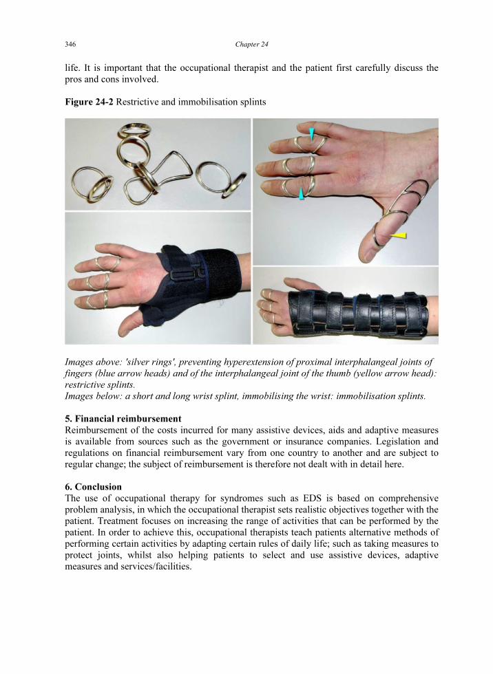
life. It is important that the occupational therapist and the patient first carefully discuss the
pros and cons involved.
Figure 24-2 Restrictive and immobilisation splints
Images above: 'silver rings', preventing hyperextension of proximal interphalangeal joints of
fingers (blue arrow heads) and of the interphalangeal joint of the thumb (yellow arrow head):
restrictive splints.
Images below: a short and long wrist splint, immobilising the wrist: immobilisation splints.
5. Financial reimbursement
Reimbursement of the costs incurred for many assistive devices, aids and adaptive measures
is available from sources such as the government or insurance companies. Legislation and
regulations on financial reimbursement vary from one country to another and are subject to
regular change; the subject of reimbursement is therefore not dealt with in detail here.
6. Conclusion
The use of occupational therapy for syndromes such as EDS is based on comprehensive
problem analysis, in which the occupational therapist sets realistic objectives together with the
patient. Treatment focuses on increasing the range of activities that can be performed by the
patient. In order to achieve this, occupational therapists teach patients alternative methods of
performing certain activities by adapting certain rules of daily life; such as taking measures to
protect joints, whilst also helping patients to select and use assistive devices, adaptive
measures and services/facilities.
Chapter 24346

Further reading
• Goodacre L, MacArthur M. Rheumatology practice in occupational therapy: promoting
lifestyle management. 1st
ed. Chichester, UK: Wiley-Blackwell, 2013.
• Russek LN. Examination and treatment of a patient with hypermobility syndrome. Phys
Ther 2000;80:386-98.
• Smith TO, Bacon H, Jerman E, et al. Physiotherapy and occupation therapy interventions
for people with benign hypermobility syndrome: a systematic review of clinical trials.
Disabil Rehabil 2014;36:797-803.
• Söderback I. International handbook of occupational therapy interventions. 1st
ed. New
York, USA: Springer, 2009.
• Steultjens EM, Dekker J, Bouter LM, van Schaardenburg D, van Kuyk MA, van den
Ende CH. Occupational therapy for rheumatoid arthritis. Cochrane Database Syst Rev
2004;(1):CD003114.
The role of occupational therapy in Ehlers-Danlos syndrome 347

This page intentionally left blank

Addendum. Considerations, precautions and measures in case of surgery in
patients with Ehlers-Danlos syndrome
These are just general recommendations; depending on the clinical situation or condition of an
individual patient, other or additional measures may be necessary.
Before surgery
1. Most important, especially in patients with classical Ehlers-Danlos syndrome (EDS),
vascular EDS or kyphoscoliotic EDS, is to weigh the potential benefits of surgery and
possible complications of surgery and of required diagnostic investigations, before
they are planned. Consider all non-surgical alternatives
2. Choose, if possible and clinically indicated, minimal and non-invasive diagnostic
procedures, such as computer tomography (CT), magnetic resonance imaging (MRI)
and ultrasonography; avoid in patients with vascular EDS or kyphoscoliotic EDS
punctures, arteriography, endoscopy, clyster and colonic irrigation
3. In case of planned intubation, X-rays of the cervical spine in several positions of the
head may be indicated to diagnose hypermobility of the upper cervical vertebrae and
malformations of vertebrae
4. Preoperative examination by a cardiologist is indicated if at adequate preoperative
screening cardiac problems are found
5. Routine screening for clotting and bleeding disorders is not indicated; screen only if
clinically indicated; in case of prolonged bleeding time, testing whether DDAVP can
diminish the bleeding time may be indicated
6. Because of increased risk of bleeding during and after surgery (in vascular and
kyphoscoliotic EDS caused by defective connective tissue within vessels and in other
EDS types caused by lax connective tissue around vessels), carbasalate calcium and
acetylsalicylic acid should be discontinued for 7-10 days if possible and other non-
steroidal anti-inflammatory drugs for 5-7 days
7. In vascular EDS, blood pressure should be checked several times and be controlled
before surgery, if too high
8. Especially for a patient with vascular or kyphoscoliotic EDS, it should be checked
before the actual day of surgery and at that day whether there is enough blood
available of the patient’s blood type
During surgery
1. In case of local or regional anaesthesia: keep in mind that patients with EDS may
experience insufficient aesthetic effect
2. Beware of (sub)luxation of the mandibular joints at intubation; then rapid
repositioning is indicated
3. Beware of an increased risk of (sub)luxation of joints because of muscle relaxing
anaesthesia, especially when repositioning a patient or performing surgery of the
upper extremity
4. For incisions of the skin, follow Langer's lines of the skin, corresponding to the
alignment of collagen fibres within the dermis, resulting in minimal cleavage of these
fibres
5. Avoid pressure or tension on skin, vessels and organs by using atraumatic surgery
equipment and haemoclips, instead of stiches. Beware of tension on the skin when
closing the wound, especially in a patient with classical EDS. Much experience is
349

required for surgery in patients with EDS, especially vascular, classical or
kyphoscoliotic EDS. It could be wise to have another experienced surgeon stand-by
for emergencies
6. Avoid the use of probes
7. In case of bowel ruptures in vascular EDS it is generally better to place a stoma than
trying to suture the rupture
8. Beware of an increased risk of pneumothorax at mechanical ventilation; a lower peak-
flow volume may be indicated
After surgery
1. Again beware of an increased risk of pneumothorax at mechanical ventilation; a lower
peak-flow volume may be indicated
2. Beware of increased risk of anastomotic leakage and its complications, such as
bleeding and infection
3. Avoid rectal body temperature assessments and punctures of arteries (e.g. for blood
gases measurements); limit number of vena punctures
4. In patients with vascular EDS: again regularly check and control blood pressure and
prescribe a proton pump inhibitor because of the risk of a major bleeding in case of a
stress ulcer
5. Apply special techniques for bandaging of wounds to prevent wound herniation
6. The revalidation period may be longer than normal; start revalidation early, under
supervision of a physiotherapist
Addendum350

Index
adaptations and devices ............................................................................................ 70, 135, 342
aesthetic surgery ......................................................................................................... see surgery
arterial tortuosity syndrome ..................................................................................................... 86
arthrochalasia EDS ............................................................................................................... 9, 22
(chapter) ............................................................................................................................... 97
clinical findings .................................................................................................................. 110
clinical findings & hip dislocation (figure) ........................................................................ 111
criteria ................................................................................................................................. 109
differential diagnosis .......................................................................................................... 116
eye ...................................................................................................................................... 230
gel electrophoresis of collagens (figure) ............................................................................ 113
hip dislocation (figure) ....................................................................................................... 112
history ................................................................................................................................. 110
inheritance .......................................................................................................................... 114
management ....................................................................................................................... 117
structural defect .......................................................................................................... 112, 114
ultrastructural appearance of collagen fibrils (figure) ........................................................ 115
Beighton score ........................................................................................................ 7, 21, 49, 314
(table) ................................................................................................................................... 18
adapted ................................................................................................................................. 52
cut-off values ...................................................................................................................... 314
bleeding tendency in EDS
(chapter) ............................................................................................................................. 167
clotting cascade .................................................................................................................. 168
DDAVP .............................................................................................................................. 172
haemostasis ......................................................................................................................... 168
measures ............................................................................................................................. 171
platelets ............................................................................................................................... 170
recombinant factor VIIa ..................................................................................................... 173
uterine bleeds ...................................................................................................................... 244
work-up .............................................................................................................................. 170
brain and spine structural anomalies in EDS ......................................................................... 192
brittle cornea syndrome .............................................................................................. 10, 23, 108
Bulbena score ..................................................................................................................... 7, 257
(table) ................................................................................................................................... 19
cardiac abnormalities in EDS
(chapter) ............................................................................................................................. 177
cardiovascular dysautonomia ............................................................................................. 190
mitral valve prolapse (figure) ............................................................................................. 179
cardiac-valvular EDS ........................................................................................................... 8, 20
Chiari malformation ............................................................................................................... 193
(figure) ................................................................................................................................ 193
children ................................................................................................................................... 289
consideration for parents with EDS to have a child (table) ................................................ 273
ethics in parenthood ........................................................................................................... 272
reproductive options for parents with EDS (table) ............................................................. 272
351

with EDS (chapter) ............................................................................................................. 255
with EDS, case ................................................................................................................... 256
with EDS, diagnostic evaluation ........................................................................................ 258
with EDS, management ...................................................................................................... 260
with EDS, signs and symptoms .......................................................................................... 257
classical EDS ........................................................................................................................ 7, 85
(chapter) ............................................................................................................................... 47
cardiac abnormalities .......................................................................................................... 178
eye ...................................................................................................................................... 228
gastrointestinal manifestations ........................................................................................... 148
gastrointestinal manifestations (table) ................................................................................ 156
classical-like EDS ................................................................................................................ 8, 20
classification of EDS .................................................................................................................. 6
genetically (table) ................................................................................................................. 17
historically (table) ................................................................................................................ 15
main problems (table) ......................................................................................................... 337
cognitive-behavioural therapy ................................................................................ 284, 306, 307
collagen
(figure) .................................................................................................................................. 37
abnormalities (table) ........................................................................................... 15, 17, 20, 43
cross-linking ....................................................................................................................... 105
gel electrophoresis in arthrochalasia EDS (figure) ............................................................ 113
structure in dermatosparaxis EDS (figure) ......................................................................... 116
ultrastructural appearance in arthrochalasia EDS (figure) .................................................. 115
coping with EDS .................................................................................................................... 288
dental issues in EDS ............................................................................................................... 220
dermatosparaxis EDS ........................................................................................................... 9, 22
(chapter) ............................................................................................................................... 97
clinical findings .................................................................................................................. 119
criteria ................................................................................................................................. 118
differential diagnosis .......................................................................................................... 120
eye ...................................................................................................................................... 230
genetic defect ...................................................................................................................... 119
structure of collagen versus normal collagen (figure) ........................................................ 116
diagnosis of EDS ...................................................................................................................... 11
differential diagnosis ............................................................................................................ 12
differential diagnosis (table) ........................................................................................... 20, 72
differential diagnosis of vascular EDS ................................................................................. 85
history taking ........................................................................................................................ 11
physical examination ............................................................................................................ 12
signs (table) .......................................................................................................................... 20
education .................................................................................................................................. 70
epilepsy ................................................................................................................................... 195
ethics
(chapter) ............................................................................................................................. 271
consideration for parents with EDS to have a child (table) ................................................ 273
living a (good) life with EDS ............................................................................................. 276
parenthood .......................................................................................................................... 272
reproductive options for parents with EDS (table) ............................................................. 272
Index352

eye
(chapter) ............................................................................................................................. 225
anotomy .............................................................................................................................. 226
anotomy (figure) ................................................................................................................. 227
screening and monitoring ................................................................................................... 230
surgery ................................................................................................................................ 230
fatigue ............................................................................................................................. 189, 284
(figure) .................................................................................................................................. 65
gastrointestinal manifestations
(chapter) ............................................................................................................................. 145
impact ................................................................................................................................. 151
in classical EDS (table) ...................................................................................................... 156
in hypermobile EDS (table) ........................................................................................ 154, 155
in hypermobility (table) ...................................................................................................... 158
in hypermobility spectrum disorders (table) .............................................................. 154, 155
in unclassified EDS ............................................................................................................ 149
management ....................................................................................................................... 151
pathogenesis ....................................................................................................................... 150
genetics
(chapters) .......................................................................................................................... 5, 33
counselling ........................................................................................................................... 13
DNA ..................................................................................................................................... 34
DNA to protein (figure) ........................................................................................................ 35
genes (table) ............................................................................................................. 15, 17, 43
glossary ................................................................................................................................. 27
with clinical signs (table) ..................................................................................................... 43
gynaecological issues .......................................................................... see also pregnancy issues
(chapter) ............................................................................................................................. 241
consideration for parents with EDS to have a child (table) ................................................ 273
contraception ...................................................................................................................... 243
contraception methods (table) ............................................................................................ 252
infertility ............................................................................................................................. 243
normal and abnormal anatomy (figure) .............................................................................. 246
other disorders .................................................................................................................... 247
prolapse & urinary incontinence ........................................................................................ 245
reproductive options for parents with EDS (table) ............................................................. 272
sexuality ............................................................................................................................. 290
uterine bleeding .................................................................................................................. 244
history of EDS ........................................................................................................................ 129
hypermobile EDS ................................................................................................................. 9, 21
cardiac abnormalities .......................................................................................................... 178
cardiovascular exercise tolerance testing ........................................................................... 319
disability ............................................................................................................................. 318
dysfunctions (figure) .......................................................................................................... 313
eye ...................................................................................................................................... 228
gastrointestinal manifestations ........................................................................................... 147
gastrointestinal manifestations (table) ........................................................................ 154, 155
history ..................................................................................................................................... 1
interactions of dysfunctions and symptoms (figure) .................................................... 65, 313
Index 353

management ................................................................................................................. 70, 320
management (chapter) ........................................................................................................ 311
management (table) ............................................................................................................ 323
multi-systemic involvement ....................................................................................... 316, 320
muscle weakness ................................................................................................................ 319
musculoskeletal dysfunction ...................................................................................... 316, 319
psychological dysfunction .......................................................................................... 317, 320
hypermobility
(chapter) ............................................................................................................................... 61
autonomic dysfunction ......................................................................................................... 66
bone mass ............................................................................................................................. 66
classification ......................................................................................................................... 66
classification (table) ............................................................................................................. 72
epidemiology ........................................................................................................................ 62
fatigue ................................................................................................................................... 64
fatigue (figure) ...................................................................................................................... 65
gastrointestinal manifestations ........................................................................................... 148
gastrointestinal manifestations (table) ................................................................................ 158
history ..................................................................................................................................... 1
International Classification of Functioning, Disability and Health (figure) ...................... 315
management ......................................................................................................................... 70
pain ....................................................................................................................................... 64
pain (figure) .......................................................................................................................... 65
prevalence ............................................................................................................................. 63
terminology .................................................................................................................... 62, 67
hypermobility spectrum disorders .................................................................................... 63, 146
dysfunctions (figure) .......................................................................................................... 313
features (table) .................................................................................................................... 154
gastrointestinal manifestations (chapter) ............................................................................ 145
gastrointestinal manifestations (table) ................................................................................ 155
interactions of dysfunctions and symptoms (figure) .......................................................... 313
management (chapter) ........................................................................................................ 311
management (table) ............................................................................................................ 323
neurological manifestations (chapter) ................................................................................ 187
kyphoscoliotic EDS ........................................................................................................ 9, 23, 85
(chapter) ............................................................................................................................... 97
clinical findings .................................................................................................................... 99
clinical findings in kEDS-FKBP14 (figure) ....................................................................... 104
criteria ................................................................................................................................... 98
diagnostic tests ................................................................................................................... 106
differential diagnosis .......................................................................................................... 107
eye ...................................................................................................................................... 229
genetic defect ...................................................................................................................... 105
history ................................................................................................................................... 99
inheritance .......................................................................................................................... 106
lysyl hydroxylase ................................................................................................................ 106
management ....................................................................................................................... 109
mutations of FKBP14 gene (figure) ................................................................................... 100
mutations of PLOD1 gene (figure) ..................................................................................... 100
prenatal testing ................................................................................................................... 107
Index354

prevalence ........................................................................................................................... 105
skeletal deformities (figure) ............................................................................................... 102
Larsen syndrome .................................................................................................................... 116
life style advice ....................................................................................................................... 341
Loeys-Dietz syndrome ............................................................................................... 69, 86, 193
Marfan syndrome ....................................................................................................... 69, 86, 193
losartan ....................................................................................................................... 1, 69, 70
mindfulness ............................................................................................................................ 288
mitral valve prolapse .............................................................. see cardiac abnormalities in EDS
musculocontractural EDS ........................................................................................... 10, 25, 108
neck (figure) ....................................................................................................................... 195
myopathic EDS .................................................................................................................. 10, 25
neurological manifestations .................................................................................................... 196
(chapter) ............................................................................................................................. 187
(figure) ................................................................................................................................ 197
developmental delay ........................................................................................................... 199
headache ............................................................................................................................. 189
investigations (table) .......................................................................................................... 201
management ....................................................................................................................... 199
muscle histology (figure) ................................................................................................... 198
neuropathy .......................................................................................................................... 198
Nevo syndrome ...................................................................................................................... 108
nursing care
(chapter) ............................................................................................................................. 331
11 functional health patterns (box) ..................................................................................... 333
at admission ........................................................................................................................ 334
medical history taking ........................................................................................................ 332
occupational therapy .............................................................................................................. 134
(chapter) ............................................................................................................................. 339
adaptations and devices .............................................................................. 342, 343, 344, 345
devices (figure) ................................................................................................................... 344
home modifications ............................................................................................................ 345
problem analysis ................................................................................................................. 340
splints ................................................................................................................................. 345
splints (figure) .................................................................................................................... 346
orthopaedic issues in EDS
(chapter) ............................................................................................................................. 127
case ..................................................................................................................................... 128
management ....................................................................................................................... 133
origin .................................................................................................................................. 130
orthopaedic shoes ............................................................................................................... 135
osteoarthritis ......................................................................................................................... 66
shoulder arthrodesis (figure) .............................................................................................. 129
signs and symptoms ........................................................................................................... 131
sugery of scoliosis (figure) ................................................................................................. 132
surgery ................................................................................................................................ 135
pain ........................................................................................................................... 64, 188, 283
(chapter) ............................................................................................................................. 297
epidural injections .............................................................................................................. 304
insufficient effect of local analgesics ........................... 64, 140, 188, 251, 303, 307, 334, 349
Index 355

Loeser circles (figure) ........................................................................................................ 299
nerve blocks ........................................................................................................................ 303
nerve stimulation ................................................................................................................ 304
nociceptive and neuropathic ............................................................................................... 298
pain killers .......................................................................................................................... 300
psychological approach ...................................................................................................... 305
sympathetic blocks ............................................................................................................. 304
periodontal EDS ........................................................................................................... 10, 25, 85
physical therapy ..................................................................................................................... 133
and exercises (figure) ........................................................................................................... 65
exercises at home ................................................................................................................. 70
in children with EDS .......................................................................................................... 261
in temporomandibular disorders ......................................................................................... 218
pregnancy issues .......................................................................... see also gynaecological issues
(chapter) ............................................................................................................................. 241
anaesthesia during delivery ................................................................................................ 250
contraception ...................................................................................................................... 243
foetal complications ........................................................................................................... 250
in classical EDS .................................................................................................................. 248
in hypermobile EDS ........................................................................................................... 248
in vascular EDS ............................................................................................................ 89, 249
infertility ............................................................................................................................. 243
preconception counselling .................................................................................................. 242
pseudoxanthoma elasticum ...................................................................................................... 86
quality of life
(chapter) ............................................................................................................................. 281
aspects of well-being and functioning (figure) .................................................................. 291
case ..................................................................................................................................... 282
consequences of a chronic disease ..................................................................................... 300
coping ................................................................................................................................. 288
living a (good) life with EDS ............................................................................................. 276
problems (figure) ................................................................................................................ 287
psychological dysfunction .................................................................................................. 320
psychological issues ........................................................................................................... 286
sexuality ............................................................................................................................. 290
sleeping problems ............................................................................................................... 285
social consequences of having a rare chronic disease ........................................................ 289
social functioning ............................................................................................................... 289
skin ......................................................................................................................................... 285
anatomy (figure) ................................................................................................................... 48
biopsy ................................................................................................................................... 39
cigarette paper aspect (figure) .............................................................................................. 50
in classical EDS .................................................................................................................... 49
in hypermobile EDS ................................................................................................... 314, 318
manifestations ......................................................................................................................... 7
molluscoid pseudotumors ............................................................................................. 51, 141
molluscoid pseudotumors (figure) ....................................................................................... 51
piezogenic papules ............................................................................................................... 52
piezogenic papules (figure) .................................................................................................. 52
protective measures ............................................................................................................ 342
Index356

testing ..................................................................................................................................... 6
sleeping problems ................................................................................................................... 285
social functioning ................................................................................................................... 289
spondylodysplastic EDS ............................................................................................. 10, 24, 108
Stickler syndrome ..................................................................................................................... 69
manifestations (table) ........................................................................................................... 73
surgery
aesthetic corrections ........................................................................................................... 141
plastic surgery (chapter) ..................................................................................................... 139
precautions and measures (addendum) .............................................................................. 349
temporomandibular disorders
(chapter) ............................................................................................................................. 209
anatomy of the temporomandibular joint (figure) .............................................................. 211
arthrogenous versus myogenous (table) ............................................................................. 222
diagnostic process .............................................................................................................. 216
dysfunctions of the temporomandibular joint (figure) ....................................................... 212
luxation of the jaw .............................................................................................................. 218
luxation of the jaw (figure) ................................................................................................. 213
management ....................................................................................................................... 217
mechanisms ........................................................................................................................ 210
oral splint (figure) ............................................................................................................... 219
osteoarthritis of the jaw .............................................................................................. 215, 219
panoramic radiograph (figure) ........................................................................................... 214
physical therapy .................................................................................................................. 218
prevalence in EDS and hypermobility ............................................................................... 215
urological complications
(chapter) ............................................................................................................................. 235
cases ................................................................................................................................... 236
lower urinary tract .............................................................................................................. 238
upper urinary tract .............................................................................................................. 238
vascular EDS .................................................................................................. 8, 40, 68, 180, 181
(chapter) ............................................................................................................................... 79
acrogeria (figure) .................................................................................................................. 68
advices (table) .................................................................................................................... 182
biochemical and molecular diagnosis .................................................................................. 83
bleeding .............................................................................................................................. 168
bleeding therapy ................................................................................................................. 171
celiprolol ....................................................................................... 1, 69, 70, 88, 173, 182, 192
cerebrovascular manifestations .......................................................................................... 191
clinical findings .................................................................................................................... 80
differential diagnosis ............................................................................................................ 85
dilated aortic root (figure) .................................................................................................. 180
eye ...................................................................................................................................... 229
gastrointestinal manifestations ........................................................................................... 146
genetics ................................................................................................................................. 84
history ................................................................................................................................... 80
management ......................................................................................................................... 87
pregnancy ..................................................................................................................... 89, 249
prevalence ............................................................................................................................. 80
vascular fragility ................................................................................................................... 81
Index 357

This page intentionally left blank

This page intentionally left blank

This page intentionally left blank