Efficacy of celery (Apium graveolens L.) alcoholic extract ...
8
Veterinary World, EISSN: 2231-0916 898 Veterinary World, EISSN: 2231-0916 Available at www.veterinaryworld.org/Vol.15/April-2022/11.pdf RESEARCH ARTICLE Open Access Efficacy of celery (Apium graveolens L.) alcoholic extract against systemic methicillin-resistant Staphylococcus aureus infection in rat models Yos Adi Prakoso 1,2 and Agustina Dwi Wijayanti 3 1. Postdoctoral Program, University of Gadjah Mada, Yogyakarta 55281, Indonesia; 2. Department of Pharmacology, Faculty of Veterinary Medicine, University of Wijaya Kusuma Surabaya 60225, Indonesia; 3. Department of Pharmacology, Faculty of Veterinary Medicine, University of Gadjah Mada, Yogyakarta 55281, Indonesia. Corresponding author: Agustina Dwi Wijayanti, e-mail: [email protected] Co-author: YAP: [email protected] Received: 04-01-2022, Accepted: 02-03-2022, Published online: 12-04-2022 doi: www.doi.org/10.14202/vetworld.2022.898-905 How to cite this article: Prakoso YA, Wijayanti AD (2022) Efficacy of celery (Apium graveolens L.) alcoholic extract against systemic methicillin-resistant Staphylococcus aureus infection in rat models, Veterinary World, 15(4): 898-905. Abstract Background and Aim: The coronavirus disease-19 (COVID-19) pandemic caused global economic and health problems. The pandemic increased the number of infectious diseases categorized as neglected diseases, such as staphylococcosis, which is caused by methicillin-resistant Staphylococcus aureus (MRSA). Celery is an herb that consist of antioxidants that can potentially act as antimicrobial agents. This study aimed to analyze the efficacy of celery alcoholic extract against systemic MRSA infections in rat models. Materials and Methods: In this study, 36 male, 6-month-old Sprague-Dawley rats (average weight: 300 g) were used as models. The rats were divided into six groups: Group K− (negative control), Group K+ (infected with MRSA without therapy), Group V (infected with MRSA+100 mg vancomycin per kg body weight [BW]), Group P1 (infected with MRSA+1 mg celery extract per kg BW), Group P2 (infected with MRSA+2 mg celery extract per kg BW), and Group P4 (infected with MRSA+4 mg celery extract per kg BW). The therapy was given once daily for 7 days. Blood and organs were taken on day 7 for hematology, serology, immunohistochemistry, and histopathology. Results: Results showed that 4 mg celery extract per kg BW promotes the healing of MRSA systemic infections in rat models (p≤0.05). The better prognosis was indicated by the normalization of red blood cell indices, white blood cell, neutrophil and lymphocyte counts, Cluster of differentiation 4+, Cluster of differentiation 8+, and Cyclooxygenase-2 expression and absence of severe tissue damage. Celery extracts inhibited MRSA growth in the blood samples. Conclusion: It can be concluded that celery alcoholic extract can potentially be used as an antimicrobial agent against systemic MRSA infections. A clinical study regarding the efficacy of celery extract must be conducted to ensure its potency against MRSA infections in humans. Keywords: celery, efficacy, healing, methicillin-resistant Staphylococcus aureus, systemic infection. Introduction The coronavirus disease-19 (COVID-19) pan- demic caused global economic and health problems. The high number of daily cases worldwide triggered chaos in health systems [1]. It impacted hospitals to halt health services for diseases with low urgency, such as metabolic diseases and bacterial infections with low pathogenicity. Indirectly, these policies increase the number of infectious diseases, causing them to be categorized as neglected diseases [2]. One of these is staphylococcosis [3]. Staphylococcosis is an infectious disease caused by Staphylococcus aureus. Methicillin-resistant S. aureus (MRSA) is a common type of staphylococcus that causes severe infections. MRSA infections can occur systemically and trigger massive tissue destruction. 30 cases of MRSA exist with a high fatality rate for every 100,000 people. The major tissue destruction caused by MRSA can lead to meningitis, encephalitis, pneumonia, hepatitis, and systemic inflammation with sepsis [4]. Severe systemic inflammation and sepsis increase the mortality rate among the infected patients. Treatment of MRSA infections becomes harder due to restrictions in massive administration of antibiotics to prevent more complex antibiotic resistance. Nevertheless, vancomy- cin is the recommended drug for MRSA infections [5]. However, there is worry about generating resistance, sooner or later, due to the widespread use of vancomycin. The development of herbal-derived therapy must be increased to lessen the use of antibiotics and prevent resistance. Indonesia exhibits high bio- diversity, consisting of several species of animals and plants [6]. Several plants contain antioxidants that can potentially be antibacterial agents. One of them is celery (Apium graveolens L.). Celery con- tains terpenoids, flavonoids, alkaloids, saponins, and tannins [7]. These components have been developed as antimicrobial agents due to their ability to inhibit Copyright: Prakoso and Wijayanti. Open Access. This article is distributed under the terms of the Creative Commons Attribution 4.0 International License (http://creativecommons.org/licenses/ by/4.0/), which permits unrestricted use, distribution, and reproduction in any medium, provided you give appropriate credit to the original author(s) and the source, provide a link to the Creative Commons license, and indicate if changes were made. The Creative Commons Public Domain Dedication waiver (http:// creativecommons.org/publicdomain/zero/1.0/) applies to the data made available in this article, unless otherwise stated.
-
Upload
khangminh22 -
Category
Documents
-
view
2 -
download
0
Transcript of Efficacy of celery (Apium graveolens L.) alcoholic extract ...

Veterinary World, EISSN: 2231-0916 898
Veterinary World, EISSN: 2231-0916Available at www.veterinaryworld.org/Vol.15/April-2022/11.pdf
RESEARCH ARTICLEOpen Access
Efficacy of celery (Apium graveolens L.) alcoholic extract against systemic methicillin-resistant Staphylococcus aureus infection in rat models
Yos Adi Prakoso1,2 and Agustina Dwi Wijayanti3
1. Postdoctoral Program, University of Gadjah Mada, Yogyakarta 55281, Indonesia; 2. Department of Pharmacology,Faculty of Veterinary Medicine, University of Wijaya Kusuma Surabaya 60225, Indonesia; 3. Department of Pharmacology, Faculty of Veterinary Medicine, University of Gadjah Mada, Yogyakarta 55281, Indonesia.
Corresponding author: Agustina Dwi Wijayanti, e-mail: [email protected]: YAP: [email protected]
Received: 04-01-2022, Accepted: 02-03-2022, Published online: 12-04-2022
doi: www.doi.org/10.14202/vetworld.2022.898-905 How to cite this article: Prakoso YA, Wijayanti AD (2022) Efficacy of celery (Apium graveolens L.) alcoholic extract against systemic methicillin-resistant Staphylococcus aureus infection in rat models, Veterinary World, 15(4): 898-905.
AbstractBackground and Aim: The coronavirus disease-19 (COVID-19) pandemic caused global economic and health problems. The pandemic increased the number of infectious diseases categorized as neglected diseases, such as staphylococcosis, which is caused by methicillin-resistant Staphylococcus aureus (MRSA). Celery is an herb that consist of antioxidants that can potentially act as antimicrobial agents. This study aimed to analyze the efficacy of celery alcoholic extract against systemic MRSA infections in rat models.
Materials and Methods: In this study, 36 male, 6-month-old Sprague-Dawley rats (average weight: 300 g) were used as models. The rats were divided into six groups: Group K− (negative control), Group K+ (infected with MRSA without therapy), Group V (infected with MRSA+100 mg vancomycin per kg body weight [BW]), Group P1 (infected with MRSA+1 mg celery extract per kg BW), Group P2 (infected with MRSA+2 mg celery extract per kg BW), and Group P4 (infected with MRSA+4 mg celery extract per kg BW). The therapy was given once daily for 7 days. Blood and organs were taken on day 7 for hematology, serology, immunohistochemistry, and histopathology.
Results: Results showed that 4 mg celery extract per kg BW promotes the healing of MRSA systemic infections in rat models (p≤0.05). The better prognosis was indicated by the normalization of red blood cell indices, white blood cell, neutrophil and lymphocyte counts, Cluster of differentiation 4+, Cluster of differentiation 8+, and Cyclooxygenase-2 expression and absence of severe tissue damage. Celery extracts inhibited MRSA growth in the blood samples.
Conclusion: It can be concluded that celery alcoholic extract can potentially be used as an antimicrobial agent against systemic MRSA infections. A clinical study regarding the efficacy of celery extract must be conducted to ensure its potency against MRSA infections in humans.
Keywords: celery, efficacy, healing, methicillin-resistant Staphylococcus aureus, systemic infection.
Introduction
The coronavirus disease-19 (COVID-19) pan-demic caused global economic and health problems. The high number of daily cases worldwide triggered chaos in health systems [1]. It impacted hospitals to halt health services for diseases with low urgency, such as metabolic diseases and bacterial infections with low pathogenicity. Indirectly, these policies increase the number of infectious diseases, causing them to be categorized as neglected diseases [2]. One of these is staphylococcosis [3].
Staphylococcosis is an infectious disease caused by Staphylococcus aureus. Methicillin-resistant S. aureus (MRSA) is a common type of staphylococcus that causes severe infections. MRSA infections can occur
systemically and trigger massive tissue destruction. 30 cases of MRSA exist with a high fatality rate for every 100,000 people. The major tissue destruction caused by MRSA can lead to meningitis, encephalitis, pneumonia, hepatitis, and systemic inflammation with sepsis [4]. Severe systemic inflammation and sepsis increase the mortality rate among the infected patients. Treatment of MRSA infections becomes harder due to restrictions in massive administration of antibiotics to prevent more complex antibiotic resistance. Nevertheless, vancomy-cin is the recommended drug for MRSA infections [5]. However, there is worry about generating resistance, sooner or later, due to the widespread use of vancomycin.
The development of herbal-derived therapy must be increased to lessen the use of antibiotics and prevent resistance. Indonesia exhibits high bio-diversity, consisting of several species of animals and plants [6]. Several plants contain antioxidants that can potentially be antibacterial agents. One of them is celery (Apium graveolens L.). Celery con-tains terpenoids, flavonoids, alkaloids, saponins, and tannins [7]. These components have been developed as antimicrobial agents due to their ability to inhibit
Copyright: Prakoso and Wijayanti. Open Access. This article is distributed under the terms of the Creative Commons Attribution 4.0 International License (http://creativecommons.org/licenses/by/4.0/), which permits unrestricted use, distribution, and reproduction in any medium, provided you give appropriate credit to the original author(s) and the source, provide a link to the Creative Commons license, and indicate if changes were made. The Creative Commons Public Domain Dedication waiver (http://creativecommons.org/publicdomain/zero/1.0/) applies to the data made available in this article, unless otherwise stated.

Veterinary World, EISSN: 2231-0916 899
Available at www.veterinaryworld.org/Vol.15/April-2022/11.pdf
membrane synthesis and destruct bacterial membrane osmolarity [8].
A previous study reported that 1 mg/mL of cel-ery extract promotes membrane damage to MRSA in vitro [7]. Further exploration described that celery can be used as a cream-based therapy against MRSA infections in diabetic wounds [9]. However, celery extract has not yet been used against MRSA systemic infections. This study aimed to analyze the efficacy of celery alcoholic extract against MRSA systemic infec-tions in rat models.Materials and MethodsEthical approval
The animal utilization in this study has been approved by the ethical clearance committee from the Faculty of Dentistry, University of Airlangga, Surabaya, with approval number: 236/HRECC/FODM/V/2020. The committee member conducted monitoring during the study.Study period and location
The study was conducted from January 2021 to September 2021. The extraction was performed in the Laboratory of Pharmacology, Faculty of Veterinary Medicine, University of Wijaya Kusuma Surabaya, Indonesia. The determination of herbal biochemical compounds was determined in the Faculty of Health, University of Muhammadiyah Sidoarjo, East Java, Indonesia. The in vivo study was conducted in the Laboratory of Pharmacology, Faculty of Veterinary Medicine, University of Gadjah Mada, Yogyakarta, Indonesia.Herbal preparation and extraction
Celery was collected from a botanical market in Batu, East Java, Indonesia. The celery species was identified as A. graveolens (L.) by a botanist from the Plant Conservation Center, Purwodadi Botanical Garden, East Java, Indonesia. The species was regis-tered with the herbarium voucher no. 0276/IPH.06/HM/II/2019. Drying of the celery was performed using an oven at 80°C for an hour. The dried celery was mashed using a blender and was soaked in 70% alcohol (1:4 dried celery: 70% alcohol). Then, it was evaporated using a rotary evaporator (Buchi R-100, Cat No. 11100v101, Buchi, Indonesia) at 69°C, and the product was stored at 4°C [10].MRSA isolate
The MRSA isolate was obtained from the clinical isolates collection of the Laboratory of Bacteriology, Faculty of Health, University of Muhammadiyah Sidoarjo, East Java, Indonesia. Before enrichment, biochemical tests were performed on the isolate. The results of the biochemical tests are as follows: Coagulase (+), DNase (+), hemolysis (+), pigmented colony (+), alkaline phosphatase (+), urease (+), mannitol (+), maltose (+), esculin hydrolysis (−), novobiocin (sensitive), polymyxin B (resistant), and cefoxitin (resistant). The MRSA isolate was enriched
using a mannitol salt agar (MSA) plate and was trans-ferred on broth media for incubation until it showed turbidity similar to the 1.0 McFarland standard. The 1.0 McFarland was standardized using a McFarland densitometer.Animal model and research design
In this study, 36 male 6-month-old Sprague-Dawley rats (weight: 303.58±10.63 g) were used as models. The rats were obtained from the Laboratory of Pharmacology, Faculty of Veterinary Medicine, University of Wijaya Kusuma Surabaya. Before the treatment, the rats were acclimated to the laboratory for 7 days using several conditions, including, 12/12 h of light/dark, 25°C room temperature, feed, water ad libitum, and they were maintained individually. The rats were divided into six groups with six rats each: Group K− (negative control/uninfected and untreated), Group K+ (infected with MRSA without therapy), Group V (infected with MRSA+100 mg vancomycin [Vanconex Iyo, Cat No. J01XA01, Amarox Pharma Global, Indonesia] per kg body weight [BW]) [11], Group P1 (infected with MRSA+1 mg celery extract per kg BW), Group P2 (infected with MRSA+2 mg cel-ery extract per kg BW), and Group P4 (infected with MRSA+4 mg celery extract per kg BW). The celery extract doses were selected following a previous study [7]. MRSA infection was induced by intraperitoneal injection of 50 µL of 1.0 McFarland standard bacterial suspension. The therapy was given 24 h after the injec-tion. Before the therapy, the celery extract was mea-sured and was dissolved in distilled water. The therapy was given once daily for 7 days using a gastric probe.Routine hematological test and serology test
After 7 days of treatment, blood and serum were collected from the tail veins of the rats. The blood was tested for red blood cells (RBC), white blood cells (WBC), hemoglobin (Hb), packed cell volume (PCV), mean corpuscular volume (MCV), mean corpuscular Hb (MCH), MCH concentration (MCHC), differential leukocytes, and platelets. The hematology test was performed using an automated hematology analyzer (Medonic M-32 Series, Cat No. CDS1400075, Boule, Sweden). The serum was tested against C-reactive protein (CRP) following a previous study [12].Bacteriological examination of blood
MRSA was isolated from the blood of the infected rats. The collected blood was cultured on an MSA plate by directly streaking the blood sample on the MSA surface. The plate was incubated at 37°C for 24 h [13]. The presence of bacterial colonies was con-sidered positive (+).Histopathology
After blood collection, the rats were euthanized using lethal doses of dissociative anesthetic (150 mg/kg BW ketamine+10 mg/kg BW xylazine). Necropsy was performed to collect several organs, including the brain, lung, heart, liver, spleen, and kidney. The organs

Veterinary World, EISSN: 2231-0916 900
Available at www.veterinaryworld.org/Vol.15/April-2022/11.pdf
were soaked in 10% neutral buffer formalin (NBF) for 24 h. The tissue was cleared and was dehydrated using xylene and graded alcohol, respectively. The organs were embedded in liquid paraffin and were blocked using a base mold. The blocked organs were cut using a microtome and were placed onto glass slides. The tis-sue sections were stained using hematoxylin and eosin.Cell tube block test and immunohistochemistry (IHC)
The cell tube block test was performed by insert-ing blood samples into plain capillary tubes. Then, it was centrifuged, and the buffy coat was taken and stored in 10% NBF [14]. Next, the collected buffy coat was processed similar to routine histopathology pro-cedures and was attached to glass slides. IHC staining against cluster of differentiation 4+ (CD4+), cluster of differentiation 8+ (CD8+), and cyclooxygenase-2 (COX-2) monoclonal antibodies was performed fol-lowing a previous study [9].Morphometry
Morphometry was performed by a senior pathol-ogist from the Department of Pathology, Faculty of Veterinary Medicine, University of Gadjah Mada, Yogyakarta, India. The observed lesions were scored as follows: 1=no histopathological changes, 2=mild, 3=moderate, and 4=severe histopathological changes. The pathologist was unaware of the sample treatment and conducted an objective analysis of the samples. The percentage of the cells that expressed CD4+, CD8+, and COX-2 was measured using ImageJ soft-ware (NIH, USA, Public Domain, BSD-2).Statistical analysis
Several types of data were collected in this study, including quantitative and categorical data. Quantitative data were collected from the hematol-ogy profile, leukocyte profile, CRP, and cell tube block and were analyzed using one-way analysis of variance and post hoc test. Categorical data from his-topathology were analyzed using Kruskal-Wallis and Mann-Whitney U-test. p≤0.05 was considered signifi-cant. All statistical analyses were performed using the statistical package for the social sciences v.16 (IBM Corp., NY, USA).ResultsRBC indices
Hematology analysis revealed significant dif-ferences between the RBC, Hb, PCV, MCV, MCH, and MCHC profiles of the rats systemically infected by MRSA 7 days after therapy (p≤0.05). The routine blood test results showed that treatment with vanco-mycin demonstrates similar results to treatment with 1 mg and 2 mg celery extract per kg BW in terms of Hb, PCV, MCH, and MCHC. Treatment with 4 mg celery extract per kg BW generated similar results to the negative control group in terms of MCH and MCHC (Table-1).
Treatment with vancomycin and all doses of celery extract generated similar results in terms of
circulatory platelet count (p≥0.05) (Table-1). Based on this study, vancomycin and all doses of celery extract increased the number of RBC, Hb, and PCV, which indicates a better prognosis during the treat-ment after being infected by MRSA. However, micro-cytic hypochromic anemia was still observed in the groups treated with vancomycin and 1 mg and 2 mg celery extract per kg BW. The group treated with 4 mg celery extract per kg BW showed signs of microcytic normochromic anemia (Table-1).WBC count
WBC, neutrophil, and lymphocyte count increased more in the in-group positive control com-pared to the others (p≤0.05). An increase in WBC, neu-trophil, and lymphocyte count was also observed in the group treated with vancomycin and celery extract compared to the negative control. Normalization of the lymphocytes/neutrophils (L/N) ratio was found in all the groups treated with celery extract, regardless of the dose, compared to the negative control (p≥0.05) (Table-2).CRP level
The CRP levels in the positive control group increased significantly compared to the other groups (p≤0.05). This indicated severe systemic inflamma-tion following artificial MRSA infection in the rat models. CRP levels decreased in the group treated with 100 mg vancomycin per kg BW and with 1 mg, 2 mg, and 4 mg celery extract per kg BW. The group treated with 4 mg celery extract per kg BW exhibited the lowest CRP level, close to the negative control group (Figure-1).Bacterial examination from blood
Based on the bacterial examination of the blood, MRSA was isolated from all the blood samples in the positive control group. MRSA was not isolated from the group treated with 100 mg vancomycin per kg BW and 4 mg celery extract per kg BW. MRSA was still isolated from the groups treated with 1 mg and 2 mg celery extract per kg BW with varying results (Table-3).Histopathology
Histopathology examination of the samples showed various results. The positive control group showed the least histopathological changes (p≤0.05). The most severe histopathological changes were observed in the vancomycin group and in the 1 mg, 2 mg, and 4 mg celery extract per kg BW (p≤0.05) (Table-4) groups. The observed histopathological changes on all the soft tissue were inflammation, necrosis, and hemorrhage. All the histopathological changes observed are summarized in Table-5.
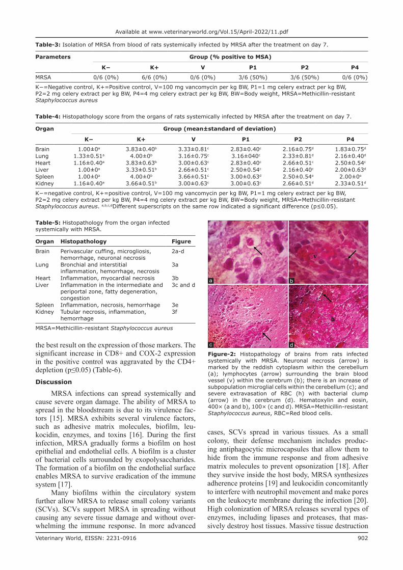
One of the eminent organs impacted by MRSA systemic infection was the brain. Brains from all the infected groups, whether treated and untreated, suffered from several histopathological changes, such as mild to severe neuronal necrosis. Neuronal

Veterinary World, EISSN: 2231-0916 901
Available at www.veterinaryworld.org/Vol.15/April-2022/11.pdf
necrosis occurred significantly in the positive con-trol (Figure-2a). Lymphocytic perivascular cuff-ing (Figure-2b) and microgliosis (Figure-2c) were observed in all the infected groups. Severe hem-orrhage was only observed in the positive control (Figure-2d). Brain histopathology significantly improved along with vancomycin therapy and celery extract dose.
Besides cerebral lesions, other lesions were also observed in several organs, including hemorrhage bronchopneumonia in the lung (score: 2-4), inflamma-tion and necrosis in the heart (score: 2-4), inflammation
in the liver (score: 2-3), inflammation and necrosis in the spleen (score: 2-4), and inflammation and hem-orrhage in the kidney (score: 2-3). Massive inflam-mation, necrosis, and hemorrhage were observed in the lung tissue (Figure-3a). Myocardial necrosis and inflammation occurred in the heart, which indicated systemic infection in the bloodstream (Figure-3b). The liver showed cholangitis, an indication of periportal inflammation (Figure-3c). The intermediate zone was also impacted with congestion and fatty degeneration (Figure-3d). Severe inflammation, pulp depletion, and fatty degeneration occurred in the spleen (Figure-3e) along with severe nephritis (Figure-3f). The observed lesions decreased concomitantly in the group treated with vancomycin and 1 mg, 2 mg, and 4 mg celery extract per kg BW. The decrease in the lesions of the treated groups indicated a better prognosis or mecha-nism of protection and healing of the visceral organs from bacterial invasion and colonization.Expression of CD4+, CD8+, and COX-2
CD4+ is a lymphocyte commonly used as a heal-ing indicator, while CD8+ is a marker used to indicate the immune system’s activity against infections. The treatment either using vancomycin or celery extract beneficially affects on the expression of CD4+ and COX-2. The positive effects of the treatment were indicated by a similar range of percentage of CD4+ and COX-2 between treated and negative control groups (p≥0.05) (Table-6). Furthermore, the group treated with 4 mg celery extract per kg BW showed
Table-2: WBC and differential leukocytes count of rats systemically infected by MRSA after the treatment on day 7.
Parameters Group (mean±standard of deviation)
K− K+ V P1 P2 P4
WBC (×103 cells/µL) 6.39±0.07a 9.28±0.49b 7.29±0.41c 7.41±0.30c 7.14±0.16c 7.01±0.19c
Neutrophil (×103cells/µL) 1.12±0.11a 2.28±0.14b 1.83±0.17c 1.53±0.16d 1.31±0.21a 1.16±0.12a
Lymphocyte (×103cells/µL) 4.58±0.09a 6.27±0.44b 4.80±0.25a 5.52±0.32c 5.53±0.21c 5.39±0.44c
Monocyte (×103 cells/µL) 0.62±0.07a 0.59±0.23a 0.57±0.09a 0.24±0.11b 0.20±0.08b 0.26±0.17b
Ratio L/N 4.12±0.43a 2.76±0.31b 2.62±0.21b 3.63±0.41a 4.32±0.74a 4.65±0.60a
K−=Negative control, K+=Positive control, V=100 mg vancomycin per kg BW, P1=1 mg celery extract per kg BW, P2=2 mg celery extract per kg BW, P4=4 mg celery extract per kg BW, L/N=Lymphocyte/neutrophil, WBC: White blood cells, MRSA=Methicillin‑resistant Staphylococcus aureus, BW=Body weight. a,b,c,dDifferent superscripts on the same row indicated a significant difference (p≤0.05)
Table-1: RBC indices and platelets count of rats systemically infected by MRSA after the treatment on day 7.
Parameters Group (mean±standard of deviation)
K− K+ V P1 P2 P4
RBC (106 cells/µL) 5.61±0.04a 4.92±0.20b 5.34±0.09c 5.37±0.07c 5.38±0.07c 5.41±0.10c
Hb (g/dL) 14.53±0.42a 10.60±0.56b 11.00±0.59b 11.03±0.59b 11.58±0.63b 13.12±1.13c
PCV (%) 41.40±0.77a 33.63±1.87b 37.55±0.84c 37.48±0.59c 37.88±0.72c 38.88±0.67d
MCV (fL) 73.73±1.18a 68.31±4.36b 70.31±1.47c 69.82±1.89c 70.31±0.93c 71.83±2.61c
MCH (Pg) 25.88±0.70a 21.52±1.07b 20.60±1.16c 20.53±0.91c 21.50±1.20c 24.25±2.69a
MCHC (%) 35.10±0.97a 31.56±1.75b 29.33±2.17b 29.44±1.91b 30.59±1.86b 33.74±3.26a
Platelets (×105/µL) 4.38±0.07a 2.98±0.35b 4.07±0.11c 3.93±0.08c 4.13±0.11c 4.22±0.10c
K−=Negative control, K+=Positive control, V=100 mg vancomycin per kg BW, P1=1 mg celery extract per kg BW, P2=2 mg celery extract per kg BW, P4=4 mg celery extract per kg BW, RBC=Red blood cells, Hb=Hemoglobin, PCV=Packed cell volume, MCV=Mean corpuscular volume, MCH=Mean corpuscular hemoglobin, MCHC=Mean corpuscular hemoglobin concentration, MRSA=Methicillin‑resistant Staphylococcus aureus, BW=Body weight. a,b,c,dDifferent superscripts on the same row indicated a significant difference (p≤0.05)
Figure-1: The CRP level of rats infected systemically with MRSA. K−=Negative control, K+=Positive control, V=100 mg vancomycin per kg BW, P1=1 mg celery extract per kg BW, P2=2 mg celery extract per kg BW, P4=4 mg celery extract per kg BW. CRP=C‑reactive protein, MRSA=Methicillin‑resistant Staphylococcus aureus, BW=Body weight.
Veterinary World, EISSN: 2231-0916 902
Available at www.veterinaryworld.org/Vol.15/April-2022/11.pdf
the best result on the expression of those markers. The significant increase in CD8+ and COX-2 expression in the positive control was aggravated by the CD4+ depletion (p≤0.05) (Table-6).Discussion
MRSA infections can spread systemically and cause severe organ damage. The ability of MRSA to spread in the bloodstream is due to its virulence fac-tors [15]. MRSA exhibits several virulence factors, such as adhesive matrix molecules, biofilm, leu-kocidin, enzymes, and toxins [16]. During the first infection, MRSA gradually forms a biofilm on host epithelial and endothelial cells. A biofilm is a cluster of bacterial cells surrounded by exopolysaccharides. The formation of a biofilm on the endothelial surface enables MRSA to survive eradication of the immune system [17].
Many biofilms within the circulatory system further allow MRSA to release small colony variants (SCVs). SCVs support MRSA in spreading without causing any severe tissue damage and without over-whelming the immune response. In more advanced
cases, SCVs spread in various tissues. As a small colony, their defense mechanism includes produc-ing antiphagocytic microcapsules that allow them to hide from the immune response and from adhesive matrix molecules to prevent opsonization [18]. After they survive inside the host body, MRSA synthesizes adherence proteins [19] and leukocidin concomitantly to interfere with neutrophil movement and make pores on the leukocyte membrane during the infection [20]. High colonization of MRSA releases several types of enzymes, including lipases and proteases, that mas-sively destroy host tissues. Massive tissue destruction
Table-3: Isolation of MRSA from blood of rats systemically infected by MRSA after the treatment on day 7.
Parameters Group (% positive to MSA)
K− K+ V P1 P2 P4
MRSA 0/6 (0%) 6/6 (0%) 0/6 (0%) 3/6 (50%) 3/6 (50%) 0/6 (0%)
K−=Negative control, K+=Positive control, V=100 mg vancomycin per kg BW, P1=1 mg celery extract per kg BW, P2=2 mg celery extract per kg BW, P4=4 mg celery extract per kg BW, BW=Body weight, MRSA=Methicillin‑resistant Staphylococcus aureus
Table-4: Histopathology score from the organs of rats systemically infected by MRSA after the treatment on day 7.
Organ Group (mean±standard of deviation)
K− K+ V P1 P2 P4
Brain 1.00±0a 3.83±0.40b 3.33±0.81c 2.83±0.40c 2.16±0.75d 1.83±0.75d
Lung 1.33±0.51a 4.00±0b 3.16±0.75c 3.16±040c 2.33±0.81d 2.16±0.40d
Heart 1.16±0.40a 3.83±0.63b 3.00±0.63c 2.83±0.40c 2.66±0.51c 2.50±0.54c
Liver 1.00±0a 3.33±0.51b 2.66±0.51c 2.50±0.54c 2.16±0.40c 2.00±0.63d
Spleen 1.00±0a 4.00±0b 3.66±0.51c 3.00±0.63d 2.50±0.54e 2.00±0e
Kidney 1.16±0.40a 3.66±0.51b 3.00±0.63c 3.00±0.63c 2.66±0.51d 2.33±0.51d
K−=negative control, K+=positive control, V=100 mg vancomycin per kg BW, P1=1 mg celery extract per kg BW, P2=2 mg celery extract per kg BW, P4=4 mg celery extract per kg BW, BW=Body weight, MRSA=Methicillin‑resistant Staphylococcus aureus. a,b,c,dDifferent superscripts on the same row indicated a significant difference (p≤0.05).
Table-5: Histopathology from the organ infected systemically with MRSA.
Organ Histopathology Figure
Brain Perivascular cuffing, microgliosis, hemorrhage, neuronal necrosis
2a-d
Lung Bronchial and interstitial inflammation, hemorrhage, necrosis
3a
Heart Inflammation, myocardial necrosis 3bLiver Inflammation in the intermediate and
periportal zone, fatty degeneration, congestion
3c and d
Spleen Inflammation, necrosis, hemorrhage 3eKidney Tubular necrosis, inflammation,
hemorrhage3f
MRSA=Methicillin‑resistant Staphylococcus aureus
Figure-2: Histopathology of brains from rats infected systemically with MRSA. Neuronal necrosis (arrow) is marked by the reddish cytoplasm within the cerebellum (a); lymphocytes (arrow) surrounding the brain blood vessel (v) within the cerebrum (b); there is an increase of subpopulation microglial cells within the cerebellum (c); and severe extravasation of RBC (h) with bacterial clump (arrow) in the cerebrum (d). Hematoxylin and eosin, 400× (a and b), 100× (c and d). MRSA=Methicillin‑resistant Staphylococcus aureus, RBC=Red blood cells.
d
a
c
b

Veterinary World, EISSN: 2231-0916 903
Available at www.veterinaryworld.org/Vol.15/April-2022/11.pdf
triggers severe hemorrhage, activates coagulation, and is aggravated by toxins released by MRSA [21].
The pathogenesis mechanism of MRSA infec-tion can be elucidated from the results of this study. The positive control suffered from microcytic hypo-chromic anemia, thrombocytopenia, leukocytosis, a decrease in L/N ratio, increased CRP level, severe his-topathological changes, and decreased CD4+ expres-sion, and an increase in CD8+ and COX-2 expression. These results agree with the results of a previous study
conducted by Prakoso et al. [22]. Microcytic hypo-chromic anemia was the main impact of severe hemor-rhage after MRSA infection in the rat models. Severe hemorrhage in the positive control was observed from the histopathology of several collected specimens, such as the brain, lung, liver, and kidney. Following hemorrhage, platelets were activated, and they infil-trated the tissue to minimize RBC extravasation and cause thrombocytopenia [23]. Circulatory MRSA within the bloodstream also acts as a chemoattractant for increasing WBCs to eliminate the infection [24]. However, leukocytosis cannot suppress MRSA colo-nization [25]. This was proven by the significant dif-ference in a number of neutrophils compared with the lymphocytes. The increase in neutrophils was brought about by the CRP level and the immune expression of CD8+ and COX-2. These systemic changes are believed to be the impacts of MRSA because MRSA can be isolated from all the blood samples from the positive control. These mechanisms indicate that the systemic MRSA infection could worsen if not treated properly.
Vancomycin is the drug of choice for treat-ing MRSA infections. In this study, MRSA was not isolated from the blood specimen of rats artificially infected with MRSA 7 days after vancomycin therapy. The rats treated using vancomycin still showed sev-eral impacts, including microcytic hypochromic ane-mia, thrombocytopenia, leukocytosis, neutrophilia, and increased CRP level; however, the decrease and/or increase in these parameters were not as high as in the positive control. In addition, the better prog-nosis after utilization of vancomycin to treat MRSA infection was indicated by the normalization of lym-phocyte count, the percentage of immune expression of CD4+ and COX-2, and the decrease in the histo-pathological changes score of the observed tissues. Lymphocytes are critical for protection against the invasion of pathogenic infectious agents [26]. The normalization of lymphocyte counts and the bal-anced percentage of circulatory CD4+ indicated that the healing mechanism occurred. The decrease in the immune expression of COX-2, compared to the posi-tive control, is also related to this. As an inflammatory biomarker, the normalization of COX-2 expression in the bloodstream indicated that systemic inflammation is subsiding [27].
Table-6: Percentage of circulatory CD4+, CD8+, and COX‑2 of rats systemically infected by MRSA after the treatment on day 7.
Parameters Group (mean±standard of deviation)
K− K+ V P1 P2 P4
CD4+ (%) 3.12±0.13a 2.26±0.77b 3.16±0.32a 2.92±0.18a 3.10±0.19a 3.53±0.29a
CD8+ (%) 2.05±0.26a 6.82±0.85b 2.96±0.12c 3.13±0.22c 2.49±0.22c 2.22±0.27a
COX‑2 (%) 2.63±0.29a 8.35±0.32b 2.66±0.25a 2.45±0.32a 2.87±0.34a 2.48±0.31a
K−=Negative control, K+=Positive control, V=100 mg vancomycin per kg BW, P1=1 mg celery extract per kg BW, P2=2 mg celery extract per kg BW, P4=4 mg celery extract per kg BW, BW=Body weight, CD4+=Cluster of differentiation 4+, CD8+=Cluster of differentiation 8+, COX‑2=Cyclooxygenase‑2, MRSA=Methicillin‑resistant Staphylococcus aureus. a,b,c,dDifferent superscripts on the same row indicated a significant difference (p≤0.05)
Figure-3: Histopathology of lung, heart, liver, spleen, and kidney from rats infected systemically with MRSA. Predominant infiltration of lymphocyte (i) in the submucosa of bronchus, with hemorrhage (h) and epithelial necrosis (arrow) within lung tissue (a); infiltration of neutrophil (arrow) with severe necrosis of myocardial (n) tissue (b); infiltration of lymphocytes surrounded bile duct (arrow) (c); and dilatation of sinusoid with congestion (ct) and vacuolization of fat within the hepatocytes (arrow) (d); predominant infiltration of neutrophil (i) within the white pulp and it causes either, necrosis and depletion (arrow) of the pulp (e); severe infiltration of neutrophil in the tubulointerstitial area (i) with the tubular necrosis (n) and hemorrhage (h) within the kidney (f). Hematoxylin and eosin, 40× (a and c), 100× (b and d-f). MRSA=Methicillin‑resistant Staphylococcus aureus.
d
fe
a
c
b

Veterinary World, EISSN: 2231-0916 904
Available at www.veterinaryworld.org/Vol.15/April-2022/11.pdf
The immune expression of CD8+ decreased compared to the positive control, which indicates that the infection was handled appropriately. This is also supported by the histopathology of the vancomycin group, which was not as severe as the positive con-trol (p≤0.05). Vancomycin inhibits the biosynthesis of the cell membrane of MRSA by forming nonco-valent hydrogen bonds [28]. The inhibition of MRSA cell membrane synthesis disrupts host-pathogen inter-action. However, the utilization of vancomycin as a treatment against MRSA remains worrying as it poses the risk of this bacteria developing resistance. A pre-vious study described that several clinical isolates can generate vancomycin-intermediate resistant S. aureus and vancomycin-resistant S. aureus [29].
Celery was chosen as the alternative therapy against MRSA systemic infection compared to vanco-mycin in this study. Celery exhibits several potential benefits against MRSA due to its biochemical con-tent. A previous study described that celery contains alkaloids, flavonoids, phenolic compounds, saponins, and tannins [7]. Phenolic compounds are the highest biochemical content of celery extract. Celery has been shown to inhibit MRSA growth in solid and broth media [30]. Based on this study, celery extract can be a potential antibacterial agent in vivo, especially against systemic MRSA infections. This was proven by the result that showed that MRSA cannot be iso-lated from the blood of rat models 7 days after therapy with 4 mg celery extract per kg BW.
In contrast, MRSA can still be isolated after ther-apy using lower doses of celery extract. MRSA pres-ence in the blood of the groups treated using lower doses of celery extract indicates that the growth of MRSA can be inhibited, but not as well as with the highest dose (4 mg celery extract per kg BW). Further anal-ysis showed that celery extract, in all doses, repaired the L/N ratio and the immune expression of CD4+ and COX-2 in the circulatory system. Statistically, no sig-nificant differences were found regarding those param-eters in all the groups treated with celery extract. In addition, treatment using celery in the highest doses caused some repairs, such as the normalization of MCHC, neutrophil count, and immune expression of COX-2, compared to the negative control. The potency of the celery extract was also determined through the inhibition of bacterial colonization in the infected hosts and through blood profile normalization.
The mechanism of celery in inhibiting the patho-genesis of MRSA infection is related to the antiox-idant content of the celery extract. The biochemical compounds found in celery extracts, such as alka-loids and saponins, decrease the oxidative stress and redox-dependent pathway to prevent the severity of infection [31]. The decrease of oxidative stress trig-gers a more specific immune response to counter the infection [32]. Further, the activity of exogenous anti-oxidants derived from celery extract induces a balance in the endogenous antioxidants within the pericytes
to maintain membrane integrity and hinder bacterial cell attachment. Failure of MRSA to attach to walls of blood vessels decreases the risks of infections [33]. Inhibiting MRSA and pericyte interaction prevents severe tissue changes histopathologically, which was shown by the better histopathological scores of the celery extract groups than the positive control. Next, histopathology changes were minimized with an increase in celery extract dose. In this study, the uti-lization of different doses of celery extract elucidated that systemic protection requires gradual processes in protection, healing, and repair.Conclusion
Celery alcoholic extract demonstrates the poten-tial to be used as an antimicrobial agent against sys-temic MRSA infections. Celery extract showed pro-tection from MRSA infection, especially from severe tissue destruction, and repaired the blood profile in infected rats. The utilization of celery extract cannot promote the direct elimination of systemic MRSA infections, but better results were observed with higher doses. A clinical study regarding the efficacy of celery extract must be conducted to ensure its potency against MRSA infections in humans.Authors’ Contributions
YAP and ADW: Study design, experimental pro-cedure, and drafted and revised the manuscript. Both authors read and approved the final manuscript.Acknowledgments
The study was supported by the Direktorat Penelitian, UGM, and Tim Peningkatan Reputasi UGM, Indonesia, toward World Class University with grant number: 6144/UN1.P.III/DIT-LIT/PT/2021. The authors acknowledge all the technicians from the Faculty of Health, University of Muhammadiyah Sidoarjo, Indonesia, for their assistance during the study.Competing Interests
The authors declare that they have no competing interests.Publisher’s Note
Veterinary World remains neutral with regard to jurisdictional claims in published institutional affiliation.References1. Barach, P., Fisher, S.D., Adams, M.J., Burstein, G.R.,
Brophy, P.D., Kuo, D.Z. and Lipshultz, S.E. (2020) Disruption of healthcare: Will the COVID pandemic worsen non-COVID outcomes and disease outbreaks? Prog. Pediatr. Cardiol., 59: 101254.
2. Garoy, E.Y., Gebreab, Y.B., Achila, O.O., Tekeste, D.G., Kesete, R., Ghirmay, R., Kiflay, R. and Tesfu, T. (2019) Methicillin-resistant Staphylococcus aureus (MRSA): Prevalence and antimicrobial sensitivity pattern among patients-a multicenter study in Asmara, Eritrea. Can. J. Infect. Dis. Med. Microbiol., 2019: 8321834.
3. Engels, D. and Zhou, X.N. (2020) Neglected tropical dis-eases: An effective global response to local poverty-related

Veterinary World, EISSN: 2231-0916 905
Available at www.veterinaryworld.org/Vol.15/April-2022/11.pdf
disease priorities. Infect. Dis. Poverty, 9(1): 10.4. Hassoun, A., Linden, P.K. and Friedman, B. (2017) Incidence,
prevalence, and management of MRSA bacteremia across patient populations-a review of recent developments in MRSA management and treatment. Crit. Care, 21(1): 211.
5. Choo, E.J. and Chambers, H.F. (2016) Treatment of meth-icillin-resistant Staphylococcus aureus bacteremia. Infect. Chemother., 48(4): 267-273.
6. Arozal, W., Louisa, M. and Soetikno, V. (2020) Selected Indonesian medicinal plants for the management of meta-bolic syndrome: Molecular basis and recent studies. Front. Cardiovasc. Med., 7: 82.
7. Prakoso, Y.A., Kurniasih, Wijayanti, A.D. and Kristianingrum, Y.P. (2019) Activity of Aloe vera, Apium graveolens and Sauropus androgynus alcoholic extracts against methicillin-resistant Staphylococcus aureus. World Vet. J., 9(4): 302-310.
8. Bruno-Bárcena, J.M., Azcárate-Peril, M.A. and Hassan, H.M. (2010) Role of antioxidant enzymes in bacterial resistance to organic acids. Appl. Environ. Microbiol., 76(9): 2747-2753.
9. Prakoso, Y.A., Rini, C.S., Rahayu, A., Sigit, M. and Widhowati, D. (2020) Celery (Apium graveolens) as a poten-tial antibacterial agent and its effect on cytokeratin-17 and other healing promoters in skin wounds infected with methicillin-re-sistant Staphylococcus aureus. Vet. World, 13(5): 865-871.
10. Prakoso, Y.A. and Kurniasih. (2018) The effects of Aloe vera cream on the expression of CD4+ and CD8+ lympho-cytes in skin wound healing. J. Trop. Med., 2018: 6218303.
11. Shi, J., Mao, N.F., Wang, L., Zhang, H.B., Chen, Q., Liu, H., Tang, X., Jin, T., Zhu, C.T., Li, F.B., Sun, L.H., Xu, X.M. and Xu, Y.Q. (2014) Efficacy of combined vancomycin and fosfomycin against methicillin-resistant Staphylococcus aureus in biofilms in vivo. PLoS One, 9(12): e113133.
12. Kanaparthy, A., Kanaparthy, R. and Niranjan, N. (2012) Evaluation of serum C-reactive protein levels in subjects with aggressive and chronic periodontitis and comparison with healthy controls. Dent. Res. J., 9(3): 261-265.
13. Meremikwu, M.M., Nwachukwu, C.E., Asuquo, A.E., Okebe, J.U. and Utsalo, S.J. (2005) Bacterial isolates from blood cultures of children with suspected septicaemia in Calabar, Nigeria. BMC Infect. Dis., 5: 110.
14. Marcos, R., Santos, M., Marrinhas, C. and Caniatti, M. (2017) Cell tube block: A new technique to produce cell blocks from fluid cytology samples. Vet. Clin. Pathol., 46(1): 195-201.
15. Cheung, G., Bae, J.S., Liu, R., Hunt, R.L., Zheng, Y. and Otto, M. (2021) Bacterial virulence plays a crucial role in MRSA sepsis. PLoS Pathog., 17(2): e1009369.
16. Thomer, L., Schneewind, O. and Missiakas, D. (2016) Pathogenesis of Staphylococcus aureus bloodstream infec-tions. Annu. Rev. Pathol., 11: 343-364.
17. Dakheel, K.H., Abdul Rahim, R., Neela, V.K., Al-Obaidi, J.R., Hun, T.G. and Yusoff, K. (2016) Methicillin-resistant Staphylococcus aureus biofilms and their influence on bacterial adhesion and cohesion. Biomed Res. Intern., 2016: 4708425.
18. Yagci, S., Sancak, B. and Hascelik, G. (2016) Detection of small colony variants among methicillin-resistant Staphylococcus aureus blood isolates. Microb. Drug Resist., 22(8): 712-716.
19. Azmi, K., Qrei, W. and Abdeen, Z. (2019) Screening of genes encoding adhesion factors and biofilm production in methicillin-resistant strains of Staphylococcus aureus iso-lated from Palestinian patients. BMC Genomics, 20(1): 578.
20. Spaan, A.N., van Strijp, J. and Torres, V.J. (2017) Leukocidins: Staphylococcal bi-component pore-forming toxins find their receptors. Nat. Rev. Microbiol., 15(7): 435-447.
21. Otto, M. (2014) Staphylococcus aureus toxins. Curr. Opin. Microbiol., 17: 32-37.
22. Prakoso, Y.A., Hidayah, N., Rini, C.S. and Kurniasih, K. (2021) Dynamic change of blood profile in rat models with acute skin injury artificially infected with methicillin-resis-tant Staphylococcus aureus. Vet. World, 14(8): 2085-2090.
23. Goerge, T., Ho-Tin-Noe, B., Carbo, C., Benarafa, C., Remold-O’Donnell, E., Zhao, B.Q., Cifuni, S.M. and Wagner, D.D. (2008) Inflammation induces hemorrhage in thrombocytopenia. Blood, 111(10): 4958-4964.
24. Turner, N.A., Sharma-Kuinkel, B.K., Maskarinec, S.A., Eichenberger, E.M., Shah, P.P., Carugati, M., Holland, T.L. and Fowler, V.G. Jr. (2019) Methicillin-resistant Staphylococcus aureus: An overview of basic and clinical research. Nat. Rev. Microbiol., 17(4): 203-218.
25. Rigby, K.M. and DeLeo, F.R. (2012) Neutrophils in innate host defense against Staphylococcus aureus infections. Semin. Immunopathol., 34(2): 237-259.
26. Zhao, F., Chong, A.S. and Montgomery, C.P. (2016) Importance of B lymphocytes and the IgG-binding protein Sbi in Staphylococcus aureus skin infection. Pathogens, 5(1): 12.
27. Bernard, J.J. and Gallo, R.L. (2010) Cyclooxygenase-2 enhances antimicrobial peptide expression and killing of Staphylococcus aureus. J. Immunol., 185(11): 6535-6544.
28. Deresinski, S. (2007) Counterpoint: Vancomycin and Staphylococcus aureus an antibiotic enters obsolescence. Clin. Infect. Dis., 44(12): 1543-1548.
29. Hu, Q., Peng, H. and Rao, X. (2016) Molecular events for promotion of vancomycin resistance in vancomycin inter-mediate Staphylococcus aureus. Front. Microbiol., 7:1601.
30. Misic, D., Tadic, V., Korzeniowska, M., Nisavic, J., Aksentijevic, K., Kuzmanovic, J. and Zizovic, I. (2020) Supercritical fluid extraction of celery and parsley fruit-chemical composition and antibacterial activity. Molecules, 25(14): 3163.
31. De Luca, C., Kharaeva, Z. and Korkina, L. (2015) Is there a role for antioxidants in the prevention of infection-associ-ated carcinogenesis and in the treatment of infection-driven tumors? Curr. Top. Med. Chem., 15(2): 120-135.
32. Gombart, A.F., Pierre, A. and Maggini, S. (2020) A review of micronutrients and the immune system-working in har-mony to reduce the risk of infection. Nutrients, 12(1): 236.
33. Claes, J., Liesenborghs, L., Lox, M., Verhamme, P., Vanassche, T. and Peetermans, M. (2015) In vitro and in vivo model to study bacterial adhesion to the vessel wall under flow conditions. J. Vis. Exp., 100: e52862.
********