Perceptually Relevant Mechanisms for the Description and ...

www.elsevier.com/locate/yfrne
Frontiers inNeuroendocrinology
Frontiers in Neuroendocrinology 28 (2007) 179–200
Review
Effects of xenoestrogens on the differentiationof behaviorally-relevant neural circuits
G.C. Panzica a,b,c,*, C. Viglietti-Panzica a,b,c, E. Mura c, M.J. Quinn Jr. d,e, E. Lavoie e,P. Palanza f, M.A. Ottinger e
a National Institute of Neuroscience, Italyb NIT (Neuroscience Institute of Turin), University of Torino, Italy
c Laboratory of Neuroendocrinology, Rita Levi Montalcini Center, Department of Anatomy, Pharmacology and Forensic Medicine,
University of Torino, Italyd U.S. Army Center for Health Promotion and Preventive Medicine, Health Effects Research Program, Directorate of Toxicology,
Aberdeen Proving Ground, MD 21020, USAe Department of Animal and Avian Sciences, University of Maryland, College Park, MD 20742, USA
f Department of Evolutionary and Functional Biology, University of Parma, Italy
Available online 6 August 2007
Abstract
It has become increasingly clear that environmental chemicals have the capability of impacting endocrine function. Moreover, theseendocrine disrupting chemicals (EDCs) have long term consequences on adult reproductive function, especially if exposure occurs duringembryonic development thereby affecting sexual differentiation. Of the EDCs, most of the research has been conducted on the effects ofestrogen active compounds. Although androgen active compounds are also present in the environment, much less information is avail-able about their action. However, in the case of xenoestrogens, there is mounting evidence for long-term consequences of early exposureat a range of doses.
In this review, we present data relative to two widely used animal models: the mouse and the Japanese quail. These twospecies long have been used to understand neural, neuroendocrine, and behavioral components of reproduction and are thereforeoptimal models to understand how these components are altered by precocious exposure to EDCs. In particular we discusseffects of bisphenol A and methoxychlor on the dopaminergic and noradrenergic systems in rodents and the impact of thesealterations. In addition, the effects of embryonic exposure to diethylstilbestrol, genistein or ethylene,1,1-dichloro-2,2-bis(p-chlor-ophenyl) is reviewed relative to behavioral impairment and associated alterations in the sexually dimorphic parvocellular vaso-tocin system in quail. We point out how sexually dimorphic behaviors are particularly useful to verify adverse developmentalconsequences produced by chemicals with endocrine disrupting properties, by examining either reproductive or non-reproductivebehaviors.� 2007 Elsevier Inc. All rights reserved.
Keywords: Bisphenol A; DES; DDE; Genistein; Phytoestrogens; Mouse; Japanese quail; Endocrine disrupting chemicals; Brain sexual differentiation;Vasotocin
0091-3022/$ - see front matter � 2007 Elsevier Inc. All rights reserved.
doi:10.1016/j.yfrne.2007.07.001
* Corresponding author. Address: Laboratory of Neuroendocrinology,Department of Anatomy, Pharmacology, and Forensic Medicine, c.soM.D’Azeglio 52, I-10126 Torino, Italy.
E-mail address: [email protected] (G.C. Panzica).
1. Introduction
All living organisms depend upon a large and intricatearray of chemical signaling systems to guide biological devel-opment and regulate cell and organ activity. The observationthat early exposure to industrial pollutants could raiseadverse effects on endocrine structures development, suchas the reproductive organs, induced public concern about

180 G.C. Panzica et al. / Frontiers in Neuroendocrinology 28 (2007) 179–200
the existence of endocrine active compounds that may affecthumans, farm animals and wildlife [67,146]. These environ-mental chemicals have been termed endocrine disruptingchemicals (EDCs)1 and have been defined by the EuropeanCommission (1996) and by the United States EnvironmentalProtection Agency (US-EPA) panel as: ‘‘Exogenous sub-stances that change endocrine function and cause adverseeffects at the level of the organism, its progeny, and of popu-lations of organisms’’ [81].
There is evidence that EDCs may interact with severalendocrine systems, including the thyroid, reproductive,and adrenal axes as well as other endocrine processes[12,42]. Because hormones play important regulatory rolesin adults, EDCs can have transient effects that may bereversible [66,67]. Additionally, hormonal signals controlnormal development of many organs, including the brain,therefore, EDCs may alter developmental processes byinterfering with these systems [180]. For these reasons,effects of EDCs on developing organisms are of greatestconcern, since the disruptive effects of developmental expo-sure interfere in the organization of neural systems fre-quently exerting permanent and irreversible impacts (seefor reviews [66,168,236]).
Among the hormonal signals with high impact on braindevelopment, gonadal hormones, such as 17b-estradiol (E2)and androgens, play key roles in the development of pri-mary and secondary sex characteristics in higher verte-brates, including several steroid dependent behaviors.Many of the EDCs are estrogens or estrogen-like moleculesthat have been classified as environmental estrogens orxenoestrogens (XEs) [151,211]. Due to their estrogeniceffects, they could, even in very low concentrations, deeplyinfluence the development and the function of estrogen-dependent neural circuits and related behaviors [165].
In recent years, the knowledge of how estrogen affectsmammalian brain function and development has substan-tially broadened. After the demonstration that bothnuclear estrogen receptors [a and b (ERa and ERb)] areexpressed in many brain areas during ontogeny (for a sum-mary of previous data see [191]), it was soon realized thatestrogens may modulate neuronal differentiation, notablyby influencing cell migration, survival and death, and syn-aptic plasticity of neurons (for reviews, see [51,68,174,209]).These effects were initially seen in the classical target area
1 Abbreviations used: AFP, a-fetoprotein; AMPH, amphetamine; ARO,aromatase; ARO-ir, aromatase immunoreactive; AVPV, anteroventralperiventricular nucleus of the preoptic area; BPA, bisphenol A; BST,nucleus of the stria terminalis; BW, body weight; CNS, central nervoussystem; CPP, conditioned place preference; DA, dopamine; DDE,1,1-dichloro-2,2-bis(p-chlorophenyl); DES, diethylstilbestrol; E2, 17b-estradiol; EB, estradiol benzoate; EDCs, endocrine disrupting chemicals;ERa-ERb, estrogen receptors [a and b]; GEN, genistein; GNX, gonad-ectomized; HPG axis, hypothalamic–pituitary–gonadal axis; LC, locuscoeruleus; M, mount; MA, mount attempt; MXC, methoxychlor; NG,neck grab; POM, medial preoptic nucleus; SDN-POA, sexually dimorphicnucleus of the preoptic area; SL, lateral septum; T, testosterone; TH,tyrosine hydroxylase; VT, vasotocin; XE, xenoestrogens.
for E2, the hypothalamus and, later, also in other brainregions that revealed neurotrophic effects of estrogen[143,218].
Appropriate levels of steroid hormones are essential fornormal development and sexual differentiation of thereproductive system, central nervous system, and reproduc-tive behavior [174,207]. Therefore, disturbing this develop-mental milieu, via exogenous estrogen treatment orgonadectomy, during critical periods of the pre- and/orpostnatal development, may induce irreversible changesin the organization of the central nervous system (for areviews, see [19,74,143,144]) and determine behavioralalterations in many species [126]. Similarly, if an EDC isweakly estrogenic or androgenic, it still has the capabilityto impact developing systems, especially the central ner-vous system (CNS), resulting in functional alterations. Fur-thermore, because behavioral responses represent theculmination of several integrated systems, even smallchanges of neural or neuroendocrine components are likelyto disrupt or modify behavior. Importantly, disturbancesin normal behavior may influence the individual fitnessand, therefore, assume a real biological significance in bothanimal and human ecosystems [165].
Our present understanding of the activity and metabolismof XEs has been based mainly on in vitro models [46], whichdo not provide detailed study of XEs low dose effects. Inaddition, the traditional testing paradigms consider mea-sures that are not tailored for impact on endocrine systems,current studies of EDCs must consider end-points that aresensitive and reliable for assessing the effects of these com-pounds on the whole living organism as well as on the inte-grated systems (behaviors) that could be affected.
For example, the in vivo estrogenic action of a wide-spread environmental pollutant such as DDT has beendemonstrated primarily at very high doses [58]. However,more recent studies suggest that subtle behavioral modi-fications may occur also in animals treated prenatallywith low, environmentally relevant doses of DDT [170].An interesting example is bisphenol A (BPA), which iswidely used in the food industry and in dentistry. BPAis known to have a weak estrogenic action due to itslow affinity for the ERa [214]. In spite of this weak bind-ing activity, very low doses of BPA administered duringthe perinatal period have consequences on male mice,specifically increased prostate weight and decreasedsperm production [155,227]. In addition, mice fetusesexposed to BPA and then raised by untreated fostermothers still showed significant increase of body weightat weaning, earlier vaginal opening signaling acceleratedpuberty onset, and altered maternal behavior as adult[107], as well as alterations of maternal behavior whenadult [172].
EDCs acting at low levels can exert subtle effects byinterfering with gene expression and other cellular activ-ities which can cause transient activational responses, orpermanent impairment [231,236,237]. Thus, the impact ofEDCs will vary depending upon a variety of factors,

G.C. Panzica et al. / Frontiers in Neuroendocrinology 28 (2007) 179–200 181
including when in the life-cycle of an organism exposureoccurs, as well as the duration and amount of the expo-sure (Fig. 1). Until recently, the critical importance oflife stage has not been fully appreciated. During thelife-cycle of an organism, developmental stages are typi-cally far more vulnerable to signal disruption than adultstages and the consequences of fetal exposure may bedrastically different from those of adult exposure. Thisis thought to occur for several reasons, including theabsence of fully developed protective enzyme systemsand higher metabolic rates. Most importantly, however,the events underway in development involve a series oforganizational alternatives that are (largely) irreversibleonce the ‘‘choice’’ in development is determined[176,180]. In sharp contrast, in adults, the processescan be reversed very often by removing the perturbatingfactor, thus returning gene expression levels and organfunctioning to (almost) normal. These transient effectsare termed ‘‘activational’’ effects [18,143].
One clear implication of this focus on low level expo-sure during fetal and neonatal development is that levelsof exposure that have been considered as ‘‘background’’and thus ‘‘safe’’ can have deleterious effects. Many labo-ratory studies on animal models now support the conclu-sion of high sensitivity of the embryo and neonate[180,184,212], as some epidemiological data from humanstudies do [121].
We have here outlined evidence from a diversity ofsources indicating that a variety of EDCs can interfere withthe neuroendocrine system, affecting sexual and braindevelopment, and resulting in reduced fertility, decreasedimmune competence, altered brain function and behaviorin wildlife, laboratory animals and humans. Four summarypoints emerge:
– EDCs at low levels can interfere with gene expression.– Wildlife, laboratory animal and human effects are
often concordant with potential differences in sensitiv-ity to specific compounds.
Fig. 1. Diagrammatic summary of effects of environmental disrupting chempostnatal, and adult life. During the postnatal and adult life, EDCs act primarilwith circulating hormonal levels and therefore with the reproduction. During esteroid-sensitive neural circuits and in this way may heavily impact adult beha
– The available data on EDCs demonstrate that tradi-tional toxicological assumptions of appropriate mea-surement end points are not sufficient for regulatoryscience and regulations.
– Due to the multiplicity of involved factors, traditionalepidemiology has great difficulty establishing causa-tion of specific EDCs’ effects in humans. All else beingequal, the ability of an epidemiological study to iden-tify the cause of an adverse outcome decreases as theprevalence of the outcome and the number of causalfactors increases; thus multifactorial diseases of highincidence are only poorly handled by epidemiology.
There are a number of potential mechanisms for EDCs’actions. Many EDCs have direct action on a specific steroidhormone receptor or on multiple receptors [72,92,117,122,127,188]. In addition, some compounds act on the enzymesystems (e.g., genistein [190]) and may impact hormone trans-port (for a review, see [55]). Finally, as for endogenous steroidhormones, they can act through signaling pathways includ-ing the activation of additional transcription factors as wellas the action through estrogen receptors located outside thenucleus: in the plasma membrane, mitochondria and proba-bly the cytosol (for a review, see [200]).
For the purpose of this review, we will concentrate onthe effects of early exposure to selected EDCs on neuralsystems that may alter sexual differentiation of brain andbehavior. There are a number of potential receptors forEDCs’ effects. In addition to humans, domestic animalsand wildlife are likely to be impacted by EDCs’ exposure.We will review the data from two groups of the animalmodels in which EDCs’ effects have been studied, includingthe rodent and galliform birds.
2. Developmental exposure to EDCs alters sexual
differentiation of brain and behavior in mice
Many studies on EDCs in rodent models reported effectson both reproductive system and performances
icals (EDCs) on reproduction through the exposure during embryonic,y at the level of peripheral organs (gonads, reproductive tracts), interferingmbryonic or early postnatal life EDCs interfere with the differentiation ofvioral activities that, in turn, control reproduction.

182 G.C. Panzica et al. / Frontiers in Neuroendocrinology 28 (2007) 179–200
[70,85,107,227,235]. However, more subtle and potentiallyinsidious effects of chemicals can emerge due to interfer-ence with brain developmental processes, resulting inbehavioral alterations [168–172]. Mothers can pass EDCsto their offspring transplacentally, and after birth bybreastfeeding newborns. Through neurotrophic and differ-entiation promoting effects, estrogens are crucial for thesexual differentiation of CNS structures and functions.
According to the classic ‘‘hormonal’’ theory, during acritical period of brain development, which in mice and ratsextends from late pregnancy until the first 1–2 weeks afterbirth, estrogens organize male-type specific circuitries per-manently and irreversibly [18,143]; E2 is actively synthe-sized in the brain from testosterone (T) by neuronsexpressing the enzyme aromatase [145]. In addition, theclassical theory hypothesizes that, in mammals, the brainof the female fetus is protected from defeminization(decrease or disappearance of displaying female-typicalbehaviors, such as lordosis) or masculinization (displayingmale-typical behaviors such as mounting a female) by a-fetoprotein (AFP, abundant during fetal and early neona-tal life [45]) which binds circulating maternal E2. At thesame time AFP is also protecting the brain of male fetusfrom the excess of maternal estrogens. This hypothesishas been recently demonstrated by elegant experiments inAFP knockout mice [28].
Contrary to estrogens, nonsteroidal estrogens, such asBPA, exhibit a lower affinity for plasma estrogen bindingproteins [149], therefore they may avoid the protectiveaction of AFP. Although recent evidence has pointed outthat sex differences in non-reproduction related neuralstructure (i.e., the hippocampus) may be related also to dif-ferent mechanisms, such as de novo synthesis of neural E2
[143] or genetic sex [19], this does not seem to apply tosexual differentiation of hypothalamus and hypothala-mus-dependent behaviors. XEs can thus interfere with themale- and female-typical development of brain areas thatcontrol the occurrence and pattern of a wide range ofbehaviors required for reproduction such as direct sexualbehaviors, social and non-social behaviors in adult life.
2.1. Sexually differentiated neural circuits in rodents
Many brain structures show volumetric or neurochemi-cal features that are sexually differentiated in rodents. Inparticular, hormone-dependent brain sexual dimorphismwas at first demonstrated in the number of dendritic spinesin the dorsal POA of the female compared with the male[197], and in the volume of a particular region of the ratpreoptic area named sexually dimorphic nucleus (SDN-POA, [91]) that was four to six times larger in male thanfemale. In addition, the whole accessory olfactory pathwayis sexually dimorphic [98]. Other regions were demon-strated to have a reverse sex dimorphism (larger in femalethan in male), in particular the locus coeruleus (LC, [97])and the anterior ventral periventricular nucleus (AVPV,[210]). It is now known that there is considerable variety
in the nature of structural sexual dimorphisms in the brainincluding not only the size of specific brain regions, butalso the extent of dendritic aborization, the density andpattern of synaptic connections (e.g., spine and somaticsynapses), size, number and phenotype of neurones in aparticular region and astrocyte morphology (for an up todate review of these data see [240]).
In rodents, these differences are due to the effect of gona-dal hormones’ neonatal environment on programmed celldeath, neurite growth, axon guidance and synaptogenesis(for reviews, see [87,209]). Functionally, the structural sex-ual dimorphisms in individual brain regions give rise to sexdifferences in neuronal circuitry and thus differences in cir-cuitries-dependent functions, such as various behaviorsrelated or not to reproduction. Among these circuitriesthe dopaminergic system has been particularly considered,due to its involvement in the control of rodent sexualbehavior [108–110], as well as for its involvement in manyother behaviors such as locomotion [44,61].
2.2. Effects of exposure to low dose XEs on reproduction
Prenatal exposure to doses of the estrogenic endocrinedisruptors, BPA and methoxychlor (MXC), within an envi-ronmentally relevant range for human exposure, have pro-duced abnormalities in daily sperm production per gram oftestis, and epididymis, prostate, and seminal vesicles inmale mice [155,227,235]. Exposure of rodents to low dosesof BPA during fetal development has been shown to induceearly vaginal opening [106], advance the onset of puberty[107], disrupt estrous cyclicity [137,202], and decreaseserum levels of LH after ovariectomy [202] in females. Peri-natal exposure to BPA alters postnatal mammary glanddevelopment [138,152]and increases the sensitivity to estra-diol at puberty [152]. These changes are a consequence ofaltered development during the period of BPA exposure[220]. Exposure during prenatal and postnatal developmentto BPA (40 lg/kg body weight (BW)) altered sexual activ-ity in rats. BPA-exposed females showed an increase inreceptive behaviors and in sexual motivation when in pro-estrus, while BPA-exposed males showed a general impair-ment of sexual behavior when compared to controls [85].
The mouse (Mus domesticus) is a good experimentalmodel to investigate the effects of developmental exposureto EDCs on certain types of behavioral systems that aredifferently expressed in male and female, such as the occur-rence and pattern of social (e.g., aggression, parentalbehavior) and non-social behaviors (e.g., exploration, emo-tionality, activity patterns, learning, and memory) in adultlife. In mice and rats, as in other mammals, non reproduc-tive behaviors have been described to show sex differencesin quantity of performance expressed rather than beingpresent in one sex and absent in the other [17,93]. Althoughsome of these sex differences reflect activational effects ofE2 and T in the blood of adult males and females[167,192,243], differential actions of gonadal steroids dur-ing the perinatal period play a crucial role in organizing
G.C. Panzica et al. / Frontiers in Neuroendocrinology 28 (2007) 179–200 183
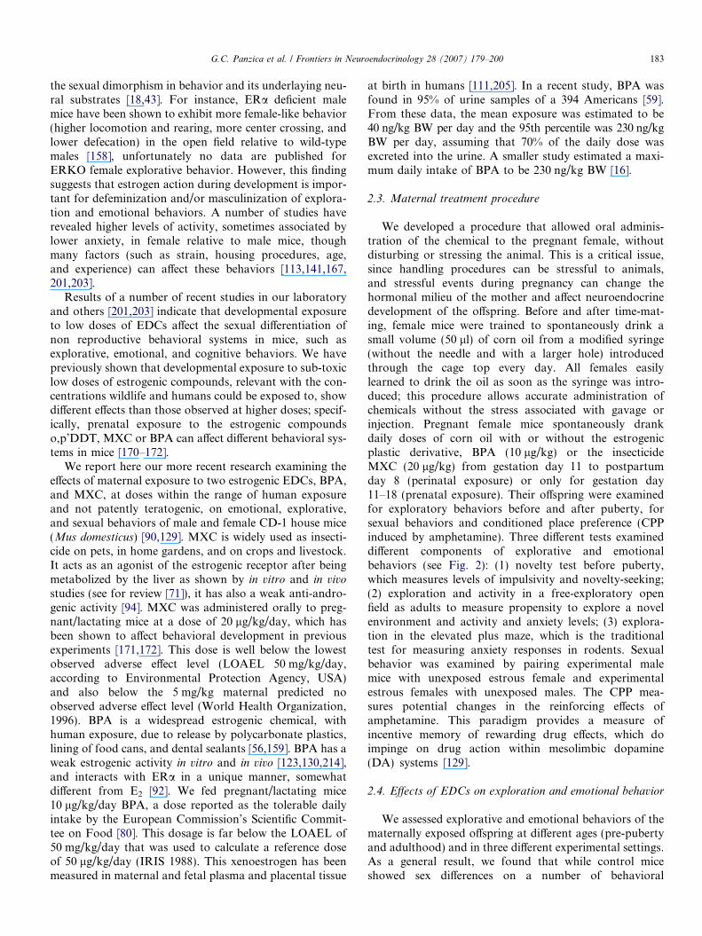
the sexual dimorphism in behavior and its underlaying neu-ral substrates [18,43]. For instance, ERa deficient malemice have been shown to exhibit more female-like behavior(higher locomotion and rearing, more center crossing, andlower defecation) in the open field relative to wild-typemales [158], unfortunately no data are published forERKO female explorative behavior. However, this findingsuggests that estrogen action during development is impor-tant for defeminization and/or masculinization of explora-tion and emotional behaviors. A number of studies haverevealed higher levels of activity, sometimes associated bylower anxiety, in female relative to male mice, thoughmany factors (such as strain, housing procedures, age,and experience) can affect these behaviors [113,141,167,201,203].
Results of a number of recent studies in our laboratoryand others [201,203] indicate that developmental exposureto low doses of EDCs affect the sexual differentiation ofnon reproductive behavioral systems in mice, such asexplorative, emotional, and cognitive behaviors. We havepreviously shown that developmental exposure to sub-toxiclow doses of estrogenic compounds, relevant with the con-centrations wildlife and humans could be exposed to, showdifferent effects than those observed at higher doses; specif-ically, prenatal exposure to the estrogenic compoundso,p’DDT, MXC or BPA can affect different behavioral sys-tems in mice [170–172].
We report here our more recent research examining theeffects of maternal exposure to two estrogenic EDCs, BPA,and MXC, at doses within the range of human exposureand not patently teratogenic, on emotional, explorative,and sexual behaviors of male and female CD-1 house mice(Mus domesticus) [90,129]. MXC is widely used as insecti-cide on pets, in home gardens, and on crops and livestock.It acts as an agonist of the estrogenic receptor after beingmetabolized by the liver as shown by in vitro and in vivo
studies (see for review [71]), it has also a weak anti-andro-genic activity [94]. MXC was administered orally to preg-nant/lactating mice at a dose of 20 lg/kg/day, which hasbeen shown to affect behavioral development in previousexperiments [171,172]. This dose is well below the lowestobserved adverse effect level (LOAEL 50 mg/kg/day,according to Environmental Protection Agency, USA)and also below the 5 mg/kg maternal predicted noobserved adverse effect level (World Health Organization,1996). BPA is a widespread estrogenic chemical, withhuman exposure, due to release by polycarbonate plastics,lining of food cans, and dental sealants [56,159]. BPA has aweak estrogenic activity in vitro and in vivo [123,130,214],and interacts with ERa in a unique manner, somewhatdifferent from E2 [92]. We fed pregnant/lactating mice10 lg/kg/day BPA, a dose reported as the tolerable dailyintake by the European Commission’s Scientific Commit-tee on Food [80]. This dosage is far below the LOAEL of50 mg/kg/day that was used to calculate a reference doseof 50 lg/kg/day (IRIS 1988). This xenoestrogen has beenmeasured in maternal and fetal plasma and placental tissue
at birth in humans [111,205]. In a recent study, BPA wasfound in 95% of urine samples of a 394 Americans [59].From these data, the mean exposure was estimated to be40 ng/kg BW per day and the 95th percentile was 230 ng/kgBW per day, assuming that 70% of the daily dose wasexcreted into the urine. A smaller study estimated a maxi-mum daily intake of BPA to be 230 ng/kg BW [16].
2.3. Maternal treatment procedure
We developed a procedure that allowed oral adminis-tration of the chemical to the pregnant female, withoutdisturbing or stressing the animal. This is a critical issue,since handling procedures can be stressful to animals,and stressful events during pregnancy can change thehormonal milieu of the mother and affect neuroendocrinedevelopment of the offspring. Before and after time-mat-ing, female mice were trained to spontaneously drink asmall volume (50 ll) of corn oil from a modified syringe(without the needle and with a larger hole) introducedthrough the cage top every day. All females easilylearned to drink the oil as soon as the syringe was intro-duced; this procedure allows accurate administration ofchemicals without the stress associated with gavage orinjection. Pregnant female mice spontaneously drankdaily doses of corn oil with or without the estrogenicplastic derivative, BPA (10 lg/kg) or the insecticideMXC (20 lg/kg) from gestation day 11 to postpartumday 8 (perinatal exposure) or only for gestation day11–18 (prenatal exposure). Their offspring were examinedfor exploratory behaviors before and after puberty, forsexual behaviors and conditioned place preference (CPPinduced by amphetamine). Three different tests examineddifferent components of explorative and emotionalbehaviors (see Fig. 2): (1) novelty test before puberty,which measures levels of impulsivity and novelty-seeking;(2) exploration and activity in a free-exploratory openfield as adults to measure propensity to explore a novelenvironment and activity and anxiety levels; (3) explora-tion in the elevated plus maze, which is the traditionaltest for measuring anxiety responses in rodents. Sexualbehavior was examined by pairing experimental malemice with unexposed estrous female and experimentalestrous females with unexposed males. The CPP mea-sures potential changes in the reinforcing effects ofamphetamine. This paradigm provides a measure ofincentive memory of rewarding drug effects, which doimpinge on drug action within mesolimbic dopamine(DA) systems [129].
2.4. Effects of EDCs on exploration and emotional behavior
We assessed explorative and emotional behaviors of thematernally exposed offspring at different ages (pre-pubertyand adulthood) and in three different experimental settings.As a general result, we found that while control miceshowed sex differences on a number of behavioral

Fig. 2. Different test paradigms (left column) to examine behavioral responses of male and female house mice perinatally exposed to BPA (10 lg/kg BW)or MXC (20 lg/kg). (a) Novelty test before puberty; male or female sibling groups were housed in compartment A of the apparatus. After 24 h, all micebut one were randomly removed from the cage, so that only one mouse was tested and the door dividing the two compartments was opened thus allowingthe mouse to enter the novel compartment (b). (b) A free-exploratory open field, consisting of a home-cage and an unfamiliar area (an open-field, OF) of73 · 110 cm bordered along by a 50 cm high wall and in which a bright and a dark zone were created. A male and a female per litter were individuallyhoused in the home cage section and after 24 h the removable barrier was removed allowing entrance in the OF. A cut-off of 10 min was used for thoseanimals that did not enter the OF and starting from the first entrance in the OF, behavioral observation lasted 5 min. (c) The Elevated Plus Maze consistsof two open arms and two closed arms that extended from a common central platform. A mouse was placed in the center and tests lasted 5 min. (d) Theconditioned place preference (CPP) paradigm consists of a three-compartments opaque rectangular box; two cues, one visual (white or black walls) andone tactile (wide or narrow mesh floor), were associated with each of the two end-compartments. The white compartment of the apparatus was repeatedlypaired with the administration amphetamine (1 or 2 mg/kg i.p.), whereas the black one was paired with an injection of saline. According to a split-litterdesign, one male and one female from each litter were randomly assigned to be conditioned with saline or one of the two amphetamine doses. On test day,mice were allowed free-access to both compartments, and a preference for the drug-paired side is taken as an index of the drug’s positive reinforcingproperties. (e) Summary of the effects of perinatal exposure to BPA or MXC on sex differences in the different test paradigms. F = M, level of behavior donot significantly differ in males and females; F > M, levels of behavior significantly higher in females than males; F < M levels of behavior significantlylower in females than males. fl statistically significant decrease of behavior following perinatal exposure; › statistically significant increase of behaviorfollowing perinatal exposure.
184 G.C. Panzica et al. / Frontiers in Neuroendocrinology 28 (2007) 179–200
responses at both ages and in all the test paradigms, miceperinatally exposed to BPA or MXC showed decreasedor no sex differences (see Fig. 2). Unexposed female mice,either pre- or post-puberty, when ‘‘stimulated’’ to explorea novel environment, were more reactive and explorativeand less anxious as compared to unexposed males. Devel-opmental exposure to the estrogenic pollutants BPA andMXC resulted in behavioral alterations mainly in females,which showed levels of exploratory behavior more similarto the typical behaviors observed in control males than tothose recorded in the control females. Altogether thesefindings may well be seen as indexes of reduced reactivityof exposed females to novel stimuli and are consistentwith an estrogenic action of BPA and MXC, and possible‘‘defeminization’’ or ‘‘masculinization’’ effects of the peri-natal exposure to these compounds. However, we also
reported that BPA-exposed males showed female-typebehavior on a few measures. The overall result was areduction or a reversal of sexual differences in EDCsexposed mice, relative to those displayed by controls. Sex-ual behavioral differences, as well as effects of estrogeniccompounds altering such differences, occur before puberty(i.e., before the increase of sexual hormones production isactivated by gonads). This can be interpreted as an indexof an interference of EDCs in the processes of develop-ment and organization of CNS and of receptor systemsof both sexes. Fig. 2 summarizes the main results of devel-opmental exposure to BPA and MXC on mice behavioralresponses. Interestingly, no effects of prenatal exposure toEDCs were recorded on male or female sexual behaviorsor reproductive success (Palanza, unpublishedobservation).

G.C. Panzica et al. / Frontiers in Neuroendocrinology 28 (2007) 179–200 185
2.5. Effects of EDCs on brain dopaminergic function
The specificity of the developmental changes affecting acentral neurochemical system can be evaluated by assessingthe effects of a psychoactive agent targeting to that systemupon the behavioral responses modulated by that system.For this reason we assessed in adult animals the possibilitythat prenatal exposure to BPA or to MXC may influencethe development of brain dopaminergic systems by investi-gating potential changes in the reinforcing effects ofamphetamine (AMPH), using a widely validated paradigm,the CPP [129]. As a general result, AMPH treatment pro-duced increased locomotory activity in mice regardless pre-natal exposure. With respect to AMPH-induced placeconditioning, females as a whole were more responsivethan males, thus confirming previous results [128]. Whencompared to unexposed female mice, BPA- as well asMXC-exposed females failed to show AMPH-induced con-ditioning. Males showed no changes due to the prenataltreatment. Thus, prenatal exposure to BPA or MXC wasapparently responsible in female mice for impairment ofbrain reward pathways targeted by the drug. Reduced nov-elty seeking and increased neophobia were also found infemale rats perinatally exposed to BPA [9]. This behavioralprofile could be related to gender-specific alterations in thefunction of brain neurochemical systems involved in theresponse to AMPH. As release of DA within the dorsaland ventral striatum is known to be involved in the behav-ioral effects that follow amphetamine administration[118,213], it is reasonable to assume that a potentialalteration in the behavioral effects of amphetamine admin-istration could be an index of BPA- and/or MXC-inducedlong-term effects on the dopaminergic function of thebrain. Our preliminary neurobehavioral study has showna decrease in D1-like receptors density in nucleus accum-bens and olfactory tubercle of mice prenatally exposed toMXC, at the same doses that have caused alteration inexploration and novelty-induced locomotor activity(Morellini F., Palanza P., Fuchs E., unpublished data).
2.6. Mechanisms of EDCs action
As for possible action mechanisms, it should be notedthat both MXC and BPA exhibit weak estrogenic activityin adult rats of both sexes [11,119,217]. In these studies,motor activity and motivation to explore were depressedat adulthood following maternal exposure to BPA [9,86].Kubo et al. [125] reported that in rats, exposure to BPAabolished sex differences in behavioral patterns in an openfield and reversed the normal sex differences in the locuscoeruleus. Locus coeruleus (LC) and dopaminergic systemare known to be involved in the regulation of animal reac-tivity to a novel environment and in the CNS, the dopami-nergic system having been reported to be affected by earlydevelopmental exposure to EDCs. Sexual dimorphism inLC has been detected in rats, with females showing largervolume, higher number of neurons and more dopamine-
b-hydroxylase immunoreactive cells than males [193]. Peri-natal exposure to low doses of BPA in rats reversed the sex-ual difference in volume and number of cells of the locuscoeruleus [125]. The mesolimbic and nigrostriatal dopami-nergic systems represent major structures of the CNSessential for locomotor activity, novelty induced behavior,reward learning, attention deficit [13,50,61]. In utero andlactational exposure to PCB77 resulted in elevations inconcentrations of DA in the frontal cortex, and of DAand its metabolites in the substantia nigra in young (beforepuberty) and in adult rats [208].
We reported that prenatal exposure to the estrogenicpollutants BPA and MXC resulted in marked alterationsin the psychopharmacological profile of female mice sug-gesting an impairment of brain reward pathways targetedby amphetamine, possibly involving brain monoaminergic(particularly dopaminergic) circuits [129]. On this basis, itmay be supposed that prenatal exposure to BPA orMXC might interact with some steps in the developmentand organization of the monoaminergic system duringthe perinatal period. A convincing body of evidence indi-cates that estrogen can modulate basal and amphet-amine-stimulated levels of DA release in rodent striatumas measured by in vivo microdialysis [44] and intrauterineexposure to estradiol has been reported to have a signifi-cant effect on the organization of monoamine systemswithin the foetal hypothalamus [116]. Developmental expo-sure to BPA has been shown to alter D1 receptor expres-sion and density in male mice [215]. The exposure toBPA during either organogenesis or lactation, but notimplantation and parturition, significantly enhanced themorphine-induced hyperlocomotion and rewarding effects.Furthermore, exposure to BPA during either organogenesisor lactation also produced an up-regulation of DA receptorfunction to activate G-protein in the mouse limbic fore-brain [156]. These results indicate that both organogenesisand lactation are more sensitive to the BPA-induced devel-opmental neuronal toxicology than any other periods.
Recent studies demonstrated that mice or rats perina-tally exposed to low doses of BPA showed alterations insexually dimorphic population of tyrosine hydroxylase(TH) neurons in the AVPV [186,201] and in the open fieldbehavior [201], an effect consistent with an estrogenicactivity of BPA on the developing brain. This is consistentwith our preliminary data on the effect of prenatal exposureto BPA or the synthetic estrogen diethylstilbestrol (DES)on the number of neurons producing TH in the locuscoeruleus of pre-puberal mice. We found that, whilecontrol animals showed sex difference in the number ofTH-stained neurons in the LC, the exposure to BPAeliminates this difference, as did DES [194].
It is recognized that DA is regulating male sexual behav-ior in rodents through its cooperation with the NO-produc-ing system [108,204]. Our preliminary data indicate thatpre- and postnatal exposure to BPA can alter selectivelythis system at the level of medial preoptic nucleus and ofthe bed nucleus of the stria terminalis [140]. These data

Fig. 3. Sex dimorphism in the control of copulatory behavior of Japanesequail (illustrated here by mount attempt, MA) by testosterone. (A) Adultmale Japanese quail readily show copulatory behavior when presented to asexually mature female. This response is never shown by intact females(Control). It disappears after gonadectomy (GNX). Treatment of gonad-ectomized birds with testosterone (T) activates MA in males but not infemales (GNX + T). Females are insensitive to the activating effects of T.Redrawn from data in [39].
186 G.C. Panzica et al. / Frontiers in Neuroendocrinology 28 (2007) 179–200
indicate in the NO system another potential and importanttarget for the action of EDCs in mammals. Although wedid not find significant alteration of sexual behaviors inmale mice following perinatal exposure to EDCs, otherstudies [85,154] reported that male rats perinatally exposedto BPA showed less efficient sexual behaviors thancontrols.
However, none of these studies can delineate the mech-anisms involved in BPA’s or MXC’s actions on the devel-oping brain; further study is needed to clarify possiblemechanisms underlying EDCs actions on brain develop-ment and their effects on behavior. At present, it must berecognized that in addition to well-documented estrogenic-ity, BPA and MXC may exert other effects on the develop-ing brain. A number of studies have suggested that some ofthe neurobehavioral effects of XEs, such as BPA, cannot beexplained by an estrogenic action of this compound, relatedto its binding to ER a and ER b [76,77,85]. MacLusky andco-workers have recently reported anti-estrogenic effects ofBPA on hippocampal synaptogenesis of mice brain [136].In another study, BPA has been shown to exert estrogenicor anti-estrogenic effects in the rat cerebellum, according tothe concentration of the compound [244]. In addition,recent studies have shown that not all effects of BPA aremediated by the classical nuclear ERs. Non-genomic cell-signaling systems involve serial activation of kinases vialigand binding to cell-membrane receptors at very low con-centrations [236].
3. Circuits controlling sexual behavior in birds
Birds, and in particular galliforms are characterized byextreme forms of sex dimorphism [i.e., the male copulatorybehavior and neural circuits associated to it, as the vasoto-cin (VT) system]. In quail, contrary to what is observed inrodents, the male-type copulatory behavior is highly differ-entiated between males and females, while the female-typereceptive behavior can be activated in both sexes by anappropriate treatment with estrogens [4] (for a review, see[31]). The sexual dimorphism affecting male copulatorybehavior is quite extreme in this species: under laboratorytest conditions, sexually mature males almost never failto exhibit the complete copulatory sequence, includinggrabbing the female’s neck feathers, mount attempts,mounts, and cloacal contact movements. These behaviorsare never seen in females and are T-dependent [4,39,163].The dimorphism is an all or none phenomenon; it is qual-itative in nature (Fig. 3). Because these behaviors areT-dependent, they disappear after castration in males.However, gonadectomized or intact females still do notexhibit these behaviors due to a relative absence of circulat-ing T. It has indeed been shown that treatment with highdoses of T is not sufficient to activate cloacal contact move-ments in gonadectomized females, while such a treatmentrestores the full spectrum of sexual behaviors in males[4,39,162,206]. This suggests that the neuronal circuits
supporting male reproductive behavior are also sexuallydifferentiated in this species.
A large number of investigations have elucidated theneural basis of this dimorphism and the results were partic-ularly interesting for two nuclei of the limbic-hypothalamicregion, the medial preoptic nucleus (POM, [183]) and thenucleus of the stria terminalis (BST, [20]). These two nucleiwere described in detail for both their cytoarchitecture andfor their neurochemical features (for a review, see [182]).Several studies have also demonstrated that, in particularthe POM, they are profoundly implicated in the controlof male sexual behavior (for detailed reviews, see[181,182]). Among the different neural circuits that havebeen characterized in POM and BST, two are particularlyinvolved in the control of reproductive behavior, the neu-rons producing aromatase (the enzyme converting T intoE2, [32]) and the parvocellular neuronal system producingVT located in the BST and POM [173]. The quail (andchicken) VT system shows however a stronger dimorphismthan the aromatase system [182], and its sexual differentia-tion is influenced in the same way by estrogens as the cop-ulatory behavior do [95,115,176].
Male copulatory behavior and VT parvocellular systemare therefore major targets of the action of estrogens dur-ing development of galliforms, in this respect they are alsoprivileged targets for the action of EDCs, in particular XEsand xenoandrogens, during the embryonic life [175].
In field birds, it is critical to consider endocrine, neuro-endocrine, and behavioral components of reproduction, asall are critical to overall fitness. Therefore, the study ofneural circuits as impacted by EDCs in specific avian mod-els provides a fundamental understanding of the mecha-nisms involved in the behavioral impairment and othereffects of exposure. This is especially important in light ofthe apparent negligible effects on testis weight, plasma ste-roids, and other more general measures of reproductive

G.C. Panzica et al. / Frontiers in Neuroendocrinology 28 (2007) 179–200 187
function. Understanding specific neural effects associatedwith EDC effects provide an important measure for fieldbirds in which more subtle effects may occur due to spottyor variable exposure. Furthermore, some of these endo-crine active compounds may translate into epigenetic orimpaired reproductive fitness and variable responsesbetween individuals [14,15]. In this way, there may belong-term deleterious effects on individuals and then even-tually on the population as a whole.
4. The Japanese quail as an avian model for testing EDCs
As briefly described in the previous chapter, the Japa-nese quail provides an advantageous model for under-standing the impact of EDCs, in particular thoseprovided by the diet, because there are well characterizedembryonic effects of gonadal steroids. In the femaleembryo, plasma E2 peaked at E10, E12, and declined posthatch, with levels always higher than in males [164], inmales, plasma androgen peaked at E10–E12 and P1, witha decrease post hatch being always higher than in females[161] (Fig. 4). Administration of exogenous gonadal ste-roids alters sexual differentiation of reproductive behavior
Fig. 4. (a) Plasma androgen concentrations in embryonic and post-hatchmale and female Japanese quail at various ages. (b) Plasma 17b-estradiolconcentrations in embryonic and post-hatch male and female Japanesequail at various ages. Redrawn from [164].
in both sexes [6,7]. In male quail, the embryonic exposureto T or E2 by E12 altered later expression of copulatorysexual behavior (demasculinized males [5,22,176]). On thecontrary, early embryonic administration of specific inhib-itors of the synthesis of E2 (fadrozole, R76713) inducesdefeminization of sexual behavior in females [34,176].These results indicate the important organizational roleof the conversion of T into E2 (aromatization) [29] in addi-tion to its role in the induction of adult sexual behavior[33]. Increased 5b reductase enzyme activity was found inthe brain of male quail embryos between E7 and E15,which may protect males from being demasculinized byinactivating T [37]. Finally, early steroid exposure alsoinfluences gonadal development, with fadrozole andtamoxifen exposure producing defeminization of the ovaryand accessory structures [82]. These data were confirmed inseveral independent studies, including those that comparethe effects of gonadal steroids and EDCs.
Neural systems that are responsive to steroids and keyregulators of endocrine and behavioral components ofreproduction are found in the preoptic, septal, limbic,and selected hypothalamic regions. In Japanese quail, neu-ral systems that control male copulatory behavior includethe POM and BST [181,182], these nuclei are characterizedby a large sexually dimorphic population of aromatase-immunoreactive (ARO-ir) neurons [23,40] and a sexuallydimorphic VT-immunoreactive parvocellular system[20,173]. ARO-ir cells are also controlled by several neuro-transmitter/neuropeptide afferents: catecholamines [36],nitric oxide [38], VT [30] (for review, see [1]). Other neuro-peptides and neurotransmitters are also present in the sameregions: galanin [27], neuropeptide Y [25], vasointestinalpolypeptide, substance P [26], and serotonin [69]. In addi-tion, ARO, VT and vasointestinal polypeptide immunore-active elements are modulated in adulthood by thecirculating levels of T. ARO and VT show a markeddecrease in the number of immunoreactive cell bodies inPOM, BST and lateral septum of castrated or aged males[21,23,177,179,222], whereas, in the same endocrine situa-tion, vasointestinal polypeptide fiber density in the caudallateral septum increases [24]. Conversely, the expressionof both VT-immunoreactive and ARO-ir elements arestimulated in castrated adults when E2 is administered[21,223]. ARO-producing neurons (primarily in the POM)and neural input to these neurons are critical modulatorsof male copulatory behavior [1,181]. In addition, the par-vocellular sexually differentiated VT system is particularlysensitive to estrogens during the embryonic period. Admin-istration of estradiol benzoate (EB) at day 9 of incubationsuppresses male copulatory behavior in adults and inducesa female-type VT phenotype in adult males [176].
4.1. The effects of estrogenic EDCs in Japanese quail
Earlier studies showed that some pesticides have an abil-ity to interfere with the hypothalamic–pituitary–gonadalaxis (HPG axis; [198]). Insecticides such as o,p 0-DDT

188 G.C. Panzica et al. / Frontiers in Neuroendocrinology 28 (2007) 179–200
[103], DDE [178] or MXC [100,101], as well as other com-pounds such as ethinylestradiol [102], diethylstilbestrol[224], and genistein [225] significantly decrease male sexualperformance when administered during embryonic devel-opment. Besides the effects on behavioral differentiation,these chemicals affect differentiation of gonads, accessorysexual organs, and brain circuits.
In birds, the female deposits steroids and steroid-likecompounds into the yolk, thereby providing a primaryroute of exposure during embryonic development (for areview, see [60]). Female quail given E2 implants werefound to transfer estradiol to offspring via the yolk [7].Therefore, a primary route of exposure is via maternaldeposition of the EDC into the yolk. This is also the casefor other endocrine active compounds, including MXC,which is an estrogenic pesticide [166]. Although EDCsare generally weaker in action than endogenous steroids,including exogenous E2, the estrogenic pesticide MXC slo-wed sexual maturation in both males and females. Treatedmales also had impaired sexual behavior, similar to theeffects of embryonic estradiol [160]. Similarly, soy phytoes-trogens and especially genistein readily transfer into theyolk when ingested by the hen (Fig. 5) [135].
4.2. The Japanese quail sexually dimorphic parvocellular
vasotocin system
Among the different neural circuits that have been previ-ously illustrated, the sexually dimorphic parvocellularvasotocin system has been thoroughly investigated (forreviews, see [115,173]). This system is sensitive to gonadalsteroids both in embryonic and in adult life [176,179]. Indetail, POM and BST contain a dense population of vasot-ocinergic cells strongly sexually differentiated [182], in addi-tion VT-ir fibers are present in much higher density in thesenuclei and in the lateral septum in males than in females[20,221]. VT-ir cell bodies have been observed in POM
Fig. 5. Concentration of genistein in Japanese quail egg yolks from henssupplemented with genistein, genistein or placebo capsules. Data pointsrepresents average of 4 replies for treatment groups and 2 replicates for thecontrol groups. Modified from [135].
and BST of males but not in females, and accordingly, asex difference in VT expression has been confirmed byin situ hybridization of VT mRNA in the BST and POM[20] (Fig. 6).
In adult birds, the VT innervation of the POM is steroidsensitive in males: the density of VT-ir fibers in this nucleusdecreases in conditions where males experience low levelsof circulating T (castration, photoregression, and aging)and are restored to levels typical of sexually mature malesby exogenous treatments with T [177,222]. Similar changeshave been demonstrated also for the expression of VT
Fig. 6. (A) Schematic representation of connectivity of the parvocellularsexually dimorphic VT system. BST and POM are the two nuclei in whichare concentrated the VT-ir cell bodies. BST is projecting to SL and POM[2]. POM is connected to several hypothalamic and brainstem nuclei [35].(B and C) Comparison of the VT immunoreactivity in adult female (B)and male (C) Japanese quail. Black nuclei show VT-ir cell bodies, greynuclei show a sexually dimorphic VT innervation [173,221]. Bar = 300 lm.AVT, ventral tegmental area; BST, bed nucleus of the stria terminalis; CA,anterior commissure; GCt, central grey; ICo, intercollicular nucleus; LoC,Locus ceoruleus; POM, medial preoptic nucles; PVN, paraventricularnucleus; VMN, ventromedial nucleus.

G.C. Panzica et al. / Frontiers in Neuroendocrinology 28 (2007) 179–200 189
mRNA in the parvocellular elements of the BST and POM[179].
As described before, T activates male sexual behaviorthrough its aromatization in estrogens (17b-estradiol, E2),in parallel, E2 administration to castrated adults inducedVT innervation of POM, BSTm, and lateral septum com-parable to that observed in the intact adult or in castratedtreated with T [223]. There are therefore many experimen-tal situations in which the VT parvocellular system withinPOM, BST and lateral septum vary with the expressionof male copulatory behavior.
The existence of causal links between the peptide VTand this behavior has been investigated in experiments thatdemonstrated either peripheral or intracerebroventricularinjection of VT or a specific V1-receptor antagonist mark-edly affected the appetitive and consummatory aspects ofmale sexual behavior in castrated T-treated male quail[63]. The VT structures (cells and fibers) of POM andBST are indeed sexually differentiated in the organizationalsense [176]. This was demonstrated by injecting fertilizedquail eggs of both sexes on day 9 of incubation with eitherEB (25 lg/egg, a treatment that suppresses the capacity toshow copulatory behavior in adulthood) or the aromataseinhibitor R76713 (10 lg/egg, a treatment that makes adultfemales behaviorally responsive to T), or with the solventsas a control (C). At 3 weeks posthatch, all subjects weregonadectomized and later implanted with Silastic capsulesfilled with T. At the age of six weeks, when quail reach pub-erty, birds were perfused and brains were sectioned.Despite the similarity of the adult endocrine conditionsof the subjects (all were gonadectomized and treated withT implants providing the same plasma level of steroid toall subjects), major qualitative differences were observedin the density of VT-ir structures in the POM of the differ-ent groups. Dense immunoreactive structures (fibers and afew cells) were observed in the POM of C males but notfemales; EB males had completely lost this immunoreactiv-ity (and lost the capacity to display copulatory behavior)and, conversely, R76713 females displayed a male-typicalVT-ir system in the nucleus (and also high levels of copula-tory behavior). Similar changes in immunoreactivity wereseen in the BST and in the lateral septum (VT-ir fibers onlyin this case) but not in the magnocellular vasotocinergicsystem. These neurochemical changes closely parallel theeffects of the embryonic treatments on male copulatorybehavior. These results clearly demonstrate that, in quail,the vasotocinergic innervation of the POM, lateral septum,and BSTm, and its sensitivity to T and E2 in adulthood areorganized during the embryonic life. Exposure to high lev-els of estrogens results in a female phenotype as far as thesevasotocinergic inputs are concerned; the (relative) absenceof estrogens in the embryos leads to the male phenotype.The sexual dimorphism observed in the adults is truly orga-nizational in its nature, even if the presence of T is requiredfor this dimorphism to be fully expressed during adult life.Similar findings have also been recently reported in thedomestic fowl [96,114].
Therefore, both male copulatory behavior and the sexu-ally dimorphic parvocellular VT system are particularlysensitive to the organizing effects of E2 during embryonicdevelopment. Hence, these variables provide useful end-points to detect the estrogenic capacity of different XEs[175]. To test this hypothesis, we administered BPA [229],diethylstilbestrol (DES, a powerful synthetic estrogen[79]), genistein (GEN, a phytoestrogen, [78,132]) and ethyl-ene, 1,1-dichloro-2,2-bis(p-chlorophenyl (DDE, a commonmetabolite of DDT, with anti-androgenic activity [117]) tofertile quail eggs. Our hypothesis was that XEs may alterthe animal physiology through their binding to estrogenreceptor. In our model this means that a XE administeredduring embryonic life, should reduce or abolish male cop-ulatory behavior and interfere with the differentiation ofthe sexually dimorphic parvocellular VT-ir system. Wehave always introduced two other experimental groups,one injected with the solvent (OIL) and a second oneinjected with estradiol benzoate (EB). The hatched birdswere raised in heterosexual cages up to the age of 4 weekswhen they were put in individual cages. At the age of 7weeks we tested the male copulatory behavior, and the fol-lowing week the animals were perfused to dissect the brainand perform immunocytochemical analyses (for the fulldescription of methods see [224,225]).
4.3. Effects of BPA administration
As previously reported the xenoestrogen BPA [226] is anindustrial chemical, used to manufacture polycarbonateand numerous plastic articles, therefore it is largely diffusedin the environment and in the food. Investigations wereperformed in both quail and chicken eggs using similardoses of BPA (from 67 to 200 lg/egg [47]; embryos weresacrified at the age of 15 or 19 days of incubation. BPAinduced Mullerian duct (embryonic oviduct) malformationin female quail embryos and feminization of the left testis(ovotestis) in male chicken embryos. BPA caused mortalityonly in chicken embryos at 67 and 200 lg/egg. In this studyno investigations were performed on brain circuits. In twodifferent experiments we administered 50, 100, or 200 lg ofBPA per egg. The result of these embryonic treatments wasa dramatic decrease in the number of hatched animals: thepercentage of living chicks was in fact ranging from 8% to11% of injected eggs (controls and EB-injected were rang-ing from 55% to 60% in these experiments). The BPA-injected young quail did not survive after one week of life.The dissection of non-hatched embryos revealed that thelarge majority of the embryos was blocked immediatelyafter the BPA administration (from 36% to 63%), whereasfor the embryos that died later, we observed a high inci-dence of malformations of the gut, abdominal wall, andlegs. With these experiments we were, therefore, unableto study any alteration in the brain (unpublished results).Contrary to what happens in mammals, BPA, even atlow doses, has robust adverse effects in birds, inducing sev-eral malformations also of the reproductive tract, and

190 G.C. Panzica et al. / Frontiers in Neuroendocrinology 28 (2007) 179–200
determining a strong reduction in their surviving after theexposure.
4.4. Effects of DES administration
Among various chemical XEs, diethylstilbestrol (DES)was initially synthesized as an orally effective estrogenfor use in human medicine and as anti-abortive sub-stance and then used as anabolic growth promoter incattle, steer, and sheep [89]. In the beginning, the envi-ronmental impact of this compound was not considered.However, DES and its metabolites were also excretedinto the ecosystem with unknown consequences. Detec-tion of radiolabeled DES in a model ecosystem demon-strated that it was persistent and bioaccumulated (for areview, see [146]). In quail, previous studies demonstrateda potent effect of DES on the development of sex organsand the differentiation of male sexual behavior [49], how-ever, no attention was dedicated to alterations of brainnervous circuits that should take place related to orinducing behavioral changes.
Fig. 7. (Upper) Changes in male VT immunoreactivity in different experimentaGEN, genistein; DDE, ethylene; 1,1-dichloro-2,2-bis(p-chlorophenyl). Bars = 3POM, and SL of male quail from different experimental groups. Data redraw
In a recent study [224], we have confirmed that embry-onic treatment with 700 ng/egg of DES demasculinize sex-ual behavior and cloacal gland size of adult intact malequail. These effects are particularly strong and fully compa-rable to those obtained after administration of higher dosesof EB (10 or 25 lg) with the same procedure. These effectsconfirm that sexual behavior of adult male quail is an excel-lent bioassay for embryonic exposure to EDCs with estro-genic activity. It is interesting to emphasize that a similardose of DES may induce in humans alterations of malegenital system (for a review, see [146]). In addition, as illus-trated in Fig. 7, this in ovo treatment significantly decreasedthe fractional area covered by VT-ir structures withinBSTm, POM, and lateral septum [224].
The effects of DES on VT-ir structures appear to be ana-tomically specific in that no changes were observed in VTmagnocellular neurons of the supraoptic and paraventricu-lar nuclei. This further supports our previous findingsshowing that changes in VT-immunoreactivity related toendocrine status affect only VT parvocellular circuits ofthe preoptic and limbic regions [173,223].
l conditions. OIL, control; EB, estradiol benzoate; DES, diethylstilbestrol;00 lm. (Lower) Fractional area covered by VT-immunoreactivity in BST,n from [178,224,225].

G.C. Panzica et al. / Frontiers in Neuroendocrinology 28 (2007) 179–200 191
4.5. Effects of genistein administration
Genistein is the simplest isoflavonoid compound pro-duced by Leguminosae, particularly abundant in soybeans.In plants, together with the other isoflavonoids collectivelycalled phytoestrogens, it has antimicrobial activity [78], aswell as a specific activity to protect plants from insects[52,233]. This action is mediated by its activity as a ligandof ecdysone (the molt steroid hormone of invertebrates)receptor [157]. This molecule shares structural features withthe 17b-estradiol and therefore can bind ERs and sex hor-mone binding proteins [127,150]. Thus genistein, as othersimilar isoflavones collectively called phytoestrogens, canexert both estrogenic and antiestrogenic activity, the latterby competing with estradiol for receptor binding [53].
A number of studies have been performed to investigatepossible alterations of brain circuitries or of behavioralactivities after administration of genistein (or of a mixtureof phytoestrogens) in mammals during adult life or duringthe critical period (for recent reviews, see [132,184]). Theadministration of genistein to neonatal female rats: (a) sig-nificantly increased SDN-POA volumes in adult gonadec-tomized female [8,84], or b) resulted in a non-significantvolumetric decrease of SDN-POA in intact adult female[133,134]. The administration of genistein to neonatal malerats induced an increase in the number of calbindin positivecells within the SDN [187]. Neonatal administration ofgenistein is also influencing the sexual differentiation ofTH system of the hypothalamic AVPV in rodents. In intactadult rat and mouse, female AVPV is larger than the maleAVPV and contains a higher number of cells expressingTH. Peri- or postnatal exposure to genistein demasculi-nized TH-ir system in the male AVPV [186,201]. In addi-tion, the number of TH-ir neurons colocalized withestrogen receptor a is strongly decreased in females [186].Overall, these studies suggest that acute exposure to geni-stein and other phytoestrogens during a critical develop-mental period alters the development of some encephalicsexually dimorphic structures. Finally, other studies per-formed during adulthood demonstrated that phytoestro-gens may have also activational effects. In fact, in adultrat, variation of the diet from a phytoestrogen-rich to aphytoestrogen-free diet determined the decrease of theSDN-POA volume in males and of the AVPV volume infemales [131].
In our study we have treated quail eggs with 100 or1000 lg of genistein [225]. The lowest dose of GEN had sig-nificant effect only on one aspect of male copulatory behav-ior (mount attempt, MA), whereas the GEN-1000 treatedmales showed a significant reduction for most aspects ofthe copulatory behavior (neck grab, NG, MA, and mount,M). No effects were detected on the cloacal gland size.
The parvocellular VT-ir system (POM, BST, and lateralseptum) was affected by GEN treatment, showing a signif-icant decrease of immunoreactivity with both 100 and1000 lg of GEN. However, the reduction was not as strongas that observed after treatment with EB (Fig. 7). As for
the experiment with DES no significant effect was detectedin the magnocellular VT system.
4.6. Effects of DDE administration
It took about 50 years after the initial use of DDT as apesticide to understand some of the mechanisms of actionof its main metabolite DDE. This is due in part to muchof the early research concentrating on the estrogenic effectsof o,p 0-DDT, which only made up about 15% of technicalDDT. o,p 0-DDT was shown to cause estrogenic effects infemale rats [65]. It was not until studies began to show phe-notypic effects of this chemical on male rats that were verysimilar to those observed with exposure to known andro-gen receptor blocking chemicals that scientists first beganto speculate about p,p 0-DDE’s anti-androgenic nature[117]. It is now known that DDE is a potent androgenreceptor antagonist and a very potent testosterone hydrox-ylase modulator. Its androgen receptor blocking ability isalmost equal to that of the anti-androgen hydroxyfluta-mide [117]. In birds, in ovo DDE administration has impacton brain structure (reduction in volume of the forebrain,and of the song controlling nuclei in a wild oscine species[112]) and on the immune system [195,196].
In a preliminary experiment, we have tested if theadministration of 20 or 40 lg of DDE per egg has animpact on male quail sexual behavior and parvocellularVT system [178]. At both doses DDE significantlydecreases the number of MA, as well as the VT immunore-activity in POM, BST, and lateral septum (Fig. 7). There-fore, this antiandrogen compound, when administeredduring the embryonic life, has also a powerful action onthe differentiation of estrogen-dependent function (malecopulatory behavior) and nervous circuitries (the parvocel-lular VT system).
5. General comments
In the conceptual frame of the evolutionary theory, sex-differences in behavior are thought to reflect adaptive dif-ferences of behavioral strategies in coping as resulting fromsexual selection [73]. Longitudinal studies on effects ofEDCs should be carried out in order to evaluate in whichcontexts, and with what intensity, reversing or leveling ofsexual differences could have relevance, in particularwhether behavioral alterations occur in systems influencingindividual fitness/reproductive success.
EDCs are globally distributed through our atmosphere,our seas and wildlife. Many are persistent; others, while notpersistent, should be treated as persistent because of theirchronic and ubiquitous use. They act at a population leveland many have the potential to (individually or cumula-tively) affect future generations, for example by decreasingfertility, feminizing males, masculinizing females or alteringcognitive abilities. All these endpoints have been studied inour or others’ laboratories and many have been observed inwildlife. Recent data, which must be confirmed by further

192 G.C. Panzica et al. / Frontiers in Neuroendocrinology 28 (2007) 179–200
studies, suggest that comparable changes can be producedin human populations as well [105,228,241]. These includeincreases in the frequency of preterm birth, obesity, cogni-tive/behavioral dysfunctions (such as autism and attentiondeficit hyperactivity disorder, ADHD, Parkinson’s dis-ease), and decreases in reproductive function (such as adecline in sperm count) and immune function [121,216].
In birds, the diversity of reproductive strategies, habitatand the migration of many species means that their expo-sure to EDCs may be extremely variable, also dependingby the amount of body fat (many of the EDCs are lipo-philic), so, for example, migratory birds might receive addi-tional exposure during migration when lipid stores areutilized for energy. Exposure of the avian embryo to mater-nal hormones or to EDCs is increased by the use of fatstores for the production of vitellogenin that becomes a pri-mary component of the yolk, which is then used by theembryo during development [7,48,60,99,104,135]. As wehave demonstrated in our studies, it is clear that low, fieldrelevant concentrations of EDCs do exert irreversibleeffects on endocrine, neuroendocrine, and behavioral sys-tems that are often due to permanent changes in neural sys-tems. Therefore, the embryonic period, during whichirreversible alterations in the organization of neural sys-tems occurs, appears to be the most vulnerable stage inthe life. The use of laboratory species, as the Japanesequail, is valuable to fully characterize the impact and riskof EDCs to avian species, as well as to understand themode and mechanisms of action of classes of EDCs, butfurther studies are needed to assess risk in field birds.
In rodents the reported experimental studies indicatethat exposure to environmentally relevant doses of EDCsduring developmental critical periods interacts with somesteps in the sexual differentiation of the neural systems con-trolling explorative and emotional behavior. Therefore,from this point of view, rodents and galliforms are simi-larly sensitive to early exposure to EDCs, mainly in those‘‘critical’’ periods that are important for brain sexual differ-entiation. However, experimental data suggest that thebehavioral responses as well as the neural circuits sensitiveto the action of EDCs differ between these vertebrate clas-ses. In rodents, the majority of data suggest alterationsmainly in females and in non-reproductive behaviors thatcould be related to altered development of central mono-aminergic pathways, but further work is needed to clarifythe neural basis of long-term consequences of developmen-tal exposure to EDCs such as BPA and MXC at the level ofneurobehavioral alterations. Some compounds target neu-roendocrine systems, thereby affecting reproductive endo-crine systems as well as other endocrine systems.Therefore, exposure to the estrogenic chemicals duringfetal/early prenatal development has consequences beyondimpaired function of the reproductive axis.
Most of the data that we discussed in the present revieware summarized in Table 1. They are only considering stud-ies in which behavioral effects following pre- and/or earlypost-natal exposure to low, environmentally relevant doses
of EDCs were recorded. Many other studies have analyzedonly alteration of neural circuits, with no description ofrelated behaviors [10,83,112,186,187], or the effects of feed-ing EDCs in adult animals [55,57,88,132,185,199,217,238,242]. A direct comparison of the EDCs effects inrodents and galliform is impossible, due to the variety ofbehaviors that were considered for rats and mice and thealmost exclusive consideration of male copulatory behaviorin quail. However, they strongly indicate that alteredbehavior is one of the most conspicuous endpoint pro-duced by EDCs. Behavioral alteration, although a rela-tively insensitive indicator of the degree of exposure, hasthe advantage of revealing both direct or indirect effectsof contamination and in some cases represents the onlyclue of functional deficits at different physiological levels[75] and can be more sensitive than other endpoints asbiomarkers of exposure, either in terms of chemical con-centration, response time or both. In particular, sexuallydimorphic behaviors (either reproductive or non non-reproductive) are useful to verify adverse developmentalconsequences produced by chemicals with endocrine dis-rupting properties: they interfere with sexual differentiationof the brain, consequently they can diminish, eliminate,reverse or widen sex differences in behaviors. In contrast,explicit recognition of sex differences in performance isnot a prominent feature of toxicological studies, exceptfor reproductive capacity studies and neurotoxicity testingdoes not typically recognizes sex differences in behavioralresponses as an experimental criterion [234]. It should benoted that in a number of the reported studies no signifi-cant effects of perinatal exposure to EDCs on brain and/or behavior were found, but a consistent result was theelimination of the sex differences shown by unexposedanimals [90,124,125,129,172,201]. If these studies hadtested only males, as the majority of classic toxicologicalstudies do, no effects of EDCs on behavioral/braindevelopment would have been detected.
Collectively, the new behavioral data from studies ofEDCs are strongly suggesting that prior methods of testingchemicals have been inadequate to detect adverse effects ofthe type now known to be caused by chemicals that areclassified as endocrine disruptors. Foremost, there is a chal-lenge to the operating assumption concerning appropriatedose. Because many EDC have effects at very low concen-trations in water (for example, nanogram per litre levels forestradiol), it is obvious there is a need to develop analyticalmethods applicable at such trace levels. It is also importantto integrate and correlate chemical analytical data withendocrine-disrupting effects [41].
The recent evidences for multiple roles of steroid hor-mones in the brain (neurogenesis, neuroprotection, regula-tory, short-term and long-term activators) indicate that thenervous system is a target for two different pools of steroids[147]), one coming from the peripheral glands (i.e., gonadalsteroids) and the second one originating in the nervous sys-tem partly by de novo in situ synthesis and partly for enzy-matic activation of peripheral steroids (neuroactive steroids

Table 1Sex-specific effects of developmental exposure (prenatal, or pre- and post-natal) to low, environmentally relevant concentrations of xenoestrogens orxenoandrogens on brain and behavior of rodent and quail animal models
EDC Species Sex specific behavioral effect Neural circuits References
Bisphenol A Rat Decreased explorative activity in females — [89]Diethylstilbestrol LC volume [123,124]
LC volume [124]Bisphenol A Mouse Decreased explorative activity in females TH neurons in AVPV [203]Bisphenol A, Methoxychlor Noradrenergic receptors in [93]Methoxychlor LC [170]Bisphenol A, Ethynil estradiol Mouse Decreased anxiety in females — [205]Bisphenol A Rat Increased exploratory activity in males — [90]
Dopaminergic system [157]Bisphenol A, methoxychlor Mouse Decreased response to reward in females LC, Dopaminergic system [128]
Bisphenol A Rat Decreased drug-induced locomotion in males Monoaminergic system [9]Ethynil Estradiol Mouse Increased spatial memory in females [205]
Ethynil Estradiol Rat Increased spatial memory in males [70]Methoxychlor Mouse Decreased onset of aggression in males — [170]Vinclozolin, Methoxychlor Quail Decreased male mating behavior — [85,101,161]Diethylstilbestrol Vasotocin system in BST [225]Genistein Vasotocin system in BST [226]DDE Vasotocin system in BST [179]Ethynilestradiol No effect on Vasotocin system [142]Bisphenol A (embryonic death) [47]
Bisphenol A Rat Decreased male mating behavior — [89,155]Methoxychlor — [85]
Bisphenol A Rat Increased female sexual interest — [88]Bisphenol A Mouse Decreased maternal behavior — [173]Methoxychlor Rat Estrous cyclicity — [141]Genistein TH neurons in AVPV [190]Genistein Mouse Estrous cyclicity — [64]Bisphenol A, Ethynil estradiol Mouse Onset of puberty [108,205]Bisphenol A Mouse Memory impairment in male ChAT neurons in hyppocampus [150]
G.C. Panzica et al. / Frontiers in Neuroendocrinology 28 (2007) 179–200 193
[189]). Probably, the gonadal steroids are most importantto determine irreversible changes in brain circuits as wellas in sexually dimorphic behaviors, whereas the neuroac-tive steroids are more important for short-term regulations.
As reviewed before, the EDCs, chiefly the xenoestro-gens, may have heavy biological effects that will vary overthe life cycle of the animal as well as across species andphyla [180], moreover they accumulate not only becauseof environmental pollution, but also due to their wide pres-ence in the food. The evolutionary implications of havingsome of these compounds in the normal food supply forcertain human populations (i.e., phytoestrogen derivativesfrom soy, [54,62,153,219]), as well as for wild and farm ani-mals have not yet been discussed, even if animals that, dueto their specific diet, are largely exposed to these com-pounds, as the domestic ruminants, seem to show unfavor-able effects of phytoestrogens at least on reproduction [3].Research in reproductive endocrinology has been almostexclusively focused on a small group of domesticated spe-cies, but if ecological variables, such as dietary burden ofphytoestrogens, have altered susceptibility to anthropo-genic contaminants, then a more diverse research base isurgently needed. Furthermore, phytoestrogens in the dietcan lead to a net decrease in estrogenic activity in the serum
in rodents and interfere with the action of other EDCs[8,139,230].
The mechanisms underlying the effects of these com-pounds needs to be further investigated; in fact, in manycases the effects of XEs cannot be easily superimposed tothose of ‘‘natural’’ estrogens, suggesting that non-estro-genic or/and metabolic effects are involved [92,239].For example, phytoestrogens may play different roles inthe cells. On one side genistein and other isoflavonesare direct regulators of the aromatase activity [190],while it has high affinity for some transcription factorsas PPAR-c [120,148] that have been recently localizedalso in the brain [64] where it, potentially, could havea role in nervous tissue differentiation [232]. Therefore,at least part of the effects of prenatal exposure to EDCsmay involve non-steroidal mechanisms activating signal-ing cascades that in many cases finalize with the activa-tion of transcription factors, turning a non-genomicresponse into a genomic one (rapid endocrine disruption[200]).
In parallel we should note that in rodents, in some cases,XEs may masculinize female brain morphology and femi-nize the male brain [125], as indicated also by the expres-sion of sexually differentiated socio-sexual and non-social

194 G.C. Panzica et al. / Frontiers in Neuroendocrinology 28 (2007) 179–200
behaviors [86]. A smaller number of reports support a sim-ilar masculinization of the female brain and feminization ofthe male brain in response to anti-androgenic EDCs [142].It is possible that these contrasting effects depend by thealteration of androgen:estrogen balance during develop-ment (for a review, see [240]).
In conclusion, in addition to gonadal steroids and neu-roactive steroids, xenoestrogens derived from food andenvironment should be considered as a third player withinthe nervous system that can regulate or alter its functionsthrough multiple pathways.
Acknowledgments
Studies described in the present review have been sup-ported by MURST-PRIN (C.V.P., G.C.P., P.P.), RegionePiemonte (C.V.P., G.C.P.), Fondazione CRT (G.C.P.),University of Torino (C.V.P., G.C.P.), EPA (M.A.O.),NSF (M.A.O.). E.M. is a fellow of Regione Piemonte.
References
[1] P. Absil, M. Baillien, G.F. Ball, G.C. Panzica, J. Balthazart, Thecontrol of preoptic aromatase activity by afferent inputs in Japanesequail, Brain Res. Rev. 37 (2001) 38–58.
[2] P. Absil, M. Papello, C. Viglietti Panzica, J. Balthazart, G.C.Panzica, The medial preoptic nucleus receives vasotocinergic inputsin male quail: a tract-tracing and immunocytochemical study, J.Chem. Neuroanat. 24 (2002) 27–39.
[3] N.R. Adams, Detection of the effects of phytoestrogens on sheepand cattle, J. Anim. Sci. 73 (1995) 1509–1515.
[4] E.K. Adkins, Hormonal basis of sexual differentiation in theJapanese quail, J. Comp. Physiol. Psych. 89 (1975) 61–71.
[5] E.K. Adkins, Effect of embryonic treatment with estradiol ortestosterone on sexual differentiation of the quail brain. Criticalperiod and dose–response relationship, Neuroendocrinology 29(1979) 178–185.
[6] E.K. Adkins Regan, Hormonal bases of sexual differentiation inbirds, in: Hormones, Brain and Behaviour in Vertebrates, in: J.Balthazart (Ed.), Sexual Differentiation, Neuroanatomical aspects,Neuropeptides and Neurotransmitters, vol. 1, Karger, Basel, NewYork, 1990, pp. 1–14.
[7] E. Adkins-Regan, M.A. Ottinger, J. Park, Maternal transfer ofestradiol to egg yolks alters sexual differentiation of avian offspring,J. Exp. Zool. 271 (1995) 466–470.
[8] H. Adlercreutz, C. Bannwart, K. Wahala, T. Makela, G. Brunow, T.Hase, P.J. Arosemena, J.T. Kellis Jr., L.E. Vickery, Inhibition ofhuman aromatase by mammalian lignans and isoflavonoid phytoes-trogens, J. Steroid Biochem. Mol. Biol. 44 (1993) 147–153.
[9] W. Adriani, D. della Seta, F. Dessı-Fulgheri, F. Farabollini, G.Laviola, Altered profiles of spontaneous novelty seeking, impulsivebehavior, and response to D-amphetamine in rats perinatallyexposed to bisphenol A, Environ. Health Perspect. 111 (2003) 395–401.
[10] R. Alo, R.M. Facciolo, M. Madeo, G. Giusi, A. Carelli, M.Canonaco, Effects of the xenoestrogen bisphenol A in diencephalicregions of the teleost fish Coris julis occur preferentially viadistinct somatostatin receptor subtypes, Brain Res. Bull. 65 (2005)267–273.
[11] A.M. Aloisi, D. Della Seta, I. Ceccarelli, F. Farabollini, Bisphenol-Adifferently affects estrogen receptors-a in estrous-cycling and lactat-ing female rats, Neurosci. Lett. 310 (2001) 49–52.
[12] J.J. Amaral Mendes, The endocrine disrupters: a major medicalchallenge, Food Chem. Toxicol. 40 (2002) 781–788.
[13] S.L. Andersen, M.H. Teicher, Sex differences in dopamine receptorsand their relevance to ADHD, Neurosci. Biobehav. Rev. 24 (2000)137–141.
[14] G. Ankley, E. Mihaich, R. Stahl, D. Tillitt, T. Colborn, S. McMaster,R. Miller, J. Bantle, P. Campbell, N. Denslow, R. Dickerson, L.Folmar, M. Fry, J. Giesy, E. Gray, P. Guiney, T. Hutchinson, S.Kennedy, V. Kramer, G. LeBlanc, M. Mayes, A. Nimrod, R. Patino,R. Peterson, R. Purdy, R. Ringer, P. Thomas, L. Touart, G. Van derKraak, T. Zacharewski, Overview of a workshop on screeningmethods for detecting potential (anti-) estrogenic/androgenic chem-icals in wildlife, Environ. Toxicol. Chem. 17 (1997) 68–87.
[15] M.D. Anway, M.K. Skinner, Epigenetic transgenerational actions ofendocrine disruptors, Endocrinology 147 (2006) S43–S49.
[16] C. Arakawa, K. Fujimaki, J. Yoshinaga, H. Imai, S. Serizawa, H.Shiraishi, Daily urinary excretion of bisphenol A, Environ.. Health.Prev. Med. 9 (2004) 22–26.
[17] J. Archer, Rodent sex differences in emotional and related behavior,Behav. Biol. 14 (1975) 451–479.
[18] A.P. Arnold, S.M. Breedlove, Organizational and activational effectsof sex steroids on brain and behavior: a reanalysis, Horm. Behav. 19(1985) 469–498.
[19] A.P. Arnold, E.F. Rissman, G.J. De Vries, Two perspectives on theorigin of sex differences in the brain, Ann. N. Y. Acad. Sci. 1007(2003) 176–188.
[20] N. Aste, J. Balthazart, P. Absil, R. Grossmann, E. Muhlbauer, C.Viglietti-Panzica, G.C. Panzica, Anatomical and neurochemicaldefinition of the nucleus of the stria terminalis in Japanese quail(Coturnix japonica), J. Comp. Neurol. 396 (1998) 141–157.
[21] N. Aste, G.C. Panzica, P. Aimar, C. Viglietti-Panzica, N. Harada, A.Foidart, J. Balthazart, Morphometric studies demonstrate thataromatase-immunoreactive cells are the main target of androgensand estrogens in the quail medial preoptic nucleus, Exp. Brain Res.101 (1994) 241–252.
[22] N. Aste, G.C. Panzica, C. Viglietti-Panzica, J. Balthazart, Effects ofin ovo estradiol benzoate treatments on sexual behavior and sizeof neurons in the sexually dimorphic medial preoptic nucleus ofJapanese quail, Brain Res. Bull. 27 (1991) 713–720.
[23] N. Aste, G.C. Panzica, C. Viglietti-Panzica, N. Harada, J. Baltha-zart, Distribution and effects of testosterone on aromatase mRNA inthe quail forebrain: a non-radioactive in situ hybridization study, J.Chem. Neuroanat. 14 (1998) 103–115.
[24] N. Aste, C. Viglietti-Panzica, J. Balthazart, G.C. Panzica, Testos-terone modulation of peptidergic pathways in the septo-preopticregion of male Japanese quail, Poultry Avian Biol. Rev. 8 (1997) 9–20.
[25] N. Aste, C. Viglietti-Panzica, A. Fasolo, C. Andreone, H. Vaudry,G. Pelletier, G.C. Panzica, Localization of neuropeptide Y (NPY)immunoreactive cells and fibres in the brain of the Japanese quail,Cell Tissue Res. 265 (1991) 219–230.
[26] N. Aste, C. Viglietti-Panzica, A. Fasolo, G.C. Panzica, Mappingof neurochemical markers in quail central nervous system: VIP-and SP-like immunoreactivity, J. Chem. Neuroanat. 8 (1995) 87–102.
[27] Y. Azumaya, K. Tsutsui, Localization of galanin and its bindingsites in the quail brain, Brain Res. 727 (1996) 187–195.
[28] J. Bakker, C. De Mees, Q. Douhard, J. Balthazart, P. Gabant, J.Szpirer, C. Szpirer, Alpha-fetoprotein protects the developing femalemouse brain from masculinization and defeminization by estrogens,Nat. Neurosci. 9 (2006) 220–226.
[29] J. Balthazart, Steroid control and sexual differentiation of brainaromatase, J. Steroid Biochem. Mol. Biol. 61 (1997) 323–339.
[30] J. Balthazart, P. Absil, C. Viglietti-Panzica, G.C. Panzica,Vasotocinergic innervation of areas containing aromatase-immu-noreactive cells in the quail forebrain, J. Neurobiol. 33 (1997)45–60.
[31] J. Balthazart, E. Adkins-Regan, Sexual differentiation of brain andbehavior in birds, in: D. Pfaff (Ed.), Hormones, Brain and Behavior,Elsevier Science, 2002, pp. 223–301.

G.C. Panzica et al. / Frontiers in Neuroendocrinology 28 (2007) 179–200 195
[32] J. Balthazart, M. Baillien, T.D. Charlier, C.A. Cornil, G.F. Ball, Theneuroendocrinology of reproductive behavior in Japanese quail,Domest. Anim. Endocrinol. 25 (2003) 69–82.
[33] J. Balthazart, G.F. Ball, New insights into the regulation andfunction of brain estrogen synthase (aromatase), Trend. Neurosci.21 (1998) 243–249.
[34] J. Balthazart, A. De Clerck, A. Foidart, Behavioral demasculiniza-tion of female quail is induced by estrogens: Studies with the newaromatase inhibitor, R76713, Horm. Behav. 26 (1992) 179–203.
[35] J. Balthazart, V. Dupiereux, N. Aste, C. Viglietti-Panzica, M.Barrese, G.C. Panzica, Afferent and efferent connections of thesexually dimorphic medial preoptic nucleus of the male quailrevealed by in vitro transport of DiI, Cell Tissue Res. 276 (1994)455–475.
[36] J. Balthazart, A. Foidart, M. Baillen, N. Harada, G.F. Ball,Anatomical relationships between aromatase and tyrosine hydrox-ylase in the quail brain: double-label immunocytochemical studies, J.Comp. Neurol. 391 (1998) 214–226.
[37] J. Balthazart, M.A. Ottinger, 5beta-reductase activity in the brainand cloacal gland of male and female embryos in the Japanese quail(Coturnix coturnix japonica), J. Endocrinol. 102 (1984) 77–81.
[38] J. Balthazart, G.C. Panzica, R.W. Krohmer, Anatomical relation-ships between aromatase-immunoreactive neurons and nitric oxidesynthase as evidenced by NOS immunohistochemistry or NADPHdiaphorase histochemistry in the quail forebrain, J. Chem. Neuro-anat. 25 (2003) 39–51.
[39] J. Balthazart, M. Schumacher, M.A. Ottinger, Sexual differences inthe Japanese quail: behavior, morphology and intracellular metab-olism of testosterone, Gen. Comp. Endocr. 51 (1983) 191–207.
[40] J. Balthazart, O. Tlemcani, N. Harada, Localization of testosterone-sensitive and sexually dimorphic aromatase-immunoreactive cells inthe quail preoptic area, J. Chem. Neuroanat. 11 (1996) 147–171.
[41] D. Barcelo, A. Kettrup, Endocrine disruptors, Anal Bioanal Chem378 (2004) 547–548.
[42] E.R. Bauer, N. Bitsch, H. Brunn, H. Sauerwein, H.H. Meyer,Development of an immuno-immobilized androgen receptor assay(IRA) and its application for the characterization of the receptorbinding affinity of different pesticides, Chemosphere 46 (2002) 1107–1115.
[43] W.W. Beatty, Gonadal hormones and sex differences in nonrepro-ductive behaviors in rodents: organizational and activational influ-ences, Horm. Behav. 12 (1979) 112–163.
[44] J.B. Becker, Gender differences in dopaminergic function in striatumand nucleus accumbens, Pharmacol. Biochem. Behav. 64 (1999) 803–812.
[45] A. Belayew, S.M. Tilghman, Genetic analysis of alpha-fetoproteinsynthesis in mice, Mol. Cell. Biol. 2 (1982) 1427–1435.
[46] S.M. Belcher, A. Zsarnovszky, Estrogenic actions in the brain:estrogen, phytoestrogens, and rapid intracellular signaling mecha-nisms, J. Pharm. Exp. Therap. 299 (2001) 408–414.
[47] C. Berg, K. Halldin, B. Brunstrom, Effects of bisphenol A andtetrabromobisphenol A on sex organ development in quail andchicken embryos, Environ.Toxicol. Chem. 20 (2001) 2836–2840.
[48] C. Berg, K. Halldin, B. Brunstrom, I. Brandt, Methods for studyingxenoestrogenic effects in birds, Toxicol. Lett. 102-103 (1998) 671–676.
[49] C. Berg, K. Halldin, A.K. Fridolfsson, I. Brandt, B. Brunstrom, Theavian egg as a test system for endocrine disrupters: effects ofdiethylstilbestrol and ethynylestradiol on sex organ development,Sci. Tot. Environt. 233 (1999) 57–66.
[50] K.C. Berridge, T.E. Robinson, What is the role of dopamine inreward: hedonic impact, reward learning, or incentive salience?Brain Res. Rev. 28 (1998) 309–369.
[51] C. Beyer, Estrogen and the developing mammalian brain, An.Embryol. 199 (1999) 379–390.
[52] S.M. Boue, A.K. Raina, Effects of plant flavonoids on fecondity,survival, and feeding of the formosan subterranean termite, J. Chem.Ecol. 29 (2003) 2575–2584.
[53] K.S. Bramlett, Y. Wu, T.P. Burris, Ligands specify coactivatornuclear receptor (NR) box affinity for estrogen receptor subtypes,Mol. Endocrinol. 15 (2001) 909–922.
[54] F. Branca, S. Lorenzetti, Health effects of phytoestrogens, ForumNutr. (2005) 100–111.
[55] R.W. Bretveld, C.M. Thomas, P.T. Scheepers, G.A. Zielhuis, N.Roeleveld, Pesticide exposure: the hormonal function of the femalereproductive system disrupted? Reprod. Biol. Endocrinol. 4 (2006)30.
[56] J.A. Brotons, F.F. Olea-Serrano, M. Villalobos, V. Pedraza, N.Olea, Xenoestrogens released from lacquer coatings in food cans,Environm. Health Persp. 103 (1995) 608–612.
[57] L.H. Bu, E.D. Lephart, Effects of dietary phytoestrogens on corebody temperature during the estrous cycle and pregnancy, BrainRes. Bull. 65 (2005) 219–223.
[58] W.H. Bulger, D. Kupfer, Estrogenic action of DDT analogs, Am. J.Industr. Med. 4 (1983) 163–173.
[59] A.M. Calafat, Z. Kuklenyik, J.A. Reidy, S.P. Caudill, J. Ekong, J.L.Needham, Urinary concentrations of bisphenol A and 4-nonylphe-nol in a human reference population, Environm. Health Persp. 113(2005) 391–395.
[60] C. Carere, J. Balthazart, Sexual versus individual differentiation: thecontroversial role of avian maternal hormones, Trends Endocrinol.Metab. 18 (2007) 73–80.
[61] A. Carlsson, On the neuronal circuitries and neurotransmittersinvolved in the control of locomotor activity, J. Neural. Transm.Suppl. 40 (1993) 1–12.
[62] A. Cassidy, Potential risks and benefits of phytoestrogen-rich diets,Int. J. Vitam. Nutr. Res. 73 (2003) 120–126.
[63] C. Castagna, P. Absil, A. Foidart, J. Balthazart, Systemic andintracerebroventricular injections of vasotocin inhibit appetitive andconsummatory components of male sexual behavior in Japanesequail, Behav. Neurosci. 112 (1998) 233–250.
[64] A. Cimini, E. Benedetti, L. Cristiano, P. Sebastiani, M.A. D’Amico,B. D’Angelo, S. Di Loreto, Expression of peroxisome proliferator-activated receptors (PPARs) and retinoic acid receptors (RXRs) inrat cortical neurons, Neuroscience 130 (2005) 325–337.
[65] J.G. Clement, A.B. Okey, Estrogenic and anti-estrogenic effects ofDDT administered in the diet to immature female rats, Can. J.Physiol. Pharmacol. 50 (1972) 971–975.
[66] T. Colborn, M.J. Smolen, R. Rolland, Environmental neurotoxiceffects: the search for new protocols in functional teratology,Environm. Health Persp. 14 (1998) 9–23.
[67] T. Colborn, F.S. vom Saal, A.M. Soto, Developmental effects ofendocrine-disrupting chemicals in wildlife and humans, Environm.Health Persp. 101 (1993) 378–384.
[68] B. Cooke, C.D. Hegstrom, L.S. Villeneuve, S.M. Breedlove, Sexualdifferentiation of the vertebrate brain: principles and mechanisms,Front. Neuroendocrinol. 19 (1998) 323–362.
[69] B. Cozzi, C. Viglietti-Panzica, N. Aste, G.C. Panzica, The seroto-ninergic system of the Japanese quail brain. An immunohistochem-ical study, Cell Tissue Res. 263 (1991) 271–284.
[70] D. Crews, E. Willingham, J.K. Skipper, Endocrine disruptors:present issues, future directions, Quart. Rev. Biol. 75 (2000) 243–260.
[71] A.M. Cummings, Methoxychlor as a model for environmentalestrogens, Crit. Rev. Toxicol. 27 (1997) 367–379.
[72] P. D’Ursi, E. Salvi, P. Fossa, L. Milanesi, E. Rovida, Modelling theinteraction of steroid receptors with endocrine disrupting chemicals,BMC Bioinform. 6 (Suppl 4) (2005) S10.
[73] C. Darwin, The Descent of Man, Murray, J., London, 1871.[74] G.J. De Vries, Minireview: sex differences in adult and developing
brains: compensation, compensation, compensation, Endocrinology145 (2004) 1063–1068.
[75] G. Dell’Omo (Ed.), Behavioural Ecotoxicology, J. Wiley, New York,2002.
[76] D. Della Seta, I. Minder, V. Belloni, A.M. Aloisi, F. Dessi-Fulgheri,F. Farabollini, Pubertal exposure to estrogenic chemicals affects

196 G.C. Panzica et al. / Frontiers in Neuroendocrinology 28 (2007) 179–200
behavior in juvenile and adult male rats, Horm. Behav. 50 (2006)301–307.
[77] F. Dessi-Fulgheri, S. Porrini, F. Farabollini, Effects of perinatalexposure to bisphenol A on play behavior of female and malejuvenile rats, Environm. Health Persp. 110 (Suppl 3) (2002)403–407.
[78] R.A. Dixon, D. Ferreira, Genistein, Phytochemistry 60 (2002)205–211.
[79] E.C. Dodds, L. Goldberg, W. Lawson, R. Robinson, Oestrogenicactivity of certain synthetic compounds, Nature 141 (1938) 247–248.
[80] ECSCF, European Commission – Scientific Committee on Food.Opinion on Bisphenol A, http://ec.europa.eu/food/fs/sc/scf/out128_en.pdf, 2002, pp. 1–22.
[81] EDSTAC, Endocrine Disruptor Screening and Testing AdvisoryCommittee (EDSTAC). Final Report, Environmental ProtectionAgency, Washington, DC, USA, 1998.
[82] A. Elbrecht, R.G. Smith, Aromatase enzyme activity and sexdetermination in chickens, Science 255 (1992) 467–470.
[83] K.A. Faber, C.L. Hughes, The effect of neonatal exposure todiethylstilbestrol, genistein, and zearalenone on pituitary respon-siveness and sexually dimorphic nucleus volume in the castratedadult rat, Biol. Reprod. 45 (1991) 649–653.
[84] K.A. Faber, C.L. Hughes, Dose–response characteristics of neonatalexposure to genistein on pituitary responsiveness to gonadotropinreleasing hormone and volume of the sexually dimorphic nucleus ofthe preoptic area (SDN-POA) in postpubertal castrated female rats,Reprod. Toxicol. 7 (1993) 35–39.
[85] F. Farabollini, S. Porrini, D. Della Seta, F. Bianchi, F. Dessi-Fulgheri, Effects of perinatal exposure to bisphenol A on sociosexualbehavior of female and male rats, Environm. Health Persp. 110Suppl. 3 (2002) 409–414.
[86] F. Farabollini, S. Porrini, F. Dessı-Fulgheri, Perinatal exposure tothe estrogenic pollutant bisphenol A affects behavior in male andfemale rats, Pharm. Biochem. Behav. 64 (1999) 687–694.
[87] N.G. Forger, Cell death and sexual differentiation of the nervoussystem, Neuroscience 138 (2006) 929–938.
[88] M. Furuya, F. Sasaki, A.M.A. Hassanin, S. Kuwahara, Y.Tsukamoto, Effects of bisphenol-A on the growth of comb andtestes of male chicken, Can. J. Vet. Res. 67 (2002) 68–71.
[89] H. Galbraith, Response of cattle and sheep to hormonal anaboliccompounds, Vet. Res. Commun. 7 (1983) 27–34.
[90] L. Gioiosa, E. Fissore, G. Ghirardelli, S. Parmigiani, P. Palanza,Developmental exposure to estrogenic endocrine disruptors alterssex differences in exploration and emotionality in mice, Horm.Behav. 52 (2007) 307–316.
[91] R.A. Gorski, J.E. Gordon, J.E. Shryne, A.M. Southam, Evidencefor a morphological sex difference within the medial preoptic area ofthe rat brain, Brain Res. 148 (1978) 333–346.
[92] J.C. Gould, L.S. Leonard, S.C. Maness, B.L. Wagner, K. Conner, T.Zacharewski, S. Safe, D.P. McDonnell, K.W. Gaido, Bisphenol Ainteracts with the estrogen receptor alpha in a distinct manner fromestradiol, Mol. Cell. Endocrinol. 142 (1998) 203–214.
[93] R.W. Goy, B.S. McEwen, Sexual Differentiation of the Brain, MITPress, Cambridge, Ma, 1980.
[94] L.E. Gray, J. Ostby, R.L. Cooper, W.R. Kelce, The estrogenic andandrogenic pesticide methoxychlor alters the reproductive tract andbehavior without affecting pituitary size or LH and prolactinsecretion in male rats, Toxicol. Industr. Health 15 (1999) 37–47.
[95] R. Grossmann, A. Jurkevich, Estradiol treatment during embryo-genesis affects masculine behavior and sex dimorphic vasotocinsystem in adult male chickens, Soc. Neurosci. Abstr. 26 (2000) 306.
[96] R. Grossmann, A. Jurkevich, A. Kohler, Sex dimorphism in theavian arginine vasotocin system with special emphasis to the bednucleus of the stria terminalis, Comp. Biochem. Physiol. A Comp.Physiol. 131 (2002) 833–837.
[97] A. Guillamon, M.R. De Blas, S. Segovia, Effects of sex steroids onthe development of the locus coeruleus in the rat, Dev. Brain Res. 40(1988) 306–310.
[98] A. Guillamon, S. Segovia, Sex differences in the vomeronasal system,Brain Res. Bull. 44 (1997) 377–382.
[99] R. Hackl, V. Bromundt, J. Daisley, K. Kotrschal, E. Mostl,Distribution and origin of steroid hormones in the yolk of Japanesequail eggs (Coturnix coturnix japonica), J. Comp. Physiol. B 173(2003) 327–331.
[100] K. Halldin, Impact of endocrine disrupting chemicals on reproduc-tion in Japanese quail, Domest. Anim. Endocrinol. 29 (2005) 420–429.
[101] K. Halldin, J. Axelsson, B. Brunstrom, Effects of endocrinemodulators on sexual differentiation and reproductive function inmale Japanese quail, Brain Res. Bull. 65 (2005) 211–218.
[102] K. Halldin, C. Berg, I. Brandt, B. Brunstrom, Sexual behavior inJapanese quail as a test end point for endocrine disruption: effects ofin ovo exposure to ethinylestradiol and diethylstilbestrol, Environ.Health Perspect. 107 (1999) 861–866.
[103] K. Halldin, L. Holm, Y. Ridderstrale, B. Brunstrom, Reproductiveimpairment in Japanese quail (Coturnix japonica) after in ovoexposure to o,p0-DDT, Archiv. Toxicol. 77 (2003) 116–122.
[104] A.M. Hanafy, T. Sasanami, M. Mori, Sensitivity of expression ofperivitelline membrane glycoprotein ZP1 mRNA in the liver ofJapanese quail (Coturnix japonica) to estrogenic compounds, Comp.Biochem. Physiol. C Toxicol. Pharmacol. 144 (2007) 356–362.
[105] J.J. Heindel, Role of exposure to environmental chemicals in thedevelopmental basis of disease and dysfunction, Reprod. Toxicol. 23(2007) 257–259.
[106] S. Honma, A. Suzuki, D.L. Buchanan, Y. Katsu, H. Watanabe, T.Iguchi, Low dose effects of in utero exposure to bisphenol A anddiethylstilbestrol on female mouse reproduction, Reprod. Toxicol.16 (2002) 117–122.
[107] K.L. Howdeshell, A.K. Hotchkiss, K.A. Thayer, J.G. Vandenbergh,F.S. vom Saal, Exposure to bisphenol A advances puberty, Nature401 (1999) 763–764.
[108] E.M. Hull, J.M. Dominguez, Getting his act together: roles ofglutamate, nitric oxide, and dopamine in the medial preoptic area,Brain Res. 1125 (2006) 66–75.
[109] E.M. Hull, J. Du, D.S. Lorrain, L. Matuszewich, Testosterone,preoptic dopamine and copulation in male rats, Brain Res. Bull. 44(1997) 327–333.
[110] E.M. Hull, J.W. Muschamp, S. Sato, Dopamine and serotonin:influences on male sexual behavior, Physiol. Behav. 83 (2004) 291–307.
[111] Y. Ikezuki, O. Tsutsumi, Y. Takai, Y. Kamei, Y. Taketani,Determination of bisphenol A concentrations in human biologicalfluids reveals significant early prenatal exposure, Hum. Reprod. 17(2002) 2839–2841.
[112] A.N. Iwaniuk, D.T. Koperski, K.M. Cheng, J.E. Elliott, L.K. Smith,L.K. Wilson, D.R. Wylie, The effects of environmental exposure toDDT on the brain of a songbird: changes in structures associatedwith mating and song, Behav. Brain Res. 173 (2006) 1–10.
[113] A.L. Johnson, S.E. File, Sex differences in animal tests of anxiety,Phys. Behav. 49 (1991) 245–250.
[114] A. Jurkevich, R. Grossmann, Vasotocin and reproductive functionsof the domestic chicken, Domest. Anim. Endocrinol. 25 (2003)93–99.
[115] A. Jurkevich, R. Grossmann, J. Balthazart, C. Viglietti-Panzica,Gender-related changes in the avian vasotocin system duringontogeny, Microsc. Res. Techniq. 55 (2001) 27–36.
[116] W.M.J. Kaylor, C.H. Song, S.J. Copeland, F.P. -Zuspan, M.H.Kim, The effect of estrogen on monoamine systems in the fetal ratbrain, J. Reprod. Med. 29 (1984) 489–492.
[117] W.R. Kelce, C.R. Stone, S.C. Laws, L.E. Gray, J.A. Kemppainen,E.M. Wilson, Persistent DDT metabolite p,p0-DDE is a potentandrogen receptor antagonist, Nature 375 (1995) 581–585.
[118] P.H. Kelly, P.W. Seviour, S.D. Iversen, Amphetamine andapomorphine responses in the rat following 6-OHDA lesions ofthe nucleus accumbens septi and corpus striatum, Brain Res. 94(1975) 507–522.

G.C. Panzica et al. / Frontiers in Neuroendocrinology 28 (2007) 179–200 197
[119] H.S. Kim, S.Y. Han, S.D. Yoo, B.M. Lee, K.L. Park, Potentialestrogenic effects of bisphenol-A estimated by in vitro and in vivocombination assays, J. Toxicol. Sci. 26 (2001) 111–118.
[120] S. Kim, H.J. Shin, S.Y. Kim, J.H. Kim, Y.S. Lee, D.H. Kim, M.O.Lee, Genistein enhances expression of genes involved in fatty acidcatabolism through activation of PPARalpha, Mol. Cell. Endocri-nol. 220 (2004) 51–58.
[121] C. Koopman-Esseboom, N. Weisglas-Kuperus, M.A. de Ridder,C.G. Van der Paauw, L.G. Tuinstra, P.J. Sauer, Effects ofpolychlorinated biphenyl/dioxin exposure and feeding type oninfants’ mental and psychomotor development, Pediatrics 97(1996) 700–706.
[122] D. Kostelac, G. Rechkemmer, K. Briviba, Phytoestrogens modulatebinding response of estrogen receptors alpha and beta to theestrogen response element, J. Agricult. Food Chem. 51 (2003) 7632–7635.
[123] A.V. Krishnan, P. Stathis, S.F. Permuth, L. Tokes, D. Feldman,Bisphenol-A: an estrogenic substance is released from polycarbonateflasks during autoclaving, Endocrinology 132 (1993) 2279–2286.
[124] K. Kubo, O. Arai, R. Ogata, M. Omura, T. Hori, S. Aou, Exposureto bisphenol A during the fetal and suckling periods disrupts sexualdifferentiation of the locus coeruleus and of behavior in the rat,Neurosci. Lett. 304 (2001) 73–76.
[125] K. Kubo, O. Arai, M. Omura, R. Watanabe, R. Ogata, S. Aou, Lowdose effects of bisphenol A on sexual differentiation of the brain andbehavior in rats, Neurosci. Res. 45 (2003) 345–356.
[126] A.E. Kudwa, V. Michopoulos, J.D. Gatewood, E.F. Rissman, Rolesof estrogen receptors alpha and beta in differentiation of mousesexual behavior, Neuroscience 138 (2006) 921–928.
[127] G.G. Kuiper, B. Carlsson, K. Grandien, E. Enmark, J. Haggblad, S.Nilsson, J.A. Gustafsson, Comparison of ligand binding specificityand transcript tissue distribution of estrogen receptors alpha andbeta, Endocrinology 138 (1997) 863–870.
[128] G. Laviola, G. Dell’Omo, F. Chiarotti, G. Bignami, D-Amphet-amine conditioned place preference in developing mice: Relationwith changes in activity and stereotypies, Behav. Neurosci. 108(1994) 514–524.
[129] G. Laviola, L. Gioiosa, W. Adriani, P. Palanza, d-Amphetamine-related reinforcing effects are reduced in mice exposed prenatally toestrogenic endocrine disruptors, Brain Res. Bull. 65 (2005) 235–240.
[130] S.C. Laws, S.A. Carey, J.M. Ferrell, G.J. Bodman, R.L. Cooper,Estrogenic activity of octylphenol, nonylphenol, bisphenol A andmethoxychlor in rats, Toxicol. Sci. 54 (2000) 154–167.
[131] E.D. Lephart, R.W. Rhees, K.D. Setchell, L.H. Bu, T.D. Lund,Estrogens and phytoestrogens: brain plasticity of sexually dimorphicbrain volumes, J. Steroid Biochem. Mol. Biol. 85 (2003) 299–309.
[132] E.D. Lephart, K.D.R. Setchell, T.D. Lund, Phytoestrogens: hor-monal action and brain plasticity, Brain Res. Bull. 65 (2005) 193–198.
[133] J.R. Levy, K.A. Faber, L. Ayyash, C.L. Hughes, The effect ofprenatal exposure to the phytoestrogen genistein on sexual-differen-tiation in rats, Proc. Soc. Exp. Biol. Med. S208 (1995) 60–66.
[134] R.W. Lewis, N. Brooks, G.M. Milburn, A. Soames, S. Stone, M.Hall, J. Ashby, The effects of the phytoestrogen genistein on thepostnatal development of the rat, Toxicol. Sci. 71 (2003) 74–83.
[135] F. Lin, J. Wu, M.A. Abdelnabi, M.A. Ottinger, M.M. Giusti, Effectsof dose and glycosylation on the transfer of genistein into the eggs ofthe Japanese quail (Coturnix japonica), J. Agricult. Food Chem. 52(2004) 2397–2403.
[136] N.J. MacLusky, T. Hajszan, C. Leranth, The environmentalestrogen bisphenol-A inhibits estradiol-induced hippocampal syna-ptogenesis, Environ. Health Perspect. 113 (2005) 675–679.
[137] C.M. Markey, M.A. Coombs, C. Sonnenschein, A.M. Soto,Mammalian development in a changing environment: exposure toendocrine disruptors reveals the developmental plasticity of steroid-hormone target organs, Evol. Dev. 5 (2003) 1–9.
[138] C.M. Markey, E.H. Luque, M.M. Munoz de Toro, C. Sonnensch-ein, A.M. Soto, In utero exposure to bisphenol A alters the
development and tissue organization of the mouse mammary gland,Biol. Reprod. 65 (2001) 1215–1223.
[139] M.E. Martin, M. Haourigui, C. Pelissero, C. Benassayag, E.A.Nunez, Interactions between phytoestrogens and human sex steroidbinding protein, Life Sci. 58 (1996) 429–436.
[140] M. Martini, D. Miceli, P. Palanza, C. Viglietti-Panzica, G.C.Panzica, Effects of bisphenol A on the hypothalamic nitrinergicsystem of CD1 mouse, Trabaj. Inst. Cajal 81 (2007) 185–186.
[141] J. Masur, M.T. Schutz, R. Boerngen, Gender differences in open-field behavior as a function of age, Dev. Psychobiol. 13 (1980) 107–110.
[142] N. Masutomi, M. Shibutani, H. Takagi, C. Uneyama, N. Takahashi,M. Hirose, Impact of dietary exposure to methoxychlor, genistein,or diisononyl phthalate during the perinatal period on the develop-ment of the rat endocrine/reproductive systems in later life,Toxicology 192 (2003) 149–170.
[143] M.M. McCarthy, A.T.M. Konkle, When is a sex difference not a sexdifference? Front. Neuroendocrinol. 26 (2005) 85–102.
[144] B.S. McEwen, Estrogens effects on the brain: multiple sites andmolecular mechanisms, J. Appl. Physiol. 91 (2001) 2785–2801.
[145] B.S. McEwen, S.E. Alves, Estrogen actions in the central nervoussystem, Endocr. Rev. 20 (1999) 279–307.
[146] J.A. McLachlan, Environmental signaling: what embryos andevolution teach us about endocrine disrupting chemicals, Endocr.Rev. 22 (2001) 319–341.
[147] R.C. Melcangi, G.C. Panzica, Neuroactive steroids: old players in anew game, Neuroscience 138 (2006) 733–739.
[148] O. Mezei, W.J. Banz, R.W. Steger, M.R. Peluso, T.A. Winters, N.Shay, Soy isoflavones exert antidiabetic and hypolipidemic effectsthrough the PPAR pathways in obese Zucker rats and murine RAW264.7 cells, J. Nutr. 133 (2003) 1238–1243.
[149] S.R. Milligan, O. Khan, M. Nash, Competitive binding of xenobi-otic oestrogens to rat alpha-fetoprotein and to sex steroid bindingproteins in human and rainbow trout (Oncorhynchus mykiss)plasma, Gen. Comp. Endocrinol. 112 (1998) 89–95.
[150] K. Morito, T. Hirose, J. Kinjo, T. Hirakawa, M. Okawa, T. Nohara,S. Ogawa, S. Inoue, M. Muramatsu, Y. Masamune, Interaction ofphytoestrogens with estrogen receptors alpha and beta, Biol. Pharm.Bull. 24 (2001) 351–356.
[151] S.O. Mueller, Xenoestrogens: mechanisms of action and detectionmethods, Anal. Bioanal. Chem. 378 (2004) 582–587.
[152] M.M. Munoz de Toro, C.M. Markey, P.R. Wadia, E.H. Luque, B.S.Rubin, C. Sonnenschein, A.M. Soto, Perinatal exposure to bisphe-nol A alters peripubertal mammary gland development in mice,Endocrinology 146 (2005) 4138–4147.
[153] F. Naftolin, M.G. Stanbury, Phytoestrogens: are they really estrogenmimics? Fertil. Steril. 77 (2002) 15–17.
[154] T. Nagao, Y. Saito, K. Usumi, M. Kuwagata, K. Imai, Reproduc-tive function in rats exposed neonatally to bisphenol A and estradiolbenzoate, Reprod. Toxicol. 13 (1999) 303–311.
[155] S.C. Nagel, F.S. vom Saal, K.A. Thayer, M.G. Dhar, M. Boechler,W.V. Welshons, Relative binding affinity-serum modified access(RBA-SMA) assay predicts the relative in vivo bioactivity of thexenoestrogens bisphenol A and octylphenol, Environ. HealthPerspect. 105 (1997) 70–76.
[156] M. Narita, K. Miyagawa, K. Mizuo, T. Yoshida, T. Suzuki,Changes in central dopaminergic systems and morphine reward byprenatal and neonatal exposure to bisphenol-A in mice: evidencefor the importance of exposure period, Addict. Biol. 12 (2007)167–172.
[157] E. Obertdorster, M.A. Clay, D.M. Cottam, F.A. Wilmot, J.A.McLachlan, M.J. Milner, Common phytochemicals are ecdysteroidagonists and antagonists: a possible evolutionary link betweenvertebrate and invertebrate steroid hormones, J. Steroid Biochem.Mol. Biol. 77 (2001) 229–238.
[158] S. Ogawa, D.B. Lubahn, K.S. Korach, D.W. Pfaff, Behavioral effectsof estrogen receptor gene disruption in male mice, Proc. Natl. Acad.Sci. USA 94 (1997) 1476–1481.

198 G.C. Panzica et al. / Frontiers in Neuroendocrinology 28 (2007) 179–200
[159] N. Olea, R. Pulsar, P. Perez, F.F. Olea-Serrano, A. Riva, A.Novillo-Fertrell, V. Pedraza, A.M. Soto, C. Sonnenscheun, Estrog-enicity of resin-based composites and sealants used in dentistry,Environ. Health Perspect. 104 (1996) 298–305.
[160] M.A. Ottinger, M.A. Abdelnabi, P. Henry, S. McGary, N. Thomp-son, J.M. Wu, Neuroendocrine and behavioral implications ofendocrine disrupting chemicals in quail, Horm. Behav. 40 (2001)234–247.
[161] M.A. Ottinger, M.R. Bakst, Peripheral androgen concentrations andtesticular morphology in embryonic and young male Japanese quail,Gen. Comp. Endocrinol. 43 (1981) 170–177.
[162] M.A. Ottinger, J. Balthazart, Altered endocrine and behavioralresponses with reproductive aging in the male Japanese quail, Horm.Behav. 20 (1986) 83–94.
[163] M.A. Ottinger, H.J. Brinkley, The relationship of testosterone andsexual behavior during maturation of the male Japanese quail,Horm. Behav. 11 (1978) 174–182.
[164] M.A. Ottinger, S. Pitts, M.A. Abdelnabi, Steroid hormones duringembryonic development in Japanese quail: plasma, gonadal, andadrenal levels, Poultry Sci. 80 (2001) 795–799.
[165] M.A. Ottinger, F.S. vom Saal, Impact of environmental endocrinedisruptors on sexual differentiation in birds and mammals, in: D.Pfaff, A. Arnold, A.M. Etgen, R. Rubin (Eds.), Hormones andBehavior in Higher Vertebrates, Academic Press, New York, 2002,pp. 325–383.
[166] M.A. Ottinger, J.M. Wu, J.L. Hazelton, M.A. Abdelnabi, N.Thompson, M.L. Quinn, D. Donoghue, S.F.M. Ruscio, J. Beavers,M. Jaber, Assessing the consequences of the pesticide methoxychlor:neuroendocrine and behavioral measures as indicators of biologicalimpact of an estrogenic environmental chemical, Brain Res. Bull. 65(2005) 199–210.
[167] P. Palanza, L. Gioiosa, S. Parmigiani, Social stress in mice: genderdifferences and effects of estrous cycle and social dominance, Physiol.Behav. 73 (2001) 411–420.
[168] P. Palanza, F. Morellini, S. Parmigiani, F.S. vom Saal, Prenatalexposure to endocrine disrupting chemicals: effects on behavioraldevelopment, Neurosci. Biobehav. Rev. 23 (1999) 1011–1027.
[169] P. Palanza, F. Morellini, S. Parmigiani, F.S. vom Saal, Ethologicalmethods to study the effects of maternal exposure to estrogenicendocrine disrupers. A study with methoxychlor, Neurotoxicol.Teratol. 24 (2002) 55–69.
[170] P. Palanza, S. Parmigiani, H. Liu, F.S. vom Saal, Prenatal exposureto low doses of the estrogenic chemicals diethylstilbestrol and o,p0-DDT alters aggressive behavior of male and female house mice,Pharmacol. Biochem. Behav. 64 (1999) 665–672.
[171] P. Palanza, S. Parmigiani, F.S. Vom Saal, Effects of prenatalexposure to low doses of diethystilbestrol, o,p 0-DDT and methoxy-chlor on postnatal growth and neurobehavioral development in maleand female mice, Horm. Behav. 40 (2001) 252–265.
[172] P. Palanza, K.L. Howdeshell, S. Parmigiani, F.S. vom Saal,Exposure to a low dose of bisphenol A during fetal life or inadulthood alters maternal behavior in mice, Environ. HealthPerspect. 110 Suppl 3 (2002) 415–422.
[173] G.C. Panzica, N. Aste, C. Castagna, C. Viglietti-Panzica, J.Balthazart, Steroid-induced plasticity in the sexually dimorphicvasotocinergic innervation of the avian brain: behavioral implica-tions, Brain Res. Rev. 37 (2001) 178–200.
[174] G.C. Panzica, N. Aste, C. Viglietti-Panzica, M.A. Ottinger,Structural sex differences in the brain: influence of gonadalsteroids and behavioral correlates, J. Endocrinol. Invest. 18(1995) 232–252.
[175] G.C. Panzica, J. Balthazart, M. Pessatti, C. Viglietti-Panzica, Theparvocellular vasotocin system of Japanese quail: a developmentaland adult model for the study of influences of gonadal hormones onsexually differentiated and behaviorally relevant neural circuits,Environ. Health Perspect. 110 (Suppl.3) (2002) 423–428.
[176] G.C. Panzica, C. Castagna, C. Viglietti-Panzica, C. Russo, O.Tlemcani, J. Balthazart, Organizational effects of estrogens on brain
vasotocin and sexual behavior in quail, J. Neurobiol. 37 (1998) 684–699.
[177] G.C. Panzica, E. Garcia-Ojeda, C. Viglietti-Panzica, N.E. Thomp-son, M.A. Ottinger, Testosterone effects on vasotocinergic inner-vation of sexually dimorphic medial preoptic nucleus and lateralseptum during aging in male quail, Brain Res. 712 (1996)190–198.
[178] G.C. Panzica, E. Mura, C. Barale, M.J. Quinn, N. Thompson, C.Viglietti-Panzica, M.A. Ottinger, Embryonic exposure to estrogenicand androgenic endocrine disrupting chemicals alters male quailsexual behavior and vasotocin system, Soc. Neurosci. Abstr. 320(2005).
[179] G.C. Panzica, M. Pessatti, C. Viglietti-Panzica, R. Grossmann, J.Balthazart, Effects of testosterone on sexually dimorphic parvocel-lular neurons expressing vasotocin mRNA in the male quail brain,Brain Res. 850 (1999) 55–62.
[180] G.C. Panzica, C. Viglietti Panzica, M.A. Ottinger, Introduction:neurobiological impact of environmental estrogens, Brain Res. Bull.65 (2005) 187–191.
[181] G.C. Panzica, C. Viglietti-Panzica, J. Balthazart, The sexuallydimorphic medial preoptic nucleus of quail: a key brain areamediating steroid action on male sexual behavior, Front. Neuroen-docrinol. 17 (1996) 51–125.
[182] G.C. Panzica, C. Viglietti-Panzica, J. Balthazart, Sexual dimorphismin the neuronal circuits of the quail preoptic and limbic regions,Microsc. Res. Techniq. 54 (2001) 364–374.
[183] G.C. Panzica, C. Viglietti-Panzica, F. Sanchez, P. Sante, J. Baltha-zart, Effects of testosterone on a selected neuronal population withinthe preoptic sexually dimorphic nucleus of the Japanese quail, J.Comp. Neurol. 303 (1991) 443–456.
[184] H.B. Patisaul, Phytoestrogen action in the adult and developingbrain, J. Neuroendocrinol. 17 (2005) 57–64.
[185] H.B. Patisaul, M. Dindo, P.L. Whitten, L.J. Young, Soy isoflavonesupplements antagonize reproductive behavior and estrogen recep-tor alpha- and beta-dependent gene expression in the brain,Endocrinology 142 (2001) 2946–2952.
[186] H.B. Patisaul, A.E. Fortino, E.K. Polston, Neonatal genistein orbisphenol-A expose alters sexual differentiation of the AVPV,Neurotoxicol. Teratol. 28 (2006) 111–118.
[187] H.B. Patisaul, A.E. Fortino, E.K. Polston, Differential disruption ofnuclear volume and neuronal phenotype in the preoptic area byneonatal exposure to genistein and bisphenol-A, Neurotoxicology 28(2007) 1–12.
[188] H.B. Patisaul, M. Melby, P.L. Whitten, L.J. Young, Genisteinaffects ERb- but not ERa-dependent gene expression in thehypothalamus, Endocrinology 143 (2002) 2189–2197.
[189] S.M. Paul, R.H. Purdy, Neuroactive steroids, FASEB J. 6 (1992)2311–2322.
[190] C. Pelissero, M.J. Lenczowski, D. Chinzi, B. Davail-Cuisset, J.P.Sumpter, A. Fostier, Effects of flavonoids on aromatase activity,an in vitro study, J. Steroid Biochem. Mol. Biol. 57 (1996)215–223.
[191] S.E. Perez, E.Y. Chen, E.J. Mufson, Distribution of estrogenreceptor alpha and beta immunoreactive profiles in the postnatal ratbrain, Dev. Brain Res. 145 (2003) 117–139.
[192] T. Perrot-Sinal, K.P. Ossenkopp, M. Kavaliers, Influence of anatural stressor (predator odor) on locomotor activity in themeadow vole (Microtus pennsylvanicus): modulation by sex, repro-ductive condition and gonadal hormones, Psychoneuroendocrinol-ogy 25 (2000) 259–276.
[193] H. Pinos, P. Collado, M. Rodrıguez-Zafra, C. Rodrıguez, S.Segovia, A. Guillamon, The development of sex differences in thelocus coeruleus of the rat, Brain Res. Bull. 56 (2001) 73–78.
[194] D. Ponzi, P. Palanza, J.A. Maruniak, S. Parmigiani, F.S. Vom Saal,Sexual dimorphism in the number of TH-immunostained neurons inthe Locus Coeruleus of young mice is eliminated by prenatalexposure to Bisphenol A, in: 5th Forum of European NeuroscienceAbstract (2006) A008.21.

G.C. Panzica et al. / Frontiers in Neuroendocrinology 28 (2007) 179–200 199
[195] M.J. Quinn Jr., M.A. Ottinger, Embryonic effects of androgen activeendocrine disrupting chemicals on avian immune and reproductivesystems, J. Poultry Sci. 43 (2006) 1–11.
[196] M.J. Quinn Jr., C.L. Summitt, M.A. Ottinger, Effects of androgendisruption by DDE on the development and functioning of theimmune system in Japanese quail, Immunopharmacol. Immunotox-icol. 28 (2006) 535–544.
[197] G. Raisman, P.M. Field, Sexual dimorphism in the neuropil of thepreoptic area of the rat and its dependence on neonatal androgen,Brain Res. 54 (1973) 1–29.
[198] B.A. Rattner, R.N. Clarke, M.A. Ottinger, Depression of plasmaluteinizing hormone concentration in quail by the anticholinesteraseinsecticide parathion, Comp. Biochem. Physiol. 83C (1986) 451–453.
[199] M. Razzoli, P. Valsecchi, P. Palanza, Chronic exposure to low dosesbisphenol A interferes with pair-bonding and exploration in femaleMongolian gerbils, Brain Res. Bull. 65 (2005) 249–254.
[200] A.B. Ropero, P. Alonso-Magdalena, C. Ripoll, E. Fuentes, A.Nadal, Rapid endocrine disruption: environmental estrogen actionstriggered outside the nucleus, J. Steroid Biochem. Mol. Biol. 102(2006) 163–169.
[201] B.S. Rubin, J.R. Lenkowski, C.M. Schaeberle, L.N. Vandenberg,P.M. Ronsheim, A.M. Soto, Evidence of altered brain sexualdifferentiation in mice exposed perinatally to low, environmen-tally relevant levels of bisphenol, Endocrinology 147 (2006)3681–3691.
[202] B.S. Rubin, M.K. Murray, D.A. Damassa, J.C. King, A.M. Soto,Perinatal exposure to low doses of bisphenol-A affects body weight,patterns of estrous cyclicity and plasma LH levels, Environ. HealthPerspect. 109 (2001) 675–680.
[203] B.C. Ryan, J.G. Vanderbergh, Developmental exposure to environ-mental estrogens alters anxiety and spatial memory in female mice,Horm. Behav. 50 (2006) 85–93.
[204] S.M. Sato, E.M. Hull, The nitric oxide-guanosine 30, 50-cyclicmonophosphate pathway regulates dopamine efflux in the medialpreoptic area and copulation in male rats, Neuroscience 139 (2006)417–428.
[205] G. Schonfelder, W. Wittfoht, H. Hopp, C.E. Talsness, M. Paul, I.Chahoud, Parent bisphenol A accumulation in the human maternal-fetal-placental unit, Environ. Health Perspect. 110 (2002) A703–A707.
[206] M. Schumacher, J. Balthazart, The effects of testosterone and itsmetabolites on sexual behavior and morphology in male and femaleJapanese quail, Physiol. Behav. 30 (1983) 335–339.
[207] M. Schumacher, J.J. Legros, J. Balthazart, Steroid hormones,behavior and sexual dimorphism in animals and men: the nature-nurture controversy, Exp. Clin. Endocrinol. 90 (1987) 129–156.
[208] R.F. Seegal, K.O. Brosch, R.J. Okoniewski, Effects of in utero andlactational exposure of the laboratory rat to 2,4,20,40- and 3,4,30,40-tetrachlorobiphenyl on dopaminefunction, Toxicol. Appl. Pharma-col. 146 (1997) 95–103.
[209] R.B. Simerly, Wired for reproduction: organization and develop-ment of sexually dimorphic circuits in the mammalian forebrain,Ann. Rev. Neurosci. 25 (2002) 507–536.
[210] R.B. Simerly, L.W. Swanson, R.A. Gorski, The distribution ofmonoaminergic cells and fibers in a periventricular preoptic nucleusinvolved in the control of gonadotropin release: immunohistochem-ical evidence for a dopaminergic sexual dimorphism, Brain Res. 330(1985) 55–64.
[211] D.W. Singleton, S.A. Khan, Xenoestrogen exposure and mecha-nisms of endocrine disruption, Front. Biosci. 8 (2003) s110–s118.
[212] A.E. Smits-van Prooije, D.H. Waalkens-Berendsen, D.C. Morse, C.Koopman-Esseboom, M. Huisman, P.J. Sauer, E.R. Boersma, J.H.Lammers, K.J. van den Berg, G.C. van der Paauw, B.M. Kulig, N.J.Snoeij, The effects on mammals of pre- and postnatal environmentalexposure to PCBS. the Dutch collaborative PCB/dioxin study, Arch.Toxicol. Suppl. 18 (1996) 97–102.
[213] D.M. Staton, P.R. Solomon, Microinjections of d-amphetamine intothe nucleus accumbens and caudate-putamen differentially affects
stereotipy and locomotion in the rat, Physiol. Psychol. 12 (1984)159–162.
[214] R. Steinmetz, N.G. Brown, D.L. Allen, R.M. Bigsby, N. Ben-Jonathan, The environmental estrogen bisphenol A stimulatesprolactin release in vitro and in vivo, Endocrinology 138 (1997)1780–1786.
[215] T. Suzuki, K. Mizuo, H. Nakazawa, Y. Funae, S. Fushiki, S.Fukushima, T. Shirai, M. Narita, Prenatal and neonatal exposure tobisphenol-A enhances the central dopamine D1 receptor-mediatedaction in mice: Enhancement of the metamphetamine-induced abusestate, Neuroscience 117 (2003) 639–644.
[216] S.H. Swan, R.L. Kruse, L. Fan, D.B. Barr, E.Z. Drobnis, J.B.Redmon, C. Wang, C. Brazil, J.W. Overstreet, Semen quality inrelation to biomarkers of pesticide exposure, Environ. HealthPerspect. 111 (2003) 1478–1484.
[217] A. Tohei, S. Suda, K. Taya, T. Hashimoto, H. Kogo, BisphenolA inhibits testicular functions and increases luteinizing hormonesecretion in adult male rats, Exp. Biol. Med. 226 (2001)216–221.
[218] K. Tsutsui, H. Sakamoto, H. Shikimi, K. Ukena, Organizing actionsof neurosteroids in the Purkinje neuron, Neurosci. Res. 49 (2004)273–279.
[219] T. Usui, Pharmaceutical prospects of phytoestrogens, Endocr. J. 53(2006) 7–20.
[220] L.N. Vandenberg, M.V. Maffini, P.R. Wadia, C. Sonnenschein, B.S.Rubin, A.M. Soto, Exposure to environmentally relevant doses ofthe xenoestrogen bisphenol-A alters development of the fetal mousemammary gland, Endocrinology 148 (2007) 116–127.
[221] C. Viglietti-Panzica, G.C. Anselmetti, J. Balthazart, N. Aste, G.C.Panzica, Vasotocinergic innervation of the septal region in theJapanese quail: sexual differences and the influence of testoster one,Cell Tissue Res. 267 (1992) 261–265.
[222] C. Viglietti-Panzica, N. Aste, J. Balthazart, G.C. Panzica, Vasoto-cinergic innervation of sexually dimorphic medial preoptic nucleusof the male Japanese quail: influence of testosterone, Brain Res. 657(1994) 171–184.
[223] C. Viglietti-Panzica, J. Balthazart, S. Fratesi, L. Plumari, P. Absil,G.C. Panzica, Estradiol mediates effects of testosterone on vasoto-cin-immunoreactivity in the adult quail brain, Horm. Behav. 40(2001) 445–461.
[224] C. Viglietti-Panzica, B. Montoncello, E. Mura, M. Pessatti, G.C.Panzica, Organizational effects of diethylstilbestrol on brain vaso-tocin and sexual behavior in male quail, Brain Res. Bull. 65 (2005)225–233.
[225] C. Viglietti-Panzica, E. Mura, G.C. Panzica, Effects of earlyembryonic exposure to genistein on male copulatory behavior andvasotocin system of Japanese quail, Horm. Behav. 51 (2007) 355–363.
[226] F.S. Vom Saal, Bisphenol a eliminates brain and behavior sexdimorphisms in mice: how low can you go? Endocrinology 147(2006) 3679–3680.
[227] F.S. vom Saal, P.S. Cooke, D.L. Buchanan, P. Palanza, K.A.Thayer, S.C. Nagel, S. Parmigiani, W.V. Welshons, A physiologi-cally based approach to the study of bisphenol A and otherestrogenic chemicals on the size of reproductive organs, daily spermproduction, and behavior, Toxicol. Industr. Health 14 (1998) 239–260.
[228] F.S. vom Saal, C. Hughes, An extensive new literature concerninglow-dose effects of bisphenol A shows the need for a new riskassessment, Environ. Health Perspect. 113 (2005) 926–933.
[229] F.S. vom Saal, S.C. Nagel, B.G. Timms, W.V. Welshons, Implica-tions for human health of the extensive bisphenol A literatureshowing adverse effects at low doses: a response to attempts tomislead the public, Toxicology 212 (2005) 244–252.
[230] F.S. vom Saal, C.A. Richter, J. Mao, W.V. Welshons, Commercialanimal feed: variability in estrogenic activity and effects on bodyweight in mice, Birth Defect Res. A Clin. Mol. Teratol. 73 (2005)474–475.

200 G.C. Panzica et al. / Frontiers in Neuroendocrinology 28 (2007) 179–200
[231] F.S. vom Saal, W.V. Welshons, Large effects from small exposures.II. The importance of positive controls in low-dose research onbisphenol A, Environ. Res. 100 (2006) 50–76.
[232] K. Wada, A. Nakajima, K. Katayama, C. Kudo, A. Shibuya, N.Kubota, Y. Terauchi, M. Tachibana, H. Miyoshi, Y. Kamisaki, T.Mayumi, T. Kadowaki, R.S. Blumberg, Peroxisome proliferator-activated receptor gamma-mediated regulation of neural stem cellproliferation and differentiation, J. Biol. Chem. 281 (2006) 12673–12681.
[233] S.F. Wang, T.J. Ridsdill-Smith, E.L. Ghisalberti, Levels of isofl-avonoids as indicators of resistance of subterranean clover trifoliatesto red legged earth mite, J. Chem. Ecol. 25 (1999) 795–803.
[234] B. Weiss, Sexually dimorphic nonreproductive behaviors as indica-tors of endocrine disruption, Environ. Health Perspect. 110 (Suppl.3) (2002) 387–391.
[235] W.V. Welshons, S.C. Nagel, K.A. Thayer, B.M. Judy, F.S. vomSaal, Low dose bioactivity of xenoestrogens in animals: fetalexposure to methoxychlor and other xenoestrogens increases adultprostate size in mice, Toxicol. Industr. Health 15 (1999) 12–25.
[236] W.V. Welshons, S.C. Nagel, F.S. vom Saal, Large effects from smallexposures. III. Endocrine mechanisms mediating effects of bisphenolA at levels of human exposure, Endocrinology 147 (2006) S56–S69.
[237] W.V. Welshons, K.A. Thayer, B.M. Judy, J.A. Taylor, E.M.Curran, F.S. vom Saal, Large effects from small exposures. I.Mechanisms for endocrine-disrupting chemicals with estrogenicactivity, Environ. Health Perspect. 111 (2003) 994–1006.
[238] K.W. Wilhelms, S.A. Cutler, J.A. Proudman, L.L. Anderson, C.G.Scanes, Atrazine and the hypothalamo–pituitary–gonadal axis insexually maturing precocial birds: studies in male Japanese quail,Toxicol. Sci. 86 (2005) 152–160.
[239] K.W. Wilhelms, C.G. Scanes, L.L. Anderson, Lack of estrogenic orantiestrogenic actions of soy isoflavones in an avian model: theJapanese quail, Poultry Sci. 85 (2006) 1885–1889.
[240] C.A. Wilson, D.C. Davies, The control of sexual differentiation ofthe reproductive system and brain, Reproduction 133 (2007) 331–359.
[241] M. Yang, M.S. Park, H.S. Lee, Endocrine disrupting chemicals:human exposure and health risks, J. Environ. Sci. Health C Environ.Carcinog. Ecotoxicol. Rev. 24 (2006) 183–224.
[242] R. Zhao, Y. Wang, Y. Zhou, Y. Ni, L. Lu, R. Grossmann, J. Chen,Dietary daidzein influences laying performance of ducks (Anas
platyrhynchos) and early post-hatch growth of their hatchlings bymodulating gene expression, Comp. Biochem. Physiol. A Comp.Physiol. 138 (2004) 459–466.
[243] B. Zimmerberg, M.J. Farley, Sex differences in anxiety behavior inrats: role of gonadal hormones, Physiol. Behav. 54 (1993) 1119–1124.
[244] A. Zsarnovszky, H.H. Le, H.S. Wang, S.M. Belcher, Ontogeny ofrapid estrogen-mediated ERK1/2 signaling in the rat cerebellarcortex in vivo: potent non-genomic agonist and endocrine disruptingactivity of the xenoestrogen bisphenol A, Endocrinology 146 (2006)5388–5396.
Copyright © 2022 FDOKUMEN