Effects of Sizes and Conformations of Fish-Scale Collagen Peptides on Facial Skin Qualities and...
9
Hindawi Publishing Corporation Journal of Biomedicine and Biotechnology Volume 2010, Article ID 757301, 9 pages doi:10.1155/2010/757301 Research Article Effects of Sizes and Conformations of Fish-Scale Collagen Peptides on Facial Skin Qualities and Transdermal Penetration Efficiency Huey-Jine Chai, 1, 2 Jing-Hua Li, 2 Han-Ning Huang, 1 Tsung-Lin Li, 3 Yi-Lin Chan, 4 Chyuan-Yuan Shiau, 1 and Chang-Jer Wu 1 1 Department of Food Science, National Taiwan Ocean University, Keelung 20224, Taiwan 2 Seafood Technology Division, Fisheries Research Institute, Council of Agriculture, Keelung 20246, Taiwan 3 Genomics Research Center, Academia Sinica, Taipei 11529, Taiwan 4 Institute of Biomedical Sciences, Academia Sinica, Taipei 11529, Taiwan Correspondence should be addressed to Chang-Jer Wu, [email protected] Received 25 February 2010; Accepted 3 April 2010 Academic Editor: Rumiana Koynova Copyright © 2010 Huey-Jine Chai et al. This is an open access article distributed under the Creative Commons Attribution License, which permits unrestricted use, distribution, and reproduction in any medium, provided the original work is properly cited. Fish-scale collagen peptides (FSCPs) were prepared using a given combination of proteases to hydrolyze tilapia (Oreochromis sp.) scales. FSCPs were determined to stimulate fibroblast cells proliferation and procollagen synthesis in a time- and dose-dependent manner. The transdermal penetration capabilities of the fractionationed FSCPs were evaluated using the Franz-type diffusion cell model. The heavier FSCPs, 3500 and 4500 Da, showed higher cumulative penetration capability as opposed to the lighter FSCPs, 2000 and 1300 Da. In addition, the heavier seemed to preserve favorable coiled structures comparing to the lighter that presents mainly as linear under confocal scanning laser microscopy. FSCPs, particularly the heavier, were concluded to efficiently penetrate stratum corneum to epidermis and dermis, activate fibroblasts, and accelerate collagen synthesis. The heavier outweighs the lighter in transdermal penetration likely as a result of preserving the given desired structure feature. 1. Introduction Collagens are the major structural element of connective tissues in vertebrate, comprising 30% or so of total protein. They also exist in the interstitial tissues of virtually all parenchymal organs, wherein they stabilize organs and keep them in good shapes [1]. To date, over 20 types of collagens have been identified, and they were otherwise divided into three groups, fibrous collagen, fibril associated collagen, and basement membrane collagen [1–3]. Skin, the largest organ in human body protects the body from varied external insults. It is composed of three layers, namely the epidermis, the dermis, and the subcutaneous layer. Each layer provides some extents of physical strength and flexibility in concert with other physiological functions [4]. The dermis contains considerable amounts of extracellular matrix (ECM), such as collagens and glycosaminoglycans (GAGs) produced mainly by fibroblasts. Collagen type I [5] known to maintain the integral of the dermis is the most abundant collagen in human body. Aging skin is admittedly related to the reduction of collagen type I [6]. Collagen has been widely processed as products in food, cosmetic, biomedical, and pharmaceutical industries. Oral consumption of collagen peptides may provide some bene- ficial effects for the body. For examples, collagen peptides as a food supplement may improve low bone mineral density in people in malnutrition and people suffering degenerative joint diseases [7–9]. Reports also indicated that consumption of collagen can thicken hairs [10], improve nail disorders such as brittle nails [11], increase the size of collagen fibrils in Achilles tendon [12], induce fibroblast density, and enhance formation of collagen fibrils in dermis etcetera [13]. Traditionally, collagens are derived from livestock sources, such as bovine hide and bones as well as swine
Transcript of Effects of Sizes and Conformations of Fish-Scale Collagen Peptides on Facial Skin Qualities and...
Hindawi Publishing CorporationJournal of Biomedicine and BiotechnologyVolume 2010, Article ID 757301, 9 pagesdoi:10.1155/2010/757301
Research Article
Effects of Sizes and Conformations ofFish-Scale Collagen Peptides on Facial Skin Qualities andTransdermal Penetration Efficiency
Huey-Jine Chai,1, 2 Jing-Hua Li,2 Han-Ning Huang,1 Tsung-Lin Li,3 Yi-Lin Chan,4
Chyuan-Yuan Shiau,1 and Chang-Jer Wu1
1 Department of Food Science, National Taiwan Ocean University, Keelung 20224, Taiwan2 Seafood Technology Division, Fisheries Research Institute, Council of Agriculture, Keelung 20246, Taiwan3 Genomics Research Center, Academia Sinica, Taipei 11529, Taiwan4 Institute of Biomedical Sciences, Academia Sinica, Taipei 11529, Taiwan
Correspondence should be addressed to Chang-Jer Wu, [email protected]
Received 25 February 2010; Accepted 3 April 2010
Academic Editor: Rumiana Koynova
Copyright © 2010 Huey-Jine Chai et al. This is an open access article distributed under the Creative Commons AttributionLicense, which permits unrestricted use, distribution, and reproduction in any medium, provided the original work is properlycited.
Fish-scale collagen peptides (FSCPs) were prepared using a given combination of proteases to hydrolyze tilapia (Oreochromis sp.)scales. FSCPs were determined to stimulate fibroblast cells proliferation and procollagen synthesis in a time- and dose-dependentmanner. The transdermal penetration capabilities of the fractionationed FSCPs were evaluated using the Franz-type diffusion cellmodel. The heavier FSCPs, 3500 and 4500 Da, showed higher cumulative penetration capability as opposed to the lighter FSCPs,2000 and 1300 Da. In addition, the heavier seemed to preserve favorable coiled structures comparing to the lighter that presentsmainly as linear under confocal scanning laser microscopy. FSCPs, particularly the heavier, were concluded to efficiently penetratestratum corneum to epidermis and dermis, activate fibroblasts, and accelerate collagen synthesis. The heavier outweighs the lighterin transdermal penetration likely as a result of preserving the given desired structure feature.
1. Introduction
Collagens are the major structural element of connectivetissues in vertebrate, comprising 30% or so of total protein.They also exist in the interstitial tissues of virtually allparenchymal organs, wherein they stabilize organs and keepthem in good shapes [1]. To date, over 20 types of collagenshave been identified, and they were otherwise divided intothree groups, fibrous collagen, fibril associated collagen, andbasement membrane collagen [1–3]. Skin, the largest organin human body protects the body from varied externalinsults. It is composed of three layers, namely the epidermis,the dermis, and the subcutaneous layer. Each layer providessome extents of physical strength and flexibility in concertwith other physiological functions [4]. The dermis containsconsiderable amounts of extracellular matrix (ECM), such ascollagens and glycosaminoglycans (GAGs) produced mainly
by fibroblasts. Collagen type I [5] known to maintainthe integral of the dermis is the most abundant collagenin human body. Aging skin is admittedly related to thereduction of collagen type I [6].
Collagen has been widely processed as products in food,cosmetic, biomedical, and pharmaceutical industries. Oralconsumption of collagen peptides may provide some bene-ficial effects for the body. For examples, collagen peptides asa food supplement may improve low bone mineral densityin people in malnutrition and people suffering degenerativejoint diseases [7–9]. Reports also indicated that consumptionof collagen can thicken hairs [10], improve nail disorderssuch as brittle nails [11], increase the size of collagenfibrils in Achilles tendon [12], induce fibroblast density,and enhance formation of collagen fibrils in dermis etcetera[13]. Traditionally, collagens are derived from livestocksources, such as bovine hide and bones as well as swine
2 Journal of Biomedicine and Biotechnology
skin [14]. Because of the outbreaks of bovine spongiformencephalopathy (BSE) and foot-and-mouth disease (FMD)in recent years, collagens and collagen-derived products fromsuch sources may have intimidated considerable users [15].Collagens and collagen-derived products from swine sourceshave an additional disadvantage as religious sake in someparts of the world [16]. “Fish wastes” therefore turn out tobe a good alternative. In fish processing, a large portion ofwastes would generate, which typically accounts for 50%–70% of raw materials including skins, bones, scales, viscera,and heads [17]. These so-called “wastes” were difficult tohandle, generally used as low-value feedstuffs or fertilizers asa whole, so that serious environmental issues arise from timeto time [18]. Given 30% or so of collagen contained in these“wastes”, they have drawn great attention lately [19].
The stratum corneum (SC) has been known to minimizepassive water loss from the body, to ward off absorptionof chemicals in the environment, and to prevent microbialinvasion [19, 20]. Topical administration of cosmetics orbiomedical materials on skin therefore has to overcomethe stratum corneum barrier so as to reach the fibroblastcells in the dermal layer. Raw collagens derived fromwhatever sources without any process for either cosmeticor biomedical purposes may not have desirable effects, forinstance the type I collagen proliferation. We consideredfactors such as molecular volume (MV) or molecular weight(MW) of collagens may play a key role on their penetrationability when administrated topically [21], of which relevantreports remained few.
In this paper, we prepared testing collagens using scalewastes of tilapia (Oreochromis sp), which is a high value-added fish in Taiwan fishery. The prepared collagens formu-lated into skin essence were assessed for their physiologicaleffects on facial skin in users. In addition, we determinedthe levels of fibroblasts proliferation and collagen synthesis inembryonic fibroblast cell lines after being subjected to FSCPtreatments. Given molecular sizes of FSCPs on transdermalefficiencies were also explored. In fact, our results showedthat the transdermal penetration efficiency of FSCPs ispositively correlated to given FSCPs’ molecular sizes andstructural features in a nude mice model.
2. Materials and Methods
2.1. Preparation of Fish Scale Collagen Peptides (FSCPs). Fishscale collagen peptides (FSCPs) were isolated from tilapia(Oreochromis sp.) scales (Ko-Fwu Fishes Co., Taiwan) byenzymatic hydrolysis according to our patented protocol[22]. In short, fish scales were washed to rid of impuritiesand then heated for 15 minutes at 121◦C to soften scales.The heated scales (200 g) were smashed into small piecesby disperser (Kinematica, NY, USA), then subjected tohydrolysis under 1% Protease N for 2.5 hours and 0.5%Flavourzyme (Novozymes, Chiba-shi, Japan) for another 0.5hour at an optimal pH and temperature. Hydrolysates werestirred and heated in a boiling water bath for 10 min toinactivate enzymes. Then, the hydrolysates were centrifugedat 12,000 g for 20 min. The supernatants (FSCPs) werelyophilized and stored at −20◦C for use.
2.2. Moisture Contents and Relative Elasticity Assays. FSCP-based skin essences were formulated into 5%, 7%, and 10%of total FSCPs as the common basal ingredients. Sixty-twovoluntary Taiwanese women (within 23 to 60 years of age)were subjected to the FSCP-based skin essence treatment onfacial skin twice a day for 30 days. These test subjects wereforbidden using any other cosmetic products during the test.Moisture contents and relatively elasticity of facial skins weremeasured every other week (at the 0, 2nd, and 4th week) bythe skin probe of Cutometer MAP 580 (KOKO, Leichlingen,Germany) at 20–22◦C and at 40%–60% relative humidity.Each measurement took place at 30 min after washing facewith DI water.
2.3. Cell Proliferation and Collagen Release Assays. About 2×10⁵ Detroit 551 cells (human embryonic skin fibroblastcell line, ATCC CCL-110) and STO cells (mouse embryonicfibroblast cell line, ATCC CRL-1503) were seeded into eachwell of a 96-well plate and maintained in eagle’s minimumessential medium (EMEM) and Dulbecco’s modified Eagle’smedium (Sigma, St. Louis, MO, USA) plus 10% fetal bovineserum (FBS) (Invitrogen, San Diego, CA, USA), respectively.Cells were cultured overnight. The medium was refreshedwith new medium containing 0, 0.4, 0.8, 1.6, 3.2, 6.3, 12.5,25, 50, 100, and 200 mM FSCPs; cells were incubated for 48hours before MTT and procollagen type I production assays.For MTT assay, 100 μl of the MTT solution [a mixture of3-(4,5-dimethylthiazol-2-yl)-2,5-diphenyl tetrazolium bro-mide (MTT) and yolk lipoprotein (YLP) (Invitrogen, SanDiego, CA, USA) in PBS] was added into wells and reactedfor 3 hours at 37◦C. The plates were stirred for 2 min, andcorresponding absorbance was recorded at 570 nm using anELISA reader [6]. For the procollagen type I productionassay, the procollagen type I C-peptide ELISA kit (TakaraBio Inc, Otsu, Japan) was used. Experimental proceduresfollowed the manufacturer’s instruction. In short, 20 μl ofculture medium and 100 μl of the antibody-POD conjugatesolution were sequentially added into microtiter plates andreacted for 3 hours at 37◦C. After 4 × 5 min washing withthe provided washing buffer solution (400 μl/well), 100 μlof the substrate solution was added in. After 15 min roomtemperature incubation, the stop solution (100 μl) was addedin; corresponding absorbance was recorded at 450 nm usingan ELISA reader.
2.4. Molecular Size Effects of FSCPs. The enzymatichydrolysates from the fish scales were fractionationedinto five different peptide pools, using the Milliporeminitan system (Millipore, Bedford, MA) with four differentmolecular weight cutoff membranes (5, 3, 1, and 0.5 kDa).A 50 μl aliquot of fish scale hydrolysate was loaded onto aSuperdex peptide 10/300 GL (10 mm × 300 mm × 13 μm)gel filtration column (Pharmacia, Uppsala, Sweden) andeluted with 200 mM phosphate buffer (with 0.25 M NaCl,pH 7.8) at a flow rate of 0.5 mL/min. FSCPs were calibratedagainst protein standards (Bio-Rad, California, USA) ofcytochrome C (Mr = 12500 Da), aprotinin (Mr = 6512 Da),vitamin B12 (Mr = 1355 Da), and cytidine (Mr = 246 Da).
Journal of Biomedicine and Biotechnology 3
Average molecular weights (MW) of fractionationed FSCPswere determined to be 4500, 3500, 2000, and 1300 Da,respectively; each fraction was lyophilized for use.
2.5. FSCPs Labeled with Fluorescein Isothiocyanate. FITC-labeled FSCPs were obtained by using the Fluoro Tag FITCconjugation kit (Sigma, St. Louis, MO, USA). The FITC-labeled FSCPs were prepared based on a conjugation reactionbetween the isothiocyanate group in FITC and the primaryamino groups in collagen peptides [23]. In short, FSCPssolution (750 ppm, 1 mL) was rebuffered with a phosphatebuffer solution (pH 7.4). The new FSCPs solution wasmixed with 0.25 mL of 0.1 M sodium carbonate-bicarbonatebuffer (pH 9.0) containing FITC, which was gently stirredfor 2 hours at room temperature. The FSCPs labeled withFITC were isolated by Sephadex G-25 column (Bio-Rad,California, USA), where phosphate saline (10 mM sodiumphosphate, 27 mM KCl, 138 mM NaCl, and pH 7.4) was usedas the mobile phase.
2.6. In Vivo Transdermal Delivery Efficiency of FITC-LabeledFSCPs. Six-week-old female C3H/HeN mice were anes-thetized using acepromazine maleate (i.p.). Hairs coveringthe areas of abdomen skin were removed with a shaver. Resid-ual hairs were removed by hair-remove-cream (Yanagiya,Japan). Mice were subjected to destratum corneum bytreating mice with 10% alpha hydroxyl acids (AHAs;BIOPEUTIC, USA) for 5 minutes and cleanup for use. Analiquot of 25 μg FITC-labeled FSCPs in sterile DI water(100 μl) was introduced into nonwoven fabrics (cosmeticsmask; Widetex Biotech Co., Taiwan). The fabric covered1 cm2 of hairless dorsal back skin which then was toppedwith 1.5 cm2 transparent dressing film (Tegaderm, Neuss,Germany). The nonwoven fabric was removed after onehour. The skin tissues treated with the FITC-labeled FSCPswere embedded in O.C.T. Embedding Medium (SakuraFinetek USA, Torrance, California) and sectioned into 10 μmthickness. The skin tissues were observed under fluorescencemicroscopy (BX-51, Olympus, Japan) equipped with a digitalcamera [24].
2.7. The Franz-Type Diffusion Cell Model. The procedure wasmodified from the method described by Kim et al. [25].Nude mice skin was mounted on the receptor compartmentof the Franz-type diffusion cell (PermeGear, HT, USA).A phosphate buffer solution (6.80 g/l potassium phosphatemonobasic, 3.78 g/l sodium chloride, and 0.235 g/l sodiumacid; pH 7.4) containing 750 ppm a given fraction of FSCPswas added in the tight interface, facing nude mice skin. Thedonor cap was filmed with a parafilm. An isotonic phosphatebuffer solution (pH 7.4) was used as the receptor solution.The receptor solution was stirred by a magnetic followerrotating at 250 rpm (which would increase mixing efficiencyand reduce tendency of forming a stagnant boundary layernext to membrane surface). Samples were performed at 1, 2,3, 4, 6, and 24 hours. The amount of total collagen peptidesfrom the receptor solution was measured by the BCA proteinassay (Pierce, Rockford, USA).
The cumulative penetration amount (μg) = [C (ppm) ×Vi (mL)] +
∑n−1(C ×Vs)
(C = concentration; Vi = initial volume; Vs = samplingvolume).
The unit area cumulative penetration amount (μg/cm2) =cumulative penetration amount/the measure of area(0.66 cm2).
2.8. Transdermal Penetration Efficiency of FITC-LabeledFSCPs Determined by Confocal Spectral Microscope. Varioussizes of FSCPs were labeled with FITC, which wouldpenetrate into nude mice skins in the Franz-type diffusioncell model. The skins of nude mice treated with FITC aloneserved as a control. After one hour, the treated skin tissueswere dissected out, embedded in O.C.T. compound andsectioned in 10 μm thickness. The slides were then mountedon and treated with a blocking solution (95% ethanol and5% acetic acid). After rinsed with PBS for 20 seconds andthen rinsed with 50%, 70%, 90%, and 100% ethanol for 30seconds, the slides were observed under a confocal spectralmicroscope (TCS SP5, Leica, Wetzlar, Germany).
2.9. Statistical Analysis. The graphs and statistical analy-ses were performed using SigmaPlot and SigmaStat. Thestatistical analyses between groups were determined bynonparametric one-way analysis of variance (Krmskal-Wallistest) and Mann-Whitney tests. Differences were consideredsignificant if the P value was ≤.05.
3. Results
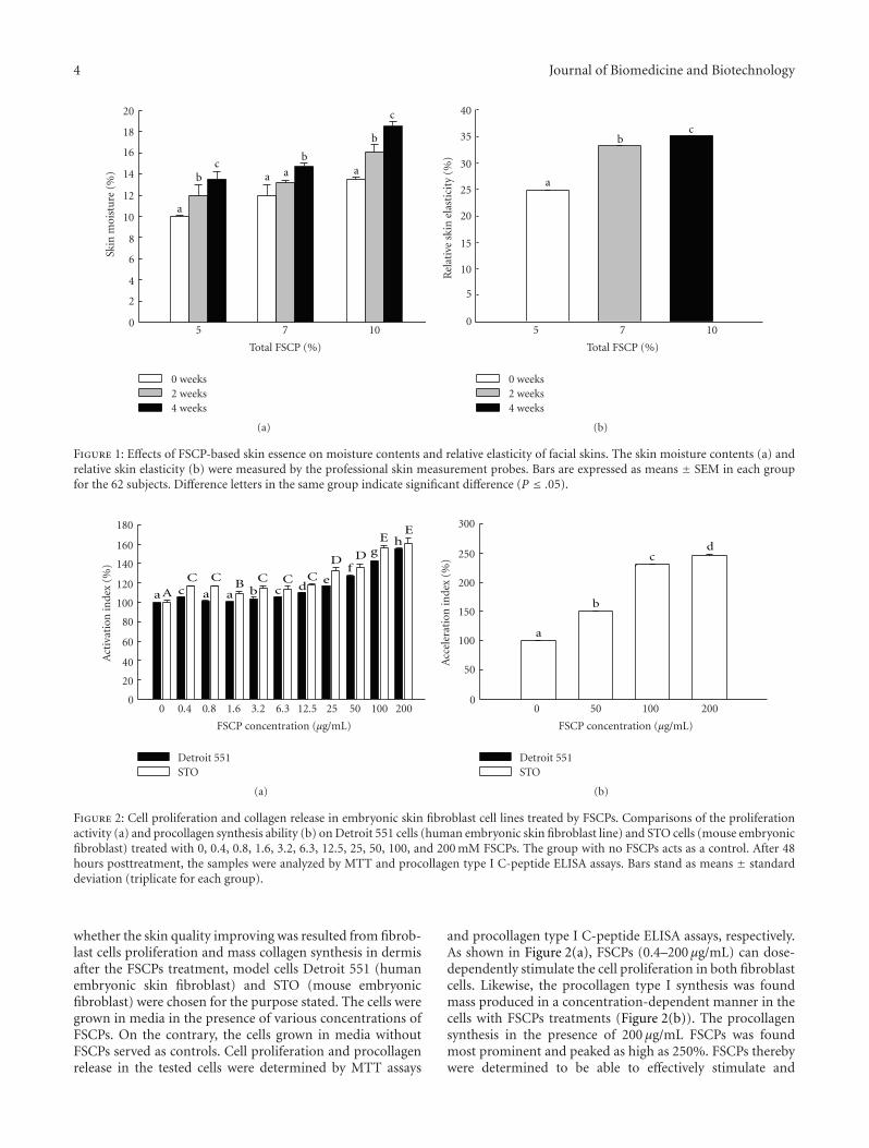
3.1. Effects of FSCPs on Human Facial Skin Moisture Contentsand Relative Elasticity. While collagen peptides have beenwell documented to be an ideal material in cosmeticindustries [26], fish scale collagen peptides (FSCPs) as asustainable collagen source however did not draw greatattention. In this study, we examined the treating effectsof FSCPs as a major component in a skin essence on thefacial skin moisture contents and relative elasticity by usingstandard professional skin probes. The FSCP-based skinessences were first formulated into 5%, 7%, and 10% oftotal FSCPs as the common basal ingredients. Sixty-twovoluntary Taiwanese women (within 23 to 60 years of age)were subjected to the FSCP-based skin essence treatment onfacial skin twice a day for 30 days. The effects with all FSCP-based skin essences on the skin moisture contents were foundimproving significantly but in a time- and dose-dependentmanner during the testing course (P ≤ .05; Figure 1(a)).Likewise, the effects with all FSCP-based skin essences on therelative elasticity of facial skin were also found increasing inthe same manner as for the skin moisture test by 24%, 33%,and 35%, respectively (P ≤ .05; Figure 1(b)). Thereby, theFSCP-based skin essences did improve facial skin qualities(moisture and elasticity) in a time- and dose-dependentmanner in these tested subjects.
3.2. Cell Proliferation and Collagen Release in Embryonic SkinFibroblast Cell Lines after FSCPs Treatments. To determine
4 Journal of Biomedicine and Biotechnology
a
bc
a a
ba
b
c
0
2
4
6
8
10
12
14
16
18
20Sk
inm
oist
ure
(%)
5 7 10
Total FSCP (%)
0 weeks2 weeks4 weeks
(a)
cb
a
0
5
10
15
20
25
30
35
40
Rel
ativ
esk
inel
asti
city
(%)
5 7 10
Total FSCP (%)
0 weeks2 weeks4 weeks
(b)
Figure 1: Effects of FSCP-based skin essence on moisture contents and relative elasticity of facial skins. The skin moisture contents (a) andrelative skin elasticity (b) were measured by the professional skin measurement probes. Bars are expressed as means ± SEM in each groupfor the 62 subjects. Difference letters in the same group indicate significant difference (P ≤ .05).
aA a a bc c de
f
gh
C C C C CB
D D
EE
0
20
40
60
80
100
120
140
160
180
Act
ivat
ion
inde
x(%
)
0 0.4 0.8 1.6 3.2 6.3 12.5 25 50 100 200
FSCP concentration (μg/mL)
Detroit 551STO
(a)
a
b
cd
0
50
100
150
200
250
300
Acc
eler
atio
nin
dex
(%)
0 50 100 200
FSCP concentration (μg/mL)
Detroit 551STO
(b)
Figure 2: Cell proliferation and collagen release in embryonic skin fibroblast cell lines treated by FSCPs. Comparisons of the proliferationactivity (a) and procollagen synthesis ability (b) on Detroit 551 cells (human embryonic skin fibroblast line) and STO cells (mouse embryonicfibroblast) treated with 0, 0.4, 0.8, 1.6, 3.2, 6.3, 12.5, 25, 50, 100, and 200 mM FSCPs. The group with no FSCPs acts as a control. After 48hours posttreatment, the samples were analyzed by MTT and procollagen type I C-peptide ELISA assays. Bars stand as means ± standarddeviation (triplicate for each group).
whether the skin quality improving was resulted from fibrob-last cells proliferation and mass collagen synthesis in dermisafter the FSCPs treatment, model cells Detroit 551 (humanembryonic skin fibroblast) and STO (mouse embryonicfibroblast) were chosen for the purpose stated. The cells weregrown in media in the presence of various concentrations ofFSCPs. On the contrary, the cells grown in media withoutFSCPs served as controls. Cell proliferation and procollagenrelease in the tested cells were determined by MTT assays
and procollagen type I C-peptide ELISA assays, respectively.As shown in Figure 2(a), FSCPs (0.4–200 μg/mL) can dose-dependently stimulate the cell proliferation in both fibroblastcells. Likewise, the procollagen type I synthesis was foundmass produced in a concentration-dependent manner in thecells with FSCPs treatments (Figure 2(b)). The procollagensynthesis in the presence of 200 μg/mL FSCPs was foundmost prominent and peaked as high as 250%. FSCPs therebywere determined to be able to effectively stimulate and
Journal of Biomedicine and Biotechnology 5
10 μm
40x
Control
(a)
10 μm
40x
(b)
4 μm
100x
(c)
WithoutAHAs
(d) (e) (f)
WithAHAs
(g) (h) (i)
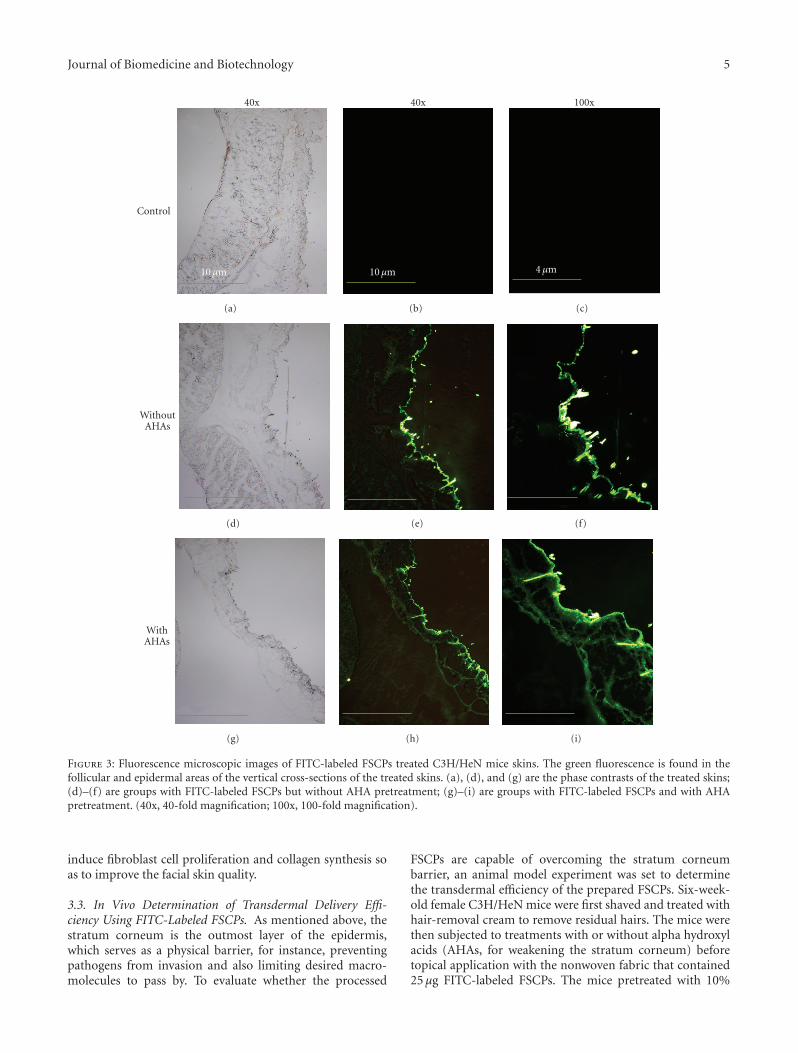
Figure 3: Fluorescence microscopic images of FITC-labeled FSCPs treated C3H/HeN mice skins. The green fluorescence is found in thefollicular and epidermal areas of the vertical cross-sections of the treated skins. (a), (d), and (g) are the phase contrasts of the treated skins;(d)–(f) are groups with FITC-labeled FSCPs but without AHA pretreatment; (g)–(i) are groups with FITC-labeled FSCPs and with AHApretreatment. (40x, 40-fold magnification; 100x, 100-fold magnification).
induce fibroblast cell proliferation and collagen synthesis soas to improve the facial skin quality.
3.3. In Vivo Determination of Transdermal Delivery Effi-ciency Using FITC-Labeled FSCPs. As mentioned above, thestratum corneum is the outmost layer of the epidermis,which serves as a physical barrier, for instance, preventingpathogens from invasion and also limiting desired macro-molecules to pass by. To evaluate whether the processed
FSCPs are capable of overcoming the stratum corneumbarrier, an animal model experiment was set to determinethe transdermal efficiency of the prepared FSCPs. Six-week-old female C3H/HeN mice were first shaved and treated withhair-removal cream to remove residual hairs. The mice werethen subjected to treatments with or without alpha hydroxylacids (AHAs, for weakening the stratum corneum) beforetopical application with the nonwoven fabric that contained25 μg FITC-labeled FSCPs. The mice pretreated with 10%

6 Journal of Biomedicine and Biotechnology
0
10
20
30
40
>6.5 3–6.5 1.5–3 0.7–1.5 0.35–0.7 <0.35
Distribution (KDa)
(a)
0
20
40
60
80
100
120
140
0 10 20
mV
30 40 50 60
6512 Da 1355 Da 246 Da(RT 23.12) (RT 32.83)(RT 40.70)
Time (min)
1300 Da2000 Da
3500 Da4500 Da
RT 30.03
RT 27.58RT 32.96
RT 25.25
(b)
Figure 4: Size distribution of FSCPs. (a) Fish scale hydrolysates were separated by gel chromatography (superdex peptide 10/300 GLcolumns); molecular sizes were determined for each fraction; size distributions were plotted accordingly. The size distribution (KDa) isexpressed as means ± SEM. (b) Size profiles of FSCPs. Protein standards (Bio-Rad, California, USA) are cytochrome C (Mr = 12500 Da),aprotinin (Mr = 6512 Da), vitamin B12 (Mr = 1355 Da), and cytidine (Mr = 246 Da). The average molecular weights (MWs) weredetermined to be 4500, 3500, 2000, and 1300 Da for the four indicated groups.
0
500
1000
1500
2000
2500
0 5
Cu
mu
lati
vep
enet
rate
dam
oun
t(μ
g/cm
2)
10 15 20 25
Time (hours)
1300 Da2000 Da3500 Da
4500 DaMixture
Figure 5: Transdermal permeation abilities of given FSCPs frac-tions. The permeation abilities of various molecular sizes of FSCPswere determined by the Franz-type diffusion cell model, performingat 1, 2, 3, 4, 6, and 24 hours. The amounts of total collagenpeptides from the receptor solutions were determined by the BCAprotein assay. “Mixture” indicates the FSCP hydrolates withoutultracentrifugation membrane treatment. The results shown are arepresentative in 3 experiments.
AHAs and topped with the nonwoven fabric containingno FITC-labeled FSCPs served as controls. These treatedskins were dissected out and immediately embedded with
O.C.T. in due course. Then, the samples were sectioned,mounted on glass slides, and observed under fluorescencemicroscopy. As shown in Figure 3, the FITC positive signalslaid mainly on the area of superficial epidermis. And, theFITC positive signals in the skin pre-treated with AHAwere found deeper and brighter than those without AHAtreatment. The FITC positive signals were also found inthe hair follicles of epidermis. Thus, the stratum corneumbarrier was determined to be a key factor that influences thepenetration efficiency of FSCPs in mice skin. Nonetheless,FSCPs without AHA treatment still considerably pass bythe stratum corneum, which agreed with the results in thehuman facial skin test.
3.4. Transdermal Penetration Ability of FSCPs in Franz-Type Diffusion Cell Model. We considered factors such asmolecular volume (MV) or molecular weight (MW) ofcollagens may play a role on their penetration capabilitywhen administrated topically. The size effect for given FSCPswas determined by employing the Franz-type diffusion cellmodel. Fish scale hydrolates were first subjected to gelchromatography (superdex peptide 10/300 GL columns),whereby FSCP hydrolysates were fractionationed by sizes.As shown in Figure 4(a), the eluted peptides were collectedinto six fractions, >6.5 KDa (3.65%), 3–6.5 KDa (16.8%),1.5–3.0 KDa (32.0%), 0.7–1.5 KDa (29.3%). 0.35–0.7 KDa(14.6%) and <0.35 KDa (3.7%). These peptide fractions werefurther subjected to ultrafiltration membrane refining (5, 3,1, and 0.5 kDa cutoff) into four refined groups I–IV (Mw =3–5, 1–3, 1–0.5, and <0.5 KDa) with average molecular
Journal of Biomedicine and Biotechnology 7
100 μm
(a)
100 μm
(b)
100 μm
(c)
100 μm
(d)
100 μm
(e)
100 μm
(f)
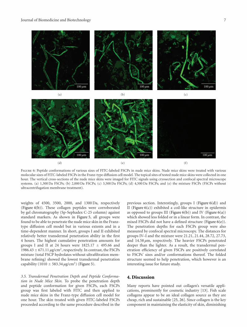
Figure 6: Peptide conformations of various sizes of FITC-labeled FSCPs in nude mice skins. Nude mice skins were treated with variousmolecular sizes of FITC-labeled FSCPs in the Franz-type diffusion cell model. The topical sites of tested nude mice skins were collected in onehour. The vertical cross-sections of the nude mice skins were imaged for FITC signals using cryosection and confocal spectral microscopesystems. (a) 1,300 Da FSCPs; (b) 2,000 Da FSCPs; (c) 3,500 Da FSCPs; (d) 4,500 Da FSCPs; and (e) the mixture FSCPs (FSCPs withoutultracentrifugation membrane treatment).
weights of 4500, 3500, 2000, and 1300 Da, respectively(Figure 4(b)). These collagen peptides were corroboratedby gel chromatography (Sp-Sephadex C-25 column) againststandard markers. As shown in Figure 5, all groups werefound to be able to penetrate the nude mice skin in the Franz-type diffusion cell model but in various extents and in atime-dependent manner. In short, groups I and II exhibitedrelatively better transdermal penetration ability in the first4 hours. The highest cumulative penetration amounts forgroups I and II at 24 hours were 1825.17 ± 495.66 and1986.43± 671.11 μg/cm2, respectively. In contrast, the FSCPsmixture (total FSCP hydrolates without ultrafiltration mem-brane refining) showed the lowest transdermal penetrationcapability (1010 ± 583.34 μg/cm2) (Figure 5).
3.5. Transdermal Penetration Depth and Peptide Conforma-tion in Nude Mice Skin. To probe the penetration depthand peptide conformation for given FSCPs, each FSCPsgroup was first labeled with FITC and then applied tonude mice skins in the Franz-type diffusion cell model forone hour. The skin treated with given FITC-labeled FSCPsproceeded according to the same procedure described in the
previous section. Interestingly, groups I (Figure 6(d)) andII (Figure 6(c)) exhibited a coil-like structure in epidermisas opposed to groups III (Figure 6(b)) and IV (Figure 6(a))which showed less folded or in a linear form. In contrast, themixed FSCPs did not have a defined structure (Figure 6(e)).The penetration depths for each FSCPs group were alsomeasured by confocal spectral microscopy. The distances forgroups IV–I and the mixture were 21.21, 21.44, 28.72, 27.73,and 14.58 μm, respectively. The heavier FSCPs penetrateddeeper than the lighter. As a result, the transdermal pen-etration efficiency of given FSCPs are positively correlatedto FSCPs’ sizes and/or conformations thereof. The foldedstructure seemed to help penetration, which however is aninteresting issue for future study.
4. Discussion
Many reports have pointed out collagen’s versatile appli-cations, prominently for cosmetic industry [13]. Fish scalecollagens appear to be an ideal collagen source as they arecheap, rich and sustainable [25, 26]. Since collagen is the keycomponent in maintaining the elasticity of skin, diminishing
8 Journal of Biomedicine and Biotechnology
collagen contents in skin would therefore result in wrinkledand flabby skins [27]. In this paper, we have demonstratedthe formulated FCSP-based skin essence possesses suchdesirable effects as increasing skin moisture contents and rel-ative elasticity (Figure 1). The FCSPs at 200 μg/mL providedthe best effects by manifesting embryonic skin fibroblastcell proliferations (Detroit 551 cells and STO cells) andprocollagen synthesis. These results otherwise are reinforcedby the reports of Katayama et al., wherein they demonstratedpentapeptides from type I collagen promoted extracellularmatrix production in fibroblasts [28, 29].
Since the stratum corneum acts as a physical barrierfor multiple purposes in skins [19, 20], it does influencethe penetration efficiency of the FSCP-based skin essencesas they were present mainly in the surface of epidermis.However, given FITC-labeled FSCPs still effectively passedby stratum corneum to epidermis and dermis in the micemodel experiment. The reports of Potts and Guy agreed withthe results, in which they proposed the diffusivity of a drugin skin is correlated to given molecular features of the drug[30, 31], although Lin et al. [32] considered the effect oflipophilicity in the passive diffusion permeability is the dom-inant factor other than the compound’s molecular weight. Inaddition, Potts and Guy further proposed that the higher themolecular weights the greater the lipophilicity/permeability[30]. The better effects of the FSCPs’ groups at Mw 3500and Mw 4500 Da may possess desired sizes and effectivestructural features. Furthermore, collagens typically exist as aright-handed triple helix structure of Gly-X-Y helix-formingrepeats in a length of 300 nm which corresponds to 1000amino acids or so in fibril-forming collagens (I, II, III) [33–36]. Fish scale collagens are within the scope of the type Icollagen [26]. The higher penetration efficiency of FSCPs at3500 and 4500 Da may thus be attributed to the synergisticeffects of high lipophilicity (adequate size) and constructivestructure features.
In conclusion, the prepared FSCPs have shown to be ableto effectively penetrate stratum corneum to epidermis anddermis. FSCPs at epidermis and dermis in situ can activatefibroblasts and accelerate collagen synthesis, whereby thefacial skin qualities (moisture contents and relative elasticity)were significantly improved. As a consequence, the preparedFSCPs along with the formulated skin essence should holdhigh commercial interests and are worthy of further in-depthstudy for enhancing FSCPs transdermal penetration abilityto a higher level.
References
[1] T. Nagai, Y. Araki, and N. Suzuki, “Collagen of the skin ofocellate puffer fish (Takifugu rubripes),” Food Chemistry, vol.78, no. 2, pp. 173–177, 2002.
[2] R. Mayne and R. G. Brewton, “New members of the collagensuperfamily,” Current Opinion in Cell Biology, vol. 5, no. 5, pp.883–890, 1993.
[3] M. Van der Rest and R. Garrone, “Collagen family of proteins,”FASEB Journal, vol. 5, no. 13, pp. 2814–2823, 1991.
[4] R. R. Wickett and M. O. Visscher, “Structure and function ofthe epidermal barrier,” American Journal of Infection Control,vol. 34, no. 10, supplement 1, pp. 98–110, 2006.
[5] K. K. H. Svoboda, D. A. Fischman, and M. K. Gordon,“Embryonic chick corneal epithelium: a model system forexploring cell-matrix interactions,” Developmental Dynamics,vol. 237, no. 10, pp. 2667–2675, 2008.
[6] J. Lee, E. Jung, J. Lee, et al., “Panax ginseng induces humantype I collagen synthesis through activation of Smad signal-ing,” Journal of Ethnopharmacology, vol. 109, no. 1, pp. 29–34,2007.
[7] J. Wu, M. Fujioka, K. Sugimoto, G. Mu, and Y. Ishimi,“Assessment of effectiveness of oral administration of collagenpeptide on bone metabolism in growing and mature rats,”Journal of Bone and Mineral Metabolism, vol. 22, no. 6, pp.547–553, 2004.
[8] Y.-I. Koyama, A. Hirota, H. Mori, et al., “Ingestion of gelatinhas differential effect on bone mineral density and body weightin protein undernutrition,” Journal of Nutritional Science andVitaminology, vol. 47, no. 1, pp. 84–86, 2001.
[9] R. W. Moskowitz, “Role of collagen hydrolysate in bone andjoint disease,” Seminars in Arthritis and Rheumatism, vol. 30,no. 2, pp. 87–99, 2000.
[10] J. Scala, N. R. S. Hollies, and K. P. Sucher, “Effect of dailygelatine ingestion on human scalp hair,” Nutrition ReportsInternational, vol. 13, no. 6, pp. 579–592, 1976.
[11] T. L. Tyson, “The effect of gelatin on fragile fingernails,”Journal of Investigative Dermatology, vol. 14, pp. 323–325,1950.
[12] J. Minaguchi, Y.-I. Koyama, N. Meguri, et al., “Effects ofingestion of collagen peptide on collagen fibrils and gly-cosaminoglycans in Achilles tendon,” Journal of NutritionalScience and Vitaminology, vol. 51, no. 3, pp. 169–174, 2005.
[13] N. Matsuda, Y.-I. Koyama, Y. Hosaka, et al., “Effects ofingestion of collagen peptide on collagen fibrils and gly-cosaminoglycans in the dermis,” Journal of Nutritional Scienceand Vitaminology, vol. 52, no. 3, pp. 211–215, 2006.
[14] M. Ogawa, R. J. Portier, M. W. Moody, J. Bell, M. A.Schexnayder, and J. N. Losso, “Biochemical properties of boneand scale collagens isolated from the subtropical fish blackdrum (Pogonia cromis) and sheepshead seabream (Archosargusprobatocephalus),” Food Chemistry, vol. 88, no. 4, pp. 495–501,2004.
[15] A. Jongjareonrak, S. Benjakul, W. Visessanguan, T. Nagai, andM. Tanaka, “Isolation and characterisation of acid and pepsin-solubilised collagens from the skin of Brownstripe red snapper(Lutjanus vitta),” Food Chemistry, vol. 93, no. 3, pp. 475–484,2005.
[16] M. Sadowska, I. Kołodziejska, and C. Niecikowska, “Isolationof collagen from the skins of Baltic cod (Gadus morhua),” FoodChemistry, vol. 81, no. 2, pp. 257–262, 2003.
[17] P. Kittiphattanabawon, S. Benjakul, W. Visessanguan, T. Nagai,and M. Tanaka, “Characterisation of acid-soluble collagenfrom skin and bone of bigeye snapper (Priacanthus tayenus),”Food Chemistry, vol. 89, no. 3, pp. 363–372, 2005.
[18] K. Gelse, E. Poschl, and T. Aigner, “Collagens—structure,function, and biosynthesis,” Advanced Drug Delivery Reviews,vol. 55, no. 12, pp. 1531–1546, 2003.
[19] P. M. Elias and E. H. Choi, “Interactions among stratumcorneum defensive functions,” Experimental Dermatology, vol.14, no. 10, pp. 719–726, 2005.
[20] P. M. Elias, “Stratum corneum defensive functions: an inte-grated view,” Journal of Investigative Dermatology, vol. 125, no.2, pp. 183–200, 2005.
[21] B. Baert, E. Deconinck, M. V. Gele, et al., “Transdermalpenetration behaviour of drugs: CART-clustering, QSPR and
Journal of Biomedicine and Biotechnology 9
selection of model compounds,” Bioorganic and MedicinalChemistry, vol. 15, no. 22, pp. 6943–6955, 2007.
[22] C. H. Wu and H. J. Chai, “Collagen of fish scale and methodof macking thereof,” Taiwan Invention patent publicationnumber I 263678, 2006.
[23] Z. Ma and L.-Y. Lim, “Uptake of chitosan and associatedinsulin in Caco-2 cell monolayers: a comparison between chi-tosan molecules and chitosan nanoparticles,” PharmaceuticalResearch, vol. 20, no. 11, pp. 1812–1819, 2003.
[24] T. D. Russell, A. Fischer, N. E. Beeman, E. F. Freed, M.C. Neville, and J. Schaack, “Transduction of the mammaryepithelium with adenovirus vectors in vivo,” Journal ofVirology, vol. 77, no. 10, pp. 5801–5809, 2003.
[25] N. Kim, A. F. El-Kattan, C. S. Asbill, et al., “Evalua-tion of derivatives of 3-(2-oxo-1-pyrrolidine) hexahydro-1H-azepine-2-one as dermal penetration enhancers: side chainlength variation and molecular modeling,” Journal of Con-trolled Release, vol. 73, no. 2-3, pp. 183–196, 2001.
[26] L. Wang, X. An, F. Yang, Z. Xin, L. Zhao, and Q. Hu, “Isolationand characterisation of collagens from the skin, scale and boneof deep-sea redfish (Sebastes mentella),” Food Chemistry, vol.108, no. 2, pp. 616–623, 2008.
[27] H. Tanaka and S. Hasegawa, “Skin permeable collagen pep-tide preventing wrinkle formation induced by photoaging,”Biotechnology Industrial, vol. 22, no. 9, pp. 18–23, 2005.
[28] M. W. Kofford, L. B. Schwartz, N. M. Schechter, D. R. Yager,R. F. Diegelmann, and M. F. Graham, “Cleavage of type Iprocollagen by human mast cell chymase initiates collagenfibril formation and generates a unique carboxyl-terminalpropeptid,” Journal of Biological Chemistry, vol. 272, no. 11,pp. 7127–7131, 1997.
[29] K. Katayama, J. Armendariz-Borunda, R. Raghow, A. H. Kang,and J. M. Seyer, “A pentapeptide from type I procollagen pro-motes extracellular matrix production,” Journal of BiologicalChemistry, vol. 268, no. 14, pp. 9941–9944, 1993.
[30] R. O. Potts and R. H. Guy, “Predicting skin permeability,”Pharmaceutical Research, vol. 9, no. 5, pp. 663–669, 1992.
[31] R. H. Guy and R. O. Potts, “Structure-permeability relation-ships in percutaneous penetration,” Journal of PharmaceuticalSciences, vol. 81, no. 6, pp. 603–604, 1992.
[32] R.-Y. Lin, C.-W. Hsu, and W.-Y. Chen, “A method to predictthe transdermal permeability of amino acids and dipeptidesthrough porcine skin,” Journal of Controlled Release, vol. 38,no. 2-3, pp. 229–234, 1996.
[33] K. Kuhn, “The collagen family-variations in the molecular andsupermolecular structure,” Rheumatology, vol. 10, pp. 29–69,1986.
[34] K. A. Piez, “Molecular and aggregate structure of the colla-gens,” in Extracellular Matrix Biology, K. A. Pietz and H. Reddi,Eds., pp. 1–39, Elsevier, New York, NY, USA, 1984.
[35] J. F. Bateman, S. R. Lamande, and J. A. M. Ramshaw, “Collagensuperfamily,” in Extracellular Matrix, W. D. Comper, Ed., pp.22–67, Harwood, Melbourne, Australia, 1996.
[36] K. Von der Mark, “Structure, biosynthesis and gene regulationof collagens in cartilage and bone,” in Dynamics of Bone andCartilage Metabolism, M. J. Seibel, S. P. Robins, and J. P.Bilezikian, Eds., pp. 3–29, Elsevier, New York, NY, USA, 1999.