Effects of chronic exposure to lead, cadmium, and manganese ...
12
51 Original article DOI: 10.1515/aiht-2015-66-2515 Effects of chronic exposure to lead, cadmium, and manganese mixtures on oxidative stress in rat liver and heart Iwona Markiewicz-Górka 1 , Lidia Januszewska 1 , Aleksandra Michalak 1 , Adam Prokopowicz 2 , Ewa Januszewska 3 , Natalia Pawlas 2 , and Krystyna Pawlas 1 Department of Hygiene, Wroclaw Medical University, Wroclaw 1 , Institute of Occupational Medicine and Environmental Health, Sosnowiec 2 , Poland, Hospital in Rheinfelden, Rheinfelden, Switzerland 3 [Received in March 2014; CrossChecked in March 2014; Accepted in February 2015] The aim of this study was to assess the effects of chronic combined exposure to low, environmental doses of Cd, Pb, and Mn on oxidative stress in the liver and heart of rats and on their liver function parameters. Male Wistar rats were divided randomly into eight groups. For nine months controls were receiving drinking water alone, whereas the exposed groups were receiving drinking water with Pb (0.2 mg L -1 ), Cd (1 mg L -1 ), and Mn (2 mg L -1 ) alone or in combinations. Malondialdehyde (MDA) significantly increased in both heart and liver of the animals after combined exposure to metals. Heart MDA correlated with blood Cd, Pb, and Mn and liver MDA with blood Cd. Aspartate aminotransferase (AST) activity and bilirubin concentration also increased significantly in the animal group exposed to all three metals and correlated positively with blood Cd, Pb, and Mn. Our study has confirmed the synergistic effect of the Cd, Mn, and Pb combination on the increase in heart MDA. A similar synergy was observed for Pb+Mn in the increase of serum alanine aminotransferase (ALT) activity as an indicator of liver function. KEY WORDS: alanine aminotransferase; aspartate aminotransferase; antioxidant enzymes; bilirubin; heavy metals; long-term poisoning Markiewicz-Górka I, et al. Effects of chronic exposure to lead, cadmium, and manganese mixtures on oxidative stress in rat liver and heart Arh Hig Rada Toksikol 2015;66:51-62 Exposure to low levels of cadmium (Cd) and lead (Pb) through food, water, and air is common in industrial areas and is a real threat to the health of the general population (1). Most European countries are nearing the tolerable weekly intake (TWI) of Cd (2.5 μg kg -1 body weight). People who smoke one pack of cigarettes a day take in additional 1-3 μg of Cd (2, 3). Dietary intakes of Pb differ between countries and regions. In the Indian city of Kanpur average daily lead intake through diet is about 114 μg per day for adults and 50 μg day -1 in children (4), in the Zhejiang province in China 0.55 μg kg -1 (body weight) per day (about 27.5-38.5 μg day -1 per person) (5), in Croatia 15-100 μg per day per person (6), and in Poland 39.5- 89.9 μg day -1 per person (7). The probability of exceeding tolerable limits of Pb intake (250 μg day -1 for adults and 90 μg day -1 for children) has been significantly reduced since the switch to unleaded gasoline. However, this risk is still high in children because of the high intake of Pb per body weight (4). Pb accumulated in industrial dust has continued to affect the environment. Environmental as well as occupational exposure to Pb still is a major public health problem (7). Chronic exposure to metals is associated with many diseases such as liver disorders, cancer, neurological diseases, and osteoporosis (3, 8). Elevated blood Cd and/or Pb may increase blood pressure and contribute to the development of cardiovascular disease (9). In contrast to these toxic metals, manganese (Mn) is an essential trace element involved in a variety of important metabolic processes such as amino acid, lipid, protein, and carbohydrate metabolism, adenosine triphosphate production, blood clotting, and bone mineralisation. As a cofactor of superoxide dismutase it takes part in the antioxidative defence of the human body. This element is required in small amounts, but high concentrations can have adverse health effects on the nervous system, lungs, arteries, heart, and liver (10-12). Health effects of Mn at lower concentrations remain unclear. The general population is exposed to Mn primarily through food. In Western and vegetarian diets its daily intake is 0.7-10.9 mg. Through drinking water it is about 20 μg (11). Exposure to excessive Mn levels through drinking water has been reported in Yancheng (China), where high Mn concentrations (~270 mg kg -1 ) were found in the river sediment (13). Environmental exposure is likely to increase due to the introduction of Mn compounds to gasoline as anti-knock agents and the occasional peaks of Mn in tap water (1, 10, 14). People are usually exposed to a combination of metals in their living and working environment. The toxic effects of such mixtures, especially at low, chronic doses (that are likely in the environment) are poorly recognised. Metals Correspondence to: Iwona Markiewicz-Górka, 50-368 Mikulicza-Radeckiego 7, Wroclaw, Poland, E-mail: [email protected]
-
Upload
khangminh22 -
Category
Documents
-
view
0 -
download
0
Transcript of Effects of chronic exposure to lead, cadmium, and manganese ...

51Original article DOI: 10.1515/aiht-2015-66-2515
Effects of chronic exposure to lead, cadmium, and
manganese mixtures on oxidative stress in rat liver and heart
Iwona Markiewicz-Górka1, Lidia Januszewska1, Aleksandra Michalak1, Adam Prokopowicz2, Ewa Januszewska3, Natalia Pawlas2, and Krystyna Pawlas1
Department of Hygiene, Wroclaw Medical University, Wroclaw1, Institute of Occupational Medicine and Environmental Health, Sosnowiec2, Poland, Hospital in Rheinfelden, Rheinfelden, Switzerland3
[Received in March 2014; CrossChecked in March 2014; Accepted in February 2015]
The aim of this study was to assess the effects of chronic combined exposure to low, environmental doses of Cd, Pb, and Mn on oxidative stress in the liver and heart of rats and on their liver function parameters. Male Wistar rats were divided randomly into eight groups. For nine months controls were receiving drinking water alone, whereas the exposed groups were receiving drinking water with Pb (0.2 mg L-1), Cd (1 mg L-1), and Mn (2 mg L-1) alone or in combinations. Malondialdehyde (MDA) significantly increased in both heart and liver of the animals after combined exposure to metals. Heart MDA correlated with blood Cd, Pb, and Mn and liver MDA with blood Cd. Aspartate aminotransferase (AST) activity and bilirubin concentration also increased significantly in the animal group exposed to all three metals and correlated positively with blood Cd, Pb, and Mn. Our study has confirmed the synergistic effect of the Cd, Mn, and Pb combination on the increase in heart MDA. A similar synergy was observed for Pb+Mn in the increase of serum alanine aminotransferase (ALT) activity as an indicator of liver function.KEY WORDS: alanine aminotransferase; aspartate aminotransferase; antioxidant enzymes; bilirubin; heavy metals; long-term poisoning
Markiewicz-Górka I, et al. Effects of chronic exposure to lead, cadmium, and manganese mixtures on oxidative stress in rat liver and heart Arh Hig Rada Toksikol 2015;66:51-62
Exposure to low levels of cadmium (Cd) and lead (Pb) through food, water, and air is common in industrial areas and is a real threat to the health of the general population (1). Most European countries are nearing the tolerable weekly intake (TWI) of Cd (2.5 μg kg-1 body weight). People who smoke one pack of cigarettes a day take in additional 1-3 μg of Cd (2, 3). Dietary intakes of Pb differ between countries and regions. In the Indian city of Kanpur average daily lead intake through diet is about 114 μg per day for adults and 50 μg day-1 in children (4), in the Zhejiang province in China 0.55 μg kg-1 (body weight) per day (about 27.5-38.5 μg day-1 per person) (5), in Croatia 15-100 μg per day per person (6), and in Poland 39.5- 89.9 μg day-1 per person (7).
The probability of exceeding tolerable limits of Pb intake (250 μg day-1 for adults and 90 μg day-1 for children) has been significantly reduced since the switch to unleaded gasoline. However, this risk is still high in children because of the high intake of Pb per body weight (4). Pb accumulated in industrial dust has continued to affect the environment. Environmental as well as occupational exposure to Pb still is a major public health problem (7). Chronic exposure to metals is associated with many diseases such as liver disorders, cancer, neurological diseases, and osteoporosis
(3, 8). Elevated blood Cd and/or Pb may increase blood pressure and contribute to the development of cardiovascular disease (9).
In contrast to these toxic metals, manganese (Mn) is an essential trace element involved in a variety of important metabolic processes such as amino acid, lipid, protein, and carbohydrate metabolism, adenosine triphosphate production, blood clotting, and bone mineralisation. As a cofactor of superoxide dismutase it takes part in the antioxidative defence of the human body. This element is required in small amounts, but high concentrations can have adverse health effects on the nervous system, lungs, arteries, heart, and liver (10-12). Health effects of Mn at lower concentrations remain unclear. The general population is exposed to Mn primarily through food. In Western and vegetarian diets its daily intake is 0.7-10.9 mg. Through drinking water it is about 20 μg (11). Exposure to excessive Mn levels through drinking water has been reported in Yancheng (China), where high Mn concentrations (~270 mg kg-1) were found in the river sediment (13). Environmental exposure is likely to increase due to the introduction of Mn compounds to gasoline as anti-knock agents and the occasional peaks of Mn in tap water (1, 10, 14).
People are usually exposed to a combination of metals in their living and working environment. The toxic effects of such mixtures, especially at low, chronic doses (that are likely in the environment) are poorly recognised. Metals
Correspondence to: Iwona Markiewicz-Górka, 50-368 Mikulicza-Radeckiego 7, Wroclaw, Poland, E-mail: [email protected]

52
can interact in the human body as soon as they are absorbed, when they bind with specific proteins, or during transport and distribution in the body. Their interaction can either increase or decrease the toxic action of single components (15, 16). Reports on the effects of long-term combined exposure to environmental pollutants are scarce. Furthermore, their effects, especially those of heavy metals, are usually tested on the basis of acute or subchronic exposures that do not reflect the actual risk to the general population.
The aim of our study was to address this interaction of metals at environmental doses by assessing the effects of long-term exposure to combinations of Cd, Pb, and Mn at low doses in rats.
One of the major heavy metal effects is oxidative stress. Metals increase lipid peroxidation and cripple antioxidant defences in tissues by altering the activity of antioxidant enzymes such as superoxide dismutase (SOD), catalase (CAT), and glutathione peroxidase (GPx) (17, 18). We therefore looked at how metal combinations affected the levels of malondialdehyde (MDA), which is a marker of oxidative stress, the antioxidant enzymes (SOD, GPx, and CAT) in the heart and liver, and liver function indicators (AST, ALT, and bilirubin) and whether these effects were dose- and interaction-related.
Changes in the prooxidant-antioxidant balance in the tissues during chronic exposure to metals were of special interest to us because oxidative stress is involved in the development of most pathological states and diseases. We designed this experiment hoping that it would help to better understand the early events in the tissues caused by chronic combined exposure to low concentrations of Cd, Pb, and Mn mixtures.
MATERIALS AND METHODS
Animals and treatment
The experiment was performed on 64 male Wistar rats. At the beginning of the experiment, the animals were 11-12 weeks old and weighed about 280 g. They were kept in plastic cages at room temperature in natural lighting conditions and had free access to water and standard rodent feed. The rats were divided into the following experimental groups (eight animals in each): control group, receiving clean drinking water; lead-exposed group (Pb group), receiving 0.2 mg L-1 of Pb in the form of lead acetate dissolved in water [Pb (CH3COO)2x3H2O]; cadmium-exposed group (Cd group), receiving 1 mg L-1 of Cd in the form of cadmium chloride dissolved in water (CdCl2x2.5 H2O); manganese-exposed group (Mn group), receiving 2 mg L-1 of Mg in the form of manganese chloride dissolved in water (MnCl2x4 H2O); cadmium and lead-exposed group (Cd+Pb group); cadmium and manganese-exposed group (Cd+Mn group); lead and manganese-exposed group
(Pb+Mn group); and finally lead, manganese, and cadmium-exposed group (Pb+Mn+Cd group). All combination groups were exposed to the metals in the doses described above. The chemicals used were obtained from Sigma-Aldrich (Steinheim, Germany).
The concentrations used in our experiment were higher than the maximum permissible limits (MPL) for Pb, Cd, and Mn in drinking water, namely 0.01, 0.003, and 0.4 mg L-1, respectively (19). They were meant to take into account other sources in addition to water such as food and air, and they largely reflect the measurements taken all over the world where the concentrations of Cd, Pb, and Mn periodically or continuously exceed the permissible limits for drinking water (13, 20, 21).
Our experimental Pb concentration (0.2 mg L-1) corresponds to the upper range reported in drinking water by Mohod and Dhote (20), whereas Mn (2 mg L-1) corresponds to the upper concentrations reported by Bouchard et al. (21) and Cd (1 mg L-1) to low environmental exposure reported by Brzoska and Moniuszko (3).
The experiment lasted nine months, during which time the rats were regularly weighed and their water intake measured. Two rats died before the end of the experiment (one from the Cd+Mn group and one from the Pb+Mn group). The remaining animals were killed by cervical dislocation after having been anaesthetised with an intramuscular injection of ketamine and their blood collected by cardiac puncture into tubes with lithium heparin for the measurement of metals in whole blood (catalogue no. 454084, Greiner Bio-One BVBA, Vilvoorde, Belgium) and into tubes for serum separation (catalogue no. 26.323 D-51588, Sarstedt, Numbrecht, Germany) for liver function tests. Liver and heart tissues were cut into smaller sections, part of them homogenised in appropriate buffers as needed for a specific biochemical assay (see below), and centrifuged. Serum was obtained from blood centrifuged at 1200 x g for 10 minutes. Samples of whole blood, serum, supernatants of tissue homogenates and tissues were stored at -80 °C until analysis. The study was approved by the First Local Ethics Committee for Animal Experiments in Wroclaw, Poland (approval no. 23/2010).
Metals in whole blood
Blood Pb and Cd concentrations were measured using a Perkin Elmer 4100ZL electrothermal atomic absorption spectrometer (ETAAS) (Bodenseewerk Perkin-Elmer, Ueberlingen, Germany) equipped with a non-specific absorption correction circuit and a Zeeman autosampler. The method has been developed by Stoepplers et al. (22, 23). Blood was deproteinised with 0.8 mol L-1 of nitric acid.
Blood Mn was determined using ETAAS after diluting blood samples with a solution of 0.1 % Triton-X100 in a ratio of 1:2 and adding 0.003 mg of matrix modifier Mg(NO3)2 and 0.05 mg of NH4H2PO4.
Markiewicz-Górka I, et al. Effects of chronic exposure to lead, cadmium, and manganese mixtures on oxidative stress in rat liver and heart Arh Hig Rada Toksikol 2015;66:51-62

53
Metals in heart and liver tissues
Wet tissue samples weighing about 700-750 mg were dried to a constant weight (about 200 mg each) and placed in Teflon containers with a screw tap. Dry samples were then mineralised in 1 mL of 65 % nitric acid (Baker Instra-Analyzed®) in a closed container by heating them at 120 °C for 16 h. After cooling, 1 mL of 30 % hydrogen peroxide (Baker Analyzed®) was added and mineralisation continued at 120 °C for another 24 h. The resulting solution was diluted with deionised water (Milli-Q, Millipore Corporation, Bedford, Massachusetts, USA) to 10 mL. Blank and control samples were prepared using the same mineralisation procedure. Heart and liver Pb, Cd, and Mn were measured in a graphite furnace using a Perkin Elmer 4100ZL ETAAS equipped with a Zeeman background correction system and autosampler. The results are given in µg g-1 of dry weight.
Quantification was performed using matrix-matched calibration. For all metals calibration was performed using bovine whole-blood standards.
The accuracy of the measurements performed in the laboratory is continuously monitored by an internal quality control system and regular participation in inter-laboratory proficiency tests conducted by the Centers for Disease Control and Prevention (CDC) in Atlanta, Georgia, USA.
All metals were measured at the Department of Chemical Hazards and Genetic Toxicology, Institute of Occupational Medicine and Environmental Health in Sosnowiec, Poland.
Oxidative stress assessment
Oxidative stress in rat heart and liver was assessed by measuring MDA, which is the most abundant end-product of lipid peroxidation. Tissues were homogenised in a radioimmunoprecipitation (RIPA) buffer (Sigma Aldrich, Cat No. R- 0278) in a ratio of 1:10 w/v and then centrifuged at 1600xg and 4 °C for 10 min. The measurement was performed on the supernatant using the thiobarbituric acid reactive substances (TBARS) assay kit No. 10009055 (Cayman Chemical Company, Ann Arbor, Michigan. USA) according to the instructions provided by the manufacturer.
Antioxidant enzyme activity
For GPx activity measurement, heart and liver tissues were homogenised in a cold, 50 mmol L-1. TRIS-HCl buffer [1:9 (w/v)], pH 7.5, containing 5 mmol L-1 of EDTA and 1 mmol L-1 of 2-mercaptoethanol and centrifuged at 10,000 x g at 4 °C for 10 min. GPx activity was measured in the obtained supernatants using the Bioxytech® GPx-340 kit No. 21017 (Oxis International, Portland, OR, USA) according to the manufacturer’s instructions and is expressed as milliUnits (mU) of GPx per milligram of protein (1 mU/mg=1 nmol of oxidised NADPH in one minute per mg of protein).
For SOD activity measurement, heart and liver tissues were washed with cold PBS buffer and homogenised in a cold, 20 mmol L-1 HEPES buffer [1:9 (w/v)], pH 7.2, containing 1 mmol L-1 of EGTA, 210 mmol L-1 of mannitol, and 70 mmol L-1 of sucrose. The homogenate was centrifuged at 1500 x g at 4°C for 5 minutes. SOD activity was measured in the obtained supernatants using the Cayman Chemical Company kit no. 706002 according to the manufacturer’s instructions. One unit of SOD is defined as the amount of enzyme decreasing superoxide anion concentration by 50 %.
Heart and liver tissues for CAT analysis were weighed and homogenised [1:9 (w/v)] in a cold 50 mmol L-1 PBS buffer supplemented with 1 mmol L-1 EDTA per gram of tissue. The homogenates were centrifuged at 10,000xg at 4 °C for 15 min and CAT activity measured in the supernatants using a Cell Biolabs reagent kit No. STA -341, San Diego, California, USA according to the manufacturer’s instructions. One unit of CAT is defined as the amount of enzyme decomposing 1 mmol L-1 of H2O2 in 1 min at 25 °C. CAT activity is expressed as U mg-1 of protein.
Protein concentrations in the samples were measured with the Sigma Diagnostics Protein Assay Kit (Cat # P 5656) based on the Lowry’s method (24) using bovine albumin as the standard.
ALT, AST, and total bilirubin in serum
Alanine aminotransferase (ALT), aspartate aminotransferase (AST), and total bilirubin in rat serum were measured in the biochemistry laboratory of the Wroclaw University Hospital no. 1, using a Konelab™ PRIME 60i automated biochemical analyser (Thermo Scientific, Vantaa, Finland) and commercial tests of the same manufacturer. The results for ALT and AST are expressed in IU L-1. Bilirubin concentration is given in mg dL-1.
Statistical analysis
Differences between the groups of rats were tested for significance using one-way analysis of variance (ANOVA), followed by post-hoc tests. When variances were heterogeneous, we used Cochran-Cox’s test. Correlations between metal concentrations in blood and tissues and biochemical parameters (MDA, antioxidative enzymes, indicators of liver function) were assessed using Pearson’s test. When the variables did not meet the assumptions for parametric tests, we used the Mann-Whitney U test and Spearman’s test.
Metal interaction in terms of their effects on the biochemical parameters was verified using the two and three-way ANOVA/MANOVA test F. Interaction was characterised by comparing the measured values with the expected ones. The expected value (expressed in %) was calculated as the sum of parameter (e.g. MDA) values (expressed as % of control value) obtained by exposure to
Markiewicz-Górka I, et al. Effects of chronic exposure to lead, cadmium, and manganese mixtures on oxidative stress in rat liver and heart Arh Hig Rada Toksikol 2015;66:51-62

54
each metal separately. If the measured value after exposure to a combination of metals was significantly higher than the expected value, we characterised the effect as synergistic. If the measured effect was significantly below the calculated (expected) value the effect of the two or three metals was characterised as antagonistic.
All results are presented as means (± standard deviation). Statistical analysis and calculations were performed using the STATISTICA software for Windows (StatSoft Polska, Krakow, Poland). Statistical significance was set at p<0.05.
RESULTS AND DISCUSSION
Table 1 shows weight gain, water consumption, and calculated Pb, Cd, and Mn intake by rats during the nine-month experiment. The exposed groups did not significantly differ from control in weight gain and amount of water consumed.
The respective daily intake of Pb, Cd, and Mn was 0.0011 %, 0.04 %, and 0.04 % of the median lethal dose (LD50) for rats and reflects environmental exposure to these metals. The exposed groups did not significantly differ between themselves in metal intake.
Metal accumulation
Figure 1 shows liver, heart, and blood Cd concentration by the groups of rats. Liver and heart concentrations of Cd did not differ significantly between the Cd-treated groups. In all Cd-treated groups blood Cd (0.3-0.5 µg L-1) kept within the average range found in the blood of residents of large European cities (0.25-0.65 µg L-1) (25). The highest blood Cd concentration was found in the group receiving all three metals (Cd+Mn+Pb), and it differed significantly from all other Cd-treated groups. These results suggest that simultaneous presence of low doses of Pb and Mn in drinking water may increase the intestinal absorption of Cd.
The highest liver, heart, and whole blood Pb concentrations were observed in the group receiving Pb alone (Figure 2). However, liver Pb in this group was
significantly higher only in comparison to the Pb+Mn+Cd group. The Pb group also had significantly higher Pb concentrations in the heart than any other Pb-treated group, whereas in blood the difference was not significant only in comparison with the Pb+Mn group. Moreover, co-exposure with Cd and/or Mn resulted in lower Pb concentrations in the heart, and co-exposure with Cd significantly decreased its concentration in blood.
Divalent metals are absorbed in the body through similar mechanisms and accumulate in the same tissues. Once in the body, they can alter each other’s absorption, distribution, and accumulation (15, 16, 26). Smith et al. (15) has observed that Cd antagonises Pb accumulation in the liver and kidney, which is in line with our findings.
Daily Pb intake by rats (26.3-26.7 µg kg-1 of body mass) was many times higher than the average human intake from diet (23-28.4 µg day-1 per person, which is about 0.28-0.4 µg kg-1 of body mass) (27). Yet, blood Pb levels in the Pb group (1.8±0.36 µg L-1, Figure 2B) was much lower than the average in European residents (4-26.9 µg L-1) (25). These differences may arise from more efficient detoxification mechanisms in rats, higher Pb intestinal absorption in humans, and lower fibre content (which hinders metal absorption) in human than in the rat’s diet.
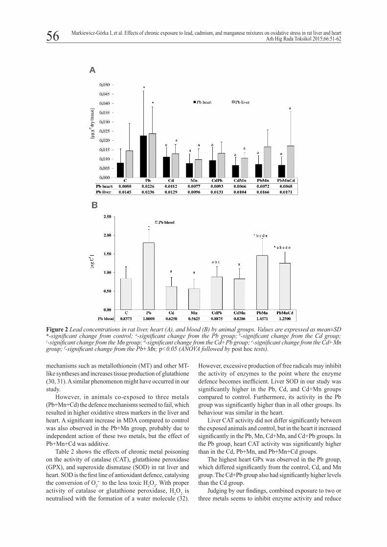
Our blood Mn findings (6.2-6.7 µg L-1) (Figure 3B) were lower than in humans exposed to manganese at the workplace (range 3.9-25.8 µg L-1; mean ±SD 10.7±4.3 µg L-1) but comparable to the general population levels (range: 2.6-15.1 µg L-1; mean±SD 7.5±2.7 µg L-1) (28).
Unlike Cd and Pb, heart and liver Mn levels (Figure 3A) were comparable across all groups, including control, which suggests that long-term exposure did not have a significant effect on Mn accumulation in these organs. This can be explained by the fact that Mn has effective homeostatic mechanisms regulating its absorption, disposition, and biliary excretion, seeking to maintain the optimal levels in the body (21).
In blood however, its accumulation peaked in the Pb+Mn group, differing significantly from the control and Pb group, which was expected, but also from the group receiving Mn alone or in combination with Cd. Also surprisingly, it did not differ significantly from the groups
Table 1 Mean body mass gain and consumption of water, lead, cadmium, and manganese by the groups of ratsGroups Control Pb Cd Mn Cd+Pb Cd+Mn Pb+Mn Pb+Mn+CdBody mass gain (g per rat during the experiment)
276.3 273.8 287.5 270.0 269.6 271.7 280.0 268.8
Consumption of water (mL kg-1 body mass per day)
131.78 136.58 132.67 132.69 138.45 138.79 133.30 131.70
Calculated metal consumption (mg kg -1 body mass per day)Pb 0.0273 0.0277 0.0267 0.0263Cd 0.1327 0.1384 0.1333 0.1317Mn 0.2654 0.2776 0.2666 0.2634
Markiewicz-Górka I, et al. Effects of chronic exposure to lead, cadmium, and manganese mixtures on oxidative stress in rat liver and heart Arh Hig Rada Toksikol 2015;66:51-62

55
receiving Cd alone or in combination with Pb. In the Cd+Mn+Pb group, Mn showed a similar pattern, except that it was not significantly higher than in control (Figure 3).
These results suggest that the presence of Cd, and especially of Pb, may increase the gastrointestinal absorption of Mn, but not its accumulation in the tissues.
Our result is consistent with the findings of Oulhote et al. (29), which indicate non-linear relationships between blood Mn levels and blood Cd or Pb. The relationship between blood Mn and Cd levels resembled an inverse U-shape with an inflexion point around Cd concentrations between 0.6 and 0.8 μg L-1. Blood Mn levels increased with higher blood Pb levels, with a sharper slope at Pb concentration of 2.5 μg dL-1.
Oxidative stress and antioxidant defence in rat liver and heart
Liver and heart MDA in the groups treated with single metals (Pb or Cd or Mn) did not differ significantly from control. However, combined treatment by three metals simultaneously (Pb+Mn+Cd) significantly increased MDA
levels in the liver compared to control and the Mn group and in the heart compared to control and the groups treated with single metals (Pb, Cd, and Mn) (Figure 4).
Weak intensification of oxidative process during chronic exposure to low doses of metals has also been observed by other authors. Fowler et al. (30) studied the effects of the lowest-observed effect levels (LOEL) of Cd, arsenic (As), and Pb in various combinations on the parameters of oxidative stress in rat kidneys. They observed dynamic changes that depended on exposure time and interactions between these elements. Oxidative stress was the highest after 30-day exposure and dropped after 90 days in the groups treated with single elements, but remained high in the combination groups. However, after 180 days it also dropped in the group exposed to Cd+As. Similarly, the concentration of non-protein SH groups (considered as an indicator of antioxidant defence activity) changed with exposure time. At first, it increased in all exposed groups, but after 90 days it dropped in some and increased in others. After 180 days it further increased in all exposed groups. Long exposure to metals triggers effective defence
Figure 1 Cadmium concentrations in rat liver, heart (A), and blood (B) by animal groups. Values are expressed as mean±SD*-significant change from control; a-significant change from the Pb group; b-significant change from the Cd group; c-significant change from the Mn group; d-significant change from the Cd+Pb group; e-significant change from the Cd+Mn group; f-significant change from the Pb+Mn; p<0.05 (ANOVA followed by post hoc tests)
Markiewicz-Górka I, et al. Effects of chronic exposure to lead, cadmium, and manganese mixtures on oxidative stress in rat liver and heart Arh Hig Rada Toksikol 2015;66:51-62
56
mechanisms such as metallothionein (MT) and other MT-like syntheses and increases tissue production of glutathione (30, 31). A similar phenomenon might have occurred in our study.
However, in animals co-exposed to three metals (Pb+Mn+Cd) the defence mechanisms seemed to fail, which resulted in higher oxidative stress markers in the liver and heart. A significant increase in MDA compared to control was also observed in the Pb+Mn group, probably due to independent action of these two metals, but the effect of Pb+Mn+Cd was additive.
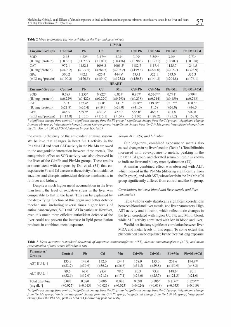
Table 2 shows the effects of chronic metal poisoning on the activity of catalase (CAT), glutathione peroxidase (GPX), and superoxide dismutase (SOD) in rat liver and heart. SOD is the first line of antioxidant defence, catalysing the conversion of O2
•– to the less toxic H2O2. With proper activity of catalase or glutathione peroxidase, H2O2 is neutralised with the formation of a water molecule (32).
However, excessive production of free radicals may inhibit the activity of enzymes to the point where the enzyme defence becomes inefficient. Liver SOD in our study was significantly higher in the Pb, Cd, and Cd+Mn groups compared to control. Furthermore, its activity in the Pb group was significantly higher than in all other groups. Its behaviour was similar in the heart.
Liver CAT activity did not differ significantly between the exposed animals and control, but in the heart it increased significantly in the Pb, Mn, Cd+Mn, and Cd+Pb groups. In the Pb group, heart CAT activity was significantly higher than in the Cd, Pb+Mn, and Pb+Mn+Cd groups.
The highest heart GPx was observed in the Pb group, which differed significantly from the control, Cd, and Mn group. The Cd+Pb group also had significantly higher levels than the Cd group.
Judging by our findings, combined exposure to two or three metals seems to inhibit enzyme activity and reduce
Figure 2 Lead concentrations in rat liver, heart (A), and blood (B) by animal groups. Values are expressed as mean±SD*-significant change from control; a-significant change from the Pb group; b-significant change from the Cd group; c-significant change from the Mn group; d-significant change from the Cd+Pb group; e-significant change from the Cd+Mn group; f-significant change from the Pb+Mn; p<0.05 (ANOVA followed by post hoc tests).
Markiewicz-Górka I, et al. Effects of chronic exposure to lead, cadmium, and manganese mixtures on oxidative stress in rat liver and heart Arh Hig Rada Toksikol 2015;66:51-62
57
the overall efficiency of the antioxidant enzyme system. We believe that changes in heart SOD activity in the Pb+Mn+Cd and heart CAT activity in the Pb+Mn are owed to the antagonistic interaction between these metals. The antagonistic effect on SOD activity was also observed in the liver of the Cd+Pb and Pb+Mn groups. These results are consistent with a report by Dai et al. (31) that co-exposure to Pb and Cd decreases the activity of antioxidative enzymes and disrupts antioxidant defence mechanisms in rat liver and kidney.
Despite a much higher metal accumulation in the liver than heart, the level of oxidative stress in the liver was comparable to that in the heart. This can be explained by the detoxifying function of this organ and better defence mechanisms, including several times higher levels of antioxidant enzymes, SOD and CAT in particular. However, even this much more efficient antioxidant defence of the liver could not prevent the increase in lipid peroxidation products in combined metal exposure.
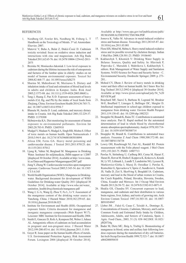
Serum ALT, AST, and bilirubin
Our long-term, combined exposure to metals also caused changes in rat liver function (Table 3). Total bilirubin increased with co-exposure to metals, peaking in the Pb+Mn+Cd group, and elevated serum bilirubin is known to indicate liver and biliary tract dysfunction (33).
A similar combined effect was observed with ALT, which peaked in the Pb+Mn (differing significantly from the Pb group), and with AST, whose levels in the Pb+Mn+Cd group significantly differed from control and the Pb group.
Correlations between blood and liver metals and liver parameters
Table 4 shows only statistically significant correlations between blood and liver metals, and liver parameters. High AST activity and bilirubin, which reflect toxic changes in the liver, correlated with higher Cd, Pb, and Mn in blood, while ALT activity correlated with Mn in blood and liver.
We did not find any significant correlation between liver MDA and metal levels in this organ. To some extent this phenomenon can be explained by the fact that long exposure
Table 2 Mean antioxidant enzyme activities in the liver and heart of ratsLIVER
Enzyme/ Groups Control Pb Cd Mn Cd+Pb Cd+Mn Pb+Mn Pb+Mn+Cd
SOD(IU mg-1 protein)
2.45(±0.361)
4.22*(±1.277)
3.47*a
(±1.001)3.31a
(±0.476)3.09a
(±0.988)3.55*a
(±1.231)3.04a
(±0.707)2.73a
(±0.388)CAT(IU mg-1protein)
972.1(±476.2)
1152.1 (±177.5)
1098.3(±284.5)
1001.5b
(±205.2)1182.7
(±159.6)1117.4
(±224.0)1125.7
(±202.7)1244.5
(±323.9)GPx(mIU mg-1protein)
500.2(±100.2)
492.1(±170.5)
625.4(±154.0)
444.8b
(±125.8)555.1
(±150.5)522.1
(±144.3)543.0
(±204.8)535.3
(±176.1)HEART
Enzyme/ Groups Control Pb Cd Mn Cd+Pb Cd+Mn Pb+Mn Pb+Mn+CdSOD(IU mg-1 protein)
0.685(±0.229)
1.255*(±0.422)
0.822a
(±0.220)0.834a
(±0.293)0.807a
(±0.238)0.526abcd
(±0.214)0.741a
(±0.159)0.794a
(±0.248)CAT(IU mg-1protein)
77.3(±21.8)
132.4* (±26.4)
88.0a
(±19.9)114.1*(±29.0)
128.8*b
(±41.0)119.0*b
31.5)73.1acde
(±26.0)100.5a
(±36.5GPx(mIU mg-1protein)
445.5(±113.8)
589.9*(±135)
436.3a
(±115.1)427.0a
(±154)585.0b
(±130)468.7
(±199.2)463.8
(±85.2)502.8
(±158.8)*-significant change from control; a-significant change from the Pb group, b-significant change from the Cd group; c-significant change from the Mn group; d-significant change from the Cd+Pb group; e-significant change from the Cd+Mn group; f-significant change from the Pb+Mn; p<0.05 (ANOVA followed by post hoc tests)
Table 3 Mean activities (±standard deviation) of aspartate aminotransferase (AST), alanine aminotransferase (ALT), and mean concentration of total serum bilirubin in rats
Parameter/Groups Control Pb Cd Mn Cd+Pb Cd+Mn Pb+Mn Pb+Mn+Cd
AST [IU L-1] 135.9(±23.7)
149.0(±39.9)
152.0(±36.2)
154.5(±36.6)
178.9(±54.5)
153.0(±29.8)
253.6(±150.9)
194.9*a
(±48.3)
ALT [IU L-1] 89.6(±32.9)
62.0(±12.0)
88.4(±21.3)
78.6(±17.1)
90.3(±24.6)
73.9(±25.7)
148.6a (±121.3)
80.1(±21.0)
Total bilirubin [mg dL-1]
0.083 (±0.027)
0.080 (±0.013)
0.086 (±0.023)
0.076 (±0.023)
0.098 (±0.024)
0.106ac (±0.018)
0.114abc (±0.033)
0.120*abc (±0.019)
*-significant change from control; a-significant change from the Pb group; b-significant change from the Cd group; c-significant change from the Mn group; d-indicate significant change from the Cd+Pb group; e-significant change from the Cd+Mn group; f-significant change from the Pb+Mn; p<0.05 (ANOVA followed by post hoc tests).
Markiewicz-Górka I, et al. Effects of chronic exposure to lead, cadmium, and manganese mixtures on oxidative stress in rat liver and heart Arh Hig Rada Toksikol 2015;66:51-62

58
to metals and their accumulation in the liver trigger effective defence mechanisms, which inhibit oxidative processes such as the synthesis of metallothionein (MT) and increase tissue production and accumulation of glutathione (34).
However, we did observe a positive correlation between blood Cd (reflecting current exposure) and liver MDA. It suggests that oxidation takes place immediately after exposure and before defence mechanisms kick in (34). This correlation also confirms the potent prooxidant properties of Cd, even at low environmental concentrations.
Correlations between blood and heart metals and heart parameters
Table 5 shows statistically significant correlations between heart and blood metals and heart parameters. Heart
MDA correlated positively with all three metals in blood, and negatively with heart Mn.
These results confirm that oxidation is dose-dependent. Metal concentrations in the blood correspond to current exposure. At low doses, their contribution to heart damage (similar to liver) is mainly related to current exposure.
Mn is an essential element required for the functioning of many enzymes and is involved in anti-oxidative mechanisms (10-12, 21). Heart is the organ of intense aerobic metabolism and high mitochondrial content, and higher Mn levels may improve free radical scavenging and protection against oxidative stress. The protective effect of Mn against Cd toxicity has already been observed by Eybl and Kotyzová (35). In their study, Mn lowered Cd concentrations in the kidneys and testicles, but increased them in the liver. Furthermore, Mn alleviated CAT inhibition
Figure 3 Manganese concentrations in rat liver, heart (A), and blood (B) by animal groups. Values are expressed as mean±SD*-significant change from control; a-significant change from the Pb group; b-significant change from the Cd group; c-significant change from the Mn group; d-significant change from the Cd+Pb group; e-significant change from the Cd+Mn group; f-significant change from the Pb+Mn; p<0.05 (ANOVA followed by post hoc tests)
Markiewicz-Górka I, et al. Effects of chronic exposure to lead, cadmium, and manganese mixtures on oxidative stress in rat liver and heart Arh Hig Rada Toksikol 2015;66:51-62

59
by Cd and completely prevented the decrease in glutathione levels, glutathione peroxidase inhibition, and oxidative stress in the liver. The authors believe that Mn induced metallothionein production and improved antioxidant status in the liver tissue.
In our study, Mn slightly improved protection against Cd toxicity. This is indicated by the inverse relationship between heart Mn and MDA levels and minor changes in
lipid peroxidation in both the heart and the liver of animals from the Cd+Mn group. Moreover, this group showed an antagonism between Mn and Cd in ALT activity.
Pb in blood correlated negatively with heart Mn. However, no protective effect was observed in the groups receiving Pb together with the Mn (Pb+Mn and Pb+Mn+Cd), as the cardiac MDA concentration remained significantly higher than in controls.
Table 4 Correlations between blood metals, liver Mn, and liver parameters
Cd blood Pb blood Mn liver Mn blood
MDA liver r=0.271* ns ns ns
GPX liver ns ns r=-0.387** ns
AST serum r=0.302** r=0.278* ns r=0.322*
ALT serum ns ns r=0.340** r=0.340**
bilirubine serum r=0.313* r=0.314* ns r=0.618**r–coefficient of correlation(*p<0.05, **p<0.01)ns–not significant
Table 5 Correlations between blood metals, heart Mn, and heart parameters
Cd blood Pb blood Mn heart Mn blood
MDA heart r=0.259* r=0.321* r=-0.299* r=0.348**
GPX heart ns r= 0.320* ns ns
r–coefficient of correlation (*p<0.05, **p<0.01)ns–not significant
Table 6 The effects of metal interactions on liver and heart biochemistry
SOD heart Catalase heart
TBARS heart SOD liver ALT serum Bilirubin
serumANOVA/MANOVA analysis of interactive effect of Pb and Cd and Mn
Main effect of Pb F=7.288** ns F=9.063** ns ns ns
Main effect of Cd F=4.358* ns ns ns ns ns
Main effect of Mn F=6.198* ns F=17.650** ns ns nsInteractive effect of Pb and Cd ns ns ns F=9.284** ns ns
Interactive effect of Pb and Mn ns F=26.324** ns F=7.853** F=4.7846* F=5.189*
Interactive effect of Cd and Mn ns ns ns ns F=4.622* ns
Interactive effect of Pb and Cd and Mn F=12.256** ns F=9.698** ns ns ns
Character of interaction between Pb and Cd ns ns ns Antagonism ns ns
Character of interaction between Pb and Mn ns Antagonism ns Antagonism Synergism Synergism
Character of interaction between Cd and Mn ns ns ns ns Antagonism
Character of interaction between Pb, Cd, and Mn Antagonism ns Synergism ns ns ns
F–the coefficient calculated with ANOVA/MANOVA*p<0.05, **p<0.01, ns–not significant
Markiewicz-Górka I, et al. Effects of chronic exposure to lead, cadmium, and manganese mixtures on oxidative stress in rat liver and heart Arh Hig Rada Toksikol 2015;66:51-62

60
Metal interactions and their effect on liver and heart biochemistry
Table 6 shows the statistically significant effects of metal interactions on biochemical parameters. Changes in SOD activity in the heart of rats exposed to Pb+Mn+Cd and changes in heart CAT activity in the Pb+Mn group seem to reflect an antagonism between these metals. The antagonistic effect on SOD activity was also observed in the liver of rats from the Cd+Pb and Pb+Mn groups.
Heart MDA in the Pb+Mn group points to an additive effect of these two metals. However, Pb+Mn+Cd exhibited a synergistic effect on the increase in heart MDA.
The change in liver function indicators such as serum alanine aminotransferase (ALT) activity and total bilirubin concentration after combined exposure to Pb+Mn was caused by a synergistic action of these two metals. However, in rats exposed to Cd+Mn, the two metals showed antagonism.
CONCLUSION
Our experiment constitutes a new approach to the study of biological effects associated with environmental exposure to heavy metals. Our goal was to identify antioxidant defence disorders and the oxidative processes in the heart and liver caused by prolonged combined exposure to very low environmental doses of Cd, Pb, and Mn and to establish the effects of interaction between these metals on oxidation processes in these tissues and liver function indicators.
Figure 4 Malonylodialdehyde concentrations in rat liver and heart by animal groups. Values are expressed as mean±SD*-significant change from control; a-significant change from the Pb group; b-significant change from the Cd group; c-significant change from the Mn group; d-significant change from the Cd+Pb group; e-significant change from the Cd+Mn group; f-significant change from the Pb+Mn; p<0.05 (ANOVA followed by post hoc tests)
Our study has confirmed that Pb, Cd, and Mn interact in environmental conditions. This interaction is mostly antagonistic if we consider the effects on antioxidative enzymes, but their oxidative stress effects in the heart and liver are mostly synergistic.
Moreover, we observed a synergistic effect between Pb and Mn on ALT increase in serum, which is an indicator of liver dysfunction.
As our study uses an animal experimental model, its results are limited in terms of extrapolation to humans. Even so, our results are very important, as they help to better understand how low environmental doses of Cd, Pb, and Mn interact in their biological effects on tissues.
Prolonged combined exposure to low-dose metals is present in many populations across the world and may contribute to the development of various diseases. Therefore, it is very important to continue and expand this kind of research, particularly of low-dose metal interaction and the resulting distribution and accumulation in the body and the weakening of the immune system and organ function disorders.
Conflicts of interest
The authors declare no conflict of interest.
Acknowledgements
This study was supported by the fund of statutory research of Wroclaw Medical University (project No. ST-453).
Markiewicz-Górka I, et al. Effects of chronic exposure to lead, cadmium, and manganese mixtures on oxidative stress in rat liver and heart Arh Hig Rada Toksikol 2015;66:51-62
61
REFERENCES
1. Nordberg GF, Fowler BA, Nordberg M, Friberg L. T Handbook on the Toxicology of Metals. 3rd ed. Amsterdam: Elsevier; 2007.
2. Matović V, Buha A, Bulat Z, Đukić-Ćosić D. Cadmium toxicity revisited: focus on oxidative stress induction and interactions with zinc and magnesium. Arh Hig Rada Toksikol 2011;62:65-76. doi: 10.2478/10004-1254-62-2011-2075
3. Brzóska M, Moniuszko-Jakoniuk J. Low-level exposure to cadmium during the lifetime increases the risk of osteoporosis and factures of the lumbar spine in elderly: studies on rat model of human environmental exposure. Toxicol Sci 2004;82:468-77. doi: 10.1093/toxsci/kfh275
4. Sharma M, Maheshwari M, Morisawa S. Dietary and inhalation intake of lead and estimation of blood lead levels in adults and children in Kanpur, India. Risk Anal 2005;2:1573-88. doi: 10.1111/j.1539-6924.2005.00683.x
5. Tang J, Huang Z, Pan X-D. Exposure assessment of heavy metals (Cd, Hg, and Pb) by the intake of local foods from Zhejiang, China. Environ Geochem Health 2014;36:765-71. doi: 10.1007/s10653-014-9594-7
6. Blanuša M, Jureša D. Lead, cadmium, and mercury dietary intake in Croatia. Arh Hig Rada Toksikol 2001;52:229-37. PMID: 11370308
7. Skibniewska KA. Diet monitoring for assessment of human exposure to environmental pollutants. Environ Int 2003;28:703-9. PMID: 12605918
8. Mudgal V, Madaan N, Mudgal A, Singh RB, Mishra S. Effect of toxic metals on human health. Open Nutraceuticals J 2010;3:94-9. doi: 10.2174/1876396001003010094
9. Alissa EM, Ferns GA. Heavy metal poisoning and cardiovascular disease. J Toxicol 2011;2011:870125. doi: 10.1155/2011/87012
10. Ljung K, Vahter M, Berglund M. Manganese in Drinking Water. Institutet för miljömedicin, IMM-rapport nr 1/2007 [displayed 30 October 2014]. Available at http://www.imm.ki.se/Datavard/Rapporter/Manganrapport2007.pdf
11. Jiang Y, Zheng W. Cardiovascular toxicities upon manganese exposure. Cardiovasc Toxicol 2005;5:345-54. doi: 10.1385/CT:5:4:345
12. World Health Organization (WHO). Manganese in Drinking-water. Background document for development of WHO Guidelines for Drinking-water Quality 2011 [displayed 30 October 2014]. Available at http://www.who.int/water_sanitation_health/dwq/chemicals/manganese.pdf
13. Wang J, Li A, Wang Q, Zhou Y, Fu L, Li Y. Assessment of the manganese content of the drinking water source in Yancheng, China. J Hazard Mater 2010;182:259-65. doi: 10.1016/j.jhazmat.2010.06.023
14. Institute for Environment and Health (IEH). Occupational exposure limits: Criteria document for manganese and inorganic manganese compounds (Web Report W17). Leicester: MRC Institute for Environment and Health; 2004.
15. Smith E, Gancarz D, Rofe A, Kempson IM, Weber J, Juhasz AL. Antagonistic effects of cadmium on lead accumulation in pregnant and non-pregnant mice. J Hazard Mater 2012;199-200:453-6. doi: 10.1016/j.jhazmat.2011.11.016
16. Goyer R. Issue paper on the human health effects of metals. U.S. Environmental Protection Agency, Risk Assessment Forum. Lexington 2004 [displayed 30 October 2014].
Available at http://www.epa.gov/raf/publications/pdfs/HUMANHEALTHEFFECTS81904.PDF
17. Jomova K, Valko M. Advances in metal-induced oxidative stress and human disease. Toxicology 2011;283:65-87. doi: 10.1016/j.tox.2011.03.001
18. Flora SJS, Mittal M, Mehta A. Heavy metal induced oxidative stress and its possible reversal by chelation therapy. Indian J Med Res 2008;128:501-23. PMID: 19106443
19. Kukharchyk T, Khomích V: Drinking Water Supply in Belarus: Sources, Quality and Safety. In: Hlavinek P, Popovska C, Marsalek J, Mahrikova I, Kukharchyk T, editors. Risk Management of Water Supply and Sanitation Systems. NATO Science for Peace and Security Series – C. Environmental Security. Dordrecht: Springer; 2009. p. 273-281..
20. Mohod CV, Dhote J. Review of heavy metals in drinking water and their effect on human health. Int J Innov Res Sci Eng Technol 2013;2:2992-6 [displayed 30 October 2014]. Available at http://www.ijirset.com/upload/july/56_%20REVIEW.pdf
21. Bouchard MF, Sauvé S, Barbeau B, Legrand M, Brodeur M-È, Bouffard T, Limoges E, Bellinger DC, Mergler D. Intellectual impairment in school-age children exposed to manganese from drinking water. Environ Health Perspect 2011;119:138-43. doi: 10.1289/ehp.1002321
22. Stoeppler M, Brandt K, Rains TC. Contributions to automated trace analysis. Part II. Rapid method for the automated determination of lead in whole blood by electrothermal atomic-absorption spectrophotometry. Analyst 1978;103:714-22. doi: 10.1039/AN9780300714
23. Stoeppler M, Brandt K. Contributions to automated trace analysis. Fresenius Z Anal Chem 1980;300:372-80. doi: 10.1007/BF01154738
24. Lowry OH, Rosebrough NJ, Farr AL, Randall RJ. Protein measurement with the Folin phenol reagent. J Biol Chem 1951;193:265-75. PMID: 14907713
25. Pawlas N, Strömberg U, Carlberg BO, Cerna M, Harari F, Harari R, Horvat M, Hruba F, Koppova K, Krskova A, Krsnik M, Li YF, Löfmark L, Lundh T, Lundström NG, Lyoussi B, Markiewicz-Górka I, Mazej D, Osredkar J, Pawlas K, Rentschler G, Spevackova V, Spiric Z, Sundkvist A, Tratnik JS, Vadla D, Zizi S, Skerfving S, Bergdahl IA. Cadmium, mercury and lead in the blood of urban women in Croatia, the Czech Republic, Poland, Slovakia, Slovenia, Sweden, China, Ecuador and Morocco. Int J Occup Med Environ Health 2013;26:58-72. doi: 10.2478/S13382-013-0071-9
26. Shukla GS, Chandra SV. Concurrent exposure to lead, manganese, and cadmium and their distribution to various brain regions, liver, kidney, and testis of growing rats. Arch Environ Contam Toxicol 1987;16:303-10. doi: 10.1007/BF01054947
27. Llobet JM. , Falcó G, Casas C, Teixidó A., Domingo JL. Concentrations of Arsenic, Cadmium, Mercury, and Lead in Common Foods and Estimated Daily Intake by Children, Adolescents, Adults, and Seniors of Catalonia, Spain. J. Agric. Food Chem., 2003, 51 (3): 838–842.DOI: 10.1021/jf020734q
28. Bader M, Dietz MC, Ihrig A, Triebig G. Biomonitoring of manganese in blood, urine and axillary hair following low-dose exposure during the manufacture of dry cell batteries. Int Arch Occup Environ Health 1999;72:521-7. doi: 10.1007/s004200050410
Markiewicz-Górka I, et al. Effects of chronic exposure to lead, cadmium, and manganese mixtures on oxidative stress in rat liver and heart Arh Hig Rada Toksikol 2015;66:51-62
62
29. Oulhote Y, Mergler D, Bouchard MF. Sex- and age-differences in blood manganese levels in the U.S. general population: national health and nutrition examination survey 2011–2012 Environmental Health 2014;13:87. doi: 10.1186/1476-069X-13-87
30. Fowler BA, Whittaker MH, Lipsky M, Wang G, Chen X-Q. Oxidative stress induced by lead, cadmium and arsenic mixtures: 30-day, 90-day, and 180-day drinking water studies in rats: An overview. BioMetals 2004;17:567-8. doi: 10.1023/B:BIOM.0000045740.52182.9d
31. Dai S, Yin Z, Yuan G, Lu H, Jia R, Xu J, Song X, Li L, Shu Y, Liang X, He C, Lv C, Zhang W. Quantification of metallothionein on the liver and kidney of rats by subchronic lead and cadmium in combination. Environ Toxicol Pharmacol 2013;36:1207-16. doi: 10.1016/j.etap.2013.10.003
32. Harris ED. Regulation of antioxidant enzymes. FASEB J 1992;6:2675-83. PMID: 1612291
33. Mesa V. A., De Vos R. and Fevery J. Elevation of the serum bilirubin diconjugate fraction provides an early marker for cholestasis in the rat. J Hepatol 1991;27:912-6. doi: 10.1016/S0168-8278(97)80330-3
34. Tandon SK, Singh S, Prasad S, Mathur N. Hepatic and renal metallothionein induction by an oral equimolar dose of zinc, cadmium or mercury in mice. Food Chem Toxicol 2001;39:571-7 doi: 10.1016/S0278-6915(00)00167-8
35. Eybl V, Kotyzová D. Protective effect of manganese in cadmium-induced hepatic oxidative damage, changes in cadmium distribution and trace elements level in mice. Interdiscip Toxicol 2010;3:68-72. doi: 10.2478/v10102-010-0013-3
Utjecaj kronične istodobne izloženosti olovu, kadmiju i manganu na oksidativni stres u jetri i srcu štakora
Cilj je ovog istraživanja bio ocijeniti učinke kronične izloženosti kombinaciji Cd, Pb i Mn u niskim dozama kakve su u okolišu na oksidativni stres u jetrima i srcu štakora te na pokazatelje jetrene funkcije. U tu smo svrhu mužjake Wistar štakora nasumce podijelili u osam skupina, od kojih je kontrolna pila čistu vodu, a izložene skupine vodu pomiješanu s olovom (0,2 mg L-1), kadmijem (1 mg L-1) i manganom (2 mg L-1), zasebno ili u kombinacijama. Koncentracija malondialdehida (MDA) značajno se povećala u oba organa životinja izloženih kombinacijama metala. Srčani MDA korelirao je s razinama kadmija, olova i mangana u krvi, a jetreni MDA s razinom kadmija u krvi. Aktivnost aspartat aminotransferaza (AST) i koncentracije bilirubina također su bile značajno povišene u skupini izloženoj svim trima metalima te su korelirale s razinama kadmija, olova i mangana u krvi. Rezultati istraživanja potvrdili su da zajedno ova tri metala djeluju na povećanje MDA u srcu. Slična je sinergija zamijećena i između olova i mangana s obzirom na povećanje razine alanin aminotransferaze (ALT) u serumu, koji je pokazatelj jetrene funkcije.
KLJUČNE RIJEČI: alanin aminotransferaza; aspartat aminotransferaza; antioksidativni enzimi; bilirubin; kronično trovanje; teški metali
Markiewicz-Górka I, et al. Effects of chronic exposure to lead, cadmium, and manganese mixtures on oxidative stress in rat liver and heart Arh Hig Rada Toksikol 2015;66:51-62