effects of brief aquatic exercise in multiple sclerosis on
206
EFFECTS OF BRIEF AQUATIC EXERCISE IN MULTIPLE SCLEROSIS ON MOBILITY AND FUNCTION A dissertation submitted to the Kent State University College of Education, Health, and Human Services in partial fulfillment of the requirements for the degree of Doctor of Philosophy By Jennifer Lee Petersen December 2015
-
Upload
khangminh22 -
Category
Documents
-
view
1 -
download
0
Transcript of effects of brief aquatic exercise in multiple sclerosis on
EFFECTS OF BRIEF AQUATIC EXERCISE IN MULTIPLE SCLEROSIS ON
MOBILITY AND FUNCTION
A dissertation submitted to the
Kent State University College
of Education, Health, and Human Services
in partial fulfillment of the requirements
for the degree of Doctor of Philosophy
By
Jennifer Lee Petersen
December 2015
ii
A dissertation written by
Jennifer L. Petersen
B.S., University of Mount Union, 1999
M.S., Akron University 2001
Ph.D., Kent State University, 2015
Approved by
________________________, Director, Doctoral Dissertation Committee
Angela L. Ridgel
________________________, Member, Doctoral Dissertation Committee
John McDaniel
________________________, Outside Member, Doctoral Dissertation
Mary Beth Spitznagel
Accepted by
________________________, Director, School of Health Sciences
Lynne E. Rowan
________________________, Interim Dean, College of Education, Health and Human
Mark Kretovics Services
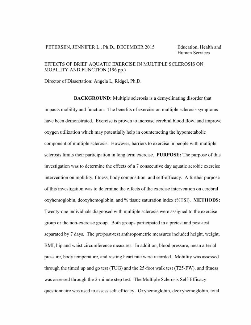
PETERSEN, JENNIFER L., Ph.D., DECEMBER 2015 Education, Health and
Human Services
EFFECTS OF BRIEF AQUATIC EXERCISE IN MULTIPLE SCLEROSIS ON
MOBILITY AND FUNCTION (196 pp.)
Director of Dissertation: Angela L. Ridgel, Ph.D.
BACKGROUND: Multiple sclerosis is a demyelinating disorder that
impacts mobility and function. The benefits of exercise on multiple sclerosis symptoms
have been demonstrated. Exercise is proven to increase cerebral blood flow, and improve
oxygen utilization which may potentially help in counteracting the hypometabolic
component of multiple sclerosis. However, barriers to exercise in people with multiple
sclerosis limits their participation in long term exercise. PURPOSE: The purpose of this
investigation was to determine the effects of a 7 consecutive day aquatic aerobic exercise
intervention on mobility, fitness, body composition, and self-efficacy. A further purpose
of this investigation was to determine the effects of the exercise intervention on cerebral
oxyhemoglobin, deoxyhemoglobin, and % tissue saturation index (%TSI). METHODS:
Twenty-one individuals diagnosed with multiple sclerosis were assigned to the exercise
group or the non-exercise group. Both groups participated in a pretest and post-test
separated by 7 days. The pre/post-test anthropometric measures included height, weight,
BMI, hip and waist circumference measures. In addition, blood pressure, mean arterial
pressure, body temperature, and resting heart rate were recorded. Mobility was assessed
through the timed up and go test (TUG) and the 25-foot walk test (T25-FW), and fitness
was assessed through the 2-minute step test. The Multiple Sclerosis Self-Efficacy
questionnaire was used to assess self-efficacy. Oxyhemoglobin, deoxyhemoglobin, total
hemoglobin and % TSI were measured using Near-Infrared Spectroscopy (NIRS). The
non-exercise group was asked to maintain their lifestyle during the 7 days between the
pretest and post-test, while the exercise group participated in 1 hour of aquatic aerobic
exercise for each of the 7 days between the pretest and post-test. Change scores were
calculated for each variable of the pretest and post-test and the non-exercise and exercise
groups were compared using independent samples t-tests for each of these scores.
RESULTS: Although there were no statistically significant differences between the
exercise and non-exercise groups for mobility, fitness, body composition, self-efficacy, or
cerebral oxygenation, there were small clinically meaningful improvements. In mobility
(T25-FW), 8/11 exercise participants improved while only 1/9 in the non-exercise group
showed improvement. Similar small improvements were demonstrated in the two minute
step test used to assess fitness. Cerebral oxygenation also showed improved blood flow
through improved oxyhemoglobin and total hemoglobin and improved oxygen utilization
through the deoxyhemoglobin response. CONCLUSION: Seven consecutive days of
moderate intensity exercise can produce small clinically meaningful changes through
improved mobility, fitness, self-efficacy and cerebral oxygen utilization.
iii
ACKNOWLEDGEMENTS
I would like to thank my husband Dean, and daughters Hannah and Deana for
enduring this challenge with me, and for their support and encouragement. Thanks to
the grandparents for endlessly helping with the girls, and family and friends who offered
support in a multitude of ways.
A special thanks to Dr. Ridgel, who has been a wonderful mentor, always willing
to help with unending patience and an expectation of excellence, a dissertation director
that I am most appreciative of. To the Kent State Exercise Physiology faculty for
guidance and teaching: Dr. Barkley a wonderful advisor and instructor, Dr. Glickman for
providing challenges and opportunity, Dr. McDaniel for learning in lab, and Dr. Kingsley
for guidance and instruction in HRV.
Thank you to Dr. Spitznagel and Dr. Arnold for time and guidance through this
project. Finally, thank you to Dayana, Brandon, and Hayden who formed the MS
research team to accomplish this task.
iv
TABLE OF CONTENTS
Page ACKNOWLEDGMENTS ............................................................................................................. iii
LIST OF FIGURES ....................................................................................................................... vi
LIST OF TABLES ....................................................................................................................... viii
CHAPTER
I. INTRODUCTION ....................................................................................................1
Background .............................................................................................................1
Rationale .................................................................................................................3
Objective .................................................................................................................4
II. REVIEW OF LITERATURE .................................................................................7
Prevalence and Risk Factors for MS .......................................................................7
Signs and Symptoms of MS ....................................................................................8
MS Pathology ........................................................................................................27
Types of MS ..........................................................................................................34
Standard Treatment of MS ....................................................................................37
Symptomatic Therapy ...........................................................................................41
Alternative Therapies for MS ...............................................................................52
Exercise and MS ...................................................................................................55
Summary ................................................................................................................70
III. METHODLOGY .................................................................................................72
Recruitment ...........................................................................................................72
Protocol .................................................................................................................76
Exercise and Physiological Variables ...................................................................77
Statistical Analysis ................................................................................................89
IV. EFFECTS OF A 7 DAY AQUATIC AEROBIC EXERCISE INTERVENTION
ON MOBILITY, FITNESS AND BODY COMPOSITION .....................................91
Introduction ...........................................................................................................91
Methods .................................................................................................................94
Results .................................................................................................................100
Discussion ...........................................................................................................115
Limitations and Future Direction ........................................................................128
v
V. THE EFFECTS OF AEROBIC EXERCISE ON CEREBRAL.OXYGENATION
IN MULTIPLE SCLEROSIS USING NEAR INFRA-RED SPECTROSCOPY ...130
Introduction .........................................................................................................130
Methods ...............................................................................................................134
Results .................................................................................................................139
Discussion ...........................................................................................................146
Limitations and Future Direction ........................................................................151
VI. SUMMARY ......................................................................................................152
APPENDICES .........................................................................................................155
APPENDIX A. INFORMED CONSENT FORM .............................................156
APPENDIX B. PRE-PARTICIPATION PHONE SCREENING ......................162
APPENDIX C. PHYSICIAN CLEARANCE FORM ........................................165
APPENDIX D. DATA SHEETS ........................................................................167
APPENDIX E. HEART RATE AND RPE ........................................................175
REFERENCES ........................................................................................................177
vi
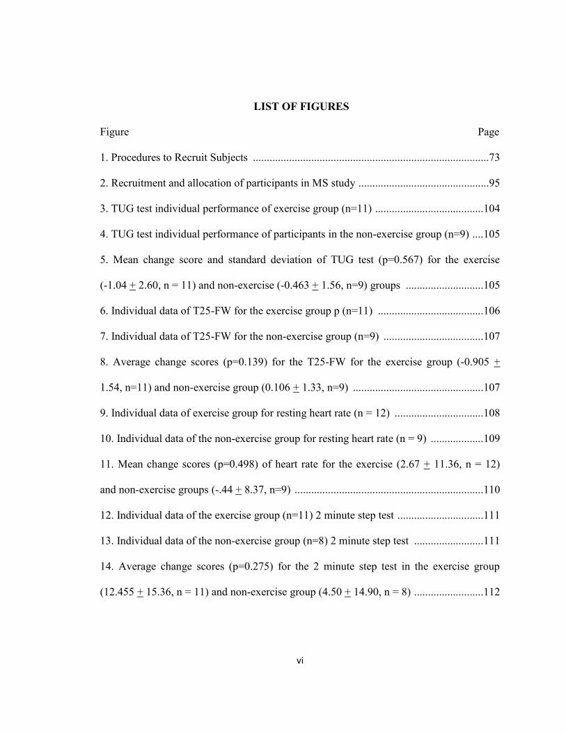
LIST OF FIGURES
Figure Page
1. Procedures to Recruit Subjects .....................................................................................73
2. Recruitment and allocation of participants in MS study ...............................................95
3. TUG test individual performance of exercise group (n=11) .......................................104
4. TUG test individual performance of participants in the non-exercise group (n=9) ....105
5. Mean change score and standard deviation of TUG test (p=0.567) for the exercise
(-1.04 + 2.60, n = 11) and non-exercise (-0.463 + 1.56, n=9) groups ............................105
6. Individual data of T25-FW for the exercise group p (n=11) ......................................106
7. Individual data of T25-FW for the non-exercise group (n=9) ....................................107
8. Average change scores (p=0.139) for the T25-FW for the exercise group (-0.905 +
1.54, n=11) and non-exercise group (0.106 + 1.33, n=9) ...............................................107
9. Individual data of exercise group for resting heart rate (n = 12) ................................108
10. Individual data of the non-exercise group for resting heart rate (n = 9) ...................109
11. Mean change scores (p=0.498) of heart rate for the exercise (2.67 + 11.36, n = 12)
and non-exercise groups (-.44 + 8.37, n=9) ....................................................................110
12. Individual data of the exercise group (n=11) 2 minute step test ...............................111
13. Individual data of the non-exercise group (n=8) 2 minute step test .........................111
14. Average change scores (p=0.275) for the 2 minute step test in the exercise group
(12.455 + 15.36, n = 11) and non-exercise group (4.50 + 14.90, n = 8) .........................112
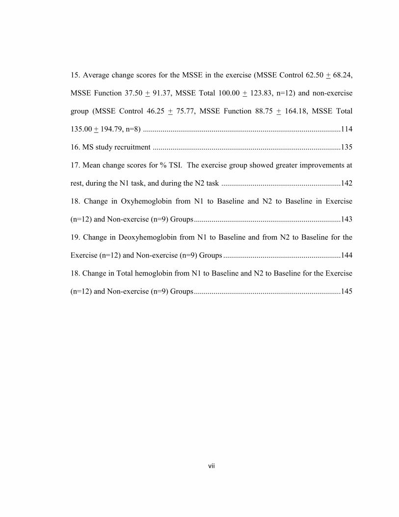
vii
15. Average change scores for the MSSE in the exercise (MSSE Control 62.50 + 68.24,
MSSE Function 37.50 + 91.37, MSSE Total 100.00 + 123.83, n=12) and non-exercise
group (MSSE Control 46.25 + 75.77, MSSE Function 88.75 + 164.18, MSSE Total
135.00 + 194.79, n=8) .....................................................................................................114
16. MS study recruitment ................................................................................................135
17. Mean change scores for % TSI. The exercise group showed greater improvements at
rest, during the N1 task, and during the N2 task .............................................................142
18. Change in Oxyhemoglobin from N1 to Baseline and N2 to Baseline in Exercise
(n=12) and Non-exercise (n=9) Groups ...........................................................................143
19. Change in Deoxyhemoglobin from N1 to Baseline and from N2 to Baseline for the
Exercise (n=12) and Non-exercise (n=9) Groups ............................................................144
18. Change in Total hemoglobin from N1 to Baseline and N2 to Baseline for the Exercise
(n=12) and Non-exercise (n=9) Groups ...........................................................................145
viii
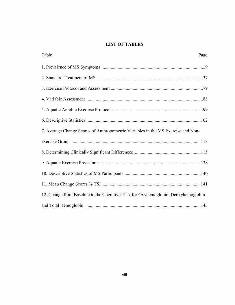
LIST OF TABLES
Table Page
1. Prevalence of MS Symptoms ..........................................................................................9
2. Standard Treatment of MS ............................................................................................37
3. Exercise Protocol and Assessment ................................................................................79
4. Variable Assessment .....................................................................................................88
5. Aquatic Aerobic Exercise Protocol ...............................................................................99
6. Descriptive Statistics ....................................................................................................102
7. Average Change Scores of Anthropometric Variables in the MS Exercise and Non-
exercise Group ................................................................................................................113
8. Determining Clinically Significant Differences .........................................................115
9. Aquatic Exercise Procedure ........................................................................................138
10. Descriptive Statistics of MS Participants ..................................................................140
11. Mean Change Scores % TSI .....................................................................................141
12. Change from Baseline to the Cognitive Task for Oxyhemoglobin, Deoxyhemoglobin
and Total Hemoglobin ....................................................................................................143
1
CHAPTER 1
INTRODUCTION
Background
Multiple sclerosis (MS) is the leading cause of non-traumatic neurologic disability
in young adults (Solari et al., 1999). The prevalence rate in the United States is 1/1000
and 2.1 million individuals suffer from multiple sclerosis worldwide (Marck and
colleagues, 2014; Dibble, Lopez-Lennon, Lake, Hoffmeister, & Gappmaier, 2013). MS
is caused by demyelination of axons, focal plaque formation, and inflammation in the
central nervous system which results in progressive loss of function. In addition, MS has
hypometabolic pathology consisting of decreased oxygen utilization and decreased
absolute cerebral blood flow (Sun, Tanaka, Kondo, Okamoto, & Hirai, 1998; Brooks, et
al., 1984). The location of pathology results in varying combinations of symptoms which
include fatigue, heat sensitivity, muscle spasms, gait problems and ataxia, dizziness and
vertigo, pain, cognitive problems, visual complaints, and bowel and bladder dysfunction
(Anthony, Jouria, & Houtman, 2014).
Forms of MS are characterized by specific onset and symptoms of the disease
process. Progressive forms of MS display continual worsening of symptoms, whereas
relapsing-remitting (RRMS) forms of MS exhibit exacerbations of symptoms followed by
resolution. The symptoms occur when demyelination and inflammation prevent the
axons from conducting impulses to and from the central nervous system to the periphery.
For the first few demyelinating events, the central nervous system is able to remyelinate
2
the axons, restoring conduction and the symptoms subside (Brown, Narayanan, &
Arnold, 2014). However, as the disease progresses, the ability of the central nervous
system to compensate diminishes. Although there is no cure for MS, pharmacological
and rehabilitative treatments aim to slow the progression of the disease and prevent
relapses, and to treat the symptoms.
Compounding the impact of the disease, symptoms resulting from MS pathology
are correlated with decreased levels of physical activity (Hayes, Gappmaier, & LaStayo,
2011; Marck et al., 2014) and subsequent deconditioning that reduces quality of life
(Doring, Pfueller, Paul, & Dorr, 2012). Further fallout from decreased physical activity
includes diminished leisure activities, social contacts, activities of daily living, as well as
lowered self-efficacy (Reitberg, Brooks, Uitdehagg, & Kwakkel, 2011). Physical and
cognitive impairments strongly influence the level of independence in the MS individual
(Langdon & Thompson, 1999), and physical activity can be used as a goal oriented,
multidisciplinary approach to improve function (Rietberg, van Wegen, Kollen, &
Kwakkel, 2014).
The primary aim of rehabilitation is to increase activity levels as well as the
independence of MS individuals (Langdon & Thompson, 1999), through success in
decreasing fatigue (Asano & Finlayson, 2014), improving cognition (Briken et al., 2014;
Prakash, Snook, Motl, & Kramer, 2010), improving mobility (Pilutti, et al., 2011;
Kargarfard, Etemadifar, Baker, Mehrabi, & Hayatbakhsh, 2012), improving physical
3
functioning and easing psychosocial burden (White, Castellano, McCoy, Patel, &
Giacobbi, 2012).
Rationale
It was once thought that exercise exacerbated MS symptoms and was
contraindicated in this population (Hayes, Gappmaier, & LaStayo, 2011). As exercise
increases body temperature, central pathways become blocked, producing temporary
physical and cognitive symptoms (Davis, Wilson, White, & Frohman, 2010). Worsening
symptoms depend on location of the demyelination and lesions, and frequently include
deficits in mobility, memory retrieval, processing speed, multitasking, and increased
fatigue (Davis, Wilson, White, & Frohman, 2010). However, current research supports
the positive effects of exercise in MS treatment, including improved fitness benefits,
feelings of well-being, strength and safety of mobility and decreased fatigue (Davis,
Wilson, White, & Frohman, 2010, Rietberg, Brooks, Uitdehagg, & Kwakkel, 2011).
Adverse effects during MS exercise were slightly higher when compared to non-
exercising individuals (Pilutti, Platta, Motl, & Latimer-Cheung, 2014). However, these
adverse effects were not greater than in healthy individuals who exercised (Pilutti, Platta,
Motl, & Latimer-Cheung, 2014). Exercise training is associated with a slight decrease in
the risk of relapse when compared to non-exercising MS individuals (Pilutti, Platta, Motl,
& Latimer-Cheung, 2014). Exercise also has the capacity to slow disease progression,
improves the physiological profile of MS and is well tolerated with a low occurrence of
adverse effects (Kargarfard, Etemadifar, Baker, Mehrabi, & Hayatbakhsh, 2012).
4
Current research has established the hypometabolic impact of MS (Fan et al.,
2015; Sun, Tanaka, Kondo, Okamoto, & Hirai, 1998; Brooks et al, 1984; Kidd et al.
1999; Bakshi, Miletich, Kinkel, Emmet, & Kinkel, 1998), however few studies have used
Near Infrared Spectroscopy Systems (NIRS) to monitor cerebral oxygenation levels in
MS (Lintas, Molinari, Simonetti, Franzini, & Liboni, 2013). There are no studies to date
that have used NIRS to determine the impact of exercise on cerebral oxygenation in MS,
though exercise has the potential to increase oxygenation through increased blood flow
and improved oxygen utilization (Ide & Secher, 2000; Rooks, Thom, McCully, &
Dishman, 2010).
Several studies have demonstrated positive effects of exercise on gait parameters
and quality of life using 4 weeks of exercise training, however, no studies to date have
used 7 consecutive days of exercise (van den Berg et al., 2006; Mostert & Kesselring,
2002). Previous work done by Fedor (2014) produced positive results in fitness and
cognitive parameters in a similar aquatic aerobic seven consecutive day exercise protocol
in older adults. RRMS forms of MS may benefit from high intensity, short duration
exercise between relapses. However, the potential benefits of 7 consecutive days of
exercise on cardiovascular fitness, and symptoms including self-efficacy and fatigue in
MS are unknown.
Objective
MS individuals have been shown to have lower physical activity levels compared
to healthy individuals (Rietberg, van Wegen, Kollen, & Kwakkel, 2014). Decreased
5
activity levels lead to a cascade of decreased mobility, balance, quality of life, self-
efficacy, and deconditioning (Kuspinar, Anderson, Teng, Asano, & Mayo, 2010).
Exercise has been found to lower oxygen consumption and heart rate; increase oxygen
pulses at a given work rate and improve the aerobic system (Mostert & Kesselring, 2002).
Kargarfard and colleagues (2012) found exercise training programs are associated with
small clinically meaningful improvements in mobility of walking in MS individuals.
Determining the role of 7 days of consecutive aquatic exercise program will assist
medical professionals in optimally using exercise for treatment in MS. The objective of
this project is to quantify changes in mobility, cardiovascular fitness, and cerebral
oxygenation parameters in order to determine if seven consecutive days of aquatic
aerobic exercise can provide benefits of fitness and mobility in MS individuals.
In this study we will be testing the following hypotheses:
1. A seven day moderate to high intensity water aerobics program will improve
mobility, as measured by the Timed Up and Go Test (TUG), and Timed 25 Foot
Walk Test (T25FW) in individuals with MS.
2. A seven day moderate to high intensity water aerobics program will improve
cardiovascular fitness, as measured through resting heart rate and 2 Minute Step
Test in individuals with MS.
3. A seven day moderate to high intensity water aerobics program will improve
Cerebral Oxygenation as measured through Near Infrared Spectroscopy (NIRS) in
individuals with MS.
6
4. A seven day moderate to high intensity water aerobics program will improve self-
efficacy, as reported through the Multiple Sclerosis Self-Efficacy Scale in
individuals with MS.
5. A seven day moderate to high intensity water aerobics program will not alter body
composition, measured with Body Mass Index (BMI), Waist to Hip Ratio (WHR),
and waist in individuals with MS.
7
CHAPTER II
REVIEW OF LITERATURE
Prevalence and Risk Factors for MS
Multiple Sclerosis (MS) is an autoimmune, demyelinating disorder of the central
nervous system (Teusnissen, Dijkstra, & Polman, 2005). Worldwide, 1-2.5 million
individuals suffers from Multiple Sclerosis (Doring, Pfueller, Paul, & Dorr, 2012;
Anthony, Jouria, & Houtman, 2014). Multiple Sclerosis is the most common neurologic
disorder in young adults (Teusnissen, Dijkstra, & Polman, 2005). In the United States
there is a 0.1% instance in the general population (Anthony, Jouria, & Houtman, 2014)
with diagnosis typically between the ages of 20-50 years (Kargarfard, Etemadifar, Baker,
Mehrabi, & Hayatbakhsh, 2012).
There are multiple risk factors involved in MS. Women are 1.5 times more likely
to have Multiple Sclerosis than men (Teusnissen, Dijkstra, & Polman, 2005), and women
are twice as likely to be affected earlier in life (Kargarfard, Etemadifar, Baker, Mehrabi,
& Hayatbakhsh, 2012). Inactivation and imprinting of the X chromosome causing a
maternal parent-of-origin effect may be responsible for the increasing instance of females
diagnosed with MS compared to males (Huynh & Casaccia, 2013). There is a
geographical component to the disease with increased prevalence north of the equator, in
industrialized countries, and in areas with less sunlight exposure (vitamin D deficiency)
(Marrie, 2004; Ascherio & Munger, 2007b). Additional risk factors include smoking,
8
exposure to infections (with chronic latency) at a young age, and genetic predispositions
(Ascherio & Munger, 2007a; 2007b; “NINDS,” n.d.).
Prevalence varies between different ethnic groups, further suggesting a genetic
component (Hanwell & Banwell, 2011). Caucasians are twice as likely to be diagnosed
with MS as other ethnicities (Khan & Pallant, 2007). Genome-wide association studies
have identified specific nucleotide polymorphisms in genes that determine the genetic
risk of developing MS (Huynh & Casaccia, 2013). Environmental factors may modify
the epigenome and manifest as MS (Huynh & Casaccia, 2013). While there is no cure for
Multiple Sclerosis, the disease course does not alter life expectancy (Anthony, Jouria, &
Houtman, 2014).
Signs and Symptoms of MS
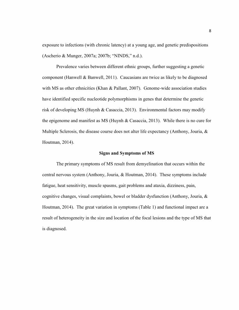
The primary symptoms of MS result from demyelination that occurs within the
central nervous system (Anthony, Jouria, & Houtman, 2014). These symptoms include
fatigue, heat sensitivity, muscle spasms, gait problems and ataxia, dizziness, pain,
cognitive changes, visual complaints, bowel or bladder dysfunction (Anthony, Jouria, &
Houtman, 2014). The great variation in symptoms (Table 1) and functional impact are a
result of heterogeneity in the size and location of the focal lesions and the type of MS that
is diagnosed.
9
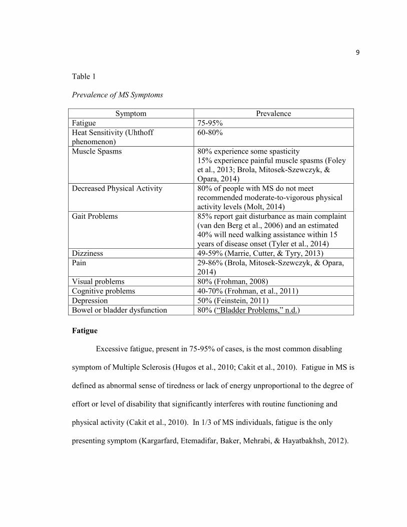
Table 1
Prevalence of MS Symptoms
Symptom Prevalence
Fatigue 75-95%
Heat Sensitivity (Uhthoff
phenomenon)
60-80%
Muscle Spasms 80% experience some spasticity
15% experience painful muscle spasms (Foley
et al., 2013; Brola, Mitosek-Szewczyk, &
Opara, 2014)
Decreased Physical Activity 80% of people with MS do not meet
recommended moderate-to-vigorous physical
activity levels (Molt, 2014)
Gait Problems 85% report gait disturbance as main complaint
(van den Berg et al., 2006) and an estimated
40% will need walking assistance within 15
years of disease onset (Tyler et al., 2014)
Dizziness 49-59% (Marrie, Cutter, & Tyry, 2013)
Pain 29-86% (Brola, Mitosek-Szewczyk, & Opara,
2014)
Visual problems 80% (Frohman, 2008)
Cognitive problems 40-70% (Frohman, et al., 2011)
Depression 50% (Feinstein, 2011)
Bowel or bladder dysfunction 80% (“Bladder Problems,” n.d.)
Fatigue
Excessive fatigue, present in 75-95% of cases, is the most common disabling
symptom of Multiple Sclerosis (Hugos et al., 2010; Cakit et al., 2010). Fatigue in MS is
defined as abnormal sense of tiredness or lack of energy unproportional to the degree of
effort or level of disability that significantly interferes with routine functioning and
physical activity (Cakit et al., 2010). In 1/3 of MS individuals, fatigue is the only
presenting symptom (Kargarfard, Etemadifar, Baker, Mehrabi, & Hayatbakhsh, 2012).
10
Fatigue and reduced exercise tolerance in MS is caused by central and peripheral
factors that are involved in pathogenesis (Rampello et al., 2007). Central factors that
cause fatigue include impaired voluntary drive of descending motor pathways, the
inability to sustain central drive to spinal motor neurons, metabolic abnormalities in the
frontal cortex and basal ganglia, and pro-inflammatory cytokines in the central nervous
system (Comi, Leocani, Rossi, & Colombo, 2001). Peripheral fatigue factors commonly
relate to deconditioning and include loss of force generating capacity in the muscle itself,
decreased potentials in the motor unit, metabolic changes in the muscle, lower maximal
voluntary force, lower muscle tension, longer relaxation time between repeated
contractions, and slowed enzymatic re-synthesis required for energy synthesis (Comi,
Leocani, Rossi, & Colombo, 2001).
Fatigue does not correlate with the degree of neurologic impairment or disability
in MS (Klefbeck & Hamrah, 2003), and is differentiated as primary or secondary fatigue.
Primary fatigue occurs as a result of the demyelination and axonal degeneration resulting
from the disease process of the neuromuscular system in Multiple Sclerosis (Herbert,
Corboy, Manago, & Schenkman, 2011). Secondary fatigue is caused by indirect factors
such as depression, physical inactivity, or sleep disorder (Herbert, Corboy, Manago, &
Schenkman, 2011). The presence of fatigue makes physical activity in this population
challenging. In addition, recent studies conflict in regards to the benefits of exercise
specifically on fatigue (Herbert, Corboy, Manago, & Schenkman, 2011; Yu, Billberg,
Dalgas, & Stenager, 2013).
11
Fatigue may be a result of elevated muscle tone, respiratory muscle weakness, or
the increased energy cost of ambulation found in MS individuals (Mostert & Kesselring,
2002). The inefficient gait patterns in MS reduce exercise tolerance and increase the
metabolic cost of walking (Olgiati, Jacquet, & Di Prampero, 1986). MS individuals were
found to have decreased limb endurance and impaired cardiorespiratory responses that
were likely linked to deconditioning, autonomic dysfunction and altered breathing control
(Chetta et al., 2004).
Yu and colleagues (2013) explored the relationship between primary fatigue and
autonomic dysfunction and found that during cognitive and physical tasks, people with
MS had a different autonomic response compared to healthy individuals. The vagus
nerve was implicated through measuring decreased vagal tone, abnormal heart rate
response, and abnormal heart rate variability (Yu, Billberg, Dalgas, & Stenager, 2013).
The authors concluded that primary fatigue was linked to autonomic dysfunction. It was
hypothesized that MS individuals mentally tire because the autonomic system fails to
adjust important body functions which eventually result in autonomic nervous system
failure (Yu, Billberg, Dalgas, & Stenager, 2013). In addition, body temperature is
controlled by autonomic regulation, and fatigued individuals may have an abnormal
response resulting in heat sensitivity, called the Uhthoff phenomenon (Yu, Billberg,
Dalgas, & Stenager, 2013).
Fatigue affects quality of life and general functioning, and often leads to a
downward spiral of inactivity and function. Diminished social relationships, affected
12
mental health, impaired activities of daily living and job loss are associated with fatigue
in MS (Kargarfard, Etemadifar, Baker, Mehrabi, & Hayatbakhsh, 2012).
Heat Sensitivity
In 1890, William Uhthoff first described a temporary worsening of vision with
exercise in individuals with optic neuritis (Bol et al., 2012). Later, symptoms were linked
to hyperthermia (Bol et al., 2012). Uhthoff Phenomenon is present in 60-80% of
individuals with MS (Anthony, Jouria, & Houtman, 2014). This phenomenon is caused
when nerve conduction becomes blocked to central pathways as core body temperature
increases (Davis et al., 2008; Humm et al., 2004; Frohman et al., 2013). The increased
temperature can affect propagation of action potentials and lengthen the refractory period,
resulting in a predisposition to conduction block (Davis, Wilson, White, & Frohman,
2010). Because of the demyelination in MS individuals, the blocked conduction occurs
at a lower temperature, resulting in both physical and cognitive symptoms including
fatigue, gait problems, decreased memory retrieval and information processing speed
(Anthony, Jouria, & Houtman, 2014; Davis 44). The increase in core temperature can be
caused by fever, exercise, or hot environment (Anthony, Jouria, & Houtman, 2014).
Several studies have examined possible mechanisms for the Uhthoff phenomenon
in MS. Bol and colleagues (2012) suggested that hyperthermia altered the level of serum
calcium resulting in a blockade of ion channels, circulatory changes, heat shock proteins
and unidentified humoral substances. In addition, increased body temperature in MS may
induce molecular changes in T-lymphocytes that exaggerate the immune response (Bol et
13
al., 2012). Lesions in the thalamus and hypothalamus may result in thermoregulatory
dysfunction (Bol et al., 2012; Davis, Wilson, White, & Frohman, 2010). Peripherally,
sweat gland function can be impaired in MS individuals, though it appears that neural
control of skin blood flow remains intact (Davis, Wilson, White, & Frohman, 2010).
Furthermore, the risk of overheating is amplified by the fact that individuals with MS
voluntarily restrict fluid consumption due to bladder dysfunction (Davis, Wilson, White,
& Frohman, 2010). This increased risk of hyperthermia and subsequent nerve
dysfunction has placed a negative connotation on exercise as treatment in the past, but
current MS research shows that exercise is a vital treatment component with
demonstrated benefits (Hayes, Gappmaier, & LaStayo, 2011).
Muscle Spasms
Muscle spasms are involuntary muscle contractions that clinically present as
feelings of muscle tightness or severe pain and uncontrollable contractions of the muscles
in the extremities (Walker, Hall, & Hurst, 1990). Spasticity is a result of an exaggerated
stretch reflex leading to increased muscle tone (Trompetto et al., 2014). The muscle
spindle is a receptor within the body of the muscle that is sensitive to lengthening or
stretching of the muscle (Walker, Hall, & Hurst, 1990). The stretch reflex is a
monosynaptic reflex that increases alpha motor neuron activity in response to the muscle
spindle being stretched, causing the muscle to contract (McArdle & Katch, 2010). The
contraction shortens the muscle, alleviating the stretch. The regulation of the muscle
14
length prevents over-stretching that could damage the muscle-tendon unit (McArdle &
Katch, 2010).
In MS, central nervous system lesions can damage upper motor neurons,
disrupting the balance between supraspinal inhibitory and excitatory inputs that lead to
the disinhibition of the stretch reflex (Trompetto et al., 2014). The corticobulbar fibers
are white matter tracts that originate in the cerebral cortex and send facilitatory impulses
to the brain stem and cranial nerves (Trompetto et al., 2014). The ventromedial reticular
formation of the brain stem receives these facilitatory impulses passing them to the dorsal
reticulospinal white matter tract (Trompetto et al., 2014). The dorsal reticulospinal tract
originates in the ventromedial reticular formation and passes inhibitory signals to the
muscle spindles (Trompetto et al., 2014). When this signal is disrupted due to
demyelination or lesion, inhibition does not occur and the muscle spindle becomes over
activated and can cause muscle spasms (Trompetto et al., 2014).
In MS, spasticity typically occurs in the legs, though it can occur in the upper
extremity and even the low back. Due to the disease process and symptoms, MS
individuals are often less active than the general population, which can increase the
prevalence of spasticity (Hayes, Gappmaier, & LaStayo, 2011; Trompetto et al., 2014).
Spasticity can also result in response to pain, infection, fever, and bowel distention
(Anthony, Jouria, & Houtman, 2014).
15
Decreased Physical Activity
Due to fatigue and overheating issues, MS individuals are generally less
physically active than healthy individuals of similar age (Hayes, Gappmaier, & LaStayo,
2011). Latimer-Cheung and colleagues (2013) conducted a meta-analysis that compared
physical activity patterns of Multiple Sclerosis individuals, those without MS and chronic
diseased people and concluded that MS individuals showed the lowest amount of
physical activity. Rietberg and colleagues (2014) found that MS individuals have
significantly lower dynamic activity compared to healthy individuals, starting with lower
activity levels in the morning, persisting into the afternoon and evening. This may stem
from adopting a lifestyle that anticipates energy conservation strategies including
reducing physical activity, decreased number of transitions (sitting to standing),
decreased dynamic movements (walking), and increased static movements (sitting and
lying) (Rietberg, van Wegen, Kollen, & Kwakkel, 2014; Comi, Leocani, Rossi, &
Colombo, 2001).
In studies controlled for gender, MS individuals on average were found to be in
the 10th
percentile for fitness (Bjarnadottir, Konradsdottir, Reynisdottir, & Olasfsson,
2007). Decreased activity levels lead to decreased mobility, balance and an increased
risk of falling (Hayes, Gappmaier, & LaStayo, 2011). Multiple Sclerosis symptoms,
along with lack of confidence in one’s abilities to manage the symptoms, can lead to
impaired functioning, decreased physical activity and compromised quality of life
(Doring, Pfueller, Paul, Dorr, 2012). The cycle of inactivity also appears to stem from
16
impairments on muscle function, sensation, coordination, balance, and stress from the
diagnosis leading to further inactivity, deconditioning and further disability (Kuspinar,
Anderson, Teng, Asano, & Mayo, 2010).
Less active lifestyles in MS individuals often coincide with diminished leisure
activities, social contacts, and normal activities of daily living which have all been found
to be important for self-esteem and psychological well-being (Reitberg, Brooks,
Uitdehagg, & Kwakkel, 2011). Self-efficacy is the belief in one’s capabilities to organize
and execute on the course of action required to manage a prospective situation (Hugos et
al., 2010). Higher levels of self-efficacy are beneficial in accomplishing tasks of daily
living. In addition, the primary aim of rehabilitation and treatment is to increase activity
levels and independence of MS individuals (Langdon & Thompson, 1999).
Gait Problems and Ataxia
Individuals with MS often have gait, mobility and posture difficulties. Mobility
or the process of moving within the environment, changing and maintaining postures is
one of the most valued health functions, and greatly impacts activities of daily living
(Rossier & Wade, 2001). Gait abnormalities are a result of muscle weakness, spasticity,
loss of balance, sensory deficit, and fatigue (“NINDS,” n.d.). Demyelination causes
deficiencies in strength and coordination in balance (Reitberg, Brooks, Uitdehagg, &
Kwakkel, 2011). Muscle weakness can cause the toe to drag, foot drop, compensatory
hip hike, trunk lean, or swinging the leg out to one side during walking, all leading to
deviations in gait (“NINDS,” n.d.; Williamson, 2011).
17
Frzovic and colleagues (2000) found MS individuals to perform worse on balance
measures than healthy individuals. Gait speed and agility is often measured in MS
through the dynamic gait index and timed walk tests (McConvey & Bennett, 2005).
Slower ambulation speeds correlate to balance dysfunction assessed with the Berg
Balance Scale (McConvey & Bennett, 2005). Sensory deficits such as severe numbness
in the feet so that the individual cannot feel the floor or perceive the location of their feet
often contribute to gait problems (“NINDS,” n.d.). To further complicate gait
abnormalities, demyelination of the vestibular nerve or area around the vestibular nuclei
in the brainstem causes dizziness, vision deficiencies, and balance problems (McConvey
& Bennett, 2005).
Ataxia or loss of muscle coordination can affect speech, eye movements,
swallowing, gait and picking up objects. Lesions in the cerebellum can cause cerebellar
ataxia resulting in deficiencies in sensory perception, coordination and motor control.
This presents as floppiness (hypotonia), lack of coordination, and the inability to control
the distance, power and speed of an arm, leg or even eye movements (Anthony, Jouria, &
Houtman, 2014). Nerve lesions and demyelination can also affect descending and
ascending neural tracts within the central nervous system. Lesions in the
vestibulocerebellar (vestibular nuclei – cerebellum) tracts cause balance and eye
movement dysfunction, resulting in a wide foot stance in gait to compensate for poor
balance (“medical news today,” n.d.). Spinocerebellar (spinal cord-cerebellum) lesions
produce an unusual gait with unequal or sideways steps and uncertain stops and starts
18
(“medical news today,” n.d.). Cerebrocerebellar (cerebrum-cerebellum) tract lesions
make voluntary planned movements difficult, and result in trembling and slurred speech
(“medical news today,” n.d).
Gait problems and ataxia contribute to deconditioning in individuals with MS due
to the abnormally high energy cost of walking, leg fatigue, respiratory muscle
dysfunction and cardiovascular autonomic dysfunction (Rampello et al., 2007). Chetta
and colleagues (2004) documented significantly lower oxygen pulse (VO2/HR) and
impaired breathing pattern during walking, and a significantly higher ventilatory
equivalent of carbon dioxide at rest and walking. Gait is further limited by fatigue, fear
of falling, sensory and motor deficits (van den Berg et al., 2006; Dibble, Lopez-Lennon,
Lake, Hoffmeister & Gappmaier, 2013). Consequentially, MS individuals adapt their
lifestyle to conserve energy, leading to further deconditioning and weakness (Marck et
al., 2014). Multiple Sclerosis has also been found to impair gait speed, cadence, stride
length and time spent on double limb support which correlates to reduced independence,
productivity, and a negative impact on overall quality of life (Tyler et al., 2014; Asano &
Finlayson, 2014).
Dizziness
Of MS individuals, 49-59% suffers from dizziness that can decrease mobility and
quality of life (Marrie, Cutter, & Tyry, 2013). Although previously thought that lesions
at the pons in the vestibulocochlear nerve (responsible for hearing, balance, and body
position sense) caused demyelinating acute vestibular syndrome resulting in dizziness
19
and vertigo in MS individuals, Pula and colleagues (2013) documented that lesions
throughout the brainstem and cerebellar peduncles are also implicated in these symptoms.
Dizziness in MS is categorized as centrally caused acute vestibular syndrome
(AVS) or peripherally caused positional vertigo. AVS is a single, elongated, spontaneous
episode of acute vertigo that is coupled with nausea, vomiting, nystagmus, and gait
disturbance (Pula, Newman-Toker, & Kattah, 2013). Mostly, AVS is caused by
vestibular neuritis and stroke, with 10% resulting from demyelination (Pula, Newman-
Toker, & Kattah, 2013). Active lesions near the fourth ventricle interrupt the otolithic
projections in the superior cerebellar peduncle causing positional vertigo (Pula, Newman-
Toker, & Kattah, 2013). Demyelination, lesions, and dysfunction in the brainstem,
cerebellum, and the audiovestibular system lead to reduced ability to integrate multiple
sensory inputs (Alpini, Caputo, Pugnetti, Giuliano, & Cesarani, 2001). The cerebellum
and brainstem are linked to sensory input and motor output, and the audiovestibular
system is involved in multisensory integration and coordination of motor responses
(Alpini, Caputo, Pugnetti, Giuliano, & Cesarani, 2001). The result is dizziness, vertigo
and gait imbalances.
Pain
Pain in MS rarely presents early in the disease process, but often increases with
the progression of MS (Brola, Mitosek-Szewczyk, & Opara, 2014). Pain can influence
the rehabilitation process and quality of life. Of those with pain, 40% experienced
difficulty working, 44% had difficulty sleeping, and 34% had troubled interpersonal
20
relationships (Brola, Mitosek-Szewczyk,,& Opara, 2014). Central neuropathic pain is
caused by damage to the nervous system due to the demyelination and axon damage
caused by MS (Brola, Mitosek-Szewczyk, & Opara, 2014). Peripheral pain is infrequent
in MS individuals.
Pain is typically classified as primary pain resulting from the disease process, and
includes painful tonic spasms, Lhermitte’s sign, and trigeminal neuralgia and
glossopharyngeal neuralgia (Anthony, Jouria, & Houtman, 2014; Brola, Mitosek-
Szewczyk,,& Opara, 2014). Spasms caused by MS pathology can directly cause pain
(Chetta et al., 2004). Lhermitte’s sign is a brief electrical shock-like sensation that runs
down the spine and is triggered by bending the neck, and is considered a classic sign of
MS (Anthony, Jouria, & Houtman, 2014). Trigeminal and glossopharyngeal neuralgia
are caused by lesions to those cranial nerves and result in abrupt, sharp unilateral facial
pain (Warren, Kotsenas, & Czervionke, 2006). Secondary pain, such as back pain,
occurs as a result of already existing symptoms such as poor posture and gait (Brola,
Mitosek-Szewczyk, & Opara, 2014).
Pain is also classified according to duration as acute or chronic. Acute or rapid
onset of pain results from lesions in the pons, brainstem and the nerve itself and clinically
manifests as attacks of burning pain in the extremities, spasmic pain, and Lhermittes sign.
Chronic or long duration pain usually manifests as pain in the lower limb and back
(Brola, Mitosek-Szewczyk, & Opara, 2014).
21
Current theory in MS associates pain with demyelination and damage to axons
which alters the function of ion channels and results in hyper-excitability (Brola,
Mitosek-Szewczyk, & Opara, 2014). Damage in the sensory tract projections in the
thalamus and parietal cortex causes a hyper-excitability and results in referred pain. Pain
has also been associated with lesions in the spinothamocortical pathways, increasing the
interpretation of pain from the periphery to the thalamus and cortex (Brola, Mitosek-
Szewczyk, & Opara, 2014).
Visual Problems
In MS, demyelination can occur in any part of the brain, and if damage occurs in
the optic nerve or in the nerve tracts controlling eye movement, visual impairment can
occur (McConvey & Bennett, 2005; Doring, Pfueller, Paul, Dorr, 2012). In about 20% of
people with MS, retrobulbar optic neuritis is the first presenting symptom (Brola,
Mitosek-Szewczyk, & Opara, 2014). Blurred vision, complete vision loss, color vision
deficiency, decreased contrast sensitivity, blindness in one eye, and a dark spot in the
center of the visual field are common symptoms (Brola, Mitosek-Szewczyk, & Opara,
2014; “Pain,” n.d.; Frohman, 2008). These symptoms are caused by an inflammation of
the optic nerve or lesions along the nerve pathways controlling eye movement and visual
coordination (“Pain,” n.d.). Symptoms typically subside 10-20 hours later, and 70-80%
of MS individuals experiencing optic neuritis regain vision completely (Brola, Mitosek-
Szewczyk, & Opara, 2014). Nystagmus, or uncontrolled horizontal or vertical eye
movement can also occur in MS (“Pain,” n.d.). Diplopia or double vision is a result of
22
weakness in the eye muscles that control eye movement. The weakness causes
uncoordinated movements, resulting from the fact that the visual image is not being
properly fused (“Pain,” n.d.).
Cognitive Changes
40-70% of individuals with MS experience cognitive impairment (Frohman et al.,
2011). Cognitive decline in MS is due to functional disconnections of different cortical
areas; in addition, individuals with MS display a smaller mean brain volume than healthy
individuals (Filippi et al., 2000). In fact, deterioration of gray matter and white matter
structures in cortical tissue is associated with functional and cognitive limitations
(Prakash, Snook, Motl, & Kramer, 2010).
Cognitive changes can include diminished concentration or memory retrieval,
deficits in executive functioning, and slowed information processing (Anthony, Jouria, &
Houtman, 2014). In addition, deficits in abstract reasoning, problem solving, visuospatial
skills are common in MS individuals (Amann et al., 2011). Multiple Sclerosis patients
usually retain the ability to consolidate new memories and rarely exhibit dementia
(Anthony, Jouria, & Houtman, 2014). Kavcic and Scheids (2011) suggested the
mechanisms for attentional deficits were due to unreliable inhibitory processes that
depend on white matter connections between early perceptual models and working
memory models. Demyelination of these circuits slows conduction and information
processing (Kavcic & Scheid, 2011).
23
Several imaging studies have proposed that early in the disease process, the brain
adapts and compensates for potential cognitive deficits before clinical symptoms are
revealed (Amann et al., 2011). Multiple areas of the brain in MS individuals have
presented greater activation compared to healthy controls (Amann et al., 2011). Audan
and colleagues (2003) reported greater activation, as measured with functional magnetic
resonance imaging (fMRI), in the right prefrontal cortex, right and left lateral frontal
cortex, and right cerebellum in MS individuals during the administration of paced
auditory serial addition test (PASAT). The PASAT is an established test that assesses
sustained attention, speed of information processing and working memory (Forn et al.,
2006). Forn and colleagues (2006) also showed greater activation (fMRI) of the left,
middle, and inferior frontal cortex, even though MS individuals did not perform worse on
the tasks than healthy individuals. Through alertness tasks, Penner and colleagues (2003)
demonstrated increased activation in the right dorsolateral frontal cortex, right lateral
cerebellum, right superior temporal gyrus, left angular gyrus, and left and right inferior
parietal cortex. Assessing working memory through comparison of the auditory 2-back
task versus 0-back task resulted in preserved performance in MS compared to healthy
individuals, but the MS individuals had greater fMRI activation in the prefrontal cortex
and the insula (Forn et al., 2007). Amann and colleagues (2011) compared RRMS
individuals that did not exhibit signs of cognitive decline with healthy controls.
Functional maps showed that RRMS had a greater brain activation shown on fMRI with
simple tasks and there was a saturation effect of (de)activation at the highest task load.
24
Differences were found in the right parahippocampal cortex, and the medial and middle
frontal regions (Amann et al., 2011).
As a result of the demyelinating events, deficits in conscious perception and
higher order cognitive functions can have a high impact on daily functions. In MS
individuals, brain activation patterns and functional adaptation patterns change before the
cognitive impairments manifest clinically (Amann et al., 2011). The mechanism for
changed activation patterns is thought to be an enlargement of neuron cell bodies that
expand dendritic arborization and post synaptic structures which compensates disruptions
in neuronal connectivity lesions (Amann et al., 2011). It is also possible that damaged
circuits induce alternative networks to become functional (Amann et al., 2011).
Weak correlations between demyelinating lesions and neurologic disability point
to a pathophysiology outside of the white matter lesions (Prakash, Snook, Motl, &
Kramer, 2010). Filippi and colleagues found that normal appearing brain tissue
undergoes changes including increased number of astrocytes, patchy edema, perivascular
cellular infiltration, and abnormally thin myelin and axonal damage that significantly
contribute to the clinical symptoms of MS, but go undetected by current imaging
technology (Filippi et al., 2000; Cohen-Adad et al., 2011).
Depression
Thirteen to thirty percent of individuals with MS suffer from major depressive
disorder with a lifetime risk of up to 50% (Fischer, Heesen, & Gold, 2011). Depression
can interfere with treatment compliance, is associated with cognitive impairment, and
25
appears to be underdiagnosed and undertreated in the MS population (Huynh & Casaccia,
2013). Depression can decrease quality of life, can lead to suicidal intent or suicide, and
can impair relationships (Feinstein, 2011). Depression in MS is not correlated to severity
of neurological impairment and can occur at any stage of the disease process (Huynh &
Casaccia, 2013; Feinstein, 2011). Typical symptoms include insomnia, early morning
awakening, loss of appetite, loss of concentration, fatigue, and short-term memory
deficits (Anthony, Jouria, & Houtman, 2014).
It is thought that depression in MS is caused by hyperactivity of the
hypothalamic-pituitary-adrenal axis, stress and excess glucocorticoid levels,
inflammation, disturbed energy homeostasis and abnormal evening (but normal morning)
cortisol levels (Fischer, Heesen, & Gold, 2011). Neuroimaging studies have documented
structural brain changes, such as greater lesion load, less gray matter volume and
increased cerebrospinal fluid volume in the left anterior temporal region, that explain
42% of the variance in major depression in this group (Feinstein, 2011). Psychosocial
and disease related factors including emotional based coping, uncertainty, loss of hope,
and degree of physical disability accounted for 40% of the variability causing major
depression in MS individuals (Feinstein, 2011). Genetics has not been connected to
depression in MS (Feinstein, 2011). As individuals progress in MS symptoms such as
impairments in gait, balance, and sensation, their levels of self-efficacy diminish and
depression often increases.
26
Bowel and Bladder Dysfunction
Spastic bladder dysfunction occurs in 80% of MS individuals and is characterized
by the inability to hold the normal amount of urine or the inability to empty the bladder
properly (“Bladder Problems,” n.d.). Cervical cord lesions in MS individuals have been
correlated to the inability to hold the normal volume of urine, whereas, improper bladder
emptying has been associated with brainstem and pontine lesions (Frohman, et al., 2011).
MS lesions block or delay the transmission of nerve signals in the central nervous system
that control bladder and urinary sphincters (“Bladder Problems,” n.d.). The results are
frequency and urgency of urination, hesitation at the start of urination, frequent night
time urination (nocturia), incontinence (the inability to hold urine), and the inability to
empty the bladder completely (“Bladder Problems,” n.d.). Complications that arise when
bladder dysfunction is untreated include worsening of symptoms, repeated urinary tract
infections and kidney stones, challenges at home and work, and loss of independence,
self-esteem and self-confidence (“Bladder Problems,” n.d.).
Bowel dysfunction includes constipation and fecal incontinence (Frohman, et al.,
2011; “Bowel Problems,” n.d.). Both can lead to discomfort and humiliation, and
constipation can further aggravate spasticity and bladder dysfunction (“Bowel Problems,”
n.d.). Causes can include insufficient fluid intake, decreased physical activity and
mobility, slowed motility through the digestive tract, and side effect of medications in
treatment of MS symptoms (“Bowel Problems,” n.d.).
27
MS Pathology
Symptoms in MS result from three forms of pathology: demyelination of axons,
focal plaques, and inflammation. It is uncertain if inflammation is the primary
pathogenic event, if neurodegeneration occurs first, or if inflammation and
neurodegeneration act in tandem or independently (Compston & Coles, 2008). Axons in
the central nervous system are myelinated by cells called oligodendrocytes. An
oligodendrocyte will contact 20-40 short segments of axons that are adjacent to each
other. The oligodendrocyte wraps around the axons, forming a myelin sheath that is
separated by nodes of Ranvier. Growth factors, including brain derived neurotropic
factor, regulate the production, migration, and maturing of oligodendrocytes (Compston
& Coles, 2008). During the myelination process, mature sodium channels are retained
along the axon while low electrical resistance sodium channels develop within the nodes
of Ranvier facilitating depolarization and allow for the saltatory conduction of the
electrical signal (Compston & Coles, 2008). However, when the myelin sheath is broken
down, impulse conduction is compromised, as seen in MS.
Early in the disease process, oligodendrocytes are able to re-myelinate the axons
and restore functional nerve transmission (Compston & Coles, 2008). The inflammatory
response subsides and the clinical symptoms diminish. Initially in the disease process,
approximately 85% of MS individuals experience neurological relapse followed by
periods of remission (Hauser & Oksenberg, 2006). However, over time reoccurring
attacks on the myelin sheath translate to permanent damage in which re-myelination does
28
not occur and symptoms progress. The irreversible changes result in gliosis (scarring),
axonal damage, neuronal degeneration and cerebral atrophy (Anthony, Jouria, &
Houtman, 2014). Loss of myelin slows nerve transmission resulting in decreased sensory
sensitivity, hyper-reflexia and muscle spasm (Compston & Coles, 2008).
Demyelination can occur through multiple mechanisms. Programmed cell death
of oligodendrocytes, and hyperactivity of astrocytes results in scarring of permanent
tissue and subsequent destruction to the myelin, prevention of remyelination and causing
permanent damage (Teusnissen, Dijkstra, & Polman, 2005; Waid et al., 2014). Antibody-
mediated demyelination results from T lymphocytes that are auto-reactive for myelin
proteins (Teusnissen, Dijkstra, & Polman, 2005; Berger et al., 2003; Anthony, Jouria, &
Houtman, 2014). The auto-activated T lymphocytes breach the blood brain barrier and
attack neurons of the central nervous system (Compston & Coles, 2008). Although the
triggers for this breach are unknown, it has been suggested that intracellular adhesion
molecules, specifically ICAM-1, located on the vascular endothelium of the brain and
spinal cord increase their permeability to these activated lymphocytes (Anthony, Jouria,
& Houtman, 2014). Lymphocytes then recruit myelin-based antigens along with
cytokines that are activated by microglial cells causing the inflammatory response
(Anthony, Jouria, & Houtman, 2014; Compston & Coles, 2008). Microglial cells
typically phagocytize debris and foreign materials, but when activated they can enhance
the inflammatory phase by producing pro-inflammatory cytokines and reactive oxygen
species (Frohman, O’Donoghue, & Northrop, 2011). An inflammatory cascade including
29
the release of cytokines that promote expression of class II major histocompatibility
complex (MHC) molecules, nitric oxide, free radicals, and superoxide which amplify the
inflammatory response, leading to destruction of the myelin sheath (Anthony, Jouria, &
Houtman, 2014).
In healthy individuals, inflammation promotes wound healing and signals
angiogenesis, neuroprotection, and maintenance of the tissues (Hauser & Oksenberg,
2006). However in MS individuals, the inflammatory process becomes excessive,
causing damage to the neuronal structures. Class II MHC molecules, only found on
antigen presenting cells, phagocytize foreign material, digest it and then present pieces of
the foreign pathogen on its own cell membrane in order to alert immune system cells of
the foreign presence. However, the pathogen has enough structurally in common with
body cells to cause a misguided response targeting cells that belong to the body resulting
in an autoimmune inflammatory attack on the myelin sheath. When an autoimmune
response mounts, there is a loss of control over the immune system, and an activation of
lymphocytes that destroy the myelin sheath, trigger inflammation, and cause axonal
damage (Hauser & Oksenberg, 2006).
Focal plaques are also involved in the pathogenesis of MS. A plaque is the end
stage of inflammation and demyelination, caused by oligodendrocyte depletion,
astrocytosis (an increase in the number of astrocytes which work to repair the damaged
tissue by laying down scar tissue) and neuronal and axon degeneration (Compston &
Coles, 2008). Focal plaques, or local zones of injury, cluster around the lateral ventricles,
30
corpus callosum, in the cortex and sub cortical white matter, optic nerves, brainstem and
spinal cord (Compston & Coles, 2008). The variability in location of focal lesions and
extent of axon damage contributes to the heterogeneity of the disease process among
individuals with Multiple Sclerosis (Anthony, Jouria, & Houtman, 2014; Mostert &
Kesselring, 2002; Waid et al., 2014). Six out of seven lesions detected by MRI are
clinically silent, however, high lesion load in MS individuals early in the disease process
is correlated to greater risk of disability in later disease stages (Hauser & Oksenberg,
2006; Sun, Tanaka, Kondo, Okamoto, & Hirai, 1998).
In MS, mostly white matter is affected, but gray matter damage can also be
detected (Waid et al., 2014). Cortical plaques produce motor, sensory and cognitive
symptoms, and microglia activation has been correlated to lesions in the cortex (Hauser
& Oksenberg, 2006). T2* -MRI is more sensitive to cortical lesions and has shown a
correlation between EDSS (Expanded Disability Status Scale) indicating disability and
lesions in the primary motor cortex (Cohen-Adad et al., 2011). White matter plaques are
associated with increased number of lymphocytes that are able to cross the blood brain
barrier (Hauser & Oksenberg, 2006). Tissue damage undetected by MRI extends beyond
the focal plaque into normal appearing white matter and normal appearing gray matter
(Ge et al., 2009, Prakash, 41, Cohen-Adad et al., 2011).
In addition to the three types of pathology reviewed above, MS can also result in
hypometabolic changes to the brain including decreased oxygen utilization, brain
vascular changes, and mitochondrial damage (Ge et al., 2009). The healthy brain
31
consumes a significant amount of the body’s total energy, about 20%, through aerobic
metabolism (Gallagher et al., 1998). However, in MS individuals, prior studies have
shown a reduced absolute cerebral blood flow and cerebral oxygen metabolism in gray
and white matter (Sun, Tanaka, Kondo, Okamoto, & Hirai, 1998; Brooks et al., 1984).
Ge and colleagues found a diminished global oxygen extraction fraction (OEF) in MS
compared to healthy individuals that correlated to disability and lesion volume (Ge et al.,
2009). OEF is the percent of oxygen that is removed from the bloodstream by the tissues
during its passage through the capillary network, and it demonstrates uniformity even
with regional variations in cerebral blood flow and cerebral metabolic rate (He, Zhu, &
Yablonskiy, 2008). This uniformity suggests that an established equilibrium exists
between local metabolic requirements needed for neuronal activity and the level of blood
flow to that local area (He, Zhu, & Yablonskiy, 2008). Neuronal activation is associated
with quick vasodilation that increases oxygen uptake in order to meet the increasing
metabolic needs (Lintas, Molinari, Simonetti, Franzini, & Liboni, 2013).
Measuring deoxyhemoglobin gives a regional metabolic picture which is
important because metabolic disturbances could relate to lesion formation and
inflammation near cortical veins, as well as cognitive decline (Kidd et al., 1999). Fan
and colleagues (2015) demonstrated that while OEF (through MRI) did not correlate to
measures of structural damage, OEF did correlate with cognitive measures, specifically
with information processing speed. These authors suggested that cerebral oxygenation
may be sensitive to pathologic processes that are different from those detected by MRI.
32
MRI may not be sensitive enough to detect structural changes that happen independently
or in conjunction with hemodynamic changes that could contribute to the pathology of
MS (Bakshi, Miletich, Kinkel, Emmet, & Kinkel, 1998; Fan et al., 2015; Kidd et al.,
1999).
Positron emission tomography (PET) examination revealed a decreased brain
oxygen utilization and extraction present in MS accompanied by extensive reduction in
cerebral glucose metabolism (Brooks and colleagues, 1984). Bakshi and colleagues,
(PET scan) demonstrated hypometabolic activity in the cerebral cortex, subcortical
nuclei, supratentorial white matter, and infratentorial structures (Bakshi, Miletich, Kinkel,
Emmet, & Kinkel, 1998). Sun and colleagues (1998) found that as disability increased,
oxygen metabolism decreased, which was also correlated with cognitive impairment.
The level of cerebral hypometabolism was also correlated to the number of relapses.
Sun and colleagues (1998) suggest that white matter lesions may cause cerebral
hypometabolism that is responsible for clinical disability. The axonal transport
breakdown may exert a depressant effect on the cerebral cortex (Sun, Tanaka, Kondo,
Okamoto, & Hirai, 1998). Brooks and colleagues (1984) propose that the decrease in
oxygen utilization is likely due to non-exchanging tissue that develops from cerebral
atrophy rather than decreases in oxygen utilization from intact neurons. They also
suggest that the hypometabolism may result from submicroscopic plaques that lead to
Wallerian degeneration, or suppression of neuronal activity by a toxin produced from the
33
myelin breakdown. Brooks and colleagues (1984) also found cognitive impairment to
correlate with cerebral atrophy and levels of cerebral oxygen utilization.
MS is recognized as a diffuse global brain pathology that has a major vascular
impact (Ge et al., 2009). During normal functioning, metabolic gas exchange at the brain
capillary level extracts oxygen from hemoglobin resulting in deoxyhemoglobin with four
unpaired electrons (Ge et al., 2009). In MS individuals, the oxygen extraction is
diminished which also reduces deoxyhemoglobin that is detected in the venous network
(Ge et al., 2009). In addition to reduced oxygen to the brain tissues, Ge and colleagues
(2009) also found cerebral chronic venous insufficiency in MS individuals with venous
visibility negatively correlated to lesion load. Widespread decreases in brain venous
blood oxygenation levels reflect the cerebral hypometabolic picture in MS (Ge et al.,
2009). Ge and colleagues (2009) found decreased visibility of periventricular white
matter venous vasculature in MS subjects when compared to healthy controls. The
proposed mechanism is two-fold: a decrease in oxygen utilization in tissue and a decrease
in glucose utilization in cortico-cerebral metabolism that is correlated with lesion load
(Ge et al., 2009; Blinkenberg et al., 2000).
Witte and colleagues (2009) studied the impact of MS on mitochondria and found
that demyelination increases the energy requirement of neurons. The impaired
conduction is compensated for by increasing the sodium channels and sodium potassium
pumps along the axon, which require ATP to function (Witte et al., 2009). The number
of mitochondrion increase in an attempt to provide for the increased energy need as
34
demonstrated by the increased density in astrocytes and axons of MS lesions (Witte et al.,
2009). A byproduct of the mitochondria is intracellular reactive oxygen species (ROS)
which cause damage to the mitochondrial DNA, mitochondrial proteins, and possibly
further increasing neuronal degeneration (Witte et al., 2009).
Lintas and colleagues (2013) used Near Infared Spectroscopy System (NIRS) to
monitor oxygenated and deoxygenated hemoglobin and Cytochrome-c-oxidase, an
enzyme in the mitochondria. Mitochondrial damage, characteristic to MS, is reflected by
lower levels of cytochrome-C-oxidase in MS individuals compared to healthy controls
(Lintas, Molinari, Simonetti, Franzini, & Liboni, 2013). The oxidative damage to DNA
induced by inflammation in chronically active plaques is a proposed mechanism to the
diminished metabolic capacity of brain tissue in MS (Lintas, Molinari, Simonetti,
Franzini, & Liboni, 2013). Fischer and colleagues (2013) found that 80% of the gene
expression changes that are MS specific are related to the following interconnected
molecular pathways: inflammation, oxidative stress associated with DNA damage that
leads to mitochondrial damage, and regeneration mechanisms that affect
oligodendrocytes, neurons, and neuronal cell processes.
Types of MS
Multiple Sclerosis often starts with a neurological episode in which the symptoms
can be unifocal or multifocal. When one sign or symptom presents, it is termed unifocal,
and is caused by a single lesion; for example, optic neuritis (“Clinically Isolated
Syndrome,” n.d.). Multifocal symptoms are multiple symptoms that result from lesions
35
located in more than one location (“Clinically Isolated Syndrome,” n.d.). The first
episode an individual sustains is termed Clinically Isolated Syndrome and treatment
focuses on delaying the conversion into MS (Anthony, Jouria, & Houtman, 2014;
“Clinically Isolated Syndrome,” n.d.).
Benign Multiple Sclerosis remains dormant after the Clinically Isolated Syndrome
with a long term absence of symptoms of greater than 10 years. Leray and colleagues
(2013) completed a 30 year observational study and found that one-third to one-half of
individuals diagnosed with clinically definite benign MS with no disability at 10 years
after disease onset developed disability over a 30 year period. However, once the
individual experiences further neurological episodes, and a MS diagnosis is made,
classification of Multiple Sclerosis is characterized by specific onset and symptoms.
The major forms of MS include: Relapsing Remitting MS (RRMS), Secondary
Progressive MS (SPMS), Primary Progressive MS (PPMS), and Malignant MS
(Marburg’s variant). Relapsing Remitting (RRMS) is the most common form of MS,
accounting for 85% of Multiple Sclerosis cases (“Types of MS,” n.d.; Berger et al.,
2003). RRMS is characterized by exacerbations followed by periods of remission, and is
attributed to demyelinating attacks followed by remyelination by the oligodendrocyte
(Goldenberg, 2012). RRMS often develops into secondary progressive multiple sclerosis
(SPMS) in people who have been diagnosed with RRMS for at least 10 years (“Types of
MS,” n.d.). Secondary progressive MS is characterized by neurologic impairment
between relapses without any remission period, as evidenced through clinical symptoms.
36
The individual continues through progressive deterioration and incomplete recovery from
each relapse (Lublin & Reingold, 1996). Primary progressive multiple sclerosis (PPMS)
accounts for 10% of MS cases (Koch, Kingwell, Riekmann, & Tremlett, 2009). PPMS is
distinguished by steady disease progression interspersed with occasional remission
involving temporary mild improvements, and this form has more lesions in the spinal
cord than in the brain with greater effects on mobility (Lublin & Reingold, 1996; Antel,
Antel, Caramanos, Arnold, & Kuhlmann, 2012). There is a lower inflammatory response
linked with less brain lesions and fewer inflammatory cells in PPMS than RRMS (Antel,
Antel, Caramanos, Arnold, & Kuhlmann, 2012). In addition, men and women are
affected equally (Antel, Antel, Caramanos, Arnold, & Kuhlmann, 2012). Compromise of
oligodendrocytes and myelin repair are greater in this type of MS (Anthony, Jouria, &
Houtman, 2014).
A hallmark of MS is chronic intrathecal production of immunoglobulin,
specifically oligoclonal IgG (Gurkov, 2005). In 90% of PPMS cases there are increased
intrathecal IgG antibodies and oligoclonal bands in the cerebral spinal fluid (Gurkov,
2005; Puccionin-Sohler, 1995; Haertle, Kallweit, Weller, & Linnebank, 2014).
Oligoclonal bands are proteins called immunoglobulins, and their presence indicates
inflammation in the CNS (Haertle, Kallweit, Weller, & Linnebank, 2014). The increase
in oligoclonal bands is a result of an immune reaction that causes antibody synthesis from
B Lymphocytes that have infiltrated the perivascular region (Haertle, Kallweit, Weller, &
Linnebank, 2014). A systemic immune reaction does not have to be present with an
37
increase in oligoclonal bands, and is common in MS due to autoimmune inflammation
(Haertle, Kallweit, Weller, & Linnebank, 2014).
A more severe but rare (5%) case, progressive-relapsing (PRMS) has high
mortality rates (Lublin & Reingold, 1996; Goldberg, 2012). Steady progression of
neurological damage with acute exacerbations, absent of total remission leads to
progressive permanent decline (Anthony, Jouria, & Houtman, 2014). Malignant MS
(Marburg’s variant) is a rapidly progressive form with major disability, and death within
one year. Children are more commonly affected but it can be found in older adults as
well (Anthony, Jouria, & Houtman, 2014).
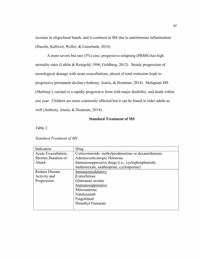
Standard Treatment of MS
Table 2
Standard Treatment of MS
Indication Drug
Acute Exacerbation,
Shorten Duration of
Attack
Corticosteroids: methylprednisolone or dexamethasone
Adrenocorticotropic Hormone
Immunosuppressive drugs (i.e.: cyclophosphamide,
methotrexate, azathioprine, cyclosporine)
Reduce Disease
Activity and
Progression
Immunomodulatory
β interferons
Glatiramer acetate
Immunosuppressive
Mitoxantrone
Natalizumab
Fingolimod
Dimethyl Fumarate
38
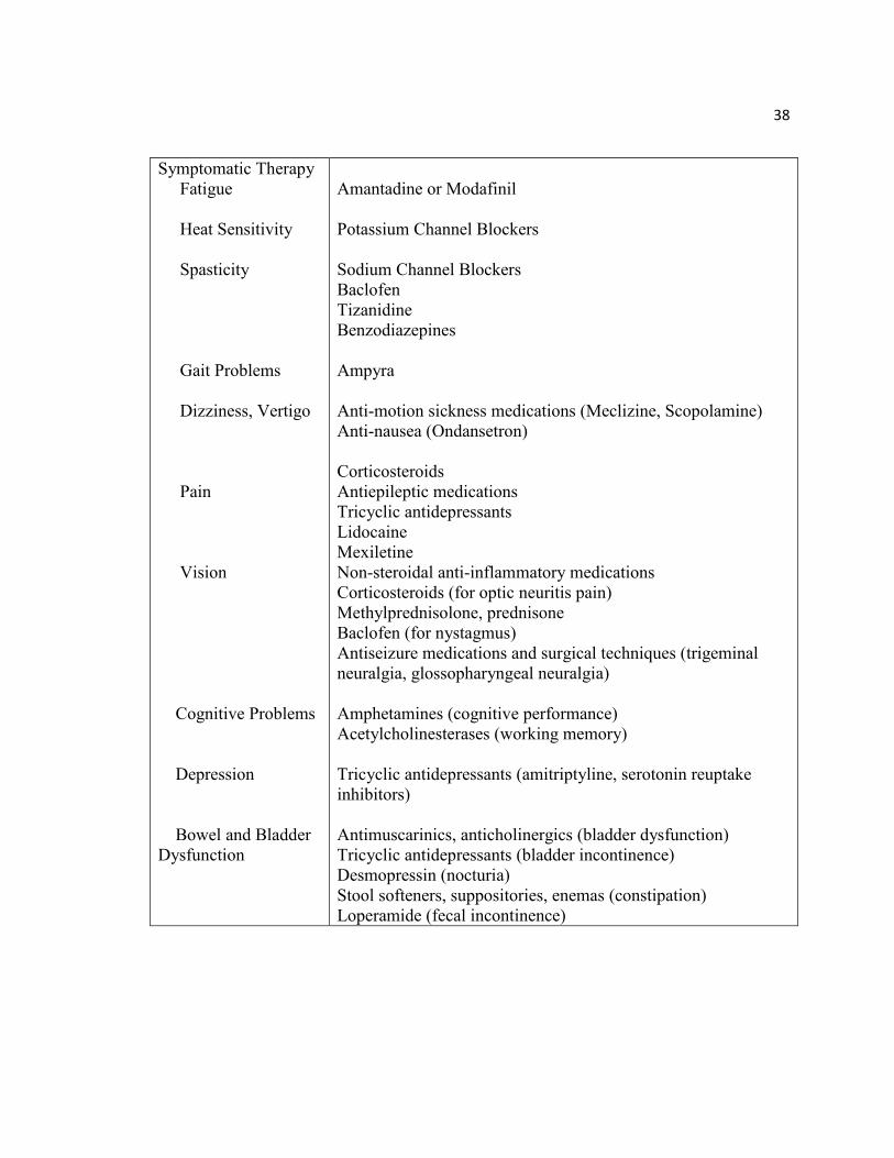
Symptomatic Therapy
Fatigue
Heat Sensitivity
Spasticity
Gait Problems
Dizziness, Vertigo
Pain
Vision
Cognitive Problems
Depression
Bowel and Bladder
Dysfunction
Amantadine or Modafinil
Potassium Channel Blockers
Sodium Channel Blockers
Baclofen
Tizanidine
Benzodiazepines
Ampyra
Anti-motion sickness medications (Meclizine, Scopolamine)
Anti-nausea (Ondansetron)
Corticosteroids
Antiepileptic medications
Tricyclic antidepressants
Lidocaine
Mexiletine
Non-steroidal anti-inflammatory medications
Corticosteroids (for optic neuritis pain)
Methylprednisolone, prednisone
Baclofen (for nystagmus)
Antiseizure medications and surgical techniques (trigeminal
neuralgia, glossopharyngeal neuralgia)
Amphetamines (cognitive performance)
Acetylcholinesterases (working memory)
Tricyclic antidepressants (amitriptyline, serotonin reuptake
inhibitors)
Antimuscarinics, anticholinergics (bladder dysfunction)
Tricyclic antidepressants (bladder incontinence)
Desmopressin (nocturia)
Stool softeners, suppositories, enemas (constipation)
Loperamide (fecal incontinence)
39
For individuals with mild MS without relapses, no treatment is required. In more
progressive MS, treatment strategies target acute exacerbations, reduce the disease
activity and progression, and treat the symptoms of MS (Table 2). When individuals
have acute exacerbations, treatment often includes corticosteroids and
adrenocorticotropic hormone (ACTH) (Goodin et al., 2002). These pharmacological
treatments promote quicker resolution of neurological deficits, lower the severity of the
attack while also reducing the risk of permanent deficits. These drugs also help to restore
the blood brain barrier, reduce inflammation, and work through immunomodulating
trafficking mechanisms (Arnason, Berkovich, Catania, Lisak, & Zaidi, 2013, Anthony,
Jouria, & Houtman, 2014). Corticosteroids and ACTH are able to block the adhesion and
migration of mononuclear phagocytes through the blood brain barrier, preventing their
movement or trafficking into the central nervous system (Anthony, Jouria, & Houtman,
2014). It is not advantageous to take corticosteroids or ACTH long term, and they serve
as acute exacerbation treatments (Goodin et al., 2002). Immunosuppressive drugs can
also be prescribed for acute exacerbations; however they increase susceptibility to
infection (Anthony, Jouria, & Houtman, 2014).
Immunomodulation drugs are used to slow the disease progression and reduce the
disease activity. These drugs can help to reduce the relapse rate and progression of
RRMS possibly by activating impaired immune cells and decreasing blood brain barrier
permeability (Goldenberg, 2012; Anthony, Jouria, & Houtman, 2014). The most
frequently prescribed immunomodulators are beta(β)-interferons which decrease acute
40
exacerbations in MS by 30% and reduce the risk of new lesion formation by 70-90%, and
reduce the severity of attacks (Goodin et al., 2002; Rejdak, Jackson, & Giavonnoni,
2010). Βeta interferon reduces the rate of disease progression by twelve months in the
short term (Rejdak, Jackson, & Giavonnoni, 2010).
Glatiramer acetate is believed to block myelin-damaging T-cells and increase the
number of immune regulatory cells that suppress the immune response resulting in
reduced inflammation (Goldenberg, 2012). Glatiramer acetate also decreases relapse rate
by 1/3 (Rejdak, Jackson, & Giavonnoni, 2010).
Mitoxantrone is a cytostatic (cell stopping) drug that is used to treat RRMS and
progressive forms of MS. Its anti-inflammatory effect helps to resolve relapses, however
there is high risk for leukemia and cardio-toxicity and is only used in treating advanced
MS (Goldenberg, 2012; Goodin et al., 2002). Mitoxantrone suppresses T and B cells,
and macrophages (cells that guide the myelin attack), resulting in reduced relapse rates
and improved disability scores (Goldenberg, 2012).
Natalizumab is a monoclonal antibody that reduces the relapse rate in RRMS,
slows disease progression, and decreases the number of lesions, but is used only if the
disease is very active or if the individual is not responsive to β-interferons or Glatiramer
acetate (Rejdak, Jackson, & Giavonnoni, 2010). Natalizumab works by preventing
movement of auto-reactive lymphocytes into the brain by blocking binding to vascular
cell adhesion molecules (VCAM) which are upregulated on the endothelium during
41
inflammation and by accelerating repair of the myelin sheath lesions (Rejdak, Jackson, &
Giavonnoni, 2010, Polman et al., 2006).
Fingolimod sphingosine-1-phosphate receptor modulator can be prescribed to
help reduce relapse rate in RRMS, but is correlated to an increased risk of infection.
Fingolimod prevents lymphocytes from migrating out of the lymph nodes and decreases
their number in circulation (Goldberg, 2012). This is the only drug approved for
treatment of highly active RRMS (Anthony, Jouria, & Houtman, 2014).
Several other pharmacological alternatives are used specifically in relapsing and
aggressive types of MS. Dimethyl Fumarate has only been evaluated in relapsing forms
of MS and works through oxidative pathways to decrease autoimmunity (Anthony,
Jouria, & Houtman, 2014). Teriflunomide, approved in the treatment of RRMS, is an
active metabolite of the anti-rheumatic drug leflunomide. Teriflunomide works by
inhibiting cell division of specific immune cells (Saguil, Kane, & Farnell, 2014). In treating
exacerbations in rapidly progressive MS that are non-responsive to corticosteroid
treatment, plasmapheresis (plasma exchange) is used as a secondary treatment (Goodin et
al., 2002).
Symptomatic Therapy
In addition to treating MS exacerbations and preventing relapse and progression
of the disease, symptoms of MS are also managed through pharmacologic and
rehabilitative methods.
42
Treatment of Fatigue
Fatigue is often assessed through self-reported scales such as the Fatigue Severity
Scale or the Modified Fatigue Impact Scale. Fatigue is treated pharmacologically by
Amantadine or Modafinil; however, drug treatment has limited effectiveness on fatigue
(Rejdak, Jackson, & Giavonnoni, 2010; Asano & Finlayson, 2014). Both Amantadine
and Modafinil are prescribed off-label in MS and have inconsistent results (“Provigil,”
n.d.; “Amantadine,” n.d.; Rejdak, Jackson, & Giavonnoni, 2010). Amantadine is an
antiviral drug prescribed to prevent or treat influenza infections, and is also used as
supplementary medication in Parkinson’s disease (“Amantadine,” n.d.). It’s mechanism
of improving fatigue is unidentified (“Amantadine,” n.d.). Modafinil is traditionally used
to promote wakefulness in narcolepsy, and has been found to have mixed results in
treating fatigue in MS (“Provigil,” n.d.).
Asano and Finlayson (2014) conducted a meta-analysis on 3 types of fatigue
management (exercise, education and medication) in MS individuals in order to compare
treatment options. Exercise rehabilitation methods including aerobic, aquatic, inspiratory
muscle exercise, vestibular rehabilitation program, progressive resistance training,
climbing and yoga were found to more effective in improving fatigue than drug therapy
(Amantadine and Modafinil). Educational fatigue management including energy
conservation strategies (limiting physical activity), fatigue self -management education
(client selected strategies to manage fatigue based on their needs, environment and
preferences), cognitive behavior therapy (changing patterns of thinking or behavior that
43
are problematic), and heat sensitivity control (pre-cooling the body, cool environment)
demonstrated stronger and more significant effects on reducing the impact and severity of
fatigue than medication (Asano & Finlayson, 2014; Hugos et al, 2010).
Hugos and colleagues (2010) found that group sessions conducted with digital
video disc (DVD), discussion and homework significantly decreased fatigue and
improved self-efficacy in MS individuals. A recent review (Latimer-Cheung et al., 2013)
noted the mixed results of treatments on fatigue. Exercise is often thought to improve
fatigue, though aerobic training alone did not consistently produce these results (Latimer-
Cheung et al., 2013). The authors concluded that optimal dose or training modality is not
determined, and they suggested that resistance training may be most effective in reducing
fatigue.
Treatment of Heat Sensitivity
Heat sensitivity, or Uhthoff phenomenon is a temporary worsening of symptoms
when body temperature increases, and is specific to MS (Bol et al., 2012). According to
White and colleagues (2000), 58-93% of MS individuals experience a relationship
between environmental temperature and fatigue. However, Bol and colleagues (2012)
were unable to identify a correlation between environmental temperature and heat
sensitivity. Heat sensitivity in MS individuals correlated to disease duration, and
decreasing disease severity, whereas sex, age, depression, and physical and mental
fatigue did not impact heat sensitive responses (Bol et al., 2012).
44
Heat sensitivity is managed by lowering body temperature through the
environment or by way of cooling devices (Syndulko, Jafari, Woldanski, Baumhefner, &
Tourtellotte, 1996). White and colleagues (2000) found that precooling up to the
suprailiac crest with water (16-17 degrees C) immersion allowed for a significantly lower
exercise heart rate, lower rate of perceived exertion, significant better walk performance
after exercise, lower core temperature through 30 minutes post exercise, decreased
fatigue and increased exercise comfort in heat sensitive MS individuals.
Treatment of Spasticity
Thirty percent of MS individuals experience a level of spasticity that causes them
to alter or eliminate their daily activities (Anthony, Jouria, & Houtman, 2014). Spasticity
is often measured through the self-reported Modified Ashworth Scale, which describes
the resistance to passive stretch (Bohannon & Smith, 1987). Treatment of spasticity aims
to decrease hypertonia and pain, as well as improve motor function, and prevent
complications including pressure ulcers and contractures (Anthony, Jouria, & Houtman,
2014). Tonic muscle spasms are those where muscle tone is continuously increased, and
these types of spasms often respond to sodium-channel blockers (Rejdak, Jackson &
Giavonnoni, 2010). Spasticity can be aggravated by sudden position changes, extreme
temperatures, humidity, infections, and tight clothing.
Treatment of spasticity in MS includes combinations of medications, physical and
occupational therapy, exercise and stretching, adjusting daily activities (Walker, Hall, &
Hurst, 1990). Rare cases that don’t respond to treatment may require surgical measures
45
(Anthony, Jouria, & Houtman, 2014). Baclofen, a muscle relaxant that targets nerves in
the spinal cord, is often the drug intervention of choice used for spasticity in MS
(Anthony, Jouria, & Houtman, 2014; Walker, Hall, & Hurst, 1990). Tizanidine is a
medication that quickly calms muscle spasms and relaxes tight muscles (Walker, Hall, &
Hurst, 1990). Benzodiazepines can also be helpful, however they can cause sedation
(Walker, Hall, & Hurst, 1990).
Treatment for Gait Problems
Treatment for gait problems includes physical and occupational therapy,
rehabilitation, and medications. Therapies often teach correct walking techniques and
gait training (Williamson, 2011). Rehabilitation may teach techniques such as energy
conservation, the use of adaptive equipment such as canes and walking devices, and
adaptations of the environment (Tyler et al., 2014). Spasticity often interferes with gait
and mobility, and is treated with stretching and antispasticity medications such as
Baclofen or Tizanidine (“NINDS,” n.d.). The drug Ampyra, has been shown to improve
walking speed by 20% in all types of MS, however, these effects are only seen in 35% of
the individuals tested (Williamson, 2011; “NINDS,” n.d.).
Additionally, In MS, several contemporary electrical stimulation treatments have
been tested in treating gait problems. Paul and colleagues (2008) found that electrical
stimulation of lower leg muscles to prevent drop foot promoted improvements in gait.
Deep brain stimulation where an electrode is implanted in the ventral intermediate
nucleus of the thalamus yielded modest and variable results in decreased tremor and
46
improved gait (Torres et al., 2010; Hu et al., 2010). Vagus nerve stimulation also reduces
tremor and the electrode is placed in the left afferent vagal fibers; whereas the right vagus
nerve innervates the sinoatrial node and would induce cardiac symptoms (Spuck et al.,
2010). However, the cost, invasiveness and side effects limit wide use of these
techniques. Tyler and colleagues (2014) found reduced gait dysfunction in multiple
sclerosis individuals with cranial nerve neuromodulation by using an electrical impulse
placed on the tongue. The authors postulated that by electrically stimulating the
trigeminal and facial cranial nerves, a flow of action potentials to the brainstem and
cerebellum induced neuroplasticity. Neuroplasticity was thought to occur through
stimulating direct collateral connections, improving brainstem interneuron circuitry, and
inducing changes in neurotransmitters and neuroactive compounds (Tyler et al., 2014).
Treatment for Dizziness
Dizziness and vertigo in MS may result from lesions in the medullary tegmentum
(near the vestibular apparatus) and the 8th
cranial nerve and are treated acutely by
corticosteroids to reduce inflammation (Pula, Newman-Toker, & Kattah, 2013; Frohman,
et al., 2011; “Dizziness and Vertigo,” n.d.). Few reports exist on treatment options for
dizziness and vertigo in MS, however, in the general population, acute vertigo attacks are
treated through vestibular blocking agents which can be used in MS in the short term (Sa
et al., 2011). In addition, anti-motion sickness medications such as Meclizine and skin
patches delivering Scopolamine are used to treat dizziness and vertigo (“Dizziness and
Vertigo,” n.d.). Anti-nausea drugs including Ondansetron are also used to treat dizziness
47
and vertigo (“Dizziness and Vertigo,” n.d.). Dizziness can be treated symptomatically
through physiotherapy, vestibular rehabilitation therapy, and repositioning maneuvers to
replace the otoconial mass onto the macular membrane (Sa et al., 2011; Frohman, et al.).
Treatment for Pain
Pain often occurs in the form of neuropathic central pain, Lhermitte’s sign,
painful tonic spasm, muscular pain, trigeminal neuralgia, headache, and retrobulbar optic
neuritis. Neuropathic central pain is treated through antiepileptic and tricyclic
antidepressant drugs (i.e.: amitriptyline) (Brola, Mitosek-Szewczyk, & Opara, 2014). If
pain is extreme and unresponsive to traditional treatment, surgical techniques have been
used to damage zones of the thalamus or to cut dorsal roots in spinal entry areas (Brola,
Mitosek-Szewczyk, & Opara, 2014).
Lhermitte’s sign is associated with lesions in the posterior column of the cervical
spinal cord and is treated similar to neuropathic pain, or with intravenously injected
lidocaine or oral mexiletine (Brola, Mitosek-Szewczyk, & Opara, 2014). A soft collar
can be used to limit neck flexion (“Pain,” n.d.).
Spasmatic pain often occurs as a result of lesions of the basal ganglia, internal
capsule, cerebral peduncle, medulla and spinal cord. Antiepileptic drugs, intravenous
infusions of lidocaine or local botulinum toxin injections can be used for treatment.
Cannabinoids have also been used to reduce spasm, thereby reducing pain (Brola,
Mitosek-Szewczyk, & Opara, 2014).
48
Muscular pain can be a side effect of the medication Beta Interferon, the most
common treatment for MS (Brola, Mitosek-Szewczyk, & Opara, 2014). Treatment for
muscular pain includes nonsteroidal anti-inflammatory drugs, physiotherapy to stabilize
proximal muscles and teach posture, electrotherapy, cryotherapy, magnotherapy, and
hydrotherapy (Brola, Mitosek-Szewczyk, & Opara, 2014; “Pain,” n.d.).
Trigeminal neuralgia is a stabbing pain in the face results from demyelinating
plaques in the pons and cranial nerve V. Antiepileptic drugs such as carbamazepine are
commonly used as treatment (Brola, Mitosek-Szewczyk, & Opara, 2014; “Pain,” n.d.).
Surgically radio frequency thermocoagulation, microvascular decompression and gamma
knife radio surgery techniques can be used to treat trigeminal and glossopharyngeal
neuralgia (Warren, Kotsenas, & Czervionke, 2006). Headache is treated with standard
migraine and tension headache medications as in the general population. Retrobulbar
optic neuritis results in pain behind the eye due to lesions in the optic nerve.
Corticosteroids are used to reduce inflammation in optic neuritis (Brola, Mitosek-
Szewczyk, & Opara, 2014).
Treatment of Visual Problems
Vision problems including optic neuritis, nystagmus, and diplopia can
significantly impact activities of daily living. Optic neuritis is treated by using
corticosteroids because of their immunosuppressive and immunomodulatory effects
(Brola, Mitosek-Szewczyk, & Opara, 2014; Frohman, 2008). Intravenous
methylprednisolone is prescribed followed by a tapered dose of oral steroids such as
49
prednisone (“Pain,” n.d.). Nystagmus is treated by Baclofen and the use of special prisms
(Frohman, 2008; Sa et al., 2011; “Pain,” n.d.). Diplopia typically resolves without
treatment, however resting the eyes or use of an eye patch for short durations is helpful
(“Pain,” n.d.; Sa et al., 2011).
Treatment of Cognitive Deficits
Cognitive deficits can interfere with daily functioning, and are associated with
depression, low self-esteem, and impaired social functioning (Kavcic & Scheid, 2011).
Cognitive impairment often manifests as poor attention, executive functioning, slowed
information processing and reduced memory retrieval (Amann et al., 2011). Kavcic and
colleagues (2011) suggested that cognitive deficits in MS are likely due to a reduction of
information processing rather than a reduction in working memory. Treatment includes
cognitive behavior therapy, psychotherapy and counseling. Amphetamines may improve
cognitive performance, and acetylcholinesterases can improve memory impairment
(Frohman, et al., 2011). However, a recent meta-analysis by Briken and colleagues
(2014) suggested that pharmacological approaches for cognitive impairment in MS were
ineffective. Therefore, additional therapeutic options for treatment of cognitive deficits
are needed.
Several randomized control trials in healthy young and aging adults have
documented improvements in cognitive function after aerobic exercise interventions
(Smith et al., 2010). Exercise has been shown to promote neuronal protection and alter
MS pathology in the animal model of MS (Rossi et al., 2009). Briken and colleagues,
50
(2014) compared arm ergometry, rowing and cycle ergometry in moderately disabled
Progressive Multiple Sclerosis patients for 8-10 weeks with a frequency of 2-3 times per
week. Exercise was found to be feasible with low dropout rates that were not different
between the three exercise group and the waitlisted control group (Briken et al., 2014).
Improvements in cognitive tonic alertness and VO2 were only seen in the cycle group,
while cognitive improvements in shift of attention, increased walk speed and decreased
depression occurred in the arm ergometer and cycle groups (Briken et al., 2014). In
addition, cognitive improvements in verbal learning and delayed memory were found in
all exercise groups (Briken et al., 2014). Higher fitness levels in Multiple Sclerosis
patients are associated with higher structural connectivity and higher gray matter density
(Briken et al., 2014). Moreover, fitness may be related to less severe central nervous
system damage and higher structural integrity of brain networks that are important to
cognitive function (Briken et al., 2014).
Treatment for Depression
Depression is commonly assessed through the self-reported Beck Depression
Inventory in MS and is treated using a sedating tricyclic antidepressant such as
amitriptyline or serotonin reuptake inhibitors (Anthony, Jouria, & Houtman, 2014;
Feinstein, 2011; Rejdak, Jackson, & Giavonnoni, 2010). Cognitive behavior therapy
develops skills to cope with emotions, thoughts, and adjustments to MS diagnosis and
symptoms, and has been found to improve depression (Mohr, Boudewyn, Goodkin,
Bostrom, Epstein, 2001; Feinstein, 2011). By incorporating behavioral activation and
51
cognitive restructuring, cognitive behavior therapy develops fatigue management,
management of mild cognitive impairment, pain management, stress management and
skills for intimacy in the MS individual (Mohr, Boudewyn, Goodkin, Bostrom, Epstein,
2001). Mindful based intervention encourages a nonjudgmental awareness of everyday
moments used to positively impact perception, increase sense of control, and acceptance
of health related problems (Feinstein, 2011).
Treatment of Bowel and Bladder Dysfunction
Bladder dysfunction is treated through lifestyle modification, physical therapy,
medications, and nerve stimulation. Adequate fluid intake should be encouraged,
however strategically planning voiding and limiting fluids 2-3 hours before bedtime can
be helpful (“Bladder Problems,” n.d.). Bladder training and physical therapy including
strengthening the pelvic floor, biofeedback, neuromuscular stimulation, and daily home
exercise can be used to reduce urinary urgency, frequency and loss of bladder control
(“Bladder Problems,” n.d.). Medications including anti-muscarinics and anticholinergics
help with bladder dysfunction, and the tricyclic antidepressant imipramine are used to
treat incontinence (Frohman, et al., 2011). Nocturia, having an overactive bladder at
night, is treated with oral desmopressin (Frohman, et al., 2011). Percutaneous tibial
nerve stimulation requires a small needle electrode inserted in the ankle to stimulate the
sacral plexus, and is known to decrease urinary frequency, urgency, nocturia and
incontinence (“Bladder Problems,” n.d.). Intermittent self-catherization several times a
day by inserting a tube into the urethra to fully empty the bladder is used in cases where
52
the individual cannot independently empty the bladder completely (“Bladder Problems,”
n.d.). Suprapubic bladder neck vibration, chemical denervation of the detrusor muscle
using either intravesicular capsaicin or botulinum toxin, and intermittent vasopressin are
also used to treat bladder dysfunction (Rejdak, Jackson, & Giavonnoni, 2010).
Bowel dysfunction includes constipation or fecal incontinence. Constipation is
treated through adequate fluid intake, fiber supplemented in the diet, stool softeners,
establishing a schedule, suppositories, and enemas (“Bowel Problems,” n.d.; Rejdak,
Jackson, & Giavonnoni, 2010). Fecal incontinence is treated through the medication
loperamide, biofeedback for pelvic floor muscles and rectal sensory perception, and in
cases unresponsive to traditional treatment, surgical colostomy is employed (Frohman, et
al., 2011).
Alternative Therapies for MS
In addition to traditional pharmacological treatment and rehabilitative treatment,
60 % of MS individuals seek alternative treatments (Anthony, Jouria, & Houtman, 2014).
One complementary therapy is Vitamin D supplementation. Low vitamin D serum levels
have been associated with a higher risk of developing Multiple Sclerosis (Duan et al.,
2014). There is also a higher correlation of low vitamin D levels with clinical and brain
MRI activity in RRMS (Bhargava et al., 2014). Further research indicates that 50 nmol/L
increases in average serum vitamin D levels translated to a 57% decrease in the rate of
new active MS-defining lesions (Hanwell & Banwell, 2011). The impact of vitamin D
deficiency in MS is thought to be linked to geographical location; those farther from the
53
equator and the sun have a higher incidence of MS (Bhargava et al., 2014; Duan et al.,
2014; Hanwell & Banwell, 2011).
Deficiencies in vitamin D may impact the instance and onset of Multiple Sclerosis
due to its immune and anti- inflammatory roles. Vitamin D is an important
immunomodulator that promotes dendritic cells, prevents proliferation and enhances
apoptosis of activated B cells, inhibits pro-inflammatory TH1 cells, modulates
inflammation, and promotes production of Treg cells (Bhargava et al., 2014). In addition,
vitamin D is known to reduce antigen presenting B cells, monocytes and dendritic cells
(Bhargava et al., 2014). Though the optimal dose and timing of supplementation to
prevent or diminish disease impact remains unclear, vitamin D supplementation does
have evidence to support its benefit as an alternative therapy for MS (Hanwell &
Banwell, 2011).
Polyunsaturated fat consumption is associated with lower MS prevalence, and
omega fatty acid intake may improve MS symptoms (Frohman et al., 2013). However
further research is required to substantiate these claims (Frohman et al., 2013). Cannabis
can be used to decrease spasticity and MS related symptoms, however this often illegal
alternative therapy is controversial due the cognitive side effects (Chong et al., 2006).
The herb Ruta graveolens (common rue) has been traditionally used in MS treatment;
however, there is no scientific evidence to support its use (Bodendiek, Mahieux, Hansel,
& Wulff, 2008). Bee venom provides anti-inflammatory properties, possibly by blocking
IL-6 an inflammatory cytokine, but the prohibitive risk of anaphylactic shock outweighs
54
the small anti-inflammatory benefit (Namaka et al., 2008). Although MS individuals
often suffered from poor oxygenation which affected the nerves and impulse conduction,
there is no evidence to support improvement with hyperbaric oxygen therapy (Bennett &
Heard, 2004).
Multiple Sclerosis focal lesions develop from an autoimmune response driven by
lymphocytes that permeate the blood brain barrier, and attack the myelin sheath.
Antioxidants can reduce the permeability of the blood brain barrier, and MS individuals
are often found to have reduced levels of antioxidants (Offen, Gilgun-Sherki, &
Melamed, 2004). It seems plausible that supplementation of antioxidants, specifically
uric acid which protects the blood brain barrier would be a viable alternative therapy, but
further research is needed (Offen, Gilgun-Sherki, & Melamed, 2004; Koch & De Keyser,
2006). Lintas and colleagues (2013) used ozone therapy to trigger mild oxidative stress
which induces the production of antioxidants and antioxidant enzymes to protect the cells
and reverse chronic oxidative stress. They concluded that ozone promotes the reduction
of oxidative stress and enhanced mitochondrial functionality in MS individuals.
A final supplementary alternative therapy is acupuncture which reduces pain and
muscle spasm resulting in improved quality of life (Quispe-Cabanillas et al., 2012).
However, additional research is needed to determine appropriate usage of acupuncture in
MS.
55
Exercise and MS
Despite proven benefit of exercise on the symptoms of MS, there are several
barriers to physical activity in this population. Multiple Sclerosis individuals have a
lower aerobic capacity, lower muscle strength, slowed rate of muscle tension
development, lower muscle endurance and impaired balance when compared to healthy
individuals (Doring, Pfueller, Paul, Dorr, 2012). Indicative of compromised fitness
levels, Kuspinar and colleagues (2010) found VO2 max in Multiple Sclerosis individuals
is below the 25th
percentile. A further concern is that 40% of MS individuals require
some form of walking assistance within 15 years of disease onset (Tyler et al., 2014).
Pharmacologically, Interferon B and Glatiramer Acetate are only partially
effective in treating MS symptoms, necessitating additional treatment options (Rudick et
al., 2006). Traditionally, exercise has been avoided in the MS population because of
acute transient problems with mobility, weakness, and fatigue after exercise (Hayes,
Gappmaier, & LaStayo, 2011). Although previous recommendations suggested that
exercise exacerbated symptoms, current research challenges this view and demonstrates
that exercise is beneficial (Pilutti et al., 2011). In fact, limits on physical activity in this
population can lead to greater weakness, fatigue, and health risks that result from
deconditioning (Mostert & Kesselring, 2002).
Physical activity decreases morbidity and mortality in healthy populations, and
likely has similar effects on people with Multiple Sclerosis (Bjarnadottir, Konradsdottir,
Reynisdottir, & Olasfsson, 2007). Exercise has the capacity to slow disease progression
56
and improve various aspects in the physiological profile of Multiple Sclerosis, and is well
tolerated with low occurrence of adverse effects (Kargarfard, Etemadifar, Baker,
Mehrabi, & Hayatbakhsh, 2012; Doring, Pfueller, Paul, Dorr, 2012).
Prakash and colleagues (2010) discovered a positive correlation between higher
fitness and gray matter volume, higher scores on composite measures of information
processing speed, and an inverse relationship with brain tissue damage in MS. The
authors concluded that fitness exerts a prophylactic influence on cerebral atrophy that
occurs early in the disease process, thereby, preserving neuronal integrity in MS and
reducing long-term disability.
Individuals with Multiple Sclerosis have a normal cardiovascular response to
exercise and have the ability to improve fitness (Mostert & Kesselring, 2002).
Bjarnadottir and colleagues (2007) demonstrated that a brief 5 week aerobic and strength
training program improved physical fitness in mild MS. However, it has been observed
that MS individuals have a low anaerobic threshold, low maximal volume of oxygen, and
high energy consumption, as well as a high heart rate reserve compared to healthy
controls (Mostert & Kesselring, 2002). Although MS individuals have an inability to
stress the cardiovascular system maximally, it has been shown that exercise lowers
oxygen consumption, heart rate, and increases oxygen pulses (VO2/HR) at a given work
rate, indicating an improved aerobic system (Mostert & Kesselring, 2002).
Pilutti and colleagues (2011) found that 12 weeks of body weight supported treadmill
training in extremely disabled MS individuals exhibited gait and mobility improvements
57
(Pilutti,, Lelli, Paulseth, Crome, Jiang, Rathbone, & Hicks). Physical activity may help
improve physical function and ease psychosocial burdens for those with the disease, as
well as for their caregivers and companions (White, Castellano, McCoy, Patel, &
Giacobbi, 2012).
Exercise improves and maintains muscle strength, balance and mobility in elderly
individuals including those with neurological disabilities (Hayes, Gappmaier, & LaStayo,
2011). A variety of exercise modalities and programs, including yoga, are known to
improve quality of life and reduce fatigue in MS individuals (Asano & Finlayson, 2014).
After review of 600 participants in diverse exercise modalities and protocols, Snook and
Motl (2009 ) concluded that exercise training programs are associated with small
clinically meaningful improvements in mobility of walking in MS individuals.
Currently there are no specific recommendations for exercise treatment that are
universally valid; only general therapeutic recommendations exist (Doring, Pfueller, Paul,
Dorr, 2012). The primary goal of rehabilitation in MS is to increase activity levels and
participation while promoting independence (Reitberg, Brooks, Uitdehagg, & Kwakkel,
2011). However there are several barriers to exercise in the MS population, including
change in medication, holidays, surgery, lack of motivation, lack of support system and
social support (White, Castellano, McCoy, Patel, & Giacobbi, 2012).
Although exercise is associated with increased fitness, decreased motor fatigue,
improved quality of life and psychological conditions (Kargarfard, Etemadifar, Baker,
Mehrabi, & Hayatbakhsh, 2012), it is also found to lower cardiovascular risk factors such
58
as heart disease and obesity. Reitberg and colleagues (2011) reviewed nine high quality
randomized control trials and found good evidence for improved muscle power, aerobic
fitness, physiological cost index, rate of perceived exertion and mobility related activities.
Moderate evidence to support improved mood, anxiety and depression was also present.
However, this review yielded little evidence to support that exercise improved fatigue,
perception of the handicap, or that specific programs are more successful in improving
MS activities and participation; contradicting studies showing improvements in fatigue
(Reitberg, Brooks, Uitdehagg, & Kwakkel, 2011). Nevertheless, no evidence for
negative effects of exercise was found.
Mechanisms of Exercise Induced Improvements in MS
Exercise believed to produce improvements in MS by enhancing the immune
system functioning, increasing growth factors (BDNF, IGF, VEGF, and NGF) and
improving motor learning. During moderate exercise there is a rise in peripheral
lymphocytes which then decrease below the initial levels after the exercise is complete
(Doring, Pfueller, Paul, Dorr, 2012). The decrease in lymphocytes after exercise is short
(3-24 hours) which emphasizes the need for a consistent exercise program in MS
individuals. The lymphocytes that decrease after exercise are T- helper (Th) cells which
aid the immune system in distinguishing foreign from self and aiding the interactions
between cells fighting foreign materials. There are many different types of T-helper
cells, and two of interest in MS are Th1 cells and Th2 cells. Th1 cells exhibit a pro-
inflammatory effect and Th2 cells produce an anti-inflammatory response (Doring,
59
Pfueller, Paul, Dorr, 2012). Exercise is especially beneficial in MS because there are
declines in Th1 cells rather than Th2 cells (Doring, Pfueller, Paul, Dorr, 2012). This is
relevant to the MS pathogenesis due to the inflammatory autoimmune attacks on the
myelin sheath. Immuno-modulatory drugs exert a similar response and may be
complemented by exercise (Doring, Pfueller, Paul, Dorr, 2012). Regular and frequent
training intervals may be beneficial due to the transitory effects of exercise (Doring,
Pfueller, Paul, Dorr, 2012).
Exercise also stimulates the production of brain derived neurotropic factor
(BDNF), Insulin-like growth factor (IGF), and vascular endothelial growth factor
(VEGF). These growth factors enhance cell proliferation, synaptic plasticity, neuro-
protection, and neurogenesis in physiological and neuro-inflammatory conditions
(Doring, Pfueller, Paul, Dorr, 2012). During human development, IGF-1 supports cell
survival, brain growth, and central nervous system myelination (Doring, Pfueller, Paul,
Dorr, 2012). Later in the adult, IGF-1 may be neuroprotective and aid synaptic and
cognitive neuroplasticity (Doring, Pfueller, Paul, Dorr, 2012). Doring and colleagues
(2012) documented increased BDNF and nerve growth factor (NGF) after thirty minutes
of moderate ergometry based exercise in healthy and MS individuals. IGF-1 increased
after exercise in healthy individuals, but has not been tested in MS individuals (Doring,
Pfueller, Paul, Dorr, 2012).
Doring and colleagues (2012) also found increased levels of BDNF in the
hippocampus, possibly aiding learning, memory, modulation of mood, and may have the
60
capability to slow cognitive decline. There is evidence of a reduction in serum BDNF
during MS relapses compared to periods of remission (Prakash, Snook, Motl, & Kramer,
2010). In fact depression in MS individuals is possibly linked to reductions in BDNF
(Prakash, Snook, Motl, & Kramer, 2010). In addition, exercise may help to attenuate
reductions in BDNF and additional neuroplasticity factors while having a positive effect
on modulating stress which is associated with increased relapse risk in MS.
In addition to the growth factors mentioned above, there are other benefits to
exercise in the MS population. Exercise has also been found to increase antioxidant
enzymes which have a neuroprotective effect (Doring, Pfueller, Paul, Dorr, 2012).
Through the repetitive movement of exercise, cortical neural pathways are strengthened,
neuroplastic adaptive processes occur and there is improved motor unit activation and
synchronized firing rates (Doring, Pfueller, Paul, Dorr, 2012). During times of inactivity
these improvements have been shown to decline (Doring, Pfueller, Paul, Dorr, 2012).
The mechanism of exercise induced improvements in MS may result from task
specific movements, improved fitness, or improved motor control. Motor learning is skill
specific, as skill acquisition theory hypothesizes that in order to achieve mastery of a
movement, one must practice the specific task (Lord, Wade, & Halligan, 1998). Lord
and colleagues (1998) compared the effects of task specific therapy and facilitation
therapy on mobility and balance in MS. The facilitation group received proprioceptive
feedback from a therapist to correct deficiencies in posture, balance, and muscle
contraction (Lord, Wade, & Halligan, 1998). The task specific group practiced a specific
61
skill (walking) to achieve mastery (Lord, Wade, & Halligan, 1998). Both the task
specific group and facilitation group showed improvements in mobility and balance in
MS.
Van den Berg and colleagues (2006) utilized a four week treadmill aerobic
intervention that was found to improve gait speed significantly. It is unclear whether task
specificity, improvements in fitness, or enhanced motor control were responsible for the
improved gait speed, or the resulting trend toward increased endurance and toward
decreased fatigue. This study demonstrated that treadmill exercise which is task oriented
specific to walking, showed improvements in these parameters during the exercise
training, though it was inconclusive in determining the mechanism for the improvements.
Furthermore, four weeks post exercise, walking performance returned towards baseline
indicating a need of continued exercise (van den Berg et al., 2006).
In relapsing-remitting forms of MS, partial spontaneous repair of demyelination
can occur. It is theorized that repair is a result of neuroplasticity in which axons and
dendrites undergo collateral sprouting (Herbert, Corboy, Manago, & Schenkman, 2011).
This appears to be enhanced through task specific rehabilitative training (Herbert,
Corboy, Manago, & Schenkman, 2011). An additional benefit of exercise is improved
motor control which may lessen the effects of motor fatigue on anticipatory postural
adjustments (Herbert, Corboy, Manago, & Schenkman, 2011).
Gait and mobility are further impacted by dizziness and deficits in postural
control that are controlled by central sensory processing (Herbert, Corboy, Manago, &
62
Schenkman, 2011). Impaired upright postural control is linked to poor central sensory
integration and fatigue in MS (Herbert, Corboy, Manago, & Schenkman, 2011).
Rampello and colleagues (2007) utilized an 8 week training protocol with mild to
moderate disability MS individuals to compare progressive aerobic training to a
neurological (postural) rehabilitation group undergoing respiratory-postural, respiratory-
motor synergies training, and stretching. The authors found that lung function and
respiratory muscle strength did not change in either group, but the aerobic training group
experienced increases in walking distance, speed, and aerobic fitness parameters, whereas
the neurological rehabilitation training group did not (Rampello et al., 2007). Fatigue and
health related quality of life were also unchanged in either group (Rampello et al., 2007).
Lesions in the brainstem and cerebellum lead to visual somatosensory and
vestibular processing impairments (Herbert, Corboy, Manago, & Schenkman, 2011). Of
MS individuals, 85% experience peripheral deficit vestibulopathy (Herbert, Corboy,
Manago, & Schenkman, 2011). Primary fatigue, caused directly by the disease process
may be better treated by vestibular rehabilitation depending on location of the lesions.
Herbert and colleagues (2011) used a 6 week intervention to compare three groups. The
first group underwent vestibular rehabilitation and saw improvements in fatigue,
impaired balance, and disability due to dizziness and disequilibrium. The second group
exercised using a cycle ergometer and underwent stretching. This group, along with the
vestibular rehabilitation group experienced improvements in depression. The third group
served as a control and continued with usual medical care. All three groups improved in
63
mobility which may have resulted from factors outside of the study (such as walking to
the testing site) (Herbert, Corboy, Manago, & Schenkman, 2011).
Regular exercise may also have an important role in managing fatigue, mental
alertness, and cognitive processing (Paul et al, 2014). Results on walking endurance and
speed due to exercise are promising though the benefits on agility are less clear (Latimer-
Cheung et al., 2013). It is likely that the mixed results found in current studies are due to
the varying length of training periods and the degree of disability of MS individuals in
the studies (Rampello et al., 2007).
Aerobic Training in MS
Aerobic training produces an increase in peak oxygen uptake, increase in muscle
strength and endurance, decrease in fatigue, increase in activity level and improvements
in balance and gait patterns in individuals with MS (Hayes, Gappmaier, & LaStayo,
2011). Cakit and colleagues (2010) found aerobic training effective in improving
mobility, functional reach, walking endurance while decreasing fatigue and disability
levels. Treadmill training resulted in improvements in strength, spasticity, walk speed
and endurance, balance and quality of life in MS individuals (Cakit et al., 2010). Carter
and White (2003) demonstrated that a 12 week general aerobics program reduced
physiological cost index and increased muscle strength in specific muscle groups.
O’Connell and colleagues (2003) determined that a similar 12 week aerobic protocol
improved muscular fitness and quality of life, pre and post heart rate, gait cadence, and
RPE without a change in gait speed in the exercise group compared to controls.
64
Aerobic activities also have a positive impact on cardiovascular risk factors,
obesity, all-cause mortality, osteoporosis, depression and diabetes mellitus (Mostert &
Kesselring, 2002). Depression and anger scores of the Profile of Mood States were
significantly decreased after 15 weeks of aerobic training in a study done by Petajan and
colleagues (1996). Schulz and colleagues (2004) showed that an 8 week low level
bicycle aerobic exercise protocol improved fitness and disease specific quality of life in
MS individuals. Latimer-Cheung and colleagues (2013) completed an extensive review
of the exercise literature and recommended 2-3 sessions per week for 30 to 60 minutes in
duration of moderate intensity defined as 60-80% max work rate or VO2 maximum to
improve aerobic fitness in MS. This is consistent with ACSM guidelines for the general
population to improve fitness including 20-60 minutes 3-5 times per week at 40-50%
maximal VO2.
Aerobic training is also associated with changes in brain structure. MS
individuals with high aerobic fitness showed larger gray matter volumes of the right post-
central gyrus and midline cortical structures than unfit MS individuals. The higher
fitness was correlated with greater recruitment in cortical regions whereas lower fitness
was associated with greater anterior cingulated cortex recruitment (Solari et al., 1999). In
addition, MS individuals exhibit more brain areas bilaterally activated compared to non-
multiple sclerosis individuals, indicating that MS individuals adapt through
neuroplasticity. The bilateral activation is correlated to MS disease and severity (Doring,
65
Pfueller, Paul, Dorr, 2012). Aerobic exercise may play a profound role in assisting and
enhancing neuroplasticity in MS individuals (Doring, Pfueller, Paul, Dorr, 2012).
Resistance Training in MS
Resistance training generates increased muscle strength and power while reducing
the perception of fatigue (Hayes, Gappmaier, & LaStayo, 2011). In other special
populations (Parkinson Disease, Cancer, Stroke) improvements in strength resulted in
improvements in mobility, balance and function. However, resistance training in MS
individuals has uncovered mixed results for balance, gait pattern, and functional capacity
(Hayes, Gappmaier, & LaStayo, 2011). Jones and colleagues (1999) compared a
weighted leg exercise treatment, mobility exercise program, and a control group and
found there were no significant differences in gait, ability to transfer and muscle strength.
DeBolt and McCubbin (2004) used an 8 week home based resistance exercise program to
increase leg extensor power in MS, and concluded there were no significant differences
in mobility and balance compared to control subjects. It is possible that motivation and
adherence played a significant role as home based studies are not as effective as
outpatient or inpatient rehabilitation sessions. Individuals in an unsupervised
environment are less likely to change their training program in order to develop adequate
workload progression, which is essential for adaptation (Latimer-Cheung et al., 2013).
In contrast to the above studies, Solari and colleagues (1999) compared 3 weeks
of inpatient exercise therapy followed by home rehabilitation to a group practicing
exclusively home exercise, and found the inpatient group had significant improvements
66
in disability, motor domains and mental components of quality of life. White and
colleagues (2012) used a 4 month resistance training protocol in MS women that
improved walking measures, and qualitatively found that exercise may be effective in
attenuating loss or improving daily function. They reported MS individuals were
motivated to exercise by a desire to maintain function and health, enhanced exercise self-
efficacy, feelings of hope and optimism, and the presence of role models.
MS responds to eccentric resistance exercises differently than other populations.
Eccentric exercise is when the muscle lengthens during the contraction. Hayes and
colleagues (2011) developed RENEW, an eccentric resistance training program but found
that eccentric exercises were not beneficial in individuals with MS, despite the fact that it
was beneficial in people with Parkinson’s disease. The authors theorized that the MS
deficiency in improvements in functional activities were due to the central nervous
system’s lost capacity to respond to higher intensity forces.
Overall, strength training promotes neural adaptation, better motor unit activation,
and synchronization of firing rates (Hayes, Gappmaier, & LaStayo, 2011). Latimer –
Cheung and colleagues (2013) concluded in their review that resistance training eliciting
muscular strength improvements should be 8-20 weeks long, 2-3 times per week, with
10-12 repetition maximum.
Cakit and colleagues (2010) examined the benefits of progressive resistance using
the cycle ergometer in MS individuals (16). Improvements in duration, maximal
workload, Timed up and Go test (TUG), Dynamic Gait Index (DGI), functional reach,
67
falls efficacy (a self-reported scale), 10 minute walk test, Fatigue Severity Scale, and
Beck Depression Inventory were documented. The authors concluded that pharmacology
alone is not optimal and that exercise is a feasible form of self-management for persons
with Multiple Sclerosis (Cakit et al., 2010).
Although traditional land-based aerobic and resistance training programs have
been shown to benefit individuals with MS, the risk of overheating, extreme fatigue and
falling makes regular exercise a challenge for these individuals. MS individuals are
limited by high impact exercise due to unpredictable relapse and pain. The aquatic
exercise environment increases body heat dissipation, provides buoyancy, decreases joint
stress, and decreases fear of falling. Aquatic exercise has been found feasible and
beneficial in the MS population (Kargarfard, Etemadifar, Baker, Mehrabi, &
Hayatbakhsh, 2012).
Aquatic Exercise and MS
Aquatic exercise is recommended by the American Physical Therapy Association
in treatment of Multiple Sclerosis (Kargarfard, Etemadifar, Baker, Mehrabi, &
Hayatbakhsh, 2012). The buoyancy and viscosity of water assist the activities of those
who have physical weakness (Kargarfard, Etemadifar, Baker, Mehrabi, & Hayatbakhsh,
2012). In addition, the increased ability to dissipate body heat in the water aids in
diminishing Uhthoff phenomenon (Kargarfard, Etemadifar, Baker, Mehrabi, &
Hayatbakhsh, 2012). Aquatic therapy reduces joint stress and decreases the fear of
falling (Plecash & Leavitt, 2014). Aquatic exercise reduces the effects of gravity,
68
allowing individuals with severe paresis to perform standing and moving exercises
(Doring, Pfueller, Paul, Dorr, 2012). Aquatic therapy generally includes stretching and
range of motion, aerobic, resistance and strength training in a shallow pool (Plecash &
Leavitt, 2014). Despite these positive characteristics of exercising in water, only 1.5% of
MS individuals use water therapy (Plecash & Leavitt, 2014).
Aquatic exercise with varying frequency, duration, intensity and mode have been
investigated in the MS population. Kargarfard and colleagues (2012) found that in
RRMS patients, 8 weeks of aquatic aerobic activity was feasible, improved fatigue, and
improved health related quality of life parameters. Fatigue and health related quality of
life measures were taken at baseline, at 4 weeks and at 8 weeks. While there was a trend
toward improvement in these parameters at 4 weeks, significant differences were
observed at 8 weeks of aquatic exercise (Kargarfard, Etemadifar, Baker, Mehrabi, &
Hayatbakhsh, 2012). Aquatic exercise lasting for six weeks was found to have a positive
impact of sickness (Mostert & Kesselring, 2002). Rhomberg and colleagues (2004) used
a 6 month aerobic training protocol including aquatic exercise and strength training,
which resulted in increased walk speeds compared to individuals not undergoing therapy.
Roehrs and Karst (2004) used a 12 week aquatic warm water (85 degrees F) intervention
and demonstrated a significant improvement in quality of life specifically in fatigue and
social functioning. Sixty minutes of aquatic class time, twice a week was used with
progressive MS individuals (Roehrs & Karst, 2004). Marinho-Buzelli and colleagues
69
(2014) used aquatic therapy in 20 MS individuals, and demonstrated a fair ability to
improve gait speed.
Non-traditional aquatic exercise has also been investigated in MS. Castro-Sanchez
and colleagues (2012) used Ai Chi aqua therapy in MS two times per week for twenty
weeks. Ai-Chi aquatic exercise was used two times per week for twenty weeks. Ai Chi
is deep breathing combined with slow broad movements to work on balance, strength,
relaxation, flexibility and breathing. They compared the Ai-Chi exercise group to a
control group that performed abdominal breathing and contraction and relaxation
exercises in the therapy lab, and found improvements in pain, spasm, fatigue, disability
and autonomy in the Ai-Chi group (Castro-Sanchez et al, 2012). Bansi and colleagues
(2012) compared land cycling to aquatic cycling and found increased serum levels of
BDNF in the aquatic group, but not in the land based group. Subjects in this study
worked at 70% maximum heart rate for 30 minutes, five times per week for 3 weeks.
Salem and colleagues (2011) used a 5 week, 2 times per week for 60 minutes protocol
that used aquatic aerobic, strength, flexibility and balance, and walking and showed
improved gait speed, Berg Balance Scores, timed up and go, and grip test.
Research literature reveals benefits of aquatic therapy, however there are no
specific recommendations for MS treatment (Doring, Pfueller, Paul, Dorr, 2012). With
current research studies employing 3 or more weeks of exercise, there is a need to
determine the minimal duration required to manifest exercise benefits. Seven
70
consecutive days of exercise may be beneficial in improving fitness, mobility, and self-
efficacy.
Summary
Multiple Sclerosis is an autoimmune, demyelinating disease that can impact
activities of daily living. Symptoms include fatigue, heat sensitivity, muscle spasms,
decreased physical activity, gait problems, dizziness or vertigo, vision problems, pain,
cognitive impairments, depression and bowel and bladder dysfunction. Although there is
no cure for MS, treatments aim to slow disease progression and lessening the symptoms.
Treatments include pharmacological, alternative therapies, and exercise. Exercise can
improve fitness, preserve motor and cognitive function, and enhance quality of life.
Aquatic exercise has been recommended by the American Physical Therapy Association
in the treatment of MS, however, currently only general exercise recommendations exist
for MS patients. MS individuals may benefit from moderate to high intensity short
duration exercise between relapses. Research has demonstrated exercise induced
improvements on gait parameters and quality of life within a 4 week protocol, however,
no studies to date have used a one week protocol (Berg 23, Mostert 22). Previous work
done by Fedor (2014) produced positive results in fitness and cognitive parameters in a
similar aquatic aerobic one week exercise protocol in older adults. However this has not
been explored in the MS population. Currently there is no research determining the
benefits of short duration aquatic exercise on mobility, cardiovascular fitness, cerebral

71
oxygenation, or self-efficacy. Benefits resulting from this protocol have the potential to
impact the MS population and the use of exercise as treatment.
72
CHAPTER III
METHODOLOGY
Recruitment
Participants were recruited for the study by placing fliers in the waiting rooms at
Neurology and Neuroscience Associates (NNA) facilities and the OAK Clinic. In
addition, researchers met with local MS support groups and distributed fliers to
prospective participants. Fliers were place strategically around town in area businesses
and churches, and a study overview was placed in the MS Connector. Twitter and
Facebook accounts were also set up with the MS study information. The study fliers
included research contact information and a physician approval form. Interested MS
patients were instructed to contact the research staff after obtaining physician consent to
participate in the study (Figure 1). A researcher conducted a telephone prescreening
interview in which interest was assessed in addition to discussing inclusion and exclusion
criteria and general information on the research study. At the conclusion of the phone
interview, the researcher scheduled the prospective participant for their first visit at the
Kent State Exercise Physiology Lab to sign the informed consent and the HIPAA release,
and for baseline testing.
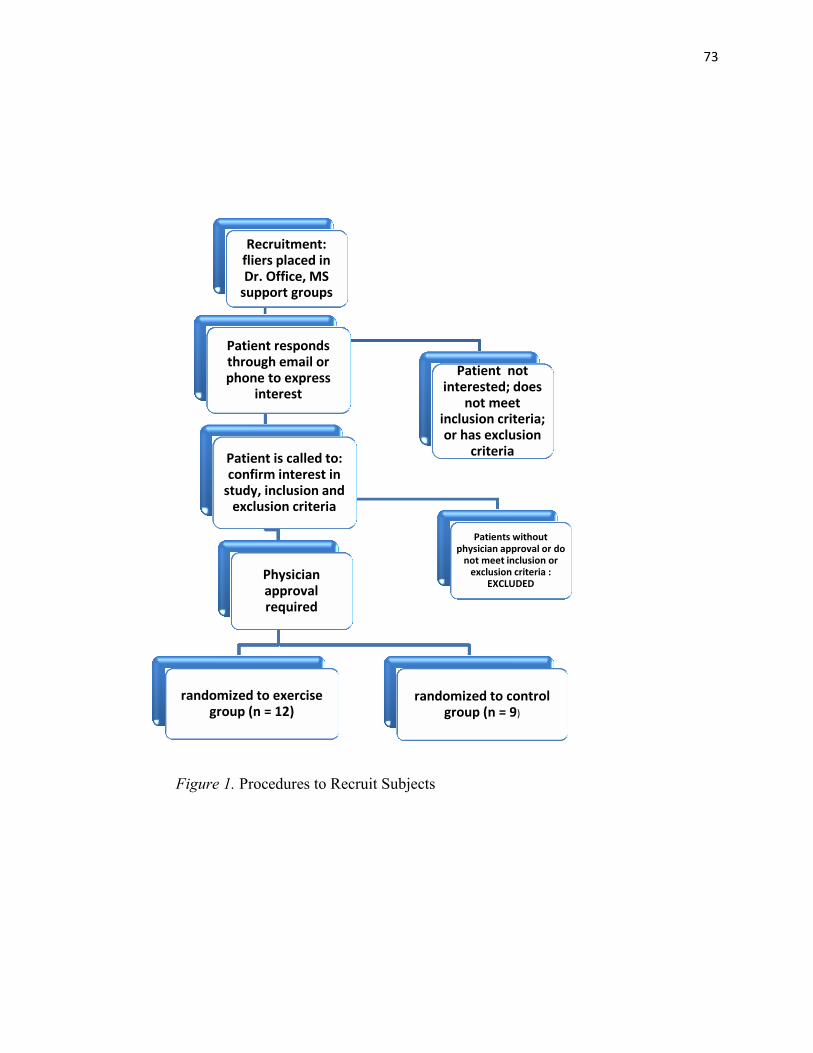
73
Recruitment: fliers placed in Dr. Office, MS
support groups
Patient responds through email or phone to express
interest
Physician approval required
randomized to exercise group (n = 12)
randomized to control group (n = 9)
Patient is called to: confirm interest in
study, inclusion and exclusion criteria
Patients without physician approval or do
not meet inclusion or exclusion criteria :
EXCLUDED
Patient not interested; does
not meet inclusion criteria; or has exclusion
criteria
Figure 1. Procedures to Recruit Subjects
74
Inclusion
Only individuals who were diagnosed with Multiple Sclerosis by a physician were
included in the study. Participants were required to obtain approval from their physician
prior to participating in the study. In addition, participants were between the ages of 20
to 65. To aid the communication within the study, subjects were required to be English
speaking.
Exclusion
Exclusion criteria included: females who were pregnant or at risk for becoming
pregnant, psychiatric illness, history of non-MS related neurological disorder or injury
(e.g. seizure, brain injury), past or current history of alcohol or drug abuse, history of
learning disorder or developmental disability, or sensory function impaired enough to
preclude cognitive testing.
Risk Minimization
Current research indicated that exercise is feasible in the MS population, and has
demonstrable fitness benefits (Pilutti et al., 2011). Aquatic therapy is recommended by
the American Physical Therapy Association for therapy in MS patients. The buoyant
effect of the water is helpful in reducing gravity and resistance against body movements
resulting in longer endurance with less fatigue (Kargarfard, Etemadifar, Baker, Mehrabi,
& Hayatbakhsh, 2012). In addition, this study followed a similar protocol to the aquatic
MS study done by Kargarfard and colleagues (2012), who concluded that this aquatic
protocol was feasible in the MS population.
75
There was an extremely small risk in exercise of serious medical complications,
including heart attack and sudden death. However, the requirement of physician’s
approval and participation in a well-organized and individualized exercise program
greatly reduced these already small risks. Small risk was present with exercising in the
water, however all exercise took place in the shallow end of the pool, and trained staff
and researchers (lifeguards, special population trained aquatic aerobics instructor, and
trained AED/CPR certified research staff) were present during exercise. Participants
were instructed to inform a research team member if they wished to stop, experienced
concern regarding exercise equipment or facilities, or happened to experience any of the
following during the study: chest pain, shortness of breath, pallor, fainting, wheezing, leg
cramps, light-headedness, confusion, nausea, cold/clammy skin, noticeable change in
heart rhythm, severe fatigue, or any other discomfort.
A certified special population aquatic aerobics instructor led the small group of 6-
10 subjects. Subjects were monitored by a lifeguard in addition to at least two trained
research staff. The subject to researcher ratio (2:1) was low to ensure adequate attention
and monitoring of each subject. Close supervision and encouragement was essential for
MS patients during exercise due to concerns that exercise may have exacerbated their
symptoms (Paul et al, 2014). Subjects were instructed about target training intensities
and encouraged to self-monitor in order to stay within the range of 65-75% age adjusted
maximal heart rate (AAMHR). In addition, the research staff monitored the MS subject’s
heart rate and rate of perceived exertion (RPE) every 9 minutes throughout the exercise
76
protocol. The exercise protocol was an interval protocol which provided rest periods
between four minute segments of exercise allowing for recovery time during the exercise.
If the heart rate reached greater than 75% AAMHR during the assessments, the subject
was required to rest at the side of the pool while being monitored by the researcher. The
subject was allowed to return to exercise when the heart rate was at 65% AAMHR. In
addition, if RPE is greater than 17, the subject was required to rest at the side of the pool
while being monitored by the researcher. The subject was allowed to return to exercise
when the RPE was at 13. For additional security, subjects were allowed to hold onto a
pool noodle while performing leg exercises. Relationships were established between the
researcher and the subject so that subjects were comfortable communicating concerns
about the exercise exertion.
Protocol
After patients responded to fliers placed in the physician office and MS support
groups, they were called by the researcher to determine interest, to discuss inclusion and
exclusion criteria, and to provide information on the study. Subjects were responsible for
obtaining physician approval prior to participating in the study, and a physician approval
form was provided on the back of the MS flier. Individuals who met the inclusion
criteria, did not exhibit any exclusion criteria, and were interested in participating were
asked to visit the Kent State Physiology lab to learn more about the study and to sign an
informed consent and HIPPA release form, and undergo baseline testing on day 1.
Baseline testing included body mass index, waist circumference, waist to hip ratio, 25
77
foot walk test, 2 minute step test, resting heart rate, heart rate variability, blood pressure,
and timed up and go test. In addition, assessments of rate of perceived exertion, cerebral
oxygenation, and tympanic temperature were recorded, and the self-reported Multiple
Sclerosis Self Efficacy Scale was completed by the subject. Subjects were randomly
assigned to the exercise or control group. Subjects randomized to the control group were
asked to maintain their current lifestyle including their physical activity levels for days
two through 8. On the ninth day they reported back to the lab for post-test measures;
repeating the same measures as on day one. The exercise group underwent baseline
testing on day 1, and participated in aquatic exercise for days two through eight. On day
nine they repeated the measures of baseline testing again.
Exercise and Physiological Variables
While the control group maintained their current lifestyle, the exercise group
participated in seven days of moderate to high intensity aquatic exercise. Table 3 lists the
exercise protocol and assessments. Exercise comprised 10 minutes of warm-up
consisting of walking and stretching in the water. The warm up was followed by 40
minutes of exercising at 65-75% age adjusted maximal heart rate. Heart rate was
measured using a Polar Heart Rate monitor FT1 in the pool. The workout portion of the
exercise consisted of 7 x 4 minute intervals with each interval separated by a one minute
rest period similar to interval training done by Carter and colleagues (2014). Intervals 1,
3, 5, and 7 focused on aerobic exercise consisting of structured walking at different
speeds with or against the current and even jogging. Interval 2 focused on upper body
78
strength training by using the resistance of the water to complete shoulder lateral raises,
chest flys, bicep curls, and triceps extensions. Interval 4 concentrated on lower body
movements including abduction and adduction and hip flexion and extension. Interval 6
combined upper body and lower body movements and included more complex
movements to mimic speed skating, hitting a punching bag, swinging a golf club, making
a figure 8 and jumping jacks.
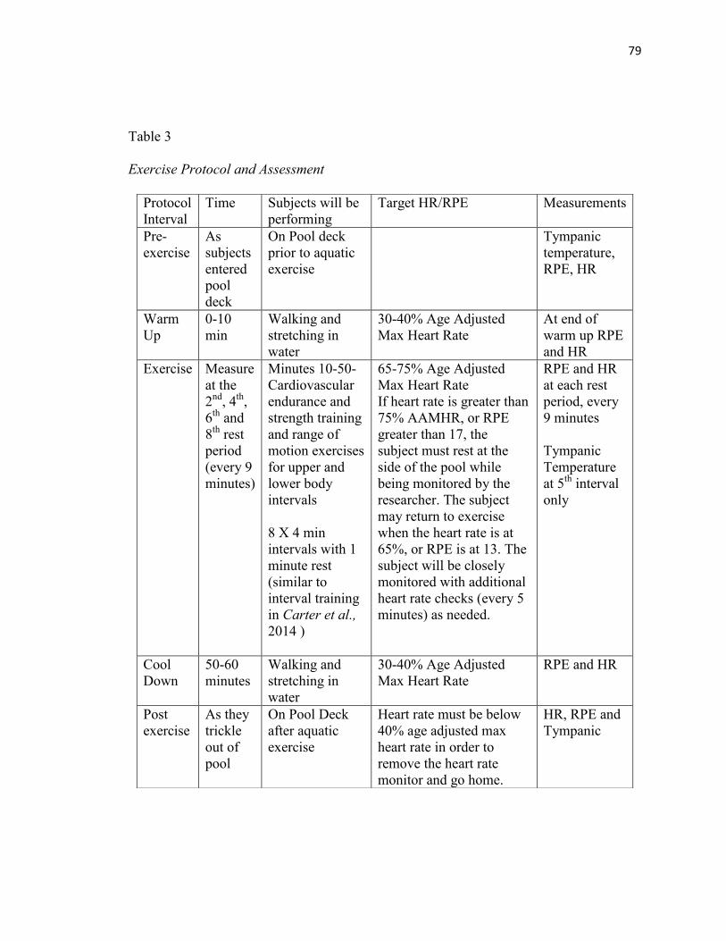
79
Table 3
Exercise Protocol and Assessment
Protocol
Interval
Time Subjects will be
performing
Target HR/RPE Measurements
Pre-
exercise
As
subjects
entered
pool
deck
On Pool deck
prior to aquatic
exercise
Tympanic
temperature,
RPE, HR
Warm
Up
0-10
min
Walking and
stretching in
water
30-40% Age Adjusted
Max Heart Rate
At end of
warm up RPE
and HR
Exercise Measure
at the
2nd
, 4th
,
6th
and
8th
rest
period
(every 9
minutes)
Minutes 10-50-
Cardiovascular
endurance and
strength training
and range of
motion exercises
for upper and
lower body
intervals
8 X 4 min
intervals with 1
minute rest
(similar to
interval training
in Carter et al.,
2014 )
65-75% Age Adjusted
Max Heart Rate
If heart rate is greater than
75% AAMHR, or RPE
greater than 17, the
subject must rest at the
side of the pool while
being monitored by the
researcher. The subject
may return to exercise
when the heart rate is at
65%, or RPE is at 13. The
subject will be closely
monitored with additional
heart rate checks (every 5
minutes) as needed.
RPE and HR
at each rest
period, every
9 minutes
Tympanic
Temperature
at 5th
interval
only
Cool
Down
50-60
minutes
Walking and
stretching in
water
30-40% Age Adjusted
Max Heart Rate
RPE and HR
Post
exercise
As they
trickle
out of
pool
On Pool Deck
after aquatic
exercise
Heart rate must be below
40% age adjusted max
heart rate in order to
remove the heart rate
monitor and go home.
HR, RPE and
Tympanic
80
Body Composition
Subjects were assessed in body composition through body mass index, waist
circumference, and waist to hip ratio. Body mass index (BMI) is a measure of body fat
based on height and weight that applies to adults. Height was plotted against the
individual’s weight to assess where they fall on the BMI scale. Body mass index in
adolescence has been correlated to incidence of MS (Munger, Chitnis, & Ascherio,
2009). BMI and waist to hip ratio is recommended by an International Consensus
Meeting on the core measures in MS (Paul et al, 2014).
The measurements to determine waist circumference were taken just above the
iliac crest at the end of exhalation. Measures were at the waist (midpoint between lower
margin of the last palpable rib and the iliac crest with a stretch resistant tape providing
100g tension) and Hip (widest portion of the buttocks with take parallel to the floor).
Measures were taken with the individual standing, feet together, arms at sides at the end
of expiration. Two measures were recorded and if within 1 cm of each other, the
measures were averaged. If the measures are not within 1 cm, the measurements were
retaken.
Heart Rate and Training Intensity
A Polar Heart Rate Monitor was used to record resting heart rate and heart rate
variability during the first and final visits. Resting heart rate reflects the cardiovascular
health of an individual. A lower heart rate correlates to more efficient heart function and
better cardiovascular fitness.
81
Heart Rate Variability (HRV) was recorded using a Polar Heart Rate Monitor
RS800CXTM
. HRV is the rhythmic periodicity of the sinoatrial neural discharge that is
regulated by the balance between the sympathetic and parasympathetic autonomic
nervous system (Meersman, 1993; Cygankiewicz & Zareba, 2013). HRV decreases with
aging by shifting to sympathetic dominance, while aerobic exercise maintains and
augments HRV (Meersman, 1993). As the body adapts to aerobic exercise, there is a
decrease in resting heart rate and an increase in HRV. Aerobic exercise improves
cardiovascular function by increasing the efficiency of the heart and increasing
parasympathetic regulation which is beneficial to the MS population. HRV is
significantly lower in MS individuals indicating a loss of vagal tone and increase in
sympathetic tone (Cygankiewicz & Zareba, 2013). The autonomic dysfunction is
associated with plaques in the brainstem and spinal cord and the degree of autonomic
impairment increases with advancement of the disease (Cygankiewicz & Zareba, 2013).
Mahovic and Lalusic (2007), found a significantly lower HRV in MS individuals with a
diagnosis of 5 years or greater as compared to those with a shorter duration of the
disease.
Heart rate was used to determine exercise training intensity as calculated through
the Karvonen Method: ([220 – age] – resting heart rate to determine the heart rate
reserve. The target heart rate was determined by: [heart rate reserve x training %] +
resting heart rate) (Karvonen, Kentala, &Mustala, 2007). Subjects were encouraged to
work at 65-75% age adjusted maximum heart rate during the interval workout. Mostert
82
and colleagues (2002) determined that it was a safe and efficient training intensity to
work at anaerobic threshold (as determined by gas exchange) in MS individuals.
For the exercise group, heart rate was monitored before getting into the water,
after the warm up, every 9 minutes throughout the exercise session, after the cool down,
and again on the pool deck after the entire exercise session was completed. MS
participants were instructed on target training intensity, and encouraged to self-monitor
heart rate and adjust intensity level as appropriate. Individuals that reach a target heart
rate at or greater than 75% AAMHR or an RPE greater than 17 were required to stand
along the wall while being monitored by the researcher. They were allowed to return to
exercise when their heart rate was 65%, and RPE was at 13. The subjects were closely
monitored to ensure they do not exercise to exhaustion. Upon completion of the exercise
session, heart rate was required to be below 40% age adjusted max heart rate in order to
remove the heart rate monitor and go home.
Rate of Perceived Exertion
Rate of Perceived Exertion is a self-reported scale that is used to measure feelings
of effort, strain, discomfort, and fatigue experienced during exercise. Using the Borg 6-
20 scale, the subject indicated his/her intensity level by pointing out numerically where
he/she was on the scale. Rate of Perceived Exertion was assessed using Borg’s Rate of
Perceived Exertion Scale at the same time intervals the HR was measured.
Solari and colleagues (1999) recommend that MS patients work at a moderate exertion
level of 11-14 on the Borg scale.
83
Mobility Assessments
The Timed 25 Foot Walk (T25-FW) is the first test in the Multiple Sclerosis
Functional Composite (MSFC). The MSFC is used to measure lower limb function (T25-
FW), upper limb function (9 hole peg test), and cognitive function (Paced Auditory Serial
Addition Test) (Potter et al., 2012, Kieseier & Pozzilli, 2012). The T25-FW is a
quantitative mobility and leg function performance test based on a timed 25-walk. The
patient was instructed: “I’d like you to walk 25 feet as quickly as possible, but safely. Do
not slow down until after you’ve passed the finish line. Ready? Go”. The time was
calculated from the initiation of the instruction to start and ends when the patient has
reached the 25-foot mark. The task was immediately administered again by having the
patient walk back the same distance. Patients were allowed to use assistive devices when
doing this task.
The T25-FW is an objective valid assessment of walking disability across a wide
range of walking disabilities (Kieseier & Pozzilli, 2012). The T25-FW has an intra-class
correlation of >0.8 and is highly reliable (inter-rater ICC 0.942, intra-rater ICC 0.99)
(Kieseier & Pozzilli, 2012). It does not exhibit practice effects (Kieseier & Pozzilli,
2012). Twenty percent deterioration in scores indicates a clinically meaningful decrease
in gait (Potter et al., 2012; Kieseier & Pozzilli, 2012).
The two minute step test was used to assess aerobic endurance. For this
assessment, the subject marched in place with a full step consisting of raising the knee
84
midway between the patella and the iliac crest. The score was the number of times the
right knee reached the required height (Jones & Rikli, 2002).
Rikli & Jones (2012), established a validity coefficient of 0.91 and a test-retest
reliability of 0.88 for the 2 minute step test. The 2 minute step test is demonstrated as
being a feasible and accurate tool for measuring cardiovascular fitness in older adults
(Rikli & Jones, 2012).
The Timed Up and Go Test (TUG) was used to assess basic mobility and dynamic
balance of the individual by recording the time to stand, walk 3 meters, turn, and walk
back to the chair and sit down. A firm chair with arms with a seat height of 46 cm was
used, set 3 meters away from a cone. The subject began in the seated position with their
back against the chair, arms on the lap, and the feet just behind the distance marker on the
floor. The subject was instructed “on the word ‘go’, stand up, walk comfortably and
safely to the cone on the floor, walk around the cone, come back, sit all the way back in
your chair.” The subject was timed from the word ‘go’ until the subject was back in their
seat with their back against the chair. Two practice trials were averaged.
TUG performance time is strongly correlated to the level of functional mobility in
the older adult with those completing it in less than 20 seconds were shown to be
independent in activities of daily living, and have higher Berg Balance Scores and walk
speeds (0.5 m/s) (Shumway-Cook, Brauer, & Woollacott, 2000). Shumway-Cook and
colleagues (2000) found that the TUG test had a validity of 87% sensitivity and 87%
specificity in older adults. A TUG score of 13.5 seconds or greater in elderly individuals
85
is correlated to increased risk of falls and has a prediction rate of 90% (Shumway-Cook,
Brauer, & Woollacott, 2000). An average score in MS patients is around 13.9 seconds
(Potter et al., 2012). Podsiadlo & Richardson (1991) concluded that the TUG test is a
valid and reliable test for quantifying functional mobility in elderly individuals with
neurological disorders.
Physiological Variables
Blood Pressure was used to assess the pressure exerted by the circulating blood
on the blood vessel walls which was a reflection of cardiovascular health and possibly
autonomic nervous system regulation. To measure blood pressure a blood pressure cuff
and sphygnanometer were used to detect systolic and diastolic blood pressure. Mean
arterial pressure was calculated as diastolic pressure + 1/3 (Systolic pressure – diastolic
pressure). Mean arterial pressure reflects the average blood pressure in an individual.
Body temperature was recorded tympanically ( Braun ThermoScan 5 ) at the first
and last appointment in all participants. In the exercise group, tympanic temperature was
measured on the pool deck before exercise, and during the rest period of the 5th
interval.
Multiple sclerosis individuals often suffer from an exacerbation of symptoms
when body temperature rises. Temperature changes between 0.18 degrees Fahrenheit and
4.14 degrees Fahrenheit have produced exacerbation of symptoms (Peterson, 2001).
While these symptoms reverse once the body has cooled, preventing changes in body
temperature alleviates these exacerbations. Exercising in an aquatic environment allows
86
for the added benefit of body heat dissipation at a greater rate than other environments,
minimizing heat sensitivity.
Costello and colleagues recommend temperature assessment in MS done with
tympanic membrane thermometers for moderate to vigorous activity (Costello, Curtis,
Sandel, Bassile, 1996). Tympanic temperature measures are valid measures of brain
temperature that reliably reflects core body temperature (Syndulko, Jafari, Woldanski,
Baumhefner, & Tourtellotte, 1996).
Cerebral oxygenation was noninvasively measured through Near Infra-Red
Spectroscopy (NIRS) which assesses cerebral oxygenation in real time at about 1 cm of
cortical tissue (Lintas, Molinari, Simonetti, Franzini, & Liboni, 2013). NIRS is able to
quantify the concentrations of oxyhemoglobin and deoxyhemoglobin using multiple
wavelengths. In healthy individuals during exercise, cerebral blood flow globally does
not change, however local cerebral blood flow increases to the sensorimotor cortex by up
to 30% (Ide & Secher, 2000). In MS individuals a hypometabolic component may result
from vascular changes (Ge et al., 2009). NIRS measurements have shown that exercise
increases oxyhemoglobin, providing for the oxygen needs of the brain tissue, which can
be beneficial in MS patients (Rooks, Thom, McCully, & Dishman, 2010).
NIRS recordings were made using a commercially made oximeter (Oxymon
MKIII, Artinis Medical Systems, Netherlands). The NIRS probe was secured on the
forehead on the midline, 1 cm above the supraorbital ridge similar to as described by
Lintas and colleagues (2013). Measurements were taken during the pre and post baseline
87
measures with the subject seated and eyes closed. Subjects will be asked to rest for 8
minutes while baseline measures were recorded. The last minute of rest with values
taken every 5 seconds were averaged to get a resting value. After baseline values were
recorded, subjects were asked to open their eyes and a cognitive task was explained (N-
back test) during the NIRS measurement. The purpose was to determine if there exist
any differences due to a 7 day aquatic aerobic exercise protocol in cerebral oxygenation,
cerebral deoxyhemoglobin, % tissue saturation index (%TSI) and total hemoglobin.
Because the NIRS does not give an absolute value, changes from baseline to N1 and from
baseline to N2 were used to determine if there was a difference from pre-test to post-test
for the exercise group compared to the non-exercise group.
Lintas and colleagues (2013) determined NIRS suitable for long term monitoring
of the effects on the brain metabolism and vasomotor reactivity and for physiological and
neuroscience experimental protocols. Soraghan and colleagues (2008) validated the use
of NIRS to measure hemodynamics during cognitive tasks.
The n-Back test is a valid and reliable indicator of working memory (Schmiedek,
Lovden, & Lindenberger, 2014). It requires continuous recognition of a stimulus (letter,
number or picture) in a particular sequence and the subject must judge whether it matches
the one presented n-items ago (Kane, Conway, Miura, & Colflesh, 2007).
Multiple Sclerosis Self-Efficacy Scale
Self-efficacy is defined as how the individual perceives their capability in
completing a specific task. The scale was developed specifically for Multiple Sclerosis
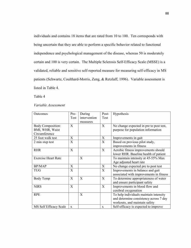
88
individuals and contains 18 items that are rated from 10 to 100. Ten corresponds with
being uncertain that they are able to perform a specific behavior related to functional
independence and psychological management of the disease, whereas 50 is moderately
certain and 100 is very certain. The Multiple Sclerosis Self-Efficacy Scale (MSSE) is a
validated, reliable and sensitive self-reported measure for measuring self-efficacy in MS
patients (Schwartz, Coulthard-Morris, Zeng, & Retzlaff, 1996). Variable assessment is
listed in Table 4.
Table 4
Variable Assessment
Outcomes Pre-
Test
During
intervention
measures
Post-
Test
Hypothesis
Body Composition:
BMI, WHR, Waist
Circumference
X X No change expected in pre to post test,
purpose for population information
25 foot walk test X X Improvements in gait
2 min step test X X Based on previous pilot study,
improvements in fitness
RHR X X Aerobic fitness improvements should
lower RHR. Baseline health of patient
Exercise Heart Rate X To maintain intensity at 45-55% Max
Age adjusted heart rate
BP/MAP X X No change expected pre to post test
TUG X X Improvements in balance and gait
associated with improvements in fitness
Body Temp X X X To determine appropriateness of water
and ensure participant safety
NIRS X X Improvements in blood flow and
cerebral oxygenation
RPE X To help individuals maintain intensity
and determine consistency across 7 day
workouts, and maintain safety
MS Self Efficacy Scale x x Self-efficacy is expected to improve
89
Statistical Analysis
A power analysis to calculate required sample size was completed using pilot data
from older adults using a similar aquatic aerobic protocol (Fedor, 2014). The pilot study
step test data (mean 14.6 steps, standard deviation 24.5) was used to determine the
number of subjects (n=24) required to achieve a power of 0.8 with a Type I error
probability at 0.05.
Using IBM SPSS Statistics 20, descriptive measures (age, height, weight, duration
of MS, BMI, resting heart rate and resting blood pressure) were compared between the
two groups with an independent T-Test. The difference between body composition
(waist circumference, waist-to-hip ratio, and BMI) pre-test and post-test values were used
to calculate a change score. The exercise group and non-exercise group change scores
were compared by using independent samples T-Test. The difference between fitness
variables (resting heart rate, heart rate variability, resting blood pressure, mean arterial
pressure, 2 minute step test) pre-test and post-test values were used to calculate a change
score. The exercise group and non-exercise group change scores were compared using
independent samples T-Test. The differences between pre-test and post-test mobility
measures (TUG test, and T25-FW test) were used to calculate a change score. The
exercise group and control group were compared using independent samples T-Test. The
differences between pre-test and post-test cerebral oxygenation (at rest and during
cognitive task) were used to calculate a change score. The exercise group and non-
exercise group change scores were compared using independent samples T-Test. The

90
differences between pre-test and post-test MSSE were used to calculate a change score.
The exercise group and non-exercise group change scores were compared using
independent samples T-Test. Statistical significance was set at p < 0.05.
91
CHAPTER IV
EFFECTS OF A 7 DAY AQUATIC AEROBIC EXERCISE INTERVENTION ON
MOBILITY, FITNESS AND BODY COMPOSITION
Introduction
Multiple sclerosis (MS) is the leading cause of non-traumatic neurologic disability
in young adults (Solari et al., 1999). MS is caused by demyelination of axons, focal
plaque formation, and inflammation in the central nervous system which results in
progressive loss of function. The location of pathology results in varying combinations
of symptoms which include fatigue, heat sensitivity, muscle spasms, gait problems and
ataxia, dizziness and vertigo, pain, cognitive problems, visual complaints, and bowel and
bladder dysfunction (Anthony, Jouria, & Houtman, 2014).
Compounding the impact of the disease, symptoms resulting from MS pathology
result in decreased levels of physical activity (Hayes, Gappmaier, & LaStayo, 2011;
Marck et al., 2014) and subsequent deconditioning that reduces quality of life (Doring,
Pfueller, Paul, & Dorr, 2012). Historically, it was once thought that exercise exacerbated
MS symptoms and it was contraindicated in people with multiple sclerosis (Hayes,
Gappmaier, & LaStayo, 2011). When exercise increases body temperature, central
pathways become blocked producing temporary physical and cognitive symptoms in
individuals with MS (Davis, Wilson, White, & Frohman, 2010). Heat sensitivity
resulting in worsening symptoms depends on the location of the demyelination and
92
lesions, and frequently includes deficits in mobility, memory retrieval, processing speed,
multitasking, and increased fatigue (Davis, Wilson, White, & Frohman, 2010).
Current research supports the positive effects of exercise in MS treatment,
including improved fitness benefits, feelings of well-being, strength and safety of
mobility and decreased fatigue (Davis, Wilson, White, & Frohman, 2010, Rietberg,
Brooks, Uitdehagg, & Kwakkel, 2011). Exercise also has the capacity to slow disease
progression, improves the physiological profile of MS and is well tolerated with a low
occurrence of adverse effects (Kargarfard, Etemadifar, Baker, Mehrabi, & Hayatbakhsh,
2012). In addition, Pilutti and colleagues found exercise training is associated with a
slight decrease in the risk of relapse when compared to non-exercising MS individuals
(Pilutti, Platta, Motl, & Latimer-Cheung, 2014).
The primary aim of rehabilitation in MS individuals is to increase activity levels,
as well as their independence (Langdon & Thompson, 1999), by improving mobility
(Pilutti, et al., 2011; Kargarfard, Etemadifar, Baker, Mehrabi, & Hayatbakhsh, 2012),
improving physical functioning, and easing psychosocial burden (White, Castellano,
McCoy, Patel, & Giacobbi, 2012). Molt and Snook (2008) found evidence that physical
activity in MS was linked to a greater self-efficacy for function and control, which was
also correlated to greater physical and psychological components of quality of life. Self-
efficacy is the individual’s perception of his or her capability in completing a specific
task, and may play a role in how the individual adjusts to MS symptoms and the
psychosocial burden of the disease (Molt & Snook, 2008).
93
Due to fatigue and overheating issues, MS individuals are generally less
physically active than healthy individuals of similar age (Hayes, Gappmaier, & LaStayo,
2011). Heat sensitivity, an increase in body temperature that exacerbates symptoms,
affects an estimated 60-80% of MS individuals (Davis, Wilson, White & Frohman, 2010)
and is a major barrier to exercise. Worsening of symptoms have been observed with
increased body temperature of approximately 0.8C (Davis, Wilson, White & Frohman,
2010). Aquatic exercise is beneficial in MS individuals because of the ability to transfer
body heat to the water 25 times faster than air (Frohman, Okuda, Beh, Treadaway, Mooi,
Davis, Shah, Frohman & Frohman, 2015), limiting the increases in body temperature that
cause the increase in MS symptoms. In addition, the buoyancy and viscosity of water
assists those who have physical weakness (Kargarfard, Etemadifar, Baker, Mehrabi, &
Hayatbakhsh, 2012). Aquatic exercise improves neurological functioning, improves
activities of daily living (Frohman, Okuda, Beh, Treadaway, Mooi, Davis, Shah,
Frohman & Frohman, 2015), reduces joint stress and decreases the fear of falling
(Plecash & Leavitt, 2014).
Relapsing Remitting (RRMS) is the most common form of MS, accounting for
85% of Multiple Sclerosis cases (“Types of MS,” n.d.; Berger et al., 2003). RRMS is
characterized by exacerbations followed by periods of remission, and is attributed to
demyelinating attacks followed by remyelination by the oligodendrocyte (Goldenberg,
2012). Participation in exercise between relapses has the potential to improve gait,
mobility, activities of daily living, and quality of life. Several studies have demonstrated
94
positive effects of exercise on gait parameters and quality of life using four weeks of
exercise training. However, no studies to date have used seven consecutive days of
exercise (van den Berg et al., 2006; Mostert & Kesselring, 2002), which may be
beneficial during times of remission. Previous work, in healthy older adults, showed that
both fitness and cognitive parameters were improved with a similar aquatic aerobic
exercise protocol in older adults (Fedor, 2014). Therefore, the purpose of this
investigation was to determine the potential benefits of seven consecutive days of water-
based exercise on mobility, fitness, body composition, and self-efficacy in multiple
sclerosis individuals. We hypothesized that a seven day moderate to high intensity water
aerobic exercise intervention would improve: 1) mobility as measured through the TUG
and T25-FW, 2) cardiovascular fitness by measuring resting heart rate and Two Minute
Step Test and 3) self-efficacy using the Multiple Sclerosis Self-efficacy questionnaire.
We hypothesized the exercise intervention would not alter body composition (body mass
index, waist height ration, Waist circumference) in individuals with MS.
Methods
The Kent State University Institutional Review Board approved all experimental
protocols. All participants voluntarily signed the written informed consent.
Participants
Prospective participants were recruited through a MS study Facebook page, ads in
the MS Connector and local newspapers, flyers distributed around town, local MS
support groups, and MS special events. Recruiting identified 68 individuals that
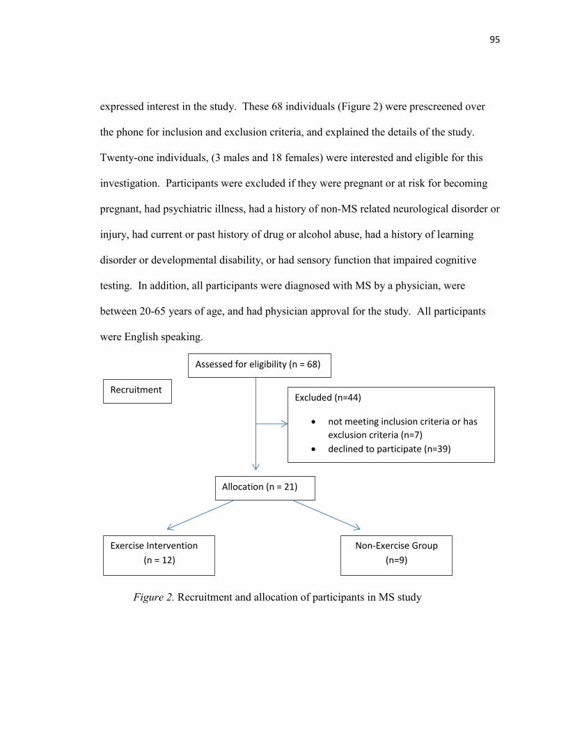
95
expressed interest in the study. These 68 individuals (Figure 2) were prescreened over
the phone for inclusion and exclusion criteria, and explained the details of the study.
Twenty-one individuals, (3 males and 18 females) were interested and eligible for this
investigation. Participants were excluded if they were pregnant or at risk for becoming
pregnant, had psychiatric illness, had a history of non-MS related neurological disorder or
injury, had current or past history of drug or alcohol abuse, had a history of learning
disorder or developmental disability, or had sensory function that impaired cognitive
testing. In addition, all participants were diagnosed with MS by a physician, were
between 20-65 years of age, and had physician approval for the study. All participants
were English speaking.
Figure 2. Recruitment and allocation of participants in MS study
Recruitment
Assessed for eligibility (n = 68)
Excluded (n=44)
not meeting inclusion criteria or has
exclusion criteria (n=7)
declined to participate (n=39)
Allocation (n = 21)
Exercise Intervention
(n = 12)
Non-Exercise Group
(n=9)
96
Experimental Protocol
Participants arrived at their scheduled time for the pre-test at the Kent State
University Exercise Physiology laboratory. After reviewing and signing the written
informed consent, anthropometric measurements including height, weight, and hip and
waist circumference were collected. Participants were fitted with a Polar heart rate
monitor (RS800CX) which was used to assess resting heart rate. Body temperature was
taken tympanically using a Braun Thermoscan 5. Resting blood pressure was recorded
and mean arterial pressure, body mass index (BMI) and waist-to-hip ratio (WHR) were
calculated. The participants then completed a series of tests including the two minute
step test, twenty-five foot walk test, and timed up and go test (TUG). The two minute
step test was administered by having the participant march in place for two minutes
following the protocol by Jones & Rikli (2002). The distance midway between the
patella and the iliac crest was measured and then marked on the wall. The score was
determined by the number of times the right knee reached the required height. The 25
foot walk (T25-FW) test was given by instructing the participant to walk as quickly but
as safely as possible the length of 25 feet. The time was recorded from the word “Go”
until the participant passed the 25- foot mark. The participant then completed a second
trial of the same task immediately by having them walk back the same distance.
Participants were allowed to use assistive devices while completing the T25-FW and the
TUG test. Participants used the same assistive device for the post-test. The timed up and
go test (TUG) (Podsiadlo & Richardson, 1991) was administered by having the
97
participant start in the seated position, hands on their lap, back against the chair. On the
word “go” they were instructed to stand up, walk quickly but safely to the cone on the
floor, walk around the cone, come back and sit in the chair. The cone was placed at 3
meters and the timer was stopped when they were seated with their back against the chair.
After each participant completed the mobility, fitness, and body composition
measures, they were given the Multiple Sclerosis Self–Efficacy questionnaire (MSSE), a
self-reported measure. The MSSE is composed of 2 parts, 9 questions assessing function
and 9 questions assessing control. Each question was weighted at 100 points, with a total
of 900 points from each section. The function questions seek to determine the
participant’s sense of confidence that they can perform behaviors that allow them to
engage in daily living activities. The control questions assess the level of confidence that
MS individuals can control their symptoms and their reactions to disease related
limitations and the impact of the disease on life activities. A total score was determined
by combining the two sections together for a total of 1800 possible points. Higher scores
indicate higher self-efficacy (Riazi, Thompson, & Hobart, 2004). The post-test was
given on day 9 and mirrored the pretest protocol.
Non-exercise Participants
Participants in the non-exercise group were asked to maintain their current
lifestyle during the 7 days between the pre-test and post-test. They were instructed to
continue their usual activities of daily living, and were asked not to start a new exercise
program during the study.
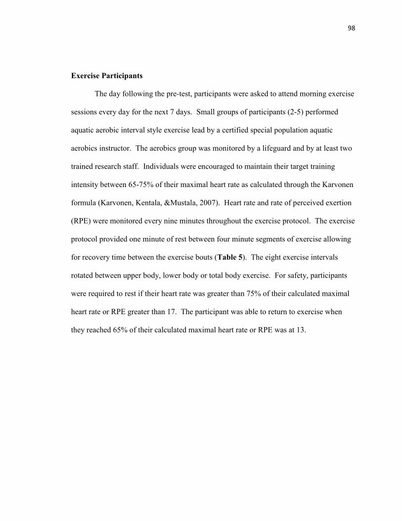
98
Exercise Participants
The day following the pre-test, participants were asked to attend morning exercise
sessions every day for the next 7 days. Small groups of participants (2-5) performed
aquatic aerobic interval style exercise lead by a certified special population aquatic
aerobics instructor. The aerobics group was monitored by a lifeguard and by at least two
trained research staff. Individuals were encouraged to maintain their target training
intensity between 65-75% of their maximal heart rate as calculated through the Karvonen
formula (Karvonen, Kentala, &Mustala, 2007). Heart rate and rate of perceived exertion
(RPE) were monitored every nine minutes throughout the exercise protocol. The exercise
protocol provided one minute of rest between four minute segments of exercise allowing
for recovery time between the exercise bouts (Table 5). The eight exercise intervals
rotated between upper body, lower body or total body exercise. For safety, participants
were required to rest if their heart rate was greater than 75% of their calculated maximal
heart rate or RPE greater than 17. The participant was able to return to exercise when
they reached 65% of their calculated maximal heart rate or RPE was at 13.
99
Table 5
Aquatic Aerobic Exercise Protocol
Statistical Analysis
The exercise and non-exercise group’s baseline measures were compared using
independent samples T-tests. All measures for mobility, fitness, body composition, and
self-efficacy were compared by calculating a change score, then comparing the two
groups using independent samples T-tests. The change score was determined by
subtracting the pre-test scores from the post-test scores for each variable. Mobility was
Protocol
Interval
Time Subjects will be
performing
Target HR/RPE Measurements
Pre-
exercise
As
subjects
trickle in
On Pool deck
prior to aquatic
exercise
Tympanic
temperature,
RPE, HR
Warm
Up
0-10
min
Walking and
stretching in water
30-40% Age Adjusted Max
Heart Rate
At end of warm
up RPE and HR
Exercise Measure
at the
2nd
, 4th,
6th and
8th rest
period
(every 9
minutes)
Minutes 10-50-
Cardiovascular
endurance and
strength training
and range of
motion exercises
for upper and
lower body
intervals
8 X 4 min
intervals with 1
minute rest
65-75% Age Adjusted Max
Heart Rate
If heart rate is greater than
75% AAMHR, or RPE
greater than 17, the subject
must rest at the side of the
pool while being monitored
by the researcher. The subject
may return to exercise when
the heart rate is at 65%, or
RPE is at 13. The subject will
be closely monitored with
additional heart rate checks
(every 5 minutes) as needed.
RPE and HR at
each rest
period, every 9
minutes
Tympanic
Temperature at
40 min only
Cool
Down
50-60
minutes
Walking and
stretching in water
30-40% Age Adjusted Max
Heart Rate
RPE and HR
Post
exercise
As they
come
out of
pool
On Pool Deck
after aquatic
exercise
Heart rate must be below
40% age adjusted max heart
rate in order to remove the
heart rate monitor and go
home.
HR, RPE
100
measured through TUG and T25FW, and cardiovascular fitness was assessed through
resting heart rate, resting blood pressure, mean arterial pressure, two minute step test.
Anthropometric measures included waist circumference, waist-to-hip ratio and BMI, and
weight. Self-efficacy was measured through the Multiple Sclerosis Self-Efficacy
questionnaire (MSSE). The MSSE consisted of two sections (control and function)
which were analyzed independently and then together for a total score. Statistical
significance was set at p< 0.05. Effect size for the mobility, fitness, body composition,
and self-efficacy variables was calculated through Cohen’s d. Cohen’s d less than or
equal to 0.2 represents a small effect size, 0.5 a moderate effect size, and 0.8 a large
effect size.
Results
Twenty-one individuals with twelve participants in the exercise group (9 females,
3 males) and nine participants in the non-exercise group (9 females) completed the study.
Recruiting began in January 2015 and continued through September 2015. Participants
were allocated to the exercise and non-exercise group based on a combination of random
assignment and convenience sampling, depending on their availability for pool exercise.
The aquatic exercise protocol was offered based on pool availability between 8am -10am
for each of the 4 exercise sessions; and to consistently offer morning exercise to
minimize the possibility of fatigue which often increases throughout the day.
There were no significant differences (Table 6) between the exercise and non-
exercise groups in age, disease duration, years of education, the resting heart rate, systolic
101
blood pressure, diastolic blood pressure, mean arterial pressure, temperature, 2 minute
step test, TUG test, T25-FW, waist circumference, hip circumference, waist-to-hip ratio,
and height. The exercise group had a significantly higher BMI and weight compared to
the non-exercise group.
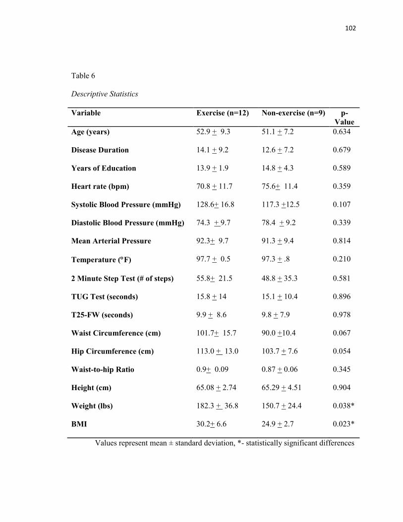
102
Table 6
Descriptive Statistics
Variable Exercise (n=12) Non-exercise (n=9) p-
Value
Age (years) 52.9 + 9.3 51.1 + 7.2 0.634
Disease Duration 14.1 + 9.2 12.6 + 7.2 0.679
Years of Education 13.9 + 1.9 14.8 + 4.3 0.589
Heart rate (bpm) 70.8 + 11.7 75.6+ 11.4 0.359
Systolic Blood Pressure (mmHg) 128.6+ 16.8 117.3 +12.5 0.107
Diastolic Blood Pressure (mmHg) 74.3 + 9.7 78.4 + 9.2 0.339
Mean Arterial Pressure 92.3+ 9.7 91.3 + 9.4 0.814
Temperature (F) 97.7 + 0.5 97.3 + .8 0.210
2 Minute Step Test (# of steps) 55.8+ 21.5 48.8 + 35.3 0.581
TUG Test (seconds) 15.8 + 14 15.1 + 10.4 0.896
T25-FW (seconds) 9.9 + 8.6 9.8 + 7.9 0.978
Waist Circumference (cm) 101.7+ 15.7 90.0 +10.4 0.067
Hip Circumference (cm) 113.0 + 13.0 103.7 + 7.6 0.054
Waist-to-hip Ratio 0.9+ 0.09 0.87 + 0.06 0.345
Height (cm) 65.08 + 2.74 65.29 + 4.51 0.904
Weight (lbs) 182.3 + 36.8 150.7 + 24.4 0.038*
BMI 30.2+ 6.6 24.9 + 2.7 0.023*
Values represent mean ± standard deviation, *- statistically significant differences
103
Exercise Variables
The exercise group worked during the intense exercise intervals at 53.7% of +
10.11 age predicted maximal heart rate. Their RPE ranged from 8-17 with a mean of
10.8 + 2.24. The exercise protocol required individuals to rest if their heart rate was
greater than their target heart rate (65-75% maximal heart rate) which was calculated
through the Karvonen formula (Karvonen, Kentala, &Mustala, 2007), or a RPE greater
than 17. None of the individuals in the exercise group reported an RPE above 17 or
exhibited a heart rate above their calculated target heart rate. The mean body temperature
was 97.8 + 0.95 degrees Fahrenheit during the exercise intervals.
Mobility Measures
Mobility was assessed through the TUG and T25-FW (exercise n=11, non-
exercise n=9). One participant’s data was removed from analysis for the TUG Test, T25-
FW, and 2 Minute Step test due to their extremely high level of disability which was
greater than any other individuals.
In the individual analysis of Timed up and Go (TUG), improvement was defined
as scores below zero, whereas decreases in performance on the TUG test were defined as
scores above zero. It is interesting to note that 82% (9/11 individuals, Figure 3) of the
exercise participants improved their TUG test performance, while only 66% (6/9
individuals, Figure 4) of the non-exercise group showed improvement. It is also
important to note that while the non-exercise group were fairly consistent in their pre-test
and post-test scores, the individuals in the exercise group showed changes in both
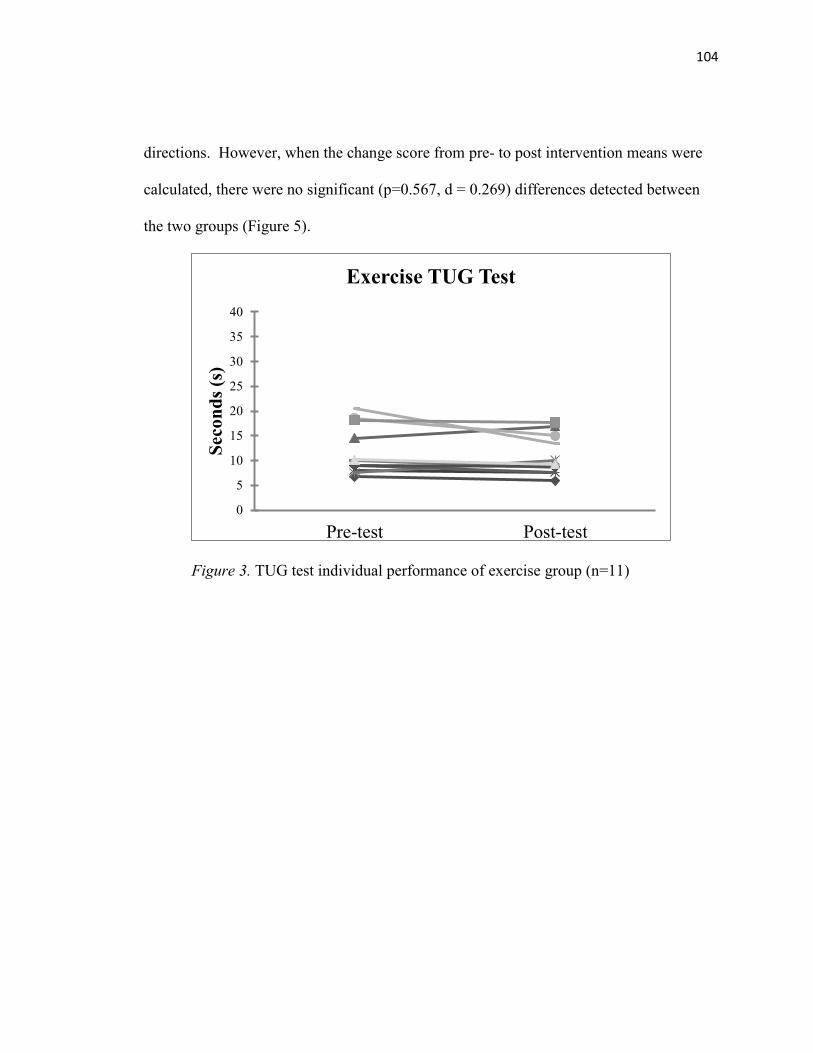
104
directions. However, when the change score from pre- to post intervention means were
calculated, there were no significant (p=0.567, d = 0.269) differences detected between
the two groups (Figure 5).
Figure 3. TUG test individual performance of exercise group (n=11)
0
5
10
15
20
25
30
35
40
Pre-test Post-test
Sec
on
ds
(s)
Exercise TUG Test
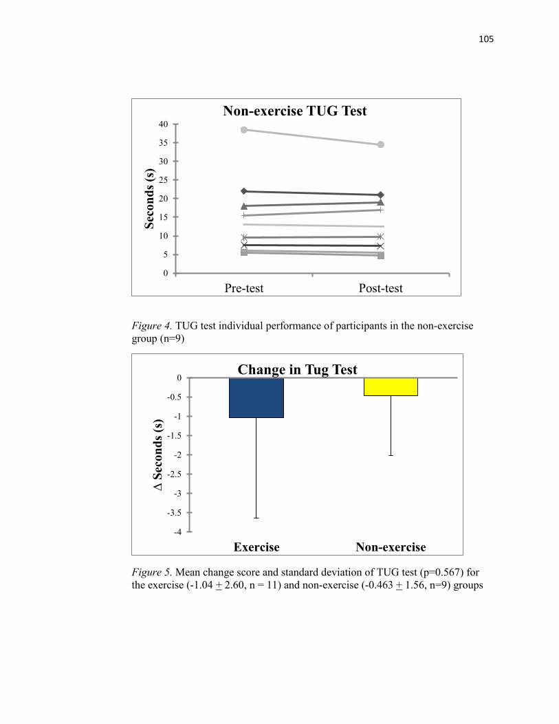
105
Figure 4. TUG test individual performance of participants in the non-exercise
group (n=9)
Figure 5. Mean change score and standard deviation of TUG test (p=0.567) for
the exercise (-1.04 + 2.60, n = 11) and non-exercise (-0.463 + 1.56, n=9) groups
0
5
10
15
20
25
30
35
40
Pre-test Post-test
Sec
on
ds
(s)
Non-exercise TUG Test
-4
-3.5
-3
-2.5
-2
-1.5
-1
-0.5
0
Exercise Non-exercise
∆ S
econ
ds
(s)
Change in Tug Test
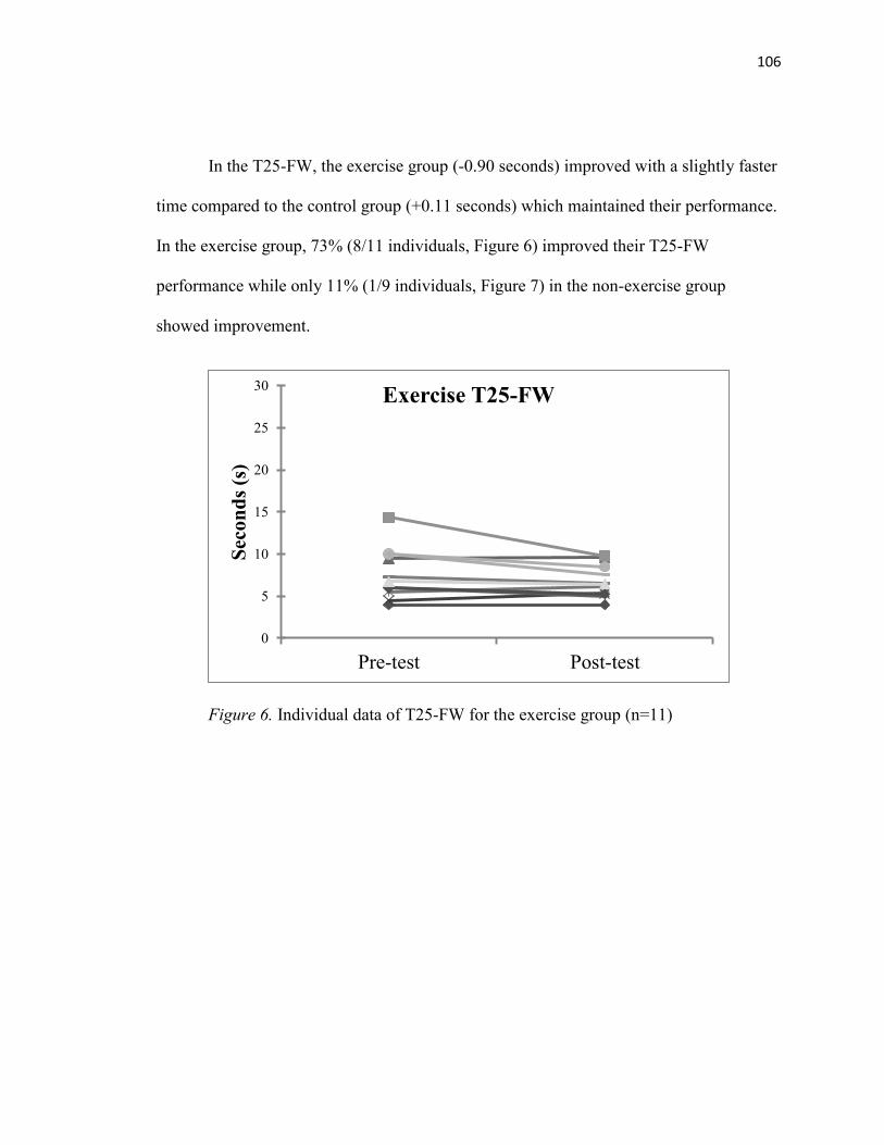
106
In the T25-FW, the exercise group (-0.90 seconds) improved with a slightly faster
time compared to the control group (+0.11 seconds) which maintained their performance.
In the exercise group, 73% (8/11 individuals, Figure 6) improved their T25-FW
performance while only 11% (1/9 individuals, Figure 7) in the non-exercise group
showed improvement.
Figure 6. Individual data of T25-FW for the exercise group (n=11)
0
5
10
15
20
25
30
Pre-test Post-test
Sec
on
ds
(s)
Exercise T25-FW
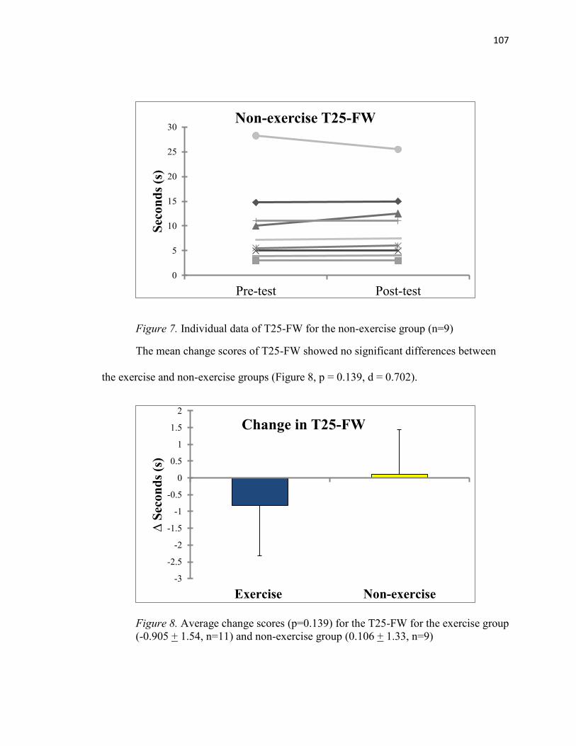
107
Figure 7. Individual data of T25-FW for the non-exercise group (n=9)
The mean change scores of T25-FW showed no significant differences between
the exercise and non-exercise groups (Figure 8, p = 0.139, d = 0.702).
Figure 8. Average change scores (p=0.139) for the T25-FW for the exercise group
(-0.905 + 1.54, n=11) and non-exercise group (0.106 + 1.33, n=9)
0
5
10
15
20
25
30
Pre-test Post-test
Sec
on
ds
(s)
Non-exercise T25-FW
-3
-2.5
-2
-1.5
-1
-0.5
0
0.5
1
1.5
2
Exercise Non-exercise
∆ S
econ
ds
(s)
Change in T25-FW
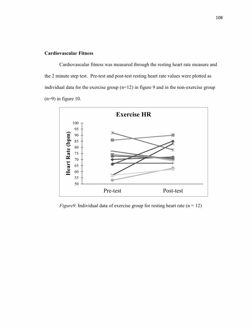
108
Cardiovascular Fitness
Cardiovascular fitness was measured through the resting heart rate measure and
the 2 minute step test. Pre-test and post-test resting heart rate values were plotted as
individual data for the exercise group (n=12) in figure 9 and in the non-exercise group
(n=9) in figure 10.
Figure9. Individual data of exercise group for resting heart rate (n = 12)
50
55
60
65
70
75
80
85
90
95
100
Pre-test Post-test
Hea
rt R
ate
(b
pm
)
Exercise HR
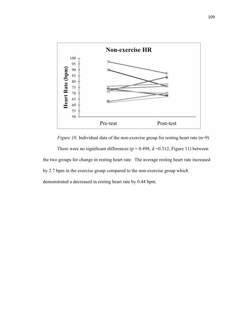
109
Figure 10. Individual data of the non-exercise group for resting heart rate (n=9)
There were no significant differences (p = 0.498, d =0.312, Figure 11) between
the two groups for change in resting heart rate. The average resting heart rate increased
by 2.7 bpm in the exercise group compared to the non-exercise group which
demonstrated a decreased in resting heart rate by 0.44 bpm.
50
55
60
65
70
75
80
85
90
95
100
Pre-test Post-test
Hea
rt R
ate
(b
pm
)
Non-exercise HR
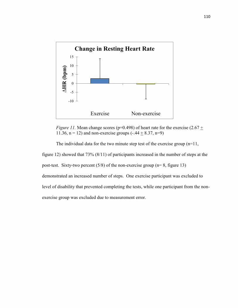
110
Figure 11. Mean change scores (p=0.498) of heart rate for the exercise (2.67 +
11.36, n = 12) and non-exercise groups (-.44 + 8.37, n=9)
The individual data for the two minute step test of the exercise group (n=11,
figure 12) showed that 73% (8/11) of participants increased in the number of steps at the
post-test. Sixty-two percent (5/8) of the non-exercise group (n= 8, figure 13)
demonstrated an increased number of steps. One exercise participant was excluded to
level of disability that prevented completing the tests, while one participant from the non-
exercise group was excluded due to measurement error.
-10
-5
0
5
10
15
Exercise Non-exercise
∆H
R (
bp
m)
Change in Resting Heart Rate
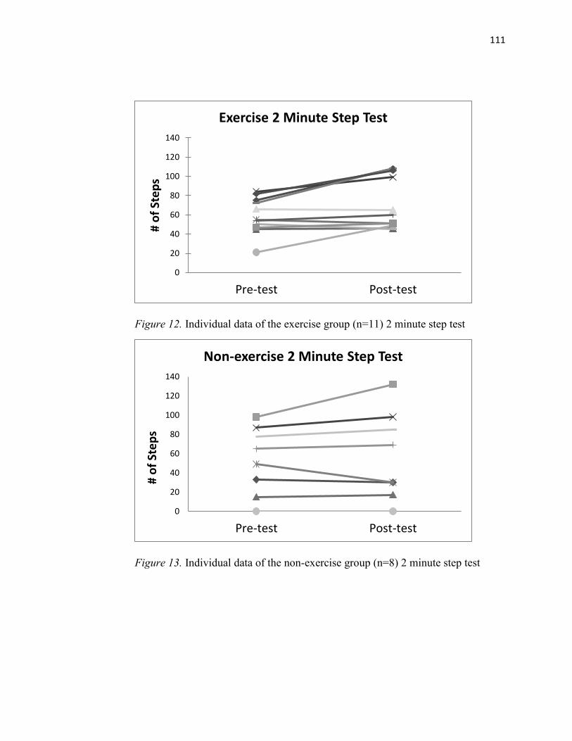
111
Figure 12. Individual data of the exercise group (n=11) 2 minute step test
Figure 13. Individual data of the non-exercise group (n=8) 2 minute step test
0
20
40
60
80
100
120
140
Pre-test Post-test
# o
f St
ep
s
Exercise 2 Minute Step Test
0
20
40
60
80
100
120
140
Pre-test Post-test
# o
f St
ep
s
Non-exercise 2 Minute Step Test
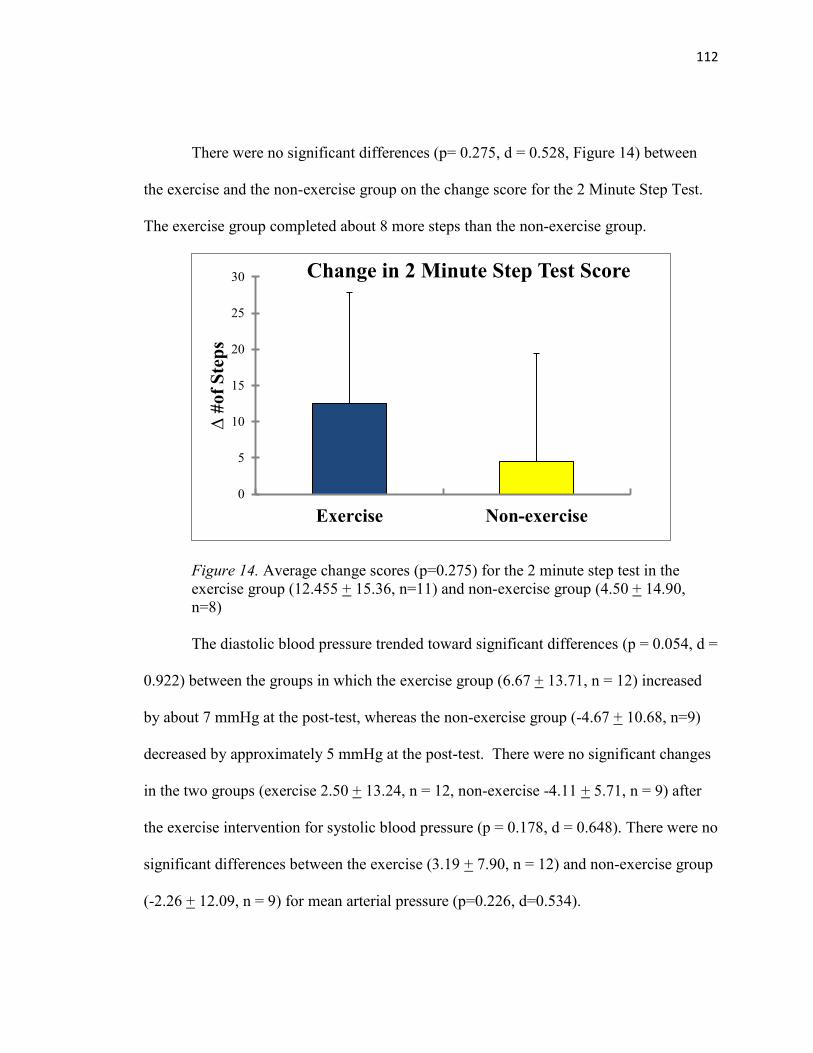
112
There were no significant differences (p= 0.275, d = 0.528, Figure 14) between
the exercise and the non-exercise group on the change score for the 2 Minute Step Test.
The exercise group completed about 8 more steps than the non-exercise group.
Figure 14. Average change scores (p=0.275) for the 2 minute step test in the
exercise group (12.455 + 15.36, n=11) and non-exercise group (4.50 + 14.90,
n=8)
The diastolic blood pressure trended toward significant differences (p = 0.054, d =
0.922) between the groups in which the exercise group (6.67 + 13.71, n = 12) increased
by about 7 mmHg at the post-test, whereas the non-exercise group (-4.67 + 10.68, n=9)
decreased by approximately 5 mmHg at the post-test. There were no significant changes
in the two groups (exercise 2.50 + 13.24, n = 12, non-exercise -4.11 + 5.71, n = 9) after
the exercise intervention for systolic blood pressure (p = 0.178, d = 0.648). There were no
significant differences between the exercise (3.19 + 7.90, n = 12) and non-exercise group
(-2.26 + 12.09, n = 9) for mean arterial pressure (p=0.226, d=0.534).
0
5
10
15
20
25
30
Exercise Non-exercise
∆ #
of
Ste
ps
Change in 2 Minute Step Test Score
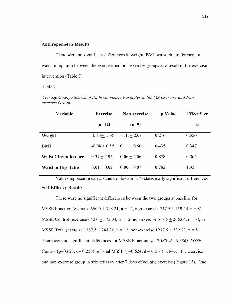
113
Anthropometric Results
There were no significant differences in weight, BMI, waist circumference, or
waist to hip ratio between the exercise and non-exercise groups as a result of the exercise
intervention (Table 7).
Table 7
Average Change Scores of Anthropometric Variables in the MS Exercise and Non-
exercise Group.
Variable Exercise
(n=12)
Non-exercise
(n=9)
p-Value Effect Size
d
Weight -0.14+ 1.68 -1.17+ 2.03 0.216 0.556
BMI -0.08 + 0.55 0.11 + 0.60 0.435 0.347
Waist Circumference 0.37 + 2.92 0.06 + 6.06 0.878 0.065
Waist to Hip Ratio 0.01 + 0.02 0.00 + 0.07 0.782 1.93
Values represent mean ± standard deviation, *- statistically significant differences
Self-Efficacy Results
There were no significant differences between the two groups at baseline for
MSSE Function (exercise 660.0 + 318.21, n = 12, non-exercise 747.5 + 159.44, n = 8),
MSSE Control (exercise 640.0 + 175.34, n = 12, non-exercise 617.5 + 266.64, n = 8), or
MSSE Total (exercise 1387.5 + 288.20, n = 12, non-exercise 1277.5 + 532.72, n = 8).
There were no significant differences for MSSE Function (p=0.380, d= 0.386), MSSE
Control (p=0.623, d= 0.225) or Total MSSE (p=0.624, d = 0.216) between the exercise
and non-exercise group in self-efficacy after 7 days of aquatic exercise (Figure 15). One
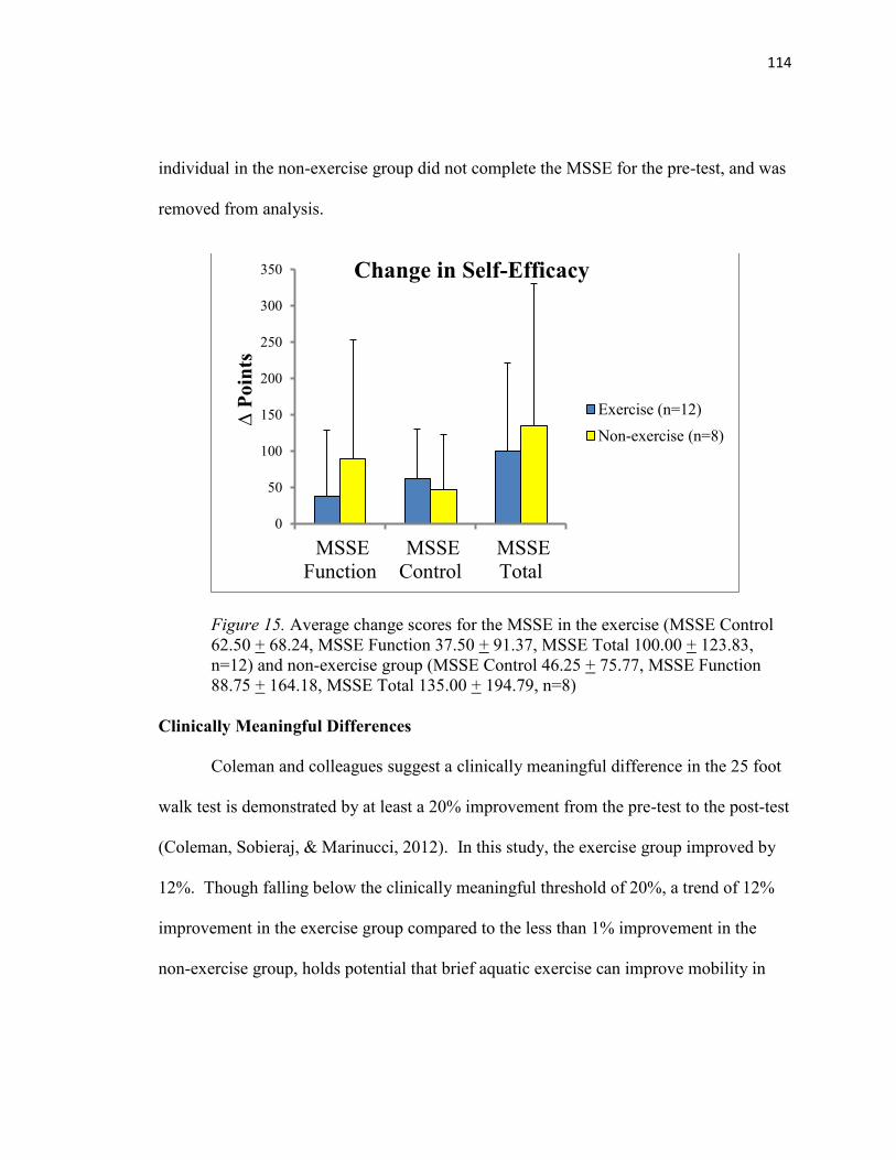
114
individual in the non-exercise group did not complete the MSSE for the pre-test, and was
removed from analysis.
Figure 15. Average change scores for the MSSE in the exercise (MSSE Control
62.50 + 68.24, MSSE Function 37.50 + 91.37, MSSE Total 100.00 + 123.83,
n=12) and non-exercise group (MSSE Control 46.25 + 75.77, MSSE Function
88.75 + 164.18, MSSE Total 135.00 + 194.79, n=8)
Clinically Meaningful Differences
Coleman and colleagues suggest a clinically meaningful difference in the 25 foot
walk test is demonstrated by at least a 20% improvement from the pre-test to the post-test
(Coleman, Sobieraj, & Marinucci, 2012). In this study, the exercise group improved by
12%. Though falling below the clinically meaningful threshold of 20%, a trend of 12%
improvement in the exercise group compared to the less than 1% improvement in the
non-exercise group, holds potential that brief aquatic exercise can improve mobility in
0
50
100
150
200
250
300
350
MSSE
Function
MSSE
Control
MSSE
Total
∆ P
oin
ts
Change in Self-Efficacy
Exercise (n=12)
Non-exercise (n=8)

115
MS. This is further supported by the moderate to large effect size for the 25 foot walk
test.
Foley and colleagues determined that minimal clinically important differences
(MCID) could be calculated for the TUG test by taking the mean value of the pre-
assessment and multiplying by 15% (Foley, Barnes, Hasson, 2015). The results fall
below the minimal clinically important differences (Table 8), however, there is a trend
toward improvement shown in the exercise group that is not shown in the non-exercise
group, with a small effect size present.
Table 8
Determining Clinically Significant Differences
Test Exercise
Group
Non-
Exercise
Group
Effect Size Standard of Clinically
Significant
25 Foot
Walk
Improved by
12%
Decrease by
< 1%
Moderate to
large
20% improvement (Coleman
and colleagues, 2012)
TUG Improved by
9%
3%
improvement
Small 15% improvement from pre-
test (Foley and colleagues,
2015)
2 Minute
Step
Increased #
steps by 21%
Increase #
steps by 18%
Moderate No standard found in
literature
Fedor, 2015 showed 12.3 %
improvement with an
exercise intervention in
elderly individuals
Discussion
This study showed that a short duration, moderately intense, aquatic exercise
intervention can produce small changes in mobility, fitness, and self-efficacy in Multiple
Sclerosis individuals. There were no adverse events or heat induced MS symptoms
116
during the exercise sessions. These results extend the conclusions of Snook and Motl
(2009), which found that exercise training programs are associated with small clinically
meaningful improvements in mobility of walking in MS individuals. There were no
statistically significant differences between the two groups for mobility, cardiovascular
fitness, or self-efficacy. Body composition was also analyzed, and as expected, changes
did not occur within the short intervention.
The exercise group demonstrated that sixteen percent more individuals improved
in the TUG test than in the non-exercise group. TUG mimics activities of daily living
such as rising and lowering from a chair and maneuvering to turn around. In the T25-
FW, greater improvements were shown as 62% more individuals in the exercise group
had a faster time than in the non-exercise group. Fitness also demonstrated an increase of
11% more exercise individuals improving in the number of steps compared to the non-
exercise group, with exercise participants taking on average 8 more steps than those in
the non-exercise group.
Land based exercise has been shown to produce benefits in MS individuals such
as improvements in mobility and balance (Lord, Wade, & Halligan, 1998) however,
aquatic exercise is favorable in MS for a variety of reasons. Aquatic exercise reduces
heat induced symptoms and stress on joints, reduces the risk of falls during exercise,
increases range of motion, and accommodates a variety of disability levels found in MS.
This study demonstrated the safety, benefits, and feasibility of aquatic exercise in MS
individuals.
117
The individual data for mobility and fitness often showed a mix of increases and
decreases in measures for the exercise group indicating the diversity of the disease
process and response to the exercise intervention. Participants were diverse in their MS
type, symptoms (fatigue, pain, spasm) and disability level, and this may have contributed
to the heterogeneous results found in each of the variables and their response to exercise.
The non-exercise group individual measures remained more consistent from pre-test to
post-test showing little change.
Participants in the exercise and non-exercise group did not differ in age, disease
duration, years of education, fitness, mobility, or body temperature. However, the
exercise group had a significantly higher BMI and body weight, which may partially be
explained by the inflexible exercise schedule. In order to participate in the exercise
group, individuals had to be available at the exercise time (8 am, seven days
consecutively) which conflicted with some participants’ work schedules. Kahraman and
colleagues researched determinants of physical activity in MS and found an association
of decreased physical activity with unemployment (Kahraman, Savci, Coskuner-Poyraz,
Ozakbas, Idiman, 2015). Those with more flexible schedules were more likely to
participate in the exercise session which may have correlated with decreased employment
and decreased physical activity, and increased BMI.
Mobility
Mobility is an important function of daily living, and this study aimed to
determine if 7 days of consecutive aquatic exercise in MS individuals could improve
118
mobility. Improvements in mobility were defined by a faster time to perform the TUG
test or T25-FW at the post-test compared to the pre-test. We hypothesized that there
would be improvements in mobility in the exercise group, and the non-exercise group
would not change from baseline. This study demonstrated small changes that showed
improvements in the TUG test and T25-FW in the exercise group.
Latt and colleagues showed an increase in risk of falls in individuals who took
greater than 12 seconds to complete the TUG test (Latt, Lord, Morris, & Fung, 2009).
Using this as an general estimate of disability, it was noted that one participant in the
exercise group performed the TUG test at 103.89 seconds at the post-test, qualitatively
indicated that he was exhausted from the exercise and was the most disabled in the study.
This individual was removed from the analysis for the TUG test, T25-FW, and the 2
minute step test due to high level of disability.
The TUG test mirrors tasks of daily living, including getting up from a seated
position, maneuvering around a cone, and sitting down. Both groups improved in TUG
time, but at the post-test, the exercise group (1.04 seconds faster) showed greater
improvements than the non-exercise group (0.46 seconds faster). Additionally, 82%
(9/11 individuals) of the exercise group improved their TUG time where as 66% (6/9
individuals) of the non-exercise group showed improvements in the TUG time.
The individual data of the TUG test in the non-exercise group shows minimal
changes from the pre-test to the post-test, whereas the exercise group TUG test individual
data shows a mix of individual’s increasing, decreasing and not changing performance
119
time from pre to post after the exercise intervention. Statistically the mean between the
two groups is very similar, but the response to exercise is different. It is likely that with
the wide range of symptoms and disability resulting from the diverse focal lesion location
severity, and diversity of symptoms (fatigue, pain, spasticity) found in persons with MS,
individuals respond differently to the exercise intervention.
This study showed small improvements in the exercise group (-0.90 seconds) in
the T25-FW, while the non-exercise group (+.11 seconds) exhibited a slightly slower
performance. In the T25-FW, 73% (8/11 individuals) of the exercise group compared to
11% (1/9 individuals) in the non-exercise group showed improvements. Similar results
were determined through a four week treadmill aerobic intervention by Van den Berg and
colleagues who found improved gait speed (van den Berg, Dawes, Wade, Newman,
Burridge, Izadi, & Sackley, 2006). The exercise intervention was 30 minutes of walking
at 55-85% age adjusted maximal heart rate three times a week. They also found that four
weeks after the exercise, walking performance returned towards baseline, indicating a
need for continued exercise. Similarly, Marinho-Buzelli and colleagues used aquatic
therapy in 20 MS individuals and demonstrated a fair ability to improve gait speed
(Marinho-Buzelli, Bonnyman, & Verrier, 2014).
Fatigue may have affected performance in mobility measures in the exercise
group. The post-test was given the day immediately following the last day of exercise,
and participants verbally indicated that they were exhausted. The T25-FW, TUG test and
2 minute step test were given consecutively in a random order, and the individuals

120
identified when they felt rested enough to perform each test. It may have been more
beneficial to incorporate a 15 minute rest between each of these tests; which is similar to
the protocol followed by Kahraman and colleagues (2015). It also may have been
beneficial to administer the post-test following 24 hours of rest.
Fitness
Similar to mobility, fitness levels can impact functions of daily living, and the aim
of this study was to determine if 7 days of consecutive aquatic exercise would improve
cardiovascular fitness in multiple sclerosis individuals. We hypothesized that there
would be an improvement in the exercise group in fitness variables (2 minute step test,
resting heart rate, and blood pressure) while the non-exercise group was expected not to
significantly change from their pre-test values.
In the step test, the exercise group increased by a mean of 12.5 steps and 73%
(8/11) of the individuals showed improvements. The non-exercise group improved by a
mean of 4.5 steps, and 62.5% (5/8 individuals) increased their number of steps. One
individual was removed from the non-exercise group analysis due to error in recording
her number of steps. One individual was removed from the analysis of the exercise group
because of their high level of disability.
Similarly, Mostert and Kesselring (2002) showed that after a cycling exercise
intervention people with MS had a normal cardiovascular response to exercise, though
they have a low anaerobic threshold, low maximal volume of oxygen, and high energy
consumption compared to healthy individuals. They found greater fitness improvements
121
in semi-ambulatory individuals compared to ambulatory individuals. Rampello and
colleagues (2007) used a progressive aerobic 8 week intervention in mild to moderate
disability MS individuals and found improvements in aerobic fitness, walking distance
and speed. Rampello and colleagues suggested that the mixed results found in MS
exercise intervention studies may be a result of the varying degree of disability in people
with MS and the length of training periods. In the current study the wide range of
disability levels and fatigue may have impacted performance on the mobility and fitness
measures.
Multiple studies have produced improved results in mobility, endurance, strength,
and balance in MS individuals (Hayes, Gappmaier, & LaStayo, 2011, Cakit and
colleagues, 2010, Carter and White, 2003). In addition, O’Connell and colleagues (2003)
found improved fitness, pre and post heart rate, gait cadence and RPE without a change
in gait speed in a 12 week aerobic exercise intervention. Exercise in MS has been shown
to be feasible and safe, and produce a variety of positive health outcomes.
The mixed heart rate and blood pressure responses were diverse in both groups,
reflecting the complexity of autonomic dysfunction in individuals with MS. The
cardiovascular autonomic dysfunction that can occur in MS depends on the lesion load
and location in the brain and spinal cord and is correlated with disease activity and
progression of disability (Flachenecker, Reiner, Krauser, Wolf, Toyka, 2001). Both
parasympathetic and sympathetic nervous system dysfunction has been noted in MS
individuals (Sternberg, 2015). We hypothesized that resting heart rate would decrease
122
with increased fitness due to the exercise intervention. However, this study showed the
exercise group’s resting heart rate increased 2.7 bpm compared to the non-exercise group
which decreased by 0.44 bpm. It is not likely that fitness declined over the course of the
week of exercise; but if the individuals had decreased vagal tone, the exercise
intervention may have increased parasympathetic activity. It is uncertain if this is the
case or if the sympathetic nervous system increased response because the underlying
cardiovascular dysfunction was not determined for each individual in this study.
Heart rate is regulated through the parasympathetic nervous system and may
improve function with lower intensity, longer duration exercise. For example, Goit,
Paudel, Khadka, Roy and Shrewastwa (2014) found that 6 months of mild to moderate
intensity exercise decreased sympathetic activity while increasing parasympathetic
activity. MS individuals who have decreased vagal tone may benefit from this type of
exercise. Mostert and Kesselring demonstrated no changes in heart rate over 4 weeks of
cycling exercise (2002).
Blood pressure was hypothesized to improve due to the exercise intervention in
the exercise group with little change expected in the non-exercise group. This study
showed that blood pressure did not significantly improve, but trended toward higher
systolic, diastolic and mean arterial pressure values. The sympathetic branch of the
nervous system regulates short term and long term blood pressure through the hormone
norepinephrine. People with MS have been found to have low sympathetic function
123
which can present as inflammation, neurodegeneration, and chronic cerebrospinal venous
insufficiency (Sternberg, 2012).
The effects of exercise on the autonomic nervous system have been shown to
depend on the intensity, duration, and mode of exercise (Sternberg, 2015). In MS
individuals undergoing short high intensity aerobic exercise (86-93% maximal heart
rate), improvements in sympathetic nervous system function were recorded. The
resulting decreases in systolic blood pressure after one hour of exercise lasted for 24
hours (James, Muson, Maldonado-Martin, & De Ste Croix, 2012). Lima and colleagues
had similar observations when using high intensity resistance training in MS individuals
(Lima, Forjaz, Silva, Meneses, Siva, & Ritti-Dias, 2011). This type of exercise may be
used to improve sympathetic function in MS individuals.
Increases in diastolic blood pressure and mean arterial pressure were shown in the
current study, indicating that the intensity level and duration may not have been sufficient
to improve the sympathetic nervous system response, and a reflection of the autonomic
dysfunction found in MS individuals.
Exercise Heart Rate, RPE and Temperature
The participants in the exercise group were encouraged to work between 65-75%
of maximal heart rate as calculated by the Karvonen Method (Karvonen, Kentala,
&Mustala, 2007). Researchers attempted to encourage participants to increase intensity
from the pool deck by performing the exercises along with the participants, verbally
124
encouraging them, and providing quick upbeat workout music in the genera they
preferred.
During the exercise session the mean heart rate during the exercise interval was
53.7% of maximal heart rate. Most participants had difficulty increasing their heart rate
to the target range, which may be a result of problems with balance and equilibrium, and
autonomic dysfunction. Several days of adjustment to the aquatic environment led to
lower intensity levels in the first few days of exercise. The last four days of exercise
participants worked at an average of 56% + 9.6 maximal heart rate and an RPE of 11.2 +
2.7. While Mostert & Kesselring (2002) found MS individuals unable to stress the
cardiovascular system maximally, MS participants were able to reach 75-80% of their
maximal heart rate during a cycling protocol. The heart rate values in this group ranged
from 107-114 bpm, however this was accomplished by programming the training heart
rate into a Cardiotrainer ® bicycle ergometer, which in turn achieved the training heart
rate by automatically adjusting the work load.
It is unclear if heart rate and the Borg RPE scale of 20 accurately reflect exercise
intensity in people with multiple sclerosis, as they have a higher heart rate reserve
(Mostert & Kesselring, 2002), and lower ability to stress the cardiovascular system. In
this study, RPE’s slowly increased over the workout session; however the workout heart
rates did not reflect this increase (Appendix 5). In addition, the workout RPE averaged
10.8 + 2.2, indicating that either the MS participants could not reach the desired intensity
or RPE was not an accurate reflection of intensity. The interval style work out
125
incorporating a 1 minute rest between 4 minutes of exercise may have prevented the heart
rate from reaching the target intensity. Using the Borg 10 scale of RPE may have been a
better reflection on intensity and has been validated in the MS population by Morrison,
Cooper, White, Larson, Leu, Zaldivar, & Ng, 2008).
Individuals with multiple sclerosis may have a more difficult time of improving
fitness due to pathogenic factors altering the body’s response to exercise. A similar
exercise intervention protocol of continuous aquatic aerobic exercise in healthy elderly
people revealed improvements in fitness (Fedor, 2014) which was not demonstrated in
the MS population. The MS participants verbally expressed that they were fatigued from
the consecutive days of exercise which may have impacted their performance on the post-
test fitness and mobility tests. Including a day of rest between the last day of exercise
and the post-test may have allowed recovery time needed to improve performance on the
fitness and mobility measures.
Aquatic exercise increases the ability to use the body’s buoyancy in water to
allow for longer physically active periods due to decreased gravity. However, the
reduced effect of weight bearing and weight shifting in the water compared to land may
have decreased the cardiovascular impact of the exercise session. Due to the large
variety of fitness and disability level in the group, some participants had to balance more
poolside, decreasing their intensity level during the aquatic aerobic exercise. In addition,
the variability of symptoms in people with MS may have contributed to symptoms that
126
could impact the individuals performance in the water (pain, balance, spasticity), and
thereby making it more difficult for them to achieve their target intensity level.
A major challenge is the increase in MS symptoms when body temperature rises,
especially with exercise. Aquatic exercise was chosen due to the ability to help MS
individuals regulate body temperature. In this study, the mean body temperature prior to
exercise was 97.3 F and during the exercise intervals was 97.8 F. Participants did not
indicate any increase in symptoms during the exercise intervention. The pool
temperature was 85 degrees Fahrenheit. This study reinforced that aquatic exercise is
beneficial and viable in the MS population.
Body Composition
It was hypothesized that the exercise protocol, due to its short duration, would not
alter body composition. No changes in body composition, as measured through BMI,
Waist-Hip-Ration (WHR), and waist circumference, were exhibited. Waist and hip
circumferences were both taken over the individual’s base clothing. If there was any
outerwear, they were asked to remove it. Measuring the hip and waist circumference in
this way made the individuals more comfortable, but decreased the accuracy of the
measure. Consistency in measuring technique was kept from the pre-test to the post-test.
Self-Efficacy
Self-efficacy is a person’s perception of their capability in completing a specific
task. Exercise is known to improve self-efficacy, and this study aimed to determine if a
short 7 day aquatic exercise intervention would improve self-efficacy in MS individuals.
127
We hypothesized that self-efficacy would improve in the exercise group with no changes
in the non-exercise group. The Multiple Sclerosis Self-Efficacy Questionnaire (MSSE)
has two components, one on function and one on control. Nine questions determine a
person’s sense of confidence that they can perform behaviors that allow them to engage
in daily living activities (function). A second section of nine questions assesses control
or the confidence that they can control their symptoms, reactions to disease related
limitations, and the impact of the disease on life activities.
This study showed that both groups increased in self-efficacy (for function,
control, and total self-efficacy) from the pre-test to the post-test. It is probable that taking
the MSSE a second time in such a short span of time, individuals were more confident
and comfortable which may have increased their self-efficacy scores. Participants also
had less anxiety at the post-test because they knew where to go to reach the exercise
physiology lab, already knew the researchers, and knew what to expect from the testing
protocol. The exercise group was fatigued at the post-test which may have impacted their
MSSE scores. In the exercise group, 92% (11/12) of individuals improved in their total
self-efficacy scores, while 75% (6/8) of the non-exercise group individuals improved.
Both groups improved by 75% (9/12 in the exercise group; 6/8 in the non-exercise group)
for self-efficacy of control. For self-efficacy of function, 67% (8/12) individuals in the
exercise group showed improvements compared to 62.5 % (5/8) individuals in the non-
exercise group.
128
Exercise may have produced a greater self-efficacy of control, or the perception
that the individual can manage their MS symptoms. This is compared to the perception
that the individual can perform functional activities of daily living, in which the exercise
group did not improve as much in. The fatigue resulting from the 7 days of consecutive
exercise may have impacted participant’s outlook on how they are able to perform their
daily tasks. Separating the post-test from the last day of exercise by 24 hours of rest may
have been beneficial in controlling for the effects of fatigue. Finally, the exercise
intervention may not have been long enough to elicit changes in self-efficacy.
Limitations and Future Direction
The study was limited by the number of participants recruited. Recruiting
consisted of a convenience sample that introduced differences between the groups that
could have been controlled for through complete random sampling. Offering the exercise
session at different times may be beneficial in allowing those with work schedules to
participate in the exercise intervention; however fatigue in MS typically increases
throughout the day. The level of disability, daily physical activity level, depression and
fatigue should be determined in order to identify covariates. Future studies may need to
incorporate 24 hours of rest between the last day of exercise and the post-test to reduce
the impact of fatigue. A rest period of 15 minutes between the fitness tests also should be
incorporated to minimize the effects of fatigue.
Multiple Sclerosis individuals have cardiovascular autonomic dysfunction, and it
is unclear if heart rate and RPE are good measures of intensity level. The participants in
129
this study were encouraged to exercise at their target heart rate level, but most
participants had a difficult time reaching their calculated intensity. It is estimated that
cardiovascular autonomic dysfunction occurs in 19-42% of MS individuals, and the
symptoms include fatigue and orthostatic challenges (Racosta, Sposato, Morrow,
Cipriano, Kimpiski, & Kremenchutzky, 2015). This is due to the variability of lesions in
the brain stem and spinal cord.
Sternberg suggests that mild to moderate aerobic exercise may be beneficial in
patients who have decreased vagal activity; while short bursts of high intensity exercise
may help those who have low sympathetic function (2015). Further investigation is
needed on detecting autonomic dysfunction and validated means of monitoring intensity
level in this population. Clinically it would be beneficial to determine the effects of
different exercise protocols on the autonomic nervous system in MS individuals.
If the MS exercise participants were working at 53% of their calculated maximal
heart rate, the intensity level may be too low to elicit mobility, fitness and self-efficacy
changes in 7 days of exercise. Future studies could increase the days of exercise sessions
or the intensity level.
130
CHAPTER V
THE EFFECTS OF AEROBIC EXERCISE ON CEREBRAL OXYGENATION IN
MULTIPLE SCLEROSIS USING NEAR INFRA-RED SPECTROSCOPY
Introduction
Multiple Sclerosis (MS) affects 2.1 million individuals worldwide (Dibble,
Lopez-Lennon, Lake, Hoffmeister, & Gappmaier, 2013). MS is caused by demyelination
of axons, focal plaque formation, and inflammation in the central nervous system which
results in progressive loss of function. In addition, MS has hypometabolic pathology
consisting of decreased oxygen utilization and decreased absolute cerebral blood flow
(Sun, Tanaka, Kondo, Okamoto, & Hirai, 1998; Brooks, et al., 1984). Reduced cerebral
oxygen metabolism (Sun, Tanaka, Kondo, Okamoto, & Hirai, 1998; Brooks et al., 1984),
and mitochondrial damage have been found in MS individuals (Ge et al., 2009).
Positron emission tomography (PET) examination revealed a decreased brain
oxygen utilization and extraction present in MS accompanied by extensive reduction in
cerebral glucose metabolism (Brooks and colleagues, 1984). Bakshi and colleagues (PET
scan) demonstrated hypometabolic activity in the cerebral cortex, subcortical nuclei,
supratentorial white matter, and infratentorial structures (Bakshi, Miletich, Kinkel,
Emmet, & Kinkel, 1998). Sun and colleagues (1998) found that as disability increased,
oxygen metabolism decreased, which was also correlated with cognitive impairment.
The level of cerebral hypometabolism was also correlated to the number of relapses.
MS is recognized as a diffuse global brain pathology that has a major vascular
impact (Ge et al., 2009). During normal functioning, metabolic gas exchange at the brain
131
capillary level extracts oxygen from hemoglobin resulting in deoxyhemoglobin with four
unpaired electrons (Ge et al., 2009). In MS individuals, the oxygen extraction is
diminished which also reduces deoxyhemoglobin that is detected in the venous network
(Ge et al., 2009). In addition to reduced oxygen to the brain tissues, Ge and colleagues
(2009) also found cerebral chronic venous insufficiency in MS individuals with venous
visibility negatively correlated to lesion load. Widespread decreases in brain venous
blood deoxyhemoglobin levels reflect the cerebral hypometabolic picture in MS (Ge et
al., 2009). Ge and colleagues (2009) found decreased visibility of periventricular white
matter venous vasculature in MS subjects when compared to healthy controls. The
proposed mechanism is two-fold: a decrease in oxygen utilization in tissue and a decrease
in glucose utilization in cortico-cerebral metabolism that is correlated with lesion load
(Ge et al., 2009; Blinkenberg et al., 2000).
Near Infra-Red Spectroscopy (NIRS) can be used to noninvasively measure
cerebral oxygenation in real time at about 1 cm of cortical tissue (Lintas, Molinari,
Simonetti, Franzini, & Liboni, 2013). NIRS is used to quantify the absolute
concentration changes of total, oxyhemoglobin and deoxyhemoglobin using multiple
wavelengths, expressed in arbitrary units. Though cerebral blood flow in healthy
individuals globally doesn’t change, local cerebral blood flow can increase to the
sensorimotor cortex by up to 30% during exercise (Ide & Secher, 2000). NIRS
measurements have shown that exercise increases oxyhemoglobin, providing for the
132
oxygen needs of the brain tissue, which can be beneficial in MS patients (Rooks, Thom,
McCully, & Dishman, 2010).
Compounding the impact of the disease, symptoms resulting from MS pathology
are correlated with decreased levels of physical activity (Hayes, Gappmaier, & LaStayo,
2011; Marck et al., 2014) and subsequent deconditioning that reduces quality of life
(Doring, Pfueller, Paul, & Dorr, 2012). Exercise has the capacity to slow disease
progression, improves the physiological profile of MS, and is well tolerated with a low
occurrence of adverse effects (Kargarfard, Etemadifar, Baker, Mehrabi, & Hayatbakhsh,
2012).
Prakash and colleagues discovered a positive correlation between higher fitness
and gray matter volume, higher scores on composite measures of information processing
speed, and an inverse relationship with brain tissue damage in MS (Prakash, Snook, Motl,
& Kramer, 2010). The authors concluded that fitness exerts a prophylactic influence on
cerebral atrophy that occurs early in the disease process, thereby, preserving neuronal
integrity in MS and reducing long-term disability.
MS individuals can benefit specifically from aquatic exercise because water
conducts heat 25 times faster than air, (Frohman et al., 2015). Heat sensitivity occurs in
60-80% of MS individuals (Anthony, Jouria, & Houtman, 2014), and is caused when
nerve conduction becomes blocked to central pathways as core body increases (Davis et
al., 2008; Humm et al., 2004; Frohman et al., 2013). The result is a transient increase in
MS symptoms with exercise; and the water‘s ability to dissipate body heat aids in
133
minimizing core temperature increase and associated heat related symptoms (Kargarfard,
Etemadifar, Baker, Mehrabi, & Hayatbakhsh, 2012).
Aquatic exercise is recommended by the American Physical Therapy Association
in treatment of Multiple Sclerosis (Kargarfard, Etemadifar, Baker, Mehrabi, &
Hayatbakhsh, 2012). Aquatic exercise provides several benefits including buoyancy and
viscosity of water that assists MS individuals who have physical weakness (Kargarfard,
Etemadifar, Baker, Mehrabi, & Hayatbakhsh, 2012). Water immersion increases
pressure in the lower body, causing blood to be diverted to the thoracic area, placing the
heart under an increased workload (Chu, Rhodes, Taunton, & Martin, 2002). The
increased pressure also increases the workload of the lungs by as much as 60%
(Agostoni, Gurtner, Torri, & Rahn, 1966). Aquatic therapy also reduces joint stress and
decreases the fear of falling (Plecash & Leavitt, 2014).
The improved metabolic profile that results from exercise can impact MS
pathology. Several studies have described the hypometabolic impact of MS (Fan et al.,
2015; Sun, Tanaka, Kondo, Okamoto, & Hirai, 1998; Brooks et al, 1984; Kidd et al.
1999; Bakshi, Miletich, Kinkel, Emmet, & Kinkel, 1998). However only one study has
used Near Infrared Spectroscopy Systems (NIRS) to monitor cerebral oxygenation levels
in MS (Lintas, Molinari, Simonetti, Franzini, & Liboni, 2013). There are no studies to
date that have used NIRS to determine the impact of exercise on cerebral oxygenation in
MS, though exercise has the potential to increase oxygenation through increased blood
flow and improved oxygen utilization (Ide & Secher, 2000; Rooks, Thom, McCully, &
134
Dishman, 2010). Lintas and colleagues (2013) determined NIRS suitable for long term
monitoring of the effects on the brain metabolism and vasomotor reactivity and for
physiological and neuroscience experimental protocols. Soraghan and colleagues
validated the use of NIRS to measure hemodynamics during cognitive tasks (Soraghan,
Matthews, Markham, Pearlmutter, O’Neill, & Ward, 2008). We hypothesized that a
seven day water aerobic exercise intervention would improve cerebral oxygenation as
measured through Near Infrared Spectroscopy (NIRS) in individuals with MS.
Methods
The Kent State University Institutional Review Board approved all experimental
protocols. All participants voluntarily signed the written informed consent.
Participants
Participants were recruited through flyers placed in neurological clinics, local MS
support groups, local businesses, MS special events, and ads in local papers and the MS
Connector. Sixty-eight individuals expressed interest in the study, and were prescreened
for inclusion and exclusion criteria (figure 22). Twenty-one individuals (3 males and 18
females) were eligible and interested in the study. Participants were excluded if they:
were pregnant or at risk for becoming pregnant, had psychiatric illness, had a history of
non-MS related neurological disorder or injury, had current or past history of drug or
alcohol abuse, had a history of learning disorder or developmental disability, or had
sensory function that impaired cognitive testing. In addition, all participants were
diagnosed with MS by a physician, were between 20-65 years of age, and had physician
approval for the study. All participants were English speaking.
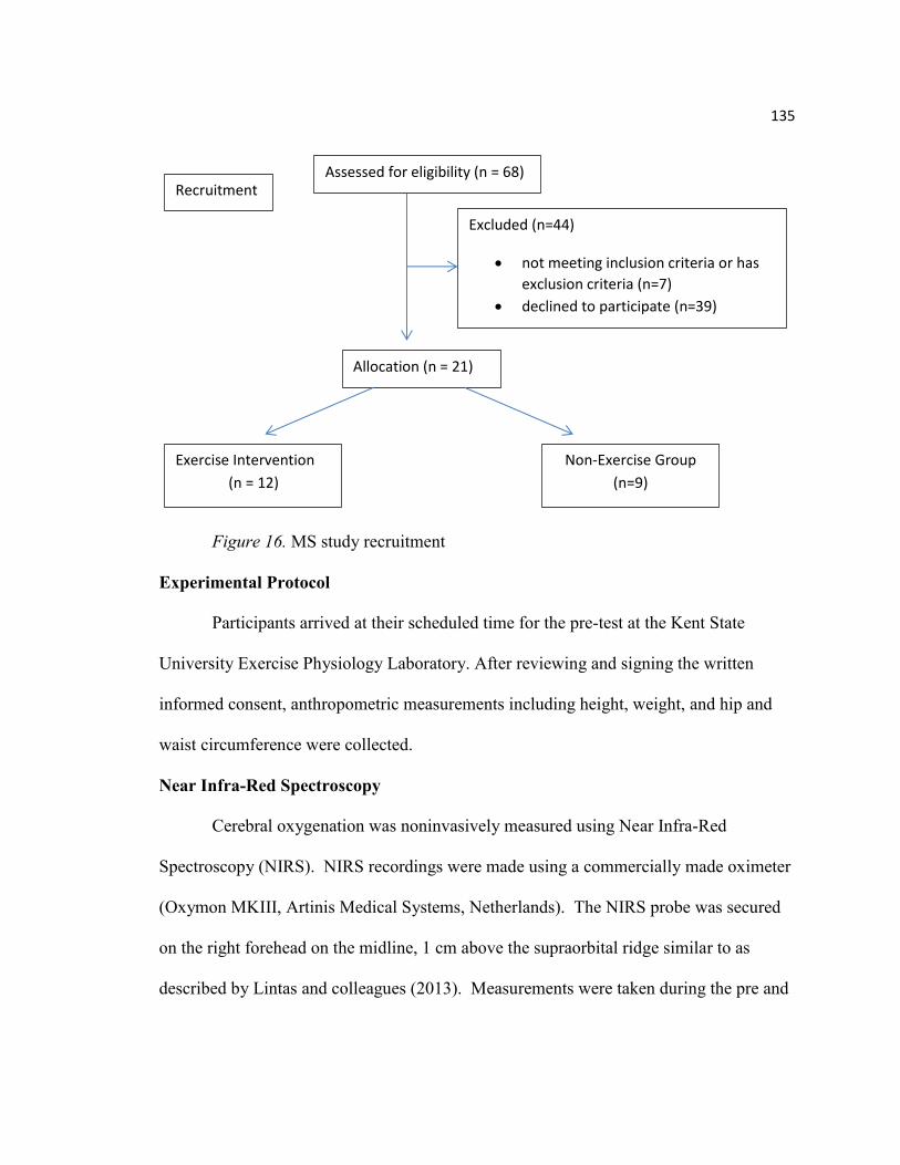
135
Figure 16. MS study recruitment
Experimental Protocol
Participants arrived at their scheduled time for the pre-test at the Kent State
University Exercise Physiology Laboratory. After reviewing and signing the written
informed consent, anthropometric measurements including height, weight, and hip and
waist circumference were collected.
Near Infra-Red Spectroscopy
Cerebral oxygenation was noninvasively measured using Near Infra-Red
Spectroscopy (NIRS). NIRS recordings were made using a commercially made oximeter
(Oxymon MKIII, Artinis Medical Systems, Netherlands). The NIRS probe was secured
on the right forehead on the midline, 1 cm above the supraorbital ridge similar to as
described by Lintas and colleagues (2013). Measurements were taken during the pre and
Recruitment Assessed for eligibility (n = 68)
Excluded (n=44)
not meeting inclusion criteria or has
exclusion criteria (n=7)
declined to participate (n=39)
Allocation (n = 21)
Exercise Intervention
(n = 12)
Non-Exercise Group
(n=9)
136
post baseline measures with the subject seated and eyes closed. Subjects were asked to
rest for 8 minutes while baseline measures were recorded. The last minute of rest with
values taken every 5 seconds were averaged to get a resting value.
After baseline values were recorded, subjects were asked to open their eyes while
a cognitive task was explained (N-back test). Participants played the N-1 back game
(Brain Workshop version 4.8.4) three times, each game lasting about 65 seconds. The
researcher then explained the N-2 back game, and participants played it three times using
the same software. The last minute (the third game) of the N-1 and the last minute (the
third game) of the N-2 were each averaged to determine a N-1 score and N-2 score. The
N-Back test is a valid and reliable indicator of working memory (Schmiedek, Lovden, &
Lindenberger, 2014). It requires continuous recognition of a stimulus (letter, number or
picture) in a particular sequence and the subject must judge whether it matches the one
presented n-items ago (Kane, Conway, Miura, & Colflesh, 2007).
The purpose was to determine if there were differences due to a 7 day aquatic
aerobic exercise protocol in cerebral oxygenation, cerebral deoxyhemoglobin, % tissue
saturation index (%TSI) and total hemoglobin. Because the NIRS does not give an
absolute values for oxyhemoglobin, deoxyhemoglobin, and total hemoglobin; changes
from baseline to N1 and from baseline to N2 were used to determine if there was a
difference from pre-test to post-test for the exercise group compared to the non-exercise
group. TSI was normalized as a percentage and a change score from pre-test to post-test
was used to report changes.
137
Non-exercise Participants
Participants in the non-exercise group were asked to maintain their current
lifestyle during the 7 days between the pre-test and post-test. They were instructed to
continue their usual activities of daily living, and were asked not to start a new exercise
program during the study.
Exercise Participants
The day following the pre-test, participants were asked to attend morning exercise
sessions every day for the next 7 days. A certified special population aquatic aerobics
instructor led the exercise session. Small groups of participants (2-5) were monitored by
a lifeguard and at least two trained research staff during the 60 minute exercise sessions.
The individual target training intensity (65-75% of their maximal heart rate) was
calculated through the Karvonen formula (Karvonen, Kentala, &Mustala, 2007). Heart
rate and rate of perceived exertion (RPE) were monitored every 9 minutes throughout the
exercise protocol. The exercise protocol provided one minute of rest between four
minute segments of exercise allowing for recovery time between the exercise bouts
(Table 8). The eight exercise intervals rotated between upper body, lower body or total
body exercise. For safety, participants were required to rest if their heart rate was greater
than 75% of their calculated maximal heart rate or RPE greater than 17. The participant
was able to return to exercise when they reached 65% of their calculated maximal heart
rate or RPE was at 13.
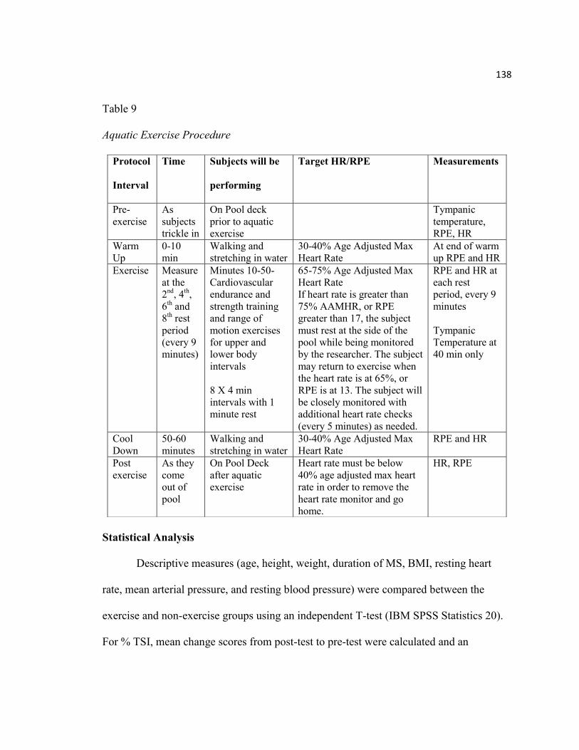
138
Table 9
Aquatic Exercise Procedure
Statistical Analysis
Descriptive measures (age, height, weight, duration of MS, BMI, resting heart
rate, mean arterial pressure, and resting blood pressure) were compared between the
exercise and non-exercise groups using an independent T-test (IBM SPSS Statistics 20).
For % TSI, mean change scores from post-test to pre-test were calculated and an
Protocol
Interval
Time Subjects will be
performing
Target HR/RPE Measurements
Pre-
exercise
As
subjects
trickle in
On Pool deck
prior to aquatic
exercise
Tympanic
temperature,
RPE, HR
Warm
Up
0-10
min
Walking and
stretching in water
30-40% Age Adjusted Max
Heart Rate
At end of warm
up RPE and HR
Exercise Measure
at the
2nd
, 4th,
6th and
8th rest
period
(every 9
minutes)
Minutes 10-50-
Cardiovascular
endurance and
strength training
and range of
motion exercises
for upper and
lower body
intervals
8 X 4 min
intervals with 1
minute rest
65-75% Age Adjusted Max
Heart Rate
If heart rate is greater than
75% AAMHR, or RPE
greater than 17, the subject
must rest at the side of the
pool while being monitored
by the researcher. The subject
may return to exercise when
the heart rate is at 65%, or
RPE is at 13. The subject will
be closely monitored with
additional heart rate checks
(every 5 minutes) as needed.
RPE and HR at
each rest
period, every 9
minutes
Tympanic
Temperature at
40 min only
Cool
Down
50-60
minutes
Walking and
stretching in water
30-40% Age Adjusted Max
Heart Rate
RPE and HR
Post
exercise
As they
come
out of
pool
On Pool Deck
after aquatic
exercise
Heart rate must be below
40% age adjusted max heart
rate in order to remove the
heart rate monitor and go
home.
HR, RPE
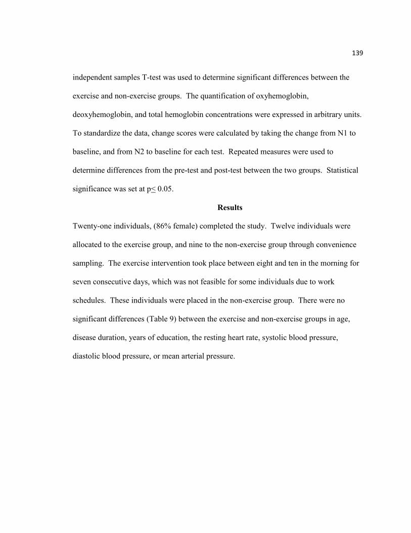
139
independent samples T-test was used to determine significant differences between the
exercise and non-exercise groups. The quantification of oxyhemoglobin,
deoxyhemoglobin, and total hemoglobin concentrations were expressed in arbitrary units.
To standardize the data, change scores were calculated by taking the change from N1 to
baseline, and from N2 to baseline for each test. Repeated measures were used to
determine differences from the pre-test and post-test between the two groups. Statistical
significance was set at p< 0.05.
Results
Twenty-one individuals, (86% female) completed the study. Twelve individuals were
allocated to the exercise group, and nine to the non-exercise group through convenience
sampling. The exercise intervention took place between eight and ten in the morning for
seven consecutive days, which was not feasible for some individuals due to work
schedules. These individuals were placed in the non-exercise group. There were no
significant differences (Table 9) between the exercise and non-exercise groups in age,
disease duration, years of education, the resting heart rate, systolic blood pressure,
diastolic blood pressure, or mean arterial pressure.
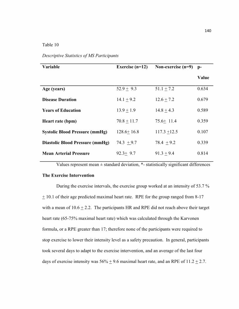
140
Table 10
Descriptive Statistics of MS Participants
Variable Exercise (n=12) Non-exercise (n=9) p-
Value
Age (years) 52.9 + 9.3 51.1 + 7.2 0.634
Disease Duration 14.1 + 9.2 12.6 + 7.2 0.679
Years of Education 13.9 + 1.9 14.8 + 4.3 0.589
Heart rate (bpm) 70.8 + 11.7 75.6+ 11.4 0.359
Systolic Blood Pressure (mmHg) 128.6+ 16.8 117.3 +12.5 0.107
Diastolic Blood Pressure (mmHg) 74.3 + 9.7 78.4 + 9.2 0.339
Mean Arterial Pressure 92.3+ 9.7 91.3 + 9.4 0.814
Values represent mean ± standard deviation, *- statistically significant differences
The Exercise Intervention
During the exercise intervals, the exercise group worked at an intensity of 53.7 %
+ 10.1 of their age predicted maximal heart rate. RPE for the group ranged from 8-17
with a mean of 10.6 + 2.2. The participants HR and RPE did not reach above their target
heart rate (65-75% maximal heart rate) which was calculated through the Karvonen
formula, or a RPE greater than 17; therefore none of the participants were required to
stop exercise to lower their intensity level as a safety precaution. In general, participants
took several days to adapt to the exercise intervention, and an average of the last four
days of exercise intensity was 56% + 9.6 maximal heart rate, and an RPE of 11.2 + 2.7.
141
The mean body temperature was 97.8 degrees Fahrenheit during the exercise intervals.
The exercise protocol was safe and feasible for this group.
Tissue Saturation Index (TSI)
Due to measurement error, four individuals from the exercise group and 3 from
the non-exercise group were excluded from analysis. Mean change scores for percent
TSI were calculated by subtracting the pre-test value from the post-test values to
determine a change score. An independent sample T-test was used to determine if there
were differences between the two groups. Though there were no statistically significant
differences (Table 10) between the two groups, the exercise group showed 3.29% greater
improvements at rest than the non-exercise group (Figure 23).
Table 11
Mean Change Scores % TSI.
Variable Exercise (n=8) Non-exercise (n=6) p-
Value
Effect
size
d
% TSI rest mean
change score
4.27+ 14.5 0.98+ 17.2 0.704 0.207
% TSI N1 mean
change score
4.39 + 22.66 3.74 + 26.47 0.961 0.026
% TSI N2 mean
change score
2.91 + 23.54 0.17 + 10.92 0.797 0.150
Values represent mean ± standard deviation, *- statistically significant differences
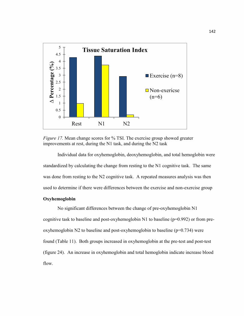
142
Figure 17. Mean change scores for % TSI. The exercise group showed greater
improvements at rest, during the N1 task, and during the N2 task
Individual data for oxyhemoglobin, deoxyhemoglobin, and total hemoglobin were
standardized by calculating the change from resting to the N1 cognitive task. The same
was done from resting to the N2 cognitive task. A repeated measures analysis was then
used to determine if there were differences between the exercise and non-exercise group
Oxyhemoglobin
No significant differences between the change of pre-oxyhemoglobin N1
cognitive task to baseline and post-oxyhemoglobin N1 to baseline (p=0.992) or from pre-
oxyhemoglobin N2 to baseline and post-oxyhemoglobin to baseline (p=0.734) were
found (Table 11). Both groups increased in oxyhemoglobin at the pre-test and post-test
(figure 24). An increase in oxyhemoglobin and total hemoglobin indicate increase blood
flow.
0
0.5
1
1.5
2
2.5
3
3.5
4
4.5
5
Rest N1 N2
∆ P
erce
nta
ge
(%)
Tissue Saturation Index
Exercise (n=8)
Non-exericse
(n=6)
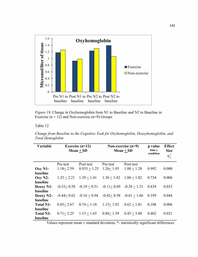
143
Figure 18. Change in Oxyhemoglobin from N1 to Baseline and N2 to Baseline in
Exercise (n = 12) and Non-exercise (n=9) Groups
Table 12
Change from Baseline to the Cognitive Task for Oxyhemoglobin, Deoxyhemoglobin, and
Total Hemoglobin
Variable Exercise (n=12)
Mean + SD
Non-exercise (n=9)
Mean + SD
p value time x
condition
Effect
Size 2
p
Pre-test Post-test Pre-test Post-test
Oxy N1-
baseline
1.18+ 2.59 0.933 + 1.25 1.26+ 1.95
1.00 + 1.28 0.992 0.000
Oxy N2-
baseline
1.23 + 2.21 1.39 + 1.41 1.30 + 1.42 1.06 + 1.82 0.734 0.006
Deoxy N1-
baseline
-0.33+ 0.58
-0.19 + 0.51 -0.11+ 0.69 -0.38 + 1.31 0.434 0.033
Deoxy N2-
baseline
-0.44+ 0.62 -0.16 + 0.94 -0.42+ 0.58 -0.61 + 1.66 0.359 0.044
Total N1-
baseline
0.85+ 2.67
0.74 + 1.18 1.15+ 1.92 0.62 + 1.81
0.108 0.006
Total N2-
baseline
0.71+ 2.25
1.15 + 1.65 0.88+ 1.39 0.45 + 3.00
0.402 0.021
Values represent mean ± standard deviation, *- statistically significant differences
0
0.2
0.4
0.6
0.8
1
1.2
1.4
1.6
Pre N1 to
baseline
Post N1 to
baseline
Pre N2 to
baseline
Post N2 to
baseline
Mic
rom
ol/
lite
r o
f ti
ssu
e Oxyhemoglobin
Exercise
Non-exercise
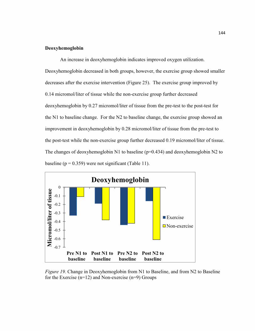
144
Deoxyhemoglobin
An increase in deoxyhemoglobin indicates improved oxygen utilization.
Deoxyhemoglobin decreased in both groups, however, the exercise group showed smaller
decreases after the exercise intervention (Figure 25). The exercise group improved by
0.14 micromol/liter of tissue while the non-exercise group further decreased
deoxyhemoglobin by 0.27 micromol/liter of tissue from the pre-test to the post-test for
the N1 to baseline change. For the N2 to baseline change, the exercise group showed an
improvement in deoxyhemoglobin by 0.28 micromol/liter of tissue from the pre-test to
the post-test while the non-exercise group further decreased 0.19 micromol/liter of tissue.
The changes of deoxyhemoglobin N1 to baseline (p=0.434) and deoxyhemoglobin N2 to
baseline (p = 0.359) were not significant (Table 11).
Figure 19. Change in Deoxyhemoglobin from N1 to Baseline, and from N2 to Baseline
for the Exercise (n=12) and Non-exercise (n=9) Groups
-0.7
-0.6
-0.5
-0.4
-0.3
-0.2
-0.1
0
Pre N1 to
baseline
Post N1 to
baseline
Pre N2 to
baseline
Post N2 to
baseline
Mic
rom
ol/
lite
r o
f ti
ssu
e
Deoxyhemoglobin
Exercise
Non-exercise
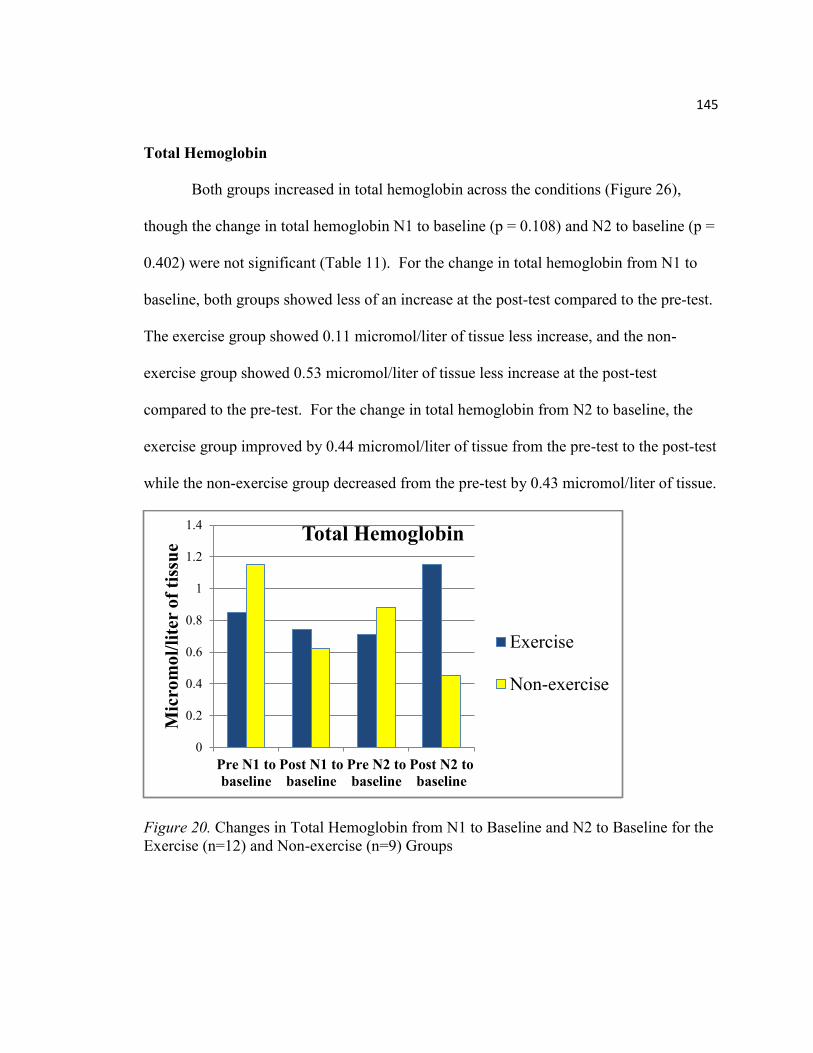
145
Total Hemoglobin
Both groups increased in total hemoglobin across the conditions (Figure 26),
though the change in total hemoglobin N1 to baseline (p = 0.108) and N2 to baseline (p =
0.402) were not significant (Table 11). For the change in total hemoglobin from N1 to
baseline, both groups showed less of an increase at the post-test compared to the pre-test.
The exercise group showed 0.11 micromol/liter of tissue less increase, and the non-
exercise group showed 0.53 micromol/liter of tissue less increase at the post-test
compared to the pre-test. For the change in total hemoglobin from N2 to baseline, the
exercise group improved by 0.44 micromol/liter of tissue from the pre-test to the post-test
while the non-exercise group decreased from the pre-test by 0.43 micromol/liter of tissue.
Figure 20. Changes in Total Hemoglobin from N1 to Baseline and N2 to Baseline for the
Exercise (n=12) and Non-exercise (n=9) Groups
0
0.2
0.4
0.6
0.8
1
1.2
1.4
Pre N1 to
baseline
Post N1 to
baseline
Pre N2 to
baseline
Post N2 to
baseline
Mic
rom
ol/
lite
r o
f ti
ssu
e Total Hemoglobin
Exercise
Non-exercise
146
Discussion
It was hypothesized that the 7 day aerobic exercise protocol would improve
cerebral oxygenation in the exercise group, but not in the non-exercise group. While
there were no statistically significant differences between the exercise and non-exercise
groups for %TSI, oxyhemoglobin, deoxyhemoglobin or total hemoglobin there were
small clinically meaningful improvements that exhibited as increases in oxyhemoglobin
and total hemoglobin, representing increased blood flow. In the change from N2 to
baseline for total hemoglobin, the exercise group showed an increase in 0.44
micromol/liter of tissue. The deoxyhemoglobin also decreased by a smaller amount after
the exercise intervention in the exercise group compared to the non-exercise group,
representing and improved oxygen utilization.
The % Tissue Saturation Index (%TSI) reflects the change in concentration of
oxygenated and deoxygenated hemoglobin, and is an estimate of the oxygen saturation in
the tissue. The mean change scores in %TSI show that the exercise group showed greater
improvements in %TSI than the non-exercise group. This indicates a greater extraction
of oxygen to the tissues after the aerobic exercise intervention, which is beneficial in the
MS population.
MS exhibits a hypometabolic component to the disease process. Two proposed
mechanisms are that brain tissues are extracting a decreased amount of oxygen, and there
is a decrease in vascular function (Sun, Tanaka, Kondo, Okamoto, & Hirai, 1998;
147
Brooks, et al., 1984). The resulting decrease in glucose utilization in the cortico-cerebral
metabolism is correlated to increased lesion load (Blinkenberg et al., 2000).
While MS individuals’ exhibit decreased oxygen utilization, exercise has been
shown to improve the aerobic system (Mostert & Kesselring, 2002). Aerobic exercise
increases peak oxygen uptake in MS individuals (Hayes, Gappmaier & LaStayo, 2011).
Exercise in MS promotes growth factors stimulating cell proliferation, synaptic plasticity,
neuroprotection, and neurogenesis (Doring, Pfueller, Paul, & Dorr, 2012). While the
improvements in TSI are small, it is exciting that a short exercise intervention can benefit
the oxygen utilization in MS individuals, and increases the need for further study on
longer duration exercise, mode of exercise (high intensity training vs long duration
aerobic training), and greater intensity level.
Oxyhemoglobin
This study aimed to determine the response of cerebral oxygen at rest and during
a cognitive task after a 7 day aquatic exercise intervention. We hypothesized that
oxyhemoglobin would progressively increase as the cognitive tasks became more
difficult, and that oxyhemoglobin would increase more in the exercise group compared to
the non-exercise group at the post-test compared to the pre-test. This study showed that
oxyhemoglobin increased during the cognitive tasks in both groups. There was a smaller
increase in both groups at the post-test from baseline to N1 cognitive task possibly
indicating that oxygen demands were increased greater than to delivery. It is also
probable that individuals were not cognitively working as hard to complete the task
148
because they were already familiar with it from the pre-test. The change from baseline to
N2 was greater in the post-test than in the pre-test for the exercise condition reflecting a
more efficient capacity to respond to oxygen demands and a greater blood flow with a
more difficult cognitive task.
Ide and Secher (2000) showed an increase in regional cerebral uptake of oxygen
during exercise in healthy individuals. Obrig and colleagues demonstrated cerebral
oxygen exceeds the increase in oxygen demand in motor stimulation as shown with
increased oxyhemoglobin (Obrig, Hirth, Junge-Hulsing, Doge, Wolf, Dirnagl, &
Villringer,1996). Increase oxyhemoglobin and total hemoglobin suggest increases in
blood volume which correlate to cerebral blood flow (Sakatani,Lichty, Xie, Li, & Zuo,
1999). Furthermore, cerebral activation, including cognitive tasks, increases cerebral
blood flow, thereby increasing cerebral oxygenation. Most of the hemoglobin
determined by NIRS is post-cellular, after the cells have extracted oxygen (Ide & Secher,
2000). Oxyhemoglobin remaining in the blood post-cellular suggests the ability to
exceed oxygen demand.
Rooks and colleagues completed a review on healthy individuals and found that
during low to moderate intensity aerobic exercise (<60% VO2 max), there were moderate
to large increases in the prefrontal cortex for oxyhemoglobin, deoxyhemoglobin and
blood volume (as estimated by total hemoglobin) (Rooks, Thom, McCully, & Dishman,
2010). They further found that aerobically trained individuals had a higher
oxyhemoglobin, deoxyhemoglobin and total hemoglobin level at hard intensities
149
compared to untrained individuals. In addition, a common pattern including a rise in
oxyhemoglobin, deoxyhemoglobin and total hemoglobin during moderate to hard
intensities, which fell at very hard intensities; with training status influencing the
response. As very hard intensities were reached, the body’s capacity to keep up with
oxygen demands was reduced. MS individuals, though generally deconditioned, respond
to exercise similarly to healthy individuals, and may benefit from improved oxygenation
resulting from exercise.
Sakatan and colleagues (1999), suggested that decreased oxyhemoglobin and total
hemoglobin indicate a decreased cerebral blood flow during cognitive tasks. Their study
compared young adults to aged adults. The most common pattern for both groups was an
increase in oxyhemoglobin, increased total hemoglobin and a decreased
deoxyhemoglobin. However, in aged adults there was a significant difference in these
patterns as more aged adults demonstrated a decrease in oxyhemoglobin and total
hemoglobin compared to the younger adults suggesting a decrease in regional cerebral
blood flow. With already compromised oxygenation, MS individuals may follow a
similar response to aging, and exercise potentially can improve the metabolic profile.
While the differences between the two groups comparing the pre-test to the post-
test are not significant, the trend toward improved efficiency of the body to supply
oxygen demands could be very beneficial in the hypometabolic condition of MS.
Increased duration or intensity may further improve cerebral oxygenation.
150
Deoxyhemoglobin
Deoxyhemoglobin reflects the tissue’s ability to extract oxygen. As oxygen is
extracted, deoxyhemoglobin increases. While differences were not statistically
significant, both groups exhibited decreases in deoxyhemoglobin in all conditions,
however, the exercise group had less of a decrease than the non-exercise group. This
suggests an improved ability to utilize oxygen due to the exercise intervention. It is
possible that MS individuals have a decreased cerebral blood oxygen reservoir due to
decrease vasculature, causing a diminished ability to improve oxyhemoglobin. However,
the exercise group exhibited an improved oxygen utilization as reflected by the improved
deoxyhemoglobin from pre-to-post exercise. The non-exercise group continued to
decrease in deoxyhemoglobin, decreasing oxygen utilization.
Total Hemoglobin
Differences between the two groups in total hemoglobin were not statistically
significant. Total Hemoglobin mirrored a similar pattern to the oxyhemoglobin.
Changes in total hemoglobin reflect changes in blood volume, and is correlated with
cerebral blood flow (Sakatani,Lichty, Xie, Li, & Zuo, 1999). In the N2 cognitive task,
the exercise group increased total hemoglobin which was also demonstrated with
oxyhemoglobin. Possibly an increase in blood flow due to the exercise intervention
improved the capacity to distribute oxygen to the tissues.
151
Limitations and Future Direction
This study has several limitations, the first being that the exercise intervention
may not have been long or intense enough to elicit significant changes in %TSI,
oxyhemoglobin, deoxyhemoglobin, or total hemoglobin. In addition, the sample size was
small, and recruiting more participants would have been beneficial. Due to a limited
response to recruiting and pool scheduling, the participants were assigned to each group
based on their ability to attend the exercise protocol and if the number of individuals in
that testing session was large enough for an exercise session. The pool exercise was also
an interval style workout; a completely aerobic protocol may have produced greater
improvements. The NIRS only measures in arbitrary units, limiting the ability for
comparisons pre-to-post-test. Finally, the N1 and N2 back cognitive tasks were difficult
cognitive tests. Participants who had a hard time with the N1 task may have been less
motivated to try harder for the N2 task.
Future research is required to determine the details of the hypometabolic
component in MS and to determine how MS individuals respond to exercise including
longer duration and higher intensity protocols. Determining the impact of aerobic, high
intensity training, or resistance training on cerebral oxygenation may provide an optimal
training mechanism during the window between relapses.
152
CHAPTER VI
SUMMARY
MS individuals comprise a unique population because of the diversity of
pathology and symptoms. The location of demyelination and lesions in the central
nervous system determine what symptoms the individual will experience, and result in
individuals that are less physically active and deconditioned. Though the symptoms
often prevent MS individuals from exercising, exercise is safe, feasible, and often
providing benefits that counteract the pathology. Aquatic exercise in particular is
beneficial because of ability of the water to absorb body heat, minimizing the symptoms
that increase transiently during exercise due to increased core temperature. In addition,
aquatic exercise provides an environment that decreases joint stress, reduces risk of falls,
and allows for a wide range of disability levels which was helpful in this study due to the
diversity of disability levels of the participants.
While disappointing, this study did not show any statistically significant results
for improvements in mobility, fitness, body composition, self-efficacy, or cerebral
oxygenation; it did provide insight to how MS individuals respond to short duration
exercise. The trends demonstrated through this study encourage MS individuals to
exercise, even if only short windows exist between relapses, and provide peace of mind
that exercise is safe, feasible, beneficial and desirable.
This study revealed small improvements that can impact functions of daily living
and quality of life in MS individuals. Mobility, an important function of living improved
153
in 73% (8/11) of the exercise participants compared to 11% (1/9) of the non-exercise
participants, as measured through the twenty-five foot walk test. Similar improvements
were demonstrated in fitness as measured by the two minute step test. Improved fitness
usually correlates with decreased resting heart rate and blood pressure, however the
exercise participants exhibited an increase in resting heart rate, while the non-exercise
group had a slight decrease. Blood pressure followed the same pattern, which reflects the
autonomic dysfunction in MS. Demyelination and lesions can impact the
parasympathetic (controlling HR) and sympathetic (controlling BP) responses, and
exercise may have been beneficial in stimulating both of these parts of the nervous
system resulting in an increased response in HR and BP. Self-efficacy also improved in
both groups, though the differences were not significant.
This study also showed that the short exercise intervention produced small
changes in cerebral oxygenation, with greater improvements in the exercise group at
post-test in the harder cognitive task. Total hemoglobin also followed this pattern
indicating an increased cerebral blood flow during the harder cognitive tasks. Increased
deoxyhemoglobin indicates increased oxygen extraction. Both groups decreased in
deoxyhemoglobin, the exercise group showed less decrease after the exercise intervention
while the non-exercise group showed a further decrease in deoxyhemoglobin from the
pre-test to the post-test. The exercise group was more efficient at extracting oxygen
during the cognitive tasks than the non-exercise group, though the differences were still
not significant.
154
This study is not without limitations. The sample size was very small, and
participants were very diverse in their disability, symptoms, and disease duration. The
sample should have been randomly assigned to the exercise and non-exercise conditions;
instead participants were assigned by convenience sampling. Increasing the exercise
intensity, decreasing the number of rest periods between intervals, and lengthening the
exercise duration to greater than one week may also be beneficial. Allowing for 24 hours
of rest post exercise may have enhanced performance on fitness, mobility and self-
efficacy measures as fatigue most likely impacted the results. Including 15 minutes of
rest between the tests may also have been beneficial. It was also unclear if HR and RPE
accurately reflected the exercise intensity levels in MS.
Future direction includes determining the optimal time of exercise that allows
benefits between relapses. In addition, determining modes (high intensity training, long
low intensity aerobic training, resistance training), intensities, and durations of exercise
that can positively impact the autonomic function in MS. Exercise can influence the
autonomic nervous system, so prescribing exercise for the desired effect
(parasympathetic vs sympathetic stimulation) individually would be beneficial for people
with MS. Along with this, developing tools that give insight to the autonomic
dysfunction and allow for monitoring would allow a more purposeful exercise paradigm
to be prescribed. It is also important to have tools that accurately reflect intensity levels
in MS so that the exercise can be monitored accordingly, therefore, validating RPE and
HR as intensity measures in MS would be beneficial.

APPENDICES

APPENDIX A
INFORMED CONSENT FORM
157
Appendix A
Informed Consent Form
Informed Consent to Participate in a Research Study
Study Title: Exercise and Cognition in Multiple Sclerosis
Principal Investigator: Mary Beth Spitznagel, PhD
Co-Investigators: Angela Ridgel, PhD; Jennifer Petersen, MS; Dayana Calvo, BA
You are being invited to participate in a research study. This consent form will provide you with
information on the research project, what you will need to do, and the associated risks and
benefits of the research. Your participation is voluntary. Please read this form carefully. It is
important that you ask questions and fully understand the research in order to make an
informed decision. You will receive a copy of this document to take with you.
Purpose: The purpose of this study is to better understand the relationship between exercise
and thinking skills in people diagnosed with Multiple Sclerosis. To do this, we are asking people
to participate in testing of thinking skills and physical abilities. Some people will be randomly
assigned to a 1 week-long, daily water aerobics exercise program, while others will not. We will
look at how thinking skills and physical abilities change during this time period, and how
participation in the exercise program may affect these changes.
Procedures: If you decide to take part in this study, you will first be asked to complete a series
of tests. These evaluations will include:
Tests of thinking skills: You will be asked to complete a short set of pen/paper and
question/answer tests that assess your ability to focus and sustain your attention, learn and
remember new information, and other types of thinking skills. These tests will take about 45-60
minutes.
Questionnaires: You will also be asked to complete paper-and-pencil questionnaires. The
questions ask about your personal characteristics (such as health issues) as well as your exercise
habits, mood, symptoms of pain and fatigue self-efficacy, and how all of these affect your day to
day life. The questionnaires will take about 20-30 minutes.
158
Physiological Testing: Baseline levels of your height, weight, and waist to hip ratio will be
recorded today. Heart rate, blood pressure, tympanic body temperature, and cerebral
oxygenation will also be recorded. These tests will take about 10 minutes.
Functional Testing: You will be asked to perform some functional testing to determine your
fitness level. These tests will assess your cardiovascular fitness, and mobility. These tests will
take about 45-60 minutes.
Activity Monitoring: You will be asked to wear an activity monitor daily throughout the study.
The monitor can be worn on your wrist, waistband, shirt, or in your pocket throughout the day.
Once you have completed the evaluations, you will be randomly assigned to either the Exercise
Group or the No Exercise Group.
If you are assigned to the No Exercise Group, you will be asked only to complete the above
baseline testing, and repeat these tests one week later, making no other changes to your normal
routine.
If you are assigned to the Exercise Program, you will be asked to attend daily exercise sessions
for 7 days. Sessions are 60 minutes each day, and consist of a structured program of water
aerobic exercises. The specific intensity of the exercise will depend upon your age and fitness
level, and will be determined by the project investigators based upon your physical testing.
Target heart rates will be monitored with heart rate monitors and used to measure exercise
intensity. Research assistants will be poolside during the course of the exercise class to monitor
your heart rate and observe for any signs of physical distress. You will not be allowed to
exercise to exhaustion.
Benefits: Both the exercise and control group will benefit from information given to them
regarding the cognitive function testing and baseline fitness testing. For the exercise group, your
involvement in this study may provide cognitive benefits to you, as prior work shows that even
one week of exercise can improve thinking skills. You may also experience other benefits
associated with exercise, such as improved fitness, health, and quality of life. The outcome of
the research will provide important information on the relationship between exercise and
thinking skills in people diagnosed with Multiple Sclerosis.
Risks and Discomforts: We believe that the risks of this study are minimal. One risk is possible
distress caused by completing the tests of memory or other thinking skills and the questionnaires. If
you experience distress during the memory testing, please understand that you can stop at any
159
time and rest until you are ready to continue, or decide not to continue. If you experience distress
while completing the questionnaires, you can choose not to answer any questions that you do not
want to answer. One of the investigators will be available during these times to meet with any
participant, upon request, to discuss any concerns. You can also ask to meet with a member of the
research team by calling Dr. Mary Beth Spitznagel at 330.672.2399 (office phone).
For those assigned to the exercise group, exercise may cause muscle soreness. Additionally,
exercise could result in heart attack or stroke; this risk for such effects is low, and is further
decreased when an individual exercises with their physician’s approval, and in a controlled
group setting such as that provided in this study. The research staff will carefully monitor your
heart rate to ensure you exercise within a predetermined range. A minimum of two CPR/AED
certified individuals and one lifeguard will be present to ensure safety. In addition, exercise will
be conducted in the shallow end of the pool. Please inform a research team member if you
should wish to stop, experience concern regarding exercise equipment or facilities, or happen to
experience any of the following during this study: chest pain, shortness of breath, pallor,
fainting, wheezing, leg cramps, light-headedness, confusion, nausea, cold/clammy skin,
noticeable change in heart rhythm, severe fatigue, or any other discomfort.
If findings emerge during this research that might affect your willingness to continue to
participate, this information will be provided to you so that you can make an informed decision
about your participation. For example, if it becomes apparent during the course of the study
that you are having significant difficulties with your memory or other thinking skills, we will
discuss this with you and may request further evaluation to ensure your safety.
Medical treatment by the University Health Center is provided only to currently registered
students. Please be advised that for all other injuries, emergency services will be called for those
occurring on the Kent State University campus. You or your medical insurance will be billed for
this service. No other medical treatment or financial compensation for injury from participation
in this research project is available.
Criteria for Inclusion/Exclusion:
Inclusion: To be included in this study, you must be between the ages of 20-65, diagnosed with
Multiple Sclerosis, have physician approval to participate, and able to communicate in English.
Exclusion: You will not be eligible if you have a history of significant neurological disorders other
than MS, such as stroke, seizures, or severe head injury. You will not be eligible if you have a
history of significant psychological problems, such as schizophrenia or bipolar disorder.
160
Privacy and Confidentiality: All results will be kept confidential. As soon as possible, your
personal identifying information such as your name and date of birth will be removed from your
file. We will store all study materials in a locked filing cabinet in a locked room at Kent State
University. Your research information may, in certain circumstances, be disclosed to the
Institutional Review Board (IRB), which oversees research at Kent State University, or to certain
federal agencies. Confidentiality may not be maintained if you indicate that you may do harm to
yourself or others.
Compensation: If you take part in this project, you will be compensated $50 for each testing
session (total of $100). In addition, if assigned to the Exercise Group, you will be compensated
an additional $10 for each day that you attend the exercise group. If you complete all 7 days of
exercise, you will receive a $30 completion bonus at the final session. Participants will receive an
additional $20 for completing the 12 week telephone follow-up.
Voluntary Participation: Taking part in this research study is entirely up to you. You may choose
not to participate or you may discontinue your participation at any time without penalty or loss
of benefits to which you are otherwise entitled. You will be informed of any new, relevant
information that may affect your health, welfare, or willingness to continue your study
participation.
Contact Information: If you have any questions or concerns about this research, you may
contact Dr. Mary Beth Spitznagel at 330.672.2399. This project has been approved by the Kent
State University Institutional Review Board. If you have any questions about your rights as a
research participant or complaints about the research, you may call the IRB at 330.672.2704.
By signing this form I acknowledge that I have read it, understand it, and have had any
questions regarding the risks and benefits of this study satisfactorily answered, and I am
voluntarily consenting to participate in this study. I have received permission from my
physician to participate in this exercise study. If any changes in my health status occur
during the course of this study (e.g., new diagnosis, change in medication, etc), I will discuss
my continued eligibility with my physician. I realize that by signing this form I do not waive
any of my legal rights, and that I can choose to terminate my participation at any time.
Date:
Participant Signature
Date:
161
Witness Signature
Date:
Person obtaining consent
Date:
Investigator
By signing the below, I give permission to be contacted for future studies, and provide my
contact information for this purpose. In signing this section, I am only giving permission for
the researchers to contact me to inform me of future projects, and am not at this time
committing to participate in future studies.
Date:
Participant Signature
Telephone: Email: ________
APPENDIX B
PRE-PARTICIPATION PHONE SCREENING
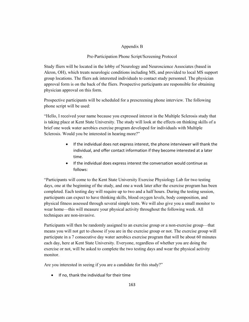
163
Appendix B
Pre-Participation Phone Script/Screening Protocol
Study fliers will be located in the lobby of Neurology and Neuroscience Associates (based in
Akron, OH), which treats neurologic conditions including MS, and provided to local MS support
group locations. The fliers ask interested individuals to contact study personnel. The physician
approval form is on the back of the fliers. Prospective participants are responsible for obtaining
physician approval on this form.
Prospective participants will be scheduled for a prescreening phone interview. The following
phone script will be used:
“Hello, I received your name because you expressed interest in the Multiple Sclerosis study that
is taking place at Kent State University. The study will look at the effects on thinking skills of a
brief one week water aerobics exercise program developed for individuals with Multiple
Sclerosis. Would you be interested in hearing more?”
If the individual does not express interest, the phone interviewer will thank the
individual, and offer contact information if they become interested at a later
time.
If the individual does express interest the conversation would continue as
follows:
“Participants will come to the Kent State University Exercise Physiology Lab for two testing
days, one at the beginning of the study, and one a week later after the exercise program has been
completed. Each testing day will require up to two and a half hours. During the testing session,
participants can expect to have thinking skills, blood oxygen levels, body composition, and
physical fitness assessed through several simple tests. We will also give you a small monitor to
wear home—this will measure your physical activity throughout the following week. All
techniques are non-invasive.
Participants will then be randomly assigned to an exercise group or a non-exercise group—that
means you will not get to choose if you are in the exercise group or not. The exercise group will
participate in a 7 consecutive day water aerobics exercise program that will be about 60 minutes
each day, here at Kent State University. Everyone, regardless of whether you are doing the
exercise or not, will be asked to complete the two testing days and wear the physical activity
monitor.
Are you interested in seeing if you are a candidate for this study?”
If no, thank the individual for their time
164
If yes, (note that they are English speaking) and ask the following:
______________Have you been diagnosed with MS by a neurologist?
______________Are you 20-65 years of age?
______________Do you have physician approval for this study?
______________Are you pregnant or at risk for becoming pregnant?
______________Do you have a history of non-MS related neurological disorder or injury such as
brain
injury or seizure?
______________Do you have a past or current history of psychiatric illness such as
schizophrenia or
bipolar disorder?
______________Do you have past or current history of drug or alcohol abuse?
______________Do you have a history of learning disorder or developmental disability?
______________Do you have impaired sensory function that would prevent cognitive testing (for
example, being deaf or blind)?
If individual meets inclusion/exclusion criteria, they are eligible to participate in the study with physician approval. Continue with:
“Would you like to schedule an appointment for the first testing session?”
o If no, “Thank you for your time.” o If yes,
schedule the appointment, or develop a timeline to schedule __________________________________________________________
“You will be required to sign a consent form prior to participating” “You must bring the physician approval form that is on the back of our
study flier on the date of your first appointment.” “Please wear comfortable clothing and tennis shoes to the first
appointment” “If you wear eye glasses, please bring them”
o If yes, but does not have physician approval yet: “In order to participate in this study you will be required to get
physician approval. The approval form is on the back of our flier.”

APPENDIX C
PHYSICIAN CLEARANCE FORM

166
Appendix C
Physician’s Clearance For Exercise Participation
Kent State University- Dept. of Exercise Physiology
Patient’s name:
Address:
Telephone number:
Dear Doctor-
Your patient, ___________________, has expressed an interest in participating in an
aquatic aerobics exercise study in individuals with Multiple Sclerosis (KSU IRB approval
_____________). The objective of this project is to demonstrate the feasibility and
tolerability of a brief water aerobics exercise intervention and determine cognitive benefits
of this intervention.
The participant will complete 7 consecutive days of aquatic interval aerobic exercise
sessions. Each session include a 10 minute warm up, 40 minutes of low to moderate
intensity exercise at 45-55% age-adjusted maximum heart rate broken up into alternating
four minutes of exercise followed by one minute of rest, and a 10 minute cool down. The
exercise sessions will last 60 minutes. We will examine changes in cognition and fitness
parameters after the exercise sessions are completed.
Below is a clearance form to be filled out and signed by you.
Physician’s recommendation (check the appropriate line)
a.________There is no contraindication for participation in this exercise research project.
b.________Because of the following diagnosis, participation in this exercise research
program is inadvisable.
Physician’s name: _________________________________________________
Physician Signature:_______________________________________________
Date:____________________________________________________________
Address:
Telephone:

APPENDIX D
DATA SHEETS
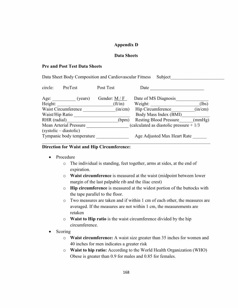
168
Appendix D
Data Sheets
Pre and Post Test Data Sheets
Data Sheet Body Composition and Cardiovascular Fitness Subject_______________________
circle: PreTest Post Test Date _______________________
Age: __________ (years) Gender: M / F Date of MS Diagnosis______________
Height: ________________________(ft/in) Weight: _____________________(lbs)
Waist Circumference ______________(in/cm) Hip Circumference__________(in/cm)
Waist/Hip Ratio ________________________ Body Mass Index (BMI)____________
RHR (radial)______________________(bpm) Resting Blood Pressure______(mmHg)
Mean Arterial Pressure __________________ (calculated as diastolic pressure + 1/3
(systolic – diastolic)
Tympanic body temperature ______________ Age Adjusted Max Heart Rate ______
________________________________________________________________________
Direction for Waist and Hip Circumference:
Procedure
o The individual is standing, feet together, arms at sides, at the end of
expiration.
o Waist circumference is measured at the waist (midpoint between lower
margin of the last palpable rib and the iliac crest)
o Hip circumference is measured at the widest portion of the buttocks with
the tape parallel to the floor.
o Two measures are taken and if within 1 cm of each other, the measures are
averaged. If the measures are not within 1 cm, the measurements are
retaken
o Waist to Hip ratio is the waist circumference divided by the hip
circumference.
Scoring
o Waist circumference: A waist size greater than 35 inches for women and
40 inches for men indicates a greater risk
o Waist to hip ratio: According to the World Health Organization (WHO)
Obese is greater than 0.9 for males and 0.85 for females.
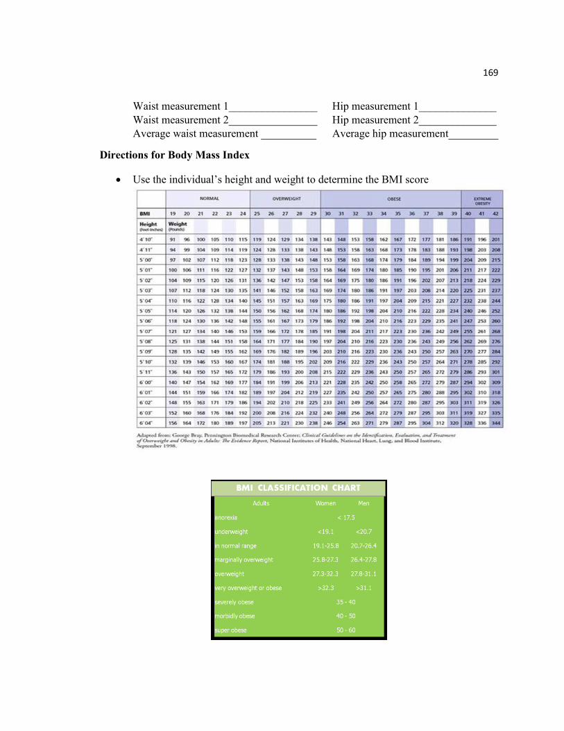
169
Waist measurement 1________________ Hip measurement 1______________
Waist measurement 2________________ Hip measurement 2______________
Average waist measurement __________ Average hip measurement_________
Directions for Body Mass Index
Use the individual’s height and weight to determine the BMI score
170
25 Foot Walk Test
Walk Time ___________________________________
Comments ___________________________________ (i.e.: ambulatory device
required?)
Procedure
o Subject is directed to one end of a clearly marked 25 foot course and is
instructed “Please walk as quickly but safely as possible to the end of the
25 foot course”
o The time is calculated from the initiation of the instruction to start and
ends when the subject has reached the 25 foot mark.
o The task is immediately administered again by having the subject walk
back the same distance.
o Subjects may use assistive devices during this task.
Scoring
o The time it takes to walk 25 feet.
o Trial 1______________________________
o Trial 2______________________________
o Average of trial 1+2 ___________________
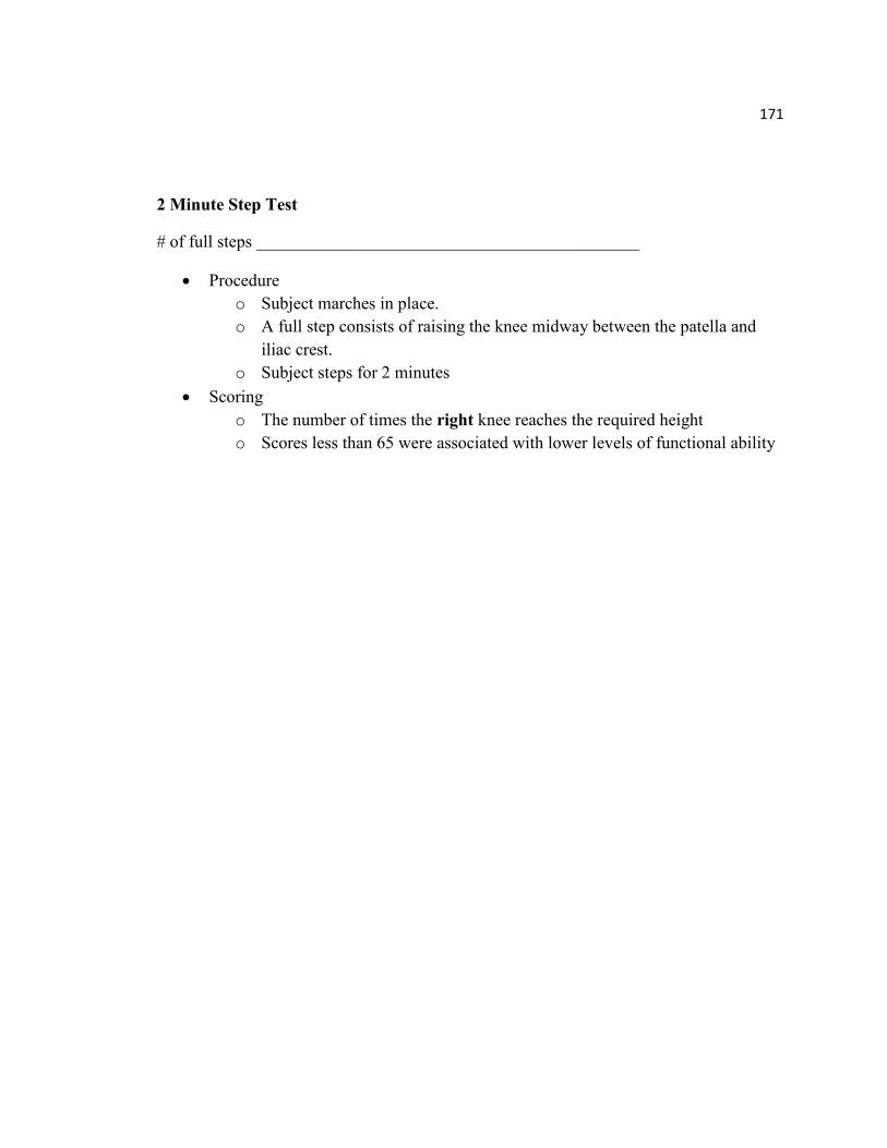
171
2 Minute Step Test
# of full steps ____________________________________________
Procedure
o Subject marches in place.
o A full step consists of raising the knee midway between the patella and
iliac crest.
o Subject steps for 2 minutes
Scoring
o The number of times the right knee reaches the required height
o Scores less than 65 were associated with lower levels of functional ability

172
Timed Up and Go Test (TUG)
Average TUG Time _____________________________________________
Procedure
o The course is set up with a firm chair with arms (seat height 46 cm)
separated by a cone 3 meters apart.
o The subject begins sitting, back against the chair, arm on the lap, feet just
behind the distance marker on the floor.
o The subject is instructed “on the word ‘go’, stand up, walk comfortably
and safely to the cone on the floor, walk around the cone, come back, sit
all the way back in your chair.”
o Timing begins on ‘go’ and stops when the subject has their back against
the chair seat.
o One practice trial and 2 recorded trials are given.
Scoring
o The two recorded trial times are averaged.
Trial 1 time ______________________________
Trial 2 time______________________________
Average time ____________________________
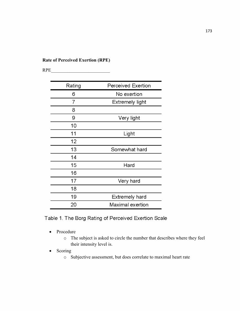
173
Rate of Perceived Exertion (RPE)
RPE_________________________
Procedure
o The subject is asked to circle the number that describes where they feel
their intensity level is.
Scoring
o Subjective assessment, but does correlate to maximal heart rate
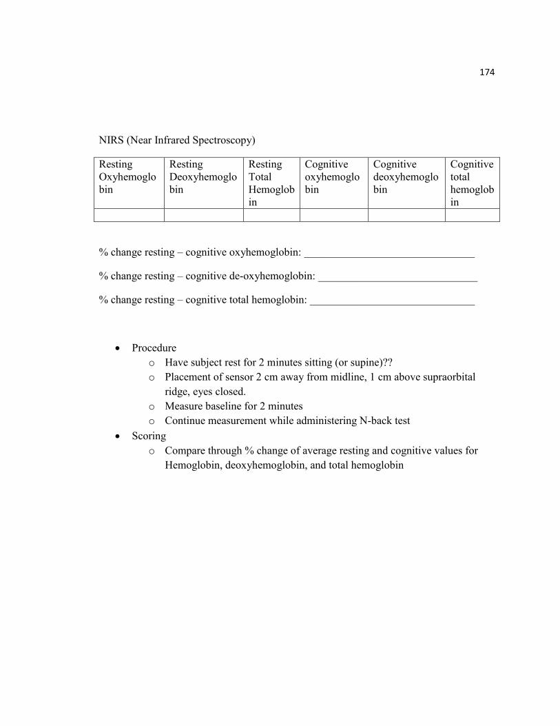
174
NIRS (Near Infrared Spectroscopy)
Resting
Oxyhemoglo
bin
Resting
Deoxyhemoglo
bin
Resting
Total
Hemoglob
in
Cognitive
oxyhemoglo
bin
Cognitive
deoxyhemoglo
bin
Cognitive
total
hemoglob
in
% change resting – cognitive oxyhemoglobin: _______________________________
% change resting – cognitive de-oxyhemoglobin: _____________________________
% change resting – cognitive total hemoglobin: ______________________________
Procedure
o Have subject rest for 2 minutes sitting (or supine)??
o Placement of sensor 2 cm away from midline, 1 cm above supraorbital
ridge, eyes closed.
o Measure baseline for 2 minutes
o Continue measurement while administering N-back test
Scoring
o Compare through % change of average resting and cognitive values for
Hemoglobin, deoxyhemoglobin, and total hemoglobin
APPENDIX E
HEART RATE AND RPE
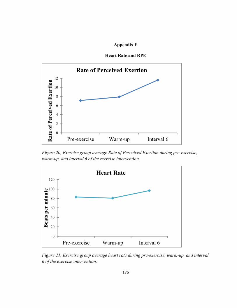
176
Appendix E
Heart Rate and RPE
Figure 20, Exercise group average Rate of Perceived Exertion during pre-exercise,
warm-up, and interval 6 of the exercise intervention.
Figure 21, Exercise group average heart rate during pre-exercise, warm-up, and interval
6 of the exercise intervention.
0
2
4
6
8
10
12
Pre-exercise Warm-up Interval 6Ra
te o
f P
erce
ived
Ex
erti
on
Rate of Perceived Exertion
0
20
40
60
80
100
120
Pre-exercise Warm-up Interval 6
Bea
ts p
er m
inu
te
Heart Rate

.
REFERENCES
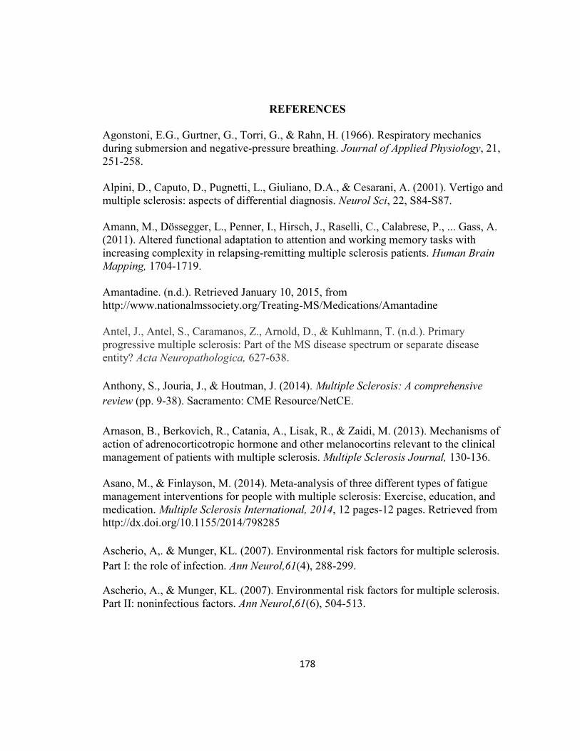
178
REFERENCES
Agonstoni, E.G., Gurtner, G., Torri, G., & Rahn, H. (1966). Respiratory mechanics
during submersion and negative-pressure breathing. Journal of Applied Physiology, 21,
251-258.
Alpini, D., Caputo, D., Pugnetti, L., Giuliano, D.A., & Cesarani, A. (2001). Vertigo and
multiple sclerosis: aspects of differential diagnosis. Neurol Sci, 22, S84-S87.
Amann, M., Dössegger, L., Penner, I., Hirsch, J., Raselli, C., Calabrese, P., ... Gass, A.
(2011). Altered functional adaptation to attention and working memory tasks with
increasing complexity in relapsing-remitting multiple sclerosis patients. Human Brain
Mapping, 1704-1719.
Amantadine. (n.d.). Retrieved January 10, 2015, from
http://www.nationalmssociety.org/Treating-MS/Medications/Amantadine
Antel, J., Antel, S., Caramanos, Z., Arnold, D., & Kuhlmann, T. (n.d.). Primary
progressive multiple sclerosis: Part of the MS disease spectrum or separate disease
entity? Acta Neuropathologica, 627-638.
Anthony, S., Jouria, J., & Houtman, J. (2014). Multiple Sclerosis: A comprehensive
review (pp. 9-38). Sacramento: CME Resource/NetCE.
Arnason, B., Berkovich, R., Catania, A., Lisak, R., & Zaidi, M. (2013). Mechanisms of
action of adrenocorticotropic hormone and other melanocortins relevant to the clinical
management of patients with multiple sclerosis. Multiple Sclerosis Journal, 130-136.
Asano, M., & Finlayson, M. (2014). Meta-analysis of three different types of fatigue
management interventions for people with multiple sclerosis: Exercise, education, and
medication. Multiple Sclerosis International, 2014, 12 pages-12 pages. Retrieved from
http://dx.doi.org/10.1155/2014/798285
Ascherio, A,. & Munger, KL. (2007). Environmental risk factors for multiple sclerosis.
Part I: the role of infection. Ann Neurol,61(4), 288-299.
Ascherio, A., & Munger, KL. (2007). Environmental risk factors for multiple sclerosis.
Part II: noninfectious factors. Ann Neurol,61(6), 504-513.
179
Audoin, B., Ibarrola, D., Ranjeva, J.P. Confort-Gouny, S., Malikova, I., Ali-Che’rif, A.,
Pelletier, J., & Cozzone, P. (2003). Compensatory cortical activation observed by fMRI
during a cognitive task at the earliest stage of MS. Hum Brain Mapp, 20, 51-58.
Bakshi, R., Miletich, R.S., Kinkel, P.R., Emmet, M.L., & Kinkel, W.R. (1998). High-
resolution fluorodeoxyglucose positron emission tomography shows both global and
regional cerebral hypometabolism in multiple sclerosis. J Neuroimaging, 8, 228-234.
Bansi, J., Bloch, W., Gamper, U., & Kesselring, J. (2012). Training in MS: influence of
two different endurance training protocols (aquatic versus overland) on cytokine and
neurotrophin concentrations during three week randomized controlled trial. Multiple
Sclerosis Journal, 19(5), 613-621.
Bennett, M., & Heard, R. (2004). Hyperbaric oxygen therapy for multiple
sclerosis. Cochrane Database Syst Rev. 1, CD003057.
Berger, T., Rubner, P., Schautzer, F., Egg, R., Ulmer, H., Mayringer, I., ... Reindl, M.
(2003). Antimyelin Antibodies as a Predictor of Clinically Definite Multiple Sclerosis
after a First Demyelinating Event. New England Journal of Medicine, 139-145.
Bhargava, P., Cassard, S., Steele, S., Azevedo, C., Pelletier, D., Sugar, E., Waubant, E.,
& Mowry, E. (2014). The vitamin D to ameliorate multiple sclerosis (VIDAMS) trial:
study design for a multicenter, randomized, double-blind controlled trial of viatamin D on
multiple sclerosis. Contemporary Clinical Trials, 39, 3, 288-293.
Bladder Problems. (n.d.). Retrieved January 8, 2015, from
http://www.nationalmssociety.org/Symptoms-Diagnosis/MS-Symptoms/Bladder-
Dysfunction
Blinkenberg, M., Rune, K., Jensen, C., Ravnborg, M., Kyllingsbak, S., Holm, S., ...
Sorensen, P. (2000). Cortical cerebral metabolism correlates with MRI lesion load and
cognitive dysfunction in MS. Neurology, 558-558.
Bjarnadottir, O.H., Konradsdottir, A.D., Reynisdottir, K., & Olasfsson, E. (2007).
Multiple sclerosis and brief moderate exercise. A randomized study. Multiple Sclerosis,
13, 776-782.
Bodendiek, S.B., Mahieux, C., Hansel, W., & Wulff, H. (2008). 4-Phenoxybutoxy-
substituded heterocycles: a structure-activity relationship study of blockers of the
lymphocyte potassium channel Kv1.3. Eur J Med Chem, 44 (5), 1838-1852.
180
Bohannon, R.W., & Smith, M.B. (1987). Interrater reliability of a Modified Ashworth
Scale of Muscle Spasticitiy. Phys Ther, 67, 206-207.
Bol, Y., Smolders, J., Duits, A., Lange, I.M.J., Romberg-Camps, M., & Hupperts, R.
(2012). Fatigue and heat sensitivity in patients with multiple sclerosis. Acta Neurol
Scand,126, 384-389.
Bowel Problems. (n.d.). Retrieved January 8, 2015, from
http://www.nationalmssociety.org/Symptoms-Diagnosis/MS-Symptoms/Bowel-Problems
Briken, S., Gold, S.M., Patra, S., Vettorazzi, E., Harbs, D., Tallner, A., Ketels, G.,
Schulz, K.H., & Heesen, C. (2014). Effects of exercise on fitness and cognition in
progressive MS: a randomized, controlled pilot trial. Multiple Sclerosis Journal, 20(3),
382-390.
Brola, W., Mitosek-Szewczyk, K., & Opara, J. (2014). Symptomatology and
pathogenesis of different types of pain in multiple sclerosis. Polish Journal of Neurology
and Neurosurgery, 48, 272-279.
Brooks, D.J., Leenders, K.L., Head, G., Marshall, J., Legg, N.J., & Jones, T. (1984).
Studies on regional cerebral oxygen utilization and cognitive function in multiple
sclerosis. J Neurol Neurosurg Psychiatry, 47, 1182-1191.
Brown, R., Narayanan, S., & Arnold, D. (2014). Imaging of repeated episodes of
demyelination and remyelination in multiple sclerosis. NeuroImage, 6, 20-25.
Cakit, BD., Nacir, B., Genc, H., Saracoglu, M., Karagoz, A., Erdem, HR., & Ergun, U.
(2010). Cycling progressive resistance training for people with multiple sclerosis: A
randomized controlled study. Am J Phys Med Rehabil, 89, 446-457.
Carter, A., Daley, A., Humphreys, L., Snowdon, N., Woodroofe, N., Petty, J., Roalfe, A.,
Tosh, J., Sharrack, B., & Saxton, J.M. (2014). Pragmatic intervention for increasing self-
directed exercise behavior and improving important health outcomes in people with
multiple sclerosis: a randomized controlled trial. Multiple Sclerosis Journal, 20(8), 1112-
1122.
181
Carter, P., & White, C.M. (2003). The effect of a general exercise training on effort of
walking in patients with multiple sclerosis. 14th
International World Confederation for
Physical Therapy. Barcelona, RR–PL–1517.
Castro-Sanchez, A., Mataran-Penarrocha, G., Lara-Palomo, I., Saavedra-Hernandez, M.,
Arroyo-Morales, M., & Moreno-Lorenzo, C. (2012). Hydrotherapy for the treatment of
pain in people with multiple sclerosis: a randomized controlled trial. Evidence-Based
Complementary and Alternative Medicine, 1-8.
Chetta, A., Rampello, A., Marangio, E., Merlini, S., Dazzi, F., Aiello, M., Ferraro, F.,
Foresi,A., Franceschini, M., & Olivieri, D. (2004). Cardiorespiratory response to walk in
multiple sclerosis patients. Respir Med, 98, 522-529.
Chong, M.S., Wolff, K., Wise, K., Tanton, C., Winstock, A., & Silber, E. (2006).
Cannabis in patients with multiple sclerosis. Mult Scler, 12(5), 646-651.
Chu, K.S., Rhodes, E.C., Taunton, J.E., & Martin, A.D. (2002). Maximal physiological
responses to deep-water and treadmill running in young and older women. Journal of
Aging and Physical Activity, 10, 306-313.
Clinically Isolated Syndrome (CIS). (n.d.). Retrieved January 9, 2015, from
http://www.nationalmssociety.org/Symptoms-Diagnosis/Clinically-Isolated-Syndrome-
(CIS)
Cohen-Adad, J., Benner, T., Greve, D., Kinkel, R.P., Radding, A., Fischl, B., Rosen,
B.R., & Mainero, C. (2011). In vivo evidence of disseminated subpial T2* signal changes
in multiple sclerosis at 7T: a surface-based analysis. Neuroimage, 57(1), 1-20.
Coleman, C., Sobieraj, D., Marinucci, L. (2012). Minimally important clinical difference
of the Timed 25-Foot Walk Test: results from a randomized controlled trial in patients
with multiple sclerosis. Current Medical Research & Opinion, 28, 49-56.
Comi, G., Leocani, L., Rossi, P., & Colombo, B. (2001). Physiopathology and treatment
of fatigue in multiple sclerosis. J Neurol, 248, 174-179.
Compston, A., & Coles, A., (2008). Multiple sclerosis. Lancet,372(9648), 1502-1517.
Costello, E., Curtis, C.L., Sandel, I.B., & Bassile, C.C. (1996). Exercise prescription for
individuals with multiple sclerosis. Neurology Report, 20, 24-30.
182
Cygankiewicz, I., & Zareba, W. (2013). Heart rate variability. In Handbook of Clinical
Neurology (Vol. 117, pp. 379-393). Elsevier B.V.
Davis, S.L., Frohman, T.C., Crandall, C.G., Brown, M.J., Mills, D.A., Kramer, P.D.,
Stuve, O., & Frohman, E.M. (2008). Modeling Uhthoff’s phenomenon in MS patients
with internuclear ophthalmoparesis. Neurology, 70(13 PT 2), 1098-1106.
Davis, S., Wilson, T., White, A., & Frohman, E. (2010). Thermoregulation in multiple
sclerosis. J Appl Physiol, 109, 1531-1537.
DeBolt, L.S., & McCubbin, J.A. (2004). The effects of home-based resistance exercise on
balance, power, and mobility in adults with multiple sclerosis. Archives of Physical and
Medical
Rehabilitation, 85(2),290–7.
Dibble, L., Lopez-Lennon, C., Lake, W., Hoffmeister, C., & Gappmaier, E. (2013).
Utility of disease-specific measures and clinical balance tests in prediction of falls in
persons with multiple sclerosis. JNPT, 37, 99-104.
Dizziness and Vertigo. (n.d.). Retrieved January 7, 2015, from
http://www.nationalmssociety.org/Symptoms-Diagnosis/MS-Symptoms/Dizziness-and-
Vertigo
Doring, A., Pfueller, C., Paul, F., & Dorr, J. (2012). Exercise in multiple sclerosis - an
integral component of disease management. EPMA Journal, 3(2). Retrieved from
http://www.epmajournal.com/content/3/1/2
Duan, S., Lv, Z., Fan, X., Wang, L., Han, F., Wang, H., & Bi, S. (2014). Vitamin D status
and the risk of multiple sclerosis: a systematic review and meta-analysis. Neuroscience
Letters, 570, 108-113.
Fan, A., Govindarajan, S., Kinkel, P., Madigan, N., Nielsen, A.S., Benner, T., Tinelli, E.,
Rosen, B., Adalsteinsson, E., & Mainero, C. (2015). Quantitative oxygen extraction
fraction from 7-Telsa MRI phase: reproducibility and application in multiple sclerosis.
Journal of Cerebral Blood Flow & Metabolism, 35, 131-139.
Fedor, A. Garcia, S., & Gunstad, J., (2015). The effects of a brief, water-based exercise
intervention on cognitive function in older adults. Archives of Clinical Neuropsychology,
30, 1-9.
183
Fedor, A., (2014). The effects of a brief, water-based exercise intervention on cognitive
function in older adults. Kent State University Doctoral Dissertation.
Feinstein, A. (2011). Multiple sclerosis and depression. Multiple Sclerosis Journal, 1276-
1281.
Flachenecker, P., Reiners, K., Krauser, M., Wolf, a., Toyka, K.V. (2001). Autonomic
dysfunction in multiple sclerosis is related to disease activity and progression of
disability. Mult Scler, 7, 327-334.
Filippi, M., Tortorella, C., Rovaris, M., Bozzali, M., Possa, F., Sormani, M.P., Iannucci,
G., & Comi, G. (2000). Changes in the normal appearing brain tissue and cognitive
impairment in multiple sclerosis. Journal of Neurology, Neurosurgery & Psychiatry, 157-
161.
Fischer, A., Heesen, C., Gold, S. (2011). Biological outcome measurements for behavior
interventions in multiple sclerosis. Ther Adv Neurol Disord, 4(4), 217-229.
Fischer, M., Wimmer, I., Hoftberger, R., Gerlach, S., Haider, L., Zrzavy, T., Hametner,
S., Mahad, D., Binder, C., Krumbholz, M., Bauer, J., Bradl, M., & Lassmann, H. (2013).
Disease-specific molecular events in cortical multiple sclerosis lesions. Brain, 136, 1799-
1815.
Foley, M., Barnes, V., Hasson, S. (2015). Effects of a community-based multimodal
exercise program on physical function and quality of life in cancer survivors: a pilot
study. Physiotherapy Theory and Practice, 31(5), 303-312.
Foley, P.L., Vesterinen, H.M., Laird, B.J., Sena, E.S., Colvin, L.A., Chandran, S.,
MacLeod, M.R., & Fallon, M.T. (2013). Prevalence and natural history of pain in adults
with multiple sclerosis: systematic review and meta-analysis. Pain, 154(5), 632-642.
Forn, C., Barros-Loscertales, A., Escudero, J., Belloch, V., Campos, S., Parcet, M.A., &
Avila, C. (2006). Cortical reorganization during PASAT task in MS patients with
preserved working memory functions. Neuroimage, 31: 686-691.
Forn, C., Barros-Loscertales, A., Escudero, J., Belloch, V., Campos, S., Parcet, M.A., &
Avila, C. (2007). Compensatory activations in patients with multiple sclerosis during
preserved performance on the auditory n-back task. Hum Brain Mapp, 28(5), 424-430.
184
Frohman, A., Okuda, T., Beh, S., Treadaway, K., Mooi, C., Davis, S., Shah, A., Frohman,
T., & Frohman, E. (2015). Aquatic training in MS: neurotherapeutic impact upon quality
of life. Annals of Clinical and Translational Neurology, 2(8), 864-872.
Frohman, E.M. (2008). Diagnosis and management of vision problems in MS. Clinical
Bulletin, Information for Healthy Professionals, National Multiple Sclerosis Society.
Frohman, T.C., Castro, W., Shah, A., Courtney, A., Ortstadt, J., Davis, S.L., Logan, D., et
al. (2011). Symptomatic therapy in multiple sclerosis. Ther Adv Neurol Disord, 4(2), 83-
98.
Frohman, T.C., Davis, S.L., Beh, S., Greenberg, B.M., Remington, G., &Frohman, E.M.
(2013). Uhthoff’s phenomena in MS – clinical features and pathophysiology. Nat Rev
Neurol, 9(9), 535-540.
Frohman, T.C., O’Donoghue, D.L., & Northrop, D.(2011). Multiple sclerosis for the
physician assistant: a practical primer. New York, NY. National Multiple Sclerosis
Society.
Frzovic, D., Morris, M.E., & Vowels, L. (2000). Clinical tests of standing balance:
performance of persons with multiple sclerosis. Arch Phys Med Rehabil, 81, 215-21.
Gallagher, D., Belmonte, D., Deurenberg, P., Wang, Z., Krasnow, N., Pi-Sunyer, Fx., &
Heymsfield, S. (1998). Organ-tissue mass measurement allows modeling of REE and
metabolically active tissue mass. Am J Physiol, 275, 249-258.
Ge, Y., Zohrabian, V., Osa, E., Xu, J., Jaggi, H., Herbert, J., ... Grossman, R. (2009).
Diminished visibility of cerebral venous vasculature in multiple sclerosis by
susceptibility-weighted imaging at 3.0 Tesla. Journal of Magnetic Resonance Imaging,
1190-1194.
Goit, R., Paudel, B., Khadka, R., Roay, R., Shrewastwa, M. (2014). Mild-to-moderate
intensity exercise improves cardiac autonomic drive in type 2 diabetes. J Diabetes
Investig, 5, 722-727.
Goldenberg, M. (2012). Multiple sclerosis review. P&T, 37(3), 175–184.
185
Golzari, Z., Shabkhiz, F., Soudi, S., Kordi, M., & Hashemi, S. (2010). Combined exercise
training reduces IFN-gamma and IL-17 levels in the plasma and the supernatant of
peripheral blood mononuclear cells in women with multiple sclerosis. Int
Immunopharmacol, 10, 1415-1419.
Goodin, D., Frohman, E., Garmany, G., Halper, J., Likosky, W., Lublin, F., ... Noort, S.
(2002). Disease modifying therapies in multiple sclerosis: Subcommittee of the American
Academy of Neurology and the MS Council for Clinical Practice Guidelines. Neurology,
169-178.
Gurkov, R. (2005). Analysis of intrathecal antibody production against chlamydia
pneumonia in multiple sclerosis patients. Dissertation, LMU, Munchen: Faculty of
Medicine.
Haertle, M., Kallweit, U., Weller, M., & Linnebank, M. (2014). The presence of
oligoclonal IgG bands in human CSF during the course of neurological diseases. Journal
of Neurology.
Hamler, B. (2006). Exercise for multiple sclerosis. A safe and effective program to fight
fatigue, build strength, and improve balance. New York: Hather-Leigh Press.
Hanwell, H., & Banwell, B. (2011). Assessment of evidence for a protective role of
vitamin D in multiple sclerosis. Biochimica Et Biophysica Acta, 202-212.
Hauser, S., & Oksenberg, J. (2006). The Neurobiology of Multiple Sclerosis: Genes,
Inflammation, and Neurodegeneration. Neuron, 61-76.
Hayes, H., Gappmaier, E., & LaStayo, P. (2011). Effects of high-intensity resistance
training on strength, mobility, balance, and fatigue in individuals with multiple sclerosis:
a randomized controlled trial. JNPT, 35, 2-10.
He, X., Zhu, M., & Yablonskiy, D. (2008). Validation of oxygen extraction fraction
measurement by qBOLD technique. Magn Reson Med, 60(4), 882-888.
Herbert, J., Corboy, J., Manago, M., & Schenkman, M. (2011). Effects of vestibular
rehabilitation on multiple sclerosis-related fatigue and upright postural control: A
randomized controlled trial. Physical Therapy, 91(8), 1166-1183.

186
Hu, X., Jiang, X., Zhou, X., Liang, J., Wang, L., Cao, Y., Jin, A., & Yang, P. (2010).
Avoidance and management of surgical and hardware-related complications of deep brain
stimulation. Stereotact Funct Neurosurg, 88, 296-303.
Hugos, CL., Copperman, LF., Fuller, BE., Yadav, V., Lovera, J., & Bourdette, DN.
(2010). Clinical trial of a formal group fatigue program in multiple sclerosis. Multiple
Sclerosis, 16(6), 724-732.
Humm, A.M., Beer, S., Kool, J., Magistris, M.R., Kesselring, J., & Rosler, K.M. (2004).
Quantification of Uhthoff’s phenomenon in multiple sclerosis: a magnetic stimulation
study. Clin Neurophysiol, 115(11), 2493-2501.
Huynh, J., & Casaccia, P. (2013). Epigenetic mechanisms in multiple sclerosis:
Implications for pathogenesis and treatment. Lancet Neurol, 12(2), 195-206.
Ide, K. & Secher, N.H. (2000). Cerebral blood flow and metabolism during exercise.
Progress in Neurobiology, 61, 397-414.
James, D., Munson, S., Maldonado-Martin, S., & De Ste Croix, M.(2012). Heart rate
variability: effect of exercise intensity on postexercise response. Res Q Exerc Sport,83,
533-539.
Jones, J., & Rikli, R. (2002). Measuring functional fitness of older adults. The Journal
on Active Aging, 24-30.
Jones, R., Davies-Smith, A., & Harvey, L. (1999). The effect of weighted leg raises and
quadriceps strength, EMG and functional activities in people with multiple sclerosis.
Physiotherapy, 85(3), 154-61.
Kahraman, T., Savci, S., Coskuner-Poyraz, E., Ozakbas, S., & Idiman, E. (2015).
Determinants of physical activity in minimally impaired people with multiple sclerosis.
Clinical Neurology and Neurosurgery, 138, 20-24.
Kane, M., Conway, A., Miura, T., & Colflesh, G. (2007). Working memory, attention
control, and the n-back task: A question of construct validity. Journal of Experimental
Psychology: Learning, Memory, and Cognition, 615-622.
Kargarfard, M., Etemadifar, M., Baker, P., Mehrabi, M., & Hayatbakhsh, R. (2012).
Effect of aquatic exercise training on fatigue and health-related quality of life in patients
with multiple sclerosis. American Congress of Rehabiliative Medicine, 93, 1701-1708.
187
Karpatkin, H. (2005). Multiple sclerosis and exercise: a review of the evidence. Int J MS
Care, 7, 36-41.
Karvonen, M. J., Kentala, E., & Mustala, O. (2007). The effects of training on heart rate:
a 'longitudinal' study. Journal of Physical Activity & Health, 4(3), 3307-3315.
Kavcic, V., & Scheid, E. (2011). Attentional blink in patients with multiple sclerosis.
Neuropsychologia, 454-460.
Khan, F., & Pallant, J. (2007). Use of the international classificaton of functioning,
disability and health to identify preliminary comprehensive and brief core sets for
multiple sclerosis. Disabil Rehabil, 29(3), 205-213.
Kidd, D., Barkhof, F., McConnell, R., Algra, P.R., Allen, I.V., & Revesz, T. (1999).
Cortical lesions in multiple sclerosis. Brain, 122, 17-26.
Kieseier, B., & Pozzilli, C. (2012). Assessing walking disability in multiple sclerosis.
Multiple Sclerosis Journal, 914-924.
Klefbeck, B., & Hamrah, N. (2003). Effect of inspiratory muscle training in patients with
multiple sclerosis. Arch Phys Med Rehabil, 84, 994-999.
Koch, M., & De Keyser, J. (2006). Uric acid in multiple sclerosis. Neurol Res, 28(3),
316-319.
Koch, M., Kingwell, E., Riekmann, P., & Tremlett, H. (2009). The natural history of
primary progressive multiple sclerosis. Neurology, 73(23),1996-2002.
Kuspinar, A., Anderson, R., Teng, S., Asano, M., & Mayo, N. (2010). Predicting exercise
capacity through submaximal fitness tests in persons with multiple sclerosis. Arch Phys
Med Rehabil, 91, 1410-7.
Langdon, D.W., & Thompson, A.J. (1999). Multiple sclerosis: a preliminary study of
selected variables affecting rehabilitation outcome. Multiple Sclerosis, 2, 94-100.
Latimer-Cheung, A., Pilutti, L., Hicks, A., Martin Ginis, K., Fenuta, A., MacKibbon, A.,
& Motl, R. (2013). Effects of exercise training on fitness, mobility, fatigue, and health-
related quality of life among adults with multiple sclerosis: a systematic review to inform
guideline development. Archives of Physical Medicine and Rehabilitation, 94, 1800-28.
188
Latt, M., Lord, S., Morris, J., & Fung, V. (2009). Clinical and physiological assessments
for elucidating falls risk in Parkinson’s disease. Movement Disorders, 24(9), 1280-1289.
Leray, E., Coustans, M., Page, E., Yaouanq, J., Oger, J., & Edan, G. (2013). 'Clinically
definite benign multiple sclerosis', an unwarranted conceptual hodgepodge: Evidence
from a 30-year observational study. Multiple Sclerosis Journal, 458-465.
Lima, A., Forjaz, C., Silva, G., Meneses, A., Silva, A., & Ritti-Dias, R. (2011). Acute
effect of resistance exercise intensity in cardiac autonomic modulation after exercise. Arq
Bras Cardiol. 96, 498-503.
Lintas, G., Molinari, F., Simonetti, V., Franzini, M., & Liboni, W. (2013).Time and time-
frequency analysis of near-infrared signals for the assessment of ozone autohemotherapy
long-term effects in multiple sclerosis. In: 35th Annual International Conference of the
IEEE Engineering in Medicine and Biology Society (EMBC), Osaka (Japan), 3-7 July.
pp. 6171-6174.
Lord, S.E., Wade, D.T., & Halligan, P.W. (1998). A comparison of two physiotherapy
treatment approaches to improve walking in multiple sclerosis: a pilot randomized
controlled study. Clinical Rehabilitation, 12, 477-486.
Lublin, F.D., & Reingold, S.C. (1996). Defining the clinical course of multiple sclerosis:
results of an international survey. Neurology, 46(4), 907-911.
Mahovic, D., Lakusic, N. (2007). Progressive impairment of autonomic control of heart
rate in patients with multiple sclerosis. Arch Med Res, 38, 322-325.
Marck, C., Hadgkiss, E., Weiland, T., van der Meer, D., Pereira, N., & Jelinek, G. (2014).
Physical activity and associated levels of disability and quality of life in people with
multiple sclerosis: a large international survey. BMC Neurology, 14, 143.
Marrie, RA. (2004). Environmental risk factors in multiple sclerosis aetiology. Lancet
Neurol, 3(12),709-718.
Marrie, R.A., Cutter, G., & Tyry, T. (2013). Substantial burden of dizziness in multiple
sclerosis. Mult Scler Relat Disord, 2(1), 21-28.
Marinho-Buzelli, A., Bonnyman, A., & Verrier, M. (2014). The effects of aquatic therapy
on mobility of individuals with neurological diseases: a systematic review. Clinical
Rehabilitation.
189
McArdle, W., & Katch, F. (2010). Exercise physiology: Nutrition, energy, and human
performance (7th ed.). Baltimore, MD: Lippincott Williams & Wilkins.
McConvey, J., & Bennett, S.E. (2005). Reliability of the dynamic gait index in
individulas with multiple sclerosis. Arch Phys Med Rehabil, 86, 130-3.
(n.d.). Retrieved January 7, 2015, from
http://www.medicalnewstoday.com/articles/162368.php
Meersman, R. (1993). Heart rate variability and aerobic fitness. American Heart Journal,
125(3), 726-731.
Mohr, D., Boudewyn, A., Goodkin, D., Bostrom, A., & Epstein, L. (2001). Comparative
outcomes for individual cognitive-behavior therapy, supportive-expressive group
psychotherapy, and sertraline for the treatment of depression in multiple sclerosis.
Journal of Consulting and Clinical Psychology, 942-949.
Molt, R.W. (2014). Lifestyle physical activity in persons with multiple sclerosis: the new
kid on the MS block. Mult Scler, 20(8), 1025-1029.
Morrison, E., Cooper, D., White, L., Larson, J., Leu, S., Zaldivar, F., & Ng, A. (2008).
Ratings of perceived exertion during aerobic exercise in multiple sclerosis. Arch Phys
Med Rehabil, 89, 1570-1574.
Mostert, S., & Kesselring, J. (2002). Effects of a short-term exercise training program on
aerobic fitness, fatigue, health perception and activity level of subjects with multiple
sclerosis. Multiple Sclerosis, 8, 161-168.
Munger, K.L., Chitnis, T., & Ascherio, A. (2009). Body size and risk of MS in two
cohorts of US women. Neurology, 73, 1543-1550.
Namaka, M., Crook, A., Doupe, A., Kler, K., Vasconcelos, M., Klowak, M., ...
Melanson, M. (2008). Examining the evidence: Complementary adjunctive therapies for
multiple sclerosis. Neurological Research, 710-719.
NINDS Multiple Sclerosis Information Page. (n.d.). Retrieved December 29, 2014, from
http://www.ninds.nih.gov/disorders/multiple_sclerosis/multiple_sclerosis.htm
O’Connell, R., Murphy, R.M., Hutchinson, M., Cooke, G., & Coote, S. (2003). A
controlled study to assess the effects of aerobic training on patients with multiple
190
sclerosis. 14th
International World Confederation for Physical Therapy. Barcelona, RR–
PL–2105.
Offen, D., Gilgun-Sherki, Y., & Melamed, E. (2004). The Role Of Oxidative Stress In
The Pathogenesis Of Multiple Sclerosis: The Need For Effective Antioxidant Therapy.
Journal of Neurology, 261-268.
Olgiati, R., Jacquet, J., & Di Prampero, P.E. (1986). Energy cost of walking and
exertional dyspnea in Multiple Sclerosis. Am Rev Respir Dis, 134, 1005-1010.
Pain. (n.d.). Retrieved January 7, 2015, from
http://www.nationalmssociety.org/Symptoms-Diagnosis/MS-Symptoms/Pain
Paul, L., Coote, S., Crosbie, J., Dixon, D., Hale, L., Holloway, E., McCrone, P., Miller,
L., Saxton, J., Sincock, C., & White, L. (2014). Core outcome measures for exercise
studies in people with multiple sclerosis: recommendations from a multidisciplinary
consensus meeting. Multiple Sclerosis Journal, 1-10.
Paul, L., Rafferty, D., Young, S., Miller, L., Mattison, P., & McFadyen, A. (2008). The
effect of functional electrical stimulation on the physiological cost of gait in people with
multiple sclerosis. Mult Scler, 14, 954-961.
Penner, I.K., Rausch, M., Kappos, L., Opwis, K., & Radu, E.W. (2003). Analysis of
impairment related functional architecture in MS patients during performance of different
attention tasks. J Neurol, 250, 461-472.
Petajan, J., Gappmaier, E., White, A., Spencer, M., Mino, L., & Hicks, R. (1996). Impact
of Aerobic Training On Fitness And Quality Of Life In Multiple Sclerosis. Annals of
Neurology, 432-441.
Peterson, C. (2001). Exercise in 94 degrees F water for a patient with multiple sclerosis.
Phys Ther, 81, 1049-1058.
Pilutti, L., Lelli, D., Paulseth, J., Crome, M., Jiang, S., Rathbone, M., & Hicks, A. (2011).
Effects of 12 Weeks of Supported Treadmill Training on Functional Ability and Quality
of Life in Progressive Multiple Sclerosis: A Pilot Study. Archives of Physical Medicine
and Rehabilitation, 31-36.
Pilutti, L., Platta, M., Motl, R., Latimer-Cheung, A. (2014).The safety of exercise training
in multiple sclerosis: a systematic review. Journal of Neurological Sciences, 343, 3-7.
191
Plecash, A., & Leavitt, B. (2014). Aquatherapy for neurodegenerative disorders. Journal
of Huntington’s Disease,3, 5-11.
Podsiadlo, D., & Richardson, S. (1991). The timed “Up & Go”: a test of basic functional
mobility for frail elderly persons. J Am Geriatr Soc, 39(2), 142-8.
Polman, C.H., O’Connor, P.W., Havdova, E., Hutchinson, M., Kappos, L., Miller, D.,
Phillips, T.….(2006). A randomized, placebo-controlled trial of natalizumab for relapsing
forms of multiple sclerosis. N Engl J Med.354(9), 899-910.
Potter, K., Allen, D., Bennett, S., Brandfass, K., Cohen, E., Widner, G., & Yorke, A.
(2012). Multiple sclerosis outcome measures taskforce. Neurology Section, 1-419.
Retrieved from http://www.neuropt.org/professional-resources/neurology-section-
outcome-measures-recommendations/multiple-sclerosis.
Prakash, R.S., Snook, E.M., Motl, R., & Kramer, A. (2010). Aerobic fitness is associated
with gray matter volume and white matter integrity in multiple sclerosis. Brain res,
1341C, 41-51.
Provigil. (n.d.). Retrieved January 10, 2015, from
http://www.nationalmssociety.org/Treating-MS/Medications/Provigil
Pula, H.H., Newman-Toker, D.E & Kattah, J.C. (2013). Multiple sclerosis as a cause of
the acute vestibular syndrome. J Neurol, 260, 1649-1654.
Puccionin-Sohler, M., Kitze, B., Felgenhauer, K., Graef, I.T., Lange, P., Novis, S.,
Reiber, H., & Vaz, B. (1995). The value of CSF analysis for the differential diagnosis of
HTLV-1 associated myelopathy and multiple sclerosis.Arq Neuropsiquiatr, 53(4), 760-5.
Quispe-Cabanillas, J., Damasceno, A., Glehn, F., Brandão, C., Damasceno, B., Silveira,
W., & Santos, L. (2012). Impact of electroacupuncture on quality of life for patients with
Relapsing-Remitting Multiple Sclerosis under treatment with immunomodulators: A
randomized study. BMC Complementary and Alternative Medicine, 209-209.
Racosta, J., Sposato, L., Morrow, S., Cipriano, L., Kimpiski, K., & Kremenchutzky,
(2015). Cardiovascular autonomic dysfunction in multiple sclerosis: a meta-analysis.
Multiple sclerosis and related disorders, 4, 104-111.
Rampello, A., Franceschini, M., Piepoli, M., Antenucci, R., Lenti, G., Olivieri, D., &
Chetta, A. (2007). Effect of aerobic training on walking capacity and maximal exercise
192
tolerance in patients with multiple sclerosis: A randomized crossover controlled study.
Physical Therapy, 87, 545-555. Retrieved from http://ptjournal.apta.org/content/87/5/545
Rejdak, K., Jackson, S., & Giavonnoni, G. (2010). Multiple sclerosis: a practical
overview for clinicians. Br MedBull, 95, 79-104.
Riazi, A., Thompson, A., & Hobart, J. (2004). Self-efficacy predicts self-reported health
status in multiple sclerosis. Multiple Sclerosis, 10, 61-66.
Rietberg, M.B., Brooks, D., Uitdehagg, B.M.J., & Kwakkel, G. (2011). Exercise therapy
for multiple sclerosis. Cochrane Database of Systemic Reviews, Issue 3, Art
No.:CD003980. DOI: 10.1002/14651858.CD003980.pub2.
Rietberg, M.B., van Wegen, E.E., Kollen, B.J., & Kwakkel, G. (2014). Do patients with
multiple sclerosis show different daily physical activity patterns from healthy
individuals? Neurorehabil Neural Repair, 28 (6), 516-523.
Rikli, R., & Jones, J. (2012). Development and validation of criterion-referenced
clinically relevant fitness standards for maintaining physical independence in later years.
The Gerontologist, 53(2), 255-267.
Roehrs, T., & Karst, G. (2004). Effects of an aquatics exercise program on quality of life
measures for individuals with progressive multiple sclerosis. Journal of Neurologic
Physical Therapy, 28 (2), 63.
Romberg, A., Virtanen, A., Ruutiainen, J., Aunola, S., Karppi, S., Vaara, M., Surakka, J.,
Pohjolainen, T., & Seppanen, A. (2004). Effects of a 6-month exercise program on
patients with multiple sclerosis: a randomizedstudy. Neurology, 63, 2034–2038.
Rooks, C., Thom, N., McCully, K., & Dishman, R. (2010). Effects of incremental
exercise on cerebral oxygenation measured by near-infrared spectroscopy: a systematic
review. Progress in Neurobiology, 92, 134-150.
Rossi, S., Furlan, R., Chiara, V., Musella, A., Giudice, T., Mataluni, G., ... Centonze, D.
(2009). Exercise attenuates the clinical, synaptic and dendritic abnormalities of
experimental autoimmune encephalomyelitis. Neurobiology of Disease, 51-59.
Rossier, P., & Wade, D.(2001). Validity and reliability comparison of 4 mobility
measures in patients presenting with neurologic impairment. Arch Phys Med Rehabil, 82,
9-13.
193
Rudick, R., Stuart, W., Calabresi, P., Confavreux, C., Galetta, S., Radue, E., ... Sandrock,
A. (2006). Natalizumab plus Interferon Beta-1a for Relapsing Multiple Sclerosis. New
England Journal of Medicine, 911-923.
Sa, J., Airas, L., Bartholome, E., Grigoriadis, N., Mattle, H., Oreja-Guevara, C., ...
Kieseier, B. (2011). Symptomatic therapy in multiple sclerosis: A review for a
multimodal approach in clinical practice. Therapeutic Advances in Neurological
Disorders, 139-168.
Saguil, A., Kane, S., & Farnell, E. (2014). Multiple sclerosis: a primary care perspective.
Am Fam Physician, 90(9), 644-652.
Salem, Y., Scott, A., Karpatkin, H., Concert, G., Haller, L., Kaminsky, E., Wiesbrot, R.
& Spatz, E. (2011). Community-based group aquatic programme for individuals with
multiple sclerosis: a pilot study. Disability and Rehabilitation, 33(9), 720-728.
Schmiedek, F., Lovden, M., & Lindenberger, U. (2014). A task is a task is a task: putting
complex span, n-back, and other working memory indicators in psychometric context.
Front Psychol, 5, 1475.
Schulz, K.H., Gold, S.M., Witte, J., Bartsch, K., Lang, U.E., Hellweg, R., Reer, R.,
Braumann, K.M., & Heesen, C. (2004). Impact of aerobic training on immune-endocrine
parameters, neurotrophic factors, quality of life and coordinative function in multiple
sclerosis. J Neurol Sci, 225(1-2), 11-8.
Schwartz, C.E., Coulthard-Morris, L., Zeng, Q., & Retzlaff, P. (1996). Measuring self-
efficacy in people with multiple sclerosis: a validation study. Arch Phys Med Rehabil, 77,
394-8.
Shumway-Cook, A., Brauer, S., & Woollacott, M. (2000). Predicting the probability for
falls in community-dwelling older adults using the timed up & go test. Phys Ther, 80,
896-903.
Smith, P., Blumenthal, J., Hoffman, B., Cooper, H., Strauman, T., Welsh-Bohmer, K.,
Sherwood, A. (2010). Aerobic Exercise And Neurocognitive Performance: A Meta-
Analytic Review Of Randomized Controlled Trials. Psychosomatic Medicine, 239-252.
Snook, E., & Motl, R. (2009). Effect of exercise training on walking mobility in multiple
sclerosis: a meta-analysis. Neurorehabilitation and Neural Repair, 23(2), 108-116.
194
Solari, A., Filippini, G., Gasco, P. Colla, L., Salmaggi, A., La Mantia, L., Farinotti, M.,
Eoli, M., & Mendozzi, L. (1999). Physical rehabilitation has a positive effect on
disability in multiple sclerosis patients. Neurology, 52, 57-62.
Soraghan, C., Matthews, F., Markham, C., Pearlmutter, R., O’Neill, R., & Ward, T.E.
(2008). A 12–channel, real-time near-infrared spectroscopy instrument for brain-
computer interface applications.
Spasticity. (n.d.). Retrieved January 10, 2015, from
http://www.nationalmssociety.org/Symptoms-Diagnosis/MS-Symptoms/Spasticity
Spuck, S., Tronnier, V., Orosz, I., Schonweiler, R., Sepehrnia, A., Nowak, G., & Sperner,
J. (2010). Operative and technical complications of vagus nerve stimulator implantation.
Neurosurgery, 67, 489-494.
Sternberg, Z. (2012). Autonomic dysfunction: a unifying multiple sclerosis theory,
linking chronic cerebrospinal venous insufficiency, vitamin D(3), and Epstein-Barr virus.
Autoimmun Rev, 12, 250-259.
Sternberg, Z. (2015). Promoting sympathovagal balance in multiple sclerosis;
pharmacological, non-pharmacological, and surgical strategies. Autoimmunity Rev,
http://dx.doi.org/10/1016/jautrev.2015.04.012.
Sun, X., Tanaka, M., Kondo, S., Okamoto, K., & Hirai, S. (1998). Clinical significance of
reduced cerebral metabolism in multiple sclerosis: A combined PET and MRI study.
Annals of Nuclear Medicine, 12(2), 89-94.
Syndulko, K., Jafari, M., Woldanski, A., Baumhefner, R., & Tourtellotte, W. (1996).
Effects of Temperature in Multiple Sclerosis: A Review of the Literature.
Neurorehabilitation and Neural Repair, 23-34.
Teunissen, C., Dijkstra, C., & Polman, C. (2005). Biological markers in CSF and blood
for axonal degeneration in multiple sclerosis. Lancet Neural, 4, 32-41.
Torres, C., Moro, E., Lopez-Rios, A., Hodaie, M., Chen, R., Laxton, A., ... Lozano, A.
(2010). Deep Brain Stimulation of the Ventral Intermediate Nucleus of the Thalamus for
Tremor in Patients With Multiple Sclerosis. Neurosurgery, 646-651.
195
Trompetto, C., Marinelli, L., Mori, L., Pelosin, E., Curra, A., Molfetta, L., & Abbruzzese,
G. (2014). Pathophysiology of spasticity: Implications for neurorehabilitation. BioMed
Research International. Retrieved from http://dx.doi.org/10.1155/2014/354906.
Tyler, M., Kaczmarek, K., Rust, K., Subbotin, A., Skinner, K., & Danilov, Y. (2014).
Non-invasive neuromodulation to improve gait in chronic multiple sclerosis: a
randomized double blind controlled pilot trial. Journal of NeuroEngineering and
Rehabilitation, 11, 79. Retrieved from http://www.jneuroengrehab.com/content/11/1/79.
Types of MS. (n.d.). Retrieved January 9, 2015, from
http://www.nationalmssociety.org/What-is-MS/Types-of-MS
van den Berg, M., Dawes, H., Wade, D., Newman, M., Burridge, J., Izadi, H., & Sackley,
C. (2006). Treadmill training for individuals with multiple sclerosis: a pilot randomized
trial. J Neurol Neurosurg Psychiatry, 77, 531-533.
Waid, D., Schreiner, T., Vaitaitis, G., Carter, J., Corboy, J., & Wagner, D. (2014).
Defining a new biomarker for the autoimmune component of Multiple Sclerosis: Th40
cells. Journal of Neuroimmunology.
Walker, H., Hall, W., & Hurst, J. (1990). Deep Tendon Reflexes. In Clinical Methods:
The history, physical, and laboratory examinations (3rd ed.). Boston: Butterworths.
Warren, H.G., Kotsenas, A.L., & Czervionke, L.F. (2006). Trigeminal and concurrent
glossopharyngeal neuralgia secondary to lateral medullary infarction. AJNR, 27, 705-707.
White, A.T., Wilson, T.E., Davis, S.L., & Petejan, J.H.(2000). Effect of precooling on
physical performance in multiple sclerosis. Multiple Sclerosis, 6, 176-180.
White, L., Castellano, V., McCoy, S., Patel, D., & Giacobbi, P. (2012). Resistance
Exercise Training And Quality Of Life In Women With Multiple Sclerosis. Medicine &
Science in Sports & Exercise, S421-S421.
Williamson, P. (2011). Exercise For People With Multiple Sclerosis. In Exercise for
special populations (pp. 401-427). Philadelphia: Wolters Kluwer Health/Lippincott
Williams & Wilkins
196
Witte, M., Bo, L., Rodenburg, R., Belien, J., Musters, R., Hazes, T., Wintjes, L.,
Smeitink, J., Geurts, J., Vries, H., van der Valk, P. (2009). Enhanced number and activity
of mitochondria in multiple sclerosis lesions. J Pathol, 219, 193-204.
Woods, J., Vieira, V., & Keylock, K. (2006). Exercise, inflammation, and innate
immunity. Neuro Clin, 24, 585-599.
Yu, F., Billberg, A., Dalgas, U., & Stenager, E. (2013). Fatigued patients with multiple
sclerosis can be discriminated from healthy controls by the recordings of a newly
developed measurement system (FAMOS): a pilot study. Disability and Rehabiliatation:
Assistive Technology, 8(1), 77-83.