Effect of transdermally delivered aspirin on blood coagulation parameters
13
Am. J. Biomed. Sci. 2010, 2(2), 129-141; doi: 10.5099/aj100200129 © 2010 by NWPII. All rights reserved. 129 American Journal of Biomedical Sciences ISSN: 1937-9080 nwpii.com/ajbms Effect of Transdermally Delivered Aspirin on Blood Coagulation Parameters Areeg A. Shamsher*, Naseem A. Charoo, Kanchan Kohli, Krishna Pillai, Ziyaur Rahman Faculty of Pharmacy, Jamia Hamdard University, New Delhi-110062, India *Corresponding author (Author is presently affiliated to following University) Department of Pharmacology, Khartoum College of Medical Sciences, Aljerief West, Ist Block Number 398 P.O. Box 10995 Khartoum, Sudan E-mail: [email protected] Received: 2 July 2009; | Revised: 16 August 2009; | Accepted: 15 December 2009 Abstract The efficacy of oral aspirin treatment in the secondary prevention of cardio and cerebro vascular disease is well known. However oral administration is often associated with abdominal discomfort. The feasibility of delivering aspirin transdermally from eudragit and polyvinyl acetate (PVA) matrix-type patches to enhance its antithrombotic efficiency of aspirin was investigated. Transdermal films containing mixture of eudragit RL: eudragit RS and polyvinyl acetate were fabricated. Eudragit RL: eudragit RS (5:1) films containing 30 mg/ transdermal patch aspirin showed maximum release (11.891.1g/cm 2 ) after 24 hrs as compared to PVA films. With regards to appearance eudragit films were also wrinkle free, uniform, flexible and transparent with good adhesion property to skin. The effect of turpentine oil and lemon oil at different concentrations on the in vitro percutaneous absorption of aspirin from eudragit copolymer patches through rat skin was investigated. Two formulation containing 50 mg/transdermal patch ASA with 0.042 ml turpentine oil and 0.042 ml lemon oil showed a significantly higher flux of ASA 4.22 g/cm 2 /hr and 38.52 g/cm 2 /hr respectively. The optimized formulations influenced the blood coagulation parameters (bleeding time, prothrombin time, Activated partial prothrombin time) significantly by means of affecting both the extrinsic coagulation system and the intrinsic coagulation system as compared to orally administered and control gel formulations. Keywords: Transdermal; Aspirin; Penetration enhancers; Antiplatelet.
-
Upload
independent -
Category
Documents
-
view
0 -
download
0
Transcript of Effect of transdermally delivered aspirin on blood coagulation parameters

Am. J. Biomed. Sci. 2010, 2(2), 129-141; doi: 10.5099/aj100200129 © 2010 by NWPII. All rights reserved. 129
American Journal of Biomedical Sciences
ISSN: 1937-9080
nwpii.com/ajbms
Effect of Transdermally Delivered Aspirin on Blood Coagulation
Parameters
Areeg A. Shamsher*, Naseem A. Charoo, Kanchan Kohli, Krishna Pillai, Ziyaur Rahman
Faculty of Pharmacy, Jamia Hamdard University, New Delhi-110062, India *Corresponding author
(Author is presently affiliated to following University)
Department of Pharmacology,
Khartoum College of Medical Sciences,
Aljerief West, Ist Block Number 398
P.O. Box 10995
Khartoum, Sudan
E-mail: [email protected]
Received: 2 July 2009; | Revised: 16 August 2009; | Accepted: 15 December 2009
Abstract
The efficacy of oral aspirin treatment in the secondary prevention of cardio and cerebro vascular disease
is well known. However oral administration is often associated with abdominal discomfort. The feasibility of
delivering aspirin transdermally from eudragit and polyvinyl acetate (PVA) matrix-type patches to enhance
its antithrombotic efficiency of aspirin was investigated. Transdermal films containing mixture of eudragit
RL: eudragit RS and polyvinyl acetate were fabricated. Eudragit RL: eudragit RS (5:1) films containing 30
mg/ transdermal patch aspirin showed maximum release (11.891.1g/cm2) after 24 hrs as compared to PVA
films. With regards to appearance eudragit films were also wrinkle free, uniform, flexible and transparent
with good adhesion property to skin. The effect of turpentine oil and lemon oil at different concentrations on
the in vitro percutaneous absorption of aspirin from eudragit copolymer patches through rat skin was
investigated. Two formulation containing 50 mg/transdermal patch ASA with 0.042 ml turpentine oil and
0.042 ml lemon oil showed a significantly higher flux of ASA 4.22 g/cm2/hr and 38.52 g/cm
2/hr
respectively. The optimized formulations influenced the blood coagulation parameters (bleeding time,
prothrombin time, Activated partial prothrombin time) significantly by means of affecting both the extrinsic
coagulation system and the intrinsic coagulation system as compared to orally administered and control gel
formulations.
Keywords: Transdermal; Aspirin; Penetration enhancers; Antiplatelet.

Am. J. Biomed. Sci. 2010, 2(2), 129-141; doi: 10.5099/aj100200129 © 2010 by NWPII. All rights reserved. 130
1. Introduction
Atherosclerotic vascular disease may
manifest as coronary heart disease (e.g. angina
pectoris, myocardial infarction, sudden death),
cerebro-vascular disease (e.g. stroke and transient
ischaemic attack) or peripheral vascular disease
(e.g. claudication and critical limb ischaemia).
Atherosclerosis is a progressive inflammatory
disorder of the arterial wall that is characterized by
focal lipid-rich deposits of atheroma that remain
clinically silent until they become large enough to
impair arterial perfusion or until ulceration or
disruption of the lesion results in thrombotic
occlusion or embolisation of the affected vessel.
The possibility that anti-inflammatory
compounds might be effective in the prevention of
cardiovascular disorders, including myocardial
infarction, stroke and thrombosis was anticipated
when aspirin was found to reduce platelet
aggregation induced by several physiological
stimuli [1]. The efficacy of oral aspirin treatment
in the secondary prevention of cardio and cerebro
vascular disease has been established [2, 3].
The blood-thinning properties of aspirin are based
on its inhibition of prostaglandin synthesis in the
blood platelets. Platelet aggregation plays a crucial
role in thrombosis, hence usefulness of aspirin as
anti-aggregating agent [4].
The most frequently reported side effects of
aspirin, when administered orally, are abdominal
discomfort along with other gastrointestinal
effects. This has limited its widespread clinical use
for the prevention of cardiovascular events [5].
Thus, the use of low dose aspirin daily, which is
virtually devoid of a measurable anti-
inflammatory effect, has been investigated [6].
Transdermal delivery offers an alternative route
for the administration of low-dose aspirin for the
treatment of atherosclerotic vascular disease. It
will retain the inhibitory effect of aspirin on
platelet COX-2 and minimize that on vascular
COX-1, thus continuous low-dose aspirin therapy
can be used without the reported risks on GI tract
[7, 8].
Aspirin is polar at physiological pH and it is
rapidly hydrolyzed to salicylic acid in the skin,
which is rich in enzymes, like esterases. Hence it
is not a good candidate for this form of delivery.
However, little aspirin is required per day to
suppress platelet COX, particularly when it is
delivered continuously. In vitro studies showed
that the skin acts as a reservoir for aspirin, with as
much as 10% to 15% absorbed over 24 to 48 hours
after a single application [9]. A 1993 study
showed that aspirin in monohydroxy alcohols
applied directly to the skin surface, selectively
inhibited the activity of cyclooxygenase in
platelets [10]. However, a large dose (750mg) of
aspirin was required, which necessitated a large
volume applied over a wide area.
Aspirin in a transdermal patch (surface area of
50 cm2) at a lower dose (84 mg and 120 mg)
without and with 12% limonene as penetration
enhancer respectively, was also found to induce
marked suppression of platelet cyclo-oxygenase
[11], though the bioavailability of aspirin applied
to the skin was only 20%.
Studies have focused on the synthesis of
stable aspirin analogues and derivatives (esters
and anhydrides) the idea being that these
molecules; would transverse the skin efficiently,
yielding ASA subcutaneously [12]. These
compounds were predicted to release the more
polar ASA by their hydrolysis within the skin.
Aspirin anhydride was found to release significant
amount of ASA and less salicylic acid over the six
hours time frame.
The inherent barrier properties of the skin, the
gastric irritation and ulceration and the fact that
aspirin is rapidly hydrolyzed to salicylic acid (SA)
during transport [13], has inspired us to design the
present study. To accomplish these objectives,
transdermal therapeutic systems of aspirin were
prepared. The influence of formulation parameters
on the release characteristics of aspirin was
examined.
2. Materials and methods
2.1 Materials Aspirin was kindly gifted by Wings
Pharmaceuticals, New Delhi. Turpentine and lemon oil
was purchased from Nice Chemicals New Delhi.
Different grades of Eudragits and PVA were purchased
from Aldrich (St. Louis, MO). All other chemical
were of analytical or higher grade.

Am. J. Biomed. Sci. 2010, 2(2), 129-141; doi: 10.5099/aj100200129 © 2010 by NWPII. All rights reserved. 131
2.2 Formulation and evaluation of placebo
films
To determine the optimum combination of
polymers, plasticizer and solvents, placebo films
were formulated. Films of PVA and eudragit RL
100/RS 100 were casted on teflon surface. The
polymer and plasticizers were accurately weighed
in different proportions and dissolved in
appropriate solvent. The required solution for one
patch was poured into the glass ring (internal
diameter 1.33 cm) kept on the teflon block and
dried at 150C ± 2
0C and RH 30% in temperature
controlled humidity chamber. An inverted funnel
plugged with the cotton was placed over the
petridish to prevent rapid evaporation of solvent.
This minimized the chances of cracking or
wrinkling of the films and allowed uniform
evaporation of the solvents. The formulas of
various placebo films are given in table 1.
The films were evaluated for thickness,
folding endurance, tensile strength and visual
examination.
Table 1: Formulae of the placebo films of eudragits
Drying temperature = 15± 2
0C
Relative humidity = 30% RH
Drying time= 24 hours approximately
*Calculated on the basis of % polymer i.e. (10%, 17%, 5%, 8%, 12%, 10%, 3%, 2.5% & 2.5% for formulae No. A, B,
C, D, E, F, G, H & J respectively).
On the basis of preliminary studies the
optimized plasticizer (diethyl phthalate), polymer
(eudragit RL100 and RS100), penetration
enhancers, and drug were dissolved in ethanol &
acetone (3:2) and films were casted on Cotron
(3MTM
, USA), a drug impermeable backing
membrane placed on teflon surface. The films
were dried at 15 ± 2ºC & 30% RH in temperature
controlled humidity chamber.
After 24 hrs, the films were cut to appropriate
size with the help of the die. The exposed side of
the patch was covered by fluoropolymer coated
polyester release liner (3MTM
, USA). The
transdermal patch was placed in laminated
aluminum foil. The formula of transdermal
patches is given in table 2.
2.3 In vitro permeation studies of formulated
ASA patches
In vitro drug skin permeation studies were
carried out in modified Keshary- Chien cell.
Abdominal rat skin was used for the studies. Rats
were sacrificed by placing them in an ether-
saturated chamber. The abdominal hair was
removed with an electrical hair clipper, and the
full-thickness skin was separated surgically. The
separated skin was cleaned from subcutaneous fat,
muscle and vasculature, and were carefully
wrapped in aluminum foil and kept frozen at -
20ºC until use. Skin samples were defrosted, and
sandwiched between donor and receptor
compartments of modified vertical Keshary Chien
diffusion cells (effective surface area of 9.79 cm2),
with the epidermis facing the donor compartment.
Receiver compartment consisted of 30 ml of
isotonic phosphate buffer of pH 7.4: ethyl alcohol
(90:10 %v/v) maintained at 37 0.50C and was
magnetically stirred at 500 rpm. Aliquots (1ml)

Am. J. Biomed. Sci. 2010, 2(2), 129-141; doi: 10.5099/aj100200129 © 2010 by NWPII. All rights reserved. 132
were withdrawn, and replaced with the same
volume of fresh receptor medium. The samples
were immediately analyzed for ASA content by
HPLC [14].
Table 2: Formulae of ASA transdermal patches
Drying temperature = 15± 2
0C
Relative humidity= 30% RH
Drying time= 24 hours approximately
The skin samples were equilibrated with the
receptor medium for 6 hours. A blank receptor
sample of 1 ml was withdrawn and analyzed to
ensure the absence of any residual absorbance.
The receptor phase was replaced every 30
minutes. After 6 hours, no absorbance was
shown, indicating the complete stabilization of the
skin.
Transdermal patches measuring 1.39 cm2
surface area were applied on the skin mounted on
Keshary Chien diffusion cell.
2.4 Pharmacodynamic studies
2.4.1 Mice tail bleeding time (BT)
Bleeding time is an in vivo measurement of
interaction between the platelets and vessel wall
[15]. This experiment was performed at room
temperature. Animals were divided into 5 groups
each containing 8 mice. Group 1 served as control
and 10mg ASA in 1% w/v CMC suspension in
water was administered orally to group 2 animals.
To group 3, 4 and 5, transdermal patches II, IIe
and IId were applied respectively.
Mice were slightly ether anesthetized, and
abdominal hair was carefully removed with
electric clipper, they were fasted overnight. On
the next day, the transdermal patch formulations
containing 10 mg of ASA in eudragit copolymers
were applied on the abdominal area of 9 cm2
gently. ASA in carboxymethyl cellulose (CMC)
suspension was also administered orally.
Bleeding was induced by transecting exactly
0.5 cm of the distal tip of the tail of an adult
mouse at 0 hr, 4 hrs and 24 hrs from the
administration of the drug at different places
across the tip of the tail. The tails were then gently
blotted with filter paper at the interval of 30
seconds and the time in seconds to cessation of
bleeding was noted. The sampling points reflect
the constraints for number of incision that can be
made to the mice tail.
2.4.2 Rat prothrombin time (PT)
Rats were slightly ether anesthetized, and
abdominal hair was carefully removed with
electric clipper, they were fasted overnight. On
the next day, the transdermal patch formulations
containing 8 mg of ASA in eudragit copolymers
were applied on the abdominal area of 9 cm2
gently. ASA in CMC suspension was also
administered orally.

Am. J. Biomed. Sci. 2010, 2(2), 129-141; doi: 10.5099/aj100200129 © 2010 by NWPII. All rights reserved. 133
The blood was then collected by venipuncture
of ketamine anesthetized rats at 0 hr and 24 hrs
into centrifuge tube containing 0.5 ml of 3.8 %
sodium citrate to make a ratio of 1:9 (sodium
citrate: blood), mixed evenly and centrifuged
immediately at 3000 rpm for 10 minutes at room
temperature. Plasma was obtained and frozen (if
not assayed immediately) in 50 µl aliquots at –
70oC until assayed.
All samples of rat plasma were thawed at the
time of testing. 50 µl of plasma was incubated at
37oC for 5 minutes in Coag Uno coagulation
analyzer, (Erba Transasia, India) containing 100
µl of Thromborel® S reagent (Dade Behring Inc.,
USA) containing mixture of thromboplastin and
30 mmol/L CaCl2. The time in seconds to
formation of a fibrin clot was then noted down
directly from the coagulation analyzer.
2.4.3 Activated partial thromboplastin time
(APTT)
Rats were slightly ether anesthetized, and
abdominal hair was carefully removed with
electric clipper, they were fasted overnight. On
the next day, the transdermal patch formulations
containing 8 mg of ASA in eudragit copolymers
were applied on the abdominal area of 9 cm2
gently. ASA in CMC suspension was also
administered orally. The blood was then collected
and processed as previously explained to obtain
the plasma.
50 µl of plasma was incubated at 37oC for 5
minutes in Coag Uno coagulation analyzer (Erba
Transasia, India), with 50 µl of Pathromtin® SL
reagent (containing vegetable phospholipids plus
other ingredients). The tube was then incubated at
37oC for 5 minutes. On addition of 50 µl of 30
mmol/L CaCl2 recalcification occurs. The time in
seconds from the addition of CaCl2 solution was
then noted directly from the coagulation analyzer.
2.5 Data analysis
2.5.1 Determination of permeation parameters
for in vitro release studies
The steady state flux was determined from the
slope of the linear portion of a cumulative amount
permeated versus time plot. The lag time (Tlag)
was determined by extrapolating the linear
portion of the cumulative amount permeated
versus time curve to the abscissa.
2.5.2 Pharmacodynamic analysis Effect of ASA on blood coagulation data is
presented as mean ± SD. Statistical analysis was
performed using Analysis of Variance (ANOVA)
followed by Dunnett’s test and students Newman
Keuls multiple comparison test.
3. Results
Due to its GI toxicity, there has been a
reluctance to recommend aspirin as a primary anti-
thrombotic drug. Although the frequency is
relatively small for low dose aspirin, GI bleeding
occurs at doses as low as 30 mg/ day [16]. Orally
administered aspirin requires high and frequent
dosing because it undergoes extensive presystemic
hydrolysis in the gut and the liver into salicylic
acid, which is devoid of antiplatelet activity [17].
Transdermal application of aspirin permits
slow absorption of aspirin and presystemic
acetylation of platelet cyclooxygenase within the
portal circulation, potentially avoiding deleterious
effects on gastric and systemic prostaglandin
synthesis [11].
Hence, to increase the antithrombotic
efficiency of aspirin in a more convenient mode of
application, we have investigated the feasibility of
delivering aspirin transdermally by a matrix-type
patch using turpentine oil and lemon oil as
penetration enhancers.
3.1 Selection of solvent systems and casting
surface
Prior to the formulation of the TDDS, various
solvents were screened out for the drug, polymers
and plasticizers.
Aspirin is freely soluble in ethanol (95%),
soluble in chloroform and slightly soluble in
water. Acrylate polymers are soluble in alcohol,
acetone and dichloromethane and insoluble in
carbon tetrachloride, Petroleum ether and benzene.
Polyvinylacetate (PVA) is soluble in alcohol,
chloroform and carbon tetrachloride.
Diethylphthalate suitably plasticized eudragits &
polyvinylacetate films and was found to be

Am. J. Biomed. Sci. 2010, 2(2), 129-141; doi: 10.5099/aj100200129 © 2010 by NWPII. All rights reserved. 134
miscible in the mixture of alcohol and acetone,
and chloroform.
During preliminary work, a number of
substrates including mercury, teflon and glass
were tried for the film formulation. The polymers
and plasticizers were dissolved in solvent systems
and poured on the teflon blocks, glass plates and
glass rings placed on the surface of mercury in a
petridish. Teflon blocks were found suitable for
the casting of films as the films were found
uniform, flexible and were easy to remove.
On the basis of above facts eudragit films
were casted on teflon block using ethanol &
acetone (3:2) as the solvent and PVA films were
casted using chloroform as the solvent.
Diethylphthalate was used as the plasticizer in
both the cases.
Different conditions were tried for solvent
evaporation. It was found that at 150C and 30%
RH (in a humidity controlled chamber) the
crystallization of aspirin was substantially
minimized. Inverted funnels were placed over the
films in order to control the evaporation of
solvent. Evaporation rate was further controlled by
placing loose cotton plugs on the neck of the
funnels.
Fig. 1: In vitro permeation of ASA through rat skin from eudragit films.
3.2 Fabrication of films
Among the surfaces tried for film casting,
teflon surface was found to be the most
appropriate. Since the drug and polymer could be
dissolved in common solvent, matrix type of
transdermal therapeutic system was developed.
Placebo films of PVA and eudragit were
prepared and evaluated for the physical
characteristics viz. folding endurance, tensile
strength, thickness and visual appearance.
Appropriate properties of the polymers,
plasticizers and solvents were determined.
Eudragit RL and RS polymers in film E and PVC
film No. G were found to result in wrinkle-free,
flexible, uniform, transparent films with good
adhesion to skin. Hence, films E and G were
chosen for further optimization and release
enhancement studies. Diethyl phthalate in
proportion of 12 % w/w and 3% w/w were found
suitable for eudragit and PVA films respectively.
Am. J. Biomed. Sci. 2010, 2(2), 129-141; doi: 10.5099/aj100200129 © 2010 by NWPII. All rights reserved. 135
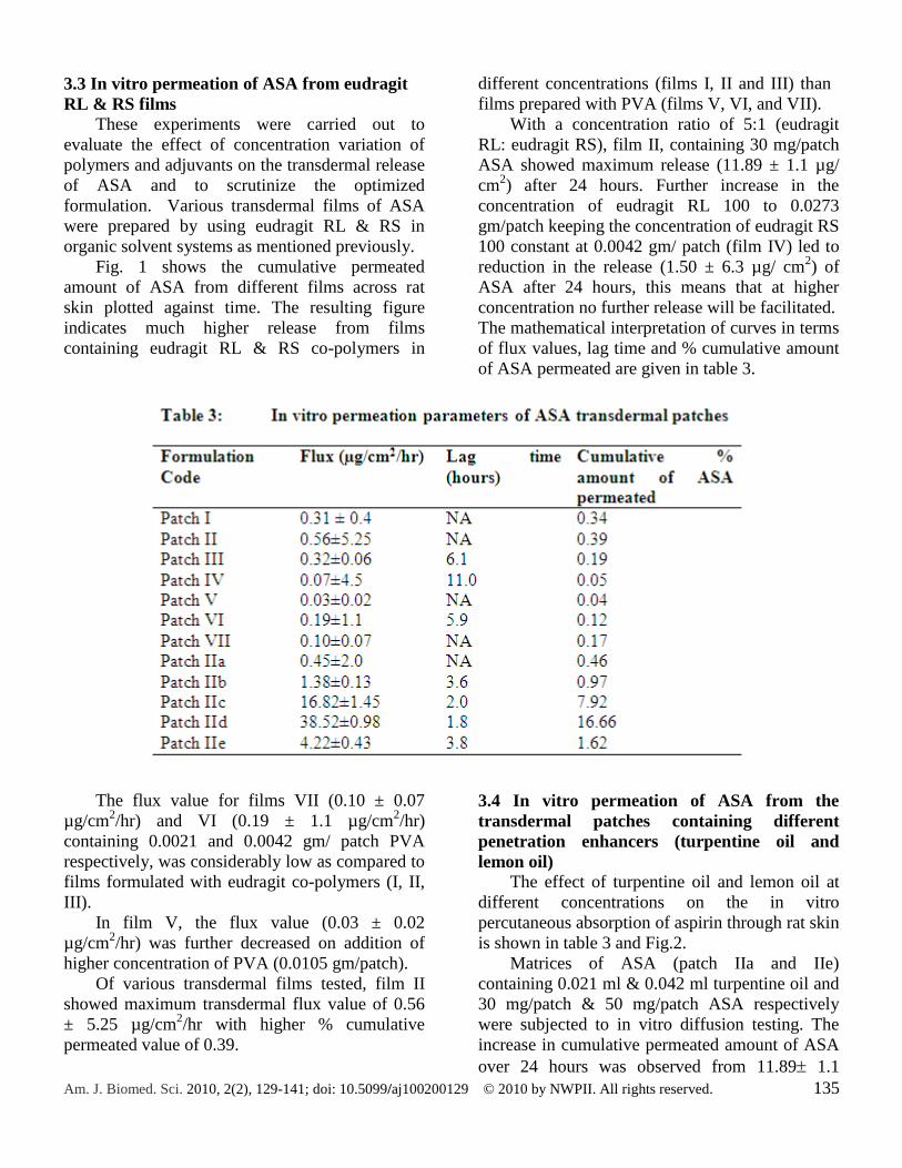
3.3 In vitro permeation of ASA from eudragit
RL & RS films
These experiments were carried out to
evaluate the effect of concentration variation of
polymers and adjuvants on the transdermal release
of ASA and to scrutinize the optimized
formulation. Various transdermal films of ASA
were prepared by using eudragit RL & RS in
organic solvent systems as mentioned previously.
Fig. 1 shows the cumulative permeated
amount of ASA from different films across rat
skin plotted against time. The resulting figure
indicates much higher release from films
containing eudragit RL & RS co-polymers in
different concentrations (films I, II and III) than
films prepared with PVA (films V, VI, and VII).
With a concentration ratio of 5:1 (eudragit
RL: eudragit RS), film II, containing 30 mg/patch
ASA showed maximum release (11.89 ± 1.1 µg/
cm2) after 24 hours. Further increase in the
concentration of eudragit RL 100 to 0.0273
gm/patch keeping the concentration of eudragit RS
100 constant at 0.0042 gm/ patch (film IV) led to
reduction in the release (1.50 ± 6.3 µg/ cm2) of
ASA after 24 hours, this means that at higher
concentration no further release will be facilitated.
The mathematical interpretation of curves in terms
of flux values, lag time and % cumulative amount
of ASA permeated are given in table 3.
The flux value for films VII (0.10 ± 0.07
µg/cm2/hr) and VI (0.19 ± 1.1 µg/cm
2/hr)
containing 0.0021 and 0.0042 gm/ patch PVA
respectively, was considerably low as compared to
films formulated with eudragit co-polymers (I, II,
III).
In film V, the flux value (0.03 ± 0.02
µg/cm2/hr) was further decreased on addition of
higher concentration of PVA (0.0105 gm/patch).
Of various transdermal films tested, film II
showed maximum transdermal flux value of 0.56
± 5.25 µg/cm2/hr with higher % cumulative
permeated value of 0.39.
3.4 In vitro permeation of ASA from the
transdermal patches containing different
penetration enhancers (turpentine oil and
lemon oil)
The effect of turpentine oil and lemon oil at
different concentrations on the in vitro
percutaneous absorption of aspirin through rat skin
is shown in table 3 and Fig.2.
Matrices of ASA (patch IIa and IIe)
containing 0.021 ml & 0.042 ml turpentine oil and
30 mg/patch & 50 mg/patch ASA respectively
were subjected to in vitro diffusion testing. The
increase in cumulative permeated amount of ASA
over 24 hours was observed from 11.89 1.1

Am. J. Biomed. Sci. 2010, 2(2), 129-141; doi: 10.5099/aj100200129 © 2010 by NWPII. All rights reserved. 136
µg/cm2
to 52.73 5.2 µg/cm2
over 24 hours (Patch
IIe). Almost 1.62% of the drug was found to
permeate across the rat skin over 24 hrs.
Table 3 also shows a significantly higher
flux of ASA from patch IIe (4.22 ± 0.43
µg/cm2/hr) than that of patch IIa and patch II
(without enhancer).
Fig 2: In vitro permeation of ASA through rat skin from different eudragit formulations containing penetration
enhancers.
The cyclic monoterpene, d-limonene which is
the main component of lemon oil and orange oil
has been shown to increase the permeation of a
number of nonsteroidal anti-inflammatory drugs
including aspirin [11], diclofenac [18], ketoprofen
[19] and ibuprofen [20]. D-limonene increases the
permeation of drugs by disrupting the highly
ordered structure of intercellular lipids and
improving the partitioning of drugs in the stratum
corneum [10].
As shown in fig. 2 effect of lemon oil on
cumulative permeation of ASA was observed to
be concentration dependent. Cumulative
permeation of ASA over 24 hours was increased
to 29.86 ± 2.2 µg/cm2
(patch IIb) with 0.021 ml
lemon oil in the patch formulation containing 30
mg/patch ASA.
In an ideal system, there is a linear
relationship between the rate of diffusion and the
concentration of diffusant. The maximum flux
occurs when the concentration reaches the
solubility limit [21]. The cumulative ASA
permeated was further increased to 404.49 ± 2.3
µg/cm2
(patch IIc) on increasing drug load to 50
mg/patch.
On increasing the lemon oil concentration to
0.042 ml /patch the cumulative ASA permeated
was almost doubled to 850.69 ± 3.9 µg/cm2
(patch
IId) and % cumulative amount of aspirin
permeated across rat skin from formulations IIc

Am. J. Biomed. Sci. 2010, 2(2), 129-141; doi: 10.5099/aj100200129 © 2010 by NWPII. All rights reserved. 137
and IId containing 50 mg/patch ASA was
increased significantly (P< 0.001) to 7.92 and
16.66 respectively.
0.042 ml lemon oil showed a very strong
enhancing effect when incorporated into patch IId
formulation with maximum transdermal
permeability flux of 38.52 ± 0.98 µg/cm2/hr) i.e.
0.92 mg/cm2 over 24 hours and a significantly low
lag period of 1.8 hours.
3.5 Influence of dermal ASA on bleeding time
The initial bleeding time (BT) was 2.81 ±
0.25 minutes. At 4 hours, of oral administration
and transdermal application of ASA, oral aspirin
at 10 mg/kg, caused a significant increase in BT
to 3.59 ± 0.04 minutes. (P<0.05, Students –
Newman - Keuls multiple comparison test) as
compared to the transdermal formulation of ASA,
patch IId (3.27 ± 0.05 minutes), patch IIe (3.25 ±
0.11 minutes) and patch II ( 2.93 ± 0.07 minutes)
respectively (Fig 3).
At 24 hours of administrations, patch IId (BT:
4.02 ± 0.26 minutes) and patch IIe (BT: 3.8 ±
0.13 minutes), showed significantly (p<0.001,
Students – Newman- Keuls multiple comparison
test) higher BT as compared to oral ASA (3.07 ±
0.20 minutes) and patch II, containing no enhancer
(3.05 ± 0.08 minutes).
Fig 3: Influence of transdermal and oral ASA formulations on bleeding time as compared to control group
3.6 Influence of dermal ASA on PT
The test measures the clotting time of plasma
in the presence of an optimal concentration of
tissue extract (thromboplastin) and indicate the
overall efficiency of the extrinsic clotting system
(Factors II, V, VII and X). The test is also known
to depend on reactions with factors V, VII and X,
and on the fibrinogen concentration of the plasma
[22]. The coagulation process is triggered by
incubation of plasma with the optimal amount of
thromboplastin and calcium. The time to
formation of a fibrin clot is then measured.
It can be seen from table 4 that the
transdermal application patch IId containing
lemon oil resulted in a significant prolongation of
prothrombin time (10.2± 0.10 seconds, P<0.001)
as compared to orally ingested ASA (8.9±0.1
seconds), control group (9.3 ± 0.1 secs.), patch II
(9.47 ± 0.06 secs), and patch IIe containing
turpentine oil (9.47±0.32 secs).
3.7 Influence of dermal ASA on APTT
The test measures the clotting time of plasma
after the activation of contact factors but without
added tissue thromboplastin, and so indicates the

Am. J. Biomed. Sci. 2010, 2(2), 129-141; doi: 10.5099/aj100200129 © 2010 by NWPII. All rights reserved. 138
overall efficiency of the intrinsic pathway (Factors
VIII and IX as well as the contact factors) [23].
Incubation of plasma with the optimal
quantity of phospholipids and a surface activator
leads to activation of factors of the intrinsic
coagulation system. The addition of calcium ions
trigger the coagulation process, the time to
formation of a fibrin clot is measured.
APTT was also prolonged to 30.6 ± 0.89 and
32.78 ± 3.25 seconds after the transdermal
application of patch IIe and IId respectively as
compared to the control group (28.3 ± 1.9 secs)
and patch II ( 27.0 ± 4.11 secs).
Table 4: Influence of transdermal and oral ASA on PT and APTT as compared to control group
4. Discussion
The increasing appreciation of the role of
inflammation in atherosclerosis and thrombosis
[24, 25] has raised intriguing possibility that
aspirin the anti- inflammatory compound which
was shown to reduce platelet aggregation induced
by several physiological stimuli [1] – might be
effective in the prevention of cardiovascular
disorders, including myocardial infarction, stroke
and thrombosis. The oral administration of ASA
for the prevention of cardiovascular events has
been associated with the elevated number of
gastrointestinal side effects. This has limited its
widespread clinical use.
Hence, the present study was designed to
develop a transdermal therapeutic system for
aspirin that could provide a predetermined
constant drug delivery, and the use of chemical
penetration enhancers to modulate skin
permeation of aspirin which would be beneficial
for an effective and safe therapy.
In 1971, Smith and Willis proved that the
blood-thinning properties of aspirin are based on
its inhibition of prostaglandin synthesis in the
blood platelets. Because platelet aggregation was
known to play a crucial role in thrombosis, it was
anticipated that the newly described anti-
aggregating activity of aspirin might translate to a
clinical benefit in ischaemic cardiovascular
disease.
Based on the in-vitro permeation studies
across rat skin, film II containing eudragit RL and
eudragit RS in the ration of 5:1 provided

Am. J. Biomed. Sci. 2010, 2(2), 129-141; doi: 10.5099/aj100200129 © 2010 by NWPII. All rights reserved. 139
maximum transdermal flux. It provided efficient
matrix to build up a large reservoir of drug in the
skin and to achieve greater permeation with a
lower amount of drug. Greater penetration with a
lower concentration of drug is always preferable
as [26], has reported increased irritation on
increasing drug concentration in the transdermal
device.
ASA crystallizes easily from the solution,
however, this tendency was substantially reduced
when films were dried at lower temperature (≤
15oC).
The permeation rate of aspirin was further
enhanced by the incorporation of turpentine and
lemon oil as penetration enhancers. Terpenes are
receiving much attention as penetration enhancers
[27]. They are derived from plant essential oils
and combine good penetration enhancing abilities
with low skin irritancy and low systemic toxicity.
A number of terpenes viz. menthol, 1-8- cineol, d-
limonene, nerolidol, geraniol, thymol, cyminine,
carvone, acetylterpineol, –bisabool have been
shown to increase transdermal permeation rate of
hydrophilic and lipophilic drugs [28, 29].
Lemon oil in Patch IId significantly increased
transdermal flux to 0.92 mg/cm2 over 24 hours. It
is reported that 21 mg/ day of aspirin is required to
be systematically available for inhibition of
platelet formation [14]. Thus, ASA can be
delivered transdermally by Patch IId of around 25
cm2 area in order to suppress the platelet
aggregation. McAdam and co-workers [11]
reported transdermal delivery of ASA 33 ± 3 mg
daily from matrix type of patch in healthy
volunteers that had a total surface area of 50 cm2
and contained 120 mg ASA along with d-
limonene at 12% of the total matrix mass. Our
results showed a higher release from formulation
containing lemon oil indicating that lemon oil
facilitates the flux of ASA to a greater extent than
that of turpentine oil.
In a clinical situation, both the bleeding time
(BT) and a number of in vitro measurements such
as prothrombin time (PT) and activated partial
thromboplastin time (APTT) are used together to
test the patient’s homeostatic mechanism and to
determine whether or not there is risk of bleeding.
Aspirin has been proven effective in secondary
prevention of coronary artery disease (CAD). Its
efficacy has been ascribed to its antiplatelet
action through the irreversible acetylation of
platelet cyclooxygenase with subsequent blockade
of platelet thromboxane synthesis. In this study,
we evaluated the effects of dermal low-dose
aspirin on blood coagulation in mice and rats
using routine laboratory blood tests (BT, PT and
APTT) as a measure of aspirin bioavailability.
Aspirin influences the bleeding time,
presumably through the inhibition of
prostaglandin biosynthesis and the resultant
platelet secretion. The response of the bleeding
time is quite variable after ingestion of aspirin. In
some instances, the bleeding time is not
prolonged, whereas in other instances there is
definite prolongation [30]. The prolongation is
dependent upon the technique used and direction
of incision, but overall aspirin was found to
produce only a modest increase in the bleeding
time [31].
Comparison of the time courses for bleeding
duration before and after the oral administration
and transdermal application of ASA, showed that
the formulated dermal low-dose aspirin (patch
IId), containing lemon oil as penetration enhancer,
significantly prolonged the bleeding time.
Taken together, our results provide
convincing evidence for the feasibility of
transdermal low-dose aspirin patch containing
lemon oil as penetration enhancer in clinical
situations because of excellent release of the drug,
and its influence on the blood coagulation by
means of affecting both the extrinsic coagulation
system and the intrinsic coagulation system
significantly. Further work is required to establish
the utility of such transdermal drug delivery
system through long term pharmacokinetic studies
on human subjects.
Acknowledgement
We are thankful to the Indian Council for
Cultural Relations for financial assistance towards
research investigation.
References
1. De Gaetano, G. Aspirin and the prevention of
ischemic heart disease. A Socratic dialogue

Am. J. Biomed. Sci. 2010, 2(2), 129-141; doi: 10.5099/aj100200129 © 2010 by NWPII. All rights reserved. 140
between a cardiologist, a clinical
pharmacologist and an expert of blood
platelets. Ital Heart J, 2001a, 2, 582-588.
2. American – Canadian Co-operative Study
Group. Persantine aspirin trial in cerebral
ischemia. part II; end point results. Stroke,
1985, 16(3), 406-415.
3. Eidelman, R.S.; Herbert, P.R.; Weisman, S.;
Hennekens, C.H. An update on aspirin in the
primary prevention of cardiovascular disease.
Arch Int Med 2003, 163, 25-36.
4. Smith, J.B.; Willis, A.L. Aspirin selectively
inhibits prostaglandin production in human
platelets. Nature [New Biol.], 1971, 231, 235-
237.
5. De Gaetano, G. Historical overview of the role
of platelets in hemostasis and thrombosis.
Haematologica, 2001b, 86, 349-356.
6. The RISC Group. Risk of myocardial
infarction and death during treatment with
low-dose aspirin and intravenous heparin in
men with unstable coronary artery disease.
Lancet, 1990, 336, 827-830.
7. Derry, S.; Loke, Y.Y. Risk of gastrointestinal
hemorrhage with long term use of aspirin:
meta-analysis. Br Med J, 2000, 321, 1183-187.
8. Krishna, D.R.; Srinivas, A.G.; Srinivas, A.
Transdermal aspirin: influence of platelet
aggregation and serum lipid peroxides. Ind J
Pharm Sci, 2000, 62(3), 200-204.
9. Bronaugh, R.L.; Collier, S.W. In vitro methods
for measuring skin permeation. In: Zatz, J.L.
(Ed.), Skin Permeation – Fundamentals and
Applications. Allured Publishing Corporation,
Wheaton, Illinois, 1993, pp. 93-111.
10. Keimowitz, R.M.; Pulvermacher, G.; Mayo,
G.; FitzGerald, D.J. Transdermal modification
of platelet function: a dermal aspirin
preparation selectively inhibits platelet
cyclooxygenase and prevents prostacyclin
biosynthesis. Circulation, 1993, 88, 556-561.
11. McAdam, B.; Keimowitz, R.M.; Maher, M.;
FitzGerald, D.J. Transdermal modification of
platelet function: an aspirin patch system
results in marked suppression of platelet
cyclooxygenase. J Pharmacol Exper Therap,
1996, 277, 559-564.
12. McMahon, G.P.; O’Connor, S.J.; FitzGerald,
D.J.; Roy, S.L.; Kelly, M.T. Determination of
aspirin and salicylic acid in transdermal
perfusates. J Chromatography B, 1998, 707,
322-327.
13. Pederson, A.K.; FitzGerald, G.A. Dose-related
kinetics of aspirin: presystemic acetylation of
platelet cycolooxygenase. New Eng J Med,
1984, 311, 1206-1211.
14. Levang, A.K.; Zhao, K.; Singh, J. Effect of
ethanol/ propylene glycol on the in vitro
percutaneous absorption of aspirin,
biophysical changes and macroscopic barrier
properties of the skin. Int J Pharm, 1999, 181,
255-263.
15. Lee, J.S.; Adrie, C.; Jacob, H.J. Chronic
inhalation of nitric oxide inhibits neointimal
formation after balloon-induced arterial injury.
Circulation Res, 1996, 78, 337-342.
16. The Dutch TIA Trial Study Group. A
comparison of two doses aspirin (30 mg vs.
283 mg a day) in patients after a transient
ischemic attack or minor ischemic stroke. New
Engl J Med, 1991, 325, 1261-1266.
17. Petersen, T.; Husted, S.E.; Pedersen, A.K.;
Geday, E. Systemic availability of
acetylsalicylic acid in normal human subjects
after oral ingestion of three different
formulations. Acta Pharmacol Toxicol, 1982,
51, 285-291.
18. Obata, Y.; Takayama, K.; Maitani, Y.; Nagai,
T. Effect of pretreatment of skin with cyclic
monoterpenes on permeation of diclofenac in
hairless rat. Biol Pharm Bull, 1993, 16, 312-
314.
19. Okabe, H.; Takayama, K.; Nagai, T.
Percutaneous absorption of ketoprofen from
acrylic gel patches containing d-limonene and
ethanol as absorption enhancers. Chem Pharm
Bull, 1992, 40, 1906-1910.
20. Priborsky, J.; Takayama, K.; Obata, Y.;
Priborska, Z.; Nagai, T. Influence of limonene
and laurocapram on percutaneous absorption
of nonsteroidal anti-inflammatory drugs.
Arzneimittel – Forschung, 1992, 42, 116-119.
21. Hadgraft, J. Recent developments in topical
and transdermal delivery. Eur J Drug Metab
Pharmacokinet, 1996, 21(2), 165-173.
22. Lautes, I.J.; Chu, J.C.; Sikranth, S.; Huber-
Lang, M. Anti-C5a Ameliorates coagulation/

Am. J. Biomed. Sci. 2010, 2(2), 129-141; doi: 10.5099/aj100200129 © 2010 by NWPII. All rights reserved. 141
fibrinolytic protein changes in a rat model of
sepsis. Am J Path, 2002, 160, 1867-1875.
23. Brandt, J. T.; Triplett, D.A.; Rock, W.A.;
Bovill, E.G.; Arkin, C. F. Effect of lupus
anticoagulants on the activated partial
thromboplastin time. Arch Pathol Lab Med,
1991, 115, 109-114.
24. Ross, R. 1999. Atherosclerosis: an
inflammatory disease. New Engl J Med, 1999,
340, 115-126.
25. Libby, P. Inflammation in atherosclerosis.
Nature, 2002, 420, 868-874.
26. Kobayashi, K.; Hosaka, T.; Ueno, H.; Maruo,
M. Relationship between the amount of
propranolol permeating through the stratum
corneum of guinea pig skin and after
application of propranolol adhesive patch and
skin irritation. Biol Pharm Bull, 1996, 19(6),
839-844.
27. Williams, A.C.; Barry, B.W. Terpenes and the
lipid protein-partitioning theory of skin
penetration enhancement. Pharm Res, 1991,
8, 17-24
28. Cornwell, P.A.; Barry, B.W. Sequiterpene
components of volatile oils as skin penetration
enhancers for the hydrophilic permeant 5-
fluorouracil. J Pharm Pharmacol, 1991, 46,
261-269.
29. Gao, S.; Singh, J. Invitro percutaneous
absorption enhancement of lipophilic drug
tamoxifen by terpenes. J Cont Rel, 1998, 51,
193-199.
30. Mielke, C.H.; Kaneshiro, M.M.; Maher, I. A.
The standardized normal Ivy bleeding time
and its prolongation by aspirin. Blood, 1969,
34, 204-210.
31. Mielke , C.H. Aspirin prolongation of the
template bleeding time: influence of venostasis
and direction of incision. Blood, 1982, 60,
1139-1142.