Effect of Selected Natural Dyes in Reduction on Colour Changes of Egyptian Linen Textiles by Fungi
14
Annali di Chimica, 97, 2007, by Società Chimica Italiana 527 EFFECT OF SELECTED NATURAL DYES IN REDUCTION ON COLOUR CHANGES OF EGYPTIAN LINEN TEXTILES BY FUNGI Omar ABDEL-KAREEM Conservation Department, Faculty of Archaeology and Anthropology, Yarmouk University, Jordan, [email protected] Summary - Linen is the most historical Egyptian textile fibre liable to fungal deterioration. Fungal deterioration of dyed linen textiles may appear as undesirable different stains. In order to success in removing of fungal stains from biodeteriorated historical Egyptian dyed linen textiles, it is necessary to understand the nature and causes of these stains, hence their subsequent removal. So this paper aims to investigate the effect of fungi on dyed linen textiles. In this study linen textile samples were experimentally dyed by two different dyes, blue one as an example to vat dye and yellow one as an example to direct dye. This work is done on two of the most important dyes (Turmeric and indigo), which were popular in most of historical periods in Egypt. Dyed linen samples were experimentally biodegraded by thirty different fungal strains isolated previously from historical Egyptian linen samples. The produced change in colours of the biodeteriorated samples was detected visually. Also, the change in reflection spectra and colour differences produced to dyed linen textiles after fungal deterioration, were assessed and evaluated by using spectrophotometer. This study reported that most of tested fungi contribute to discoloration of all tested dyed linen samples. These results indicate that most of stains on historical Egyptian dyed linen textiles, may be fungal stains. The results confirm that undyed linen textiles more liable to fungal biodeterioration than dyed ones. Also the results show that yellow dyed linen textiles are more susceptible to fungal deterioration than blue dyed linen textiles. The obtained results show that Alternaria tenuissima, Chaetomium globosum, Chaetomium sp., Penicillium raistrickii, P. soppi, P. asperum, P. citrinum, Aspergillus carbonarius, A. fischeri, A. nidulans, A. terreus and A. niger, had showed the maximum colour changes of the deteriorated yellow dyed linen samples. The results also show that Alternaria tenuissima, Chaetomium sp., Penicillium asperum, P. citrinum, Aspergillus nidulans and A. spinulosus, had shown the maximum colour changes of the deteriorated blue dyed linen samples. INTRODUCTION The history of dyeing in Egypt starts in Antiquity as dyed textiles from thousand of years B.C., which has been found in Egyptian tombs 1,2 . From Roman times the art of dyeing was well established in Egypt. Coptic textiles illustrate well the range of dyes being used in the late Roman,
Transcript of Effect of Selected Natural Dyes in Reduction on Colour Changes of Egyptian Linen Textiles by Fungi
Annali di Chimica, 97, 2007, by Società Chimica Italiana 527
EFFECT OF SELECTED NATURAL DYES IN REDUCTION ON COLOUR
CHANGES OF EGYPTIAN LINEN TEXTILES BY FUNGI
Omar ABDEL-KAREEM Conservation Department, Faculty of Archaeology and Anthropology, Yarmouk University, Jordan, [email protected] Summary - Linen is the most historical Egyptian textile fibre liable to fungal deterioration. Fungal deterioration of dyed linen textiles may appear as undesirable different stains. In order to success in removing of fungal stains from biodeteriorated historical Egyptian dyed linen textiles, it is necessary to understand the nature and causes of these stains, hence their subsequent removal. So this paper aims to investigate the effect of fungi on dyed linen textiles. In this study linen textile samples were experimentally dyed by two different dyes, blue one as an example to vat dye and yellow one as an example to direct dye. This work is done on two of the most important dyes (Turmeric and indigo), which were popular in most of historical periods in Egypt. Dyed linen samples were experimentally biodegraded by thirty different fungal strains isolated previously from historical Egyptian linen samples. The produced change in colours of the biodeteriorated samples was detected visually. Also, the change in reflection spectra and colour differences produced to dyed linen textiles after fungal deterioration, were assessed and evaluated by using spectrophotometer. This study reported that most of tested fungi contribute to discoloration of all tested dyed linen samples. These results indicate that most of stains on historical Egyptian dyed linen textiles, may be fungal stains. The results confirm that undyed linen textiles more liable to fungal biodeterioration than dyed ones. Also the results show that yellow dyed linen textiles are more susceptible to fungal deterioration than blue dyed linen textiles. The obtained results show that Alternaria tenuissima, Chaetomium globosum, Chaetomium sp., Penicillium raistrickii, P. soppi, P. asperum, P. citrinum, Aspergillus carbonarius, A. fischeri, A. nidulans, A. terreus and A. niger, had showed the maximum colour changes of the deteriorated yellow dyed linen samples. The results also show that Alternaria tenuissima, Chaetomium sp., Penicillium asperum, P. citrinum, Aspergillus nidulans and A. spinulosus, had shown the maximum colour changes of the deteriorated blue dyed linen samples.
INTRODUCTION
The history of dyeing in Egypt starts in Antiquity as dyed textiles from thousand of years B.C., which has been found in Egyptian tombs1,2. From Roman times the art of dyeing was well established in Egypt. Coptic textiles illustrate well the range of dyes being used in the late Roman,
ABDEL-KAREEM
528
early Byzantine and early Islamic periods.3 The most important historical Egyptian dyes, which were popular in Egypt, were reported in references.1,3-5 Indigo, which is historically the most important natural dye known to man, has been in continuous in the Arab world for over four thousand years.6 Among the many plants, which yield a yellow dye, turmeric stands out as one of the most popular used in different historical Egyptian periods.1,4
Archaeological materials of historical textiles found in burial sites are often exposed to the most severe conditions. Exposure to bacteria, yeast, algae, and fungi cause severe discoloration and degradation of dyed textiles. The dyed textiles change colour on burial in damp soils with the nature and degree of colour change affected by the fibre, dye type involve, the presence of mordant and the burial conditions encountered.7 The most severe deterioration in indoor environments caused by cellulolytic fungi, resulting in loss of fibre strength and actual failure. Other fungal spoilage may occur as a result of the permanent staining from pigmentation and mycelium penetration.8 The author visited a lot of Egyptian museums, and he noticed that the fungal stains are commonly present on Egyptian dyed linen textiles. Abdel-Kareem, et al, 1997, reported that linen is the most ancient Egyptian textile fibre liable to fungal deterioration.9 Of great concern to textile conservators is the present range of various fungal stains, most commonly observed on white textile.10 It is reported that several fungi contributed to discoloration of textiles. The coloured mycelium penetrates inside the fibres and between them creating a full impression of coloration. Many fungi caused stains because of the colour of the conidial mass or sclerotia. Other causes are associated with biochemical transformation and with the formation of various fungal metabolites. The colouring matter generated in the mycelium was sometimes discharged from the hyphae and impregnated into the fibres.11 The circular shape of a fungus spot is due to the concentric growth of the hyphae outwards from the central germinated spore or conidia. The fungal growth may also occur downwards into the substrate. Hyphae may force themselves between fibres, or digest the fibres themselves and then absorb the digested substrate. Under adverse environmental conditions some fungi species produce sclerotia which look like black fly spots in the fungal stains. Sclerotia are made up of an organised mass of pigmented hyphae and stored nutrients, hence function in vegetative propagation.12 The identification of dyes present on the biodeteriorated ancient Egyptian textiles is very difficult7 because the fungi may produce complicated coloured pigments in their fungal structures and substrate.13 However, the conservation literature on components found on fungal spots areas and their formation mechanisms is negligible, and many studies are interesting to note brown (foxing) spots found on various materials.14-20 Florian (1997) discussed the source of the colour in fungal spots and suggested that they are different kinds namely: (i) those that originated from a group of conidia, some of which germinated and produced minimal hyphae growth, (ii) those that developed mycelia and (iii) those that developed along with mycelium conidiophores and new conidia.12
The removal and prevention of fungal stains on dyed historical Egyptian textiles is very important because of their offence to senses of sight and smell. Also, due to the fact that they contain enzymes that can still continue degrading the fibres, even after fungi responsible for their production has been destroyed. In order to success in removing of fungal stains from biodeteriorated ancient Egyptian dyed linen textiles, it is necessary to understand the nature and causes of these stains, hence their subsequent removal. So this study aims to investigate the effect of fungi on dyed linen textiles. This work is done on two of the most important historical Egyptian dyes (Turmeric and indigo) which were popular in different historical Egyptian periods.1,4,21
Investigation of the Role of Fungi in Discoloration of Ancient Egyptian Dyed Linen Textiles
529
MATERIALS AND METHODS
Fabrics The linen fabric used in this work was scoured, unbleached plain linen textile. The count of warp and weft threads /1 cm are 20 and 14 respectively, the coloured shade was L = 56.62, a = 1.57, b = 10.23. Linen samples were bleached according to Wickens, 1990.22 Dyeing of samples with indigo (blue colour) 1g of natural indigo powder was ground with 10 mL water until a creamy, even consistency was obtained. 2) 50 mL of warm water were added into a bottle and mixed with 1g of sodium hydroxide which was slowly applied with continuous stirring until dissolved. 3) 50 mL of warm water was also added into another bottle and gently slowly mixed with 1 g of sodium dithionite. 4) All were added, but small amounts of the sodium hydroxide solution and the sodium dithionite solution were reserved. 5) After 1 hour, it was tested by dipping in a glass rod according to Dalby, 1989.5 6) After preparing the vat dye according to Robertson, 1973,1 the clean wetted fabric was lowered gently into both sides of the vat. It was kept there and moved gently for 1 to 5 minutes. 7) The fabric was taken out of the vat with a faint yellow colour, which later turned blue in the air within a few minutes. 8) After the dyed fabric dried, it was washed in soapy water, rinsed very thoroughly, and dried.
Dyeing of samples with Turmeric (yellow colour) The samples were dyed according to Weigle, 1974, following recipe below: 1) 10g of turmeric powder were loosely tied in a cheesecloth bag and was soaked in 5 litres of water overnight. 2) The next day the bath was slowly brought to the boiling point and then simmered gently for 1 hour. By using a spoon, the cheesecloth bag was pressed down to help release the colour. 3) At the end of 1 hour, the cheesecloth bag was removed and squeezed out as much dye as possible. 4) The clean, wetted fabric was placed in dye bath and was simmered gently for 1 hour. 5) After the dyed fabric dried, it was washed in soapy water, rinsed very thoroughly, and dried.4
Fungal strains Fungi used in this study were 30 species isolated from biodeteriorated ancient Egyptian textiles (Abdel-Kareem, et al, 1997). These fungi belong to genera Aspergillus, Penicillium, Fusarium, Alternaria, Trichoderma and Chaetomium. Aspergillus species were Aspergillus auratus, A. carbonarius, A. chrysellus, A. fischeri, A. flavus, A. fumigatus, A. nidulans, A. niger, A. proliferans, A. spinulosus, A. sp., A. terreus, and A. ustus. Penicillium species were Penicillium asperum, P. biforme, P. citrinum, P. funiculosum, P. raistrickii, P. soppi and P. wortmanni. Chaetomium species were Chaetomium cochlioides, Chaetomium globosum, Chaetomium sp., Chaetomium sp., Chaetomium sp. and Chaetomium sp. Fusarium species were Fusarium nivale and Fusarium sp. Other genera were Alternaria tenuissima and Trichoderma viride. Treatment of linen with fungi The tests were performed in 90 mm diameter Petri dishes contain solid medium, Czapek-Dox agar modified.9 Linen samples were treated with tested fungi by using Plate test method. The plates were incubated at 28 C. Two weeks later, the textile samples were picked out and washed with water to remove mycelium then they were dried in air room condition.23 Before testing specimens were conditioned at 21±2 °C and 65 % ± 2 RH.
ABDEL-KAREEM
530
Methods of measurement of biodeterioration: Visual assessment: Extent of fungal growth on samples deteriorated by tested fungi for two weeks was assessed visually. Spectrophotometric measurements Reflection Spectra: Reflection spectra were recorded for the biodeteriorated samples in the visible range (400-700 nm) by a Shimadzu Double Beam UV-2101PC Spectrophotometer Equiped with ISR48 Intergating Sphere, using barium sulfate as reference (white standard).23 Reflectance of both front side and behind side of each specimen was measured. Brightness and colour difference evaluation: In order to determine the relative rate of colour changes of the biodeteriorated samples the reflectance values were inserted into a computer program which then calculated the CIELAB colour coordinates for L, a, and b values. Calculation of total colour change (∆E) is achieved by the use of the following equations: ∆E =[(∆L)2 + (∆a)2 + (∆b)2]0.5.
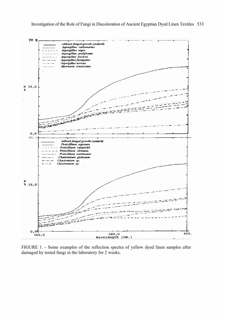
RESULTS AND DISCUSSION Visual assessment: The results of visual assessment of the bleached, yellow and blue linen samples before and after the deterioration by tested fungi for 14 days were recorded in (Photofigs.1,2). These results show that all tested fungi cause different changes of the colour of bleached and dyed linen samples. These changes in the colour of the tested samples may be were caused due to the coloured fungi pigments which penetrated inside the fibres and between them creating a full impregnation of coloration or either of the conidial mass or sclerotia or because of the fungal metabolites.11 However the obtained results show that the changes of the colour of the biodeteriorated linen textiles samples depend on fungal species and so on kind of the dye. The obtained results confirmed that Alternaria tenuissima, Aspergillus nidulans, Chaetomium globosum, Aspergillus carbonarius, A. terreus and A. niger had shown the maximum colour changes, and that Fusarium nivale, Fusarium sp. and Aspergillus ustus were the least effect. When comparing the results in (Photofigure 1), it was found that Alternaria tenuissima, Chaetomium globosum, Chaetomium sp, Penicillium raistrickii, P. soppi, P.citrinum, Aspergillus carbonarius, A. terreus and A. niger had showed the maximum colour changes of the yellow linen samples, and that the other fungi species were characterized by least effect. While the results in (Photofigure 2) show that Alternaria tenuissima, Chaetomium globosum, Chaetomium sp, Aspergillus niger, A. carbonarius, A. fischeri, A. fumigatus, Penicillium asperum, P. citrinum and P. wortmanni had shown the maximum colour changes of the blue dyed linen samples, and that the other fungi species were characterized by least effect. When comparing the results of the extent of fungal growth and the colour change of all tested biodeteriorated linen samples, it is noticed that the degree of the fungal growth on the bleached linen samples are more than the degree of fungal growth on the dyed linen samples. Also the obtained results show that the yellow dyed samples changed in colour more than the blue dyed linen samples. Reflection spectra: The reflection spectra of dyed linen textile samples that had been deteriorated by different fungi for 14 days are presented in (Fig. 1,2,3). The curves in (Fig. 1,2,3) show that all tested fungi caused that the yellow and blue dyed linen textile samples became darker after the deterioration by fungi. The rate % of the darkness of the biodeteriorated linen textiles samples depend on fungal species and so on kind of dye. Most of fungi caused that the yellow dyed linen textile samples became dark after being deteriorated by fungi (Fig. 1). The yellow dyed linen samples deteriorated by Alternaria tenuissima, Chaetomium globosum, Chaetomium sp.,

Investigation of the Role of Fungi in Discoloration of Ancient Egyptian Dyed Linen Textiles
531
Penicillium raistrickii, P. soppi, P.citrinum, Aspergillus carbonarius, A. terreus and A. niger showed a noticeable decrease in the reflection spectra.
PHOTOFIGURE 1. - Colour differences of biodeteriorated yellow linen samples damaged by tested fungi in the laboratory for 2 weeks.
ABDEL-KAREEM
532
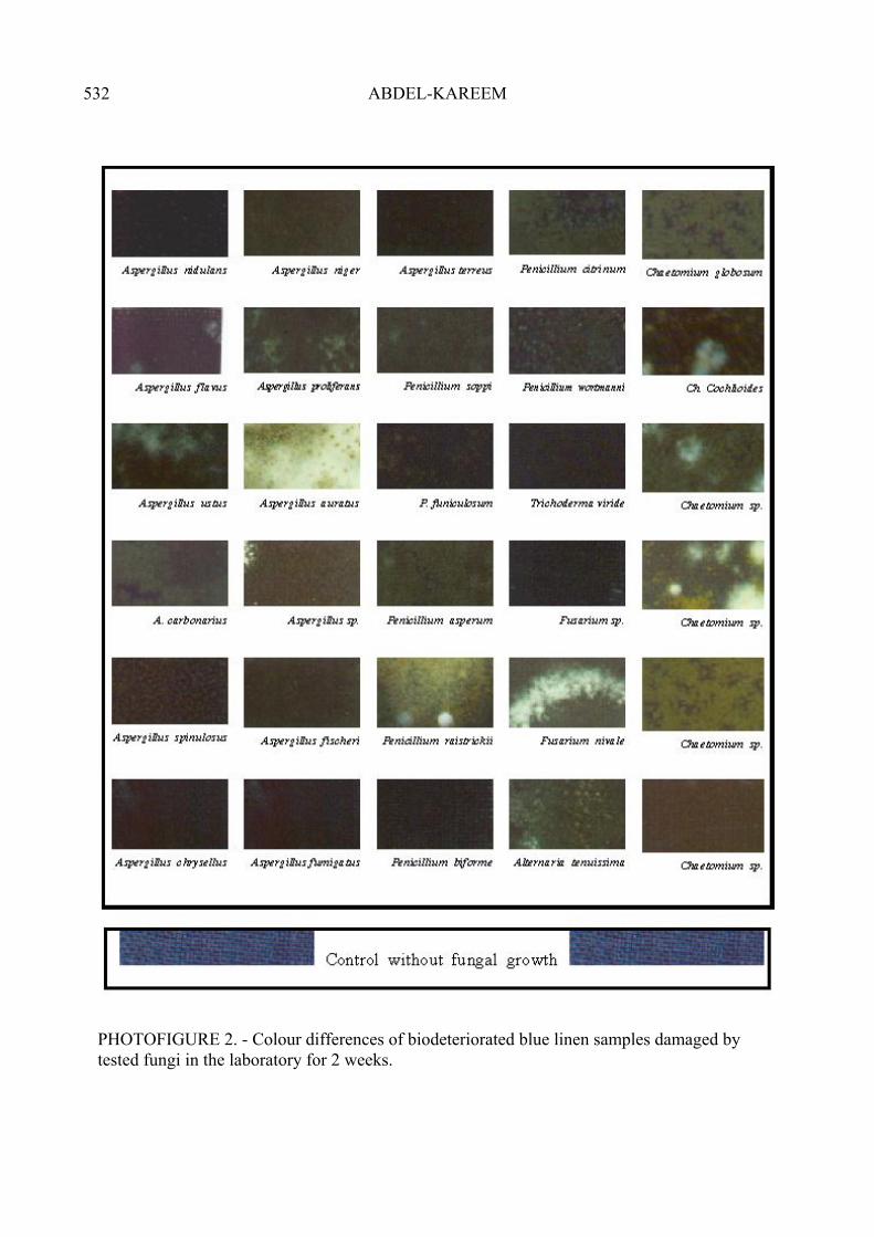
PHOTOFIGURE 2. - Colour differences of biodeteriorated blue linen samples damaged by tested fungi in the laboratory for 2 weeks.
Investigation of the Role of Fungi in Discoloration of Ancient Egyptian Dyed Linen Textiles
533
FIGURE 1. - Some examples of the reflection spectra of yellow dyed linen samples after damaged by tested fungi in the laboratory for 2 weeks.
ABDEL-KAREEM
534
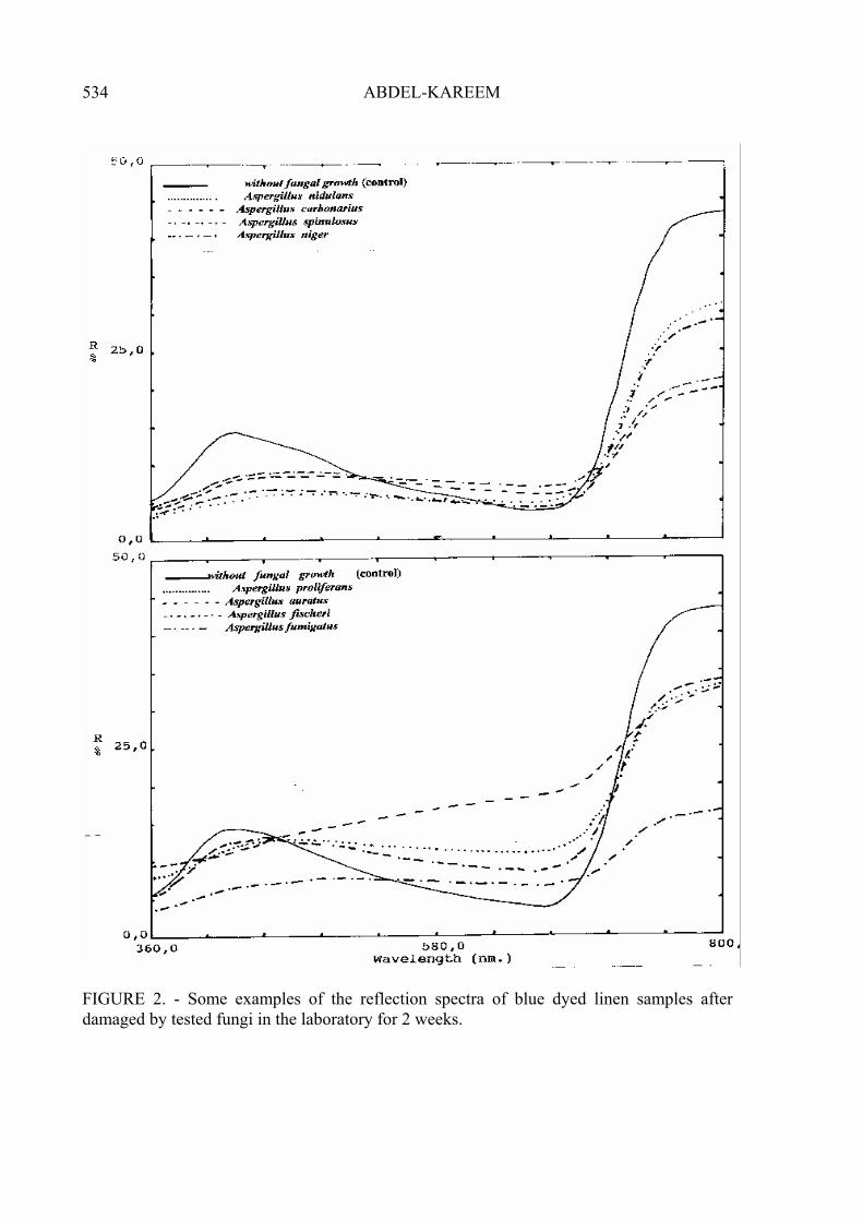
FIGURE 2. - Some examples of the reflection spectra of blue dyed linen samples after damaged by tested fungi in the laboratory for 2 weeks.
Investigation of the Role of Fungi in Discoloration of Ancient Egyptian Dyed Linen Textiles
535
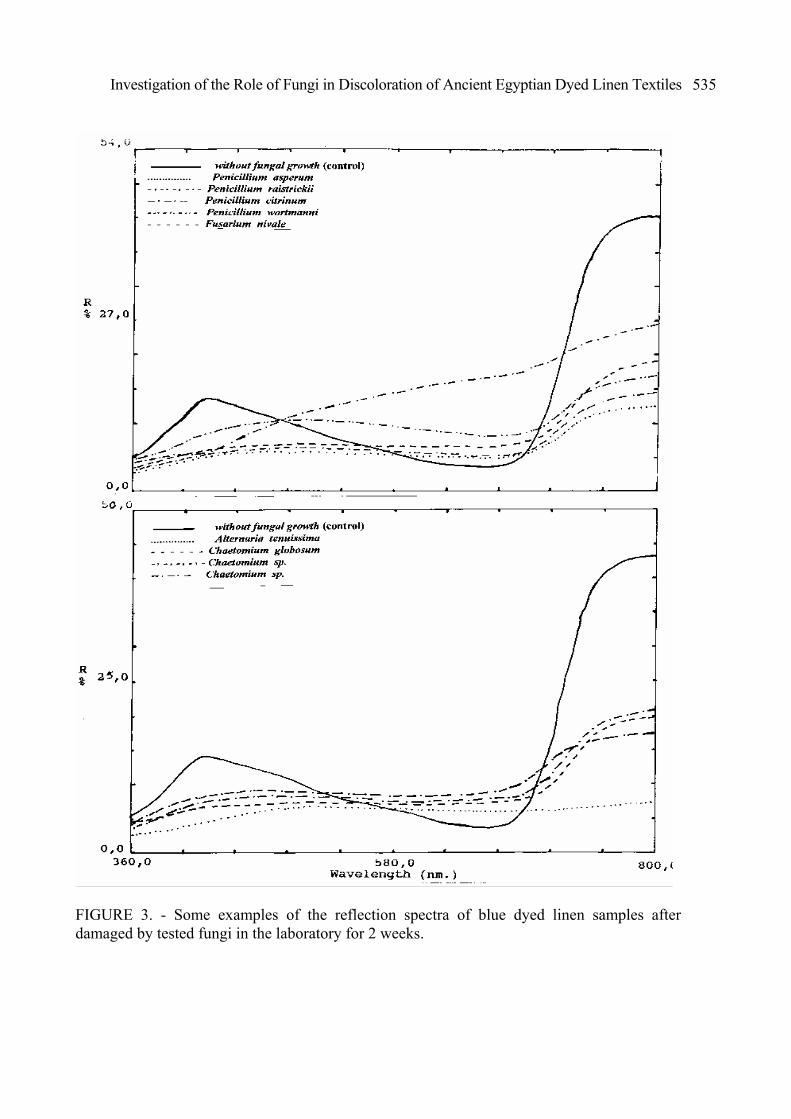
FIGURE 3. - Some examples of the reflection spectra of blue dyed linen samples after damaged by tested fungi in the laboratory for 2 weeks.
ABDEL-KAREEM
536
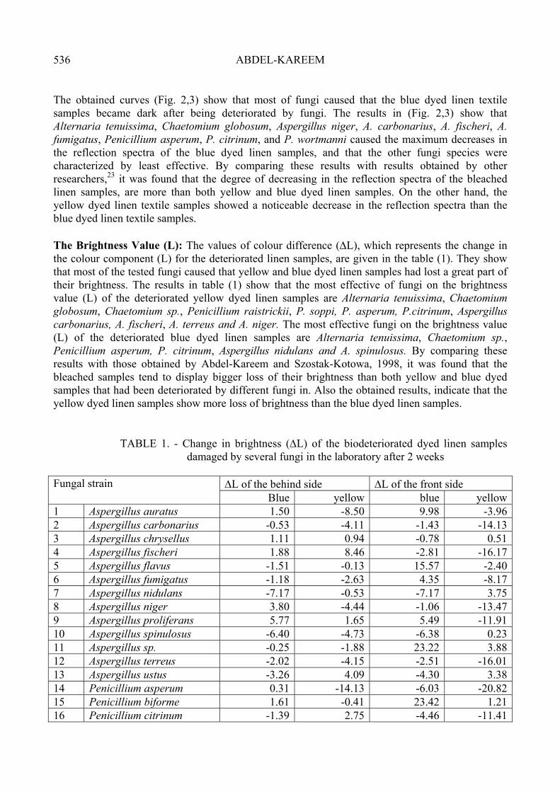
The obtained curves (Fig. 2,3) show that most of fungi caused that the blue dyed linen textile samples became dark after being deteriorated by fungi. The results in (Fig. 2,3) show that Alternaria tenuissima, Chaetomium globosum, Aspergillus niger, A. carbonarius, A. fischeri, A. fumigatus, Penicillium asperum, P. citrinum, and P. wortmanni caused the maximum decreases in the reflection spectra of the blue dyed linen samples, and that the other fungi species were characterized by least effective. By comparing these results with results obtained by other researchers,23 it was found that the degree of decreasing in the reflection spectra of the bleached linen samples, are more than both yellow and blue dyed linen samples. On the other hand, the yellow dyed linen textile samples showed a noticeable decrease in the reflection spectra than the blue dyed linen textile samples. The Brightness Value (L): The values of colour difference (∆L), which represents the change in the colour component (L) for the deteriorated linen samples, are given in the table (1). They show that most of the tested fungi caused that yellow and blue dyed linen samples had lost a great part of their brightness. The results in table (1) show that the most effective of fungi on the brightness value (L) of the deteriorated yellow dyed linen samples are Alternaria tenuissima, Chaetomium globosum, Chaetomium sp., Penicillium raistrickii, P. soppi, P. asperum, P.citrinum, Aspergillus carbonarius, A. fischeri, A. terreus and A. niger. The most effective fungi on the brightness value (L) of the deteriorated blue dyed linen samples are Alternaria tenuissima, Chaetomium sp., Penicillium asperum, P. citrinum, Aspergillus nidulans and A. spinulosus. By comparing these results with those obtained by Abdel-Kareem and Szostak-Kotowa, 1998, it was found that the bleached samples tend to display bigger loss of their brightness than both yellow and blue dyed samples that had been deteriorated by different fungi in. Also the obtained results, indicate that the yellow dyed linen samples show more loss of brightness than the blue dyed linen samples.
TABLE 1. - Change in brightness (∆L) of the biodeteriorated dyed linen samples damaged by several fungi in the laboratory after 2 weeks
∆L of the behind side ∆L of the front side Fungal strain
Blue yellow blue yellow1 Aspergillus auratus 1.50 -8.50 9.98 -3.962 Aspergillus carbonarius -0.53 -4.11 -1.43 -14.133 Aspergillus chrysellus 1.11 0.94 -0.78 0.514 Aspergillus fischeri 1.88 8.46 -2.81 -16.175 Aspergillus flavus -1.51 -0.13 15.57 -2.406 Aspergillus fumigatus -1.18 -2.63 4.35 -8.177 Aspergillus nidulans -7.17 -0.53 -7.17 3.758 Aspergillus niger 3.80 -4.44 -1.06 -13.479 Aspergillus proliferans 5.77 1.65 5.49 -11.9110 Aspergillus spinulosus -6.40 -4.73 -6.38 0.2311 Aspergillus sp. -0.25 -1.88 23.22 3.8812 Aspergillus terreus -2.02 -4.15 -2.51 -16.0113 Aspergillus ustus -3.26 4.09 -4.30 3.3814 Penicillium asperum 0.31 -14.13 -6.03 -20.8215 Penicillium biforme 1.61 -0.41 23.42 1.2116 Penicillium citrinum -1.39 2.75 -4.46 -11.41
Investigation of the Role of Fungi in Discoloration of Ancient Egyptian Dyed Linen Textiles
537
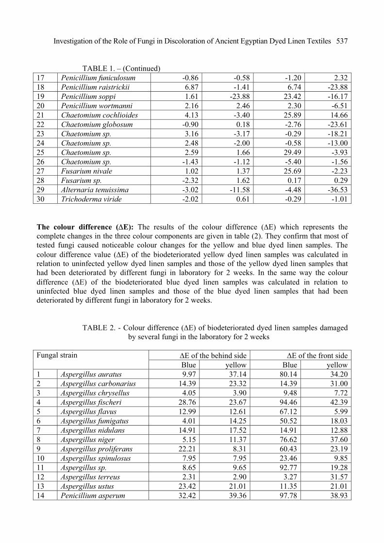
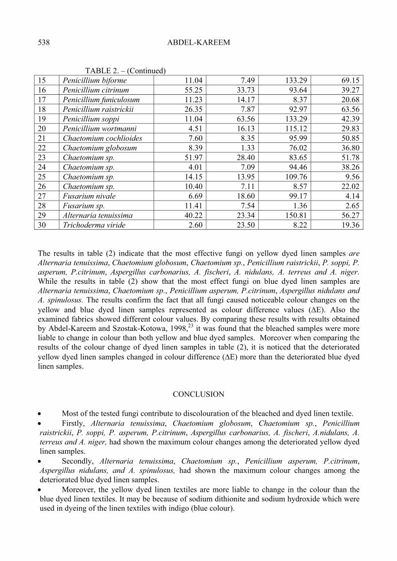
TABLE 1. – (Continued) 17 Penicillium funiculosum -0.86 -0.58 -1.20 2.3218 Penicillium raistrickii 6.87 -1.41 6.74 -23.8819 Penicillium soppi 1.61 -23.88 23.42 -16.1720 Penicillium wortmanni 2.16 2.46 2.30 -6.5121 Chaetomium cochlioides 4.13 -3.40 25.89 14.6622 Chaetomium globosum -0.90 0.18 -2.76 -23.6123 Chaetomium sp. 3.16 -3.17 -0.29 -18.2124 Chaetomium sp. 2.48 -2.00 -0.58 -13.0025 Chaetomium sp. 2.59 1.66 29.49 -3.9326 Chaetomium sp. -1.43 -1.12 -5.40 -1.5627 Fusarium nivale 1.02 1.37 25.69 -2.2328 Fusarium sp. -2.32 1.62 0.17 0.2929 Alternaria tenuissima -3.02 -11.58 -4.48 -36.5330 Trichoderma viride -2.02 0.61 -0.29 -1.01 The colour difference (∆E): The results of the colour difference (∆E) which represents the complete changes in the three colour components are given in table (2). They confirm that most of tested fungi caused noticeable colour changes for the yellow and blue dyed linen samples. The colour difference value (∆E) of the biodeteriorated yellow dyed linen samples was calculated in relation to uninfected yellow dyed linen samples and those of the yellow dyed linen samples that had been deteriorated by different fungi in laboratory for 2 weeks. In the same way the colour difference (∆E) of the biodeteriorated blue dyed linen samples was calculated in relation to uninfected blue dyed linen samples and those of the blue dyed linen samples that had been deteriorated by different fungi in laboratory for 2 weeks.
TABLE 2. - Colour difference (∆E) of biodeteriorated dyed linen samples damaged by several fungi in the laboratory for 2 weeks
∆E of the behind side ∆E of the front sideFungal strain Blue yellow Blue yellow
1 Aspergillus auratus 9.97 37.14 80.14 34.202 Aspergillus carbonarius 14.39 23.32 14.39 31.003 Aspergillus chrysellus 4.05 3.90 9.48 7.724 Aspergillus fischeri 28.76 23.67 94.46 42.395 Aspergillus flavus 12.99 12.61 67.12 5.996 Aspergillus fumigatus 4.01 14.25 50.52 18.037 Aspergillus nidulans 14.91 17.52 14.91 12.888 Aspergillus niger 5.15 11.37 76.62 37.609 Aspergillus proliferans 22.21 8.31 60.43 23.1910 Aspergillus spinulosus 7.95 7.95 23.46 9.8511 Aspergillus sp. 8.65 9.65 92.77 19.2812 Aspergillus terreus 2.31 2.90 3.27 31.5713 Aspergillus ustus 23.42 21.01 11.35 21.0114 Penicillium asperum 32.42 39.36 97.78 38.93
ABDEL-KAREEM
538
TABLE 2. – (Continued) 15 Penicillium biforme 11.04 7.49 133.29 69.1516 Penicillium citrinum 55.25 33.73 93.64 39.2717 Penicillium funiculosum 11.23 14.17 8.37 20.6818 Penicillium raistrickii 26.35 7.87 92.97 63.5619 Penicillium soppi 11.04 63.56 133.29 42.3920 Penicillium wortmanni 4.51 16.13 115.12 29.8321 Chaetomium cochlioides 7.60 8.35 95.99 50.8522 Chaetomium globosum 8.39 1.33 76.02 36.8023 Chaetomium sp. 51.97 28.40 83.65 51.7824 Chaetomium sp. 4.01 7.09 94.46 38.2625 Chaetomium sp. 14.15 13.95 109.76 9.5626 Chaetomium sp. 10.40 7.11 8.57 22.0227 Fusarium nivale 6.69 18.60 99.17 4.1428 Fusarium sp. 11.41 7.54 1.36 2.6529 Alternaria tenuissima 40.22 23.34 150.81 56.2730 Trichoderma viride 2.60 23.50 8.22 19.36 The results in table (2) indicate that the most effective fungi on yellow dyed linen samples are Alternaria tenuissima, Chaetomium globosum, Chaetomium sp., Penicillium raistrickii, P. soppi, P. asperum, P.citrinum, Aspergillus carbonarius, A. fischeri, A. nidulans, A. terreus and A. niger. While the results in table (2) show that the most effect fungi on blue dyed linen samples are Alternaria tenuissima, Chaetomium sp., Penicillium asperum, P.citrinum, Aspergillus nidulans and A. spinulosus. The results confirm the fact that all fungi caused noticeable colour changes on the yellow and blue dyed linen samples represented as colour difference values (∆E). Also the examined fabrics showed different colour values. By comparing these results with results obtained by Abdel-Kareem and Szostak-Kotowa, 1998,23 it was found that the bleached samples were more liable to change in colour than both yellow and blue dyed samples. Moreover when comparing the results of the colour change of dyed linen samples in table (2), it is noticed that the deteriorated yellow dyed linen samples changed in colour difference (∆E) more than the deteriorated blue dyed linen samples.
CONCLUSION
• Most of the tested fungi contribute to discolouration of the bleached and dyed linen textile. • Firstly, Alternaria tenuissima, Chaetomium globosum, Chaetomium sp., Penicillium raistrickii, P. soppi, P. asperum, P.citrinum, Aspergillus carbonarius, A. fischeri, A.nidulans, A. terreus and A. niger, had shown the maximum colour changes among the deteriorated yellow dyed linen samples. • Secondly, Alternaria tenuissima, Chaetomium sp., Penicillium asperum, P.citrinum, Aspergillus nidulans, and A. spinulosus, had shown the maximum colour changes among the deteriorated blue dyed linen samples. • Moreover, the yellow dyed linen textiles are more liable to change in the colour than the blue dyed linen textiles. It may be because of sodium dithionite and sodium hydroxide which were used in dyeing of the linen textiles with indigo (blue colour).
Investigation of the Role of Fungi in Discoloration of Ancient Egyptian Dyed Linen Textiles
539
• Tested dyes decrease the deterioration effect of fungi on the linen textiles. That may explain why archaeologists discover dyed textiles in great quantities, as by comparing the obtained results with those obtained by Abdel-Kareem and Szostak-Kotowa, 1998, it was found that the bleached linen was more liable to change in colour than dyed ones. • I hope that these results assist other conservation scientists from all over the world in solving problems of fungal spots on dyed ancient Egyptian linen textiles in order to pass them to future generations as accurately as possible.
REFERENCES 1) Robertson, S.M. (1973): Dyes from plants, New York, 1973. 2) Liles, J.N., (1996): The Art and Craft of Natural Dyeing, 3rd printing, the University of
Tennessee Press, U.S.A. 3) Wouters, J., (1994): Dye Analysis in a Broad Perspective: a Study of 3rd to 10th Century Coptic
Textiles from Belgian Private Collections, in: Dyes in History and Archaeology, No. 13, pp.38-45.
4) Weigle, P., (1974): Ancient Dyes for Modern Weavers, New York. 5) Dalby, G., (1989): Natural Dyes Fast or Fugitive, England. 6) Balfour-Paul, J., (1990): Indigo in the Arab World, in: Dyes in History and Archaeology, No. 9,
pp.3-6 7) Needles, H.L., and Regazzi, M.E., (1987): Burial-Induced Colour Changes in Unmordanted
Alizarin-Dyed Cotton and Wool Fabrics, Preprints of the 7th Triennial Meeting of the ICOM Committee for Conservation, Sydnney, pp.403-406.
8) Montegut, D., Indictor, N., and Kostler, R.J., (1991): Fungal Deterioration of Cellulosic Textiles: a Review, International Biodeterioration Bulletin, Vol.28, No.1, pp.209-226.
9) Abdel-Kareem, O.M.A., Szostak-Kotowa, J., Barabasz, W., Paśmionka, I., and Galus, A., (1997): Fungal Biodeterioration of Ancient Egyptian Textiles, Part I: Survaying Study for The Most Dominant Fungi on Ancient Egyptian Textiles, in: Drobnoustroje W Środowisku Występowanie, Aktywność i Znaczenie, Wyd. AR Kraków, pp.279-290.
10) Carter, J.M., (1984): Iron Stains on Textiles: A Study to Determine Their Nature and to Evaluate Current Treatments, Preprints of the 7th Triennial Meeting of the ICOM Committee for Conservation, Copenhagen, pp.84.9.11-84.9.14.
11) Aranyank, C., (1995): Microscopical Study of Fungal Growth on Paper and Textiles, Proceedings of the 3rd International Conference on Biodeterioration of Cultural Property, July 4-7, 1995, Bangkok, Thailand, edited by Aranyanak, C., and Singhasiri, C., pp.83-102.
12) Florian, M.L., (1997): Heritage Eaters, Insects & Fungi in Heritage Collections, London, pp.111-153.
13) Florian, M.L., (1993): Conidial Fungi (Mould, Mildew) Biology: a Basis for Logical Prevention, Eradication and Treatment for Museum and Archival Collections, Leather Conservation News, Vol.10, pp.1-26.
14) Meynell, and Newsam, (1978): Foxing a Fungal Infection of Paper, Nature 274 (5670), pp.466-468.
15) Arai, H., Matsui, N., Matsumura, N., and Murakita, H., (1988): Biochemical Investigation on the Formation Mechanisms of Foxing, in: The Conservation of Far Eastern Art, Preprints of the Contributions to the Kyoto Congress, 19-23 September 1988, pp.11-12.
16) Arai, H., (1989): Microbiological Studies on the Conservation of Paper and Related Cultural Properties, (part8) On the Components Found in Foxing, Science for Conservation 28, pp.7-15.
ABDEL-KAREEM
540
17) Arai, H., Matsumura, N., and Murakita, H., (1990): Induced Foxing by Components in Foxed Areas, Preprints of the 9th Triennial Meeting of the ICOM Committee for Conservation, Dresden, Germany, pp.801-805.
18) Arai, H., (1993): Relationship between Fungi and Brown Spots Found in Various Materials, Proceedings of the 2nd International Conference on Biodeterioration of Cultural Property, October 5-8, 1992, Yokohama, Japan, edited by Toishi, et al, Tokyo, 1993, pp.320-336.
19) Arai, H., (1995): Biological Researches in Conservation of Cultural Property, Proceedings of the 3rd International Conference on Biodeterioration of Cultural Property, July 4-7, 1995, Bangkok, Thailand, edited by Aranyanak, C., and Singhasiri, C., pp.3-13.
20) Florian, M.L., (1996): The Role of the Conidia of Fungi in Fox Spots, Studies in Conservation 41, pp.65-75.
21) Zidan, Y.E. (1988): Treatment and Conservation of Textiles - Comparative Studies with Practical Application in this field - Ph.D. Thesis, Conservation And Restoration Dept., Faculty of Archaeology, Cairo Univ.
22) Wickens, H., (1990): Natural Dyes for Spinners and Weavers, London. 23) Abdel-Kareem ,O.M.A., Szostak-Kotowa, J., (1998): Spectrophotometric Studies of the Effect
Fungi on Egyptian Linen Textiles, in: ‘’4th International Conference on Biodeterioration of Culture Property’’, 21-25 November, Tehran