Effect of a trunk-targeted intervention using vibration on posture and gait in children with spastic...
10
2013 Developmental Neurorehabilitation, April 2013; 16(2): 79–88 Effect of a trunk-targeted intervention using vibration on posture and gait in children with spastic type cerebral palsy: A randomized control trial MARIANNE UNGER 1 , JENNIFER JELSMA 2 , & CHRISTINA STARK 1 1 Division of Physiotherapy, Faculty of Health Sciences, Stellenbosch University, Cape Town, South Africa and 2 Division of Physiotherapy, Department of Rehabilitation Sciences, University of Cape Town, Cape Town, South Africa (Received 25 June 2012; revised 17 July 2012; accepted 21 July 2012) Abstract Aim: This study aimed to determine whether strengthening trunk muscles using vibration can improve posture and gait in children with spastic-type cerebral palsy (STCP). Methods: A total of 27 children (6–13 years) participated in a single-blinded pre–post crossover experimental trial. The 1-Minute Walk Test, 2D-posturography, ultrasound imaging and sit-ups in one minute were used to assess effect on gait, posture, resting abdominal muscle thickness and functional strength. Results: Significant increase in distance walked (p 5 0.001), more upright posture, an increase in sit-ups executed (p 5 0.001) and an increase in resting thicknesses of all the four abdominal muscles – transversus abdominis (p ¼ 0.047), obliquus internus (p ¼ 0.003), obliquus externus (p ¼ 0.023) and the rectus abdominis (p ¼ 0.001) was recorded. Strength and posture were maintained at 4-weeks post-intervention. Conclusion: A trunk-targeted intervention using vibration can improve posture and gait in children with STCP without any known side effects. It is recommended that vibration and specific trunk strengthening is included in training or rehabilitation programmes. Effects of vibration on force generation and spasticity need further investigation. Keywords: CP, abdominal muscles, strengthening, whole body vibration, posture and gait Introduction Several researchers maintain that the underlying postural problems and range of ambulation and upper limb activity limitations in children with cerebral palsy (CP) are due to poor postural control [1]. Poor abdominal muscle activation is evident in many children with CP and is often accompanied by an excessive lumbar lordosis and anterior pelvic tilt. If the pelvis is not stabilised, the muscle action around the hip is impeded by sub-optimal length– tension relationships [2]. For example, if the pelvis is tilted forward the iliopsoas, muscle is placed in a shortened position and is unable to contract strongly enough to flex the femur forward without compen- satory movements of the trunk. Similarly, the hip extensors placed in a lengthened position are unable to generate optimal force needed for forward pro- pulsion. It is hypothesised that providing a more stable base of support will allow for more controlled and directed movement, and therefore strengthening the trunk or core musculature should be one of the primary focus areas for improving motor perfor- mance [3, 4]. Despite evidence supporting this, therapeutic interventions often primarily target the limbs and seldom make specific reference to target- ing the core or trunk musculature. There are studies that have reported on the impact of interventions directed at strengthening or target- ing the trunk musculature for improving postural control and/or balance in children with CP. However, these have generally been at a lower level of evidence and the results have often not been conclusive with regard to the impact on posture and function. Electrical stimulation applied to the abdominal and extensor back muscles in 6–18- month-old infants reported improved sitting balance and trunk control [5], while another case series in six children aged 2–7 reported acquisition of indepen- dent sitting following a seating intervention [6]. Interventions such as therapeutic horseback riding have also reported improved short-term posture and balance in children with CP [7, 8]. More rigorous Correspondence: M. Unger, Division of Physiotherapy, Faculty of Medicine and Health Sciences, Stellenbosch University, PO Box 19063, Tygerberg 7505, Cape Town, South Africa. Tel: 27 21 938 9300. Fax: 27 21 931 1252. E-mail: [email protected] ISSN 1751–8423 print/ISSN 1751–8431 online/13/020079–10 ß 2013 Informa UK Ltd. DOI: 10.3109/17518423.2012.715313 Dev Neurorehabil Downloaded from informahealthcare.com by IBI Circulation - Ashley Publications Ltd on 04/10/13 For personal use only.
-
Upload
independent -
Category
Documents
-
view
4 -
download
0
Transcript of Effect of a trunk-targeted intervention using vibration on posture and gait in children with spastic...

2013
Developmental Neurorehabilitation, April 2013; 16(2): 79–88
Effect of a trunk-targeted intervention using vibration on postureand gait in children with spastic type cerebral palsy: A randomizedcontrol trial
MARIANNE UNGER1, JENNIFER JELSMA2, & CHRISTINA STARK1
1Division of Physiotherapy, Faculty of Health Sciences, Stellenbosch University, Cape Town, South Africa and2Division of Physiotherapy, Department of Rehabilitation Sciences, University of Cape Town, Cape Town, South Africa
(Received 25 June 2012; revised 17 July 2012; accepted 21 July 2012)
AbstractAim: This study aimed to determine whether strengthening trunk muscles using vibration can improve posture and gait inchildren with spastic-type cerebral palsy (STCP).Methods: A total of 27 children (6–13 years) participated in a single-blinded pre–post crossover experimental trial. The1-Minute Walk Test, 2D-posturography, ultrasound imaging and sit-ups in one minute were used to assess effect on gait,posture, resting abdominal muscle thickness and functional strength.Results: Significant increase in distance walked (p50.001), more upright posture, an increase in sit-ups executed(p5 0.001) and an increase in resting thicknesses of all the four abdominal muscles – transversus abdominis (p¼ 0.047),obliquus internus (p¼ 0.003), obliquus externus (p¼ 0.023) and the rectus abdominis (p¼ 0.001) was recorded. Strengthand posture were maintained at 4-weeks post-intervention.Conclusion: A trunk-targeted intervention using vibration can improve posture and gait in children with STCP withoutany known side effects. It is recommended that vibration and specific trunk strengthening is included in training orrehabilitation programmes. Effects of vibration on force generation and spasticity need further investigation.
Keywords: CP, abdominal muscles, strengthening, whole body vibration, posture and gait
Introduction
Several researchers maintain that the underlyingpostural problems and range of ambulation andupper limb activity limitations in children withcerebral palsy (CP) are due to poor postural control[1]. Poor abdominal muscle activation is evident inmany children with CP and is often accompanied byan excessive lumbar lordosis and anterior pelvic tilt.If the pelvis is not stabilised, the muscle actionaround the hip is impeded by sub-optimal length–tension relationships [2]. For example, if the pelvis istilted forward the iliopsoas, muscle is placed in ashortened position and is unable to contract stronglyenough to flex the femur forward without compen-satory movements of the trunk. Similarly, the hipextensors placed in a lengthened position are unableto generate optimal force needed for forward pro-pulsion. It is hypothesised that providing a morestable base of support will allow for more controlledand directed movement, and therefore strengtheningthe trunk or core musculature should be one of the
primary focus areas for improving motor perfor-mance [3, 4]. Despite evidence supporting this,therapeutic interventions often primarily target thelimbs and seldom make specific reference to target-ing the core or trunk musculature.
There are studies that have reported on the impactof interventions directed at strengthening or target-ing the trunk musculature for improving posturalcontrol and/or balance in children with CP.However, these have generally been at a lower levelof evidence and the results have often not beenconclusive with regard to the impact on posture andfunction. Electrical stimulation applied to theabdominal and extensor back muscles in 6–18-month-old infants reported improved sitting balanceand trunk control [5], while another case series in sixchildren aged 2–7 reported acquisition of indepen-dent sitting following a seating intervention [6].Interventions such as therapeutic horseback ridinghave also reported improved short-term posture andbalance in children with CP [7, 8]. More rigorous
Correspondence: M. Unger, Division of Physiotherapy, Faculty of Medicine and Health Sciences, Stellenbosch University, PO Box 19063, Tygerberg 7505,Cape Town, South Africa. Tel: 27 21 938 9300. Fax: 27 21 931 1252. E-mail: [email protected]
ISSN 1751–8423 print/ISSN 1751–8431 online/13/020079–10 � 2013 Informa UK Ltd.DOI: 10.3109/17518423.2012.715313
Dev
Neu
rore
habi
l Dow
nloa
ded
from
info
rmah
ealth
care
.com
by
IBI
Cir
cula
tion
- A
shle
y Pu
blic
atio
ns L
td o
n 04
/10/
13Fo
r pe
rson
al u
se o
nly.

studies aimed at improving standing balance in olderchildren [9–11] did not specifically report on thevarious factors or components (such as abdominalmuscle functioning) contributing to the total out-come, i.e. balance. A randomised control trialinvestigating the effect of an 8-week progressiveresisted exercise programme in adolescents withspastic-type CP (STCP) included abdominal andback extensor exercises [12] reported that inclusionof the trunk in their exercise programme did notresult in ‘better’ gait performance compared tooutcome reported in studies investigating onlylower limb targeted strengthening programmes,despite apparent increase in abdominal musclestrength.
While each of the four abdominal muscles worksindependently from one another and have predom-inant mover and/or stabilizer functions, it is thecollective effort of the trunk musculature that pro-vides efficient trunk stability and movement [13–15].It is hypothesised therefore that the type of exercisemay be responsible for the varied outcome seen inthe above studies. Typically, ‘trunk strengthening’exercises in rehabilitation programmes demandmainly isotonic activity, with not enough emphasison isometric and eccentric components of musclework. Furthermore, as Stackhouse et al. [16] sug-gested, because children with CP demonstrate largedeficits in voluntary muscle activation, using volun-tary contractions for strength training may notproduce forces sufficient to cause muscle hypertro-phy and recommended adjuncts such as enhancedfeedback and neuromuscular electrical stimulationmay be more helpful for strengthening musclesthat cannot be sufficiently recruited with voluntaryeffort alone.
The use of vibration as an adjunct to exercise wasfirst introduced by scientists investigating interven-tions effective for reducing muscle atrophy.Although studies investigating effects of vibrationon motor performance have reported varying bene-fits associated with vibration training [17, 18] usingit, is gaining favour in the field of rehabilitation.Training using vibration platforms, has shown to beeffective in increasing strength [19, 20] resulting inimproved balance and coordination [19–22].Vibration effectively provides perturbation of thegravitational field during the course of the interven-tion [23, 24] and the principle upon which it workslies in the laws of motion and one can improvestability, strength or power by either applying moremass or more acceleration to the body. Many formsof training and conditioning use mass such as weightmachines and free weights while vibrating machines,instead, applies acceleration to the body, whilekeeping mass the same – increase the gravitationalload of up to 14 g (where g¼ 0.98 m s�2) [25].
The effect of the mechanical action on the muscu-loskeletal system is to produce fast and short changesin the length of the muscle-tendon complex whichcan elicit a tonic muscle contraction via the tonicvibration reflex. The stretch and H-reflex areinhibited during exposure, while post-exposure thestretch reflex displays increased potentiation.Recorded electromyography (EMG) activity of thebiceps while exercising with vibrating dumbbells was200% higher than when performing elbow flexionwith a dumbbell with a mass of 5% of the subject’sbody mass [26].
There is paucity in the literature regarding evi-dence for the safe and effective use of vibrationintervention in children with or without pathology.An earlier investigation focussed on the effect ofvibration on bone mineral density in children with‘disabling’ conditions and the device used allowedfor stationary standing on a vibrating platform only[27]. A more recent pilot trial in 6–13 year-oldchildren with CP suggests vibration may also beeffective for improving mobility function [28]. Onestudy in adults with CP [22] reported whole bodyvibration (WBV) to increase strength and improvemotor performance and did neither appear toincrease spasticity nor result in any other detrimentaladverse effects. The use of vibration in other areas ofneurological pathology in adults has been studiedand a systematic review which included persons withmultiple sclerosis, Parkinson’s disease and CP [29]suggest WBV therapy to be effective in improvingbalance and gait function and reducing risk of fallingin the elderly.
There is some evidence for the impact of targetingthe abdominal muscles on function in adults, how-ever in children and within the population of CP, theevidence is limited to few studies with small samplesizes and varying outcome. The use of vibration tofacilitate muscle contraction seems to be a safe andappropriate choice of intervention for supplementinga strengthening exercise programme targeting theabdominal muscles in children with CP becausethese muscles are poorly activated and are deeplysituated rendering typical manual facilitation tech-niques inadequate. This study aimed to establishwhether an intervention specifically targeting theabdominal muscles in children with STCP usingWBV would improve posture and impact lower limbactivity and function.
Methods
A single-blinded experimental pre–post crossoverstudy design with random assignment was used toinvestigate the effect of a trunk-targeted interventionin children with STCP (Table I). A large standard
80 M. Unger et al.
Dev
Neu
rore
habi
l Dow
nloa
ded
from
info
rmah
ealth
care
.com
by
IBI
Cir
cula
tion
- A
shle
y Pu
blic
atio
ns L
td o
n 04
/10/
13Fo
r pe
rson
al u
se o
nly.
deviation (SD) was expected within the sampledgroup for the primary outcome (distance walked inone minute) and the availability of only a limitednumber of potential subjects motivated the selectionof the current design. This design allowed forcomparison with a control group (M1–M2) as wellas preliminary investigation into the medium-termeffects following withdrawal of the intervention.
Sampling
A sample of convenience was used in that all childrenbetween the ages of six and 13 years with spastic-typediplegic or hemiplegic CP attending a local specialschool were invited to participate in the study. Thenames of those who agreed to participate andprovided written informed assent and parental con-sent were randomly assigned to the different inter-vention groups using excel generated randomnumbers (Table I). A sample size calculation wasdone using MacDowell’s 1-minute fast walking test[30] of total distance walked in one minute. Thefollowing parameters were entered: mean 90 m beforetreatment and 97.5 m post-treatment, a SD of 10 mbefore and after treatment, a significance level of 0.05and a power level of 80%. As the small number ofsubjects available was unable to support a comparisonindependent t-test design, the sample size was calcu-lated using a dependent t-test (Statistica version 9).This suggested a sample size of 29 subjects. To beincluded in the study, subjects had to be in goodgeneral health, ambulant – with or without walkingaids, with or without orthoses, i.e. classified as levelsI, II or III according to the Gross Motor FunctionClassification Scale (GMFCS). Subjects wereexcluded from the study if they presented with anyother motor dysfunction or condition affecting motorperformance (e.g. ataxia, spina bifida, musculardystrophy), had any orthopaedic surgery or spasti-city-altering procedures in the previous 12 months orhad a body mass index (BMI)4 25 kg m�2 as toomuch adipose tissue renders ultrasound (US) imag-ing difficult and effects reliability [31].
Instrumentation
The 1-Minute Walk Test and 2-D (two-dimensional) photographic posture analysis wasused to assess the effect of an abdominal muscle
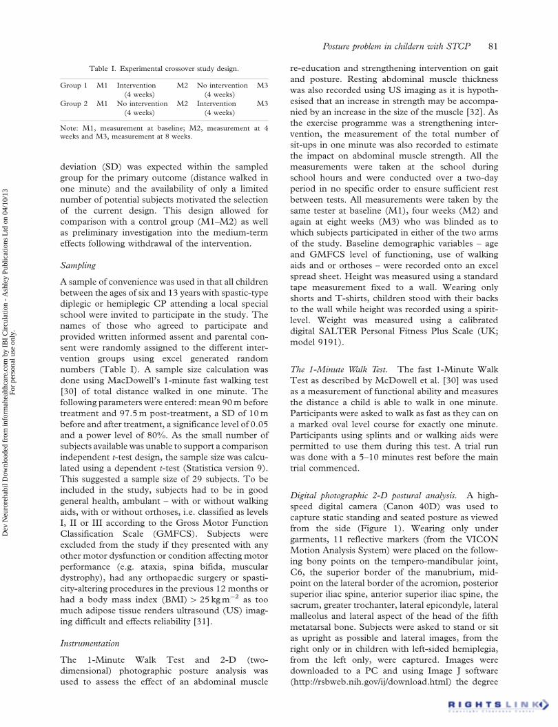
re-education and strengthening intervention on gaitand posture. Resting abdominal muscle thicknesswas also recorded using US imaging as it is hypoth-esised that an increase in strength may be accompa-nied by an increase in the size of the muscle [32]. Asthe exercise programme was a strengthening inter-vention, the measurement of the total number ofsit-ups in one minute was also recorded to estimatethe impact on abdominal muscle strength. All themeasurements were taken at the school duringschool hours and were conducted over a two-dayperiod in no specific order to ensure sufficient restbetween tests. All measurements were taken by thesame tester at baseline (M1), four weeks (M2) andagain at eight weeks (M3) who was blinded as towhich subjects participated in either of the two armsof the study. Baseline demographic variables – ageand GMFCS level of functioning, use of walkingaids and or orthoses – were recorded onto an excelspread sheet. Height was measured using a standardtape measurement fixed to a wall. Wearing onlyshorts and T-shirts, children stood with their backsto the wall while height was recorded using a spirit-level. Weight was measured using a calibrateddigital SALTER Personal Fitness Plus Scale (UK;model 9191).
The 1-Minute Walk Test. The fast 1-Minute WalkTest as described by McDowell et al. [30] was usedas a measurement of functional ability and measuresthe distance a child is able to walk in one minute.Participants were asked to walk as fast as they can ona marked oval level course for exactly one minute.Participants using splints and or walking aids werepermitted to use them during this test. A trial runwas done with a 5–10 minutes rest before the maintrial commenced.
Digital photographic 2-D postural analysis. A high-speed digital camera (Canon 40D) was used tocapture static standing and seated posture as viewedfrom the side (Figure 1). Wearing only undergarments, 11 reflective markers (from the VICONMotion Analysis System) were placed on the follow-ing bony points on the tempero-mandibular joint,C6, the superior border of the manubrium, mid-point on the lateral border of the acromion, posteriorsuperior iliac spine, anterior superior iliac spine, thesacrum, greater trochanter, lateral epicondyle, lateralmalleolus and lateral aspect of the head of the fifthmetatarsal bone. Subjects were asked to stand or sitas upright as possible and lateral images, from theright only or in children with left-sided hemiplegia,from the left only, were captured. Images weredownloaded to a PC and using Image J software(http://rsbweb.nih.gov/ij/download.html) the degree
Table I. Experimental crossover study design.
Group 1 M1 Intervention(4 weeks)
M2 No intervention(4 weeks)
M3
Group 2 M1 No intervention(4 weeks)
M2 Intervention(4 weeks)
M3
Note: M1, measurement at baseline; M2, measurement at 4weeks and M3, measurement at 8 weeks.
Posture problem in childern with STCP 81
Dev
Neu
rore
habi
l Dow
nloa
ded
from
info
rmah
ealth
care
.com
by
IBI
Cir
cula
tion
- A
shle
y Pu
blic
atio
ns L
td o
n 04
/10/
13Fo
r pe
rson
al u
se o
nly.

of antero–posterior pelvic tilt – the angle formedbetween a line which runs through the posterior andanterior superior iliac spines and the horizontal(Figure 1); and forward trunk sway (or antero–posterior (a–p) angle) – the angle between linesdrawn from the marker positioned on C6 posteriorand the sternum anterior and a horizontal line fromC6 – was measured; as well as shoulder-to-sittingheight – the distance from the marker on theacromion to the seating surface (sitting; Figure 1).Although the information obtained using 2-D anal-ysis is limited, stringent standardization of theprocedure did produce reliable measurements(Table I) to determine kinematic variables [33].
For the a–p angle, it is assumed that the smallerthe angle, the more upright the posture in relation tothe pelvis (i.e. less crouched or less forward leaning)and a negative value is indicative of an over extendedupper trunk. Similarly, for the pelvic tilt measure-ment, the smaller the angle, the more neutral thepelvis is positioned and a negative value suggests aposterior pelvic tilt. In sitting a more neutral pelvissuggest a more upright posture. This study hypoth-esised that an increase in abdominal muscle strengthwill result in a reduced anterior tilt (typically seen inchildren with STCP) which in turn will allow for amore upright posture.
US imaging. Using the same procedure asdescribed by Unger [34], US imaging was used to
record and measure the thickness of the fourabdominal muscles – transversus abdominis (TrA),obliquus internus (OI), obliquus externus (OE) andthe rectus abdominis (RA) – using a Siemens�
Accusonic X150 US imaging machine.1 Real-timeimages of the right side in children with diplegia andthe affected side in children with hemiplegia of thefour abdominal muscles were captured. Participantswere positioned in supine with their arms restingalong their sides on a plinth with their kneessupported on a cushion, keeping their hips in�20–45� of flexion ensuring in as far as it waspossible a neutral lumbar curvature. A resting imagewas captured on end-inspiration as observed by thetester. The average of three thickness measurements(i.e. the distance between facia boundaries) at 10, 15and 20 mm from a fixed point [34] for all fourabdominal muscles during rest and during activitywas calculated (mm).
Sit-ups in one minute. The total number of sit-upsexecuted in one minute was selected to measureeffect on abdominal muscle strength. Although not astandardised outcome measure, this field perfor-mance measure is well used in research to reflectpossible strength gains in the abdominal muscula-ture [35]. In the crook lying position with the feetsupported by the research assistant, subjects had tosit-up or curl-up to 90� hip flexion without armsupport and return to the almost flat (flat hand)
Figure 1. Standing and sitting positions for 2-D photographic imaging.Note: A, a–p angle; B, a–p pelvic tilt and C, shoulder-to-seat-height.
82 M. Unger et al.
Dev
Neu
rore
habi
l Dow
nloa
ded
from
info
rmah
ealth
care
.com
by
IBI
Cir
cula
tion
- A
shle
y Pu
blic
atio
ns L
td o
n 04
/10/
13Fo
r pe
rson
al u
se o
nly.

supine position as many times as they could in oneminute.
Reliability
All the above outcome measures have proven validityand were selected for the clinical feasibility.Reliability of US imaging for measurement ofabdominal muscle thickness in children was shownby Unger [34] while tester reliability for the1-Minute Walk Test, sit-ups in one minute and theangle measurements for the 2-D photographicimaging for the posture analysis were conducted on10 randomly selected participants from the controlgroup. Repeated measurements by the same testereither on the same day or between days wereconducted as described in the testing proceduresbelow for each of the above measures. Table IIpresents a complete listing of the intra-class corre-lation coefficient (ICC) scores for all variablesmeasured in this study.
Intervention
A selective trunk-targeted exercise programme usingthe WBV was used to activate and strengthen theabdominal musculature in this study. Although it is anovel form of exercise in this population, theunderpinning theory regarding involuntary capacityto activate weak or dormant muscle, it was deemedappropriate to use vibration to target the abdominalmuscles. Further benefits for using vibration is the‘ease’ of use – no external resistance, i.e. no heavyweights necessary and only one set of repetitions arerequired versus 3–10 repetitions recommended inthe strengthening or weight training (PRE) literature[32], allowing for a short 5–10 minutes workoutsession. All the exercises including the warm-upwere conducted on the vibrating platform. As theexercise programme was a novel approach to trunk-targeted intervention, children with STCP – thosenot selected for participation in this study, i.e. olderchildren or those who had undergone surgical pro-cedures in the 12 months prior to the study – assisted
the researchers to pilot the exercise regime that wasfollowed in this study.
Procedure
Approval was obtained from the Human ResearchEthics Committee at Stellenbosch University andfrom relevant institutional heads. All the measure-ments were taken at the school during school hours.Tests were conducted over a two-day period in nospecific order – two tests on day 1 and the remainingtwo outcomes measured on day 2. This was toensure sufficient rest between tests and to limit timeout of the classroom.
The primary investigator conducted the balance,1-Minute fast walking test and photographic postureanalysis. A research assistant conducted the USimaging and measurement. All four measurementswere taken by the same tester at baseline (M1), fourweeks (M2) and again at eight weeks (M3). Theprimary investigator was blinded as to which subjectsparticipated in either of the two arms of the study.The exercise session was introduced as follows: twicein week 1; thrice in week 2; four to five times inweeks 3 and 4. This allowed for the recovery ofpossible delayed onset muscle soreness. Exercisingon a vibrating platform was a novel activity for thisparticular group of participants and initially someexperienced some muscle soreness. Exercises wereprogressed by increasing the time to 45 s andeventually 60 s per exercise. No additional exercises,external resistance or equipment were used. Allsessions occurred during their usual therapy sessionsor during break-time. All other therapy – occupa-tional or speech – continued as per usual. Thesessions were all supervised by a qualifiedphysiotherapist.
Statistical analysis
Statistica (Version 10) was used to analyse data. Aone-way analysis of variance (ANOVA) was used totest the effect of randomisation at pre-interventionusing the M1 measurements of the two groups.
Table II. Intra-tester reliability of: distance walked in 1 min,number of sit-ups in 1 min and degree of pelvic tilt, a–p angle andshoulder-to-floor height measurements (2-D imaging), ICC(n¼ 10).
Outcome measurement ICC95% Confidence
interval SEM
1-Min Walk Test (s) 0.979 0.910; 0.994 2.403Number of sit-ups in 1 min 0.990 0.972; 0.997 1.085Degree of a–p pelvic tilt (�) 0.982 0.939; 0.994 0.749a–p angle (�) 0.947 0.843; 0.983 2.394Shoulder-to-floor
height (mm)0.905 0.738; 0.968 17.04
Table III. Trunk-targeted exercise programme.
Dosage:time (s)
Frequency(Hz) Activity
1�45 35 Warm-up: standing3�30 35–40 Various sit-up exercises in supine on a
cushion (crunches, cycling, handbehind head and table top)
1�30 35–40 Hip and lumbar extension exercisein four point kneeling or proneover a ball
2�30 35–40 Side lying crunches1�30 35–40 Plank
Posture problem in childern with STCP 83
Dev
Neu
rore
habi
l Dow
nloa
ded
from
info
rmah
ealth
care
.com
by
IBI
Cir
cula
tion
- A
shle
y Pu
blic
atio
ns L
td o
n 04
/10/
13Fo
r pe
rson
al u
se o
nly.

Kolmogorov–Smirnov tests were also done at M1and M2 to inspect distribution of the values of all thevariables measured. In cases where no normaldistribution was found, the Mann–Whitney-U testwas used. Using repeated-measures ANOVA – usinga mixed model approach – measurements at M2were compared to baseline measurements. Thechange between M2 and M3 was also examined toestablish if Group 1 remained the same and whetherGroup 2 improved over this period. Fisher leastsignificant (LS) difference and post hoc tests werealso done to inspect the data further and to deter-mine the level of significance at the various timepoints. A 5% significance level (p5 0.05) was usedas guideline for determining significant differences.Where the time� group; LS means (ANOVA) wasnot significant, but where visual inspection suggesteda possible effect from pre- to post-interventionwithin each group, two time-point ANOVA wasconducted. The data of Group 1 from pre- to post-intervention (M1–M2) were combined with the dataof Group 2 from pre- to post-intervention (M2–M3)and the total effect was determined.
Results
A total of 30 informed consent forms were distrib-uted to learners at the school and 27 subjects wererecruited into the study, parents of three potentialsubjects not consenting to participation. There were17 males and 10 females enrolled – 15 werediagnosed with spastic diplegia, nine presentedwith right hemiplegia and three with left hemiplegia.The numbers relating to gender and type of CP andmobility level (levels I – 13; II – 8; III – 6) wereevenly distributed between the two groups. Therewas a normal distribution for the values recorded forheight, weight and BMI within each group. Group 1however had a skewed distribution for age whichranged between 10.5 and 13.8 years
(median¼ 13.1). Participants in Group 1 weresignificantly older and taller than participants inGroup 2. Although the difference between the twogroups was not significant and despite being youngerand shorter, subjects in Group 2 were slightlyheavier for size than subjects in Group 1.
Effect of intervention on gait. There was a significantinteraction between group and time (p5 0.001) andthe results (Table IV) indicate there was a significantincrease in gait speed seen in Group 1 and similarly inGroup 2 following intervention. However, a signifi-cant difference was again found in Group 1 betweenM2 which was higher and M3 which was lower and nosignificant difference between M1 and M3 in thisgroup which indicate that the treatment effect was notsustained after the intervention was withdrawn.
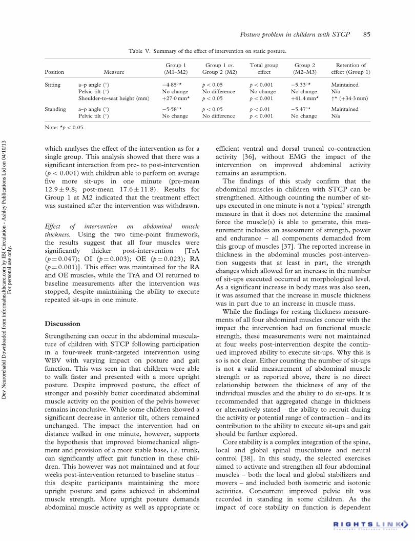
Effect of intervention on posture. The effect of theintervention on posture had varied impact on thedifferent variables measured (Table V). In sitting, the�5� decrease in forward sway and an increase of2.7 cm in shoulder-to-seat height was significantlymore than the change seen in the control group.Similarly, a decrease forward sway in standing alsosuggest a more upright posture following participa-tion in the exercise programme. These improvementswere maintained four weeks after the intervention waswithdrawn. While there was no significant change ina–p pelvic tilt within each group, the total group effectshowed a significant decrease in anterior pelvic tilt inboth the seated and standing positions.
Effect of intervention on functional abdominal muscle
strength. No measurements for the control groupwere recorded at M1 due to an administrative errorand pre- to post-intervention data could thereforeonly be analysed using a two time-point framework
Table IV. Intra-group analysis – M1–M2; M2–M3; M1–M3 for distance walked in 1 min (m).
Measurement (m) Mean SD N Difference SD p
Group 1 M1_distance 94.74 26.28M2_distance 106.68 23.56 13 �11.95 15.43 50.001*
Group 2 M1_distance 87.03 20.07M2_distance 88.58 19.58 14 �1.556 2.74 0.613
Group 1 M2_distance 106.68 23.56M3_distance 95.68 22.64 13 11.008 11.32 0.001*
Group 2 M2_distance 88.58 19.58M3_distance 98.30 20.62 14 �9.709 8.07 0.003*
Group 1 M1_distance 94.74 26.28M3_distance 95.68 22.64 13 �0.938 17.30 0.768
Note: *p5 0.05.
84 M. Unger et al.
Dev
Neu
rore
habi
l Dow
nloa
ded
from
info
rmah
ealth
care
.com
by
IBI
Cir
cula
tion
- A
shle
y Pu
blic
atio
ns L
td o
n 04
/10/
13Fo
r pe
rson
al u
se o
nly.
which analyses the effect of the intervention as for asingle group. This analysis showed that there was asignificant interaction from pre- to post-intervention(p5 0.001) with children able to perform on averagefive more sit-ups in one minute (pre-mean12.9� 9.8; post-mean 17.6� 11.8). Results forGroup 1 at M2 indicated that the treatment effectwas sustained after the intervention was withdrawn.
Effect of intervention on abdominal muscle
thickness. Using the two time-point framework,the results suggest that all four muscles weresignificantly thicker post-intervention [TrA(p¼0.047); OI (p¼ 0.003); OE (p¼ 0.023); RA(p¼0.001)]. This effect was maintained for the RAand OE muscles, while the TrA and OI returned tobaseline measurements after the intervention wasstopped, despite maintaining the ability to executerepeated sit-ups in one minute.
Discussion
Strengthening can occur in the abdominal muscula-ture of children with STCP following participationin a four-week trunk-targeted intervention usingWBV with varying impact on posture and gaitfunction. This was seen in that children were ableto walk faster and presented with a more uprightposture. Despite improved posture, the effect ofstronger and possibly better coordinated abdominalmuscle activity on the position of the pelvis howeverremains inconclusive. While some children showed asignificant decrease in anterior tilt, others remainedunchanged. The impact the intervention had ondistance walked in one minute, however, supportsthe hypothesis that improved biomechanical align-ment and provision of a more stable base, i.e. trunk,can significantly affect gait function in these chil-dren. This however was not maintained and at fourweeks post-intervention returned to baseline status –this despite participants maintaining the moreupright posture and gains achieved in abdominalmuscle strength. More upright posture demandsabdominal muscle activity as well as appropriate or
efficient ventral and dorsal truncal co-contractionactivity [36], without EMG the impact of theintervention on improved abdominal activityremains an assumption.
The findings of this study confirm that theabdominal muscles in children with STCP can bestrengthened. Although counting the number of sit-ups executed in one minute is not a ‘typical’ strengthmeasure in that it does not determine the maximalforce the muscle(s) is able to generate, this mea-surement includes an assessment of strength, powerand endurance – all components demanded fromthis group of muscles [37]. The reported increase inthickness in the abdominal muscles post-interven-tion suggests that at least in part, the strengthchanges which allowed for an increase in the numberof sit-ups executed occurred at morphological level.As a significant increase in body mass was also seen,it was assumed that the increase in muscle thicknesswas in part due to an increase in muscle mass.
While the findings for resting thickness measure-ments of all four abdominal muscles concur with theimpact the intervention had on functional musclestrength, these measurements were not maintainedat four weeks post-intervention despite the contin-ued improved ability to execute sit-ups. Why this isso is not clear. Either counting the number of sit-upsis not a valid measurement of abdominal musclestrength or as reported above, there is no directrelationship between the thickness of any of theindividual muscles and the ability to do sit-ups. It isrecommended that aggregated change in thicknessor alternatively stated – the ability to recruit duringthe activity or potential range of contraction – and itscontribution to the ability to execute sit-ups and gaitshould be further explored.
Core stability is a complex integration of the spine,local and global spinal musculature and neuralcontrol [38]. In this study, the selected exercisesaimed to activate and strengthen all four abdominalmuscles – both the local and global stabilizers andmovers – and included both isometric and isotonicactivities. Concurrent improved pelvic tilt wasrecorded in standing in some children. As theimpact of core stability on function is dependent
Table V. Summary of the effect of intervention on static posture.
Position MeasureGroup 1
(M1–M2)Group 1 vs.
Group 2 (M2)Total group
effectGroup 2
(M2–M3)Retention of
effect (Group 1)
Sitting a–p angle (�) �4�85�* p5 0.05 p5 0.001 �5.33�* MaintainedPelvic tilt (�) No change No difference No change No change N/aShoulder-to-seat height (mm) þ27�0 mm* p5 0.05 p5 0.001 þ41.4 mm* "* (þ34�3 mm)
Standing a–p angle (�) �5�58�* p5 0.05 p5 0.01 �5.47�* MaintainedPelvic tilt (�) No change No difference p5 0.001 No change N/a
Note: *p5 0.05.
Posture problem in childern with STCP 85
Dev
Neu
rore
habi
l Dow
nloa
ded
from
info
rmah
ealth
care
.com
by
IBI
Cir
cula
tion
- A
shle
y Pu
blic
atio
ns L
td o
n 04
/10/
13Fo
r pe
rson
al u
se o
nly.

on the collective functioning of all the core muscu-lature [39], limiting the strengthening interventionto the abdominal muscles only may not be enough toovercome other ‘contributing factors’ and onceexposure to the strength training programmeceased, this effect was lost. Similarly in the standingposition, the ventral abdominal muscles cannoteffectively control pelvic position without counter-activity in the posterior hip musculature/extensors[2, 38]. Inclusion of exercises targeting the hipextensors (and hip abductor and adductor for lateraltilt control) including exercises in the standingposition may more effectively increase posturalcontrol in upright stance.
This study used 2-D photography to investigateposture. While an attempt was made to standardizethe procedure, the results should be interpreted withcaution. First, while it is generally acceptable toclassify severity in children with crouch gait bymeasuring the degree of flexion at the knee, refer-ence to a single variable is limiting and the contri-bution of hip and ankle angles are crucial wheninterpreting the underlying causes of crouch gaitand or mechanisms involved [2]. In this study,three variables – forward sway, pelvic tilt and bodylength – were selected and deemed appropriate to berepresentative of upright posture in sitting andstanding. It should be stated that an improvementin one variable did not necessarily result in a changein the other(s). Second, despite demonstrated test–retest reliability, these variables were not alwaysstable over time. This was noted in Group 2 (whichserved as the control for period M1–M2) and validityof the measurement for investigating impact ofintervention could be questioned. With 2-D analysis,there are limited measures for controlling rotation ofthe body or body parts which may account for theapparent lack of stability of selected variables and3-D posture analysis is recommended.
Despite maintaining the improved strength, thechange in gait function was however not maintainedonce the intervention was stopped. Similarly, thechanges in thickness for two of the four muscles(both stabilizers – TrA and OI) returned to baselinemeasurements. Perhaps, the impact of vibration onspasticity and muscle morphology, which was notinvestigated in this study – cannot be ignored. Theeffect of the intervention on endurance was also notexplored in this study. Although exercises in thisstudy were executed in the supine and side lyingpositions (no standing exercises except for the30–45 s warm-up) some participants anecdotallycommented that their legs felt ‘so loose’ (sic) thatit was ‘easier to walk’ (sic). An eight-week strength-ening intervention using WBV in adults with CPconfirms that vibration can significantly decreasespasticity [22]. This phenomenon may also have
contributed to the increased walking speed seen inthis study. The primary aim of most non-surgicalspasticity targeting interventions is to allow for easierfacilitation of movement and create a window ofopportunity to strengthen the appropriate muscles.As these interventions only temporarily affect tone[40], it may be that a similar response is seenfollowing vibration therapy and accounted for thedecline in gait function seen at four weeks afterexercise was stopped.
Conclusion
Targeting the abdominal muscles using WBV can inthe short-term significantly improve posture andself-selected fast walking in 6–13 year-old childrenwith STCP. The impact on gait and position of thepelvis in this study, however, was not maintaineddespite maintaining the gain in functional abdominalmuscle strength. The maintained ability to executesit-ups and the increase recorded in selected restingabdominal muscles thicknesses confirm this finding.Further investigation into better understanding therelationship(s) between abdominal muscle morphol-ogy and activity; and posture and function isrecommended. The potential effect of exercisewhile standing on a vibrating platform on enduranceand spasticity needs to be further investigated. Theinclusion of exercises targeting all appropriate weakmusculature involved in maintaining core stability isrecommended.
Acknowledgements
The authors would like to acknowledge ProfessorMartin Kidd from the Centre for StatisticalConsultation at Stellenbosch University for hisinvaluable assistance in the data processing andanalysis. They would also like to thank the MedialResearch Council of South Africa for funding thisproject.
Declaration of interest: The authors report nodeclaration of interest. The authors alone areresponsible for the content and writing of this article.
Note
1. Siemens� Accusonic X150 US imaging machine with a 5.5 cmwide band linear array frequency of 5 Hz B-mode (2-D).
References
1. Gramsbergen A, Hadders-Algra M. Editorial: Relevance ofpostural control in children with developmental motor disor-ders. Neural Plasticity 2005;12(2–3):73–75.
86 M. Unger et al.
Dev
Neu
rore
habi
l Dow
nloa
ded
from
info
rmah
ealth
care
.com
by
IBI
Cir
cula
tion
- A
shle
y Pu
blic
atio
ns L
td o
n 04
/10/
13Fo
r pe
rson
al u
se o
nly.

2. Gage JR. The treatment of gait problems in cerebralpalsy. London: Mac Keith Press; 2004.
3. Behm DG, Drinkwater EJ, Willardson JM, Cowley PM.The use of instability to train the core musculature.Applied Physiolology, Nutrition and Metabolism 2010;35(1):91–108.
4. Sato K, Mokha M. Does core strength training influencerunning kinetics, lower-extremity stability, and 5000-Mperformance in runners? Journal of Strength andConditioning Research 2009;23(1):133–140.
5. Park ES, Park CI, Lee HJ, Cho YS. The effect of electricalstimulation on the trunk control in young children withspastic diplegic cerebral palsy. Journal of Korean Medicineand Science 2001;16(3):347–350.
6. Butler PB. A preliminary report on the effectiveness of trunktargeting in achieving independent sitting balance in childrenwith cerebral palsy. Clinical Rehabilitation 1998;12(4):281–293.
7. Bertoti DB. Effect of therapeutic horseback riding on posturein children with cerebral palsy. Physical Therapy1988;68(10):1505–1512.
8. Sterba JA, Rogers BT, France AP, Vokes DA. Horsebackriding in children with cerebral palsy: Effect on gross motorfunction. Developmental Medicine and Child Neurology2002;44(5):301–308.
9. Woollacott M, Shumway-Cook A, Hutchinson S, Ciol M,Price R, Kartin D. Effect of balance training on muscleactivity used in recovery of stability in children with cerebralpalsy: A pilot study. Developmental Medicine and ChildNeurology 2005;47(7):455–461.
10. Woollacott MH, Shumway-Cook A. Postural dysfunctionduring standing and walking in children with cerebral palsy:What are the underlying problems and what new therapiesmight improve balance? Neural Plasticity 2005;12(2–3):211–219.
11. Shumway-Cook A, Hutchinson S, Kartin D, Price R,Woollacott M. Effect of balance training onrecovery of stability in children with cerebral palsy.Developmental Medicine and Child Neurology 2003;45(9):591–602.
12. Unger M, Faure M, Frieg A. Strength training in adolescentlearners with cerebral palsy: A randomized controlled trial.Clinical Rehabilitation 2006;20(6):469–477.
13. Urquhart DM, Hodges PW, Story IH. Postural activity of theabdominal muscles varies between regions of these musclesand between body positions. Gait and Posture 2005;22(4):295–301.
14. Willson JD, Dougherty CP, Ireland ML, Davis IM. Corestability and its relationship to lower extremity function andinjury. Journal of American Academy of OrthopaedicSurgeons 2005;13(5):316–325.
15. Arendt EA. Core strengthening. Instructional CourseLectures 2007;56:379–384.
16. Stackhouse SK, Binder-Macleod SA, Lee SCK. Voluntarymuscle activation, contractile properties, and fatigability inchildren with and without cerebral palsy. Muscle & Nerve2005;31(5):594–601.
17. Cardinale M, Erskine JA. Vibration training in elite sport:Effective training solution or just another fad? InternationalJournal of Sports Physiology and Performance 2008;3(2):232–239.
18. Nordlund MM, Thorstensson A. Strength training effects ofwhole-body vibration? Scandinavian Journal of Medicine andScience in Sports 2007;17(1):12–17.
19. King LK, Almeida QJ, Ahonen H. Short-term effects ofvibration therapy on motor impairments in Parkinson’sdisease. Neurorehabilitation 2009;25(4):297–306.
20. Ebersbach G, Edler D, Kaufhold O, Wissel J. Whole bodyvibration versus conventional physiotherapy to improvebalance and gait in Parkinson’s disease. Archives of PhysicalMedicine Rehabilitation 2008;89(3):399–403.
21. Kawanabe K, Kawashima A, Sashimoto I, Takeda T, Sato Y,Iwamoto J. Effect of whole-body vibration exercise andmuscle strengthening, balance, and walking exercises onwalking ability in the elderly. Keio Journal of Medicine2007;56(1):28–33.
22. Ahlborg L, Andersson C, Julin P. Whole-body vibrationtraining compared with resistance training: Effect on spasti-city, muscle strength and motor performance in adults withcerebral palsy. Journal of Rehabilitation Medicine 2006;38(5):302–308.
23. Cardinale M, Rittweger J. Vibration exercise makes yourmuscles and bones stronger: Factor fiction? Journal of BritishMenopause Society 2006;12(1):12–18.
24. Jordan MJ, Norris SR, Smith DJ, Herzog W. Vibrationtraining: An overview of the area, training consequences, andfuture considerations. Journal of Strength and ConditioningResearch 2005;19(2):459–466.
25. Cardinale M, Bosco C. The use of vibration as an exerciseintervention. Exercise and Sport Science Review 2003;31(1):3–7.
26. Bosco C, Cardinale M, Tsarpela O. Influence of vibration onmechanical power and electromyogram activity in humanarm flexor muscles. European Journal of Applied Physiologyand Occupational Physiology 1999;79(4):306–311.
27. Ward K, Alsop C, Caulton J, Rubin C, Adams J, Mughal Z.Low magnitude mechanical loading is osteogenic in childrenwith disabling conditions. Journal of Bone Mineral Research2004;19(3):360–369.
28. Ruck J, Chabot G, Rauch F. Vibration treatment in cerebralpalsy: A randomized controlled pilot study. Journal ofMusculoskeletal Neuronal Interactions 2010;10(1):77–83.
29. Madou KH, Cronin JB. The effects of whole body vibrationon physical and physiological capabilities in special popula-tions. Hong Kong Physiotherapy Journal 2008;26:24–38.
30. McDowell B, Humphreys L, Kerr C, Stevenson M. Test–retest reliability of a 1-min walk test in children with spasticcerebral palsy (BSCP). Gait and Posture 2009;29(2):267–269.
31. Ferreira PH, Ferreira ML, Hodges PW. Changes in recruit-ment of the abdominal muscles in people with low back pain:Ultrasound measurement of muscle activity. Spine 2004;29(22):2560–2566.
32. McArdle WD, Katch FI, Katch VL. Skeletal muscle:Structure and function. In: McArdle WD, Katch FI, KatchVL, editors. Exercise physiology energy, nutrition and humanperformance, 7th ed. Vol. 4. Philadelphia, PA: LippincotWilliams & Wilkins; 1996. pp 315–338.
33. Dunk NM, Lalonde J, Callaghan JP. Implications for the useof postural analysis as a clinical diagnostic tool: Reliability ofquantifying upright standing spinal postures from photo-graphic images. Journal of Manipulative PhysiologicalTherapeutics 2005;28(6):386–392.
34. Unger M. Reliability of abdominal muscle thickness mea-sures in typically developing children. South African Journalof Physiotherapy 2009;65(3):22–26.
35. Moreland J, Finch E, Stratford P, Balsor B, Gill C. Interraterreliability of six tests of trunk muscle function and endurance.Journal of Orthopaedic and Sports Physical Therapy 1997;26(4):200–208.
36. Reiman MP. Trunk stabilization training: An evidence basisfor the current state of affairs. Journal of Back andMusculoskeletal Rehabilitation 2009;22(3):131–142.
37. Monfort-Panego M, Vera-Garcia FJ, Sanchez-Zuriaga D,Sarti-Martinez MA. Electromyographic studies in abdominal
Posture problem in childern with STCP 87
Dev
Neu
rore
habi
l Dow
nloa
ded
from
info
rmah
ealth
care
.com
by
IBI
Cir
cula
tion
- A
shle
y Pu
blic
atio
ns L
td o
n 04
/10/
13Fo
r pe
rson
al u
se o
nly.

exercises: A literature synthesis. Journal of ManipulativePhysiological Therapeutics 2009;32(3):232–244.
38. Richardson CA, Jull GA, Hodges PW, Hides JA. Therapeuticexercise for spinal segmental stabilisation in low back pain:Scientific basis and clinical approach. Edinburgh: ChurchillLivingston; 1999.
39. Hibbs AE, Thompson KG, French D, Wrigley A, Spears I.Optimizing performance by improving core stability and corestrength. Sports Medicine 2008;38(12):995–1008.
40. Tilton A. Management of spasticity in children with cere-bral palsy. Seminars in Paediatric Neurology 2009;16(2):82–89.
88 M. Unger et al.
Dev
Neu
rore
habi
l Dow
nloa
ded
from
info
rmah
ealth
care
.com
by
IBI
Cir
cula
tion
- A
shle
y Pu
blic
atio
ns L
td o
n 04
/10/
13Fo
r pe
rson
al u
se o
nly.