Ectonucleotidase activities in Sertoli cells from immature rats
10
Braz J Med Biol Res 34(10) 2001 Brazilian Journal of Medical and Biological Research (2001) 34: 1247-1256 ISSN 0100-879X Ectonucleotidase activities in Sertoli cells from immature rats Departamento de Bioquímica, Instituto de Ciências Básicas da Saúde, Universidade Federal do Rio Grande do Sul, Porto Alegre, RS, Brasil E.A. Casali, T.R. da Silva, D.P. Gelain, G.R.R.F. Kaiser, A.M.O. Battastini, J.J.F. Sarkis and E.A. Bernard Abstract Sertoli cells have been shown to be targets for extracellular purines such as ATP and adenosine. These purines evoke responses in Sertoli cells through two subtypes of purinoreceptors, P 2Y2 and P A1 . The signals to purinoreceptors are usually terminated by the action of ectonucleotidases. To demonstrate these enzymatic activities, we cultured rat Sertoli cells for four days and then used them for different assays. ATP, ADP and AMP hydrolysis was estimated by measuring the Pi released using a colorimetric method. Adenosine deaminase activity (EC 3.5.4.4) was determined by HPLC. The cells were not disrupted after 40 min of incubation and the enzymatic activities were considered to be ectocellularly localized. ATP and ADP hydrolysis was markedly increased by the addition of divalent cations to the reaction medium. A competition plot demonstrated that only one enzymatic site is responsible for the hydrolysis of ATP and ADP. This result indicates that the enzyme that acts on the degradation of tri- and diphosphate nucleosides on the surface of Sertoli cells is a true ATP diphosphohydrolase (EC 3.6.1.5) (specific activities of 113 – 6 and 21 – 2 nmol Pi mg -1 min -1 for ATP and ADP, respectively). The ecto-5- nucleotidase (EC 3.1.3.5) and ectoadenosine deaminase activities (specific activities of 32 – 2 nmol Pi mg -1 min -1 for AMP and 1.52 – 0.13 nmol adenosine mg -1 min -1 , respectively) were shown to be able to terminate the effects of purines and may be relevant for the physiological control of extracellular levels of nucleotides and nucleo- sides inside the seminiferous tubules. Correspondence E.A. Bernard Departamento de Bioquímica Rua Ramiro Barcellos, 2600, Anexo 90035-003 Porto Alegre, RS Brasil Fax: +55-51-316-5540 E-mail: [email protected] Received September 6, 2000 Accepted July 2, 2001 Key words Sertoli cells Purine nucleotides ATP diphosphohydrolase Ecto-5’-nucleotidase Ectoadenosine deaminase Introduction Extracellular purines interact with spe- cific receptors (purinoreceptors) on the sur- face of cells activating several biological processes (for reviews, see 1-3). Previous studies have demonstrated that adenosine nucleotides can modulate Sertoli cell re- sponses through the purinoreceptors present on these cells (4-10). The hormonal regulation of Sertoli cell functions in the maintenance of spermato- genesis occurs through inhibitory and stimu- latory signals. These signals interact for a bimodal regulation of adenyl cyclase with increasing or decreasing cAMP levels. Ex- tracellular ATP interacting with P 2Y2 purino- receptors coupled with G i protein modulates the follicle-stimulating hormone (FSH) re- sponse in Sertoli cell cultures. This modula- tion is pertussis toxin-sensitive and decreases the cAMP levels stimulated by FSH (5). In
Transcript of Ectonucleotidase activities in Sertoli cells from immature rats
1247
Braz J Med Biol Res 34(10) 2001
Ectonucleotidases in Sertoli cellsBrazilian Journal of Medical and Biological Research (2001) 34: 1247-1256ISSN 0100-879X
Ectonucleotidase activities inSertoli cells from immature rats
Departamento de Bioquímica, Instituto de Ciências Básicas da Saúde,Universidade Federal do Rio Grande do Sul, Porto Alegre, RS, Brasil
E.A. Casali, T.R. da Silva,D.P. Gelain, G.R.R.F. Kaiser,
A.M.O. Battastini,J.J.F. Sarkis and
E.A. Bernard
Abstract
Sertoli cells have been shown to be targets for extracellular purinessuch as ATP and adenosine. These purines evoke responses in Sertolicells through two subtypes of purinoreceptors, P2Y2 and PA1. Thesignals to purinoreceptors are usually terminated by the action ofectonucleotidases. To demonstrate these enzymatic activities, wecultured rat Sertoli cells for four days and then used them for differentassays. ATP, ADP and AMP hydrolysis was estimated by measuringthe Pi released using a colorimetric method. Adenosine deaminaseactivity (EC 3.5.4.4) was determined by HPLC. The cells were notdisrupted after 40 min of incubation and the enzymatic activities wereconsidered to be ectocellularly localized. ATP and ADP hydrolysiswas markedly increased by the addition of divalent cations to thereaction medium. A competition plot demonstrated that only oneenzymatic site is responsible for the hydrolysis of ATP and ADP. Thisresult indicates that the enzyme that acts on the degradation of tri- anddiphosphate nucleosides on the surface of Sertoli cells is a true ATPdiphosphohydrolase (EC 3.6.1.5) (specific activities of 113 ± 6 and 21± 2 nmol Pi mg-1 min-1 for ATP and ADP, respectively). The ecto-5�-nucleotidase (EC 3.1.3.5) and ectoadenosine deaminase activities(specific activities of 32 ± 2 nmol Pi mg-1 min-1 for AMP and 1.52 ±0.13 nmol adenosine mg-1 min-1, respectively) were shown to be ableto terminate the effects of purines and may be relevant for thephysiological control of extracellular levels of nucleotides and nucleo-sides inside the seminiferous tubules.
CorrespondenceE.A. Bernard
Departamento de Bioquímica
Rua Ramiro Barcellos, 2600, Anexo
90035-003 Porto Alegre, RS
Brasil
Fax: +55-51-316-5540
E-mail: [email protected]
Received September 6, 2000
Accepted July 2, 2001
Key words· Sertoli cells· Purine nucleotides· ATP diphosphohydrolase· Ecto-5’-nucleotidase· Ectoadenosine deaminase
Introduction
Extracellular purines interact with spe-cific receptors (purinoreceptors) on the sur-face of cells activating several biologicalprocesses (for reviews, see 1-3). Previousstudies have demonstrated that adenosinenucleotides can modulate Sertoli cell re-sponses through the purinoreceptors presenton these cells (4-10).
The hormonal regulation of Sertoli cell
functions in the maintenance of spermato-genesis occurs through inhibitory and stimu-latory signals. These signals interact for abimodal regulation of adenyl cyclase withincreasing or decreasing cAMP levels. Ex-tracellular ATP interacting with P2Y2 purino-receptors coupled with Gi protein modulatesthe follicle-stimulating hormone (FSH) re-sponse in Sertoli cell cultures. This modula-tion is pertussis toxin-sensitive and decreasesthe cAMP levels stimulated by FSH (5). In
1248
Braz J Med Biol Res 34(10) 2001
E.A. Casali et al.
the same study, Filippini et al. (5) demon-strated that ATP can activate inositol phos-pholipid turnover and Ca2+ signaling.
Regarding adenosine, a product of ATPdegradation by ectonucleotidases, Rivkees(11) localized and characterized receptorsfor this structure in rat testis tissue and dem-onstrated that Sertoli cells have the PA1 re-ceptor subtype on the plasma membrane. Intestis tissue, the PA1 receptors inhibit adenylylcyclase activity (4,10). Adenosine inhibitionof the hormonal effects of FSH in Sertolicells is reversed by pertussis toxin (8). Thisfact suggests that the regulation of a cyclicnucleotide-dependent pathway is one of thetransduction mechanisms by which adeno-sine regulates the functions of these cells.
The extracellular hydrolysis of ATP toadenosine by ectonucleotidases has been re-ported for several cell types (12-20). Theseenzymatic activities can regulate the extra-cellular concentration of adenine nucleo-tides and nucleosides modulating their localeffects. Degradation of ATP and other nucleo-tides occurs through a cascade of cell sur-face-bound enzymes such as ecto-ATPase(EC 3.6.1.3), ectoapyrase/ATP diphospho-hydrolase/NTPDase (EC 3.6.1.5), and ecto-5�-nucleotidase (EC 3.1.3.5), resulting in theformation of ADP, AMP and adenosine (21).The presence of apyrase activity has beenwell demonstrated in a large number of mam-malian sources (12,13,15). Apyrase is theenzyme that hydrolyzes ATP and ADP (andother tri- and diphosphate nucleosides) tothe monophosphate esters plus inorganicphosphate (Pi), releasing 2 mol Pi/mol ATPand 1 mol Pi/mol ADP.
Barbacci et al. (22) identified and char-acterized a possible ecto-ATPase activity inrat Sertoli cells. In the present study, wedemonstrate that Sertoli cells in culture areable to promote the hydrolysis of ATP, ADPand AMP and we present evidence for thefirst time that the enzymes responsible fornucleotide hydrolysis are a true ectoapyrase(ATP and ADP hydrolysis) and an ecto-5�-
nucleotidase (AMP hydrolysis). The kineticparameters for nucleotide hydrolysis and theeffect of divalent cations, calcium and mag-nesium, on enzymatic activities were deter-mined. Moreover, we show that Sertoli cellscan control extracellular adenosine levelsthrough ectoadenosine deaminase activity(EC 3.5.4.4).
Material and Methods
Material
Culture medium (DMEM/F-12) and soy-bean trypsin were purchased from Gibco(Grand Island, NY, USA). Lactate dehydro-genase (LDH) kit, soybean trypsin inhibitorI-S, DNase I, collagenase I, hyaluronidase I-S, nucleotides, nucleosides and HEPES wereobtained from Sigma (St. Louis, MO, USA),FBS from Cultilab Ltda. (São Paulo, SP,Brazil), and 24-well plates from Costar Co.(Cambridge, MA, USA). Tetrabutyl ammo-nium chloride was purchased from FlukaChemika (Neu Ulm, Switzerland). All otherchemical reagents were of the highest avail-able grade.
Sertoli cell cultures
Primary cultures of Sertoli cells from 17-to 19-day-old Wistar rats were prepared aspreviously described (23). Briefly, the testiswas sequentially digested with 0.25% tryp-sin and DNase (10 µg/ml) for 30 min at 37oCto remove the interstitial tissue. The seminif-erous tubules obtained were dissociated withcollagenase (1 mg/ml) and hyaluronidase (1mg/ml) to separate Sertoli cells from myoidand germ cells for centrifugation at 40 g for10 min. The cultures were grown to conflu-ence on 24-multiwell plates (approximately0.6 x 106 cells/well or 100 µg protein/well) at34oC in a water-saturated atmosphere with95% air and 5% CO2 in DMEM/F-12 (1:1)with 1% FBS for 24 h. On the second day ofculture, the monolayer was washed with

1249
Braz J Med Biol Res 34(10) 2001
Ectonucleotidases in Sertoli cells
Hank�s-buffered saline solution and main-tained for three days more in serum-freeDMEM/F-12 (1:1). On the fourth day ofculture, the Sertoli cell monolayers wereused for the ectonucleotidase assays. TheSertoli cell cultures were estimated to bemore than 95% pure, as determined by brightlight and phase contrast microscopy and al-kaline phosphatase cytochemistry (24). Cel-lular integrity was determined on the basis ofLDH activity. The Sertoli monolayers wereincubated with reaction medium for 40 minand then samples were taken for the determi-nation of LDH with a Sigma kit (LD-L 10,catalog No. 228-10) at 37oC in a Cobas Miraautomatic incubator. LDH activity is reportedas unit of enzyme per mg protein (U/mg).
Ectonucleotidase assays
Sertoli cell monolayers were washed threetimes with the reaction medium containing135 mM NaCl, 5 mM KCl, 10 mM glucoseand 10 mM HEPES, pH 7.4. The reactionwas started by adding the substrate (ATP,ADP or AMP) to the reaction medium con-taining 135 mM NaCl, 5 mM KCl, 10 mMglucose and 10 mM HEPES, pH 7.4, plusCaCl2 and/or MgCl2 (1, 2 and 5 mM, asindicated) and EDTA (2 mM, as indicated).The final volume was 0.2 ml and incubationwas carried out at 34oC. After incubation, asupernatant sample was taken and mixedwith cold trichloroacetic acid to a final con-centration of 5%. This mixture was centri-fuged for 10 min at 16,000 g at 4oC andaliquots were taken for the assay of releasedPi according to the procedure of Chan et al.(25). Incubation time and protein concentra-tion were chosen in order to ensure the lin-earity of the reaction. Controls to correct fornonenzymatic hydrolysis of nucleotides wereprepared by measuring the Pi released intothe same reaction medium incubated with-out cells. The possible contamination withPi released from cultures was avoided byincubating the monolayers in reaction medi-
um without nucleotides. The cultures did notpresent a measurable release of Pi at anytime of incubation. All assays were done inquadruplicate. Km and Vmax were calculatedby linear regression and presented graphi-cally by the Eadie-Hofstee plot. The ATPand ADP concentrations for the same hy-drolysis rate were obtained from the sub-strate curve and used to construct theChevillard competition plot (26).
Adenosine deaminase assay
On the fourth day of culture, the mono-layers were washed three times with adeno-sine deaminase assay (ADA) buffer consist-ing of 100 mM NaCl, 20 mM KCl, 4 mMMgCl2, 2 mM CaCl2, 10 mM NaHCO3, 5mM glucose and 15 mM Tris, pH 7.4 (27).The reaction was started by adding adeno-sine (0.15 mM) to the same ADA buffer asdescribed above. The final volume was 0.2ml and the incubation was carried out at34oC. After incubation (0, 30 and 60 min),the supernatant was taken and maintained onice. The samples were boiled for 3 min andcentrifuged at 4oC for 15 min at 16,000 g.Aliquots of 50 µl were applied to a reversed-phase HPLC system using a C18 Shimadzucolumn (Shimadzu, Japan) at 260 nm with amobile phase containing 60 mM KH2PO4, 5mM tetrabutylammonium chloride, pH 6.0,in 30% methanol according to a previouslydescribed method (28). The adenosine peakwas identified by its retention time and bycomparison with standards. The ectoadeno-sine deaminase activity was measured by thedecrease in the adenosine peak and by theappearance of inosine and hypoxanthinepeaks. The enzyme activity was expressedby the difference between initial (ADi) andfinal adenosine (ADf) concentration at thetime of incubation per mg of protein (D[ADi] -[ADf]/mg protein). The addition of dipy-ridamole (a classical inhibitor of adenosineuptake) did not modify the extracellular aden-osine concentration. Spontaneous deamina-

1250
Braz J Med Biol Res 34(10) 2001
E.A. Casali et al.
tion of adenosine was not detected at anytime.
Cellular protein determination
After the assays, the Sertoli cell mono-layers were digested with 0.5 N NaOH andtotal protein was measured by the method ofLowry et al. (29).
Statistical analysis
The mean ± SEM data for groups of threeor four experiments were analyzed byANOVA and the post hoc Student-New-man-Keuls test using the statistical programSPSS 6.0 for Windows.
Results and Discussion
ATP, ADP and AMP hydrolysis
Sertoli cell cultures promoted ATP, ADPand AMP hydrolysis that was linear for atleast 20 min (Figure 1). One possible prob-lem in the detection of apyrase activity (ATPand ADP hydrolysis) in most sources is theinterference of 5�-nucleotidase, which maycause an overestimation of ATPase andADPase activity (30). The procedure used inthe present study to avoid this problem wasthe limitation of both ATP and ADP hy-drolysis to less than 10%. In addition, allmammalian ecto-5�-nucleotidase activitiesdescribed are strongly inhibited by ATP andADP in the low micromolar range (31). ATPdiphosphohydrolase (apyrase) is an enzymeable to promote the removal of two phos-phate groups of ATP but of only one phos-phate group of ADP. This enzyme presentsdivalent cation dependence and can be stim-ulated by Ca2+ and Mg2+ (12,15,30,32). Theecto-5�-nucleotidase can also be stimulatedby Mg2+ (14,31) but this activation was lowerthan the apyrase activation (26.3 ± 5% forthe ecto-5�-nucleotidase activation and 1490± 84% for the apyrase activation in relationto the control). In the presence of 2 mMEDTA, ATP and ADP hydrolysis was prac-tically negligible and AMP hydrolysis waslower than control (without the addition ofdivalent cations) (Figure 2). Thus, the en-zymes responsible for the hydrolysis of ATP,ADP and AMP in Sertoli cells could becation activated. No significant differenceswere observed in enzymatic activation bydifferent cations at the different concentra-
nmol
Pi/6
x 1
05 c
ells
120
105
90
75
60
30
15
0
45
0 10 20 30 40
nmol
Pi/6
x 1
05 c
ells
60
50
40
30
10
0
20
0 10 20 30 40
150
100
50
00 10 20 30 40
Time (min)
nmol
Pi/6
x 1
05 c
ells
Figure 1. Time course of ATP,ADP and AMP hydrolysis. Ca2+-ATP, Ca2+-ADP and Mg2+-AMPwere incubated with Sertoli cellmonolayers as described in Ma-terial and Methods. The datashown were from an experimentcarried out in quadruplicate. Dataare reported as means ± SEM.
AMP
ADP
ATP

1251
Braz J Med Biol Res 34(10) 2001
Ectonucleotidases in Sertoli cellsnm
ol P
i mg
prot
ein-
1 m
in-1
125
100
75
0
AMP
ADP
ATP
++
*
*+
*+ *+
*+
*+
*+
*+
Con
trol
2 m
M E
DTA
1 m
M C
a2+
2 m
M C
a2+
1 m
M M
g2+
2 m
M M
g2+
Con
trol
2 m
M E
DTA
1 m
M C
a2+
5 m
M C
a2+
2 m
M C
a2+
1 m
M M
g2+
2 m
M M
g2+
5 m
M M
g2+
2 m
M C
a2+/M
g2+
Con
trol
2 m
M E
DTA
1 m
M C
a2+
2 m
M C
a2+
5 m
M C
a2+
1 m
M M
g2+
2 m
M M
g2+
5 m
M M
g2+
2 m
M C
a2+/M
g2+
Figure 2. Cation dependence onectonucleotidase activities. Ser-toli cells were incubated withthe reaction medium plus 1 mMAMP (gray bars), 1 mM ADP(white bars), and 1 mM ATP(black bars) plus cations orEDTA, as indicated. The data arerepresentative of three differentexperiments: mean ± SEM (N =4) of one typical experiment.*P<0.05 compared to the con-trol group; +P<0.05 compared tothe 2 mM EDTA group (Student-Newman-Keuls test).
nmol
Pi m
g pr
otei
n-1
min
-1 8.00
6.40
4.80
3.20
1.60
0.000.0 0.2 0.4 0.6 0.8 1.0
P
Figure 3. The competition plot.The concentration at which thevelocities were the same forATP and ADP was chosen forthe Chevillard plot. The assayconditions are described in Ma-terial and Methods. The incuba-tion time was 10 min; substrateA (ADP) at P = 0 was 0.1 mMand substrate B (ATP) at P = 1was 0.04 mM. Data representan experiment carried out inquintuplicate. The values are themean ± SEM. No significant dif-ference was found between dif-ferent points.
tions tested for ATP, ADP and AMP hy-drolysis (Figure 2). This result indicates thatmaximal activation of the apyrase and ecto-5�-nucleotidase activities is possible with 2mM or less of both cations tested. No addi-tive effects were observed when the twodivalent cations tested were added at thesame time to the reaction medium, suggest-ing that both Ca2+ and Mg2+ are competingfor the same activation site. It is important tonote that there was a parallel profile of acti-vation for all substrates (ATP, ADP andAMP) with each cation added. Based onthese results, we established as optimal con-ditions for measuring the ectonucleotidaseactivities the ratio of 1 mM/2 mM for thenucleotides/divalent cation. In this way theectonucleotidase activities were measuredin the physiological extracellular range ofdivalent cations and nucleotides.
A single active site
ATP and ADP hydrolysis could be cata-lyzed by an ATP diphosphohydrolase (apy-rase) or by enzyme combinations able tomimic apyrase activity. To show that ATPand ADP hydrolysis occurs due to an apy-
rase and that one active site is able to hydro-lyze the two substrates, we used the Chevil-lard competition plot used by Kettlun et al.(32) to characterize a human placental ATPdiphosphohydrolase. To assay the combina-tion of substrate concentrations in a Chevil-lard competition plot (26) we chose concen-trations at which the rate of hydrolysis wasthe same when either ATP or ADP was usedas substrate. The P values ranged from 0 to 1.The horizontal straight line obtained in thecompetition plot (Figure 3) indicates a con-stant hydrolysis rate at all substrate combi-nations tested and the interpretation is thatthe hydrolysis of both substrates (ATP andADP) occurs at the same active site of asingle enzyme.
*
Addition
50
25*
+ *+ *+
*+ *+
*+*+ *+ *+ *+
*+ *+
1252
Braz J Med Biol Res 34(10) 2001
E.A. Casali et al.
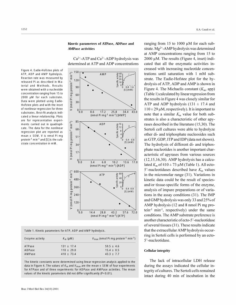
ranging from 15 to 1000 µM for each sub-strate. Mg2+-AMP hydrolysis was determinedat AMP concentrations ranging from 15 to2000 µM. The results (Figure 4, inset) indi-cated that all the enzymatic activities in-creased with increasing nucleotide concen-trations until saturation with 1 mM sub-strate. The Eadie-Hofstee plot for the hy-drolysis of ATP, ADP and AMP is shown inFigure 4. The Michaelis constant (Km, app)(Table 1) calculated by linear regression fromthe results in Figure 4 was closely similar forATP and ADP hydrolysis (131 ± 17.4 and110 ± 29 µM, respectively). It is important tonote that a similar Km value for both sub-strates is also a characteristic of other apy-rases described in the literature (15,30). TheSertoli cell cultures were able to hydrolyzeother di- and triphosphate nucleosides suchas GTP, GDP, ITP and IDP (data not shown).The hydrolysis of different di- and triphos-phate nucleotides is another important char-acteristic of apyrases from various sources(12,15,16,30). AMP hydrolysis has a calcu-lated Km of 410 ± 73 µM (Table 1). All ecto-5�-nucleotidases described have Km valuesin the micromolar range (31). Variations inkinetic data could be the result of species-and/or tissue-specific forms of the enzyme,analysis of impure preparations or of varia-tions in the assay conditions (31). The IMPand GMP hydrolysis was only 33 and 25% ofAMP hydrolysis (12 and 8 nmol Pi mg pro-tein-1 min-1, respectively) under the sameconditions. The AMP substrate preference isanother characteristic of ecto-5�-nucleotidaseof several tissues (31). These results indicatethat the extracellular AMP hydrolysis occur-ring in Sertoli cells is performed by an ecto-5�-nucleotidase.
Cellular integrity
The lack of intracellular LDH releaseduring the assays indicated the cellular in-tegrity of cultures. The Sertoli cells remainedintact during 40 min of incubation in the
Table 1. Kinetic parameters for ATP, ADP and AMP hydrolysis.
Enzyme activity Km (µM) Vmax (nmol Pi mg protein-1 min-1)
ATPase 131 ± 17.4 59.5 ± 4.6ADPase 110 ± 29.0 15.4 ± 0.5AMPase 410 ± 73.4 43.3 ± 7.7
The kinetic constants were determined using linear regression analysis applied to thedata in Figure 4. The values of Km and Vmax are the mean ± SEM of four experimentsfor ATPase and of three experiments for ADPase and AMPase activities. The meanvalues of the kinetic parameters did not differ significantly (P<0.01).
V (n
mol
Pi m
g pr
otei
n-1
min
-1) 150
120
90
60
30
0
85
68
51
34
17
0
0.0 8.6 17.2 25.8 34.4 43.0
40302010
0
Spec
ific
activ
ity
0 0.8 1.6[S] (mM)
AMP
(nmol Pi mg-1 min-1) [AMP]
1612
8
4
Spec
ific
activ
ity
0 0.4 0.8[S] (mM)
0
0.0 3.4 6.8 10.2 13.6 17.0(nmol Pi mg-1 min-1) [ADP]
600
480
360
240
120
0
80
40
20
0Spec
ific
activ
ity
0 0.4 0.8[S] (mM)
0.0 14.4 28.8 43.2 57.6 72.0(nmol Pi mg-1 min-1) [ATP]
ADP
ATP
V (n
mol
Pi m
g pr
otei
n-1
min
-1)
V (n
mol
Pi m
g pr
otei
n-1
min
-1)
Figure 4. Eadie-Hofstee plots ofATP, ADP and AMP hydrolysis.Reaction rate was measured byreleased Pi as described in Ma-terial and Methods. Resultswere obtained with a nucleotideconcentration ranging from 15 to2000 µM for each substrate.Data were plotted using Eadie-Hofstee plots and with the insetof nonlinear regression for threesubstrates. Best-fit analysis indi-cated a linear relationship. Plotsare for representative experi-ments carried out in quadrupli-cate. The data for the nonlinearregression plot are reported asmean ± SEM. V is nmol Pi mgprotein-1 min-1 and [S] is the sub-strate concentration in mM.
Kinetic parameters of ATPase, ADPase andAMPase activities
Ca2+-ATP and Ca2+-ADP hydrolysis wasdetermined at ATP and ADP concentrations

1253
Braz J Med Biol Res 34(10) 2001
Ectonucleotidases in Sertoli cells
reaction medium. Only 5% of LDH activityof lysed cells (1.33 ± 0.19 U/mg protein) wasmeasured during 40 min of incubation. Inthis way the participation of cytosolic en-zymes in extracellular nucleotide hydrolysiswas excluded.
Adenosine deaminase activity
It has been previously demonstrated thattestis tissue contains adenosine deaminasemRNA (33) and we have investigatedwhether this enzyme is present on the plasmamembrane surface of Sertoli cells and itscapacity to degrade extracellular adenosine.Sertoli cells are able to degrade the extracel-lular adenosine by an ectoadenosine deami-nase activity (Figure 5). The ectoadenosinedeaminase is a key enzyme in purine me-tabolism that catalyzes the irreversible trans-formation of adenosine and 2�-deoxyadeno-sine to inosine and 2�-deoxyinosine, respec-tively. Its enzymatic activity is homoge-neously distributed along the cell surface insome cells (34). The Sertoli cell ectoadeno-sine deaminase activity was linear up to 60min of incubation (Figure 5) and had a spe-cific activity of 1.52 ± 0.13 nmol adenosinemg protein-1 min-1. The decrease in adeno-sine levels in the extracellular medium wasof the same order of magnitude as the in-crease in inosine and hypoxanthine (data notshown) and this demonstrates that the aden-osine decrease is a result of adenosine deami-nase activity rather than of cellular adeno-sine uptake. The presence of dipyridamole(10 µM), a classical inhibitor of adenosineuptake (35), in the ADA buffer did not alterthe extracellular adenosine concentration butcaused 30% inhibition of ectoadenosinedeaminase activity. A possible explanationfor this result is the participation of ecto-adenosine deaminase in a protein complexthat can have multiple functions such ascatalytic deamination activity, coupled withadenosine receptors and with selective chan-nels by uptake of extracellular adenosine
(34,36). The presence of dipyridamole canalter the conformation of the protein com-plex with a modification in the enzymaticactivity.
The physiological role
The physiological role of ectonucleoti-dases is unknown but it has been speculatedthat their function could be the control ofextracellular concentration of purines. Themodulation of the levels of different nucleo-tides may involve �cross-talking� betweenthe diverse pathways activated in purinergicreceptor subtypes. The general way to con-trol the concentration of adenine nucleotidesis the sequential activity of ecto-ATPases,ecto-ATP diphosphohydrolases (apyrase) andecto-5�-nucleotidases (12-17,19,20,30). Theadenosine released from cells or resultingfrom extracellular ATP hydrolysis can bedeaminated by the ectoadenosine deaminaseactivity, producing inosine (18,34,36).
Adenosine and its antagonists, such ascaffeine, have been long postulated to influ-ence the male reproductive system. Severalreports have demonstrated the presence ofadenosine receptors in Sertoli cells (10,11)and their modulation by adenosine and dif-ferent adenosine analogues (4,8,9). Thepurinoreceptor subtype P2Y2 is present inthese cells (5) and its modulation by ATPhas been well demonstrated (5-7). In Sertoli
D[A
Di]
- [A
Df]
/mg
prot
ein
80
60
40
20
00 15 30 45 60
Time (min)
Figure 5. Ectoadenosine deami-nase activity. Adenosine deami-nase was determined as de-scribed in Material and Methods.The enzymatic activity is re-ported as the difference be-tween the initial and final con-centration of adenosine/mg ofprotein (D[ADi] - [ADf]/mg pro-tein). The values are the mean ±SEM for a representative experi-ment carried out in quadrupli-cate.

1254
Braz J Med Biol Res 34(10) 2001
E.A. Casali et al.
cells, extracellular ATP and adenosine canmodulate the hormonal response to FSH,decreasing cAMP levels (4,5,8-10), and canregulate anionic (6) and transferrin secretion(7) and the intracellular levels of Ca2+ byinositol phospholipid turnover (5). Thus, thecontrol of extracellular levels of adenosinenucleotides is of high physiological rel-evance. However, the mechanism control-ling purine concentrations in testicular tis-sue is poorly known. We show in this studythat Sertoli cells were able to hydrolyze ATPand ADP (Figure 1) and that only one activesite is responsible for the hydrolysis of thetwo nucleotides (Figure 3). Barbacci et al.(22) identified and characterized one pos-sible ecto-ATPase in rat Sertoli cells. The Km
calculated for the possible ecto-ATPase isthe same we found for ATP and ADP hy-drolysis (Table 1), indicating that one en-zyme is responsible for the hydrolysis of thetwo nucleotides. The results obtained in thecompetition plot confirm this hypothesis.The cation requirements demonstrated byBarbacci et al. (22) for ATP hydrolysis areequal to those for ADP hydrolysis demon-strated by us (Figure 2). On the other hand,our results cannot exclude the co-expressionof two enzymes (a fact demonstrated in therat brain) (37) because the ATP hydrolysisdemonstrated by Barbacci et al. (22) wasslightly inhibited by ADP. In other tissues ithas been postulated that the control of extra-cellular nucleotide concentration is due tothe action of enzymatic complexes with thepossible participation of two or more ecto-enzymes (21). The most obvious physiologi-cal role for apyrase in Sertoli cells, in anal-ogy to other tissues, is to participate in an�enzyme chain� together with a 5�-nucleoti-dase for the complete hydrolysis of ATP toadenosine.
AMP hydrolysis occurs through the ac-tion of an ecto-5�-nucleotidase releasingadenosine that could create a secondary sig-nal for PA1 receptors. The Sertoli cell ecto-
5�-nucleotidase has proved to be cation acti-vated and in the presence of normal extracel-lular concentrations of Ca2+ and Mg2+ (1-2mM) its activity is increased compared to thecontrol activity (without cation addition) andthe 2 mM EDTA group (Figure 2). The Km/Vmax values (Table 1) and the substrate pref-erence are in accordance with the other char-acteristics of ecto-5�-nucleotidase from sev-eral tissues (31).
In order to terminate the purine extracel-lular cascade of nucleotides and nucleosides,the ectoadenosine deaminase produces ino-sine that can be taken up and/or degraded bythe cells. We have demonstrated that Sertolicells have an ectoadenosine deaminase ac-tivity with the same buffer requirement asfor other cells (Figure 5) (28). In analogy,this enzyme can participate together with anATP diphosphohydrolase and the ecto-5�-nucleotidase in the control of the extracellu-lar adenosine levels and eliminate the purinecascade.
The physiological control of ectonucle-otidase activities is unknown. Moreover,Franco et al. (34,36) have demonstrated thatsome ectoenzymes can play the role of oneenzyme and that of a receptor which can beinternalized when the substrate is in its ac-tive site. Another possibility is the co-local-ization of ectoenzyme and receptor at thesame site on the plasma membrane. Recep-tor desensitization can occur by endocytosisof membrane fragments where the ectoen-zyme and the receptor in question are pres-ent. Some authors have shown that ecto-nucleotidases can lose their activities in re-sponse to a cell signal (38,39) and thatectonucleotidase activities initially localizedon the membrane surface are internalizedinto endoplasmic vesicles (40).
In experiments currently underway, weare searching for a possible modulation inectonucleotidase activities by hormones thatact on Sertoli cells, possibly representing afine control of these enzymatic activities.

1255
Braz J Med Biol Res 34(10) 2001
Ectonucleotidases in Sertoli cells
References
1. El-Moatassim C, Dornand J & Mani J(1992). Extracellular ATP and cell signal-ling. Biochimica et Biophysica Acta, 1134:31-45.
2. Dubyak GR & El-Moatassim C (1993). Sig-nal transduction via P2-purinergic recep-tors for extracellular ATP and othernucleotides. American Journal of Physiol-ogy, 265: C577-C606.
3. Ralevic V & Burnstock G (1998). Recep-tors for purines and pyrimidines. Pharma-cological Reviews, 50: 413-492.
4. Conti M, Boitani C, DeManno DA,Migliaccio S, Monaco L & Szymeczek C(1989). Characterization and function ofadenosine receptors in the testis. Annalsof the New York Academy of Sciences,564: 39-47.
5. Filippini A, Riccioli A, Cesaris P de,Paniccia R, Teti A, Stefanini M, Conti M &Ziparo E (1994). Activation of inositolphospholipids and calcium signaling in ratSertoli cells by P2 purinergic receptors:modulation of follicle-stimulating hor-mone responses. Endocrinology, 134:1537-1545.
6. Ko WH, Chan HC, Chew SB & Wong PYD(1998). Regulated anion secretion in theepithelia from Sertoli cells of immaturerats. Journal of Physiology, 512: 471-480.
7. Meroni SB, Cánepa DF, Pellizari EH &Schteingart HF (1998). Effects of puriner-gic agonists on aromatase and gamma-glutamyl transpeptidase activities and ontransferrin secretion in cultured Sertolicells. Journal of Endocrinology, 157: 275-283.
8. Monaco L, DeManno DA, Martin MW &Conti M (1988). Adenosine inhibition ofthe hormonal response in the Sertoli cellis reversed by pertussis toxin. Endocrinol-ogy, 122: 2692-2698.
9. Monaco L, Toscano MV & Conti M (1984).Purine modulation of the hormonal re-sponse of the rat Sertoli cell in culture.Endocrinology, 115: 1616-1624.
10. Stiles GL, Pierson G, Sunay S & ParsonsWJL (1986). The rat testicular A1 adeno-sine receptor-adenylate cyclase system.Endocrinology, 119: 1845-1851.
11. Rivkees SA (1994). Localization and char-acterization of adenosine receptor expres-sion in rat testis. Endocrinology, 135:2307-2313.
12. Battastini AMO, Oliveira EM, Moreira CM,Bonan CD, Sarkis JJF & Dias RD (1995).Solubilization and characterization of anATP diphosphohydrolase (EC 3.6.1.5)from rat brain synaptic plasma mem-
branes. Biochemistry and Molecular Biol-ogy International, 37: 209-219.
13. Candinas D, Koyamada N, Miyatake T,Siegel J, Hancock WW, Bach FH &Robson SC (1996). Loss of rat glomerularATP diphosphohydrolase activity duringreperfusion injury is associated with oxi-dative stress reactions. Thrombosis andHaemostasis, 76: 807-812.
14. Darvish A, Pomerantz WR, Zografides P &Metting P (1996). Contribution of cytoso-lic and membrane-bound 5'-nucleotidaseto cardiac adenosine production. Ameri-can Journal of Physiology, 271: H2162-H2167.
15. Frasseto SS, Dias RD & Sarkis JJF (1993).Characterization of an ATP diphosphohy-drolase activity (apyrase, EC 3.6.1.5) in ratblood platelets. Molecular and CellularBiochemistry, 129: 47-55.
16. Ishikawa H, Tamiya T, Tsuchiya T & Mat-sumoto J (1984). A novel ATP-ADPasefrom Mimosa pulvinus. Comparative Bio-chemistry and Physiology, 78B: 59-61.
17. Kohring K & Zimmermann H (1998). Up-regulation of ecto-5’nucleotidase in hu-man neuroblastoma SH-SYY cells on dif-ferentiation by retinoic acid or phorbol-ester. Neuroscience Letters, 258: 127-130.
18. Lloyd HGE & Fredholm BB (1995). In-volvement of adenosine deaminase andadenosine kinase in regulating extracellu-lar adenosine concentration in rat hippo-campal slices. Neurochemistry Interna-tional, 26: 387-395.
19. Minelli A, Moroni M, Trinari D & Mezza-soma I (1997). Hydrolysis of extracellularadenine nucleotides by equine epididy-mal spermatozoa. Comparative Biochem-istry and Physiology, 117B: 531-534.
20. Node K, Kitakaze M, Minamini T, Michi-hiko T, Inoue M, Hori M & Kamada T(1997). Action of ecto-5'-nucleotidase byprotein kinase C and its role in ischaemictolerance in canine heart. British Journalof Pharmacology, 120: 273-281.
21. Grobben B, Anciaux K, Roymans D, StefanC, Bolen M, Esmans EL & Slegers H(1999). An ecto-nucleotide pyrophos-phatase is one of the main enzymes in-volved in the extracellular metabolism ofATP in rat C6 glioma. Journal of Neuro-chemistry, 72: 826-834.
22. Barbacci E, Filippini A, Cesaris P De &Ziparo E (1996). Identification and charac-terization of an ecto-ATPase in rat Sertolicells. Biochemical and Biophysical Re-search Communications, 222: 273-279.
23. Rocha AB, Guma FCR, Casali EA, SchererGS, Achaval ME & Bernard EA (1997).Influence of the biomatrix on the re-sponse of Sertoli cells to FSH. Archives ofPhysiology and Biochemistry, 105: 1-5.
24. Palombi F & Di Carlo C (1988). Alkalinephosphatase is a marker for myoid cells incultures of peritubular and tubular tissue.Biology of Reproduction, 39: 1101-1109.
25. Chan K, Delfert D & Junger KD (1986). Adirect colorimetric assay for the Ca2+-ATPase activity. Analytical Biochemistry,157: 375-380.
26. Chevillard C, Cárdenas ML & Cornish-Bowden A (1993). The competition plot: asimple test of whether two reactions oc-cur at the same active site. BiochemicalJournal, 289: 599-604.
27. Trams GE & Lauter CJ (1974). On thesidedness of plasma membrane en-zymes. Biochimica et Biophysica Acta,345: 180-197.
28. Voelter W, Zech K, Arnold P & Ludwig G(1980). Determination of selected pyrim-idines, purines and their metabolites inserum and urine by reversed-phase ion-pair chromatography. Journal of Chroma-tography, 199: 345-354.
29. Lowry OH, Rosebrough NJ, Farr AL &Randall RJ (1951). Protein measurementwith the Folin phenol reagent. Journal ofBiological Chemistry, 193: 265-275.
30. Sarkis JJF & Salto C (1991). Characteriza-tion of a synaptosomal ATP diphosphohy-drolase from the electric organ of Tor-pedo marmorata. Brain Research Bulletin,26: 871-876.
31. Zimmermann H (1992). 5'-Nucleotidase:molecular structure and functional as-pects. Biochemical Journal, 285: 345-365.
32. Kettlun AM, Alvarez A, Quintar R, Valen-zuela MA, Collados L, Aranda E, Banda A,Chayet L, Chiong M, Mancilla M &Traverso-Cori A (1994). Human placentalATP-diphosphohydrolase biochemicalcharacterization, regulation and function.International Journal of Biochemistry, 26:437-488.
33. Meng J, Zhang F, Huhtaniemi I & Pakari-nen P (1997). Characterization and devel-opmental expression of a testis-specificadenosine deaminase mRNA in themouse. Journal of Andrology, 18: 88-95.
34. Franco R, Mallol J, Casadó V, Lluis C,Canela EI, Saura C, Blanco J & Ciruela F(1998). Ecto-adenosine deaminase: anecto-enzyme and costimulatory proteinacting on a variety of cell surface recep-tors. Drug Development Research, 45:
1256
Braz J Med Biol Res 34(10) 2001
E.A. Casali et al.
261-268.35. Meghji P & Newby AC (1990). Sites of
adenosine formation, action and inactiva-tion in the brain. Neurochemistry Interna-tional, 16: 227-232.
36. Saura CA, Mallol J, Canela EI, Lluis C &Franco R (1998). Adenosine deaminaseand A1 adenosine receptors internalizetogether following agonist-induced recep-tor desensitization. Journal of BiologicalChemistry, 273: 17610-17617.
37. Kegel B, Braun N, Heine P, MaliszewskiCR & Zimmermann H (1997). An ecto-ATPase and an ecto-ATP diphosphohydro-lase are expressed in rat brain. Neuro-pharmacology, 36: 1189-1200.
38. Robson SC, Kaczmarek E, Siegel JB,Candinas D, Kosiak K, Millan M, HancockWW & Bach FH (1997). Loss of ATP di-phosphohydrolase activity with endotheli-al activation. Journal of ExperimentalMedicine, 185: 153-163.
39. Kobayashi T, Okada T, Saz EG del &Seguchi H (1997). Internalization of ecto-ATPase activity in human neutrophilsupon stimulation with phorbol ester orformyl peptide. Histochemistry and CellBiology, 107: 353-363.
40. Biederbick A, Rose S & Elsässer H (1999).A human intracellular apyrase-like protein,LALP70, localizes to lysosomal/auto-phagic vacuoles. Journal of Cell Science,112: 2473-2488.