Early secreted antigen ESAT-6 of Mycobacterium tuberculosis promotes protective T helper 17 cell...
12
Early Secreted Antigen ESAT-6 of Mycobacterium tuberculosis Promotes Protective T Helper 17 Cell Responses in a Toll-Like Receptor-2-dependent Manner Samit Chatterjee 1. , Ved Prakash Dwivedi 1. , Yogesh Singh 1¤ , Imran Siddiqui 1 , Pawan Sharma 1 , Luc Van Kaer 2 , Debprasad Chattopadhyay 3 , Gobardhan Das 1 * 1 Immunology Group, International Centre for Genetic Engineering and Biotechnology, Aruna Asaf Ali Marg, New Delhi, India, 2 Department of Microbiology and Immunology, Vanderbilt University School of Medicine, Nashville, Tennessee, United States of America, 3 ICMR Virus Unit, Calcutta, ID & BG Hospital, GB 4, Beliaghata, Kolkata, India Abstract Despite its relatively poor efficacy, Bacillus Calmette-Gue ´rin (BCG) has been used as a tuberculosis (TB) vaccine since its development in 1921. BCG induces robust T helper 1 (Th1) immune responses but, for many individuals, this is not sufficient for host resistance against Mycobacterium tuberculosis (M. tb) infection. Here we provide evidence that early secreted antigenic target protein 6 (ESAT-6), expressed by the virulent M. tb strain H37Rv but not by BCG, promotes vaccine- enhancing Th17 cell responses. These activities of ESAT-6 were dependent on TLR-2/MyD88 signalling and involved IL-6 and TGF-b production by dendritic cells. Thus, animals that were previously infected with H37Rv or recombinant BCG containing the RD1 region (BCG::RD1) exhibited improved protection upon re-challenge with virulent H37Rv compared with mice previously infected with BCG or RD1-deficient H37Rv (H37RvDRD1). However, TLR-2 knockout (TLR-2 -/- ) animals neither showed Th17 responses nor exhibited improved protection in response to immunization with H37Rv. Furthermore, H37Rv and BCG::RD1 infection had little effect on the expression of the anti-inflammatory microRNA-146a (miR146a) in dendritic cells (DCs), whereas BCG and H37RvDRD1 profoundly induced its expression in DCs. Consistent with these findings, ESAT-6 had no effect on miR146a expression in uninfected DCs, but dramatically inhibited its upregulation in BCG-infected or LPS- treated DCs. Collectively, our findings indicate that, in addition to Th1 immunity induced by BCG, RD1/ESAT-6-induced Th17 immune responses are essential for optimal vaccine efficacy. Citation: Chatterjee S, Dwivedi VP, Singh Y, Siddiqui I, Sharma P, et al. (2011) Early Secreted Antigen ESAT-6 of Mycobacterium tuberculosis Promotes Protective T Helper 17 Cell Responses in a Toll-Like Receptor-2-dependent Manner. PLoS Pathog 7(11): e1002378. doi:10.1371/journal.ppat.1002378 Editor: William R. Bishai Jr, Johns Hopkins School of Medicine, United States of America Received April 13, 2011; Accepted September 29, 2011; Published November 10, 2011 Copyright: ß 2011 Chatterjee et al. This is an open-access article distributed under the terms of the Creative Commons Attribution License, which permits unrestricted use, distribution, and reproduction in any medium, provided the original author and source are credited. Funding: This work was supported by grants from The Wellcome Trust-Department of Biotechnology (DBT) alliance, and by the DBT, Govt. of India. Grant No. WT01/GD/09/339 URL: http://wellcomedbt.org/seniorfellows.html and Grant No. DB05/PS/09/331. URL:http://dbtindia.nic.in SC is supported by a CSIR Senior Research Fellowship, VPD is supported by a DBT Senior Research Fellowship, Govt. of India. The funders had no role in study design, data collection and analysis, decision to publish, or preparation of the manuscript. Competing Interests: The authors have declared that no competing interests exist. * E-mail: [email protected] ¤ Current address: MicroRNA and Lymphocyte Development Research Group, Department of Veterinary Basic Sciences, Royal Veterinary College (University of London), London, United Kingdom . These authors contributed equally to this work. Introduction Tuberculosis (TB) remains a major health problem, with an estimated one third of the world’s population infected with Mycobacterium tuberculosis, the causative agent of TB, resulting in ,3 million deaths annually. Bacillus Calmette-Gue ´rin (BCG), the only TB vaccine presently used in humans, has been widely used throughout the world since its inception in 1921, and an estimated 3 billion people have received it [1]. However, its efficacy against pulmonary TB in adults is highly variable (0–80%) [2] and depends on ethnicity and geographical location [3,4,5]. The antigenic component(s) that is absent in BCG to elicit critical protective immune responses against TB has been an area of intense research [4,5]. Early secreted antigenic target protein 6 (ESAT-6) is one of the most prominent antigens expressed by Mycobacterium tuberculosis (M. tb), but not by BCG [6,7]. ESAT-6- specific T cells are frequently found in TB patients as well as in infected animals [8,9,10]. Thus, ESAT-6 is being extensively studied for its potential activity as a subunit vaccine [11]. T cell receptor transgenic T cells specific for ESAT-6 exhibit significant protection against TB [12]. Consistent with this, a recombinant BCG strain that contains region of difference 1 (RD1), which includes ESAT-6, exhibited improved protection against TB [13]. However, the basis of this improved protection remains elusive. Furthermore, the mechanism by which ESAT-6 vaccination induces protective immune responses against TB remains to be investigated. Furthermore, deletion mutants of virulent M. tb strains for RD1 or ESAT-6 (a protein product of the RD1 region) resemble BCG in their infectivity and attenuation [14]. Therefore, these bacterial strains provide insight into the rational selection and design of suitable candidate vaccines for M. tb infection. It is clear that vaccination with BCG produces Th1 cell-mediated immune responses, and this is moderately effective in protecting against PLoS Pathogens | www.plospathogens.org 1 November 2011 | Volume 7 | Issue 11 | e1002378
-
Upload
independent -
Category
Documents
-
view
4 -
download
0
Transcript of Early secreted antigen ESAT-6 of Mycobacterium tuberculosis promotes protective T helper 17 cell...
Early Secreted Antigen ESAT-6 of Mycobacteriumtuberculosis Promotes Protective T Helper 17 CellResponses in a Toll-Like Receptor-2-dependent MannerSamit Chatterjee1., Ved Prakash Dwivedi1., Yogesh Singh1¤, Imran Siddiqui1, Pawan Sharma1, Luc Van
Kaer2, Debprasad Chattopadhyay3, Gobardhan Das1*
1 Immunology Group, International Centre for Genetic Engineering and Biotechnology, Aruna Asaf Ali Marg, New Delhi, India, 2 Department of Microbiology and
Immunology, Vanderbilt University School of Medicine, Nashville, Tennessee, United States of America, 3 ICMR Virus Unit, Calcutta, ID & BG Hospital, GB 4, Beliaghata,
Kolkata, India
Abstract
Despite its relatively poor efficacy, Bacillus Calmette-Guerin (BCG) has been used as a tuberculosis (TB) vaccine since itsdevelopment in 1921. BCG induces robust T helper 1 (Th1) immune responses but, for many individuals, this is not sufficientfor host resistance against Mycobacterium tuberculosis (M. tb) infection. Here we provide evidence that early secretedantigenic target protein 6 (ESAT-6), expressed by the virulent M. tb strain H37Rv but not by BCG, promotes vaccine-enhancing Th17 cell responses. These activities of ESAT-6 were dependent on TLR-2/MyD88 signalling and involved IL-6 andTGF-b production by dendritic cells. Thus, animals that were previously infected with H37Rv or recombinant BCG containingthe RD1 region (BCG::RD1) exhibited improved protection upon re-challenge with virulent H37Rv compared with micepreviously infected with BCG or RD1-deficient H37Rv (H37RvDRD1). However, TLR-2 knockout (TLR-2-/-) animals neithershowed Th17 responses nor exhibited improved protection in response to immunization with H37Rv. Furthermore, H37Rvand BCG::RD1 infection had little effect on the expression of the anti-inflammatory microRNA-146a (miR146a) in dendriticcells (DCs), whereas BCG and H37RvDRD1 profoundly induced its expression in DCs. Consistent with these findings, ESAT-6had no effect on miR146a expression in uninfected DCs, but dramatically inhibited its upregulation in BCG-infected or LPS-treated DCs. Collectively, our findings indicate that, in addition to Th1 immunity induced by BCG, RD1/ESAT-6-induced Th17immune responses are essential for optimal vaccine efficacy.
Citation: Chatterjee S, Dwivedi VP, Singh Y, Siddiqui I, Sharma P, et al. (2011) Early Secreted Antigen ESAT-6 of Mycobacterium tuberculosis Promotes Protective THelper 17 Cell Responses in a Toll-Like Receptor-2-dependent Manner. PLoS Pathog 7(11): e1002378. doi:10.1371/journal.ppat.1002378
Editor: William R. Bishai Jr, Johns Hopkins School of Medicine, United States of America
Received April 13, 2011; Accepted September 29, 2011; Published November 10, 2011
Copyright: � 2011 Chatterjee et al. This is an open-access article distributed under the terms of the Creative Commons Attribution License, which permitsunrestricted use, distribution, and reproduction in any medium, provided the original author and source are credited.
Funding: This work was supported by grants from The Wellcome Trust-Department of Biotechnology (DBT) alliance, and by the DBT, Govt. of India. Grant No.WT01/GD/09/339 URL: http://wellcomedbt.org/seniorfellows.html and Grant No. DB05/PS/09/331. URL:http://dbtindia.nic.in SC is supported by a CSIR SeniorResearch Fellowship, VPD is supported by a DBT Senior Research Fellowship, Govt. of India. The funders had no role in study design, data collection and analysis,decision to publish, or preparation of the manuscript.
Competing Interests: The authors have declared that no competing interests exist.
* E-mail: [email protected]
¤ Current address: MicroRNA and Lymphocyte Development Research Group, Department of Veterinary Basic Sciences, Royal Veterinary College (University ofLondon), London, United Kingdom
. These authors contributed equally to this work.
Introduction
Tuberculosis (TB) remains a major health problem, with an
estimated one third of the world’s population infected with
Mycobacterium tuberculosis, the causative agent of TB, resulting in ,3
million deaths annually. Bacillus Calmette-Guerin (BCG), the only
TB vaccine presently used in humans, has been widely used
throughout the world since its inception in 1921, and an estimated
3 billion people have received it [1]. However, its efficacy against
pulmonary TB in adults is highly variable (0–80%) [2] and
depends on ethnicity and geographical location [3,4,5]. The
antigenic component(s) that is absent in BCG to elicit critical
protective immune responses against TB has been an area of
intense research [4,5]. Early secreted antigenic target protein 6
(ESAT-6) is one of the most prominent antigens expressed by
Mycobacterium tuberculosis (M. tb), but not by BCG [6,7]. ESAT-6-
specific T cells are frequently found in TB patients as well as in
infected animals [8,9,10]. Thus, ESAT-6 is being extensively
studied for its potential activity as a subunit vaccine [11]. T cell
receptor transgenic T cells specific for ESAT-6 exhibit significant
protection against TB [12]. Consistent with this, a recombinant
BCG strain that contains region of difference 1 (RD1), which
includes ESAT-6, exhibited improved protection against TB [13].
However, the basis of this improved protection remains elusive.
Furthermore, the mechanism by which ESAT-6 vaccination
induces protective immune responses against TB remains to be
investigated.
Furthermore, deletion mutants of virulent M. tb strains for RD1
or ESAT-6 (a protein product of the RD1 region) resemble BCG
in their infectivity and attenuation [14]. Therefore, these bacterial
strains provide insight into the rational selection and design of
suitable candidate vaccines for M. tb infection. It is clear that
vaccination with BCG produces Th1 cell-mediated immune
responses, and this is moderately effective in protecting against
PLoS Pathogens | www.plospathogens.org 1 November 2011 | Volume 7 | Issue 11 | e1002378

disseminated TB and against meningitis in children [15].
However, immune responses that are critical for protection against
adult pulmonary TB remain incompletely understood. Recently, it
has been shown that Th17 cell responses play an important role in
establishing protective immune responses against TB [16].
However, Th17 cells do not contribute to the primary immune
responses in tuberculosis infection [17]. The antigen-specificity of
protective Th17 cell responses in M. tb vaccination has not been
reported. The differentiation of Th17 cells involves the cytokines
interleukin (IL)-6 and TGF-b [18,19]. Earlier studies indicated
that IL-6 production in DCs is regulated by microRNA-146a
(miR146a) expression, which acts as a negative feedback regulator
in TLR signalling by targeting IL-1R associated kinase (IRAK)-1
and TRAF6[20,21]. miR146a inhibits the expression of IRAK-1
and TRAF6 and impairs NF-kB activity, which results in
suppression of IL-6, IL-1b and TNF-a expression [21,22].
Recently, it has been shown that expression of miR146a is also
upregulated in viral and bacterial diseases to modulate immune
responses [23,24]. Therefore, we hypothesised that miR146a
might have a key role in M. tb infection by regulating IL-6
production.
Here we show that H37Rv and recombinant BCG containing
the RD1 region (BCG::RD1) induce improved vaccine efficacy
compared with BCG and H37Rv deletion mutants for RD1
(H37RvDRD1). The virulent strain H37Rv and BCG::RD1
induced both Th1 and Th17 cell responses, whereas BCG and
H37RvDRD1 induced only Th1 cell responses. Inhibition of IL-17
by neutralizing antibodies dramatically reduced the vaccine
efficacy of H37Rv and BCG::RD1. H37Rv and BCG::RD1
induced IL-6 and TGF-b in DCs, which generated a microenvi-
ronment conducive to the differentiation of Th17 cells. In contrast,
BCG and H37RvDRD1 induced dramatically lower levels of IL-6
and TGF-b. Interestingly, production of both IL-6 and TGF-b in
DCs induced by H37Rv and BCG::RD1 was dependent on the
TLR-2/MyD88 signalling pathway. Furthermore, DCs infected
with H37Rv or BCG::RD1 upregulated lower levels of miR146a
compared with BCG and H37RvDRD1, which differentially
affected IL-6 production in infected DCs. Consistent with this,
ESAT-6-treated DCs produced IL-6 and TGF-b in a TLR-2/
MyD88-dependent manner, and facilitated the polarization of
Th17 cell responses. miR146a expression in DCs was unaffected
by ESAT-6 treatment and comparable to uninfected DCs, and
ESAT-6 dramatically inhibited miR146a upregulation in BCG-
infected or LPS-treated DCs. Therefore, these results indicate that
interaction of ESAT-6 with TLR-2 generates a cytokine
environment that facilitates the differentiation of Th17 cells,
which in turn contributes to protection against TB.
Results
Virulent M. tb strain H37Rv induces Th17 cell responses inlung
It is well accepted that Th1 cell responses are indispensable for
host protection against TB [25]. The vaccine strain BCG induces
robust Th1 cell responses, yet it is not an effective vaccine against
adult pulmonary TB in many individuals [3,4,5]. Therefore,
additional immune response(s) are required for optimal vaccine
efficacy. Recently, Th17 cells have been implicated in protective
immunity against TB [16]. Previous studies have demonstrated
that RD1, which is absent in BCG, plays a dominant role in
protective immune responses and bacterial virulence [14]. Thus,
an RD1 deletion mutant of H37Rv resembles BCG in its
infectivity [7]. Therefore, we tested the virulence and cytokine
production by virulent strain H37Rv and vaccine strain BCG. We
challenged C57BL/6 mice with a low dose (,110 CFU) of H37Rv
or BCG by the aerosol route. We found that H37Rv and BCG
replicated to a similar extent during the initial phase of the
infection (Fig. 1A). However, at later time points, growth of BCG
was gradually diminished (p,0.032) (Fig. 1A), suggesting that
adaptive immune responses play an important role in clearing
BCG. After 21 days of infection, only a few bacilli were found in
the lungs of animals infected with BCG (Fig. 1A). These kinetics
of BCG and H37Rv infection are in agreement with the published
literature [25,26,27,28]. Interestingly, we observed significantly
higher numbers of IL-17-producing CD4+ T cells in the lungs of
animals infected with H37Rv, as compared with BCG (p,0.0001)
(Fig. 1B). In sharp contrast, both H37Rv and BCG induced IFN-
c in the bronchoalveolar lavage (BAL) fluid (Fig. 1B & C). This is
further supported by the increased amounts of IL-17 produced in
the BAL fluid of mice infected with H37Rv but not BCG
(p,0.0001) (Fig. 1B & C). Although significantly lower, BCG-
infected animals produced some IL-17 in the BAL fluid (Fig. 1C).However, we were unable to detect IL-17-producing CD4+ cells in
the lung of BCG-infected animals (Fig. 1B), suggesting that the
source of IL-17 in BCG-infected animals is not Th17 cells. It has
been previously shown that cD T cells are the primary source of
IL-17 in the lung during BCG infection [29].
Consistent with the observation that H37Rv induces higher
Th17 responses in the lung, we found significant levels of IL-6
(p,0.001) and TGF-b (p,0.001), two key cytokines required for
the differentiation of Th17 cells [18,19], in the BAL fluid of
animals infected with H37Rv (Fig. 1C). In contrast, both H37Rv
and BCG produced similar amounts of IL-12p40 (Fig. 1C), a
cytokine that supports Th1 cell differentiation. These observations
suggested that H37Rv creates an environment that is conducive to
the differentiation of both Th1 and Th17 cells, whereas BCG
promotes Th1 cell differentiation only. To investigate the
molecular basis for the capacity of H37Rv to induce high levels
of IL-6, we compared induction of microRNA-146a (miR146a), a
negative regulator of innate immune components such as IL-6, in
infected DCs[20,21]. Interestingly, we found that BCG signifi-
cantly upregulated miR146a in DCs as compared with H37Rv-
infected DCs (p,0.01) (Fig. 1D). Furthermore, specific knock-
Author Summary
Tuberculosis is a global health problem, with one-third ofthe global population infected with tubercle bacteria.Numerous studies have shown that Th1 cell responses areindispensable for protective immunity against TB. Howev-er, while the vaccine strain BCG induces sufficient Th1 cellresponse, this response does not appear to be sufficientfor immune protection in many individuals. Here, weprovide evidence for the first time that Th17 cell responsesin the lung play a critical role for enhanced protectionagainst TB. Surprisingly, the virulent M. tb strain H37Rvinduced Th17 cell responses in the lung. Consequently,antibiotic-treated animals that were previously infectedwith H37Rv, as compared with similarly treated BCG-infected mice, generated improved protective immuneresponses against infection with virulent M. tb. We alsoprovide evidence that the ESAT-6 protein, which is absentin BCG but present in H37Rv, induces IL-6 and TGF-b indendritic cells in a TLR-2 and MyD88-dependent manner,which generates an environment that is conducive for thedifferentiation of Th17 cells in the lung. Our findingsindicate that, in addition to Th1 cells, Th17 cells play acritical role in conferring optimal protection against TB.
ESAT-6 Directs Th17 Cell Differentiation via TLR-2
PLoS Pathogens | www.plospathogens.org 2 November 2011 | Volume 7 | Issue 11 | e1002378

down of miR146a expression dramatically upregulate both mRNA
and protein level of IL-6 in BCG-infected DCs (Fig. 1E & F).These data indicated that H37Rv promotes the differentiation of
both Th1 and Th17 cell responses, whereas BCG induces Th1
responses but fails to support Th17 cell differentiation due to its
induction of miR146a in infected cells. Nonetheless, a recent study
indicated that BCG is unable to induce IL-17-producing cells
during primary challenge but can do so after several rounds of
challenge [30]. Therefore, it is highly likely that repeated
immunization with BCG induces IL-17 production by innate-like
cells or recruits IL-17-producing cells to the lung due to the local
chemokine milieu.
Compared with virulent H37Rv, BCG possesses multiple
deletion mutations. These mutations are called regions of
difference (RD). Among these RD regions, RD1 is the most
dominant and plays an important role in virulence [31]. Thus, an
RD1 deletion mutant of H37Rv resembles BCG in its infectivity
[14]. Within the RD1 region, the proteins ESAT-6 and CFP-10
have been shown to form a complex and participate in a type VII
secretion system [6,32]. Therefore, we tested the virulence and
cytokine production induced by H37RvDRD1. Consistent with
the results obtained for BCG, we observed that H37RvDRD1
initially grew to a similar extent as the parental H37Rv strain, but
at later time points its growth gradually diminished (Fig. 1A). Akin
to BCG, H37RvDRD1 failed to induce IL-17 production in the
lung (Fig. 1B & C). However, both strains induced similar
quantities of IFN-c (Fig. 1B & C). Furthermore, IL-12
production was comparable between these strains, whereas IL-6
and TGF-b production was dramatically reduced as compared
with H37Rv (Fig. 1C). To provide further support for these
findings, we performed similar experiments with the BCG
recombinant strain in which the RD1 region was reintroduced
(BCG::RD1). In agreement with previous reports [33], BCG::RD1
showed a dramatically higher virulence as compared with the
parental BCG strain, but was comparable to H37Rv (Fig. 1A).Consistent with this finding, BCG::RD1 induced both IFN-c and
IL-17 (Fig. 1 B & C), as well as the Th1- and Th17-
differentiating cytokines IL-12p40, IL-6, and TGF-b (Fig. 1C)
Figure 1. Infection with H37Rv or BCG::RD1 induces both Th1 and Th17 immunity, whereas BCG and H37RvDRD1 selectively induceTh1 cell responses in the lung. C57BL/6 mice were challenged with H37Rv, BCG, H37RvDRD1 or BCG::RD1 by the aerosol route and lungs wereharvested at different time points. (A) CFU from the lung homogenate of mice that were infected with H37Rv, BCG, H37RvDRD1 or BCG::RD1 strains.(B) Intracellular staining for IFN-c and IL-17 of CD4+ T lymphocytes isolated from the lungs of infected mice. (C) Presence of IFN-c and IL-17 in BALfluid was measured by Luminex. (D) miR146a expression profile of DCs infected with different strains of bacteria. miR146a expression was normalizedwith 5S rRNA control primer. (E) IL-6 mRNA expression profile after infection of DCs with different bacterial strains and compared after knock-down ofmiR146a with anti-miR146a miRCURY LNA knock-down probes. IL-6 mRNA expression increases after knock-down of miR146a increases. (F) IL-6cytokine production increases after knock-down of miR146a increases. The results shown are representative of at least 3–4 independent experiments.doi:10.1371/journal.ppat.1002378.g001
ESAT-6 Directs Th17 Cell Differentiation via TLR-2
PLoS Pathogens | www.plospathogens.org 3 November 2011 | Volume 7 | Issue 11 | e1002378

in the lungs. Additionally, H37RvDRD1 induced significantly
higher levels of miR146a than the parental H37Rv strain in
infected DCs (p,0.02) (Fig. 1D), and knock-down of miR146a
significantly improved IL-6 production both at mRNA transcript
and protein level (Fig. 1E & F). To provide further support for
these data, we performed similar experiments with the BCG
recombinant strain containing RD1 (BCG::RD1). Consistently,
BCG::RD1 induced both IFN-c and IL-17 (Fig. 1 B & C), and
the Th1- and Th17-differentiating cytokines IL-12p40, IL-6, and
TGF-b (Fig. 1C) in the lungs, but failed to induce miR146a in
infected DCs (Fig. 1D). These observations suggested that the
RD1 region is responsible for the induction of Th17 cell responses.
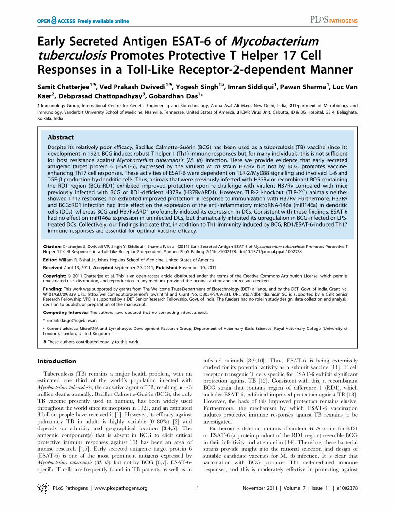
IL-17 induced by H37Rv or BCG::RD1 mediates improvedvaccine efficacy
Previously, it has been shown that IL-17 plays an important role
in the secondary immune response following vaccination with
virulent H37Rv [16]. Thus, we examined whether IL-17 induced
by virulent strains contributes to improved vaccine efficacy. For
this purpose, we infected animals with H37Rv, BCG,
H37RvDRD1, or BCG::RD1 for 30 days. These animals were
subsequently treated with antibiotics for four weeks, and then
rested for an additional month. We could not find any detectable
M. tb organisms in these animals. These mice were then
challenged with H37Rv through aerosol infection. We found that
animals previously infected with BCG or H37RvDRD1 exhibited
robust protective immunity compared with primary infection
(p,0.01) (Fig. 2A). Interestingly, we found that animals that were
previously infected with H37Rv produced dramatically enhanced
protective immune responses (Fig. 2A). This is in agreement with
previous reports suggesting that virulent strains of M. tb H37Rv
induce superior protective immune responses [34,35]. However,
the kinetics of host protective responses in our hand are somewhat
different from these studies, which may be due to differential
environmental factors in different geographical regions. In fact, it
has been well documented that the efficacy of BCG in human
vaccine trials dramatically varies depending on the geographical
location ([3,4,5]). Nevertheless, we tested M. tb antigen-specific
responses induced by randomly selected animals from our colony.
We challenged splenocytes from sixteen animals with M. tb-
derived complete soluble antigen (CSA) or the unrelated antigen
ovalbumin (OVA) and measured lymphoproliferation. We ob-
served that animals from our colony responded weakly to CSA,
whereas no response was detected against OVA. Therefore, these
animals were likely exposed to environmental organism(s) that
share antigenic similarities with M. tb. As a positive control, we
used spleen cells from CSA immunized mice, which showed
dramatic proliferation against in vitro rechallenge with CSA
(Fig 2B). These observations might also be relevant to the variable
vaccine response of BCG. In either case, our findings suggested
that, while BCG and H37RvDRD1 induced significant protective
immunity against TB, this was not sufficient to confer complete
protection against disease pathology. In contrast, H37Rv and
BCG::RD1 induced improved protective immune responses.
Collectively, these observations suggested that the RD1 region
enhances protective immune responses. Importantly, we found
that animals that were previously infected with H37Rv or
BCG::RD1 induced significantly higher numbers of Th17 cells
in the lungs than animals infected with BCG or H37RvDRD1
(p,0.001) (Fig. 2C). Therefore, we tested whether IL-17 was
responsible for the improved vaccine efficacy of H37Rv or
BCG::RD1. For this purpose, we injected animals with anti-IL-17
or control mouse IgG antibodies every 72 hours during re-
challenge with H37Rv or BCG::RD1. Treatment with anti-IL-17
abrogated the observed enhancement in protective immune
responses induced by H37Rv or BCG::RD1 (Fig. 2D). Therefore,
these observations suggested that H37Rv induced Th17 cell
responses, which complemented Th1 cell responses for improved
protection against TB.
Dendritic cells infected with H37Rv or BCG::RD1 directTh1 and Th17 cell responses, whereas BCG andH37RvDRD1 selectively induce Th1 cell responses
Our in vivo experiments demonstrated that H37Rv induces
Th17 cell differentiation. Therefore, to provide insight into the
mechanism whereby H37Rv promotes Th1 and Th17 cell
differentiation, we compared the cytokines induced by DCs
(characterized with CD11c, CD11b, CD80, CD86, and MHC
Class II markers Fig. 3A) infected with H37Rv, BCG::RD1,
BCG, and H37RvDRD1. We found that H37Rv- or BCG::RD1-
infected DCs produced substantial amounts of IL-12p40, IL-6,
and TGF-b (Fig. 3B). However, BCG and H37RvDRD1 induced
dramatically reduced amounts of IL-6 (p,0.001) and TGF-b(p,0.001) by DCs than H37Rv and BCG::RD1 (Fig. 3B).
Nevertheless, IL-12p40 was induced at comparable levels by all
bacterial strains. Interestingly, we found that IL-6 and TGF-bproduction was dependent on TLR-2 and MyD88, whereas IL-
12p40 production was independent of TLR-2 but required
MyD88 (Fig. 3B). To determine whether these cytokines play a
role in Th cell differentiation, we co-cultured ovalbumin (OVA)-
specific CD4+ T cells from OT-II T cell receptor (TCR) transgenic
(Tg) animals with infected DCs in the presence of OVA peptide
and collected supernatant to determine the production of IFN-cand IL-17 (Fig. 3C). The results, which were supported by
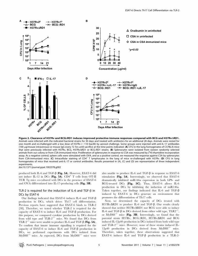
intracellular cytokine staining, indicated that H37Rv-infected DCs
directed the differentiation of both IL-17- and IFN-c-producing
cells (Fig. 3C & 4A). In sharp contrast, DCs infected with BCG or
H37RvDRD1 supported only Th1 cell differentiation. While the
levels of IFN-c production were similar for DCs infected with
H37Rv, BCG, H37RvDRD1 or BCG::RD, production of IL-17
was significantly higher in DCs infected with H37Rv or
BCG::RD1 as compared with cells infected with BCG or
H37RvDRD1 (p,0.001). Furthermore, it is known that IL-22 is
also secreted by IL-17 producing Th cells and recent study
suggested that IL-22 was upregulated during M. tb infection in
rhesus macaques and protective in function[36]. Therefore, we
have also checked the IL-22 mRNA transcript level in our DC-T
cells co-culture experiments and found that IL-22 mRNA
transcript was 5–8 fold upregulated in H37RV or BCG::RD
virulent strains compared to BCG and H37RvDRD1 (Fig. 4B).Therefore, these data indicated that the RD1 locus plays an
important role in directing Th17 cell responses during M. tb
infection.
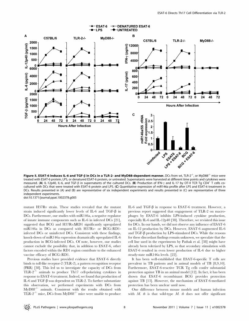
ESAT-6 drives Th17 cell differentiation by inducing IL-6and TGF-b production in DCs
ESAT-6-reactive T cells are prevalent in TB patients and in
animals infected with M. tb [8,9,10]. Furthermore, it has been
shown that ESAT-6-specific T cells provide substantial protection
against TB [12]. Therefore, it has been assumed that ESAT-6 is a
good candidate for development of a TB vaccine [37]. From the
preceding section, it was clear that the RD1 region plays an
important role in directing Th17 cell differentiation, which in turn
contributes to protective immune responses against TB. Differen-
tiation of Th17 cells requires IL-6 and TGF-b simultaneously
[18,19]. Therefore, we determined whether ESAT-6 induces these
cytokines in DCs. We found that DCs treated with ESAT-6
ESAT-6 Directs Th17 Cell Differentiation via TLR-2
PLoS Pathogens | www.plospathogens.org 4 November 2011 | Volume 7 | Issue 11 | e1002378
produced both IL-6 and TGF-b (Fig. 5A). However, ESAT-6 did
not induce IL-12 in DCs (Fig. 5A). CD4+ T cells from OT-II
TCR Tg mice co-cultured with DCs in the presence of ESAT-6
and OVA differentiated into IL-17-producing cells (Fig. 5B).
TLR-2 is required for the induction of IL-6 and TGF-b inDCs by ESAT-6
Our findings indicated that ESAT-6 induces IL-6 and TGF-bproduction in DCs, which drives Th17 cell differentiation.
Previous reports have suggested that ESAT-6 binds to TLR-2
[38]. Therefore, we tested whether TLR-2 is required for the
capacity of ESAT-6 to induce IL-6 and TGF-b production. For
this purpose, we compared cytokine production by DCs derived
from wild type and TLR-2-/- mice. We found that DCs from
TLR-2-/- mice were unable to produce IL-6 and TGF-b (Fig. 5A).
To confirm that innate immune signalling is required for the
capacity of ESAT-6 to induce IL-6 and TGF-b production in
DCs, we performed experiments with DCs isolated from
MyD88-/- mice. As expected, DCs from MyD88-/- mice were
also unable to produce IL-6 and TGF-b in response to ESAT-6
stimulation (Fig. 5A). Interestingly, we observed that ESAT-6
dramatically inhibited miR146a expression in both LPS- and
BCG-treated DCs (Fig. 5C). Thus, ESAT-6 allows IL-6
production in DCs by inhibiting the induction of miR146a.
Taken together, our findings indicated that IL-6 and TGF-binduced by ESAT-6 in DCs generate an environment that
promotes the differentiation of Th17 cells.
Next, we determined the capacity of DCs treated with
H37RvDRD1 to produce IL-6 and TGF-b. Our results clearly
showed that neither H37RvDRD1 nor BCG were able to induce
IL-6 and TGF-b in DCs derived from either wild type, TLR2-/-,
or MyD88-/- mice (Fig. 3B). Interestingly, we found that the
parental strain H37Rv, BCG::RD1, H37RvDRD1 and BCG
induced IL-12p40 production in DCs isolated from both wild type
and TLR-2-/- mice. However, none of these strains induced IL-
12p40 production in DCs derived from MyD88-/- mice.
Therefore, taken together, these observations suggested that
ESAT-6 induces IL-6 and TGF-b production in a TLR-2-
Figure 2. Clearance of H37Rv and BCG::RD1 induces improved protective immune responses compared with BCG and H37RvDRD1.Animals were infected with the indicated bacterial strains for 30 days and treated with antibiotics for an additional 28 days. Animals were rested forone month and re-challenged with a low dose of H37Rv (,110 bacilli) by aerosol challenge. Some groups were injected with anti-IL-17 antibodies(100 mg/mouse intravenous) or mouse IgG every 72 hrs until sacrifice at the time points indicated. (A) CFU in the lung homogenates of C57BL/6 micethat were previously infected with H37Rv, BCG, H37RvDRD1 or BCG::RD1 strains. (B) Splenocytes were isolated from sixteen randomly selectedanimals from our colony or from CSA-immunized mice. Proliferation of splenocytes in response to CSA was measured by [3H]-thymidine incorporationassay. As a negative control, spleen cells were stimulated with OVA, and as a positive control, we measured the response against CSA by splenocytesfrom CSA-immunized mice. (C) Intracellular staining of CD4+ T lymphocytes in the lung of mice re-challenged with H37Rv. (D) CFU in lunghomogenates of mice that received anti-IL-17 or control antibodies. Results presented in (A), (C) and (D) are representative of three independentexperiments.doi:10.1371/journal.ppat.1002378.g002
ESAT-6 Directs Th17 Cell Differentiation via TLR-2
PLoS Pathogens | www.plospathogens.org 5 November 2011 | Volume 7 | Issue 11 | e1002378

dependent manner. In contrast, production of IL-12p40 by DCs
following infection with mycobacteria is independent of ESAT-6
and TLR-2 expression. However, IL-12p40 production induced
by mycobacteria is dependent on MyD88 signalling.
TLR-2-/- animals fail to induce M. tb-mediated Th17 cellresponses and show enhanced susceptibility to M. tbinfection
From the preceding section it is clear that interaction of ESAT-
6 with TLR-2 creates an environment that is conducive to the
differentiation of Th17 cells, which in turn results in protective
immunity against TB. Therefore, to confirm that TLR-2
signalling is important for the observed Th17 cell responses and
improved vaccine efficacy, we performed vaccination experi-
ments in TLR-2-/- animals. These animals were infected with
H37Rv and subsequently treated with antibiotics as described in
the materials and methods. These animals were then challenged
with virulent strain H37Rv. H37Rv-immunized TLR-2-/- mice
generated protective immunity against H37Rv at a level similar
to wild-type mice immunized with BCG during primary
challenge (Fig. 6A). However, TLR-2-/- mice exhibited reduced
protective immune responses as compared with wild type
littermates (Fig. 6A). H37RvDRD1 and BCG::RD1 also showed
reduced protection in TLR-2-/- mice (Fig. 6A) similar to BCG
and H37Rv. Other mycobacterial components, such as LAM and
lipoproteins, can also activate TLR-2 [39]. Therefore, the
observed differences in protective immune responses in TLR-
2-/- animals could be caused by multiple TLR-2-dependent
agonists. However, most of the M. tb-derived TLR-2 ligands
induce only suppressive immune responses [39]. Therefore, the
observed protective responses are most likely contributed by
RD1-derived proteins. Furthermore, these differences are com-
parable with responses induced by H37Rv versus BCG in wild-
type animals.
Next, we analyzed effector T cells in the lungs. We found that
TLR-2-/- animals generated IFN-c-producing cells comparable to
wild type littermates. However, these animals produced signifi-
cantly fewer numbers of IL-17-producing cells in their lungs
(p,0.001) (Fig. 6B). This finding is further strengthened by a
recently published report, suggesting that TLR-2 is indispensible
for the generation of Th17 responses during M. tb infection [40].
Therefore, these observations suggested that TLR-2 plays an
important role in mounting Th17 cell responses to H37Rv, which
in turn confers protective immunity to TB.
Figure 3. H37Rv directs differentiation of both Th1 and Th17 cells by inducing IL-12p40, IL-6, and TGF-b in DCs. (A) Characterization ofdendritic cells (DCs) by flocytometric analysis using anti-CD11c, CD11b, -CD80, -CD86, -MHC class II, and - IgG2a(isotype control). (B) Production of IL-12p40, IL-6, and TGF-b in DCs infected with H37Rv, BCG, H37RvDRD1 or BCG::RD1. (C) Induction of IFN-c and IL-17 in the culture supernatants of OT-IITCR Tg CD4+ T cells co-cultured with infected DCs Results presented here are representative of at least three independent experiments.doi:10.1371/journal.ppat.1002378.g003
ESAT-6 Directs Th17 Cell Differentiation via TLR-2
PLoS Pathogens | www.plospathogens.org 6 November 2011 | Volume 7 | Issue 11 | e1002378
Discussion
It is well accepted that Th1 cells play a central role for
protection against TB [41]. Therefore, animals that are deficient
in IFN-c, IFN-c receptor, Stat-4, T-bet, or IL-12 exhibit increased
susceptibility to M. tb infection [25,42,43]. BCG induces a robust
Th1 response, but this does not appear to be sufficient for optimal
protection against challenge with virulent M. tb [44]. Abundant
Th1 cell responses have been found in TB patients as well as M. tb
infected animals [45,46]. Thus, Th1 cells alone are not sufficient
for protection against TB. Therefore, in addition to Th1 cell
responses, a vaccine needs to induce additional Th cell response(s)
to provide optimal protection against TB. Recently, it has been
shown that Th17 cells play an important role in the secondary
immune response against TB [16]. However, Th17 cells do not
appear to participate in the primary immune response against TB
[17]. Our findings clearly demonstrated that BCG and
H37RvDRD1 are unable to induce Th17 cell responses in the
lung. Therefore, we considered the possibility that BCG lacks an
antigen that drives Th17 cell differentiation and that such a
response is required for optimal protection against TB. We
observed that the virulent M. tb strain H37Rv and the engineered
BCG::RD1 strain induced Th17 cell responses, which correlated
with improved protection against re-infection and, thus, improved
vaccine efficacy. This is in agreement with a previous report
indicating that immunization with virulent M. tb H37Rv induces
superior protective memory T cell responses [34]. However, unlike
our results these authors found only 2–3 log differences in CFUs
upon re-challenge with virulent H37Rv. This apparent discor-
dance could be due to several differences in the experimental
procedures employed. Jung et al. (2005) employed an extended
(100 days) antibiotic treatment protocol, which may have
influenced immune responses, and increased the age of the mice
at re-challenge. Furthermore, these investigators rechallenged
mice with a higher (two-fold) dose of bacteria. In addition, it is also
possible that differences in the microflora in different animal
facilities might contribute to these apparent differences. Indeed,
our results demonstrated that unimmunized animals from our
facility were able to respond to M. tb antigens, albeit weakly,
suggesting that exposure to environmental organisms that share
antigenic properties with M. tb might contribute to improved
protection. This may explain the differential vaccine responses
against BCG that have been observed in different geographical
locations and in subjects from different ethnicities.
Previous reports have suggested that ESAT-6, a protein within
the RD1 region that is absent in BCG, is a promising vaccine
candidate [8]. Furthermore, deletion mutants of H37Rv for RD1
resembled BCG in many aspects [8]. Interestingly, we found that
the RD1 mutant of virulent H37Rv was unable to induce Th17
cell responses. BCG and H37RvDRD1 were unable to induce
persistent infection and, hence, the bacterial load declined much
more rapidly [14] over time as compared with H37Rv or
BCG::RD1. Therefore, the observed differential Th1 and Th17
cell responses could be related to bacterial replication and
antigenic load. However, previous reports indicated that BCG
inhibits Th17 cell responses in lung and other organs [47,48].
Furthermore, primary infection with a high dose (2.56105 CFU)
of BCG by intratracheal injection was unable to induce IL-17 in
the lung, until re-challenge with PPD-coated beads, and the
cellular source of IL-17 produced in this situation is not known
[49]. Therefore, BCG alone, even at a high dose, is unable to
induce Th17 cell responses. Nonetheless, a recent report indicated
that BCG was unable to induce IL-17-producing cells in a primary
challenge, however it did so upon repeated re-challenge [30].
Although our study indicated that the RD1 recombinant strain
exhibited an improved vaccine efficacy compared with the
parental BCG strain, it does not exclude a role for other RD
regions in inducing improved host protective immune responses.
We found that H37Rv and BCG::RD1 induce both Th1 and
Th17 cell responses that contribute to improved vaccine efficacy as
compared with BCG, which selectively induces Th1 cell responses.
Th17 cell responses are directed by IL-6 and TGF-b, derived from
antigen presenting cells (APCs). Therefore, pathogen-associated
molecular patterns (PAMP) encoded within the RD1 region are
likely responsible for inducing these two cytokines. Considering
that ESAT-6 induces protective immune responses and that the
RD1 mutant was unable to induce Th17 cell responses, we
considered the possibility that ESAT-6 induces Th17 cell-
polarizing cytokines in APCs. Therefore, we tested IL-6 and
TGF-b production in DCs infected with the wild-type and RD1
Figure 4. H37Rv directs differentiation of both Th1 and Th17cells. (A) Intracellular cytokine staining for IFN-c and IL-17 in OT-IITCR Tg CD4+ T cells co-cultured with infected DCs. (B) IL-22 mRNAexpression profile from OT-II TCR Tg CD4+ T cells co-cultured with DCsafter infection of DCs with H37Rv, BCG, BCG::RD1 or H37RvDRD1.Results are representative of at least three independent experiments.doi:10.1371/journal.ppat.1002378.g004
ESAT-6 Directs Th17 Cell Differentiation via TLR-2
PLoS Pathogens | www.plospathogens.org 7 November 2011 | Volume 7 | Issue 11 | e1002378
mutant H37Rv strain. These studies revealed that the mutant
strain induced significantly lower levels of IL-6 and TGF-b in
DCs. Furthermore, our studies with miR146a, a negative regulator
of innate immune components such as IL-6 in infected DCs [21],
suggested that BCG and H37RvDRD1 significantly upregulated
miR146a in DCs as compared with H37Rv- or BCG::RD1-
infected DCs or uninfected DCs. Consistent with these findings,
knock-down of miR146a expression dramatically upregulated IL-6
production in BCG-infected DCs. Of note, however, our studies
cannot exclude the possibility that, in addition to ESAT-6, other
factors encoded within the RD1 region contribute to the enhanced
vaccine efficacy of BCG::RD1.
Previous studies have provided evidence that ESAT-6 directly
binds to toll-like receptor-2 (TLR-2), a pattern recognition receptor
(PRR) [38]. This led us to investigate the capacity of DCs from
TLR-2-/- animals to produce Th17 cell-polarizing cytokines in
response to ESAT-6 treatment. Indeed, we found that production of
IL-6 and TGF-b was dependent on TLR-2. To further substantiate
this observation, we performed experiments with DCs from
MyD88-/- animals. Consistent with the results obtained with
TLR-2-/- mice, DCs from MyD88-/- mice were unable to produce
IL-6 and TGF-b in response to ESAT-6 treatment. However, a
previous report suggested that engagement of TLR-2 on macro-
phages by ESAT-6 inhibits LPS-induced cytokine production,
especially IL-6 and IL-12p40 [38]. Therefore, we revisited this issue
for DCs. In our hands, we did not observe any influence of ESAT-6
on IL-12 production by DCs. However, ESAT-6 augmented IL-6
and TGF-b production by LPS-stimulated DCs. While the reasons
for these discordant findings remain unknown, we speculate that the
cell line used in the experiments by Pathak et al. [38] might have
already been tolerized by LPS, so that secondary stimulation with
ESAT-6 resulted in even lower production of IL-6 due to higher
steady-state miR146a levels. [22].
It has been well-established that ESAT-6-specific T cells are
prevalent in TB patients and in animal models of TB [8,9,10].
Furthermore, ESAT-6-reactive TCR Tg cells confer substantial
protection against TB in an animal model [12]. In fact, it has been
shown that ESAT-6 recombinant BCG provides protection
against TB [13]. However, the mechanism of ESAT-6-mediated
protection has been unclear until now.
One difference between mouse models and human infection
with M. tb is that wild-type M. tb does not offer significant
Figure 5. ESAT-6 induces IL-6 and TGF-b in DCs in a TLR-2- and MyD88-dependent manner. DCs from wt, TLR-2-/-, or MyD88-/- mice weretreated with ESAT-6 protein, LPS, or denatured ESAT-6 protein, or untreated. Supernatants were harvested at different time points and cytokines weremeasured. (A) IL-12p40, IL-6, and TGF-b in supernatants of the cultured DCs. (B) Production of IFN-c and IL-17 by OT-II TCR Tg CD4+ T cells co-cultured with DCs that were treated with ESAT-6 protein and LPS. (C) Quantitative expression of miR146a profile after LPS and ESAT-6 treatment inDCs. Results presented in (A) and (B) are representative of six independent experiments and results presented in (C) are representative of threeindependent experiments.doi:10.1371/journal.ppat.1002378.g005
ESAT-6 Directs Th17 Cell Differentiation via TLR-2
PLoS Pathogens | www.plospathogens.org 8 November 2011 | Volume 7 | Issue 11 | e1002378

protection against reinfection in humans, despite containing
ESAT-6 and other major antigens. This could be due to various
reasons. First, there might be genetic differences between mice and
humans that cause altered immune responses. Second, environ-
mental exposures may alter protective immunity. Third, different
individuals might respond differently to drugs used to treat TB
and, thus, it is difficult to determine whether treatment was
complete, while the remaining bacteria may cause secondary
infection. Fourth, M. tb evolved many different types of immune
evasion mechanisms [50]. For example, we have recently shown
that bacteria that are within granuloma-like structures are
sequestered from host protective immune responses by mesenchy-
mal stem cells [51].
In summary, our findings indicate that, in addition to Th1 cells,
Th17 cells play a critical role in conferring optimal protection
against TB. The ESAT-6 protein, which is present in H37Rv and
BCG::RD1 but not in BCG and H37RvDRD1, directs Th17 cell
differentiation by inducing IL-6 and TGF-b in DCs in a TLR-2-
and MyD88-dependent manner. Therefore, ESAT-6 can contrib-
ute to vaccine preparations by promoting Th17 cell responses.
Materials and Methods
Ethics statementAnimal experiments were performed according to the guidelines
approved by the Institutional Animals Ethics Committee meeting
held on 22nd November 2007 at ICGEB (approval ID; ICGEB/
IAEC/IMM-13/2007), New Delhi, India and Department of
Biotechnology (DBT) guidelines, Government of India. All mice
used for experiments were ethically sacrificed by asphyxiation in
carbon dioxide according to institutional and DBT regulations.
MiceC57BL/6 and OT-II TCR transgenic mice (6–8 wks of age)
were initially purchased from The Jackson Laboratories, USA.
TLR-2 and MyD88 knock-out mice (6–8 weeks of age), both on a
C57BL/6 background, were the kind gift of Prof. Ruslan
Medzhitov, Yale University, New Haven, USA. All animals were
subsequently bred and maintained in the animal facility of the
International Centre for Genetic Engineering and Biotechnology
(ICGEB), New Delhi, India.
BacteriaMycobacterium tuberculosis strain H37Rv was a kind gift from the
Colorado State University repository. H37RvDRD1 and BCG
were a kind gift from Prof. David Sherman, (SBRI, Seattle,
WA,USA). The integrative cosmid vector pYUB412 (control
vector) and the recombinant cosmid vector RD1-2F9 harboring
RD1 locus of M. tuberculosis [33] were kind gifts from Prof.
Stewart Cole of the Ecole Polytechnique Federale de Lausanne
(EPFL), Switzerland. The control and recombinant cosmids were
electroporated individually into electrocompetent cells of BCG
(Danish) to obtain BCG::YUB412 and BCG::RD1 strains,
essentially as described previously [33]. Briefly, 100 ml of bacilli
suspension (OD600nm, 0.4) from a 7-day-old Middlebrook 7H9
(Difco) culture, supplemented with albumin-dextrose-catalase
(ADC; Difco), was pelleted by centrifugation at 2500 g for
15 min at 16uC, washed twice with 10% glycerol and finally
resuspended in 3 ml of 10% glycerol. Two hundred microliters of
the electrocompetent bacilli were mixed with 5 microliter of the
control vector pYUB412 (85 ng ml21) or recombinant vector RD1-
2F9 (100 ng ml21) and electroporated using the Gene Pulser Xcell
Electroporation System (Bio-Rad Pacific, Hong Kong) with
settings of 2.5 kV, 25 mF and 1000 V. After electroporation, cells
were resuspended in 5 ml of 7H9 medium supplemented with
ADC, and kept overnight at 37uC. The cells were then pelleted by
centrifugation, resuspended in 100 ml of 7H9 medium, and plated
on Middlebrook 7H11 medium supplemented with oleic acid-
albumin-dextrose-catalase (OADC, Difco), hygromycin (200 mg
ml21) and ampicillin (100 mg ml21). After three to four weeks of
incubation at 37uC, hygromycin-resistant clones were selected.
BCG::RD1-2F9 (BCG::RD1) clones were characterized for
secretion of ESAT-6 by immunoblotting using mouse anti-ESAT6
antibody (unpublished data).
Recombinant ESAT6 proteinDetailed procedures for preparation and characterization of
recombinant ESAT6 have been described in our earlier
publication [52]. Briefly, E. coli BL21(plysS) transformed with
pET23b+ vector (Novagen) carrying esat6 gene of M. tuberculosis
Figure 6. TLR-2-deficient animals induce reduced levels ofprotective immune responses. For in vivo experiments, wild typeand TLR-2-/- mice were infected with H37Rv for 30 days, andsubsequently treated with antibiotics for 4 weeks as described in thematerials and methods. Mice were then re-challenged with H37Rv.Groups of animals were sacrificed for the following studies. (A) CFU inlung homogenates of mice re-challenged with H37Rv. (B) Intracellularcytokines in CD4+ T lymphocytes derived from the lung. Resultspresented here are representative of three independent experiments.doi:10.1371/journal.ppat.1002378.g006
ESAT-6 Directs Th17 Cell Differentiation via TLR-2
PLoS Pathogens | www.plospathogens.org 9 November 2011 | Volume 7 | Issue 11 | e1002378
was grown to mid-log phase, induced with IPTG (0.4 mM final
conc.) for 4 hrs, and the recombinant ESAT6 protein was
extracted from the inclusion bodies in 8 M urea. The recombinant
ESAT6 protein was then purified by Nickel -nitrilotriacetic acid
(Ni-NTA) chromatography, checked for LPS contamination by
LAL (limulus amebocyte lysate) tests, and characterized for purity
by SDS-PAGE, immunoblotting and N-terminal amino acid
sequencing as described previously [52]. The purified and LPS-
free recombinant ESAT6 protein was aliquoted and kept at -80uCuntil further use.
Bacterial culturesAll mycobacterial strains were grown in 7H9 (Middlebrook,
Difco, USA) medium supplemented with 10% ADC and with
0.05% Tween 80 and 0.2% glycerol, and cultures were grown to
mid-log phase. Aliquots of the cultures in 20% glycerol were
preserved at 280uC and these cryo-preserved stocks were used for
infections.
M. tb infection of mice and estimation of Colony FormingUnits (CFU)
Mice were infected with various mycobacterial strains (namely
H37Rv, H37RvDRD1, BCG, or BCG::RD1) via the aerosol route
using a Madison aerosol chamber (University of Wisconsin,
Madison, WI) with its nebulizer pre calibrated to deposit a total of
,110 to the lungs of each mouse as previously described [51,53].
Briefly, mycobacterial stocks recovered from a 280uC freezer
were quickly thawed and subjected to light ultra-sonication to
obtain a single cell suspension. Fifteen ml of the bacterial cell
suspension (106106 cells per ml) was placed in the nebulizer of the
Madison aerosol chamber pre-calibrated to deliver via aerosol
route the desired number of CFUs to the lungs of animals placed
inside the chamber. A day after the aerosol exposure procedure,
three randomly selected mice were sacrificed at various time points
and organs were harvested, homogenised in 0.2 mm filtered PBS
containing 0.05% Tween 80 and plated onto 7H11 Middlebrook
(Difco USA) plates containing 10% oleic acid, albumin, dextrose
and catalase (OADC) (Difco USA). Undiluted, ten-fold diluted and
one hundred-fold diluted lung and spleen cell homogenates were
plated in duplicate on the above 7H11 plates and incubated at
37uC for 21–28 days. Colonies were counted and CFU were
estimated. Mice from various groups were euthanized at the
indicated time points in various experiments; their organs were
harvested for obtaining CFU counts and/or immune cell
subpopulations for immunological studies as described under
other sub-sections.
ReagentsLuminex kits were purchased from Millipore and Bio-Rad.
GM-CSF and IL-4 were obtained from R&D Biosystems, USA.
Purified or fluorescently-conjugated monoclonal antibodies against
mouse CD11c (N418), CD11b (M1/70), CD80 (16-10A1), CD86
(GL1), and MHC-II (NIMR-4) were purchased from eBioscience,
USA, and fluorescently-conjugated anti-mouse IgG2a (R19-15)
was purchased from BD Pharmingen. LPS was obtained from
Sigma-Aldrich (L-2654).
Generation of dendritic cells (DCs)Mice were euthanized and the femurs were isolated. Bone
marrow was flushed out with RPMI-1640 medium using a 2.0 ml
syringe (26.5G). The cells were washed twice with PBS and then
cultured in complete RPMI-1640 (Gibco, UK) medium supple-
mented with GM-CSF (40 ng/ml) and IL-4 (10 ng/ml) on 24-well
plates (1 million/ml). On the third day, 75% of the medium was
replaced with fresh DC culture medium. On day 5, the suspended
cells were removed and the loosely adherent cells were collected as
immature DCs (CD11c-positive cells were .90%). Flowcytometric
analysis by using anti-CD11c, -CD11b, -CD80, -CD86, -MHC
class II, and - IgG2a (isotype control) antibodies suggested that
.95% of the cells were conventional DCs.
Bacterial infection of DCs and co-culture with CD4+ Tcells from OT-II TCR transgenic mice
BM cells were isolated from different mouse strains (C57BL/6,
TLR-2-/- and MyD88-/-) and differentiated into immature DCs as
described above and cultured in 24-well plates (16106 cells per
well). Cells were infected with H37Rv, H37RvDRD1, BCG or
BCG::RD1 (MOI of 1:10). Similarly, 16106 DCs were cultured in
24-well plates in the presence or absence of LPS at 1 mg/mL and
co-stimulated with ESAT-6 protein at a final concentration of
5 mg/mL, with PBS as the negative control. Supernatants from
cells were collected at 24, 48 and 72 hrs for cytokine profiling. For
Th1 and Th17 cell differentiation, CD4+ T cells (16106) were
purified by MACS method (CD4+ T cell isolation beads kit;
Miltenyi Biotech, Germany) from OT-II TCR transgenic mice
and cultured with immature DCs (16106) infected with H37Rv,
H37RvDRD1, BCG or BCG::RD1 (MOI of 1:10) in the presence
of ovalbumin (10 mg/ml) peptide (Thermo Scientific, USA) for
72 hours. Then, CD4+ T cells were harvested and subjected to
intracellular staining for IL-17 and IFN-c expression.
Antibiotic treatment of miceThirty days post infection, groups of mice were treated with
0.1 g/L rifampicin and 0.1 g/L isoniazid (Sigma-Aldrich, St.
Louis, MO, USA) administered in the drinking water (changed
daily) for 4 weeks. M. tuberculosis-infected control mice received
plain drinking water. A control group of infected mice was
sacrificed at the start of treatment (early control group). A second
group of infected but untreated mice was sacrificed 4 weeks after
therapy was initiated (late control group).
Isolation of lymphocytes from infected animalsLungs from infected or uninfected animals were harvested and
washed by swirling in PBS. They were opened up by cutting
longitudinally and then cut into ,0.5-cm pieces. These lung pieces
were agitated in 25 ml of extraction buffer (PBS, 3% FCS, 1 mM
dithiothreitol, 1 mM EDTA) for 30 min at 37uC. This slurry was
passed through a loosely packed nylon wool column to remove the
aggregates. The filtrate was layered on a discontinuous Percoll
gradient (Amersham Pharmacia Biotech, USA). This gradient was
then centrifuged at 9006g for 20 minutes. Cells at the interface of
the 40/70% layer were collected and washed in staining buffer
(PBS, 3% FCS). Cells were cultured for intracellular staining as
described below. Bronchoalveolar lavage (BAL) fluid was collected
from lungs by intratrachial infusion of PBS and cell-free BAL was
used for cytokine assay [54].
[3H]-Thymidine incorporation assay of splenocytesSpleens were isolated from sixteen randomly selected C57BL/6
mice of our colony or CSA-immunized (100 mg/ml in 200 ml
incomplete Freund’s adjuvant) mice. Spleens were macerated by
frosted slides in complete RPMI 1640 (Gibco, Invitrogen, UK) and
made into a single cell suspension. Red blood cells (RBCs) were
lysed with RBC cell lysis buffer and washed with complete RPMI
1640. Splenocytes were counted and plated at 0.16106 cells/well
in 96-well plates and stimulated with different concentrations of
ESAT-6 Directs Th17 Cell Differentiation via TLR-2
PLoS Pathogens | www.plospathogens.org 10 November 2011 | Volume 7 | Issue 11 | e1002378
M. tb complete soluble antigen (CSA). Cells were cultured for
48 hours and then pulsed with tritiated thymidine (3H-TdR,
1.0 mCi per well; Amersham Biosciences UK) before measuring
incorporation of 3H-TdR by means of a cell harvester and liquid
scintillation counter 16 hours later (Wallac Trilux, Perkin Elmer,
UK).
Intracellular cytokine stainingFor intracellular cytokine staining, cells were treated with 50 ng/
ml phorbol myristate acetate (PMA) and 500 ng/ml ionomycin in
the presence of 10 mg/ml brefeldin A (Sigma-Aldrich or eBios-
ciences, USA) added during the last 6 h of culture. Cells were
washed twice with PBS and resuspended in a permeabilization
buffer (Cytofix/Cytoperm kit; BD), and stained with the following
fluorescently conjugated monoclonal antibodies: anti-CD4 (clone:
GK1.5)-APC, anti-IFN-c (clone: XMG1.2)-FITC, and anti-IL-17
(clone: 17B7)-PE (all from eBiosciences, USA). Fluorescence
intensity was measured by flow cytometry (FACS Calibur; BD)
and data were analysed with FlowJo (Tree star, USA).
q-PCR analysisBone marrow derived DC were isolated and infected with
different bacterial strains (H37Rv, H37RvDRD1, BCG and
BCG::RD1) as described above and cultured for 24 hours for
RNA isolation. Total RNA, including miRNAs was isolated by
miRNAeasy isolation kit (QIAGEN, Germany) according to the
manufacturer’s instructions. Real-time quantitative RT-PCR
analysis was performed using BioRad Real-Time thermal cycler
(BioRad, USA) and miRCURY LNA universal reverse transcrip-
tase microRNA PCR SYBR green master mix (EXIQON,
Denmark) for miRNA amplification and IQ BioRad SYBER
green master mix (BioRad, USA) for IL-6 expression, respectively.
cDNA was synthesized by the miRCURY LNA universal reverse
transcriptase microRNA cDNA synthesis kit (EXIQON) and the
reaction was set up according to the manufacturer’s protocol. For
amplification of miR146a, LNA PCR miR146a and reference 5S
rRNA primer sets were used and the reaction was set up as
recommended by EXIQON. The relative expression level of
miRNAs was normalized to that of internal control 5S rRNA by
using 2-DDCt cycle threshold method. Furthermore, for amplifi-
cation of IL-6 or IL-22, cDNA was synthesized by Omniscript RT
kit (QIAGEN) using oligodT primers (Fermentas, Maryland,
USA). For IL-6 mRNA expression analysis primer sequences were
IL-6F, 59-TGGAGTCACAGAAGGAGTGGCTAAG-39 and IL-
6R, 59- TCTGACCACAGTGAGGAATGTCCAC-39, and con-
trol GAPDH-F, 59- CGTCCCGTAGACAAAATGGT-39 and
GAPDH-R, 59- TTGATGGCAACAATCTCCAC-39 and for IL-
22 mRNA expression analysis primer sequences were IL-22F 59-
GTGACGACCAGAACATCCAG-39 and IL22R 59-ATCTCT-
CCGCTCTCTCCAAG-39. Data were normalized by the level of
GAPDH expression in samples as described above.
Knock-down of miR146a using anti-miR146aFor transfection of anti-miR146a and scramble control (EX-
IQON) into DCs, cells were transfected at day 4 of culture in
antibiotic free media using Lipofectamine (Invitrogen, UK) reagents
and at day 5 cells were infected with H37Rv, H37RvDRD1, BCG
or BCG::RD1. After 24 hours of bacterial infection, cells were
harvested for RNA preparation and analyzed for miR146a and IL-6
expression by quantitative real-time PCR as described above.
Detection of cytokinesCytokines in the culture supernatant of DCs were assayed by a
Luminex microbead- based multiplexed assay using commercially
available kits according to the manufacturer’s protocol (Milliplex
kit, Millipore and BioPlex, Bio-Rad, USA).
Statistical analysisAll data were derived from at least three independent experiments.
Statistical analyses were conducted using SPSS software and values
were presented as mean6SD. Significant differences between the
groups were determined by ANOVA followed by Tukey’s multiple
comparison test (SPSS software). A value of p,0.05 was accepted as
an indication of statistical significance.
Accession numbers of proteins mentioned in themanuscript
CFP-10: 10 kDa culture filtrate protein [Mycobacterium
tuberculosis H37Rv]. ACCESSION ACH88465.
ESAT-6: early secreted antigenic target 6 kDa [Mycobacterium
tuberculosis H37Rv]. ACCESSION AAC83446.
Acknowledgments
We thank Prof. Ruslan Medzhitov for providing MyD88-/- and TLR-2-/-
mice. We thank Prof. David R. Sherman for the kind gift of H37Rv and
mutants for RD1. We acknowledge Mr. Ashok Das and Dr. D.B.
Chandramouli for assistance with animal experiments.
Author Contributions
Conceived and designed the experiments: SC VPD GD. Performed the
experiments: SC VPD YS IS. Analyzed the data: SC VPD YS LVK DC
GD. Contributed reagents/materials/analysis tools: PS LVK DC. Wrote
the paper: SC VPD YS GD.
References
1. Gupta UD, Katoch VM, McMurray DN (2007) Current status of TB vaccines.
Vaccine 25: 3742–3751.2. Colditz GA, Brewer TF, Berkey CS, Wilson ME, Burdick E, et al. (1994)
Efficacy of BCG vaccine in the prevention of tuberculosis. Meta-analysis of thepublished literature. JAMA 271: 698–702.
3. Brewer TF (2000) Preventing tuberculosis with bacillus Calmette-Guerin
vaccine: a meta-analysis of the literature. Clin Infect Dis 31 Suppl 3: S64–67.4. Fine PE (1989) The BCG story: lessons from the past and implications for the
future. Rev Infect Dis 11 Suppl 2: S353–359.5. Fine PE (1995) Variation in protection by BCG: implications of and for
heterologous immunity. Lancet 346: 1339–1345.6. Brodin P, Rosenkrands I, Andersen P, Cole ST, Brosch R (2004) ESAT-6
proteins: protective antigens and virulence factors? Trends Microbiol 12:
500–508.7. Simeone R, Bottai D, Brosch R (2009) ESX/type VII secretion systems and their
role in host-pathogen interaction. Curr Opin Microbiol 12: 4–10.8. Brandt L, Oettinger T, Holm A, Andersen AB, Andersen P (1996) Key epitopes
on the ESAT-6 antigen recognized in mice during the recall of protective
immunity to Mycobacterium tuberculosis. J Immunol 157: 3527–3533.
9. Ravn P, Demissie A, Eguale T, Wondwosson H, Lein D, et al. (1999) Human T
cell responses to the ESAT-6 antigen from Mycobacterium tuberculosis. J InfectDis 179: 637–645.
10. Ulrichs T, Munk ME, Mollenkopf H, Behr-Perst S, Colangeli R, et al. (1998)Differential T cell responses to Mycobacterium tuberculosis ESAT6 in
tuberculosis patients and healthy donors. Eur J Immunol 28: 3949–3958.
11. Dietrich J, Weldingh K, Andersen P (2006) Prospects for a novel vaccine againsttuberculosis. Vet Microbiol 112: 163–169.
12. Gallegos AM, Pamer EG, Glickman MS (2008) Delayed protection by ESAT-6-specific effector CD4+ T cells after airborne M. tuberculosis infection. J Exp
Med 205: 2359–2368.13. Pym AS, Brodin P, Majlessi L, Brosch R, Demangel C, et al. (2003)
Recombinant BCG exporting ESAT-6 confers enhanced protection against
tuberculosis. Nat Med 9: 533–539.14. Lewis KN, Liao R, Guinn KM, Hickey MJ, Smith S, et al. (2003) Deletion of
RD1 from Mycobacterium tuberculosis mimics bacille Calmette-Guerinattenuation. J Infect Dis 187: 117–123.
15. Perera PY, Derrick SC, Kolibab K, Momoi F, Yamamoto M, et al. (2009) A
multi-valent vaccinia virus-based tuberculosis vaccine molecularly adjuvanted
ESAT-6 Directs Th17 Cell Differentiation via TLR-2
PLoS Pathogens | www.plospathogens.org 11 November 2011 | Volume 7 | Issue 11 | e1002378
with interleukin-15 induces robust immune responses in mice. Vaccine 27:
2121–2127.16. Khader SA, Bell GK, Pearl JE, Fountain JJ, Rangel-Moreno J, et al. (2007) IL-
23 and IL-17 in the establishment of protective pulmonary CD4+ T cell
responses after vaccination and during Mycobacterium tuberculosis challenge.Nat Immunol 8: 369–377.
17. Khader SA, Cooper AM (2008) IL-23 and IL-17 in tuberculosis. Cytokine 41:79–83.
18. Bettelli E, Carrier Y, Gao W, Korn T, Strom TB, et al. (2006) Reciprocal
developmental pathways for the generation of pathogenic effector TH17 andregulatory T cells. Nature 441: 235–238.
19. Mangan PR, Harrington LE, O’Quinn DB, Helms WS, Bullard DC, et al.(2006) Transforming growth factor-beta induces development of the T(H)17
lineage. Nature 441: 231–234.20. Starczynowski DT, Kuchenbauer F, Argiropoulos B, Sung S, Morin R, et al.
(2010) Identification of miR-145 and miR-146a as mediators of the 5q-
syndrome phenotype. Nat Med 16: 49–58.21. Taganov KD, Boldin MP, Chang KJ, Baltimore D (2006) NF-kappaB-
dependent induction of microRNA miR-146, an inhibitor targeted to signalingproteins of innate immune responses. Proc Natl Acad Sci U S A 103:
12481–12486.
22. Nahid MA, Pauley KM, Satoh M, Chan EK (2009) miR-146a is critical forendotoxin-induced tolerance: IMPLICATION IN INNATE IMMUNITY.
J Biol Chem 284: 34590–34599.23. Liu Z, Xiao B, Tang B, Li B, Li N (2010) Up-regulated microRNA-146a
negatively modulate Helicobacter pylori-induced inflammatory response inhuman gastric epithelial cells. Microbes Infect 12: 854–863.
24. Lu LF, Liston A (2009) MicroRNA in the immune system, microRNA as an
immune system. Immunology 127: 291–298.25. Flynn JL, Chan J, Triebold KJ, Dalton DK, Stewart TA, et al. (1993) An
essential role for interferon gamma in resistance to Mycobacterium tuberculosisinfection. J Exp Med 178: 2249–2254.
26. Glickman MS, Jacobs WR, Jr. (2001) Microbial pathogenesis of Mycobacterium
tuberculosis: dawn of a discipline. Cell 104: 477–485.27. Goter-Robinson C, Derrick SC, Yang AL, Jeon BY, Morris SL (2006) Protection
against an aerogenic Mycobacterium tuberculosis infection in BCG-immunizedand DNA-vaccinated mice is associated with early type I cytokine responses.
Vaccine 24: 3522–3529.28. Kelly BP, Furney SK, Jessen MT, Orme IM (1996) Low-dose aerosol infection
model for testing drugs for efficacy against Mycobacterium tuberculosis.
Antimicrob Agents Chemother 40: 2809–2812.29. Umemura M, Yahagi A, Hamada S, Begum MD, Watanabe H, et al. (2007) IL-
17-mediated regulation of innate and acquired immune response againstpulmonary Mycobacterium bovis bacille Calmette-Guerin infection. J Immunol
178: 3786–3796.
30. Cruz A, Fraga AG, Fountain JJ, Rangel-Moreno J, Torrado E (2010)Pathological role of interleukin 17 in mice subjected to repeated BCG
vaccination after infection with Mycobacterium tuberculosis. J Exp Med 207:1609–1616.
31. Ganguly N, Siddiqui I, Sharma P (2008) Role of M. tuberculosis RD1 regionencoded secretory proteins in protective response and virulence. Tuberculosis
(Edinb) 88: 510–517.
32. Guinn KM, Hickey MJ, Mathur SK, Zakel KL, Grotzke JE, et al. (2004)Individual RD1-region genes are required for export of ESAT-6/CFP-10 and
for virulence of Mycobacterium tuberculosis. Mol Microbiol 51: 359–370.33. Pym AS, Brodin P, Brosch R, Huerre M, Cole ST (2002) Loss of RD1
contributed to the attenuation of the live tuberculosis vaccines Mycobacterium
bovis BCG and Mycobacterium microti. Mol Microbiol 46: 709–717.34. Jung YJ, Ryan L, LaCourse R, North RJ (2005) Properties and protective value
of the secondary versus primary T helper type 1 response to airborneMycobacterium tuberculosis infection in mice. J Exp Med 201: 1915–1924.
35. Kamath AB, Behar SM (2005) Anamnestic responses of mice following
Mycobacterium tuberculosis infection. Infect Immun 73: 6110–6118.
36. Zeng G, Chen CY, Huang D, Yao S, Wang RC (2011) Membrane-bound IL-22
after de novo production in tuberculosis and anti-Mycobacterium tuberculosiseffector function of IL-22+ CD4+ T cells. J Immunol 187: 190–199.
37. Aagaard C, Dietrich J, Doherty M, Andersen P (2009) TB vaccines: current
status and future perspectives. Immunol Cell Biol 87: 279–286.
38. Pathak SK, Basu S, Basu KK, Banerjee A, Pathak S, et al. (2007) Direct
extracellular interaction between the early secreted antigen ESAT-6 of
Mycobacterium tuberculosis and TLR2 inhibits TLR signaling in macrophages.
Nat Immunol 8: 610–618.
39. Srivastava V, Manchanda M, Gupta S, Singla R, Behera D, et al. (2009) Toll-like receptor 2 and DC-SIGNR1 differentially regulate suppressors of cytokine
signaling 1 in dendritic cells during Mycobacterium tuberculosis infection. J Biol
Chem 284: 25532–25541.
40. Teixeira-Coelho M, Cruz A, Carmona J, Sousa C, Ramos-Pereira D (2011)TLR2 deficiency by compromising p19 (IL-23) expression limits Th 17 cell
responses to Mycobacterium tuberculosis. Int Immunol 23: 89–96.
41. Ottenhoff TH, Kumararatne D, Casanova JL (1998) Novel human immuno-
deficiencies reveal the essential role of type-I cytokines in immunity to
intracellular bacteria. Immunol Today 19: 491–494.
42. Cooper AM, Dalton DK, Stewart TA, Griffin JP, Russell DG, et al. (1993)
Disseminated tuberculosis in interferon gamma gene-disrupted mice. J Exp Med
178: 2243–2247.
43. Cooper AM, Khader SA (2008) The role of cytokines in the initiation,
expansion, and control of cellular immunity to tuberculosis. Immunol Rev 226:
191–204.
44. Majlessi L, Simsova M, Jarvis Z, Brodin P, Rojas MJ, et al. (2006) An increase in
antimycobacterial Th1-cell responses by prime-boost protocols of immunizationdoes not enhance protection against tuberculosis. Infect Immun 74: 2128–2137.
45. Bhattacharyya S, Singla R, Dey AB, Prasad HK (1999) Dichotomy of cytokine
profiles in patients and high-risk healthy subjects exposed to tuberculosis. Infect
Immun 67: 5597–5603.
46. Leal IS, Smedegard B, Andersen P, Appelberg R (2001) Failure to induce
enhanced protection against tuberculosis by increasing T-cell-dependent
interferon-gamma generation. Immunology 104: 157–161.
47. Lagranderie M, Abolhassani M, Vanoirbeek JA, Lima C, Balazuc AM (2010)Mycobacterium bovis bacillus Calmette-Guerin killed by extended freeze-drying
targets plasmacytoid dendritic cells to regulate lung inflammation. J Immunol
184: 1062–1070.
48. Lee J, Reinke EK, Zozulya AL, Sandor M, Fabry Z (2008) Mycobacterium bovisbacille Calmette-Guerin infection in the CNS suppresses experimental
autoimmune encephalomyelitis and Th17 responses in an IFN-gamma-
independent manner. J Immunol 181: 6201–6212.
49. Ito T, Schaller M, Hogaboam CM, Standiford TJ, Sandor M, et al. (2009)
TLR9 regulates the mycobacteria-elicited pulmonary granulomatous immuneresponse in mice through DC-derived Notch ligand delta-like 4. J Clin Invest
119: 33–46.
50. Flynn JL, Chan J (2003) Immune evasion by Mycobacterium tuberculosis: living
with the enemy. Curr Opin Immunol 15: 450–455.
51. Raghuvanshi S, Sharma P, Singh S, Van Kaer L, Das G (2010) Mycobacterium
tuberculosis evades host immunity by recruiting mesenchymal stem cells. Proc
Natl Acad Sci U S A 107: 21653–8.
52. Ganguly N, Giang PH, Gupta C, Basu SK, Siddiqui I, et al. (2008)
Mycobacterium tuberculosis secretory proteins CFP-10, ESAT-6 and the
CFP10:ESAT6 complex inhibit lipopolysaccharide-induced NF-kappaB trans-
activation by downregulation of reactive oxidative species (ROS) production.
Immunol Cell Biol 86: 98–106.
53. Tousif S, Singh Y, Prasad DV, Sharma P, Van Kaer L (2011) T cells from
Programmed Death-1 deficient mice respond poorly to Mycobacterium
tuberculosis infection. PLoS One 6: e19864.
54. Devadas S, Das J, Liu C, Zhang L, Roberts AI, et al. (2006) Granzyme B iscritical for T cell receptor-induced cell death of type 2 helper T cells. Immunity
25: 237–247.
ESAT-6 Directs Th17 Cell Differentiation via TLR-2
PLoS Pathogens | www.plospathogens.org 12 November 2011 | Volume 7 | Issue 11 | e1002378