Early head movements elicited by visual stimuli or collicular electrical stimulation in the cat
12
Vision Research 41 (2001) 3283 – 3294 Early head movements elicited by visual stimuli or collicular electrical stimulation in the cat Denis Pe ´lisson *, Laurent Goffart, Alain Guillaume, Nicolas Catz 1 , Gae ¨lle Raboyeau Espace et Action, INSERM Unite ´ 534, 16 Aenue du Doyen Le ´pine, 69500 Bron, France Received 30 September 2000; received in revised form 10 August 2001 Abstract During the course of previous recordings of visually-triggered gaze shifts in the head-unrestrained cat, we occasionally observed small head movements which preceded the initiation of the saccadic eye/head gaze shift toward a visual target. These early head movements (EHMs) were directed toward the target and occurred with a probability varying between animals from 0.4% to 16.4% (mean =5.2%, n =11 animals). The amplitude of EHM ranged from 0.4° to 8.3° (mean =1.9°), their latency from 66 to 270 ms (median =133 ms) and the delay from EHM onset to gaze shift onset averaged 183 108 ms (n =240). Their occurrence did not depend on visual target eccentricity in the studied range (7 – 35°), but influenced the metrics and dynamics of the ensuing gaze shifts (gain and velocity reduced). We also found in the two tested cats that low intensity microstimulation of the superior colliculus deeper layers elicited a head movement preceding the gaze shift. Altogether, these results suggest that the presentation of a visual target can elicit a head movement without triggering a saccadic eye/head gaze shift. The visuomotor pathways triggering these early head movements can involve the deep superior colliculus. © 2001 Elsevier Science Ltd. All rights reserved. Keywords: Head; Gaze; Coordination; Saccade; Superior colliculus www.elsevier.com/locate/visres 1. Introduction Shifting the line of sight ( =gaze) in space between relevant objects requires the transformation of sensory signals into appropriate motor commands for the eyes, the head and eventually the trunk. Saccadic eye move- ments, frequently studied in isolation by restraining the head, are by far the best understood component of this gaze orienting behavior (for review see Moschovakis, Scudder, & Highstein, 1996). During the last decade, investigators have studied the neural control of the head movement component and tried to determine how and where within the visuo-motor pathways gaze-re- lated signals are transformed into motor commands for the eye and head plants (for review see Guitton, 1992; Sparks, 1999). It has been shown that neuronal activi- ties at the level of the superior colliculus encode the desired displacement of gaze (eye in space), and not the individual eye and head movements (Munoz, Guitton, & Pe ´lisson, 1991; Freedman & Sparks, 1997a). In con- trast, the transformation of this collicular gaze-related signal into eye and head premotor commands in the brainstem reticular formation is the subject of contro- versies (Whittington, Lestienne, & Bizzi, 1984; Guitton, Munoz, & Galiana, 1990; Lefe `vre & Galiana, 1992; Pare ´ & Guitton, 1998; Phillips, Ling, Fuchs, Siebold, & Plorde, 1995; Phillips, Ling, & Fuchs, 1999; Sparks, 1999). Beside this neurophysiological approach, the behav- ioral approach has focused on the coupling between the eye and head movements contributing to the gaze shift and has shown significant variations between experi- mental conditions and between animal species (see for review Fuller, 1992). First studies reported in human subjects a nearly simultaneous initiation of eye and head movements (Bartz, 1966; Uemura, Arai, & Shi- mazaki, 1980; Biguer, Jeannerod, & Prablanc, 1982). However, systematic measurements of the delay be- tween saccade onset and head movement onset (eye – * Corresponding author. Tel.: +33-472-91-3417; fax: +33-472-91- 3401. E-mail address: [email protected] (D. Pe ´lisson). 1 Current address: Department of Cognitive Neurology, University of Tu ¨ bingen, Eibenweg 11, Ammerbuch-Entringen 72119, Germany. 0042-6989/01/$ - see front matter © 2001 Elsevier Science Ltd. All rights reserved. PII: S0042-6989(01)00224-3
Transcript of Early head movements elicited by visual stimuli or collicular electrical stimulation in the cat
Vision Research 41 (2001) 3283–3294
Early head movements elicited by visual stimuli or collicularelectrical stimulation in the cat
Denis Pelisson *, Laurent Goffart, Alain Guillaume, Nicolas Catz 1, Gaelle RaboyeauEspace et Action, INSERM Unite 534, 16 A�enue du Doyen Lepine, 69500 Bron, France
Received 30 September 2000; received in revised form 10 August 2001
Abstract
During the course of previous recordings of visually-triggered gaze shifts in the head-unrestrained cat, we occasionally observedsmall head movements which preceded the initiation of the saccadic eye/head gaze shift toward a visual target. These early headmovements (EHMs) were directed toward the target and occurred with a probability varying between animals from 0.4% to 16.4%(mean=5.2%, n=11 animals). The amplitude of EHM ranged from 0.4° to 8.3° (mean=1.9°), their latency from 66 to 270 ms(median=133 ms) and the delay from EHM onset to gaze shift onset averaged 183�108 ms (n=240). Their occurrence did notdepend on visual target eccentricity in the studied range (7–35°), but influenced the metrics and dynamics of the ensuing gazeshifts (gain and velocity reduced). We also found in the two tested cats that low intensity microstimulation of the superiorcolliculus deeper layers elicited a head movement preceding the gaze shift. Altogether, these results suggest that the presentationof a visual target can elicit a head movement without triggering a saccadic eye/head gaze shift. The visuomotor pathwaystriggering these early head movements can involve the deep superior colliculus. © 2001 Elsevier Science Ltd. All rights reserved.
Keywords: Head; Gaze; Coordination; Saccade; Superior colliculus
www.elsevier.com/locate/visres
1. Introduction
Shifting the line of sight (=gaze) in space betweenrelevant objects requires the transformation of sensorysignals into appropriate motor commands for the eyes,the head and eventually the trunk. Saccadic eye move-ments, frequently studied in isolation by restraining thehead, are by far the best understood component of thisgaze orienting behavior (for review see Moschovakis,Scudder, & Highstein, 1996). During the last decade,investigators have studied the neural control of thehead movement component and tried to determine howand where within the visuo-motor pathways gaze-re-lated signals are transformed into motor commands forthe eye and head plants (for review see Guitton, 1992;Sparks, 1999). It has been shown that neuronal activi-ties at the level of the superior colliculus encode the
desired displacement of gaze (eye in space), and not theindividual eye and head movements (Munoz, Guitton,& Pelisson, 1991; Freedman & Sparks, 1997a). In con-trast, the transformation of this collicular gaze-relatedsignal into eye and head premotor commands in thebrainstem reticular formation is the subject of contro-versies (Whittington, Lestienne, & Bizzi, 1984; Guitton,Munoz, & Galiana, 1990; Lefevre & Galiana, 1992;Pare & Guitton, 1998; Phillips, Ling, Fuchs, Siebold, &Plorde, 1995; Phillips, Ling, & Fuchs, 1999; Sparks,1999).
Beside this neurophysiological approach, the behav-ioral approach has focused on the coupling between theeye and head movements contributing to the gaze shiftand has shown significant variations between experi-mental conditions and between animal species (see forreview Fuller, 1992). First studies reported in humansubjects a nearly simultaneous initiation of eye andhead movements (Bartz, 1966; Uemura, Arai, & Shi-mazaki, 1980; Biguer, Jeannerod, & Prablanc, 1982).However, systematic measurements of the delay be-tween saccade onset and head movement onset (eye–
* Corresponding author. Tel.: +33-472-91-3417; fax: +33-472-91-3401.
E-mail address: [email protected] (D. Pelisson).1 Current address: Department of Cognitive Neurology, University
of Tubingen, Eibenweg 11, Ammerbuch-Entringen 72119, Germany.
0042-6989/01/$ - see front matter © 2001 Elsevier Science Ltd. All rights reserved.PII: S0042-6989(01)00224-3
D. Pelisson et al. / Vision Research 41 (2001) 3283–32943284
head delay) have revealed different strategies accordingto subjects and to experimental conditions (Bizzi, Kalil,& Morasso, 1972; Barnes, 1979; Zangemeister & Stark,1981; Roll, Bard, & Paillard, 1986; Fuller, 1992;Goossens & van Opstal, 1997). Head movements wereshown to be initiated earlier and to contribute more tothe gaze shift in ‘head movers’ than in ‘non-headmovers’. They were also triggered earlier with respect tothe ocular saccade when the target was not sharplydefined (auditory target, flashed visual target) or eccen-tric in the visual field. Examples of markedly uncoupledeye and head movements have been reported underconditions of conflict between two sequential visualtargets (Ron, Berthoz, & Gur, 1993) or between avisual target and an auditory distractor (Corneil &Munoz, 1999). Another experimental factor influencingeye–head coupling is the initial eye-in-head and head-on-trunk positions, as shown in humans (Becker &Jurgens, 1992; Volle & Guitton, 1993; Fuller, 1996) andin the monkey (Freedman & Sparks, 1997b). The coor-dination between the eye and head components of gazeshifts quantitatively differs between animal speciesmainly because of different ocular- and cephalo-motorranges. Thus, cat and barn owl head movements signifi-cantly contribute to most visually-triggered gaze shifts;in addition both the dynamics and the initiation time ofeye and head movements have been shown to bestrongly coupled (Guitton et al., 1990; Munoz et al.,1991; Masino & Knudsen, 1993).
Over the course of several past studies (Goffart &Pelisson, 1997, 1998; Goffart, Pelisson, & Guillaume,1998; Pelisson, Goffart, & Guillaume, 1998), we occa-sionally noted a small head movement triggered by thevisual target presentation and temporally dissociatedfrom the subsequent orienting gaze shift. To under-stand their functional significance and neurophysiologi-cal substrate, we analyzed these early head movements(EHMs) elicited by visual stimulation. Low-intensityelectrical stimulation was then applied to the superiorcolliculus of two animals to similarly elicit head move-ments preceding gaze shift onset. These data have beenpresented at the ‘Eye Movements and Vision in theNatural World’ symposium held in Amsterdam (Sep-tember 2000).
2. Methods
The responses examined for the purpose of this paperhave been recorded in 11 animals before any invasiveexperiment (electrode or canula penetration). The meth-ods for recording saccadic gaze shifts in the head-unre-strained cat have been previously described in detail(Goffart & Pelisson, 1998) and will be recalled onlybriefly.
2.1. Animal preparation
The animals were prepared for the experiments undergeneral anesthesia (pentobarbital sodium, 30 mg/kg I.P.for induction and 1–3 mg/kg per hour I.V. duringsurgery) and aseptic conditions following the guidelinesfrom the French Ministry of Agriculture (87/848) andfrom the European Community (86/609/EEC). Twocoils were implanted for the recording of gaze and headpositions by the search-coil-in-magnetic-field technique(Robinson, 1963). A U-shaped lightweight plastic piece,permitting the painless restraint of the animal’s headduring later experimental phases, and plugs soldered tothe coil leads were fixed to the skull with dental cement.Craniotomies and placement of recording chambersover the cerebellum and/or superior colliculus was addi-tionally achieved for the purpose of past studies or, incats L and O, for the SC stimulation.
2.2. Experimental setups and animal training
After recovery, each cat was placed in a hammockthat gently restrained the body, without constraint onnatural movements of the head. The hammock wasplaced inside a 1 m coil frame (CNC Engineering) withthe head located at the center of the frame. The visualtarget was a spoon, subtending 3.5° of visual angle,filled with a food puree and fitted with two infra-reddiodes that permitted continuous recording of its posi-tion (Urquizar & Pelisson, 1992). In the ‘barrierparadigm’, the cat’s task was to orient its gaze towardsa target presented to either side of an opaque screenlocated in a fronto-parallel plane at a distance of 41 cm.Different screen sizes were used to elicit gaze shiftstowards targets located at 7°, 15°, 19°, 27° and 35° fromthe animal’s body sagittal plane. During conditioning,cats were trained to look, before spoon target presenta-tion, at a white plastic bolt (3° of visual angle) locatedat the center of the screen. The ambient room light wasprovided through optical fibers and was interrupted inabout 90% of the trials by an electronic shutter (re-sponse time=5 ms) at the beginning of the gaze shift,so that the orienting response was completed in dark-ness. After 2–3 weeks of training including the periodof habituation to the hammock, recording sessionsstarted. Each session consisted of 150–400 trials. Eachtrial was initiated (data acquisition started for 2 s) whenthe animal looked roughly in the direction of the fixa-tion stimulus. Then, the food target was presented atthe edge of the opaque screen pseudo-randomly to theleft or right along the azimuth (80% of the trials) or upor down along the vertical axis (20%). Two animalswere also tested in a different paradigm. After severalrecording sessions in the ‘barrier paradigm’, cat I wastested in the ‘hemi-cylindrical paradigm’, by presentingthe food target pseudo-randomly through one of the
D. Pelisson et al. / Vision Research 41 (2001) 3283–3294 3285
holes of a hemi-cylindrical screen centered on the ani-mal’s head at a distance of 41 cm. The holes weresituated along the azimuth at cat’s head level, at aneccentricity of 12°, 24°, 36° and 48°. Other aspects ofthe procedure were identical to those in the ‘barrierparadigm’ (a detailed description of these two ‘foodtarget paradigms’ can be found in our previous papers)and notably, the time of manual target presentationrelative to the onset of central fixation varied randomlyfrom trial to trial in a wide range (about 300–2000 ms).The second animal (cat X) was first tested in the‘hemi-cylindrical paradigm’ and then trained to respondto light emitting diodes (LEDs) in the following ‘LEDtarget paradigm’: after fixation for about 1 s, a centralLED was turned off and 200 ms later a peripheral LEDwas pseudo-randomly presented along the azimuth at10°, 20°, 30° or 40° from the animal’s body sagittalplane. The LED was selected by a computer to corre-spond to the position of a hand-held food reward whichbecame visible by turning the room lights back on aftereach trial. Within 1 week of training, the animal pro-duced consistent visually-triggered orienting movementstoward LEDs. In all paradigms (food or LED target),the room lights were turned back on after completionof the trial and the animal was rewarded for orientingto the target within a 0.1–1.5 s latency period.
2.3. Electrical microstimulation of the deeper SC layers
In two cats (L and O), after completion of therecording of the visually-triggered movements, electricalstimulation was applied to the SC deeper layers. Atungsten microelectrode with an impedance of 0.2–1M� (Merrill & Ainsworth, 1972) was lowered in thehead-restrained animal and the electrode’s entrance intothe SC was detected by recording the characteristicvisual responses. Stimulation sites were 1.8 mm deeprelative to the SC dorsal surface and their location hasbeen checked after the end of the experimental series bypost-mortem histological reconstruction of electrolyticlesions. When the electrode was positioned in a suitablesite, the head of the animal was freed and stimulationstarted. Stimulation consisted in trains of cathodalpulses (0.5 ms duration) delivered by a S88 Grassstimulator and a PSIU6 isolation unit. After thethreshold current intensity T was measured (minimumintensity allowing to evoke a gaze shift in more than75% of trials with 300 ms trains at a pulse frequency of300 pps), we recorded a series of responses evoked withtrains of 300 ms, 2×T and 300 pps. Then, we reducedeither current intensity to 1×T or frequency to 150 ppsand recorded two ‘weak stimulation’ series. Quantita-tive data reported in this paper concerns the frequencytests. Stimulations were applied while the animal was ina lighted environment and looked at the center of theopaque screen, waiting for the presentation of a periph-
eral food target which was delayed by 1 s relative tostimulation onset. These stimulation trials were pseudo-randomly mixed with visual trials with a relative pro-portion of 1/1.
2.4. Data recording and analysis
Search coil signals were linearized and scaled on-lineby a computer program, providing four signals propor-tional to the horizontal and vertical positions of gaze(eye-in-space) and head. The calibration of each coilwas performed before implantation and checked invivo, and if necessary amended by presenting the ani-mal an attractive target at different locations. Theoverall precision of gaze and head measurement wasestimated to be �0.5° and the spatial resolution was0.25°. The same program computed on-line signalsproportional to horizontal and vertical position ofspoon target relative to the animal’s longitudinal bodyaxis (Urquizar & Pelisson, 1992).
Gaze, head and target position signals were sampledon a second PC microcomputer (DataWave software,sampling frequency=500 Hz), displayed on-line andstored to disk for off-line analysis. Analyses were per-formed with PC software developed in our laboratory.Gaze and head position signals were digitally filtered(FIR filter, 70 Hz cut-off frequency) and differentiated.The onset and termination of gaze shifts and of headmovements were detected based on a velocity threshold(30°/s). The results of this automatic process werechecked by displaying each analyzed trial and cor-rected, when required. Most EHMs were too small tobe reliably detected based on velocity criteria and weretherefore detected manually by setting cursors at theironset and termination. The time of food target presen-tation was measured as the time the target positionsignal exceeded the barrier size. Target, eye, head, andgaze movements parameters were then automaticallyextracted and further processed by Statistica software(StatSoft). In addition to rejecting anticipatory gazeshifts (latency less than 80 ms: Goffart & Pelisson,1997), the following criteria were used for EHMs detec-tion: EHM onset lags target onset, gaze is stable duringEHM, EHM amplitude exceeds 0.4° and the velocity ofEHM decreases before gaze shift onset. The last criterialed us to exclude complex head responses in which theEHM and the ensuing orienting head movement associ-ated with the gaze shift fused together, which renderedthe detection of EHM offset ambiguous. Note that thisvelocity criteria implies an underestimation of the pro-portion of EHMs. Finally, quantitative analyses wererestricted to EHMs with a latency larger than 65 ms(rejecting 11 responses out of 251), which is the shortestvisuo-motor delay based on the estimates of afferentand efferent delays of SC motor neurons (Guitton &Munoz, 1991; Munoz et al., 1991).
D. Pelisson et al. / Vision Research 41 (2001) 3283–32943286
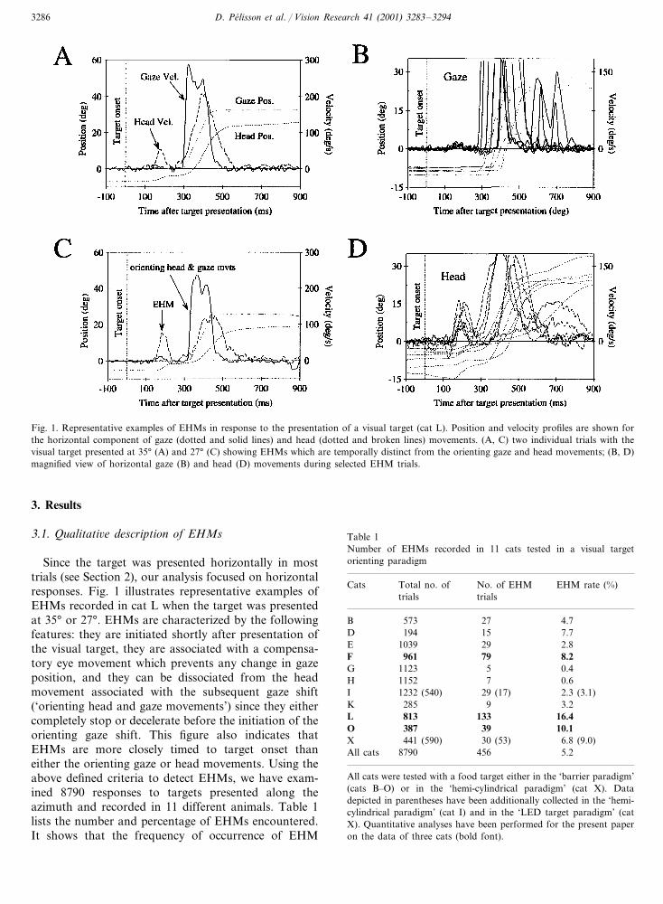
Fig. 1. Representative examples of EHMs in response to the presentation of a visual target (cat L). Position and velocity profiles are shown forthe horizontal component of gaze (dotted and solid lines) and head (dotted and broken lines) movements. (A, C) two individual trials with thevisual target presented at 35° (A) and 27° (C) showing EHMs which are temporally distinct from the orienting gaze and head movements; (B, D)magnified view of horizontal gaze (B) and head (D) movements during selected EHM trials.
3. Results
3.1. Qualitati�e description of EHMs
Since the target was presented horizontally in mosttrials (see Section 2), our analysis focused on horizontalresponses. Fig. 1 illustrates representative examples ofEHMs recorded in cat L when the target was presentedat 35° or 27°. EHMs are characterized by the followingfeatures: they are initiated shortly after presentation ofthe visual target, they are associated with a compensa-tory eye movement which prevents any change in gazeposition, and they can be dissociated from the headmovement associated with the subsequent gaze shift(‘orienting head and gaze movements’) since they eithercompletely stop or decelerate before the initiation of theorienting gaze shift. This figure also indicates thatEHMs are more closely timed to target onset thaneither the orienting gaze or head movements. Using theabove defined criteria to detect EHMs, we have exam-ined 8790 responses to targets presented along theazimuth and recorded in 11 different animals. Table 1lists the number and percentage of EHMs encountered.It shows that the frequency of occurrence of EHM
Table 1Number of EHMs recorded in 11 cats tested in a visual targetorienting paradigm
EHM rate (%)Cats No. of EHMTotal no. oftrialstrials
573 27B 4.77.715D 194
1039 29E 2.8961F 79 8.2
0.45G 11231152H 7 0.61232 (540) 29 (17) 2.3 (3.1)I
285K 9 3.2L 813 133 16.4
39387 10.1OX 6.8 (9.0)441 (590) 30 (53)
8790All cats 456 5.2
All cats were tested with a food target either in the ‘barrier paradigm’(cats B–O) or in the ‘hemi-cylindrical paradigm’ (cat X). Datadepicted in parentheses have been additionally collected in the ‘hemi-cylindrical paradigm’ (cat I) and in the ‘LED target paradigm’ (catX). Quantitative analyses have been performed for the present paperon the data of three cats (bold font).

D. Pelisson et al. / Vision Research 41 (2001) 3283–3294 3287
Fig. 2. Effect of target eccentricity on the rate of occurrence of EHM.Symbols represent different animals and the relationship shown byopen squares and solid lines represent mean data computed on thethree animals. Only cat L has been tested at an eccentricity of 7°.There is no consistent relationship between EHM probability andtarget eccentricity (see regression analysis in text).
varies strongly among animals (range=0.4–16.4%,mean=5.2%). Note that the animal (cat I) who wastested in both ‘food target paradigms’ showed a nearlyequal amount of EHMs in the two conditions. Inaddition, cat X also produced EHMs when respondingto the presentation of a LED target, with a slightlyhigher rate than in response to a food target. Thus,EHMs produced by the 11 animals when they orientedtoward a suddendly presented food target are not spe-cific to this ‘barrier paradigm’.
In the following text, we quantitatively analyzeEHMs collected in cats F, L and O which showed thehighest rates of EHM occurrence. Some EHMs with avery short latency could in fact be premature headmovements anticipating the target presentation. How-ever, after selection of EHM based on the minimumlatency criteria (see Section 2), we found that the vastmajority of EHMs were in fact driven by the visualtarget since only three were directed in the wrongdirection (1.2%, as compared to 240 movements in thecorrect direction). Only these correctly directed EHMswill be considered in the following analyses.
3.2. Quantitati�e analysis of EHMs
3.2.1. Probability of occurrenceFig. 2 plots the probability of EHM as a function of
target eccentricity. As already mentioned, EHM proba-bility is highest for cat L. In this cat, the probabilitydecreases as target eccentricity increases in the 7–35°range. An opposite trend is observed in cats F and Otested in a 15–35° range of target eccentricity. Overall,a correlation analysis performed across all three catsdoes not reveal any significant relationship (Pearsoncorrelation coefficient r=0.02, P�0.05).
3.2.2. Timing, metrics and dynamicsThe timing of EHM and gaze shift is illustrated in
Fig. 3 in which the onset time of each EHM andsubsequent gaze shift are plotted relative to the time oftarget presentation. Note that some EHMs have a veryshort latency relative to the target presentation, reach-ing the minimum value of 65 ms determined by ourselection criterion (see Section 2). To test whether EHMonset was better timed to target presentation or to gazeshift initiation, we computed the delay from targetonset to EHM onset (EHM latency=133�38 ms) andthe pre-gaze delay, as the period separating EHM onsetfrom gaze shift onset (=183�108 ms). The variabilityof the former is significantly smaller than the variabilityof the latter (variance ratio=0.12), indicating thatEHMs are better timed to the onset of visual targetpresentation than to the onset of the ensuing gaze shift.We further note that EHM latency and pre-gaze delayare affected by the position of the visual target, sinceboth parameters significantly increase as a function of
Fig. 3. Timing of EHM onset and of gaze shift onset (data from catsF, L, O pooled together). (A) the onset of EHM (filled squares) andof gaze shift (open circles) are plotted with respect to the onset oftarget presentation; (B) distribution of the EHM onset (black bars)and of the gaze shift onset (gray bars) relative to target presentation(bin width=25 ms).
D. Pelisson et al. / Vision Research 41 (2001) 3283–32943288
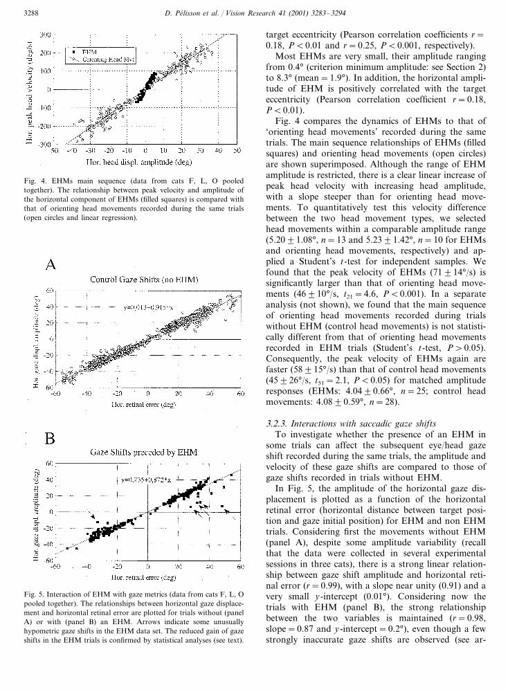
Fig. 4. EHMs main sequence (data from cats F, L, O pooledtogether). The relationship between peak velocity and amplitude ofthe horizontal component of EHMs (filled squares) is compared withthat of orienting head movements recorded during the same trials(open circles and linear regression).
target eccentricity (Pearson correlation coefficients r=0.18, P�0.01 and r=0.25, P�0.001, respectively).
Most EHMs are very small, their amplitude rangingfrom 0.4° (criterion minimum amplitude: see Section 2)to 8.3° (mean=1.9°). In addition, the horizontal ampli-tude of EHM is positively correlated with the targeteccentricity (Pearson correlation coefficient r=0.18,P�0.01).
Fig. 4 compares the dynamics of EHMs to that of‘orienting head movements’ recorded during the sametrials. The main sequence relationships of EHMs (filledsquares) and orienting head movements (open circles)are shown superimposed. Although the range of EHMamplitude is restricted, there is a clear linear increase ofpeak head velocity with increasing head amplitude,with a slope steeper than for orienting head move-ments. To quantitatively test this velocity differencebetween the two head movement types, we selectedhead movements within a comparable amplitude range(5.20�1.08°, n=13 and 5.23�1.42°, n=10 for EHMsand orienting head movements, respectively) and ap-plied a Student’s t-test for independent samples. Wefound that the peak velocity of EHMs (71�14°/s) issignificantly larger than that of orienting head move-ments (46�10°/s, t21=4.6, P�0.001). In a separateanalysis (not shown), we found that the main sequenceof orienting head movements recorded during trialswithout EHM (control head movements) is not statisti-cally different from that of orienting head movementsrecorded in EHM trials (Student’s t-test, P�0.05).Consequently, the peak velocity of EHMs again arefaster (58�15°/s) than that of control head movements(45�26°/s, t51=2.1, P�0.05) for matched amplituderesponses (EHMs: 4.04�0.66°, n=25; control headmovements: 4.08�0.59°, n=28).
3.2.3. Interactions with saccadic gaze shiftsTo investigate whether the presence of an EHM in
some trials can affect the subsequent eye/head gazeshift recorded during the same trials, the amplitude andvelocity of these gaze shifts are compared to those ofgaze shifts recorded in trials without EHM.
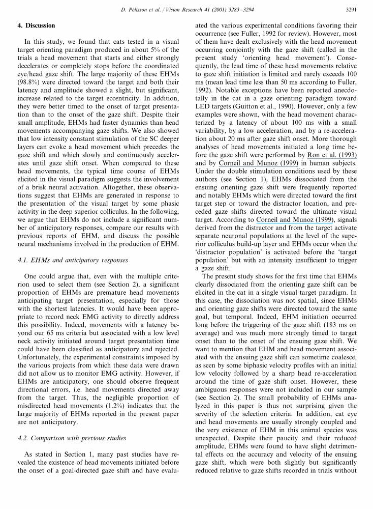
In Fig. 5, the amplitude of the horizontal gaze dis-placement is plotted as a function of the horizontalretinal error (horizontal distance between target posi-tion and gaze initial position) for EHM and non EHMtrials. Considering first the movements without EHM(panel A), despite some amplitude variability (recallthat the data were collected in several experimentalsessions in three cats), there is a strong linear relation-ship between gaze shift amplitude and horizontal reti-nal error (r=0.99), with a slope near unity (0.91) and avery small y-intercept (0.01°). Considering now thetrials with EHM (panel B), the strong relationshipbetween the two variables is maintained (r=0.98,slope=0.87 and y-intercept=0.2°), even though a fewstrongly inaccurate gaze shifts are observed (see ar-
Fig. 5. Interaction of EHM with gaze metrics (data from cats F, L, Opooled together). The relationships between horizontal gaze displace-ment and horizontal retinal error are plotted for trials without (panelA) or with (panel B) an EHM. Arrows indicate some unusuallyhypometric gaze shifts in the EHM data set. The reduced gain of gazeshifts in the EHM trials is confirmed by statistical analyses (see text).

D. Pelisson et al. / Vision Research 41 (2001) 3283–3294 3289
Fig. 6. Interaction of EHM with the dynamics of gaze (panel A) andhead (panel B) orienting movements (data from cats F, L, O pooledtogether). Gaze and head main sequence relationships in EHM trials(filled squares) are compared with the corresponding relationships forcontrol movements recorded in trials without EHM (open circles).Note the reduced gaze peak velocity in EHM trials.
of EHM and the gain of the subsequent gaze shift.Concerning the effect of EHM latency, two subsets ofEHM trials are defined according to the median EHMlatency (133 ms): a ‘long latency EHMs’ group includesall trials with an EHM latency�133 ms and a ‘shortlatency EHMs’ group includes all remaining EHMtrials (EHM latency�133 ms). The comparison be-tween these two groups fails to reveal any statisticallysignificant difference in gaze shift gain (Student’s t-test,t(229)=0.7, P�0.05; 0.90�0.16, n=111 versus 0.89�0.14, n=120 for the ‘long latency EHMs’ group andthe ‘short latency EHMs’ group, respectively). Concern-ing the effect of EHM amplitude, two subsets of EHMtrials are defined according to the median EHM ampli-tude (1.6°): a ‘large amplitude EHM’ group includes alltrials with an EHM�1.6° and a ‘small amplitudeEHM’ group includes all remaining EHM trials (EHMamplitude�1.6°). Similarly to the previous analysis,the comparison between these two groups fails to revealany gaze shift gain difference between the ‘large ampli-tude EHM’ group (0.88�0.17, n=123) and the ‘smallamplitude EHM’ group (0.91�0.12, n=108, Student’st(229)=1.29, P�0.05).
Thus, the presence of an EHM slightly but signifi-cantly interferes with the accuracy of the subsequentorienting gaze shift and this influence does not dependon the latency or on the amplitude of EHM.
Fig. 6A shows the influence of EHM on the dynam-ics of the subsequent gaze shift. The main sequencerelationships of the gaze shifts horizontal componentare plotted for those gaze shifts preceded (EHM group)or not by an EHM (non EHM group). A large overlapin these relationships is observed between the twogroups. However, the peak gaze velocity for the EHMgroup is on average markedly reduced with respect tothe non EHM group. This difference is confirmedstatistically when testing 5° gaze amplitude bins cen-tered on −20° (EHM: −300�47°/s, n=19, nonEHM: −363�77°/s, n=209; Student’s t(226)=3.51,P�0.001) or on 20° (EHM: 281�62°/s, n=23, nonEHM: 354�73, n=201; Student’s t(222)=4.61, P�0.001). Fig. 6B plots the horizontal peak velocity versusamplitude relationship of orienting head movementsrecorded during EHM and non EHM trials. In bothcases, the head main sequence relationships are nearlylinear and the two data sets largely overlap. The com-parison between the two groups for 5° head amplitudebins centered on −20 and 20° (same analysis as de-scribed above for gaze dynamics) reveals a statisticallysignificant difference in peak head velocity for right-ward head movements (EHM: 142�26°/s, n=29, nonEHM: 161�34°/s, n=235; Student’s t(262)=2.78, P�0.01) but not for leftward movements (EHM: −147�31°/s, n=27, non EHM: −155�32, n=231;Student’s t(256)=1.20, P�0.05). In sum, the presenceof an EHM differentially influences the dynamics of the
rows). We have re-computed the regression parametersafter excluding these five highly hypometric movements.The results (r=0.99, slope=0.89 and y-intercept=0.53°) indicate that the reduction of the slope as com-pared to that of the non EHM relationship cannot beaccounted for by these strongly hypometric movementsalone. To further quantify the influence of EHM on theaccuracy of the subsequent gaze shift, we comparedbetween EHM and non EHM trials the horizontal gazedisplacement to horizontal retinal error ratio (gaze shiftgain). To avoid noisy estimates, the analysis was limitedto trials with an horizontal retinal error larger than 8°.The results indicate that gaze shift gain is significantlysmaller in the EHM trials (0.90�0.15, n=231) than inthe non EHM trials (0.93�0.13, n=1835, Student’st(2064)=3.7, P�0.001).
To further test the influence of EHM on the accuracyof the subsequent gaze shift, we now search for anyrelationship between either the latency or the amplitude

D. Pelisson et al. / Vision Research 41 (2001) 3283–32943290
subsequent gaze shift and head movement, with amarked velocity reduction for the former and a slightreduction for the latter.
3.3. EHMs e�oked by SC electrical stimulation
Gaze shifts evoked by electrical stimulation of thedeep layers of the superior colliculus were recorded intwo cats which were previously tested in the visualtarget paradigm (cats L and O, see Table 1). As alreadyreported (Pelisson, Guitton, & Munoz, 1989; Munoz etal., 1991; Pare, Crommelinck, & Guitton, 1994), supra-threshold electrical microstimulation of the SC in head-unrestrained cats evokes short latency gaze shifts with astrong temporal coupling between eye and head move-ments onset. However, we find that weaker electricalmicrostimulation (current intensity less than 2×T and/or pulse frequency less than 300 pps) can elicit headmovements which largely preceed gaze shift. In fact,decreasing the stimulation pulse frequency to 150 pps isassociated with an increase in the latency of both eyeand head movements, but the effect is larger for the eyethan for the head, resulting in an increased probabilityof head leading the gaze (84% at 150 pps versus 70% at
300 pps). Notably, this effect of stimulation frequencydiffers between the two tested animals (cat L: 100versus 68%; cat O: 68 versus 73%). Fig. 7 shows repre-sentative examples of EHMs evoked by SC electricalstimulation in cat L (stimulation parameters: currentintensity 10 �A=2×T, pulse frequency 150 pps andtrain duration 300 ms). The head movement beginsshortly after stimulation onset, whereas gaze shift onsetoccurs later and more variably. A distinctive feature ofthe majority of the electrically-evoked EHMs (95%) isthat their velocity remains somewhat constant or in-creases slightly until gaze shift initiation, after which afast acceleration is observed corresponding to the ‘ori-enting head movement’. This contrasts with EHMs inthe visual paradigm which quickly decelerate and oftencompletely stop before gaze shift initiation, a differencewhich may be related to the nature of the stimulus (seeSection 4). The magnified view (panels B and D) indi-cates that electrically-evoked EHMs, as reported abovefor visually-triggered ones, occur without any de-tectable gaze movement. These examples also empha-size that the synchronization between stimulation onsetand EHM onset is much stronger than that betweenstimulation onset and the onset of either gaze or headorienting movement.
Fig. 7. Representative examples of EHMs evoked by electrical stimulation of the SC deeper layers (cat L). Position and velocity profiles are shownfor the horizontal component of gaze (dotted and solid lines) and head (dotted and broken lines) movements. (A, C) two individual EHMstriggered about 50 ms after stimulation onset and characterized by a slowly increasing velocity. (B, D) magnified view of horizontal gaze (B) andhead (D) profiles during selected collicular stimulation trials with EHM. Note that EHMs are more strongly timed to stimulation onset than dogaze shifts. SC stimulation parameters: current intensity 10 �A, pulse width 0.5 ms, pulse frequency 150 pps and train duration 300 ms.
D. Pelisson et al. / Vision Research 41 (2001) 3283–3294 3291
4. Discussion
In this study, we found that cats tested in a visualtarget orienting paradigm produced in about 5% of thetrials a head movement that starts and either stronglydecelerates or completely stops before the coordinatedeye/head gaze shift. The large majority of these EHMs(98.8%) were directed toward the target and both theirlatency and amplitude showed a slight, but significant,increase related to the target eccentricity. In addition,they were better timed to the onset of target presenta-tion than to the onset of the gaze shift. Despite theirsmall amplitude, EHMs had faster dynamics than headmovements accompanying gaze shifts. We also showedthat low intensity constant stimulation of the SC deeperlayers can evoke a head movement which precedes thegaze shift and which slowly and continuously acceler-ates until gaze shift onset. When compared to thesehead movements, the typical time course of EHMselicited in the visual paradigm suggests the involvementof a brisk neural activation. Altogether, these observa-tions suggest that EHMs are generated in response tothe presentation of the visual target by some phasicactivity in the deep superior colliculus. In the following,we argue that EHMs do not include a significant num-ber of anticipatory responses, compare our results withprevious reports of EHM, and discuss the possibleneural mechanisms involved in the production of EHM.
4.1. EHMs and anticipatory responses
One could argue that, even with the multiple crite-rion used to select them (see Section 2), a significantproportion of EHMs are premature head movementsanticipating target presentation, especially for thosewith the shortest latencies. It would have been appro-priate to record neck EMG activity to directly addressthis possibility. Indeed, movements with a latency be-yond our 65 ms criteria but associated with a low levelneck activity initiated around target presentation timecould have been classified as anticipatory and rejected.Unfortunately, the experimental constraints imposed bythe various projects from which these data were drawndid not allow us to monitor EMG activity. However, ifEHMs are anticipatory, one should observe frequentdirectional errors, i.e. head movements directed awayfrom the target. Thus, the negligible proportion ofmisdirected head movements (1.2%) indicates that thelarge majority of EHMs reported in the present paperare not anticipatory.
4.2. Comparison with pre�ious studies
As stated in Section 1, many past studies have re-vealed the existence of head movements initiated beforethe onset of a goal-directed gaze shift and have evalu-
ated the various experimental conditions favoring theiroccurrence (see Fuller, 1992 for review). However, mostof them have dealt exclusively with the head movementoccurring conjointly with the gaze shift (called in thepresent study ‘orienting head movement’). Conse-quently, the lead time of these head movements relativeto gaze shift initiation is limited and rarely exceeds 100ms (mean lead time less than 50 ms according to Fuller,1992). Notable exceptions have been reported anecdo-tally in the cat in a gaze orienting paradigm towardLED targets (Guitton et al., 1990). However, only a fewexamples were shown, with the head movement charac-terized by a latency of about 100 ms with a smallvariability, by a low acceleration, and by a re-accelera-tion about 20 ms after gaze shift onset. More thoroughanalyses of head movements initiated a long time be-fore the gaze shift were performed by Ron et al. (1993)and by Corneil and Munoz (1999) in human subjects.Under the double stimulation conditions used by theseauthors (see Section 1), EHMs dissociated from theensuing orienting gaze shift were frequently reportedand notably EHMs which were directed toward the firsttarget step or toward the distractor location, and pre-ceded gaze shifts directed toward the ultimate visualtarget. According to Corneil and Munoz (1999), signalsderived from the distractor and from the target activateseparate neuronal populations at the level of the supe-rior colliculus build-up layer and EHMs occur when the‘distractor population’ is activated before the ‘targetpopulation’ but with an intensity insufficient to triggera gaze shift.
The present study shows for the first time that EHMsclearly dissociated from the orienting gaze shift can beelicited in the cat in a single visual target paradigm. Inthis case, the dissociation was not spatial, since EHMsand orienting gaze shifts were directed toward the samegoal, but temporal. Indeed, EHM initiation occurredlong before the triggering of the gaze shift (183 ms onaverage) and was much more strongly timed to targetonset than to the onset of the ensuing gaze shift. Wewant to mention that EHM and head movement associ-ated with the ensuing gaze shift can sometime coalesce,as seen by some biphasic velocity profiles with an initiallow velocity followed by a sharp head re-accelerationaround the time of gaze shift onset. However, theseambiguous responses were not included in our sample(see Section 2). The small probability of EHMs ana-lyzed in this paper is thus not surprising given theseverity of the selection criteria. In addition, cat eyeand head movements are usually strongly coupled andthe very existence of EHM in this animal species wasunexpected. Despite their paucity and their reducedamplitude, EHMs were found to have slight detrimen-tal effects on the accuracy and velocity of the ensuinggaze shift, which were both slightly but significantlyreduced relative to gaze shifts recorded in trials without

D. Pelisson et al. / Vision Research 41 (2001) 3283–32943292
EHM. In addition, the reduction of gaze shift ampli-tude did not depend on the latency or amplitude ofEHM. This suggests that the decrement in gaze shiftperformance cannot be accounted for by a possibleincrease of gaze latency associated with the occurrenceof EHM, or by the slight head deviation which followsEHM, but is more specifically related to the occurrenceof the EHM. One may speculate that the production ofan EHM is associated with some leakage in neuralsignals encoding both the metrics and dynamics of theimpending gaze shift. Unit recording experiments arenecessary to test this hypothesis but in any case, thefunctional relevance of EHMs, if any, is totally uncer-tain. However, analyzing the factors favoring their oc-currence can tell us about their neurophysiologicalsubstrate and about the structure of the gaze shiftsystem.
Since the probability of EHM occurrence varied be-tween animals in a 40-fold range, one such factor isrelated to the experimental subject. Such inter-subjectdifference can be related to the variable propensity touse the head in orienting gaze, as clearly illustrated inhuman subjects (Fuller, 1992). The present study fur-ther suggests that this propensity can in part be relatedto the excitability of the tecto-fugal pathways involvedin the initiation of head movements without triggeringa gaze shift. Indeed, although inferences from only twoanimals may be anecdotal, we found that weak electri-cal microstimulations in the SC evoked head move-ments preceding gaze shifts with a larger probability(100% versus 68%) in the subject who was more proneto produce EHMs in the visual paradigm. The occur-rence of EHM may also depend on experimental fac-tors such as the saliency of the target which has beenshown to affect eye/head coupling in gaze orientingtasks (see for review Fuller, 1992). To test this possibil-ity, we used in two animals a modified food targetparadigm and a LED target with a 200 ms gapparadigm, respectively, in addition to the basicparadigm in which a food target appears at the edge ofan opaque barrier. The results indicate that the numberof EHMs did not strongly depend on the experimentalparadigm. We only note that using a LED instead of apiece of food as a visual target led to slightly morefrequent EHM in cat X. Further experiments are neces-sary to confirm this difference and to test whether thetemporal gap used in the LED task or the motivationalvalue attached to the target could be responsible for it.
4.3. Neural basis of EHMs
The presence of EHM without concurrent gaze dis-placement indicates that some neuronal activity gener-ated in response to the visual target presentation candrive the head motor centers without activating thebrainstem saccadic burst generator. In addition, since
short latency head movements preceeding gaze shiftonset can be produced by low intensity electrical stimu-lation of the SC deeper layers, the origin of EHM couldbe a brief activation of collicular neurons resulting fromthe visual target presentation. It is known that the SCdrives eye and head premotor neurons located in thepontine and medullary reticular formation. Some ofthese neurons are specifically involved in the generationof saccadic eye movements and are gated off by theOPNs during periods of fixation (Sparks & Mays, 1990;Moschovakis et al., 1996) whereas other neurons receiv-ing inputs from the SC (reticulo-spinal neurons) areinvolved in the production of both eye and head move-ments or of head movement alone and are not gated byOPNs (Grantyn, Berthoz, Hardy, & Gourdon, 1992;Robinson, Phillips, & Fuchs, 1994). In addition, at leastin the cat, a substantial fraction of collicular outputneurons project directly to the spinal cord where theycontact the premotor neurons driving head movements(Grantyn & Grantyn, 1982; May & Porter, 1992;Olivier, Chat, & Grantyn, 1991). Thus, through thesedirect and indirect (reticular) pathways which by-passthe brainstem burst generator for saccades, the SC canselectively drive spinal neurons involved in the genera-tion of head movement while the OPNs inhibit theoculomotor circuitry.
This scheme has been previously proposed by Corneiland Munoz (1999) to account for their data recorded inhuman subjects. A prediction of their scheme was thatelectrical stimulation of SC deep layers should be ableto trigger head movements alone when a sub-thresholdintensity regarding the production of saccadic eyemovements is used. Responses reported in the presentpaper are consistent with this prediction, since lowintensity stimulation trains can evoke a head movementmore than 100 ms in advance of the saccade onset.These responses are reminiscent of the EHMs observedin the visual target paradigm except for their velocityprofile. Indeed most electrically-evoked EHMs show aconstantly increasing velocity until the gaze shift wasinitiated, after which the high head acceleration associ-ated with gaze shift initiation was observed. This can berelated to the constant electrical stimulation of the SC.In contrast, visually-triggered EHMs decelerate or evencompletely stop before gaze shift initiation, implyingthe involvement of a short lasting neural drive. Thesedata suggest that the collicular activity involved in theproduction of visually-triggered EHM is a transientactivity in the deep collicular neurons resulting from thetarget presentation. Note that a short lasting activity inthe SC deep layers, either a visual burst or a fusedvisual and motor burst, has been shown to evoke shortlatency saccadic eye movements in monkeys (Edelman& Keller, 1996; Dorris, Pare, & Munoz, 1997; Sparks,Rohrer, & Zhang, 2000). The fact that EHM is notassociated with a concurrent gaze displacement in our
D. Pelisson et al. / Vision Research 41 (2001) 3283–3294 3293
study could indicate that the corresponding SC activityis weak. However, contrary to this hypothesis, we haveshown that EHMs have dynamics even slightly fasterthan regular orienting head movements and have laten-cies down to the shortest visuo-motor delay (about 65ms, see Guitton & Munoz, 1991, and Munoz et al.,1991). Thus an alternative explanation of the lack ofsimultaneous movement of gaze is that, at the time thevisual stimulation leads to an intense but sudden activa-tion of the SC deep layers, the saccadic pulse generatoris still highly inhibited by OPNs. This would preventthe transient SC activation from triggering a saccadicgaze shift but not from driving the head motor systemthrough the by-pass pathways mentioned above.
Acknowledgements
We thank Marcia Riley and Christian Urquizar forwriting some of the display/parameter extraction soft-ware. We also acknowledge Marie-Line Loyalle andAbdelhak Jallane for taking care of the animals. Thisresearch was supported by Institut National de la SanteEt de la Recherche Medicale (INSERM) U94 and U534and by Human Frontier Science Program grant RG58/92B. L. Goffart was supported by a fellowship from theFrench Ministry of Research and Technology and A.Guillaume by the ‘Fondation pour la RechercheMedicale’.
References
Barnes, G. R. (1979). Vestibulo-ocular function during coordinatedhead and eye movements to acquire visual targets. The Journal ofPhysiology, 287, 127–147.
Bartz, A. E. (1966). Eye and head movements in peripheral vision:nature of compensatory eye movements. Science, 152, 1644–1645.
Becker, W., & Jurgens, R. (1992). Gaze saccades to visual targets:does head movement changes the metrics? In A. Berthoz, W.Graf, & P. P. Vidal, The head–neck sensory-motor system (pp.427–433). New York: Oxford University Press.
Biguer, B., Jeannerod, M., & Prablanc, C. (1982). The coordinationof eye, head and arm movements during reaching at a singlevisual target. Experimental Brain Research, 46, 301–304.
Bizzi, E., Kalil, R. E., & Morasso, P. (1972). Two modes of activeeye–head coordination in monkeys. Brain Research, 40, 45–48.
Corneil, B. D., & Munoz, D. P. (1999). Human eye–head gaze shiftsin a distractor task. II. Reduced threshold for initiation of earlyhead movements. Journal of Neurophysiology, 82(3), 1406–1421.
Dorris, M. C., Pare, M., & Munoz, D. P. (1997). Neuronal activity inmonkey superior colliculus related to the initiation of saccadic eyemovements. Journal of Neuroscience, 17(21), 8566–8579.
Edelman, J. A., & Keller, E. L. (1996). Activity of visuomotor burstneurons in the superior colliculus accompanying express saccades.Journal of Neurophysiology, 76(2), 908–926.
Freedman, E. G., & Sparks, D. L. (1997a). Activity of cells in thedeeper layers of the superior colliculus of the rhesus monkey:evidence for a gaze displacement command. Journal of Neuro-physiology, 78, 1669–1690.
Freedman, E. G., & Sparks, D. L. (1997b). Eye–head coordinationduring head-unrestrained gaze shifts in rhesus monkeys. Journalof Neurophysiology, 77, 2328–2348.
Fuller, J. H. (1992). Comparison of head movements strategiesamong mammals. In A. Berthoz, W. Graf, & P. P. Vidal, Thehead–neck sensory-motor system (pp. 101–112). New York: Ox-ford University Press.
Fuller, J. H. (1996). Eye position and target amplitude effects onhuman visual saccadic latencies. Experimental Brain Research,109, 457–466.
Goffart, L., & Pelisson, D. (1997). Changes in initiation of orientinggaze shifts after muscimol inactivation of the caudal fastigialnucleus in the cat. Journal of Physiology, 503(3), 659–673.
Goffart, L., & Pelisson, D. (1998). Orienting gaze shifts duringmuscimol inactivation of caudal fastigial nucleus in the cat. I.Gaze dysmetria. Journal of Neurophysiology, 79, 1942–1958.
Goffart, L., Pelisson, D., & Guillaume, A. (1998). Orienting gazeshifts during muscimol inactivation of caudal fastigial nucleus inthe cat. II. Dynamics and eye–head coupling. Journal of Neuro-physiology, 79, 1959–1976.
Goossens, H. H., & van Opstal, A. J. (1997). Human eye–headcoordination in two dimensions under different sensorimotorconditions. Experimental Brain Research, 114(3), 542–560.
Grantyn, A., Berthoz, A., Hardy, O., & Gourdon, A. (1992). Contri-bution of the reticulo-spinal neurons to the dynamic control ofhead movements: presumed neck bursters. In A. Berthoz, W.Graf, & P. P. Vidal, The head–neck sensory-motor system (pp.318–329). New-York: Oxford University Press.
Grantyn, A., & Grantyn, R. (1982). Axonal patterns and sites oftermination of cat superior colliculus neurons projecting to thetectobulbo-spinal tract. Experimental Brain Research, 46, 243–256.
Guitton, D. (1992). Control of eye–head coordination during gazeshifts. TINS, 15(5), 174–179.
Guitton, D., & Munoz, D. P. (1991). Control of orienting gaze shiftsby the tectoreticulospinal system in the head-free cat. I. Identifica-tion, localization, and effects of behavior on sensory responses.Journal of Neurophysiology, 66, 1605–1623.
Guitton, D., Munoz, D. P., & Galiana, H. L. (1990). Gaze control inthe cat: studies and modelling of the coupling between orientingeye and head movements in different behavioral tasks. Journal ofNeurophysiology, 64, 509–531.
Lefevre, P., & Galiana, H. L. (1992). Dynamic feedback to thesuperior colliculus in a neural network model of the gaze controlsystem. Neural Networks, 5, 871–890.
Masino, T., & Knudsen, E. I. (1993). Orienting head movementsresulting from electrical microstimulation of the brainstem teg-mentum in the barn owl. Journal of Neuroscience, 13(1), 351–370.
May, P. J., & Porter, J. D. (1992). The laminar distribution ofmacaque tectobulbar and tectospinal neurons. Visual Neuro-science, 8, 257–276.
Merrill, E. G., & Ainsworth, A. (1972). Glass-coated platinum-platedtungsten microelectrodes. Medical and Biological Engineering, 10,662–672.
Moschovakis, A. K., Scudder, C. A., & Highstein, S. M. (1996). Themicroscopic anatomy and physiology of the mammalian saccadicsystem. Progress in Neurobiology, 50(2–3), 133–254.
Munoz, D. P., Guitton, D., & Pelisson, D. (1991). Control oforienting gaze shifts by the tectoreticulospinal system in thehead-free cat. III. Spatiotemporal characteristics of phasic motordischarges. Journal of Neurophysiology, 66(5), 1642–1666.
Olivier, E., Chat, M., & Grantyn, A. (1991). Rostrocaudal andlateromedial density distributions of superior colliculus neuronsprojecting in the predorsal bundle and to the spinal cord: aretrograde HRP study in the cat. Experimental Brain Research,87, 268–282.

D. Pelisson et al. / Vision Research 41 (2001) 3283–32943294
Pare, M., Crommelinck, M., & Guitton, D. (1994). Gaze shiftsevoked by stimulation of the superior colliculus in the head-freecat conform to the motor map but also depend on stimulusstrength and fixation activity. Experimental Brain Research, 101,123–139.
Pare, M., & Guitton, D. (1998). Brain stem omnipause neurons andthe control of combined eye–head gaze saccades in the alert cat.Journal of Neurophysiology, 79(6), 3060–3076.
Pelisson, D., Goffart, L., & Guillaume, A. (1998). Contribution of therostral fastigial nucleus to the control of orienting gaze shifts inthe head-unrestrained cat. Journal of Neurophysiology, 80, 1180–1196.
Pelisson, D., Guitton, D., & Munoz, D. P. (1989). Compensatory eyeand head movements generated by the cat following stimulation-induced perturbations in gaze position. Experimental Brain Re-search, 78(3), 654–658.
Phillips, J. O., Ling, L., Fuchs, A. F., Siebold, C., & Plorde, J. J.(1995). Rapid horizontal gaze movement in the monkey. Journalof Neurophysiology, 73, 1632–1652.
Phillips, J. O., Ling, L., & Fuchs, A. F. (1999). Action of the brainstem saccade generator during horizontal gaze shifts. I. Dischargepatterns of omnidirectional pause neurons. Journal of Neurophysi-ology, 81(3), 1284–1295.
Robinson, D. A. (1963). A method of measuring eye movement usinga scleral coil in a magnetic field. IEEE Trans. Bio-Med. Elect., 10,137–145.
Robinson, F. R., Phillips, J. O., & Fuchs, A. F. (1994). Coordinationof gaze shifts in primates: brainstem inputs to neck and extraocu-lar motoneurons pools. The Journal of Comparati�e Neurology,346, 43–62.
Roll, R., Bard, C., & Paillard, J. (1986). Head orienting contributesto the directional accuracy of aiming at distant targets. HumanMo�ement Science, 5, 359–371.
Ron, S., Berthoz, A., & Gur, S. (1993). Saccade-vestibulo-ocularreflex co-operation and eye–head uncoupling during orientationto flashed target. Journal of Physiology, 464, 595–611.
Sparks, D. L. (1999). Conceptual issues related to the role of thesuperior colliculus in the control of gaze. Current Opinion inNeurobiology, 9, 698–707.
Sparks, D. L., & Mays, L. E. (1990). Signal transformations requiredfor the generation of saccadic eye movements. Annual Re�iew ofNeuroscience, 13, 309–336.
Sparks, D., Rohrer, W. H., & Zhang, Y. (2000). The role of thesuperior colliculus in saccade initiation: a study of express sac-cades and the gap effect. Vision Research, 40, 2763–2777.
Uemura, T., Arai, Y., & Shimazaki, C. (1980). Eye–head coordina-tion during lateral gaze in normal subjects. Acta Oto-laryngolog-ica, 90, 191–198.
Urquizar, C., & Pelisson, D. (1992). A non-contact system for2-dimensional trajectory recording. Journal of Neuroscience Meth-ods, 43(1), 77–82.
Volle, M., & Guitton, D. (1993). Human gaze shifts in which headand eyes are not initially aligned. Experimental Brain Research,94, 463–470.
Whittington, D. A., Lestienne, F., & Bizzi, E. (1984). Behavior ofpreoculomotor burst neurons during eye–head coordination. Ex-perimental Brain Research, 55, 215–222.
Zangemeister, W. H., & Stark, L. (1981). Active head rotations andeye–head coordination. In B. Cohen, Vestibular and oculomotorphysiology (pp. 540–559). New York: Ann NY Acad Sci.