Early-Age Orthodontic Treatment - konkur.in
726
www.konkur.in
-
Upload
khangminh22 -
Category
Documents
-
view
0 -
download
0
Transcript of Early-Age Orthodontic Treatment - konkur.in

www.konkur.in

Early-Age Orthodontic Treatment
2
www.konkur.in

EARLY-AGEORTHODONTIC
TREATMENT
Aliakbar Bahreman, DDS, MSClinical Professor
Orthodontic and Pediatric Dentistry ProgramsEastman Institute for Oral Health
University of RochesterRochester, New York
3
www.konkur.in

Library of Congress Cataloging-in-Publication Data
Bahreman, Aliakbar.Early-age orthodontic treatment / Aliakbar Bahreman. p. ; cm.Includes bibliographical references.ISBN 978-0-86715-566-2I. Title.[DNLM: 1. Malocclusion--therapy. 2. Adolescent. 3. Child. 4. Orthodontics, Corrective--methods. WU 440]
617.6’45--dc232013001198
© 2013 Quintessence Publishing Co, Inc
Quintessence Publishing Co, Inc4350 Chandler DriveHanover Park, IL 60133www.quintpub.com
5 4 3 2 1
All rights reserved. This book or any part thereof may not be reproduced, stored in a retrieval system, ortransmitted in any form or by any means, electronic, mechanical, photocopying, or otherwise, without prior writtenpermission of the publisher.
Editor: Leah HuffmanDesign: Ted PeredaProduction: Angelina Sanchez
Printed in China
4
www.konkur.in

ContentsForeword by J. Daniel Subtelny
Preface and Acknowledgments
Introduction
Part IClinical and Biologic Principles of Early-AgeOrthodontic Treatment
1 Rationale for Early-Age Orthodontic Treatment
2 Development of the Dentition and Dental Occlusion
3 Examination, Early Detection, and Treatment Planning
Part IIEarly-Age Orthodontic Treatment of NonskeletalProblems
4 Space Management in the Transitional Dentition
5 Management of Incisor Crowding
6 Management of Deleterious Oral Habits
7 Orthodontic Management of Hypodontia
8 Orthodontic Management of Supernumerary Teeth
9Diagnosis and Management of Abnormal FrenumAttachments
10 Early Detection and Treatment of Eruption Problems
Part IIIEarly-Age Orthodontic Treatment of DentoskeletalProblems
5
www.konkur.in

11 Management of Sagittal Problems (Class II and Class IIIMalocclusions)
12Management of Transverse Problems (PosteriorCrossbites)
13Management of Vertical Problems (Open Bites and DeepBites)
Bonus Cases
6
www.konkur.in

ForewordThis book is a compendium of significant and pertinent information related to early-age orthodontic treatment, a subject that seems to have evolved into one ofconsiderable controversy, with as many orthodontists expressing a negative reactionas a positive reaction to its benefits. Dr Bahreman is a believer in early-ageorthodontic treatment, and he expresses some cogent arguments founded in years ofexperience in practice and teaching to back up his beliefs. In developing his treatise,Dr Bahreman outlines the development of the occlusion and/or malocclusion fromthe embryonic stages, when the foundation of the jaws and thereby the position of thedentition is first established.
Early-age orthodontics is not about the time it takes to orthodontically treat aproblem; it is a story of growth, of variation in anatomy, and of muscle function andinfluences, a realization that it is the jaws that contain the teeth and that where thejaws go, the teeth will have to go, and both undergo varying influences as well asgrow in varying directions. Early-age orthodontics necessitates recognition of thisprocess and aims to alter and redirect it whenever feasible and possible. DrBahreman has undertaken a monumental effort in directing efforts along this path. Anextensive exploration of the literature is an added bonus, as the mechanicalapproaches are based on this literature. In fact, the extensive review of the literatureand its application to diagnosis and varying forms of therapy are worth a veritablefortune.
You may or may not agree with the basic premises, but you will have access toimportant information that will widen your scope of vision and thereby widen yourtreatment horizons. To my mind, an ounce of prevention, if possible, is worth apound of cure. The reality of prevention can exist at the earliest stages ofdevelopment.
7
www.konkur.in

J. Daniel Subtelny, DDS, MS, DDSc(Hon)Professor Emeritus
Interim Chair and Director of Orthodontic ProgramEastman Institute for Oral Health
University of RochesterRochester, New York
8
www.konkur.in

PrefaceAfter obtaining a master’s degree in orthodontics in 1967, I began my career at anewly founded dental school in Tehran. My responsibilities included teaching andadministrative duties at the university and maintenance of a very busy privatepractice. In addition, I established both the orthodontic and pediatric dentistrydepartments at the university.
Many patients were being referred to the orthodontic department, and there wereno qualified faculty members to help me provide care. To rectify the situation, Idesigned an advanced level, comprehensive curriculum in orthodontics forundergraduate students, including classroom instruction, laboratory research, andclinical demonstrations. Once the students completed the course, they could work inthe clinic, thus temporarily solving the issue of the heavy patient load in theorthodontic clinic. With additional staff now available, I could select patients,mostly children in the primary or mixed dentition, for some interceptive treatment.
Despite my difficulties in performing all of the aforementioned duties, thissituation had a fortunate outcome. It helped me to understand and discover theadvantages of early-age orthodontic treatment, which was not common in thoseyears. During my more than 40 years of practice and teaching, especially in earlyorthodontic treatment, I have accumulated a considerable amount of educational datafor teaching purposes. I would like to share this experience and information withreaders.
The public’s growing awareness of and desire for dental services, especially atan early age, have encouraged our profession to treat children earlier. Despite therecommendation by the American Association of Orthodontists that orthodonticscreening begin by the time a child is 7 years old, many orthodontists still do nottreat children prior to the complete eruption of the permanent teeth. I believe that thisinconsistency is due to the educational background of orthodontists as well as a lackof familiarity with recent technical advancements and the various treatment optionsthat are available for young patients.
9
www.konkur.in

The therapeutic devices available for this endeavor are not complex, but decidingwhich ones to use and when to employ them are important steps. As we make thesedecisions, we should also remember not to treat the symptom but rather to treat thecause. My goal is to present the basic information necessary to understand theproblems, to differentiate among various conditions, and to review differenttreatment options. Case reports are examined to facilitate clinical application of thetheory in a rational way.
To understand the morphogenesis of nonskeletal and skeletal occlusal problems,to detect problems early, and to intervene properly, we must look at all areas ofocclusal development, including prenatal, neonatal, and postnatal changes of thedentoskeletal system, and explore all genetic and environmental factors that canaffect occlusion at different stages of development. In other words, we must have aprofound understanding of the fundamental basis and morphogenesis of each problemand then apply this knowledge to clinical practice. Thus, the goals of this book are:
• To provide a comprehensive overview of all areas of dental development, fromtooth formation to permanent occlusion, to refresh the reader’s memory of thefundamentals necessary for diagnosis and treatment planning.
• To emphasize all the important points of the developmental stages that must berecognized during examination of the patient to facilitate differential diagnosis.Each tooth can become anomalous in a number of ways and to different degrees.Occlusion and maxillomandibular relationships can vary in the sagittal,transverse, and vertical directions.
• To discuss the application of basic knowledge to practice by presenting severalcases with different problems and different treatment options.
• To demonstrate the benefits of early-age orthodontic treatment, achieved byintervention in developing malocclusion and guidance of eruption.
Materials are presented in three parts: In Part I, “Clinical and Biologic Principlesof Early-Age Orthodontic Treatment,” three chapters introduce and explain theconcept of early-age treatment, describe its necessity and advantages, and discussthe controversies surrounding this topic; discuss the basic foundation of occlusaldevelopment, empowering the practitioner to detect anomalies and intervene asnecessary; and illustrate the procedures, tools, and techniques available fordiagnosis, emphasizing differential diagnosis and treatment planning for early-agetreatment.
Part II, “Early-Age Orthodontic Treatment of Nonskeletal Problems,” consists of
10
www.konkur.in

seven chapters describing the non-skeletal problems that might develop during theprimary and mixed dentitions. The chapters explain the ontogeny, diagnosis, andearly detection of, and intervention for, these problems. Topics include spacemanagement, crowding, abnormal oral habits, abnormal frenum attachment,hypodontia, supernumerary teeth, and abnormal eruption problems.
Part III, “Early-Age Orthodontic Treatment of Dentoskeletal Problems,” consistsof three chapters on early intervention for the dentoskeletal problems that might ariseduring the primary and mixed dentitions in the three dimensions: sagittal problems(anterior crossbite and Class II and Class III malocclusions); transverse problems(posterior crossbites); and vertical problems (open bites and deep bites).
This book will provide the reader with a firm foundation of the basic science andcase examples with various treatment options. It is my hope that the informationprovided will promote a better understanding of abnormalities and their causes andenable readers to recognize the clues for early detection and intervention.
AcknowledgmentsFirst and foremost, I would like to gratefully acknowledge the valuable opportunitythat was afforded me as a student in Dr Daniel Subtelny’s orthodontic program.Between 1964 and 1967, I completed both my orthodontic specialty and masterdegree programs with Dr Subtelny as my mentor. As chairman and program director,researcher, and mentor, Dr Subtelny has dedicated over 57 years of his life toteaching, personally influencing the lives of over 350 students from around theworld, myself included. In 1999, after over 32 years of teaching, practicing, andadministrating in Tehran, I was fortunate enough to return to the Eastman Institute forOral Health to work alongside Dr Subtelny as a faculty member in the Orthodonticand Pediatric Dentistry Programs.
In addition to Dr Subtelny, there are several individuals to whom I would like toexpress my deep gratitude for their help and encouragement in preparation of thisbook: the late Dr Estepan Alexanian, head of the Department of Histology at theShahid Beheshti University Dental School in Tehran, whose dedication as aneducator and preparation of superb histologic slides is remarkable and who allowedme to use his slides in my publication; Mr Aryan Salimi for scanning some of theslides and radiographs in this book; and Ms Elizabeth Kettle, Program Chair of theDental Section of the Medical Library Association, head of Eastman’s library, forher sincere help in editing this publication.
11
www.konkur.in

Finally, I wish to acknowledge the constant support of my family: Malahat,Nasreen, Saeid, Alireza, Tannaz, and Peymann Motevalei. Especially high gratitudegoes to my wife, Malahat, for her tolerance, support, and encouragements. I alsowant to thank my son Alireza for his technical help and guidance in computer skillsand my granddaughter Tannaz Motevalei for drawing some of the illustrations.
This publication is the product of 17 years spent organizing materials derivedfrom my 45 years of practice and teaching as well as reviewing hundreds of articlesand books. I herewith dedicate this book to the teachers, practitioners, residents, andstudents who are dedicated to treating malocclusion earlier in children, before itbecomes more complicated and costly.
12
www.konkur.in

IntroductionOcclusal development is a long process starting around the sixth week of intrauterinelife and concluding around the age of 20 years. This long developmental process is asequence of events that occur in an orderly and timely fashion under the control ofgenetic and environmental factors. Dental occlusion is an integral part ofcraniofacial structure and coordination of skeletal growth changes. Occlusaldevelopment is essential for establishing a normal and harmonious arrangement ofthe occlusal system.
As we learn about craniofacial growth changes, the potential influences offunction on the developing dentition, and the relationships of basal jawbones andhead structure, we acquire a better understanding of when and how to intervene inthe treatment guidance for each patient. It is more effective to intervene during theprimary or mixed dentition period to reduce or, in some instances, avoid the need formultibanded mechanotherapy at a later age.
Untreated malocclusions can result in a variety of problems, includingsusceptibility to dental caries, periodontal disease, bone loss, temporomandibulardisorders, and undesirable craniofacial growth changes. Moreover, the child’sappearance may be harmed, which can be a social handicap. The benefits ofimproving a child’s appearance at an early age should not be undervalued. The goalsof many clinicians who provide early treatment are not only to reduce the time andcomplexity of comprehensive fixed appliance therapy but also to eliminate or reducethe damage to the dentition and supporting structures that can result from toothirregularity at a later age. In short, early intervention of skeletal and dentalmalocclusions during the primary and mixed dentition stages can enable the greatestpossible control over growth changes and occlusal development, improving thefunction, esthetics, and psychologic well-being of children.
For many decades, orthodontists have debated about the best age for children tostart orthodontic treatment. While we agree on the results of high-quality orthodontictreatment, we often differ in our opinions as to how and when to treat the patient.
13
www.konkur.in

Some practitioners contend that starting treatment in the primary dentition is the mosteffective means of orthodontic care. Others prefer to begin the treatment in the mixeddentition. There is also controversy about whether the early, middle, or late mixeddentition is preferable.
Despite the fact that the American Association of Orthodontists recommends thatorthodontic screening be started by the age of 7 years, many orthodontists do nottreat children prior to the eruption of permanent teeth, and some postpone thetreatment until the full permanent dentition has erupted, at approximately 12 years.The controversy surrounding early versus late treatment is often confusing to thedental community; therefore, clinicians must decide on a case-by-case basis when toprovide orthodontic treatment. Indeed, there are occasions when delaying treatmentuntil a later age may be advisable.
The long-term benefits of early treatment are also controversial. The majority ofdebates seem to revolve around early or late treatment of Class II malocclusions.There is less controversy regarding many other services that can be performed forthe benefit of young patients during the primary or mixed dentition, such as treatmentof anterior and posterior crossbite, habit control, elimination of crowding, spacemanagement, and management of eruption problems.
Practitioners who are in favor of early treatment of Class II problems contend thatearly intervention is the best choice for growth modification when the problem isskeletal and especially when it results from mandibular retrusion. On the other hand,opponents believe that there is no difference in the final result and that a single-phase treatment approach is preferable because of the advantages that accompany thereduced treatment time.
Unfortunately, some practitioners, without a profound evaluation of the indicationsfor early treatment, conclude that late treatment is always preferable. However,broad conclusions drawn from narrowly focused research can be misleading. Onecannot conclude that no birds can fly by considering the flight characteristics of theostrich.
To evaluate and demonstrate the benefits of early treatment, I aim to discuss andclarify available treatments and services and discuss cases with different problemsand different treatment options. An understanding of all aspects of early treatmentrequires a thorough knowledge of the basics of embryology, physiology, and growthand development. This includes development of the dentition, tooth formation,eruption, exfoliation, and all transitional changes. Therefore, my other goal is tointegrate the basic science and the clinical, in order to refresh the reader’s memoryon important points about the bases of nonskeletal and skeletal problems that can
14
www.konkur.in

arise during the transitional stages of occlusion.Each patient who enters our practice represents a new chapter and a new lesson
that we can learn from. A thorough knowledge of the basis for early-age orthodontictreatment, an understanding of the proper treatment techniques, and a willingness toconsider their appropriateness for each individual patient will allow us to intervenein ways that will provide the maximum benefit for a young and growing child.
15
www.konkur.in

16
www.konkur.in

Rationale for Early-Age OrthodonticTreatment
In the past, orthodontic treatment has been focused mainly on juvenile and adulttreatment. Treatment options for patients in these age groups often are limited bycomplex dental and orthodontic problems and the lack of sufficient futurecraniofacial growth.
During the later part of the 18th century, orthodontic treatment of Class IImalocclusion was limited primarily to retraction of the maxillary anterior teeth todecrease excessive overjet. In 1880, Norman Kingsley1 published a description oftechniques for addressing protrusion. He was among the first to use extraoral forceto retract the maxillary anterior teeth after extraction of the maxillary first premolars;the extraoral force was applied with headgear. Later, Case2 continued to refine thesemethods.
Angle’s classification3 of malocclusion, published in the 1890s, provided asimple definition of normal occlusion and was an important step in the developmentof orthodontic treatment. Angle opposed the extraction of teeth and favored thepreservation of the full dentition. His position against tooth extraction led him todepend on extraoral force for the expansion of crowded dental arches and retractionof the anterior segment. Later he discontinued the use of extraoral force andadvocated the use of intraoral elastics to treat sagittal jaw discrepancies.
Because of Angle’s dominating belief that treatment with Class II elastics was justas effective as extraoral force, the use of headgear was abandoned by the 1920s.Then, in 1936, Oppenheim4 reintroduced the concept of extraoral anchorage,employing extraoral traction to treat maxillary protrusion. Accepting the position ofthe mandible in Class II malocclusions, Oppenheim attempted to move the maxillary
17
www.konkur.in

dentition distally by employing a combination of occipital anchorage and an E-arch,allowing the mandible to continue its growth. This resulted in an improvedrelationship with the opposing jaw. In 1947, Silas Kloehn5 reintroduced extraoralforce, in the form of cervical headgear, for the treatment of skeletal Class IIrelationships.
In 1944, another student of Angle’s, Charles Tweed, 6 was discouraged by theprevalence of relapse in many of his patients treated without extraction, so hedecided to oppose the conventional wisdom of nonextraction.
In the early part of the 20th century, there was optimism about the influence oforthopedic force on skeletal growth. An almost universal belief was that orthodonticforces, if applied to the growing face, could alter the morphologic outcome.
In the United States, headgear was the principal appliance used for facialorthopedic treatment, whereas in Europe the functional appliance was predominantlyused.
In 1941, Alan Brodie,7 one of Angle’s students, concluded that the growing facecould not be significantly altered from its genetically predetermined form and thatthe only option for the orthodontist in cases of skeletal malocclusion would be dentalcamouflage, or the movement of teeth within their jaws. This idea led to toothextraction.
In 1931, Broadbent8 introduced the standardized lateral cephalometric radiographas a tool for longitudinal evaluation of growth and developmental changes. Thisradiograph was significant because of its ability to demonstrate treatment results.Kloehn5 used this technique to evaluate dentoskeletal changes after the application ofextraoral forces and found that these forces could produce positive skeletal changesas well as dentoalveolar changes in the correction of skeletal Class II problems.
In the late 19th and early 20th centuries, interceptive orthopedic procedures werenot common. Intraoral and extraoral devices such as face masks, functionalappliances, and mini-implants were not as well known and accessible as they aretoday. At that time, the treatment focus was on alignment of irregularities after thecomplete development of malocclusions in older juveniles and adults. However,early orthodontic treatment is now more generally accepted as a means of gaining thegreatest possible control over form and function and producing superiordentoskeletal changes over time.
The more that practitioners learn about growth and its potential, the influence offunction on the developing dentition, the relationship of basal jawbones and headstructure, and the influence of neuromuscular activity and functional balance, the
18
www.konkur.in

more they can apply these processes in clinical practice. With a thoroughunderstanding of the biologic facts and the availability of new intraoral and extraoraldevices and techniques, clinicians can acquire a better understanding of when andhow to apply early intervention for prevention of the problems and control futureadverse effect on the patient’s growth pattern.
What Is Early-Age Orthodontic Treatment?The term early-age orthodontic treatment encompasses all interventions andtreatments that can be performed during the primary or mixed dentition, with thepurpose of eliminating or minimizing dentoalveolar and skeletal disharmonies thatcan interfere with the normal growth and development of occlusion, function,esthetics, and the psychologic well-being of children. In other words, the mainobjective of this type of intervention is to prepare an environment that will enhanceocclusal development.
General objectivesThe following are the major goals of treatment started during the primary or mixeddentition:
• Enhancement of the normal dental and skeletal development• Elimination or control of any environmental factors that might disturb normal
occlusal development• Preparation of the optimal environment for normal occlusal development• Correction or guidance of a developing malocclusion to normal occlusion• Elimination of the need for or reduction in the duration of second-phase treatment• Maximization of growth potential for growth modification treatment
General strategyThe words prevention and interception have been used in many types of earlytreatment but are sometimes misleading. Neither of these terms can possibly includeall areas of early-age orthodontic treatment. To eliminate these misunderstandings,the terms preventive, interceptive, and corrective must be explained:
19
www.konkur.in

• Preventive orthodontic treatment includes treatments that prevent thedevelopment of a malocclusion before it happens, such as space maintenance orhabit control.
• Interceptive orthodontic treatment includes all types of treatment that can be usedduring the development of a malocclusion to guide the developing abnormalitytoward normality and to prevent further damage to the occlusion; these methodsinclude space regaining, palatal expansion, correction of posterior and anteriordental crossbites, maintenance of leeway space in cases of moderate crowding,and management of severe arch size–tooth size discrepancy.
• Corrective orthodontic treatment is performed after a malocclusion is completelydeveloped.
Early-age orthodontic treatment can include preventive, interceptive, orcorrective treatment as well as combinations of all three types applied during theprimary or mixed dentition period, before the complete development of amalocclusion. This timetable is necessary to take advantage of growth potential atthe right time for skeletal problems by initiating treatment such as headgear or facemask therapy.
In 1975, Popovich and Thompson9 evaluated preventive and interceptiveorthodontic treatment in patients between 3 and 18 years of age. They concluded thatfew malocclusions can be prevented but that about 25% can be intercepted.
Why Is Early Orthodontic Intervention Recommended?Despite progress in the understanding of growth and development and thephysiology, morphology, and ontogeny of dentoskeletal anomalies, as well as theemergence of sophisticated diagnostic techniques, many practitioners do not knowhow to manage the problems they diagnose or when the right time is to refer theirpatients to a specialist for intervention.
One-phase versus two-phase orthodontic treatmentThere are two schools of thought in regard to orthodontic treatment strategy. Onephilosophy advocates beginning treatment when the second molars and all premolarshave erupted. These clinicians believe that it is easier to design the treatment planand begin treatment when major growth has been completed. This approach avoids
20
www.konkur.in

the need to compensate for variations that might occur during or after treatment as aresult of unexpected and unpredictable variations in growth patterns. Usually thistype of procedure requires comprehensive appliance therapy. This can be time-consuming and pose serious risks to the health of the teeth and surrounding tissuestructures. Some conditions are more prone to relapse and require long-termretention.
The other school of thought advocates early treatment because the development ofthe dental occlusion is a long process, beginning at 6 weeks of intrauterine life andending 20 or more years later. The most important stage of the dental occlusion is thetransitional dentition; hence, most dentoskeletal anomalies start and develop duringthe primary or mixed dentition. Therefore, proponents believe that early detection ofthe problem and proper intervention can guide the abnormality toward normalityand, under some conditions, can prevent or at least reduce the severity of theproblems.
The evidence regarding early versus late orthodontic treatment, a subject that hasbeen debated for years, will be discussed later in the chapter. However, the fact thatimportant occlusal developments and dentoskeletal changes happen duringtransitional dentition and most dentoskeletal anomalies develop during the primaryand mixed dentition prompts the following question: Why should unfavorable dental,skeletal, or soft tissue relationships be allowed to remain for a number of years ifthey can be corrected completely or partially at an earlier age with a minimum ofappliance therapy and treatment effort? Delayed treatment fails to take advantage ofgrowth potential that could eliminate or modify deviations in skeletal growth and thefunctional matrix associated with mouth breathing, atypical swallowing, and otherabnormal behavior. It also fails to take advantage of the opportunity to guidedentoalveolar development. Furthermore, early intervention can also preventdamage to teeth and surrounding structures that may result from a misarticulated andtraumatized dentoskeletal relationship.
The author recommends a single- or two-phase treatment approach that beginsaround the time of the middle or late mixed dentition. There are also conditions thatrequire early intervention during the primary or mixed dentition. All of theseprocedures will be discussed in detail in part 3 of this book and will includedetailed descriptions of cases.
Mechanisms affecting occlusal developmentTo clarify the rationale for early-age treatment, it is necessary to describe some of
21
www.konkur.in

the important mechanisms that can influence occlusal development, such as the longprocess of occlusal development, genetics and environmental factors, form andfunction, and locked occlusions.
Long process of occlusal development
Postnatal development of dentofacial structures spans 18 to 25 years. This longprocess allows practitioners to observe and monitor these changes at different stagesof the dentition. Understanding and memorizing all the events that take place duringdevelopment of the occlusion (see chapter 2) gives clinicians the opportunity todetect and intercept incipient abnormalities at their early stage of development.
During this long process, craniofacial growth is also interacting with thedevelopment of the dentition. Different parts of the skull grow at different rates andmature at different ages. Carlson10 stated that 80% of craniofacial growth iscompleted by the age of 6 to 8 years, while only 50% of midfacial and mandibulargrowth is completed by the age of 8 to 10 years. Thus, a considerable amount ofmidfacial and mandibular growth potential remains during the transitional dentition.Monitoring these important growth changes and their interactions with thetransitional dentition will also provide an opportunity to detect and intervene indeveloping malocclusions.
Genetics and environmental factors
The process of occlusal development and its fundamental principles are under theinfluence of two basic mechanisms: genetic processes and environmental processes,which can act individually or interact with each other.
The relative contributions of genes and environment as an etiologic factor ofmalocclusions have been a matter of controversy in the past century. The previousemphasis on genetics was based on the results of early twin and family studies.These studies revealed that the effects of common environment were not separatedfrom the gene-environment interaction. However, Christian11 conducted anacceptable segregation of genetic from environmental effects.
Research also indicates that genetic mechanisms have more influence onmorphogenesis of the craniofacial structure during embryonic life, whileenvironmental factors influence developing occlusion, especially during the earlypostnatal period. Harris and Johnson,12 distinguishing between skeletal variablesand occlusal, tooth-based variables, evaluated serial assessments of 30 untreated
22
www.konkur.in

siblings from the ages of 4 years (full primary dentition) to 20 years (full permanentdentition). They concluded that while the skeletal variables had high heritability,nearly all of the occlusal variability was acquired.
Therefore, many incipient malocclusions that are under the influence ofenvironmental factors during the primary or mixed dentition could be distinguishedand prevented. Early recognition and interception of these factors can eliminate orreduce the severity of future problems. Examples of these common factors include:
• Early primary tooth loss• Delayed exfoliation or overretained primary teeth• Arch size–tooth size discrepancy• Abnormalities of tooth number (hyperdontia and hypodontia)• Eruption problems (ectopia, transposition, impaction, and ankylosis)• Deleterious oral habits• Craniofacial dysfunctions, including mouth breathing, abnormal swallowing
patterns, and all aspects of cranial posture
Form and function
The orofacial structure is anatomically and functionally one of the most complexregions of the human body. The interplay between form and function is anotherimportant mechanism that affects orofacial structure during development of theocclusion. This mechanism is managed by the surrounding environment, which iscalled the functional matrix.
The effects of muscle dysfunction during the primary or mixed dentition are notself-corrected and become worse if continued in older age. Examples of thisphenomenon are:
• The interplay between perioral musculature and the tongue and the interactionbetween tongue size and tongue volume and craniofacial skeletal growth
• Respiration and the capsular matrix influencing the nasomaxillary complex• Activity of the muscles maintaining the head position, which also can affect
craniofacial growth and occlusion
Locked occlusions
23
www.konkur.in

Some occlusal interferences, such as posterior or anterior crossbites, can have anadverse effect on the normal rate and direction of jaw growth. Unless treated duringearly stages of development, these types of irregularities, also called lockedocclusions, can cause skeletal jaw deformity.
Examples of these malocclusions include the following:
• Collapsed maxillary arch, occurring during the primary or mixed dentition, whichcan prevent normal sagittal and transverse growth and displacement of themaxillary arch (Fig 1-1)
• Class II division 1 impinging bite, which can cause mandibular collapse within themaxilla, affecting normal anterior growth of the mandible (Fig 1-2)
• Unilateral crossbite with a functional shift, which is a common problem in primaryand early mixed dentition and, if not treated at an early age, can affect mandibulargrowth, causing jaw asymmetry (Fig 1-3)
Early detection and intervention of occlusal interference by mechanical unlockingof occlusion will allow normal function and therefore normal development of theocclusal system.
Fig 1-1 Maxillary arch collapse.
Fig 1-2 (a and b) Impinging deep bite and locked mandibular arch.
24
www.konkur.in

Fig 1-3 (a to d) Asymmetric mandibular growth.
Current interest in early-age treatmentOver the past two or three decades, interest in early orthodontic treatment hasincreased. Parents now seek treatment for their children at an earlier age. Thisinterest has been stimulated by the following developments:
• Parents who underwent orthodontic treatment themselves are more likely to beaware of their children’s orthodontic concerns.
• Members of the medical and dental community, including specialists and generalpractitioners, are more aware of current research and the influence of function onthe developing dentition as well as the development of sophisticated appliancesfor treatment.
Scientific evidence has shown that orthodontic treatment is more than just thetreatment of dental misalignment. It is also necessary to understand the cause and themechanism of effects that are influenced by genetic and environmental factors. Forexample, functional activity of the surrounding tissues, such as neuromuscularfunctional balance, habits, and upper airway obstruction, can affect the properdevelopment of occlusion. Therefore, early detection of the problem throughsystematic diagnostic procedures can ensure that proper interceptive and correctivemeasures are instituted at the proper time; in this way, many facial and dentalproblems can be minimized or averted.
Rationale
25
www.konkur.in

When considering the reasons why early treatment is recommended, the followingimportant points should be considered:• Almost all of the occlusal variability that happens during early stages of occlusal
development is acquired rather than inherited.• Most incipient irregularities are preventable and, if neglected, can result in more
complicated deformities.• A considerable amount of midfacial and mandibular growth occurs during the
transitional dentition. This must be considered an advantage of early treatment andgrowth modification.
• It is easier for patients to adapt to a new environment and normal function at earlyages, and therefore the outcome has better stability.
• The orofacial skeletons of young children have high plasticity, and thus occlusalguidance is easier to achieve.
When Is the Best Time to Start Early Intervention?There is no simple answer to the question of timing, because there is no one “besttime” to start early treatment. The answer depends on the nature of the patient’sproblems. Each patient must be evaluated individually, and the treatment plan mustbe designed according to each individual’s problem. The challenge is to select theproper type of intervention at the proper time for each individual case.
As a general rule, clinicians should follow these guidelines when determining thebest time to intervene:
• Orthodontic screening must be started by the age of 4 or 5 years.• Treatment can begin after the permanent first molars and all incisors have erupted.
Occasionally there are situations where even earlier intervention is needed, evenat the primary or early mixed dentition stage. Some examples are locked occlusions,posterior and anterior crossbites (especially accompanied with mandibular shift thatdisturbs normal growth and development of occlusion), or functional problems suchas mouth breathing, abnormal tongue position and function, and other deleteriousoral habits.
After a thorough examination and analysis of diagnostic findings, clinicians shouldask the following questions:
26
www.konkur.in

• Is the problem damaging involved teeth, periodontium, temporomandibular joints,or other tissues?
• Is there a possibility for the problem to worsen?• Is there a possibility of adverse effect on the growth of the other jaw?• Is this situation psychologically problematic for the patient or others?
If the answer to any of these questions is “yes,” then it is time to start earlyintervention.
How Are Early Intervention Procedures Applied?In order to apply the proper treatment at the proper time, it is necessary to look firstat the principles and strategy of early orthodontic intervention (for more detail, seechapter 11).
Goals of early orthodontic treatmentThe strategy and major techniques of early orthodontic intervention include thefollowing important goals:
• Elimination of primary etiologic factors, if possible• Correction of obvious problems• Interception of developing problems• Prevention of worsening of obvious problems• Preparation of an environment for normal occlusal development and function• Guidance of growth in a more favorable direction by unlocking occlusal
interferences, which can have an adverse effect on occlusion• Management of arch size–tooth size discrepancy• Reduction in susceptibility to trauma and incisor fractures (increased overjet)• Correction of skeletal dysplasia at an early stage of development
Phases of early orthodontic treatmentThe controversy surrounding early versus late orthodontic treatment is often
27
www.konkur.in

confusing to the dental community. The literature regarding early orthodontictreatment is often misleading. Some practitioners limit their practice only to thepermanent dentition, and some believe that early treatment always needs a secondphase of treatment, which is not true.
Early orthodontic treatment protocols can be performed in two separate phases.Depending on the type of problem, the age of the patient, and the stage of dentition,this procedure can be accomplished in one phase and sometimes in two phases.Occasionally, a three-phase treatment plan might be implemented if some correctionof the primary dentition is needed, such as elimination of posterior cross-bite, andserial extractions in the mixed dentition must be followed by a final phase oftreatment for the permanent dentition.
Some patients benefit from a single phase of treatment, which usually startsaround the late mixed dentition and ends during the permanent dentition. Otherpatients might be managed better if treatment is initiated during the early or middlemixed dentition, followed by a phase of observation or retention, and then completedduring the permanent dentition with the second phase of treatment. The challenge isto select the proper type of intervention at the proper time for each individualpatient.
During each phase, the main objectives of intervention and treatment includereducing adverse growth, preventing dental and skeletal disharmonies, improving theesthetics of the smile, enhancing the patient’s self-image, and improving theocclusion. First-phase early orthodontic treatments offer many advantages to both thedoctor and the patient. These therapies should be part of every orthodontist’sarmamentarium.
One-phase early orthodontic treatment
To clarify the different types of early intervention used in orthodontic practice andclearly distinguish between the meanings of these terms, it is necessary to explainthem first:
• One-phase early treatment consists of a type of interceptive or correctivetreatment that is performed during the primary or mixed dentition stage toeliminate the cause and to correct the present abnormality.
• One-phase treatment is a single phase of comprehensive treatment for correctionof an abnormality, whether in the primary, mixed, or permanent dentition.
28
www.konkur.in

For example, one-phase early treatment of Class II malocclusion is a single phaseof comprehensive treatment usually started around the end of mixed dentition, justbefore the growth spurt and ending after the completion of canine eruption. Thisusually takes about 2 to 3 years.
For more than 40 years, the author has used this comprehensive, one-phase earlytreatment to correct severe Class II malocclusion (dental or dentoskeletal).Treatment begins around the late mixed dentition and finishes with completion of thepermanent dentition. This method does not require a second phase, which is usuallyexpected in early treatment procedures.
The major goal of late mixed dentition treatment is growth modification to takeadvantage of growth potential during the growth spurt. This type of treatmentinvolves orthopedic management such as extraoral traction, functional therapy, andrapid maxillary expansion.
Some abnormalities may require only one phase of treatment for the correction ofthe present malocclusion, even at the early or middle mixed dentition stage; forexample, regaining space, space maintenance, guidance of eruption, and correctionof posterior or anterior dental crossbite may not need second-phase treatment.
Two-phase early orthodontic treatment
This type of early treatment involves an initial intervention during the primary, early,or middle mixed dentition stage, followed by an interim or resting stage, which is atransitional period. During this period, the patient is under observation or may bewearing some type of simple retainer (full- or part-time). After the transition tocomplete permanent dentition, the second phase of comprehensive fixed appliancetherapy is performed for final adjustment.
Two-phase treatment is advocated to address skeletal, dental, and neuromuscularproblems such as abnormal habits, hyperactive musculature, crowding, dental cross-bites, hypodontia, supernumerary teeth, and problems of tooth eruption in order toeliminate or reduce the severity of their effects and to facilitate the second phase oftreatment. The treatment will be completed by a second phase of comprehensivefixed appliance therapy. The severity of the problem will have been considerablydiminished during phase 1 treatment, and a short phase of treatment will be requiredto align the permanent occlusion.
An example of two-phase treatment is serial extraction, where preparation ofanchorage, extractions, and guidance of canine eruption are performed in phase 1.After canine eruption and completion of the permanent dentition, the second phase
29
www.konkur.in

begins, with full bonding of appliances for uprighting of teeth, correction of rotation,and minor space closure to finalize the treatment.
Another example of two-phase treatment is early intervention and control ofabnormal habits, which might be started during the primary or mixed dentition andfollowed by an interim phase of supervision during the transitional dentition, untileruption of the permanent dentition for phase 2 treatment.
Many other abnormalities, such as hypodontia and hyperdontia, anterior andposterior crossbite, eruption problems (impaction and transposition ankylosis),abnormal oral habits, and abnormal frenum attachment, require early intervention asa phase 1 treatment during the primary or mixed dentition and a second phase duringthe permanent dentition.
Occasionally, there are cases that have been planned for two-phase treatment but,after completion of phase 1 and enhancement of the conditions to achieve a normalenvironment, ultimately do not require a second phase of treatment.
Monitoring the dentitionDepending on the type of malocclusion, the chronologic and skeletal ages of thepatient, and the stage of dentition, there are various situations that justify earlydetection and intervention. The importance of managing the developing dentition andocclusion and its effect on the well-being of infants, children, and adolescents isclear. This management includes the recognition, diagnosis, and appropriatetreatment of dentofacial abnormalities. Comprehensive clinical examinations,appropriate pretreatment records, differential diagnosis, sequential treatmentplanning, and progress records are necessary to manage any condition affecting thedeveloping dentition.
Systematic monitoring of the developing dentition at regular clinical examinationsand periodic radiographic evaluation, from the beginning of the mixed dentition tocomplete eruption of the permanent dentition, can expose many developmentalproblems and facilitate early intervention. For more than 40 years, the author hasconsistently advised all undergraduate and postgraduate students that taking serialpanoramic radiographs of patients at ages 6, 8, and 10 years is a necessary step inevaluating and monitoring the transitional dentition. Comparison of these serialradiographs taken at the early stages of occlusal development can guide treatmentand aid early recognition of many ongoing problems throughout the developingdentition. Early detection also allows clinicians to inform the patient’s parents and
30
www.konkur.in

plan and recommend the appropriate intervention.For more detail, see the longitudinal panoramic radiograph monitoring technique
in chapter 3.
What Is Controversial About Early Treatment?Untreated malocclusions can result in a variety of problems, such as susceptibility todental caries, periodontal disease, bone loss, and temporomandibular jointproblems. The most significant detrimental effect can be on the appearance of thepatient; studies by Shaw et al13,14 have confirmed that severe malocclusion is likelyto be a social handicap. Facial esthetics have also been found to be a significantdeterminant of self- and social perceptions and attributes. Tung and Kiyak15 andKilpeläinen et al16 concluded that perceptions of facial esthetics influencepsychologic development from early childhood to adulthood. The goal of manyclinicians who provide early treatment is not only to reduce the time and complexityof fixed appliance therapy but also to eliminate or reduce the damage to occlusionthat can be produced if treatment is postponed.
The majority of debates surrounding the long-term benefits of early orthodontictreatment center around early or late treatment of Class II malocclusion. Some claimthat early intervention in Class II situations is the best choice when the problem isskeletal and especially if the problem is the result of mandibular retrusion. Othersbelieve that there is no difference in the final result and a single-phase treatmentapproach is preferable because of the advantages that accompany the reducedtreatment time. The questions related to early treatment have led to the need forcritical analyses of the effectiveness of such an approach.
Clinical evidenceThe relative merits of the early and late approaches to orthodontic treatment havebeen debated for years. Randomized clinical trials conducted by Ghafari et al,17
Keeling et al,18 and Tulloch et al19 were specifically designed to address importantissues in Class II treatment. All three studies concluded that, for children withmoderate to severe Class II problems, treatment seems to be as effective in latechildhood as it is at an earlier age. On the basis of these three ongoing clinical trials,these authors concluded that both the single- and two-phase approaches are effective
31
www.konkur.in

in the correction of Class II malocclusion and that this correction is the result of bothskeletal and dental changes. In a review of the literature, Kluemper et al20 similarlyasserted that both the single- and two-phase approaches are effective in correctingClass II malocclusion. Moreover, depending on the type of appliances andtherapeutic techniques used, the treatments seem to exert more influence on skeletalchanges in one jaw than in the other.
All of the above studies advocate that both one- and two-phase approaches areeffective in Class II correction, but critical evaluation of these studies reveals thatthe following questions must be clarified:
• Was the type of Class II malocclusion (dental, skeletal, or combination)determined for all subjects?
• What criteria were used for selecting the subjects (the amount of overjet or pointA–nasion–point B angle cannot show the whole detail)?
• Was the skeletal discrepancy caused by the mandible, the maxilla, or acombination?
• Was the variability in skeletal growth patterns considered?• Was skeletal age (growth spurt and growth maturation) considered in each
individual case?• Were variations in patient compliance considered in the analysis?• Was early treatment managed properly?• Were the interim phase and follow-up managed properly?
These are conditions that can influence the results of any investigation comparingthe results of early and late treatments. Many experienced teachers and clinicians,including Ricketts,21–23 Subtelny,24 Gugino and Dus,25 Bench et al,26 Graber,27
McNamara and Brudon,28 and Dugoni29 have compared the two approaches andconcluded that there are many disadvantages to waiting for the second molars toerupt. In a survey of 159 diplomates of the American Board of Orthodontics,30
participants were asked what they perceived to be the benefits of early treatment.The most common responses were:
• Greater ability to modify skeletal growth• Improved patient self-esteem and parental satisfaction• Better and more stable results
32
www.konkur.in

• Less extensive therapy required later• Reduced potential for iatrogenic tooth damage such as trauma, root resorption, and
decalcification
A review of all the literature related to the controversy about early versus lateorthodontic treatment reveals that the major differences center around early Class IItreatment. There does not seem to be as much controversy about the benefits of otherservices that can be performed for young patients during the primary or mixeddentition, such as anterior and posterior crossbite correction, habit control, andcrowding and space management. Unfortunately, many practitioners, withoutattempting a thorough evaluation of the many services that can benefit young patients,extend the controversy surrounding Class II treatment to all kinds of early treatmentand thus prefer late treatment.
First-phase, or early, orthodontic treatment offers many advantages to both thedoctor and the patient, and these therapies should be a part of every orthodontist’sarmamentarium.
Misconceptions about early treatmentThe following are the most controversial claims expressed by opponents of theearly-age treatment approach, together with some counterpoints:
Claim 1. Most patients who undergo early treatment require a second phase oftreatment.
Counterpoint 1. Some patients do need a second phase of treatment but not mostpatients. It is also true that, when needed, the second phase of treatment is easier andshorter and produces better skeletal results. Most of the correction has already beenaccomplished when a second phase of treatment is needed.
Claim 2. Two-phase treatment lengthens the treatment time.Counterpoint 2. Some two-phase treatments do require a longer period of time
because of the patient’s age and the need to wait for complete eruption of thepermanent dentition. However, the length of treatment must be measured by chairtime, not calendar time. Most intervals between visits are longer in early treatmentthan they are during comprehensive treatment, and each visit needs considerably lesschair time. For example, changing an archwire requires more time than doesadjusting headgear or a removable appliance.
Claim 3. Improper early treatment can be harmful to the patient’s growth pattern.
33
www.konkur.in

The reasoning goes that, if growth can be directed advantageously in early treatment,it can also be misdirected.
Counterpoint 3. Current knowledge of growth changes and growth patterns can bevery helpful in treatment planning. It is true that in rare cases, growth prediction ismore complex and unpredictable, but the length of treatment and continuousobservation of a growing patient can guide modifications of the treatment plan, ifneeded. As the saying goes, it does no good to drive faster if you are on the wrongroad. You have to have a map and correct driving directions before starting.
Claim 4. Early diagnosis and treatment planning are sometimes more tentative.When growth has terminated, the future of a malocclusion is more visible and thediagnosis is more certain.
Counterpoint 4. It is true that early diagnosis and treatment planning are moretentative, but periodic reassessment of cephalometric radiographs and study casts(as an absolute necessity) aids in this process. Furthermore, it is easier to monitortreatment progress and the developing occlusion, which is a slow process.
What Are the Benefits of the Early Treatment Approach?The types of treatments and services that can be provided to young children duringthe developmental stages of the dentition and skeletal growth are tremendous. Manydental and skeletal anomalies can be prevented or intercepted during the primary ormixed dentition. Some might be treated in one phase and others in two phases, butproper intervention can definitely reduce the duration and complications of second-phase treatment. The following services can be provided to young patients iftreatment is initiated at the proper time:
• Space management (see chapter 4)• Management of incisor crowding (see chapter 5)• Management of deleterious oral habits (see chapter 6)• Orthodontic management of missing teeth (see chapter 7)• Orthodontic management of supernumerary teeth (see chapter 8)• Diagnosis and management of abnormal frenum attachments (see chapter 9)• Early detection and treatment of eruption problems (see chapter 10)• Management of sagittal (see chapter 11), transverse (see chapter 12), and vertical
(see chapter 13) dentoskeletal problems in the primary and early mixed dentition
34
www.konkur.in

stages
Benefits to patients• Improvement of facial appearance and self-esteem. Facial esthetics has been found
to be a significant determinant of self- and social perceptions. Early interventionin and improvement of a patient’s appearance are very important, especially inpreadolescents.
• Easier resolution or interception of developing malocclusion. Any incipientorthodontic problem detected during the transitional period is easier to correctthan it would be after complete formation of the malocclusion. The resultinglonger periods between appointments and shorter chair time of each visit isdesirable for patients and their parents.
• Minimization of severe malocclusions. Early detection and interception willminimize the severity of the problem.
• Correction of functional problems. Some tooth malalignments, such asprematurities and incorrect dental inclinations (anterior and posterior crossbites),can result in mandibular shift, functional discomfort, and structural defects andhave an adverse effect on normal growth patterns. Early correction can eliminatethe patient’s discomfort and prevent many complicated problems that can happenlater.
• Prevention of damage to teeth and dentoskeletal structures. Many irregularities cancause structural damage. Some irregularities, such as severe dental protrusion andoverjet, increase the risk of fracture of the maxillary incisors because of trauma.Early correction of these irregularities would prevent such problems.
• Reduction in the need for extraction. Much of the crowding that develops duringthe mixed dentition can be corrected by space regaining, expansion, or growthmodification. These corrections might not be possible after completion of thegrowth spurt and eruption of the permanent dentition.
• Greater patient compliance. Prior to their teens, most children are enthusiasticabout getting braces and are more comfortable wearing appliances.
• More stable results. A major goal of early intervention is to provide a normalenvironment to enhance the normal development of occlusion. Teeth in youngpatients are more adaptive to the changes of orthodontic tooth movement, and theresults are more stable.
35
www.konkur.in

• Less traumatic, and therefore less painful, treatment procedures. Most earlytreatments do not require massive tooth movement. Hence, less force is applied,there is less pain, and children are more comfortable. Younger children seem tohave a lower resistance to bone and tooth movement and tend to complain less.
• Prevention of psychologic problems, which may occur in some children. Theanterior teeth play an important role in appearances. Irregularities can causepsychologic problems for young children and elicit teasing from classmates. Earlycorrection of defects will give children more confidence and lessen the hardship.
• Lower treatment costs. The total fee for early orthodontic treatment is less thanthat of comprehensive treatment of the permanent dentition because therapy is lessextensive, there is less chair time per visit, and appointment intervals are longer.Also, if a second phase of treatment is needed, it tends to be easier and for ashorter period of time.
Benefits to practitioners• Availability of more treatment options. Because of the patient’s age, the
possibility of guidance of eruption, and the growth potential, more options areavailable in treatment planning, especially because the abnormalities are still indevelopmental stages.
• Better patient compliance. A patient’s compliance in orthodontic treatment is oneof the most important factors in treatment success. Cooperation is much greateramong children aged 7 to 10 years than among older patients. If an adolescent oradult insists on having the appliances removed before the proper results areobtained, the orthodontist might be forced to compromise treatment goals.
• Better use of growth potential. One of the most important benefits of earlytreatment is utilization of the growth potential to correct skeletal abnormalitiesand the potential to modify growth, which is impossible to achieve aftertermination of growth. Proper orthopedic treatment during jaw growth can resultin significant reduction of dentoskeletal deformities in three dimensions. Theseinclude early Class III correction involving overgrowth of the mandible orunderdevelopment of the maxilla, correction of the constricted maxilla for bettermandibular growth, or stimulation of mandibular growth in the retrognathicmandible.
• Reduction in the need for extraction. As mentioned earlier, the availability of moreoptions for regaining lost space or creating new space in crowding situations hasthe potential to reduce the need for tooth extraction.
36
www.konkur.in
• Easier control of habits. Control of serious deleterious oral habits is easier toobtain in younger children. Also, future structural damage to the dentition andalveolarskeletal structure can be prevented. If abnormal habits continue, there ismore potential for damage, and management of the problems will be morecomplicated.
• Better management of problematic growth patterns. Long-term observationfacilitates control of abnormal, complex problems in the growth pattern, forexample, by guiding adjustments to the treatment plan when necessary.
• Less need for mass tooth movement and complex therapy. En masse toothmovements, such as torquing movements, dental compensation, and othercomplicated mechanics, usually are not necessary in early treatment. Earlytreatment also reduces the need for second-phase treatment.
• Shorter treatment time in the second phase. Proper intervention at the proper timereduces the duration of the second phase and significantly reduces the severity ofthe problems treated in phase 2.
• More stable results. Relapse after the completion of treatment is one of the mostdisturbing events to confront an orthodontist. This failure happens more often aftertreatment initiated in the permanent dentition than it does after early-age treatment.Early-age treatments are usually performed during occlusal development,providing a normal environment for the dentition. Most tooth movements in earlytreatment are the result of natural eruption processes caused by guidancetechniques. Therefore, the dentition has more potential to adapt to the changes,making posttreatment relapse less likely.
How Can the Profession Encourage Early-Age Treatment?Traditionally, early orthodontic treatment has not been taught effectively in manyorthodontic specialty programs, for the following reasons:
• Some instructors’ lack of awareness, interest, or experience in early-age treatmenttechniques.
• Management of early orthodontic treatment in some cases may last several years;treatment of these patients cannot fit into a 2-year curriculum. However, it is stillpossible to continue these cases with a new resident and through an organizedsystem of patient transfer. Furthermore, all residents would be exposed todifferent phases of treatment for these types of problems.
37
www.konkur.in

• Some insurance companies do not provide coverage for early-age treatment.
For the benefit of patients and community care and to prevent the high expense ofcomprehensive treatment, the author suggests that the American Association ofOrthodontists place more emphasis on the inclusion of the early treatment approachin all orthodontic specialty programs, including a mandate that it be a required partof the curriculum. It would also be beneficial to provide an introductory course onthis topic to both undergraduate dental students and pediatric dental residents.
Summary
• Early orthodontic treatment includes all types of preventive, interceptive, orcorrective treatments applied during the primary or mixed dentition, beforethe complete development of occlusion.
• Most dentoskeletal malocclusions initiate and develop during thetransitional dentition.
• A considerable amount of midfacial and mandibular growth occurs duringthe transitional dentition.
• Studies indicate that genetic mechanisms have more influence duringembryonic life, while environmental factors influence the developingocclusion, especially during the early postnatal period.
• Many incipient malocclusions, which are under the influence ofenvironmental factors during the primary or mixed dentition, are detectableand preventable.
• Examples of these factors include early primary tooth loss, delayedexfoliation, overretained primary teeth, arch size–tooth size discrepancy,hyperdontia and hypodontia, eruption problems (ectopia, transposition,impaction, and ankylosis), deleterious oral habits, all craniofacialdysfunctions including mouth breathing, abnormal swallowing patterns, andall aspects of cranial posture.
• Why should an unfavorable dental, skeletal, or soft tissue relationship beallowed to remain for a number of years if it can be corrected completely orpartially at an earlier age, with a minimum of appliance therapy andtreatment effort?
• Early orthodontic protocols can be applied during occlusal development andfacial growth period into separate phases. One-phase treatment is an
38
www.konkur.in

interceptive treatment that can be performed during the primary or mixeddentition to correct existing abnormalities, to eliminate or reduce severity ofproblems, and to facilitate the second phase of treatment. Two-phasetreatment consists of an initial intervention followed by an interim orarresting phase; after eruption of the complete permanent dentition, thesecond or final phase of comprehensive therapy is performed.
• The main objectives of early-age treatment are to prepare an environmentfor normal occlusal development, to eliminate or control any environmentalfactor disturbing normal occlusal development, to correct or to guide adeveloping malocclusion to normal occlusion to take advantage of growthpotential to modify skeletal growth, and to eliminate the need for or reducethe duration of second-phase of treatment.
• Early treatment has various benefits for patients and practitioners, such asbetter patient compliance, better final esthetic results due to growthmodification, more stable results, less damage to teeth and supportingstructures, the availability of more treatment options, a better chance toprevent extraction, and better use of growth potential.
References1. Kingsley NW. Treatise on Oral Deformities as a Branch of Mechanical Surgery. New York: Appleton &
Lange, 1880.2. Case C. Dental Orthopedia and Cleft Palate. New York: Les L. Bruder, 1921.3. Angle EH. Treatment of Malocclusion of the Teeth, ed 7. Philadelphia: SS White, 1907.4. Oppenheim A. A possibility for physiologic orthodontic movement. Dent Rec (London) 1945;65:278–280.5. Kloehn S. Guiding alveolar growth and eruption of the teeth to reduce treatment time and produce a more
balanced denture and face. Angle Orthod 1947;17:10–33.6. Tweed CH. Indication for the extraction of teeth in orthodontic procedure. Am J Orthod Oral Surg 1944–
1945;42:22–45.7. Brodie AG. On the growth pattern of the human head from the third month to eighth year of life. Am J Anat
1941;68:209–262.8. Broadbent BH. A new x-ray technique and its application to orthodontia. Angle Orthod 1981;51:93–114.9. Popovich F, Thompson GW. Evaluation of preventive orthodontic treatment between three and eighteen years
of age. In: Cook JR (ed). Transactions of the Third International Orthodontic Congress. St Louis: Mosby,1975:260–281.
10. Carlson DS. Biological rationale for early treatment of dentofacial deformities. Am J Orthod DentofacialOrthop 2002;121:554–558.
11. Christian JC. Testing twin means and estimating genetic variance: Basic methodology for the analysis ofquantitative twin data. Acta Genet Med Gemellol (Roma) 1979;28:35–40.
12. Harris EF, Johnson MG. Heritability of craniometric and occlusal variables: A longitudinal sib analysis. Am JOrthod Dentofacial Orthop 1991;99:258–268.
39
www.konkur.in

13. Shaw WC, Rees G, Dawe M, Charles CR. The influence of dentofacial appearance on the socialattractiveness of young adults. Am J Orthod 1985;87:21–26.
14. Shaw WC. The influence of children’s dentofacial appearance on their social attractiveness as judged bypeers and lay adults. Am J Orthod 1981;79:399–415.
15. Tung AW, Kiyak HK. Psychological influence on timing of orthodontic treatment. Am J Orthod DentofacialOrthop 1998;113:29–39.
16. Kilpeläinen PV, Phillips C, Tulloch JF. Anterior tooth position and motivation for early treatment. AngleOrthod 1993;63:171–174.
17. Ghafari J, Shofer FS, Jacobsson-Hunt U, Markowitz DL, Laster LL. Headgear versus function regulator inthe early treatment of Class II, division 1 malocclusion: A randomized clinical trial. Am J Orthod DentofacialOrthop 1998;113:51–61.
18. Keeling SD, Wheeler TT, King GJ, et al. Anteroposterior skeletal and dental changes after early Class IItreatment with Bionators and headgear. Am J Orthod Dentofacial Orthop 1998;113:40–50.
19. Tulloch JF, Phillips C, Proffit WR. Benefit of early Class II treatment: Progress report of a two-phaserandomized clinical trial. Am J Orthod Dentofacial Orthop 1998;113:62–72.
20. Kluemper T, Beeman C, Hicks P. Early orthodontic treatment: What are the imperatives? J Am Dent Assoc2000;131:613–620.
21. Ricketts RM. Early treatment. 1. J Clin Orthod 1979;13:23–38.22. Ricketts RM. Early treatment. 2. J Clin Orthod 1979;13:115–127.23. Ricketts RM. Early treatment. 3. J Clin Orthod 1979;13:181– 199.24. Subtelny JD. Early Orthodontic Treatment. Chicago: Quintessence, 2000.25. Gugino CF, Dus I. Unlocking orthodontic malocclusions: An interplay between form and function. Semin
Orthod 1998;4:246–255.26. Bench RW, Gugino CF, Hilgers J. Bioprogressive therapy. 11. J Clin Orthod 1978;12:505–521.27. Graber TM. Extraoral force: Facts and fallacies. Am J Orthod 1955;41:490–505.28. McNamara JA, Brudon W. Orthodontic and Orthopedic Treatment in the Mixed Dentition. Ann Arbor, MI:
Needham Press, 1995.29. Dugoni SA. Comprehensive mixed dentition treatment. Am J Orthod Dentofacial Orthop 1998;113:75–84.30. Bishara SE, Justus R, Graber TM. Proceedings of the workshop discussions on early treatment. Am J Orthod
Dentofacial Orthop 1998;113:5–6.
40
www.konkur.in

Development of the Dentition andDental Occlusion
In any difficult situation, the first step toward remediation is to recognize theproblem. The first step in recognizing the problem is to know how the problem hasdeveloped. Thus, the best “cure” is early recognition and proper intervention beforethe problem arises. A review of the prenatal stages of tooth development, especiallythose occurring during the embryonic period, helps to clarify the normalrelationships of adult body structures and the causes of congenital anomalies.
It is not within the scope of this chapter to discuss all aspects of embryology andodontogenesis. There are many reference texts on oral histology, embryology, anddevelopmental anatomy. There are also many contemporary research reports aboutthe molecular level of tooth development. However, this chapter will provide abrief review of the different stages of dentition during prenatal life.
The embryonic period is one of the most critical developmental events, includingweeks 3 to 8 of intrauterine life. During this stage, all major external and internalstructures are established. Most developmental processes are controlled by precisecoordination and interaction of genetic and environmental factors. This controlmechanism guides differentiation and synchronizes tissue interactions, migration ofcells, and controlled proliferation.
Most facial structure is ultimately derived from migration of neural crest cells,which occurs during the embryonic period. Any interference with this migration cancause different craniofacial anomalies. For example, teratogenic agents, such asviruses and some drugs, can produce this interference and trigger the incidence ofcongenital anomalies.
Development of the dentition is also a sequence of events that occurs in an orderlyand timely fashion; it begins during this stage (week 6 of intrauterine life), continues
41
www.konkur.in

as a very long process, and ends after the age of 20 years. There are many normaltransitional changes during this course of development that must be separated fromthe abnormal.
Development of the dentition is an integral part of craniofacial structure. Skeletalgrowth of craniofacial bony structures also interacts with the development of thedentition, which leads to normal interdigitation between the maxillary andmandibular dental arches. Any disruption of these complicated processes may affectthe ultimate occlusal status. Appropriate actions are needed to restore the normalprocesses of occlusal development. Depending on the age of the patient and the stageof dentition, these treatment procedures may be preventive, interceptive, orcorrective.
An understanding of the complex processes of the growth and development of theface and dentition can play an important role in recognition of dentofacialdisharmony and help in diagnosis and treatment planning. The growth and changesthat take place during the development of the dentition are also a basis for clinicalapplication of early-age orthodontic treatment within the complex processes ofocclusal development. An understanding of the structural components can helpclinicians to recognize developing problems at their initial stages and to determinethe proper time of intervention.
Therefore, a thorough grasp of all stages of development of this system is a must forany dental practitioner treating children who are in the developmental stages ofocclusion.
The formation, eruption, and exfoliation of teeth; their changes from a deepposition within the jawbones into the oral cavity; and the concomitant growth ofbones comprise one of the most fascinating processes of biology. This chapterdiscusses different stages of tooth formation and eruption and developmental stagesof occlusion at three different stages:
1. Prenatal stage2. Neonatal stage3. Postnatal stage
Prenatal Development of the DentitionThe embryonic period, weeks 3 to 8 of intrauterine life, is the most critical stage of
42
www.konkur.in

developmental events. Development of the occlusion initiates during week 6 ofintrauterine life and ends after 20 years of age. Neural crest cells begin migrationduring the embryonic period, forming most of the facial structure. Any interferencewith this migration can cause different craniofacial anomalies. By the end of thisperiod, all of the main organ systems have begun to develop.
During the first 2 months of intrauterine life, the ectomesenchyme cells, fromwhich the orofacial structures develop, undergo a complex and coordinated series ofsteps of proliferation and differentiation. At approximately 7 weeks of intrauterinelife, a bandlike ectodermal thickening called the dental lamina develops inmaxillary and mandibular processes. Odontogenesis (tooth development) beginswith budding of the dental lamina and continues into the next week of life.
Massler and Schour1 have divided the life history of the primary dentition, frominitiation of tooth formation until birth and exfoliation, into six major stages: (1)growth, ( 2 ) calcification, ( 3 ) eruption, ( 4 ) attrition, ( 5 ) resorption, and (6)exfoliation.
The growth stage, or tooth formation, consists of the following five subdivisions:
1. Initiation2. Proliferation3. Histodifferentiation4. Morphodifferentiation5. Calcification
Initiation stageAt this stage, the primitive oral cavity, or stomodeum, is lined by epithelium thatconsists of two to three cell layers. These cells cover the embryonic connectivetissue originating from the neural crest. This is known as ectomesenchyme.
Epithelial thickening occurs after 37 days and is the first sign of development ofthe dentition.2
At this stage, certain cells of the basal layer of the oral epithelium proliferate at amore rapid rate than adjacent cells and form a horseshoe-shaped thickened band ofepithelium around the mouth, called the primary epithelial band, which correspondsin position to the future dental arch (Fig 2-1). The primary epithelial band quicklygives rise to two subdivisions, known as the dental lamina and the vestibular
43
www.konkur.in

lamina.
Fig 2-1 Epithelial thickening.
Dental lamina
The dental lamina develops by proliferation of the basal layer of the oral epitheliumat a rate that is faster than that of adjacent cells and produces a thicker regionextending along the entire free margin of the jaw (Figs 2-2 and 2-3).
Fig 2-2 Upper and lower dental lamina.
Fig 2-3 Higher magnification of the dental lamina.
Vestibular lamina
The vestibular lamina is another proliferation of the oral epithelium; it occursoutside of the dental lamina (buccally and labially). As a result of proliferation ofthe vestibular lamina into the ectomesenchyme, the vestibule develops and forms aseparate plate between the cheeks, lips, and developing dental lamina. Thisseparating plate rapidly degenerates and forms a sulcus, or vestibule, between the
44
www.konkur.in

cheek and tooth area (Fig 2-4).
Fig 2-4 Vestibular lamina.
From this point, tooth development proceeds in three stages (bud, cap, and bellstages), and 10 round or ovoid swellings occur in each arch in the position to beoccupied by the primary teeth.
Although these stages are discussed separately here, tooth formation is acontinuous process, and a clear distinction between the transition stages is notpossible.
Bud stage (proliferation stage)Within the dental lamina, a localized proliferative activity leads to the formation oftooth buds as a series of 10 round or ovoid swellings of epithelial ingrowth into theectomesenchyme, corresponding to the position of future primary teeth (Fig 2-5).According to Ten Cate,3 at this time the mitotic index, the labeling index, and thegrowth of the epithelial cells are significantly lower than corresponding indices inthe underlying ectomesenchyme, which suggests that part of the “ingrowth” isachieved by ectomesenchymal upgrowth.
Fig 2-5 Proliferation stage (bud formation).
Within 6 to 8 weeks, 10 primary tooth buds develop from anterior to posterior.
45
www.konkur.in

These proliferating cells contain the entire growth potential of the teeth. Thepermanent tooth germs of the incisors, canines, and premolars develop as a result ofnew extension and further proliferation of the buds of their primary predecessors,within the dental lamina, attached to the dental organ of the primary tooth germ,which leads to the formation of a new cap on the lingual aspect of primary germs(Fig 2-6).
Fig 2-6 Permanent tooth bud formation.
Permanent molars follow a different process, when the jaw growth and boneremodeling increase the body length of the jaw. The dental lamina burrowsposteriorly beneath the epithelium of oral mucosa into the ectomesenchymal tissueand forms the first, second, and third molar germs.
Enamel knot
The enamel knot is a localization of cells on an enamel organ that appear thickenedin the center of the inner enamel epithelium during the late bud stage of developmentat the site of the primary tooth cusps (see Fig 2-9).
According to Ten Cate,3 the function of this structure is not known, but it seemsthat it is involved in determining the initial position of the first cusp of the toothduring crown pattern formation.
Developing abnormalities at the bud stage
During the bud stage, abnormal proliferation, lack of initiation, or hyperactivity ofthe dental lamina can cause different abnormalities in tooth formation:
• Hypodontia: Congenital absence of a tooth can be the result of a lack of initiationor an arrest of the proliferation of dental lamina cells.
• Hyperdontia: Supernumerary teeth are the result of a hyperactivity of the dentallamina and continued bud formation of the enamel organ.
46
www.konkur.in

• Odontoma: During bud formation, and depending on the degree of differentiationfrom the enamel organ, a supernumerary tooth or a dysmorphic tooth tissue calledan odontoma can be produced.
• Cyst formation: Excessive proliferation of cells can produce epithelial rests.When epithelial rest cells are stimulated and become active, they assumesecretory functions, which can result in cyst formation.
• For additional detail on these phenomena, see chapters 7 and 8.
Cap stage (proliferation stage)During this stage, proliferation of the cells continues. As a result of unequal growthand proliferation of different parts of the dental organ, a caplike configurationdevelops, and a shallow invagination appears on the deep surface of the bud. Thisconsists of the ball of condensed ectomesenchymal cells, called the dental papilla,that later form the dentin and the pulp (Fig 2-7). Surrounding the enamel organ, theectomesenchymal cells form the dental follicles (Fig 2-8). The peripheral cells ofthe cap later form the outer and inner enamel epithelium.
Fig 2-7 Early cap stage. A— dental follicle; B—dental papilla; C—enamel organ.
Fig 2-8 Early bell stage. A—enamel organ; B—outer enamel epithelium; C—inner enamelepithelium.
47
www.konkur.in

At this stage, each bud consists of three parts (see Fig 2-8): (1) the enamel organ,derived from ectoderm; (2) the dental follicle, derived from mesenchyme; and (3) thedental papilla, derived from mesenchyme.
Early bell stage (histodifferentiation stage)At this stage, the epithelium continues to invaginate and deepen. The enamel organchanges from a caplike configuration to a bell-shaped form; dentinogenesis andamelogenesis both begin (Fig 2-9). Late in the cap stage, during the transition of thetooth germ from cap to bell, important developmental changes begin. This process iscalled histodifferentiation.
Fig 2-9 Enamel knot (arrow).
The enamel organ undergoes histodifferentiation, and the cells in the center of thedental organ continue to synthesize and secrete glycosaminoglycans into theextracellular space between the epithelial cells. Glycosaminoglycans arehydrophilic and absorb water into the dental organ.
The increasing amount of fluid increases the volume of the extracellularcompartment of the dental organ. This causes the cells to separate while they areconnected with each other by desmosomal contacts. Next, the stretched cells becomestar shaped and are then known as the stellate reticulum (Fig 2-10).
Histodifferentiation is actually the end of the proliferative stage, as the cells losetheir capacity to multiply.
48
www.konkur.in

Fig 2-10 Histodifferentiation. A—stellate re-ticulum; B—outer enamel epithelium; C— stratumintermedium; D—ameloblasts.
Fig 2-11 Histodifferentiation. A—ameloblast; B—enamel; C—dentin; D—cytoplasmic extension;E—odontoblast.
Stratum intermedium
Some epithelial cells between the internal dental epithelium and the stellatereticulum differentiate into a layer called the stratum intermedium. This layer iscomposed of two to three layers of flattened cells. The stratum intermedium givesrise to ameloblasts at a later stage, which produce enamel (Figs 2-10 and 2-11).Although the cells of this layer are histologically distinct from the cells of theinternal dental epithelium, both layers should be considered as a single functionalunit responsible for the formation of enamel.4
The outer surface of the enamel organ consists of simple cubical cells called theexternal enamel epithelium or outer enamel epithelium. Stellate reticulum cells areconnected to each other and to the cells of the external dental epithelium and stratumintermedium by attachment plaques known as desmosomes.
Odontoblasts differentiate under an organizing influence stemming from the cellsof the internal dental epithelium. During this stage, the cells of the dental papilladifferentiate into odontoblasts, and the cells of the internal dental epitheliumdifferentiate into ameloblasts (Figs 2-11 and 2-12; see also Fig 2-6).
49
www.konkur.in

Fig 2-12 Morphodifferentiation. A—ameloblast; B—enamel; C— dentin; D—odontoblast.
Developing abnormalities at the histodifferentiation stage
Disturbances during the histodifferentiation stage of tooth formation causeabnormalities in the differentiation of the formative cells of tooth germs. This resultsin abnormal structures of the dentin or enamel:
• Amelogenesis imperfecta: Amelogenesis imperfecta is a clinical example of thefailure of ameloblasts to differentiate properly to enamel at this stage.
• Dentinogenesis imperfecta: Dentinogenesis imperfecta is a clinical example offailure of the odontoblasts to differentiate properly to dentin during thehistodifferentiation stage of tooth formation.
When the tooth germ is growing rapidly during the early bell stage, cell divisionoccurs throughout the internal dental epithelium. As development continues, celldivision ceases because the cells are beginning to differentiate and assume theireventual function of producing enamel.
Late bell stage (morphodifferentiation stage)In this stage of tooth formation, the formative cells of ameloblasts and odontoblastsare arranged to outline the final form and size of the teeth. Until the completion of thebell stage, all cells of the internal dental epithelium are continually dividing andtooth germs are rapidly growing. At the end of this stage, the overall growth andfinal morphologic pattern of the tooth germ have been established.
The cessation of mitotic division determines the final shape and size of a tooth.This process occurs before matrix deposition.
50
www.konkur.in

When the inner enamel epithelium is arranged, the boundary between it and theodontoblast outlines the future dentinoenamel junction (see Fig 2-12).
Developing abnormalities at the morphodifferentiation stage
At the end of the morphodifferentiation stage, proliferation ends, and the morphologyand size of the tooth germ reach their final form. Disturbances and aberrationsoccurring during the morphodifferentiation stage lead to teeth with abnormal formsand sizes. Some examples of these abnormalities are peg-shaped lateral incisors,mulberry molars, microdontia, and macrodontia.
Formation of the permanent dentition
The permanent teeth (successional teeth) also arise from the dental lamina, but notall permanent tooth formation is alike. Permanent incisors, canines, and premolarsform as a result of extraproliferative activity from the dental organ of the primaryteeth within the dental lamina. This extraproliferative activity leads to the formationof another epithelial cap located on the lingual aspect of the primary tooth germ thatinitiates the formation of the permanent tooth bud (see Fig 2-6).
The tooth germs of the first, second, and third permanent molars, which have noprimary tooth predecessor, originate in a different way. When jaw growth and boneremodeling increase the body length of the jaw, the dental lamina burrowsposteriorly beneath the epithelium of the oral mucosa. This, and the associatedectomesenchymal response, forms the tooth germs of the first, second, and thirdmolars. This backward extension of dental lamina into the ramus area develops fromthe mandibular body, which has a more horizontal position at this age.
Occasionally, insufficient mandibular growth or insufficient resorption of theanterior border of the ramus during remodeling results in a space deficiency that canlead to third molar impaction in the bony ramus of the adult mandible.
Hard tissue formation
Dentin and enamel are two principle hard tissues of the tooth that form during thelate bell stage. Dentin, as the bulk-forming tissue of the tooth, is a specialized hardconnective tissue; dentin formation always precedes enamel formation and marks theonset of the crown stage of tooth development.
Overall growth of the tooth germ continues by cell division of all the cells of the
51
www.konkur.in

internal dental epithelium until the completion of the bell stage (Fig 2-13).
Fig 2-13 Early crown formation.
The first area of dentinogenesis appears at the future cusp tip, or the growthcenter, where mitotic cell division ceases. The internal dental epithelial cellsdifferentiate and assume their role in producing enamel. The occurrence of second,third, and more zones of maturation within the internal dental epithelium lead to thefinal cuspal pattern of the tooth.
Role of the enamel knot in cusp formation. The enamel knot is a localizedthickening of cells that appears in the center of the inner enamel epithelium of theenamel organ during the late bud stage of development. This corresponds to the siteof the primary tooth cusps (see Fig 2-9). Vaahtokari et al 5 concluded that the enamelknot acts as a signaling center, responsible for directing cell proliferation andsubsequent cuspal morphogenesis in the developing enamel organ. The enamel knotis seemingly necessary for morphogenesis of the tooth germ to progress from the budto the cap stage. The ectomesenchyme eventually assumes the dominant role indetermining the final pattern of the cusps and the shape of the teeth.
Dentinogenesis. Dentinogenesis, or dentin formation, in the crown stage is the firstidentifiable feature of tooth development, which starts at the tip of future cusps. Atthis time, mitotic activity ceases, and the ectomesenchymal cells increase rapidly insize and differentiate into odontoblasts. Odontoblasts are the dentin-forming cells.
Odontoblasts start collagen formation and elaborate the organic matrix of dentinby formation of collagen fibers and ground substance. Mineralization takes placefollowing matrix deposition and involves the precipitation of inorganic calciumsalts.
When organic matrix is deposited, the odontoblasts move toward the center of the
52
www.konkur.in

dental papilla, leaving behind a cytoplasmic extension called the odontoblasticprocess. The odontoblastic process is embedded in mineralized dentin tissues, andthe tubular character of dentin is established. Dentin formation proceeds toward theinside of the dental papilla and odontoblastic process and results in the secretion ofhydroxyapatite crystals and mineralization of the matrix (see Fig 2-11).
Amelogenesis. Although dentin must be present for enamel formation, within theenamel organ the preameloblasts differentiate before the odontoblasts and have aninductive influence on the odontoblasts, sending a message that causes them todifferentiate and secrete dentin. Ameloblasts also require the signal of dentinformation to initiate their secretory activities. A message is sent from the newlydifferentiated odontoblasts to the inner enamel epithelium, causing further celldifferentiation and activating secretory ameloblasts to form enamel tissue. Thisprerequisite is an example of the biologic concept termed reciprocal induction.
Enamel formation generally occurs in two stages: (1) the secretory stage and (2)the maturation stage. In the first stage of amelogenesis, the function of theameloblasts is to secrete and release enamel proteins into the surrounding area andproduce an organic matrix against the newly formed dentin surface. Ameloblastsimmediately become mineralized by alkaline phosphatase enzymes and form the firstlayer of enamel tissue, causing a separation of the ameloblastic cells from the dentinand leaving enamel tissue behind (see Fig 2-6).
Initial mineralization of enamel matrix takes place almost simultaneously withorganic matrix production; an unmineralized organic matrix cannot be found.
In the maturation stage, ameloblasts change their function from enamel matrixproduction to mineralization. Mineralization takes place by transferring substancesused in the formation of enamel. In this stage, most of the materials transported byameloblasts are proteins used to complete mineralization.
Mineralization of dental hard tissues (calcificationstage)The production of mineralized tissues involves two major processes, apposition andcalcification.
Apposition
As explained earlier, at the morphodifferentiation stage, enamel and dentin tissues
53
www.konkur.in

are initially secreted by ameloblasts and odontoblasts as a nonvital extracellularsecretion in the form of a tissue matrix. The tissue matrix is deposited layer by layeralong the future dentinoenamel and dentinocemental junctions. The organic matrix forenamel tissue is not collagenous, but the organic matrix for dentin, cementum, andbone is collagenous. The organic matrix for enamel comprises mainly uniqueproteins (mostly amelogenin), reflecting the tissue’s epithelial origin. The organicmatrix in dentin forms a substantial part of the mineralized tissue.
The organic matrix of enamel is removed during the final stage of mineralizationand leaves less than 1% of the weight of organic matrix. Matured enamel organicmatrix is replaced with hydroxyapatite. Thus, enamel is physically different fromdentin and cementum because it is formed from a noncollagenous matrix (mostlyamelogenin) that is almost completely removed after mineralization.
As already mentioned, the formation of dentin always precedes enamel formationand marks the onset of the crown stage of tooth development. At this stage, the dentallamina is disintegrating, so the tooth germ now continues its development separatedfrom the oral epithelium. The crown pattern of the tooth is established by folding ofthe internal dental epithelium. This folding reduces the amount of stellate reticulumover the future cusp tip. Dentin and enamel have begun to form at the crest of thefolded internal dental epithelium. This stage is associated with the formation of thedental hard tissues, commencing at about the 18th week (Fig 2-14).
Fig 2-14 Crown stage. A shows the enamel space that has been lost during slide preparation. B—dentin; C—dental papilla.
Calcification
The second stage of dental hard tissue formation, like other hard tissues, iscalcification or mineralization. This process takes place following the matrixdeposition by precipitation of inorganic calcium salts (primarily in the form ofcalcium hydroxyapatite crystals) within the deposited matrix.
54
www.konkur.in

The mechanism involved in mineralization is not completely understood,especially in the first step, the formation of first crystals. According to Berkovitz etal,4 any tissue could calcify after crystal formation because blood plasma issupersaturated with calcium and phosphate ions, meaning that mineralization is notlimited by the supply of basic ions. An example of this phenomenon is the pathologiccalcification that can occur in soft tissues (eg, muscle and tendon).
The structural unit of calcified tissues is the crystal or crystallite, which has abasic shape in all four calcified tissues (enamel, dentin, cementum, and bone). Thesizes of the crystals within these tissues are all different.
Calcification of cartilage, which is controlled by the cells, is another mechanismthat seems to be occurring in the initial formation of dentin and bone. These cellsform small matrix vesicles that contain calcium and phosphate ions, alkalinephosphatase, and calcium-binding lipids. These vesicles separate from the cells, andconditions within the vesicles permit formation of hydroxyapatite crystals.
Precipitation of small niduses of crystals continues with precipitation of furtherniduses (calcospherites) spreading around the original nidus and increasing its sizeby the addition of concentric lamination. Finally, through approximation and fusion,these individual calcospherites transform into a homogenously mineralized layer oftissue matrix.
After the secretory phase of amelogenesis is completed and the ameloblasts enterthe maturation stage, they perform a functional and structural task, removing proteinsand water from the maturing enamel and allowing them to achieve completemineralization. Once the enamel is completely mineralized, the ameloblasts shrinkfrom columnar to cubical or flattened cells but remain a part of the reduced enamelepithelium that forms a more or less continuous lining over the completed enamel.
Figure 2-14 shows the crown stage of tooth development at a point when hardtissue formation is well advanced.
Developing abnormalities at the mineralization stage
Any systemic disturbance or local trauma that injures the ameloblastic cells duringenamel formation can interrupt or arrest matrix apposition, resulting in enamelhypoplasia. These disturbances include tetracycline deposition, which causesyellow to brown hypoplastic enamel, and dental fluorosis, which results when anindividual receives too much fluoride during tooth development.
Dentin hypoplasia is less common than enamel hypoplasia and occurs only aftersevere systemic disturbances. If the calcification process is disturbed, there is a lack
55
www.konkur.in

of fusion of the calcospherites. These deficiencies are not readily identified inenamel, but they are evident microscopically in dentin and are referred to asinterglobular dentin.
Root formationAfter enamel formation of the crown is complete and the crown is fully formed, rootformation begins, and the inner and outer dental epithelium reaches and joins at therim of the dental organ. This junctional zone is known as the cervical loop (Fig 2-15).
Fig 2-15 Root formation.
As epithelial cells of the external and internal dental epithelium continueproliferation, the cervical loop forms a double layer of cells known as Hertwig’sepithelial root sheath (Fig 2-16).
Fig 2-16 Hertwig’s epithelial root sheath.
This epithelial sheath grows around the dental papilla, between the papilla andthe dental follicle, and covers the entire dental follicle except the basal portion of
56
www.konkur.in

the papilla that encloses the primary apical foramen. Hertwig’s epithelial root sheathis responsible for the shape and number of root formations.
The dental follicle lies external to the root sheath and forms cementum,periodontal ligament (PDL), and alveolar bone. Root development involvesinteractions between the dental follicles and the epithelial root sheath. The onset ofroot development coincides with the axial phase of tooth eruption.
As the inner epithelial cells of the root sheath progressively enclose more andmore of the expanding dental papilla, they initiate the differentiation of odontoblastsfrom cells at the periphery of the dental papilla. These cells eventually form thedentin of the root. In this way, a single-rooted tooth is formed. Multirooted teeth areformed in the same way, and all teeth undergo the same process. It is still not clear,however, how various crown shapes develop.
Molecular level of tooth developmentThe main objective in discussion of the molecular level of tooth development is togive an overview of the more established theories on the molecular regulation oftooth development and to briefly introduce some recent advances.
The prechordal mesoderm, the craniofacial ectoderm, and the neural crest cellsthat develop on the dorsal side of the neural tube play a complex interaction in theformation of all craniofacial structures. The migration of neural crest cells beginscranially and gradually extends caudally, ultimately differentiating into a widevariety of adult structures.
Neural crest cells are derived from the ectoderm, but because of their importancein ectodermal-mesenchymal interaction, they are called the fourth germ layer.Neural crest cells express homeobox genes in the mandibular and maxillary archesfor orofacial development. Homeobox genes are a large group of genes that code fortranscription factors responsible for regulating the expression of downstream targetgenes. A homeobox is a DNA sequence found within genes that are involved in theregulation of patterns of morphogenesis in animals, fungi, and plants.
The different stages of tooth formation and morphologic changes, including bud,cap, and bell stages and terminal differentiation, have been known for severaldecades, but the molecular interactions involved in this process have beenresearched widely only in the past two to three decades. In this process, a series ofinductive and reciprocal signals between the epithelium and mesenchyme determinethe number, growth, morphology, and the ultimate differentiation of craniofacial and
57
www.konkur.in

tooth tissues and organs.Development of the mammalian teeth as a model system has been intensively
studied for epithelial-mesenchymal interactions during organogenesis, and progresshas been made in identifying key molecules involved in this signaling. It is knownthat the morphogenesis and cell differentiation in developing teeth are governed byinteractions between the oral epithelium and neural crest–derived ectomesenchyme.It has also become evident recently that the inductive interactions between epithelialand mesenchymal cells that operate in these processes are the same signalingmolecules involved in the development of most vertebrate organs.
Coin et al6 reported that bone morphogenetic protein 2 (BMP-2), combined withapatite, could induce both morphologic and functional differentiation of ameloblastsusing the in vitro culture system. BMPs are known to be a group of growth factorsand cytokines that have the ability to induce the formation of bone, tooth, andcartilage. BMPs are also available commercially as oral supplements.
Fibroblast growth factors (FGFs), a family of growth factors involved inembryonic development, have been implicated as regulators of mesenchymal geneexpression and cell proliferation during tooth initiation, epithelial foldingmorphogenesis, and the establishment of tooth shape (specifically FGF-4, -8, and -9). Kettunen et al7 analyzed the roles of FGF-3, -7, and -10 in developing mouseteeth. They found that FGF-10 stimulated cell proliferation in the dental epitheliumand that FGF-3 may participate in signaling functions of the enamel knot. Theyconcluded that the expression patterns and interactions of FGFs in dental epitheliumand mesenchyme suggest their involvement in regulatory signaling pathways duringtooth development.
Tompkins8 stated that the FGF, BMP, hedgehog, and wingless protein families andtheir downstream transcription factors have been identified as key players in theepithelial-mesenchymal signaling loops that drive tooth development. He also addedthat recent results suggest that phenotypic proteins of both ameloblasts andodontoblasts, such as amelogenin and dentin matrix protein 2 (DMP-2), may act asthe final instructive signals in cytodifferentiation. Amelogenin is a protein found inthe developing tooth enamel, and it belongs to a family of extracellular matrixproteins that constitute 90% of enamel proteins.
DMP is an extracellular matrix protein that is critical for proper mineralization ofbone and dentin. DMP is primarily present as a nuclear protein in an undifferentiatedosteoblast that regulates the expression of osteoblast-specific genes; duringosteoblast maturation, it becomes phosphorylated and is exported to the extracellular
58
www.konkur.in

matrix, where it coordinates mineralized matrix formation. Mutations in the DMPgene are known to cause autosomal-recessive hypophosphatemia, a disease thatmanifests as rickets and osteomalacia.
Advancements in molecular biology approaches, coupled with mapping of thecomplete human genome sequence, have shown a number of putative disease genesand loci to be associated with the hypodontia and oligodontia phenotypes. Missingmaxillary lateral incisors along with the lack of BMP4 expression were reported byNeubuser et al9 to be the manifestation of odontogenic potential shifts from theepithelium to the dental mesenchyme.
Family studies by Burzynski and Escobar10 have established that incisor andpremolar hypodontia is inherited via an autosomal-dominant gene that demonstratesincomplete penetrance. Expression of the human MSX1 and MSX2 genes in relationto hypodontia, affecting one or a few teeth, has recently been investigated by manyresearchers, including Nieminen et al.11
Formation of tooth-supporting tissuesWhile roots are forming, the supporting tissues of the teeth, such as cementum, PDL,and alveolar bone, are also developing from the dental follicle in the late stages oftooth formation.
The dental follicle (sac) is a condensation of the ectomesenchymal cells thatinitially surrounds the enamel organ and the enclosed dental papilla. ElNesr andAvery2 stated that the dental follicle has an important role in the initiation,formation, and maintenance of the tissues of the tooth root, PDL, cementum, andalveolar bone.
Cementum
Cementum formation, or cementogenesis, occurs late in tooth formation, just beforethe degeneration of the epithelial root sheath and root dentin deposition. It manifestsas a thin, amorphous, structureless, and highly mineralized secretion on the surfaceof the root dentin. ElNesr and Avery2 noted that this secretion is more evident in theapical region of the root and averages some 10 to 20 μm in thickness.
After root formation and proliferation of the epithelial root sheath, somefragmentation occurs. Ectomesenchymal cells of the dental follicle penetratebetween the epithelial fenestrations and become closed to the newly formed dentin
59
www.konkur.in

of the root. The cells differentiate into cementum-forming cells, or cementoblasts.Cementoblasts secrete an organic matrix, consisting of fine collagen fibrils, groundsubstance, osteocalcin, and sialoprotein, that is mineralized later by minerals in theoral fluid (Fig 2-17).
Fig 2-17 Formation of tooth-supporting tissue.
Following mineralization, the cementoblasts move away from the cementum andcollagen deposition continues to lengthen and thicken the bundle fibers. Finally, thesurface fibers join the forming PDLs and become anchored with them.
During cementogenesis, two types of cementum form: (1) acellular cementum and(2) cellular cementum. Acellular cementum forms first, in the coronal two-thirds ofthe root. When the cementoblasts are trapped in lacunae in their own matrixlike bonecells, the cementum is called cellular o r secondary cementum. This cementum ispresent only in the apical third of the root. At this time, the cementoblasts lose theirsecretory activity and become cementocytes. Cellular cementum develops after mostof the tooth formation is complete and after contact is established with a tooth in theopposing arch.
According to Ten Cate,3 cellular cementum is not found in teeth with one root; inpremolars and molars, it is found only in the part of the root closest to the apex andin interradicular areas between multiple roots.
Periodontal ligament
The supporting tissues of the tooth develop during root formation from the dentalfollicle, a fibrocellular layer investing the dental papilla and enamel organ. Thecells and the fiber bundles of the PDL differentiate from the primitive dental sac ordental follicle (Fig 2-18).
60
www.konkur.in

Fig 2-18 Principal collagen fiber bundles of the PDL.
When the root sheath fragments, ectomesenchymal cells of the dental folliclepenetrate between the epithelial root sheath and the newly formed dentin of the root.These cells differentiate into cementum-forming cells (cementoblasts); according toTen Cate,3 some cells from Hertwig’s epithelial root sheath may also transform intocementoblasts. These cells elaborate an organic matrix that becomes mineralizedand in which collagen fiber bundles of the PDL become anchored.
The dental follicle also differentiates the cells and fiber bundles of the PDL.Recent evidence indicates that dental follicle cells also form the bone in which theligament fiber bundles are embedded.3
Fibroblasts of the dental follicle also secrete collagen fibers, which interact withfibers on the surface of adjacent bone and cementum. This interaction between thealveolar socket and the tooth root is believed to help the process of tooth eruption.
The formation and arrangement of different PDL fibers are influenced by the wayteeth occlude with the teeth in the opposing arch. The force and function of occlusioncan lead to the formation of groups of fibers in different orientations, such ashorizontal and oblique fibers. In other words, local environmental factors regulatethe ability of precursor cells for cementoblasts and PDL fibroblasts within the dentalfollicle to function as cementoblasts that form root cementum or as fibroblasts of thePDL, respectively.
Some of these fibers incorporate as Sharpey’s fibers into alveolar bone; others,called cemental fibers, embed in cementum. These two kinds of fibers are linked bya middle zone of fiber called the intermediate plexus.
The formation of the PDL varies between the primary and permanent teeth andalso among various species.
Alveolar bone
61
www.konkur.in

During root and cementum formation, bone is also developing from the dentalfollicle in the adjacent area. During root formation, primary cementum covering newbone is also deposited, and this bone gradually reduces the space between thecementum and crypt wall and diminishes the PDL space.
According to Ten Cate,3 evidence shows that the bone in which the ligament fiberbundles are embedded is also formed by osteoblastic cells that differentiate from thedental follicle. Similar to the primary cementum, collagen fibers are created on thesurface nearest the tooth, and they remain there until they attach to PDLs and aremineralized. Like any other bone, alveolar bone is prone to remodeling throughoutlife, such as modification induced by osteoblastic and osteoclastic activity arisingfrom the force applied during orthodontic treatment.
Dentogingival junction
The connection between the gingiva and tooth structure is called the dentogingivaljunction. Luke12 explained that this junction and the PDL perform unique functions:The dentogingival junction makes a seal wherever a tooth punctures the integument,while the PDL cushions the tooth during mastication, sheltering the delicate cells thatremodel the socket.
A special feature of both tissues is the rapid turnover of their major components.The dentogingival junction has three epithelial types: the gingival, sulcular, andjunctional epithelia. These three types form a mass of epithelial cells, known as theepithelial cuff, between the tooth and the mouth.
Before eruption, tooth crowns are covered by a double layer of epithelial cells;the cells of the inner layer, which is in contact with the enamel, are the ameloblasts,which have completed their formative function and develop hemidesmosomes,secrete a basal lamina, and become firmly attached to the enamel surface. The outerlayer forms from the remaining cells of the dental organ; these two layers of cells arecalled the reduced dental epithelium.
When tooth eruption begins, the connective tissue that supports both layers breaksdown. The remnants of amelo-blasts provide hemidesmosomes, which aredeveloped by the inner layer between the gingival epithelium. Ten Cate3 indicatedthat much about gingival formation is not fully understood, but it is known thathemidesmosomes that form between the gingival epithelium and the tooth areresponsible for the primary epithelial attachment.
The tissues of the dentogingival junction are dynamic rather than static. Evenwhen they are pathologic, they can be reconstituted by repair, and they have a high
62
www.konkur.in

rate of turnover. The cells are capable of movement and of positional change. Itsunique structural and functional adaptation enables the junctional epithelium tocontrol the constant microbiologic challenges.
Role of form and function in occlusal developmentThe phenomenon of “structure related to function” plays a major role in dentofacialstructures, including dental occlusion. The human face is anatomically andfunctionally one of the most complex regions of the human body. Several importantfunctions reside within the face, including the senses of vision, hearing, smell, andtaste. At the same time, the face provides proprioceptive input with respect topressure, temperature, and stereognosis. The oronasal complex is also an importantand complex region of the face, serving vital functions such as mastication,deglutition, speech, and breathing.
Dental occlusion is an important part of the oral cavity. To understand thecomplexity of the development of occlusion, which involves the interaction oforofacial growth and structure, tooth formation, and the influence of functionalactivity, a brief review of mandibular bone growth and development and theirinterrelationship are presented here. Examination of the initial stage of embryologicdevelopment and morphogenesis of the mandibular component can clearly explainthe close relationship and interaction of this phenomenon.
Within the mandibular process of the first branchial arch, Meckel’s cartilageforms; this later becomes atrophied and is replaced by intramembranous ossificationof the mandible. Ossification starts at about the 6th week, laterally to Meckel’scartilage near the mental foramen between the inferior dental nerve and mentalbranch. Ossification spreads anteriorly and posteriorly to form the mandibular baseand ramus. Condylar, coronoid, and symphyseal secondary cartilage is derived frommesenchyme. Tooth buds are located in a bone crypt and are surrounded by alveolarbone. Figure 2-19 shows a sagittal section of an embryo at this age, showing bonedeposition and the relationship of bone, nerve, Meckel’s cartilage, tooth germs, andthe alveolar plate of the mandibular bone.
63
www.konkur.in

Fig 2-19 Jaw ontogenesis and tooth formation. A—mandible; B—maxilla; C—maxillary andmandibular tooth buds; D—alveoli; E— lower lip; F—tongue.
Maxillary bone is also derived from the first branchial arch. The maxilla, unlikethe mandibular bone, has no precursor cartilage, and ossification appears from asingle center on each side at the angle between the infraorbital nerve and its anteriorsuperior dental branch.
Role of craniofacial growth in development of thedentitionDuring development of the dentition, craniofacial and jaw growth are alsointeracting and influencing the final result of occlusion, leading to normalinterdigitation and normal intercuspation. Many difficult orthodontic problems orskeletal malocclusions are the result of maldevelopment, malrelationship, ormalfunctional conditions of the skeletal structure. Simply, it can be said thatwherever the jaw goes or grows, the teeth also must go. Thus, any disruption ordisharmony between these structures can affect the rate, amount, or pattern of growthof each of these different parts. The end result, dental occlusion, can be profoundlyaffected. It can be concluded that, if these problems are recognized and treated early,it might be possible to minimize or even eliminate the consequences of undesirablecraniofacial growth.
On the basis of development, growth, and function, mandibular bone has beendivided into four parts: (1) the body or basal portion, (2) the alveolar portion, (3) themuscular portion, and (4) the condylar process. All of these parts show their owndistinct growth characteristics (Fig 2-20).
64
www.konkur.in

Fig 2-20 Different portions of the mandible: basal bone (yellow), muscular part (red), and alveolarpart (blue).
From this platform, the ideal dental occlusion, which is the accomplishment ofperfect interdigitation between the maxillary and mandibular dental arches, is theresult of a developmental process that consists of three main events: jaw growth,tooth formation, and tooth eruption. All of these processes are under the control ofgenetic, environmental, and functional factors that determine the rate of growth, theamount of growth, and the pattern of growth of the basal bone.
Mandibular bone is not only a major determinant in the masticatory function butalso plays an important role in facial appearance and dental occlusion. Deviantgrowth of the mandible can result in various malocclusions, ranging from anteriorcrossbite to open bite to deep bite to excessive overjet. Asymmetric disturbances inmandibular growth lead to lateral malocclusion and asymmetry in skeletalmorphology.
Growth of the alveolar process
The mandibular alveolar process functions as supporting tissue for the teeth, andthere is a definite interplay between teeth and alveolar bone, as is clearly evident inthe following situations:
• Congenital absence of the teeth accompanied by lack of alveolar bonedevelopment
• Extraction of teeth that results in resorption of the alveolar bone• Tooth ankylosis that leads to a retardation of tooth eruption and slows the growth
of the alveolar bone• Orthodontic tooth movement and bone reaction against orthodontic forces
65
www.konkur.in

Vertical drift and mesial drift are important concepts and play a key role inmaxillary and mandibular morphogenesis. Enlow13 stated that the dentition driftsvertically and horizontally as the maxilla and mandible develop to keep pace withrespect to anatomical position.
Enlow13 also indicated that the periodontal connective tissue membrane (notligament) provides the intramembranous bone remodeling that changes the locationof the alveolar socket and moves the tooth itself. The developmental verticalmovements of the teeth in normal growth are not the same as eruption; the process ofdrift moves the whole tooth and its socket as a unit.
Role of tooth migration
After clinical eruption of a tooth and achievement of occlusal contact, functionalforces of occlusion acting horizontally on the teeth also affect the growth of thealveolar process. Studies by Björk and Skieller14 and Björk15 demonstrated that,depending on the growth pattern of the mandible, there is marked difference in theeruption direction of the teeth. For instance, clockwise rotation of the mandiblechanges the tooth eruption pathway to a more labial direction.
Muscle force and function
The human jaw muscles are essential to mastication, and they play an important partin craniofacial growth; their contraction is controlled by the nervous system.16
The coronoid process is an attachment site for the temporalis muscle. Thisprocess grows, or drifts, upward and backward by bone apposition at its posteriorborder and resorption along the anterior border. Many studies13,17,18 have shown thatthe size and shape of the process are interrelated with the size of the temporalismuscle. They also have found underdevelopment or diminution of the coronoidprocess after removal of the temporalis muscle.
The angular portion of the mandible is also the site of attachment for two majormasticatory muscles, namely, the masseter and medial pterygoid muscles. This anglegrows mainly by periosteal apposition, while in the coronoid process secondarycartilage plays a role, both prenatally and during early postnatal life.
As Björk15 illustrated, there is great variation between different individuals.
Growth of the mandibular condyle
66
www.konkur.in

The mandibular condyle is a major site of mandibular growth that contributes to bothanteroposterior and vertical growth. The vital importance of mandibular growth isalso clear in normal development of occlusion and growth of the face. The maingrowth direction of the condyle is upward, but Björk15 has also shown that thedirection and extent of condylar growth are variable.
Mandibular growth direction is one of the fundamental considerations of any typeof orthodontic treatment, whether early-age or adult treatment. Three factors play animportant role in the regulation of mandibular growth direction: (1) factors lyingoutside the temporomandibular joint, (2) factors inside the temporomandibular joint,and (3) intermediate factors. Some hypotheses in this regard have been proposed; forinstance, the functional matrix concept proposed by Moss18 suggests that themandibular condyle is merely a passive and adaptive component.
Examples of factors lying outside the temporomandibular joint are the effect ofhead posture and mouth breathing on mandibular growth and the effect of muscles onthe direction of mandibular growth. The effect of muscles is not as obvious as theother factors. However, as many investigators have indicated, there is somecorrelation between facial morphology and muscle strength or activity.16,19,20 Theeffect of muscle fiber orientation on morphology of the bone has also been reportedby Takada et al.21 Congenital muscular dystrophy is an example of a pathologiccondition of muscle force and function that influences the growth of the facial bone.
Solow and Kreiborg22 and Houston23 questioned the effect of soft tissue stretchingon dentofacial morphology. They found a high correlation between craniofacialmorphology and head posture. Extension of the head in relation to cervical columnwas found in connection with large anterior and small posterior facial height, smallanteroposterior craniofacial dimension, clockwise mandibular rotation, facialretrognathism, and small nasopharyngeal space.
Houston23 proposed an intermediate hypothesis, where factors both outside andinside the temporomandibular joint interact, resulting in a specific growth pattern.
However, there are examples of factors within the temporomandibular joint thatinfluence the mandibular growth pattern; for example, juvenile rheumatoid arthritisand ankylosis of the mandibular condyle are conditions that cause posterior rotationof the mandible.24–26 This indicates that in the presence of a shallow glenoid fossa,the mandible rotates downward (vertical pattern); in the presence of a deep fossa, anupward and forward growth pattern of the mandible is evident.
These mechanisms and their influence on the ontogeny and morphology of themandibular bone and dental occlusion illustrate the complexity of the phenomena that
67
www.konkur.in

are acting in different areas of the dentoskeletal structure and, either directly orindirectly, affecting the development of occlusion.
Growth of the nasomaxillary complex
The nasomaxillary complex has multidirectional growth potential. There are severalcomponents of sutures in different planes of space with adjacent bony structues,especially near the cranial base. In addition, many neuromuscular activities andfunctions are channeled through the working environment of the oronasal cavity,referred to as the oronasal functional matrix.
Role of the tongue
Normal formation and development of the dental arches also require a normalbalance between the perioral musculature and the tongue. The tongue itself can affectocclusion through its size, function, and position (see chapter 3).
Conclusion
Development of the occlusion is a long and complicated process requiring a normalinteraction of several genetic and environmental factors affecting bone growth,muscle force, and the functional matrix environment. The early resolution of oralmaldevelopmental or malfunctional problems is not only an essential part of earlyorthodontic treatment but also a vital component of treatment stability.
Neonatal Dentition
Gum padsThe maxillary gum pad is horseshoe shaped; the mandibular gum pad is U-shapedand somewhat flattened anteriorly.
Sheldon Friel27 studied the form of gum pads at birth. He noted the presence ofsome divisions of the pad that formed sections corresponding to the underlyingprimary teeth. (The elevation related to the primary canine is well defined bygrooves distal to the crypts of the primary canine in both arches. This is referred toas the gingival groove.) His observation indicates that the section related to the
68
www.konkur.in

primary second molar is not clearly defined at the time of birth and arises by about 5months of age. Friel27 also observed that the sagittal and transverse dimensions ofthe maxillary pad are larger than those of the mandibular pad, causing an overlap ofthe maxillary gum pad over the mandibular gum pad (Fig 2-21). The gum pads arenot in contact when the jaws are at rest.
Fig 2-21 Newborn skull showing the jaws’ basal bone as part of the gum pads.
In a careful study of a group of 709 infants ranging from 1 to 11 days, Sillman28
indicated that the mandibular gum pad is distal to the maxillary gum pad in all cases,on average by 2.7 mm in the male and 2.5 mm in the female. He found that themandibular gum pad progressively moves forward until the primary first molarserupt. The space between the anterior segments of the gum pads varies in form.
In another serial study of occlusion from birth to 3 years, Sillman28 examined agroup of 50 infants and found a limited anteroposterior movement of the mandiblebut no lateral movement. Material was drawn from casts of more than 50 children,followed continuously from birth to 11 years. As the child ages, the dimensions ofthe mandibular gum pad also change, and its distal position is reduced. The lengthand anterior width of the mandibular gum pad increase while the posterior widthdecreases.
The mandible “grows” forward at a greater rate than the maxilla to catch up withthe maxilla in varying magnitude over time. Sillman28 also found that theinterrelationship of the gum pads at birth has no bearing on the futureinterrelationship of the jaws. There is no relationship between the anterior openingsof the gum pads and future open bite tendency.
Leighton,29 in a longitudinal study, examined the records of 109 children and 30pairs of twins to determine the form of the maxillary gum pad at the age of 6 monthsand before the emergence of teeth. Heredity was found to make less contribution tothe form of the gum pad at 6 months than at birth, mainly because the contributionmade by underlying teeth had increased during the first 6 months of life.
Leighton29 also concluded that the shape of the alveolar process may be modified
69
www.konkur.in

by physical forces, such as sucking habits, before tooth eruption. During the firstyear, the gum pads enlarge, and the arches widen to accommodate all the teeth.
There is also some adjustment in maxillomandibular relationships. When the jawsare at rest, the gums pads are not brought together in function, because at this stagethe mouth is designed for suckling. The space between the gum pads is occupied bythe tongue, which, at this age, is advanced in development and larger than thesurrounding jaws.
Natal teethNatal teeth are defined as those types of teeth present in the mouth at birth. They areuncommon and most frequently occur in the anterior part of the mandible as a pair ofincisors. They are usually small and poorly developed with little or no rootformation. Only 10% of these teeth are supernumerary.
Natal teeth sometimes cause problems for the child and mother such as difficultyin suckling and severe ulceration of the child’s tongue or the mother’s breast, andthey may present the risk of aspiration. If the natal teeth are loose and problematic,extraction is the recommended treatment. However, if the teeth are firm and cause noproblems for the child or mother, conservative management and observation areindicated.
Chawla30 stated that the management of natal, neonatal, and early infancy teethshould aim at preservation of esthetics and maintenance of space for eruption of thepermanent successors. He reported on 50 children with natal, neonatal, or earlyinfancy teeth. In 10% of the cases, the teeth caused trauma, and 94% presented withmobility and a danger of inhalation; in 97% of cases, extraction was carried out. Healso indicated that the adjacent primary teeth tended to move into the extractionspace.
Development of tooth germsTooth buds develop from the anterior teeth to the posterior. This process issynchronized with the growth of the tuberosity and anterior resorption andremodeling of the ramus, providing room for new tooth germs.
Each tooth develops sequentially, first from the crown and tip of canines, thengradually toward the root to the apex. Initially there is a great deal of space betweenteeth germs, but because of rapid interstitial growth of the tooth germs, which
70
www.konkur.in

continues until mineralization, tooth germs become crowded.At the same time, rapid growth of the basal part of the jaws occurs in three
dimensions. This will accommodate space for all of the tooth germs and crowdingwill disappear. This growth increment occurs in the mandible by sagittal growth ofthe condyle and remodeling of the ramus and transversely by sutural growth of thesymphysis, between the two parts of the mandible. In the maxilla, the sagittal andtransverse growth of the jaw is provided by rapid growth of the tuberosity andmidpalatal suture.
The mandibular sutures fuse before incisor eruption (around 6 years of age), andthe maxillary sutures fuse after puberty. Successional tooth germs first develop in thesame crypt as their primary predecessors. Permanent tooth germs move also withtheir predecessors.
The first tooth buds to form are the mandibular anterior teeth, between 6 and 7weeks in utero. All mandibular and maxillary primary buds are formed at 8 weeks inutero.
Tooth eruptionEruption is an axial or occlusal physiologic movement of the tooth from itsdevelopmental position within the jaw to its functional position in the occlusalplane.
During tooth formation and interaction with maxillomandibular growth, tooth budsalso undergo a complex movement to maintain their position in the growing jaw tocompensate for masticatory function and growth.
Physiologic tooth movement, stages of eruption, theories of tooth eruption, andmechanisms of tooth eruption are discussed in detail in chapter 10.
Postnatal Development of the DentitionThe development of the dentition during postnatal life has been divided into sixstages:
1. Birth to complete development of the primary dentition2. First intertransitional period3. First transitional period
71
www.konkur.in

4. Second intertransitional period5. Second transitional period6. Adult dentition
Intertransitional periods are actually more like dormant interludes during whichthe external appearance of the dentition remains unchanged.
Primary DentitionThe primary dentition starts with eruption of the first primary tooth (mandibularprimary central incisor) and is considered complete around the age of 3 years, whenthe roots of the second molars are completed. This stage of dentition lasts until theage of 6 years, when the permanent first molars begin to erupt.
From ages 3 to 5 years, the dental arch is relatively stable and changes veryslightly. This is the first intertransitional period. Preservation of the dental archcircumferences of the primary dentition is very important during the transitionaldentition. From ages 5 to 6 years, the arch length begins to decrease because of theeruption of permanent first molars, which push the primary molars forward to reduceinterdental spacing.
Both dental arches are semicircular. Spaces are present between all teeth,particularly in the anterior segment. Spacing in the primary dentition is verycommon, and the prevalence of spaced dentition varies between different ethnicgroups. According to Baume,31–34 there are distinct spaces between the maxillarylateral incisors and canines, the mandibular canines, and the first molars calledprimate spaces (mesial to the maxillary primary canine and distal to the mandibularcanine) (Fig 2-22).
Fig 2-22 Maxillary and mandibular primate spaces (arrows) in the primary dentition.
The spaces found between incisors in the primary dentition are also calledsecondary o r developmental spaces. The presence of these spaces is helpful for
72
www.konkur.in

aligning permanent teeth, and the absence of these spaces in the primary dentitionindicates a lack of proportion between the jaw and tooth sizes.
In 1819, Delabarre was the first to describe interproximal spacing in the primarydentition of children between the ages of 4 and 6 years and suggested that this spaceis necessary for the permanent dentition.31 Baume classified the primary dentition asfollows:
• Class I: Primary dentition with spacing; spaces are present between all teeth,particularly in the anterior segment (see Fig 2-22)
• Class II: Primary dentition with no spacingBaume31–34 investigated physiologic changes in human dental arches in a clinical
study of 60 children over a period of 8 years. The first part of this study examineddevelopmental changes in the primary dentition. Annual impressions of 30 childrenaged between 3 and 4.5 years were made, and different measurements were taken.Regarding the developmental changes happening between 3 and 5.5 years of age,Baume31–34 reported that:
• After completion of the dental arch, the sagittal and transverse dimensions are notaltered.
• Two consistent morphologic arches are found: one continuously spaced, onecontinuously closed. No physiologic spacing occurs after eruption of the primaryteeth, and an individual has either a spaced or a closed dentition.
• A primary dentition without spacing is followed by crowding in the permanentdentition 40% of the time. Primary spacing occurs in the maxilla in 70% ofpatients and in the mandible in 63%. No spaces exist in the maxillary dentition in30% of patients and in the mandibular dentition in 37%.
• The maxillary intercanine distance is 1.7 mm greater in spaced dentitions than inclosed dentitions. The mandibular intercanine distance is 1.5 mm greater inspaced dentitions.
• Spaced arches show two distinct spaces: mesial to maxillary primary canines anddistal to mandibular primary canines. These spaces are interpreted as primatespaces.
• The terminal planes of the arches in occlusion remain constant.• Alveolar vertical growth is observed concomitant with the development of
successional tooth germs, and sagittal growth is observed concomitant with thedevelopment of accessional tooth germs.
73
www.konkur.in

Terminal planeThe relationship of the distal surfaces of the maxillary and mandibular primarysecond molars, termed the terminal plane, is an important part of the primaryocclusion that must be checked by practitioners when examining a child aftercompletion of the primary occlusion. The terminal plane is one of the most importantfactors that influences the future occlusion of the permanent dentition.
Baume31–34 classified the terminal plane into three categories (Fig 2-23):
Fig 2-23 Baume’s classification31–34 of the terminal plane.
1. Flush terminal plane (flush terminus): In centric occlusion, the anterior-posteriorpositions of the distal surfaces of opposing primary second molars are in the samevertical plane.
2. Mesial step terminus: In centric occlusion, the distal surface of the mandibularsecond primary molar is more mesial than the distal surface of the maxillaryprimary second molar terminus.
3. Distal step terminus: In centric occlusion, the mandibular primary second molarterminus is distal relative to the maxillary primary second molar terminus.
Frequency of different terminal planes
Physiologic interdental spacing is common in the primary dentition but varies amongdifferent ethnic groups. Studies have reported different values regarding thefrequency of different types of terminal plane. Baume31–34 reported that a flushterminal plane was present in 76%, a mesial step was present in 14%, and a distalstep was present in 10% of the 60 children he studied. In contrast, statistical studiesof the primary terminal plane status by Arya et al35 showed a mesial step 49% of thetime, a flush terminal plane 37% of the time, and a distal step 14% of the time.Bishara et al36 reported a mesial step of . 1 mm in 42% of patients, a mesial step of
74
www.konkur.in

> 1 mm in 19% of patients, a flush terminal plane in 29% of patients, and a distalstep in 10% of patients.
Importance of the primary dentitionBecause of the presence of a secondary dentition (permanent dentition), theimportance of primary dentition is sometimes neglected. However, carefulmaintenance of a healthy primary occlusion is necessary for the child because of theimportant roles it plays:
• Mastication is perhaps the most important role of a good, intact occlusion becauseof its impact on nutrition. Early loss of primary teeth can disturb mastication andconsequently the child’s nutrition, which is important during this period of rapidgrowth.
• Speech development is another important process happening at this age, and earlyloss of primary teeth can cause sustained speech defects, such as anterior lisping.
• Esthetics is another important consideration for children at this age. Loss ofanterior teeth can have a profound psychologic effect on children.
• Maintenance of arch length for permanent teeth is another important function ofprimary teeth, and early loss of primary teeth can have a profound effect ondeveloping occlusion.
Exfoliation of the primary teethExfoliation of the primary dentition is a physiologic event that occurs during thetransitional dentition through the resorption of primary tooth roots. The cellsresponsible for dental tissue resorption are odontoclasts, but knowledge regardinghow the precursors of the odontoclasts appear, how they differentiate, and whatgives them the signal to start resorbing the root at specific areas and times, as wellas how they are activated in pathologic root resorption, is limited. It is still unclearwhether odontoclasts, the cells that resorb the dental hard tissues, are different fromosteoclasts, the cells that resorb bone.
The stellate reticulum and the dental follicle of the underlying permanent toothroot are responsible for the initiation of root resorption by the secretion ofstimulatory molecules, cytokines, and transcription factors.
Harokopakis-Hajishengallis37 stated that the primary root resorption process is
75
www.konkur.in

regulated in a manner similar to bone remodeling, involving the receptor-ligandsystem known as receptor activator for nuclear factor κB (RANK) and ligand forreceptor activator for nuclear factor κB (RANKL). The RANK-RANKL systemwas discovered in the mid-1990s as a regulating factor of bone resorption, whichhas led to major advances in scientific understanding of how bone modeling andremodeling are regulated. She added that the consistency of symmetric exfoliationand eruption between the right and left sides of the arch suggests that shedding ofprimary teeth and eruption of the permanent teeth are coupled and may beprogrammed events.37
Sahara38 investigated the cellular events at the onset of physiologic rootresorption in rabbit primary teeth. He stated that the pressure of the eruptingpermanent tooth is believed to play a contributory role in the resorption of primaryroots. Primary teeth without a permanent successor eventually exfoliate, but withsome delay.
Fukushima et al39 stated that shedding of human primary teeth is mediated byosteoclast-like cells (odontoclasts). They used immunocytochemistry and reversetranscriptase polymerase chain reaction to examine the expression of RANKL andosteoprotegerin (OPG), a decoy receptor that prevents RANKL from binding toRANK in human PDL cells during physiologic root resorption. The effect ofRANKL on root resorbing activity of odontoclasts was evaluated by measuring thesize of the dissolved area on calcium phosphate–coated cover slips. The resultsindicated that PDL cells during the root resorbing state express RANKL butdecrease OPG expression. Expression of RANKL likely participates inodontoclastogenesis and activates physiologic root resorption.
Regarding the effect of the erupting permanent tooth and the resorption process ofprimary roots, some researchers emphasize the role of the dental follicle and stellatereticulum. It is believed that the pressure of the erupting permanent tooth causesdifferentiation and activation of the odontoclasts.
Marks and Cahill40 conducted an experimental study in dogs evaluating the effectof the permanent tooth crown. They removed the developing tooth crown andinserted substitute teeth, such as silicone or metal replicates, that were placed indental follicles. They found that the tooth substitutes erupted successfully, indicatingthat the dental follicle, rather than the tooth itself, regulates and coordinates theresorption events of the overlying bone and presumably of the primary predecessortooth. They also showed that removing the dental follicles and leaving the toothgerms prevented their eruption.
76
www.konkur.in

In another experimental study, Cahill41 prevented the eruption of the permanenttooth bud by using transmembrane wires to inhibit eruption of the permanent toothwhile leaving the dental follicle intact. He observed normal resorption of theoverlying bone and resorption of the primary roots.
Timing of exfoliation
Exfoliation of the primary dentition is a physiologic event that must occur at theproper time during the transitional dentition to ensure the normal sequence oferuption of the successor teeth and to establish normal occlusion.
Careful monitoring and control of these events by longitudinal panoramicradiograph monitoring, which the author always recommends, can prevent manyabnormal exfoliation and eruption disturbances (see chapter 3).
Early exfoliation
Variations in the time of eruption or time of exfoliation of primary teeth arefrequently observed in children, and it is commonly believed that a 6- to 10-monthearly exfoliation or delay in exfoliation can be considered normal. However, thispattern must be consistent with other aspects of the dental development.
Dean et al42 indicated that exfoliation of primary teeth in the absence of trauma inchildren younger than 5 years of age merits special attention because it can berelated to pathologic conditions of local and systemic origin or the result of local orsystemic factors (see chapter 10).
Local factors. Untreated and severe caries and mouth injuries from sports oraccidents may be a factor in early tooth loss. The most frequent causes of prematureloss of teeth are accidents, especially in children. The teeth lost with the greatestfrequency are the maxillary central incisors, especially in children having severeoverjet due to severe incisor protrusion.
Other local factors that can cause early exfoliation of primary teeth arelongstanding periapical abscesses (Fig 2-24) and periodontitis, which cause earlyshedding of primary teeth and early resorption of the bone covering the permanentsuccessor.
77
www.konkur.in

Fig 2-24 (a) Panoramic radiograph of a 7-year-old child whose primary right molars have beenlost due to long abscess. (b) Loss of the molars caused early eruption of the premolars, whichhave short roots, are mobile, and require maintenance, such as a lower holding arch, splinting, orbonding.
Systemic factors. Examples of systemic factors that cause early exfoliation ofprimary teeth are agranulocytosis, hypophosphatasia, periodontitis, acrodynia, andradiation therapy (see chapter 10).
Delayed exfoliation
Delayed exfoliation of primary teeth also can be problematic for eruption of theirsuccessors and consequently causes problems for normal development of occlusion.Careful serial, longitudinal monitoring with panoramic radiographs can help in theearly detection of and intervention in this problem. This anomaly also can resultfrom local or systemic factors.
Local factors. Delayed exfoliation, or overretained primary teeth, can result from(1 ) congenital absence of the permanent follicle, (2 ) ankylosis, or (3 ) trauma.Congenital absence of the permanent follicle is a common cause of delayedexfoliation; in some conditions, the primary tooth can stay for a long period of time.Depending on the patient’s occlusion and the situation of the primary tooth, it can beleft in place or extracted and replaced by an implant or prosthetic. In someconditions, the best option can be extraction of the primary tooth and space closureby orthodontic treatment.
Ankylosis of primary or permanent teeth is an anomaly of fusion betweencementum and alveolar bone that can occur during tooth eruption. Primary toothankylosis is frequently seen during the transitional dentition and can cause problemsfor permanent eruption and vertical alveolar growth. This, in turn, can result inproblems for occlusion, such as deflection or impaction of the successor tooth,lateral open bite, anterior crossbite, deep curve of Spee, and overeruption of theopposing tooth (see chapter 10).
78
www.konkur.in

Trauma to primary or permanent incisors is common in children. Trauma to theprimary incisors can result in tooth germ displacement, delayed eruption or absenceof eruption, or hypoplastic enamel. The traumatized primary tooth can be lost orexfoliated early or, due to rupture of the PDL, can become ankylosed and retained,causing problems for successors such as maxillary incisor crossbite (Fig 2-25).
Fig 2-25 (a and b) Overretained primary incisors causing eruption problems for the permanentsuccessors.
Systemic factors. Some factors that can have a general effect on the eruption andexfoliation of primary teeth include familial patterns, endocrine disturbances, andsyndromic or congenital defects such as cleidocranial dysostosis.
Relationship of the permanent crown and primary root resorption
The vicinity and location of the permanent tooth germ are important factors inprimary tooth exfoliation that must be watched during the transitional stage ofocclusal development. Resorption of primary tooth roots starts at the site that isclosest to the permanent crown; for example, the eruption pathway of permanentincisors is labial and incisal, and the crowns of permanent incisors are located in thelingual side of the apical third of primary roots.
Resorption starts at the lingual surface of the primary root and continues to thelabial surface. When the labial surface is also resorbed, the permanent tooth islocated at the root apex and resorption proceeds horizontally, until the primary toothsheds and the permanent tooth erupts.
There are instances when the permanent incisors will not move enough labiallyover the apex of the primary root, and this causes incomplete and delayed rootresorption of the primary root, lingual eruption of the permanent tooth, and delayedexfoliation of the primary tooth.
Occasionally the permanent tooth buds can be displaced due to trauma to theprimary teeth. This results in overretained primary teeth and lingual eruption of thepermanent incisors (Fig 2-26).
79
www.konkur.in

Fig 2-26 Abnormal path of eruption of the permanent lateral incisor and overretained primarylateral incisor.
Careful observation and monitoring of the dental transition can ensure earlydetection of these issues. Routine clinical testing of the mobility of overretainedincisors and radiographic evaluation can aid early detection and determine theoptimal time for extraction of the primary tooth for guidance of eruption (see chapter4).
The developing premolar follicles are also located lingual to their predecessorsand gradually move toward the divergent roots of primary molars. Thus, theresorption pattern of primary roots depends on the position and size of the permanentfollicle. Uneven resorption can be observed between different roots of some primarymolars.
Transitional DentitionThe transitional dentition is a long stage that starts with eruption of the permanentfirst molars and terminates with exfoliation of the last primary tooth. During this longinterchange of primary and permanent teeth, many local or systemic factors caninterfere with and influence the normal transition and consequently the normaldevelopment of occlusion. As explained earlier, this stage requires carefullongitudinal radiograph monitoring.
The transitional dentition during the mixed dentition stage has been arbitrarilydivided into three phases:
1. Phase I: Eruption of the permanent first molars2. Phase II: Exfoliation of the primary incisors and eruption of the permanent
incisors3. Phase III: Exfoliation of the primary canines and molars and eruption of the
permanent canines and premolars
80
www.konkur.in

Phase I (eruption of permanent first molars)The mandibular permanent first molar, as an accessional tooth (with no primarypredecessor), is the first permanent molar to erupt; this is followed by eruption ofthe maxillary permanent first molar in a normal transition.
The position of the distal surfaces of maxillary and mandibular primary secondmolars governs the eruption path of permanent molars; in other words, the permanentfirst molar follows the condition of the terminal plane. The position of the distalsurface of the primary second molars is not functionally important at the stage ofprimary dentition, but it can greatly influence the position of permanent first molarrelations and consequently the final permanent occlusion.
As was explained earlier in the chapter, the distal surfaces of maxillary andmandibular primary second molars form a plane, the so-called terminal plane, whichcan have one of three types of relationship:
1. Mesial step2. Flush terminal plane3. Distal step
Depending on the type of terminal plane and the rate and pattern of mandibulargrowth, four different relationships between permanent first molars may develop:
1. Class I relationship2. End-to-end relationship3. Class II relationship4. Class III relationship
With a flush terminal plane, when the distal surfaces of primary second molars arein the same vertical level, the position of the permanent first molar varies accordingto jaw growth. When growth of the mandible and maxilla are normal, the permanentmolars end up in a normal relationship. However, they may stay in an end-to-endrelationship when growth of the mandible is insufficient or end up in a full Class IIrelationship when there is severe mandibular deficiency or maxillary overgrowth(Fig 2-27).
81
www.konkur.in

Fig 2-27 Flush terminal plane in the primary dentition. When the distal surfaces of primary secondmolars are flush, the final position of the permanent first molar varies depending on jaw growth. Ifgrowth of the mandible and the maxilla is normal, the permanent molars will end up in a normalrelationship. If, for example, growth of the mandible is insufficient, the molars may develop an end-to-end relationship.
A mesial step terminal plane, depending on jaw growth, can result in a Class Imolar relationship or a Class III malocclusion when there is excess mandibulargrowth (Fig 2-28).
Fig 2-28 Mesial step terminal plane in the primary dentition. The permanent molars may develop aClass I molar relationship or, if mandibular growth is excessive, a Class III malocclusion.
With a distal step, depending on the growth pattern, the molars may stay in a ClassII relationship or may develop an end-to-end molar relationship. However, themolars will never end up in a normal molar relationship, and thus this terminal planedeserves attention for interceptive treatment (Fig 2-29).
82
www.konkur.in

Fig 2-29 Distal step terminal plane in the primary dentition. Depending on the growth pattern, thepermanent molars may stay in a Class II relationship or may develop an end-to-end relationship,but they will never develop a normal molar relationship without interceptive treatment.
Incisor liability
There is a difference between the mesiodistal width of permanent incisors and thatof their primary incisors; this difference is referred to as the incisor liability.Maxillary permanent incisors are around 7.6 mm larger than their predecessors, andmandibular permanent incisors are around 6 mm larger than their predecessors.
Several mechanisms accommodate the permanent incisors and compensate forthese differences:
• General dental spacing• Intercanine width increase, which occurs during lateral incisor eruption• Some distal movement of the mandibular primary canines to the primate spaces
because of lateral pressure• More labial inclination of the maxillary and mandibular permanent incisors
relative to the primary incisors (Fig 2-30)
Fig 2-30 Change of incisor inclination. The permanent incisors are more proclined.
As mentioned before, interdental spacing of the primary incisors is one of the
83
www.konkur.in

factors favoring incisor liability. Baume31–34 showed that, around 40% of the time,absence of spacing in the primary dentition is followed by crowding in thepermanent dentition. The average tooth accommodation derived from interdentalspacing is about 3.8 mm in the maxillary arch and 2.7 mm in the mandibular arch.The intercanine arch width increases about 3.0 mm in each arch. Anteriorpositioning of maxillary permanent incisors provides increases of about 2.2 mm tothe maxillary dentition and about 1.3 mm in the mandibular arch. These mechanismstogether result in total increases in space availability of around 9.0 mm in themaxillary dentition and 7.0 mm in the mandibular dentition.
Phase II (eruption of permanent incisors)This stage of the transition is relevant because the eruption of the maxillary andmandibular permanent incisors is a very complex and critical situation. This is dueto the larger size of the permanent incisors relative to their predecessors and manyother local factors that may interfere with incisor transition. Because differentcomplications can occur during maxillary or mandibular central or lateral incisoreruption, each stage is explained separately.
Eruption of mandibular permanent central incisors
Sometimes mandibular permanent central incisors erupt lingual to the primaryincisors. When the primary central incisors are examined, they are not loose.Periapical radiographs indicate delayed resorption of primary roots. Earlyextraction of the primary central incisors is recommended for this condition. Tonguepressure usually can improve the lingual position of the erupting permanent centralincisors (Fig 2-31).
Fig 2-31 (a to c) Delayed resorption of the primary central incisor roots, causing lingual eruptionof the mandibular permanent central incisors.
84
www.konkur.in

Eruption of mandibular permanent lateral incisors
Mandibular lateral incisors also can erupt lingual to resorbing roots of the primarylateral incisors, which are then pushed labially to exfoliate. The permanent lateralincisors also push primary canines to the primate spaces to gain some room foreruption.
When mandibular lateral incisors are disproportionately wide, they may stay intheir lingual position or remain unerupted. Often lack of space for the mandibularlateral incisor may cause early exfoliation of mandibular primary canines. This typeof early root resorption of the primary canine sometimes shows a crescent moon–shaped resorption on the mesial surface of the primary canine root (Fig 2-32). If thisis the case, urgent space analysis and proper intervention are required. Some caseswith early loss of the primary canine and severe incisor crowding might fall in theserial extraction category (see chapter 5).
Fig 2-32 Mandibular lateral incisor eruption causing crescent-shaped resorption (arrows) of theprimary canine roots.
When mandibular primary canines are lost prematurely, mandibular permanentincisors are in a less stable situation and may tip lingually and increase overjet aswell as diminish available space for the permanent canines. Sometimes early loss ofthe primary canine can be unilateral, which causes immediate midline shifts towardthe exfoliated canine.
In some instances of early loss of primary canines, especially if it is concomitantwith a hypertonic lower lip or lip dysfunction, besides severe lingual tipping ofincisors and increasing overjet, the mandibular incisors will continue overeruptionuntil reaching and impinging on the palatal mucosa and produce severe overbite anda deep curve of Spee.
Early intervention in any of the aforementioned situations can prevent futureproblems. Accurate space analysis and immediate insertion of a lower holding archis the best option. In some cases of unilateral exfoliation, in addition to use of alower holding arch, extraction of the contralateral primary canine is recommendedfor prevention of midline shift.
85
www.konkur.in

Eruption of maxillary permanent central incisors
Maxillary incisors also develop lingual to primary incisors. Figure 2-33 shows therelationship of the developing permanent tooth buds to the primary teeth. Thepermanent incisors are positioned lingual to the roots of the primary incisors, whilethe canine is more labially placed.
Fig 2-33 Dissected skull of a child of approximately 5 years showing the relationship of thedeveloping permanent tooth buds to the primary teeth.
The maxillary incisors erupt with a greater labial inclination and more flaring thanthe mandibular incisors. Most of the time, some spacing is present between themaxillary central incisors, while the mandibular incisors usually erupt with somedegree of crowding.
The maxillary central incisor crowns are usually tipped slightly distally aftereruption.
Normal eruption of the maxillary lateral incisors generally reduces the size of thediastema and the distal crown tipping of central incisors. After permanent canineeruption with normal inclination, the diastema between the central incisors should becompletely closed (Fig 2-34).
Fig 2-34 Normal pattern of canine and incisor movement (arrows) and space closure after canineeruption.
The total width of the maxillary permanent incisors is about 7.6 mm greater thanthat of their predecessors. Mechanisms to compensate for the incisor liabilityinclude interdental spacing, intercanine width increase, primate spaces, and the morelabial inclination of the maxillary incisors.
86
www.konkur.in

Eruption of maxillary permanent lateral incisors
Maxillary permanent lateral incisors are also located behind the tip of the primarylateral incisors. In comparison to central incisors, maxillary lateral incisors have amore mesial inclination (see Fig 2-37). The mesial inclination of the lateral incisorsmoves the central incisors more mesially and reduces the size of the centraldiastema.
After eruption of the maxillary permanent lateral incisors, sometimes their crownstip distobuccally because of pressure of the maxillary canine crowns on the lateralroots. Broadbent43 named this normal transitional change the “ugly duckling” stage(Fig 2-35). Careful radiographic monitoring and palpation of the canine bulge isrecommended during this stage; the more upright and lower position in the mouth ofthe permanent canines during eruption will reduce the force to lateral incisors, andthe distobuccal position of the lateral incisors will correct spontaneously.
Fig 2-35 (a and b) Ugly duckling transitional stage.
Any orthodontic force applied to the lateral incisors during the ugly duckling stagemay cause severe resorption of the lateral incisor roots.
During careful monitoring of this stage, if the inclination of the permanent caninedoes not change and the canine bulge is not palpable, there might be a possibility ofcanine impaction that requires early intervention (see chapter 10).
Another problem that may arise during the eruption of maxillary lateral incisors isthe persistence of the central incisor diastema. This can cause space deficiency andcrowding, rotation, or even crossbite of the lateral incisors. Persistence of thediastema can result from several factors, such as mesiodens, missing lateral incisors,abnormal frenum attachment, impinging bite, and abnormal habits. Carefulmonitoring, early elimination of the cause, and closure of the diastema will facilitateeruption of the lateral incisors and prevent the aforementioned problems.
Another problem after eruption of the lateral incisors is severe proclinationaccompanied by interdental spacing. Incisors with this condition are prone tofracture, especially in children of this age, and need early intervention for prevention
87
www.konkur.in

of teeth fracture (Fig 2-36).
Fig 2-36 Severe maxillary incisor protrusion. These teeth are prone to fracture and require earlyintervention.
Secondary spacing. Secondary spacing is another mechanism that can counteract themaxillary incisor liability. According to Moorrees,44 secondary spacing is a processthat facilities maxillary lateral incisor eruption. This process occurs whenmandibular lateral incisors emerge and push mandibular primary canines laterallyand distally to the primate spaces, causing the maxillary primary canines to movelaterally by force of occlusion. This process creates more space for the maxillarylateral incisors (Fig 2-37).
Fig 2-37 Process of secondary spacing, in which eruption of the mandibular lateral incisors forcesthe primary canines (C) to move laterally and distally (arrows). The maxillary canines are movedlaterally by the forces of occlusion, creating additional space for eruption of the maxillary lateralincisors.
Early extraction or early exfoliation of the mandibular primary canines duringmandibular lateral incisor eruption will prevent this natural phenomenon fromhappening and will prevent secondary spacing. Disruption of this phenomenon mayeven convert a nonextraction case to an extraction case. This is why early extractionof mandibular primary canines is not recommended; many eruption problems formandibular incisors in cases of mild crowding can be resolved by stripping themesial side of mandibular primary canines with diamond-coated disks.
Common problems during incisor eruption
During the transition of permanent incisors, many external and internal factors can
88
www.konkur.in

interfere with normal eruption patterns and the position of permanent incisors.Careful clinical and paraclinical examination and monitoring of this process candetect problems early, and early intervention can prevent or lessen the sequelae.
A common problem in the early and middle mixed dentition stages during incisoreruption is asymmetric eruption, which requires careful evaluation. If the time ofemergence between the left and right incisors is more than 6 months, it indicates anabnormal eruption process.
Many local or environmental factors can cause asymmetric eruption. Box 2-1provides a summary of the disruptive events that can happen during the transition ofpermanent incisors. Diagnosis and management of these problems are discussed inpart 2 of this book, “Early-Age Orthodontic Treatment of Nonskeletal Problems”(see chapters 4 to 10).
Box 2-1 Common problems during the incisor transition
• Abnormal diastema and spacing• Abnormal path of eruption• Crowding• Mesiodens• Congenitally missing lateral incisor• Ectopic eruption• Delayed eruption or absence of eruption• Asymmetric eruption• Early primary tooth loss• Overretained primary tooth• Overeruption (deep bite)• Undereruption (open bite)• Anterior crossbite• Midline shift• Severe protrusion• Crowding
Phase III (eruption of permanent canines andpremolars)Eruption of the permanent canines and premolars occurs around 10 to 12 years ofage. This stage is one of the most critical stages of the transitional dentition andneeds careful observation during the exchange of canines and premolars.
The favorable development of occlusion in three dimensions (sagittal, transverse,and vertical) in this region is highly dependent on five factors: (1) the availability of
89
www.konkur.in

space, (2) a favorable sequence of eruption, (3) a normal permanent first molarrelationship, (4) a favorable transverse relationship of the maxilla and mandible tothe alveolar processes, and (5) normal exfoliation of the primary molars.
Availability of space
One of the important factors that provides normal eruption and normal interdigitationin this region is the presence of normal leeway space. The mesiodistal widths of theprimary canines and the primary first and second molars are relatively larger thanthose of their successors (canines and first and second premolars). This difference isapproximately 3.4 mm in the mandible and 1.8 mm in the maxilla.
Leeway space plays an important role in the availability of space for successorteeth and, through the late mesial shift, in the final occlusal relationships of thepermanent first molars. Furthermore, the availability of sufficient leeway space canbe a useful opportunity to apply interceptive treatment to alleviate mild incisorcrowding and to guide eruption (see chapter 5). Therefore, careful monitoring andmaintenance of the leeway space during eruption of the permanent canines andpremolars are important steps in promoting the normal development of occlusion.
Favorable sequence of eruption
The sequence of eruption is an important process necessary for normal developmentof occlusion. This phenomenon plays an important role in phase III of the transitionaldentition, during the transition of canines and premolars. In the mandible, the canineerupts before the premolars; in the maxilla, the canine is the last tooth to erupt(excluding the third molar).
The most favorable eruption sequence in the mandible is canine, first premolar,second premolar, and then second molar. Eruption of the mandibular canines beforethe premolars is important, because mandibular canines with strong roots and slightmesial inclination play an important role in maintaining the integrity of the incisorsin the anterior region of the mandible. Because of the overlapping of the maxillaryincisors over the mandibular incisors and lip force, mandibular incisors have astrong tendency to become retroclined, thus increasing the overjet, and overerupt,causing deep bite and in some cases impinging bite.
Eruption of the mandibular first premolars before the mandibular canine is anotherkind of abnormal sequence that can follow the early loss of the primary canine or thetransposition or impaction of the permanent canine. This abnormal sequence can
90
www.konkur.in

cause problems such as retroclination of the incisors, deep bite, midline shift,labioversion, and canine impaction.
An abnormal sequence of canine eruption in the maxilla, such as eruption of thecanine before the premolars, can cause space problems and impaction of themaxillary premolar.
Another abnormal sequence in phase III transitional dentition is the eruption of thepermanent second molar before eruption of the maxillary or mandibular premolars.This situation can cause mesial shifting of the permanent first molar and loss ofspace for the premolars. Early detection and insertion of a lower lingual arch ormaxillary Nance or transpalatal arch can prevent mesial movement of the firstmolars, premolar crowding, and impaction.
Normal permanent first molar relationship
Normal maxillary and mandibular permanent first molar position is anotherimportant factor that can influence the normal eruption patterns of phase IIItransition. Permanent first molars with an end-to-end relationship have a highertendency to tip mesially and cause problems for premolar eruption. Maxillary andmandibular permanent first molars with normal position and good intercuspation andinterdigitation have less tendency to tip and lose less mesial space followingprimary tooth loss.
Favorable transverse relationship of the maxilla and mandible to the alveolarprocesses
Another factor that can disturb the normal eruption of canines and premolars anddevelopment of occlusion is an anomaly in the transverse relationship of the basalbones and the alveolar processes. For example, the presence of posterior crossbite,especially buccal bite or Brodie syndrome, affects the buccolingual inclination andrelationship of maxillary and mandibular premolars.
Normal exfoliation of the primary molars
Primary molar ankylosis, which is a common finding during phase III of thetransitional dentition, can prevent or deflect premolar eruption. In a submergedcondition, ankylotic molars prevent alveolar vertical growth and cause severedamage to the occlusion. Asymmetric eruption between the left and right sides of theposterior segments of the arch, like any other area of the dental arch, is a sign of
91
www.konkur.in

primary ankylosis.
Other problems during phase III
Other abnormalities that are common during phase III transition and require carefulmonitoring and early intervention include early loss of primary molars, the presenceof supernumerary teeth, and the congenital absence of teeth. The presence of any ofthese problems during canine and premolar eruption indicates the necessity forlongitudinal panoramic radiograph monitoring to ensure early detection and properintervention.
Mesial shift
Mandibular permanent first molars shift mesially twice during the transitionaldentition. The first shift takes place during the early eruption stage of the permanentfirst molars and the second during the late mixed dentition.
Early mesial shift. Early mesial shift is the first mesial movement of the permanentfirst molars, to occupy interdental spacing or the primate spaces, which occursduring eruption of the permanent first molars (around 5 to 6 years of age). Inindividuals with Baume class II primary dentition, which has no spacing, no mesialshift occurs.
Late mesial shift. Late mesial shift is the second mesial movement of the permanentfirst molars. This happens after exfoliation of the primary second molars (around 11to 12 years of age). The molars shift to occupy leeway space and improve thepermanent molar relationship. Depending on the amount of leeway space,mandibular permanent molars usually move more mesially than do maxillary molarsto establish a normal molar relationship.
Problems during the transition of the permanent canines and premolars
Phase III of the transitional dentition is one of the most critical stages of transition,and several genetic and environmental factors (systemic or local) can influence theeruption pattern or position of canines and premolars and consequently the occlusion(Box 2-2). Diagnosis and management of all these problems are discussed in part 2of this book, “Early-Age Orthodontic Treatment of Nonskeletal Problems” (chapters4 through 10).
92
www.konkur.in

Box 2-2 Common problems during the canine and premolar transition
• Overretained primary teeth(ankylosis)• Abnormal sequences oferuption• Early loss of primary teeth• Habits, such as a lateraltongue thrust• Asymmetric eruption
• Delayed eruption or absence of eruption• Transposition• Impaction• Hypodontia• Hyperdontia
Dimensional arch changes during the transitionaldentitionDuring transitional dentition, some changes happen to the arch length from thedistance of labial incisors to mesial permanent first molars. The first change is aslight decrease in arch length that happens when the first molar erupts, forcingprimary molars to use interdental spaces and primate space. The second change is asmall increase when the maxillary and mandibular incisors erupt with moreproclination. The third change is another decrease that occurs during exfoliation ofthe primary first and second molars, during the second mesial shift. Therefore, ingeneral the arch is shorter at 18 years of age than at 4 years of age, especially in themandible.
Crowding and third molarsCrowding of mandibular incisors is a common event that can occur inorthodontically treated or untreated patients; most of the time, crowding by thirdmolars is blamed. Besides the aforementioned decrease in length, there are severalreasons for late crowding of mandibular incisors:
• Arch perimeter decrease• Late mandibular forward growth• Mesial migration of teeth due to proximal surface wear and mesial force of
occlusion• Improper mechanical movement of the mandibular canine after retraction
An understanding of transitional changes that occur during the different stages of
93
www.konkur.in

dentition between the primary and permanent dentition is crucial for any clinicianinvolved in early orthodontic treatment. Careful clinical and radiographicmonitoring during the transitional dentition allows early detection of problems. Inthis way, it is possible to eliminate the cause and prevent, or at least reduce theseverity of, the developing abnormalities. Box 2-3 lists some general problems thatmay arise during development of the dentition.
Box 2-3 General problems during development of thedentition
• Presence of preprimary teeth (natal and neonatalteeth)• Troublesome primary tooth emergence• Dental caries• Discoloration, decalcification, and structuralhypoplasia• Hereditary defects of teeth (amelogenesisimperfecta and dentinogenesis imperfecta)• Early loss of primary teeth• Injury and loss of primary or permanent teeth• General interdental spacing• Crowding• Abnormal oral habits• Anomalies of tooth number (hypodontia andsupernumerary teeth)
• Anomalies of tooth formation (fusion andmalformed teeth)• Anomalies of tooth eruption (delayed or earlyeruption, abnormal sequence, ectopic eruption,impaction, transposition, and ankylosis)• Soft tissue problems (frenum, tonsils, adenoids,tongue, and lips)• Serious genetic and congenital anomalies (clefts,ectodermal dysplasia, Down syndrome, and PierreRobin syndrome)• Dental and skeletal malrelationships (sagittal,vertical, and transverse malocclusions), such asClass I, Class II, and Class III occlusions alone or incombination with other abnormalities such ascrowding, spacing, deep bite, open bite, and crossbite
AcknowledgmentThe author would like to thank Dr Estepan Alexanian for providing the histologicslides in this chapter.
Summary
• The foundation of craniofacial structures, including the dentition, formsduring the embryonic period (the third to eighth weeks of intrauterine life).This period is the most critical stage of development.
• Most developmental processes involve a precise coordination andinteraction of genetic and environmental factors.
• Most of the facial structure is ultimately derived from migration of neuralcrest cells during the embryonic period. Any interference with this
94
www.konkur.in

migration can cause different craniofacial anomalies.• During this long process, craniofacial bone growth, neuromuscular
functions, and soft tissue structures surrounding the dentition are alsointeracting; these processes govern the final tooth relationships and lead toocclusal interdigitation.
• This long formative period makes it possible for many environmental andgenetic influences to affect the morphology of the dentition and face. Theseinfluences include primary tooth loss, early or delayed exfoliation,deleterious oral habits, craniofacial dysfunctions, and cranial posture.
• Development of the dentition is a basis for the clinical application of early-age orthodontic treatment. An understanding of the complex processes ofocclusal development not only is helpful for understanding the structuralcomponents but also can guide recognition of developing problems at initialstages and intervention at the proper time.
• Review of the prenatal stages of development, especially those occurringduring the embryonic period, clarifies the normal relationships of adultbody structures and the causes of congenital anomalies.
• An understanding of the complex processes of odontogenesis elucidates thegenesis of different anomalies. Disturbances during the initiation stage causehypodontia and hyperdontia; disturbances during histodifferentiation causeabnormal structure of the dentin or enamel (amelogenesis imperfecta ordentinogenesis imperfecta); aberrations during morphodifferentiation lead toabnormal forms and sizes such as peg-shaped lateral incisors, microdontia,and macrodontia; and systemic or local disturbances during enamelformation cause enamel hypoplasia.
• Development of the dental occlusion during postnatal life is a long, complexprocess beginning around 6 months of age with the eruption of themandibular primary incisors and ending at 18 to 20 years, after eruption ofall third molars.
• For better understanding, this long process can be divided arbitrarily intosix stages of dentition: (1 ) primary dentition, (2 ) first intertransitionalperiod, (3) first transitional period, (4) second intertransitional dentition,(5) second transitional dentition, and (6) permanent dentition.
• During all transitional stages of dentition, delicate biologic events occur.Only profound clinical and paraclinical observations can detect problems ifthey arise.
• Several structural components and normal interaction between them isnecessary for normal occlusal development. These important structural
95
www.konkur.in

components, such as normal relationship of primary second molars,interdental spacing (primate space), leeway space, normal exfoliation ofprimary teeth, normal permanent root development, normal sequence oferuption, normal tooth number, and normal muscle balance and function canbe evaluated by careful clinical and radiographic assessment.
• All dental practitioners, especially general practitioners, pediatric dentists,and orthodontists, are responsible for understanding and monitoring thesedevelopmental changes, detecting problems at an early stage, andintervening or referring patients to a qualified specialist.
References1. Massler M, Schour I. Growth of the child and the calcification pattern of the teeth. Am J Orthod Oral Surg
1946;32:495–517.2. ElNesr NM, Avery JK. Development of the teeth: Root and supporting structures. In: Avery JK (ed). Oral
Development and Histology, ed 3. New York: Thieme, 2002:108–122.3. Nanci A. Ten Cate’s Oral Histology: Development, Structure, and Function, ed 8. St Louis: Mosby, 2012.4. Berkovitz BK, Holland GR, Moxham BJ. Oral Anatomy, Histology and Embryology, ed 4. St Louis: Mosby,
2009.5. Vaahtokari A, Aberg T, Jervvall J, Keränen S, Thesleff I. The enamel knot as a signaling center in the
developing mouse tooth. Mech Dev 1996;54:39–43.6. Coin R, Haikel Y, Ruch JV. Effects of apatite, transforming growth factor b-1, bone morphogenetic protein-2
and interleukin-7 on ameloblasts differentiation in vitro. Eur J Oral Sci 1999;107:487–495.7. Kettunen P, Laurikkala J, Itäranta P, Vainio S, Itoh N, Thesleff I. Associations of FGF-3 and FGF-10 with
signaling networks regulating tooth morphogenesis. Dev Dyn 2000;219:322–332.8. Tompkins K. Molecular mechanisms of cytodifferentiation in mammalian tooth development. Connect Tissue
Res 2006;47:111–118.9. Neubuser A, Peters H, Balling R, Martin GR. Antagonistic interactions between FGF and BMP signaling
pathways: A mechanism for positioning the sites of tooth formation. Cell 1997;90:247–255.10. Burzynski N, Escobar V. Classification genetics of numeric anomalies of the dentition. Birth Defects
1983;13:95–106.11. Nieminen P, Arte S, Pirinen S, Peltonen L, Thesleff I. Gene defect in hypodontia: Exclusion of MSX-1 and
MSX-2 as candidate genes. Hum Genet 1995;96:305–308.12. Luke DA. The structure and functions of the dentogingival junction and periodontal ligament. Br Dent J
1992;172:187–190.13. Enlow D. Handbook of Facial Growth, ed 2. Philadelphia: Saunders, 1982.14. Björk A, Skieller V. Facial development and tooth eruption. An implant study at the age of puberty. Am J
Orthod 1972;62:339–383.15. Björk A. Variations in the growth pattern of the human mandible: Longitudinal radiographic study by the
implant method. J Dent Res 1963;42(1)pt 2:400–411.16. Sassouni V. A classification of skeletal facial types. Am J Orthod 1969;55:109–123.17. Horwitz SL, Shapiro HH. Modification of mandibular architecture following removal of temporalis muscle in
rat. J Dent Res 1951;30:276–280.18. Moss ML. The functional matrix hypothesis revisited. 1. The role of mechanotransduction. Am J Orthod
96
www.konkur.in

Dentofacial Orthop 1997;112:8–11.19. Ingervall B, Thilander B. Relation between facial morphology and activity of the masticatory muscles. J Oral
Rehabil 1974;1:131–147.20. Ingervall B, Helkimo E. Masticatory muscle force and facial morphology in man. Arch Oral Biol
1978;23:203–206.21. Takada K, Lowe AA, Freund VK. Canonical correlations between masticatory muscle orientation and
dentoskeletal morphology in children. Am J Orthod 1984;86:331–341.22. Solow B, Kreiborg S. Soft tissue stretching: A possible control factor in craniofacial morphogenesis. Scand J
Dent Res 1977;85:505–507.23. Houston WJ. Mandibular growth rotations—Their mechanism and importance. Eur J Orthod 1988;10:369–
373.24. Kantomaa T. The shape of the glenoid fossa affects the growth of the mandible. Eur J Orthod 1988;10:249–
254.25. Rönning O, Väliaho ML, Laaksonen AL. The involvement of the temporomandibular joint in juvenile
rheumatoid arthritis. Scand J Rheumatol 1974;3:89–96.26. Rönning O, Väliaho ML. Involvement of the facial skeleton in juvenile rheumatoid arthritis. Ann Radiol
(Paris) 1975;18:347–353.27. Friel S. The development of ideal occlusion of the gum pads and the teeth. Am J Orthod 1954;40:196–227.28. Sillman JH. Relationship of maxillary and mandibular gum pads in the newborn infant. Am J Orthod Oral Surg
1938;24:409–424.29. Leighton BC. A preliminary study of the morphology of the upper gum pad at the age of 6 months. Swed
Dent J Suppl 1982;15:115–122.30. Chawla HS. Management of natal/neonatal/early infancy teeth. J Indian Soc Pedod Prev Dent 1993;11:33–
36.31. Baume LG. Physiological tooth migration and its significance for the development of occlusion. 1. The
biogenetic course of the deciduous dentition. J Dent Res 1950;29:123–132.32. Baume LG. Physiological tooth migration and its significance for the development of occlusion. 2. The
biogenesis of accessional dentition. J Dent Res 1950;29:331–337.33. Baume LG. Physiological tooth migration and its significance for the development of occlusion. 3. The
biogenesis of the successional dentition. J Dent Res 1950;29:338–348.34. Baume LG. Physiological tooth migration and its significance for the development of occlusion. 4. The
biogenesis of overbite. J Dent Res 1950;29:440–447.35. Arya BS, Savara BS, Thomas DR. Prediction of first molar occlusion. Am J Orthod 1973;63:610–621.36. Bishara SE, Hoppens BJ, Jakobsen JR, Kohout FJ. Changes in the molar relationship between the deciduous
and permanent dentitions: A longitudinal study. Am J Orthod Dentofacial Orthop 1988;93:19–28.37. Harokopakis-Hajishengallis E. Physiologic root resorption in primary teeth: Molecular and histological events.
J Oral Sci 2007;49:1–12.38. Sahara N. Cellular events at the onset of physiological root resorption in rabbit deciduous teeth. Anat Rec
2001;264:387–396.39. Fukushima H, Kajiya H, Takada K, Okamoto F, Okabe K. Expression and role of RANKL in periodontal
ligament cells during physiological root-resorption in human deciduous teeth. Eur J Oral Sci 2003;111:346–352.40. Marks SC Jr, Cahill DR. Experimental study in the dog of the non-active role of the tooth in the eruptive
process. Arch Oral Biol 1984;29:311–322.41. Cahill DR. Eruption pathway formation in the presence of experimental tooth impaction in puppies. Anat Rec
1969;164:67–77.42. Dean JA, Avery DR, McDonald RE. Dentistry for the Child and Adolescent, ed 9. St Louis: Mosby, 2010.43. Broadbent BH. The face of the normal child. Angle Orthod 1937;7:183–208.44. Moorrees CFA. The Dentition of the Growing Child: A Longitudinal Study of Dental Development between 3
and 18 Years of Age. Cambridge, MA: Harvard University Press, 1959.
97
www.konkur.in

Examination, Early Detection, andTreatment Planning
In the past, orthodontics was focused mainly on juvenile and adult treatment. Sometypes of early intervention procedures, such as orthopedic treatment, were not aswell known and accessible as they are today. The major focus was limited toaligning irregularities after complete development of malocclusions.
Regardless of whether therapy involves the early-age or the traditionalorthodontic approach, sometimes more emphasis is placed on the mechanisms oftreatment than on the important element of diagnosis. Every day, new orthodonticproducts come to the market, and orthodontic residents and new orthodontists areinfluenced by the manufacturer’s advertising.With so much emphasis on knowledgeof bands, brackets, archwire configurations, and “systems,” practitioners sometimesget the mistaken impression that orthodontic care is primarily a mechanicalprocedure.
Early orthodontic treatment is now more generally accepted as a means of gainingthe greatest possible control over form and function and changes that happen withtime. The main objectives of early orthodontic treatment are to detect problems atearly stages of development, to eliminate or control the disturbing effects of thesefactors, and to intervene at the proper time. To achieve these objectives as well asefficient treatment and excellent results, a thorough diagnosis and sophisticatedtreatment plan are required.
Treatment planning is strategy, the treatment itself is the tactic, and all of theseprocesses must be organized on a sequential basis. In other words, the clinician mustfollow this systematic procedure:
1. Examine the patient
98
www.konkur.in

2. Diagnose the problem3. Classify the problems4. Plan the treatment5. Treat the patient
Diagnosis is the most critical part of orthodontic treatment. The goal of thediagnostic process is to prepare a comprehensive list of the patient’s problems inorder to synthesize the various treatment options into a rational treatment plan for thebest outcome.
A comprehensive orthodontic diagnosis does not merely focus on the relationshipsof the maxillary and mandibular dentition. It requires a thorough evaluation of thepatient’s general health and occlusal situation and a consideration of the relationshipof the dentition to the basal bone, other skeletal components, and the neuromuscularand soft tissue environments.
All treatment planning must be based on the elimination or control of etiologicfactors and use of the patient’s growth potential to guide occlusion toward normality,especially in early orthodontic treatment.
Diagnostic procedures must follow a series of organized and sequential steps (Fig3-1). The first step toward a thorough and accurate diagnosis in orthodontictreatment is the creation of an accurate and adequate diagnostic database. This isnecessary for assessment and recognition of dentoskeletal anomalies. The secondstep is the design of a treatment plan, based on accurate findings, that both addressesthe present situation and allows for future changes in dentition and jaw growth thatwill take place during the early treatment.
99
www.konkur.in

Fig 3-1 Elements of a complete diagnostic database.
Orthodontic problems almost always result from improper developmentalprocesses that take place during development of the occlusal system; they are not theresult of a pathologic process. It is often difficult to be certain of etiologic factors,but it is important to establish and rule out the causes of malocclusion. The etiologyof malocclusion and the relative contributions of genes and environment have been amatter of controversy over the past century (see chapter 1). Studies in more recentdecades indicate that during embryonic life, genetic mechanisms have moreinfluence on the morphogenesis of craniofacial structure. The development ofocclusion, in contrast, seems to be influenced strongly by the environment,especially during the early postnatal period.1
For orthodontic purposes, all diagnostic databases, whether clinical orparaclinical, are derived from three major sources: (1 ) the questionnaire andinterview, (2) the clinical evaluation, and (3) the paraclinical evaluation (diagnosticrecord).
100
www.konkur.in

Questionnaire and InterviewThe interview is an important appointment for the patient and the provider. The mainpurpose is to evaluate the patient’s and parents’ desires and their social andbehavioral statuses. Many orthodontic practices use questionnaires, which can becompleted by nurses or orthodontists.
This type of form can be filled out by patients or parents in advance to determinethe patient’s wishes, expectations, and goals for orthodontic treatment.
The questionnaire can be broken down into four sections: (1 ) patient’s chiefcomplaint, (2) social and behavioral evaluation, (3) physical growth evaluation, and(4) medical and dental history.
Patient’s chief complaintThe first step of the interview is to find out the patient’s chief compliant andexpectations from treatment. The best way to ascertain this information is for theorthodontist to directly question the parent or patient, especially if facial esthetics isa concern. Some example questions include:
• What brought you here?• What changes are you expecting to achieve?• What is your chief complaint?
The provider may or may not agree with the patient’s wishes; this judgment mustbe explained after the record is completed, when the clinician explains the problemand offers the best treatment options. The main purpose at this stage is to determinethe patient’s exact goal, whether it is entirely esthetic or a health concern.Confirming this information makes the practitioner aware of the patient’s or parents’knowledge and understanding of the situation. In addition, it makes it easier for theprovider to explain the problem, the treatment options, treatment limitations, and thedependability of the results. The results of some early-age treatments are dependenton the patient’s cooperation and the parents’ care. Reasons for early intervention andthe possibility of a second phase of treatment must be clearly explained.
Social and behavioral evaluation
101
www.konkur.in

The social and behavioral histories are another important part of the first interview.Although these factors are important for treatment success, some parents may bereluctant to speak of their child’s emotional or behavioral problems. In thesesituations, the practitioner must cautiously explain that treatment success depends onpatient compliance.
For this reason, questioning about school progress or the patient’s attitude withfriends or siblings could be helpful. Recognition of the patient’s emotional orlearning disabilities will help to modify the treatment approach according to thepatient’s ability. Treatment can be designed in such a way to reduce theresponsibility and cooperation needed from the patient in the use of someorthodontic devices such as headgear or removable appliances.
Pretreatment evaluation of the social and behavioral statuses of the patient is alsoimportant for understanding the patient’s expectations, particularly for adult patients.Sometimes, after minor interceptive treatment, the patient or parents may expect agreat change in facial appearance before full eruption of the permanent dentition. Itis the practitioner’s responsibility to explain the limitations and the possible finalresults of treatment.
Physical growth evaluationThe general physical growth of the child is another important element to assess in thegeneral evaluation for any type of early orthodontic treatment. The growth status of achild is an important part of treatment planning; this includes the amount of growththat can occur during treatment and the potential for growth that remains aftertreatment. The best growth modification strategy is achieved during the growth spurtof children.
The growth status of children can be explored by asking questions such as:
• How rapidly has the child grown recently?• Has there been a change of clothing size?• Are there signs of sexual maturation?
While these questions can be helpful in estimating the child’s current growthstatus, other more accurate techniques are available for evaluation of the growthpotential, including evaluation of hand-wrist radiographs, neck vertebrae, andcephalometric radiographs.
102
www.konkur.in

Medical and dental historyThe main purpose of seeking the medical and dental history of the orthodonticpatients is to establish the causes of malocclusion, if possible. Although it issometimes difficult to be certain of etiologic processes, it is still important toconduct a thorough examination and prepare good records. This will clarifyproblems and allow the best treatment planning. The orthodontist must base thedesign of the treatment strategy on the nature of the problem and the findingsobtained from recorded data.
The patient’s medical history consists of two parts, the family’s history and thepatient’s history.
Family’s medical history
The most important points in the evaluation of family medical history are:
• Genetic background• General health of the parents and siblings• Facial and dental resemblances between the parents and the patient• History of orthodontic treatment for the parents or siblings• Dental health of the parents• Condition of the mother during pregnancy (diseases, medications, accidents) as
well as length of term and type of delivery
The family and genetic history can be evaluated by finding resemblances betweenthe facial characteristics and dental occlusions of the parents and the patient.Previous orthodontic treatment of family members and the nature of these issuesshould also be considered.
The parents’ general and dental health is a good indicator of the patient’ssusceptibility to periodontal disease and caries. The mother’s general health duringpregnancy can have a direct effect on the patient’s health and dental occlusion. Somemedications or diseases (eg, viral infections and endocrine disease) and accidentsduring pregnancy are examples of history items that should be assessed. Reportsindicate that length of term and premature birth can also affect development ofocclusion by disturbing normal jaw growth or dental structures.2,3 The use of forcepsin difficult deliveries can affect the temporomandibular joint (TMJ), and long-term
103
www.konkur.in

effects could detrimentally influence growth and development of the jaws.4
Patient’s medical history
As discussed in chapter 2, the exact contribution of genetic and environmentalfactors associated with specific types of malocclusion is not always easy to verify.Therefore, the orthodontist or assistant must ask several important questions to aidassessment.
When the patient’s health is evaluated prior to treatment, it is important to notewhether the patient has previously taken or is currently taking any long-termmedication and for what purpose. This information can help to determine if thepatient has any systemic or metabolic disease that may contraindicate orthodontictreatment or postpone the time of treatment. For example, orthodontic treatment forchildren with diabetes, even under medical control, requires special and carefulmonitoring, because these individuals are susceptible to periodontal breakdownfollowing application of orthodontic forces. In children with mitral valve prolapseor heart problems caused by rheumatic fever, premedication and antibioticprophylaxis might be required prior to invasive procedures such as banding.
Another area to assess when gathering the patient’s history is any previousoccurrences of accidents or trauma to the dentition, jaws, and joints. Early fracturesof the condylar neck should not be overlooked, because they can cause significantdevelopmental defects at older ages. For example, childhood condylar jaw fractureis the most likely cause of asymmetric mandibular deficiency in children.5,6
Other destructive processes in the TMJ that can cause mandibular problemsinclude rheumatoid arthritis and congenital absence of tissue, as is seen in patientswith hemifacial microsomia.
Considering the phenomenon of “form related to function” and the possibility thatone might affect the other, any imbalance in muscle function and any disturbedphysiologic functions such as respiration, deglutition, mastication, and speech mustbe carefully evaluated. Assessment of a previous or present history of mouthbreathing in growing children is important. This condition can have profound effects,including enlarged tonsils or adenoids, oronasal obstruction, and allergies.
Eliciting information regarding allergies is also important because many patientsare allergic to clinical and orthodontic materials such as latex or have sensitivity tonickel, which can be a component of wires and brackets.
Any history of blood transfusions must be considered because of the risk of
104
www.konkur.in

immunodeficiency after exposure to viruses such as hepatitis and humanimmunodeficiency virus (HIV).
The important areas to ask about prior to orthodontic treatment can be summarizedas follows:
• History of long-term medication use• Last visit to a physician and dentist• Hospitalizations: when and why• Allergies, especially latex or nickel sensitivity• History of blood transfusion (to assess the chance of hepatitis or HIV infection)• Heart problems, such as mitral valve prolapse or problems associated with
rheumatic fever (to assess need for antibiotic prophylaxis)• Accidents or any traumatic injuries to the teeth or jaws (the effects of previous
trauma to teeth and apical involvement without any clinical signs may beintensified by orthodontic force and tooth movement)
Clinical ExaminationsThe orthodontic clinical examination is a thorough evaluation of orofacial structure,both in stasis and in function. The health of oral and perioral hard and soft tissuestructures must be carefully assessed before any orthodontic treatment. Pathologicproblems such as dental caries, periodontal disease, inadequate attached gingiva,and apical involvement must be treated first. After the problem is under control,bonding and tooth movement can be started.
Clinical examinations involve a series of tests that can be performed by visualobservation, digital inspection, and functional analysis of all of the extraoral andintraoral structures. It is important not to concentrate so closely on one aspect of thepatient’s overall condition that other significant problems are overlooked.
Extraoral ExaminationThe extraoral clinical examination includes evaluation of facial esthetics andmorphology, including structures, proportion, typing, and symmetry. The facial form,in both the frontal and lateral views, is now a major determinant of orthodontic
105
www.konkur.in

diagnosis and treatment planning. Facial and dental appearances are of majorconcern to almost all patients seeking orthodontic treatment. Evaluation of estheticsis an important part of the clinical examination. Graber and Vanarsdal l7 pointed outthat malocclusion is a disability with a potential to affect physical and mental health,not a disease, and that appropriate treatment can improve the well-being of thepatient.
Assessment of dentoskeletal hard tissue relationships alone without a thoroughknowledge of the condition and changes of soft tissue that take place during growthand orthodontic treatment is entirely inadequate. Facial esthetics is an extremelycomplex issue that must be carefully assessed in the extraoral examination.
Gugino and Dus8 stated that the human face as a whole is anatomically andfunctionally one of the most complex regions of the human body. A soundunderstanding of embryology and physiology of the occlusal system and thestomatognathic system is required to understand and recognize the problem ofmalocclusion in order to provide the best orthodontic treatment for each patient’socclusion.
Many factors, including eyes, hair, skin, lips, teeth, nose, chin, and jaws, play animportant role in the composition of an esthetic face. These components impact ourperception of the face and must be carefully evaluated during diagnosis andtreatment planning for orthodontic treatment.
Esthetic evaluation of the patient’s frontal and lateral appearances and detectionof any deformity before treatment, prediction of future growth changes in earlyorthodontic treatment, and design of a treatment plan that takes these elements intoconsideration are majors goal of the extraoral examination.
Esthetic evaluation of the patient’s frontal and lateral appearances deservesspecial attention, especially in early-age orthodontic treatment. This evaluation isimportant at the time of examination, and all potential growth changes during andafter treatment must be considered.
The extraoral examination should include assessment of the following importantfeatures (see Fig 3-1):
• Vertical and transverse frontal facial morphology (dolichocephalic, mesocephalic,or brachycephalic)
• Facial profile (straight, convex, or concave), to identify any adverse sagittal(anteroposterior) growth patterns and occlusal disharmonies
• Facial proportions, that is, the upper and lower facial dimensions, to identify any
106
www.konkur.in

adverse vertical growth patterns• Upper lip–lower lip height ratio• Facial symmetry, to identify any adverse transverse growth patterns including
asymmetries of maxillary and mandibular bones• Development, tonicity, and form of facial muscles and soft tissue• Nose size and proportion and symmetry to the rest of the facial structures• Chin size, shape, symmetry, and position related to other facial structures• Lip size, tonicity, and position related to profile and incisors, at rest and in
function• Nasolabial angle• Mentolabial sulcus• Patient’s posture
Frontal facial evaluationAt the beginning of a systematic facial examination, the child should be seated in anupright position with the practitioner at a distance looking straight at the child’s face.Evaluation of the patient’s frontal view allows the examiner to identify the patient’sfacial form, facial symmetry, and proportion between different parts of the face.
Other factors must be considered in treatment planning besides cephalometricmeasurements. Lines, angles, and numbers cannot be the only diagnostic tools used.The things an observer’s eyes reveal about the patient’s facial characteristics inthree dimensions, both in stasis and in function, are also very important to consider.These elements include facial typing, facial symmetry, and facial proportion.
Facial typing
When the face is viewed from the front, the facial form can be identified as long,wide, or medium. According to En-low’s classification,9 these three types of facialpattern are termed dolichocephalic (long face), brachycephalic (broad face), andmesocephalic (medium face), respectively. According to Enlow,4 the facial complexattaches to the cranial base, which dictates the dimension, angle, and topographiccharacteristics of the face.
The dolichocephalic head shape yields a narrow, long, and protrusive face and amostly convex profile. Patients with Class II malocclusions with a tendency toward
107
www.konkur.in
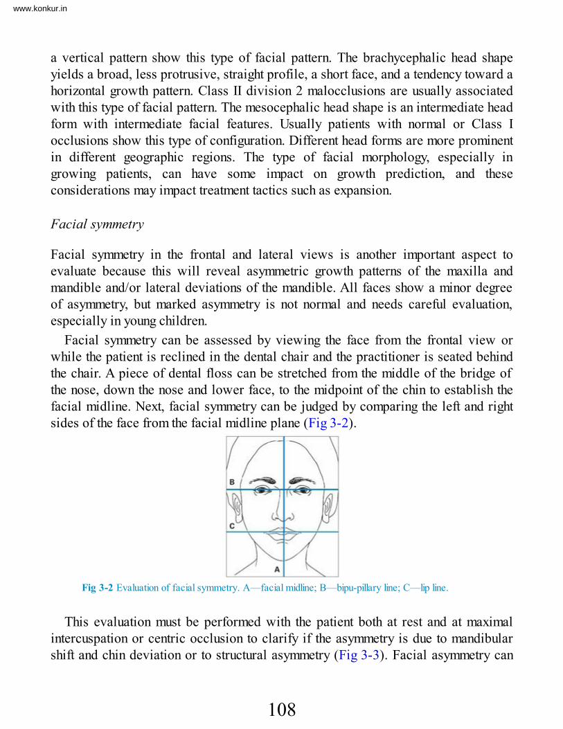
a vertical pattern show this type of facial pattern. The brachycephalic head shapeyields a broad, less protrusive, straight profile, a short face, and a tendency toward ahorizontal growth pattern. Class II division 2 malocclusions are usually associatedwith this type of facial pattern. The mesocephalic head shape is an intermediate headform with intermediate facial features. Usually patients with normal or Class Iocclusions show this type of configuration. Different head forms are more prominentin different geographic regions. The type of facial morphology, especially ingrowing patients, can have some impact on growth prediction, and theseconsiderations may impact treatment tactics such as expansion.
Facial symmetry
Facial symmetry in the frontal and lateral views is another important aspect toevaluate because this will reveal asymmetric growth patterns of the maxilla andmandible and/or lateral deviations of the mandible. All faces show a minor degreeof asymmetry, but marked asymmetry is not normal and needs careful evaluation,especially in young children.
Facial symmetry can be assessed by viewing the face from the frontal view orwhile the patient is reclined in the dental chair and the practitioner is seated behindthe chair. A piece of dental floss can be stretched from the middle of the bridge ofthe nose, down the nose and lower face, to the midpoint of the chin to establish thefacial midline. Next, facial symmetry can be judged by comparing the left and rightsides of the face from the facial midline plane (Fig 3-2).
Fig 3-2 Evaluation of facial symmetry. A—facial midline; B—bipu-pillary line; C—lip line.
This evaluation must be performed with the patient both at rest and at maximalintercuspation or centric occlusion to clarify if the asymmetry is due to mandibularshift and chin deviation or to structural asymmetry (Fig 3-3). Facial asymmetry can
108
www.konkur.in

be the result of many morphologic abnormalities of different parts of thedentoskeletal structures in congenital misconfiguration.
Fig 3-3 Asymmetric growth of the mandible (right condylar hyperplasia) in a 16-year-old girl.
As Subtelny indicated,10 except in cases of congenital deformity, the maxillarybone is a fixed, stable bone, and the midpalatal suture (raphe) is the true midlineindicator of the maxilla. The maxillary midline, except in cases where the maxillarydental midline has shifted because of local factors, coincides with the facial midlineand can be a good point of reference in evaluation of facial symmetry.
The causes and the morphologic configurations of facial asymmetry can bedetermined through the use of diagnostic aids such as posteroanterior cephalometricand lateral jaw radiographs, examination of the dental occlusion on the left and rightsides, and observation of occlusal level (curve of Wilson). Patients with significantskeletal asymmetry (those patients with asymmetry not resulting from only afunctional shift of the mandible) always fall into the severe problem category.
Early detection of any asymmetry, especially in young children, could be veryimportant for early intervention. The timing of intervention and the treatment plandepend on the cause of the problem, the type of asymmetric mandibular growth, andwhether the asymmetry is caused by deficient or excessive growth of the condyle.
Analysis of facial symmetry is discussed further in the section on frontalphotography.
Facial proportion
Evaluation of facial proportion is another important aspect of the clinical andparaclinical evaluation. When the anterior facial proportion is evaluated in thevertical dimension, three segments of the face can be assessed: (1) from foreheadhair line to glabella, (2) from glabella to subnasale, and (3) from subnasale to softtissue menton. In a normal, balanced ratio, these three segments are equal (Fig 3-4).
109
www.konkur.in

Fig 3-4 Frontal evaluation of facial proportions. In a balanced ratio, the segments from foreheadhairline to glabella (A), from glabella to subna-sale (B), and from subnasale to soft tissue menton(C) are equal.
The upper lip–lower lip height ratio is another frontal evaluation of facialproportion. The area from subnasale to stomion should be approximately one third ofthe total lower third of the face (subnasale to menton)11 (Fig 3-5).
Fig 3-5 Upper lip–lower lip height ratio. The area from subnasale to stomi-on should beapproximately one-third of the total lower third of the face.
Another frontal evaluation that can be considered is the sagittal facial proportion,proposed by Sarver,12 also called the rule of fifths. This method is discussed inmore detail in the section on facial photography.
There are other important points to be evaluated in both the facial frontal andlateral views, including facial form, symmetry, nose, and chin and lip relationship inthree spatial planes: the sagittal, vertical, and transverse dimensions.
Lateral (sagittal) facial evaluationDuring the systematic lateral facial examination, the child should be seated in an
110
www.konkur.in

upright position with the practitioner looking from a distance at three points: (1) thebridge of the nose, (2) the base of the upper lip, and (3) the chin. This examinationgives the observer an idea of the patient’s profile.
A straight profile is indicated when all three points are at the same level and forma straight line. If the midpoint is anterior, the patient has a convex profile; if themidpoint is posterior, the patient’s profile is concave (Fig 3-6). Each of these threeprofiles can be caused by abnormalities of one, two, or three point combinations.For example, a convex profile can result from maxillary protrusion, mandibularretrusion, or a combination of both.
Fig 3-6 Profile types: (a) concave; (b) straight; (c) convex.
Slight retrusion of the mandible before puberty can be considered normal. Thisdimension will catch up to the maxilla with normal growth of the mandible. Slightconvexity of the profile at this age can also be considered normal.
A well-balanced profile usually indicates the presence of a normal or Class Iocclusion. An underlying normal skeletal relationship that has no skeletaldiscrepancy in the anteroposterior relationship, molars and canines with a normalrelationship, and primary second molars with a terminal plane that is flush or has amesial step can be considered normal.
Some children may have normal basal bone relationships but disturbed profiles.These situations may be due to abnormal inclination of the incisors such as thatresulting from abnormal, nonnutritive oral habits. Correction of incisor protrusionwill improve the profile.
Children with a severely convex profile usually have a Class II malocclusion, andthose with a straight or concave profile can have a Class III malocclusion. Theclinical and paraclinical examinations can clarify the exact type of malocclusion,whether it is the result of dental or skeletal factors (or a combination of both) or theresult of maxillomandibular or dental discrepancies.
111
www.konkur.in

The morphology of the patient’s profile is also another important area that canchange the treatment plan. For example, tooth extraction can be considered inchildren with a convex profile if other analyses confirm extraction, while extractionshould be avoided in children with a straight or concave profile.
Divergence of the face is a termed used by Hellman.13 Divergence indicates if aprofile slopes either anteriorly (anterior divergence) or posteriorly (posteriordivergence). Divergence is influenced by the patient’s racial and ethnic background.For example, while people of American Indian and Asian descent tend to have ananteriorly divergent face, white people of northern European descent are likely toshow posterior divergence.14
The upper-lower facial proportion, nose, chin, and lips as well as theinterrelationships of these components must also be examined during the evaluation.
Intraoral ExaminationIntraoral examinations for orthodontic patients, as for any other type of patient,require a thorough evaluation of the health of all oral hard and soft tissue structures.Any problem, disease, or pathologic condition, including medical problems, dentalcaries, and pulpal or periodontal involvement, must be treated and under controlbefore orthodontic treatment begins.
The orofacial structure and occlusal system consist of three basic tissues:
1. Dental tissues, consisting of enamel, dentin, cementum, pulp, and the periodontalligament
2. Skeletal tissues, consisting of bone, cartilage, and ligaments3. Soft tissues, consisting of neuromuscular tissue, epithelial tissue, glands, the
circulatory system, mucous membranes, and connective tissue
The oral cavity and all of the tissue components have several interrelatedphysiologic functions that directly or indirectly play some role in the development ofdental occlusion. Therefore, treatment planning must be based on a thoroughexamination of all of these structures, in both static and functional states.
For example, evaluating the dental arch relationship in centric occlusion andlooking at the relationship of maxillary and mandibular first molars are not sufficientmeans to classify the type of occlusion. It is necessary to assess all types ofmandibular movement, such as protrusive, retrusive, and lateral excursions, and the
112
www.konkur.in
dental and facial midline relationships at wide open, rest, occlusion, and path ofclosure. Assessment of the relationships between rest position and occlusion candifferentiate between a functionally true and a forced-bite malocclusion caused by amandibular shift.
Rest position is the result of a myotatic antistretch reflex in response to gravity; italters with the position of the head, and natural head position must also bedetermined for each patient.
To emphasize the important role of orthodontic diagnosis, especially in earlytreatment, important areas of this assessment are discussed in static and functionalevaluations.
The following are important features to consider in the static evaluation of theoral cavity (see Fig 3-1):
• Oral health• Lips (position relative to each other and to incisors)• Caries susceptibility and restorations• Dental age (tooth eruption and sequence)• Tooth size, forms, and number• Periodontal and oral mucosal conditions• Tongue size, posture, and function• Frenum and oral mucosa• Adenoids and tonsils• Type of occlusion (classification, overjet, overbite, midline, urve of Spee)
The following are important features to consider in the functional evaluation of theoral cavity (see Fig 3-1):
• Perioral musculature, including the tonicity and functionduring mastication, deglutition, respiration, and speech
• Lip competency, tonicity, and function during rest, swallowing, speech, andrespiration
• Dental and facial midline relationships at wide open, rest, occlusion, and path ofclosure
113
www.konkur.in

• Mandibular movements and protrusive, retrusive, and lateral excursions• Freeway space• TMJ function and dysfunction and condylar movements• Mode of breathing
Evaluation of the dentitionThe first step of occlusal development starts when primary first molars reachocclusion. Numerous reports indicate that many malocclusions develop duringprimary and early mixed dentition and are recognizable and preventable.15
Evaluation and classification of the primary dentition are important steps for earlydetection and intervention. Inspection of a child’s dental occlusion must beperformed first during the late primary dentition and then before eruption of thepermanent first molars.
At this age, occlusion must be carefully examined in three dimensions (sagittal,vertical, and transverse) in both centric occlusion and centric relation. Theinterdental spacing (Baume classification),16–19 crowding, primate space, terminalplane, primary molar and canine relationship, overjet, overbite, and midline must beevaluated. The child’s age and tooth eruption pattern are also very important toevaluate.
The development of the childhood dentition and resulting occlusion is a dynamicprocess, occurs in all three planes of space, and may undergo considerable changeduring childhood and early adolescence. With periodic evaluations, the practitionercan intercept and favorably influence undesirable changes. After more than 40 yearsof orthodontic practice, especially involving young children, the author believes thatmany eruption problems can be prevented if the problem is detected early andintervention is started early. The best way to detect these problems is longitudinalmonitoring of panoramic radiographs, which is discussed later in this chapter.
Eruption that is early or delayed by a few months is not harmful unless local orsystemic problems prevent normal eruption. Any early or late eruption that isdetected clinically, especially if it is asymmetric, must be evaluatedradiographically to determine if a problem exists.
Asymmetric eruption between the right and left sides of the dental arch lastingmore than 6 months is another problem that may require early intervention. Likewise,an abnormal sequence of eruption can be very harmful to occlusal development anddeserves careful assessment during the intraoral examination. Early loss and delayed
114
www.konkur.in

exfoliation of primary teeth are etiologic factors in some malocclusions. These mustbe detected at an early stage through a complete intraoral examination.
Other eruption problems, such as ankylosis, tooth transposition, ectopic eruption,and tooth impaction, must be evaluated clinically and radiographically (see chapter10).
Problems of tooth size, form, and number (missing or supernumerary teeth) areother factors that can interfere with normal eruption and tooth displacement. Theabsence of a mandibular incisor or the presence of a supernumerary mandibularincisor is easily overlooked; counting the teeth during the examination can preventthese problems (see chapters 6 and 7).
The intraoral examination should include assessment of the following aspects ofthe dentition (see Fig 3-1):
• Primary occlusion (interdental spacing, terminal plane, and mandibular shift)• Dental age• Eruption pattern of the dentition• Asymmetric eruption• Early loss and delayed exfoliation of primary teeth• Eruption problems such as ankylosis, tooth transposition, ectopic eruption, and
impaction• Problems of tooth size, form, and number (missing or supernumerary teeth)• Dental and facial midlines
Evaluation of soft tissuesAnother area of inspection during intraoral examination is evaluation of the softtissues. The buccal tissues, lips, floor of the mouth, palate, and gingiva should becarefully inspected and palpated. The gingival tissue in young children might beredder and smoother than that of adults because of increased vascularity and thinnerepithelium.
Early detection of periodontal involvement before treatment is important; carefulprobing can be very helpful. Bleeding during probing is indicative of active disease,which must be brought under control before other treatment.
Crowded mandibular incisors must be carefully assessed for tissue recession,especially when attachment to the gingiva is inadequate. Extraction and
115
www.konkur.in

nonextraction options must be carefully evaluated.Recent findings suggest that periodontal disease may have its origins in childhood.
A noted increase in the frequency of gingival inflammation in children andprogressive periodontal conditions causing primary and permanent tooth loss led theAmerican Academy of Pediatric Dentistry to place greater emphasis on prevention,early diagnosis, and treatment of gingival and periodontal disease in children.20
Other areas of the soft tissues that can play an important role in diagnosis andtreatment planning in early-age orthodontic treatment are the tongue, frenum, lips,and adenoid and tonsillar tissues. Growth and development of maxillofacialmorphology and oral function are closely interrelated.
The tongue has several necessary functions for life and at the same time can playan important role in development of the occlusion. The tongue has three mainfunctions. First, it carries on its surface the taste buds, which send information to thebrain about the nature of the food being eaten. Second, the tongue plays an importantpart in the process of mastication and deglutition. Third, the tongue is involved inspeech, assuming different positions in the mouth and altering the shape of the airpassage through which sounds made by the vocal cords will pass.
Most tongue postures and movements during normal function exert some forces onthe dentition that are in balance with other neuromuscular forces exerted by theperioral musculature. Any imbalanced force, especially at early stages of thedentition, can cause morphologic changes in the occlusal system. Therefore, thisstructure must be evaluated, in both static and functional states, for the purpose oforthodontic diagnosis and treatment planning. This assessment is based on threeimportant conditions of the tongue: (1) tongue size, (2) tongue posture, and (3) tonguefunction.
Tongue size
The interaction between tongue size and volume and craniofacial skeletal growth isessential for understanding the mechanism of specific types of malocclusion andtreatment planning.
Harvold et al,21,22 using rhesus monkeys, demonstrated that reducing tonguevolume by partial glossectomy caused the dental arch to collapse lingually andproduce crowding.
One of the factors that plays an important role in the development of the occlusionis the balance between the tongue and the perioral chain of muscle. Any imbalance
116
www.konkur.in

between these two factors can cause irregularities in occlusion. For example, inpatients with acromegaly, macroglossia causes many types of irregularities, such asinterdental spacing, anterior open bite, dental protrusion, and Class III malocclusion(Fig 3-7).
Fig 3-7 (a to c) Macroglossia causing a large mandibular arch, anterior and posterior crossbites,and anterior open bite. (Courtesy of J. Daniel Subtelny.)
Another example is the effect of Pierre Robin syndrome, which is a conditionpresent at birth. The infant has a smaller-than-normal mandible, a tongue that fallsback in the throat, and difficulty breathing because the tongue is smaller andretropositioned. Patients with this condition have severe crowding.
Tongue posture
Tongue posture also plays an important role in development of some malocclusions,including open bite, interdental spacing, and anterior crossbite. Tongue position canbe altered by other conditions, including large tonsils or adenoids and chronic nasalinflammation that can contribute to tongue thrusting and poor tongue posture (Fig 3-8).
117
www.konkur.in

Fig 3-8 (a to d) Large tonsils causing forward positioning of the tongue and open bite. (Courtesyof J. Daniel Subtelny.)
Prolonged low tongue position during critical growth periods in children mayinitiate a sequence of events resulting in excessive molar eruption, a clockwiserotation of the growing mandible, a disproportional increase in lower anterior facialheight, retrognathic mandible, and open bite (see chapter 5).
Tongue function
Growth and development of maxillofacial morphology and oral function are closelyinterrelated; tongue activity and forces during physiologic functions such asdeglutition, mastication, and speech can play a role in developing malocclusion if itfunctions improperly (Fig 3-9). Myofunctional disorder of the tongue has beendescribed in various ways, including deviate swallow, infantile swallow, andabnormal swallow, and the terms tongue thrust and tongue thrusting have greaterusage among authors.
118
www.konkur.in

Fig 3-9 Abnormal tongue function.
Atypical swallowing patterns are considered important factors in the etiology ofposterior crossbite. Ovsenik23 examined 243 patients aged 3, 4, and 5 years. Shefound that atypical swallowing patterns were more common among children withposterior crossbite than those without crossbite. She concluded that the orofacialfunctions of children in the primary dentition stage with sucking habits should beassessed at each examination. Special attention should be paid to the swallowingpattern, which was found to be an important etiologic factor in the development ofposterior crossbite.17
Production of certain sounds can be difficult for some patients with severemalocclusion, such as anterior lisping in patients with anterior crossbite. Therelationship between malocclusion and speech problems is another aspect of thepatient’s situation that must be evaluated before treatment planning. Determiningwhether the malocclusion is contributing to the speech problem or the speech defecthas contributed to the malocclusion is complicated and controversial.Neuromuscular problems that cause speech problems can also be causative agentsfor malocclusion.
Anterior open bite with a large gap between the incisors can cause lispingbecause it affects the position of the lips in pronunciation of sibilants such as /s/ and/z/, /sh/, /ch/, or /th/. Irregular incisors or retroclination of maxillary incisorsimpedes production of linguoalveolar sounds such as /t/ and /d/. In patients withskeletal Class III relationships, labiodental sounds such as /v/ and /f/ are sometimesdisturbed.
Evaluation of these three aspects of the tongue to determine whether they representan etiologic factor in the malocclusion is important for treatment planning and toensure stability of the treatment result (see chapter 5).
Assessment of TMJ functionEvaluation of the TMJ is an essential part of the clinical evaluation before any typeof orthodontic treatment, especially in routine clinical examination of young patients.
The first thing to evaluate is the range of mandibular movement in protrusive,retrusive, and lateral excursions. If these mandibular movements are normal, thefunction is usually normal. However, if a restrictive movement is present, it usuallyindicates a functional problem.
119
www.konkur.in

Other functional evaluations of the TMJ, such as maximum opening, palpation ofthe muscles of mastication and head of the condyle, and palpation of the patient’sneck and submandibular area, must also be carefully performed. The goal is to lookfor any signs of TMJ problems, such as limitation of opening, clicking, pain, noise,and crepitus, that may require further evaluation and treatment.
Palpation of the muscles must be performed bilaterally, while the clinician isstanding in front of or behind the patient and assessing any soreness or discomfortfor the patient on either side. The lateral and medial pterygoid muscles must bepalpated intraorally and the temporalis and masseter muscles extraorally.
During jaw movement, any lateral or anterior mandibular shift must also berecognized. Differentiating true mandibular asymmetry from mandibular shift is ofspecial importance in growing patients. Posterior crossbite can cause lateralmandibular shift, and anterior crossbite causes anterior shift. “Sunday bite” isanother abnormal position of the mandible that can be seen in some patients withskeletal Class II malocclusion. In this type of bite, also known as “dual bite,” a lackof proper fit between the maxillary and mandibular teeth causes the mandible toslide forward to compensate for the maxillary protrusion.
The following are important aspects to examine during evaluation of the TMJs:
• Range of mandibular movement, including protrusive, retrusive, and lateralexcursions
• Maximum opening• Pain during movements• Crepitus and clicking• Path of closure of the mandible from resting position• Freeway space• Prematurities and point of initial contact• Displacement or tooth guidance• History of pain• TMJ tenderness to palpation• Muscle tenderness to palpation
Paraclinical Evaluation (Diagnostic Records)
120
www.konkur.in

Dental abnormalities or malocclusions are common orthodontic problems that arealmost always the result of improper developmental processes. These abnormalitiesare the result of many genetic and environmental factors acting during prenatal orpostnatal life. The result is a multifactorial entity that has many differentcharacteristics and different morphologic patterns.
The first step in treating this multifactorial and multi–characteristic entity is todiagnose, classify, and clarify the problem by careful investigation.
Orthodontic diagnosis involves the study and interpretation of informationcollected from the clinical examination and data accumulated through theparaclinical evaluation. The paraclinical evaluation includes analysis of all dataaccumulated from different diagnostic tools, such as study cast, photographs,intraoral and extraoral radiographs, and cephalometric radiographs. Forcomprehensive diagnosis and treatment planning, these findings are combined withprevious information gathered from questionnaires and the clinical examination.
Dental CastsThe orthodontic study cast is one of the most useful tools in orthodontic treatment.Preparation of a good set of casts requires a good impression that covers thecomplete area of the patient’s dentition with maximum displacement of soft tissues,created by maximum extension of the impression to the deepest parts of the sulcusand all retromolar areas. A good cast must show as much as possible of the alveolarprocess and teeth and must be able to display the inclination of the teeth, not just thelocation of the crown.
For young children with no previous experience of having impressions taken, theprocedures, including the type of impression maneuver, the child’s head position,and tongue placement must be explained and demonstrated. It is recommended thatthe mandibular impression be taken first, because it is easier for children than amaxillary impression.
To prevent posterior flow of alginate, the tray should be seated first on theretromolar area in the mandible, or on the palate for the maxilla, and then on theanterior part of the arch, before it is forced down toward the occlusal surface. Atthis time, the child’s lips should be separated from the teeth, and alginate should fillthe sulcus area.
The wax bite taken in centric occlusion is used to register the exact position of themaxillary and mandibular dentition and orient the maxillary and mandibular casts for
121
www.konkur.in

proper trimming. After it is properly trimmed and polished, the study cast usuallyincludes patient information such as name, date of birth, and date taken.
A set of good study casts is a part of the patient’s record and is a useful tool forocclusal assessment and analysis in diagnosis and treatment planning. The casts canbe used to help determine the type of occlusion, arch form, arch symmetry, and curveof Spee, to perform space analysis, and to demonstrate findings to the patient andparents.
Type of occlusionThe patient’s exact type of occlusion in three dimensions can be easily verified bycasts, while it cannot be done easily through intraoral examination. Examination ofeach arch and single tooth will reveal displacement or rotation of any single tooth orgroup of teeth. Any abnormal intercuspation or interdigitation can be recognized inthe sagittal, transverse, and vertical evaluations.
Sagittal evaluation
With the sagittal evaluation of the casts, the anteroposterior relationship of caninesand molars, in both the permanent and primary dentition (terminal plane), and Angleclassification can be accurately established. Intercuspation and interdigitation can beassessed easily from the buccal and lingual views. The anteroposterior relationshipof the incisors and the amount of overjet and overbite also can be accuratelymeasured from the cast.
Transverse evaluation
Transverse evaluation of the cast can reveal midline discrepancies and posteriorcrossbites. Evaluation of the dental midline relative to the maxillary midline suturecan reveal any dental midline shift. Therefore, if the maxillary midline is at a normalrelationship with the facial and palatal midlines, the problem is from the mandibularmidline. This discrepancy can be caused by mandibular dental midline shift ormandibular jaw shift, which can be confirmed by functional evaluation of themandible during the clinical assessment of resting position and centric occlusion.These are questions that can be answered by combining clinical findings and castevaluation and by assessing cast symmetry.
Posterior crossbite can be the cause of lateral mandibular shift (see chapter 12).
122
www.konkur.in

Vertical evaluation
Vertical evaluation of study casts involves assessment of the vertical overlap ofincisors, which is measured and recorded either in millimeters or as a percentage ofthe total height of the overlapping maxillary incisor over the mandibular incisorcrown. Because of variations in the crown length of incisors in different people, it isbetter to use percentages instead of millimeters.
In a normal bite, the maxillary incisors usually overlap the mandibular incisorcrowns by about 25%. In deep bite, the mandibular incisors are completely covered(100%), and in some situations the mandibular incisors impinge on the palatalgingiva. The vertical relationship of the posterior segment of dentition must also beevaluated, because in some kinds of buccal bite or Brodie syndrome there isincreased incisor overlapping.
An absence of vertical overlap of incisors is called open bite, which is alsomeasured in millimeters. Anterior open bite is most commonly caused by a suckinghabit in the young age group. There are many different types of open bite, such asdental, dentoalveolar, skeletal, or combination open bite. Open bite can be causedby the maxillary or mandibular teeth, either jaw, or a combination of factors andtherefore needs a careful differential diagnosis for proper intervention and treatmentplanning (see chapter 13).
All of these conditions can be determined by a combined cast evaluation, clinicalfindings, and cephalometric evaluation.
Arch form and symmetryThe ability to assess arch form and detect any asymmetry within the dental arches isanother advantage of having good study casts. To detect arch asymmetry moreaccurately, a graded plastic tool called a symmetrograph can be used. Thesymmetrograph is placed over the occlusal surface of the maxillary arch, and thecentral line of the grid is superimposed over the midpalatal suture. This positionreveals whether teeth are equidistant from the midpalatal raphe or a distortion ispresent in the arch.
With this technique, buccolingual and mesiodistal distances of the left and rightsides of the dentition can be accurately analyzed and compared with the midpalatalsuture to detect if any asymmetry is present (Fig 3-10). Application of this analysiscan differentiate whether posterior crossbite is caused by arch constriction oroverexpansion (see chapter 12).
123
www.konkur.in

Fig 3-10 (a and b) Two-surface ruled grid of thick, transparent plastic for evaluation of archsymmetry (Dentaurum).
Early loss of primary teeth and dental shift may cause a unilateral asymmetrywithin the dental arch, either from distal drift of incisors or from mesial drift ofposterior teeth. This kind of asymmetry can be distinguished from arch asymmetry byan accurate evaluation with the symmetrograph, which can also reveal themesiodistal shift of the dentition.
Mixed dentition space analysisStudy casts can also be used to determine the availability of space in the arch incrowding cases. The amount of crowding and available space are important factorsin determining whether the treatment plan will include tooth extraction. Spaceanalysis is a comparison between the amount of space available in the arch and theamount of space needed for the alignment of the dentition.
Space analysis can be performed either directly on casts and radiographs or withcomputerized methods after appropriate digitization of the arch and toothdimensions. The latter methods are easier and more practical.
Arch length, that is, the distance from the mesial surface of one permanent firstmolar to the one on the other side, can be measured two different ways: It can bemeasured with a divider or Boley gauge, segment by segment, or with a piece of softbrass wire contoured from the mesial surface of one permanent first molar, over thecontact points of the posterior teeth and incisal edges (not including the displacedincisors), to the mesial surface of the molar on the other side. The straightened wireis measured as the amount of available arch length.
Required space is the amount of space needed for the canines and premolarspresent in the arch. This value can be measured directly from the mesiodistaldiameter of all erupted teeth on the cast (contact point to contact point), or indirectlyfrom radiographs. The sum of the mesiodistal widths of all individual teeth indicates
124
www.konkur.in

the space needed for the dentition. Good periapical radiographs or lateral jawradiographs can be helpful for this measurement. For correction of any magnificationerror, the exact mesiodistal width of an erupted tooth on the cast can be calculatedand compared with the width of the same tooth on the radiograph.
A comparison between the available space and the total measurement of toothmaterial will establish whether space is sufficient to accommodate the dentition,there is a need for more space, or there is excess space that will result in interdentalspacing. In all space analysis and application of the measurements, six importantfactors must always be considered before the final decision is made for spacecreation or tooth extraction:
1. Incisor inclination2. Profile3. Relationship of incisors to lip position4. Growth pattern5. Mesial shift of permanent molars6. Curve of Spee
Incisor inclination
The anteroposterior position of incisors can affect treatment decisions in spacemanagement. If incisors are crowded and already proclined, the required space isgreater than indicated by the measurements. If the mandibular incisors are retrudedbecause of some muscular dysfunction or habits and the incisors can be brought to amore normal inclination, this would provide more space for crowded incisors.
Profile
The patient’s profile and lip position also can affect the treatment decisions. In apatient with a convex profile and procumbent lips, the possibility of tooth extractionwould change the result of space measurement.
Relationship of incisors to lip position
Lips that are procumbent relative to the chin and nose may necessitate incisorretraction, which needs more space.
125
www.konkur.in

Growth pattern
The patient’s growth pattern is another important factor to be considered in theanalysis of space availability. Björk and Skieller24 stated that there is little or notendency for the dentition to be displaced during growth in a child with a well-proportioned face, while the teeth often shift anteriorly or posteriorly in a child withjaw discrepancy (such as Class II, Class III, long face, or short face relationships)so that space analysis would be less accurate.
Mesial shift of permanent molars
Early loss of primary molars and mesial tipping of permanent molars and space lossis another factor that must be considered in space analysis. The uprighting anddistalization of molars (if possible) will reduce space deficiency. In some patientswith moderate incisor crowding, taking advantage of leeway space and preventingmolar mesial movement could be the treatment of choice (see chapter 4).
Curve of Spee
The study cast is also a useful tool in the measurement of curve of Spee, which is ameasurement (in millimeters) of the deepest part of the occlusal plane of themandibular arch. This can be measured by resting a straight horizontal plane fromthe tip of the most distobuccal cusp of the last molar to the tip of the mandibularcanine. The distance from this plane to the tip of the tallest buccal cusp of thepremolars or primary molars shows the amount of curve of Spee. Correction of thecurve of Spee also requires extra space, which must be considered in spaceanalysis.
Methods of analysisOne purpose of space analysis during the mixed dentition stage is an estimation of
the total mesiodistal width of unerupted permanent teeth to determine the spaceneeded for a well-aligned dentition. Measurement of unerupted permanent teeth hasbeen achieved through three basic approaches:
1. In the Nance25 technique, the mesiodistal width of the unerupted teeth can bemeasured directly on radiographs while the arch length is measured with softbrass wire. As mentioned earlier, good periapical or lateral jaw radiographs arerequired, and the possibility of magnification errors must be considered.
126
www.konkur.in

2. The width of unerupted permanent teeth can be estimated with proportionalitytables. Studies have shown that there is a reasonably good correlation between thesize of the permanent incisors and canines and the premolars.15,26 This method canbe used without radiographs.
3. Staley and Kerber27 proposed a combination of radiographic and prediction tablemethods. They used the size of permanent incisors, measured on dental casts, andthe size of unerupted premolars, measured on radiographs, to predict the size ofunerupted canines.
Another type of space analysis, the Bolton analysis,28 compares the totalmesiodistal widths of the maxillary and mandibular teeth to assess for tooth massdiscrepancies that would prevent good occlusion. The greatest mesiodistaldimension of all teeth (except second and third molars) is measured on each cast.The ratio of the 12 mandibular measurements to the 12 maxillary measurements isconsidered the overall ratio, and the ratio of the 6 mandibular anterior measurementsto the 6 maxillary anterior measurements is the anterior ratio.
Other methods of space analysis have been proposed by different investigators;see chapter 4 for more details.
The following are important uses for study casts in orthodontic treatment:
• Preservation of a record of the patient’s original occlusion• Analysis of the original occlusion• Demonstration to parents and patients• Transfer of the patient’s case records to another practitioner• Evaluation of treatment progress• Assessment of arch form and symmetry• Space analysis• Evaluation of tooth size discrepancy• Legal protection
Photographic EvaluationPhotography plays an important role in diagnosis and treatment planning inorthodontic treatment. Because the patient is still growing, many changes occur
127
www.konkur.in

during early orthodontic treatment. Initial photographs and longitudinal evaluationsduring and after treatment can be very useful for assessing treatment results. Clinicalphotography is usually applied for the following purposes:
• As a permanent record and as documentation to show the initial status of thepatient’s dental and facial morphology before treatment.
• As an important aid in diagnosis and treatment planning for evaluation andanalysis of facial form and proportion in three dimensions.
• As a means of education and demonstration for patients and parents.• As a means of legal protection.• As an invaluable source for education, research, patient referral, conferences, and
publication, if a complete set of photographs is maintained from the first visit,including photographs during treatment and after treatment.
• For accurate measurement of important points and proportions of the face in threedimensions on tracings of the photographs and comparison of the initialassessment with progress photographs and final results. These assessments cannotbe applied clinically.
Photography for orthodontic patients consists of two types of image: extraoralphotographs and intraoral photographs.
Extraoral photographyExtraoral photographs are taken while the patient is standing in a relaxed position,assuming a natural head position with the mandible at rest position, and lookingstraight into the camera or at his or her eyes in a mirror. Extraoral photographs fortypical orthodontic cases usually include the frontal view, left and right profiles, 45-degree view, and the smile (Fig 3-11). If patients have asymmetry or craniofacialdeformity, extraoral photographs of the affected areas are also needed.
Fig 3-11 Standard series of extraoral facial photographs. (a) Frontal smiling view. (b ) Frontal
128
www.konkur.in

resting view. (c) Profile view. (d) Oblique view.
Frontal view
Figure 3-11 shows four standard facial photographs, including frontal and lateralviews of the same patient. Depending on the existence of other facial deformities,other photographs with different angulations or focused on special areas of the facemight be needed. For example, Fig 3-12 shows two views of a patient withincompetent lip closure caused by severe dental protrusion and overjet. For thispatient, retracting the incisors would improve both lip function and facial esthetics.In patients with prominent lips that are not separated and do not exhibit strain onclosure, incisor retraction would have little effect on lip position.
Fig 3-12 (a and b) Lip strain.
Lateral view
Profile photographs are also taken in the patient’s natural head position, while thepatient is in a relaxed position looking straight forward or into a mirror. A lateralphotograph is taken from the left and right sides of the patient’s profile with the hairpulled behind the ear to permit visualization of the entire face. The lateral view mustshow the whole head, chin, and base of the neck to permit visualization of thecontours of the chin and neck area (see Fig 3-11c).
Oblique (45-degree) view
The 45-degree lateral photograph is a three-quarter view of the face that can helpvisualization of the midface and any deformities, such as nasal deformities. Thisview represents the view of the patient most frequently seen by others (Fig 3-11d).
Photographic evaluation of facial esthetics
An improved facial and dental appearance is perhaps the main objective for every
129
www.konkur.in

orthodontic patient seeking orthodontic treatment. The major objective of treatmentis achieving a normal dentoskeletal relation for better function and stability.Overcoming the psychosocial difficulties that can result from facial and dentalappearance is also considered an important esthetic goal in treatment planning.
Clinical facial examination is a direct observation of the patient’s face in threedimensions. Facial photographs should be viewed as an adjunct to what the eyesreveal clinically. Printed photographs allow more accurate analysis of the facialform, symmetry, and proportion.
Acetate tracing paper can be placed over different facial photographs, and thestructures of the face can be traced and analyzed by drawing specific lines. Thetracing can be kept as documentation for the patient’s permanent records and forcomparison with the treatment progress records and final results.
Evaluation of lateral photographs. The patient’s profile is an important part ofesthetics and has a profound effect on treatment planning. No treatment planning canbe complete and designed without consideration of the patient’s profile. There arespecial areas of concern that must be assessed in the photographic evaluation:profile form (straight, convex, or concave), facial proportion, lip size, lip position,nasolabial angle, and mentolabial sulcus.
Profile. The bridge of the nose, base of the upper lip, and the chin are used toevaluate the patient’s profile proportion. Once identified, these three points areconnected with straight lines to form a complete drawing of the patient’s profile.Figure 3-6 shows an illustrated analysis of the lateral view and delineates theappearance of concave, straight, and convex profiles.
Lateral facial proportion. The lateral vertical proportions of the patient’s profilecan also be traced and measured with more accuracy on a printed lateral photograph.The vertical proportions can be assessed using four points: the hairline, the bridge ofthe nose, the base of the upper lip, and the chin. In patients with normal verticalproportions, the upper face, middle face, and lower face are equal (Fig 3-13).
130
www.konkur.in

Fig 3-13 Equal vertical proportions. In a balanced proportion, the upper, middle, and lower facialsegments are equal.
Lip proportion. The same technique can also be used to trace and measure thevertical lip proportions. The relative normal proportion between the upper lip andthe lower lip and chin segment is one-third to two-thirds (Fig 3-14).
Fig 3-14 Vertical lip proportions. The normal ratio of upper lip height to lower lip height is one-third to two-thirds.
Lip position. The relative position of the lips to the nose and chin plays an importantrole in the patient’s profile and in esthetics. Basal bone position and incisor positionand inclination have a direct effect on the position of the lips; therefore, assessmentof the lip position is an important aspect of lateral photograph evaluation.
Several analyses have been proposed for evaluation of this lateral soft tissue.29–32
One of the simplest and most practical ways to evaluate the lip position is Ricketts’esthetic line (E line), which is drawn from the tip of the nose to the most anteriorpoint on the soft tissue chin or soft tissue pogonion. In normal situations in the 3- to6-year-old child, the upper lip should lie about 3 to 4 mm behind the line, whereasthe lower lip should touch or lie slightly in front of this line (Fig 3-15).
131
www.konkur.in

Fig 3-15 Relationship of lips to nose and chin (Ricketts E line). In 3- to 6-year-old children, theupper lip usually lies about 3 to 4 mm behind the line, and the lower lip touches or lies slightly infront of the line.
Nasolabial angle. The nasolabial angle shows the relationship of the nose andupper lip. This angle can be traced with two lines, one from the base of the nose andthe other parallel to the frontal surface of the upper lip. The value of the angle canrange from 90 to 110 degrees (Fig 3-16).
Fig 3-16 Nasolabial angle. The normal range of the angle is 90 to 110 degrees.
The inclination of incisors has a direct effect on this angle; greater incisorprotrusion reduces the size of the angle. For example, for an acute angle, caused bymaxillary incisor protrusion, treatment involves camouflage and extraction of themaxillary primary first molar. An obtuse nasolabial angle can be caused by dentalretrusion, as in patients with a Class II division 2 malocclusion and retrudedincisors. Torquing of incisors can correct this angle. An obtuse angle is alsoobserved in patients with maxillary anterior crossbite; proclination of the maxillaryincisors can correct this angle.
However, the nasolabial angle may also be out of normal range if the patient has
132
www.konkur.in

an elevated nasal tip; this possibility must be considered in treatment planning.
Mentolabial sulcus. The mentolabial sulcus is the fold of soft tissue between thelower lip and the chin; it varies greatly in form and depth among patients.
The mandibular incisor position and inclination and the lower facial height affectthe degree of this angle (Figs 3-17 and 3-18). Proclination of mandibular incisorsdeepens this angle. Because of a lack of lower lip projection, retroclination ofmandibular incisors results in a shallow mentolabial sulcus.
Fig 3-17 Normal mentolabial sulcus angle.
Fig 3-18 Acute mentolabial sulcus angle.
In patients with short lower facial height and Class II relationships, the lower lipis usually everted against the maxillary incisors.
Evaluation of frontal photographs. Extraoral frontal facial photographs are auseful tool in the orthodontic diagnostic record and can be used to assess variousfrontal measurements, such as vertical and lateral proportions and facial symmetry.
Facial symmetry. In almost all normal faces, a small degree of asymmetry existsbetween the left and right sides.
133
www.konkur.in

Proffit and Fields14 illustrated this asymmetry when they compared the real full facewith composites of two right or two left sides. Minor asymmetry can be ignored, butany significant asymmetry, especially in young children, must be detected and treatedearly.
Asymmetry can be detected more accurately with photographic tracing than withvisual observation. However, evaluation of frontal and 45-degree cephalometricradiographs can detect the problem much more accurately.
There are different techniques for evaluation of facial symmetry on frontal facialphotographs; one involves drawing the facial midsagittal plane on tracing paper. Themidsagittal plane is a vertical line drawn from the center of the nasal bridge, or softtissue nasion, to subnasale, extending to soft tissue pogonion. Two parallelhorizontal lines also are drawn: the bipupillary plane, which is drawn between theleft and right pupils, and the lower horizontal line, the stomion plane.
In a symmetric face, the sagittal plane intersects at all three points (soft tissuenasion, subnasale, and soft tissue menton), and the two horizontal lines are paralleland perpendicular to the facial midline. Both sides of the face relative to the facialmidline are vertically and horizontally equal and symmetric (Fig 3-19).
Fig 3-19 Evaluation of facial symmetry. Both sides of the face must be vertically and horizontallyequal and symmetric relative to the facial midline.
Rule of fifths. Another technique for assessing frontal facial symmetry waspresented by Sarver,7 who termed it the rule of fifths. In this evaluation, six verticalplanes are drawn to divide the face sagittally into five equal parts from helix to helixof the outer ears. In this way, the right and left portions of the face can be compared.According to this analysis, the ideal face comprises equal fifths, all approximatelyequal to the width of one eye (Fig 3-20).
134
www.konkur.in

Fig 3-20 Rule of fifths. According to this standard, in the sagittal plane the ideal face is composedof equal fifths, all approximately equal to the width of one eye.
Facial proportion. Evaluation of the balance between the upper, middle, and lowerthirds of the face can also be assessed by drawing four parallel lines over the frontalphotograph: at the hairline, the glabella or supraorbital ridge line, the base of thenose or subnasale line, and soft tissue menton (Fig 3-21). This divides the face intothree segments, which can then be compared. In a normal, balanced ratio, these threeportions of the face should be equal.
Fig 3-21 Evaluation of frontal facial proportions (upper, lower, and middle facial heights). In abalanced face, these three segments are equal.
Intraoral photographyIntraoral photography is also an important part of the diagnostic record. The seriesof intraoral photographs routinely includes five views of the occlusion: lateral left,lateral right, frontal, and maxillary and mandibular occlusal views. The occlusalviews must show the occlusal surface of the entire dentition and can be taken using afront surface mirror.
Occasionally, other special views with different angulations might be needed,
135
www.konkur.in

such as for patients with abnormal frenum attachment, palatal impaction, impingingbite, or severe overjet. In patients with mandibular shift, two photographs, one takenin centric occlusion and one at rest, can show the difference in the midline position.
Intraoral photographs also have many advantages as part of the patient’s record.One important purpose of the intraoral photograph is to record the condition of thehard and soft tissues before treatment. The photograph can then be referred to duringtreatment and compared with the final results. Another important advantage of agood, complete set of intraoral photographs is that it can be used in casepresentations, seminars, education, and publications. Furthermore, a good set ofintraoral photographs is a perfect tool for showing different views of the dentalocclusion and any soft and hard tissue defects, such as white-spot lesions of theenamel, hyperplastic areas, gingival problems, and other pathologic soft tissueconditions. It can also serve as an important form of treatment documentation.
Radiographic ExaminationsRadiography is a useful tool in diagnosis and treatment planning for the patient’sorthodontic problems and can play an important role in prevention, interception, andearly detection of many types of eruption problems.
Depending on the existing dentoskeletal anomalies and malocclusions, differentradiographic techniques can be used. The radiographic techniques used inorthodontic treatment can be classified into two major types: intraoral radiographyand extraoral radiography. The most common intraoral radiographs used inorthodontics are the periapical, bitewing, and occlusal views. Common extraoralradiographs include panoramic radiographs, lateral jaw radiographs, hand-wristradiographs, and different types of cephalometric radiographs (lateral,posteroanterior, and 45-degree views). In recent years, computed tomography (CT)has had an increasing role in dentistry and orthodontic diagnosis.
Intraoral radiography
Periapical radiographs
Periapical radiographs can be used to evaluate tooth structure, pulp, periodontium,and supporting bone prior to orthodontic treatment. Any caries, periodontalinvolvement, bone loss, and periapical lesions must be treated or under control
136
www.konkur.in

before tooth movement.Good periapical radiographs can also be used for accurate measurement of the
mesiodistal crown width of unerupted canines and premolars for different spaceanalyses.
Bitewing radiographs
Bitewing radiographs are helpful in detection of proximal caries. A set of twobitewing radiographs, taken before the start of treatment and before molar banding,is recommended.
Occlusal radiographs
Occlusal radiographic projection is a useful technique that reveals the entiremaxillary or mandibular arch. These radiographs can also reveal supernumeraryteeth, cysts, or any pathologic lesions present in these areas. Occlusal radiographsalso can be used in evaluation of the buccopalatal relationships of supernumeraryteeth or odontomas to other teeth in the arch.
The most common application of occlusal radiographs in orthodontics is theevaluation of impacted and ectopic eruption to determine the relationships toadjacent teeth. However, the use of CT scanning is much more accurate for thispurpose.
Extraoral radiographyCommon extraoral radiographs used in orthodontic treatment are lateral jawprojections, hand-wrist radiographs, CT scans, panoramic radiographs, and differentcephalometric radiographs (lateral, posteroanterior, and oblique or 45-degreeviews). The lateral cephalometric radiograph is usually taken at centric occlusionbut, depending on the type of occlusal problem and possibility of mandibular shift, itcan be taken with the patient at rest and in wide-open positions too.
It is not the intention of this book to discuss all aspects of radiographicapplication or the different techniques and analyses used in orthodontic diagnosticprocedures; there are many books, articles, and reports on these subjects. The mainobjective of this chapter is to refresh the reader’s memory regarding the availabilityof techniques and tools and to emphasize the need for careful pretreatmentinvestigation before any treatment planning, especially for preventive and
137
www.konkur.in

interceptive considerations. The following are the most common extraoralradiographic techniques used in routine orthodontic diagnosis and treatmentplanning.
Lateral jaw radiographs
The lateral jaw radiograph is taken from each side of the jaw while the cassette filmis held on one side of the face. Because there is minimal image superimposition inthis type of radiograph, it shows a clear view of one side of the jaw.
Lateral jaw radiographs are useful during the mixed dentition to evaluate therelationship and position of primary teeth and their successors, third molar position,and their supporting bone and status of eruption. This type of radiograph is alsouseful in space analysis for accurate measurement of the mesiodistal width ofunerupted canines and premolars during the mixed dentition.
Hand-wrist radiographs
The most important goal in early orthodontic and especially orthopedic treatment isestablishment of a normal and balanced correlation between basal jaw growth anddevelopment of occlusion in patients with malocclusions that are progressing with askeletal discrepancy. This goal must be achieved before skeletal growth is finished.Obviously, the best and fastest result can be achieved when the skeletal growth rateis at its highest (peak height velocity [PHV]). General skeletal maturity is usuallyused as an indicator to predict the timing of PHV in jaw growth.
Skeletal maturity is a stage of bone development based primarily on the size,shape, and degree of mineralization of the epiphyses and degree of closure of theepiphyseal plates. It has also been recognized that individual chronologic age doesnot necessarily correlate with skeletal maturation age.33,34 Effective orthopedictreatment cannot be determined by chronologic age or the stage of dentaldevelopment, because neither of these two is reliable. Therefore, all orthopedicorthodontic treatment, such as headgear, chin cap, face mask, and functionalappliances, must correlate with maximum jaw growth.
Attempts to determine general skeletal maturity have included measurements ofbody height, sexual maturity, cervical vertebrae, bone age, and hand-wrist growth tofind out whether the pubertal growth has started, is occurring, or has finished. Hand-wrist radiographs in particular have been used in many ways by many differentinvestigators.34–37 Several human growth studies have also shown that the timing of
138
www.konkur.in

the pubertal peak growth velocity in statural height (PHV), as well as in growth ofjaws, is closely related to specific ossification events observed in the hand-wristarea.33–35 Fishman33 developed the Skeletal Maturation Assessment (SMA), amethod for the evaluation of skeletal maturity using hand-wrist radiographs. Thissystem is based on four stages of bone maturation (epiphyseal widening, ossificationof adductor sesamoid of the thumb, capping of selected epiphyses over theirdiaphysis, and fusion of selected epiphyses and diaphysis), all located at sixanatomical regions of the hand and wrist (thumb, third finger, fifth finger, andradius). In this system, 11 skeletal maturation indicators are found in the sixaforementioned sites.
The adolescent growth spurt usually starts around the age of 12 years for girls and14 years for boys. Pubertal growth acceleration lasts 2.0 to 2.5 years in both sexes.PHV at the apex of the incremental curve occurs for 1 year (6 months before and 6months after), that is, around 10.3 cm for boys and 9.0 cm for girls, whichcorrespond with SMA values 4 to 7. It has also been shown that pubertal maximumfacial growth occurs slightly later than peak growth of general body height. TheSMA system has been used for several years in the orthodontic department at theEastman Institute for Oral Health.
CT scans
Computed axial tomography (CAT) is the process of using computers to generate athree-dimensional image from a flat two-dimensional radiographic image, one sliceat a time. CT scans, sometimes called CAT scans, show the body’s interiorstructures, such as bone, teeth, and soft tissues, as a three-dimensional image. Thesescans provide greater clarity and reveal more details than regular radiographicexaminations.
This type of radiography can be a very useful tool to localize and show uneruptedand superimposed teeth in three dimensions in the jaw (eg, to detect and localizesupernumerary or impacted teeth). CT scans also play an important role inidentifying problems in the bones of the face as a whole, including tumors andfractures, in guiding difficult extraction procedures, and in evaluating bone supportfor placement of dental implants. Figure 3-22 is a composite of different CT viewsof a patient with oligodontia.
139
www.konkur.in

Fig 3-22 Different CT scans of a patient with oligodontia.
Digital imaging
Digital imaging is a recent technology replacing radiographic film in different areasof medical and dental technology. It must be stressed that the image obtained on adigital radiograph is not necessarily any better than one taken using standardradiographic film; however, digital technology does require substantially lessradiation than film.
Another advantage of digital radiographs is the ability to enhance the images, forexample, by enlarging the image or changing the contrast to achieve better diagnosticquality. Digital imaging can play an invaluable role in treatment and treatmentplanning for orthodontic and orthognathic surgery by facilitating cephalometricevaluation, hard and soft tissue assessments, and facial photographysuperimposition.
Panoramic radiographs
The panoramic radiograph is a common diagnostic tool in today’s dental practice. Itis a kind of radiograph that provides a full picture of the dentition and the completemaxilla and mandible.
Panoramic radiographs do not show the fine detail captured on intraoralradiographs and are not as specific as other intraoral radiographs, but in a singleradiograph it provides a useful general view of all dentition, the maxilla andmandible, the sinuses, and both TMJs. This type of radiograph is very useful,especially during the mixed dentition, for early detection and prevention of allproblems disturbing the normal development of occlusion.
140
www.konkur.in

Especially during the mixed dentition as a diagnostic tool for early-ageorthodontic treatment, the following are important aspects that should be carefullyevaluated on a panoramic radiograph before any orthodontic treatment:
• Position and pattern of fully emerged as well as emerging permanent teeth• Sequence of permanent tooth eruption• Asymmetric eruption• Comparison of crown height levels on the left and right sides• Obstacles preventing eruption• Abnormal tooth malformations (gemination, fusion, dens in dente, or dilaceration)• Exfoliation and pattern of primary teeth root resorption• Tooth number and supernumerary teeth or congenitally missing teeth• Eruption problems, such as impaction, ectopic, transposition, or ankylosis• Bone density and trabeculation• Cysts, odontomas, tumors, and other bone defects or pathologic lesions• Third and second molar positions, inclinations, and relationships to the first
molars and ramus edge• Shape of the condylar head and ramus height• Comparison of the left and right condylar heads and rami
The characteristics and management of these problems are discussed in theirrelated chapters in part 2 of this book. Chapter 10 introduces a simple and practicaltechnique for application of panoramic radiographs to assess canine impaction.
Longitudinal Panoramic Radiograph MonitoringOver many years of teaching and practice, in both pediatric dentistry and orthodonticdepartments, the author became interested in conducting a retrospective evaluationof patients who were referred for some type of orthodontic problem and who hadprevious panoramic radiographs available. This retrospective evaluation led to theconclusion that the longitudinal monitoring of panoramic radiographs during themixed dentition is a very valuable, easy technique that enables detection ofdevelopmental anomalies during the transitional dentition. Today the author stronglyrecommends this easy and very useful technique to all practitioners, especially
141
www.konkur.in

pediatric dentists and orthodontists.The transitional dentition is one of the most critical stages of the dentition, and
many eruption problems, whether hereditary or environmental, emerge during thisstage. Longitudinal panoramic radiograph monitoring is a careful serial monitoringtechnique that any practitioner can perform for young patients during transitionaldentition to watch for developmental anomalies that may arise at these ages.
The technique the author recommends is to take one panoramic radiograph whenthe patient is around the age of 6 years (during the eruption of the permanent firstmolar) and then two more panoramic radiographs at 8 and 10 years of age. Carefulcomparison of two or three consecutive radiographs of a patient at this stage of thedentition can easily reveal any abnormal developmental processes emergingbetween radiographs and therefore can enable early detection and intervention. Thefollowing three cases illustrate the advantages of longitudinal monitoring ofpanoramic radiographs and proper intervention.
Case 3-1This case confirms the importance of longitudinal radiographic evaluation, indicating how earlyintervention could have helped this little girl. Figures 3-23a to 3-23c are three consecutive radiographsfound in her record. A periapical radiograph reveals the first sign of a problem, that is, asymmetriceruption of the central incisors at age 7 years. A panoramic radiograph taken about 15 months latershows the eruption of both central incisors and the asymmetric position of the lateral incisors. A thirdradiograph, a panoramic radiograph taken about 7 months later, reveals that the left lateral incisor haderupted while the right lateral incisor remained unerupted.
The important, detectable abnormal sign in this radiograph is the abnormal position of the maxillarypermanent right canine in relation to the unerupted lateral incisor; unfortunately, no intervention wasperformed at this point, and the patient did not return until 3 years later. Figures 3-23d and 3-23e presentthe last panoramic and occlusal views, showing the complete resorption of the permanent lateral incisorroot.
Possible intervention:Assessment of the available serial radiographs indicates that the best treatment option was earlyintervention and extraction of the maxillary primary right canine when the first (see Fig 3-23b), or eventhe second (see Fig 3-23c), panoramic radiograph was taken. Extraction of the maxillary primary rightcanine would have facilitated and accelerated eruption of the permanent lateral incisor, moving thistooth away from the canine forces and preventing root resorption (see Figs 3-23d and 3-23e).
142
www.konkur.in

Fig 3-23 (a) Periapical radiograph showing asymmetric eruption of the maxillary central incisors.(b ) Panoramic radiograph taken about 15 months later, showing the eruption of both centralincisors and the asymmetric position of the lateral incisors. (c ) Panoramic radiograph taken 7months after the first panoramic radiograph, revealing that the right lateral incisor remainsunerupted. Panoramic (d) and occlusal (e) radiographs taken 3 years later. In the absence oftreatment, the permanent lateral incisor has undergone complete root resorption.
Case 3-2A 10-year 5-month old girl exhibited Class II molar relationships, some space deficiency in the maxillarycanine area, and two impacted maxillary canines. The maxillary left canine was also ectopic. Figure 3-24a is the first panoramic radiograph, taken by her dentist. Figure 3-24b is the second panoramicradiograph, taken by the same dentist 18 months later, before referral for orthodontic treatment. Notreatment had been conducted between radiographs. There was some root resorption of the maxillaryleft lateral incisor.
Treatment plan:• Use of maxillary cervical headgear to retract the maxillary molars to Class I molar relationships
and to gain space for the canines• Extraction of the maxillary left primary molar to accelerate eruption of the maxillary left first
premolar, which in turn would facilitate eruption of the maxillary left canine• Placement of a semiremovable lower holding arch and extraction of all mandibular primary molars
143
www.konkur.in

to take advantage of leeway space
Result:Figure 3-24c shows the status during treatment. All mandibular canines and premolars had erupted, themaxillary molars were in a Class I relationship, and sufficient space was available for both canines. Themaxillary right canine changed its path of eruption and was erupting spontaneously, but the left caninewas still positioned over the apex of the lateral incisor. Therefore, the next step was surgical exposureand traction of the left canine.
Figure 3-24d shows the conditions 1.5 years later, after completion of active treatment. Good resultswere achieved, but some root resorption of the maxillary left lateral incisor was present. Figure 3-24e,taken another 1.5 years later, without a retainer, shows that lateral root resorption was unchanged.
Discussion:Figures 3-24f and 3-24g present close-ups of the first and second panoramic radiographs, which weretaken at an 18-month interval. Comparison of the two images seems to indicate that retraction of themolars and extraction of the maxillary primary molar, performed immediately after the first radiograph,could have provided sufficient space to change the path of eruption of both canines or that even earliersurgical traction could have prevented more damage to the lateral incisor root.
144
www.konkur.in

Fig 3-24 (a) Initial panoramic radiograph of a 10-year-old girl with a Class II occlusion, spacedeficiency in the maxillary canine area, impacted maxillary canines, and an ectopic maxillary leftcanine. (b) Panoramic radiograph taken 18 months later, before referral for orthodontic treatment.No treatment has been conducted. The maxillary left lateral incisor shows signs of resorption. (c)Panoramic radiograph taken during treatment. After molar retraction, the maxillary right caninehas erupted spontaneously, but the left canine will need surgical exposure and orthodontic traction.(d ) Panoramic radiograph taken after treatment. The maxillary left lateral incisor shows rootresorption (arrow). (e) Panoramic radiograph taken 18 months later. No retainer has been used.The root resorption of the lateral incisor has not progressed. (f and g) Immediate intervention atthe time of the initial or second panoramic radiograph by molar distalization and primary teethextraction could have accelerated eruption of the premolar (marked by an x) and guided left molar
145
www.konkur.in

movement (arrow).
Case 3-3A 12-year-old boy (at the time of referral) had a serial set of panoramic radiographs taken by thedentist, but intervention was neglected (Figs 3-25a to 3-25c). The third radiograph was taken when thepatient was referred for orthodontic treatment.
Several problems are visible in these radiographs. As shown in the first radiograph, both maxillarycanines were impacted, the maxillary left canine and first premolar were transposed, and the maxillarypermanent right lateral incisor was missing. The second radiograph, taken 1 year later, shows thefollowing changes: The maxillary permanent left lateral incisor was erupted, the maxillary left firstpremolar was erupting, and both maxillary canines were horizontally impacted. The final panoramicradiograph, taken 1 year after the second, shows the following changes: The primary maxillary rightlateral incisor and canine roots were resorbing, the maxillary right canine was moving down, and themaxillary left canine was still horizontally impacted.
Discussion:These three panoramic radiographs indicate the importance of longitudinal radiographic evaluation andreveal the significant changes during transition that can require early detection and proper intervention.The questions are:
1. What caused this significant difference between the right and left canines?2. Was there any chance of early intervention in this case?
The answers are:
1. The different patterns of eruption for the left and right canines can be attributed to the following:the missing maxillary right lateral incisor and resorption of the primary lateral incisor and canine,the transposition of the maxillary permanent left canine and first premolar, and delayed exfoliationof the primary maxillary left lateral incisor and canine, causing delayed eruption of the maxillarypermanent left lateral incisor.
2. Yes. Interceptive treatment was possible.
Possible intervention:Careful observation of the first panoramic radiograph reveals a problem on the left side—that is, thetransposition of the left canine and the first premolar—that was not present on the right side (Fig 3-25d). This situation caused the differences in the eruption patterns of the right and left canines (Fig 3-25e).
The following intervention, if it had been carried out after the first radiograph was taken, could havehelped maxillary left canine guidance:
• Early extraction of both primary canines and the primary left lateral incisor to accelerate eruptionof the maxillary permanent left lateral incisor and permanent right canine
• Early extraction of the maxillary primary left and right first molars to accelerate eruption of themaxillary left and right first premolars
Accelerating eruption of the maxillary left premolar, an obstacle for the left canine, could havefacilitated maxillary left canine eruption.
146
www.konkur.in

Fig 3-25 (a) Initial panoramic radiograph of a 10-year-old boy with impacted maxillary canines,transpositioned maxillary left canine and first premolar, and missing permanent right lateral incisor.(b) Panoramic radiograph taken 1 year later. No treatment has been performed. (c) Panoramicradiograph taken before orthodontic referral, when the patient was 12 years old. The maxillaryright canine is moving down, while the maxillary left canine is still horizontally impacted. (d and e)Close-up views of the initial and second panoramic radiographs, revealing the cause of thecontinued left canine impaction: the transposition of the canine and first premolar. Earlyintervention at the time of the initial radiograph could have provided left canine guidance.
Cephalometric radiographyDifferent cephalometric radiography techniques and different analyses have beenintroduced by numerous investigators over the past 80 years, and the array oftechniques used by different practitioners around the world is so extensive that anydiscussion in this regard would be incomplete. The discussion of this subject in thischapter is intended only to draw the reader’s attention to the importance of usingcephalometric analysis as a tool in orthodontic diagnosis and treatment planning,
147
www.konkur.in

especially for early diagnosis and early treatment.From the time that this technique was introduced by Broadbent et al38 in the
United States and by Hofrath in Germany to the present, the cephalometricradiograph is the most frequently used radiograph in orthodontic treatment.Cephalometry is a radiographic technique for head measurement. This technique is afurther development of the craniometric technique, which was used byanthropologists for direct measurement of the dried skull.
The essence of cephalometric radiography is standardization. The patient’sposition and the orientation of the x-ray beam are established by mechanical devicesin such a way that repeated exposures at other times have essentially the sameconditions.
Before 1931 and the introduction of the cephalometric radiograph, orthodonticdiagnosis and treatment planning were primarily based on observation of the facialform and dental relationships and a set of plaster study casts. The cephalometrictechnique became more common because advancements in understanding of thebiology and morphology of dentoskeletal anomalies and improvement incephalometric technology motivated practitioners to use this technique.
Downs39 analysis, introduced in 1948, was the first analysis based on skeletal andfacial proportions in a group of 25 untreated white adolescents with ideal dentalocclusions. Comparisons were made only with patients with excellent occlusion andfacial proportions.
Now orthodontists are more aware of the significance of the underlying jaw andother bony structures of the skull, the relationships of dental occlusion, and theimportance of differentiating these relationships of the teeth to each other, to the jawbase, and to other bony structures of the skull.
The original purpose of cephalometric analysis was to enable research ofcraniofacial growth changes. Very soon it was recognized that the cast alone cannotidentify the type of occlusion and whether the malocclusion is the result of dental ordentoskeletal problems. Two dental casts may seem to show a similar malocclusion,while cephalometric evaluation may reveal that the two malocclusions have adifferent basis.
Uses of cephalometric radiography
Since the cephalometric radiograph was introduced, the importance of this techniquehas been widely understood. The following discussion briefly summarizes the
148
www.konkur.in

benefits and applications of this radiograph.
Longitudinal growth evaluation. As mentioned earlier, the first purpose of thistechnique was research on craniofacial growth changes. Before this technique, allinformation regarding growth and development of the skull was based onanthropometric evaluation of dried skulls or investigation in animals, including vitalstaining, metallic implantation, surgical approaches, and histologic evaluation.
The application of cephalometric radiographs by Broadbent et al38 brought thistechnique to the highest level of attention regarding longitudinal evaluation inchildren from 3 to 18 years of age.
Growth prediction. Another advantage of the cephalometric technique is predictionof the patient’s growth pattern before treatment planning in early orthodontictreatment. All craniofacial bones, including the maxilla and mandible, grow underthree mechanisms: increase in size, change in proportion, and change in position. Allof these mechanisms are under the control of genetic and environmental factors.Normal occlusal development depends on the correlation and harmony of thesestructures, which are controlled by growth rate, amount of growth, and direction ofgrowth.
Orthodontic treatment planning in growing patients cannot be completed withoutpredicting the facial growth pattern. Successful prediction requires specification ofboth the amount and the direction of growth. Cephalometric data and the morphologyof the basal bone can identify the growth pattern of each individual growing patientto enable proper timing and sequencing of the treatment plan.
One of important advantages of early orthodontic treatment is the possibility ofobserving the actual treatment response in growing children during treatment. Bycarefully monitoring the patient’s response at all stages of treatment, the providercan determine appropriate adjustments to the original plan according to variations inresponse.
Identification of type of malocclusion. Another important role of cephalometry inorthodontics is identification and classification of the patient’s type of malocclusion.So many kinds of malocclusions present with different etiologic factors, anddifferent morphologic and structural combinations can be identified and classifiedby accurate cephalometric measurement and analysis to allow proper treatmentplanning.
Soft tissue evaluation. Besides the identification of type of malocclusion and
149
www.konkur.in

dentoskeletal malrelationships, evaluation of the soft tissue (lips, nose, and chin)relationships to each other and to dentoskeletal structures is another important partof cephalometric evaluation, as well as evaluation of other soft tissues, such as thetongue, soft palate, tonsil, and adenoid tissues.
Reevaluation of orthodontic progress. Reevaluation of patients during treatment isanother important application of this technique. Especially in early-age treatment,cephalometric reevaluation of treatment progress and growth changes is highlyrecommended.
Orthodontic treatment planning. Another important application of cephalometry isplanning the treatment tactics that take place after analysis of the dentoskeletal andsoft tissue relationships and recognition of the types of anomalies and classificationof malocclusion.
Evaluation of treatment results. After the completion of orthodontic treatment,posttreatment reevaluation through superimposition of different dentoskeletalsegments for comparison of the treatment results is another important role of thecephalometric evaluation technique.
Clinical application of cephalometric analysis
Since the introduction of the cephalometric radiographic technique to medicine anddentistry, many articles, books, techniques, and analyses have been presented. It isnot the intention of this book to recommend one special method or analysis to beused for diagnosis and treatment planning in early orthodontic treatment. The mainpurpose of this discussion is to remind the reader of some important biologic factsfor early-age orthodontic diagnosis and treatment planning:
• Dental occlusion is not a single entity; teeth and alveolar processes are not theonly foundations of the functional component of occlusion. Other craniofacialstructures that can influence the occlusion through their growth rate and patterninclude the cranium, cranial base, nasomaxillary complex, and maxillary andmandibular basal bones.
• The malformation and disharmony of many factors and structures can influencedevelopment of the occlusion. Therefore, differential diagnosis and recognition ofthe involved structures and morphology of the malocclusion is essential.Cephalometric evaluation and analysis can clarify the morphology of
150
www.konkur.in

malocclusion and differentiate the structures involved in dentoskeletalabnormalities.
Dentoskeletal components to evaluate by cephalometric analysis
Many different points and analyses can be used in a routine cephalometric approach,depending on the practitioner’s desire. From a practical point of view, the elementsintroduced here are those important components of occlusion that absolutely must beevaluated for early orthodontic treatment. These areas can be classified according tostructures and according to measurement data (angles and linear measurement). Themain structures that must be evaluated for young patients are:• The relationships of the incisors and the molars to each other, to basal bone, to the
cranial base, and to the profile• The relationships of the basal bones, maxilla, and mandible to each other and to
other craniofacial structures• The relationships of the cranial base to other structures, including length, and
angulation
Over the past 80 years, hundreds of angles and linear measurements have beendeveloped for orthodontic evaluation. Box 3-1 shows the measurements andrelationships that are especially important in early orthodontic treatment.
Box 3-1 Cephalometric measurements relevant toearly-age orthodontic treatment
Maxillary skeletal measurements• Sella–nasion–point A• Lande angle (nasion–Frankfort horizontal)• N perpendicular• Maxillary length (anatomical)• Maxillary length (functional)
Mandibular skeletal measurements• Facial angle• Sella-nasion–point B• Y-axis• Mandibular plane angle• Gonial angle• Occlusal mandibular angle• Mandibular plane to palatal plane• Mandibular length (anatomical)• Mandibular length (functional)
Maxillary dental measurements• Interincisal angle• Maxillary incisor to palatal plane• Maxillary incisor to Frankfort horizontal• Maxillary incisor to sella–nasion• Maxillary incisor to point A to pogonion (degrees)• Maxillary incisor to point A to pogonion (mm)• Maxillary incisor to nasion–point A (degrees)• Maxillary incisor to nasion–point A (mm)• Maxillary incisor to N perpendicular
Mandibular dental measurements• Interincisal angle
151
www.konkur.in

• Mandibular shape
Soft tissue evaluation• Lips: size, position, and proportion to each other, toincisors, and to profile• Tongue: size, position, and proportion• Tonsils and adenoids: size and airway passage• Nose: size, shape, and relationship to profile• Chin: size, shape, and relationship to profile
• Mandibular incisor to Frankfort horizontal• Mandibular incisor to occlusal plane• Mandibular incisor to point A–pogonion (degrees)• Mandibular incisor to point A–pogonion (mm)• Mandibular incisor to N perpendicular
Application of frontal cephalometric radiographs
Frontal (anteroposterior) cephalometric evaluation has several indications inorthodontic treatment, especially in early treatment. Orthodontic diagnosis andtreatment planning for young children have some specific considerations that deservespecial attention in growing children, including facial asymmetry, maxillary dentaland skeletal constriction, and mandibular functional shift. In addition, a lateralcephalometric radiograph describes only two planes of space, whereas patients mustbe treated in all three planes. Anteroposterior cephalometric radiographs allowevaluation of the transverse dimension. Frontal cephalometric analysis allowsevaluation of the following features:
• Facial type (dolichocephalic, mesocephalic, or brachycephalic)• Facial proportion• Facial symmetry (dental and skeletal)• Maxillomandibular basal arch width in crossbites• Maxillomandibular molar relationships in crossbites• Occlusal plane disturbances• Mandibular functional shift• Unerupted, impacted, or ectopic teeth• Palatal expansion• Nasal passage constriction• Congenital deformities• Orthognathic surgery needs
Summary
152
www.konkur.in

• Diagnosis is the most critical part of orthodontic treatment. The goal of thediagnostic process is to prepare a comprehensive list of the patient’sproblems in order to synthesize the various treatment options into a rationaltreatment plan for the patient’s best benefit.
• Treatment planning is strategy, the treatment is the tactics, and this entireprocess has to be organized on a sequential basis: Examine the patient,diagnose the problem, classify the problems, plan the treatment, and thentreat the patient.
• A comprehensive orthodontic diagnosis does not merely focus on therelationships of the maxillary and mandibular dentition. It requires athorough evaluation of the patient’s general health and occlusal situation andconsiders the relationships of the dentition to basal bone and other skeletal,neuromuscular, and soft tissue environments.
• All this information, whether clinical or paraclinical, is derived from threemajor sources: the questionnaire and interview, clinical evaluation, andparaclinical evaluation (diagnostic record).
• Paraclinical evaluations include diagnostic tools such as study casts,intraoral and extraoral photographs, and intraoral and extraoral radiographs,including cephalometric radiographs.
• Careful collection of data from clinical and paraclinical assessmentsenables practitioners to detect the type of malocclusion, to clarify theproblems list, to classify the problems, and to design a proper treatmentplan according to the individual patient’s malocclusion and growth pattern.
References1. Harris EF, Johnson MG. Heritability of craniometric and occlusal variables: A longitudinal sib analysis. Am J
Orthod Dentofacial Orthop 1991;99:258–268.2. Paulsson L. Premature birth—Studies on orthodontic treatment need, craniofacial morphology and function.
Swed Dent J Suppl 2009;(199):9–66.3. Rythén M. Preterm infants—Odontological aspect. Swed Dent J Suppl 2012;(224):1–106.4. Germane N, Rubenstein L. The effects of forceps delivery on facial growth. Pediatr Dent 1989;11:193–197.5. Van den Bergh B, Heymans MW, Duvekot F, Forouzanfar T. Treatment and complications of mandibular
fractures. J Craniomaxillofac Surg 2012;40:e108–e111.6. Chrcanovic BR. Open versus closed reduction: Mandibular condylar fractures in children. Oral Maxillofac
Surg 2012;16:245–255.7. Graber LW, Vanarsdall RL Jr, Vig KWL. Orthodontics: Current Principles andTechniques, ed 5. St Louis:
Mosby, 2011.8. Gugino CF, Dus I. Unlocking orthodontic malocclusions: An interplay between form and function. Semin
Orthod 1998;4:246–255.
153
www.konkur.in

9. Enlow D. Handbook of Facial Growth, ed 2. Philadelphia: Saunders, 1982.10. Subtelny JD. Early Orthodontic Treatment. Chicago: Quintessence, 2000.11. Jacobson A, Jacobson RL. Radiographic Cephalometry: From Basics to 3D Imaging. Chicago: Quintessence,
2006.12. Sarver DM. Esthetic Orthodontics and Orthognathic Surgery. St Louis: Mosby, 1998.13. Krogman WM. The contributions of Milo Hellman to physical anthropology. Am J Orthod 1947;34:61–82.14. Proffit WR, Fields HW Jr, Sarver DM. Contemporary Orthodontics, ed 5. St Louis: Mosby, 2012.15. Moyers RE. Handbook of Orthodontics, ed 4. Chicago: Year Book Medical, 1988.16. Baume LG. Physiological tooth migration and its significance for the development of the occlusion. 1. The
biogenetic course of the deciduous dentition. J Dent Res 1950;29:123–132.17. Baume LG. Physiological tooth migration and its significance for the development of occlusion. 2. The
biogenesis of accessional dentition. J Dent Res 1950;29:331–337.18. Baume LG. Physiological tooth migration and its significance for the development of occlusion. 3. The
biogenesis of the successional dentition. J Dent Res 1950;29:338–348.19. Baume LG. Physiological tooth migration and its significance for the development of occlusion. 4. The
biogenesis of overbite. J Dent Res 1950;29:440–447.20. American Academy of Periodontology Research, Science and Therapy Committee. Periodontal diseases of
children and adolescents. Pediatr Dent 2008–2009;30(7 suppl):240–247.21. Harvold EP. The role of function in the etiology and treatment of malocclusion. Am J Orthod 1968;54:883–
898.22. Harvold EP, Chierici G, Vargervik K. Experiments on the development of dental malocclusions. Am J Orthod
1972;61:38–44.23. Ovsenik M. Incorrect orofacial functions until 5 years of age and their association with posterior crossbite.
Am J Orthod Dentofacial Orthop 2009;136:375–381.24. Björk A, Skieller V. Facial development and tooth eruption. An implant study at the age of puberty. Am J
Orthod 1972;62:339–383.25. Nance HN. The limitations of orthodontic treatment. 1. Mixed dentition diagnosis and treatment. Am J Orthod
1947;33:177–223.26. Tanaka MM, Johnston LE. The prediction of the size of unerupted canines and premolars in a contemporary
orthodontic population. J Am Dent Assoc 1974;88:798–801.27. Staley RN, Kerber PE. A revision of the Hixon and Old-father mixed-dentition prediction method. Am J
Orthod 1980;78:296–302.28. Bolton WA. The clinical application of a tooth-size analysis. Am J Orthod 1962;48:504–529.29. Ricketts RM. Planning treatment on the basis of the facial pattern and an estimate of its growth. Angle
Orthod 1957;27:14–37.30. Steiner CC. Cephalometrics for you and me. Am J Orthod 1953;39:729.31. Merrifield LL. The profile line as an aid in critically evaluating facial esthetics. Am J Orthod 1966;52:804–
822.32. Holdaway RA. A soft-tissue cephalometric analysis and and its use in orthodontic treatment planning. Part I.
Am J Orthod 1983;84:1–28.33. Fishman LS. Chronological versus skeletal age; an evaluation of craniofacial growth. Angle Orthod
1979;49:181–189.34. Todd TW. Atlas of Skeletal Maturation. St Louis: Mosby, 1937.35. Fishman LS. Radiographic evaluation of skeletal maturation; A clinically oriented method based on hand-wrist
films. Angle Orthod 1982;52:88–112.36. Greulich WW 2nd, Pyle SI. Radiographic Atlas of Skeletal Development of the Hand and Wrist, ed 2.
Stanford, CA: Stanford University Press, 1959.37. Pike JB. A serial investigation of facial and statural growth in 7 to 12 year old children. Angle Orthod
1968;38:63–73.38. Broadbent BH Sr, Broadbent BH Jr, Golden WH. Bolton Standards of Dentofacial Developmental Growth.
154
www.konkur.in

St Louis: Mosby, 1975.39. Downs WB. Variations in facial relationships: Their significance in treatment and prognosis. Am J Orthod
1948;34:812–840.
155
www.konkur.in

156
www.konkur.in

Space Management in the TransitionalDentition
Development of the dentition is a long process that occurs in an orderly fashionunder the control of genetic and environmental factors, from the primary dentitionthrough the transitional stage to the permanent dentition, resulting in a functional,esthetic, and stable occlusion. During this long process, many local or systemicfactors can influence development of the occlusal system, and any disturbances canprevent the progression to normal occlusion. General practitioners, pediatricdentists, and orthodontists have the highest responsibility to monitor this processcarefully during developmental stages and to detect and manage disruptions ofnormal occlusal guidance.
Every tooth is maintained in its socket over the basal bone in a certainrelationship to the dental arch, with adjacent and opposing teeth under a continuousbalanced force resulting from the action of different environmental forces. If one ofthese forces is altered or removed, the relationships with adjacent teeth will change,causing drifting of teeth and initiating space problems. Early loss of primary teeth isone of the most common problems during the transitional stage of dentition, resultingin loss of space.
Fundamentals of Space ManagementBefore treatment planning and designing any appliance for space management,practitioners must understand the nature of the problem and the fundamental basesgoverning the biology and mechanism of occlusal development. The following areimportant processes to consider in this regard:
157
www.konkur.in

• Etiology of early primary tooth loss• Effects of primary molar extraction on succedaneous teeth• Incidence and nature of space closure• Prediction of tooth emergence• Factors influencing time of eruption• Factors influencing mesial or distal drift
Etiology of early primary tooth lossLocal or systemic factors can cause early loss or early exfoliation of primary teeth:
• Extraction due to extensive caries• Traumatic injury• Early exfoliation due to abnormal root resorption (eg, ectopic eruption)• Systemic disorders or hereditary syndromes, such as hypophosphatasia, rickets,
acrodynia, leukemia, juvenile periodontitis, and Papillon-Lefèvre syndrome (seechapter 10)
Effects of primary molar extraction on succedaneousteeth
Fanning1 studied the lateral jaw radiographs of four boys and four girls who hadundergone early and unilateral extraction of primary molars at different ages. Thelongitudinal study evaluated the tooth development and eruption of the antimereteeth, which were used as controls, and the rate of formation and the speed and timeof eruption of the succedaneous teeth. No change was observed in the rate of rootformation of the premolars after extraction; however, an immediate spurt occurred inthe eruption of the premolar, regardless of its stage of development and the stage atwhich the primary molar was extracted. Premolar eruption was accelerated in thepresence of longstanding necrosis of the primary tooth, especially if accompanied byloss of surrounding bone. Extraction of the primary tooth during active eruption of adeveloped premolar caused early clinical emergence of the premolar.
Early extraction of the primary tooth, before complete formation of the premolarcrown (approximately 4 years of age), resulted in an initial surge, but then the toothremained stationary and eruption was delayed. Therefore, the question arises
158
www.konkur.in

whether it is possible to estimate individual eruption patterns by considering rootdevelopment and skeletal development in addition to chronologic age. The effects ofprimary molar extraction on formation and eruption of their successors can besummarized as follows:
• The rate of formation of premolars does not change.• An immediate surge in eruption occurs, regardless of the dentition stage and the
patient’s age.• Eruption is delayed if extraction occurs before half of the root formation is
completed.• Eruption is accelerated if extraction occurs when half of the root is developed.
Posen2 examined the complete records of 350 children at the ages of 4 and 5years. Of these children, 62 patients, 34 girls and 28 boys, were selected who hadundergone unilateral extraction of primary molars. The accumulated recordsincluded plaster casts and annual oblique cephalometric radiographs.
Posen2 concluded that eruption of premolars was delayed in these children. Afterthe age of 5 years, a gradual decrease in delayed eruption of premolars was noted inthese patients, and, at ages 8, 9, and 10 years, eruption of premolars greatlyaccelerated. This study confirmed delayed eruption of premolars and acceleration atages 8 to 10 years after early extraction of primary molars.
Incidence and nature of space closurePremature loss of primary teeth can cause functional disturbances and, because ofthe possibility of drifting of adjacent teeth, can result in space loss, difficulty ineruption of their successors, and, consequently, occlusal problems. Thus, prematureloss of primary teeth needs careful attention during development of the dentition.
The consequences of premature extraction of primary teeth have been the subjectof discussion for many years. In 1742, French physician Pierre Fauchard spoke ofthe inevitability and undesirability of removing primary teeth before their naturalshedding.
The consequence of premature loss of primary teeth has been evaluated in severallongitudinal and cross-sectional studies. In a review of previous studies, Owen3
revealed a large controversy in opinions on space management in the past. Someauthors, such as Lundström,4 Linder-Aronson,5 and Seipel,6 believe that space
159
www.konkur.in

maintainers are usually a waste of time and even are harmful 19% of the time. On theother hand, many researchers believe that premature loss of primary teeth usuallyresults in space closure and causes harmful effects in the permanent dentition.3,7–10
Owen3 concluded that the following general trends are evident:
• The incidence of space closure increases with time. For all experiments, at least96% of premature losses result in some closure within 12 months.
• The highest and earliest incidence of closure happens after premature loss of themaxillary primary second molar.
• Some mandibular extraction spaces show no closure.• The rate of space closure is higher in the maxilla than in the mandible.• Rates of closure in the mandible are more varied.• The greatest amount of space closure in a given unit of time occurs at the maxillary
primary second molars and then the mandibular primary second molars.• There is complete agreement that space closure in the maxilla is predominantly
caused by mesial movement of the teeth posterior to the extraction space. In themandible, space closure is predominantly caused by distal movement of the teethanterior to the space.
• There is evidence that the tooth mesial to the affected molar can drift distally intothe space. Therefore, loss of space or arch length can occur from both directions.4
• Reports of space opening are only associated with late extraction.
The rates of the space lost after primary tooth loss differ. Northway et al10 statedthat the space loss was greatest in the first 6 months and occurred in smaller amountsafter that, with the least occurring 1 year after tooth loss.
Johnsen7 evaluated primary first molar loss in 10 white children during the mixeddentition stage (8 had bilateral and 2 unilateral premature primary first molar loss).At the initial visit, alginate impressions, photographs, and radiographs were taken.In all patients, the primary canine was present at the initial impression, and theprimary second molars were present at the final impression. For six of the patientsextraction was within 1 month of the initial casts; for one it was within 3 months; andfor the remaining three it was more than 6 months after the casts were prepared.
Final measurements of the distances between the permanent first molars andpermanent incisors were taken on the initial and final casts and compared. Spaces atthe extraction site were measured from teeth immediately adjacent to the extraction
160
www.konkur.in

site. Johnsen7 reported no significant mesial movement of the mandibular permanentmolars after mandibular primary first molar loss but some space loss at themaxillary molars, especially if the permanent molars were erupting. Space loss inthe mandible after premature loss of the primary first molar was the result of distalmovement of the anterior teeth, especially in the presence of mandibular anteriorcrowding.
Northway et al10 digitized 107 dental casts from University of Montreal growthsamples. Longitudinal data were collected annually from the age of 6 years, for anaverage observation period of 5.9 years. Trying to determine different aspects ofearly loss of primary teeth, Northway et al10 reported the following:
• Regarding dimensional loss due to caries or primary tooth loss (the first molar[D], the second molar [E], or both the first and second molars [D + E]), theyfound that in both sexes significantly greater amounts of space were lost in themandible, with an average loss of 1.7 mm (ranging from 0.9 to 3.7 mm), than inthe maxilla, with an average loss of 1.2 mm (ranging from 0.7 to 3.0 mm).
• The maxillary D loss group experienced a later increase in the amount ofmaxillary D + E loss.
• In both arches, the greatest loss of space occurred as a result of mesial molarmovement; maxillary canine migration was significant only when the maxillaryprimary molars were lost as early as 9 years of age.
• In the mandible, maximum canine migration occurred after mandibular primaryfirst molar loss.
• More space was lost in the first year following an extraction than in the successiveyears.
• In the maxilla, the rate of loss was age related (more space loss in youngerchildren); in the mandible, the rate of loss was not age related.
• There was no significant difference in age for space opening during the emergenceof the succedaneous teeth in the mandible; the opening occurred during secondpremolar eruption.
• Permanent molar mesial drift occurred in the maxilla after both E and D + E loss.• Mesial movement of the mandibular permanent molar occurred only after E loss.• Maxillary D loss affected maxillary canine relationships, while mandibular D + E
loss affected mandibular canine relationships.
161
www.konkur.in

Prediction of tooth emergenceThe timing of tooth eruption in orthodontic treatment is sometimes a decisive factor,whether in space management or extraction strategy. Unfortunately, individualvariation in the age of eruption, especially in teeth emerging through the gingiva, isvery high.
Grøn11 assessed tooth formation and emergence in 874 children (347 boys and440 girls) by taking radiographs of the emerging tooth and its antimere as well as aleft hand-wrist radiograph. Tooth formation was estimated by intraoral radiographsand rated in four stages of root length: one-quarter, one-half, three-quarters, and fullroot length (with open apex). Findings indicated that tooth emergence is moreclosely associated with the stage of root formation than chronologic age or skeletalage. The majority of teeth studied had three-quarters of the root complete at the timeof clinical emergence; the mandibular central incisors and first molars had less rootdevelopment, while the mandibular canines and second molars had just past three-quarters’ root development at emergence.
Hägg and Taranger12 studied tooth emergence in 212 randomly selected urbanSwedish children who were followed from birth to 18 years of age. The study foundthat emergence of the primary teeth in boys is less than 1 month ahead of girls untilthe eruption of the primary second molars. Girls are consistently ahead of boys fromeruption of the primary second molars to eruption of the permanent second molars(difference from 3 to 11 months).
Demirjian,13 studying a genetically homogenous French Canadian group ofchildren 2.5 to 19 years of age, evaluated 5,437 panoramic radiographs and found nodifference in the timing of dental development between boys and girls from 5 to 6years of age. In contrast, among the older children girls were always moredeveloped than boys.
Smith and Buschang14 examined growth of the mandibular canine and premolarroots in a mixed longitudinal sample of orthodontic patients (77 females and 74males). They found that as a percentage of tooth length, roots grow with decreasingvelocity between 7 and 14 years of age. More complex patterns appear for absolutegrowth in root length; for example, girls show an earlier maximum growth rate forthe canine than for the premolars. Substitution of dental age for chronologic agereduces between-subject variation (assessed at age 11 years), especially for boys. Abetter understanding of dental maturation, including root length growth, should allowimproved prediction models.
162
www.konkur.in

Factors influencing time of eruptionTo summarize the literature, the following are some guidelines for predicting toothemergence:
• Tooth emergence appears to be more closely associated with stage of rootformation than chronologic age or skeletal age.
• There are variations between individuals.• There is a much larger variation in the stage of root development at the time of
clinical emergence than there is at the time of alveolar emergence.• Sex differences in the timing of eruption are smallest for mandibular first molars
and greatest for mandibular canines.• The majority of teeth had attained three-quarters of their root length at clinical
emergence and one-half at alveolar emergence.• In normal tooth emergence, the root apices are always open and the root length is
never less than one-quarter of normal size.• Mandibular first molars and central incisors had one-half of their root developed
at the time of emergence.• Mandibular canines and second molars had more than three-quarters’ development
of their roots.• Emergence of the primary teeth in boys is around 1 month ahead of that in girls.• In emergence of the permanent dentition, girls are ahead.• There is a tendency for earlier tooth eruptions in the mandible.• Usually there is no significant difference in age of eruption between the right and
left sides of the mouth. (The right and left side difference in development is nevermore than one-quarter of the root). A variation in the emergence between the leftand right sides can be considered normal if it persists for less than 6 months. Anyasymmetric pattern of eruption that lasts for more than 6 months must be evaluatedcarefully.
• Most asymmetric eruption occurs in the premolar area.
Factors influencing mesial or distal driftPrincipal factors that have the greatest influence on the rate and extent of mesial anddistal drift of teeth are the degree of crowding in the dental arch, the type of primary
163
www.konkur.in

tooth that is extracted, the type of occlusion, the age of the patient, the presence ofabnormal oral musculature, the existence of abnormal oral habits, and the stage ofdentition.
Degree of crowding
Degree of crowding in the dental arch is an important factor in space loss followingpremature loss of primary teeth.
Mandibular incisor crowding and extraction of the mandibular primary canine orprimary first molar may cause a quick movement of adjacent teeth, such as midlineshift or retraction of the incisors; increase overjet; and cause overeruption ofmandibular incisors and impinging bite. On the contrary, in an uncrowded arch, theremay be little or no movement of teeth following extraction. The author has observedno space loss in patients with a large mandible (mandibular prognathism). Much lessspace loss occurs in a primary dentition with interdental spacing (Baume class I)than in a primary dentition with no spacing (Baume class II).
Type of extracted tooth
Space loss and drift of adjacent teeth also depend on the type of primary tooth. Forexample, premature loss of the primary second molar, whether in the maxilla ormandible, causes mesial shift of the permanent first molar. Early loss of primary firstmolars can cause mesial drift of the posterior teeth and distal drift of the anteriorteeth (in the mandible mostly distal and in the maxilla mostly mesial). Extraction of aprimary canine allows permanent incisors to drift distally, but mesial drift of teeth isminimal.
Type of occlusion
Patients with deep bite have a greater tendency for space loss, especially inmandibular dentition, than do patients with open bite. Class II division 1malocclusion usually becomes more severe after loss of mandibular primary teeth.
Permanent first molar occlusion has some influence on the rate of space loss. Forexample, maxillary and mandibular permanent first molars with good intercuspationhave less tendency for mesial shift than do those with an end-to-end relationship.
Age of the patient
164
www.konkur.in

The age of the patient, or the dental age of the patient, plays a strong role in spaceloss following primary tooth extraction. The earlier a primary tooth is extracted, thegreater the amount of drift. Research has shown that the highest rate of space losshappens in the first year after extraction, especially the first 6 months.15
If a primary molar is extracted before eruption of the permanent first molar, thenthe severity of mesial drift and space loss is much greater, even in arches that are notcrowded.
Abnormal oral musculature
A strong mentalis muscle and high tongue position can damage occlusion after theloss of a mandibular primary molar or primary canine. In these conditions, collapseof the mandibular dental arch and distal drifting of the anterior segment will result.
Abnormal oral habits
Thumb or finger habits that exert abnormal forces on the dental arch, if accompaniedby early loss of primary teeth, can aggravate arch collapse.
Stage of dentition
The stage of dentition and occlusal development can influence the rate and amount ofspace loss. In general, if the teeth near the space left by the premature loss ofprimary teeth are in an active stage of eruption, more space loss is likely to occur.
Planning for Space ManagementBesides all of the aforementioned treatment planning considerations, such as theincidence and nature of space loss, prediction of tooth emergence, mesial and distaldrift of adjacent teeth, and local and systemic factors, including type of occlusion,age of the patient, location of tooth loss, and time elapsed after extraction, thefollowing specific points must be considered in order to determine the appropriatetreatment plan, type of management, and type of device to be applied:
• Space availability (determined via space analysis)• Amount of space loss
165
www.konkur.in

• Severity of the problem• Nature of the problem• Crowding or spacing in other areas of the arch• Chronologic and dental ages of the patient• Stage of occlusal development• Presence or absence of a permanent successor• Condition of the permanent successor and eruption potential (delayed or retarded
eruption)• Patient’s profile and any skeletal disharmony• Relation of adjacent and opposing teeth• Dental and periodontal conditions• Time elapsed since loss of the primary tooth• Status of permanent first molar eruption• State of emergence and type of occlusion of the permanent first molar• Position of the teeth and stage of eruption in the opposing arch• Amount of overlying bone• Sequence of eruption of the permanent teeth• Presence of any abnormal muscle dysfunction• Presence of any oral habits• Patient’s occlusion and existing malocclusion• Prognosis of the proposed management
Planning for any type of space management, like any other orthodontic treatment ortooth movement, requires a series of diagnostic procedures.
Diagnostic proceduresDiagnostic aids such as clinical evaluation, cast analysis, and radiograph andcephalometric evaluation enable the practitioner to reach proper decisions aboutspace management that are linked to individual conditions and variations. Aftercareful evaluation, the decision then will be made whether to maintain, regain,create, or eliminate space for each patient according to his or her dentoskeletalstructural composite.
166
www.konkur.in

Diagnostic procedures, like any other treatments, require tools for data collection,analysis, and measurements. After the necessary diagnostic aids are collected, thefirst step in space management is recognition of the available space relative to thespace needed for the permanent successor and occlusion.
Space evaluation and size prediction
The space available in the arch can be determined with space analysis. Spaceanalysis is the evaluation of spacing or crowding within the dental arches, achievedthrough a comparison of the amount of space available and the amount of spacerequired to align the teeth properly. In other words, the purpose of this type ofanalysis is a comparison of arch length to tooth material. Several types of analysisare available:
• Nance analysis16
• Moyers analysis9
• Tanaka and Johnston analysis17
• Staley and Kerber analysis18
• Merrifield analysis19
• Bolton analysis20
Nance analysis. In a comprehensive study, Nance16 concluded that the length of thedental arch from the mesial surface of one mandibular permanent first molar to themesial surface of the opposite side is always shortened during the transition from themixed to the permanent dentition.
Nance16 also reported an average leeway of 1.7 mm between the combinedmesiodistal widths of the mandibular primary canine and the primary first andsecond molars and the mesiodistal widths of the corresponding permanent teeth ineach side of the arch, with the primary teeth being larger. In the maxillary arch, thedifference between the primary and corresponding permanent teeth is 0.9 mm perside.
Nance’s method16 compares a direct measurement from the periapical radiographof unerupted teeth to available space. Available space is calculated by measuringarch length with a soft brass wire from the mesial surface of one permanent firstmolar to the mesial surface of the first molar on the other side. A sharp divider or apiece of 0.010-inch brass wire is used to measure the mesiodistal width of
167
www.konkur.in

unerupted teeth on complete-mouth periapical radiographs (the tube is aimeddirectly toward the contact points). For accuracy and to assess for distortion in thefilm, the width measured on the radiograph is compared with the actual cast width.
Besides leeway space, two other dimensions are measured: the outsidemeasurement, which is the perimeter of the arch from the mesiobuccal surface of onemandibular permanent first molar around to the mesiobuccal surface of the firstmolar of the opposite side, taken with a brass wire, and the inside measurement,which is taken with a divider from the mesiolingual surface of the mandibularpermanent first molar on one side, at the point where the lingual tissues touch thecrown of the tooth, to the gingival tissues between the mandibular central incisors ontheir lingual aspect. Nance16 indicated that the outside measurements do not increasefrom the time of the mixed dentition to the time when all permanent teeth are presentbut decrease during the transition; likewise, the inside measurement also decreases.
Moyers mixed dentition analysis. The mixed dentition analysis advocated byMoyers9 is a measurement taken from the four mandibular incisors to find theestimated size of maxillary and mandibular canines and premolars from probabilitytables that he calculated for girls and boys. The mandibular incisors were usedbecause they would provide greater accuracy than maxillary lateral incisors, whichhave more variation in size.
The purpose of this analysis is to evaluate the amount of space available in thearch for succeeding permanent teeth and necessary occlusal adjustment. This methodhas some advantages: The analysis can be applied directly in the mouth as well ason casts on both arches; it is not time-consuming; and it requires no specialequipment or radiographic projection.
Tanaka and Johnston analysis. The Tanaka and Johnston17 analysis is a variationof Moyers’s method9 except that the probability chart is not needed. In thistechnique, the total width of the four mandibular permanent incisors is measured andthen divided by 2. The result plus 10.5 mm gives the estimated width of themandibular permanent canine and premolars and the result plus 11.0 gives theestimated width of the maxillary canine and premolars. According to Dean et al,8 theestimated width, in millimeters, of unerupted canines and premolars measured withthe Tanaka and Johnston method17 corresponded to the 75% level of probability inMoyers’s prediction table.9
For example, if the width of the mandibular incisors is 23.0 mm, one woulddivide by 2 and add 10.5 mm for the mandibular arch. The result is 22.0 mm,
168
www.konkur.in

compared with 22.2 mm obtained from Moyers’s table. The corresponding valuesfor the maxillary arch are 22.5 mm for the Johnston and Tanaka analysis and 22.6from Moyers’s table. The Tanaka and Johnston analysis provides significant clinicalacceptability with a minimal amount of time and effort.
Staley and Kerber analysis. Staley and Kerber18 introduced a combination methodof space analysis in which the size of permanent incisors is measured directly on thedental casts and the size of unerupted premolars is measured on radiographs topredict the mesiodistal width of unerupted canines, which cannot be accuratelymeasured on a radiograph.
Merrifield analysis. Merrifield’s “total space analysis” is based on craniofacialanalysis and Tweed’s triangle together with total dentition analysis.19 In thisanalysis, the dentition is divided into three areas: anterior, midarch, and posterior.This division is made for two reasons: (1) simplicity in identifying the area ofdeficient or surplus space and (2) the possibility of arriving at a more accuratediagnosis, according to the mandibular incisor position and inclination and thepatient’s skeletal pattern.
Bolton analysis. The Bolton20 analysis is another kind of cast analysis used for acomparative evaluation of the mesiodistal widths of the maxillary and mandibulardentition. The greatest mesiodistal dimension of all teeth (except second and thirdmolars) is measured on each cast. The ratio of the 12 mandibular measurements tothe 12 maxillary measurements (sum of mandibular 12 divided by sum of maxillary12, multiplied by 100) is considered the overall ratio. The ratio of the 6 mandibularanterior measurements to the 6 maxillary anterior measurements (sum of mandibular6 divided by sum of maxillary 6, multiplied by 100) is the anterior ratio. Bolton20
reported that the mean overall ratio is 91.3 (± 1.91). The mean anterior ratio is 77.2(± 1.65).
This analysis addresses discrepancies in tooth mass between the maxillary andmandibular arches. Proportional sizes of the dentition are a prerequisite for goodocclusion; for example, if teeth are large in one arch and normal or small in theopposing arch, some irregularity in occlusion will occur, and there is no way toachieve an ideal occlusion.
Another example of disproportion is when large maxillary incisors combined withnormal mandibular incisors result in increased overjet. Large mandibular incisorsand normal maxillary incisors cause mandibular incisor crowding (Fig 4-1). Thus,
169
www.konkur.in

these discrepancies deserve attention before treatment planning.
Fig 4-1 (a and b) Bolton discrepancy (oversized maxillary incisors).
Other considerations determining space adequacy
Space management is a general strategy of different procedures applied during theprimary or mixed dentition to prevent or intercept problems of occlusaldevelopment. The purpose of all mixed dentition analysis is to estimate as accuratelyas possible the amount of space required for permanent canines and premolarsbefore eruption. Careful evaluation is an important step in achieving proper spacemanagement during the primary or mixed dentition.
Regardless of the method used in arch length analysis, which is a linearcomparison of arch length and tooth size, the following factors also must beconsidered:
• Inclination of the mandibular incisors• Patient’s profile• Patient’s growth pattern• Incisor shape and degree of crowding• Curve of Spee• Interdental spacing• General eruption patterns and problems of eruption• Amount of leeway space
Inclination of mandibular incisors. The anteroposterior position of incisors canaffect decisions about space management. If incisors are crowded and alreadyproclined, the space requirement would be greater than the analysis measurementindicates. If the mandibular incisors are retruded because of muscular dysfunction orhabits, there is a possibility of bringing the incisors to a more normal inclination andgaining some space. This also must be considered in space calculation.
170
www.konkur.in

Nance16 asserted that overprotruding incisors to gain space is a big mistake; heclaimed that “it is a suicide” because it is not stable, it damages alveolar bonesupport, and it disturbs the patient’s profile. Treatment planning for any type ofspace creation must be based on the position and inclination of incisors and thepatient’s profile.
Patient’s profile. The patient’s profile and lip position are another importantconsideration in space analysis. A lip that is procumbent relative to the chin andnose may require incisor retraction, which needs more space. Likewise, correctionof incisor crowding while the profile is convex and the lips are procumbent requiresmore space than the analysis indicates, and the possibility of extraction wouldchange the results of space measurement.
Patient’s growth pattern. The patient’s growth pattern is another important factorto be considered. As Björk and Skieller21 stated, in a child with a well-proportionedface there is little or no tendency for the dentition to be displaced during growth,while in a child with jaw discrepancy the teeth often shift anteriorly or posteriorly.Space analysis would be less accurate in children with problems such as Class II orIII malocclusion, long face, or short face).
In addition, the growth pattern informs whether distalization of posterior segmentsis possible for space regaining or space creation. Distalization can be very criticalin vertical growth and advantageous in deep bite conditions.
Incisor shape and degree of crowding. During evaluation of anterior crowding andmeasurement of the amount of crowding, the amount of incisor overlap must becarefully determined. Generally every overlapped contact will require 1 mm ormore of space for correction. Incisors with wide edges have a higher tendency foroverlap and more need for stripping and the result is more stable.
Curve of Spee. According to Andrews,22 the ideal occlusion will have a nearly flatcurve; correcting the curve of Spee and bringing the curve to a flat or nearly flatlevel requires some space, which must also be considered in space analysis.
Study casts are a useful tool in the measurement of the curve of Spee, which canbe measured, in millimeters, from the deepest part of the curve to the occlusal plane,which is a straight horizontal plane resting from the tip of the most distobuccal cuspof the last molar to the tip of mandibular canine of the mandibular arch. Correctingthe curve of Spee also requires some extra space that must be considered in spaceanalysis.
171
www.konkur.in

It has been generally believed that, for each 1 mm of curve, 1 mm of space isrequired for correction on each side of the arch.
Interdental spacing. The presence of any interdental spacing in the arch must becarefully measured and considered to ensure accuracy of the overall space analysis.
General eruption pattern and problems of eruption. The presence of any obstacledisturbing the eruption process must be considered in management planning. Thisincludes hypodontia and hyperdontia, abnormal sequence of eruption, retardederuption, abnormal root development, resorption, infection, ankylosis, or anypathologic lesions.
All of these problems can change final decisions about space management, andcareful radiographic evaluation of eruption patterns is essential.
Amount of leeway space. Many investigators, including Nance,16 Moorrees andChadha,23 and Ricketts,24–26 have shown that leeway space and arch circumferencesdecrease during the transitional dentition as the molars shift mesially. Arch lengthalso decreases through the proximal wear of molars and premolars; Moorrees andChadha23 reported that the average arch length of an individual is smaller at 18 yearsof age than at 3 years of age.
Considering these events, the presence of good leeway space is another importantpoint to be considered in space analysis. Moderate space deficiency of the incisorsduring the mixed dentition can be solved in some situations in the presence of a goodamount of leeway space (see space supervision in the following section).
The best tools for accurate evaluation of all the aforementioned points andcompletion of accurate space analysis are the clinical evaluation, study casts,radiographic examination, and cephalometric analysis.
Treatment Options for Space ManagementAfter careful space analysis and evaluation of the important points mentioned, anddepending on the specific conditions of the patient’s problem, the following fiveoptions may be considered for space management:
1. Space maintenance2. Space regaining
172
www.konkur.in

3. Space creation4. Space closure5. Space supervision
Space MaintenanceSpace maintenance is a procedure performed in the primary and mixed dentitions topreserve the available space before any closures occur in the arch length. The use ofa space maintainer appliance or restoration of a proximal caries lesion in theprimary dentition at the right time can obviate the consequences of loss of archlength and the need for complex orthodontic treatment at a later stage.
Appropriate space maintenance usually is a process to maintain a space whenadequate space is available and all unerupted teeth are present and at the properstage of development. There are also other situations when maintaining the space isindicated even without the presence of a successor.
Indications• When the available space is sufficient for the permanent successor and there is
more than 6 months’ time before the permanent successor erupts, spacemaintenance is recommended.
• When the permanent successor is missing but the patient has a perfect occlusion,space must be maintained to prevent occlusal disturbances and allow for a futureimplant or fixed partial denture.
• When hypodontia in one arch causes constriction and arch length deficiency andthere is crossbite, space must be maintained to preserve arch length for a betterarch relationship; an example is when there is a missing maxillary lateral incisorand maxillary deficiency.
• If the space has already decreased some and is not sufficient for the succedaneoustooth but overall arch evaluation indicates adequate arch length, there is no needfor space regaining; space maintenance is sufficient.
Prevention of space loss during the primary or mixed dentition is not limited topremature loss of primary teeth; there are other situations that need earlyintervention, such as the presence of proximal caries and primary molar ankylosis.
173
www.konkur.in

Early maintenance is necessary to preserve space for the interproximal contact untilproper restoration. Ankylosis of primary teeth, especially when they are below thelevel of occlusion, can result in tipping of adjacent teeth and space loss as well asovereruption of the opposing tooth. These problems are more destructive when theankylosed tooth is submerged.
ContraindicationsThere are situations when space maintenance is not the proper management and otheroptions such as space regaining, space creation, space closure, or other choices mustbe applied. Examples of these situations include the following:
• When the space is not sufficient for the permanent successor• When the space is sufficient but the space analysis indicates an overall arch length
deficiency and comprehensive orthodontic treatment and extraction is required• When no permanent successor is present but overall space analysis in the arch
indicates a need for space closure• When the primary dentition is widely spaced• When space loss is not predicted• When the succeeding tooth is expected to erupt in less than 6 months• When the opposing molars are locked into a desirable and stable relationship
Failure to maintain spaceEarly loss of primary teeth can cause many different problems for the dentition. Twocases can best explain this principle (Figs 4-2 and 4-3). Both patients were at almostthe same stage of dentition and similar occlusion, and each had lost one of themaxillary primary lateral incisors. The child shown in Fig 4-2 had lost the maxillaryright primary lateral incisor, and the child shown in Fig 4-3 had lost the maxillaryleft primary lateral incisor. Both patients showed a midline shift to the place of loss;however, a severe diastema was created in the second patient because she had alsoprematurely lost her left primary canine.
174
www.konkur.in

Fig 4-2 (a to e) Early loss of the right primary lateral incisor, resulting in a midline shift and spaceloss.
Fig 4-3 (a to c) Early loss of both the maxillary left primary lateral incisor and canine, resulting ina diastema, severe midline shift, and interdental space.
Qualities of a good space maintainerA good space maintainer must have the following qualities:
• Maintain the entire mesiodistal available space created by premature primarytooth loss
• Prevent overeruption of the opposing tooth• Not irritate or damage oral tissues• Provide masticatory function• Improve esthetics in anterior teeth• Assist speech development of children• Control abnormal tongue function• Not inhibit or deflect normal growth changes• Be strong enough to withstand functional forces• Not exert excessive stress on opposing teeth
175
www.konkur.in

• Permit maintenance of oral hygiene
Types of space maintainerSeveral treatment techniques and several different kinds of devices can be usedsuccessfully for space maintenance, depending on the specific situation. Spacemaintaining appliances can be classified:
• According to structure . They can be fixed, such as the band and loop or lingualholding arch; removable, such as the Hawley appliance; or semiremovable, suchas the Ellis lingual arch.
• According to design. They can be unilateral, such as the band and loop, orbilateral, such as the lower holding arch (LHA), transpalatal arch (TPA), orNance appliance.
• According to function. They can be functional, such as the band and bar or crownand bar, or nonfunctional, such as the band and loop, lingual arch, or transpalatalarch.
Nonfunctional fixed unilateral space maintainers
Band and loop. The band and loop is a commonly used unilateral fixednonfunctional space maintainer (Fig 4-4). The band and loop can be used afterunilateral loss of primary molars anywhere in the maxillary or mandibular posteriorsegments. It is usually applied in cases of unilateral loss of one tooth, but it can beused when two primary teeth are lost; in that case, a rest is created on the occlusalsurface of the anterior tooth. This appliance has a number of advantages: It is easy toconstruct; easily adjustable; noninvasive; inexpensive; and comfortable for patients.In addition, it does not interfere with permanent tooth eruption.
Fig 4-4 Band and loop space maintainer. This is a commonly used unilateral fixed, nonfunctionalspace maintainer.
176
www.konkur.in

The band and loop also has some disadvantages: It cannot prevent overeruption ofthe opposing tooth, and it does not performing any functions.
Distal shoe. The distal shoe is a unilateral fixed space maintainer that is appliedafter early loss of primary second molars to guide eruption of the permanent molar.Construction and application of this type of space maintainer require carefulevaluation with radiographs prior to insertion and cementation. The length andposition of the device and relationship to the unerupted permanent first molar mustbe carefully evaluated.
Nonfunctional fixed bilateral space maintainers
All of these kinds of maintainers have bilateral construction and play a role inmaintaining space on the left and right sides of the arch. The most common forms ofthis type of space maintainer are the LHA, the Nance appliance, and the TPA.
Lower holding arch. The LHA is the most common type of bilateral fixedmaintainer. It consists of two bands or crowns on the permanent first molars(sometimes primary molars) connected with a palatal or lingual bar of 0.032- or0.036-inch stainless steel wire. In special cases, 0.040-inch stainless steel wire issoldered to the molar bands, touching the mandibular incisors (anterior stop on thecingulum of the mandibular incisors).
The inclusion of two U-loops, mesial to the left and right bands, allows for minoradjustment (Fig 4-5). Adjustment and activation of the loop make it possible toachieve some expansion, torquing, or uprighting of the molars. The LHA can be usedas good anchorage in mandibular canine retraction after first premolar extraction.
Fig 4-5 Lower holding arch. The inclusion of two U-loops, mesial to the left and right bands,allows minor adjustment.
Another important function of the LHA is prevention of molar eruption, which hasmany applications in treating vertical problems (see chapter 13). The LHA is alsoused when there is bilateral posterior tooth loss, when more than two unilateral units
177
www.konkur.in

of posterior teeth are missing, or in situations of anterior tooth loss.When early loss of the primary first molars has occurred in the mandible,
soldering of a spur distal to the canine or lateral incisor on each side isrecommended to prevent distal shifting of the primary canine and lateral incisor (Fig4-6). Other modifications of the LHA include the Ellis semiremovable arch and theactive LHA.
Fig 4-6 Lower holding arch with spurs to prevent distal shifting of the primary canine and lateralincisors.
Note: After prolonged use of an LHA, the stainless steel wire has a tendency toexpand gradually and cause molar crossbite. To prevent expansion, heat treatment ofthe archwire prior to cementation is very important.
Ellis lingual arch. The Ellis lingual arch is a semiremovable holding arch. Thelingual bow is not soldered to the bands. Instead, the arch has a post to be inserted toa prewelded lingual tube on the molar bands. The advantage of this holding arch isthe ability to easily remove the arch without removing the band for any activationand readjustment (Fig 4-7).
Fig 4-7 (a and b) Ellis semiremovable lingual arch. The advantage of this holding arch is its easyremoval for activation and readjustment.
Active holding arch. Attachment of two light wire springs to the lingual arch canmake the LHA an active appliance that can be applied for minor uprighting,alignment of incisors, and correction of moderate incisor crowding (Fig 4-8).
178
www.konkur.in

Fig 4-8 Active lower holding arch. The light wire springs allow minor uprighting, alignment ofincisors, or correction of moderate incisor crowding.
Transpalatal arch. The TPA is a bilateral holding arch used in the maxilla. It can befixed or semiremovable. It consists of two bands for the maxillary molars and apalatal bar made of 0.036-inch stainless steel wire that is soldered to the molarbands (Fig 4-9). It can also be semiremovable like the mandibular Ellis arch with ahorizontal tube welded to the molar bands.
Fig 4-9 Simple transpalatal arch.
Both fixed and removable arches can be made with or without omega loops.Omega loops allow several adjustments, such as molar expansion, molar rotation,and molar torquing.
There are two kinds of TPA: the regular and the reverse TPA, which has an extraU-loop before the molar bands and gives the appliance more flexibility foradjustment (Fig 4-10).
Fig 4-10 Reverse transpalatal arch with extra loops for molar movement.
179
www.konkur.in

The TPA can be used as a holding arch to maintain space in the maxillarydentition following multiple primary tooth loss. The lack of acrylic resin button (likethat found in the Nance appliance) makes the TPA less tissue irritating and morecleansable, and the TPA is especially recommended for young patients.
The TPA is a bilateral fixed maintainer for the maxilla, but because of the lack ofanterior stop (acrylic resin button) it is not as strong an anchorage device as theNance appliance.
With adjustment, it has the potential to provide expansion. A TPA with omegaloops can rotate the permanent molars and gain space. The addition of extra coil tothe TPA wire results in more flexibility and the possibility of more molar movement(see Fig 4-10). All of these advantages can be achieved more effectively with areverse TPA.
Another important indication for the TPA is the ability to prevent molar extrusionin children who have lost a mandibular permanent molar.
With some modification, the TPA has the ability to intrude molars (see chapter13).
Nance holding arch. The Nance holding arch is also a bilateral holding arch appliedin the maxillary arch (Fig 4-11a). It is constructed of two molar bands and a palatalbar of 0.036- to 0.040-inch stainless steel wire soldered to the molar bands. Theanterior part of the bar is located in the deepest part of the palate. The bar should beseparated from the tissue with acrylic resin material, and a V-bend is created in thewire for acrylic resin retention.
Fig 4-11 (a) Nance holding arch. (b) Reverse Nance holding arch. The extra loops allow forexpansion and rotation of the molars.
There are two kinds of Nance appliance: the regular Nance and the reverseNance. The reverse Nance has an extra U-loop before each joint soldering the wireto the bands. Besides holding the molars, the reverse Nance can serve as an activeappliance for expansion and rotation of the molars (Fig 4-11b).
180
www.konkur.in

The Nance appliance is a good space maintainer in the maxillary arch to preventmesial molar movement in cases of multiple primary tooth loss.
The acrylic resin button provides an additional stop, and it plays an important rolein providing strong anchorage in extraction cases.
Early loss of the maxillary primary second molar usually causes mesial migrationof the molars and rotation around the palatal root. Both the Nance holding arch andthe TPA can prevent these events. However, because of hygiene problems and thepossibility of irritation, the Nance appliance is not recommended for a long periodof time in early-age treatment, unlike the TPA.
Functional fixed space maintainers
There are many types of fixed or removable, unilateral or bilateral functionalappliances that can maintain space and prevent overeruption of opposing teeth.These appliances can play a role in functions such as mastication, speech, andesthetics. The following are some of the different types of fixed functional spacemaintainers:
• Band and occlusal bar• Band and pontic• Crown and bar• Crown and pontic• Anterior provisional fixed partial denture
Posterior unilateral functional maintainers have the same indications as band andloop appliances, along with some extra indications.
Band and occlusal bar. This two-band space maintainer with an occlusal bar is aunilateral fixed space maintainer similar to the band and loop appliance. It isdesigned to maintain space after primary tooth loss; it can also prevent supereruptionof the opposing teeth, and it allows patients to easily maintain their oral hygiene.This maintainer cannot be used if the adjacent molars are tipped, unless both sidesare uprighted (Fig 4-12a).
Band and pontic. A functional space maintainer with an occlusal pad or tooth, thisappliance has the same function as the band and bar appliance but is moreesthetically pleasing, provides better masticatory function, and allows better oral
181
www.konkur.in

hygiene (Fig 4-12b).
Fig 4-12 (a) Band and bar functional space maintainer. (b) Band and pontic functional spacemaintainer, which has the same function as the band and bar but is more esthetically pleasing,provides better masticatory function, and allows better oral hygiene. (Courtesy of Great LakesOrthodontics.)
Crown and bar or crown and pontic. These maintainers are also unilateralfunctional space maintainers. They can play the same role as band and bar or bandand pontic appliances, but because of the crown they are stronger. The bestindication for a crown and bar or crown and pontic maintainer is for patients withdeep caries and weak crowns.
Anterior provisional partial denture. Anterior tooth loss occurs primarily as aresult of trauma and secondarily as a result of caries. Although the prevalence ofdental caries appears to be declining, children suffering from nursing bottle cariesand rampant caries exhibit early tooth loss in the anterior and posterior segments.Early loss of anterior teeth causes drift and space problems for occlusaldevelopment and can cause functional disturbances in speech and chewing as wellas some psychologic issues in young children.
Early intervention in cases of missing primary incisors may be necessary forseveral reasons, namely space maintenance, function, speech, and esthetics.
Space maintenance. Some believe that early loss of primary incisors does notalways result in drifting of the adjacent teeth.4–6 This appears to be true in someclinical situations with adequate interdental spacing. Nevertheless, patients withsevere space deficiency have a higher tendency for drifting; this is more evident inthe presence of mandibular incisor crowding and gets even more complicated indeep bite cases.
Preservation of function. The child’s nutrition, especially at an early age, is animportant factor to be considered in early loss of primary incisors. Improving
182
www.konkur.in

masticatory function is another reason for replacing missing primary incisors.
Prevention of speech problems. Some investigators contend that the altered speechdevelopment following early incisor loss is a justification for replacing missingmaxillary incisors. Many sounds are made by touching the tongue to the lingual sideof the maxillary incisors (so-called linguodental sounds), and early loss of incisorscan cause inappropriate speech compensations, especially at ages when speechdevelopment is critical. This defect can produce lisping.
Esthetics. Esthetics is also an important factor to be considered, especially when itis a matter of concern for the child or parents. A fixed lingual arch or a removablepartial denture with attached primary teeth can serve as a prosthetic replacement.These types of appliances must be monitored, adjusted, and possibly replaced over alonger period of time.
Prosthetic maintainers can be fixed or removable. Appliances designed for theincisor region have special specifications. The fixed type, also called a pedotemporary bridge, is formed by two bands on the permanent first molar and a stronglingual archwire soldered to molar bands. Each anterior tooth (up to four teeth) isfitted to the wire or a spur soldered to the base arch for more strength (Fig 4-13).
Fig 4-13 Pediatric anterior fixed partial denture (pedo temporary bridge). (Courtesy of SummitCo.)
When more than one pontic is included in the prosthesis, acrylic resin material isalso applied to the lingual surfaces of the teeth to bond them into a single unit. Thistype of anterior maintainer can provide very pleasant esthetics but requires extracare because it is vulnerable to distortion and breakage.
The Groper fixed anterior prosthesis is a pediatric fixed prosthesis that isattached to each tooth separately by a specially designed, stainless steel pad. Eachunit is then welded and soldered to the archwire. Placement of a rest seat on eachside gives more strength to the appliance.
183
www.konkur.in

Early loss of primary incisors in the mandible may not affect esthetics or functionas much as their loss in the maxilla, but the possibility of space loss and midlineshift is higher, especially in patients with crowding. Another consequence of earlyloss in the mandible is the possibility of retroclination and overeruption of thepermanent incisors (increasing overjet and overbite). Early intervention to preventthe mandibular anterior teeth from moving lingually or mesiodistally isrecommended. Placement of a lingual arch after the permanent incisors erupt may beappropriate to prevent the midline shift and lingual movement.
Figure 4-14 shows a functional removable partial denture that can be used tomaintain space, function, and esthetics.
Fig 4-14 Removable functional space maintainer.
Removable space maintainers
Removable appliances also can be applied for different kinds of space management.These appliances are typically used when more than one tooth has been lost in aquadrant. If abutment teeth and a distal cantilever design would be visible, or theband and loop appliance is too weak to withstand occlusal forces over a two-toothspan, the removable appliance is the only alternative appliance. Furthermore,inclusion of pontics in the appliance can help occlusal function.
Removable maintainers can be simple maintainers, unilateral or bilateral, for theposterior or anterior segment, and functional or nonfunctional. Removablemaintainers have several advantages:
• They are easier to fabricate.• They are easier to remove, allowing patients to maintain oral hygiene.• They can be used in cases of multiple tooth loss, bilaterally (anteriorly and/or
posteriorly).• They can be used in the absence of abutment teeth.• They can be designed in such a way as to have multiple functions at the same time,
184
www.konkur.in

for example, as a maintainer and retainer, for control of habits, as an active platefor different purposes such as a bite plate, or as an expander.
Removable appliances also have some disadvantages that must be consideredduring treatment planning and appliance design:
• They require the patient’s compliance.• They are more breakable.• When several teeth have been lost, retention is a problem.• They are easily lost by children.• If cleaning is not done properly, caries, tissue irritation, and hyperplasia are
inevitable.
Hawley appliance. Hawley appliances can be used in the maxillary and mandibulararches to maintain space in cases of multiple primary tooth loss (Fig 4-15). Hawleyappliances can fulfill several functions during the mixed dentition, serving as both asimple maintainer and a functional device to preserve esthetics and preventovereruption of opposing teeth in the posterior segment.
Fig 4-15 (a) Mandibular Hawley appliance. (b) Maxillary Hawley appliance.
Figure 4-16 shows a Hawley appliance with primary incisors embedded inacrylic resin as a flipper, or provisional partial denture. The regular Hawley labialbow provides good retention. This kind of construction can be used to maintainspace and replace maxillary and mandibular incisors lost after an accident.
Fig 4-16 (a and b) Hawley appliances fabricated with three replacement primary incisors toserve as a functional space maintainer.
185
www.konkur.in

For all kinds of pediatric fixed prostheses and partial dentures, fabrication of anopposing cast and completion of a bite registration are necessary tools for properconstruction.
Space RegainingSpace regaining is a procedure to regain the lost space or improve the eruptiveposition of the permanent succedaneous teeth; it is an important treatment modality inthe management of developing occlusions. Space regaining actually represents atooth movement, and, like any other active procedure in orthodontic treatment,requires consideration of numerous factors during treatment planning and a specialmechanical design. There are sometimes serious limitations in tooth movement incertain directions that cannot be recognized except by clinical observation. Forexample, tight contact between the permanent first and second molars usuallyprecludes repositioning and distalization of the first molar.
Lack of proper anchorage control in some space-regaining appliances may havean adverse effect on other parts of the dentition, such as distalization of molarswithout proper anchorage preparation that can cause incisor flaring.
The timing of space regaining is another condition that must be considered beforethe procedure is started. To determine the time, it is necessary to distinguish if thespace loss is stable or progressive. If it is progressing, immediate action is neededafter all diagnostic and pretreatment assessments are completed. If the present spaceis static, such as in cases where the mandibular first molar is tipped and theextrusion of the maxillary primary second molar serves as a stop against the mesialsurface of the mandibular permanent first molar, early regaining may not be practicalin a 7- or 8-year-old child because after space regaining the space would have to bemaintained for a long time.
After the necessary space is regained, it should be maintained until the adjacentpermanent tooth is completely erupted.
Success in regaining the necessary space depends on recognition of all relevantfactors, such as the severity of the problem, position of the adjacent teeth, and thegeneral situation of the occlusion; pretreatment assessment cannot be focused only onthe dental segment containing the lost space. There are limitations and problems thatmust be considered before space opening is attempted:
• Tight contact between first and second molars
186
www.konkur.in

• Crowding in the molar area• Possible anterior tooth flaring• Timing and whether the space loss is static or progressive• Nature of the crowding (acquired or hereditary)• Patient’s profile
The position of the second molars in relation to the first molar and crowding of allmolars in general are important factors to consider in retraction or uprighting of thepermanent first molars.
Another factor that must be considered in all types of space regaining is thephenomenon of action and reaction. When a space is opened or a tooth or a segmentof dentition is moved, there might be negative effects on other areas of dentition,such as flaring of the incisors. Thus, incisor position and the patient’s profile areimportant factors to consider in space regaining.
IndicationsSpace regaining is indicated in the following situations:
• When space is decreasing and there is not enough space for a permanent successor• When no permanent successor is present but maintenance of good occlusion
requires space opening and uprighting of adjacent teeth
Note: Space loss in the molar area, especially the maxillary molar area, issometimes the result of molar rotation and mesial tipping; therefore, correction of therotation will create some space too.
ContraindicationsThere are situations where space opening or space regaining is not the proper optionfor space management:
• When space analysis indicates an overall space deficiency and extraction of otherareas of the dentition is needed
• When space opening is difficult and the results of space creation will not be stable
187
www.konkur.in

In these situations, the proper option might be premolar extraction.
Types of space regainerAppliances used for space regaining can be classified into three types: fixed,removable, and semiremovable. Any of these can be unilateral or bilateral.
Fixed unilateral and bilateral regainers
Sliding loop regainer. The sliding loop is a good, effective appliance for posteriorsegments. It is designed with one band on the permanent molar and a loop, similar tothe space maintainer, but the loop is not soldered to the band. Rather, two 0.036-inchbuccal tubes are welded to the molar band. The loop is made with 0.036-inchstainless steel wire (Fig 4-17).
Fig 4-17 Fixed unilateral sliding loop space regainer.
Space is regained by activating the sliding loop inside the molar tubes andinserting push coil springs larger than the available space.
This regainer is especially recommended in cases where both the first molar andfirst premolar have tipped into the primary second molar space. The setup applies aconstant force to move the first premolar mesially and, with some reciprocal distalmovement, move the permanent molar distally.
Gurin lock regainer. The Gurin appliance is also a unilateral fixed space regainer.It has the same indications as the sliding loop and less chance of premolar rotation.It consists of bands on the first premolar and molar and a sliding bar soldered to thepremolar band. The bar slides into a buccal tube on the molar. A piece of nickel-titanium coil spring is placed between the tube and a Gurin lock, which can bereadjusted for coil activation on each visit (Fig 4-18).
188
www.konkur.in

Fig 4-18 Gurin lock space regainer.
Band and U-loop. The band and U-loop is another type of fixed unilateral expander.It is designed with two bands and a U-shaped wire that is soldered to both bands. Itcan be activated by slight opening of the loop to upright the adjacent teeth (Fig 4-19).
Fig 4-19 Band and U-loop space regainer. (Courtesy of Great Lakes Orthodontics.)
All of the aforementioned regainers (sliding loop, Gurin lock, and band and U-loop) are used when it is desirable to deliver force to both sides of the space andupright both abutment teeth. When only distalization of the molar, and not force to theanterior abutment, is required, anchorage must be included in the design of theappliance.
Anchored regainers (distalizer). These types of regainers are designed in such away to deliver force only to the posterior tooth and to prevent force to the anteriorabutment.
Molar distalizer with Nance anchorage. This is an anchored, fixed space regainerused in maxillary molar distalization. It prevents application of force to the anteriorteeth through a palatal acrylic resin button (similar to that in the Nance holding arch),thereby facilitating molar distalization without having an adverse effect on theanterior component (Fig 4-20).
189
www.konkur.in

Fig 4-20 Molar distalizer with Nance anchorage. (a) Space loss at the time of applianceplacement. (b) Space regained at the end of treatment.
This type of unilateral regainer is recommended in cases where the force is to bedirected only to the molar in the maxillary dentition.
Sliding loop and lingual arch. This appliance is designed similarly to the slidingloop regainer, but it includes a lingual holding arch connected to the opposite molarband to provide anchorage and prevent adverse effects on the anterior component(Fig 4-21).
Fig 4-21 Mandibular molar distalizer. (Courtesy of Great Lakes Orthodontics.)
Pendulum appliance (molar distalizer). The pendulum appliance is a fixedbilateral or unilateral molar distalizer. It is designed with two bands cemented to theprimary first molars or the premolars and an acrylic resin button touching the palateto provide good anchorage. One end of a β-titanium spring is embedded in acrylicand the other end is inserted in the palatal tube, making the spring removable (Fig 4-22). The appliance can be activated at each appointment. This type of distalizer isindicated for the permanent dentition, in cases of space loss or Class II molarcorrection.
190
www.konkur.in

Fig 4-22 Pendulum distalizer with spring activation on the right molar. The distalizer in this imagealso includes a screw for expansion.
Distal jet appliance. The distal jet appliance is also a fixed unilateral or bilateraldistalizer with an acrylic resin button for anchorage. Bands are cemented to theanterior abutment, and two bars with open coil spring slide to embedded tubes foractivation. The bars connected to the molar palatal tube can be removed, and thepush coil can be reactivated (Fig 4-23).
Fig 4-23 Distal jet appliance for molar distalization. (Courtesy of Great Lakes Orthodontics.)
2 × 4 bonding. Molar distalization and space regaining can be achieved as a part of2 × 4 bonding in patients who need incisor alignment (such as space closure,crossbite correction, or midline shift) during the early or middle mixed dentition. Alight force can be applied to molars by a push coil inserted between lased incisorsand the permanent molar tube (Fig 4-24).
191
www.konkur.in
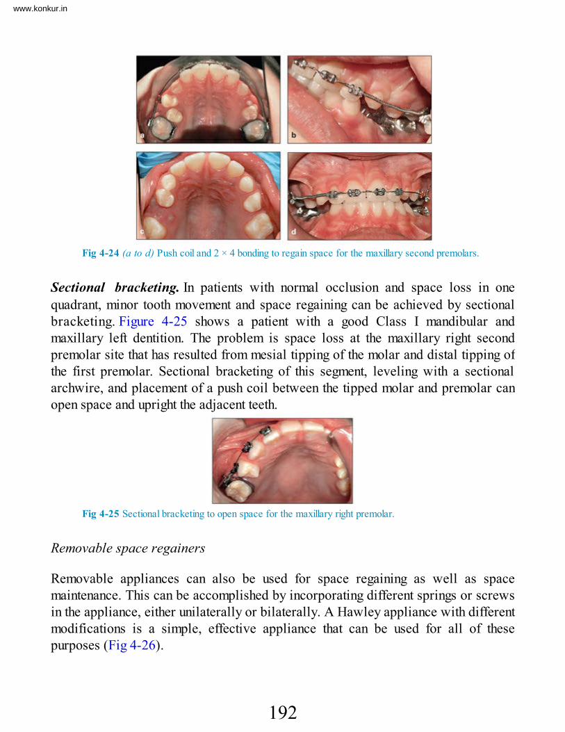
Fig 4-24 (a to d) Push coil and 2 × 4 bonding to regain space for the maxillary second premolars.
Sectional bracketing. In patients with normal occlusion and space loss in onequadrant, minor tooth movement and space regaining can be achieved by sectionalbracketing. Figure 4-25 shows a patient with a good Class I mandibular andmaxillary left dentition. The problem is space loss at the maxillary right secondpremolar site that has resulted from mesial tipping of the molar and distal tipping ofthe first premolar. Sectional bracketing of this segment, leveling with a sectionalarchwire, and placement of a push coil between the tipped molar and premolar canopen space and upright the adjacent teeth.
Fig 4-25 Sectional bracketing to open space for the maxillary right premolar.
Removable space regainers
Removable appliances can also be used for space regaining as well as spacemaintenance. This can be accomplished by incorporating different springs or screwsin the appliance, either unilaterally or bilaterally. A Hawley appliance with differentmodifications is a simple, effective appliance that can be used for all of thesepurposes (Fig 4-26).
192
www.konkur.in

Fig 4-26 Hawley removable space regainers with jackscrews. (a and b) Bilateral removableregainers for the maxilla. ( c ) Bilateral removable regainer for the mandible. ( d ) Unilateralremovable regainer for the maxilla.
The two main advantages of removable regainer appliances are that they facilitatecleansing and maintenance of good oral hygiene and they allow patients to open thescrew outside the mouth. Another advantage is that these appliances can be designedto have multiple actions, such as concomitant space maintenance, space regaining,and tooth movement.
The major disadvantage of removable space regainers, like all other removableappliances, is the need for patient compliance in wearing of the appliance. Breakageand loss of the appliance are other disadvantages.
Space CreationSpace creation is another type of space management that can be performed in casesof moderate space deficiency. This indication is different from cases in which thepatient has experienced space loss and requires space regaining. In situations thatrequire space creation, the patient exhibits moderate crowding in the arch, and spaceanalysis indicates that there are some overall space deficiencies.
Space creation is a tactic that can be applied in certain cases, especially duringthe mixed dentition as an early intervention procedure. Generally, creation of spaceis much more complex than space maintenance and regaining, and it involves moresophisticated application of biomechanics and appliance therapy. The patient’s ageand growth potential are important factors that must be considered in treatmentplanning for space creation, as in any other early treatment.
193
www.konkur.in

Space creation can be achieved in the following ways:
• Tooth extraction• Sequential selective enamel stripping• Sagittal expansion• Transverse expansion• Combination of the aforementioned methods
Tooth extractionTooth extraction is a general strategy applied in cases of severe crowding that needmore than 7 to 8 mm of space, where extraction combined with comprehensiveorthodontic treatment is the only option (for more details, see chapter 5).
Sequential selective enamel strippingIn some cases of moderate crowding, a tooth size discrepancy in a segment of dentalarch may be the cause of crowding while the rest of the dentition shows normalrelationships. An example is mandibular incisor crowding resulting from oversizedmandibular incisors (Bolton discrepancy).
The best treatment option in these situations is interproximal stripping to alleviatethe crowding.
Removal of minor amounts of hard tissue with a disk does not cause patientdiscomfort; some postoperative sensitivity may occur, and fluoride therapy helps. Itis possible to gain as much as 3 to 4 mm of anterior space through this procedure.
Sagittal expansionAnother option for space creation is an increase in the anteroposterior arch length,or sagittal expansion. The amount of space that can be created through this kind ofexpansion is limited, and the technique is more complicated and sometimes verydifficult to achieve in some cases. Therefore, careful clinical and paraclinicalevaluations must be completed for each patient.
With this strategy, the arch length can be increased through protrusion of anteriorteeth, by distalization of posterior teeth, or by a combination of both. Protrusion of
194
www.konkur.in

anterior teeth and distalization of posterior teeth, if applied in the wrong situation,are unstable and can result in other complications.
Protrusion must be applied to the proper patients at the proper time and withcareful mechanics. The results are not only unstable but also can disturb the patient’sprofile and cause alveolar bone resorption. Molar distalization, especially in themandible, is not only a difficult process but also unstable and can result in manycomplications, such as second molar impaction, vertical dimension problems, anddisturbances of the patient’s profile.
Nevertheless, careful creation of space in the anteroposterior dimension of thedental arch during the mixed dentition in suitable cases is the kind of earlyinterceptive treatment that can obviate extraction in the permanent dentition.
Case selection
Treatment planning and selection of appropriate cases are the first steps in this typeof treatment. The following are important points to consider before sagittalexpansion:
• Age of the patient and growth potential• Patient’s growth pattern• Molar position and inclination• Incisor position and inclination• Roots and periodontal condition• Patient’s soft tissue profile• Intraoral and extraoral muscle balance• Periodontal condition of the incisors• Lip size and position relative to incisors• Lip dysfunction• Deleterious oral habits
Patient’s growth pattern. One of the tactics for space creation through sagittalexpansion is distalization of the permanent molars. Application of this technique insome cases can be very harmful to the occlusion, such as in patients with a verticalgrowth pattern, in whom molar distalization will result in additional mandibularrotation, greater anterior open bite, and more vertical problems. In contrast, molardistalization in patients with some horizontal growth patterns not only creates space
195
www.konkur.in

but also can be helpful in reducing anterior overbite and lower facial height.
Molar position and inclination. Another important point that must be consideredbefore distalization of the first molars is the position and condition of other molars;these considerations include tight contact between the first and second molars,crowding in the molar area, and the possibility of second molar impaction (Fig 4-27). Distalization of the first molar often causes impaction of the second molar; insome cases, preventive extraction of third molar buds might be the only choice.
Fig 4-27 Molar crowding that is unsuitable for first molar distalization because of severe crowdingin the second and third molars.
Incisor position and inclination. In certain patients with moderate anteriorcrowding, sagittal expansion can be the treatment of choice, provided that theinclination of incisors allows this movement; otherwise the result will be flaring ofanterior teeth.
As Nance16 indicated, labial movement of the incisors can be extremelydetrimental in some cases. Therefore, careful clinical and paraclinical assessments,including the incisor inclination relative to basal bone and other bony and soft tissuestructures, must be completed before this movement is attempted.
Patient’s soft tissue profile. Labiolingual movement of the incisors has a directeffect on the patient’s soft tissue profile that must be evaluated before sagittalexpansion.
Intraoral and extraoral muscle balance. The tonicity and function of the perioralmusculature also play an important role in the position and inclination of incisors.Labial movement that results in overproclination and places the incisors underexcessive muscle force and dysfunction would not have a stable result.
Periodontal condition of the incisors. The periodontal condition and bone supportin the incisor area are also important factors that must be considered before anylabiolingual movement of incisors is attempted, even if evaluation of the incisor
196
www.konkur.in

position and inclination support application of minor protrusive forces to theincisors as part of sagittal space creation.
Treatment options
After careful case selection, the options available for increasing the sagittaldimension of the dental arch are molar distalization, incisor proclination, and acombination of both.
Molar distalization. Maxillary and mandibular molar distalization can be achievedwith many different appliance therapies (see chapter 11). The present discussionwill be limited to space creation and the advantages and disadvantages related tothis procedure.
Certain cases suitable for application of this procedure include all cases ofmesially tipped molars or of Class II malocclusions with deep bite, deep curve ofSpee, and tipped maxillary and mandibular molars. A lip bumper or a lip bumpercombined with cervical headgear is the appliance of choice if used during the mixeddentition and in proper cases (see chapter 11).
In any situation, distalization of the molars requires careful analysis of the molarposition and the patient’s growth pattern and profile.
Incisor proclination. The other option for increasing arch length in the sagittaldirection is proclination of the incisors. This procedure can be the treatment ofchoice in certain cases, such as retroclined incisors that cause incisor crowding oranterior crossbite. Proclination of incisors for the purpose of space creation inunsuitable cases can also cause several complications, such as profile imbalance,overprotrusion of the incisors, and alveolar recession.
The ideal choices for application of incisor proclination are patients during themixed dentition stage whose incisors have some degree of crowding and are alreadyretroclined relative to basal bone. Usually these patients have a straight or slightlyconcave profile. An example of this situation is anterior dentoalveolar crossbitewith mandibular mesial shift causing crowding in the anterior region of the maxilla,and sometimes space deficiencies and impaction of the maxillary permanent canines.
Another indication for incisor proclination is treatment of patients with crowded,retroclined mandibular incisors that are causing overjet and overbite. The etiologyin these cases can be the early loss of mandibular primary canines, lip dysfunction,and finger sucking. Elimination of the cause and forward positioning of the
197
www.konkur.in

mandibular incisors creates space and can correct other related malocclusions, suchas overjet, crowding, and profile problems (see cases 4-3 and 4-6).
Mechanotherapy in sagittal expansion
Sagittal expansion can be achieved by many different devices, such as lip bumpers,headgear, all kinds of fixed distalizers, and removable appliances with a spring orscrew and extended arch (see the earlier section on space regaining in this chapterand chapter 12).
Lip bumper. The lip bumper is a semiremovable appliance that has severalindications in early orthodontic treatment. This appliance consists of two bandscemented to the mandibular permanent first or second molars and a removable bow.An acrylic resin pad is included in the front part of the bow, which is inserted in abuccal tube (Fig 4-28). The lip bumper does not touch the mandibular incisors, andtherefore the force of lip musculature is directed to the molars, causing molaruprighting and distalization. At the same time, the absence of force from the lip onthe incisors and the concomitant application of forward force from the tongue canprocline the incisors. Therefore, the lip bumper can create sagittal space in bothdirections.
Fig 4-28 Lip bumper for molar uprighting and distalization.
Active lingual arch. The active lingual arch is a fixed LHA with two finger springssoldered to the posterior part of the arch and extended to the lingual surface of themandibular incisors. This applies a buccal force to the incisors (see Fig 4-8). Thisappliance can be used to correct moderate crowding and linguoversion of themandibular incisors.
Pendulum appliance (maxillary molar distalizer). Currently several kinds of molardistalizer are available. One of these, the pendulum appliance, is a good spacecreator that works by distalizing the maxillary molars. It is a fixed type of appliance
198
www.konkur.in

that consists of an acrylic resin button on the palate to serve as anchorage and twoextensions soldered to the primary first molar or permanent first premolar bands.Two springs are inserted in the acrylic resin part; the other end of each spring isinserted in the palatal tube of the molar bands; the springs can be removed andreactivated in the mouth.
If a jackscrew is added to the palatal acrylic resin of this appliance, it canfunction as a rapid palatal expander and therefore achieve sagittal expansion throughthe springs and transverse expansion through the screw (Fig 4-29).
Fig 4-29 Pendulum appliance (molar distalizer).
Removable distalizer. The removable distalizer is a Hawley appliance with anacrylic resin portion and a labial bow that serve as anchorage. A jackscrew isembedded in the acrylic resin and connected to the molars by some clasps (see Fig4-26).The screw is activated every 3 to 5 days to distalize the molar. The appliancecan be unilateral or bilateral, and it can be used in the maxillary or mandibulardentition. Patients can reactivate it themselves and clean it after every meal. Patientcooperation is important for success.
Headgear. Headgear is another kind of molar distalizer that has many otherindications in early orthodontic treatment (see chapter 11).
Transverse expansionTransverse expansion is another type of space creation. Transverse expansion alsoneeds careful evaluation before any determination is made and before treatmentplanning. Creation of space by transverse expansion depends on the degree ofskeletal involvement and whether or not a functional shift exists. Posterior crossbiteswith mandibular shift need early intervention and maxillary expansion to correct theproblems and prevent skeletal deformity. These are ideal cases for space creation bytransverse expansion. Posterior crossbites, whether unilateral or bilateral, with orwithout mandibular shift, are morphologically and etiologically different and need
199
www.konkur.in

different treatment options and different mechanotherapy, by slow or rapidexpansion. This can be accomplished by a variety of available expanders (seechapter 12).
O’Higgins and Lee15 reported that a 1-mm increase in the intermolar width willallow approximately 0.3 mm reduction in overjet or creation of 0.6 mm of spacewithin the arch. Transverse expansion could be a treatment of choice in thefollowing situations:
• In a rapidly growing child• When there is lateral constriction of the dental arch• When the basal bone accommodates such a movement• In the mandibular arch, but only distal to the mandibular canines
There are many indications for which proper expansion at the proper time is thebest choice for early treatment during the primary or mixed dentition. Lateralexpansion can be accomplished in several ways, including dental expansion,dentoalveolar expansion, skeletal expansion, or a combination of these.
McInaney et al27 showed that early expansion therapy with Crozat applianceseliminated the need for extraction of primary canines or permanent premolars. Lutzand Poulton28 reported on 13 cases of expansion during the primary dentition,followed for 6 years. Comparing arch perimeter and width at the permanent caninesand premolars with those of a control group, they found slightly greater values in theexpanded group.
Orthopedic palatal expansion appears to be effective and stable if applied prior tolate puberty. The application of expansion must be determined by the specific needsof each patient. Expansion during the primary and mixed dentition for correction oftransverse abnormalities (unilateral or bilateral crossbite) is a common treatmentoption that is discussed in chapter 12.
Another indication for transverse expansion during the mixed dentition iscorrection of moderate incisor crowding. Sayin and Türkkahraman,29 studying thepossible factors contributing to mandibular anterior crowding during the early mixeddentition, compared the total incisor widths, primary arch widths, and permanentintermolar widths of a group with crowding and a group without crowding. Theyfound significant correlations between crowding and total available incisor spaceand between primary and permanent arch widths and interalveolar width but nosignificant correlation between crowding and total arch length.
200
www.konkur.in

Radnzic30 compared arch dimensions in 120 boys (aged 13 years to 15 years 11months) and reported that boys with crowding had smaller arch widths than did boyswithout crowding or with spaced arches. He suggested that expansion in carefullyselected young patients might be beneficial.
Depending on the dentoskeletal relationships of the dental arch, several kinds offixed and removable techniques can be applied for transverse expansion. Formaxillary arch expansion, several kinds of appliances are available, including theremovable jackscrew expander, the W-arch, the quad helix, the reverse TPA, theHyrax expander, and the Haas expander. For the mandibular arch, a removablejackscrew, a lingual arch with expansion arm, or a lip bumper can be used (seechapter 12).
Fixed expanders
Various fixed expanders can be used during the primary, mixed, and permanentdentitions for different reasons such as posterior crossbite correction and somespace creation, including the W-arch, the quad helix, the Haas expander, and theHyrax expander.
W-arch. The W-arch, or Porter appliance, is a W-shaped arch soldered to molarbands made with 0.036- or 0.040-inch stainless steel wire. Depending on the shapeof the arch, the appliance can be activated in different ways (Fig 4-30). If molarexpansion is needed, the U-shaped bar in the anterior part can be widened; ifexpansion at the buccal segments (primary molars or premolars) is required, the U-shaped loop distal to the molar bands can be activated; if expansion for both thepermanent molars and primary molars is required, both the anterior and posteriorparts of the expander can be activated.
Fig 4-30 W-arch expander (Porter appliance).
The W-arch is an efficient and easy appliance for correction of posteriorcrossbite during the primary or mixed dentition, especially in 3- to 5-year-old
201
www.konkur.in

children. It is well tolerated by children, and it does not need activation by patientsor parents. It provides a continuous force and can be reactivated every 4 to 6 weeks.The W-arch also can be used as a reminder appliance for control of finger habits.
Quad helix. The quad helix is another bilateral fixed expander. It has almost thesame design specifications as the W-arch but has four coils incorporated in the arch,which give it more flexibility and a greater range of activity (Fig 4-31).
Fig 4-31 Quad helix bilateral fixed expander.
Haas expander. The Haas expander is the first type of expansion applianceintroduced by Haas.31 It is designed for lateral expansion, and because of the palatalacrylic resin it is especially effective for opening and separating the midpalatalsuture. It can be used during both the mixed and permanent dentition stages.
This appliance is both tooth and tissue borne, and it is usually fabricated withbands on the premolars and molars; in the mixed dentition, bands can be placed onthe primary molars. Two heavy palatal wires are formed to touch both sides of thebuccal segments and are soldered to the bands. The wires extend to the palate,where they are embedded in the acrylic resin pads. A jackscrew is located at thecenter of the acrylic resin (Fig 4-32). The acrylic resin pads and lingual wire applypressure to both the teeth and palatal mucosa.
Fig 4-32 Haas rapid expander.
The Haas appliance has several indications for use, such as expansion ofconstricted maxillary arches in patients with maxillary deficiency, correction ofposterior crossbite, and alleviation of some maxillary crowding.
202
www.konkur.in

Hyrax expander. The Hyrax expander is a fixed rapid expander made entirely fromstainless steel. The appliance is typically fabricated with bands on the first molarsand lingual bars extending to the first premolar. A screw located in the center isseparated from the tissue, and therefore the appliance is more hygienic than the Haasexpander (Fig 4-33).
Fig 4-33 Hyrax rapid expander.
Removable expanders
The Schwarz expander is a removable slow expander that has been used in themaxilla and the mandible. The basic parts of this appliance are a jackscrew withsome kind of clasp for retention that is embedded in acrylic resin (Fig 4-34). Thescrew can be opened and reactivated every 3 to 5 days.
Fig 4-34 Schwarz removable slow expander.
The Schwarz expander is especially useful for dental expansion during the earlymixed dentition. It can be removed by the patient for cleaning, and therefore its userequires the patient’s compliance.
Space ClosureAnother type of space management is elimination or closure of abnormal spacing thatcan interfere with normal development of the dentition and occlusion. The aims ofspace closure are to guide eruption and facilitate normal transitional changes during
203
www.konkur.in

occlusal development.Several etiologic factors can produce abnormal spacing in the dentition, such as
congenital absence of teeth, microdontia, abnormal habits, and large tongue. Each ofthese problems has different etiologic factors and different treatment options, whichare discussed in their related chapters.
An example of space closure as a space management strategy is during transitionof the maxillary incisors. Maxillary permanent incisors usually erupt with adiastema, and during the eruption of the lateral incisor and canine, this space willgradually close. Before their eruption, lateral incisors are slightly behind the centralincisor; after the central diastema is closed, the lateral incisor will erupt morelabially. If this normal pattern is disturbed and the central diastema remains open,lateral incisors may erupt palatally into a crossbite situation. With early detectionand intervention, closure of the diastema provides space for lateral incisors andprevents the development of crossbite.
Another example is when a patient is congenitally missing one or both lateralincisors, resulting in a large diastema. Not only is this situation estheticallyunacceptable, but also the probability of canine impaction is high; therefore, closureof the diastema and guidance of canine eruption is recommended.
The following are indications for space closure:
• When extra space is present• For management of hypodontia, when treatment planning requires a decrease in
arch length and space closure is the best treatment option (see chapter 7)• For closure of the resulting diastema after frenectomy when there is an abnormal
frenum attachment• When an open space such as an abnormally large diastema is preventing eruption
and causing malpositioning of adjacent teeth• For elimination of interdental spacing of maxillary incisors with severe overjet,
sometimes seen during the early mixed dentition, to prevent incisor fracture
Space Supervision and Guidance of EruptionIncisor crowding is a common problem during the mixed dentition. The amount andtype of crowding and its management is discussed in detail in chapter 5. In thissection, treatment of moderate crowding is discussed within the context of space
204
www.konkur.in

supervision.Space supervision is a term used for space management and correction of
moderate crowding during the mixed dentition when no severe arch length deficiencyis present. With this tactic, moderate crowding of incisors can be directed to theposterior segment and to leeway space.
The prognosis for space supervision is sometimes questionable, whereas theprognosis is always good for space maintenance and regaining.
As Moyers9 claimed, space supervision cases are those that will have a betterchance of getting through the mixed dentition with clinical guidance than without it.Proper selection of cases and a thorough case evaluation are always necessary forthis approach. Misdiagnosed space supervision cases that subsequently requireextraction of permanent teeth are more difficult to treat than cases of grossdiscrepancy.
The following are examples of different kinds of space supervision protocols:
• Sequential slicing of the primary canine will allow spontaneous alignment of thelateral incisors.
• Enamel stripping of the mesial surface of mandibular primary first molars willfacilitate eruption of the mandibular permanent canine and reduce pressure on thelateral incisors.
• Disking of the mesiodistal convexity of the primary molars will help normaleruption of adjacent teeth.
• Insertion of an LHA to preserve arch circumferences followed by sequentialextraction of primary first molars and then second molars will help anteriorcrowding.
According to Gianelly,32 the leeway space provides adequate space to resolvecrowding that is present during the mixed dentition in the majority of individuals. Healso recommended insertion of a lip bumper after eruption of the first premolars topreserve arch length.
In a study of patients with incisor crowding during the mixed dentition, Brennanand Gianelly33 preserved arch length and found adequate space to resolve thecrowding in 73 (68%) of 107 patients.
Case Reports
205
www.konkur.in

The following cases show correction of moderate crowding, whether the problemwas space loss that could be regained or moderate space deficiency that could bemanaged with space creation. As mentioned earlier, space creation is appropriatefor cases of moderate space deficiency when the incisor inclination, the patient’sprofile, and the relationship between the teeth and basal bone permit creation of thenecessary space by minor sagittal or transverse expansion. These types ofinterceptive treatment are especially recommended during the mixed dentition stageand when children are growing.
Case 4-1A 13-year-old girl in the early permanent dentition stage had a Class I malocclusion, zero overjet, astraight profile, and good mandibular dentition. She presented with maxillary crowding; both maxillarylateral incisors were in crossbite (locked occlusion), and both maxillary canines were out of the archbecause of space deficiency (Figs 4-35a to 4-35d).
Treatment:The treatment plan called for nonextraction management; minor space creation was to be achieved bycross-bite correction of the lateral incisors (sagittal movement) and slight expansion. The treatmentbegan with slow expansion with a maxillary Hawley appliance that had occlusal coverage to disoccludethe anterior teeth during correction of the lateral crossbite. This was followed by 2 × 6 maxillarybonding to help lateral incisor proclination and alignment of the canines. Use of the Hawley appliancewas stopped, and treatment was concluded with canine alignment. Figures 4-35e to 4-35g show theposttreatment occlusion.
206
www.konkur.in

Fig 4-35 Space creation in a 13-year-old girl in the early permanent dentition stage. (a to d)Pretreatment occlusion. There is maxillary crowding, both maxillary lateral incisors are in crossbite(locked occlusion), and both maxillary canines are out of the arch because of space deficiency. (eto g) Posttreatment occlusion.
Case 4-2A 10-year-old boy in the late mixed dentition stage had a Class I malocclusion and space deficiency inthe maxillary right and mandibular left and right segments as a result of early primary molar loss (Figs 4-36a to 4-36f).
Treatment:Space was regained by 2 × 4 bonding first and later by bonding of erupted premolars and a push coilmechanism for space opening and uprighting of the tipped molars (Figs 4-36g and 4-36h). Figures 4-36ito 4-36k show the posttreatment occlusion.
Fig 4-36 Space regaining in a 10-year-old boy in the late mixed dentition stage. (a to e)Pretreatment occlusion. (f) Panoramic radiograph showing that the maxillary right premolars andmandibular left and right premolars (arrows) are affected by space deficiency due to earlyprimary loss. (g and h) Panoramic radiographs showing the mechanism of space opening. (i to k)Posttreatment occlusion.
207
www.konkur.in

Case 4-3A 13-year-old girl had a Class I malocclusion and right molars with a slight Class II tendency. Themaxillary midline had shifted to the right due to early loss of the primary canine. There was no space forthe maxillary right permanent canine, and the left canine was slightly out of the arch. She also exhibitedmandibular anterior crowding (Figs 4-37a to 4-37g).
Treatment:The treatment involved space creation (sagittal expansion) through distalization of the maxillary rightsegment and slight proclination of the maxillary and mandibular incisors (Figs 4-37h to 4-37n).
Fig 4-37 Space creation in a 13-year-old girl with early loss of maxillary primary canines,maxillary midline shift (arrows), no space for the maxillary right canine, and mandibular anteriorcrowding. (a to e) Pretreatment occlusion. ( f ) Pretreatment panoramic radiograph. (g)Pretreatment lateral cephalometric radio-graph. (h to l) Posttreatment occlusion.
208
www.konkur.in

Fig 4-37 (cont) (m) Posttreatment panoramic radiograph. (n) Posttreatment lateral cephalometricradiograph.
Case 4-4A 10-year-old boy had a history of multiple primary tooth extractions because of extensive caries. Hepresented with severe space deficiency because primary tooth extraction was not followed by properspace management. At the time of presentation, he had a Class II molar relationship, normal overjet andoverbite, posterior cross-bite, and a 2-mm midline diastema. The midline was shifted to the left, andthere was a 12- to 14-mm space deficiency in the maxillary and mandibular arches (Figs 4-38a to 4-38f).
Treatment:Because of his normal skeletal pattern, normal sagittal basal bone relationship, and straight profile, thetreatment plan called for nonextraction space creation through transverse expansion to correct theposterior cross-bite, sagittal expansion by maxillary molar distalization for correction of the Class IImolar relationship, space opening for unerupted canines and premolars, and uprighting of eruptedpermanent teeth (Figs 4-38g to 4-38m).
Fig 4-38 Space creation in a 10-year-old boy with severe space deficiency resulting from earlyloss of primary teeth and the lack of any space management. (a to d) Pretreatment occlusion. (e)Pretreatment panoramic radiograph. (f ) Pretreatment cephalometric radiograph. (g ) Panoramic
209
www.konkur.in

radio-graph taken during treatment.
Fig 4-38 (cont) (h to k) Posttreatment occlusion. (l) Posttreatment panoramic radiograph. (m)Posttreatment cephalometric radiograph showing the condition of the dentition and thedentoskeletal relationships.
Case 4-5This case is an example of space supervision. A 14-year-old girl presented with four submergedprimary molars preventing eruption of premolars and normal vertical alveolar growth and consequentlydisturbing normal occlusal development (Fig 4-39a to 4-39e).
Figures 4-39f to 4-39j reveal the status of the occlusion after extraction of the primary submergedteeth, placement of anchorage appliances (maxillary Nance and LHA), guidance of eruption, and noother treatment or bonding. Figures 4-39k to 4-39n show the posttreatment results.
Fig 4-39 Space supervision in a 14-year-old girl with four submerged primary molars that arepreventing premolar eruption and alveolar growth, disrupting the development of occlusion. (a to c)Pretreatment occlusion.
210
www.konkur.in

Fig 4-39 (cont) (d) Pretreatment panoramic radiograph. ( e ) Pretreatment cephalometricradiograph. (f to j) Condition of the occlusion during space supervision, which involved extractionof the primary submerged teeth, anchorage preparation (maxillary Nance holding arch and lowerholding arch), and guidance of eruption. (k to m ) Posttreatment occlusion. ( n ) Posttreatmentcephalometric radiograph.
Case 4-6A 10-year-old girl exhibited a Class I malocclusion (left molars with a Class II relationship) and severemaxillary and mandibular crowding due to space loss. Both mandibular canines were out of the arch, themaxillary midline had shifted to the left, and there was no space for the maxillary right canine, whichshowed a prominent canine eminence (Figs 4-40a to 4-40f).
Treatment:
211
www.konkur.in

Because of the patient’s age and skeletal pattern and the etiology of crowding, that is, early loss of themandibular primary canines and maxillary left primary canine, the plan was designed as nonextractiontreatment with space regaining and minor space creation. A lower lingual holding arch was placed topreserve leeway space. Maxillary and mandibular anterior teeth were bonded using push coil springs todistalize the maxillary left molar and shift the midline to the right with minor incisor proclination.Thisachieved correction of the maxillary midline and space opening for the maxillary left canine. Figures 4-40g to 4-40l show the posttreatment results.
Fig 4-40 Space regaining and minor space creation in a 10-year-old girl with maxillary andmandibular crowding due to space loss. (a to e) Pretreatment occlusion. ( f ) Pretreatmentpanoramic radiograph. (g to k) Posttreatment occlusion. (l) Posttreatment panoramic radiograph.
Summary
• Every tooth in its socket over the basal bone is maintained under continuous,balanced mesiodistal, buccolingual, and occlusal forces from adjacent andopposing teeth.
• Any change in this balancing force, such as loss of a tooth unit, can causeunfavorable tooth movement of the adjacent and opposing teeth and produce
212
www.konkur.in

tooth malrelationships. Therefore, any space as a result of tooth loss must becontrolled by some kind of space management.
• Management of the space depends on several factors, such as the patient’socclusion, the time elapsed since tooth loss, and the age of the patient.
• Proper space management requires sufficient knowledge of the etiology andmorphology of tooth loss, the type of tooth movement, the effects of primarymolar extraction on succedaneous teeth, the incidence and nature of spaceloss, prediction of tooth emergence, and factors influencing mesial anddistal tooth drift.
• Depending on these factors, several kinds of management are available:space maintenance, space regaining, space creation, space closure, andspace supervision.
• Space maintenance is a preventive type of management to prevent space lossand abnormal tooth movement.
• Space regaining is an interceptive procedure to regain lost space.• Space creation is a corrective procedure to create some space in patients
with moderate space deficiency. This procedure is more complicated thanspace regaining and requires biomechanical principles and more cautioustooth movement.
• Space closure is an interceptive procedure for guidance of eruption (eg,closure of abnormal diastemas to guide normal eruption of lateral incisors).
• Space supervision is also a kind of interceptive procedure to superviseocclusal development, such as by controlling leeway space to eliminatemoderate incisor crowding.
References1. Fanning EA. A longitudinal study of tooth formation and root resorption. N Z Dent J 1961;57:202–217.2. Posen AL. The effect of premature loss of deciduous molars on premolar eruption. Angle Orthod
1965;35:249–252.3. Owen DG. The incidence and nature of space closure following the premature extraction of deciduous teeth:
A literature survey. Am J Orthod 1971;59:37–49.4. Lundström A. The significance of early loss of deciduous teeth in the etiology of malocclusion. Am J Orthod
1955;41:819–826.5. Linder-Aronson S. The effect of premature loss of deciduous teeth: A biometric study in 14- and 15-years-
olds. Acta Odontol Scand 1960;18:101–122.6. Seipel CM. Prevention of malocclusion. Trans Eur Orthod Soc 1947/1948:203–211.7. Johnsen DC. Space observation following loss of the mandibular first primary molars in mixed dentition. J Dent
Child 1980;47:24–27.
213
www.konkur.in

8. Dean JA, Avery DR, McDonald RE. McDonald and Avery’s Dentistry for the Child and Adolescent, ed 9. StLouis: Mosby, 2010.
9. Moyers RE. Handbook of Orthodontics, ed 4. Chicago: Year Book Medical,1988.10. Northway WM, Wainright RL, Demirjian A. Effect of premature loss of deciduous molars. Angle Orthod
1984;54: 295–329.11. Grøn AM. Prediction of tooth emergence. J Dent Res 1962;41:573–585.12. Hägg U, Taranger J. Dental development, dental age and tooth counts. Angle Orthod 1985;55:93–107.13. Demirjian A. Cranio-maxillofacial and somatic growth and oral health in French Canadian children [in
French]. J Can Dent Assoc (Tor) 1968;34:79–78.14. Smith SL, Buschang PH. Growth in root length of the mandibular canine and premolars in a mixed-
longitudinal orthodontic sample. Am J Hum Biol 2009;21:623–634.15. O’Higgins M, Lee RT. How much space is created from expansion or premolar extraction? J Orthod
2000;27:11–13.16. Nance H. Limitations of orthodontic treatment in the permanent dentition. 2. Diagnosis and treatment in the
permanent dentition. Am J Orthod 1947;33:253–301.17. Tanaka MM, Johnston LE. The prediction of the size of unerupted canines and premolars in a contemporary
orthodontic population. J Am Dent Assoc 1974;88:798–801.18. Staley RN, Kerber PE. A revision of the Hixon and Old-father mixed-dentition prediction method. Am J
Orthod 1980;78:296–302.19. Merrifield LL. Differential diagnosis. Semin Orthod 1996;2:241–253.20. Bolton WA. The clinical application of a tooth-size analysis. Am J Orthod 1962;48:504–529.21. Björk A, Skieller V. Normal and abnormal growth of the mandible: A synthesis of longitudinal cephalometric
implant studies over a period of 25 years. Eur J Orthod 1983;5:1–46.22. Andrews LF. The six keys to normal occlusion. Am J Orthod 1972;9:296–309.23. Moorrees CF, Chadha JM. Available space for the incisors during dental development—A growth study
based on physiologic age. Angle Orthod 1965;36:12–22.24. Ricketts RM. Early treatment. 1. J Clin Orthod 1979;13:23–38.25. Ricketts RM. Early treatment. 2. J Clin Orthod 1979;13:115–127.26. Ricketts RM. Early treatment. 3. J Clin Orthod 1979;13:181–199.27. McInaney JB, Adams RM, Freeman M. A nonextraction approach to crowded dentitions in young children:
Early recognition and treatment. J Am Dent Assoc 1980;101:251–257.28. Lutz HD, Poulton D. Stability of dental arch expansion in the deciduous dentition. Angle Orthod 1985;55:299–
315.29. Sayin MO, Türkkahraman H. Factors contributing to mandibular anterior crowding in the early mixed
dentition. Angle Orthod 2004;74:754–758.30. Radnzic D. Dental crowding and its relationship to mesiodistal crown diameters and arch dimensions. Am J
Orthod Dentofacial Orthop 1988;94:50–56.31. Haas JA.The treatment of maxillary deficiency by opening the midpalatal suture. Angle Orthod 1965;35:200–
217.32. Gianelly AA. Leeway space and the resolution of crowding in the mixed dentition. Semin Orthod 1995;1:188–
194.33. Brennan MM, Gianelly AA. The use of the lingual arch in the mixed dentition to resolve incisor crowding. Am
J Orthod Dentofacial Orthop 2000;117:81–85.
214
www.konkur.in

Management of Incisor Crowding
Incisor crowding is one of the most frequently observed dental phenomena in youngchildren during the early and middle mixed dentition periods. Incisor crowding hasbeen a subject of increasing concern for children and their parents, and it is amongthe most common reasons patients seek orthodontic treatment.
Because of the discrepancy between primary and permanent incisors, some degreeof incisor crowding is very common during the early mixed dentition. Some of thiscrowding can be transitional and needs no intervention, some can be corrected withocclusal guidance and space supervision, and some can be critical and develop to asevere malocclusion and therefore must be managed with tooth extraction at theproper time.
Overall space availability for all dentition varies, depending on the growth of thejaws and relative variation in the mesiodistal crown diameters of the permanentteeth and their predecessors. Children who have little or no developmental spacingbetween the primary incisors and occasionally some crowding in the primarydentition develop severe crowding in the permanent incisors. As Baume1 showed,children with no spacing during the primary dentition (Baume class II) have a 40%chance of developing crowding in their permanent incisors.
Mandibular anterior crowding is initially identified as a discrepancy between themesiodistal tooth widths of the four permanent incisors and the available space inthe anterior part of basal bone. However, many other variables must be consideredprior to early intervention and treatment, such as the direction of mandibular growth,early loss of primary molars, incisor and molar inclinations, and oral and perioralmusculature balance. All of these factors can play a role in and be associated withincisor crowding.
215
www.konkur.in

Because not all incisor crowding during the mixed dentition is alike, the treatmentprocedure must vary depending on the etiology and morphology of the crowding.However, this common problem sometimes is a cause of great concern for parentsand patients, so practitioners must be prepared to address this issue with parents.The most appropriate answer to their questions is not, “This is not a problem,” or“Wait until all permanent teeth erupt.” Rather, the wise and proper answer is, “Thismight be a sign of other problems, so the situation should be evaluated now.”
The next question for practitioners is how to identify and differentiate differentsituations of incisor crowding to enable optimal management of each case. Thischapter will answer this question by examining different aspects of incisorcrowding:
• Is there any way to predict this problem?• What is the common cause of this problem?• How can different types of crowding be classified and differentiated?• What kind of intervention can prevent the problem?• How can the problem be treated?
PredictionPredicting permanent incisor crowding at an earlier stage has been a goal in severallongitudinal studies during the primary, mixed, and permanent dentitions. However,there are conflicting results in the literature evaluating the correlation betweencrowding and other dentofacial structures. Baume1 reported that children with nospacing during the primary dentition (Baume class II) have a 40% chance ofcrowding in their permanent incisors.
Hunter,2 after analysis of crowding and spacing of the teeth, concluded thatestimates of the size of unerupted teeth in the mixed dentition are more accurate thanestimates of future spacing or crowding; this is because arch perimeter does notdecrease by the same amount in all cases but is rather associated with the amount ofcrowding in the mixed dentition. The amount of crowding or spacing must always bedetermined after considering molar, canine, and incisor relationships as well as thecondition of lip posture on the dentition.
Sampson and Richards3 tested the hypothesis that pre-eruptive tooth positions anddental arch parameters might forecast crowding changes in Class I untreated
216
www.konkur.in

dentition unaffected by caries or attrition. Crowding scores, radiographicrelationships, and dental arch dimensions were evaluated for the mixed dentition.The sample was divided into two groups, depending on whether there was anincrease or decrease in incisor and canine crowding between the mixed and thepermanent dentitions. Dental arch form and tooth size were important factors inmeasuring the amount of incisor or canine crowding at stage 1 and stage 2. Theresearchers found that neither the radiographic nor the dental arch predictors proveduseful in forecasting crowding changes.
Howe et al4 examined the extent to which tooth size and jaw size each contributeto dental crowding. Two groups of dental casts were selected on the basis of dentalcrowding. In one group, 50 pairs of dental casts exhibited gross dental crowding.The second group consisted of 54 pairs of dental casts that exhibited little or nocrowding. Howe et al4 observed significant differences between the dental archdimensions of the two groups. The crowded group was found to have smaller dentalarch dimensions than the noncrowded group. The researchers suggested thatpractitioners should consider treatments that increase dental arch length rather thanthose that reduce tooth mass.
Bishara et al5 attempted to determine the changes in the maxillary and mandibulartooth size–arch length discrepancy between the primary dentition (mean age of 4.0years) and second molar eruption (mean age of 13.3 years) to establish whether atooth size–arch length discrepancy in the permanent dentition can be predicted in theprimary dentition. They evaluated the records of 35 boys and 27 girls. Themesiodistal diameters of all primary teeth and their permanent successors, as well asvarious parameters of dental arch width and length, were measured in the primaryand permanent dentitions.
The researchers concluded that additional factors are involved in tooth size–archsize discrepancy and should be considered in explaining this phenomenon. Thesefactors are present in most persons, whether or not they have a malocclusion or haveundergone orthodontic treatment. Regression analysis indicated that these changesare related, in part, to tooth size as well as to changes in arch length and arch width.The correlations between the various primary and permanent tooth size–arch lengthdiscrepancies are of such a magnitude that they do not allow accurate prediction ofdiscrepancies in the permanent dentition from the available dental measurements inthe primary dentition.5
Sinclair and Little6 also found no clinically significant associations betweenvarious mandibular parameters and incisor crowding.
217
www.konkur.in

Melo et al7 searched for indicators during the primary dentition of futurecrowding of the mandibular incisors during the early mixed dentition. For thispurpose, they examined the skeletal and dental morphologic characteristics of theprimary dentition using dental casts and cephalograms of 23 subjects. They thenassessed the degree of crowding in the same subjects at 9 years of age and found 12normal and 11 crowded dentitions. The size of permanent incisors in the crowdinggroup was significantly greater than that in the normal group. The maxillary andmandibular dental arch lengths and the posterior cranial base length (sella-basion)were also effective discriminators between the two groups.
Common Causes
Conducting an implant study in children, Björk8 clearly showed that the directionand rotational pattern of mandibular growth have different effects on occlusion,incisor position, and crowding. In fact, the same implant studies revealed thatinternal jaw rotation also changes the anteroposterior position of the incisor teeth,which in turn has a major influence on changes in arch length. They showed that therotational pattern of jaw growth obviously influences the magnitude of tootheruption. When the mandible rotates downward and backward, anterior facial heightincreases, there is a tendency toward anterior open bite, and the incisors are thrustforward relative to the mandible. When excessive counterclockwise rotation occurs,short facial height develops, and lingual displacement of the incisors relative to themaxilla and mandible increases the tendency toward crowding.
Leighton and Hunter9 also reported that the angles between the sella-nasion lineand the mandibular and occlusal planes were significantly larger in patients withcrowding during both the mixed and permanent dentition stages. They alsoassociated downward and deficient growth of the mandible with upright orretroclined mandibular incisors and with crowding.
Sayin and Türkkahraman10 tried to find factors that can contribute to mandibularanterior crowding. They evaluated 60 dental casts of patients during the early mixeddentition, divided into two groups according to the severity of mandibular anteriorcrowding. In the mandible, the primary intercanine widths, intermolar widths,permanent intermolar widths, interalveolar widths, space available for themandibular permanent incisors, and total arch length were all significantly larger inpatients with crowding than they were in patients without crowding. No significantcorrelation was found between crowding and total arch length.
218
www.konkur.in

Türkkahraman and Sayin11 evaluated the association between dentofacialstructures and mandibular crowding during the early mixed dentition by analyzingcephalometric radiographs and casts of 60 children. They found that children withcrowding had smaller mandibular incisor–nasion–point B angles and shortermaxillary and mandibular skeletal length. They also found greater interincisalangles, overjet, overbite, and Wits appraisal measurements. They concluded thatcrowding of the mandibular incisors is not merely the result of a tooth size–arch sizediscrepancy; dentofacial characteristics also contribute to this misalignment.
Intervention and PreventionDespite much research and many longitudinal investigations to find specialcharacteristics and criteria for early detection and prevention of permanent incisorcrowding during the primary dentition, there is still much controversy on the subject.Practitioners still see young children with incisor crowding that was not preventedand highly concerned parents seeking some kind of early intervention. Nevertheless,incisor crowding during the mixed dentition can be managed differently dependingon the amount of crowding and other variables involved in this process, and allfactors must be considered in treatment planning.
There is no reliable criterion during the primary dentition to predict permanentincisor crowding, but even if it were possible prevention is not possible until allincisors are erupted. However, the author’s clinical experience in some earlytreatment during the primary dentition indicates that some type of early intervention,even before eruption of permanent incisors, can be useful in providing space for thepermanent incisors. For example, during rapid palatal expansion in the primary orearly mixed dentition for correction of posterior crossbite, some space can developfor incisors and canines, facilitating normal eruption of the anterior segment (Fig 5-1; see the discussion on early correction of posterior crossbite in chapter 12).
Fig 5-1 (a and b) Space opening (arrows) after rapid expansion and posterior crossbitecorrection.
Moreover, clinical observation indicates that an abnormal sequence of eruption
219
www.konkur.in

between mandibular canines and first premolars has a tendency to precede somekinds of mandibular incisor crowding, and in such cases early detection and properintervention can reduce or correct incisor crowding. Other examples of preventionand early intervention for mandibular incisor crowding are control of some types ofthumb sucking to retrude the mandibular incisors and control of lower lipdysfunction that can cause mandibular incisor crowding.
Longitudinal monitoring of panoramic radiographs and the three phases oftransitional dentition is a prudent measure that can be considered the first step inprevention and early intervention.
Characteristics and ClassificationMandibular anterior crowding is identified as a discrepancy between themesiodistal tooth widths of the four permanent incisors and the space availablebetween the mesial surfaces of the primary canines. Because crowding can resultfrom different morphologic and etiologic factors, the first step for the purpose ofproper treatment planning is to identify the kind of crowding, that is, to determine theamount of permanent incisor crowding during an early stage of the mixed dentition.This can be achieved by careful measurement and analysis of tooth size–arch sizediscrepancy (see chapter 4).
However, incisor crowding is not merely a tooth size–arch size discrepancy.Many variables, such as direction of mandibular growth, early loss of primarymolars, the oral and perioral musculature, and incisor and molar inclinations, can beassociated with crowding. Thus, the second issue for proper treatment planning is tosearch for dentofacial factors that might be associated with mandibular crowding,using all necessary diagnostic tools during the early mixed dentition.
After careful assessment of the aforementioned variables, incisor crowding can bearbitrarily classified into three types of crowding:
1. Minor crowding up to 3 mm of space deficiency2. Moderate crowding with 3 to 5 mm of space deficiency3. Severe crowding with more than 6 mm of space deficiency
However, the amount of crowding in millimeters is not the only decisive criterionfor treatment decisions; many other factors must be evaluated and considered intreatment planning. The purpose of this classification is to provide the first practical
220
www.konkur.in

step of discriminating between different amounts of crowding.Generally incisor crowding has been classified into two general types, acquired
crowding and hereditary crowding, which are discussed in detail later in thischapter.
TreatmentAlthough incisor crowding is not merely a function of tooth size–arch sizediscrepancy and many other local and general variables might be involved and mustbe evaluated, for practical purposes the treatment options have been classifiedaccording to the amount of incisor crowding (minor, moderate, and severecrowding).
Minor CrowdingBecause of the size difference between primary and permanent incisors (incisorliability), a minor amount of permanent incisor crowding (less than 3 mm) at anearly stage of the mixed dentition is considered transient crowding; up to 3 mm ofincisor crowding may resolve spontaneously during the transitional dentition withoutthe need for early intervention.
Studies of children with normal occlusion also indicate that transitional changesfrom the primary to the mixed dentition resolve up to 3 mm of incisor crowdingspontaneously without treatment. As was discussed in chapter 2, there are naturalmechanisms that can self-correct minor crowding of incisors during the transitionaldentition, such as interdental spacing, more proclination of permanent incisors, andintercanine width increase.
Moorrees and Reed12 studied a series of 184 casts at 3 years and 16 to 18 yearsof age. They found that, in the mandible, an average of 1.6 mm of crowding in boysand 1.8 mm of crowding in girls will recover to 0.0 mm by the age of 8 years. Theyattributed this recovery to a phenomenon termed secondary spacing, whichfacilitates lateral incisor eruption. This process occurs when the mandibular lateralincisors emerge and push the mandibular primary canines laterally. This also causesthe maxillary primary canines to move laterally by occlusal force, creating spacesfor the maxillary lateral incisors.
Extraction of the mandibular primary canines during this natural phenomenon will
221
www.konkur.in

prevent secondary spacing and the increase in intercanine width. Prevention of thisphenomenon may even convert a nonextraction case to an extraction case. Therefore,no greater relief of spacing in the incisor segment can be expected after completeeruption of the lateral incisors. They also added that the level of dental maturation,that is, tooth formation and emergence, gives decisive clues for diagnosis andtreatment planning because it defines the timetable of individual development.12
It is also important to remember that physiologic self-correction of minorcrowding can occur, provided that the following factors are present:
• Normal growth and development of the jaws• Normal interdental spacing, especially primate spaces• Normal labial inclination of the maxillary and mandibular incisors• Normal increase in intercanine width• On the other hand, the presence of any of the following disturbances can disrupt
self-correction of minor crowding and change the treatment plan:• Premature loss of primary teeth• Problems of tooth number, size, or form• Abnormal habits• Soft tissue problems, such as lip dysfunction• Hereditary or congenital disturbances
Therefore, for patients with minor incisor crowding, practitioners must carefullymonitor the transitional dentition and maintain arch length integrity against anyabnormal factors such as interproximal caries, accidental tooth loss, or abnormalhabits.
Moderate CrowdingModerate mixed dentition crowding is the presence of 3 to 5 mm of crowding duringthe transitional stage of dentition. Depending on the variable conditions of occlusion,moderate crowding can be treated differently at different stages of transition.Treatment can take place from the time of eruption of the mandibular permanentcentral incisors (around 6 years) to the late mixed dentition stage (around 11 to 12years).
Permanent incisor buds are located lingual to the root of primary incisors and,
222
www.konkur.in

especially in mandibular dentition, they often erupt lingually. Therefore, monitoringis very important at this stage of transition. For example, delayed exfoliation ofprimary incisors might be problematic; sometimes permanent central incisors eruptlingually while primary central incisors are still present and immobile. In thissituation, immediate radiographic evaluation and extraction of the over-retainedprimary teeth must be performed (Fig 5-2).
Fig 5-2 (a to c) Delayed resorption of the primary central incisor roots, causing lingual eruption ofthe permanent incisors.
If the primary canine has already exfoliated or been extracted without supervisionand some space has been lost, a space regainer such as a lip bumper should be usedto regain the lost space. Afterward, use of a lingual holding arch or fixed applianceis recommended (for more detail on space regaining, see chapter 4).
If, on the other hand, space analysis and evaluation of all other perioral factorsindicate that the space available in the arch and basal bone can accommodate allincisors, canines, and premolars and there is no evidence of space loss resultingfrom early loss of the primary dentition or caries, moderately crowded incisors canbe guided to better positions by careful space management in the posterior segmentof the arch, in a procedure called space supervision. In other words, when spaceanalyses indicate that there is “just enough” space, the situation is a borderline casefor extraction, and an attempt should be made to correct moderate crowding throughsupervision of the transitional dentition to prevent loss of leeway space. Moyers13
described space supervision cases as those that will make it through the mixeddentition better with clinical guidance than without it.
Proper selection of cases and a thorough case evaluation are always necessary forthis tactic. In patients with moderate anterior crowding, space supervision can beapplied through four different tactics:
1. Sequential stripping of primary teeth2. Preservation of E space
223
www.konkur.in

3. Space creation4. Combination approach, using more than one of the aforementioned methods
Sequential stripping of primary teethSequential stripping of primary lateral incisors, canines, or molars can allowspontaneous alignment of incisors and distal eruption of the canines and premolarswithout any mechanotherapy. Minor amounts of stripping do not cause patientdiscomfort. Greater amounts of slicing may require administration of localanesthesia and produce some postoperative sensitivity; fluoride therapy helpsalleviate the sensitivity. It is possible to gain as much as 4 mm (maximum) ofanterior space through this procedure. This procedure can be done by stripping metaltape or a fine flexible diamond disk.
Space supervision and slicing of primary teeth can be applied during eruption ofthe mandibular permanent lateral incisors, which usually have more difficulty inerupting than central incisors because of their size. Disking of the mesial surface ofthe primary canine can resolve the problem. In some cases of severe spacedeficiency, it may be necessary to extract the primary canines, but immediateinsertion of a lower holding arch with spurs is indicated to prevent any space lossthat can result from lingual movement of mandibular incisors (Fig 5-3).
Fig 5-3 Lower holding arch with spurs.
When disking or extraction of the mandibular primary canine is planned, thenormal transitional process of secondary spacing must be considered. The disking orextraction must be timed so as not to disrupt the development of secondary spacing.
Another example of sequential stripping is disking of the mesial surface of theprimary first molar to provide space for the mandibular permanent canine and guidedistal eruption of the canine helping spontaneous correction of moderate incisorcrowding. Likewise, the mesial surface of the primary second molar can be strippedto facilitate and guide first premolar eruption. With this technique, the moderateincisor crowding is actually transferred to the posterior segment, taking advantage of
224
www.konkur.in

leeway space, without use of any appliances.Another example of selective grinding is disking of the distal surface of the
mandibular primary second molar. This procedure is indicated when the maxillarypermanent first molar has erupted before the mandibular molar. Distal stripping ofthe primary second molar accelerates mesial migration of the mandibular molar toachieve a Class I molar relationship.
Preservation of E spacePreservation of E space, or guidance of eruption in moderate incisor crowding, isanother kind of space supervision that can be applied after careful evaluation ofdentofacial structures and muscles, space analysis, and confirmation of a moderatespace deficiency.
Gianelly14 stated that the late mixed dentition stage of dentition, after the eruptionof the first premolars, is a favorable time to start treatment to resolve incisorcrowding. Moyers13 and Gianelly15 suggested that in the majority of individuals withmoderate crowding in the early mixed dentition, this technique provides adequatespace to resolve the moderate incisor crowding.
The author suggests that the optimum time to apply this procedure is beforeexfoliation of both primary first and second molars. Insertion of a proper lingualholding arch connected to the permanent first molars before exfoliation of theprimary first and second molars will prevent any mesial shift of the first permanentmolar to leeway space. This tactic will preserve 2 to 4 mm of space for crowdedincisors.
After insertion of a good lower holding arch to prevent any mesial shift of thepermanent molars, extraction of the primary first molar will actually guide the firstpremolar eruption more distally. Subsequently, the canine can move distally tofacilitate spontaneous correction of incisor crowding.
Preservation of E space and guidance of eruption of premolars can be applied inpatients with primary molar ankylosis and tilted eruption of premolars.
The best appliances to preserve E space or prevent mesial movement ofpermanent molars are the lower holding arch in the mandible and the transpalatalarch in the maxilla (Fig 5-4).
225
www.konkur.in

Fig 5-4 (a and b) Preservation of E space to allow normal eruption of premolars.
Space creationSpace creation is another means of correcting moderate incisor crowding (3 to 5 mmof crowding) during the mixed dentition. The procedure of space creation is differentfrom space regaining, which is the procedure to apply when existing space has beenlost due to early loss of primary teeth. Rather, space creation is a procedure tocreate needed space. Space creation is a more complex procedure and needs muchmore evaluation and analysis before any treatment planning. This procedure can beperformed in the following ways:
• Transverse expansion• Sagittal expansion (anteroposterior arch length increase)• Selective stripping• Combination of the aforementioned methods
For more details about space creation, see chapter 4.
Combination approachIncisor crowding during the transitional dentition is a common problem that worriespatients and parents. In many patients, the problem can be treated with nonextractionprocedures at an early age, while tooth extraction will be required if the treatment ispostponed to the permanent dentition.
Clinical experience indicates that this kind of crowding usually develops duringincisor transition as a result of a combination of etiologic factors such as earlyprimary tooth loss, trauma, supernumerary teeth, missing teeth, Bolton discrepancy,abnormal habits, abnormal muscle function, abnormal basal bone relationships suchas deep bite, or anterior crossbite. Thus, the practitioner must take advantage of all
226
www.konkur.in

the examinations and diagnostic tools available to search for dentofacial factors inthe early mixed dentition that might be associated with mandibular crowding.
As discussed earlier, Björk and Skieller16 reported that the mandibular growthpattern and mandibular rotation affect incisor position and crowding. When themandible rotates downward and backward, anterior facial height increases, there isa tendency toward anterior open bite, and the incisors are thrust forward relative tothe mandible. When excessive counterclockwise rotation occurs, a short facialheight results, and lingual displacement of the incisors relative to the maxilla andmandible increases the tendency toward crowding. Therefore, these types ofcrowding with multifactorial causes cannot be managed by space supervision orspace creation alone. However, elimination or prevention of the causes, especiallythrough early detection and proper intervention, can result in a stable and balancedocclusion. The following cases are examples of this type of crowding that can bemanaged at an early age.
Case 5-1A 10-year, 8-month-old boy presented with a Class I malocclusion, locked anterior crossbite (withoutmandibular shift), maxillary incisor crowding, displacement, and a diastema (Figs 5-5a to 5-5e).
A panoramic radiograph taken about 1 year after an accident (Fig 5-5f) revealed the effect of traumato the permanent incisors (early resorption of the left primary central and lateral incisors and delayedresorption of the right primary lateral and central incisors), causing delayed eruption of the rightpermanent central and lateral incisors, tilting to crossbite, and mandibular crowding (Fig 5-5g to 5-5j).
Treatment:Treatment was started with 2 × 4 maxillary bonding and placement of composite resin on the occlusalsurfaces of the mandibular molars to disocclude the anterior segment. A nickel-titanium archwire wasalso placed for leveling, followed by a stainless steel archwire with open loop for correction of incisorcrossbite.
After crossbite correction was completed, mandibular bonding was performed for correction ofincisor crowding. Figures 5-5k and 5-5l show the dentition during treatment. Figure 5-5m reveals thechange of canine inclination. This patient was then treated only with 2 × 6 maxillary and mandibularbonding. Figures 5-5n to 5-5r show the final posttreatment occlusion.
227
www.konkur.in

Fig 5-5 Treatment of a 10-year, 8-month-old boy with a Class I malocclusion, locked anteriorcrossbite, maxillary incisor crowding, displacement, and a diastema. (a to e) Pretreatmentocclusion.
228
www.konkur.in

Fig 5-5 (cont) (f) Magnified panoramic radiograph, taken about 1 year after trauma, revealsdisplacement of the maxillary permanent right lateral incisor. (g to j) Consecutive pretreatmentpanoramic radiographs. (k and l) Dentition during treatment. (m) Panoramic radiograph duringtreatment, revealing the change in canine inclination. (n to r) Posttreatment occlusion.
Case 5-2A 10-year-old girl presented with an anterior locked occlusion that caused severe crowding anddisplacement of the mandibular incisors (Figs 5-6a to 5-6f).
229
www.konkur.in

Treatment:A nonextraction treatment procedure was planned. Treatment was started with mandibular 2 × 6bonding and application of composite resin on the occlusal surfaces of the mandibular first molars todisocclude the anterior segment. After correction of crossbite and proclination of all of the maxillaryincisors, mandibular 2 × 3 bonding was used first to align three incisors and create space for themandibular right lateral incisor. The final step was traction of the severely displaced right lateral incisorinto the arch and alignment (Figs 5-6g to 5-6l).
Fig 5-6 Treatment of a 10-year-old girl with severe anterior crowding. (a to e) Pretreatmentocclusion. ( f ) Pretreatment panoramic radiograph. (g to k) Posttreatment occlusion. (l)Posttreatment panoramic radiograph.
Case 5-3A 10-year-old boy presented with a Class I very deep bite (impinging) and retroclined maxillary andmandibular incisors. He had severe mandibular incisor crowding, and the mandibular right lateral incisorwas completely displaced lingually. Early loss of the mandibular primary incisors and deep bite resultedin mandibular anterior collapse and some retrusion of the maxillary incisors (Fig 5-7a to 5-7f).
Treatment:
230
www.konkur.in

The treatment plan involved correction of the deep bite, slight proclination of the incisors (spacecreation), and correction of crowding without extraction. First, the maxillary first molars were bandedand an anterior bite plate was placed to disocclude the posterior segment in order to stimulate tootheruption and reduce overbite. Then 2 × 4 bonding was used for maxillary incisor alignment. A maxillaryutility arch was used to intrude the maxillary anterior segment and extrude the posterior segment. Lowe2 × 4 bonding was used for mandibular incisor alignment and proclination, to create some space toalleviate mandibular crowding. The final step was to continue with complete-mouth bonding forcorrection of the deep bite. Figures 5-7g to 5-7k show the posttreatment results, and Figs 5-7l and 5-7mshow the pre- and posttretment cephalometric radiographs.
Fig 5-7 Treatment of a 10-year-old boy with a Class I very deep bite (impinging bite) andretroclined maxillary and mandibular incisors. (a to e) Pretreatment occlusion. ( f ) Pretreatmentpanoramic radiograph. (g to j) Posttreatment occlusion. (k ) Posttreatment panoramic radiograph.(l and m) Pretreatment and post-treatment cephalometric radiographs.
Severe Crowding
231
www.konkur.in

Severe crowding includes incisor crowding of more than 6 mm. As discussedearlier, the number of millimeters of crowding is not the only decisive point forcorrection of crowding cases. There are many other points, such as inclination ofincisors, patient’s profile, growth pattern and the type of crowding (acquiredcrowding or hereditary crowding), that must be considered in treatment planning ofcrowding cases. These include all kinds of incisor crowding that cannot be treatedwith space supervision or space creation with a favorable and stable result, with theonly remaining option being tooth extraction.
Another kind of mandibular incisor crowding usually seen in the permanentdentition is a kind of Bolton discrepancy between maxillary and mandibularincisors, causing severe crowding in the mandibular incisor segment with normalmaxillary incisors and normal interdigitation in the buccal segments. This type ofproblem can be managed through a completely different procedure17 discussed inmore detail later in this chapter (see case 5-4).
Serial ExtractionThe controversy over whether or not to extract has been going on for more than 100years. Orthodontic theory and practice was based on Angle’s idea that extractionshould be avoided,18 which he first expressed early in the 20th century. Extractionversus nonextraction plans have since been debated for several generations. Thispendulum has been swinging to either side over the past few decades.
However, the nonextraction treatment philosophy does not have a strong scientificbasis; in fact, numerous studies contradict this theory and have shown relapsefollowing nonextraction treatment.19,20 They advocate that the extraction strategy notonly will provide normal alignment of the teeth but also will improve the patient’sprofile and smile with a stable result.
Considering the controversy regarding extraction, clinicians sometimes are facedwith the following question: Does jaw growth solve the crowding of incisors afterthe completion of eruption? The answer is “no”: Extensive research has confirmedthat there is a continuous decrease in arch length (about 2 to 3 mm) during the earlytransition to adulthood.1,8,12,13,21
The following processes are responsible for arch length reduction during thetransitional dentition:
• General mesial tooth migration because of proximal contact wear
232
www.konkur.in

• First and second mesial shifting of permanent first molars• Forward jaw growth against muscles and incisor uprighting
Malocclusions can be treated in many ways, including methods that involveextraction of some tooth units and methods without extraction. Selection of the bestoption requires a thorough evaluation of the problem. Therefore, diagnosis andtreatment planning must be based on full assessment of the anteroposterior, vertical,and transverse dentofacial patterns and growth status of each individual patient.
When all clinical and paraclinical evaluations show a severe space deficiencyand a tooth size–arch size discrepancy is confirmed, the best solution might beelimination of some tooth units.
This kind of treatment during the mixed dentition is termed serial extraction.Serial extraction is a sequential extraction of some primary teeth followed byextraction of some permanent teeth to resolve space deficiency, facilitate eruption ofremaining permanent teeth, and reduce the need for mechanical tooth movement at alater age.
As Charles H. Tweed20 said, “Knowledge will gradually replace harshmechanics,” and intervention will be more effective if it is carried out during themixed dentition period. When the basal bone cannot accommodate 32 teeth and theonly option is reduction in the number of tooth units, failure to perform earlyintervention not only will affect the occlusion and supporting structures but also mayhave negative effects on the patient’s physical and psychologic well-being andcoordination of jaw growth and certainly will demand much more complicatedtreatment at a later age.
The technique of early extraction of some tooth units before complete eruption ofthe permanent dentition was first introduced by Bunon22 in 1743. However, a lack ofunderstanding of the biology involved and insufficient knowledge of biomechanicsproduced disastrous results. Therefore, this technique was considered a failure andpractitioners did not have the desire to acquire necessary knowledge to perform itwell. Kjellgren23 reintroduced the concept of serial extraction in the 1940s.Unfortunately, this procedure is still applied sometimes by individuals who have notattained the requisite knowledge of diagnosis, different types of crowding, andproper application of this technique, and therefore even now the results sometimesare disastrous.
The term serial extraction may not be the best term for this type of treatment; itrepresents an oversimplification and sometimes, especially among general
233
www.konkur.in

practitioners, implies that only extraction of teeth is involved. Either of the termsused later by Hotz,24 guidance of eruption o r guidance of occlusion, is moresuitable.
As Dale25 has said, “Serial extraction is not easy, as so many mistakenly believe.”On the contrary, he argues, a comprehensive diagnosis is necessary in order tothwart failure and disappointment. Ignoring the basic principles of diagnosis willcause injury not only to the patient but also to the reputation of the practitioner andultimately the profession.23
Early extraction of teeth can facilitate eruption of the remaining dentition in thedental arch, depending on the available basal bone accommodation and perioralneuromuscular balance, but removal of some teeth does not replace mechanotherapy.However, it certainly can reduce significantly the amount of time and effort of finalmechanotherapy.
Diagnostic proceduresComprehensive orthodontic examination of children during the primary and mixeddentitions is discussed in chapter 3; the following is a brief discussion of theassessments specifically important for the serial extraction procedure.
Clinical examination
Clinical examination of the child for the possibility of serial extraction includesextraoral and intraoral examinations. The extraoral examination is used to evaluatethe patient’s facial morphology, symmetry, and proportion. The profile evaluationassesses the convexity and concavity of the profile and lower and upper facialheights. The lower mandibular border is examined for any vertical or horizontalgrowth pattern. The size and position of the lips, the relationship of the lips to theincisors, and the chin-nose relationship are evaluated. The tonicity of the lipmusculature is an important factor in determining incisor inclination and position.
The intraoral examination includes assessment of the general health and hygiene ofthe soft and hard tissues, the patient’s occlusion, tooth eruption and exfoliation, andthe sequence and pattern of eruption. Different aspects of occlusion, such ascrowding, spacing, overjet, overbite, and midline, both at rest and at function, areimportant points that must be carefully evaluated.
234
www.konkur.in

Paraclinical examinations
Paraclinical examinations can be performed with diagnostic tools such as studycasts, photographs, and different radiographic techniques.
Study casts. Study casts reveal the type of occlusion, arch form and symmetry, theamount of arch length inadequacy (assessed with different space analyses to verifythe severity of arch length discrepancy), and the kind of crowding (minor, moderate,or severe).
Photographs. Photographs can be used to distinguish facial form, symmetry, andproportion, as well as other components such as the lips, chin, and nose, with moreaccuracy than is provided by clinical evaluation.
Radiographs. Different types of radiographic images can reveal many undetectedfeatures that were not shown by clinical examination. A good panoramic radiographis a very helpful and necessary tool, especially for determination of the serialextraction procedure. This radiograph reveals many abnormalities, obstacles, andpathologic conditions such as missing teeth, supernumerary teeth, odontoma, anteriorcrowding, bone loss, and many other conditions that can change the entire protocolfor serial extraction.
Panoramic radiographs reveal all transitional stages of the dentition, includingprimary root resorption, stage of permanent root development, situation andcondition of permanent successors, prediction of tooth emergence, and exfoliationand eruption patterns. Panoramic radiographs can also reveal radiographic signs oftrue hereditary crowding, which include palisading maxillary molars and ectopicmaxillary molar eruption.
Longitudinal panoramic radiograph monitoring of the transitional dentition is animportant step in a preventive protocol that the author always recommends to allpractitioners who work with children during the transitional dentition. Longitudinalmonitoring can play an especially important role in serial extraction procedures (seechapter 3). In this technique, three panoramic radiographs are taken, at ages 6, 8, and10 years. By comparing this series of radiographs, the practitioner can discovermany eruption problems at their initial stages of development and therefore interveneto prevent many future abnormalities.
Other standard radiographic techniques, such as periapical, bitewing, occlusal,and hand-wrist radiographs, have special indications as pretreatment guides inevaluation for the serial extraction procedure. Before any serial extraction
235
www.konkur.in

procedure is planned, the following important aspects must be evaluatedradiographically:
• Tooth size–arch size discrepancies• Tooth structure• Dental health• Periapical pathoses• Tooth fracture• Bone structure and pathologic lesion• Dental age of the patient and stage of root development• Eruption pattern of unerupted permanent teeth• Maturational age of the patient and bone growth• Size, shape, and relative positions of unerupted permanent teeth
Cephalometric radiographs. Cephalometric evaluation of dentoskeletal and softtissue relationships is an important part of orthodontic diagnosis and treatment,especially in growing children who are undergoing serial extraction procedures.Dental occlusion is an integral part of craniofacial structure, and the interaction andcoordination of skeletal growth changes during occlusal development are essentialfor establishing a normal and harmonious arrangement of dental occlusion.
The growth and development of craniofacial structures is not merely aprogressive increase in size; instead, growth is a differential process in which someparts enlarge more or less than others and in a multitude of directions. The growthand development of various craniofacial bony structures happens under threeessential processes: (1) increase in size by new bone formation; (2) change in shapeby bone remodeling; and (3) displacement, which is a movement of whole bonesaway from one another at their articular junctions, as each undergoes a size increaseand remodeling.
In some special areas of craniofacial structure, these three growth mechanismscan affect normal development of the dental occlusion. These areas include theanterior cranial base, posterior cranial base, cranial base angle, ramus of themandible, corpus of the mandible, gonial angle, nasomaxillary complex, andmaxillary and mandibular dentition. Therefore, cephalometric analysis of allcraniofacial components, especially in young patients before serial extraction, canguide treatment planning according to the individual patient’s growth pattern. Thefollowing are important values that have to be clarified by careful cephalometric
236
www.konkur.in

measurements before the serial extraction approach is undertaken:
• Dentoskeletal relationships• Maxillary and mandibular jaw position and proportion in three dimensions• Facial growth patterns• Incisor inclination and position relative to basal bone• Relationship of maxillary and mandibular incisors to profile• Soft tissue profile, including lips, nose, and chin
Treatment planningProper planning for serial extraction requires adequate knowledge of manyhistologic and biologic aspects of development of the dentition: (1) tooth formation,(2) tooth eruption, (3) tooth exfoliation, (4) tooth replacement, (5) prediction of toothemergence, (6) effects of premature loss of primary teeth on the permanent dentition,(7) arch dimension, (8) space loss, (9) nature of space closure, and (10) availablemethods of space analysis for tooth size prediction (see chapters 2 and 4).
Before treatment planning and any extraction, it is also important to evaluate manyother factors that can affect the final results of treatment:
• Amount of crowding• Patient’s profile• Type of occlusion• Incisor inclination• Dental age and root development• Skeletal pattern• Type of crowding
Amount of crowding
Measuring the amount of crowding and the amount of space available is the first stepin determination of tooth extraction (see chapter 4 for different space analyses).According to the classification cited, severe crowding is defined as a spacedeficiency of at least 6 mm, but the amount of deficiency in millimeters alone is not adecisive factor. For example, for a patient with a 7-mm deficiency but retroclined
237
www.konkur.in

incisors or a dished-in or flat profile, it may be necessary to avoid extraction orchange the type of extraction. In another patient with a 2- or 3-mm space deficiencyand dental protrusion and a convex profile, an extraction approach may be the bestchoice.
Patient’s profile
Another important consideration in treatment planning is the patient’s soft tissueprofile and the relationship of incisors to each other, to basal bone, and to the lips atrest and during a smile. Not only does tooth extraction have a direct effect on theprofile, but the patient’s profile is an important determining factor in extractionplanning. A patient with a convex or slightly convex profile is a good candidate forserial extraction, while serial extraction is contraindicated in a patient with a flat ordished-in face. If extraction is an inevitable option for the latter type of patient, thepractitioner must try to extract more posteriorly and design mechanotherapy in a waythat will not disturb the profile.
Type of occlusion
Serial extraction should be planned according to dentoskeletal relationship. Forexample, in one patient with Class II division 1 malocclusion, it may be better toremove two maxillary first premolars and two mandibular second premolars. Inanother patient with Class II division 1 malocclusion but with severe overjet and agood mandibular dentition, extraction may be limited to the maxillary firstpremolars.
Incisor inclination
Incisor inclination also plays an important role in serial extraction procedures.Proclined incisors favor tooth extraction while uprighted or retroclined incisorscontraindicate extraction.
Dental age and root development
The dental age of the patient and the amount of root development of permanent teethare other factors to evaluate before serial extraction is planned. For example,extraction of primary molars depends on the sizes and stages of root formation of thepremolars. If the primary molars are extracted too early, eruption of premolars may
238
www.konkur.in

be delayed. On the other hand, eruption is accelerated if primary molar extractiontakes place after formation of half of the premolar root.
Skeletal pattern
The skeletal pattern of the patient is another important factor to evaluate beforeserial extraction. For example, tooth extraction in patients with a horizontal growthpattern is not the same as that in patients with a vertical growth pattern;mechanotherapy might also be different. In a patient with a vertical growth patternand anterior open bite tendency, extraction of second premolars might be preferableif other factors allow it. In a patient with horizontal growth (low-angle cases),extraction must be avoided unless the patient has severe hereditary crowding andextraction is compulsory. In this situation, mechanotherapy must be designed in sucha way to prevent abnormal incisor uprighting and disturbance of the patient’s profile.
Type of crowding
Depending on the etiology and morphology of crowding, there are two general typesof crowding: acquired crowding and hereditary crowding. Each kind of crowdingcan have completely different treatment options, and therefore differentiation of theirspecial characteristics is essential for proper prevention or correction of problems.
Characteristics of acquired or environmental crowding. Acquired orenvironmental crowding is crowding that is caused by local or environmentalfactors, such as early loss of primary teeth or abnormal oral habits. In other words,patients with this type of crowding initially did not have discrepancy between theavailable space and tooth material; however, as a result of some local factor, suchas caries, trauma, or early primary tooth loss, and subsequent neglected intervention,their dentition now shows crowding and space deficiency.
Management of environmental crowding most often involves regaining the lostspace and providing necessary space for unerupted or crowded teeth through earlymechanical intervention. Some environmental crowding, if neglected for a long time,may change to severe crowding that requires extraction for treatment.
There are many environmental factors that can, if detected early, prevent futurecrowding or, if neglected, produce crowding and other abnormalities. Following is alist of local factors that must be detected and managed at the proper time:
• Overretained primary teeth
239
www.konkur.in

• Oversized restorations and crowns• Congenitally missing teeth (see chapter 7)• Supernumerary teeth (see chapter 8)• Trauma• Aberrant tooth shape• Tooth transposition (see chapter 10)• Tooth rotation• Abnormal sequence of eruption of permanent teeth• Premature loss of primary teeth (see chapter 4)• Space loss due to interproximal caries (see chapter 4)• Uneven root resorption of primary teeth• Abnormal path of eruption of permanent teeth (see chapter 10)• Constricted dental arches as a result of habits, airway obstruction, or muscle
imbalance (see chapter 6)• Discrepancy in the size of individual teeth (anterior Bolton discrepancy; see
discussion later in this chapter)
Each of these factors, the consequences for occlusion if intervention is delayed orneglected, and their management are discussed in the related chapters.
Characteristics of hereditary crowding. Hereditary crowding, which has a geneticbackground, is caused by tooth size–arch size discrepancy and most of the time is acandidate for an extraction approach. Dale25 mentioned some clinical andradiographic signs that can help in early recognition of this type of crowding:
• Premature exfoliation of the primary canines: Ideally, when the permanentmandibular incisors erupt, the mechanisms of interdental spacing between theprimary incisors, including primate spaces1 and secondary spacing,12 canaccommodate the larger permanent incisors. However, when incisors are largeand there is some inadequacy of anterior basal bone, the force of lateral incisoreruption causes early root resorption and exfoliation of mandibular primarycanines.
• Crescent moon–shaped canine root resorption: This form of resorption on themesial aspect of the primary canine roots is a sign of hereditary crowding that canbe detected by radiograph during eruption of the mandibular lateral incisors (Fig
240
www.konkur.in

5-8).• Displacement of the mandibular midline and blocking out of one lateral incisor:
This situation is another obvious sign of hereditary crowding. It occurs after earlyexfoliation of the primary canine on one side and a rapid midline shift due tospace deficiency. In cases of bilateral early exfoliation of the primary mandibularcanines, lip pressure can displace the mandibular incisors lingually, resulting inan upright positioning of the permanent mandibular incisors and increasingoverjet. Early detection and insertion of lower lingual holding arch can preventthis abnormality.
• Gingival recession at prominent mandibular incisors: In severe mandibular incisorcrowding, one or more incisors may be pushed labially and, due to deficiency ofbone support, suffer some gingival recession (Fig 5-9).
• Prominent bulging of unerupted canines: Because the maxillary permanent caninesare the last permanent teeth to erupt (excluding third molars), the eruption ofincisors and premolars in a situation of space deficiency will push the maxillarycanines out of the arch. Occasionally, mandibular canines can be out of the archand exhibit a prominent bulge due to space deficiency, especially if the sequenceof eruption is also disturbed (Fig 5-10).
• Splaying of lateral incisors: Crowding of the incisors and canines and a morehorizontal inclination of the canines force the lateral incisor root mesially andcause splaying of the lateral incisor crowns. The resulting Class II division 2relationship of the lateral incisors is another sign of hereditary crowding (Fig 5-11).
• Ectopic eruption of the maxillary permanent first molars and premature exfoliationof the maxillary primary second molars: Ectopic eruption is an alteration of thefirst molar eruption pathway, evident most often in the maxilla, that causespremature resorption of the primary second molar teeth. This abnormality canhave different causes; one is deficient growth of the tuberosity. Therefore, thepresence of this abnormality may be a sign of arch length deficiency and thushereditary crowding (Fig 5-12).
• Vertical palisading of the maxillary molars in the tuberosity area: This palisadingrelationship can be easily detected by evaluation of panoramic radiographs. Thissituation also indicates a lack of sufficient growth of maxillary bone (Fig 5-13).
• Impaction of the mandibular permanent second molars: Crowding in themandibular molar area, especially impaction of mandibular permanent secondmolars, is another sign of hereditary crowding that indicates growth deficiency of
241
www.konkur.in

the mandible and improper bone remodeling of the anterior border of the ramus.This abnormality is also easily detected on panoramic radiographs. Earlydetection and intervention can prevent many future eruption problems (Fig 5-14;for more details, see chapter 3 for longitudinal panoramic radiograph monitoring).
• Anterior dentoalveolar protrusion without spacing: Regular dental protrusion ofincisors in patients with lip dysfunction, a thumb sucking habit, or a Class IIdivision 1 malocclusion is usually accompanied by some interdental spacing. Inincisor protrusion caused by hereditary crowding, there is no spacing or there mayeven be overlapping of the incisors.
Fig 5-8 Crescent moon–shaped resorption of the primary canines (arrows), a sign of hereditarycrowding.
Fig 5-9 Bone recession caused by severe crowding.
Fig 5-10 Prominent canine bulge.
242
www.konkur.in

Fig 5-11 (a and b) Splayed incisor crowns.
Fig 5-12 Ectopic eruption of the permanent first molar.
Fig 5-13 Vertical palisading of the maxillary molars (arrows) and molar crowding.
Fig 5-14 Impaction of the permanent second molars.
Serial extraction in patients with Class I malocclusionsAfter comprehensive examination and analysis and before any treatment planning forserial extraction, two important issues must be verified:
• The exact etiology, morphology, and type of problems, that is, the amount ofcrowding, the type of crowding, and the dentoskeletal and soft tissue relationships
• The primary and permanent tooth relationships and the location of crowding in thearch
243
www.konkur.in

The most ideal conditions for performing successful serial extraction are a Class Imalocclusion with severe hereditary crowding, minimal overjet and overbite,normal basal jaw relationships, and an orthognathic facial pattern or a slightmaxillomandibular dentoalveolar protrusion. In this type of malocclusion, properserial extraction would need minimal mechanotherapy.
In everyday practice, however, clinicians are faced with many different types ofconditions that, depending on the eruption pattern and place of crowding in thedental arch, may require a different type of extraction sequence between the primaryand permanent teeth. In other words, there is no one single prescription to write forall patients undergoing serial extraction procedures.
In patients with marked incisor crowding during the early mixed dentition, whenspace analysis and all paraclinical examinations confirm a severe space deficiencyand tooth extraction is the best choice to relieve the severe arch length discrepancy,two questions arise:
1. When should the serial extraction procedure be started?2. What sequence of extraction is correct?
Timing of serial extraction
The timing of serial extraction procedures depends on two conditions: (1 ) theeruption of incisors and (2) the stage of root development of the premolars. If allincisors are erupted and the premolar roots are half developed, it is time to start theprocedure.
There are special conditions under which this procedure can be started earlier.For example, if the incisors are not fully erupted or are erupting lingually because ofsevere space deficiency, then incisor eruption must be facilitated first by guidance oferuption, as discussed earlier. These procedures can include sequential stripping orextraction of overretained primary incisors or extraction of primary canines andinsertion of a holding arch or 2 × 4 bonding to maintain arch circumference andprevent lingual displacement of permanent incisors and space loss. Then, at theproper time, serial extraction is continued.
Sequence of serial extraction
There is no one prescription for the order of serial extractions; it may be started withthe primary canines, the primary first molars, the primary canines and first molars
244
www.konkur.in

together, or even the primary second molars. The proper sequence depends on (1)the location of crowding, (2) the sequence of permanent tooth eruption, (3 ) thegeneral amount of crowding, (4) the amount of arch space deficiency, and (5) thepatient’s dentoskeletal occlusion.
Extraction of primary canines first. In this situation, the primary goal is tofacilitate unraveling of the incisors and influence eruption of the permanent firstpremolars, to facilitate eruption and distal movement of the permanent canines intothe premolar spaces.
Figure 5-15a shows a situation of severe incisor crowding. The first step oftreatment is extraction of the primary canines to provide space for the incisors tounravel and to accelerate permanent canine eruption. Figure 5-15b shows thesituation after extraction of the primary canines. Extraction has resulted in alignmentof the incisors, some root lengthening, and some eruption of the permanent caninesand premolars. Figure 5-15c shows the conditions after extraction of the primaryfirst molars. The primary first molars are extracted when one-half to two-thirds ofthe first premolar root is formed to accelerate eruption of the first premolars. Thepermanent first premolars are extracted after eruption to facilitate eruption of thecanines into the remaining extraction spaces. Figure 5-15d illustrates the final stageof serial extraction and guidance of eruption, which will be continued with a secondphase of treatment that includes full bonding for root paralleling, space closure, ifnecessary, and final occlusal adjustment.
Fig 5-15 (a) Extraction of the primary canine to unravel crowded incisors and accelerate canineeruption. (b) Condition after extraction of the canines and before extraction of the primary firstmolars. The incisors have started to unravel, and the canines have erupted further. (c) Conditionafter extraction of the primary first molars, to accelerate premolar eruption, and before extraction
245
www.konkur.in

of the first premolars, to facilitate canine eruption. (d) Condition after eruption of all permanentteeth and before bonding of appliances for final occlusal adjustment. (Courtesy of Dr CynthiaWong, Rochester, New York.)
To prevent eruption problems and facilitate automatic alignment of the dentition,the author always recommends use of some kind of temporary anchorage (maxillarytranspalatal arch, lower holding arch) during the stages shown in Figs 5-15a to 5-15c. Patients with a Class I malocclusion, without severe incisor crowding, andwithout muscle dysfunction may not need anchorage; it can be eliminated to allowsome automatic space closure.
In patients with a Class I malocclusion, severe incisor crowding (around 10 mm),and no skeletal discrepancy, extraction therapy with good anchorage can result in avery good occlusion with no need for a second phase of treatment or a very limitedtime of mechanotherapy (see case 5-5).
Extraction of primary molars first. To prevent an abnormal sequence of eruptionof the permanent teeth during serial extraction, it may be necessary to change theextraction sequence, that is, extract the primary first molars first and then thecanines. The normal sequence of eruption in the maxilla is eruption of first andsecond premolars before the canines, and usually there is no problem during serialextraction. However, in the mandibular arch, the canines normally erupt before thefirst premolars. In cases of severe crowding, the canine will often be displacedfacially or rarely lingually. To avoid this abnormality, extraction of the mandibularprimary first molar when the root of the premolar is at least half formed isrecommended to accelerate premolar eruption. Subsequent extraction of thepremolars will result in early eruption of the canines and their movement to thepremolar space.
Figure 5-16a shows a situation in which the permanent canine is located belowthe premolar and has the potential to be displaced. Figure 5-16b shows the conditionafter the primary first molars have been extracted, accelerating the eruption of thefirst premolars. The primary canines are still present, maintaining the position of thepermanent incisors to prevent lingual tipping and development of a deep bite. Figure5-16c shows the condition after extraction of the primary canines and advancederuption of the first premolars. This stage will be followed by extraction of the firstpremolars. Figure 5-16d shows the complete eruption of all permanent canines andsecond premolars, before full appliance therapy is implemented for final spaceclosure and uprighting.
246
www.konkur.in

Fig 5-16 ( a ) Extraction of the primary first molars before the primary canines, to preventcrowding of the first premolars in situations where crowding is not severe. (b) Condition afterprimary first molar extraction. Eruption of the first premolars has accelerated. (c) Condition beforeextraction of the first premolars. (d) Condition after complete eruption of all permanent caninesand second premolars and before full appliance therapy for final space closure and uprighting.(Courtesy of Dr Cynthia Wong, Rochester, New York.)
Figure 5-17 shows another type of serial extraction that can be implemented insome patients with severe incisor crowding that has caused early exfoliation of theprimary canines and early eruption of the permanent canines, which occupy thepremolar spaces and therefore are causing impaction of the first premolars. Thetreatment of choice in this situation is extraction of the primary second molars andsimultaneous enucleation of the first premolars. This procedure is accompanied byplacement of a lower holding arch.
Fig 5-17 Severe incisor crowding, early loss of primary canines, and early eruption of permanentcanines, resulting in crowding and impaction of the first premolars. The recommended sequence ofextraction is extraction of the primary second molar and enucleation of the first premolar. Thisshould be accompanied by placement of a lower holding arch. (Courtesy of Dr Cynthia Wong,Rochester, New York.)
Wrong serial extraction of primary canines without application of a holding arch
247
www.konkur.in

or too early extraction of first primary molars can also cause early eruption of thepermanent canines and impaction of the first premolars.
In cases of severe incisor crowding and lack of space for lateral incisor eruption,early exfoliation of primary canines may occur, or early intervention may requireearly extraction of primary canines; in any of these conditions, inserting a holdingarch before primary canine extraction or exfoliation is highly recommended.
The best early intervention in these types of problems is the following protocol:
1. Insertion of a lower holding arch before exfoliation or extraction of primarycanines, to hold arch circumferences.
2. Extraction of the primary first molar to accelerate eruption of the first premolar.3. Extraction of the first premolar, which prevents later surgical removal and
facilitates and guides canine eruption.
The perception that serial extraction is an easy procedure and requires nomechanotherapy is incorrect, and mismanagement can result in a complicatedsituation.
As discussed earlier, the author always recommends anchorage preparationduring the early stages of extraction, but it is also important to remember that withserial extraction there is always a tendency toward increasing the overbite.
This tendency might be beneficial in cases of vertical problems and open bite butwill certainly cause problems in patients with horizontal growth patterns, leading tomore deep bite and requiring more effort in a second phase of treatment. For thesepatients, application of a Hawley anterior bite plate may be beneficial because it can(1) provide anchorage and act as a holding arch in the maxillary dentition and (2)prevent overbite increase and correct deep bite.
Serial extraction in patients with Class II division 1malocclusionsAs discussed earlier, the most ideal condition for successful serial extraction is aClass I malocclusion with severe hereditary crowding, minimal overjet andoverbite, normal basal jaw relationships, and an orthognathic facial pattern or aslight maxillomandibular dentoalveolar protrusion. Nevertheless, even in patientswith Class II and Class III malocclusions, if all evaluations confirm spaceinadequacy during the mixed dentition, then reducing the number of teeth at the
248
www.konkur.in

proper time in conjunction with mechanotherapy not only can solve crowdingproblems and facilitate eruption but also can help final coordination of occlusion.However, serial extraction in patients with different malocclusions is much morecomplicated than it is in patients with Class I malocclusion and demands carefulmanagement and caution at every step of the procedure.
Serial extraction can be performed in patients with Class II division 1malocclusion and severe space deficiency, but extreme caution must be taken not toaggravate the Class II relationship by causing lingual collapse of the mandibularincisors. Depending on the type of Class II malocclusion and growth pattern of thepatient, the steps of treatment vary greatly. Treatment might involve extractions inone arch or both arches, and it might involve first premolar or second premolarextractions.
For this type of problem, there are usually two phases of treatment and one phaseof observation, or interim phase. The initial period of interceptive treatment mayextend 1 to 18 months.
Maxilla1. Initial phase:
• Anchorage preparation.• Extraction of the maxillary primary first molars.• Extraction of the maxillary first premolars to provide space for canine eruption.
2. Interim phase, observation:• Waiting for canine eruption.• Depending on the situation of the maxillary incisors, 2 × 4 bonding can be
applied, keeping anchorage or headgear if needed for anterior alignment andretraction if the incisors are spaced and proclined before permanent canineeruption.
3. Second phase:• Permanent canine alignment and retraction, if needed.• Incisor retraction.
Mandible1. Initial phase:
• Anchorage preparation (lingual lower holding arch), which is very important toprevent any uprighting or lingual movement of the mandibular incisors and
249
www.konkur.in

thereby to prevent aggravation of the Class II relationships.• Extraction of the primary canines if the mandibular incisors are crowded or
extraction of the mandibular primary first molars and finally the first premolars.• Banding and bonding of the mandibular permanent first molars and primary
second molars and incisors, to level and preserve arch circumferences andprogress to an ideal arch.
2. Interim phase, observation:• After extraction of the primary first molars, primary canines, and premolars and
subsequent alignment of the permanent incisors, if the permanent canines areerupting and ready for bonding, canine alignment can be continued beforeretraction.
• If the canines are high and not ready to erupt, an observation or interim phasecan be instituted. During this period, retentive appliances are worn, andpatients are examined every 3 months for assessment of growth and occlusaldevelopment.
3. Second phase, final stage:• Multibonding when all the permanent teeth are erupted.• Uprighting, final leveling, and space closure.
Serial extraction in patients with Class II division 2malocclusionsClass II division 2 malocclusions often are characterized by deep bite, a convergentface, prominent pogonion, and a dished-in profile. These patients are not goodcandidates for serial extraction strategy.
The best choice for patients with this type of malocclusion is early intervention,and every effort should be made to treat with a nonextraction protocol, such as 2 × 4bonding and an anterior bite plate to reduce overbite and head-gear to distalize themaxillary molars and achieve Class I molar relationships (see chapter 11).
Occasionally clinicians are faced with a patient who has a Class II division 2malocclusion accompanied by severe space deficiency during the mixed dentition,and extraction of the teeth is inevitable. Every effort should be made to prevent anyworsening of the dished-in face by maintaining the mandibular incisors as farmesially as possible and preserving the stability of the force produced by theperioral musculature.
250
www.konkur.in

Extraction therapy in Class II division 2 malocclusions should be limited to casesof true, severe hereditary tooth size–arch size discrepancy.
1. Initial phase:Maxilla
• Cervical headgear and anterior bite plate to correct the molar relationship,reduce overbite, and rotate the mandible to reduce chin prominence. Extractionof primary and permanent teeth can follow the same procedure as in treatmentof Class II division 1 patients.
• Bonding of the maxillary anterior segment and primary second molars aftercorrection of molar relationships.
• Leveling and some anterior intrusion, if needed. End use of the cervicalheadgear, anterior bite plate, and rectangular archwire and start of retractionand torque with a high-pull J-hook.
Mandible• Banding and bonding of permanent first molars, primary second molars, and the
anterior segment.• Leveling and progression to an ideal edgewise archwire to upright the incisors
and maintain arch length.• Extraction of primary first molars or canines, if needed.
2. Interim phase, observation: Placement of a maxillary Hawley anterior bite plateand lower lingual holding arch or fixed canine to canine retainer to preventanterior collapse.
3. Second phase, final stage:• Full bonding, including permanent second molars, for leveling and uprighting to
prevent deep bite.• Multibonding when all the permanent teeth are erupted, for leveling, anterior
retraction, and maintenance of necessary root torquing.
Serial extraction in patients with open biteDepending on whether the problem is dental, dentoskeletal, or skeletal, anddepending on the severity of crowding, different extraction procedures can beperformed. The first or second premolars or even the permanent first molars can beextracted for correction of severe open bite.
251
www.konkur.in

For all of these vertical problems, careful analysis, anchorage preparation, andprevention of any posterior extrusion are key to success (see chapter 13).
Serial extraction in patients with Class IIImalocclusionsCrowding in Class III malocclusions has different characteristics that requiredifferent extraction procedures. In patients with a pseudo Class III malocclusion,serial extraction can be performed if hereditary crowding is confirmed but only aftercorrection of anterior crossbite. Any mandibular shift must be eliminated first. Thenthe position of the maxillary and mandibular incisors relative to the basal bone andsoft tissue profile must be reevaluated so that the conditions of crowding and theproper type of extraction can be determined.
The serial extraction approach in Class III malocclusions is different regardingwhich jaw requires extraction and which tooth must be extracted. In patients with aClass III malocclusion and mandibular dentoalveolar protrusion, extraction only inthe mandibular arch is usually desirable to improve the final incisor relationship.
In patients with a true Class III malocclusion with mandibular prognathism, earlyextraction is not recommended because the mandibular incisors may already beretroclined; comprehensive treatment and even orthognathic surgery may be advised.
Patients with a Class III malocclusion and true hereditary crowding in themaxillary and mandibular arch, mandibular crowding with dentoalveolar protrusion,or severe maxillary crowding might be treated with four premolar extractions (fourfirst premolars or mandibular first and maxillary second premolars).
It should be emphasized that for all skeletal problems, serial extractions facilitatethe correction of a true hereditary tooth size–arch size discrepancy. The skeletalproblems themselves cannot be corrected by extraction alone.
Early Intervention in Severe Space DeficiencyAs discussed earlier, when severe space deficiency is found to be the result of atooth size–arch size discrepancy, the best solution is to reduce the number of toothunits through serial extraction. The best timing for this procedure is during themiddle or late mixed dentition. Early detection of these cases is not difficult, andproper management can reduce the mechanical effort needed during the second
252
www.konkur.in

phase. In some cases, a second phase of treatment may not even be necessary.
Case 5-4A 10-year-old girl exhibited a Class I malocclusion, severe maxillary and mandibular crowding, slightincisor proclination, mandibular molar crowding, and palisading maxillary molars (Figs 5-18a to 5-18f).
Treatment:The treatment plan called for serial extraction. Treatment was started with anchorage preparation andthen extraction of the maxillary primary canines and permanent first premolars, which had alreadyerupted. Subsequently, extraction of the mandibular primary canines and primary first molars wasfollowed by extraction of both mandibular first premolars. Figures 5-18g to 5-18m illustrate theposttreatment results.
253
www.konkur.in

Fig 5-18 Treatment of a 10-year-old girl with a Class I malocclusion, severe maxillary andmandibular crowding, and deep bite. (a to d) Pretreatment occlusion. (e) Pretreatment panoramicradiograph, revealing palisading and crowded molars. (f) Pretreatment cephalometric radiograph,revealing slight incisor proclination. (g to k) Posttreatment occlusion after serial extraction of allfour first premolars. ( l) Posttreatment panoramic radiograph. (m) Posttreatment cephalometricradiograph.
Case 5-5A 10-year-old girl with a Class I malocclusion during the middle mixed dentition presented with severemaxillary and mandibular crowding, insufficient space for the permanent canines, and some profileconvexity (Fig 5-20a).
Treatment:Treatment was planned as a two-phase procedure. Phase 1 included anchorage preparation andsequential extraction of primary first molars, primary canines, and permanent first premolars. Phase 2would have involved full bonding for minor tooth movement and finishing. Unfortunately, because of alack of insurance coverage, only phase 1 of treatment was accepted by the parents.
Figures 5-19b to 5-19d show different stages of treatment: primary tooth extraction and subsequentplacement of anchorage appliances. A reverse transpalatal arch was used to hold the molars andcorrect rotation and tipping in the maxilla, and a lower holding arch was used in the mandible. Figures 5-19e to 5-19h illustrate the patient’s occlusion after eruption of all premolars and before extraction of thefirst premolars.
Figures 5-19i to 5-19n show the conditions during the final stage of active treatment. All secondpremolars and canines (except on the maxillary right side) have erupted. Both the transpalatal arch andthe lower holding arch have been removed to allow occlusal settling and self-closure of remainingspaces. Considering the limitations of treatment in this case, with only phase 1 treatment allowed andthe use of only two holding arches, the results are acceptable.
254
www.konkur.in

Fig 5-19 Treatment of a 10-year-old girl with a Class I malocclusion during the middle mixeddentition. (a ) Pretreatment panoramic radiograph, revealing severe and insufficient space foreruption of the permanent canines. (b and c) Panoramic radiographs taken during the stages ofprimary tooth extraction. (d) Panoramic radiograph after eruption of the first premolars and beforetheir extraction. (e to h) Anchorage preparation before extraction of the first premolars. (i)Panoramic radiograph after removal of four premolars, eruption of the permanent dentition,removal of anchorage to allow self-closure of space, and occlusal settling.
Fig 5-19 (cont) (j to n) Occlusion after removal of the transpalatal arch and lower holding arch.All premolars and canines (except the maxillary right side) have erupted. Very minor rotation ofthe canine is present.
Mandibular Incisor Crowding Caused by BoltonDiscrepancyA rare kind of mandibular incisor crowding is severe crowding caused bymandibular incisor size discrepancy (Bolton discrepancy). The following arespecific characteristics of this type of malocclusion:
• Class I dental malocclusion with severe incisor crowding• Absence of skeletal discrepancy• Good interdigitation and normal molar and canine relationships• Good maxillary dentition• Normal overjet, overbite, and especially anterior end-to-end incisor relationships• Normal profile
None of the treatment options discussed earlier, including proximal stripping,
255
www.konkur.in

sagittal or transverse expansion, or serial extraction, can solve this problem withperfect dental relationships and perfect functional and esthetic results. As a result ofclinical experience that led to good results, the author introduced the strategy ofextracting one incisor as an ideal technique for treatment of this type ofmalocclusion.17 This technique has since been cited by many investigators.26–29
Extraction of a single mandibular incisor can be employed as a compromisetreatment for certain malocclusions, if the end result will fulfill the requirements fora healthier dentition that is functionally and esthetically harmonious with thesurrounding structures. Class I malocclusions with a normal maxillary dentition andgood buccal interdigitation in which there is severe mandibular anterior crowdingare the best cases for this procedure, provided that the mandibular anterior archlength deficiency is greater than 5 mm and the anterior tooth ratio according toBolton analysis is more than 83 mm. This procedure also can be applied in selectedadults with a tendency toward mild to moderate Class III malocclusion and reducedoverjet and overbite.
For patients who exhibit a deep overbite pattern, extraction of the mandibularanterior unit should be avoided. Furthermore, the author does not regard mandibularincisor extraction to be a routine orthodontic procedure but rather a measure ofalmost last resort, because it involves the most important stabilizing area ofocclusion. Careful torque control of all mandibular teeth, particularly the canines, isrequired throughout the treatment period, as is placement of fixed canine-to-caninelingual retention.
Patients with mandibular incisor crowding resulting from Bolton discrepancyusually are treated during the permanent dentition. The following case reportsdescribe two patients who were treated with extraction of one mandibular incisor.
Case 5-6A 22-year-old man with a Class I malocclusion exhibited severe mandibular anterior crowding. Bothcanines were out of the arch because of a Bolton discrepancy. He also had minor maxillary crowding(Figs 5-20a to 5-20c).
Treatment:In keeping with the patient’s wishes, treatment was limited to the mandibular arch. Because the dentalmidline had shifted to the right and there was more crowding in the left segment, the mandibular leftcentral incisor was extracted. There was no anchorage. Figures 5-20d and 5-20e show theposttreatment occlusion.
256
www.konkur.in

Fig 5-20 Treatment of a 22-year-old man with a Class I malocclusion and a Bolton discrepancythat has resulted in severe mandibular anterior crowding, with both canines out of the arch, andminor maxillary anterior crowding. ( a to c) Pretreatment occlusion. (d and e) Posttreatmentocclusion after extraction of one mandibular incisor.
Case 5-7A 26-year-old woman with a Class I malocclusion had severe mandibular incisor crowding caused by aBolton discrepancy and minor rotation of the maxillary lateral incisors (Figs 5-21a to 5-21c).
Treatment:Treatment involved only mandibular bonding and extraction of the mandibular right central incisor. Noanchorage was applied. A Hawley maxillary appliance was used to align the maxillary lateral incisorsafter alignment of the mandibular canines. Figures 5-21d to 5-21f show the posttreatment occlusion.
Fig 5-21 Treatment of a 26-year-old woman with a Class I malocclusion and severe incisorcrowding resulting from a Bolton discrepancy. (a to c) Pretreatment occlusion. (d to f)Posttreatment occlusion after extraction of one mandibular incisor.
Summary
257
www.konkur.in

• Mandibular incisor crowding is identified as a discrepancy between themesiodistal tooth widths of the four permanent incisors and the spaceavailable between the mesial surfaces of the primary canines.
• Because of size discrepancies between the primary and permanent incisors,some degree of incisor crowding during the early mixed dentition iscommon.
• Not all incisor crowding is alike; hence, treatment options depend on theamount, etiology, and morphology of crowding and other variablesassociated with this problem.
• Incisor crowding is not merely the result of a tooth size–arch sizediscrepancy; many variables, such as early loss of primary molars, directionof mandibular growth, incisor and molar inclination, and oral and perioralmusculature, can be associated with crowding.
• Predicting permanent incisor crowding at an earlier stage has been a goal.The literature indicates a relationship of crowding to other criteria such asprimary intercanine width, primary intermolar width, permanent intermolarwidth, and total arch length, all of which are greater in patients withoutcrowding. Other reports have indicated larger angles between sella-nasionand the mandibular and occlusal planes in patients with crowding duringboth the mixed and permanent dentition stages. Baume1 stated that childrenhave a 40% possibility of crowding of their permanent incisors if theprimary dentition has no spacing.
• Patterns of jaw growth influence tooth eruption. Clockwise rotation causesincisors to thrust forward; counterclockwise rotation causes lingualdisplacement of the incisors relative to the maxilla and mandible andincreases the tendency toward crowding.
• There are also conflicting results in the literature, and there is no precisecriterion to predict incisor crowding. In fact, even if crowding could bepredicted, there is little that can be done as prevention before incisoreruption.
• Therefore, for proper treatment planning and ideal interception, the first stepis to identify the amount and type of crowding.
• Crowding according to tooth size–arch length discrepancy has beenclassified into two general types: acquired crowding and hereditarycrowding.
• The amount of crowding and space availability can be determined bydifferent space analyses. For practical purposes, incisor crowding can be
258
www.konkur.in

arbitrarily divided into three types: minor crowding (less than 3 mm),moderate crowding (3 to 5 mm), and severe crowding (more than 6 mm).
• The amount of crowding (in millimeters) is not the only decisiveconsideration for treatment planning; many other factors must be evaluatedand considered, including the patient’s profile, the position and inclinationof the incisors, the relationship of the incisors to the profile, the tonicity ofthe lip musculature, and the patient’s growth pattern.
• Research reports indicate that minor crowding is usually self-corrected inthe presence of other physiologic factors, such as primate spacing andintercanine width increases.
• Moderate crowding can also be corrected by space supervision, such aspreservation of leeway space.
• If severe incisor crowding is determined to be the hereditary type ofcrowding, the serial extraction approach can be implemented.
• Hereditary crowding is caused by a tooth size–arch size discrepancy, whichhas certain clinical and radiographic characteristics.
• Acquired crowding is the result of environmental factors such as early toothloss, abnormal habits, and lip dysfunction.
References1. Baume LJ. Physiological tooth migration and its significance for the development of occlusion. 1. The
biogenetic course of the deciduous dentition. J Dent Res 1950;29:123–132.2. Hunter WS. Application of analysis of crowding and spacing of the teeth. Dent Clin North Am 1978;22:563–
577.3. Sampson WJ, Richards LC. Prediction of mandibular incisor and canine crowding changes in the mixed
dentition. Am J Orthod 1985;88:47–63.4. Howe RP, McNamara JA, O’Connor KA. An examination of dental crowding and its relationship to tooth size
and arch dimension. Am J Orthod 1983;83:363–373.5. Bishara SE, Khadivi P, Jakobsen JR. Changes in tooth size-arch length relationships from the deciduous to the
permanent dentition: A longitudinal study. Am J Orthod Dentofacial Orthop 1995;108:607–613.6. Sinclair PM, Little RM. Maturation of untreated normal occlusions. Am J Orthod 1983;83:114–123.7. Melo L, Ono Y, Takagi Y. Indicators of mandibular dental crowding in the mixed dentition. Pediatr Dent
2001;23:118–122.8. Björk A. The use of metallic implants in the study of facial growth in children: Method and application. Am J
Phys Anthropol 1968;29:243–254.9. Leighton BC, Hunter WS. Relationship between lower arch spacing/crowding and facial height and depth. Am
J Orthod 1982;82:418–425.10. Sayin MO, Türkkahraman H. Factors contributing to mandibular anterior crowding in the early mixed
dentition. Angle Orthod 2004;74:754–758.11. Türkkahraman H, Sayin M. Relationship between mandibular anterior crowding and lateral dentofacial
259
www.konkur.in

morphology in the early mixed dentition. Angle Orthod 2004;74:759–764.12. Moorrees CFA, Reed RB. Changes in dental arch dimension expressed on the basis of tooth eruption as a
measure of biologic age. J Dent Res 1965;44:129–141.13. Moyers RE. Handbook of Orthodontics, ed 4. Chicago: Year Book Medical, 1988.14. Gianelly AA. Crowding:Timing of treatment. Angle Orthod 1994;64:415–418.15. Gianelly AA. Leeway space and the resolution of crowding in the mixed dentition. Semin Orthod 1995;1:188–
194.16. Björk A, Skieller V. Normal and abnormal growth of the mandible: A synthesis of longitudinal cephalometric
implant studies over a period of 25 years. Eur J Orthod 1983;5:1–46.17. Bahreman AA. Lower incisor extraction in orthodontic treatment. Am J Orthod 1977;72:560–567.18. Angle EH. Treatment of Malocclusion of the Teeth, ed 7. Philadelphia: S.S White Dental, 1907.19. DeKock WH. Dental arch depth and width studied longitudinally from 12 years of age to adulthood. Am J
Orthod 1972;62:56–66.20. Tweed CH. Indication for the extraction of teeth in orthodontic procedure. Am J Orthod Oral Surg 1944–
1945;44:22–45.21. Nance H. Limitations of orthodontic treatment in the permanent dentition. 2. Diagnosis and treatment in the
permanent dentition. Am J Orthod 1947;33:253–301.22. Bunon R. Essay sur les maladies des dents; ou I’on propose les moyens de leur procurer une bonne
confirmation des la plus tendre enfance, et d’en assurer la conservation pendant tout le cours de la vie. Paris:Briasson, Chaubert, et De Hansy, 1743.
23. Kjellgren B. Serial extraction as a corrective procedure in dental orthopedic therapy. Eur Orthod Soc Trans1947– 1948:134–160.
24. Hotz R. Guidance of eruption versus serial extraction. Am J Orthod 1970;58:1–20.25. Dale JG, Brandt S. Dr. Jack C. Dale on serial extraction [interview]. J Clin Orthod 1976;10:44–60.26. Kokich VG, Shapiro PA. Lower incisor extraction in orthodontic treatment. Four clinical reports. Angle
Orthod 1984;54:139–153.27. Færøvig E, Zachrisson BU, Effects of mandibular incisor extraction on anterior occlusion in adults with Class
III malocclusion and reduced overbite. Am J Orthod Dentofacial Orthop 1999;115:113–124.28. Pinto MR, Mottin LP, Derech CD, Araújo MTS. Extração de incisivo inferior: Uma opção de tratamento.
Rev Dent Press Ortodon Ortop Facial 2006;11:114–121.29. Valinoti JR. Mandibular incisor extraction therapy. Am J Orthod 1994;105:107–116.
260
www.konkur.in

Management of Deleterious OralHabits
Oral function and the growth and development of maxillofacial morphology areclosely interrelated. The interplay between form and function is an importantmechanism that acts on orofacial structures during the development of occlusion.This mechanism is influenced by the surrounding environment, known as thefunctional matrix. Examples of this phenomenon include:
• The interplay between facial musculature and the tongue, which affects facial andjaw growth and dental occlusion
• The influence of respiration and the capsular matrix on the nasomaxillary complex• The activity of the muscles maintaining the head position, which can also affect
craniofacial growth and occlusion
Abnormal functions such as mouth breathing, tongue thrust swallowing, lowtongue position at rest, unilateral chewing, and abnormal postures of perioralmusculature can cause malocclusion.
Research has shown that many deformities caused by muscle dysfunction duringthe primary or mixed dentition are not self-corrected and become worse in olderage. In the context of the present discussion, functional problems include all of theetiologic factors that affect development of normal function and can consequentlyaffect the overall health of the stomatognathic system, including dental occlusion.
The human face is, anatomically and functionally, one of the most complex regionsof the human body. Important vital senses of vision, hearing, smell, and taste all
261
www.konkur.in

reside within the face. Orofacial components of the stomatognathic system includethe dental occlusion. These components serve as a portal of entry to thegastrointestinal system, for mastication and deglutition, and have other importantfunctions such as articulation of words and the emotional expression of variousmoods, which is made possible by extremely delicate movements of the facialmuscles. The mouth is also believed to be the first organ of which consciousness isaware.
All of these habits are complex, learned patterns of muscle contraction that serveas stimuli for normal growth patterns and are a part of normal physiologic functionof the oropharyngeal structures. Examples include normal lip position and actionduring speech and normal tongue position and function during deglutition. Anyabnormality in the pattern of physiologic habits may interfere with the regular patternof facial growth and must be differentiated from the desired normal habits.
Non-Nutritive SuckingDevelopmental psychologists have produced a number of theories to explainabnormal oral habits. These habits are referred to as non-nutritive oral habits.
The habit of sucking is the first coordinated muscular activity of the infant. Thereare essentially two forms of sucking: the nutritive form, which provides essentialnutrients, and non-nutritive sucking, which ensures a feeling of warmth and a senseof security. Non-nutritive sucking has been defined as the sucking on an object(digit or pacifier) not related to ingestion of nutrients.
DefinitionsDifferent definitions have been proposed for abnormal oral habits:
• An abnormal habit has been defined as an act performed without thinking.1
• Bryant2 described a habit as a thing done often and therefore easily and that hasbecome automatic.
• According to Peterson and Schneider,3 a habit is a formed reaction that is resistantto change. Whether it is harmful or useful depends on the degree to which itinterferes with the child’s physical, emotional, or social function.
262
www.konkur.in

EtiologyThere are two schools of thought regarding the cause of abnormal oral habits: (1)The psychoanalytic schools regard the habit as a symptom of some emotionaldisturbance, while (2) behaviorists view the habit as a simple learned act with nounderlying neurosis.
Psychologic views
Early psychologic views of abnormal oral habits were firmly based on classicFreudian theory of personality development, which emphasizes the effects of thesexual pleasure drive on the individual psyche.4 He claimed that specific body partsare particularly sensitive to sexual, erotic stimulation, these zones being the mouth,the anus, and the genital region.4
A child has certain needs and demands at each stage of development, such as theinfant’s need to nurse. The oral stage therefore begins at birth, and according toFreud the infant, whose primary focus of libidal energy is the oral cavity, cannotseparate this sexual activity from the taking of nourishment; thus, thumb suckingprovides the same pleasure to the infant as nursing. An abrupt interference with sucha basic mechanism would likely lead to the substitution habit.
Behavioral views
After Freud described sucking habits as a manifestation of infantile sexuality, manycontroversies arose between psychoanalytic and learning theory proponents. Despiteextensive discussion in the literature, few studies support the psychologichypothesis, and learning theory appears more relevant.
Thumb sucking is one of the earliest examples of the neuromuscular learningprocess in the infant. It has been shown that the fetus sometimes sucks fingers inutero.
Haryett et al5 strongly supported the theory that digit-sucking habits in humans area simple learned response and reported that suckers failed to demonstrate anyconsistent psychologic differences from a control sample. These researchersprovided further proof of their idea when they studied the psychologic effects onpatients treated through orthodontic intervention.
Inadequate sucking activity. Digit sucking has also been related to inadequate
263
www.konkur.in

sucking activity, based on the findings that children who did not suck their fingerswere nursed longer.6,7 According to this theory, the frustration of weaning producesthumb sucking.
Oral drive theory. Sears and Wise8 presented their oral drive theory, whichsuggested that the strength of the oral drive is in part a function of how long a childcontinues to feed by sucking. Prolonged nursing (feeding by sucking) strengthens theoral drive, and sucking increases the erotogenesis of the mouth.
In an experiment with monkeys, Benjamin9 found far less thumb sucking amongthose whose nutritive sucking experience had been greatly reduced. Benjamin alsoproposed that thumb sucking arises simply as a placing reflex, which was tested bycovering infants’ hands with mittens in the very first weeks to prevent accidentalplacement of the thumb in mouth.
Time of habit appearance
The time that a habit starts also has etiologic significance:
• Those habits that are typically related to feeding appear during the very firstweeks of life.
• Some children begin to suck a finger as a teething device during the difficultperiod of primary tooth eruption.
• Some children suck for the release of emotional tensions when they are unable tocope.
• Sometimes, a new habit may arise from a desire to attract attention.
Clinical implications
Different theories regarding non-nutritive oral habits are not incompatible with oneanother. The clinician should view these habits as a behavioral pattern ofmultivariate nature. In a general view, abnormal habits can be defined as learnedfunctions responding to a drive. It is believed that a neural pathway exists between astimulus and a response. Thus, all digit sucking habits should be studied for theirpsychologic and behavioral implications. They may be related to hunger, the suckinginstinct, insecurity, or even a desire to attract attention.
264
www.konkur.in

Thumb or Finger SuckingThumb or finger sucking is one of the most common types of non-nutritive oral habitpresent in children. Reports indicate that sucking habits are reflexes whoseprecursors appear during intrauterine life.10 Ultrasound pictures of intrauterine lifehave shown fetuses sucking their thumb. Thumb sucking appears to be a natural habitof children in all parts of the world, and it seems that putting objects in their mouthsand sucking on them is a way for children to explore their world.
Reported prevalence rates in the first year vary from 50% to 70% and decreasethereafter. Helle and Haavikko11 reported that two-thirds of finger suckers stop byage 5 years. Persistence beyond this age is considered a chronic non-nutritive habitthat needs some type of intervention.
Prolonged digit sucking habits can have a negative impact on dental occlusion,speech, and the physical and emotional development of children. Friman et al12
found that, among first graders, thumb suckers were considered to be significantlyless intelligent, less attractive, and less desirable as friends.
Parents who are under the impression that the habit will stop and there is no harmto the permanent teeth must be informed that many children who continue this kind ofhabit do so to relieve stress; stress is a powerful stimulus for sucking activity andprolonged sucking habits. The investigation by Kelly et al13 demonstrated thatmillions of children continue their habit through the permanent dentition. And,according to Van Norman,14 stress levels in today’s society are probably muchhigher than they were when Kelly’s study was performed in 1973.
Effects on occlusionThe extent of damage caused by any abnormal force to the dentition, such as suckingon a finger or any other object, depends on the duration, frequency, and intensity ofthe force. Intensity is the amount of force applied to the teeth and supporting boneduring sucking. Duration is the total time children spend sucking. Frequency refersto the number of times children practice the habit during the day.
The biologic response of tooth movement to different types of force is not thesame; continuous light force has a greater effect on the dentition than high butintermittent force. In other words, the duration of a force is more important than itsmagnitude. Based on this mechanism, if a habit such as thumb sucking createspressure against the teeth for a sufficient time, it certainly could move teeth.
265
www.konkur.in

According to Profitt,15 sufficient time must be more than threshold duration, whichis 6 hours or more per day. Therefore, if the habit has a shorter duration, little or noeffect would be expected, no matter how heavy the pressure. In other words,children who suck intermittently with high intensity may not produce much toothmovement at all, whereas a child who sucks continuously (for more than 6 hours)can create significant dental change.
Sucking habits during the primary dentition usually have little, if any, long-termeffect. If sucking stops at this stage, normal lip and cheek pressures soon restore theteeth to their normal positions. However, if these habits persist beyond the time thatthe permanent teeth begin to erupt, malocclusion will develop.
Clinical signsThe characteristics of malocclusions that develop because of thumb or finger suckingdepend on the position of the digit, associated orofacial muscle contractions, theposition of the mandible during sucking, and the facial skeletal morphology duringthe sucking habit. The most common dental signs of an active habit are reported tobe:
• Maxillary incisor flaring. When a child places a thumb or finger between the teethwithout sucking, the force is directed only to incisors. Depending on the positionof the force, the habit can produce maxillary incisor flaring or, if the force isdirected to the mandibular incisors, retroclination and crowding of the mandibularincisors, or both (maxillary incisor protrusion and mandibular incisor retrusion).
• Anterior open bite. When the thumb is placed between anterior teeth and force isapplied to the maxillary incisors, the result can be anterior open bite due toanterior undereruption, posterior overeruption, or a combination of both. Todifferentiate between etiologies, the author recommends the evaluation of theocclusal plane in relation to the palatal and mandibular planes (for more detail,see chapter 13). If the jaw is positioned downward and posterior teeth are apartcausing posterior tooth overeruption, a clockwise mandibular rotation willexaggerate open bite and cause antegonial notching.
• Posterior crossbite. The tongue is lowered vertically away from the maxillaryposterior teeth, and children vigorously suck and contract the cheek. This forcecan cause a narrow palate due to the change in equilibrium between the oralmusculature and the tongue. The orbicularis oris and buccinator muscles continueto exert force on the buccal surfaces of the maxillary dentition, while the tongue
266
www.konkur.in

no longer exerts a counterbalancing force from the lingual surface, and theposterior maxillary arch collapses into crossbite.Finger sucking can cause many different types of deformities in the dentition or the
skeleton (Figs 6-1 and 6-2) . Box 6-1 summarizes the dental and skeletal effects offinger sucking.
Box 6-1 Effects of chronic thumb or finger sucking
Dental effects • Increased overjet due to incisor flaring• Anterior open bite• Interdental spacing• Posterior crossbite• Mandibular incisor crowding• Lingual tipping of mandibular incisors• Undereruption of maxillary incisors• Overeruption of posterior teeth• Class II molar relationships• Narrow (V-shaped) anterior maxillary arch
Skeletal effects • Counterclockwise maxillary rotation• Clockwise mandibular rotation• Antegonial bending• Increased mandibular plane steepness• Increased Class II relationship (forward positioning of point A)• Increased anterior facial height
Fig 6-1 (a to c) Severe open bite; severe overjet; A-B discrepancy; bilateral crossbite; narrow,deep palate; and Class II malocclusion as a result of thumb sucking.
Fig 6-2 (a to c) Dentoskeletal effects of thumb sucking: severe open bite, overjet, constricted
267
www.konkur.in

maxilla, bilateral crossbite, and Class II malocclusion.
Clinical examinationWhen the history of a child’s habit is assessed, parents are usually the best source ofinformation. However, indirect questions to the child might also be helpful, forexample: “When do you suck your finger?” “Which finger do you use whilesucking?”
Extraoral examination
The offending digit may be red, wrinkled, calloused (Fig 6-3), or unusually clean.The profile is usually convex because of dentoalveolar protrusion, and the lips areusually apart.
Fig 6-3 Calluses developed as a result of the sucking habit.
Intraoral examination
The positions of the individual teeth and occlusion should be noted, based on thesigns noted during the extraoral examination. Cephalometric analysis is very usefulin assessing dentoskeletal changes.
Treatment PlanningBecause of their high incidence, oral habits should be considered a major healthhazard to children. Patients who exhibit signs of harmful effects to occlusion requireintervention. Successful treatment requires a multidisciplinary approach to the basiccause of the problem. Regarding the etiology of abnormal oral habits, although theviews of psychoanalysts and behaviorists differ, it would be wiser to consider thepossibility of both views in treatment planning. The child must be involved in theprocess; successful control of abnormal habits depends on the child and parents’
268
www.konkur.in

cooperation and the child’s decision to discontinue the habit.Before any treatment planning, some important questions should be considered to
assess the type, severity, and nature of the problem:
• Is the habit causing or enhancing a malocclusion?• How long has the child had this habit?• When does he or she perform the habit? Day? Night? Constantly?• Does the child do it at school?• Will cessation of the habit allow for self-correction of minor defects?• Will cessation prevent worsening of the malocclusion?• Is the child emotionally mature enough to accept treatment?• Does anyone ridicule the child about the habit? Mocking, especially in front of
others, tends to create a negative reaction in the child.
Age of interventionThe early elimination of digit sucking habits is one of the most important therapeuticservices offered by practitioners. Sucking habits during the primary dentition yearsusually have little if any long-term effect and usually stop around 4 to 5 years of age.Intervention is recommended after 4 or 5 years of age but should be consideredearlier if a high-intensity and long duration are involved and there is severe damageto occlusion or to the jaw, such as clockwise rotation of the mandible and antegonialnotching.
There are other situations that may necessitate early intervention before 4 years ofage. This includes children who, in addition to abnormal oral habits, have otherbehavioral problems such as hair pulling or parents and children who ask for help instopping the habit because of developing speech problems or embarrassment.
Treatment ApproachesDepending on the age of the child, type of habit, etiologic background, andwillingness of the child to stop the habit, four different approaches to treatment havebeen advocated:
1. Psychoanalytic methods
269
www.konkur.in

2. Behavioral modification techniques3. Use of orthodontic appliances4. Combination method
Psychoanalytic methodsPsychologic implications of therapy are less clearly understood and have often beenoverstated. With this method, the clinician attempts to analyze and understand thepsychologic cause of an oral habit. This approach could solve the problem of thechild’s primary need for the oral habit in a radical manner. However, it ispractically impossible to apply these techniques within the practice of dentistry andrequires consultation with a specialist.
Behavioral modification techniquesOral habits have been labeled psychodynamic phenomena by psychologists andpsychiatrists. The behavioral modification technique is a method aimed to reinforcethe child’s positive behavior according to the Skinnerian principle of stimulus-response reward.16 It has fast results, but it is a conditioned treatment.
Dentists are concerned with oral habits because of the detrimental consequencesthey have in the orofacial system, and they must be aware of the psychologicbackground of the patient as well as of the conditions under which the childperforms the habit in order to overcome emotional difficulties. The dentist shouldalso search into the child’s family to discover the causes of the child’s oral habit andto ensure both the child’s and the family’s cooperation; otherwise, themechanotherapy alone might not be successful.
Depending on the cooperation of the child and his or her willingness to stop thehabit, different approaches to treatment have been advocated.17 These approachesinclude reminder therapy, reward therapy, and appliance therapy.
Reminder therapy
Reminder therapy is applied with children who want to stop the habit and need somekind of reminder until termination of the habit. An adhesive bandage can be attachedto the involved finger for this purpose. Any type of reminder must be applied withthe child’s awareness and willingness, not as a punishment. Another device that has
270
www.konkur.in

been used as a reminder is a thumb device that covers the thumb and is held on thewrist by straps.
Reward therapy
In this approach, a type of contract is established between the child and parents orbetween the child and dentist. The contract simply states that the child agrees to stopthe habit for a specified period of time and in return he or she will receive a rewardif the requirements of the contract are met.
Use of orthodontic appliancesManagement of non-nutritive habits should begin with the simplest form of therapy.If reminder therapy and reward therapy fail, appliance therapy can be implemented,with the child’s awareness and cooperation. The patient and parents must be taughtthat the appliance is not a punishment device but rather a permanent reminder.
This method is easy and useful, provided that the child’s and parent’s acceptanceand cooperation are obtained and they are informed that the appliance is visible andcan cause speaking and eating difficulties for a short period of time. Otherwise,failure may result, which can increase the child’s psychologic need for the habit, orit can be considered by the child as a punishment.
Habit-discouraging appliances usually have positive effects within 2 to 3 months,but they should be left in place for at least 6 months to make sure the habit iscompletely controlled.
Depending on the patient’s cooperation, the kind of habit, and the type ofdeformity, several kinds of removable, fixed, or semiremovable appliances can beused.
Removable habit breakers
A removable habit breaker is a simple Hawley appliance with a piece of wireembedded in the acrylic resin portion behind the incisors; it can be a useful reminderdevice and has the advantage of allowing the patient to remove it for eating andmaintaining good hygiene. The disadvantages of this type of appliance are thatpatient compliance is a major factor and the appliance can be easily misplaced orlost (Fig 6-4).
271
www.konkur.in

Fig 6-4 Removable habit breaker.
Fixed habit breakers
Fixed habit breakers are advantageous because they do not rely on patientcompliance. A fixed device is an intraoral appliance attached to the maxillary teethby means of two bands fitted to the primary second molars or the permanent firstmolars. A palatal archwire soldered to the bands forms the base of the appliance, towhich a crib or some loop is soldered to the anterior portion of the arch, behind theincisors (Fig 6-5).
Fig 6-5 Fixed habit breaker (palatal bar with reminder).
Fixed devices serve as reminder and also prevent the patient from putting thepalmar surface of the thumb in contact with palatal gingiva, thus preventing thepleasure of sucking.
Many kinds of fixed habit breaker are available, including the Bluegrassappliance, palatal bar, and quad helix.
Bluegrass appliance. This appliance was introduced by Haskell and Mink18 in 1991and can be used during the primary or mixed dentition. It is very well tolerated bypatients and parents. The Bluegrass consists of a six-sided roller that is slipped overa 0.045-inch stainless steel wire soldered to molar bands. The child is instructed toturn the roller with his or her tongue instead of sucking the digit. The substitution oftongue movement for digit sucking seems to be very effective and encourages thechild to stop the habit (Fig 6-6).
272
www.konkur.in

Fig 6-6 Bluegrass habit breaker.
Palatal bar. The palatal bar is constructed of two molar bands and a 0.036-inchstainless steel wire to serve as the base of the appliance. A shield or crib made of0.030-inch wire is adapted to the deepest anterior part of the palate, separating thethumb or finger from the soft tissue. A short vertical extension in the front serves as areminder (Fig 6-7).
Fig 6-7 Palatal bar habit breaker.
Quad helix. The quad helix is a fixed expander that is applied during the primary ormixed dentition for transverse expansion of the maxillary arch. A combination of thequad helix and habit controller soldered to the anterior part of the helix can be usefulin patients who have thumb sucking habits and also need palatal expansion. The quadhelix alone, without a habit controller, also can prevent thumb sucking (Fig 6-8).
Fig 6-8 Quad helix with habit controller.
Tongue crib–transpalatal arch appliance
273
www.konkur.in

The tongue crib–transpalatal arch (TC-TPA) is a combination of tongue guard andtranspalatal arch with some modifications that the author designed years ago. Theauthor has used this appliance with habit control to treat many cases of open bite oropen bite tendency, whether the problem is dental or dentoskeletal.
This appliance is constructed of two maxillary molar bands and two pieces of0.036-inch stainless steel wire (Fig 6-9). One wire serves as a transpalatal archwith an omega loop of 10- to 12-mm diameter toward the distal side, where theforce of the dorsum of the tongue exerts maximum pressure during swallowing. Thisarch should be soldered to molar bands so that it is separated by at least 1 mm fromthe palate to facilitate maxillary molar intrusion without impinging on the palate. Thesecond wire is used to form a tongue guard wide enough to cover the incisor gap. Itshould be long enough to stay below the level of mandibular incisal edge atocclusion.
Fig 6-9 Transpalatal arch and tongue guard combination (TC-TPA).
The author applies TC-TPA in cases of severe open bite associated with somedegree of facial divergence (steep mandibular plane, clockwise mandibular rotation,and vertical problems) when the patient is a finger sucker or has stopped sucking thefinger but continues with tongue thrust. In conjunction with TC-TPA, a heavy lowerholding arch should always be applied to prevent eruption of the mandibular molars.The goals of applying this appliance plus a lower holding arch are fourfold:
1. Control of the finger and tongue habit.2. Facilitation of maxillary incisor eruption and anterior alveolar growth.3. Intrusion of maxillary molars and prevention of mandibular molar extrusion.4. Clockwise rotation of the mandible. For more detail, see chapter 13.
Case 6-1A 7-year, 6-month-old girl in the middle mixed dentition presented with a Class II division 1
274
www.konkur.in

malocclusion, a severe overjet (11 mm), and open bite (4 mm); she was a severe thumb sucker who haddeveloped a tongue thrust. Figures 6-10a to 6-10c show the pretreatment intraoral photographs andlateral cephalometric radiograph.
Treatment:The TC-TPA appliance was provided to control the tongue thrust habit and intrude the maxillary molars.A lower holding arch was applied to prevent overeruption of the mandibular molars. High-pull headgearwas used for Class II molar correction and maxillary molar intrusion.
The posttreatment results (Figs 6-10d to 6-10f) show Class I molar relationships with a 1-mm overjetand 10% to 15% overbite.
Fig 6-10 Treatment of a 7-year, 6-month-old thumb sucker with a Class II division 1 malocclusion,severe overjet and open bite, a steep mandibular plane angle, and increased lower facial height. (aand b) Pretreatment occlusion. ( c ) Pretreatment cephalometric radiograph. (d and e)Posttreatment occlusion. (f) Posttreatment cephalometric radiograph.
Case 6-2A 9-year, 10-month-old girl who was an active thumb sucker with a consequent tongue thrust presented(Figs 6-11a to 6-11e). She had a Class I dentoskeletal malocclusion, 5-mm overjet, 3-mm dental openbite, and maxillary and mandibular dental protrusion.
Treatment:The first phase of treatment included placement of a fixed palatal tongue guard and lower lingualholding arch, preserving leeway space and molar eruption. Within 2 months, finger sucking had stopped.The tongue guard was left in place for another 6 months. The open bite and overjet were more than60% corrected. The second phase of treatment, after removal of the tongue guard, involved 2 × 4maxillary and mandibular bonding for some incisor retraction and bite closure (Figs 6-11f to 6-11j).
275
www.konkur.in

Fig 6-11 Treatment of a 7-year-old girl who is an active thumb sucker with a developing tonguethrust. She has a dentoskeletal Class I malocclusion, 5-mm overjet, 3-mm dental open bite, andmandibular dental protrusion. (a to c) Pretreatment occlusion. ( d ) Pretreatment panoramicradiograph. (e ) Pretreatment cephalometric radiograph. (f to h) Posttreatment occlusion afterretention. (i) Posttreatment panoramic radiograph. (j) Posttreatment cephalometric radiograph.
Pacifier HabitsSucking is a powerful reflex in infants in their early days of life. Babies who arebreastfed benefit from both the nutrition and satisfaction of their sucking instinct.Therefore, if babies are not sufficiently breastfed, they may satisfy their suckinginstinct by using pacifiers or other objects. Unfortunately, some parents believe thatpacifier use or digit sucking in early childhood is a harmless habit, and use of apacifier to satisfy children is common.
Several reports have shown that improper use of a pacifier can have disastrouseffects on occlusion. Larsson19 reported that anterior open bite is normally more
276
www.konkur.in

obvious and visible earlier in pacifier suckers than in digit suckers. As in digitsuckers, the open bite is associated with tongue thrust during swallowing. Hereported that the prevalence of posterior crossbite among pacifier-sucking girls inFalköping, Sweden, was 26%. Larsson19 suggested that the transverse occlusalrelationship should be evaluated in pacifier-sucking children between 2 and 3 yearsof age. He concluded that if inter-fering contacts of the primary canines exist, theparents should be instructed to reduce the “in the mouth time” of the pacifier.
In a review of the literature, Adair20 concluded that dentists are generally wellaware of the oral implications of non-nutritive sucking via digit or pacifier. Hereported that some evidence indicates that pacifiers may be less harmful to thedentition than digit sucking, particularly because children often abandon pacifierhabits spontaneously at 2 to 4 years of age. In contrast, children are more likely topersist with digit habits into their school years, and they may require appliancetherapy to achieve discontinuation. He and other researchers also suggested thatdentists should be aware of other harmful effects of pacifier use, such as failure ofbreastfeeding, dental deformities, recurrent acute otitis media and other infections,and safety issues. Castilho and Rocha21 claimed that pacifiers prevent theestablishment of breastfeeding and lead to weaning.
On the other hand, Li et al22 and Cinar23 concluded that use of a pacifier seems toreduce the risk of sudden infant death syndrome (SIDS) and possibly reduces theinfluence of known risk factors in the sleep environment. The American Academy ofPediatrics recommends that parents consider offering pacifiers to infants aged 1month and older at the onset of sleep to reduce the risk of SIDS.24 The AmericanAcademy of Family Physicians (AAFP) recommends weaning children frompacifiers between 6 months and 12 months of age to prevent otitis media.25
According to the AAFP, pacifier use should not be actively discouraged and may beespecially beneficial in the first 6 months of life.
In a study verifying the relationship between breastfeeding, bottle-feeding, andnon-nutritive sucking habits, de Holanda et al26 concluded that breastfeeding thatcontinues for more than 6 months is a protective factor against the persistence ofpacifier sucking, although the effects of the mother-child relationship requireadditional investigation. They added that pacifier habits appear to end earlier thandigit habits. They reported that more than 90% of pacifier users ended the habitbefore 5 years of age and 100% stopped by age 8 years.
Some people believe that pacifier habits are easier to stop than digit habits andthat the pacifier can be discontinued gradually or completely if the issue is explained
277
www.konkur.in

carefully to the child. Damage to occlusion created by pacifiers, depending on thetype of pacifier and duration and intensity of sucking, is not much different from thatresulting from thumb or finger sucking. If continued with high intensity and highduration, pacifier sucking can certainly have some deleterious effects. Bishara et al27
proposed that the transverse occlusal relationship should be evaluated between 2and 3 years of age, particularly in children who suck pacifiers.
Tongue ThrustClinically, tongue thrust is defined as a forward placement of the tip of tonguebetween the anterior teeth during swallowing. According to Graber,28 humansswallow 1,200 to 2,000 times every 24 hours with about 4 lbs of pressure perswallow. The constant pressure of the tongue may force the teeth out of alignment.Graber28 indicated that nervous thrusting also pushes the tongue against the teethwhile it is at rest. This is an involuntary, subconscious habit that is difficult tocorrect.
This myofunctional disorder of the tongue in the oral cavity has been referred toa s deviate swallow, infantile swallow, abnormal swallow, reverse swallow, andimmature swallow. The terms most commonly used are tongue thrust and tonguethrusting.
To understand tongue thrust, it is necessary to learn about normal and abnormaltongue function.
Normal and abnormal deglutition
Moyers29 described normal swallowing: The teeth are in occlusion, the lips arelightly closed, and the tip of the tongue is held lightly against the palate behind theanterior teeth, while the anterior middle dorsum of the tongue is elevated and incontact with the hard palate; there is no marked contraction of perioral musculature.
Tongue thrust can develop following finger sucking or because of other factorsleading to tongue protrusion during swallowing. It can be present without any visiblemuscle contraction and can be self-corrected or reduced by age.
Proffit15 evaluated tongue pressure in the transition and normal swallowing andindicated that individuals who place the tongue tip forward when they swallowusually do not have more tongue force against the teeth than those who keep the
278
www.konkur.in

tongue tip back; in fact, tongue pressure may be lower in the former individuals.Kelly et al13 produced epidemiologic data showing that the percentage of persons
with infantile and transitional swallowing patterns is greater than the percentage ofpersons with open bite. They also reported that open bite is seen much morefrequently in blacks than in whites. Therefore, it can be concluded that swallowingis not a learned behavior; rather it is a physiologically controlled function at thesubconscious level. As Subtelny and Subtelny30 stated after assessingcineradiographs and cinephotographs, tongue thrust must be considered as aconcomitant, rather than a causative, factor in most cases of open bite.
Tongue thrust develops when an open bite is present between incisors because ofprevious abnormal sucking conditions and the tongue is placed more forward tomaintain an anterior seal. Abnormal swallowing patterns play an important role inmaintaining the open bite or worsening the problem and are related to developmentof posterior crossbite in early ages. Ovsenik31 investigated the etiology of posteriorcrossbites to determine the correlation between posterior crossbite and orofacialfunction. She found that atypical swallowing was an important factor in the etiologyfor posterior crossbite development: Children with crossbite showed atypicalswallowing patterns more often than children without crossbite. Ovsenik31
concluded that for every child with sucking habits in the primary dentition, clinicalexamination should include assessment of the swallowing pattern and other orofacialfunctions.
Classification
Moyers29 divided abnormal swallowing into three types: the simple tongue-thrustswallow, the complex tongue-thrust swallow, and retained infantile swallow.
Simple tongue thrustSimple tongue thrust is usually associated with a history of digit sucking that has ledto open bite. Even though the sucking habit may no longer be practiced, it isnecessary for the tongue to thrust forward into the open bite area to maintain ananterior seal with the lips during swallowing. According to Moyers,29 in simpletongue thrust the teeth are in occlusion during swallowing, some muscle contractioncan be seen, and correction of malocclusion will correct the habit.
279
www.konkur.in

Complex tongue thrustComplex tongue thrust is a more complicated type of swallowing pattern associatedwith chronic nasorespiratory issues such as mouth breathing, tonsillitis, orpharyngitis. When the tonsil is inflamed and enlarged, the root of the tongue exertsforce on the tonsil and causes pain.
To avoid this force and resulting pain, the mandible will drop reflexively,separating the maxillary and mandibular teeth, enlarging the freeway space, andproviding more room for the tongue to move forward. This will create a morecomfortable position during swallowing and a more adequate airway.
The forward position of the tongue exerts continuous light force on the anteriorteeth and alveoli, which will result in dental or dentoalveolar protrusion, interdentalspacing, and open bite. Open bite might not be limited to anterior teeth. Treatment ofthis type of tongue thrust is more complicated; myofunctional therapy might also berequired.
Retained infantile swallow
Infant gum pads are not brought together in function, because the mouth is designedfor suckle feeding at this stage, and the space between the gum pads is occupied bythe tongue. At this age, the tongue is advanced in development and is relativelylarger than the surrounding jaws to facilitate suckling. The transition from theinfantile swallowing pattern to an adult swallowing behavior occurs after 6 months,with tooth eruption.
Moyers29 states that retained infantile swallow is an abnormal swallowing patternin which the infantile swallow remains and the transition to an adult swallowingbehavior has not occurred. Open bite is more severe in patients with this type ofswallowing and may not be confined to the anterior segment.32 Treatment is alsomore complicated and may include orthognathic surgery and myofunctional therapy.
EtiologyAs already mentioned, simple tongue thrust usually develops to maintain anteriorseal in the open bite area created by a finger- or thumb-sucking habit. Open bitecaused by this type of tongue thrust is limited to undereruption of the anterior teeth orthe dentoalveolar area. After control of abnormal tongue function, open bite is
280
www.konkur.in

corrected, and no relapse occurs (see case 6-4).The specific causes of complex tongue thrust and retained infantile tongue thrust
have not been determined. The following have been proposed as local or generalcauses for these types of abnormal swallowing:
• Hereditary factors, such as a large tongue• Vertical skeletal problems such as a steep mandible or wide gonial angle• Thumb or other finger sucking• Short lingual frenum (tonguetie)• Mouth breathing, which might be due to many factors that cause nasal obstruction,
such as allergies, nasal congestion, deviated conchae, or large adenoid• Certain artificial nipples used in bottle feeding• Sore throat, enlarged tonsils, or adenoids that cause difficulty in swallowing• Premature loss of primary teeth and abnormal tongue adaptation• Muscular, neurologic, or other physiologic abnormalities, such as loss of muscle
coordinationExamples of these conditions are shown in Figs 6-12 to 6-14.
Fig 6-12 (a and b) Anterior tongue thrusts of different magnitudes can have different effects onthe dentition.
Fig 6-13 (a to c) Macroglossia causing open bite and tongue thrust. (Courtesy of J. DanielSubtelny, Rochester, New York.)
281
www.konkur.in

Fig 6-14 Skeletal open bite causing tongue thrust.
Different types of tongue thrustTongue thrust has many variations, depending on its etiologic factors and interactionwith other environmental factors; the effects of these different patterns on occlusioncan also vary.
Anterior tongue thrust
Anterior tongue thrust is one of the most common and typical types of tongue thrust.The resulting occlusal problem is anterior open bite. The extent of bite openingdepends on the magnitude and position of tongue force (see Fig 6-12).
Sometimes anterior tongue thrust is accompanied by macroglossia. In such cases,the lips are apart, the tongue at rest protrudes beyond the lips, and anterior open biteis usually more extensive (see Fig 6-13).
Tongue thrust develops as an adaptive function to maintain anterior seal duringswallowing when an open bite is present between incisors. The gap or open bite canbe the result of previous abnormal sucking conditions or can be a skeletal open bitethat has developed because of a vertical growth pattern and divergence between themaxilla and the mandible (see Fig 6-14).
Sometimes the bite opening is extensive and the only occlusion is in the molararea, and tongue dysfunction is secondary to this situation. An orthosurgicalapproach is usually the only treatment option.
Anterior tongue thrust combined with strong mentalis muscle dysfunctionexaggerates maxillary incisor protrusion, mandibular incisor retroclination, andcrowding and consequently causes severe overjet.
Lateral tongue thrust
Lateral tongue thrust is not as common as anterior tongue thrust and, depending on its
282
www.konkur.in

etiology, can cause unilateral or bilateral open bite. The anterior bite is usuallyclosed; however, the posterior teeth may be open on one or both sides, from the firstpremolar to the distalmost molars. Correction of these anomalies is much moredifficult.
Lateral tongue thrust usually develops following undereruption of primary molarsdue to ankylosis. This problem can be more severe where primary ankylosed molarsare at a lower occlusal level. If the situation is neglected, patients will continueabnormal tongue function, which may strengthen the tongue, preventing even eruptionof the permanent dentition. Early detection and extraction of ankylosed primarymolars can prevent these abnormalities (Fig 6-15).
Fig 6-15 Posterior bilateral tongue thrust resulting from ankylosis of all primary molars.
Bilateral tongue thrust can develop following primary molar ankylosis on bothsides or when macroglossia causes the tongue to rest over occlusal surfaces (see Fig6-13). Proffit15 stated that the failure of posterior teeth to erupt fully into occlusalcontact is the cause of this kind of open bite.
Problems associated with tongue thrustAbnormal pressure from the tongue during deglutition or speech can produce ormaintain open bite, dental protrusion, or spacing. It is generally accepted that allinfants show some tongue thrust during swallowing. At age 6 years, 50% ofindividuals show some degree of tongue thrust; at age 15 years, the proportiondecreases to 25%. According to Profitt,15 the maturation of the adult swallow patternappears in some children as early as age 3 years but is not usual until about age 6years and is never achieved in 10% to 15% of a typical population. Tongue thrustswallowing in older patients superficially resembles the infantile swallow. Someopen bites might correct spontaneously as children pass through puberty.
Clinical observations also show that tongue thrust will persist as long as the openbite is present. In patients with narrow maxillary arches who have a persistenttongue thrust, maxillary expansion will accommodate the tongue and produce better
283
www.konkur.in

treatment results.Orthodontic treatment will relapse if the force of the tongue is not controlled,
whether the tongue thrust is a causative or contributing factor in the malocclusion.Speech problems are also a consequence of tongue dysfunction and affected
occlusion. The /s/ sounds are most affected, and lisping can result. Lateral lispingcan occur in patients with lateral open bite caused by lateral tongue thrust duringswallowing.
Clinical examination and differential diagnosisThe investigation of tongue function, tongue size, and tongue posture is an importantpart of oral diagnosis (see chapter 3). Many studies have demonstrated that tonguedysfunction plays an important role in the etiology of open bite as well as in therelapse of treated open bite patients.25 Therefore, to determine the etiology of tonguedysfunction, it is important to identify patients with abnormal swallowing patterns.
Graber28 described three types of swallowing pattern: (1) visceral, (2) somatic,a n d ( 3 ) inconstant. The visceral swallowing pattern, also termed infantileswallowing, is present at birth. It is characterized by a forward movement of the tipof the tongue during swallowing. Visceral swallowing facilitates suckling and isconsidered normal at this age because the tongue is relatively large compared withthe mandible. Normally this type of swallowing pattern changes gradually into a so-called mature or somatic swallowing pattern. If the visceral swallowing patternpersists after the primary dentition, it is then considered abnormal or a dysfunctionof the tongue, which can cause problems for occlusion.
The somatic swallowing pattern is a normal, mature type of swallowing in whichthe tip of the tongue is held lightly against the palate behind the anterior teeth. Theanterior middle dorsum of the tongue is elevated and in contact with the hard palateand exerts pressure on the incisive papillae rather than on the teeth.
Graber et al33 characterized inconstant swallowing as a transitory pattern ofswallowing that normally occurs during the transitional period between the infantileand somatic swallowing patterns.
To diagnose and differentiate between infantile and mature swallowers, Peng etal32 conducted an ultrasonography study in two groups of children. One groupconsisted of visceral swallowers, and the second group had the somatic swallowingpattern. The researchers found that the dorsal surface of the tongue, which wasthought to be ideal for observing tongue function, was not suitable for differentiating
284
www.konkur.in

between visceral and somatic swallowing patterns. Instead, they found that themovements of the genioglossus muscle were identical within each group butsignificantly different (P < .01) between the two groups. They concluded that thegenioglossus muscle appears to play an important role in visceral swallowing andcan serve as a reliable means for differentiating between visceral and somaticswallowers.
Peng et al32 also found that the activity and strength of the genioglossus muscleseems greater in individuals with a somatic swallowing pattern. They added that,because the movements of the tongue tip proved to be inconsistent in visceralswallowers, it is important that movements of the genioglossus muscle be used todistinguish between visceral and somatic swallowing even when the tongue tipmoves atypically.
Examination of a child to determine the presence or absence of tongue dysfunctionrequires a thorough evaluation of both the occlusal conditions and all warning signsthat can play a role in tongue dysfunction. For example, anterior open bite is one ofthe most common signs of anterior tongue thrust, but open bite is a multifactorialphenomenon, and no single factor can account for open bite. The following mayindicate the presence of tongue dysfunction:
• Initial cause of open bite, such as thumb and finger sucking, prolonged use ofpacifiers, and bottle feeding
• Mouth breathing resulting from a history of allergies or enlarged tonsils oradenoids
• Open mouth posture (lip incompetence)• Tightening of facial and lip muscles (grimace or pursing of lips) during
swallowing• Speech difficulty during sounds such as /s/ and /z/• Skeletal malformation of genetic origin
The clinician should also evaluate tongue size at rest (the position relative to themandibular dentition, tongue scalloping) and tongue posture (the resting position ofthe tongue in the mouth). The clinician evaluates tongue function by keeping the childin an upright position, watching in the mirror, and instructing the patient to swallowsaliva. During the patient’s swallow, the evaluator should observe function of theperioral musculature and then watch the tongue activity by gently lowering andpulling the lip corners.
285
www.konkur.in

Treatment considerations
Some investigators, such as Subtelny and Subtelny,30 believe that tongue thrust mustbe considered as a concomitant rather than a causative factor in most cases of openbite. Some reports indicate self-correction of or reduction in thrusting activity withincreasing age.34
The author’s clinical experience after years of observation of many patients isthat, no matter what the cause of open bite, once space has been created between themaxillary and mandibular teeth, the tongue will continue to enter the created spaceand consequently enlarge the space or prevent self-correction. If the adapted tonguefunction persists beyond eruption of the incisors, self-correction may be impossibleand the dentoalveolar deformity can worsen during jaw growth.
These deformities will be more destructive to the occlusion if the child’s tonguethrust is combined with other hereditary or environmental factors, such as a verticalgrowth pattern and clockwise rotation of the mandible, mouth breathing, largetongue, or large tonsils.
If the problem is detected before permanent incisors erupt or during eruption andthe original cause, such as pacifier or finger sucking, is stopped, the chance of self-correction is high. If mouth breathing is the result of some kind of airwayobstruction, consultation with a specialist is necessary. If early detection andintervention are neglected, tongue thrust can persist, causing occlusal deformity andstructural damage.
Treatment approachesTwo methods for treatment of tongue thrust have been proposed: (1) mechanotherapywith an orthodontic device and (2) oral habit training. Oral habit training is anexercise technique taught by a qualified speech therapist who tries to reeducate themuscles associated with swallowing by changing the swallowing pattern.
Mechanotherapy in tongue thrust patients can be accomplished with either aremovable or a fixed appliance. The part of the appliance that controls the abnormalpressure from the tongue (tongue crib or tongue guard) must be designed in such away that it covers the entire anterior tooth gap but does not interfere with the biteduring chewing (Fig 6-16).
286
www.konkur.in

Fig 6-16 (a) Removable tongue guard. (b) Fixed tongue guard.
As with many other orthodontic procedures, the result is more successful with thesincere commitment and cooperation of the child and parents. If the patient iscompliant, mechanotherapy is 100% successful in correcting simple tongue thrust,unless other complications such as airway problems, neuromuscular involvement, orvertical skeletal growth patterns are interacting with abnormal tongue function. Thisrequires more comprehensive treatment in conjunction with tongue thrustmechanotherapy.
Correction of open bite with tongue guards alone may not be sufficient in everypatient, and other treatment concomitant with a tongue guard might be necessary. Forexample, palatal expansion of the narrow palate and correction of mouth breathingwould facilitate correction of tongue dysfunction.
In cases of severe tongue thrust, proper myofunctional therapy might be advised inaddition to mechanotherapy. Good results can be achieved when the patient isinstructed to complete daily exercises and use the fixed tongue guard. After theproblem is clearly demonstrated to the child and parents on casts, photographs, andradiographs, the appliance function and the type of exercises must be explained.
The following is one useful exercise for tongue adaptation: The appliance isinserted, and the child is instructed to close the teeth, put the tip of tongue behind thetongue crib, and swallow while trying to hold the tongue tip so that it does not touchthe crib. This exercise should be repeated several times during the day. Dailyexercise with fixed tongue guard therapy facilitates tongue adaptation and is veryeffective in the treatment of tongue dysfunction.
To prevent relapse and impairment of orthodontic results, tongue dysfunction mustbe eliminated before completion of orthodontic treatment. Some practitionerssuggest bonding lingual cleats over the palatal side of the central incisors to help thepatient to avoid tongue thrust.
Case 6-3
287
www.konkur.in

A 14-year-old boy was concerned about a diastema in the anterior teeth (Figs 6-17a to 6-17d). Thepatient had a history of thumb sucking that was stopped by 9 years, but a severe tongue thrust habitcontinued.
Treatment:The treatment plan involved application of a fixed tongue guard and tongue exercises. Active treatmentwas very effective and terminated after 9 months. Follow-up treatment continued with a Hawleyretainer (Figs 6-17e to 6-17h).
Fig 6-17 Treatment of a 14-year-old boy with severe tongue thrust who had a history of thumbsucking until the age of 9 years. (a to d) Pretreatment occlusion. (e to h) Posttreatment occlusion.The only appliance used was a tongue guard.
Case 6-4A 12-year, 4-month-old girl with a normal skeletal pattern exhibited severe anterior open bite (3 to 5mm) from the maxillary left to right primary second molars and simple tongue thrust (Figs 6-18a to 6-18c).
Treatment:Because patient cooperation was excellent, the treatment plan included use of a removable Hawleyappliance with anterior tongue crib and tongue exercises (Figs 6-18d and 6-18e). Figures 6-18f to 6-18iillustrate the final occlusion, and Fig 6-18j shows posttreatment dentoskeletal changes.
288
www.konkur.in

Fig 6-18 Treatment of a 12-year-old girl with severe anterior open bite and a simple tongue thrusthabit. (a and b) Pretreatment occlusion. (c) Pretreatment cephalometric radiograph. (d and e)Removable Hawley appliance with tongue crib, the only appliance used. (f to i) Posttreatmentocclusion before termination of the retainer. (j) Posttreatment cephalometric radiograph.
Mouth BreathingNasal respiration and its capsular matrix, the nasomaxillary complex, have animportant role in and effect on normal maxillofacial morphology and growth andtherefore on occlusion. Normal respiration requires adequate airway space throughthe nasal and nasopharyngeal areas. If the structures within this passage, such as theadenoids, tonsils, or nasal turbinate, are enlarged pathologically or anatomically,nasal respiration is precluded; the result can be an adaptation to oral respiration.Oral respiration and nasal obstruction are common findings among orthodonticpatients.
289
www.konkur.in

The allegations that disturbed respiratory function can affect craniofacial growthand morphology and produce malocclusion have been a matter of controversy formore than 100 years. The adenoid facial type is characterized as a long, narrowface with anterior open bite, dental protrusion, incompetent lip, narrow maxillaryarch, and deep palate; this facial morphology was considered to be caused by mouthbreathing for many years.
There are many controversies regarding whether mouth breathing is the cause ofmalocclusion or whether craniofacial configurations have a genetic origin that wouldmake airway problems a secondary cause.
Subtelny35 hypothesized that mouth breathing can cause postural adaptations andaffect the positional relationship of the jaws and the developing occlusion. He alsostated that children with large adenoids exhibit a longer face, more retrudedincisors, and lips that are parted to a greater degree than do children without largeadenoids.
In 1960, Linder-Aronson36 compared children selected for an adenoidectomy witha control group by cephalometric evaluation and found a retroclination of maxillaryand mandibular incisors and a tendency toward an open bite in the children whorequired surgery.
Subtelny37 also evaluated the changing dimension of adenoid tissue on serialcephalometric roentgenograms in 33 young patients with enlarged adenoids andcompared them with a sample of children with minimal adenoid mass. He reportedthat adenoid tissue does exhibit a developmental cycle superimposed upon hardtissue, which may be hypertrophic reactions related to stress, nasorespiratoryinfections, and allergies.
Subtelny37 stated that adenoid tissue becomes evident by 6 months to 1 year of ageand occupies about one-half of the nasopharyngeal cavity by age 2 to 3 years. In hislongitudinal cephalometric series, he found that peak mass was observed as early as9 to 10 years and as late as 14 to 15 years of age, and thereafter the adenoid massseemed to gradually diminish. He added that the growth of adenoid and tonsil tissuedoes not follow the classic growth curve described by Scammon38 for lymphoidtissue.
Subtelny37 also claimed that large adenoid masses often may be observed duringthe prepubertal stages of development, when the nasomaxillary complex is growingfast. The presence of large adenoid mass and protruding nasal airway during agrowth spurt can have an adverse effect on the nasomaxillary complex and postureand consequently on occlusion.
290
www.konkur.in

Effects on occlusion and dentoskeletal structuresMany recent reports indicate that mouth breathing can have an adverse effect on themaxillary structure.35,39–41 In addition, the growth of the nasomaxillary complexcauses some degree of maxillary retrusion, maxillary deficiency, andcounterclockwise rotation of the palate; deeper palatal height; retroclinedmandibular incisors; increased total anterior facial height and lower facial height; alarger gonial angle; and greater sella-nasion plane, palatal plane, and occlusal planeto mandibular plane angles.
Chronic mouth breathing has negative effects on the maxillofacial complex andocclusion and can cause serious problems in the general health of patients at any agebut especially growing children. Much human and animal research has focused onthis complicated problem, which is of interest to many specialists, including speechscientists, otolaryngologists, allergists, pediatricians, and orthodontists (Fig 6-19).
Fig 6-19 (a to e) Typical characteristics of patients with mouth breathing caused by nasalobstruction: narrow, dolichocephalic face; convex profile; tapering, narrow maxillary dentition; andanterior crowding.
In an experimental study in primates, Harvold et al42 induced mouth breathing byobstructing the nasal passages of the animals with silicon nose plugs. They found thatthe monkeys adapted to nasal obstruction in different ways, and the experimentalanimals maintained an open mouth. Some increased the oral airway rhythmically,while others maintained the mandible in a lower position with or without protrudingtheir tongue. All experimental animals gradually acquired a facial appearance anddental occlusion different from those of the control animals. Harvold et al42
concluded that changes in neuromuscular activity in turn affect both muscledevelopment and bone remodeling.
In a 5-year follow-up study, Kerr et al43 treated 26 children for nasal obstruction
291
www.konkur.in

by adenoidectomy and used lateral skull radiographs to compare the patients with acontrol group of children with normal breathing. They used 36 digitized points toexamine mandibular morphology and concluded that this method provided a valuablecomplement to isolated measurements in determining growth changes. Kerr et al43
found that the group that underwent adenoidectomy had a more anterior direction ofsymphyseal growth after surgery and experienced some reversal of the initialtendency to a posterior rotation of the mandible.
Woodside et al 39 studied the amount of maxillary and mandibular growth and thedirection of maxillary growth over 5 years following adenoidectomy in 38 childrenwho had severe nasopharyngeal obstruction. They found that both the maxilla andmandible in children who had adenoidectomy grew significantly more than did thejaws of a control group without adenoidectomy; this increase was higher in boysthan in girls. Woodside et al 39 detected no difference in the direction of maxillarygrowth between those who had undergone adenoidectomy and the control group.
Experiments with human subjects have shown that a change in posture doesaccompany nasal obstruction. For example, Tourne and Schweiger44 artificiallyeliminated the respiratory patterns of 25 adults for a period of 1 hour. Cephalometricradiographs were taken before and after the experimental hour to determine theextent of the postural reflexive behavior of the cranium, mandible, hyoid bone,tongue, and lips. They found statistically significant parting of the lips, a drop inmandibular position, and downward movement of the hyoid bone. The change incranial extension did not reach statistical significance. Tourne and Schweiger44
added that if the same postural reactions are maintained over a long-term period,they may be instrumental in influencing the vertical craniofacial growth pattern.
As Gugino and Dus45 stated, every malocclusion is associated with at least somedegree of orofacial dysfunction that should be removed to maximize the chances ofobtaining a stable treatment outcome. Prolonged, predominant oral respiration,particularly in growing children, can be associated with esthetically and functionallyunacceptable facial features.
EtiologyAnything that blocks the nasal airway can reduce or prevent nasal breathing. Toovercome this blockage, the mouth is forced to open so that the individual canbreathe through the oral pathway.
One of the most common causes of mouth breathing is allergies. Allergies cause
292
www.konkur.in

swelling of the mucous membranes that line the nose. Even mild swelling can affectlung oxygenation by closing off the sinus openings; the greater the swelling of themucosa, the greater percentage of blockage of nasal airway and therefore the moremouth breathing. Inflammation and enlargement of tonsils and adenoids are otherfactors that block the airway and promote mouth breathing.
Congenital malformations that cause structural deformities can also block nasalairways. These include deviated or enlarged conchae and deviated septum.Sometimes trauma such as a broken nose can distort the anatomy of the nose andcause blockage.
The low tongue posture observed in individuals with oral respiration can preventnormal exertion of lateral force of the tongue on the maxilla and impede the lateralexpansion and anterior development of the maxilla.
In some children, mouth breathing is considered a habit. This habit is believed tobe sustained abnormal breathing following thumb sucking and long pacifier habits inchildren who simply fail to breathe through the nose during infancy and childhood,while the face and upper airway are developing and growing fast. Góis et al40 statedthat early disturbed breathing can affect nasal pathway growth and mouth breathingand continue in later ages.
Clinical signsEarly detection of mouth breathing in young children is an important step ininterceptive orthodontic treatment. Prolonged mouth breathing, especially duringages of rapid growth, can affect dental occlusion, skeletal morphology, and growthpatterns, as well as the child’s posture. To review this process and understand themechanism of these changes, it is useful to first look at the characteristics of a childwith these problems.
The child can be examined for signs of mouth breathing in two areas: (1) generalbody growth and posture of the child and (2) dentofacial characteristics such asorofacial morphology, soft tissue characteristics, and dental occlusion.
General body growth and posture
Children suffering from chronic mouth breathing usually have problems gettingenough oxygen into their blood during sleep. Thus, the sleep cycle is disrupted,growth hormone production is interfered with, and general body growth (size andweight) and even school performance are affected.
293
www.konkur.in

Warren46 studied the effect of airway obstruction on facial growth. He suggestedthat use of respirometric techniques offers a more quantitative assessment ofrespiratory behaviors and more objective definitions of airway impairment andmouth breathing. Warren46 concluded that, in instances when the nasopharyngeal ororopharyngeal airway space is small, exaggerated postural responses in obligatorynasaloral breathers may be detrimental to growth.
Tourne and Schweiger,44 in experiments with human subjects, showed that achange in posture does accompany nasal obstruction. When the nose is completelyblocked, there is usually an immediate change of about 5 degrees in thecraniovertebral angle. When the nasal obstruction is removed, the original postureimmediately returns. This physiologic response occurs to the same extent, however,in individuals who already have some nasal obstruction, which indicates that it maynot totally result from respiratory demands.
Sleep disorders of early childhood are most often the consequence of respiratorydisorders due to airway insufficiency in the upper respiratory tract, which has threeclinical symptoms: breathing with the mouth open, snoring, and sleep apnea.
Obstructive sleep apnea syndrome (OSAS) is a severe sleep disorder of earlychildhood, and any delay in diagnosis and treatment may cause prolonged morbidity;early treatment is simpler and more effective.
Many researchers relate SIDS to OSAS. Children with OSAS usually snore(snoring is a manifestation of a blocked airway, which in essence is a milder versionof sleep apnea). Bedwetting, poor-quality sleep, and obesity are other effects onthese children. These children are usually tired during the day, and their performancein school is poor; they often exhibit anger and frustration. Sometimes they can bemisdiagnosed with attention deficit–hyperactivity disorder.
Many experiments in animals and humans have shown the effect of chronic mouthbreathing and postural change.39,42,46 Children who are chronic mouth breathers willinhale and exhale faster than normal breathers (about every 3 seconds instead ofevery 6 seconds). Sometimes it is noticeable that shoulders rise and fall duringbreathing, while in normal breathing there is no visible, outward sign of breathing.
Improvement in nasopharyngeal function may occur as a natural phenomenon ofnormal growth of the upper airway and specifically as a result of the involution ofthe lymphoid tissue in the respiratory passages. Orofacial dysfunctions are mostsuccessfully treated in young patients who still have to experience most of theirsomatic growth and their growth spurts.
294
www.konkur.in

Dentofacial characteristics
Recent studies support the hypothesis that prolonged oral respiration during criticalgrowth periods has an adverse effect on the growth and development of the child’sface and jaws. The adverse effects increase with the severity and duration of theproblem; especially when chronic mouth breathing and abnormal tongue function areboth present, the consequences would be more extensive.
Souki et al47 reported epidemiologic data on the prevalence of malocclusionamong 401 patients ranging from 2 to 12 years of age. All subjects of the study wereevaluated by otorhinolaryngologists to confirm mouth breathing. The study found ahigher prevalence of posterior crossbite, anterior open bite, and Class IImalocclusion.
Trask et al48 studied the effects of perennial allergic rhinitis on dentoskeletalstructures and corroborated earlier reports that allergic children tend to haveincreased anterior face height and increased overjet/decreased overbite. In anotherstudy, Linder-Aronson36 found that, on average, Swedish children with largeadenoids, facial skeleton, and dentition had a significantly longer anterior face heightthan control children. These children also had a tendency toward maxillaryconstriction and more upright incisors. Linder-Aronson and Bäckström49 also foundthat children who had undergone adenoidectomy tended to return toward the meancephalometric norm of the control group.
Solow et al50,51 and Solow and Tallgren52,53 showed the relationship betweenfacial proportions and head posture. Children who hold their head and chin upusually have vertical problems, a divergent face, greater anterior facial height, andopen bite or open bite tendency. Those who exhibit a posture with their head andchin down are more associated with short anterior facial height and deeper bite.
Using lateral cephalograms, Zettergren-Wijk et al54 compared children sufferingfrom OSAS (mean age of 5.6 years) with a control group. They found that childrenwith OSAS had a more retruded mandible, a more anteriorly inclined maxilla,greater lower anterior facial height, retroclined maxillary and mandibular incisors,reduced airway space, and a less pronounced nose. Over a 5-year period followingadenoidectomy, tonsillectomy, or both procedures for treatment of OSAS, there wereno statistically significant differences between the two groups. Zettergren-Wijk etal49 concluded that early treatment of OSAS was successful and that dentofacialmorphology was normalized after adenoidectomy/tonsillectomy. They contended thatchildren with OSAS must be diagnosed early and evaluated medically and
295
www.konkur.in

dentofacially. This demands close cooperation among pediatricians,otolaryngologists, orthodontists, and pediatric dentists.
In another cephalometric study, Solow et al51 assessed the anteroposteriordiameters of the pharyngeal airway in a sample of 50 male OSAS patients and areference sample of 103 male students to examine the relationship between thesediameters and the posture of the head and the cervical column. They observed thatthe largest difference was behind the soft palate, where the diameter was 50%narrower in the OSAS sample than in the reference sample. They also concluded thatdifferences in the awake erect posture in OSAS patients represented a compensatoryphysiologic postural mechanism to maintain airway adequacy.
Good lateral cephalometric radiographs can show the situation of adenoid tissueand the airway passage between the adenoid and soft palate in the sagittal view.Frontal or anteroposterior cephalometric radiographs also can be helpful in showingthe width of nasal cavities and the size and shape of the nasal septum. Theseradiographs can also reveal the size and degree of hypertrophy of the bony nasalturbinates located on the lateral walls of the nasal cavities and the apparent amountof open airway space within the nasal cavity.
Problems associated with mouth breathingThere is an extensive amount of research and reports concerning the effect of chronicmouth breathing on general body health and especially on the dentoskeletal complexof children and adults.35,39–41 The first consequence of nasal obstruction is theadaptation of the patient to mouth breathing. To facilitate these abnormal types ofbreathing, these children usually hold their head more upright to increase airwayvolume; their mandible is dropped down and the mouth is open; the tongue protrudesin the floor of the mouth and does not touch the maxillary dental arch; and suprahyoidmuscles are stretched.
Holding the head upright and back produces a reflex forward head posture, puttinga large load on the neck and upper back muscles, which, if sustained, will causepermanent postural changes such as abnormal curvature in the cervical and thoracicvertebrae and an altered shoulder posture. Keeping the mouth open can causeovereruption of posterior teeth, anterior open bite, more clockwise rotation of themandible, and an increased lower facial height. In severe cases of head uprighting,the stretching of suprahyoid muscles through the anterior border of the mandible anddownward can produce antegonial notching.
296
www.konkur.in

The downward position of the tongue and absence of tongue force on themaxillary arch disturbs the balance of the tongue and perioral muscles.Consequently, maxillary constriction, posterior crossbite, anterior crowding andprotrusion, and lip incompetence will develop.
The consequences of these structural and functional problems are summarized inBox 6-2.
Box 6-2 Effects of chronic mouth breathing
Dental and soft tissue changes• Anterior crowding• Maxillary incisor proclination• Anterior open bite• Narrow anterior maxilla• Narrow maxillary arch and bilateral posteriorcrossbite• Overeruption of molars• Dry lips• Bad breath and periodontal disease, caused by theshift in the bacterial flora in the mouth• Dark circles under the eyes
Skeletal changes• Clockwise rotation of the growing mandible• Increased anterior facial height• Increased lower anterior vertical face height• Long, narrow face• Retrognathia of the mandible• Antegonial notching• Increased mandibular steepness• Maxillary constriction, deep palate, and posteriorcrossbite• Downward rotation of the posterior palate• Maxillary deficiency• Narrowed nasal airway passage and decreasedinternasal capacity
Clinical examinationEarly detection and diagnosis of mouth breathing is an important part of children’sdental examination. All practitioners, and especially pediatric dentists andorthodontists, must pay careful attention in this regard.
Components of evaluation
Evaluation for mouth breathing consists of four steps:
1. The patient and parent should be questioned regarding the child’s general health,the possibility of any allergy, the type of breathing during the day and whilesleeping, the presence of daytime sleepiness, the presence of snoring, and thechild’s activities and school performance.
2. The child’s general growth and posture, such as height and weight as comparedwith a normal growth chart, should be assessed. The child’s posture should be
297
www.konkur.in

observed while he or she walks, sits, is weighed, and is measured for height. Thechild’s shoulders should be watched for the presence of any asymmetry, and thenatural head posture, which can be altered in mouth breathers, should be noted.
3. Dentoskeletal characteristics of mouth breathers can be checked by extraoral andintraoral examination. Incompetent lips, forward position of the tongue, increasedoverjet, maxillary anterior crowding and tapering arch, posterior crossbite orcrossbite tendency, and deep and narrow palate are visual signs of mouthbreathing.
4. Direct examination for evaluation of the child’s breathing can be performed in theclinic while the patient is seated straight and relaxed on a dental chair:
• First the patient’s breathing is observed indirectly without informing the child.• Then the patient is asked to take a deep breath without closing the lips, while
the examiner watches the patient’s lip position and alar muscle reflex carefully.• The child is asked to close the lips and take a deep breath. Nose breathers
dilate the external nares on inspiration by controlling alar muscles.• Other kinds of direct examination can be done, such as holding a cold dental
mirror in front of the nose for a little while. Nose breathers cloud the mirrorwith nasal moisture during breathing. Some practitioners recommend brieflycovering the mouth with the hand and watching the child’s reaction.
Differential diagnosis for lip incompetence
Lip incompetence can be a sign of mouth breathing. Other factors can cause lipseparation during the rest position; these must be differentiated during the patient’sexamination (see chapter 3):
• Short lips• Hypotonic lips• Increased lower facial height• Severe dental protrusion
When examining the patient, the practitioner should also consider the possibilitythat any of the aforementioned factors might exist in conjunction with mouthbreathing.
298
www.konkur.in

Adenoid tissue location
Airway space and adenoid tissue can be seen with a good lateral head radiograph.The status of the tonsils can be examined intraorally. The adenoid is located in thenasopharynx above the level of the soft palate and may extend as far forward as theposterior nasal conchae and downward, approaching the nasal surface of the softpalate, disrupting nasal breathing. Studies indicate that adenoid and tonsil tissuesfollow a specific cycle of growth. The classic growth curve described byScammon38 for lymphoid growth does not apply to adenoid and tonsillar tissues.Subtelny,37 in a longitudinal cephalometric study, showed that the peak mass ofadenoid tissue was observed as early as 9 to 10 and as late as 14 to 15 years.
An otolaryngologist must be consulted for an accurate evaluation, diagnosis, andtreatment plan for the nasopharyngeal or oropharyngeal airway space and nasalobstructions.
Orthodontic managementOtolaryngologists have the ability to assess upper airway conditions and decide onany medical or surgical treatment of respiratory dysfunction. However, in patientswith maxillary constriction, the orthodontic treatment technique of rapid palatalexpansion results in significant changes in children’s breathing patterns.55,56 Thistype of treatment corrects transverse occlusal disharmony and functional problemsand provides more space for teeth. It also increases nasal airway capacity as animmediate result of rapid skeletal expansion. Expansion also provides room for thetongue to rest and function normally in the palate.
In younger children, early detection of respiratory problems and properorthodontic treatment can correct occlusal abnormalities and prevent adverse effectson dentoskeletal growth patterns. In addition, increasing the volume of the nasalpassages will facilitate normal breathing in the child.
Even after medication, anatomical correction of problems, and orthodontictreatment, the child may continue the mouth breathing habit. Suggestions for breakingthis habit include use of a piece of surgical tape to keep the mouth closed, use of anoral screen with a thin piece of rubber behind and between the lips to block airflow,or a device to control mandibular dropping during sleep.
In patients with short or hypotonic lips, in whom normal lip seal is distorted,exercises will strengthen the seal of the lips and obstruct the mouth to force
299
www.konkur.in

breathing through the nose. For example, the patient can be instructed to hold a sheetof paper between the lips and try to breath through the nose for a period of time afew times a day to improve lip seal. Another exercise involves use of a button with apiece of string tied to it. The patient should hold the button behind the lips andstretch the string while the lips are resisting; this exercise can improve lip seal andtonicity.
Speech and MalocclusionAlthough it is widely accepted that teeth play an important role in speech productionand although the incidence of malocclusion in persons with defective speecharticulation has been reported,57,58 the relationship between tooth position andspeech remains controversial.30,59
The majority of sound is developed by the tongue and anterior teeth, and a causalrelationship between speech defects and malocclusion has long been assumed toexist. One example is the case of lisping associated with malocclusion of theanterior teeth. Some other teeth irregularities that can be suspected to cause speechdisorders are narrow, high palates; incisal irregularities; and spaced or absent teeth.Anteroposterior arch malrelationships (Angle Class II and Class III malocclusions)are also commonly listed as the malocclusions chiefly associated with defectivespeech.
This does not mean that malocclusions are always the cause of the speech defector that malocclusions are always the result of a speech defect. Many etiologicfactors are common to both problems. Speech defects can be found in persons withor without normal occlusion. In other words, if a speech defect is present, a strictcause-and-effect relationship with malocclusion cannot be assumed. The speechdefect may be the result of an underlying emotional problem that is contributing tothe malocclusion, or it can be due to a previous deleterious finger sucking habitwhich itself may be an expression of emotional disturbances.
Sometimes soft tissues, such as the lips and tongue, may adapt to toothirregularities and compensate for difficulties of articulation and speech.
Subtelny and Subtelny30 studied Class II division 1 patients with an excessivemaxillary dental protrusion of at least 6-mm overjet. This condition has beenreported to be associated with abnormal protrusive tongue habits duringswallowing. The subjects were grouped into adaptive and maladaptive categories onthe basis of speech adequacy (lispers and nonlispers). Speech samples were tape-
300
www.konkur.in

recorded, and cephalometric radiographs were obtained during the production of the/s/ sounds.
Subtelny and Subtelny30 considered the /s/ sound to be the most commonlydefective sound in American English; the teeth and supporting structures are directlyinvolved in its production. They also found that most children between the ages of10 and 16 years learn to adapt to this malocclusion without speech training.Adaptation can take the form of excessive lower lip function or tongue thrust, butgenerally these are not evident coincidentally.
The majority of children with speech defects protrude the tongue tip between theincisors during speech and swallowing. When the pattern of tongue thrust isconsistent during both speech and swallowing, the pattern of tongue function may berelated etiologically to the malocclusion.
Subtelny and Subtelny30 also revealed, in a sample of orthodontic patients, thatchildren with speech defects did not have a higher incidence of malocclusion thanchildren with normal speech. It was only in the children with open bite malocclusionthat speech defects presented constantly. In these cases, lisping was found to besignificantly related to the open bite defect.
Speech and sound production mature by age 5 years. Some spontaneous correctionof speech sound articulation occurs during eruption of some permanent teeth at thefirst phase of mixed dentition.
Johnson and Sandy60 reviewed relevant studies and discussed the difficulties ofscientific investigation in this area. They stated that although the adaptability ofpatients to compensate for abnormal tooth position during speech is recognizable,the mechanism for this adaptation remains incompletely understood. They concludedthat certain tooth irregularities can cause some speech disorders, but there is nocorrelation with the severity of malocclusion and no indication that alteration oftooth position can improve articulation disorders.
BruxismBruxism is defined as a grinding of teeth resulting from a nonfunctional rhythmiccontraction of the masseter, temporalis, and internal pterygoid muscles. Bruxismusually occurs while a child is sleeping. Tooth grinding is a subconscious action thatis characterized by an audible sound; it is silent during the daytime. Sometimesbruxism is associated with lip or nail biting. The natural reaction of these children is
301
www.konkur.in

to grind their teeth to calm themselves; often this practice later develops into a habit.It seems that both children and adults grind their teeth when they are excited,
scared, or as a response to facial pain. This is common in children suffering fromear pain or illness; hyperactive children seem to develop bruxism more often.61–65
Bruxism is a destructive habit that may result in structural damage, tooth wear,temporomandibular dysfunction (TMD), facial pain, muscle pain and tenderness,muscle spasms, headache, stiffness of the neck, and periodontal problems.
Incidence
According to Peterson and Schneider,3 the prevalence of bruxism in children is veryhigh, ranging from 5.1% to 96%. This wide range is due to differences in diagnosticcriteria, definitions, the technique of sampling, and the population. In a study of 15-to 18-year-old Swedish adolescents, Nilner66 reported a prevalence of 7% to15.1%. Reding et al67 reported a more frequent incidence among boys.
EtiologyThe exact cause of bruxism is unknown; there is much controversy, and manydifferent factors have been proposed. Most explanations center on local, systemic,and psychologic factors.
The local theory suggests that bruxism is a reaction to an occlusal interference ofopposing teeth, high restoration, or some irritating dental condition during functionor at rest.68 Some systemic factors in relation to bruxism have also been proposed.These include intestinal parasites, nutritional deficiencies, allergies, or endocrinedisorders.
Nervous disorders, psychologic dysfunction, emotional illness, anxiety,frustration, stress, allergy, and asthma are other proposed factors.61–65 Children withcerebral palsy and the severely mentally retarded grind their teeth morecommonly.61–65
Funch and Gale69 state that bruxism is correlated with psychologic factors; theybelieved that the patient’s lifestyle has great influence on severity, frequency, andduration of the habit. However, the exact contribution of psychologic factors remainsdebatable.
Attanasio70 describes bruxism as a condition of multifactorial etiology,
302
www.konkur.in

determined by an association of psychologic, local, and systemic factors.Antonio et al71 evaluated two cases of severe bruxism. The patients were of
similar age and dentition but had different life histories and socioeconomicbackgrounds. One patient lived in a violent area and was apprehensive, while theother lived in a good area but suffered from night terrors. Antonio et al71 concludedthat the condition in both patients was triggered by psychologic disturbances,resulting from harrowing, although different experiences. They also concluded thatbruxism is independent of socioeconomic status but closely associated with eachpatient’s life events.
Reding et al67 studied simultaneous recordings of brain waves, eye movements,and masticatory muscle potentials throughout the night and found a temporalrelationship between episodes of bruxism and periods of rapid eye movementsindicative of dreaming.
An association between mouth breathing and the occurrence of night bruxism hasalso been studied. Grechi et al72 investigated the occurrence of bruxism in 60children with nasal obstruction. The children were divided into two groups, onegroup with bruxism and one group without. The researchers concluded that bruxismand deleterious oral habits such as biting behavior (objects, lips, and nails) weresignificantly present, together with the absence of sucking habits, in children withnasal obstruction.
Effects on the dentitionTooth wear, a common effect of bruxism, can range from mild to severe; it can belocalized in some areas or can be seen extensively throughout the dentition.Attanasio70 claimed that bruxism results in other traumas to the dentition andsurrounding tissues. These include thermal hypersensitivity, tooth hypermobility,injury to the periodontal ligament and periodontium, hypercementosis, fracturedcusps, pulpitis, and pulpal necrosis.
In a review of the literature from 1970 to 2007, Barbosa et al73 concluded that theprevalence of TMD in children and adolescents varies widely. They also suggestedthat bruxism in children may be a consequence of immaturity of the masticatoryneuromuscular system.
Some investigators report an association between bruxism with TMDs in theprimary and mixed dentition. Pereira et al74 studied the risk indicators for signs andsymptoms of TMD in 106 children between 4 and 12 years of age and found that
303
www.konkur.in

12.26% of those studied showed at least one sign or symptom of TMD. Childrenwith bruxing or clenching habits and those with posterior crossbite had a greaterlikelihood of developing signs and symptoms of TMD.
TreatmentResearch indicates that the etiology of bruxism is not well understood and might beconsidered as a multifactorial problem. Before any treatment planning ormanagement, the practitioner must look at all medical and dental data.
Therapeutic approaches to bruxism in children depend on the patient’s healthstatus and etiologic factors. Treatment can include occlusal adjustment of thedentition, use of interocclusal appliances, behavior modification, orpharmaceuticals. Consequently, application of a team approach with otherspecialties might be the best option.
A soft or hard occlusal bite plate is a useful orthodontic appliance withreasonable treatment results that also prevents the destructive effects of bruxism.Hachmann et al75 evaluated two groups of children with bruxism aged 3 to 5 years,one group receiving no treatment and one using a nocturnal bite plate. Study casts ofboth groups were taken to compare the progression of wear facets during an 8-monthevaluation period. The researchers found increased wear facets in the control group,while the treatment group showed no increase in wear facets, even after removal ofthe device.
Children with grinding and clenching habits usually have occlusal wear, deepbite, and in severe cases show shortened lower facial height. Vinyl bite guards arefabricated to cover the occlusal surfaces of all teeth and can be worn at night toprevent continuing abrasion.
The author recommends application of a simple Hawley appliance with anteriorbite plate, which is not only easy for children to wear but also prevents occlusalwear. This appliance, by opening the posterior bite, promotes posterior tootheruption and alveolar growth and consequently reduces the deep bite and enhanceslower facial height during the early and middle mixed dentition.
Besides selective grinding and use of soft or hard interdental splints, othertreatment approaches have been proposed, such as psychotherapy, hypnosis,physical therapy, and habit modification programs. Other factors to consider are thechild’s personality and home and social environments; other methods of managementinclude increasing emotional support, removing threats of punishment, and
304
www.konkur.in

maintaining authority.
Summary
• Non-nutritive sucking is sucking on an object (digit or pacifier) that is notrelated to the ingestion of nutrients.
• Abnormal habits must be distinguished from normal habits that are a part ofnormal oropharyngeal function and that play an important role incraniofacial growth and occlusal physiology.
• Abnormal oropharyngeal functions such as finger sucking, mouth breathing,tongue thrust swallowing, low tongue position at rest, unilateral chewing,and abnormal postures of perioral musculature can cause malocclusion.
• There are two schools of thought regarding the cause of abnormal habits.The psychoanalytic school regards a habit as a symptom of some emotionaldisturbance. Few studies support the psychologic hypothesis. Behavioristsview a habit as a simple learned act, with no underlying neurosis; learningtheory appears more relevant.
• Thumb sucking is one of the earliest examples of the neuromuscular learningprocess; it has been shown that the fetus sometimes sucks fingers in utero.Digit sucking has also been related to inadequate sucking (early weaning).In contrast, some investigators contend that prolonged nursing strengthensthe oral drive and sucking habit. Practitioners must consider the psychologicbackground of the patient as well as the conditions under which childrenconduct the habit.
• The extent of damage of any abnormal force applied to the dentition, such asthat from sucking on a finger or any other object, depends on the duration,frequency, and intensity of the habit. The duration seems to have the greatesteffect, and research has that shown 5 to 6 hours of force per day isnecessary to cause tooth movement.
• Finger sucking, depending on severity and type, can cause damage to thedentoskeletal structure, including open bite, overjet, dental protrusion andspacing, posterior crossbite, Class II malocclusion, and clockwise rotationof the mandible.
• Research indicates that two-thirds of finger suckers stop by age 5 years.Sucking habits during the primary dentition years usually have little, if any,long-term effect.
305
www.konkur.in

• Intervention is recommended after age 4 or 5 years, or earlier if it is a habitof high intensity and long duration that can cause clockwise rotation andantegonial notching of the mandible.
• Swallowing is not a learned behavior; it is a physiologically controlledfunction at the subconscious level. Tongue thrust must be considered as aconcomitant, rather than causative, factor in most cases. Even after a thumbhabit has ceased, a compensatory tongue thrust may be operating, which mayoffer considerable difficulty in management. • Three types of tongue thrusthave been described: simple tongue-thrust swallow, complex tongue-thrustswallow, and retained infantile swallow. Factors that may prohibit someonefrom developing a mature or adult swallowing pattern and continue thetongue thrust habit are hereditary factors such as a large tongue and verticalskeletal problems such as a steep mandibular plane angle or wide gonialangle.
• Mouth breathing is an abnormal habit caused by nasal obstruction. Manyfactors cause nasal obstruction, including allergies, nasal congestion,deviated conchae, or large adenoids.
• Prolonged oral respiration, particularly in growing children, can causedentoskeletal damage such as posterior cross-bite, anterior open bite, ClassII malocclusion, and antegonial notching.
• Early intervention in and proper management of all deleterious oral habitscan prevent or reduce dentoskeletal damage at later ages. Properintervention requires careful diagnosis and thorough treatment planning.
• Before undertaking any orthodontic intervention and mechanotherapy for thetreatment of oral habits, the practitioner should ensure that both the childand the family want to cooperate and that all parties are aware of theadvantages and disadvantages of every available method of treatment.
References1. Bruun RA, Hertzberg JL, Tayer BH. Oral habits: Non-nutritive sucking and tongue thrusting. Am Assoc
Orthod Orthod Dialogue 1991;4:2–3.2. Bryant P, Gale E, Rugh J. Oral Motor Behavior Workshop, 16–17 May 1979 [Report NIH 79-1845].
Bethesda, MD: National Institutes of Health, 1979.3. Peterson JE, Schneider PE. Oral habits: A behavioral approach. Pediatr Clin North Am 1991;38:1289–1307.4. Freud S. Three Contributions to the Theory of Sex, ed 3. New York: Nervous and Mental Disease Publishing,
1919.5. Haryett RD, Hansen FC, Davidson PO, Sandilands ML. Chronic thumb sucking: The psychologic effects and
306
www.konkur.in

the relative effectiveness of various methods of treatment. Am J Orthod 1967;53:569–585.6. Levy SM, Slager SL, Warren JJ, Levy BT, Nowak AJ. Associations of pacifier use, digit sucking, and child
care attendance with cessation of breast feeding. J Fam Pract 2002;51:465.7. Moimaz SA, Zina LG, Saliba NA, Saliba O. Association between breast-feeding practices and sucking habits:
A cross-sectional study of children in their first year of life. J Indian Soc Pedod Prev Dent 2008;26:102–106.8. Sears R, Wise G. Relation of cup-feeding in infancy to thumb sucking and oral drive. Am J Orthopsychiatry
1950;20:123–138.9. Benjamin L. Nonnutritive sucking and dental malocclusion in the deciduous and permanent teeth of the rhesus
monkey. Child Dev 1962;33:29–35.10. Hepper PG, Wells DL, Lynch C. Prenatal thumb sucking is related to postnatal handedness.
Neuropsychologia 2005;43:313–315.11. Helle A, Haavikko K. Prevalence of earlier sucking habits revealed by anamnestic data and their
consequences for occlusion at the age of eleven. Proc Finn Dent Soc 1974;70:191–196.12. Friman PC, McPherson KM, Warzak WJ, Evans J. Influence of thumb sucking on peer social acceptance in
first-grade children. Pediatrics 1993;91:784–786.13. Kelly JE, Sanchez M, Van Kirk LE. An Assessment of the Occlusion of Teeth of Children 6–11 Years,
United States. National Center for Health Statistics, US Department of Health, Education, and Welfare, PublicHealth Service, DHEW Publication No. HRA 74-1612. Rockville, MD: National Center for Health Statistics,1973.
14. Van Norman RA. Digit-sucking: A review of the literature, clinical observations and treatment. Int JOrofacial Myology 1997;23:14–34.
15. Proffit WR. Lingual pressure patterns in the transition from tongue thrust to adult swallowing. Arch Oral Biol1972;17:555–563.
16. Romanou-Kouvelas K, Kouvelas N. Oral habits. Etiology and treatment. Hell Stomatol Chron 1988;32:285–291.
17. Slade PD, Owens RG. A dual process model of perfectionism based on reinforcement theory. Behav Modif1998;22:372–390.
18. Haskell BS, Mink JR. An aid to stop thumb-sucking: The “Bluegrass” appliance. Pediatr Dent 1991;13:83–85.19. Larsson E. Artificial sucking habits: Etiology, prevalence and effect on occlusion. Int J Orofacial Myology
1994;20:10–21.20. Adair SM. Pacifier use in children: A review of recent literature. Pediatr Dent 2003;25:449–458.21. Castilho SD, Rocha MA. Pacifier habit: History and multidisciplinary view. J Pediatr (Rio J) 2009;85:480–
489.22. Li DK, Willinger M, Petitti DB, Odouli R, Liu L, Hoffman HJ. Use of a dummy (pacifier) during sleep and
risk of sudden infant death syndrome (SIDS): Population based case-control study. BMJ 2006;332(7532):18–22.23. Cinar DN. The advantages and disadvantages of pacifier use. Contemp Nurse 2004;17:109–112.24. Hitchcock S. Endorsing safe infant sleep: A call to action. Nurs Womens Health 2012;16:386–396.25. Sexton S, Natale R. Risks and benefits of pacifiers. Am Fam Physician 2009;79:681–685.26. de Holanda AL, dos Santos SA, Fernandes de Sena M, Ferreira MA. Relationship between breast- and
bottle-feeding and non-nutritive sucking habits. Oral Health Prev Dent 2009;7:331–337.27. Bishara SE, Nowak AJ, Kohout FJ, Heckert DA, Hogan MM. Influence of feeding and non-nutritive sucking
methods on the development of the dental arches: Longitudinal study of the first 18 months of life. Pediatr Dent1987;9:13–21.
28. Graber TM. The ‘three Ms’: Muscles, malformation, and malocclusion. Am J Orthod 1963;49:418–450.29. Moyers RE. The infantile swallow. Rep Congr Eur Orthod Soc 1964;40:180–187.30. Subtelny JD, Subtelny JD. Malocclusion, and speech, and deglutition. Am J Orthod 1962;48:685–697.31. Ovsenik M. Incorrect orofacial functions until 5 years of age and their association with posterior crossbite.
Am J Orthod Dentofacial Orthop 2009;136:375–381.32. Peng CL, Jost-Brinkmann PG, Yoshida N, Miethke RR, Lin CT. Differential diagnosis between infantile and
mature swallowing with ultrasonography. Eur J Orthod 2003;25:451–456.
307
www.konkur.in

33. Graber TM, Rakosi T, Petrovic AG. Dentofacial Orthopedics with Functional Appliances, ed 2. St Louis:Mosby, 1997.
34. Subtelny JD, Subtelny JD. Oral habits—Studies in form, function, and therapy. Angle Orthod 1973;43:349–383.
35. Subtelny JD. Oral respiration: Facial maldevelopment and corrective dentofacial orthopedics. Angle Orthod1980;50:147–164.
36. Linder-Aronson S. Adenoids: Their effect on mode of breathing and nasal airflow and their relationship tocharacteristics of the facial skeleton and dentition. A biometric, rhino-manometric and cephalometro-radiographic study on children with and without adenoids. Acta Otolaryngol Scand Suppl 1970;265:1–132.
37. Subtelny JD. The significance of adenoid tissue in orthodontia. Angle Orthod 1954;24:59–69.38. Scammon RE. The first seriatim study of human growth. Am J Phys Anthropol 1927;10:329–336.39. Woodside DG, Linder-Aronson S, Lundstrom A, McWilliam J. Mandibular and maxillary growth after
changed mode of breathing. Am J Orthod Dentofacial Orthop 1991;100:1–18.40. Góis EG, Ribeiro-Júnior HC, Vale MP, et al. Influence of nonnutritive sucking habits, breathing pattern and
adenoid size on the development of malocclusion. Angle Orthod 2008;78:647–654.41. Gupta N, Gupta SD, Varshney S, Singh R, Bist SS, Barthwala J. Orthodontic treatment after adenoidectomy
patients: Effect on jaw relations in saggital plane. Indian J Otolaryngol Head Neck Surg 2009;61:153–156.42. Harvold EP, Tomer BS, Vargervik K, Chierici G. Primate experiments on oral respiration. Am J Orthod
1981;79: 359–372.43. Kerr WJ, McWilliam JS, Linder-Aronson S. Mandibular form and position related to changed mode of
breathing— A five-year longitudinal study. Angle Orthod 1989;59: 91–96.44. Tourne LP, Schweiger J. Immediate postural responses to total nasal obstruction. Am J Orthod Dentofacial
Orthop 1996;110:606–611.45. Gugino CE, Dus I. Unlocking orthodontic malocclusions: An interplay between form and function. Semin
Orthod 1998;4:246–255.46. Warren DW. Effect of airway obstruction upon facial growth. Otolaryngol Clin North Am 1990;23:699–712.47. Souki BQ, Pimenta GB, Souki MQ, Franco LP, Becker HM, Pinto JA. Prevalence of malocclusion among
mouth breathing children: Do expectations meet reality? Int J Pediatr Otorhinolaryngol 2009;73:767–773.48. Trask GM, Shapiro GG, Shapiro PS. The effects of perennial allergic rhinitis and dental and skeletal
development: A comparison of sibling pairs. Am J Orthod Dentofacial Orthop 1987;92:286–293.49. Linder-Aronson S, Bäckström A. A comparison between mouth and nose breathers with respect to occlusion
and facial dimension. Odontol Revy 1960;11:343–376.50. Solow B, Ovesen J, Nielsen PW, Wildschiødtz G, Tallgren A. Head posture in obstructive sleep apnoea. Eur
J Orthod 1993;15:107–114.51. Solow B, Skov S, Ovesen J, Norup PW, Wildschiødtz G. Airway dimensions and head posture in obstructive
sleep apnoea. Eur J Orthod 1996;18:571–579.52. Solow B, Tallgren A. Head posture and craniofacial morphology. Am J Phys Anthropol 1976;44:417–435.53. Solow B, Tallgren A. Natural head position in standing subjects. Acta Odontol Scand 1971;29:591–607.54. Zettergren-Wijk L, Forsberg CM, Linder-Aronson S. Changes in dentofacial morphology after adeno-
/tonsillectomy in young children with obstructive sleep apnoea—A 5-year follow-up study. Eur J Orthod2006;28:319–326.
55. Haas AJ. Palatal expansion: Just the beginning of dentofacial orthopedics. Am J Orthod 1970;57:219–255.56. Matsumoto MA, Itikawa CE, Valera FC, Faria G, Anselmo-Lima WT. Long-term effects of rapid maxillary
expansion on nasal area and nasal airway. Am J Rhinol Allergy 2010;24:161–165.57. Weinberg B. A cephalometric study of normal and defective -s- articulation and variations in incisor dentition.
J Speech Hear Res 1968;11:288–300.58. Khinda V, Grewal N. Relationship of tongue-thrust swallowing and anterior open bite with articulation
disorders: A clinical study. J Indian Soc Pedod Prev Dent 1999;17:33–39.59. Doshi UH, Bhad-Patil WA. Speech defect and orthodontics: A contemporary review. Orthodontics (Chic)
2011;12:340–353.
308
www.konkur.in

60. Johnson NC, Sandy JR. Tooth position and speech—Is there a relationship? Angle Orthod 1999;69:306–310.61. Vieira-Andrade RG, Martins-Júnior PA, Corrêa-Faria P, et al. Oral mucosal conditions in preschool children
of low socioeconomic status: Prevalence and determinant factors [epub ahead of print 26 January 2013]. Eur JPediatr.
62. Serra-Negra JM, Paiva SM, Auad SM, Ramos-Jorge ML, Pordeus IA. Signs, symptoms, parafunctions andassociated factors of parent-reported sleep bruxism in children: A case-control study. Braz Dent J2012;23:746–752.
63. Ghafournia M, Hajenourozali Tehrani M. Relationship between bruxism and malocclusion among preschoolchildren in Isfahan. J Dent Res Dent Clin Dent Prospects 2012;6:138–142.
64. Renner AC, da Silva AA, Rodriguez JD, et al. Are mental health problems and depression associated withbruxism in children? Dent Oral Epidemiol 2012;40:277–287.
65. Ferreira-Bacci Ado V, Cardoso CL, Díaz-Serrano KV. Behavioral problems and emotional stress in childrenwith bruxism. Braz Dent J 2012;23:246–251.
66. Nilner M. Prevalence of functional disturbances and diseases of the stomatognathic system in 15- to 18-year-olds. Swed Dent J 1981;5:189–197.
67. Reding GR, Rubright WC, Rechtschaffen A, Daniels RS. Sleep pattern of tooth-grinding: Its relationship todreaming. Science 1964;145:725–726.
68. Lindqvist B. Occlusal interferences in children with bruxism. Odontol Revy 1973;24:141–148.69. Funch DP, Gale EN. Factors associated with nocturnal bruxism and its treatment. J Behav Med 1980;3:385–
387.70. Attanasio R. Nocturnal bruxism and its clinical management. Dent Clin North Am 1991;35:245–252.71. Antonio AG, Pierro VS, Maia LC. Bruxism in children: A warning sign for psychological problems. J Can
Dent Assoc 2006;72:155–160.72. Grechi TH, Trawitzki LV, de Felício CM, Valera FC. Bruxism in children with nasal obstruction. Int J Pediatr
Otorhinolaryngol 2008;72:391–396.73. Barbosa Tde S, Miyakoda LS, Pocztaruk Rde L, Rocha CP, Gavião MB. Temporomandibular disorders and
bruxism in childhood and adolescence: Review of the literature. Int J Pediatr Otorhinolaryngol 2008;72:299–314.74. Pereira LJ, Costa RC, França JP, Pereira SM, Castelo PM. Risk indicators for signs and symptoms of
temporomandibular dysfunction in children. J Clin Pediatr Dent 2009;34:81–86.75. Hachmann A, Martins EA, Araujo FB, Nunes R. Efficacy of the nocturnal bite plate in the control of bruxism
for 3- to 5-year-old children. J Clin Pediatr Dent 1999;24:9–15.
309
www.konkur.in

Orthodontic Management of Hypodontia
Tooth agenesis (hypodontia) is the most common craniofacial malformation; it canoccur as an isolated condition (non-syndromic) or in association with otherdevelopmental anomalies (syndromic). Lack of one or more teeth is a congenitaldefect that occurs during the initiation and proliferation stages of tooth formation.The large number of genes involved in the odontogenic process means that there arenumerous opportunities for mutations to disrupt this process. Recent advances ingenetic techniques have begun to identify the complex process of reciprocalinteractions in tooth development and the genetic mechanisms that underlie theodontogenic process.
Congenital absence of teeth has great potential to disrupt normal occlusaldevelopment; it can result in abnormal spacing, tipping of adjacent teeth, abnormaltooth relationships, and disturbed intercuspation and interdigitation. Inclined teethcan result in occlusal trauma to the affected teeth, areas of stagnation susceptible tocaries, periodontal problems, bone loss, and mandibular shift. Missing anterior teethcan disturb physiologic functions such as speech and mastication and can seriouslyimpair the patient’s esthetics. These issues can profoundly affect the patient’s self-esteem and confidence, resulting in psychologic problems.
Muller et al1 reported an increased prevalence of hypodontia during the 20thcentury. Therefore, future identification and analysis of its genetic basis is essentialfor early detection and intervention to obtain acceptable alignment and occlusal
310
www.konkur.in

relationships. Timely management of hypodontia can be of great benefit to the oralhealth and occlusion of children who present with such anomalies. Earlyintervention reduces the complexity of the problems and, in some situations, mayeliminate the need for a second phase of treatment.
Terminology
HypodontiaHypodontia is a general term used for the congenital absence of teeth. This anomalycan appear as absence of a single tooth, agenesis of several teeth, or even completetooth agenesis in patients with ectodermal dysplasia syndrome.
OligodontiaOligodontia o r partial hypodontia is a term that has been used to describe theabsence of multiple teeth. Some investigators use this term to refer to the absence ofthree or more tooth buds, while others have defined this term as the congenitalabsence of at least four teeth. Oligodontia can be present concomitant withsyndromic conditions or can arise in individuals without a syndromic condition orany general abnormalities.
AnodontiaAnodontia (total hypodontia), or absence of all teeth, is a very rare anomaly thathas occurred in some patients with ectodermal dysplasia.
PrevalenceMany studies have been published on the subject of hypodontia, and they havereported a wide range of prevalence rates (Table 7-1). Excluding third molars, theincidence of other missing teeth has been reported to be between 1.6% and 9.6%.Almost all reports show a higher prevalence in girls than in boys. The teeth mostfrequently absent are the maxillary lateral incisors and mandibular second
311
www.konkur.in

premolars.
Table 7-1 Prevalence of hypodontia reported in various populations*
Investigator Year Country Prevalence
Brekhus et al2 1944 USA 1.60%
Rothenberg andWerther3 1939 USA 2.30%
Byrd4 1943 USA 2.80%
Dolder5 1937 Switzerland 3.40%
Shah and Boyd6 1978 Canada 3.60%
Buenviaje and Rapp7 1984 USA 3.70%
Brown8 1957 USA 4.30%
Rose9 1966 England 4.30%
Gimmes10 1964 Norway 4.50%
Eidelman et al11 1973 Israel 4.60%
Glenn12 1964 USA 5.10%
Hermel et al13 1971 Israel 5.30%
Grahnén14 1956 Sweden 6.10%
Lynham15 1990 Australia 6.30%
Thompson andPopovich16 1974 Canada 7.40%
Maklin et al17 1979 USA 7.50%
Locht18 1980 Denmark 7.70%
Magnússon19 1977 Iceland 7.90%
Haavikko20 1971 Finland 8.00%
312
www.konkur.in

Hunstadbraten21 1973 Norway 10.10%
Bahreman et al* 2007 USA 6.38%
*Bahreman AA, Jensen MO, Lothyan JD, unpublished data, 2007.
Many of these studies were conducted with regard to specific populations, withsome variations in the findings (see Table 7-1). For example, Brekhus et al2
conducted a hypodontia survey in American children and found the prevalence to be1.6%, whereas Hunstadbraten21 conducted a survey of Norwegian children andfound the prevalence to be 10.1%.
In an unpublished study performed at the Eastman Institute for Oral Health, 800panoramic radiographs of children aged 6 to 17 years were randomly selected fromboth active and inactive chart pools with no regard to sex or ethnicity (BahremanAA, Jensen MO, Lothyan JD, unpublished data, 2007). Patients who werecongenitally missing third molars and children who were syndromic or had cleftswere excluded from the study. The aim of this investigation was to determine theprevalence of anomalies of tooth number (hypodontia and hyperdontia) of threeracial/ethnic groups (whites, blacks, and Hispanics) in the greater Rochester, NewYork, area.
Prevalence data were calculated from the findings. Fisher exact test wasperformed to assess the statistical significance of the findings. Among the 800panoramic radiographs evaluated, 51 children showed hypodontia (6.38%). Theincidence was 6.57% in girls and 6.15% in boys (Table 7-2). The racial/ethnicdistribution was as follows: 22 of 383 (5.74%) among black subjects, 23 of 292(7.88%) among white subjects, and 5 of 103 (4.85%) among Hispanic subjects(Table 7-3). The ethnic distribution, when controlled by sex, showed that whitefemales had the highest prevalence of hypodontia, 14 of 155 subjects (9.03%), whileHispanic males had the lowest rate, 2 of 47 (4.26%) (Table 7-4).
Table 7-2 Distribution of hypodontia by sex*
Sex Hypodontia Total patients Percentage†
No Yes
Male 351 23 374 6.15%
Female 398 28 426 6.57%
Total 749 51 800 6.38%
313
www.konkur.in

*Bahreman AA, Jensen MO, Lothyan JD, unpublished data, 2007.†Fisher exact test (P value = .89) revealed no statistically significant difference.
Table 7-3 Distribution of hypodontia by ethnicity*
Ethnic group Hypodontia Total patients Percentage†
No Yes
Black 361 22 383 5.47%
White 269 23 292 7.88%
Hispanic 98 5 103 4.85%
Other 21 1 22 4.54%
Total 749 51 800 6.38%
*Bahreman AA, Jensen MO, Lothyan JD, unpublished data, 2007.†Fisher exact test (P value = .64) revealed no statistically significant difference.
Table 7-4 Distribution of hypodontia by ethnicity and sex*
Group Hypodontia Total patients Percentage
No Yes
Black males 167 12 179 6.70%
Black females 194 10 204 4.90%
Hispanic males 45 2 47 4.26%
Hispanic females 53 3 56 5.36%
White males 128 9 137 6.57%
White females 141 14 155 9.03%
Other males 11 0 11 0.00%
Other females 10 1 11 9.09%
Total 749 51 800 6.38%
*Bahreman AA, Jensen MO, Lothyan JD, unpublished data, 2007.
314
www.konkur.in

The most common congenitally missing tooth was the mandibular permanentsecond premolar, followed by the maxillary second premolar, followed by themaxillary lateral incisor (Table 7-5).
Table 7-5 Distribution of congenitally missing teeth (hypodontia) by location*
Mandible Maxilla
Location No. of patients Location No. of patients
Central incisor 2 Central incisor 0
Lateral incisor 7 Lateral incisor 12
Canine 0 Canine 4
First premolar 3 First premolar 2
Second premolar 32 Second premolar 13
First molar 2 First molar 0
*Bahreman AA, Jensen MO, Lothyan JD, unpublished data, 2007.
There were 77 congenitally missing teeth distributed among the 51 panoramicradiographs exhibiting hypodontia. Of those, 36 radiographs revealed only onecongenitally missing tooth, and 1 panoramic radiograph showed 7 congenitallymissing teeth. The distribution of the number of congenitally missing teeth shown oneach panoramic radiograph demonstrating hypodontia is shown in Table 7-6.
Table 7-6 Distribution of missing teeth by panoramicradiograph*
No. of missing teeth No. of panoramic radiographs
1 36
2 12
3 0
4 1
5 0
6 1
7 1
315
www.konkur.in

*Bahreman AA, Jensen MO, Lothyan JD, unpublished data, 2007.
A study by Bahreman and Shokoofan22 examined the panoramic radiographs of610 Iranian children between the ages of 9 and 14 years. The investigators foundthat, excluding third molars, the prevalence of hypodontia was 4.0% in boys and6.5% in girls. The most frequently missing tooth was the mandibular secondpremolar.
According to Muller et al,1 the prevalence of hypodontia in whites increasedduring the 20th century.
Grahnén and Granath23 concluded that hypodontia is less frequent in the primarydentition but that there is a high correlation between primary tooth absence andpermanent tooth absence.
EtiologyDifferent etiologic factors have been proposed for this anomaly:
• Genetic factors• Environmental factors:
- Allergy- Facial trauma-Maternal medications during pregnancy- Endocrine disturbances-Maternal health during pregnancy-Maternal rubella (German measles) during pregnancy- Evolutionary dental changes- Localized inflammation and infection during the initial stage of tooth formation- Systemic conditions (rickets, syphilis)- Dysplastic syndromes (ectodermal dysplasia) and abnormalities of the
ectodermal structures (discussed later in this chapter)- Chemotherapy and irradiation
Genetic factors
316
www.konkur.in

Odontogenesis is a fascinating process consisting of a complex series of epithelial-mesenchymal interactions involving growth factors and other morphogenetic factors.A large number of genes are involved in this process, and the opportunity formutations to disrupt the process of odontogenesis is very high.
Nonsyndromic agenesis of permanent teeth is the most common developmentaldental anomaly. Excluding the third molars, the prevalence of other missing teeth hasbeen reported with a wide range of between 1.6% and 9.6%; the prevalence forthird molars is more than 20%. The prevalence of hypodontia in the primarydentition is less than 1%. Hypodontia usually occurs in the incisor region and isassociated with missing succedaneous teeth.14 Mandibular second premolars andmaxillary lateral incisors are the most frequently missing teeth in the permanentdentition.
Genetically nonsyndromic tooth agenesis is a heterogenous condition, and it isbelieved that different phenotypic forms are caused by different mutated genes.24 In astudy of families, Burzynski and Escobar24 established that lateral incisor andpremolar hypodontia is inherited via an autosomal-dominant gene that demonstratesincomplete penetrance. This form of hypodontia, affecting one or a few teeth(maxillary lateral incisors and mandibular second premolars) is the most common.
Arte et al,25 in a study of 214 Finnish family members in three generations of 11probands, evaluated the characteristic of genetic linkage of incisor-premolarhypodontia. They confirmed the existence of an incisor-premolar hypodontia trait.They concluded that it is a genetic condition with autosomal-dominant transmissionwith reduced penetrance in other relatives. They found the prevalence of hypodontiaand/or peg-shaped teeth to be more than 40% in first- and second-degree relativesand 18% in first cousins of the probands.
They also found that 4 out of 9 carriers of the hypodontia gene had some dentalanomalies, including small maxillary lateral incisors, ectopic canines, taurodontism,and rotated premolars. These anomalies were also observed at higher than normalfrequency in relatives affected with hypodontia.25
Advanced technology in molecular biology and genetics and the completion of theHuman Genome Project have made the DNA sequence of all 24 human chromosomesavailable. Now, localization of all human genes and, ultimately, determination oftheir function are possible.
Homeoboxes, or Hox genes, are present in the genomes of many organisms, fromfruit flies to humans. This DNA sequence is found within genes that are involved in
317
www.konkur.in

the regulation of patterns of development (morphogenesis) during embryonic life. Asmall mutation in a Hox gene could have profound effects on an organism.
One family of homeobox genes, called MSX genes, seems to play a crucial role indevelopment. Recently, tooth development was shown to be inhibited in transgenicmice lacking a functional Msx1 gene.25 Initial discoveries indicate that thehomeodomain proteins MSX1 and the paired-domain transcription factor PAX9 arecausative genes in tooth morphogenesis in mice.25
Lidral and Reising26 tested the hypothesis that MSX1 mutations are a commoncause of congenital tooth agenesis. They used single-strand conformation analysis toscreen for mutations in 92 affected individuals representing 82 nuclear families.Two siblings from a large family with autosomal-dominant tooth agenesis exhibiteda Met61Lys substitution. Complete concordance of the mutation with tooth agenesiswas observed in the extended family. The siblings had a pattern of severe toothagenesis similar to that described in other reports, suggesting that mutations in MSX1are responsible for a specific pattern of inherited tooth agenesis. No mutations werefound in more common cases of incisor or premolar agenesis, supporting the theorythat these have a different etiology.26
In a review of current literature about the molecular mechanisms responsible forselective tooth agenesis in humans, Mostowska et al27 concluded that the only genesassociated with the nonsyndromic form of tooth agenesis are MSX1 a nd PAX9,which encode transcription factors that play a critical role during tooth development.
Seifi et al28 conducted a study of 40 Iranian children; the subjects included 20unaffected individuals and 20 affected individuals with at least one congenitallymissing tooth. DNA was extracted from all 40 individuals, and a polymerase chainreaction test for MSX1 was carried out. Results verified the presence of the mutationin all 20 affected individuals but not in the 20 unaffected individuals, indicating thatmutations of the MSX1 gene contribute to tooth agenesis in Iranian individuals.
Different results of genetic studies have drawn interest to phenotypic variations ofthis anomaly. Studies such as those by Nieminen et al29 and Vastardis30 have alreadyshown that different phenotypic forms of tooth agenesis are caused by differentmutated genes.
Genetic studies have begun to elucidate the relationship between specific geneand dentition anomalies and expression elsewhere in the body. Lammi et al31 studieda Finnish family in which severe permanent tooth agenesis (oligodontia) andcolorectal neoplasia segregated with dominant inheritance. Results indicated thatWnt signaling regulates embryonic pattern formation and morphogenesis of most
318
www.konkur.in

organs. (The Wnt signaling pathway is a network of signaling proteins; aberrationsmay lead to cancer.) Eleven members of the family lacked at least eight permanentteeth; two members developed only three permanent teeth each. Colorectal cancer orprecancerous lesions of variable types were found in eight of the patients witholigodontia.
To identify the causative mutation, Lammi et al31 used positional cloning andfound that mutations in the AXIN2 gene can cause agenesis of several permanent teethand may be a predisposing factor in familial colon cancer. However, variableexpression of the trait suggests that there is a polygenic mode of inheritance in whichepistatic genes and environmental factors interact.
Environmental factorsHypodontia has a significantly higher prevalence in the relatives of affectedindividuals than in the general population. Nevertheless, the differences found inhypodontia between monozygotic twins suggest that environmental factors can alsoinfluence odontogenesis.
To identify the differences in presence, severity, and location of hypodontiaamong family members, Parkin et al32 examined 117 first-degree relatives of 41index patients clinically and radiographically. The study concluded that theoccurrence of hypodontia is not solely determined by genetic factors. Epigenetic andenvironmental factors are probably also important; the possibility that this conditionhas a multifactorial etiology must be considered.
Environmental factors can play a variety of roles in tooth agenesis and arrestedtooth development. These factors can act independently or as an enforcing agent inpositioning and physical development of the tooth buds. To evaluate the possibleinfluence of environmental factors on tooth development and disturbances duringodontogenesis, many investigators have assessed different environmental factors.
Allergy
Yamaguchi et al 33 investigated allergy as a predisposing factor for hypodontia in anexamination of 3,683 patient files; they found 215 patients to have hypodontia(prevalence of 5.8%). They also found a high prevalence of systemic complications,including allergy, asthma, atopy, and enlarged adenoids, associated with hypodontia.However, only allergies showed a significant relationship with hypodontia (P <.01).
319
www.konkur.in

Facial trauma
Grahnén and Granath23 indicated that trauma in the dental region, such as fractures,surgical procedures on the jaws, and extraction of the preceding primary tooth arelocal factors in hypodontia.
Medications
Thalidomide has been reported as a cause of congenitally missing teeth in childrenwhose mothers took this drug during pregnancy.34,35
Endocrine disturbances
Cohen36 also found that endocrine disturbances can be an environmental cause ofhypodontia.
Maternal health during pregnancy
In their previously mentioned study of the severity and distribution of familialhypodontia, Parkin et al32 also sought to determine if maternal health duringpregnancy or the patient’s birthweight had any influence. They reported that maternalhealth during pregnancy had no effect on the expression of hypodontia within afamily.
Maternal rubella
Developmental anomalies may develop in the fetus of a pregnant woman infectedwith German measles during the first trimester. Maternal rubella or congenitalrubella syndrome is transmitted to the baby through the placenta. This can causemiscarriage, stillbirth, or birth defects such as deafness, brain damage, heart defects,and cataracts.
Although rubella is now a rare condition, the effect of this syndrome onodontogenesis has also been investigated. In a prospective study of children aged 1to 3 years with histories of maternal rubella, Lundstrom et al37 found that 550children born of women with rubella in the first 4 months of pregnancy had anaverage of 0.6 teeth fewer than 429 children with maternal rubella in the fifth and
320
www.konkur.in

following months and 639 matched control subjects.
Evolutionary dental changes
Recent findings of increased prevalence of hypodontia in humans and the findings byanthropologists in primates have influenced the assumption of the possibility of anevolutionary effect on hypodontia. In the evolution of primates, there has been atendency toward reduction in jaw length and prognathism, mandibular canine size,and number of first molar cusps as well as an increase in the incidence of missingthird molars. Anderson et al38 compared these oral structures with cranial size, bodyheight and weight, and finger length in 118 males and 102 females. They found asignificant relationship between body weight and canine width, jaw length, andprognathism. These relationships were stronger in males than in females. Theyconcluded that the evolutionary reduction in these dental dimensions may result froman evolutionary reduction in genetically determined body size.
In both sexes, agenesis of third molars and the length of the maxilla were related.In females, canine width was related to the number of cusps of the first molars,agenesis of third molars, and length of a finger. Females exhibited more frequentsimultaneous reductions in dental structures.
Systemic diseases
Clinical reports of congenital syphilis,39 familial hypophosphatemic rickets,40 andother infectious diseases41 have demonstrated their effects on the dentition: reductionin dimensions of the tooth and thinning of the enamel, abnormal tooth form such asHutchinson incisors and Moon molars, and hypodontia.
Localized inflammation and infection of primary teeth
Another local factor that has been reported is prolonged neglect of localizedinflammation and infection of primary teeth during the initial stage of toothformation, which can disturb tooth formation during the stage of initiation.42
Chemotherapy and irradiation
Side effects of irradiation and chemotherapy in growing children have beeninvestigated by several investigators, and their reports show irreversible effects onthe dentition. Children who receive high-dose radiotherapy and chemotherapy during
321
www.konkur.in

the time of tooth formation are at risk of numerous dental abnormalities. Kaste et al43
reviewed the clinical and radiographic records of 423 survivors of acutelymphoblastic leukemia. The researchers found root stunting in 24.4% of patients,microdontia in 18.9%, hypodontia in 8.5%, and taurodontia in 5.9%. (Taurodontismis characterized by enlargement of the crown relative to the root size and anelongated pulp chamber that extends deeply into the region of the roots.) They alsoobserved overretention of primary teeth in 4% of patients.
They also found that patients who were younger than 8 years old or those whoreceived cranial irradiation (in addition to chemotherapy) developed more dentalabnormalities than did those who were older than 8 years at diagnosis and those whodid not receive cranial irradiation.43
Other reports from different investigators in long-term survivors who hadreceived different types of treatment for malignant diseases during childhoodindicated different dental defects attributed to chemotherapy or irradiation. Thesedefects include disturbances in enamel formation and arrested rootdevelopment.44–46
All developing teeth are irreversibly affected by multiagent chemotherapy andradiation therapy. Irradiation seems to produce more severe effects thanchemotherapeutic agents, especially in young patients.44–46
Association of Hypodontia with Other SyndromesHypodontia can also occur in association with other developmental anomalies.Cobourne47 indicated more than 60 different syndromic conditions that includehypodontia as a part of their phenotypic spectrum of anomalies. The most commonsyndromes associated with hypodontia are ectodermal dysplasia, clefting, Downsyndrome, and hemifacial microsomia.
Ectodermal dysplasiaEctodermal dysplasia is a syndromic type of hypodontia in which several dentalunits are usually missing (oligodontia). The congenital absence of primary teeth isrelatively rare. Patients who are missing a number of primary teeth usually haveother ectodermal deficiencies, such as ectodermal dysplasia. Ectodermal dysplasiais not a single disorder; it is a group of conditions in which there is abnormal
322
www.konkur.in

development of the skin, hair, nails, teeth, or sweat glands.Many gene defects can cause ectodermal dysplasia, and many different syndromes
have been identified. Despite the fact that some syndromes have different geneticcauses, the symptoms are sometimes very similar. The most common form ofectodermal dysplasia usually affects men. Other forms of the disease affect men andwomen equally. These conditions are usually X-linked or autosomal dominant.
Symptoms include dry skin, fine, sparse hair, frontal bossing, and congenitalabsence of multiple teeth. Underdevelopment of the maxilla and mandible in all threedimensions, but especially in the vertical dimension, is due to reduced growth ofalveolar height. Hypodontia ranges from oligodontia to complete anodontia. This canaffect both the primary and permanent dentitions.48–50
For children suffering complete or partial hypodontia in the primary dentition,partial dentures can be constructed at an early age and can be adjusted or remade atintervals to allow for the eruption of permanent teeth. This increases the ability tochew and also reduces the psychologic problem of the child feeling different fromothers.
Various types of cleftsIn patients with cleft lip and palate, disruption of the dental lamina and abnormalinduction or proliferation of the oral mesenchyme may disturb tooth formation,leading to hypodontia, hyperdontia, or both at the same time. In a review of toothformation in children with cleft lip and palate, Ranta51 reported that the maxillarylateral incisor is the tooth most susceptible to injury in the area of cleft in bothprimary and permanent dentitions. This tooth is affected in most instances, even inthe cases of microforms of the cleft lip.
The prevalence of hypodontia increases strongly with the severity of cleft.Hypodontia is similarly prevalent in subjects with isolated cleft palate with andwithout a positive family history of clefts.
Shapira et al52 studied the radiographs of 278 patients with cleft lip, cleft palate,or both and reported that the prevalence of hypodontia (excluding third molars) was77% for the total cleft sample. The maxillary permanent lateral incisors were theteeth most frequently missing on the cleft side (259 teeth). The next most commonlyabsent were the maxillary (47 teeth) and mandibular (23 teeth) second premolars, inboth boys and girls.
323
www.konkur.in

Down syndromeCongenital absence of teeth is a common defect in patients with Down syndrome.Mestrović et al53 examined a group of 112 subjects with Down syndrome, aged 12 to36 years. After clinical examination and panoramic radiograph analysis, theresearchers reported hypodontia in 38.6% of patients. The maxillary lateral incisorsand mandibular second premolars were the most commonly missing teeth.
Suri et al54 studied specific craniofacial features of 25 patients with Downsyndrome (12 male and 13 female) and found hypodontia of one or more permanentteeth in 92% of the sample.
Kumasaka et al55 investigated developmentally absent permanent teeth in 98subjects with Down syndrome, aged 6 to 28 years. The prevalence of hypodontiawas 63%, and 53% of subjects were missing two or more teeth. The most frequentlyabsent teeth were the mandibular lateral incisors (23.3%), the maxillary secondpremolars (18.2%), the maxillary lateral incisors (16.5%), and the mandibularsecond premolars (15.3%).
Hemifacial microsomiaHemifacial microsomia disorder, which affects the development of the lower half ofthe face, is the second most common congenital anomaly of the face, after cleft lipand palate. It is also referred to as first and second brachial arch syndrome, oral-mandibular-auricular syndrome, lateral facial dysplasia, and otomandibulardysostosis. Most commonly, the ear, the mouth, the mandible, and the tissues on oneside of the face are underdeveloped. This anomaly can involve the skull as well asthe face, and sometimes both sides of the face can be affected.
The etiology is not clear. Most agree that it results from a disturbance of the bloodsupply to the first and second branchial arches during the first 6 to 8 weeks ofpregnancy. Hemifacial microsomia is the most frequently encountered form ofisolated facial asymmetry, affecting approximately 1 in every 5,000 births. Thisanomaly involves underdevelopment of the temporomandibular joint, mandibularramus, masticatory muscles, and the ear.
According to Silverman and Ackerman,56 the incidence of hypodontia is fivetimes greater in individuals with hemifacial microsomia than in the generalpopulation.
Maruko et al57 found a 26.9% incidence of hypodontia among 125 patients with
324
www.konkur.in

hemifacial microsomia, based on assessment of records from the craniofacial centerat Boston’s Children’s Hospital. They also indicated that normal odontogenesisrequires the presence and interaction of neural crest ectoderm and mesenchymalcells and that disturbances in the odontogenic process can produce abnormal orincomplete dental development.
Association of Hypodontia with Dental AnomaliesAn association of different dental anomalies concomitant with both syndromic andnonsyndromic hypodontia has been widely reported in the literature. The mostcommon of these anomalies are microdontia, transposition of permanent teeth,ectopic eruption of the permanent canines, ankylosis of primary molars, andtaurodontism. The prevalence of these dental anomalies in association withhypodontia can play a role in early detection; their presence is a sign of hypodontiathat must be considered during examination.
MicrodontiaThe association between hypodontia and microdontia is well established; forexample, the presence of small peg-shaped maxillary lateral incisors is frequentlyseen with unilateral missing contralateral incisors.
Lai and Seow58 assessed the records of 1,032 patients to evaluate the associationbetween other dental anomalies and hypodontia and reported an association withenamel hypoplasia (11.9%) and conical or peg lateral incisors (8.9%).
McKeown et al59 measured the tooth dimensions of patients with hypodontia andcompared the measurements with those of unaffected relatives and a control group.He reported that permanent tooth dimensions were significantly smaller in patientswith severe cases of hypodontia (six or more congenitally missing teeth).Microdontia or smaller dimensions of other permanent teeth can be seen in somecases when multiple teeth are missing (Fig 7-1).
325
www.konkur.in

Fig 7-1 (a and b) Association of oligodontia (all four premolars missing) and microdontia of allteeth.
Transposition of permanent teethTooth transposition is an abnormal eruption anomaly that can occur as a singleanomaly or in association with congenitally missing teeth (see chapter 10). Cases ofhypodontia seem to be more severe if they are accompanied by transposition.
In a sample of 43 subjects with maxillary canine–first premolar transposition,Peck et al60 looked for evidence of other anomalies and found that tooth agenesis(excluding third molars) and/or peg-shaped maxillary lateral incisors accompaniedcanine–first premolar transposition in 49% (21) of the subjects, 4 to 10 times thenormal rate of occurrence.
Ectopic eruption of permanent caninesThe prevalence of hypodontia is increased not only in patients with transposition butalso in patients with displaced canines. Peck et al61 evaluated two selected samplegroups with nonsyndromic hypodontia; one group included subjects withtransposition of one or both maxillary canines–first premolars, and the second groupincluded subjects with palatal displacement of the maxillary canine. They found asignificant elevation of hypodontia in both samples.
Ankylosis of primary molarsThe combination of missing premolar and retained primary molar, accompanied byprimary tooth ankylosis, is often seen during the mixed dentition. Sometimes if theonset of fusion is early, there is a possibility of progressive infraoc-clusion, without
326
www.konkur.in

predictable exfoliation. This can have several other consequences, such as tipping ofadjacent teeth and arch space loss, alveolar vertical growth deficiency, lateral openbite, and overeruption of opposing teeth (Fig 7-2). Early detection and removal ofthe ankylosed tooth and prophylactic treatment and restorative care arerecommended in this situation.
Fig 7-2 Ankylosis of the primary second molar associated with absence of the second premolar.
Via62 suggested a genetic predisposition for ankylosis and reported that theincidence of submerged primary molars has familial tendencies, based on findingsthat the prevalence of ankylosis among siblings is 46% compared with a controlgroup of 1.3% in American children.
Zengin et al63 and Bianchi and Roccuzzo64 reported that the incidence of primarymolar impaction in the absence of the permanent successor is a very rare situation(1:10,000).
Figures 7-2 and 7-3 show a 14-year-old girl in whom all second premolars weremissing and the four primary second molars were retained.
Fig 7-3 Absence of all permanent second premolars associated with overretained primary secondmolars.
TaurodontismTaurodontism is a term derived from the Greek words tauros, meaning bull, anddontia, meaning teeth. This anomaly occurs mostly in molars. The body of the toothand pulp chamber are enlarged vertically, and the floor of the pulp and furcation arepositioned more apically than normal.
327
www.konkur.in

A higher prevalence of taurodontism in hypodontia patients has been reported. Laiand Seow58 found taurodontism of the mandibular permanent first molar in 34.3% ofsubjects with hypodontia compared with 7.1% of subjects in the control groups.
Concomitant Hypodontia and HyperdontiaThe simultaneous occurrence of hypodontia and hyperdontia (supernumerary teeth)in the same individual is a rare anomaly, but it can be found occasionally in patientswith syndromic or nonsyndromic conditions. This anomaly is also termedconcomitant hypo-hyperdontia.
The etiopathogenesis of this simultaneous hypodontia-hyperdontia is obscure, butthe suspected cause of this disturbance is migration, proliferation, and differentiationof the neural crest cells and interactions between the epithelial and mesenchymalcells during the initiation stage of tooth development.65,66
Very few cases of this condition have been reported in the literature, but most ofthose that have been reported occurred in the maxillary arches. Sharma67 andMatsumoto et al68 each reported a case of a missing maxillary lateral incisor and thepresence of a midline supernumerary tooth. Ranta57 reported a very rarenonsyndromic case of concomitant multiple supernumerary teeth in both arches and amissing maxillary permanent left canine in a 12-year-old girl.
Varela et al 69 investigated the frequency of concomitant hypodontia andhyperdontia in a large sample of nonsyndromic orthodontic patients. The records of2,108 consecutive nonsyndromic orthodontic patients aged 7 to 16 years wereexamined retrospectively. They found single or multiple hypodontia in 137 patients(6.5%), 62 males and 75 females. One or more supernumerary teeth were found in42 patients (2 %), 22 males and 20 females. Concomitant hypodontia-hyperdontiawas diagnosed in only 7 subjects (0.33%), 4 males and 3 females.
In the aforementioned Eastman Institute study of 800 patients, only 3 (0.375%)exhibited concomitant hypodontia and hyperdontia (Bahreman AA, Jensen MO,Lothyan JD, unpublished data, 2007).
Effects of Hypodontia on Occlusion, Soft Tissue, andDentoskeletal Patterns
328
www.konkur.in

Depending on the number of missing teeth and the area where the teeth are missing,the following irregularities may result from hypodontia:
• Spacing• Midline shifting• Tipping of adjacent teeth• Tooth displacement• Overeruption of teeth in the opposing arch• Canine impaction• Premature contacts, occlusal trauma, and crossbite• Mandibular shift• Abnormal path of growth• Soft tissue retrusion
The congenital absence of teeth, depending on the severity of the problem, cancreate serious problems that affect both the physical and emotional conditions of ayoung person. Even a single missing tooth can cause problems; for example, onemissing maxillary lateral incisor can create an unwanted space that is estheticallydisturbing and can also result in midline shift, abnormal path of eruption, or evenimpaction of the canine. One missing premolar can allow tipping of adjacent teeth,disturb interdigitation, cause overeruption of opposing teeth, and disrupt theposterior segment of occlusion.
Multiple hypodontia can create much more serious problems for children,including physical disabilities, emotional conditions, occlusal trauma, prematurecontacts, mandibular shift, temporomandibular joint dysfunction, adverse growtheffects, difficulty in mastication and swallowing, and speech problems (Fig 7-4).
Fig 7-4 (a to c) Severely disturbed occlusion in a 16-year-old boy, resulting from oligodontia (10teeth missing) and neglect. The remaining primary teeth are ankylosed. The situation is esthetically,functionally, and emotionally detrimental.
The dentoskeletal pattern of patients with multiple hypodontia can be unique, and
329
www.konkur.in

studies have shown various results. Ogaard and Krogstad70 investigated thecraniofacial structure and soft tissue profile changes in three groups of persons withmild, moderate, or severe hypodontia and compared their patterns with those ofpersons without hypodontia and with normal occlusion. The mean age of thosestudied was about 12 years. The researchers observed no consistent pattern whenmore than five teeth were missing, indicating that severe hypodontia has a differentgenetic mechanism than does mild hypodontia. A significant retroclination of theincisors and increased interincisal angle and lip retrusion were observed withincreasing severity of hypodontia.
Increasing numbers of missing teeth also resulted in a decrease in the mandibularplane angle and a reduction in the anterior lower facial height. They did not findsignificant differences in the skeletal parameters and concluded that the typicaldentofacial structure in persons with severe hypodontia may be the result of dentaland functional compensation rather than a different growth pattern.70
Ben-Bassat and Brin71 evaluated the dentoskeletal patterns of patients withmultiple congenitally missing teeth. The authors compared lateral cephalograms of115 subjects with at least three congenitally absent teeth (excluding third molars)with those of normal Israeli populations. The differences between lack of teeth in theanterior and posterior segments were also evaluated. In the subjects withhypodontia, the maxillomandibular relationship was normal but the maxillary andmandibular basal bones were more retruded and the profile was flatter than in thenormal population. The dental pattern in those with hypodontia was characterized byupright incisors. In the vertical dimension, the study group exhibited a reducedFrankfort mandibular plane angle.71
Early Recognition and Clinical Signs of HypodontiaEarly referral of patients with hypodontia is helpful in the long-term planning ofcare. The pediatric dentist and general practitioner have an important role in thisregard. Periodic longitudinal panoramic radiograph monitoring during the mixeddentition is very helpful in the early detection of many developmental andtransitional problems occurring during this important period of dentition, includinganomalies of tooth number (for more detail, see chapters 3 and 11).
If observed during the mixed dentition and transitional stages of occlusion, thefollowing clinical signs can be helpful in early detection of hypodontia, which canbe confirmed by radiograph investigation:
330
www.konkur.in

• Overretained primary teeth• Incomplete eruption and ankylosis of primary teeth• Abnormal spacing and tipping of adjacent teeth• Midline shift• Microdontia• Asymmetric exfoliation of the primary dentition• Evidence of hypodontia in other members of the family
ManagementRecognition of hypodontia before complete development of the permanent dentitionis not difficult, but after complete eruption the problem is more complicated andmanagement is more difficult. Early detection of and proper intervention inhypodontia can reduce its severity or even prevent damage to occlusion at later ages.The best management of hypodontia is early recognition and intervention at theproper time to provide guidance of eruption.
Unfortunately, referral of patients with hypodontia is usually late, and the majorityof oligodontia patients are managed when the patients seek orthodontic care becauseof their unesthetic malocclusion. As a long-time orthodontic practitioner, the authorhas treated many cases of hypodontia in the permanent dentition but very seldom hadreferrals of patients during the mixed dentition, which would have allowed fortreatment with an interceptive or guidance strategy. Therefore, more attention mustbe paid by general practitioners and pediatric dentists during the transitionaldentition to accurately detect this anomaly (see Fig 7-4).
Detection of this condition and intervention during the early stages of the dentitionmaximize the treatment options and therefore the potential to provide optimumclinical care. In addition, the result is achieved mostly through interceptive treatmentand guidance, providing better function and esthetics.
Depending on the number and location of missing teeth, comprehensivemanagement often requires a multidisciplinary approach. Interdisciplinary treatment,especially when several teeth are missing, involves a team of committed specialists;each member contributes his or her expertise to achieve an optimum outcome. Thedental team has an important role in helping that young person during the importantformative years.
331
www.konkur.in

GoalsThe following are the main goals of comprehensive treatment of patients withcongenitally missing teeth:
• Guidance of the existing dentition through necessary orthodontic tooth movementto achieve the best balanced position for occlusal restoration
• Maintenance of normal occlusion, preservation of esthetics, and improvement ofthe profile
• Maintenance of normal occlusal functions (mastication, deglutition, and speech)• Preservation of the young patient’s emotional and psychologic well-being
Treatment planningSelection of the appropriate option and proper treatment planning for hypodontia,especially when multiple teeth are missing, requires a careful analysis andassessment of many factors:
• Design of future restorations (team consultation)• Patient’s chief complaints and preferred treatment• Number and location of missing teeth• Age of the patient and his or her developmental dental age• Type of malocclusion• Specific space requirements• Conditions in the opposing arch• Tooth size relationships (Bolton discrepancy)• Oral and dentoalveolar health conditions• Structure, form, color, and condition of adjacent teeth• Patient’s profile• Incisor positions• Status of primary teeth
Treatment options
332
www.konkur.in

Generally, there are four basic treatment options for management of missing teeth:
1. Moving the adjacent tooth to close the space created by the missing tooth, such ascanine substitution for a lateral incisor
2. Opening and aligning space for a conventional prosthesis or an implant-supportedreplacement
3. Retaining the primary tooth (in special cases)4. Autotransplantation
To determine the appropriate treatment option, the clinician must consider factorsthat can vary for each individual patient: (1) the type of malocclusion, (2) anteriortooth relationships, (3) the patient’s profile, (4) specific space requirements, (5)condition of the adjacent teeth, and (6) the patient’s desires.
Depending on the number and site of missing teeth, the type of occlusion, thepatient’s age, the patient’s desires, and the patient’s oral health, other variations oftreatment plan might be suitable for each individual patient. The ideal treatmentoption is the most conservative one that will maintain existing dentition, satisfyindividual esthetic desires, and fulfill functional requirements.
Management of Missing Lateral IncisorsFailure to provide early intervention when a maxillary lateral incisor is missing,especially unilaterally, can cause several occlusal problems, including midline shift,diastema, palatal tipping of the central incisor, dental crossbite, space deficiency forthe canine, and even impaction. Approaches to early orthodontic intervention, spacedistribution, and tooth alignment vary depending on the type of damage.
When selecting the best treatment option, the clinician should consider the type ofmalocclusion, tooth size relationships relative to adjacent teeth, specific spacerequirements, the size and shape of the canine, and the importance ofinterdisciplinary treatment planning.
After space redistribution and tooth alignment are completed, the available spacecan be restored in different ways, depending on the age of the patient and the kind ofocclusion. Restorations can take the form of a single implant, a cantilevered fixedpartial denture, a conventional complete-coverage fixed partial denture, or aMaryland bridge or other resin-bonded fixed partial denture. In the early dentition, aprovisional prosthesis (flipper) can also be used to maintain the space for a future
333
www.konkur.in

prosthesis or implant.Orthodontic uprighting of tipped teeth is important to make a more favorable
abutment for a prosthesis or implant.Implant placement usually has to be postponed until the completion of skeletal
growth. An exception would be in patients with ectodermal dysplasia; where thereare large edentulous spaces and no expectation of growth of the alveolar processes,implants can be placed at an earlier age.
Therefore, early management of missing incisors, depending on the patient’s typeof occlusion and dental age, usually has two or occasionally three phases oftreatment. During the interim phase, the available space has to be preserved by aprovisional removable or fixed device. At the same time, the device can play animportant role in esthetics and function for the young child.
Absence of the maxillary lateral incisor is a common type of hypodontia. It canoccur unilaterally or bilaterally; occasionally a small, peg-shaped maxillary lateralincisor is found concomitant with absence of the contralateral incisor. As previouslymentioned, Lai and Seow58 assessed 1,032 patient records to determine theassociation of other dental anomalies with hypodontia and found an association withconical incisors or peg lateral incisors in 8.9% of those patients. The presence of apeg-shaped lateral incisor, with or without concomitant hypodontia, is not onlydisturbing esthetically but also has a considerable impact on the final occlusionbecause of its tooth size discrepancy. Reshaping of the teeth and a composite resinbuildup or veneers can easily solve the problem.
Space closure by canine substitutionThe absence of lateral incisors often raises difficult treatment planning issues;achievement of the best esthetic, functional, and stable results depends on accurateinitial treatment planning.
One orthodontic option for closing the space left by a missing lateral incisor is tomove the canine to the lateral incisor space, a procedure termed canine substitution.
Indications
Space closure and canine substitution can be an excellent treatment option forreplacement of missing lateral incisors; it should result in a lasting, natural, healthydentition. However, if it is used in the wrong situation, the final result will not be
334
www.konkur.in

ideal. The following are the best conditions for canine substitution when a maxillarylateral incisor is missing:
• Class I crowded mandibular dentition requiring mandibular extraction• Class II division 1 malocclusion with severe overjet and good mandibular
dentition• Canine with proper form, size, and color• Normal or moderate overbite• Concomitant missing maxillary lateral incisor and missing mandibular incisor
Evaluations
To establish a normal overbite and overjet relationship, the clinician must firstevaluate the patient’s profile, lip line, anterior tooth size relationships, and thecanine shape, color, and size.
Patient’s profile. The ideal profile for canine substitution is a balanced andrelatively straight one; however, patients with mild convexity (especially if theconvexity is the result of maxillary dental protrusion), increased overjet, and goodmandibular dentition are suitable cases for canine substitution.
Patients with a convex profile, mandibular retrusion, and a retrognathic chin arenot appropriate candidates for canine substitution. These patients can end with goodresults if treated early with a combination of orthopedic treatment and enhancementof mandibular growth.
Lip line. Patients who show excessive gingiva on smiling because of an excess ofvertical alveolar size or short upper lip are not good candidates for caninesubstitution.
If the gingival margin of the canine is at a higher level than that of the centralincisors, the gingival margin of the canine can be positioned slightly incisal to thegingival margin of the central incisors. Kokich and Kinzer72 suggested gingivectomyin this situation if this is a concern to the patient.
Brackets for canine extrusion must be positioned according to gingival marginheight rather than incisal edge; the incisal edge of the canine can be reduced later.
Canine shape and color. The size, shape, and color of the canines are importantfactors to consider before replacement. Canines, with their wide and more convex
335
www.konkur.in

labial surface, are naturally larger than lateral incisors, but recon-touring of thecanine through some reduction of coronal structure and buildup with composite resinor porcelain veneer can achieve a normal occlusion and acceptable esthetic andfunctional results. As Zachrisson73 has shown, contouring and reshaping the caninecrown may lead to dentin exposure, which requires restorative intervention.Depending on the shape of the incisal edge, it may be necessary to restore themesioincisal and distoincisal edges with composite resin to recreate normal lateralincisor contours. Some palatal reduction of the cingulum area also might benecessary in order to place the canine in the normal vertical position of a lateralincisor (Fig 7-5).
Fig 7-5 Reshaping the canine (areas of reduction shown in blue).
The canine might be more saturated and slightly darker than the central incisors.The easiest option is canine bleaching. If this is not sufficient, application of aveneer may be indicated.
Zachrisson and Mjör74 showed that extensive grinding using diamond instrumentswith abundant water spray cooling can be performed on young teeth without long-term changes in tooth sensitivity. They noted that some patients experienced short-term increases in tooth sensitivity for 2 to 3 days.
Anterior tooth size relationships. In treatment planning for canine substitution, theanterior Bolton ratio also must be considered to evaluate the final relationshipbetween the maxillary and mandibular anterior teeth.
Space creation and alignment for a prosthesisAnother option for management of hypodontia is alignment of adjacent teeth in theregion and preparation of the occlusion for prosthetic replacement or implants.
336
www.konkur.in

Maxillary lateral incisors and mandibular second premolars are the most commonlymissing teeth. Application of this option is the most conservative and idealalternative that satisfies individual esthetic and functional requirements. Thistechnique can be applied to a missing lateral incisor or a missing premolar, withsome modifications between the two. In both situations, all other available optionsmust be closely examined.
In hypodontia cases, especially if proper early intervention has been performed,the second phase requires only some orthodontic space redistribution to facilitateprosthetic or implant replacement.
One consequence of a single missing lateral incisor or second premolar is tippingof adjacent teeth, which necessitates reopening of space and uprighting andalignment of the misplaced teeth.
Indications
Management of a missing maxillary lateral incisor by replacement with an implant ora prosthesis has certain indications and cannot be applied in every situation. Forexample, in patients with missing maxillary lateral incisors and severe mandibularincisor crowding that requires extraction, alignment of the remaining maxillaryincisors and subsequent prosthetic replacement of the lateral incisor is definitely anincorrect procedure.
The following are the best situations for management of a missing maxillarylateral incisor with an implant or prosthetic replacement:
• Class I malocclusion, minimum overjet, and good mandibular dentition• Maxillary deficiency with normal mandibular dentition• Severe deep bite
Treatment approaches
The restorative treatment approach can be divided into two categories: single-toothimplant and tooth-supported restoration. Different types of tooth-supportedrestorations can be used in the replacement of missing lateral incisors: cantileveredfixed partial denture, resin-bonded fixed partial denture, and conventional complete-coverage fixed partial denture. The choice of the implant-supported or the tooth-supported option depends on the skill of the practitioner and the quality of theprosthesis as well as the general health and oral hygiene of the patient. An
337
www.konkur.in

interdisciplinary approach must be considered.As previously mentioned, a neglected missing maxillary lateral incisor can cause
several occlusal problems, including midline shift, diastema, palatal tipping of thecentral incisor, dental crossbite, space deficiency for the canine, and impaction.Therefore, after comprehensive evaluation of the patient, depending on the type ofdamage, the first step is interdisciplinary approaches and consultation for missingtooth restoration after orthodontic treatment; this is followed by orthodonticintervention, which consists of space redistribution and tooth alignment.
Orthodontic intervention
Early orthodontic intervention and tooth movement of existing incisors depends onthe type of problem. Treatment usually consists of closure of diastemata, correctionof midline shifts, elimination of any crowding or rotation, and correction ofcrossbite or any other irregularities present.
Orthodontists play an important role in determining and establishing the spacerequired for missing tooth replacement. To determine the appropriate amount ofspace needed for the missing lateral incisor, Kokich75 suggested three methods:
1. Application of the “golden proportion,” which is a 1:0.618 ratio for the width ofthe central incisor to the width of the lateral incisor. For example, with an 8-mm-wide central incisor, the appropriate space for a lateral incisor is 5 mm.
2. Use of the width of the contralateral incisor, provided that it has a normal width,to judge the space for the missing incisor. He did not recommend this method foradults or for patients with peg lateral incisors.
3. Application of the Bolton analysis.76 The proper space for an edentulous area canbe mathematically calculated by using the anterior ratio.
Fabrication of a diagnostic setup cast is very useful for determining ideal spacing.
Prosthetic replacement
Depending on the patient’s occlusion, amount of overbite, and the size, form, andcondition of adjacent teeth, many options for replacement of missing lateral incisorsare available.
Fiber-reinforced composite resin fixed partial denture. Adolescent patients with
338
www.konkur.in

congenitally missing lateral incisors require an interim space maintainer afterorthodontic treatment to restore function and esthetics before craniofacial growth iscompleted and an endosseous implant can be placed. Direct and indirect fiber-reinforced composite resin fixed partial dentures are a new way to rapidly producea metal-free tooth replacement that is minimally invasive, esthetic, and cost-effective.
For young patients, because of the size of the immature pulp, a resin-bondedprosthesis is preferable to conventional fixed partial dentures. This technique canalso be used as an interim prosthesis before placement of an implant. In addition toallowing chairside tooth replacement, it is also economically feasible for patientswho cannot afford more traditional treatment regimens.
This technique is also useful for medically compromised patients who are unableto withstand the physical stress associated with fabrication of traditional fixedprostheses.
According to Botelho et al,77 two-unit cantilevered resin-bonded prostheses havea mean service life of 4 years, 4 months (standard deviation, 20 months).
Removable partial denture. In young children, after anterior tooth alignment andpreparation of the region for prosthetic replacement of the missing tooth,overdentures and removable partial dentures can be used as a provisional retainerand as an interim prosthesis.
This type of partial denture can be constructed from a simple Hawley retainerwith an acrylic resin tooth placed in the area of the missing tooth. This can serve asa space maintainer before definitive restorative work. It can also be used as a biteplane to increase the vertical dimension in cases of overclosure.
This appliance not only is an economically feasible option but also restores thepatient’s appearance and function, especially in cases of severe oligodontia.Therefore, the patient’s compliance is ensured.
Disadvantages include the possibility of appliance loss and the fact that prolongeduse may increase the risk of caries and periodontal disease. Maintenance ofexcellent oral hygiene is required.
Single-tooth implant. Osseointegrated implants have been used successfully indentistry for more than 43 years. Their direct alveolar anchorage provides apredictable and successful means of supporting crowns, fixed partial dentures,overdentures, and other maxillofacial prosthetics to replace missing teeth. Placementof a single-tooth implant has become a popular method of replacing missing teeth
339
www.konkur.in

(Fig 7-6). Several longitudinal studies of conventional implant treatment have shownsuccess rates of 91% to 99% in the mandible and 84% to 92% in the maxilla.78–82
Achievement of optimal results for single implants requires an interdisciplinaryapproach, because there are several options to provide the best treatment outcome.Orthodontic treatment must be based on the final restorative treatment plan, in orderto position the adjacent teeth in a way that facilitates the definitive restoration andprovides the best possible esthetics and function.
One of the main advantages of a single-tooth implant is the ability to leave theadjacent teeth untouched, while most restorative treatments require varying amountsof tooth structure removal. This is a particularly important advantage when singleimplants are used in young patients with an unrestored dentition; however, theimplant cannot be placed unless facial growth is completed.
Achieving root parallelism and maintaining enough space for implants are otherimportant parts of the orthodontist’s responsibility before any dental replacement(see Fig 7-6). Periodic examination of periapical radiographs of the edentulous areaprior to removal of orthodontic appliances is highly recommended to confirm thattreatment has created ideal root position and adequate space for the implant.
Fig 7-6 (a) Pretreatment panoramic radiograph of a 23-year-old man, revealing missing teeth,diastemata, and tooth displacement. (b) Panoramic radiograph after alignment, space distribution,and placement of several single-tooth implants.
The buccolingual thickness of the alveolar bone must also be adequate to allowproper implant placement. Because of the absence of the lateral incisor, the ridge ofalveolar bone in the area is usually narrow and underdeveloped. If the osseous ridgehas not fully developed, it may be necessary to place a bone graft either prior to orin conjunction with implant placement to achieve optimal alveolar ridge thickness.
Another means of developing sufficient bone support in the edentulous area isorthodontic tooth movement. Kinzer and Kokich83 suggested that early extraction ofthe primary lateral incisor encourages the permanent canine to erupt adjacent to thecentral incisor and that its large buccolingual width will influence the thickness ofthe edentulous ridge. After complete eruption of the permanent canine, it is moved
340
www.konkur.in

distally, leaving behind an adequate buccolingual width for implant placement.Research indicates that the buccolingual width of the alveolus remains stable if theimplant site is developed by orthodontic tooth movement.75,84
Placement of implants is contraindicated during growth changes, because residualfacial growth causes infraocclusion of the implant. The implant becomes ankylosedto the alveolar bone, while the adjacent teeth erupt and alveolar bone growthcontinues.85
AutotransplantationAutotransplantation is a surgical repositioning of a tooth from one site to anotherwithin the same individual. The new location may be a fresh extraction socket or anartificially prepared socket in an edentulous alveolar ridge.
Autotransplantation has been performed for different reasons, includingmanagement of impacted teeth, carious and nonrestorable teeth, and hypodontia.Autotransplantation has been carried out for many years, but with varying successrates. Reports of successful autotransplantation first appeared in the 1950s, whencarious first molars were replaced with transplanted immature third molars.86,87
Evidence for the successful transplantation of premolars and molars to the incisorregion has also been presented.88,89
The preservation and regeneration of the periodontal ligament is the key tosuccess of this treatment strategy. Jonsson and Sigurdsson90 carried out a long-termstudy of 370 autotransplanted premolar teeth to determine a standardized surgicalprocedure that would optimize pulpal and periodontal healing. They concluded thatcareful surgical technique without trauma can maximally preserve an intactperiodontal ligament. They added that if Hertwig’s root sheath is traumatized, futureroot growth will be limited or inhibited, depending on the severity of the trauma.
Tooth transplantation is considered a viable treatment method because it allowsdentofacial development and maintains alveolar bone volume, especially in youngpatients with tooth agenesis. This procedure is obviously more cost-effective thanconventional prosthetic restorations (implants or fixed partial dentures). Anothermajor advantage of transplantation over implantation is the applicability oftransplantation for the management of patients before puberty, while implants arecontraindicated during growth changes. In addition, transplanted teeth are biologicmaterial and are able to erupt in harmony with adjacent teeth and growing jaws.
A disadvantage of transplantation is the need for an available donor tooth of
341
www.konkur.in

suitable size and morphology. In addition, implants have higher success rates thantransplanted teeth. The surgical success of autotransplantation depends on good oralhygiene of the patient and a medical history that does not contraindicatetransplantation (eg, cardiac defect).
Management of Missing Mandibular Second PremolarsThe second premolar is one of the most common congenitally absent teeth. Itsabsence can cause serious problems for occlusion and represents a complicatedtreatment situation. Therefore, absence of the second premolar requires earlydetection and intervention.
There are two general strategies for management of congenitally missing secondpremolars: (1) space opening for future restorations and (2) space closure so thatnatural teeth touch each other. However, the formulation of a comprehensivetreatment plan depends on the basic orthodontic diagnosis and evaluation of specialcriteria:
• Existing malocclusion (dental and skeletal relationships)• Age of the patient• Dental age of the patient• Arch length deficiency• Facial profile• Condition of the primary molar• Willingness of the patient to cooperate with extensive treatment• Financial considerations
This anomaly is often associated with retained and infraoccluded primary molars,which require early intervention. Delayed treatment or failure to provide treatmentcan cause the following sequelae:
• Reduction of alveolar height• Supraeruption of opposing teeth• Tipping of adjacent teeth with space loss• Deep curve of Spee• Impaction of the first premolar
342
www.konkur.in

This clinical situation is a challenge to pediatric dentists, prosthodontists, andorthodontists and requires early detection and intervention (see chapter 10).
Replacement of congenitally missing premolars generally involves one of fourbasic treatment options:
1. Retaining the primary molar2. Closing the space left by the missing tooth3. Placing a tooth-supported restoration or a single-tooth implant4. Autotransplantation
Retention of the primary toothPrimary teeth with agenesis of their succedaneous tooth usually have a very longstage of root resorption and exfoliation, as the literature reports.91 Depending onocclusion and their relationship to adjacent and opposing teeth, they may stay intactfor a long period of time.
There have been reports in the literature of preserving primary teeth withoutpermanent successors as an orthodontic option for management of missing permanentteeth.91
These kinds of overretained primary teeth can be kept if the permanent successoris missing, provided that the condition of the primary tooth roots and the occlusionindicate a good prognosis.
Before deciding to retain the primary molars in patients who are missing theirpermanent successors, the orthodontist must consider the age of the patient, theocclusal status, the condition of the infraoccluded tooth, including the degree ofinfraocclusion and root resorption, and adjacent alveolar bone levels. If the primarytooth is in normal condition and in normal relationship to the adjacent and opposingteeth (excluding the aforementioned abnormal condition) but the succedaneous toothis missing, it can be preserved as long as it remains naturally. The retained primarytooth has an advantage as a space maintainer and serves an esthetic and functionalpurpose. In addition, alveolar bone can also stay intact for future implant orprosthetic replacement.
In cases of infraocclusion of primary molars, building the occlusal surface withcomposite is recommended. However, Kurol and Olson92 assessed 143 permanentmolars adjacent to 119 infraoccluded primary molars and 24 normal contralateralprimary molars in 68 individuals. The subjects were reexamined both clinically and
343
www.konkur.in

radiographically about 8 years after the exfoliation or extraction of the infraoccludedprimary second molars. The researchers found that all but two permanent firstmolars had a normal alveolar bone level mesially.
When the primary molar is nonmobile, functioning, and meets a patient’s estheticstandards, its retention can be another management option in patients with missingpremolars. Other advantages include the psychologic benefits of a person keepinghis or her own teeth and the ability for that tooth to maintain the surrounding boneand soft tissue intact.
Sletten et al93 evaluated the long-term retention of primary molars without thepermanent successor in 20 patients who had at least one retained primarymandibular second molar without a permanent successor. The mean age at the initialexamination was 36.1 years, and the mean age at the final examination was 48.5years. Of the 28 retained primary molars initially identified, 24 (86%) continued tofunction; the other 4 mandibular primary molars were lost because of caries orperiodontal breakdown.
Furthermore, they found that the average shortening of all primary root lengthswas negligible (0.16 mm). Based on this study in adults and earlier studies inadolescents, they concluded that retention of mandibular primary second molars is aviable treatment alternative.93
Although restoration of infraoccluded teeth to the occlusal plane may provide agood immediate result, progressive infraocclusion, which can happen during facialgrowth, or continued root resorption, and later exfoliation, is often not a long-termsolution.
Management of a missing premolar by retaining the primary molar is not the bestoption in all cases (eg, premolar hypodontia concomitant with severe crowding inanother area of dentition that may need extraction). Depending on the age of thepatient and the type of occlusion, the treatment of choice in these conditions can beextraction of the primary molar and space closure, as explained in the followingsection.
Space closureSpace closure to manage congenital absence of permanent teeth is definitely a moreattractive solution in adolescent patients than retaining primary teeth. Space that hasresulted from mandibular second premolar hypodontia can be closed in two ways:(1) as an intervention during the early mixed dentition or (2) as an option during the
344
www.konkur.in

permanent dentition. There are two different orthodontic treatment approaches toclosing the premolar space:
1. Gradual slicing of the distal surface of the primary second molar to allow thepermanent first molars to drift slowly, thus facilitating future orthodontic treatment
2. Extraction of the primary second molar to allow the permanent first molar to driftmesially followed by immediate orthodontic uprighting and closure of residualspace
Early gradual slicing
Early intervention for a missing mandibular premolar is the ideal and easy way toaccomplish space closure, if started at the proper time. This technique can be startedbefore complete eruption of the permanent first molar and can be continued duringthe early and middle mixed dentition with gradual disking of the distal surfaces ofthe primary second molars.
In a study of 34 patients with 52 missing premolars diagnosed between 8 and 11years of age, Valencia et al94 found 42 premolars missing in the mandible (81%) and10 missing in the maxilla (19%). The patients were divided into different groups toallow comparison of slicing versus extraction of primary molars. These groups weresubdivided into two age groups: 8 to 9 years and 10 years or older.
Sequential slicing followed by hemisection had a greater success rate than didextraction. Permanent molars showed an 80% bodily space closure within 1 year,without mesial rotation, midline loss, or any inflammation, leading to better finalorthodontic results. A 90% success rate was achieved when the technique wasapplied at an early age (8 to 9 years), and the success rate tended to decrease as theage at start of treatment increased. Extraction of the primary second molars, withoutthe controlled slicing technique, showed an average to poor result in 75% of thepatients, with no different clinical response in any age group.
Extraction and space closureAnother option in the orthodontic management of congenitally missing second
premolars is the early extraction of primary molars during transitional developmentand closure of the extraction space. Lindqvist95 investigated the effectiveness of thisprocedure in 101 children aged 5 to 12 years. A planned extraction of the primarysecond molar was carried out after cast and profile registration. Extraction wasfollowed annually by cast registration and oblique lateral radiography. The presence
345
www.konkur.in

or absence of mandibular dysfunction was recorded after the second molars hadcome into occlusion. She reported that residual space was an average of 2 mm in themandible and less than 1 mm in the maxilla 4 years after extraction.
She also stated that extraction after root development of the adjacent teeth led toclosure of the extraction space by tipping of these teeth. Correct timing of theextraction of the primary second molars was advocated to facilitate bodilymovement of the permanent first molars. Lindqvist96 concluded that early extractionof the corresponding primary molar should be seriously considered as a treatmentalternative in cases of hypodontia of the second premolar.
Mamopoulou et al96 investigated space closure and occlusal changes in 11subjects (mean age of 11.8 years). Treatment began when the first premolars cameinto occlusion. Subjects were followed for 4 years. Dental casts were prepared atthe start of treatment and after 1, 2, and 4 years. Lateral cephalograms were taken atthe start of treatment and after 2 and 4 years.
The researchers reported that most of the extraction space closed during the firstyear (55% in the maxilla, 46% in the mandible). At the end of the follow-up period,89% of the extraction space had closed in the maxilla and 80% in the mandible,leaving mean residual extraction spaces of 0.9 and 2.0 mm, respectively. They alsoindicated that unilateral extraction had no influence on the maxillary midline, whileit caused a statistically significant mandibular dental shift to the extraction side.Extraction therapy had no impact on the overjet, overbite, or incisor inclination.96
Tooth-supported or implant-supported restorationAlignment of adjacent teeth in the region of missing premolars and preparation of theocclusion for prosthetic replacement or implants is another option that can beapplied in patients with missing premolars. This option is usually applied in thepermanent dentition when adjacent teeth have tipped toward the missing space andtooth irregularity and occlusal interference are problematic for the properapplication of a restoration. This situation is usually the consequence of a lack ofinterceptive treatment at an earlier age. However, even after proper intervention andinterceptive treatment during the mixed dentition, such as retention of primary molarsor gradual space closure, some minor orthodontic tooth movement, such asuprighting or final space closure, may be necessary.
Preparation of the occlusion for prosthetics or implants in the premolar region canbe simple or complicated, depending on the changes to occlusion after primary tooth
346
www.konkur.in

loss. Goals may include uprighting of teeth, root paralleling, correction of rotations,leveling, and, in cases of overeruption, intrusion of the opposing teeth. Theorthodontic needs of premolar hypodontia that have been treated at the proper timeby simple interceptive treatments can be managed much easier at the later age.
Depending on the patient’s age, type of occlusion, and desires, different types ofrestorations can be applied. These include single-tooth implants, cantilevered fixedpartial dentures, conventional complete-coverage fixed partial dentures, or differenttypes of resin-bonded fixed partial dentures.
AutotransplantationOne kind of autotransplantation that has been reported widely in the literature istransplantation of an extracted premolar to the site of a missing premolar in the samepatient. Slagsvold and Bjercke97 postoperatively observed 34 premolarstransplanted before completion of root formation. Follow-up periods ranged from 3to 13 years. All 34 teeth in question were found to still be in place. Root formationcontinued, supporting tissues developed, and teeth erupted. In most instances, theteeth functioned normally without giving rise to any problems. The authorsconcluded that most of the teeth functioned normally and predicted that they wouldfor many years to come, perhaps for a lifetime. They also added that substitution byautotransplantation cannot be applied unless a suitable transplant is available, suchas a premolar that has been extracted for orthodontic reasons.97
Kvint et al98 performed a follow-up study of 215 patients, aged 9.1 to 56.4 years,who had undergone transplantation of a total of 269 teeth. The researchers evaluatedthe success rate and analyzed factors affecting the outcome. The transplants wererecorded as unsuccessful if the tooth had been extracted or as surviving but with rootresorption or ankylosis. The interval between transplantation and final follow-upwas a median of 4.8 years. Kvint et al98 recorded 175 (81%) of the transplantationsas surviving and 40 (19%) as unsuccessful.
Jonsson and Sigurdsson90 investigated 32 orthodontic patients with missingpremolars who had 40 premolars transplanted into contralateral or opposing jawquadrants. The long-term outcome, pulp survival rate, periodontal condition, androot development of autotransplanted premolars were evaluated. The teeth wereexamined systematically with clinical and radiographic measures, and theobservation time varied from 2 years, 5 months to 22 years, 3 months (mean of 10years, 4 months).
347
www.konkur.in

At the last examination, 37 of 40 transplanted teeth and their supporting structureswere healthy, a 92.5% success rate. Transplanted teeth with closed apices receivedend-odontic therapy; however, among those with open or half-open apices, a 66%pulp survival rate was observed. No teeth in the sample had signs of replacementresorption or developed periodontal attachment loss.90
It seems that autotransplantation is an ideal treatment option for child oradolescent patients who suffer from hypodontia and who need premolar extraction asa part of orthodontic treatment. This treatment strategy is especially suitable forpremolar-to-premolar transplantation. As previously mentioned, the preservationand regeneration of the periodontal ligament is the key to success in this type oftreatment.
Case ReportsAs already discussed, management of hypodontia depends on the number of teethmissing, the location in the arch, the patient’s type of occlusion, the condition ofadjacent teeth, and the patient’s desires. The following case reports present differenttreatment options with an emphasis on early treatment.
Case 7-1A 15-year, 3-month-old girl with a Class I malocclusion, normal skeletal pattern, and straight profile wascongenitally missing a maxillary right lateral incisor and mandibular left second premolar (Figs 7-7a to 7-7d). Their absence had caused diastemata and midline shift. The mandibular left molar and firstpremolar had tipped toward the missing space.
Treatment:Because she had a good profile, no crowding, and good interdigitation, the treatment plan was limited totooth uprighting, diastemata closure, alignment of the anterior segment, and placement of a resin-bondedfixed partial denture to replace the right lateral incisor. The mandibular premolar space was closedorthodontically (Figs 7-7e to 7-7g).
348
www.konkur.in

Fig 7-7 Treatment of a 15-year, 3-month-old girl who is missing her maxillary right lateral incisorand mandibular left second premolar. (a to c) Pretreatment occlusion. (d) Pretreatment panoramicradiograph. (e to g) Posttreatment occlusion, after orthodontic treatment and placement of a resin-bonded prosthesis.
Case 7-2A 12-year, 5-month-old girl with a Class I malocclusion exhibited severe overjet (14-mm), 2-mm openbite, and dental protrusion (Figs 7-8a to 7-8f). Her tongue was very active during swallowing, and shehad a steep mandibular plane, retrognathic mandible, and very convex profile. Both maxillary lateralincisors were missing, but the primary canines were present. She had mandibular incisor protrusion andminor crowding in the mandibular posterior segment.
Treatment:The treatment plan included extraction of two mandibular premolars and the maxillary primary canines.Mechanotherapy was performed in two phases:
In the first phase, a mandibular lingual arch and a maxillary tongue crib–transpalatal arch (TC-TPA)(see chapter 6) were inserted to control tongue activity. Maxillary and mandibular molar intrusion wascarried out in an attempt to achieve mandibular counterclockwise rotation. After tongue control wasachieved and the retained maxillary canines were extracted, maxillary dental proclination and overjetwere significantly decreased.
The second phase of mechanotherapy was removal of the TC-TPA, insertion of Nance anchorage,and full bonding for retraction of the anterior and then retention. Figures 7-8g to 7-8m illustrate theposttreatment changes.
349
www.konkur.in

Fig 7-8 A 12-year, 5-month-old girl who is missing both maxillary lateral incisors. (a to d)Pretreatment occlusion. Both maxillary primary canines are present. (e) Pretreatment panoramicradiograph. (f) Pretreatment cephalometric radiograph.
Fig 7-8 (cont) (g to k) Posttreatment occlusion, after extraction of both mandibular firstpremolars and the maxillary left and right primary canines. ( l ) Posttreatment panoramicradiograph. (m) Posttreatment cephalometric radiograph.
350
www.konkur.in

Case 7-3An 11-year, 8-month-old girl with a Class I malocclusion, 6-mm overjet, moderate overbite, and upperlip protrusion presented with a missing maxillary left lateral incisor (Figs 7-9a to 7-9d). The mandibularright second premolar was impacted. The maxillary right primary canine was still present. The maxillarymidline had shifted laterally to the side of the missing tooth. The mandibular dentition was good.
Treatment:Because the position and inclination of the mandibular incisors were normal, the maxillary right lateralincisor and primary canine were removed to correct the maxillary midline, allow retraction of themaxillary anterior segment, and allow the canines to shift to the position of the lateral incisors. Theimpacted mandibular second premolar was extracted, adjacent teeth were uprighted, and the space wasclosed. Figures 7-9e to 7-9h illustrate the posttreatment occlusion.
Fig 7-9 Treatment of an 11-year, 8-month-old girl who is missing her maxillary left lateral incisorand has an impacted mandibular right second premolar. The maxillary right primary canine is stillpresent. (a to c) Pretreatment occlusion. The maxillary midline has shifted to the side of themissing lateral incisor. (d) Pretreatment panoramic radiograph. (e to g) Posttreatment occlusion,after extraction of the maxillary permanent right lateral incisor and mandibular second premolar tocorrect the midline and upright the teeth. (h) Posttreatment panoramic radiograph.
Case 7-4
351
www.konkur.in

A 9-year-old girl in the middle mixed dentition presented with a diastema and locked traumatized incisorcrossbite (Figs 7-10a to 7-10c). Radiographic evaluation indicated that both maxillary lateral incisorswere missing (Fig 7-10d).
Treatment:The treatment plan was designed in two phases. The first phase involved correction of crossbite, incisoralignment, and closure of the diastema to prevent occlusal trauma. The maxillary primary lateral incisorand canines were extracted to guide permanent canine eruption and prevent impaction. The mandibularprimary canine, and later the mandibular primary first molar, were extracted to accelerate mandibularcanine eruption.
Phase 2 treatment was started after all canines had erupted, including extraction of both mandibularfirst premolars (Figs 7-10e to 7-10h).
Fig 7-10 Treatment of a 9-year-old girl with open diastemata and central incisor crossbite. Bothmaxillary lateral incisors are missing. (a to c) Pretreatment occlusion. (d) Pretreatment panoramicradiograph. (e to g) Posttreatment occlusion. (h) Posttreatment panoramic radiograph.
Case 7-5A 10-year-old boy presented for orthodontic treatment. His chief complaints were his inability to closehis lips and the resulting esthetic issue. There were no remarkable features in his medical history,although a mouth breathing habit, which perhaps was related to the severe lip incompetence, was
352
www.konkur.in

reported by the parents. He presented with a Class I malocclusion and severe overjet of around 16 mm(Figs 7-11a to 7-11i). Both mandibular central incisors were congenitally missing. Severe lip dysfunctionwas aggravating the amount of overjet.
The original factors creating overjet seemed to be mandibular clockwise rotation and retrognathism,congenitally missing mandibular central incisors, and retroclination of the mandibular anterior segment.
Treatment:The treatment objective was to achieve normal overjet, normal lip closure, and close the gap caused bythe missing incisors without aggravating retroclination of the mandibular anterior segment. Bothmaxillary first premolars and remaining mandibular primary central incisors were extracted.
A lower holding arch was placed to prevent any incisor retroclination and move the lateral incisorsaround the arch mesially to close the gap. Then the permanent mandibular canines were mesializedaround the arch without reducing arch circumferences.
A maxillary Nance holding appliance was used during maxillary canine retraction. After maxillarycanine retraction and consolidation of the mandibular anterior segment (mandibular lateral incisor andcanine), retraction of the maxillary four central incisors with good, controlled root torque was started.
At the same time, movement of one mandibular premolar on each side was started with chain andheavy class 2 elastics. After consolidation of all mandibular premolars with the anterior segment aroundthe lingual arch, the mandibular lingual arch was removed, and the mandibular first permanent molarswere mesialized with continuation of heavy class 2 mechanics combined with maxillary anteriorretraction and control of lingual root torque.
Figures 7-11j to 7-11p show posttreatment views of the patient’s occlusion, lip relationships, anddentoskeletal relationships.
Fig 7-11 Treatment of a 10-year-old boy who exhibited lip dysfunction and was missing bothmandibular central incisors. (a to d) Pretreatment occlusion. (e to g) Severe lip strain on closing,as a result of the severe overjet.
353
www.konkur.in

Fig 7-11 (cont) (h) Pretreatment panoramic radiograph. ( i ) Pretreatment cephalometricradiograph. (j to l) Posttreatment occlusion, after maxillary first premolar extraction andmandibular space closure. (m and n) Posttreatment lip closure. (o ) Posttreatment panoramicradiograph. (p) Posttreatment cephalometric radiograph.
Case 7-6A 14-year-old girl presented with oligodontia; all second premolars and third molars were missing. Thepatient also had microdontia and significant interdental spacing (Figs 7-12a to 7-12f). The parents andthe patient desired space closure and no prosthetic replacement. The initial panoramic radiograph (seeFig 7-12a) was taken by the dentist 3 years before the orthodontic treatment. Unfortunately, nothingwas done at that time. Early detection and intervention, such as removal of the primary molars, mighthave reduced the severity of the problems.
Treatment:
354
www.konkur.in

Despite all the problems present, one helpful condition was the presence of maxillary and mandibularincisor proclination and lip protrusion that would be corrected automatically by space closure. Thetreatment mechanics were similar to those applied in case 7-4, but in both arches. That is, maxillary andmandibular holding arches were inserted to preserve arch circumferences and start space closure fromthe anterior segment. This was followed by anterior consolidation and tooth-by-tooth mesial movementof the buccal segment and consolidation for posterior teeth. The only restoration was composite resinbuildup of the maxillary lateral incisors. Figures 7-12g to 7-12k show the final occlusion andimprovement of the profile.
Fig 7-12 Treatment of a 14-year-old girl with oligodontia. All second premolars and third molarsare missing. She also exhibits microdontia and interdental spacing. (a) Pretreatment panoramicradiograph taken 3 years before orthodontic referral. (b to d) Pretreatment casts of the occlusion.(e) Pretreatment panoramic radiograph, taken at the time of orthodontic referral. (f) Pretreatmentcephalometric radiograph. (g to i) Posttreatment occlusion. All spaces are closed, and the softtissue profile is improved. ( j ) Posttreatment panoramic radiograph. ( k ) Posttreatmentcephalometric radiograph.
Case 7-7Cases 7-7 through 7-9 present patients who have several missing teeth (oligodontia), requiring combinedorthodontic-prosthodontic treatment.
In this case, a 22-year-old man with 12 missing teeth (including third molars) had a partial denture
355
www.konkur.in

that had been constructed to work with the tipped and shifted teeth and space availability (Figs 7-13a to7-13f). The patient’s complaints were diastemata, unequal lateral incisors, and a shifted dental midline.
Treatment:Based on the patient’s desires, orthodontic treatment was limited to bonding in the maxillary arch. Afterleveling was completed, the midline diastema was shifted to the normal place and closed, the lateralincisor spaces were equalized, and the canine and central incisors were aligned and uprighted to leavethe necessary spacing for implants (Figs 7-13g to 7-13j). Figures 7-13k and 7-13l show the final resultsof treatment.
Fig 7-13 Treatment of a 22-year-old man with oligodontia. He is missing 12 teeth, including thethird molars. (a to c) Pretreatment occlusion. (d and e) Pretreatment smile, revealing that themidline has shifted and maxillary lateral incisor spacing is unequal. (f ) Pretreatment panoramicradiograph showing that other teeth are missing. (g to i) Occlusion after orthodontic treatment,including midline shift and closure, root parallelism, and opening of equal space for the lateralincisors. (j ) Panoramic radiograph taken after implant insertion. (k and l) Occlusion and smileafter prosthetic treatment. (Orthodontic treatment performed at the Eastman Dental Institute forOral Health, University of Rochester, New York. Prosthetic treatment by Dr Guglielmi Marcello.)
Case 7-8In selected cases, one option for management of hypodontia is retention of the primary tooth. A 13-year-old girl with a Class I malocclusion was missing both maxillary permanent lateral incisors. The
356
www.konkur.in

maxillary right primary lateral incisor had exfoliated, but the left lateral incisor remained intact.Early detection and proper intervention could have prevented the problems that occurred after
exfoliation of the primary right canine. The lack of treatment resulted in diastemata opening, midlineshift, and displacement, rotation, and an edge-to-edge relationship of the maxillary right canine tomandibular canine (Figs 7-14a to 7-14d).
Treatment:Because the occlusion of the mandibular arch was acceptable and the condition of the maxillary leftprimary canine was good, orthodontic treatment was confined to the maxillary arch. The orthodonticprocedures included closure of the diastemata, correction of the midline, correction of the maxillarypermanent canine rotation and displacement, and preparation of adequate space for replacement of themaxillary right lateral incisor. After completion of orthodontic treatment, the primary left lateral incisorwas built up to the shape of a permanent lateral incisor. A resin-bonded prosthesis was used to replacethe missing right lateral incisor (Figs 7-14e to 7-14h).
A postretention panoramic radiograph, taken 8 years after treatment, revealed that the primary lateralincisor remained in good condition (Fig 7-14i).
Fig 7-14 Treatment of a 13-year-old girl who is missing both maxillary lateral incisors. The leftprimary lateral incisor is present. (a to d) Pretreatment casts of the occlusion. (e to g)Posttreatment occlusion, after orthodontic treatment and placement of a resin-bonded prosthesis.(h ) Posttreatment panoramic radiograph. ( i) Postretention panoramic radiograph, revealing thegood condition of the retained primary lateral incisor 8 years after treatment.
Case 7-9A 12-year, 8-month-old girl presented with a Class 1 occlusion, a good profile, and moderate maxillaryand mandibular incisor crowding. Her chief complaint was the unattractive appearance of her incisorsdue to crowding and deformed maxillary lateral incisors (peg lateral) and chipped mandibular lateralincisors (Figs 7-15a to 7-15d) . Figures 7-15e and 7-15f show the pretreatment panoramic and
357
www.konkur.in

cephalometric radiographs.
Treatment:Because of the perfect interdigitation in the posterior segments and the patient’s good, straight profile,orthodontic treatment was confined to extraction of the four lateral incisors. Figures 7-15g to 7-15l showthe post-treatment photographs, panoramic radiograph, and cephalometric radiograph.
Fig 7-15 (a to d) Pretreatment casts of the occlusion. (e) Pretreatment panoramic radiograph. (f)Pretreatment cephalometric radiograph. (g to j) Posttreatment results. ( k ) Posttreatmentpanoramic radiograph. (l) Posttreatment cephalometric radiograph.
Summary
• Hypodontia is the most common craniofacial malformation. It can beisolated (nonsyndromic) or can occur in association with other
358
www.konkur.in

developmental anomalies (syndromic).• Hypodontia is a general name for congenital absence of teeth. The term
oligodontia has been applied to patients who are missing at least four teeth.Anodontia is used when patients are missing all teeth, a rare condition thathas been found in some patients with ectodermal dysplasia.
• An increase in the prevalence of hypodontia in white individuals during the20th century has been reported.
• Hypodontia is less frequent in the primary dentition, but there is a highcorrelation between primary tooth absence and permanent tooth absence.
• Hypodontia is an anomaly that arises during the initiation stage of toothformation.
• The odontogenic process consists of a complex series of epithelial-mesenchymal interactions involving growth factors and other morphogeneticfactors. A large number of genes are involved, and the opportunity fordisruptions caused by mutations is very high.
• Initial discoveries indicate that MSX1 and PAX9 play critical roles in toothmorphogenesis and that mutations can lead to agenesis.
• The occurrence of hypodontia is not solely determined by genetic factors;epigenetic and environmental factors are probably also important. Thepossibility that this condition has a multifactorial etiology must beconsidered.
• Many environmental factors have also been associated with hypodontia,including allergy, facial trauma, maternal medications during pregnancy,endocrine disturbances, maternal health during pregnancy, maternal rubella(German measles) during pregnancy, evolutionary changes, radiation orchemotherapy during tooth development, dysplastic syndromes (ectodermaldysplasia), localized inflammation and infection during the initial stage oftooth formation, and systemic conditions (rickets, syphilis).
• Association of different dental anomalies concomitant with hypodontia hasalso been reported. These conditions include microdontia, toothtransposition, ectopic permanent canine, taurodontism, and infraocclusion ofprimary molars.
• Concomitant hypodontia and hyperdontia has also been reported. In theRochester study, the prevalence was 0.375%.
• Depending on the number of teeth missing and the location in the dental arch,hypodontia can significantly damage occlusion. Hypodontia can result inspacing, midline shift, tipping of adjacent teeth, tooth displacement,overeruption of the opposing teeth, canine impaction, premature contacts,
359
www.konkur.in

occlusal trauma, crossbite, mandibular shift, abnormal path of growth,shorter lower facial height, and soft tissue retrusion.
• There are some clinical signs during the transitional stage of occlusion thatcan be helpful in early detection of hypodontia. These signs, which can beconfirmed by radiographic investigation, include overretained primaryteeth, incomplete eruption and ankylosis of primary teeth, abnormal spacingand tipping of adjacent teeth, midline shift, microdontia, asymmetricexfoliation of the primary dentition, and evidence of missing teeth in othermembers of the family.
• Management of hypodontia usually requires a multidisciplinary teamapproach to ensure proper design of future restorations.
• The practitioner must assess all the conditions carefully, including thepatient’s complaints and desire for different treatment options; the numberand area of missing teeth; the age of the patient and his or her developmentaldental age; the type of malocclusion; specific space requirements; thesituation of the opposing arch; tooth size relationships (Bolton discrepancy);oral and dentoalveolar health conditions; the structure, form, color, andcondition of adjacent teeth; the patient’s profile; the incisor position; and thestatus of the primary teeth.
• Treatment decisions depend on the basic orthodontic diagnosis, evaluationof the aforementioned criteria, the patient’s desire, and financialconsiderations.
• Treatment options include moving the adjacent tooth to the area of themissing tooth for space closure, such as canine substitution for a lateralincisor; opening and aligning space for a traditional prosthesis or animplant-supported replacement; retaining the primary tooth; andautotransplantation.
References1. Muller TP, Hill IN, Peterson AC, Blayney JR. A survey of congenitally missing permanent teeth. J Am Dent
Assoc 1970;81:101–107.2. Brekhus P, Oliver C, Montelius G. A study of the pattern and combinations of congenitally missing teeth in
man. J Dent Res 1944;23:117–131.3. Rothenberg F, Werther A. Anodontia. Am J Orthod 1939;25:61–81.4. Byrd ED. Incidence of supernumerary and congenitally missing teeth. J Dent Child 1943;10:84–86.5. Dolder E. Deficient dentition. Dent Rec 1937;57:142–143.6. Shah R, Boyd M. Studies of permanent tooth anomalies in 1,886 Canadian individuals. J Can Dent Assoc
360
www.konkur.in

1978;6:265– 268.7. Buenviaje TM, Rapp R. Dental anomalies in children: A clinical and radiographic survey. J Dent Child
1984;51:42– 46.8. Brown RV. The pattern and frequency of congenital absence of teeth. Iowa State Dent J 1957;43:60–61.9. Rose JS. A survey of congenitally missing teeth, excluding third molars in 6000 orthodontic patients. Dent
Pract Dent Rec 1966;17:107–114.10. Gimmes H. Congenital absence of teeth in Oslo school children. Dent Abstr 1964;9:236–237.11. Eidelman E, Chosack I, Rosenzweig KA. Hypodontia: Prevalence amongst Jewish populations of different
origin. Am J Phys Anthropol 1973;39:129–133.12. Glenn FB. A consecutive 6 year study of the prevalence of congenitally missing teeth in private paedodontic
practice of 2 geographically separate areas. J Dent Child 1964;31:264–270.13. Hermel J, Hermel G, Ulmansky M. Dental abnormalities. A study based on 2123 roentgenograms. Refuat
Hapeh Vehashinayim 1971;20:1–4.14. Grahnén H. Hypodontia in the permanent dentition. A clinical and genetical investigation. Odontol Revy
1956;7(sup-pl 3):1–100.15. Lynham A. Panoramic radiographic survey of hypodontia in Australian Defence Force recruits. Aust Dent J
1990;35:19–22.16. Thompson GW, Popovich F. Probability of congenitally missing teeth: Results in 1,191 children in the
Burlington Growth centre in Toronto. Community Dent Oral Epidemiol 1974;2:26–32.17. Maklin M, Dummett CO, Weinberg R. A study of oligodontia in a sample of New Orleans children. J Dent
Child 1979;46:478–482.18. Locht S. Panoramic radiographic examination of 704 Danish children aged 9–10 years. Community Dent Oral
Epidemiol 1980;8:375–380.19. Magnússon TE. Prevalence of hypodontia and malformation of permanent teeth in Iceland. Community Dent
Oral Epidemiol 1977;5:173–178.20. Haavikko K. Hypodontia of permanent teeth. An orthopan-tomographic study. Suom Hammaslaak Toim
1971;67:219– 225.21. Hunstadbraten K. Hypodontia in the permanent dentition. J Dent Child 1973;40:115–117.22. Bahreman AA, Shokoofan K. Prevalence of hypodontia in Iranian children. Iranian Dent Assoc J 1991;15:2–
11.23. Grahnén H, Granath L. Numerical variations in primary dentition and their correlations with the permanent
dentition. Odontol Revy 1961;12:348–357.24. Burzynski N, Escobar V. Classification genetics of numeric anomalies of the dentition. Birth Defects
1983;13:95–106.25. Arte S, Nieminen P, Apajalahti S, Haavikko K, Thesleff I, Pirinen S. Characteristics of incisor-premolar
hypodontia in families. J Dent Res 2001;80:1445–1450.26. Lidral AC, Reising BC. The role of MSX1 in human tooth agenesis. J Dent Res 2002;81:274–278.27. Mostowska A, Kobielak A, Trzeciak WH. Molecular basis of non-syndromic tooth agenesis: Mutations of
MSX1 and PAX9 reflect their role in patterning human dentition. Eur J Oral Sci 2003;111:365–370.28. Seifi M, Kazemi B, Golkar P. The role of MSX1 in tooth agenesis in Iranians. Int J Paediatr Dent
2007;17:254–258.29. Nieminen P, Arte S, Pirinen S, Peltonen L, Thesleff I . Gene defect in hypodontia: Exclusion of MSX1 and
MSX2 as candidate genes. Hum Genet 1995;96:305–308.30. Vastardis H. The genetics of human tooth agenesis: New discoveries for understanding dental anomalies. Am
J Orthod Dentofacial Orthop 2000;117:650–656.31. Lammi L, Arte S, Somer M, et al. Mutations in AXIN2 cause familial tooth agenesis and predispose to
colorectal cancer. Am J Hum Genet 2004;74:1043–1050.32. Parkin N, Elcock C, Smith RN, Griffin RC, Brook AH. The aetiology of hypodontia: The prevalence, severity
and location of hypodontia within families. Arch Oral Biol 2009;54(suppl 1):S52–S56.33. Yamaguchi T, Tomoyasu Y, Nakadate T, Oguchi K, Maki K. Allergy as a possible predisposing factor for
361
www.konkur.in

hypodontia. Eur J Orthod 2008;30:641–644.34. Axrup K, D’Avignon M, Hellgren K, et al. Children with thalidomide embryopathy: Odontological
observations and aspects. Acta Odontol Scand 1966;24:3–21.35. Schübel F, Partsch CJ. Thalidomide-embryopathies and their effect on dentition [in German]. Dtsch Zahnarztl
Z 1965;20:1278–1283.36. Cohen MM. Congenital, genetic, and endocrinologic influences on dental occlusion. Dent Clin North Am
1975;19:499–514.37. Lundstrom R, Lysell L, Berghagen N. Dental development in children following maternal rubella. Acta
Paediatr 1962;51:155–160.38. Anderson BL, Thompson GW, Popovich F. Evolutionary dental changes. Am J Phys Anthropol 1975;43:95–
102.39. Jacobi KP, Cook DC, Corruccini RS, Handler JS. Congenital syphilis in the past: Slaves at Newton Plantation,
Barbados, West Indies. Am J Phys Anthropol 1992;89:145–158.40. Sattur A, Naikmasur VG, Shrivastava R, Babshet M. Familial hypophosphatemic rickets. J Indian Soc Pedod
Prev Dent 2010;28:302–306.41. Lambert PM. Infectious disease among enslaved African Americans at Eaton’s Estate, Warren County,
North Carolina, ca. 1830–1850. Mem Inst Oswaldo Cruz 2006;101(suppl 2):107–117.42. Mcdonnell ST, Liversidge H, Kinirons M. Temporary arrest of root development in a premolar of a child with
hypodontia and extensive caries. Int J Pediatr Dent 2004;14:455–460.43. Kaste SC, Hopkins KP, Jones D, Crom D, Greenwald CA, Santana VM. Dental abnormalities in children
treated for acute lymphoblastic leukemia. Leukemia 1997;11:792–796.44. Näsman M, Björk O, Söderhäll S, Ringdén, Dahllöf G. Disturbances in the oral cavity in pediatric long-term
survivors after different forms of antineoplastic therapy. Pediatr Dent 1994;16:217–223.45. Pajari D, Lahtela P, Lanning M, Larmas M. Effect of antineoplastic therapy on dental maturity and tooth
development. J Pedod 1988;12:266–274.46. Sonis AL, Tarbell N, Valachovic RW, Gelber R, Schwenn M, Sallan S. Dentofacial development in long-term
survivors of acute lymphoblastic leukemia. Cancer 1990;66:2645– 2652.47. Cobourne MT. Familial human hypodontia—Is it all in the genes? Br Dent J 2007;203:203–208.48. Bansal M, Manchanda K, Pandey SS. Hypohidrotic ectodermal dysplasia. Int J Trichology 2012;4:167–168.49. Agarwal S, Gupta S. Hypohidrotic ectodermal dysplasia. Indian Dermatol Online J 2012;3:125–127.50. Koyuncuoglu CZ, Metin S, Minoglu-Saylan I, Calisir K, Tuncer O, Alpdogan K. Full-mouth rehabilitation of a
patient with ectodermal dysplasia patient with dental implants [epub ahead of print 19 November 2012]. J OralImplantol.
51. Ranta R. A review of tooth formation in children with cleft lip/palate. Am J Orthod Dentofacial Orthop1986;90:11–18.
52. Shapira Y, Lubit E, Kuftinec MM. Hypodontia in children with various types of clefts. Angle Orthod2000;70:16–21.
53. Mestrović JS. Hypodontia in patients with Down’s syndrome. Coll Antropol 1998;22(suppl): 69–72.54. Suri S, Tompson BD, Cornfoot L. Cranial base, maxillary and mandibular morphology in Down syndrome.
Angle Orthod 2010;80:861–869.55. Kumasaka S, Miyagi A, Sakai N, Shindo J, Kashima I. Oligodontia: A radiographic comparison of subjects
with Down syndrome and normal subjects. Spec Care Dentist 1997;17:137–141.56. Silverman NE, Ackerman JL. Oligodontia: A study of its prevalence and variation in 4032 children. ASDC J
Dent Child 1979;46:470–477.57. Maruko E, Hayes C, Evans CA, Padwa B, Mulliken JB. Hypodontia in hemifacial microsomia. Cleft Palate
Craniofac J 2001;38:15–19.58. Lai PY, Seow WK. A controlled study of the association of various dental anomalies with hypodontia of
permanent teeth. Pediatr Dent 1989;11:291–296.59. McKeown HF, Robinson DL, Elcock C, al-Sharood M, Brook AH. Tooth dimensions in hypodontia patients,
their unaffected relatives and a control group measured by a new image analysis system. Eur J Orthod
362
www.konkur.in

2002;24:131–141.60. Peck L, Peck S, Attia Y. Maxillary canine-first premolar transposition, associated dental anomalies and
genetic basis. Angle Orthod 1993;63:99–109.61. Peck S, Peck L, Kataja M. Site-specificity of tooth agenesis in subjects with maxillary canine malpositions.
Angle Orthod 1996;66:473–476.62. Via WF Jr. Submerged deciduous molars: Familial tendencies. J Am Dent Assoc 1964;69:127–129.63. Zengin AZ, Sumer AP, Karaarslan E. Impacted primary tooth and tooth agenesis: A case report of
monozygotic twins. Eur J Dent 2008;2:299–302.64. Bianchi SD, Roccuzzo M. Primary impaction of primary teeth: A review and report of three cases. J Clin
Pediatr Dent 1991;15:165–168.65. Low T. Hypodontia and supernumerary tooth: Report of a case and its management. Br J Orthod
1977;4:187–190.66. Sharma A. A rare non-syndrome case of concomitant multiple supernumerary teeth and partial anodontia. J
Clin Pediatr Dent 2001;25:167–169.67. Sharma A. A rare case of concomitant hypo-hyperdontia in identical twins. J Indian Soc Pedod Prev Dent
2008;26(suppl 2):S79–S81.68. Matsumoto M, Nakagawa Y, Sobue S, Ooshima T. Simultaneous presence of a congenitally missing premolar
and supernumerary incisor in the same jaw: Report of case. ASDC J Dent Child 2001;68:63–66.69. Varela M, Arrieta P, Ventureira C. Non-syndromic concomitant hypodontia and supernumerary teeth in an
orthodontic population. Eur J Orthod 2009;31:632–637.70. Ogaard B, Krogstad O. Craniofacial structure and soft tissue profile in patients with severe hypodontia. Am J
Orthod Dentofacial Orthop 1995;108:472–477.71. Ben-Bassat Y, Brin I. Skeletodental patterns in patients with multiple congenitally missing teeth. Am J Orthod
Dentofacial Orthop 2003;124:521–525.72. Kokich VO Jr, Kinzer GA. Managing congenitally missing lateral incisors. 1. Canine substitution. J Esthet
Restor Dent 2005;17:5–10.73. Zachrisson BU. Improving orthodontic results in cases with maxillary incisors missing. Am J Orthod
1978;73:274– 289.74. Zachrisson BU, Mjör IA. Remodeling of teeth by grinding. Am J Orthod 1975;68:545–553.75. Kokich VG. Maxillary lateral incisor implants: Planning with the aid of orthodontics. Int J Oral Maxillofac
Surg 2004;62:48–56.76. Bolton WA. The clinical application of a tooth-size analysis. Am J Orthod 1962;48:504–529.77. Botelho MG, Leung KC, Ng H, Chan K. A retrospective clinical evaluation of two-unit cantilevered resin-
bonded fixed partial dentures. J Am Dent Assoc 2006;137:783–788.78. Adell R, Eriksson B, Lekholm U, Brånemark PI, Jemt T. Long-term follow-up study of osseointegrated
implants in the treatment of totally edentulous jaws. Int J Oral Maxillofac Implants 1990;5:347–359.79. Mayer TM, Hawley CE, Gunsolley JC, Feldman S. The single-tooth implant: A viable alternative for single-
tooth replacement. J Periodontol 2002;73:687–693.80. Naert I, Koutsikakis G, Duyck J, Quirynen M, Jacobs R, van Steenberghe D. Biologic outcome of single-
implant restorations as tooth replacements: A long-term follow-up study. Clin Implant Dent Relat Res2000;2:209–218.
81. Noack N, Willer J, Hoffmann J. Long-term results after placement of dental implants: Longitudinal study of1,964 over 16 years. Int J Oral Maxillofac Implants 1999;14:748– 755.
82. Zarb GA, Schmitt A. The longitudinal clinical effectiveness of osseointegrated dental implants: The Torontostudy. 1. Surgical results. J Prosthet Dent 1990;63:451–457.
83. Kinzer GA, Kokich VO Jr. Managing congenitally missing lateral incisors. 3. Single-tooth implants. J EsthetRestor Dent 2005;17:202–210.
84. Ostler MS, Kokich VG. Alveolar ridge changes in patients congenitally missing mandibular second premolars.J Prosthet Dent 1994;71:144–149.
85. Thilander B, Odman J, Grondhi K, Friberg B. Osseointegrated implants in adolescents. An alternative in
363
www.konkur.in

replacing missing teeth. Eur J Orthod 1994;16:84–95.86. Apfel H. Preliminary work in transplanting the third molar to the first molar position. J Am Dent Assoc
1954;48:143– 150.87. Miller H. Transplantation and reimplantation of teeth. Oral Surg Oral Med Oral Pathol 1956;9:84–95.88. Natiella JR, Armitage JE, Greene GW. The replantation and transplantation of teeth. A review. Oral Surg
Oral Med Oral Pathol 1970;29:397–419.89. Kristerson L, Lagerström I. Autotransplantation of teeth in cases with agenesis or traumatic loss of maxillary
incisors. Eur J Orthod 1991;13:486–492.90. Jonsson T, Sigurdsson TJ. Autotransplantation of premolars to premolar sites. A long-term follow-up study of
40 consecutive patients. Am J Orthod Dentofacial Orthop 2004;125:668–675.91. Sabri R. Management of over-retained mandibular deciduous second molars with and without permanent
successors. World J Orthod 2008;9:209–220.92. Kurol J, Olson L. Ankylosis of primary molars—A future periodontal threat to the first permanent molars?
Eur J Orthod 1991;13:404–409.93. Sletten DW, Smith BM, Southard KA, Casko JS, Southard TE. Retained deciduous mandibular molars in
adults: A radiographic study of long-term changes. Am J Orthod Dentofacial Orthop 2003;124:625–630.94. Valencia R, Saadia M, Grinberg G. Controlled slicing in the management of congenitally missing second
premolars. Am J Orthod Dentofacial Orthop 2004;125:537–543.95. Lindqvist B. Extraction of the deciduous second molar in hypodontia. Eur J Orthod 1980;2:173–81.96. Mamopoulou A, Hägg U, Schröder U, Hansen K. Agenesis of mandibular second premolars. Spontaneous
space closure after extraction therapy: A 4-year follow-up. Eur J Orthod 1996;18:589–600.97. Slagsvold O, Bjercke B. Indications for autotransplantation in cases of missing premolars. Am J Orthod
1978;74: 241–257.98. Kvint S, Lindsten R, Magnusson A, Nilsson P, Bjerklin K. Autotransplantation of teeth in 215 patients: A
follow-up study. Angle Orthod 2010;80:446–451.
364
www.konkur.in

Orthodontic Management of Supernumerary Teeth
Supernumerary teeth are defined as teeth formed in excess of the normal dentalcomplement in a normal primary or permanent dentition. This phenomenon, alsoknown as hyperdontia, is a disorder of odontogenesis that develops at the stage ofinitiation during teeth formation. This abnormality develops because of continuedbud formation of the enamel organ or excessive proliferation of cells. Excessive cellproliferation or continued budding can be responsible for a variety of irregularitiesin the primary and transitional dentition.
The presence of an extra tooth in the dental arch has great potential to disruptnormal occlusal development. Early detection and intervention to remove thesupernumerary tooth, or monitoring until the proper time for extraction, is a type ofinterceptive or guidance treatment that can be performed at early ages.
PrevalenceSupernumerary teeth are relatively common in the general population. They canaffect both the primary and the permanent dentitions. The association of multiplehyperdontia with different syndromes, such as Gardner syndrome, cleidocranialsyndrome, or facial fissures, has also been reported widely in the literature1–4;however, supernumerary teeth can also appear in nonsyndromic situations.
365
www.konkur.in

Supernumerary teeth may occur singly or in multiples, unilaterally or bilaterally,and in one or both arches. The occurrence of one or two supernumerary teeth is mostcommon in the anterior region of the maxilla, followed by the mandibular premolarregion. When multiple (more than three) supernumerary teeth are present, the mostcommon site affected is the mandibular premolar region.
The incidence of one or two supernumerary teeth occurred most frequently in themaxillary anterior region (46.9% of the patients); this was followed bysupernumerary premolars (24.1%), then supernumerary molars (18%), and, finally,supernumerary paramolars in 5.6% of cases.5 Yusof5 reported that hyperdontia fromdifferent ethnic groups varies widely, from 0.1% to 3.8%. This range could be dueto the methodology for detection or the population studied in different reports.
Table 8-1 is a comparison of reports of hyperdontia in various populations. In astudy performed in Rochester, NY, panoramic radiographs of children distributedamong three racial/ethnic groups (whites, blacks, and Hispanics) were randomlyselected from the records of 800 children aged 6 to 17 years (Bahreman AA, JensenMO, Lothyan JD, unpublished data, 2007). The radiographs were evaluated for thepresence of hyperdontia and hypodontia. Hyperdontia was found in 2.25% of thetotal group.
Table 8-1 Prevalence of supernumerary teeth reported in various populations*
Investigator Year Country Prevalence
Shah6 1978 Canada 0.25%
Boyne7 1954 USA 0.3%
Buenviaje and Rapp8 1984 USA 0.5%
Wallfeldt9 1961 Sweden 0.5%
Frome et al10 1977 USA 1.0%
Schulze11 1960 Germany 1.0%
Morris et al12 1969 USA 1.4%
Billberg and Lind13 1965 Sweden 1.4%
McKibben andBrearley14 1971 USA 1.5%
366
www.konkur.in

Jarvinen15 1976 Finland 1.7%
Locht16 1980 Denmark 1.7%
Clayton17 1956 USA 1.9%
Luten18 1967 USA 2.0%
Parry and Iyer19 1961 India 2.5%
Lacoste et al20 1962 France 2.8%
Salcido-García et al21 2004 Mexico 3.2%
Bäckman and Wahlin22 2001 Sweden 1.9%
Bahreman et al* 2007 USA 2.25%
*Bahreman AA, Jensen MO, Lothyan JD, unpublished data, 2007.
Multiple supernumerary teeth are not a common occurrence, although a singlesupernumerary tooth or a few such teeth in a single patient have been widelyreported in the literature. The occurrence of supernumerary teeth is less frequent thanthe congenital absence of teeth, and supernumerary teeth occur twice as often in menas in women. Hyperdontia occurs more often in the maxilla than in the mandible,particularly in the maxillary anterior region. The condition is very rare in theprimary dentition.
The most common site for supernumerary teeth is the maxillary incisor area. Theoccurrence of supernumerary teeth in several members of the same family has beenalso observed, which indicates a familial pattern.
All reports indicate that hyperdontia is more prevalent in males than in females.Table 8-2 shows the sex distribution of hyperdontia in the subjects of the author’sinvestigation. Among the three racial/ethnic populations, the distribution ofsupernumerary teeth was highest in the white group, followed by the black group; theprevalence was lowest in the Hispanic group (Table 8-3). A comparison of thedistribution of hyperdontia by race/ethnicity and sex is found in Table 8-4. Thehighest prevalence rates were found among white males and black males.
Table 8-2 Distribution of supernumerary teeth by sex*
Sex Supernumerary teeth Total patients Percentage†
367
www.konkur.in

No Yes
Male 362 12 374 3.21%
Female 420 6 426 1.41%
Total 782 18 800 2.25%
*Bahreman AA, Jensen MO, Lothyan JD, unpublished data, 2007.†Fisher exact test (P value = .10) revealed no statistically significant difference.
Table 8-3 Distribution of supernumerary teeth by ethnicity*
Ethnic group Supernumerary teeth Total patients Percentage†
No Yes
Black 376 7 383 1.83%
White 286 6 292 2.05%
Hispanic 101 2 103 1.94%
Other 19 3 22 13.6%
Total 782 18 800 2.25%
*Bahreman AA, Jensen MO, Lothyan JD, unpublished data, 2007.†Fisher exact test (P = .05) revealed no statistically significant difference.
Table 8-4 Distribution of supernumerary teeth by ethnicity and sex*
Group Supernumerary teeth Total patients Percentage
No Yes
Black males 174 5 179 2.79%
Black females 202 2 204 0.98%
Hispanic males 46 1 47 2.13%
Hispanic females 55 1 56 1.79%
White males 132 5 137 3.65%
White females 154 1 155 0.65%
Other males 10 1 11 9.09%
368
www.konkur.in

Other females 9 2 11 18.2%
Total 782 18 800 2.25%
*Bahreman AA, Jensen MO, Lothyan JD, unpublished data, 2007.
Almost all the literature about the distribution of supernumeraries reveals a higherprevalence in the maxilla than in the mandible and the highest occurrence in themaxillary anterior area. The author’s Rochester study confirmed earlier findings. Ofthe 24 supernumerary teeth found, 6 were in the mandible and 18 were in themaxilla. The most prevalent location was the anterior maxilla, and the next was themandibular premolar area (Table 8-5).
Table 8-5 Distribution of supernumerary teeth by location*
Mandible Maxilla
Location No. of teeth Location No. of teeth
Incisor 1 Incisor 15
Canine 0 Canine 0
Premolar 3 Premolar 1
Molar 2 Molar 2
Total 6 Total 18
*Bahreman AA, Jensen MO, Lothyan JD, unpublished data, 2007.
EtiologyThe etiology of supernumerary teeth is not fully understood. Some hypothesize thatthey have a genetic background; a similarity to primates has also been proposed.Because this anomaly is more commonly found in relatives of affected individualsthan the general population, genetics may play a part. However, Yusof 5 reported thatthe inheritance pattern does not follow Mendelian principles. Therefore,environmental factors must also be considered in the etiology of supernumeraryteeth.
Neville et al23 reported evidence of supernumerary teeth in some genetic
369
www.konkur.in

syndromes such as cleidocranial dysostosis, Gardner syndrome, cleft lip and palate,Apert syndrome, Crouzon syndrome, and ectodermal dysplasia.
Three general etiologic theories—dichotomy, hyperactivity, and atavism—for thedevelopment of supernumerary teeth have been proposed.
Dichotomy theoryDichotomy theory suggests that the supernumerary tooth is created as a result of adichotomy of the tooth bud. Advocates of the dichotomy theory contend that the toothbud splits into two equal- or different-sized parts, resulting in two teeth of equal sizeor one normal and one dimorphic tooth, respectively. This hypothesis is supportedby animal experiments in which split germs have been cultivated in vitro.24,25
Hyperactivity theoryAnother theory, which is more supported in the literature, is the hyperactivity theory,which suggests that supernumeraries are formed as a result of local, independentconditions by hyperactivity of the dental lamina.26,27 This abnormality develops dueto continued bud formation of the enamel organ or excessive proliferation of cellsduring the stage of initiation. Sharma28 explained that disturbance in migration,proliferation, and differentiation of the neural crest cells and interaction between theepithelial and mesenchymal cells during the initiation stage of tooth developmenthave been suspected as the possible cause.
Depending on the stage of differentiation, this anomaly may terminate to a cyst, anodontoma, or a supernumerary tooth.
Atavism theory
Shapira and Kuftinec29 accepted the theories of hyperactivity and dichotomy andalso suggest the phylogenetic process of atavism syndromes as a factor forhyperdontia.
Atavism is a reappearance of an earlier characteristic that had disappearedgenerations ago. It occurs because genes for previously existing phenotypic featuresare often preserved in DNA, even though the genes are not expressed in some ormost of the organisms possessing them. The late development of supernumerary teethor a “postpermanent dentition” may also be an example of this condition.
370
www.konkur.in

Becker et al30 reported a case of a child in whom a maxillary midlinesupernumerary tooth was removed at age 8 years. When the child was 12 years old,multiple anterior and posterior maxillary and mandibular supernumerary teeth weredetected. This may be an example of development of postpermanent dentition.
Paramolars and parapremolars would also seem to fit a model of postpermanentdentition development consistent with continued dental lamina activity.
Concomitant Hypodontia and HyperdontiaConcomitant hypodontia-hyperdontia is a rare anomaly of mixed numeric conditionof the human dentition; very few cases have been reported in the literature.28,31,32
The etiopathogenesis of this simultaneous hypodontia-hyperdontia is obscure, butaccording to Low26 and Sharma,28 the suspected possible cause of this disturbance ismigration, proliferation, and differentiation of the neural crest cells and interactionbetween the epithelial and mesenchymal cells during the initiation stage of toothdevelopment (see chapter 7).
OdontomaHyperactivity of the dental lamina during the stage of initiation can terminate to extratooth formation (supernumerary tooth) or a dysmorphic tooth tissue calledodontoma. Odontoma is the most common benign odontogenic tumor of epithelialand mesenchymal origin. The World Health Organization33 has defined odontomasas either complex odontoma or compound odontoma.
Complex odontoma is a malformation in which all dental tissues are present butarranged in a more or less disorderly pattern. In compound odontoma, all of thedental tissues are represented in a pattern that is more orderly than that of thecomplex type. Enamel, dentin, cementum, and pulp are arranged as they would be inthe normal tooth.
In another classification, this anomaly has been described as simple, complex, andcompound odontoma. The simple kind is classified as a supernumerary tooth that hasall the structures of a tooth, with separate dentin, enamel, and pulpal structures.33
Eruption of odontoma into the oral cavity is extremely uncommon. There arereports of the exceptional spontaneous eruption of this tumor (both compound and
371
www.konkur.in

complex odontoma).34–36 A literature review by Litonjua et al 37 recorded only 14cases in the English literature from 1980 to 2003. In 2007, Vengal et al 38 reported acase in Canada; this case was particularly interesting because of a possiblerelationship of the malformation to a traditional tooth extraction that was performedon the patient as a young child.
The average age of people found with odontoma is 14 years, and the condition isfrequently associated with unerupted adjacent teeth. Odontomas are mostlyassociated with permanent teeth, and they are rarely associated with primary teeth.
Formation of an odontoma, depending on the location and size, can cause severedamage to the dental arch and dentition. If intervention is late or completelyneglected, the odontoma can require major surgery and result in loss of adjacentteeth. Periodic radiographic evaluation and monitoring of the dental transition can bevery helpful in early detection of and intervention in this kind of problem.
Figure 8-1 presents a case of neglected odontoma. Retrospective evaluation of theradiographs clearly show the extent of damage and final undesirable results. Thiscase highlights the importance of longitudinal monitoring of panoramic radiographsand the need for early intervention to prevent adverse consequences.
Fig 8-1 Neglected odontoma, resulting in extensive damage. (a and b) Panoramic radiograph of a9-year-old boy. The eruption pattern seems normal, but there is a slight difference of canineheights (horizontal lines). (c and d) Mandibular canines, 15 months later, with a significantdifference of canine heights. A radiopacity has developed around the left canine crown (arrow ind). (e) Panoramic radiograph taken 2 years later, showing significant changes in the position of theleft canine due to growth of the odontoma. ( f ) Periapical radiograph taken on the same day,showing the huge odontoma.
Dentigerous Cyst
372
www.konkur.in

A dentigerous cyst or follicular cyst is an odontogenic cyst that forms around thecrown of an unerupted or partially erupted tooth and appears on the radiograph as apericoronal radiolucency.
The cyst cavity is lined with epithelial cells derived from the reduced enamelepithelium of the tooth-forming organ.
Its pathogenesis is thought to be pressure exerted by an erupting tooth on thefollicle, which may obstruct venous flow and induce accumulation of exudatesbetween the reduced enamel epithelium and the tooth crown.39
Most patients with this type of cyst are young or middle-aged adults. The teethmost commonly affected are third molars and maxillary canines, but any otherunerupted tooth can be involved (Figs 8-2 and 8-3). Formation of dentigerous cystsduring the primary dentition is very rare.
Fig 8-2 (a and b) The primary canines in this case crowned without previous evaluation of thepulp vitality. This led to the formation of cysts, which were not checked properly and consequentlycaused deflection of both permanent canines.
Fig 8-3 Cyst formation displacing the permanent first premolar and preventing eruption.
Shen et al40 evaluated and monitored 100 embedded supernumeraries and foundthat 35% showed cystic changes by biopsies.
Classification of Supernumerary TeethSupernumerary teeth have been classified into two categories according to theirshape and their position (Box 8-1).
373
www.konkur.in

Box 8-1 Classification of supernumerary teeth
Classification on the basis of location in the arch• Mesiodentes • Parapremolars • Paramolars • Distomolars
Classification on the basis of form• Conical types • Tuberculate types • Supplemental types • Odontomas
Supplemental types are those that are closely similar to the adjacent tooth (Fig 8-4).
Fig 8-4 (a) Supplemental maxillary right lateral incisor. (b) Supplemental mandibular left lateralincisor. (c) Conical maxillary supernumerary tooth.
A mesiodens is a type of supernumerary tooth that is located between the centralincisors. Kaler41 reported that the incidence of mesiodentes varied from 0.15% to3.0%, as reported among a large population of different ethnic origins.
Low26 found that the conical types of supernumerary teeth, which are located nearthe midline, often erupt in childhood and have root formation chronologicallymatched with teeth in the region.
Ectopic Eruption of Supernumerary TeethEctopic eruption of the teeth in various areas of the dentition has been widelyreported by different investigators, but ectopic eruption of a tooth in other sites ofthe jaws, especially for supernumeraries, is rare. Ectopic teeth may be permanent,primary, or supernumerary. Erkmen et al 42 reported the case of an ectopicsupernumerary tooth in the left maxillary sinus.
Many theories have been proposed for the etiology of this type of anomaly,including developmental disturbances such as cleft palate, traumatic toothdisplacement, cysts, genetic factors, or crowding and dense bone.43–45
Most ectopic supernumeraries are asymptomatic and are usually found during
374
www.konkur.in

routine clinical or radiologic investigations. Periodic use of panoramic radiographsduring the transitional dentition is a useful technique for early detection of thesetypes of anomalies. The definitive treatment is surgical removal of the tooth.
Late Development of Supernumerary TeethLate development of supernumerary teeth is a situation that demands careful attentionand follow-up monitoring even after completion of orthodontic treatment.
Gibson46 reported late development of a supernumerary mandibular premolar in apatient between the ages of 11 and 20 years. Other cases of late development ofparapremolar supernumerary teeth, or “recurring supernumerary mandibularpremolar,” have also been reported by Rubenstein et al,27 Chadwick andKilpatrick,47 and Poyton et al.48
Rubenstein et al27 reported that 8% to 10% of recurring supernumeraries arefound in the premolar area, occurring in 0.29% to 0.64% of the population. Malesare affected twice as often as females. Most late supernumerary teeth are seen in thepremolar area of the mandible. Evidence of late development of the supernumerarytooth comes from consecutive panoramic radiographs even after treatment (see case8-4).
Effect of Hyperdontia on OcclusionThe presence of supernumerary teeth in the dental arch is almost always harmful toadjacent teeth and occlusion. Depending on the number, location, size, and type ofsupernumeraries, different kinds of irregularity of occlusion may result:
• Delayed eruption• Impaction• Ectopic eruption• Displacement of adjacent teeth• Damage to tooth structure, including root resorption, malformation, dilaceration,
and loss of vitality of adjacent teeth• Crowding• Spacing
375
www.konkur.in

• Premature contact and occlusal interference• Cystic formation
As a rule, the damage to adjacent teeth varies depending on the number, type, andposition of supernumeraries. The more supernumerary teeth, the greater the damageto occlusion; management is more complicated. Supernumerary teeth in the maxillaryanterior region can prevent eruption and cause crowding, tooth rotation, abnormaldiastemata, ectopic eruption, and impaction. Figure 8-5 shows different kinds ofsupernumeraries causing many occlusal problems in different patients. Mandibularsupplemental supernumerary teeth can cause crowding, midline shift, and archasymmetry (Fig 8-6).
Fig 8-5 (a to h) Various supernumerary teeth, affecting occlusion in many different ways.
Fig 8-6 Supplemental mandibular supernumerary tooth (arrow) causing crowding, midline shift,and arch asymmetry.
376
www.konkur.in

Hyperdontia in the buccal segments of the arch can also cause many problems foradjacent teeth and the occlusion. These supernumeraries can prevent eruption andcause crowding, impaction, and posterior open bite. Figure 8-7a shows fourparapremolar supernumeraries that have prevented eruption of all mandibularpremolars. Figure 8-7b shows two paramolar supernumerary teeth that are damagingthe roots of the permanent first molars.
Fig 8-7 (a) Parapremolar supernumerary teeth preventing eruption of mandibular premolars. (b)Paramolar supernumerary teeth damaging the permanent first molar roots.
Most of these conditions develop because of an irregularity during the developingocclusion and especially during the transitional dentition. Careful examination andmonitoring can detect the problem easily. With proper intervention, development ofirregularities can be prevented, or the severity of the problem can be reduced.Unfortunately, most cases are referred after the occlusion is damaged. Longitudinalmonitoring of panoramic radiographs is an easy technique for the generalpractitioner and pediatric dentist, who see children at an early age. Radiographsshould be compared at 6, 8, and 10 years of age (see chapter 3).
Early Recognition and Clinical Signs of HyperdontiaDevelopment of supernumerary teeth can occur any time during the primarydentition, mixed dentition, and the permanent dentition. They are almost alwaysharmful to adjacent teeth and to the occlusion. Most cases of supernumerary teeth areasymptomatic and are usually found during routine clinical or radiologicinvestigations. Therefore, early recognition of and treatment planning forsupernumerary teeth are important components of the preliminary assessment of achild’s occlusal status and oral health, which is based on careful clinical andparaclinical examinations.
Clinical examination
377
www.konkur.in

Clinical examination of children during the primary or mixed dentition is discussedin detail in chapter 3. When assessing supernumerary teeth in the developingocclusion of a child, the clinician must consider the number, size, and form of teeth,the eruption time, the sequence of eruption, the position of each tooth, and local andgeneral factors that can affect occlusion during transitional changes. The followingare clinical signs of the presence of supernumerary teeth:
• Abnormal pattern and abnormal sequence of eruption• Delayed eruption• Absence of eruption• Ectopic eruption• Asymmetric eruption between the left and right sides of more than 6 months’
duration• Local spacing or crowding• Abnormal tooth bulge
Radiographic examinationIf presence of a supernumerary tooth is suspected, special radiographic techniquessuch as posteroanterior radiographs, orthopantomograms, occlusal radiographs, andcomputed tomograms are helpful to detect and localize the supernumerary tooth.Computed axial tomography (CAT or CT) is the process of using a computer togenerate a three-dimensional image from flat, two-dimensional radiographs, oneslice at a time. CT scans of internal organs, bone, teeth, and soft tissue providegreater clarity and reveal more details than regular radiographs. Figure 8-8 revealsthe presence of multiple supernumerary teeth three dimensionally.
378
www.konkur.in

Fig 8-8 Three-dimensional views of a patient with multiple supernumerary teeth (oligodontia).
Saini et al49 presented some cases where supernumerary teeth were recognized asincidental findings during routine radiographic examination in patients with an agerange of 5 to 39 years. They showed that the initiation and development ofsupernumerary premolars are often delayed by a magnitude of years. Therefore,periodic examination is recommended to assess for late occurrence of this type ofanomaly (see case 8-4).
In the presence of multiple supernumeraries, clinicians must assess for a possibleassociation with other syndromes, such as Gardner syndrome and cleidocranial dys-ostosis.
ManagementThe majority of, if not all, supernumerary teeth are harmful to the occlusion and mustbe removed at the proper time. Management of supernumerary teeth depends on theirtype, number, and position; their effect or potential effect on adjacent teeth; the rootdevelopment of adjacent teeth; the dental age of the patient; and the patient’socclusion.
Treatment options
Early detection and removal before any damage
The best option and the best type of management is to detect the supernumerary toothearly and remove it at the proper time, before any damage has happened. Thisincludes cases in which the supernumerary tooth is located within unerupteddentition and has the potential to disturb normal occlusal development. Cozza et al50
reported a case of a supplemental tooth with morphology similar to that of a lateralincisor, found in the canine region of the mandible of a 6-year-old boy. Treatmentwas extraction of the mandibular primary canine to facilitate spontaneous eruption ofthe supplemental tooth. Extraction was followed by spontaneous permanent canineeruption.
Another possibility is when supernumerary teeth erupt before or at the same timeas the adjacent teeth. Detection is easy, and after careful clinical and radiographicevaluation, orthodontic management usually consists of extraction of the
379
www.konkur.in

supernumerary tooth followed by orthodontic therapy.
Early detection and observation for proper time of removal
In some situations, the practitioner may detect the presence of a supernumerary toothearly but decide to postpone surgery and continue observation. This course isprudent if the tooth is located high and close to the apex of developing roots and isnot harmful to the dentition or if surgical removal might harm normal rootdevelopment of the adjacent teeth.
Late detection and management of damaged situation
The third type of management arises when supernumeraries have not erupted andeither have not been detected early or have been neglected. These aresupernumeraries that prevent eruption of adjacent teeth, displace them, and damagetooth structure and occlusion. In this situation, careful clinical and radiographicevaluation and localization of the supernumerary is the first step in treatmentplanning.
Depending on the location and number of supernumerary teeth, the patient’socclusion, and the developmental stage of the adjacent teeth, a decision can be madeto intervene surgically and follow up with the proper comprehensive orthodontictreatment. Several factors can be helpful to consider when determining whether toextract immediately or wait and monitor the condition.
The indications for immediate removal and orthodontic treatment include thefollowing:
• When the supernumerary tooth is erupted and causing space problems for theadjacent teeth
• When the presence of the supernumerary tooth is preventing or delaying eruptionor damaging adjacent teeth
• When the supernumerary tooth is displacing adjacent teeth• When a pathologic lesion such as a cyst is evident• When the supernumerary tooth is an obstacle to active orthodontic tooth movement
The indications for postponed extraction and continued monitoring include thefollowing:
380
www.konkur.in

• When the supernumerary tooth is located extremely high in the apex area ofpermanent teeth and surgical removal can damage root development
• When the supernumerary tooth is very high and does not interfere with urgentinterceptive tooth movement (the extraction can be postponed until after the initialphase of treatment)
Orthodontic considerations following supernumerarytooth removalThe following are important points to be considered before, during, or after removalof supernumerary teeth:
• If the supernumerary has not erupted and delayed eruption of permanent teeth ispredicted, the space for unerupted teeth must be maintained.
• Long-delayed eruption and the forces of mastication might have caused formationof fibrous tissue or a bone barrier that will have to be removed to facilitateeruption.
• In many cases of neglected supernumeraries and delayed eruption of permanentteeth, the adjacent teeth often shift, creating a space deficiency for unerupted teeth.In these situations, besides removal of the supernumerary, space regaining is alsorecommended.
• If the permanent tooth or teeth are located high in the arch and/or displaced, it isnecessary to bond an attachment for orthodontic traction at the time of extractionof the supernumerary tooth.
Case 8-1A 15-year-old girl had a supplemental supernumerary lateral incisor on the maxillary right segment,resulting in crowding, disturbed occlusion in the right segment, overjet, and midline shift (Figs 8-9a and8-9b). This type of problem can be considered an easy type of supernumerary tooth to manage;nevertheless, early recognition of an erupted supernumerary tooth and early treatment could preventdamage and even prevent the need for orthodontic treatment.
Treatment:The treatment plan involved extraction of the supernumerary lateral incisor and only 2 × 6 bonding ofthe maxillary and mandibular arches to correct anterior crowding and the maxillary midline (Figs 8-9cand 8-9d).
381
www.konkur.in

Fig 8-9 Treatment of a 15-year-old girl with a supplemental lateral incisor causing malocclusion,crowding, and a midline shift. (a and b) Maxillary right supplemental supernumerary tooth(arrow). (c and d) Posttreatment result after extraction of the supernumerary tooth and 2 × 6bonding of maxillary and mandibular arches to eliminate crowding and correct the maxillarymidline.
Case 8-2A 14-year, 6-month-old girl complained that her maxillary permanent central incisors had not erupted.Her orthodontic problems included high-positioned, impacted permanent central incisors. The impactionwas the result of negligence, as the primary incisors and two mesiodentes were allowed to remain for along period of time. The other consequence was inadequate space for eruption of the permanent centralincisors because of mesial tipping of lateral incisors after canine eruption.
Two panoramic radiographs were taken by her dentist before referral for orthodontic treatment. Theinitial radiograph (Fig 8-10a) shows the presence of two mesiodentes and both primary central incisors.The second panoramic radiograph (Fig 8-10b) was taken 1 year after extraction of both mesiodentesand the primary central incisors at the time of referral.
When supernumerary teeth are detected, extraction and space maintenance of the adjacent teeth arerecommended in the majority of situations, except in rare cases where they are positioned close todeveloping roots and observation is an option.
The patient also had severe maxillary and mandibular dental protrusion and moderate mandibularcrowding (Figs 8-10c to 8-10e). The patient’s lateral cephalometric radiograph revealed severemaxillomandibuar dental protrusion and a steep mandibular plane (Fig 8-10f).
Treatment:Because of the space loss and severe maxillary and mandibular anterior dental protrusion and crowding,the treatment plan called for extraction of four first premolars. After canine retraction and spaceopening for the central incisors, both maxillary incisors received a chain attachment for orthodontictraction (Figs 8-10g).
Total treatment time in this case was more than 3 years (Figs 8-10h to 8-10l). Early extraction of theprimary incisors and space maintenance might have prevented the damage and drastically shortened thetreatment time.
382
www.konkur.in

Fig 8-10 Treatment of a 14-year, 6-month-old girl with unerupted maxillary central incisors. (a)Panoramic radiograph taken by a dentist 2 years before orthodontic referral. ( b ) Panoramicradiograph taken after extraction of both mesiodentes and before orthodontic treatment. (c to e)Pretreatment occlusion. ( f ) Pretreatment lateral cephalometric radiograph revealing anteriorcrowding and severe dental protrusion. (g) Orthodontic traction of the central incisors.
Fig 8-10 (cont) (h to j) Posttreatment occlusion. (k ) Posttreatment panoramic radiograph. (l)Posttreatment cephalometric radiograph.
Case 8-3A 13-year, 7-month-old girl exhibited Class I minor anterior crowding and a buccally tipped maxillaryright lateral incisor crown. Asymmetric eruption is evident. The permanent left canine and bothpremolars are completely erupted, while the maxillary primary right canine and first molar are stillpresent. No canine bulge is present. The maxillary right canine and first premolar are deeply impacted
383
www.konkur.in

because of a complex odontoma (Figs 8-11a to 8-11d). Radiographs revealed the odontoma and damageto the occlusion (Fig 8-11e to 8-11h).
The position of the impacted canine over the apex of the maxillary right central and lateral incisorscaused resorption of the central and lateral incisor roots.
Treatment:The treatment plan included removal of the odontoma and chain attachment to both the impacted canineand premolar (see chapter 10 for more details). Because of the size and complexity of the odontoma,this patient had to undergo two surgeries. Figure 8-11i shows the chain attachments before traction.Figures 8-11j to 8-11m show the posttreatment occlusion.
Careful observation and periodic radiograph monitoring during the transitional dentition of this patientcould have reduced the severity of her problems and the damage to the maxillary incisor roots.
Fig 8-11 (a to d) Treatment of a 13-year, 7-month-old girl with asymmetric eruption.
384
www.konkur.in

Fig 8-11 (cont) (e) Pretreatment periapical radiograph showing the odontoma and impactedmaxillary right canine and first premolar. (f) Pretreatment occlusal radiograph showing the damageto the occlusion. ( g ) Pretreatment panoramic radiograph. ( h ) Pretreatment cephalometricradiograph. (i) Panoramic radiograph showing chain attachment before traction. Note the damageto the maxillary right central and lateral incisors from the odontoma and canine displacement. (j tom) Posttreatment occlusion.
Late development of new supernumerary teethAnother important point to consider in the treatment of patients with hyperdontia,especially in cases with multiple supernumerary teeth, is the phenomenon ofsupernumerary reoccurrence, or late development of new supernumerary teeth afterremoval of the initial supernumerary teeth and even after orthodontic treatment.Careful monitoring is therefore required even after the completion of treatment. Thefollowing case is an example of this condition.
Case 8-4A 10-year, 6-month-old boy with a Class II division 1 malocclusion and impinging deep bite had two
385
www.konkur.in

mesiodentes preventing eruption of both maxillary central incisors (Figs 8-12a to 8-12d).
Treatment:Because of maxillary anterior dental crowding, maxillary protrusion, and severe space deficiency for themaxillary central incisors, both maxillary first premolars and both supernumeraries were extracted (Figs8-12e to 8-12g).
Figure 8-12h shows a panoramic radiograph taken 3 years after treatment, which revealed latedevelopment of five new supernumeraries. This panoramic radiograph clearly demonstrates thepotential damage that a newly developed supernumerary can inflict on the permanent dentition.
Late development of new supernumeraries is possible after removal of the initial supernumerary toothor teeth and even after orthodontic treatment. This is especially true in patients with multiplesupernumerary teeth. This possibility means that patients with hyperdontia require careful radiographicmonitoring, even after the completion of treatment.
Fig 8-12 Treatment of a 10-year, 6-month old boy with a Class II division 1 impinging deep biteand recurring supernumerary tooth development. (a to c) Pretreatment occlusion. Twomesiodentes are preventing eruption of the central incisors. ( d ) Pretreatment panoramicradiograph. (e to g) Posttreatment occlusion. ( h ) Post-treatment panoramic radiographdemonstrating development of five new supernumerary parapremolars.
Summary
386
www.konkur.in

• Supernumerary teeth (hyperdontia) are teeth formed in excess of the normaldental complement.
• Prevalence reports from different ethnic groups vary widely, from 0.1% to3.8%.
• The presence of supernumerary teeth is less common than congenitalabsence of teeth (hypodontia). Supernumerary teeth are seen twice as oftenin males as in females. Supernumerary teeth can also be found in theprimary dentition but with less frequency.
• Depending on the shape and position of the teeth in the dental arch, severalkinds of supernumeraries have been distinguished. The most common isfound in the maxillary incisor area (mesiodens).
• Supernumeraries can occur in multiple numbers; the site most commonlyaffected by multiple supernumeraries is the mandibular premolar region.
• Three theories for morphogenesis of this anomaly have been proposed:dichotomy theory, hyperactivity theory, and atavism theory.
• The suspected possible cause of this disturbance is the migration,proliferation, and differentiation of the neural crest cells and interactionbetween the epithelial and mesenchymal cells during the initiation stage oftooth development.
• Depending on the nature of the disturbance during initiation, the result maybe hypodontia, hyperdontia, or formation of an odontoma or cyst.
• The presence of supernumerary teeth is always problematic for occlusaldevelopment. Depending on their number, location, and vicinity to adjacentteeth, supernumerary teeth can cause crowding, spacing, delayed or lack oferuption of adjacent teeth, impaction, and structural damage to roots, pulp,and supporting bone.
• Management of supernumerary teeth depends on their location and numberand the stage of dental development. Almost all supernumeraries areharmful and must be removed. The best management is early detection andintervention to prevent further damage to the dentition and occlusion.
• The timing of extraction of a supernumerary tooth depends on its positionand the probable damage it will cause. If it is erupted, preventing eruptionof other teeth, or damaging adjacent teeth, it should be removed. If it islocated high, close to the apex, extraction can be postponed until after rootcompletion.
• New supernumeraries can occur even after completion of orthodontictreatment; therefore, follow-up radiographic evaluation is recommended.
387
www.konkur.in

References1. Vadiati Saberi B, Shakoorpour A. Apert syndrome: Report of a case with emphasis on oral manifestations. J
Dent (Tehran) 2011;8:90–95.2. Bhat MA, Laway BA, Mantoo S, Choudry K, Kotwal S, Mir SA. Cleidocranial dysplasia: A rare cause of
disproportionate severe short stature. Oman Med J 2012;27:408–410.3. D’Alessandro G, Tagariello T, Piana G. Cleidocranial dysplasia: Etiology and stomatognathic and craniofacial
abnormalities. Minerva Stomatol 2010;59:117–127.4. Panjwani S, Bagewadi A, Keluskar V, Arora S. Gardner’s syndrome. J Clin Imaging Sci 2011;1:65.5. Yusof WZ. Non-syndromal multiple supernumerary teeth: Literature review. J Can Dent Assoc 1990;56:147–
149.6. Shah R, Boyd M. Studies of permanent tooth anomalies in 1,886 Canadian individuals. J Can Dent Assoc
1978;6:265– 268.7. Boyne PJ. Supernumerary maxillary incisors. Oral Surg 1954;7:901–905.8. Buenviaje TM, Rapp R. Dental anomalies in children: A clinical and radiographic survey. J Dent Child
1984;51:42– 46.9. Wallfeldt A. Overtaliga tander: Overkakens incisivregeion. Odont Revy 1961;12:67–74.10. Frome K, Dickert P, Silko K, Miller AS. Panoramic survey. Dent Hyg (Chic) 1977;51:208–210.11. Schulze C. Incidence of supernumerary teeth. Stoma 1960;13:128–140.12. Morris CR, Marano PD, Swimley DC, Runco JG. Abnormalities noted on panoramic radiographs. Oral Surg
1969;28:772–782.13. Billberg B, Lind V. Medfodda antalsvariationer I permanenta dentitionen. Odont Revy 1965;16:259–272.14. McKibben DR, Brearley IJ. Radiographic determination of the prevalence of selected dental anomalies in
children. J Dent Child 1971;28:390–398.15. Jarvinen S. Supernumerary and congenitally missing permanent upper anterior teeth in 7-year-old Finnish
children. A radiographic study. Proc Finn Dent Soc 1976;72:99–102.16. Locht S. Panoramic radiographic examination of 704 Danish children aged 9–10 years. Community Dent Oral
Epidemiol 1980;8:375–380.17. Clayton JM. Congenital dental anomalies occurring in 3,557 children. J Dent Child 1956;23:206–208.18. Luten JR. The prevalence of supernumerary teeth in primary and mixed dentitions. J Dent Child 1967;34:246–
353.19. Parry RR, Iyer VS. Supernumerary teeth amongst orthodontic patients in India. Br Dent J 1961;111:257–258.20. Lacoste L, Hirsch C, Frank R. Les inclusions dentaires surnumeraires chezl’enfant. Revue Fr Odonto-Stomat
1962;9:967–983.21. Salcido-García JF, Ledesma-Montes C, Hernández-Flores F, Pérez D, Garcés-Ortíz M. Frequency of
supernumerary teeth in Mexican population. Med Oral Patol Oral Cir Bucal 2004;9:403–409.22. Bäckman B, Wahlin YB. Variations in number and morphology of permanent teeth in 7-year-old Swedish
children. Int J Paediatr Dent 2001;11:11–17.23. Neville BW, Damm D, Allen C, Bouquot J. Oral & Maxillofacial Pathology, ed 3. St Louis: Saunders, 2008.24. Liu JF. Characteristics of premaxillary supernumerary teeth: A survey of 112 cases. ASDC J Dent Child
1995;62: 262–265.25. Taylor GS. Characteristics of supernumerary teeth in the primary and permanent dentition. Dent Pract Dent
Rec 1972;22:203–208.26. Low T. Hypodontia and supernumerary tooth: Report of a case and its management. Br J Orthod
1977;4:187–190.27. Rubenstein LK, Lindauer SJ, Isaacson RJ, Germane N. Development of supernumerary premolars in an
orthodontic population. Oral Surg Oral Med Oral Pathol 1991;71:392– 395.
388
www.konkur.in

28. Sharma A. A rare non-syndrome case of concomitant multiple supernumerary teeth and partial anodontia. JClin Pediatr Dent 2001;25:167–169.
29. Shapira Y, Kuftinec MM. Multiple supernumerary teeth: Report of two cases. Am J Dent 1989;2:28–30.30. Becker A, Bimstein E, Shteyer A. Interdisciplinary treatment of multiple unerupted supernumerary teeth.
Report of a case. Am J Orthod 1982;81:417–422.31. Matsumoto M, Nakagawa Y, Sobue S, Ooshima T. Simultaneous presence of a congenitally missing premolar
and supernumerary incisor in the same jaw: Report of case. ASDC J Dent Child 2001;68:63–66.32. Ratna R. Numeric anomalies of teeth in concomitant hypodontia and hyperdontia. J Craniofac Genet Develop
Biol 1988;8:245–251.33. Satish V, Parabhadevi MC, Sharma R. Odontome: A brief overview. Int J Clin Dent 2011;4:177–185.34. Serra-Serra G, Berini-Aytés L, Gay-Escoda C. Erupted odontomas: A report of three cases and review of the
literature. Med Oral Patol Oral Cir Bucal 2009;14:E299–E303.35. Tejasvi MLA, Babu BB. Erupted compound odontomas: A case report. J Dent Res Dent Clin Dent Prospects
2011;5:33–36.36. Amado Cuesta S, Gargallo Albiol J, Berini Aytés L, Gay-Escoda C. Review of 61 cases of odontoma.
Presentation of an erupted complex odontoma. Med Oral 2003;8:366–373.37. Litonjua LA, Suresh L, Valderrama LS, Neiders ME. Erupted complex odontoma: A case report and
literature review. Gen Dent 2004;52:248–251.38. Vengal M, Arora H, Ghosh S, Pai KM. Large erupting complex odontoma: A case report. J Can Dent Assoc
2007;73:169–173.39. Main DM. Epithelial jaw cysts: A clinico-pathological reappraisal. Br J Oral Surg 1970;8:114–125.40. Shen WJ, Shen YM, Zha Z, et al. The clinical and pathologic study of embedded supernumerary teeth [in
Chinese]. Shanghai Kou Qiang Yi Xue 2001;10:108–109,118.41. Kaler LC. Prevalence of mesiodens in a pediatric Hispanic population. ASDC J Dent Child 1988;55:137–138.42. Erkmen N, Ölmez S, Önerci M. Supernumerary tooth in the maxillary sinus: Case report. Aust Dent J
1998;43:385– 386.43. Carver DD, Peterson S, Owens T. Intranasal teeth. Oral Surg Oral Med Oral Pathol 1990;70:804–805.44. Spencer MG, Couldery MG. Nasal tooth. J Laryngol Otol 1985;99:1147–1150.45. Gadalla GH. Mandibular incisor and canine ectopia. A case of two teeth erupted in the chin. Br Dent J
1987;163:236.46. Gibson N. A late developing mandibular premolar supernumerary tooth. Aust Dent J 2001;46:51–52.47. Chadwick SM, Kilpatrick NM. Late development of supernumerary teeth: A report of two cases. Int J
Pediatr Dent 1993;3:205–210.48. Poyton GH, Morgan GA, Crouch SA. Recurring supernumerary mandibular premolars. Report of a case of
post-mature development. Oral Surg Oral Med Oral Pathol 1960;13:964–966.49. Saini T, Keene JJ Jr, Whetten J. Radiographic diagnosis of supernumerary premolars: Case reviews. ASDC J
Dent Child 2002;69:184–190.50. Cozza P, Laganà G, Mucedero M. Early diagnosis and treatment of supplemental mandibular tooth: Report of
a case. ASDC J Dent Child 2002;69:180–183.
389
www.konkur.in

Diagnosis and Management of Abnormal Frenum Attachments
An abnormal labial frenum attachment can cause many problems for the dentition,such as an abnormal midline diastema. A midline diastema is a gap between themaxillary central incisors; it can be a normal occurrence during transitory stages ofthe dentition (during the primary, early, and middle mixed dentitions). However, adiastema can also be considered as an abnormal separation of incisors, that is, as amalocclusion.
Clinicians must be able to differentiate between these two types of diastema andknow when and how to manage the abnormal diastema. Figure 9-1 shows twodiastemata, developed from two different etiologic factors: an abnormal diastemadeveloped because of impinging deep bite and an abnormal diastema caused byabnormal frenum attachment. Many factors other than an abnormal frenum attachmentcan cause an abnormal diastema; ways to differentiate between these causes arepresented in this chapter.
390
www.konkur.in
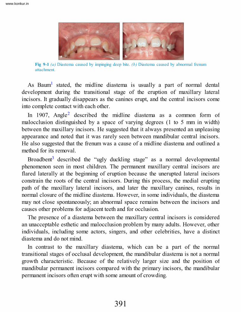
Fig 9-1 (a) Diastema caused by impinging deep bite. (b) Diastema caused by abnormal frenumattachment.
As Baum1 stated, the midline diastema is usually a part of normal dentaldevelopment during the transitional stage of the eruption of maxillary lateralincisors. It gradually disappears as the canines erupt, and the central incisors comeinto complete contact with each other.
In 1907, Angle2 described the midline diastema as a common form ofmalocclusion distinguished by a space of varying degrees (1 to 5 mm in width)between the maxillary incisors. He suggested that it always presented an unpleasingappearance and noted that it was rarely seen between mandibular central incisors.He also suggested that the frenum was a cause of a midline diastema and outlined amethod for its removal.
Broadbent3 described the “ugly duckling stage” as a normal developmentalphenomenon seen in most children. The permanent maxillary central incisors areflared laterally at the beginning of eruption because the unerupted lateral incisorsconstrain the roots of the central incisors. During this process, the medial eruptingpath of the maxillary lateral incisors, and later the maxillary canines, results innormal closure of the midline diastema. However, in some individuals, the diastemamay not close spontaneously; an abnormal space remains between the incisors andcauses other problems for adjacent teeth and for occlusion.
The presence of a diastema between the maxillary central incisors is consideredan unacceptable esthetic and malocclusion problem by many adults. However, otherindividuals, including some actors, singers, and other celebrities, have a distinctdiastema and do not mind.
In contrast to the maxillary diastema, which can be a part of the normaltransitional stages of occlusal development, the mandibular diastema is not a normalgrowth characteristic. Because of the relatively larger size and the position ofmandibular permanent incisors compared with the primary incisors, the mandibularpermanent incisors often erupt with some amount of crowding.
391
www.konkur.in

PrevalenceA review of older literature indicates that the midline diastema was of concern topatients and was considered an unacceptable malocclusion, and clinicians were veryinterested in the etiology and treatment of this problem. In 1882, Farrer4 described amethod for the correction of diastema.
According to many epidemiologic investigations, the prevalence of midlinediastema is high during the early mixed dentition and decreases between the ages of9 and 12 years. In normal conditions, it closes after complete eruption of thepermanent canines.5–7
In general, the midline diastema is more prevalent in females than in males.Richardson et al8 studied the sex prevalence of diastema in 5,307 children between6 and 14 years of age. They found that, at age 6 years, females showed a higherprevalence than males, but, at age 14 years, males had a higher prevalence.
Racial differences also exist; Lavelle9 investigated the distribution of midlinediastema in different human populations and reported that the prevalence is greaterin Africans (West Africa) than in British whites or Chinese from Hong Kong andMalaysia.
Horowitz10 evaluated the racial differences of midline diastema in a group of over700 children, 397 black children and 321 white children, between the ages of 10 and12 years. He found that 19% of the total sample of black children had a midlinemaxillary diastema of 2 mm or more compared with 8% of the while children.
Taylor5 examined 516 children between 5.5 and 11.0 years of age and found that66 of 68 children in the 5.5- to 6-year age group (97%) showed spacing betweenincisors. In the 6- to 7-year group, the prevalence of diastema was 87.7%, and in the10- to 11-year group, 48.7% showed midline diastema. In another study of childrenaged 12 to 18 years, Weyman11 reported a 7% occurrence.
Brunelle et al,12 analyzing a national health and nutrition survey carried out in theUnited States, reported that a maxillary diastema of 2 mm or more was present in19% of 8- to 11-year-olds, 6% of 12- to 17-year-olds, and 5% of adults aged 18 to50 years.
Etiology of Midline DiastemaInitiation of any treatment for a midline diastema must be based on proper evaluation
392
www.konkur.in

of the extent, etiology, and pathogenesis of the diastema. The possible influence ofthe superior labial frenum in relation to the midline diastema has been of greatinterest to clinicians for many years. The presence of persistent diastema has oftenbeen blamed on an enlarged labial frenum. Sometimes, without a proper evaluationof the etiology of a particular diastema, frenectomy has been performedunnecessarily, although many other factors can be involved in this abnormality.Therefore, the cause of a diastema must be evaluated in regard to both theanatomical structure of frenum tissue and other causes of abnormal diastema.
Morphogenesis and structure of the frenumThe frenum can be the original cause of a diastema in only a small proportion ofmidline diastemata. According to Huang and Creath,13 the superior labial frenumbegins to form in the 10th week of gestation, and by the 3rd month in utero themorphology of the fetal frenum is similar to that of the abnormal frenum of postnatallife. It extends as a continuous band of tissue from the tuberculum on the inner side ofthe lip, over and across the alveolar ridge, to be inserted in the palatine papilla.Before birth, the lateral halves of the alveolar ridge unite, and the continuous band oftissue becomes totally enclosed by bone; it is divided into a palatal portion (palatinepapilla) and a labial portion (superior labial frenum) by this closure.
The frenum of a newborn at the gum pad stage and before eruption of the dentitionis a large, fleshy attachment with a wide origin. The tissue gradually narrows,attaching in the midline to the connective tissue of the internal maxillary suture andalveolar process. With the eruption of teeth and development of the alveolar ridge,the position of the frenum changes. As growth progresses, the frenum may atrophyand may assume a higher position or maintain its attachment to the papilla.
It has been suggested that a slight clefting of intercrestal bone can hold the teethapart and cause an abnormal diastema.14
In a study of dogs, Picton and Moss15 hemisectioned the mandibular first molarand disked the mesiodistal contact of other teeth in one side of the arch and producedartificial interdental spacing between the halves of the first molar on one side. Aftera few weeks, they found an enlarged space between the two parts of the sectionedtooth. One half moved mesially, and the other half moved distally because thetransseptal fibers were not present between the roots of the divided molar, whileother created spaces were spontaneously closed. They concluded that the transseptalfibers are one of the principle factors that bring about and maintain the interstitial
393
www.konkur.in

mesiodistal contacts of the teeth in each arch.Stubley16 compared the transseptal fibers to an orthodontic chain used for
interdental space closure. He stated that transseptal fibers do not contain elastictissue; they are made of collagen, and the motive power that they exert derives fromtiny coils into which they contract as they mature; the force generated varies fromindividual to individual.
Stubley16 described persistent midline diastema where interdental fibers arecharacterized by their strength and a wide space develops in the maxillary midline.In the absence of any pathologic condition, the underlying cause is congenital and isdue to the presence of a persistent midline suture that interferes with arrangement ofthe transseptal fibers. Instead of passing directly across the interdental space fromone tooth to the other, the fibers turn upward at a right angle from the mesial aspectof each central incisor to enter the suture. Such an arrangement is like a break in thetransseptal fiber chain, and the distal transseptal fibers of developing centralincisors can move them apart. This fiber pattern could account for difficulty in thespontaneous diastema closures.
Martin and Jones17 examined 17 consecutive individuals with various forms ofholoprosencephaly at autopsy or during clinical evaluation and found that 88% of thepatients were missing the superior labial frenum, regardless of the severity ofholoprosencephaly or other associated craniofacial defects. Holoprosencephaly is acephalic disorder in which the prosencephalon (the forebrain of the embryo) fails todevelop into two hemispheres. Hox genes, which guide placement of embryonicstructures, fail to activate along the midline of the head, allowing structures that arenormally paired on the left and right to merge. Martin and Jones17 stated that absenceof the frenum in individuals with holoprosencephaly provides evidence that thefrenum has its embryonic origin in the medial nasal process.
Dean et al18 stated that the maxillary labial frenum is composed of two layers ofepithelium enclosing a loose vascular connective tissue. In some instances, thefrenum may include muscle fibers that originate from the orbicularis oris muscle ofthe upper lip. The origin from the lip is often wide, but the tissue of the frenum itselfnarrows and is inserted in the midline into the outer layer of the periosteum and intoconnective tissue of the internal maxillary suture and alveolar process. The frenummay attach at variable locations in the attached gingival tissue some millimetersabove the crest of the ridge, or on the ridge, or the fibers may pass between thecentral incisors and attach to the palatine papilla. Figure 9-2 shows an abnormaldiastema caused by heavy and low attachment of an abnormal frenum.
394
www.konkur.in

Fig 9-2 Abnormal diastema caused by heavy, low frenum attachment.
The frenum may be completely absent, or its existence may be a part of normaltransition stage of dentition. With the completion of incisor and canine eruption, thegap spontaneously closes.
Other causes of midline diastemaBesides the anatomical structure of the frenum (size, position, and type of insertion),other causes of midline diastema must be considered before treatment planning andfrenectomy are initiated. Possible therapeutic approaches include orthodontics,restorative dentistry, surgery, or various combinations of these. The ideal treatmentshould seek to close the diastema in question but also address the cause behind it.
During incisor transition, because permanent incisors are larger than primaryincisors (incisor liability), some mechanisms interact to compensate for these widthdifferences and enable normal development of occlusion. Mandibular incisorsgenerally erupt with some degree of crowding, while maxillary incisors, because oftheir position and inclination, erupt with some degree of spacing. Depending on theirsize, this can be considered a normal transitional condition or can be a sign of anabnormal situation that deserves early detection and intervention before completedevelopment.
In the normal transitional stage of maxillary incisors, a midline gap of up to 3 mmis normal. The mesial inclination of maxillary lateral incisors and their mesial forceduring eruption reduce or completely close this gap; eruption of the maxillarypermanent canines with the same mechanism will completely close the remaininggap between central incisors. However, persistent midline diastema is also an ethnicnorm for some races that have large dentoalveolar arches, such as African andMediterranean groups.
To differentiate between normal transitional changes in the dentition that result inspacing in the maxillary permanent incisors from other factors that cause abnormalspacing, clinicians must carefully examine the patient’s occlusion intraorally and
395
www.konkur.in

extraorally through clinical and radiographic evaluation. Incisor separations canresult from one or more of the following factors:
• Lateral incisor hypodontia• Microdontia of maxillary lateral incisors (peg-shaped lateral incisors)• Presence of mesiodens or odontoma• Deleterious oral habits• Muscle imbalance (lip dysfunction)• Impinging deep bite• Anterior Bolton discrepancy• Pathologic tooth migration
Lateral incisor hypodontia
One common cause of midline diastema is congenital absence of one or bothmaxillary lateral incisors (Fig 9-3). Sustained diastema after complete eruption ofthe central incisors is a diagnostic clue for early detection of this problem, whichmust be evaluated by radiograph. A remaining diastema with no midline shift isusually accompanied by absence of both lateral incisors, and a remaining diastemawith a shift of midline is usually the consequence of one absent lateral incisor.
Fig 9-3 (a and b) Severe diastema caused by a congenitally missing maxillary right lateral incisor.
Depending on the type of occlusion and other conditions, treatment options areclosing the diastema and using canine substitution or closing the diastema, aligningthe anterior teeth, and replacing the missing teeth with conventional prostheses orimplants (see chapter 7).
Microdontia of maxillary lateral incisors
Abnormal maxillary midline diastema can be the result of small incisors, that is, peglateral incisors, which can be unilateral or bilateral (Fig 9-4).
396
www.konkur.in

Fig 9-4 Diastemata caused by missing teeth and microdontia of the central incisors.
Depending on the type of occlusion, the age of the patient, and the patient’sdesires, treatment options can be building up the small lateral incisor withcomposite resin, veneers, or crowns or extraction and space closure (see chapter 7).
Presence of mesiodens or odontoma
Another common cause of midline diastema is the presence of supernumerary teeth.The mesiodens is the most common type of supernumerary tooth (Fig 9-5). Earlydetection and diagnosis are not difficult if the incisor transition is carefullymonitored. The presence of supernumerary teeth in the incisor area has some clinicalsigns that can help in early detection, including asymmetric or retarded tootheruption, retained diastema after complete eruption of the lateral incisors, androtation of the central incisors.
Fig 9-5 Diastema caused by mesiodens.
The treatment of choice depends on the patient’s occlusion, dental age, and rootformation of the incisors. Some treatment options include early detection andextraction of the supernumerary tooth and guidance of eruption and incisor alignmentby segmental bonding (see chapter 8).
Deleterious oral habits
Another common cause of midline diastema or interdental spacing of incisors isabnormal habits, such as finger sucking, tongue thrust, and lip biting or lipdysfunction (Figs 9-6 and 9-7).
397
www.konkur.in

Fig 9-6 Diastema caused by thumb sucking.
Fig 9-7 Diastema caused by tongue dysfunction.
The treatment of choice in this situation is early detection, habit control, and spaceclosure (see chapter 6).
Muscle imbalance (lip dysfunction)
One of the most important factors influencing incisor relationships and maintainingnormal occlusion is inner and outer muscle balance around the dental arches.Midline diastema can be caused by orofacial muscular imbalances, such asmacroglossia, improper tongue posture and position, and flaccid lip muscles (Fig 9-8).
Fig 9-8 (a and b) Diastema caused by lip dysfunction.
Proper treatment of these conditions involves elimination of causative factors andspace closure. Long-term stability depends on proper long-term retention.
Impinging deep bite
398
www.konkur.in

Deep bite malocclusions, especially Class II division 1 malocclusions withimpinging bite caused by a severe curve of Spee and overeruption of mandibularincisors, is a common type of malocclusion that can cause incisor flaring andmidline diastema.
The treatment of choice for this type of diastema is early intervention andcorrection of the curve of Spee, correction of deep bite, and then space closure andincisor retraction (see Fig 9-1a).
Anterior Bolton discrepancy
Another cause of midline diastema is an abnormal anterior ratio, which can bedetermined through the Bolton analysis.19 This discrepancy results from the presenceof relatively large mandibular incisors or small maxillary incisors in comparison toeach other.
Space closure must be accompanied by a reduction in the size discrepancybetween maxillary and mandibular incisors, either by enamel stripping or extractionof the bigger incisors or buildup of the smaller incisors (see chapter 5 on correctionof incisor crowding).
Pathologic tooth migrationAnother cause of incisor spacing and flaring, especially in the adult dentition, is
development of a pathologic condition, such as cystic formation, bone loss due toperiodontal problems, or some systemic condition that causes bone loss and toothmigration. Increased areas of inflammation cause bone loss, loss of attachment, andconsequently migration of teeth from the pocket (Fig 9-9).
Fig 9-9 Diastema caused by pathologic tooth migration.
Management of this condition, especially in advanced situations, is verychallenging and requires a multidisciplinary approach. Any orthodontic toothmovement and treatment must wait until a thorough evaluation has been completedand the cause has been eliminated.
399
www.konkur.in

Effects of Abnormal Frenum Attachment on OcclusionAn abnormal labial frenum, in addition to causing a maxillary midline diastema, canproduce other undesirable clinical conditions:
• Abnormal frenum attachment can cause an abnormal space between all incisors aswell as tooth displacement and rotation.
• An abnormal frenum attachment and a sustained diastema between central incisorscan complicate the eruption of lateral incisors and canines, resulting in delayederuption or complete absence of eruption, displacement, crossbite of lateralincisors, and/or canine impaction.
• Depending on their size and position, the abnormal frenum attachment may restrictmovements of the lip or tongue and may interfere with speech and swallowing.
• Frenum tissue with a heavy band and low attachment can interfere with propertoothbrushing.
• Tight fiber attachment of frenum tissue, especially the mandibular labial frenum,may cause stretching of the labial and gingival tissues and tissue recession fromthe necks of the teeth.
Differential Diagnosis of Abnormal FrenumMidline diastema is a common occurrence that can be seen in all races, sexes, andages.
Because of its potential to have multiple etiologies, differential diagnosis must bebased on a thorough medical and dental history and a complete clinical andparaclinical evaluation (radiograph and cast analysis if tooth size or arch sizediscrepancy is suspected). Several points should be investigated in evaluations ofsuspicious frenum attachment: hereditary background, age of the patient, localfactors, and the size, position, and strain of the frenum tissue.
Hereditary backgroundAmong the patients with abnormal frenum attachment whom the author has examinedover the past 40 years of practice, almost all had some type of familial pattern ofinvolvement, whether in parents, siblings, or other close relatives. For differential
400
www.konkur.in

diagnosis of sustained diastema, the author strongly recommends that the clinicianlook for any familial history of diastema.
In a follow-up evaluation of 96 consecutive cases, Shashua and Artun20 assessedthe relapse after orthodontic correction of maxillary midline diastema (ranging from0.50 to 5.62 mm) 4 to 9 years after completion of active treatment. The onlysignificant risk factors for relapse were pretreatment diastema size and presence of afamily member with a similar condition.
Age of the patientThe presence of a midline diastema can be a normal transitional change of thedentition during the early and middle mixed dentitions. Such diastemata are self-corrected after complete eruption of all incisors and canines, provided that all otherenvironmental conditions are normal.
Local factorsAny local obstacle that can interfere with normal diastema closure must bethoroughly assessed clinically and paraclinically. These obstacles include missingteeth, supernumerary teeth, microdontia, deleterious habits, muscle dysfunction,impinging deep bite, and pathologic conditions (periodontal involvement or cystformation).
Clinical and radiographic signs of true abnormalfrenumThe size, position, and strain of the frenum tissue are another important area ofexamination. Certain signs related to these aspects can help in the detection andconfirmation of true abnormal frenum. These signs are the size, position, andlocation of tissue attachment; the shape of the midline gap; blanching of the papilla;and the presence of a bony cleft on radiographs.
Size, shape, and location of tissue attachment
An abnormal frenum is usually large, and its location is low, close to the ridge,between the central incisors, or even extended to the palatine papilla. In severe
401
www.konkur.in

conditions, tied frenum, especially tongue-tie of the lingual frenum, may negativelyaffect the patient’s speech.
Shape of midline gap
A common shape of midline gap or diastema is a reverse V-shaped space betweenthe central incisors, wider toward the incisal edge. This is sometimes accompaniedby mesial rotation of the central incisors (Fig 9-10).
Fig 9-10 Reverse V-shaped separation between central incisors, a common sign of abnormalfrenum attachment.
Blanching test
A simple diagnostic test for an abnormal frenum involves stretching the upper lip upand forward intermittently. If a heavy band of tissue is attached to the palatinepapilla, the force exerted produces blanching of the papilla. This is called theblanching test (Fig 9-11).
Fig 9-11 Local anemia or blanching of the papilla in response to stretching of the lip, another signof abnormal frenum attachment.
Periapical radiograph
Evaluation of the midline and the condition of the alveolar suture is a helpful meansof diagnosis. Midline bony clefts have been proposed as an etiologic factor forabnormal diastemata,14 which often exhibit a V-shaped midline bony cleft and opensuture (Fig 9-12).
402
www.konkur.in

Fig 9-12 Sutural opening in the alveolar crest, a sign of frenum tissue insertion.
Management of Abnormal FrenumDepending on the etiologic factor or factors, abnormal midline diastemata can betreated with various options that have been discussed in previous chapters (such asabnormal habits [see chapter 6], hypodontia [see chapter 7], and supernumeraryteeth [see chapter 8]). This section focuses on the management of abnormal diastemacaused by abnormal frenum attachment. Management varies depending on the stageof dentition.
Management in adultsManagement of diastema in adults varies, depending on the type of malocclusion,etiologic factors, the health of the periodontium, and the patient’s desires. There arefour basic choices for treating diastemata in adults:
1. If the patient has an acceptable occlusion and does not want to undergocomprehensive orthodontic treatment, he or she may opt to forego treatment. Somepeople are fine with diastemata, while others find them unappealing and wish tohave them changed or closed.
2. If the patient has good occlusion and a mild diastema but is not willing to undergoorthodontic treatment, the second choice is to close the space through porcelainveneers or composite resin buildup.
3. Another choice is placement of a conventional crown and fixed partial dentureprosthesis. Depending on the size of the incisors and the width of the diastema,space closure can be accomplished by crown placement only on the central
403
www.konkur.in

incisors or by distribution of space closure among all incisors.4. The most complicated treatment plan includes frenectomy and proper orthodontic
treatment, which can be accomplished with comprehensive orthodontic treatmentor 2 × 6 bonding. Retention is critical after most frenum corrections, especially inadults, and requires extended or permanent retention.
Management during the mixed dentitionIntervention for an abnormal frenum is easier during the early mixed dentition, andthe results are more stable. Also, young patients are more willing to acceptorthodontic options, and often parents find a diastema to be unacceptable in theirchildren and do not like the appearance.
Early interception not only can solve the current problems for the child but alsocan prevent many negative consequences for the occlusion. Management options foran abnormal diastema during the early and middle mixed dentitions, whether it is dueto abnormal frenum attachment or other etiologic factors, vary greatly. The timing ofintervention is important for achieving satisfactory results. Timing depends on theinitial cause, the patient’s age, and the stage of the dentition.
Generally there is controversy among practitioners about the timing of frenumremoval; some advocate that treatment be delayed until full eruption of all maxillarypermanent anterior teeth, including the canines. This rule may be considered correct,provided that the frenum is not disturbing occlusal transition and the patient does notmind abnormal diastema and needs comprehensive orthodontic treatment.
However, there are many situations when the presence of abnormal frenum tissueduring early stages of the dentition disturbs the normal development of occlusion. Itmay prevent normal eruption of adjacent teeth; disturb the normal sequence oftransition; cause crowding, rotation, or displacement of adjacent teeth; and evencause malocclusion such as crossbite or canine impaction.
The foundations of permanent occlusion are the eruption of the permanent firstmolars in the posterior region and the incisors in the anterior region. Anydisturbances anywhere in this structure can interfere with the normal development ofthe whole structure. Angle2 based his idea of normal occlusion on the position of themaxillary permanent first molar as “the key of occlusion.”
The question facing clinicians is whether they should neglect the conditions thatthey observe in the anterior region of the occlusion that are causing a sustaineddiastema, claiming that they can correct it at the later age. The answer is “No”;
404
www.konkur.in

instead, they should eliminate the cause and maintain the integrity of the anteriorsegment of the arch at a natural time, to enhance the normal development of the restof the arch.
Removal of any etiologic factors other than the frenum that are causing diastemaand disturbing the eruption of adjacent teeth usually can be initiated on diagnosis andafter sufficient development of the central incisors; however, tooth movement isusually deferred until eruption of the permanent lateral incisors or canines. There aredifferent management options to choose from, depending on the severity of theabnormal diastema and its effects (eg, interference in eruption or interruption ofnormal transitional changes).
Early treatment procedures
The design of the treatment plan and the timing of intervention depend on the severityof the frenum problems, including size, type of insertion, the potential for affectingadjacent teeth, and patient occlusion.
Delaying treatment until complete eruption of all maxillary incisors. If thepresence of an abnormal frenum is confirmed but there is no evidence of severe,disturbing effects on the occlusion and adjacent teeth, the treatment should bepostponed until eruption of all maxillary incisors is completed. Treatmentprocedures are initiated with 2 × 4 bonding for leveling and correction of rotationand alignment of all incisors. After this phase, the patient is referred for frenectomybefore diastema closure; closure of the diastema must begin the same day asfrenectomy to prevent regeneration of tissue and collagen fibers in the suture areas.
Urgent intervention, frenectomy, and diastema closure. Gap closure is urgentwhen the presence of the diastema interferes with normal eruption or the transitionalstage of dentition. For example, if the presence of heavy frenum tissue interfereswith normal eruption of the maxillary lateral incisors, the intervention and diastemaclosure can be started even before eruption of the lateral incisors. This isaccomplished with a removable appliance or by attaching two brackets on centralincisors with a sectional arch to facilitate lateral incisor eruption. In this situation, ifthe patient’s molar relationships, overjet, overbite, and midline relationship arenormal and no other treatment is needed, all maxillary incisors must be retained untileruption of the canines.
Figure 9-13 shows the normal pattern of maxillary incisor transition (see chapter2). During this process, any factor that interferes with normal, gradual closure of the
405
www.konkur.in

midline gap can affect normal eruption and transition of adjacent teeth, causingproblems such as lateral crowding, rotation, lack of eruption, or crossbite. Eruptionof canines also can be disturbed if the midline diastema remains open.
Fig 9-13 Normal pattern of maxillary incisor and canine eruption.
Figure 9-14 illustrates an abnormal frenum attachment that caused a diastema thatremained mostly unchanged for 15 months, before and after eruption of the lateralincisors. The best treatment option in this case would be early intervention:frenectomy and diastema closure.
Fig 9-14 Wide diastema that has persisted even after eruption of both lateral incisors because ofabnormal frenum attachment. (a) Pre-eruption. (b) Posteruption.
Figure 9-15 shows an abnormal frenum that caused problems for the maxillarycanines and had the potential to result in lateral incisor root resorption and canineimpaction. The patient was treated with frenectomy, diastema closure, and guidanceof canine eruption.
406
www.konkur.in

Fig 9-15 (a to c) Frenum that is preventing diastema closure and with the potential to causecanine impaction and lateral root resorption. (d ) Panoramic radiograph after fre-nectomy anddiastema closure.
Two-phase management. When other problems such as abnormal habits, overjet,and overbite are associated with a midline diastema and abnormal frenum andfurther treatment will be needed in the permanent dentition, the procedure can bemanaged in two phases. Phase 1 includes correction of the diastema and incisoralignment. After an interim phase of continued monitoring, the second phase oftreatment is begun. However, not all diastemata can be treated the same way, interms of modality or timing; careful pretreatment evaluation, proper orthodontic-surgical planning, and proper intervention are needed to achieve an ideal solution.
Sometimes local factors other than the frenum, such as abnormal habits, absenceof the lateral incisors, or a mesiodens, are causing an abnormal diastema; however,if the frenum attachment is low in the ridge area, the appearance can be misleadingbecause it looks like an abnormal frenum. Frenectomy is not recommended in thesesituations; elimination of the cause and orthodontic space closure are usuallyfollowed by spontaneous atrophy of the frenum tissue (see case 9-5).
Management during the primary dentitionPresence of an abnormal frenum is not as common in the primary dentition as in themixed dentition, but there are patients with abnormal frenum attachment even duringthe primary dentition who may show severe diastema and incisor displacement.These are usually patients with severe frenum problems, but frenectomy during theprimary dentition is not recommended, and the treatment must be postponed to the
407
www.konkur.in

mixed dentition period.The author’s clinical observation is that all cases of abnormal frenum attachment
during the primary dentition have a hereditary background; children with this traitwill show abnormal frenum attachment during the permanent dentition too.Therefore, frenectomy during the primary stage has no benefits for these children.Recurrence of the frenum in the permanent dentition is inevitable and willnecessitate another phase of treatment (see case 9-3).
Management in infants
Kotlow21 reported the presence of labial dental caries on the maxillary anterior teethin breastfeeding infants due to abnormal attachment of the maxillary frenum. Headded that abnormal frenum attachment may interfere with proper latching of aninfant to a mother’s breast and impede lip flanging, thereby preventing nursing. Herecommended diode laser surgery for these infants to prevent this type of caries.
In contrast, Dean et al18 stated that the effectiveness of surgical reduction of thelabial frenum in infants cannot be substantiated.
FrenectomyDespite advances in diagnostic methods, orthodontic treatment, and surgicaltechniques, some cases of relapse after diastema treatment have been reported. Acareful pretreatment assessment is crucial, and the interaction of orthodontictreatment with the anticipated surgical procedure must be analyzed in treatmentplanning. For optimum results and stability, besides the frenum attachment anddiastema, other aspects of the dentoskeletal malocclusion and perioralneuromuscular balance and function must also be considered; these includedeleterious habits, tongue function, lip tonicity, and lip competency.
For the surgical technique, Dean et al18 suggested removal of a wedge-shapedsection of the tissue between the central incisors on either side of the frenum,extending the incision palatally to the nasal palatine papilla and to the depth of theunderlying bone. The free marginal tissue on the mesial side of the central incisorsshould not be disturbed. The wedge of tissue can be picked up with tissue forcepsand excised with tissue shears at an area close enough to the origin of the frenum toprovide a desirable cosmetic effect. Sutures are placed inside the lip to approximate
408
www.konkur.in

the free margins of the tissue. It is generally not necessary to suture or pack the tissuebetween the incisors.
Recently, application of lasers in general dentistry and especially in soft tissuesurgery has been advocated by many practitioners. Haytac and Ozcelik22 comparedthe degree of postoperative pain, including discomfort and functional complications(eating and speech), in 40 patients requiring frenectomy. Patients were assignedrandomly to a conventional surgical technique or surgery with a carbon dioxidelaser. They concluded that carbon dioxide laser treatment is safe for frenectomy andthat patients treated with the laser experience less postoperative pain and betterfunction than do those treated with the scalpel technique.
Olivi et al23 clinically evaluated the efficacy of an erbium, chromium-dopedyttrium-scandium-gallium-garnet laser in removing the labial frenum in 143adolescent and prepubescent patients. Patients returned for recall, and surgical areaswere checked for adverse events, recurrence of frenum, functional complications,and patient acceptance. They reported that only two adolescent patients exhibitedrecurrence of the frenum. None of the three prepubescent patients needed additionalintervention at follow-up, 3 years after treatment.
Kotlow24 stated that traditional methods of oral surgery by scalpels orelectrosurgery may place pediatric patients at risk during the use of a generalanesthetic for an elective procedure and may produce significant postoperativediscomfort; these procedures also require sutures and prolonged healing. Lasersurgery provides a simple and safe in-office alternative for children, reducing thechances of infection, swelling, discomfort, and scarring.
In cases of deep attachment of frenum tissue in the midline suture, completeremoval of the tissue in the suture and immediate space closure are necessary toprevent any tissue regeneration (Fig 9-16).
409
www.konkur.in

Fig 9-16 (a to e) Frenectomy followed by space closure and sutural obliteration.
Ankyloglossia (Tongue-Tie)The lingual frenum located under the tongue is a normal part of the oral cavity. It is athin, vertical fold of tissue attached to the undersurface of the tongue and to the floorof the mouth. Ankyloglossia (tongue-tie), in which shortness and consolidation of thelingual frenum tissue reduce tongue mobility, is an uncommon oral anomaly that cancause some difficulty for tongue function.
In many children, ankyloglossia might be asymptomatic; in some the condition mayresolve spontaneously, or affected children may learn to compensate adequately fortheir decreased lingual mobility. There are also children who may benefit fromsurgical intervention for tongue-tie.
Signs of ankyloglossiaA person with tongue-tie usually cannot protrude the tongue beyond the edges of themandibular incisors or touch the palatine papilla of the maxillary incisors. Thislimitation has the potential to disturb speech, normal deglutition, and suckling andcan have an effect on the dentition, such as increasing mandibular interincisalspacing (Fig 9-17).
410
www.konkur.in

Fig 9-17 Ankyloglossia, or tongue-tie, a rare lingual frenum attachment. The condition can affectthe dentition and tongue movement.
Management of ankyloglossiaThe literature includes several reports, some exaggeration of functionalcomplications in patients having this anomaly, and many different proposals forsurgical therapy.25,26 Management of lingual frenum has been mostly based on theresults of clinical examination; because there is no distinct border between normaland tied mandibular frenums, the dental practitioner is often unsure of an appropriatecourse of action for a patient with suspected ankyloglossia.
Wright27 retrospectively reviewed the records of 158 patients who had undergonea frenectomy and concluded that the operation requires general anesthesia, except inolder children, for whom local anesthesia is appropriate. He added that speechdifficulties related to tongue-tie are overrated and mechanical problems areunderestimated.
Indications for lingual frenotomy include articulation difficulties confirmed by aspeech pathologist and mechanical limitations such as inability to lick the lips orperform internal oral toilet. Limitations in the tongue’s ability to sweep the teeth andspread saliva can cause dental caries and periodontal health problems. There maybe rare instances in infants where problems with feeding and suction can be helpedby frenectomy.
Fiorotti et al28 indicated that a short lingual frenum can result in incorrect speechsounds, dentofacial alterations, and acquired functional adaptations. They assessed15 patients who underwent frenectomy utilizing the carbon dioxide laser. Theresearchers indicated that the technique is safe, effective, and perfect for use inyoung children and can be performed in an outpatient unit. They also contended that,when frenectomy is indicated, it should be performed as early as possible to preventfunctional alterations.
Suter and Bornstein29 searched the literature and found 64 articles regarding theclassifications of tongue-tie and many other proposed criteria. They concluded thatlack of an accepted definition and classification of ankyloglossia makes
411
www.konkur.in

comparisons between studies almost impossible. No conclusive suggestionsregarding the method of choice can be made; therefore, it remains controversialwhich tongue-ties should be surgically removed and which can be left toobservation. Many children with the tied frenum compensate well and have normal-sounding speech.
Each individual with ankyloglossia requires a thorough individual assessment andevaluation to ensure that the most appropriate management strategies are selected.Parents should be educated about the possible long-term effects of tongue-tie.
Case 9-1A 10-year-old boy presented with a hereditary abnormal frenum attachment, a Class I malocclusion,and a wide diastema that persisted even after eruption of the lateral incisors (Figs 9-18a to 9-18d). Heexhibited deep bite and maxillary incisor flaring but good mandibular dentition. Figures 9-18e and 9-18fshow the diastemata of his mother and his aunt.
Treatment:Treatment was designed to include one phase of active treatment followed by a period of observation.First 2 × 4 bonding was used in conjunction with an anterior bite plate to align the incisors, and then autility arch and bite plate were used to reduce overbite and flaring (Fig 9-18g). After these goals wereaccomplished, the second step was frenectomy (Fig 9-18h). The same day, space closure by elasticligature was applied (Fig 9-18i). The final active part of treatment was incisor retraction and overbitecontrol.
The final step was placement of a Hawley retainer to retain the anterior changes and control overbiteuntil completion of permanent tooth eruption. Figures 9-18j to 9-18m show the posttreatment occlusionafter 2 years of observation and eruption of all permanent teeth. This case represented interceptivetreatment, using only 2 × 4 maxillary bonding.
Fig 9-18 Treatment of a 10-year-old boy with a hereditary abnormal frenum attachment resultingin a wide, persistent diastema, deep bite, and flaring incisors. (a to d) Pretreatment occlusion. (eand f) Diastemata exhibited by the patient’s mother and aunt.
412
www.konkur.in

Fig 9-18 (cont) (g) After incisor alignment and overbite reduction but before frenectomy. (h)Frenectomy and initiation of space closure. (i) Space closure after 1 week. (j to m) Posttreatmentocclusion.
Case 9-2A 10-year, 8-month-old girl exhibited a Class II division 1 malocclusion and maxillary and mandibularincisor protrusion. In addition, an invasive frenum attachment caused severe maxillary incisor crowding,displacement, and cystic formation (Figs 9-19a to 9-19e).
Treatment:The treatment plan included removal of the frenum, the cyst, and all abnormal soft tissue attachmentand extraction of the four first premolars, carried out as a serial step-by-step extraction.
After the surgical procedure and tissue healing, a removable maxillary Hawley appliance wasinserted to achieve slow, minor incisor alignment, and use of a lower holding arch for about 1 year wasfollowed by step 1 of the extraction series: removal of the maxillary primary canines, both maxillaryprimary first molars, and both mandibular primary first molars. Figure 9-19f shows alignment of themaxillary incisors and the canine bulges before serial extraction.
Step 2 was extraction of all four first premolars. Maxillary anchorage was prepared with a Nanceappliance, and the lower holding arch was removed as reciprocal anchorage.
Step 3 of the extraction sequence was removal of the remaining primary second molars. This wasfollowed by maxillary and mandibular bonding to start maxillary canine retraction. Then mandibular andlater anterior retraction and space closure were accomplished. Some mesial movement of themandibular molars was allowed, in order to achieve a Class I molar relationship (Figs 9-19g to 9-19k).
413
www.konkur.in

Fig 9-19 Treatment of a 10-year, 8-month-old girl with a Class II division 1 malocclusion andmaxillary and mandibular protrusion. An invasive frenum attachment has caused toothdisplacement, maxillary incisor crowding, and formation of a cyst. (a to c) Pretreatment occlusion.(d) Pretreatment panoramic radiograph. (e ) Pretreatment cephalometric radiograph. ( f ) Tissuehealing and some incisor alignment. The arrows show canine bulge. (g to i) Posttreatmentocclusion. (j) Posttreatment panoramic radiograph. (k) Posttreatment cephalometric radiograph.
Case 9-3A 5-year-old girl presented with a mesial step and good interdigitation in the posterior segments but asevere hereditary diastema, resulting from a strong and low abnormal frenum attachment, and displacedprimary incisors (Figs 9-20a to 9-20d). Because of the patient’s age and the hereditary nature of thediastema (her mother also had a severe diastema and abnormal attachment), no treatment wassuggested at this age. The parents were advised to bring back the child when all incisors had erupted.
The patient returned during the late mixed dentition with the same problem and a more intensiveeffect on the permanent incisors, including a severe, V-shaped diastema, incisor rotation, and incisordisplacement (Figs 9-20e to 9-20h).
Treatment:Orthodontic problems consisted of severe frenum attachment; a wide, V-shaped diastema; rotated anddisplaced incisors; anterior open bite; crowding; severe maxillary and mandibular dental protrusion; andsteep mandibular plane. The treatment plan included deep frenectomy and extraction of the four firstpremolars.
414
www.konkur.in

The treatment followed this order: (1) placement of a maxillary Nance appliance and a lower holdingarch as well as extraction of four first premolars; (2) 2 × 6 bonding, incisor alignment, frenectomy, anddiastema closure; (3 ) canine retraction, anterior retraction, and completion of active treatment; (4)retention. Figures 9-20i to 9-20l show the posttreatment results.
Fig 9-20 Treatment of a girl with a severe hereditary diastema, resulting from a strong and lowabnormal frenum attachment in the primary dentition, and displaced incisors. (a to c) Occlusion at5 years. Her parents were advised to return for evaluation when all the child’s incisors haderupted. (d) Panoramic radiograph of the primary dentition. (e and f) Occlusion in the late mixeddentition, showing significant frenum attachment, a severe V-shaped diastema, and incisordisplacement.
415
www.konkur.in

Fig 9-20 (cont) (g) Pretreatment panoramic radiograph during the late mixed dentition. (h)Pretreatment cephalometric radiograph during the late mixed dentition. ( i and j) Posttreatmentocclusion. (k) Posttreatment panoramic radiograph. (l) Post-treatment cephalometric radiograph.
Case 9-4Many factors besides abnormal frenum attachment can cause abnormal diastema, and it is veryimportant to differentiate and verify the cause before frenectomy. Cases 9-4 and 9-5 are examples ofconditions in which a diastema was not caused by abnormal frenum attachment.
An 8-year, 5-month-old girl presented for correction of her diastema. Her orthodontic problems werea Class III malocclusion tendency in the molars and canines, maxillary deficiency, zero overjet, andminor mandibular incisor crowding. Cephalometric evaluation indicated a negative value for the pointA–nasion–point B angle and maxillary incisor protrusion. Therefore, the cause of the diastema wasdiagnosed to be the point A–point B discrepancy, the absence of overjet, and the force of mandibulargrowth (Figs 9-21a to 9-21e).
Treatment:The treatment plan included face mask therapy for maxillary protraction in order to increase the overjet,labial root torque, and then midline closure. No frenectomy was planned. Figures 9-21f to 9-21j showthe posttreatment occlusal and dentoskeletal changes.
416
www.konkur.in

Fig 9-21 Treatment of an 8-year, 4-month-old girl with a Class III tendency, maxillary deficiency,and zero overjet, resulting in diastema and space deficiency in the maxillary anterior segment. (a toc ) Pretreatment occlusion. ( d ) Pretreatment panoramic radiograph. ( e ) Pretreatmentcephalometric radiograph. (f to h) Posttreatment occlusion after maxillary protraction. ( i) Post-treatment panoramic radiograph. (j) Posttreatment cephalometric radiograph.
Case 9-5A 12-year-old girl presented with a midline diastema and severe incisor protrusion. Orthodonticproblems included a Class II division 1 malocclusion, a 12-mm overjet, interdental spacing, an impingingdeep bite, and lip dysfunction because of a two-step mandibular occlusion, the latter two of whichcaused an abnormal diastema. The frenum attachment was low but not the cause of the diastema (Figs9-22a to 9-22d).
Treatment:First, cervical headgear was used to correct the Class II molar relationship and reduce overbite. Thencanine retraction was accomplished, while the headgear was kept as anchorage. Figure 9-22e showsthe situation after a Class I molar relationship was achieved, during canine retraction with sectionalretractor springs and anchorage with the cervical headgear; no anterior banding was applied as yet.This step was followed by mandibular complete bonding and correction of the two-step occlusion.Finally, treatment was concluded with anterior bonding and incisor retraction and diastema closure (Figs
417
www.konkur.in

9-22f to 9-22h).
Fig 9-22 Treatment of a 12-year-old girl with a Class II division 1 malocclusion, impinging deepbite, and two-step mandibular occlusion that are causing a diastema. The frenum is low but is notthe cause of the diastema. (a to d) Pretreatment occlusion. (e) Occlusion after achievement of aClass I molar relationship, during canine retraction but before anterior retraction and diastemaclosure. (f to h) Posttreatment occlusion. The frenum has atrophied and been eliminated without afrenectomy.
Summary
• Labial abnormal frenum attachment can cause many problems for thedentition, including an abnormal midline diastema that is often considered tobe a malocclusion and esthetically unpleasing.
• A midline diastema is a gap between the maxillary central incisors that ispart of the normal transitional stage of dentition; it usually closesautomatically after incisor and canine eruption is completed.
• The prevalence of diastema is high in the early mixed dentition anddecreases between the ages of 9 and 12 years.
• Other factors can cause abnormal diastema or prevent automatic closure ofthe normal developmental diastema; these diastemata must be differentiatedfrom diastemata caused by abnormal frenum attachment.
• These other causative factors include lateral incisor hypodontia,
418
www.konkur.in

microdontia of maxillary lateral incisors (peg lateral incisors), presence ofmesiodens or odontoma, deleterious oral habits, muscle imbalance,impinging deep bite, anterior Bolton discrepancy, and pathologic toothmigration.
• Abnormal diastema is more prevalent in females than in males; racialdifferences also exist.
• Abnormal frenum attachment can cause several problems in the dentition,including midline diastema with a malocclusion, rotation and displacementof central incisors, displacement of lateral incisors, lateral incisorcrossbite, and a disrupted sequence of eruption.
• Treatment planning for the midline diastema must be based on properevaluation of the extent, etiology, and pathogenesis of the diastema.
• Abnormal attachment of the frenum has special characteristics that aidrecognition, such as the size, shape, and location of the tissue attachment andthe shape of the midline gap. The blanching test can also be used, and openmidline interseptal sutures can be diagnosed by periapical radiograph.
• Management of the frenum depends on the age of the patient and stage ofdentition and on local factors such as the size, position, and strain of thefrenum tissue.
• All diagnostic evaluation and assessment of all other possible causes mustbe completed before frenectomy is selected as a treatment.
• If frenectomy is performed, after involved tissue is removed, orthodonticclosure must begin the same day to prevent fibrous tissue regeneration.
References1. Baum AT. The midline diastema. J Oral Med 1966;21:30–39.2. Angle EH. Treatment of Malocclusion of the Teeth, ed 7. Philadelphia: SS White, 1907.3. Broadbent BH. Ontogenetic development of occlusion. Angle Orthod 1941;11:223–241.4. Farrer JN. Regulation of teeth made easy by the positive system. Dent Cosmos 1882;24:186–193.5. Taylor JE. Clinical observation relating to the normal and abnormal frenum labii superioris. Am J Orthod Oral
Surg 1939;25:646–660.6. Gardiner JH. Midline spaces. Dent Pract 1987;17:287–298.7. Weyman J. The incidence of median diastema during the eruption of the permanent teeth. Dent Pract 1987;17:
276–298.8. Richardson ER, Malhotra SK, Henry M, Coleman HT. Biracial study of the maxillary midline diastema. Angle
Orthod 1973;43:438–443.9. Lavelle CL. The distribution of diastemas in different human population samples. Scand J Dent Res 1970;78:
530–534.
419
www.konkur.in

10. Horowitz HS. A study of occlusal relations in 10- to 12-year old Caucasian and Negro children: Summaryreport. Int Dent J 1970;20:593–605.
11. Weyman J. The incidence of median diastema during the eruption of the permanent teeth. Dent Pract DentRec 1967;17:276–298.
12. Brunelle JA, Bhat M, Lipton JA. Prevalence and distribution of selected occlusal characteristics in the USpopulation, 1988–1991. J Dent Res 1996;75:706–713.
13. Huang WJ, Creath CJ. The midline diastema: A review of its etiology and treatment. Pediatr Dent1995;17:171–179.
14. Higley LB. Maxillary labial frenum and midline diastema. ASDC J Dent Child 1969;36:413–414.15. Picton DC, Moss JP. The effect on approximal drift of altering the horizontal component of biting force in
adult monkeys (Macaca irus). Arch Oral Biol 1980;25:45–48.16. Stubley R. The influence of transseptal fibers on incisor position and diastema formation. Am J Orthod
1976;70:645–662.17. Martin RA, Jones KL. Absence of the superior labial frenu-lum in holoprosencephaly: A new diagnostic sign.
J Pediatr 1998;133:151–153.18. Dean JA, Avery DR, McDonald RE. Dentistry for the Child and Adolescent, ed 9. St Louis: Mosby, 2010.19. Bolton WA. The clinical application of a tooth-size analysis. Am J Orthod 1962;48:504–529.20. Shashua D, Artun J. Relapse after orthodontic correction of maxillary median diastema: A follow-up
evaluation of consecutive cases. Angle Orthod 1999;69:257–363.21. Kotlow LA. The influence of the maxillary frenum on the development and pattern of dental caries on
anterior teeth in breastfeeding infants: Prevention, diagnosis, and treatment. J Hum Lact 2010;26:304–308.22. Haytac MC, Ozcelik O. Evaluation of patient perceptions after frenectomy operations: A comparison of
carbon dioxide laser and scalpel techniques. J Periodontol 2006;77:1815–1819.23. Olivi G, Chaumanet G, Genovese MD, Beneduce C, Andreana S. Er, Cr: YSGG laser labial frenectomy: A
clinical retrospective evaluation of 156 consecutive cases. Gen Dent 2010;58:e126–e133.24. Kotlow LA. Lasers and soft tissue treatments for the pediatric dental patient. Alpha Omegan 2008;101:140–
151.25. Edmunds J, Miles SC, Fulbrook P. Tonguetie and breastfeeding: A review of the literature. Breastfeed Rev
2011;19:19–26.26. Berg KL. Tonguetie (ankyloglossia) and breast feeding: A review. J Hum Lact 1990;6:109–112.27. Wright JE. Tonguetie. J Paediatr Child Health 1995;31: 276–278.28. Fiorotti RC, Bertolini MM, Nicola JH, Nicola EM. Early lingual frenectomy assisted by CO2 laser helps
prevention and treatment of functional alterations caused by ankyloglossia. Int J Orofacial Myology 2004;30:64–71.
29. Suter VG, Bornstein MM. Ankyloglossia: Facts and myths in diagnosis and treatment. J Periodontol2009;80:1204–1219.
420
www.konkur.in

Early Detection and Treatment of Eruption Problems
Tooth eruption is an occlusal physiologic movement of the tooth within the jaw fromits developmental position to its functional position in the occlusal plane. Activeeruption starts when crown formation is completed and the root begins to form. Theeruption process moves the tooth toward its functioning position. Massler et al1
defined tooth eruption as the “movement of a tooth from its site of developmentwithin the alveolar process to its functional position in the oral cavity.”
An understanding of the mechanisms of eruptive tooth movement is essential in allareas of dentistry. In general, tooth eruption is a complex process that involves aregulated interrelationship of root genesis and cementogenesis. Bosshardt andSchroeder2 stated that prefunctional development of the roots of permanent teeth is aprotracted phenomenon in humans and may continue for 5 to 7 years prior toemergence of the tooth in the oral cavity. Tooth eruption, which is a continuousprocess to maintain the vertical dimension of every individual, continues throughoutlife. Movement can be observed following occlusal wear or as overeruption when atooth is unopposed.
Early orthodontic treatment affords clinicians the opportunity to use thisphenomenon in young patients. With proper mechanics, the eruption process can beused to increase or control vertical dimension during tooth eruption in patients withan overbite or underbite. There are also situations in space management when thespace and mesiodistal position of a tooth must be controlled and overeruption of
421
www.konkur.in

opposing teeth must be prevented (if a tooth is congenitally missing or has beenextracted in one arch).
Tooth buds and the alveolar process develop simultaneously, and when a crownis completed it is enclosed in a crypt within alveolar bone. When eruption andmovement of the tooth start, localized bone resorption begins to produce an eruptionpathway for the tooth. At the same time, bone starts to form to fill the space leftbehind by the moving crown and growing root. During this complex interaction oftooth movement and jaw growth, the teeth move in three-dimensional space tomaintain their position in the growing jaw and to compensate for masticatoryfunction.
Phases of Tooth EruptionFor convenient categorization and for descriptive purposes, physiologic toothmovement is described in three phases:
1. Pre-eruptive phase2. Eruptive phase3. Posteruptive phase
Pre-eruptive phaseThe pre-eruptive phase starts with follicular growth when the crown is being formedwithin its bone crypt. Growing teeth move in various directions to maintain theirposition in the expanding jaws. These movements in permanent teeth are mainly theresult of primary tooth eruption and alveolar growth.
Pre-eruptive movement within the crypt wall occurs during bone remodeling.When there is bone resorption on one side and bone apposition on the opposite side,the tooth germ moves bodily as a filling-in process. However, when eccentricgrowth takes place and only bone resorption occurs on one side, the shape of thecrypt changes, and the tooth germ moves according to this shape.
Primary tooth germs are very small when they develop, have plenty of space in thedeveloping jaw, and grow very rapidly. However, they become crowded; furtherjaw growth in all directions accommodates all tooth germs.
Successional tooth germs develop on the lingual aspect of their primary
422
www.konkur.in

predecessors, in the same bony crypt, and as the jaw develops they shift from theirpositions. For example, the permanent incisors and canines eventually occupy aposition lingual to the roots of their primary predecessors, and the premolar toothgerms are positioned between the divergent roots of the primary molars.
Permanent molar tooth germs, which have no predecessors, develop from abackward extension of dental lamina. Maxillary molar tooth germs develop first,with their occlusal surfaces facing distally, and after sufficient jaw growth theyswing into normal inclination. Mandibular permanent molars develop with their axesshowing a mesial inclination, which becomes vertical when sufficient jaw growthhas occurred.
All movements in this phase take place within the crypts of the developing andgrowing crowns before root formation.
Eruptive phaseThe eruptive, or prefunctional, phase begins with the initiation of root formation andends when the teeth reach occlusal contact. The mechanisms of eruption for primaryand permanent teeth are similar, and many histologic changes occur around the toothstructure to accommodate tooth eruption. These changes include the following:
• Root formation• Development of the periodontal ligament, which develops only after root
formation has been initiated (the periodontal ligament must be remodeled toaccommodate continued eruptive tooth movement)
• Resorption of primary roots• Resorption of overlying bone
Any disturbances during these histologic processes can disturb tooth eruption.This stage of tooth eruption is a relatively rapid phase that lasts until the teeth
reach the occlusal level. In a radiographic study, Shumaker3 observed that each toothstarts to move toward occlusion at approximately the time of crown completion. Theinterval from crown completion and the beginning of eruption until the tooth is in fullocclusion is approximately 5 years for permanent teeth.
Posteruptive phase
423
www.konkur.in

The posteruptive, or functional, phase begins when the teeth reach occlusion. Duringthis period the teeth continue to move occlusally, alveolar processes increase inheight, roots continue to grow to accommodate jaw growth, alveolar bone densityincreases, and principal fibers of the periodontal ligament establish themselves.Posteruptive changes have been divided into three categories:
1. Juvenile occlusal equilibrium: This is a phase of very slow eruption thatparallels the rate of vertical growth of jaws.
2. Circumpubertal occlusal eruptive spurt: This is the second phase of activeeruption, coinciding with the skeletal growth spurt and compensatory alveolargrowth to maintain the freeway space and increase lower facial height. This phasehas a faster rate of eruption, which slows between the ages of 11 to 16 years asfacial growth reaches maturity.
3. Adult occlusal equilibrium: This is an extremely slow rate of eruption that occursthroughout life, in order to maintain facial height in response to situations such asocclusal tooth wear or tooth loss in one arch and overeruption of the tooth in theopposing arch.
Mechanisms of Eruptive Tooth MovementThe mechanism of tooth eruption still is not fully understood, but a review of thissubject leads to the conclusion that eruption is a multifactorial process. Severaltheories to explain tooth eruption have been proposed.
Root formationBecause tooth eruption coincides with root formation, some investigators, such asMassler et al,1 considered that root elongation is a force responsible for tootheruption. Shumaker,3 in a radiographic study, indicated that eruption starts atapproximately the time of crown completion. In a study of dogs, in which the toothgerms were removed and replaced with dead crown shells and synthetic substitutes,Marks and Cahill4 showed that the follicular changes and the path of eruption wereno different from those seen in normally erupting teeth. They concluded that the toothitself played no part in the eruptive process. Marks and Cahill4 described themovement of the teeth during eruption as a three-dimensional movement andconcluded that root elongation cannot move the tooth in three dimensions; it may,
424
www.konkur.in

however, accelerate eruption speed.Through animal histologic experiments and root dissection, Berkovitz and
Thomas5 also showed that eruption of rootless teeth continues in the same manner aseruption of teeth with normal roots. The normal eruption of teeth that had beensubjected to irradiation and consequently had shortened root formation is anotherexample.6
Considering the high position of the maxillary canine before eruption and the longdistance it moves during eruption, this tooth would have a long root formation if theroot were responsible for the eruption process, which is not the case.
Hydrostatic pressureSome studies have demonstrated that a differential hydrostatic pressure existsbetween the tissues investing the crown and its apical extent (ie, higher tissuepressure or accumulation of tissue fluid in the apex during apex development pushesthe tooth germ toward occlusion).
Van Hassel and McMinn,7 in a study of dogs, found that the tissue pressure apicalto the erupting tooth was greater than that found occlusally and thus advocated thetheory that tissue pressure generates an eruptive force. However, they did notdemonstrate any association between the magnitude of the force and the rate oferuption. Surgical excision of the growing root and associated tissues andelimination of the periapical vasculature did not stop eruption.
In root-resected incisors of rats, Chiba and Ohshima8 investigated the effect of asingle injection of colchicine and daily injections of hydrocortisone on eruption rate.The study found that the agents had opposite effects on tooth eruption; colchicine andhydrocortisone caused a marked reduction and a marked increase, respectively, inunimpeded eruption rates. Root-resected and normal, nonresected incisorsresponded identically to these two drugs, supporting the view that the eruptiveprocess following root resection is physiologic. No direct evidence was found toconfirm or refute the hydrostatic pressure or periodontal fibroblast hypothesis oftooth eruption.
Dental follicleThe soft tissue of the dental follicle, which is located between the bony crypt andunerupted crown, exhibits osteo-clastic activity and occlusal bone resorption and
425
www.konkur.in

creates an eruption tunnel to facilitate eruption. Simultaneously, new bone forms tofill the space left behind the crown and growing root.
Marks9 studied crypt surfaces during eruption and confirmed the polarization ofalveolar bone metabolism around a tooth with respect to both bone cells andmineralized surface topography. In experimental studies of tooth eruption, he showedthat the dental follicle, the dense connective tissue investment of the tooth, isnecessary for eruption and that neither bone resorption nor bone formation occurswithout the adjacent part of the dental follicle.
Wise et al10 used scanning electron microscopy to study bone formation in thebase of the alveolar crypt of the first molar in the rat as a potential motive force oftooth eruption. Results indicated that the motive force of tooth eruption is likely boneformation at the base of the alveolar crypt and that this osteogenesis may relate toproduction of bone morphogenetic protein 2 in the dental follicle.
Gubernaculum dentisAt an early stage, the primary tooth and its successor share one bony crypt, buteruption and alveolar growth cause the permanent bud to be located in an inferiorcrypt. The gubernaculum dentis attachment acts to maintain the relative position ofthe follicle within the alveolar process and to prevent the tooth from submerging; inaddition, it acts as a guide for the path of eruption.
Cahill and Marks,11 through radiographic and histologic evaluation in dogs,studied the roles of the gubernaculum dentis, root formation, the tooth crown, and thedental follicle in prefunctional eruption of a mandibular premolar. They concludedthat the dental follicle was the only one of these structures required for thecoordinated enlargement of the eruption pathway and formation of bone in the baseof the bony crypt.
Periodontal ligamentTwo mechanisms have been suggested to support the theory that the periodontalligament influences tooth eruption. One involves the maturation of fibers, and theother suggests that the periodontal ligament may function as myofibroblasts to inducetension, moving the tooth toward occlusion:
• As the collagen fibers of the intermediate plexus mature, they contract and, as they
426
www.konkur.in

are vertically inclined, draw the tooth vertically.• Recent studies report a contractile protein in some fibroblasts of periodontal
ligament that act as muscle cells.
Ten Cate12 supported the theory that the periodontal ligament has a major role indetermining tooth eruption. He stated that the periodontal ligament, which is derivedfrom the dental follicle, provides the force required for eruption and that the cellsresponsible are the fibroblasts within the periodontal ligament, which havecontractile potential.
In a discussion of the role of fibroblasts in the remodeling of periodontalligament, Ten Cate et al13 explained the role of fibroblasts in phagocytosis duringremodeling. According to these investigators, increased fibroblastic activity duringeruption plays a key role in physiologic tooth movement. However Marks9 statedthat a limited period of growth and the presence of a periodontal ligament do notassure tooth eruption.
Berkovitz14 favored Ten Cate et al’s theory13 of fibroblast contraction, althoughhe recognized the limitations of in vitro tissue studies. He indicated that formationand renewal of the periodontal ligament are associated with the continuous eruptionof permanent incisors in rodents.
Bone remodelingBone remodeling in the pre-eruptive phase is theorized to move teeth by selectivedeposition and resorption in the immediate neighborhood of the tooth. Brash15–18
proposed that the formation of bone apical to developing teeth is one mechanism fortooth eruption.
Marks and Cahill’s experiment4 in dogs showed that the dental follicle must bepresent for tooth eruption and that bone remodeling occurs around the eruptingfollicle, regardless of the presence or absence of a tooth crown. The investigatorssuggested that the remodeling process may be under the control of the dental folliclebecause the periodontal ligament, cementum, and alveolar bone proper arederivatives in part of the follicle, so that later events could be controlled by theseand other related tissues.
Alveolar bone growth, tooth development, and eruption are interdependentphenomena, and bone formation itself is not sufficient for tooth eruption.Cleidocranial dysostosis (also known as cleidocranial dysplasia) is a good
427
www.konkur.in

example of a disorder in which eruption is delayed, absent, or ectopic, despite thefact that bone formation is normal.
Cellular and Molecular Bases of Tooth EruptionReview of the mechanisms of tooth eruption and exploration of the differentproposals and hypotheses related to tooth eruption suggest that tooth eruptionrequires the presence of three important components: the dental follicle, alveolarbone resorption for creation of an eruption pathway, and alveolar bone formation atthe base of the bony crypt. In order for a tooth to erupt, resorption of alveolar bonemust take place to remove the overlying bone covering the tooth bud and open apathway; then the tooth bud must erupt through the formed pathway through abiologic process, and alveolar bone must form at the base of the bony crypt.
Marks and Cahill4 stated that tooth eruption is a series of metabolic events inalveolar bone characterized by bone resorption and formation on opposite sides ofthe dental follicle; the tooth itself does not contribute to this process. The questionis, what biologic process regulates both the osteoclastic activity and theosteogenesis needed for tooth eruption?
In a comparison between human premolars and rodent molars, Bosshardt andSchroeder2 used light and electron microscopy and various measurements to assessthe development and repair of cementum during and after root formation and tootheruption. They indicated that cementum is a highly responsive mineralized tissue,and its biologic activity is necessary for root integrity and for bringing andmaintaining the tooth in its proper position. They added that cementum formation andperiodontal fiber attachment in premolars are characterized by a long-lasting phaseof prefunctional development and that initial cementogenesis and root developmentlast an average of 5 to 7 years in human premolars.
Larson et al19 removed the external layer of the enamel organ (dental follicle)before eruption in 13 mandibular premolars in dogs and followed eruptionclinically, radiographically, and histologically to determine the role of the enamelorgan in eruption. They found that none of the teeth without dental follicles erupted,but three teeth from which the follicle was separated and then replaced did erupt.They concluded that the enamel organ cannot support tooth eruption without thedental follicle.
As mentioned earlier, Wise et al 10 examined bone formation in the base of bonycrypts as a potential motive force of tooth eruption. They used scanning electron
428
www.konkur.in

microscopy to study the rat mandibular molar from postnatal days 3 to 18 andconcluded that the motive force of tooth eruption is bone formation at the base of thealveolar crypt. They suggested that this osteogenesis may relate to production ofbone morphogenetic protein 2 in the dental follicle.
In another study aimed at finding what mechanism regulates bothosteoclastogenesis and osteogenesis, which molecules are needed to initiate thisregulation, and which cells and tissues are involved in this process, Wise20 reportedthe existence of a major burst of osteoclastogenesis in the rat mandibular first molarat day 3 and a minor burst at day 10 postnatally. Therefore, the osteoclastogenesisand osteogenesis needed for eruption are regulated by differential gene expression inthe dental follicle both chronologically and spatially.
Stages of Permanent Tooth EruptionThe process of tooth eruption is an organized sequence of events that starts whencrown formation is completed and the root begins to form. During this process, thetooth moves from its crypt through bone and soft tissue to its functional position.Tooth emergence is a part of the eruption process that starts with the appearance ofany part of the cusp or crown through the gingiva.
Philbrick et al21 demonstrated that the time interval from crown completion andthe beginning of eruption to full occlusion of permanent teeth is approximately 5years.
After a review of the literature, Suri et al22 described the process of eruption as adynamic process encompassing completion of root development, establishment ofthe periodontium, and maintenance of a functional occlusion.
During this complicated process, jaw growth is also interacting with numerousskeletal growth changes, such as increase in size, change in shape, and change inposition. Normal bone remodeling under the control of genetic and environmentalfactors is another process necessary for progression of tooth eruption, which isoccurring within the jaws.
The exfoliation of primary teeth and eruption of permanent successors result froma series of complex, reciprocal interactions between the root and surroundingstructures. These interactions are controlled by a series of biochemical and biologicevents that occur under the influence of several genetic signals. All of these eventsare consecutive steps; every step is a prerequisite for the next to occur and
429
www.konkur.in

facilitates normal permanent tooth eruption after primary tooth exfoliation.The following are the consecutive steps for replacement of a primary tooth by a
permanent successor that can be seen at a site of normal dental transition. Thesesteps can be a useful guide in monitoring tooth eruption to detect problems anddesign a treatment plan accordingly:
1. Resorption of the primary tooth root2. Lengthening of the permanent tooth root3. Exfoliation of the primary crown4. Resorption of overlying bone to clear the path of eruption5. Bone apposition under the moving tooth germ to fill the space left behind6. Alveolar vertical growth and increasing alveolar height according to vertical
tooth eruption7. Movement of the permanent tooth through bone8. Piercing of the alveolar crest at two-thirds’ root formation9. Piercing of the gingival margin at three-quarters’ root formation10. Root completion after several months in occlusion
Early detection of developmental problems at their initial stage of development isthe most important step in early intervention in orthodontics. Many dental anomaliesarise during tooth formation and eruption, and recognition of these problems requiresa thorough understanding of these developmental changes. Longitudinal panoramicradiograph monitoring of these changes can be very helpful in early detection of andintervention in these abnormalities (see chapter 3).
Factors That Can Disturb Tooth EruptionNumerous in vivo animal experiments and radiographic studies in humans have beenperformed to better understand the process of tooth eruption. As previouslymentioned, Cahill and Marks11 stated that tooth eruption is a series of metabolicevents in alveolar bone, characterized by bone resorption and formation on oppositesides of the dental follicle, and that the tooth itself does not contribute to thisprocess. In a molecular study, Wise and King23 revealed more precisely thateruption is a tightly coordinated process regulated by a series of signaling eventsbetween the dental follicle and the osteoblasts and osteoclasts found in the alveolar
430
www.konkur.in

bone.Any disruption in this process can affect normal tooth eruption; outcomes range
from delayed eruption to a complete failure of eruption. These abnormalities canoccur as part of a syndrome or as a nonsyndromic disorder (isolated or familial).
Defects in tooth formation and eruption have been attributed to three generalfactors, categorized as systemic factors, genetic factors, and local factors. Thesefactors can disturb matrix formation or the calcification process of tooth formation,resulting in structural defects such as decalcification and demineralization of toothstructure or abnormal tooth form. These factors can also arrest tooth development(hypodontia) or cause extra bud formation (hyperdontia). Likewise, if theapposition-resorption mechanism in alveolar bone is defective for any reason, theeruption process can be affected. Depending on the stage of eruption, syndromic andnonsyndromic problems can range from delayed eruption to a complete failure oferuption, including tooth ankylosis, primary failure of eruption (PFE), secondaryfailure of eruption, transposition, ectopic eruption, and impaction.
There is variation in the normal chronologic range of eruption of primary andpermanent teeth because ethnic, racial, sex, and individual variations can influencethis phenomenon. However, attentiveness to severe deviations of eruption time,sequence, and other disturbances in the tooth eruption process is critical to thetiming and selection of different orthodontic treatment modalities. The purpose ofthis discussion is to provide a review of etiologic factors affecting normal eruptionprocesses and the pathogenesis and differential radiographic interpretation ofimpaired tooth eruption.
Systemic factorsDisturbances of endocrine glands have a profound effect on the entire body,including tooth eruption. Baume and Becks24 assessed the response of the incisors ofrats to growth hormone and thyroxine and the combination of both. The growthhormone resulted in an increase in size without hastening the eruption rate.Thyroxine treatment increased tooth dimensions and accelerated the eruption rate36% in the young group and 46% in the older group.
To elucidate the effects of parathyroid hormone–related protein (PTHrP) onosteoclast regulation, Philbrick et al21 established a primary culture of epithelialstellate reticulum cells and mesenchymal dental follicle cells surrounding the teeth.The addition of osteoclastogenesis inhibitory factor resulted in a decrease in the
431
www.konkur.in

number of osteoclasts formed in the co-cultures, suggesting that osteoclast formationis mediated by osteoclast differentiation factor. They concluded that PTHrP seems toregulate osteoclast formation via mediation of the dental follicle, in a manneranalogous to the osteoblast-mediated process in the peripheral skeleton.
Tse Mdo et al25 used hemidecortication to investigate the effect of thyroidhormone in rats. This procedure, which consists of removing one cerebralhemisphere and leaving the thalamus and hypothalamus intact, producedhypothyroidism. The investigators also found a significant decrease in the weeklyeruption rate of the maxillary incisors and partial recovery after the administrationof thyrotropin-releasing hormone in rats.
Congenital hypothyroidism
Congenital hypothyroidism, also known as cretinism, is the result of the absence orunderdevelopment of the thyroid gland and insufficient levels of thyroid hormone atbirth. Today hypothyroidism is routinely diagnosed and corrected at birth. Ifundetected and untreated, hypothyroidism causes mental deficiency anddisproportionate dwarfism. In addition, development of the dentition is delayed inall stages, including primary tooth eruption, exfoliation, and eruption of permanentteeth. The teeth are usually normal in size but crowded.
Juvenile hypothyroidism (acquired hypothyroidism)
Juvenile hypothyroidism is the result of a malfunction of the thyroid gland that isusually seen around the age of 10 to 12 years. Because most of the growth has beencompleted by this age, the body disproportion is not present, as it is in congenitalhypothyroidism, but some degree of obesity is found. Development of the dentition isslow, and delayed exfoliation and delayed permanent eruption are common.
Hypopituitarism
Pituitary dwarfism is the result of an early hypofunction of the pituitary gland.Delayed eruption of the dentition is characteristic in patients with hypopituitarism;the primary teeth may be retained throughout the life of the person.
Extraction of the primary teeth is not indicated, because eruption of the permanentteeth cannot be ensured. The dental arch has been reported to be smaller thannormal; thus, it cannot accommodate all the teeth, and crowding develops. The roots
432
www.konkur.in

of the teeth are shorter than normal in conditions of dwarfism, and the supportingstructures are retarded in growth.
Achondrodysplasia (achondroplastic dwarfism)
Achondrodysplasia is an autosomal-dominant disorder that causes disproportionatedwarfism. In this abnormality, growth of cartilage, or endochondral ossification, isdisturbed; thus, the secondary displacement of the nasomaxillary complex isinsufficient, and consequently the upper face is retruded, and the bridge of the noseis depressed. The maxilla is deficient, with resultant crowding of the teeth and atendency for a Class III sagittal relationship and open bite. The development of thedentition is slightly delayed.
Defects in development of cartilage, especially of long bones, result in arrestedgrowth and dwarfism that are usually diagnosed in babies or children. The head isdisproportionately large, although the trunk is normal in size. The fingers may bealmost equal in length.
In a radiographic study of the jaws and teeth in a group of 48 individuals withpituitary dwarfism, Kosowicz and Rzymski26 found an absence of root resorptionand delayed exfoliation of primary teeth, marked delay in eruption of the permanentteeth, deep retention of permanent teeth in the maxillary and mandibular sockets,small jaw size, and crowding.
Barbería Leache et al27 studied 50 children with growth deficit regarding theirendocrine and dentition status. They planned to investigate the relationship betweenthe chronology of tooth eruption and delayed growth, regardless of whether thedelayed growth is associated with low genetic height or caused by hormones. Theyconcluded that retardation in bone age does not necessarily imply retardation in thedentition.
Genetic factorsEruption defects have been found to be a feature in many genetic disorders andsyndromes, including cleidocranial dysostosis and Down syndrome. Both thesequence and timing of eruption seem to be largely genetically determined. Therecent finding that the parathyroid hormone receptor 1 (PTH1R) gene can be acausative factor for familial cases of PFE28 suggests that other disturbances in tootheruption may have a genetic etiology.
433
www.konkur.in

Cleidocranial dysostosis
Cleidocranial dysostosis is a condition that primarily affects the development of thebones and teeth. Depending on the severity of the problem, signs and symptoms ofcleidocranial dysostosis can vary widely, even within the same members of thefamily. Cleidocranial dysostosis occurs in approximately one individual per millionworldwide.
Individuals with this syndrome usually have underdeveloped or absentcollarbones (clavicles). As a result, their shoulders are narrow and sloping, and theycan bring their shoulders unusually close together in front of the body. Delayedclosure of cranial sutures and fontanels is another characteristic of this condition.Fontanels that usually close in early childhood may remain open into adulthood inpeople with this disorder.
Researchers believe that the RUNX2 protein acts as a master switch, regulating anumber of other genes involved in the development of cells that build osteoblasts forbone formation. This protein is essential for osteoblastic differentiation and skeletalmorphogenesis. It is believed that the RUNX2 gene provides instructions for makinga protein that is involved in bone and cartilage development and maintenance.29
In individuals with cleidocranial dysostosis, the development of the dentition isdelayed, resulting from delayed resorption of the primary teeth, and delayed eruptionof the permanent teeth is common. Retarded eruption, failure of eruption, impactionof multiple teeth, the presence of supernumerary teeth, and the absence of other teethare other dental problems that complicate occlusion.
Down syndrome
Down syndrome, or trisomy 21 syndrome, is a congenital anomaly. The cause of thissyndrome is the presence of three copies of chromosome 21 rather than the normaltwo (diploid).
In patients with Down syndrome, delayed eruption of the teeth is common.Sometimes the first primary teeth may not appear until the individual is 2 years old,and the primary dentition may be completed at 5 years of age. The eruption oftenfollows an abnormal sequence, and some of the primary teeth may be retained untilage 15 years.
Because of the special characteristics of facial patterns, diagnosis is not difficult.The bridge of the nose is more depressed than normal, the orbits are small, and theeyes slope upward.
434
www.konkur.in

The tongue tends to be larger than normal and is usually protruded because themandible is smaller.
Another characteristic of Down syndrome in most children is mental disability inthe mild-to-moderate range.
Local factorsMany local factors can cause defects of tooth eruption. These defects can beclassified as nonsyndromic types of tooth eruption disorders. The first step in earlyintervention in and treatment of these types of eruption problems is differentialdiagnosis and understanding of the initial cause of the problem. For example, lack oferuption can result from an obvious cause, such as inadequate arch length, or it canrepresent a PFE with many possible causes.
Examples of local factors that can interfere with normal eruption processesinclude supernumerary teeth, odontomas, cysts, other pathologic conditions, primarytooth ankylosis, remaining primary tooth roots, fibrotic soft tissue, bone barriers,lack of space due to early primary tooth loss, overretained primary teeth, primaryperiapical lesions that may hasten permanent eruption, or habits and muscledysfunction. In addition, extraction of primary teeth has some effect on the eruptionof their permanent successors.
In a study of 874 children, Gron30 observed that tooth emergence is associatedmore closely with the stage of root formation than with the chronologic or skeletalage of the child. By the time of clinical emergence, approximately three-quarters ofroot formation has occurred. Teeth reach occlusion before root development iscomplete.
Posen31 reviewed records of children with unilateral extraction of primarymolars. He reported that eruption of premolars is delayed when primary molars areextracted by age 4 or 5 years. Delayed eruption is decreased when primary molarsare lost after age 5 years. He also found that a significant acceleration of premolareruption occurs when primary molars are lost at 8, 9, and 10 years of age.
Types of Eruption DisturbanceBefore any treatment is planned for eruption disorders, the type of problem and theetiologic factors disrupting normal eruption must be determined. Eruption
435
www.konkur.in

disturbances can be broadly classified into two general types:
1. Disturbances related to the time of eruption, such as delayed or prematureeruption or failure of eruption
2. Disturbances related to the position of the affected tooth, such as ectopiceruption, tooth transpositions, and impactions
Box 10-1 lists different eruptive disturbances that can result from differentsystemic or local factors.
Box 10-1 Classification of eruptive disturbances
Disruptions in timing• Delayed tooth eruption • Early exfoliation of primary teeth and prematureeruption of permanent teeth • Failure of eruption (PFE and secondary failure oferuption) • Retarded tooth development • Abnormal sequence of eruption
Disruptions in position• Ectopic eruption • Transposition • Impaction • Ankylosis
Delayed Tooth EruptionEmergence that is outside the normal chronologic range of eruption includes delayedor early types of tooth eruption. According to numerous population studiesconducted over the past century, there are variations in dental chronology based onrace, ethnicity, and sex as well as environmental factors. Therefore, eruption that isearly or delayed by a few months is not harmful unless there are local or systemicproblems preventing normal eruption.
Delayed tooth eruption (DTE) is a common clinical finding that can occur in boththe primary and permanent dentitions as a localized condition caused by localfactors or as a sign of a generalized condition caused by systemic or genetic factors.Eruption of primary teeth usually occurs around 6 to 8 months of age, but, because ofgenetic or constitutional factors, the dentition sometimes may be delayed up to 1year. A 1-year-old child with no primary teeth cannot be ignored and must beassessed for the possibility of some systemic disorder or syndromic conditions.
In such children, if there is no evidence of any ectopic tooth position, physicalobstruction, or structural defect of tooth and the eruption status is within normal
436
www.konkur.in

limits, periodic observation is the best treatment option to follow. However, anyclinically detected instance of early or late eruption that is beyond the chronologicnorms of emergence must be evaluated and monitored radiographically, especially ifit is asymmetric. This condition must not be ignored, especially if eruption isdelayed more than 10 months beyond the normal chronologic standard of emergence.
Timely screening and follow-up by the practitioner can prevent or minimizeconsequences that can develop at a later age and affect the patient’s dentition andhealth. Longitudinal panoramic radiograph monitoring of different stages of thedentition can be very helpful in early detection of and intervention in theseabnormalities.
EtiologyThere is considerable controversy regarding the terminology and pathogenesis ofDTE. Suri et al22 published a table listing the terminology used for DTE. Theseterms include primary retention, embedded teeth, late eruption, retarded eruption,arrested eruption, impaired eruption, and others. Box 10-2 lists causes of delayederuption in both the primary and permanent dentitions.
Box 10-2 Causes of delayed eruption
Primary teeth• Constitutional delay • Vitamin D deficiency in rickets (affects calciummetabolism, causing delayed eruption of teeth andbone abnormalities) • Hypothyroidism (low synthesis of thyroid hormonecauses delayed development, including delayederuption of teeth) • Hypopituitarism (results in developmental delay andDTE) • Cleidocranial dysostosis • Gardner syndrome • Apert syndrome • Down syndrome • Cerebral palsy • Protein-energy malnutrition, or protein-caloriemalnutrition (a form of malnutrition caused byinadequate protein intake that occurs most frequentlyin infants and young children and is the leading causeof death in children in some developing countries)
Permanent teeth• All of the factors that cause delayed emergence ofprimary teeth can also cause delayed emergence ofpermanent teeth • Crowding of the jaw and space deficiency • Supernumerary teeth and odontomas • Odontogenic cysts or other pathologic lesions • Overretained primary teeth or remaining roots • Tooth deformity and defective tooth development • Sclerotic gingiva or bone barrier covering the tooth
437
www.konkur.in

Diagnostic proceduresDiagnostic procedures for DTE consist of careful intraoral and extraoralexaminations, assessment of medical and dental histories, and radiographicevaluation. As mentioned earlier, longitudinal panoramic radiograph monitoring canhelp in early detection and differential diagnosis of disrupted tooth eruption beforeits complete development.
The patient’s medical and family histories and information related to eruptionproblems of the patient and close relatives are very important parts of theexamination. A wide variety of disorders have been reported in association withDTE.
Clinical examination
Clinical examination must begin with the overall physical evaluation of the patient.For example, the presence of syndromes is usually obvious, but in patients with mildforms only a careful examination can reveal abnormalities and alert the clinician toperform further investigation if needed.
Specific oral examination
Specific oral examinations should include inspection, palpation, percussion, andnecessary radiographic examinations to assess tooth eruption problems. Inspectionincludes evaluation of the number and morphology of the teeth present by assessingtooth counts, eruption timing, and eruption sequence.
The examination must also include clinical inspection of the alveolar ridge bothbuccally and lingually. The size and shape of the alveolar ridges usually show thecharacteristic bulge of a tooth in the process of eruption and can help to reveal DTE.A thin alveolar ridge indicates the absence of teeth in the area close to the ridge, anda wide and full alveolar ridge indicates the presence of a tooth. Palpation of theridge can indicate the condition of an unerupted tooth, whether it is in the bone or isclose to piercing the soft tissue. Careful observation and palpation of the alveolarridges and soft tissue can also reveal the presence of swelling, scars, and fibrous ordense tissue.
Another important component of intraoral evaluation is assessment of the generaltooth eruption pattern in the mandible and maxilla and especially comparison of theleft and right segments of each arch. Any asymmetric pattern of eruption, including
438
www.konkur.in

overretained primary teeth or asymmetric eruption of the permanent dentition on theleft and right sides of the arch (more than 6 months’ difference), can be an importantsign of eruption problems (Figs 10-1 and 10-2).
Fig 10-1 ( a ) Developing, detectable eruption problem in the maxillary left segment, whichreceived no intervention. (b) Result of asymmetric eruption in the maxillary left segment.
Fig 10-2 (a) Original, normal eruption pattern. (b) Developing asymmetric eruption. (c) Resultingproblem.
Diagnostic procedures for eruption problems cannot be completed without carefulradiographic evaluation. Several kinds of radiographic techniques are available forthis purpose (see chapter 3).
Systemic factors also must be considered in clinical examination. Furtherevaluation and consultation with other medical professionals might be required.Delayed eruptions caused by systemic factors are usually associated with ageneralized delayed timing in all tooth emergence and show no change in thesequence of eruption. In contrast, when delayed eruption has a local cause, thenumber of affected teeth are few (in a short segment), and the order of tooth eruptionis also altered, such as an asymmetric type of eruption on the left and right sides ofthe arch.
Radiographic evaluation
Gron30 reported that the basis for distinguishing a delayed eruption is the root lengthat the time of eruption. Under normal circumstances, tooth emergence begins whenthree-quarters of the final root length is established. The lengths of roots at the timeof eruption are not exactly the same for different teeth, however. For example, the
439
www.konkur.in

canines and permanent second molars erupt after three-quarters’ root length has beencompleted, and mandibular incisors and permanent first molars erupt beforecompletion of three-quarters’ root length.
DTE resulting from local factors or abnormalities in tooth or root development,such as ankylosis, space loss, or the presence of any obstacle, such as asupernumerary tooth, odontoma, or cyst, can be diagnosed by careful radiographicevaluation. DTE might be the result of defective tooth formation, and the first step inexamination should be to assess whether the defect is localized or generalized.Retarded formation and eruption of mandibular second premolars is a commonproblem that must be carefully evaluated in all kinds of treatment planning andespecially before serial extraction and space management (Fig 10-3).
Fig 10-3 Retarded development of the mandibular right premolar. The left premolar showscomplete development.
As mentioned earlier, several local and general factors can delay eruption of thepermanent dentition, including ankylosis, cyst formation, tooth displacement,supernumerary teeth, and space deficiencies (Figs 10-4 to 10-7).
Fig 10-4 Mandibular right premolar, unerupted because of ankylosis. (a) Panoramic radiographtaken in the late mixed dentition. (b) Panoramic radiograph taken 3 years later, showing changes tothe right premolar, which should have been extracted earlier.
440
www.konkur.in

Fig 10-5 Cyst formation, preventing eruption of the mandibular left canine and premolar.
Fig 10-6 Four supernumerary teeth, preventing eruption of all mandibular premolars. Thesupernumerary teeth should have been extracted long ago.
Fig 10-7 Space loss, preventing eruption of both mandibular left premolars.
Therapeutic considerationsDTE presents a challenge for orthodontic treatment if it is not diagnosed and treatedat the initial stages of development. The best management of DTE, when the teeth donot erupt at the expected age, is early detection through longitudinal monitoring ofpanoramic radiographs and timely intervention in the problem.
If the problem has already developed, a careful evaluation should be performedfirst to establish the etiology of the problem and the stage of the problem; then thetreatment plan can be designed accordingly. After careful examination, determinationof the problem and the etiologic factors, and establishment of the type and stage ofDTE, a number of options are available for management of this abnormality:
• If all diagnostic data indicate a poor prognosis and hopeless situation for theaffected tooth, the management is extraction of the affected tooth or teeth andsubsequent space closure or prosthodontic or implant replacement. These types ofteeth include unerupted and deeply ankylosed permanent teeth and unerupted teethwith severely deformed tooth structure (see Fig 10-4).
• If DTE has affected a primary tooth, it must first be kept under close observation.An unerupted primary tooth with serious defects should be extracted, and,
441
www.konkur.in

depending on the condition of the succedaneous tooth and stage of eruption, spacemust be maintained until permanent tooth eruption.
• If the affected tooth is severely malformed, permanent teeth usually must beextracted, but the extraction can be postponed until after close observation of thegrowth period and appropriate development and preservation of the surroundingalveolar ridge. Occasionally, defective teeth can be saved after eruption with acrown or to serve as abutments for restorative options.
• If an obstacle is preventing normal eruption, the best option is surgical eliminationof the obstacle to facilitate eruption. This obstacle can be fibrotic tissue, a bonebarrier, an ankylosed primary molar, a remaining root, a cyst, a supernumerarytooth, or an odontoma. If the DTE results from a lack of space and crowding, thebest option is space regaining or space creation, space maintenance, and guidanceof eruption. Depending on etiologic factors and the type of problem, differentoptions might be needed after surgical removal of the obstacle:- Some patients may need only a period of observation or space maintenance for
the unerupted tooth and then observation.- Some patients may need space regaining after removal of the obstructing factor if
the space has been lost and then observation.- Some patients, even after surgical removal of the obstacle, may also need
orthodontic traction of the delayed or unerupted tooth, which can be achievedby bonding an attachment to the affected tooth and then initiating tooth traction.
• If clinical and radiographic evaluation of DTE indicates no obviousdevelopmental defect, such as root deformity, ectopic tooth position, or physicalobstruction, periodic observation is recommended.
• If observation indicates that the tooth is lagging in its eruption status and the roothas reached two-thirds its length, active orthodontic treatment and traction must beimplemented.
• If DTE has a systemic or syndromic cause, a team of specialists might be requiredfor proper testing and treatment procedures.
Early Exfoliation of Primary Teeth and Premature Eruptionof Permanent TeethIn the same way that the timing of eruption of primary and permanent dentition mayvary, the timing of exfoliation may differ among individual children. There are also
442
www.konkur.in

certain occasions when early exfoliation of primary teeth and early eruption ofpermanent successors might need attention and management.
Etiology
Local factors
Untreated and severe caries affecting primary teeth and mouth injuries sufferedduring sports or accidents are common factors in early tooth loss. The most frequentcause of premature loss of teeth is accidents, especially in children. The teeth thatare lost with the greatest frequency are the maxillary central incisors, especially inchildren with severe overjet due to severe incisor protrusion.
Local factors that can cause early exfoliation of the primary teeth are longstandingperiapical abscess and aggressive periodontitis, which cause early shedding ofprimary teeth and early resorption of the covering bone of permanent successors andearly permanent tooth eruption. Figure 10-8 shows panoramic radiographs of twopatients who lost their right primary molars due to local infection and abscess,resulting in early eruption of the first premolars, which have short roots, are mobile,and will need to be maintained. In contrast, on the left side the primary molars arepresent, and the permanent successor is far from eruption.
Fig 10-8 (a and b) Early eruption of the mandibular right premolar, which has very short rootsand is very mobile.
Pathologic and systemic factors
Although premature loss of the primary teeth in conjunction with early eruption maybe of no clinical significance, early exfoliation in children younger than 5 years ofage without any history of trauma should not be overlooked by the clinician. Thesesituations need special attention because they can be related to pathologic conditionsof local or systemic origin. The following are some examples of systemic conditionsthat cause early exfoliation.
443
www.konkur.in

Hypophosphatasia. Hartsfield32 stated that premature loss of teeth associated withsystemic disease results from some change in the immune system or connectivetissue. The most common of these conditions appear to be hypophosphatasia andearly onset of periodontitis. Hypophosphatasia is a rare inherited metabolic diseasethat affects bone mineralization due to mutations in the ALPL gene. The ALPL geneplays an important role in signaling the production of an enzyme called alkalinephosphatase, which plays an essential role in mineralization of the skeleton andteeth. The loss of function that results from mutation of the ALPL gene disrupts theprocess of mineralization; deposition of minerals such as calcium and phosphorus indeveloping bones and teeth is affected.
Hypophosphatasia can appear anywhere from before birth to adulthood. The mostsevere forms of this disorder occur before birth and in early infancy. Because ofmineral deficiency, bones are weak and soft, causing skeletal abnormalities similarto those found with rickets. Childhood forms are less severe, and early loss ofanterior primary teeth associated with deficient cementum is one of the first signs ofthe condition in children. Tooth loss may be spontaneous or may result from slighttrauma to the anterior teeth.
Agranulocytosis. Agranulocytosis, also known as agranulosis or granulopenia, isan acute condition in which there is a severe decrease in the production ofgranulocytes, most commonly neutrophils, so that a pronounced neutropenia affectsthe body, leaving it defenseless against bacterial invasion. Agranulocytosis isassociated with gingival diseases such as gingival bleeding, hypersalivation,halitosis, osteoporosis, destruction of the periodontal ligament, and consequentlyearly exfoliation of teeth.
Severe oral infection. Early exfoliation and tooth loss can also be seen in patientswith severe oral infections caused by other disorders, such as Wiskott-Aldrichsyndrome, diabetes mellitus, or herpes zoster.
Wiskott-Aldrich syndrome was described first by Wiskott in 1937 and later byAldrich in 1954. It is a rare, X-linked recessive immunodeficiency diseasecharacterized by eczema, thrombocytopenia (low platelet count), immune deficiency,and bloody diarrhea (secondary to thrombocytopenia).
Periodontitis. Another cause of early tooth loss is periodontitis. The termperiodontitis is used to describe a group of multifactorial diseases that can result inthe progressive destruction of the periodontal structures, including the periodontalligament, cementum, and alveolar bone, and ultimately lead to tooth loss.
444
www.konkur.in

Acrodynia. Acrodynia, also known as mercury poisoning o r pink disease, is adisease caused by exposure of young children to mercury or its compounds. Clinicalmanifestations of acrodynia include fever, anorexia, desquamation of the soles andpalms (causing them to be pink), sweating, tachycardia, gastrointestinal disturbance,and hypotonia.33 The oral findings include inflammation and ulceration of themucous membranes, excessive salivation, loss of alveolar bone, and prematureexfoliation of teeth.
Radiation therapy. Radiation therapy for malignancies of the orofacial regioncauses xerostomia resulting from salivary gland destruction; the hyposalivation isresponsible for the development of cervical caries. Another complication isosteonecrosis, which in many instances is initiated by severe periodontalinvolvement. Loss of teeth is the end result of these secondary radiation effects.
Failure of EruptionAnother kind of eruption problem is complete failure of eruption of the tooth. PFE isa rare and poorly understood condition usually characterized as a nonsyndromicfailure of eruption of permanent teeth when no mechanical obstruction is present.
PFE affects posterior quadrants and causes a dramatic posterior open bite thatrequires a complex management strategy. Proffit and Vig34 stated that failure ofposterior teeth to erupt fully into occlusal contact is the cause of this kind of openbite. According to Proffit and Vig,34 this problem cannot be attributed to amechanical interference with the eruptive process (eg, ankylosis or soft tissueinterference). PFE affects all teeth distal to the most mesial involved tooth, whilemechanical failure of eruption affects only the involved tooth or teeth and isprimarily the result of ankylosis or soft tissue interference with eruption. Earlydistinction between PFE and mechanical failure of eruption is clinically importantbecause it determines whether all posterior teeth, or only an individual affectedtooth, will not respond to orthodontic force (Fig 10-9).
Fig 10-9 (a to c) Primary failure of eruption of the maxillary right first molar.
445
www.konkur.in

Periodic radiographic assessment is recommended to monitor the pattern oferuption of the teeth distal to the most mesial affected tooth. PFE usually manifestsas a unilateral involvement of the maxillary and mandibular quadrants, but it can bebilateral.
Etiology
Rasmussen and Kotsaki35 explained that unerupted primary teeth are rare and thatmost cases of failure involve the primary second molars. They classified instancesof this anomaly as those in which the impaction is primary, meaning that the teethhave never erupted, or secondary, meaning that after partial eruption the teeth stoperupting and become reimpacted. They concluded that inheritance was the etiologyof PFE in the patients they examined and explained that the mode of transmissionwas autosomal dominant.
Tooth eruption requires the presence of a dental follicle; as Wise20 indicated, thedental follicle regulates the osteoclastogenesis and osteogenesis needed for alveolarbone resorption to create an eruption pathway, alveolar bone formation at the baseof the bony crypt, and tooth eruption. Philbrick et al21 concluded that PTHrP seemsto regulate osteoclast formation via mediation of the dental follicle, in a manneranalogous to the osteoblast-mediated process in the peripheral skeleton.
Recent studies show that PFE is inherited. Mutations in PTH1R genes explainseveral familial cases of PFE. Therefore, genetic analysis can be combined withclinical diagnostic information to ensure improved orthodontic management of PFE.Frazier-Bowers et al36 investigated the etiology of eruption disorders by using anetwork pathway analysis in a rat model and found that PTH1R acts in a pathwaywith genes such as PTHrP that have been shown to be important in bone remodelingand eruption.
In one of the largest and most detailed analyses of teeth affected by PFE, Ahmadet al37 conducted a systematic review of 40 cases in the literature. The review founda family history of eruption failure in almost 50% of the sample. Ahmad et al37
reported that 60% of the affected patients were female, and first and second molarswere the teeth most commonly affected. Incisors, canines, and premolars were alsoinvolved but with less frequency. There were no significant differences in incidencebetween the maxilla and mandible or between the left and right sides.
In a more recent study, Proffit and Frazier-Bowers38 reviewed pre-emergent andpostemergent eruption, with particular emphasis on distinguishing between isolated
446
www.konkur.in

molar ankylosis, PFE, and genetic considerations in eruption problems. The studyrevealed that, in conditions of PFE, all teeth distal to the most mesial affected toothwill fail to erupt and cannot be moved orthodontically. If the problem is isolatedankylosis of the first molar, however, the second and third molars are likely to benormal and will erupt on their own and can be moved orthodontically into the firstmolar area when the ankylosed tooth is removed.
The study concluded that PFE is an often overlooked cause of posterior open biteand that the diagnostic distinction between isolated ankylosis and PFE is criticallyimportant in planning treatment. Proffit and Frazier-Bowers38 also added thatgenetics is an important consideration in the mechanism and control of tooth eruptionand becomes a factor in differential diagnosis of eruption failure.
Stellzig-Eisenhauer et al28 conducted a clinical and molecular genetics study infour families where at least two members were affected by nonsyndromic PFE. Thestudy indicated that nonsyndromic PFE has an autosomal-dominant mode ofinheritance. Molecular genetic analysis of the PTHR1 gene revealed three distinctheterozygous mutations, while unaffected persons exhibited no mutations. Theresearchers concluded that the genetic causes of non-syndromic PFE can be used forthe differential diagnosis of eruption failure.
Therapeutic considerationsPrimary failure of tooth eruption can cause severe posterior open bite with a poortreatment prognosis that sometimes requires a complex management strategy.Depending on the age of the patient, the number and position of affected teeth, thedevelopmental stage of the roots, and the severity of infraocclusion, varioustreatment approaches have been proposed, some with successful results, and somewith failure.39,40 Simple procedures include extraction of ankylosed primary molars,space control, and monitoring of the permanent teeth for eruption. In morecomplicated cases, surgical luxation followed by elevation and orthodontic tractionof the affected teeth and stabilization of the lux-ated teeth in young patients withincomplete root formation have also been proposed.
When primary molars fail to erupt, orthodontic guidance of eruption is rarelyindicated if problems can be detected early and managed properly. Early detectionand removal of affected primary molars at the proper time, space maintenance, andmonitoring of canine and premolar eruption seem to encompass the first preventivestage for managing this problem; this sequence usually facilitates permanent tooth
447
www.konkur.in

eruption. In some cases, orthodontic traction may be needed; however, in rare cases,traction and tooth eruption may result in failure and extraction of permanent teeth.Control of space availability and removal of bone barriers and overlying soft tissuecan also facilitate premolar eruption.
Mc Cafferty et al39 reported on an 8-year-old boy with severe posterior open bitedue to PFE; both right permanent first molars failed to erupt. No significant medicalhistory was present, and the siblings had no similar dental abnormalities. Theinfraoccluded right primary first and second molars were extracted, and the rightpermanent first molars were surgically exposed. Eruption of the premolars andmolars was monitored over the next 2 years. The premolars showed signs oferuption and continued root development. Exfoliation of the remaining primary teethproceeded as usual. The right permanent first molars did not erupt and weresubsequently extracted. When the patient was 13 years old, orthodontic extrusionwas used to further reduce the interdental distance between the canine andpremolars.
Lygidakis et al40 reported on a 7.5-year-old boy who presented with localizedsecondary eruption failure of the mandibular right permanent first molar withinfraocclusion. There was no history of eruption failure or ankylosis in any otherfamily member. Treatment was surgical luxation of the tooth, followed by elevationto the occlusal plane and immobilization to the adjacent primary molar. The splintwas removed after 4 weeks. Three years’ follow-up revealed successful results andno clinically or radiologically evident pathosis of the area. Root development of theaffected tooth was arrested; there was no sign of pulpal necrosis.
Retarded Tooth DevelopmentRetarded eruption of the second premolars is occasionally encountered during thetransitional dentition and requires careful attention before treatment planning. Thistype of eruption disturbance is different from eruption problems commonly observedafter space loss and following premature loss of the primary molars and subsequenttilting of the permanent molar that can cause eruption difficulties for secondpremolars. Retarded eruption of the second premolars is a rare anomaly that isassociated with late development of tooth germs. In these cases, eruption of thepremolars is excessively late, and it is difficult to predict the time of theiremergence (see Fig 10-3). Because of the developmental delays, radiographicevaluation can even lead retarded tooth formation to be misdiagnosed as congenital
448
www.konkur.in

absence of teeth (Fig 10-10).
Fig 10-10 ( a ) Initial panoramic radiograph, considered to reveal congenital absence of themandibular right premolar. (b) Panoramic radiograph taken 3 years later, revealing a premolar withretarded formation.
According to Massler et al,1 calcification of the second premolar usually begins atthe age of 2 to 3 years, and crown formation is usually completed by 6 to 7 years.However, there is always a wide variation in individual timing of premolarcalcification and crown formation. In individuals with retarded premolarcalcification, the appearance of the tooth germ may not be recognized even at 8 to 12years.
This eruption disturbance is usually seen unilaterally but can occur bilaterally. Incases of unilateral occurrence, the degree of tooth formation on the contralateral sidemay help to predict the approximate age of emergence of the affected side.
In an evaluation of five unusual cases of anomalous eruption of the maxillarysecond premolars that related to late development of the germs, Taguchi et al41
reported that the affected premolars emerged between the ages of 12 years, 3 monthsand 14 years, 6 months. The study also found that, in some cases, this anomaly isassociated with microdontia. Peterka et al42 reported a higher prevalence of retardedtooth development in the affected side of patients with unilateral cleft palate.
In a study assessing the timing of exchange of the maxillary primary and permanentteeth in boys with three types of orofacial clefts (unilateral, bilateral, and isolatedcleft), Peterka and colleagues43 concluded that the developmental disturbances of themaxilla and maxillary teeth in patients with orofacial clefts are also associated withalteration of timing of dental exchange.
If a defect or retardation of tooth formation is present, the first step should be toassess whether the defect is localized or generalized. Local delayed eruption can bedue to retardation of tooth development. Mandibular and maxillary secondpremolars are the most common teeth to show retardation in development that is ofunknown etiology and disharmonious with the development of adjacent teeth.Therefore, the eruptive development of these teeth must be watched more carefully
449
www.konkur.in

during the transitional dentition. Treatment planning in the absence of a clear-cutunderstanding of the condition of these teeth can complicate proper treatment.
Abnormal Sequence of EruptionA normal sequence of eruption is an important aspect of occlusal development.Variations in the sequence of tooth eruption can have direct effects on thedevelopment of the dentition and can be more problematic than tooth eruptions thatare a few months early or late. Individual variations in sequence of eruption can playan important role in orthodontic treatment planning and can have a direct clinicalapplication in occlusal guidance and early orthodontic treatment.
According to Moorrees et al, “exactly how the leeway space is utilized dependson the sequence of shedding and eruption of the maxillary and mandibular posteriorteeth and the molar occlusion.”44 The normal sequence of exfoliation of the primarydentition plays an important role in the normal sequence of eruption of permanentcanines and premolars and in preservation of leeway space for the permanentdentition. Any disruption during the transitional stage, such as premature loss oroverretention of primary teeth, may interfere with normal tooth transition anddevelopment of occlusion. (The mechanism of the transitional dentition is discussedin more detail in chapters 2 and 5.)
The sequence of eruption of the primary and permanent dentition has been studiedextensively over the past several decades. In 1953, Lo and Moyers45 studied thesequence of eruption in 236 Canadian schoolchildren by intraoral examination andradiographs. The study attempted to determine the most frequent sequence oferuption and the final effects of the different sequences on occlusion (according toAngle’s classification). The researchers found 18 different sequences in the maxilla.The most frequent sequence was 6 1 2 4 5 3 7, in 48.72% of children studied, andthe second most frequent sequence was 6 1 2 4 3 5 7, in 16.01% of subjects (1=central incisor, 2 = lateral incisor, 3 = canine, 4 = first premolar, 5 = secondpremolar, 6 = first molar, and 7 = second molar). In the mandibular arch, 17 eruptionsequences were observed. The most common sequence was 6 1 2 3 4 5 7, occurringin 45.77% of children, and the second most common sequence was 6 1 2 3 4 7 5,seen in 18.64% of children.45
In a cross-sectional examination of 6,000 children, Garn and Smith46 studied thetiming of tooth emergence and reported that the most common sequence in themaxilla was 6 1 2 4 3 5 7 and the most common sequence in the mandible was 6 1 2
450
www.konkur.in
3 4 5 7.Different alterations in the sequence of eruption of the permanent teeth can have
different clinical signs and different harmful effects on the occlusion, which arediscussed in the following sections.
Eruption of the second molars before the premolarsIf the second molars erupt prior to the premolars, the mesial force of second molareruption can push the permanent first molar and decrease the space available for thesecond premolar. This may block the second premolar from the arch.
Eruption of the maxillary canines before the premolarsIf the maxillary canine erupts before the maxillary first premolar, it can causecrowding or impaction of the second premolar. If the maxillary canine erupts at thesame time as the first premolar, the canine will be forced labially. This conditioncan develop into a more complicated condition if the maxillary molar also shiftsforward and the problem of space loss is added to the abnormal sequence oferuption.
Eruption of the mandibular first premolars before thecaninesIn normal occlusion, the mandibular and maxillary incisors are positioned in such away that the mandibular dentition is confined within the parameters of the maxillaryarch. After premature loss of the mandibular primary canines or primary first molar,the mandibular incisors tip lingually, and arch length is decreased anteroposteriorly.A normal eruption sequence and positioning of the mandibular permanent canineswith a strong root and slightly distal position provide a strong support for normalmandibular incisor positioning. Any disturbances in the sequence betweenmandibular canine and first premolar can cause lingual tipping of the mandibularincisors and lingual or buccal displacement of the mandibular canines.
When the sequence of eruption in the mandibular arch is normal (3 4 5) and slightcrowding is observed in the mandibular incisor area, the canine erupts into the spacecreated by the exfoliation of the primary canine and moves slightly distally, creatinga small amount of space for those crowded incisors.
451
www.konkur.in

Sampson and Richards47 stated that, depending on the timing and sequence oferuption of the canine, various contact points are formed between the lateral incisorand the canine. They reported that incisor crowding seems to be greatest when thecanine erupts buccally.
When incisor crowding is severe and tooth size–arch size discrepancy is present,the canine may be impeded in its eruption by the primary first molar, or exfoliationof the primary first molar may be accelerated, and the first premolar consequentlymay erupt before the canine. In this situation, the first and second premolars willoccupy leeway space, causing canine impaction. If mandibular canines and firstpremolars erupt, occupying some leeway space, impaction or lingual eruption of thesecond premolars is possible.
Eruption of the maxillary canines before the firstpremolarsThe sequence of eruption of the maxillary canine and premolar usually proceedsfrom the posterior to the anterior region of the arch. If the canine erupts before thepremolar and space is deficient, the canine can push the maxillary incisors forwardto create space or push the first premolar out of the arch. If the maxillary primarysecond molar is exfoliating, it can cause second premolar impaction.
Eruption of the maxillary second molars before themandibular second molarsThis abnormal sequence can push the maxillary first molar mesially and cause aClass II molar relationship. This condition occurs more easily if the maxillary andmandibular permanent first molars are in an end-to-end relationship.
Eruption of the maxillary lateral incisors before thecentral incisorsThere are some occasions when the lateral incisors may erupt before the centralincisors, such as in the presence of overretained primary central incisors, when thereis very premature loss of primary central incisors, after development of sclerotictissue, in the presence of a dense bone barrier, or in the presence of a mesiodens.
452
www.konkur.in

Any of these conditions and the consequent potential for eruption of lateral incisorsbefore central incisors requires early detection and intervention. It is important toeliminate the cause and maintain the space for the central incisors, or crowding,uneruption, and impaction of the central incisors are inevitable.
Asymmetric eruption in the left and right sides of thearchIn all types of eruption sequences, usually there is a symmetric pattern between theleft and right sides of the arch with some time variation among different individuals.Most investigators accept a maximum of 6 months’ difference between left and rightsides of the arch as normal. Asymmetric eruption between the left and right sides ofthe arch that persists for more than 6 months indicates the presence of some problemand requires a careful radiographic evaluation (Fig 10-11).
Fig 10-11 Asymmetric eruption that, due to negligence, was not evaluated at the proper time andcaused various occlusal problems. (a) Occlusal radiograph taken in 1999, showing asymmetriceruption of the central incisors. (b) Panoramic radiograph taken in 2000, showing asymmetriceruption continuing with the lateral incisors. (c) Panoramic radiograph taken in 2003, showing thecontinued asymmetric eruption of the lateral incisors and the proximity of the maxillary right canineto the lateral incisor root. (d) Panoramic radiograph taken in 2004, showing complete resorption ofthe lateral incisor root. (e) Occlusal radiograph taken in 2004.
Application of longitudinal panoramic radiograph monitoring during thetransitional dentition can be very helpful in the early detection of asymmetriceruption or abnormal sequence of eruption, and careful intervention can preventdamage to the occlusion. Utilization of permanent molar anchorage, such as a lowerholding arch, Nance appliance, or transpalatal arch, and guidance of eruption byselective grinding or primary tooth extraction can change the pattern of tooth
453
www.konkur.in

eruption and intercept many future occlusal problems (see chapters 3 and 5).The case shown in Fig 10-11 is an example of asymmetric eruption that, due to
negligence, was not evaluated at the proper time and caused various occlusalproblems. Two early radiographs show evidence of asymmetric eruption of thecentral and lateral incisors (see Figs 10-11a and 10-11b). The next panoramicradiograph, taken 3 years after the first one, shows continuation of the asymmetriceruption of the lateral incisors and the close proximity of the maxillary right caninecrown to the lateral incisor root (see Fig 10-11c). No early intervention wasinitiated at this time, and the parents were asked to bring the child back for acheckup within 6 months. Unfortunately, they returned 2 years later. The finalradiographs, taken 2 years later when the child was brought to clinic with a mobilemaxillary right lateral incisor, show the complete resorption of the root (see Figs10-11d and 10-11e).
Ectopic EruptionEctopic eruption, or ectopia, is defined as eruption that is out of the normalposition. Ectopic eruption is a developmental disturbance in the eruption pattern ofthe teeth causing alteration of the tooth’s eruption pathway and creating problems foradjacent teeth.
Prevalence
According to Weinberger, 48 the incidence of ectopic eruption ranges from 2% to4.3% of the population. It occurs more often in the maxilla and more oftenunilaterally. The teeth that are most frequently found to be ectopic are maxillary firstmolars, maxillary canines, mandibular second premolars, and mandibular canines.
Ectopic eruption of permanent first molarsEctopic eruption of the permanent first molar is a common problem during the earlymixed dentition. In this situation, the permanent first molar erupts at a mesialangulation toward the primary second molar, resulting in a cessation of eruption thatcauses atypical resorption of the adjacent primary molar (Fig 10-12a). The ectopicpermanent tooth may get locked in this position or can correct itself withouttreatment and erupt into a normal position later. These two types of ectopia are
454
www.konkur.in

classified as reversible (jump) and irreversible (hold) ectopic eruption.
Fig 10-12 (a) Ectopic eruption of the maxillary right first molar, causing resorption of the primarysecond molar. (b) Wedging technique for distalization of the molar. Brass ligature wire has beenplaced between the first molar and adjacent teeth.
Etiology of ectopic molar eruption
Reports indicate some familial tendency for ectopic molars.49 Several local factorscan contribute to this anomaly:
• Large primary or permanent molars• Convexity of the distal surface of the maxillary primary second molar• Convexity of the mesial surface of the maxillary permanent first molar• Missing maxillary second premolar• Growth deficiency of the maxillary tuberosity and posterior positioning of the
maxilla• Mesially directed path of eruption of the permanent first molar• Delayed development of the permanent first molar
Sequelae
Early detection and proper intervention can prevent many destructive sequelae.Delayed correction of this anomaly can have consequences such as pulpal infectionof the primary second molar, abscess formation, premature loss of the primarysecond molar, space loss and tipping of the permanent first molar, and impaction ofthe second premolar.
Treatment options for ectopic molars
Traditionally, three different strategies have been applied in the management ofectopic permanent molars: observation, extraction of the primary molar, anddistalization of the permanent first molar. An alternative treatment for mandibular
455
www.konkur.in

molar ectopia involves disking of the distal surface of the primary second molars,which allows the permanent molars to erupt.
Observation. One option is to observe the area for 5 to 6 months; not all ectopicmolar eruptions require treatment. For example, treatment is not necessary if thepermanent first molar has moved less than 2 mm toward the primary second molarand the long axis of the permanent molar shows minimal tipping.
Extraction of the primary molar. The second option is extraction of the primarymolar—if pain or mobility is present— or waiting for exfoliation of the primarysecond molar, followed by an orthodontic approach, which involves distalization ofthe permanent molar and space regaining.
Distalization of the permanent first molar. The third option is distalization of thepermanent molars through the use of orthodontic appliances and salvaging of theprimary molar. There are two general strategies for distal movement of ectopicmolars: the interproximal wedging technique and distal tipping of the ectopic molar.
The interproximal wedging technique can be applied when the ectopic tooth is notseverely inclined and there is minimal impaction of the permanent first molar on thedistal aspect of the primary second molar. Application of brass ligature wire is anold interproximal wedging technique and a frequently used treatment method fordistalization of ectopic molars (Fig 10-12b); separating elastic (O-ring) can also beused in a wedging technique (Fig 10-13).
Fig 10-13 Distalization of an ectopic molar with a separating elastic (O-ring) used for theinterproximal wedging technique.
When impaction is severe, the best option for ectopic treatment is distal tippingwith active appliances, whether fixed or removable. The length of treatment willvary among patients. A treatment period of 1 to 3 months would be considerednormal. The treatment goal is to upright and achieve normal angulation of the longaxis of the permanent first molar.
456
www.konkur.in

Different fixed and removable appliances have been used for distalization anduprighting of ectopic molars. These include the elastic Halterman appliance, theWeinberger appliance, the de-impactor spring, and removable appliances.
Elastic Halterman appliance. This appliance consists of a band adjusted to theprimary molar and a 0.036-inch stainless steel wire soldered to the buccal side ofthe band. The wire is extended distally behind the ectopic permanent molar and hasa hook for attachment to a power chain. The other end of the power chain is fixed toan attachment bonded to the occlusal surface of the ectopic molar for distalization ofthe molar (Figs 10-14 and 10-15).
Fig 10-14 Halterman distalizer. (Courtesy of Great Lakes Orthodontics.)
Fig 10-15 Management of two ectopic maxillary molars. ( a ) Pretreatment occlusion. (b)Treatment with two modified Halterman appliances. (c) Posttreatment occlusion after distalizationof the molars.
Weinberger appliance. Designed by Weinberger 48 in 1992, this appliance is abilateral distalizer and has distal extensions on both sides. Both extensions arefabricated using 0.036-inch wire and have small hooks on their distal aspect. Theprimary first molars are banded, and the bands are soldered to a transpalatal bar. Anacrylic button is incorporated in the appliance for anchorage. Buttons are bonded onthe disto-occlusal aspect of the ectopic molars. To provide distal force on bothpermanent molars, chain elastics are attached to both buttons and placed over thehooks on the distal aspect of both extensions (Fig 10-16).
457
www.konkur.in

Fig 10-16 Weinberger appliance for bilateral distalization. (Courtesy of Great LakesOrthodontics.)
The force of this kind of distalizer can be activated every 2 to 3 weeks byshortening the elastics. If the primary second molar has already exfoliated and somespace loss has occurred, activation can be continued until the lost space is regained.If necessary, this procedure can be followed by maintenance of the permanent molarwith a Nance appliance or transpalatal arch.
De-impactor spring. The de-impactor spring is a kind of separating spring insertedbetween molars for tooth separation and band adjustment; it is also an efficientdevice to upright a locked permanent molar and can be used easily for minor molardistalization. The position of the spring can be checked every 3 to 4 weeks to ensurethat the loops are well engaged. It is usually not necessary to adjust the spring duringeach visit, if the initial placement was tight (Fig 10-17).
Fig 10-17 De-impactor separating spring.
Removable appliances. Removable appliances such as a Hawley appliance withjackscrew or spring have also been applied for distalization of ectopic molars.
Ectopic eruption of permanent caninesExcluding the third molars, the maxillary canines are the permanent teeth with thehighest tendency for ectopia, because maxillary canines are the last permanent teeth
458
www.konkur.in

to erupt (excluding third molars) and have the longest and most tortuous eruptionroute.
Ectopic canines cause various complications, such as impaction, resorption ofadjacent teeth, transposition, and formation of dentigerous cysts. Therefore, for earlyintervention, it is important to evaluate a patient’s canine positions no later than 10years of age by a thorough clinical and radiographic examination, including visualinspection and palpation of the buccal sulcus and palatal mucosa. Longitudinalpanoramic radiograph monitoring, careful supervision of the developing dentitionduring the transitional dentition, and proper intervention may prevent complications(for more detail, see the section on canine impaction later in this chapter and chapter3).
Figure 10-18 illustrates treatment of a 13-year-old girl with an ectopic canine thatresulted in resorption of the maxillary right central incisor root and then exfoliationof the central incisor.
Fig 10-18 Management of an ectopic maxillary canine that has caused resorption of thepermanent central incisor root and subsequent exfoliation. (a to c) Pretreatment occlusion. (d)Pretreatment panoramic radiograph. (e to h) Occlusion during active treatment and leveling. The
459
www.konkur.in

canine bracket has a higher K distance to achieve elongation. (i to l) Posttreatment occlusion,after end of active treatment and reshaping of the canine to mimic the central incisor. 1—permanent central incisor; 2—permanent lateral incisor; 3—permanent canine; C—primarycanine.
Tooth TranspositionAnother kind of eruption disturbance is tooth transposition, or positional interchangeof two adjacent teeth, especially their roots. Tooth transposition is a rare butclinically difficult developmental anomaly. Depending on the transposed teeth andtheir position, normal eruption of adjacent teeth can be affected, root anatomy can bedamaged, and eruption of the affected teeth can be delayed. This eruptiondisturbance was first defined in 1849 by Harris,50 who described tooth transpositionas an “aberration in the position of the teeth.”
Transposed teeth are classified into two types of tooth displacement: completetransposition and incomplete transposition (Fig 10-19). In complete transposition,both the crowns and the entire root structures of the involved teeth are displaced toabnormal positions. In incomplete transposition, only the crown of the involvedtooth is transposed, and the root apices remain in place.
Fig 10-19 (a to f) Different varieties of tooth transposition. 2—lateral incisor; 3—canine; 4—premolar.
Transposition is sometimes accompanied by other dental anomalies, such as peg-shaped lateral incisors, congenitally missing teeth, crowding, overretained primaryteeth, dilacerations, and rotation of adjacent teeth.
Displacement of one tooth from one quadrant across the midline to the other sideof the arch has very rarely been reported, but according to Shapira and Kuftinec51
460
www.konkur.in

these types of anomalies should be considered ectopically erupted teeth, nottransposed teeth.
IncidenceTransposition may occur in either jaw but appears with more frequency in the
maxilla; it is usually unilateral and seldom bilateral. It may be associated with otherdental anomalies, and it can affect both sexes but has a higher frequency in females.
The teeth most frequently involved in the maxilla are canines, premolars, andlateral incisors. The most commonly transposed tooth in the mandible is the lateralincisor. Transposition has never been reported in both jaws simultaneously or in theprimary dentition. The canine seems to be involved in almost all kinds oftransposition. Shapira and Kuftinec51 reported an approximate ratio of 12 to 1.Huber et al52 reported that the incidence of transposition in maxillary teeth isapproximately 1 in every 300 patients.
In 85 cases of dental transposition, Ely et al53 used panoramic radiographs andclinical records to assess 75 subjects (27 boys and 48 girls with a mean age of12.25 years). They reported that 76% of the transpositions occurred in the maxillarydentition and 24% in the mandibular dentition. Unilateral transposition accounted for88% of cases.
Overall, they found that the most common transposition involved the canine andfirst premolar (58%); in the maxilla, 84% of cases involved canine-premolartransposition. In the mandible, the canines and lateral incisors were most commonlytransposed (73%). In the patients with unilateral transposition, they found nosignificant difference between left-side and right-side distribution.53
Shapira and Kuftinec54 evaluated the records of 65 orthodontically treatedindividuals with maxillary tooth transpositions (40 female and 25 male subjects withan age range of 9 to 25 years). The aim was to determine the distribution ofmaxillary transposition and evaluate accompanying dental anomalies. They reportedthat 55% of the transpositions involved maxillary canines and first premolars, 42%involved canines and lateral incisors, and 3% involved central and lateral incisors.Females had 60% more transpositions than did males, and more transpositions wereunilateral (88%), with moderate left-side dominance (58%).
Dental anomalies associated with transposition included missing lateral incisorsand second premolars, undersized lateral incisors, overretained primary canines,and impaction of permanent canines and central incisors. Severely rotated adjacent
461
www.konkur.in

teeth were observed on the side of the transposition. Shapira and Kuftinec54 alsoproposed that the dominance of the affected side suggested that unknown localfactors may play a major role in these dental anomalies.
Shapira et al55 evaluated the prevalence of tooth anomalies in a study of 34individuals with Down syndrome. They assessed 15 female patients and 19 malepatients ranging in age from 11 to 24 years using standardized records, whichincluded a clinical examination, dental casts, and a panoramic radiograph. Theresults showed notably high prevalence rates for third molar agenesis (74% ofindividuals older than 14 years), canine impaction (15%), and maxillary canine–firstpremolar transposition (15%) compared with published figures from randompopulation samples. Maxillary canine–first premolar transposition was found in fivepatients (three male patients and two female patients [15%]). Two of these patientswere bilaterally affected, making a total of seven transpositions in all patients.
In a systematic review, Papadopoulos et al56 initially retrieved 590 papers andthen applied inclusion and exclusion criteria to select nine studies to review. Meta-analysis of the studies found the mean prevalence of tooth transposition to be 0.33%;the prevalence rates were statistically the same in both sexes. The review alsoconfirmed that tooth transposition occurs more frequently in the maxilla than in themandible and presents unilaterally more often than bilaterally.
EtiologyThe etiology of tooth transposition is not fully explained; a heredity background andlocal factors such as overretained primary teeth, abnormal eruptive path, trauma, andmigration of tooth buds have been proposed.
As a potential explanation for tooth transposition, Shapira and Kuftinec51 statedthat transposition results from an interchange in location between the anlagen of thedeveloping teeth. They believed that transposition of the maxillary canine is due toits high position and its long eruption path through the region. The permanent canineusually erupts through the labial maxillary alveolar bone, and any bony obstruction,crowding, or resistance of the adjacent teeth, such as an overretained primary canineor supernumerary tooth, may deflect the canine. If deflection is toward the palate, thecanine becomes impacted palatally; if the canine is displaced mesially, it becomestransposed with the lateral incisor. When it is displaced distally, the canine can betransposed with the premolar.
Regarding overretained primary canines, Shapira and Kuftinec51 stated that there
462
www.konkur.in

may be a cause-and-effect relationship between retained primary teeth and theectopic eruption of permanent teeth. They reported that in the majority of cases withcanine transposition, primary canines and lateral incisors were retained. It is notreally clear whether the overretained primary canine deflects the permanent canineor an abnormal path of eruption of the permanent canine causes overretention of theprimary tooth.
Trauma to the primary dentition causes displacement of permanent tooth germs.Jaw fracture can also cause tooth bud displacement (Fig 10-20). The presence of asupernumerary tooth that changes the path of eruption of an affected tooth also seemsto be a conceivable factor in permanent tooth transposition. The incidence ofbilateral transposition, especially in siblings, suggests a hereditary cause, accordingto Allen57 and Payne.58 An abnormal eruptive path and migration of the tooth bud forunknown reasons can also cause tooth transposition.
Fig 10-20 Tooth transposition in a child who had a mandibular fracture during the early mixeddentition. (a to c) Panoramic radiographs taken annually after the mandibular fracture. (d)Magnification of the 2006 panoramic radiograph, revealing the transposition of the mandibular leftlateral incisor (2) and canine (3). (e to h) Occlusion after eruption of the lateral incisor and duringemergence of the canine (3).
Through longitudinal panoramic radiograph monitoring during the transitionaldentition, clinicians not only can detect transpositions earlier but also can recognizeunusual transitional behavior and migration of the tooth germs that occur forunknown reasons. Having this goal in mind, the author has investigated
463
www.konkur.in

retrospectively many panoramic radiographs of different patients presenting withseveral kinds of tooth abnormalities in Iran and in Rochester, New York, over theyears. The process of tooth eruption, exfoliation, and transition of the dentition is acomplex phenomenon that is not clear, but it seems that several genetic andenvironmental factors can play roles in this process. What can be concluded fromthis investigation is that any change to the site of tooth eruption or transition—whether of genetic, physical, pathologic, or traumatic origin—can affect the normaleruption path of the permanent tooth buds.
The author has observed several types of abnormal tooth migration andtransposition in his research. Figure 10-21a is a panoramic radiograph taken afterextraction of a mandibular permanent first molar. Periapical radiographs taken 1year later revealed distal migration of the premolar and distal root resorption of theprimary second molar (Figs 10-21b and 10-21c).
Fig 10-21 Transposition of the second premolar after tooth extraction. (a) Panoramic radiographtaken after extraction of the mandibular permanent first molar. (b and c) Periapical radiographstaken 1 year after extraction, revealing distal migration of the premolar and resorption of the distalroot of the primary second molar.
Figure 10-22 shows a child in whom both mandibular permanent first molarswere congenitally absent, causing migration of both premolars.
Fig 10-22 (a and b) Tooth transposition of the mandibular premolars in a patient with congenitallymissing mandibular permanent first molars. The maxillary lateral incisors are also congenitallyabsent.
Figure 10-23 shows three significant processes at work during development oftooth transposition over the course of 18 months:
464
www.konkur.in

• The first process was an abnormal position of the mandibular left secondpremolar, which was preventing normal eruption of the mandibular permanent leftfirst molar; the first molar on the right side had completely erupted (see Fig 10-23a).
• The second process was a positive intervention that was performed throughextraction of the primary second molar and guidance of eruption of the abnormallypositioned premolar (see Fig 10-23b).
• The third process was migration of both mandibular permanent canines, whichoccurred when the primary canines were restored with provisional crowns but notproperly monitored, leading to infection and abscess formation (see Fig 10-23b).
Fig 10-23 Development of tooth transposition over 18 months. (a) The displaced mandibular leftpremolar is preventing molar eruption on that side. (b) Positive intervention has taken place in theform of extraction of the primary left second molar and guided eruption of the abnormallypositioned premolar. However, after the primary canines were restored with provisional crowns,they were not properly monitored, leading to infection and abscess formation. As a result, bothmandibular permanent canines are displaced.
Diagnostic proceduresTooth transposition, like many other developmental anomalies, takes place over along period of time during the transitional dentition. Early diagnosis of a developingtransposition is based on a thorough intraoral examination and completeradiographic analysis, especially during the first and second stages of dentaltransition (6 to 10 years of age). Longitudinal panoramic radiograph monitoring ofpatients at ages 6, 8, and 10 years can identify tooth transposition at an early stage ofdevelopment. Proper intervention can prevent problems or reduce their severity.
When incipient transposition is recognized at an early stage of development, earlyintervention, such as extraction of a supernumerary tooth, removal of a cyst,extraction of an overretained primary canine, or space opening, can guide theectopic eruption to a normal pathway. Delayed intervention of an incompletelytransposed tooth can allow complete transposition and lead to a more complicatedtreatment situation.
465
www.konkur.in

Diagnosis of transposition at a later age, when the anomaly has alreadydeveloped, consists of a thorough clinical evaluation by observation and palpationaround the affected area and radiographic evaluation at different angulations.Sometimes computed tomography (CT) can be helpful in detecting the exactangulations and positions of the affected teeth. Good diagnostic procedures revealthe teeth involved and whether it is a complete or incomplete transposition. Thesefactors play an important role in treatment planning, understanding the type of toothmovement, and prognosis.
Treatment considerationsDetection of transposed teeth at an early stage of development and recognition ofetiologic factors make it possible to intervene and correct the eruptive path of thetransposed tooth. The following interceptive actions can be effective toward thisgoal:
• Removal of any obstacle preventing a normal path of eruption of the affected tooth,such as an overretained primary tooth, supernumerary tooth, or cyst
• Opening of space in the arch if space deficiency is preventing a normal path oferuption
• Surgical uncovering of the transposed tooth and subsequent orthodontic movementof the tooth
Treatment optionsDepending on the type of transposition (complete or incomplete), the stage ofdevelopment of the anomaly, the amount of alveolar bone support (which is differentin the maxilla and the mandible), and the patient’s type of occlusion, the transposedtooth can be treated by one of the following three options:
1. Orthodontic treatment can be used to move the transposed tooth to its normalposition.
2. The transposed tooth can be left in its present location, and the adjacent tooth canbe aligned accordingly. Restorative procedures can be used to improve theesthetics of the teeth.
3. If neither of the other options can be applied, the transposed tooth may be
466
www.konkur.in

extracted. This option is commonly selected if the transposed tooth is deeplydisplaced and has an incorrect inclination.
Correction of incompletely transposed teeth, where the crowns are transposed butthe root apices are in their normal position, is usually easier than correction ofcompletely transposed teeth and will have an esthetically and functionally goodoutcome.
The possibility of moving the transposed tooth to its normal position (option 1) ismuch greater in the maxilla than in the mandible because the maxilla provides awider area of alveolar bone support. The prognosis for exchanging the affected teethis much better when the transposition occurs in the anterior segment of the maxilla,such as the lateral incisor and canine or the canine and premolar. Successfulapplication of this option is almost impossible in the posterior segment (eg, betweenthe premolar and molar).
Case 10-1A 12-year-old girl presented with complete transposition of the mandibular left canine and mandibularleft lateral incisor and mesial tipping of the mandibular canine. The mandibular left primary canine wasstill present. She had good occlusion on the right side and space loss on the maxillary left side. She hada straight profile. Figures 10-24a and 10-24b show pretreatment photographs after exfoliation of theprimary canine and eruption of the permanent canine. Figure 10-24c shows the pretreatment panoramicradiograph.
Treatment:Because of the divergency between the roots of the permanent canine and the lateral incisor andbecause of the volume of mandibular basal and alveolar bone, the treatment plan was to keep thecanines and lateral incisors in their new positions, close the space of the missing second premolar on thesame side, and achieve complete root parallelism.
The treatment sequence was leveling, uprighting, and root parallelism for the mandibular left canine,lateral incisor, and first premolar; space regaining for the maxillary left second premolar; and Class IIImechanics against a maxillary solid arch to mesialize the mandibular left molars and close the spacewhere the mandibular left premolar was missing (Figs 10-24d to 10-24g).
467
www.konkur.in

Fig 10-24 Management of lateral incisor–canine transposition in a 12-year-old girl with a Class Iocclusion. (a and b) Pretreatment occlusion. ( c ) Pretreatment panoramic radiograph. Themandibular left permanent lateral incisor (2) and canine (3) are transposed. The left primary canine(C) is still present. The mandibular left first premolar (5) is missing. 4—first premolar. (d to f)Posttreatment occlusion. (g) Post-treatment panoramic radiograph.
Case 10-2A 12-year, 8-month-old girl presented with a Class I occlusion and complete transposition of themaxillary left lateral incisor and canine (Figs 10-25a to 10-25e). The primary canine was still present,and an ectopic canine crown was located palatally, close to the central incisor root. There was a midlinediastema of about 2.5 mm, and the left lateral incisor was in crossbite.
Treatment:Considering the maxillary bone structure and the adequate volume of alveolar bone, treatment wasdesigned to place the canine and lateral incisor in their normal positions. The treatment sequence wasexposure of the impacted canine, insertion of an attachment, and then traction and separation of thecanine from the central incisor root; correction of the lateral crossbite and alignment; and alignment ofthe canine and achievement of root parallelism and then closure of the diastema and shifting of themidline. Figures 10-25f to 10-25i show active alignment, root parallelism, and space closure at the finalstage of active treatment. Figures 10-25j to 10-25m illustrate the posttreatment occlusion.
468
www.konkur.in
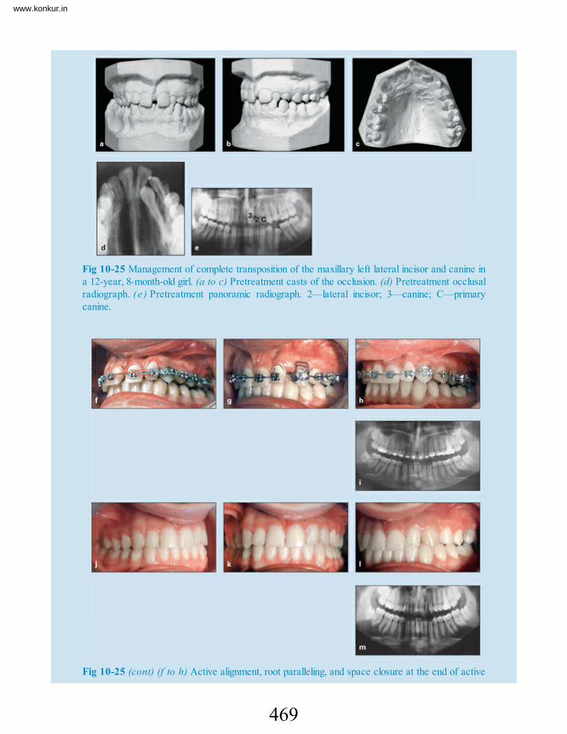
Fig 10-25 Management of complete transposition of the maxillary left lateral incisor and canine ina 12-year, 8-month-old girl. (a to c) Pretreatment casts of the occlusion. (d) Pretreatment occlusalradiograph. (e ) Pretreatment panoramic radiograph. 2—lateral incisor; 3—canine; C—primarycanine.
Fig 10-25 (cont) (f to h) Active alignment, root paralleling, and space closure at the end of active
469
www.konkur.in

treatment. ( i ) Panoramic radiograph taken during active treatment. ( j to l) Posttreatmentocclusion. (m) Posttreatment panoramic radiograph.
Case 10-3A 7-year-old girl in the early mixed dentition had a Class I malocclusion, minor anterior crowding,rotation of the maxillary incisors, and end-to-end dental relationships. The partially erupted mandibularleft lateral incisor had the potential to erupt in crossbite. She presented with an incomplete transpositionof the mandibular permanent right lateral incisor and canine (Figs 10-26a to 10-26e).
Treatment:Treatment phases were extraction of the mandibular primary canine, leveling and uprighting of thedisplaced mandibular lateral incisor, and alignment of the mandibular incisors. The next step wasguidance of eruption of the displaced canine. This was followed by maxillary 2× 4 bonding to align themaxillary incisors and some proclination to increase the arch circumferences, thereby providing normaloverbite and overjet and creating more space for the maxillary canines. Figures 10-26f to 10-26iillustrate different phases of mechanotherapy. Figures 10-26j to 10-26o show the posttreatmentcondition of the dentition.
Fig 10-26 Management of incomplete lateral incisor–canine transposition in a 7-year-old girl in theearly mixed dentition. The partially erupted mandibular left lateral incisor has the potential to eruptin crossbite. (a to c) Pretreatment occlusion. C— primary canine; 2—permanent lateral incisor.(d) Pretreatment occlusal radiograph. (e) Pretreatment panoramic radiograph. (f and g) Differentstages of mechanotherapy, including leveling, incisor alignment, and space creation.
470
www.konkur.in

Fig 10-26 (cont) (h and i) Panoramic radiographs taken during active treatment. (j to n)Posttreatment occlusion. (o) Posttreatment panoramic radiograph.
ImpactionTooth impaction is an intraosseously displaced position of the teeth and an abnormaleruption pathway that results in a prognosis of delayed or failed eruption. Impactedteeth are a commonly found anomaly in dental practice. They pose a problem for themaintenance and continuity of the patient’s dental health and sometimes represent arisk-management threat for the practitioner. Any tooth can be impacted, but thefollowing are the most frequently impacted teeth:
• Mandibular third molars• Maxillary canines• Maxillary third molars• Maxillary and mandibular second premolars
471
www.konkur.in

• Maxillary central incisors
Maxillary Canine ImpactionThe role of the canines in the development of occlusion was described byBroadbent59 as a major factor in the “ugly duckling” stage of normal dentaldevelopment. Today it is known that canines play several important vital roles infacial appearance and function, such as their role in dental esthetics of the smile, asan important part of the occlusal foundation in dental arch development, and asimportant dental units in functional occlusion.
Canines are prone to ectopic eruption and impaction because of their high anddeep position in alveolar bone, their extended developmental period, and their longand tortuous eruption route.
Adjacent teeth or other local factors may also narrow the eruption path of thecanine because of its late appearance in the eruption sequence. Early clinical andradiographic evaluation of canine position and proper intervention can guide thecanine to a normal path of eruption and prevent many complications and risks.
PrevalenceMaxillary canines are the most commonly impacted teeth, second to mandibular thirdmolars. The incidence of canine impaction in the maxilla is more than three timesthat in the mandible. Bishara’s review60 of the literature reported that the incidenceof maxillary canine impaction ranges between 1% and 3% of patients. For themandibular canine, the incidence of impaction is about 0.35%. This anomaly istwice as common in females as it is in males. Most affected patients haveunilaterally impacted maxillary canines, and only 8% have bilateral impactions.Canine impaction has a high family association and is five times more common inEuropeans than in Asians.61
Despite the fact that maxillary canine buds develop high near the orbit and sinusand buccal to adjacent tooth roots, the prevalence of palatal impaction is muchhigher than that of buccal impaction. Approximately two-thirds of impactedmaxillary canines are impacted palatally and one-third labially.62 Jacoby63 reportedthat 85% of impacted canines are located palatally and 15% buccally. Fournier etal64 reported a palatal-to-buccal impaction ratio of 3 to 1, and Jacoby63 reported a
472
www.konkur.in

ratio of 12 to 1.Buccal canine impactions are mostly associated with inadequate space and are
positioned more vertically, while palatally impacted canines are positioned morehorizontally. Because palatal bone is denser and palatal mucosa is thicker, palatallyimpacted canines rarely erupt without orthosurgical treatment.
In a clinical and radiographic evaluation of individuals with Down syndrome,Shapira et al55 reported that prevalences of canine impaction (15%) and maxillarycanine–first premolar transposition (15%) were high compared with publishedfigures from random population samples. Warford et al 65 reported that maxillarycanine impaction has an incidence of 1 in 100 among the general population.
Etiology of palatal canine impactionPalatal canine impaction and buccal canine impaction differ in etiology,morphogenesis, and management. Two general theories have been proposed toexplain the occurrence of palatally displaced and impacted maxillary canines:guidance theory and genetic theory.
Guidance theory
Guidance theory contends that this anomaly develops because of the presence oflocal predisposing causes. For example, the canine erupts along the root of thelateral incisor; thus, the presence of normal lateral incisor roots serve an importantrole in guidance of canine eruption. If the lateral incisor root is malformed or thelateral incisor is missing or misshaped (peg lateral), extra space in the apical part ofthe maxilla can deflect the eruption path of the canine.
In a retrospective study of the records of 182 orthodontic patients with palatallydisplaced canines, Mossey et al66 measured the tooth length of lateral and centralincisors on radiographs and the crown widths of lateral incisors on study casts. Formore accuracy, the crown widths and root lengths of 160 extracted maxillary lateralincisors were also measured. The study supported the hypothesis that there is a weakassociation between palatally displaced maxillary canines and lateral incisors ofsmaller than average crown width and found weak support for the associationbetween palatal canines and absence of the adjacent lateral incisor.
Becker67 and other advocates of this theory have stated that even if absence of thelateral incisor is genetically determined, the palatal canine displacement has no
473
www.konkur.in

genetic association but occurs as a result of local environmental disturbances (Fig10-27; see also Fig 10-30).
Fig 10-27 Palatal canine impaction. The impaction is related to the missing lateral incisor.
Arch length deficiency is another local factor that has been mentioned in theliterature. According to Jacoby,63 space deficiency is a valid explanation for mostimpactions but not for palatal impaction of the maxillary canine. His study showedthat 85% of palatally impacted canines have sufficient space for eruption.
The buds of the lateral incisor and the first premolar are located behind thecanine’s palatal surface, and the maxillary canine bud is wedged between the nasalcavity, the orbit, and the anterior wall of the maxillary sinus. When there is archlength discrepancy or space loss in the maxillary canine area, the canine becomesimpacted buccally; when the maxillary lateral incisor is missing, the canine becomesimpacted palatally. Jacoby63 indicated that this space can be provided by excessivegrowth in the base of the maxillary bone, by agenesis of lateral incisors or peg-shaped lateral incisors, or by stimulated eruption of the lateral incisor or the firstpremolar. He added that dysplasia in the maxillary-premaxillary suture can alsomodify the direction of eruption of the maxillary canine.
The author’s clinical experience indicates that space deficiency can also causepalatal impaction in this situation. The radiographic evaluation also shows somepalatal positioning of the canine.
The presence of a supernumerary tooth or an odontoma can also deflect theeruption path of the canine and cause impaction (see case 10-7). If the problem isdetected early, interceptive treatment and preservation of space can guide the normaleruption pattern, and canine impaction can be prevented.
Al-Nimri and Gharaibeh62 studied the etiology of palatal impaction by assessingthe pretreatment dental casts of 34 patients with unilateral palatal canine impaction.The casts of the subjects were compared with the pretreatment dental casts ofunaffected orthodontic patients, which were matched according to age, sex, and typeof malocclusion. They found that palatal canine impaction occurred most frequentlyin subjects with Class II division 2 malocclusions and in those who were missing a
474
www.konkur.in

lateral incisor. They also found that the transverse arch dimension was significantlywider in the impaction group.
Zilberman et al68 established a strong link between small, peg-shaped, or missinglateral incisors and palatally displaced maxillary canines. They also proposed that,because anomalies of the lateral incisor develop under strong genetic control, thefirst-degree relatives of patients with palatally impacted canines would themselvesshow an increased prevalence. They reported that anomaly of the lateral incisorsamong the relatives was found to be four times that of the general population.
Therefore, the following are the most common local factors that can cause canineimpaction:
• Missing lateral incisor• Peg-shaped lateral incisor• Supernumerary tooth or odontoma• Space deficiency• Dysplasia in the maxillary-premaxillary suture• Some Class II division 2 malocclusions
Genetic theory
According to genetic theory, disturbance of maxillary permanent canine eruption isdue to an abnormal developmental condition of the dental lamina. Advocates of thistheory cite evidential categories related to genetic origin such as familial andbilateral occurrence of impaction as well as increased occurrence of otherassociated anomalies, such as lateral incisor and second premolar hypodontia,ectopic eruption of first molars, and infraocclusion of primary molars.
Baccetti69 studied an untreated orthodontic population to reveal patterns ofassociation among seven types of dental anomalies, including small maxillary lateralincisors and palatal displacement of maxillary canines. The study found significantreciprocal associations among five of these anomalies, including size of themaxillary lateral incisors and palatal displacement of the maxillary canines andsuggested a common genetic origin for these conditions. The existence ofassociations between different tooth anomalies is clinically relevant, because theearly diagnosis of one anomaly may indicate an increased risk for others.
Peck et al70 demonstrated that 33% of palatal canine impaction has someassociation with missing or peg-shaped lateral incisors. They examined the
475
www.konkur.in

specificity of tooth agenesis sites associated with the occurrence of palatallydisplaced canines, mandibular lateral incisor–canine transposition, and maxillarycanine–first premolar transposition. They suggested that coupling these new clinicalfindings with results from recent molecular studies indicates that transcriptionfactors such as MSX1 and PAX9, which have been associated with agenesis ofmolars, might be involved in the genetic control of mandibular lateral incisor–caninetransposition and palatal canine impaction. However, it remains uncertain whetherthe anomalous or missing lateral incisor is a local causal factor or an associatedgenetic development.
Pirinen et al71 examined 106 patients who had been treated orthodontically forpalatally impacted canines and 110 first-degree and 93 second-degree relatives. Theprevalence of hypodontia affecting permanent teeth in examined patients was 36%,which is 4.5 times the prevalence in the general population. The prevalence ofhypodontia in both first- and second-degree relatives was 19% to 20%, which is 2.5times the prevalence in the general population. From these findings, they concludedthat the palatally displaced canine belongs to the spectrum of dental abnormalitiesrelated to hypodontia. Palatal displacement of canines is genetic and related togenetic incisor-premolar hypodontia and peg-shaped incisors. They also stressed theimportance of screening for impacted canines in patients with hypodontia or peg-shaped teeth as well as their families.71
To summarize, the exact etiology of maxillary impaction is not clear; it may be amultifactorial disturbance, in which either genetic or systemic factors can play aspredisposing factors, and local factors prepare the environment for canine deflectionand an abnormal pathway. Regardless of the cause, because the maxillary canine hasthe longest and most tortuous path of eruption of any tooth, the likelihood ofdisturbances during the developmental path toward occlusion is great. At the age of3 years, the canine bud is located very high in the maxilla close to the orbit. Thecrown is directed mesiolingually. From this point, it starts moving gradually towardthe occlusal plane, while it is uprighting itself, until it reaches the distal aspect of theroot of the lateral incisor. It then seems to be deflected to a more vertical position,until it reaches the occlusal contact at approximately age 13 years, with a markedmesial inclination. However, this long, complicated journey does not happenovernight; it takes about 10 years to be completed, and it is the responsibility ofdental practitioners to monitor these events (by longitudinal panoramic radiographicmonitoring) in order to detect problems early and to prevent complications throughproper intervention.
476
www.konkur.in

Factors associated with palatal impactionThe following factors have been reported to be associated with palatal impaction:
• Space deficiency• Trauma• Overretention of the primary canine• Premature loss of the primary canine• Failure of the primary canine root to resorb, creating a potential mechanical
obstacle for canine eruption• Abnormal position of the tooth bud• Disturbances in tooth eruption• Localized pathologic lesions such as cysts and odontomas• Abnormal sequence of eruption• Missing lateral incisors• Anomalous abnormal size of the lateral incisor or abnormal form of the lateral
incisor roots (dilacerations of the root)• Variation in timing of lateral incisor root formation• Ankylosis of the permanent canine• Presence of a supernumerary tooth• Presence of an alveolar cleft• Idiopathic conditions
Etiology of labial canine impactionLabial impaction of a maxillary canine is most often the result of space deficiencyduring the transitional dentition (Fig 10-28). This deficiency can be caused bypremature loss of the primary canine followed by a midline shift that results ininsufficient space for and buccal eruption of the canine; anterior dental crossbite thatdecreases the anterior arch circumferences, deflecting the canine buccally; ectopicmigration of the permanent canine over the adjacent teeth; or overretention of theprimary canine.
477
www.konkur.in

Fig 10-28 Buccal impaction caused by anterior crossbite (a), insufficient space (b), andoverretained primary canine (c and d).
Consequences of impacted maxillary caninesMost eruption problems, including canine impaction, are asymptomatic, and patientsare unaware of their existence. If practitioners do not monitor their patient’s eruptionpatterns, the time of discovery is usually late, and the problem has alreadycompletely developed. In some situations, adjacent teeth and surrounding structureshave already been damaged. Abnormal eruption paths within the dentoalveolarprocess can cause migration of the adjacent teeth and loss of arch length,development of cystic lesions and infection, root resorption of the nearby lateralincisors, and overretention of primary lateral incisors.
Resorption of the incisor roots is also asymptomatic and cannot be recognizedwithout clinical examination. Ericson and Kurol72 investigated lateral rootresorption caused by ectopic eruption of the maxillary canines. They assessed twogroups, one with 40 cases of lateral incisor root resorption and a control group of118 ectopic eruption cases without lateral incisor root resorption. When the cusp ofthe well-developed canine was positioned mesial to the lateral incisor root, the riskof complications was three times greater. Furthermore, the risk of resorptionincreased by 50% when the mesial eruption angle exceeded 25 degrees comparedwith the controls. Lateral incisor root resorption was three times more common ingirls than in boys.
Walker et al 73 assessed the spatial relationship of impacted canines by usingthree-dimensional images of 27 unilaterally and bilaterally impacted canines from19 consecutive patients (15 female and 4 male subjects). The study found that 92.6%
478
www.konkur.in

of impactions were palatal. Incisor resorption was present in 66.7% of the lateralincisors and in 11.1% of the central incisors adjacent to the impacted canine.Follicle size did not have a major influence on the position of the impacted canine.The alveolus was narrower on the side of the impacted canine than on the side of theerupted canine; however, the width of the alveolus on the impacted canine side isindependent of the primary canines.
The potential complications emphasize the need for practitioners to closelymonitor the development and eruption of the dentition during the stage of dentaltransition. In brief, the following complications can result when canine impaction isnot treated promptly:
• Resorption of the roots of adjacent teeth• External resorption• Infection• Cyst formation• Loss of arch length• Transposition• Periodontal defects
Early detection of canine impactionBecause canine impactions generally require longer treatment times, depending onthe location of the impacted tooth, early identification of impaction is of criticalinterest to the orthodontist and to all practitioners. Potentially impacted canines canbe detected at an early age. With proper clinical diagnosis and radiographicevaluation and monitoring (longitudinal panoramic radiograph monitoring), theclinician might be able to prevent impaction through timely interceptive treatment.
Because of the wide variation that exists in the timing of eruption of permanentteeth among members of different ethnic groups, different investigators haverecommended an age range of 8 to 10 years for early detection of maxillary canineimpaction. Diagnostic procedures at these ages are a combination of careful clinicaland complete radiographic examinations (longitudinal monitoring of panoramicradiographs).
Clinical examination
479
www.konkur.in

The clinical examination consists of digital palpation of the buccal bulge of thecanine in the sulcus above the primary canine root, which can reveal the position ofthe maxillary permanent canine even in an 8- to 10-year-old patient. The clinician’sindex fingers can be used to palpate the area above the primary canine on both thebuccal and palatal aspects of both sides of the arch.
This clinical examination should be repeated every 2 to 3 months. Impaction ofthe permanent canine is indicated if the buccal canine bulge is not palpable inchildren older than 10 years and the primary canine is retained and shows no signsof mobility. This preliminary diagnosis should be followed by careful radiographicexamination for confirmation.
Another clinical finding, which may be observed after eruption of the permanentlateral incisors at around 9 to 10 years of age, is distobuccal inclination of thelateral incisors. Broadbent59 called this normal transitional stage of dentition the“ugly duckling” stage preceding the eruption of the permanent canines. Tipping maycause spacing of the incisor crowns, despite the crowding of the roots (Fig 10-29).Severe distal tipping of the lateral incisor crowns might be due to pressure from themesially displaced and abnormally positioned canine and indicate the potential forcanine impaction. This situation requires careful radiographic evaluation andmonitoring and possibly early intervention.
Fig 10-29 (a and b) Ugly duckling stage of the dentition.
Likewise, a labially inclined lateral incisor can be the result of a displaced caninelying on the labial aspect of the lateral incisor root. In all of these situations, carefulmonitoring of the permanent canine is recommended.
No orthodontic movement should be applied to the lateral incisors until thepressure of the canine crown is eliminated from the lateral incisor root. Extraction ofthe remaining primary canine can help the permanent canine crown toward itsnormal position. Untreated cases may cause incisor root resorption.
Clinical signs of canine impaction
480
www.konkur.in

The following are signs of canine impaction:• Delayed eruption of the permanent canine• Prolonged retention of the primary canine, especially asymmetric exfoliation of the
primary canine• Absence of a normal labial canine bulge• Presence of a palatal bulge• Distal tipping of the lateral incisor crown• Retroclined position of the lateral incisor (if its root is forced buccally and its
crown palatally by the palatally impacted canine)
Radiographic evaluationRadiographic evaluation is essential for detecting potentially impacted canines andan important tool to determine whether the canine is positioned palatally or buccally.Different radiographic techniques can be used, including periapical, occlusal,panoramic, and cephalometric radiographs (lateral or posteroanterior).
Periapical radiographs. Clinicians can use different kinds of periapical radiographsfor localization of a displaced canine, including the standard periapical or Clark’srule technique. Also termed the image shift principle, in the Clark’s rule techniquetwo different angulations are taken in the same horizontal plane. If the object movesto the same side of the angulation, the subject (in this case, the canine) is locatedmore lingually (palatally); if it moves to the opposite direction, the object is morebuccally positioned.
Occlusal radiographs. Occlusal radiographs are recommended to determine theposition of the involved tooth in the buccolingual and mesiodistal directions.Various techniques can be applied to obtain occlusal radiographs for localizing theimpacted tooth. Occlusal radiographs or vertex occlusal view radiographs takenwith the x-ray beam directed along the long axis of the central incisors are useful foridentifying the position of the palatally displaced tooth relative to adjacent teeth (Fig10-30).
481
www.konkur.in

Fig 10-30 Occlusal radiograph showing the position of a palatally impacted canine.
Lateral and posteroanterior cephalometric radiographs. Lateral andposteroanterior (frontal) cephalometric radiographs are also useful techniques forlocalizing and identifying the inclination of impacted teeth. Lateral cephalometricradiographs can show the anteroposterior position of the canine relative to theincisors. Frontal radiographs can show the vertical and lateral positions of thecanines relative to the incisors.
Williams74 has shown that the permanent canine, during its pre-eruptivedevelopment, should be inclined slightly mesially, with the crown below the level ofthe apices of the lateral incisors and well below the lateral border of the nasalcavity. The canine root should lie distal to the lateral border of the nasal cavity. Ifthe frontal radiograph reveals that the canine is mesially tipped, with its crownmedial to the lateral border of the nasal cavity, and there is no evidence of primarycanine root resorption, impending impaction of the displaced maxillary canineshould be suspected.
Computed tomography scans. A CT scan is an expensive, routine radiographictechnique in medicine and occasionally is used in dentistry. Scanning radiographycan accurately locate the object in three dimensions.
In a recent study comparing conventional radiographic procedures (a single two-dimensional panoramic radiograph) with cone beam CT scans, Alqerban et al75
showed that the cone beam CT is more sensitive than conventional radiography forboth canine localization and identification of root resorption of adjacent teeth.
The three-dimensional images provided by this technique show the buccopalatalposition of the impacted canine. The extent and exact location of lateral incisor rootresorption can be detected; no other technique has the ability to show these aspects.Walker et al 73 stated that three-dimensional volumetric imaging of impacted caninescan show the presence or absence of the canine, the size of the follicle, the
482
www.konkur.in

inclination of the long axis of the tooth, the relative buccal and palatal positions, theamount of bone covering the tooth, the three-dimensional proximity and resorption ofroots of adjacent teeth, the condition of adjacent teeth, local anatomicalconsiderations, and the overall stage of dental development. They concluded thatthree-dimensional imaging is clearly advantageous in the management of impactedcanines.
Panoramic radiographs. The panoramic radiograph has been used extensively indentistry; it is a useful tool that shows a general view of the combined permanentand primary dentitions and allows clinicians to monitor all abnormalities developingduring the transitional dentition. Panoramic radiographs can also show otherdevelopmental anomalies, such as supernumerary teeth, missing teeth, odontomas,other pathologic conditions, the conditions of the primary roots and their successors,and the conditions of permanent root development. Moreover, panoramicradiographs have been used extensively for detecting canine inclination during thetransitional dentition.
The general perception is that routine panoramic radiographs taken during themixed dentition are useful for detecting only the mesiodistal position and inclinationof the displaced unerupted canine; however, careful evaluation of high-qualitypanoramic radiographs can help in the detection of buccal or palatal positioningalso.
Distinguishing buccopalatal position of an impacted canine by panoramicradiograph. Evaluation of the position of the impacted canine (buccal or palatal)depends on the relative radiopacities of the two superimposed objects. The film orthe screen that captures the image is located in front of two objects, the canine andincisors. If the area of superimposition of the two images is dominated by theopacity representing the shape of the lateral incisor, it means that the lateral incisoris closer to the film, indicating that the canine is located more palatally. If the area isdominated by the opaque contours of the canine, it means that the canine is closer tothe film, indicative of a more buccally positioned canine.
Figure 10-31 is a good example of this radiographic phenomenon for comparisonof the different appearances of the two impacted canine positions. The panoramicradiograph clearly shows the position of the canines relative to the lateral incisorson the left and right sides; the palatal positioning of the right canine is indicated bythe dominant contours of the lateral incisor in that area, and the buccal positioning ofthe left canine is indicated by the dominant contours of the canine on that side.
483
www.konkur.in

Fig 10-31 Panoramic radiograph showing two impacted canines. The right canine is palatallyimpacted and the left canine is buccally impacted.
Longitudinal monitoring of panoramic radiographs can help in early detection andprediction of maxillary canine impaction. Two important signs for prediction andearly detection of maxillary canine impaction on panoramic radiographs are theposition of the impacted tooth and the inclination of the impacted tooth. Asmentioned earlier, Williams74 described the normal position of the permanent canineduring pre-eruptive development: inclined slightly mesially with the crown belowthe level of the apices of the lateral incisors and well below the lateral border of thenasal cavity. The canine root should lie distal to the lateral border of the nasalcavity. If the canine is mesially tipped in the frontal radiograph with its crownmedial to the lateral border of the nasal cavity and there is no evidence of primarycanine root resorption, an impending impaction of the displaced maxillary canineshould be suspected.
Figure 10-32 is a retrospective evaluation of three panoramic radiographs takenduring different stages of the dentition. The panoramic radiograph taken when thepatient was 9 years old indicated that the maxillary left canine had a higher positionand greater inclination than the right canine. No early intervention was attempted,and panoramic radiographs taken 1 and 4 years later revealed the incorrect eruptionpath of the palatally impacted canine. Anchorage preparation for the maxillarymolars and early extraction of the primary canines and primary first molars mighthave guided the canine to a normal path of eruption or at least might have reducedthe severity of the problems.
Fig 10-32 Retrospective evaluation of an incorrectly positioned maxillary left canine. (a)Panoramic radiograph taken at age 9 years. The left canine is positioned higher and is moreinclined than the right canine. ( b ) Panoramic radiograph taken 1 year later. ( c ) Panoramic
484
www.konkur.in

radiograph taken 4 years later. No early intervention has been attempted to correct the path oferuption.
The proximity of impacted canine to adjacent teeth is another important point to beassessed before treatment planning. Depending on the position of the impacted tooth,the complexity of treatments differ. Impacted maxillary canines are usuallypositioned distal and either buccal or palatal to the lateral incisor root, with somedegree of increased inclination. They are in a lower or higher position than thelateral incisor root.
Impacted canines have more mesial inclination than normal, and sometimes theyare displaced from their normal position, with close proximity to the roots ofadjacent teeth, and therefore are also ectopic. Ectopic impacted teeth are usuallylocated close to the lateral or central incisor roots and hence have the potential todamage those roots. Canines that are both impacted and ectopic are more difficult tomanage, require longer treatment time, can be more destructive to adjacent teethduring treatment, and demand much more percussion during treatment planning andprecision during the treatment process (see case 10-4).
The other important factor for prediction of impaction from a panoramicradiograph is the inclination of the impacted tooth. The mesiodistal inclination of themaxillary canine is another predictor of impaction as well as a sign of complexityand an indicator of the potential success of treatment. Some investigators haveproposed different techniques for evaluating potential outcomes of palatallyimpacted canines. Ericson and Kurol76 found that the more mesially located andhorizontally inclined the crown is, the less the likelihood of eruption after extractionof the primary canine. Power and Short77 also looked at angulation as a predictorand found that if the tooth is angled more than 31 degrees relative to the midline, itschances of eruption after primary extraction are decreased.
Warford et al 65 drew a horizontal reference line on panoramic radiographs,across the most superior point of both condyles, and measured the mesial angleformed by the constructed horizontal line and the long axis of the unerupted tooth.They found that the angulation for nonimpacted teeth, a mean of 75.12 degrees, wasgreater than that for impacted teeth, 63.20 degrees. Figure 10-33 presents a Warfordanalysis of a panoramic radiograph, indicating that the left canine is much moreseverely impacted than the right canine (see case 10-4).
485
www.konkur.in

Fig 10-33 Inclination of both canines assessed with the Warford analysis.65 The angulationindicates the severity of the problem affecting the left canine.
Treatment options for impacted maxillary caninesThe duration of the long and problematic transitional process of permanent
canines is usually about 10 years, from the beginning of eruption to the completeocclusal position (from ages 3 to 13 years). Thus, for any type of preventive orinterceptive treatment, clinicians must monitor this long process during the mixeddentition. Depending on the age of the patient, the dental developmental age, thestage of eruption disturbances, and the type of impaction, there are a number oftreatment options for the management of impacted teeth:
• Observation during the transitional dentition• Interceptive treatment• Surgical exposure of the canine and orthodontic treatment• Autotransplantation of the canine• Extraction of the impacted canine and space closure• Extraction of the impacted canine and prosthetic or implant replacement• Periodic radiograph observation (in adults when the impacted canine is located
very high or horizontally and because of either high risk or the patient’s desire forno treatment)
Some kind of interaction among these options might be necessary, such asselective primary tooth extraction, removal of any obstacle deflecting the canine, orspace creation.
The most desirable approach in managing impacted maxillary canines is earlydetection and interception of a deflected canine that has the potential for impaction atits initial stage, at around 8 to 10 years of age, when the canine starts its longintrabony movement toward its final position.
486
www.konkur.in

Observation
During the transitional dentition, such as during the ugly duckling stage or when thepermanent canine is tipped mesially and is close to the lateral incisor apex,orthodontic movement of the lateral incisor crown will cause root resorption of thelateral incisor. In this situation, the clinician should only observe the situation untilthe canine crown changes its position, away from the apex. If the canine does notshow any change in position after 10 to 12 months and the possibility of lateralincisor root resorption is evident, exposure of the canine and bonding attachment fororthodontic traction is indicated.
Williams74 stated that age 8 years is the best time to begin observation of themaxillary canines and that the positional changes over the next 2 years requirecareful observation.
Observation of the transitional dentition from the beginning of the mixed dentitionto complete eruption of the canines can predict tooth impaction and many othereruption disturbances (see Figs 10-32 and 10-33).
Interceptive treatment
Interceptive treatment, or guidance of eruption, for canine impaction is a series ofprocedures that should begin when the canine starts its long intrabony movementtoward its final position.
Many investigators suggest that the best time to begin assessing a patient forpotential maxillary canine impaction is around 8 to 10 years of age. It is believedthat after the patient is 11 to 12 years of age the only remaining option is to correctthe anomaly by orthosurgical intervention, which is sometimes risky, unpredictable,or unsuccessful.
During this period, the permanent canine begins its intrabony descent from itslingual position relative to the root apex of the primary canine and the primarycanine root starts to resorb. Dewel78 and Newcomb79 concluded that adequateobservation, diagnosis, and treatment planning can be critical during this stage ofcanine transition.
Interceptive options. After thorough clinical and radiographic evaluation, when thepotential for canine impaction is confirmed, depending on the etiologic factors andthe patient’s age and type of occlusion, several interceptive and guidance procedurescan be applied:
487
www.konkur.in

• Removal of the obstacle causing deflection of the canine, such as a supernumerarytooth, odontoma, cyst, or ankylosed primary tooth.
• Creation of space, if space deficiency is the cause of this anomaly. Depending onthe occlusion and age of the patient, different tactics can be applied for providingthe necessary space:- Distalization of the buccal segment in some Class II mixed dentition treatment- Correction of maxillary anterior dental crossbite if retroclination of the
maxillary incisors has resulted in space deficiency for maxillary canines or ifcorrection of crossbite increases the arch circumference in the anterior segment
- Closure of the space that sometimes remains between the incisors, even aftercomplete eruption of the lateral incisors
• Extraction of the primary canine at the right time is advocated by manyinvestigators, who believe this action facilitates and promotes permanent canineeruption.
Primary canine extraction procedure. Primary canine extraction is an interceptivetreatment for guiding the permanent canine eruption to prevent impaction; the bestage for this procedure is when the child is between 8 and 10 years and not after theage of 11 years.
Shapira and Kuftinec80 concluded that early diagnosis and detection of apotentially impacted maxillary canine and timely interceptive removal of the primarycanine reduces the need for complicated orthodontic treatment. Ericson and Kurol76
reported that favorable eruption occurred 78% of the time in their sample. In aninvestigation into the response of palatally displaced canines to the removal ofprimary canines, Power and Short77 reported success 62% of the time.
The best situation for this procedure is when the clinical examination andradiographic evaluation of an 8- to 10-year-old child indicate the presence of thefollowing conditions:
• The permanent canine is mesially displaced and does not follow the guidance ofthe lateral incisor root.
• The permanent lateral incisor crown is somewhat distally inclined, indicating thepresence of permanent canine crown force.
• The buccal canine bulge cannot be palpated.• An overretained primary canine is present and shows no signs of mobility.
488
www.konkur.in

Timely extraction of the primary canine in the presence of any of theaforementioned conditions allows the permanent canines to become upright anderupt into their proper positions in the arch and provides the best long-term results,without many of the complications that can arise if intervention is delayed.
Before this interceptive treatment is initiated, the proximity of the permanentcanine crown and lateral apex must be evaluated. If distal tipping of the lateralincisor crown is clinically and radiographically evident, no orthodontic force can beapplied to the lateral incisor, because the force will eventually cause severe rootresorption. In this situation, after extraction of the primary canine, the most cautioustreatment plan is use of some kind of anchorage device, such as a transpalatal arch,and then monitoring the permanent canine crown and lateral incisor apex until someuprighting of the permanent canine has occurred and the canine crown is locateddistal to the lateral incisor root.
The next step in any case of canine guidance would be 2 × 4 bonding, slowalignment of the maxillary incisors, and closure of any diastema or dental spacingpresent between incisors to facilitate eruption of the permanent canine.
Olive81 evaluated 30 palatally impacted canines that emerged followingorthodontic space opening without surgical exposure and tried to determine factorsinfluencing the time of eruption. The sector of impaction (severity of canineinclination) was the best guide to the duration of orthodontic treatment prior toemergence.
Jacobs82 stated that extraction of the primary canine for prevention or interceptionof a palatally displaced canine is best carried out as early as the displacement isdetected, soon after 10 years of age.
Ericson and Kurol76 stated that successful eruption of the impacted canine afterextraction of the primary canine depends on the horizontal angulation of thepermanent canine and the position of the canine crown relative to the root of thelateral incisor. If radiographs showed that the crown of the permanent canine waspositioned over the root of the maxillary lateral incisor but not past the mesialsurface of the root, self-correction of the ectopic canine occurred with highpredictability. However, if the permanent canine was positioned well beyond themesial surface of the lateral incisor root, the chance of self-correction and eruptionof the permanent canine was very low.
Early diagnosis and intervention prevent resorption of the adjacent incisor root,save time, reduce expense, avoid the surgical and orthodontic treatment needed toalign a palatally impacted canine, and eliminate complications associated with
489
www.konkur.in

surgical exposure and orthodontic procedures. These complications include longtreatment time, bone loss, root resorption, and gingival recession around the treatedteeth.
Surgical exposure of the canine and orthodontic treatment
If impacted canines are in their final stage of development in the permanentdentition, the clinician must consider orthodontic treatment combined with surgicalexposure of the canine to bring it into occlusion. Orthosurgical approaches varygreatly depending on place of impaction, whether the impaction is labial or palatal,the age of the patient, the proximity of the impacted tooth to adjacent teeth, etiologicfactors, and the morphology of impacted teeth. These approaches depend on theclinical judgment and experience of the orthodontist and surgeon. To ensure thatappropriate surgical and orthodontic techniques are applied, the first step iscoordination between the orthodontist and the oral surgeon.
Next, before any treatment planning is completed, a thorough clinical andradiographic evaluation must be performed to precisely locate the position andinclination of the impacted tooth and the proximity to adjacent teeth and to determinethe type of surgical approach and the method and direction of orthodontic traction.Important points of clinical examination and different radiographic techniques thatcan help in diagnosis and precise localization have been discussed previously.
Pretreatment considerations. An impacted canine can be located palatally orbuccally, can be ectopic or nonectopic, and can be unilateral or bilateral; each ofthese situations requires different surgical techniques and different orthodonticapproaches. Among the factors that must be considered are:
• The inclination of the impacted tooth• The position of the impacted crown and its proximity to adjacent teeth• The cause of impaction• The age of the patient and the amount of root development
As discussed earlier, the inclination of the canine is a sign of impaction as well asa predictor of treatment complexity and success. The more mesially located andhorizontally inclined the crown is, the less likelihood of eruption after extraction ofthe primary canine.
The position of the impacted crown and its proximity to adjacent teeth not only
490
www.konkur.in

influence the amount of damage that the canine may cause to other teeth but alsoaffect the complexity of treatments and dictate the technique of orthodontic tractionthat must be used. Impacted canines usually have more mesial inclination thannormal and are positioned distal and either buccal or palatal to the lateral incisorroot. They also can have a lower or higher position than the lateral incisor root.Sometimes impacted canines also are in close proximity to the roots of adjacentteeth and therefore are also ectopic, which represents a treatment challenge (Fig 10-34).
Fig 10-34 Difference between impaction and ectopic impaction.
Another point to be considered before treatment planning is the dental age of thepatient and the stage of root development. Proper intervention before rootcompletion has a better prognosis.
There are several factors that can cause canine impaction. Early detection andrecognition of the problem is helpful in treatment planning and can indicate thecomplexity of the treatment and the possibility of success. If the cause is clear-cut,such as space deficiency or a supernumerary tooth, the prognosis is morepredictable; if the cause is unknown and ankylosis of the impacted tooth issuspected, the prognosis is unknown, and treatment is risky, the treatment plan mustbe adjusted accordingly.
For example, if extraction of a premolar is part of the treatment plan and ankylosisof the impacted canine is also suspected, it is recommended that the impacted caninebe exposed and orthodontic traction started first to ascertain impacted toothmovement. If the canine shows resistance to eruption and ankylosis is confirmed, theimpacted tooth can be extracted, and the premolar can be substituted for the canine inthe dentition (see case 10-8).
Surgical approach. After careful clinical and radiographic evaluation andconfirmation of the exact location and inclination of the impacted tooth, thefeasibility of surgical exposure and proper access for the surgical approach aredetermined.
491
www.konkur.in

In the past, surgical approaches to access impacted teeth consisted of wideresection of gingival and osseous tissues to uncover the impacted tooth forattachment of the traction device or wires. These approaches were oftenaccompanied by surgical problems such as bleeding, difficulty in the placement ofthe attachment device, and, most significant, excessive removal of bone and softtissues. Figure 10-35 shows the type of attachment that was used before bondingtechniques were available. Many reports indicate various periodontal complicationsafter this kind of surgical technique, including bone loss, gingival recession,decreased width of keratinized tissue, delayed periodontal healing, and gingivalinflammation.83
Fig 10-35 Surgical attachment used before the bonding technique was available.
Now surgical techniques are improved and less aggressive, and the availability ofdirect bonding with delicate attachments means that the surgeon can bond to theimpacted crown directly and then replace and suture the flap. These procedures canbe accomplished with few complications.
Palatal versus buccal impaction. Palatal canine impaction and buccal canineimpaction differ in morphogenesis, etiology, and anatomical position, and thereforetheir treatment, whether interceptive or corrective via surgical procedure, is alsodifferent. The most common cause of buccal impaction is crowding. As discussedpreviously, many other etiologic factors also have been proposed for palatalimpaction.
Both orthodontic and orthosurgical approaches are more predictable and easier intreatment of buccal impaction than palatal impaction. Buccally impacted caninesreach occlusion faster and with less complexity. Palatally impacted canines also canvary in terms of complexity and difficulty of management, and there are somevariations that must be considered before treatment planning.
Surgical considerations in palatal canine impaction. Early extraction of primarymaxillary canines, around the age of 8 to 9 years, when the crown of the permanentcanine is positioned over the root of the maxillary lateral incisor but not past the
492
www.konkur.in

mesial surface of the root, will result in normal eruption of ectopically displacedpermanent maxillary canines with high predictability. However, if early interventionwas not performed at the proper age or the permanent canine is positioned wellbeyond the mesial surface of the lateral incisor root, the permanent canine willbecome impacted and self-correction of the permanent canine is not possible; thesesituations must be treated by orthosurgical procedures.
Careful surgical management requires thorough clinical and radiographicevaluations; accurate localization of the palatally impacted tooth with regard tolabiopalatal position, vertical position, inclination, depth of impaction, andproximity to adjacent teeth; and design of an accurate surgical approach according tothe type of impaction.
Surgical considerations in buccal canine impaction. In any type of mucogingivalintervention, an adequate zone of keratinized tissue must be maintained. If thisprocedure is not done properly and the tooth is moved through alveolar mucosa, it islikely that gingival recession will occur, leaving a periodontally compromisedgingival margin. All orthodontic and surgical techniques in the management of labialimpaction must be designed to maintain or ensure the presence of adequate amountsof keratinized gingiva.
To determine the correct surgical technique for exposing a maxillary canine that islabially or intra-alveolarly impacted, four criteria have been proposed:
1. The vertical position of the canine relative to the mucogingival junction2. The amount of gingiva in the area of the canine3. The labiolingual position of the canine crown4. The mesiodistal position of the canine crown
Depending on the vertical position of labially impacted teeth relative to themucogingival junction and the amount of gingiva, Kokich84 described threetechniques for uncovering a labially impacted maxillary canine: excisionaluncovering, apically positioned flap, or closed-eruption techniques. Herecommended simple excisional uncovering if the tooth is positioned labially and thecrown is below the mucogingival junction. If the canine crown is positioned slightlyapical to the mucogingival junction, an excisional technique would be inappropriate,because it would not result in any gingiva over the labial surface of the tooth after ithad erupted. If the crown is positioned significantly apical to the mucogingivaljunction, an apically positioned flap would also be inappropriate, because it wouldresult in instability of the crown and possible reintrusion of the tooth after
493
www.konkur.in

orthodontic treatment. In this situation, a closed-eruption technique will provideadequate gingiva over the crown and will not result in reintrusion of the tooth overthe long term.
Basic orthodontic procedures for correction of canine impaction. Orthodonticstrategy must be based on the position and complexity of the impacted tooth. It iscrucial that the orthodontist establishes accurate appliance design for anchoragepreparation, proper force delivery, and especially correct direction of force. Box10-3 provides orthodontists with guidelines for step-by-step management of canineimpaction.
Box 10-3 Step-by-step management of impactedmaxillary canines
1. Prepare complete records, including clinical andparaclinical examinations, to enable clearidentification of the problem and design accuratesurgical and orthodontic techniques.2. Accomplish orthodontic preparation prior tosurgical intervention; this can include leveling,correction of rotation, elimination of crowding, andcreation of space for the impacted tooth if tipping ofadjacent teeth has resulted in insufficient space.3. After all preliminary orthodontic movement iscompleted, refer the case for surgical intervention.The surgical approach should be based on the buccalor palatal inclination and vertical position of theimpacted tooth and its proximity to adjacent teeth.4. After a week or more of postsurgical healing,initiate orthodontic movement of the crown towardthe edentulous site.
5. Design the direction and the amount of forcedelivered for traction of the impacted tooth tosmooth the eruption path and prevent any damage tothe impacted tooth and its supporting structure aswell as the adjacent teeth. Follow biomechanicalprinciples with regard to the specific conditions ofeach case. Among the appliance designs appropriatefor anchorage preparation are the modifiedtranspalatal arch, mini-implant or vertical andhorizontal loop, auxiliary labial archwire, spring orchain, or elastic traction. For example, when thecrown of a palatally impacted canine is in intimatecontact with the lingual surfaces of the incisor roots,the first direction of movement should be to shift theimpacted tooth away from the adjacent teeth toprevent root resorption.
Some investigators have reported on early uncovering of palatally impactedcanines during the mixed dentition, so that the canines can erupt autonomously,without orthodontic intervention. They believe that when the impacted crown isavailable, regular bonding of brackets and movement into the dental arch can beaccomplished more efficiently, and the overall active treatment time for the patient isreduced. Periodontal and esthetic results are also excellent. This procedure is notrecommended if the canine is deeply impacted and its proximity to other tooth rootsis harmful; in such cases, the need to move the impacted tooth quickly away from theroots is urgent (see Case 10-7).
When severe incisor crowding or protrusion is present and extraction of
494
www.konkur.in
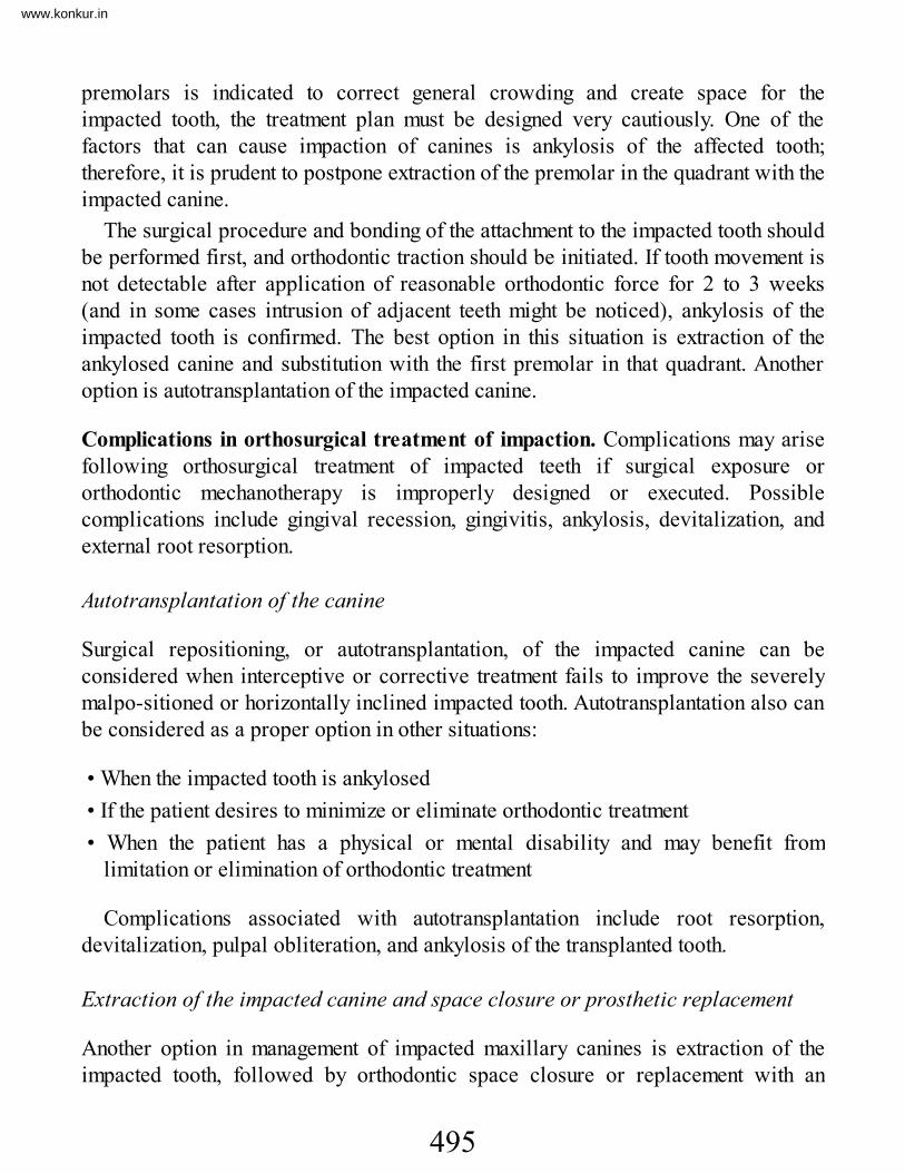
premolars is indicated to correct general crowding and create space for theimpacted tooth, the treatment plan must be designed very cautiously. One of thefactors that can cause impaction of canines is ankylosis of the affected tooth;therefore, it is prudent to postpone extraction of the premolar in the quadrant with theimpacted canine.
The surgical procedure and bonding of the attachment to the impacted tooth shouldbe performed first, and orthodontic traction should be initiated. If tooth movement isnot detectable after application of reasonable orthodontic force for 2 to 3 weeks(and in some cases intrusion of adjacent teeth might be noticed), ankylosis of theimpacted tooth is confirmed. The best option in this situation is extraction of theankylosed canine and substitution with the first premolar in that quadrant. Anotheroption is autotransplantation of the impacted canine.
Complications in orthosurgical treatment of impaction. Complications may arisefollowing orthosurgical treatment of impacted teeth if surgical exposure ororthodontic mechanotherapy is improperly designed or executed. Possiblecomplications include gingival recession, gingivitis, ankylosis, devitalization, andexternal root resorption.
Autotransplantation of the canine
Surgical repositioning, or autotransplantation, of the impacted canine can beconsidered when interceptive or corrective treatment fails to improve the severelymalpo-sitioned or horizontally inclined impacted tooth. Autotransplantation also canbe considered as a proper option in other situations:
• When the impacted tooth is ankylosed• If the patient desires to minimize or eliminate orthodontic treatment• When the patient has a physical or mental disability and may benefit from
limitation or elimination of orthodontic treatment
Complications associated with autotransplantation include root resorption,devitalization, pulpal obliteration, and ankylosis of the transplanted tooth.
Extraction of the impacted canine and space closure or prosthetic replacement
Another option in management of impacted maxillary canines is extraction of theimpacted tooth, followed by orthodontic space closure or replacement with an
495
www.konkur.in

implant- or tooth-supported prosthesis. It is not always possible to recover alldeeply impacted canines and reposition all impacted teeth within the alveolus.Removal might be indicated, for example, when the tooth is impacted very deeplyand severely inclined, when the tooth’s position and proximity to incisor rootscontraindicate any tooth movement, or when the impacted tooth is ankylosed andpatient compliance is not sufficient for transplantation.
Mandibular Canine ImpactionMandibular canine impaction is rare compared with maxillary canine impaction.Grover and Lorton85 found only 11 impacted mandibular canines in 5,000individuals (0.22%). Röhrer86 examined 3,000 patients radiographically and found62 impacted maxillary canines (2.06%) and only 3 impacted mandibular canines(0.1%), a 20:1 ratio.
Many causes for impacted mandibular canines have also been proposed, includinginadequate space, the presence of a supernumerary tooth or odontoma, prematureloss of the primary canine, hereditary factors, functional disturbances of theendocrine glands, tumors, cysts, and trauma. Impaction, failure of eruption, andhorizontal displacement of the mandibular canine are a common occurrence inpatients with cleidocranial dysostosis.
Early loss of the primary canine or arch length deficiency in the anterior segmentof the mandible is a common cause of buccal or lingual eruption of the mandibularpermanent canine. However, this type of canine position cannot be consideredimpaction and may be treated by space provision or extraction.
In some situations, the tooth bud of the mandibular canine may become displacedor rotated in the alveolus because of some obstacle or for idiopathic reasons, and thecanine root can develop in a horizontal direction (see Fig 10-23). In this situation, ifthe impacted mandibular canine is displaced horizontally below the apices of theincisors, the only option is extraction of the impacted canine to prevent damage tothe roots of the incisors.
Depending on the patient’s age, the position of the impacted canine, and thecondition of the occlusion, there are several treatment options for mandibularimpaction: observation, exposure, orthodontic traction, transplantation, andextraction.
Observation of the mandibular impacted canine is indicated if the canine is deeplyimpacted and asymptomatic and no associated pathosis is evident. Periodic
496
www.konkur.in

radiographic follow-up is necessary. In this situation, if the primary canine is ingood condition and has a good root length, it can be maintained in the arch.
Case ReportsThe following examples of different types of canine impaction illustrate orthodonticand orthosurgical approaches to the treatment of impacted teeth.
Case 10-4Figures 10-36a and 10-36b show panoramic radiographs of an 11-year-old girl with a Class IImalocclusion. Both maxillary canines were impacted. The panoramic radiographs were taken by herdentist 18 months apart, and no treatment was attempted in the interval. The right canine was impacted,and the left canine was ectopic and impacted. The radiographs revealed the proximity of the caninecrown to the left lateral incisor root and some damage to the root. The cause of this abnormality wasdiagnosed as space deficiency on both sides.
Treatment:The first step of the intervention was extraction of both maxillary left primary molars and creation ofspace for both canines by distalization of the molars and premolar with headgear (Fig 10-36c). Theimpacted right canine erupted automatically after space opening, but the ectopic and impacted leftcanine remained positioned over the lateral incisor roots. The next step was surgical exposure of the leftcanine and orthodontic traction. Figure 10-36d shows the patient’s posttreatment occlusion, and Fig 10-36e reveals the resorption of the lateral incisor root.
Fig 10-36 (a and b) Right and left maxillary canines impacted because of space deficiency. Theleft canine is also ectopic. (c) Panoramic radiograph taken after distalization of both maxillarysegments to provide space for the canines. (d) Posttreatment panoramic radiograph. (e) Lateralincisor root resorption.
497
www.konkur.in

Case 10-5An 11-year-old girl with a Class II division 1 malocclusion presented with displacement of bothmaxillary canines and maxillary space deficiency (Figs 10-37a to 10-37c). The mandibular dentition wasnormal. The left canine was displaced buccally and slightly overlapped the lateral incisor, while the rightcanine was severely impacted and ectopic.
Treatment:Cervical headgear was used to correct the Class II molar relationships and create space for bothmaxillary canines. Figure 10-37d illustrates the patient’s occlusion after molar retraction and spacecreation and before surgical exposure and bonding attachment. The left canine erupted spontaneouslyand was aligned. A spring was soldered to the palatal side of the molar buds for vertical traction andseparation of the ectopic canine from the lateral incisor root as the first step of orthodontic traction (Fig10-37e). Figures 10-37f to 10-37h show the posttreatment occlusion.
Fig 10-37 Management of maxillary canine displacement and space deficiency in an 11-year-oldgirl. (a and b) Pretreatment casts of the occlusion. (c) Pretreatment panoramic radiograph. (d)Panoramic radiograph taken after space creation and before surgical attachment. The left caninehas erupted. (e) Spring used for vertical traction. (f and g) Posttreatment casts of the occlusion.(h) Posttreatment panoramic radiograph.
Case 10-6
498
www.konkur.in

A 15-year-old girl presented with a Class II severe deep bite and oligodontia; she was missing bothmaxillary lateral incisors and three mandibular permanent incisors. The maxillary left permanent caninewas impacted and ectopic. The maxillary left primary canine and three primary incisor roots were stillpresent (Figs 10-38a to 10-38d).
Treatment:After consideration of the patient’s age and evaluation of her complicated orthodontic problems, thefollowing treatment plan was designed:
1. Extraction of the primary canine.2. Alignment of the maxillary anterior teeth and closure of the diastema.3. Surgical exposure and insertion of an attachment, which was a soft 0.022-inch stainless steel wire
(see Fig 10-35). This was the technique available at the time, before bonding techniques had beendeveloped.
4. Correction of the impacted canine.5. Substitution of both maxillary canines for the lateral incisor positions.6. Extraction of the three mandibular primary incisor roots, leveling of the mandibular arch, and
intrusion of the maxillary central incisors to reduce overbite.7. Prosthetic restoration to replace the missing mandibular incisors. The mandibular fixed prosthesis
extended from canine to canine and included the single permanent incisor present.
Figures 10-38e to 10-38h show the patient’s occlusion after completion of treatment.
Fig 10-38 Management of an impacted and ectopic maxillary left permanent canine in a 15-year-old girl. She exhibits a Class II severe deep bite malocclusion and oligodontia. Both maxillarylateral incisors and three mandibular permanent incisors are missing. (a to c) Pretreatmentocclusion. (d) Pretreatment panoramic radiograph. (e to g) Posttreatment occlusion. (h ) Post-treatment panoramic radiograph.
Case 10-7
499
www.konkur.in

A 13-year, 7-month-old girl presented with Class I malocclusion and minor anterior crowding. Thecrown of the maxillary right lateral incisor was tipped buccally due to force from the impacted canine.The maxillary right primary canine and first molar were still present. There was no canine bulge. Themaxillary right canine and first premolar were deeply impacted because of a compound odontoma (Figs10-39a to 10-39h).
Treatment:After complete clinical and paraclinical evaluations, the patient was referred to an oral surgeon forremoval of the odontoma, extraction of the overretained maxillary primary canine and first molar, andsurgical attachment to the impacted canine and premolar. Because the odontoma tissue was extensive,removal was accomplished in two procedures (Figs 10-39i and 10-39j).
Orthodontic tooth traction was started with vertical movement of the impacted first premolar toprepare space for the impacted and ectopic canine, which had to be moved horizontally first because ofits proximity to the incisor roots. Figures 10-39k to 10-39n show different phases of orthodontic traction:Fig 10-39k was taken during premolar traction; Fig 10-39l was taken after separation of the premolarand at the beginning of canine traction; and Figs 10-39m and 10-39n were taken after premolar eruptionand during application of distal force to the canine to move it horizontally and separate it from theincisors.
Figure 10-39o shows the horizontal direction of force used to separate the impacted canine from theincisor roots. Figures 10-39p and 10-39q show canine traction. Figures 10-39r to 10-39t show thedifferent stages of canine movement after its emergence. The results of treatment are shown in Figs10-39u to 10-39x.
Fig 10-39 Management of maxillary canine impaction caused by an odontoma. This 13-year, 7-month-old girl had a Class I malocclusion, minor anterior crowding, and asymmetric canine andpremolar eruption. (a to e) Pretreatment occlusion. C—maxillary right canine; D—maxillary rightfirst premolar; 3—maxillary left canine; 4—maxillary left first premolar.(f) Pretreatment periapical
500
www.konkur.in

radiograph. (g) Pretreatment occlusal radiograph. (h ) Pretreatment panoramic radiograph. Theodontoma has caused displacement of the maxillary right canine (3) and first premolar (4). Theimpacted and ectopic canine is resorbing the roots of the lateral (2) and central (1) incisors.
Fig 10-39 (cont) (i) Periapical radiograph taken after the second surgery to remove theodontoma. (j) Occlusal radiograph taken after the second surgery to remove the odontoma. (k)Panoramic radiograph taken during premolar traction. ( l ) Panoramic radiograph taken afterpremolar separation and at the beginning of canine traction. (m and n) Panoramic radiographstaken after premolar eruption and during application of distal force to move the canine horizontallyand separate it from the incisor roots. (o) Horizontal traction of the canine. (p and q) Occlusalradiographs taken during canine traction.
501
www.konkur.in

Fig 10-39 (cont) (r to t) Different stages of canine movement: traction, alignment, and uprighting.(u to w) Posttreatment occlusion. (x) Posttreatment panoramic radiograph.
Case 10-8The possibility that an impacted canine is ankylosed must be carefully evaluated before a treatment planis designed, especially when extraction of a premolar is an inevitable part of the orthodonticmanagement. As discussed earlier, many factors can cause canine impaction; although it is rare,ankylosis is one of these factors. If the possibility of canine ankylosis is present, extraction of thepremolar must be postponed until orthodontic traction of the surgically exposed canine has beenattempted; if the orthodontic traction is ineffective, the best option would be extraction of the impactedankylosed canine and substitution by the first premolar. This case is an example of this situation.
A 14-year-old girl presented with a Class I malocclusion, moderate anterior crowding,maxillomandibular dental protrusion, and an impacted maxillary right canine (Figs 10-40a and 10-40b).
Treatment:Because of the maxillomandibular dental protrusion and moderate crowding, the treatment plan includedextraction of the four first premolars. Because of the high position of the impacted canine in theabsence of any obstacle preventing its eruption, ankylosis of this tooth was suspected. The firstpremolar on the maxillary left side was left in place. Three other premolars (maxillary right andmandibular left and right) were removed.
A surgical attachment was placed on the impacted canine, and orthodontic force was applied (Fig 10-40c). After a few weeks, no movement of the canine was noted, but intrusion of the adjacent teeth(lateral incisor, central incisor, and premolar) was produced ( Figs 10-40d and 10-40e). Therefore,treatment was continued with extraction of the ankylosed canine and substitution of the premolar for thecanine (Figs 10-40f to 10-40k).
502
www.konkur.in

Fig 10-40 Management of an impacted maxillary right canine in a 14-year-old girl with a Class Imalocclusion, moderate anterior crowding, and maxillomandibular dental protrusion. (a)Pretreatment panoramic radiograph. Ankylosis of the canine is suspected. ( b ) Pretreatmentcephalometric radiograph. (c) Panoramic radiograph taken after surgical attachment and tractionof the canine. (d) Panoramic radiograph taken after application of force, resulting in intrusion ofthe central incisor, lateral incisor, and canine. (e ) Occlusion after unintended intrusion of theadjacent teeth. (f to i) Posttreatment occlusion after extraction of three premolars and theankylosed maxillary right canine and substitution of the canine with the first premolar. (j)Posttreatment panoramic radiograph. (k) Posttreatment cephalometric radiograph.
Incisor ImpactionEruption disturbances affecting the maxillary incisors during the early or middlemixed dentition are not uncommon. Any negligence in diagnosis and earlyintervention usually will result in other complications during the permanentdentition, including impaction, which is a rare condition. Impaction of the anteriorteeth creates occlusal problems and can have serious dental, esthetic, andpsychologic consequences for patients.
Etiology of incisor impaction
503
www.konkur.in

Several local etiologic factors can harm normal development and eruption of theanterior region. These factors include trauma, supernumerary teeth or odontomas,hypodontia, anomaly of tooth shape and size (peg lateral incisors), bone barriers orfibrotic tissue, space loss or space inadequacy, and delayed resorption of primaryroots.
Trauma
Trauma to the maxillary primary anterior teeth is a common cause of disruption ofmaxillary incisor eruption. Various accidents during early stages of the primarydentition can have negative consequences due to the proximity of the roots of theprimary teeth to their permanent successors. Several incisor disturbances have beenattributed to trauma to the primary incisors,87–90 including permanent tooth buddisplacement, root dilaceration, complete arrest of root formation, enamel dysplasia,retarded eruption, failure of eruption, impaction, and transposition.
Da Silva et al87 evaluated 389 pediatric records for a total of 620 traumatizedprimary teeth that received damage between 6 and 36 months of age. Clinical andradiographic examinations were carried out after eruption of the permanentsuccessors. Developmental disturbances were detected in 126 teeth (20.2%). Whiteor yellow-brown discoloration of enamel was found in 78.0% of teeth, and 86% ofthe teeth showed hypoplasia detected through radiographic analysis. Root alterationswere rare; root dilaceration was observed in only one tooth. The researchers foundthat intrusive luxation and avulsion were associated with most of the cases ofsequelae in the successor permanent teeth and concluded that special attention isrequired after such injuries. Severe injury to the periodontal ligament duringintrusion may lead to ankylosis of the primary incisor and consequently to delayed orectopic eruption of its permanent successor.
To examine the relationship between maxillary incisor impaction and caninedisplacement, Chaushu et al91 retrospectively assessed 75 unilateral cases of incisorimpaction using initial panoramic radiographs. They evaluated the positions of theipsilateral and contralateral canines and the lateral incisors as well as the eruptionprogress of the canines. They found a significant increase in the prevalence andseverity of displaced canines on the ipsilateral side (41.3%).
Considering the consequences of trauma to primary incisors, special attention andradiographic follow-up for children who suffered dental trauma at an early age areimportant to monitor possible sequelae in the permanent successors even beforetheir eruption (Fig 10-41).
504
www.konkur.in

Fig 10-41 Impaction of the maxillary right central incisor caused by trauma to the primaryincisors.
Supernumerary teeth or odontomas
Supernumerary teeth (mesiodentes) are common in this region and can cause severalproblems for incisors and canines, including incisor crowding and rotation, severediastemata, incisor displacement, delayed incisor eruption, failed incisor eruption,incisor root resorption, and incisor impaction (Fig 10-42). The best management isearly detection and extraction of any supernumerary and primary teeth, if present,and maintaining the space for permanent tooth eruption. After removal of thesupernumerary tooth and space maintenance, spontaneous eruption of the permanentincisors occurs.
Fig 10-42 (a to i) Eruption problems, including incisor impaction, caused by supernumerary teethin the incisor region.
Hypodontia or peg-shaped lateral incisors
505
www.konkur.in

Congenital absence or abnormal shape of the lateral incisors also disrupts anteriortooth eruption, resulting in problems such as diastema and canine impaction.
Bone barriers or fibrotic tissue
Sclerotic tissue or dense bone that covers incisors, which is common followingpremature loss of primary incisors, can interfere with the normal eruption pattern ofincisors. It can result in delayed eruption, space loss and consequently crowding ofthe incisors, abnormal sequence of eruption, and even impaction of the incisors.
Space loss or space inadequacy
Space loss in the incisor region following premature loss of the primary teeth totrauma or caries disrupts normal eruption of permanent incisors and can result incrowding of the permanent incisors, delayed eruption, failed eruption, and evenimpaction of the incisors.
Overretained primary incisors
Primary incisors that are overretained because of ankylosis or infection and necroticpulp can deflect erupted teeth, delay or prevent eruption, and cause impaction of thepermanent incisors.
Early detection and diagnostic considerationsThe best means of early detection of these anomalies is periodic radiographicevaluation during the transitional dentition. There are variations in the time ofshedding of primary teeth and emergence of their permanent successors, butsignificant deviation from the established norms is a sign of potential impaction. Thefollowing clinical signs should alert clinicians who are examining a child during themixed middle dentition to suspect incisor impaction:
• Delayed exfoliation of primary incisors• Delayed eruption of permanent incisors• Asymmetric eruption of permanent incisors• Severe diastemata• Abnormal sequence of eruption (eg, eruption of maxillary lateral incisors before
506
www.konkur.in

central incisors)
Interceptive treatment for incisor impactionThe best management for incisor eruption problems is early intervention to
eliminate etiologic factors and guide eruption. This interceptive managementincludes the following steps:
1. Recognition of the cause through careful clinical and radiographic examination.2. Elimination of the cause, such as extraction of a supernumerary tooth or an
odontoma, maintenance or regaining of necessary space for the permanentincisors, or removal of bone barriers or fibrotic tissue that is preventing eruption.
3. After removal of the obstacle or overretained primary tooth, maintenance of thespace for permanent eruption and prevention of a midline shift. If there is nosignificant progress of permanent tooth eruption after the elimination of the causeand maintenance of the necessary space for 6 to 12 months, surgical exposure andorthodontic traction of the impacted incisor to the normal position is the onlyoption. The surgery and bonding attachment are usually accomplished through aclosed-eruption technique.
Case 10-9An 8-year-old boy with a Class I malocclusion and minor maxillary anterior crowding had a chiefcomplaint of an unerupted maxillary right central incisor (Figs 10-43a to 10-43c). His mother indicatedthat the left incisor had erupted more than 1 year earlier. He had a history of trauma to the primaryincisors.
Treatment:The first step in treatment was maxillary 2 × 4 bonding to align the maxillary incisors and providesufficient space for the unerupted central incisor and placement of a lower holding arch to preserve themandibular leeway space for the integrity of the mandibular permanent dentition.
The next step was exposure of the unerupted incisor and placement of a bracket for orthodontictraction. Figures 10-43d to 10-43f illustrate the different phases of active treatment. Figures 10-43g to10-43j show the results of treatment. The only appliances used were a lower holding arch and maxillary2 × 4 bonding.
507
www.konkur.in

Fig 10-43 Management of permanent central incisor displacement and impaction in an 8-year-oldboy with a history of trauma to the primary incisors. (a and b) Pretreatment occlusion. (c)Pretreatment panoramic radiograph.
Fig 10-43 (cont) (d) Lower holding arch to preserve leeway space. (e ) Tooth exposure andtraction. (f) Final phase of active treatment. (g to i) Posttreatment occlusion. (j ) Posttreatmentpanoramic radiograph.
Case 10-10A 9-year-old boy presented with an unerupted maxillary right central incisor, end-to-end anteriorrelationships, and some space deficiency (Figs 10-44a to 10-44d).
The author always suggests retrospective evaluation of a patient’s record to find the initial cause of theproblem; Figs 10-44e to 10-44g clearly answer this question. The first periapical radiograph revealsneglected deep caries in the primary dentition; the second radiograph, taken at the beginning ofpermanent incisor eruption, reveals the remnants of the pulpless primary incisors and developingperiapical cyst; consequently, the third radiograph reveals asymmetric eruption.
Treatment:Figures 10-44h to 10-44j show the different phases of mechanotherapy, and Figs 10-44k and 10-44lshow the guidance of eruption. Figures 10-44m to 10-44q show the tooth alignment that has beenachieved through maxillary 2 × 4 bonding and a mandibular lingual arch appliance.
508
www.konkur.in

Fig 10-44 Management of an unerupted maxillary right central incisor in a 9-year-old boy withend-to-end anterior relationships and space deficiency. (a to c) Pretreatment occlusion. (d)Pretreatment panoramic radiograph. (e to g) Pre-referral occlusal radiographs showing neglectednecrotic primary incisors and cyst formation, causing asymmetric eruption. (h to j) Phases ofmechanotherapy, with (j) showing the occlusion before midline correction.
509
www.konkur.in

Fig 10-44 (cont) (k and l) Guidance of eruption. (m to p) Posttreatment occlusion. (q)Posttreatment panoramic radiograph.
Case 10-11Mesiodentes, or supernumerary teeth in the maxillary incisor area, are one of the most common types ofsupernumerary tooth. They can cause various problems for adjacent teeth, including displacement, cystformation, structural damage, delayed eruption, failed eruption, and impaction. Early detection andproper management is the key to a successful treatment result (see chapter 8).
After extraction of the supernumerary tooth, maintenance of space for the incisors is important,especially if the unerupted permanent incisor is positioned high and early eruption is not predicted. Ifspace is not maintained after extraction, other problems can arise in the incisor area. This case is anexample of this condition.
A 7-year-old girl had two mesiodentes preventing eruption of the maxillary right central incisor (Fig10-45a). After extraction of the mesiodentes, the space was not maintained and no treatment wasperformed. Because of crowding around both the maxillary and the mandibular incisors, space loss wasrapid. Figures 10-45b to 10-45d show the patient’s occlusion about 1 year after extraction of themesiodentes. Unfortunately, the parents did not want to start the treatment at this stage of the problem
510
www.konkur.in

because of a lack of insurance coverage.The patient returned 1 year later, when the parents were willing to start treatment. Figures 10-45e to
10-45h reveal the patient’s occlusion at the second referral before treatment. The long period of neglectand delayed treatment clearly complicated the patient’s problems, which could have been prevented byproper space maintenance immediately after extraction of the mesiodentes. In the absence ofprevention, the second choice would have been simple 2 × 4 bonding and interceptive treatment whenthe patient was referred at the earlier age, before eruption of all the incisors.
The problems at this late stage consisted of severe maxillary crowding, rotation, and toothdisplacement; moderate mandibular crowding; locked crossbite of the right lateral incisor; and dentalmidline shift.
Treatment:The first step of treatment was complete maxillary bonding and placement of occlusal composite resinto disocclude the anterior crossbite and locked occlusion. After crossbite correction, the second stepwas removal of the mandibular occlusal composite resin and insertion of mandibular bonding. Figures10-45i to 10-45l show different phases of treatment. Figures 10-45m to 10-45q show the posttreatmentocclusion.
Fig 10-45 Management of an unerupted maxillary right central incisor in a 7-year-old girl.Eruption of the central incisor was prevented by two mesiodentes. (a) Panoramic radiograph takenbefore extraction of the mesiodentes. (b to d) Occlusion 1 year after extraction of themesiodentes. Space loss has been rapid.
511
www.konkur.in

Fig 10-45 (cont) (e to g) Occlusion an additional 1 year later, revealing the consequences ofdelayed treatment. (h) Pretreatment panoramic radiograph. (i to l) Phases of mechanotherapy. (mto q) Posttreatment occlusion.
Ankylosis
512
www.konkur.in

Ankylosis is a specific abnormality of tooth eruption and development that is causedby the fusion of cementum with the alveolar bone; it is a progressive anomaly oftooth eruption that can occur before, during, or after tooth eruption and usually has aprofound effect on occlusion.
Both permanent and primary teeth may become ankylosed, but primary teethbecome ankylosed far more frequently than do permanent teeth; the ratio is about 10to 1. Mandibular teeth are ankylosed more than twice as often as maxillary teeth.Almost all infraoccluded primary molars are the result of ankylosis. The mostfrequent ankylosed teeth are mandibular primary second molars, then first molars,and then maxillary primary molars.
Ankylosis may occur at any time during the eruption process and as soon as thefusion between alveolar bone and cementum occurs. Ankylosis leads to cessation ofthe eruptive movement. As the vertical growth of the jaw and alveolar process of theadjacent occlusal plane continues, it gives the impression that the ankylosed tooth issubmerged (Fig 10-46).
Fig 10-46 (a and b) Periapical radiographs showing the increasing submergence of the primarymolar over 16 months.
Tooth ankylosis, especially in the primary dentition, deserves much more attentionby the profession because of the different types of damage that may be associatedwith this anomaly. Early recognition and proper intervention are important.
PrevalenceThe frequency of infraerupted or ankylosed teeth reportedly ranges from 1.3% to38.5%; it seems this variation is related to the age of patients. Kurol92 evaluated1,059 Swedish children aged 3 to 12 years. The age groups were evenly distributed.Infraocclusion was found beginning at age 3 years, and the overall prevalence ofinfraocclusion for the whole sample was 8.9%. Prevalence varied between agegroups; the maximum, 14.3%, was found in 8- and 9-year-old children, and the
513
www.konkur.in

minimum, 1.9%, was found in 12-year-old children. The mandibular primary molarswere affected more than 10 times as often as the maxillary molars. The highest ratewas found in mandibular second molars. The second molar was the tooth mostcommonly found in infra-occlusion. Supporting the hypothesis that there is a familialtendency in infraocclusion of primary molars, Kurol92 found that the prevalence ofinfraocclusion was 18.1% in 138 siblings aged 3 to 12 years, more than double thefrequency in the total sample (8.9%).
Biederman93 reported that primary molar ankylosis is most prevalent during themiddle mixed dentition stage of development and that the primary second molars areaffected more often than the primary first molars. He also reported that theprevalence of ankylosed primary molars was greater in the mandible than in themaxilla.
Krakowiak94 reported a higher prevalence of ankylosis among white children(4.10%) and a lower prevalence among black children (0.93%).
EtiologyThe biologic mechanism by which a tooth can become ankylosed has not beendemonstrated. However, as long as the periodontal membrane remains intact, thereis no possibility of contact between cementum and alveolar bone and therefore nofusion can develop. Among the possible causes are a genetically determined gap inthe membrane that is followed by a filling in of the bone; some trauma or otherenvironmental factor that has created a rupture; or a direct local ossification of themembrane itself, which is not likely. In radiographic evaluation, no trace of thelamina dura or periodontal membrane can be found at the site of ankylosis.
Several theories regarding tooth ankylosis have been proposed. The three maintheories are heredity, trauma, and intermittent primary root resorption, although othercauses have been suggested.
Heredity
The conclusion that heredity is responsible has been drawn from the observation ofankylosis in several members of the same family. This lends support to the theorythat it follows a familial pattern; likewise, the presence of other dental anomalies inconjunction with ankylosis supports a genetic etiology for this anomaly.69,95–97
514
www.konkur.in
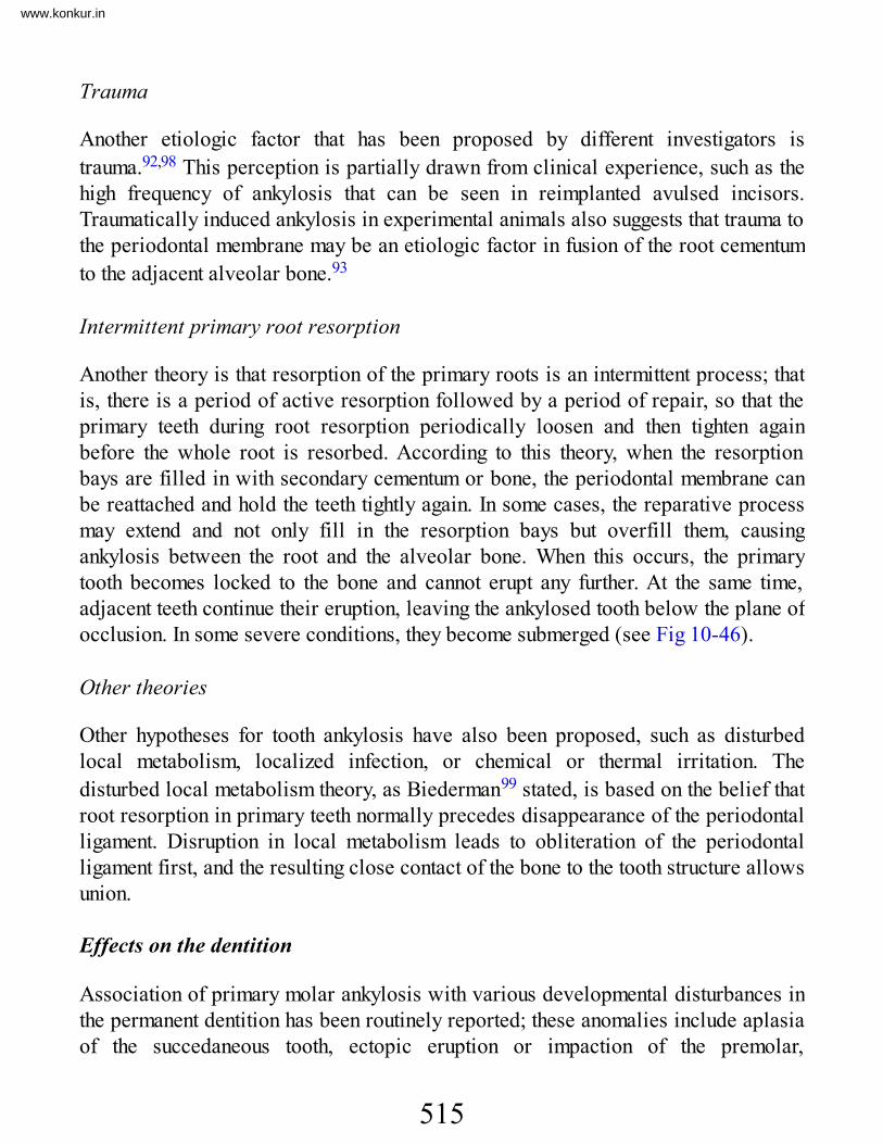
Trauma
Another etiologic factor that has been proposed by different investigators istrauma.92,98 This perception is partially drawn from clinical experience, such as thehigh frequency of ankylosis that can be seen in reimplanted avulsed incisors.Traumatically induced ankylosis in experimental animals also suggests that trauma tothe periodontal membrane may be an etiologic factor in fusion of the root cementumto the adjacent alveolar bone.93
Intermittent primary root resorption
Another theory is that resorption of the primary roots is an intermittent process; thatis, there is a period of active resorption followed by a period of repair, so that theprimary teeth during root resorption periodically loosen and then tighten againbefore the whole root is resorbed. According to this theory, when the resorptionbays are filled in with secondary cementum or bone, the periodontal membrane canbe reattached and hold the teeth tightly again. In some cases, the reparative processmay extend and not only fill in the resorption bays but overfill them, causingankylosis between the root and the alveolar bone. When this occurs, the primarytooth becomes locked to the bone and cannot erupt any further. At the same time,adjacent teeth continue their eruption, leaving the ankylosed tooth below the plane ofocclusion. In some severe conditions, they become submerged (see Fig 10-46).
Other theories
Other hypotheses for tooth ankylosis have also been proposed, such as disturbedlocal metabolism, localized infection, or chemical or thermal irritation. Thedisturbed local metabolism theory, as Biederman99 stated, is based on the belief thatroot resorption in primary teeth normally precedes disappearance of the periodontalligament. Disruption in local metabolism leads to obliteration of the periodontalligament first, and the resulting close contact of the bone to the tooth structure allowsunion.
Effects on the dentition
Association of primary molar ankylosis with various developmental disturbances inthe permanent dentition has been routinely reported; these anomalies include aplasiaof the succedaneous tooth, ectopic eruption or impaction of the premolar,
515
www.konkur.in

infraocclusion of the ankylosed tooth (leading to tipping of the adjacent teeth),overeruption of the opposing tooth, and taurodontism of the permanent first molar.Ankylosed primary teeth that are located below the occlusal plane, especiallysubmerged ankylosed primary molars, need special attention at their early stage ofdevelopment before complications arise:
• They may interfere with the eruption of succeeding permanent teeth, causingectopic eruption.
• Arch length loss can result from tipping of adjacent teeth (Fig 10-47).• Extrusion of teeth in the opposing arch may occur (Fig 10-48).• Submergence of primary teeth damages the occlusion and can be harmful to the
jaws and health of the patient (Fig 10-49).• Transposition of the second premolar to the place of the missing permanent first
molar is a rare consequence of primary second molar ankylosis (Fig 10-50).• Submerged primary teeth not only prevent eruption of the permanent teeth but also
can prevent vertical alveolar growth and cause lateral open bite (see Fig 10-48).
Fig 10-47 (a and b) Ankylosed primary molars causing space loss.
Fig 10-48 Ankylosis preventing eruption of permanent teeth and causing lateral open bite and thepossibility of overeruption of the opposing teeth.
516
www.konkur.in

Fig 10-49 Submerged maxillary left primary first molar causing impaction of the permanentsecond premolar.
Fig 10-50 (a to c) Transposition of the second premolar to the site of the permanent first molar asa consequence of extraction of the first molar and primary second molar ankylosis.
DiagnosisRecognition of primary tooth ankylosis is not difficult; the condition can bediagnosed through careful clinical and radiographic examinations; histologicchanges in the area of fusion have also been reported.100,101
Clinical examinationAnkylosed primary molars show the following clinical signs:
• Ankylosed primary molars often present with infraocclusion relative to adjacentteeth; however, depending on the time of fusion, they may be at the same occlusallevel as adjacent teeth.
• On percussion, an ankylosed tooth gives a solid sound, whereas the normaladjacent teeth give a cushioned or dull sound.
• Ankylosed teeth lack mobility even when root resorption is advanced.
Radiographic evaluation
Radiographs can show obliteration of the periodontal ligament space and absence of
517
www.konkur.in

continuity of periodontal ligament in some areas. The roots are less radiopaque, andin some cases of advanced fusion, the root is less distinguishable from surroundingbone.
Radiographic assessment is useful only when the area of fusion is extensive andwhen the site of fusion is located on the mesial or distal surface of the roots.Detection of fusion on the palatal and buccal aspects is impossible because of thesuperimposition of other structures.
Histologic findings
Mancini et al100 and Haselden et al101 reported histologic changes in areas of fusionof cementum and bone, fibrotic periodontal ligament remnants with very few cells,and absence of mucopolysaccharidase activity, which is essential for the normalprocess of root resorption during eruption of the permanent successor.
Management of ankylosed primary teethEarly recognition is extremely important in the management of an ankylosed tooth.Although the treatment eventually may involve surgical removal of the affected tooth,early detection and proper management facilitate normal eruption and guidance ofthe successor tooth and prevent potential complications.
Treatment considerations
The treatment procedure depends on the following factors:
• Whether the ankylosed tooth is a primary or a permanent tooth• The time of onset and stage of eruption; early fusion and submergence result in
more complications• The degree of infraocclusion• The patient’s chronologic and dental ages• The location of the affected tooth and the normal time of exfoliation• The presence or absence of a permanent successor• The root development of the successor tooth, the proximity of the permanent tooth
to the ankylosed primary tooth, and the possibility of deflection of the permanenttooth
518
www.konkur.in

• The patient’s occlusion and presence of crowding or spacing
The importance of the presence of a permanent successor for normal exfoliation ofa primary molar has been emphasized by Kurol and Thilander.98 They indicated thatthere was no spontaneous exfoliation or very slow root resorption for most of theankylosed teeth of primary molars without a permanent successor.
There are controversies regarding the timing of extraction of severelyinfraoccluded and ankylosed primary molars. Messer and Cline102 emphasizedproper timing of extraction of severely infraoccluded ankylosed primary molars andthe risk of future periodontal damage to the mesial alveolar support of permanentfirst molars.
However, Kurol and Olson103 studied 143 permanent molars adjacent to 119infraoccluded primary molars and 24 normal contralateral primary molars in 68individuals. The subjects were reexamined 8 years after the exfoliation or extractionof the infraoccluded primary second molars. The alveolar bone level mesial to thepermanent first molars was measured on bitewing radiographs. They reported thatall permanent first molars showed a normal alveolar bone level mesially except twothat showed 3- to 4-mm bone loss but no pocket. They concluded that infraocclusionand ankylosis of primary molars does not constitute a general risk for future alveolarbone loss mesial to the permanent first molars.
Treatment options
Treatment options include periodic clinical and radiographic observation, an attemptat luxation, extraction of the involved tooth, extraction and space maintenance,extraction and implant or prosthetic substitution, or extraction and orthodonticintervention and space closure:
• If the ankylosed tooth is interfering with eruption of the successor tooth, immediateextraction is recommended. If the emergence time of the permanent successor ispredicted to be more than 6 months later, a proper space maintainer must beprovided.
• If the successor is present, the onset of ankylosis in the primary tooth is late, andthe permanent successor is usually not affected by the ankylosed tooth, periodicevaluation and removal of the ankylosed tooth at the proper time is recommended.
• If the successor is present, the onset of ankylosis is early, the occlusal level islow, and the possibility of submergence and disturbed eruption of the permanent
519
www.konkur.in

tooth is predictable, extraction of the ankylosed tooth and space maintenance isrecommended.
• If the successor is missing, depending on the patient’s type of occlusion and thesituation of the ankylosed tooth, different procedures can be applied:- If the ankylosed tooth has a good prognosis, it can be left in place. It should be
restored with composite resin buildup or a crown if it is below the occlusallevel to prevent overeruption of the opposing tooth.
- The ankylosed tooth can be extracted if the patient has crowding and needsextraction in the other segments of the arch. The space can be closedorthodontically.
- If the ankylosed tooth has poor structure and the patient has good occlusion andno need for orthodontic treatment, the ankylosed primary tooth can be extractedat an early age and a provisional bonded restoration can be placed. Thepermanent tooth should then be replaced by an implant or prosthetic treatment ata proper age.
• If the ankylosed tooth is a permanent tooth and the onset of ankylosis is early,surgical luxation might help.
• If repeated luxation is not useful, extraction is recommended to preventsubmergence of the tooth.
• Reimplantation of an ankylosed permanent tooth is another option.• If the onset of ankylosis in a permanent tooth is late, luxation is not effective, and
there is no evidence of submergence, the tooth can be built up later to preventovereruption of the opposing tooth.
Therefore, the general treatment recommendation for an ankylosed primary tooththat is not disturbing the patient’s occlusion or its permanent successor is to wait andmonitor for normal exfoliation of the primary tooth and eruption of the successor. Ifan abnormality is associated with the succedaneous tooth (such as agenesis orectopic eruption), early intervention will most likely be the best option to follow.
Case 10-12This case is an example of proper intervention and guidance of eruption of a permanent dentition thatwas disrupted by submerged primary molars.
A 14-year-old girl with a Class I malocclusion and normal skeletal pattern presented with fourprimary molars that were ankylosed and submerged, preventing eruption of the premolars, which had
520
www.konkur.in

almost completely developed roots (Figs 10-51a to 10-51c).The proper timing of intervention for this patient would have been around the age of 10 years, when
the premolar roots lengths were at least half formed and before there was any effect on the dentition.
Treatment:The first step of treatment was anchorage preparation for the maxillary and mandibular molars byplacement of a maxillary Nance appliance and a lower holding arch and extraction of the ankylosedprimary molars (Figs 10-51d and 10-51e). Lateral cephalometric and panoramic radiographs showguidance of the premolars (Figs 10-51f and 10-51g); spontaneous eruption was achieved without anymechanotherapy. Figures 10-51h to 10-51n show the treatment results.
Fig 10-51 Proper intervention and guidance of eruption in a 14-year-old girl with submergedprimary molars. The ankylosed primary molars prevent eruption of the premolars, which havealmost completely developed roots. (a and b) Pretreatment occlusion. (c) Pretreatment panoramicradiograph. (d and e) Anchorage preparation and extraction of all ankylosed primary molars. (f)Panoramic radiograph during premolar eruption, which occurred spontaneously withoutmechanotherapy. (g) Cephalometric radiograph during premolar eruption.
521
www.konkur.in

Fig 10-51 (cont) (h to l) Posttreatment occlusion. (m) Posttreatment panoramic radiograph. (n)Posttreatment cephalometric radiograph.
Summary
• Eruption is the movement of a tooth from its developmental site within thealveolar process to its functional position in the oral cavity; this processtakes about 5 years for the permanent dentition.
• During this prolonged path of movement, many events can disturb normaltooth eruption.
• Eruption disturbances can be broadly classified into two general categories:disturbances related to the time of eruption of the affected tooth anddisturbances related to position of the affected tooth.
• Defects in tooth formation and eruption have been attributed to systemic,genetic, and local factors.
• During different stages of eruption, these factors can cause problems such asdelayed tooth eruption, early exfoliation, premature tooth eruption, primaryand secondary failure of eruption, retarded tooth development, abnormalsequence of eruption, ectopic eruption, transposition, abnormal path of
522
www.konkur.in

eruption, impaction, and ankylosis.• Many theories about tooth eruption have been proposed; recent reports
indicate that the tooth itself does not contribute to the eruption process.Rather, osteoclastogenesis and osteogenesis needed for eruption areregulated by differential gene expression in the dental follicle, bothchronologically and spatially. Osteoclastic activity causes occlusal boneresorption in the bony crypt and creates an eruption tunnel to facilitateeruption. Simultaneously, new bone formation fills the space left behind thecrown and growing root.
• Tooth eruption requires at least the presence of three important conditions:(1) the dental follicle, (2) alveolar bone resorption to produce an eruptionpathway, and (3) alveolar bone formation at the base of the bony crypt.
• Longitudinal monitoring of the transitional dentition with panoramicradiographs is an accurate means for early detection of eruption problems.The following are the consecutive steps of primary tooth replacement bypermanent successors that can be seen in a normal site of dental transition.Any disturbance of these steps can be a sign of problems:
- Resorption of the primary tooth root- Lengthening of the permanent root- Exfoliation of the primary crown- Resorption of overlying bone to clear the path of eruption- Bone apposition under the moving tooth germ to fill the space left behind- An increase in alveolar height- Movement of the permanent tooth through bone- Piercing of the alveolar crest at two-thirds’ root formation- Piercing of the gingival margin at three-quarters’ root formation- Root completion after several months in occlusion• Basic knowledge of the embryology, histology, and morphogenesis of tooth
formation, eruption, and exfoliation and careful clinical and radiographicevaluation of these changes can help the clinician to identify factors that maycause eruption problems, such as bone barriers, fibrotic tissue,supernumerary teeth, odontomas, remaining primary roots, ankylosis, andcyst formation or other pathologic lesions.
• Delayed eruption is a problem that ranges from delayed eruption to completefailure of eruption, including tooth ankylosis, PFE, or secondary failure of
523
www.konkur.in

eruption. It can be a simple inability to erupt due to space loss or it can be afailure of eruption. The morphology and etiologic causes can differentiatethe kind of delayed or failure of eruption. PFE is an abnormality that affectsall teeth distal to the most mesially involved tooth, while mechanical failureof eruption affects only the involved tooth or teeth. Recent studies show thatthe former is an inherited, autosomal-dominant mode of nonsyndromicprimary failure of eruption.
• Retarded eruption is a different form of delayed eruption, commonlyobserved in the second premolar area. This kind of anomaly is associatedwith late development of tooth germs. Radiographic evaluation of retardedtooth formation can even be misleading because of its similarity tocongenital absence of premolars.
• An abnormal sequence of eruption can have direct effects on thedevelopment of occlusion. The sequence of shedding and subsequenteruption of the maxillary and mandibular posterior teeth and the way that theleeway space is utilized have important implications for molar occlusion.Likewise, the normal sequence of eruption of mandibular and maxillarycanines relative to the premolars plays an important role in the anterior partof occlusion. In all types of eruption sequences, there is usually a symmetricpattern between the left and right sides of the arch, although there is sometime variation among different individuals. More than 6 months’ differencebetween the left and right sides of the arch can be an important sign oferuption problems.
• Ectopic eruption is tooth eruption that is out of normal position. Ectopiceruption may lead to various complications, including impaction, loss ofbone, formation of dentigerous cysts, and resorption of adjacent teeth. Onecommon form is ectopic eruption of the permanent first molar during theearly mixed dentition. A mesial angulation of the permanent first molarresults in a cessation of its eruption and causes atypical resorption of theprimary second molar.
• Tooth transposition is the positional interchange of two adjacent teeth. Theteeth most frequently involved in the maxilla are canines, premolars, andlateral incisors, and the most commonly transposed teeth in the mandible arelateral incisors. Early detection of transposed teeth at an early stage ofdevelopment and recognition of the etiologic factors makes it possible tointervene and change the abnormal eruptive path of the transposed teeththrough proper guidance of eruption.
• Impacted teeth are a commonly found anomaly. After mandibular third
524
www.konkur.in

molars, maxillary canines are the most commonly impacted teeth. Palatalcanine impaction and buccal canine impaction are not exactly the same inetiology, morphogenesis, or management. Several etiologic factors that cancause canine impaction require early detection and intervention. Theseinclude space deficiency, trauma, prolonged retention of the primary canine,premature loss of the primary canine, abnormal position of the tooth bud,localized obstacles such as cysts and odontoma, supernumerary teeth,abnormal sequence of eruption, missing lateral incisors, and ankylosis of thepermanent canine.
• Ankylosis is a specific abnormality of tooth eruption and developmentcaused by the fusion of some part of cementum with the alveolar bone.Primary tooth ankylosis is a progressive anomaly of tooth eruption that canoccur before, during, or after tooth eruption and usually has a profoundeffect on successor teeth, alveolar bone growth, and occlusion.
• Careful clinical examination and radiographic monitoring of changes duringthe transitional dentition can help to detect incipient problems and ensurethat proper intervention is initiated.
• The following are important clinical signs that indicate the possibility oferuption problems:
- Overretained primary teeth- Early exfoliation of primary teeth- Asymmetric permanent eruption (left and right sides of the arch)- Asymmetric root development- Abnormal sequence of eruption- Failure of eruption or delayed eruption- Sustained partial eruption- Displacement of an erupted permanent tooth (lateral incisor tilting)- Absence of a crown bulge
References1. Massler M, Schour I, Poncher HG. Developmental pattern of the child as reflected in the calcification pattern
of the teeth. Am J Dis Child 1941;62:33–67.2. Bosshardt DD, Schroeder HE. Cementogenesis reviewed: A comparison between human premolars and
rodent molars. Anat Rec 1996;245:267–292.
525
www.konkur.in

3. Shumaker DB. A comparison of chronologic age and physiologic age as predictors of tooth eruption. Am JOrthod 1974;66:50–57.
4. Marks SC Jr, Cahill DR. Experimental study in the dog of the non-active role of the tooth in the eruptiveprocess. Arch Oral Biol 1984;29:311–322.
5. Berkovitz BK, Thomas NR. Unimpeded eruption in the root-resected lower incisor of the rat with a preliminarynote on root transection. Arch Oral Biol 1969;14:771–780.
6. Carl W, Wood R. Effects of radiation on the developing dentition and supporting bone. J Am Dent Assoc1980;101:646–648.
7. Van Hassell HJ, McMinn RG. Pressure differential favouring tooth eruption in the dog. Arch Oral Biol1972;17:183– 190.
8. Chiba M, Ohshima S. Effects of colchicine and hydrocortisone on unimpeded eruption rates of root-resectedmandibular incisors of rats. Arch Oral Biol 1985;30:147–153.
9. Marks SC Jr. The basic and applied biology of tooth eruption. Connect Tissue Res 1995;32(1–4):149–157.10. Wise GE, Yao S, Henk WG. Bone formation as a potential motive force of tooth eruption in the rat molar.
Clin Anat 2007;20:632–639.11. Cahill DR, Marks SC Jr. Tooth eruption: Evidence for the central role of the dental follicle. J Oral Pathol
1980;9:189– 200.12. Ten Cate AR. Oral Histology: Development, Structure, and Function, ed 8. St Louis: Mosby-Year Book,
2012.13. Ten Cate AR, Deporter DA, Freeman E. The role of fibroblasts in the remodeling of periodontal ligament
during physiological tooth movement. Am J Orthod 1976;69:155– 168.14. Berkovitz BK. How teeth erupt. Dent Update 1990;17:206– 210.15. Brash JC.The growth of the alveolar bone and its relation to the movements of the teeth, including eruption. 1.
Int J Orthod Oral Surg Radiogr 1928;14:196–223.16. Brash JC.The growth of the alveolar bone and its relation to the movements of the teeth, including eruption. 2.
Int J Orthod Oral Surg Radiogr 1928;14:283–293.17. Brash JC.The growth of the alveolar bone and its relation to the movements of the teeth, including eruption. 3.
Int J Orthod Oral Surg Radiogr 1928;14:398–405.18. Brash JC.The growth of the alveolar bone and its relation to the movements of the teeth, including eruption. 4.
Int J Orthod Oral Surg Radiogr 1928;14:487–504.19. Larson EK, Cahill DR, Gorski JP, Marks SC Jr. The effect of removing the true dental follicle on premolar
eruption in the dog. Arch Oral Biol 1994;39:271–275.20. Wise GE. Cellular and molecular basis of tooth eruption. Orthod Craniofac Res 2009;12:67–73.21. Philbrick WM, Dreyer BE, Nakchbandi IA, Karaplis AC. Parathyroid hormone-related protein is required for
tooth eruption. Proc Natl Acad Sci U S A 1998;95(20):11846– 11851.22. Suri L, Gagari E, Vastardis H. Delayed tooth eruption: Pathogenesis, diagnosis, and treatment. A literature
review. Am J Orthod Dentofacial Orthop 2004;126:432–445.23. Wise GE, King GJ. Mechanisms of tooth eruption and orthodontic tooth movement. J Dent Res 2008;87:414–
434.24. Baume LJ, Becks H. The effect of thyroid hormone on dental and paradental structures. Parodontopathies
1952;6:89–109.25. Tse Mdo C, Boaventura MC, Fernandes GD, Merzel J. The effects of cerebral hemidecortication on the
eruption rate and uptake of [3H]-glycine by the periodontal ligament of the rat incisor. Arch Oral Biol1988;33:605–611.
26. Kosowicz J, Rzymski K. Abnormalities of tooth development in pituitary dwarfism. Oral Surg Oral Med OralPathol 1977;44:853–863.
27. Barbería Leache E, Marañes Pallardo JP, Mourelle Martínez MR, Moreno González JP. Tooth eruption inchildren with growth deficit. J Int Assoc Dent Child 1988;19(2):29–35.
28. Stellzig-Eisenhauer A, Decker E, Meyer-Marcotty P, et al. Primary failure of eruption (PFE)—Clinical andmolecular genetics analysis. J Orofac Orthop 2010;71:6–16.
526
www.konkur.in

29. Frazier-Bowers SA, Simmons D, Koehler K, Zhou J. Genetic analysis of familial non-syndromic primaryfailure of eruption. Orthod Craniofac Res 2009;12:74–81.
30. Gron AM. Prediction of tooth emergence. J Dent Res 1962;41:573–585.31. Posen AL. The effect of premature loss of deciduous molars on premolar eruption. Angle Orthod
1965;35:249–252.32. Hartsfield JK Jr. Premature exfoliation of teeth in childhood and adolescence. Adv Pediatr 1994;41:453–470.33. Dean JA, Avery DR, McDonald RE. Dentistry for the Child and Adolescent, ed 9. St Louis: Mosby,
2010:111.34. Proffit WR, Vig KW. Primary failure of eruption: A possible cause of posterior open-bite. Am J Orthod
1981;80:173– 190.35. Rasmussen P, Kotsaki A. Inherited primary failure of eruption in the primary dentition: Report of five cases.
ASDC J Dent Child 1997;64:43–47.36. Frazier-Bowers SA, Puranik CP, Mahaney MC. The etiology of eruption disorders—Further evidence of a
‘genetic paradigm’. Semin Orthod 2010;16:180–185.37. Ahmad S, Bister D, Cobourne MT. The clinical features and etiological basis of primary eruption failure. Eur
J Orthod 2006;28:535–540.38. Proffit WR, Frazier-Bowers SA. Mechanism and control of tooth eruption: Overview and clinical implications.
Orthod Craniofac Res 2009;12:59–66.39. Mc Cafferty J, Al Awadi E, O’Connell AC. Case report: Management of severe posterior open bite due to
primary failure of eruption. Eur Arch Paediatr Dent 2010;11:155–158.40. Lygidakis NA, Bafis S, Vidaki E. Case report: Surgical luxation and elevation as treatment approach for
secondary eruption failure of permanent molars. Eur Arch Paediatr Dent 2009;10(suppl 1):46–48.41. Taguchi Y, Yano Y, Kobayashi H, Noda T. Retarded eruption of maxillary second premolars associated with
late development of the germs. J Clin Pediatr Dent 2003;27:321–326.42. Peterka M, Tvrdek M, Müllerová Z. Tooth eruption in patients with cleft lip and palate. Acta Chir Plast
1993;35:154– 158.43. Peterka M, Peterková R, Likovský Z. Timing of exchange of the maxillary deciduous and permanent teeth in
boys with three types of orofacial clefts. Cleft Palate Craniofac J 1996;33:318–323.44. Moorrees CF, Gron AM, Lebret LM, Yen PK, Fröhlich FJ. Growth studies of the dentition: A review. Am J
Orthod 1969;55:600–616.45. Lo R, Moyers RE. Studies in the etiology and prevention of malocclusion. 1. The sequence of eruption of the
permanent dentition. Am J Orthod 1953;39:460–467.46. Garn SM, Smith BH. Developmental communalities in tooth emergence timing. J Dent Res 1980;59:1178.47. Sampson WJ, Richards LC. Prediction of mandibular incisor and canine crowding changes in the mixed
dentition. Am J Orthod 1985;88:47–63.48. Weinberger SJ. Correction of bilateral ectopic eruption of first permanent molars using a fixed appliance.
Pediatr Dent 1992;14:382–383.49. Bjerklin K. Ectopic eruption of the maxillary first permanent molar. An epidemiological, familial, etiological
and longitudinal clinical study. Swed Dent J Supp l994;100:1–66.50. Harris CA. A Dictionary of Dental Science: Biography, Bibliography and Medical Terminology. Philadelphia:
Lindsay & Blakiston, 1849.51. ShapiraY, Kuftinec MM. Tooth transpositions—A review of the literature and treatment considerations.
Angle Orthod 1989;59:271–276.52. Huber KL, Suri L, Taneja P. Eruption disturbances of the maxillary incisors: A literature review. J Clin
Pediatr Dent 2008;32:221–230.53. Ely NJ, Sherriff M, Cobourne MT. Dental transposition as a disorder of genetic origin. Eur J Orthod
2006;28:145–151.54. Shapira Y, Kuftinec MM. Maxillary tooth transpositions: Characteristic features and accompanying dental
anomalies. Am J Orthod Dentofacial Orthop 2001;119:127–134.55. Shapira J, Chaushu S, Becker A. Prevalence of tooth transposition, third molar agenesis, and maxillary canine
527
www.konkur.in

impaction in individuals with Down syndrome. Angle Orthod 2000;70:290–296.56. Papadopoulos MA, Chatzoudi M, Kaklamanos EG. Prevalence of tooth transposition. A meta-analysis. Angle
Orthod 2010;80:275–285.57. Allen WA. Bilateral transposition of teeth in two brothers. Br Dent J 1967;123:439–440.58. Payne GS. Bilateral transposition of maxillary canine and premolars. Report of two cases. Am J Orthod
1969;56:45– 52.59. Broadbent BH. Ontogenic development of occlusion. Angle Orthod 1941;11:223–241.60. Bishara SE. Impacted maxillary canines: A review. Am J Orthod Dentofacial Orthop 1992;101:159–171.61. Peck S, Peck L, Kataja M. The palatally displaced canine as a dental anomaly of genetic origin. Angle
Orthod 1994;64:249–256.62. Al-Nimri K, Gharaibeh T. Space conditions and dental and occlusal features in patients with palatally
impacted maxillary canines: An etiological study. Eur J Orthod 2005;27:461–465.63. Jacoby H. The etiology of maxillary canine impactions. Am J Orthod 1983;84:125–132.64. Fournier A, Turcotte JY, Bernard C. Orthodontic considerations in the treatment of maxillary impacted
canines. Am J Orthod 1982;81:236–239.65. Warford JH Jr, Grandhi RK, Tira DE. Prediction of maxillary canine impaction using sectors and angular
measurement. Am J Orthod Dentofacial Orthop 2003;124:651–655.66. Mossey PA, Campbell HM, Luffingham JK. The palatal canine and the adjacent lateral incisor: A study of a
west of Scotland population. Br J Orthod 1994;21:169–174.67. Becker A. In defense of the guidance theory of palatal canine displacement. Angle Orthod 1995;65:95–98.68. Zilberman Y, Cohen B, Becker A. Familial trends in palatal canines, anomalous lateral incisors, and related
phenomena. Eur J Orthod 1990;12:135–139.69. Baccetti T. A controlled study of associated dental anomalies. Angle Orthod 1998;66:267–274.70. Peck S, Peck L, Kataja M. Concomitant occurrence of canine malposition and tooth agenesis: Evidence of
orofacial genetic fields. Am J Orthod Dentofacial Orthop 2002;122:657–660.71. Pirinen S, Arte S, Apajalahti S. Palatal displacement of canine is genetic and related to congenital absence of
teeth. J Dent Res 1996;75:1742–1746.72. Ericson S, Kurol J. Resorption of maxillary lateral incisors caused by ectopic eruption of the canines. A
clinical and radiographic analysis of predisposing factors. Am J Orthod Dentofacial Orthop 1988;94:503–513.73. Walker L, Enciso R, Mah J. Three-dimensional localization of maxillary canines with cone-beam computed
tomography. Am J Orthod Dentofacial Orthop 2005;128:418–423.74. Williams BH. Diagnosis and prevention of maxillary cuspid impaction. Angle Orthod 1981;51:30–40.75. Alqerban A, Jacobs R, Fieuws S, Willems G. Comparison of two cone beam computed tomographic systems
versus panoramic imaging for localization of impacted maxillary canines and detection of root resorption. Eur JOrthod 2011;33:93–102.
76. Ericson S, Kurol J. Early treatment of palatally erupting maxillary canines by extraction of the primarycanines. Eur J Orthod 1988;10:283–295.
77. Power SM, Short MB. An investigation into the response of palatally displaced canines to the removal ofdeciduous canines and an assessment of factors contributing to favourable eruption. Br J Orthod 1993;20:215–223.
78. Dewel BF. The upper cuspid: Its development and impaction. Angle Orthod 1949;19:79–90.79. Newcomb MR. Recognition and interception of aberrant canine eruption. Angle Orthod 1959;21:161–168.80. Shapira Y, Kuftinec MM. Early diagnosis and interception of potential maxillary canine impaction. J Am Dent
Assoc 1998;129:1450–1454.81. Olive RJ. Factors influencing the non-surgical eruption of palatally impacted canines. Aust Orthod J
2005;21:95–101.82. Jacobs SG. The impacted maxillary canine. Further observations on aetiology, radiographic localization,
prevention/interception of impaction, and when to suspect impaction. Aust Dent J 1996;41:310–316.83. Frank CA, Long M. Periodontal concerns associated with the orthodontic treatment of impacted teeth. Am J
Orthod Dentofacial Orthop 2002;121:639–649.
528
www.konkur.in

84. Kokich VG. Surgical and orthodontic management of impacted maxillary canines. Am J Orthod DentofacialOr-thop 1980;126:278–283.
85. Grover PS, Lorton L. The incidence of unerupted permanent teeth and related clinical cases. Oral Surg OralMed Oral Pathol 1985;59:420–425.
86. Röhrer A. Displaced and impacted canines: A radiographic research. Int J Orthod Oral Surg Radiogr1929;15:1003– 1020.
87. Da Silva Assunção LR, Ferelle A, Iwakura ML, Cunha RF. Effects on permanent teeth after luxation injuriesto the primary predecessors: A study in children assisted at an emergency service. Dent Traumatol2009;25:165–170.
88. Anthonappa RP, Ongtengco KL, King NM. A report of an impacted primary maxillary central incisor tooth[epub ahead of print 10 January 2013]. Dent Traumatol doi: 10.1111/edt.12031.
89. Pavoni C, Mucedero M, Laganà G, Paoloni V, Cozza P. Impacted maxillary incisors: Diagnosis and predictivemeasurements. Ann Stomatol (Roma) 2012;3:100–105.
90. Biagi R, Butti AC, Salvato A. Premature loss of maxillary primary incisor and delayed eruption of itssuccessor: Report of a case. Eur J Paediatr Dent 2011;12:194–197.
91. Chaushu S, Zilberman Y, Becker A. Maxillary incisor impaction and its relationship to canine displacement.Am J Orthod Dentofacial Orthop 2003;124:144–510.
92. Kurol J. Infra-occlusion of primary molars: An epidemiologic and familial study. Community Dent OralEpidemiol 1981;9:94–102.
93. Biederman W. The problem of the ankylosed tooth. Dent Clin North Am 1968;Jul:409–424.94. Krakowiak FJ. Ankylosed primary molars. ASDC J Dent Child 1978;45:288–292.95. Via WF Jr. Submerged deciduous molars: Familial tendencies. J Am Dent Assoc 1964;69:128–129.96. Brearley LJ, McKibben DH Jr. Ankylosis of primary molar teeth. 1. Prevalence and characteristics. ASDC J
Dent Child 1973;40:54–63.97. Garib DG, Peck S, Gomes SC. Increased occurrence of dental anomalies, associated with second premolar
agenesis. Angle Orthod 2009;79:436–441.98. Kurol J,Thilander B. Infraocclusion of primary molars with aplasia of the permanent successor: A longitudinal
study. Angle Orthod 1984;54:283–294.99. Biederman W. Etiology and treatment of tooth ankylosis. Am J Orthod 1962;48:670–684.100. Mancini G, Francini E, Vichi M, Tollaro I, Romagnoli P. Primary tooth ankylosis. Report of a case with
histological analysis. J Dent Child 1965;62:215–219.101. Haselden K, Hobkirk JA, Goodman JR, Jones SP, Hemmings KW. Root resorption in retained deciduous
canine and molar teeth without permanent successors in patients with severe hypodontia. Int J Paediatr Dent2001;11:171– 178.
102. Messer LB, Cline JT. Ankylosed primary molars: Results and treatment recommendations from an eight-year longitudinal study. Pediatr Dent 1980;2:37–47.
103. Kurol J, Olson L. Ankylosis of primary molars—A future periodontal threat to first permanent molars? Eur JOrthod 1991;13:404–409.
529
www.konkur.in

530
www.konkur.in

Management of Sagittal Problems (Class II and Class III)
A major and important objective of early treatment during the primary and mixeddentitions is enhancing the normal growth and development of dental occlusion.Recent studies indicate that genetic mechanisms have more influence on themorphogenesis of craniofacial structure during embryonic life, while environmentalfactors influence the developing occlusion, especially during the early postnatalperiod.1
The first step in development of the vertical dimension of occlusion is establishedwhen the primary first molars reach occlusal contact. When the primary secondmolars reach occlusion, their distal surfaces (terminal plane) play an important rolein the sagittal dimension of final occlusion.
It is also known that establishment of the occlusion is under the control of geneticand environmental influences and influenced by the interaction of growth anddevelopment of basal bone and other craniofacial structures. All of these structuresare also under the influence of neuromuscular and soft tissue structures and theirfunctional matrix. Disturbance of any of these components or the occurrence ofdisharmony or dysfunction during the development of occlusion can result inabnormal occlusion.
Many incipient malocclusions that are under the influence of environmental
531
www.konkur.in

factors during the primary or mixed dentition could be preventable; early recognitionof these anomalies and interception can eliminate or reduce the severity of futureproblems. There are special orthodontic problems that require early intervention,even in very young patients during the primary or early mixed dentition; theseinclude Class II malocclusion, anterior dental crossbite, and dentoskeletal Class IIImalocclusion, which will be discussed in this chapter, as well as posteriorcrossbite, especially if it is associated with a lateral mandibular shift (see chapter12), open bite associated with persistent oral habits or other oral dysfunction (seechapter 13), space management (see chapter 4), severe incisor protrusion (seechapter 5), and craniofacial anomalies such as clefts or hemifacial microsomia (seechapter 7).
Understanding the mechanisms of early detection and orthodontic interventionrequires a thorough understanding of the development of the dentition (see chapters 1and 2). The major goal of early intervention is to eliminate or reduce the damagefrom etiologic factors that cause trauma to the occlusion or interfere with the normalgrowth and development of occlusion, thereby providing an environment thatpromotes normal jaw growth and an optimal eruption pattern during the transitionaldentition.
Proper intervention at early ages both corrects the present abnormalities andeliminates or minimizes more severe problems that can occur later. Depending onthe type of problem and the dental and skeletal age of the patient during treatment,some patients may need a second or third phase of treatment. However, the finalphase is usually much easier and faster after early intervention. Some patients maynot need any further treatment.
The timing of early treatment is controversial; some practitioners advocate fortreatment during the primary dentition, and some orthodontists prefer to begintreatment during the mixed dentition. There is also controversy about whether theearly, middle, or late mixed dentition is preferable. The American Association ofOrthodontists2 recommends that the initial orthodontic evaluation occur at the firstsign of orthodontic problems or no later than the age of 7 years.
Examination of a child’s dentition during the primary dentition does not alwaysmean treatment; careful examination can detect and anticipate developing problemsat this early age and clarify the possibility of early intervention. The orthodonticexamination, especially during the primary and early mixed dentitions, is more thanmerely an examination of dental occlusion. The assessment should include diagnosisof facial or skeletal problems that are initiating and developing in a growing child.Problems such as upper airway obstruction (mouth breathing), temporomandibular
532
www.konkur.in

dysfunction, and abnormal myofunctional habits can have adverse effects ondentoskeletal structures. If problems are recognized early and preventive measuresare instituted, many facial and dental problems may be minimized or averted.
Gugino and Dus3 stated that the human face is anatomically and functionally one ofthe most complex regions of the human body. A sound understanding of thephysiology of the stomatognathic system is an important requirement for clinicianswho wish to provide their patients with the best orthodontic and orthopedictreatment results. Therefore, clinicians must understand the basic process of occlusaldevelopment, always be aware for cautious detection, and decide on a case-by-casebasis when to provide orthodontic intervention.
Early treatments follow two major strategies:
1. Removal of etiologic factors and restoration of a normal environment to facilitatenormal growth and development of the dentition
2. Guidance of tooth eruption and the growth pattern to a normal state to correct orreduce the severity of dentoskeletal malrelationships
This chapter discusses common sagittal abnormalities that initiate and developduring the primary or early mixed dentition, are not self-corrected, and in somesituations worsen in later stages of the dentition:
• Class II malocclusion (Class II division 1 and Class II division 2)• Anterior dental crossbite• Dental and dentoskeletal Class III malocclusion
Class II MalocclusionTwo large-scale surveys carried out by the United States by the Division of HealthStatistics of the US Public Health Service covered 6- to 11-year-old children andyouths aged 12 to 17 years. The surveys indicated that Class II malocclusionaccounts for about one-third of the total malocclusions of the US population,4 withvariable combinations of dental and skeletal factors contributing to the disharmony.Class II malocclusion usually causes esthetic and functional problems of varyingseverity, depending on the amount of anteroposterior discrepancy and its interactionwith adjacent soft tissues.5
Long-term studies from the primary to the permanent dentition have shown that
533
www.konkur.in

Class II abnormalities are not self-correcting and in some cases may worsen withage.6,7 Not all Class II malocclusions are alike; numerous types of skeletal anddental Class II malocclusions might be observed under Class II molar relationships.
A review of older literature indicates that Class II discrepancy has mainly beenperceived as a sagittal problem. The characteristics of these anomalies have beenclassified under four sets of anteroposterior criteria: (1) maxillary skeletal position,(2 ) maxillary dentoalveolar position, (3 ) mandibular skeletal position, and (4)mandibular dentoalveolar position. The vertical and transverse dimensions were notconsidered to be related to Class II discrepancy; however, later research showedthat either of these dimensions may be involved.
Schudy8 showed that variations in facial height may conceal or intensify theclinical appearance of Class II malocclusion. An increased mandibular verticaldimension in association with Class II malocclusion usually rotates the mandibledownward and backward, which manifests as a more retruded mandible and a moreconvex profile. Any mistake in treatment would worsen the situation. A decreasedvertical dimension causes the mandible to rotate upward and forward, manifesting asa prominent chin point.
The transverse dimension also must not be overlooked in evaluation of Class IImalocclusions. Most Class II patients with a narrow maxillary arch may show anormal relationship of buccal segments in centric occlusion. Tollaro et al9
investigated posterior transverse interarch discrepancy (PTID), which wasmeasured as the difference between the maxillary and mandibular intermolar width,in two Class II division 1 groups: one with PTID and one without PTID. In the ClassII group with PTID, mandibular retrusion was associated with a posteriorlydisplaced mandible of normal size (functional mandibular retrusion). In the Class IIgroup without PTID, mandibular retrusion was caused by a micrognathic mandible(anatomical mandibular retrusion).
Sometimes an underlying transverse discrepancy of 3 to 5 mm exists in the dentalarch. This maxillary constriction can be unmasked clinically by having the patientbring the mandible forward to a Class I molar relationship.
Early intervention in transverse problems and proper expansion at earlier stageswill facilitate forward growth of the mandible. Although Class II malocclusion isperceived as a sagittal problem, the vertical and transverse dimensions of eachpatient must also be considered in treatment planning.
Morphologic characteristics
534
www.konkur.in

The molar or canine relationships in patients with Class II malocclusion constituteonly one part of a broader and extended syndrome and cannot clearly definetreatment planning. Different morphologic characteristics of Class II malocclusioncan be caused by malrelationships of the basal bone, malrelationships of thedentition, dentoalveolar or dentoskeletal malrelationships, or some combination ofthese factors (Box 11-1). In addition, discrepancies in other bony structures, such asdifferent growth or different angulation of the anterior and posterior cranial bases,can have destructive effects on jaw harmony and occlusion (Box 11-2). Figure 11-1shows different variations of skeletal Class II malocclusion that can develop,depending on the morphologic characteristics of basal bone.
Box 11-1 Morphologic characteristics of the jaws inpatients with Class II malocclusion
• Maxillary basal protrusion • Maxillary dentoalveolar protrusion • Maxillary counterclockwise rotation • Overdevelopment of the maxillary length relative tothe mandible • Vertical overgrowth of the maxilla • Mandibular basal retrognathism
• Mandibular dentoalveolar retrognathism• Underdevelopment of the mandibular lengthrelative to the maxilla • Overgrowth of the ramus relative to the mandibularbody • Underdevelopment of the ramus relative to themandibular body
Box 11-2Morphologic characteristics of other bonystructures in patients with Class IImalocclusion
• Increased anterior cranial base length, causinganterior positioning of the maxilla • Increased posterior cranial base length, causingposterior positioning of the mandible
• Increased saddle angle (nasion-sella-articulare, N-S-Ar), causing posterior positioning of the condyle • Increased articular angle (sella-articularegonion, S-Ar-Go), causing posterior positioning of the mandible
535
www.konkur.in

Fig 11-1 Possible variations of Class II malocclusions based on skeletal components.
Diagnostic proceduresClass II malocclusion is a multifactorial entity caused by hereditary factors orabnormally acquired environmental factors that produce aberrations in form andfunction during occlusal development. Therefore, thorough diagnostic evaluationsare necessary for correct identification of etiologic factors and affected anatomicalstructures as well as proper treatment planning for correction of Class IImalocclusion.
The dental occlusion and dental arch are not an isolated entity; rather, they are apart of an extensive system comprising the teeth, basal bone, and other skeletalstructures of the face and cranium that interact with the neuromuscular system andsurrounding soft tissues. All kinds of dentoskeletal malocclusions, including Class IItypes, are associated, to one degree or another, with the relationships of thefollowing six dentoskeletal components:
1. Cranium and cranial base2. Skeletal maxilla3. Nasomaxillary complex4. Maxillary teeth and alveolar process5. Skeletal mandible6. Mandibular teeth and alveolar process
These are independent functional units, but their interrelationships in threedimensions can produce several variations in the morphology of all dentoskeletalmalocclusions, including Class II forms.
536
www.konkur.in

Cephalometric analysis
Proper treatment planning for dentoskeletal malocclusions cannot be establishedwithout a thorough evaluation of the interrelationships of these compartmentsrelative to the dentition and dental occlusion. Since the introduction of thecephalometric technique by Broadbent10 in 1931, a variety of analyses has beenpresented by different investigators.
One of the goals of cephalometric analysis is to establish the relationships ofthese functional components in both the anteroposterior and vertical planes of spacein order to differentiate the type of malocclusion. Research reports that haveevaluated the morphology of the maxillomandibular relationship in patients withClass II malocclusion have reached differing conclusions. Some reports haveindicated a high percentage of maxillary base protrusion,11 and some reports haveindicated a higher percentage of mandibular base retrusion.12 Variations also exist indentoalveolar types. Although Class II is perceived as a sagittal problem, thevertical and transverse dimensions of each patient must also be considered.
It is not the intention of this book to discuss cephalometric analysis; there aremany valuable sources for this information. However, the reader should note theimportance of these tools for differential diagnosis in dentoskeletal malocclusion,including Class II malocclusion, especially in early treatment approaches aiming forgrowth modification.
In cephalometric evaluation of dentoskeletal malocclusions and differentiation ofvarious components of each malrelationship, it is wise not to rely on any specificangle or linear measurement; no single measurement may be reliable all the time, andother data are available. For example, because of variability in length andinclination of sella-nasion in some patients, the sella-nasion–point A (SNA) andsella-nasion–point B (SNB) angles cannot always be accurate in anteroposteriorevaluation.
In all sagittal evaluations of Class II dentoskeletal malrelationships, besides theSNA and SNB measurements, the Landes angle (nasion–point A to Frankfort plane)provides a high degree of reliability for the maxilla. The Wit analysis(perpendicular lines drawn from point A and from point B to the occlusal plane) canalso be used to compare the anteroposterior relationships of the maxilla and themandible.
Growth prediction
537
www.konkur.in

Another important aspect of cephalometric analysis is prediction of the growthpattern, which is critical in treatment planning, especially for growing patients.Many efforts have been made to develop methods for growth prediction fromcephalometric radiographs. Successful prediction is achieved when the amount andthe direction of growth are predicted before early-age treatment.
Orthopedic interventions for growing children should be preceded by carefulcephalometric evaluation of all components of dentoskeletal structures, such as thecranium and cranial base, the skeletal maxilla and nasomaxillary complex, theskeletal mandible, the maxillary teeth and alveolar process, the mandibular teeth andalveolar process, and especially the potential direction of jaw growth for the child.Facial divergence and convergence, Frankfort plane–mandibular incisor angle(FMA), gonial angle, y-axis, anterior and posterior facial heights, and posterior andanterior cranial base lengths and angulations are examples of measurements that canindicate children’s potential growth pattern.
Advantages of early treatment
Early orthodontic treatment for Class II malocclusion has the following benefits:
• It affords the greatest utilization of skeletal growth.• Skeletal disharmony can be controlled in three dimensions (sagittal, vertical, and
transverse).• Observation and guidance of eruption during the transitional dentition are
promoted.• Disarticulation of the posterior segment during tooth eruption facilitates normal
interdigitation.• Leeway spaces within the dental arches are preserved.• The need for tooth extraction is reduced.• Better skeletal harmony is achieved.• Results are more stable.• Early treatment of unesthetic malocclusion can have an important impact on the
psychosocial development of the patient.• Fractures caused by trauma to protruded incisors are prevented.• Patients exhibit better compliance at earlier ages.
The major goal of early treatment is to correct or reduce the adverse effects of
538
www.konkur.in

dentoskeletal and muscular imbalance by improving the environment to encourageoptimal occlusal development.
Class II Treatment OptionsA Class II malocclusion can have various anatomical, morphologic, and functionalcharacteristics. Treatment strategies differ depending on the special characteristicsof this anomaly. Class II treatment also varies according to the skeletal age of thepatient. Growth modification strategy is entirely different from treatment tacticsapplicable after completion of the growth spurt. Depending on the type of Class IImalocclusion present and the age of the patient, four treatment options are availablefor Class II correction:
1. Camouflage treatment2. Orthognathic surgery3. Growth modification and occlusal guidance4. A combination of the above
Camouflage treatmentCamouflage treatment is used in the permanent dentition, usually after the growthspurt, to obtain optimum results within the physiologic limits of each patient. Withcamouflage treatment the goal is to produce acceptable dental relationships anddisguise unacceptable skeletal relationships through tooth movement, which may ormay not require serial extractions. This type of treatment is indicated in adolescentand adult patients with mild to moderate skeletal problems.
Orthognathic surgeryOrthodontic treatment combined with surgical procedures is applied in adult patientsto correct dental malrelation-ships and skeletal disharmonies. These types oftreatments are usually implemented in individuals with severe dentoskeletalmalrelationships caused by heredity, skeletal disharmony, or congenital skeletalmalformation.
539
www.konkur.in

Growth modification and occlusal guidanceGrowth modification and occlusal guidance treatments are interceptive treatmentsthat are applied in growing children during the primary or mixed dentition toeliminate or minimize the dentoalveolar and skeletal disharmonies that can interferewith the normal growth and development of occlusion. The main objective of thistype of intervention is to prepare an environment that will enhance the positiveeffects of growth potential for improving occlusal development.
Early versus late orthodontic treatment, especially early treatment for Class IIproblems, is one of the greatest controversies in orthodontics. Randomized clinicaltrials have been specifically designed to address these important issues in Class IItreatment. Keeling et al,13 Tulloch et al,14 and Ghafari et al15 concluded that earlytreatment followed by later comprehensive treatment of Class II malocclusionassociated with moderate to severe deformity does not produce major differences injaw relationships or dental occlusion compared with later one-stage treatment.
On the other hand, many other experienced researchers and clinicians, includingGugino and Dus,3 Ricketts,16–18 Subtelny,19 Bench et al,20 Graber,21 andMcNamara,22 insist that there are many disadvantages to waiting for eruption of thepermanent dentition and second molars. In addition, many animal studies, carried outby pioneers such as Harvold et al,23 Woodside et al,24 and McNamara25 have clearlyshown the histomorphologic changes induced in animal jaw growth by orthopedicforces.
Most dentoskeletal anomalies develop during the primary and mixed dentitions,and, according to Carlson,26 the midface and the mandible complete only 50% oftheir total growth by age 8 to 10 years; thus, a considerable amount of midface andmandibular growth occurs during the transitional dentition, which provides a goodopportunity to intervene.
Long-term studies from the primary to the permanent dentition have also shownthat Class II abnormalities are not self-correcting and in some cases may evenworsen.6,7 Why should dental practitioners allow an unfavorable dental, skeletal, orsoft tissue relationship to remain for a number of years if it can be correctedcompletely or at least partially at an earlier age with minimal appliance therapy andtreatment effort, by taking advantage of the child’s growth potential?
Deviations in skeletal growth patterns accompanied by a disturbed functionalmatrix environment, such as mouth breathing, atypical swallowing, or otherabnormal behaviors that can disturb jaw growth, require early intervention to
540
www.konkur.in

eliminate or modify the condition and give orthodontists the opportunity to guidedentoalveolar development.
History of growth modification tactics
The mandible is the only freely functioning bone in the body not connected to anotherbone. It is tied to its contiguous structures by 13 attached muscles. The mandibularposition in three planes of space (sagittal, vertical, and transverse) plays animportant role in harmony of the dental occlusion. These specifications of themandible have drawn orthodontists for many years to the possibility of influencingthe mandibular position by altering the structure of the temporomandibular joint.
In 1880, Kingsley27 introduced the concept of jumping the bite for correction ofmandibular retrusion. He designed a vulcanite palatal plate consisting of an anteriorinclined surface to guide the mandible to a forward position. This idea influencedthe development of functional jaw orthopedic treatment. Graber et al28 provided abrief history of different functional devices developed in the past century. Hotz, bymodifying the Kingsley device, introduced the Vorbissplatte for treatment of severeoverbite, mandibular retrusion, and retroclined incisors by muscle hyperactivity.Pierre Robin, a French physician, introduced the monobloc in 1902 as a passivepositioning device for his patients to prevent glossoptosis (blocking of the airway bythe tongue).
Viggo Anderson was familiar with the appliances of Kingsley and Pierre Robinand, in 1908, without thinking of “guiding growth,” applied a monobloc as a retainerfor his daughter to eliminate adverse effects of abnormal function. He found that useof the appliance resulted in sagittal correction and profile improvement. Later,Anderson, in association with Haupl, named the appliance the activator because ofits ability to activate the muscle forces.
In 1927, Emil Herbst introduced the fixed functional appliance (Herbstappliance). Later several modifications of the activator were introduced by differentinvestigators, such as the Bionator by Balters in 1965, the functional regulatorinducing muscle stretch by Fränkel29 in 1966, functional headgear (combinedheadgear and Bionator) by Stockli30 in 1973, fixed Herbst fully banded appliancesby McNamara,22 the twin block (two-piece functional) appliance by Clark31 in 1977,and the magnetic activator device by Darendeliler and Joho.32
Several methods of Class II treatment that do not rely on significant patientcompliance have become popular during the last decade, including several versions
541
www.konkur.in
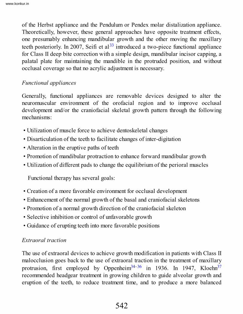
of the Herbst appliance and the Pendulum or Pendex molar distalization appliance.Theoretically, however, these general approaches have opposite treatment effects,one presumably enhancing mandibular growth and the other moving the maxillaryteeth posteriorly. In 2007, Seifi et al33 introduced a two-piece functional appliancefor Class II deep bite correction with a simple design, mandibular incisor capping, apalatal plate for maintaining the mandible in the protruded position, and withoutocclusal coverage so that no acrylic adjustment is necessary.
Functional appliances
Generally, functional appliances are removable devices designed to alter theneuromuscular environment of the orofacial region and to improve occlusaldevelopment and/or the craniofacial skeletal growth pattern through the followingmechanisms:
• Utilization of muscle force to achieve dentoskeletal changes• Disarticulation of the teeth to facilitate changes of inter-digitation• Alteration in the eruptive paths of teeth• Promotion of mandibular protraction to enhance forward mandibular growth• Utilization of different pads to change the equilibrium of the perioral muscles
Functional therapy has several goals:
• Creation of a more favorable environment for occlusal development• Enhancement of the normal growth of the basal and craniofacial skeletons• Promotion of a normal growth direction of the craniofacial skeleton• Selective inhibition or control of unfavorable growth• Guidance of erupting teeth into more favorable positions
Extraoral traction
The use of extraoral devices to achieve growth modification in patients with Class IImalocclusion goes back to the use of extraoral traction in the treatment of maxillaryprotrusion, first employed by Oppenheim34–36 in 1936. In 1947, Kloehn37
recommended headgear treatment in growing children to guide alveolar growth anderuption of the teeth, to reduce treatment time, and to produce a more balanced
542
www.konkur.in

dentition and face. Since then, other investigators have reported on the effects ofheadgear use in early treatment of Class II malocclusions and recommended the useof headgear as one of the most reliable methods for management of maxillaryprotrusion.16–18,37,38
Kopecky and Fishman39 established that early orthodontic treatment and growthmodification are best achieved before and during the growth spurt. Baccetti et al,40
in a cephalometric study, evaluated the role of timing in relation to skeletal maturityon the outcomes of comprehensive, nonextraction Class II therapy. They concludedthat treatment of Class II malocclusion before or during the pubertal growth spurtinduces significant favorable skeletal changes. They also stated that use of headgearduring the prepubertal stage restricts maxillary advancement and enhancesmandibular growth. The only significant changes in patients treated after the pubertalgrowth spurt were dentoalveolar changes.
The effects of headgear alone or headgear combined with other devices on thedentition or jaws have also been reported by many investigators. These reportsreveal several changes, such as molar relationships, SNA changes and downwardtipping of the palatal plane, opening or closing of the bite, maxillary molar extrusion,intrusion, downward or backward rotation of the maxilla and consequentlymandibular rotation, and increases in anterior facial height.4,24,34–36,41–43
Applications of headgear in combination with removable or fixed and functionalappliances have also been reported. Cephalometric evaluation in Class II division 1patients treated with the Cetlin method—use of cervical headgear, lip bumper, andmaxillary removable plate with springs to distalize the molars— showed asignificant distal tipping in 70% of the treated patients and significant limitation ofmaxillary forward growth but no significant change in the mandibular position.42
The lip bumper is another orthodontic device that has several indications in earlyorthodontic treatment, especially in some Class II malocclusions. Many investigatorshave reported the effect of lip bumper application on transverse, sagittal, andvertical changes in occlusion.19,44,45 These reports indicated that the lip bumper hasseveral effects: increase in arch length, uprighting of mandibular first molars, bodilydental expansion, alveolar bone remodeling, proclination of mandibular incisors byreduction of lip muscle force and abnormal dysfunction and thereby reduction ofoverjet, correction of the curve of Spee, opening of the bite, and maintenance ofleeway space.19,44,45
The Hawley appliance also has many applications as a passive or activeappliance in orthodontic treatment, including as an anterior bite plate for reducing
543
www.konkur.in

overbite and overjet by retroclination of maxillary incisors and as a slow expanderby adding a jackscrew.
Early Class II Treatment with Headgear, Lip Bumper, andHawley Appliance (HLH) TechniqueThis section introduces a technique for early treatment of Class II malocclusionsduring the mixed dentition. In this technique, a combination of headgear, a lipbumper, and a Hawley appliance, which the author calls the HLH technique, hasbeen designed with certain modifications.
To explain the characteristics, mechanisms, and the reasons for the modificationsthat have been applied in the HLH technique, it is necessary to review briefly theapplication and original function of each of these three devices separately.
HeadgearAs an extraoral device, headgear can exert different directions of force to the basalbone and dentition and produce different treatment effects on the occlusion. Successin the application of headgear depends on three important factors: application at theproper time of growth, the patient’s cooperation, and proper mechanicalmanagement.
Proper timing
The proper age for starting early orthodontic treatment and achieving proper growthmodification is just before and during the growth spurt, as has been advocated bysome investigators.40 Kopecky and Fishman39 also indicated that this type oftreatment has the best results when implemented before and during the growth spurt,which coincides with a value of 4 to 7 according to Fishman’s method46 of skeletalmaturation assessment.
Patient’s cooperation
The patient’s cooperation is an important factor in treatment in any orthodontictreatment, especially in headgear wear, and must be considered in treatmentplanning. Practitioners have an important role in gaining the understanding of
544
www.konkur.in

patients and parents and must explain the patient’s dentofacial problems as well asthe necessity and advantages of orthopedic force.
Proper mechanical management
Contrary to perceptions that headgear application is an easy procedure, themechanical management of headgear therapy is a procedure that requires precision.Headgear consists of three parts: the facebow, the anchorage pad, and a strap fortraction. Depending on whether tipping, bodily, or combination movement is desiredfor the teeth or the maxilla, the following determinations must be made:
• Location of anchorage• Location of the inner bow attachment• Position of the outer bow relative to the inner bow (above, below, or at the same
level)• Type of outer bow (short, medium, or long)
The location of anchorage can be cervical, occipital, or a combination. Acervically located headgear pad with a long outer bow exerts a distal and low forceon the teeth and the maxilla. This design has distalization and extrusion effects on themolars and a tipping effect on the palatal plane (counterclockwise rotation). Thebest indication for this configuration is Class II deep bite in a patient with ahorizontal growth pattern.
High-pull headgear, in which the anchorage pad is located on the occipital area,exerts a vertical and distal force on the maxillary molars and maxillary base andcauses intrusion and distal movement. Depending on the size of the outer bow (shortor medium) used with high-pull headgear, different effects on the molars and themaxilla can be achieved. These include intrusion and distal tipping or intrusion anddistal bodily movement.
Combination headgear has a combination of cervical and occipital pull thatimparts a straight distal force to the molars for controlling extrusion and distalmovement.
In all mentioned types of headgear, the size of the outer bow (short, medium, orlong) and the position of the outer bow relative to inner bow (above, below, or atthe same level) can change the direction of forces.
Another mechanical specification of headgear that can have different effects on thedentition and basal bone is the location of the inner bow attachment. The inner bow
545
www.konkur.in

is commonly connected to molar tubes, but in certain situations it can be connectedto the anterior segment of the maxillary archwire (like J-hook headgear), such as inpatients who need incisor retraction accompanied with intrusion.
In a headgear–functional appliance combination, the inner bow can be inserted inan acrylic resin pad in the anterior, molar, or premolar area, depending on thedesired direction of orthopedic force.
The following are the mechanical functions of headgear, which may vary withdifferent configurations:
• Establishment of normal molar relationships• Restriction of forward maxillary growth (prevention of forward movement of
point A)• Restriction of vertical maxillary growth (high-pull headgear)• Creation of minor space by distalization of molars (tipped or bodily, depending on
the type of headgear)• Extrusion of maxillary molars to reduce overbite• Intrusion of maxillary molars to reduce open bite or open bite tendency• Correction of molar rotation• Expansion of molar region• Constriction of molar region
Modified Hawley applianceThe second component of the HLH technique for early treatment of Class IImalocclusion is a modified Hawley appliance (Fig 11-2). The modified appliancehas the following specifications:
• Two C-clasps: These are simple clasps made with 0.032- or 0.036-inch stainlesssteel wire and designed to be inserted in an acrylic resin pad that runs from thedistal surface of the molars over the molar buccal tube and extends to thepremolar area for easy removal.
• An inclined acrylic resin pad: The pad is placed behind the maxillary incisors formesial movement of the mandible during closure. The position of this pad varies,depending on the severity of overjet and sagittal discrepancy of the maxilla andthe mandible.
546
www.konkur.in

• A standard labial bow: The bow is designed to maintain anterior retention andhelp incisor retraction if needed.
• In some cases, 2 × 4 bonding instead of a labial bow: Another modification mightbe necessary in some patients with maxillary incisor crowding, rotation, oroverlap that cannot be managed by a labial bow. In this situation, the Hawleyappliance is designed without a labial bow; instead, 2 × 4 bonding can be usedsimultaneously with the inclined acrylic resin pad to achieve leveling, unraveling,and correction of crowding in the incisors.
• Addition of a jackscrew for expansion of the posterior arch segment: Thejackscrew is added when patients with Class II malocclusion also have transverseproblems.
• Occlusal acrylic resin coverage in the posterior regions: Occlusal coverage is amodification that might be needed in patients with Class II malocclusion, anterioropen bite or open bite tendency, and a vertical growth pattern, where all of theeffort must be focused on controlling the vertical dimension.
Fig 11-2 (a and b) Modified Hawley appliance designed for the HLH technique.
When the modified Hawley appliance is used in conjunction with headgear, thefollowing treatment results can be achieved:• Unilateral or bilateral distal movement of the molars by distal positioning of the
C-clasp to achieve Class I molar relationships• Distal maxillary traction, applied to the whole maxilla as a unit, for orthopedic
action to control maxillary growth and correct point A–point B (A-B) discrepancy• Mandibular forward displacement as an orthopedic force to achieve mandibular
protraction• Correction of deep bite by anterior placement of the acrylic resin pad and opening
of posterior bite and extrusion of molars by cervically placed headgear.• Correction of anterior open bite or open bite tendency by the addition of posterior
occlusal acrylic resin coverage to the modified Hawley in conjunction with high-pull headgear
547
www.konkur.in

• Maxillary arch expansion, if needed, by the addition of a jackscrew to themodified Hawley appliance
Lip bumperThe third appliance used in the HLH technique is the lip bumper, which consists oftwo molar bands with a double buccal tube—one a round 0.040- or 0.045-inch tubefor insertion of the lip bumper bow and one for insertion of the archwire—and anacrylic resin or plastic pad located behind the lower lip.
In patients that need some buccal expansion, the pad can be extended toward thebuccal segments to separate perioral muscle force, facilitate natural arch expansion,and perhaps initiate bone remodeling. Many investigators have reported on thestability of this type of expansion that takes place during occlusal development.19,45
The lip bumper has several indications in early orthodontic treatment, especiallyin some types of Class II malocclusion. As mentioned earlier, many investigatorshave reported use of different types of lip bumper and the transverse and verticalchanges they have on occlusion.17,42,43
As a part of the HLH technique, the lip bumper is used in conjunction withheadgear and a modified Hawley appliance for the following conditions:
• Class II division 1 malocclusions associated with deep bite, deep curve of Spee,and retruded and sometimes crowded mandibular incisors are usually the result ofearly loss of mandibular primary canines or primary first molars, combined withlip dysfunction. These cases are ideal for lip bumper application.
• The lip bumper can upright permanent molars, procline mandibular incisors,correct curve of Spee, and reduce overbite in patients with Class II division 1malocclusion who exhibit deep bite and tipped mandibular permanent molars dueto early loss of primary molars.
• In patients with Class II division 1 malocclusion who have normal overbite oropen bite tendency but minor mandibular incisor crowding and retrusion due tohyperactivity or dysfunction of the lower lip, a regular lip bumper cannot beapplied because it can produce greater bite opening. In these patients, if it isnecessary to release the incisors from lip pressure, the author recommends usingthe combination of a lip bumper and a lower holding arch, which will have thesame effect on the mandibular incisors but prevent any changes in the molar area(Fig 11-3).
548
www.konkur.in

Fig 11-3 (a) Simple lip bumper. (b) Combination of lip bumper and lower holding arch for theHLH technique.
The lip bumper can achieve the following:
• Increase in arch length (distal molar movement; labial anterior movement)• Disruption of the equilibrium between the perioral musculature and the tongue to
increase arch width and promote alveolar growth• Correction of the curve of Spee by uprighting the molars, proclining the anterior
teeth, and facilitating eruption of the posterior segment• Reduction of overjet by preventing lip muscle dysfunction and proclining the
incisors
Strategy and Tactics of HLH ApplicationApplication of triple appliances (HLH) with the proper modifications during themixed dentition is a simple, effective way of achieving early Class II correction.Selection of the proper type of headgear, determination of the necessary designmodifications for the Hawley appliance (such as anterior inclined surface, biteplane, or posterior occlusal coverage), and proper use of the lip bumper arecritically important to the success of the HLH technique.
For early intervention in Class II division 1 malocclusion, the HLH approach maybe applied as a two-phase or a one-phase treatment. For treatment of Class IIdivision 2 malocclusion, the HLH technique has some differences in tactics, whichare designed to address the specific characteristics of this anomaly.
Two-phase strategy for Class II division 1 malocclusionTwo-phase HLH application is used for management of Class II dental malocclusion
549
www.konkur.in

or Class II malocclusion with minor A-B discrepancy. This tactic usually is startedduring the middle mixed dentition to establish normal permanent molar relationships,normal overjet, and normal overbite. After the interceptive HLH therapy hasestablished Class I molar relationships, an interim phase continues until allpermanent canines and premolars erupt. The interim phase can include use of aHawley retainer or be limited to observation. Phase 2 takes place during thepermanent dentition and involves minor final adjustment.
The advantage of early HLH intervention during the mixed dentition is thepossibility for normal eruption of the canines and premolars with normalinterdigitation that may not need any further treatment (see cases 11-1 and 11-2). Inthe two-phase approach, phase 1 (HLH treatment) usually takes 1 to 1.5 years; theinterim phase of observation and guidance of eruption takes about 1 year; and phase2 (final alignment) takes about 1 year. However, permanent canines and premolarsmay exhibit normal interdigitation after eruption, eliminating the need for phase 2.This prospect is based on the finding that, if Class I molar relationships areachieved, normal interdigitation of buccal segments will follow, as Poulton47 hasshown. Canine and premolar tooth buds move distally and follow the root movementof the maxillary permanent first molars after they are distalized.
One-phase strategy for Class II division 1 malocclusionThe one-phase HLH treatment strategy is applied for correction of skeletal Class IImalocclusions with moderate or severe A-B discrepancies, usually starting aroundthe late mixed dentition or before the growth spurt (skeletal maturation assessmentvalue of 4 to 7) and continuing until eruption of the permanent dentition (excludingthird molars) is completed. It is a continuous treatment approach with no interimphase. The one-phase HLH treatment is followed by complete bonding for final toothalignment. It usually takes about 2 to 2.5 continuous years of treatment from HLH tothe end of comprehensive treatment.
The first step is establishment of normal permanent molar relationships combinedwith control of maxillomandibular discrepancy by controlling maxillary growth,promoting mandibular growth, or both. The second step starts with the bonding ofpremolars after they erupt and retraction, if needed, to establish normalinterdigitation in the molar and premolar segments. The next step is canine alignmentand retraction, which might be accompanied by Class II elastics. The final step isincisor retraction and final adjustment before retention. Depending on the severity ofthe skeletal jaw discrepancy, the use of headgear might be continued to the final
550
www.konkur.in

stage of treatment, even after normal molar relationships are established, or toprovide anchorage.
A significant advantage of this type of early intervention is that sagittalabnormalities, whether the result of maxillary protrusion, mandibular retrusion, or acombination, can be controlled with the HLH approach, while the vertical andtransverse dimensions of the malocclusion can be controlled by taking advantage ofthe patient’s growth potential.
Strategy for Class II division 2 malocclusionEarly treatment of Class II division 2 malocclusions has some specific differences intactics. The major characteristic of Class II division 2 malocclusions, besides ClassII interdigitation, is deep bite and maxillary incisor crowding with severeretroclination of the maxillary central incisors. Therefore, the first steps in treatmentare to control overbite with a thick anterior bite plate that will disarticulate theposterior dentition, encouraging eruption of those teeth, and to facilitate molarretraction with the cervical headgear. The next step is 2 × 4 maxillary bracketing toachieve anterior alignment and some proclination of the central incisors. Thismeasure will facilitate mandibular growth and allow for accelerated dentoalveolardevelopment that might have been restricted by locked occlusion caused bymaxillary incisor retroclination and deep bite.
In the next step, after some overbite reduction, a lip bumper is applied if themandibular incisors are also crowded and retroclined; later, this is followed bymandibular incisor bonding.
One important goal of early intervention in Class II division 2 malocclusion is tocontrol the child’s overbite at an early age by taking advantage of the patient’sgrowth; this type of treatment is very difficult to manage later.
Case ReportsThe following case reports present examples of early treatment with the HLHprocedure, applied in either one phase or two phases of treatment.
Case 11-1: Two-phase interceptive treatmentand observation
551
www.konkur.in

A 10-year, 5-month-old girl in the late mixed dentition had a Class II division 1 malocclusion, a 9.2-mmoverjet, and deep overbite. The maxillary incisors had normal inclination, the mandibular incisors wereslightly uprighted, and she had a large A-B discrepancy (point A–nasion–point B [ANB] angle = 7.4degrees) resulting from mandibular retrognathia (Figs 11-4a to 11-4d).
Treatment:The treatment plan was use of the HLH technique alone: cervical headgear, a maxillary modifiedHawley appliance with anterior inclined surface, and a mandibular lip bumper. Treatment resulted insignificant changes in the patient’s occlusion, soft tissue profile, and molar relationships (Figs 11-4e to11-4j). The A-B discrepancy (ANB) was reduced from 7.4 to 0.8 degrees and overjet from 9.2 to 2.9mm.
552
www.konkur.in

Fig 11-4 Management of severe overjet and deep overbite in a 10-year, 5-month-old girl in thelate mixed dentition. She was treated only with the HLH technique. (a to c) Pretreatment
553
www.konkur.in

occlusion. (d) Pretreatment soft tissue profile. (e to g) Posttreatment occlusion. (h) Posttreatmentsoft tissue profile. (i) Pretreatment (left) and posttreatment (right) cephalometric radiographs. (j)Changes in cephalometric measurements and superimposition of pretreatment (black ) andposttreatment (green) tracings.
Case 11-2: Two-phase interceptive treatmentand observation
A 9-year-old girl had a Class II division 1 malocclusion, minor overbite, a 7.9-mm overjet, a steepmandibular plane, and a slight open bite tendency (Figs 11-5a to 11-5d). Maxillary incisor protrusion hadcaused lip incompetence, but the mandibular dentition was good.
Treatment:Treatment involved only the application of the HLH technique (combination headgear) with one stage ofactive treatment and a second stage of observation. Instead of a lip bumper, a lower holding arch wasused to preserve leeway space and prevent molar eruption. A maxillary Hawley appliance was used formaxillary incisor retraction, and the headgear also aided maxillary retraction. Figures 11-5e to 11-5jshow the results of treatment.
Fig 11-5 Management of minor overbite and severe overjet in a 9-year-old girl with a Class IIdivision 1 malocclusion. (a to c) Pretreatment occlusion. (d) Pretreatment soft tissue profile. (e to
554
www.konkur.in

g) Posttreatment occlusion. (h) Posttreatment soft tissue profile.
Fig 11-5 (cont) (i) Pretreatment (left) and posttreatment (right) cephalometric radiographs. (j)Changes in cephalometric measurements.
Case 11-3: Two-phase interceptive treatmentand observation
A 9-year-old girl presented with a Class II division 1 malocclusion, a 7.6-mm overjet, an A-Bdiscrepancy of 7.1 degrees, impinging deep bite, and mandibular retrognathism caused by clockwise
555
www.konkur.in

rotation (Figs 11-6a to 11-6c).
Treatment:This patient was treated with a two-phase active HLH procedure, including combination headgear forretraction of the maxillary first molar while controlling extrusion, and a combination lip bumper andlower holding arch (see Fig 11-3b) was used to control uprighting of the mandibular molars whilepreventing lip dysfunction. After Class I molar relationships were achieved, a Hawley retainer wasplaced during the interim phase (at first 24 hours a day and then 12 hours’ use at night) until the caninesand premolars had erupted (Figs 11-6d to 11-6f) . Figures 11-6g and 11-6h show the pre- andposttretment cephalometric radiographs, superimposition of tracings, and measurements.
Fig 11-6 Management of severe overjet, impinging deep bite, and mandibular retrognathism in a 9-year-old girl. (a to c) Pretreatment occlusion. (d to f) Posttreatment occlusion.
556
www.konkur.in

Fig 11-6 (cont) (g) Pretreatment (left) and posttreatment (right) cephalometric radiographs. (h)Changes in cephalometric measurements and superimposition of pretreatment (black ) andposttreatment (green) tracings.
Case 11-4: Two-phase treatment with HLHand comprehensive treatment
A 9-year, 7-month-old girl in the middle mixed dentition presented with a Class II division 1malocclusion, an 11-mm overjet, and severe overbite that had resulted in a collapsed mandibulardentition and retrognathia (retrognathic chin). Here lips were apart, and lip dysfunction continued toworsen the irregularities (Figs 11-7a to 11-7d).
Treatment:The treatment approach was designed as two-phase HLH treatment. In phase 1, cervical headgear wasused to retract the maxillary molars and reduce overbite; an inclined maxillary Hawley appliance wasused to stimulate mandibular growth by unlocking the collapsed mandible, helping forward mandibulargrowth; a lip bumper was used to control lip dysfunction and alleviate the lip force directed at themandibular incisors, thereby correcting mandibular incisor retroclination, uprighting mandibular molars,and correcting the curve of Spee. Figure 11-7e illustrates the maxillary dentition and spontaneous distalmovement of the buccal segments achieved by cervical headgear and good compliance. Phase 2 was
557
www.konkur.in

started after complete eruption of the canines and premolars. Use of the headgear was stopped, andmaxillary Nance anchorage was placed. Figures 11-7f to 11-7k show the posttreatment results, whichincluded correction of the Class II malocclusion, correction of the severe overjet (reduction from 11.0 to1.9 mm), correction of the deep bite (4.6 to 0.8 mm), reduction in the ANB angle from 6.2 to 2.7degrees, and a change in the retrognathic chin from 82.5 to 85.9.
Fig 11-7 Management of collapsed mandibular dentition and retrognathia caused by severe overjetand overbite in a 9-year, 7-month-old girl. (a to c) Pretreatment occlusion. (d) Pretreatment softtissue profile. (e) Cast prepared after phase 1 treatment with the HLH protocol and prior to thestart of phase 2 treatment with a Nance appliance. (f to h) Posttreatment occlusion. (i)Posttreatment soft tissue profile.
558
www.konkur.in

Fig 11-7 (cont) (j) Pretreatment (left) and posttreatment (right) cephalometric radiographs. (k)Changes in cephalometric measurements and superimposition of pretreatment (black ) andposttreatment (green) tracings.
Case 11-5: One-phase orthopedic treatment
A 10-year, 7-month-old girl in the early permanent dentition presented with a Class II division 1malocclusion, a 12.1-mm overjet, impinging deep bite, a severe curve of Spee, incompetent andhypotonic lips, maxillomandibular dental protrusion, and a severely convex profile (Figs 11-8a to 11-8d).She exhibited some interdental spacing. Her dental age was advanced but her skeletal growth retarded.
Treatment:Because of her skeletal age, interdental spacing, and good compliance, the treatment plan was designedas a nonextraction, one-phase HLH technique.
The patient received cervical headgear and a modified Hawley appliance with an inclined bite plane.Mandibular complete bonding was performed to correct the curve of Spee, and no lip bumper was usedbecause of the mandibular incisor protrusion to achieve a consolidated mandibular arch against theinclined maxillary Hawley appliance during forward mandibular positioning to stimulate mandibulargrowth.
After good Class I molar relationships were established, complete maxillary bonding was started. Use
559
www.konkur.in

of the headgear was continued as anchorage. Maxillary buccal segments were retracted while themandibular curve of Spee was corrected and the overbite was significantly decreased. The final stepwas removal of the maxillary inclined Hawley appliance and initiation of anterior retraction.
Figures 11-8e to 11-8k illustrate the treatment outcome.
Fig 11-8 Management of severe overjet and impinging deep bite in a 10-year, 7-month-old girl.She has a severe curve of Spee, incompetent lips, and maxillomandibular dental protrusion. (a to d)Pretreatment occlusion. Although her dental age is advanced, her skeletal growth is retarded. (e tog ) Posttreatment occlusion. (h ) Pretreatment soft tissue profile. ( i ) Posttreatment soft tissueprofile.
560
www.konkur.in

Fig 11-8 (cont) (j) Pretreatment (left) and posttreatment (right) cephalometric radiographs andtracings. (k) Changes in cephalometric measurements and superimposition of pretreatment (black)and posttreatment (green) tracings.
Case 11-6: One-phase orthopedic treatment
A 10-year-old boy in the late mixed dentition presented with a severe Class II division 1 malocclusion, a16-mm overjet, impinging deep bite, lip dysfunction, and a 5.6-mm interlabial gap (Figs 11-9a to 11-9e).He also exhibited a deep curve of Spee and proclined maxillary incisors, causing lip incompetence andsevere A-B discrepancy (ANB = 7.4 degrees). In this type of occlusion, the mandibular dentitioncollapses and becomes locked behind the maxillary teeth; this type of abnormality normally worsenswith age.
Treatment:The patient was treated with one-phase orthopedic treatment, which started with an anterior bite plate,cervical headgear, and a mandibular 2 × 4 bonding utility arch to upright and depress the mandibularincisors and correct the curve of Spee. After a Class I molar relationship was achieved, the maxillaryincisors were bonded for interdental space closure and some retraction accompanied with intrusion toreduce overjet and overbite and control lip dysfunction.
The next stage was sequential extraction of the remaining primary molars and guidance of canine and
561
www.konkur.in

premolar eruption. The headgear was stopped, and insertion of Nance anchorage and completeposterior and anterior retraction with Class II elastics against the consolidated mandibular arch wasinitiated.
Figures 11-9f to 11-9l show the dental, dentoskeletal, and soft tissue changes achieved throughtreatment.
Fig 11-9 (a to c) Pretreatment occlusion. (d) Pretreatment soft tissue profile. (e) Pretreatment lipclosure. (f to h) Posttreatment occlusion. (i) Posttreatment soft tissue profile. (j) Posttreatment lipclosure.
562
www.konkur.in

Fig 11-9 (cont) (k) Pretreatment (left) and posttreatment (right) cephalometric radiographs. (l)Changes in cephalometric measurements and superimposition of pretreatment (black ) andposttreatment (green) tracings.
Case 11-7: One-phase orthopedic treatment
A 12-year-old girl presented with a Class II division 1 malocclusion, a 13.9-mm overjet, impinging deepbite, a deep curve of Spee, and lip dysfunction (Figs 11-10a to 11-10e). Her lips were incompetent, witha 9.1-mm interlabial gap. Lip closure was very difficult and caused excessive lip strain (Figs 11-10f to11-10h).
Treatment:Treatment started with cervical headgear, a maxillary Hawley bite plate, and a lip bumper to controllower lip dysfunction, distalize the maxillary molars, and reduce overbite. After good molar relationshipswere achieved, full bonding was started to align the anterior segment and correct the mandibular curveof Spee. Then use of the bite plate and the lip bumper was stopped, but use of the headgear wascontinued as anchorage during anterior retraction. Figures 11-10i to 11-10o show the posttreatmentresults: normal overjet and overbite; significant change in profile, lips, and appearance; and boost in self-confidence.
563
www.konkur.in

Fig 11-10 Management of severe overjet, impinging deep bite, deep curve of Spee, and lipdysfunction in a 12-year-old girl with a Class II division 1 malocclusion. (a to e) Pretreatmentocclusion. ( f ) Pretreatment soft tissue profile. (g and h) Pretreatment lip closure. (i to k)Posttreatment occlusion. (l) Posttreatment soft tissue profile. (m) Posttreatment lip closure.
564
www.konkur.in

Fig 11-10 (cont) (n) Pretreatment (left) and posttreatment (right) cephalometric radiographs. (o)Changes in cephalometric measurements and superimposition of pretreatment (black ) andposttreatment (green) tracings.
Anterior Dental Crossbite and Class III MalocclusionAnterior dental crossbite is a common problem in the primary or mixed dentition andmerits early intervention to prevent further damage to occlusion. Treatment iseffective and desirable in certain situations. Early correction of anterior crossbitenot only resolves esthetic problems but also improves function and consequentlyprevents structural damage and adverse effects on the growth of the dentition andbasal bones.
Anterior crossbites can have differing morphology, etiology, and treatmentprocedures. Generally there are three kinds of anterior crossbite:
1. Simple dental crossbite2. Pseudo-Class III malocclusion (functional crossbite)
565
www.konkur.in

3. Skeletal Class III and potential Class III malocclusion
Precise clinical evaluation in static conditions and during functional movementand radiographic examinations (lateral cephalograms in centric occlusion andcentric relation) are required for differentiation of dental, skeletal, and functionalcrossbites.
Differential diagnosisDifferential diagnosis of the skeletal and dental components of anterior crossbite isan important and essential part of determining the proper treatment approach andtiming of treatment for these anomalies. Diagnosis consists of a complete intraoraland extraoral clinical evaluation, including assessment of the child’s facial patternand symmetry, left and right molar and canine relationships, both in centric relationand centric occlusion, and dental and facial midlines. Anterior occlusal relationshipboth in centric relation and centric occlusion must also be carefully evaluated. Anykind of mandibular shift must be carefully examined to determine the type ofabnormality, treatment options, and prognosis.
Proper treatment planning and good results in the correction of anterior crossbiterequire careful evaluation of the morphology, etiology, and special characteristicsfor every single case. Two skeletal Class III malocclusions may look alike but havecompletely different characteristics that require different treatment plans. Therefore,the first step is differential diagnosis between different types of anterior crossbites,and the second step is the recognition of specific characteristics for every individualpatient.
Clinical examination
The following are important points that should be evaluated in the clinicalevaluation:
• Anterior and posterior dental relationships with the mandible in both centricrelation and centric occlusion
• The patient’s soft tissue profile in both centric relation and centric occlusion• Any mandibular shift, by holding the mandible to the most retruded position and
checking the incisor relationships• Hereditary background, by checking for the possibility of similar traits in the
566
www.konkur.in

patient’s parents or siblings
Cephalometric evaluation
Since 1931 and the introduction of cephalometric radiographs, severalcephalometric analyses and measurements have been proposed for diagnosis andtreatment planning in orthodontic treatment. However, it is not the intention of thispublication to discuses the special methods or analysis to be used for diagnosis andtreatment planning in early orthodontic treatment. The main purpose of this section isto emphasize and remind the reader of the importance of application of thecephalometric technique, not only in diagnosis and treatment planning but also ingrowth prediction in early-age treatment.
Simple Dental CrossbiteSimple anterior dental crossbite is defined as a dental malocclusion that resultsfrom lingual positioning of the maxillary anterior teeth in relation to the opposingmandibular anterior teeth. The condition is localized and involves the tipping of onlya tooth or teeth and not the basal bone. In other words, simple dental crossbite is dueto an abnormal labiolingual relationship between one or more maxillary andmandibular incisors within the context of a normal anteroposterior skeletal jawrelationship. Single dental crossbite usually results in no mandibular shift from restto occlusion, exhibiting a smooth path of closure to an Angle Class I relationship.When more incisors are involved, some mandibular anterior shift may occur.
Simple dental crossbite is the most common kind of anterior crossbite seen duringthe primary and early mixed dentitions. Reports show a frequency of 3% to 12%.This significant variation of incidence is due to differences in reports from oneethnic group to another. For example, in a Japanese population the reportedincidence was 10%, while in a US population the reported incidence was 3%.48
In these types of anterior crossbite, one or more of the maxillary anterior teethocclude lingually to the mandibular incisors when the teeth are in centric occlusion.Crossbites usually do not self-correct with age. As Tausche et al 48 reported, anteriorcrossbites show progression in severity in the permanent dentition.
Characteristic signs
567
www.konkur.in

As already mentioned, patients with dental crossbite have a normal ANB angle andprofile. The maxillary incisors are retroclined, and mandibular incisors areproclined, but the molars and canines show Class I relationships in both centricrelation and centric occlusion.
There are two types of anterior dental crossbite:
1. Crossbite of a single or multiple incisors with lingual tipping and usually nocrowding and mandibular shift.
2. Locked anterior crossbite in which one or two incisors are tipped lingually andsome are labial to the mandibular incisors. Some crowding is present. No shift ispresent, but maxillary and mandibular incisors are locked within each other, andthe anterior occlusion is usually more traumatized.
EtiologyThe majority of anterior dental crossbites are caused by local dental factors. Manyetiologic factors have been reported, such as:
• Congenital abnormal path of eruption (lingual eruption of maxillary anteriorincisors)
• Trauma to the primary incisors that has led to displacement of the permanent toothbuds
• Direct trauma to the permanent incisors that has caused luxation and displacement• Overretained primary teeth in the maxillary incisor region that have caused palatal
eruption of the permanent incisor or overretained mandibular primary incisors thathave increased the proclination of the mandibular incisors
• Labially positioned supernumerary tooth or odontoma• Sclerotic bony or fibrous tissue barrier caused by premature loss of a primary
tooth• Rarely, a habit of biting the upper lip• Overretained necrotic or pulpless primary tooth or root• Crowding in the incisor region (Bolton discrepancy)• Arch length inadequacy• Repaired cleft lip or palate
568
www.konkur.in

Figure 11-11 illustrates an anterior crossbite in the middle mixed dentition causedby one overretained mandibular primary incisor that created a longer archcircumference in the anterior region of the mandibular dentition and consequently anend-to-end relationship of the incisors and crossbite.
Fig 11-11 (a to e) Anterior crossbite resulting from overretained primary incisors that havecreated a longer arch circumference.
Figure 11-12 shows two overretained primary central incisors that have causedsevere separation and delayed eruption of the permanent central incisors. Theultimate result was creation of a severe diastema, displacement of the permanentincisors, and development of anterior crossbite.
Fig 11-12 (a) Overretained primary incisors delaying the eruption of the permanent incisors. (band c) Anterior crossbite and severe diastema resulting from the neglected, overretained primaryincisors.
Figures 11-13 and 11-14 illustrate overretained primary incisors that have causedpalatal eruption of the permanent incisors and crossbite.
569
www.konkur.in

Fig 11-13 (a and b) Palatal eruption and crossbite of the permanent incisor caused by anoverretained primary lateral incisor.
Fig 11-14 (a and b) Crossbite of the permanent incisors caused by overretained primary centralincisors.
Figure 11-15a shows incisor crossbite that developed because both maxillarypermanent canines were missing, causing diastema and displacement of themaxillary right central and lateral incisors. Figures 11-15b and 11-15c demonstratethe occlusion after orthodontic treatment.
Fig 11-15 (a) Dental crossbite resulting from the congenital absence of the permanent canines,which has created a diastema and displaced the maxillary right central and lateral incisors. (b andc) Posttreatment occlusion.
Figure 11-16 demonstrates single maxillary central incisor crossbite that resultedfrom trauma to the primary incisor that occurred about 3 years before referral.
Fig 11-16 Crossbite of the maxillary central incisor caused by trauma to the primary centralincisor 3 years prior to referral.
Advantages of early treatmentAs already mentioned, anterior dental crossbite requires early and immediate
570
www.konkur.in

treatment to prevent serious problems that can result from delayed correction.Proper treatment during an early stage of the dentition is easier to achieve and lesscomplicated and can prevent many harmful sequelae to the teeth, supportingstructures, and jaw growth.
Before eruption, the maxillary permanent incisors are located behind the primaryincisor roots. The maxillary permanent lateral incisors are located distal to andslightly behind the permanent central incisors; any displacement of one permanentincisor during eruption can affect the adjacent incisors; thus, early correction isrecommended even for single dental crossbite. If radiographs indicate possiblelingual eruption of the adjacent teeth, the finger spring of the interceptive appliancecan be designed to provide eruption guidance for the next erupting incisor.
Many studies confirm the advantages of early correction of incisors crossbite.49–52
One immediate result of early treatment of this malocclusion is increased anteriorarch circumference, which corrects anterior crowding and facilitates incisor andcanine eruption. The following are additional advantages:
• Improvement of dental esthetics• Prevention of structural damage such as dental attrition, periodontal disease, and
root resorption• Prevention of dental compensation and redirection of skeletal growth modification• Prevention of growth interferences that cause maxillary deficiency• Improvement of the dentoalveolar relationship• Increase in anterior arch perimeter
Consequences of delayed treatmentPostponing treatment of simple dental crossbite can have serious repercussions.Anterior dental crossbite in the primary dentition is often transferred to thepermanent dentition; postponing the treatment results in prolonged and more complextreatment during the permanent dentition.48
Many reports have indicated that delayed correction of this anomaly can causeabnormal enamel abrasion; anterior tooth mobility and fracture; labial alveolar plateresorption and gingival recession; periodontal pathosis; reduction of anterior archcircumference, which can result in canine impaction; and temporomandibular jointdisturbances.49,53–56 In addition, dentoalveolar and even skeletal Class IIImalocclusion can develop due to mandibular displacement or maxillary
571
www.konkur.in

underdevelopment. Functional shifts cause asymmetric muscle strain and condylardisplacement and consequently asymmetric mandibular growth.
Early treatment strategiesEarly treatment of this abnormality is a must; the main goal of early intervention andunlocking the teeth is to stimulate well-balanced growth and occlusal development.Early treatment is also an easy and efficient procedure, and treated patients whosemalocclusion is not accompanied by crowding or rotation do not need retention.
Various treatment methods have been proposed to correct anterior dentalcrossbite, including tongue blades, reversed stainless steel crowns, fixed acrylicresin inclined planes, bonded composite resin slopes, removable acrylic resinappliances with finger springs, and 2 × 4 bonding. Generally there are two types ofappliance (fixed or removable) for correction of this anomaly. The decisionregarding which appliance to use is based on the type of problem and the patient’shygiene and compliance.
Hawley appliances with anterior finger springs and occlusal coverage areappropriate removable appliances for cooperative children. Noncompliant patientsare best treated with 2 × 4 bonding and occlusal composite resin to disocclude theanterior teeth. The best treatment option for patients with anterior dental crossbite,crowding, and rotation is 2 × 4 bonding.
During correction of anterior crossbite, the involved teeth must be disoccluding toprevent trauma and to facilitate jumping of the tooth or teeth in crossbite. This can beachieved by adding occlusal coverage to the removable appliance or by bondingcomposite resin to the molars in the 2 × 4 bonding approach.
Case 11-8: Anterior dental crossbite
A 10-year-old girl presented with a single-incisor crossbite with sufficient space. A few months’ delayin early intervention caused mandibular trauma and bone recession (Figs 11-17a to 11-17d).
Treatment:Because there was no crowding, rotation, or any displacement of teeth adjacent to the involved incisoras well as good patient compliance, she was treated with a removable Hawley appliance with occlusalcoverage and a single finger spring (Figs 11-17e and 11-17f).
Ten months posttreatment, the incisors were well positioned, the permanent canines and premolarswere erupting, the bone recession at the mandibular incisors had self-corrected, and no furthertreatment was required (Figs 11-17g and 11-17h).
572
www.konkur.in

Fig 11-17 Management of crossbite of a single incisor that had sufficient space for correction. Ashort delay in intervention resulted in mandibular incisor crowding, trauma, and bone recession inthe 10-year-old girl. (a to d) Pretreatment occlusion. ( e and f) Hawley appliance with simpleocclusal coverage and finger spring. (g and h) Posttreatment occlusion 10 months after treatment.No further treatment is required.
Case 11-9: Anterior dental crossbite
A 10-year-old girl in the middle mixed dentition presented with a Class III molar relationship on the rightside because of space loss, 0- to 1-mm overbite and overjet, and three maxillary incisors in crossbite.Treatment had been delayed, causing severe crowding of the mandibular incisors and ectopic eruptionof the mandibular right lateral incisor (Figs 11-18a to 11-18f).
Treatment:Because of the severe crowding and displacement of incisors, the treatment plan incorporated fixedappliances with maxillary and mandibular 2 × 6 bonding. The first step in treatment was 2 × 4 maxillarybonding, mandibular first molar occlusal bonding to disocclude the anterior segment, and placement of0.016-inch nickel-titanium maxillary arches (cinched back) for leveling and release of abnormal anteriorcontact. The second step was placement of 0.016-inch stainless steel maxillary arches with an open U-loop mesial to the molar tube (extended arch length) to procline the maxillary incisors out of crossbite.The third step was mandibular 2 × 4 bonding: first with 0.014-inch nickel-titanium archwire because ofsevere crowding and later with 0.016-inch nickel-titanium archwire for further leveling.
573
www.konkur.in

The fourth step was use of an open U-loop to place an extended-length stainless steel archwireagainst the mandibular molar tube to achieve minor mandibular incisor proclination in order to gain spaceand align the mandibular incisors. The final step was bonding the permanent canines after eruption forfinal anterior alignment. Figures 11-18g to 11-18k show the treatment outcome.
Fig 11-18 Management of incisor cross-bite in a 10-year-old girl. The locked occlusion hasresulted in severe displacement and crowding of the mandibular incisors as well as ectopic eruptionof the mandibular right central incisor. (a to e) Pretreatment occlusion. (f) Pretreatment panoramicradiograph. (g to j) Post-treatment occlusion. (k) Posttreatment panoramic radiograph.
Case 11-10: Anterior dental crossbite
A 10-year, 6-month-old boy presented with Class I malocclusion, anterior locked occlusion, traumatizedocclusion, and three incisors in crossbite. The crossbite had resulted in crowding of the maxillary andmandibular incisors as well as displacement and rotation of the maxillary incisors (Figs 11-19a to 11-19e).
Treatment:The treatment plan called for maxillary and mandibular 2 × 6 bonding and placement of occlusalcomposite resin on the mandibular molars to disocclude the anterior segment. Treatment was startedwith 2 × 4 maxillary bonding of 0.016-inch nickel-titanium wire for incisor alignment. Occlusalcomposite resin buildup was used to disocclude the incisors. A 0.016-inch stainless steel maxillary
574
www.konkur.in
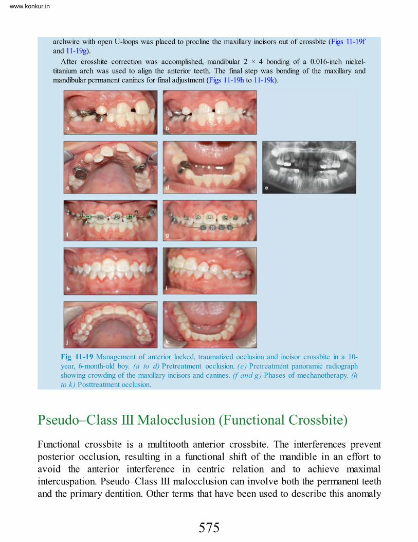
archwire with open U-loops was placed to procline the maxillary incisors out of crossbite (Figs 11-19fand 11-19g).
After crossbite correction was accomplished, mandibular 2 × 4 bonding of a 0.016-inch nickel-titanium arch was used to align the anterior teeth. The final step was bonding of the maxillary andmandibular permanent canines for final adjustment (Figs 11-19h to 11-19k).
Fig 11-19 Management of anterior locked, traumatized occlusion and incisor crossbite in a 10-year, 6-month-old boy. (a to d) Pretreatment occlusion. (e) Pretreatment panoramic radiographshowing crowding of the maxillary incisors and canines. (f and g) Phases of mechanotherapy. (hto k) Posttreatment occlusion.
Pseudo–Class III Malocclusion (Functional Crossbite)Functional crossbite is a multitooth anterior crossbite. The interferences preventposterior occlusion, resulting in a functional shift of the mandible in an effort toavoid the anterior interference in centric relation and to achieve maximalintercuspation. Pseudo–Class III malocclusion can involve both the permanent teethand the primary dentition. Other terms that have been used to describe this anomaly
575
www.konkur.in

include pseudoprognathism,57,58postural Class III,50 and functional Class III.49
These anomalies are usually characterized by a Class I skeletal pattern. Sometimes,especially if treatment has not been performed early, a minor maxillary deficiency orconstricted palate may be present. These patients usually show a straight profile atrest position (centric relation) and a slightly concave profile at centric occlusion.
Rabie and Gu,59 in a study designed to identify the diagnostic criteria for pseudo–Class III in comparison with Class I malocclusion in a southern Chinese population,examined 36 patients with pseudo–Class III malocclusion and 31 patients with ClassI malocclusion. They found that patients with a pseudo–Class III malocclusionexhibit certain morphologic, dental, and skeletal characteristics, such as retroclinedmaxillary incisors, retrusive upper lip, decreased midfacial length, and increasedmaxillomandibular differences.
In regard to criteria for distinguishing between pseudo– Class III and skeletalClass III malocclusion, Ngan et al52 stated that cephalometric evaluation may not bethe most reliable tool in differential diagnosis. The most consistent findings seem tobe the dental characteristics of Angle Class III molars and canines, retroclinedmandibular incisors, and the presence of an end-to-end or anterior cross-biteocclusion.
Gu60 attempted to clarify the dentoskeletal characteristics of pseudo–Class IIImalocclusion during the mixed dentition by comparing 36 patients with pseudo–Class III malocclusion (mean age: 10.7 ± 2.0 years) and 40 patients with skeletalClass III malocclusion (mean age: 9.7 ± 2.2 years). All subjects were followed afterthe growth spurt; cephalograms were taken in the mixed dentition for subjects withpseudo–Class III malocclusion, subjects with skeletal Class III malocclusion, and acontrol group of Class I subjects, and dentoskeletal characteristics were compared.Gu60 concluded that pseudo–Class III malocclusion is characterized by decreasedmidfacial length, mandibular displacement, retroclined maxillary incisors, andnormal vertical development.
Moyers61 proposed that the pseudo–Class III relationship is a positionalmalocclusion with an acquired neuromuscular reflex and considered the hypothesisthat the positional relationship in apparent Class III malocclusion may result from anearly interference with the muscular reflex of mandibular closure.
Several other investigators evaluating pseudo–Class III malocclusion alsoattributed the retroclined maxillary incisors and proclined mandibular incisors toincisor interference.50,57,62,63
576
www.konkur.in

Characteristic signsEvaluation of pseudo–Class III malocclusion always requires that anterior andposterior dental relationships be examined with the mandible in centric relation. Thefollowing are general signs of pseudo–Class III malocclusion that can be recognizedby careful clinical and cephalometric evaluation:
• Multitooth anterior crossbite• Normal ANB angle at rest• Some maxillary incisor retroclination• Minor maxillary deficiency• Normal to concave profile (appearing normal at centric relation and slightly
concave at habitual occlusion)• Class I molar and canine relationships in centric relation• Habitual shift from rest to occlusion
Advantages of early treatmentWhen multiple incisors are involved in a pseudo–Class III malocclusion and afunctional shift is present, urgent intervention is needed to prevent structural damageand adverse effects on the maxilla. Unlocking the maxilla before the growth spurtnormalizes maxillary growth, improving the profile and preventing structuraldamage. Early treatment of this kind of anterior crossbite, like anterior dentalcrossbite, is easy and efficient. Delayed treatment may necessitate much morecomprehensive management.
Early correction of this malocclusion eliminates the mandibular shift, unlocks themaxillary incisors and the incisive bone to enhance maxillary growth for betterfunction and esthetics, and preserves and facilitates eruption of the canines byincreasing anterior arch length. Gu and Rabie64 studied 21 consecutively treatedpatients who had a pseudo–Class III malocclusion and 15 untreated control subjects.They found that, on average, 4.7 mm of space was gained as a result of the treatment,whereas lack of space was evident in the untreated control subjects.
Early orthodontic intervention for pseudo–Class III malocclusion is alwaysrecommended to prevent existing problems from worsening and to prepare a betterenvironment for occlusal development, thereby minimizing or eliminating the need
577
www.konkur.in

for comprehensive orthodontic treatment later.
Consequences of delayed treatmentDelayed treatment and collapse of the maxillary dentition can cause A-Bdiscrepancy and maxillary retrusion, which may require comprehensive full bondingand face mask therapy.
Delayed treatment can cause structural damage to the involved teeth andsupporting structures, crowding in the anterior region, and even canine impactionand adverse effects on jaw growth51,65 (see case 11-13).
Early treatment strategiesDepending on the severity of the problem and the patient’s age and compliance,correction may be achieved with removable, functional, or fixed appliances. Whenmaxillary protraction is needed, extraoral devices such as a face mask or chin capmight also be used.
The methods described for management of simple anterior dental crossbite(removable Hawley appliance with occlusal coverage and 2 × 4 bonding) can alsobe used for correction of most pseudo–Class III malocclusions. In pseudo–Class IIImalocclusion, multiple incisors are involved and the crossbite often is accompaniedby crowding and rotation. Application of 2 × 4 bonding is preferable; the result isfaster, the patient’s compliance is not required, and there is the possibility of incisoralignment after crossbite correction. Patients with some maxillary deficiencyresulting from delayed treatment or hereditary factors may need extraoral traction.
Removable appliances for correction of pseudo–Class III malocclusions, whichusually involve multiple incisors in crossbite, have special specifications tomaintain sufficient retention. The labial bow is separated from the incisors to allowlabial movement of the maxillary incisors that are in crossbite. Therefore, retentionmust be established from the posterior segments by a long labial bow (Fig 11-20;see case 11-14), or the appliance must have a special horizontal loop on the canines,as was designed in 197766 (Fig 11-21).
578
www.konkur.in

Fig 11-20 (a and b) Modified Hawley appliance with a long labial bow soldered to the Adamsclasp. The long labial bow establishes retention in the posterior segment, which separates from theanterior segment to allow labial movement.
Fig 11-21 Removable appliance designed for crossbite correction of four incisors. It has ahorizontal loop on the canines for retention.
Case 11-11: Pseudo–Class III malocclusion
A 9-year-old boy presented with pseudo–Class III malocclusion, mandibular anterior shift, and threeincisors in crossbite, which were causing displacement and rotation of the maxillary incisors andcrowding in the mandibular incisor region (Figs 11-22a to 11-22e).
Treatment:Because of financial considerations and in accordance with the parent’s wishes, treatment wasaccomplished with a removable appliance (Fig 11-22f). Posttreatment results are shown in Figs 11-22gto 11-22i.
579
www.konkur.in

Fig 11-22 Management of a pseudo–Class III malocclusion with mandibular anterior shift. Threeincisors are in crossbite. ( a to d) Pretreatment occlusion in centric occlusion. (e ) Pretreatmentocclusion in centric relation. (f) Removable appliance used for treatment. (g to i) Posttreatmentocclusion.
Case 11-12: Pseudo–Class III malocclusion
An 11-year, 7-month-old boy in the late mixed dentition presented with a Class I malocclusion, pseudo–Class III malocclusion, and a severe anterior mandibular shift (Figs 11-23a to 11-23e).
Treatment:The treatment plan in this case was limited to 2 × 6 maxillary bonding and placement of composite resinon the occlusal surfaces of the molars to disocclude the anterior segment. No treatment was performedin the mandibular dentition. Figures 11-23f to 11-23n show the treatment outcome. Early unlocking ofthe incisor crossbite not only prevented structural damage and maintained sufficient space for theadjacent teeth but also allowed normal jaw growth, which improved the profile.
580
www.konkur.in

Fig 11-23 Management of a pseudo–Class III malocclusion with severe anterior shift in an 11-year, 7-month-old boy in the late mixed dentition. (a to c) Pretreatment occlusion in centricocclusion. (d) Pretreatment occlusion in centric relation. (e) Pretreatment soft tissue profile. (f toh ) Posttreatment occlusion. (i) Posttreatment soft tissue profile. (j ) Pretreatment cephalometricradiograph in centric occlusion. (k) Pretreatment cephalometric radiograph in centric relation.
581
www.konkur.in

Fig 11-23 (cont) (l) Posttreatment cephalometric radiograph. ( m) Pretreatment (le f t) andposttreatment (right) tracings. (n) Changes in cephalometric measurements.
Case 11-13: Pseudo–Class III malocclusion
A 17-year-old girl presented with a Class I occlusion, anterior crossbite, and a mandibular shift. Herchief complaints were temporomandibular dysfunction, pain, and restricted opening. She also had severestructural damage to the maxillary and mandibular incisors and periodontium (Figs 11-24a to 11-24d).This case clearly demonstrates the consequences of delayed treatment of anterior crossbite.
Treatment:After scaling and periodontal care were provided, orthodontic treatment was accomplished with 2 × 6maxillary and mandibular bonding. Occlusal composite resin buildup was used for anterior disocclusion.Anterior cross-bite correction and midline alignment were accomplished successfully, but because of alack of early treatment and some mandibular overgrowth, the mandibular incisors were also retractedwith lingual root torque according to available space.
582
www.konkur.in

Some interdental spacing remained in the maxillary incisors as a result of some microdontia, and therewas some crown damage; therefore, the maxillary anterior segment (canine to canine) was restoredwith metalcermic crowns (Figs 11-24e to 11-24g).
Fig 11-24 Management of anterior crossbite and mandibular shift in a 17-year-old girl. Neglecthas resulted in restricted opening, temporomandibular joint pain, and structural damage to themandibular incisors and periodontium. (a to d) Pretreatment occlusion. (e to g) Posttreatmentocclusion after 2 × 6 bonding and placement of crowns on the maxillary anterior teeth.
Case 11-14: Pseudo–Class III malocclusion
An 11-year, 7-month-old girl presented with a Class I anterior pseudo–Class III malocclusion withmandibular shift, minor damage to the maxillary incisors, and minor crowding in the maxillary arch (Figs11-25a to 11-25c).
Treatment:Treatment was accomplished with a removable Hawley appliance that had two Adams clasps. A longlabial bow, separated from the incisors to facilitate their proclination, was soldered to the Adams claspsto maintain retention from the posterior segments (see Fig 11-20). Figures 11-25d and 11-25e show theposttreatment occlusion.
Comparison of this case with previous cases, in which treatment was neglected for a period of time,clearly demonstrates the difference in complexity of treatment procedures and the damage that canoccur to the teeth and supporting structures if early treatment is not provided.
583
www.konkur.in

Fig 11-25 Management of a pseudo–Class III malocclusion with mandibular shift that has causedminor damage to the maxillary incisors in an 11-year, 7-month-old girl. (a) Pretreatment occlusionin centric occlusion. ( b ) Pretreatment occlusion in centric relation. ( c ) Retroclination of themaxillary incisors. (d and e) Posttreatment occlusion.
Skeletal Class III and Potential Class III MalocclusionTrue Class III malocclusion, or mesial occlusion, is an anteroposteriordentoalveolar or skeletal malrelationship characterized by a mandibular dentitionthat has a more anterior position than the maxillary dentition.67 This anomaly can becaused by mesial mandibular basal or dentoalveolar positioning, distal maxillarybasal or dentoalveolar positioning, basal bone size discrepancy (either shortermaxilla or longer mandible), or a combination of any of these factors.
Skeletal Class III malocclusion can also be caused by an aberration in other bonystructures, such as underdevelopment of the anterior cranial length,underdevelopment of the posterior cranial length, and divergence or convergence ofsome angles, such as the saddle angle, articular angle, y-axis, gonial angle, ormaxillary and mandibular length. It can also be caused by variations in dentalrelationships without any underlying skeletal malrelationship.
Ngan et al68 explained that patients with skeletal Class III malocclusion exhibit aconcave facial profile, a retrusive nasomaxillary area, and a prominent mandibularthird of the face. The lower lip is often protruded relative to the upper lip. Themaxillary arch is usually narrower than the mandibular arch, and overjet andoverbite can range from reduced to reverse. Therefore, occlusal relationships inpatients with Class III malocclusion constitute only one part of a much largersyndrome. Proper treatment planning requires a thorough clinical assessment andcephalometric analysis.
584
www.konkur.in

Any of the aforementioned skeletal combinations can be, but are not always,accompanied by a mandibular shift or anterior crossbite. Therefore, the first step inrecognition and treatment planning starts with accurate differential diagnosis of trueClass III malocclusion from other kinds of anterior crossbite.
Developing Class III malocclusion in growing patients is one of the mostchallenging problems confronting clinicians. Early orthodontic intervention isrecommended to encourage normal dentoalveolar and skeletal growth. Class IIImalocclusions are usually growth-related discrepancies; delayed intervention oftendevelops into a more severe abnormality until growth is complete. Angle69 reportedClass III malocclusion in 3.4% of his sample of 1,000 white patients.
It can be assumed that delayed treatment of a pseudo– Class III abnormality canallow it to change to a skeletal deformity later. This is the situation the author callspotential Class III malocclusion. This is not true in all cases of pseudo–Class IIImalocclusion, but clinically the author has observed some untreated parents orrelatives with more severe skeletal deformity while their children or siblings withpseudo–Class III malocclusion were treated easily.
Treatment options for this malocclusion depend on the dentoskeletal age of thepatient and the morphologic characteristics of the malocclusion. Early treatmentapproaches include growth modification and dental camouflage. However, whengrowth has ceased, orthognathic surgery is the only choice. Depending on the amountof skeletal discrepancy, surgical correction may consist of mandibular setback,maxillary advancement, or a combination of mandibular and maxillary procedures.
This type of malocclusion can have genetic or environmental origins or acombination of both.
Class III malocclusions initiate and develop during the primary or early mixeddentition and are recognizable at early ages.
Dental and skeletal Class III malocclusions must be carefully differentiated. Thereare special situations that must be distinguished:
• Sometimes in patients with true Class III malocclusion with incisal interference,proclination of the mandibular incisors, and retroclination of the maxillaryincisors, a mandibular mesial shift causes posturing of the mandible in an anteriorposition that can be misleading, causing a mistaken diagnosis of pseudo–Class IIImalocclusion. Careful clinical and cephalometric evaluations can identify trueClass III malocclusion with skeletal discrepancy.
• In some patients with true Class III malocclusion with skeletal discrepancy,
585
www.konkur.in

especially mandibular prognathism, no mandibular shift is present, and theincisors are in and end-to-end position. This is indicative of a more severemalocclusion that may need orthognathic surgery.
• Patients with dental characteristics of Angle Class III molars and canines,retroclined mandibular incisors, and an end-to-end or anterior crossbite occlusionwithout a mandibular shift have true skeletal Class III malocclusion. Thesepatients require a comprehensive orthodontic or orthosurgical approach.
Originally, it was believed that Class III malocclusions arise primarily from anoverdevelopment of the mandible (mandibular prognathism). The fact that 33 of 40members of the Hapsburg family had prognathic mandibles was considered to be aclassic example of this anomaly. Now, many reports have indicated that maxillaryretrusion contributes to the anomaly in up to 60% of cases.57
The orthopedic effect of face mask use and maxillary sutural opening in earlytreatment of patients with Class III malocclusion, especially those with maxillarydeficiency, can correct anterior crossbite and facilitate normal maxillary growth inadaptation and synchronization with the mandible during the growth period. Lateadolescent treatment and treatment when growth changes will be limited minimizethese adaptations and eventually result in unsuccessful treatment.
Early treatment of Class III malocclusion with mandibular prognathism seems tobe much more complicated, with minimal success and frequent relapse, when startedduring the pubertal growth period. Proffit57 stated that early treatment of patientswith Class III malocclusion depends on whether the problem is the result ofmandibular prognathism or deficiency in maxillary growth. He also stated thatrestraining mandibular growth at an early stage rarely succeeds; later mandibulargrowth wipes out any early correction. On the other hand, face mask treatment tomodify growth in children with maxillary deficiency succeeds if, and only if, thetreatment is completed early.57
The author has found good results in early intervention during the primarydentition and correction of Class III malocclusion with mandibular prognathism ifnormal incisor relationships are achieved at an early age. Establishment of normalincisor relationships during the primary dentition usually allows the maxilla tocontinue growth stimulation and synchronize with mandibular growth, evidently withstable results (see cases 11-19 to 11-23).
Treatment for skeletal Class III malocclusion with mandibular prognathism duringthe early permanent dentition or even during the late mixed dentition is usuallyunsuccessful and has to be continued with comprehensive orthodontic and
586
www.konkur.in

orthognathic surgical procedures.Other variations of skeletal Class III malocclusion accompanied by severe
vertical problems are more complicated and require comprehensive treatment andpossibly two-jaw surgery later. The author postpones orthodontic treatment of thesepatients until after the age 15 or 16 years and then follows with surgery aftercompletion of growth.
Guidelines for pretreatment evaluationAccurate differential diagnosis and careful pretreatment evaluations are essential fordetermining the proper approach to and timing of treatment. Box 11-3 presentsguidelines to consider in treatment planning in order to achieve efficient correctionand long-term stability.
Box 11-3Factors influencing long-term stability oftreatment in patients with Class IIImalocclusion
Positive factors • Convergent facial type • Anteroposterior functional shift • Symmetric condylar growth • Good growth potential • ANB of 2 degrees or less • Maxillary anterior crowding and retroclination • Good patient cooperation • Good facial esthetics • Lack of familial prognathism
Negative factors • Divergent facial type • Absence of anteroposterior functional shift • Asymmetric condylar growth • Growth completed • Severe, negative ANB value • Lack of maxillary anterior crowding andproclination • Poor patient cooperation • Poor facial esthetics • Familial prognathism
Prevalence of Class III malocclusionThe prevalence of Class III malocclusion has a strong ethnic distribution; reportsvary greatly depending on the different methods of classification. Nakasima et al63,70
reported an incidence of 1% in white populations and a frequency of 10% in aJapanese population. Haynes71 screened a white population of British females aged11 to 22 years and reported a prevalence of 1.6%. Endo72 reported finding reversedocclusion in 7.81% of 11-year-old Japanese girls studied. Susami et al73 reported aprevalence of 4.24% in Japanese females between 3 and 19 years of age.
587
www.konkur.in

Greater frequency of reversed occlusion is not limited to Japanese individuals;the rate is also higher in other Asian populations. Chan74 reported that theprevalence of Class III malocclusion in Chinese individuals is 9.4%, and Baik etal75 reported a rate of 19% in Korean patients.
Box 11-4 shows the differences in characteristics of dental and skeletalcrossbites.
Box 11-4 Comparison of the characteristics of dental andskeletal crossbites
Skeletal Class III malocclusion • The etiology can be genetic, environmental, or acombination of both. • The ANB angle has a negative value. • Incisors are in end-to-end relationships or exhibitnegative overjet. • Molars and canines show a Class III relationship ora Class III tendency. • The malocclusion may or may not be accompaniedby a mandibular shift. • The profile is straight or concave.
Dental crossbite • The etiology is usually local. • The ANB angle is normal. • Only the incisors show an incorrect inclination. • Molars and canines are in Class I relationships atcentric relation.• The crossbite is often accompanied by a habitualshift from rest to occlusion. • The profile is normal.
Early treatment strategiesEvaluation and specification of the exact morphology and characteristics of eachpatient’s Class III malocclusion is the first important step in treatment.
Early interception of Class III malocclusion has been advocated for many years;Angle69 suggested that deformities in this class begin at about the age of eruption ofthe permanent first molars or earlier and are always associated with enlargedtonsils. He also indicated that the skeletal aberrations worsen with time. Angle69
stated that, when disharmony is being established, the problem usually progressesrapidly. Angle69 was also one of the first to suggest that combined orthodontics andorthognathic surgery was the only way to correct mandibular prognathism once it hasfully developed.
Salzmann76 suggested that treatment for Class III malocclusion commence as soonas the abnormality is diagnosed. Graber et al50 contended that because Class IIImalocclusions are among the most difficult to treat and surgical intervention iscontemplated in some cases, it makes good sense to try early treatment with at least achin cap to prevent worsening of the malocclusion.
588
www.konkur.in

Tweed77 divided Class III malocclusions into two categories: pseudo–Class III,with a normal mandible and an underdeveloped maxilla, and skeletal Class III with alarge mandible. Tweed77 stated that these irregularities should be treated during themixed dentition (7 to 9 years); if the conditions occur during the primary dentition,treatment can be started as early as 4 years of age. He also stated that, if treatment isnot started at an early age, lingual locking of the maxillary incisors will retardmaxillary growth and accelerate growth in the mandible.
Proffit57 stated that there is some degree of maxillary deficiency in most patientswith skeletal Class III malocclusion (30% to 40%) and expressed hope thatimproved appliance design would allow more downward and forward repositioningof the maxilla.
Björk78 stated that circum-maxillary sutures are active sites of growth during theearly juvenile period and facilitate forward maxillary growth.
General strategies for early intervention in Class III maxillary deficiencies oranterior crossbites with the potential to develop to Class III malocclusion includethe following four major techniques:
1. Unlocking of the occlusion and elimination of mandibular shift2. Incisor proclination combined with labial root torque3. Maxillary protraction and expansion, if needed4. Enhancement of maxillary growth
Depending on the type of problem and the dentoskeletal condition of the anomaly,different early intervention techniques are available, including palatal expansion, aface mask, a standard chin cap, a chin cap with spurs, and functional appliances.
Special appliances
Chin cap with spurs
A chin cap with spurs is an extraoral device that can be used in Class III orthopedictreatment for simultaneous control of mandibular growth and protraction of themaxilla (Fig 11-26). A rubber band connected from the spurs to the maxillary archproduces maxillary protraction (see case 11-20).
589
www.konkur.in

Fig 11-26 (a and b) Vertical chin cap with spurs. Maxillary protraction is achieved with rubberbands connected from the spurs to the maxillary arch.
Face mask–chin cap combination
The face mask–chin cap combination (FCC) is a special orthopedic device theauthor originally designed and constructed in 1968 and developed from that timeuntil 1999; when practicing in Tehran, the author used this appliance with goodsuccess in many patients with skeletal Class III malocclusion. This device can beused in early treatment of all kinds of skeletal Class III malocclusion, whether theyresult from maxillary deficiency, mandibular prognathism, or a combination of both.With some minor modifications, the FCC can also be applied in Class III patientswith a horizontal or vertical growth pattern.
Generally this appliance has two different kinds of design, type 1 and type 2. Bothtypes have a soft head holder pad that can be adjusted to each patient’s head size andshape. The advantage of the head holder is that the appliance can be firmly held tothe face all night without any slipping.
The type 1 FCC has a chin cap without traction (Fig 11-27a), and the author usesit in Class III patients with maxillary deficiency and a normal mandible. The acrylicresin chin cap provides anchorage against maxillary protraction (see case 11-19).
The type 2 FCC, besides the head holder pad and acrylic resin chin cap, has atraction elastic connected to the cap on each side of the face (Fig 11-27b). Theseelastics can impart distal force to the mandible and therefore can be used in Class IIImalocclusions in which both the maxilla and the mandible are involved (see cases11-21 and 11-23). The chin cap force can also be designed in a high-pull directionor a cervical direction, depending on the patient’s mandibular growth pattern.
590
www.konkur.in

Fig 11-27 Face mask–chin cap (FCC) combination. (a) Type 1. (b) Type 2.
Case ReportsThe following case reports present examples of management of various types ofanterior crossbite with different characteristics and different etiologic factors; withdifferent treatment options, such as preventive, interceptive, corrective, orcombination treatment; and in patients at different dental and skeletal stages ofdevelopment.
Although the major goal of this publication is to advocate early intervention forall kinds of anterior crossbites, whether dental, dentoalveolar, or skeletal, a fewcases of delayed treatment are presented to illustrate the problems that can resultfrom negligence. Therefore, case reports are presented and classified in threegroups:
1. Early treatment during the primary or early mixed dentition before incisoreruption
2. Early treatment after eruption of the incisors (middle or late mixed dentition)3. Nonsurgical treatment of the Class III permanent dentition
Early treatment during the primary or early mixeddentition before incisor eruptionEarly intervention at this stage of the dentition usually consists of interceptivetreatment and guidance, and the main goals are unlocking of the anterior teeth andslight proclination of the primary incisors, if needed. Correction of posteriorcrossbite is also another goal to achieve at this stage of treatment by either slow
591
www.konkur.in

dental expansion or rapid expansion, depending on the type of posterior crossbiteand maxillary base conditions.
Maxillary basal protraction by extraoral devices might also be necessary in somepatients with maxillary deficiency. This goal can be achieved with a face mask orchin cap with spur attached to removable or bonded expanders.
The fundamental strategy of this type of intervention is to prepare a normalenvironment at an early stage of the dentition to enable coordinated jaw growth andencourage proper development of the occlusion, which should continue to the nextdental stages.
Case 11-15A 6-year-old girl presented with both anterior segments and the posterior left segment in crossbite. Theonly permanent teeth that had erupted were the first molars. She exhibited a mandibular shift and aconcave profile (Figs 11-28a to 11-28d).
Treatment:Treatment was accomplished with only a Hawley posterior bite plate without a labial bow. Theappliance included an expansion screw for posterior crossbite correction and two finger springs foranterior crossbite correction. Composite resin attachments were bonded to the buccal surfaces of themaxillary primary molars; the attachments had long C-clasps hooked over the composite resin for addedretention in place of bands with tubes (Fig 11-28e). Photographs and a radiograph revealed the resultsof posterior and anterior crossbite correction; the mandibular permanent central incisors were erupting,and the posterior segment was settling (Figs 11-28f to 11-28j).
Fig 11-28 Early treatment of anterior and posterior crossbite in a 6-year-old girl. (a and b)Pretreatment occlusion. ( c ) Pretreatment soft tissue profile. ( d ) Pretreatment cephalometricradiograph. (e) Appliance used in treatment.
592
www.konkur.in

Fig 11-28 (cont) (f to h) Posttreatment occlusion. ( i ) Posttreatment soft tissue profile. (j)Posttreatment cephalometric radiograph.
Case 11-16A 3-year, 5-month-old boy in the primary dentition presented with a Class III malocclusion that had ahereditary background. He had a mesial step at the primary second molars (slight Class III relationshipsat centric relation) and a mandibular mesial shift (Figs 11-29a to 11-29e).
Treatment:During the primary dentition, even unlocking the occlusion in the area of crossbite can eliminatefunctional shift and guide development of normal occlusion. This boy showed good compliance, so thetreatment plan included only the use of a Hawley appliance, banding of the primary second molars witha buccal tube for better retention, occlusal coverage, and a simple finger spring behind the maxillaryincisors.
Figures 11-29f and 11-29g show the settling posterior segment after correction of the incisorcrossbite. Figures 11-29h to 11-29k show the final occlusion, 10 months after use of the appliance wasstopped.
Fig 11-29 Management of a hereditary Class III malocclusion in a 3-year, 5-month-old boy in theprimary dentition. He has a mesial step at the primary second molars and a mesial mandibularshift. (a to c) Pretreatment occlusion in centric occlusion. (d) Pretreatment occlusion in centricrelation. (e) Pretreatment cephalometric radiograph.
593
www.konkur.in

Fig 11-29 (cont) (f and g) Occlusion after correction of anterior crossbite and during settling ofthe posterior occlusion. (h to j) Posttreatment occlusion 10 months after cessation of the appliance.(k) Posttreatment cephalometric radiograph.
Case 11-17A 5-year, 9-month-old boy presented with a collapsed maxillary dentition, severe reverse deep bite, anda mandibular mesial shift. Even in centric relation, the maxillary and mandibular incisors did not meet inan end-to-end position (Figs 11-30a to 11-30g). He exhibited a negative ANB angle and a concaveprofile. His problems had a hereditary background.
Treatment:The treatment approach was interceptive intervention to correct the dentoskeletal malrelationships bymaxillary expansion, maxillary protraction by face mask therapy, and some maxillary incisorproclination. Because the patient’s compliance was excellent and the parent preferred it, treatment wasperformed with a removable appliance and face mask.
Figure 11-30h shows the modified Hawley appliance, which included two Adams clasps, thickposterior occlusal coverage to disocclude the anterior segment, a finger spring for slight incisorproclination, a palatal jackscrew for expansion, and two horizontal loops on the canines for connectingelastics to the face mask to achieve maxillary protraction and provide appliance retention (as discussedearlier66). Figure 11-30i shows the patient’s occlusion 6 months after the start of treatment. Thepermanent incisors have erupted out of crossbite. During this stage, the posterior bite block wasreduced gradually at each visit.
Figures 11-30j and 11-30k show different panoramic radiographs taken during treatment; theyindicate that sufficient space was created for eruption of all incisors and canines. Figures 11-30l to 11-30o show the occlusion 6 months after active treatment and during the observation period.
594
www.konkur.in

Figures 11-30p to 11-30s were taken 15 months after active treatment and show complete eruption ofthe maxillary central incisors and all mandibular incisors. At this time, active treatment was stopped, andthe patient was placed on a recall schedule for observation.
Fig 11-30 Management of collapsed maxillary dentition, severe reverse deep bite, and mesialmandibular shift in a 5-year, 4-month-old boy with a hereditary Class III malocclusion. (a to d)Pretreatment occlusion. (e) Pretreatment panoramic radiograph. (f ) Pretreatment cephalometricradiograph in centric occlusion. (g ) Pretreatment cephalometric radiograph in centric relation.
595
www.konkur.in
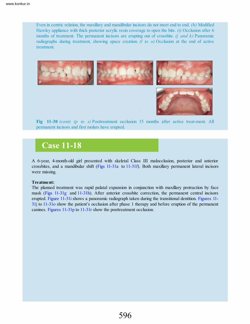
Even in centric relation, the maxillary and mandibular incisors do not meet end to end. (h) ModifiedHawley appliance with thick posterior acrylic resin coverage to open the bite. (i) Occlusion after 6months of treatment. The permanent incisors are erupting out of crossbite. (j and k) Panoramicradiographs during treatment, showing space creation. (l to o) Occlusion at the end of activetreatment.
Fig 11-30 (cont) (p to s) Posttreatment occlusion 15 months after active treat-ment. Allpermanent incisors and first molars have erupted.
Case 11-18A 6-year, 4-month-old girl presented with skeletal Class III malocclusion, posterior and anteriorcrossbites, and a mandibular shift (Figs 11-31a to 11-31f). Both maxillary permanent lateral incisorswere missing.
Treatment:The planned treatment was rapid palatal expansion in conjunction with maxillary protraction by facemask (Figs 11-31g and 11-31h). After anterior crossbite correction, the permanent central incisorserupted. Figure 11-31i shows a panoramic radiograph taken during the transitional dentition. Figures 11-31j to 11-31o show the patient’s occlusion after phase 1 therapy and before eruption of the permanentcanines. Figures 11-31p to 11-31r show the posttreatment occlusion.
596
www.konkur.in

Fig 11-31 Management of skeletal Class III malocclusion, posterior and anterior crossbites, andmandibular shift in a 6-year, 4-month-old girl. Both maxillary permanent lateral incisors arecongenitally missing. (a to c) Pretreatment occlusion. (d) Pretreatment panoramic radiograph. (e)Pretreatment cephalometric radiograph in centric occlusion. ( f ) Pretreatment cephalometricradiograph in centric relation. (g and h) Bonded rapid expander with hook for mask protraction.
597
www.konkur.in

Fig 11-31 (cont) (i) Panoramic radiograph during the transitional dentition, before eruption of thepermanent canines. (j to m) Occlusion after phase 1 treatment. All incisors have erupted duringthe observation period. (n ) Panoramic radiograph after phase 1 treatment. ( o ) Cephalometricradiograph after phase 1 treatment. (p to r) Posttreatment results.
Early treatment after eruption of the incisors (middle orlate mixed dentition)As discussed earlier, anterior crossbites with mandibular shift are not onlydestructive to the dentition and supporting structures but also can have adverseeffects on jaw growth and worsen the anteroposterior basal jaw relationships.Therefore, correction of the problem during the mixed dentition, especially the casesthat have some familial background, are more complicated. Patients exhibit a greatersagittal malrelationship (A-B discrepancy) and usually need a combination oforthodontic and orthopedic treatment.
Case 11-19
598
www.konkur.in

A 9-year, 8-month-old girl in the middle mixed dentition presented with a maxillary deficiency (SNA =76.5 degrees) and a maxillary arch that was collapsed within the mandibular arch (Figs 11-32a to 11-32d). There was an A-B discrepancy of –0.8 degrees as well as severe maxillary incisor crowding,rotation, and displacement. She had no mandibular shift.
Treatment:The treatment plan included rapid maxillary expansion and maxillary protraction with a type 1 FCC (seeFig 11-27a). Treatment was started with rapid palatal expansion, occlusal coverage, and a hook to theFCC. The second step was maxillary incisor bonding and placement of a nickel-titanium arch forleveling and anterior alignment. Next, a stainless steel round arch with open loop was placed to proclinethe maxillary incisors out of crossbite. Finally, a rectangular arch was placed in conjunction with thetype 1 FCC to achieve buccal root torque.
Figures 11-32e to 11-32h show the occlusal results. Cephalometric measurements revealed thefollowing effects of treatment: ANB increased from –0.8 to 1.5 degrees, SNA increased from 76.5 to82.5 degrees, and overjet increased from –1.5 to 0.8 mm (Figs 11-32i and 11-32j).
Fig 11-32 Management of maxillary arch collapse and maxillary deficiency in a 9-year, 8-month-old girl. The maxillary incisors exhibit severe crowding, rotation, and displacement. There is nomandibular shift. (a to c) Pretreatment occlusion. (d) Pretreatment soft tissue profile. (e to g)Posttreatment occlusion. (h) Posttreatment soft tissue profile.
599
www.konkur.in

Fig 11-32 (cont) (i) Pretreatment (left) and posttreatment (right) cephalometric radiographs. (j)Changes in cephalometric measurements and superimposition of pretreatment (black ) andposttreatment (green) tracings.
Case 11-20An 11-year-old boy presented with skeletal Class III malocclusion, maxillary deficiency, mandibularprognathism, a steep mandibular plane, and a mandibular shift (Figs 11-33a to 11-33d).
Treatment:Treatment was started with rapid palatal expansion and a vertical chin cap (see Fig 11-26) that hadspurs to allow the elastics to be attached from the spurs to the premolar brackets to initiate maxillaryprotraction (Figs 11-33e to 11-33h). After expansion and bite jumping, treatment was continued withmaxillary anterior bonding. There were also elastics from spurs to the connected midline arch hook.
Figures 11-33i to 11-33l illustrate the patient’s posttreatment occlusion.
600
www.konkur.in

Fig 11-33 Management of skeletal Class III maxillary deficiency, mandibular prognathism, steepmandibular plane, and mandibular shift in an 11-year-old boy. (a to c) Casts of pretreatmentocclusion. ( d ) Pretreatment cephalometric radiograph. (e to h) Rapid palatal expansion andmaxillary protraction. (i to k) Posttreatment occlusion. (l) Posttreatment cephalometric radiograph.
Case 11-21An 11-year, 6-month-old girl in the late mixed dentition presented with a hereditary skeletal Class IIImalocclusion, mandibular prognathism, slight maxillary deficiency, anterior open bite, left posteriorcrossbite, maxillomandibular dental protrusion, and a 1.9-mm reverse overjet (Figs 11-34a to 11-34c).
Treatment:
601
www.konkur.in

Because of the patient’s space deficiency, dental protrusion, vertical growth tendency, and anterioropen bite, treatment was designed to include extraction of four premolars combined with orthopedictreatment. As a result of the patient’s dentoskeletal configuration—mandibular prognathism andmandibular incisor protrusion— two mandibular first premolars and two maxillary second premolarswere removed.
First, a mandibular lingual arch was inserted to provide anchorage and prevent mandibular molareruption. Next, a maxillary transpalatal arch was used to provide maxillary anchorage and intrude themaxillary molars. The maxillary and mandibular primary second molars were extracted first. Thistreatment continued later with extraction of the maxillary second premolars and mandibular firstpremolars to obtain greater retraction of the mandibular incisors and greater mesial movement of themaxillary molars. These changes were designed to provide counterclockwise rotation of the mandiblethat would help correct the open bite.
The next step was maxillary anterior bonding to procline the incisors and placement of a type 2 FCCappliance to achieve protraction of the maxilla (see Fig 11-27b).
Figures 11-34d to 11-34f show the posttreatment occlusion. Some of the results of treatment includedchanges in SNA from 81.0 to 87.4 degrees, ANB from –0.3 to 4.7 degrees, A-B to facial plane from1.3 to –6.5 degrees (normal), overbite from –2.4 to 1.2 mm, and overjet from –1.9 to 2.9 mm (Figs 11-34g and 11-34h).
Fig 11-34 Management of skeletal Class III malocclusion, mandibular prognathism, slightmaxillary deficiency, anterior open bite, left posterior crossbite, and maxillomandibular dentalprotrusion in an 11-year, 6-month-old girl in the late mixed dentition. (a to c) Pretreatmentocclusion. (d to f) Posttreatment occlusion.
602
www.konkur.in

Fig 11-34 (cont) (g) Pretreatment (left) and posttreatment (right) cephalometric radiographs. (h)Changes in cephalometric measurements and superimposition of pretreatment (black ) andposttreatment (green) tracings.
Case 11-22A 10-year-old girl at the beginning of the late mixed dentition presented with Class III skeletalmalocclusion, maxillary deficiency, anterior and posterior crossbites (collapsed maxillary arch), andreverse deep bite (Figs 11-35a to 11-35d).
Treatment:The treatment plan called for maxillary rapid palatal expansion and protraction with a type 1 FCC. Theexpander had thick occlusal acrylic resin to disocclude the anterior teeth and facilitate maxillaryprotraction and incisor protrusion and thereby correct the reverse overbite (Fig 11-35e). It also hadhooks for connecting elastics to the face mask.
Figure 11-35f shows the occlusal situation after completion of expansion, during incisor protrusion andspace closure, and before labial root torquing in conjunction with the face mask. Treatment had anumber of positive effects (Figs 11-35g to 11-35l). The Landes angle changed from 83.4 to 89.9degrees, ANB changed from –5.3 to –0.8 degrees, and overjet was increased from –3.3 to 1.6 mm.
603
www.konkur.in

Fig 11-35 Management of skeletal Class III malocclusion, maxillary deficiency, collapsedmaxillary arch, and reverse deep bite in a 10-year-old girl. (a to c) Pretreatment occlusion. (d)Pretreatment panoramic radiograph. (e) Rapid palatal expander with occlusal coverage and hooksfor a face mask. (f) Occlusion after expansion, during incisor protrusion and space closure, andbefore labial root torquing. (g to i) Posttreatment occlusion. ( j ) Posttreatment panoramicradiograph.
604
www.konkur.in

Fig 11-35 (cont) (k) Pretreatment (left) and posttreatment (right) cephalometric radiographs. (l)Changes in cephalometric measurements and superimposition of pretreatment (black ) andposttreatment (green) tracings.
Case 11-23An 8-year-old girl in the early mixed dentition presented with a skeletal Class III malocclusion, maxillarydeficiency, and mandibular prognathism (Figs 11-36a to 11-36c). She had an A-B discrepancy of –4.0degrees, anterior open bite, and a 5.4-mm reverse overjet. There was no mandibular shift. The incisorsand permanent first molars were partially erupted.
Treatment:The treatment strategy was two-phase treatment. Phase 1 was orthopedic treatment to controlmandibular growth and protract the maxilla. This phase was started with a lower holding arch topreserve arch length and control eruption of the mandibular molars. Maxillary orthopedic treatment wasstarted with a type 2 FCC.
After eruption of the permanent maxillary molars and incisors, banding and anterior bonding (2 × 4)was started. Maxillary traction in a vertical direction was continued with an elastic from the maxillaryarch to the type 2 FCC. Phase 2 consisted of comprehensive bonding, light Class III mechanics, andless time wearing the FCC appliance with vertical traction. Figures 11-36d to 11-36i show the treatmentoutcome.
605
www.konkur.in

Fig 11-36 Management of skeletal Class III malocclusion, maxillary deficiency, and mandibularprognathism in an 8-year-old girl. She is in the early mixed dentition and has an anterior open biteand a 5.4-mm reverse overjet. (a to c) Casts of pretreatment occlusion. (d to f) Posttreatmentocclusion. ( g ) Pretreatment cephalometric radiograph. ( h ) Posttreatment cephalometricradiograph. ( i ) Changes in cephalometric measurements and superimposition of pretreatment(black) and posttreatment (green) tracings.
Nonsurgical treatment of Class III permanent dentitionThree cases are presented to show the results of delayed correction of anteriorcrossbite. Treatment options available during the permanent dentition are limited tocamouflage treatment, either with or without extraction, and orthogna-thic surgery,for patients with severe skeletal discrepancy.
606
www.konkur.in

Case 11-24A 16-year, 5-month-old girl presented with severe anterior dentoalveolar crossbite, reverse deep bite,two-step occlusion, maxillary anterior crowding, mandibular mesial shift, and structural damage to themaxillary incisors (Figs 11-37a to 11-37e).
Treatment:A nonextraction treatment strategy was planned. It included slight maxillary incisor proclination toeliminate crossbite and correct the overbite. A posterior bite plate with acrylic resin occlusal coveragewas placed to disocclude the anterior teeth. Because of the severe reverse overbite, an occlusalcomposite resin attachment could not be used.
Complete maxillary and mandibular bonding was begun with a nickel-titanium archwire to align theincisors, and then stainless steel archwire with open loop was used to complete incisor proclination, levelthe mandibular arch, and correct the two-step occlusion. The occlusal acrylic resin was graduallyreduced after crossbite correction and then use of the posterior bite plate eventually was stopped.
As mentioned earlier, dentoalveolar crossbite correction after puberty is limited to camouflageprocedures or, in patients with severe skeletal discrepancy, orthognathic surgery. In this patient, the finalresults achieved in the dentition, including anteroposterior and buccolingual intercuspation andinterdigitation, were good (Figs 11-37f to 11-37h). The dental and facial midlines were perfect, and nomandibular shift was present. However, no significant skeletal changes were achieved because of thelack of significant growth changes during corrective treatment. The major changes achieved wereanteroposterior and vertical changes in occlusion accomplished through minor incisor proclination and bytaking advantage of the mandibular shift. These dental changes had some positive results on thepatient’s soft tissue profile and upper and lower lip relationships (Figs 11-37i to 11-37l).
Fig 11-37 Management of severe anterior dentoalveolar crossbite and reverse deep bite in a 16-year, 5-month-old girl. She exhibited two-step occlusion, maxillary anterior crowding, and a mesialmandibular shift. (a to c) Pretreatment occlusion in centric occlusion. The dental problems haveresulted in structural damage to the maxillary incisors. (d ) Pretreatment occlusion in centricrelation. (e) Pretreatment soft tissue profile.
607
www.konkur.in

Fig 11-37 (cont) (f to h) Posttreatment occlusion. ( i ) Posttreatment soft tissue profile. (j)Pretreatment cephalometric radiograph. (k ) Posttreatment cephalometric radiograph. (l) Changesin cephalometric measurements and superimposition of pretreatment (black) and posttreatment(green) tracings.
Case 11-25A 12-year-old boy presented with a severe anterior dentoalveolar crossbite, severe maxillary anteriorcrowding in which the canines were out of the arch, some maxillary incisor retroclination, a mesialmandibular shift, and good mandibular dentition (Figs 11-38a to 11-38d).
Treatment:After consideration of the maxillary incisor retroclination, mandibular shift, and good mandibulardentition, the treatment plan was designed as camouflage with a nonextraction approach that waslimited to maxillary treatment. The anterior teeth were disoccluded through the use of a maxillaryposterior bite block, and maxillary complete bonding was used to align and procline the maxillary incisors
608
www.konkur.in

to eliminate crossbite.Unlike the patient in case 11-24, this patient did experience some positive dentoskeletal changes
during treatment (Figs 11-38e to 11-38j): The Landes angle increased from 88.4 to 91.3 degrees; SNAincreased from 79.9 to 83.8 degrees; ANB increased from – 2.5 to –0.5 degrees; and overjet increasedfrom –3.3 to 1.3 mm. Presumably these positive changes could be attributed to the 12-year-old boy’sjaw growth during treatment.
Fig 11-38 Management of severe anterior dentoalveolar crossbite and maxillary anterior crowdingin a 12-year-old boy. The canines are out of the arch, and there is some maxillary incisorretroclination. There is a mesial mandibular shift, but the mandibular dentition is good. (a to c)Pretreatment occlusion. (d) Pretreatment soft tissue profile. (e and f) Posttreatment occlusion. (g)Posttreatment soft tissue profile. (h ) Pretreatment cephalometric radiograph. ( i) Posttreatmentcephalometric radiograph. ( j ) Changes in cephalometric measurements and superimposition ofpretreatment (black) and posttreatment (green) tracings.
609
www.konkur.in

Case 11-26A 14-year-old girl presented with a severe dentoalveolar anterior crossbite, maxillary and mandibularcrowding, minor mandibular incisor proclination, and a mesial mandibular shift (Figs 11-39a to 11-39d).
Treatment:Because she exhibited severe maxillary and mandibular crowding and some mandibular incisorproclination, and because the incisors could not reach end-to-end contact during centric occlusion orcentric relation, the treatment plan called for both camouflage and extraction of four first premolars.
Because of the age of the patient and the lack of significant jaw growth, the results of camouflagetreatment were mostly dental, that is, achievement of good occlusion and dental relationship (Figs 11-39e to 11-39h). Comparison of cephalometric evaluations revealed no significant skeletal changes (Figs11-39i and 11-39j). The major changes were an improvement in the incisor relationships and animprovement in lip relationships, which was due to mandibular incisor retraction. There was also someminor improvement in the sagittal A-B relationship from –3.2 to –1.3 degrees and an increase in overjetfrom –2.8 to 2.0 mm.
Fig 11-39 Management of dentoalveolar anterior crossbite, maxillary and mandibular crowding,and a mesial mandibular shift in a 14-year-old girl. There is minor proclination of the mandibularincisors. (a and b) Pretreatment occlusion in centric occlusion. (c ) Pretreatment occlusion incentric relation. ( d ) Pretreatment soft tissue profile. (e to g) Posttreatment occlusion. (h)Posttreatment soft tissue profile.
610
www.konkur.in

Fig 11-39 (cont) (i) Pretreatment (left) and posttreatment (right) cephalometric radiographs. (j)Changes in cephalometric measurements and superimposition of pretreatment (black ) andposttreatment (green) tracings.
Summary
• Class II malocclusion accounts for about one-third of the total malocclusionsin the US population.
• Long-term studies from the primary to the permanent dentition have shownthat Class II abnormalities are not self-correcting and in some cases mayworsen.
• Class II discrepancy has mainly been perceived as a sagittal problem; thevertical and transverse dimensions traditionally were not considered to berelated to Class II discrepancy.
• Despite that perception, variations in facial height may conceal or intensifythe clinical appearance of Class II malocclusion. An increased mandibularvertical dimension associated with Class II malocclusion usually rotates the
611
www.konkur.in

mandible downward and backward, which makes the mandible appearretruded and the profile more convex. Any mistake in treatment wouldworsen the situation. A decreased vertical dimension causes the mandible torotate upward and forward, manifesting as a prominent chin point.
• Likewise, evaluation of the transverse dimension in patients with Class IImalocclusion must not be overlooked. Most Class II patients with a narrowmaxillary arch may show normal relationships in centric occlusion. Earlyintervention of transverse problems and proper expansion at earlier stageswill facilitate mandibular forward growth.
• Not all Class II malocclusions are alike; numerous types of skeletal anddental malocclusions may be observed.
• Class II variations are the result of its multifactorial origins. Class IImalocclusions have different etiologic factors, different morphologiccharacteristics, and different treatment approaches.
• Completion of thorough diagnostic procedures to enable recognition ofetiologic factors and the affected anatomical structures causing thedentoskeletal malrelationships is the first step for proper treatment planningfor correction of Class II malocclusion.
• Treatment strategies differ according to the specific characteristics of theanomaly as well as the skeletal age of the patient. Growth modificationstrategy is entirely different from treatment tactics applicable after thegrowth spurt has been completed. Depending on the type of Class IImalocclusion and the age of the patient, four treatment options are availablefor Class II correction: growth modification and occlusal guidance,camouflage treatment, orthognathic surgery, or a combination of theseapproaches.
• Growth modification and occlusal guidance are interceptive treatments thatare applied in growing children during the primary or mixed dentition. Theirpurpose is to eliminate or minimize the dentoalveolar and skeletaldisharmonies that can interfere with the normal growth and development ofocclusion.
• Deviations in skeletal growth patterns and disturbed functional matrixenvironments, such as mouth breathing, atypical swallowing, or otherabnormal behaviors that can disrupt jaw growth, require early interventionto eliminate or modify the condition and provide the opportunity to guidedentoalveolar development.
• Several methods of Class II treatment have been used during the last decade,including functional appliances, extraoral force, several versions of the
612
www.konkur.in

Herbst appliance, and the Pendulum or Pendex molar distalizationappliances. Theoretically, these general approaches enhance mandibulargrowth and move the maxillary teeth posteriorly.
• Use of headgear in combination with removable or fixed and functionalappliances has also been reported by many investigators. As an extraoraldevice, headgear can exert force on the basal bone and dentition fromdifferent directions and produce different occlusal treatment results.
• This chapter has introduced a combination of headgear, a modified Hawleyappliance, and a lip bumper—the HLH technique—as an early interventionstrategy for treatment of Class II malocclusion.
• The lip bumper has many effects on the mandibular dentoalveolar structure.By controlling muscle balance, the lip bumper can induce sagittal andtransverse changes in occlusion.
• When used in conjunction with headgear and a lip bumper, the Hawleyappliance, with some modifications, can play several important roles in thecorrection of Class II malocclusion, including maxillary jaw retraction,maxillary molar retraction, mandibular growth stimulation, and overbitereduction.
• Anterior dental crossbite is a common problem during the primary or mixeddentition. This problem merits early intervention to prevent further damageto the occlusion.
• Early correction of anterior crossbite alleviates esthetic and functionalproblems and prevents structural damage and adverse growth effects on thedentition and basal bones.
• Generally there are three kinds of anterior crossbite: (1 ) simple dentalcrossbite, (2) functional crossbite (pseudo– Class III malocclusion), and (3)skeletal Class III malocclusion. Each of these crossbites has a differentetiology, different characteristics, and different treatment approaches; thefirst step in proper treatment is early detection and differentiation of theproblem.
• Simple dental crossbite is a dental malocclusion resulting from lingualpositioning of the maxillary anterior teeth with or without buccal tipping ofthe opposing teeth. The condition is localized and involves the tipping ofonly a tooth or teeth and not the basal bone. Dental crossbite may or may notbe accompanied by a mandibular shift.
• Functional crossbite (pseudo–Class III malocclusion) is a multitoothanterior crossbite in which interferences prevent posterior occlusion. The
613
www.konkur.in

response is a functional shift of the mandible in an effort to avoid theanterior interference in centric relation and to achieve maximalintercuspation. These kinds of crossbite usually are not associated withskeletal problems. However, if not treated early, maxillary anteriorcollapse can induce maxillary growth deficiency and mandibularovergrowth and consequently sagittal jaw discrepancy.
• True Class III malocclusions are associated with a sagittal skeletaldiscrepancy that results from maxillary deficiency, mandibular prognathism,or both. This abnormality can present with or without incisor crossbite.True skeletal Class III malocclusion may or may not be associated withmandibular shift.
• Dental crossbite and functional crossbites can be corrected very easily byremovable or fixed mechanotherapy that changes the incisor relationships.True Class III malocclusion requires more comprehensive therapy with orwithout rapid palatal expansion and maxillary traction with extraoral force,such as a face mask.
• For all types of anterior crossbite, anterior bite opening is required duringcorrection of incisor relationships. This can be accomplished through theaddition of acrylic resin occlusal coverage in the posterior segments of theappliance or placement of composite resin on the molars to build up theocclusal surfaces.
References1. Harris EF, Johnson MG. Heritability of craniometric and occlusal variables: A longitudinal sib analysis. Am J
Orthod Dentofacial Orthop 1991;99:258–268.2. American Association of Orthodontists. Seven common questions parents have.
https://www.aaomembers.org/Press/Seven-Common-Questions-Parents-Have.cfm. Accessed 10 December2012.
3. Gugino CF, Dus I. Unlocking orthodontic malocclusions: Interplay between form and function. Semin Orthod1998;4:246–255.
4. Proffit WR, Fields HW Jr. Contemporary Orthodontics, ed 5. St Louis: Mosby, 2012.5. De Baets J, Schatz JP, Joho JP. Skeletal changes associated with plate-headgear therapy in the early mixed
dentition. J Clin Orthod 1995;29:700–705.6. Bishara SE. Mandibular changes in persons with untreated and treated Class II division 1 malocclusion. Am J
Orthod Dentofacial Orthop 1998;113:661–673.7. Baccetti T, Franchi L, McNamara JA Jr, Tollaro I. Early dentofacial features of Class II malocclusion: A
longitudinal study from the deciduous through the mixed dentition. Am J Orthod Dentofacial Orthop1997;111:502–509.
8. Schudy F. Vertical growth versus anteroposterior growth as related to function and treatment. Angle Orthod
614
www.konkur.in

1964;34:75–93.9. Tollaro I, Baccetti T, Franchi L, Tanasescu CD. Role of posterior transverse interarch discrepancy in Class II,
division 1 malocclusion during the mixed dentition phase. Am J Orthod Dentofacial Orthop 1996;110:417–422.10. Broadbent BH Sr, Broadbent BH Jr, Golden WH. Bolton Standards of Dentofacial Developmental Growth.
St Louis: Mosby, 1975.11. You ZH, Fishman LS, Rosenblum RE, Subtelny JD. Dentoalveolar changes related to mandibular forward
growth in untreated Class II persons. Am J Orthod Dentofacial Or-thop 2001;120:598–607.12. McNamara JA Jr, Hinton RJ, Hoffman DL. Histologic analysis of temporomandibular joint adaptation to
protrusive function in young adult rhesus monkeys (Macaca mulatta). Am J Orthod 1982;82:288–298.13. Keeling SD, Wheeler TT, King GJ, et al. Anteroposterior skeletal and dental changes after early Class II
treatment with Bionators and headgear. Am J Orthod Dentofacial Orthop 1998;113:40–50.14. Tulloch JF, Phillips C, Proffit WR. Benefit of early Class II treatment: Progress report of a two-phase
randomized clinical trial. Am J Orthod Dentofacial Orthop 1998;113:62–72.15. Ghafari J, Shofer FS, Jacobsson-Hunt U, Markowitz DL, Laster LL. Headgear versus function regulator in
the early treatment of Class II, division 1 malocclusion: A randomized clinical trial. Am J Orthod DentofacialOrthop 1998;113:51–61.
16. Ricketts RM. Early treatment. 1. J Clin Orthod 1979;13:23–38.17. Ricketts RM. Early treatment. 2. J Clin Orthod 1979;13:115–127.18. Ricketts RM. Early treatment. 3. J Clin Orthod 1979;13:181–199.19. Subtelny JD. Early Orthodontic Treatment. Chicago: Quintessence, 2000.20. Bench RW, Gugino CF, Hilgers JJ. Bioprogressive therapy. 11. J Clin Orthod 1978;12:505–521.21. Graber TM. Extraoral force—Facts and fallacies. Am J Orthod 1955;41:490–505.22. McNamara JA Jr. Fabrication of the acrylic splint Herbst appliance. Am J Orthod Dentofacial Orthop
1988;94:10–18.23. Harvold EP,Tomer BS, Vargervik K, Chierici G. Primate experiments on oral respiration. Am J Orthod
1981;79:359–572.24. Woodside DG, Linder-Aronson S, Lundstrom A, McWilliam J. Mandibular and maxillary growth after
changed mode of breathing. Am J Orthod Dentofacial Orthop 1991;100:1–18.25. McNamara JA Jr. Components of Class II malocclusion in children 8–10 years of age. Angle Orthod
1981;51:177–202.26. Carlson DS. Biological rationale for early treatment of dentofacial deformities. Am J Orthod Dentofacial
Orthop 2002;121:554–558.27. Kingsley NW. Treatise on Oral Deformities as a Branch of Mechanical Surgery. New York: Appleton &
Lange, 1880.28. Graber TM, Rakosi T, Petrovic AG. Dentofacial Orthopedics with Functional Appliances, ed 2. St Louis:
Mosby, 1997.29. Fränkel R. A functional approach to orofacial orthopaedics. Br J Orthod 1980;7:41–51.30. Stockli PW. Jaw orthopedics—A biological problem [in German]. SSO Schweiz Monatsschr Zahnheilkd
1973;83: 727–749.31. Clark WJ. New horizons in orthodontics and dentofacial orthopedics: Fixed Twin Blocks and TransForce
lingual appliances. Int J Orthod Milwaukee 2011;22:35–40.32. Darendeliler MA, Joho JP. Magnetic activator device II (MAD II) for correction of Class II, division 1
malocclusions. Am J Orthod Dentofacial Orthop 1993;103:223–239.33. Seifi M, Bargrizan M, Memar-Kermani N, Vahid-Dastjerdi D. The skeletal and alveolodental effects of an
innovated functional appliance (Seifi functional-phase 1). Iran J Orthod 2007;1:6–12.34. Oppenheim A. Biologic orthodontic therapy and reality. 3. Angle Orthod 1936;6:5–38.35. Oppenheim A. Biologic orthodontic therapy and reality. 4. Angle Orthod 1936;6:69–116.36. Oppenheim A. Biologic orthodontic therapy and reality. 5. Angle Orthod 1936;6:153–183.37. Kloehn SJ. Guiding alveolar growth and eruption of teeth to reduce treatment time and produce a more
balanced denture and face. Angle Orthod 1947;17:10–33.
615
www.konkur.in

38. Kloehn SJ. Evaluation of cervical anchorage force in treatment. Angle Orthod 1961;31:91–104.39. Kopecky GR, Fishman LS. Timing of cervical headgear treatment based on skeletal maturation. Am J Orthod
Dentofacial Orthop 1993;104:162–169.40. Baccetti T, Franchi L, Kim LH. Effect of timing on the outcomes of 1-phase nonextraction therapy of Class
II malocclusion. Am J Orthod Dentofacial Orthop 2009;136:501–509.41. De Oliveira JN Jr, Rodrigues de Almeida R, Rodrigues de Almeida M, de Oliveira JN. Dentoskeletal changes
induced by the Jasper jumper and cervical headgear appliances followed by fixed orthodontic treatment. Am JOrthod Dentofacial Orthop 2007;132:54–62.
42. Ferro F, Monsurró A, Perillo L. Sagittal and vertical changes after treatment of Class II division 1malocclusion according to the Cetlin method. Am J Orthod Dentofacial Orthop 2000;118:150–158.
43. O’Reilly MT, Nanda SK, Close J. Cervical and oblique headgear: A comparison of treatment affects. Am JOrthod Dentofacial Orthop 1993;103:504–520.
44. Gianelly AA. A strategy for nonextraction Class II treatment. Semin Orthod 1998;4:26–32.45. Moin K, Bishara SE. Evaluation of the effect of buccal shield modification of the lip bumper on dental arch.
Angle Orthod 2007;77:57–63.46. Fishman LS. Radiographic evaluation of skeletal maturation. Angle Orthod 1982;52:88–112.47. Poulton DR. Changes in Class II malocclusion with and without occipital headgear therapy. Angle Orthod
1959;29:234–249.48. Tausche E, Luck O, Harzer W. Prevalence of malocclusions in the early mixed dentition and orthodontic
treatment need. Eur J Orthod 2004;26:237–244.49. Estreia F, Almerich J, Gascon F. Interceptive correction of anterior crossbite. J Clin Pediatr Dent
1991;15:157–159.50. Graber TM, Vanarsdall RL. Orthodontics: Principles and Practice, ed 3. Philadelphia: Saunders, 2000:544–
550.51. Lee BD. Correction of crossbite. Dent Clin North Am 1978;22:647–668.52. Ngan P, Hu AM, Fields HW Jr. Treatment of Class III problems begins with differential diagnosis of anterior
cross-bites. Pediatr Dent 1997;19:386–395.53. Hannuksela A, Laurin A, Lehmus V, Kauri R. Treatment of cross-bite in the early mixed dentition. Proc Finn
Dent Soc 1988;84:175–182.54. Jacobs SG. Teeth in crossbite: The role of removable appliances. Aust Dent J 1989;34:20–28.55. Tobias MT, Album MM. Anterior crossbite correction on a cerebral palsy child: Report of a case. ASDC J
Dent Child 1977;44:460–462.56. Valentine F, Howitt JW. Implications of early anterior crossbite correction. ASDC J Dent Child 1970;37:420–
427.57. Proffit WR. The timing of early treatment: An overview. Am J Orthod Dentofacial Orthop 2006;129(4
suppl):47–49.58. Jirgensone I, Liepa A, Abeltins A. Anterior crossbite correction in primary and mixed dentition with
removable inclined plane (Bruckl appliance). Stomatologija 2008;10:140–144.59. Rabie AB, GuY. Diagnostic criteria for pseudo–Class III malocclusion. Am J Orthod Dentofacial Orthop
2000;117:1–9.60. Gu Y. The characteristics of pseudo Class III malocclusion in mixed dentition. Zhonghua Kou Qiang Yi Xue
Za Zhi 2002;37:377–380.61. Moyers RE. Handbook of Orthodontics, ed 4. Chicago: Year Book Medical, 1988.62. Tulley WJ, Campbell AC. A Manual of Practical Orthodontics, ed 3. Bristol, England: John Wright and Sons,
1970:232–239.63. Nakasima A, Ichinose M, Takahama Y. Hereditary factors in the craniofacial morphology of Angle’s Class II
and Class III malocclusions. Am J Orthod 1982;82:150–156.64. Gu Y, Rabie AB. Dental changes and space gained as a result of early treatment of pseudo–Class III
malocclusion. Aust Orthod J 2000;16:40–52.65. Sharma PS, Brown RV. Pseudo mesiocclusion: Diagnosis and treatment. ASDC J Dent Child 1968;35:385–
616
www.konkur.in
392.66. Bahreman AA. Modified Hawley appliance for better retention of cuspids. J Clin Orthod 1977;11:689.67. Arvystas MG. The rationale for early orthodontic treatment. Am J Orthod Dentofacial Orthop 1998;113:15–
18.68. Ngan P, Hägg U, Yiu C, Merwin D, Wei SHY. Soft tissue and dentoskeletal profile changes associated with
maxillary expansion and protraction headgear treatment. Am J Orthod Dentofacial Orthop 1996;109:38–49[erratum 1996;109:459].
69. Angle EH. Treatment of Malocclusion of the Teeth, ed 7. Philadelphia: S.S. White, 1907.70. Nakasima A, Ichinose M, Nakata S. Genetic and environmental factors in the development of so-called
pseudo– and true mesiocclusions. Am J Orthod Dentofacial Orthop 1986;90:106–116.71. Haynes S. The prevalence of malocclusion in English children aged 11-12 years. Rep Congr Eur Orthod Soc
1970:89–98.72. Endo T. An epidemiological study of reversed occlusion. 1. Incidence of reversed occlusion in children 6 to 14
years old [in Japanese]. J Jpn Orthod Soc 1971;30:73–77.73. Susami R, Asai Y, Hirose K, Hosoi T, Hayashi I. The prevalence of malocclusion in Japanese school children
[in Japanese]. J Jpn Orthod Soc 1972;31:319–324.74. Chan GK. Class III malocclusion in Chinese (Cantonese): Etiology and treatment. J Orthod 1974;65:152–156.75. Baik HS, Han HK, Kim DJ, Proffit WR. Cephalometric characteristic of Korean Class III surgical patients
and their relationship to plans for surgical treatment. Int J Adult Orthodont Orthognath Surg 2000;15:119–128.76. Salzmann JA. Practice of Orthodontics, vol 1. Philadelphia: Lippincott, 1966.77. Tweed CH. Treatment planning and therapy in mixed dentition. Am J Orthod 1963;49:881–906.78. Björk A. Variations in the growth pattern of the human mandible; Longitudinal radiographic study by the
implant method. J Dent Res 1963;42:400–411.
617
www.konkur.in

Management of Transverse Problems (Posterior Crossbites)
Moyers1 defines posterior crossbite as a term used to indicate an abnormalbuccolingual (labiolingual) relationship of the teeth. In other words, it is a deviationfrom ideal occlusion in the transverse plane of space in posterior segments. Thisanomaly can occur between one single posterior tooth or a group of teeth,unilaterally or bilaterally; it can be dental, skeletal, or a combination of both, and itcan result from various combinations of transverse malrelationships between themaxillary or mandibular dental arch.
Posterior crossbites are not self-correcting (except some during the primarydentition caused by premature contacts) and in some situations worsen during laterstages of the dentition; therefore, they warrant early detection and intervention. Thischapter discusses the characteristics and management of posterior crossbites,especially those accompanied by mandibular shift.
Morphologic CharacteristicsPosterior crossbite is one of the most prevalent malocclusions in the primary and
618
www.konkur.in

early mixed dentitions, with a reported incidence of 7% to 23%.2 Posterior crossbiteis usually accompanied by a mandibular shift from rest to occlusion, also known asforced occlusion, causing midline deviation. The most common form of posteriorcrossbite is a unilateral presentation with a functional shift of the mandible towardthe crossbite side.3 Posterior crossbite during the primary or early mixed dentition isusually due to a slight bilateral constriction of the maxillary arch, which, because ofthe functional shift to one side, looks unilateral. According to Kutin and Hawes,4 theprevalence of functional crossbite is 8.4% during the primary dentition stage anddrops to 7.2% during the mixed dentition stage. This reduction can be the result ofself-grinding of premature contacts.
Reports indicate that posterior crossbites are very seldom self-correcting;delayed treatment and asymmetric muscle strain on orofacial structures can haveadverse effects on the temporomandibular joint and masticatory system and causeskeletal growth changes.5,6 Tomographic studies have shown that the asymmetricposition of the condyle within the fossa in children with unilateral crossbite isrestored after early treatment.5–7
Maxillary arch constriction, which is usually present in children with thiscondition, produces insufficient maxillary arch width, causing anterior crowding andsometimes canine impaction. Early treatment is advised to create an environmentsuitable for normalizing occlusal development.1,4,8,9
EtiologySeveral theories concerning the etiology of posterior crossbite have been proposed.This anomaly has been associated with hereditary or congenital conditions, such ascongenital developmental anomalies (cleft palate or asymmetric growth of themaxilla or the mandible), as well as some pathologic conditions, such as arthritis,acromegaly, muscular dystrophy, condylar hypoplasia or hyperplasia, andosteochondroma.9–11 Posterior crossbite has also been attributed to local factorssuch as premature loss or prolonged retention of primary teeth, crowding, andabnormalities in eruption sequence.
A review of the literature revealed that most investigators12–15 agree that thefollowing are the most common causes of posterior crossbite:
• Digit or pacifier sucking habits
619
www.konkur.in

• Impaired nasal breathing• Atypical swallowing patterns• Low tongue position• Combination of factors
In general, in posterior crossbite, skeletal, muscular, or dental factors or acombination of these might be involved under the influence of genetic, congenital,environmental, functional, or habitual origins.
Allen et al16 evaluated the skeletal contribution to posterior crossbite and foundthat the two variables often associated with the anomaly were a smaller ratio ofmaxillary to mandibular intermolar dental width and a greater lower facial height. Asmall maxillomandibular width ratio may arise from genetic or environmentalfactors.
As an example of genetic factors, Fig 12-1 shows the dental occlusions ofidentical twin sisters, both of whom exhibited unilateral posterior crossbite, one onthe left side and one on the right side.
Fig 12-1 Dental occlusion of identical twin sisters. (a to c) One twin has posterior crossbiteaffecting the left side. (d to f) The other twin shows posterior crossbite on the right side.
Besides the hereditary and congenital causes that are rarely the cause of thisanomaly, the other previously mentioned factors are the most common local causes,with an especially high prevalence during the primary and early mixed dentitions.Thus, practitioners have a responsibility to perform careful examinations andthereby ensure early recognition of these factors, which are interacting in youngpatients during a critical stage of occlusal development.
Many reports indicate that non-nutritive sucking habits are a prevalent cause of the
620
www.konkur.in

development of posterior crossbite. For example, Warren and Bishara17 monitoredthe sucking habits of 372 children from birth to 4 to 5 years of age and found that thegreatest dental and skeletal changes happened when the habit continued beyond 48months. Of the children who continued thumb sucking beyond 48 months, 29%showed posterior crossbites. They also reported that children using pacifiers beyond24 months showed a significant increase of posterior crossbite.
Many investigators have reported the effects of mouth breathing on the child’shead, jaw, and tongue posture as well as occlusion. Souki et al18 studied 401children aged 2 to 12 years. The children’s mouth breathing was confirmed byotorhinolaryngologists, who reported a higher prevalence of posterior crossbite,anterior open bite, and Class II malocclusion in mouth breathers.
Nasal obstruction and mouth breathing, which are prevalent during the primaryand early mixed dentitions as a result of hypertrophied adenoids or tonsils andallergic rhinitis, are important causes of maxillary constriction and posteriorcrossbite. Therefore, when a child with posterior crossbite is examined, theassessment should include evaluation of nasal respiration as well.
Oulis et al19 studied the incidence of maxillary posterior crossbite in a sample of120 children who displayed hypertrophied adenoids with or without enlarged tonsilsand underwent adenoidectomy. A lateral cephalometric radiograph was obtained foreach patient to relate the presence of crossbite to the severity of upper respiratoryairway obstruction. Results indicated that 47% of the children examined haddeveloped a posterior crossbite.
In a large study of 3-year-old Scandinavian children, Ogaard et al20 comparedprevious or continuing finger sucking and pacifier habits with crossbite. The studyfound that pacifier use was associated with increased mandibular intercanine width,decreased maxillary intercanine width, and increased prevalence of posteriorcrossbite. In a study of 2- to 5-year-old subjects, Adair et al21 also reported asignificantly higher prevalence of posterior crossbite in children when a pacifierwas used. The results also indicated that both pacifiers and prolonged digit sucking,particularly if extended beyond age 4 years, are strongly associated with thedevelopment of posterior crossbite. In an epidemiologic study of finger sucking in 2-to 6-year-old American children, Infante22 found a significant association withposterior crossbite.
Prevalence
621
www.konkur.in

The prevalence of posterior crossbite in the primary and mixed dentitions has beenreported with a wide range; generally, studies have found that unilateral crossbitecoupled with a lateral mandibular shift have a greater prevalence relative to othertypes of crossbite.
In a sample of 898 4-year-old Swedish children, Thilander and Lennartsson9
identified crossbites in 9.6% of the children. Kutin and Hawes4 studied 238 nurseryschool and 277 second grade children and found some form of posterior crossbite in8% of 3- to 5-year-old children and 7.2% of 7- to 9-year-old subjects. Theprevalence of crossbite did not differ greatly between girls and boys. Hanson et al23
studied 227 children aged 3 to 5 years and found that the percentage of children withposterior crossbites exceeded 23%.
Consequences of Delayed TreatmentMany reports have indicated that serious defects can result from neglect or delays intreatment of posterior crossbite. Abnormal movement of the mandible resulting frommandibular shift can place a special strain on orofacial structures, causing adverseeffects on the temporomandibular joint and masticatory system and asymmetricmandibular growth.
Pinto et al6 evaluated the morphologic and positional mandibular asymmetry ofyoung patients with unilateral functional posterior crossbite at the initiation oftreatment and approximately 6 months after the retention phase. They usedsonograms to assess articular joint spaces and sub-mental vertex radiographs toassess morphologic and positional asymmetry. The researchers concluded thatunilateral posterior crossbites produce morphologic and positional asymmetries ofthe mandible in young children and that these asymmetries can be largely eliminatedwith early expansion therapy.
Posterior crossbites are usually accompanied with some maxillary constrictionand therefore can create insufficient maxillary arch width and space deficiencies inthe anterior segment. In some cases, these conditions may result in canine impactionor ectopic eruption and destruction of adjacent teeth (Fig 12-2). Early correction isadvised to normalize the occlusion and prepare an environment for normal occlusaldevelopment.
622
www.konkur.in

Fig 12-2 (a to e) Neglected posterior cross-bite, causing crowding and ectopia of the canines anddestruction of the lateral incisor roots.
Primozic et al24 assessed facial and cast asymmetry using three-dimensional laserscanning before and after 6 months of treatment in 30 children with posteriorcrossbite. They also examined 28 children without crossbite for comparison. Thechildren with crossbite had statistically significantly greater asymmetry of the face,especially the mandibular third, and a significantly smaller palatal volume than didthe subjects without crossbite. The researchers also found that treatment of acrossbite in the primary dentition corrected the facial asymmetry at a later age.
Kennedy and Osepchook2 reviewed the literature regarding unilateral crossbiteand concluded that unilateral crossbites with mandibular shift are not self-correcting; can cause skeletal, dental, and muscle adaptation; and have someassociation with temporomandibular disorders. Kennedy and Osepchook2 alsorecommended early intervention for crossbite, which can be difficult to treat inadults without a combination of orthodontics and surgery.
Advantages of Early TreatmentThe foundation of dental occlusion starts when the primary first molars reachocclusion. Many dentoskeletal abnormalities start and develop during the primaryand early mixed dentitions and are recognizable at this age. Considering the damageto dentoskeletal structures, arch length, and mandibular growth symmetry that canarise from delayed correction of posterior crossbite, almost all investigatorsrecommend early intervention. Correction of posterior crossbites in the permanentdentition is much more complicated and in some cases may require surgery.
The best time for correction of posterior crossbite is during the late primary orearly mixed dentition; technically the expansion procedure is easy and fast and
623
www.konkur.in

provides good and stable results. Almost all posterior crossbites at these ages arebilateral because symmetric constriction of the maxillary arch causes a unilateralmandibular shift toward one side that looks like a unilateral crossbite. Treatmentinvolves symmetric bilateral expansion.
Early correction of posterior crossbites is simple, efficient, and stable and has thefollowing advantages:
• Treatment prevents adverse effects on growth and mandibular asymmetry.• It establishes a better environment for occlusal development. • Expansion unlocks
the occlusion and promotes better function.• Expansion creates more space for the anterior dentition and especially prevents
canine impaction.• Rapid palatal expansion not only corrects posterior crossbites but also increases
nasal airway capacity, which is an important benefit for nasal breathing problemsthat are common at these ages.
• Treatment also prevents adverse growth effects on the nasomaxillary complex.
Variations of Posterior CrossbitePosterior crossbites have many etiologic factors and several different morphologiccharacteristics; thus, differential diagnosis is an essential part of treatment planning.Posterior crossbite can be dental, dentoalveolar, skeletal, or functional; each ofthese types can be unilateral or bilateral (Fig 12-3). Posterior crossbite can alsoresult from other combinations of transverse malrelationships between the maxillaryor mandibular dental arch. These include maxillary constriction or maxillaryoverexpansion, mandibular constriction or mandibular overexpansion, or acombination of these. These types of posterior transverse abnormalities aredistinguished as nonbite or buccal bite, with lack of occlusal contact. Depending onthe type of occlusion and number of teeth involved, different terms, including pistonbite, Brodie syndrome, or scissors bite, have been used.
624
www.konkur.in

Fig 12-3 Variations of posterior crossbite.
Functional crossbiteFunctional posterior crossbite is a kind of crossbite caused by an occlusalinterference that forces the mandible to shift laterally to achieve maximum contact inocclusion. This is a common type of posterior crossbite observed during the primaryor mixed dentition (Fig 12-4).
Fig 12-4 Functional shift causing unilateral posterior crossbite due to premature contact of themaxillary and mandibular primary canines. (a) Centric occlusion. (arrows) Midline deviation. (b)Centric relation. (black arrows) Coincident midlines, showing mandibular shift needed to achievemaximum contact in occlusion. (red arrow) Primary contact causing the shift.
Maxillary constriction is the most common cause of functional shift and unilateralposterior crossbite during the primary or early mixed dentition. Careful clinical andcast evaluations will reveal this situation and confirm symmetry of the maxillaryarch; early intervention and bilateral expansion prevent functional shift and cure
625
www.konkur.in

unilateral crossbite (Fig 12-5; see case 12-2).
Fig 12-5 Symmetric maxillary constriction, confirmed by vertical and horizontal rules.
Buccal crossbiteBuccal crossbite, or buccal bite, is caused by a buccal displacement of one ormultiple maxillary teeth in relation to the opposing tooth or teeth in the posteriorsegment of the arch. In this type of anomaly, if the affected area includes all left orright teeth, or both posterior segments, the mandibular arch is collapsed within themaxilla; this condition is also called piston bite, scissors bite, or Brodie syndrome(Fig 12-6).
Fig 12-6 Brodie syndrome form of buccal crossbite.
Lingual crossbiteLingual crossbite is caused by lingual displacement of one or multiple mandibularteeth in relation to the opposing tooth or teeth in the posterior segment of the arch. Ifthe affected area includes all left or right teeth, or both posterior segments ofmandibular teeth, the condition is also called complete mandibular collapse, pistonbite, or Brodie syndrome (Fig 12-7).
626
www.konkur.in

Fig 12-7 (a to d) Unilateral lingual crossbite resulting from the lingual position of the mandibularright segment.
Palatal crossbitePalatal crossbite is caused by the palatal displacement of one or multiple maxillaryteeth in relation to the opposing mandibular tooth or teeth, as a result of the narrowermaxillary arch. If the affected area includes all left or right teeth or both posteriorsegments of the maxilla, it is called complete maxillary collapse o r scissors bite(see Fig 12-2b).
Scissors biteScissors bite occurs when one or more posterior teeth are positioned completelybuccally or lingually to the opposing teeth and exhibit a vertical overlap in one arch.
Brodie syndromeOne of the most complex and difficult transverse abnormalities is a nonbitemalocclusion called Brodie bite, Brodie syndrome , scissors bite, or nonbiteocclusion. In this abnormality, there is no contact between maxillary and mandibularposterior occlusal surfaces. This anomaly can affect just the molars or it can extendto the premolar and canine areas. Brodie syndrome occurs in 1.0% to 1.5% of thepopulation.25 Traditionally, this anomaly has been treated with extractions, dentalarch compensations, or orthognathic surgery.
During the past 20 years, distraction osteogenesis has been shown as a viable
627
www.konkur.in

treatment modality to correct skeletal problems, including Brodie syndrome withtransverse mandibular deficiency and mandibles that are locked within the maxilla.Mandibular expansion has been achieved with midsymphyseal osteotomy followedby gradual expansion with a special expander screw.
To correct unilateral scissors bite, Guerrero et al25 recommended aparasymphyseal osteotomy on the ipsilateral side. Legan26 suggested using cross-arch elastics to increase or inhibit expansion in patients with asymmetry. The patientin that report was also treated with distraction osteogenesis that included a custom-made hybrid distractor.
Nonsurgical correction of bilateral Brodie syndrome is presented in case 12-8.Unilateral posterior crossbite may also be caused by asymmetric mandibular
growth.
Differential DiagnosisRecognition of posterior crossbites is not difficult, and a simple clinical evaluationof occlusion can easily detect the problem. However, because this anomaly can havevarious morphologic characteristics, careful differential diagnosis is an essentialpart of treatment planning. To determine what type of crossbite is present, it isnecessary to answer the following questions:
• Is there a functional jaw shift on closing?• Is the crossbite unilateral or bilateral?• Is the problem dental, skeletal, or a combination?• Is the problem related only to the maxilla or the mandible or is it associated with
both jaws?• Is mandibular asymmetry present?
Careful clinical and paraclinical examinations are essential to differentiate thekind of posterior crossbite that affects the patient.
Clinical examinationOrthodontic clinical examination of a child for any type of dentoskeletal abnormalityincludes a thorough evaluation of orofacial structures both in stasis and in function
628
www.konkur.in

( see chapter 3). Lack of harmony of centric occlusion and centric relation isparticularly important in the diagnosis of mandibular shift problems.
True unilateral constriction results in a unilateral posterior crossbite both incentric relation and in centric occlusion, without a lateral shift. This problem is besttreated with unilateral posterior expansion.
Specific areas must be considered in the clinical examination and differentialdiagnosis of a child with posterior crossbite:
• Facial pattern, proportion, and symmetry• Occlusion in three dimensions• Facial and maxillary dental midline relationships• Maxillary and mandibular midline relationships both at rest and in centric
occlusion• Curve of Wilson• Symmetry of the chin to the face at centric occlusion and centric relation
An asymmetric chin and lack of coincidence of the facial midline and chin can bea sign of mandibular asymmetry. This deformity can be the result of mandibular shift,condylar hyperplasia, or condylar hypoplasia. It is very important to detect this typeof anomaly at an early age.
Inspection of the curve of Wilson (the horizontal plane between the mandibularright and left occlusal surfaces) can be helpful in determining mandibular asymmetryand left and right ramal heights.
Differentiating between hypertrophy and hypotrophy of condylar growth isessential in diagnosis and treatment planning; each anomaly has specialcharacteristics.
Condylar hypertrophy
Condylar hypertrophy of one side of the mandible has the following characteristics(Fig 12-8):
• Chin shift to the opposite side• Lower level of occlusal surface of the affected side (due to longer ramus)• Class III tendency on the affected side
629
www.konkur.in
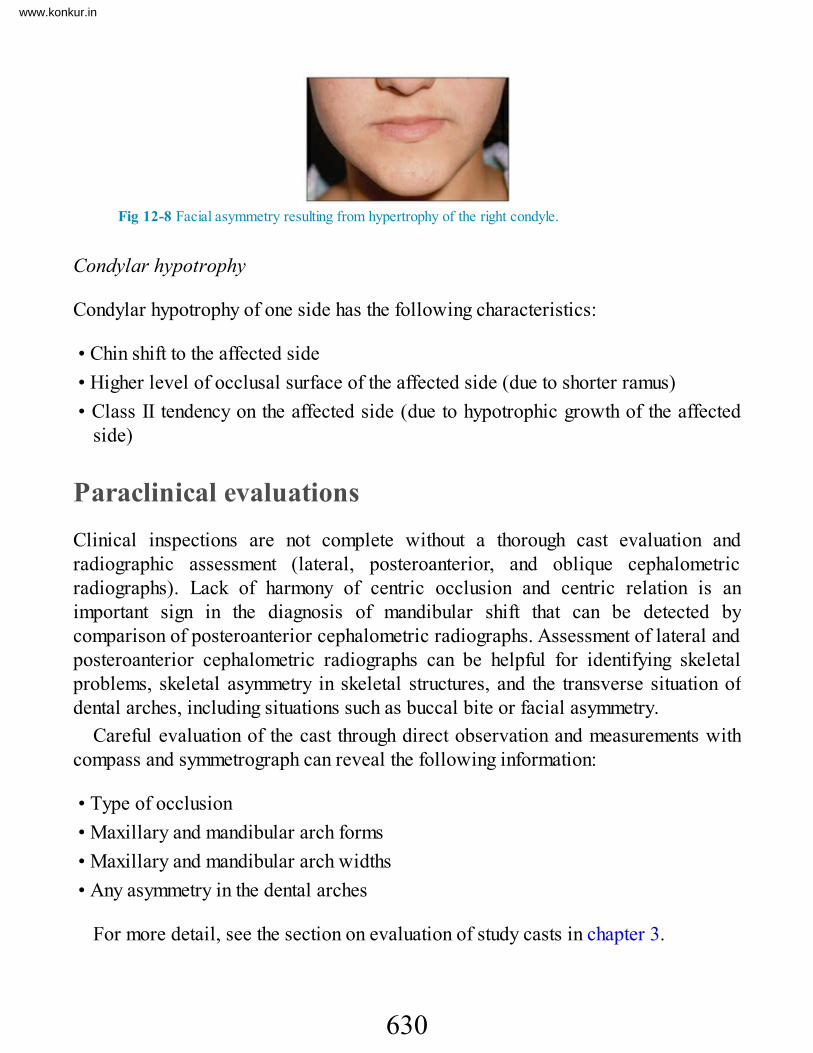
Fig 12-8 Facial asymmetry resulting from hypertrophy of the right condyle.
Condylar hypotrophy
Condylar hypotrophy of one side has the following characteristics:
• Chin shift to the affected side• Higher level of occlusal surface of the affected side (due to shorter ramus)• Class II tendency on the affected side (due to hypotrophic growth of the affected
side)
Paraclinical evaluationsClinical inspections are not complete without a thorough cast evaluation andradiographic assessment (lateral, posteroanterior, and oblique cephalometricradiographs). Lack of harmony of centric occlusion and centric relation is animportant sign in the diagnosis of mandibular shift that can be detected bycomparison of posteroanterior cephalometric radiographs. Assessment of lateral andposteroanterior cephalometric radiographs can be helpful for identifying skeletalproblems, skeletal asymmetry in skeletal structures, and the transverse situation ofdental arches, including situations such as buccal bite or facial asymmetry.
Careful evaluation of the cast through direct observation and measurements withcompass and symmetrograph can reveal the following information:
• Type of occlusion• Maxillary and mandibular arch forms• Maxillary and mandibular arch widths• Any asymmetry in the dental arches
For more detail, see the section on evaluation of study casts in chapter 3.
630
www.konkur.in

Early Treatment StrategiesEarly correction of posterior crossbite, especially in patients who exhibit amandibular shift, is recommended by almost all investigators. The best age forcorrection of posterior crossbites is the late primary dentition or early mixeddentition. The best treatment option depends on the answers to the followingquestions:
• Is the problem unilateral or bilateral?• Is the problem dental or skeletal?• Is the maxillary or mandibular arch involved or are both arches involved?• Is the problem the result of overexpansion or constriction of the arch?• Is the treatment of choice slow expansion or rapid expansion?
After careful clarification of the problem, the clinician can determine themechanical design and type of appliance that should be applied. A variety of fixedand removable expanders with different kinds of functions, such as rapid expansionor slow expansion, are available.
Another important consideration before correction of posterior crossbites is todetermine the patient’s vertical status (vertical or horizontal growth pattern);posterior expansion usually causes some extrusion of the posterior tooth segment,bite opening, and mandibular clockwise rotation. To overcome this problem, theappliance must be designed with posterior occlusal coverage to prevent posteriorextrusion. In patients with deep bite or a horizontal growth pattern, palatal expansionnot only can correct the posterior crossbite problem but also can reduce theoverbite. In contrast, in patients with open bite or open bite tendency, posteriorexpansion without control of the vertical dimension, such as occlusal coverage, canworsen the vertical problem.
Appliance therapyDepending on the type of problem, the age of the patient, and the patient’scompliance, the appliances selected for treatment of posterior crossbite problemscan be fixed or removable, rapid or slow expanders.
Fixed expanders
631
www.konkur.in

Fixed expanders available for rapid or slow expansion include the W-arch, quadhelix, Haas, and hyrax expanders, among others.
W-arch. The W-arch, or Porter, appliance is a fixed expander; it is efficient for thecorrection of posterior crossbites and is ideal for bilateral expansion. It includestwo bands for the permanent first molars or any distal teeth in the arch (primary orpermanent) and a 0.036- or 0.040-inch stainless steel archwire soldered to thebands. The arch should be free of tissue to prevent tissue impingement (Figs 12-9and 12-10).
Fig 12-9 W-arch expander. (Courtesy of Great Lakes Orthodontics.)
Fig 12-10 W-arch fixed palatal expander in position. A—Activation in the center of the palatalbow expands the molars; B—activation in the U-loop, close to the bands, expands the buccalsegments.
The W-arch can be activated in the center of the palatal bow for expansion of themolars or on the U-loop close to the bands to activate the arms for expansion ofbuccal segments. Depending on the type of crossbite, the appliance can be activatedunilaterally or bilaterally. It should be activated about 3 to 4 mm before fixation.The appliance is activated approximately every 4 or 5 weeks until the cross-bite hasbeen corrected and then remains in the mouth, inactive, for 4 to 6 months to provideretention. The W-arch requires little patient cooperation and therefore has betterpatient compliance.
When used at an early age during the primary dentition, the appliance seems toprovide efficient palatal expansion or accelerate the rate of normal expansion of themidpalatal suture. In children with posterior crossbite associated with thumbsucking, the W-arch can simultaneously function as a habit controller. The W-arch is
632
www.konkur.in

a suitable expander in 3- to 5-year-old children, especially in situations whereactivation of a fixed expander, such as the Haas appliance, is difficult for the parent.
Quad helix expander. The quad helix is also a fixed expander; it is fabricated from0.036-inch stainless steel wires soldered to molar bands.The quad helix is moreflexible than the W-arch; because four helixes are incorporated in the arch, the rangeand springiness of the appliance are increased (Fig 12-11).The wire should contactthe teeth involved in the crossbite and should be slightly separated from the palate toprevent any soft tissue impingement. The distal helixes are about 2 to 3 mm distal tothe molar bands. Activation of the anterior helixes produces posterior expansion,while activation of the distal helixes produces buccal segment expansion.
Fig 12-11 Quad helix expander. A—Activation of the anterior helixes produces posteriorexpansion; B—activation of the posterior helixes expands the buccal segments.
Reactivation can be performed to some extent intraorally by bird beak pliers, butit is difficult to activate the appliance in the desired direction. It is recommendedthat the appliance be removed for correct activation every 6 to 8 weeks.
The quad helix is used to correct bilateral maxillary constriction in the primarydentition. However, through some modifications to the appliance and the eliminationof one extension arm, the quad helix can also work unilaterally. In situations ofsevere unilateral deformity, a cross elastic from the mandibular opposing tooth canbe added to quad helix therapy, especially if teeth in both arches contribute to theproblem.
In some instances, by extension of the arms to include the canine, lateral incisors,and central incisors, anterior crossbites can be also corrected.
The combination of a posterior crossbite and a finger-sucking habit is the bestindication for this appliance because the anterior helix can effectively serve as areminder to the patient to stop the finger sucking habit.
To achieve tongue control, a tongue crib can be added to the anterior bow (Fig12-12).
633
www.konkur.in

Fig 12-12 Combination quad helix and tongue guard.
Arnold expander. The Arnold expander is a fixed expander consisting of two molarbands on the permanent first molars with two soldered bars (0.040-inch stainlesssteel wire) touching the primary molars or premolars, a 0.040- or 0.045-inch tubesoldered to one side, and a wire soldered to the other side with push coil to expandthe anterior segment (Fig 12-13). This appliance has good results for anteriorexpansion.
Fig 12-13 Arnold expander.
Haas expander. The Haas expander is also a fixed expander but is attached to boththe left and right buccal segments of the arch as anchorage and has a screwembedded in the palatal acrylic resin. The screw can be activated once or twice aday; this rapid force, dual-side anchorage, and expansion force will open the palatalsuture (Fig 12-14).
Fig 12-14 Haas rapid palatal expander.
Rapid palatal expansion is a specific type of expansion that can increase thepalatal width skeletally. Rapid palatal expansion has several indications inorthodontic and orthopedic treatments, such as skeletal crossbite and constricted
634
www.konkur.in

maxilla, maxillary protraction, and maxillary deficiency. This type of expander hasbeen used during the primary or early mixed dentition with good results.
Hyrax expander. The Hyrax expander is also a type of fixed rapid expander, but thescrew is not embedded in acrylic resin and is separated from the palate. Thus, theHyrax is more hygienic than the Haas expander (Fig 12-15).
Fig 12-15 Hyrax rapid palatal expander.
Rapid expander with occlusal coverage. It has been established that expansion ofposterior segments of the arch reduces anterior overbite; therefore, transverseexpansion must be applied cautiously in patients with open bite tendency andvertical problems. One tactic to avoid this outcome is occlusal coverage of theposterior segment.
Figure 12-16 shows an example of this type of expander, which the author hasused in the past. It is a combination of the Haas appliance and bite block acrylicresin that covers the occlusal surfaces and is cemented to the buccal segments.
Fig 12-16 Rapid palatal expander with occlusal coverage.
Bonded expander. The bonded expander was introduced by McNamara.15 Thisappliance is a combination of Hyrax expander and two acrylic resin parts coveringthe buccal segments (Fig 12-17).
635
www.konkur.in

Fig 12-17 Bonded rapid palatal expander.
Removable expanders
Removable expanders are also available for palatal and mandibular expansion.Removable expanders are a slow type of expander and usually are applied for dentalor dentoalveolar expansion, not palatal expansion (Fig 12-18). In slow expansion,the screw is opened every 3 to 5 days, in contrast to rapid expanders, which areopened once or twice a day.
Fig 12-18 Removable Hawley slow expanders with jackscrew. (a) Without occlusal coverage.(b) With occlusal coverage.
Most posterior crossbites in the primary or mixed dentition that are associatedwith mandibular shift are caused by bilateral constriction of the maxillary arch.Hence, the removable expander is constructed with the screw on the center of theappliance. In true unilateral constriction, the jackscrew is placed asymmetrically,and the appliance is sectioned asymmetrically. This has the effect of putting moreteeth against fewer teeth and results in asymmetric movement.
The success of this type of expander depends on good construction and design andon the patient’s cooperation.
Case ReportsAs discussed earlier, posterior crossbites have many variations in morphology,etiology, and possible treatment tactics. Because these types of anomaly form and
636
www.konkur.in

develop during the primary and early mixed dentitions, the earliest intervention notonly is easier to accomplish but also provides more stable results and prevents manyconsequences of delayed correction. Delays in treatment can result in structuraldamage to the dentition and supporting tissue, temporomandibular joint dysfunction,and adverse effects on jaw growth.
The following cases present early intervention treatments carried out at differentstages of the dentition with different tactics for different kinds of crossbite.
Case 12-1A 7-year-old boy presented with a posterior left-side crossbite and mandibular shift. The first etiologicfactor was diagnosed as a prematurity present between the maxillary and mandibular primary leftcanines (Figs 12-19a and 12-19b).
Treatment:Treatment was started with occlusal equilibration and grinding of the buccal surface of the mandibularprimary canine and palatal surface of the maxillary primary canine on the left side. The result was animmediate change in tooth relationships, mandibular shift, and dental midlines (Figs 12-19c and 12-19d).
Fig 12-19 Management of posterior crossbite and mandibular shift in a 7-year-old boy. Theprimary etiologic factor was premature contact between the maxillary and mandibular canines onthe left side. (a and b) Pretreatment occlusion. (c and d) Immediate improvement in relationshipsafter occlusal equilibration and selective grinding.
Note: When unilateral posterior crossbite and mandibular shift are the result ofpremature contact and the problem is not corrected at an early age, the situation maycause a strong habitual shift. In this situation, because the child’s habit is sustained,the equilibration technique alone cannot solve the problem. In addition to occlusalequilibration, slight bite opening with composite resin or acrylic resin occlusalcoverage, with gradual reduction of the material, can help. Otherwise, an expansionprocedure might be the best option.
637
www.konkur.in

Case 12-2A 5-year-old boy in the primary dentition presented with right-side complete posterior crossbite andmandibular shift. In centric occlusion, the mandibular midline shifted to the right. He had a Class Iocclusion on the left side and Class II malocclusion on the right side (Figs 12-20a to 12-20c).
The study cast revealed symmetric maxillary and mandibular arches, and clinical evaluation at centricrelation indicated slight bilateral constriction of the maxillary arch.
Treatment:Treatment was initiated with a fixed Haas expander with four bands on the primary second molars andprimary canines. The appliance was expanded once a day by the patient’s mother, and expansion wascompleted after 2 weeks. Figures 12-20d to 12-20f illustrate the appliance and changes that took placeduring activation. The patient experienced correction of crossbite and developed some spacing betweenthe incisors that was not present before.
The appliance was kept in place for 3 months to provide retention. As a result of treatment, thecrossbite was corrected, there was no more shift, midlines were coincident, and spacing remained tofacilitate transition of the anterior teeth (Figs 12-20g and 12-20h).
Fig 12-20 Management of complete posterior crossbite of the right side and mandibular shift in a5-year-old boy. (a to c) Pretreatment occlusion. (arrows) Midline deviation. (d and e) Occlusionduring use of the Haas rapid palatal expander. ( f ) Occlusion 2 weeks after completion ofexpansion; the midline deviation is almost corrected (arrows), and interincisal spacing hasdeveloped. (g and h) Posttreatment occlusion 3 months after expansion. (arrows) Correcteddental midlines.
Case 12-3A 10-year-old girl in the middle mixed dentition presented with a complete anterior and left posteriorcross-bite. She exhibited a severe mandibular shift. The mandibular dentition was good, but there wassome bony recession of mandibular central incisors because of the traumatic occlusion. The maxillary
638
www.konkur.in

incisors show minor crowding and retroclination (Figs 12-21a to 12-21c).Careful cast assessment and clinical evaluation of the patient’s occlusion at centric relation clarified
that retroclination of three maxillary incisors (both central incisors and the left lateral incisor) caused asevere mandibular shift to the left and created the complete left posterior crossbite and anteriorcrossbite in centric occlusion. Assessment of the patient’s occlusion in centric relation indicated thatthere was no maxillary arch constriction (Figs 12-21d to 12-21g).
Treatment:Because of the maxillary incisor retroclination and minor crowding, treatment was limited to proclinationof the incisor and correction of the anterior crossbite.
Treatment involved 2 × 4 maxillary bonding and composite resin on the occlusal surface of themandibular molars to disocclude the incisors (Figs 12-21h to 12-21j).
Fig 12-21 Management of complete anterior and left posterior crossbite in a 10-year-old girl in themixed dentition. There was a severe mandibular shift. (a to c) Pretreatment occlusion in centricocclusion. C—primary canines; (arrows) midline deviation. (d to f) Pretreatment occlusion incentric relation. (g) Retroclination of both central incisors and the left lateral incisor. (h to j)Posttreatment occlusion after proclination of the maxillary incisors.
Case 12-4A 5-year, 6-month-old girl in the primary dentition presented with left posterior crossbite and mandibular
639
www.konkur.in

shift. The midlines were not coincident in centric occlusion (Figs 12-22a and 12-22b).
Treatment:A jackscrew slow expander with occlusal coverage was used (Fig 12-22c). When the expander was inplace and the patient occluded on the acrylic resin, there was no midline shift in centric occlusion (Figs12-22d and 12-22e).
After 6 weeks of active treatment (opening the appliance every 3 days), the patient lost theappliance, but the problem was completely corrected (no crossbite and no shift). A 4-week trial withoutthe appliance was decided upon. Figures 12-22f and 12-22g show the occlusion after 6 months withoutthe appliance. Figures 12-22h and 12-22i show the occlusion 1 year later, after all permanent firstmolars, mandibular incisors, and maxillary central incisors had erupted. Figures 12-22j to 12-22l showsthe occlusion 3 years after treatment.
Fig 12-22 Management of posterior left crossbite and mandibular crossbite in a 5-year, 6-month-old girl. (a and b) Pretreatment occlusion. (arrows) Midlines in centric occlusion. (c) Jackscrewslow expander with occlusal coverage. (d and e) Centric occlusion with expander in place; thereis no midline shift (arrows). (f and g) Posttreatment occlusion 6 months after active treatment. (hand i) Posttreatment occlusion 1 year later. (j to l) Posttreatment occlusion 3 years aftertreatment.
Case 12-5
640
www.konkur.in

A 7-year, 4-month-old boy presented with left-side posterior crossbite, mandibular shift, and spacedeficiency in the maxillary anterior segment (Figs 12-23a to 12-23d).
Treatment:The treatment modality in this case was a Haas rapid palatal expander, on which the screw was openedonce a day. The expander was designed with only two bands on the permanent first molars and ajackscrew embedded in acrylic resin, together with two 0.032-inch stainless steel bars adapted to theprimary molars and canines and bonded to the palatal side of these teeth with composite resin.
Figures 12-23e to 12-23g show the occlusion 3 months after completion of expansion and at the timeof removal of the expander. The results of expansion were correction of the crossbite, coincidentmidlines, elimination of the shift, and creation of space for the anterior segment. All incisors haderupted.
Fig 12-23 Management of posterior left crossbite, mandibular shift, and space deficiency in a 7-year, 4-month-old boy. (a and b) Pretreatment occlusion in centric occlusion. (arrows) Midlinedeviation. (c) Pretreatment view revealing space deficiency in the maxillary anterior region. (d)Pretreatment panoramic radiograph. (e to g) Occlusion at the time of expander removal, 3 monthsafter completion of expansion.
Case 12-6A 5-year, 8-month-old boy in the primary dentition presented with posterior crossbite on the left side,mandibular shift, and a mandibular midline that was shifted 4 mm to the left (Figs 12-24a to 12-24c). Hehad class I spacing according to the Baume classification.27
Treatment:Because of the patient’s medical conditions (attention deficit–hyperactivity disorder, asthma, and poororal hygiene), the treatment plan included use of a W-arch expander. The appliance consisted of twomolar bands on the maxillary primary second molars and a W-shaped 0.036-inch stainless steel wire
641
www.konkur.in

adapted in such a way as to have more activation on the left side (Figs 12-24d and 12-24e).Figures 12-24f to 12-24h illustrate the occlusion 3 months after removal of the expander and after
placement of crowns and restorations.
Fig 12-24 Management of posterior left crossbite, mandibular shift, and a 4-mm midline deviationin a 5-year, 8-month-old boy with class I Baume spacing. (a to c) Pretreatment occlusion.(arrows) Midline deviation. (d) W-arch appliance used for expansion. (e) Appliance in place. (f toh ) Posttreatment occlusion 3 months after removal of the appliance and after placement ofrestorations and crowns. (arrows) Corrected midlines.
Case 12-7A 4-year, 9-month-old girl presented with a left-side posterior crossbite and mandibular shift ( Figs 12-25a to 12-25c).
Treatment:The appliance used for this patient was a W-arch expander with 0.036-inch stainless steel wire (Fig 12-25d). The appliance was expanded 8 mm when placed and activated only once, 4 weeks after insertion.
After completion of expansion and slight overcorrection, the appliance was left in place for 2 monthsfor retention. Figures 12-25e and 12-25f show the results 3 months after the end of retention.
642
www.konkur.in

Fig 12-25 Management of posterior left crossbite and mandibular shift in a 4-year, 9-month-oldgirl. (a to c) Pretreatment occlusion. (d) W-arch expander in place. (e and f) Occlusion 3 monthsafter the end of retention.
Case 12-8A 14-year, 6-month-old boy presented with Class II division 1 bilateral complete buccal bite (Brodiesyndrome). The patient’s family had limited financial resources, and their chief concern was the factthat the boy could not chew properly. Besides the transverse problem, this patient had some otheranomalies that made the situation more complicated. These included a Class II sagittal problem withsevere overjet and 90% impinging overbite; missing maxillary and mandibular permanent left firstmolars; microdontia, which was causing severe interdental spacing; and poor interdigitation andintercuspation (Figs 12-26a to 12-26f).
The result of all these anomalies was a collapsed mandibular arch with spacing within the maxillaryarch that made the occlusion and dentofacial component not only unesthetic but also dysfunctional.
Treatment:Because of the family’s financial situation, no surgical, prosthetic, or implant restoration could beconsidered as part of the treatment plan. Therefore, the problems had to be solved orthodontically. Thetreatment was completed in a number of steps:
1. Maxillary anterior bite plane to disocclude the locked mandibular arch within the maxilla2. Mandibular heavy semiremovable lingual arch to expand the constricted mandibular molars
gradually3. Maxillary constricting cervical headgear to constrict the maxillary molars and distalize them for
Class II correction (Fig 12-26g)4. Full maxillary and mandibular bonding as soon as molar occlusal contact was established5. Retraction of maxillary buccal segments and continuing headgear use as anchorage6. Consolidation of maxillary buccal segments between molars and premolar retraction7. After retraction of maxillary molars and premolars, heavy class II mechanics to retract the
maxillary canines and mesialize the posterior mandibular segment while transverse relationshipswere controlled
8. Maxillary incisor retraction and intrusion to correct the deep bite and severe overjet
643
www.konkur.in

Figures 12-26h to 12-26j illustrate the posttreatment occlusion after closure of the maxillary andmandibular arches. All the mechanical effort was focused on narrowing the maxillary arch whilesimultaneously widening the mandibular arch, especially in the molar areas.
Sagittal and transverse changes in the dentition were accomplished in this treatment. Thepretreatment width at the maxillary first premolars was 35 mm; after treatment, the width was 30 mm(Fig 12-26k). The maxillary intermolar width was 40 mm pretreatment and 35 mm after treatment (Fig12-26l). The width at the mandibular first premolars changed from 30 to 29 mm (Fig 12-26m). Thepretreatment mandibular intermolar width was 35 mm; it remained the same after treatment, despite thesevere mesial movement that was done because of the missing first molar and significant interdentalspacing (Fig 12-26n).
Significant dentoskeletal changes were achieved through this treatment (Figs 12-26o to 12-26q):
• Correction of severe bilateral buccal bite• Correction of Class II malocclusion• Closure of the severe interdental spacing caused by missing teeth and microdontia• Significant change in lower facial height (correction of mandibular collapse) from 53.7% to 60.5%• Change in point A–nasion–point B angle from 4.2 to 3.6 degrees• Correction of maxillomandibular dental protrusion (increase in interincisal angle from 111.4 to 136.0
degrees)
Fig 12-26 Management of bilateral complete buccal bite (Brodie syndrome) in a 14-year, 6-month-old boy with Class II division 1 malocclusion. (a to c) Pretreatment occlusion.
644
www.konkur.in
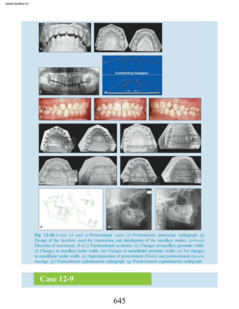
Fig 12-26 (cont) (d and e) Pretreatment casts. ( f ) Pretreatment panoramic radiograph. (g)Design of the facebow used for constriction and distalization of the maxillary molars. (arrows)Direction of movement. (h to j) Posttreatment occlusion. (k) Changes in maxillary premolar width.(l) Changes in maxillary molar width. (m) Changes in mandibular premolar width. (n) No changesin mandibular molar width. (o) Superimposition of pretreatment (black) and posttreatment (green)tracings. (p) Pretreatment cephalometric radiograph. (q) Posttreatment cephalometric radiograph.
Case 12-9
645
www.konkur.in

A 13-year-old boy presented with a Class II division 1 malocclusion, unilateral buccal bite on the leftside, severe overjet (16.5 mm), and impinging deep bite. Study casts indicated that the buccal bite wasdue to asymmetry in both the maxillary and mandibular left posterior segments, that is, buccoversion ofthe maxillary left segment and linguoversion of the mandibular left segment (Figs 12-27a to 12-27d).
Treatment:Treatment was started with a maxillary anterior bite plate to open the locked posterior left segment. Amandibular lingual arch was placed for expansion of the mandibular left segment, and constrictingheadgear was used for distalization of the maxillary left and right molars and constriction of themaxillary left segment. Treatment followed the same steps described in case 12-8, including sagittalchanges of the posterior segments to Class I molar, premolar, and canine relationships and finishing withmaxillary anterior retraction.
Because the transverse problems of this patient were unilateral, all of the mechanical effort wasfocused on constricting only the maxillary left segment and expanding only the mandibular left segment.This goal was achieved with placement of continuous cross-arch elastics on the left side.
Significant changes were achieved through this treatment procedure (Figs 12-27e to 12-27j). Thesechanges included correction of the unilateral buccal bite; correction of the Class II malocclusion andreduction of overjet, from 16.5 to 3.5 mm; correction of deep bite; change in the point A–nasion–point Bangle, from 12.3 to 4.5 degrees; and significant changes in the angle of convexity and the angle of pointA–point B to the facial plane.
Fig 12-27 Management of unilateral buccal bite on the left side, severe overjet, and impingingdeep bite in a 13-year-old boy with a Class II division 1 malocclusion. (a to d) Pretreatment castsrevealing overexpansion of maxillary left segment and constriction of the mandibular left segment.(e to h) Posttreatment occlusion. (i) Pretreatment cephalometric radiograph. ( j ) Posttreatment
646
www.konkur.in

cephalometric radiograph.
Summary
• Posterior crossbite is one of the most prevalent malocclusions found duringthe primary and early mixed dentitions. The most common form of posteriorcrossbite is a unilateral crossbite with a functional shift of the mandibletoward the crossbite side.
• Posterior crossbites are seldom self-correcting; delayed treatment andasymmetric muscle strain in the orofacial structures can have adverseeffects on the temporomandibular joint, the masticatory system, and skeletalgrowth changes.
• Maxillary arch constriction, which is usually present in patients with thiscondition, creates insufficient maxillary arch width and results in anteriorcrowding and sometimes canine impaction.
• Early treatment is advised to create an environment that facilitates normalocclusal development.
• The most common causes of posterior crossbite are digit or pacifier suckinghabits, impaired nasal breathing, atypical swallowing patterns, and lowtongue position. Therefore, evaluation during the primary and mixeddentitions is recommended. These facts emphasize the importantresponsibility that practitioners have in careful examination for and earlyrecognition of these factors, which are interacting in young patients during acritical stage of occlusal development.
• Almost all investigators studying these anomalies recommend earlyintervention and correction of posterior cross-bites. Correction of posteriorcrossbite affecting the permanent dentition is much more complicated and insome cases may require surgery.
• The best time for correction of posterior crossbite is during the late primaryor early mixed dentition. The expansion procedure is easy and fast andprovides good, stable results.
• Posterior crossbite has many etiologic factors and several differentmorphologic characteristics; thus, differential diagnosis is an essential partof treatment planning.
• Posterior crossbite can be dental, dentoalveolar, skeletal, or functional; eachof these types can be unilateral or bilateral. Posterior crossbite can also
647
www.konkur.in

result from other combinations of transverse malrelationship between themaxillary or mandibular dental arch, such as buccal bite, also called Brodiesyndrome, scissors bite, and palatal bite.
• Recognition of posterior crossbite is not difficult, and a simple clinicalevaluation of occlusion, study casts, and frontal cephalometric radiographscan enable the practitioner to easily identify the type of problem.
• Depending on the type of posterior crossbite, many treatment optionsincorporating fixed and removable appliances that offer slow or rapidpalatal expansion are available.
References1. Moyers R. Handbook of Orthodontics, ed 4. Chicago: Year Book Medical, 1988.2. Kennedy DB, Osepchook M. Unilateral posterior cross-bite with mandibular shift: A review. J Can Dent
Assoc 2005;71:569–573.3. Harper DL. A case report of a Brodie bite. Am J Orthod Dentofacial Orthop 1995;108:201–206.4. Kutin G, Hawes RR. Posterior cross-bites in the deciduous and mixed dentitions. Am J Orthod 1969;56:491–
504.5. Hesse KL, Artun J, Joondeph DR, Kennedy DB. Changes in condylar position and occlusion associated with
maxillary expansion for correction of functional unilateral posterior crossbite. Am J Orthod Dentofacial Orthop1997;111: 410–418.
6. Pinto AS, Buschang PH, Throckmorton GS, Chen P. Morphological and positional asymmetries of youngchildren with functional unilateral posterior crossbite. Am J Orthod Dentofacial Orthop 2001;120:513–520.
7. Myers DR, Barenie JT, Bell RA, Williamson EH. Condylar position in children with functional posterior cross-bite: Before and after crossbite correction. Pediatr Dent 1980;2:190–194.
8. Egermark-Ericksson I, Carlsson GE, Magnusson T, Thilander B. A longitudinal study on malocclusion inrelation to signs and symptoms of cranio-mandibular disorders in children and adolescents. Eur J Orthod1990;12:399–407.
9. Thilander B, Lennartsson B. A study of children with unilateral posterior crossbite, treated and untreated, in thedeciduous dentition—Occlusal and skeletal characteristics of significance in predicting the long-term outcome. JOrofacial Orthop 2002;63:371–383.
10. Schröder U, Schröder I. Early treatment of unilateral posterior crossbite in children with bilaterally contractedmaxillae. Eur J Orthod 1984;6:65–69.
11. Proffit WR, Fields HW Jr. Contemporary Orthodontics, ed 5. St Louis: Mosby, 2012.12. Subtelny JD. Oral respiration: Facial maldevelopment and corrective dentofacial orthopedics. Angle Orthod
1980;50:147–164.13. Kurol J, Berglund L. Longitudinal study and cost-benefit analysis of the effect of early treatment of posterior
cross-bites in the primary dentition. Eur J Orthod 1992;14:173–179.14. Linder-Aronson S. Adenoids. Their effect on mode of breathing and nasal airflow and their relationship to
characteristics of the facial skeleton and the dentition. A biometric, rhino-manometric and cephalometro-radiographic study on children with and without adenoids. Acta Otolaryngol Suppl 1970;265:1–132.
15. McNamara JA. Early intervention in the transverse dimension: Is it worth the effort? Am J OrthodDentofacial Or-thop 2002;121:572–574.
16. Allen D, Rebellato J, Sheats R, Ceron AM. Skeletal and dental contributions to posterior crossbites. Angle
648
www.konkur.in

Orthod 2003;73:515–524.17. Warren JJ, Bishara SE. Duration of nutritive and nonnutritive sucking behaviors and their effects on the
dental arches in the primary dentition. Am J Orthod Dentofacial Orthop 2002;121:347–356.18. Souki BQ, Pimenta GB, Souki MQ, Franco LP, Becker HM, Pinto JA. Prevalence of malocclusion among
mouth breathing children: Do expectations meet reality? Int J Pediatr Otorhinolaryngol 2009;73:767–773.19. Oulis CJ, Vadiakas GP, Ekonomides J, Dratsa J. The effect of hypertrophic adenoids and tonsils on the
development of posterior crossbite and oral habits. J Clin Pediatr Dent 1994;18:197–201.20. Ogaard B, Larsson E, Lindsten R. The effect of sucking habits, cohort, sex, intercanine widths, and breast or
bottle feeding on posterior crossbite in Norwegian and Swedish 3-year-old children. Am J Orthod DentofacialOrthop 1994;106:161–166.
21. Adair SM, Milano M, Lorenzo I, Russell C. Effects of current and former pacifier use on the dentition of 24-to 59-month old children. Pediatr Dent 1995;17:437–444.
22. Infante PF. An epidemiologic study of finger habits in preschool children as related to malocclusion,socioeconomic status, race, sex, and size of community. ASDC J Dent Child 1976;43:33–38.
23. Hanson M, Barnard L, Case J. Tongue thrust in preschool children. 2. Dental occlusal patterns. Am J Orthod1970;57:15–22.
24. Primozic J, Richmond S, Kau CH, Zhurov A, Ovsenik M. Three-dimensional evaluation of early crossbitecorrection: A longitudinal study. Eur J Orthod 2013;35:7–13.
25. Guerrero CA, Bell WH, Contasti GI, Rodriguez AM. Mandibular widening by intraoral distractionosteogenesis. Br J Oral Maxillofac Surg 1997;35:383–392.
26. Legan HL. Orthodontic planning and biomechanics for transverse distraction osteogenesis. Semin Orthod2001;7:160–168.
27. Baume LG. Physiological tooth migration and its significance for the development of occlusion. 1. Thebiogenetic course of the deciduous dentition. J Dent Res 1950;29:123–132.
649
www.konkur.in

Management of Vertical Problems (Open Bites and Deep Bites)
This chapter discusses common vertical problems that initiate and develop duringthe primary or early mixed dentition: (1) open bites associated with persistent oralhabits and other dysfunctions and (2) deep bites (dental deep bite and skeletal deepbite). These anomalies are not self-corrected and in some situations worsen in laterstages of the dentition; therefore, early detection and intervention are important.
Open BiteTreatment of skeletal open bite, or hyperdivergent phenotype malocclusion, andanterior open bite is one of the common difficulties facing orthodontists. They areespecially problematic during the permanent dentition and are one of the mostchallenging malocclusions to treat.
The intent of this book is to emphasize the advantages of early-age intervention forall malocclusions, including open bites that initiate and develop during the primaryand mixed dentitions. Open bite is a multifactorial phenomenon; recognition of theetiology plays an important role in diagnosis and treatment of this anomaly. The
650
www.konkur.in

problem can be diagnosed clinically and cephalometrically; however, diagnosisshould be viewed in the context of both skeletal and dental structures.
Hereditary factors influence the direction, form, and rate of skeletal growth; theycan produce abnormal growth patterns and incorrect jaw posture, resulting inskeletal open bite. Environmental factors during the primary and mixed dentitionscan also play an important role in developing open bites. Therefore, different kindsof open bite with different morphologic and etiologic characteristics must beaccurately classified for precise treatment planning. This requires experience andtraining.
Early recognition of the cause and proper intervention can prevent thedevelopment of abnormalities or at least reduce the severity of the problem later. Atthe present time, it is not possible to eliminate or prevent the influence of geneticfactors on dentoskeletal structure; however, with accurate examination andrecognition of the effect, practitioners might be able to prevent, modify, or at leastreduce the severity of the problem. Because environmental factors are mostlyrecognizable, recognition of and intervention in such influences at an early stage offormation can prevent development of open bite or at least guide the abnormalitytoward normality. The following are the major common local or environmentalfactors that play a role in development of open bite at early age:
• Digit sucking• Abnormal tongue size, posture, and function• Abnormal neuromuscular function• Enlarged lymphatic tissue causing mouth breathing and postural alteration that
induces dental and skeletal modificationsThe etiology and mechanism of the factors causing open bite are discussed in
detail in chapter 6. The current chapter presents the differential diagnosis, treatmentstrategies, and techniques for early correction of open bite during the primary andmixed dentitions.
Pediatric dentists and general practitioners who see children at an early age havegreater responsibility for the detection of and intervention in these developinganomalies. Differential diagnosis and treatment of open bite malocclusion aresometimes challenging. However, successful identification of the etiology throughcareful clinical and cephalometric evaluation as well as some general history of theproblem, improve the chances of treatment success. A brief review of the problemand its etiologic factors will be provided here, for the purpose of recognition and
651
www.konkur.in

proper intervention.Depending on the etiologic factors and the complexity of the abnormality,
treatment for open bite ranges from observation to simple habit control to complexsurgical procedures.
EtiologyComponents of vertical problems must be diagnosed and classified under twocontexts: skeletal structures and dental structures. Skeletal open bite usually has agenetic background and interacts with environmental causes. It can develop to amore complex dentoskeletal open bite and in some cases, especially if not resolvedby the end of the mixed dentition years, will require complicated orthodontic andorthopedic treatment, with or without surgical intervention.
The first step toward early treatment is recognition of the problem, and the bestway to recognize the problem is to understand and differentiate between thecharacteristics of different types of open bite. No single factor can be considered thecause of open bite. The most common local causes of this anomaly are thumb andfinger sucking, tongue problems (such as abnormal function, size, position, andposture), mouth breathing as a result of enlarged lymphatic tissue or other airwayobstruction, and lip dysfunction.
Finger sucking
Depending on the type of sucking and the position of the fingers, sucking can affectthe occlusion by different mechanisms. Finger force directed to the anterior teeth andthe incisive bone can cause dental and dentoalveolar protrusion and open bite,prevent eruption of anterior teeth, and lead to underdevelopment of the alveolarprocess; it can also cause posterior crossbite. A prolonged habit and absence ofcontact in the posterior teeth can also cause overeruption of the posterior teeth andeven clockwise rotation of the mandible (Fig 13-1).
Fig 13-1 Dentoskeletal open bite and posterior crossbite caused by thumb sucking.
652
www.konkur.in

Abnormal tongue force
The tongue can cause anterior open bite through different mechanisms, such astongue size, tongue position, tongue posture, and abnormal tongue function.Depending on the size of the tongue, macroglossia can have different destructiveeffects on the occlusion, such as interdental spacing, Class III malocclusion, andopen bite. Severe enlargement of the tongue can also cause functional difficulties inspeaking, eating, swallowing, and sleeping. This condition can be seen inindividuals with certain inherited or congenital disorders, such as neonatalhypothyroidism, Down syndrome, and acromegaly.
Abnormal position of the tongue is another condition that can cause dentoskeletaldeformity. This situation is not the result of the size of the tongue; rather the tongue ispositioned more anteriorly as a subconscious reaction to large tonsils or because ofmouth breathing in response to nasal obstruction or enlarged adenoid tissue (Fig 13-2).
Fig 13-2 (a to c) Dentoskeletal open bite caused by abnormal size, position, or function of thetongue.
Oral respiration
During mouth breathing, no matter what the cause, the mouth is open, the mandible ispostured inferiorly, and the tongue is protruded between the anterior teeth andresting against the oral floor. This situation causes postural alteration and inducesdental and skeletal modifications similar to those caused by thumb sucking.
Mouth breathing can also cause excessive eruption of the posterior teeth andmaxillary constriction, change head posture, cause an increase in the verticaldimension of the face, and result in development of anterior dentoskeletal open bite.
Genetic factors
Genetic factors, either alone or in combination with local factors, can disrupt thenormal skeletal growth pattern through different mechanisms, such as disturbance ofthe normal size, shape, and proportion of different parts of skeletal structures.
653
www.konkur.in

Development of skeletal abnormalities causes several skeletal malocclusions,including open bite, which mostly require orthosurgical management.1
Tongue thrust is an abnormal tongue function that usually develops secondary tothumb sucking and then maintains or increases the open bite that was produced by thefinger, even after the finger-sucking habit has stopped. During correction of openbite and application of different treatment mechanics, abnormal tongue function mustbe controlled.
Morphologic characteristicsFor practical purposes and simplification of differential diagnosis, the open bitesyndrome can be classified into two types: dental open bite and skeletal open bite.
Dental open bite
Simple dental open bite is considered an open bite without skeletal abnormalities,with the following characteristics:
• Normal craniofacial pattern• Normal skeletal jaw relationships• Previous history of finger sucking• Proclined incisors• Anterior tongue thrust• Undererupted incisors• Normal or slightly excessive molar height• Normal inclination of posterior dentition• Divergent occlusal plane• No gummy smile• No vertical excess or posterior tipping of the maxilla
Prolonged continuation of environmental factors and failure to initiate earlytreatment of simple dental open bite can produce more complicated effects, such asmolar overeruption, mandibular clockwise rotation, and subsequently skeletaldefects.
654
www.konkur.in

Skeletal open bite
True skeletal open bite abnormalities are usually caused by genetics, prolongedenvironmental factors, or a combination of both. They consist of many unfavorablegrowth patterns and skeletal disharmonies that must be distinguished by carefulclinical and cephalometric evaluation before treatment planning.
The following are the important structural defects that can be detected by carefulclinical and paraclinical evaluation of patients with skeletal open bite or verticaldivergent face:
• Steep mandibular plane• Retrognathic mandible• Obtuse gonial angle• Short ramal height• Short anterior cranial base• Short posterior cranial base• Obtuse y-axis angle• Obtuse saddle angle• Obtuse articular angle• Obtuse occlusal plane–mandibular plane angle• Obtuse mandibular plane to palatal plane angle• Counterclockwise rotation of the palate• Increased lower facial height• Decreased upper facial height• Decreased posterior facial height• Steep anterior cranial base
Differential diagnosisSimple dental open bite is a common abnormality that occurs during the primary orearly mixed dentition, caused by environmental problems such as abnormal habits,the type of nutrition, or enlargement of lymphatic tissues. Untreated open bite notonly increases in severity over time but also can cause other dentoskeletal damagethat can be more complicated to manage at a later age. These consequences are not
655
www.konkur.in

confined to undereruption of anterior teeth; open bite can cause overeruption of themaxillary or mandibular molars or both, and in severe cases it can cause skeletaldeformities such as clockwise rotation of the mandible or counterclockwise rotationof the maxilla.
For early intervention in simple anterior dental open bite, the first step beforemechanotherapy is recognition of the type of open bite and the structure involved.Therefore, besides the clinical and paraclinical evaluations that always must beperformed for assessment of all malocclusions, special attention to cephalometricanalysis of the vertical dimension is important in diagnosis of open bitemalocclusions. For differentiation of dental open bite and to verify whether the openbite is due to anterior undereruption or posterior overeruption, the authorrecommends a simple cephalometric evaluation of the occlusal plane relative tomandibular plane–palatal plane angle. In normal occlusion, the mandibular plane–palatal plane angle is about 30 degrees, and the occlusal plane bisects this anglealmost equally. In patients with open bite, variations in this configuration indicatethe cause of the open bite (Fig 13-3).
Fig 13-3 Cephalometric evaluation of mandibular plane– palatal plane angle to determine thecause of open bite. In normal occlusion, this measurement is about 30 degrees, and the occlusalplane bisects this angle almost equally. (a ) Normal molar position with undereruption of theincisors. (b) Overeruption of the maxillary molars. (c) Overeruption of the mandibular molars.
Treatment strategies during the permanent dentitionAs already discussed, early detection and interception of the developing anomaly atits early stage of development (primary or mixed dentition) is easier and providesmore stable results. However, for the purposes of comparison, the following is abrief review of open bite correction in adolescents and adults.
Sustained open bite malocclusion during the late mixed dentition or permanentdentition is not self-correcting, and orthodontic treatment is almost alwayschallenging; this is one of the most difficult malocclusions to treat orthodontically.
656
www.konkur.in

Treatment approaches in this period depend on the age of the patient and the severityof the problem.
Habit control and lip seal
Habit control, lip seal, and swallowing exercises can have a positive effect incamouflage treatment, even during the permanent dentition. These methods facilitateorthodontic tooth movement against the tongue force, which can be achieved by adevice such as a tongue guard.
Control of vertical growth
Control of vertical dental eruption and posterior dentoalveolar development duringcamouflage treatment can also be helpful in correction of the permanent dentition.This is achieved by use of a lower holding arch and a transpalatal arch.
Orthodontic camouflage
Orthodontic camouflage consists of only orthodontic procedures, with special tacticsdepending on the morphologic and anatomical structures involved in thismalocclusion. These approaches include posterior intrusion, anterior extrusion, or acombination of both. This type of treatment can be combined with tooth extraction ifappropriate.
Many appliances for correction of anterior open bite in the permanent dentitionhave been proposed, including a bite block; an extrusion or intrusion arch; amultiloop edgewise archwire2; functional appliances3; and extraoral forces such ashigh-pull headgear, vertical chin cup (which can have some positive effect in theearly permanent dentition), titanium screw anchorage,4 titanium miniplates fixedbilaterally to the zygomatic buttress area,5 and a magnetic active vertical correctordevice.6
Orthognathic surgery
True skeletal open bites cannot be treated by orthodontic camouflage alone andrequire a combination of orthodontics and surgical treatment,1 such as surgicalimpaction of the maxillary posterior segment, which is considered to be the mosteffective treatment option for correction of skeletal open bites in adult patients.
657
www.konkur.in

Early Treatment Strategies for Open Bite
Vertical growth of the face, as shown by Enlow, 7 is the last dimension of thedentofacial structure to be completed during early postnatal growth and thetransitional dentition. As Carlson8 pointed out, only 50% of growth in the midfaceand the mandible is complete by the age of 8 to 10 years. Therefore, early treatmentand control of vertical growth during this period is more efficient.
Harris and Johnson,9 in a longitudinal sibling analysis, concluded that mostocclusal variability is acquired, not inherited.
As mentioned earlier, the most common local causes of anterior open bites arethumb and finger sucking, mouth breathing in response to enlarged lymphatic tissueor other airway obstruction, lip dysfunction, and tongue problems (such as abnormalfunction, size, position, and posture). Anterior open bites caused by local factors areoften accompanied by a downward rotation of the mandible, excessive eruption ofthe posterior teeth, or undereruption of the anterior teeth. The main strategy of earlyintervention is to overcome these abnormalities by eliminating or preventing thecause, as well as to correct or guide the affected sites, through the following tactics:
• Elimination or control of the etiologic factors that are disturbing incisor eruptionand alveolar growth (such as finger sucking, tongue thrust, and lip dysfunction)
• Prevention or correction of maxillary and mandibular molar overeruption• Prevention, control, or guidance of clockwise rotation of the mandible• Incisor retraction to “close the drawbridge” between separated maxillary and
mandibular incisors• Achievement of an increase in the posterior facial height–anterior facial height
ratio
Different fixed and removable appliances have been used to prevent and controlthe force of finger sucking and abnormal tongue thrusting in children (see chapter 6).The purpose of this discussion is to clarify the mechanotherapy and tactics that canbe efficient in early treatment of anterior open bite.
Thumb or finger sucking is perhaps the most common type of non-nutritive oralhabit present in children. Depending on the severity of the problem and type of fingersucking, this habit can cause severe dentoskeletal defects, including open bite(ranging from a simple dental open bite to a severe skeletal open bite), posteriorcrossbite, overeruption of molars, and mandibular rotation. Thus, treatment would
658
www.konkur.in

be more efficient if started at an earlier age.Placement of the tongue between the incisors has been sought as a causative factor
for anterior open bite for some years. Subtelny10 stated that abnormal tongue functionbetween incisors is a type of adaptation of the tongue to the open bite area formed byprevious finger sucking. Proffit11 showed that individuals who place the tongue tipforward when they swallow usually do not have more tongue force against the teeththan do those who keep the tongue tip back; in fact, tongue pressure may be lower.
Swallowing is not a learned behavior. Forward tongue placement is aphysiologic, subconscious function to fill the gap present between incisors duringswallowing. The lips are brought together, and the tongue is placed between theseparated anterior teeth to form an anterior seal to prevent food or liquids fromescaping. Correction of anterior open bite is more complicated and disturbing whenpersistent tongue activity is present. Resolving the problem is easier if it iscorrected at an earlier age; correction of open bite with persistent tongue thrust ismuch more difficult in the permanent dentition.
Depending on the etiologic factors and the complexity of the abnormality, earlyorthodontic treatment for open bite ranges from observation to simple habit controlto complex surgical procedures.
Classification of open biteTo simplify the treatment planning process for anterior open bite correction duringthe primary or early mixed dentition, the author classifies open bite into three typesof abnormality: type I, type II, and type III.
Type I simple dental open bite
Simple dental open bite is an anterior open bite in which some environmental factorhas affected anterior tooth eruption and/or vertical growth of the alveolar process;as a result, a gap has developed between the anterior teeth. The size of the gapdepends on the severity of the cause. In response to the open bite, abnormal tonguefunction has developed subconsciously to close the gap during swallowing, andtherefore continuous tongue force maintains or worsens the gap. In type I open bite,no skeletal discrepancy or molar overeruption is involved; only the incisors or thedentoalveolar process is underdeveloped.
Type II combined dental open bite
659
www.konkur.in

In type II anterior open bite, an abnormality was initially caused by someenvironmental factor plus some minor skeletal disharmony or molar overeruption. Inthese situations, sustained local factors or hereditary background can cause furtherdeformity to the dentition or basal bone, such as molar overeruption or slightmandibular rotation; therefore, signs of vertical problems are also present.
Type III dentoskeletal open bite
Type III open bite is an anterior open bite in which hereditary or congenital defectswith or without local factors have changed the growth pattern of the child andtherefore have changed the morphology and foundation of the dentoskeletalocclusion. This type of open bite is usually combined with some type of divergenceof dentoskeletal structures and can be clearly recognized by cephalometricevaluation. Early detection and proper intervention can solve the problem or at leastreduce the severity of the anomaly. Treatment during the permanent dentition is muchmore complicated and occasionally requires orthognathic surgery.
Treatment considerationsEarly intervention in open bite differs depending on the stage of dentition and thetype of open bite. Early treatment strategy for open bite malocclusion can be dividedinto three periods of dental development: primary dentition, mixed dentition, andpermanent dentition. The tactics for treatment of the permanent dentition werediscussed briefly earlier in the chapter. The following sections focus on earlyorthodontic intervention during the primary and mixed dentition periods.
Management during the primary dentition
Generally, active orthodontic treatment of open bite during the primary dentition isnot recommended, because most cases at this stage of the dentition involve localfactors such as pacifier or finger sucking or mouth breathing, which can be self-corrected after etiologic factors are eliminated. However, periodic observation bythe dentist or the awareness of the parents can be helpful.
If abnormal habits are persistent and severe enough to affect normal jaw growth,for example by preventing anterior dentoalveolar growth or causing mandibularrotation, the patient and the parents should be advised that these severe habits can becontrolled with a simple appliance such as a fixed tongue guard.
660
www.konkur.in

Management during the mixed dentition
After careful clinical and paraclinical evaluations of the problem are completed,treatment during the mixed dentition stage can be designed in one or two phases withor without orthognathic surgery. The pretreatment assessments should carefullyverify the following:
• Type of open bite (skeletal, dental, or combination)• Etiologic factors (whether the cause is still active)• Morphology of the malocclusion and affected structures• Skeletal age of the patient• Cooperation of the patient and parents
The best age for early intervention is during the early mixed dentition.
Tactics of Early Intervention During the Mixed DentitionThe main strategy of early intervention is to eliminate or prevent the cause of theabnormality behind the open bite by controlling or guiding the affected sites.
Depending on the stage of the dentition, etiologic factors, characteristics, and themorphology of the open bite, the following six tactics may be applied.
1. Habit controlControl of finger or tongue force as an early intervention is the first and mostimportant tactic in treatment of this anomaly. This tactic will facilitate anterior tootheruption, alveolar growth, and open bite correction. Several kinds of fixed orremovable appliances are available for this purpose. This tactic is very helpful intreatment of type I open bite, and if applied at the proper time such appliances cancorrect the problem without the need for further mechanotherapy (see cases 13-1 to13-3).
As Rakosi3 and Huang et al12 have shown, the tongue crib has been used widelyand successfully to stop thumb sucking and the abnormal tongue forces that produceand maintain anterior open bite.
661
www.konkur.in

2. Lip seal and swallowing exercisesTo establish normal neuromuscular function and maintain stability of the result, lipseal and tongue exercises have been recommended.3,13 The patient is instructed tokeep the lips together at all times and swallow without thrusting the tip of the tonguetoward the maxillary or mandibular incisors. These exercises can be performedbefore appliance therapy and during treatment and then continued during retention.
3. Growth modificationThe third tactic for open bite correction during the mixed dentition involvescorrection or guidance of the affected sites by controlling vertical growth,overeruption of the posterior teeth, and dentoalveolar development. This goal can beachieved by controlling the eruption or intrusion of the posterior segments.Overeruption of molars can occur in patients with prolonged finger sucking whilethe posterior teeth are not in contact.
Different devices have been proposed for this purpose: simple bite block, spring-loaded bite block, functional appliances, titanium screw anchorage, magnetic activevertical corrector, anterior extrusion arch. Two appliances that the author hasdesigned and applied with excellent results— the tongue crib–transpalatal arch (TC-TPA) and the step-up or step-down anterior arch— are introduced here.
Tongue crib–transpalatal arch
The TC-TPA is a combination of a modified transpalatal arch and tongue crib ( Fig13-4). This appliance is made with 0.036- or 0.040-inch stainless steel wire andconsists of two parts (the transpalatal arch and the tongue crib) in a one-unitappliance (see case 9). The transpalatal bar should be separated from the palate byat least 1 mm. The omega loop is located distally to help molar intrusion duringswallowing. The tongue crib or tongue guard is designed as a vertical loop (see Fig13-4a) or as a bow with vertical pieces of wire soldered to the arch (see Fig 13-4b).
662
www.konkur.in

Fig 13-4 (a and b) TC-TPA appliance. This appliance can control tongue force, prevent molareruption, and intrude the molars.
When the treatment plan for anterior open bite calls for intrusion of the maxillarymolars, it is necessary to prevent overeruption of the mandibular molars at the sametime; the results of intrusion of maxillary molars alone may not be effective. The besttactic for this purpose is insertion of a mandibular lingual arch at the same time.
Step-type anterior arch
In patients with a vertical growth pattern, early intervention can be followed withtreatment of the permanent dentition by application of special archwires that theauthor has designed, the step-up and step-down anterior arches. Figure 13-5 shows astep-down anterior arch for the maxillary arch (see case 13-9).
Fig 13-5 (a and b) Step-down anterior arch to intrude the posterior teeth and extrude the anteriorteeth.
In severe open bites involving the maxilla, the mandible, and the dentition, twostep-type arches can be applied at the same time: one step-down arch in themaxillary dentition and one step-up arch in the mandibular dentition. When skeletalopen bite is combined with underdeveloped anterior dentoalveolar structure,vertical elastics can be added to step-down arches if vertical loops are incorporatedin the maxillary and mandibular anterior segments.
The step-down and step-up arches can also be used in the treatment of deep biteproblems by changing the direction of the steps opposite to what is used in open bitecases (see Deep Bite section later in chapter).
663
www.konkur.in

4. Closing the drawbridgeAnother tactic suitable for elimination of some anterior open bites is retraction ofproclined incisors. This method can be applied in patients with incisor spacing orafter premolar extraction (Fig 13-6). This approach has been termed closing thedrawbridge by Subtelny and Sakuda.14
Fig 13-6 Retraction of incisors followed by gap closure (closing the drawbridge).
5. Increase of posterior facial height– anterior facialheight ratioIncreasing the ratio of the posterior facial height to the anterior facial height isanother tactic for correction of skeletal anterior open bite. Combinations of biteblock and anterior vertical elastics or combinations of bite block with vertical chincap or high-pull headgear have been used for this purpose by many investigators.15,16
6. Combined mechanotherapy with selective extractionFor some morphologic situations, selective extraction can be used along withmechanotherapy. These approaches include extraction of premolars to close thedrawbridge in cases of anterior protrusion (see case 13-8) and extraction ofpermanent molars to close the bite by inducing counterclockwise rotation of themandible (see case 13-10).
Case Reports
664
www.konkur.in

The following case reports show the different types of open bite: type I simpledental open bite (cases 13-1 to 13-3), type II combined dental open bite (case 13-4),and type III dentoskeletal open bite (cases 13-5 to 13-10).
Case 13-1: Type I open bite
A 14-year-old boy presented with an anterior open bite. He had a history of thumb sucking that wasstopped by age 9 years, but a severe tongue thrust habit continued (Figs 13-7a to 13-7d).
Clinical, cast, and cephalometric evaluations indicated that he had a simple dentoalveolar open bitecaused by persistent tongue thrust. No skeletal discrepancy or molar overeruption was evident.
Treatment:The treatment plan called only for the application of a fixed tongue guard and tongue exercises (Fig 13-7e). This treatment was very effective and terminated after 9 months. The corrected occlusion wasmaintained with a Hawley retainer. Figures 13-7f to 13-7h show the posttreatment results.
Fig 13-7 Management of simple anterior open bite in a 14-year-old boy with a history of thumbsucking and severe tongue thrusting. (a to c) Pretreatment occlusion. (d) Activity of the tongueduring deglutition. ( e ) Fixed tongue guard, the only appliance used. (f to h) Posttreatmentocclusion.
Case 13-2: Type I open bite
A 12-year, 4-month-old girl in the late mixed dentition presented with a severe anterior dental open bite(3 to 5 mm) extending from the left primary second molar to the right primary second molar (Figs 13-8aa nd 13-8b). She had a Class I dentoskeletal pattern and no skeletal abnormality or divergence.According to the parents, no finger sucking was involved, but the patient had a prolonged history of
665
www.konkur.in

pacifier use.
Treatment:The problem was diagnosed as a dentoalveolar anterior open bite, and the treatment plan was simply tocontrol tongue forces and instruct the patient in tongue exercises. Because the patient showed goodcompliance, the only appliance used was a removable tongue guard (Figs 13-8c and 13-8d). Figures 13-8e to 13-8g show the patient’s posttreatment occlusion.
Significant dentoskeletal changes that were achieved were anterior tooth eruption, slight retroclinationof the incisors, change from open bite of –3.5 mm to overbite of 1.9 mm, reduction in overjet from 4.0to 2.3 mm, and change in point A–nasion–point B (ANB) angle from 4.5 to 2.6 degrees (Figs 13-8h to13-8j).
Fig 13-8 Management of severe anterior dental open bite extending from the maxillary left tomaxillary right primary second molars in a 12-year, 4-month-old girl in the late mixed dentition.There is no skeletal abnormality. (a and b) Pretreatment occlusion. (c and d) Removable tongueguard, the only appliance used. (e to g) Posttreatment occlusion. (h) Pretreatment cephalometricradiograph. (i) Posttreatment cephalometric radiograph.
666
www.konkur.in

Fig 13-8 (cont) (j) Changes in cephalometric measurements and superimposition of pretreatment(black) and posttreatment (green) tracings.
Case 13-3: Type I open bite
A 9-year-old boy with thumb-sucking and tongue-thrusting habits presented with an anterior dental openbite, slight maxillary constriction, and a mandibular shift (Figs 13-9a to 13-9d). Clinical and paraclinicalevaluations indicated that he had a type I simple dental open bite that originally was caused by thumbsucking and later was combined with tongue thrusting.
Treatment:Because the slight maxillary constriction on the right side was causing mandibular shift, treatment wasdesigned as a slight unilateral maxillary arch expansion with simultaneous control of tongue force. Theappliance used was a quad helix unilateral expander with a tongue guard soldered to the anterior bow(Fig 13-9e). Figures 13-9f to 13-9h show the results after phase 1 treatment and correction of anterioropen bite and posterior crossbite.
667
www.konkur.in

Fig 13-9 Management of anterior dental open bite, slight maxillary constriction, and mandibularshift in a 9-year-old boy with thumb-sucking and tongue-thrusting habits. (a to d) Pretreatmentocclusion. (e ) Unilateral quad helix expander with a tongue guard. (f to h) Occlusion aftercompletion of phase 1 treatment and correction of anterior open bite and posterior crossbite.
Case 13-4: Type II open bite
A 9-year, 10-month-old girl presented with Class I occlusion, an anterior dentoalveolar open bite (type IIcombined open bite), and maxillomandibular dental protrusion (Figs 13-10a to 13-10c). She exhibited atongue thrust habit and had a history of previous thumb sucking. Cephalometric analyses indicated thatshe had a slight vertical growth pattern (Frankfort plane–mandibular incisor angle [FMA], 30.6 degrees;y-axis, 73.4 degrees; lower facial height, 68.7 mm.
Treatment:Based on the morphologic conditions, the treatment strategy was to control tongue force and to alleviatethe vertical growth tendency by intrusion of the maxillary molars and prevention of extrusion of themandibular molars, which would in turn correct the open bite and induce counterclockwise rotation ofthe mandible. This patient was given a maxillary TC-TPA to control the tongue force andsimultaneously intrude the maxillary molars. A lower holding arch was used to prevent mandibular molarextrusion. Figures 13-10d to 13-10h show the results of treatment.
668
www.konkur.in

Fig 13-10 Management of Class I anterior dentoalveolar open bite with maxillomandibular dentalprotrusion and tongue thrusting in a 9-year, 10-month-old girl. (a to c) Pretreatment occlusion. (dto f) Posttreatment occlusion. ( g ) Posttreatment cephalometric radiograph. ( h ) Changes incephalometric measurements.
Case 13-5: Type III open bite
An 11-year-old girl presented with Class I occlusion, a 3.5-mm anterior open bite, a tongue thrust, and avertical growth pattern (Figs 13-11a to 13-11c). She had no history of finger sucking.
Treatment:Treatment was designed to have two phases. Phase 1 was control of tongue forces and interceptivetreatment (growth modification) to control the vertical growth pattern. Mechanotherapy at this stagewas limited to a TC-TPA for maxillary molar intrusion and tongue control ( Fig 13-11d) and a lowerholding arch to control mandibular molar eruption (Fig 13-11e) . Figures 13-11f and 13-11g show the
669
www.konkur.in

change in occlusion after 8 months of interceptive treatment, which clearly resulted in molar intrusion(no contact in the molar area) and significant improvement of the anterior open bite. Significantcephalometric dentoskeletal changes were achieved in this phase of treatment (Figs 13-11h to 13-11j).These changes can be summarized as intrusion of maxillary molars, prevention of mandibular molareruption, mandibular counterclockwise rotation, control of the increased lower facial height, andcorrection of anterior dentoskeletal open bite. Phase 2 will be carried out after complete eruption of thepermanent dentition (excluding third molars).
Fig 13-11 Management of Class I malocclusion and type III anterior open bite and tongue thrustin an 11-year-old girl. She has a vertical growth pattern and no history of finger sucking. (a to c)Pretreatment occlusion. (d) Combined TC-TPA for maxillary molar intrusion and tongue control.(e) Lower holding arch to control mandibular molar eruption. (f and g) Posttreatment occlusionwith only 8 months of interceptive treatment. (arrow) Molar separation achieved by intrusion. (h)Pretreatment cephalometric radiograph. (i) Posttreatment cephalometric radiograph.
670
www.konkur.in

Fig 13-11 (cont) (j) Changes in cephalometric measurements and superimposition ofpretreatment (black) and posttreatment (green) tracings.
Case 13-6: Type III open bite
A 9-year, 10-month-old boy presented with a Class II division 1 malocclusion and a severe anteriordentoskeletal open bite caused by a severe vertical growth pattern and divergent skeletal structurecombined with a severe finger-sucking habit (two fingers) and consequently a severe tongue thrust.Clinical and paraclinical evaluations indicated the following problems: Class II malocclusion;dentoskeletal open bite; steep mandibular plane angle; divergent facial pattern (long-face syndrome);undereruption of the anterior teeth and overeruption of the posterior teeth, resulting in mandibularrotation; and space deficiency (Figs 13-12a to 13-12f).
Treatment:The treatment plan was divided into two phases. Phase 1 included the following appliances: a TC-TPAto control the finger habit and tongue force and intrude the maxillary molars; a mandibular bite block toprevent further eruption and to intrude the posterior mandibular segments; and maxillary high-pullheadgear to intrude and distalize the maxillary molars to obtain Class I relationships (Fig 13-12g and 13-12h).
Phase 2 consisted of complete maxillary bonding for leveling and correction of the rotation andapplication of a step-down arch for final correction of the open bite. The results of treatment are shownin Figs 13-12i to 13-12m.
671
www.konkur.in

Fig 13-12 Management of severe dentoskeletal anterior open bite in 9-year, 10-month-old boywith a Class II division 1 malocclusion. The open bite is caused by a severe vertical growthpattern, divergent skeletal structure, and severe finger sucking and tongue thrusting. (a to d) Castsof the pretreatment occlusion. ( e ) Pretreatment panoramic radiograph. ( f ) Pretreatmentcephalometric radiograph. (g and h) Panoramic radiographs taken during treatment. (i to k)Posttreatment occlusion. ( l ) Post-treatment panoramic radiograph. ( m ) Posttreatmentcephalometric radiograph.
Case 13-7: Type III open bite
A 7-year, 6-month-old girl in the middle mixed dentition presented with a Class II division 1malocclusion, severe overjet (10.7 mm), and open bite (4.0 mm). She had a severe thumb-sucking habitthat was combined with tongue thrusting (Figs 13-13a and 13-13b). Cephalometric evaluation indicatedslight mandibular plane steepness, an increased y-axis, increased lower facial height, and a high A-Bdiscrepancy.
Treatment:Treatment started with placement of a TC-TPA appliance and high-pull headgear to control the habitand abnormal tongue forces and to intrude and distalize the maxillary molars for normal molar
672
www.konkur.in

relationships and bite closure. A heavy lingual lower holding arch was applied to preserve E space andprevent molar eruption. Figures 13-13c to 13-13e show the posttreatment occlusion.
Cephalometric evaluations indicated that treatment resulted in the following changes: correction ofthe Class II malocclusion, reduction in overjet from 10.7 to 2.6 mm, change in overbite from –4.0 to 1.1mm, and reduction in ANB from 8.4 to 3.2 degrees (Figs 13-13f and 13-13g).
Fig 13-13 Management of a Class II division 1 malocclusion, severe overjet, and severe open bitein a 7-year, 6-month-old girl with severe thumb-sucking and tongue-thrusting habits. (a and b)Pretreatment occlusion. (c to e) Posttreatment occlusion. (f) Pretreatment (left) and posttreatment( r ig h t) cephalometric radiographs. ( g ) Changes in cephalometric measurements andsuperimposition of pretreatment (black) and posttreatment (green) tracings.
673
www.konkur.in

Case 13-8: Type III open bite
A 9-year, 8-month-old boy presented with severe skeletal open bite. As a result of the severe clockwiserotation of the mandible, early loss of the primary molars, and tipping of the permanent molars, the onlyocclusal contact between the maxillary and mandibular dentitions was between the distal cusps of thepermanent first molars (Figs 13-14a to 13-14d). The patient had a long lower facial height anddivergence, a convex profile, and severely procumbent lips due to maxillomandibular dental protrusion.There was space deficiency in both the maxillary and mandibular dentitions; cephalometric analysisshowed that the open bite was 5.4 mm and the overjet was 0.9 mm.
Treatment:The treatment plan was based on extraction of both maxillary first premolars and both mandibularpermanent first molars to achieve the following goals: closure of the bite, counterclockwise rotation ofthe mandible, creation of space for the dentition, and retraction of both the maxillary and mandibularanterior segments.
Treatment procedures were started with insertion of a TC-TPA to serve as anchorage, to controltongue force, and to prevent overeruption of the maxillary molars and intrude them. The second stage ofmechanotherapy, started after maxillary canine eruption, was complete bonding for leveling and canineretraction. The next step was mandibular bonding, after permanent second molar eruption wascompleted, for leveling and mandibular anterior retraction. The final stage was maxillary incisorretraction and coordination of both arches. Treatment achieved open bite correction, with the overbitechanging from –5.4 to 1.2 mm and the overjet changing from 0.9 to 2.0 mm (Figs 13-14e to 13-14j).Incisor proclination and consequently lip protrusion were normal, and the patient’s profile improved.
Despite the good dental relationships and interdigitation and intercuspation, the extreme verticalgrowth pattern continued, indicating that severe dentoskeletal deformity would have developed if earlytreatment had not been initiated.
674
www.konkur.in

Fig 13-14 Management of severe skeletal open bite in a 9-year, 8-month-old boy. There is severeclockwise rotation of the mandible. (a to c) Pretreatment occlusion. (d) Pretreatment panoramicradiograph. (e to g) Posttreatment occlusion. (h) Post-treatment panoramic radiograph.
675
www.konkur.in

Fig 13-14 (cont) (i) Pretreatment (left) and posttreatment (right) cephalometric radiographs. (j)Changes in cephalometric measurements and superimposition of pretreatment (black ) andposttreatment (green) tracings.
Case 13-9: Type III open bite
A 9-year, 6-month-old girl presented with a Class I occlusion and an anterior open bite resulting frommaxillary molar overeruption, a severe tongue thrust, and a vertical growth pattern (Figs 13-15a to 13-15c). She had no history of finger sucking.
Treatment:The treatment strategy was based on interceptive treatment to control the abnormal tongue force andmodify the vertical growth tendency. The treatment procedure was begun with insertion of a TC-TPA(see Fig 13-4b) and initiation of tongue exercises to control abnormal tongue function, intrude themaxillary molars, and induce mandibular counterclockwise rotation (Figs 13-15d to 13-15f). This stagewas followed by maxillary full bonding, and treatment continued with a step-down maxillary arch (seeFig 13-5a) to reinforce maxillary molar intrusion and provide gradual extrusion of the maxillary dentitionanteriorly (Figs 13-15g to 13-15i). Figures 13-15j to 13-15l show the final results of treatment.
676
www.konkur.in

Fig 13-15 Management of Class I malocclusion with type III anterior open bite in a 9-year, 6-month-old girl. The open bite is caused by overeruption of the maxillary molars, severe tonguethrusting, and a vertical growth pattern. (a to c) Pretreatment occlusion. (d to f) Progress after 7months’ use of a TC-TPA appliance and tongue exercises. (g to i) Final phase of treatment, withmaxillary full bonding and step-down archwire. (j to l) Posttreatment occlusion.
Case 13-10: Type III open bite
An 11-year, 6-month-old girl presented with Class III dentoskeletal problems, a long-face pattern, andanterior teeth in an end-to-end relationship (Fig 13-16a to 13-16c). The pretreatment panoramicradiograph revealed the severe mesial position of the mandibular dentition relative to the maxillarydentition, especially at the molars (Fig 13-16d).
Clinical and cephalometric analyses revealed a long mandibular base, severe divergence of the lowerface, a long lower facial height (67.6 mm), an FMA of 35.6 degrees, a y-axis of 70.1 degrees, anegative sagittal basal relationship (ANB angle, –2.7), normal maxillary and mandibular incisorinclinations, and a normal interincisal angle.
Treatment:The treatment strategy was mandibular counterclockwise rotation and slight retraction of the mandibularincisors to correct the incisor relationships and reduce the vertical problem. To accomplish these goals,treatment included the following procedures:
• Extraction of the mandibular permanent first molars• Placement of a maxillary transpalatal arch similar to the transpalatal arch used in the TC-TPA
677
www.konkur.in

method, to intrude the maxillary molars and serve as anchorage until maxillary canine eruption• Banding of the mandibular second molars and complete bonding• Placement of a mandibular posterior bite block to intrude the posterior segment, thereby helping in
counterclockwise rotation of the mandible, and to provide anchorage during premolar retraction.• Complete banding of the maxillary arch for leveling and then placement of a step-down arch to
increase the overbite anteriorly after canine alignment.• Removal of the mandibular bite block and insertion of a lower holding arch to prevent mandibular
molar extrusion after establishment of Class I relationships between the maxillary first andmandibular second molars
• Retraction of the mandibular anterior segment and coordination of both the maxillary andmandibular dentitions
Figures 13-16e to 13-16j show the posttreatment results.
Fig 13-16 Management of a Class III dentoskeletal malocclusion in an 11-year, 6-month-old girlwith a long-face skeletal pattern and end-to-end anterior tooth relationships. (a to c) Pretreatmentocclusion. (d ) Pretreatment panoramic radiograph showing the severe forward position of themandibular molars and entire mandibular dentition. 6—permanent first molars. (e to g)Posttreatment occlusion. (h) Posttreatment panoramic radiograph. 6—permanent first molars; 7—permanent second molars.
678
www.konkur.in

Fig 13-16 (cont) (i) Pretreatment (left) and posttreatment (right) cephalometric radiographs. (j)Changes in cephalometric measurements.
Deep BiteExcessive anterior overlapping of incisors, or deep bite, is a common malocclusion
679
www.konkur.in

that poses a significant problem for orthodontists because treatment is difficult, andthe strong relapse tendency means that results lack long-term stability.
Strang17 defined overbite as “the overlapping of the upper anterior teeth over thelower teeth in the vertical plane.” Deep bites are not always alike; deep bite may beassociated with a Class I, a Class II division 2, or a Class II division 1malocclusion, and reverse overbite may be associated with a Class III malocclusion;each type has a different skeletal morphology, different etiology, and differenttreatment strategies.
Normal occlusion is defined as the overlapping of maxillary incisors overmandibular incisors with a range of 2 to 3 mm. However, because the crown lengthsof maxillary and mandibular incisors vary significantly between individuals,percentage is a more accurate and descriptive evaluation of the amount of overbite.Thus, more appropriately, around 5% to 30% of overlap is accepted as normaloverbite. According to Nanda,18 a range of 25% to 40% may be considered normal,provided that no functional problems exist during various movements of thetemporomandibular joint. Neff19 expressed overbite as a percentage of themandibular incisor covered by the corresponding maxillary incisor; he considered a20% overbite to be ideal.
EtiologyExcessive overlapping of incisors is not the whole problem in a patient withexcessive overbite; rather, it is a part of the total malocclusion. Deep bites are notall alike; they can involve a simple overlapping of incisors, dentoalveolar overlap,skeletal overclosure, or a combination of these. Each of these can develop throughdifferent etiologic factors, have different morphologic characteristics, and requiredifferent treatment options.
Strang17 suggested that the cause of developing incisor overbite is eithersupereruption of the incisors, infraeruption of the posterior teeth, or a combination.Diamond20 believed that excessive overbite is due to a lack of growth in the verticalheight of the mandibular ramus. In cephalometric studies, Wylie21 concluded that thegrowth of the mandibular ramus has no direct relationship on the development ofoverbite.
Development
680
www.konkur.in

In a survey of 52 series of casts taken before and after eruption of the permanentincisors, Baume22 assessed the development of overbite during the transitionaldentition. He found that change in the amount of overbite depends on three factors:(1) the degree of overbite in the primary dentition, (2) the forward growth of themandible relative to the maxilla, and (3) the relative position of the mandibularincisor edges to the maxillary incisor edges; he explained the effects of these factorsas follows.22
Degree of overbite. The degree of overbite during the primary dentition is adetermining factor in the formation of overbite during the permanent dentition. Aslight overbite affecting the primary teeth is followed by increased overbite of thepermanent teeth; deep bite during the primary dentition has an unfavorable prognosisfor the permanent dentition.
Amount of mandibular forward growth. The degree of overbite depends primarilyupon the amount of mandibular forward growth. If the mandible grows at the samerate as the maxilla, incisor overlap will develop normally. If the mandible growsmore slowly than the maxilla, the overbite will increase; if the mandible grows morequickly, the amount of overbite will decrease.
Baume22 also explained that these growth changes take place during three differentperiods: (1) during eruption of the primary canines, which determines the amount ofoverbite of the primary incisors; (2) during eruption of the permanent incisors, whichregulates the permanent incisor overlap; and (3) during eruption of the permanentcanines and premolars, which defines the degree of permanent overbite.
Relative position of the mandibular incisors. Baume22 stated that the position of theedge of the mandibular incisors guides the eruption of the maxillary incisors; themore the mandibular incisors remain lingual to the maxillary incisors, the greater theoverlap by the maxillary incisors.
Flemming23 showed that the extent of vertical overbite varies with age. At 5 to 6years of age, the percentage of overbite varies between 36.5% and 39.2%; theoverbite usually increases from 9 to 12 years of age and then decreases from 12years to adulthood. Therefore, in any type of sagittal or transverse occlusalrelationship, there may be normal or abnormal overlapping of the incisors in thevertical dimension.
Generally, the cause of excessive overbite is complex and may be genetic,environmental, or a combination. The morphology of different deep bites can also
681
www.konkur.in

demonstrate variations of different structures. For the purposes of differentiation, theetiology, morphologic characteristics, and treatment of deep bite can be reviewedunder two classifications:
1. Dental deep bite2. Skeletal deep bite (overclosure or closed bite)
Dental deep bite
Dental deep bites can develop because of supraocclusion (overeruption) of theincisors, infraocclusion (undereruption) of the posterior teeth, or a combination ofboth factors.
Dental deep bites are primarily caused by environmental factors and therefore canbe assumed to be acquired. The following environmental or local conditions canresult in dental deep bite:
• Diminished posterior dental height due to loss of posterior teeth and mesialtipping of the posterior segment
• Early loss of primary molars and lingual tipping of permanent molars, causingscissors bite (buccal bite) and collapse of the mandibular arch
• Early loss of the primary canines, lingual collapse, and overeruption of themandibular anterior teeth
• Abrasion of molar teeth or molar cusps• Bilateral undereruption of primary teeth due to ankylosis• Periodontal disease and pathologic mesial migration of the posterior teeth that
worsens the existing anterior deep bite• Hypodontia or microdontia in one arch relative to the opposing arch• Buccal bite and mandibular collapse• Perioral neuromuscular imbalance• Imbalance of the posterior vertical chain of orofacial muscles• Abnormal oral habits such as lateral tongue thrust, bruxism, clenching, lip
dysfunction
Neuromuscular balance around the dentition is an important factor in thedevelopment of normal occlusion. It is well known that equilibrium between thebuccinator, mentalis, and orbicularis oris muscles and the tongue is required for
682
www.konkur.in

normal occlusal development.The posterior vertical chains of muscles (masseter, internal pterygoid, and
temporal muscles) is another neuromuscular mechanism that can influence thevertical dimension of occlusion. The posterior vertical chains of muscles are strongand attached anteriorly to the mandible. During function, these muscles stretch in anearly straight vertical direction. The molars are directly under the impact ofmasticatory forces during function. When the posterior vertical chain of muscles isstrong and anteriorly positioned, a greater depressive action is transmitted to thedentition, preventing vertical growth of the mandible and the alveolar process anderuption of the posterior teeth and consequently resulting in excessive anterior bite.When these chains of muscles are positioned slightly posteriorly and the direction offorce is posterior, there is a potential for downward and backward rotation of themandible and incisor open bite tendencies.
Abnormal oral habits can sometimes influence the vertical dimension. Forexample, lateral tongue thrust can affect posterior tooth eruption, disturb posteriorvertical height, and consequently increase anterior bite. Bruxism and clenching canaffect posterior height by grinding the occlusal surfaces of posterior teeth. Inaddition, the continuous abnormal vertical forces can affect posterior verticalgrowth and molar eruption.
Another abnormal habit that can increase incisor overlap is lip dysfunction.Abnormal lower lip function can result from hypertonicity of the lower lip muscles(mentalis and orbicularis oris muscles) or develop as an abnormal function of thelower lip when overjet is increased and the lower lip is displaced behind themaxillary incisors (Fig 13-17).
Fig 13-17 (a to c) Hypertonic lower lip muscles (mentalis and orbicularis oris), causing lingualtipping of the mandibular incisors and increased overbite and overjet.
During swallowing, lip dysfunction not only increases overjet but also can causeretroclination and overeruption of the mandibular incisors and consequently a two-step occlusion resulting in incisor deep bite.
Skeletal deep bite (closed bite)
683
www.konkur.in

Skeletal deep bite (closed bite) or complex deep bite is a dentoskeletalmalrelationship of dentoskeletal structures in the vertical dimension. In other words,it is an overlapping of incisors combined with a malrelationship of vertical basalbone. This can be a malrelationship of alveolar bones and/or underlying mandibularor maxillary bones or a malrelation-ship caused by an overgrowth or undergrowth ofone or both alveolar segments. Complex deep bite is frequently found in patientswith Class II division 2 malocclusion and occasionally in individuals with Class IIImalocclusion.
This anomaly is usually the result of specific hereditary characteristics of thedental, skeletal, and soft tissue profiles; therefore, management is more complicatedand difficult, and the chance of relapse is high if the condition is treated late.
Morphologic characteristics
Dental deep bite
Dental deep bites or simple deep bites are confined to the teeth and alveolarprocesses. Therefore, the problem lies mainly within the dentition. This abnormalitydevelops because of excessive overlapping of incisors (incisor overeruption), molarundereruption, or a combination of both.
Excessive incisor overlap can be due to extrusion of incisors with normalinclination, such as in Class II division 1 deep bite; or it can be the result of incisorextrusion combined with retroclination of incisors that exaggerates the situation,such as in Class II division 2 malocclusion. Patients with dental deep bite usuallyhave a normal vertical skeletal relationship of the basal bones, normal lower facialheight, normal profile, normal upper lip–lower lip relationships, and normal lip-chinand lip-nose relationships.
Other variables also control the depth of the bite:
• Length of the incisor crowns• Long axis of the incisors, which can accentuate the depth by more retroclination• Width of the maxillary dental arch (a broader maxillary arch increases anterior
deep bite)
Skeletal deep bite
684
www.konkur.in

Dentoskeletal characteristics. The skeletal morphology dominates the dentalfeatures in skeletal deep bite. Patients with skeletal deep bite show the followingdentoskeletal characteristics:
• Reduced lower facial height (brachycephalic)• Excessive freeway space• Increased ramus height and width• Large coronoid process• Two-step occlusion (incisors and canines at higher levels relative to the
premolars and molars)• Strong temporalis and masseter muscles• Small gonial angle• Increased ramus height and posterior cranial base causing counterclockwise
rotation of the mandible• Short and broad mandibular symphysis• Deep nasion relative to frontal boss• Small cranial base angle• Convergent face (four facial planes of the face are horizontal)• Straight or concave profile
Soft tissue and profile characteristics. Patients with skeletal deep bite exhibit thefollowing soft tissue characteristics:
• Competent lips• Thin lips with excessive height• Curled appearance of lips• Deep mentolabial sulcus
RelapseExcessive overbite in adolescents and adults is challenging and difficult to treat, andthere is a high tendency for relapse. Canut and Arias24 assessed the study casts of 30subjects with Class II division 2 malocclusion pretreatment, at the end of treatment,and postretention after a period of at least 3 years. The researchers found that molar
685
www.konkur.in

relationships were stable, overcorrection of overbite showed some relapse (10% ofpatients showed maxillary and 30% mandibular irregularity of anterior teeth afterretention), and mandibular arch length and width showed a decrease.
Wasilewsky25 studied treated cases of deep bite 10 years or more after retentionand observed 22.5% correction at the time of retention and 44.9% relapse. Thefollowing factors were most commonly associated with relapse:
• Maxillary molar extrusion• Forward positioning of the mandibular molars (as part of treatment)• Inadequate mandibular posterior dentoalveolar height• Divergent facial pattern
Maximum relapse was seen in patients with a horizontal mandibular plane, andthe minimum relapse occurred in patients with some mandibular steepness. Patientswho demonstrated forward positioning of the mandible with growth and verticalgrowth in the posterior aspect showed long-term stability.25
Differential diagnosisDental deep bite and skeletal deep bite share some characteristics and differ inothers:
• Both dental deep bite and skeletal deep bite cause incisor overlap of differentseverity.
• Patients with dental deep bite usually have a normal vertical basal bonerelationship, while patients with skeletal deep bite have a convergent facialpattern with shorter lower facial height.
• Both kinds of deep bite are associated with a relatively broad facial pattern(brachycephalic pattern), but this characteristic is more severe in closed bitebecause of the short facial height.
• The soft tissue profile is flat in both patients with skeletal deep bite and those withdental deep bite, but there is greater concavity in patients with the skeletal typebecause the chin is more prognathic.
• Lips are competent, thin, and curled and show excess height but are more invertedin patients who have closed bite.
• The mentolabial sulcus is deeper in individuals with skeletal deep bite.
686
www.konkur.in

• Freeway space is greater in closed bite.• Closed bite mostly is associated with two-step occlusion, while dental deep bite
is associated with a deep curve of Spee.
The occlusal plane provides specific cephalometric differentiation betweenincisor overeruption and molar undereruption in all kinds of deep bite. The authorhas used this variable for several years for differentiation of the etiology of bothdeep bite and open bite. In normal occlusion, the occlusal plane bisects the anglebetween the mandibular and palatal planes almost equally. In normal occlusion, thepalatal plane–occlusal plane angle is slightly smaller than the mandibular plane–occlusal plane angle (see Fig 13-3). In deep bite, if the palatal plane–occlusal planeangle is larger than the mandibular plane–occlusal plane angle, the deep bite is dueto overeruption of the maxillary incisors or intrusion of the maxillary molars.
If the palatal plane–occlusal plane is smaller, the deep bite is due to overeruptionof the mandibular incisors or intrusion of the mandibular molars. This comparisonhelps to determine whether maxillary or mandibular molars must be extruded,maxillary or mandibular incisors must be intruded, or both.
Factors influencing the extent of overbiteOther factors that can influence the extent of overbite have been reported. Among themost important are cuspal height, ramus length, and interincisal angle.
Cuspal height
Popovich26 studied the natural dentition and found that, in patients with normalocclusion, a strong positive correlation existed between cuspal height and the degreeof vertical overbite.
Ramus length
Diamond20 argued that ramus length is the primary factor in the vertical developmentof dental height. He indicated that retardation of the growth in ramus length inhibitederuption of the posterior teeth but did not interfere with eruption of the anterior teeth,thus resulting in an excessive degree of overbite.
Interincisal angle
687
www.konkur.in

The degree of overbite also depends on the degree of angulation of the incisors.Overbite decreases as the interincisal angle decreases. Popovich26 confirmed thisfinding by showing a significant positive correlation between the interincisal angleand the degree of vertical overbite.
Treatment of Deep Bite
Consequences of delayed treatmentFailure to correct deep bite problems, especially in the permanent dentition,contributes to a variety of pathologic conditions affecting the masticatory apparatus.For example, a common problem associated with deep bite is difficulty in properrestoration. The general practitioner may refer a patient for orthodontic treatment ofexcessive deep bite, which usually displays a significant loss of vertical dimension.
Neglected deep bite conditions can interfere with the normal closure pattern of themandible and lead to periodontal disease, malfunction of the temporomandibularjoint, and labial migration of the maxillary incisors.
Interference with the normal closure pattern of the mandible
Interference with the normal closure pattern of the mandible is sometimes seen indeep bite patients. Alexander et al27 investigated the effects of deep bitemalocclusion on masticatory function to determine changes that may occur withorthodontic correction. The study evaluated typical chewing patterns of five subjectswith deep bite. Results indicated that deep bite malocclusions did not create a majordisturbance in chewing. Some differences between preoperative and postoperativeconditions were noted, however. Prior to treatment, three subjects showed a nearlyvertical chewing cycle that avoided tooth contact at the border path. Followingtreatment, all subjects showed a greater coincidence between the closing chewingpath and lateral border movement.
Periodontal disease
Many reports indicate that there is a relationship between periodontal pathosis anddental malocclusion. Deep bites in particular are often accompanied by periodontalpathosis, especially in the presence of poor oral hygiene.
688
www.konkur.in

Wragg et al28 reported that deep traumatic overbite can be associated withperiodontal lesions. They found an association between deep overbite and unusualperiodontal lesions showing on radiographs as circumscribed radiolucencies fromthe alveolar crest and sometimes close to the apex.
Bollen29 conducted a systematic review on the effects of a malocclusion onperiodontal health and suggested that subjects with a malocclusion have worseperiodontal health than subjects without a malocclusion.
Nasry and Barclay30 reported an association between deep overbite, especially inpatients with traumatic overbite, and poor hygiene. They noted that an unusualperiodontal lesion often appeared on radiographs as a circumscribed radiolucencyand bone destruction, remote from the alveolar crest and sometimes close to theapex. The infection appeared to track labially, creating a tunnel through the boneadjacent to the root surface.
Malfunction of the temporomandibular joint
Temporomandibular joint syndromes with joint pain, tinnitus, clicking, vertigo, andsometimes loss of hearing have been reported in patients with severe deep bite.Sonnesen and Svensson31 compared two groups of adult patients, 30 with a deep biteand 30 with neutral occlusion. Examinations for temporomandibular dysfunction andbite force were performed, lateral cephalometric radiographs were obtained, andocclusal registrations were completed. Deep bite patients more frequently reportednocturnal and diurnal clenching, an uncomfortable bite, jaw stiffness, and “ringing”in the ears than did the controls. Headache, muscle disorders, disc displacement, andother joint disorders occurred significantly more often in the deep bite group than inthe control group.
Labial migration of the maxillary incisors
Labial migration of the maxillary incisors is due to impingement of the mandibularincisors on the palatal tissue or periodontal involvement and bone loss at themaxillary incisors as a result of deep bite and vertical loss.
Advantages of early treatmentEarly treatment of deep bite malocclusion has the following advantages:
689
www.konkur.in

• Dentoalveolar growth and posterior facial height increase are best achievedthrough early intervention.
• Long-term stability can be achieved.• Treatment is readily established.• Growth potential can produce better skeletal relationships.• Changes in the dentoskeletal relationships and environment can bring more
positive muscular adaptation.• Less mass tooth movement and comprehensive mechanics are needed.
Treatment considerationsDeep bite correction can be managed into two stages of dentition: during differentstages of the mixed dentition or during the permanent dentition (adolescence andadult). In other words, deep bite problems can be managed at early ages as apreventive or interceptive procedure or late during the permanent dentition as acorrective procedure.
Proper management of deep bite problems, like any other malocclusion, requiresthorough evaluation and examination to determine the type of problem, including theetiology and morphology. Before treatment planning is started, some importantpoints must be considered for each patient:
• The patient’s age, especially skeletal maturation• Whether the problem is hereditary, environmental, or a combination• Whether the structures involved are dental, dentoalveolar, skeletal, or a
combination• Whether freeway space is normal, excessive, or short (the greater the freeway
space, the better the result)• The patient’s profile (convexity and concavity, lip condition, and chin and nose
relationships)• The facial vertical proportion• Incisor inclination and relationship to the lips and profile• Amount of incisor exposure during a smile
Treatment strategies during the permanent dentition
690
www.konkur.in

It is widely accepted that correction of deep bite before the patient’s skeletal growthis completed is easier to accomplish and provides more stable results. A detaileddiscussion of deep bite treatment in adults is beyond the scope of this book.However, for the purposes of comparison, the basic strategy and the most commontreatment options applied in adult treatment will be briefly reviewed.
By the time the complete permanent dentition has erupted, the age for fast facialgrowth changes has passed. Therefore, all orthodontic treatment during this stage ofdentition is confined to the teeth or movements of the teeth and the alveolar processthat have a strong relapse tendency. Bell et al32 stated that conventional orthodonticcorrection of Class II deep bite deformity accompanied by decreased mandibularanterior facial height can be mechanically difficult, inefficient, and, in manyinstances, impossible. Orthodontic treatment alone in either adults or adolescentswith such deformities can neither increase mandibular anterior facial heightsufficiently to achieve ideal facial proportions nor achieve long-term occlusalstability. To achieve efficient and stable treatment results for this deformity inadults, various surgical techniques in combination with orthodontic treatment havebeen presented.
Orthodontic treatment mechanics of all deep bite cases in adults or adolescentsare generally based on four general tactics:
1. Intrusion of maxillary or mandibular incisors or both2. Extrusion of maxillary or mandibular posterior teeth or both3. A combination of intrusion and extrusion of the anterior and posterior segments4. Combined surgical and orthodontic intervention in patients with severe skeletal
overclosure
Intrusion of the incisors
One common strategy of deep bite correction in adult and adolescent patients isintrusion of the maxillary or mandibular incisors. Whether maxillary or mandibularincisors or both must be intruded depends on the results of clinical, cast, andradiographic evaluations.
However, there are also other important points to be considered in thisdetermination:
• Interlabial gap (normally 2 to 4 mm)• High smile line
691
www.konkur.in

• Alveolar problems• Growth pattern (vertical or horizontal)• Presence of adequate freeway space or interocclusal clearance
Intrusion of the maxillary incisors is generally indicated in patients with verticalmaxillary excess, a large interlabial gap, and a high smile line. The primary goal ofthe appliance is to achieve “true incisor intrusion.”
A variety of mechanical techniques have been applied for incisor intrusion,including intrusion arches, utility arches, three-piece intrusion arches or segmentalarch mechanics, and miniscrews.
Extrusion of the posterior teeth
Extrusion of the posterior teeth in adults is another type of treatment for correction ofdeep bite. Incorrect treatment planning for extrusion of posterior teeth may worsenthe esthetics, for example, by increasing the interlabial gap or causing functional andstability problems.
Considering the high tendency for relapse in adult patients, a long retention timewith a Hawley anterior bite plate is recommended. The following methods can beused to accomplish extrusion of posterior segments: archwires with a reverse curveof Spee, maxillomandibular vertical elastics (box elastics), combination of anteriorbite plate and vertical elastics, and mini-implants.
Early Treatment Strategies for Deep BiteConsidering the complications and difficulty associated with adult treatment ofexcessive deep bite and the high tendency for relapse, early orthodontic treatment ishighly recommended.
Deep bite malocclusion occurs in the primary dentition but is rarely treated at thisage. Horizontal growth patterns and deep bite malocclusions have specific facialand dental characteristics that can be recognized in their primary stage ofdevelopment.
The foundation of occlusion is established during the primary and mixeddentitions. Environmental causes that affect occlusion are also interacting during theprimary and mixed dentitions. These environmental causes are mostly detectable,
692
www.konkur.in

and in many situations the anomalies that are developing can be controlled. Properintervention can play an important role in intercepting and guiding abnormalitytoward normality.
Dental deep biteAs already explained, there are some local causes that can either initiate oraggravate the problem. For the sake of simplicity, the strategies and techniques ofearly intervention of excessive dental deep bite are discussed as a combination ofthe three basic parts of the issue together: cause, effect, and remedy. The followingare local factors that can initiate or aggravate dental deep bite.
Mesial migration of the posterior teeth
Mesial migration of the posterior teeth after early loss of primary or permanent teethis a common local cause of early development of anterior deep bite. When posteriorteeth shift mesially, incisor overlap increases. This process is clearly evident as it ishappening and can be prevented or corrected if the mesialized teeth are distalized totheir normal position and maintained in place until eruption of the adjacent dentition.
The author recommends using an anterior bite plate at the same time to disoccludethe posterior segments. This facilitates distalization of the posterior segments andwill help overbite correction.
Abnormal habits
The presence of abnormal habits such as lateral tongue thrust, bruxism, andclenching during occlusal development can disturb normal posterior tooth eruptionand vertical alveolar growth and create anterior deep bite. Early recognition andhabit control can facilitate normal eruption of the posterior segment, alveolargrowth, and opening of the anterior bite.
Lower lip dysfunction
Lower lip dysfunction can result from hypertonicity of the lower lip or from overjetthat traps the lower lip behind the maxillary incisors and in front of the mandibularincisors during swallowing. Any type of lip dysfunction during swallowing canincrease overjet, cause retraction and overeruption of mandibular incisors, and
693
www.konkur.in

increase the curve of Spee, consequently increasing anterior overbite.The best type of intervention at this age is control of lip dysfunction by
application of a lip bumper to separate the lip force from the mandibular incisorsand distalize and upright the molars (correcting the curve of Spee and reducing deepbite). Others tactics to control deep bite at this stage of the dentition include (1) amaxillary Hawley bite plate to disocclude posterior segments and facilitateovereruption of the posterior segment, consequently reducing overbite, and (2)maxillary and mandibular 2 ×4 bonding and application of a utility arch for anteriorintrusion.
Early loss of the primary canines
Early loss of the primary canines also causes mandibular incisor retroclination andextrusion, which increases the curve of Spee and anterior deep bite. Treatmentinvolves immediate insertion of a lower holding arch to maintain the dental arch. Insituations where arch length has already been lost and overbite has increased, thesame option described for management of lip dysfunction can be applied.
Skeletal deep biteDeep overbite with a horizontal growth pattern has specific facial and dentalcharacteristics and a heredity background. It may manifest itself only as an excessiveincisor overlap or as severe overclosure of the jaws, convergent facial pattern, andshort anterior facial height, especially short lower facial height; the latter pattern ismostly seen in patients with Class II division 2 malocclusion.
Early intervention during the early and middle mixed dentitions is the best optionto guide the abnormality toward normality. The early treatment strategy for this typeof anomaly depends on the severity and type of deep bite as well as sagittal skeletalrelationships, which vary in different stages of dentition. In general, early deep bitetreatment during the mixed dentition in growing children is based on the followingstrategies:
• Intrusion of the maxillary or mandibular incisors or both• Extrusion of the maxillary or mandibular posterior segments or both• Growth modification• Any combination of the above
694
www.konkur.in

To determine whether intrusion, extrusion, or both of these tactics must beapplied, or whether growth modification is the proper strategy to overcome skeletalmalrelationships, thorough clinical and paraclinical evaluations are an importantfirst step. After clarification and clear recognition of the type of malocclusion, andbefore initiation of the treatment, other important points must be considered intreatment planning:
• Interlabial gap (normal gap: 2 to 4 mm)• High smile line• Alveolar problems• Facial skeletal pattern and form• Mandibular form and direction of growth• Presence of adequate freeway space or interocclusal clearance• Intraoral and extraoral musculature balance• The patient’s age
Management during the primary dentition
Anterior deep bites are fairly common during the primary dentition but are rarelytreated at this stage. Treatment of deep bite during the primary dentition is indicatedonly when a severe impinging deep bite is present, especially if excessive grindingis associated with the deep bite. The presence of excessive bite during the primarydentition indicates the presence of a skeletal aberration. If the deep bite is notimpinging, intervention must be postponed until after permanent incisor eruption.
Management during the early mixed dentition
Normally, overbite slightly increases after eruption of the permanent incisors, andoverbite is reduced after eruption of the posterior segment. To take advantage ofposterior eruption and alveolar growth, if there is some incisor overlap, incisorinclination is normal, and skeletal disharmony is not present, early intervention canbe postponed until the canines and premolars begin to erupt. If the overbite is severeor impinging during the early mixed dentition and evidence of posteriorundereruption or a horizontal growth pattern is present, application of a removablebite plate is recommended at this age.
A removable bite plate appliance can reduce overbite for patients who have less
695
www.konkur.in

than normal eruption of the posterior teeth or in whom overbite is associated withreduced facial height. The anterior acrylic resin part is made in such a way that thepatient’s mandibular incisors occlude with the plastic plane lingual to the maxillaryincisors and the posterior teeth are disoccluded about 1.0 to 1.5 mm. Thisencourages posterior tooth eruption and alveolar growth. This appliance must beworn full time. At periodic recalls, if the posterior teeth are reaching the occlusionagain, the anterior acrylic resin must be increased to disocclude the posteriorsegment until normal anterior overlap of incisors is achieved.
For patients who exhibit poor cooperation, the anterior bite plate can be made asa fixed appliance. Two bands with a palatal bow similar to that of the Nanceappliance are placed on the maxillary first molars, but the acrylic resin button isbehind the maxillary incisors (Fig 13-18).
Fig 13-18 Fixed anterior bite plate for disoccluding the posterior segment, facilitating posterioreruption, and reducing anterior overbite.
In either situation, use of the appliance must be continued as a retainer untileruption of the premolars is complete, with full-time use at first and graduallyreducing to half-time use.
Management during the middle mixed dentition
Correction of deep bite during the middle mixed dentition can also be managed withthe same procedure as recommended during the early mixed dentition, ifundereruption of the posterior segment is the cause or if short facial height ispresent. If incisor intrusion is required, application of a utility arch with 2 × 4bonding in the maxilla, mandible, or both is the best option at this stage of dentition.If extrusion of the posterior segment is also desired in addition to incisor intrusion,utility arches can be combined with an anterior bite plate. This enhances thecorrection of deep bite by separating the posterior segments while the utility archesare working both anteriorly and posteriorly.
Utility arches used during the mixed dentition are a type of archwire engaged onlyto the incisors and molars. They have a bent-down step bypassing the buccal
696
www.konkur.in

segment and a V-bend at the end in the buccal tube. Utility arches produce anintrusive force on the anterior teeth and a reciprocal extrusive force on the molars;they can also be used for protraction or even retraction of incisors. Molar extrusionis part of the treatment plan in many cases of incisor deep bite; however, if molarextrusion is not indicated, both extrusion and rotation of molars can be prevented byapplication of a transpalatal arch or lower holding arch.
The utility arch must be constructed in such a way to avoid impinging on thegingival or buccal soft tissues. Use of soft elastic tubing to cover the areas betweenthe lateral incisor bracket and the molar tube can help this problem.
If incisor deep bite is combined with a Class II malocclusion, application ofextraoral traction (headgear) or a functional appliance and growth modification isrecommended (see chapter 11).
Management during the late mixed dentition
Comprehensive orthodontic treatment is usually required to treat cases of deep biteduring late stages of the mixed dentition. Depending on the type of malocclusion andskeletal variation, treatment can be easy or complicated. The treatment becomesmore complicated if there is, in addition to the deep bite, excessive overjet, reverseoverjet, crowding in either anterior region, or excessive alveolar bone loss.
One advantage of deep bite correction during the late mixed dentition is theexistence of rapid growth changes during the growth spurt that takes place at theseages. These changes can be utilized very effectively for management of closed biteconditions where growth modification is a part of the strategy.
Correction of the curve of Spee or two-step occlusion is another part of deep bitecorrection during the late mixed dentition or early permanent dentition. This isaccomplished by leveling of the teeth to elevate the posterior teeth and depress theanterior segments.
Different appliances used for treatment during this stage of the dentition are theintrusion arch, utility arch, three-piece intrusive arch or segmented arch mechanics,mini-screw, and posterior vertical elastics (box elastics).
Management of combined problems
Anterior deep bite can be associated with other sagittal problems such as Class II orClass III malocclusion. Treatment of these types of abnormalities can be combinedwith deep bite correction. Depending on the type of sagittal deformity, correction
697
www.konkur.in

can be achieved by the use of headgear, face mask, or other functional appliances(see chapter 11).
AppliancesSeveral types of mechanics are available for overbite reduction, including biteplates, headgear, lip bumpers, lateral box elastics, different types of intrusionarchwires, mini-implants, or step-type arches.
Use of step-up and step-down arches was explained earlier in the discussion onopen bite correction. With the same mechanism, the step-down arch can also be usedfor correction of deep bite. In open bite correction, the step-type arch is bent upanteriorly in the mandibular arch (step-up arch), and the step-down arch is appliedin the maxillary arch; in deep bite, the designs are reversed.
Each of these devices can reduce overbite; the best results and greatest stabilityare achieved when the type of malocclusion is identified correctly and propermechanics are applied. For example, both deep curve of Spee and two-stepocclusion can produce deep bite malocclusion, but the mechanical strategy for eachof these two anomalies is entirely different. Regardless of the mechanics, however,the best results for treatment of deep bite can be achieved through early-ageintervention.
Case 13-11A 10-year-old boy presented with Class I occlusion and 100% anterior overlap (impinging deep bite).There was no crowding or any other complication (Figs 13-19a to 13-19e).
Treatment:Because of the patient’s age and the stage of dentition, that is, the time of canine and premolartransition, the treatment strategy was to accelerate tooth eruption and alveolar vertical growth in theposterior segment. Therefore, the best treatment option was application of a Hawley appliance with ananterior acrylic resin plane to disocclude the posterior segment (about 1.0 to 1.5 mm). All mobileprimary molars were also removed to accelerate eruption. Every 4 weeks, if the posterior segmentswere reaching occlusal contact, the acrylic resin plane was built up with new material (Figs 13-19f and13-19g). Figures 13-19h to 13-19k show the resulting overbite correction. The canines and premolarswere erupting, and no further treatment would be required.
698
www.konkur.in

Fig 13-19 Management of 100% anterior overlap (impinging deep bite) in a 10-year-old boy. Hehas a Class I occlusion and no crowding or other complications. (a to e) Pretreatment occlusion. (fand g) Hawley anterior bite plate, the only appliance used. (h to k) Posttreatment occlusion. Thecanines and premolars are erupting.
Case 13-12A 10-year, 6-month-old girl presented with a Class I malocclusion, maxillary right lateral incisorcrossbite, mandibular incisor crowding, and lingual displacement of the mandibular permanent rightcanine (Figs 13-20a to 13-20e). Early loss of the primary canines caused a midline shift, deep bite,anterior crowding, and crossbite. Earlier correction of the crossbite and control of the deep bite mighthave eliminated the anterior locked occlusion, permitted mandibular anterior tooth alignment, andprevented mandibular incisor crowding.
Treatment:Treatment in the maxillary arch involved placement of a modified Hawley appliance with posteriorocclusal coverage and a Z-spring to correct the lateral incisor crossbite (Fig 13-20f). Occlusal acrylicsmust be removed immediately after crossbite correction, and acrylic buildup must be added to theanterior part of the Hawley appliance instead of a spring to disocclude the posterior segment while theoverbite is reduced. Correction of mandibular incisor crowding was achieved by insertion of a lower
699
www.konkur.in
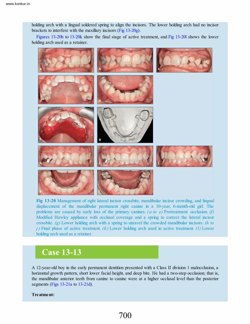
holding arch with a lingual soldered spring to align the incisors. The lower holding arch had no incisorbrackets to interfere with the maxillary incisors (Fig 13-20g).
Figures 13-20h to 13-20k show the final stage of active treatment, and Fig 13-20l shows the lowerholding arch used as a retainer.
Fig 13-20 Management of right lateral incisor crossbite, mandibular incisor crowding, and lingualdisplacement of the mandibular permanent right canine in a 10-year, 6-month-old girl. Theproblems are caused by early loss of the primary canines. ( a to e) Pretreatment occlusion. (f)Modified Hawley appliance with occlusal coverage and a spring to correct the lateral incisorcrossbite. (g) Lower holding arch with a spring to unravel the crowded mandibular incisors. (h toj ) Final phase of active treatment. (k ) Lower holding arch used in active treatment. ( l) Lowerholding arch used as a retainer.
Case 13-13A 12-year-old boy in the early permanent dentition presented with a Class II division 1 malocclusion, ahorizontal growth pattern, short lower facial height, and deep bite. He had a two-step occlusion; that is,the mandibular anterior teeth from canine to canine were at a higher occlusal level than the posteriorsegments (Figs 13-21a to 13-21d).
Treatment:
700
www.konkur.in

Because of the patient’s growth potential, the treatment strategy involved interceptive Class IItreatment with the headgear, lip bumper, and modified Hawley appliance (HLH) technique (see chapter11). The maxillary Hawley appliance was modified with an inclined surface and a lip bumper.
Treatment was continued with mandibular complete bonding to correct the two-step occlusion byintrusion of the anterior segment and eruption of the posterior segment, first with a mandibular doubleshow loop and then with a step-down anterior arch. A maxillary bite plate was used to disocclude theposterior segments.
The next step was maxillary complete bonding and anterior retraction and continuation of the cervicalheadgear. Figures 13-21e to 13-21g show posttreatment views of the occlusion. Cephalometricevaluations revealed that treatment had the following results (Figs 13-21h and 13-21i): Overbite wasreduced from 7.8 to 1.1 mm, overjet was reduced from 11.8 mm to 2.7 mm, ANB was reduced from4.8 to 2.5 degrees, FMA was increased from 17.6 to 20.6 degrees, upper facial height increased from56.8 to 58.8 mm, and lower facial height increased from 54.2 to 56.8.
Fig 13-21 Management of deep bite and two-step occlusion in a 12-year-old boy with a Class IIdivision 1 malocclusion. He has a horizontal growth pattern and short lower facial height. (a to d)Pretreatment occlusion. (e to g) Posttreatment occlusion.
701
www.konkur.in

Fig 13-21 (cont) (h) Pretreatment (left) and posttreatment (right) cephalometric radiographs. (i)Changes in cephalometric measurements.
Case 13-14An 11-year-old girl in the late mixed dentition presented with a Class I malocclusion, deep bite, andsevere maxillary and mandibular space deficiency (Figs 13-22a to 13-22f).
Treatment:
702
www.konkur.in

Treatment was planned as a serial extraction approach, despite the increased overbite, which wascontrolled mechanically. Extraction treatment was chosen for several reasons: (1) the severe spacedeficiency in both jaws (space needed: 10 mm in the maxillary arch, 12 mm in the mandibular arch), (2)panoramic radiographic evaluation and clues that confirmed that this was a hereditary type of crowding(palisading maxillary molars, mandibular molar crowding, and the possibility of second molar impaction),and (3) clinical signs of mandibular incisor compression and the effect of that finding on bone support.
Treatment procedures included the following steps:
1. Placement of a maxillary Hawley bite plate as anchorage and to control deep bite2. Insertion of a lower holding arch as anchorage3. Extraction of all remaining primary molars and maxillary first premolars4. Extraction of the mandibular first premolars5. Removal of the Hawley appliance and complete bonding to finalize treatment
Figures 13-22g to 13-22j show the results of treatment.
Fig 13-22 Management of deep bite and severe maxillary and mandibular space deficiency in an11-year-old girl in the late mixed dentition. (a to e) Pretreatment occlusion. ( f ) Pretreatmentpanoramic radiograph. (g to i) Posttreatment occlusion. (j) Posttreatment panoramic radiograph.
Case 13-15
703
www.konkur.in

A 10-year, 8-month-old girl in the late mixed dentition presented with a Class II division 2 malocclusionand deep overbite. She exhibited maxillary moderate space deficiency and slight retroclination of themandibular incisors (Figs 13-23a to 13-23g).
Treatment:Because of the patient’s skeletal age and growth potential, the treatment plan was designed asinterceptive treatment of the Class II malocclusion, with intraoral and extraoral appliances to encouragegrowth modification and provide space for the dentition. Treatment was started with maxillary cervicalheadgear and a maxillary Hawley anterior bite plate to reduce overbite, retract the maxillary molars, andmodify maxillary growth.
Mandibular treatment was started with 2 × 4 bonding to achieve slight protrusion of the incisors. Thiswas followed by some intrusion with a utility arch and sequential extraction of primary teeth to achieveguidance of eruption and overbite control.
After full Class I molar relationships were established and normal overbite was achieved, themaxillary Hawley appliance was removed. Treatment continued with full bonding for leveling andanterior retraction (Figs 13-23h to 13-23n).
Fig 13-23 Management of a Class II division 2 malocclusion and deep overbite in a 10-year, 8-month-old girl. She exhibits moderate maxillary space deficiency and slight retroclination of themandibular incisors. (a to e) Pretreatment occlusion. (f) Pretreatment panoramic radiograph. (g)Pretreatment cephalometric radiograph.
704
www.konkur.in

Fig 13-23 (cont) (h to l) Posttreatment occlusion, achieved without extractions. (m)Posttreatment panoramic radiograph. (n) Posttreatment cephalometric radiograph.
Case 13-16A 14-year-old boy in the late mixed dentition presented with a Class I malocclusion, upright maxillaryand mandibular incisors, and severe deep bite (110%) that had caused mandibular incisor collapse. Thecollapse contributed to severe mandibular crowding and ectopic eruption of the mandibular right lateralincisor (Figs 13-24a to 13-24f).
Treatment:Because of the patient’s severe deep bite and retrusion of the maxillary and mandibular incisors, thetreatment strategy was to correct incisor retroclination and open the bite to resolve the mandibularincisor collapse and regain some space for the mandibular incisors.
First, the maxillary molars were banded, and a removable bite plate with no labial bow was insertedto disocclude the posterior segments and thereby stimulate mandibular molar eruption. This wasfollowed by maxillary 2 × 4 bonding; nickel-titanium archwire was used for leveling, and then stainlesssteel round wire with an open loop was used for incisor proclination. A maxillary utility arch was usedfor maxillary incisor intrusion and molar uprighting. After some bite opening was achieved, threemandibular incisors and the primary first molars were bonded to achieve mandibular incisor alignmentand protrusion and to regain space for the ectopic lateral incisors. Concomitant with eruption of thepermanent teeth, complete bonding and the final stage of treatment was continued. Figures 13-24g to13-24i show some of the stages of active treatment.
Significant changes were achieved in the dentoskeletal structures (Figs 13-24j to 13-24q). Changesincluded deep bite correction and improvement of incisor inclination and therefore elimination of thesevere crowding. The vertical growth pattern continued, and facial height increased after treatment.
705
www.konkur.in

Fig 13-24 Management of severe deep bite (110%) and maxillary and mandibular retrusion thathave caused mandibular incisor collapse in a 14-year-old boy in the late mixed dentition. Thecollapse has contributed to severe mandibular crowding and ectopic eruption of the mandibularright lateral incisor. (a to e) Pretreatment occlusion. (f) Pretreatment panoramic radiograph.
706
www.konkur.in

707
www.konkur.in

Fig 13-24 (cont) (g to i) Phases of active treatment. (j to n) Posttreatment occlusion. Noextractions have been performed. (o) Posttreatment panoramic radiograph. (p) Pretreatment (left)and posttreatment ( r ig h t ) cephalometric radiographs. ( q ) Changes in cephalometricmeasurements.
Summary
• Open bite is a multifactorial phenomenon, initiating and developing duringthe primary and mixed dentitions. Recognition of the etiology plays animportant role in the diagnosis and treatment of this anomaly. The problemcan be diagnosed clinically and cephalometrically; however, diagnosisshould be viewed in the context of both skeletal and dental structures.
• Hereditary factors influence the direction of skeletal growth by changingform and size, causing abnormal growth pattern and incorrect jaw posture,and initiating skeletal open bite.
• Environmental factors during the primary and mixed dentitions can also playan important role in the development of open bite. Therefore, different kindsof open bite with different morphologic and etiologic characteristics mustbe accurately classified for precise treatment planning.
• The major common local or environmental factors that play a role indeveloping open bite at an early age are digit sucking; abnormal tongue size,posture, and function; abnormal neuromuscular function; and enlargedlymphatic tissues that force mouth breathing and postural alterations thatinduce dental and skeletal modifications.
• Pediatric dentists and general practitioners who see children at an early agehave great responsibility in the detection of and intervention in thesedeveloping anomalies.
• Tongue thrust is an abnormal tongue function that usually developssecondary to thumb sucking and then maintains or increases the open bitethat was produced by the finger, even after the finger habit has stopped.During correction of open bite, abnormal tongue function must also becontrolled.
• Simple dental open bite is considered an open bite without skeletalabnormalities. Patients with dental open bite have the followingcharacteristics: normal craniofacial pattern, normal skeletal jawrelationships, previous history of finger sucking, proclined incisors, anteriortongue thrust, undererupted incisors, normal or slightly excessive molar
708
www.konkur.in

height, normal inclination of the posterior dentition, divergent occlusalplane, lack of a gummy smile, and no vertical excess or posterior tipping ofthe maxilla.
• Skeletal open bites are usually caused by genetics, prolonged environmentalfactors, or a combination of both. Patients with skeletal open bite exhibitunfavorable growth patterns and skeletal disharmonies: steep mandibularplane, retrognathic mandible, obtuse gonial angle, short ramal height, shortanterior cranial base, short posterior cranial base, obtuse y-axis angle,obtuse saddle angle, obtuse articular angle, obtuse occlusal plane–mandibular plane angle, obtuse mandibular plane–palatal plane angle,counterclockwise rotation of the palate, increased lower facial height,decreased upper facial height, decreased posterior facial height, and steepanterior cranial base.
• Open bite management during the primary dentition is usually limited toobservation or control of habits. In patients with a persistent habit, inaddition to habit control, control of the vertical dimension is recommended.This can be accomplished, for example, by application of a lower holdingarch and transpalatal arch.
• Open bite management during the mixed dentition can be designed to includeone or two phases.
• The best timing for early intervention is during the early mixed dentition.Depending on the etiologic factors and morphology of the anomaly, thefollowing tactics can be applied: habit control, lip seal and swallowingexercises, growth modification, closing the drawbridge, and increasing theposterior facial height–anterior facial height ratio.
• Excessive anterior overlap of incisors, or deep bite, is a commonmalocclusion that presents significant problems for orthodontists iftreatment is delayed until the patient is an adolescent or adult. This is due todifficulty in treatment procedures, lack of long-term stability, and strongrelapse tendency.
• Not all deep bites are alike; deep bite can be associated with a Class I,Class II division 2, or Class II division 1 malocclusion, and each type hasdifferent skeletal morphology, different etiology, and different treatmentstrategies. Deep bite can manifest as a simple incisor overlap, adentoalveolar overlap, skeletal overclosure, or a combination of both.
• When a patient with excessive overbite is examined, it is important toremember that excessive overlapping of incisors is not the whole problem;rather it is a part of the total malocclusion.
709
www.konkur.in

• Dental deep bites can develop because of supraocclusion (overeruption) ofincisors, infraocclusion (undereruption) of the posterior teeth, or acombination of both. Dental deep bites are confined to the teeth and alveolarprocesses. Therefore, the problem lies mainly within the dentition and iscaused by the environment.
• Patients with dental deep bite usually have a normal vertical skeletalrelationship of the basal bones, normal lower facial height, normal profile,and normal relationships of the lips to each other and to the chin and nose.
• Skeletal deep bite (closed bite), or complex deep bite, is a dentoskeletalmalrelationship of the dentoskeletal structures in the vertical dimension. Inother words, it is a combination of an overlapping of incisors and a verticalbasal bone malrelationship, a malrelationship of the alveolar bones, and/ormalrelationship of the underlying mandibular or maxillary bones. Theskeletal malrelationship of the basal bones may be partially the result ofcounterclockwise rotation of the mandible or clockwise rotation of themaxilla.
• The major etiologic factors of skeletal deep bite are usually hereditaryinfluences on the dental, skeletal, and soft tissue profiles; therefore, this typeof anomaly has more complicated and difficult management and has a hightendency for relapse if treated late.
• Dentoskeletal and soft tissue characteristics of skeletal deep bite includereduced lower facial height, excessive freeway space, increased ramusheight and width, two-step occlusion (incisors and canines at higherocclusal levels relative to the premolar and molars), strong temporalis andmasseter muscles, a small gonial angle, increased ramus height andposterior cranial base causing counterclockwise rotation of the mandible, asmall cranial base angle, a convergent face (four facial planes of the faceare horizontal), and a straight or concave profile.
• Deep bite problems can be managed at early ages as a preventive orinterceptive procedure or late during the permanent dentition as a correctiveprocedure. Early treatment is easier to achieve and produces more stableresults.
• Before treatment planning, the following points must be considered: thepatient’s age, especially skeletal maturation; the influence of genetics and/orenvironment; the structures involved (dental, dentoalveolar, skeletal, orcombination); the available freeway space (normal, excessive, or short );the patient’s profile; the patient’s facial vertical proportions; the incisor
710
www.konkur.in

inclination and their relation to the lips and profile; and the amount ofincisor exposure during a smile.
• The general treatment strategy in adults with deep bites includes intrusion ofthe maxillary or mandibular incisors or both, extrusion of the maxillary ormandibular posterior teeth or both, a combination of intrusion and extrusionof the anterior and posterior segments, or combined surgical and orthodonticintervention in patients with severe skeletal overclosure.
• Deep bite malocclusion occurs in the primary dentition but is rarely treatedat this age. Children with horizontal growth patterns and deep bitemalocclusions have specific facial and dental characteristics that can berecognized in their primary stage of development.
• Environmental causes that affect occlusion interact mostly during theprimary and mixed dentitions. These are mostly detectable, and in manysituations the anomalies that are developing can be controlled. Commonenvironmental causes that can aggravate the problem are mesial migration ofthe posterior teeth, the presence of abnormal habits, lower lip dysfunction,early loss of the primary canine, and a horizontal growth pattern.
• There are several general strategies for early deep bite treatment during themixed dentition and in growing children: intrusion of the maxillary ormandibular incisors or both, extrusion of the maxillary or mandibularposterior segment or both, growth modification, or any combination of thesestrategies.
References1. Beane RA. Non surgical management of the anterior open bite: A review of the options. Semin Orthod
1999;5:275– 283.2. Kim YH. Anterior openbite and its treatment with multiloop edgewise archwire. Angle Orthod 1987;57:290–
321.3. Rakosi T. The open bite malocclusion. In: Graber TM, Ra-kosi T, Petrovic AG (eds). Dentofacial Orthopedics
with Functional Appliances, ed 2. St Louis: Mosby Year Book, 1997:481–508.4. Kuroda S, Katayama A, Takano-Yamamoto T. Severe anterior open-bite case treated using titanium screw
anchorage. Angle Orthod 2004;74:558–567.5. Seres L, Kocsis A. Closure of severe skeletal anterior open bite with zygomatic anchorage. J Craniofac Surg
2009;20:478–482.6. Barbre RE, Sinclair PM. A cephalometric evaluation of anterior openbite correction with the magnetic active
vertical corrector. Angle Orthod 1991;61:93–102.7. Enlow DH. Handbook of Facial Growth, ed 2. Philadelphia: Saunders, 1982.8. Carlson DS. Biological rationale for early treatment of dentofacial deformities. Am J Orthod Dentofacial
Orthop 2002;121:554–558.
711
www.konkur.in

9. Harris EF, Johnson MG. Heritability of craniometric and occlusal variables: A longitudinal sib analysis. Am JOrthod Dentofacial Orthop 1991;99:258–268.
10. Subtelny JD. Examination of current philosophies associated with swallowing behavior. Am J Orthod1965;51:161– 182.
11. Proffit WR. Lingual pressure patterns in the transition from tongue thrust to adult swallowing. Arch Oral Biol1972;17:555–563.
12. Huang GJ, Justus R, Kennedy DB, Kokich VG. Stability of anterior openbite treated with crib therapy. AngleOrthod 1990;60:17–24.
13. Van der Linden FPGM. Vertical dimension. In: McNamara JA, Brudon WL (eds). Orthodontics andDentofacial Orthopedics. Ann Arbor, MI: Needham Press, 2001:111–148.
14. Subtelny JD, Sakuda M. Open-bite: Diagnosis and treatment. Am J Orthod 1964;50:337–358.15. English JD. Early treatment of skeletal open bite malocclusions. Am J Orthod Dentofacial Orthop
2002;121:563–265.16. Erverdi N, Keles A, Nanda R. The use of skeletal anchorage in open bite treatment: A cephalometric
evaluation. Angle Orthod 2004;74:381–390.17. Strang RHW. A Textbook of Orthodontia, ed 3. Philadelphia: Lea & Febiger, 1950.18. Nanda R. The differential diagnosis and treatment of excessive overbite. Dent Clin North Am 1981;25:69–84.19. Neff C. Tailored occlusion with the anterior coefficient. Am J Orthod 1949;35:309–313.20. Diamond M. The development of the dental height. Am J Orthod 1944;30:589–605.21. Wylie WL. The assessment of anteroposterior dysplasia. Angle Orthod 1947;17:97–109.22. Baume LJ. Physiological tooth migration and its significance for the development of the occlusion. J Dent Res
1950;29:440–447.23. Flemming HB. An investigation of the vertical overbite during the eruption of the permanent dentition. Angle
Orthod 1961;31:53–62.24. Canut JA, Arias S. A long-term evaluation of treated Class II division 2 malocclusions: A retrospective study
model analysis. Eur J Orthod 1999;21:377–386.25. Wasilewsky H. Three Dimensional Evaluation of Overbite Relapse [thesis]. Rochester, NY: University of
Rochester, 1985.26. Popovich F. Cephalometric evaluation of vertical overbite in young adult. J Can Dent Assoc 1955;21:209–
222.27. Alexander TA, Gibbs CH, Thompson WJ. Investigation of chewing patterns in deep-bite malocclusions before
and after orthodontic treatment. Am J Orthod 1984;85:21–27.28. Wragg PF, Jenkins WM, Watson IB, Stirrups DR. The deep overbite: Prevention of trauma. Br Dent J
1990;168:365– 367.29. Bollen AM. Effects of malocclusions and orthodontics on periodontal health: Evidence from a systematic
review. J Dent Educ 2008;72:912–918.30. Nasry HA, Barclay SC. Periodontal lesions associated with deep traumatic overbite. Br Dent J
2006;200:557–561.31. Sonnesen L, Svensson P. Assessment of pain sensitivity in patients with deep bite and sex- and age-matched
controls. J Orofac Pain 2011;25:15–24.32. Bell WH, Jacobs JD, Legan HL. Treatment of Class II deep bite by orthodontic and surgical means. Am J
Orthod 1984;85:1–20.
712
www.konkur.in

713
www.konkur.in

Chapter 6An 8-year-old boy with a long-term, chronic thumb-sucking habit developed a callus on his thumb (Figs1A to 1D). He exhibited a Class II division 1 malocclusion, an 8- to 10-mm open bite, contact only onthe primary second and permanent first molars, and an overjet of about 17 mm.
Treatment:The patient was a very cooperative and determined boy, and after consultation with his parents, aremovable appliance was planned for the first phase of treatment (Fig 1E). The patient’s level ofcompliance was excellent, and the habit was stopped after 2 months. The use of the appliance wascontinued for 12 months: He wore it 24 hours a day for 6 months and at night only for 6 months.
The habit was completely discontinued, and the open bite and overjet were reduced significantly, butthe patient stopped treatment and returned 2 years later. Figures 1F and 1G show the patient’socclusion during the permanent dentition. He had a Class II division 1 occlusion, 9-mm overjet, 2- to 3-mm open bite, acceptable mandibular dentition, and severe maxillary anterior crowding with the caninesout of the arch.
In accordance with the parents’ wishes and the patient’s age, the second phase of treatment waslimited to the maxillary arch and extraction of two first premolars. Figures 1H and 1I show the results ofphase 2 treatment.
Fig 1 Treatment of an 8-year-old boy with a severe, long-term thumb-sucking habit. He has aClass II division 1 malocclusion, an 8- to 10-mm open bite, and a 17-mm overjet. Only the primarysecond and permanent first molars make contact. (A to C) Pretreatment occlusion. (D) Callusesdeveloped as a result of the sucking habit. (E) Removable habit breaker. (F and G) Occlusion ofthe permanent dentition 30 months after habit control. (H and I) Posttreatment occlusion afterphase 2 therapy.
714
www.konkur.in

Chapter 7An 18-year-old man presented with oligodontia. He was missing 10 teeth, including third molars, andfive remaining submerged primary molars had arrested the growth of the alveolar process. The extremespacing and tipping of all dentition, including distal tipping of the anterior teeth, had resulted in a bizarreocclusion (Fig 1A to 1F). Orthodontic problems included an anterior deep bite, severe open biteaffecting the buccal segments (no bite), interdental spacing, mesial tipping of all molars, and distaltipping of all remaining permanent teeth. The patient’s major complaint was a chewing problem;because of financial problems he did not consider esthetics to be an issue.
This is a clear example of the necessity for general practitioners to understand the importance ofearly detection of and intervention in ankylosed primary molars as well as hypodontia. Timely extractionof the ankylosed teeth and proper space maintenance, instead of placement of amalgam restorations,would have restored the integrity of the occlusion of this patient earlier and more easily.
Treatment:After extraction of all primary ankylosed molars, the treatment plan for this young man includedorthodontic treatment first and then prosthodontic restoration. The comprehensive orthodontic approachincluded leveling, correction of rotation, uprighting, space closure, anterior retraction, torque control, andoverbite reduction.
The prosthetic approach was limited to fixed partial dentures because implants were not common atthat time. Considering the patient’s age and skeletal maturation, it was recommended to restore theocclusion toward some bite opening. Figures 1G to 1J show the final occlusion after prosthodonticrestoration.
715
www.konkur.in

Fig 1 Treatment of an 18-year-old man with oligodontia. He is missing 10 teeth, including thirdmolars. Some submerged primary molars remain. (A to D) Pretreatment occlusion. (E)Pretreatment panoramic radiograph. ( F ) Pretreatment cephalometric radiograph. (G to J)Posttreatment occlusion, after prosthetic restoration. (K) Posttreatment panoramic radiograph. (L)Posttreatment cephalometric radiograph.
Chapter 8A 9-year, 7-month-old boy in the early mixed dentition had a Class I malocclusion and two mesiodentespreventing eruption of the maxillary right central and lateral incisors (Fig 1A). Damage to the centralincisor crown and space loss had resulted from the neglected supernumeraries.
Figure 1B, taken after removal of the mesiodentes, clearly demonstrates the space closure in theanterior segment that has resulted from the failure to maintain the necessary space for the uneruptedcentral incisor.
Figure 1C was taken during the initial stage of orthodontic treatment for space opening. The lateralincisor had erupted, but the central incisor was still highly positioned. The central incisor was exposedfor traction attachment. The crown damage and pulp exposure were restored first with hydroxyapatite(Figs 1D to 1G) and later, after completion of orthodontic movement, with a composite resin restoration(Figs 1H to 1K).
Undiagnosed or neglected mesiodentes that delay eruption of the central incisors can result in themesial movement of erupted lateral incisors and space loss even before removal of the mesiodentes.Therefore, an important consideration in management of hyperdontia is maintaining the space betweenerupted teeth after removal of the supernumerary. Space maintenance for unerupted teeth is especiallya factor when unerupted teeth are located deeply, and, even after extraction of the supernumerarytooth, their eruption is delayed and slow.
716
www.konkur.in

Fig 1 Treatment of a 9-year, 7-month-old boy with mesiodentes preventing eruption and damagingthe maxillary right central incisor crown. (A) Pretreatment panoramic radiograph. (B) Panoramicradiograph after extraction of the supernumerary teeth and lateral incisor eruption. (C) Panoramicradiograph taken during the initial stage of orthodontic treatment and space opening. (D to G)Traction of the central incisor. Note the damage to the crown and pulp exposure caused by thesupernumerary teeth; a hydroxyapatite restoration has been placed. (H to J) Posttreatmentocclusion, after orthodontic treatment and final composite restoration. ( K ) Post-treatmentpanoramic radiograph.
Chapter 11: Case 1A 9-year, 3-month-old boy in the middle mixed dentition had a history of previous thumb sucking. Hepresented with a severe Class II division 1 malocclusion, an 11.3-mm overjet, and a 2.6-mm open bite.He exhibited severe maxillary and mild mandibular incisor proclination, a convex profile, and aretrognathic chin caused by mandibular clockwise rotation (Figs 1A to 1D).
717
www.konkur.in

Treatment:Because of his vertical growth tendency and aligned mandibular incisors, the only appliance used in themandibular dentition was a heavy lower holding arch to preserve leeway space and prevent verticalmolar eruption, thereby controlling clockwise mandibular rotation. In the maxillary arch, a combinedtongue crib–transpalatal arch appliance was inserted first to control anterior tongue movement andachieve some intrusion of the maxillary molars (for more detail on the tongue crib–transpalatal archappliance, see chapter 6). Then high-pull headgear was applied to correct the Class II molarrelationships and intrude the molars in order to achieve more counterclockwise rotation of the mandible.Treatment provided significant results in correcting all dentoskeletal aberrations and improving theprofile (Figs 1E to 1J). Treatment resulted in a Class I dentition; a significant change in the A-Bdiscrepancy from 9.1 to 4.3 degrees; correction of the overjet, from 11.3 to 2.1 mm; and a change inincisor overlap from a –2.6 to 1.4 mm. In addition, significant changes in the retruded chin wereachieved by mandibular growth and counterclockwise rotation of the mandible; these changes wereconfirmed by cephalometric improvement in the facial angle, lower facial height, and profile convexity.
Fig 1 Management of severe overjet and overbite in a 9-year, 3-month-old boy with a history ofthumb sucking. He has a Class II division 1 malocclusion, severe maxillary incisor proclination, anda retrognathic chin. (A to C) Pretreatment occlusion. (D) Pretreatment soft tissue profile. (E to G)Posttreatment occlusion. (H) Posttreatment soft tissue profile.
718
www.konkur.in

Fig 1 (cont) (I) Pretreatment (lef t) and posttreatment (right) cephalometric radiographs andtracings. (J) Changes in cephalometric measurements and superimposition of pretreatment (black)and posttreatment (green) tracings.
Chapter 11: Case 2
719
www.konkur.in

An 11-year, 3-month-old girl in the late mixed dentition presented with a Class II division 1malocclusion, an 11.6-mm overjet, impinging deep bite, and maxillary and mandibular incisor crowding.The maxillary incisors were crowded and proclined, and the mandibular incisors were retroclined andovererupted, creating a two-step occlusion (Figs 2A to 2D).
Treatment:Because of the patient’s severe maxillary incisor crowding, the HLH approach was modified to includean inclined bite plane and, instead of a labial bow, 2 × 4 bonding, plus cervical headgear and amandibular lip bumper. The first goal of treatment was to align the maxillary incisors by bonding. Thesecond goal was to distalize and extrude the maxillary molars by the use of cervical headgear inconjunction with an anterior inclined bite plane to disocclude the posterior segments to reduce theoverbite. Third, the curve of Spee was reduced by use of the lip bumper to upright the proclinedmandibular incisors. After some reduction of overbite was achieved by the previous three steps, 2 × 4bonding of the mandibular incisors was started. Later, a utility arch was placed to intrude the mandibularincisors for complete leveling of the mandibular arch.
The treatment approach was a one-phase treatment that consisted of the aforementioned steps,which were continued until normal interdigitation was achieved and the crowding, overjet, and overbitewere corrected. Active treatment was finished with 2 × 6 bonding followed by placement of a maxillaryHawley retainer and mandibular fixed retainer extending from canine to canine. Figures 2E to 2J showthe results of treatment.
Fig 2 Management of severe overjet, impinging deep bite, and incisor crowding in an 11-year, 3-month-old girl with a Class II division 1 malocclusion. (A to C) Pretreatment occlusion. (D)Pretreatment soft tissue profile. (E to G) Posttreatment occlusion. (H) Posttreatment soft tissue
720
www.konkur.in

profile.
Fig 2 (cont) (I) Pretreatment (lef t) and posttreatment (right) cephalometric radiographs andtracings. (J) Changes in cephalometric measurements and superimposition of pretreatment (black)and posttreatment (green) tracings.
721
www.konkur.in

Chapter 11: Case 3A 14-year-old girl presented with a neglected pseudo–Class III malocclusion, space deficiency for themaxillary second premolar, and a mandibular shift that was causing temporomandibular dysfunction(Figs 3A to 3D). She exhibited some damage to the incisor structure and minor spacing in themandibular anterior segment. The patient’s chief complaint was temporomandibular joint pain.
Treatment:The treatment approach involved use of only one removable appliance in the maxilla and no treatment inthe mandible. The appliance was a modified Hawley appliance with a special labial bow with twohorizontal loops on the buccal side of the canines to provide good retention in the anterior segmentwithout touching incisors; occlusal coverage; two Z-springs for labial movement of the maxillary incisorsout of crossbite; and a jackscrew for distalization of the maxillary left molars (Fig 3E). Figures 3F to 3Ishow the posttreatment occlusion. The crossbite was corrected, the maxillary second premolar wasaligned, and minor mandibular incisor spacing was spontaneously closed.
\
Fig 3 Management of a neglected pseudo–Class III malocclusion with mandibular shift and spacedeficiency. The neglected problems have caused temporomandibular dysfunction and pain. (A toC) Pretreatment occlusion in centric occlusion. (D) Pretreatment occlusion in centric relation. (E)Modified Hawley appliance with occlusal coverage, modified labial bow, and jackscrew to regainspace for the maxillary left second premolar. (F to I) Posttreatment occlusion.
722
www.konkur.in

Chapter 11: Case 4A 5-year-old girl in the primary dentition presented with a complete unilateral and anterior crossbite aswell as a mandibular functional shift (Figs 4A to 4F).
Treatment:This patient was treated with a removable Hawley appliance. The maxillary primary second molarswere bonded with a buccal tube and C-clasps over the tube for better retention of the appliance. TwoZ-springs were constructed to procline the maxillary incisors out of crossbite, while posterior occlusalcoverage was used to disocclude the anterior segment.
Figures 4G to 4I show different stages of anterior crossbite correction. The posterior segmentsexhibited open occlusion before settling. Figure 4J shows the appliance after removal of the occlusalacrylic resin. By 10 months after the end of retention, all mandibular permanent incisors and themaxillary central incisors had erupted (Figs 4K to 4O).
The type of appliance used in this patient must be worn 24 hours a day except during toothbrushing.As mentioned earlier, the first tactic in such cases is to jump the incisors out of crossbite. In somepatients, after the maxillary incisors are corrected, open occlusion may result in the posterior segmentsif the appliance is worn continuously.
In this situation, the occlusal acrylic resin should be left in place and gradually reduced in thickness ateach visit. Once the posterior dentition has erupted and settled, the occlusal coverage can be removedor the appliance can be stopped, if required.
723
www.konkur.in

Fig 4 Management of complete unilateral and anterior crossbite and mandibular functional shift ina 5-year-old girl in the primary dentition. (A to E) Pretreatment occlusion. ( F ) Pretreatmentcephalometric radiograph. ( G ) Occlusion after correction of anterior crossbite but beforecorrection of posterior occlusion. (H and I) Occlusion after more occlusal contact has beenestablished in the posterior segments. (J) Appliance used in treatment, after removal of occlusalcoverage.
Fig 4 (cont) (K to N) Posttreatment occlusion. (O) Posttreatment cephalometric radiograph.
724
www.konkur.in

About the AuthorAliakbar Bahreman, DDS, MS, currently serves as a clinical professor in thePediatric and Orthodontic Programs at the Eastman Institute for Oral Health at theUniversity of Rochester in New York. After graduating from dental school at TehranUniversity of Iran in 1961. he completed a pediatric dentistry fellowship (1964) anda master's degree in orthodontic and dentofacial orthopedics (1967) at the EastmanInstitute. He then started his career at the dental school at Shahid Beheshti University(former National University of Iran) as founder and chairman of the Departments ofOrthodontics and Pediatric Dentistry. Later, as the dean of the dental school, hepresented complete postgraduate curriculum to the Iranian Ministry of HigherEducation and, for the first time in Iran, started postgraduate training. He returned tothe Eastman Institute for Oral Health as a visiting professor in 1999 and started as afull-time clinical professor and clinical supervisor of the orthodontic, pediatricdentistry, and advanced education of dentistry programs in 2003. Dr Bahreman is thefounder of the Iranian Orthodontic Association and was the first president of theIranian International Orthodontic Meeting, and he is a fellow of the InternationalCollege of Dentists, the American Association of Orthodontists, and the InternationalFederation of Orthodontics. He received many awards and medals both as a studentand as a faculty member in Iran, and in June 2010, he received the Iranpour Awardfor excellence in clinical education from the University of Rochester.
725
www.konkur.in

Learn more about Quintessence Publishing Co., Inc
www.quintpub.com
726
www.konkur.in