Dynamic Model-Driven Quantitative and Visual Evaluation of the Aortic Valve from 4D CT
8
Dynamic model-driven quantitative and visual evaluation of the aortic valve from 4D CT Razvan Ioan Ionasec 1,2 ? , Bogdan Georgescu 1 , Eva Gassner 3 , Sebastian Vogt 4 , Oliver Kutter 2 , Michael Scheuering 4 , Nassir Navab 2 , and Dorin Comaniciu 1 1 Integrated Data Systems, Siemens Corporate Research, Princeton, USA 2 Computer Aided Medical Procedures, Technical University Munich, Germany 3 Department of Radiology, Medical University of South Carolina, Charleston, USA 4 Siemens Medical Solutions, Computed Tomography, Forchheim, Germany Abstract. Aortic valve disease is an important cardio-vascular disorder, which affects 2.5% of the global population and often requires elaborate clinical management. Experts agree that visual and quantitative evalu- ation of the valve, crucial throughout the clinical workflow, is currently limited to 2D imaging which can potentially yield inaccurate measure- ments. In this paper, we propose a novel approach for morphological and functional quantification of the aortic valve based on a 4D model estimated from computed tomography data. A physiological model of the aortic valve, capable to express large shape variations, is generated using parametric splines together with anatomically-driven topological and geometrical constraints. Recent advances in discriminative learning and incremental searching methods allow rapid estimation of the model parameters from 4D Cardiac CT specifically for each patient. The pro- posed approach enables precise valve evaluation with model-based dy- namic measurements and advanced visualization. Extensive experiments and initial clinical validation demonstrate the efficiency and accuracy of the proposed approach. To the best of our knowledge this is the first time such a patient specific 4D aortic valve model is proposed. 1 Introduction Aortic valve disease represents the most common valvular disease in developed countries [1], and shows the second highest incidence among congenital valvular defects [2]. Although, aortic root preserving surgery [3] along with minimally invasive procedures are emerging, the management of patient with valvular heart disease (VHD) has remained challenging. Precise knowledge and reliable display of the four-dimensional valve characteristics are requested by clinicians. To date, most data on geometry and dynamics were obtained by experimental studies on explanted valves or using animal models [4], with small numbers of cases across publications. Standard dimensions of the aortic root are given in literature, though considerable variations in the geometry is recognized [5]. ? This work was initiated as a joint diploma thesis between Friedrich-Alexander- University of Erlangen-Nuremberg and Siemens Corporate Research. We acknowl- edge the advice of Prof. Joachim Hornegger and Dr. Martin Huber. Correspondence to razvan [email protected]
-
Upload
mtpartners -
Category
Documents
-
view
1 -
download
0
Transcript of Dynamic Model-Driven Quantitative and Visual Evaluation of the Aortic Valve from 4D CT

Dynamic model-driven quantitative and visualevaluation of the aortic valve from 4D CT
Razvan Ioan Ionasec1,2 ?, Bogdan Georgescu1, Eva Gassner3, Sebastian Vogt4,Oliver Kutter2, Michael Scheuering4, Nassir Navab2, and Dorin Comaniciu1
1 Integrated Data Systems, Siemens Corporate Research, Princeton, USA2 Computer Aided Medical Procedures, Technical University Munich, Germany
3 Department of Radiology, Medical University of South Carolina, Charleston, USA4 Siemens Medical Solutions, Computed Tomography, Forchheim, Germany
Abstract. Aortic valve disease is an important cardio-vascular disorder,which affects 2.5% of the global population and often requires elaborateclinical management. Experts agree that visual and quantitative evalu-ation of the valve, crucial throughout the clinical workflow, is currentlylimited to 2D imaging which can potentially yield inaccurate measure-ments. In this paper, we propose a novel approach for morphologicaland functional quantification of the aortic valve based on a 4D modelestimated from computed tomography data. A physiological model ofthe aortic valve, capable to express large shape variations, is generatedusing parametric splines together with anatomically-driven topologicaland geometrical constraints. Recent advances in discriminative learningand incremental searching methods allow rapid estimation of the modelparameters from 4D Cardiac CT specifically for each patient. The pro-posed approach enables precise valve evaluation with model-based dy-namic measurements and advanced visualization. Extensive experimentsand initial clinical validation demonstrate the efficiency and accuracy ofthe proposed approach. To the best of our knowledge this is the firsttime such a patient specific 4D aortic valve model is proposed.
1 IntroductionAortic valve disease represents the most common valvular disease in developedcountries [1], and shows the second highest incidence among congenital valvulardefects [2]. Although, aortic root preserving surgery [3] along with minimallyinvasive procedures are emerging, the management of patient with valvular heartdisease (VHD) has remained challenging. Precise knowledge and reliable displayof the four-dimensional valve characteristics are requested by clinicians.
To date, most data on geometry and dynamics were obtained by experimentalstudies on explanted valves or using animal models [4], with small numbersof cases across publications. Standard dimensions of the aortic root are givenin literature, though considerable variations in the geometry is recognized [5].
? This work was initiated as a joint diploma thesis between Friedrich-Alexander-University of Erlangen-Nuremberg and Siemens Corporate Research. We acknowl-edge the advice of Prof. Joachim Hornegger and Dr. Martin Huber. Correspondenceto razvan [email protected]

(a) (b)Fig. 1. (a) A generic aortic valve model in perspective view. Reproduced with permis-sion of the author and the European Association for Cardio-Thoracic Surgery from:Anderson RH (doi:10.1510/mmcts.2006.002527). (b) The proposed aortic valve modelin perspective view, where u and v are the circumferential and height parametric di-rection, respectively, of the root and leaflets.
Quantitative and visual evaluation methods are limited to 2D images and arepotentially affected by measurement inaccuracies [6].
Computed tomography is a rapidly evolving modality for non-invasive imag-ing of the heart which enables dynamic four-dimensional evaluation of cardiacstructures throughout the cardiac cycle. However, quantification of the aorticvalve is impaired by the need to translate the four-dimensional data set intoa set of 2D planes in order to obtain measurements. This gives potentially er-roneous results for the curved anatomy of the basal aorta. A four dimensionalmodel derived from CT data sets offers the unique possibility to non-invasivelyvisualize and quantify the dynamics of the human aortic root and leaflets infunctional and diseased valves.
Existent valve models presented in the literature [7, 8] are either used forhemodynamic studies or for analysis of various prosthetic valves. Although someof them are generated from volumetric data, these models are generic and obvi-ously not applicable for the evaluation of individual patients.
In this paper we introduce a new modeling framework for the aortic valvefrom 4D cardiac CT data. A dynamic model of the valve is constructed fromanatomic structures together with physiology driven geometrical and topologicalconstraints (Section 2.1). The patient specific parameters of the model are esti-mated from CT data by combining learning-based technologies into a three-stage,coarse-to-fine parameter estimation algorithm (Section 2.2): landmark detection,full model fitting and model dynamics estimation.
The estimated model enables for the first time precise morphological andfunctional quantification as well as enhanced visualization of the aortic valve.This novel model-based evaluation paradigm has the potential to significantlyadvance the management of valvular heart disease.
Extensive experiments on 37 patients with various valvular disorders demon-strate the accuracy and speed of the proposed model estimation algorithm (Sec-tion 3.1). Initial clinical validation on various functional and pathological valvesshows a strong correlation among a proposed set of model-based measurements,manually performed measurements and previously reported aortic valve dimen-sions (Section 3.2).

2 Physiological Valve ModelingThe morphology and function of the aortic valve is very complex, which is un-derlined by the lack of consensus regarding its optimal physiological description[9]. The central anatomical structures of the aortic valve are the leaflets and theroot. Its function is to regulate the blood flow between the left ventricle andaorta.
2.1 Model Representation
We propose a physiology driven parametric 4D model capable to express a largespectrum of morphological and pathological variations of the aortic valve. Aset of well-defined landmarks, which includes hinges, commissures, leaflet tipsand coronary ostiums, describe key anatomical locations of the valve (see Fig.1(a)). The aortic root and lefleats form the central anatomic structures and theirgeometries are represented by Non uniform rational B-splines (NURBS), whichis the de facto standard in computational modeling. These components togetherwith topological and geometrical constraints define a physiologically compliantmodel of the aortic valve, capable to implicitly handle bicuspid malformations.
The aortic root connects the left ventricular outflow tract to the ascendingaorta and provides the supporting structures for the leaflets. This is representedby a NURBS surface Croot(u, v) closed in the u parametric direction and canbe imagined as a deformed cylinder constrained by the hinge, commissure andostium points:
Croot(u, v)︸ ︷︷ ︸u,v∈[0,1]
=
n∑i=0
m∑j=0
Ni,d(u)Nj,e(v)wi,jProoti,j
n∑i=0
m∑j=0
Ni,d(u)Nj,e(v)wi,j
P rootl,j = P root
l−n,j , 0 ≤ l ≤ d, 0 ≤ j ≤ m(1)
Croot(uhk , vh
k ) = Lhk , 0 ≤ k < 3
Croot(uck, vc
k) = Lck, 0 ≤ k < 3
Croot(uok, vo
k) = Lok, 0 ≤ k < 2
where P rooti,j are the control points, wi,j are the corresponding weights,
Ni,d(u) and Nj,e(u) are the dth and eth degree B-splines basis functions de-fined on the non-periodic knot vector U and V , respectively. The root surfaceCroot passes through the hinges (Lr
h), commissures (Lrc) and ostrium(Lr
o) land-marks at parametric location (uh
k , vhk ) , (uc
k, vck) and (uo
k, vok), respectively. A
comprehensive description of NURBS is given in [10].The three valvular leaflets, expressed as NURBS paraboloids, are fixed to the
root on an attachment crown delineated by the hinges and commissures, whilethe remaining free edge of the leaflets is constrained by the corresponding tippoint. These open and close during the cardiac cycle allowing one way bloodflow during systole, from the left ventricle to the aorta:
where Cleaf stands for the lth leaflet surface and P leaf l
i,j , wi,j , Ni,d(u) andNj,e(u) are defined analogous to equation (1). The surface converges into theadjacent commissures (Lc
l and Lcl+1), passes through the corresponding hinge
(Lhl ) and tip (Lt
l), and the 0− isocurve lies on the root at parametric locations(ul
k, vlk).

Cleaf l(u, v)︸ ︷︷ ︸
u,v∈[0,1]
=
n∑i=0
m∑j=0
Ni,d(u)Nj,e(v)wi,jPleaf l
i,j
n∑i=0
m∑j=0
Ni,d(u)Nj,e(v)wi,j
P leaf l0,0 = P leaf l
0,j = Lcl , 0 ≤ j ≤ m
P leaf ln,0 = P leaf l
n,j = Lcl+1, 0 ≤ j ≤ m
(2)
Cleaf l(uh
l , vhl ) = Lh
l
Cleaf l(ut
l , vtl ) = Lt
l
Cleaf l(u, 0) = Croot(ul
k, vlk)
It is straightforward within the NURBS framework to extend the above pre-sented 3D model to a dynamic model (4D) using the tensor product, whichintroduces a temporal parametric direction t to the model representation [10].
2.2 Model Estimation
The parameters of the valve model proposed in section 2.1 are estimated for eachpatient from 4D cardiac CT data. A specific instance of the model is exactly de-termined by the landmarks and NURBS control points in a four-dimensionalEuclidean space (3D+time), which cumulates into 3T (11 + 300) parameters5.Due the high dimension of the parameter vector, direct estimation in the orig-inal space is very difficult. We propose a three-step approach to estimate theparameters of the dynamic valve model, which are: landmarks detection, fullmodel fitting and model dynamics estimation.
Landmark Detection. Recent advances in discriminative learning and incremen-tal searching techniques are applied to automatically determine the landmarkslocations (Lr
h, Lrc , Lr
o and Lrt ) from an input volume. A training set, which con-
tains positive and negative samples of the landmarks positions, is created froma manually annotated database. We train a discriminative classifier H(x, y, z)based on the Probabilistic Boosting Tree (PBT) [11], which learns the targetdistribution by exploiting a divide-and-conquer strategy:
p(Li|xs, ys, zs) = Hi(xs, ys, zs), xs, ys, zs ∈ Di (3)
where p(Li|xs, ys, zs) is the probability of landmark Li at location (xs, ys, zs),Hi the strong classifier trained for Li and Di the search domain obtain fromthe training set. The detection is performed in a coarse-to-fine manner as wellas in incrementally increasing parameter spaces similar to the marginal spacelearning (MSL) [12] concept.
A thin-plate-spline (TPS) transformation [13, 14] is computed from the de-tected set of corresponding points, K =
{(Lm
i , LIi ), Lm
i ∈M,LIi ∈ I, 0 < i ≤ N
},
which maps each control point of priorly computed mean shape M at the corre-sponding location in the image I and provides an initial model estimation.
5 T represents the number of discrete samples in the time dimension (10 for a regular4D cardiac CT acquisitions), 11 and 300 the number of landmarks and control points,respectively.

(a) End-Diastole (b) (c) (d) End-Systole (e)Fig. 2. (a-e) Five frames of the dynamic valve model obtained from a CT sequence.
Full Model Fitting. The initial estimation obtained through landmark detec-tion and TPS transformation provides a quite accurate global fitting of themodel, however it requires further local processing for precise object delineation.A boundary detector is trained using the PBT algorithm in combination withsteerable features, proposed in [12]. This is applied locally at a set of discreteboundary locations and is used to evolve the shape towards high probabilityresponses of the boundary detector. The final estimation is obtained by fittingthe parametric model to the refined samples by solving a linear least squaresproblem [15].
Model Dynamics Estimation. The estimation of the dynamic valve model fol-lows a physiology-driven strategy and is more accurate and efficient comparedto a sequential computation of the input sequence. Parameter estimation is ini-tially performed for the dominant shapes observed in the end-diastolic (valve iscompletely closed) and end-systolic (valve is completely opened) cardiac phases,according to the algorithm described above. The estimation for the remainingframes exploits a prior model constructed as a linear combination of the tworeference shapes, leading to a significant performance boost. Fig. 2 illustratesthe dynamic model of the aortic valve estimated from a CT cardiac sequence.
3 Results3.1 Results on Valve Model Estimation
We demonstrate the performance of the proposed algorithm on 37 4D cardiacCT data set, which consist of 364 CT volumes. The scans are acquired fromdifferent patients with various cardiovascular diseases using heterogeneous pro-tocols, resulting in significant resolution and capture range variation (80 to 350slices with sizes from 512x 512 to 153x153 pixels and resolution from 0.28 mmto 2.0 mm). Each volume in the data set is associated to an annotation, whichwas manually generated and is considered to be the ground truth. Three-foldcross validation is performed in order to evaluate our algorithm.
Performance of Landmark Detection The landmark detection is evaluated bythe Euclidean distance between the ground truth and detected positions. Ini-tially, the detection is performed in low-resolution images and generates a set ofposition hypotheses for each landmark. Fig. 3(a) shows the error of the most ac-curate hypothesis, averaged over all landmarks, with respect to the cardinality ofthe hypotheses set. An optimal trade-of between speed and accuracy is achievedby keeping 100 candidates with an error of 1.59 mm. Detection is refined inhigh resolution images (1mm), which enables 10% increase in accuracy for 30%hypothesis. Averaging over the candidates set results in the final detection errorof 2.28 mm.

(a) (b)
(c) (d) (e)Fig. 3. (a) The detection error for the landmarks with respect to the cardinality of thecandidates set. (b) The error curve for model estimation with respect to the numberof samples. (c-e) Model estimation results in fused visualization.
Performance of Model Estimation The model estimation accuracy is evaluatedby the point-to-mesh measurement, which computes the average distance be-tween sample pairs of the detected and ground-truth model. The accuracy ofthe initial model estimation relative to the rendering and computation resolutionused within the system (2500 samples) is on average 2.00 mm. The model re-finement improves the estimation accuracy by nearly 40% (see Fig. 3(b)), equiv-alent to an error of 1.33 mm. The computation time of the proposed methodwas evaluated on a standard desktop machine (3.0 GHz CPU, 2.0 GB RAM).The estimation of the full dynamic valve model from a regular CT sequence (10volumes), is computed in 21.3 seconds with 70% of the time required for thelandmark detection.
3.2 Results on Clinical Valve Evaluation
We demonstrate the quantitative and visual capabilities and underline the per-formance of the proposed method by comparing a set of morphological anddynamic model-based measurements to expert measurements and literature re-ported valve dimensions. Evaluation is performed on CT images of healthy,stenotic, dilated aorta and bicuspid valves, while the ground truth is providedby measurements manually performed by a radiologist with five years of expe-rience in cardiovascular imaging. Table 1 summarizes the evaluation results anddemonstrates the precision of the proposed model-based quantification method.
The root diameter, important in surgical treatment of dilated and stiff anatomies[4] is computed at three levels: ventricular-arterial junction (VAJ), sinus of val-salva (SV) and sinotubular junction (SJ). Severity assessment in patients withdegenerative aortic stenosis is supported through the aortic valve area (AVA)

VAJ (cm) SV (cm) STJ (cm) AVA (cm2) LCT (mm) RCT (mm) NCT (mm)
Mean 0.137 0.166 0.098 0.120 2.211 1.951 2.352
STD 0.017 0.043 0.029 0.380 0.866 0.936 1.162
Table 1. Model-based qunatification error with respect to expert measurements.
measurement [6]. The mean AVA derived from the model was 3.74±1.34cm2, cor-relation with respect to manual planimetry r = 0.963 and p < 0.0001, and Bland-Altman systematic bias 0.12± 0.38cm2. Left-coronary tip (LCT), non-coronarytip (NCT) and right-coronary tip (RCT) orthogonal excursion is proposed forthe evaluation of valve’s function and motion characteristic after surgery. Mea-surements variation in healthy and diseased valves is illustrated in Fig. 4.
(a) (b)
(c) (d)Fig. 4. Advanced visualization of (a) calcified aortic valve in endoscopic view and (b)bicuspid valve in long axis view. (c,d) Valve area and leaflet motion variation in differentpathologies.
Fused visualization of direct volume rendering (DVR) of the 4D CT datasetand the estimated model provides further anatomical insight. Advanced tech-niques [16] enable visualization via post color-attenuated transferfunctions [17]of the aortic lumen from CTA and integration of the valve model geometry intothe DVR. Combination of CT data and model geometry can be directly used forvisual validation of the estimated parameters with respect to the anatomy andvisual quantification of pathological valves. For real-time 4D rendering of thesequence we use GPU Raycasting to efficiently stream the volume and geometrydata to the graphics card. The combination of these techniques is capable ofrendering high quality images, at interactive frame rates (Fig. 4(a), 4(b)).
4 Discussion
This paper proposes a novel quantitative and visual evaluation approach of theaortic valve, based on a dynamic model estimated from 4D CT sequences. A
robust and computationally efficient algorithm, which combines learning-basedtechnologies into a coarse-to-fine approach, was proposed for estimating a patientspecific valve model from imaging data. Automatic model-based measurementsprovide a significant advance in morphological and functional clinical evaluationof the aortic valve, currently limited to 2D imaging methods, operator-dependentand potentially inaccurate measurements. Future research will include high-levelinformation provided by soft tissue composition and mechanical constrains, aswell as investigations for other modalities (e.g. Ultrasound and MRI).
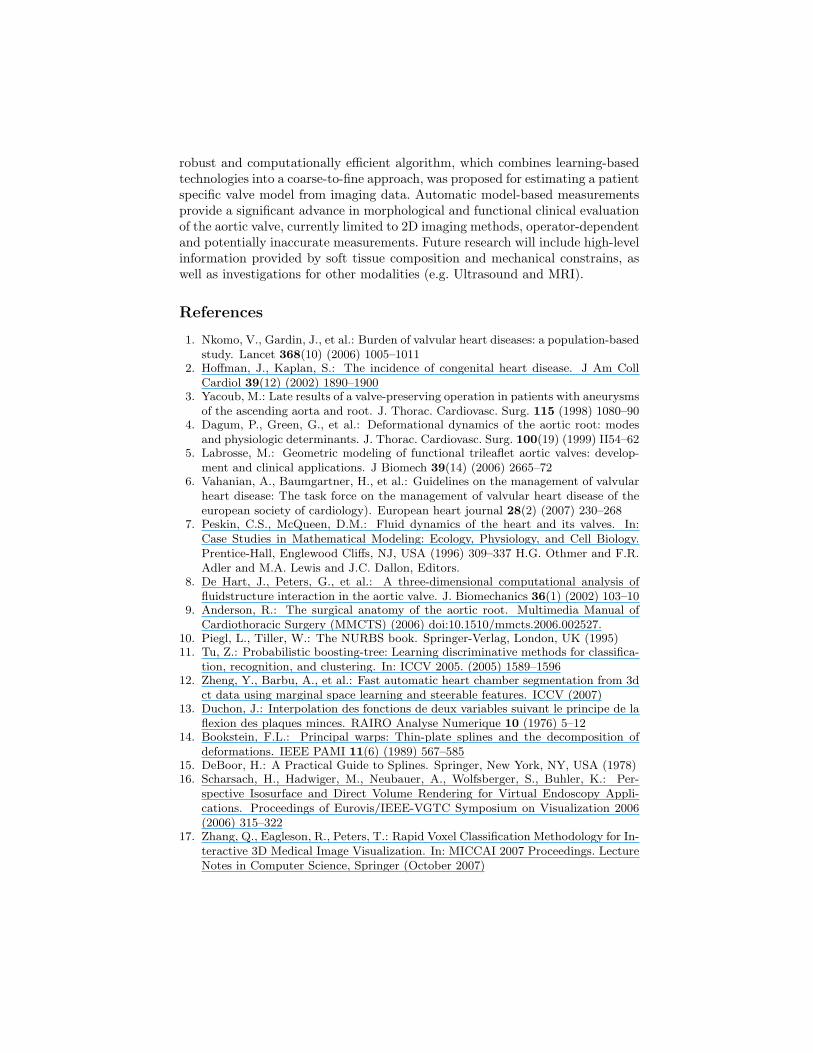
References
1. Nkomo, V., Gardin, J., et al.: Burden of valvular heart diseases: a population-basedstudy. Lancet 368(10) (2006) 1005–1011
2. Hoffman, J., Kaplan, S.: The incidence of congenital heart disease. J Am CollCardiol 39(12) (2002) 1890–1900
3. Yacoub, M.: Late results of a valve-preserving operation in patients with aneurysmsof the ascending aorta and root. J. Thorac. Cardiovasc. Surg. 115 (1998) 1080–90
4. Dagum, P., Green, G., et al.: Deformational dynamics of the aortic root: modesand physiologic determinants. J. Thorac. Cardiovasc. Surg. 100(19) (1999) II54–62
5. Labrosse, M.: Geometric modeling of functional trileaflet aortic valves: develop-ment and clinical applications. J Biomech 39(14) (2006) 2665–72
6. Vahanian, A., Baumgartner, H., et al.: Guidelines on the management of valvularheart disease: The task force on the management of valvular heart disease of theeuropean society of cardiology). European heart journal 28(2) (2007) 230–268
7. Peskin, C.S., McQueen, D.M.: Fluid dynamics of the heart and its valves. In:Case Studies in Mathematical Modeling: Ecology, Physiology, and Cell Biology.Prentice-Hall, Englewood Cliffs, NJ, USA (1996) 309–337 H.G. Othmer and F.R.Adler and M.A. Lewis and J.C. Dallon, Editors.
8. De Hart, J., Peters, G., et al.: A three-dimensional computational analysis offluidstructure interaction in the aortic valve. J. Biomechanics 36(1) (2002) 103–10
9. Anderson, R.: The surgical anatomy of the aortic root. Multimedia Manual ofCardiothoracic Surgery (MMCTS) (2006) doi:10.1510/mmcts.2006.002527.
10. Piegl, L., Tiller, W.: The NURBS book. Springer-Verlag, London, UK (1995)11. Tu, Z.: Probabilistic boosting-tree: Learning discriminative methods for classifica-
tion, recognition, and clustering. In: ICCV 2005. (2005) 1589–159612. Zheng, Y., Barbu, A., et al.: Fast automatic heart chamber segmentation from 3d
ct data using marginal space learning and steerable features. ICCV (2007)13. Duchon, J.: Interpolation des fonctions de deux variables suivant le principe de la
flexion des plaques minces. RAIRO Analyse Numerique 10 (1976) 5–1214. Bookstein, F.L.: Principal warps: Thin-plate splines and the decomposition of
deformations. IEEE PAMI 11(6) (1989) 567–58515. DeBoor, H.: A Practical Guide to Splines. Springer, New York, NY, USA (1978)16. Scharsach, H., Hadwiger, M., Neubauer, A., Wolfsberger, S., Buhler, K.: Per-
spective Isosurface and Direct Volume Rendering for Virtual Endoscopy Appli-cations. Proceedings of Eurovis/IEEE-VGTC Symposium on Visualization 2006(2006) 315–322
17. Zhang, Q., Eagleson, R., Peters, T.: Rapid Voxel Classification Methodology for In-teractive 3D Medical Image Visualization. In: MICCAI 2007 Proceedings. LectureNotes in Computer Science, Springer (October 2007)