Dual-modality PET/CT imaging: the effect of respiratory motion on combined image quality in clinical...
9
European Journal of Nuclear Medicine and Molecular Imaging Vol. 30, No. 4, April 2003 Abstract. To reduce potential mis-registration from dif- ferences in the breathing pattern between two comple- mentary PET and CT data sets, patients are generally allowed to breathe quietly during a dual-modality scan using a combined PET/CT tomograph. Frequently, how- ever, local mis-registration between the CT and the PET is observed. We have evaluated the appearance, magni- tude, and frequency of respiration-induced artefacts in CT images of dual-modality PET/CT studies of 62 pa- tients. Combined PET/CT scans during normal respira- tion were acquired in 43 subjects using single- or dual- slice CT. Nineteen patients were scanned with a special breathing protocol (limited breath-hold technique) on a single-slice PET/CT tomograph. All subjects were in- jected with ~370 MBq of FDG, and PET/CT scanning commenced 1 h post injection. The CT images were reconstructed and, after appropriate scaling, used for on-line attenuation correction of the PET emission data. We found that respiration artefacts can occur in the ma- jority of cases if no respiration protocol is used. When applying the limited breath-hold technique, the frequen- cy of severe artefacts in the area of the diaphragm was reduced by half, and the spatial extent of respiration-in- duced artefacts was reduced by at least 40% compared with the acquisition protocols without any breathing instructions. In conclusion, special breathing protocols are effective and should be used for CT scans as part of combined imaging protocols using a dual-modality PET/CT tomograph. The results of this study can also be applied to multi-slice CT to potentially reduce further breathing artefacts in PET/CT imaging and to improve overall image quality. Keywords: PET/CT imaging – Dual-modality tomogra- phy – Oncology – Respiration protocols Eur J Nucl Med Mol Imaging (2003) 30:588–596 DOI 10.1007/s00259-002-1097-6 Introduction Oncologic disease is typically detected by anatomical imaging techniques, such as computed tomography (CT), which is often considered the method of choice for can- cer imaging. In recent years positron emission tomogra- phy (PET), as a functional imaging technique, has gained widespread acceptance in clinical oncology for the de- tection of metabolic and functional abnormalities at an early stage of disease. In general, PET has a higher sen- sitivity and specificity than CT for the detection and staging of cancer [1]. However, the combination of PET and CT promises to be more accurate than either modali- ty alone in the initial diagnosis and for therapy manage- ment [2]. A straightforward approach to using PET and CT to- gether is to align the two image sets retrospectively [3]. Several computer-based algorithms have been described and research is ongoing to improve and automate these alignment techniques. So far, retrospective registration of complementary image sets works well for the brain [4], but often requires interaction with an operator to achieve an acceptable level of registration accuracy [5]. Another approach to using complementary PET and CT information is to combine the hardware into a single, dual-modality PET/CT tomograph [6]. The basic concept of combined PET/CT tomography is to examine the patient in both PET and CT mode with- out moving the patient off the table between examina- tions. Thus any potential misalignment between the two data sets is minimised, patient comfort is increased and Thomas Beyer ( ✉ ) Department of Diagnostic and Interventional Radiology, University Hospital Essen, Hufelandstrasse 55, 45147 Essen, Germany e-mail: [email protected] Tel.: +49-201-7231528, Fax: +49-201-9597756 Original article Dual-modality PET/CT imaging: the effect of respiratory motion on combined image quality in clinical oncology Thomas Beyer 1,2 , Gerald Antoch 1 , Todd Blodgett 3 , Lutz F. Freudenberg 2 , Tim Akhurst 4 , Stephan Mueller 2 1 Department of Diagnostic and Interventional Radiology, University Hospital Essen, Essen, Germany 2 Department of Nuclear Medicine, University Hospital Essen, Essen, Germany 3 PET Facility, University of Pittsburgh Medical Center, Pittsburgh, USA 4 Memorial Sloan Kettering Cancer Center, New York, USA Received: 7 October 2002 / Accepted: 18 November 2002 / Published online: 12 February 2003 © Springer-Verlag 2003
-
Upload
independent -
Category
Documents
-
view
0 -
download
0
Transcript of Dual-modality PET/CT imaging: the effect of respiratory motion on combined image quality in clinical...
European Journal of Nuclear Medicine and Molecular Imaging Vol. 30, No. 4, April 2003
Abstract. To reduce potential mis-registration from dif-ferences in the breathing pattern between two comple-mentary PET and CT data sets, patients are generally allowed to breathe quietly during a dual-modality scanusing a combined PET/CT tomograph. Frequently, how-ever, local mis-registration between the CT and the PETis observed. We have evaluated the appearance, magni-tude, and frequency of respiration-induced artefacts inCT images of dual-modality PET/CT studies of 62 pa-tients. Combined PET/CT scans during normal respira-tion were acquired in 43 subjects using single- or dual-slice CT. Nineteen patients were scanned with a specialbreathing protocol (limited breath-hold technique) on asingle-slice PET/CT tomograph. All subjects were in-jected with ~370 MBq of FDG, and PET/CT scanningcommenced 1 h post injection. The CT images were reconstructed and, after appropriate scaling, used for on-line attenuation correction of the PET emission data.We found that respiration artefacts can occur in the ma-jority of cases if no respiration protocol is used. Whenapplying the limited breath-hold technique, the frequen-cy of severe artefacts in the area of the diaphragm wasreduced by half, and the spatial extent of respiration-in-duced artefacts was reduced by at least 40% comparedwith the acquisition protocols without any breathing instructions. In conclusion, special breathing protocolsare effective and should be used for CT scans as part of combined imaging protocols using a dual-modalityPET/CT tomograph. The results of this study can also beapplied to multi-slice CT to potentially reduce furtherbreathing artefacts in PET/CT imaging and to improveoverall image quality.
Keywords: PET/CT imaging – Dual-modality tomogra-phy – Oncology – Respiration protocols
Eur J Nucl Med Mol Imaging (2003) 30:588–596DOI 10.1007/s00259-002-1097-6
Introduction
Oncologic disease is typically detected by anatomicalimaging techniques, such as computed tomography (CT),which is often considered the method of choice for can-cer imaging. In recent years positron emission tomogra-phy (PET), as a functional imaging technique, has gainedwidespread acceptance in clinical oncology for the de-tection of metabolic and functional abnormalities at anearly stage of disease. In general, PET has a higher sen-sitivity and specificity than CT for the detection andstaging of cancer [1]. However, the combination of PETand CT promises to be more accurate than either modali-ty alone in the initial diagnosis and for therapy manage-ment [2].
A straightforward approach to using PET and CT to-gether is to align the two image sets retrospectively [3].Several computer-based algorithms have been describedand research is ongoing to improve and automate thesealignment techniques. So far, retrospective registrationof complementary image sets works well for the brain[4], but often requires interaction with an operator toachieve an acceptable level of registration accuracy [5].Another approach to using complementary PET and CTinformation is to combine the hardware into a single, dual-modality PET/CT tomograph [6].
The basic concept of combined PET/CT tomographyis to examine the patient in both PET and CT mode with-out moving the patient off the table between examina-tions. Thus any potential misalignment between the twodata sets is minimised, patient comfort is increased and
Thomas Beyer (✉)Department of Diagnostic and Interventional Radiology, University Hospital Essen, Hufelandstrasse 55, 45147 Essen, Germanye-mail: [email protected].: +49-201-7231528, Fax: +49-201-9597756
Original article
Dual-modality PET/CT imaging: the effect of respiratory motionon combined image quality in clinical oncologyThomas Beyer1,2, Gerald Antoch1, Todd Blodgett3, Lutz F. Freudenberg2, Tim Akhurst4, Stephan Mueller2
1 Department of Diagnostic and Interventional Radiology, University Hospital Essen, Essen, Germany2 Department of Nuclear Medicine, University Hospital Essen, Essen, Germany3 PET Facility, University of Pittsburgh Medical Center, Pittsburgh, USA4 Memorial Sloan Kettering Cancer Center, New York, USA
Received: 7 October 2002 / Accepted: 18 November 2002 / Published online: 12 February 2003© Springer-Verlag 2003
589
European Journal of Nuclear Medicine and Molecular Imaging Vol. 30, No. 4, April 2003
logistical issues with patient referral are potentially min-imised [7].
In addition to providing accurately aligned functionaland anatomical information, combined PET/CT offersthe benefit of routine CT-based attenuation correction ofthe emission data [8, 9]. When using CT-based attenua-tion correction transmission times can be shortened toless than a minute for a whole-body scan, and transmis-sion scans can be performed post injection without theneed to correct for the bias from emission contamination.
Nevertheless, a number of issues need to be addressedin the context of complementary PET/CT imaging. Mostissues pertain to the effect of methodological differencesin scanning a patient with PET and CT when, at the sametime, using the available CT transmission images for at-tenuation correction of the emission data. CT scans, forexample, are typically (95%) performed in full-inspira-tion breath-hold with the arms up to reduce both attenua-tion artefacts and motion artefacts outside the heart. Cur-rent CT technology allows high-quality CT scans of thethorax or the abdomen to be completed in 20 s. In con-trast, the scan time for a number of PET emission scansto cover a similar axial imaging range is in the order ofseveral minutes. Due to the longer scan time needed,dedicated PET scans are acquired with the arms downand during normal respiration [10]. Ideally, in a com-bined tomograph, both CT and PET data should be ac-quired during the same respiratory (and cardiac) cycle.However, this is impractical in current generations ofPET/CT systems, which have CT components that donot allow for efficient cardiac and respiratory gating [11]and do not routinely offer gating options for the emissionacquisition. Therefore alternative solutions have to befound to best match the spatial extent of the transmissioninformation to the average spatial distribution of thetracer activity in the emission data. This is of importancenot only in matching the reconstructed anatomical andfunctional information but also in limiting any propaga-tion of mis-registered CT and PET information duringCT-based attenuation correction.
Goerres and colleagues showed that CT-based attenu-ation maps match most closely the tracer activity distri-bution during the emission scan when the CT is per-formed in end-expiration [12]. However, to ask the pa-tient to hold his or her breath for the duration of the en-tire CT scan, which often extends from the lower jaw tothe upper thighs, would require a very fast, multi-ringCT scanner. In addition, some cancer patients may nottolerate holding their breath in expiration or end-expira-tion for the duration of a whole-body CT examination.Taking into account the availability of single-slice anddual-slice CT systems and in order to minimise the stresson the patient, we suggest a limited breath-hold protocolwhich requires patients to hold their breath only for alimited time during the CT part of the combined PET/CTexamination.
Materials and methods
Patient population. PET/CT data sets from 62 patients from threeclinical sites were collected as part of this study. All patients werepart of clinical protocols and had been referred for a PET/CTstudy by their oncology physician.
PET/CT system. The PET/CT tomograph used in this study is aBGO-based biograph (biograph BGO, Siemens Medical Solutions,Hoffman Estates, USA). The biograph BGO [11] combines a PETtomograph based on the ECAT EXACT HR+ and a SomatomEmotion CT scanner in a single, compact gantry. The CT compo-nents are available as single-slice or dual-slice CT. The transversefield of view of the CT and PET are 50 cm and 58.5 cm, respec-tively. The axial field of view of the PET is 15.5 cm with 63 im-age planes per bed position (2.4 mm slice spacing). The centres ofthe field of view of the CT and the PET are separated by 90 cm inthe axial direction. A common patient handling system is installedat the front of the combined gantry and assures accurate position-ing of patients up to 204 kg for both the CT and the PET examina-tion.
PET/CT scanning and data processing. All patients fasted for atleast 4 h prior to the PET/CT examination. An average activity of370 MBq of fluorine-18 fluorodeoxyglucose (FDG) was injected60 min±10 min before the examination. Patients were positionedhead-first supine, and were moved to just above the first scanningposition on the CT. A topogram (or scout scan) was acquired todefine the axial imaging range, which for a whole-body PET/CTexamination typically extended from the lower jaw to the upperthighs. A single, continuous spiral CT scan was defined on thetopogram, with the total extent of the spiral being automaticallymatched to the closest integer number of PET bed positions. Thiswas done to ensure sufficient CT data for CT-based attenuationcorrection of the PET emission data, as will be described below.
CT scan parameters were 130 kVp, 5-mm slice width, pitch1.6, and a reconstruction increment of 2.4 mm to match the axialsampling of the reconstructed emission images. Note that the pitchwas defined as the table feed per rotation divided by the nominalslice width. When necessary, the patients were given oral and/orintravenous contrast agents prior to the spiral CT scan. Dependingon the axial imaging range and the number of CT detector rows,the CT transmission scan took between 40 s and 75 s. Once the CThad been completed, the table moved automatically to position thepatient in the field of view of the PET. Emission scanning com-menced in the caudocranial direction with the pelvis scanned firstto limit artefacts in the bladder resulting from FDG excretion intothe urine. Emission scan duration was 4 min to 8 min per bed po-sition depending on the size of the patient and the preferences ateach of the three sites. Typically whole-body PET/CT scans didnot exceed eight bed positions (95 cm).
The CT images were reconstructed on-line and were availablefor CT-based attenuation correction (CT-AC) of the emission data.First the CT images were segmented into bone and non-bone usinga threshold of 300 HU. The segmented images were transformedinto maps of attenuation coefficients at the effective CT energy,ECT. A non-bone scale factor was applied to the pixel values in thenon-bone class, and a bone scale factor was applied to the pixelvalues in the bone class. Both scale factors were derived from theratio of the mass attenuation coefficient of water and bone, respec-tively, at 511 keV and ECT [8]. The presence of intravenous con-trast and oral contrast agents in CT images in the context of theCT-AC described above has been discussed in [13] and [14].
590
European Journal of Nuclear Medicine and Molecular Imaging Vol. 30, No. 4, April 2003
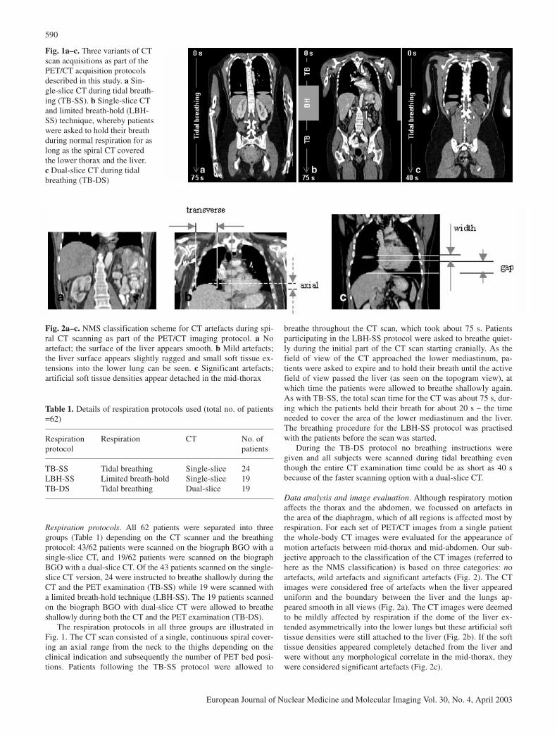
Respiration protocols. All 62 patients were separated into threegroups (Table 1) depending on the CT scanner and the breathingprotocol: 43/62 patients were scanned on the biograph BGO with asingle-slice CT, and 19/62 patients were scanned on the biographBGO with a dual-slice CT. Of the 43 patients scanned on the single-slice CT version, 24 were instructed to breathe shallowly during theCT and the PET examination (TB-SS) while 19 were scanned witha limited breath-hold technique (LBH-SS). The 19 patients scannedon the biograph BGO with dual-slice CT were allowed to breatheshallowly during both the CT and the PET examination (TB-DS).
The respiration protocols in all three groups are illustrated inFig. 1. The CT scan consisted of a single, continuous spiral cover-ing an axial range from the neck to the thighs depending on theclinical indication and subsequently the number of PET bed posi-tions. Patients following the TB-SS protocol were allowed to
breathe throughout the CT scan, which took about 75 s. Patientsparticipating in the LBH-SS protocol were asked to breathe quiet-ly during the initial part of the CT scan starting cranially. As thefield of view of the CT approached the lower mediastinum, pa-tients were asked to expire and to hold their breath until the activefield of view passed the liver (as seen on the topogram view), atwhich time the patients were allowed to breathe shallowly again.As with TB-SS, the total scan time for the CT was about 75 s, dur-ing which the patients held their breath for about 20 s – the timeneeded to cover the area of the lower mediastinum and the liver.The breathing procedure for the LBH-SS protocol was practisedwith the patients before the scan was started.
During the TB-DS protocol no breathing instructions were given and all subjects were scanned during tidal breathing eventhough the entire CT examination time could be as short as 40 sbecause of the faster scanning option with a dual-slice CT.
Data analysis and image evaluation. Although respiratory motionaffects the thorax and the abdomen, we focussed on artefacts inthe area of the diaphragm, which of all regions is affected most byrespiration. For each set of PET/CT images from a single patientthe whole-body CT images were evaluated for the appearance ofmotion artefacts between mid-thorax and mid-abdomen. Our sub-jective approach to the classification of the CT images (referred tohere as the NMS classification) is based on three categories: noartefacts, mild artefacts and significant artefacts (Fig. 2). The CTimages were considered free of artefacts when the liver appeareduniform and the boundary between the liver and the lungs ap-peared smooth in all views (Fig. 2a). The CT images were deemedto be mildly affected by respiration if the dome of the liver ex-tended asymmetrically into the lower lungs but these artificial softtissue densities were still attached to the liver (Fig. 2b). If the softtissue densities appeared completely detached from the liver andwere without any morphological correlate in the mid-thorax, theywere considered significant artefacts (Fig. 2c).
Table 1. Details of respiration protocols used (total no. of patients=62)
Respiration Respiration CT No. of protocol patients
TB-SS Tidal breathing Single-slice 24LBH-SS Limited breath-hold Single-slice 19TB-DS Tidal breathing Dual-slice 19
Fig. 1a–c. Three variants of CTscan acquisitions as part of thePET/CT acquisition protocolsdescribed in this study. a Sin-gle-slice CT during tidal breath-ing (TB-SS). b Single-slice CTand limited breath-hold (LBH-SS) technique, whereby patientswere asked to hold their breathduring normal respiration for aslong as the spiral CT coveredthe lower thorax and the liver.c Dual-slice CT during tidalbreathing (TB-DS)
Fig. 2a–c. NMS classification scheme for CT artefacts during spi-ral CT scanning as part of the PET/CT imaging protocol. a Noartefact; the surface of the liver appears smooth. b Mild artefacts;the liver surface appears slightly ragged and small soft tissue ex-tensions into the lower lung can be seen. c Significant artefacts;artificial soft tissue densities appear detached in the mid-thorax
591
European Journal of Nuclear Medicine and Molecular Imaging Vol. 30, No. 4, April 2003
slice CT and tidal breathing (TB-DS) protocol was freeof artefacts.
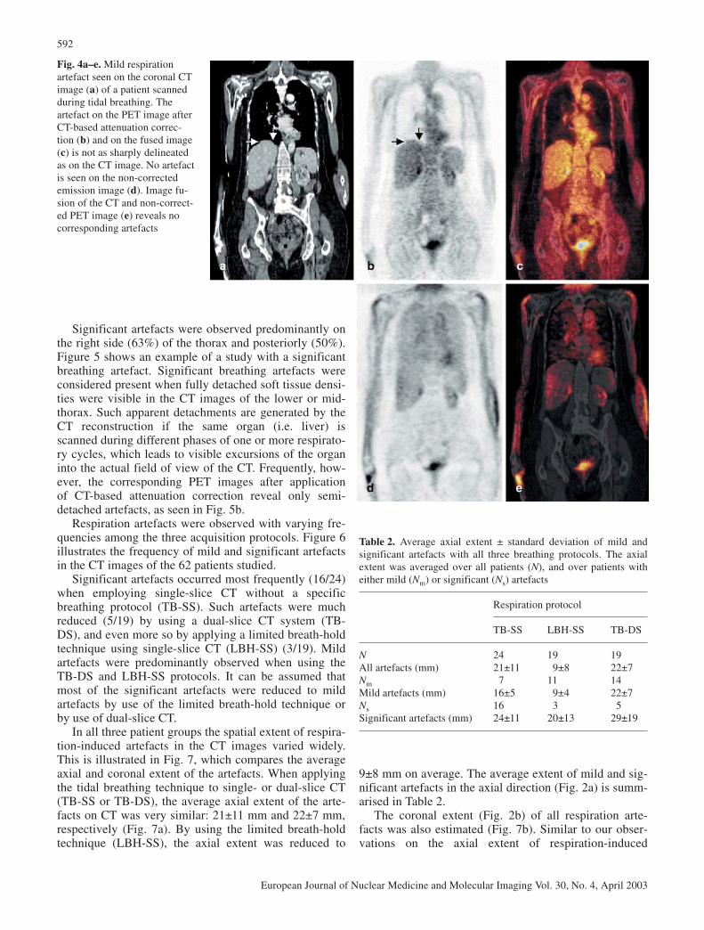
Throughout our patient population, mild respiration-induced artefacts were the most frequently observed.This was true for 11/19 (58%) and 14/19 (74%) patientsexamined with the LBH-SS and the TB-DS protocol, re-spectively. An example of a patient study with mild arte-facts in the right liver is shown in Fig. 4. The coronal CTimage (Fig. 4a) illustrates a typical mild respiration arte-fact as previously described by Goerres et al. [12]. Mildartefacts were observed mostly on the patients’ rightside, and translated from the CT to the corrected PETimages after CT-based attenuation correction. In our ex-perience the spatial extent of these artefacts seen in thecorrected PET images (Fig. 4b) was less than on the cor-responding CT images.
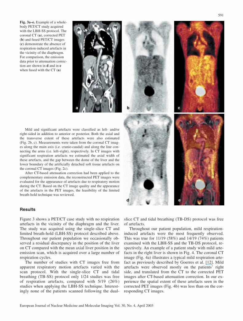
Fig. 3a–e. Example of a whole-body PET/CT study acquiredwith the LBH-SS protocol. Thecoronal CT (a), corrected PET(b) and fused PET/CT images(c) demonstrate the absence ofrespiration-induced artefacts inthe vicinity of the diaphragm.For comparison, the emissiondata prior to attenuation correc-tion are shown in d and in ewhen fused with the CT (a)
Mild and significant artefacts were classified as left- and/orright-sided in addition to anterior or posterior. Both the axial andthe transverse extent of these artefacts were also estimated(Fig. 2b, c). Measurements were taken from the coronal CT imag-es along the main axis (i.e. cranio-caudal) and along the line con-necting the arms (i.e. left–right), respectively. In CT images withsignificant respiration artefacts we estimated the axial width ofthese artefacts, and the gap between the dome of the liver and thelower boundary of the artificially detached soft tissue artefacts onthe coronal CT images (Fig. 2c).
After CT-based attenuation correction had been applied to thecomplementary emission data, the reconstructed PET images wereevaluated for the appearance of artefacts due to respiratory motionduring the CT. Based on the CT image quality and the appearanceof the artefacts in the PET images, the feasibility of the limitedbreath-hold technique was reviewed.
Results
Figure 3 shows a PET/CT case study with no respirationartefacts in the vicinity of the diaphragm and the liver.The study was acquired using the single-slice CT andlimited breath-hold (LBH-SS) protocol described above.Throughout our patient population we occasionally ob-served a residual discrepancy in the position of the liveron CT compared with the mean axial liver position in theemission scan, which is acquired over a large number ofrespiration cycles.
The number of studies with CT images free from apparent respiratory motion artefacts varied with thescan protocol. With the single-slice CT and tidalbreathing (TB-SS) protocol only 1/24 studies was freeof respiration artefacts, compared with 5/19 (26%)studies when applying the LBH-SS technique. Interest-ingly none of the patients scanned following the dual-
592
European Journal of Nuclear Medicine and Molecular Imaging Vol. 30, No. 4, April 2003
9±8 mm on average. The average extent of mild and sig-nificant artefacts in the axial direction (Fig. 2a) is summ-arised in Table 2.
The coronal extent (Fig. 2b) of all respiration arte-facts was also estimated (Fig. 7b). Similar to our obser-vations on the axial extent of respiration-induced
Fig. 4a–e. Mild respirationartefact seen on the coronal CTimage (a) of a patient scannedduring tidal breathing. Theartefact on the PET image afterCT-based attenuation correc-tion (b) and on the fused image(c) is not as sharply delineatedas on the CT image. No artefactis seen on the non-correctedemission image (d). Image fu-sion of the CT and non-correct-ed PET image (e) reveals nocorresponding artefacts
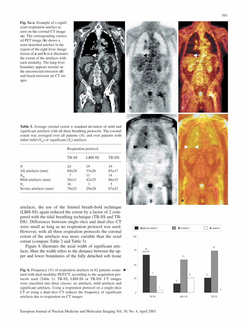
Significant artefacts were observed predominantly onthe right side (63%) of the thorax and posteriorly (50%).Figure 5 shows an example of a study with a significantbreathing artefact. Significant breathing artefacts wereconsidered present when fully detached soft tissue densi-ties were visible in the CT images of the lower or mid-thorax. Such apparent detachments are generated by theCT reconstruction if the same organ (i.e. liver) isscanned during different phases of one or more respirato-ry cycles, which leads to visible excursions of the organinto the actual field of view of the CT. Frequently, how-ever, the corresponding PET images after application of CT-based attenuation correction reveal only semi-detached artefacts, as seen in Fig. 5b.
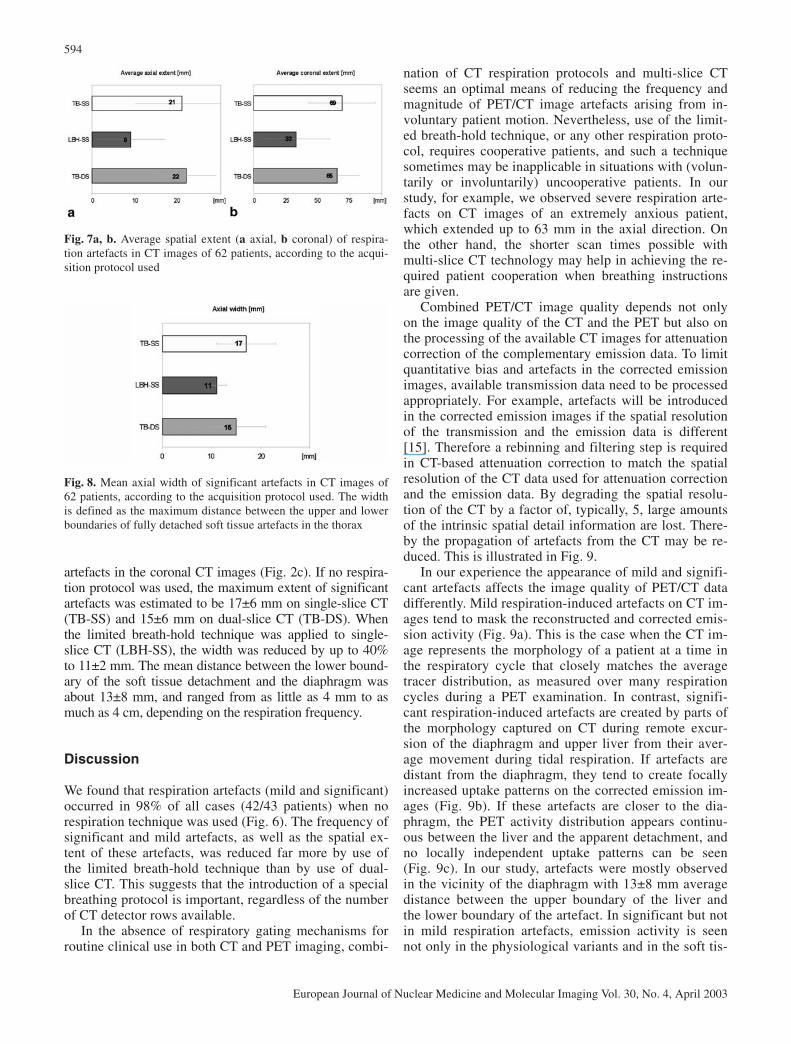
Respiration artefacts were observed with varying fre-quencies among the three acquisition protocols. Figure 6illustrates the frequency of mild and significant artefactsin the CT images of the 62 patients studied.
Significant artefacts occurred most frequently (16/24)when employing single-slice CT without a specificbreathing protocol (TB-SS). Such artefacts were muchreduced (5/19) by using a dual-slice CT system (TB-DS), and even more so by applying a limited breath-holdtechnique using single-slice CT (LBH-SS) (3/19). Mildartefacts were predominantly observed when using theTB-DS and LBH-SS protocols. It can be assumed thatmost of the significant artefacts were reduced to mildartefacts by use of the limited breath-hold technique orby use of dual-slice CT.
In all three patient groups the spatial extent of respira-tion-induced artefacts in the CT images varied widely.This is illustrated in Fig. 7, which compares the averageaxial and coronal extent of the artefacts. When applyingthe tidal breathing technique to single- or dual-slice CT(TB-SS or TB-DS), the average axial extent of the arte-facts on CT was very similar: 21±11 mm and 22±7 mm,respectively (Fig. 7a). By using the limited breath-holdtechnique (LBH-SS), the axial extent was reduced to
Table 2. Average axial extent ± standard deviation of mild andsignificant artefacts with all three breathing protocols. The axialextent was averaged over all patients (N), and over patients witheither mild (Nm) or significant (Ns) artefacts
Respiration protocol
TB-SS LBH-SS TB-DS
N 24 19 19All artefacts (mm) 21±11 9±8 22±7Nm 7 11 14Mild artefacts (mm) 16±5 9±4 22±7Ns 16 3 5Significant artefacts (mm) 24±11 20±13 29±19
593
European Journal of Nuclear Medicine and Molecular Imaging Vol. 30, No. 4, April 2003
Fig. 5a–e. Example of a signif-icant respiration artefact asseen on the coronal CT image(a). The corresponding correct-ed PET image (b) shows asemi-detached artefact in theregion of the right liver. Imagefusion of a and b in c illustratesthe extent of the artefacts witheach modality. The lung-liverboundary appears normal onthe uncorrected emission (d)and fused emission (e) CT im-ages
Table 3. Average coronal extent ± standard deviation of mild andsignificant artefacts with all three breathing protocols. The coronalextent was averaged over all patients (N), and over patients witheither mild (Nm) or significant (Ns) artefacts
Respiration protocol
TB-SS LBH-SS TB-DS
N 24 19 19All artefacts (mm) 69±26 33±26 65±17Nm 7 11 14Mild artefacts (mm) 56±11 42±25 66±15Ns 16 3 5Severe artefacts (mm) 78±21 29±28 67±23
Fig. 6. Frequency (%) of respiration artefacts in 62 patients exam-ined with dual-modality PET/CT, according to the acquisition pro-tocols used (Table 1): TB-SS, LBH-SS or TB-DS. CT imageswere classified into three classes: no artefacts, mild artefacts andsignificant artefacts. Using a respiration protocol on a single-sliceCT or using a dual-slice CT reduces the frequency of significantartefacts due to respiration on CT images
artefacts, the use of the limited breath-hold technique (LBH-SS) again reduced the extent by a factor of 2 com-pared with the tidal breathing technique (TB-SS and TB-DS). Differences between single-slice and dual-slice CTwere small as long as no respiration protocol was used.However, with all three respiration protocols the coronalextent of the artefacts was more variable than the axialextent (compare Table 2 and Table 3).
Figure 8 illustrates the axial width of significant arte-facts. Here the width refers to the distance between the up-per and lower boundaries of the fully detached soft tissue
▲
594
European Journal of Nuclear Medicine and Molecular Imaging Vol. 30, No. 4, April 2003
nation of CT respiration protocols and multi-slice CTseems an optimal means of reducing the frequency andmagnitude of PET/CT image artefacts arising from in-voluntary patient motion. Nevertheless, use of the limit-ed breath-hold technique, or any other respiration proto-col, requires cooperative patients, and such a techniquesometimes may be inapplicable in situations with (volun-tarily or involuntarily) uncooperative patients. In ourstudy, for example, we observed severe respiration arte-facts on CT images of an extremely anxious patient,which extended up to 63 mm in the axial direction. Onthe other hand, the shorter scan times possible withmulti-slice CT technology may help in achieving the re-quired patient cooperation when breathing instructionsare given.
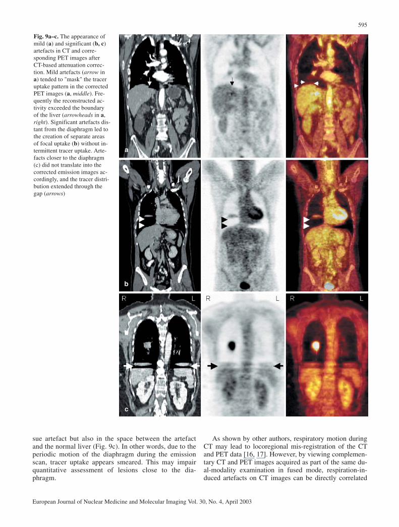
Combined PET/CT image quality depends not onlyon the image quality of the CT and the PET but also onthe processing of the available CT images for attenuationcorrection of the complementary emission data. To limitquantitative bias and artefacts in the corrected emissionimages, available transmission data need to be processedappropriately. For example, artefacts will be introducedin the corrected emission images if the spatial resolutionof the transmission and the emission data is different[15]. Therefore a rebinning and filtering step is requiredin CT-based attenuation correction to match the spatialresolution of the CT data used for attenuation correctionand the emission data. By degrading the spatial resolu-tion of the CT by a factor of, typically, 5, large amountsof the intrinsic spatial detail information are lost. There-by the propagation of artefacts from the CT may be re-duced. This is illustrated in Fig. 9.
In our experience the appearance of mild and signifi-cant artefacts affects the image quality of PET/CT datadifferently. Mild respiration-induced artefacts on CT im-ages tend to mask the reconstructed and corrected emis-sion activity (Fig. 9a). This is the case when the CT im-age represents the morphology of a patient at a time inthe respiratory cycle that closely matches the averagetracer distribution, as measured over many respirationcycles during a PET examination. In contrast, signifi-cant respiration-induced artefacts are created by parts ofthe morphology captured on CT during remote excur-sion of the diaphragm and upper liver from their aver-age movement during tidal respiration. If artefacts aredistant from the diaphragm, they tend to create focallyincreased uptake patterns on the corrected emission im-ages (Fig. 9b). If these artefacts are closer to the dia-phragm, the PET activity distribution appears continu-ous between the liver and the apparent detachment, andno locally independent uptake patterns can be seen(Fig. 9c). In our study, artefacts were mostly observedin the vicinity of the diaphragm with 13±8 mm averagedistance between the upper boundary of the liver andthe lower boundary of the artefact. In significant but notin mild respiration artefacts, emission activity is seennot only in the physiological variants and in the soft tis-
Fig. 7a, b. Average spatial extent (a axial, b coronal) of respira-tion artefacts in CT images of 62 patients, according to the acqui-sition protocol used
Fig. 8. Mean axial width of significant artefacts in CT images of62 patients, according to the acquisition protocol used. The widthis defined as the maximum distance between the upper and lowerboundaries of fully detached soft tissue artefacts in the thorax
artefacts in the coronal CT images (Fig. 2c). If no respira-tion protocol was used, the maximum extent of significantartefacts was estimated to be 17±6 mm on single-slice CT(TB-SS) and 15±6 mm on dual-slice CT (TB-DS). Whenthe limited breath-hold technique was applied to single-slice CT (LBH-SS), the width was reduced by up to 40%to 11±2 mm. The mean distance between the lower bound-ary of the soft tissue detachment and the diaphragm wasabout 13±8 mm, and ranged from as little as 4 mm to asmuch as 4 cm, depending on the respiration frequency.
Discussion
We found that respiration artefacts (mild and significant)occurred in 98% of all cases (42/43 patients) when norespiration technique was used (Fig. 6). The frequency ofsignificant and mild artefacts, as well as the spatial ex-tent of these artefacts, was reduced far more by use ofthe limited breath-hold technique than by use of dual-slice CT. This suggests that the introduction of a specialbreathing protocol is important, regardless of the numberof CT detector rows available.
In the absence of respiratory gating mechanisms forroutine clinical use in both CT and PET imaging, combi-
595
European Journal of Nuclear Medicine and Molecular Imaging Vol. 30, No. 4, April 2003
sue artefact but also in the space between the artefactand the normal liver (Fig. 9c). In other words, due to theperiodic motion of the diaphragm during the emissionscan, tracer uptake appears smeared. This may impairquantitative assessment of lesions close to the dia-phragm.
As shown by other authors, respiratory motion duringCT may lead to locoregional mis-registration of the CTand PET data [16, 17]. However, by viewing complemen-tary CT and PET images acquired as part of the same du-al-modality examination in fused mode, respiration-in-duced artefacts on CT images can be directly correlated
Fig. 9a–c. The appearance ofmild (a) and significant (b, c)artefacts in CT and corre-sponding PET images afterCT-based attenuation correc-tion. Mild artefacts (arrow ina) tended to "mask" the traceruptake pattern in the correctedPET images (a, middle). Fre-quently the reconstructed ac-tivity exceeded the boundaryof the liver (arrowheads in a,right). Significant artefacts dis-tant from the diaphragm led tothe creation of separate areasof focal uptake (b) without in-termittent tracer uptake. Arte-facts closer to the diaphragm(c) did not translate into thecorrected emission images ac-cordingly, and the tracer distri-bution extended through thegap (arrows)
596
European Journal of Nuclear Medicine and Molecular Imaging Vol. 30, No. 4, April 2003
with artificial uptake patterns in corrected PET images(Fig. 9). Some authors recommend viewing the emissionimages before attenuation correction is applied in order toexclude respiration-induced bias [18]. We feel that thismight be necessary in the case of lesions in the vicinity ofthe diaphragm. For most clinical scenarios today, howev-er, a combined PET/CT acquisition protocol involving acarefully designed respiration technique, like the limitedbreath-hold protocol described here, seems sufficient.
Conclusion
In conclusion, an appropriate respiration protocol is themain determinant of artefact-free PET/CT images of thelower thorax and upper abdomen. We have shown thatapplication of a limited breath-hold technique when ac-quiring the CT part of a combined PET/CT examinationsignificantly reduces the frequency and extent of respira-tion-induced artefacts in the lower thorax.
Acknowledgements. The authors like to thank Paulina Khersonskaya,Sam Hsiao, Bärbel Terschüren, Sandra Pabst, and Ruth Hall fortheir help with the data acquisition. This paper benefited fromhelpful discussions with David Townsend, Jeffrey Yap, and Thomas Bruckbauer.
References
1. Gambhir SS, Czernin J, Schwimmer J, Silverman DHS, Coleman EE, Phelps ME. A tabulated summary of the FDGPET literature. J Nucl Med 2001; 42 (5 Suppl):1S–93S.
2. Vansteenkiste JF, Stroobants SG, Dupont PJ, Leyn PRD, Wever WF, Verbeken EK, Nuyts JL, Maes FP, Bogaert JG.FDG-PET scan in potentially operable non-small cell lungcancer: Do anatometabolic PET-CT fusion images improve thelocalisation of regional lymph node metastasis? Eur J NuclMed 1998; 25:1495–1501.
3. Hutton BF, Braun M, Thurfjell L, Lau DYH. Image registra-tion: an essential tool for nuclear medicine. Eur J Nucl Med2002; 29:559–577.
4. Woods RP, Grafton ST, Holmes CJ, Cherry SR, Mazziotta JC.Automated image registration. I. General methods and intra-subject, intramodality validation. J Comput Assist Tomogr1998; 22:139–152.
5. Yu JN, Fahey FH, Gage HD, Eades CG, Harkness BA, PelizzariCA, Keys JW Jr. Intermodality, retrospective image registra-tion in the thorax. J Nucl Med 1995; 36:2333–2338.
6. Townsend DW, Cherry S. Combining anatomy and function:the path to true image fusion. Eur Radiol 2001; 11:1968–1974.
7. Beyer T, Townsend DW, Brun T, Kinahan PE, Charron M,Roddy R, Jerin J, Young J, Nutt R, Byars LG. A combinedPET/CT tomograph for clinical oncology. J Nucl Med 2000;41:1369–1379.
8. Kinahan PE, Townsend DW, Beyer T, Sashin D. Attenuationcorrection for a combined 3D PET/CT scanner. Med Phys1998; 25:2046–2053.
9. von Schulthess GK. Cost considerations regarding an integrat-ed CT-PET system. Eur Radiol 2000; 10 (Suppl 3):S377–S380.
10. Hoh CK, Hawkins RA, Glaspy JA, Dahlbom M, Tse NY,Hoffman EJ, Schiepers C, Choi Y, Rege S, Nitzsche E, Maddahi J, Phelps ME. Cancer detection with whole-bodyPET using 2-[18F]fluoro-2-deoxy-d-glucose. J Comput AssistTomogr 1993; 17:582–589.
11. Beyer T, Townsend D, Blodgett T. Dual-modality PET/CT to-mography for clinical oncology. Q J Nucl Med 2002; 46:24–34.
12. Goerres GW, Kamel E, Heidelberg T-NH, Schwitter MR,Burger C, von Schulthess GK. PET-CT image co-registrationin the thorax: influence of respiration. Eur J Nucl Med 2002;29:351–360.
13. Beyer T, Townsend D. Dual-modality PET/CT imaging: CT-based attenuation correction in the presence of CT contrastagents. J Nucl Med 2001; 42:56P.
14. Carney J, Beyer T, Brasse D, Yap J, Townsend D. ClinicalPET/CT scanning using oral CT contrast agents. J Nucl Med2002; 43:57P.
15. Meikle SR, Dahlbom M, Cherry SR. Attenuation correctionusing count-limited transmission data in positron emission tomography. J Nucl Med 1993; 34:143–150.
16. Cohade C, Osman M, Wahl RL. Accuracy of PET and CTspatial registration of lung lesions with PET-CT. J Nucl Med2002; 43:14P–15P.
17. Nakamoto Y, Cohade C, Osman M, Tatsumi M, Traughber BJ,Marshall LT, Wahl RL. Comparison of size and location ofnormal upper abdominal organs on PET and CT usingPET/CT. J Nucl Med 2002; 43:14P.
18. Osman MM, Cohade C, Nakamoto Y, Wahl RL. Clinically sig-nificant inaccurate localization of lesions with PET-CT: fre-quency in 275 patients. J Nucl Med 2002; 43:32P.