Download - Zurich Open Repository and Archive
67
University of Zurich Zurich Open Repository and Archive Winterthurerstr. 190 CH-8057 Zurich http://www.zora.uzh.ch Year: 2009 Chapter 32 Human Retroviruses, HIV and HTLV Schüpbach, J Schüpbach, J (2009). Chapter 32 Human Retroviruses, HIV and HTLV. In: Specter, S; Hodinka, R L; Young, S A; Wiedbrauk, D L. Clinical Virology Manual. Washington D.C., 578-629. Postprint available at: http://www.zora.uzh.ch Posted at the Zurich Open Repository and Archive, University of Zurich. http://www.zora.uzh.ch Originally published at: Specter, S; Hodinka, R L; Young, S A; Wiedbrauk, D L 2009. Clinical Virology Manual. Washington D.C., 578-629.
-
Upload
khangminh22 -
Category
Documents
-
view
0 -
download
0
Transcript of Download - Zurich Open Repository and Archive

University of ZurichZurich Open Repository and Archive
Winterthurerstr. 190
CH-8057 Zurich
http://www.zora.uzh.ch
Year: 2009
Chapter 32 Human Retroviruses, HIV and HTLV
Schüpbach, J
Schüpbach, J (2009). Chapter 32 Human Retroviruses, HIV and HTLV. In: Specter, S; Hodinka, R L; Young, S A;Wiedbrauk, D L. Clinical Virology Manual. Washington D.C., 578-629.Postprint available at:http://www.zora.uzh.ch
Posted at the Zurich Open Repository and Archive, University of Zurich.http://www.zora.uzh.ch
Originally published at:Specter, S; Hodinka, R L; Young, S A; Wiedbrauk, D L 2009. Clinical Virology Manual. Washington D.C.,578-629.

Manuscript for Clinical Virology Manual, 4th Edition;
Steven Specter, Richard L. Hodinka, Stephen A. Young, Eds.,
American Society for Microbiology Press, Washington, D.C.; 2008 (in press)
Human Retroviruses, HIV and HTLV
Jörg Schüpbach, M.D.
Swiss National Center for Retroviruses
University of Zurich
Gloriastrasse 30
CH-8006 Zurich, Switzerland
Fax: (+41)-44-634-4965
Email: [email protected]
2
Human Retroviruses, HIV and HTLV Jörg Schüpbach, M.D.
INTRODUCTION
Retroviruses were for many decades well known causative agents of leukemias, lymphomas, other cancers, or chronic inflammations in various animal species. The discovery of the first human retrovirus, human T-cell leukemia virus (now renamed human T-lymphotropic retrovirus type 1; HTLV-1) was reported in 1980 (Poiesz et al., 1981). HTLV-1 was soon identified as the causative agent of adult T-cell leukemia/lymphoma (ATLL), a rapidly progressing cancer of CD4+ T lymphocytes first described in southeastern Japan (Takatsuki et al., 1977). Knowledge gained from HTLV-1 research was important for the subsequent detection of other human retroviruses, first the related HTLV-2 (Kalyanaraman et al., 1982). Soon thereafter, human immunodeficiency virus (HIV)-1 was for the first time isolated from a patient with an early stage of the newly recognized acquired immunodeficiency syndrome (AIDS) (Barre-Sinoussi et al., 1983). Two years later, a second AIDS-causing virus, HIV-2, was discovered (Clavel et al., 1986a).
Investigations among nonhuman primates showed a wide distribution of viruses resembling both the HTLV and, respectively, HIV groups of retroviruses. Simian T- lymphotropic retrovirus type (STLV)-1 and STLV-2, simian counterparts of HTLV-1 and HTLV-2, were identified. STLV-3 forms a third group of lymphotropic viruses infecting various African monkey species. HTLV-3, a counterpart of STLV-3 in man, was recently detected in African pygmies (Calattini et al., 2005; Wolfe et al., 2005; Calattini et al., 2006; Switzer et al., 2006). A further HTLV forming a fourth group, HTLV-4, also has been reported (Wolfe et al., 2005). Together, these viruses now constitute four groups of primate T-lymphotropic retroviruses (PTLV-1, -2, -3, -4), with
representatives in both simians (STLV) and humans (HTLV). Similarly, both HIV-1 and HIV-2 were shown to originate from primate lentiviruses collectively named simian immunodeficiency viruses (SIV).
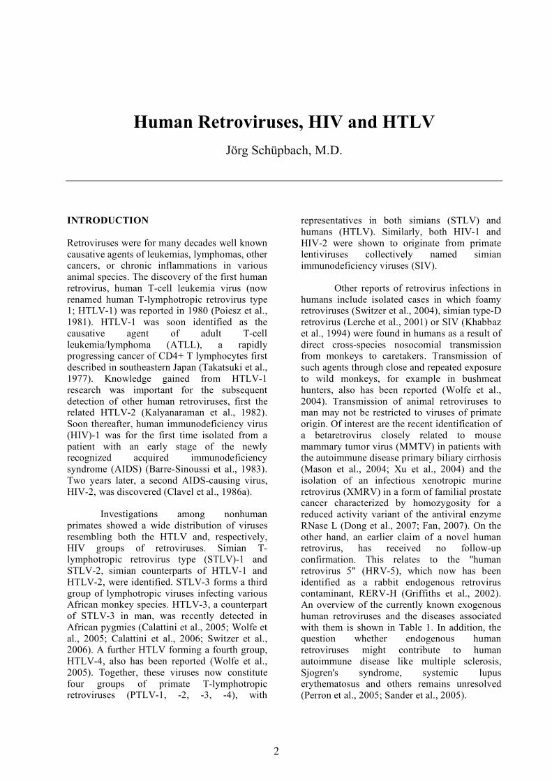
Other reports of retrovirus infections in humans include isolated cases in which foamy retroviruses (Switzer et al., 2004), simian type-D retrovirus (Lerche et al., 2001) or SIV (Khabbaz et al., 1994) were found in humans as a result of direct cross-species nosocomial transmission from monkeys to caretakers. Transmission of such agents through close and repeated exposure to wild monkeys, for example in bushmeat hunters, also has been reported (Wolfe et al., 2004). Transmission of animal retroviruses to man may not be restricted to viruses of primate origin. Of interest are the recent identification of a betaretrovirus closely related to mouse mammary tumor virus (MMTV) in patients with the autoimmune disease primary biliary cirrhosis (Mason et al., 2004; Xu et al., 2004) and the isolation of an infectious xenotropic murine retrovirus (XMRV) in a form of familial prostate cancer characterized by homozygosity for a reduced activity variant of the antiviral enzyme RNase L (Dong et al., 2007; Fan, 2007). On the other hand, an earlier claim of a novel human retrovirus, has received no follow-up confirmation. This relates to the "human retrovirus 5" (HRV-5), which now has been identified as a rabbit endogenous retrovirus contaminant, RERV-H (Griffiths et al., 2002). An overview of the currently known exogenous human retroviruses and the diseases associated with them is shown in Table 1. In addition, the question whether endogenous human retroviruses might contribute to human autoimmune disease like multiple sclerosis, Sjogren's syndrome, systemic lupus erythematosus and others remains unresolved (Perron et al., 2005; Sander et al., 2005).
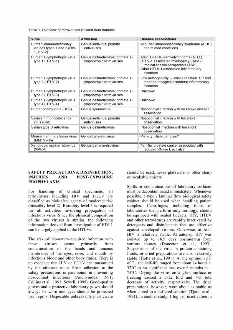
Table 1. Overview of retroviruses isolated from humans
Virus Affiliation Disease associations Human immunodeficiency
viruses types 1 and 2 (HIV-1, HIV-2)
Genus lentivirus; primate lentiviruses
Acquired immunodeficiency syndrome (AIDS) and related conditions
Human T-lymphotropic virus type 1 (HTLV-1)
Genus deltaretrovirus; primate T-lymphotropic retroviruses
Adult T-cell leukemia/lymphoma (ATLL) HTLV-1 associated myelopathy (HAM) /
tropical spastic paraparesis (TSP) Other HTLV-1 associated inflammatory
disorders Human T-lymphotropic virus
type 2 (HTLV-2) Genus deltaretrovirus; primate T-
lymphotropic retroviruses Low pathogenicity — cases of HAM/TSP and
other neurological disorders; inflammatory disorders
Human T-lymphotropic virus type 3 (HTLV-3)
Genus deltaretrovirus; primate T-lymphotropic retroviruses
Unknown
Human T-lymphotropic virus type 4 (HTLV-4)
Genus deltaretrovirus; primate T-lymphotropic retroviruses
Unknown
Human foamy virus (HFV) Genus spumavirus Nosocomial infection with no known disease association
Simian immunodeficiency virus (SIV)
Genus lentivirus; primate lentiviruses
Nosocomial infection with too short observation
Simian type D retrovirus Genus deltaretrovirus Nosocomial infection with too short observation
Mouse mammary tumor virus (MMTV)-like
Genus betaretrovirus Primary biliary cirrhosis?
Xenotropic murine retrovirus (XMRV)
Genus gammaretrovirus Familial prostate cancer associated with reduced RNase L activity?
SAFETY PRECAUTIONS, DISINFECTION, INJURIES AND POST-EXPOSURE PROPHYLAXIS
For handling of clinical specimens, all retroviruses including HIV and HTLV are classified as biological agents of moderate risk (biosafety level 2). Biosafety level 3 is required for all activities involving propagation of infectious virus. Since the physical composition of the two viruses is similar, the following information derived from investigation of HIV-1 can be largely applied to the HTLVs.
The risk of laboratory-acquired infection with these viruses stems primarily from contamination of the hands and mucous membranes of the eyes, nose, and mouth by infectious blood and other body fluids. There is no evidence that HIV or HTLV are transmitted by the airborne route. Strict adhesion to the safety precautions is paramount in preventing nosocomial infections (Anonymous, 1991; Collins et al., 1991; Sewell, 1995). Good-quality gloves and a protective laboratory gown should always be worn and eyes should be protected from spills. Disposable unbreakable plasticware
should be used, never glassware or other sharp or breakable objects.
Spills or contaminations of laboratory surfaces must be decontaminated immediately. Whenever possible, a type 2 laminar flow biological safety cabinet should be used when handling patient samples. Centrifuges, including those of laboratories that perform only serology, should be equipped with sealed buckets. HIV, HTLV and other retroviruses are rapidly inactivated by detergents and disinfectants that are effective against enveloped viruses. Otherwise, at least HIV is relatively stable. At autopsy, HIV was isolated up to 16.5 days postmortem from various tissues (Douceron et al., 1993). Suspensions of the virus in protein-containing fluids, or dried preparations are also relatively stable (Tjotta et al., 1991). At the optimum pH of 7.1 the half-life ranged from about 24 hours at 37°C to no significant loss over 6 months at –75°C. Drying the virus on a glass surface or freezing caused a 5-12 fold and 4-5 fold decrease of activity, respectively. The dried preparations, however, were about as stable as when stored in a buffered solution (Tjotta et al., 1991). In another study, 1 log10 of inactivation in

4
culture fluid, seawater, sewage, and dechlorinated tap water (all sterile and kept at 16°C in the dark) required 1.3, 1.6, 2.9, and 1.8 days, respectively. After the first 4 days the inactivation became even slower (1 log10 inactivation after 4.3, 2.6, 5.7, and 4.6 days respectively). HIV was more stable than herpes simplex virus, but less stable than poliovirus (Sattar and Springthorpe, 1991). These data are not meant to suggest that HIV transmission might occur by exposure to water, for which there is absolutely no basis. They should, however, make clear that caution is important when working with HIV.
The standard disinfectant recommended for contaminated surfaces is a hypochlorite solution with a concentration of 0.5% available chlorine (5 g/L, 5000 ppm). When working with HIV cultures and virus preparations, a higher concentration of 1% available chlorine is recommended (Anonymous, 1991; Van Bueren et al., 1995). Fresh 2% solutions of alkaline glutaraldehyde are effective, but care should be taken that they are not too dilute or have not become stale when used for disinfecting HIV associated with organic matter. A solution of iodine and detergent (2% Jodopax) will remove all detectable HIV-1 activity. In contrast, 70% industrial methylated spirit or 70% ethanol is not effective in inactivating dried protein-rich spills of cell-free or cell-associated HIV within a reasonable amount of time; complete inactivation requires up to 20 minutes (Tjotta et al., 1991; van Bueren et al., 1994).
The risk of HIV infection following percutaneous needle-stick exposure to HIV-contaminated blood is estimated to be between 0.13 and 0.5%. It depends on the depth of the penetration (relative risk (RR) of percutaneous lesions 16.1), visible contamination of the penetrating object with blood (RR 5.2), prior use for an intravenous or intra-arterial injection (RR 5.1) and on the disease stage (respectively the viral load) of the index patient (RR 6.4) (Anonymous, 1995). Needlestick or other puncture wounds, cuts, and skin contaminated by spills or splashes of specimen material should be thoroughly washed with soap and water and disinfected with a nonirritating disinfectant. Bleeding should be encouraged. In case of percutaneous injury or contact of mucous membranes or nonintact skin (e.g., exposed skin that is chapped, abraded, or afflicted with dermatitis) with blood, tissue, or other body fluids potentially infectious, an antiretroviral postexposure prophylaxis (PEP) should be started immediately according to guidelines published online http://www.hivatis.org/Guidelines/Default.aspx?MenuItem=Guidelines. Note that these recommendations do not apply to HTLV, as many of the drugs effective against HIV, particularly the protease inhibitors and non-nucleoside reverse transcriptase inhibitors, are ineffective against HTLV.
HUMAN IMMUNODEFICIENCY VIRUSES
Biology and Epidemiology
HIV-1 and HIV-2 are members of the genus Lentivirus of the Retroviridae family. They are enveloped plus-strand RNA viruses, with a diameter of about 110 nm. Infectious particles (virions) contain two identical copies of single-stranded RNA of about 9 to 10 kb. These are surrounded by structural proteins that form the nucleocapsid and the matrix shell, surrounded by a lipid envelope derived from the host cell membrane. Viral glycoprotein trimers which mediate adsorption to and penetration of the host cell membrane are inserted in this envelope (Fig. 1).
HIV-1, first isolated in 1983 (Barre-Sinoussi et al., 1983) and confirmed in the following year as being virologically and serologically associated with early and late stages of AIDS (Gallo et al., 1984; Levy et al., 1984; Popovic et al., 1984; Sarngadharan et al., 1984; Schupbach et al., 1984), is the more aggressive virus and responsible for the AIDS pandemic (Table 2). HIV-2, discovered in 1986 (Clavel et al., 1986b), is less pathogenic. Rates of heterosexual and mother-to-child transmission of HIV-2 are low, and latency dominates the clinical picture; the virus rarely causes AIDS (Schim van der Loeff and Aaby, 1999; Bock and Markovitz, 2001; Jaffar et al., 2004).
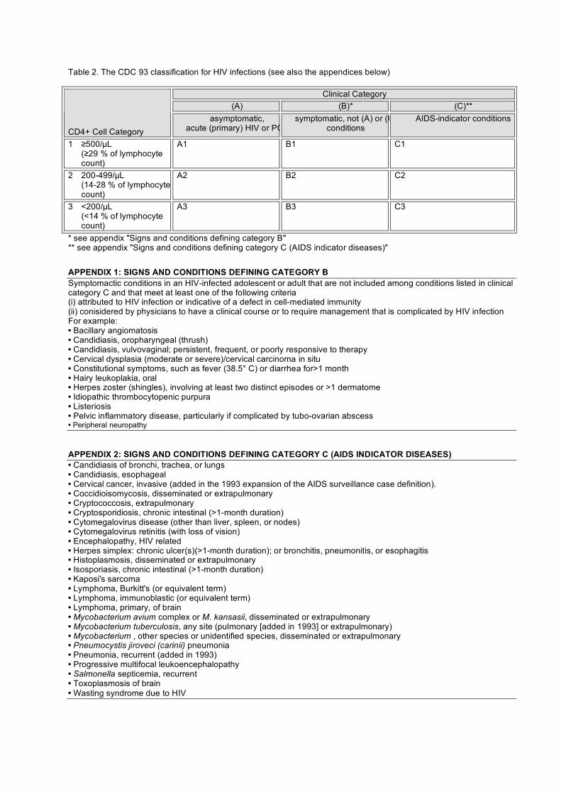
Table 2. The CDC 93 classification for HIV infections (see also the appendices below)
Clinical Category (A) (B)* (C)**
CD4+ Cell Category
asymptomatic, acute (primary) HIV or PGL
symptomatic, not (A) or (C) conditions
AIDS-indicator conditions
1 ≥500/µL (≥29 % of lymphocyte
count)
A1 B1 C1
2 200-499/µL (14-28 % of lymphocyte
count)
A2 B2 C2
3 <200/µL (<14 % of lymphocyte
count)
A3 B3 C3
* see appendix "Signs and conditions defining category B" ** see appendix "Signs and conditions defining category C (AIDS indicator diseases)"
APPENDIX 1: SIGNS AND CONDITIONS DEFINING CATEGORY B Symptomactic conditions in an HIV-infected adolescent or adult that are not included among conditions listed in clinical category C and that meet at least one of the following criteria (i) attributed to HIV infection or indicative of a defect in cell-mediated immunity (ii) conisidered by physicians to have a clinical course or to require management that is complicated by HIV infection For example: • Bacillary angiomatosis • Candidiasis, oropharyngeal (thrush) • Candidiasis, vulvovaginal; persistent, frequent, or poorly responsive to therapy • Cervical dysplasia (moderate or severe)/cervical carcinoma in situ • Constitutional symptoms, such as fever (38.5° C) or diarrhea for>1 month • Hairy leukoplakia, oral • Herpes zoster (shingles), involving at least two distinct episodes or >1 dermatome • Idiopathic thrombocytopenic purpura • Listeriosis • Pelvic inflammatory disease, particularly if complicated by tubo-ovarian abscess • Peripheral neuropathy APPENDIX 2: SIGNS AND CONDITIONS DEFINING CATEGORY C (AIDS INDICATOR DISEASES) • Candidiasis of bronchi, trachea, or lungs • Candidiasis, esophageal • Cervical cancer, invasive (added in the 1993 expansion of the AIDS surveillance case definition). • Coccidioisomycosis, disseminated or extrapulmonary • Cryptococcosis, extrapulmonary • Cryptosporidiosis, chronic intestinal (>1-month duration) • Cytomegalovirus disease (other than liver, spleen, or nodes) • Cytomegalovirus retinitis (with loss of vision) • Encephalopathy, HIV related • Herpes simplex: chronic ulcer(s)(>1-month duration); or bronchitis, pneumonitis, or esophagitis • Histoplasmosis, disseminated or extrapulmonary • Isosporiasis, chronic intestinal (>1-month duration) • Kaposi's sarcoma • Lymphoma, Burkitt's (or equivalent term) • Lymphoma, immunoblastic (or equivalent term) • Lymphoma, primary, of brain • Mycobacterium avium complex or M. kansasii, disseminated or extrapulmonary • Mycobacterium tuberculosis, any site (pulmonary [added in 1993] or extrapulmonary) • Mycobacterium , other species or unidentified species, disseminated or extrapulmonary • Pneumocystis jiroveci (carinii) pneumonia • Pneumonia, recurrent (added in 1993) • Progressive multifocal leukoencephalopathy • Salmonella septicemia, recurrent • Toxoplasmosis of brain • Wasting syndrome due to HIV

6
Origin of HIV
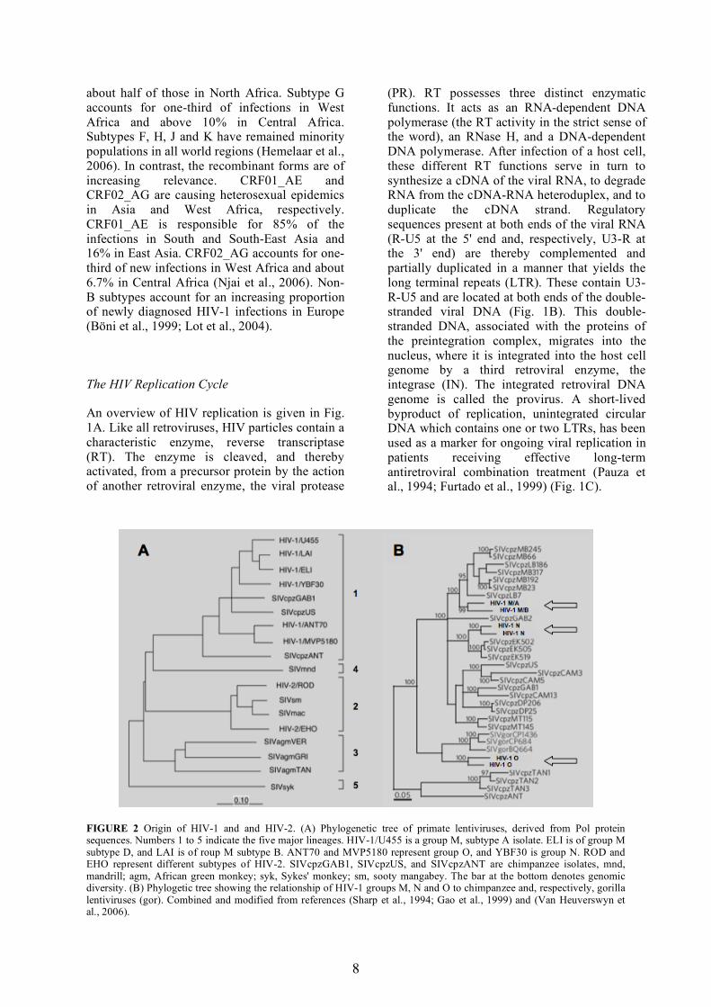
A group of related viruses, SIV, naturally infect various species of Old World monkeys and the chimpanzee (Fig. 2A). These primate lentiviruses are categorized into five major lineages. Lineage 1 contains the various isolates of HIV-1, which are subclassified into three groups, M (main), O (outlier), and N (Simon et al., 1998). From the phylogenetic tree it is evident that group M isolates (e.g. HIV-1/LAI) are more closely related to two isolates from chimpanzee, SIVcpzGAB1 and SIVcpzUS, than to isolates of HIV-1 group O (HIV-1/ANT70) or to another chimpanzee isolate, SIVcpzANT. These data indicate that the HIV-1 epidemic is the result of zoonotic virus transmissions from chimpanzee, subspecies Pan troglodytes troglodytes, to human (Gao et al., 1999). The origin of group M diversification, i.e., the beginning of the HIV-1 pandemic, is placed around 1930 (Korber et al., 2000; Salemi et al., 2001). Recent investigations involving HIV serology and RT-PCR performed on fecal samples collected in big ape habitats in Cameroon have demonstrated a wide variety of SIVcpz isolates, which are organized in phylogenetic clades restricted to the respective habitat area. SIVcpz prevalence in some habitat areas is as high as 23 — 35%, while in others it is only a few percent or absent. Phylogenetic analysis of SIVcpz together with HIV-1 isolates clearly shows that HIV-1 group M originates from SIVcpz isolates that are prevalent in two P. t. troglodytes populations living in the extreme south-east of Cameroon. Moreover, HIV-1 group N originated from SIVcpz isolates from P. t. troglodytes living in a different area located about 250 km to the west-northwest (Keele et al., 2006). Wild chimpanzees therefore act as a reservoir for HIV-1 groups M and N. Viruses closely related to HIV-1 group O have been isolated from gorillas living in forest habitats of Cameroon 400 km apart from each other (Van Heuverswyn et al., 2006). Phylogenetic analysis demonstrates that both HIV-1 group O and SIVgor have originated from chimpanzee viruses (Fig. 2 B). Whether chimpanzees transmitted HIV-1 group O viruses to gorillas and humans independently, or to gorillas that then transmitted it to humans secondarily is unknown.
Lineage 2 of primate lentiviruses contains the various isolates of HIV-2, which are related to
viruses infecting sooty mangabeys (SIVsm). SIVsm also has been transmitted naturally to macaques. HIV-2 strain ROD differs less from SIVsm or SIVmac than it does from another human isolate, HIV-2/EHO (Fig. 2A). This, together with other similar examples, has led to the conclusion that the HIV-2 epidemic is also the result of multiple simian-to-human cross-species transmissions. Transmission of the epidemic subtypes HIV-2 A and B may have occurred around 1940 (Lemey et al., 2003).
HIV Groups and Subtypes
The extraordinary variability of HIV, due to rapid mutation and recombination, has led to the development and geographical distribution of various distinctive clades, or subtypes, of viruses (McCutchan, 2000; Peeters and Sharp, 2000). HIV-1 group M is divided into subtypes A, B, C, D, F, G, H, J and K. Genetic variation within a subtype can be of the order of 15–20%, whereas variation between subtypes is approximately 25–35%, depending on the subtypes and genome regions examined (Korber et al., 2001). Viral recombination, a consequence of infection in a person by more than one virus (co-infection or superinfection), has furthermore resulted in a great variety of so-called circulating recombinant forms (CRFs), which increasingly dominate the epidemic. To date, more than 20 CRFs have been defined, as based on their identification in at least three epidemiologically unlinked individuals and characterization of the full-length sequence. According to a WHO study involving 23,874 HIV-1 samples from 70 countries, subtype C accounted for 50% of all infections worldwide in 2004. Subtypes A, B, D and G accounted for 12%, 10%, 3% and 6%, respectively. Subtypes F, H, J and K together accounted for 1%. The circulating recombinant forms CRF01_AE and CRF02_AG each were responsible for 5%, and CRF03_AB for 0.1%. Other recombinants accounted for the remaining 8% of infections. All recombinant forms together were responsible for 18% of infections (Hemelaar et al., 2006). Isolates of group O, which are almost exclusively restricted to persons originating from Cameroon, Gabon and Equatorial Guinea, differ as much from each other as do viruses from different subtypes of group M, but their limited number has so far

7
precluded a definition of distinct subtypes. Group N viruses were isolated from only a few individuals from Cameroon (Simon et al., 1998). A total of 7 subtypes of HIV-2, two of which are
epidemic (A and B) and five non-epidemic (C to G), have been defined, resulting from as many different simian-to-human transmissions (Lemey et al., 2003).
FIGURE 1 HIV replication cycle. (A) Overview. (B) Reverse transcription. The retroviral genome contained in virions consists of RNA. Its characteristic features include terminal repeats (R), U5 (5' untranslated), U3 (3' untranslated), 3' polyadenylation, a binding site for a tRNA which serves as the primer for reverse transcription, and the encapsidation signal Y. During reverse transcription, the viral RNA is reverse transcribed into double-stranded DNA and terminal sequences are partially duplicated in a way that leads to an LTR composed of U3-R-U5. (C) Unintegrated circular DNA is a short-lived by-product of provirus integration; its presence in a cell sample indicates actively replicating virus. (D) Genomic organization of HIV-1 and HIV-2. The hatched boxes denote ORFs for proteins which are contained in particles. Drawing modified from (Schüpbach, 2003).
Of all HIV-1 infections worldwide, 64% are present in sub-Saharan Africa. In 2004, 56% of infections in that region were caused by subtype C, with smaller proportions caused by subtypes A (14%) or G (10%), CRF02_AG (7%) and other recombinants (9%). Subtype C accounts for more than 97% of the infections in Southern Africa, Ethiopia and India and for significant proportions in East, North and Central Africa.
Subtype A is responsible for one-third of the infections in East and Central Africa, one-fifth in West Africa, and 80% in Eastern Europe and Central Asia. Subtype B, until two decades ago solely responsible for the epidemic in North America, the Caribbean, Latin America, Europe and Australia, now has a share of 75 to 95% in these regions. Subtype D accounts for 10-15% of infections in Central and East Africa and
8
about half of those in North Africa. Subtype G accounts for one-third of infections in West Africa and above 10% in Central Africa. Subtypes F, H, J and K have remained minority populations in all world regions (Hemelaar et al., 2006). In contrast, the recombinant forms are of increasing relevance. CRF01_AE and CRF02_AG are causing heterosexual epidemics in Asia and West Africa, respectively. CRF01_AE is responsible for 85% of the infections in South and South-East Asia and 16% in East Asia. CRF02_AG accounts for one-third of new infections in West Africa and about 6.7% in Central Africa (Njai et al., 2006). Non-B subtypes account for an increasing proportion of newly diagnosed HIV-1 infections in Europe (Böni et al., 1999; Lot et al., 2004).
The HIV Replication Cycle
An overview of HIV replication is given in Fig. 1A. Like all retroviruses, HIV particles contain a characteristic enzyme, reverse transcriptase (RT). The enzyme is cleaved, and thereby activated, from a precursor protein by the action of another retroviral enzyme, the viral protease
(PR). RT possesses three distinct enzymatic functions. It acts as an RNA-dependent DNA polymerase (the RT activity in the strict sense of the word), an RNase H, and a DNA-dependent DNA polymerase. After infection of a host cell, these different RT functions serve in turn to synthesize a cDNA of the viral RNA, to degrade RNA from the cDNA-RNA heteroduplex, and to duplicate the cDNA strand. Regulatory sequences present at both ends of the viral RNA (R-U5 at the 5' end and, respectively, U3-R at the 3' end) are thereby complemented and partially duplicated in a manner that yields the long terminal repeats (LTR). These contain U3-R-U5 and are located at both ends of the double-stranded viral DNA (Fig. 1B). This double-stranded DNA, associated with the proteins of the preintegration complex, migrates into the nucleus, where it is integrated into the host cell genome by a third retroviral enzyme, the integrase (IN). The integrated retroviral DNA genome is called the provirus. A short-lived byproduct of replication, unintegrated circular DNA which contains one or two LTRs, has been used as a marker for ongoing viral replication in patients receiving effective long-term antiretroviral combination treatment (Pauza et al., 1994; Furtado et al., 1999) (Fig. 1C).
FIGURE 2 Origin of HIV-1 and and HIV-2. (A) Phylogenetic tree of primate lentiviruses, derived from Pol protein sequences. Numbers 1 to 5 indicate the five major lineages. HIV-1/U455 is a group M, subtype A isolate. ELI is of group M subtype D, and LAI is of roup M subtype B. ANT70 and MVP5180 represent group O, and YBF30 is group N. ROD and EHO represent different subtypes of HIV-2. SIVcpzGAB1, SIVcpzUS, and SIVcpzANT are chimpanzee isolates, mnd, mandrill; agm, African green monkey; syk, Sykes' monkey; sm, sooty mangabey. The bar at the bottom denotes genomic diversity. (B) Phylogetic tree showing the relationship of HIV-1 groups M, N and O to chimpanzee and, respectively, gorilla lentiviruses (gor). Combined and modified from references (Sharp et al., 1994; Gao et al., 1999) and (Van Heuverswyn et al., 2006).

The genomic organization of HIV-1 and HIV-2 proviruses is shown in Fig. 1D. Like all retroviruses, HIVs possess the open reading frames (ORFs) gag and env, which code for structural proteins, namely, the precursor proteins of the viral capsid and the envelope, and pol, which codes for the enzymes. Additional
overlapping ORFs code for the trans-acting transcriptional activator (Tat) and the regulator of viral expression (Rev), which are both essential for virus replication. Furthermore, both HIV types contain ORFs for several accessory or auxiliary proteins including (in HIV-1) Vif, Vpr, Vpu, and Nef or, (in HIV-2) Vif, Vpx, Vpr, and Nef.
FIGURE 3 Translational products of HIV-1 and particle composition. (A) Translation. The open boxes in the genome representation at the top denote ORFs of the accessory proteins Tat, Rev, Nef, Vif, Vpr, and Vpu, which are translated into proteins of final size. The hatched boxes denote ORFs translated into precursor proteins. The products of the gag, pol, and env genes are synthesized as polyprotein precursors. The principal Gag precursor, Pr55Gag, is cleaved by the viral protease (PR or p10) into the matrix (MA) protein p17, the capsid (CA) protein p24, and a C-terminal protein p15, which is subsequently cleaved into p7 and the nucleocapsid (NC) protein p9. Cleavage of Pr160Gag-Pol, which is produced by ribosomal frameshifting at the gag-pol junction, yields PR, RT, and IN. All three enzymes remain dimerized after cleavage. RT first forms a homodimer, p66-p66, which is subsequently modified into the heterodimer, p66-p51. The Env precursor gp160 is glycosylated in the Golgi system, oligomerizes into dimers and trimers, and is cleaved by a cellular protease into the SU protein gp120 and the smaller TM protein gp41. The small arrows indicate protease cleavage sites. (B) Localization of viral proteins in mature virions.

10
Virus Entry into Host Cells
For infection of a host cell, the virion must bind via gp120 to a membrane-located virus receptor, which is the CD4 molecule. Each monomer of gp120 contains a binding site for CD4. Some cell types targeted by HIV in vivo express high levels of CD4 (for example, T-cells), others, like macrophages and dendritic cells (DC), express very little. In these instances, HIV may initially attach to cells by CD4-independent mechanims including interaction of sugar groups on gp120 with other sugars or lectin-like domains on cell surface receptors. Furthermore, cell surface proteins with high affinity to gp120 are expressed on certain DC populations (DC-SIGN) and on endothelial cells (DC-SIGNR). Gp120 also binds the glycolipid galactocerebroside and its sulphated derivative, sulphatide. These molecules are expressed on neurons and glia in the brain, colon epithelial cells and macrophages. In all instances, interaction of gp120 with CD4 is, however, needed to induce conformational changes in the gp120 trimer that enable interaction with a coreceptor, a molecule of the family of seven- transmembrane chemokine receptors. This interaction is followed by another conformational change of gp120 allowing insertion of the fusion domain of the virion's transmembrane protein, gp41, into the host cell membrane. This leads to fusion of the viral and cellular membranes and viral entry (reviewed by Clapham and McKnight, 2002; Moore et al., 2004).
The chemokine coreceptors are G-protein-coupled signaling receptors which bind chemokines involved in controlling the activation of various leukocytes and their migration to a site of infection. In vivo, HIV replication is restricted to hematopoietic cells that express CD4 and CCR5 and/or CXCR4. Cells that express CCR5 can be infected by so-called R5 viruses (previously called macrophage-tropic viruses or non-syncytium-inducing viruses). CCR5-mediated HIV infection is inhibited by the natural ligands of CCR5, the beta-chemokines RANTES, MIP-1 alpha, MIP-1 beta, and MCP-2 (Cocchi et al., 1995) and by a new class of antiretroviral drugs, CCR5 antagonists. The main target cells of R5 viruses in vivo are T-lymphocytes of the CD4+CD45RO+ memory cell phenotype and, to a lesser degree, CD4+CD45RA+ naive cells.
Monocytes, various tissue macrophages and dendritic cells are also infected by R5 viruses (Montaner et al., 2006). Viruses that enter cells via CXCR4 are called X4 isolates (Berger et al., 1998). In contrast to R5 viruses, which only infect primary cultures of lymphocytes or macrophages but no T-cell lines in vitro, X4 viruses also infect T-cell lines and were thus called T-cell tropic, syncytium-inducing viruses. The natural ligand of CXCR4 is the stroma-derived factor SDF-1 (Bleul et al., 1996; Oberlin et al., 1996); new investigational drugs inhibit CXCR4-mediated infection. When X4 viruses emerge in vivo, their tropism is broader and new cell populations are targeted, as CXCR4 expression is more widespread and predominates on naive T-cells. Current data support a model where R5 viruses predominate early in the asymptomatic phase, before strains able to use CXCR4 and often several other coreceptors (R5X4++ viruses) emerge (Scarlatti et al., 1997).
Aspects of HIV Expression
Host cell activation induces transcription of the viral genes from the promoter located in the U3 region of the 5' LTR (Fig. 1B). HIV transcription is enhanced by a number of cellular activation factors, and therefore the virus replicates better in activated cells (Stevens et al., 2006). Virus levels consistently increase when the immune system is activated, for example by infections, or immunogens such as influenza or tetanus toxoid vaccines (Lawn, 2004). Virus production is also enhanced by certain cytokines, namely the proinflammatory cytokines tumor necrosis factor (TNF)-α, interleukin (IL)-1β, and IL-6 (Hunt, 2007). It has been estimated that the total number of virions that are produced and released in an untreated HIV-1 infected individual is in the order of 1010
per day (Simon and Ho, 2003). Conversely, the immune system is activated by HIV expression (Smith, 2006). Inside infected cells, Nef activates signal transduction pathways, namely, the NF-κB system, thereby enhancing viral transcription (DeLuca et al., 1999; Baba, 2006; Stevens et al., 2006). Chronic production of viral antigens activates lymphocytes of corresponding specificity. In addition, the binding of gp120 to CD4 nonspecifically activates CD4+ T lymphocytes (Misse et al., 2005). This

11
permanent stimulation causes a chronic hyperactivation of the immune system, thus constituting a vicious cycle leading to new virus expression and killing of CD4+ T lymphocytes (Fauci, 1993; Lawn et al., 2001). Efficient antiretroviral combination therapy decreases the levels of viral proteins by blocking new host cell infection, thus leading to a near-normal state of immune system activation (Autran et al., 1997). Unfortunately, this also drives the virus into proviral latency, in which it can be attacked neither by the immune system nor the therapy, which is effective only against replicating virus (Marcello, 2006; Stevens et al., 2006).
Sequence Diversity as a Result of RT Errors and Recombination
Retroviral RTs do not possess a proofreading activity and thus have a high misincorporation rate. Additional errors may occur during transcription since RNA polymerase II does not proofread either. For the 9.5 kb HIV genome, the in vivo error rate is estimated to amount to one to three misincorporations per replication cycle (Coffin, 1992). Given the high rate of virus replication, every single mutation at every possible position of the 9.5 kb long genome could arise daily. Another mechanism contributing to sequence diversity is genomic recombination, which may occur after coinfection of a cell with two different viruses and encapsidation of both viral RNAs in the same particle (heterozygosity). Its frequency is estimated at 2 to 3 events per viral genome and replication cycle (Jetzt et al., 2000; Zhuang et al., 2002). Recombination is well documented in CRFs of HIV-1, which are evidence of inter-subtype recombination (see above). Recombination may have played a key role in the recent evolution of HIV-1, and the geographic intermixing of subtypes, which is increasing, is likely to foster the emergence of an even greater variety of recombinant strains.
Sequence diversity is manifested not only on the level of the pandemic but also in the infected individual, in whom it is generated. The rapidity with which virus replicates is an important factor contributing to the accumulation of virus variants. Selective pressure factors, such as the local availability of host cell receptors or coreceptors, cellular or humoral antiviral immune responses, or antiretroviral drugs may then act on this pool of variant viruses,
inhibiting the growth of some variants and favoring the replication of others that exhibit a better-suited phenotype. The outgrowth of such a group of viruses under selection pressure is called a quasispecies (Wain-Hobson, 1992). The many quasispecies in each patient evolve both in time and space. It is estimated that the sequence variability in an infected person increases by about 1% per year. In a given patient, different quasispecies are present at different sites in the body, for example, in Langerhans' cells of different skin patches (Sala et al., 1994), individual microdissected splenic white pulps (Cheynier et al., 1994), brain, or genital tract (Zhu et al., 1996).
Virus Transmission and Establishment of Infection
HIV is transmitted predominantly by sexual intercourse, connatally from mother to child, postnatally by breast feeding, or by parenteral inoculation. Globally, the most frequent route of transmission is by sexual intercourse. The probability of HIV-1 transmission per unprotected coital act is estimated at 1/10 — 1/1,600 for male-to-male transmission, at 1/200 — 2,000 for male-to-female transmission, and at 1/200 — 10,000 for female-to-male transmission. The average risk is 0.5 to 1% for one-time injecting drug use, 12 to 50% for connatal mother-to-child transmission, 12% for breast-feeding, 90% for a contaminated blood transfusion, and 0.1 to 1.0% for nosocomial transmission (reviewed in Levy, 1997). In general, the risk is proportional to the viral load. The virus is not transmitted through casual contact in household settings, and there is no evidence for transmission by nonhuman vectors.
Sexual transmission is mediated by infectious HIV-1 and/or infected cells in semen or mucosal secretions. The relative transmissibility of cell-free versus cell-associated virus is unknown. The risk of transmitting or acquiring infection varies greatly. Epidemiologic studies indicate that transmission is linked to viral shedding, i.e., the amount of infectious virus in genital fluids. This in turn is linked to the disease stage and is highest during acute infection and late-stage AIDS (cf. Fig. 5). Effective antiviral therapy can reduce HIV-1 shedding in semen and the female genital tract to undetectable levels, but virions can sometimes be found in semen even when they are undetectable in the blood plasma. Thus,
12
although some untreated infected individuals pose a low transmission risk, others may be 'super-shedders' and highly infectious. Acutely infected individuals pose a particular risk. Moreover, other sexually transmitted diseases (STDs) have a marked effect on both viral shedding and the risk of acquiring HIV-1 infection (reviewed in Kaul et al., 2007).
For sexual transmission, virions or infected cells must cross the epithelial barriers of the female or male genital tract (reviewed in Shattock and Moore, 2003; Kaul et al., 2007). The multiple layers of stratified squamous epithelium that line the most exposed regions of the female and male genital mucosa (vagina and ectocervix in women; inner foreskin, penile glans and fossa navicularis in men) constitute a significant physical barrier. It may be transgressed through physical breaches or by infection of intra-epithelial Langerhans cells. The single-layered columnar epithelium which lines the endocervix is more fragile than the stratified epithelium, especially when present as cervical ectopy located on the exocervix and exposed directly to physical stress. The single-layered rectal epithelium likewise provides little protection against potential trauma during intercourse, facilitating HIV-1 access to the underlying target cells and even the systemic circulation. Moreover, the rectum, unlike the genital tract, is populated with organized lymphoid tissues (lymphoid follicles). The epithelium also contains specialized M-cells capable of binding and presenting HIV-1 to the underlying lymphoid tissue. Such physiological and anatomical differences could account for the greatly increased risk of acquiring HIV-1 infection during anal intercourse.
Both the genital and rectal subepithelial stromal tissues are densely populated with dendritic cells, macrophages and T cells that express CD4, CCR5 and, to a lesser extent, CXCR4 and are susceptible to HIV-1 infection. Any break in epithelial integrity permits virions direct access to these target cells, allowing the establishment of infection in mucosal sites (Fig. 4). Infection of these cells can be detected within 1 h of the addition of SIV to the macaque vagina and is most commonly observed where the epithelium is abraded (reviewed in Miller and Shattock, 2003).
The peroral route of infection is involved in the many mother-to-child transmissions through breast-feeding, but whether the site of actual virus transmission is within the oral cavity or in the small intestine is unclear (Herzberg et al., 2006). Oral transmission also has been implicated in cases in which the only risk factor was receptive oral intercourse (reviewed by Campo et al., 2006; Syrjanen, 2006). In parenteral infections, the likely primary target cells of intravenously inoculated virus consist of dendritic cells, which further transmit the virus to circulating CD4+ T-cells (Cameron et al., 2007).
For the sexual transmission of HIV at mucosal surfaces, DC are considered to play an important role (reviewed in Teleshova et al., 2003; Wu and KewalRamani, 2006). DC include Langerhans cells, which are non-migratory, in epithelial and mucosal tissues, and immature DC of myeloid origin in the submucosa. Upon contact with antigen the myeloid DC are activated and migrate through the afferent lymphatics to the T lymphocyte-rich areas of regional lymph nodes, where they present the antigen to T-cells. Tissue culture studies have shown that DC can capture and transmit HIV to CD4+ T-cells, mainly through DC-SIGN, which interacts with gp120 (Geijtenbeek and van Kooyk, 2003). In vivo, the immature DC with the captured HIV migrate to lymphoid tissues and transmit the virus to activated CD4+ T lymphocytes (Fig. 4).
The availability of densely packed CD4+ T-cells in the absence of an efficient immune response in early infection results in large-scale virus production within the regional lymphoid tissues. As a consequence, free virus and virus-infected cells will leave the lymph node by the efferent lymphatics to infect lymph node stations further downstream and to enter the blood. This leads to generalized infection of all organs including the central nervous system (CNS). The SIV model has shown that this initial propagation is very rapid: infection of DC in the lamina propria of the vagina and the regional lymph nodes can be detected within 2 days, and plasma viremia was demonstrated 5 days after inoculation (Spira et al., 1996).

13
FIGURE 4 Propagation of HIV from the mucosal entry port to the lymphatics and the bloodstream.
Acute Phase and Chronicity
Investigations in the SIV model also have shown that there is an early, dramatic effect of the virus on the immune system located in the gastrointestinal tract (Johnson and Kaur, 2005; Veazey and Lackner, 2005). The gut-associated lymphoid tissue (GALT) harbors the majority of the body's lymphocytes compared with the peripheral blood, which contains only 2% of these cells. It consists of organized lymphoid tissue (Peyer's patches and solitary lymphoid follicles) as well as large numbers of activated memory T lymphocytes diffusely distributed throughout both the intestinal lamina propria and epithelium. Due to the constant exposure to a myriad of food and microbial antigens, a major fraction of GALT CD4+ T cells are activated and well differentiated with a memory phenotype. Furthermore, the gastrointestinal mucosa is in a state of constant physiological inflammation characterized by high expression levels of proinflammatory, HIV-1–stimulatory cytokines. During the first few days of infection there is a massive infection of CCR5+CD4+ memory T lymphocytes by SIV, which results in the elimination of 60 to 80% of these cells within days (Veazey et al., 1998; Li et al., 2005;
Mattapallil et al., 2005). As most CCR5+CD4+ memory T lymphocytes of the body are located in the GALT, this wipes out 30 — 60% of the total of these cells, notably without a similar manifestation in the blood or the lymph nodes. Similar to the SIV model, studies in HIV-1 infected patients also have shown an early, rapid, profound and persistent loss of intestinal CCR5+ CD4+ T cells (Brenchley et al., 2004; Mehandru et al., 2004). The early elimination of CCR5+ CD4+ T cells notably also includes HIV specific CD4+ T-cells, which are lacking in disease progressors while being preserved in both adult and pediatric long-term non-progressors (Rosenberg et al., 1997; Chakraborty et al., 2005). Thus, the first days and weeks of the infection may be at least as decisive for the destruction of the CD4+ memory T-cells, which is the hallmark of AIDS, as are the pathogenetic mechanisms during the subsequent protracted chronic stage.
Acute infection is thus the timepoint at which a large proportion of memory T helper cells are infected and eliminated. A small minority of surviving infected CD4+ CD45RO+ T lymphocytes (estimated at less than 106 cells) remain in, or return to, a stage of non-activation
14
and proviral latency (Chun et al., 1998; Schacker et al., 2000). Establishment of proviral latency in these long-lived cells is the strategy by which HIV has so far resisted all therapeutic eradication attempts (Finzi et al., 1997; Wong et al., 1997; Finzi et al., 1999).
Virus production in the lymphatics, notably also the GALT, continues during all phases of infection (Biberfeld et al., 1986; Cameron et al., 1987; Tenner-Racz et al., 1988; Embretson et al., 1993; Pantaleo et al., 1993). Monocytes and macrophages may also be an important source of infectious virus, especially after depletion of CD4+ T-cells in advanced disease (Orenstein et al., 1997; Igarashi et al., 2001). Virus produced in the lymphoid tissues interacts with HIV-specific antibodies, resulting in immune complex formation. These complexes then pass through the follicular DC network of the lymphatics, where they become trapped. Trapped virus remains infectious even in the presence of neutralizing antibodies and has a half-life of about 2 weeks (Heath et al., 1995; Simon and Ho, 2003).
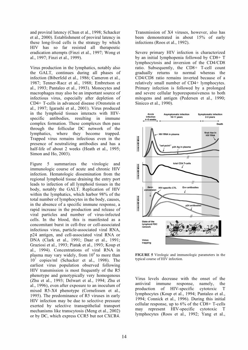
Figure 5 summarizes the virologic and immunologic course of acute and chronic HIV infection. Hematologic dissemination from the regional lymphoid tissue draining the entry port leads to infection of all lymphoid tissues in the body, notably the GALT. Replication of HIV within the lymphatics, which harbor 98% of the total number of lymphocytes in the body, causes, in the absence of a specific immune response, a rapid increase in the production and release of viral particles and number of virus-infected cells. In the blood, this is manifested as a concomitant burst in cell-free or cell-associated infectious virus, particle-associated viral RNA, p24 antigen, and cell-associated viral RNA or DNA (Clark et al., 1991; Daar et al., 1991; Graziosi et al., 1993; Piatak et al., 1993; Koup et al., 1994). Concentrations of viral RNA in plasma may vary widely, from 104 to more than 107 copies/ml (Schacker et al., 1998). The earliest virus population observed following HIV transmission is most frequently of the R5 phenotype and genotypically very homogenous (Zhu et al., 1993; Delwart et al., 1994; Zhu et al., 1996), even after exposure to an inoculum of mixed R5-X4 phenotype (Cornelissen et al., 1995). The predominance of R5 viruses in early HIV infection may be due to selective pressure exerted by selective transepithelial transport mechanisms like transcytosis (Meng et al., 2002) or by DC, which express CCR5 but not CXCR4.
Transmission of X4 viruses, however, also has been demonstrated in about 15% of early infections (Roos et al., 1992).
Severe primary HIV infection is characterized by an initial lymphopenia followed by CD8+ T lymphocytosis and inversion of the CD4/CD8 ratio. Subsequently, the CD8+ T-cell count gradually returns to normal whereas the CD4/CD8 ratio remains inverted because of a relatively small number of CD4+ lymphocytes. Primary infection is followed by a prolonged and severe cellular hyporesponsiveness to both mitogens and antigen (Pedersen et al., 1990; Sinicco et al., 1990).
FIGURE 5 Virologic and immunologic parameters in the typical course of HIV infection.
Virus levels decrease with the onset of the antiviral immune response, namely, the production of HIV-specific cytotoxic T lymphocytes (Koup et al., 1994; Pantaleo et al., 1994; Connick et al., 1996). During this initial cellular response, up to 6% of the CD8+ T-cells may represent HIV-specific cytotoxic T lymphocytes (Roos et al., 1992; Yang et al.,

15
1996; Borrow et al., 1997). Studies of SIV-infected macaques in which CD8 T-cells were temporarily ablated by infusion of a CD8-specific monoclonal antibody have also demonstrated the importance of these cells in lowering the viral load in both primary and chronic infection (Jin et al., 1999; Schmitz et al., 1999). Moreover, after seroconversion, antivirus antibodies that bind to virus particles and to which complement is fixed may increase virus retention on follicular DC that carry complement receptors at high density and thus can retain large quantities of complexed infectious virions (Embretson et al., 1993; Pantaleo et al., 1993; Heath et al., 1995). In agreement with this, the viral RNA load in early HIV infection is high in lymphoid tissues but low in plasma (Pantaleo et al., 1998).
After the initial peak, the virion concentrations in blood are, at least in some patients, stabilized on individually different levels. This so-called "set point" or "inflection point" is strongly associated with disease outcome (Jurriaans et al., 1994; Henrard et al., 1995; Mellors et al., 1996; Schacker et al., 1998). The set point is the equilibrium that results from the interplay of viral, host cell, and immunological factors and is usually reached within a few months to 1 year of infection (Kaufmann et al., 1998; Schacker et al., 1998). Viral titers in plasma subsequently increase only slowly for a long time, corresponding to clinical latency. During this time, the CD4+ T-cell count decreases continuously at an individually different but constant rate. In the lymphatics, there is a continuous, progressive destruction of the follicular dendritic cell network leading to the complete loss of the regular lymph node architecture (Fig. 5, bottom). A marked increase in the level of viral RNA in plasma is seen in advanced immunodeficiency, when the CD4+ T-cell count has dropped to below 200/µl. This has been interpreted as a final complete breakdown
of the mechanisms that previously maintained a certain control of virus replication. The destruction of the FDC network may also contribute, as it leads to a decreased retention of virions; hence, more virus will reach the peripheral blood (Fauci, 1993). Frequently, the final increase is also preceded by an emergence of X4 viruses (Schellekens et al., 1992; Koot et al., 1993).
Dynamics of HIV Replication In Vivo
The availability of antiretroviral drugs which interrupt virus replication and experiments involving plasmapheresis have permitted determination of the dynamics of virus replication (reviewed in Simon and Ho, 2003). The half-life of virus in plasma is 56 minutes on average. To keep the virus concentration in an equilibrium, at least 1010 virus particles must be produced per day. About 93-99% of the virus in the blood plasma of untreated patients originates from activated CD4+ T lymphocytes that get infected, produce virus and die with a half-life of only 0.7 ± 0.2 days (so-called productively infected CD4+ T lymphocytes). An additional 1 - 7% of the virus in plasma originates from longer-lived cells (replication in monocytes or macrophages, release of surface-bound virus from dendritic cells) that have a half-life of 14 ± 7.5 days. Less than 1% of the virus in plasma is produced by latently infected CD4+ T-cells, which become activated and then start producing virus. This last compartment has a very slow decay rate. Its half-life is estimated at 6 - 44 months (Finzi et al., 1997; Wong et al., 1997; Finzi et al., 1999), or it may even not decay at all (Siliciano et al., 2003). Eradication of this compartment will not be possible without measures that activate the virus from its state of latency.
Diagnosis of HIV Infection
The two principal questions in HIV diagnostics are whether a person is infected and, if infected, how actively the virus is replicating. The susceptibility of a patient's virus to antiretroviral drugs has emerged as another question of eminent practical importance.
HIV infection can be detected by a variety of tests. Assayed virus components include proteins, especially p24, which can be measured by immunological tests; RT, whose enzymatic activity can be detected by functional tests; and viral DNA or RNA, which can be identified by

16
molecular tests. Most frequently, however, HIV infection is diagnosed by tests that assess whether an individual's immune system has produced an HIV-specific immune response. Since retroviruses are known to establish infections that persist for life, demonstration of an HIV-specific immune response, if it is consistent and directed against various viral antigens, can be trusted to reflect ongoing infection. Thus, testing for HIV-specific antibodies is still the mainstay of HIV diagnostics, at least in adults. In infants, only testing for virus components allows early diagnosis or exclusion of infection.
The diagnosis of HIV infection relies on commercially available test kits. Competition among manufacturers and strict evaluation and control by regulatory authorities have led to a large number of excellent well standardized commercial diagnostic products of high sensitivity and specificity, which provide a continuously high standard of quality. They are usually better and yield more consistent results than research procedures developed in diagnostic laboratories. Good commercial tests are therefore strongly recommended. Using unregistered tests for screening or for certain types of supplemental testing is unlawful in many countries. In the U.S., refer to http://www.fda.gov/cber/products/testkits.htm for the actual list of U. S. Food and Drug Administration (FDA) approved commercial diagnostic tests. Commercial tests for diagnostic use in Europe need to be Communauté Européenne (CE)-marked.
Only very general descriptions of procedures are given in the following sections, since commercial test kits all contain detailed step-by-step instructions. For procedures that are not commercially available, the reader is directed to the referenced literature. The intent is to guide the reader through the multitude of available procedures and to discuss their strengths and weaknesses.
Screening for HIV Infection, Early Infection Window Periods
HIV-specific antibodies are produced within a few weeks after infection. The time to positivity in screening tests (i.e., to seroconversion) may be influenced by the phenotype of the infecting
virus, the infectious dose, the transmission mode, and the sensitivity of the assay.
In a study based on the first generation of HIV antibody screening assays, developed more than two decades ago against subtype B, seroconversion was estimated to occur on average 45 days after infection; with 95% certainty the window period for 90% of individuals was less than 20 weeks(Petersen et al., 1994). The usefulness of more recently developed tests in reducing the average window period has since been estimated as follows: third-generation anti-HIV-1/2 enzyme immunoassays based on detection of antibodies, -20.3 days (95% confidence interval [CI], 8.0 to 32.5); use of p24 antigen or PCR for proviral DNA , -26.4 days (CI, 12.6 to 38.7); and PCR for viral RNA in plasma, -31.0 days (CI, 16.7 to 45.3) (Busch et al., 1995). With modern third-generation antibody-screening assays, half of the infected individuals should become antibody positive within 3 weeks after infection. Most of the other half should become positive within 2 months, but 5% still seroconvert more than 6 months after infection. It is important to realize that the use of tests for viral RNA, DNA, or p24 in such patients reduces the long diagnostic window periods only insignificantly by 1 to 2 weeks (Busch et al., 1997).
Compared to the 3 weeks median window of 3rd generation antibody assays, p24 antigen testing or the use of 4th generation combination assays that detect both HIV antibodies and p24 antigen reduces the window by a further 5 days (i.e., to 16 days). Finally, the most sensitive test currently available, a test for HIV-1 RNA with a detection limit of 50 copies/ml, reduces the median window length by a further 7 days (i.e., to 9 days). The viral load at which p24 antigen would be detected was estimated by regression analysis at 10,000 copies/ml (CI 2,000 to 93,000) and the HIV replication rate at 0.35 log copies/ml/day, corresponding to a virion doubling time in the preseroconversion phase of 20.5 h (Fiebig et al., 2003). Note again that the 9 day window period for HIV-1 RNA tests is a median and that, as mentioned above, the most sensitive tests for HIV-1 RNA do not significantly shorten the window period of patients with late seroconversion. Late seroconversion cannot be excluded by a negative HIV-1 RNA test.

17
Formats of Screening Tests
There are numerous commercial HIV tests for screening, and it may be difficult to recognize the advantages and disadvantages of a particular test based on the information given by the manufacturer and without systematic comparison (Courouce, 1999). An overview of different test formats and their properties is given in Fig. 6.
The most important kit formats used for HIV antibody screening are the indirect binding assay, the antibody capture assay, and the double-antigen sandwich (DAGS) assay. Indirect binding assays comprise the so-called first-generation enzyme-linked immunosorbent assays (ELISA), which are based on purified viral lysate, and so-called second-generation tests which utilize recombinant antigen or synthetic peptides usually representing Gag and transmembrane (TM) protein. The first-generation indirect binding assay format also applies to immunofluorescence tests and Western blot (WB). Line immunoassays (LIA), which use recombinant proteins and synthetic
peptides, may be considered second-generation tests. Antibody capture assays usually employ recombinant proteins; their principle is that of an indirect binding assay reversed. DAGS assays, frequently also called third-generation assays, usually employ recombinant antigen. Particle agglutination assays may be considered a variant of DAGS assays because for generating a positive signal an antibody molecule must react with at least two antigen molecules, each located on a separate gel particle.
Although all of these tests detect antibodies, they vary in their precise diagnostic questions and answers. Indirect binding assays and antibody capture assays verify, by binding the sample's HIV-specific antibodies to an immunoglobulin (Ig)-specific reagent, that the component that causes reactivity in such a test is indeed an Ig. In contrast, the identity of a component causing reactivity in a DAGS assay remains uncharacterized; the only information provided is that it is capable of linking solid-phase HIV antigen with liquid-phase tracer antigen.
FIGURE 6 Kit design and test performance of HIV screening tests. Synopsis of the most frequently used test formats, their principles, the meaning of positive results, and performance in two typical problem situations. Ag, antigen; Ab, antibody; Ig, immunoglobulin; EIA, enzyme immunoassay; LIA, line immunoassay; WB, Western blot.

18
The different kit formats are affected in different ways by diagnostic challenge situations. One such challenge is antigenic variation. The virus with which a patient is infected may exhibit antigens which differ considerably from the antigens used in the test. Consequently, the patient's antibodies may not bind well to the test kit's antigens, and if the antibody titer is low, a false-negative result may be generated. This type of problem was recognized when antibodies induced by HIV-2 infection were not well recognized by screening kits based on HIV-1 antigens alone. This led to the inclusion of HIV-2 components, usually of the TM protein, in the kits. A similar problem was recognized when group O viruses were discovered, leading to inclusion of group O antigens into all CE marked test kits in Europe (De Leys et al., 1990; Gurtler et al., 1994). DAGS assays are the assays most affected by antigenic variation, because an antibody molecule must bind at least two antigen molecules in order to generate a signal. Such double binding is unlikely if the kit's antigens and the patient's antibodies do not fit. Moreover, endogenous soluble viral antigen present in the serum sample may compete with the test's antigens for free binding sites on HIV-specific antibodies. IgG, which contains only two antigen binding sites per molecule, is most strongly affected since a single endogenous antigenic molecule suffices to abolish detection of an IgG molecule in a DAGS assay. In contrast, in indirect binding and antibody capture assays, antibodies need bind only a single antigen molecule in order to generate a signal.
Another diagnostic challenge is early seroconversion. Antibodies in this phase are restricted to a few viral antigens (usually envelope and p24) and are of low titer and low affinity, and the dominating isotypes are IgM and possibly IgA. In addition, these antibodies may be partially complexed with HIV antigen, which is usually present at high concentration in primary HIV infection (Fig. 5). In this situation it is important that the test provides a high concentration of that antigen that is best recognized. This goal is more easily achieved with recombinant proteins than viral lysate. Furthermore, the test must select for the few HIV-specific antibodies present in the bulk immunoglobulin; this is impossible with antibody capture assays that bind Ig of all
antigenic specificities. In addition, the test should detect IgM because, in the presence of antigenemia, its pentameric structure with a total of 10 antigen binding sites is most likely to have several sites remaining accessible. The best assay in this situation is the DAGS assay: it initially selects for HIV-specific antibodies (binding to solid phase), and does not discriminate against non-IgG isotypes. The first test based on this principle was the particle agglutination assay, which was introduced in the mid-1980s, i.e., long before third-generation ELISA were developed. This test performs remarkably well in seroconversion panels, and due to the broad spectrum of antigens present in the viral lysate it also has a broad detection range for antigenic variation (Constantine et al., 1994; Vercauteren et al., 1995; Poljak et al., 1997; Lien et al., 2000).
The practical relevance of these considerations is shown when the performances of different kits with seroconversion panels are compared. Among 23 different commercial kits whose performance on at least 15 different commercially available seroconversion panels was compared by the Swiss Federal Office of Public Health, the 17 DAGS assays were the most sensitive and occupied ranks 1 to 15, 17, and 18. The four indirect binding (2nd generation) assays occupied ranks 16, 19, 21, and 22, and the two antibody capture assays ranked 20th and 23rd (unpublished data of the author). Seroconversion panel comparisons also demonstrated the inferior sensitivity of immunofluorescence tests and WB, which ranked at the end together with other first-generation indirect antibody binding assays (see also (Busch and Satten, 1997; Thorstensson et al., 1998)). Assessment of the performance in seroconversion panels followed by revocation of approval for the 1020% least sensitive kits, is one of the most powerful — though obviously underused — instruments by which regulatory agencies could guarantee a continuous further technical improvement of diagnostic tests (Schupbach, 1996).
Fourth-Generation Screening Tests
Several companies now offer kits that detect both antibodies and antigen (fourth-generation tests), and in many European countries the use

19
of these products for HIV screening performed in diagnostic laboratories has become mandatory. In seroconversion panel analysis, these kits now rank first among all screening tests even if their detection of antibodies is based on the insensitive antibody capture format. The average gain in time to detection compared with third-generation kits is 3-5 days (Gurtler et al., 1998; Weber et al., 1998; Laperche et al., 2000; Ly et al., 2001). The use of such tests for screening is strongly recommended because individuals in the antigen-positive stage of pre-seroconversion have a high viral load and are particularly infectious (Fig. 5).
Rapid Tests and Use of Alternative Specimens
Rapid tests can be performed with minimal or no laboratory equipment; they yield results within 30 minutes. Such tests may be useful in certain situations, e.g., in assessing the risk of HIV transmission in needle-stick injuries and similar exposures to possibly HIV-contaminated materials, organ donations, or whenever a laboratory test result may not be available quickly. Rapid tests may be of different formats, including DAGS, indirect binding, Ig capture, agglutination, or chromatographic assay. The diagnostic sensitivity of some of these tests seems somewhat inferior to third-generation ELISA-based antibody tests, especially in seroconversion panels (Kuun et al., 1997; Vallari et al., 1998; Giles et al., 1999). Others, however, exhibit a comparable diagnostic sensitivity and specificity, even during seroconversion and can therefore be recommended for certain diagnostic settings (Giles et al., 1999; Kelen et al., 1999; Palmer et al., 1999; Zaw et al., 1999; Phillips et al., 2000; Ketema et al., 2001).
Many persons infected with HIV are not tested until they develop symptoms of AIDS. Up to one-third of patients receive their HIV diagnosis within 2 months of progression to AIDS. The hope that such individuals could be motivated to be tested earlier has led to new testing strategies, particularly in the U.S.. These now recommend routine, "opt-out" testing in all health-care settings (Branson et al., 2006). The shift in testing strategy also has led to the use of new test systems believed to be more attractive to the client. They include home collection test systems, in which sample collection devices are
ordered by phone and delivered by express courier. Blood is collected by finger pricking onto filter paper and sent to a designated laboratory for screening. Such testing systems have good sensitivity and specificity; collecting a sufficiently large specimen may be the biggest problem, affecting 7-10% of the users. As an alternative, testing systems for other specimens, such as oral fluids or urine, also received FDA approval (reviewed in Mylonakis et al., 2000). Excellent sensitivity and specificity were reported in studies involving oral fluids collected from postseroconversion individuals (Saville et al., 1997; Wisnom et al., 1997; Granade et al., 1998; Martinez et al., 1999). This also applies to FDA-approved test systems for urine samples (Urnovitz et al., 1997). Very recently, the FDA has approved a rapid test system for oral fluids, whole blood or serum, which is so easy to perform that testing at the point of care with a return of the result within 20-40 min has become possible. Extended studies of this device have reported a sensitivity and specificity comparable to that of other EIAs (Delaney et al., 2006; Wesolowski et al., 2006). The sensitivity of test systems utilizing specimens other than blood in early seroconversion remains untested, as standardized materials comparable to seroconversion panels are not available. The use of such alternative tests in recent exposure settings should therefore be avoided. True home tests, which would be sold to the public, have not been approved by the FDA or the health authorities of other countries, and their safety cannot be guaranteed.
Supplemental Testing
Antibody Tests — Western Blot (WB) and Line Immunoassay (LIA)
WB was introduced into HIV testing by the author in 1984 (Sarngadharan et al., 1984; Schupbach et al., 1984), proposed for systematic confirmation of reactive screening results in 1985 (Schupbach et al., 1985), and has remained a principal confirmatory tool worldwide (Mylonakis et al., 2000). Over the years it has, however, also become clear that, in contrast to the continuously improved screening tests, WB has remained a first-generation test with certain well-known flaws: the sensitivity in seroconversion panels is clearly inferior to that of third- and fourth-generation screening tests.

20
WB is also prone to detect cross-reactive antibodies, which results in a high rate of indeterminate results.
A single improvement, the use of recombinant proteins and synthetic peptides for the production of the strips, has been realized by some manufacturers. When recombinant proteins and peptides are used entirely instead of viral lysate, strips can be produced as LIA in which selected antigens are applied as distinct lines and at defined, optimal concentrations. In format, such assays are comparable to 2nd generation screening EIAs and may thus be considered as "2nd generation Western blots". One such assay, the Inno-LiaTM HIV I/II Score, is increasingly used in countries outside the U.S.. It contains 7 HIV antigen bands (sgp120 [including group O peptides], gp41, p31, p24 and p17 of HIV-1; sgp105 and gp36 of HIV-2), which are coated as discrete lines on a nylon strip with plastic backing. As each test strip also contains 3 quantitative internal standards, a semiquantitative ranking of the different antibody reactions into 6 intensity scores is possible. This enables a standardized interpretation of test reactions which, unlike for WB, is not only based on the presence of reactions (yes/no), but also their intensity. This LIA provides excellent confirmation of HIV infection and is superior to WB for differentiating between HIV-1 and HIV-2 infection (Pollet et al., 1991; Walther et al., 1995).
Early identification of HIV-2 infection is important with regard to both virus load quantification and the choice of effective antiretroviral treatment. None of the virus load assays approved for patient monitoring can reliably quantitate HIV-2, and HIV-2 virus loads in symptomatic patients may be severely underestimated. Furthermore, HIV-2 is naturally resistent to NNRTI and some other antiretroviral drugs effective against HIV-1. HIV-2 infection must therefore be diagnosed early in order to prevent suboptimal disease monitoring and start of ineffective treatment regimens leading to resistance.
WB and LIA are more prone to problems with carryover contamination than are most screening assays. Use of the convenient multichannel troughs for incubation of the strips presents a certain risk. Contamination with minute volumes of a strongly positive serum may lead to faint Env bands, even if the dilution
is up to 106-fold. While intra-assay contamination can be ruled out by repeating the assay in an isolated test chamber, repeat testing will not identify contamination within the specimen tube. Touching the wet inner side of a specimen tube lid with the gloved fingers may carry enough material to the lid of a subsequently opened tube to result in faint WB reactivity to Env antigens. The probability of such events depends on the proportion of strongly positive sera among the specimens tested by a laboratory and on how many times a specimen tube is opened. To minimize this risk, handling and testing of samples from known HIV-positive patients together with diagnostic samples should be avoided, and gloves that have become contaminated with specimen must be changed immediately. It should also be recognized that samples with initial borderline results carry an increased risk of contamination, since these tubes are opened repeatedly for supplemental testing. This results in a higher cumulative risk of contamination. Alarm bells should ring when a sample with borderline or low positive results in screening is faintly reactive in WB. It may be an early-seroconversion sample, but it may also be the result of contamination. The contamination problem is a strong reason why WB interpretation should follow the most stringent and not the most sensitive guidelines.
WB, and to a lesser degree, LIA still have a relatively high rate of indeterminate results (Pollet et al., 1991). This shows that indeterminate WB reactions are usually caused by antibodies that cross-react with viral rather than with cellular proteins. Indeterminate WB results have been described for patients with autoimmune disorders, in particular systemic lupus erythematosus, after infections with certain viruses including herpes simplex virus type 1 or cytomegalovirus, or after vaccination against influenza or rabies virus (Guan, 2007). For the latter, epitopes related to HIV have been implicated. Such information is, however, of little practical value, and the origin of indeterminate WB reactions usually remains obscure. In spite of all these flaws, a WB or LIA with a "full-house" pattern of reactive antibodies probably remains the most convincing laboratory evidence for an HIV infection. When reactive bands are few and their intensities are low, interpretation is hazardous, and a diagnosis must not be based on WB or LIA alone.

21
WB Interpretation Guidelines
In an attempt to render WB more sensitive, the Association of State and Territorial Public Health Laboratory Directors and the CDC (ASTPHLD-CDC) issued interpretation recommendations which request antibody reaction to any two of three antigen bands including gp120/160 (considered to be one antigen), gp41, and p24 (Anonymous, 1989). Since most of the gp160 and gp120 bands on WB are not due to the Env precursor or the surface (SU) protein but instead represent tetramers or trimers of gp41 (Pinter et al., 1989), reaction with gp120-gp160 and gp41 bands may be based on reaction with a single protein, TM, and is thus inherently unsafe. The same is true for the very similar recommendations by the Consortium for Retrovirus Serology Standardization, the only difference being that p24 may be replaced by p31(pol). Similarly, the World Health Organization recommendation specifies any two of gp160, gp120, and gp41 (Anonymous, 1990). This means practically that TM-reactive antibodies must be present at a concentration sufficient for detection of not only the strongest but also the second-strongest TM band. The strongest TM band is usually the largest antigen, i.e., gp160, which migrates least far in the sodium dodecyl sulfate-polyacrylamide gel electrophoresis–based separation of proteins and thus is the sharpest and best-detected band. Depending on the manufacturer, either gp120 or gp41 may be the second strongest band. Due to varying degrees of glycosylation, gp41 migrates in sodium dodecyl sulfate-polyacrylamide gel electrophoresis as a very diffuse band. Reactive antibodies thus generate a signal that is much less easily recognized than if the same antibodies bind to the sharp gp160 band.
Due to the propensity of WB to detect cross-reactive antibodies, a combination of Env and p24 bands is not sufficiently stringent for confirmation. WB analysis of 100 screening-negative but otherwise unselected Swiss blood donors showed an isolated reaction with p24 in 9% and with gp160 or both gp160 and gp120 in 3%. The likelihood of a chance combination leading to "confirmed positivity" in a healthy donor would thus be 0.09 x 0.03, or 0.0027, i.e., 1 in 370 (Schupbach et al., 1990). We and others (Healey and Bolton, 1993) have observed several cases that satisfied the ASTPHLD-CDC criteria for WB positivity and kept this pattern essentially unchanged over years but were
negative in long-term follow-up in all direct tests for HIV components, including virus culture, regular polymerase chain reaction (PCR) for HIV DNA and RNA, and an ultrasensitive sequence capture-PCR test "Mega-PCR" enabling detection of a few provirus copies in as much as 100-500 µg of DNA (Boni et al., 2004b). Representative WB results of such a case are shown in Fig. 7.
More-stringent criteria have been issued by the American Red Cross (ARC) and the FDA. ARC specifies at least one band each from Env, Gag, and Pol. The most stringent, but least sensitive recommendation is that of the FDA, which specifies reaction with p24, p31, and Env.
FIGURE 7 Example of a false-positive HIV-1 WB interpretation according to ASTPHLD-CDC or CRSS guidelines. Lanes: a, weakly positive control; b, sample from a healthy individual exhibiting weak reaction with gp160, gp120 (very weak), and p24 (this sample was taken 3.5 m after an initial sample with the same pattern [data not shown]); c, sample from the same individual taken 1 month after the initial sample. p24 antigen with signal amplification–boosted ELISA was negative in all three plasma samples; PCR for viral DNA was negative in PBMC from the samples in lanes b and c, and RNA was negative in the sample in lane b. Culture with PBMC depleted of CD8 T cells from the sample in lane b was negative for p24 antigen and RT by the PERT assay; this test was also negative with the samples in lanes b and c.
22
In my opinion, only the most stringent interpretation guidelines should be applied if a diagnosis of HIV infection is established using WB as the only supplemental test. If some true cases of HIV infection are WB indeterminate by FDA guidelines, this is of no concern as long as their WB pattern is suggestive of HIV infection. This is always the case when the ASTPHLD-CDC interpretation would render these patterns positive. In almost all such cases, a safe diagnosis can be established based on supplemental tests for virus components (p24 antigen or nucleic acids). One also has to take into account that the ASTPHLD-CDC criteria were established based on a single commercial product. Meanwhile, other kits are available. Guidelines established for one particular kit and with one particular sample cohort cannot be applied to other kits or populations from other geographical regions without careful re-examination of their validity.
Another attempt to render WB more sensitive in early infection is its use in the detection of IgM. Unfortunately, such testing lacks specificity. Gag-reactive antibodies of IgM, IgA, and IgG isotype are frequently detected by WB in sera of infants born to HIV-negative mothers. Therefore, many of these reactions appear to be due to common agents unrelated to HIV (Schupbach et al., 1994).
Virus Component Tests — p24 Antigen and Nucleic Acid Tests (NAT)
In many cases, supplemental testing requires the use of tests for virus components. This applies to patients in primary HIV infection as well as those with indeterminate results in antibody tests. Diagnosis of pediatric HIV infection is also best established with virus component tests. Virus components that can be assayed include the p24 antigen, viral DNA or RNA, and RT activity. In addition, the capability of the virus to replicate can be assessed by virus isolation in cell culture. This requires a virus component test, usually an antigen test or immunofluorescence for cell-associated HIV antigen.
Commercially available tests for virus components include p24 antigen assays. CE-marked p24 antigen assays are available from various manufacturers in Europe, while in the U.S. only one test is FDA-approved. A CE-
marked kit is available for PCR detection of HIV-1 DNA, but lacks approval in the U.S. NAT for HIV-1 RNA that are both CE-marked and FDA-approved are available from several manufacturers and currently include PCR for reverse-transcribed RNA (RT-PCR), nucleic acid sequence-based amplification (NASBA) which is also known as transcription-mediated amplification (TMA), and a signal-amplification procedure which involves branched DNA (bDNA) probes.
Tests for HIV p24 Antigen
Antigen tests for p24 are easy to perform and diagnostically valuable in early infection, when antigen is usually present at high titers while HIV-specific antibodies may still be undetectable. Although NAT are increasingly used instead of p24 antigen, this test still has a place in supplemental testing because it can, in contrast to NAT, also be performed on serum, thus enabling diagnosis of primary infection in the first specimen received for HIV testing, usually serum. A highly improved version of a p24 antigen test, involving a signal amplification boosted p24 EIA of heat-denatured plasma also has proven to be a sensitive, specific, simple and inexpensive solution for diagnosing pediatric HIV-1 infection in resource-poor settings, especially in subtype C epidemics (Schupbach, 2003; Fiscus et al., 2006; Patton et al., 2006).
Tests for Viral RNA or DNA
Three fundamentally different techniques for the sensitive detection or quantification of HIV RNA or DNA are available.
Polymerase Chain Reaction (PCR) In PCR, double-stranded DNA is denatured, a pair of HIV-specific oligonucleotide primers is annealed to the separated viral DNA strands, and these primers are extended by a heat-resistent DNA-dependent DNA polymerase (Taq polymerase). This procedure is continued for 30 to 40 cycles, each of which comprises a high-temperature denaturation, a low-temperature primer annealing, and an intermediate-temperature primer extension (DNA synthesis) (Kwok et al., 1987). If the starting material for PCR is RNA, a cDNA must
23
first be generated by reverse transcription. This cDNA can then be amplified by the regular procedure (Byrne et al., 1988). A further development of PCR, TaqMan® real-time PCR, employs the AmpliTaq Gold® DNA polymerase for PCR amplification. This enzyme also has a 5′ exonuclease activity, enabling it to cleave away a synthetic oligonucleotide called a TaqMan® probe annealed to a specific sequence of the template between the forward and reverse amplification primers. The probe, which contains a fluorophor at the 5'-end and a quencher at the 3'-end, sits in the path of the enzyme as it proceeds to copy DNA or cDNA. When the enzyme reaches the annealed probe it cleaves the probe into its nucleotides, thus releasing the fluorophor from the inhibitory effect of the quencher. Thus, the growing copy number of amplified DNA is accompanied by a likewise increasing intensity of fluorescence, which is measured continuously, thus permitting real-time observation of the amplification process. Important advantages of real-time PCR include the broad range of concentration in which the target DNA is measured precisely and the protection against carry-over contamination, as both amplification and product analysis take place in a closed system.
RT-PCR test systems for both manual sample preparation and or fully automated operation are available from both Roche (Roche Molecular Diagnostics, Pleasanton, California) and Abbott, (Abbott Laboratories, Abbott Park, Illinois). The assays manufactured by Roche (Amplicor HIV-1 Monitor Vs. 1.5, Cobas AmpliPrep/Cobas TaqMan HIV-1) amplify a sequence in gag, which permits good detection of the various subtypes of HIV-1 group M and most CRFs, but not of group O or HIV-2 (Triques et al., 1999; Katsoulidou et al., 2006; Schumacher et al., 2007). Abbott's tests (LCx HIV RNA Quantitative, RealTime HIV-1 assay for use on the m2000 system) amplify a conserved sequence in the integrase region of pol and detect groups N and O in addition to the various subtypes and CRFs of group M (Swanson et al., 2005; Swanson et al., 2006; Swanson et al., 2007). Although the intended primary purpose of these tests is for patient monitoring (virus load determination), they are used also diagnostically, in particular for diagnosing acute HIV-1 infection.
Nucleic Acid Sequence Based Amplification (NASBA) and Transcription-Mediated Amplification (TMA) In the NASBA and TMA procedures RNA is amplified in an isothermal multienzymatic procedure mediated by the enzymatic effects of RNA-dependent DNA polymerase (RT), RNase H, DNA-dependent DNA polymerase, and DNA-dependent RNA polymerase. This procedure thus mimics the retroviral nucleic acid replication cycle (Fig. 1). The product of this amplification is a single-stranded RNA (Kievits et al., 1991). In current NASBA and TMA kits, the resulting RNA product is detected by a specific DNA probe (molecular beacon) which, when annealed to the target RNA, forms a stem-loop structure resulting in emission of a chemoluminescent signal.
Currently available kits include a CE-marked and FDA-approved quantitative real-time NASBA kit (NucliSENS EasyQ) from bioMerieux, Marcy l'Etoile, France, which amplifies a sequence in the gag gene, and a TMA-based HIV-1 RNA qualitative assay (APTIMA HIV-1 RNA Qualitative Assay) manufactured by Gen-Probe, San Diego, CA. The latter is an FDA-approved assay for diagnosis of HIV-1 infection in human plasma. The assay amplifies conserved sequences in both the LTR and pol of HIV-1 thus enabling simultaneous detection of all known groups of HIV-1, i.e., M, N and O. It is intended for use in the diagnosis of HIV-1 infection, including acute or primary infection (Roland et al., 2004). The same test principle and target sequences also are used in an FDA-approved test for fully automated blood donor screening with simultaneous detection of HIV-1, hepatitis B virus and hepatitus C virus RNA (Linnen et al., 2002; Candotti et al., 2003; Koppelman et al., 2005).
Branched DNA (bDNA) In the bDNA method viral RNA is captured on a solid surface by immobilized specific capture probes. The captured RNA is then reacted with "connector" probes, which with one end hybridize to a series of short sequences of the pol region of the RNA and with the other end mediate fixation of bDNA detector probes. These bDNAs are then reacted with still other bDNAs that hybridize to the first. Enzyme-labeled tracer probes are finally hybridized to all the branches, and the analysis is based on chemoluminescence (Urdea et al., 1993). The principal difference of the bDNA method compared with PCR or NASBA-

24
TMA is the lack of amplification of viral sequences and, in consequence, lack of a carryover problem. What is called signal amplification is in fact a mere signal accumulation in which interacting molecular probes added to the reaction mixture at predetermined high concentration are hybridized to captured viral RNA in an ordered process which results in the deposition of consecutive probe layers. The procedure is in fact comparable to an indirect antibody-binding assay. Higher precision is probably achieved than with nucleic acid amplification techniques, whose outcome depends on the efficacy of primer annealing in each cycle.
A bDNA-based assay for HIV-1, the Versant® HIV-1 RNA 3.0 (bDNA), is sold by Bayer HealthCare, Leverkusen, Germany (Galli et al., 2005). This test is based on about 40 different probes that cover most of the pol gene and permit detection of the various subtypes of group M but not of group O. A drawback of the method is the relatively high detection limit, which must be compensated by large specimen volumes (2 ml of plasma).
Nucleic acid tests with FDA approval and their intended uses are listed on http://www.fda.gov/cber/products/testkits.htm. With the exception of TMA and the RT-PCR based systems for blood donor screening, the kits described above are designated primarily for quantification of viral RNA. Depending on specificity and level of detection, some of them under certain conditions also may be suitable for diagnostic (qualitative) purposes, but caution must be exercised since false positive results have been reported (Roland et al., 2004). Of note, the cut-off for the Bayer Versant HIV-1 RNA 3.0 (bDNA) has been set in a way that will result in 5% false-positives when used on uninfected controls! For diagnostic questions, it is therefore safer to use qualitative tests such as the APTIMA HIV-1 RNA Qualitative Assay (Gen-Probe). There is one CE-marked commercial PCR kit for HIV-1 DNA (Roche), but there are reports of false-negative results with this kit, particularly with non-B subtypes (Bogh et al., 2001; Obaro et al., 2005). Many diagnostic laboratories, including ours, have developed their own PCR methods for qualitative detection of viral DNA or RNA. Detailed step-by-step instructions for our diagnostic procedures, which are capable of detecting a single DNA copy even without using nested-PCR protocols (which carries a high risk
of carry-over contamination), were published (Boni, 1996). For quantification of viral RNA a number of excellent real-time RT-PCR systems have been described (Drosten et al., 2006; Muller et al., 2007; Rouet et al., 2007).
The availability of commercial kits for the detection of HIV-1 RNA or DNA by sequence or signal amplification has rendered these tests attractive for laboratories with no background in molecular biology. The fact that under optimal circumstances a single gene copy can be detected by some of these tests has created a relatively uncritical attitude, resulting in a degree of trust that is not justified in several aspects. Molecular tests are sensitive to sequence variation, resulting in underdetection or false-negative results in some cases. Carryover contamination, whose main source is amplified DNA but which may also originate from other specimens, can also be a problem, and commercial products or systems must therefore have built-in carryover protection devices. In addition, precautionary measures are vital for all laboratories performing such tests (Boni, 1996).
Important factors in molecular testing are sample handling and sample preparation. Particle-associated RNA in plasma has been claimed to be very unstable, demanding special expensive plasma preparation tubes from a certain manufacturer and immediate separation of plasma from the cellular pellet before the sample is shipped to the laboratory. Independent investigators have not been able to confirm these claims. Their work indicates that HIV-1 RNA levels are stable (variance, less than 0.3 log unit) for up to 3 days after collection when stored either at room temperature or at 4°C as cell-free plasma in the ethylenediamine tetraacetic acid (EDTA)-plasma preparation tubes or even as EDTA-anticoagulated whole blood in regular tubes. Comparison of paired HIV-1-positive plasma and serum specimens revealed that RNA quantitation was 20-65% lower in serum than in plasma. EDTA plasma is thus the preferred specimen for these assays and provides the highest levels of RNA. EDTA plasma that is prepared and frozen within 8 h of collection can thus be trusted to be of sufficient quality for these tests. It can be thawed and frozen up to three times before the RNA levels decrease significantly (Todd et al., 1995; Ginocchio et al., 1997; Sebire et al., 1998).

25
The maximal sensitivity of molecular tests is limited by sample size and sequence variation. The detection limit of the PCR is a single DNA molecule. However, 1 µg of genomic DNA, which contains the DNA of approximately 150,000 cells, corresponds to the number of peripheral blood mononuclear cells (PBMC) contained in only about 75 µl of blood. Consequently, even with a detection limit of one provirus/µg of DNA, roughly 70,000 infected cells must be present in 5,000 ml of blood for PCR analysis to be positive. A detection limit of 10 copies per reaction is more realistic because of the Poisson distribution, which means, for example, that not every sample with a nominal concentration of one copy/sample indeed contains such a copy. Similarly, the sensitivity for the detection of particle-associated HIV RNA is limited by the efficiency of the reverse transcription step and by the volume of analyzed plasma. To achieve higher sensitivity ("ultrasensitive tests"), sample input has to be increased to milliliter plasma volumes (Schockmel et al., 1997). Loss of sensitivity may also be due to HIV sequence divergence. Individual mutations at a critical downstream position of an amplification primer may lead to reduced or entirely abolished amplification. Infallible tests based on sequence amplification are thus an illusion, but significant progress resulting in broader sensitivity has definitely been achieved. Subtypes A, CRF02_AE, F, and G were systematically underdetected by version 1.0 of the Roche Amplicor HIV-1 Monitor test (Coste et al., 1996; Simons et al., 1997; Debyser et al., 1998; Parekh et al., 1999). The introduction of modified primers in the subsequent version 1.5 of the test considerably improved the recognition of these clades, while group O isolates remain undetectable. Change from Monitor Version 1.5 to Roche TaqMan based RT-PCR kits may also result in significantly lower copy numbers of HIV-1 RNA in some instances.
HIV-1 subtypes A and G were insufficiently detected by earlier NASBA kits. Designation of new primers for the current real-time version of the test, NucliSens EasyQ (bioMerieux) have, however, resulted in performance similar to Roche's RT-PCR (Lam et al., 2007; Stevens et al., 2007). Underdetection of a significant fraction of subtype C samples was reported in one study from Israel (Gottesman et al., 2006), but was not seen in studies performed in South Africa (Stevens et al., 2005; Stevens et al., 2007). On the other hand, the NucliSens EasyQ,
as the first test so far, has recently been shown to enable quantification of subtype A of HIV-2 (Rodes et al., 2007). Improved recognition of group O has been achieved with Abbott's RT-PCR based tests and the Gen-Probe TMA assays.
Broad application of NAT in the diagnostic laboratory has now become feasible as full automation of both nucleic acid extraction and test conduction has been achieved. PCR is very helpful when serology has failed to provide a clear answer. This applies in particular to specimens with borderline reactivity in screening assays or incomplete patterns on confirmatory WB or LIA, specimens from individuals with suspected primary HIV infection (although a p24 antigen test would suffice in most instances), and babies of HIV infected mothers. For individuals with confirmed-positive HIV-1 serology, quantification of the viral RNA in plasma is essential for clinical assessment and, if positive, serves as a further confirmation of the infection.
Virus Isolation
Leukocytes are separated from anticoagulated blood by Ficoll centrifugation and cocultured with phytohemagglutinin-stimulated leukocytes from healthy blood donors. Culture supernatants are periodically assayed for p24 antigen. Cultures usually become positive within 2 weeks, but culture times of up to 60 days have been reported (Ho et al., 1989; Burgard et al., 1992). The procedure has a sensitivity of about 90% over all stages; the success rate is lower for asymptomatic patients. Significant improvement may be achieved by complicated procedures combining concentration of cells likely to be infected; removal of cells that might interfere with virus replication, such as CD8 T-cells; and activation of infected lymphocytes using phytohemagglutinin or antibody to CD3 and CD28. Such procedures enabled virus isolation from lymph node cells of all patients whose viral RNA level in plasma had become undetectable under antiretroviral treatment (Finzi et al., 1997; Wong et al., 1997). Despite these improvements in sensitivity, virus isolation remains time-consuming and costly. Since PCR for viral DNA or RNA, as well as the signal amplification-boosted p24 antigen test, have a diagnostic sensitivity of more than 96% and yields results within 1 day, virus

26
isolation has become unsuitable for the routine diagnosis of adult and pediatric HIV infection in both developed and resource-poor settings, although it remains important for many research questions.
Reverse Transcriptase Assays
Particle-associated RT is a unique marker of retroviruses, and RT assays have been instrumental in the discovery of all known human retroviruses. Tests have now been developed that combine the broad detection range of RT tests with the high sensitivity of nucleic acid amplification procedures. These so-called product-enhanced reverse transcriptase (PERT) assays are based on the selective enhancement, by PCR or another amplification method, of the cDNA product synthesized from an RNA template by the RT activity contained in a test sample. They are 106 to 107 times more sensitive than a conventional RT test and detect fewer than 10 particles (Pyra et al., 1994), thus rivaling PCR for detecting viral RNA in plasma (Boni et al., 1996a; Reisler et al., 2001). Detailed step-by-step instructions have been published (Boni and Schupbach, 1999). Assays based on this principle were established also by others (Silver et al., 1993; Heneine et al., 1995). In the context of HIV infection, the PERT assay may be diagnostically useful when a low CD4+ lymphocyte count or indeterminate serological results, such as a WB test result positive according to ASTPHLD-CDC but indeterminate by FDA guidelines, suggest the presence of an HIV infection, possibly with an agent not well detected by the current HIV-specific amplification methods. A negative result with the PERT assay, which would detect any retrovirus independent of its genomic sequence, excludes infection with an abberrant HIV strain. Since a commercial kit is not available, the test is currently restricted to specialized retrovirus laboratories that also do research. A less sensitive test kit lacking amplification has been evaluated as an alternative virus load test for resource-poor settings (Seyoum et al., 2006). Unlike the PERT assay, due to the lack of sensitivity it cannot be used for diagnostic purposes.
Diagnosis of HIV-2 Infection
Screening tests, at least in Europe, must demonstrate good sensitivity to HIV-2 in order to receive CE marking. Detection of HIV-2 infection at the level of screening is therefore not a problem. In contrast, determination of whether reactivity in screening is due to infection with HIV-1, HIV-2, or both is sometimes difficult. On HIV-1 WB, sera from HIV-2 infected individuals frequently have strong reactions with Gag and Pol proteins compared to their reaction with Env. In particular, they may present with an unusually strong p31 (integrase) band. A suspected HIV-2 infection is further supported by reaction with the recombinant HIV-2 TM protein present on the products of some manufacturers. We have, however, occasionally seen samples that did not react with this band, although HIV-2 infection was subsequently confirmed. The presence of isolated Gag bands on HIV-1 WB or of strong reactivity against p24 and Gag precursors (p55, p43, and p39) is not an indication of HIV-2 infection. Similarly, the presence of faint Gag-reactive bands on HIV-1 blots is not an indication for an HIV-2 WB.
Results of HIV-1 WB suggestive of HIV-2 infection may be confirmed by an HIV-2 WB or, preferable, by tests involving specific recombinant proteins, e.g., an LIA or a rapid test that differentiates between the two. If reactions to both HIV-1 and HIV-2 proteins are present at similar intensities, diagnostic PCR for proviral DNA of both viruses is necessary. HIV-2 infected asymptomatic individuals, i.e., the overwhelming majority, have much lower viral loads than those infected with HIV-1; HIV-2 RNA is usually undetectable in these patients. The confirmatory test of choice thus is PCR for HIV-2 DNA.
Diagnostic Algorithms and General Considerations
Guidelines for HIV testing may vary in different countries based on prevalence of virus types, available tests, laboratory facilities, health care systems, and economic or other factors. Revised U.S. recommendations for HIV testing of adults, adolescents, and pregnant women in health-care settings were issued in 2006 (Branson et al., 2006); these recommendations are also accessible online under

27
(http://www.hivatis.org/Guidelines). FDA-approved test kits are listed under http://www.fda.gov/cber/products/testkits.htm.
U.S. Guidelines
Initial screening is done with an FDA-approved enzyme immunoassay (EIA) for HIV antibodies. Specimens with a nonreactive result are considered HIV negative unless new exposure has occurred. Specimens with a reactive EIA result are retested in duplicate. If the result of either duplicate is reactive, the specimen is reported as repeatedly reactive and undergoes confirmatory testing with a supplemental test (e.g., WB or immunofluorescence assay). Specimens repeatedly reactive by EIA and positive by WB or immunofluorescence assay are considered HIV positive (Fig. 8, dark area).
Specimens that are repeatedly EIA reactive occasionally provide an indeterminate WB result, which might represent either an incomplete antibody response to HIV in specimens from infected persons or nonspecific reactions in specimens from uninfected persons. Immunofluorescent antibody (IFA) can be used to resolve an indeterminate WB sample. Generally, a second specimen should be collected >1 month later and retested for persons with indeterminate WB results. Nucleic acid testing for viral RNA or proviral DNA could also help resolve an initial indeterminate WB in certain situations.
An HIV test should be considered positive only after screening and confirmatory tests are reactive. A confirmed positive test result indicates that a person has been infected with HIV. False-positive results when both screening and confirmatory tests are reactive are rare. However, the possibility of a mislabeled sample or laboratory error must be considered, especially for a person with no identifiable risk for HIV infection.
Because a negative test result probably indicates absence of HIV infection, a negative test need not be repeated in persons with no new exposure in settings with low HIV prevalence. For persons with a recent history of known or possible exposure to HIV who are tested before they could develop detectable antibodies, the possibility of HIV infection cannot be excluded without follow-up testing. A false-negative
result also should be considered in persons with a negative HIV-1 test who have clinical symptoms suggesting HIV-1 infection or AIDS. Additional testing for HIV-2 and HIV-1 group O infection might be appropriate for these persons.
Most people with an initial indeterminate WB result who are infected with HIV-1 will develop detectable HIV antibody within 1 month. Thus, persons with an initial indeterminate WB result should be retested for HIV-1 infection >1 month later. Individuals with continued indeterminate WB results after 1 month are unlikely to be HIV infected and should be counseled as though they are not infected unless recent HIV exposure is suspected. Nucleic acid tests for HIV DNA or RNA are not generally recommended for resolving indeterminate WB results except in suspected cases of primary or acute infection (Divine et al., 2001).
Alternative Testing Strategies
In contrast to the U.S., where screening does not normally include testing for HIV-2 or group O viruses, CE-marked screening tests used in Europe must detect all HIVs. Great importance is placed also on the detection of primary HIV infection; therefore, fourth-generation screening tests or additional antigen testing are recommended whenever a primary HIV infection must be considered. Finally, verification of all confirmed HIV-positive results with a second, freshly drawn specimen is mandatory when a diagnosis of HIV infection is first established. The peripheral, lightly shaded areas of Fig. 8 illustrate possible modifications of the U.S. recommendations that may under certain circumstances be useful.
To avoid the practical and financial problems associated with WB testing, the WHO has recommended alternative test strategies based on the use of at least two different screening tests (Sato et al., 1994). Large studies have shown that such testing algorithms may yield results that are at least equivalent to the conventional testing algorithm outlined above. In one alternative algorithm, reactive samples are subjected to a different screening assay and only those samples with discrepant results are subjected to WB testing. Initial screening may also be performed with two different tests, and those with discrepancies undergo supplemental

28
testing. Supplemental testing by WB can also be replaced without loss in sensitivity or specificity by a third screening assay (Laleman et al., 1991; van der Groen et al., 1991; Urassa et al., 1992; Nkengasong et al., 1999). Given the further improvement of screening tests since the time most of these studies were done, such testing algorithms have become even more attractive. In particular, initial screening with a fourth-generation antibody/antigen test and a third-generation DAGS test in parallel is bound to increase the sensitivity. Samples nonreactive in both assays are reported as HIV negative. Samples reactive in both assays are almost always infected, since the high diagnostic specificity (>99.5%) of each of the two tests is potentiated, resulting in an overall diagnostic specificity of ≥99.9975%. Therefore, a freshly drawn confirmatory sample can be directly used for quantification of the viral RNA load. An HIV-1 RNA concentration above about 5,000 copies/ml plasma is sufficient additional proof of an infection, but is not specific for HIV-1. As some HIV-2 isolates are recognized relatively well by current RT-PCR test kits while others are severely underdetected or recognized not at all, there is always the possibility that a patient with low to medium level of detectable RNA in an HIV-1 test is actually infected with HIV-2(Schutten et al., 2004). Serological tests able to distinguish between HIV-1 and HIV-2 are thus the best solution to diagnose, or exclude, HIV-2 infection.
Result discrepancy between the two screening tests or borderline results may be due to acute HIV infection or a nonspecific reaction. Acute infection must be suspected if reactivity is restricted to the fourth-generation assay; a reactive antigen test confirmed by a positive antigen neutralization test further supports this possibility. If the antigen test result is negative, seroconversion is unlikely, and false reactivity must be suspected. Since inconsistent or indeterminate results may be due to laboratory errors (sample mix-up, carryover contamination), diagnostic clarity is sometimes best achieved with a freshly drawn sample. An EDTA-anticoagulated blood sample of at least 7 ml should be requested. This will permit performance of any of the supplemental tests. Since nucleic acid tests almost always become positive prior to seroconversion, there is no rationale for a prolonged interval to clarify indeterminate antibody test results. If PCR for viral RNA or DNA is negative in such samples, an HIV infection as the reason for the
indeterminate antibody result can be excluded for practical purposes.
Assays performed on the second specimen are chosen according to the results of the first sample. A confirmatory laboratory should be capable of performing a variety of supplemental tests to establish a "confirmed diagnosis of HIV infection". A third positive screening assay, a positive WB according to FDA guidelines, a positive and neutralized antigen assay, or PCR for viral RNA or DNA may be used alone or in combination to establish such a diagnosis, with the results obtained with the first sample also being taken into consideration (case interpretation). Tests performed with the second sample should also establish the type of virus (HIV-1 or HIV-2), and the viral RNA load and may, if indicated, even include antiretroviral resistance testing.
It is important that the diagnosis of an HIV infection is never established on a single specimen. The possibility that an error might lead to a false-positive diagnosis dictates verification of all reactive results with a second, freshly drawn sample. Also, indeterminate results of different methods never add up to a positive result. For example, a borderline screening test plus a borderline WB with a pattern ruled positive according to guidelines issued by the Association of State and Public Health Laboratory Directors and the Centers for Disease Control and Prevention (ASTPHLD-CDC), but indeterminate by American Red Cross (ARC) or FDA criteria, plus a reactive antigen test which, however, cannot be confirmed by neutralization are not sufficient for a confirmed positive diagnosis. Testing must continue until clear-cut positive results are obtained.
Diagnosis of Pediatric HIV-1 Infection
Diagnosis of HIV infection in babies born to HIV-positive mothers is complicated by the presence of HIV-specific IgG antibodies of maternal origin; HIV-IgG concentrations in term-born babies are as high as in their mothers. Since the half-life of IgG is about 3 weeks, HIV-specific maternal antibodies disappear slowly and may remain detectable for up to 15-18 months. Early diagnosis of HIV infection in maternally exposed infants is thus only possible with tests for virus components. PCR for

29
proviral DNA in blood cells or HIV-1 RNA in plasma have become the methods of choice (Nielsen and Bryson, 2000). Approximately one-third of maternally transmitted infections, probably those representing transmission in utero, can be detected within the first 10 days of life. In a few cases, PCR for DNA from PBMC may still be negative when PCR for viral RNA in plasma is already positive (Steketee et al., 1997; Cunningham et al., 1999; Young et al., 2000). The remainder, assumed to have become
infected at birth, become PCR positive within the next 2 months. Several studies have shown that testing of heat-denatured plasma samples by signal amplification–boosted p24 antigen EIA diagnoses pediatric HIV-1 infection with sensitivity and specificity similar to that of tests for viral DNA or RNA (Nadal et al., 1999; Fiscus et al., 2006). Tests for HIV-1 DNA, RNA, or p24 antigen can also be conducted on dried blood spots specimens (Patton et al., 2006; Knuchel et al., 2007; Patton et al., 2007).
FIGURE 8 Algorithms for diagnosis of HIV infection in adults. The darker shading represents algorithms of U.S. guidelines; the lighter shading illustrates possible alternatives. Reprinted from the Manual of Clinical Microbiology, 8th Edition (Schüpbach, 2003) with permission of the publisher.
HIV Disease and Treatment Monitoring
Determination of the HIV RNA concentration (viral load) is instrumental in several aspects for the clinical management of HIV infection (for updated online comprehensive treatment information and guidelines refer to http://aidsinfo.nih.gov/ or http://www.hivatis.org/?list/). First, in early infection at the set point it serves to assess the likely course the infection will take. In untreated
patients, the viral load and CD4+ T-cell count are measured every 3-4 months. Treatment decisions are taken based on the CD4+ T-cell count and the viral load. When patients start antiretroviral treatment (ART) or after change in ART, drug efficacy is initially assessed by measuring the viral load after 2-8 weeks. The next viral load measurement for assessment of antiviral effect is performed 3-4 months after start of ART, and at 3 to 4 months intervals thereafter. Outside of such regular

30
measurements, the viral load is also determined in case of clinical events or a significant decline in CD4+ T-cells (Anonymous, 2006).
Prognostic Value of Viral Load
Higher HIV RNA levels correlate with lower baseline CD4+ T-cell counts, a more rapid decline in CD4+ T-cell counts, and more rapid disease progression. Patients with more than 100,000 copies/ml plasma within 6 months of seroconversion were 10 times more likely to progress to AIDS over 5 years than were those with fewer copies. Maintenance of <10,000 copies/ml in early HIV infection is associated with a decreased risk of progression to AIDS. In contrast, in patients with more advanced disease, a low RNA count does not protect from progression; up to 30% of patients with <10,000 copies/ml progressed (Coombs et al., 1996; Mellors et al., 1996; Saag et al., 1996; Welles et al., 1996; O'Brien et al., 1997). Patients with advanced disease can present with high or low viral RNA concentrations (Saag et al., 1996).
RT-PCR, NASBA and bDNA methods are valid procedures for viral-load quantification, and the FDA-approved respective kits include the Amplicor HIV-1 Monitor Test version 1.5 (Roche Diagnostic) with a detection limit of 50 copies/ml; the NucliSens HIV-1 QT (bioMerieux) with a detection limit of 80 copies/ml, and the VERSANT HIV-1RNA 3.0 assay (Bayer) with a detection limit of 75 copies/ml. The minimal change in viral load considered to be statistically significant (2 standard deviations) is a threefold or a 0.5 log10 copies/ml change. In the plasma of most untreated patients, viral RNA is detectable at all stages of disease. If RNA is undetectable in an untreated patient, this may reflect a very low viral load, as seen in long-term nonprogressors. However, the negative result may also be due to a virus not well recognized by the respective assay. The above-mentioned kits are not FDA approved for measuremtent of HIV-1 group O or HIV-2. Although underdetection of entire subtypes or clades by some tests meanwhile has been much improved, the current virus load kits may still exhibit weaknesses in detection of certain CRFs, or individual virus strains (Gueudin et al., 2007). This may also extend to subtype B viruses (author's unpublished observations).
As a general rule, if quantitative tests of untreated patients yield an undetectable viral load, one should check the likelihood of an infection with an alternative subtype. This is particularly important in case of a low CD4+ T-cell count or clinical events. A negative result in other virus component tests less affected by sequence variation supports a truly low load. Such alternative methods might also include the PERT assay, which, as a functional test for RT activity, is entirely independent of viral sequence and can therefore be used for quantification of HIV-1 subtype O or HIV-2 (Bürgisser et al., 2000). Contrary to general perception, p24 antigen is also a valid prognostic marker when assessed by optimized procedures (Ledergerber et al., 2000; Sterling et al., 2002). Short-term changes in CD4+ T-cell counts in both adult and pediatric HIV-1 infection correlated even better with the corresponding changes in concentration of HIV-1 p24 than with those of HIV-1 RNA (Schupbach et al., 2005; Brinkhof et al., 2006).
Antiretroviral Treatment and Its Monitoring
Eradication of HIV infection cannot be achieved with available antiretroviral regimens. This is due to the pool of latently infected CD4+ T-cells that is established during early HIV infection and persists with a long half-life. Hence, the primary goals of therapy in HIV infected patients are to reduce HIV-related morbidity and mortality, to improve quality of life, to restore and preserve immunologic function, and to maximally and durably suppress viral load (Anonymous, 2006). Treatment with effective combinations of antiretroviral drugs has resulted in substantial reductions in HIV-related morbidity and mortality. Plasma viremia is a strong prognostic indicator of HIV disease progression. Reductions in plasma viremia achieved with such antiretroviral combination therapy account for substantial clinical benefits (O'Brien et al., 1996). Therefore, suppression of plasma viremia as much as possible for as long as possible is a critical goal of antiretroviral therapy.
Due to the high variability of HIV and the generation of drug-resistant mutants, successful long-lasting suppression of virus replication can only be achieved with combination ART, frequently also called highly active anti-retroviral therapy (HAART). Such regimens are

31
usually composed of at least three drugs selected from the groups of non-nucleoside RT inhibitors (NNRTI), protease inhibitors (PI), and nucleoside (or nucleotide) analogue RT inhibitors (NRTI). NRTIs function as nucleoside-triphophates for RT-mediated cDNA synthesis and act as chain terminators. NNRTI bind directly to the RT thereby blocking its active site either directly or indirectly. PIs block the active site of the viral PR, thereby inhibiting the processing of the Gag-Pol and Gag precursor proteins. More recently, entry and fusion inhibitors have become available, which block fusion-mediated virus entry into host cells. Investigational drug classes currently still in clinical evaluation include integrase inhibitors and co-receptor (CCR5, CXCR4) antagonists. For FDA-approved or investigational drugs and all questions regarding ART consult http://aidsinfo.nih.gov/.
Treatment monitoring assesses the treatment-induced reductions of the amounts of virus in the body. Effective ART decreases HIV-1 RNA concentrations in plasma by at least 1 log10 within 8 weeks after start of treatment, to less than 400 copies/ml by 24 weeks and to less than 50 copies/ml by 48 weeks. A virologic failure on treatment is present if these cornerstones are not met or if there is a repeated HIV RNA level above 400 copies/ml after prior suppression of viremia to lower than 400 copies/ml (Anonymous, 2006).
The decrease in HIV RNA concentration in plasma is accompanied by a slow decrease in PBMC-associated proviral DNA. Analysis of lymphoid tissues shows, however, that virus-expressing cells, not to mention provirus, are still detectable after years, and replication-competent virus can also be isolated from lymphod tissues of such patients (Finzi et al., 1997; Wong et al., 1997; Finzi et al., 1999). Interruption of the treatment regimen for brief periods may thus, within a few days, lead to reappearance of virus in plasma to levels seen prior to therapy (Neumann et al., 1999; Fischer et al., 2003).
Concentrations of viral RNA in plasma are thus an imperfect reflection of the HIV situation in the lymphatics, which harbor 98% of the body's lymphocytes. The rapid decline of viral RNA levels in plasma of patients receiving HAART is nevertheless paralleled by a similar decline of viral RNA levels in the lymphatics (Cavert et al., 1997) and the gut-associated lymphoid
tissue (Guadalupe et al., 2006; Poles et al., 2006). In tonsil tissue there was a rapid drop in mononuclear cells acutely producing virus, with a half-life of 0.9 day, which is comparable to the 1.1 days observed for acutely infected CD4+ T-cells in the blood under similar treatment conditions. Viral RNA levels at the surface of follicular DC declined with an initial half-life of 1.7 days followed by a slower decay with a half-life of 14 days. Cell-associated viral RNA levels declined with a similar half-life. After 6 months, there were still infected cells and low levels of virus expression in the majority of patients (Cavert et al., 1997); cell-associated viral DNA and mRNA levels reached a plateau after about 500 days of treatment, after which no further decrease was observed (Furtado et al., 1999). Ongoing low-level replication after more than 18 months of aggressive treatment has been confirmed by the demonstration of unintegrated circular forms of viral DNA (Fig. 1C) or of sequence evolution (Chun et al., 1997; Furtado et al., 1999; Zhang et al., 1999).
Viral load studies of patients receiving ART have so far focused on effects in plasma and the lymphatics. Little is known about the impact of these regimens on HIV infection of the CNS, and viral kinetics in the CNS have not been established.
RNA-based tests are currently viewed as the only feasible methods of viral-load determination, and all treatment recommendations are based on HIV-1 RNA. Treatment monitoring based on real-time PERT assay, though not available commercially, would also be feasible, however, and is an excellent alternative to sequence-based tests (Bürgisser et al., 2000). In addition, the measurement of p24 antigen represents a valuable simple alternative for resource poor settings. In both adult and pediatric patients, HIV RNA and p24 behave similarly, in certain cases virtually identically (Boni et al., 1997; Nadal et al., 1999; Tehe et al., 2006). Similar to early PCR kits (Monitor HIV-1 RNA version 1) there is, however, underdetection of certain non-B subtypes, while the sensitivity to subtype C is good (Knuchel et al., 2007).
HIV Drug Resistance Testing
The Achilles heel of any antimicrobial chemotherapy is the development of resistance.

32
Antiretroviral resistance develops when viral replication continues in the presence of the selective pressure of drug exposure. In the case of the antiretroviral drugs used against HIV, this is caused most frequently by mutations of the genes targeted by these drugs, namely, the genes coding for RT, PR, IN or gp41. Mutations conferring resistance may, however, also occur outside of the protein sequences targeted by antiretroviral drugs, as shown by certain mutations in the proteolytic processing sites encoded by the gag gene, which are targeted by the viral PR (Carrillo et al., 1998). Since many resistance mutations affect different drugs, resulting in cross-resistance, a detailed knowledge of these mutations is valuable for the design of treatment in individual patients. This is particularly true in view of the fact that viruses with resistance mutations may be transmitted and thus may be present in patients prior to any ART. The frequency of such transmissions in the U.S. and Western Europe is estimated at 10-15%; transmission of multidrug-resistant viruses also has been observed (Weinstock et al., 2004; Descamps et al., 2005; Novak et al., 2005; Oette et al., 2006).
There are two types of resistance mutations, primary and secondary. Primary mutations directly reduce the susceptibility of the virus to an antiretroviral drug, are relatively specific for each drug, and appear soon after treatment initiation. They permit the mutated virus to replicate in the presence of the drug, but its replicative capacity is usually impaired due to a decreased functional efficiency of the mutated enzyme or protein. When treatment with the failing drug is continued, virus strains with secondary mutations, which compensate for the impairment, will be selected over time. Most of these secondary (or compensatory) mutations do not further increase resistance to the drug but restore the replicative capacity of the mutant virus. While there is little overlap among primary mutations, many of the compensatory mutations are shared among drugs of the same family, NRTIs, NNRTIs, and PI. Failing drug regimens should therefore be switched as soon as they are recognized, in order to prevent secondary mutations (Anonymous, 2006).
Not all apparent resistance to antiretroviral treatment is due to viral mutation and drug-induced selection. Viruses of group O and HIV-2 are intrinsically resistant to NNRTIs, since their RTs do not bind these drugs, which were developed based on the B subtype prevalent in
industrialized countries (Parkin and Schapiro, 2004). For some drugs, such as stavudine, cellular phosphokinases are needed to activate the drug to its triphosphate form intracellularly; previous treatment with zidovudine appears to decrease this enzyme activity, thus impairing the intracellular concentrations of active stavudine. Frequently, however, the reasons for a failing viral response to antiretroviral treatment are a lack of adherence to treatment, impaired intestinal drug absorbance, pharmacokinetic interactions, or continuing viral replication at sanctuary sites where drug concentrations are inadequate. These points should be evaluated before a resistance analysis is considered (Anonymous, 2006).
Current guidelines issued in 2003 by the International AIDS Society-USA (IAS-USA) panel and in 2004 by a European panel recommend drug resistance testing in the setting in cases of acute or recent HIV infection, for patients who have been infected as long as 2 years or more prior to initiating therapy, in cases of antiretroviral failure, and during pregnancy (Hirsch et al., 2003; Vandamme et al., 2004).
The methods include genotypic and phenotypic assays (reviewed in Hirsch et al., 2003). Genotypic tests examine the population of viral genomes in a test sample for the presence of mutations known to confer resistance. More than 100 resistance-associated mutations have been described. An updated list of HIV-1 resistance mutations is maintained by the IAS-USA Drug Resistance Mutations Group (Johnson et al., 2006) and also accessible online (http://www.iasusa.org/resistance_mutations/). Phenotypic assays measure the degree of drug sensitivity of a population of viruses or the enzyme(s) targeted by the drug; the result of this assay is given as a 50 or 90% inhibitory concentration. Genotypic assays use RT-PCR to amplify the population of viral RNA sequences which code for the viral enzymes RT and PR, which are targeted by most of the currently available antiretroviral drugs. The analysis of the amplified sequences for known resistance mutations can then be done by various methods. One method uses reverse hybridization of the amplified material to sequence probes that are immobilized on a carrier surface. This principle is also used in the line probe assay, in which a limited number of mutations (a limitation which renders the test unsuitable for clinical practice (Garcia-Bujalance et al., 2005)) are assessed by hybridizing the amplified sequence to probes

33
that discriminate between wild-type and mutant sequences and are immobilized on a WB–like strip. On a more general scale, this principle is also utilized in chip-based high-density oligonucleotide arrays. Another frequently used method of product analysis is by automated sequence analysis of the target region.
Phenotypic assays are performed with viruses isolated from patients and grown to sufficient quantity in a suitable target cell culture. To improve standardization, phenotypic resistance analysis also is performed by a recombinant-virus approach. In this method, the respective target gene sequence is reverse transcribed from RNA that has been extracted from the patient's plasma, amplified, and inserted into a cloned defective virus backbone that lacks the sequences of interest. The reinsertion of these sequences renders the virus again competent for replication in a given cell line, and the recombinant viruses will exhibit standardized replication properties for all gene products except the inserted target gene sequences, which represent the patient's own virus population.
A problem of all resistance assays is their limited capability to detect a minority of resistant mutants against a background of wild-type sequences. Even under optimal conditions, resistant sequences usually need to be present in at least at 25-30% of the circulating virions in order to be reliably detected. Under practical conditions, as assessed in a round trial, in which panels with standardized mixtures of wild-type and mutant sequences of a cloned virus were assessed by genotyping laboratories around the world, results were far from optimal, indicating, as in the early days of PCR, the urgent need for standardization of these procedures and quality control (Schuurman et al., 1999). With these measures in place there is now both retrospective and prospective evidence in support of both clinical utility of resistance testing, especially when combined with expert interpretation (Deeks et al., 1999; Baxter et al., 2000; Clevenbergh et al., 2002; Torre and Tambini, 2002; Ena et al., 2006) and cost efficacy (Sax et al., 2005; Sendi et al., 2007).
HUMAN T-LYMPHOTROPIC VIRUSES (HTLV-1, HTLV-2, HTLV-3, HTLV-4)
Biology
Primate T-Lymphotropic Retroviruses (PTLV)
The HTLV are members of the group of PTLV. Together with bovine leukemia virus, the PTLV form the genus deltaretrovirus in the orthoretrovirinae subfamily of the retroviridae.
The first PTLVs identified were the human T-lymphotropic viruses HTLV-1, discovered independently in the U.S. and Japan (Poiesz et al., 1981; Yoshida et al., 1982), and HTLV-2 (Kalyanaraman et al., 1982). Subsequent investigations among nonhuman primates demonstrated related viruses in many different species of Old World monkeys. Phylogenetic analysis separated the PTLV into three different branches, PTLV-1, -2 and -3. Depending on whether PTLV are found in man or nonhuman primates, they are now named either HTLV-1, HTLV-2 or STLV-1, STLV-2, STLV-3. Hitherto unknown human viruses related to
STLV-3 were recently identified in African hunters from Cameroon (HTLV-3), and a new human virus forming a fourth branch of PTLV, HTLV-4, was also identified (Calattini et al., 2005; Wolfe et al., 2005; Calattini et al., 2006; Switzer et al., 2006). Fig. 9 summarizes the phylogenetic relationship of the PTLV. It documents that HTLV infection in man has resulted from multiple cross-species transmissions of STLV in the past. Such zoonotic transmission may well be ongoing.
The finding of HTLVs in now four distinct clades suggests an ancient evolution. Molecular studies estimate the PTLV ancestor to have originated about 630,000 to 950,000 years ago, confirming an ancient evolution of primate deltaretroviruses (Salemi et al., 2000; Switzer et al., 2006). The separation of PTLV-1 and PTLV-2 occurred between 580,000 and 870,000 years ago, while HTLV-2 and STLV-2 diverged around 190,000 to 290,000 years ago. The PTLV-3 progenitor was estimated to have

34
appeared between 63,000 and 95,000 years ago, with the ancestor of HTLV-3 occurring about
36,087 to 54,067 years ago (Switzer et al., 2006).
FIGURE 9 Phylogenetic relationships of Primate T-Lymphotropic Retroviruses (PTLV) based on polymerase (pol; 662 bp). Sequences isolated from humans in the study by (Wolfe et al., 2005) are shown in boxes. Support for the branching order is based on 1,000 bootstrap replicates; only values of 60% or more are shown. Branch lengths are proportional to the evolutionary distance (scale bar) between the taxa. Taxa abbreviations are: Ppn, Pan paniscus (bonobo); Mto, Macaca tonkeana (Celebes macaque); Ppa, Papio papio (Guinea baboon); Cto, Cercocebus torquatus (red-capped mangabey); Tge, Theropitecus gelada (gelada baboon); Pha, Papio hamadryas (sacred baboon). Reprinted from the Proceedings of the National Academy of Sciences U.S.A. (Wolfe et al., 2005) with permission of the publisher.
HTLV-1 comprizes different subtypes A-F. The cosmopolitan subtype A includes the prototype isolates from Japan and is found in many endemic areas worldwide. Its current worldwide distribution is thought to result from relatively recent human migration such as the European voyages of discovery of past centuries and the slave trade. Subtypes B, D and F are still restricted to Central Africa. Subtype E is prevalent in South and Central Africa and subtype C is found in Melanesia (Seiki et al., 1982; Sherman et al., 1992; Gessain, 1996; Slattery et al., 1999).
HTLV-2 comprises two main subtypes, A and B (Hall et al., 1992). Both are present in intravenous drug users in North America, Europe, and Asia and have been found sporadically in Africa. HTLV-2a is present in certain American Indian tribes of North, Central,
and South America, including the Navajo and Pueblo in New Mexico and the Kayapo, Kraho, and Kaxuyana in Brazil. A subcluster of Brazilian Indian HTLV-2a strains has been proposed to represent a different subtype HTLV-2c (Eiraku et al., 1996). Due to a high prevalence in isolated Amerindian populations, HTLV-2 was originally considered to be of New World origin. The discovery of endemic HTLV-2 infections in remote Pygmy populations and the identification of a simian virus closely related to HTLV-2 in bonobos suggest, however, that HTLV-2 rather has its origin in Africa. The molecular characterization of an HTLV-2b isolate from a Cameroonian Pygmy and a Congolese Efe Pygmy HTLV-2 strain belonging to a potential new subtype, HTLV-2d, also support an ancient African origin of HTLV-2 (Vandamme et al., 1998).

FIGURE 10 HTLV-1 open reading frames (ORFs) and transcription map. A scheme of the HTLV-1 genome, alternatively spliced mRNAs, and putative proteins encoded by each mRNA is shown. ORFs are indicated by boxes. Nucleotide numbering starts from the first nucleotide in the mRNA. Reproduced from Oncogene (Nicot et al., 2005) with permission of the publisher.
Genome, transcripts and viral proteins
The HTLV genome contains downstream of the gag, pol and env genes an additional coding region called pX (Fig. 10). The pX region codes for regulatory and accessory genes in four open reading frames (pX ORFs I to IV). The regulatory proteins encoded by pX ORFs III and IV include p40-Tax and p27-Rex. These proteins have been characterized extensively.
Tax (40 kd in HTLV-1and 37 kd in HTLV-2, respectively) is a potent trans-activator of HTLV expression (Kiyokawa et al., 1984; Sodroski et al., 1984). Tax does not bind to the LTR directly, but activates transcription by recruiting, or modifying the activity of, cellular transcription factors including cyclic AMP-responsive element binding protein (CREB), serum-responsive factor (SRF) and NF-κB. Three highly conserved 21-bp repeat elements located within the long terminal repeat, commonly referred to as Tax-responsive element 1 (TRE-1), are critical to Tax-mediated viral
transcriptional activation through complex interactions with cellular transcription factors (Grassmann et al., 2005). Tax also has been shown to activate transcription from a large number of critical cellular genes through the NF-κB and serum-responsive factor pathways (reviewed in Grassmann et al., 2005; Hall and Fujii, 2005; Kashanchi and Brady, 2005; Sun and Yamaoka, 2005).
Rex (p27 for HTLV-1; p26 for HTLV-2), is a splicing suppressor of the viral transcripts functionally similar to HIV Rev (Seiki et al., 1985). It recognizes a specific response element on incompletely spliced viral mRNAs, stabilizes them, inhibits their splicing and transports them to the cytoplasm. Rex is indispensable for efficient viral replication, infection and spread. It is considered to regulate the switch between latent and productive HTLV infection. Without Rex, the virus would still produce regulatory and some accessory proteins; however, structural and enzymatic post-transcriptional gene expression would be severely repressed, essentially leading

36
to a non-productive state of infection (reviewed by Younis and Green, 2005).
The interplay of Tax and Rex leads to a sequential and, to some degree, transitory viral expression in infected cells (Seiki et al., 1988; Yoshida et al., 1989). As summarized in Fig. 11, an initial HTLV-1 transcript is fully spliced into a pX mRNA encoding for p40-Tax and p27-Rex. Tax trans-activates the transcription of the viral
genome; thus viral expression is potently enhanced. Next, p27-Rex, which is encoded by the same pX mRNA species, accumulates and suppresses the splicing of the viral transcripts. As a consequence, unspliced gag-pol-env and singly spliced env mRNAs are expressed and viral structural proteins produced, while at the same time the level of fully spliced pX mRNA encoding for Tax decreases, thus resulting in downregulation of viral gene expression.
FIGURE 11 Feed back regulation of HTLV-1 gene expression by Tax and Rex. Spontaneous viral expression of Tax from doubly spliced viral transcript. (1) Tax further activates subsequent viral transcription; (2) Rex encoded by the same mRNA as Tax suppresses splicing of viral RNA, (3) thus accumulation of unspliced mRNAs which express Gag, Pol and Env proteins on the one side, but down-regulation of Tax/Rex expression and shut-off of transcription on the other side. Reprinted from Oncogene (Yoshida, 2005) with permission of the publisher.
Tax is a tumorigenic protein, as is shown by the induction of mesenchymal tumors in transgenic mice (Nerenberg et al., 1987). This finding implies that Tax is not only a potent activator for HTLV's own transcription, but must also act on cellular genes. Numerous investigations indeed have demonstrated that Tax influences the expression of many different cellular genes, and the initial steps in the pathogenesis of ATLL are firmly linked to the pleiotropic activity of Tax. In brief, and as described in detail in recent reviews (Neuveut and Jeang, 2002; Franchini et al., 2003; Jeang et al., 2004; Kehn et al., 2004; Kashanchi and Brady, 2005; Pise-Masison and Brady, 2005; Yoshida, 2005), Tax induces abnormal cell growth by activating growth-promoting genes, by repressing growth suppressing genes and also by inhibiting tumor suppressor proteins. During abnormal cell proliferation, once thus induced, Tax also suppresses DNA repair capacity and bypasses the cell cycle checkpoint by inactivation of checkpoint function, thereby enhancing
accumulation of mutations. Furthermore, Tax inhibits apoptotic cell death even in cells with abnormally damaged DNA. Over repeated cell cycles, some cells may fortuitiously accumulate a combination of DNA mutations, that trigger transformation and progress into malignant conversion. The pleiotropic effects of Tax are thus comparable to the many steps required for tumorigenesis in other cancers, which occur sequentially over time and at a low rate of incidence (Yoshida, 2005).
In contrast to Tax and Rex, the contribution of the four accessory proteins p12(I), p27(I), p13(II), and p30(II) to viral replication and pathogenesis is still rather unclear. Although they are dispensable for replication in vitro, the finding of specific cytotoxic T-cells and antibodies in infected patients suggests that these proteins are expressed in vivo. Proviral clones mutated in either pX ORF I or II, while fully competent in cell culture, are severely limited in their replicative capacity in a rabbit model of

37
HTLV-1 infection. Emerging evidence indicates that the HTLV-1 accessory proteins are important for establishment of viral infectivity, enhance T lymphocyte activation, and potentially alter gene transcription and mitochondrial function (reviewed in Albrecht and Lairmore, 2002; Kehn et al., 2004; Nicot et al., 2005).
Establishment and persistence of HTLV infection in the host
HTLV-1-infected cells enter the human body via three major routes: mother-to-infant transmission (mainly through breast-feeding), sexual transmission, and parenteral transmission. Recipients of blood transfusions have a high probability of getting infected. In contrast, fresh frozen plasma from seropositive donors does not transmit HTLV-1 (Okochi et al., 1984). Similarly, killing live cells from mother's milk by freezing and thawing abolishes viral transmission (Ando et al., 2004). Virus transmission in vivo thus requires live infected cells. This is in accordance with the results of early in vitro experiments, which found cell-free infection ineffective (Yamamoto et al., 1982; Popovic et al., 1983). HTLV-1 can infect various cell types, including T-cells, B cells, bone marrow, cord blood, and synovial cells. Its receptor has been identified as the ubiquitous glucose transporter type 1 (GLUT1) (Manel et al., 2005). Efficient transmission necessites, as noted above, a direct intimate contact between infected and noninfected cells. This is a prerequisite for the formation of the so-called 'virological synapse', by which a viral core complex containing the viral RNA is transfered into a new target cell (Igakura et al., 2003). Infectious propagation of HTLV-1 thus is highly efficient. It can be targeted on suitable cells and seems not to depend on released virions, which are highly susceptible to various ways of inactivation by the host's immune system.
Compared to HIV, the replication characteristics of HTLV in vivo differ in several aspects. There is no primary HTLV infection with a high level of virions in plasma. HTLV plasma viremia is also absent at the later stages of infection. RT-PCR for HTLV RNA in plasma is negative even in symptomatic patients. HTLV expression in vivo is also low and viral transcripts require PCR for detection. Only a small minority of cells, about 1/5000 PBMC, express mRNA, usually for Tax/Rex (Gessain et al., 1991). In
contrast, the proviral load, i.e. the proportion of infected PBMC, can be extremely high, exceeding 30% of the PBMC, or 50% of the CD4+ T-cells, in some individuals. The presence of such high levels of HTLV-1 infected cells, apparently in the absence of virions, mRNA or viral protein in the majority of HTLV-1 infected individuals, initially led to the conclusion that the proviral load was maintained mainly by clonal expansion of infected cells (Wattel et al., 1996). This conclusion was supported by the relative lack of sequence variation both within and between HTLV-1 isolates, which appeared to exclude a major role of the error-prone reverse transcriptase in maintaining the proviral load (Daenke et al., 1990).
The discovery of chronically activated cytotoxic T lymphocyte (CTL) responses, particularly to Tax (Jacobson et al., 1990; Kannagi et al., 1991), and a high titer of anti-HTLV-1 antibodies often including IgM (Nagasato et al., 1991) suggested, however, that in addition to the clonal expansion HTLV expression must be ongoing in chronically infected patients. Therefore, current models propose that during chronic infection there is persistent expression of HTLV enabling Tax-mediated stimulation of mitosis resulting in clonal expansion. At the same time, virus expression may also lead to new infection of neighboring cells, perhaps preferentially via the immunologically sheltered 'virological synapses'. Overt expression and release of virions into the bloodstream may, however, be efficiently curtailed by Tax-specific CTL responses, as the expression of Tax occurs prior to that of virion proteins and particle release (Fig. 11). CTL response in turn is subject to host genetic polymorphism, mainly in HLA class 1. In Japan, HLA-A*02 and HLA-Cw*08 were independently and significantly associated with a lower proviral load and a lower risk for HTLV-associated myelopathy/tropical spastic paraperasis (HAM/TSP) (reviewed by Bangham and Osame, 2005). Compared to asymptomatic carriers, significantly higher average concentrations of proviral DNA are found in ATLL, HAM/TSP, and other inflammatory HTLV-1 associated disease manifestations (Nagai et al., 1998; Manns et al., 1999b; Bangham, 2003b; Yakova et al., 2005; Silva et al., 2007). Proviral load in asymptomatic carriers correlates strongly with the presence of abnormal lymphocytes resembling malignant ATLL cells (Hisada et al., 1998) and was identified as a significant predictor of subsequent progression to ATLL (Okayama et al., 2004). Interestingly, there is also a strong

38
correlation between Strongyloides stercoralis infection and the provirus load, suggesting that strongyloidosis could promote oligoclonal
proliferation and development of ATLL (Gabet et al., 2000).
Table 3. Diseases associated with HTLV-1 infection. Reprinted from Oncogene (Proietti et al., 2005), with permission by the publisher
Adult disease Association Adult T-cell leukemia/lymphoma (ATLL) ++++ HTLV-1-associated myelopathy/tropical spastic paraparesis (HAM/TSP) ++++ Uveitis (frequent in Japan) ++++ Infective dermatitis (rare) +++ Polymyositis, inclusion body myositis ++ HTLV-1-associated arthritis ++ Pulmonary infiltrative pneumonitis ++ Sjögren’s syndrome +
Childhood disease Association Infective dermatitis (frequent in Jamaica) ++++ Tropical spastic paraparesis/HTLV-1-associated myelopathy (rare) ++++ Adult T-cell leukemia/lymphoma (very rare) ++++ Persistent lymphadenopathy +
HTLV Associated Disease and Pathogenesis
HTLV-1–Associated Diseases
Most individuals infected with HTLV-1 remain disease-free carriers throughout their lifetime. In 2-6% of infected individuals, however, chronic disease may develop, usually after a long incubation time. Three characteristic disease entities, ATLL, HAM/TSP, and HTLV-associated uveitis (HAU) have been etiologically linked with HTLV-1 infection in adults. Syndromes found associated with HTLV-1 in children predominantly include infective dermatitis and HAM/TSP. Other manifestations less well linked with HTLV-1 infection include various inflammatory diseases like polymyositis, arthritis, infiltrative pneumonitis, Sjögren's syndrome and, in children, persistent lymphadenopathy (Table 3). A general susceptibility to infectious diseases is also frequent.
Adult T-cell Leukemia/Lymphoma (ATLL)
Three decades ago, ATLL was recognized as a new disease in Japan based on characteristic clinical features, origin of the patients from a
distinct geographic region in the South of the country and a CD4+ T-cell phenotype of the leukemic cells (Takatsuki et al., 1977; Uchiyama et al., 1977). ATLL develops after a long latency in a small fraction (2-6% life-time risk) of HTLV-1 infected individuals that are normally infected shortly after birth (Tajima, 1990; Tajima and Cartier, 1995). The mean age at disease onset is in the fifth life decade and the male to female ratio is 1.4:1 (Uchiyama et al., 1977; Tajima, 1990; Shimoyama, 1991). Recently however, acute-type ATLL was diagosed also in 3 out of 8 HTLV-1 seropositive carriers only 6, 9, and 25 months after start of immunosuppressive therapy given in conjunction with allogeneic liver transplantation (Kawano et al., 2006). ATLL is classified into four clinical types: acute, chronic, smoldering and lymphoma type. More than 50% are of the acute type, 20% of the lymphoma type, 20% of the chronic type and about 5% of the smoldering type (Yamaguchi et al., 1983; Kawano et al., 1985; Shimoyama, 1991; Takatsuki et al., 1996; Yamaguchi and Watanabe, 2002).
Acute ATLL characteristically presents with general malaise, fever, cough, dyspnea, abdominal fullness, thirst and drowsiness. The diagnosis is based on the following criteria: 1) Histologically and/or cytologically proven
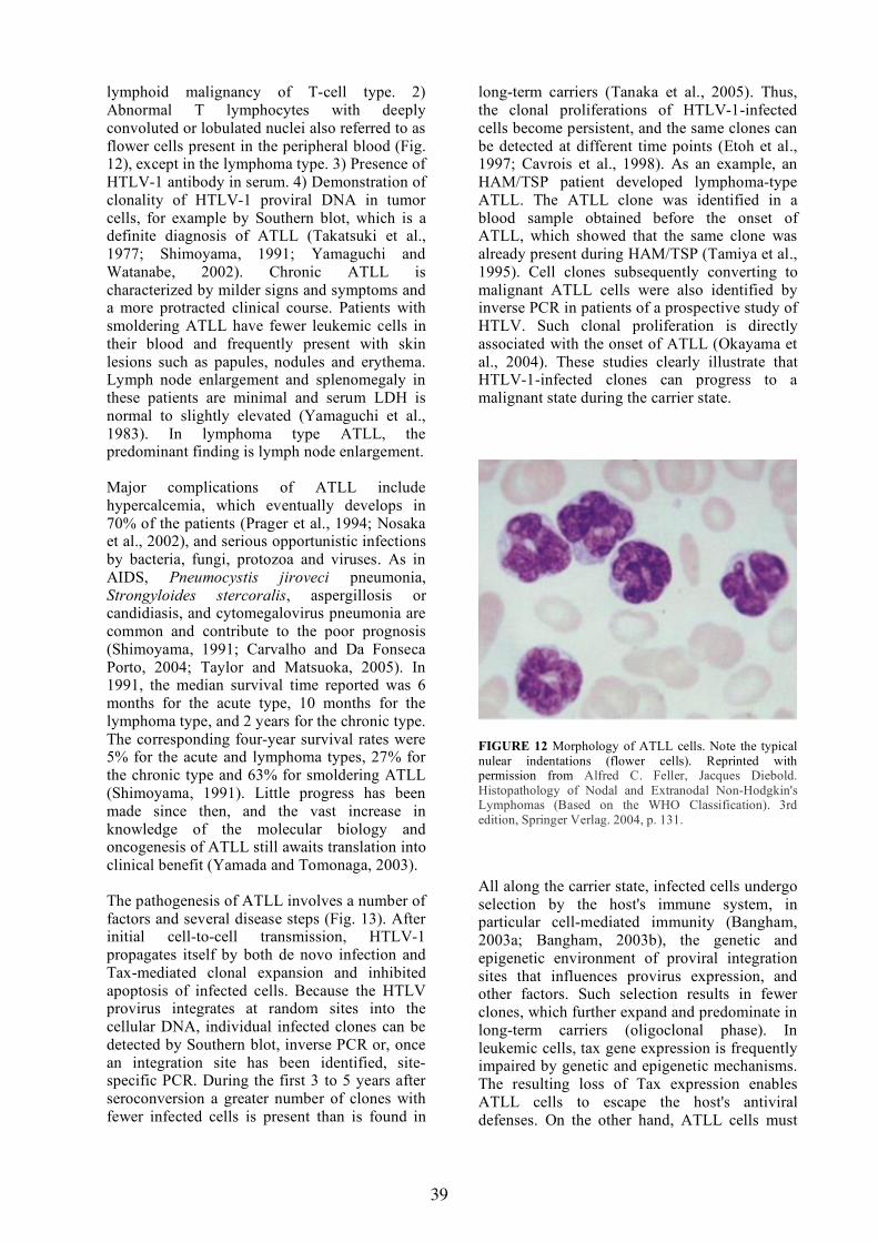
39
lymphoid malignancy of T-cell type. 2) Abnormal T lymphocytes with deeply convoluted or lobulated nuclei also referred to as flower cells present in the peripheral blood (Fig. 12), except in the lymphoma type. 3) Presence of HTLV-1 antibody in serum. 4) Demonstration of clonality of HTLV-1 proviral DNA in tumor cells, for example by Southern blot, which is a definite diagnosis of ATLL (Takatsuki et al., 1977; Shimoyama, 1991; Yamaguchi and Watanabe, 2002). Chronic ATLL is characterized by milder signs and symptoms and a more protracted clinical course. Patients with smoldering ATLL have fewer leukemic cells in their blood and frequently present with skin lesions such as papules, nodules and erythema. Lymph node enlargement and splenomegaly in these patients are minimal and serum LDH is normal to slightly elevated (Yamaguchi et al., 1983). In lymphoma type ATLL, the predominant finding is lymph node enlargement.
Major complications of ATLL include hypercalcemia, which eventually develops in 70% of the patients (Prager et al., 1994; Nosaka et al., 2002), and serious opportunistic infections by bacteria, fungi, protozoa and viruses. As in AIDS, Pneumocystis jiroveci pneumonia, Strongyloides stercoralis, aspergillosis or candidiasis, and cytomegalovirus pneumonia are common and contribute to the poor prognosis (Shimoyama, 1991; Carvalho and Da Fonseca Porto, 2004; Taylor and Matsuoka, 2005). In 1991, the median survival time reported was 6 months for the acute type, 10 months for the lymphoma type, and 2 years for the chronic type. The corresponding four-year survival rates were 5% for the acute and lymphoma types, 27% for the chronic type and 63% for smoldering ATLL (Shimoyama, 1991). Little progress has been made since then, and the vast increase in knowledge of the molecular biology and oncogenesis of ATLL still awaits translation into clinical benefit (Yamada and Tomonaga, 2003).
The pathogenesis of ATLL involves a number of factors and several disease steps (Fig. 13). After initial cell-to-cell transmission, HTLV-1 propagates itself by both de novo infection and Tax-mediated clonal expansion and inhibited apoptosis of infected cells. Because the HTLV provirus integrates at random sites into the cellular DNA, individual infected clones can be detected by Southern blot, inverse PCR or, once an integration site has been identified, site-specific PCR. During the first 3 to 5 years after seroconversion a greater number of clones with fewer infected cells is present than is found in
long-term carriers (Tanaka et al., 2005). Thus, the clonal proliferations of HTLV-1-infected cells become persistent, and the same clones can be detected at different time points (Etoh et al., 1997; Cavrois et al., 1998). As an example, an HAM/TSP patient developed lymphoma-type ATLL. The ATLL clone was identified in a blood sample obtained before the onset of ATLL, which showed that the same clone was already present during HAM/TSP (Tamiya et al., 1995). Cell clones subsequently converting to malignant ATLL cells were also identified by inverse PCR in patients of a prospective study of HTLV. Such clonal proliferation is directly associated with the onset of ATLL (Okayama et al., 2004). These studies clearly illustrate that HTLV-1-infected clones can progress to a malignant state during the carrier state.
FIGURE 12 Morphology of ATLL cells. Note the typical nulear indentations (flower cells). Reprinted with permission from Alfred C. Feller, Jacques Diebold. Histopathology of Nodal and Extranodal Non-Hodgkin's Lymphomas (Based on the WHO Classification). 3rd edition, Springer Verlag. 2004, p. 131.
All along the carrier state, infected cells undergo selection by the host's immune system, in particular cell-mediated immunity (Bangham, 2003a; Bangham, 2003b), the genetic and epigenetic environment of proviral integration sites that influences provirus expression, and other factors. Such selection results in fewer clones, which further expand and predominate in long-term carriers (oligoclonal phase). In leukemic cells, tax gene expression is frequently impaired by genetic and epigenetic mechanisms. The resulting loss of Tax expression enables ATLL cells to escape the host's antiviral defenses. On the other hand, ATLL cells must

40
have acquired the ability to proliferate without Tax by intracellular genetic and epigenetic changes (reviewed in Taylor and Matsuoka, 2005).
HTLV-1 associated inflammatory disorders
HTLV-1 is associated with a number of inflammatory disorders in addition to ATLL (Table 3). In contrast to ATLL, which is thought to develop partially due to a lack or secondary deterioration of CTL functions against T-cells expressing HTLV-1, the HTLV-1 associated inflammatory disorders are increasingly viewed as resulting from tissue damage triggered by exaggerated HTLV-1 induced T-cell responses (Shimojima et al., 2004).
HTLV-1–associated myelopathy/tropical spastic paraparesis (HAM/TSP)
Following identification of HTLV-1 as the etiologic agent of ATLL, a serological association with HTLV-1 was independently reported for patients from the Caribbean who suffered from TSP and for patients from Japan who presented with a myelopathy (Gessain et al., 1985; Osame et al., 1986). Comparative studies subsequently demonstrated that the viruses found in these diseases were genetically indistinguishable from the HTLV-1 strains that cause ATLL (Yoshida et al., 1987). HAM/TSP and ATLL are seen together only rarely in the same patient (Bartholomew et al., 1986; Kawai et al., 1989).
The life-time risk of developing HAM/TSP among seropositive individuals varies from less than 0.1% in Japan to 1.7-7% reported from Africa, the Caribbean, and the U.S. (reviewed by Taylor, 1998). The mean age at onset is in the forth decade of life and the male to female ratio is about 1:3. The incubation time extends from years to decades, but may occasionally be as short as 18 weeks (Osame et al., 1990). Disease onset is usually slow; patients often have had symptoms for years before the diagnosis is established.
Revised diagnostic criteria based on a WHO definition for HAM/TSP were proposed recently (De Castro-Costa et al., 2006). A definite diagnosis of HAM/TSP thus requires (i) a non-remitting progressive spastic paraparesis with
sufficiently impaired gait to be perceived by the patient. Sensory symptoms or signs may or may not be present. When present, they remain subtle and without a clear-cut sensory level. Urinary and anal incontinence may or may not be present. (ii) Presence of HTLV-1 antibodies in serum and cerebrospinal fluid (CSF) confirmed by WB and/or a positive PCR for HTLV-1 in blood and/or CSF. (iii) Exclusion of a long list of other conditions that can resemble HAM/TSP comprising, for example, multiple sclerosis, Lyme disease, neurosyphilis or neurotuberculosis. Criteria for probable and possible HAM/TSP were also proposed.
Pathological examination shows the most prominent changes in the thoracic spinal cord, with atrophy of the cord and thickening of the meninges. The primarily affected white matter shows inflammatory cell infiltrates which to a lesser degree also are seen in gray matter (Iwasaki, 1993). These inflammatory infiltrates are initially comprised of both CD8 and CD4+ T-cells, B cells, and foamy macrophages, but later in the disease CD8 T-cells predominate. Through this inflammatory process, myelin and axon loss eventually occurs, and the tissue is replaced by glial proliferation and fibrillary astrocytosis (Iwasaki, 1993; Abe et al., 1999; Jacobson, 2002; Grindstaff and Gruener, 2005). Similar changes are seen in the WKAH rat model after infection with HTLV-1 (Miyatake et al., 2006).
The HTLV-1 proviral load in PBMC of HAM/TSP patients is high; provirus concentrations are 3-50 fold higher than in asymptomatic carriers (Kubota et al., 1993; Olindo et al., 2005). HAM/TSP prevalence rose exponentially with the log(proviral load) when the proviral load exceeded 1% of the PBMC (Nagai et al., 1998). A high ratio of proviral loads in CSF cells compared to PBMC, but not the absolute load in either compartment, was found associated with clinically progressive disease and recent onset of HAM/TSP (Takenouchi et al., 2003). Together these findings suggest that clinical progression of HAM/TSP is associated with increased proliferation in, or increased immigration of HTLV-1-infected lymphocytes into, the CNS. How the presence of HTLV-1 in the CNS leads to HAM/TSP is still unclear, however, and several models are proposed (reviewed by Jacobson, 2002; Bangham, 2003b; Araujo and Silva, 2006). One model centers around a CTL response to Tax expressing cells within the CNS, leading to co-activation of microglial cells and

41
release of cytokines like TNFα that are toxic to the myelin (bystander effects). An autoimmune mechanism induced by molecular mimicry between Tax and a neuron-specific autoantigen, the so-called heterogeneous nuclear ribonuclear
protein A1 (hnRNP A1), and associated with detectable cross-reactive antibodies in HAM/TSP also has been proposed (Lee et al., 2005).
FIGURE 13 Natural course from HTLV-1 infection to onset of ATLL. HTLV-1 is transmitted in a cell-to-cell fashion. After infection, HTLV-1 promotes clonal proliferation of infected cells by pleiotropic actions of Tax and other viral proteins. Proliferation of HTLV-1-infected cells is controlled by cytotoxic T-cells in vivo. After a long latent period, ATLL develops in about 5% of asymptomatic carriers. In ATLL, the expression of Tax is inactivated by several mechanisms, suggesting that Tax is no longer necessary in ATLL stage. Alternatively, alterations and errors in the host genome accumulate progressively during the latent period, finally leading to onset of ATLL. Reprinted from Oncogene (Taylor and Matsuoka, 2005) with permission of the publisher
HTLV-1–associated uveitis (HAU)
In 1992, an uveitis of otherwise unexplained etiology in HTLV-1 infected patients was proposed as another disease entity (Mochizuki et al., 1992). HAU patients are of similar age as those with HAM/TSP. They present with a blurred vision of acute or subacute onset, preserved visual acuity in most instances, iritis, vitreous opacities, retinal vasculitis with exudates and hemorrhages (Takahashi et al., 2000). The condition usually responds well to ocular or oral administration of corticosteroids, although recurrence is observed frequently.
Proviral DNA of HTLV-1 is present in a high percentage of T lymphocytes isolated from the intraocular fluid, suggesting that the intraocular
presence of HTLV-1 infected T lymphocytes is due to positive selection rather than coincidence. These T-cells are activated and release inflammatory cytokines deemed responsible for HAU since they can be abrogated by corticosteroid therapy (Sagawa et al., 1995). HAU frequently seems to be associated with a history of Graves' disease (Yamaguchi et al., 1994; Watanabe et al., 1997; Sarui et al., 2002); the combination of the two diseases was associated with a significantly higher proviral load in PBMC. The proviral load also correlated with disease activity in terms of vitreous inflammation and interval to recurrence (Ono et al., 1998).

42
HTLV-1–associated arthritis
A chronic arthritis in HTLV-1–infected individuals was first described in 1989 (Nishioka et al., 1989). HTLV-1–specific antibodies can be shown in synovials fluids of the affected joints, and proviral DNA was demonstrated in synovial tissues and synovial fluid lymphocytes (Kitajima et al., 1991). Like HAM/TSP and HAU, HTLV-1–associated arthritis is associated with increased proviral DNA levels. Similar to HAU, an increased provirus load was found in the disease-affected tissue (synovial cells) compared to PBMC (Yakova et al., 2005).
Evidence that the arthritis of HTLV-1 infected persons is not due to a mere coincidence with a disease frequent in most countries includes the demonstration that HTLV-1 env-pX transgenic mice or rats develop chronic inflammatory lesions with similarity to human rheumatoid arthritis (Iwakura et al., 1995; Abe et al., 2006). The majority of infiltrating T-cells in arthritic joints of env-pX rats were activated CD4+ T-cells, and their transfer into the joints of wild-type rats also induced arthritis.
HTLV-1–associated bronchopneumopathy
Pulmonary complications are more frequent in patients with ATL than in patients with other hematologic malignancies. They are present in more than 90% of the ATL patients and include, in addition to leukemic cell infiltration, a variety of opportunistic infections. Patients with inflammatory conditions like HAM/TSP, HAU or arthritis also have frequent pulmonary complications characterized by T-lymphocytic alveolitis in the absence of leukemic cells or opportunistic pathogens. A similar pulmonary involvement is also detectable in clinically asymptomatic carriers. Characteristically, respiratory symptoms and chest radiographic abnormalities are rare, although 60-80% have T-lymphocytic bronchiolitis, alveolitis or interstitial pneumonia. Chronic sinusitis is also frequent, especially in patients with diffuse panbronchiolitis. Regarding pathogeneis, similar mechanisms as discussed for HAM/TSP are proposed, namely cytotoxic T-cell responses and an inflammatory effect exerted by cytokines released from activated cells (Sugimoto et al., 1998; Seki et al., 2000).
HTLV-1 associated infective dermatitis (IDH)
HTLV-1 associated IDH is a recurrent, infective form of eczema first described in Jamaica in 1966 (Sweet, 1966) and later linked to vertically transmitted HTLV-1 infection (La Grenade et al., 1998). The onset is generally after 18 months of life, and the disease rarely persists until adulthood. Infective dermatitis is a chronic, relapsing skin infection frequently involving staphylococci or streptococci. It always involves the scalp and may progress to HAM/TSP or ATL. Most cases of IDH were reported from Jamaica and Brazil. Smaller case series were described in Trinidad and Tobago, Peru and Senegal. Curiously, in Japan, where the prevalence of HTLV-I infection is elevated, only two cases of children with IDH have been reported, both of which progressed to ATL in adulthood (Bittencourt et al., 2006).
Unproven disease associations of HTLV
A large study of the most common hematological diseases in Europe (n=730 plus 210 controls) reported HTLV-1 infection in 11/67 (17%) patients with a myelodysplastic syndrome (a neoplasia of myeloid cells), 1/26 patients with T-cell non-Hodgkin lymphoma and 1/1 patient with T-cell acute lymphocytic leukemia (Karlic et al., 1997), but these findings, in particular regarding the myelodysplastic syndrome, remain unconfirmed (Morselli et al., 1999). Claims that cutaneous T-cell lymphomas are associated with an isolated presence of the HTLV-1 tax gene in the absence of antibodies to structural proteins (Manca et al., 1994; Pancake et al., 1995; Pancake et al., 1996) were not confirmed by other groups, even when cutaneous T-cell lymphomas patients from regions endemic for HTLV-1 or from the same country from which such reports originated were analyzed (Boni et al., 1996b; Bazarbachi et al., 1997; Kikuchi et al., 1997; Wood et al., 1997). Reports of an isolated presence of sequences related to HTLV-1 tax continue, however, also in association with autoimmune disorders (Zucker-Franklin et al., 2000; Zucker-Franklin, 2001; Manca et al., 2002; Morozov et al., 2002; Morozov et al., 2005).

43
HTLV-2–Associated Diseases
HTLV-2 was originally isolated from a T-cell line (Mo-T) derived from a patient with a T-cell variant of hairy cell leukemia (Kalyanaraman et al., 1982). Subsequently, the virus was also isolated from a similar case which upon closer examination demonstrated a coexistence of two different proliferative processes, namely a CD8+ T-cell leukemia with monoclonally integrated HTLV-2 and a B-cell hairy cell leukemia which was negative for integrated HTLV-2 (Rosenblatt et al., 1986).
A pathogenetic role of HTLV-2 in malignancies involving CD8+ T-cells was supported by the virus' tropism for CD8+ T-cells in vitro (Ijichi et al., 1992) and the fact that lymphocytes of HTLV-2 infected patients proliferate in vitro in the absence of antigenic stimulation, similar to cells of HTLV-1 infected individuals (Wiktor et al., 1991). Epidemiological studies have, however, excluded that the typical B-cell form
of hairy cell leukemia is associated with HTLV-2 (Hjelle et al., 1991). There is also no further evidence supporting a significant role of HTLV-2 in causing lymphoproliferative diseases. Some evidence links HTLV-2 with HAM/TSP and perhaps with other neurological syndromes (Jacobson et al., 1993; Lehky et al., 1996; Murphy, 1996). It also appears to be associated with an increased incidence of pneumonia and bronchitis, inflammatory conditions such as arthritis and, perhaps, with an increased mortality (Murphy, 1996; Roucoux and Murphy, 2004; Jarvis et al., 2005). In general, HTLV-2 pathogenicity is lower than that of HTLV-1 (Feuer and Green, 2005), which may be due to a lower proviral load (Hisada et al., 2005). The generally lower proviral load may be associated with a comparatively lower efficiency of Tax-2 for viral transactivation, cellular transformation, induction of cell cycle arrest and suppression of hematopoiesis (reviewed by Feuer and Green, 2005).
FIGURE 14 Epidemiologic map of HTLV-1. Countries with a prevalence between 1 and 5% in some populations, are shown darkly shaded. Countries with a prevalence of less than 1% in some groups, due mainly to immigration from endemic areas, are shown in light shade. Note that HTLV-1 endemic areas do not correspond exactly to the country boundaries shown in the map. For example, HTLV-1 in Brazil, Japan and Iran is limited to distinct areas within each country. Reprinted from Oncogene (Proietti et al., 2005) with permission of the publisher.
Epidemiology and Transmission of the HTLVs
Foci of HTLV-1 infection are found geographically clustered, amounting to about 20 million infected individuals worldwide (Proietti
et al., 2005). The geographic distribution of the virus has been defined, with Japan, Africa, the Caribbean islands and South America as the areas of highest prevalence. Additional endemic regions include the Middle East, the Pacific Melanesian islands and Papua New Guinea (Fig.

44
14). HTLV-2 is endemic in Amerindian and pygmy tribes. More recently, injecting drug users in the U.S. and Europe have become infected with HTLV-1 and HTLV-2, particularly subtype A, and secondary sexual transmission has introduced the viruses at low levels into the general population and blood donors (Gessain and de The, 1996; Manns et al., 1999a; Roucoux and Murphy, 2004; Taylor et al., 2005).
Like the HIVs, the HTLVs are transmitted by hetero- or homosexual intercourse, from mother-to-child or by parenteral inoculation. Live HTLV-infected cells are essential in all transmission modes. Mother-to-child transmission is with 15-30% of similar frequency as in untreated HIV-1 infection and occurs predominately in the postnatal period through breast milk. Breast-milk transmission seems to be more efficient than for HIV-1 and occurs with a time- or dose-dependent frequency. In one study, overall transmission was 16%. It was 5% among infants breast-fed for up to three months and 27% among those breast-fed for over three months. Of 78 bottle-fed infants 13% turned out to be infected, suggesting also connatal transmission (Hirata et
al., 1992). In other studies, however, connatal transmission was considerably less frequent (3%). Although HTLV-1 infected cells were detected by PCR in 2.5% of the cord bloods from HTLV-1–positive pregnancies, this was not associated with infection when the babies were formula-fed (Hino et al., 1996). In high-prevalence areas, serologic testing of pregnant women and counseling of those found infected with regard to alternatives to breast-feeding is thus recommended. Transmission by breast-milk also depends on its provirus load (Ureta-Vidal et al., 1999; Li et al., 2004).
Transmission by blood products is, in contrast to HIV, strictly cell-associated; the virus is not transmitted by plasma or plasma-derived products (Okochi et al., 1984). Recipients of contaminated blood seroconvert with a 40–60% probability and a median seroconversion time estimated at 51 days (Manns et al., 1999a). HTLV screening of blood donors is justified in countries with an elevated prevalence in the general population.
Diagnosis of HTLV Infections
The principles, tools and problems of HTLV diagnosis are, with some modifications, the same as those for the diagnosis of HIV (see above). Screening is based on tests for HTLV-specific antibodies by ELISA or particle agglutination tests. Confirmatory tests are based on WB or LIA. Since there are many indeterminate WB results, confirmation must be backed by supplemental tests, usually PCR for proviral DNA. Tests for virion components (antigen, viral RNA) in plasma are not to be used, as there is no HTLV plasma viremia.
Screening Tests for HTLV-1/2
For HTLV-1/2 screening, tests analogous to those reviewed in Fig. 6 for the detection of HIV are used, with the same principal advantages and drawbacks. Since all subtypes of HTLV-1 diverge less than 10% from each other, underdetection of HTLV-1 infection as a result of sequence diversity is not a problem. Similarly, some sequences like the capsid region
of gag, are also well conserved between HTLV-1 and HTLV-2, and there is broad serologic crossreactivity between the two virus types. Due to the low degree of provirus expression and the presumed rapid CTL-mediated elimination of Tax-expressing cells, antibody levels to structural proteins may be low, however. Thus, third generation tests (double antigen sandwich EIA or particle agglutination) are the preferred test formats, as they should provide the highest sensitivity. Tests based on ELISA, particle agglutination or indirect immune fluorescence are available from various companies (Taylor et al., 1996; Farias de Carvalho et al., 1997; Vrielink et al., 1999; Boni et al., 2004a). For FDA-approved tests refer to http://www.fda.gov/cber/products/testkits.htm. Note that there are considerable differences in the quality of these products, particularly regarding specificity. In low-prevalence populations, a reactive result in an assay of low specificity has a very low positive predictive value (PPV). For example, one recent study involving four different HTLV-1/2 EIAs found a specificity above 99% in three of the tests, but of

45
only 93% in the fourth test. Even among a high-risk group with a prevalence of 311/100,000, the PPV varied from an unacceptably low 4% (for the test with 93% specificity) to a maximum of 44% for a test with 99.6% specificity. Provided that these assays exhibited the same specificity among blood donors (in whom the prevalence was 0.08/100,000), these PPV would indicate that with the most specific assay only 2/10,000 reactive results would indicate a true infection. In contrast, with the least specific assay only 1/100,000 reactive results would indicate a true infection. These data illustrate the need for both a careful choice of screening assays and of supplementary testing (Petersen et al., 1994; Boni et al., 2004a).
Supplemental Tests for HTLV-1/2
Western blotting and line immunoassay
Serologic confirmation of HTLV-1/2 infection requires the demonstration of antibodies to both gag (p24) and env (gp46 and/or gp68 proteins) by WB and/or radioimmunoprecipitation assay (Anonymous, 1993). In our view, WB is preferable to in-house methods of confirmation like radioimmunoprecipitation assay, since the commercial WB kits are better standardized and much easier to run. WB kits are provided by a number of companies. Of particular interest are strips which contain, in addition to the viral proteins derived from viral lysate, recombinant proteins representing TM of HTLV-1 (which due to a high homology is also detected by antibodies from individuals infected with HTLV-2) and type-specific SU (gp46) of HTLV-1 and HTLV-2. This increases the sensitivity since the concentration of the gp46 for HTLV-1 and gp68 for HTLV-2Env proteins on the strips is usually low in kits derived from lysate alone. Based on the pattern of the reactivity it is frequently, though not always, possible to decide whether infection by HTLV-1 or HTLV-2 is present. With such strips, intense reaction with the gag proteins p19 and p24 and the env recombinant proteins rgp21 and rgp46 of HTLV-1 satisfies positivity for HTLV-1 and intense reaction with p24, rgp21 and rgp46 of HTLV-2 satisfies positivity for HTLV-2 (Medrano et al., 1997).
Furthermore, the use of LIA strips, which contain standardized concentrations of recombinant proteins and/or synthetic peptides of HTLV-1 and/or HTLV-2 at defined positions
and thus can be considered as a kind of 2nd generation WB, presents advantages with respect to both sensitivity and specificity (Zrein et al., 1998; Sabino et al., 1999; Thorstensson et al., 2002).
Still, in many cases WB and LIA do not permit an unequivocal diagnosis, owing to a high percentage of samples with indeterminate results. There is nonspecific reaction not only with lysate-derived natural gag proteins, but also with recombinant env proteins (Table 4). Some of these indeterminates may have very intense reactions with p19 or p24 and a variety of larger proteins such as p26, p28, p32, p36, p45 and p53. These proteins are present in HTLV-1 infected cells and contain either a p19 or p24 moiety, or both (Schupbach and Kalyanaraman, 1989). Reaction with several of these gag proteins on a WB thus may signify no more than reaction with a single epitope of p19 or, respectively, p24 (Schupbach et al., 1988). Sometimes, intense gag patterns may also be combined with weak reaction to envelope rgp21 and/or rgp46I or rgp46II, but even this does not necessarily imply HTLV infection. Weak reactions with recombinant envelope proteins in all possible combinations may also be found in the absence of reaction with gag proteins. In many instances, confirmation by PCR is thus necessary. This is particulary true for areas or populations in which HTLVs are not endemic. Under such conditions, any suggestive serological result not strongly antibody-positive should be confirmed by PCR. Indeterminate reactions are frequent in populations at risk for exposure, as drug addicts (Medrano et al., 1997), but may also be present in truly infected individuals (Zehender et al., 1996; Caterino-de-Araujo et al., 1998). Infection with the SARS coronavirus has recently been identified as a possible cause of false reactivity in both HTLV screening tests and WB (Tsao et al., 2005).
Given the problems with nonspecific bands in WB alternative diagnostic strategies based on testing with a combination of two sensitive and specific EIAs have been proposed (Thorstensson et al., 2002). Thus, while maintaining a higher overall sensitivity than with the classical EIA-WB combination, an EIA-EIA strategy reduced the frequency of samples with indeterminate results to 2.5%.

46
Polymerase chain reaction (PCR)
PCR analysis for HTLV-1 and/or HTLV-2 DNA is necessary for all serologically indeterminates in which antibody reaction to Env proteins (rgp21, rgp46I or rgp46II) is present. Antibody reaction with Gag proteins p19 and/or p24 alone, or rgp21 alone, has been found by PCR not to be associated with HTLV infection (Defer et al., 1995). PCR is performed on Ficoll-purified PBMC and frequently uses a sequence of tax which is conserved for both HTLV-1 and HTLV-2 and amplified by primers designated SK43/SK44, while the product is detected by probe SK45 (Kwok et al., 1988; Kwok et al., 1990). Differentiation of HTLV-1 and HTLV-2
in samples positive in this initial "screening PCR" is then achieved by amplification of a type-specific region in pol. Primers SK110 and SK111 in combination with probe SK112 are used for detection of HTLV-1. The same primers in combination with probe SK188 are employed for HTLV-2. This system also is available as a commercial kit (Amplicor HTLV 1-2 PCR test, Roche Diagnostic Systems)(Vrielink et al., 1997). Alternatively, in-house PCR methods described by various authors can be used for both the screening step and the type differentiation (Vandamme et al., 1997; Salemi et al., 1998; Boni et al., 2004a). Real-time PCR methods also have been described (Davidson et al., 2006).
Table 4: Breakdown of procedures undertaken in a reference lab during repeat testing of HIV-positive samples with additional reactivity or high-negative results in HTLV-1/2 ELISA screening. Reprinted from Journal of Medical Virology (Boni et al., 2004a) with permission of the publisher.
Western Blot3 ELISA Natural Proteins
Recombinant Proteins
Culture Supernats RT-PCR
# Risk1
Test2
OD/ CO
p19 p24 p26 p28 p32 p36 p53 p21E rgp46E HTLV-1
rgp46E HTLV-2
PCR HTLV
PBMC PERT4
HTLV HIV-1
Final Diagn
1 IDU PL 25.77 +++ +++ - ++ - +++ +++ +++ - +++ + HTLV-2 2 IDU PL 8.09 ++ +++ - + - ++ + +++ - +++ + HTLV-2 3 HET CR 7.76 +++ +++ +++ +++ - - - +++ +++ - + HTLV-1 4 HET PL 7.37 +++ +++ - ++ - ++ ++ +++ - +++ + HTLV-2 5 IDU CR 6.16 +++ - - - - - - +++ - +++ + + +,+ -,- HTLV-2 6 IDU PL 3.34 ++ +++ - + - ++ + +++ - +++ + HTLV-2 7 IDU AB 2.47 - - - - - - - - - + - -,- -,- -,- neg 8 IDU AB 1.93 - ++ - - - - - +++ - +++ - -,- -,- -,- neg 9 IDU CR 1.50 - - - - - - - ++ - - - - - + neg 10 HET AB 1.35 - - - - - - - - + - - -,- -,- -,- neg 11 HET AB 1.34 - - - - - - - + - - - -,- -,- -,- neg 12 HET AB 1.30 - - - - - - - - - + - - - + neg 13 IDU AB 1.26 - - - - - - - - ++ - - - - - neg 14 MSM AB 1.25 - - - - - - - - + + - -,- - - neg 15 HET AB 1.13 - ++ - - - - - - - - - - - - neg 16 MSM AB 1.11 +++ - ++ - ++ - - - - + neg 17 IDU AB 1.07 - + - - - - - - - - - - - + neg 18 IDU AB 1.06 - + - - - - - - - - - - - - neg 19 IDU AB 1.01 - - - - - - - - + - - - - - neg 20 HET AB 0.87 - - - - +++ - - - - - - -,- - - neg 21 HET AB 0.86 +++ - ++ ++ - ++ - - - - - -,- - - neg 22 IDU AB 0.83 - - - - - - - ++ + + - - nd nd neg 1 MSM = homosexual contact; HET = heterosexual contact; IDU = intravenous drug use. 2 PL = Platelia (Bio-Rad); CR = Cobas (Roche); AB = Abbott HTLV-I/II EIA 3 HTLV BLOT Version 2.4 (Genelabs Diagnostics, Inc). NB: Reaction intensities were subjectively rated as ‘no
reaction’ (-); ‘clearly visible, but weak’ (+); ‘intermediately strong’ (++); ‘strong’ (+++). Reactions with natural proteins p21E and gp46E were all negative and are not listed.
4 Symbols summarize results for supernatants sampled twice per week for at least 2 weeks. Symbols separated by a comma represent results of duplicate cultures.
Virus isolation
Compared with PCR virus isolation is time-consuming and overall more expensive and therefore has little use for mere confirmation
of HTLV infection. The procedure is, however, still justified as a research tool for detecting unknown retroviruses, that might eventually explain some of the numerous indeterminate serologic results (Boni et al., 2004a). Virus

47
isolation should thus not only be evaluated with HTLV-specific tests, (antigen assay or immunofluerescence), but where available also by assays for particle-associated reverse transcriptase, preferentially PERT assay (see "Supplemental testing - virus components" in the section on HIV). For virus isolation, Ficoll-purified PBMC are cocultured with phytohemagglutinin-preactivated normal PBMC or cord blood leukocytes in an IL-2 containing medium. Supernatant is analyzed twice weekly for RT. In case of HTLV infection the PERT assay usually becomes positive within a few days, while detection of RT by convential assays may take several weeks. When the PERT assay has become positive, specific tests for HTLV like p24 antigen assay, PCR or RT-PCR can be performed. Differentiation of HTLV-1 and HTLV-2 in virus culture is best achieved by PCR. The use of generic PCR primers and specific probes capable of differentiating between the various primate T-lymphotropic retroviruses is helpful in such a situation (Vandamme et al., 1997).
Disease monitoring by provirus quantification
As a high HTLV-1 provirus concentration is associated with the development of various HTLV-associated illnesses and the transmission risk by breast milk (Li et al., 2004), measurement of the HTLV-1 provirus load is often required, although it has not yet been as firmly established for disease monitoring as is the case with the HIV-1 virion load. Most ATLL cells contain only one provirus copy. As ATLL cells are derived from HTLV-1 infected cells, it is reasonable to conclude that most HTLV-1 infected cells contain also only one provirus. Provirus quantification by quantitative PCR thus can be used for enumeration of HTLV-1 infected cells in vivo. HTLV-1 provirus load in infected individuals differs more than 1000-fold among asymptomatic carriers (Etoh et al., 1999).
Treatment of HTLV Infection
During the three decades that have passed since the recognition of ATLL a variety of treatment approaches has been evaluated
(reviewed by Taylor and Matsuoka, 2005). Combination chemotherapy with cyclosphosphamide, adriamycin, vincristine and prednisolone (CHOP) is still the standard first-line therapy. Many patients experience either partial or complete remission, but its duration is usually short, and the median survival time is only 6 months. Intensification of CHOP with etoposide, vindesine, ranimustine, and mitoxantrone was associated with a higher remission rate, but median survival increased only insignificantly. The best outcome of combination chemotherapy, with a median survival time of 13 months, was achieved with an aggressive multidrug approach of high bone marrow toxicity given in combination with granulocyte monocyte – colony stimulating factor (Yamada et al., 2001). Overall, ATLL survival with various chemotherapy regimens remains poor, with survival in several cohorts of patients presenting prodominantly with acute leukemia or lymphoma ranging between 5.5 and 13 months. Supplementation of chemotherapy with interferon-alpha and zidovudine as first line therapy yielded a median survival time of 18 months (Matutes et al., 2001; Kchour et al., 2007). Since ATLL is a monoclonal disease which does no longer depend on viral replication, it is unlikely that the effect of zidovudin is based on inhibition of RT; an antineoplastic mechanism is more likely . Of interest are also drugs that appear to induce apoptosis of ATLL cells, like the combination of IFNα and arsensic trioxide (Mahieux and Hermine, 2005; Heraud et al., 2006), blockers of NF-κB, and histone deacetylation inhibitors (HDIs) like sodium valproate. Sodium valproate is a drug widely prescribed for epilepsy, bipolar disorders and migraine and has an excellent safety record. Of interest is that dramatic clearance of both lymphoma and leukemia has been demonstrated in a B-cell malignancy of sheep, that is induced by bovine leukemia virus, which is a close relative of HTLV. It is hypothesized that inhibition of DNA deacetylation leads to better provirus expression, thereby resulting in improved immune-mediated elimination of virus-expressing leukemic cells (Achachi et al., 2005). Valproate is also of potential interest for prevention of progression to acute ATLL in patients with the smoldering or chronic forms of ATLL, or for carriers still asymptomatic with a high proviral load, who are at risk for development of disease. A variety of monoclonal antibodies also has been evaluated.
ATLL cells exhibit a high density of the IL-2 receptor alpha chain (CD25), and treatment with CD25-specific mAb was capable of inducing

48
remission in a minority of the patients (Waldmann et al., 1988). CD25-specific mAb, meanwhile in humanized form, are also tried in combination with CHOP. Another target of mAb therapy is CD52. A humanized mAb to CD52, Campath-1H, was shown to effectively treat severe combined immunodeficiency mice inoculated with tumor-causing human ATLL cells (Zhang et al., 2003) and showed promise in human patients (Mone et al., 2005).
Finally, both allogeneic and autologous bone marrow transplantation are evaluated as possible treatments of ATLL. A first case of apparent cure was reported in 1996. After a 4-day infusion of cyclophosphamide, etoposide and doxorubicin, the patient was grafted with bone marrow cells donated by an HTLV-uninfected sister (Borg et al., 1996). Nine years later, in 2005, the patient was still alive and free of disease. In a case series of 10 patients receiving allogeneic hematopoietic stem cell therapy (Allo-SCT) (9/10 from HLA-identical siblings) after receiving total body irradiation and other conditioning agents, the median leukemia-free survival time was >17.5 months. Four patients died, however, and in two others ATLL relapsed (Utsunomiya et al., 2001). Further experience with this treatment in 40 patients shows that complete remission is achieved in a high proportion of patients. However, there is also a high rate of transplantation-associated complications resulting in an overall median survival of less than 10 months (Fukushima et al., 2005).
Overall, ATLL remains a disease of poor prognosis. Prevention of the disease is thus of paramount importance. Public health intervention with the aim to provide education and counseling of high-risk individuals and populations is required. Avoidance of breast feeding and the introduction of HTLV screening of all blood donors have led to a significant decline of the carrier rate among the younger generation in Japan (Takatsuki et al., 1996). Given the high kit costs for blood donor screening, a transfer of this strategy to resource poor settings with HTLV-1 endemicity has so far not been possible. Blood transfusion still represents a risk of HTLV-1 infection for recipients in most African countries, as well as for other less developed areas (Mbanya et al., 2003). Prevention of mother to child transmission would likely have a significant impact on the incidence of HTLV-1-associated diseases, but the benefits
of avoiding breastfeeding must be weighed against its risks, namely malnutrition and increased infant mortality. Recommendations to prevent sexually transmitted infections are the same as for the prevention of HIV infection.
REFERENCES
Abe, A., A. Ishizu, K. Fugo, H. Ikeda, and T. Yoshiki. 2006. Articular tissues expressing the env-pX transgene are required for generation of arthritogenic T cells in human T cell leukemia virus type I transgenic rats. Clin Exp Rheumatol 24:313-316.
Abe, M., F. Umehara, R. Kubota, T. Moritoyo, S. Izumo, and M. Osame. 1999. Activation of macrophages/microglia with the calcium-binding proteins MRP14 and MRP8 is related to the lesional activities in the spinal cord of HTLV-I associated myelopathy. J Neurol 246:358-364.
Achachi, A., A. Florins, N. Gillet, C. Debacq, P. Urbain, G. M. Foutsop, F. Vandermeers, A. Jasik, M. Reichert, P. Kerkhofs, L. Lagneaux, A. Burny, R. Kettmann, and L. Willems. 2005. Valproate activates bovine leukemia virus gene expression, triggers apoptosis, and induces leukemia/lymphoma regression in vivo. Proc Natl Acad Sci U S A 102:10309-10314.
Albrecht, B., and M. D. Lairmore. 2002. Critical role of human T-lymphotropic virus type 1 accessory proteins in viral replication and pathogenesis. Microbiol Mol Biol Rev 66:396-406, table of contents.
Ando, Y., Y. Ekuni, Y. Matsumoto, S. Nakano, K. Saito, K. Kakimoto, T. Tanigawa, M. Kawa, and T. Toyama. 2004. Long-term serological outcome of infants who received frozen-thawed milk from human T-lymphotropic virus type-I positive mothers. J Obstet Gynaecol Res 30:436-438.
Anonymous. 1989. Interpretation and use of the western blot assay for serodiagnosis of human immunodeficiency virus type 1 infections. Morb Mortal Wkly Rep 38:1-7.
Anonymous. 1990. Acquired immunodeficiency syndrome (AIDS). Proposed WHO criteria for interpreting results from western blot assays for HIV-1, HIV-2, and HTLV-I/HTLV-II. Wkly Epidemiol Rec 65:281-283.
Anonymous. 1991. Biosafety guidelines for diagnostic and research laboratories working with HIV, vol. 9. World Health Organization, Geneva.
Anonymous. 1993. Centers for Disease Control and Prevention and the USPHS Working Group: Guidelines for counselling persons infected with human T-lymphotropic virus type I (HTLV-I) and type II (HTLV-II). Ann Intern Med 118:448-454.
Anonymous. 1995. Case-control study of HIV seroconversion in health-care workers after percutaneous exposure to HIV-infected blood--France, United Kingdom, and United States, January 1988-August 1994. Morb Mortal Wkly Rep 44:929-933.
Anonymous. 2006. Panel on Antiretroviral Guidelines for Adult and Adolescents. Guidelines for the use of antiretroviral agents in HIV-infected adults and adolescents. October 10, 2006; 1-113. Available at http://www.aidsinfo.nih.gov/ContentFiles/AdultandAdolescentsGL.pdf. .

49
Araujo, A. Q., and M. T. Silva. 2006. The HTLV-1 neurological complex. Lancet Neurol 5:1068-1076.
Autran, B., G. Carcelain, T. S. Li, C. Blanc, D. Mathez, R. Tubiana, C. Katlama, P. Debre, and J. Leibowitch. 1997. Positive effects of combined antiretroviral therapy on CD4+ T cell homeostasis and function in advanced HIV disease [see comments]. Science 277:112-116.
Baba, M. 2006. Recent status of HIV-1 gene expression inhibitors. Antiviral Res 71:301-306.
Balvay, L., M. Lopez Lastra, B. Sargueil, J. L. Darlix, and T. Ohlmann. 2007. Translational control of retroviruses. Nat Rev Microbiol 5:128-140.
Bangham, C. R. 2003a. Human T-lymphotropic virus type 1 (HTLV-1): persistence and immune control. Int J Hematol 78:297-303.
Bangham, C. R. 2003b. The immune control and cell-to-cell spread of human T-lymphotropic virus type 1. J Gen Virol 84:3177-3189.
Bangham, C. R., and M. Osame. 2005. Cellular immune response to HTLV-1. Oncogene 24:6035-6046.
Barre-Sinoussi, F., J. C. Chermann, F. Rey, M. T. Nugeyre, S. Chamaret, J. Gruest, C. Dauguet, C. Axler-Blin, F. Vezinet-Brun, C. Rouzioux, W. Rozenbaum, and L. Montagnier. 1983. Isolation of a T-lymphotropic retrovirus from a patient at risk for acquired immune deficiency syndrome (AIDS). Science 220:868-871.
Bartholomew, C., F. Cleghorn, W. Charles, P. Ratan, L. Roberts, K. Maharaj, N. Jankey, H. Daisley, B. Hanchard, and W. Blattner. 1986. HTLV-I and tropical spastic paraparesis [letter]. Lancet 2:99-100.
Baxter, J. D., D. L. Mayers, D. N. Wentworth, J. D. Neaton, M. L. Hoover, M. A. Winters, S. B. Mannheimer, M. A. Thompson, D. I. Abrams, B. J. Brizz, J. P. Ioannidis, and T. C. Merigan. 2000. A randomized study of antiretroviral management based on plasma genotypic antiretroviral resistance testing in patients failing therapy. CPCRA 046 Study Team for the Terry Beirn Community Programs for Clinical Research on AIDS. AIDS 14:F83-93.
Bazarbachi, A., V. Soriano, R. Pawson, A. Vallejo, T. Moudgil, E. Matutes, J. Peries, A. Molina, H. de The, T. F. Schulz, D. Catovsky, and P. S. Gill. 1997. Mycosis fungoides and Sezary syndrome are not associated with HTLV-I infection: an international study. Br J Haematol 98:927-933.
Berger, E. A., R. W. Doms, E. M. Fenyo, B. T. M. Korber, D. R. Littman, J. P. Moore, Q. J. Sattentau, H. Schuitemaker, J. Sodroski, and R. A. Weiss. 1998. A New Classification For Hiv-1. Nature 391:240.
Biberfeld, P., K. J. Chayt, L. M. Marselle, G. Biberfeld, R. C. Gallo, and M. E. Harper. 1986. HTLV-III expression in infected lymph nodes and relevance to pathogenesis of lymphadenopathy. Am J Pathol 125:436-442.
Bittencourt, A. L., J. Primo, and M. F. de Oliveira. 2006. Manifestations of the human T-cell lymphotropic virus type I infection in childhood and adolescence. J Pediatr (Rio J) 82:411-420.
Bleul, C. C., M. Farzan, H. Choe, C. Parolin, I. Clark-Lewis, J. Sodroski, and T. A. Springer. 1996. The
lymphocyte chemoattractant SDF-1 is a ligand for LESTR/fusin and blocks HIV-1 entry. Nature 382:829-833.
Bock, P. J., and D. M. Markovitz. 2001. Infection with HIV-2. Aids 15 Suppl 5:S35-45.
Bogh, M., R. Machuca, J. Gerstoft, C. Pedersen, N. Obel, B. Kvinesdal, H. Nielsen, and C. Nielsen. 2001. Subtype-specific problems with qualitative Amplicor HIV-1 DNA PCR test. J Clin Virol 20:149-153.
Boni, J. 1996. PCR detection of HIV. Methods Mol Biol 50:93-107.
Boni, J., L. R. Bisset, J. J. Burckhardt, H. I. Joller-Jemelka, P. Burgisser, L. Perrin, M. Gorgievski, P. Erb, W. Fierz, J. C. Piffaretti, and J. Schupbach. 2004a. Prevalence of human T-cell leukemia virus types I and II in Switzerland. J Med Virol 72:328-337.
Boni, J., M. Opravil, Z. Tomasik, M. Rothen, K. Bisset, P. Grob, R. Lüthy, and J. Schupbach. 1997. Simple monitoring of antiretroviral therapy with a signal-amplification-boosted HIV-1 P24 antigen assay with heat-denatured plasma. AIDS 11:F47-F52.
Böni, J., H. Pyra, M. Gebhardt, L. Perrin, P. Bürgisser, L. Matter, W. Fierz, P. Erb, J. C. Piffaretti, E. Minder, P. Grob, J. J. Burckhardt, M. Zwahlen, J. Huder, and J. Schüpbach. 1999. High frequency of non-B subtypes in newly diagnosed HIV-1 infections in Switzerland. J Acquir Immune Defic Syndr Hum Retrovirol 22:174-179.
Boni, J., H. Pyra, and J. Schupbach. 1996a. Sensitive detection and quantification of particle-associated reverse transcriptase in plasma of HIV-1-infected individuals by the product-enhanced reverse transcriptase (PERT) assay. J Med Virol 49:23-28.
Boni, J., and J. Schupbach. 1999. Reverse transcriptase assay based on product enhancement for assessing the drug susceptibility of retroviruses., p. 301-312. In D. Kinchington and R. F. Schinazi (ed.), Antiviral Methods and Protocols, vol. 24. Humana Press Inc., Totowa NJ.
Boni, J., C. Shah, M. Flepp, R. Luthy, and J. Schupbach. 2004b. Detection of low copy numbers of HIV-1 proviral DNA in patient PBMCs by a high-input, sequence-capture PCR (Mega-PCR). J Med Virol 72:1-9.
Boni, R., A. Davis-Daneshfar, G. Burg, D. Fuchs, and G. S. Wood. 1996b. No detection of HTLV-I proviral DNA in lesional skin biopsies from Swiss and German patients with cutaneous T-cell lymphoma. [Review] [20 refs]. Br J Dermatol 134:282-284.
Borg, A., J. A. Yin, P. R. Johnson, J. Tosswill, M. Saunders, and D. Morris. 1996. Successful treatment of HTLV-1-associated acute adult T-cell leukaemia lymphoma by allogeneic bone marrow transplantation. Br J Haematol 94:713-715.
Borrow, P., H. Lewicki, X. Wei, M. S. Horwitz, N. Peffer, H. Meyers, J. A. Nelson, J. E. Gairin, B. H. Hahn, M. B. Oldstone, and G. M. Shaw. 1997. Antiviral pressure exerted by HIV-1-specific cytotoxic T lymphocytes (CTLs) during primary infection demonstrated by rapid selection of CTL escape virus [see comments]. Nat Med 3:205-211.
Branson, B. M., H. H. Handsfield, M. A. Lampe, R. S. Janssen, A. W. Taylor, S. B. Lyss, and J. E. Clark. 2006. Revised recommendations for HIV testing of adults,

50
adolescents, and pregnant women in health-care settings. MMWR Recomm Rep 55:1-17; quiz CE11-14.
Brenchley, J. M., T. W. Schacker, L. E. Ruff, D. A. Price, J. H. Taylor, G. J. Beilman, P. L. Nguyen, A. Khoruts, M. Larson, A. T. Haase, and D. C. Douek. 2004. CD4+ T cell depletion during all stages of HIV disease occurs predominantly in the gastrointestinal tract. J Exp Med 200:749-759.
Brinkhof, M. W., J. Boni, F. Steiner, Z. Tomasik, D. Nadal, and J. Schupbach. 2006. Evaluation of p24-based antiretroviral treatment monitoring in pediatric HIV-1 infection: prediction of the CD4+ T-cell changes between consecutive visits. J Acquir Immune Defic Syndr 41:557-562.
Burgard, M., M. J. Mayaux, S. Blanche, A. Ferroni, M. L. Guihard-Moscato, M. C. Allemon, N. Ciraru-Vigneron, G. Firtion, C. Floch, and F. Guillot. 1992. The use of viral culture and p24 antigen testing to diagnose human immunodeficiency virus infection in neonates. The HIV Infection in Newborns French Collaborative Study Group [see comments]. N Engl J Med 327:1192-1197.
Bürgisser, P., P. Vernazza, M. Flepp, J. Böni, Z. Tomasik, U. Hummel, G. Pantaleo, J. Schüpbach, and T. S. H. C. Study. 2000. Performance of five different assays for the quantification of viral load in subjects infected with various subtypes of HIV-1. J Acquir Immune Defic Syndr 23:138-144.
Busch, M., G. Satten, N. Sakahara, P. Garrett, and S. Herman. 1997. Time course and dynamics of viremia during early stages of HIV seroconversion. 4th Conference on Retroviruses and Opportunistic Infections, Washington D.C. January 22-26:203.
Busch, M. P., L. L. Lee, G. A. Satten, D. R. Henrard, H. Farzadegan, K. E. Nelson, S. Read, R. Y. Dodd, and L. R. Petersen. 1995. Time course of detection of viral and serologic markers preceding human immunodeficiency virus type 1 seroconversion: implications for screening of blood and tissue donors. Transfusion 35:91-97.
Busch, M. P., and G. A. Satten. 1997. Time course of viremia and antibody seroconversion following human immunodeficiency virus exposure. [Review] [43 refs]. Am J Med 102:117-124; discussion 125-116.
Byrne, B. C., J. J. Li, J. Sninsky, and B. J. Poiesz. 1988. Detection of HIV-1 RNA sequences by in vitro DNA amplification. Nucleic Acids Res 16:4165.
Calattini, S., S. A. Chevalier, R. Duprez, P. Afonso, A. Froment, A. Gessain, and R. Mahieux. 2006. Human T-cell lymphotropic virus type 3: complete nucleotide sequence and characterization of the human tax3 protein. J Virol 80:9876-9888.
Calattini, S., S. A. Chevalier, R. Duprez, S. Bassot, A. Froment, R. Mahieux, and A. Gessain. 2005. Discovery of a new human T-cell lymphotropic virus (HTLV-3) in Central Africa. Retrovirology 2:30.
Cameron, P. U., R. L. Dawkins, J. A. Armstrong, and E. Bonifacio. 1987. Western blot profiles, lymph node ultrastructure and viral expression in HIV-infected patients: a correlative study. Clin Exp Immunol 68:465-478.
Cameron, P. U., A. J. Handley, D. C. Baylis, A. E. Solomon, N. Bernard, D. F. Purcell, and S. R. Lewin. 2007. Preferential infection of dendritic cells during human
immunodeficiency virus type 1 infection of blood leukocytes. J Virol 81:2297-2306.
Campo, J., M. A. Perea, J. del Romero, J. Cano, V. Hernando, and A. Bascones. 2006. Oral transmission of HIV, reality or fiction? An update. Oral Dis 12:219-228.
Candotti, D., A. Richetin, B. Cant, J. Temple, C. Sims, I. Reeves, J. A. Barbara, and J. P. Allain. 2003. Evaluation of a transcription-mediated amplification-based HCV and HIV-1 RNA duplex assay for screening individual blood donations: a comparison with a minipool testing system. Transfusion 43:215-225.
Carrillo, A., K. D. Stewart, H. L. Sham, D. W. Norbeck, W. E. Kohlbrenner, J. M. Leonard, D. J. Kempf, and A. Molla. 1998. In vitro selection and characterization of human immunodeficiency virus type 1 variants with increased resistance to ABT-378, a novel protease inhibitor. J Virol 72:7532-7541.
Carvalho, E. M., and A. Da Fonseca Porto. 2004. Epidemiological and clinical interaction between HTLV-1 and Strongyloides stercoralis. Parasite Immunol 26:487-497.
Caterino-de-Araujo, A., E. de los Santos-Fortuna, M. C. Meleiro, J. Suleiman, M. L. Calabro, A. Favero, A. De Rossi, and L. Chieco-Bianchi. 1998. Sensitivity of two enzyme-linked immunosorbent assay tests in relation to western blot in detecting human T-cell lymphotropic virus types I and II infection among HIV-1 infected patients from Sao Paulo, Brazil. Diagn Microbiol Infect Dis 30:173-182.
Cavert, W., D. W. Notermans, K. Staskus, S. W. Wietgrefe, M. Zupancic, K. Gebhard, K. Henry, Z. Q. Zhang, R. Mills, H. McDade, J. Goudsmit, S. A. Danner, and A. T. Haase. 1997. Kinetics of response in lymphoid tissues to antiretroviral therapy of HIV-1 infection [see comments]. Science 276:960-964.
Cavrois, M., I. Leclercq, O. Gout, A. Gessain, S. Wain-Hobson, and E. Wattel. 1998. Persistent oligoclonal expansion of human T-cell leukemia virus type 1-infected circulating cells in patients with Tropical spastic paraparesis/HTLV-1 associated myelopathy. Oncogene 17:77-82.
Chakraborty, R., A. S. Morel, J. K. Sutton, V. Appay, R. M. Ripley, T. Dong, T. Rostron, S. Ogola, T. Palakudy, R. Musoke, A. D'Agostino, M. Ritter, and S. L. Rowland-Jones. 2005. Correlates of delayed disease progression in HIV-1-infected Kenyan children. J Immunol 174:8191-8199.
Cheynier, R., S. Henrichwark, F. Hadida, E. Pelletier, E. Oksenhendler, B. Autran, and S. Wain-Hobson. 1994. HIV and T cell expansion in splenic white pulps is accompanied by infiltration of HIV-specific cytotoxic T lymphocytes. Cell 78:373-387.
Chun, T. W., L. Carruth, D. Finzi, X. Shen, J. A. DiGiuseppe, H. Taylor, M. Hermankova, K. Chadwick, J. Margolick, T. C. Quinn, Y. H. Kuo, R. Brookmeyer, M. A. Zeiger, P. Barditch-Crovo, and R. F. Siliciano. 1997. Quantification of latent tissue reservoirs and total body viral load in HIV-1 infection [see comments]. Nature 387:183-188.
Chun, T. W., D. Engel, M. M. Berrey, T. Shea, L. Corey, and A. S. Fauci. 1998. Early Establishment of a Pool of Latently Infected, Resting Cd4(+) T Cells During Primary Hiv-1 Infection. Proc Natl Acad Sci USA 95:8869-8873.

51
Clapham, P. R., and A. McKnight. 2002. Cell surface receptors, virus entry and tropism of primate lentiviruses. J Gen Virol 83:1809-1829.
Clark, S. J., M. S. Saag, W. D. Decker, S. Campbell-Hill, J. L. Roberson, P. J. Veldkamp, J. C. Kappes, B. H. Hahn, and G. M. Shaw. 1991. High titers of cytopathic virus in plasma of patients with symptomatic primary HIV-1 infection [see comments]. N Engl J Med 324:954-960.
Clavel, F., D. Guetard, F. Brun-Vezinet, S. Chamaret, M. A. Rey, M. O. Santos-Ferreira, A. G. Laurent, C. Dauguet, C. Katlama, and C. Rouzioux. 1986a. Isolation of a new human retrovirus from West African patients with AIDS. Science 233:343-346.
Clavel, F., M. Guyader, D. Guetard, M. Salle, L. Montagnier, and M. Alizon. 1986b. Molecular cloning and polymorphism of the human immune deficiency virus type 2. Nature 324:691-695.
Clevenbergh, P., R. Garraffo, J. Durant, and P. Dellamonica. 2002. PharmAdapt: a randomized prospective study to evaluate the benefit of therapeutic monitoring of protease inhibitors: 12 week results. Aids 16:2311-2315.
Cocchi, F., A. L. De Vico, A. Garzino-Demo, S. K. Arya, R. C. Gallo, and P. Lusso. 1995. Identification of RANTES, MIP-1 alpha, and MIP-1 beta as the major HIV-suppressive factors produced by CD8+ T cells. Science 270:1811-1815.
Coffin, J. M. 1992. Genetic diversity and evolution of retroviruses. Curr Top Microbiol Immunol 176:143-164.
Collins, C. L., R. J. Mullan, and R. R. Moseley. 1991. HIV safety guidelines and laboratory training. NIOSH. Public Health Rep 106:727-732.
Connick, E., D. G. Marr, X. Q. Zhang, S. J. Clark, M. S. Saag, R. T. Schooley, and T. J. Curiel. 1996. HIV-specific cellular and humoral immune responses in primary HIV infection. AIDS Res Hum Retroviruses 12:1129-1140.
Constantine, N. T., G. van der Groen, E. M. Belsey, and H. Tamashiro. 1994. Sensitivity of HIV-antibody assays determined by seroconversion panels. AIDS 8:1715-1720.
Coombs, R. W., S. L. Welles, C. Hooper, P. S. Reichelderfer, R. T. D'Aquila, A. J. Japour, V. A. Johnson, D. R. Kuritzkes, D. D. Richman, S. Kwok, J. Todd, J. B. Jackson, V. DeGruttola, C. S. Crumpacker, and J. Kahn. 1996. Association of plasma human immunodeficiency virus type 1 RNA level with risk of clinical progression in patients with advanced infection. AIDS Clinical Trials Group (ACTG) 116B/117 Study Team. ACTG Virology Committee Resistance and HIV-1 RNA Working Groups. J Infect Dis 174:704-712.
Cornelissen, M., G. Mulder-Kampinga, J. Veenstra, F. Zorgdrager, C. Kuiken, S. Hartman, J. Dekker, L. van der Hoek, C. Sol, and R. Coutinho. 1995. Syncytium-inducing (SI) phenotype suppression at seroconversion after intramuscular inoculation of a non-syncytium-inducing/SI phenotypically mixed human immunodeficiency virus population. J Virol 69:1810-1818.
Coste, J., B. Montes, J. Reynes, M. Peeters, C. Segarra, J. P. Vendrell, E. Delaporte, and M. Segondy. 1996. Comparative evaluation of three assays for the
quantitation of human immunodeficiency virus type 1 RNA in plasma. J Med Virol 50:293-302.
Courouce, A. M. 1999. [Sensitivity of screening kits for anti-HIV antibodies. 1999 update. Retrovirus Working Group of the French Society for Blood Transfusion]. [French]. Transfus Clin Biol 6:381-394.
Cunningham, C. K., T. T. Charbonneau, K. Song, D. Patterson, T. Sullivan, T. Cummins, and B. Poiesz. 1999. Comparison of human immunodeficiency virus 1 DNA polymerase chain reaction and qualitative and quantitative RNA polymerase chain reaction in human immunodeficiency virus 1-exposed infants. Pediatr Infect Dis 18:30-35.
Daar, E. S., T. Moudgil, R. D. Meyer, and D. D. Ho. 1991. Transient high levels of viremia in patients with primary human immunodeficiency virus type 1 infection [see comments]. N Engl J Med 324:961-964.
Daenke, S., S. Nightingale, J. K. Cruickshank, and C. R. Bangham. 1990. Sequence variants of human T-cell lymphotropic virus type I from patients with tropical spastic paraparesis and adult T-cell leukemia do not distinguish neurological from leukemic isolates. J Virol 64:1278-1282.
Davidson, F., C. Lycett, L. M. Jarvis, D. Kerr, S. Lumley, J. Petrik, and B. C. Dow. 2006. Detection of HTLV-I and -II in Scottish blood donor samples and archive donations. Vox Sang 91:231-236.
De Castro-Costa, C. M., A. Q. Araujo, M. M. Barreto, O. M. Takayanagui, M. P. Sohler, E. L. da Silva, S. M. de Paula, R. Ishak, J. G. Ribas, L. C. Rovirosa, H. Carton, E. Gotuzzo, W. W. Hall, S. Montano, E. L. Murphy, J. Oger, C. Remondegui, and G. P. Taylor. 2006. Proposal for diagnostic criteria of tropical spastic paraparesis/HTLV-I-associated myelopathy (TSP/HAM). AIDS Res Hum Retroviruses 22:931-935.
De Leys, R., B. Vanderborght, M. Vanden Haesevelde, L. Heyndrickx, A. van Geel, C. Wauters, R. Bernaerts, E. Saman, P. Nijs, and B. Willems. 1990. Isolation and partial characterization of an unusual human immunodeficiency retrovirus from two persons of west-central African origin. J Virol 64:1207-1216.
Debyser, Z., E. Vanwijngaerden, K. Vanlaethem, K. Beuselinck, M. Reynders, E. Declercq, J. Desmyter, and A. M. Vandamme. 1998. Failure to Quantify Viral Load With Two of the Three Commercial Methods in a Pregnant Woman Harboring an Hiv Type 1 Subtype G Strain. AIDS Res Hum Retroviruses 14:453-459.
Deeks, S. G., N. S. Hellmann, R. M. Grant, N. T. Parkin, C. J. Petropoulos, M. Becker, W. Symonds, M. Chesney, and P. A. Volberding. 1999. Novel four-drug salvage treatment regimens after failure of a human immunodeficiency virus type 1 protease inhibitor-containing regimen: antiviral activity and correlation of baseline phenotypic drug susceptibility with virologic outcome. J Infect Dis 179:1375-1381.
Defer, C., J. Coste, F. Descamps, S. Voisin, J. M. Lemaire, M. Maniez, and A. M. Courouce. 1995. Contribution of polymerase chain reaction and radioimmunoprecipitation assay in the confirmation of human T-lymphotropic virus infection in French blood donors. Retrovirus Study Group of the French Society of Blood Transfusion. Transfusion 35:596-600.
Delaney, K. P., B. M. Branson, A. Uniyal, P. R. Kerndt, P. A. Keenan, K. Jafa, A. D. Gardner, D. J. Jamieson, and M. Bulterys. 2006. Performance of an oral fluid rapid HIV-1/2 test: experience from four CDC studies. Aids 20:1655-1660.

52
DeLuca, C., H. Kwon, R. Lin, M. Wainberg, and J. Hiscott. 1999. NF-kappaB activation and HIV-1 induced apoptosis. Cytokine Growth Factor Rev 10:235-253.
Delwart, E. L., H. W. Sheppard, B. D. Walker, J. Goudsmit, and J. I. Mullins. 1994. Human immunodeficiency virus type 1 evolution in vivo tracked by DNA heteroduplex mobility assays. J Virol 68:6672-6683.
Descamps, D., M. L. Chaix, P. Andre, V. Brodard, J. Cottalorda, C. Deveau, M. Harzic, D. Ingrand, J. Izopet, E. Kohli, B. Masquelier, S. Mouajjah, P. Palmer, I. Pellegrin, J. C. Plantier, C. Poggi, S. Rogez, A. Ruffault, V. Schneider, A. Signori-Schmuck, C. Tamalet, M. Wirden, C. Rouzioux, F. Brun-Vezinet, L. Meyer, and D. Costagliola. 2005. French national sentinel survey of antiretroviral drug resistance in patients with HIV-1 primary infection and in antiretroviral-naive chronically infected patients in 2001-2002. J Acquir Immune Defic Syndr 38:545-552.
Divine, B. T., S. M. Greby, K. V. Hunt, M. L. Kamb, R. W. Steketee, and L. Warner. 2001. Revised Guidelines for HIV Counseling, Testing, and Referral. Morb Mortal Wkly Rep 50 (RR19):1-58.
Dong, B., S. Kim, S. Hong, J. Das Gupta, K. Malathi, E. A. Klein, D. Ganem, J. L. Derisi, S. A. Chow, and R. H. Silverman. 2007. An infectious retrovirus susceptible to an IFN antiviral pathway from human prostate tumors. Proc Natl Acad Sci U S A 104:1655-1660.
Douceron, H., L. Deforges, R. Gherardi, A. Sobel, and P. Chariot. 1993. Long-lasting postmortem viability of human immunodeficiency virus: a potential risk in forensic medicine practice. Forensic Sci Int 60:61-66.
Drosten, C., M. Panning, J. F. Drexler, F. Hansel, C. Pedroso, J. Yeats, L. K. de Souza Luna, M. Samuel, B. Liedigk, U. Lippert, M. Sturmer, H. W. Doerr, C. Brites, and W. Preiser. 2006. Ultrasensitive monitoring of HIV-1 viral load by a low-cost real-time reverse transcription-PCR assay with internal control for the 5' long terminal repeat domain. Clin Chem 52:1258-1266.
Eiraku, N., P. Novoa, M. da Costa Ferreira, C. Monken, R. Ishak, O. da Costa Ferreira, S. W. Zhu, R. Lorenco, M. Ishak, V. Azvedo, J. Guerreiro, M. P. de Oliveira, P. Loureiro, N. Hammerschlak, S. Ijichi, and W. M. Hall. 1996. Identification and characterization of a new and distinct molecular subtype of human T-cell lymphotropic virus type 2. J Virol 70:1481-1492.
Embretson, J., M. Zupancic, J. L. Ribas, A. Burke, P. Racz, K. Tenner-Racz, and A. T. Haase. 1993. Massive covert infection of helper T lymphocytes and macrophages by HIV during the incubation period of AIDS [see comments]. Nature 362:359-362.
Ena, J., R. F. Ruiz de Apodaca, C. Amador, C. Benito, and F. Pasquau. 2006. Net benefits of resistance testing directed therapy compared with standard of care in HIV-infected patients with virological failure: A meta-analysis. Enferm Infecc Microbiol Clin 24:232-237.
Etoh, K., S. Tamiya, K. Yamaguchi, A. Okayama, H. Tsubouchi, T. Ideta, N. Mueller, K. Takatsuki, and M. Matsuoka. 1997. Persistent clonal proliferation of human T-lymphotropic virus type I-infected cells in vivo. Cancer Res 57:4862-4867.
Etoh, K., K. Yamaguchi, S. Tokudome, T. Watanabe, A. Okayama, S. Stuver, N. Mueller, K. Takatsuki, and M. Matsuoka. 1999. Rapid quantification of HTLV-I provirus
load: detection of monoclonal proliferation of HTLV-I-infected cells among blood donors. Int J Cancer 81:859-864.
Fan, H. 2007. A new human retrovirus associated with prostate cancer. Proc Natl Acad Sci U S A 104:1449-1450.
Farias de Carvalho, S. M., M. S. Pombo de Oliveira, L. C. Thuler, M. Rios, R. C. Coelho, L. C. Rubim, E. M. Silva, A. M. Reis, and D. Catovsky. 1997. HTLV-I and HTLV-II infections in hematologic disorder patients, cancer patients, and healthy individuals from Rio de Janeiro, Brazil. J Acquir Immune Defic Syndr Hum Retrovirol 15:238-242.
Fauci, A. S. 1993. Multifactorial nature of human immunodeficiency virus disease: implications for therapy. Science 262:1011-1018.
Feuer, G., and P. L. Green. 2005. Comparative biology of human T-cell lymphotropic virus type 1 (HTLV-1) and HTLV-2. Oncogene 24:5996-6004.
Fiebig, E. W., D. J. Wright, B. D. Rawal, P. E. Garrett, R. T. Schumacher, L. Peddada, C. Heldebrant, R. Smith, A. Conrad, S. H. Kleinman, and M. P. Busch. 2003. Dynamics of HIV viremia and antibody seroconversion in plasma donors: implications for diagnosis and staging of primary HIV infection. Aids 17:1871-1879.
Finzi, D., J. Blankson, J. D. Siliciano, J. B. Margolick, K. Chadwick, T. Pierson, K. Smith, J. Lisziewicz, F. Lori, C. Flexner, T. C. Quinn, R. E. Chaisson, E. Rosenberg, B. Walker, S. Gange, J. Gallant, and R. F. Siliciano. 1999. Latent infection of CD4+ T cells provides a mechanism for lifelong persistence of HIV-1, even in patients on effective combination therapy. Nat Med 5:512-517.
Finzi, D., M. Hermankova, T. Pierson, L. M. Carruth, C. Buck, R. E. Chaisson, T. C. Quinn, K. Chadwick, J. Margolick, R. Brookmeyer, J. Gallant, M. Markowitz, D. D. Ho, D. D. Richman, and R. F. Siliciano. 1997. Identification Of a Reservoir For Hiv-1 In Patients On Highly Active Antiretroviral Therapy. Science 278:1295-1300.
Fischer, M., R. Hafner, C. Schneider, A. Trkola, B. Joos, H. Joller, B. Hirschel, R. Weber, and H. F. Gunthard. 2003. HIV RNA in plasma rebounds within days during structured treatment interruptions. Aids 17:195-199.
Fiscus, S. A., B. Cheng, S. M. Crowe, L. Demeter, C. Jennings, V. Miller, R. Respess, and W. Stevens. 2006. HIV-1 viral load assays for resource-limited settings. PLoS Med 3:e417.
Franchini, G., R. Fukumoto, and J. R. Fullen. 2003. T-cell control by human T-cell leukemia/lymphoma virus type 1. Int J Hematol 78:280-296.
Fukushima, T., Y. Miyazaki, S. Honda, F. Kawano, Y. Moriuchi, M. Masuda, R. Tanosaki, A. Utsunomiya, N. Uike, S. Yoshida, J. Okamura, and M. Tomonaga. 2005. Allogeneic hematopoietic stem cell transplantation provides sustained long-term survival for patients with adult T-cell leukemia/lymphoma. Leukemia 19:829-834.
Furtado, M. R., D. S. Callaway, J. P. Phair, K. J. Kunstman, J. L. Stanton, C. A. Macken, A. S. Perelson, and S. M. Wolinsky. 1999. Persistence of HIV-1 transcription in peripheral-blood mononuclear cells in patients receiving potent antiretroviral therapy [see comments]. N Engl J Med 340:1614-1622.
Gabet, A. S., F. Mortreux, A. Talarmin, Y. Plumelle, I. Leclercq, A. Leroy, A. Gessain, E. Clity, M. Joubert, and E.

53
Wattel. 2000. High circulating proviral load with oligoclonal expansion of HTLV-1 bearing T cells in HTLV-1 carriers with strongyloidiasis. Oncogene 19:4954-4960.
Galli, R., L. Merrick, M. Friesenhahn, and R. Ziermann. 2005. Comprehensive comparison of the VERSANT HIV-1 RNA 3.0 (bDNA) and COBAS AMPLICOR HIV-1 MONITOR 1.5 assays on 1,000 clinical specimens. J Clin Virol 34:245-252.
Gallo, R. C., S. Z. Salahuddin, M. Popovic, G. M. Shearer, M. Kaplan, B. F. Haynes, T. J. Palker, R. Redfield, J. Oleske, B. Safai, and et al. 1984. Frequent detection and isolation of cytopathic retroviruses (HTLV-III) from patients with AIDS and at risk for AIDS. Science 224:500-503.
Gao, F., E. Bailes, D. L. Robertson, Y. L. Chen, C. M. Rodenburg, S. F. Michael, L. B. Cummins, L. O. Arthur, M. Peeters, G. M. Shaw, P. M. Sharp, and B. H. Hahn. 1999. Origin of HIV-1 in the chimpanzee Pan troglodytes troglodytes. Nature 397:436-441.
Garcia-Bujalance, S., C. L. de Guevara, J. Gonzalez-Garcia, J. R. Arribas, and A. Gutierrez. 2005. Comparison between sequence analysis and a line probe assay for testing genotypic resistance of human immunodeficiency virus type 1 to antiretroviral drugs. J Clin Microbiol 43:4186-4188.
Geijtenbeek, T. B., and Y. van Kooyk. 2003. DC-SIGN: a novel HIV receptor on DCs that mediates HIV-1 transmission. Curr Top Microbiol Immunol 276:31-54.
Gessain, A. 1996. Virological aspects of tropical spastic paraparesis/HTLV-I associated myelopathy and HTLV-I infection [see comments]. [Review] [80 refs]. J Neurovirol 2:299-306.
Gessain, A., F. Barin, J. C. Vernant, O. Gout, L. Maurs, A. Calender, and G. de The. 1985. Antibodies to human T-lymphotropic virus type-I in patients with tropical spastic paraparesis. Lancet 2:407-410.
Gessain, A., and G. de The. 1996. Geographic and molecular epidemiology of primate T lymphotropic retroviruses: HTLV-I, HTLV-II, STLV-I, STLV-PP, and PTLV-L. [Review] [198 refs]. Adv Virus Res 47:377-426.
Gessain, A., A. Louie, O. Gout, R. C. Gallo, and G. Franchini. 1991. Human T-cell leukemia-lymphoma virus type I (HTLV-I) expression in fresh peripheral blood mononuclear cells from patients with tropical spastic paraparesis/HTLV-I-associated myelopathy. J Virol 65:1628-1633.
Giles, R. E., K. R. Perry, and J. V. Parry. 1999. Simple/rapid test devices for anti-HIV screening: do they come up to the mark? J Med Virol 59:104-109.
Ginocchio, C. C., X. P. Wang, M. H. Kaplan, G. Mulligan, D. Witt, J. W. Romano, M. Cronin, and R. Carroll. 1997. Effects of specimen collection, processing, and storage conditions on stability of human immunodeficiency virus type 1 RNA levels in plasma. J Clin Microbiol 35:2886-2893.
Gottesman, B. S., Z. Grossman, M. Lorber, I. Levi, P. Shitrit, M. Katzir, E. Shahar, G. Gottesman, and M. Chowers. 2006. Comparative performance of the Amplicor HIV-1 Monitor Assay versus NucliSens EasyQ in HIV subtype C-infected patients. J Med Virol 78:883-887.
Granade, T. C., S. K. Phillips, B. Parekh, P. Gomez, W. Kitson-Piggott, H. Oleander, B. Mahabir, W. Charles, and S. Lee-Thomas. 1998. Detection of antibodies to human immunodeficiency virus type 1 in oral fluids: a large-scale evaluation of immunoassay performance. Clin Diagn Lab Immunol 5:171-175.
Grassmann, R., M. Aboud, and K. T. Jeang. 2005. Molecular mechanisms of cellular transformation by HTLV-1 Tax. Oncogene 24:5976-5985.
Graziosi, C., G. Pantaleo, L. Butini, J. F. Demarest, M. S. Saag, G. M. Shaw, and A. S. Fauci. 1993. Kinetics of human immunodeficiency virus type 1 (HIV-1) DNA and RNA synthesis during primary HIV-1 infection. Proc Natl Acad Sci USA 90:6405-6409.
Griffiths, D. J., C. Voisset, P. J. Venables, and R. A. Weiss. 2002. Novel endogenous retrovirus in rabbits previously reported as human retrovirus 5. J Virol 76:7094-7102.
Grindstaff, P., and G. Gruener. 2005. The peripheral nervous system complications of HTLV-1 myelopathy (HAM/TSP) syndromes. Semin Neurol 25:315-327.
Guadalupe, M., S. Sankaran, M. D. George, E. Reay, D. Verhoeven, B. L. Shacklett, J. Flamm, J. Wegelin, T. Prindiville, and S. Dandekar. 2006. Viral suppression and immune restoration in the gastrointestinal mucosa of human immunodeficiency virus type 1-infected patients initiating therapy during primary or chronic infection. J Virol 80:8236-8247.
Guan, M. 2007. Frequency, Causes and New Challenges of Indeterminate Results in Western Blot Confirmatory Testing for Antibodies to Human Immunodeficiency Virus. Clin Vaccine Immunol.
Gueudin, M., J. C. Plantier, V. Lemee, M. P. Schmitt, L. Chartier, T. Bourlet, A. Ruffault, F. Damond, M. Vray, and F. Simon. 2007. Evaluation of the Roche Cobas TaqMan and Abbott RealTime extraction-quantification systems for HIV-1 subtypes. J Acquir Immune Defic Syndr 44:500-505.
Gurtler, L., A. Muhlbacher, U. Michl, H. Hofmann, G. G. Paggi, V. Bossi, R. Thorstensson, R. G. Villaescusa, A. Eiras, J. M. Hernandez, W. Melchior, F. Donie, and B. Weber. 1998. Reduction of the diagnostic window with a new combined p24 antigen and human immunodeficiency virus antibody screening assay. J Virol Methods. 75:27-38.
Gurtler, L. G., P. H. Hauser, J. Eberle, A. von Brunn, S. Knapp, L. Zekeng, J. M. Tsague, and L. Kaptue. 1994. A new subtype of human immunodeficiency virus type 1 (MVP-5180) from Cameroon. J Virol 68:1581-1585.
Hall, W. W., and M. Fujii. 2005. Deregulation of cell-signaling pathways in HTLV-1 infection. Oncogene 24:5965-5975.
Hall, W. W., H. Takahashi, C. Liu, M. H. Kaplan, O. Scheewind, S. Ijichi, K. Nagashima, and R. C. Gallo. 1992. Multiple isolates and characteristics of human T-cell leukemia virus type II. J Virol 66:2456-2463.
Healey, D. S., and W. V. Bolton. 1993. Apparent HIV-1 glycoprotein reactivity on western blot in uninfected blood donors. AIDS 7:655-658.
Heath, S. L., J. G. Tew, J. G. Tew, A. K. Szakal, and G. F. Burton. 1995. Follicular dendritic cells and human immunodeficiency virus infectivity [see comments]. Nature 377:740-744.

54
Hemelaar, J., E. Gouws, P. D. Ghys, and S. Osmanov. 2006. Global and regional distribution of HIV-1 genetic subtypes and recombinants in 2004. Aids 20:W13-23.
Heneine, W., S. Yamamoto, W. M. Switzer, T. J. Spira, and T. M. Folks. 1995. Detection of reverse transcriptase by a highly sensitive assay in sera from persons infected with human immunodeficiency virus type 1. J Infect Dis 171:1210-1216.
Henrard, D. R., J. F. Phillips, L. R. Muenz, W. A. Blattner, D. Wiesner, M. E. Eyster, and J. J. Goedert. 1995. Natural history of HIV-1 cell-free viremia. JAMA 274:554-558.
Heraud, J. M., F. Mortreux, F. Merien, H. Contamin, R. Mahieux, J. F. Pouliquen, E. Wattel, A. Gessain, H. de The, A. Bazarbachi, O. Hermine, and M. Kazanji. 2006. The efficacy of combined therapy of arsenic trioxide and alpha interferon in human T-cell leukemia virus type-1-infected squirrel monkeys (Saimiri sciureus). Antiviral Res 70:132-139.
Herzberg, M. C., A. Weinberg, and S. M. Wahl. 2006. (C3) The oral epithelial cell and first encounters with HIV-1. Adv Dent Res 19:158-166.
Hino, S., S. Katamine, H. Miyata, Y. Tsuji, T. Yamabe, and T. Miyamoto. 1996. Primary prevention of HTLV-I in Japan. [Review] [34 refs]. J Acquir Immune Defic Syndr Hum Retrovirol 13:S199-203.
Hirata, M., J. Hayashi, A. Noguchi, K. Nakashima, W. Kajiyama, S. Kashiwagi, and T. Sawada. 1992. The effects of breastfeeding and presence of antibody to p40tax protein of human T cell lymphotropic virus type-I on mother to child transmission. Int J Epidemiol 21:989-994.
Hirsch, M. S., F. Brun-Vezinet, B. Clotet, B. Conway, D. R. Kuritzkes, R. T. D'Aquila, L. M. Demeter, S. M. Hammer, V. A. Johnson, C. Loveday, J. W. Mellors, D. M. Jacobsen, and D. D. Richman. 2003. Antiretroviral drug resistance testing in adults infected with human immunodeficiency virus type 1: 2003 recommendations of an International AIDS Society-USA Panel. Clin Infect Dis 37:113-128.
Hisada, M., W. J. Miley, and R. J. Biggar. 2005. Provirus load is lower in human T lymphotropic virus (HTLV)-II carriers than in HTLV-I carriers: a key difference in viral pathogenesis? J Infect Dis 191:1383-1385; author reply 1385-1386.
Hisada, M., A. Okayama, N. Tachibana, S. O. Stuver, D. L. Spiegelman, H. Tsubouchi, and N. E. Mueller. 1998. Predictors of level of circulating abnormal lymphocytes among human T-lymphotropic virus type I carriers in Japan. Int J Cancer 77:188-192.
Hjelle, B., R. Mills, S. Swenson, G. Mertz, C. Key, and S. Allen. 1991. Incidence of hairy cell leukemia, mycosis fungoides, and chronic lymphocytic leukemia in first known HTLV-II-endemic population. J Infect Dis 163:435-440.
Ho, D. D., T. Moudgil, and M. Alam. 1989. Quantitation of human immunodeficiency virus type 1 in the blood of infected persons [see comments]. N Engl J Med 321:1621-1625.
Hunt, P. W. 2007. Role of immune activation in HIV pathogenesis. Curr HIV/AIDS Rep 4:42-47.
Igakura, T., J. C. Stinchcombe, P. K. Goon, G. P. Taylor, J. N. Weber, G. M. Griffiths, Y. Tanaka, M. Osame, and C. R. Bangham. 2003. Spread of HTLV-I between lymphocytes by virus-induced polarization of the cytoskeleton. Science 299:1713-1716.
Igarashi, T., C. R. Brown, Y. Endo, A. Buckler-White, R. Plishka, N. Bischofberger, V. Hirsch, and M. A. Martin. 2001. Macrophage are the principal reservoir and sustain high virus loads in rhesus macaques after the depletion of CD4+ T cells by a highly pathogenic simian immunodeficiency virus/HIV type 1 chimera (SHIV): Implications for HIV-1 infections of humans. Proc Natl Acad Sci USA 98:658-663.
Ijichi, S., M. B. Ramundo, H. Takahashi, and W. W. Hall. 1992. In vivo cellular tropism of human T cell leukemia virus type II (HTLV-II). J Exp Med 176:293-296.
Iwakura, Y., S. Saijo, Y. Kioka, J. Nakayama-Yamada, K. Itagaki, M. Tosu, M. Asano, Y. Kanai, and K. Kakimoto. 1995. Autoimmunity induction by human T cell leukemia virus type 1 in transgenic mice that develop chronic inflammatory arthropathy resembling rheumatoid arthritis in humans. J Immunol 155:1588-1598.
Iwasaki, Y. 1993. Human T cell leukemia virus type I infection and chronic myelopathy. Brain Pathol 3:1-10.
Jacobson, S. 2002. Immunopathogenesis of human T cell lymphotropic virus type I-associated neurologic disease. J Infect Dis 186 Suppl 2:S187-192.
Jacobson, S., T. Lehky, M. Nishimura, S. Robinson, D. E. McFarlin, and S. Dhib-Jalbut. 1993. Isolation of HTLV-II from a patient with chronic, progressive neurological disease clinically indistinguishable from HTLV-I-associated myelopathy/tropical spastic paraparesis. Ann Neurol 33:392-396.
Jacobson, S., H. Shida, D. E. McFarlin, A. S. Fauci, and S. Koenig. 1990. Circulating CD8+ cytotoxic T lymphocytes specific for HTLV-I pX in patients with HTLV-I associated neurological disease. Nature 348:245-248.
Jaffar, S., A. D. Grant, J. Whitworth, P. G. Smith, and H. Whittle. 2004. The natural history of HIV-1 and HIV-2 infections in adults in Africa: a literature review. Bull World Health Organ 82:462-469.
Jarvis, G. A., E. N. Janoff, H. Cheng, D. Devita, C. Fasching, C. E. McCulloch, and E. L. Murphy. 2005. Human T lymphotropic virus type II infection and humoral responses to pneumococcal polysaccharide and tetanus toxoid vaccines. J Infect Dis 191:1239-1244.
Jeang, K. T., C. Z. Giam, F. Majone, and M. Aboud. 2004. Life, death, and tax: role of HTLV-I oncoprotein in genetic instability and cellular transformation. J Biol Chem 279:31991-31994.
Jetzt, A. E., H. Yu, G. J. Klarmann, Y. Ron, B. D. Preston, and J. P. Dougherty. 2000. High rate of recombination throughout the human immunodeficiency virus type 1 genome. J Virol 74:1234-1240.
Jin, X., D. E. Bauer, S. E. Tuttleton, S. Lewin, A. Gettie, J. Blanchard, C. E. Irwin, J. T. Safrit, J. Mittler, L. Weinberger, L. G. Kostrikis, L. Q. Zhang, A. S. Perelson, and D. D. Ho. 1999. Dramatic rise in plasma viremia after CD8(+) T cell depletion in simian immunodeficiency virus-infected macaques. J Exp Med. 189:991-998.

55
Johnson, R. P., and A. Kaur. 2005. HIV: viral blitzkrieg. Nature 434:1080-1081.
Johnson, V. A., F. Brun-Vezinet, B. Clotet, D. R. Kuritzkes, D. Pillay, J. M. Schapiro, and D. D. Richman. 2006. Update of the drug resistance mutations in HIV-1: Fall 2006. Top HIV Med 14:125-130.
Jurriaans, S., B. Van Gemen, G. J. Weverling, D. Van Strijp, P. Nara, R. Coutinho, M. Koot, H. Schuitemaker, and J. Goudsmit. 1994. The natural history of HIV-1 infection: virus load and virus phenotype independent determinants of clinical course? Virology 204:223-233.
Kalyanaraman, V. S., M. G. Sarngadharan, M. Robert-Guroff, I. Miyoshi, D. Golde, and R. C. Gallo. 1982. A new subtype of human T-cell leukemia virus (HTLV-II) associated with a T-cell variant of hairy cell leukemia. Science 218:571-573.
Kannagi, M., S. Harada, I. Maruyama, H. Inoko, H. Igarashi, G. Kuwashima, S. Sato, M. Morita, M. Kidokoro, M. Sugimoto, and et al. 1991. Predominant recognition of human T cell leukemia virus type I (HTLV-I) pX gene products by human CD8+ cytotoxic T cells directed against HTLV-I-infected cells. Int Immunol 3:761-767.
Karlic, H., M. Mostl, H. Mucke, B. Pavlova, M. Pfeilstocker, and R. Heinz. 1997. Association of human T-cell leukemia virus and myelodysplastic syndrome in a central European population. Cancer Res 57:4718-4721.
Kashanchi, F., and J. N. Brady. 2005. Transcriptional and post-transcriptional gene regulation of HTLV-1. Oncogene 24:5938-5951.
Katsoulidou, A., M. Petrodaskalaki, V. Sypsa, E. Papachristou, C. G. Anastassopoulou, P. Gargalianos, A. Karafoulidou, M. Lazanas, T. Kordossis, A. Andoniadou, and A. Hatzakis. 2006. Evaluation of the clinical sensitivity for the quantification of human immunodeficiency virus type 1 RNA in plasma: Comparison of the new COBAS TaqMan HIV-1 with three current HIV-RNA assays--LCx HIV RNA quantitative, VERSANT HIV-1 RNA 3.0 (bDNA) and COBAS AMPLICOR HIV-1 Monitor v1.5. J Virol Methods 131:168-174.
Kaufmann, G. R., P. Cunningham, A. D. Kelleher, J. Zaunders, A. Carr, J. Vizzard, M. Law, and D. A. Cooper. 1998. Patterns of viral dynamics during primary human immunodeficiency virus type 1 infection. The Sydney Primary HIV Infection Study Group. J Infect Dis 178:1812-1815.
Kaul, R., C. Pettengell, P. M. Sheth, S. Sunderji, A. Biringer, K. Macdonald, S. Walmsley, and A. Rebbapragada. 2007. The genital tract immune milieu: An important determinant of HIV susceptibility and secondary transmission. J Reprod Immunol.
Kawai, H., Y. Nishida, M. Takagi, S. Nakamura, and S. Saito. 1989. [HTLV-I associated myelopathy (HAM) with adult T-cell leukemia (ATL)]. [Japanese]. Rinsho Shinkeigaku - Clinical Neurology 29:588-592.
Kawano, F., K. Yamaguchi, H. Nishimura, H. Tsuda, and K. Takatsuki. 1985. Variation in the clinical courses of adult T-cell leukemia. Cancer 55:851-856.
Kawano, N., K. Shimoda, F. Ishikawa, A. Taketomi, T. Yoshizumi, S. Shimoda, S. Yoshida, K. Uozumi, S.
Suzuki, Y. Maehara, and M. Harada. 2006. Adult T-cell leukemia development from a human T-cell leukemia virus type I carrier after a living-donor liver transplantation. Transplantation 82:840-843.
Kchour, G., N. J. Makhoul, M. Mahmoudi, M. M. Kooshyar, A. Shirdel, M. Rastin, H. Rafatpanah, M. Tarhini, P. A. Zalloua, O. Hermine, R. Farid, and A. Bazarbachi. 2007. Zidovudine and interferon-alpha treatment induces a high response rate and reduces HTLV-1 proviral load and VEGF plasma levels in patients with adult T-cell leukemia from North East Iran. Leuk Lymphoma 48:330-336.
Keele, B. F., F. Van Heuverswyn, Y. Li, E. Bailes, J. Takehisa, M. L. Santiago, F. Bibollet-Ruche, Y. Chen, L. V. Wain, F. Liegeois, S. Loul, E. M. Ngole, Y. Bienvenue, E. Delaporte, J. F. Brookfield, P. M. Sharp, G. M. Shaw, M. Peeters, and B. H. Hahn. 2006. Chimpanzee reservoirs of pandemic and nonpandemic HIV-1. Science 313:523-526.
Kehn, K., R. Berro, C. de la Fuente, K. Strouss, E. Ghedin, S. Dadgar, M. E. Bottazzi, A. Pumfery, and F. Kashanchi. 2004. Mechanisms of HTLV-1 transformation. Front Biosci 9:2347-2372.
Kelen, G. D., J. B. Shahan, and T. C. Quinn. 1999. Emergency department-based HIV screening and counseling: experience with rapid and standard serologic testing. Ann Emerg Med 33:147-155.
Ketema, F., C. Zeh, D. C. Edelman, R. Saville, and N. T. Constantine. 2001. Assessment of the performance of a rapid, lateral flow assay for the detection of antibodies to HIV. J Acquir Immune Defic Syndr 27:63-70.
Khabbaz, R. F., W. Heneine, J. R. George, B. Parekh, T. Rowe, T. Woods, W. M. Switzer, H. M. McClure, M. Murphey-Corb, and T. M. Folks. 1994. Brief report: infection of a laboratory worker with simian immunodeficiency virus [see comments]. N Engl J Med 330:172-177.
Kievits, T., B. van Gemen, D. van Strijp, R. Schukkink, M. Dircks, H. Adriaanse, L. Malek, R. Sooknanan, and P. Lens. 1991. NASBA isothermal enzymatic in vitro nucleic acid amplification optimized for the diagnosis of HIV-1 infection. J Virol Methods 35:273-286.
Kikuchi, A., T. Nishikawa, Y. Ikeda, and K. Yamaguchi. 1997. Absence of human T-lymphotropic virus type I in Japanese patients with cutaneous T-cell lymphoma. Blood 89:1529-1532.
Kitajima, I., K. Yamamoto, K. Sato, Y. Nakajima, T. Nakajima, I. Maruyama, M. Osame, and K. Nishioka. 1991. Detection of human T cell lymphotropic virus type I proviral DNA and its gene expression in synovial cells in chronic inflammatory arthropathy. J Clin Invest 88:1315-1322.
Kiyokawa, T., M. Seiki, K. Imagawa, F. Shimizu, and M. Yoshida. 1984. Identification of a protein (p40x) encoded by a unique sequence pX of human T-cell leukemia virus type I. Gann 75:747-751.
Knuchel, M. C., B. Jullu, C. Shah, Z. Tomasik, M. P. Stoeckle, R. F. Speck, D. Nadal, H. Mshinda, J. Boni, M. Tanner, and J. Schupbach. 2007. Adaptation of the ultrasensitive HIV-1 p24 antigen assay to dried blood spot testing. J Acquir Immune Defic Syndr 44:247-253.
Koot, M., I. P. Keet, A. H. Vos, R. E. de Goede, M. T. Roos, R. A. Coutinho, F. Miedema, P. T. Schellekens, and M. Tersmette. 1993. Prognostic value of HIV-1 syncytium-inducing phenotype for rate of CD4+ cell depletion and

56
progression to AIDS [see comments]. Ann Intern Med 118:681-688.
Koppelman, M. H., A. Assal, M. Chudy, P. Torres, R. G. de Villaescusa, H. W. Reesink, P. N. Lelie, and H. T. Cuypers. 2005. Multicenter performance evaluation of a transcription-mediated amplification assay for screening of human immunodeficiency virus-1 RNA, hepatitis C virus RNA, and hepatitis B virus DNA in blood donations. Transfusion 45:1258-1266.
Korber, B., B. Gaschen, K. Yusim, R. Thakallapally, C. Kesmir, and V. Detours. 2001. Evolutionary and immunological implications of contemporary HIV-1 variation. Br Med Bull 58:19-42.
Korber, B., M. Muldoon, J. Theiler, F. Gao, R. Gupta, A. Lapedes, B. H. Hahn, S. Wolinsky, and T. Bhattacharya. 2000. Timing the ancestor of the HIV-1 pandemic strains. [see comments]. Science 288:1789-1796.
Koup, R. A., J. T. Safrit, Y. Cao, C. A. Andrews, G. McLeod, W. Borkowsky, C. Farthing, and D. D. Ho. 1994. Temporal association of cellular immune responses with the initial control of viremia in primary human immunodeficiency virus type 1 syndrome. J Virol 68:4650-4655.
Kubota, R., T. Fujiyoshi, S. Izumo, S. Yashiki, I. Maruyama, M. Osame, and S. Sonoda. 1993. Fluctuation of HTLV-I proviral DNA in peripheral blood mononuclear cells of HTLV-I-associated myelopathy. J Neuroimmunol 42:147-154.
Kuun, E., M. Brashaw, and A. d. P. Heyns. 1997. Sensitivity and specificity of standard and rapid HIV-antibody tests evaluated by seroconversion and non-seroconversion low-titre panels. Vox Sang 72:11-15.
Kwok, S., D. Kellogg, G. Ehrlich, B. Poiesz, S. Bhagavati, and J. J. Sninsky. 1988. Characterization of a sequence of human T cell leukemia virus type I from a patient with chronic progressive myelopathy. J Infect Dis 158:1193-1197.
Kwok, S., J. J. Lipka, N. McKinney, D. E. Kellogg, B. Poiesz, S. K. Foung, and J. J. Sninsky. 1990. Low incidence of HTLV infections in random blood donors with indeterminate western blot patterns. Transfusion 30:491-494.
Kwok, S., D. H. Mack, K. B. Mullis, B. Poiesz, G. Ehrlich, D. Blair, A. Friedman-Kien, and J. J. Sninsky. 1987. Identification of human immunodeficiency virus sequences by using in vitro enzymatic amplification and oligomer cleavage detection. J Virol 61:1690-1694.
La Grenade, L., A. Manns, V. Fletcher, C. Carberry, B. Hanchard, E. M. Maloney, B. Cranston, N. P. Williams, R. Wilks, E. C. Kang, and W. A. Blattner. 1998. Clinical, pathologic, and immunologic features of human T-lymphotrophic virus type I-associated infective dermatitis in children. Arch Dermatol 134:439-444.
Laleman, G., M. Kambale, I. Van Kerckhoven, N. Kapila, M. Konde, U. Selemani, P. Piot, and G. van der Groen. 1991. A simplified and less expensive strategy for confirming anti HIV-1 screening results in a diagnostic laboratory in Lubumbashi, Zaire. Ann Soc Belg Med Trop 71:287-294.
Lam, H. Y., J. H. Chen, K. H. Wong, K. Chan, P. Li, M. P. Lee, D. N. Tsang, K. Y. Yuen, and W. C. Yam. 2007. Evaluation of NucliSens EasyQ HIV-1 assay for quantification of HIV-1 subtypes prevalent in South-east Asia. J Clin Virol 38:39-43.
Laperche, S., M. Maniez-Montreuil, and A. M. Courouce. 2000. [Screening tests combined with p24 antigen and anti-HIV antibodies in early detection of HIV-1]. [Review] [8 refs] [French]. Transfus Clin Biol 7:18s-24s.
Lawn, S. D. 2004. AIDS in Africa: the impact of coinfections on the pathogenesis of HIV-1 infection. J Infect 48:1-12.
Lawn, S. D., S. T. Butera, and T. M. Folks. 2001. Contribution of immune activation to the pathogenesis and transmission of human immunodeficiency virus type 1 infection. Clin Microbiol Rev 14:753-777, table of contents.
Layne, S. P., M. J. Merges, M. Dembo, J. L. Spouge, S. R. Conley, J. P. Moore, J. L. Raina, H. Renz, H. R. Gelderblom, and P. L. Nara. 1992. Factors underlying spontaneous inactivation and susceptibility to neutralization of human immunodeficiency virus. Virology 189:695-714.
Ledergerber, B., M. Flepp, J. Boni, Z. Tomasik, R. W. Cone, R. Lüthy, and J. Schupbach. 2000. Human immunodeficiency virus type 1 p24 concentration measured by boosted ELISA of heat-denatured plasma correlates with decline in CD4 cells, progression to AIDS, and survival: Comparison with viral RNA measurement. J Infect Dis 181:1280-1288.
Lee, S. M., Y. Morcos, H. Jang, J. M. Stuart, and M. C. Levin. 2005. HTLV-1 induced molecular mimicry in neurological disease. Curr Top Microbiol Immunol 296:125-136.
Lehky, T. J., N. Flerlage, D. Katz, S. Houff, W. H. Hall, K. Ishii, C. Monken, S. Dhib-Jalbut, H. F. McFarland, and S. Jacobson. 1996. Human T-cell lymphotropic virus type II-associated myelopathy: clinical and immunologic profiles. Ann Neurol 40:714-723.
Lemey, P., O. G. Pybus, B. Wang, N. K. Saksena, M. Salemi, and A. M. Vandamme. 2003. Tracing the origin and history of the HIV-2 epidemic. Proc Natl Acad Sci U S A 100:6588-6592.
Lerche, N. W., W. M. Switzer, J. L. Yee, V. Shanmugam, A. N. Rosenthal, L. E. Chapman, T. M. Folks, and W. Heneine. 2001. Evidence of infection with simian type D retrovirus in persons occupationally exposed to nonhuman primates. J Virol 75:1783-1789.
Levy, J. A., A. D. Hoffman, S. M. Kramer, J. A. Landis, J. M. Shimabukuro, and L. S. Oshiro. 1984. Isolation of lymphocytopathic retroviruses from San Francisco patients with AIDS. Science 225:840-842.
Levy, J. E. 1997. HIV and the Pathogenesis of AIDS (2nd Ed.), vol. ASM Press, Washington, D.C.
Li, H. C., R. J. Biggar, W. J. Miley, E. M. Maloney, B. Cranston, B. Hanchard, and M. Hisada. 2004. Provirus load in breast milk and risk of mother-to-child transmission of human T lymphotropic virus type I. J Infect Dis 190:1275-1278.
Li, Q., L. Duan, J. D. Estes, Z. M. Ma, T. Rourke, Y. Wang, C. Reilly, J. Carlis, C. J. Miller, and A. T. Haase. 2005. Peak SIV replication in resting memory CD4+ T cells depletes gut lamina propria CD4+ T cells. Nature 434:1148-1152.

57
Lien, T. X., N. T. Tien, G. F. Chanpong, C. T. Cuc, V. T. Yen, R. Soderquist, K. Laras, and A. Corwin. 2000. Evaluation of rapid diagnostic tests for the detection of human immunodeficiency virus types 1 and 2, hepatitis B surface antigen, and syphilis in Ho Chi Minh City, Vietnam. Am J Trop Med Hyg 62:301-309.
Linnen, J. M., J. M. Gilker, A. Menez, A. Vaughn, A. Broulik, J. Dockter, K. Gillotte-Taylor, K. Greenbaum, D. P. Kolk, L. T. Mimms, and C. Giachetti. 2002. Sensitive detection of genetic variants of HIV-1 and HCV with an HIV-1/HCV assay based on transcription-mediated amplification. J Virol Methods 102:139-155.
Lot, F., C. Semaille, F. Cazein, F. Barin, R. Pinget, J. Pillonel, and J. C. Desenclos. 2004. Preliminary results from the new HIV surveillance system in France. Euro Surveill 9:34-37.
Ly, T. D., S. Laperche, and A. M. Courouce. 2001. Early detection of human immunodeficiency virus infection using third- and fourth-generation screening assays. Eur J Clin Microbiol Infect Dis 20:104-110.
Mahieux, R., and O. Hermine. 2005. In vivo and in vitro treatment of HTLV-1 and HTLV-2 infected cells with arsenic trioxide and interferon-alpha. Leuk Lymphoma 46:347-355.
Manca, N., F. Perandin, N. De Simone, F. Giannini, D. Bonifati, and C. Angelini. 2002. Detection of HTLV-I tax-rex and pol gene sequences of thymus gland in a large group of patients with myasthenia gravis. J Acquir Immune Defic Syndr 29:300-306.
Manca, N., E. Piacentini, M. Gelmi, P. Calzavara, M. A. Manganoni, A. Glukhov, F. Gargiulo, M. De Francesco, F. Pirali, G. De Panfilis, and et al. 1994. Persistence of human T cell lymphotropic virus type 1 (HTLV-1) sequences in peripheral blood mononuclear cells from patients with mycosis fungoides [published erratum appears in J Exp Med 1995 Jan 1; 181(1):441]. J Exp Med 180:1973-1978.
Manel, N., J. L. Battini, N. Taylor, and M. Sitbon. 2005. HTLV-1 tropism and envelope receptor. Oncogene 24:6016-6025.
Manns, A., M. Hisada, and L. La Grenade. 1999a. Human T-lymphotropic virus type I infection. [Review] [82 refs]. Lancet 353:1951-1958.
Manns, A., W. J. Miley, R. J. Wilks, O. S. Morgan, B. Hanchard, G. Wharfe, B. Cranston, E. Maloney, S. L. Welles, W. A. Blattner, and D. Waters. 1999b. Quantitative proviral DNA and antibody levels in the natural history of HTLV-I infection. J Infect Dis 180:1487-1493.
Marcello, A. 2006. Latency: the hidden HIV-1 challenge. Retrovirology 3:7.
Martinez, P. M., A. R. Torres, R. Ortiz de Lejarazu, A. Montoya, J. F. Martin, and J. M. Eiros. 1999. Human immunodeficiency virus antibody testing by enzyme-linked fluorescent and western blot assays using serum, gingival-crevicular transudate, and urine samples. J Clin Microbiol 37:1100-1106.
Mason, A., L. Xu, and J. Neuberger. 2004. Proof of principal studies to assess the role of the human betaretrovirus in patients with primary biliary cirrhosis. Am J Gastroenterol 99:2499-2500.
Mattapallil, J. J., D. C. Douek, B. Hill, Y. Nishimura, M. Martin, and M. Roederer. 2005. Massive infection and loss of memory CD4+ T cells in multiple tissues during acute SIV infection. Nature 434:1093-1097.
Matutes, E., G. P. Taylor, J. Cavenagh, A. Pagliuca, D. Bareford, A. Domingo, M. Hamblin, S. Kelsey, N. Mir, and J. T. Reilly. 2001. Interferon alpha and zidovudine therapy in adult T-cell leukaemia lymphoma: response and outcome in 15 patients. Br J Haematol 113:779-784.
Mbanya, D. N., D. Takam, and P. M. Ndumbe. 2003. Serological findings amongst first-time blood donors in Yaounde, Cameroon: is safe donation a reality or a myth? Transfus Med 13:267-273.
McCutchan, F. E. 2000. Understanding the genetic diversity of HIV-1. AIDS 14 (suppl. 3):S31-S44.
Medrano, F. J., V. Soriano, E. J. Calderon, C. Rey, M. Gutierrez, R. Bravo, M. Leal, J. Gonzalez-Lahoz, and E. Lissen. 1997. Significance of indeterminate reactivity to human T-cell lymphotropic virus in western blot analysis of individuals at risk. Eur J Clin Microbiol Infect Dis 16:249-252.
Mehandru, S., M. A. Poles, K. Tenner-Racz, A. Horowitz, A. Hurley, C. Hogan, D. Boden, P. Racz, and M. Markowitz. 2004. Primary HIV-1 infection is associated with preferential depletion of CD4+ T lymphocytes from effector sites in the gastrointestinal tract. J Exp Med 200:761-770.
Mellors, J. W., C. R. Rinaldo, Jr., P. Gupta, R. M. White, J. A. Todd, and L. A. Kingsley. 1996. Prognosis in HIV-1 infection predicted by the quantity of virus in plasma [see comments]. Science 272:1167-1170.
Meng, G., X. Wei, X. Wu, M. T. Sellers, J. M. Decker, Z. Moldoveanu, J. M. Orenstein, M. F. Graham, J. C. Kappes, J. Mestecky, G. M. Shaw, and P. D. Smith. 2002. Primary intestinal epithelial cells selectively transfer R5 HIV-1 to CCR5+ cells. Nat Med 8:150-156.
Miller, C. J., and R. J. Shattock. 2003. Target cells in vaginal HIV transmission. Microbes Infect 5:59-67.
Misse, D., J. Gajardo, C. Oblet, A. Religa, N. Riquet, D. Mathieu, H. Yssel, and F. Veas. 2005. Soluble HIV-1 gp120 enhances HIV-1 replication in non-dividing CD4+ T cells, mediated via cell signaling and Tat cofactor overexpression. Aids 19:897-905.
Miyatake, Y., H. Ikeda, A. Ishizu, T. Baba, T. Ichihashi, A. Suzuki, U. Tomaru, M. Kasahara, and T. Yoshiki. 2006. Role of neuronal interferon-gamma in the development of myelopathy in rats infected with human T-cell leukemia virus type 1. Am J Pathol 169:189-199.
Mochizuki, M., T. Watanabe, K. Yamaguchi, K. Tajima, K. Yoshimura, S. Nakashima, M. Shirao, S. Araki, N. Miyata, S. Mori, and et al. 1992. Uveitis associated with human T lymphotropic virus type I: seroepidemiologic, clinical, and virologic studies [letter]. J Infect Dis 166:943-944.
Mone, A., S. Puhalla, S. Whitman, R. A. Baiocchi, J. Cruz, T. Vukosavljevic, A. Banks, C. F. Eisenbeis, J. C. Byrd, M. A. Caligiuri, and P. Porcu. 2005. Durable hematologic complete response and suppression of HTLV-1 viral load following alemtuzumab in zidovudine/IFN-{alpha}-refractory adult T-cell leukemia. Blood 106:3380-3382.
Montaner, L. J., S. M. Crowe, S. Aquaro, C. F. Perno, M. Stevenson, and R. G. Collman. 2006. Advances in macrophage and dendritic cell biology in HIV-1 infection stress
58
key understudied areas in infection, pathogenesis, and analysis of viral reservoirs. J Leukoc Biol 80:961-964.
Moore, J. P., S. G. Kitchen, P. Pugach, and J. A. Zack. 2004. The CCR5 and CXCR4 coreceptors--central to understanding the transmission and pathogenesis of human immunodeficiency virus type 1 infection. AIDS Res Hum Retroviruses 20:111-126.
Morozov, V. A., H. Ellerbrok, C. Fleischer, H. H. Brackmann, and G. Pauli. 2002. Defective human T-cell leukaemia virus type 1 (HTLV-1) genomes: no evidence in serologically indeterminate german blood donors but new type detected in established cell lines. J Med Virol 66:102-106.
Morozov, V. A., A. V. Syrtsev, H. Ellerbrok, E. V. Nikolaeva, A. S. Bavykin, and G. Pauli. 2005. Mycosis fungoides in European Russia: no antibodies to human T cell leukemia virus type I structural proteins, but virus-like sequences in blood and saliva. Intervirology 48:362-371.
Morselli, M., M. Luppi, P. Barozzi, M. Dominici, P. Temperani, D. Campione, F. Lanza, R. Trovato, R. Marasca, G. Longo, G. Emilia, and G. Torelli. 1999. Lack of confirmation of an association between HTLV-I infection and myelodysplastic syndrome. Br J Haematol 105:1146-1147.
Muller, J., A. M. Eis-Hubinger, M. Daumer, R. Kaiser, J. M. Rox, L. Gurtler, P. Hanfland, and B. Potzsch. 2007. A novel internally controlled real-time reverse transcription-PCR assay for HIV-1 RNA targeting the pol integrase genomic region. J Virol Methods 142:127-135.
Murphy, E. L. 1996. The clinical epidemiology of human T-lymphotropic virus type II (HTLV-II). J Acquir Immune Defic Syndr Hum Retrovirol 13 Suppl 1:S215-219.
Mylonakis, E., M. Paliou, M. Lally, T. P. Flanigan, and J. D. Rich. 2000. Laboratory testing for infection with the human immunodeficiency virus: established and novel approaches. [see comments]. [Review] [79 refs]. Am J Med 109:568-576.
Nadal, D., J. Böni, C. Kind, O. Varnier, F. Steiner, Z. Tomasik, and J. Schüpbach. 1999. Prospective evaluation of amplification-boosted ELISA for heat-denatured p24 antigen for diagnosis and monitoring of pediatric HIV-1 infection. J Infect Dis 180:1089-1095.
Nagai, M., K. Usuku, W. Matsumoto, D. Kodama, N. Takenouchi, T. Moritoyo, S. Hashiguchi, M. Ichinose, C. R. M. Bangham, S. Izumo, and M. Osame. 1998. Analysis of HTLV-I proviral load in 202 HAM/TSP patients and 243 asymptomatic HTLV-I carriers: high proviral load strongly predisposes to HAM/TSP. J Neurovirol 4:586-593.
Nagasato, K., T. Nakamura, S. Shirabe, K. Shibayama, K. Ohishi, K. Ichinose, M. Tsujihata, and S. Nagataki. 1991. Presence of serum anti-human T-lymphotropic virus type I (HTLV-I) IgM antibodies means persistent active replication of HTLV-I in HTLV-I-associated myelopathy. J Neurol Sci 103:203-208.
Nerenberg, M., S. H. Hinrichs, R. K. Reynolds, G. Khoury, and G. Jay. 1987. The tat gene of human T-lymphotropic virus type 1 induces mesenchymal tumors in transgenic mice. Science 237:1324-1329.
Neumann, A. U., R. Tubiana, V. Calvez, C. Robert, T. S. Li, H. Agut, B. Autran, and C. Katlama. 1999. HIV-1
rebound during interruption of highly active antiretroviral therapy has no deleterious effect on reinitiated treatment. AIDS 13:677-683.
Neuveut, C., and K. T. Jeang. 2002. Cell cycle dysregulation by HTLV-I: role of the tax oncoprotein. Front Biosci 7:d157-163.
Nicot, C., R. L. Harrod, V. Ciminale, and G. Franchini. 2005. Human T-cell leukemia/lymphoma virus type 1 nonstructural genes and their functions. Oncogene 24:6026-6034.
Nielsen, K., and Y. J. Bryson. 2000. Diagnosis of HIV infection in children. Pediatr Clin North Am 47:39-63.
Nishioka, K., I. Maruyama, K. Sato, I. Kitajima, Y. Nakajima, and M. Osame. 1989. Chronic inflammatory arthropathy associated with HTLV-I [letter]. Lancet 1:441.
Njai, H. F., Y. Gali, G. Vanham, C. Clybergh, W. Jennes, N. Vidal, C. Butel, E. Mpoudi-Ngolle, M. Peeters, and K. K. Arien. 2006. The predominance of Human Immunodeficiency Virus type 1 (HIV-1) circulating recombinant form 02 (CRF02_AG) in West Central Africa may be related to its replicative fitness. Retrovirology 3:40.
Nkengasong, J. N., C. Maurice, S. Koblavi, M. Kalou, D. Yavo, M. Maran, C. Bile, K. N'Guessan, J. Kouadio, and S. Bony. 1999. Evaluation of HIV serial and parallel serologic testing algorithms in Abidjan, Cote d'Ivoire. AIDS 13:109-117.
Nosaka, K., T. Miyamoto, T. Sakai, H. Mitsuya, T. Suda, and M. Matsuoka. 2002. Mechanism of hypercalcemia in adult T-cell leukemia: overexpression of receptor activator of nuclear factor kappaB ligand on adult T-cell leukemia cells. Blood 99:634-640.
Novak, R. M., L. Chen, R. D. MacArthur, J. D. Baxter, K. Huppler Hullsiek, G. Peng, Y. Xiang, C. Henely, B. Schmetter, J. Uy, M. van den Berg-Wolf, and M. Kozal. 2005. Prevalence of antiretroviral drug resistance mutations in chronically HIV-infected, treatment-naive patients: implications for routine resistance screening before initiation of antiretroviral therapy. Clin Infect Dis 40:468-474.
O'Brien, W. A., P. M. Hartigan, E. S. Daar, M. S. Simberkoff, and J. D. Hamilton. 1997. Changes in plasma HIV RNA levels and CD4+ lymphocyte counts predict both response to antiretroviral therapy and therapeutic failure. VA Cooperative Study Group on AIDS [see comments]. Ann Intern Med 126:939-945.
O'Brien, W. A., P. M. Hartigan, D. Martin, J. Esinhart, A. Hill, S. Benoit, M. Rubin, M. S. Simberkoff, and J. D. Hamilton. 1996. Changes in plasma HIV-1 RNA and CD4+ lymphocyte counts and the risk of progression to AIDS. Veterans Affairs Cooperative Study Group on AIDS [see comments]. N Engl J Med 334:426-431.
Obaro, S. K., P. Losikoff, J. Harwell, and D. Pugatch. 2005. Failure of serial human immunodeficiency virus type 1 DNA polymerase chain reactions to identify human immunodeficiency virus type 1 clade A/G. Pediatr Infect Dis J 24:183-184.
Oberlin, E., A. Amara, F. Bachelerie, C. Bessia, J. L. Virelizier, F. Arenzana-Seisdedos, O. Schwartz, J. M. Heard, I. Clark-Lewis, D. F. Legler, M. Loetscher, M. Baggiolini, and B. Moser. 1996. The CXC chemokine SDF-1 is the ligand for LESTR/fusin and prevents infection by T-cell-line-adapted HIV-1. Nature 382:833-835.

59
Oette, M., R. Kaiser, M. Daumer, R. Petch, G. Fatkenheuer, H. Carls, J. K. Rockstroh, D. Schmaloer, J. Stechel, T. Feldt, H. Pfister, and D. Haussinger. 2006. Primary HIV drug resistance and efficacy of first-line antiretroviral therapy guided by resistance testing. J Acquir Immune Defic Syndr 41:573-581.
Okayama, A., S. Stuver, M. Matsuoka, J. Ishizaki, G. Tanaka, Y. Kubuki, N. Mueller, C. C. Hsieh, N. Tachibana, and H. Tsubouchi. 2004. Role of HTLV-1 proviral DNA load and clonality in the development of adult T-cell leukemia/lymphoma in asymptomatic carriers. Int J Cancer 110:621-625.
Okochi, K., H. Sato, and Y. Hinuma. 1984. A retrospective study on transmission of adult T cell leukemia virus by blood transfusion: seroconversion in recipients. Vox Sang 46:245-253.
Olindo, S., A. Lezin, P. Cabre, H. Merle, M. Saint-Vil, M. Edimonana Kaptue, A. Signate, R. Cesaire, and D. Smadja. 2005. HTLV-1 proviral load in peripheral blood mononuclear cells quantified in 100 HAM/TSP patients: a marker of disease progression. J Neurol Sci 237:53-59.
Ono, A., E. Ikeda, M. Mochizuki, M. Matsuoka, K. Yamaguchi, T. Sawada, S. Yamane, S. Tokudome, and T. Watanabe. 1998. Provirus load in patients with human T-cell leukemia virus type 1 uveitis correlates with precedent Graves' disease and disease activities. Jpn J Cancer Res 89:608-614.
Orenstein, J. M., C. Fox, and S. M. Wahl. 1997. Macrophages As a Source Of Hiv During Opportunistic Infections. Science 276:1857-1861.
Osame, M., R. Janssen, H. Kubota, H. Nishitani, A. Igata, S. Nagataki, M. Mori, I. Goto, H. Shimabukuro, R. Khabbaz, and et al. 1990. Nationwide survey of HTLV-I-associated myelopathy in Japan: association with blood transfusion. Ann Neurol 28:50-56.
Osame, M., K. Usuku, S. Izumo, N. Ijichi, H. Amitani, A. Igata, M. Matsumoto, and M. Tara. 1986. HTLV-I associated myelopathy, a new clinical entity [letter]. Lancet 1:1031-1032.
Palmer, C. J., J. M. Dubon, E. Koenig, E. Perez, A. Ager, D. Jayaweera, R. R. Cuadrado, A. Rivera, A. Rubido, and D. A. Palmer. 1999. Field evaluation of the Determine rapid human immunodeficiency virus diagnostic test in Honduras and the Dominican Republic. J Clin Microbiol 37:3698-3700.
Pancake, B. A., E. H. Wassef, and D. Zucker-Franklin. 1996. Demonstration of antibodies to human T-cell lymphotropic virus-I tax in patients with the cutaneous T-cell lymphoma, mycosis fungoides, who are seronegative for antibodies to the structural proteins of the virus. Blood 88:3004-3009.
Pancake, B. A., D. Zucker-Franklin, and E. E. Coutavas. 1995. The cutaneous T cell lymphoma, mycosis fungoides, is a human T cell lymphotropic virus-associated disease. A study of 50 patients. J Clin Invest 95:547-554.
Pantaleo, G., O. J. Cohen, T. Schacker, M. Vaccarezza, C. Graziosi, G. P. Rizzardi, J. Kahn, C. H. Fox, S. M. Schnittman, D. H. Schwartz, L. Corey, and A. S. Fauci. 1998. Evolutionary Pattern of Human Immunodeficiency Virus (Hiv) Replication and Distribution in Lymph Nodes Following Primary Infection - Implications For Antiviral Therapy. Nat Med 4:341-345.
Pantaleo, G., J. F. Demarest, H. Soudeyns, C. Graziosi, F. Denis, J. W. Adelsberger, P. Borrow, M. S. Saag, G. M. Shaw, and R. P. Sekaly. 1994. Major expansion of CD8+ T cells with a predominant V beta usage during the primary immune response to HIV [see comments]. Nature 370:463-467.
Pantaleo, G., C. Graziosi, J. F. Demarest, L. Butini, M. Montroni, C. H. Fox, J. M. Orenstein, D. P. Kotler, and A. S. Fauci. 1993. HIV infection is active and progressive in lymphoid tissue during the clinically latent stage of disease [see comments]. Nature 362:355-358.
Parekh, B., S. Phillips, T. C. Granade, J. Baggs, D. J. Hu, and R. Respess. 1999. Impact of HIV type 1 subtype variation on viral RNA quantitation. AIDS Res Hum Retroviruses 15:133-142.
Parkin, N. T., and J. M. Schapiro. 2004. Antiretroviral drug resistance in non-subtype B HIV-1, HIV-2 and SIV. Antivir Ther 9:3-12.
Patton, J. C., E. Akkers, A. H. Coovadia, T. M. Meyers, W. S. Stevens, and G. G. Sherman. 2007. Evaluation of dried whole blood spots obtained by heel or finger stick as an alternative to venous blood for diagnosis of human immunodeficiency virus type 1 infection in vertically exposed infants in the routine diagnostic laboratory. Clin Vaccine Immunol 14:201-203.
Patton, J. C., G. G. Sherman, A. H. Coovadia, W. S. Stevens, and T. M. Meyers. 2006. Ultrasensitive human immunodeficiency virus type 1 p24 antigen assay modified for use on dried whole-blood spots as a reliable, affordable test for infant diagnosis. Clin Vaccine Immunol 13:152-155.
Pauza, C. D., P. Trivedi, T. S. McKechnie, D. D. Richman, and F. M. Graziano. 1994. 2-LTR circular viral DNA as a marker for human immunodeficiency virus type 1 infection in vivo. Virology 205:470-478.
Pedersen, C., E. Dickmeiss, J. Gaub, L. P. Ryder, P. Platz, B. O. Lindhardt, and J. D. Lundgren. 1990. T-cell subset alterations and lymphocyte responsiveness to mitogens and antigen during severe primary infection with HIV: a case series of seven consecutive HIV seroconverters. AIDS 4:523-526.
Peeters, M., and P. M. Sharp. 2000. Genetic diversity of HIV-1: the moving target. AIDS 14 (suppl 3):S129-S140.
Perron, H., F. Lazarini, K. Ruprecht, C. Pechoux-Longin, D. Seilhean, V. Sazdovitch, A. Creange, N. Battail-Poirot, G. Sibai, L. Santoro, M. Jolivet, J. L. Darlix, P. Rieckmann, T. Arzberger, J. J. Hauw, and H. Lassmann. 2005. Human endogenous retrovirus (HERV)-W ENV and GAG proteins: physiological expression in human brain and pathophysiological modulation in multiple sclerosis lesions. J Neurovirol 11:23-33.
Petersen, L. R., G. A. Satten, R. Dodd, M. Busch, S. Kleinman, A. Grindon, and B. Lenes. 1994. Duration of time from onset of human immunodeficiency virus type 1 infectiousness to development of detectable antibody. The HIV Seroconversion Study Group. Transfusion 34:283-289.
Phillips, S., T. C. Granade, C. P. Pau, D. Candal, D. J. Hu, and B. S. Parekh. 2000. Diagnosis of human immunodeficiency virus type 1 infection with different subtypes using rapid tests. Clin Diagn Lab Immunol 7:698-699.
Piatak, M., Jr., M. S. Saag, L. C. Yang, S. J. Clark, J. C. Kappes, K. C. Luk, B. H. Hahn, G. M. Shaw, and J. D. Lifson. 1993. High levels of HIV-1 in plasma during all stages

60
of infection determined by competitive PCR [see comments]. Science 259:1749-1754.
Pinter, A., W. J. Honnen, S. A. Tilley, C. Bona, H. Zaghouani, M. K. Gorny, and S. Zolla-Pazner. 1989. Oligomeric structure of gp41, the transmembrane protein of human immunodeficiency virus type 1. J Virol 63:2674-2679.
Pise-Masison, C. A., and J. N. Brady. 2005. Setting the stage for transformation: HTLV-1 Tax inhibition of p53 function. Front Biosci 10:919-930.
Poiesz, B. J., F. W. Ruscetti, M. S. Reitz, V. S. Kalyanaraman, and R. C. Gallo. 1981. Isolation of a new type C retrovirus (HTLV) in primary uncultured cells of a patient with Sezary T-cell leukaemia. Nature 294:268-271.
Poles, M. A., W. J. Boscardin, J. Elliott, P. Taing, M. M. Fuerst, I. McGowan, S. Brown, and P. A. Anton. 2006. Lack of decay of HIV-1 in gut-associated lymphoid tissue reservoirs in maximally suppressed individuals. J Acquir Immune Defic Syndr 43:65-68.
Poljak, M., N. Zener, K. Seme, and L. Kristancic. 1997. Particle agglutination test "Serodia HIV-1/2" as a novel anti-HIV-1/2 screening test: comparative study on 3311 serum samples. Folia Biol (Praha) 43:171-173.
Pollet, D. E., E. L. Saman, D. C. Peeters, H. M. Warmenbol, L. M. Heyndrickx, C. J. Wouters, G. Beelaert, G. van der Groen, and H. Van Heuverswyn. 1991. Confirmation and differentiation of antibodies to human immunodeficiency virus 1 and 2 with a strip-based assay including recombinant antigens and synthetic peptides [see comments]. Clin Chem 37:1700-1707.
Popovic, M., P. S. Sarin, M. Robert-Gurroff, V. S. Kalyanaraman, D. Mann, J. Minowada, and R. C. Gallo. 1983. Isolation and transmission of human retrovirus (human t-cell leukemia virus). Science 219:856-859.
Popovic, M., M. G. Sarngadharan, E. Read, and R. C. Gallo. 1984. Detection, isolation, and continuous production of cytopathic retroviruses (HTLV-III) from patients with AIDS and pre-AIDS. Science 224:497-500.
Prager, D., J. D. Rosenblatt, and E. Ejima. 1994. Hypercalcemia, parathyroid hormone-related protein expression and human T-cell leukemia virus infection. Leuk Lymphoma 14:395-400.
Proietti, F. A., A. B. Carneiro-Proietti, B. C. Catalan-Soares, and E. L. Murphy. 2005. Global epidemiology of HTLV-I infection and associated diseases. Oncogene 24:6058-6068.
Pyra, H., J. Boni, and J. Schupbach. 1994. Ultrasensitive retrovirus detection by a reverse transcriptase assay based on product enhancement. Proc Natl Acad Sci USA 91:1544-1548.
Reisler, R. B., D. M. Thea, V. Pliner, T. Green, F. Lee, S. Nesheim, T. Brown, M. Kalish, T. M. Folks, and W. Heneine. 2001. Early detection of reverse transcriptase activity in plasma of neonates infected with HIV-1: a comparative analysis with RNA-based and DNA-based testing using polymerase chain reaction. J Acquir Immune Defic Syndr 26:93-102.
Rodes, B., J. Sheldon, C. Toro, L. Cuevas, E. Perez-Pastrana, I. Herrera, and V. Soriano. 2007. Quantitative detection of plasma human immunodeficiency virus type 2
subtype A RNA by the Nuclisens EasyQ Assay (version 1.1). J Clin Microbiol 45:88-92.
Roland, M. E., T. A. Elbeik, J. O. Kahn, J. D. Bamberger, T. J. Coates, M. R. Krone, M. H. Katz, M. P. Busch, and J. N. Martin. 2004. HIV RNA testing in the context of nonoccupational postexposure prophylaxis. J Infect Dis 190:598-604.
Roos, M. T., J. M. Lange, R. E. de Goede, R. A. Coutinho, P. T. Schellekens, F. Miedema, and M. Tersmette. 1992. Viral phenotype and immune response in primary human immunodeficiency virus type 1 infection. J Infect Dis 165:427-432.
Rosenberg, E. S., J. M. Billingsley, A. M. Caliendo, S. L. Boswell, P. E. Sax, S. A. Kalams, and B. D. Walker. 1997. Vigorous HIV-1-specific CD4+ T cell responses associated with control of viremia. Science 278:1447-1450.
Rosenblatt, J. D., D. W. Golde, W. Wachsman, J. V. Giorgi, A. Jacobs, G. M. Schmidt, S. Quan, J. C. Gasson, and I. S. Chen. 1986. A second isolate of HTLV-II associated with atypical hairy-cell leukemia. N Engl J Med 315:372-377.
Roucoux, D. F., and E. L. Murphy. 2004. The epidemiology and disease outcomes of human T-lymphotropic virus type II. AIDS Rev 6:144-154.
Rouet, F., M. L. Chaix, E. Nerrienet, N. Ngo-Giang-Huong, J. C. Plantier, M. Burgard, M. Peeters, F. Damond, D. K. Ekouevi, P. Msellati, L. Ferradini, S. Rukobo, V. Marechal, N. Schvachsa, L. Wakrim, C. Rafalimanana, B. Rakotoambinina, J. P. Viard, J. M. Seigneurin, and C. Rouzioux. 2007. Impact of HIV-1 Genetic Diversity on Plasma HIV-1 RNA Quantification: Usefulness of the Agence Nationale de Recherches sur le SIDA Second-Generation Long Terminal Repeat-Based Real-Time Reverse Transcriptase Polymerase Chain Reaction Test. J Acquir Immune Defic Syndr.
Saag, M. S., M. Holodniy, D. R. Kuritzkes, W. A. O'Brien, R. Coombs, M. E. Poscher, D. M. Jacobsen, G. M. Shaw, D. D. Richman, and P. A. Volberding. 1996. HIV viral load markers in clinical practice. Nat Med 2:625-629.
Sabino, E. C., M. Zrein, C. P. Taborda, M. M. Otani, G. Ribeiro-Dos-Santos, and A. Saez-Alquezar. 1999. Evaluation of the INNO-LIA HTLV I/II assay for confirmation of human T-cell leukemia virus-reactive sera in blood bank donations. J Clin Microbiol. 37:1324-1328.
Sagawa, K., M. Mochizuki, K. Masuoka, K. Katagiri, T. Katayama, T. Maeda, A. Tanimoto, S. Sugita, T. Watanabe, and K. Itoh. 1995. Immunopathological mechanisms of human T cell lymphotropic virus type 1 (HTLV-I) uveitis. Detection of HTLV-I-infected T cells in the eye and their constitutive cytokine production. J Clin Invest 95:852-858.
Sala, M., G. Zambruno, J. P. Vartanian, A. Marconi, U. Bertazzoni, and S. Wain-Hobson. 1994. Spatial discontinuities in human immunodeficiency virus type 1 quasispecies derived from epidermal Langerhans cells of a patient with AIDS and evidence for double infection. J Virol 68:5280-5283.
Salemi, M., J. Desmyter, and A. M. Vandamme. 2000. Tempo and mode of human and simian T-lymphotropic virus (HTLV/STLV) evolution revealed by analyses of full-genome sequences. Mol Biol Evol 17:374-386.
Salemi, M., K. Strimmer, W. W. Hall, M. Duffy, E. Delaporte, S. Mboup, M. Peeters, and A. M. Vandamme. 2001. Dating the common ancestor of SIVcpz and HIV-1 group M and the

61
origin of HIV-1 subtypes using a new method to uncover clock-like molecular evolution. FASEB J 15:276-278.
Salemi, M., A. M. Vandamme, C. Gradozzi, K. Van Laethem, E. Cattaneo, G. Taylor, C. Casoli, P. Goubau, J. Desmyter, and U. Bertazzoni. 1998. Evolutionary rate and genetic heterogeneity of human T-cell lymphotropic virus type II (HTLV-II) using isolates from European injecting drug users. J Mol Evol 46:602-611.
Sander, D. M., S. Szabo, W. R. Gallaher, J. E. Deas, J. J. Thompson, Y. Cao, H. Luo-Zhang, L. G. Liu, I. Colmegna, J. Koehler, L. R. Espinoza, S. S. Alexander, D. J. Hart, D. M. Tom, C. D. Fermin, J. J. Jaspan, P. C. Kulakosky, S. A. Tenenbaum, R. B. Wilson, and R. F. Garry. 2005. Involvement of human intracisternal A-type retroviral particles in autoimmunity. Microsc Res Tech 68:222-234.
Sarngadharan, M. G., M. Popovic, L. Bruch, J. Schupbach, and R. C. Gallo. 1984. Antibodies reactive with human T-lymphotropic retroviruses (HTLV-III) in the serum of patients with AIDS. Science 224:506-508.
Sarui, H., A. Sano, T. Maruyama, K. Nakashima, N. Takeda, and K. Yasuda. 2002. Severe graves ophthalmopathy accompanied by HTLV-1-associated uveitis (HAU) and anti-neutrophil cytoplasmic antibody-related vasculitis: a case report and a review of the literature of HAU with Graves disease. Am J Med Sci 324:109-114.
Sato, P. A., W. J. Maskill, H. Tamashiro, and D. L. Heymann. 1994. Strategies for laboratory HIV testing: an examination of alternative approaches not requiring Western blot. Bull W H O 72:129-134.
Sattar, S. A., and V. S. Springthorpe. 1991. Survival and disinfectant inactivation of the human immunodeficiency virus: a critical review. Rev Infect Dis 13:430-447.
Saville, R. D., N. T. Constantine, C. Holm-Hansen, C. Wisnom, L. DePaola, and W. A. Falkler, Jr. 1997. Evaluation of two novel immunoassays designed to detect HIV antibodies in oral fluids. J Clin Lab Anal 11:63-68.
Sax, P. E., R. Islam, R. P. Walensky, E. Losina, M. C. Weinstein, S. J. Goldie, S. N. Sadownik, and K. A. Freedberg. 2005. Should resistance testing be performed for treatment-naive HIV-infected patients? A cost-effectiveness analysis. Clin Infect Dis 41:1316-1323.
Scarlatti, G., E. Tresoldi, A. Bjorndal, R. Fredriksson, C. Colognesi, H. K. Deng, M. S. Malnati, A. Plebani, A. G. Siccardi, D. R. Littman, E. M. Fenyo, and P. Lusso. 1997. In vivo evolution of HIV-1 co-receptor usage and sensitivity to chemokine-mediated suppression. Nat Med 3:1259-1265.
Schacker, T., S. Little, E. Connick, K. Gebhard-Mitchell, Z. Q. Zhang, J. Krieger, J. Pryor, D. Havlir, J. K. Wong, D. Richman, L. Corey, and A. T. Haase. 2000. Rapid accumulation of human immunodeficiency virus (HIV) in lymphatic tissue reservoirs during acute and early HIV infection: implications for timing of antiretroviral therapy. J Infect Dis 181:354-357.
Schacker, T. W., J. P. Hughes, T. Shea, R. W. Coombs, and L. Corey. 1998. Biological and Virologic Characteristics of Primary Hiv Infection. Ann Intern Med 128:613-620.
Schellekens, P. T., M. Tersmette, M. T. Roos, R. P. Keet, F. de Wolf, R. A. Coutinho, and F. Miedema. 1992. Biphasic rate of CD4+ cell count decline during progression to AIDS correlates with HIV-1 phenotype [see comments]. AIDS 6:665-669.
Schim van der Loeff, M. F., and P. Aaby. 1999. Towards a better understanding of the epidemiology of HIV-2. [Review] [198 refs]. AIDS 13:S69-84.
Schmitz, J. E., M. J. Kuroda, S. Santra, V. G. Sasseville, M. A. Simon, M. A. Lifton, P. Racz, K. Tenner-Racz, M. Dalesandro, B. J. Scallon, J. Ghrayeb, M. A. Forman, D. C. Montefiori, E. P. Rieber, N. L. Letvin, and K. A. Reimann. 1999. Control of viremia in simian immunodeficiency virus infection by CD8(+) lymphocytes. Science. 283:857-860.
Schockmel, G. A., S. Yerly, and L. Perrin. 1997. Detection of low HIV-1 RNA levels in plasma. J Acquir Immune Defic Syndr Hum Retrovirol 14:179-183.
Schumacher, W., E. Frick, M. Kauselmann, V. Maier-Hoyle, R. van der Vliet, and R. Babiel. 2007. Fully automated quantification of human immunodeficiency virus (HIV) type 1 RNA in human plasma by the COBAS AmpliPrep/COBAS TaqMan system. J Clin Virol 38:304-312.
Schupbach, J. 1996. Licencing of diagnostic HIV kits – To keep a sensible standpoint in an area of permanent revolution. Clin Diagn Virol 5:137-146.
Schupbach, J. 2003. Viral RNA and p24 antigen as markers of HIV disease and antiretroviral treatment success. Int Arch Allergy Immunol 132:196-209.
Schüpbach, J. 2003. Human imunodeficiency viruses, p. 1253-1281. In P. R. Murray, E. J. Baron, J. H. Jorgenson, M. A. Pfaller, and R. H. Yolken (ed.), Manual of Clinical Microbiology, 8th ed. ASM Press, Washington, D.C.
Schupbach, J., A. Baumgartner, and Z. Tomasik. 1988. HTLV-1 in Switzerland: low prevalence of specific antibodies in HIV risk groups, high prevalence of cross-reactive antibodies in normal blood donors. Int J Cancer 42:857-862.
Schupbach, J., H. Gunthard, B. Joos, M. Fischer, J. Boni, Z. Tomasik, S. Yerly, L. Perrin, M. Battegay, H. Furrer, P. Vernazza, E. Bernasconi, and B. Hirschel. 2005. HIV-1 p24 may persist during long-term highly active antiretroviral therapy, increases little during short treatment breaks, and its rebound after treatment stop correlates with CD4(+) T cell loss. J Acquir Immune Defic Syndr 40:250-256.
Schupbach, J., O. Haller, M. Vogt, R. Luthy, H. Joller, O. Oelz, M. Popovic, M. G. Sarngadharan, and R. C. Gallo. 1985. Antibodies to HTLV-III in Swiss patients with AIDS and pre-AIDS and in groups at risk for AIDS. N Engl J Med 312:265-270.
Schupbach, J., and V. S. Kalyanaraman. 1989. Detection of high concentrations of HTLV-1 p24 and a novel gag precursor, p45, in serum immune complexes of a healthy seropositive individual. Int J Cancer 44:90-94.
Schupbach, J., M. Popovic, R. V. Gilden, M. A. Gonda, M. G. Sarngadharan, and R. C. Gallo. 1984. Serological analysis of a subgroup of human T-lymphotropic retroviruses (HTLV-III) associated with AIDS. Science 224:503-505.
Schupbach, J., Z. Tomasik, and J. Boni. 1990. Wide divergence of different HIV Western Blot (WB) interpretation standards in low-positive sera. Int Conf AIDS 6:215.

62
Schupbach, J., Z. Tomasik, J. Jendis, J. Boni, R. Seger, and C. Kind. 1994. IgG, IgM, and IgA response to HIV in infants born to HIV-1 infected mothers. Swiss Neonatal HIV Study Group. J Acquir Immune Defic Syndr Hum Retrovirol 7:421-427.
Schutten, M., M. E. van der Ende, H. G. M. Niesters, and A. D. M. E. Osterhaus. 2004.
Schuurman, R., L. Demeter, P. Reichelderfer, J. Tijnagel, T. de Groot, and C. Boucher. 1999. Worldwide evaluation of DNA sequencing approaches for identification of drug resistance mutations in the human immunodeficiency virus type 1 reverse transcriptase. J Clin Microbiol. 37:2291-2296.
Sebire, K., K. McGavin, S. Land, T. Middleton, and C. Birch. 1998. Stability of human immunodeficiency virus RNA in blood specimens as measured by a commercial PCR-based assay. J Clin Microbiol 36:493-498.
Seiki, M., S. Hattori, and M. Yoshida. 1982. Human adult T-cell leukemia virus: molecular cloning of the provirus DNA and the unique terminal structure. Proc Natl Acad Sci U S A 79:6899-6902.
Seiki, M., A. Hikikoshi, T. Taniguchi, and M. Yoshida. 1985. Expression of the pX gene of HTLV-I: general splicing mechanism in the HTLV family. Science 228:1532-1534.
Seiki, M., J. Inoue, M. Hidaka, and M. Yoshida. 1988. Two cis-acting elements responsible for posttranscriptional trans-regulation of gene expression of human T-cell leukemia virus type I. Proc Natl Acad Sci U S A 85:7124-7128.
Seki, M., Y. Higashiyama, J. Kadota, H. Mukae, K. Yanagihara, K. Tomono, and S. Kohno. 2000. Elevated levels of soluble adhesion molecules in sera and BAL fluid of individuals infected with human T-cell lymphotropic virus type 1. Chest 118:1754-1761.
Sendi, P., H. F. Gunthard, M. Simcock, B. Ledergerber, J. Schupbach, and M. Battegay. 2007. Cost-Effectiveness of Genotypic Antiretroviral Resistance Testing in HIV-Infected Patients with Treatment Failure. PLoS ONE 2:e173.
Sewell, D. L. 1995. Laboratory-associated infections and biosafety. Clin Microbiol Rev 8:389-405.
Seyoum, E., D. Wolday, M. Girma, A. Malmsten, T. Meselle, J. S. Gronowitz, and S. Britton. 2006. Reverse transcriptase activity for quantitation of HIV-1 subtype C in plasma: relation to RNA copy number and CD4 T-cell count. J Med Virol 78:161-168.
Sharp, P. M., D. L. Robertson, D. L. Gao, and B. H. Hahn. 1994. Origins and diversity of human immunodeficiency viruses. AIDS 8:S27-S42.
Shattock, R. J., and J. P. Moore. 2003. Inhibiting sexual transmission of HIV-1 infection. Nat Rev Microbiol 1:25-34.
Sherman, M. P., N. K. Saksena, D. K. Dube, R. Yanagihara, and B. J. Poiesz. 1992. Evolutionary insights on the origin of human T-cell lymphoma/leukemia virus type I (HTLV-I) derived from sequence analysis of a new HTLV-I variant from Papua New Guinea. J Virol 66:2556-2563.
Shimojima, Y., W. Ishii, A. Hineno, K. Yamamoto, M. Matsuda, and S. Ikeda. 2004. Mixed connective tissue disease with interstitial pneumonia in HTLV-1 carrier: case report and review of the literature. Clin Rheumatol 23:548-551.
Shimoyama, M. 1991. Diagnostic criteria and classification of clinical subtypes of adult T-cell leukaemia-lymphoma. A report from the Lymphoma Study Group (1984-87). Br J Haematol 79:428-437.
Siliciano, J. D., J. Kajdas, D. Finzi, T. C. Quinn, K. Chadwick, J. B. Margolick, C. Kovacs, S. J. Gange, and R. F. Siliciano. 2003. Long-term follow-up studies confirm the stability of the latent reservoir for HIV-1 in resting CD4+ T cells. Nat Med 9:727-728.
Silva, M. T., R. C. Harab, A. C. Leite, D. Schor, A. Araujo, and M. J. Andrada-Serpa. 2007. Human T lymphotropic virus type 1 (HTLV-1) proviral load in asymptomatic carriers, HTLV-1-associated myelopathy/tropical spastic paraparesis, and other neurological abnormalities associated with HTLV-1 infection. Clin Infect Dis 44:689-692.
Silver, J., T. Maudru, K. Fujita, and R. Repaske. 1993. An RT-PCR assay for the enzyme activity of reverse transcriptase capable of detecting single virions. Nucleic Acids Res 21:3593-3594.
Simon, F., P. Mauclere, P. Roques, I. Loussertajaka, M. C. Mullertrutwin, S. Saragosti, M. C. Georgescourbot, F. Barresinoussi, and F. Brunvezinet. 1998. Identification Of a New Human Immunodeficiency Virus Type 1 Distinct From Group M and Group O. Nat Med 4:1032-1037.
Simon, V., and D. D. Ho. 2003. HIV-1 dynamics in vivo: implications for therapy. Nat Rev Microbiol 1:181-190.
Simons, P., G. Muyldermans, P. Lacor, G. Zissis, and S. Lauwers. 1997. False-Negative Hiv Viral Load in Aids Patients. AIDS 11:1783-1784.
Sinicco, A., G. Palestro, P. Caramello, D. Giacobbi, G. Giuliani, G. Paggi, M. Sciandra, and P. Gioannini. 1990. Acute HIV-1 infection: clinical and biological study of 12 patients. J Acquir Immune Defic Syndr Hum Retrovirol 3:260-265.
Slattery, J. P., G. Franchini, and A. Gessain. 1999. Genomic evolution, patterns of global dissemination, and interspecies transmission of human and simian T-cell leukemia/lymphotropic viruses. Genome Res 9:525-540.
Smith, S. M. 2006. The pathogenesis of HIV infection: stupid may not be so dumb after all. Retrovirology 3:60.
Sodroski, J. G., C. A. Rosen, and W. A. Haseltine. 1984. Trans-acting transcriptional activation of the long terminal repeat of human T lymphotropic viruses in infected cells. Science 225:381-385.
Spira, A. I., P. A. Marx, B. K. Patterson, J. Mahoney, R. A. Koup, S. M. Wolinsky, and D. D. Ho. 1996. Cellular targets of infection and route of viral dissemination after an intravaginal inoculation of simian immunodeficiency virus into rhesus macaques. J Exp Med 183:215-225.
Steketee, R. W., E. J. Abrams, D. M. Thea, T. M. Brown, G. Lambert, S. Orloff, J. Weedon, M. Bamji, E. E. Schoenbaum, J. Rapier, and M. L. Kalish. 1997. Early detection of perinatal human immunodeficiency virus (HIV) type 1 infection using HIV RNA amplification and detection. New York City Perinatal HIV Transmission Collaborative Study. J Infect Dis 175:707-711.

63
Sterling, T. R., D. R. Hoover, J. Astemborski, D. Vlahov, J. G. Bartlett, and J. Schupbach. 2002. Prognostic value of heat-denatured HIV-1 p24 antigen and correlation with plasma HIV-1 viral load and CD4+ T-lymphocyte level in adults. J Infect Dis 186:1181-1185.
Stevens, M., E. De Clercq, and J. Balzarini. 2006. The regulation of HIV-1 transcription: molecular targets for chemotherapeutic intervention. Med Res Rev 26:595-625.
Stevens, W., P. Horsfield, and L. E. Scott. 2007. Evaluation of the performance of the automated NucliSENS easyMAG and EasyQ systems versus the Roche AmpliPrep-AMPLICOR combination for high-throughput monitoring of human immunodeficiency virus load. J Clin Microbiol 45:1244-1249.
Stevens, W., T. Wiggill, P. Horsfield, L. Coetzee, and L. E. Scott. 2005. Evaluation of the NucliSens EasyQ assay in HIV-1-infected individuals in South Africa. J Virol Methods 124:105-110.
Sugimoto, M., M. Kitaichi, A. Ikeda, S. Nagai, and T. Izumi. 1998. Chronic bronchioloalveolitis associated with human T-cell lymphotrophic virus type I infection. [Review] [44 refs]. Current Opinion In Pulmonary Medicine 4:98-102.
Sun, S. C., and S. Yamaoka. 2005. Activation of NF-kappaB by HTLV-I and implications for cell transformation. Oncogene 24:5952-5964.
Swanson, P., C. de Mendoza, Y. Joshi, A. Golden, R. L. Hodinka, V. Soriano, S. G. Devare, and J. Hackett, Jr. 2005. Impact of human immunodeficiency virus type 1 (HIV-1) genetic diversity on performance of four commercial viral load assays: LCx HIV RNA Quantitative, AMPLICOR HIV-1 MONITOR v1.5, VERSANT HIV-1 RNA 3.0, and NucliSens HIV-1 QT. J Clin Microbiol 43:3860-3868.
Swanson, P., S. Huang, K. Abravaya, C. de Mendoza, V. Soriano, S. G. Devare, and J. Hackett, Jr. 2007. Evaluation of performance across the dynamic range of the Abbott RealTime HIV-1 assay as compared to VERSANT HIV-1 RNA 3.0 and AMPLICOR HIV-1 MONITOR v1.5 using serial dilutions of 39 group M and O viruses. J Virol Methods 141:49-57.
Swanson, P., S. Huang, V. Holzmayer, P. Bodelle, J. Yamaguchi, C. Brennan, R. Badaro, C. Brites, K. Abravaya, S. G. Devare, and J. Hackett, Jr. 2006. Performance of the automated Abbott RealTime HIV-1 assay on a genetically diverse panel of specimens from Brazil. J Virol Methods 134:237-243.
Sweet, R. D. 1966. A pattern of eczema in Jamaica. Br J Dermatol 78:93-100.
Switzer, W. M., V. Bhullar, V. Shanmugam, M. E. Cong, B. Parekh, N. W. Lerche, J. L. Yee, J. J. Ely, R. Boneva, L. E. Chapman, T. M. Folks, and W. Heneine. 2004. Frequent simian foamy virus infection in persons occupationally exposed to nonhuman primates. J Virol 78:2780-2789.
Switzer, W. M., S. H. Qari, N. D. Wolfe, D. S. Burke, T. M. Folks, and W. Heneine. 2006. Ancient origin and molecular features of the novel human T-lymphotropic virus type 3 revealed by complete genome analysis. J Virol 80:7427-7438.
Syrjanen, S. 2006. PL7 Oral viral infections that could be transmitted oro-genitally. Oral Dis 12 Suppl 1:2.
Tajima, K. 1990. The 4th nation-wide study of adult T-cell leukemia/lymphoma (ATL) in Japan: estimates of risk of ATL and its geographical and clinical features. The T- and B-cell Malignancy Study Group. Int J Cancer 45:237-243.
Tajima, K., and L. Cartier. 1995. Epidemiological features of HTLV-I and adult T cell leukemia. Intervirology 38:238-246.
Takahashi, T., H. Takase, T. Urano, S. Sugita, K. Miyata, N. Miyata, and M. Mochizuki. 2000. Clinical features of human T-lymphotropic virus type 1 uveitis: a long-term follow-up. Ocul Immunol Inflamm 8:235-241.
Takatsuki, K., M. Matsuoka, and K. Yamaguchi. 1996. Adult T-cell leukemia in Japan. [Review] [38 refs]. J Acquir Immune Defic Syndr Hum Retrovirol 13:S15-19.
Takatsuki, K., J. Uchiyama, K. Sagawa, and J. Yodi. 1977. Adult T-cell leukemia in Japan. Buch: S. Senos, F. Takaku, S. Irino: Topics in hematology. Excerpta Medica, Amsterdam:73-77.
Takenouchi, N., Y. Yamano, K. Usuku, M. Osame, and S. Izumo. 2003. Usefulness of proviral load measurement for monitoring of disease activity in individual patients with human T-lymphotropic virus type I-associated myelopathy/tropical spastic paraparesis. J Neurovirol 9:29-35.
Tamiya, S., M. Matsuoka, S. Takemoto, K. Shimizu, K. Yamaguchi, S. Mita, M. Uchino, M. Ando, T. Miyaguni, K. Kuniyoshi, and et al. 1995. Adult T cell leukemia following HTLV-I-associated myelopathy/tropical spastic paraparesis: case reports and implication to the natural course of ATL. Leukemia 9:1768-1770.
Tanaka, G., A. Okayama, T. Watanabe, S. Aizawa, S. Stuver, N. Mueller, C. C. Hsieh, and H. Tsubouchi. 2005. The clonal expansion of human T lymphotropic virus type 1-infected T cells: a comparison between seroconverters and long-term carriers. J Infect Dis 191:1140-1147.
Taylor, G. P. 1998. Pathogenesis and treatment of HTLV-I associated myelopathy [Review]. Sex Transm Infect 74:316-322.
Taylor, G. P., M. Bodeus, F. Courtois, G. Pauli, A. Del Mistro, A. Machuca, E. Padua, S. Andersson, P. Goubau, L. Chieco-Bianchi, V. Soriano, J. Coste, A. E. Ades, and J. N. Weber. 2005. The seroepidemiology of human T-lymphotropic viruses: types I and II in Europe: a prospective study of pregnant women. J Acquir Immune Defic Syndr 38:104-109.
Taylor, G. P., and M. Matsuoka. 2005. Natural history of adult T-cell leukemia/lymphoma and approaches to therapy. Oncogene 24:6047-6057.
Taylor, M. B., S. P. Parker, H. H. Crewe-Brown, J. McIntyre, and W. D. Cubitt. 1996. Seroepidemiology of HTLV-I in relation to that of HIV-1 in the Gauteng region, South Africa, using dried blood spots on filter papers. Epidemiol Infect 117:343-348.
Tehe, A., C. Maurice, D. L. Hanson, M. Y. Borget, N. Abiola, M. Maran, D. Yavo, Z. Tomasik, J. Boni, J. Schupbach, and J. N. Nkengasong. 2006. Quantification of HIV-1 p24 by a highly improved ELISA: An alternative to HIV-1 RNA based treatment monitoring in patients from Abidjan, Cote d'Ivoire. J Clin Virol 37:199-205.

64
Teleshova, N., I. Frank, and M. Pope. 2003. Immunodeficiency virus exploitation of dendritic cells in the early steps of infection. J Leukoc Biol 74:683-690.
Tenner-Racz, K., P. Racz, H. Schmidt, M. Dietrich, P. Kern, A. Louie, S. Gartner, and M. Popovic. 1988. Immunohistochemical, electron microscopic and in situ hybridization evidence for the involvement of lymphatics in the spread of HIV-1. AIDS 2:299-309.
Thorstensson, R., J. Albert, and S. Andersson. 2002. Strategies for diagnosis of HTLV-I and -II. Transfusion 42:780-791.
Thorstensson, R., S. Andersson, S. Lindback, F. Dias, F. Mhalu, H. Gaines, and G. Biberfeld. 1998. Evaluation of 14 commercial HIV-1/HIV-2 antibody assays using serum panels of different geographical origin and clinical stage including a unique seroconversion panel. J Virol Methods 70:139-151.
Tjotta, E., O. Hungnes, and B. Grinde. 1991. Survival of HIV-1 activity after disinfection, temperature and pH changes, or drying. J Med Virol 35:223-227.
Todd, J., C. Pachl, R. White, T. Yeghiazarian, P. Johnson, B. Taylor, M. Holodniy, D. Kern, S. Hamren, and D. Chernoff. 1995. Performance characteristics for the quantitation of plasma HIV-1 RNA using branched DNA signal amplification technology. J Acquir Immune Defic Syndr 10:S35-44.
Torre, D., and R. Tambini. 2002. Antiretroviral drug resistance testing in patients with HIV-1 infection: a meta-analysis study. HIV Clin Trials 3:1-8.
Triques, K., J. Coste, J. L. Perret, C. Segarra, E. Mpoudi, J. Reynes, E. Delaporte, A. Butcher, K. Dreyer, S. Herman, J. Spadoro, and M. Peeters. 1999. Efficiencies of four versions of the AMPLICOR HIV-1 MONITOR test for quantification of different subtypes of human immunodeficiency virus type 1. J Clin Microbiol 37:110-116.
Tsao, K. C., G. W. Chen, C. G. Huang, Y. L. Huang, J. Y. Lin, C. K. Mok, C. F. Sun, and S. R. Shih. 2005. False positive antibody results against human T-cell lymphotropic virus in patients with severe acute respiratory syndrome. J Med Virol 77:331-336.
Uchiyama, T., J. Yodoi, K. Sagawa, K. Takatsuki, and H. Uchino. 1977. Adult T-cell leukemia: clinical and hematologic features of 16 cases. Blood 50:481-492.
Urassa, W. K., U. Bredberg-Raden, E. Mbena, K. Palsson, E. Minja, R. A. Lema, K. Pallangyo, F. S. Mhalu, and G. Biberfeld. 1992. Alternative confirmatory strategies in HIV-1 antibody testing. J Acquir Immune Defic Syndr Hum Retrovirol 5:170-176.
Urdea, M. S., J. C. Wilber, T. Yeghiazarian, J. A. Todd, D. G. Kern, S. J. Fong, D. Besemer, B. Hoo, P. J. Sheridan, and R. Kokka. 1993. Direct and quantitative detection of HIV-1 RNA in human plasma with a branched DNA signal amplification assay. AIDS 7:S11-14.
Ureta-Vidal, A., C. Angelin-Duclos, P. Tortevoye, E. Murphy, J. F. Lepere, R. P. Buigues, N. Jolly, M. Joubert, G. Carles, J. F. Pouliquen, G. de The, J. P. Moreau, and A. Gessain. 1999. Mother-to-child transmission of human T-cell-leukemia/lymphoma virus type I: implication of high antiviral antibody titer and high proviral load in carrier mothers. Int J Cancer 82:832-836.
Urnovitz, H. B., J. C. Sturge, and T. D. Gottfried. 1997. Increased Sensitivity Of Hiv-1 Antibody Detection. Nat Med 3:1258.
Utsunomiya, A., Y. Miyazaki, Y. Takatsuka, S. Hanada, K. Uozumi, S. Yashiki, M. Tara, F. Kawano, Y. Saburi, H. Kikuchi, M. Hara, H. Sao, Y. Morishima, Y. Kodera, S. Sonoda, and M. Tomonaga. 2001. Improved outcome of adult T cell leukemia/lymphoma with allogeneic hematopoietic stem cell transplantation. Bone Marrow Transplant 27:15-20.
Vallari, A. S., R. K. Hickman, J. R. Hackett, C. A. Brennan, V. A. Varitek, and S. G. Devare. 1998. Rapid assay for simultaneous detection and differentiation of immunoglobulin G antibodies to human immunodeficiency virus type 1 (HIV-1) group M, HIV-1 group O, and HIV-2. J Clin Microbiol. 36:3657-3661.
van Bueren, J., D. P. Larkin, and R. A. Simpson. 1994. Inactivation of human immunodeficiency virus type 1 by alcohols. J Hosp Infect 28:137-148.
Van Bueren, J., R. A. Simpson, H. Salman, H. D. Farrelly, and B. D. Cookson. 1995. Inactivation of HIV-1 by chemical disinfectants: sodium hypochlorite. Epidemiol Infect 115:567-579.
van der Groen, G., I. Van Kerckhoven, G. Vercauteren, and P. Piot. 1991. Simplified and less expensive confirmatory HIV testing. Bull W H O 69:747-752.
Van Heuverswyn, F., Y. Li, C. Neel, E. Bailes, B. F. Keele, W. Liu, S. Loul, C. Butel, F. Liegeois, Y. Bienvenue, E. M. Ngolle, P. M. Sharp, G. M. Shaw, E. Delaporte, B. H. Hahn, and M. Peeters. 2006. Human immunodeficiency viruses: SIV infection in wild gorillas. Nature 444:164.
Vandamme, A. M., M. Salemi, M. Van Brussel, H. F. Liu, K. Van Laethem, M. Van Ranst, L. Michels, J. Desmyter, and P. Goubau. 1998. African origin of human T-lymphotropic virus type 2 (HTLV-2) supported by a potential new HTLV-2d subtype in Congolese Bambuti Efe Pygmies. J Virol 72:4327-4340.
Vandamme, A. M., A. Sonnerborg, M. Ait-Khaled, J. Albert, B. Asjo, L. Bacheler, D. Banhegyi, C. Boucher, F. Brun-Vezinet, R. Camacho, P. Clevenbergh, N. Clumeck, N. Dedes, A. De Luca, H. W. Doerr, J. L. Faudon, G. Gatti, J. Gerstoft, W. W. Hall, A. Hatzakis, N. Hellmann, A. Horban, J. D. Lundgren, D. Kempf, M. Miller, V. Miller, T. W. Myers, C. Nielsen, M. Opravil, L. Palmisano, C. F. Perno, A. Phillips, D. Pillay, T. Pumarola, L. Ruiz, M. Salminen, J. Schapiro, B. Schmidt, J. C. Schmit, R. Schuurman, E. Shulse, V. Soriano, S. Staszewski, S. Vella, M. Youle, R. Ziermann, and L. Perrin. 2004. Updated European recommendations for the clinical use of HIV drug resistance testing. Antivir Ther 9:829-848.
Vandamme, A. M., K. Van Laethem, H. F. Liu, M. Van Brussel, E. Delaporte, C. M. de Castro Costa, C. Fleischer, G. Taylor, U. Bertazzoni, J. Desmyter, and P. Goubau. 1997. Use of a generic polymerase chain reaction assay detecting human T-lymphotropic virus (HTLV) types I, II and divergent simian strains in the evaluation of individuals with indeterminate HTLV serology. J Med Virol 52:1-7.
Veazey, R. S., M. DeMaria, L. V. Chalifoux, D. E. Shvetz, D. R. Pauley, H. L. Knight, M. Rosenzweig, R. P. Johnson, R. C. Desrosiers, and A. A. Lackner. 1998. Gastrointestinal tract as a major site of CD4+ T cell depletion and viral replication in SIV infection. Science 280:427-431.
Veazey, R. S., and A. A. Lackner. 2005. HIV swiftly guts the immune system. Nat Med 11:469-470.
65
Vercauteren, G., G. Beelaert, and G. van der Groen. 1995. Evaluation of an agglutination HIV-1 + 2 antibody assay. J Virol Methods 51:1-8.
Vrielink, H., H. W. Reesink, M. R. Habibuw, M. Schuller, C. B. M. van der Meer, and P. N. Lelie. 1999. Comparison of four HTLV-I and HTLV-I plus II ELISAs. Vox Sang 76:187-191.
Vrielink, H., H. L. Zaaijer, H. T. Cuypers, C. L. van der Poel, M. Woerdeman, P. N. Lelie, C. Winkel, and H. W. Reesink. 1997. Evaluation of a new HTLV-I/II polymerase chain reaction. Vox Sang 72:144-147.
Wain-Hobson, S. 1992. Human immunodeficiency virus type 1 quasispecies in vivo and ex vivo. Curr Top Microbiol Immunol 176:181-193.
Waldmann, T. A., C. K. Goldman, K. F. Bongiovanni, S. O. Sharrow, M. P. Davey, K. B. Cease, S. J. Greenberg, and D. L. Longo. 1988. Therapy of patients with human T-cell lymphotrophic virus I-induced adult T-cell leukemia with anti-Tac, a monoclonal antibody to the receptor for interleukin-2. Blood 72:1805-1816.
Walther, L., P. Putkonen, F. Dias, G. Biberfeld, and R. Thorstensson. 1995. Evaluation of HIV-1/HIV-2 immunoblots for detection of HIV-2 antibodies. Clin Diagn Virol 4:67-79.
Watanabe, T., M. Mochizuki, and K. Yamaguchi. 1997. HTLV-1 uveitis (HU). Leukemia 3:582-584.
Wattel, E., M. Cavrois, A. Gessain, and S. Wain-Hobson. 1996. Clonal expansion of infected cells: a way of life for HTLV-I. J Acquir Immune Defic Syndr Hum Retrovirol 13 Suppl 1:S92-99.
Weber, B., E. H. M. Fall, A. Berger, and H. W. Doerr. 1998. Reduction of diagnostic window by new fourth-generation human immunodeficiency virus screening assays. J Clin Microbiol. 36:2235-2239.
Weinstock, H. S., I. Zaidi, W. Heneine, D. Bennett, J. G. Garcia-Lerma, J. M. Douglas, Jr., M. LaLota, G. Dickinson, S. Schwarcz, L. Torian, D. Wendell, S. Paul, G. A. Goza, J. Ruiz, B. Boyett, and J. E. Kaplan. 2004. The epidemiology of antiretroviral drug resistance among drug-naive HIV-1-infected persons in 10 US cities. J Infect Dis 189:2174-2180.
Welles, S. L., J. B. Jackson, B. Yen-Lieberman, L. Demeter, A. J. Japour, L. M. Smeaton, V. A. Johnson, D. R. Kuritzkes, R. T. D'Aquila, P. A. Reichelderfer, D. D. Richman, R. Reichman, M. Fischl, R. Dolin, R. W. Coombs, J. O. Kahn, C. McLaren, J. Todd, S. Kwok, and C. S. Crumpacker. 1996. Prognostic value of plasma human immunodeficiency virus type 1 (HIV-1) RNA levels in patients with advanced HIV-1 disease and with little or no prior zidovudine therapy. AIDS Clinical Trials Group Protocol 116A/116B/117 Team. J Infect Dis 174:696-703.
Wesolowski, L. G., D. A. MacKellar, S. N. Facente, T. Dowling, S. F. Ethridge, J. H. Zhu, and P. S. Sullivan. 2006. Post-marketing surveillance of OraQuick whole blood and oral fluid rapid HIV testing. Aids 20:1661-1666.
Wiktor, S. Z., S. Jacobson, S. H. Weiss, G. M. Shaw, J. S. Reuben, V. J. Shorty, D. E. McFarlin, and W. A. Blattner. 1991. Spontaneous lymphocyte proliferation in HTLV-II infection. Lancet 337:327-328.
Wisnom, C., L. DePaola, R. D. Saville, N. T. Constantine, and W. Falkler. 1997. Clinical applications of two detection systems for HIV using saliva. Oral Dis 3:S85-87.
Wolfe, N. D., W. Heneine, J. K. Carr, A. D. Garcia, V. Shanmugam, U. Tamoufe, J. N. Torimiro, A. T. Prosser, M. Lebreton, E. Mpoudi-Ngole, F. E. McCutchan, D. L. Birx, T. M. Folks, D. S. Burke, and W. M. Switzer. 2005. Emergence of unique primate T-lymphotropic viruses among central African bushmeat hunters. Proc Natl Acad Sci U S A 102:7994-7999.
Wolfe, N. D., W. M. Switzer, J. K. Carr, V. B. Bhullar, V. Shanmugam, U. Tamoufe, A. T. Prosser, J. N. Torimiro, A. Wright, E. Mpoudi-Ngole, F. E. McCutchan, D. L. Birx, T. M. Folks, D. S. Burke, and W. Heneine. 2004. Naturally acquired simian retrovirus infections in central African hunters. Lancet 363:932-937.
Wong, J. K., M. Hezareh, H. F. Gunthard, D. V. Havlir, C. C. Ignacio, C. A. Spina, and D. D. Richman. 1997. Recovery Of Replication-Competent Hiv Despite Prolonged Suppression Of Plasma Viremia. Science 278:1291-1295.
Wood, G. S., J. M. Schaffer, R. Boni, R. Dummer, G. Burg, M. Takeshita, and M. Kikuchi. 1997. No evidence of HTLV-I proviral integration in lymphoproliferative disorders associated with cutaneous T-cell lymphoma. Am J Pathol 150:667-673.
Wu, L., and V. N. KewalRamani. 2006. Dendritic-cell interactions with HIV: infection and viral dissemination. Nat Rev Immunol 6:859-868.
Xu, L., M. Sakalian, Z. Shen, G. Loss, J. Neuberger, and A. Mason. 2004. Cloning the human betaretrovirus proviral genome from patients with primary biliary cirrhosis. Hepatology 39:151-156.
Yakova, M., A. Lezin, F. Dantin, G. Lagathu, S. Olindo, G. Jean-Baptiste, S. Arfi, and R. Cesaire. 2005. Increased proviral load in HTLV-1-infected patients with rheumatoid arthritis or connective tissue disease. Retrovirology 2:4.
Yamada, Y., and M. Tomonaga. 2003. The current status of therapy for adult T-cell leukaemia-lymphoma in Japan. Leuk Lymphoma 44:611-618.
Yamada, Y., M. Tomonaga, H. Fukuda, S. Hanada, A. Utsunomiya, M. Tara, M. Sano, S. Ikeda, K. Takatsuki, M. Kozuru, K. Araki, F. Kawano, M. Niimi, K. Tobinai, T. Hotta, and M. Shimoyama. 2001. A new G-CSF-supported combination chemotherapy, LSG15, for adult T-cell leukaemia-lymphoma: Japan Clinical Oncology Group Study 9303. Br J Haematol 113:375-382.
Yamaguchi, K., M. Mochizuki, T. Watanabe, K. Yoshimura, M. Shirao, S. Araki, N. Miyata, S. Mori, T. Kiyokawa, and K. Takatsuki. 1994. Human T lymphotropic virus type 1 uveitis after Graves' disease [see comments]. Br J Ophthalmol 78:163-166.
Yamaguchi, K., H. Nishimura, H. Kohrogi, M. Jono, Y. Miyamoto, and K. Takatsuki. 1983. A proposal for smoldering adult T-cell leukemia: a clinicopathologic study of five cases. Blood 62:758-766.
Yamaguchi, K., and T. Watanabe. 2002. Human T lymphotropic virus type-I and adult T-cell leukemia in Japan. Int J Hematol 76 Suppl 2:240-245.
Yamamoto, N., M. Okada, Y. Koyanagi, M. Kannagi, and Y. Hinuma. 1982. Transformation of human leukocytes by

66
cocultivation with an adult T cell leukemia virus producer cell line. Science 217:737-739.
Yang, O. O., S. A. Kalams, M. Rosenzweig, A. Trocha, N. Jones, M. Koziel, B. D. Walker, and R. P. Johnson. 1996. Efficient lysis of human immunodeficiency virus type 1-infected cells by cytotoxic T lymphocytes. J Virol 70:5799-5806.
Yoshida, M. 2005. Discovery of HTLV-1, the first human retrovirus, its unique regulatory mechanisms, and insights into pathogenesis. Oncogene 24:5931-5937.
Yoshida, M., J. Inoue, J. Fujisawa, and M. Seiki. 1989. Molecular mechanisms of regulation of HTLV-1 gene expression and its association with leukemogenesis. Genome 31:662-667.
Yoshida, M., I. Miyoshi, and Y. Hinuma. 1982. Isolation and characterization of retrovirus from cell lines of human adult T-cell leukemia and its implication in the disease. Proc Natl Acad Sci U S A 79:2031-2035.
Yoshida, M., M. Osame, K. Usuku, M. Matsumoto, and A. Igata. 1987. Viruses detected in HTLV-I-associated myelopathy and adult T-cell leukaemia are identical on DNA blotting [letter]. Lancet 1:1085-1086.
Young, N. L., N. Shaffer, T. Chaowanachan, T. Chotpitayasunondh, N. Vanparapar, P. A. Mock, N. Waranawat, K. Chokephaibulkit, R. Chuachoowong, P. Wasinrapee, T. D. Mastro, R. J. Simonds, and T. B. C. P. H. T. S. Group. 2000. Early diagnosis of HIV-1-infected infants in Thailand using RNA and DNA PCR assays sensitive to non-B subtypes. J Acquir Immune Defic Syndr 24:401-407.
Younis, I., and P. L. Green. 2005. The human T-cell leukemia virus Rex protein. Front Biosci 10:431-445.
Zaw, M., R. R. Frerichs, K. Y. Oo, and N. Eskes. 1999. Local evaluation of a rapid HIV assay for use in developing countries. Trop Med Int Health 4:216-221.
Zehender, G., M. Girotto, C. De Maddalena, G. Francisco, M. Moroni, and M. Galli. 1996. HTLV infection in ELISA-negative blood donors. AIDS Res Hum Retroviruses 12:737-740.
Zhang, L. Q., P. J. Dailey, T. He, A. Gettie, S. Bonhoeffer, A. S. Perelson, and D. D. Ho. 1999. Rapid clearance of simian immunodeficiency virus particles from plasma of rhesus macaques. J Virol. 73:855-860.
Zhang, Z., M. Zhang, C. K. Goldman, J. V. Ravetch, and T. A. Waldmann. 2003. Effective therapy for a murine model of adult T-cell leukemia with the humanized anti-CD52 monoclonal antibody, Campath-1H. Cancer Res 63:6453-6457.
Zhou, Q., and J. H. Yik. 2006. The Yin and Yang of P-TEFb regulation: implications for human immunodeficiency virus gene expression and global control of cell growth and differentiation. Microbiol Mol Biol Rev 70:646-659.
Zhu, T., H. Mo, N. Wang, D. S. Nam, Y. Cao, R. A. Koup, and D. D. Ho. 1993. Genotypic and phenotypic characterization of HIV-1 patients with primary infection. Science 261:1179-1181.
Zhu, T., N. Wang, A. Carr, D. S. Nam, R. Moor-Jankowski, D. A. Cooper, and D. D. Ho. 1996. Genetic characterization of human immunodeficiency virus type 1 in blood and genital secretions: evidence for viral compartmentalization and selection during sexual transmission. J Virol 70:3098-3107.
Zhuang, J., A. E. Jetzt, G. Sun, H. Yu, G. Klarmann, Y. Ron, B. D. Preston, and J. P. Dougherty. 2002. Human immunodeficiency virus type 1 recombination: rate, fidelity, and putative hot spots. J Virol 76:11273-11282.
Zrein, M., J. Louwagie, H. Boeykens, L. Govers, G. Hendrickx, F. Bosman, E. Sablon, C. Demarquilly, M. Boniface, and E. Saman. 1998. Assessment of a new immunoassay for serological confirmation and discrimination of human T-cell lymphotropic virus infections. Clin Diagn Lab Immunol 5:45-49.
Zucker-Franklin, D. 2001. The role of human T cell lymphotropic virus type I tax in the development of cutaneous T cell lymphoma. Ann N Y Acad Sci 941:86-96.
Zucker-Franklin, D., B. A. Pancake, P. Lalezari, and M. Khorshidi. 2000. Transmission of human T-cell lymphotropic virus type 1 tax to rabbits by tax-only-positive human cells. Clin Diagn Lab Immunol 7:274-278.