Double-pass measurements of retinal image quality in monofocal contact lens wearers
9
2358 J. Opt. Soc. Am. A / Vol. 12, No. 10 / October 1995 Artal et al. Double-pass measurements of the retinal-image quality with unequal entrance and exit pupil sizes and the reversibility of the eye’s optical system Pablo Artal, Ignacio Iglesias, and Norberto L ´ opez-Gil Laboratorio de Optica, Departamento de F´ ısica, Universidad de Murcia, Campus de Espinardo (Edificio C), 30071 Murcia, Spain Daniel G. Green Department of Ophthalmology, University of Michigan, Ann Arbor, Michigan 48104 Received January 17, 1995; revised manuscript received May 15, 1995; accepted May 25, 1995 We have used a modified double-pass apparatus with unequal entrance and exit pupil sizes to measure the optical transfer function in the human eye and have applied the technique to three different problems. First, we confirm that in the eye the double-pass spread function is the cross correlation of the input spread function with the output spread function [J. Opt. Soc. Am. A 12, 195 (1995)]. Consequently, when entrance and exit pupil sizes are equal, phase information is lost from the double-pass images. Second, we show that in double- pass measurements the eye behaves like a reversible optical system. That is, when entrance and exit pupils are equal, the double-pass image results from two passes through an optical system having a transfer function that is the same in both directions. To test for reversibility in the living eye we have used a double-pass apparatus with different exit and entrance pupil sizes (one of them small enough to consider the eye diffraction limited), so that the ingoing and the outgoing transfer functions are different. The measured image quality was unchanged when the pupils were interchanged, i.e., when the first-pass entrance pupil size becomes the second-pass exit pupil size, and vice versa. Third, the technique provides a means for inferring the complete optical transfer function of the eye, including the phase transfer function, and the shape of the point-spread function. 1. INTRODUCTION For nearly four decades the double-pass technique has been used to estimate the quality of the retinal images in the human eye. Through such measurements it has been possible to obtain information about the eye’s optical performance under different conditions. 1–5 Nonetheless, the technique suffers from the shortcoming that what one measures are the double-pass images, and to obtain the retinal images it is necessary to make assumptions that may not be correct. Because of the potential utility of the method in basic 4 and clinical 6 research, various studies have evaluated most of these assumptions. Thus the limits of validity of the technique are well established, but there may still be surprises. A recent example of this type of surprise can be found in the experiments in which a direct test showed that the long-held assumption that the double-pass image is the autoconvolution of the single pass is in fact wrong. In particular, it was shown that the double-pass image is related to the retinal image through a correlation operation. 7 This finding implies that, although the ocular modulation transfer function (MTF) is correctly computed from the double-pass image, 7 asymmetric aberrations, such as coma, are lost in the double-pass images. In this study we present a modification of the double- pass apparatus with unequal entrance and exit pupil sizes. We use this system to address three different prob- lems: (i) to confirm that, in the living eye, the double- pass spread function is the correlation of the input spread function and the output spread function; (ii) to test the equivalence of the first and the second passages in the double-pass (reversibility); and (iii) to obtain in the eye the optical transfer function (OTF), including its phase, the phase transfer function (PTF). An important assumption in the double-pass technique that remains untested is the reversibility of the eye in this kind of measurement. That is, one assumes that when the entrance and the exit pupils are equal the double- pass image results from two passes through an optical system having a transfer function that is the same in both directions. Reversibility, in the sense that the first and the second passages are equivalent, traditionally has been assumed by the double-pass technique practition- ers, but it had not been experimentally verified previ- ously. In an optical system the reciprocity theorem of Helmholtz 8 holds. That is, if a source and an image are interchanged, so that a point source is located at the im- age position, then the optical system will have the same effect on the image formed in the reverse direction as it had on the image formed in the forward direction. In the case of a double pass through the eye, the retinal reflec- tion potentially plays an important role, and reversibility could fail. This failure would occur in the double pass, not because the general principle of reciprocity fails but rather because a point on the retina and a point source are not equivalent. For example, if the retinal reflection is directional, 9 then the rays forming an image through 0740-3232/95/102358-09$06.00 1995 Optical Society of America
-
Upload
independent -
Category
Documents
-
view
2 -
download
0
Transcript of Double-pass measurements of retinal image quality in monofocal contact lens wearers

2358 J. Opt. Soc. Am. A/Vol. 12, No. 10 /October 1995 Artal et al.
Double-pass measurements of the retinal-imagequality with unequal entrance and exit pupil sizes
and the reversibility of the eye’s optical system
Pablo Artal, Ignacio Iglesias, and Norberto Lopez-Gil
Laboratorio de Optica, Departamento de Fısica, Universidad de Murcia,Campus de Espinardo (Edificio C), 30071 Murcia, Spain
Daniel G. Green
Department of Ophthalmology, University of Michigan, Ann Arbor, Michigan 48104
Received January 17, 1995; revised manuscript received May 15, 1995; accepted May 25, 1995
We have used a modified double-pass apparatus with unequal entrance and exit pupil sizes to measure theoptical transfer function in the human eye and have applied the technique to three different problems. First,we confirm that in the eye the double-pass spread function is the cross correlation of the input spread functionwith the output spread function [J. Opt. Soc. Am. A 12, 195 (1995)]. Consequently, when entrance and exitpupil sizes are equal, phase information is lost from the double-pass images. Second, we show that in double-pass measurements the eye behaves like a reversible optical system. That is, when entrance and exit pupilsare equal, the double-pass image results from two passes through an optical system having a transfer functionthat is the same in both directions. To test for reversibility in the living eye we have used a double-passapparatus with different exit and entrance pupil sizes (one of them small enough to consider the eye diffractionlimited), so that the ingoing and the outgoing transfer functions are different. The measured image qualitywas unchanged when the pupils were interchanged, i.e., when the first-pass entrance pupil size becomes thesecond-pass exit pupil size, and vice versa. Third, the technique provides a means for inferring the completeoptical transfer function of the eye, including the phase transfer function, and the shape of the point-spreadfunction.
1. INTRODUCTION
For nearly four decades the double-pass technique hasbeen used to estimate the quality of the retinal imagesin the human eye. Through such measurements it hasbeen possible to obtain information about the eye’s opticalperformance under different conditions.1 – 5 Nonetheless,the technique suffers from the shortcoming that what onemeasures are the double-pass images, and to obtain theretinal images it is necessary to make assumptions thatmay not be correct. Because of the potential utility of themethod in basic4 and clinical6 research, various studieshave evaluated most of these assumptions. Thus thelimits of validity of the technique are well established,but there may still be surprises. A recent example ofthis type of surprise can be found in the experiments inwhich a direct test showed that the long-held assumptionthat the double-pass image is the autoconvolution of thesingle pass is in fact wrong. In particular, it was shownthat the double-pass image is related to the retinal imagethrough a correlation operation.7 This finding impliesthat, although the ocular modulation transfer function(MTF) is correctly computed from the double-pass image,7
asymmetric aberrations, such as coma, are lost in thedouble-pass images.
In this study we present a modification of the double-pass apparatus with unequal entrance and exit pupilsizes. We use this system to address three different prob-lems: (i) to confirm that, in the living eye, the double-
0740-3232/95/102358-09$06.00
pass spread function is the correlation of the input spreadfunction and the output spread function; (ii) to test theequivalence of the first and the second passages in thedouble-pass (reversibility); and (iii) to obtain in the eyethe optical transfer function (OTF), including its phase,the phase transfer function (PTF).
An important assumption in the double-pass techniquethat remains untested is the reversibility of the eye in thiskind of measurement. That is, one assumes that whenthe entrance and the exit pupils are equal the double-pass image results from two passes through an opticalsystem having a transfer function that is the same inboth directions. Reversibility, in the sense that the firstand the second passages are equivalent, traditionally hasbeen assumed by the double-pass technique practition-ers, but it had not been experimentally verified previ-ously. In an optical system the reciprocity theorem ofHelmholtz8 holds. That is, if a source and an image areinterchanged, so that a point source is located at the im-age position, then the optical system will have the sameeffect on the image formed in the reverse direction as ithad on the image formed in the forward direction. In thecase of a double pass through the eye, the retinal reflec-tion potentially plays an important role, and reversibilitycould fail. This failure would occur in the double pass,not because the general principle of reciprocity fails butrather because a point on the retina and a point sourceare not equivalent. For example, if the retinal reflectionis directional,9 then the rays forming an image through
1995 Optical Society of America

Artal et al. Vol. 12, No. 10 /October 1995 /J. Opt. Soc. Am. A 2359
a large pupil returning through a small pupil would bedifferent than the rays formed through a small pupil re-turning through a large pupil. To verify this experimen-tally, we used the modified double-pass apparatus withdifferent exit and entrance pupil sizes, so that the ingoingand the outgoing transfer functions are different. One ofthe pupils was 1.5 mm in diameter, yielding a single-passimage that was close to that of a diffraction-limited sys-tem, and the other pupil was significantly larger. If re-versibility in the double-pass method holds, the measuredimage quality should be unchanged when the pupils areinterchanged, i.e., if the entrance pupil for the first pas-sage becomes the exit pupil for the second passage, andvice versa.
In addition, these double-pass measurements with un-equal entrance and exit pupils (with one of them smallenough to produce near-diffraction-limited images) pro-vide a useful means for inferring the complete OTF of theeye, including the PTF.
2. METHODS
A. Double-Pass Apparatus with DifferentEntrance and Exit Pupil SizesA diagram of the modified double-pass setup used in thisstudy is shown in Fig. 1. The light source is a He–Nelaser with 543-nm wavelength, which was shown to pro-vide the best results of double-pass image quality.10 Aset of neutral-density filters (DF’s) and a linear polarizer(LP) are placed beyond the laser. The beam is spatiallyfiltered and is collimated by a 200-mm lens L1. The en-trance artificial pupil (AP1) is conjugate with the eye pupilplane. After reflecting in the pellicle beam splitter, thebeam passes the 100-mm lenses L2 and L3, and the eyeforms the image of the point source on the retina. A sec-ond artificial pupil (AP2), placed beyond the pellicle, isalso conjugate with the eye’s pupil plane and acts as theeffective exit pupil (when the natural pupil of the eye isdilated). A zoom camera objective (with 300-mm focal
Fig. 1. Schematic diagram of the double-pass setup with unequal entrance and exit pupil sizes. DF, variable neutral-density (ND)filter (ND, 0.5–2.5); LP, linear polarizer; AP1, artificial entrance pupil; AP2, artificial exit pupil; BS, pellicle beam splitter; L1 –L3,achromatic lenses. The solid line indicates the first passage; the dashed line, the second passage. To test the eye’s reversibility, tworetinal images are recorded with the entrance and the exit pupils interchanged as shown in the scheme.

2360 J. Opt. Soc. Am. A/Vol. 12, No. 10 /October 1995 Artal et al.
length selected) forms the aerial retinal image in a slow-scan, scientific-grade cooled CCD camera (SpectraSourceMCD 1000) that integrates the light coming back fromthe retina during the exposure time. The retina and theCCD plane are conjugate, with a magnification of 18.2.The field of view is approximately 80 arcmin, with a sam-pling rate in the double-pass images of 0.31 arcmin. Inthese experiments measurements were obtained with theuse of different-diameter artificial pupils as entrance andexit pupils. The aperture AP1 acts as the artificial en-trance pupil, and AP2 acts as the exit pupil. The smallestdiameter of the pupil was 1.5 mm, assuming that in thiscase the eye was a near-diffraction-limited system. Thesubject’s head is fixated by a bite bar mounted on three-dimensional micropositioners. We achieved centrationby moving the subject horizontally and vertically and bynoting the positions at which the image of the source point(with the intensity greatly attenuated with appropriatedensity filters) disappeared to the left and the right andon the top and the bottom. That is, we determined thepositions for which the beam entering the eye was blockedby the iris. The centered point midway between thesevalues was taken as the geometric center of the pupil.By moving the lens L3 and the eye with respect to thelens L2 to maximize the peak intensity of the double-pass
aerial image recorded by the CCD camera, we objectivelydetermined the best refractive state with equal 4-mm en-trance and exit pupils.
B. Experimental ProcedureComplete sets of measurements were taken on five sub-jects. The representative results presented here are fromthe right eye of subject NL [male, 27 years old, and 4diopters (D) myopic] and from the left eye of subject II(male, 31 years old, and 0.5 D myopic). The pupil wasdilated by instillation of two drops of tropicamide 1% inthe subject’s eyes, with subjects waiting 5 min betweendrops and then waiting 30 min before measurements weretaken.
Seven series of retinal images were collected from eachsubject, corresponding to the following first-pass entranceand second-pass exit pupil sizes (in mm): 1.5–1.5, 1.5–4,4–1.5, 4–4, 1.5–6, 6–1.5, 6–6. During the complete se-ries of measurements great care was taken to maintainthe relative alignment of the artificial pupils with re-spect to the eye. To test for reversibility in the eye, wecompared pairs of retinal images with the sizes of the en-trance and the exit pupils interchanged; for example,1.5-mm entrance–4-mm exit versus 4-mm entrance–1.5-mm exit. For each configuration of artificial pupils,
Fig. 2. Scheme of the calculations of the eye’s optical performance from double-pass (D-P) measurements. I sx, yd is the equal pupilsizes’ double-pass image; Idsx, yd is the unequal pupil sizes’ double-pass image; P sx, yd is the ocular point-spread function; Pdsx, yd isthe diffraction-limited point-spread function. The images shown here as example correspond to data obtained from subject II.

Artal et al. Vol. 12, No. 10 /October 1995 /J. Opt. Soc. Am. A 2361
four 5-s-exposure retinal images and one background im-age (by removal of the eye from the system and placementof a light trap in place) were recorded. The images have256 3 256 pixels, with 16 bitsypixel. The exposure timeis long enough to break the coherence and to blur thespeckle but short enough to avoid eye blinks or subjectdiscomfort. The incident irradiance beam was measuredfor each exit and entrance pupil and was kept well belowthe published safety limits.11
C. Optical Transfer Function CalculationsA background image (acquired without the eye in the sys-tem) is subtracted from the average of four retinal images.This allows one to remove the CCD bias, scattered lightfrom the system, and possible artifacts. To compute theMTF from the retinal images, two different procedureswere followed, depending on whether the entrance andthe exit pupil sizes were equal or unequal. The PTF canbe estimated only from the unequal pupils’ double pass.Figure 2 presents a diagram of the calculation procedure.For equal-size entrance and exit pupils, the double-passaerial retinal image fI sx, ydg is related with the ocularpoint-spread function fP sx, ydg by an autocorrelation7:
I sx, yd P sx, yd ≠ P s2x, 2yd . (1)
In the case of unequal pupils, the aerial retinal imagesfIdsx, ydg are given by cross correlation:
Idsx, yd Pdsx, yd ≠ P s2x, 2yd , (2)
where Pdsx, yd is the retinal point-spread function withthe small pupil, which is assumed to render the eyediffraction limited. By a Fourier transform of Eq. (1) itis not difficult to show that the ocular MTF can be com-puted as the root square of the modulus of the Fouriertransform of the aerial retinal image, whereas the phaseof the OTF is lost. For the case of unequal entranceand exit pupils, and in particular when one of the pupilsis small enough to consider the eye as diffraction lim-ited, one can compute the complete OTF, including phase,by dividing the Fourier transform of the double-pass im-age by the diffraction-limited MTF. If Pdsx, yd is circu-larly symmetric, the PTF, Of su, vd, is obtained directlyfrom the unequal pupils’ double-pass function by meansof the following expression, with u, v spatial-frequencycoordinates:
Of su, vd tan21
√√√ImhFT fIdsx, ydgjRehFT fIdsx, ydgj
!!!. (3)
The inverse tangent function is multivalued, and the PTFis restricted to the interval f2p, pg. For PTF’s com-puted from highly asymmetric double-pass images, phase-unwrapping methods should be used.
One typical difficulty in all these double-pass calcu-lations is a peak that appears at zero frequency in themodulus of the Fourier transform of the retinal images.An example of this problem is shown in Fig. 3. This ef-fect is due to the aerial retinal images’ having a dc off-set produced by ocular scattering, the corneal reflex, andlight, coming from the setup, that has not been elimi-nated. In consequence, because zero spatial frequency
in the Fourier domain is normalized to value one, all thevalues at frequencies different from zero are reduced (seethe curve with squares in Fig. 3). There are two equiva-lent procedures that attempt to deal with this problem.One procedure is performed in the image plane, and itconsists of subtraction of the background veil4 in the reti-nal image prior to computation of the Fourier transform.The other, in the Fourier domain, consists of remov-ing the low spatial frequencies [less than 3 cyclesydegree(cydeg)]. The value of the transfer function at zero spa-tial frequency is extrapolated from the low range of spatialfrequency by use of an exponential function.5 We recal-culate the final function by dividing the function by thisextrapolated value. After the peak removal by this lastprocedure, we calculated the actual ocular MTF either bytaking the squared root for the case of equal-size pupils orby dividing by the diffraction-limited MTF (in the appro-priate range) for the unequal-pupil sizes’ measurements.To test reversibility, we directly compare the modulus ofthe Fourier transform of the double-pass retinal imagesafter applying the Fourier domain peak-removal proce-dure. These transfer functions correspond to the prod-uct of the diffraction-limited MTF with the first- or thesecond-passage MTF.
3. RESULTS
A. Aerial Images for Different Entranceand Exit Pupil SizesThe double-pass retinal images of an object point in thefovea for subject NL are shown in Fig. 4. All the possiblecombinations of entrance and exit pupils with diametersof 1.5 and 4 mm, respectively, and of 1.5 and 6 mm, re-spectively, are presented. The 1.5–1.5-mm pupils’ reti-nal image is circularly symmetric and in consequence doesnot affect the shape of the double-pass images obtainedwith unequal entrance and exit pupil sizes. The retinalimages obtained with unequal pupil sizes, for this particu-lar subject, were nonsymmetrical in shape (comalike aber-
Fig. 3. Examples of different stages in the MTF calculationswith 4 mm. The curve with squares is the Fourier transform ofthe retinal images (with the dc peak; raw data). The dashedcurve with circles is the function after peak removal [MTF(4 mm) 3 MTF (1.5 mm)], and the dashed curve with trian-gles is the final MTF (4 mm). The solid curve represents thediffraction-limited MTF (1.5 mm).

2362 J. Opt. Soc. Am. A/Vol. 12, No. 10 /October 1995 Artal et al.
(a) (b)Fig. 4. Double-pass retinal images obtained with different entrance and exit pupil sizes for subject NL. (a) 4-mm pupil diameter,(b) 6-mm pupil diameter.
ration). When the artificial pupils are interchanged, theimages are rotated 180 deg, and the equal-size pupils pro-duce symmetrized double-pass images, as was predictedin a previous study.7 Moreover, by use of a small en-trance or exit pupil size in conjunction with a large pupil,it is possible to record retinal images that preserve the ac-tual shape of the ocular retinal image. The lack of asym-metries when the apparatus is used with equal stops isshown by these results. Several of the subjects studiedhad comatic retinal images, confirming that coma is animportant aberration in the eye for the fovea, as was sug-gested by studies other than those based on the double-pass technique.12,13
B. Modulation Transfer FunctionResults with 1.5-mm PupilFigure 5 presents the one-dimensional radial profileMTF’s for subject NL with the 1.5-mm pupil size. We cal-culate the radial profile by averaging the two-dimensionalMTF over all the orientations. We computed the MTF’sby taking the square root or by dividing by the diffraction-limited MTF, as was explained above. In Fig. 5 thesecomputed MTF’s are compared with the diffraction-limited MTF for this pupil size. Although the retinalimage for the 1.5-mm pupil is symmetric, the MTF’s arenot sufficiently near diffraction limited. In a previousstudy14,15 we showed that, when 1.5-mm-diameter ar-tificial pupils are decentered, the retinal-image qualitydecreases, and off-axis aberrations become important. In
these measurements there was good geometric centering,but this result might not correspond to the best opti-cal quality. Another possible explanation for this resultwould be a contribution from the retinal scattering to theMTF’s. However, previous studies10,16 suggest that theretinal contribution in the fovea to degradation of MTFestimates is small. Another possibility is that the se-
Fig. 5. Radial profile MTF’s for 1.5-mm pupil diameter in sub-ject NL. We computed the MTF shown by the solid curve bysquare root and the MTF shown by the curve with circles on itby dividing by the diffraction-limited MTF. The dashed curverepresents the diffraction-limited MTF for 1.5 mm.

Artal et al. Vol. 12, No. 10 /October 1995 /J. Opt. Soc. Am. A 2363
lected optical correction (for 4-mm pupil diameter) is notthe best correction with a 1.5-mm pupil. Whatever thecause, if the 1.5-mm MTF is significantly lower than thediffraction-limited MTF, one should probably calculatethe larger-pupil MTF by dividing the Fourier transformof the double-pass MTF by the actual 1.5-mm MTF in-stead of by the theoretical diffraction-limited function.
C. Comparison of Modulation Transfer Functionswith Interchanged Entrance and Exit PupilsFigure 6 shows the radial profile of the modulus of theFourier transform of the retinal images with entrance andexit pupils of 1.5–4, 4–1.5 mm [Figs. 6(a) and 6(c), re-spectively], and of 1.5–6, 6–1.5 mm [Figs. 6(b) and 6(d),respectively], for subjects NL and II. These data repre-sent the products of the 1.5-mm pupil diameter MTF bythe first- and the second-passage MTF’s with the largerpupil, respectively. This figure shows that the first andthe second passages are equivalent in terms of OTF. Asa consequence, the eye’s optical system can be consideredreversible in double-pass measurements.
D. Optical Transfer Function ResultsFigure 7 shows the MTF for subject II that we computedfrom the aerial retinal images obtained with equal 4-mmpupils and from the retinal image obtained with 1.5-mmentrance and 4-mm exit pupils. The MTF for unequalpupils falls slightly below the MTF for equal pupils. Wecomputed the MTF for unequal pupils by dividing bythe diffraction-limited MTF. This small discrepancy canbe explained because the 1.5-mm pupil MTF is not ex-actly diffraction limited and because of the possible dif-ferences in the experimentally determined aerial imageswith equal and unequal pupils.
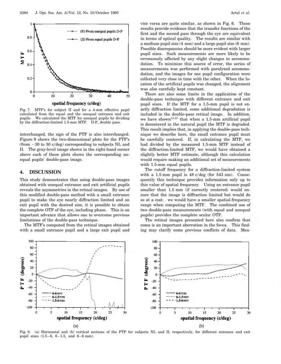
The PTF was computed up to the corresponding fre-quency limit for a 1.5-mm pupil diameter by Eq. (3).Figure 8 presents horizontal and vertical sections of thetwo-dimensional PTF’s for subjects NL and II, respec-tively, computed from aerial images with different en-trance and exit pupil configurations (1.5–6, 6–1.5, and6–6 mm). In the case of the equal pupil sizes’ double-pass images, PTF’s tend to zero at all spatial frequencies,as predicted. When the entrance and the exit pupils are
(a) (c)
(b) (d)
Fig. 6. Comparison of first-pass and second-pass MTF’s for subjects NL and II. The solid curve with circles represents thediffraction-limited MTF times the second-passage MTF. The dashed curve with triangles represents the diffraction-limited MTFtimes the first-passage MTF. (a) Subject NL with 4-mm pupil diameter, (b) subject NL with 6-mm pupil diameter, (c) subject IIwith 4-mm pupil diameter, (d) subject II with 6-mm pupil diameter.
2364 J. Opt. Soc. Am. A/Vol. 12, No. 10 /October 1995 Artal et al.
Fig. 7. MTF’s for subject II and for a 4-mm effective pupilcalculated from the equal and the unequal entrance and exitpupils. We calculated the MTF for unequal pupils by dividingby the diffraction-limited 1.5-mm MTF. D-P, double pass.
interchanged, the sign of the PTF is also interchanged.Figure 9 shows the two-dimensional plots for the PTF’s(from 230 to 30 cydeg) corresponding to subjects NL andII. The gray-level image shown in the right-hand cornerabove each of these plots shows the corresponding un-equal pupils’ double-pass image.
4. DISCUSSIONThis study demonstrates that using double-pass imagesobtained with unequal entrance and exit artificial pupilsreveals the asymmetries in the retinal images. By use ofthis modified double-pass method with a small entrancepupil to make the eye nearly diffraction limited and anexit pupil with the desired size, it is possible to obtainthe complete OTF of the eye, including phase. This is animportant advance that allows one to overcome previouslimitations of the double-pass technique.
The MTF’s computed from the retinal images obtainedwith a small entrance pupil and a large exit pupil and
vice versa are quite similar, as shown in Fig. 6. Theseresults provide evidence that the transfer functions of thefirst and the second pass through the eye are equivalentin terms of optical quality. The results are similar witha medium pupil size (4 mm) and a large pupil size (6 mm).Possible discrepancies should be more evident with largerpupil sizes. Such measurements are more likely to beerroneously affected by any slight changes in accommo-dation. To minimize this source of error, the series ofmeasurements was performed with paralyzed accommo-dation, and the images for one pupil configuration werecollected very close in time with the other. When the lo-cation of the artificial pupils was changed, the alignmentwas also carefully kept constant.
There are also some limits in the application of thedouble-pass technique with different entrance and exitpupil sizes. If the MTF for a 1.5-mm pupil is not ex-actly diffraction limited, some additional degradation isincluded in the double-pass retinal image. In addition,we have shown14,15 that when a 1.5-mm artificial pupilis decentered in the natural pupil the MTF is degraded.This result implies that, in applying the double-pass tech-nique we describe here, the small entrance pupil mustbe carefully centered. If, in calculating the MTF, wehad divided by the measured 1.5-mm MTF instead ofthe diffraction-limited MTF, we would have obtained aslightly better MTF estimate, although this calculationwould require making an additional set of measurementswith 1.5-mm equal pupils.
The cutoff frequency for a diffraction-limited systemwith a 1.5-mm pupil is 48 cydeg (for 543 nm). Conse-quently this technique provides information only up tothis value of spatial frequency. Using an entrance pupilsmaller than 1.5 mm (if correctly centered) would en-sure that the image is diffraction limited but would doso at a cost: we would have a smaller spatial-frequencyrange when computing the MTF. The combined use oftwo double-pass measurements (with equal and unequalpupils) provides the complete ocular OTF.
The retinal images presented here also confirm thatcoma is an important aberration in the fovea. This find-ing may clarify some previous conflicts of data. Mea-
(a) (b)Fig. 8. (a) Horizontal and (b) vertical sections of the PTF for subjects NL and II, respectively, for different entrance and exitpupil sizes (1.5–6, 6–1.5, and 6–6 mm).

Artal et al. Vol. 12, No. 10 /October 1995 /J. Opt. Soc. Am. A 2365
Fig. 9. Two-dimensional plots of the PTF (in deg) for (a) subject NL and (b) subject II. The PTF’s were computed from the 1.5–6-mmpupils’ configuration. The gray-level images shown in the corners are the corresponding unequal-pupil double-pass images.

2366 J. Opt. Soc. Am. A/Vol. 12, No. 10 /October 1995 Artal et al.
surements of aberrations have shown large amounts ofcoma, whereas measurements based on the double-passtechnique have shown very little evidence of coma. It isnow clear that the double-pass technique could not pro-vide evidence of coma because of the loss of phase informa-tion. The use of the double-pass technique with unequalpupils reveals that retinal images in some of the subjectsshow the typical shape produced by coma. Now it is pos-sible for ocular aberrations, and in particular coma, to bestudied further. In particular, the ocular wave aberra-tion can be determined by application of computationalphase-retrieval methods17 to the double-pass retinal im-ages that we obtained with different entrance and exitpupil sizes.
ACKNOWLEDGMENTThis research was partially supported by a grant fromNATO (CG 940081).
REFERENCES1. M. F. Flamant, “Etude de la repartition de lumiere dans
l’image retinienne d’une fente,” Rev. Opt. 34, 433–459(1955).
2. F. W. Campbell and R. W. Gubisch, “Optical image quality ofthe human eye,” J. Physiol. (London) 186, 558–578 (1966).
3. J. Santamarıa, P. Artal, and J. Bescos, “Determination ofthe point-spread function of human eyes using a hybridoptical–digital method,” J. Opt. Soc. Am. A 4, 1109–1114(1987).
4. R. Navarro, P. Artal, and D. R. Williams, “Modulation trans-fer of the human eye as a function of retinal eccentricity,”J. Opt. Soc. Am. A 10, 201–212 (1993).
5. P. Artal and R. Navarro, “Monochromatic modulation trans-fer function of the human eye for different pupil diameters:
an analytical expression,” J. Opt. Soc. Am. A 11, 246–249(1994).
6. P. Artal, S. Marcos, R. Navarro, M. Ferro, and I. Miranda,“Through focus image quality with monofocal and multifocalintraocular lenses,” Opt. Eng. 34, 772–779 (1995).
7. P. Artal, S. Marcos, R. Navarro, and D. R. Williams, “Oddaberrations and double-pass measurements of retinal imagequality,” J. Opt. Soc. Am. A 12, 195–201 (1995).
8. M. Born and E. Wolf, Principles of Optics, 6th ed. (Pergamon,Oxford, 1980).
9. G. J. van Blokland and D. van Norren, “Intensity and po-larization of light scattered at small angles from the humanfovea,” Vision Res. 26, 485–494 (1986).
10. D. R. Williams, D. Brainard, M. MacHahon, and R. Navarro,“Double-pass and interferometric measures of the opticalquality of the eye,” J. Opt. Soc. Am. A 11, 3123–3135(1994).
11. D. Sliney and M. Wolbarsht, Safety with Laser and OtherOptical Sources (Plenum, New York, 1980).
12. H. C. Howland and B. Howland, “Subjective method forthe measurement of monochromatic aberrations of the eye,”J. Opt. Soc. Am. 67, 1508–1518 (1977).
13. M. C. W. Campbell, E. M. Harrison, and P. Simonet, “Psy-chophysical measurements of the blur on the retina dueto the optical aberrations of the eye,” Vision Res. 30,1587–1602 (1990).
14. D. G. Green, “Visual resolution when light enters throughdifferent parts of the pupil,” J. Physiol. (London) 190,583–593 (1967).
15. S. Marcos, P. Artal, and D. G. Green, “The effect of decen-tered small pupils on ocular modulation transfer and con-trast sensitivity,” Invest. Ophthalmol. Vis. Sci. Suppl. 35,1258 (1994).
16. P. Artal and R. Navarro, “Simultaneous measurement oftwo-point-spread functions at different locations across thehuman fovea,” Appl. Opt. 31, 3646–3656 (1992).
17. P. Artal, J. Santamarıa, and J. Bescos, “Retrieval of the waveaberration of human eyes from actual point-spread functiondata,” J. Opt. Soc. Am. A 5, 1201–1206 (1988).