Dose-dependent action of the RNA binding protein FOX-1 to ...
34
*For correspondence: [email protected] Present address: † Environmental Genomics and Systems Biology Division, Lawrence Berkeley National Laboratory, Berkeley, United States; ‡ Genentech, South San Francisco, United States Competing interests: The authors declare that no competing interests exist. Funding: See page 30 Received: 10 September 2020 Accepted: 23 December 2020 Published: 29 December 2020 Reviewing editor: Douglas L Black, University of California, Los Angeles, United States Copyright Farboud et al. This article is distributed under the terms of the Creative Commons Attribution License, which permits unrestricted use and redistribution provided that the original author and source are credited. Dose-dependent action of the RNA binding protein FOX-1 to relay X-chromosome number and determine C. elegans sex Behnom Farboud 1,2 , Catherine S Novak 1,2† , Monique Nicoll 1,2‡ , Alyssa Quiogue 1,2 , Barbara J Meyer 1,2 * 1 Howard Hughes Medical Institute, University of California at Berkeley, Berkeley, United States; 2 Department of Molecular and Cell Biology, University of California at Berkeley, Berkeley, United States Abstract We demonstrate how RNA binding protein FOX-1 functions as a dose-dependent X-signal element to communicate X-chromosome number and thereby determine nematode sex. FOX-1, an RNA recognition motif protein, triggers hermaphrodite development in XX embryos by causing non-productive alternative pre-mRNA splicing of xol-1, the master sex-determination switch gene that triggers male development in XO embryos. RNA binding experiments together with genome editing demonstrate that FOX-1 binds to multiple GCAUG and GCACG motifs in a xol-1 intron, causing intron retention or partial exon deletion, thereby eliminating male-determining XOL-1 protein. Transforming all motifs to GCAUG or GCACG permits accurate alternative splicing, demonstrating efficacy of both motifs. Mutating subsets of both motifs partially alleviates non- productive splicing. Mutating all motifs blocks it, as does transforming them to low-affinity GCUUG motifs. Combining multiple high-affinity binding sites with the twofold change in FOX-1 concentration between XX and XO embryos achieves dose-sensitivity in splicing regulation to determine sex. Introduction Determining sex is one of the most fundamental developmental decisions that most organisms must make. Sex is often specified by a chromosome-counting mechanism that distinguishes one X chro- mosome from two: 2X embryos become females, while 1X embryos become males (Bull, 1983; Charlesworth and Mank, 2010). The nematode Caenorhabditis elegans determines sex with high fidelity by tallying X-chromosome number relative to ploidy, the sets of autosomes (X:A signal) (Madl and Herman, 1979; Nigon, 1951). The process is executed with remarkable precision: embryos with ratios of 1X:2A (0.5) or 2X:3A (0.67) develop into fertile males, while embryos with ratios of 3X:4A (0.75) or 2X:2A (1.0) develop into self-fertile hermaphrodites. Here we dissect molec- ular mechanisms by which the X:A signal specifies sex and thereby discover how small quantitative differences in intracellular signals can be translated into dramatically different developmental fates. The X:A signal determines sex by controlling the activity of its direct target, the master sex-deter- mination switch gene xol-1 ( XO lethal) (Figure 1; Carmi et al., 1998; Farboud et al., 2013; Meyer, 2018; Miller et al., 1988; Nicoll et al., 1997; Powell et al., 2005; Rhind et al., 1995). xol-1 encodes a GHMP kinase that must be activated to set the male fate and repressed to set the her- maphrodite fate (Luz et al., 2003; Rhind et al., 1995). xol-1 controls not only the choice of sexual fate but also X-chromosome gene expression through the process of X-chromosome dosage com- pensation (Meyer, 2018; Miller et al., 1988; Rhind et al., 1995). Males and hermaphrodites Farboud et al. eLife 2020;9:e62963. DOI: https://doi.org/10.7554/eLife.62963 1 of 34 RESEARCH ARTICLE
-
Upload
khangminh22 -
Category
Documents
-
view
3 -
download
0
Transcript of Dose-dependent action of the RNA binding protein FOX-1 to ...

*For correspondence:
Present address: †
Environmental Genomics and
Systems Biology Division,
Lawrence Berkeley National
Laboratory, Berkeley, United
States; ‡ Genentech, South San
Francisco, United States
Competing interests: The
authors declare that no
competing interests exist.
Funding: See page 30
Received: 10 September 2020
Accepted: 23 December 2020
Published: 29 December 2020
Reviewing editor: Douglas L
Black, University of California,
Los Angeles, United States
Copyright Farboud et al. This
article is distributed under the
terms of the Creative Commons
Attribution License, which
permits unrestricted use and
redistribution provided that the
original author and source are
credited.
Dose-dependent action of the RNAbinding protein FOX-1 to relayX-chromosome number and determine C.elegans sexBehnom Farboud1,2, Catherine S Novak1,2†, Monique Nicoll1,2‡, Alyssa Quiogue1,2,Barbara J Meyer1,2*
1Howard Hughes Medical Institute, University of California at Berkeley, Berkeley,United States; 2Department of Molecular and Cell Biology, University of Californiaat Berkeley, Berkeley, United States
Abstract We demonstrate how RNA binding protein FOX-1 functions as a dose-dependent
X-signal element to communicate X-chromosome number and thereby determine nematode sex.
FOX-1, an RNA recognition motif protein, triggers hermaphrodite development in XX embryos by
causing non-productive alternative pre-mRNA splicing of xol-1, the master sex-determination
switch gene that triggers male development in XO embryos. RNA binding experiments together
with genome editing demonstrate that FOX-1 binds to multiple GCAUG and GCACG motifs in a
xol-1 intron, causing intron retention or partial exon deletion, thereby eliminating male-determining
XOL-1 protein. Transforming all motifs to GCAUG or GCACG permits accurate alternative splicing,
demonstrating efficacy of both motifs. Mutating subsets of both motifs partially alleviates non-
productive splicing. Mutating all motifs blocks it, as does transforming them to low-affinity GCUUG
motifs. Combining multiple high-affinity binding sites with the twofold change in FOX-1
concentration between XX and XO embryos achieves dose-sensitivity in splicing regulation to
determine sex.
IntroductionDetermining sex is one of the most fundamental developmental decisions that most organisms must
make. Sex is often specified by a chromosome-counting mechanism that distinguishes one X chro-
mosome from two: 2X embryos become females, while 1X embryos become males (Bull, 1983;
Charlesworth and Mank, 2010). The nematode Caenorhabditis elegans determines sex with high
fidelity by tallying X-chromosome number relative to ploidy, the sets of autosomes (X:A signal)
(Madl and Herman, 1979; Nigon, 1951). The process is executed with remarkable precision:
embryos with ratios of 1X:2A (0.5) or 2X:3A (0.67) develop into fertile males, while embryos with
ratios of 3X:4A (0.75) or 2X:2A (1.0) develop into self-fertile hermaphrodites. Here we dissect molec-
ular mechanisms by which the X:A signal specifies sex and thereby discover how small quantitative
differences in intracellular signals can be translated into dramatically different developmental fates.
The X:A signal determines sex by controlling the activity of its direct target, the master sex-deter-
mination switch gene xol-1 (XO lethal) (Figure 1; Carmi et al., 1998; Farboud et al., 2013;
Meyer, 2018; Miller et al., 1988; Nicoll et al., 1997; Powell et al., 2005; Rhind et al., 1995). xol-1
encodes a GHMP kinase that must be activated to set the male fate and repressed to set the her-
maphrodite fate (Luz et al., 2003; Rhind et al., 1995). xol-1 controls not only the choice of sexual
fate but also X-chromosome gene expression through the process of X-chromosome dosage com-
pensation (Meyer, 2018; Miller et al., 1988; Rhind et al., 1995). Males and hermaphrodites
Farboud et al. eLife 2020;9:e62963. DOI: https://doi.org/10.7554/eLife.62963 1 of 34
RESEARCH ARTICLE

generally require the same level of X-encoded gene products despite their difference in dose of X
chromosomes. A dosage compensation complex (DCC) binds to both X chromosomes of diploid XX
hermaphrodites to reduce transcription by half and thereby equalize X-linked gene expression with
that from the single X of diploid XO males (Figure 1; Chuang et al., 1994; Chuang et al., 1994;
Csankovszki et al., 2009; Dawes et al., 1999; Lieb et al., 1998; Mets and Meyer, 2009; Mets and
Meyer, 2009; Meyer, 2018; Pferdehirt et al., 2011; Tsai et al., 2008; Tsai et al., 2008;
Wheeler et al., 2016). The DCC resembles condensin, a chromosome remodeling complex required
for the compaction and resolution of mitotic and meiotic chromosomes prior to their segregation
during division (Hirano, 2016; Meyer, 2018; Yatskevich et al., 2019). Failure to activate dosage
compensation kills all hermaphrodites. If xol-1 is inappropriately activated in diploid XX animals, the
DCC cannot bind to X chromosomes, and all hermaphrodites die from elevated X expression. Con-
versely, if xol-1 is inappropriately repressed in diploid XO males, the DCC binds to the single X chro-
mosome and kills all males by reducing X expression. Thus, the X:A signal determines the choice of
sexual fate and sets the level of X-chromosome gene expression.
Our prior studies identified a set of genes on X chromosomes called X-signal elements (XSEs)
that communicate X-chromosome dose by repressing xol-1 in a dose-dependent manner
XX(2X:2A)
xol-1
ASEASE
XSEXSE
XSE
XO(1X:2A)
xol-1
ASEASE
sdc-2
onsdc-2
off
off
on
Dosage compensation ON
Dosage compensation OFF
½
½
½
½
X
X
X
X
½
X 1
Figure 1. Overview of the X:A signal and regulatory hierarchy that control sex determination and X-chromosome
dosage compensation. The X:A signal includes a set of genes on X called X-signal elements (XSEs) that repress
their direct target gene xol-1 (XO lethal) in a cumulative dose-dependent manner via transcriptional and post-
transcriptional mechanisms and set of genes on autosomes called autosomal signal elements (ASEs) that stimulate
xol-1 transcription in a cumulative dose-dependent manner. xol-1 is the master sex-determination switch gene that
must be activated in XO animals to set the male fate and must be repressed in XX animals to permit the
hermaphrodite fate. xol-1 triggers male sexual development by repressing the feminizing switch gene sdc-2 (sex
determination and dosage compensation). sdc-2 induces hermaphrodite sexual development and triggers binding
of a dosage compensation complex (DCC) to hermaphrodite X chromosomes to repress gene expression by half.
xol-1 mutations enable the DCC to bind to the single male X chromosome and thereby kill all XO animals by
causing reduced X-chromosome expression. The dying xol-1 XO mutant animals are also feminized. Hence,
mutations that disrupt elements of the X:A signal transform sexual fate and also alter X-chromosome gene
expression.
Farboud et al. eLife 2020;9:e62963. DOI: https://doi.org/10.7554/eLife.62963 2 of 34
Research article Chromosomes and Gene Expression
(Figure 1; Akerib and Meyer, 1994; Carmi et al., 1998; Gladden et al., 2007; Gladden and
Meyer, 2007; Hodgkin et al., 1994; Nicoll et al., 1997). In addition, a set of genes on autosomes
called autosomal signal elements (ASEs) communicates the ploidy by stimulating xol-1 activity in a
cumulative, dose-dependent manner to counter XSEs (Figure 1; Farboud et al., 2013;
Powell et al., 2005). Two of the XSEs, the nuclear hormone receptor SEX-1 and the homeodomain
protein CEH-39, as well as two of the ASEs, the T-box transcription factor SEA-1 and the zinc finger
protein SEA-2, bind directly to multiple, non-overlapping sites in 5’ transcriptional regulatory regions
of xol-1 (Farboud et al., 2013). XSEs and ASEs antagonize each other’s opposing transcriptional
activities to control xol-1 transcript levels. The X:A signal is thus transmitted in part through multiple
antagonistic molecular interactions carried out on a single promoter to regulate transcription
(Farboud et al., 2013). Fidelity of X:A signaling is enhanced by a second tier of dose-dependent
xol-1 repression, via the XSE called FOX-1 (Feminizing locus On X), an RNA binding protein that
includes an RNA recognition motif (RRM) (Akerib and Meyer, 1994; Hodgkin et al., 1994;
Nicoll et al., 1997; Skipper et al., 1999).
The cumulative, dose-dependent action of XSEs was revealed by key genetic observations. For
example, deleting one copy of ceh-39 and sex-1 from XX animals caused no lethality, but deleting
one copy of ceh-39, sex-1, and fox-1 killed more than 70% of XX animals, and deleting one copy of
all genetically identified XSEs killed all XX animals (Akerib and Meyer, 1994; Carmi and Meyer,
1999; Farboud et al., 2013; Gladden et al., 2007). In reciprocal experiments, duplicating one copy
of fox-1 killed 25% of XO animals, while duplicating fox-1 and ceh-39 killed 50% of XO animals, and
duplicating one copy of all genetically identified XSEs killed all XO animals (Akerib and Meyer,
1994; Carmi and Meyer, 1999; Nicoll et al., 1997).
Our current study analyzes the mechanism of FOX-1 action in regulating xol-1. fox-1 was discov-
ered originally through a mutation that suppressed the XO lethality caused by a large X duplication
shown later to include multiple XSEs (Akerib and Meyer, 1994; Hodgkin et al., 1994; Nicoll et al.,
1997). FOX-1 is the founding member of an ancient family of sequence-specific RNA binding pro-
teins that are conserved from worms to humans (Akerib and Meyer, 1994; Conboy, 2017;
Hodgkin et al., 1994; Nicoll et al., 1997). Recent experiments show that mammalian FOX family
members recognize and bind the primary motifs GCAUG and GCACG but also bind secondary
motifs with lower affinity (Auweter et al., 2006; Begg et al., 2020; Jangi et al., 2014; Jin et al.,
2003; Lambert et al., 2014; Lee et al., 2016; Modafferi and Black, 1999; Underwood et al.,
2005). FOX proteins regulate diverse aspects of RNA metabolism, including alternative pre-mRNA
splicing, mRNA stability, translation, micro-RNA processing, and transcription (Carreira-
Rosario et al., 2016; Chen et al., 2016; Conboy, 2017; Jin et al., 2003; Kim et al., 2014b;
Lee et al., 2016; Ray et al., 2013; Wei et al., 2016). FOX proteins act as developmental regulators
in different tissues of many species, controlling neuronal and brain development (Begg et al., 2020;
Gehman et al., 2012; Gehman et al., 2011; Kuroyanagi et al., 2013; Lee et al., 2016;
Shibata et al., 2000; Underwood et al., 2005) as well as muscle formation (Gao et al., 2016;
Kuroyanagi et al., 2007; Kuroyanagi et al., 2006; Singh et al., 2014; Wei et al., 2015). C. elegans
FOX-1 controls sex determination by repressing xol-1 activity through a post-transcriptional mecha-
nism that acts on any residual xol-1 transcripts present in diploid XX animals after xol-1 repression
by the XSE transcription factors (Carmi and Meyer, 1999; Nicoll et al., 1997; Skipper et al., 1999).
The level of this regulation, whether controlling pre-mRNA splicing, mRNA stability, nuclear trans-
port, or translation of xol-1 RNA, had not been determined.
We demonstrate that FOX-1 represses xol-1 in XX embryos by regulating alternative xol-1 pre-
mRNA splicing to inhibit formation of the mature transcript that is both necessary and sufficient for
xol-1 activity in XO embryos. By binding to multiple sites in intron VI using both GCAUG and
GCACG motifs, FOX-1 causes either intron VI retention or directs the use of an alternative 3’ splice
site, causing deletion of exon 7 coding sequences. Either alternative splicing event prevents produc-
tion of essential male-specific XOL-1 proteins in XX embryos. Experiments performed in vivo demon-
strate that intron VI is both necessary and sufficient for FOX-1-mediated pre-mRNA splicing
regulation at the endogenous xol-1 locus and at lacZ reporters. FOX-1 RNA binding experiments
performed in vitro demonstrate that FOX-1 binds to multiple GCAUG and GCACG motifs in intron
VI. Genome editing of endogenous motifs coupled with functional assays in vivo demonstrate that
mutation of different GCAUG and GCACG combinations reduces FOX-1-mediated repression, but
only mutation of all motifs or transformation of all to low affinity GCUUG motifs blocks non-
Farboud et al. eLife 2020;9:e62963. DOI: https://doi.org/10.7554/eLife.62963 3 of 34
Research article Chromosomes and Gene Expression

productive alternative splicing and mirrors the effect on X:A signaling of an engineered fox-1 dele-
tion. Splicing regulation is dose-dependent: mutating one copy of fox-1 or all binding motifs in one
copy of xol-1 kills XX animals sensitized by reduced XSE activity. In contrast, conversion of all endog-
enous intron VI motifs to either GCAUG or GCACG permits normal splicing regulation, indicating
that GCACG motifs are as effective as GCAUG motifs in promoting FOX-1 binding and splicing reg-
ulation. Hence, the number of high-affinity motifs is critical. Utilizing multiple high-affinity binding
sites to elicit alternative splicing amplifies the X signal by permitting the concentration of FOX-1
made from two doses of fox-1 in XX embryos to reach the threshold level necessary to inhibit XOL-1
production.
Results
An in vivo assay to determine regions of xol-1 essential for repressionby FOX-1Prior studies showed that the RNA binding protein FOX-1 determines sex by repressing xol-1 via a
post-transcriptional mechanism, but the molecular basis of this regulation was not established
(Carmi and Meyer, 1999; Nicoll et al., 1997; Skipper et al., 1999). We therefore devised an assay
to identify regions of xol-1 necessary for repression by FOX-1. Our prior experiments showed that
overexpression of FOX-1 by itself is sufficient to repress endogenous xol-1 activity, causing XO
embryos to adopt the hermaphrodite sexual fate and die from reduced X-chromosome expression
triggered by binding of the DCC to the single X (Nicoll et al., 1997). Hence, our strategy to identify
FOX-1 regulatory sites was to assay deletion derivatives of a xol-1 transgene controlled by the native
xol-1 promoter for responsiveness to FOX-1 repression in strains lacking the endogenous xol-1
gene.
The wild-type xol-1 transgene included all xol-1 genomic sequences, and an insertion of gfp
sequences in frame at the first ATG codon of xol-1. Expression of wild-type transgenes and deletion
derivatives was monitored in xol-1(null) mutant strains by functional assays of XOL-1 activity. XX ani-
mals are very sensitive to the dose of xol-1, and extra-chromosomal arrays carrying wild-type xol-1
transgenes could only be established using a 20–30-fold lower concentration of xol-1 DNA than typi-
cal for routine markers (see Materials and methods). Wild-type transgenes in all seven independent
arrays exhibited proper sex-specific regulation: XO animals were rescued from lethality caused by
the endogenous xol-1(null) mutation, and XX animals were viable (Figure 2A). The proportion of XX
versus XO animals in each line was in agreement with the expected ratio (2:1) from the male-produc-
ing mutation him-5(e1490) present in all lines. However, the xol-1(+) transgenes were expressed in
XX animals at a somewhat higher level than the endogenous xol-1 gene: the seven XX array lines
could only be maintained if both endogenous copies of fox-1 were wild type. The observation that
fox-1 mutations kill XX animals carrying wild-type xol-1 transgenes shows the need for stringent xol-
1 repression in hermaphrodites by both transcriptional and post-transcriptional mechanisms.
For XO animals with wild-type xol-1 transgenes, excess FOX-1 protein expressed from an inte-
grated array [yIs44(fox-1)] carrying multiple copies of the fox-1(+) gene was lethal
(Figure 2A; Nicoll et al., 1997). No XO animals were viable in the seven lines that carried wild-type
xol-1 transgenes and expressed high FOX-1 levels. Although GFP fluorescence was XO-specific
when produced from wild-type transgenes, it proved to be too insensitive a monitor for changes in
xol-1 activity and was not used as part of our subsequent assays.
Deletion derivatives of xol-1 transgenes lacking FOX-1 regulatory sequences are predicted to be
insensitive to repression by excess FOX-1, allowing xol-1(null) XO animals to be rescued and fully via-
ble. Deletion derivatives lacking FOX-1 regulatory regions are also expected to kill XX animals or
cause visible dosage compensation defects (Dumpy and Egg-laying defective phenotypes) due to
lack of repression by FOX-1. The XX lethality and other dosage compensation phenotypes should
not be suppressed by excess FOX-1. Thus, the phenotypic consequences of wild-type and deletion-
derivative xol-1 transgenes act as sensitive monitors of regulation by FOX-1.
Intron VI is essential for FOX-1 to repress xol-1We first assayed the effects on XX and XO animals of xol-1 transgenes with different combinations
of intron deletions to define FOX-1 regulatory regions (Figure 2A–E). The deletion derivatives were
Farboud et al. eLife 2020;9:e62963. DOI: https://doi.org/10.7554/eLife.62963 4 of 34
Research article Chromosomes and Gene Expression

IIII II IV V
VIsplice junctions removed
3' UTRfull-length
IVI
3' UTR introns II-V
IIII II IV V
3' UTRintron VI
I
3' UTR introns II-VI
IIII II IV V
VI
3' UTRfull-length
wild-type
5
2
3
7
1
2
Lines
Viability
VIA
VIA
VIA
VIA
VIA
VIA
VIA
VIA
VIA
DEAD
DEAD/
Dpy Egl
Dpy Egl
DEAD
DEAD
VIA
DEAD/
Dpy Egl
Dpy Egl
DEAD
DEAD
XOxol-1
XXxol-1
VIA
Extra FOX-1 [yIs44(fox-1)]
VIA DEADVIAVIA
intron VI removal
2.2 kb transcript
(active)
STOP
F xol-1 pre-mRNA
I IIIII
IV VVI
1 2 3 4 5 6 7
STOP
intron VI retention
2.5 kb transcript
(inactive)1.5 kb transcript
(inactive)
STOP
1 2 3 4 5 6 7
A
xol-1 TransgeneXOxol-1
XXxol-1
B
C
D
E
Figure 2. Intron VI is essential for repression of xol-1 by FOX-1. (A–E) Assays of wild-type xol-1 transgenes and deletion derivatives with different
combinations of introns show that removal of intron VI prevents FOX-1 from repressing xol-1. Diagrams on the left show the intron–exon structure of
xol-1 sequences in the transgene derivatives. Exon 7 includes both coding sequences (blue) and the 3’ UTR (orange). The wild-type parent transgene
rescues the XO-specific lethality caused by xol-1 null mutations but permits XX animals to be viable. Intron deletion derivatives have genomic regions
Figure 2 continued on next page
Farboud et al. eLife 2020;9:e62963. DOI: https://doi.org/10.7554/eLife.62963 5 of 34
Research article Chromosomes and Gene Expression

made by replacing genomic regions of xol-1 with corresponding xol-1 cDNA to remove introns with-
out altering the protein coding sequence. Extra-chromosomal arrays carrying deletion derivatives of
xol-1 transgenes were created in him-5; xol-1 strains capable of producing both XX and XO embryos
and then crossed into yIs44(fox-1); him-5; xol-1 strains that produce excess FOX-1. The DNA concen-
trations used for the distinct deletion-bearing transgenes were the same as for xol-1(+) transgenes.
Even if a transgene deletion derivative causes XX-specific lethality, the array can be recovered and
maintained through array-bearing XO animals.
The three array lines created from xol-1 transgenes lacking introns II–VI (D introns II–VI) rescued
xol-1(null) XO males, but killed all xol-1(null) XX hermaphrodites, even though the starting DNA con-
centration was the same as for xol-1(+) arrays (Figure 2B). Hundreds of array-bearing males were
maintained for each independent array line through genetic crosses but no array-bearing hermaph-
rodite progeny survived, indicating that essential FOX-1 regulatory sequences had been deleted.
Excess FOX-1 also failed to suppress the XX-specific lethality and failed to kill XO animals, demon-
strating the necessity of intronic sequences for xol-1 repression by FOX-1 (Figure 2B).
The essential role of introns in causing the death of XO animals with elevated FOX-1 levels could
reflect the specific contribution of intron VI, which undergoes alternative splicing to yield at least
three different xol-1 transcript variants (Figure 2F; Rhind et al., 1995). Only the 2.2 kb variant, which
lacks intron VI and includes all of exon 7, encodes a functional XOL-1 protein that has full XOL-1
activity essential for male development (Figure 2F). This 2.2 kb variant is both necessary and suffi-
cient for full XOL-1 function in XO embryos (Rhind et al., 1995). It is the most abundant transcript of
the three, accumulating to a level 10-fold higher in XO than XX embryos. A 1.5 kb variant is made
from the same 5’ donor in exon 6 as used for the 2.2 kb variant but a different 3’ splice acceptor,
one in the 3’ UTR. This splicing event eliminates the coding region of exon 7, an essential exon
(Rhind et al., 1995). The 1.5 kb variant does not encode xol-1 XO activity. A 2.5 kb variant results
from the failure to remove intron VI. In this variant, an in-frame UAA stop codon within intron VI ter-
minates translation prematurely, precluding production of the male-determining protein
(Rhind et al., 1995).
Indeed, removal of only the alternatively spliced intron VI from the xol-1 transgene (D intron VI) in
all three independent lines permitted all XO animals to be viable and to escape lethality caused by
high levels of FOX-1 (Figure 2C). Two of the lines caused complete XX-specific lethality that was not
suppressed by excess FOX-1; lines were maintained through genetic crosses with array-bearing XO
males (Figure 2C). A third line caused milder dosage compensation defects in XX animals that were
not suppressed by excess FOX-1 and caused the animals to be sterile, likely reflecting a lower copy
number of the transgene and hence less expression. The line had to be maintained through array-
bearing XO animals. Thus, intron VI is essential for FOX-1 repression of xol-1.
Figure 2 continued
of xol-1 replaced by corresponding xol-1 cDNA to remove introns without altering protein coding sequence. Shown on the right is the viability of XX
and XO xol-1(y9) deletion mutant animals carrying extra-chromosomal arrays of the different transgene derivatives. The arrays were made and assayed
in him-5(e1490); xol-1(y9) mutants that produce 33% XO and 67% XX embryos and then crossed into him-5; xol-1 strains producing high levels of FOX-1
from an integrated array [yIs44 (fox-1)] carrying multiple copies of fox-1. The far right shows the number of independent extra-chromosomal arrays
assayed. The term ‘dead’ means that all animals of the genotype were inviable, and no extra-chromosomal arrays could be established in XX animals.
The arrays could only be established and maintained through XO males. The term ‘via’ means the animals were viable and appeared wild type. The
term ‘Dpy Egl’ refers to the phenotype of dosage-compensation-defective XX animals that escape lethality. XX animals are typically dumpy (Dpy) in
body size and egg-laying defective (Egl). The term ‘dead/Dpy Egl’ means that most animals (greater than 90%) were dead, and rare escapers were Dpy
Egl. High levels of FOX-1 kill all XO animals only if a spliceable form of intron VI is present. FOX-1 does not repress xol-1 in XO animals if intron VI is
absent or is relocated to the 3’ UTR without splice junctions. Transgenes lacking intron VI kill XX animals, as do wild-type xol-1 transgenes in strains with
a fox-1 null mutation, because all transgenes are expressed at a somewhat higher level than the endogenous xol-1 gene in XX animals. (F) Structures of
the three most abundant splice variants of xol-1 transcripts are shown. Only the 2.2 kb variant, which lacks intron VI (red dashed line) and includes
essential exon 7 coding sequences (blue) and 3’ UTR, is necessary and sufficient for survival of XO animals. This isoform corresponds to Wormbase.org
transcript C18A11.5b.1. Transcripts that retain intron VI (2.5 kb isoform corresponding to Wormbase.org transcript C18A11.5c.1) (black dashed line) or
lack exon 7 coding sequences (1.5 kb isoform corresponding to Wormbase.org transcript C18A11.5a) (orange dashed line) due to use of an alternative
3’ splice acceptor site cannot produce essential XOL-1 male-determining proteins.
The online version of this article includes the following source data for figure 2:
Source data 1. xol-1 transcription is not repressed by high levels of FOX-1.
Farboud et al. eLife 2020;9:e62963. DOI: https://doi.org/10.7554/eLife.62963 6 of 34
Research article Chromosomes and Gene Expression

Further analysis confirmed that introns II–V are dispensable for FOX-1 regulation. All lines carrying
transgenes lacking these introns (D introns II–V) behaved like lines of the wild-type xol-1 transgene
(Figure 2D). XX and XO xol-1 animals in each line were viable in the relative proportion expected
for the him-5 mutation, and XO xol-1 males were killed by excess FOX-1.
When intron VI was removed from its normal location in xol-1 and relocated to the 3’ UTR without
splice junctions, FOX-1 could not repress xol-1 (Figure 2E). In two independent arrays of this trans-
gene derivative, XO xol-1 animals were viable, despite excess FOX-1, and greater than 99% of XX
animals were inviable, even with excess FOX-1. The rare viable XX animals were Dpy and Egl. Thus,
the presence of intron VI does not simply enable FOX-1 to repress xol-1 by reducing mRNA stability,
blocking nuclear transport of xol-1 RNA or preventing translation in the cytoplasm. Furthermore,
experiments quantifying levels of all xol-1 RNA splice variants showed that high levels of FOX-1 did
not diminish transcription from xol-1 or cause transcript degradation (Figure 2—source data 1).
Instead, FOX-1 likely regulates xol-1 pre-mRNA splicing.
FOX-1 regulates intron retention and alternative splicing of xol-1 pre-mRNAIf FOX-1 represses xol-1 by promoting alterative splicing, elevated levels of FOX-1 that cause male
lethality should change the distribution of xol-1 RNA splice variants. Specifically, the 2.2 kb splice
variant that encodes male-determining activity should be reduced, while the inactive 2.5 kb variant
with intron VI retention and/or the inactive 1.5 kb variant with partial exon 7 deletion should be
increased. Using RNase protection assays and sequence analysis of cloned cDNAs, we show that ele-
vated FOX-1 levels cause precisely these results (Figure 3 and Figure 3—figure supplement 1).
For RNase protection assays, RNAs from two genetically distinct embryo populations were tested
for the relative abundance of specific xol-1 splice variants within each population: one RNA from
him-5 mixed-stage XX and XO embryos with wild-type FOX-1 levels and one RNA from yIs44(fox-1);
him-5 mixed-stage XX and XO embryos with high levels of FOX-1. These RNAs were assayed initially
using an act-1 control gene probe to assess the quality and quantity of RNAs (Figure 3—figure sup-
plement 1D). The first set of xol-1 RNase protection assays with these quantified RNAs used a xol-1
antisense probe that spanned the 3’ splice junction of intron VI–exon 7 and distinguished the 2.2 kb
and 2.5 kb transcripts (Figure 3A). The ratio of 2.2 kb and 2.5 kb splice variants was calculated from
transcripts within each RNA population, and ratios from different populations were then compared
to assess the change in transcript ratios caused by varying the FOX-1 level. XX and XO him-5 mixed
embryo populations with wild-type levels of FOX-1 showed a fourfold accumulation of active 2.2 kb
transcript relative to the unspliced 2.5 kb transcript. In contrast, XX and XO populations that overex-
pressed FOX-1 [yIs44(fox-1); him-5] showed a drastic reduction in accumulation of the 2.2 kb tran-
script and a corresponding sixfold higher accumulation of the 2.5 kb transcript (Figure 3A). This
experiment and repetitions (Figure 3—figure supplement 1A) established that FOX-1 promotes
intron VI retention.
Transcript analysis using RT-PCR next showed that intron VI was the only intron retained in the
presence of high FOX-1 levels. Using an oligonucleotide primer set that spans all six introns, RT-PCR
was performed on cDNA made from the same RNAs as the protection assays. Only two PCR prod-
ucts were observed, one that corresponded to the 2.5 kb transcript and the other to the 2.2 kb tran-
script (see Materials and methods).
A second set of xol-1 RNase protection assays utilized a different antisense probe that not only
distinguished between the 2.2 kb and 2.5 kb transcripts, but also detected the 1.5 kb transcript and
all alternatively spliced variants that contained the 3’ end of exon 6 (Figure 3—figure supplement
1B). The probe included the 3’ end of exon 6 and the 5’ end of exon 7. These protection experi-
ments showed a decrease in accumulation of active 2.2 kb transcript relative to inactive 2.5 kb tran-
script in the presence of excess FOX-1 (2.4 to 1 with low FOX-1 and 1 to 5 with high FOX-1). They
also showed a dramatic decrease in 2.2 kb transcripts compared to all spliced variants. With low
FOX-1 levels, the 2.2 kb transcript was present at a ratio of 1:3, but with excess FOX-1 its accumula-
tion decreased more than 10-fold, to a ratio of 1:38.
A third xol-1 probe differentiated the inactive 1.5 kb transcript from the combination of 2.2 kb
and 2.5 kb transcripts and from all alternatively spliced transcripts that included the 3’ end of exon 6
(Figure 3—figure supplement 1C). Excess FOX-1 caused the 1.5 kb transcript to increase in accu-
mulation compared to the 2.2 kb and 2.5 kb transcripts, from a ratio of 1:14 with low FOX-1 to a
Farboud et al. eLife 2020;9:e62963. DOI: https://doi.org/10.7554/eLife.62963 7 of 34
Research article Chromosomes and Gene Expression

4:1 1:6
Ratio of 2.2 kb:
2.5 kb transcripts
RNase Protection Assay
of Total Embryonic RNA
XX (67%) & XO (33%)
him-5
yIs44
(fox-1);
him-5
299 nt
189 nt
(2.5 kb transcript)
(2.2 kb transcript)
exon 6
189 nt protected
xol-1 Splice Junctions:
exon 6
299 nt protected
intron VI
3' UTRintron VI
exon 6
1
2
3
1
trans-splice to unrelated gene
him
-5yIs
44
(fo
x-1
); h
im-5
alternate splice site
alternate splice site
Sequence Analysis:
exon 6
exon 6
exon 6
exon 6
4exon 6 3' UTR
alternate splice site
3' UTR
2
alternate splice site
exon 6 3' UTR
exon 6 1
2.5 kb transcript
2.2 kb transcript
466 nt
113
399
62
no splicing
no splicing
no splicing
proper splicing
alternative splicing
alternative splicing
inactive
inactive
inactive
inactive
active
inactive
inactive
Transcript Clones
A
B
C
Figure 3. FOX-1 inhibits formation of the active 2.2 kb xol-1 transcript by promoting intron retention and alternative 3’ splice acceptor selection. (A)
RNase protection assays show that FOX-1 causes intron VI retention. Shown on the left are diagrams of relevant splice junctions for exon 6 (grey) and
coding sequences of exon 7 (blue) that pertain to the inactive 2.5 kb and active 2.2 kb xol-1 transcripts. Also shown are portions of the probe protected
against RNase by each transcript: 299 nt for the 2.5 kb transcript and 189 nt for the 2.2 kb transcript. On the right is an image of the RNase protection
Figure 3 continued on next page
Farboud et al. eLife 2020;9:e62963. DOI: https://doi.org/10.7554/eLife.62963 8 of 34
Research article Chromosomes and Gene Expression

ratio of 1:4 with high FOX-1. The 1.5 kb transcript also increased in accumulation compared to all
splice variants present in excess FOX-1, from a 1:35 ratio to a 1:21 ratio.
This series of protection experiments demonstrated that FOX-1 represses xol-1 by controlling its
pre-mRNA splicing, promoting both intron retention and also deletion of exon 7 coding sequences
via alternative 3’ acceptor site choice. They also reveal that excess FOX-1 causes the production of
more splice variants than previously mapped. Abundance of the universal protected fragment from
the 3’ end of exon 6 (79 nt) relative to the protected fragments from known splice variants (191 nt
and 144 nt) (Figure 3—figure supplement 1A) indicates the occurrence of unidentified splice var-
iants caused by excess FOX-1. As a consequence, we took an alternative approach to identify splice
variants by synthesizing cDNA from total embryonic RNA made from both the him-5 and the yIs44;
him-5 strains and then selectively cloning and sequencing cDNAs that contained the 3’ end of exon
6 (Figure 3B).
Sequences of cDNA clones from xol-1 transcripts in low FOX-1 conditions revealed only the
expected: properly spliced active 2.2 kb transcripts, inactive 2.5 kb transcripts that retained intron
VI, and inactive alternatively spliced 1.5 kb transcripts that deleted part of exon 7 through use of a
3’ acceptor site in the 3’ UTR (Figure 3B). Clones of cDNAs from high FOX-1 conditions revealed
both expected and unexpected transcripts (Figure 3C). As expected, no active 2.2 kb transcripts
were found, but splice variants that retained intron VI or deleted exon 7 were found. Unexpectedly,
several cDNAs corresponded to xol-1 transcripts that had been trans-spliced to unrelated genes.
This splicing involved the 5’ donor at the exon 6–intron VI splice junction, permitting inclusion of
exon 6, but then utilized an naturally occurring 3’ acceptor site at an exon from an unrelated gene
on chromosome II, either flcn-1, polyg-1, or K02E7.12, thus achieving accurate trans-splicing. Thus,
the process by which FOX-1 enhances intron retention during RNA processing also promotes the
use of alternative 3’ acceptor sites, causing deletion of exon coding sequences and enabling trans-
splicing.
Promiscuous fox-1-mediated trans-splicing that fuses partially spliced xol-1 transcripts onto tran-
scripts from unrelated genes may result from the normal 3’ acceptor site in xol-1 exon 7 being
unavailable to receive the primed 5’ splice donor in exon 6, perhaps because the 3’ site is blocked
or not yet synthesized. The primed 5’ site then fuses with a nearby available 3’ acceptor site,
whether or not the site is in xol-1 or an unrelated gene. Analysis of recovered promiscuous splicing
events revealed xol-1 transcripts spliced not only to transcripts from chromosome II genes, but also,
and even more frequently, to transcripts from adjacent genes encoded on transgenic arrays that
include xol-1. Although trans-splicing between a common 22 nucleotide SL-1 or SL-2 leader
sequence and the 5’ end of nascent transcripts has been well documented in C. elegans (Blumen-
thal, 2012), trans-splicing of exons from two different protein-coding genes has not been reported
previously. In contrast, developmentally programmed trans-splicing has been observed in Drosophila
Figure 3 continued
assay of xol-1 transcripts in him-5 and in yIs44(fox-1); him-5 strains quantified by a phosphorimager. The probe is labeled with 32P-UTP: 43 U residues in
the intron VI portion and 44 U residues in the exon 7 portion. Prior to quantifying the ratio of 2.2 kb to 2.5 kb transcripts, the 299 nt signal was divided
in half to compensate for its higher number of U residues. The assay demonstrates that FOX-1 inhibits production of the male-determining 2.2 kb
transcript by preventing the removal of intron VI. Levels of the act-1 control transcript were also assayed in these two RNA samples (Figure 3—figure
supplement 1D). Quantification of a separate RNase protection experiment using this pMN86 probe is shown in Figure 3—figure supplement 1B. (B,
C) Sequence analysis of cDNA clones from xol-1 transcripts shows that FOX-1 causes intron VI retention and also alternative 3’ splice acceptor selection
in xol-1. FOX-1 also causes trans-splicing of xol-1 pre-mRNA to pre-mRNA of unrelated genes. Below the diagram of xol-1’s relevant intron–exon
structure (exon 6 in grey and exon 7 coding sequences in blue) are diagrams representing the splicing pattern revealed by DNA sequence analysis of
xol-1 cDNAs from him-5 (B) and from yIs44(fox-1); him-5 strains (C). Also shown are the predicted xol-1 activity states of transcripts with the different
splicing patterns and the number of cDNA clones with each pattern. In instances of intron VI retention, the number of intron VI nucleotides in each
clone is shown with blue numbers. During trans-splicing (C), the proper 5’ donor at the exon 6–intron VI junction was used together with a naturally
occurring 3’ acceptor at an intron–exon junction of an unrelated gene identified in the text. Resulting trans-splicing events had the junction expected
from proper use of the 5’ and 3’ sites.
The online version of this article includes the following figure supplement(s) for figure 3:
Figure supplement 1. RNase protection experiments show that FOX-1 promotes intron VI retention and partial exon 7 deletion to inhibit formation of
the functional 2.2 kb xol-1 transcript.
Farboud et al. eLife 2020;9:e62963. DOI: https://doi.org/10.7554/eLife.62963 9 of 34
Research article Chromosomes and Gene Expression
for neighboring genes (Buchner et al., 2000; Dorn et al., 2001; Gabler et al., 2005;
Horiuchi et al., 2003; Labrador et al., 2001).
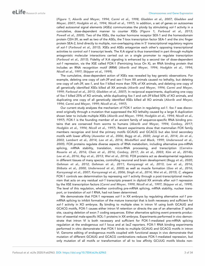
Intron VI is sufficient for FOX-1-mediated repressionTo understand the mechanism by which FOX-1 regulates intron splicing, we asked whether intron VI
is sufficient to confer FOX-1 repression upon a heterologous transcript (Figure 4A,B). We placed
xol-1’s 466 bp intron VI into the fifth exon of a lacZ reporter gene driven by the xol-1 promoter and
tested whether excess FOX-1 from yIs44(fox-1) could prevent its expression (Figure 4B). The
reporter gene also included the 3’ UTR from the unc-54 myosin gene to assess whether intron VI by
itself, without the 3’ UTR from xol-1, can confer FOX-1-mediated repression. Four independently
derived lines with extra-chromosomal arrays carrying the Pxol-1::lacZ::intron VI::unc-54 3’ UTR
reporter exhibited proper sex-specific regulation, expressing b-galactosidase at high levels in XO
embryos (Figure 4B, see XX and XO images) and low levels in XX embryos (Figure 4B, see XX
image), as did five independently derived lines with control Pxol-1::lacZ::xol-1 3’ UTR reporter arrays
that lacked intron VI and had a 3’ UTR from xol-1 (Figure 4A, see XX vs. XX and XO images). Excess
FOX-1 caused a marked decrease in the frequency and intensity of embryonic b-galactosidase
expression in all four intron-VI-containing lines (Figure 4B, see yIs44 image) but had no effect on the
five lines carrying the control lacZ reporter without intron VI (Figure 4A, see yIs44 image). At least
1000 embryos were scored for each genotype derived from each of the nine independent arrays.
Therefore, intron VI by itself is sufficient to confer FOX-1 repression. These results also show that
xol-1’s 3’ UTR is neither necessary nor sufficient for FOX-1 repression.
To determine whether repression of the lacZ reporter occurs by the same mechanism as repres-
sion of xol-1, we examined the splicing pattern of the reporter transcripts. cDNA was synthesized
from total embryonic RNA made from intron VI-containing lacZ reporters expressed in both him-5
and yIs44(fox-1); him-5 strains. lacZ-specific PCR primer sets were used to clone lacZ transcripts from
the two strains and determine the splicing pattern. Clones from XX animals with low FOX-1 levels
revealed the same classes of RNA processing events in lacZ transcripts as found from RNA process-
ing events of endogenous xol-1 RNA, consistent with intron VI conferring FOX-1 repression
(Figure 4C). They included a class with proper intron VI removal, one with intron VI retention, one
with proper intron VI removal plus an additional splice in lacZ sequences corresponding to the DNA
region between AgeI and Pvul restriction sites, and one with the correct 5’ donor site usage at the
lacZ–intron VI junction but an alternative 3’ splice acceptor in an exon of an unrelated gene, unc-76
(chromosome V), F58D5.5 (chromosome I), or H05L03 (chromosome X).
Clones from XX animals with high FOX-1 levels revealed five classes of transcripts consistent with
transcripts from endogenous xol-1 in the presence of high FOX-1, confirming that intron VI is suffi-
cient for FOX-1 regulation (Figure 4C). The classes included one that had proper intron VI removal,
one that retained intron VI, one that used the correct 5’ donor at the lacZ–intron VI junction but an
alternative 3’ splice acceptor in lacZ, one that had aberrant 5’ and 3’ junctions in lacZ, and one that
used the correct 5’ donor but a naturally occurring 3’ splice acceptor at an exon of an unrelated
gene, most commonly unc-76, but also to zen-4 (chromosome IV) and T28F4.4 (chromosome I), an
ortholog of human ARMC5. The high frequency of trans-splicing, particularly to unc-76, likely results
from both lacZ and unc-76 being transcribed from the same extra-chromosomal array. We conclude
that repression by FOX-1 can be conferred in vivo onto a heterologous gene simply by the insertion
of intron VI. Moreover, repression occurs by promoting either intron retention or use of alternative
3’ acceptor sites, resulting in deletion of exon coding sequences and enabling trans-splicing.
FOX-1 binds directly to multiple sites in intron VIBecause intron VI is both necessary and sufficient for xol-1 repression by FOX-1, we developed an in
vitro assay to determine whether FOX-1 regulates RNA splicing by binding directly to intron VI.
Using purified FOX-1 protein and equimolar amounts of 32P-labeled RNA probe for full-length intron
VI and for intron III, we performed initial cross-linking binding assays to find conditions that could
permit highly specific FOX-1 binding. In experiments with increasing concentrations of FOX-1, we
found that FOX-1 bound robustly to the intron VI probe, but not to the intron III negative control
probe (Figure 5—figure supplement 1A). Also, intron III RNA served as a better nonspecific com-
petitor than tRNA to inhibit nonspecific binding (Figure 5—figure supplement 1A). In competition
Farboud et al. eLife 2020;9:e62963. DOI: https://doi.org/10.7554/eLife.62963 10 of 34
Research article Chromosomes and Gene Expression

ATGTSS
ATGTSS
3' UTR
XX XX & XO XX & XO
yIs44(fox-1)
XX XX & XO XX & XO
yIs44(fox-1)
intron VI lacZ
7
1
2
3
2
1
ClonesSequence Analysis:
him
-5yIs
44
(fo
x-1
); h
im-5
M PA H BT
A PT
A H B M PT
A H B M PT
A H B M PT
PT
P
proper intron removal
proper intron removal
proper intron removal and additional splice in lacZ
aberrant 5 and 3 junctions
accurate 5 junction and aberrant 3 junction
no splicing
T
trans-splice to unrelated gene
T
trans-splice to unrelated gene
3
4
A
C
T = TaqI, A = AgeI, H = BssHII, B = BbsI, M = BsmBI, P = PvuI
unrelated gene
unrelated gene
B
1
A H B M PT
no splicing
xol-1 intron VI
unc-54Pxol-1
3' UTR
xol-1xol-1
lacZ
P lacZ
lacZ
D
Figure 4. Intron VI of xol-1 is sufficient to confer FOX-1 repression. (A) The promoter and 3’ UTR of xol-1 are not sufficient for FOX-1 to repress xol-1.
Below the diagram of the Pxol-1::lacZ::xol-1 3’ UTR reporter transgene (pMN27) are sections of adult gonads from different genotypes stained with 5-
bromo-4-chloro-3-indolyl-D-galactopyranoside. Genotypes of embryos in the gonads include: (left) XX, unc-76; yEx231 [pMN27 and unc-76 (+)]; (middle)
XX and XO, him-5 unc-76; yEx231 [pMN27 and unc-76 (+)]; (right) XX and XO, yIs44(fox-1); him-5 unc-76; yEx231 [pMN27 and unc-76 (+)]. The lacZ
Figure 4 continued on next page
Farboud et al. eLife 2020;9:e62963. DOI: https://doi.org/10.7554/eLife.62963 11 of 34
Research article Chromosomes and Gene Expression

experiments for which 32P-labeled intron VI was challenged with increasing concentrations of either
cold intron III RNA or cold intron VI RNA, intron III did not compete for FOX-1 binding. In contrast,
cold intron VI severely reduced FOX-1 binding to intron VI probe (Figure 5—figure supplement
1B). These results show that FOX-1 binds directly and specifically to intron VI. All subsequent bind-
ing experiments were performed in the presence of cold intron III RNA to inhibit nonspecific
binding.
Direct binding assays with 32P-labeled RNA probes to five subregions of intron VI demonstrated
specific binding to three, fragments B, C, and E (Figure 5—figure supplement 1C). Competition
experiments using intron VI RNA as probe and cold RNA fragments as competitors confirmed that
FOX-1 binds to multiple sites in intron VI (Figure 5—figure supplement 1D). Sequence analysis of
the FOX-1 binding fragments revealed common RNA sequences within the three fragments, as
delineated by B-37, C-35, and E-35 in Figure 5A.
To determine whether these common sequences are responsible for FOX-1 binding, we first used
three RNA oligonucleotides of different sizes to fragment B in direct competition experiments
against 32P-labeled fragment B probe and 32P-labeled intron VI probe (Figure 5B,C). FOX-1 binding
to the B probe was eliminated not only by cold B RNA, but also by the cold 45 nt and 37 nt RNA oli-
gonucleotides that covered the entire common fragment B sequence (Figure 5B). Thus, the common
sequence is sufficient for FOX-1 binding. In addition, FOX-1 binding to 32P-intron VI probe was elimi-
nated by both cold intron VI RNA and cold 45 nt and 37 nt fragment B RNA oligonucleotides, indi-
cating the common sequence supports high-affinity binding that might account for all FOX-1
binding to intron VI (Figure 5C,D). Consistent with that interpretation, FOX-1 binding to 32P-intron
VI was eliminated by a cold 35 nt RNA oligonucleotide to fragment C sequence and a cold 35 nt
RNA oligonucleotide to fragment E sequence (Figure 5D).
In contrast, a 15 nt RNA oligonucleotide (CAUUUGAUCGUUAUG) from the middle of sequences
common to all three FOX-1 binding fragments was incapable of competing for FOX-1 binding to
either a fragment B probe or an intron VI probe, indicating that FOX-1 utilizes one or both of the
small motifs, GCAUG and GCACG (Figure 5B,C). Both GCAUG and GCACG are in fragments B and
C, but only GCACG is in fragment E.
A 25 nt RNA oligonucleotide that includes GCACG and the center of the common sequence in
fragment E was sufficient to eliminate binding to an intron VI probe, indicating that GCACG pro-
motes strong FOX-1 binding, and GCAUG is not essential for FOX-1 binding (Figure 5D). This result
does not exclude the interesting possibility that GCAUG might substitute for GCACG or enhance
binding to RNA that also includes GCACG.
In a final series of competition experiments, we asked whether FOX-1 binding to intron VI utilizes
all three separate regions of common sequence. We compared a cold intron VI RNA competitor that
lacked the common sequences in fragments B and C (D37 D35) with a cold intron VI RNA competitor
that lacked the common sequences in fragments B, C, and E (D37 D35 D26) (Figure 5D). While the
D37 D35 intron was a very poor competitor for the intact intron VI probe, the D37 D35 D26 intron
was even worse; it lacked the ability to compete (Figure 5D). Thus, the three regions of common
Figure 4 continued
reporter is sex-specifically regulated: high levels of b-galactosidase in XO embryos but low levels in XX embryos. High levels of FOX-1 do not diminish
b-galactosidase activity in the absence of intron VI, indicating that the xol-1 promoter and xol-1 3’ UTR cannot confer FOX-1 repression. Five
independent extra-chromosomal array strains of each genotype carrying pMN27 showed the results represented. At least 1000 embryos were examined
for each genotype derived from each of the five independent arrays. (B) Intron VI of xol-1 is sufficient for FOX-1 to repress a lacZ reporter gene. Shown
is a diagram of the Pxol-1::lacZ::intronVI::unc-54 3’ UTR reporter transgene (pMN110) in which the 3’ UTR is from the body-wall myosin gene unc-54.
Genotypes of adult gonads stained for b-galactosidase activity are the same as listed in (A), except the array is yEx280 [(pMN110) and unc-76 (+)]. This
intron VI-containing lacZ reporter is also sex-specifically regulated: active in XO embryos and repressed in XX embryos. High levels of FOX-1 (from
yIs44) greatly diminish the level of b-galactosidase activity in XO embryos, indicating that intron VI alone is sufficient for FOX-1 repression. Four
independent extra-chromosomal array strains of each genotype carrying pMN110 showed the results represented. At least 1000 embryos were
examined for each genotype derived from each of the four independent arrays. (C, D) Sequence analysis of cDNA clones from lacZ transcripts shows
that excess FOX-1 increases intron VI retention and also causes alternative pre-mRNA splicing using 3’ splice acceptor sites in lacZ via cis-splicing and
also 3’ splice acceptor sites in unrelated genes via trans-splicing. Below the diagram of lacZ’s relevant intron–exon structure and restriction sites is the
sequence analysis of lacZ cDNAs from him-5 (C) and from yIs44(fox-1); him-5 (D) strains. Shown are the splicing patterns revealed by DNA sequence
analysis and also the number of lacZ clones with each indicated structure. During trans-splicing, the proper 5’ donor at the lacZ exon–intron VI junction
was used in combination with a naturally occurring 3’ acceptor at an intron–exon junction of an unrelated gene (see text).
Farboud et al. eLife 2020;9:e62963. DOI: https://doi.org/10.7554/eLife.62963 12 of 34
Research article Chromosomes and Gene Expression

0
20
40
60
80
100
120
140
0 10 20 30 40 50 60
fragment B
oligo B-45
oligo B-37
oligo B-15
B%
of
32P Fragment B R
NA Input
Fragment B Probe
Cold Competitors:
B-45 CAAAAUUGCAUGUAGCACAUUUGAUCGUUAUGCUUGCACGCAAAC
B-37 GCAUGUAGCACAUUUGAUCGUUAUGCUUGCACGCAAA
B-15 CAUUUGAUCGUUAUG
C-35 GCAUG-AGUUCAUUUGAUCGUUAUG-AUGCACGGAAA
C
A
oligo B-15
oligo B-37
oligo B-45
Intron VI
0
20
40
60
80
100
120
140
160
0 10 20 30 40 50 60
% of
32P Intron VI RNA Input Intron VI Probe
Cold Competitors:
CAAAAUGCAUAUUUGAUCGA-AUGCCUGCACGUUUGE-35
GCAUAUUUGAUCGA-AUGCCUGCACGE-25
0
20
40
60
80
100
120
cold competitor molar excess
% of
32P Intron VI RNA Input
0 95 179 278 350 466 ntB EC
Intron VI
A D
37 nt 35 nt 26 nt
D
0 10 20 30 40 50 60
Intron VI Probe
Cold Competitors:
37 35 26
intron VI
37 35
C-35 oligo
B-37 oligo
E-35 oligo
E-25 oligo
Figure 5. Purified FOX-1 protein binds in vitro to multiple sites in intron VI using motifs GCAUG and GCACG. (A)
The diagram of intron VI shows the intron VI fragments (A–E) and smaller regions (RNA oligonucleotides B-45 to
E-25) tested for direct FOX-1 binding in vitro. Only RNAs from fragments B, C, and E bind to purified FOX-1
(Figure 5—figure supplement 1C). Motifs GCAUG and GCACG (red) and sequences common to all three
Figure 5 continued on next page
Farboud et al. eLife 2020;9:e62963. DOI: https://doi.org/10.7554/eLife.62963 13 of 34
Research article Chromosomes and Gene Expression

sequence contribute to FOX-1 binding in vitro and suggest they might all contribute to FOX-1
repression in vivo. The competition experiments reinforce the model that direct FOX-1 binding to
intron VI facilitates intron VI retention and also causes deletion of exon 7 coding sequences by pro-
moting use of an alternative 3’ splice acceptor site.
Disruption of endogenous FOX-1 binding sites in intron VI abrogatessplicing-mediated repression of xol-1 in vivoIdentification of FOX-1 binding sites in vitro led us to analyze the function of these sites in vivo for
regulating xol-1 splicing and to determine the impact of xol-1 splicing regulation on X-signal activity
during normal nematode development (Figure 6). Thus far, our experiments identified intron VI as
the target of xol-1 splicing regulation in the context of elevated xol-1 expression caused by multiple
copies of xol-1. Because elevated xol-1 expression is lethal to XX animals, these results cannot be
extrapolated to disclose the full contribution of splicing regulation to X-signal activity during normal
embryogenesis. The impact of splicing regulation in vivo can be determined by editing the endoge-
nous xol-1 gene to eliminate sites used in vivo for regulation by FOX-1.
The approach of removing cis-acting sites in xol-1 by CRISPR/Cas9-mediated genome editing
confers three additional advantages over mutating the fox-1 gene itself for analyzing pre-mRNA
splicing regulation. It cleanly separates the role of FOX-1 in sex determination from its roles in other
developmental processes, revealing a more precise understanding for the contribution of alternative
splicing regulation to the X signal. Furthermore, eliminating intron VI blocks all RNA binding proteins
and accessory factors from participating in xol-1 splicing, potentially revealing a larger role for splic-
ing regulation in communicating the X signal than simply achieved by FOX-1 alone. Lastly, disrupting
individual FOX-1 binding sites allows us to determine the specific sites used for splicing regulation
and the number of sites needed to convey the effect of a twofold difference in FOX-1 dose between
XO and XX embryos to specify sex. Repression through multiple sites has the potential to amplify
the small change in FOX-1 concentration between sexes by minimizing aberrant splicing with one
Figure 5 continued
fragments (light blue) are shown below the diagram. Blue, yellow, and green rectangles indicate the locations of
sequences B-37, C-35, and E-25, respectively, within FOX-1 binding fragments of intron VI. The RNA
oligonucleotides listed were used in competition experiments with 32P-labeled fragment B (panel B) and 32P-
labeled intron VI (panels C and D). (B) Small RNA oligonucleotides corresponding to sequences within fragment B
compete for FOX-1 binding in vitro. Graphs show cross-linking competition experiments in which binding of FOX-
1 (32 ng) to 32P-labeled fragment B RNA was challenged with an increasing molar excess of either cold fragment B
RNA or small RNA oligonucleotides to sequences in fragment B that are also found in the other FOX-1 binding
regions in fragments C and E. Binding is expressed as the percent of 32P fragment B bound by FOX-1 without any
competitor RNA. (C) RNA oligonucleotides compete for FOX-1 binding to intron VI. The cross-linking competition
experiments are similar to those in panel (A), except the probe is 32P-labeled full-length intron VI RNA. Binding is
expressed as the percent of 32P intron VI bound by FOX-1 without any competitor RNA. The finding that the B-15
oligonucleotide fails to compete with either fragment B probe or intron VI probe, while the B-45 and B-37
oligonucleotides compete well, indicates that GCAUG, GCACG, or both are utilized for FOX-1 binding. (D) FOX-1
binds to multiple sites within intron VI using both GCAUG and GCACG. Graphs show results of cross-linking
competition experiments in which binding of FOX-1 (32 ng) to 32P-labeled intron VI RNA was challenged with an
increasing molar excess of several cold RNAs, as indicated. Binding is expressed as the percent of 32P intron VI
RNA bound by FOX-1 without any competitor RNA. Cold intron VI RNA carrying deletions of the common
sequences in B (D37) and C (D35) competed very poorly with intron VI probe for FOX-1 binding, and cold intron VI
with deletions in all three common regions [B (D37), C (D35), and E (D26)] competed even less efficiently,
demonstrating the critical role of these sequences in FOX-1 binding. In contrast, RNA oligonucleotides (C-35,
B-37, E-35, and E-25) of sequences in fragments B, C, and E competed very effectively with intron VI for binding to
FOX-1, further supporting the conclusion that FOX-1 binds to multiple sites in intron VI. The 25 nt RNA
oligonucleotide in fragment E contains only the motif GCACG, but not GCAUG, indicating that GCACG promotes
robust FOX-1 binding. The deletion in E (D26) is one nucleotide longer than the E-25 oligonucleotide, including
deletion of a 3’ U. Error bars, SEM.
The online version of this article includes the following figure supplement(s) for figure 5:
Figure supplement 1. Cross-linking experiments show that FOX-1 binds directly to multiple sites in intron VI.
Farboud et al. eLife 2020;9:e62963. DOI: https://doi.org/10.7554/eLife.62963 14 of 34
Research article Chromosomes and Gene Expression
GCAUG
GCACG
GCAUG
GCACG GCACG
Intron VI
37 nt 35 nt 26 nt
u GCAUGuagcacauuugaucguuaugcuuGCACGcaaa
c GCAUGaguucauuugaucguuaugauGCACGgaaa
gcauauuugaucgaaugccuGCACGu
AUAUA
AUACA
AUAUA
AUACA GCACG
y803
AUAUA
AUACA
AUAUA
AUACA AUACA
y804
GCAUG
GCAUG
GCAUG
GCAUG GCAUG
y806
GCACG
GCACG
GCACG
GCACG GCACG
y807
AUAUA
GCACG
AUAUA
GCACG GCACG
y809
GCAUG
AUACA
GCAUG
AUACA AUACA
y808
GCAUG
GCACG
GCAUG
GCACG AUACA
y811
y805
y810
WT
GCAUG
GCACG
GCAUG
GCACG GCACG
fox-1
(y793)
101 5
(1602)
± 94 8
(2216)
± 0 0
(865)
±57 1
(1378)
±
96 0.7
(1767)
± 103 2
(2090)
± not
applicable2 1
(543)
±
99 2
(1440)
± 96 6
(836)
±
(1063)
89 4±0 0
(31)
±
98 4
(1668)
± 86 10
(1299)
±
(4361)
88 11±0 0
(7)
±
100 6
(1260)
± 81 13
(1068)
±
(3196)
89 6±1 1
(84)
±
97 5
(1370)
± 84 12
(983)
±
(1427)
8 4±24 2
(2044)
±
102 5
(1686)
± 84 10
(837)
±
(2314)
41 7±33 3
(1557)
±
98 3
(1638)
± 86 12
(1215)
±
(927)
1 0.3±49 7
(1031)
±
99 2
(1692)
± 100 5
(1171)
±
(1406)
3 3±57 2
(1352)
±
97 5
(1200)
± 70 12
(870)
±
(1136)
36 5±52 4
(1607)
±
101 3
(1584)
± 102 8
(2671)
±
(2641)
22 8±6 3
(798)
±
% XX
Hermaphrodite
Viability ± SEM
(Total Embryos)
% XO
Viability ± SEM
High FOX-1 Levels
(Total Embryos)
% XO
Male
Viability ± SEM
(Total Embryos)
% XX
Viability ± SEM
with sex-1(RNAi)
(Total Embryos)
GCUUG
GCUUG
GCUUG
GCUUG GCUUG
y820 99 1
(1113)
± 8 1
(1109)
± 105 2
(875)
±
(712)
0 0±
GCAUG
GCACG
WT
AUAUA
AUACA
Mutant
A
C
B
D
E
F
G
H
I
J
K
L
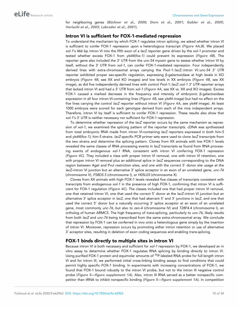
Figure 6. FOX-1 binds in vivo to multiple binding sites in xol-1 intron VI using both GCAUG and GCACG motifs to regulate alternative splicing. The
diagram of intron VI (top left) shows locations of the three regions (blue, yellow, and green) shown to exhibit FOX-1 binding in vitro. RNA sequences
corresponding to each color-coded region are shown below the diagram. CRISPR/Cas9 editing was used to modify endogenous DNA encoding these
regions and thereby identify cis-acting sites that control xol-1 splicing in vivo. The motif GCAUG was changed to AUAUA, and the motif GCACU was
Figure 6 continued on next page
Farboud et al. eLife 2020;9:e62963. DOI: https://doi.org/10.7554/eLife.62963 15 of 34
Research article Chromosomes and Gene Expression

dose of fox-1 to allow high xol-1 activity for male development and by increasing aberrant splicing
with two doses of fox-1 to allow low xol-1 activity for hermaphrodite development.
We used multiple assays to judge the impact on xol-1 splicing regulation caused by disrupting
endogenous FOX-1 binding sites. First, we assessed the viability of XX hermaphrodites carrying xol-
1 mutations in FOX-1 regulatory regions. This assay measures the contribution of xol-1 splicing regu-
lation toward X:A signal assessment in the context of full transcriptional repression by other X signal
elements, SEX-1 (nuclear hormone receptor) and CEH-39 (homeodomain protein). Second, we
assessed the viability of XX xol-1 mutant hermaphrodites in the context of reduced SEX-1 activity,
and hence elevated xol-1 transcription, to measure synergy between transcriptional and splicing reg-
ulation. This sensitized XSE mutant condition was achieved using RNA interference (RNAi) against
sex-1. Third, we assessed the viability of XO xol-1 mutant males in the context of high FOX-1 levels
that are sufficient to kill all otherwise wild-type XO animals by causing non-productive alternative
splicing. In these XO animals, the single dose of sex-1 and ceh-39 does not repress xol-1 expression.
This sensitive assay measures the efficacy of single and multiple wild-type FOX-1 binding sites on
splicing regulation under conditions in which FOX-1 levels are not limiting.
In initial experiments, we eliminated the non-productive alternative splicing mode of xol-1 repres-
sion using CRISPR/Cas9 editing to fuse exon 6 in frame with exon 7 at the endogenous xol-1 locus
(y810) and thereby exclude intron VI from the pre-mRNA. In XO animals, removing intron VI blocked
the XO-specific lethality caused by overexpressing FOX-1. Viability of XO males increased from 0%
to 89% (p<10�5) for y810, indicating that splicing regulation was severely disrupted, as predicted
(Figure 6A,C).
In XX animals, loss of intron VI did not reduce either the viability of mutant (y810) versus wild-
type animals (99% vs. 101%, respectively) or the average brood size per hermaphrodite (240 ± 4 vs.
267 ± 32 embryos) (Figure 6—figure supplement 1A,C). Consistent with this intron VI deletion
result, a null mutation of fox-1(y793) created by a Cas9-induced deletion of the entire endogenous
gene also resulted in insignificant XX lethality and no reduction in brood size (Figure 6B and Fig-
ure 6—figure supplement 1B). However, just as eliminating FOX-1 activity by gene deletion killed
XX hermaphrodites sensitized by reduced activity of the XSE transcription repressor SEX-1, eliminat-
ing xol-1 intron VI killed all XX hermaphrodites with reduced SEX-1 activity (Figure 6C). XX viability
decreased from 57% to 2% (p<10�5) for deletion of fox-1(y793) and from 57% to 0% (p<10�5) for
deletion of the intron (y810) (Figure 6C and Figure 6—figure supplement 1C). Thus, repression of
Figure 6 continued
changed to AUACA. (A–L) Diagrams of introns with the different CRISPR/Cas9 edits used for testing xol-1 splicing regulation in vivo are shown on the
left side. Multiple assays (right side) evaluate the effects on X:A signal activity of these intron mutations as well as a Cas9-induced deletion of
endogenous fox-1(y793). The effects on X:A signal activity are judged by the viability of XX and XO animals with different combinations of X-signal
element (XSE) levels. Viability of XX hermaphrodites with mutations only in FOX-1 regulatory regions of xol-1 measures the full contribution of splicing
regulation toward X:A signal activity in the context of wild-type XSE levels and hence normal transcriptional repression by XSEs. Viability of XX xol-1
mutants treated with RNAi against the XSE gene sex-1, which encodes a nuclear hormone receptor transcriptional repressor of xol-1, monitors the
synergy between transcriptional and splicing regulation when transcriptional regulation is compromised such that xol-1 expression is elevated. XX
mutants were treated with sex-1(RNAi) for one generation to cause only partial sex-1 inhibition and enable 57% survival. Viability of XO xol-1 mutant
animals in the context of high FOX-1 levels tests the efficacy of single and multiple FOX-1 binding sites on splicing regulation under conditions in which
FOX-1 levels are not limiting for splicing regulation. The viability of xol-1 XO mutant animals with wild-type FOX-1 levels serves as a control for any
adverse effects of intron VI mutations unrelated to repression by excess FOX-1. All formulae for calculating viabilities of XX and XO animals with
different XSE levels are provided in Materials and methods. For all assays, the average viability of multiple broods, each from a single hermaphrodite, is
shown with the standard error of the mean (SEM). The total number of embryos scored for viability from all broods is indicated in parenthesis. The
experiments show that splicing regulation becomes essential when transcriptional repression is compromised. Multiple FOX-1 binding sites utilizing
GCACG and GCAUG motifs are required for full splicing regulation. The number of binding sites is more critical than whether the motif sequence is
GCAUG or GCACG. However, replacing all high-affinity motifs with the low-affinity secondary motif GCUUG promotes only minimal non-productive
alternative splicing. High-affinity motifs are essential for FOX-1 mediated repression. Eliminating intron VI revealed no greater benefits for male viability
than deleting the entire fox-1 gene.
The online version of this article includes the following source data and figure supplement(s) for figure 6:
Source data 1. Overexpression of ASD-1 kills both XO males and XX hermaphrodites.
Figure supplement 1. Brood sizes of XX animals with cis-acting xol-1 mutations in intron VI are consistent with the strain viability.
Figure supplement 2. High FOX-1 levels in XX animals repress xol-1 with multiple low-affinity GCUUG motifs in intron VI.
Farboud et al. eLife 2020;9:e62963. DOI: https://doi.org/10.7554/eLife.62963 16 of 34
Research article Chromosomes and Gene Expression

xol-1 by splicing regulation in XX animals becomes critical primarily in the context of compromised
transcriptional repression.
One other FOX-1 family member, the autosomal protein ASD-1, binds GCAUG motifs and con-
trols alternative splicing of other C. elegans developmental regulators (Kuroyanagi et al., 2013;
Kuroyanagi et al., 2007; Kuroyanagi et al., 2006). Our genetic evidence suggested it does not reg-
ulate xol-1 either in the presence or absence of FOX-1 (Figure 6—source data 1). That possibility
could not have been fully eliminated until intron VI mutations removed the opportunity for any RNA
binding factors to regulate xol-1 alternative splicing. No greater benefit to XO males or greater det-
riment to XX hermaphrodites occurred when all potential sources of splicing regulation were elimi-
nated by removing intron VI rather than by deleting fox-1 alone.
FOX-1 binding sites identified in vitro function in vivo to regulate xol-1splicingTo determine whether FOX-1 binding sites identified in vitro by biochemical analysis do indeed func-
tion in vivo to regulate xol-1 RNA splicing, we used two strategies to mutate endogenous DNA
encoding these sites in intron VI. First, we deleted the 37 bp, 35 bp, and 26 bp regions correspond-
ing to the in vitro FOX-1 binding sites (y805) (Figure 6D). Second, we altered the sequence of all
individual putative binding motifs within each binding site (y804) (Figure 6E). Endogenous DNA was
edited to convert the RNA motif GCAUG to AUAUA and GCACG to AUACA.
The nucleotide deletions (y805) and substitutions (y804) of all FOX-1 binding sites and motifs,
respectively, within intron VI had the same effect as eliminating the intron: nearly complete loss of
XX viability in the sensitized sex-1(RNAi) XSE mutant condition and suppression of male lethality
caused by FOX-1 overexpression (Figure 6D,E). XX viability with sex-1(RNAi) was reduced from 57%
to 0% (p<10�5) by the binding site deletions, and from 57% to 1% (p<10�5) by the motif substitu-
tions. Male viability with high FOX-1 levels increased from 0% to 88% (p<10�5) with the deletions
and from 0% to 89% (p<10�5) with the substitutions. In contrast, the viability and brood sizes of XX
animals bearing only deletions or nucleotide substitutions of all FOX-1 binding sites and motifs in a
sex-1(+) condition were not different from wild-type XX animals (Figure 6D,E and Figure 6—figure
supplement 1D,E). These experiments indicate that FOX-1 binding sites and motifs identified in
vitro do function in vivo to mediate xol-1 splicing repression, and confirm that splicing regulation is
essential for hermaphrodite viability primarily in the context of reduced transcriptional repression.
Multiple GCAUG motifs and GCACG motifs in intron VI are essential forFOX-1-regulated alternative xol-1 splicingGenome editing of fox-1 binding sites also allowed us to determine the efficacy in vivo of GCAUG
motifs versus GCACG motifs in splicing-mediated xol-1 repression in XX and XO animals and to
determine how many FOX-1 binding sites are required for full splicing regulation. Mutating the three
GCACG motifs while retaining the two GCAUG motifs (y808) reduced the viability of sex-1(RNAi) XX
animals to about half the level of sex-1(RNAi) XX animals with five wild-type motifs (24% vs. 57%)
(p<10�5), but permitted more XX viability than with mutant versions of all five GCAUG and GCACG
motifs (y804) (24% vs. 1%) (p<10�5) (Figure 6F and Figure 6—figure supplement 1F). These results
indicate that GCACG motifs function in vivo for splicing repression in XX animals, but by themselves
are not sufficient for full repression; the GCAUG motifs are also required. The two GCAUG motifs
remaining in y808 also severely reduced the viability of XO animals with high FOX-1 levels compared
to those with five mutant motifs (y804) (8% vs. 89%) (p<10�5), demonstrating that GCAUG motifs
contribute to FOX-1-mediated repression in XO animals (Figure 6F).
Reciprocally, mutating the two GCAUG motifs while retaining the three wild-type GCACG motifs
(y809) reduced the viability of XSE-sensitized XX animals by about half compared to those sensitized
XX animals having all five wild-type motifs (33% vs. 57%) (p<10�4), but permitted more XX viability
than all five mutant motifs (y804) (33% vs. 1%) (p<10�5) (Figure 6G and Figure 6—figure supple-
ment 1G). These results indicate that GCAUG motifs function in vivo for splicing repression in XX
animals but are not sufficient for full repression; GCACG motifs are also important. The three
GCACG motifs remaining in y809 reduced the viability of XO animals with high FOX-1 levels by half
compared to XO animals with all mutant motifs (y804) (41% vs. 89%) (p<10�5) (Figure 6G), indicat-
ing that GCACG motifs contribute to splicing regulation in XO animals. Thus, multiple FOX-1
Farboud et al. eLife 2020;9:e62963. DOI: https://doi.org/10.7554/eLife.62963 17 of 34
Research article Chromosomes and Gene Expression

binding sites are required in vivo for xol-1 repression by alternative splicing, and both GCACG and
GCAUG motifs are important.
To determine whether GCACG motifs alone or GCAUG motifs alone could be sufficient for full
splicing-mediated repression when present at all five sites, we converted all five motifs to either
GCACG (y807) or GCAUG (y806) (Figure 6H,I and Figure 6—figure supplement 1H,I). When pres-
ent in all sites, either GCACG or GCAUG motifs were sufficient to confer complete splicing repres-
sion, just like a wild-type intron VI. Virtually all XO animals were killed by high levels of FOX-1 (1% or
3%, respectively, vs. 0%), and the viability of XSE-sensitized XX animals was equivalent to that
achieved with a wild-type intron VI (49% or 57%, respectively, vs. 57%) (Figure 6H,I). These results
indicate that the number of binding sites is more important than whether the sequences are GCAUG
or GCACG.
The significance of motif number in splicing repression is further illustrated by additional experi-
ments. First, XX viability was compared directly between edited animals that have only GCAUG
motifs but differ in GCAUG number. Viability of sex-1(RNAi) XX animals with five GCAUG motifs
(y806) was 57%, but viability with two GCAUG motifs was 24% (p<10�4) (Figure 6I and Figure 6—
figure supplement 1I). Second, viability of sex-1(RNAi) XX animals was relatively normal with four of
five wild-type motifs (y811) (52% vs. 57%) (Figure 6J and Figure 6—figure supplement 1J). How-
ever, viability of sex-1(RNAi) XX animals was reduced when xol-1 had just two or three wild-type
motifs: 24% (p<10�5) for two GCAUG motifs in y808% and 33% (p<10�4) for three GCACG motifs in
y809 (Figure 6F,G). Third, mutation of a single motif, the terminal GCACG motif (y811), was suffi-
cient to reduce splicing-mediated repression of xol-1 in XO animals by high FOX-1 levels
(Figure 6J). More XO animals with excess FOX-1 were viable with only four of five wild-type motifs
(y811) than with all five (36% vs. 0%). Fourth, in a reciprocal experiment, a single GCACG motif
(y803) was sufficient to decrease viability of XO animals with high FOX-1 levels compared to XO ani-
mals with no wild-type sites (y804) (22% vs. 89%) (p<10�5) (Figure 6K). Thus, in the context of high
FOX-1 levels, a single GCACG motif functions in splicing repression, but increasing the number of
either GCACG or GCAUG motifs causes progressively greater repression and less XO viability.
To further assess the effect of only a single FOX-1 binding site in xol-1, we examined XX animals
in which only the terminal GCACG motif (y803) was present (Figure 6K and Figure 6—figure sup-
plement 1K). We found severe XX lethality in the XSE-sensitized background (6% viable), indicating
that the single GCACG site by itself is not sufficient for robust regulation in XX animals. Multiple
sites are required, as exemplified from the increased viability of sex-1(RNAi) XX animals with three
GCACG motifs (33%, p<10�5) (Figure 6G) and five GCACG motifs (49%, p=0.01) (Figure 6H).
The fact that 6% of XX animals were viable with just the single site (y803) instead of 1% (p=0.002)
when all sites were altered (y804) indicates that the GCACG site by itself permits sufficient alterna-
tive splicing to rescue some XX animals (Figure 6E,K). Utility of the single site is also reflected in the
brood size of these XSE-sensitized animals. Hermaphrodites with one GCACG site had an average
brood size of 100 ± 25 embryos, while those with no sites had an average brood size of 11 ± 4
embryos (Figure 6—figure supplement 1E,K). Consistent with the efficacy of a single GCACG site,
viability of XO animals in the presence of high FOX-1 levels was reduced from 89% when all sites
were absent (y804) to only 22% (p<10�5) when a single GCACG site (y803) was present (Figure 6E,
K). Thus, while one binding site can achieve some repression, multiple sites are needed for full
repression, thereby creating a sensitive mechanism for xol-1 regulation by FOX-1.
No GCAUG or GCACG motifs are present in any xol-1 intron other than intron VI, consistent with
intron VI being sufficient for FOX-1 regulation. Two GCACG motifs are present in the 3’ UTR of the
2.2 kb transcript, but this 3’ UTR is not necessary for repression by FOX-1 (Figure 4B).
Multiple low-affinity GCUUG motifs in intron VI are not sufficient torepress xol-1 with the FOX-1 levels present in wild-type XX or XOanimals but are sufficient for xol-1 repression in both sexes with highFOX-1 levelsRecent studies demonstrated that mammalian Rbfox can utilize secondary, low-affinity binding
motifs such as GCUUG to promote alternative splicing in vivo if these motifs are present in multiple
copies, and if Rbfox reaches a concentration higher than necessary for binding to GCAUG motifs
(Begg et al., 2020). Therefore, we tested whether replacing all GCACG and GCAUG motifs in intron
VI with GCUUG motifs would promote sufficient non-productive splicing in the presence of high
Farboud et al. eLife 2020;9:e62963. DOI: https://doi.org/10.7554/eLife.62963 18 of 34
Research article Chromosomes and Gene Expression

FOX-1 levels to cause the death of XO males and, reciprocally, the survival of XX embryos exposed
to sex-1(RNAi). In XO animals with high FOX-1 levels, the low-affinity GCUUG binding sites were suf-
ficient to cause complete XO lethality (y820 in Figure 6L).
In XX animals with wild-type GCUCG and GCACG motifs in intron VI, high levels of FOX-1 could
suppress the death caused by sex-1(RNAi) (24% vs. 63% viable p=0.004) (Figure 6—figure supple-
ment 2A,B), demonstrating that enhancing post-transcriptional repression of xol-1 can compensate
for the deficiency in repression caused by reducing transcriptional repression. Although high FOX-1
levels in XX animals with mutated GCACG and GCAUG motifs could not suppress any death caused
by sex-1(RNAi) (Figure 6—figure supplement 2D), high FOX-1 levels could rescue XX animals with
low-affinity GCUUG motifs from sex-1(RNAi)-induced death (p=0.006) (Figure 6—figure supple-
ment 2C), as could high FOX-1 levels with normal GCAUG and GCACG motifs.
In contrast, GCUUG motifs were not adequate to suppress the lethality caused by sex-1(RNAi)
when only the two wild-type doses of fox-1 were present in XX embryos. Only 8% of y820 sex-1
(RNAi) XX animals carrying GCUUG motifs survived compared to 57% survival when all sites had the
original high-affinity GCAUG or GCACG motifs (p<10�3) (Figure 6L and Figure 6—figure supple-
ment 1L). These results show that although multiple low-affinity GCUUG binding sites, coupled with
high FOX-1 levels, are sufficient to kill XO animals or to suppress the death of sex-1(RNAi) XX ani-
mals, the low-affinity GCUUG sites are inadequate to repress xol-1 in XX embryos with only the nor-
mal two doses of fox-1. The degree of non-productive splicing necessary to repress xol-1 in XX
embryos with two copies of fox-1 is only reached if multiple high-affinity motifs are present in intron
VI.
FOX-1 acts in a dose-dependent manner in XX animals to regulate xol-1splicing and thereby determines sexThe need for multiple high-affinity binding sites to enable two doses of FOX-1 to repress xol-1 in XX
animals suggested that xol-1 splicing control would be sensitive to FOX-1 dose. We took two
approaches to evaluate the dose-dependence of FOX-1 action in determining sex. In XX animals
with reduced sex-1 activity caused by RNAi directed against sex-1, we compared the impact on xol-
1 regulation of reducing the dose of FOX-1 from two copies to one to the impact of mutating com-
binations of FOX-1 binding sites in one endogenous copy of xol-1. Both approaches revealed dose-
sensitivity of FOX-1 action.
Viability of the sex-1(RNAi) XX animals declined more than 10-fold, from 44% to 3% (p=0.002),
when the dose of fox-1 was reduced by half, from two copies to one copy, demonstrating dose-
dependence of FOX-1 function in XX animals (Figure 7A,B). Similarly, viability of sex-1(RNAi) XX ani-
mals declined from 44% to 7% (p=0.006) when all FOX-1 binding sites in intron VI were mutated in
one copy of xol-1 (Figure 7A,C). As expected, viability of sex-1(RNAi) XX animals declined to an
intermediate level when either the three GCACG motifs (18%) (p=0.018) or the two GCAUG motifs
(13%) (p=0.012) were mutated in one copy of xol-1 (Figure 7A,D,E). Viability of sex-1(RNAi) XX ani-
mals with five low-affinity GCUUG motifs in one copy of xol-1 was better (36%) than that with either
two GCAUG (p=0.023) or three GCACG (p=0.017) mutated motifs in one xol-1 copy (Figure 7F).
Hence, the dose-dependence of FOX-1 function in regulating alternative xol-1 splicing in XX animals
is evident both from reducing the dose of the trans-acting FOX-1 protein or by mutating different
combinations of cis-acting FOX-1 binding sites in only one copy of xol-1. Thus, FOX-1 acts as an XSE
to convey X-chromosome number by regulating alternative xol-1 splicing in a dose-dependent
manner.
DiscussionWe dissected the mechanism by which the C. elegans RNA binding protein FOX-1 acts as a dose-
dependent X-chromosome signal element to specify sexual fate. In XX embryos, FOX-1 binds to the
single alternatively spliced intron of xol-1, the master regulator that sets the male fate in XO
embryos, and causes either intron retention, and hence premature translation termination, or alter-
native 3’ acceptor site usage, and hence exclusion of essential exon coding sequences (Figure 8).
Both events prevent production in XX embryos of functional male-determining XOL-1 protein, which
also sets the level of X-chromosome gene expression in XO embryos. FOX-1 must bind to multiple
high-affinity GCAUG and GCACG motifs in xol-1 intronic sequences to regulate xol-1 splicing in XX
Farboud et al. eLife 2020;9:e62963. DOI: https://doi.org/10.7554/eLife.62963 19 of 34
Research article Chromosomes and Gene Expression

GCAUG
GCACG
GCAUG
GCACG GCACG
AUAUA
AUACA
AUAUA
AUACA AUACA
y804
AUAUA
GCACG
AUAUA
GCACG GCACG
y809
GCAUG
AUACA
GCAUG
AUACA AUACA
y808
WT
GCAUG
GCACG
GCAUG
GCACG GCACG
GCAUG
GCACG
GCAUG
GCACG GCACG
WT
GCAUG
GCACG
GCAUG
GCACG GCACG
fox-1(y793)
WT
GCAUG
GCACG
GCAUG
GCACG GCACG
WT
GCAUG
GCACG
GCAUG
GCACG GCACG
WT
GCAUG
GCACG
GCAUG
GCACG GCACG
WT
fox-1(+)
fox-1(+)
fox-1(+)
WT
fox-1(+)
fox-1(+)
fox-1(+)
fox-1(+)
fox-1(+)
fox-1(+)
y820
GCAUG
GCACG
GCAUG
GCACG GCACG
WT
fox-1(+)
fox-1(+)
GCUUG
GCUUG
GCUUG
GCUUG GCUUG
% XX
Hermaphrodite
Viability ± SEM
(Total Males)
p-value
vs.
fox-1(y793)/+
p-value
vs.
WT
% XX
Viability ± SEM
with sex-1(RNAi)
(Total Males)
100 2
(1474)
± 0.00444 7
(1195)
± 0.006
98 1
(1139)
± 0.006 0.0067 1
(1160)
±
99 3
(823)
± 0.018 0.00818 3
(702)
± 0.027
100 2
(833)
± 0.012 0.02513 3
(791)
± 0.112
p-value
vs.
xol-1(y804)/+
97 1
(931)
± 0.0043 1
(653)
± 0.006
Intron VI
37 nt 35 nt 26 nt
u GCAUGuagcacauuugaucguuaugcuuGCACGcaaa
c GCAUGaguucauuugaucguuaugauGCACGgaaa
gcauauuugaucgaaugccuGCACGu
A
B
C
D
E
F
not
applicable
not
applicable
not
applicable
98 4
(1873)
± 36 4
(2772)
± 0.083 < 0.001 < 0.001
GCAUG
GCACG
WT
AUAUA
AUACA
Mutant
Figure 7. FOX-1 acts in a dose-dependent manner to regulate xol-1 splicing in XX animals and thereby determine sex. (A–F) Diagrams on the left show
sequences for the two different xol-1 intron VI combinations assayed in each cohort of sex-1(RNAi) XX animals to assess the dose-dependence of FOX-
1 action in regulating xol-1 splicing. Viability of both sex-1(+) and sex-1(RNAi) animals is shown on the right along with statistical comparisons of
viability across different genotypes. Except for (A, B) in which both copies of intron VI have unaltered FOX-1 binding sites, one xol-1 intron VI has
Figure 7 continued on next page
Farboud et al. eLife 2020;9:e62963. DOI: https://doi.org/10.7554/eLife.62963 20 of 34
Research article Chromosomes and Gene Expression

embryos, and this splicing regulation is dose-dependent to achieve xol-1 repression in XX but not
XO embryos. Deleting one copy of fox-1 or removing all FOX-1 binding sites in one copy of xol-1
reduces splicing regulation sufficiently to kill XX animals with reduced XSE activity, demonstrating
the importance of fox-1 dose for viability of XX animals. Having two doses of fox-1 in XX embryos is
as important for viability as restricting the dose of fox-1 to one in XO embryos.
Dose-dependent regulation of xol-1 splicing acts as a secondary mode of repression in XX
embryos to enhance the fidelity of X:A signaling. Transcriptional repression by XSEs is the primary
mode of xol-1 regulation (Carmi et al., 1998; Carmi and Meyer, 1999; Farboud et al., 2013;
Gladden and Meyer, 2007; Meyer, 2018; Powell et al., 2005). We showed that non-productive
alternative splicing is then imposed on residual xol-1 transcripts to achieve full xol-1 repression in XX
embryos. Repression of xol-1 by splicing regulation is especially critical in the context of compro-
mised transcriptional repression and during early development prior to maximal transcriptional
repression. XX embryos lacking splicing regulation die if xol-1 transcription is even partially activated
in XX embryos. Reciprocally, increasing FOX-1 concentration above the normal level in XX embryos,
and hence increasing non-productive splicing, suppresses the XX lethality caused by reducing xol-1
transcriptional repression. The combined action of splicing and transcriptional repression of xol-1
enhances the precision of X-chromosome counting.
Mechanisms underlying the action of FOX family members in regulatingalternative pre-mRNA splicingMultiple high-affinity FOX-1 binding motifs are necessary to restrict the non-productive mode xol-1
splicing to XX embryos over the small FOX-1 concentration range that distinguishes XX from XO
embryos. Among numerous experiments, the need for multiple high-affinity RNA binding sites to
repress xol-1 was well exemplified by the comparison in XX viability of engineered xol-1 strains that
carried only GCACG motifs in intron VI, but in different numbers, and were subjected to sex-1
(RNAi). The viability of the XX strain with five GCACG motifs (49%) was greater than that with three
GCACG motifs (33%) or with one motif (6%). Moreover, replacing all GCUAG and GCACG motifs
with low-affinity GCUUG motifs permitted only minimal survival (8% vs. 57%).
In other cases of splicing regulation by C. elegans FOX-1, specifically the unc-32 and egl-15 gene
targets, robust regulation is achieved through a single GCAUG binding site using a different strategy
from the one for xol-1. FOX-1 binds the unc-32 pre-mRNA at the single GCAUG site in concert with
the neuronally expressed CELF RNA binding protein UNC-75 to promote skipping of the upstream
exon (Kuroyanagi et al., 2013). To do so, FOX-1 acts redundantly with ASD-1, another FOX-1 family
member. Either FOX-1 or ASD-1 can bind the GCAUG site with UNC-75, and together regulate unc-
32. Similarly, FOX-1 and ASD-1 regulate splicing of egl-15 pre-mRNA in combination with the mus-
cle-specific RNA binding protein SUP-12 using a single GCAUG site (Kuroyanagi et al., 2007). For
both gene targets, two FOX-1 family members bind a single binding site to help ensure proper splic-
ing in conjunction with key tissue-specific RNA binding proteins. Loss of either fox-1 or asd-1 activity
causes partial loss-of-function phenotypes for egl-15 and for unc-32, revealing that asd-1 and fox-1
function in a collaborative fashion to reinforce splicing control of two different pre-mRNA targets,
each with one binding site (Kuroyanagi et al., 2013; Kuroyanagi et al., 2007). In contrast, our
Figure 7 continued
mutated FOX-1 binding sites as indicated and one intron has unaltered FOX-1 binding sequences (C–F). (B) Low viability of sex-1(RNAi) XX animals with
only one dose of fox-1 [fox-1(y793) / +] shows strong dose-dependence of FOX-1 action in xol-1 splicing regulation. (C–F) The impact of heterozygous
combinations of intron VI mutations on viability of sex-1(RNAi) XX animals also indicates that FOX-1 functions as a dose-dependent X signal element to
regulate xol-1 splicing and thereby communicate X-chromosome dose. Viability assays were conducted using the protocols that follow. Separate
matings were performed between Prps-0::mNeonGreen::4xNLS::unc-54 green males and hermaphrodites of genotypes: wild-type XX for (A), fox-1(y793)
XX for (B), xol-1(y804) XX for (C), xol-1(y808) for (D), xol-1(y809) for (E), and xol-1(y820) for (F). For the sex-1(RNAi) experiments, matings were performed
on plates with bacteria containing plasmids that produce double-stranded sex-1 RNA when XX animals were young adults. For the control set of
matings, males and hermaphrodites were grown on bacteria that do not produce double-stranded sex-1 RNA (pL4440 empty vector control). For both
sets of crosses, all green hermaphrodites and green males were counted. Since viability of XO animals is not affected by sex-1(RNAi), the number of
green XX hermaphrodites expected if all were viable would be the same as the number of green XO males. Percent XX viability was calculated by
(Number of green hermaphrodites/Number of green males) � 100.
Farboud et al. eLife 2020;9:e62963. DOI: https://doi.org/10.7554/eLife.62963 21 of 34
Research article Chromosomes and Gene Expression

xol-1ATG
Hightranscript
levels
Minimal non-productive
alternativesplicing
HighXOL-1level
XOL-1
Lowtranscript
levels
non-productivealternative
splicing
LowXOL-1level
xol-1ATG
XOL-1
TSS TSS
1X:2A (1 XSE dose) 2X:2A (2 XSE doses)
STOP
intron VI retention
2.5 kb transcript
(inactive)
intron VI removal
2.2 kb transcript
(active)
STOP1.5 kb transcript
(inactive)
STOP
I II
III
IV V
VI
1 2 3 4 5 6 7
xol-1
FOX-1 binding
FOX-1
FOX-1FOX-1
XSE ASEBinding sites: FOX-1
A
B
Figure 8. Summary of xol-1 splicing regulation by FOX-1 and model for X:A signal assessment. (A) Summary of xol-1 splicing regulation by FOX-1.
Through binding to multiple GCAUG and GCACG motifs in intron VI of xol-1, FOX-1 reduces formation of the male-determining 2.2 kb transcript by
causing intron VI retention (2.5 kb transcript) or by directing use of an alternative 3’ splice acceptor site, causing deletion of essential exon 7 coding
sequences (blue) and part of the 3’ UTR (orange) (1.5 kb transcript). (B) Model for X:A signal assessment: two tiers of xol-1 repression. X-signal
Figure 8 continued on next page
Farboud et al. eLife 2020;9:e62963. DOI: https://doi.org/10.7554/eLife.62963 22 of 34
Research article Chromosomes and Gene Expression

results showed that ASD-1 is not required for regulation of alternative xol-1 splicing to repress xol-1
activity in XX embryos.
For many mammalian genes, Rbfox can bind to an intron and control pre-mRNA splicing using a
single high-affinity GCAUG motif. A tyrosine-rich, low-complexity domain at the C-terminus then
nucleates aggregation of Rbfox to attain the concentration of bound proteins necessary to drive
alternative splicing (Ying et al., 2017). Rbfox aggregation is essential for inclusion of specific exons
into mature mRNA and has the potential to recruit additional splicing factors. Rbfox aggregation
may also be necessary for concentrating Rbfox complexes in nuclear speckles, where RNA synthesis
occurs (Ying et al., 2017). Such concentrated localization may promote binding to and alternative
splicing of newly synthesized RNAs. C. elegans FOX-1 lacks a tyrosine-rich low-complexity domain to
cause protein aggregation, but during xol-1 regulation, recruitment of FOX-1 through multiple
intronic binding sites using GCAUG and GCACG motifs appears to substitute for protein aggrega-
tion in achieving robust alternative splicing. In this case, availability of multiple sites would increase
the probability of any site being occupied.
Genome-wide binding studies of Rbfox revealed that many mammalian introns have more than
one primary GCAUG or GCACG motif (Begg et al., 2020; Lambert et al., 2014). While experiments
have not yet addressed whether the multiple motifs enhance splicing regulation at these endoge-
nous sites, other experiments using mammalian reporter constructs have demonstrated that three
GCAUG motifs are more effective at regulating splicing than a single motif (Modafferi and Black,
1999).
The importance of multiple motifs in mammalian pre-mRNA splicing regulation is further exempli-
fied by a recent study showing that a cluster of six Rbfox low-affinity secondary binding motifs, such
as GCUUG or GUUUG, but not a single low-affinity motif, controls splicing regulation during neuro-
nal differentiation in vivo, as the Rbfox concentration naturally increases during development
(Begg et al., 2020). Higher Rbfox concentrations are required for splicing regulation using the clus-
tered low-affinity sites than those required for splicing regulation by a single high-affinity GCAUG
motif. Hence, the clustered motifs create tissue and temporal specificity in splicing regulation in
response to changes in Rbfox concentration during development (Begg et al., 2020).
A cluster of intronic xol-1 motifs could tune splicing regulation over the twofold increase in FOX-
1 concentration between XO and XX embryos, as Rbfox appears to do during neuronal differentia-
tion (Begg et al., 2020). The cluster of xol-1 motifs likely enhances the probability that the higher
FOX-1 concentration in XX embryos versus XO embryos reaches the threshold necessary for FOX-1
binding and function, thereby restricting splicing regulation to XX embryos. Our studies showed that
a single FOX-1 binding site becomes sufficient to induce some non-productive xol-1 splicing in XO
embryos and thereby kill them when the concentration of FOX-1 is increased artificially by integrat-
ing numerous extra copies of fox-1 into the genome. However, multiple high-affinity motifs are nec-
essary to achieve the non-productive mode of xol-1 splicing to XX embryos. Even for xol-1, though,
secondary GCUUG motifs can achieve splicing regulation if the FOX-1 concentration is elevated
beyond the normal level in XX embryos. Five GCUUG motifs are sufficient to permit high FOX-1 lev-
els to suppress the XX-specific lethality caused by sex-1(RNAi) or to kill all XO embryos. These results
converge with and extend those found for mammalian Rbfox.
Figure 8 continued
elements (XSEs) and autosomal signal elements (ASEs) bind directly to numerous non-overlapping sites in the 5’ regulatory region of xol-1 to
antagonize each other’s opposing transcriptional activities and thereby control xol-1 transcription (Farboud et al., 2013). Molecular rivalry at the xol-1
promoter between the XSE transcriptional repressors and ASE transcriptional activators causes high xol-1 transcript levels in 1X:2A embryos with one
dose of XSEs and low levels in 2X:2A embryos with two doses of XSE. All binding sites for the XSEs (nuclear receptor SEX-1 and homeodomain protein
CEH-39) are shown in magenta and binding sites for the T-box transcription factor ASE called SEA-1 are shown in blue. Binding sites for the zinc finger
ASE called SEA-2 have not been mapped precisely enough in this xol-1 regulatory region to represent. In a second tier of xol-1 repression shown by
our studies, the XSE RNA binding protein FOX-1 (green) then enhances the fidelity of X-chromosome counting by binding to numerous GCAUG and
GCAUG motifs in intron VI (yellow) of the residual xol-1 pre-mRNA, thereby causing non-productive alternative splicing and hence xol-1 mRNA variants
that have in-frame stop codons or lack essential exons. High XOL-1 protein induces the male fate and low XOL-1 permits the hermaphrodite fate. Black
rectangles represent xol-1 exons, dark gray rectangles represent xol-1 introns, and light gray rectangles represent 5’ and 3’ xol-1 regulatory regions.
Farboud et al. eLife 2020;9:e62963. DOI: https://doi.org/10.7554/eLife.62963 23 of 34
Research article Chromosomes and Gene Expression

Strategies to determine sex: Comparison across species of primary sex-determination mechanisms requiring both transcriptional and pre-mRNA splicing regulationLike C. elegans, the fruit fly D. melanogaster utilizes the combination of transcriptional and pre-
mRNA splicing regulation to enhance the precision of X:A counting by controlling Sxl (Sex-lethal),
the master sex-determination switch gene and direct target of the X:A signal. This sex switch gene
dictates female development when active and permits male development when inactive (Cline and
Meyer, 1996). Sxl encodes an X-linked RNA binding protein with an RRM. It directs female develop-
ment by regulating alternative pre-mRNA splicing of itself and downstream sex-determination
genes. Sxl is activated in 2X:2A embryos, but not 1X:2A embryos, by a set of feminizing XSEs that
stimulate transcription of Sxl in a dose-dependent manner. Once produced, SXL protein functions in
a positive autoregulatory loop to control splicing of its own pre-mRNA in a dose-dependent manner
and thereby promote continued production of female-specific SXL protein (Bell et al., 1991;
Cline and Meyer, 1996; Horabin and Schedl, 1993; Sakamoto et al., 1992). By binding to two
neighboring Sxl introns, SXL prevents inclusion of the intervening male-specific exon 3, which enco-
des an in-frame stop codon that prevents translation of the full-length female SXL protein when
incorporated into mature RNA.
SXL also directs pre-mRNA splicing of its downstream sex-determination target gene called tra
(transformer), a switch gene that directs female sexual differentiation (Inoue et al., 1990;
Sosnowski et al., 1989; Valcarcel et al., 1993). SXL controls tra by regulating 3’ splice site selec-
tion. By binding to an upstream 3’ splice site in the first intron of tra pre-mRNA, SXL competes with
binding of splicing factor U2AF and thereby diverts splicing to a distal 3’ splice acceptor site in that
intron. The resulting shorter RNA isoform encodes a full-length TRA protein.
Thus, SXL dictates female development by directing formation of RNA isoforms for itself and
downstream targets that encode essential female-specific proteins. In contrast, FOX-1 promotes her-
maphrodite development by dose-dependent binding to intron VI of the male-determining gene
xol-1, causing either intron retention or deletion of exon coding sequences, and thereby blocking
formation of the RNA variant that encodes male-specific XOL-1 protein. While fox-1 acts as a worm
XSE to communicate X-chromosome dose, Sxl responds to fly XSEs to determine sex, but its location
on X and its autoregulatory feature allow it to serve as both signal and target.
Materials and methods
Production and assay of xol-1 transgenes in extrachromosomal arraysThe parental transgene plasmid (pMN45) that Nicoll et al., 1997 used to construct deletion deriva-
tives diagrammed in Figure 2A is a genomic xol-1 rescuing clone that has gfp coding sequences
inserted at the first ATG of xol-1. In extra-chromosomal arrays made by injecting 5 mg/ml plasmid
DNA, the multi-copy transgenes produce a bifunctional GFP::XOL-1 protein that rescues the lethality
of xol-1 XO null mutants and expresses GFP in a sex-specific manner, high in XO and low in XX
embryos. Although arrays of xol-1(+) transgenes from 5 mg/ml injections exhibited sex-specific regu-
lation, the levels of XOL-1 were higher in XX animals than those produced by two copies of the
endogenous gene. This elevated level of xol-1 expression made XX animals dependent on endoge-
nous FOX-1 activity to repress the transgenes and achieve full XX viability. Because xol-1(+) trans-
genic arrays cause XX lethality when fox-1 is inactive, reporters with deletion derivations that
eliminate nucleotides required for FOX-1 regulation are expected to cause XX lethality or at least
milder dosage compensation phenotypes such as a dumpy (Dpy) body shape and an egg-laying
defect (Egl). Prior experiments showed that excess FOX-1 is unable to repress a translational xol-1::
gfp reporter that lacks sequences downstream of the first two introns and 89 codons of xol-1, allow-
ing us to keep intron I in the reporters. Because we found that the phenotypic analysis of XOL-1
activity (Dead, Viable, Dpy Egl) was a much more sensitive indicator of xol-1 activity than GFP fluo-
rescence, we reported only the phenotypic analysis. As an example of the difference in sensitivity,
we found that derepression of the gfp::xol-1 transgene caused XX-specific lethality and/or a Dpy
phenotype even for cases in which GFP fluorescence did not appear to increase as measured by a
fluorescence dissecting microscope.
Farboud et al. eLife 2020;9:e62963. DOI: https://doi.org/10.7554/eLife.62963 24 of 34
Research article Chromosomes and Gene Expression

Arrays of xol-1(+) transgenes could only be established using a xol-1(+) DNA injection concentra-
tion at 5 mg/ml. Any higher xol-1(+) DNA injection concentration killed XX animals, and extra-chro-
mosomal lines could not be established. All deletion derivatives of xol-1 transgenes were also
injected with a DNA concentration of 5 mg/ml. The xol-1 plasmids were co-injected with 150 mg/ml
of unc-76(+) marker plasmid p76-16b into him-5(e1490) unc-76(e911); xol-1(y9) hermaphrodites.
him-5 unc-76; xol-1 hermaphrodites produce 67% viable Unc XX hermaphrodites and 33% Unc XO
males that die as embryos or L1 larvae in the absence of xol-1(+) from a transgenic array. Extra-chro-
mosomal arrays have variable transmission through meiosis and can only be followed when the
arrays carry DNA encoding a wild-type genetic maker, such as unc-76(+), and they are established in
animals carrying an unc-76 mutation, which causes the animals to be Unc. An array resulting in only
Unc hermaphrodites and both Unc and non-Unc males indicate the transgenic array causes XX-spe-
cific lethality. Lines were maintained through non-Unc animals in each generation either by cloning
non-Unc hermaphrodites when the arrays permitted XX viability or crossing non-Unc males into him-
5 unc-76; xol-1 hermaphrodites and recovering non-Unc XO progeny bearing the arrays, if the arrays
caused XX lethality.
Once array lines were made with xol-1 transgene deletion derivatives in the him-5 unc-76; xol-1
strain, they were crossed into yIs44(fox-1); him-5 unc-76; xol-1 hermaphrodites. yIs44 has multiple
copies of the fox-1-containing cosmid R04B3 and the rol-6 plasmid pRF4, which causes the array ani-
mals to have a roller phenotype. xol-1 activity was scored by the presence of non-Unc males, an indi-
cation of rescue of the XO-specific lethality caused by the xol-1(y9) mutation. DNA sequences and
details of transgene constructions are available upon request for pMN66 (D introns II-VI), pMN67 (D
intron VI), pMN60 (D introns II-V), and pCP5 (intron VI in 3’ UTR).
The lacZ reporters Pxol-1::lacZ::xol-1 3’UTR (pMN27) and Pxol-1::lacZ::intron VI::unc-54 3’ UTR
(pMN110) were co-injected at 20 mg/ml with 150 mg/ml of unc-76(+) marker plasmid p76-16b into
him-5(e1490) unc-76(e911) hermaphrodites. Array lines were crossed into yIs44(fox-1); him-5 unc-76
hermaphrodites. pMN27 was made from the promoterless lacZ vector pRD95.11 from A. Fire.
pMN110 was made by inserting intron VI into pMN21, a Pxol-1::lacZ transcriptional fusion made
from the intron-rich lacZ plasmid pPD95.03 from A. Fire. DNA sequences and details of reporter
transgene constructions are available upon request. Embryonic b-galactosidase expression was
assayed as previously described using 4 ml of 4% 5-bromo-4-chloro-3-indolyl-D-galactopyranoside
staining solution and incubation overnight at room temperature (Nicoll et al., 1997). More than
1000 embryos of each genotype were scored for b-galactosidase expression.
Isolation of total RNA from embryosTotal RNA was isolated from 0.2 to 0.5 ml of washed, packed embryos that had been stored at �80˚
C. Frozen embryos were ground to a very fine powder using a liquid-nitrogen-cooled mortar and
pestle, adding more liquid nitrogen as needed to prevent the material from thawing. Ground, frozen
material was vortexed in two microfuge tubes, each with 1 ml Trizol, for 30 s to 1 min and then incu-
bated for 5 min at room temperature. Chloroform (0.2 ml) was added to each tube, vortexed for 30
s, and incubated at room temperature for 2–3 min. After centrifugation at 13,000 � g for 5 min at 4˚
C, the upper aqueous phases (0.75 ml/tube) were transferred to a new tube, and 0.5 ml of the mate-
rial was distributed into each of three tubes. An equal volume of phenol was added to each tube,
vortexed, and 0.2 ml CHCl3 was added and vortexed. Following centrifugation at 13,000 � g for 5
min at 4˚C, the aqueous phases were transferred to new tubes, and an equal volume of CHCl3 was
added and vortexed. Following a 13,000 � g spin in a microfuge for 5 min at 4˚C, the aqueous
phases were transferred to new tubes and incubated a room temperature with one volume of 2-
propanol for 10 min. Following centrifugation at 13,000 � g spin for 10 min at 4˚C, the supernatants
were removed, and the pellets were washed with 70% ethanol and dried in a Spin-Vac. Each pellet
from the three tubes was resuspended in 100 ml water, and all volumes were combined into one
tube (~0.3 ml total). One volume of 4 M LiCl was added and incubated overnight at 4˚C. After a
13,000 � g microfuge spin for 5 min at 4˚C, the aqueous phase was transferred to a new tube. The
RNA was incubated with 0.1� volume of 3 M NaOAc and 2.5� volume of ethanol on ice for more
than 10 min. After centrifugation at 13,000 � g for 10 min at 4˚C, the aqueous phase was removed,
the pellet was washed with 70% ethanol, and dried in a Spin-Vac. The RNA was resuspended in 100
ml water, and the O.D. was read after diluting a sample 250-fold.
Farboud et al. eLife 2020;9:e62963. DOI: https://doi.org/10.7554/eLife.62963 25 of 34
Research article Chromosomes and Gene Expression
Quantification of transcript levelsQuantitative RT-PCR was used to measure transcript levels in RNA isolated from three independent
growths of the four strains listed in Figure 2—source data 1 as previously described (Gladden and
Meyer, 2007; Van Gilst et al., 2005). Three different sets of RT-PCR experiments were performed
with each of the three independent growths of worms. Worms were grown on egg plates (http://
www.wormbook.org) prior to isolating the mixed-stage embryos, and the total RNA was isolated as
described above. A complete list of primer sequences is in Supplementary file 1. The xol-1 primers
were designed to measure all splice variants simultaneously, not just the active ones.
Transcript levels for xol-1 and nhr-64 were normalized to the transcript level of fasn-1, which is
expressed constitutively throughout embryogenesis, by adjusting the Ct (cycle threshold) value of
fasn-1 measured in each strain to equal the Ct value of fasn-1 measured in the control embryos,
either him-5 or wild type. This adjustment equalizes the small variations in concentration of the start-
ing material added to each PCR reaction from different RNA preparations. The transcript level of
each mutant strain was then expressed as fold change relative to the control embryos, either him-5
or wild-type (DCt). The normalized Ct value for each transcript measured in each strain was sub-
tracted from the normalized Ct value of the same transcript measured in control embryos. The differ-
ence between these values corresponds to the change in transcript levels relative to those in control
animals. Ct values are expressed as PCR cycle numbers. Each PCR cycle increases the concentration
of the template by twofold. Therefore, to convert the difference in Ct values to a relative change in
concentration, the expression 2DCt was used. The same protocol was used to measure xol-1 and
fasn-1 transcript levels in the four strains of Figure 2—source data 1 when they were normalized to
the nhr-64 transcript.
Riboprobe preparation for RNase protection assays and FOX-1 cross-linking experimentsRiboprobes were made from linearized pBluescript SK plasmids in which DNA encoding the RNA
region of interest had been cloned adjacent to the T3 promoter. For a 20 ml T3 RNA polymerase
reaction mixture, the following reagents were combined: 4 ml of 5� transcription buffer (Stratagene),
2 ml each of 50 mM ATP, GTP, and CTP, 10 ml of a-32P-UTP (Amersham, 400 Ci/mmol, 10 mCi/ml), 1
ml linearized DNA (0.4 mg), 1 ml of 0.2 M DTT, 1 ml RNasin (Promega 40 U/ml), and 1 ml T3 RNA Poly-
merase (Stratagene, 50 U/ml). The reaction was incubated for 60 min at 40˚C. The following reagents
were added to the transcription reaction and incubated for 15 min at 37˚C: 1 ml RNasin, 2.5 ml
vanadyl ribonucleoside complex (200 mM), 20 ml water, 6 ml 5� transcription buffer, and 1 ml RNase
free DNase I (1 mg/ml). The reaction was extracted with 50 ml phenol. The phenol was back
extracted with 50 ml TE. The two aqueous phases were combined and extracted with 100 ml of chlo-
roform:isoamyl alcohol (24:1). Two ethanol precipitation steps were then performed. For the first,
the following reagents were added and the mixture was incubated at room temperature for 5 min
and then spun: 1 ml glycogen, 66 ml of 5 NH4Ac, and 450 ml of 100% ethanol. The pellet was resus-
pended in 100 ml water. For the second, 10 ml of 3 M NaOAc and 250 ml of 100% ethanol were
added, and the mixture was spun and dried. The pellet was resuspended in 100 ml water.
RNase protection assaysRNase protection assays were performed using Ambion’s RPA (RNase Protection Assay) III Kit.
PCR assay showing that only intron VI is retained in strains with highFOX-1 levelsTo determine how many introns were retained in xol-1 when FOX-1 was at high levels, we performed
PCR on cDNA prepared from total RNA isolated as for the RPA. Using primers overlapping the first
ATG of xol-1 (5’-gcaggttgaagcaaattctgagagaag-3’) and within the 7th exon (5’-cactcttcatcctcatca-
tacgtg tc-3’), only two bands corresponding to the size of the 2.5 kb and 2.2 kb transcripts were
amplified from wild-type, him-5, and yIs44; him-5 animals. The primer set does not detect cDNA
from the alternatively spliced 1.5 kb transcript.
Farboud et al. eLife 2020;9:e62963. DOI: https://doi.org/10.7554/eLife.62963 26 of 34
Research article Chromosomes and Gene Expression

DNA sequence analysis of clones from xol-1 or lacZ transcriptsTo characterize splice variants of xol-1 transcripts made in low and high levels of FOX-1, we synthe-
sized cDNA and then cloned and sequenced xol-1 specific transcripts. To capture full-length tran-
scripts, the first strand of cDNA was made from total embryonic RNA made from him-5 and yIs44
(fox-1); him-5 strains using a Clontech Advantage RT-for-PCR Kit and an oligo(dT)18 primer. We then
performed two sets of PCR reactions to capture transcripts that included exon 6 of xol-1 and the
downstream splice variants. In the first PCR reaction, the xol-1-specific primer in exon 6 (CSNP-8, 5’-
GAGTTTGATAGCCAAGTTGCTCTTG-3’) was used in combination with a 3’ RACE primer with a
unique tag on it (5’-aagcagtggtatcaacgcagagTAC(T)30N-1 N-3’, where N-1 is A,C, or G and N is A,
C, G, or T). In the second PCR reaction, a nested exon 6 primer closer to the 3’ end of the exon
(CSNP-9, 5’-CATGAGCAAGTAGAAGGTTTCGAAG-3’) was used in combination with a primer to the
unique tag (5’-aagcagtggtatcaacgcagagT-3’). The PCR products were TOPO cloned (Thermo Fisher
Scientific) and sequenced.
To characterize the effect of different FOX-1 levels on splice variants from the Pxol-1- driven lacZ
reporter carrying intron VI of xol-1, we made total embryonic RNA from two strains: TY2882, him-5
(e1490) unc-76(3911) V; yEx280 and TY3082, yIs44; him-5(e1490) unc-76(3911) V; yEx280. yEx80 is
an extra-chromosomal array that carries the unc-76 co-injection marker and the reporter Pxol-1::
lacZ::xol-1 intron VI::unc-54 3’ UTR (pMN110). The first strand of cDNA was made using a Clontech
Advantage RT-for-PCR Kit and the 3’ RACE primer with the unique tag from above (5’-aagcagtggtat-
caacgcagagTAC(T)30N-1 N-3’). In the first PCR reaction the lacZ-specific primer just before intron VI
(CSNP-12, 5’-AATCAGGCCACGGCGCTAATCACG-3’) was used in conjunction with a primer to the
unique tag (5’-aagcagtggtatcaacgcagagT-3’). In the second PCR reaction, a nested lacZ-specific
primer closer to intron VI (CSNP-13, 5’-CGCTGGATCAAATCTGTCGATCC-3’) was used in conjunc-
tion with primer to the unique tag. PCR products were TOPO cloned and sequenced. In the second
PCR, because the sequence from the beginning of intron VI to the poly A tail was too long to pick
up transcripts with intron VI properly spliced out in strain TY2882, a third set of PCR reactions was
performed using exclusively lacZ sequences that flank intron VI. DNA from the first PCR reaction was
used to perform PCR with the nested lacZ-specific primers CSNP-14 (5’-CTAATCACGACGCGCTG
TATCG-3’) and CSNP-15 (5’-GTCGGCAAAGACCAGACCGTTC-3’). PCR products were TOPO
cloned and sequenced. The expected PCR product with no intron VI splicing was 900 bp, and the
expected product with correct intron VI splicing was 434 bp.
Cross-linking protocolA cocktail of 2 ml normalized riboprobe, 2 ml of water or RNA competitor, and 1 ml non-competing
cold background RNA (intron III, 100 molar excess, 217 ng) was added to 1.5 ml tubes on ice. Ribop-
robe was used in RNA excess, and intron VI riboprobe was determined empirically to be in excess
when 8 � 106 cpm riboprobe was cross-linked with 32 ng FOX-1 protein. Riboprobes of intron VI
subregions (A–E) or intron VI with small deletions were cross-linked at the same molar concentration
as full-length intron VI (determined by percent of labeled Us in each fragment vs. Us in intron VI). A
cocktail of the following reagents was added to the chilled tubes: 11 ml of cross-linking buffer [20
mM HEPES (pH 7.9), 20% glycerol, 100 mM KCl, 0.2 mM EDTA, 0.2 mM PMSF, 0.5 mM DTT, 0.091
ng/ml BSA], 2 ml of 20 mg/ml heparin, and 2 ml of 16 ng/ml FOX-1 protein (or a series of FOX-1 con-
centrations). The 20 ml reactions were incubated for 25 min in a room-temperature water bath. The
tubes were placed in an aluminum block pre-chilled on ice and then UV irradiated for 12 min (model
UVP-54G shortwave UV lamp: 254 nm, 1 cm from tube). Reactions were digested for 15 min at room
temperature with RNase (1 ml of 3.5 mg/ml RNase A, final concentration of 175 ng/ml and 1 ml of 10
U/ml RNase T1, final concentration of 0.5 U/ml). Then 7 ml of SDS sample buffer and 1 ml of b-mercap-
toethanol (0.5 M final) were added to the samples. The samples were boiled for 5 min, spun down in
a microfuge, and half of the sample (15 ml) was loaded onto a NOVEX 10% Tris-glycine gel, which
was run at 150 V for 90 min. The gel was dried for at least 30 min and exposed to a phosphor
screen. The 32P RNA-protein bands were imaged using a Fuji Phosphorimager and quantified using
the MacBASV2.5 software package.
For RNA competition experiments using 32P-labeled intron VI as the riboprobe, the probe
included all 466 nt of intron VI and was transcribed from pMN147. The xol-1 intron III non-competing
cold background RNA included all 456 nt of intron III and was transcribed from pKA1. The RNA
Farboud et al. eLife 2020;9:e62963. DOI: https://doi.org/10.7554/eLife.62963 27 of 34
Research article Chromosomes and Gene Expression

oligos B-37, C-35, E-35, and E-25 were purchased from Dharmacon Research, Inc The sizes of the
cold RNA competing fragments for each region in Figure 5A and their fraction of Us relative to
intron VI are the following: A, 124 nt (0.27); B, 104 nt (0.17); C, 116 nt (0.23); D, 89 nt (0.16); E, 137
nt (0.21); intron VI with DB-37 and DC-35, 426 nt (0.87); intron VI with DB-37, DC-35, and DE26, 400
nt (0.83); and intron III, 456 nt (0.88).
FOX-1 protein was tagged at the N-terminus with maltose binding protein (MBP), expressed in
bacteria, and purified with an amylose resin column, using 10 mM maltose to elute FOX-1 from the
resin.
Genome editing using CRISPR/Cas9The endogenous fox-1 and xol-1 genes (Figure 6) were edited using microinjected Cas9 ribonucleo-
protein complexes (RNPs), as described previously (Farboud et al., 2019). Target-specific CRISPR
guide RNAs (crRNA) (Supplementary file 2) and trans-activating crRNA (tracrRNA) were obtained
from Dharmacon. crRNAs have a 2xMS modification to improve nuclease resistance. For all experi-
ments, a co-CRISPR strategy was used that relied on the dpy-10 co-conversion marker
(Supplementary files 2–4) to enrich for edited worms, based on a Dpy or Rol phenotype, prior to
PCR screening and DNA sequencing to identify desired mutations (Arribere et al., 2014; Kim et al.,
2014a; Ward, 2015).
To edit fox-1, two crRNAs (Supplementary file 2) targeted Cas9-dependent DSBs to two sites
flanking fox-1. A single-stranded DNA oligonucleotide (ssDNA) (Supplementary file 3) with homol-
ogy 5’ of one DSB and 3’ of the other served as the homology-directed repair template to join the
broken DNA ends and thereby delete fox-1. The ssDNA repair template was injected with Cas9
RNPs at a final concentration of 0.5 mM. PCR using the three primers BF-2492, BF-2493, and BF-
2494 in a single reaction (Supplementary file 4) permitted the simultaneous detection of an uned-
ited wild-type fox-1 gene, a heterozygous 10 kb fox-1 deletion, and a homozygous 10 kb fox-1 dele-
tion. PCR amplification with BF-2493, which anneals to sequences targeted for deletion, and BF-
2494, which anneals to sequences downstream of the deletion, yields a 307 bp amplicon only from
an unedited fox-1 gene. In contrast, PCR amplification with BF-2492 and BF-2494, which anneals to
sequences upstream of the deletion, yields a 475 bp amplicon only from a deleted fox-1 gene,
named fox-1(y793). Precise excision was confirmed by PCR amplifying the edited locus with primers
BF-2492 and BF-2494 and performing Sanger sequencing with primer BF-2492.
To edit xol-1 DNA encoding intron VI, two crRNAs targeted Cas9-dependent DSBs to sites flank-
ing intron VI (Supplementary file 2). The broken DNA ends were joined by homology-directed
repair that was templated from exogenously provided double-stranded DNA (dsDNA) fragments,
either 1553 bp for the deletion allele xol-1(y810) or 1651 bp for all others. The dsDNA repair tem-
plates with intron VI mutations used in Figure 6 were synthesized as gBlocks by Integrated DNA
Technologies. The templates also included silent mutations to eliminate the PAM (AGG to AGA at
codon 330 associated with crispr_bf69 RNA) or to mutate a key nucleotide (GCA to GCT at codon
255 associated with crispr_bf68 RNA). These mutations prevented Cas9 from cleaving the repair
template and from re-cleaving the precisely edited genomic locus. They did not alter the primary
amino acid sequence of xol-1. The dsDNA repair template also included approximately 500 bp of
uninterrupted homology on either side of the silent mutations. Sequences of dsDNA repair tem-
plates are available upon request. The gBlocks were subcloned into pCR-Blunt II vectors, sequenced,
and then PCR amplified using Phusion High-Fidelity DNA polymerase with primers BF-2507 and BF-
2508 (Supplementary file 4). The resulting amplicon was purified and concentrated using Qiagen
MinElute columns. dsDNA repair templates were injected with Cas9 RNPs at a final concentration of
350 ng/ml. Resulting co-converted Dpy or Rol F1 animals were lysed, and the xol-1 intron VI region
examined. To screen for all xol-1 intron VI mutations except the y810 deletion, primers BF-2518 and
BF-2519 were used to PCR amplify the targeted locus, and BF-2518 was used to perform Sanger
sequencing to confirm precise editing. To screen for the xol-1(y810) deletion, F1 Dpy or Rol worms
were screened by PCR using primers BF-2301, BF-2676, and BF-2746 in a single reaction to detect
an unedited gene and heterozygous or homozygous deletion variants lacking the intron. Strains car-
rying homozygous deletions were reexamined by amplifying xol-1 using BF-2518 and BF-2519, and
performing Sanger sequencing with primer BF-2301 to confirm precise editing. All oligonucleotides
used to screen for CRISPR/Cas9 induced mutations and to determine sequences of resulting muta-
tions are listed in Supplementary file 4.
Farboud et al. eLife 2020;9:e62963. DOI: https://doi.org/10.7554/eLife.62963 28 of 34
Research article Chromosomes and Gene Expression

Sequences within intron VI of xol-1 include five potential FOX-1 binding sites (Figure 6), each hav-
ing GCAUG and/or GCACG motifs. The motifs are distributed throughout the intron and start at the
following positions relative to the 5’ end of the intron: 124 nt, 152 nt, 231 nt, 257 nt, and 440 nt.
Adjacent and overlapping the GCACG motif at position 152 is a GCUUG sequence resembling a
FOX-1 binding motif. The overlap is one nt: GCUUGCACG. In vitro binding studies of Rbfox to
GCAUG and GCACG sites showed that GCACG is a higher affinity binding site than GCUUG. Since
this site can only be occupied by a single FOX-1 protein, and FOX-1 is more likely to bind GCACG
than GCUUG, we classify this entire GCUUGCACG sequence as a GCACG site (Figure 6 and Fig-
ure 6—figure supplement 1). Even though the GCUUG motif is highly unlikely to bind FOX-1, we
mutated it in several strains in case it can bind FOX-1 when GCACG is altered. To disrupt it in xol-1
(y803) and xol-1(y804), the GCUUG motif was mutated to AUUUA in addition to changing the
GCACG motif, making the final sequence AUUUAUACA. For xol-1(y808), the GCUUG motif was
mutated to GCUUA in addition to mutating the GCACG motif, making the final sequence
GCUUAUACA. For xol-1(y809), the GCUUG motif was mutated to AUUUG while preserving the
adjacent GCACG motif, making the final sequence AUUUGCACG. For xol-1(y807), the GCUUG site
was mutated to GCUCG, while preserving the GCACG motif, making the final sequence GCUCG-
CACG. The GCUUG motif was not mutated by itself to measure the effect of GCUUG loss because
changing that motif to AUUUA would mutate the adjacent higher affinity GCACG motif.
Quantifying the effects on viability and xol-1 splicing repression causedby mutations in endogenous FOX-1 regulatory sequences induced usingCRISPR/Cas9Viability of xol-1 mutant XX animalsThe viability of wild-type and xol-1 mutant XX animals in Figure 6 was determined by the following
protocol: individual adult hermaphrodites were placed onto NG agar plates with thin OP50 bacterial
lawns and transferred to new plates three times per day for 4 days. Laid embryos were counted after
hermaphrodites were transferred. During the 2–5 days after embryos were laid, viable adult progeny
were counted. Percent viability was calculated by the formula: (number of adults/number of
embryos) � 100. Counts were compiled from at least two independent experiments. Statistical com-
parisons of viability used the two-tailed unpaired Student’s t-test. The same procedure was used for
determining the viability of fox-1(y793) XX hermaphrodites.
Viability of mutant XX animals in combination with sex-1(RNAi)For experiments in Figure 6, Figure 6—figure supplement 1, and Figure 6—source data 1, RNAi
against the XSE gene sex-1 was performed as described previously (Gladden et al., 2007;
Kamath et al., 2001) using RNAi clones from the Ahringer RNAi feeding library (Kamath and
Ahringer, 2003). L1 stage wild-type and mutant XX animals were grown on plates with a lawn of E.
coli that produced sex-1 dsRNA until they reached adulthood. Individual adult hermaphrodites were
then transferred to new RNAi plates with a lawn of dsRNA-producing E. coli three times a day for 4
days. Laid embryos were counted after hermaphrodites were transferred. During the 2–5 days after
embryos were laid, viable adult progeny were counted. Percent viability was calculated by the for-
mula: (number of adults/number of embryos) � 100. Counts were compiled from at least three inde-
pendent experiments for Figure 6 and Figure 6—figure supplement 1 and from two for Figure 6—
source data 1. Statistical comparisons of viability used the two-tailed unpaired Student’s t-test.
Viability of xol-1 mutant XO animals and fox-1 mutant XO animalsTo determine the viability of xol-1 mutant XO males, mutant XX hermaphrodite strains of genotype
him-5(e1490) V; xol-1 X were constructed. him-5(e1490) XX animals produce 33% XO male progeny
(Hodgkin et al., 1979). Individual him-5(e1490); xol-1 young adult XX hermaphrodites were placed
on NG agar plates with a thin OP50 lawn and transferred to new plates three times per day for 4
days. Laid embryos were counted after hermaphrodite transfer. During the 2–5 days after embryos
were laid, plates were examined, and viable adult XX and XO progeny were counted. Percent viabil-
ity for him-5(e1490) XO and for him-5(e1490); xol-1 XO animals was calculated by the formula: [(num-
ber of adult males/number of embryos) / (0.33)] � 100.
Farboud et al. eLife 2020;9:e62963. DOI: https://doi.org/10.7554/eLife.62963 29 of 34
Research article Chromosomes and Gene Expression

The same protocol was used to determine the viability of fox-1(y793 null) XO males, except the
starting strain was him-8(e1489); fox-1(y793). Because our control him-8(e1489) hermaphrodites pro-
duce 36% XO male progeny, the percent viability for him-8(e1489); fox-1(y793) XO males was calcu-
lated by the formula: [(number of adult males/number of embryos) / (0.36)] � 100.
Viability of xol-1 mutant XO animals with elevated FOX-1 levelsTo determine the viability of xol-1 XO mutant animals with elevated FOX-1 levels, mutant XX her-
maphrodite strains of genotype yIs44 IV; him-5(e1490) V; xol-1 X were constructed. yIs44 is an inte-
grated transgenic array carrying multiple copies of the fox-1-containing cosmid R04B3 and the rol-6
plasmid, pRF4, which causes animals to have a Rol phenotype. Individual yIs44; him-5(e1490); xol-1
young adult hermaphrodites were placed on NG agar plates with thin OP50 lawns and transferred
to new plates three times per day for 4 days. After the hermaphrodites were transferred, laid
embryos were counted. During the 2–7 days after embryos were laid, plates were examined, and via-
ble adult XX and XO progeny were counted. Percent viability of yIs44; him-5(e1490) XO animals and
yIs44; him-5(e1490); xol-1 XO mutants was calculated by the formula: [(number of adult males/num-
ber of embryos) / (0.33)] � 100.
asd-1 extra-chromosomal transgenic arraysTo overexpress asd-1(+) in wild-type nematode strains, extra-chromosomal transgenic arrays carrying
multiple copies of asd-1(+) were made by coinjecting a PCR product containing the endogenous
Pasd-1::asd-1::asd-1 3’ UTR sequences (50 ng/ml) and plasmid pRF4 [dominant rol-6(su1006)] (100
ng/ml) into him-8(tm611) hermaphrodites. Individual Rol worms were picked to isolate and maintain
three independent array-bearing lines. Primers used to amplify the genomic asd-1 locus (BF-2748:
5’-agatttgatcattttgtgcaggaactccttcgttatttgcctggactac-3’ and BF-2749: 5’-gatgtgcagctattttgagatttcc-
gatgcctgatttagatgatgagccgatggatg-3’) amplified sequences 1936 bp upstream of the asd-1 transla-
tion start site through 613 bp downstream of the translation terminator, adjacent to the proceeding
ORF.
AcknowledgementsWe thank D Black, T Blumenthal, D Rio, and members of the Meyer lab for critical discussions, T
Cline for critical comments on the manuscript, and D Stalford for assistance with figures. Some
strains were provided by the CGC, which is funded by the NIH Office of Research Infrastructure (P40
OD010440).
Additional information
Funding
Funder Grant reference number Author
Howard Hughes Medical Insti-tute
Senior Investigator Award Barbara J Meyer
National Institutes of Health R 35 GM 131845 Barbara J Meyer
The funders had no role in study design, data collection and interpretation, or the
decision to submit the work for publication.
Author contributions
Behnom Farboud, Conceptualization, Data curation, Formal analysis, Investigation, Methodology,
Writing - review and editing; Catherine S Novak, Data curation, Investigation, Methodology; Moni-
que Nicoll, Conceptualization, Data curation, Investigation, Methodology; Alyssa Quiogue, Investiga-
tion; Barbara J Meyer, Conceptualization, Resources, Formal analysis, Supervision, Funding
acquisition, Writing - original draft, Project administration, Writing - review and editing
Farboud et al. eLife 2020;9:e62963. DOI: https://doi.org/10.7554/eLife.62963 30 of 34
Research article Chromosomes and Gene Expression

Author ORCIDs
Behnom Farboud https://orcid.org/0000-0001-7439-3095
Barbara J Meyer https://orcid.org/0000-0002-6530-4588
Decision letter and Author response
Decision letter https://doi.org/10.7554/eLife.62963.sa1
Author response https://doi.org/10.7554/eLife.62963.sa2
Additional filesSupplementary files. Supplementary file 1. List of primers. This table includes a complete list of primers.
. Supplementary file 2. List of target-specific sequences for guide RNAs used in CriSPR/Cas9
genome editing experiments. This table lists the gene targets, the target-specific sequence for each
Cas9 guide RNA, the figure in which the results are presented, the genomic coordinates correspond-
ing to each guide RNA, and the reference name of each guide. Two guides were used together to
delete the fox-1 gene, and two guides were used together to replace sequences within intron VI of
xol-1. Coordinates are based on Wbcel235/c11 version of the C. elegans genome.
. Supplementary file 3. DNA sequences of repair templates used for CRISPR/Cas9 genome editing
experiments. This table presents information for CRISPR/Cas9 genome editing experiments involving
HDR using single-stranded repair templates. The table lists the gene targets that were edited, the
figure in which the results are presented, the DNA sequences of the repair template, with SNPs
highlighted in red letters and deletion junctions highlighted in blue letters, the reference names of
related guides, and the reference names of the oligonucleotide repair template. Two guide RNAs
were used together in combination with the repair template to delete the endogenous fox-1 gene,
resulting in the allele fox-1(y793). The double-stranded repair templates used for inducing mutations
in the DNA encoding intron VI of endogenous xol-1 are available upon request but are too long to
include in this table.
. Supplementary file 4. List of oligonucleotides used to screen for CRISPR/Cas9 induced mutations
and to determine sequences of resulting mutations. The table lists the gene targets, the figure or
table in which the results are presented, the sequences of oligonucleotides used to screen for
CRISPR/Cas9 induced mutations or to determine sequences of resulting mutations, the reference
name of each oligonucleotide, and the function of each oligonucleotide. For asd-1, the oligos were
used to verify the construction of strains built with a pre-existing asd-1 mutation, not for identifying
new mutations made using Cas9. The oligonucleotides BF-2507 and BF-2508 were used to synthe-
size xol-1 repair templates rather than to identify Cas9-induced mutations in intron VI.
. Transparent reporting form
Data availability
All data generated or analysed during this study are included in the manuscript and supporting files.
ReferencesAkerib CC, Meyer BJ. 1994. Identification of X chromosome regions in Caenorhabditis elegans that contain sex-determination signal elements. Genetics 138:1105–1125. PMID: 7896094
Arribere JA, Bell RT, Fu BX, Artiles KL, Hartman PS, Fire AZ. 2014. Efficient marker-free recovery of customgenetic modifications with CRISPR/Cas9 in Caenorhabditis elegans. Genetics 198:837–846. DOI: https://doi.org/10.1534/genetics.114.169730, PMID: 25161212
Auweter SD, Fasan R, Reymond L, Underwood JG, Black DL, Pitsch S, Allain FH. 2006. Molecular basis of RNArecognition by the human alternative splicing factor Fox-1. The EMBO Journal 25:163–173. DOI: https://doi.org/10.1038/sj.emboj.7600918, PMID: 16362037
Begg BE, Jens M, Wang PY, Minor CM, Burge CB. 2020. Concentration-dependent splicing is enabled by rbfoxmotifs of intermediate affinity. Nature Structural & Molecular Biology 27:901–912. DOI: https://doi.org/10.1038/s41594-020-0475-8, PMID: 32807990
Farboud et al. eLife 2020;9:e62963. DOI: https://doi.org/10.7554/eLife.62963 31 of 34
Research article Chromosomes and Gene Expression

Bell LR, Horabin JI, Schedl P, Cline TW. 1991. Positive autoregulation of sex-lethal by alternative splicingmaintains the female determined state in Drosophila. Cell 65:229–239. DOI: https://doi.org/10.1016/0092-8674(91)90157-T, PMID: 2015624
Blumenthal T. 2012. Trans-splicing and operons in C. elegans. WormBook 1:11. DOI: https://doi.org/10.1895/wormbook.1.5.2
Buchner K, Roth P, Schotta G, Krauss V, Saumweber H, Reuter G, Dorn R. 2000. Genetic and molecularcomplexity of the position effect variegation modifier mod(mdg4) in Drosophila. Genetics 155:141–157.PMID: 10790390
Bull JJ. 1983. Evolution of Sex Determining Mechanisms. The Benjamin/Cummings Publishing Co, Inc.Carmi I, Kopczynski JB, Meyer BJ. 1998. The nuclear hormone receptor SEX-1 is an X-chromosome signal thatdetermines nematode sex. Nature 396:168–173. DOI: https://doi.org/10.1038/24164, PMID: 9823896
Carmi I, Meyer BJ. 1999. The primary sex determination signal of Caenorhabditis elegans. Genetics 152:999–1015. PMID: 10388819
Carreira-Rosario A, Bhargava V, Hillebrand J, Kollipara RK, Ramaswami M, Buszczak M. 2016. Repression ofpumilio protein expression by Rbfox1 promotes germ cell differentiation. Developmental Cell 36:562–571.DOI: https://doi.org/10.1016/j.devcel.2016.02.010, PMID: 26954550
Charlesworth D, Mank JE. 2010. The birds and the bees and the flowers and the trees: lessons from geneticmapping of sex determination in plants and animals. Genetics 186:9–31. DOI: https://doi.org/10.1534/genetics.110.117697, PMID: 20855574
Chen Y, Zubovic L, Yang F, Godin K, Pavelitz T, Castellanos J, Macchi P, Varani G. 2016. Rbfox proteins regulatemicroRNA biogenesis by sequence-specific binding to their precursors and target downstream dicer. NucleicAcids Research 44:4381–4395. DOI: https://doi.org/10.1093/nar/gkw177, PMID: 27001519
Chuang PT, Albertson DG, Meyer BJ. 1994. DPY-27:a chromosome condensation protein homolog that regulatesC. elegans dosage compensation through association with the X chromosome. Cell 79:459–474. DOI: https://doi.org/10.1016/0092-8674(94)90255-0, PMID: 7954812
Cline TW, Meyer BJ. 1996. Vive la difference: males vs females in flies vs worms. Annual Review of Genetics 30:637–702. DOI: https://doi.org/10.1146/annurev.genet.30.1.637, PMID: 8982468
Conboy JG. 2017. Developmental regulation of RNA processing by rbfox proteins. Wiley InterdisciplinaryReviews: RNA 8:e1398. DOI: https://doi.org/10.1002/wrna.1398
Csankovszki G, Collette K, Spahl K, Carey J, Snyder M, Petty E, Patel U, Tabuchi T, Liu H, McLeod I, ThompsonJ, Sarkeshik A, Sarkesik A, Yates J, Meyer BJ, Hagstrom K. 2009. Three distinct condensin complexes control C.elegans chromosome dynamics. Current Biology 19:9–19. DOI: https://doi.org/10.1016/j.cub.2008.12.006,PMID: 19119011
Dawes HE, Berlin DS, Lapidus DM, Nusbaum C, Davis TL, Meyer BJ. 1999. Dosage compensation proteinstargeted to X chromosomes by a determinant of hermaphrodite fate. Science 284:1800–1804. DOI: https://doi.org/10.1126/science.284.5421.1800, PMID: 10364546
Dorn R, Reuter G, Loewendorf A. 2001. Transgene analysis proves mRNA trans-splicing at the complex mod(mdg4) locus in Drosophila. PNAS 98:9724–9729. DOI: https://doi.org/10.1073/pnas.151268698, PMID: 11493677
Farboud B, Nix P, Jow MM, Gladden JM, Meyer BJ. 2013. Molecular antagonism between X-chromosome andautosome signals determines nematode sex. Genes & Development 27:1159–1178. DOI: https://doi.org/10.1101/gad.217026.113, PMID: 23666922
Farboud B, Severson AF, Meyer BJ. 2019. Strategies for efficient genome editing using CRISPR-Cas9. Genetics211:431–457. DOI: https://doi.org/10.1534/genetics.118.301775, PMID: 30504364
Gabler M, Volkmar M, Weinlich S, Herbst A, Dobberthien P, Sklarss S, Fanti L, Pimpinelli S, Kress H, Reuter G,Dorn R. 2005. Trans-splicing of the mod(mdg4) complex locus is conserved between the distantly relatedspecies Drosophila melanogaster and D. virilis. Genetics 169:723–736. DOI: https://doi.org/10.1534/genetics.103.020842, PMID: 15520256
Gao C, Ren S, Lee J-H, Qiu J, Chapski DJ, Rau CD, Zhou Y, Abdellatif M, Nakano A, Vondriska TM, Xiao X, Fu X-D, Chen J-N, Wang Y. 2016. RBFox1-mediated RNA splicing regulates cardiac hypertrophy and heart failure.Journal of Clinical Investigation 126:195–206. DOI: https://doi.org/10.1172/JCI84015
Gehman LT, Stoilov P, Maguire J, Damianov A, Lin CH, Shiue L, Ares M, Mody I, Black DL. 2011. The splicingregulator Rbfox1 (A2BP1) controls neuronal excitation in the mammalian brain. Nature Genetics 43:706–711.DOI: https://doi.org/10.1038/ng.841, PMID: 21623373
Gehman LT, Meera P, Stoilov P, Shiue L, O’Brien JE, Meisler MH, Ares M, Otis TS, Black DL. 2012. The splicingregulator Rbfox2 is required for both cerebellar development and mature motor function. Genes &Development 26:445–460. DOI: https://doi.org/10.1101/gad.182477.111
Gladden JM, Farboud B, Meyer BJ. 2007. Revisiting the X:A signal that specifies Caenorhabditis elegans sexualfate. Genetics 177:1639–1654. DOI: https://doi.org/10.1534/genetics.107.078071, PMID: 17947428
Gladden JM, Meyer BJ. 2007. A ONECUT homeodomain protein communicates X chromosome dose to specifyCaenorhabditis elegans sexual fate by repressing a sex switch gene. Genetics 177:1621–1637. DOI: https://doi.org/10.1534/genetics.106.061812, PMID: 17720939
Hirano T. 2016. Condensin-Based chromosome organization from Bacteria to vertebrates. Cell 164:847–857.DOI: https://doi.org/10.1016/j.cell.2016.01.033, PMID: 26919425
Hodgkin J, Horvitz HR, Brenner S. 1979. Nondisjunction mutants of the nematodeCaenorhabditis elegans.Genetics 91:67–94. PMID: 17248881
Farboud et al. eLife 2020;9:e62963. DOI: https://doi.org/10.7554/eLife.62963 32 of 34
Research article Chromosomes and Gene Expression

Hodgkin J, Zellan JD, Albertson DG. 1994. Identification of a candidate primary sex determination locus, fox-1,on the X chromosome of Caenorhabditis elegans. Development 120:3681–3689. PMID: 7821230
Horabin JI, Schedl P. 1993. Sex-lethal autoregulation requires multiple cis-acting elements upstream anddownstream of the male exon and appears to depend largely on controlling the use of the male exon 5’ splicesite. Molecular and Cellular Biology 13:7734–7746. DOI: https://doi.org/10.1128/MCB.13.12.7734, PMID: 8246990
Horiuchi T, Giniger E, Aigaki T. 2003. Alternative trans-splicing of constant and variable exons of a Drosophilaaxon guidance gene, lola. Genes & Development 17:2496–2501. DOI: https://doi.org/10.1101/gad.1137303,PMID: 14522953
Inoue K, Hoshijima K, Sakamoto H, Shimura Y. 1990. Binding of the Drosophila sex-lethal gene product to thealternative splice site of transformer primary transcript. Nature 344:461–463. DOI: https://doi.org/10.1038/344461a0, PMID: 1690860
Jangi M, Boutz PL, Paul P, Sharp PA. 2014. Rbfox2 controls autoregulation in RNA-binding protein networks.Genes & Development 28:637–651. DOI: https://doi.org/10.1101/gad.235770.113, PMID: 24637117
Jin Y, Suzuki H, Maegawa S, Endo H, Sugano S, Hashimoto K, Yasuda K, Inoue K. 2003. A vertebrate RNA-binding protein Fox-1 regulates tissue-specific splicing via the pentanucleotide GCAUG. The EMBO Journal 22:905–912. DOI: https://doi.org/10.1093/emboj/cdg089, PMID: 12574126
Kamath RS, Martinez-Campos M, Zipperlen P, Fraser AG, Ahringer J. 2001. Effectiveness of specific RNA-mediated interference through ingested double-stranded RNA in Caenorhabditis elegans. Genome Biology 2:RESEARCH0002. DOI: https://doi.org/10.1186/gb-2000-2-1-research0002, PMID: 11178279
Kamath RS, Ahringer J. 2003. Genome-wide RNAi screening in Caenorhabditis elegans. Methods 30:313–321.DOI: https://doi.org/10.1016/S1046-2023(03)00050-1, PMID: 12828945
Kim H, Ishidate T, Ghanta KS, Seth M, Conte D, Shirayama M, Mello CC. 2014a. A co-CRISPR strategy forefficient genome editing in Caenorhabditis elegans. Genetics 197:1069–1080. DOI: https://doi.org/10.1534/genetics.114.166389, PMID: 24879462
Kim KK, Yang Y, Zhu J, Adelstein RS, Kawamoto S. 2014b. Rbfox3 controls the biogenesis of a subset ofmicroRNAs. Nature Structural & Molecular Biology 21:901–910. DOI: https://doi.org/10.1038/nsmb.2892,PMID: 25240799
Kuroyanagi H, Kobayashi T, Mitani S, Hagiwara M. 2006. Transgenic alternative-splicing reporters reveal tissue-specific expression profiles and regulation mechanisms in vivo. Nature Methods 3:909–915. DOI: https://doi.org/10.1038/nmeth944, PMID: 17060915
Kuroyanagi H, Ohno G, Mitani S, Hagiwara M. 2007. The Fox-1 family and SUP-12 coordinately regulate tissue-specific alternative splicing in vivo. Molecular and Cellular Biology 27:8612–8621. DOI: https://doi.org/10.1128/MCB.01508-07, PMID: 17923701
Kuroyanagi H, Watanabe Y, Hagiwara M. 2013. CELF family RNA-binding protein UNC-75 regulates two sets ofmutually exclusive exons of the unc-32 gene in neuron-specific manners in Caenorhabditis elegans. PLOSGenetics 9:e1003337. DOI: https://doi.org/10.1371/journal.pgen.1003337, PMID: 23468662
Labrador M, Mongelard F, Plata-Rengifo P, Baxter EM, Corces VG, Gerasimova TI. 2001. Protein encoding byboth DNA strands. Nature 409:1000. DOI: https://doi.org/10.1038/35059000
Lambert N, Robertson A, Jangi M, McGeary S, Sharp PA, Burge CB. 2014. RNA Bind-n-Seq: quantitativeassessment of the sequence and structural binding specificity of RNA binding proteins. Molecular Cell 54:887–900. DOI: https://doi.org/10.1016/j.molcel.2014.04.016, PMID: 24837674
Lee JA, Damianov A, Lin CH, Fontes M, Parikshak NN, Anderson ES, Geschwind DH, Black DL, Martin KC. 2016.Cytoplasmic Rbfox1 regulates the expression of synaptic and Autism-Related genes. Neuron 89:113–128.DOI: https://doi.org/10.1016/j.neuron.2015.11.025, PMID: 26687839
Lieb JD, Albrecht MR, Chuang PT, Meyer BJ. 1998. MIX-1: an essential component of the C. elegans mitoticmachinery executes X chromosome dosage compensation. Cell 92:265–277. DOI: https://doi.org/10.1016/S0092-8674(00)80920-4, PMID: 9458050
Luz JG, Hassig CA, Pickle C, Godzik A, Meyer BJ, Wilson IA. 2003. XOL-1, primary determinant of sexual fate inC. elegans, is a GHMP kinase family member and a structural prototype for a class of developmentalregulators. Genes & Development 17:977–990. DOI: https://doi.org/10.1101/gad.1082303, PMID: 12672694
Madl JE, Herman RK. 1979. Polyploids and sex determination in Caenorhabditis elegans. Genetics 93:393–402.PMID: 295035
Mets DG, Meyer BJ. 2009. Condensins regulate meiotic DNA break distribution, thus crossover frequency, bycontrolling chromosome structure. Cell 139:73–86. DOI: https://doi.org/10.1016/j.cell.2009.07.035, PMID: 19781752
Meyer BJ. 2018. Sex and death: from cell fate specification to dynamic control of X-chromosome structure andgene expression. Molecular Biology of the Cell 29:2616–2621. DOI: https://doi.org/10.1091/mbc.E18-06-0397,PMID: 30376434
Miller LM, Plenefisch JD, Casson LP, Meyer BJ. 1988. xol-1: a gene that controls the male modes of both sexdetermination and X chromosome dosage compensation in C. elegans. Cell 55:167–183. DOI: https://doi.org/10.1016/0092-8674(88)90019-0, PMID: 3167975
Modafferi EF, Black DL. 1999. Combinatorial control of a neuron-specific exon. RNA 5:687–706. DOI: https://doi.org/10.1017/S1355838299990155, PMID: 10334339
Nicoll M, Akerib CC, Meyer BJ. 1997. X-chromosome-counting mechanisms that determine nematode sex.Nature 388:200–204. DOI: https://doi.org/10.1038/40669, PMID: 9217163
Farboud et al. eLife 2020;9:e62963. DOI: https://doi.org/10.7554/eLife.62963 33 of 34
Research article Chromosomes and Gene Expression

Nigon V. 1951. Polyploidie experimentale chez un nematode libre, Rhabditis elegans maupas. BulletinBiologique De La France Et De La Belgique 85:187–225.
Pferdehirt RR, Kruesi WS, Meyer BJ. 2011. An MLL/COMPASS subunit functions in the C. elegans dosagecompensation complex to target X chromosomes for transcriptional regulation of gene expression. Genes &Development 25:499–515. DOI: https://doi.org/10.1101/gad.2016011, PMID: 21363964
Powell JR, Jow MM, Meyer BJ. 2005. The T-box transcription factor SEA-1 is an autosomal element of the X:asignal that determines C. elegans sex. Developmental Cell 9:339–349. DOI: https://doi.org/10.1016/j.devcel.2005.06.009, PMID: 16139225
Ray D, Kazan H, Cook KB, Weirauch MT, Najafabadi HS, Li X, Gueroussov S, Albu M, Zheng H, Yang A, Na H,Irimia M, Matzat LH, Dale RK, Smith SA, Yarosh CA, Kelly SM, Nabet B, Mecenas D, Li W, et al. 2013. Acompendium of RNA-binding motifs for decoding gene regulation. Nature 499:172–177. DOI: https://doi.org/10.1038/nature12311, PMID: 23846655
Rhind NR, Miller LM, Kopczynski JB, Meyer BJ. 1995. xol-1 acts as an early switch in the C. elegans male/hermaphrodite decision. Cell 80:71–82. DOI: https://doi.org/10.1016/0092-8674(95)90452-2, PMID: 7813020
Sakamoto H, Inoue K, Higuchi I, Ono Y, Shimura Y. 1992. Control of Drosophila Sex-lethal pre-mRNA splicing byits own female-specific product. Nucleic Acids Research 20:5533–5540. DOI: https://doi.org/10.1093/nar/20.21.5533, PMID: 1454517
Shibata H, Huynh DP, Pulst SM. 2000. A novel protein with RNA-binding motifs interacts with ataxin-2. HumanMolecular Genetics 9:1303–1313. DOI: https://doi.org/10.1093/hmg/9.9.1303, PMID: 10814712
Singh RK, Xia Z, Bland CS, Kalsotra A, Scavuzzo MA, Curk T, Ule J, Li W, Cooper TA. 2014. Rbfox2-coordinatedalternative splicing of Mef2d and Rock2 controls myoblast fusion during myogenesis. Molecular Cell 55:592–603. DOI: https://doi.org/10.1016/j.molcel.2014.06.035, PMID: 25087874
Skipper M, Milne CA, Hodgkin J. 1999. Genetic and molecular analysis of fox-1, a numerator element involved inCaenorhabditis elegans primary sex determination. Genetics 151:617–631. PMID: 9927456
Sosnowski BA, Belote JM, McKeown M. 1989. Sex-specific alternative splicing of RNA from the transformergene results from sequence-dependent splice site blockage. Cell 58:449–459. DOI: https://doi.org/10.1016/0092-8674(89)90426-1, PMID: 2503251
Tsai CJ, Mets DG, Albrecht MR, Nix P, Chan A, Meyer BJ. 2008. Meiotic crossover number and distribution areregulated by a dosage compensation protein that resembles a condensin subunit. Genes & Development 22:194–211. DOI: https://doi.org/10.1101/gad.1618508, PMID: 18198337
Underwood JG, Boutz PL, Dougherty JD, Stoilov P, Black DL. 2005. Homologues of the Caenorhabditis elegansFox-1 protein are neuronal splicing regulators in mammals. Molecular and Cellular Biology 25:10005–10016.DOI: https://doi.org/10.1128/MCB.25.22.10005-10016.2005, PMID: 16260614
Valcarcel J, Singh R, Zamore PD, Green MR. 1993. The protein Sex-lethal antagonizes the splicing factor U2AFto regulate alternative splicing of transformer pre-mRNA. Nature 362:171–175. DOI: https://doi.org/10.1038/362171a0, PMID: 7680770
Van Gilst MR, Hadjivassiliou H, Jolly A, Yamamoto KR. 2005. Nuclear hormone receptor NHR-49 controls fatconsumption and fatty acid composition in C. elegans. PLOS Biology 3:e53. DOI: https://doi.org/10.1371/journal.pbio.0030053, PMID: 15719061
Ward JD. 2015. Rapid and precise engineering of the Caenorhabditis elegans genome with lethal mutation co-conversion and inactivation of NHEJ repair. Genetics 199:363–377. DOI: https://doi.org/10.1534/genetics.114.172361, PMID: 25491644
Wei C, Qiu J, Zhou Y, Xue Y, Hu J, Ouyang K, Banerjee I, Zhang C, Chen B, Li H, Chen J, Song LS, Fu XD. 2015.Repression of the central splicing regulator RBFox2 is functionally linked to pressure Overload-Induced heartfailure. Cell Reports 10:1521–1533. DOI: https://doi.org/10.1016/j.celrep.2015.02.013, PMID: 25753418
Wei C, Xiao R, Chen L, Cui H, Zhou Y, Xue Y, Hu J, Zhou B, Tsutsui T, Qiu J, Li H, Tang L, Fu XD. 2016. RBFox2binds nascent RNA to globally regulate polycomb complex 2 targeting in mammalian genomes. Molecular Cell62:875–889. DOI: https://doi.org/10.1016/j.molcel.2016.04.013, PMID: 27211866
Wheeler BS, Anderson E, Frøkjær-Jensen C, Bian Q, Jorgensen E, Meyer BJ. 2016. Chromosome-widemechanisms to decouple gene expression from gene dose during sex-chromosome evolution. eLife 5:e17365.DOI: https://doi.org/10.7554/eLife.17365, PMID: 27572259
Yatskevich S, Rhodes J, Nasmyth K. 2019. Organization of chromosomal DNA by SMC complexes. AnnualReview of Genetics 53:445–482. DOI: https://doi.org/10.1146/annurev-genet-112618-043633, PMID: 31577909
Ying Y, Wang XJ, Vuong CK, Lin CH, Damianov A, Black DL. 2017. Splicing activation by Rbfox requires Self-Aggregation through its Tyrosine-Rich domain. Cell 170:312–323. DOI: https://doi.org/10.1016/j.cell.2017.06.022, PMID: 28708999
Farboud et al. eLife 2020;9:e62963. DOI: https://doi.org/10.7554/eLife.62963 34 of 34
Research article Chromosomes and Gene Expression