AD7091R | Data Sheet | 1 MSPS, Ultralow Power, 12-Bit ADC ...
Upload
khangminh22Category
view
5download
0
ARTICLE
DNA sequence-selective G-A cross-linking ADCpayloads for use in solid tumour therapiesGeorge Procopiou 1, Paul J. M. Jackson1, Daniella di Mascio2, Jennifer L. Auer1, Chris Pepper3,
Khondaker Miraz Rahman 1,4, Keith R. Fox2 & David E. Thurston 1,4✉
Antibody-Drug Conjugates (ADCs) are growing in importance for the treatment of both solid
and haematological malignancies. There is a demand for new payloads with novel mechan-
isms of action that may offer enhanced therapeutic efficacy, especially in patients who
develop resistance. We report here a class of Cyclopropabenzindole-Pyridinobenzodiazepine
(CBI-PDD) DNA cross-linking payloads that simultaneously alkylate guanine (G) and adenine
(A) bases in the DNA minor groove with a defined sequence selectivity. The lead payload,
FGX8-46 (6), produces sequence-selective G-A cross-links and affords cytotoxicity in the
low picomolar region across a panel of 11 human tumour cell lines. When conjugated to the
antibody cetuximab at an average Drug-Antibody Ratio (DAR) of 2, an ADC is produced with
significant antitumour activity at 1 mg/kg in a target-relevant human tumour xenograft mouse
model with an unexpectedly high tolerability (i.e., no weight loss observed at doses as high as
45mg/kg i.v., single dose).
https://doi.org/10.1038/s42003-022-03633-0 OPEN
1 Femtogenix, Lawes Open Innovation Hub, Rothamsted Research, West Common, Harpenden, Hertfordshire AL5 2JQ, UK. 2 School of Biological Sciences,Life Sciences Building B85, University of Southampton, Southampton, Hampshire SO17 1BJ, UK. 3 Brighton and Sussex Medical School, University of Sussex,Brighton BN1 9PX, UK. 4 School of Cancer & Pharmaceutical Sciences, Faculty of Life Sciences & Medicine, King’s College London, Franklin-Wilkins Building,150 Stamford Street, London SE1 9NH, UK. ✉email: [email protected]
COMMUNICATIONS BIOLOGY | (2022) 5:741 | https://doi.org/10.1038/s42003-022-03633-0 |www.nature.com/commsbio 1
1234
5678
90():,;

An Antibody-Drug Conjugate (ADC) comprises of amonoclonal antibody (mAb) attached via a chemicallinker to a cytotoxic agent (payload) to provide a tar-
geted therapy for various cancer types1. There is currentlysignificant interest in ADCs due to the approval of seven agentsby the FDA, polatuzumab vedotin-piiq (Polivy®), enfortumabvedotin-ejfv (Padcev®), trastuzumab deruxtecan-nxki(Enhertu®), sacituzumab govitecan-hziy (Trodelvy®), belanta-mab mafadotin-blmf (Blenrep®), loncastuximab tesirine-lpyl(Zynlonta®) and tisotumab vedotin-tftv (Tivdak®), all between2019–20222. Furthermore, there are currently over 100 ADCsin clinical trials, with further approvals anticipated in the nearfuture3.
DNA alkylating and cross-linking agents represent majorclasses of standalone cancer therapeutics, and examples of com-pounds of this type have also been developed as ADC payloads.For example, an adenine-alkylating duocarmycin analogue (i.e.,seco-DUBA) has been used as a payload in Byondis’ trastuzumabduocarmazine (SYD985)4, and a guanine-alkylating pyrrolo-benzodiazepine (PBD) analogue (i.e., DGN462, indolino-benzo-diazepine, IGN) in Immunogen’s IMGN632 (PivekimabSunirine)5, both of which are in late-stage clinical trials. Mostrecently, ADCT’s loncastuximab tesirine (Zynlonta®), whichemploys a G-G cross-linking payload (i.e., PBD dimer, SG3199, 1,Fig. 1), was approved by the FDA in 2021 for use in adult patientswith relapsed or refractory B-cell lymphoma6. SG3199 (1) was
N
N
O
H
O
OO
N
N
O
H
O
SG3199 (1)
O
HN
HN
NH
NH
O
N N
O
HN NH
Cl Cl
HO OH
Bizelesin (2)
DNA-Guanine Reactive DNA-Guanine Reactive
DNA-Adenine ReactiveDNA-Adenine Reactive
O
O N
N
O
HO
HN
O
NHN
ONH
UTA-6026 (3)
'27eS' (4)
O
HN
O
H2N
Cl
O
H
O
N
NO
ONH
'Compound 11' (5)
DNA-Guanine Reactive
DNA-Adenine Reactive
O
O N
N
O
HN
OCl
OH
A BC
1
7
8
3
Fig. 1 Structures of previously published G-G and G-A DNA cross-linking agents. (1) The DNA G-G cross-linking PBD dimer SG3199, the ADC-payloadcomponent of Tesirine. (2) The A–A cross-linking agent Bizelesin, a cyclopropapyrroloindole (CPI) dimer shown in its bis-chloromethyl seco (prodrug)form. The three published families of CXI-PBD dimers capable of cross-linking Guanine and Adenine DNA bases: The Hurley et al. (3)11, Tercel et al. (4)12
and Lee et al. (5)13 hybrid dimers.
ARTICLE COMMUNICATIONS BIOLOGY | https://doi.org/10.1038/s42003-022-03633-0
2 COMMUNICATIONS BIOLOGY | (2022) 5:741 | https://doi.org/10.1038/s42003-022-03633-0 | www.nature.com/commsbio

also used as a component of StemCentrx’s rovalpituzumabtesirine, and a related PBD dimer payload (SGD-1882) wasemployed in Seagen’s vadastuximab talirine7. However, both ofthese ADCs failed in the clinic due to modest clinical activity,with investigators concluding that the observed toxicities werepayload-related8.
Synthetic ‘CXI’ dimers (consisting of two duocarmycin units)capable of cross-linking two adenine bases have also beendeveloped as potential payloads9 but no ADCs containing thesehave yet reached the clinic, potentially due to unfavourabletoxicity profiles given that the synthetic dimer bizelesin (2, Fig. 1),which forms interstrand A-A cross-links in the DNA minorgroove, was evaluated as a standalone therapeutic agent in aPhase I clinical trial but was found to be highly toxic at very lowdoses with little clinical activity10.
Overall, of the approved or clinically-evaluated ADCsdescribed above, for those containing DNA-mono-alkylating orcross-linking agents, all have either failed or only workedeffectively in haematological cancers with the exception oftrastuzumab duocarmazine (SYD-985) developed by Byondis.This ADC has produced impressive results in the Phase IIITULIP® study in patients with pre-treated HER2-positiveunresectable locally advanced or metastatic breastcancer (MBC).
Given that DNA-interactive ADC payloads working throughG- or A-mono-alkylating or G-G cross-linking mechanisms havereached the clinic, whereas no examples of ADCs with A-A cross-linking payloads have yet progressed to the clinic, we decided toinvestigate payloads capable of forming G-A cross-links. Non-symmetrical heterodimeric molecules of this type have beenpreviously synthesised by Hurley et al. (i.e., UTA-6026, 3,Fig. 1)11, Tercel et al. (i.e., ′27eS′, 4)12 and Lee et al. (i.e.,‘Compound 11’, 5)13, and a Molecular Dynamics Simulation(MDS) study to evaluate the sequence selectivity of these mole-cules has been reported14. An ADC version of 4 has beendescribed and reported to have antitumour activity in xenograftmodels, although no ADCs containing payloads of this type havebeen progressed to date15.
Results and discussionMolecular modelling and payload design. We recently reporteda class of DNA G-mono-alkylating ADC payloads, the pyr-idinobenzodiazepine (PDD) monomers16,17. The core unit of aPDD (cf. Fig. 2a) has a similar ABC-fused tricyclic system to thePBDs (cf. Fig. 1) but the C-ring is expanded to contain sixcarbons.
Our modelling studies predicted that molecules containing aPDD core should have superior DNA-binding propertiescompared with PBD-based molecules due to greater flexibilityof the six-membered C-ring, thus allowing a more isohelical fit inthe DNA minor groove18. In support of this, a higher DNA-binding affinity for PDD molecules of this type has beendemonstrated by Markandeya and co-workers19 using DNAthermal denaturation methodology based on calf thymus DNA.These researchers compared the PBD natural product DC-81,which has a five-membered C-ring, to the analogous PDDmolecule containing a six-membered C-ring. At a molar ratio ofdrug/DNA 1:5 (pH 7.0, 37 °C), they observed increases in DNAmelting compared to control (i.e., the ΔTM) of 0.3 and 1.2 °C(with no incubation) and 0.7 and 2.2 °C (after 18 h incubation),respectively. Given the improved properties of PDDs for DNAbinding, we next used molecular modelling, including severalrounds of optimisation, to design the Cyclopropabenzindole-Pyridinobenzodiazepine (CBI-PDD) G-A cross-linker FGX8-46(6) (Fig. 2a) which contains a meta-di-substituted benzylic linker.Molecular features studied in the rounds of optimisation includedassessment of linker length between the CBI and PDD units, thetype of linker (e.g., aryl or alkyl), and orientation of the CBI andPDD units with respect to one another to ensure that the A- andG-alkylating components aligned with relevant DNA bases.Overall, the modelling suggested that 6 should fit isohelically inthe DNA minor groove, spanning approximately seven base pairs(Fig. 2b), and with the ability to form interstrand cross-links at 5′-X-C(G)AAAA-X-3′ sequences (covalently-bound bases under-lined; X= any base), although the formation of G-A intrastrandcross-links and/or G- or A- mono-alkylated adducts at somesequences could not be excluded.
FGX8-46 (6) (Fig. 2a) was deliberately designed to becompatible with most linker technologies. It has two possiblelinker attachment sites, either the C4-hydroxyl group of the CBImoiety, or the N11-atom of the PDD unit, as utilised in theconstruction of the linker-payload used in this study (videinfra). The C4-hydroxyl group of the CBI unit can also beprotected with a chemical grouping to slow or inhibit theA-alkylation event thus creating a prodrug effect, for exampleusing a 4-methylpiperazine-1-carbamoyl moiety20 or a phos-phate group21.
Synthesis and characterisation of FGX8-46 (6). FGX8-46 (6)was synthesised in 15 steps and 0.98% overall yield (LLS), andpurified using standard chromatographic techniques. It was fullycharacterised by 1H- and 13C-NMR, LC-MS and HRMS, anddetermined to be >95% pure by analytical UPLC (see Supple-mentary Figs. S1-S5, and Supplementary Results S6).
In vitro cytotoxicity and MTD. FGX8-46 (6) was evaluated forcytotoxicity across 11 human cancer cell lines and found to behighly potent, with IC50 values ranging from 1.1 picomolar in Rehlymphoblasts to 2.72 nanomolar in the MCF-7 breast cancer cellline, with a median IC50 of 0.069 nM across the panel (Table 1).
Overall, 6 was highly cytotoxic in cell lines representing bothsolid and haematological cancers. For comparative purposes,SG3199 (1, Fig. 1) was screened in the SW48, SW620 andLIM1215 cell lines under identical conditions, and produced IC50
5’-A-T-C-A-A-A-A-T-T-3’
3’-A-T-G-T-T-T-T-A-A-5’
FGX8-46 (6)
aCI
OO
O
FGX8-46 (6)
O
A B
N H
N C
OH
N
4
911
4
121
8
b
c
Fig. 2 Structure and DNA interaction of the G-A cross-linking ADCpayload FGX8-46. a Structure of the G-A cross-linking ADC payloadFGX8-46 (6). b Low energy snapshot of a Molecular Dynamics Simulationof 6 binding in the DNA minor groove, with the molecule spanning theseven base-pair sequence 5′-T-C(G)AAAA-T-3′. c Details of the sequencespanned in the molecular model (covalently-bound bases in red).
COMMUNICATIONS BIOLOGY | https://doi.org/10.1038/s42003-022-03633-0 ARTICLE
COMMUNICATIONS BIOLOGY | (2022) 5:741 | https://doi.org/10.1038/s42003-022-03633-0 |www.nature.com/commsbio 3

values of 0.033, 0.92 and 0.30 nM, respectively. This similar levelof cytotoxic potency for the two molecules may reflect their DNAcross-linking mechanism of action, albeit for different types ofcross-links (i.e., G-G versus G-A). 6 was also evaluated forsystemic toxicity in a mouse model and found to have a singledose Maximum Tolerated Dose (MTD) of >2.0 mg/kg i.v.(endpoint not reached). This compares favourably with theTercel G-A cross-linker (4, Fig. 1) which was reported to have anMTD of 0.103 mg/kg in a similar mouse model12.
DNA cross-linking assay. 6 was investigated for its ability toform interstrand DNA cross-links using a modification of anassay developed by Hartley et al.22. This involved treating32P-labelled double-stranded DNA with 6 at a range of con-centrations (i.e., 0.001 to 100 μM) followed by overnight incu-bation at 37 °C. Samples were then subjected to denaturingconditions (i.e., 65 °C in formamide for 5 min) followed by ana-lysis on a native polyacrylamide gel. If interstrand cross-linksform, the two strands of DNA will be covalently linked and haveapproximately the same mobility in the gel as double-strandedDNA (dsDNA) (Fig. 3, Left Panel). As the CBI component of 6cleaves DNA at high temperatures due to adenine-alkylation23,24,mild conditions were used to perform this assay. Cross-linkingstarted to appear at ~0.3 µM and was not complete by 100 μM.For comparative purposes, the G-G cross-linking payload SG3199(1) was evaluated in the same assay under identical conditions(Fig. 3, Right Panel). In this case, cross-linking began at the lowerconcentration of 0.01 µM, and the DNA was fully cross-linked by1 µM. These results suggest that 6 is a less efficient DNA cross-linking agent compared with 1 which could explain the highertolerability profile in mouse models compared to 1 (vide infra), asalthough both molecules possess efficient cell-killing ability (i.e.,
both are equipotent in the SW48, SW620 and LIM1215 cell lines),G-G cross-links are formed at lower concentrations, potentiallyexplaining the greater systemic toxicity of 1 in animal models.
DNA sequence-selectivity assay. The DNA sequence-selectivity of6 was evaluated using an assay based on the principle that alkylatedadenine bases undergo cleavage upon heating up to 100 °C23,24.Using 32P-labelled DNA fragments, the precise cleavage sites canbe observed on an electrophoresis gel, although for molecules oftype 6 this will not allow discrimination between intra- or inter-strand cross-linked adducts or mono-adenine-alkylated sites.However, the presence of these different adduct types can some-times be inferred based on sequence context23. Two fragments ofDNA were used for this assay, MS1 (Fig. 4) which contains everytetranucleotide combination and WnGWn (Fig. 5) designed tocontain more potential cross-linking sites for 6.
Given the length of the MS1 and WnGWn DNA fragments(i.e., ~200 and 135 base pairs, respectively), the gels shown inFigs. 4 and 5 are surprising in that they contain relatively fewcleavage sites suggesting that 6 has a high degree of sequenceselectivity, although the formation of mono-G-alkylated adductscannot be ruled out. However, as the cross-linking gel shown inFig. 3 (Left Panel) provides evidence that 6 can form interstrandcross-links, it is reasonable to assume that adducts of this typerepresent at least some of the cleavage sites observed in Figs. 4and 5, with the others potentially due to G-A intrastrand cross-links and/or mono-A-alkylated adducts. According to ourmolecular model (Fig. 2b), 6 should span approximately sevenbase pairs, covalently binding to guanine and adenine basesseparated by three base pairs. Five of the eight labelled cleavagesites on the Fig. 4 gel (i.e., Sites 1, 2, 5, 7 and 8) are consistent with5′-AXXXC interstrand cross-linked adducts, and four withintrastrand cross-linked adducts: Sites 2, 3 and 7 (5′-GXXXA)and Site 4 (5′-AXXXG). Site 6 does not contain guanine andadenine bases three base-pairs apart, and so could represent amono-alkylated A-adduct. Furthermore, in the gel for WnGWnshown in Fig. 5, of the three cleavage sites observed for 6, the first(starting from the 5′-end) is a mono-A-alkylation site, and thesecond and third are both consistent with interstrand cross-links(i.e., 5′-AXXXC and 5′-CXXXA, respectively), again suggesting apreference for interstrand cross-links.
To provide further information on DNA adduct formation, thecleavage pattern for 6 on the WnGWn DNA fragment (Fig. 5,Left-Hand gel) was compared to those produced by the controlmolecules 7 (Middle Gel) and 8 (Right-Hand Gel). Compound 7consists of the CBI component of 6 alone which should bind toadenine bases with a higher degree of promiscuity than 6, as thereis no requirement for the molecule to locate less common cross-linking sites. Whereas only three cleavage sites were observed onthis sequence for the parent molecule 6 (Left-Hand gel), the CBIunit alone (7) produced six cleavage sites (Middle Gel). Similarly,8, an analogue of 6 in which the guanine-reactive N11-C12 imine
Table 1 IC50 values for FGX8-46 (6) across a panel of 11human cancer cell lines after 72 h incubation using an MTSassay (see Methods section).
Cell Line Cancer Type IC50 (nM) SD
SK-BR-3 Breast 0.055 0.006MCF-7 Breast 2.72 0.484ZR 75-1 Breast 0.486 0.080U138-MG Brain 0.434 0.076Reh Lymphoblast 0.0011 0.0001SW48 Colon 0.022 0.003SW620 Colon 0.069 0.007LIM1215 Colon 1.30 0.400RAJI, B Lymphoblast 0.073 0.024JVM2 Lymphoblast 0.022 0.003JURKAT Lymphocyte 0.047 0.012
The results represent an average value of three determinations for each cell line (StandardDeviations provided in right-hand column).
Fig. 3 DNA cross-linking assay for the G-A and G-G DNA cross-linking agents FGX8-46 (6) (Left Panel) and SG3199 (1) (Right Panel). The MS1 DNAsequence40 was incubated with 0.001 to 100 μM of either FGX8-46 (6) or SG3199 (1) in 10 mM Tris-HCl, 0.1 mM EDTA, at pH 7.5 overnight (37 °C).Tracks labelled C1 are dsDNA control lanes in which DNA was not incubated with 6 or 1. Tracks labelled C2 are control lanes in which DNA was denaturedby treating with formamide and then heated to 65 °C for 5 min in the absence of 6 or 1.
ARTICLE COMMUNICATIONS BIOLOGY | https://doi.org/10.1038/s42003-022-03633-0
4 COMMUNICATIONS BIOLOGY | (2022) 5:741 | https://doi.org/10.1038/s42003-022-03633-0 | www.nature.com/commsbio

Fig. 4 DNA cleavage assay for the G-A cross-linking agent FGX8-46 (6) on DNA fragment MS1. The MS1 was incubated with 10, 3, 1, 0.3, 0.1 and0.03 μM of 6 (indicated at top of gel) in 10mM Tris-HCl, 10 mM NaCl at pH 7.5 overnight at 37 °C, and a dose-dependent effect is evident (Left). The ’GA‘lane contains Maxam-Gilbert markers specific for purines, and the ‘con’ lane is a control in the absence of 6. The arrows show the locations of cleavagesites due to the CBI component of 6, thus indicating covalent A-binding positions. The sequence of the labelled DNA strand is provided (Right), with thecleavage sites numbered to correspond to the arrows on the gel.
Fig. 5 DNA cleavage assay for the G-A cross-linking agent FGX8-46 (6), and compounds 7 and 8, on the 135 base-pair WnGWn DNA fragment thatcontains isolated guanines within long tracts of adenines and thymines. The WnGWn was incubated with 10, 3, 1, 0.3, 0.1 and 0.3 μM of 6, 7 and 8(indicated at tops of gels) in 10 mM Tris-HCl, 10 mM NaCl at pH 7.5 overnight at 37 °C, and a dose-dependent effect is evident (Left). The ‘GA’ lanescontain Maxam-Gilbert markers specific for purines, and the ‘con’ lanes are controls in the absence of 6, 7 and 8. The black, red and green arrows on thegels show the locations of cleavage points due to the CBI component of the compounds, thus indicating covalent binding sites. These cleavage sites arealso indicated on the DNA sequence using coloured blocks (Bottom Right).
COMMUNICATIONS BIOLOGY | https://doi.org/10.1038/s42003-022-03633-0 ARTICLE
COMMUNICATIONS BIOLOGY | (2022) 5:741 | https://doi.org/10.1038/s42003-022-03633-0 |www.nature.com/commsbio 5

moiety is replaced by a non-electrophilic (i.e., non-covalent-binding) lactam, produced seven cleavage sites (Right-HandGel). Taken together, the results shown in Figs. 4 and 5 suggestthat, as with the PBD dimers25, 6 may be able to produceinterstrand and intrastrand cross-links and also mono-adducts,but with a slight preference for interstrand cross-links.
Transcription factor inhibition. Payload 6 was studied in atranscription factor (TF) activation profiling plate array assay(Signosis Inc., USA) designed to monitor the activation of mul-tiple TFs simultaneously to investigate the effect of DNA adductformation on TF modulation. This assay utilises a series of biotin-labelled DNA probes based on the consensus sequences of TFDNA-binding sites. Nuclear extracts were prepared from HELAcells previously treated with 6 according to the manufacturer’sprotocol, which were then incubated with the probe mix. Indi-vidual probes find their corresponding transcription factors andform complexes which are separated from free probes throughspin column purification. The TF-bound probes are detached
from the complex, denatured and then re-hybridised with com-plementary TF-related sequences coated within individual wellsof a plate. The captured biotinylated probes are then detectedwith a Streptavidin-HRP conjugate (see Supplementary Fig-ures S7A and S7B for partial and full sets of TF array data,respectively).
Of the 95 TFs analysed in the assay, 58 were down-regulated and37 up-regulated. Another interesting observation was that, for thedown-regulated TFs, the extent of down-regulation was signifi-cantly greater than for the up-regulated TFs (by up to 12-fold). Ofthe most down-regulated TFs, notable examples included membersof important cancer pathways and regulators of cell cycle such asNFκB, HIF, E2F1 and NF1, and this may contribute to thesignificant cytotoxicity of 6 (See Supplementary Fig. S7A).
In order to further validate the transcription factor inhibitoryproperties of 6, additional studies were carried out using twobreast cancer cell lines, MCF-7 and MDA-MB-231 (Fig. 6 andSupplementary Data 1). The former was chosen because it wasthe least sensitive (i.e., IC50= 2.72 nM) within the panel of cell
-10.0 -9.5 -9.0 -8.5 -8.0 -7.5 -7.00
20
40
60
80
100
Log [FGX8-46 (6)] M
% a
popt
osis
MCF-7: mean LD50 = 3.4 x 10⁻⁹MMDA-MB-231: mean LD50 = 1.4 x 10⁻⁹M
n=3
untre
ated
1nM FGX8-4
6 (6)
2.5nM
FGX8-46 (
6)
5nM FGX8-4
6 (6)
untre
ated
1nM FGX8-4
6 (6)
2.5nM
FGX8-46 (
6)
5nM FGX8-4
6 (6)
untre
ated
1nM FGX8-4
6 (6)
2.5nM
FGX8-46 (
6)
5nM FGX8-4
6 (6)
0
20
40
60
80
100
120
Rel
ativ
e ex
pres
sion
(%
)
CFLARBIRC5IL-1 n = 6
untre
ated
1nM FGX8-4
6 (6)
2.5nM
FGX8-46 (
6)
5nM FGX8-4
6 (6)
untre
ated
1nM FGX8-4
6 (6)
2.5nM
FGX8-46 (
6)
5nM FGX8-4
6 (6)
untre
ated
1nM FGX8-4
6 (6)
2.5nM
FGX8-46 (
6)
5nM FGX8-4
6 (6)
untre
ated
1nM FGX8-4
6 (6)
2.5nM
FGX8-46 (
6)
5nM FGX8-4
6 (6)
untre
ated
1nM FGX8-4
6 (6)
2.5nM
FGX8-46 (
6)
5nM FGX8-4
6 (6)
0.0
0.5
1.0
1.5
2.0
NF-
B a
ctiv
atio
n (O
D45
0 nm
)
p65p52
p50RelB
c-Rel
n=3
ns ns
ns
ns
a b
c
untre
ated
1nM FGX8-4
6 (6)
2.5nM
FGX8-46 (
6)
5nM FGX8-4
6 (6)
untre
ated
1nM FGX8-4
6 (6)
2.5nM
FGX8-46 (
6)
5nM FGX8-4
6 (6)
untre
ated
1nM FGX8-4
6 (6)
2.5nM
FGX8-46 (
6)
5nM FGX8-4
6 (6)
untre
ated
1nM FGX8-4
6 (6)
2.5nM
FGX8-46 (
6)
5nM FGX8-4
6 (6)
untre
ated
1nM FGX8-4
6 (6)
2.5nM
FGX8-46 (
6)
5nM FGX8-4
6 (6)
0.0
0.5
1.0
1.5
NF-
B a
ctiv
atio
n (O
D45
0 nm
)
p65p52
p50RelB
c-Rel
n=3
ns ns
ns
ns
ns
d
MDA-MB-231 cellsMDA-MB-231 cells
MCF-7 cells
Fig. 6 Results of the NF-κB transcription factor studies for the G-A DNA cross-linking agent FGX8-46 (6). a Determination of LD50 values for FGX8-46(6) in MCF-7 and MDA-MB-231 cell lines using Annexin V/7-AAD labelling methodology (n= 3); b, c NF-κB nuclear activity assays in MCF-7 and MDA-MB-231 cell lines, respectively, measuring the effect of 4 h exposure to 6 at concentrations of 1.0, 2.5 and 5.0 nM on P65, p52, c-Rel, p50 and RelBcompared to untreated controls (n= 3); d QRT-PCR validation assays on three NF-κB target genes (CFLAR, BIRC5, IL-1β) in the MDA-MB-231 cell line.Each target transcript was quantitatively assessed using real-time RT-PCR, and the results were normalised to the amount of RPS14 transcript in allsamples as an internal house-keeping control (n= 6).
ARTICLE COMMUNICATIONS BIOLOGY | https://doi.org/10.1038/s42003-022-03633-0
6 COMMUNICATIONS BIOLOGY | (2022) 5:741 | https://doi.org/10.1038/s42003-022-03633-0 | www.nature.com/commsbio

lines in Table 1, and the latter because MDA-MB-231 is a triplenegative breast cancer cell line known to have aberrant NF-κBactivation26,27. Using an Annexin V/7-AAD labelling methodol-ogy (Fig. 6a), for MCF-7 an LD50 of 3.4 nM was established,consistent with the IC50 data in Table 1, and for MDA-MB-231an LD50 of 1.4 nM was observed. NF-κB nuclear activity assayswere then carried out with the same cell lines (Figs. 6b, c,respectively), measuring the effect of 4 h exposure to 6 on theP65, p52, c-Rel, p50 and RelB NF-κB subunits at concentrationsof 1.0, 2.5 and 5.0 nM compared to untreated controls. A clearconcentration-dependent effect was observed for each cell line.Finally, QRT-PCR validation assays were carried out on threeNF-κB target genes (CFLAR, BIRC5 and IL-1β) in the MDA-MB-231 cell line. The amount of each target transcript wasquantitatively assessed using real-time RT-PCR, and normalisedto the amount of RPS14 transcript in all samples, which was usedas an internal house-keeping control. The data (Fig. 6d) show thatall three genes were significantly repressed by 6 in comparisonwith the RPS14 control.
Linker-payload synthesis and conjugation. To demonstrate thesuitability of 6 for use as an ADC payload, the linker-payloadconstruct FGX16-11 (9, Fig. 7) was synthesised. This constructcontains a maleimide moiety (for conjugation to an antibodyvia a thiol group), a cleavable Val-Ala linker with a para-aminobenzylcarbamate (PABC) self-immolative release unit atthe N11-position of the PDD, and a 4-methylpiperazine-1-carbamate protecting moiety for the C4-hydroxy group of theCBI to avoid its premature activation. There is evidence fromthe literature20 that a 4-methylpiperazine-1-carbamate moietyat this position of a CBI unit will cleave in vivo to release thephenolic group, thus leading to activation of the CBI (i.e., spiro-cyclisation) for adenine alkylation. In addition, the electrophilicN11-C12 imine moiety of the PDD unit was converted to aprodrug form by incorporating its N11-position into the para-aminobenzylcarbamate (PABC) linkage, thus preventingunwanted reaction with nucleophiles prior to reaching thetarget cancer cells. In constructs of this type, the PABC
functionality has been shown to self-immolate in a cascadeinitiated by protease-catalysed cleavage of the Val-Ala linker,thus releasing the cytotoxic payload upon internalisation of theADC in tumour cells28. The synthesis of 9 was achieved in16 steps with 0.66% overall yield (LLS), using a convergentroute (see Supplementary Figs. S8–S12 and SupplementaryResults S13 for characterisation and analytical data).
The Linker-Payload construct (FGX16-11, 9) was thenconjugated to the EGFR-targeting monoclonal antibody cetux-imab with a Drug-Antibody Ratio (DAR) of ~2, using standardstochastic reductive conjugation methodology29 to provide theADC cetuximab-(FGX16-11) (10). Due to the hydrophobicity ofthe maleimide-linker-payload 9, it was not possible to produce ahigher DAR under the conjugation conditions used. However, itshould be possible to produce higher DAR species throughoptimisation of the conjugation conditions, exploration ofalternative conjugation strategies, or the addition of water-solubilising chemical groups to the linker and/or payload. TheADC was purified using standard chromatographic methods (i.e.,HIC and SEC) and determined to be 96.7% monomer by SEC, theremaining material being dimeric or higher molecular weightspecies (Supplementary Figs. S14 and S15).
In vitro cytotoxicity of ADC (10). The cetuximab-(FGX16-11)ADC (10) was not active in in vitro cytotoxicity studies in eitherthe SW-48 (EGFR-positive) or SW-620 (EGFR-negative) cell linesat concentrations up to 10 µM. This was attributed to a’doubleprodrug effect’, with the relevant enzymes not being availableunder cell culture conditions to release the 4-methylpiperazine-1-carbamoyl moiety from the C4-position of the CBI unit, or thePABC moiety from the N11-position of the PDD. As the sameADC was active towards SW-48 in the in vivo xenograft model(vide infra), this suggests that the relevant enzymes must bepresent in the mouse and/or the developing tumour tissue.
In vivo tolerability of ADC (10). ADC 10 was initially evaluatedin a single-dose CD1 mouse study, and it was established thatdoses as high as 45 mg/kg (i.v.) were tolerated, withconcentration-dependent weight loss the only toxicity observed.These doses were unexpectedly high given the generally lowtolerability profile of PBD dimer based ADCs. For example,Miller et al. have reported30 that anti-FRα ADCs based on eithermono-G-alkylating IGN or G-G cross-linking PBD dimers havesingle-dose MTDs in mice of 6 mg/kg and 2.8 mg/kg, respectively,and a single dose MTD of 1.5 mg/kg in rats has been reported foranother ADC employing SG3199 (1) as payload31. Also, when anADC version of the Tercel G-A cross-linker 27eS (4) based on thehu7C2 HER2-targeted antibody was assessed in an antigen-independent rat toxicity model, an MTD of 15 mg/kg wasobserved. Interestingly, in the same study, an MTD of <5 mg/kgwas observed for an equivalent ADC containing an A-A cross-linking CBI dimer payload, along with MTDs of 10 mg/kg and7.5 mg/kg for equivalent ADCs containing a G-G cross-linkingPBD dimer payload with cleavable and non-cleavable linkers,respectively32,33. Based on allometric scaling34, the rat MTD of15 mg/kg observed for the Tercel G-A cross-linker is approxi-mately equivalent to an MTD of 30 mg/kg in mice, although thelimitations of allometric scaling between species are well-recognised. Thus, the payload 6 (FGX8-46) appears to producean ADC with an improved tolerability profile compared to thosebased on either 27eS (4), which has an identical mechanism ofaction, or the G-G cross-linking SG3199 (1), although the dif-ferent antigen targets and linker types may also contribute to this.
To investigate this further, the potential for delayed toxicityin mice was explored by producing an ADC from a non-
Fig. 7 Structure of the Maleimide-Val-Ala-PABC-(FGX8-46) Linker-Payloadconstruct FGX16-11 (9).
COMMUNICATIONS BIOLOGY | https://doi.org/10.1038/s42003-022-03633-0 ARTICLE
COMMUNICATIONS BIOLOGY | (2022) 5:741 | https://doi.org/10.1038/s42003-022-03633-0 |www.nature.com/commsbio 7
targeting (anti-GFP) antibody and 6 (i.e., Isotype-(FGX16-11)),monitoring bodyweight out to 90 days. In this experiment, anequivalent ADC (Isotype-tesirine) containing the G-G cross-linking PBD dimer SG3199 as payload (1) was included. Asevident from Fig. 8 (See also Supplementary Data 2 and 3), micein the Isotype-(FGX16-11) cohort dosed at 22.5 mg/kg ondays 0, 7 and 14 (i.e., Q7Dx3) showed no sign of toxicity withall animals gradually gaining weight out to 90 days. However,mice in the Isotype-tesirine cohorts dosed at 5 mg/kg on days 0,7 and 14 (Q7Dx3) showed a downward trend in bodyweight from the first administration and, by day 50, all micehad to be sacrificed due to weight loss and hind-limb paralysis.It is interesting to note that, although the double prodrugstructure of 9 within Isotype-(FGX16-11) may be a contributingfactor toward its enhanced tolerability profile relative toIsotype-tesirine, ADCs based on similarly prodrugged PBDdimers still produce cumulative toxicity35, suggesting that theG-A cross-linking mechanism of action may be inherentlyless toxic.
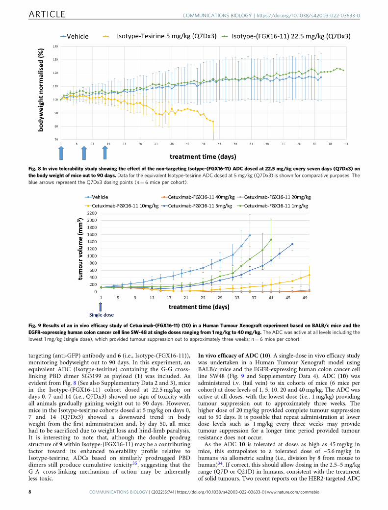
In vivo efficacy of ADC (10). A single-dose in vivo efficacy studywas undertaken in a Human Tumour Xenograft model usingBALB/c mice and the EGFR-expressing human colon cancer cellline SW48 (Fig. 9 and Supplementary Data 4). ADC (10) wasadministered i.v. (tail vein) to six cohorts of mice (6 mice percohort) at dose levels of 1, 5, 10, 20 and 40 mg/kg. The ADC wasactive at all doses, with the lowest dose (i.e., 1 mg/kg) providingtumour suppression out to approximately three weeks. Thehigher dose of 20 mg/kg provided complete tumour suppressionout to 50 days. It is possible that repeat administration at lowerdose levels such as 1 mg/kg every three weeks may providetumour suppression for a longer time period provided tumourresistance does not occur.
As the ADC 10 is tolerated at doses as high as 45 mg/kg inmice, this extrapolates to a tolerated dose of ~5.6 mg/kg inhumans via allometric scaling (i.e., division by 8 from mouse tohuman)34. If correct, this should allow dosing in the 2.5–5 mg/kgrange (Q7D or Q21D) in humans, consistent with the treatmentof solid tumours. Two recent reports on the HER2-targeted ADC
Fig. 8 In vivo tolerability study showing the effect of the non-targeting Isotype-(FGX16-11) ADC dosed at 22.5 mg/kg every seven days (Q7Dx3) onthe body weight of mice out to 90 days. Data for the equivalent Isotype-tesirine ADC dosed at 5 mg/kg (Q7Dx3) is shown for comparative purposes. Theblue arrows represent the Q7Dx3 dosing points (n= 6 mice per cohort).
Fig. 9 Results of an in vivo efficacy study of Cetuximab-(FGX16-11) (10) in a Human Tumour Xenograft experiment based on BALB/c mice and theEGFR-expressing human colon cancer cell line SW-48 at single doses ranging from 1mg/kg to 40mg/kg. The ADC was active at all levels including thelowest 1 mg/kg (single dose), which provided tumour suppression out to approximately three weeks; n= 6 mice per cohort.
ARTICLE COMMUNICATIONS BIOLOGY | https://doi.org/10.1038/s42003-022-03633-0
8 COMMUNICATIONS BIOLOGY | (2022) 5:741 | https://doi.org/10.1038/s42003-022-03633-0 | www.nature.com/commsbio

(DHES0815A), which contains a G-mono-alkylating payload36,37,provide additional context on allometric scaling. For DHES0815Adosed every three weeks (x 5) in cynomolgus monkeys, anHNSTD (Highest Non-Severely Toxic Dose) of 12 mg/kg wasobserved which translated into an MTD of 2.4 mg/kg in humansin the subsequent Phase 1 study (i.e., 12/4.75, rather than 12/2 asestimated by allometric scaling34). Although DHES0815A con-tains a G-mono-alkylating rather than a G-A cross-linkingpayload, the observed HNSTD in cynos of 12 mg/kg is similar tothe estimated 11.25 mg/kg for ADC 10, thus supporting thepotential for 10 to be dosed in the 2.5-5 mg/kg range in humans(i.e., 11.25/4.75= ~2.4 mg/kg), consistent with the treatment ofsolid tumours.
ConclusionsAn ADC payload class, the CBI-PDD G-A cross-linkers, hasbeen designed, a member of which (6, FGX8-46) has beenevaluated as a component of an EGFR-targeted ADC (10). Gel-based biophysical studies have established that 6 has a uniqueDNA sequence-selectivity with a preference for binding tosequences containing adenine and guanine bases separated bythree base pairs (e.g., 5′-AXXXC), and with a small preferencefor interstrand rather than intrastrand cross-linking. Althoughapproximately equivalent in in vitro cytotoxicity to G-G cross-linking PBD dimers such as SG3199 (1) in human tumour cellslines, 6 has been shown to produce an efficacious ADC with animproved tolerability profile. Although the reason for this is notfully understood, it may relate to the weaker covalent bondbetween the CBI unit and the adenine N3-atom compared tothe stronger bond between a PBD/PDD unit and the C2-NH2
group of a guanine. This is evident from the gel experimentsreported here which show that the CBI/adenine-N3 bond canbe cleaved at higher temperatures while leaving the PDD/gua-nine-N2 bond intact. It is conceivable that this may lead to amore facile repair of G-A cross-links in healthy cells comparedto G-G cross-links which could explain the greater tolerability.However, this is a complex situation as the different DNAsequence-selectivity of 6 compared with a PBD dimer coulddirectly affect the types and/or rates of relevant repairmechanisms in both healthy and cancer cells. Therefore, furthermechanistic studies are required to elucidate the role of thesedifferences on tolerability. Also, the transcription factor studieshave shown that 6 can down-regulate a number of key cancer-related transcription factors which may contribute to itsactivity.
The relative contributions of the C4-carbamate prodrugmoiety, the lower cross-linking efficacy, or the GA-sequence-selectivity of 6 to the high tolerability of cetuximab-(FGX16-11)are presently unclear. However, for the discontinued ADCMDX-1203 developed by Medarex which contains a similarmono-A-alkylating CBI-based unit, the C4-prodrug moiety wasconsidered to play an important role, with cleavage of theprotecting group postulated to occur in the endoplasmic reti-culum by the human enzyme Carboxylesterase 238. If itbecomes apparent that cleavage of the C4-carbamate prodrugmoiety of 10 provides a significant contribution to its efficacy,then the fact that mice have higher levels of esterases comparedto humans may be a risk factor in transitioning from pre-clinical studies to clinical trials.
Overall, the results reported here suggest that 6 could offeradvantages over currently available DNA covalent-binding ADCpayloads as it is well tolerated when incorporated into an ADC, isnot associated with late-onset toxicity, and is capable of beingused in a multi-dose schedule. As the cetuximab-(FGX16-11)ADC (10) is tolerated in mice at doses of up to 45 mg/kg, this
suggests that ADCs based on the G-A cross-linking payload 6may be better tolerated in humans than those based on G-Gcross-linking or mono-G-alkylating payloads. Therefore, thispayload type is worthy of further investigation as it could be auseful component of next-generation ADCs, particularly with aview to treating solid tumours and overcoming resistance toADCs containing other payload types.
MethodsMolecular modelling. Molecular models were constructed using the AMBER(v11) software suite with DNA sequences constructed using the nab module. Theligand was constructed using Maestro followed by conversion to mol2 files (withthe application of Gasteiger charges) using antechamber. Parmchk was then usedto determine missing parameters for the ligand. The gaff and DNA-optimisedparm99bsc0 force-fields were loaded for DNA, and xleap was used to manuallydock the ligand and create the covalent bond between the N11-C12 imine moietyof the PDD and the C2-amino of the reacting guanine, and between the alkylhalide of the CBI and the N3 of an adenine base. Covalent attachment of thePDD was based on the crystallographic structure of anthramycin39. An identicalprocess was performed with the PDD of 6 in its N11-C12 imine form and theCBI in its alkyl halide form to understand non-covalent interactions with theDNA sequence. Parameters for covalent attachment of the PDD and CBI to theDNA structure were derived from in-house studies. Each adduct was minimisedin a gradient manner by initially placing the DNA under a high force constant toenable the ligand to find its energy minimum, followed by reduction in force anda relaxation of restraints in a periodic manner. Once the full system wasminimised, production simulations were run in implicit solvent for a period of20 ns using pmemd. The generalised Born/surface area (GB/SA) implicit solventmodel was used, with monovalent electrostatic ion screening simulated withSALTCON set to 0.2 M. The dynamics integration time-step was 0.002 ps whileconstraining all bonds to hydrogen atoms using the SHAKE algorithm. Atemperature of 300 K was maintained using the Langevin thermostat, and a longrange non-bonded cut-off of 50 Å was used. Molecular dynamics simulationswere analysed using the VMD software, and images were created using theChimera (Version 1.11) software.
In vitro cytotoxicity assay. Cell-lines were established and, after 72-hour incu-bation with 1 or 6, the cell proliferation of each sample was measured using theCell Titre 96 AQueous One Solution Cell Proliferation Assay (Promega #G3582)according to the manufacturer’s protocol. The percentage inhibition was calculatedagainst the mean of the control-treated samples. IC50 values for inhibition of cellgrowth were determined with the GraphPad Prism software using non-linearregression (4 parameter logistic equation) with bottom and top constraints at 0 and100%, respectively. Cells were sourced from ATCC.
Preparation of 32P-labeled DNA fragments MS1 and WnGWn. The DNAsequence MS140 containing every possible tetranucleotide sequence, and WnGWndesigned to contain isolated guanines within long tracts of adenines and thymines,were cloned into the BamHI site of pUC18. Plasmids containing the sequences ofinterest were digested with HindIII and EcoRI, and labelled at the 3’-end of theHindIII site using α-32P[dATP] with the Klenow fragment (exo-). The labelledDNA was separated from the remainder of the plasmid on a 6% polyacrylamide gel,and then eluted and dissolved in 10 mM Tris-HCl pH 7.4 containing0.1 mM EDTA.
Cross-linking assay. 1.5 µL of the cross-linking agent (1 or 6) was diluted in10 mM Tris-HCl pH 7.4 containing 10 mM NaCl, and then added to 1.5 µL ofradiolabelled MS1 DNA at concentrations between 100 µM – 1 nM. The complexeswere incubated overnight at 37 °C, then denatured by adding 5 µL formamidecontaining 10 mM EDTA, before heating at 65 °C for 5 min and then placing onice. Control non-denatured duplex DNA (Control 1) was prepared in the sameway, but was not heated to 65 °C. Samples were run on 6.4% native polyacrylamidegels at 100 V in 1 X TBE running buffer. Gels were then fixed in 10% acetic acid forat least 10 min and transferred to Whatman 3mm paper. After drying under avacuum at 80 °C, the gels were exposed to a phosphorimager screen overnight andimaged using a Typhoon FLA 7000 phosphorimager.
DNA cleavage assay. Both the MS1 and WnGWn DNA fragments were used inthe cleavage experiments in order to determine the binding sites of 6, 7 and 8.Stocks of the compounds were diluted in Tris-HCl pH 7.4 containing 10 mMNaCl, and 1.5 µL of each diluted compound was mixed with 1.5 µL of theradiolabelled DNA and incubated at 37 °C overnight. Heating CBI-DNA com-plexes results in cleavage at the adenines to which the CBI-components areattached. Therefore, 4.5 µL of formamide containing 10 mM EDTA was added tothe ligand-DNA complexes followed by heating at 100 °C for 3 min, beforecooling on ice and loading onto an 8% denaturing polyacrylamide gel containing8 M urea. The gels were fixed in 10% acetic acid for 10 min, transferred to
COMMUNICATIONS BIOLOGY | https://doi.org/10.1038/s42003-022-03633-0 ARTICLE
COMMUNICATIONS BIOLOGY | (2022) 5:741 | https://doi.org/10.1038/s42003-022-03633-0 |www.nature.com/commsbio 9

Whatman 3 mm paper and dried under vacuum at 80 °C. They were thenexposed to a phosphorimager screen overnight and imaged using a TyphoonFLA 7000 phosphorimager.
Transcription factor plate array assay. The transcription factor plate array assaykit was obtained from Signosis, Inc (USA). 2 × 10⁶ HeLa cells were treated with100 nM of 6, and incubated for 6 h before extracting the nuclear protein andcarrying out the TF plate array assay according to the manufacturer’s instructions.In the case of each transcription factor, the RLU value obtained for the cells treatedwith 6 was deducted from the respective values obtained for the cells treated withvehicle control to obtain the differences in TF activation/inhibition (see Supple-mentary Figs. S7A and S7B for partial and full sets of TF array data, respectively).
Annexin V/7-AAD labelling, NF-kB nuclear activity, and QRT-PCR validationassays: cell culture conditions. Cells were sourced from ATCC. MCF-7 andMDA-MB-231 cells were cultured in DMEM and RPMI media, respectively(Invitrogen), both supplemented with 10% foetal calf serum and 100 units/mLpenicillin and 100 μg/mL streptomycin. Cells were aliquoted (105 cells in 500 μL ofmedia) into 24-well plates and incubated at 37 °C in a humidified 5% carbondioxide atmosphere in the presence of 6 (1 × 10⁻¹⁰ −1 × 10⁻⁷ M) for up to 72 h. Inaddition, control cultures were set-up in which no drug was added. Cells weresubsequently harvested by centrifugation, and then analysed by flow cytometryusing the methods outlined below. Experiments were performed in either duplicateor triplicate.
Measurement of in vitro apoptosis. Harvested cells were re-suspended in bindingbuffer containing 4 μL of annexin V labelled with allophycocyanin (APC) (Biole-gend). Cells were subsequently labelled with 7-AAD, and apoptosis was quantifiedusing a CytoFLEX LX flow cytometer (Beckman Coulter). At least 10,000 eventswere acquired, and data were subsequently analysed using the FlowJo software.LD50 values (the concentration of 6 required to kill 50% of cells) were interpolatedfrom the dose-response curves.
Nuclear NF-κB subunit detection by ELISA. MCF-7 and MDA-MB-231 cellswere treated with 6 (0, 1 nM, 2.5 nM, 5 nM) for 4 h. Subsequently, nuclear proteinswere isolated using a nuclear extract kit (Active Motif). Extracts were then assayedfor NF-κB subunit DNA binding with a TransAM NF-κB family kit according tothe manufacturer’s protocol. The consensus oligonucleotide used for NF-κBbinding was 5′-GGGACTTTCC-3′, and specific NF-κB proteins bound to theoligonucleotide were identified using primary antibodies for NF-κB subunits p50,p52, p65, c-Rel and RelB, followed by detection with an HRP-conjugated secondaryantibody. The absorbance at 450 nm (A450) was read on a microtitre plate reader(Bio-Rad), and readings were compared with the untreated controls.
Real-time reverse transcription-PCR. MDA-MB-231 and MCF-7 cells weretreated with 6 (0, 1 nM, 2.5 nM, 5 nM) for 4 h. Aliquots of 1 × 10⁶ cells were thenharvested by centrifugation prior to RNA extraction using RNeasy mini-spincolumns according to the manufacturer’s instructions (Qiagen). RNA (1 μg) wasthen reverse transcribed in 20 μL reactions containing 10× Buffer II, 5 mmol/LMgCl2, 0.5 μmol/L deoxynucleotide triphosphates, 2.5 units reverse transcriptase, 1unit of RNase inhibitor, and 2.5 μmol/L random hexamers. cDNA (2 μL) was usedin each RT-PCR reaction. PowerTrack SYBR Green Mastermix (ThermoFisherScientific) was used to quantify the amount of transcript present in each sampleusing primer pairs for the NF-κB target genes: CFLAR, BIRC5 and IL-1β. Allprimers were purchased from Eurogentec Ltd (Southampton, UK). The amount oftarget transcript was assessed using real-time RT-PCR using a QuantStudio 7instrument (ThermoFisher Scientific), and was normalised to the amount of RPS14transcript in all samples as an internal house-keeping control. The results of thereal-time RT-PCR (Fig. 6d) are presented as normalised target gene values (e.g., theratio between CFLAR and RPS14 transcripts calculated from the crossing points ofeach gene). All experiments were performed in triplicate. cDNA was amplifiedusing the following primers:
CFLAR: 5′-gtggagacccacctgctca-3′ (forward) and 5′-ggacacatcagatttatccaaatcc-3′(reverse);
BIRC5: 5′-ttagcagaaaatgcactccag-3′ (forward) and 5′-ctggttttaaggatggccttt-3′(reverse);
IL-1β: 5′-tggcagaaagggaacagaaa-3′ (forward) and 5′-acttcttgccccctttgaat-3′(reverse);
RPS14: 5′-ggcagaccgagatgaactct-3′ (forward) and 5′-ccaggtccaggggtcttggt-3′(reverse).
Statistics and reproducibility. All statistical analyses were performed usingGraphpad Prism 9 software (Graphpad Software, Inc.). Drug sensitivity to 6 wasevaluated using non-linear regression and line of best fit dose-response curves.Comparison of relative gene transcription and NF-kB subunit nuclear activityfollowing exposure to 6 for 4 h were assessed using two-way ANOVA with Dun-nett’s correction for multiple comparisons. The Flow Cytometry Gating Strategyused is provided in Supplemental Methods S16 and Supplementary Fig. S17.
Conjugation and ADC purification. The interchain disulfides of commercially-available cetuximab formulated at pH 7-8 in 2 mM EDTA were partially reducedwith TCEP for 90 – 180 min. The extent of reduction was controlled to achieve aspecified drug-to-antibody ratio (DAR) of ~2. The reduced antibody was thendiluted with PBS/2 mM EDTA to 2 mg/mL. FGX16-11 (9) was dissolved in DMA/DMSO, and conjugation to the antibody was achieved by addition of an excess of 9to a 1:1 mixture of the reduced antibody and propylene glycol, at a final proteinconcentration of 1 mg/mL. The mixture was allowed to react for 1 h to form theantibody-drug conjugate 10, and the reaction was then quenched by adding anexcess of N-acetyl cysteine to remove unreacted 9. The mixture was further dilutedwith 1:1 PBS/3% cyclodextrin and added to a proprietary purifying resin. Theresin-bound ADC was washed with PBS/3% cyclodextrin to remove excess small-molecule impurities, and then released from the resin. The ADC was finally for-mulated through G25 desalting into PBS/3% cyclodextrin, and filtered prior toaliquoting and storing at −80 °C. HIC and SEC chromatograms for the final ADCproduct are provided in Supplementary Figs. S14 and S15.
In vivo tolerability studies. A total of 56 male CD1 mice were used for thetolerability study. All animals were allowed free access to a standard certifiedcommercial diet and sanitised water during the study. The holding room wasmaintained under standard conditions: 20–24 °C, 40–70% humidity and a 12 hlight/dark cycle. All protocols used in this study were approved by the CRO’sAnimal Welfare and Ethical Review Committee, and all procedures were carriedout under the guidelines of the Animal (Scientific Procedures) Act 1986. Mice wererandomly assigned to a treatment group and dosed intravenously through the tailvein with the free payload (6) or ADC (10). The weight of the mice was monitoredfor a period of 28 days, and the Maximum Tolerated Dose (MTD) was deemed tohave been reached when >15% weight loss of a mouse was observed. The minimumdose at which >15% weight loss was observed was considered to be the MTD.
Delayed toxicity study. A total of 54 male ICR-CD1 mice aged 5–8 weeks wereused for the study. These were purchased from Envigo and allowed to accli-matise for 7 days prior to dosing. Animals were housed in IVC cages (up to 5 percage) with individual mice identified by tail marks. All animals were allowed freeaccess to a standard certified commercial diet and sanitised water during thestudy. The holding room was maintained as follows: room temperature at20–24 °C, humidity at 30–70% and a 12 h light/dark cycle. All protocols used inthis study were approved by the CRO’s Animal Welfare and Ethical ReviewCommittee, and all procedures were carried out under the guidelines of theAnimal (Scientific Procedures) Act 1986. The mice were randomly assigned totreatment groups, and animals received a dose of each compound either Q7D orQ21D (data not shown) for 3 cycles, with bodyweight and health observationsmade daily for up to 90 days. The ADCs were prepared for injection in phos-phate buffered saline (PBS) pH= 7.4 with 3% w/w cyclodextrin. Dosing solu-tions were prepared from stock solutions immediately prior to dosing, and anyremaining dosing solutions were discarded.
Efficacy study. Cells were sourced fromATCC. A total of 36male BALB/c nudemiceaged 6–8 weeks was used for the efficacy study. SW48 cells (1 × 10⁷, 1:1 with Matrigel)were implanted into the flank of a mouse using a 23-gauge needle. Once tumoursreached 150mm3, the mice were randomly assigned to the relevant treatment groupsand then dosed intravenously through the tail vein with ADC 10. Tumour volume wasmeasured daily using standard methodology based on digital callipers. The length andwidth of the tumour was measured and the volume calculated using the followingformula: volume= (length × width2)/2, where length represents the largest tumourdiameter and width represents the perpendicular tumour diameter.
Reporting summary. Further information on research design is available in the NatureResearch Reporting Summary linked to this article.
Data availabilityKey data supporting the findings of this study are available within this article, thecorresponding Supplementary Information document, and the Supplementary Data 1 to4 files.
Received: 14 July 2021; Accepted: 24 June 2022;
References1. Ponziani, S. et al. Antibody-Drug Conjugates: The New Frontier of
Chemotherapy. Int. J. Mol. Sci. 21, 5510 (2020).2. Joubert, N., Beck, A., Dumontet, C. & Denevault-Sabourin, C. Antibody–Drug
Conjugates: The Last Decade. Pharmaceuticals 13, https://doi.org/10.3390/ph13090245 (2020).
ARTICLE COMMUNICATIONS BIOLOGY | https://doi.org/10.1038/s42003-022-03633-0
10 COMMUNICATIONS BIOLOGY | (2022) 5:741 | https://doi.org/10.1038/s42003-022-03633-0 | www.nature.com/commsbio

3. do Pazo, C., Nawaz, K. & Webster, R. M. The oncology market for antibody-drug conjugates. Nat. Rev. Drug Discov., https://doi.org/10.1038/d41573-021-00054-2 (2021).
4. Jukes, Z., Morais, G. R., Loadman, P. M. & Pors, K. How can the potential ofthe duocarmycins be unlocked for cancer therapy? Drug Dis. Today, https://doi.org/10.1016/j.drudis.2020.11.020 (2020).
5. Daver, N. G. et al. A phase I/II study of IMGN632, a novel CD123-targetingantibody-drug conjugate, in patients with relapsed/refractory acute myeloidleukemia, blastic plasmacytoid dendritic cell neoplasm, and other CD123-positive hematologic malignancies. J. Clin. Oncol. 38, TPS7563 (2020).
6. FDA grants accelerated approval to loncastuximab tesirine-lpyl for large B-celllymphoma, https://www.fda.gov/drugs/fda-grants-accelerated-approval-loncastuximab-tesirine-lpyl-large-b-cell-lymphoma (2021).
7. Mullard, A. Cancer stem cell candidate Rova-T discontinued. Nat. Rev. DrugDiscov. 18, 814 (2019).
8. Saber, H., Simpson, N., Ricks, T. K. & Leighton, J. K. An FDA oncologyanalysis of toxicities associated with PBD-containing antibody-drugconjugates. Regulatory Toxicol. Pharmacol. 107, 104429 (2019).
9. Procopiou, G. & O’Donnell, C. J. In Cytotoxic Payloads for Antibody–DrugConjugates 209–240 (The Royal Society of Chemistry, 2019).
10. Schwartz, G. H. et al. A phase I study of bizelesin, a highly potent and selectiveDNA-interactive agent, in patients with advanced solid malignancies. Ann.Oncol. 14, 775–782 (2003).
11. Zhou, Q. et al. Design and Synthesis of a Novel DNA−DNA InterstrandAdenine−Guanine Cross-Linking Agent. J. Am. Chem. Soc. 123, 4865–4866(2001).
12. Tercel, M. et al. Unsymmetrical DNA Cross-Linking Agents: Combination ofthe CBI and PBD Pharmacophores. J. Med. Chem. 46, 2132–2151 (2003).
13. Purnell, B. et al. DNA interstrand crosslinking agents: Synthesis, DNAinteractions, and cytotoxicity of dimeric achiral seco-amino-CBI andconjugates of achiral seco-amino-CBI with pyrrolobenzodiazepine (PBD).Bioorg. Med. Chem. Lett. 16, 5677–5681 (2006).
14. Jackson, P. J. M., Rahman, K. M. & Thurston, D. E. The use of moleculardynamics simulations to evaluate the DNA sequence-selectivity of G–A cross-linking PBD–duocarmycin dimers. Bioorg. Med. Chem. Lett. 27, 102–108(2017).
15. Pillow, T. H. & Tercel, M. In Cytotoxic Payloads for Antibody–DrugConjugates 241–258 (The Royal Society of Chemistry, 2019).
16. Veillard, N. et al. Abstract 736: Pyridinobenzodiazepines (PDDs): A new classof sequence-selective DNA mono-alkylating ADC payloads with lowhydrophobicity. Cancer Res. 78, 736 (2018).
17. Veillard, N., Cascio, F., Jackson, P. J. M. & Thurston, D. E. In CytotoxicPayloads for Antibody–Drug Conjugates 349–363 (The Royal Society ofChemistry, 2019).
18. Mantaj, J., Jackson, P. J., Rahman, K. M. & Thurston, D. E. FromAnthramycin to Pyrrolobenzodiazepine (PBD)-Containing Antibody-DrugConjugates (ADCs). Angew. Chem. Int Ed. Engl. 56, 462–488 (2017).
19. Markandeya, N., Shankaraiah, N., Reddy, C. S., Santos, L. S. & Kamal, A.Asymmetric syntheses of piperidino-benzodiazepines through ‘cation-pool’host/guest supramolecular approach and their DNA-binding studies.Tetrahedron.: Asymmetry 21, 2625–2630 (2010).
20. Wang, Y., Li, L., Tian, Z., Jiang, W. & Larrick, J. W. Synthesis and antitumoractivity of CBI-bearing ester and carbamate prodrugs of CC-1065 analogue.Biorg. Med. Chem. 14, 7854–7861 (2006).
21. Hyung, S.-J., Leipold, D. D., Lee, D. W., Kaur, S. & Saad, O. M. MultiplexedQuantitative Analysis of Antibody–Drug Conjugates with Labile CBI-DimerPayloads In Vivo Using Immunoaffinity LC-MS/MS. Anal. Chem. 94,1158–1168 (2022).
22. Hartley, J. A., Berardini, M. D. & Souhami, R. L. An agarose gel method forthe determination of DNA interstrand crosslinking applicable to themeasurement of the rate of total and “second-arm” crosslink reactions. Anal.Biochem. 193, 131–134 (1991).
23. Reynolds, V. L., Molineux, I. J., Kaplan, D. J., Swenson, D. H. & Hurley, L. H.Reaction of the antitumor antibiotic CC-1065 with DNA. Location of the siteof thermally induced strand breakage and analysis of DNA sequencespecificity. Biochemistry 24, 6228–6237 (1985).
24. Wang, S. et al. A Novel Depurination Methodology to Assess DNAAlkylation of Chloro-Bis-Seco-Cyclopropylbenzoindoles Allowed forComparison of Minor-Groove Reactivity. Drug Metab. Disposition. 47,547–555 (2019).
25. Rahman, K. M., Thompson, A. S., James, C. H., Narayanaswamy, M. &Thurston, D. E. The Pyrrolobenzodiazepine Dimer SJG-136 Forms Sequence-Dependent Intrastrand DNA Cross-Links and Monoalkylated Adducts inAddition to Interstrand Cross-Links. J. Am. Chem. Soc. 131, 13756–13766(2009).
26. Espinoza-Sánchez, N. A., Enciso, J., Pelayo, R. & Fuentes-Pananá, E. M. AnNFκB-dependent mechanism of tumor cell plasticity and lateral transmissionof aggressive features. Oncotarget 9, 26679–26700 (2018).
27. Poma, P., Labbozzetta, M., D’Alessandro, N. & Notarbartolo, M. NF-κB Is aPotential Molecular Drug Target in Triple-Negative Breast Cancers. OMICS21, 225–231 (2017).
28. Zhang, D. et al. Immolation of p-Aminobenzyl Ether Linker and PayloadPotency and Stability Determine the Cell-Killing Activity of Antibody–DrugConjugates with Phenol-Containing Payloads. Bioconjugate Chem. 29,267–274 (2018).
29. Yao, H., Jiang, F., Lu, A. & Zhang, G. Methods to Design and SynthesizeAntibody-Drug Conjugates (ADCs). Int. J. Mol. Sci. 17, 194 (2016).
30. Miller, M. L. et al. A DNA-Interacting Payload Designed to Eliminate Cross-Linking Improves the Therapeutic Index of Antibody–Drug Conjugates(ADCs). Mol. Cancer Ther. 17, 650–660 (2018).
31. Hinrichs, M. J. M. et al. Fractionated Dosing Improves Preclinical TherapeuticIndex of Pyrrolobenzodiazepine-Containing Antibody Drug Conjugates. Clin.Cancer Res. 23, 5858–5868 (2017).
32. Lewis Phillips, G. D. et al. Abstract 1207: Preclinical development of 2ndgeneration HER2-directed antibody-drug conjugates. Cancer Res. 76, 1207(2016).
33. Gail Lewis Phillips, G. L. et al. In AACR 107th Annual Meeting 2016 (NewOrleans, Louisiana, USA, 2016, April 16–20).
34. Nair, A. B. & Jacob, S. A simple practice guide for dose conversion betweenanimals and human. J. Basic Clin. Pharm. 7, 27–31 (2016).
35. Min, B. et al. cIRCR201-dPBD, a Novel Pyrrolobenzodiazepine Dimer-Containing Site-Specific Antibody–Drug Conjugate Targeting c-MetOverexpression Tumors. ACS Omega. 5, 25798–25809 (2020).
36. Phillips, G. L. et al. Abstract P2-13-33: Preclinical development ofDHES0815A: A HER2-directed antibody-drug conjugate comprised of areduced potency PBD dimer linked to a domain I binding HER2 antibody.Cancer Res. 82, P2-13-33 (2022).
37. Krop, I. et al. Abstract P2-13-25: A phase I dose-escalation study ofDHES0815A, a HER2-targeting antibody-drug conjugate with a DNAmonoalkylator payload, in patients with HER2-positive breast cancer. CancerRes. 82, P2-13-25 (2022).
38. Derwin, D. W. et al. Abstract LB-252: Enzymology of the mechanismof action for MDX-1203 antibody drug conjugate. Cancer Res. 72, LB-252 (2012).
39. Mostad, A., Rømming, C., Storm, B., Deutsch, A. & Matsuno, T. Structure ofthe DNA Complexing Agent Anthramycin. Acta Chem. Scand. 32b, 639–645(1978).
40. Lavesa, M. & Fox, K. R. Preferred Binding Sites for [N-MeCys3,N-MeCys7]TANDEM Determined Using a Universal Footprinting Substrate. AnalBiochem. 293, 246–50. (2001).
AcknowledgementsChris Keightley (Femtogenix Ltd), and Shaun Kirkpatrick, Kurt Gehlsen and ChadSouvignier (Research Corporation Technologies Inc) are acknowledged for their helpfuldiscussions.
Author contributionsP.J.M.J., K.M.R. and D.E.T. conceived the project, designed the overall research pro-gramme, and supervised various components. G.P., P.J.M.J., D.d.M., J.L.A., C.P., K.M.R.and K.R.F. performed the laboratory experiments. G.P. led the chemical synthesisresearch, P.J.M.J the molecular modelling, D.d.M and K.R.F. the gel-based assays, andC.P and K.M.R. the transcription factor studies. G.P., P.J.M.J. and D.E.T. wrote themanuscript, but all authors discussed and contributed to it.
Competing interestsAll authors except C.P. are either paid employees/consultants (G.P., P.J.M.J., D.E.T.) orrecipients of grants funded by Femtogenix Ltd (D.d.M, K.R.F.). G.P., P.J.M.J., K.M.R andD.E.T. hold shares and/or share options in the company. C.P declares no competinginterests.
Additional informationSupplementary information The online version contains supplementary materialavailable at https://doi.org/10.1038/s42003-022-03633-0.
Correspondence and requests for materials should be addressed to David E. Thurston.
Peer review information Communications Biology thanks the anonymous reviewers fortheir contribution to the peer review of this work. Primary Handling Editors: Eve Rogersand Christina Karlsson Rosenthal.
Reprints and permission information is available at http://www.nature.com/reprints
Publisher’s note Springer Nature remains neutral with regard to jurisdictional claims inpublished maps and institutional affiliations.
COMMUNICATIONS BIOLOGY | https://doi.org/10.1038/s42003-022-03633-0 ARTICLE
COMMUNICATIONS BIOLOGY | (2022) 5:741 | https://doi.org/10.1038/s42003-022-03633-0 |www.nature.com/commsbio 11

Open Access This article is licensed under a Creative CommonsAttribution 4.0 International License, which permits use, sharing,
adaptation, distribution and reproduction in any medium or format, as long as you giveappropriate credit to the original author(s) and the source, provide a link to the CreativeCommons license, and indicate if changes were made. The images or other third partymaterial in this article are included in the article’s Creative Commons license, unlessindicated otherwise in a credit line to the material. If material is not included in thearticle’s Creative Commons license and your intended use is not permitted by statutoryregulation or exceeds the permitted use, you will need to obtain permission directly fromthe copyright holder. To view a copy of this license, visit http://creativecommons.org/licenses/by/4.0/.
© The Author(s) 2022
ARTICLE COMMUNICATIONS BIOLOGY | https://doi.org/10.1038/s42003-022-03633-0
12 COMMUNICATIONS BIOLOGY | (2022) 5:741 | https://doi.org/10.1038/s42003-022-03633-0 | www.nature.com/commsbio
Copyright © 2022 FDOKUMEN