Disruption of Frontal Theta Coherence by Δ9-Tetrahydrocannabinol is Associated with Positive...
44
NPP-10-0739 Morrison PD. Revised 15/10/10 1 Title: Disruption of frontal theta coherence by Δ 9 -tetrahydrocannabinol is associated with positive psychotic symptoms. Running Title: THC and theta Paul D Morrison 1 , Judith Nottage 1, James M Stone 1,2 , Sagnik Bhattacharyya 1 , Nigel Tunstall 1 , Rudolf Brenneisen 4 , David Holt 5 , Daniel Wilson 1,3 , Alex Sumich 1,3 , Philip McGuire 1 , Robin M Murray 1 , Shitij Kapur 1 and Dominic H ffytche 1 . (1) King’s College London, The Biomedical Research Centre, Institute of Psychiatry, Denmark Hill, London SE5 8AF ([email protected] ; +44 (0)2032283066), ([email protected]; +44 (0)20322830955), ([email protected] ; +44 (0)2083915947), ([email protected];2078480424), ([email protected]; +44 (0) 207 8480355), ([email protected]; +44 (0)2032283064), ([email protected]; +44 (0)2078480100) (2) Department of Experimental Medicine, Imperial College London, W12 0NN ([email protected]: +44 (0) 0 7594 7087) (3) School of Social Sciences, Nottingham Trent University, NG1 4B ([email protected]; +44 (0)115848683), ([email protected]; +44 (0)115848683) (4) University of Bern, Dept. Clinical Research, Murtenstr. 35, CH-3010 Bern, Switzerland ([email protected] ++41-31-63 287 14) (5) The Analytical Unit, St George’s, University of London, SW17 ([email protected]; +44 (0)20 8767 9686) Paul Morrison MRCPsych, MSc; Judith Nottage BSc; James Stone MRCPsych, PhD; Nigel Tunstall MRCPsych; Rudolf Brenneisen PhD; David Holt PhD; Alex Sumich PhD; Daniel Wilson MSc; Phillip Mcguire FRPsych PhD; Robin M Murray FRS; Shitij Kapur FMedSci, PhD; Dominic ffytche MRCP, PhD. Abstract: 219 words Correspondence: Dr PD Morrison, Psychosis Studies, The Institute of Psychiatry, De-Crespigny Park, Denmark Hill, London, SE5 8AF [email protected] Tel: 44 (0)208 391 5947
-
Upload
independent -
Category
Documents
-
view
0 -
download
0
Transcript of Disruption of Frontal Theta Coherence by Δ9-Tetrahydrocannabinol is Associated with Positive...

NPP-10-0739 Morrison PD. Revised 15/10/10
1
Title: Disruption of frontal theta coherence by Δ9-tetrahydrocannabinol is associated with positive psychotic symptoms. Running Title: THC and theta Paul D Morrison1, Judith Nottage1, James M Stone1,2, Sagnik Bhattacharyya1, Nigel Tunstall1, Rudolf Brenneisen4, David Holt5, Daniel Wilson1,3, Alex Sumich1,3, Philip McGuire1, Robin M Murray1, Shitij Kapur1 and Dominic H ffytche1. (1) King’s College London, The Biomedical Research Centre, Institute of Psychiatry, Denmark Hill, London SE5 8AF ([email protected]; +44 (0)2032283066), ([email protected]; +44 (0)20322830955), ([email protected]; +44 (0)2083915947), ([email protected];2078480424), ([email protected]; +44 (0) 207 8480355), ([email protected]; +44 (0)2032283064), ([email protected]; +44 (0)2078480100) (2) Department of Experimental Medicine, Imperial College London, W12 0NN ([email protected]: +44 (0)0 7594 7087) (3) School of Social Sciences, Nottingham Trent University, NG1 4B ([email protected]; +44 (0)115848683), ([email protected]; +44 (0)115848683) (4) University of Bern, Dept. Clinical Research, Murtenstr. 35, CH-3010 Bern, Switzerland ([email protected] ++41-31-63 287 14) (5) The Analytical Unit, St George’s, University of London, SW17 ([email protected]; +44 (0)20 8767 9686)
Paul Morrison MRCPsych, MSc; Judith Nottage BSc; James Stone MRCPsych, PhD; Nigel Tunstall MRCPsych; Rudolf Brenneisen PhD; David Holt PhD; Alex Sumich PhD; Daniel Wilson MSc; Phillip Mcguire FRPsych PhD; Robin M Murray FRS; Shitij Kapur FMedSci, PhD; Dominic ffytche MRCP, PhD. Abstract: 219 words Correspondence: Dr PD Morrison, Psychosis Studies, The Institute of Psychiatry, De-Crespigny Park, Denmark Hill, London, SE5 8AF [email protected] Tel: 44 (0)208 391 5947

NPP-10-0739 Morrison PD. Revised 15/10/10
2
Abstract The main ingredient in cannabis, Δ9-tetrahydrocannabinol (THC), can elicit acute psychotic reactions
in healthy individuals and precipitate relapse in schizophrenic patients. However, the neural mechanism
of this is unknown. We tested the hypothesis that THC-psychopatholgy is related to changes in EEG
power or inter-regional coherence. In a within-subjects design, participants (n=16) were given
intravenous (IV) THC (1.25mg) or placebo under double-blind conditions during EEG recordings.
Using fast-Fourier transform, EEG data was analysed for power and coherence in the delta (1-3.5 Hz),
theta (3.5-7 Hz), alpha (8-13 Hz), beta (14-25 Hz), low-gamma (30-40Hz) and high-gamma (60-70Hz)
bands during engagement in the n-back test of working-memory (WM). Compared to placebo, THC
evoked positive and negative psychotic symptoms, as measured by the PANSS scale (p<0.001) and
slowed WM performance (p<0.05). Under THC, theta power was specifically reduced, (p<0.001)
regardless of WM load, however, the reduction showed no relationship with psychotic symptoms or
WM impairment. Coherence between bi-frontal electrodes in the theta band was also reduced by THC
(p<0.05) and these reductions correlated with the change in positive psychotic symptoms (rho=0.79,
p<0.001). Bi-frontal specificity was suggested by the absence of a relationship between psychotic
symptoms and fronto-parietal coherence. The results reveal that the pro-psychotic effects of THC might
be related to impaired network dynamics with impaired communication between the right and left
frontal lobes.
Keywords
Δ9-tetrahydrocannabinol; theta; psychosis; EEG

NPP-10-0739 Morrison PD. Revised 15/10/10
3
INTRODUCTION
There has been a renewed interest in the cannabis-model of acute psychosis, driven in part by
epidemiological studies which have demonstrated that the use of cannabis is a risk factor for the
development of schizophrenia (Arseneault et al, 2004; Moore et al, 2007). The major psychoactive
effects of cannabis stem from the action of Δ9-tetrahydrocannabinol (THC) at central CB1 receptors
(CB1Rs) (Howlett et al, 2002; Mechoulam et al, 1970). Recent laboratory studies have confirmed
earlier reports which used plant-derived preparations by showing that pure intravenous (IV) THC
elicits acute schizophrenia-like symptoms and cognitive impairments in healthy volunteers (D'Souza et
al, 2004; Morrison et al, 2009). Similarly, schizophrenic patients can experience a sudden and transient
relapse of psychotic symptoms following IV THC despite having been previously well on dopamine-
based anti-psychotic medication (D'Souza et al, 2005).
At present the neurophysiological mechanism responsible for the pro-psychotic properties of THC is
unknown. The CB1 receptor is found at high density in the prefrontal and association cortices, the
anterior, mediodorsal, and intralaminar thalamic nuclei, the hippocampal complex, amygdala,
entorhinal cortex, basal ganglia, substantia-nigra pars-reticulata, and cerebellum (Eggan and Lewis,
2007; Glass et al, 1997; Herkenham et al, 1991). Typically, CB1 receptors are localised at the pre-
synaptic terminals of glutamate and GABA-ergic neurons (Bodor et al, 2005; Eggan et al, 2010; Hill et
al, 2007). Their endogenous transmitters, (endocannabinoids, eCBs) are released from dendritic spines
and function as retrograde signalling molecules (meaning that neurochemical 'information' is passed
from dendrite to axon terminal) (Piomelli, 2003). At the synaptic level, the eCBs inhibit GABA and
glutamate release from nerve endings, over short and sustained durations, whereas at the psychological

NPP-10-0739 Morrison PD. Revised 15/10/10
4
level eCBs are vital for learning and memory (Chevaleyre et al, 2006; Freund et al, 2003). Exogenous
cannabinoids such as THC probably do not capture the subtleties of eCB signalling (which involves
tightly regulated local release and re-uptake) and hence are associated with disruption of learning and
memory performance, and presumably their underlying neural correlates (Murray et al, 2007;
Ranganathan and D'Souza, 2006).
In the last two decades it has become clear that neural oscillations are intrinsic to higher CNS
functions (Buzsaki and Draguhn, 2004; Uhlhaas et al, 2009; Varela et al, 2001). Oscillations arise
from the synchronised firing of large numbers of neurons. In turn, the precise timing of spikes from
individual neurons is constrained by ongoing rhythms within the network. Oscillations are believed to
be essential for rapid, dynamic integration within and between brain regions. Synchronised gamma (30-
200Hz) rhythms “bind” local assemblies, whereas lower frequencies (theta, alpha and beta) sub-serve
long-distance communication between brain areas (Kopell et al, 2000; Rodriguez et al, 1999; von Stein
et al, 2000). Studies in animals have begun to unravel the effects of THC on such oscillations. Robbe
and colleagues showed that THC decreased the power of theta, gamma and ripple oscillations in the rat
hippocampus, effects which could be blocked by CB1 antagonists (Robbe et al, 2006). Theta and
gamma rhythms are believed to be essential for working memory (WM) and the encoding of episodic
memories, whereas ripples are involved in the transfer of memory to cortical stores. In agreement,
Hajos and co-workers found that CB1 agonists disrupted theta and gamma oscillations within the septo-
hippocampal system in rats, and disrupted auditory-gating; effects which were inhibited by CB1
antagonists (Hajos et al, 2008). In humans there have been several recent reports of the effects of
cannabis on neuronal oscillations, recorded using electroencephalography (EEG). A study by IIan and
colleagues analysed EEG power in 10 subjects, during the performance of cognitive tasks (Ilan et al,

NPP-10-0739 Morrison PD. Revised 15/10/10
5
2004). Working and episodic memory performance was poorer following inhaled marijuana (3.5%
THC) and the principal EEG findings were decreased global theta power and reduced alpha band
reactivity. Similarly in a study of inhaled marijuana (0, 29, 49 & 69mg) in 16 participants, Bocker and
co-workers observed a dose-dependent decrease of resting theta and beta power. Furthermore, they
found the greater the decrease in theta power during a WM task, the greater the slowing of response
time (Bocker et al, 2010).
Our primary interest in this study was to characterise EEG changes linked to the pro-psychotic
properties of THC. Neither of the previous EEG studies of THC had investigated psychotic symptoms
and one possibility was that psychotic symptoms would be related to the reductions in theta power
demonstrated in these studies. However, more recent work has highlighted the importance of brain
connectivity in the generation of psychotic symptoms. In particular, reductions in EEG coherence have
been linked to specific positive symptoms (auditory hallucinations; Ford et al, 2002). EEG coherence is
a measure of the correlation of activity in different brain regions. A high coherence indicates that EEG
activity within a specific frequency band in one brain region predicts EEG activity in same frequency
band in another and that the temporal relationship (phase) of the activity at the two sites is relatively
constant. The implication of such amplitude and phase relationships is of cross-talk and shared function
between the two regions. The link between positive symptoms and reductions in EEG coherence found
in schizophrenia suggested a working hypothesis that the pro-psychotic properties of THC relates to a
reduction in EEG coherence, specifically in the theta band given the animal and human data outlined
above.

NPP-10-0739 Morrison PD. Revised 15/10/10
6
METHODS
The study was approved by the Joint Institute of Psychiatry and Maudsley Hospital Ethics Committee.
All subjects provided written informed consent. Safety protocols have previously been described
(Morrison et al, 2009).
Design
A randomised, double-blind placebo controlled crossover study in healthy volunteers of the effects of
IV THC (1.25mg) on working-memory performance, psychopathology and concurrent EEG activity.
Participants
Twenty healthy participants were recruited via the King’s College email lists. Inclusion criteria were:
age between 21-50 years, previous cannabis use ≥ 1; a score of < 15 on the General Health
Questionnaire (GHQ-12, Goldberg et al, 1970). Exclusion criteria included: current pregnancy; a
history of mental illness; drug or alcohol dependence (excluding nicotine); current or past severe
medical disorders, or a history of major mental illness in a first-degree family member. Previous
alcohol and drug use was recorded and a urine drug screen was carried out. Participants were asked to
avoid alcohol and drugs for 24 h before, and to abstain from driving for 24 h after experiments.
Sessions were performed at least two weeks apart and started between 0900 and 1400 h. Placebo and
THC were administered under double-blind conditions, in a randomised counterbalanced order.
Subjects received remuneration for their participation.

NPP-10-0739 Morrison PD. Revised 15/10/10
7
Pharmaceuticals
Synthetic Δ9-tetrahydrocannabinol was supplied by THC Pharm GmbH (Frankfurt am Main, Germany)
and prepared as 1mg/ml vials for intravenous injection, by Bichsel Laboratories (Interlaken,
Switzerland) as previously described (Naef et al, 2004). After dilution in normal saline, preparations
for injection contained 1.25 % (v/v) ethanol absolute. Sterile cannulae were inserted into veins in the
antecubital fossa of both arms: one for administration of pharmaceuticals and one for plasma sampling.
THC was administered in 1ml/min pulses over a period of 5 min (total dose = 1.25mg). Blood samples
were taken at baseline and at 1, 5, 15, 60, and 120 min after dosing, for analysis of [THC] as previously
described (Morrison 2009). The plasma concentration of THC following IV delivery follows a similar
time-course to that observed following inhalation (Naef et al, 2004). The dose, (1.25mg) was selected
on the basis of previous studies, and roughly corresponds to one standard ‘joint’ (D'Souza et al, 2004;
Morrison et al, 2009; Bhattacharyya et al, 2010).
Psychopathological and cognitive measures
Psychotic symptoms were rated using the positive and negative syndrome scale (PANSS, (Kay et al,
1987). A completely independent PANSS-trained senior psychiatrist rated 3 x 10-minute periods - at
baseline, at 30-minutes post-pharmaceutical and finally at 90-minutes post-pharmaceutical. Apart from
these 3 periods, the psychiatrist and participant were kept separate to minimise potential bias. The
PANSS consists of a positive sub-scale (7 items; delusions, conceptual disorganisation, hallucinations,
hyperactivity, grandiosity, suspiciousness/persecution and hostility), a negative sub-scale (7 items;

NPP-10-0739 Morrison PD. Revised 15/10/10
8
blunted affect, emotional withdrawal, poor rapport, passive/apathetic social withdrawal, difficulty in
abstract thinking, lack of spontaneity and flow of conversation, stereotyped thinking) and a general
psychopathological sub-scale (14 items). Items are rated from 1-7 (absent-severe), thus the range of
scores on the positive subscale is 7-49. It is recognised that there is wide inter-individual variation in
PANSS scores following IV THC (D'Souza et al, 2004; Morrison et al, 2009). Previously we found
that investigator-rated (PANSS) and participant-rated (CAPE, Community Assessment of Psychic
Experiences) measures of THC-elicited positive symptoms were in agreement (rho=0.62, p<0.001), and
that both measures of positive symptoms were distinct from anxiety (Morrison et al, 2009). Considered
as a group however, positive symptoms following THC are modest and short-lived. Overall, in earlier
studies approximately 35-50% of healthy participants showed changes of >= 3-4 points on the PANSS
positive subscale under THC conditions (D'Souza et al, 2004; Morrison et al, 2009).
Immediately following the psychiatric assessment at 30-mins post-pharmaceutical, participants were
administered a standard computerised version of the n-back task. The n-back procedure has been used
extensively to measure human working memory performance (Owen et al, 2005). Participants were
required to monitor a series of letters and report when the current letter matched the letter n integers
back, where n=1 (1-back) or n=2 (2-back), the latter being more difficult. The task requires continuous
updating of information stores. In contrast, in the 0-back condition (which does not require
manipulation of material in WM), participants responded to the appearance of a pre-specified letter.
Overall, the task consisted of alternating 30-second blocks of 0-back, 1-back and 2-back conditions,
and lasted 6 minutes in total. Within blocks, letters were displayed every 2 seconds for 1 second.
Written instructions were read out and participants were given a practice run to demonstrate their
understanding of the rules. Subjects were seated ~66cm from a CRT monitor and instructed to report

NPP-10-0739 Morrison PD. Revised 15/10/10
9
correct answers as rapidly as possible by pressing a joy-pad button with their R-index finger. Accuracy
of responses and reaction-times were measured and stored digitally.
Electroencephalography
All data recording and signal processing was performed in Neuroscan 4.3. Electroencephalographic
(EEG) activity was recorded from 63 electrode sites using a Quik-Cap system (Compumedics inc.),
with a linked mastoid reference and ground at AFz. All impedances were maintained below 10 kΩ.
Additional electrodes were placed at the outer canthi to measure horizontal electrooculographic activity
(EOG; monopolar with linked mastoid reference). Vertical EOG was measured using a bipolar
recording with electrodes above and below the left eye. The EEG was sampled at 2000 Hz and
corrected for eye-blinks using a regression approach. The corrected EEG was epoched, using a 10%
Hanning window, into 2048ms segments (-24 to 2024ms with respect to each n-back letter stimulus).
Epochs were baseline corrected. For each of the three n-back conditions, average power within the
frequency bands delta (1-3.5 Hz), theta (3.5-7 Hz), alpha (8-13 Hz), beta (14-25 Hz), low-gamma (30-
40Hz), and high-gamma (60-70Hz) bands was calculated using Fast Fourier Transform (FFT).
For the power analysis, individual electrodes were grouped as left-frontal, LF (F1, F3, F5, F7, AF3);
right-frontal, RF (F2, F4, F6, F8, AF4); left-central, LC (C1, C3, FC1, FC3); right-central, RC (C2, C4,
FC2, FC4); left-temporal, LT (FT7, T7, TP7, CP5, P7); right-temporal, RT (FT8, T8, TP8, CP6, P8)
left occipito-parietal, LOP (O1, PO5, PO3, P3, P1); and right occipito-parietal, ROP (O2, PO6, PO4,
P4, P2). The mean value from each group of electrodes was used for statistical analysis. The midline
electrodes FZ, CZ and PZ were analysed individually.

NPP-10-0739 Morrison PD. Revised 15/10/10
10
For the coherence analysis the data was transformed to bipolar derivations. These derivations consist of
pairs of neighbouring electrodes at different scalp locations to eliminate the contribution of activity
from a common reference to the coherence estimate. Bipolar channels were derived for left and right
frontal and parietal regions (F3/F5; PO3/PO5; F4/F6; PO4/PO6). The measure of coherence used is
equivalent to a Pearson correlation performed with complex numbers. It measures the correlation (a
value between 0 and 1) of EEG activity in a specific frequency band between two scalp locations. For
each of the three n-back conditions, coherence measures were calculated between three pre-specified
inter-regions, left frontal-left parietal F3/F5-PO3/PO5; right frontal-right parietal F4/F6-PO4/PO6; and
left frontal-right frontal F3/F5-F4/F6. Laplacian derivations were also derived from frontal sites over
the left hemisphere (LpF3, LpF5 and LpFT7) and right hemisphere (LpF4, LpF6 LpFT8). In Laplacian
derivations, the influence of deep brain sources is minimised by referencing each site to the average of
its 4 surrounding neighbouring electrodes (see results).
Statistical analyses
All analyses were conducted in SPSS 15.0. Distributions were checked for normality using
Kolmogorov-Smrnov statistics. Non-parametric tests were used to analyse PANSS scores, because of
floor effects under placebo conditions; Thus differences between THC v placebo sessions were
assessed using Friedman’s test and relationships between PANSS scores and EEG measures were
analysed using Spearman’s rho. Accuracy and speed of performance in the n-back were analysed by
repeated measures ANOVA, with Task-Difficulty (0-back, 1-back, 2-back) and THC-Treatment
(placebo, THC) as within-subjects factors. Relationships between accuracy/speed of processing in the

NPP-10-0739 Morrison PD. Revised 15/10/10
11
most challenging (2-back) condition of the n-back and EEG measures were analysed by Pearson’s
correlation coefficient.
A repeated measures ANOVA was used to analyse EEG power in each frequency band (delta, theta,
alpha, beta, gamma-low, gamma-high), Within subject factors were: Region (LF, RF, LC, RC, LT, RT,
LOP, ROP, Fz, Cz and Pz), Task-Difficulty (0-back, 1-back, 2-back) and THC-Treatment (placebo,
THC). A repeated measures ANOVA was used to analyse EEG coherence. Factors (4) were; Frequency
(delta, theta, alpha, beta, gamma-low, gamma-high), inter-Region (left frontal-left parietal, right
frontal-right parietal and left frontal-right frontal), Task-Difficulty (0-back, 1-back, 2-back) and THC-
Treatment (placebo, THC). Separate ANOVAs were also conducted for each frequency band (delta to
gamma-high) in which factors were inter-Region, Task-Difficulty and THC-Treatment. Where
sphericity assumptions were violated, Huynh-Feldt corrected statistics were used. Post-hoc t-tests were
carried out where appropriate.
Correlations between psychological outcomes and EEG measures were Bonferroni corrected to adjust
for multiple comparisons. Otherwise significance was accepted at p<0.05. All analyses were 2-tailed.

NPP-10-0739 Morrison PD. Revised 15/10/10
12
RESULTS
Overall, 16 of 20 participants (7 male, 9 female) completed both sessions of the study. Two
participants were lost to follow up. One subject discontinued her involvement and one subject
experienced short-lived panic and the session was stopped prematurely. Mean age was 26 ± 6 years.
Prior to experimental sessions, all urine drug screens were negative. Previous use of cannabis ranged
from 2 to approximately 1000 occasions (median = 40). With regard to other drugs, 11 (of 16) had
previously taken stimulants (cocaine/amphetamines), 6 had taken ecstasy, 6 had taken psychedelics
(psilocybin/LSD) and there was a single case each of previous ketamine and gamma-hydroxybutyric
acid (GHB). Plasma concentrations of THC over the course over the experiment are summarised in
Figure 1.
Psychopathology and cognitive performance
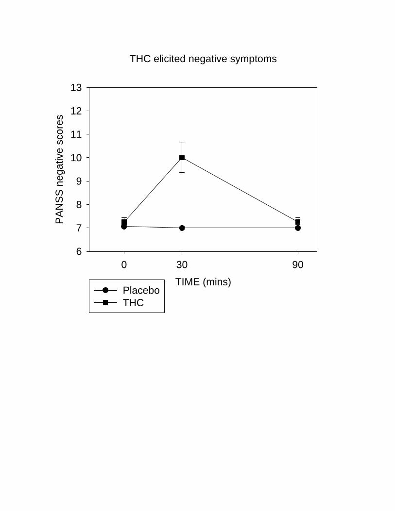
Compared to placebo, THC increased positive (Friedman’s χ2=63.7, p<0.001) negative (Friedman’s
χ2=56.0, p<0.001) and general PANSS scores (Friedman’s χ2=36.1, p<0.001). Increases were most
pronounced at the 30-minute assessment point and tended back towards baseline by 90-minutes (Figure
2a, b). Overall, 40% of participants showed increases in PANSS positive symptom scores of >4 points
at 30-minutes post-injection.
There was a trend towards reduced accuracy in the n-back task following treatment with THC (F=2.95,
p=0.11). Accuracy was robustly affected by task-difficulty (F=5.38, p<0.005), with poorer performance
in the 2-back condition compared to both the 1-back (p<0.05) and 0-back (p<0.01) conditions (Figure
NPP-10-0739 Morrison PD. Revised 15/10/10
13
3a). In terms of accuracy, there was no THC-treatment x task-difficulty interaction.
Response times in the n-back task were slower under THC versus placebo conditions (F=6.8, p<0.05),
and as task-difficulty increased (F=32.3, p<0.001). There was an interaction between THC-treatment x
task-difficulty, in that the effect of THC was significantly greater as the n-back became more
challenging (F=7.74, p<0.005) (Figure 3b).
Electroencephalography
Power
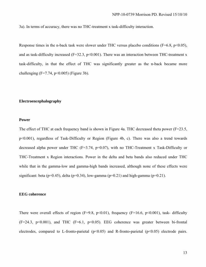
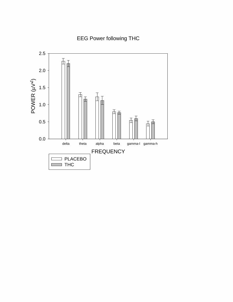
The effect of THC at each frequency band is shown in Figure 4a. THC decreased theta power (F=23.5,
p<0.001), regardless of Task-Difficulty or Region (Figure 4b, c). There was also a trend towards
decreased alpha power under THC (F=3.74, p=0.07), with no THC-Treatment x Task-Difficulty or
THC-Treatment x Region interactions. Power in the delta and beta bands also reduced under THC
while that in the gamma-low and gamma-high bands increased, although none of these effects were
significant: beta (p=0.45), delta (p=0.34), low-gamma (p=0.21) and high-gamma (p=0.21).
EEG coherence
There were overall effects of region (F=9.8, p<0.01), frequency (F=16.6, p<0.001), task- difficulty
(F=24.3, p<0.001), and THC (F=6.1, p<0.05). EEG coherence was greater between bi-frontal
electrodes, compared to L-fronto-parietal (p<0.05) and R-fronto-parietal (p<0.05) electrode pairs.

NPP-10-0739 Morrison PD. Revised 15/10/10
14
Compared to the 0-back condition, overall coherence increased under the 1-back (p<0.001) and 2-back
conditions (p<0.005).
Interactive effects were region x frequency (F=4.0, p=0.01) and frequency x THC (F=3.1, p<0.05).
There was a trend towards a 3-way interaction between region, frequency and THC-treatment (F=2.0,
p=0.09).
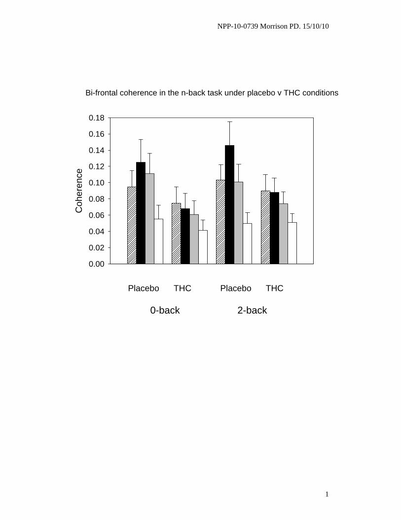
Under placebo, bi-frontal coherence was largest in the theta band and theta coherence was greater
between bi-frontal region compared to both the left (p=0.01) and right (p<0.005) fronto-parietal
regions. THC selectively decreased bi-frontal coherence in the theta (p<0.05) and alpha (p<0.05) bands
(Figure 5).
The observed decrease in theta coherence between bi-frontal electrodes under THC could reflect a
decrease in connectivity between frontal regions in both hemispheres. However, another interpretation
might be that it reflects a decrease in activity of a deep prefrontal source detected by both frontal
regions. To explore this issue further we examined theta coherence in Laplacian derivations over the
left and right hemispheres to minimise the contribution from deep sources. Coherence under placebo
conditions (0.18±0.02; mean±SEM averaged over 3 frontal Laplacian sites) was reduced by THC
(0.11±0.02) and differences emerged at the level of a strong trend (F=4.0, p=0.08). This suggests the
coherence changes do not relate to activity in a deep source but reflect true changes in bi-frontal
connectivity.

NPP-10-0739 Morrison PD. Revised 15/10/10
15
Correlations
EEG Power and psychopathology
Overall there was no correlation between change-in theta power, either globally or specifically at
electrode Fz, and (1) change in reaction-time in the most challenging (2-back) level of the n-back task,
(2) change in positive PANSS scores (THC – placebo at 30 minutes) (3) change in negative PANSS
scores (THC – placebo at 30 minutes).
EEG coherence and psychopathology
We investigated possible relationships between change in theta and alpha coherence and (1) change in
reaction-time in the most challenging (2-back) level of the n-back task, (2) change in positive PANSS
scores (THC – placebo at 30 minutes) (3) change in negative PANSS scores (THC – placebo at 30
minutes). Since there were six comparisons in total, Bonferroni-corrected statistical significance was
set at p < 0.008.
The change-in theta coherence (averaged over bi-frontal, and left and right fronto-parietal) under THC
conditions was strongly associated with the change-in positive PANSS scores (rho=0.75, p=0.001), but
neither change in negative symptoms nor reaction-time. The relationship between theta coherence and
positive symptoms was specific for the bi-frontal region (rho=0.79, p<0.001). Neither change in left or
right fronto-parietal theta coherence was related to positive symptoms. The larger the reduction in bi-
frontal theta coherence, the larger the increase in positive symptoms on the PANSS. Reduced bi-

NPP-10-0739 Morrison PD. Revised 15/10/10
16
frontal theta coherence occurred at all levels of the n-back and the correlation between the decrease in
theta and increase in positive symptoms survived the removal of 2 potential outliers (rho=0.69,
p=0.006) (Figure 6).
There was a weaker relationship between the change-in alpha coherence and the change-in negative
symptoms (Averaged rho=0.57, p=0.02) which was insufficiently robust to survive correction for
multiple testing, and no relationship with positive symptoms nor reaction-time in the n-back.
DISCUSSION
The major finding of the present study is that THC decreased theta coherence between bi-frontal brain
regions compared to the placebo day and that the reduction in coherence was strongly associated with
positive psychotic symptoms. Additional effects of THC – transient psychosis as reflected in the
PANSS score, slower working-memory performance and global suppression of power in the theta band
- are consistent with previous reports.
Methodological issues - coherence
Our measure of coherence is influenced by both the amplitude and phase of EEG activity. A high theta
coherence value indicates a linear relationship between theta amplitude in left frontal and right frontal
cortices across sequential WM epochs and a consistent phase relationship. The reduction in coherence
under THC indicates either that the theta amplitude relationship between the two frontal sites is weaker
of that theta phase varies from trial to trial. Our coherence measure does not disambiguate these two

NPP-10-0739 Morrison PD. Revised 15/10/10
17
possibilities; however, a reduction in either amplitude or phase relationship points to a weakening of
shared function or cross-talk between the frontal sites. The coherence seems to be driven by activity in
the lateral frontal cortex and not from a deep source (for example on the medial surface of the frontal
lobes) as the reduction in coherence under THC is preserved in the Laplacian data which is sensitive to
local sources but not deeper ones. The THC effects in the Laplacian data are less significant than those
in the bipolar montage analysis suggesting either that the Laplacian electrodes used were not directly
above the cortical regions involved in the cross–talk or that large regions of frontal cortex are involved
as this reduces the sensitivity of Laplacian data.
THC, theta coherence and psychosis
Previously there has been speculation that the pro-psychotic effects of THC stem from disruption of
synchronised neural rhythms (Hajos et al, 2008; Sewell et al, 2009). The major finding here provides
the first experimental support for this idea. It may be that THC elicits a 'lesion', at the molecular level
which can 'push' an otherwise healthy nervous system towards acute psychosis, hastens the onset of
psychotic-breakdown in those destined to develop schizophrenia and provokes acute relapse in
established cases (Kuepper et al, 2010). The precise nature (and location) of the molecular lesion,
downstream of CB1 receptors is unknown. But the most likely scenario is that THC disrupts the
intricacies of fast amino-acid based neurotransmission; and in doing so, disrupts network oscillations
which depend, in-part, upon reciprocal glutamate and GABA-ergic connections (Buzsaki, 2006; Ford
and Mathalon, 2008; Hajos et al, 2008; Robbe et al, 2006; Uhlhaas et al, 2008).
The findings point to the pre-frontal cortex and implicate the theta band in the generation of psychotic

NPP-10-0739 Morrison PD. Revised 15/10/10
18
symptoms. Reduced bi-frontal (but not fronto-parietal) coherence from placebo day levels was strongly
associated with positive psychotic symptoms. This was not the case for theta power. The simplest
interpretation is that acute THC-psychosis is associated with disruption in long-distance synchrony
(reflected in coherence) but not with disruption of local theta rhythms (reflected in power). There is a
consensus that theta oscillations are important for long-distance ‘cross-talk’ between brain regions
(Uhlhaas et al, 2010; Varela et al, 2001). The findings here are broadly in keeping with the
disconnection hypothesis of schizophrenia, in which impaired functional connectivity between brain
regions underlies psychotic symptoms (Stephan et al, 2009). Some caution is required in attributing the
pro-psychotic effects of THC to a direct action within the frontal cortices. It is likely that, in the present
study, theta oscillations within limbic regions were also disrupted by THC (Hajos et al, 2008; Robbe et
al, 2006). Thus an apparent cortical lesion might only be a marker for a cortico-limbic lesion which is
‘closer’ to the pathophysiology of THC. For example Df(16)A+/– mice, (which mimic one of the largest
genetic risk-factors for schizophrenia, a microdeletion on human chromosome 22q11.2), show reduced
phase-locking of pre-frontal cells to hippocampal theta rhythms and reduced coherence of pre-frontal
and hippocampal local field potentials. (Sigurdsson et al, 2010).
It is unclear whether the link between positive psychotic symptoms and decreased bi-frontal theta
coherence reported here is specific to THC and WM tasks or constitutes a more generally applicable
biomarker for positive psychotic symptoms whatever their context. We did not find a robust association
between EEG coherence and negative symptoms suggesting that these symptoms may be related to a
different mechanism or disconnection between different areas
The link between positive symptoms and coherence was specific to the theta band. It is possible that the

NPP-10-0739 Morrison PD. Revised 15/10/10
19
gamma band would also have been linked to psychotic symptoms. It is known that gamma oscillations
'nest' within slower oscillations; thus the power of gamma rhythms rises and falls on a slower theta
oscillation (Canolty et al, 2006). However, clear separation of neural and muscle-derived gamma in the
human scalp-recorded EEG can be difficult, although algorithms have been developed to permit
identification of neural gamma in humans (Nottage, 2010). It is possible that changes in the gamma
band in addition to theta will be detected using these novel methods. Studies in schizophrenic patients
have reported decreased power of frontal theta oscillations during performance of WM tasks
(Haenschel et al, 2009; Schmiedt et al, 2005). Ford and colleagues compared the EEG of speech
production versus listening, in patients and controls. Speech production was associated with increased
coherence across classical left hemisphere language regions within the theta band, in healthy controls
but not in schizophrenic patients (Ford et al, 2002). The authors concluded that reduced fronto-
temporal functional connectivity in schizophrenia could lead to the attribution of self-generated speech
to an external source; auditory hallucinations. Our subjects did not experience auditory hallucinations
and our findings suggest that other classes of psychotic symptom by be linked to reductions in bi-
frontal coherence.
THC, theta oscillations and cognition
Under THC, there were decreases in theta power and theta coherence regardless of the level of
difficulty in the n-back task. The reduction in theta power is unlikely to relate to a non-specific
decrease in arousal as drowsiness and the transition to sleep has the opposite effect, increasing theta
activity. All three n-back levels differed from the 'at-rest' state in placing explicit demands on attention.
Generally, the impact of THC on n-back performance was relatively subtle. As demands on working

NPP-10-0739 Morrison PD. Revised 15/10/10
20
memory increased, performance was significantly slower, but accuracy was maintained. Indeed the
majority of participants continued to 'achieve' 100% success, even on the most challenging level. In
agreement with previous findings, THC-elicited deficits in WM showed no relationship with acute
psychosis (Morrison et al, 2009). Additionally, in the present data set, we found no relationship
between WM deficits and the decreases in EEG power. The latter finding is in contrast with the recent
report of Bocker et al, who found the greater the reduction in theta power under THC the greater the
slowing of responses (but not reduction in accuracy) in a WM task (Bocker et al, 2010). It also has
been reported that fronto-parietal coherence (specifically within the theta band) increases during WM
performance (Sarnthein et al, 1998). This effect was not observed in the present study. One possibility
is that the n-back task employed here was insufficiently challenging.
The neurophysiology of THC
Recently, cannabinoid agonists have emerged as a 'tool' for the study of brain network dynamics and
for the study of psychotic mental states (Buzsaki, 2006; Sewell et al, 2009). In the former case, CB1
agonists are unique in that they can disrupt network synchrony whilst preserving the firing-rates of
individual pyramidal and GABA-ergic interneurons (Buzsaki, 2006; Robbe et al, 2006). Comparing
different drug-models of psychosis; CB1 agonists can elicit a psychotic reaction in otherwise healthy
controls after a single exposure, in contrast to stimulants (amphetamine/cocaine) where repeated use is
a pre-requisite. And repeated use of cannabis (especially forms high in THC-content) is a risk factor for
the genesis of schizophrenia, in contrast to ketamine where no association has as yet been found.
NPP-10-0739 Morrison PD. Revised 15/10/10
21
The endocannabinoids are key components in plasticity and a rapidly developing field is the overlap
between neural oscillations and plasticity. The role of theta oscillations in the generation of
hippocampal long-term potentiation (LTP) has long been appreciated (Huerta and Lisman, 1993). But
more recently it has become apparent that gamma rhythms and spike-timing dependent plasticity
(STDP) occur within the same, critical time-frame (tens of milliseconds) suggestive of mechanistic
links between the two phenomena (Buzsaki, 2006; Uhlhaas et al, 2010). Spike-timing dependent long-
term depression (LTD) in the cortex is implemented by retrograde endocannabinoid signalling
(Chevaleyre et al, 2006). And animal work has shown that THC disrupts network oscillations and
network plasticity simultaneously (Robbe et al, 2006). Potentially this could offer insight into how
THC can precipitate an acute, reversible psychosis (disruption of oscillations) and a chronic psychotic
illness (additional disruption of plasticity).
Strengths and limitations
A weakness of the current study is that only one dose of THC was used. The sample size is also small
for teasing out interactions, but typical for studies of this type. There are two main strengths.
Experimentally-induced psychoses permit the testing of healthy participants before, during and after
administration of a pro-psychotic drug, and intra-individual comparisons. We also took advantage of
pure synthetic preparations of THC. Plant derived material contains further cannabinoid molecules
such as cannabidiol (CBD) which counteracts the pro-psychotic effects of THC (Bhattacharyya et al,
2010), and tetrahydrocannabivarin (THCV), an antagonist at CB1 receptors (Pertwee et al, 2008).

NPP-10-0739 Morrison PD. Revised 15/10/10
22
Conclusion
Global theta power was reduced by THC – without any manifest psychopathological consequences. In
contrast, there was a strong and specific association between THC-induced positive psychotic
symptoms and reduced bi-frontal theta coherence. Impaired functional 'cross-talk' between the frontal
lobes in the theta band might account for the pro-psychotic effects of THC/cannabis. Whether this is
also true of psychotic symptoms more generally is a question for further study.
DISCLOSURE/CONFLICT OF INTEREST The authors have no conflicts of interest to disclose ACKNOWLEDGEMENTS Supported by the MRC (UK) and the Beckley Foundation.

NPP-10-0739 Morrison PD. Revised 15/10/10
23
References
Arseneault L, Cannon M, Witton J, Murray RM (2004). Causal association between cannabis and
psychosis: examination of the evidence. Br J Psychiatry 184: 110-117.
Bocker KB, Hunault CC, Gerritsen J, Kruidenier M, Mensinga TT, Kenemans JL (2010). Cannabinoid
modulations of resting state EEG theta power and working memory are correlated in humans. J Cogn
Neurosci 22: 1906-1916.
Bodor AL, Katona I, Nyiri G, Mackie K, Ledent C, Hajos N, et al (2005). Endocannabinoid signaling
in rat somatosensory cortex: laminar differences and involvement of specific interneuron types. J
Neurosci 25: 6845-6856.
Bhattacharyya S, Morrison PD, Fusar-Poli P, Martin-Santos R, Borgwardt S, Winton-Brown T, et al
(2010) Opposite effects of delta-9-tetrahydrocannabinol and cannabidiol on human brain function and
psychopathology. Neuropsychopharmacology 35: 764-74.
Buzsaki G (2006): Rhythms of the brain Oxford University Press: New-York.

NPP-10-0739 Morrison PD. Revised 15/10/10
24
Buzsaki G, Draguhn A (2004). Neuronal oscillations in cortical networks. Science 304: 1926-1929.
Canolty RT, Edwards E, Dalal SS, Soltani M, Nagarajan SS, Kirsch HE, et al (2006). High gamma
power is phase-locked to theta oscillations in human neocortex. Science 313: 1626-1628.
Chevaleyre V, Takahashi KA, Castillo PE (2006). Endocannabinoid-mediated synaptic plasticity in the
CNS. Annu Rev Neurosci 29: 37-76.
D'Souza DC, Abi-Saab WM, Madonick S, Forselius-Bielen K, Doersch A, Braley G, et al (2005).
Delta-9-tetrahydrocannabinol effects in schizophrenia: implications for cognition, psychosis, and
addiction. Biol Psychiatry 57: 594-608.
D'Souza DC, Perry E, MacDougall L, Ammerman Y, Cooper T, Wu YT, et al (2004). The
psychotomimetic effects of intravenous delta-9-tetrahydrocannabinol in healthy individuals:
implications for psychosis. Neuropsychopharmacology 29: 1558-1572.
Eggan SM, Lewis DA (2007). Immunocytochemical distribution of the cannabinoid CB1 receptor in
the primate neocortex: a regional and laminar analysis. Cereb Cortex 17: 175-191.
Eggan SM, Melchitzky DS, Sesack SR, Fish KN, Lewis DA (2010). Relationship of cannabinoid CB1
receptor and cholecystokinin immunoreactivity in monkey dorsolateral prefrontal cortex. Neuroscience.
doi:10.1016/j.neuroscience.2010.06.011

NPP-10-0739 Morrison PD. Revised 15/10/10
25
Ford JM, Krystal JH, Mathalon DH (2007). Neural synchrony in schizophrenia: from networks to new
treatments. Schizophr Bull 33: 848-852.
Ford JM, Mathalon DH (2008). Neural synchrony in schizophrenia. Schizophr Bull 34: 904-906.
Ford JM, Mathalon DH, Whitfield S, Faustman WO, Roth WT (2002). Reduced communication
between frontal and temporal lobes during talking in schizophrenia. Biol Psychiatry 21: 485-492.
Freund TF, Katona I, Piomelli D (2003). Role of endogenous cannabinoids in synaptic signaling.
Physiol Rev 83: 1017-1066.
Glass M, Dragunow M, Faull RL (1997). Cannabinoid receptors in the human brain: a detailed
anatomical and quantitative autoradiographic study in the fetal, neonatal and adult human brain.
Neuroscience 77: 299-318.
Goldberg DP, Blackwell B (1970). Psychiatric illness in general practice. A detailed study using a new
method of case identification. British Medical Journal 1: 439-43.
Gonzalez-Burgos G, Lewis DA (2008). GABA neurons and the mechanisms of network oscillations:
implications for understanding cortical dysfunction in schizophrenia. Schizophr Bull 34: 944-961.

NPP-10-0739 Morrison PD. Revised 15/10/10
26
Guevara MA, Corsi-Cabrera M (1996). EEG coherence or EEG correlation? Int J Psychophysiol 23:
145-153.
Haenschel C, Bittner RA, Waltz J, Haertling F, Wibral M, Singer W, et al (2009). Cortical oscillatory
activity is critical for working memory as revealed by deficits in early-onset schizophrenia. J Neurosci
29: 9481-9489.
Hajos M, Hoffmann WE, Kocsis B (2008). Activation of cannabinoid-1 receptors disrupts sensory
gating and neuronal oscillation: relevance to schizophrenia. Biol Psychiatry 63: 1075-1083.
Herkenham M, Lynn AB, Johnson MR, Melvin LS, de Costa BR, Rice KC (1991). Characterization
and localization of cannabinoid receptors in rat brain: a quantitative in vitro autoradiographic study. J
Neurosci 11: 563-583.
Hill EL, Gallopin T, Ferezou I, Cauli B, Rossier J, Schweitzer P, et al (2007). Functional CB1
receptors are broadly expressed in neocortical GABAergic and glutamatergic neurons. J Neurophysiol
97: 2580-2589.
Howlett AC, Barth F, Bonner TI, Cabral G, Casellas P, Devane WA, et al (2002). International Union
of Pharmacology. XXVII. Classification of cannabinoid receptors. Pharmacol Rev 54: 161-202.
Huerta PT, Lisman JE (1993). Heightened synaptic plasticity of hippocampal CA1 neurons during a
cholinergically induced rhythmic state. Nature 364: 723-725

NPP-10-0739 Morrison PD. Revised 15/10/10
27
Ilan AB, Smith ME, Gevins A (2004). Effects of marijuana on neurophysiological signals of working
and episodic memory. Psychopharmacology (Berl) 176: 214-222.
Kay SR, Fiszbein A, Opler LA (1987). The positive and negative syndrome scale (PANSS) for
schizophrenia. Schizophr Bull 13: 261-276.
Kopell N, Ermentrout GB, Whittington MA, Traub RD (2000). Gamma rhythms and beta rhythms have
different synchronization properties. Proc Natl Acad Sci U S A 97: 1867-1872.
Kuepper R, Morrison PD, van Os J, Murray RM, Kenis G, Henquet C (2010). Does dopamine mediate
the psychosis-inducing effects of cannabis? A review and integration of findings across disciplines.
Schizophr Res. [Epub ahead of print] doi:10.1016/j.schres.2010.05.031
Lachaux JP, Rodriguez E, Martinerie J, Varela FJ (1999). Measuring phase synchrony in brain signals.
Hum Brain Mapp 8: 194-208.
Mechoulam R, Shani A, Edery H, Grunfeld Y (1970). Chemical basis of hashish activity. Science 169:
611-612.
Miltner WH, Braun C, Arnold M, Witte H, Taub E (1999). Coherence of gamma-band EEG activity as
a basis for associative learning. Nature 397: 434-436.

NPP-10-0739 Morrison PD. Revised 15/10/10
28
Moore TH, Zammit S, Lingford-Hughes A, Barnes TR, Jones PB, Burke M, et al (2007). Cannabis use
and risk of psychotic or affective mental health outcomes: a systematic review. Lancet 370: 319-328.
Morrison PD, Zois V, McKeown DA, Lee TD, Holt DW, Powell JF, et al (2009). The acute effects of
synthetic intravenous Delta9-tetrahydrocannabinol on psychosis, mood and cognitive functioning.
Psychol Med 39: 1607-1616.
Murray RM, Morrison PD, Henquet C, Di Forti M (2007). Cannabis, the mind and society: the hash
realities. Nat Rev Neurosci 8: 885-895.
Naef M, Russmann S, Petersen-Felix S, Brenneisen R (2004). Development and pharmacokinetic
characterization of pulmonal and intravenous delta-9-tetrahydrocannabinol (THC) in humans. J Pharm
Sci 93: 1176-1184.
Nottage JF (2010). Uncovering gamma in visual tasks. Brain Topogr 23: 58-71
Owen AM, McMillan KM, Laird AR, Bullmore E (2005). N-back working memory paradigm: a meta-
analysis of normative functional neuroimaging studies. Hum Brain Mapp 25: 46-59.
Pertwee RG (2008). The diverse CB1 and CB2 receptor pharmacology of three plant cannabinoids:
delta9-tetrahydrocannabinol, cannabidiol and delta9-tetrahydrocannabivarin. Br J Pharmacol 153: 199-
215

NPP-10-0739 Morrison PD. Revised 15/10/10
29
Piomelli D (2003). The molecular logic of endocannabinoid signalling. Nat Rev Neurosci 4: 873-884.
Ranganathan M, D'Souza DC (2006). The acute effects of cannabinoids on memory in humans: a
review. Psychopharmacology (Berl) 188: 425-444.
Roach BJ, Mathalon DH (2008). Event-related EEG time-frequency analysis: an overview of measures
and an analysis of early gamma band phase locking in schizophrenia. Schizophr Bull 34: 907-926.
Robbe D, Montgomery SM, Thome A, Rueda-Orozco PE, McNaughton BL, Buzsaki G (2006).
Cannabinoids reveal importance of spike timing coordination in hippocampal function. Nat Neurosci 9:
1526-1533.
Rodriguez E, George N, Lachaux JP, Martinerie J, Renault B, Varela FJ (1999). Perception's shadow:
long-distance synchronization of human brain activity. Nature 397: 430-433.
Ruchkin D (2005). EEG coherence. Int J Psychophysiol 57: 83-85.
Sarnthein J, Petsche H, Rappelsberger P, Shaw GL, von Stein A (1998). Synchronization between
prefrontal and posterior association cortex during human working memory. Proc Natl Acad Sci U S A
95: 7092-7096.
Schmiedt C, Brand A, Hildebrandt H, Basar-Eroglu C (2005). Event-related theta oscillations during
working memory tasks in patients with schizophrenia and healthy controls. Brain Res Cogn Brain Res

NPP-10-0739 Morrison PD. Revised 15/10/10
30
25: 936-947.
Sewell RA, Ranganathan M, D'Souza DC (2009). Cannabinoids and psychosis. Int Rev Psychiatry 21:
152-162.
Sigurdsson T, Stark KL, Karayiorgou M, Gogos JA, Gordon JA (2010). Impaired hippocampal-
prefrontal synchrony in a genetic mouse model of schizophrenia. Nature 464: 763-767.
Stephan KE, Friston KJ, Frith CD (2009). Dysconnection in schizophrenia: from abnormal synaptic
plasticity to failures of self-monitoring. Schizophr Bull 35: 509-527.
Uhlhaas PJ, Haenschel C, Nikolic D, Singer W (2008). The role of oscillations and synchrony in
cortical networks and their putative relevance for the pathophysiology of schizophrenia. Schizophr Bull
34: 927-943.
Uhlhaas PJ, Pipa G, Lima B, Melloni L, Neuenschwander S, Nikolic D, et al (2009). Neural synchrony
in cortical networks: history, concept and current status. Front Integr Neurosci 3: 17.
Uhlhaas PJ, Singer W (2010). Abnormal neural oscillations and synchrony in schizophrenia. Nat Rev
Neurosci 11: 100-113.
Varela F, Lachaux JP, Rodriguez E, Martinerie J (2001). The brainweb: phase synchronization and
NPP-10-0739 Morrison PD. Revised 15/10/10
31
large-scale integration. Nat Rev Neurosci 2: 229-239.
von Stein A, Chiang C, Konig P (2000). Top-down processing mediated by interareal synchronization.
Proc Natl Acad Sci U S A 97: 14748-14753.
NPP-10-0739 Morrison PD. Revised 15/10/10
32
Titles & Legends to figures Figure 1. Plasma concentrations of THC (mean±SEM) following the intravenous injection of THC
(1.25mg).
Figure 2. (a) Positive and (b) negative psychotic symptoms (mean±SEM) as measured by the PANSS
scale were increased from baseline (time 0 min) following the administration of intravenous THC
(1.25mg).
Figure 3. (a) Accuracy and (b) reaction-time in the n-back task (mean±SEM) under THC versus
placebo conditions. (a) THC had no effect on accuracy. (b) Performance was slower under THC
conditions (p<0.05) and showed an interaction with task-difficulty (p<0.005).
Figure 4. (a) The effect of THC at each frequency band. Compared to placebo, theta power
(mean±SEM) was decreased by THC (p<0.001). Decreases or increases under THC in other frequency
bands are not significant. Theta power was reduced regardless of task-difficulty (b) or recording site
(c). Left frontal (LF), right-frontal (RF), left central (LC), right central (RC), left temporal (LT), right
temporal (RT), left occipitoparietal (LOP), right occipitoparietal (ROP), midline frontal (FZ), midline
parietal (PZ) and midline central (CZ).
Figure 5. Coherence between left and right prefrontal regions, under THC versus placebo, in the
control (0-back) and most challenging (2-back) levels of the n-back task.
Bars show coherence (mean±SEM) by frequency. (Diagonal stripes – delta. Black – theta. Grey –
alpha. White – beta.) THC treatment selectively decreased coherence in the theta (p<0.05) and alpha

NPP-10-0739 Morrison PD. Revised 15/10/10
33
(p<0.05) bands.
Figure 6. Reductions in theta coherence between left and right prefrontal regions under THC was
correlated with positive psychotic symptoms (rho=0.79, p<0.001), and survived the removal of 2
potential outliers (rho=0.69, p=0.006).

THC concentration over time
TIME (mins)0 20 40 60 80 100 120
[TH
C] n
g/m
l
0
20
40
60
80
100
120

THC elicited positive symptoms
TIME (mins)
0 30 90
PA
NS
S p
ositi
ve s
core
s
6
7
8
9
10
11
12
13
PlaceboTHC
THC elicited negative symptoms
TIME (mins)
0 30 90
PA
NS
S n
egat
ive
scor
es
6
7
8
9
10
11
12
13
PlaceboTHC

The effect of THC on response accuracy in the N-Back Task
N-Back Condition
0-back 1-back 2-back
Cor
rect
Res
pons
es
6.0
6.5
7.0
7.5
8.0
PlaceboTHC

The effect of THC on response timesin the N-Back Task
N-Back Condition
0-back 1-back 2-back
Res
pons
e Ti
me
(ms)
300
400
500
600
700
800
PlaceboTHC
EEG Power following THC
FREQUENCYdelta theta alpha beta gamma-l gamma-h
POW
ER
(μV
2 )
0.0
0.5
1.0
1.5
2.0
2.5
PLACEBO THC

THETA power decreases under THC regardeless of memory load
CONDITION
0-BACK 1-BACK 2-BACK
POW
ER
(μV2 )
0.0
0.2
0.4
0.6
0.8
1.0
1.2
1.4
1.6
PLACEBOTHC

THETA power under THC v placebo according to region
REGION
LF RF LC RC LT RT LOP ROP FZ PZ CZ
PO
WE
R (μ
V2 )
0.0
0.2
0.4
0.6
0.8
1.0
1.2
1.4
1.6
1.8
PLACEBOTHC
NPP-10-0739 Morrison PD. 15/10/10
1
Figure 5 Placebo THC Placebo THC 0-back 2-back
Bi-frontal coherence in the n-back task under placebo v THC conditions
Coh
eren
ce
0.00
0.02
0.04
0.06
0.08
0.10
0.12
0.14
0.16
0.18

NPP-10-0739 Morrison PD. 15/10/10
2

The Relationship between positive psychotic symptoms & bi-frontal theta coherence
Change in theta coherence (Placebo-THC sessions)-0.1 0.0 0.1 0.2 0.3
Cha
nge
in p
ositi
ve
PA
NS
S s
core
s
0
2
4
6
8
10
12