Diseases of trees in the Great Plains - USDA Forest Service
161
United States Depa rt ment of " Agric ulture Forest Service Rocky Mountain Forest 1nd Range Experiment St ·1t1on Fort Collins, Col orado 80526 General Technical Report RM-129 Diseases of Trees in the Great Plains Jerry W. Riffle and Glenn W. Techni c al Coordinators This file was created by scanning the printed publication. Errors identified by the software have been corrected; however, some errors may remain.
-
Upload
khangminh22 -
Category
Documents
-
view
1 -
download
0
Transcript of Diseases of trees in the Great Plains - USDA Forest Service

United States Department of" Ag ric ulture
Forest Service
Rocky Mountain Forest 1nd Range Experiment St·1t1on
Fort Collins, Colorado 80526
General Technical Report RM-129
Diseases of Trees
in the Great Plains
Jerry W. Riffle and Glenn W. Peter~on
Technical Coordinators
This file was created by scanning the printed publication.Errors identified by the software have been corrected;
however, some errors may remain.

Riffle, Jerry W.; Peterson, Glenn W., tech. coords. Diseases of trees
in the Great Plains. Gen. Tech. Rep. RM-129. Fort Collins, CO: U.S.
Department of Agriculture, Forest Service, Rocky Mountain Forest
and Range Experiment Station; 1986. 149 p.
Abstract
Hosts, distribution, symptoms and signs, disease cycle, and control
measures are described for 46 hardwood and 15 conifer diseases.
Diseases in which abiotic agents are contributory factors also are
described. Color and black-and-white illustrations that stress diagnosis
and control are provided. A glossary of technical terms and indexes
to hosts, pathogens, and insect vectors also are included.
Keywords: Tree diseases, forest pathology, Great Plains, windbreaks
Disclaimer
Mention of a tradename or proprietary product does not constitute
a guarantee or warranty of the product by the U.S. Department of
Agriculture and does not imply its approval to the exclusion of other
products that may also be suitable.
Riffle, jerry W.; Peterson, Glenn W., tech. coords. Diseases of trees in the Great Plains. Gun. Tech. Rep. RM-129. Fort Collins, CO: U.S. Department of Agriculture, Forest Service, Rocky Mountain Forest and Range Experiment Station; 1986. 149 p.
Abstract
Hosts, distribution, symptoms and signs, disease cycle, and control measures are described for 46 hardwood and 15 conifer diseases. Diseases in which abiotic agents are contributory factors also are described. Color and black-and-white illustrations that stress diagnosis and control are provided. A glossary of technical terms and indexes to hosts, pathogens, and insect vectors also are included.
Keywords: Tree diseases, forest pathology, Great Plains, windbreaks
Disclaimer
Mention of a tradename or proprietary product does not constitute a guarantee or warranty of the product by the U.S. Department of Agriculture and does not imply its approval to the exclusion of other products that may also be suitable.

USDA Forest Service June 1986
General Technical Report RM-129
Diseases of Trees in the Great Plains
Technical Coordinators:
Jerry W. Riffle and Glenn W. Peterson
Rocky Mountain Forest and Range Experiment Station1
^Headquarters is in Fort Collins, in cooperation with Colorado State University; technical
coordinators are plant pathologists at the Station's Research Unit at Lincoln, Nebraska, in
cooperation with the University of Nebraska—Lincoln.
USDA Forest Service June 1986 General Technical Report RM-129
Diseases of Trees in the Great Plains
Technical Coordinators:
Jerry W. Riffle and Glenn W. Peterson Rocky Mountain Forest and Range Experiment Station 1
1Headquarters is in Fort Collins, in cooperation with Colorado State University; technical coordinators are plant pathologists at the Station's Research Unit at Lincoln, Nebraska, in cooperation with the University of Nebraska-Lincoln.

Preface
This technical report provides a guide to assist ar-
borists, land owners, pest management specialists,
foresters, and plant pathologists in the diagnosis and con-
trol of tree diseases encountered in the Great Plains. It
contains 64 articles on tree diseases prepared by 31
authors, and emphasizes disease situations as observed
in the 10 states of the Great Plains.
The need for such a Handbook for the Great Plains has
been recognized for some time.
During its 1980 meeting the Pest Management Task
Force of the Forestry Committee, Great Plains Agricul-
tural Council requested that the Forestry Committee
"support the publication of a 'Tree Disease Handbook
for the Great Plains', by the Task Force, and assist the
Task Force in obtaining the necessary funding." The re-
quest was approved.
During the Task Force's 1981 meeting a working group
was formed to prepare a prospectus for the Handbook.
The working group members, Dr. Edward M. Sharon,
Mr. James A. Walla, Dr. Mark O. Harrell, and Dr. Jerry
W. Riffle, selected specific diseases for inclusion in the
Handbook, selected potential authors for each article,
and determined the format for articles to emphasize
diagnosis, biology, damage, and control of diseases.
In 1982 the Pest Management Task Force agreed that
Dr. Riffle would serve as coordinator of the Handbook,
with Dr. Glenn Peterson as co-coordinator. Dr. Riffle
contacted all potential contributors in May 1982, and
their response was excellent; 31 persons agreed to author
or co-author articles on 64 diseases.
Funding for the publication of the Handbook was
resolved in 1985 when it was proposed that the Hand-
book be published as a General Technical Report by the
USDA Forest Service, Rocky Mountain Forest and Range
Experiment Station. This proposal was approved by the
Executive Committee of the Forestry Committee of the
Great Plains Agricultural Council.
Preface
This technical report provides a guide to assist arborists, land owners, pest management specialists, foresters, and plant pathologists in the diagnosis and control of tree diseases encountered in the Great Plains. It contains 64 articles on tree diseases prepared by 31 authors, and emphasizes disease situations as observed in the 10 states of the Great Plains.
The need for such a Handbook for the Great Plains has been recognized for some time.
During its 1980 meeting the Pest Management Task Force of the Forestry Committee, Great Plains Agricultural Council requested that the Forestry Committee "support the publication of a 'Tree Disease Handbook for the Great Plains', by the Task Force, and assist the Task Force in obtaining the necessary funding." The request was approved.
During the Task Force's 1981 meeting a working group was formed to prepare a prospectus for the Handbook. The working group members, Dr. Edward M. Sharon,
Mr. James A. Walla, Dr. Mark 0. Harrell, and Dr. Jerry W. Riffle, selected specific diseases for inclusion in the Handbook, selected potential authors for each article, and determined the format for articles to emphasize diagnosis, biology, damage, and control of diseases.
In 1982 the Pest Management Task Force agreed that Dr. Riffle would serve as coordinator of the Handbook, with Dr. Glenn Peterson as co-coordinator. Dr. Riffle contacted all potential contributors in May 1982, and their response was excellent; 31 persons agreed to author or co-author articles on 64 diseases.
Funding for the publication of the Handbook was resolved in 1985 when it was proposed that the Handbook be published as a General Technical Report by the USDA Forest Service, Rocky Mountain Forest and Range Experiment Station. This proposal was approved by the Executive Committee of the Forestry Committee of the Great Plains Agricultural Council.

Contents
Page
Authors and Their Affiliations iv
Acknowledgements vi
Introduction 1
Use and Safe Handling of Pesticides 3
HARDWOOD DISEASES
Foliage
1. Melampsora Leaf Rust of Cottonwood and Willow 4
Glenn W. Peterson and Robert W. Stack
2. Marssonina Leaf Spot of Cottonwood and Aspen 6
John E. Watkins and David S. Wysong
3. Septoria Leaf Spots of Cottonwood, Caragana, and Maple 8
Joseph M. Krupinsky and David W. Johnson
4. Phyllosticta Leaf Spots of Maple and Caragana 10
Robert L. James and David W. Johnson
5. Tar Spot of Maple 12
Edward M. Sharon and Jerry W. Riffle
6. Mycosphaerella Leaf Spots of Ash 14
Robert W. Stack and Jerry W. Riffle
7. Gloeosporium and Gnomonia Leaf Diseases of Broadleaf Trees ... 16
Robert W. Stack and Kenneth E. Conway
8. Oak Leaf Blister 20
Kenneth E. Conway and John E. Watkins
9. Leaf Spots of Nanking Cherry and Chokecherry 22
Robert L. James and John E. Watkins
10. Western X-disease of Chokecherry 24
Glenn W. Peterson and David W. Johnson
11. Honeysuckle Leaf Blight 26
Jerry W. Riffle and John E. Watkins
12. Powdery Mildew of Lilac 30
Richard Dorset and Michael W. Ferguson
13. Cylindrosporium Leaf Spot of Buffaloberry and Skunkbush Sumac 32
Glenn W. Peterson and Jerry W. Riffle
14. Herbicides (Air Pollution) 33
Gary A. Boutz and Robert W. Stack
15. Chlorosis 36
Mark O. Harrell and Mark W. Andrews
Branch and Stem
16. Botryodiplodia Canker of Elms 38
Jerry W. Riffle and Joseph M. Krupinsky
17. Tubercularia Canker of Siberian Elm and Russian-olive 40
Joseph M. Krupinsky and James A. Walla
18. Botryodiplodia Disease of Russian-olive 42
Glenn W. Peterson and Harrison L. Morton
19. Phomopsis Canker of Russian-olive 44
Harrison L. Morton and Joseph M. Krupinsky
20. Botryodiplodia Canker of Sycamore 46
Robert Lewis, Jr. and Kenneth E. Conway
21. Cytospora Canker of Cottonwoods and Willows 48
James A. Walla and Kenneth E. Conway
22. Septoria Canker of Cottonwood and Hybrid Poplars 50
Jerry W. Riffle and David S. Wysong
23. Phomopsis Canker on Cottonwood 52
Theodore H. Filer, Jr. and Edward M. Sharon
i
Contents Page
Authors and Their Affiliations . . . . . . . . . . . . . . . . . . . . . . . . . . . . . . . . . . . iv Acknowledgements . . . . . . . . . . . . . . . . . . . . . . . . . . . . . . . . . . . . . . . . . . . . vi Introduction . . . . . . . . . . . . . . . . . . . . . . . . . . . . . . . . . . . . . . . . . . . . . . . . . . 1 Use and Safe Handling of Pesticides . . . . . . . . . . . . . . . . . . . . . . . . . . . . . 3
HARDWOOD DISEASES Foliage
1. Melampsora Leaf Rust of Cottonwood and Willow . . . . . . . . . . . . . 4 Glenn W. Peterson and Robert W. Stack
2. Marssonina Leaf Spot of Cottonwood and Aspen . . . . . . . . . . . . . . . 6 John E. Watkins and David S. Wysong
3. Septoria Leaf Spots of Cottonwood, Caragana, and Maple . . . . . . . 8 Joseph M. Krupinsky and David W. Johnson
4. Phyllosticta Leaf Spots of Maple and Caragana . . . . . . . . . . . . . . . . 10 Robert L. James and David W. Johnson
5. Tar Spot of Maple . . . . . . . . . . . . . . . . . . . . . . . . . . . . . . . . . . . . . . . . . 12 Edward M. Sharon and Jerry W. Riffle
6. Mycosphaerella Leaf Spots of Ash . . . . . . . . . . . . . . . . . . . . . . . . . . . . 14 Robert W. Stack and Jerry W. Riffle
7. Gloeosporium and Gnomonia Leaf Diseases of Broadleaf Trees . . . 16 Robert W. Stack and Kenneth E. Conway
8. Oak Leaf Blister . . . . . . . . . . . . . . . . . . . . . . . . . . . . . . . . . . . . . . . . . . . 20 Kenneth E. Conway and John E. Watkins
9. Leaf Spots of Nanking Cherry and Chokecherry . . . . . . . . . . . . . . . . 22 Robert L. James and John E. Watkins
10. Western X-disease of Chokecherry . . . . . . . . . . . . . . . . . . . . . . . . . . . 24 Glenn W. Peterson and David W. Johnson
11. Honeysuckle Leaf Blight . . . . . . . . . . . . . . . . . . . . . . . . . . . . . . . . . . . . 26 Jerry W. Riffle and John E. Watkins
12. Powdery Mildew of Lilac . . . . . . . . . . . . . . . . . . . . . . . . . . . . . . . . . . . 30 Richard Dorset and Michael W. Ferguson
13. Cylindrosporium Leaf Spot of Buffaloberry and Skunkbush Sumac 32 Glenn W. Peterson and Jerry W. Riffle
14. Herbicides (Air Pollution) . . . . . . . . . . . . . . . . . . . . . . . . . . . . . . . . . . . 33 Gary A. Bautz and Robert W. Stack
15. Chlorosis . . . . . . . . . . . . . . . . . . . . . . . . . . . . . . . . . . . . . . . . . . . . . . . . . 36 Mark 0. Harrell and Mark W. Andrews
Branch and Stem
16. Botryodiplodia Canker of Elms . . . . . . . . . . . . . . . . . . . . . . . . . . . . . . 38 Jerry W. Riffle and Joseph M. Krupinsky
17. Tubercularia Canker of Siberian Elm and Russian-olive . . . . . . . . . 40 Joseph M. Krupinsky and ]ames A. Walla
18. Botryodiplodia Disease of Russian-olive . . . . . . . . . . . . . . . . . . . . . . . 42 Glenn W. Peterson and Harrison L. Morton
19. Phomopsis Canker of Russian-olive . . . . . . . . . . . . . . . . . . . . . . . . . . . 44 Harrison L. Morton and Joseph M. Krupinsky
20. Botryodiplodia Canker of Sycamore . . . . . . . . . . . . . . . . . . . . . . . . . . 46 Robert Lewis, Jr. and Kenneth E. Conway
21. Cytospora Canker of Cottonwoods and Willows . . . . . . . . . . . . . . . . 48 James A. Walla and Kenneth E. Conway
22. Septaria Canker of Cottonwood and Hybrid Poplars . . . . . . . . . . . . 50 Jerry W. Riffle and David S. Wysong
23. Phomopsis Canker on Cottonwood . . . . . . . . . . . . . . . . . . . . . . . . . . . 52 Theodore H. Filer, Jr. and Edward M. Sharon

24. Dothichiza Canker of Populus Species 54
David W. Johnson and Robert W. Stack
25. Cryptosphaeria Canker of Cottonwood and Aspen 56
Thomas E. Hinds and Jerry W. Riffle
26. Hypoxylon Canker of Aspen 58
Thomas E. Hinds and Mark O. Harrell
27. Thyronectria Canker of Honeylocusts 60
William R. Jacobi and Jerry W. Ri/_fle
28. Stem Decays of Willow 62
James A. Walla and Robert W. Stack
29. Wetwood (Slime Flux) of Elm, Cottonwood, and Mulberry 64
David W. Johnson and Jerry W. Riffle
30. Crown gall of Cottonwood, Willow, and Prunus Species 66
Michael W. Ferguson and Richard Dorset
31. Hypoxylon Canker of Oaks 68
Kenneth E. Conway and Mark W. Andrews
32. Black Knot of Cherry and Plum 70
David S. Wysong and Mark O. Harrell
33. Plum Pockets 72
William R. Jacobi and John E. Watkins
34. Brown Rot of Stone Fruits 74
David S. Wysong and James A. Walla
35. Fire Blight of Pear, Apple, Cotoneaster, and Other Ornamental Shrubs
and Trees 76
John E. Watkins and Mark W. Andrews
36. Witches'-Broom of Hackberry 78
Mark O. Harrell and Frederick J. Crowe
37. Perennial Woodrotting Fungi that Cause Stem Decays of Hardwoods 80
Jerry W. Riffle and James A. Walla
38. Phellinus Stem Decays of Hardwoods 83
Jerry W. Riffle and Kenneth E. Conway
39. Quince Rust 86
Kenneth E. Conway and Mark W. Andrews
40. Environmental Stress Effects 88
Donald F. Schoeneweiss
Vascular Wilts
41. Dutch Elm Disease 92
Robert W. Stack and John G. Laut
42. Dothiorella Wilt of Elm 94
Joseph M. Krupinsky and Robert L. James
43. Elm Yellows 96
Wayne A. Sinclair and David S. Wysong
44. Verticillium Wilt of Maple, Catalpa, and Elm 98
David S. Wysong and Mark O. Harrell
45. Oak Wilt 100
David S. Wysong and Edward M. Sharon
46. Mimosa Wilt 104
Mark W. Andrews
Root and Soil-Borne
47. Phymatotrichum Root Rot 106
Glenn W. Peterson and Charles Maier
48. Armillaria Root Rot 108
Lloyd R. Fuller and Robert L. James
49. Nematodes of Broadleaf Trees 110
Jerry W. Riffle and Joseph M. Krupinsky
ii
24. Dothichiza Canker of Populus Species . . . . . . . . . . . . . . . . . . . . . . . . 54 David W. Johnson and Robert W. Stack
25. Cryptosphaeria Canker of Cottonwood and Aspen . . . . . . . . . . . . . . 56 Thomas E. Hinds and Jerry W. Riffle
26. Hypoxylon Canker of Aspen . . . . . . . . . . . . . . . . . . . . . . . . . . . . . . . . . 58 Thomas E. Hinds and Mark 0. Harrell
27. Thyronectria Canker of Honeylocu.sts . . . . . . . . . . . . . . . . . . . . . . . . . 60 WiJliam R. Jacobi and Jerry W. Riffle
28. Stem Decays of Willow . . . . . . . . . . . . . . . . . . . . . . . . . . . . . . . . . . . . . 62 James A. Walla and Robert W. Stack
29. Wetwood (Slime Flux) of Elm, Cottonwood, and Mulberry . . . . . . . 64 David W. Johnson and Jerry W. Riffle
30. Crown gall of Cottonwood, Willow, and Prunus Species . . . . . . . . . 66 Michael W. Ferguson and Richard Dorset
31. Hypoxylon Canker of Oaks . . . . . . . . . . . . . . . . . . . . . . . . . . . . . . . . . . 68 Kenneth E. Conway and Mark W. Andrews
32. Black Knot of Cherry and Plum . . . . . . . . . . . . . . . . . . . . . . . . . . . . . . 70 David S. Wysong and Mark 0. Harrell
33. Plum Pockets . . . . . . . . . . . . . . . . . . . . . . . . . . . . . . . . . . . . . . . . . . . . . . 72 William R. Jacobi and John E. Watkins
34. Brown Rot of Stone Fruits . . . . . . . . . . . . . . . . . . . . . . . . . . . . . . . . . . 74 David S. Wysong and James A. Walla
35. Fire Blight of Pear, Apple, Cotoneaster, and Other Ornamental Shrubs and Trees . . . . . . . . . . . . . . . . . . . . . . . . . . . . . . . . . . . . . . . . . . . . . 76
John E. Watkins and Mark W. Andrews 36. Witches'-Broom of Hackberry . . . . . . . . . . . . . . . . . . . . . . . . . . . . . . . . 78
Mark 0. Harrell and Frederick J. Crowe 37. Perennial Woodrotting Fungi that Cause Stem Decays of Hardwoods 80
Jerry W. Riffle and James A. Walla 38. Phellinus Stem Decays of Hardwoods . . . . . . . . . . . . . . . . . . . . . . . . . 83
Jerry W. Riffle and Kenneth E. Conway 39. Quince Rust . . . . . . . . . . . . . . . . . . . . . . . . . . . . . . . . . . . . . . . . . . . . . . . 86
Kenneth E. Conway and Mark W. Andrews 40. Environmental Stress Effects . . . . . . . . . . . . . . . . . . . . . . . . . . . . . . . . 88
Donald F. Schoeneweiss
Vascular Wilts
41. Dutch Elm Disease . . . . . . . . . . . . . . . . . . . . . . . . . . . . . . . . . . . . . . . . . 92 Robert W. Stack and John G. Laut
42. Dothiorella Wilt of Elm . . . . . . . . . . . . . . . . . . . . . . . . . . . . . . . . . . . . . 94 Joseph M. Krupinsky and Robert L. James
43. Elm Yellows . . . . . . . . . . . . . . . . . . . . . . . . . . . . . . . . . . . . . . . . . . . . . . 96 Wayne A. Sinclair and David S. Wysong
44. Verticillium Wilt of Maple, Catalpa, and Elm . . . . . . . . . . . . . . . . . . 98 David S. Wysong and Mark 0. Harrell
45. Oak Wilt . . . . . . . . . . . . . . . . . . . . . . . . . . . . . . . . . . . . . . . . . . . . . . . . . . 100 David S. Wysong and Edward M. Sharon
46. Mimosa Wilt . . . . . . . . . . . . . . . . . . . . . . . . . . . . . . . . . . . . . . . . . . . . . . 104 Mark W. Andrews
Root and Soil-Borne
47. Phymatotrichum Root Rot . . . . . . . . . . . . . . . . . . . . . . . . . . . . . . . . . . . 106 Glenn W. Peterson and Charles Maier
48. Armillaria Root Rot . . . . . . . . . . . . . . . . . . . . . . . . . . . . . . . . . . . . . . . . 108 Lloyd R. Fuller and Robert L. James
49. Nematodes of Broadleaf Trees . . . . . . . . . . . . . . . . . . . . . . . . . . . . . . . 110 Jerry W. Riffle and Joseph M. Krupinsky
ii

CONIFER DISEASES
Foliage
50. Phomopsis Blight of Junipers 112
Glenn W. Peterson
51. Cercospora Blight of Junipers 114
Glenn W. Peterson and David S. Wysong
52. Kabatina Tip Blight of Junipers 116
Andrea Ostrojsky and Glenn W. Peterson
53. Brown Spot Needle Blight of Pines 118
Albert G. Kais and Glenn W. Peterson
54. Dothistroma Blight of Pines 120
Glenn W. Peterson and David S. Wysong
55. Naemacyclus (Cyclaneusma) Needle Cast of Pines 122
Glenn W. Peterson and James A. Walla
56. Rhizosphaera Needle Cast of Spruce 124
Darroll D. Skilling and James A. Walla
Branch and Stem
57. Western Gall Rust of Pines 126
Glenn W. Peterson and James A. Walla
58. Diplodia Blight of Pines 128
Glenn W. Peterson and David W. Johnson
59. Sirococcus Shoot Blight of Spruce 130
Kathryn Robbins and Edward M. Sharon
60. Cytospora Canker of Spruce 132
James A. Walla and Frederick J. Crowe
61. Antrodia Stem Decay of Eastern Redcedar 134
Edward M. Sharon and Jerry W. Riffle
62. Gymnosporangium Rusts on Junipers 136
James A. Walla and Jerry W. Riffle
Wilts
63. Pine Wilt Disease 138
Jerry W. Riffle and Frederick J. Crowe
Root and Soil-Borne
64. Root Lesion Nematodes in Junipers and Pines 140
Glenn W. Peterson and Jerry W. Riffle
Glossary 142
Index to Host Plants with Scientific Equivalents 145
Index to Plant Diseases and Pathogens 147
Index to Insect Vectors 149
CONIFER DISEASES Foliage
50. Phomopsis Blight of Junipers . . . . . . . . . . . . . . . . . . . . . . . . . . . . . . . . 112 Glenn W. Peterson
51. Cercospora Blight of Junipers . . . . . . . . . . . . . . . . . . . . . . . . . . . . . . . . 114 Glenn W. Peterson and David S. Wysong
52. Kabatina Tip Blight of Junipers . . . . . . . . . . . . . . . . . . . . . . . . . . . . . . 116 Andrea Ostrofsky and Glenn W. Peterson
53. Brown Spot Needle Blight of Pines . . . . . . . . . . . . . . . . . . . . . . . . . . . 118 Albert G. Kais and Glenn W. Peterson
54. Dothistroma Blight of Pines . . . . . . . . . . . . . . . . . . . . . . . . . . . . . . . . . 120 Glenn W. Peterson and David S. Wysong
55. Naemacyclus (Cyclaneusma) Needle Cast of Pines . . . . . . . . . . . . . . 122 Glenn W. Peterson and ]ames A. Walla
56. Rhizosphaera Needle Cast of Spruce . . . . . . . . . . . . . . . . . . . . . . . . . . 124 DorroH D. Skilling and James A. Walla
Branch and Stem
57. Western Gall Rust of Pines . . . . . . . . . . . . . . . . . . . . . . . . . . . . . . . . . . 126 Glenn W. Peterson and ]ames A. Walla
58. Diplodia Blight of Pines . . . . . . . . . . . . . . . . . . . . . . . . . . . . . . . . . . . . 128 Glenn W. Peterson and David W. Johnson
59. Sirococcus Shoot Blight of Spruce . . . . . . . . . . . . . . . . . . . . . . . . . . . 130 Kathryn Robbins and Edward M. Sharon
60. Cytospora Canker of Spruce . . . . . . . . . . . . . . . . . . . . . . . . . . . . . . . . . 132 ]ames A. Walla and Frederick ]. Crowe
61. Antrodia Stem Decay of Eastern Redcedar . . . . . . . . . . . . . . . . . . . . 134 Edward M. Sharon and Jerry W. RiffJe
62. Gymnosporangium Rusts on Junipers . . . . . . . . . . . . . . . . . . . . . . . . . 136 ]ames A. Walla and Jerry W. RiffJe
Wilts
63. Pine Wilt Disease . . . . . . . . . . . . . . . . . . . . . . . . . . . . . . . . . . . . . . . . . . 138 Jerry W. Riffle and Frederick ]. Crowe
Root and Soil-Borne
64. Root Lesion Nematodes in Junipers and Pines . . . . . . . . . . . . . . . . . 140 Glenn W. Peterson and Jerry W. RiffJe
Glossary . . . . . . . . . . . . . . . . . . . . . . . . . . . . . . . . . . . . . . . . . . . . . . . . . . . . . . 142 Index to Host Plants with Scientific Equivalents . . . . . . . . . . . . . . . . . . . 145 Index to Plant Diseases and Pathogens . . . . . . . . . . . . . . . . . . . . . . . . . . . 147 Index to Insect Vectors . . . . . . . . . . . . . . . . . . . . . . . . . . . . . . . . . . . . . . . . . 149
iii

Authors and Their Affiliations
Authors
Mark W. Andrews
Gary A. Boutz
Kenneth E. Conway
Frederick J. Crowe
Richard Dorset
Michael W. Ferguson
Theodore H. Filer, Jr.
Lloyd R. Fuller
Mark O. Harrell
Thomas E. Hinds
William R. Jacobi
Robert L. James
David W. Johnson
Affiliation
Senior Agriculturist, Department of Plant
Pathology, Oklahoma State University, Stillwater,
Oklahoma 74078
Ecological Specialist, Weed and Pesticide Division,
Kansas State Board of Agriculture, Topeka, Kan-
sas 66612
Plant Pathologist, Department of Plant Pathology,
Oklahoma State University, Stillwater, Oklahoma
74078
Superintendent, Central Oregon Experiment Sta-
tion, Redmond, Oregon 97756
Forest Pest Specialist, Department of Game, Fish
and Parks, Division of Forestry, Pierre, South
Dakota 57501
Plant Pathologist, Department of Plant Science,
South Dakota State University, Brookings, South
Dakota 57007
Plant Pathologist, U.S. Department of Agriculture
Forest Service, Southern Forest Experiment Sta-
tion, Southern Hardwoods Laboratory, Stone-
ville, Mississippi 38776
Plant Pathologist (retired), U.S. Department of Agri-
culture Forest Service, Rocky Mountain Region,
State and Private Forestry, Lakewood, Colorado
80225
Forest Pest Specialist, Department of Forestry,
Fisheries, and Wildlife, University of Nebraska,
Lincoln, Nebraska 68583
Plant Pathologist (retired), U.S. Department of
Agriculture Forest Service, Rocky Mountain
Forest and Range Experiment Station, Fort Col-
lins, Colorado 80526
Plant Pathologist, Department of Botany and Plant
Pathology, Colorado State University, Fort Col-
lins, Colorado 80523
Plant Pathologist, U.S. Department of Agriculture
Forest Service, Northern Region, Cooperative
Forestry and Pest Management, Missoula, Mon-
tana 59807
Plant Pathologist, U.S. Department of Agriculture
Forest Service, Rocky Mountain Region, Timber,
Forest Pest, and Cooperative Forestry Manage-
ment, Lakewood, Colorado 80225
iv
Authors and Their Affiliations Authors
Mark W. Andrews
Gary A. Houtz
Kenneth E. Conway
Frederick J. Crowe
Richard Dorset
Michael W. Ferguson
Theodore H. Filer, Jr.
Lloyd R. Fuller
Mark 0. Harrell
Thomas E. Hinds
William R. Jacobi
Robert L. James
David W. Johnson
Affiliation
Senior Agriculturist, Department of Plant Pathology, Oklahoma State University, Stillwater, Oklahoma 74078
Ecological Specialist, Weed and Pesticide Division, Kansas State Board of Agriculture, Topeka, Kansas 66612
Plant Pathologist, Department of Plant Pathology, Oklahoma State University, Stillwater, Oklahoma 74078
Superintendent, Central Oregon Experiment Station, Redmond, Oregon 97756
Forest Pest Specialist, Department of Game, Fish and Parks, Division of Forestry, Pierre, South Dakota 57501
Plant Pathologist, Department of Plant Science, South Dakota State University, Brookings, South Dakota 57007
Plant Pathologist, U.S. Department of Agriculture Forest Service, Southern Forest Experiment Station, Southern Hardwoods Laboratory, Stoneville, Mississippi 38776
Plant Pathologist (retired), U.S. Department of Agriculture Forest Service, Rocky Mountain Region, State and Private Forestry, Lakewood, Colorado 80225
Forest Pest Specialist, Department of Forestry, Fisheries, and Wildlife, University of Nebraska, Lincoln, Nebraska 68583
Plant Pathologist (retired), U.S. Department of Agriculture Forest Service, Rocky Mountain Forest and Range Experiment Station, Fort Collins, Colorado 80526
Plant Pathologist, Department of Botany and Plant Pathology, Colorado State University, Fort Collins, Colorado 80523
Plant Pathologist, U.S. Department of Agriculture Forest Service, Northern Region, Cooperative Forestry and Pest Management, Missoula, Montana 59807
Plant Pathologist, U.S. Department of Agriculture Forest Service, Rocky Mountain Region, Timber, Forest Pest, and Cooperative Forestry Management, Lakewood, Colorado 80225
iv

Albert G. Kais
Joseph M. Krupinsky
John G. Laut
Robert L. Lewis, Jr.
Charles Maier
Harrison L. Morton
Andrea Ostrofsky
Glenn W. Peterson
Jerry W. Riffle
Kathryn Robbins
Donald F. Schoeneweiss
Edward M. Sharon
Wayne A. Sinclair
Darroll D. Skilling
Plant Pathologist, U.S. Department of Agriculture
Forest Service, Southern Forest Experiment Sta-
tion, Forestry Sciences Laboratory, Gulfport,
Mississippi 39503
Plant Pathologist, U.S. Department of Agriculture,
Science and Education Administration, North
Central Region, Northern Great Plains Research
Laboratory, Mandan, North Dakota 58554
Plant Pathologist, Colorado State Forest Service,
Colorado State University, Fort Collins, Colorado
80523
Plant Pathologist, U.S. Department of Agriculture
Forest Service, Southern Forest Experiment Sta-
tion, Southern Hardwoods Laboratory, Stone-
ville, Mississippi 38776
Biologist, Wayne State University, Wayne, Ne-
braska 68787
Plant Pathologist, University of Michigan, School
of Natural Resources, Ann Arbor, Michigan
48109
Plant Pathologist, MRB, Box 135, Bangor, Maine
04469
Plant Pathologist, U.S. Department of Agriculture,
Forest Service, Rocky Mountain Forest and
Range Experiment Station, Forestry Sciences
Laboratory, East Campus, University of
Nebraska, Lincoln, Nebraska 68583-0822
Plant Pathologist (retired), U.S. Department of
Agriculture, Forest Service, Rocky Mountain
Forest and Range Experiment Station, Forestry
Sciences Laboratory, East Campus, University of
Nebraska, Lincoln, Nebraska 68583-0822
Plant Pathologist, U.S. Department of Agriculture,
Forest Service, Northeastern Area, State and
Private Forestry, St. Paul, Minnesota 55108
Plant Pathologist, Illinois Department of Energy
and Natural Resources, State Natural History
Survey Division, Champaign, Illinois 68120
Plant Pathologist, U.S. Department of Agriculture,
Forest Service, Rocky Mountain Region, Timber,
Forest Pest, and Cooperative Forestry Manage-
ment, Lakewood, Colorado 80225
Plant Pathologist, Department of Plant Pathology
Cornell University, Ithaca, New York 14853
Plant Pathologist, U.S. Department of Agriculture,
Forest Service, North Central Forest Experiment
Station, St. Paul, Minnesota 55108
Albert G. Kais
Joseph M. Krupinsky
John G. Laut
Robert L. Lewis, Jr.
Charles Maier
Harrison L. Morton
Andrea Ostrofsky
Glenn W. Peterson
Jerry W. Riffle
Kathryn Robbins
Donald F. Schoeneweiss
Edward M. Sharon
Wayne A. Sinclair
Darroll D. Skilling
Plant Pathologist, U.S. Department of Agriculture Forest Service, Southern Forest Experiment Station, Forestry Sciences Laboratory, Gulfport, Mississippi 39503
Plant Pathologist, U.S. Department of Agriculture, Science and Education Administration, North Central Region, Northern Great Plains Research Laboratory, Mandan, North Dakota 58554
Plant Pathologist, Colorado State Forest Service, Colorado State University, Fort Collins, Colorado 80523
Plant Pathologist, U.S. Department of Agriculture Forest Service, Southern Forest Experiment Station, Southern Hardwoods Laboratory, Stoneville, Mississippi 38776
Biologist, Wayne State University, Wayne, Nebraska 68787
Plant Pathologist, University of Michigan, School of Natural Resources, Ann Arbor, Michigan 48109
Plant Pathologist, MRB, Box 135, Bangor, Maine 04469
Plant Pathologist, U.S. Department of Agriculture, Forest Service, Rocky Mountain Forest and Range Experiment Station, Forestry Sciences Laboratory, East Campus, University of Nebraska, Lincoln, Nebraska 68583-0822
Plant Pathologist [retired), U.S. Department of Agriculture, Forest Service, Rocky Mountain Forest and Range Experiment Station, Forestry Sciences Laboratory, East Campus, University of Nebraska, Lincoln, Nebraska 68583-0822
Plant Pathologist, U.S. Department of Agriculture, Forest Service, Northeastern Area, State and Private Forestry, St. Paul, Minnesota 55108
Plant Pathologist, Illinois Department of Energy and Natural Resources, State Natural History Survey Division, Champaign, Illinois 68120
Plant Pathologist, U.S. Department of Agriculture, Forest Service, Rocky Mountain Region, Timber, Forest Pest, and Cooperative Forestry Management, Lakewood, Colorado 80225
Plant Pathologist, Department of Plant Pathology Cornell University, Ithaca, New York 14853
Plant Pathologist, U.S. Department of Agriculture, Forest Service, North Central Forest Experiment Station, St. Paul, Minnesota 55108
v

Robert W. Stack
James A. Walla
John E. Watkins
David S. Wysong
Plant Pathologist, Department of Plant Pathology,
North Dakota State University, Fargo, North
Dakota 58105
Instructor, Department of Plant Pathology, North
Dakota State University, Fargo, North Dakota
58105
Extension Plant Pathologist, Department of Plant
Pathology, University of Nebraska, Lincoln,
Nebraska 68583
Extension Plant Pathologist, Department of Plant
Pathology, University of Nebraska, Lincoln,
Nebraska 68583
Acknowledgements
Appreciation and thanks is given to chairman David
Hintz and other members of the Funding Promotion
Committee of the Forestry Committee of the Great Plains
Agricultural Council who gave insparingly of their time
to seek funds for financing the publication of this report.
We also thank the many reviewers who critically re-
viewed individual manuscripts for technical accuracy
and suggested improvements.
Most of the illustrations used in individual articles
were provided by the authors. In some cases illustrations
were obtained from other individuals or organizations,
and credits for use of their material are as follows:
American Phytopathological Society (Plant disease pro-
files), figures 4-2, 7-2, 7-6, 30-3, 33-1, 45-3, 45-5, 46-1,
46-2; Dr. S. V. Beer, figures 35-2, 35-4; Dr. Kenneth E.
Conway, figure 48-3; Dr. Frederick J. Crowe, figure 33-2;
Dr. Robert P. Esser, figure 49-1; Dr. Asimina Gkinis and
American Phytopathological Society, figures 42-1, 42-2,
42-3; Mr. Thomas E. Hinds, figure 21-4; Dr. David Keith,
figure 44-2; Dr. Joseph Krupinsky, figure 18-2; Dr. James
E. Kuntz, figure 29-3; Dr. H. A. Lamey, figure 41-3;
Dr. Mark Linit, figure 63-4; Dr. Stuart Lyda, figure 47-2;
Dr. Yasuhara Mamiya, figures 63-2, 63-3; Dr. William
Merrill, figure 55-3; Mr. Curtis G. O'Neil, figure 5-4;
Dr. Michael Ostry, figures 2-1, 2-2, 2-3, 2-4, 3-2, 22-1,
22-2, 22-3, 22-4; Dr. V. D. Pederson, figure 37-2;
Dr. Glenn W. Peterson, figures 29-1, 30-4, 32-4; Dr. Jerry
W. Riffle, figures 14-2, 14-3, 21-1, 21-3, 48-1, 48-2,
48-4; Dr. Elias A. Shahin, figure 59-2; Dr. Robert W.
Stack figures 4-1, 4-3, 5-2, 5-3, 9-1, 59-1, 59-3, 60-1,
60-3; Mr. James A. Walla, figures 1-1,1-2, 3-1, 5-1, 7-3,
8-1, 8-2,11-1, 11-2,12-1,14-1,14-4, 32-1, 35-3,36-3,
37-3, 38-1, 38-3, 40-5; Dr. David S. Wysong, figures
12-2, 12-3, 12-4, 29-2; and Dr. Eldon I. Zehr, figures
34-1, 34-2, 34-4, 34-5.
Robert W. Stack Plant Pathologist, Department of Plant Pathology, North Dakota State University, Fargo, North Dakota 58105
James A. Walla Instructor, Department of Plant Pathology, North Dakota State University, Fargo, North Dakota ·58105
John E. Watkins Extension Plant Pathologist, Department of Plant Pathology, University of Nebraska, Lincoln, Nebraska 68583
David S. Wysong Extension Plant Pathologist, Department of Plant Pathology, University of Nebraska, Lincoln, Nebraska 68583
Acknowledgements
Appreciation and thanks is given to chairman David Hintz and other members of the Funding Promotion Committee of the Forestry Committee of the Great Plains Agricultural Council who gave insparingly of their time to seek funds for financing the publication of this report. We also thank the many reviewers who critically reviewed individual manuscripts for technical accuracy and suggested improvements.
Most of the illustrations used in individual articles were provided by the authors. In some cases illustrations were obtained from other individuals or organizations, and credits for use of their material are as follows: American Phytopathological Society (Plant disease profiles), figures 4-2, 7-2, 7-6, 3Q-3, 33-1, 45-3, 45-5, 46-1, 46-2; Dr. S. V. Beer, figures 35-2, 35-4; Dr. Kenneth E. Conway, figure 48-3; Dr. Frederick J. Crowe, figure 33-2; Dr. Robert P. Esser, figure 49-1; Dr. Asimina Gkinis and American Phytopathological Society, figures 42-1, 42-2,
42-3; Mr. Thomas E. Hinds, figure 21-4; Dr. David Keith, figure 44-2; Dr. Joseph Krupinsky, figure 18-2; Dr. James E. Kuntz, figure 29-3; Dr. H. A. Lamey, figure 41-3; Dr. Mark Linit, figure 63-4; Dr. Stuart Lyda, figure 47-2; Dr. Yasuhara Mamiya, figures 63-2, 63-3; Dr. William Merrill, figure 55-3; Mr. Curtis G. O'Neil, figure 5-4; Dr. Michael Ostry, figures 2-1, 2-2, 2-3, 2-4, 3-2, 22-1, 22-2, 22-3, 22-4; Dr. V. D. Pederson, figure 37-2; Dr. Glenn W. Peterson, figures 29-1, 30-4, 32-4; Dr. Jerry W. Riffle, figures 14-2, 14-3, 21-1, 21-3, 48-1, 48-2, 48-4; Dr. Elias A. Shahin, figure 59-2; Dr. Robert W. Stack figures 4-1, 4-3, 5-2, 5-3, 9-1, 59-1, 59-3, 60-1, 60-3; Mr. James A. Walla, figures 1-1,1-2,3-1,5-1, 7-3, 8-1,8-2,11-1,11-2,12-1,14-1,14-4,32-1,35-3,36-3, 37-3, 38-1, 38-3, 40-5; Dr. David S. Wysong, figures 12-2, 12-3, 12-4, 29-2; and Dr. Eldon I. Zehr, figures 34-1, 34-2, 34-4, 34-5.

Introduction
Scope
The purpose of this Handbook is to assist users in
diagnosing tree diseases and reducing their impact. In-
cluded are the most damaging diseases, along with some
which, though of minor importance, are the subject of
frequent inquiry.
The diseases included are those encountered primari-
ly on trees in windbreak, Christmas tree, recreational,
roadside, and landscape plantings in the Great Plains.
Diseases of fruit and nut trees are included when they
are encountered on such trees that are used in the above
types of plantings. Information on nutrient deficiencies
is not included, except in the article on chlorosis. There
is no extensive coverage of damage by airborne
pollutants except that caused by herbicides.
The articles are organized under hardwood diseases
and conifer diseases; under each of these categories they
are classified as to part of plant affected—foliage, branch
and stem, vascular wilt, and root and soilborne.
Most of the diseases are treated separately in the
following standard format: Hosts and Distribution,
Symptoms and Signs, Disease Cycle, Damage, Control,
and Selected References. The emphasis is on disease
situations in the Great Plains. Accordingly, no special
effort has been made to include the total distribution of
pathogens, or to list all of their hosts. Similarly, Symp-
toms and Signs, and Diseases Cycles are described
primarily as observed in the Great Plains.
Diagnosis
In making a diagnosis, the reader is referred first to
the host index to see which diseases are listed for the
tree host in question. Then the reader should check the
write-up on the diseases on that host and also the
photographs to see if they conform with the diseases
under consideration. A glossary of technical terms is in-
cluded for a more complete understanding of the articles.
English units of measure are used for the most part;
metric units are used primarily in descriptions of
pathogens.
Control
Information on control (reducing damage) is provided.
Where chemicals are suggested or recommended, the
reader should determine if the material is currently
registered for the intended use. Most of the articles listed
in the Selected References were chosen because they will
be useful in understanding the recommendations for
reducing damage. Information on the disease cycle of
some diseases is limited, thus the recommendations for
their control are quite general.
Damage due to pathogens can often be reduced by in-
creasing the vigor of trees. Thus, the Control section of
several of the articles in this Handbook include recom-
mendations for increasing vigor. Vigor of trees in the
Great Plains usually can be increased by supplemental
watering. Application of fertilizers sometimes can result
in increased tree vigor; however, the wholesale applica-
tion of fertilizers for increasing tree vigor is not recom-
mended. Use of fertilizers should be based on evidence
that tree species of the age in question will respond to
fertilizers on the sites involved.
History
There has been a demand for trees in the Great Plains
since the early settlers wanted orchards, woodlots, wind-
breaks, and hedges. Long before the federal Timber
Culture Act of 1873, individuals, companies, associa-
tions, and local governments had promoted tree plant-
ing in the Great Plains. Establishment of dry-land
Agricultural Experiment Stations at Mandan, North
Dakota; Woodward, Oklahoma; and Cheyenne, Wyo-
ming resulted in increased planting of trees by farmers
and ranchers, who cooperated with these stations in tests
of tree species, cultural methods, spacing of trees, and
the number of rows needed (Droze 1977).
The above work aided the U.S. Department of
Agriculture's tree distribution program authorized by the
Clarke-McNary Act, which was begun in 1924. This pro-
gram, under the direction of the Extension Service of
the USDA, encouraged tree planting in the Great Plains.
The biggest impetus to tree planting on the Great Plains
came, however, when President Franklin D. Roosevelt,
by executive order in July 1934, authorized the
Shelterbelt Project. This Project, later called the "Prairie
States Forestry Project", was operative between 1935 and
1942. It resulted in 223 million trees being planted in
18,600 miles of field windbreaks. Tree planting for pro-
tection purposes continued after 1942 under the direc-
tion of the USDA Soil Conservation Service.
Tree planting since 1942 has not been as extensive as
during the Prairie States Forestry Project, but has been
rather steady. The Soil Bank program of the 1960"s
resulted in an increase in Plains tree planting when con-
siderable land was taken out of crop production and
planted to trees.
The increased cost of energy has resulted in an in-
crease in tree planting, not only on farms and ranches
but in communities as well. Recently there has been an
increase in use of trees in several Plains states for "liv-
ing snow fence" established along roads.
A number of pest problems were noted in the early
plantings in the Great Plains, but there was very little
systematic study of tree diseases and insects with the ex-
Introduction
Scope
The purpose of this Handbook is to assist users in diagnosing tree diseases and reducing their impact. Included are the most damaging diseases, along with some which, though of minor importance, are the subject of frequent inquiry.
The diseases included are those encountered primarily on trees in windbreak, Christmas tree, recreational, roadside, and landscape plantings in the Great Plains. Diseases of fruit and nut trees are included when they are encountered on such trees that are used in the above types of plantings. Information on nutrient deficiencies is not included, except in the article on chlorosis. There is no extensive coverage of damage by airborne pollutants except that caused by herbicides.
The articles are organized under hardwood diseases and conifer diseases; under each of these categories they are classified as to part of plant affected-foliage, branch and stem, vascular wilt, and root and soilborne.
Most of the diseases are treated separately in the following standard format: Hosts and Distribution, Symptoms and Signs, Disease Cycle, Damage, Control, and Selected References. The emphasis is on disease situations in the Great Plains. Accordingly, no special effort has been made to include the total distribution of pathogens, or to list all of their hosts. Similarly, Symptoms and Signs, and Diseases Cycles are described primarily as observed in the Great Plains.
Diagnosis
In making a diagnosis, the reader is referred first to the host index to see which diseases are listed for the tree host in question. Then the reader should check the write-up on the diseases on that host and also the photographs to see if they conform with the diseases under consideration. A glossary of technical terms is included for a more complete understanding of the articles. English units of measure are used for the most part; metric units are used primarily in descriptions of pathogens.
Control
Information on control (reducing damage) is provided. Where chemicals are suggested or recommended, the reader should determine if the material is currently registered for the intended use. Most of the articles listed in the Selected References were chosen because they will be useful in understanding the recommendations for reducing damage. Information on the disease cycle of some diseases is limited, thus the recommendations for their control are quite general.
1
Damage due to pathogens can often be reduced by increasing the vigor of trees. Thus, the Control section of several of the articles in this Handbook include recommendations for increasing vigor. Vigor of trees in the Great Plains usually can be increased by supplemental watering. Application of fertilizers sometimes can result in increased tree vigor; however, the wholesale application of fertilizers for increasing tree vigor is not recommended. Use of fertilizers should be based on evidence that tree species of the age in question will respond to fertilizers on the sites involved.
History
There has been a demand for trees in the Great Plains since the early settlers wanted orchards, woodlots. windbreaks, and hedges. Long before the federal Timber Culture Act of 1873, individuals, companies, associations, and local governments had promoted tree planting in the Great Plains. Establishment of dry-land Agricultural Experiment Stations at Mandan, North Dakota; Woodward, Oklahoma; and Cheyenne, Wyoming resulted in increased planting of trees by farmers and ranchers, who cooperated with these stations in tests of tree species, cultural methods, spacing of trees, and the number of rows needed (Droze 1977).
The above work aided the U.S. Department of Agriculture's tree distribution program authorized by the Clarke-McNary Act, which was begun in 1924. This program, under the direction of the Extension Service of the USDA, encouraged tree planting in the Great Plains.
The biggest impetus to tree planting on the Great Plains came, however, when President Franklin D. Roosevelt, by executive order in July 1934, authorized the Shelterbelt Project. This Project, later called the "Prairie States Forestry Project", was operative between 1935 and 1942. It resulted in 223 million trees being planted in 18,600 miles of field windbreaks. Tree planting for protection purposes continued after 1942 under the direction of the USDA Soil Conservation Service.
Tree planting since 1942 has not been as extensive as during the Prairie States Forestry Project, but has been rather steady. The Soil Bank program of the 1960's resulted in an increase in Plains tree planting when considerable land was taken out of crop production and planted to trees.
The increased cost of energy has resulted in an increase in tree planting, not only on farms and ranches but in communities as well. Recently there has been an increase in use of trees in several Plains states for "living snow fence" established along roads.
A number of pest problems were noted in the early plantings in the Great Plains, but there was very little systematic study of tree diseases and insects with the ex-

ception of research on diseases of seedlings (Dix, Pasek,
and Peterson 1983).
Research on diseases of tree seedlings in the Great
Plains was initiated some 70 years ago by Carl Hartley
and colleagues of the U.S. Department of Agriculture.
Their research resulted in a good understanding of
damping-off disease of pines and other conifers (Hartley
1921) and provided some information on other diseases
of conifer seedlings (Hartley, Merrill, and Rhoades 1918).
There was little additional tree disease research in the
Great Plains until the period of the Prairie States Forestry
Project (1935-1942). With the expansion of existing
nurseries and the establishment of many new nurseries
to provide trees for that project, a number of disease pro-
blems were encountered, particularly on hardwood
seedlings. Thus, during this period, Ernest Wright and
colleagues of the U.S. Department of Agriculture con-
centrated their research on disease problems of hard-
wood seedlings (Wright 1944, 1945), with some work
being done on plantation diseases such as Phymato-
trichum root rot (Wright and Wells 1948) and Cytospora
canker of cottonwood (Wright 1957).
When the Prairie States Forestry Project was ter-
minated in 1942, tree disease research in the Great Plains
by the U.S. Department of Agriculture was discontinued.
It was not resumed until 1958, when a plant pathologist
position was established in the Lincoln Unit of the Rocky
Mountain Forest and Range Experiment Station.
Prior to 1958 there had been occasional tree disease
studies by Land Grant institutions in the Great Plains.
Agricultural extension personnel in the Great Plains ac-
cumulated some information on tree diseases, but prior
to 1960, they had little time to devote to tree problems.
Also in the earlier periods, pest management specialists
assigned to USDA Forest Service Regional Offices did
not devote much time to tree disease problems in the
Great Plains, other than those associated with federal
nurseries and National Monuments.
Tree diseases in the Great Plains have received con-
siderable attention since the early 1960's. Land grant in-
stitutions in North Dakota, South Dakota, Nebraska,
Kansas, and Oklahoma increased their research efforts;
more extension plant pathologists were hired; USDA-FS
pest management specialists increased their activities in
the Plains, and pest management specialists were as-
signed to several State Forester's offices. Since 1958
there has been a continuous research effort on tree
diseases by the Lincoln Unit; nursery diseases received
first attention, then later pine and juniper diseases in
plantings were emphasized. With the addition of another
pathologist to the Lincoln Unit in 1972, research was ex-
panded to include additional research on hardwood
diseases and on mycorrhizae.
The increased effort on tree diseases prompted the
Forestry Committee of the Great Plains Agricultural
Council to establish a Pest Management Task Force to
increase communication among workers and to coor-
dinate activities in research, pest management, and ex-
tension. This task force sponsored the development of
this Handbook.
References
Dix, Mary Ellen; Pasek, Judith E.; Peterson, Glenn W.
Forest insect and disease publications of the Great
Plains. Lincoln, NE: U.S.Department of Agriculture,
Forest Service, Rocky Mountain Forest and Range Ex-
periment Station; 1983. 42 p.
Droze, Wilmon H. Trees, prairies, and people. Denton,
TX: Texas Woman's University Press; 1977. 313 p.
Hartley, Carl. Damping-off in forest nurseries. Agric.
Bull. 934. Washington, DC: U.S. Department of Agri-
culture; 1921. 99 p.
Hartley, Carl; Merrill, T. C.; Rhoads, Arthur S. Seedling
diseases of conifers. Journal of Agricultural Research.
15: 521-558; 1918.
Peterson, Glenn W. Pine and juniper diseases in the
Great Plains. Gen. Tech. Rep. RM-86. Fort Collins, CO:
U.S. Department of Agriculture, Forest Service, Rocky
Mountain Forest and Range Experiment Station; 1981.
47 p.
Wright, Ernest. Damping-off in broadleaf nurseries of the
Great Plains Region. Journal of Agricultural Research.
69: 77-94; 1944.
Wright, Ernest. Relation of macrofungi and micro-
organisms of soils to damping-off of broadleaf seed-
lings. Journal of Agricultural Research. 70: 133-141;
1945.
Wright, Ernest; Wells, H. R. Tests on the adaptability of
trees and shrubs to shelterbelt planting on certain
Phymatotrichum root rot infested soils of Oklahoma
and Texas. Journal of Forestry. 46: 256-262; 1948.
Wright, Ernest. Cytospora cankers of cottonwood. Plant
Disease Reporter. 41: 892-893; 1957.
ception of research on diseases of seedlings (Dix, Pasek, and Peterson 1983).
Research on diseases of tree seedlings in the Great Plains was initiated some 70 years ago by Carl Hartley and colleagues of the U.S. Department of Agriculture. Their research resulted in a good understanding of damping-off disease of pines and other conifers (Hartley 1921) and provided some information on other diseases of conifer seedlings (Hartley, Merrill, and Rhoades 1918}.
There was little additional tree disease research in the Great Plains until the period of the Prairie States Forestry Project (1935-1942). With the expansion of existing nurseries and the establishment of many new nurseries to provide trees for that project, a number of disease problems were encountered, particularly on hardwood seedlings. Thus, during this period, Ernest Wright and colleagues of the U.S. Department of Agriculture concentrated their research on disease problems of hardwood seedlings (Wright 1944, 1945), with some work being done on plantation diseases such as Phymatotrichum root rot (Wright and Wells 1948) and Cytospora canker of cottonwood (Wright 1957).
When the Prairie States Forestry Project was terminated in 1942, tree disease research in the Great Plains by the U.S. Department of Agriculture was discontinued. It was not resumed until1958, when a plant pathologist position was established in the Lincoln Unit of the Rocky Mountain Forest and Range Experiment Station.
Prior to 1958 there had been occasional tree disease studies by Land Grant institutions in the Great Plains. Agricultural extension personnel in the Great Plains accumulated some information on tree diseases, but prior to 1960, they had little time to devote to tree problems. Also in the earlier periods, pest management specialists assigned to USDA Forest Service Regional Offices did not devote much time to tree disease problems in the Great Plains, other than those associated with federal nurseries and National Monuments.
Tree diseases in the Great Plains have received considerable attention since the early 1960's. Land grant institutions in North Dakota, South Dakota, Nebraska, Kansas, and Oklahoma increased their research efforts; more extension plant pathologists were hired; USDA-FS pest management specialists increased their activities in the Plains, and pest management specialists were assigned to several State Forester's offices. Since 1958 there has been a continuous research effort on tree
2
diseases by the Lincoln Unit; nursery diseases received first attention, then later pine and juniper diseases in plantings were emphasized. With the addition of another pathologist to the Lincoln Unit in 1972, research was expanded to include additional research on hardwood diseases and on mycorrhizae.
The increased effort on tree diseases prompted the Forestry Committee of the Great Plains Agricultural Council to establish a Pest Management Task Force to increase communication among workers and to coordinate activities in research, pest management, and extension. This task force sponsored the development of this Handbook.
References
Dix, Mary Ellen; Pasek, Judith E.; Peterson, Glenn W. Forest insect and disease publications of the Great Plains. Lincoln, NE: U.S.Department of Agriculture, Forest Service, Rocky Mountain Forest and Range Experiment Station; 1983. 42 p.
Droze, Wilmon H. Trees, prairies, and people. Denton, TX: Texas Woman's University Press; 1977. 313 p.
Hartley, Carl. Damping-off in forest nurseries. Agric. Bull. 934. Washington, DC: U.S. Department of Agriculture; 1921. 99 p.
Hartley, Carl; Merrill, T. C.; Rhoads, ArthurS. Seedling diseases of conifers. Journal of Agricultural Research. 15: 521-558; 1918.
Peterson, Glenn W. Pine and juniper diseases in the Great Plains. Gen. Tech. Rep. RM-86. Fort Collins, CO: U.S. Department of Agriculture, Forest Service, Rocky Mountain Forest and Range Experiment Station; 1981. 47 p.
Wright, Ernest. Damping-off in broadleaf nurseries of the Great Plains Region. Journal of Agricultural Research. 69: 77-94; 1944.
Wright, Ernest. Relation of macrofungi and microorganisms of soils to damping-off of broadleaf seedlings. Journal of Agricultural Research. 70: 133-141; 1945.
Wright, Ernest; Wells, H. R. Tests on the adaptability of trees and shrubs to shelterbelt planting on certain Phymatotrichum root rot infested soils of Oklahoma and Texas. Journal of Forestry. 46: 256-262; 1948.
Wright, Ernest. Cytospora cankers of cottonwood. Plant Disease Reporter. 41: 892-893; 1957.

Use and Safe Handling
of Pesticides
Some States have restrictions on the use of certain
pesticides. Check your State and local regulations. Also,
because registrations of pesticides are under constant
review by the U.S. Environmental Protection Agency,
consult your county agricultural agent or State extension
specialist to be sure the intended use is still registered.
The following rules should be followed when handling
pesticides. These rules should be read by all persons in-
volved in pesticide use. Copies of these rules should be
posted in several places, particularly in the areas of
pesticide storage.
Pesticides are poisonous and always should be used
with caution. If used properly, they will not cause in-
jury. The dangers associated with mishandling and
misapplication of pesticides, however, include possible
injury to the operator and handler, and damage to the
seedling crop, to the equipment, and to the environment.
Read the Health and Safety codes of your organization
pertaining to use of toxic chemicals prior to use of
pesticides.
1. Read the label.—Handlers should read, understand,
and follow all instructions on the label. Notice warnings
and cautions before opening the container. Repeat the
procedure every time, no matter how familiar you think
you are with the directions. Apply the material only in
the amounts and at the times specified.
2. Avoid contact.—Avoid inhaling sprays and dusts.
Avoid contact with skin and eyes. When directed by the
label, wear the proper protective clothing and a mask.
Do not eat or chew while spraying or dusting. Wash
thoroughly before eating.
3. Apply safely.—Use only the specified dosages and
mix as directed. Do not use your mouth to siphon liquids
from containers or blow out clogged lines or nozzles. Use
clean, well-functioning equipment to apply the
pesticides. Do not spray with leaking hoses or connec-
tions. Do not work or allow others to work in the drift
of the spray or dust.
4. Wash immediately.—Stop and wash off any pesti-
cide spilled on the body. Remove contaminated clothing.
Wash and change to clean clothing after spraying and
dusting. Also, wash clothing each day before re-use.
5. Dispose of containers properly.—Always dispose of
empty containers so that they pose no hazard to humans,
animals, or plants (either terrestrial or aquatic). When
in doubt on proper disposal procedures, contact the
nearest agricultural authority.
6. Store safely.—Keep pesticides stored together out-
side the home or office away from food and usual work-
ing areas. Keep them under lock and key. Label and sign
the area well and do not store other chemicals among
the pesticides. Always keep the pesticides in the original
containers, and keep them tightly closed.
7. Report illness.—If symptoms of illness occur dur-
ing or shortly after dusting or spraying, call a physician
or get the patient to a hospital immediately.
Use and Safe Handling of Pesticides
Some States have restrictions on the use of certain pesticides. Check your State and local regulations. Also, because registrations of pesticides are under constant review by the U.S. Environmental Protection Agency, consult your county agricultural agent or State extension specialist to be sure the intended use is still registered.
The following rules should be followed when handling pesticides. These rules should be read by all persons involved in pesticide use. Copies of these rules should be posted in several places, particularly in the areas of pesticide storage.
Pesticides are poisonous and always should be used with caution. If used properly, they will not cause injury. The dangers associated with mishandling and misapplication of pesticides, however, include possible injury to the operator and handler, and damage to the seedling crop, to the equipment, and to the environment. Read the Health and Safety codes of your organization pertaining to use of toxic chemicals prior to use of pesticides.
1. Read the labeL-Handlers should read, understand, and follow all instructions on the label. Notice warnings and cautions before opening the container. Repeat the procedure every time, no matter how familiar you think you are with the directions. Apply the material only in the amounts and at the times specified.
2. Avoid contact.-Avoid inhaling sprays and dusts. Avoid contact with skin and eyes. When directed by the
3
label, wear the proper protective clothing and a mask. Do not eat or chew while spraying or dusting. Wash thoroughly before eating.
3. Apply safely.-Use only the specified dosages and mix as directed. Do not use your mouth to siphon liquids from containers or blow out clogged lines or nozzles. Use clean, well-functioning equipment to apply the pesticides. Do not spray with leaking hoses or connections. Do not work or allow others to work in the drift of the spray or dust.
4. Wash immediately.-Stop and wash off any pesticide spilled on the body. Remove contaminated clothing. Wash and change to clean clothing after spraying and dusting. Also, wash clothing each day before re-use.
5. Dispose of containers properly.-Always dispose of empty containers so that they pose no hazard to humans, animals, or plants (either terrestrial or aquatic). When in doubt on proper disposal procedures, contact the nearest agricultural authority.
6. Store safely.-Keep pesticides stored together outside the home or office away from food and usual working areas. Keep them under lock and key. Label and sign the area well and do not store other chemicals among the pesticides. Always keep the pesticides in the original containers, and keep them tightly closed.
7. Report illness.-If symptoms of illness occur during or shortly after dusting or spraying, call a physician or get the patient to a hospital immediately.

1. Melampsora Leaf Rust of Cottonwood and Willow
Glenn W. Peterson and Robert W. Stack
Cottonwoods and willows in the Great Plains are subject to loss in vigor due to leaf rust caused by Melampsora spp.
Hosts and Distribution
The common leaf rust of cottonwood in the Great Plains is caused by the fungus Melampsora medusae. In addition to cottonwood, several Populus hybrids and species including aspen are susceptible to this fungus. The fungus is present in the Great Plains from North Dakota to Texas. Other species of Melampsora infect willows.
Symptoms and Signs
The most obvious indicators of this disease are pustules (uredia) on the surfaces of leaves. These uredia are conspicuous because of the powdery masses of bright orange-yellow urediospores which they contain (figs. 1-1, 1-2). Highly susceptible trees may exhibit premature leaf drop, particularly in the lower crown, late in sum-
mer. Later, telia form on the fallen leaves. These appear as orange to brown waxy crusts.
Disease Cycle
M. medusae requires two hosts to complete its life cycle. Larch and a few other coniferous hosts are infected by basidiospores, which are produced in spring on overwintered fallen cottonwood leaves. Infected larch produce aeciospores, which infect cottonwood in early summer. The infected cottonwood produces urediospores, which can re-infect cottonwood.
Telia are produced on cottonwood leaves in the fall. The telia overwinter on fallen leaves; in the spring the teliospores germinate and produce basidia and basidiospores. The cycle is completed with infection of larch or other coniferous hosts by basidiospores.
There is evidence that, in the Great Plains, this fungus is spread primarily from north to south by urediospores. Initial infection of cottonwood in the south is later than in the north; this suggests long-distance dispersal of urediospores from the north, presumably by wind.
Figura 1-1. Branch with poplar leaves infected by Melampsora medusae.
4

Possibly, in some locations, uredia on cottonwood can survive over winter and produce urediospores the following growing season. In most areas of the Great Plains, however, it is unlikely that overwintered uredia are the source of primary infection of cottonwood.
Damage
The primary effect of the rust on poplars is premature leaf drop with accompanying loss of vigor. The early loss of leaves likely reduces carbohydrate reserves in tree roots, which may be responsible for the decline of some cottonwoods in windbreaks.
Control
The impact of this disease can be reduced by use of resistant selections. The ease with which cottonwood can be produced vegetatively makes this a practical solution. One of the resistant selections, 'Siouxland' cottonwood, is highly susceptible to canker disease fungi, and thus, is no longer recommended for Great Plains plantings. Other resistant selections are available. The recognition of pathogenic races of Melampsora on Populus species in Australia and elsewhere suggests that any 'resistant' clones may become susceptible to new races in the future.
The fungus could probably be controlled by fungicides. This would not be practical in established plantings, but may improve establishment in new windbreak plantings. In Mississippi, one applicatiion of cupric oxide controlled the rust on cottonwood in an experimental nursery.
Selected References
Chitzanidis, A.; Van Arsdel, E. P. Autumn introduction and winter survival of poplar rust on the Texas Coastal Plain. (Abstract) Phytopathology. 60: 582; 1970.
Filer, T. H., Jr. Melampsora rust on cottonwood. In: Forest Nursery Diseases in the United States. Agric. Handb. 470. Washington, DC: U.S. Department of Agriculture, Forest Service; 1975: 99- 100.
Nagel, C. M. Leaf rust resistance within certain species and hybrids of Populus. (Abstract) Phytopathology. 39: 16; 1949.
Toole, E. Richard. Melampsora medusae causes cottonwood rust in lower Mississippi Valley. Phytopathology. 57: 1361- 1362; 1967.
Ziller, Wolf G. Studies of western tree rusts. VI. The aecial host ranges of Melarnpsora albertensis, M. medusae, and M. occidentalis. Canadian Journal of Botany. 43: 217-230; 1965.
Figure 1-2. Poplar leaf infected by M. medusae.
5

2. Marssonina Leaf Spot of Cottonwood and Aspen
John E. Watkins and David S. Wysong
Marssonina leaf spot, a widespread and serious disease of native and hybrid poplars, can severely defoliate susceptible trees well before normal fall leaf drop. The disease is caused by fungi in the genus Marssonina.
Hosts and Distribution
Marssonina spp. are distributed widely on poplar in Europe and are native to North America. All species of poplar are hosts for these pathogens. M. populi damages quaking aspen in the Rocky Mountains and has been found on narrowleaf cottonwood, although the latter is not considered a highly susceptible host. Severe defoliation of eastern cottonwood by M. brunnea has been observed along the Mississippi River Valley from Mis-
Figure 2-1. Marssonina leaf spots on a poplar leaf.
6
sissippi to Iowa and as far west as Nebraska. Although M. brunnea defoliates most native poplars, it is most damaging to susceptible clones grown under a short rotation intensive-management culture, and is considered a significant threat to eastern cottonwood in central United States.
Symptoms and Signs
Dark brown flecks, often with yellow margins, appear on leaves within a few weeks after leaves emerge in spring. On eastern cottonwood, reddish-brown to purple lesions develop on both leaf surfaces. These spots darken with age and gradually enlarge to 1-2 mm in diameter (fig. 2-1). Individual spots on severely infected leaves may coalesce to form angular, necrotic blotches. The leaf spots on quaking aspen vary in size, and are circular or lens-shaped. A yellow to golden margin often borders each spot.
Figure 2-2. Elliptical lesions on petioles of poplar leaves caused by Marsson/na sp.

Subcuticular lens-shaped fruiting bodies (acervuli), containing a white gelatinous mass of conidia, form within leaf lesions. As macroconidia are released, the epidermis ruptures forming a small, ring-like blister (fig. 2-1).
Diseased leaves on affected trees appear smaller than normal, turn yellow to bronze, and are shed prematurely. The fungus moves progressively upward into the crown. If viewed from a distance, the diseased leaves appear bronzed.
Elongate black lesions develop on veins, leaf petioles, and terminal sections of new shoot growth (fig. 2-2). Characteristic fruiting bodies also form on these structures and release masses of conidia.
Macroconidia of Marssonina are hyaline, unequally 2-celled, ovate or pear-shaped, and are 11 to 16 Jtm by 3.5 to 7 ttm (fig. 2-3). Microconidia are hyaline, 1-celled, elliptical, and range from 3.3 to 5.5 ttm by 1.2 to 1.8 ttm. Marssonina grows slowly in culture; it produces spores in a yellow matrix, with portions of the colony turning dark as it matures (fig. 2-4).
Disease Cycle
Primary infection is by conidia and ascospores produced in overwintering fruiting bodies on fallen leaves or on infected shoots on the tree. In spring and early summer, conidia are released and splashed by rain or carried by wind to newly emerged leaves and succulent young shoots. Throughout summer and early fall, conidia produced on leaf and shoot lesions are spread to other leaves and shoots, initiating secondary infections. Epidemics coincide with the length and frequency of wet weather.
Damage
In nurseries and plantations, the Marssonina leaf spot pathogen causes only slight damage to cuttings and young seedlings until sufficient inoculum develops on infected shoots and on fallen leaf debris. On more established plantings and in native stands, repeated outbreaks result in branch and twig dieback and predispose trees to other pathogens or pests and to injury from low temperatures. Severe defoliation of eastern cottonwood by M. brunnea has been reported throughout much of the central and southern Mississippi River Valley.
Control
No fungicides are presently registered for control of Marssonina leaf spot on poplar.
The most successful control is to plant poplars resistant or tolerant to Marssonina leaf spot. Current research, however, indicates specialization in Marssonina spp. infecting poplar. This must be taken into account in developing resistant cultivars.
The disease can be minimized on established susceptible poplars through sanitation. Removing dead and infected twigs from diseased trees and raking up and
7
Figure 2-3. Macroconidia of Marssonfna sp.
Figure 2-4. Colony of Marssonina sp. in pure culture.
destroying fallen leaves during the growing season reduces primary and secondary infections. To restrict spread within the nursery and into home landscapes, nurserymen should take propagative cuttings only from disease-free shoots.
Selected References
Davidson, R. M., Jr. Marssonina leaf and twig spot on willow. Washington State University Cooperative Extension Service Bulletin EM 4043; 1976.
Hepting, George H. Diseases of forest and shade trees in the United States. Agric. Handb. 386. Washington, DC: U.S. Department of Agriculture; 1971. 658 p.
Jokela, J. J.; Paxton, J. D.; Zegar, E. J. Marssonina leaf spot and rust on eastern cottonwood. Plant Disease Reporter. 60: 1020-1024; 1976.
Mielke, J. L. Aspen leaf blight in the Intermountain Region. Research Note 42. Ogden, UT: U.S. Department of Agriculture, Forest Service, Intermountain Forest and Range Experiment Station; 1957. 5 p.
Palmer, M. A.; Ostry, M. E.; Schipper, Jr., A. L. How to identify and control Marssonina leaf spot of poplars. St. Paul, MN: U.S. Department of Agriculture, Forest Service, North Central Forest Experiment Station; 1980.

3. Septoria Leaf Spots of Cottonwood, Caragana, and Maple
Joseph M. Krupinsky and David W. Johnson
Septaria leaf spots of cottonwood, maple, and caragana (Siberian peashrub) are caused by the fungi Septaria musivo, S. oceris, and S. corogonoe, respectively. Because the life cycles of these fungi are similar and information on S~ oceris and S. corogonoe is limited, information on S. musivo on cottonwood will be presented as a general example of this type of disease.
Hosts and Distribution
S. musivo causes leaf spot and cankers on native and hybrid poplars commonly grown in windbreaks (fig. 3-1). The disease is widely distributed in Canada and the United States. S. oceris on maple is distributed throughout the United States, while S. corogonoe has been reported on caragana in windbreaks in the Great Plains.
Symptoms and Signs
Symptoms reported for Septaria leaf spot on cottonwood vary according to time of infer.tion, hosts, and texture and age of leaves. Four types of leaf spot symptoms have been described (figs. 3-2, 3-3): 1) Brown, mostly circular leaf spots that may have a brown or yellow margin, with pycnidia clustered within. These spots often are 1 em in diameter. 2) Small flecks, commonly with very angular margins. Flecks may coalesce to form spots 2 to 10 mm across. 3) White or silvery spots, mostly 1 to 3 mm in diameter. 4) Irregularly shaped large spots that are light tan in the center, with a dark brown margin. Black pycnidia are commonly clustered in the center of spots (fig. 3-4). These spots may be several em across; some look like targets because of rings or zones within the spot.
When leaves bearing pycnidia are under high humidity, conidia are discharged from the top of the pycnidia in pink or white curled spore horns. Because fungi other than Septorio can cause similar leaf spots, the type and size of the conidia must be examined for positive fungal identification. Conidia of S. musivo are hyaline, cylindric, straight or slightly curved, usually 2-4 septate, and 17-56 by 3-4 J.lm (fig. 3-5). Septaria leaf spots on maple species usually are small and reddish-tan to brown. The same fungus can also infect immature fruits of maple and cause browning. The conidia of S. oceris are cylindric, straight or slightly curved, usually 3 septate, and 20-43 by 2-3 J.lm. Conidia of S. corogonoe are hyaline, usually 1-3 septate, and 30-50 by 2.5-3.5 J.lm.
Disease Cycle
S. musiva overwinters in dead leaves and twigs in-
8
Figure 3-1. Defoliation of Northwest poplar by Septorle muslva.
fected the previous growing season. Initial infections in spring are from ascospores (sexually produced spores) released from fungal fruiting structures on fallen infected leaves. The perfect stage of S. musiva is Mycosphoerella populorum. Ascospores are 1 septate, hyaline, and 16-28 by 4.5-6 J.lm. Primary infections are mostly confined to leaves on lower branches. During summer rains, conidia produced on leaf spots may be washed from infected leaves and twigs, causing secondary infections. With favorable conditions of moisture and high humidity, numerous infections result in yellowing of leaves and premature defoliation. In new nurseries or plantings, infections can develop from spores lodged on cuttings from infected stool beds, from windborne spores from nearby infected cottonwood trees, and from spores produced on young cankers on the planting stock.
Damage
Numerous infections can result in premature defolia-

Figure 3-2. Circular leaf spots on leaf of Northwest poplar Infected by S. musilfa.
tion of highly susceptible trees. Multiple cankers can girdle stems. Individual cankers are possible infection courts for other pathogens such as Cytosporo, Phomopsis, and Fusarium that can girdle stems. When conditions are favorable, S. coroganoe can become epidemic and cause premature defoliation.
Control
These pathogens are best controlled by planting only resistant or tolerant clones. Spraying stool beds with a fungicide and spacing trees to provide good aeration and reduce high humidity may reduce infection of leaves and stems. Sanitation is also important in stool beds. All infected overwintering leaves and stems should be removed to prevent new shoots from becoming infected by ascospores. Sanitation in the field-burial or removal of leaves and stems-can reduce primary infections in the spring.
Selected References
Filer, T. H., Jr. Septaria leaf spot and canker on cottonwood. In: Forest Nursery Diseases in the United States. Agric. Handb. 470. Washington, DC: U.S. Department
Figure 3-4. Pycnidia of S. musilfa within leaf spot lesions.
9
Figure 3-3. Range of leaf spot symptoms on Northwest poplar In· oculated with S. musilfa.
of Agriculture; 1975: 101- 103. Kennedy, Patrick C. Insects and diseases of Siberian pea
shrub (Caragana) in North Dakota, and their control. Research Note RM-104. Fort Collins, CO: U.S. Department of Agriculture, Forest Service, Rocky Mountain Forest and Range Experiment Station; 1968. 4 p.
Palmer, Marguerita A.; Schipper, Arthur L. Jr.; Ostry, Michael E. How to identify and control Septaria leaf spot and canker of poplar. St. Paul, MN: U.S. Department of Agriculture, Forest Service, North Central Forest Experiment Station; 1980. 6 p.
Thompson, G. E. Leaf-spot diseases of poplars caused by Septaria musiva and S. populicola. Phytopathology. 31: 241-254; 1941.
Waterman, Alma M. Septoria canker of poplars in the United States. Circular 947. Washington, DC: U.S. Department of Agriculture; 1954. 24 p.
Figure 3-5. Conidia of S. musllfa.

4. Phyllosticta Leaf Spots of Maple and Caragana
Robert L. James and David W. Johnson
Leaf spots are common diseases of maples and caragana (Siberian peashrub) throughout the Great Plains. Maples are important ornamentals, and caragana is commonly planted in windbreaks.
Hosts and Distribution
Phyllosticta minima commonly causes leaf spots on most native, as well as several introduced, maple species. The most commonly reported hosts include silver, sugar, red, and mountain maples. The disease occurs throughout the range of maple, including most portions of the Great Plains.
PhyiJosticta gallarum causes similar leaf spots on caragana throughout the Great Plains and as far west as Alaska and east to Wisconsin.
Symptoms and Signs
PhyUosticta spp. produce either single distinct spots on leaves, or irregular necrotic patterns indicating coalescence of several spots (fig. 4-1). Host tissue, age, and several environmental factors are thought to influence differences in spotting characteristics. Individual spots are usually 3-4 mm in width and are irregularly
10
circular. At first the spots appear entirely dark brown; later they develop a pale, somewhat bleached center and a dark margin which is often reddish or purple-brown (fig. 4-2). Usually,large numbers of tiny black dots (pycnidia) are seen, often in circular patterns, within the center of spots (fig. 4-3). Premature leaf abscission may occur if there is sufficient infection.
Disease Cycle
Little is known of the disease cycle of Phyllosticta spp. on maple and caragana. However, these pathogens probably act similarly to Phyllosticta species that have been studied more thoroughly on other hosts.
Initial infection of leaves in the spring is probably caused by wind borne ascospores of perfect stages (mostly Mycosphaerella and Guignardia) arising from nearby previously infected, fallen leaves. The pathogen subsequently increases and spreads through infection by conidia, which are produced in black pycnidia within necrotic spots on leaves. Conidia are produced and released during periods of rain and high humidity, and are likely dispersed by rain splash. Initial spring infec· tion occurs from late April to early May; subsequent secondary infections develop throughout the growing
Figure 4-1. Phyllostlcta leaf spots on sugar maple.

season. These pathogens may cause premature defoliation of heavily infected trees. Extensive defoliation by mid-June has been reported on red maple severely infected with P. minima.
Phyllosticta species may infect seed on other hosts and cause branch and stem cankers, which serve as sources of primary inoculum in the spring. However, this type of infection has not been demonstrated on maple or caragana.
Damage
Phyllosticta leaf spots usually cause little damage. Individual trees may have extensive leaf infections and some premature defoliation may occur; however, overall damage to the host is usually minimal.
There have been a few reports of relatively severe defoliation caused by these pathogens. However, the reported damage is usually associated with above normal rainfall in the spring, or is restricted to tree nurseries.
Control
Control of leaf spots is usually neither necessary nor economically feasible. However, if control is deemed necessary, damage can usually be reduced by proper application of fungicide. Bordeaux mixture has been effective against several leaf spot diseases, including those caused by Phyllosticta. Other effective chemicals include benomyl and captan. Three applications of fungicide at approximately 3-4 week intervals in the spring and early summer are generally sufficient to give adequate control.
Figure 4-2. Phyllostlcta leaf
spots on red maple.
11
Figure 4-3. Pycnldla (black dots) of Phyllosticta sp. In necrotic leaf spot.
Selected References
Anderson, P. J. Index to American species of PhylJosticta. Mycologia. 11: 66-79; 1919.
Fergus, Charles L. An epiphytotic of Phyllosticta leaf spot of maple. Plant Disease Reporter. 38: 678-679; 1954.
Guba, E. F.Phyllosticta leaf spot, fruit blotch and canker of the apple; its etiology and control. Phytopathology. 14: 234-237; 1924.
James, Robert L. Leafspot of silver maple seedlings at Bessey Nursery, Nebraska. Biological Evaluation, R2-79-3. Denver, CO: U.S. Department of Agriculture, Forest Service, Rocky Mountain Region; 1979. 6 p.
Kohl, Edwin J. Investigations on apple blotch. Phytopathology. 22: 349-369; 1932.

5. Tar Spot of Maple Edward M. Sharon and Jerry W. Riffle
Tar spot on maples is caused by the fungus Rhytismo ocerinum and related species. Other species of Rhytisma cause tar spot of other hosts.
Hosts and Distribution
Tar spot is a foliage disease common to red and silver maples; it has also been reported on bigleaf, Norway, sugar, and sycamore maples. The disease occurs on maples throughout the eastern and Great Lakes states, in Oregon and Washington, and in Manitoba. Tar spot occurs on maple in Nebraska, but its distribution in other states of the Great Plains is uncertain.
Symptoms and Signs
One or more small greenish-yellow spots on the upper leaf surface indicates early infection. These sp?ts appear from mid to late July. By late summer, consp1cuous black tar-like structures, up to one-half inch in diameter, form within the chlorotic areas (fig. 5-1). The lower leaf surface opposite the tar-like structures is cupped and less black. By mid-fall the tar spots appear ridged and are sometimes referred to as "wrinkled scab" (figs. 5- 2, 5-3). The tar spots are stromata, which contain two types of fruiting bodies, spermogonia and apothecia.
Figures S-1 and S-2. Ta.-.pots on maple leaves Infected with Rhytlsma acerlnum.
12

' .. . · :.··
/
Figure 5-3. Close-up of tar-like structures (stromata) of R. acer/num on maple leaf.
Disease Cycle
Ascospores of R. acerinum develop in the stromata after leaves are cast, and these spores mature in the spring (fig. 5-4). In the spring ascospores are forcibly ejected from apothecia on fallen leaves. The spores are ejected a limited distance and are carried upward on air currents, where they make contact with expanding foliage of susceptible trees. The fungus enters leaves through stomates, and produces a black gummy substance within the tissues. This substance binds host and fungus together to form a stroma beneath the upper epidermis of the leaf. At this time the infected spot appears as a definite blackened area fringed with a yellowish-green border. First spermogonia, and later apothecia, develop within this tissue before the leaves drop.
Numerous spermogonia develop in a group at the center of the stroma. They look like small pimples with a minute hole in the center, and represent the imperfect stage MeJasmia acerina. Simple or branched conidiophores are formed within the spermogonia. The single-celled spermatia are hyaline or subhyaline, rodshaped, 6 by 1 J.lm and are not infectious.
The stromata continue to expand and develop radiating wrinkles in which apothecia form. During the winter, asci develop slowly; by early spring their development is completed. Each ascus contains eight needle-like ascospores, approximately 130 by 10 J.lm. Ascospores are forcibly ejected when the stroma splits along the radiating wrinkles. Air currents carry the spores to new foliage, and new infections develop.
Damage
Tar spot seldom severely impacts the health of trees. It is not known to kill trees. Open-grown trees are seldom infected. In sheltered situations, such as in the forest,
13
APPEARANCE
r OF STROMA AFTER SPLITTING
1'
i
- ·· ::.. -MAPLE TREE
l . ~ ~ ·---jj . ' . ; ' /, ·'"'.:,_ ~ ,~-.;:.-{ : ,"\ L~ ._._.
~ '1;-:.~::::-_,'-... \ BLACK GUMMY
\
SUBSTANCE ASCOGONIA STROMA FORMED BY FUNGUS
~Jn;l~11• ON LEAF
:g; N ._~ SPERMAGONIA / DEVELOPING APOTHECJUM ~· ;_-
~~·· , ~ ' '- YOUNG STROMA
Figure 5-4. Disease cycle of R. acerinum on maple.
trees can be heavily infected and defoliate prematurely. Successive years of defoliation may predispose trees to other pests. Premature defoliation of infected leaves gives an appearance of crown thinning.
Control
The disease can be controlled in shade trees by raking and burning fallen leaves to destroy overwintering inoculum. If spraying is warranted, Bordeaux mixture is considered satisfactory. Apply fungicide at 2-3 week intervals during leaf emergence.
Selected References
Alexopoulos, Constantine John. Introductory mycology. 2nd Edition, New York: Wiley; 1962. 613 p.
Boyce, John Shaw. Forest pathology. New York: McGraw-Hill; 1961. 572 p.
Carter, J. Cedric. Diseases of Midwest trees. Urbana: University of Illinois Press; 1979. 168 p.
Marshall, Rush P.; Waterman, Alma M. Common diseases of important shade trees. Farmer's Bulletin 1987. Washington, DC: U.S. Department of Agriculture; 1948. 53 p.
May, Curtis. Diseases of shade and ornamental maples. Agric. Handb. 211. Washington, DC: U.S. Department of Agriculture; 1961. 22 p.
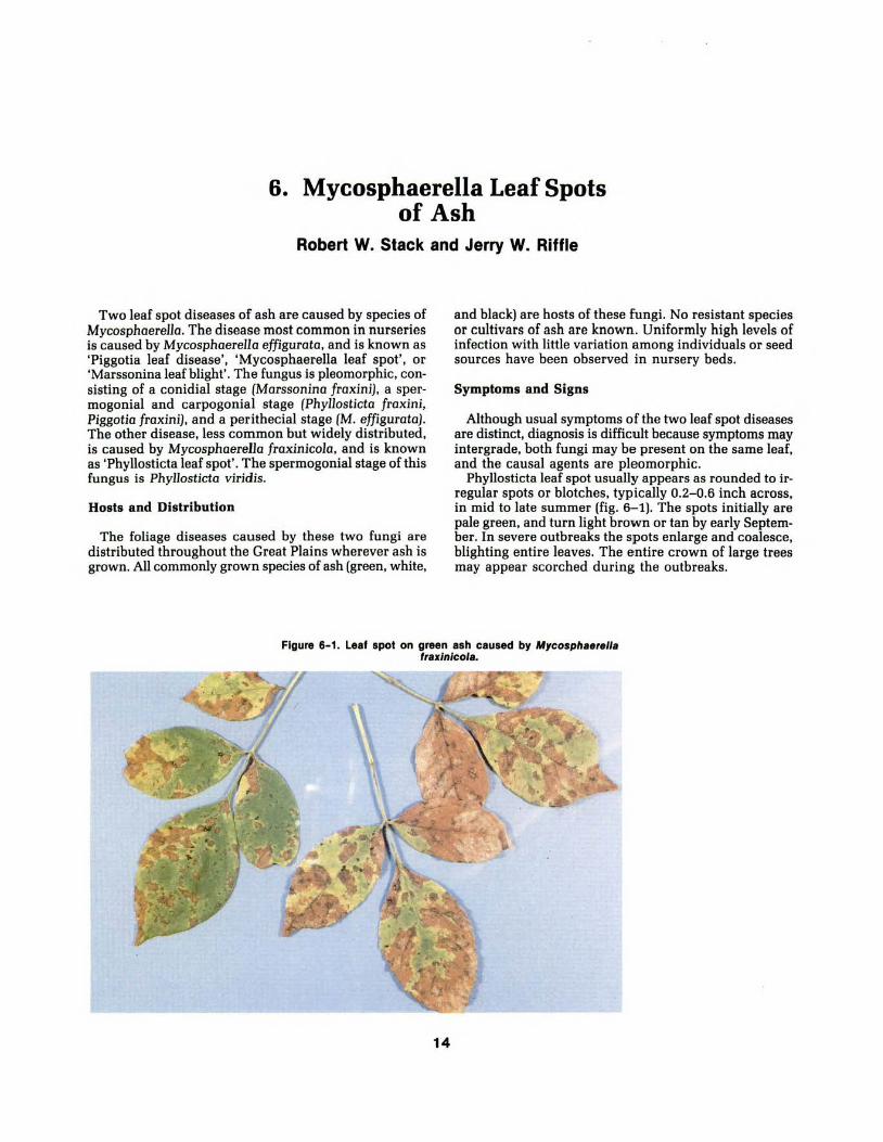
6. Mycosphaerella Leaf Spots of Ash
Robert W. Stack and Jerry W. Riffle
Two leaf spot diseases of ash are caused by species of Mycosphaerella. The disease most common in nurseries is caused by Mycosphaere!Ja effigurata, and is known as 'Piggotia leaf disease', 'Mycosphaerella leaf spot', or 'Marssonina leaf blight'. The fungus is pleomorphic, consisting of a conidial stage (Marssonina fraxini), a spermogonial and carpogonial stage (Phyllosticta fraxini, Piggotia fraxini), and a perithecial stage (M. effigurata). The other disease, less common but widely distributed, is caused by Mycosphaerella fraxinicola, and is known as 'Phyllosticta leaf spot'. The spermogonial stage of this fungus is Phyllosticta viridis.
Hosts and Distribution
The foliage diseases caused by these two fungi are distributed throughout the Great Plains wherever ash is grown. All commonly grown species of ash (green, white,
and black} are hosts of these fungi. No resistant species or cultivars of ash are known. Uniformly high levels of infection with little variation among individuals or seed sources have been observed in nursery beds.
Symptoms and Signs
Although usual symptoms of the two leaf spot diseases are distinct, diagnosis is difficult because symptoms may intergrade, both fungi may be present on the same leaf, and the causal agents are pleomorphic.
Phyllosticta leaf spot usually appears as rounded to irregular spots or blotches, typically 0.2-0.6 inch across, in mid to late summer (fig. 6-1}. The spots initially are pale green, and turn light brown or tan by early September. In severe outbreaks the spots enlarge and coalesce, blighting entire leaves. The entire crown of large trees may appear scorched during the outbreaks.
Figure 6-1. Leaf spot on green ash caused by Mycospheerelle frexlnicole.
14

Symptoms of Mycosphaerella leaf spot first appear in June. Scattered yellow spots, approximately 1-3 mm in size, develop on the upper leaf surface. If the spots are numerous the entire leaf may appear yellow. Conidia are produced in immersed acervuli on the lower leaf surface. Several weeks after the first symptoms appear, numerous small dark stromata, which contain carpogonia and spermogonia, develop over the lower leaf surface. By fall the black stromata give the lower leaf surface a rough sooty appearance (fig. 6-2). The upper leaf surface remains green.
Disease Cycle
Both M. effigurata and M. fraxinicola develop perithecia in fallen leaves over winter. Ascospores of both species are 2-celled, and are similar in shape but not in size. M. effiguroto ascospores are 4 by 15 14m, while those of M. fraxinicola are 4-5 by 8- 10 14m . In spring the ascospores are dispersed by wind to infect new leaves. Infection by ascospores of M. effiguroto appears as minute yellow spots in June. Two-celled hyaline conidia form in acervuli below spots, and the conidia may cause secondary leaf infections. Phyllosticta leaf spot has only a primary disease cycle; spores produced in the spermogonial stage are not infectious. Infections by ascospores of M. froxinicola do not become evident until mid or late summer in contrast to M. effiguroto.
Damage
M. effigurata is especially common in nurseries and on young trees where it may cause premature defoliation. In at least one Northern Great Plains nursery the early defoliation caused by this fungus is considered beneficial by the nursery manager as it causes leaf drop before early snowfall.
M. fraxinicola may cause severe premature defoliation on large established trees. If repeated for several seasons, such defoliation might affect growth increment.
Control
Defoliation by either of these fungi occurs too late in the season to cause renewed growth or winter hardiness problems. No specific controls are known for these diseases.
Selected References
Conners, I. L. An annotated index of plant diseases in Canada. Agric. Publ. 1251. Ottawa: Canada Department of Agriculture; 1967. 381 p.
Hepting, George H. Diseases of forest and shade trees of the United States. Agric. Handb. 386. Washington, DC: U.S. Department of Agriculture; 1971. 658 p.
Wolf, Fredrick A. Leafspot of ash and Phyllosticta viridis. Mycologia. 31: 258-266; 1939.
Wolf, Fredrick A.; Davidson, Ross W. Life cycle of Piggotio fraxini causing leaf disease of ash. Mycologia. 33: 526-539; 1941.
Figure 6-2. Black stromata on lower leaf surface of green ash nursery seedlings caused by M. eff/gurata.
1 5

7. Gloeosporium and Gnomonia Leaf Diseases of Broadleaf Trees
Robert W. Stack and Kenneth E. Conway
Many trees are affected by diseases caused by fungi of the genus Gnomonia and its asexual stage Gloeospo·rium. Diseases caused by these fungi are commonly called 'anthracnose.' The site of infection, kinds of symptoms and signs, and severity of anthracnose disease varies from species to species and often from year to year. The only symptom commonly found on maples and elms may be leaf spot or leaf blight (figs. 7-1, 7-2). In other species, such as sycamore and white oaks. the entire range of anthracnose symptoms may occur, including bud, twig, and shoot blights, leaf spots, and stem cankers.
Figure 7- 1. Black leaf spot of elm caused by Gnomonla ulmea.
16
Hosts and Distribution
Gnomonia-caused diseases are widespread throughout the native and planted ranges of their respective hosts. The pathogens and their hosts growing on the Great Plains are summarized in table 7-1.
Each species of Gnomonia/Gloeosporium is specific for a particular host genus, with little or no cross pathogenicity as far as is known. For each host, the symptoms may differ based on the part of the tree attacked (table 7-1). The taxonomy of Gnomonio/Gloeosporium has been revised, but the more familiar names for the anthracnose pathogens are presented.
Figure 7-2. Symptoms of maple anthracnose caused by Gloeosporlum apocryptum.

Figure 7-3. Leaf distortion and necrosis of green ash caused by Gloeospor/um ar/dum.
Symptoms and Signs
Leaf infections early in the season result in blotching, distortion, and large necrotic areas. These symptoms are typical of ash. oak, and sycamore anthracnose (fig. 7-3 through fig. 7-6). Later leaf infections result in discrete small to large spots, often surrounded by a dark ring or a chlorotic halo. Such spots are the principal symptoms in basswood, elm, and maple (figs. 7-1, 7-2). Severe leaf spot infection may cause extensive defoliation, especially in ash and walnut.
On sycamore and oak, four phases of anthracnose can be distinguished-twig blight, bud blight, shoot blight,
Figure 7-4. Acervuli of Gnomon/a quercina on mldveln of oak.
and leaf blight; leaf blight is already described above. In the twig blight phase, small, 1- or 2-year-old twigs are girdled and killed (fig. 7-5). Later, larger twigs and branches may be cankered and killed. In the bud blight phase, buds are penetrated by the fungus and killed before they expand. In the shoot blight phase, new young shoots and expanding leaves are suddenly killed. In sycamore (and probably other hosts), severe shoot blight depends on temperature, with infection greatest when temperatures are 50° to 59° F. In walnut, optimal infection temperature is 70° F.
Table 7-1. G/oeosporium and Gnomonia anthracnose pathogens and host trees on the Great Plains.
Host
Ash, esp. green Basswood Elm Maple Oak, esp. white Sycamore and London plane·tree Walnut
Pathogen
Gloeosporium aridum Gnomonia tiliae Gnomonia ulmea Gloeosporium apocryptum Gnomonia quercina Gnomonia platani Gnomonia leptostyla
17
Parts of tree attacked
Leaves, twigs Leaves, twigs Leaves Leaves Leaves, twigs, shoots, buds Leaves, twigs, shoots, buds Leaves, twigs, nuts

Figure 7-5. Leaf necrosis and twig die back of sycamore caused by Gnomon/a platen/.
Disease Cycle
The life cycles of these anthracnose fungi are similar and may be typified by G. platani, which attacks sycamore (figs. 7-5, 7-6). The fungus survives the winter in infected twigs and branches on the tree and on fallen infected leaves and branches. In most situations there is abundant inoculum for infection if conditions are favorable. Asexual spores (conidia) are the most important stage for dissemination and infection by anthracnose fungi. In several species, the sexual spores (ascospores) may also be produced and serve as primary inoculum; however, their relative importance compared to the conidial stage is undetermined.
Spores are disseminated by wind and splashing rain to buds, shoots, and expanding leaves, where infection begins if conditions are favorable. In some species, infections develop during favorable periods in summer, giving rise to late-season spots or leaf blight.
18
Damage
Anthracnose fungi cause defoliation and branch dieback, which disfigures the tree. If anthracnose is severe for several seasons, the tree may be weakened and start to decline, or may become susceptible to other diseases or insect pests. Trees stressed by root restriction, drought, heavy scale infestation, etc., are much less tolerant to anthracnose, and may show decreased vigor after only a single season of severe anthracnose defoliation.

Figure 7-6. Leaf necrosis of sycamore caused by Gnomon/a platen/.
Control
Because individual trees vary in susceptibility to anthracnose, there is potential for selection of resistant clones or seed sources of highly resistant trees. The true London plane-tree (an inter-specific hybrid) is much less susceptible than the native American sycamore. Oaks in the white oak group are much more susceptible to anthracnose than those in the red oak group.
Fungicide sprays have long been used in attempts to control anthracnose on boulevard and residential plantings. Specific protectant fungicides are registered for certain tree species. Follow label instructions for times of application and dosage rates. Specific recommendations on type of spray and timing vary with locality. Consult your local Extension Plant Pathologist or Forester for current recommendations for your area. Recently, several systemic fungicides have been registered for tree injection to control anthracnose fungi.
Cultural measures, such as raking up leaves in fall and pruning out infected twigs and branches, reduce spring inoculum, but probably not enough to affect disease development except on isolated young trees.
19
Selected References
Barr, M. E. The Diaporthales in North America. Mycologia Memoir 7. Lehre, Germany: J. Cramer Publication; 1978.
Cooper, D. T.; Filer, T. H. Jr.; Wells, 0. 0. Geographic variation in disease susceptibility of American sycamore. Southern Journal of Applied Forestry. 1: 21-24; 1977.
Hepting, George H. Diseases of forest and shade trees of the United States and Canada. Agric. Handb. 386. Washington, DC: U.S. Department of Agriculture; 1971. 658 p.
Neely, Dan; Himelick, E. B. Temperature and sycamore anthracnose severity. Plant Disease Reporter. 47: 171-175; 1963.
Neely, Dan; Himelick, E. B. Characteristics and nomenclature of the oak anthracnose fungus. Phytopathology. 57: 1230-1236; 1967.
Snyder , T. E.; Stack, R. W. Occurrence of Gloeosporium oridum on green ash at different times of the year. Proceedings North Dakota Academy of Sciences. 37: 95; 1983.

8. Oak Leaf Blister Kenneth E. Conway and John E. Watkins
Oaks, our most abundant hardwood genus, grow naturally in the eastern and southern Great Plains. They are planted frequently in rural and urban environments as shade trees, and are occasionally used in windbreaks in the northern Great Plains. Oak leaf blister, caused by Taphrina caerulescens, is one of the most common leaf diseases of oak.
Hosts and Distribution
Over 90 species of Taphrina are recognized; they cause diseases on various hosts. Some of the more widely recognized diseases include peach leaf curl (peach), plum pockets (plum), and leaf blisters (cherry, maple, elm, alder, oak).
Oak leaf blister is a major leaf disease of oaks in the southern United States. Oaks in the red and black subgenus are most susceptible to T. caerulescens, but all species of oak are susceptible.
Symptoms and Signs
This disease may go unnoticed until a large number of leaves are severely infected or begin to fall. The in-
itial symptom is a slight yellowing of infected leaf tissue, followed by formation of circular, raised blisters on the leaves (fig. 8-1). Blisters form when infected cells are stimulated to enlarge, while surrounding noninfected cells remain rigid. Blisters are usually less than 1 inch in diameter, and the lower surface will appear gray as the fungus develops in the leaf tissue.
The upper surface ofthe leaf blister remains light green for several weeks before dead tissue turns brown. Multiple infections cause a single leaf to become distorted (fig. 8-2). Premature defoliation may occur in the early fall. The fungus survives, presumably as conidia, on bud scales and in bark crevasses.
Disease Cycle
No fruiting structure is formed by T. caerulescens. Leaves are infected in early spring by conidia formed from ascospores during the previous season. Mycelium ramifies through the leaf tissue intercellularly and becomes massed just below the cuticle, where it eventually fragments into thick-walled binucleate cells. The upper portion of these cells becomes an ascus and eight ascospores are formed. As the asci form, pressure is ex-
Figure 8-1. Distorted oak leaves showing mild symptoms caused by Taphr/na caeru/escens.
20

erted on the cuticle, and the asci eventually break through to form a compact layer on the epidermis of the host (fig. 8-3). Ascospores bud in the ascus to produce conidia or are released intact from the ascus. Released ascospores then bud to form conidia. The conidia reside in bud scales and in bark crevasses, and spread to susceptible leaf tissue by rain where they produce germ tubes that infect leaves the following spring as the leaf buds begin to develop.
Damage
Heavy infections occur following cool, wet springs and may result in 50 to 85 percent defoliation of affected trees by midsummer. Defoliation can reduce growth and, if repeated over a period of years, may weaken the tree so that it is susceptible to attack by other organisms.
Control
Chemical control of oak leaf blister is not normally recommended for forest or shade trees. The disease may be unsightly and cause much anguish to homeowners, but vigorous trees are not severely affected. Particularly valuable trees may warrant treatment with fungicide, which must be applied as dormant spray to be effective. Fungicides are not effective after leaves begin to develop because infection has already occurred. Collecting and disposing of infected leaves will remove some inoculum. Maintenance of trees in healthy condition by fertilization, watering, pruning, and insect control will reduce the effect of this disease.
Figure 8-3. Cross section of a leaf blister showing a compacted layer of asci of T. caerulescens on the upper surface of a leaf.
Selected References
Kramer, C. L. Protomycetales and Taphrinales. In: The fungi. An advanced treatise. Vol. IV A. Ainsworth, G. C.; Sparrow, F. K.; Sussman, A. S., eds., New York: Academic Press; 1973: 33-41.
Mix, A. J. A monograph of the genus Taphrina. University of Kansas Science Bulletin. 33: 3-167; 1949.
Skelly, J. M. Leaf diseases of hardwood. Forest tree diseases of Virginia. Extension Circular MR-FTD-16. Richmond, VA: 1972.
Figure 8-2. Blistered and distorted leaves of bur oak caused by T. caerulescens.
21

9. Leaf Spots of Nanking Cherry and Chokecherry
Robert L. James and John E. Watkins
Leaf spots are common diseases of Nanking cherry and chokecherry in the Great Plains. Chokecherry is often planted in windbreaks; Nanking cherry, a common or· namental. is also planted occasionally in windbreaks.
Hosts and Distribution
Nanking cherry and chokecherry leaf spots are caused by the fungus Coccomyces hiemalis (often referred to as C. Jutescens on chokecherry) and the bacterium Pseudomonas syringae pv. syringae. Both pathogens produce typical "shot-hole" lesions, a common name of the disease. These diseases are common throughout the range of their hosts and are particularly important in the Great Plains and eastern United States. C. hiemaJis also causes a serious disease of commercial cherries, par·
22
ticularly in the Lake States. Pseudomonas syringae pv. syringae also causes leaf spots and cankers of commer· cial stone fruit trees.
Symptoms and Signs
Leaf spots caused by C. hiemalis first appear as small, purple or reddish circular spots, which later enlarge and turn brown. Spots are usually 4-5 mm in diameter; they may be few in number or become so numerous as to coalesce and form large irregular necrotic areas (figs. 9-1, 9-2). During humid periods, whitish spore masses (con· idia) are visible in the center of lesions; these masses are more numerous on the underside of leaves. Generally, necrotic tissues within lesions separate from the surrounding leaf tissues, forming characteristic shot-holes. During severe stages of disease, infected leaves become highly chlorotic, and trees may appear yellow. Premature defoliation of heavily infected trees may occur.
The leaf spots caused by P. syringae pv. syringae are similar. Lesions are reddish-brown, angular or circular, and frequently fall out, giving infected leaves a tattered appearance. Lesions may coalesce to form large areas of necrotic tissue. Heavily infected leaves are chlorotic and may be shed prematurely. P. syringae pv. syringae also may cause depressed black lesions on fruits, and cankers on twigs and branches. Gummosis commonly occurs around cankers.
Disease Cycle
Infection by C. hiemalis begins in the spring, and is caused by ascospores from fruiting bodies (apothecia) that form on previously infected, fallen leaves. Ascospores are wind dispersed and cause infection dur· ing April and May. Leaves are especially susceptible
Figure 9-1. Leaf spot symptoms on chokecherry caused by Coc· comyces hiemalis.

shortly after bud break. The conidial stage of the fungus (Cylindrosporium sp.) forms white spore masses within the center of leaf lesions. Conidia are dispersed by rain and cause secondary infections throughout the summer. Warm temperatures are especially conducive to disease development. Heavy infection may cause premature leaf fall; some trees may be completely defoliated by July. Repeated infections reduce tree vigor.
P. syringae pv. syringae is a common epiphyte and often occurs on the fruit, limbs, and leaves of both susceptible and nonsusceptible plants. Infection is correlated more closely with favorable weather conditions than with the availability of inoculum. Bacteria overwinter within cankers and are spread to and cause infection of leaves in the spring during cool, wet weather. Free-standing water is necessary for leaf infection. Cankers are formed when bacteria infect branches through wounds, such as those made during pruning. Repeated infections develop throughout the growing season during wet periods. Heavily infected leaves fall prematurely. Trees with repeated infections decline over several years.
Damage
C. hiemalis causes serious damage to commercial cherries in parts of the United States and Europe. Effects on Nanking cherry and chokecherry are less dramatic, although plantings of chokecherry have been reduced in parts of the Great Plains because of this disease. The pathogen commonly causes severe defoliation of chokecherry in Great Plains nurseries. P. syringae pv. syringae is most important as a canker-causing pathogen of commercial stone fruit trees. Its effect on Nanking cherry and chokecherry is limited, although some windbreak and ornamental plantings may be severely infected. Both diseases reduce vigor of trees but rarely kill them.
Control
Usually, control of these leaf spot diseases is necessary only in commercial fruit growing areas and in nurseries. C. hiemalis can be controlled with prote'Ctant fungicides applied directly to foliage, or with eradicant fungicides applied on previously infected leaves on the ground about the time apothecia are swelling. Best results have been obtained with several foliar applications of fungicides coordinated with periods of high infection hazard.
Figure 9-2. Leaf spot symptoms on sweet cherry caused by C. hlemalls.
23
A model has been formulated recently which predicts periods of high disease probability in commercial cherry orchards. The number of fungicide applications neoessary to control the disease has been reduced by use of this model.
Leaf spot caused by P. syringae pv. syringae can be controlled by sanitation, such as dormant pruning of cankered wood and removal of leaves and other debris in the fall. Fungicides can be used to suppress disease development, but fungicides alone do not provide good control. A combination of sanitation and fungicide application is recommended.
Selected References
Cameron, H. R. Mode of infection of sweet cherry by Pseudomonas syringae. Phytopathology. 52: 917-921; 1962.
Eisensmith, S. P., R. Loria, B. D. Olson, and A. L. Jones. 1980:EPIST AR: An epidemiological information storage, retrieval, and analysis system. Plant Disease 64: 646-651.
Heald, Frederick Deforest. Manual of plant diseases. New York: McGraw-Hill; 1933. 953 p.
Inman, R. E.; Weihing, J. L. Cyprex: a superior control for shothole disease of chokecherry. Plant Disease Reporter. 43: 536-539; 1959.

10. Western X-Disease of Chokecherry
Glenn W. Peterson and David W. Johnson
Chokecherry is used as a shrub in windbreaks and as a component of wildlife plantings in the central and northern Great Plains. Chokecherry in many of these plantings is infected by the western X-disease pathogen (fig. 10-1).
Hosts and Distribution
The western X-disease pathogen infects chokecherry, sweet and sour cherries, several varieties of peach, and some other Prunus species. American plum has been infected by inoculations; however, after 9 years there were no visible symptoms on American plum interplanted among infected chokecherry in eastern Nebraska.
The disease is present in the northern and central Great Plains (Wyoming, North Dakota, South Dakota, and Nebraska).
Figure 10-1. Infected chokecherry along length of windbreak.
24
Symptoms and Signs
Infected leaves become greenish-yellow in late June. These leaves may have a reddish tinge on their borders. In July and August the leaves turn deep red (figs. 10-2, 10-3). Shoots are stunted, and rosettes result from shortened internodes at the tip. Infected fruits are somewhat pointed and are yellowish-red, not the normal deep red of healthy fruit. Both diseased and healthy fruits may be found on the same tree.
Disease Cycle
The western X-disease pathogen for many years was thought to be caused by a virus; however, the disease is caused by a spiroplasma.
The western X-disease pathogen is transmitted to

Figure 10-2. Western X·dlsease symptoms, several trees.
Figure 10-3. Symptoms, single tree.
Prunus hosts by Collodonus geminotus, C. montonus, and other leafhoppers when they feed on the leaves. Symptoms on leaves usually do not develop until the growing season following the year of transmission.
Damage
Growth of infected chokecherry is reduced, internodes become shorter, the shrubs gradually decline, and ultimately die. In eastern Nebraska, symptoms appeared on more than 80 percent of the chokecherries within 3 years after the X-disease pathogen was introduced; mortality was more than 50 percent within 8 years. Infected fruits are not suitable for use in jams and jellies, and their seeds do not germinate.
Control
Nurseries should avoid establishing beds of chokecherry near Prunus species that are hosts of the pathogen. Because the pathogen spreads rapidly from infected to healthy shrubs, new plantings of chokecherry should not be established near infected chokecherry.
Because American plum has been infected following artificial inoculation with the pathogen, it could possibly
25
be a symptomless carrier and thus a threat to chokecherry, but there is no supporting evidence.
Thus, American plum can be included in plantings containing chokecherry with confidence that the Xdisease pathogen will not seriously damage it.
Selected References
Gilmer, R. M.; Moore, J. Duain; Keitt, G. W. X-disease virus: I. Host range and pathogenesis in chokecherry. Phytopathology. 44: 180- 185; 1954.
Granett, A. L.; Gilmer, R. M. Mycoplasmas associated with X-disease in various Prunus species. Phytopathology. 61: 1036-1037; 1971.
Peterson, Glenn W. Western X-disease virus of chokecherry: transmission and seed effects. Plant Disease Reporter. 50: 659- 660; 1966.
Peterson, Glenn W. Spread and damage of western Xdisease of chokecherry in eastern Nebraska plantings. Plant Disease. 68: 103- 104; 1984.
Rosenberger, D. A.; Jones, A. L. Leafhopper vectors of the peach X-disease pathogen and its seasonal transmission from chokecherry. Phytopathology. 68: 782-790; 1978.

11. Honeysuckle Leaf Blight Jerry W. Riffle and John E. Watkins
Honeysuckle has been commonly used in windbreak and landscape plantings in the northern Great Plains. Demand for hardy shrub species is high, but production of honeysuckle is hampered by frequent epidemics of honeysuckle leaf blight and other pests. This blight is caused by Insolibasidium deformans. This pathogen is listed as Herpobasidium deformans in papers published before 1984.
Hosts and Distribution
Most species and varieties of native and introduced honeysuckle are hosts of I. deformans. Amur honey· suckle has shown some resistance to the pathogen in a nursery in Iowa. The disease is widespread in north·
Figure 11-1. Lower surfaces of honeysuckle leaves infected with lnsollbasldlum detonnans and showing tan to brown necrotic areas
involving large portions of leaves.
26
eastern and northcentral U.S., and occurs in North Dakota, South Dakota, Nebraska, Minnesota, Iowa, Wisconsin, Michigan, Indiana, Ohio, Pennsylvania, New York, Massachusetts, Connecticut, and Rhode Island.
Symptoms and Signs
This disease appears in the spring on newly emerging leaves. The first symptom is a yellowing of infected veinlets and of leaf tissues bounded by veinlets. These tissues become tan to brown and finally necrotic and dry, with brown areas involving an entire leaf or a large portion of it (fig. 11-1). The leaves are often rolled and twisted (fig. 11-2) and drop prematurely. The first sign of the pathogen is a thin, white layer of basidia and
Figure 11-2. Honeysuckle seedlings infected with /. deformans show curled, rolled, and discolored leaves.

Flgure11-3. Honeysuckle seedling Infected with powdery mildew fungus.
basidiospores on the lower leaf surface, often followed by a white powdery mass of conidia. Powdery mildew fungus can be distinguished from I. deformans by white powdery mildew mycelia and black fruiting bodies that develop principally on the upper surface of nonrolled leaves in late summer (figs. 11- 3, 11- 4).
Two spore stages may be found on diseased leaf tissues. Mature basidiospores arise from curved, transversely septate basidia emerging through stomates from hyphae in the leaves. Basidiospores are hyaline, uninucleate, cylindrical with rounded ends except for an apiculus at the attached end, and measure 9-13 by 5-7.5 ~tm (average 10.9 by 6.6~tm) (fig. 11-5). They have been found commonly throughout the growing season in nursery beds containing infected stock. Conidia frequently develop in or near the area of basidial formation on the lower leaf surface. They occur less commonly than basidiospores and are difficult to find. Conidia are 8-17 ~tm in diameter, globose, hyaline, warty, binucleate, and form three pairs to a cluster (fig. 11-6}. The outer pairs are usually the only spores to germinate. The function of conidia is unknown; they are presumably resting spores.
27
Figure 11-4 Honeysuckle leaf with numerous black fruiting bodies of powdery mildew fungus on upper surface.
Figure 11-5. Basldlospores of/. deformans.

Disease Cycle
I. deformans overwinters as mycelium or basidiospores in dead leaves. Basidiospores serve as primary inoculum for infection of developing J.eaves of the first foliage flush, and also for secondary infections during the remainder of the year. Infection of leaves is intensified whim the temperature is 59° to 64° F, the relative humidity is near or at 100 percent in sustained periods for at least 2 days, and leaves are less than 20 days old. Below-normal temperatures and high humidity in August and September enhance disease development, which results in premature loss of foliage and growth reduction.
Dalllage
Epidemics have increased in frequency in central and northern Great Plains nurseries since 1960. When blight is severe, most seedlings in nursery beds become infected and defoliate prematurely (fig. 11-7). Severe defoliation results in stem dieback and reduced growth, and stock may have to be retained in the nursery an additional year. Fifty percent or more of plantable seedlings have been lost in at least one northern Great Plains nursery even after seedlings were held for an additional year. Such losses also disrupt planned nursery stock inventories because seedbeds are used for an additional year.
No information is available on extent of damage to honeysuckle in windbreak and landscape plantings.
Control (Cultural)
Honeysuckles in landscape, windbreak, and nursery plantings may become infected; thus they should not be used in nursery windbreaks or in other plantings on nursery grounds because they may be a source of I. deformans inoculum for nursery seedlings.
Seedlings grown at high densities in seedbeds develop dense foliage that reduces aeration and increases humidity, conditions that favor disease development. Where possible, reduce seedbed density or grow seedlings as row crops to improve aeration and reduce humidity. Irrigation should be scheduled so that periods of high moisture and free water in and around foliage and stems are as short as possible. Any cultural practice that destroys or removes overwintering foliage from seedbeds will reduce inoculum sources.
28
Figure 11-6. A cluster of three pairs of I. deformans conidia.
Control (Chemical)
A regular protective spray program will minimize the risk of disease damage. Seedlings need protection during the entire growing season because basidiospores are dispersed throughout the period. Protectant fungicides must be applied frequently to protect newly developing susceptible foliage. Mancozeb (ForeR turf and ornamental fungicide) is registered for control of I. deformans on honeysuckle. Use 1.5 pounds per 100 gallons of water in full coverage spray to point of runoff. Begin spraying when seedlings are leafed-out and apply at 7- to 10-day intervals throughout the season. Chlorothalonil (Daconil 2787R), applied at 2 quarts per 100 gallons of water per acre gave best control of I. deformans on tatarian honeysuckle among seven fungicides tested in an Iowa nursery in 1984. Fungicides used for control of I. deformans in nurseries may be effective for control of this pathogen in windbreaks and landscapes.

Figure 11-7. Nursery beds containing 2-year-old honeysuckle seed· lings Infected with I. deformans.
Selected References
Gould, Charles J. , Jr. The parasitism of Glomerularia Joniceroe (Pk.) D. and H. in Lonicero species. Iowa State College Journal of Science. 19: 301-331; 1945.
Oberwinkler, F.; Bandori, R. Herpobosidium and allied genera. Transactions British Mycological Society. 83: 639- 658; 1984.
Riffle, Jerry W. Recovery of Herpobosidium deformans basidiospores from Lonicera tatarica leaves overwintered in nursery beds. (Abstract) Phytopathology. 71: 251-252; 1981.
Sweets, Laura E.; Croghan, Catherine F. Evaluation of fungicides for honeysuckle leaf blight control on Tatarian honeysuckle, 1984. American Phytopathological Society; Fungicide and Nematicide Tests. 40: 205; 1985.
29

12. Powdery Mildew of Lilac Richard Dorset and Michael W. Ferguson
Lilacs (Syringa spp.) are planted throughout the Great Plains. Most varieties sprout readily from roots, creating dense thickets that make lilac valuable for use in windbreaks, especially in the central and northern Great Plains.
One of the few pathogens that infects and damages lilacs is Microsphaera olni, which causes powdery mildew of lilac foliage.
Hosts and Distribution
Powdery mildew fungi infect most species of deciduous woody shrubs and trees. Some are highly specific, infecting only one host, whime others have a wide host range. M. olni infects not only lilac but a large variety of plants, including alder, birch, hornbeam, hophornbeam, chestnut, holly, maple, hickory, golden chinkapin,
beech, honeylocust, walnut, sycamore, oak, elm, and basswood. With such a wide host range, the fungus is distributed throughout the Great Plains.
Even though powdery mildew fungi can infect most species and varieties of lilac, there is a considerable range of host susceptibility.
Symptoms and Signs
During mid-summer, leaves develop small white or gray dusty-looking patches. These patches enlarge throughout the summer, and by early fall the entire leaf surface may be covered with a white powdery-looking substance (figs. 12- 1, 12- 2). Later in the fall, small pinpoint-sized brown to black structures develop throughout the powdery areas. These are the sexual fruiting bodies of the fungus.
Figures 12-1, 12-2. Lilac leaves infacted by powdery mildew fungus.
30

Microscopically. the white powder consists of fungus mycelium and asexual spores (fig. 12-3). The sexual fruiting bodies, called cleistothecia, have numerous slender appendages that are dichotomously branched at the tips. Several asci are contained in each cleistothecium.
Disease Cycle
The pathogen overwinters in fa llen leaves as partially developed ascospores (fig. 12-4). These spores mature during wet spring weather, and are then exuded from the black fruiting bodies. These spores are blown or splashed onto non-infected foliage. After germination, haustoria of the fungus penetrate the leaf tissue and are restricted to a single layer of cells, the palisade layer. Mycelium growing on the leaf surface produces asexual spores, which are powdery white. These asexual spores are dispersed by wind and rain to other leaves, starting new infections.
The fungus grows best during warm, damp. summer weather. At the onset of cool weather, growth slows or ceases and the sexual fruiting bodies are produced; they overwinter on dead leaves.
Damage
Powdery mildew fungi seldom cause enough damage to warrant control measures. However, when infection is extensive, the unsightly appearance caused by these fungi is often severe enough that homeowners may wish to attempt control. Extensive infections occur in late fall just before the leaves drop normally, and reduce the aesthetic and ornamental values of landscape plants.
Control
The easiest way to control powdery mildew is to prevent its occurrence. The disease is best prevented by using resistant species, cultivars, and varieties. Removal of all dead leaves and leaf pieces in the fall will reduce the amount of primary inoculum during the following spring, thereby reducing the number of initial infections.
The severity of infection can be minimized by providing good air circulation and sunlight. Dense plantings, shady areas, and damp places all favor disease development.
Chemicals can be used to either prevent infection or to control established infections. Chemicals should be applied initially when the new leaves are emerging, and repeated as per label instructions. Sulfur has long been used fo r control. Bayleton and benomyl are two newer chemicals that are registered for powdery mildew on lilac. Both work systemically and have good residual action. Bayleton is a foliar spray, while benomyl may be used as either a soil drench or foliar spray.
31
Figure 12-3. Chaine of conidia of powdery mildew tungua.
Selected References
Fenicchia, Richard A. Susceptibility of lilacs to leaf curl necrosis and powdery mildew. Plant Propagator. 23(3): 7-13; 1977.
Hepting, George H. Diseases of forest and shade trees of the United States. Agric. Handb. 386. Washington, DC: U.S. Department of Agriculture; 1971. 658 p.
Tattar, Terry A. Diseases of shade trees. New York: Academic Press; 1978. 361 p.
Figure 12-4. Life cycle of a powdery mlldrew fungua.

13. Cylindrosporium Leaf Spot of Buffaloberry and Skunkbush Sumac
Glenn W. Peterson and Jerry W. Riffle
Buffaloberry (Shepherdio orgenteo) and skunkbush sumac (Rhus trilobata) are commonly used in shrub rows in Great Plains windbreaks and in wildlife plantings.
Hosts and Distribution
The leaf spot diseases of skunkbush sumac and buffaloberry were detected in an eastern Nebraska nursery in the early 1960's. The leaf spots are caused by fungi in the genus Cylindrosporium. The fungi have not been identified to species; however, they are similar to Cylindrosporium species that have been described on other Rhus and Shepherdio species. C. toxicodendri has been found on R. toxicodendron, R. trilobata and R. diversiloba; C. shepherdiae has been found on S. canadensis. The distribution of these fungi in the Great Plains is not known.
Symptoms and Signs
Circular to irregular leaf spots develop on skunkbush sumac; these spots have tan centers and dark borders. Spotted leaves ultimately turn yellow and drop prematurely (fig. 13- 1). On buffaloberry, the leaf spots are irregularly shaped, with tan centers and olive borders. Spotted leaves ultimately become necrotic and drop prematurely. Initial symptoms (leaf spots) on both shrub species develop in late May; considerable defoliation of buffaloberry may occur before mid-June.
Disease Cycle
Detailed information on the disease cycle is lacking.
32
The primary source of spores for initial infection in the spring is probably fruiting bodies that have overwintered on fallen leaves. Leaves are initially infected in May. The fruiting bodies (acervuli) form within the leaf spots and produce slender, septate spores which average 32 by 2.5 I'm on buffaloberry and 49 by 2.7 I'm on skunkbush sumac.
Damage
Nursery production of skunkbush sumac and buffaloberry is hindered by these leaf spot diseases. Infected seedlings grow poorly due to premature defoliation; they often are kept an additional year in the nursery to reach size sufficient for planting. Damage by these fungi is seldom extensive in field plantings.
Control
Caplan or maneb provided ·adequate control of these fungi in tests in an eastern Nebraska nursery. The first application should be made in mid- to late May. Several applications are necessary to protect newly developing foliage.
Selected References
Peterson, Glenn W. Control of Cylindrosporium leaf spot disease of Rhus trilobota and Shepherdia argentea seedlings. Plant Disease Reporter. 51: 700- 701; 1967.
Saccardo, P. A. Sylloge Fungorum. 11: 582; 1895. Saccardo, P. A. Sylloge Fungorum. 25: 623; 1931.
Figure 13-1. Defoliation of skunkbush aumac aeedllnga caused by Cyllndrosporlum sp.

14. Herbicides (Air Pollution) Gary A. Boutz and Robert W. Stack
The greatly increased use of herbicides since the mid-1950's has also increased the hazard to nontarget vegetation. Disorders or mortality caused by herbicides are particularly prevalent where susceptible tree species (table 14-1) are located near crops or areas where her-
Table 14-1. Sensitivity of various tree species to broadleafed weed-killers.
Sensitive
boxelder elm ash hackberry Amur maple hard maple Ailanthus hickory apple sycamore redbud walnut Amur cork tree willow birch horsechestnut
Intermediate or unknown
mulberry honeylocust soft maple oak cottonwood cherry
Tolerant
catalpa linden pine e. redcedar
33
bicides are frequently used. Windbreaks are especially vulnerable to exposure because they are generally located in or around cultivated fields. Trees near right-of-ways, railroads, roadsides, or areas treated for noxious weed control also have a high risk of exposure.
Symptoms
The more common expressions of phenoxy or hormonal-type herbicide injury include cupped leaves, parallel leaf venation on normally net-veined leaves, chlorosis, nastic growth, and wavy or curled leaf margins (figs. 14-1, 14-2, 14- 3). Redbud and boxelder are indicator tree species that readily exhibit these effects. Leaves of some trees, such as pin oak, become waxy and stiff, while maples may develop pebbled foliage that appears weather-beaten. Studies with 2,4-D applied to Siberian elm have shown that bark abnormalities may result from relatively heavy exposure. A single 2,4-D exposure may produce injury for 2 years or more.
Loss of apical growth is typical of phenoxy or hormonal herbicide injury. Affected trees may suffer a gradual crown dieback and eventually die. Lateral leaf development may also be hindered. Exposure to 2,4-D
Figure 14-1. Phenoxy herbicide Injury to boxelder. Affected foliage Is chlorotic, leaves are cupped, and chlorophyll in leaves appears
variegated.

Figure 14-2. Foliage of Siberian elm becomes cupped when dam· aged by herbicides.
may result in the production of fewer normal leaves, flowers, and fruits.
Leaf scorch can indicate herbicide injury, particularly when exposure is heavy. Ash and cottonwood may exhibit scorched leaves without expressing the more common effects of herbicide exposure.
Evergreens are generally resistant to phenoxy herbicides, but they may be injured under certain circumstances. Exposed fir trees may develop curled leader growth, burned needle tips, and needle cast. Spruce may also show terminal growth damage and needle cast (fig. 14-4). Pine trees are most susceptible to damage during periods of active growth, and candles exposed to phenoxy herbicide may develop nastic growth.
The non-hormonal type herbicides generally do not induce abnormal growth, but cause chlorosis and eventual death of affected tissue. With contact herbicides, such as paraquat, this damage is restricted to areas contacted.
Diagnosis of herbicide damage can be aided by careful collection and analysis of residue samples. It is important that the samples be collected as soon as possible following exposure. It is necessary to specify the herbicides the laboratory should evaluate. With some herbicides, such as 2,4-D, it is necessary to specify the exact
34
Figure 14-3. Green ash severely damaged by herbicides in a young windbreak. Note distortion of new growth and cupping of foliage.
kind of 2,4-D. When possible, residue samples should be kept frozen until they reach the laboratory. It may be advisable to contact an analysis laboratory for specific instructions on collection and care of samples.
Damage
All possible sources of chemical exposure should be considered when dealing with suspected herbicide damage. Harmful exposure to herbicides can result from drift of the spray particles, movement of volatiles, application to the soil or movement in soil (or water) resulting in root exposure, and direct application. Spray drift volatiles can move considerable distances, resulting in damage several miles from where the herbicide was applied. Direct application may cause damage if sensitive desirable trees are not carefully avoided, or if selective herbicides are misapplied directly over trees.
In urban areas, desirable plants are most likely to be damaged by lawn sprays or fertilizers that contain herbicides. Damage is also commonly associated with soilapplied herbicides used around driveways, sidewalks, and structures. In situations where damage has resulted from an apparent misuse of herbicides, it may be appro-

Figure 14-4. Colorado blue spruce with symptoms of MCPA her· bicide damage. Affected foliage is necrotic and some needles have
been cast prematurely.
priate to contact the state pesticide regulatory agency or the U.S. Environmental Protection Agency.
Control
Desirable trees located where herbicide exposure is likely will require protective effort. Persons who apply herbicides should be informed of where desirable trees are located, and products that pose a minimal risk to the trees should be used. Extra caution should be exercised to ensure proper calibration and application. Once exposure has occurred, little can be done to minimize the initial effects. Washing herbicide off foliage can be effective if it is done during or immediately after exposure. Soil-active herbicides can be deactivated on a limited scale with activated charcoal or similar products. Generally, any effort that promotes tree vigor should help minimize the effects of the spray, except that regrowth of foliage should not be stimulated. Trees weakened by
35
herbicides may be predisposed to insects, pathogens, and other types of environmental damage.
Selected References
Hibbs, R. H. Decline of hackberry attributed to ambient herbicide drift. Proceedings Iowa Academy of Science. 72{3-4}: 187-190; 1976.
Hibbs, Robert H. Recognition of weed-killer injury to trees. Journal of Arboriculture. 4: 189-191; 1978.
Otta, J. D. Effects of 2,4-D herbicide on Siberian elm. Forest Science. 20: 287-290; 1974.
Phipps, Howard M. The role of 2,4-D in the appearance of a leaf blight of some plains tree species. Forest Science. 9: 283-288; 1963.
Sherwood, C. H.; Weigie, J. L.; Denisen, E. L. 2,4-D as an air pollutant; effects on growth of representative horticultural plants. Horticultural Science. 5: 202, 211-213; 1970.

15. Chlorosis Mark 0 . Harrell and Mark W. Andrews
Many trees in the Great Plains suffer from nutrient deficiencies induced by alkaline soils. One symptom commonly produced by these deficiencies is a yellowing of the foliage, called chlorosis.
Hosts and Distribution
Chlorosis of fo liage may develop wherever trees are grown in alkaline soils. Chlorosis generally is most severe in the western two-thirds of the Great Plains where highly alkaline soils are common. Chlorosis is also common in urban a reas where excavation during construction of buildings brings alkaline subsoil to the surface.
Many tree species are susceptible to chlorosis. Pin oak and silver maple are commonly affected. Also susceptible are other oak and maple species, birches, cottonwood, poplars, elms, pines, junipers, yews, walnut, peach, and apple.
Symptoms and Signs
The degree of yellowing varies from a yellowish-green of leaves only slightly chlorotic to lemon-yellow and almost white in leaves severely chlorotic . In some cases the leaves have a slight reddish or "fall" coloration. The yellowing is most intense in the interveinal areas of leaves (fig. 15- 1); brown, necrotic areas often develop in these areas on severely affected leaves, giving the appearance of leaf scorch.
Symptoms of chlorosis may be uniform throughout the entire tree or be confined to one or a few branches (fig. 15- 2). Generally, leaves formed in early spring are normal in both color and size, but leaves formed later in the season become increasingly chlorotic and are smaller. If chlorosis continues for several years, shoot growth is reduced, branches begin to die back (fig. 15-3), and the tree eventually dies.
Figure 15-1. Chlorotic pin oak leaves showing typical green veins and chlorotic lntervelnal areas.
36
Cause
Deficiency of iron is the most common cause of chlorosis of trees in the Great Plains. In most cases, iron in the soil is present in sufficient quantity; but under alkaline conditions (pH greater than 7.0), it is in an insoluble form. Reduced availability of nutrients occurs commonly in alkaline soils. Even if absorbed, these nutrients may remain in a form that the tree is unable to use. Iron is used in the production of chlorophyll, thus a deficiency of iron prevents the leaves from producing the normal amount of chlorophyll.
Deficiencies of other nutrients such as zinc, manganese, or nitrogen can contribute to chlorosis or in some cases they are the primary cause of chlorosis. Factors such as low temperature and high soil moisture, and excessive amounts of copper; manganese, zinc, and phosphorus can cause or contribute to development of chlorosis.
Figure 15- 2. A moderately chlorotic silver maple wllh some branch dleback.

Figura 15-3. A severely chlorotic pin oak with ex· tanslva branch dlaback (left).
Figure 15-4. The same tree as In fig. 15-3 show· lng the Improvement In leaf color and crown full· nasa 2 years after a trunk Injection with ferrous
sulfate (right).
Damage
Homeowners with chlorotic trees commonly spend $20 to $50 per tree every 3 or 4 years for therapeutic treatments. Without treatment. the trees would continue to decline and die. In many areas of the Great Plains some species, especially pin oak, are no longer being planted because this disorder severely reduces their chance of survival.
Control
Three methods can be used to treat chlorosis caused by iron deficiency: (1) spraying the foliage with a solution of ferrous sulfate (iron sulfate) or chelated iron; (2) incorporating ferrous sulfate and sulfur, or chelated iron into the soil; and (3) injecting ferrous sulfate or chelated iron into tree trunks.
Spray treatment gives the quickest response, but is the most temporary. This treatment only improves the condition of leaves present when foliage is sprayed. It has little or no effect on leaves formed after spraying, and the effectiveness does not carry over into the next growing season. This treatment is not recommended for longterm control of chlorosis. It is only used on trees when chlorosis is extremely severe and a quick response is desired.
Soil treatment provides the most permanent control of chlorosis. A single soil treatment is often effective for 4 to 5 years. The disadvantages of this treatment are that it may not become effective until the next growing season, and it requires considerable labor and materials.
37
This treatment is often recommended for shrubs and trees that are too small for trunk injections, and in cases where the landowner wants to avoid making the wounds required for trunk injections. The recommended materials and rates are: (1) 2 to 2.5 pounds per inch tree diameter of a mixture of equal parts ferrous sulfate and sulfur, or (2) an iron chelate used at the rate indicated on the label. Several chelated iron materials are available.
Trunk injection is the most commonly used treatment for chlorosis. This treatment involves drilling holes into the lower trunk and either placing capsules of powdered material into the holes, or injecting a liquid solution through the holes. Trees usually respond within 3 to 4 weeks, and the treatment is generally effective for 2 to 4 years (fig. 15-4). More information on procedures and materials is available from most nurseries and garden stores. Some materials can be applied easily by the homeowner, while others are applied only by trained arborists. Follow label recommendations for application rates.
Selected References
Harrell, M. 0.; Pierce, P. A.; Mooter, D. P.; Webster, B. L. A comparison of treatments for chlorosis of pin oak and silver maple. Journal of Arboriculture. 10: 246-249; 1984.
Neely, Dan. Iron deficiency chlorosis of shade trees. Journal of Arboriculture. 2: 128- 130; 1976.
Neely, Dan. Trunk and soil chlorosis treatments of pin oak. Journal of Arboriculture. 6 : 298-299; 1980.

16. Botryodiplodia Canker of Elms
Jerry W. Riffle and Joseph M. Krupinsky
Botryodiplodia canker of elms is caused by the fungus Botryodiplodia hypodermia.
Hosts and Distribution
Botryodiplodia canker has been found on American, Siberian, English, and smooth-leaved elms in the United States;. it occurs principally on Siberian elm in North Dakota, South Dakota, Nebraska, Kansas, Oklahoma, Minnesota, and Montana. In 1979, B. hypodermia was recovered from 256 of 609 Siberian elm cankers collected in 56 counties in four States in the northern Great Plains.
Symptoms and Signs
The surface of Siberian elm bark infected with B. hypodermia becomes reddish-brown to black, and frequently splits longitudinally (fig. 16- 1). The outer bark may become loose and coil back upon itself. Infected inner bark tissues turn reddish-brown to brownish-black, and become water-soaked and very soft_ The cambium and sapwood immediately beneath infected bark
38
becomes red brown; this discoloration ends at the margins of the canker (fig. 16-2). When the fungus girdles an infected stem, the cambium is killed, the foliage wilts, and stem tissue distal to the canker dies. Adventitious sprouts often develop below girdling cankers, giving infected trees a bushy appearance.
Yellow foliar symptoms on American elm, which superficially resemble those of Dutch elm disease, may be caused by B. hypodermia.
The fungus produces numerous globose to flaskshaped fruiting bodies (pycnidia) on patches of dying and dead bark near canker margins (fig. 16-1). They appear as small, black, pimple-like eruptions in bark tissues (fig. 16-3). Both one-celled hyaline conidia and twocelled brown conidia may develop in pycnidia. Nearly all B. hypodermia conidia from cankers in the Great Plains are one-celled, hyaline, and measure 20-32 by 15-18 .urn (fig. 16-4).
Disease Cycle
Growth and spore production by B. hypodermia are op-
Figure 16-1. Natural canker originating at dead branch on windbreak tree.
Figure 16-2. Red·brown discoloration of Inner bark and sapwood at junction of Infected and healthy (nondlscolored) stem tissues
of Siberian elm.

timum at 77° F in vitro on potato dextrose agar. Conidia are exuded from pycnidia after rains and are probably dispersed in water droplets or by wind. Wounded bark is infected readily during the growing season of the host. Infections of wounded Siberian elm bark in early spring, late fall, and winter months usually produce small cankers that are callused over during the next growing season. Girdling cankers develop rapidly during dry summer months when demand for water is high and temperatures range from 61 o to 86°F. Pycnidia develop predominantly in the fall in dead or dying bark. The fungus overwinters as fruiting bodies or mycelium in cankered bark.
Factors contributing to stress, including drought and winter injury, appear to increase susceptibility of elms to infection by B. hypodermio. Some herbicides, such as 2,4-D, alter the normal development of bark tissues in Siberian elm and may increase its susceptibility to infection.
Virulence of isolates of B. hypodermio varies. Typical isolates, which have dark gray to black mycelium in culture, cause greater disease development on Siberian elm than do atypical isolates. Atypical isolates have gray to white mycelium in cultme and their cirrhi are white and contain aseptate hyaline spores when first extruded from pycnidia.
Damage
B. hypodermio is the most damaging canker pathogen of Siberian elm in the Great Plains. It causes dieback and death of infected trees, and has severely limited the usefulness of this species in windbreaks. Because infection in windbreaks frequently occurs on large branches and boles of trees, major branches and entire trees are girdled and killed. Damage is most severe during dry summer months in the central Plains. In a seasonal inoculation study in south central Nebraska, 44 of 48 trees inoculated during the period July through September subsequently died or had extensive branch dieback.
Control
Control of canker diseases involves both disease prevention and treatment of the disease. Disease prevention involves growing vigorous trees to prevent entrance of pathogens into the bark. Wounds are essential for the establishment of B. hypodermia infections. Thus, the best preventive measure is to avoid wounds, especially in the immediate area of active cankers containing fungus spores.
New plantings should be established on good sites with vigorous planting stock, and weeds should be controlled for several years after planting. Improve vigor of landscape trees by deep watering, especially during dry summer months, and maintain good drainage. Sunscald on newly planted trees may be prevented by wrapping the boles with burlap, kraft paper, or special tree-wrapping paper.
Genetic variation in resistance to B. hypodermio has been observed in Siberian elm nursery stock. This
39
/
Figura 16-3. Extensive pycnidia development on dead bark of Siberian elm sapling 44 days after inoculation with B. hypoderm/a.
Figure 16-4. One·celled conidia, 200X.
genetic resistance is being incorporated into a tree improvement program in North Dakota, and will be available in future cultivars of Siberian elm.
Treatment of the disease involves pruning dead, dying, or severely cankered branches from infected trees during winter or before spring rains to prevent fungal spores from splashing to new infection sites. Severely infected or dead cankered trees should be removed and destroyed, because they may otherwise serve as a reservoir of spores for several years.
Selected References
Krupinsky, J. M. Botryodiplodia hypodermio and Tuberculorio ulmeo in cankers on Siberian elm in northern Great Plain windbreaks. Plant Disease. 65: 677-678; 1981.
Riffle, Jerry W. Cankers. In: Stipes, R. Jay; Campana, Richard J. eds. Compendium of elm diseases. American Phytopathological Society; St. Paul, MN: 1981: 34-42.

17. Tubercularia Canker of Siberian Elm and Russian-Olive
Joseph M. Krupinsky and James A. Walla
Tubercu!orio ulmea causes Tubercularia canker on Siberian elm and Russian-olive.
Hosts and Distribution
Tubercularia canker is widely distributed on Siberian elm ·and Russian-olive in the northern Great Plains. In 1979, T. ulmea was isolated from 17 percent of 609 Siberian elm cankers collected in 56 counties in Minnesota, Montana, North Dakota, and South Dakota. It has been reported on both hosts in the Canadian Prairie Provinces.
Tubercularia has a wide host range. During 1971 through 1975 T. ulmea was identified (confirmed by J. C. Carter) on 28 different host plants in North Dakota. T. vulgaris, the asexual stage of Nectria cinnoborino, is similar to T. ulmea and occurs on honey locust and other hardwoods in the Great Plains.
Symptoms and Signs
Oval to elongate Tubercularia cankers can develop on trunks, branches, and twigs of affected trees. Flags, recently killed branches with dead leaves still attached,
40
can indicate the presence of a canker that has girdled the stem (fig. 17-1). Gum deposits may be found on Russian-olive branches and stems attacked by Tubercularia (fig. 17-2).
The surface of infected bark is red-brown, and becomes brown to black as it dies and dries out. Sporodochia (fungal fruiting bodies) initially are produced in the diseased bark and emerge onto the bark surface. Immature sporodochia range from tan to orange to black (fig. 17-3); sporodochia become black as they mature (fig. 17-4). Sporodochia, which can be scattered or gregarious, may be up to 1.5 mm in diameter and up to 0.9 mm high. Conidiophores are hyaline, straight to strongly curved, and mostly 45-65 by 1.5-2.5 1-'ffi- Spores (conidia) from sporodochia are one-celled, hyaline, ovoid to oblong, and usually 4.6-6.2 by 1.5-2.3 I-'m (fig. 17-5).
T. ulmea readily colonizes dead or broken branches; thus the presence of its sporodochia on a dead branch does not necessarily mean that it killed the branch. Sporodochia may not be present on young cankers. At this stage, wood chips from the edge of the canker must be cultured on agar to confirm the presence of T. ulmea. Other canker-causing organisms, such as Botryodiplodia hypodermia on Siberian elm and B. theobromae and Phomopsis arnoldiae (syn. P. elaeagni) on Russian-olive, can cause similar cankers and may be present. The conidial stage of Nectrio cinnobarino (Tubercularia vulgaris) is similar to T. ulmea.
Figure 17-1. Flagging of Russian-olive stems caused by Tuber· cularla canker.
Figure 17-2. Gum deposits may develop on Russian-olive branch Infected by T. ulmea.

Disease Cycle
Conidia liberated from sporodochia on cankers and on dead branches spread to and infect dead trees, or living trees that have been wounded. Tubercularia is a wound pathogen, and only infects living trees through wounds in the bark, such as those caused by hail, wind, snow, cattle, cultivation, or herbicide damage. The fungus can be found on weakened twigs and branches in the shaded interior of tree crowns. It is considered a weak parasite, and often infects stressed trees. Infections of wounded bark can either result in small cankers that callus over during the next season or in girdling cankers. The fungus occasionally causes perennial cankers. It overwinters as fruiting bodies and mycelium in cankered bark.
Damage
Tubercularia causes die back or death of infected trees. Tubercularia canker appears to be a more important disease on Russian-olive than on Siberian elm. T. ulmea was isolated more frequently from girdling cankers on Russian-olive collected in North and South Dakota than either B. theobromae or P. arnoldiae (Krupinsky, unpublished). Although T. ulmea was isolated from girdling cankers on Siberian elm, it was considered secondary in importance to B. hypodermia, which was considered the primary pathogen.
Control
Because healthy trees are less susceptible to infection and damage, trees should be managed for optimum vigor. If possible, water and fertilize trees as needed. New plantings should be on good sites with vigorous planting stock and good weed control. Reduce chances of infection by preventing wounds, and do not bring infected material into uninfected areas. Because severely infected or dead branches are a source of fungal spores, they should be removed and destroyed. Prune back to the nearest living branch beyond the canker. Prune during dry weather. Disinfect pruning tools with alcohol after each cut, and apply fungicidal wound dressing to all cuts. Protective fungicides are registered for control of wound fungi on trees, but none are specifically labeled for this fungus. Genetic variation in disease resistance has been observed in common Siberian elm nursery stock. This genetic resistance is being utilized in a tree improvement program, and will be available in future cultivars of Siberian elm.
Selected References
Carter, J. C. Tubercula ria canker and dieback of Siberian elm (Ulmus pumila L.). Phytopathology. 37: 243-246; 1947.
Krupinsky, J. M. Botryodiplodia hypodermia and Tubercularia ulmea in cankers on Siberian elm in northern Great Plains windbreaks. Plant Disease. 65: 677-678; 1981.
41
Figure 17-3. Immature sporodochla on surface of canker on Russian-olive. Removal of bark reveals canker margin.
Figure 17-4. Mature black sporodochla on bark of Siberian elm.
Figure 17-5. Conidia of T. u/mea from sporoc:lochla.

18. Botryodiplodia Disease of Russian-Olive
Glenn W. Peterson and Harrison L. Morton
Russian-olive has been planted in the Great Plains for over 65 years. Early reports indicated that Russian-olive was highly adapted to Plains conditions, and was free of serious diseases and insect pests. Thus, this species was used extensively in windbreaks established in the Great Plains by the Prairie States Forestry Project (1935-1942). An investigation in the 1960's however, revealed that the fungus Botryodiplodia theobromae was causing a serious stem disease of Russian-olive in windbreaks in Nebraska (fig. 18-1).
Hosts and Distribution
B. theobromae has a wide host range that includes association with cankers of sycamore (see Article 20) and live oak, and with stained wood in the southern United States. The fungus has been found on Russian-olive in Nebraska and Oklahoma. Because autumn olive is being planted at an increasing rate in the Great Plains, inoculations were made to determine whether this related species is also damaged by B. theobromae. Inoculated seedlings exuded some gum and the fungus was recovered 6 weeks after inoculation, but no damage was evident after 5 months.
4 2
Symptoms and Signs
Typically, bark and cambium tissues are killed in strips extending many feet along stems and branches. Callus tissue usually does not form at the margin of the dead strips. Although the dead strips are not readily apparent on the bark surface, their presence on stems and major branches is often indicated by small dead branches along the strips. These small branches die as their bases become engulfed by necrotic tissue. Mortality of small branches can be detected most readily in the upper crown during July and August. Major branches may have more than one strip of dead tissue. The strips sometimes spiral along stems.
Infected trees sometimes exude gum from stems and branches (fig. 18-2), but gum is not a reliable indicator of this disease. Other pathogens can also cause gumming. Copious exudation may occur at the base of stems, but gum also can be found on branches of all sizes including current-year growth.
Disease Cycle
Information on the disease cycle is limited. The source of infection of Russian-olive in field plantings is not
Figure 18-1. Russian· olive Infected by Botryodiplodla theobromae.

Figure 18-2. Gum on trunk of Russlan·ollve Infected by B. theobromee.
known; the fungus has not been detected on seedlings in nurseries (fig.18-3). Because B. theobromae has such a wide host range there may be other tree species in or adjacent to Russian-olive plantings that are a source of spores for new infections. Infection likely follows wounding of trees. Pycnidia are formed on the bark of dead branches. Spores produced in these pycnidia are ellipsoid to obovoid, walls are thick, striated, and not constricted at the septum; the spore surface is smooth (fig. 18-4). The average size of spores from Russian-olive is 16.4 by 30.4 ~m. Spores are usually hyaline and nonseptate when extruded from pycnidia, becoming dark brown and septate after extrusion. The fungus has high temperature optima for growth (86°F), and germ tube development (93°F).
Damage
Tissues are killed rapidly in a longitudinal direction, but slowly in a transverse direction. Complete girdling of main stems may take many years. Infected trees may have some dead stems, other stems with many small branches dead and dying, and others with no appreciable damage. Vigorous sprouts developed from stumps after infected 20-year-old Russian-olive trees were cut; however, within 2 years many of these sprouts were extensively cankered, and within 4 years most were dead.
In a 1960 survey of 44 windbreaks established during the period 1935- 1942 in 14 eastern Nebraska counties, some 30 percent of the Russian-olive were dead or near-
43
Figure 18-3. Russlan·ollve seedling Inoculated with B. theobromee.
Figure 18-4. Spores of B. theobromee (800)().
ly so. B. theobromae frequently was isolated from dying and recently killed branches.
Control
The lack of information on the disease cycle limits the recommendations that can be made to reduce the impact of this disease. The 1960 survey in Nebraska did not give any indication of factors that might decrease the threat of this disease. Surveillance will be required to determine whether there are areas in the Great Plains where this disease is not a threat.
Selected References
Lewis, R. Jr.; Van Arsdel, E. P. Vulnerability of waterstressed sycamores to strains of Botryodiplodia theobromae. Plant Disease Reporter. 62: 62-63; 1978.
Peterson, Glenn W. Disease of Russian-olive caused by Botryodiplodia theobromae. Plant Disease Reporter. 60: 490-494; 1976.

19. Phomopsis Canker of Russian-Olive
Harrison L. Morton and Joseph M. Krupinsky
Russian-olive is used in windbreaks and in landscape plantings. It has been planted widely in windbreaks in the northern Great Plains because it survives rigorous environmental conditions. It is particularly tolerant of soil salt and drought. Unfortunately, it is susceptible to several fungal canker diseases: Phomopsis canker caused by Phomopsis arnoldiae (syn. P. elaeagni); Botryodiplodia canker caused by Botryodiplodia theobromae; and Tubercularia canker caused by Tubercularia ulmea. Information on Botryodiplodia and Tubercularia cankers is included elsewhere in this Handbook.
Hosts and Distribution
P. arnoldiae is one of the three pathogens causing cankers on Russian-olive in the northern Great Plains (Krupinsky, unpublished). Phomopsis canker appears to be the most important canker disease of Russian-olive throughout the North Central and Northeast United
Figure 19-1. Flagged branches with attached, wilted foliage.
States. The disease was first found in Missouri in 1963 and subsequently in Illinois, Ohio, Delaware, Michigan, and New York. The only other recorded host is black walnut nursery seedling stock in Indiana.
Symptoms and Signs
Recently killed branches with dead leaves still attached (flags) indicate the presence of a canker that has girdled the stem (fig. 19- 1). The primary symptom is an elongated reddish-brown to purplish-black canker (fig. 19-2). There is often an obvious canker margin in bark tissue, especially in older tissue. The sapwood immediately beneath the bark canker is brown, and this browning may extend beyond the margin of the canker.
Cankers often develop on shoots of the current year. Young shoots are girdled quickly, wilting the new silvery foliage. Phomopsis cankers also have been found on branches up to 4 inches in diameter. Frequently there
44
Figure 19-2. E~ted canker with reddish brown to purplish-black bark.

Figure 19-3. Conldle exuding from pycnldle of dying bertc.
are gum deposits around the margins of cankers. Artificial inoculation sometimes produced pycnidia in
bark tissues within days; they are usually produced the same season. The pycnidia are multiloculate, usually gregarious on affected bark, erumpent, and 275 I'm diameter by 500 I'm high. Conidia exuded from pycnidia (fig. 19-3) are of two types. Alpha conidia are short (5- 11 I'm long), blunt, and straight; beta conidia are long (15-26 I'm), filiform, and curved (fig. 19-4). Both spore types are produced on plant tissue and sometimes in culture.
When fruiting bodies are not present, the pathogen is confirmed by incubating wood chips, taken from the edge of the canker, on a nutrient medium until the fungus produces white fluffy aerial hyphae and pycnidia in stroma. B. theobromae and T. ulmea can cause similar cankers and may be present, particularly in the northern Great Plains.
Disease Cycle
The life cycle of the parasite has not been demonstrated, but observations of naturally infected trees suggest that most infection occurs on new growth. It is not known whether natural infection takes place throughout the growing season. Cankers develop within a few days following natural infection or artificial inoculation. Sporulation on infected tissue also follows quickly.
Damage
Infected Russian-olive and black walnut nursery stock must be culled and regraded. Terminal dieback of black walnut leads to multiple-stemmed trees. In the North Central States esthetic damage to ornamentals is most serious. While young ornamentals may appear to be disease-free, an inspection of trees in southeastern Michigan indicated that 52 percent of Russian-olive was infected. Both disease incidence and severity increase with age until plant appearance is no longer acceptable. In
45
Figure 19-4. Alphe (shorter) end bets (longer) con Idle of Phomop· sis ernoldiee.
the northern Great Plains, the disease contributes to the decline of Russian-olive windbreaks.
Control
Because healthy trees are less susceptible to infection and damage, trees should be managed for optimum vigor. If possible, water and fertilize trees as needed. Establish new plantings on good sites with vigorous planting stock, and control weeds. Reduce chances of infection by preventing wounds to the bark. Do not bring infected material into nondiseased areas. Because severely infected or dead branches of trees are a source of fungal inoculum, the branches should be removed from the site and burned. Infected branches should be pruned back to the nearest living branch beyond the canker. On specimen trees, the fungus was found no more than 6 inches below the canker margin. Trees should be pruned during dry weather, and pruning tools should be disinfected with alcohol after each cut. Preliminary results suggest that new canker development may be inhibited by systemic chemicals. Another option may be the use of resistant varieties. The newly released Russianolive variety 'King Red' is described as being disease resistant.
Selected References
Carter, J. C.; Sacamano, Charles M. Fusicoccum canker, a new disease of Russian olive. Mycologia. 59: 535- 537; 1967.
Maffei, H. M.; Morton, H. L. Phomopsis canker of Russian-olive in southeastern Michigan. Plant Disease. 67: 964- 965; 1983.
Morehart, A. L.; Carroll, R. B.; Stuart, M. Phomopsis canker and dieback of EJoeognus ongustifolio. Plant Disease. 64: 66-69; 1980.
Stewart, J.; Worf. G. Fungicide control of Phomopsis canker of Russian olive. American Phytopathological Society; Fungicide and nematicide tests. 38: 185; 1983.

20. Botryodiplodia Canker of Sycamore
Robert Lewis, Jr., and Kenneth E. Conway
American sycamore is native to the eastern half of the United States, including some eastern parts of the Great Plains. It is planted frequently in rural and urban environments for noncommercial uses, but is also grown in plantations for economic return in hardwood-related industries. American sycamore grows best on moist sites, but will thriive on a variety of soil and moisture conditions. Natural stands are most common along lakes, rivers, and streams. The dry climate of the Great Plains is a limiting factor for American sycamore. When planted on dry sites, sycamore becomes more vulnerable to cankers caused by Botryodiplodia theobromae.
Hosts and Distribution
B. theobromae has a broad host range and geographical distribution. There are at least 48 synonyms for this fungus. Among the names most commonly used in recent years are Diplodia theobromae, D. natalensis, and Lasiodiplodio theobromae. The perfect stage of the fungus is known by the name Botryosphaeria rhodina.
B. theobromae is found throughout the world on a large variety of plants in tropical, subtropical, and temperate zones. It causes cankers in trees located in temperate zones. Its hosts in or near the Great Plains are Ailanthus, hickory, flowering dogwood, persimmon, American holly, walnut, and oak. The fungus is only weakly pathogenic in some of these trees, and may cause cankers only when trees are severely stressed.
Figure 20-1 . Incipient Botryodlplodla theobromae canker of sycamore.
46
Figure 20-2. Advancing edge of large canker.
Symptoms and Signs
Botrydiplodia cankers in sycamore develop on both limbs and boles. The cankers are inconspicuous during initial development and detectable only by careful examination. Incipient cankers have tan to dark brown streaks in the normally green to white, smooth bark of sycamore (fig. 20-1). The streaks usually run longitudinally with the grain of the wood. As cankers enlarge and age, cracks appear in the affected bark, and callus forms around dead tissue (fig. 20-2). Old, inactive cankers appear flattened, sunken, and completely surrounded by callus (fig. 20-3). Cankers vary from a few em to several m in length.
B. theobromae also causes twig and limb dieback. Early stages of dieback are indicated by dying twigs and limbs with browning leaves. Dieback may occur in just one limb or throughout most of the crown (fig. 20-4). In severe cases, when the main stem is affected, sprouts

grow from the lower bole and root collar. Similar symptoms can be caused by other fungi, severe droughts, and chemical injury. Associated fungi and environmental conditions should be carefully investigated when attempting to identify the causes of dieback.
B. theobromae fruits on infected tissue but may saprophytically colonize and fruit on dead wood. Pycnidia, the fruiting bodies, are black and about the size of a pinhead. They are embedded in the outer layer of dead bark. Conidia produced by pycnidia must be observed microscopically for positive identification. In addition to identifying the fungus by pycnidial production on dead wood, cultures should be made from the advancing edges of cankers and dieback. B. theobromae and other canker fungi can be cultured on potato dextrose agar.
Disease Cycle
Conidia from B. theobromae pycnidia on dead bark are the primary inoculum for new infections. They are dispersed by wind. but can also be carried by insects, splashing rain, or pruning tools. When conidia make contact with suitable infection courts, such as wounded stems and branch stubs. they germinate and colonize host tissues.
Colonization does not always result in a canker. Susceptibility of the tree involved. virulence of the fungus strain, and environmental conditions are determining factors for canker formation. The most virulent strains can cause cankers in non-stressed trees. but the least virulent strains do not. Canker development is favored by high temperatures and waterstress. Conidia are again produced on established cankers, and the infection process is repeated if environmental conditions are favorable.
Damage
The effects of infection can range from small, inconspicuous cankers to tree mortality. Small cankers "callus over" and have little effect on trees. Large cankers slow the rate of growth, cause wood defects, and weaken stems to make trees more vulnerable to windbreakage. Dieback changes the growth pattern and form of individual trees. Forked terminals result from apical dieback. Severe dieback and/or cankering may kill trees.
Control
Losses from Botryodiplodia cankers can be reduced by cultural practices. Avoid wounding stems and making branch stubs. Prune during late fall or winter when colonization by B. theobromae is lessened because of low temperatures. Avoid planting on poor and dry sites because waterstress favors infection and disease development. Shade trees should be watered during periods of drought. When possible, plant seedlings that are adapted to a pa rticula r geographic or climatic site. The seed source should be local if possible. Use genetically improved stock with resistance to Botryodiplodia cankers when available.
47
Figure 20-3. Old Inactive canker surrounded by callus.
Selected References
Filer, T. H., Jr. Sycamore cankers caused by Botryodiplodia theobromae. Phytopathology. 59: 76-78; 1969.
Lewis, R., Jr. Influence of infection court, host vigor, and culture filtrates on canker production by Botryodiplodia theobromae conidia in sycamore. Plant Disease Reporter. 62: 934- 937; 1978.
Thompson, G. E. Die-back of sycamore. Plant Disease Reporter 35: 29-30; 1951.
Figure 20-4. Dieback of twigs and limbs.
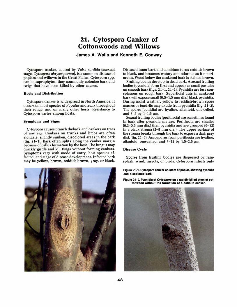
21. Cytospora Canker of Cottonwoods and Willows
James A. Walla and Kenneth E. Conway
Cytospora canker, caused by Valsa sordida (asexual stage, Cytospora chrysosperma), is a common disease of poplars and willows in the Great Plains. Cytosporo spp. can be saprophytes; they commonly colonize bark and twigs that have been killed by other causes.
Hosts and Distribution
Cytospora canker is widespread in North America. It occurs on most species of Populus and Salix throughout their range, and on many other hosts. Resistance to Cytospora varies among hosts.
Symptoms and Signs
Cytospora causes branch dieback and cankers on trees of any age. Cankers on trunks and limbs are often elongate , slightly sunken, discolored areas in the bark (fig. 21- 1). Bark often splits along the canker margin because of callus formation by the host. The fungus may quickly girdle and kill twigs without forming cankers. Symptoms vary with mode of entry, host species af· fected, and stage of disease development. Infected bark may be yellow, brown, reddish-brown, gray, or black.
48
Diseased inner bark and cambium turns reddish-brown to black, and becomes watery and odorous as it deteri· orates. Wood below the cankered bark is stained brown.
Fruiting bodies develop in dead bark. Asexual fruiting bodies (pycnidia) form first and appear as small pustules on smooth bark (figs. 21- 1, 21- 2). Pycnidia are less con· spicuous on rough bark. Superficial cuts in cankered bark will expose small (0.5- 1.5 mm dia.) black pycnidia. During moist weather, yellow to reddish-brown spore masses or tendrils may exude from pycnidia (fig. 21- 3). The spores (conidia) are hyaline, allantoid, one-celled, and 3-5 by 1- 1.5 J.tm.
Sexual fruiting bodies (perithecia) are sometimes found in bark after pycnidia mature. Perithecia are smaller {0.3- 0 .5 mm dia.) than pycnidia and are grouped {6- 12) in a black stroma (2-8 mm dia.). The upper surface of the stroma breaks through the bark to expose a dark gray disk (fig. 2 1-4). Ascospores from perithecia are hyaline, allantoid, one-celled, and 7- 12 by 1.5- 2 .5 J.tm.
Disease Cycle
Spores from fruiting bodies are dispersed by rain· splash. wind, insects, or birds. Cytospora infects only
Figure 21- 1. Cytoapora canker on stem of poplar, showing pycnldla and discolored bark.
Figure 21-2. Pycnldla of Cytospora on a rapidly killed stem of cot· tonwood without the formation of a definite canker.

through wounds or other openings in the bark. It is considered a weak parasite, and most often attacks stressed trees, such as those growing on poor sites or injured by drought, frost, sunscald, severe pruning, fire, insect or mechanical damage, or herbicides.
The fungus grows in the bark until limited by internal processes of the tree. Fruiting bodies form in infected bark to complete the life cycle. The fungus overwinters as fruiting bodies and mycelium in cankered bark.
Cytospora has been found in apparently healthy bark; thus it may be able to infect bark tissue and remain there without causing visible damage until the tree is stressed.
Damage
Cytospora can cause dieback or death of planted or native trees. It is a limiting factor in the establishment and growth of some poplars and willows. In nurseries, it can attack cuttings of poplars and willows that are used for propagation. A disease called blackstem is caused by Cytospora and other canker fungi; it can develop during storage or after outplanting (fig. 21-5) and can result in severely reduced nursery production.
Control
Healthy trees are less susceptible to infection and damage. If possible, water and fertilize trees as needed to maintain optimum vigor. Reduce chances of infection by preventing wounds. Do not bring infected plant materials into the area. Plant resistant varieties.
Trees that usually are not resistant to C. chrysosperma include various species (eastern cottonwood, white cottonwood, weeping willow), varieties (Lombardy and Bolleana poplar), and the selection Siouxland cottonwood. Those usually resistant include the hybrid poplar Noreaster, certain eastern cottonwood selections (Platte, Mighty Mo, Ohio Red), and the species valley cottonwood, and black and peachleaf willow. Norway poplar is relatively resistant to blackstem.
Severely cankered branches or trees should be removed and destroyed. Prune back to a live branch beyond the canker. Small cankers on stems can be removed by excising all affected bark. Shape wounds into an ellipse to promote rapid healing. Prune during dry weather. Disinfect pruning tools with alcohol after each cut.
A systemic fungicide is labeled for Cytospora canker control. Protective fungicides are registered for control of wound fungi on trees but are not specifically labeled for Cytospora.
To avoid cankers on nursery cuttings, maintain healthy stock blocks. Collect shoots from stock blocks in the fall before very cold weather causes bark moisture to fall. Process and store cuttings quickly: dip cuttings in a fungicide solution before winter storage, store at a constant temperature below 35°F, and plant cuttings in beds after soil warms to allow rapid plant growth.
Figure 21-5. Poplar cuttings with necrotic tissue typical of blackstem caused by Cytospora.
49
Figure 21-3. Spore tendril from pycnldlum of Cytospora.
Figure 21-4. Perlthecla with gray disk typical of Valse spp. (scale lnmm).
i l l t lllll l ll l lllllll l l l l l
Selected References
Bloomberg, W. J. Cytospora canker of poplars: factors influencing the development of the disease. Canadian Journal of Botany. 40: 1271-1280; 1962.
Walla, J. A.; Stack, R. W. Dip treatment for control of blackstem on Populus cuttings. Plant Disease. 64: 1092-1095; 1980.
Wysong, DavidS.; Riffle, Jerry W. Cytospora canker of poplars and willows. NebGuide G75-257. Lincoln: Cooperative Extension Service, University of Nebraska; 1982. 3 p.

22. Septoria Canker of Cottonwood and Hybrid Poplars
Jerry W. Riffle and David S. Wysong
Septaria musiva causes leaf spots and cankers on native and hybrid poplars. Information on the leaf spots caused by this fungus appears in Article 3.
Hosts and Distribution
S. musiva is indigenous throughout much of the United States and Canada. In the Great Plains it occurs in North Dakota, Nebraska, and Texas. The fungus produces cankers on native cottonwood (Populus deltoides), and on a wide range of hybrid poplars, particularly those of cottonwood, balsam poplar (P. balsamifera), or black poplar (P. nigra) parentage. In the north-central region of the United States, hybrids with a parent of Japanese poplar (P. maximowiczii), black cottonwood (P. trichocarpa), or laurel poplar (P. Iaurifolia) are very susceptible.
Figure 22-1. Canker on young plantation tree.
50
Symptoms and Signs
Cankers are formed on the main stem and branches of the current season's growth, usually within 5 feet of the ground. Cankers are often flat-faced, or have swollen marginal callus. The bark of young cankers is dark brown or black and depressed (fig. 22-1). Infected cambium is killed, and small black pycnidia may develop in bark in the ashy-white central area of the cankers (fig. 22-2). Continued development of cankers may result in girdling and death of affected stems during late summer. Affected stems may be infected by other canker fungi, such as Cytospora chrysosperma, that cause additional damage.
Two types of spores form in fruiting structures in infected host tissues. Conidia (pycnidiospores) develop in
Figure 22-2. Depressed cankers on cottonwood cuttings. Pycnidia develop on bark In the central area of young cankers.

pycnidia on bark or leaves, and exude in pink or white tendrils during wet weather. Conidia are hyaline, cylindric, straight or curved, one to four septate, and are 2Q-56 J.Lm long by 3-4 J.Lm wide. Ascospores of the perfect stage Mycosphaerella popuJorum are produced in perithecia that develop on fallen leaves. The ascospores are hyaline, 1-septate, and 16-28 I-'m long by 4.5-6.0 I-'m wide.
Disease Cycle
S. musiva overwinters on fallen infected leaves and in bark of cankers. In the spring, ascospores and conidia from fallen leaves and conidia from cankers are discharged during wet weather. These spores are dispersed by wind and washed by rain to infect leaves and stems. Both ascospores and conidia can cause stem infections. Infections may occur through stipules (fig. 22-3), petioles, buds, lenticels, or through bark wounds. The fungus also can infect unwounded leaves and stems. Leaf infection usually precedes stem infection. Leaf spots appear soon after leaves develop, and the fungus spreads to stems and branches to form cankers. Cankers are formed on twigs of the current season's growth, and pycnidia develop shortly after infection. Conidia from pycnidia in leaf spots and cankers cause secondary in-
Figure 22-3. Infection of cottonwood stem through leaf stlpules and subsequent canker development.
51
fections. Disease development is enhanced by warm temperatures and long periods of humidity.
Damage
S. musiva damages poplars of all ages, but damage is most severe in nursery stool (propagation) beds and in young plantations. In plantations, growth of the fungus from twigs into main stems results in cankers that girdle small stems of susceptible trees, particularly hybrids. Extensive losses have occurred in hybrid poplar plantings in northeastern States. Multiple cankers can girdle affected stems; single cankers seldom girdle a branch or stem of moderately susceptible trees, but the cankers may be invaded by other fungi that do girdle stems and kill trees.
Control
Damage caused by S. musiva is reduced primarily by the use of resistant cultivars. Clones resistant or moderately susceptible to leaf spot also tend to be resistant to cankers, so these clones should be selected for planting. Vigorous, disease free planting stock should be used for establishment and maximum early growth of hybrid poplar.
Fungicides can reduce pathogen populations. Captafol effectively controlled S. musiva in a central Iowa planting in tests in 1979-1980. Benomyl applied at one pound active ingredient per 100 gallons of water once in spring and bimonthly throughout the growing season also has controlled S. musiva on susceptible clones.
Cultural treatments, such as cultivation or raking in the fall to remove leaf litter containing fungal inoculum, will minimize primary infections in the spring if inoculum from adjacent trees is not a factor. Planting moderately susceptible trees at a wide spacing to provide good air circulation within the canopy will reduce the duration of free moisture on leaves and minimize infection by S. musiva.
Selected References
Bier, J. E. Septaria canker of introduced and native hybrid poplars. Canadian Journal of Research, Section c. 17: 195-204; 1939.
Filer, T. H.; McCracken, F. 1.; Mohn, C. A.; Randall, W. K. Septaria canker on nursery stock of Populus deltoides. Plant Disease Reporter. 55: 460-463; 1971.
Ostry, M. E.; McNabb, H. S. Jr. Diseases of intensively cultivated hybrid poplars: A summary of recent research in the north-central region. In: Hansen, E. A. compiler. Intensive plantation culture: 12 years research. Gen. Tech. Rep. NC-91. St. Paul, MN: U.S. Forest Service, North Central Forest Experiment Station; 1983: 102-109.
Waterman, Alma M. Canker of hybrid poplar clones in the United States, caused by Septoria musiva. Phytopathology. 36: 148-156; 1946.
Waterman, Alma M. Septaria canker of poplars in the United States. Circular 947. Washington, DC: U.S. Department of Agriculture; 1954. 24 p.

23. Phomopsis Canker on Cottonwood
Theodore H. Filer, Jr., and Edward M. Sharon
Cottonwood occurs naturally along streams and rivers throughout the Great Plains. In most of its range, cottonwood grows on moist, well drained soils; but in the western semi-arid one-third of its range, it is found only along streams.
Hosts and Distribution
Phomopsis mocrosporo causes dieback and cankers on many tree species in the United States, as well as in
Figure 23-1. Small black pustules of Phomopsls macrospora on surface of canker.
Figure 23-2. White curled sporehoms composed of millions of spores that can cause new Infections.
52
Europe and Japan. The species was first described in Japan in 1961 and in the United States in 1967. The perfect stage of the fungus was first described on willow in 1891. This stage is distinctly different from the perfect stages- Dioporthe eres and D. medusoeo (the predominant species on Populus) because of its larger ascospore with appendage and because the alpha spores are larger than the beta spores.
Symptoms and Signs
Phomopsis cankers develop on cottonwood twigs, limbs, and boles; they are inconspicuous during early development. Small pustules on cankers appear as small black dots, protruding through bark epidermis (fig. 23-1). Spores are pushed out of the pore of pustules under moist conditions, and often form sticky orange or reddish sporehorns (fig. 23-2). As the canker develops and enlarges, the phloem tissues die, forming a sunken area (fig. 23-3). When the stem is girdled, the distal portion dies; the resulting dead leaves cling to the branches for several weeks (fig. 23-4). Similar symptoms are caused by Fusarium, Cytospora, and other canker-causing fungi.
P. macrospora fruits profusely on dead wood. The small black fmiting bodies are less than a millimeter in size. They look like miniature mountains protruding through the bark (fig. 23-5).
Figure 23-3. (a) Sunken area Inside canker. (b) Canker 23-3a wllh outer bark removed to show necrotic tissues.

Disease Cycle
The asexual spores are the primary inoculum. Conidia are primarily wind- and insect-dispersed, but rain can spread the spores to wounds on lower parts of the tree. The fungus is considered a wound parasite, and needs some natural or wound opening before it can penetrate and colonize tissues. The size of canker that develops depends on tree vigor. On fast-growing cottonwoods, canker development is usually limited and the callus tissue formed restricts fungal growth. On slow-growing trees (trees growing under moisture stress or in poorly aerated soils), cankers can girdle the stem and cause mortality.
Damage
Phomopsis is one of the most important pathogens that cause mortality of planted cuttings. In most cases the fungus is on the cuttings when they are taken from the nursery, and girdles the stem within a few weeks after planting.
Control
Good cultural practices can reduce the possibility of mortality of shade trees. Fertilize trees at the rate of 6 pounds of N-P-K per 1,000 square feet, water at the rate of 2 inches per week during droughty periods, and control insect defoliators and leaf diseases to reduce tree stress and promote tree vigor.
To prevent losses when planting poplars, select healthy, canker-free cuttings or seedlings. Prevent cuttings from drying in storage; soak them at least 24 hours prior to planting. Cultivation is needed during the first year to keep trees growing vigorously. Plant poplars that are resistant to Septoria leaf disease.
Selected References
Filer, T. H., Jr. Pathogenicity of Cytospora, Phomopsis, and Hypomyces on Populus deltoides. Phytopathology. 57: 978-980; 1967.
Filer, T. H., Jr. Cottonwood cankers caused by Phomopsis mocrospora. (Abstract) Phytopathology. 57: 458; 1967.
Fowells, H. A. Silvics of forest trees of the United States. Agric. Handb. 271. Washington, DC: U.S. Department of Agriculture; 1965. 762 p.
Kobayashi, T.; Chiba, 0. Fungi inhabiting poplar in Japan. In: Bull. 130. Tokyo: Government Forest Experiment Station; 1961: 1-43.
Morris, R. C.; Filer, T. H.; Solomon, J. D.; McCracken, F. 1.; Overgaard, N. A.; Weiss, M. J. Insects and diseases of cottonwood. General Technical Report S0-8. New Orleans, LA: U.S. Department of Agriculture, Forest Service, Southern Forest Experiment Station; 1975. 37 p.
53
Figure 23-4. Crown of cottonwood showing dead leaves on branches girdled by cankers.
Figure 23-5. Small black fruiting bodies of P. macrospora protruding through bark.

24. Dothichiza Canker of Populus species
David W. Johnson and Robert W. Stack
Hosts and Distribution
Canker of poplars caused by Dothichiza populea has been known in the United States since 1915. The disease is most common in nurseries and plantations in the eastern and central States. D. populea infects a wide range of Populus species and hybrids, especially those in the Tacamahaca and Aegieros groups; Lombardy poplar is particularly susceptible. It has also been reported on quaking aspen and bigtooth aspen. The range of D. populea extends from Maine to Virginia and westward to Minnesota, Nebraska, and New Mexico. It occurs on young trees in nurseries and plantations, but is rare or of little significance in native stands.
Figure 24-1. Young Dothlchlza canker with discolored bark.
Symptoms and Signs
Early symptoms are a premature yellowing of the leaves, followed by defoliation. Girdling cankers cause dieback of small stems and shoots. The development of cankers varies greatly with host age and species. Young trees may be girdled rapidly, which causes topkill. Poplar with large spreading branches and those resistant to the fungus may only develop small branch cankers.
In general, cankers first appear as slightly sunken areas with the diseased bark slightly darker than healthy bark (fig. 24-1). After the bark is killed to the cambium, the sapwood is invaded and turns brown. Cankers develop during the dormant season. As the canker develops, the bark cracks and extensive callus is produced (fig. 24-2).
54
Figure 24-2. Extensive callus development associated with Dothlchlza stem cankers.

Sprouts may develop below dead limbs and cankers. These sprouts may be killed in following years.
Cushion-like fruiting bodies called pycnidia are formed on the dead or wilting twigs (fig. 24-3}. In time the diseased bark turns brown and cracks, and the underlying dark brown, diseased wood is exposed.
Disease Cycle
D. populea overwinters as spores in unopened pycnidia and as mycelium within the bark tissue. In the spring new pycnidia, when mature and moisture conditions are favorable, rupture the bark and extrude spores in olivebuff tendrils or in masses. Conidia are unicellular, hyaline, and ovate pyriform to spherical (fig. 24-4}. They are washed about by rain or carried by insects or birds. Possibly, after the spore tendrils dry, some are windborne. New pycnidia may be formed as the dieback and canker enlarges, so that mature spores may be present throughout the summer and fall.
The fungus may infect through bud scales, leaf scars, or bark at the base of small lateral twigs into a stem or branch. Cankers develop around the base of twigs or injured buds. The fungus also infects leaf scars following early defoliation by Melampsora rust or Marssonina leaf blotch.
The perfect stage, Cryptodiaporthe popuJea, is reported from Europe but is not known in the United States.
Damage
In Europe the fungus has been recognized as an im-
Figure 24-3. Pycnidia develop on dying bark of affected twigs.
55
Figure 24-4. Conidia of D. popu/ea are hyaline, unicellular, and ovate pyriform to spherical.
portant pathogen of poplars since 1903, particularly on young or newly planted trees, or on trees that have been weakened by low spring temperatures, poor drainage, drought, pruning wounds, or infertile soil.
Although the disease is endemic in the United States, it has not been as damaging as in Europe; however, its potential for damage in plantations is a concern.
Control
The disease has not been sufficiently severe in the United States to warrant a detailed study of control measures. Because of the possibility of infection in plantations, care should be taken to use disease-free stock for planting and to maintain good growing conditions. Infected stock should be destroyed. Applications of excess nitrogen and dense nursery beds should be avoided. Leaf diseases caused by Me!ampsora and Morssonina should be controlled in the nursery with fungicides. Choice of planting site is important so that the trees are adapted to the site. Pruning should be avoided if possible, and care exercised to prevent other wounding. Older stands should be thinned. Highly susceptible cultivars such as Lombardy should not be planted.
Selected References
Carter, J. Cedric. Diseases of Midwest trees. Special Publication 35. Urbana: University of Illinois Press; 1979. 168 p.
Gremmen, J. Research on Dothichiza-bark necrosis (Cryptodiaporthe popuJea) in poplar. European Journal of Forest Pathology. 8: 362-368; 1978.
Ostry, Michael E.; McNabb, Harold S. Jr. How to identify and prevent injury to poplars caused by Cytospora, Phomopsis, and Dothichiza. HT-57. St. Paul, MN: U.S. Department of Agriculture, Forest Service, North Central Forest Experiment Station; 1982. 6 p.
Waterman, Alma M. Canker and dieback of poplars caused by Dothichiza popu!eo. Forest Science. 3: 175-183. 1957.

25. Cryptosphaeria Canker of Cottonwood and Aspen
Thomas E. Hinds and Jerry W. Riffle
The genus Populus is widespread throughout the Great Plains; five of the nine species native to the United States are found from Texas northward to North Dakota. Ofthe numerous pathogens known to attack poplars, Cryptosphaeria populina is a relatively new stem canker pathogen found on poplars in the Great Plains.
Hosts and Distribution
Although aspen is the principal host of C. populina, its occurrence and distribution on other poplars is becoming more apparent because of its recognition. The fungus has been found on narrow leaf cottonwood in Colorado, on Plains cottonwood in Colorado, Oklahoma, and Nebraska, on Lombardy poplar in Nebraska, and on aspen in South Dakota. Other hosts of the fungus include balsam and black poplar, and some hybrid poplars. The proper identification of this organism will no doubt extend its distribution and host range.
56
Symptoms and Signs
C. populina causes a stem canker that results in branch and tree mortality. Extensive discoloration and decay are attributed to the asexual stage, Libertella sp. The cankers, frequently associated with wounds, are long, narrow, and found on the trunk of hosts (fig. 25-1). They may be only 2 to 4 inches wide, but up to 10 feet in length, following the grain of the underlying wood. Cankers may grow up to an inch in width anl:l many inches in length annually. Small trees usually die several years after being infected and before the trunk is girdled. Although branch infection does not always result in a prominent canker, the fungus will spread from a branch infection onto the trunk.
Cryptosphaeria populina colonizes the heartwood and sapwood, and causes extensive discoloration and decay. Various hues of gray, brown, yellow, and orange are associated with the brown-mottled decay, from which the Libertella stage of the pathogen is easily isolated. The fungus grows outward to the cambium and bark tissues, causing necrosis and canker formation. The infected bark becomes light brown to orange. After one or more years, the dead bark becomes black, stringy, and sootlike; however, it contains small (0.5- 2.0 mm), scattered, lensshaped, light-colored areas (fig. 25-2), and adheres tightly to the sapwood. This black, adhering dead bark with light
Figure 25-1. Elongated canker on aspen caused by Cryptosphaerla popu/lna.
Figure 25-2. Lens·shaped, light-colored areas In the black, dead bark of the canker.

specks is a good diagnostic characteristic of this canker disease.
Perithecia are formed in a confined area in the dead bark that is raised on the surface in the form of a flat, broad blister that may vary from 0.3 inch in width and up to 1-foot in length (fig. 25-3). The fungus readily produces fruiting bodies on branches and smooth bark, but it is somewhat limited in penetrating and fruiting on the thick, rough bark of older trees. Light-orange fruiting bodies (acervuli) of the Libertella stage are occasionally found near the perimeter of cankers on aspen.
Cytospora chrysosperma frequently produces fruiting bodies along the canker perimeter and quickly colonizes the dead bark after the branch or tree dies. Because Cytosporo fruiting bodies are very conspicuous on the dead tissue, it is often erroneously assumed to be the primary causal agent of the canker.
Disease Cycle
Branch and trunk infections become established in wounds to the bark and xylem. The sapwood is colonized by the pathogen, which causes discoloration and decay; consequently, the cankers seldom completely girdle the stem before stem death (fig. 25-4). Perithecia form in the spring on bark tissues that have been dead at least one year and mature during the summer and fall of their formation. Perithecia persist in the dead bark for several years and produce viable spores. Ascospores expelled from the perithecia are apparently wind dispersed. Size and shape of C. populina ascospores are similar to Cytosporo conidia, but they are somewhat larger (8-10 by 2 J,tm vs. 3-5 by 1- 5 J,tm) and vary from hyaline to pale yellow. Their similarity to Cytospora spores may have contributed to misidentification of the causal agent of the canker in the past.
Damage
Saplings and small trees can be killed before cankers and perithecia are produced. Trees weakened by branch and trunk decay are vulnerable to wind breakage. In a 1977 study of Colorado aspen, the canker was present on 83 percent of 30 sites examined, on 1 percent of the 2,873 live trees, and was responsible for 26 percent of the tree mortality encountered. The importance of this disease of poplars in the Plains is presently unknown.
Control
No direct control measures are known. The prevention of trunk wounds and pruning of dead, dying, or diseased branches on high-value trees should aid in reducing the incidence of disease.
Selected References
Hinds, Thomas E. Cryptosphaeria canker and Libertella decay of aspen. Phytopathology. 71: 1137-1145; 1981.
Juzwik, J.; Nishijima, W. T.; Hinds, T. E. Survey of aspen cankers in Colorado. Plant Disease Reporter. 62: 906-910; 1978.
57
Figure 25-3. Formation of new perlthecla In dead bark (left); periderm removed (right) to show perlthecla formed the previous
year (scale In em).
Figure 25-4. Discoloration and brown·mottled trunk decay of aspen behind a canker (scale In em).

26. Hypoxylon Canker of Aspen Thomas E. Hinds and Mark 0. Harrell
Aspen is the most widely distributed forest tree in North America. While the tree is found in the northern States from Maine to Washington and as far south as Mexico in the West, its occurrence in the Great Plains region is sporadic, with small stands in Nebraska, North Dakota, and South Dakota.
Hosts and Distribution
Hypoxylon canker (fig. 26- 1), caused by Hypoxylon mammatum, is abundant east of the Rocky Mountains; it is the most important killer of aspen in the Great Lakes region. The canker is of minor importance in the tree's western range. Although the disease is present in aspen stands of the northern Great Plains, its importance is unknown.
Quaking aspen is very susceptible, bigtooth aspen is occasionally infected, and balsam poplar is rarely infected.
Symptoms and Signs
Incipient cankers appear as slightly sunken, yellowish-
Figure 26-1. Canker Initiation at forte of live 12-lnch aspen.
58
orange areas with irregular, lobed margins centered around wounds, dead branch stubs, or insect injuries and galls. As the infection progresses, the affected bark becomes mottled. A slight flow of sour-smelling brownish sap is common around the perimeter of infection. Small, blisterlike patches are formed under the outer dead bark during the second year of infection. The bark then ruptures, exposing small, bristlelike structures called hyphal pegs or pillars, upon which the sporebearing structures are formed (fig. 26-2). The structures soon disintegrate. The diseased inner bark becomes laminated or mottled black and yellowish-white (fig. 26-3); white mycelial fans are formed near the canker margin under the bark.
Cankers are easier to identify 2 or 3 years after infec-
Figure 26-2. Hyphal pegs beneath blistered bark (scale In mm).
Figure 26-3. Mottled black and yellowish-white diseased Inner bart.

tion, when perithecia are formed in small, crustlike stromata several mm in diameter on the dead bark (fig. 26-4). The grayish to black stromata appear to be partially covered with a white. flou rlike material. The stromata are formed annually on the dead bark, and persist for several years.
Although the tree produces callus tissue at the margin of the infection, the fungus continues to invade new tissue each year. This invasion cracks the bark along the canker margins and leaves the dead bark in a slight depression. The black, dead bark in the center of older cankers begins to crack in a checkerboard fashion and sloughs off in small patches, revealing a checkering of the wood beneath (fig. 26-5). Older trunk cankers may be 3 feet or more in length before the trees are girdled and killed, usually within several years. The roughened, spotted gray and black bark of cankered trees stands out in contrast to the healthy bark of noninfected trees.
Disease Cycle
The fungus is a wound parasite, but the means by which infection takes place is unknown. Apparently, airborne spores enter through wounds to the wood, including insect wounds. The fungus colonizes the sapwood, and then invades the bark from within by producing a toxin that causes bark collapse and necrosis.
The asexual stage of the fungus consists of conidiophores and conidiospores, which are produced on the hypha! pegs formed under the bark blisters at the end of the first or during the second growing season. The role of conidiospores in causing new infections is unknown. Between the second and third year of infection, the first immature perithecial stromata of the sexual state are formed on the dead bark. They are generally abundant on older cankers. Ascospore release requires free moisture, usually in the form of dew, rain, or snowfall, and can continue for several years.
Damage
H. mammatum kills 1 to 2 percent of the standing
Figure 26-4. Perltheclal stromata on dead bar11 (scale in mm).
aspen volume each year in the Lake States region by girdling the trees. While this canker disease is not of major importance in the Rocky Mountain region, its impact on aspen in the Great Plains is unknown.
Canker incidence varies with geographic location; the level of infection fluctuates from year-to-year. Tree vigor does not appear to influence host susceptibility. Although older stems are generally more resistant, upper bole infections, usually found on older trees, may partially kill the crown, and ultimately result in tree mortality from suppression. Trees weakened by decay behind cankers in the lower bole are susceptible to wind breakage before canker girdling is complete. Low density stands, mixed stands, thinned stands, and trees on the perimeter of stands are more susceptible to infection than fully stocked stands.
Control
Although no direct control measures are known for Hypoxylon canker, certain silvicultural techniques can reduce canker losses. Because Hypoxylon canker is favored by stand openings and poor stocking, maintaining fully stocked stands and a closed canopy without openings should reduce volume losses.
Other silvicultural management practices advocated for aspen in the Lake States include: (1) if 15 to 20 percent of the trees are infected, harvest the stand early and treat the site to encourage good aspen reproduction; (2) if more than 25 percent are infected, harvest immediately and convert to other species (susceptible clones should not be perpetuated); and (3) lightly infected stands can be managed on rotations longer than 40 years.
Selected References
Anderson, R. L.; Anderson, G. W. Hypoxylon canker of aspen. Forest Pest Leaflet 6. Washington, DC: U.S. Department of Agriculture, Forest Service; 1979. 7 p.
Schipper, A. L., Jr.; Anderson, R. L. How to identify Hypoxylon canker of aspen. St. Paul, MN: U.S. Department of Agriculture, Forest Service, North Central Forest Experiment Station; 1976. 5 p.
Figure 26-5. Checkering ol wood beneath old canker.
59

2 7. Thyronectria Canker of Honey locusts William R. Jacobi and Jerry W. Riffle
Thyronectria canker is a disease of honeylocusts caused by the fungus Thyronectrio oustro-omericono. The imperfect stage of T. oustro-americono is Gyrostromo austro-omericonum.
Hosts and Distribution
Native thorny honeylocust (Gleditsia triacanthos var. trioconthos), all cultivars of thornless and podless honeylocust (G. triancanthos var. inermis) and oriental honeylocust (G. japonica) are susceptible to T. oustroamericano. The fungus also has been found on dead tissue of several hardwoods including American elm, white ash, mockernut hickory, willow, and bur oak.
The disease has been ·reported from Colorado to Massachusetts, occurring in Alabama, Colorado, Illinois, Kansas, Massachusetts, Mississippi, Nebraska, Oklahoma, South Dakota, and Tennessee. The disease has not been reported from northern States such as Minnesota or Wisconsin, so fungus distribution may be restricted by colder climates.
Flgunt 27-1. Thyronectrla canker on a honeylocust originating from a branch crotch.
Symptoms and Signs
Symptoms include dieback of affected branches, reduced foliage, yellow foliage, premature fall coloration, and early leaf drop. Cankers range from slightly flattened surfaces to distinctly sunken areas with large callus ridges at the canker margin (figs. 27-1, 27-2). Thin bark areas of stems and branches may have a red-yellow discoloration associated with the canker. Usually there is no discoloration where the bark is thicker. Wood beneath infected bark turns wine-red to yellow.
Signs of the pathogen allow for better disease identification than do symptoms. Irregular pycnidia in stroma (stromatic conidiomata) (figs. 27-2, 27-3), which are light yellow-brown when fresh and blacken with age, and perithecia that are reddish-brown or gray but also darken with age, form in the cankered area on dying and dead bark. They are usually found in natural openings such as lenticels in thick bark areas (fig. 27-2), and scattered on bark surfaces in thin barked areas (fig. 27-4). Pycnidia exude milky masses of conidia. Conidia are
60
Figure 27-2. Canker on an Inoculated honeylocust with large, dark colored masses of pycnldla extruding through bark.

hyaline, ovoid to ellipsoid, one-celled, and are 1.6-4.2 by 0.8-2.7 14m in size. The perithecia develop in rounded clusters in a stroma, and are smooth-surfaced and eggshaped with a short neck. Asci contain 8 ellipsoid to pear-shaped ascospores. The hyaline to pale yellow ascospores have both transverse and longitudinal septa and range in size from 8-16 by 5-9 14m.
Nectria canker of honey locust induced by Nectrio cin· noborino appears very similar to Thyronectria canker, and is best distinguished from T. oustro-omericono on cultural characteristics, morphology of the asexual fruiting structures, and ascospore morphology. T. oustroomericono cultures have somewhat waxy margins with orange slimy centers containing masses of conidia. N. cinnoborina cultures are white and fluffy. N. cinnoborino induces cankers that are usually sunken and contain a sporodochial asexual stage (Tuberculorio vulgaris). The ascospores of N. cinnoborino are divided into two cells by a single transverse septum, unlike those ofT. oustroomericono.
Disease Cycle
The Thyronectrio fungus overwinters as mycelium and fruiting structures on infected trees. Conidia presumably are spread by rain and ascospores by wind, although this has not been demonstrated. Infections are thought to take place through pruning wounds and other natural openings. Fresh bark wounds are infected readily by the fungus. Because the fungus is also a saprophyte, it can become established on dead wood such as branch stubs or wound edges.
The fungus grows in the cambium and outer xylem, where it eventually kills the cambium and surrounding cells. Trees, or affected parts, die because of cambial death and possible vascular dysfunction. Pycnidia can form within one month after tissue is colonized, and are abundant on bark of dying or dead trees. Perithecia form in affected areas but are not as common or abundant as pycnidia. Spore release is favored by high humidity and rain; spore germination is optimum at 77°-86°F. Infection and disease development is rapid at temperatures of 75°to 82°F.
Damage
Cankers at the tree base are usually fatal. Main stem or branch crotch cankers may cause complete girdling, depending on the tree's health. Stressed trees cannot compartmentalize the fungus, whereas vigorous trees may be able to callus over a canker and recover.
The incide n ce of the disease across the United States is not known, but occurs on honey locust in urban, rural, and windbreak plantings.
Control
Thyronectria canker should be prevented rather than controlled. Physical damage and wounds should be avoided or properly cleaned and allowed to dry. The fungus infects trees under a variety of stresses. Avoid
61
Figure 27-3. Clumps of pycnldl1 on honeylocust b1rk (left).
Figure 27-4. Clumps of pycnldl1 1re sc1ttered on stems with thin bark (right).
stress due to drought, overwatering, restricted area for root growth, or restricted oxygen for root growth. Trees should be watered adequately but not excessively. Frequent light watering ln heavy clay soils allows attack by other fungi at the tree base just below groundline. T. oustro-omericono then infects the weakened tree above the area previously infected by other fungi. Allowing the soil to dry out at the tree base would prevent infections at the soil line.
Pruning infected branches can reduce the chances of other infections. Trees should be pruned in cool, dry weather when the presence of fungal spores is reduced. Small cankers may be scribed out if the tree is reasonably vigorous.
Selected References
Crowe, F.; Starkey, D.; Lengkeek, V. Honeylocust canker in Kansas caused by Thyronectrio oustro-americano. Plant Disease. 66: 155- 158; 1982.
Hudler, George W.; Oshima, Nagayoshi. The occurrence and distribution of Thyronectrio austro-omericono on honeylocust in Colorado. Plant Disease Reporter. 60: 920- 922; 1976.
Lieneman, Catharine. Observations on Thyronectria denigrata. Mycologia. 30: 494- 511; 1938.
Riffle, Jerry W.; Peterson, Glenn W. Thyronectria canker of honeylocust: Influence of temperature and wound age on disease development. Phytopathology. 76: {3): 313-316; 1986.
Seeler, E. V., Jr. Two diseases of Gieditsia caused by species of Thyronectria. Journal Arnold Arboretum, Harvard University 21: 405-427; 1940.

28. Stem Decays of Willow James A. Walla and Robert W. Stack
Several species of fungi are able to decay live or dead willow wood. The most common decay fungi on willow in the Great Plains are species of Daedulea and Trome!es. Names used in this text follow Overholts. Daedulea and Trometes are in the family Polyporaceae. Taxonomic relationships in this family are in a state of revision. Species of Daedoleo and Trometes discussed here have been placed in eight genera (Antrodia, Cerreno, Coriolus, Doedoleo, Doedoleopsis, Dotronio, Funolio, and Trometes). Names presently advocated by the U.S. Forest Service Center for Forest Mycology Research are listed in the index next to names used in this text.
Hosts and Distribution
Three species of Doedoleo (D. ombiguo, D. confrogoso, D. unicolor) and seven of Trometes (T. hispido, T. molicolo, T. mollis, T. rigido, T. sepium, T. suoveolens, T. !rogiiJ that decay willow have been reported in the Great Plains. T. suaveolens, which causes a white-mottled heart rot, is the most common decay fungus that attacks live willows. The other species generally are found on dead wood. These fungi are widespread in the Great Plains and have been found on many willow species. Species that decay willow also decay many other hardwoods and some conifers. No decay resistant willow species are known.
Symptoms and Signs
Often the first outward indication of stem decay is formation of fruiting bodies (sporocarps) on the outside of the stem. This fruiting occurs after the fungus has been present for some time and has caused significant decay. Sporocarps of these fungi are usually annual, but they often grow on the same wood for several years.
Sporocarps vary in appearance, both within and among species and genera. Both Daedulea and Trametes usually have shelflike sporocarps (fig. 26-1), but some may also be appressed on the stem. The lower surface consists of a layer of pores in which spores are produced. In Daedaleo species, pore openings usually are mazelike (fig. 28-2), but they may vary from round to almost gilllike. In Trametes species, the pores have circular to angular openings. Sporocarps of Trometes species often are effused-reflexed and spread out over the surface of the host, but turned up at the upper margin to form a pileus (fig. 28-3). Such sporocarps often have toothlike pores. Laboratory diagnosis is required for specific identification.
The fungus can be found before sporocarps appear by cutting or boring into the stem and looking for rotted wood. At this stage, infected wood must be cultured on agar medium to isolate the fungus for identification.
Daedalea species an willow cause white rot of wood. Most Trometes species on willow cause white rot, but two cause brown rot. Most species of these genera decay sapwood, but at least one causes heart rot. Some white rot species cause black lines (zone lines) to form in rotted wood. One, T. suaveolens, has a characteristic anise (licorice) odor when fresh.
Live trees with internal rot often exhibit top dieback or poor vigor. If trees with these symptoms are examined, the sporocarps of decay fungi may be found on the lower stem.
Some of these species do not attack live trees, and all can grow as sa probes; their presence on dead wood does not mean they cause tree mortality.
Disease Cycle
62
The sequence of events leading to decay of living trees by some fungi is as follows: Openings in the bark
Figure 28- 1. Pileus of Daedalea confragosa, showing shelfllke growth ol sporocarps.

(wounds, branch stubs) allow nondecay organisms (bacteria, nondecay fungi) to enter the wood. These organisms alter the wood and allow decay fungi to invade and decay the altered wood. Only the wood present at the time of wounding is susceptible to decay. After growth and subsequent wood decay, the decay fungi produce sporocarps to complete their life cycle. The sequence of events has not been examined in decay of willow.
Damage
Young trees are usually free from decay. Incidence of decay increases with advancing age of trees. Decay fungi are common on dead wood. On live trees, decayed stems are more vulnerable to wind or snow breakage. Live trees are seldom killed by decay fungi, but affected wood is unsuitable for use as wood products.
Control
Little can be done to control stem decays after trees are infected; control measures should be directed toward preventing infection. Wounds close faster, and more effective defense barriers are formed on vigorous trees. Maintain tree vigor by applying water and fertilizer if
Figure 28-2. Hymenla of D. confragosa, showing daedalold (mazellke) pores.
63
possible. Prevent mechanical wounds and avoid wounds caused by fires. If wounds occur, prune to promote callusing. If branches break, prune back to the nearest living lateral. If a main stem is wounded, shape the wound into an ellipse to promote closing by callus formation.
If thinning is feasible, remove the least vigorous trees and trees that are already or most likely to become infected. Do not damage residual trees while thinning. Remove dead wood from the site.
Selected References
Hepting, George H. Diseases of forest and shade trees of the United States. Agric. Handb. 386. Washington, DC: U.S. Department of Agriculture; 1971. 658 p.
Hirt, Ray R. On the biology of Trametes suaveolens (L.) Fries. Tech. Pub. 37. Syracuse: Bulletin of the New York State College of Forestry at Syracuse University; 1932. 36 p.
Overholts, Lee Or as. The Polyporaceae of the United States, Alaska, and Canada. Ann Arbor: University of Michigan Press; 1953. 466 p.
Shigo, Alex L. Tree decay: an expanded concept. Agric. Info. Bull. 419. Washington, DC: U.S. Department of Agriculture, Forest Service; 1979. 73 p.
Figure 28-3. Effused·reflexed fruiting bodies of Trametes trog/1 with toothlike pores.

29. Wetwood (Slime Flux) of Elm, Cottonwood, and Mulberry
David W. Johnson and Jerry W. Riffle
Bacterial wetwood is a common disease affecting the xylem of many softwood and hardwood trees. In some species wetwood is lethal, while in others little damage occurs.
Hosts and Distribution
Wetwood affects trees throughout the United States. It is common in elm, mulberry, and cottonwood in the Great Plains. 'ifrees growing on wet sites and poorly drained soils are more likely to develop wetwood than those on upland sites or on well-drained soils. Wetwood has been reported in 86 percent of cottonwood over 6 inches in diameter sampled along the Mississippi River.
Symptoms and Signs
Wetwood can be found in the trunk, branches, and roots (figs. 29-1, 29-2), and has a dark color, high moisture content, elevated pH, decreased electrical resistance, abnormally high gas pressure, and an increase in mobile cations.
Western cottonwoods are often infected early in their life. Dark streaks or bands may appear in the annual rings. Discoloration is most extensive in heartwood or older sapwood, but can occur in current wood. Foliage of affected limbs is often prematurely yellow, scorched, and wilted. Wilting may cause dieback of scattered branches; the entire crown may decline over several years. Premature defoliation may occur, but causes little growth loss.
"Bleeding" or slime-fluxing from trunk wounds, cracks, or other injuries is the most conspicuous symptom. Air-borne bacteria, yeasts, and other fungi contaminate the sap, resulting in a frothy, slimy, foul-smelling liquid, and upon drying it leaves a light gray to white crust (figs. 29-1, 29- 2).
Abundant gas is produced in wetwood-affected tissues by the fermenting action of bacteria on carbohydrates and other materials in the sap. The gas is composed primarily of methane, nitrogen, carbon dioxide, and oxygen. When the gas is confined in the trunk, abnormally high pressures of up to 60 psi develop; 5 to 10 psi are common. The accumulation of liquid under pressure results in a water-soaked condition that gives rise to the
Figure 29-1. Light gray discoloration of baltl of Siberian elm affected with wetwood.
64
name "wetwood." The pressure forces the accumulated liquid and gas out of the trunk through cracks in crotches, through pruning and other wounds, and through other natural openings. This liquid can cause localized cambial mortality and prevent callus formation.
Foliage wilts when sufficient quantities of the toxic liquid accumulated in the trunk wood are carried into the branches. Leaves first curl upward along their margins, then the petioles become flaccid, and finally the leaves droop and wilt.

Disease Cycle
The bacteria associated with wetwood disease are com· mon soil and water inhabitants. Wetwood is primarily associated with the facultatively anaerobic bacterium Enterobacter cloacae ( = Erwinia nimipressuralis). The bacterium is a small, motile, rod-shaped, single-celled, gram-negative organism with up to six peritrichous flagella. Several other bacteria, including species of Xan· thomonas, Agrobacterium, Bacillus, Clostridia, Acinetobacter, and Pseudomonas, are commonly isolated from diseased tissue and probably play a role in the production of the complex symptoms associated with wetwood.
Roots may become infected through wounds. Bleeding trees harbor large populations of bacteria that may be transmitted by bark beetles to infect other stem or branch wounds.
Damage
Wetwood is a chronic disease that may contribute to general decline, especially of old trees and trees of low vigor. It causes an unsightly and often foul-smelling bleeding from tree wounds. It retards or prevents callus formation over wounds, and therefore lengthens suscep· tibility to decay fungi. Dripping wetwood flux may kill turf beneath infected trees.
Wetwood is responsible for substantial losses of wood in the forest products industry. Loss occurs through shake and frost cracks in living trees, checking and col· lapse in lumber and veneer during drying, and increased drying time in the kiln. The stained wood is also a serious defect in lumber and veneer graded on appearance. Strength properties of wetwood-infected tissues do not differ significantly from those of healthy tissues. In fact, wetwood tissue in living trees is rarely decayed, and ap· pears to be resistant to wood-inhabiting fungi.
Control
No completely satisfactory chemical control measures are available. Installation of metal or plastic drain tubes to lower stem pressures and remove excess liquid has prevented additional damage and spread in infected trees, but may allow entrance of decay fungi. Fertiliza· tion of affected trees may be helpful in lessening the effects of the disease. Severely affected trees and limbs should be removed in the spring. Care should be taken to sterilize tools after each cut to prevent spread from diseased to healthy trees.
Selected References
Hamilton, W. Douglas. Wetwood and slime flux in land· scape trees. Journal of Arboriculture. 6(9): 247-249; 1980.
Hartley, Carl; Davidson, Ross W.; Crandall, Bowen S.
65
Watwood, bacteria, and increased pH in trees. Report 2215. Madison, WI: U.S. Department of Agriculture, Forest Service, Forest Products Laboratory; 1961. 35 p.
Murdoch, Christopher W.; Campana, Richard J. Bacterial wetwood. In: Stipes, R. Jay; Campana, Richard J., eds. Compendium of elm diseases. St. Paul, MN: The American Phytopathological Society; 1981: 31-34.
Murdoch, C. W.; Campana, R. J. Bacterial species asso· ciated with wetwood of elms. Phytopathology. 73: 127Q-1273; 1983.
Murdoch, C. W.; Biermann, C. J.; Campana, R. J. Pressure and composition of intrastem gases produced in wetwood of American elm. Plant Disease. 67: 74-76; 1983.
Toole, E. Richard. Wetwood in cottonwood. Plant Disease Reporter. 52: 822-823; 1968.
Figure 29-2. Light ash gray discoloration of bark of American elm affected with wetwood.

30. Crown Gall of Cottonwood, Willow, and Prunus Species
Michael W. Ferguson and Richard Dorset
Species of cottonwood, willow, and Prunus are commonly used in windbreak plantings in the Great Plains Region. Many of these species are susceptible to crown gall disease caused by the bacterium Agrobacterium tumefaciens.
Hosts and Distribution
Crown gall occurs worldwide on numerous tree hosts, including both broadleaf and conifer species. De Cleene and De Ley (1976) list 643 host plants belonging to 331 genera and 93 families. Most members of the genera Populus, Salix, and Prunus are listed as susceptible to infection by at least one isolate of A. tumefaciens. Exceptions are Populus canadensis cv. serotina erecta, P. robusta, Prunus caroliniana, Prunus divaricata, and Prunus ilicifolia, which appear resistant. While not all of these species may be adapted to the Great Plains region, they could be considered as sources of resistance, providing that their resistance is tested with local isolates of A. tumefaciens from the same host genus.
Certain species of Juniperus, Cupressus, and Libocedrus have been reported as susceptible to the crown gall bacterium. Crown gall occurs on eastern redcedar seedlings in some Great Plains nurseries.
Symptoms and Signs
Individual swellings or galls may form on the aerial portions of the tree (fig. 30- 1), or on roots (figs. 30-2, 30-3, 30-4). The galls usually are rounded with rough, irregular surfaces that darken with age. Galls vary in diameter from less than 1 inch to more than 1 foot.
Internally, the galls show irregular structure, with the various tissue elements being disrupted.
Several insects and mites, and physiological responses to grafting or wound regrowth may cause similar-looking galls. The particular diagnostic characters of crown gall are the rough, irregular surface and the lack of small holes (caused by insects) in the core of the gall itself.
The bacterium can be identified by techniques outlined by Schaad (1980).
Disease Cycle
A. tumefaciens is found in the soil, and apparently enters the plant through wounds resulting from cultivation, root pruning, or insect feeding. The bacterium may also be spread through use of contaminated pruning or grafting equipment. After entering the plant, a portion
66
of the bacterial genetic material is transferred to the host, resulting in abnormal cell growth and cell numbers in that region of the host tissue. The resultant swollen tissue forms the gall. Young trees, such as nursery stock, may be stunted when the woody tissue is disrupted, preventing flow of water or nutrients. The bacteria re-enter the soil, probably through disintegration of the gall.
Chewing insects can carry the bacteria from plant to plant, while long-distance movement is by infected nursery stock.
Symptoms may not appear for several weeks after infection, depending on weather conditions. Warm weather favors this disease. The bacteria may survive for two years in soil without a host, but may survive longer in decaying galls. Crown gall is favored by soils of neutral or higher pH.
Figure 30-1. Weeping willow with aerial crown galls.

Damage
Infection of large trees is infrequent and results in no economic loss. Infection of nursery stock, however, can be quite extensive, especially when inoculation takes place during propagation. Small trees may be stunted. Normally, in most states there is no tolerance for nursery stock infected with crown gall; all infected plants are destroyed. Subsequent monitoring losses may be high.
Control - In nurseries:
1. Select planting wood or propagation stock from wood free of crown gall.
2. Use care during propagation to avoid transmitting the bacteria from diseased to healthy wood.
3. Sterilize cutting instruments frequently using a disinfectant such as 70 percent alcohol.
4. In species that are vegetatively propagated, use a budding technique rather than grafting.
5. Avoid mechanical wounds of young trees or bushes. 6. Remove and destroy all infected plants. 7. Plant where a non-susceptible crop has been grown
for at least 2 years. Growing oats, corn, or a grass crop prior to susceptible crops will reduce crown gall.
Figure 30-2. Galls on surface of roots of eastern cottonwood.
67
Figure 30-3. Galls formed on roots of pecan (left).
Figure 30-4. Galls on roots of eastern redcedar nursery stock develop at wound where roots were pruned (right).
Control - In general:
1. Infested soil can be treated either with heat (180°F for 30 min) or by soil fumigation (such as methyl bromide or other fumigants). Exposure to moist heat at 150°F for 30 minutes will destroy many plant pathogens, insects, and weeds.
2. Dipping understocks in disinfectants or antibiotics (such as terramycin) can be effective.
3. A biological control has been shown to be effective utilizing a related bacterial species A. radiobacter (Strain 84), which is antagonistic to A. tumefaciens. Nursery stock is dipped in a solution of live bacteria before planting. The antagonistic bacteria produce a toxin that prevents infection by A. tumefaciens. This treatment should be effective in large plantings such as nurseries if crown gall is a chronic problem.
4. The best control is to plant disease-free stock.
Selected References
Anderson, A. R.; Moore, L. W. Host specificity in the genus Agrobacterium. Phytopathology. 69: 320-323; 1979.
De Cleene, Marcel; De Ley, }ozef. The host range of crown gall. The Botanical Review. 42: 389-466; 1976.
Schaad, Norman W. Laboratory guide for the identification of plant pathogenic bacteria. St. Paul, MN: American Phytopathological Society; 1980. 72 p.
Smith, Clayton 0. Susceptibility of species of Cupressaceae to crown gall as determined by artificial inoculation. Journal of Agricultural Research. 59: 919-925; 1939.
Spiers, A. G. Biological control of Agrobacterium species on Salix. New Zealand Journal of Agricultural Research. 23: 139-142; 1980.

31. Hypoxylon Canker of Oaks
Kenneth E. Conway and Mark W. Andrews
Oaks {Quercus spp.) are important in the southern and eastern Great Plains as shade and forest trees. Hypoxylon canker caused by the fungus HypoxyJon otropunc· tatum has become an increasingly important stressrelated disease on oaks.
Hosts and Distribution
H. otropunctotum infects most species of oak and has been reported on maple, beech, basswood, and sycamore. Outbreaks of this disease following drought have been reported from Oklahoma, Arkansas, Mississippi, and Florida. In Oklahoma, the disease has been diagnosed from several habitats including forest sites, trees in pastures, recently developed home sites, and established residential areas.
Figure 31-1. Hypoxylon atropunctatum canker on oak showing the effuse sliver stroma.
68
Symptoms and Signs
The most obvious symptom of Hypoxylon canker is the sloughing of bark to expose an effuse silver stroma or cushion of the fungus (fig. 31-1). Information on how this organism attacks and kills trees is limited. It is known that trees that have been stressed or weakened by drought or have had their root systems injured are much more susceptible than healthy trees.
Disease Cycle
The fungus evidently enters branches through wounds, grows through the sapwood, and causes decay. The first symptoms are yellowing and wilting of leaves, and death of top branches of the tree. The fungus is capable of
Figure 31-2. Cross section of an Infected oak log showing canker formation within the bark.

spreading 3 feet above and below the point of inoculation within one growing season. The fungus may be active in trees for a number of years before symptoms are noticed. It may kill trees that are weakened or injured.
The disease develops through branches, causing progressive dieback. Later, the outer bark is sloughed (fig. 31-2), exposing a thin stroma. Initially the stroma bears brownish, dusty masses of one-celled conidia, which are easily blown from tree to tree and cause new infections (fig. 31-3). The stroma quickly changes to silver and later to black, and becomes thicker and harder as the sexual stage of the fungus develops (fig. 31-4). Inside the stroma, the sexual stage produces fruiting bodies (perithecia) in which masses of dark spores are formed. Spores ooze out of the perithecia onto the surface of the stroma, where they can be spread by various means (rain, insects) to other branches or trees. Large trees may be killed within 1 or 2 years, depending on the health of the tree. Because initial stages of the disease may go unnoticed, however, trees may appear to die within a period of a few weeks. Stromata may be limited in their development, or extend the length of the tree.
Damage
Reports from Arkansas and Oklahoma indicate that the
Figure 31-3. Young dusty brown colored canker composed of con· Idle of the asexual stage.
69
Figure 31-4. Close up of an older stroma of H. atropunctatum on an oak stump.
red and black oaks are more susceptible to Hypoxylon canker than are the white oaks. Canker formation is also more effuse on red and black oaks. Cankers were found on 65-95 percent of the dead oaks observed in two surveys in Arkansas. Measured and potential losses of gross volume of red oak in Arkansas were estimated at 7.1 percent of total volume.
Control
There is no effective control for this disease. In commercial operations where trees can be harvested for pulp, trees should be cut before decay reduces their value.
In urban areas, trees with more than 15 percent of the crown area infected should be cut to ground level and burned. No stump should be left because, in several instances in Oklahoma, stroma have developed on stumps with as little as 6 inches exposed above the ground (fig. 31-4). Trees with less damage should be given extra care, such as watering during drought, and fertilization. Because the fungus remains active on dead wood, fuel wood should be burned as soon as possible to prevent further sporulation and spread of this disease. In addition, all dead branches should be pruned from the tree and destroyed. The best defense against this disease is to maintain trees in a healthy, vigorous growing condition.
Selected References
Bassett, Edward N.; Fenn, Patrick; Mead, Margaret M. Drought-related oak mortaliity and incidence of Hypoxylon canker. Arkansas Farm Research. 31: 8; 1982.
Conway, K. E. Hypoxylon canker of oaks. Extension Facts No. 7620. Stillwater, OK: Oklahoma State University; 1980. 2 p.
Lewis, R., Jr. Hypoxylon spp., Ganoderma Jucidum, and Agrilus bilineatus in association with drought related oak mortality in the South. (Abstract) Phytopathology. 71: 890; 1981.

32. Black Knot of Cherry and Plum David S. Wysong and Mark 0. Harrell
Increased interest in prune and plum production in the Midwest has made black knot, caused by the fungus Apiosporina morbosa ( = Dibotryon morbosum), of concern to commercial fruit growers.
Hosts and Distribution
The two major plum varieties grown in the Midwest, Stanley and Damson, are susceptible to the black knot pathogen. Bluefire is also susceptible. Varietal susceptibility tests in Pennsylvania show Shropshire to be highly susceptible also. Methley, Milton, Early Italian, Bradshaw, and Fellenburg were moderately susceptible; Shiro, Santa Rosa, and Formosa were slightly susceptible in these tests. President is apparently resistant to the black knot fungus. Black knot is severe on wild plum (fig. 32- 1) and cherry seedlings (fig. 32- 2), but the disease is not known to occur on commercial cherry trees. However, infected wild hosts are important because the fungus can spread from areas containing diseased plants to the commercial or backyard plum planting.
Symptoms and Signs
The disease is characterized by production of elongated swelling or knots on the limbs of susceptible cherry and plum. These corky outgrowths predominate on small twigs and branches and on the trunk. Knots, which are longer than wide, may reach 1 foot or more in length.
Knots are greenish and soft when newly formed, but they become hard and black with age (fig. 32-3). Old knots may be covered with a white or pink parasitic fungus during the summer, and may be infested with insects.
Disease Cycle
In spring, when about 1 inch of new growth is present, spores are initially discharged from fungal fruiting bodies along the surface of the knots. Spore discharge is moderate to heavy from pink blossom stage to 2 weeks after bloom, and ends about the time terminal growth stops. Rain is required for spore discharge. Spores are carried by wind and rain to the site of infection, where
70
Figure 32-1. Black knot
of American plum.
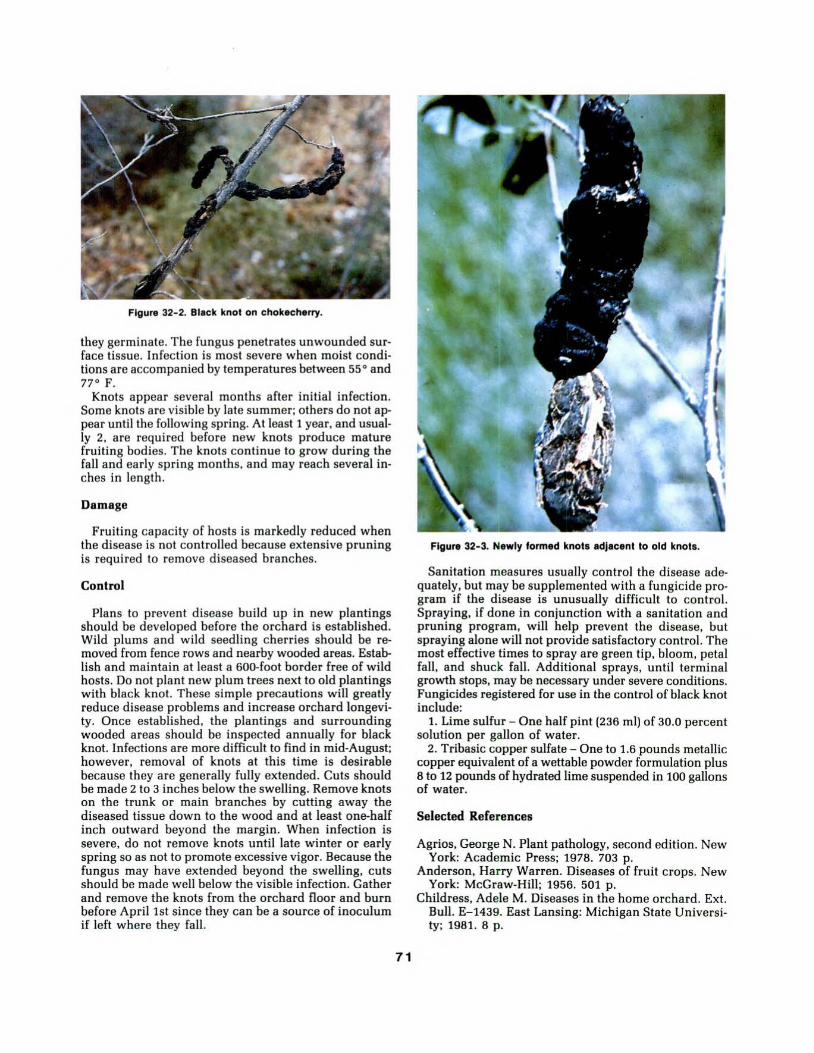
Figure 32- 2. Black knot on chokecherry.
they germinate. The fungus penetrates unwounded sur· face tissue. Infection is most severe when moist condi· tions are accompanied by temperatures between 55° and 77° F.
Knots appear several months after initial infection. Some knots are visible by late summer; others do not ap· pear until the following spring. At least 1 year, and usual· ly 2, are required before new knots produce mature fruiting bodies. The knots continue to grow during the fall and early spring months, and may reach several in· ches in length.
Damage
Fruiting capacity of hosts is markedly reduced when the disease is not controlled because extensive pruning is required to remove diseased branches.
Control
Plans to prevent disease build up in new plantings should be developed before the orchard is established. Wild plums and wild seedling cherries should be removed from fence rows and nearby wooded areas. Establish and maintain at least a 600-foot border free of wild hosts. Do not plant new plum trees next to old plantings with black knot. These simple precautions will greatly reduce disease problems and increase orchard longevi· ty. Once established, the plantings and surrounding wooded areas should be inspected annually for black knot. Infections are more difficult to find in mid-August; however, removal of knots at this time is desirable because they are generally fully extended. Cuts should be made 2 to 3 inches below the swelling. Remove knots on the trunk or main branches by cutting away the diseased tissue down to the wood and at least one-half inch outward beyond the margin. When infection is severe, do not remove knots until late winter or early spring so as not to promote excessive vigor. Because the fungus may have extended beyond the swelling, cuts should be made well below the visible infection. Gather and remove the knots from the orchard floor and burn before April 1st since they can be a source of inoculum if left where they fall.
71
Figure 32-3. Newly formed knots adjacent to old knots.
Sanitation measures usually control the disease ade· quately, but may be supplemented with a fungicide pro· gram if the disease is unusually difficult to control. Spraying, if done in conjunction with a sanitation and pruning program, will help prevent the disease, but spraying alone will not provide satisfactory control. The most effective times to spray are green tip, bloom, petal fall, and shuck fall. Additional sprays. until terminal growth stops. may be necessary under severe conditions. Fungicides registered for use in the control of black knot include:
1. Lime sulfur- One half pint (236 ml) of 30.0 percent solution per gallon of water.
2. Tribasic copper sulfate- One to 1.6 pounds metallic copper equivalent of a wettable powder formulation plus 8 to 12 pounds of hydrated lime suspended in 100 gallons of water.
Selected References
Agrios, George N. Plant pathology, second edition. New York: Academic Press; 1978. 703 p.
Anderson, Harry Warren. Diseases of fruit crops. New York: McGraw-Hill; 1956. 501 p.
Childress, Adele M. Diseases in the home orchard. Ext. Bull. E-1439. East Lansing: Michigan State Universi· ty; 1981. 8 p.

33. Plum Pockets William R. Jacobi and John E. Watkins
Plum pockets is a disease of plums caused by Taphrina communis. Taphrina pruni, another causal agent of plum pockets commonly found in Europe, has been reported on plums in North America, but the report is not confirmed.
Hosts and Distribution
All plum species and varieties are susceptible to T. communis. T. communis is found in most regions of the United States where plums are grown, but the exact distribution in the Great Plains region is unknown.
Symptoms and Signs
Symptoms appear as small white blisters on the young fruit. The blisters enlarge as the fruit develops and eventually involve the entire fruit The infected fruit increases
in size and becomes spongy and distorted (fig. 33-1). The seed does not develop, and a hollow cavity forms in the infected fruit. The fruit is reddish initially, then becomes covered with greyish powder consisting of asci. Young shoots and leaves also may become infected and malformed (fig. 33-2).
The asci are cylindrical-clavate and usually rounded at the apex. Stalk cells supporting the asci are narrower than the ascus and variable in length. The eight ascospores are round, ovate, or elliptical. The ascospores commonly germinate in the ascus and produce conidia by budding. The dimensions of the asci are 27-83 by 5-18 ~tm. of the stalk cells 6-56 by 3-12 ~tm. and of the ascospores 4-7 by 3.5-5.5 ~tm. One obvious feature that distinguishes this fungus from other Tophrina spp. is the long asci that frequently project above other asci.
72
Figure 33-1. Distorted, enlarged plums affected by plum pockets disease, and healthy unaffected plums.

Disease Cycle
The fungus apparently overwinters as conidia or ascospores on twigs and buds on the tree. In the spring these spores are blown to young, succulent tissues. The spores germinate and penetrate developing leaves or other tissues through stomata or directly through cell walls. The mycelium grows between cells, causing abnormal cell enlargement and division. Infected fruit or tissue is distorted and much larger than normal.
The fungus produces asci below the epidermis of the fruit. These eventually enlarge and break through the epidermis. The ascospores multiply by budding off conidia inside or outside the ascus. Spores are wind blown to new tissue, where infection takes place during bud break. Low temperatures and high humidity favor infection of susceptible tissue. All tissues become resistant as they mature.
Damage
Fifty percent or more of the fruit may be lost in years when the disease is severe. Buds and twigs may be af-
Figure 33-2. Distorted plums and leaves on plum tree affected by plum pockets disease.
73
fected, thus stressing the tree. Once the disease is established in a tree, it will appear each year unless controlled.
Control
The disease can be controlled by a single fungicide application in late fall or early spring before leaf buds swell. Any fungicide that is labeled for use on plum trees would be legally acceptable. Fungicides that have been effective are Ferbam, lime-sulfur, and Bordeaux mixture.
Selected References
Agrios, George N. Plant pathology, second edition. New York: Academic Press; 1978. 703 p.
Galloway, B. J. Plum pocket. U.S. Agriculture Comm. Report 1888: 366-369; 1889.
Mix, A. J. A monograph of the genus Tophrino. Univ. Kansas Sci. Bull. 33: 3- 167; 1949.
Swingle, D. B.; Morris, H. E. Plum pocket and leaf gall on Americana plums. Univ. Montana Agric. Exp. Sta. Bull. 123: 167-188; 1918.
Wilson, E. E.; Ogawa, J. M. Fungal, bacterial, and certain nonparasitic diseases of fruit and nut crops in California. Agriculture Science Publication. Berkeley: University of California; 1979. 190 p.

34. Brown Rot of Stone Fruits David S. Wysong and James A. Walla
A common and destructive disease of stone fruits called brown rot, blossom blight, twig blight, and/or brown rot canker is caused by the fungus Monilinia fructicola (Sderotina fructicola).
Hosts and Distribution
All of the commonly cultivated stone fruits, including cherry, apricot, and plum, are susceptible. The disease is less important on apple and pear, and of minor importance on flowering almond, cherry-laurel, chokecherry, flowering quince, and western sand-cherry.
While brown rot is world-wide, it is of little importance in regions where rainfall is low. Severe epidemics are common in the high-rainfall peach-growing areas of the
Figure 34-1. Mummified fruit with adjacent non·lnfected fruit clingIng to the branch.
Figure 34-2. Formation of ash-gray mold on surface of Infected fruits.
74
Southeast and along the Atlantic Seaboard, but may also occur elsewhere when moist conditions prevail during fruit ripening.
Symptoms and Signs
Brown rot is usually first seen as the fruit approaches maturity. Small, circular, light brown areas of decay appear on the surface of the fruit, expand rapidly, and within a few hours may encompass the entire fruit. A day or so later the fruits rot. Some fall to the ground, while others remain attached to the tree, where they dry and become mummified (fig. 34-1). In warm, wet, humid weather, tufts of ash-gray mold develop over the surface of the rotting fruits (fig. 34- 2). These fungal masses help distinguish brown rot from other diseases.
Flower blossoms, fruit spurs, woody shoots, and immature fruit are also attacked (fig. 34-3). Infected blossoms wilt, turn brown, and persist into summer. The fungus may progress downward into the flower cluster base and into the fruit spur (fig. 34- 4). When the fungus grows into woody tissue, small cankers are formed. They enlarge and may girdle the branch or twig, killing terminal growth. Droplets of resinous gum may accompany the blighted spurs and cankers.
Disease Cycle
The brown rot fungus overwinters in mummified fruits, branch or twig cankers, and infected peduncles. In spring, when fruit buds are opening, small cupshaped apothecia arise from the mummies beneath infected trees (fig. 34-5). When these apothecia are wetted, spores produced within them are ejected into the air and carried by wind to infect blossoms or new shoots.
Figure 34-3. Infected shoot and Immature fruit.

Figure 34-4. Infected flower cluster and fruit spur.
Infection may also arise from a second type of spore produced later in the season on dead blossoms, in spur or twig cankers, or on attached mummies. These spores are also carried by wind or splashing rain to infect ripening green fruit. Infection may occur directly through the cuticle or through natural openings. Wounded fruit is infected much more rapidly than unwounded fruit. Injuries caused by the stink bug, plum curculio, and other insects are frequently avenues of entrance for the brown rot fungus.
Warm, wet, humid weather favors rapid development of brown rot. The time of wetting necessary for blossom infection decreases from 18 hours at 50°F to 5 hours at 77°F. Infection rate decreases above 80°F and below 55°F, but may continue at temperatures as low as 40°F. Under optimum conditions, mature fruit decays in 36 to 48 hours.
Figure 34-5. Close up of Infected fruit and developing apotheclum.
Damage
Brown rot is economically important in commercial and home orchards. The disease reduces yields by damaging blossoms, twigs, and fruit. After harvest, fruit decomposition poses a constant threat in storage and transit.
Control
Brown rot control involves integrating several disease prevention methods. Control starts with removal of all fruit, mummies, and blighted twigs from trees in the fall. This procedure reduces the amount of brown rot fungus surviving the winter. Cultivate around trees before first bloom to reduce the early spring spore potential. Prune trees to promote good air circulation.
Fungicidal applications at blossom time and preharvest time are also important (table 34-1). Blossom blight must be avoided to insure against fruit infection later in the season. The first spray should be applied when the pistil tips extend above the flowers, even unopened flowers. If warm, wet weather prevails, spray every 4 to 5 days until one week after petal drop. Begin pre-harvest sprays about 3 weeks before harvest to control brown rot on ripening fruit. If brown rot has built up, the pre-harvest sprays should be started earlier. Applications should be made at closer than weekly intervals if weather is warm and wet as the fruit colors.
Plant fruit trees in a sunny, open site with well-drained soil away from frost pockets. Plant resistant cultivars when available. Do not plant near wild fruit trees because they may be a source of inoculum. Control fruit injury (e.g. insect or mechanical damage) to prevent early infection of fruit. Apply fungicides if such injury occurs. Excess nitrogen application may increase infection.
Selected References
Byrde, R. J. W.; Willetts, H. J. The brown rot fungi of fruit, their biology and control. New York: Pergamon Press; 1977. 171 p.
Zehr, Eldon I. Control of brown rot in peach orchards. Plant Disease. 66: 1101-1105; 1982.
Table 34-1. Recommended fungicides for brown rot control.
Chemical
Benomyl, 50% WP (Benlate, Benomyl)
Caplan, 50% WP (Captan, Orthocide)
Sulfur, 80-92% WP (Fiotox)
Ferbam, 76% WP (Fermat e)
Minimum number of days from last application until harvest
Rate/gal (3.8 L) Apricots Cherries Peaches Plums & Prunes
3/4- 1 1/2 Tbs1 0 0 0 0 (5.4 to 10.8 gm) 3 1/3 Tbs 0 0 0 (28 gm) 2 1/2 Tbs - 2 0 0 0 (19 gm) 1 Tbs 0 21 7 (6.8 gm)
1Tbs = tablespoonsful 2 - not registered
75

35. Fire Blight of Pear, Apple, Cotoneaster, and Other Ornamental Shrubs and Trees
John E. Watkins and Mark W. Andrews
Fire blight, caused by Erwinia amylovora, is the first plant disease shown to be caused by a bacterium. It is apparently indigenous to North America; it was first observed in 1780 in the Hudson River Valley in New York State. It is a major threat to susceptible pear, apple, crabapple, and cotoneaster cultivars.
Hosts and Distribution
Fire blight occurs on most species of Pomoideae and some species in the sub-families of the Rosaceae. Of the genera reported susceptible to fire blight, Malus (apple), Pyrus (pear), Cotoneaster (cotoneaster), Crataegus (hawthorn), Cydonia (quince), Pyracantha (pyracantha), and Sorbus (mountain ash), are the most important economically and show the most severe blight. Resistant species and/or cultivars of most hosts are available. Some clonal selections and cultivars of Pyrus communis are highly resistant. Nearly all selections of the callery pear (P. calleryana), including the Bradford cultivars, show good resistance. Several cultivars of the cultivated species of apple and crabapple are resistant.
Fire blight is native to North America. Since its first observation in New York, it has been found throughout the United States.
Symptoms and Signs
The bacterium infects blossoms and leaves near the growing tips. Leaves quickly wilt and turn black, but remain attached to infected twigs (fig. 35-1). The scorched appearance of affected branches is the most obvious symptom. The affected parts first appear water-soaked, then wilt, shrivel, and turn brownish to black. The bacterium may advance down a small branch to older branches, causing dark, sunken stem cankers (fig. 35-2). Those cankers at ground level are sometimes referred to as collar blight. Infection may spread into the roots, where the bark is killed in a manner similar to that on the trunk. Fruit blight generally is found in immature fruit.
With few exceptions, symptoms on pyracantha, hawthorn, and cotoneaster (fig. 35- 3) are generally similar to those on pear, apple, or crabapple.
Disease Cycle
During spring the pathogen begins to multiply. A milky to amber-colored exudate containing millions of bacterial cells oozes from the peduncle, the lenticels in the skin of fruits, and from the margins of cankers (fig. 35-4). This sticky exudate attracts insects that carry the bacteria to blossoms and leaves. The bacteria enter the host through
76
natural openings in blossoms and leaves or through wounds. Colonization proceeds intercellularly after the primary infection. The bacteria spread through the tissues, and secondary infection may continue throughout the growing season. Bacterial ooze or strands produced on blossoms, shoots, leaves, fruits, or larger branches are the sources of secondary inoculum, which can be spread by rain, wind, insects, or birds.
The bacterium overwinters in tissue adjacent to the margins of cankers. The pathogen is more likely to overwinter successfully in cankers with smooth margins and healthy tissue on all sides. The bacteria again multiply in spring and invade healthy tissue. In addition to producing primary inoculum, extending cankers can significantly injure trees.
Figure 35-1. Scorched appearance of blighted foliage on apple.
Figure 35-2. Localized fire blight canker.
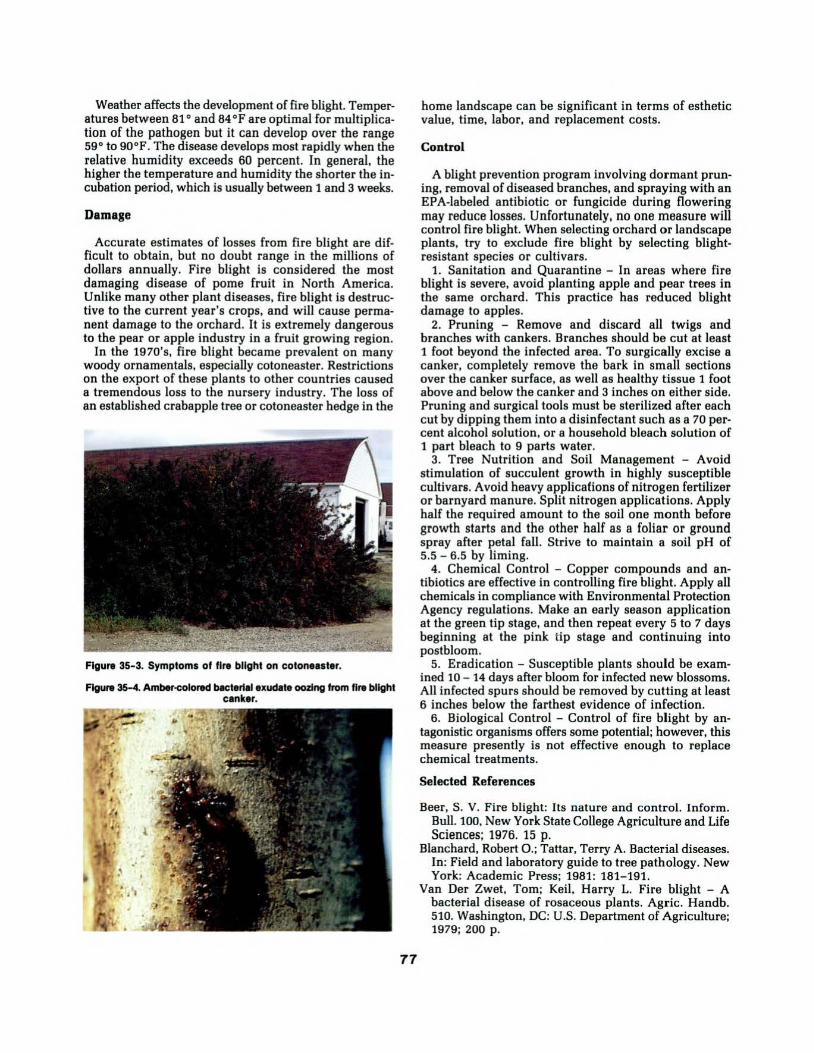
Weather affects the development of fire blight. Temperatures between 81 o and 84°F are optimal for multiplication of the pathogen but it can develop over the range 59° to 90°F. The disease develops most rapidly when the relative humidity exceeds 60 percent. In general, the higher the temperature and humidity the shorter the incubation period, which is usually between 1 and 3 weeks.
Damage
Accurate estimates of losses from fire blight are difficult to obtain, but no doubt range in the millions of dollars annually. Fire blight is considered the most damaging disease of pome fruit in North America. Unlike many other plant diseases, fire blight is destructive to the current year's crops, and will cause permanent damage to the orchard. It is extremely dangerous to the pear or apple industry in a fruit growing region.
In the 1970's, fire blight became prevalent on many woody ornamentals, especially cotoneaster. Restrictions on the export of these plants to other countries caused a tremendous loss to the nursery industry. The loss of an established crabapple tree or cotoneaster hedge in the
Figure 35-3. Symptoms of fire blight on cotone11ter.
Figure 35-4. Amber-colored b8ctert.l exuct.te oozing from fire blight c•nker.
71
home landscape can be significant in terms of esthetic value, time, labor, and replacement costs.
Control
A blight prevention program involving dormant pruning, removal of diseased branches, and spraying with an EPA-labeled antibiotic or fungicide during flowering may reduce losses. Unfortunately, no one measure will control fire blight. When selecting orchard or landscape plants, try to exclude fire blight by selecting blightresistant species or cultivars.
1. Sanitation and Quarantine - In areas where fire blight is severe, avoid planting apple and pear trees in the same orchard. This practice has reduced blight damage to apples.
2. Pruning - Remove and discard all twigs and branches with cankers. Branches should be cut at least 1 foot beyond the infected area. To surgically excise a canker, completely remova the bark in small sections over the canker surface, as well as healthy tissue 1 foot above and below the canker and 3 inches on either side. Pruning and surgical tools must be sterilized after each cut by dipping them into a disinfectant such as a 70 percent alcohol solution, or a household bleach solution of 1 part bleach to 9 parts water.
3. Tree Nutrition and Soil Management - Avoid stimulation of succulent growth in highly susceptible cultivars. Avoid heavy applications of nitrogen fertilizer or barnyard manure. Split nitrogen applications. Apply half the required amount to the soil one month before growth starts and the other half as a foliar or ground spray after petal fall. Strive to maintain a soil pH of 5.5 - 6.5 by liming.
4. Chemical Control - Copper compounds and antibiotics are effective in controlling fire blight. Apply all chemicals in compliance with Environmental Protection Agency regulations. Make an early season application at the green tip stage, and then repeat every 5 to 7 days beginning at the pink tip stage and continuing into post bloom.
5. Eradication - Susceptible plants should be examined 10 - 14 days after bloom for infected new blossoms. All infected spurs should be removed by cutting at least 6 inches below the farthest evidence of infection.
6. Biological Control - Control of fire blight by antagonistic organisms offers some potential; however, this measure presently is not effective enough to replace chemical treatments.
Selected References
Beer, S. V. Fire blight: Its nature and control. Inform. Bull. 100, New York State College Agriculture and Life Sciences; 1976. 15 p.
Blanchard, Robert 0.; Tattar, Terry A. Bacterial diseases. In: Field and laboratory guide to tree pathology. New York: Academic Press; 1981: 181-191.
Van Der Zwet, Tom; Keil, Harry L. Fire blight - A bacterial disease of rosaceous plants. Agric. Handb. 510. Washington, DC: U.S. Department of Agriculture; 1979; 200 p.

36. Witches'-Broom of Hackberry
Mark 0. Harrell and Frederick J. Crowe
Hackberry is commonly planted in cities and parks in much of the central Great Plains. Although principally a bottomland tree, it is able to withstand a wide range of moisture and temperature conditions and does well in an urban environment. A common and sometimes disfiguring branch disease of hackberry is witches'broom.
Hosts and Distribution
Witches'-broom probably occurs throughout the range of hackberry in the Great Plains; it is very common in eastern Kansas and Nebraska. Its principal host is hackberry.
Winter surveys of northeastern Kansas city street plantings, showed between 40 and 75 percent of the hackberry trees had witches'-brooms. However, even
Figure 36-1. Young broom on hackberry with numerous enlarged buds.
78
though branches of mature, closely spaced hackberry trees penetrated and touched the canopies of each neighbor, many trees had remained broom-free for many years in spite of the fact that neighbors were heavily broomed.
Symptoms and Signs
The most apparent symptom of witches'-broom is a dense clustering or "brooming" of twigs. Because brooms do not occur on branches less than 1 year old, they are not found at branch tips. They occasionally appear to be terminal, however, when a branch leader fails to develop fully or when it dies and breaks off. Buds of shoots within a broom are usually larger than normal and have more open bud scales.
The first indication of witches'-broom appears in the
Figure 36-2. Young broom with open cluster of affected twigs.

Figure 36-3. Old brooms with compact woody centers.
buds on branch portions at least 1 year old. The infected buds are usually smaller than normal, and often there are many small, swollen buds instead of a single large one. The shoots produced from the diseased buds grow vigorously and produce buds that are usually larger, more open, and more numerous than normal (fig. 36- 1).
During the following year the development of the broom varies, depending upon the vigor of the diseased branch. If the branch is vigorous and grows rapidly, its buds develop into branchlets similar to the original, and over a number of years a loose or open cluster of diseased twigs is formed (fig. 36-2). If the diseased branch is weak, only the buds at the base grow, resulting in a large number of abortive twigs. Over several years this type of development produces a compact broom with a firm woody center often an inch or more in diameter (fig. 36-3).
Disease Cycle
The cause of witches'-broom of hackberry is not understood. An eriophyid mite (Aceria sp.) rarely found on symptomless trees is almost always associated with the brooming; the powdery mildew fungus Sphaerotheca phytophila is much less often found. The original report of the mite/mildew association with brooms has commonly been misinterpreted as a requirement for the abnormal development of witches'-brooms. However, there is no experimental evidence to confirm or disprove the role of these organisms in the development of the disease.
79
Viruses and mycoplasma-like organisms (MLO's) also can cause brooming in woody plants. Whether the growth abnormality results directly from mite activity, whether the powdery mildew is involved but remains too obscure to be routinely identified, or whether a viruslike entity is instead responsible remains uncertain.
Damage
Witches'-brooms on hackberry are more unsightly than harmful to the tree. Brooms can cause branches to break more readily and therefore expose wood to decay fungi. Extensive brooming can reduce the vigor of the tree. However, trees are seldom seriously injured.
Control
No effective control measures are known for this disease. Pruning back diseased twigs to sound wood can improve the tree's appearance.
Selected References
Kellerman, W. A.; Swingle, W. T. Branch knot of the hackberry. Annual Report of the Kansas Experiment Station, 1888. Manhattan: Kansas State Agricultural College; 1889: 302-315.
Snetsinger, Robert; Himelick, E. B. Observations on witches'-broom of hackberry. Plant Disease Reporter. 4 1: 541-544; 1957.

3 7. Perennial Woodrotting Fungi that Cause Stem Decays of Hardwoods
Jerry W. Riffte and James A. Walla
A number of perennial polypore fungi other than those of the genus Phellinus cause stem decays. Most species that attack hardwood trees and shrubs in the Great Plains are classed either as white-rot or brown-rot fungi. Whiterot fungi degrade cellulose and lignin, have extracellular phenol oxidases, and generally give positive oxidase tests with gallic and tannic acid media or with gum guaiac. Brown-rot fungi degrade cellulose and generally give negative oxidase tests.
Hosts and Distribution
Eleven species of polypores with perennial sporocarps are important decay fungi on hardwood tree and shrub species in the Great Plains. They have been classified in
Figure 37-1. Sporocarps of Perenn#por#a traxtnophlla on bole of a green ash.
the genus Fornes in older literature. Five common species are discussed in this paper. Characteristics and distribution of these species are given in table 3 7-1. Perenniporia fraxinophila (fig. 37-1) is the most commonly reported stem decay fungus on live green and white ash in the Great Plains; it has been found on many other tree species, including bur oak and boxelder. Perenniporia ellisiana (fig. 37-2) is the most commonly reported stem decay fungus on live individuals of the genus Shepherdia. Ganoderma applanatum (fig. 37-3) and Fomitopsis melliae attack both live and dead hardwoods, including poplar, maple, honey locust, and ash. Fornes fomentarius occurs principally on dead hardwoods, especially birch. Other, less-common species in the Great Plains include Oxyporus populinus, Ganoderma lobatum, Perenniporia ohiense, Datronia scutellata, and Fomitopsis cajanderi.
Figure 37-2. Sporocarp of P. ellls#ana on stem of buffaloberry.
80

Figure 37-3. Sporocarps of Ganoderma app#anatum on dead cottonwood.
Symptoms and Signs
Sporocarps (fruiting bodies) of decay fungi develop on boles or major branches of trees with extensive wood decay. Sporocarps of these polypores are perennial, hard and woody when mature, typically shelf- to hoof-shaped, sessile, and have well-developed tubes seen in a vertical section through the sporocarps. More than one layer of tubes can be seen in vertical cuts through the center of sporocarps that have sporulated more than 1 year.
Some macroscopic characteristics of sporocarps useful in identifying species of perennial polypores are the size, shape, and color of sporocarps, size of pores, and color and texture of the context (table 37- 1). Microscopic characteristics of hyphal and hymenial elements also are useful for identification of species.
Most species of perennial polypores that infect live hardwood trees in the Great Plains eventually produce a white-mottled decay of the wood. Decay caused by P. fraxinophila on green ash is characterized by a wide, brown zone surrounding the advanced decay (fig. 37-4). Invaded tissues become light brown and later dark brown; decayed wood is then bleached until it becomes straw color to white. Finally, white spots appear in the wood and decayed wood becomes soft and crumbly. G. applanatum and F. fomentarius also produce a white or straw-colored wood decay. Wood decayed by P. ellisiana is brown, with small, irregular white flecks scattered
Table 37-1. Geographical distribution, and sporocarp and basidiospore characteristics of five species of perennial polypores that decay wood of hardwood trees and shrubs in the Great Plains.
Sporocarps Basldloaporea
Decay fungus 1 Geographical Size, cm2 Shape Pores per mm Context Pore surface distribution hymenlum color color Slze,"m3 Shape
Ganoderma NO,NE,KS,MT,CO, 3x5xt- Sessile, pileus 4-6 brown, occas. white 5x4-9x6 ovoid, truncate applanatum NM,MAN. 30x50x10 plane or convex white at one end
appearing or-namented
Perenniporia MT,WY,CO,NM, 3x3x1- Sessile, pileus 2- 3 pallid to white to 4x3-8x6 oblong-ellipsoid el/isiana NO,SO,SASK. 10x15x8 convex to ungulate wood~olored lsabelllne to broadly
elllpsoid,trunca-teat apex
Perenniporia MT,WY,CO,NM,NO, 2x3xt- Sessile or decurrent, 2- 3 pale wood lo white to 6x5-9x6 ellipsoid to fraxinophila SD,NE,KS,OK,MAN. 25x40x10 pileus convex to yellow brown brownish ovoid, truncate
ungulate al end
Fomltopsls NE,TX tx3x1- Sessile or decurrent 4- 5 cinereous to isabelline 6x2-8x3 cylindric melliae 5x10x5 pileus convex or pale wood
plane, usually imbricate
Fomes MT,NO,SO,NE,MAN. 3x6x2- Sessile, pileus 3-4 dark tan gray to 12x4- 18x5 cylindric· fomentarius 15x20x15 convex to ungulate or brown brown ellipsoid
1 F. melliae is a brown-rot fungus; the remaining four species are white-rot fungi. 21ength x width x height 31ength x width
81

Figure 37-4. Cross section of green ash stem with white mottled decay caused by P. fraxlnoph/la.
throughout (fig. 37-5). F. melliae causes an extensive brown crumbly decay of sapwood of hardwoods.
Disease Cycle
Decay in trees is a complex process that begins at wounds, branch stubs, or other openings in the bark of boles or branches of trees. Many microorganisms, including bacteria, yeasts, and other nonhymenomycetous fungi, invade these openings in succession. Physical and chemical changes caused by these microorganisms in the invaded wood make it possible for wood decay fungi to become established. After growth and subsequent wood decay, sporocarps are produced to complete the life cycle. Discoloration and decay of wood by microorganisms are limited to wood present at the time of wounding by a barrier zone formed by the cambium. Additional wounding of stems may result in multiple infections.
Damage
Damage to hardwood trees by stem decay fungi becomes increasingly serious when trees reach the age at which tree vigor declines. These fungi seldom kill trees, but infected stems are more vulnerable to breakage during windstorms, and affected wood is not suitable for wood products.
Buffaloberry has been severely damaged by P. ellisiana in the northern Great Plains. This fungus may destroy windbreak plantings that are only 20 to 25 years old. Buffaloberry is so susceptible that its extensive use in the northern Great Plains is not encouraged.
At least 2 million of the 20 million living green ash in Nebraska woodlands are infected by P. fraxinophila. Incidence of green ash with sporocarps increases in a straight line relationship with diameter of the tree stem.
82
Figure 37-5. Cross section of buffaloberry stem with wood decayed by P. ellis/ana.
Control
Because the decay process begins with wounds, prevention of wounds-particularly basal wounds caused by grazing livestock and cultivating too close with machinery, removal of trees, or fires-will reduce development of decay in plantings and woodlands.
Landscape trees in urban areas and in recreational plantings occasionally are pruned to remove dead or diseased branches. It is advisable to cut small branches; do not cut through the callus collar at the base of the branches. Where it is impractical to prune trees, regulate tree spacing so that natural pruning will reduce the number and size of potential infection sites. When removing trees from plantings during thinning, removal of defective and diseased trees will help reduce storm damage and sources of inoculum.
Selected References
Lowe, Josiah L. Polyporaceae of North America. The genus Fornes. Tech. Pub. 80. Syracuse: State University College of Forestry at Syracuse University; 1957. 97 p.
Overholts, Lee Oras. Polyporaceae of the United States, Alaska, and Canada. Ann Arbor: University of Michigan Press; 1953. 466 p.
Riffle, J. W.; Ostrofsky, W. D.; James, R. L. Fornes fraxinophilus on green ash in Nebraska windbreaks. Plant Disease. 65: 667-669; 1981.
Riffle, Jerry W.; Sharon, Edward M.; Harrell, Mark 0. Incidence of Fornes fraxinophilus on green ash in Nebraska woodlands. Plant Disease. 68: 322-324; 1984.
Walla, James A.; Riffle, Jerry W. Fornes fraxinophilus on green ash in North Dakota windbreaks. Plant Disease. 65: 669- 670; 1981.

38. Phellinus Stem Decays of Hardwoods Jerry W. Riffle and Kenneth E. Conway
Species of Phellinus are associated with white rots of woody plants, and generally give a positive oxidase reaction on tannic and gallic acid media or with gum guaiac solution. In older taxonomic publications, the species now placed in Phellinus are found in the genera Fornes, Poria, and Polyporus.
Hosts and Distribution
Ten species of Phellinus occur on hardwood tree and shrub species in the Great Plains. P. gilvus is usually found on wood of dead trees, but it is occasionally found on live green ash, black locust, maple, and willow. P. tremulae (fig. 38-1) is restricted to aspen. Its range in North America, north of Mexico, is approximately the same as the range of aspen. Phellinus igniarius attacks live hardwood trees, especially birch, but it also has been found on ash, black walnut, poplars, buckthorn, and willow.
P. tuberculosus (formerly P. pomaceus) (fig. 38-2) produces an extensive stem decay in live Prunus species,
Figure 38-1. Sporocarp of Phe/1/nus tremutae on bole of an aspen tree.
Figure 38-2. Sporocarps of P. tubercutosus on stem of American plum.
83
especially American and chickasaw plums. P. punctatus (fig. 38-3) occurs on both live and dead hardwood trees and shrubs; it decays wood of live green ash, Siberian peashrub, American plum, lilac, willows (golden, diamond, Bebb}, black locust, and common buckthorn.
P. robineae (fig. 38-4), a white-rot fungus, produces an extensive yellow-brown decay of living black locust (fig. 38-5) in the central and southern Great Plains. P. weirianus decays wood of black walnut. P. everhartii usually occurs on live trunks of oak, whereas P. conchatus occurs on dead wood of hardwood species. P. rib is has been found on live Ribes spp. in North Dakota. The ten Phellinus species and their known distribution in the Great Plains and surrounding states are described in table 38-1.
Symptoms and Signs
Sporocarps (fruiting bodies) develop on trunks and branches of trees and shrub species that have extensive wood decay. Characteristics of sporocarps and spores
Figure 38-3. Sporocarps of P. punctatus and canker rot symptoms on bole of green ash.

o
CD
Q.
to
A
3
T3
o
o
5
-o
CO
.IZ
â– D
O
o
3
>.
to
o
CD
"O
co
.c
to
o
CD
Q.
10
co
3
c
"S
£
O co
o «
fflQ-
To J3
03
U
CO
o
CD
l_
o
a
co
g
o
Q.
w
(0
u
o
w
o
a.
(0
to
Q.
w
<O
o
2
o
Q.
10
c
o
.O
Q.
O
o
o
c/>
w
4)
U
a
CO O
a. "
0
Q.
5 ,-
• o
5 °
O O
o
E
E E
a q,
If
a.
o
a
CO
E
o
N
ra C
.9 o
a a
tt3
co
O
A
O
O3
n
3
?!?
.b a3
Q. CO
« SR
(0 c
°o
S to
V
,2 x
z
<
CO
c
3
o
k-
Ix
»°
<13 ^
cd
E
c
.cz
o
CO "O
.9-5
= >
CD o
CD
C
'o CO
o °
>-e
o o
is
3
o
>.
.o
3
.C
CO
CD
b
CO
Q.
-o
S
o
-O
o
CO
n
3
â– D
O
CO
Q.
■— CO
2 o
T3 co
o S
6 o
o
52
CO
.if CD
O CO
CO O
Q.J3
E °
CD O3
CD x
— CD
to >
to c
CD O
CO O
r- I X
CM CO
X CO
CD
TJ CO
CD O
3z cd E
CD = -
._ Q. w
O .-O
» « £-
« S S
co — zz
cSSS
V x
X CM
CM t-
CD
"D CD
CO
!0
0-!2
<D ttx
:= co cd
CO 3 >
CO <D C
CD = O
to au
CM O
X CN
m x
x m
CO 1-
LU
z
°*i
q"o
zo
ft
IS
S S
^2
CO &
CD „
CO CO
o«2
3 CM
CD
C
â– A w
So.
« CO
^ CD
tD =
CD
c^8
CD
â– D
• *
.Sf-o
Q. E CD
3 O <°
CO o 3
CD CD t
â–¡C XI CD
>-
o
o
to :s
CD
O
cd
O
8
a ja
O 10
• -■=.
O
to 3
P
CD c
;? 9
0. O
a. cd
CD
CO
"> ai
3
C „
c 3
& to
â– ^ k*
ft 3
= CO
d> s!
£?,
« CD
â– c J>
Q. i:
VCD
CO |
Ul'O
ZO
a
SO
CO
3
to
CO O
II
a. 3
O
O
o
Q
Z
Si
C TO
S CJ
CD C
a. o.
Q
Z.W
z^
22
3
C
1-2
to o
sill
■a "5, <u <°
o -Q C a>
> 3 CO C
OOJIO
s
o
>. c
CO k_
Q. J3
CD
CO
O
a -a
2"5
So
52
X
X
X
T—
X
X
X
â– *T
X
tn
CD
m
Y
m
m
en
tn
CD
n
CO
CO
-<r
â– <r
CO
m
c\j
â– >}â–
X
X
X
X
X
X
X
X
X
â– <t
â– <*
-*
m
■»-
T
CD
CO
m
ll
c
3
c
o
c
3
o
c
3
CO
o
c
3
o
O
3
to
E
CO
o
3
5
o
^3 O
o
CO
k_
O
ez
C
»- o
4;^
A
n
Ql
c
o
n
A
o
CO
.c
i3
3
3
JZ 5
CO
to
JZ
3
c
JZ
CO
to
"CD
T3
CO
i
o
o
a>si
2-g
3
CO
Q.
C
o
c
>.
t:
3
CO
A
A
3 «
o
T3
j<:
3
CD
3
Q.
Q.
JC
CD
o
'^
>>Jc:
O-o
CD o
"CD
â– o
CO
o
CO
o
3
3
CO
"CD
CO
CO
CO
>.£!
CD
^
Q..O
â– a
o
k.
>.£
>.
â– D
Q.
Q.
c
3
o
c
>.
O3
tZ0T3
c
C
3
o
A
o
3
o
CO
CO
^3
x:
1
to
~-o
TJ
A
CO
t:
A CD
5
n
T3
CO
5
"D
*1
c
CD
c
CD
3
C
CD
^5
3
o
3
i-
c
T3
3
*- cz
^
3
o
CD
1°
CD >-
'»—
CO
o
CO
o
o
I—
o
ra t:
>. O
3
A
k—
O
JD
T3 J3
-o
A
>.
A
= n
CO
X
CD
O
l
XI
A
CO
3
o
CD
£
to
>-nj
11
3
to
CD
CD O
.— *-"
CO to
O
a. cd
CD
3 -
CD
»»
®" c
«
if CO
to J
CO â„¢
3
CO 3
CO —
to an
to at
c^£
CD Q.
CZ
CD C
CO CO
3
CO 3
Vo
i- o
X CM
x co
1- X
in x
x o
x o
V- T-
CO CM
o
3 «>
2 to
-S a>
= c
®3
S2
2?
Q.<x
3 CO
O
WX
to
CO
3
3
C
C
«5
.TO
«
CD
0.
5
CD
A
A A
2 ?
3 3
x x
7 C
CD CD
84
Tab
le 3
8-1.
Ge
og
rap
hic
al
dis
trib
uti
on
and
sp
oro
carp
and
ba
sid
iosp
ore
ch
ara
cte
rist
ics
of
10 P
helli
nus
spe
cie
s th
at
deca
y w
oo
d o
f ha
rdw
ood
tre
e a
nd s
hru
b s
pe
cie
s in
th
e G
reat
Pla
ins.
Spo
roca
rp c
ha
ract
eri
stic
s B
asi
dio
spo
re c
ha
ract
eri
stic
s D
ecay
G
eo
gra
ph
ica
l P
ores
pe
r m
m
Co
nte
xt
Por
e su
rfa
ce
fun
gu
s d
istr
ibu
tio
n
Siz
e, c
m1
Sha
pe
hym
en
lum
co
lor
colo
r S
ize,
,.m
2 S
hape
C
olo
r
Phe
llinu
s M
AN
,ND
,MT
,TX
<1
x4x<
1-
Mo
stly
res
upin
ate;
5
-7
yello
w-b
row
n
yello
wis
h-b
row
n
4x3
-5x4
su
bg
lob
ose
h
yalin
e
conc
hatu
s 7x
12x1
so
me
time
s se
ssile
o
r da
rker
to
dar
k br
own
Phe
llinu
s M
T,N
E,K
S,N
M
2x4
x2-
Ses
sile
, p
ileu
s 4
-6
rust
y br
own
yello
wis
h-t
o
4x3
-6x4
su
bg
lob
ose
ch
est
nu
t-b
row
n
ever
hart
ii 15
x36x
15
conv
ex
redd
ish-
brow
n to
glo
bo
se
in K
OH
Phe
llinu
s M
T,N
D,N
E,K
S,
1x2
x<1
-S
ess
ile o
r e
ffu
sed
-6
-8
bri
gh
t ye
llow
da
rk p
urp
lish
4
x3-5
x3
elli
pso
id t
o
hya
line
g
ilvu
s O
K,T
X,N
M
7x12
x15
refle
xed;
pile
us
brow
n br
own
ovo
id
solit
ary
or
imb
rica
te
Phe
llinu
s M
T,N
D,S
D,N
E,
3x5
x2-
Se
ssile
or
de
curr
en
t;
5-6
da
rk r
eddi
sh
pale
cin
na
mo
n
5x4
-<1
x5
bro
ad
ly o
void
h
yalin
e
ign
iari
us
WY
,CO
,NM
15
x20x
12
pile
us
pla
ne
to
brow
n br
own
to d
ark
to s
ub
glo
bo
se
conv
ex
(X)
pu
rplis
h b
row
n
,Jio.
Phe
llinu
s C
O,M
T,N
M,W
Y
up t
o
Ses
sile
, d
eve
lop
ing
6
-7
dark
red
dish
p
urp
lish
bro
wn
4x4
-5x4
su
bg
lob
ose
h
yalin
e
trem
ulae
20
x15
at b
ran
ch s
cars
br
own
Phe
llinu
s M
T,N
D,N
E,K
S,
<1
x4x1
-S
ess
ile o
r e
ffu
sed
-4
-6
yello
wis
h-b
row
n
dark
ye
llow
ish
-4
x3-5
x4
ovo
id t
o
hya
line
tu
be
rcu
losu
s O
K,W
Y,C
O,N
M
6x10
x3
refle
xed;
pile
us
plan
e to
red
dish
-bro
wn
to r
ed
dis
h-b
row
n
bro
ad
ly e
llip
soid
to
co
nve
x
Phe
llinu
s N
D,N
E,O
K,C
O
-R
esup
inat
e,
6-8
-
yello
wis
h t
o
6x5
-8x7
b
roa
dly
ovo
id
hya
line
p
un
cta
tus
be
com
ing
wid
ely
gr
ayis
h-br
own
to s
ub
glo
bo
se
eff
use
d
Phe
llinu
s M
AN
,ND
, 1
x1x<
1-
Se
ssile
or
effu
sed-
7-8
br
own
gray
-bro
wn
Ia
3x2
.5-4
x3
elli
pso
id t
o
pale
ru
sty
rib
is
MT
,KS
10
x20x
4 re
flexe
d da
rk b
row
n
glo
bo
se
Phe
llinu
s N
E,K
S,O
K,
3x5
x1-
Ses
sile
; p
ileu
s 7
-8
ligh
t re
dd
ish
ye
llow
ish
to
5x4
-6x5
o
void
to
re
dd
ish
-bro
wn
ro
bine
ae
TX
,NM
20
x30x
15
ap
pla
na
te t
o
brow
n re
ddis
h-br
own
sub
glo
bo
se;
un
gu
late
fla
tte
ne
d o
n o
ne
sid
e
Phe
llinu
s N
E,K
S,O
K,
up t
o
Ses
sile
, u
sua
lly
5-7
ye
llow
ish
-bro
wn
g
old
en
bro
wn
4x3
-5x3
su
bg
lob
ose
pa
le y
ello
w
wei
rianu
s N
M,T
X
3x8x
8 u
ng
ula
te,
sulc
ate
to
ovo
id
brow
n
1 ie
ng
th x
wid
th x
he
igh
t 2 1
engt
h x
wid
th

Figure 38- 4. Sporocarps of P. robin••• on bole of black locust.
that are useful in the identification of PheUinus species appe~r in table 38-1. Sporocarps are perennial, resupmate, effused-reflexed, or sessile. The context of the sporocarps is yellowish-brown to reddish-brown or purplish-brown, and darkens permanently to black in KOH solution. The hyphae may have simple septa but lack clamp connections. Setal hyphae and hymenial setae are present or absent; and basidiospores are cylindric to globose, and hyaline to pigmented. The pore surface is yellowish-brown to purplish-brown, and the pores are circular to daedaloid.
All PhelJinus species cause a white-rot decay. Wood of aspen decayed by P. tremuloe is initially light pink to straw-brown, then chocolate-brown, and finally is reduced to a soft, spongy, whitened mass. The decayed wood is surrounded by conspicuous dark zones or black lines. Wood invaded by P. tuberculosus initially becomes brownish with flecks and streaks, and finally becomes soft and stringy. P. robineae reduces wood of black locust to a soft spongy yellow or brown mass (fig. 38-5). The decay extends outward from the center of the heartwood in a series of radial lines along the rays. Red-brown mycelial felts characterize the decay. Decay by P. gilvus is confined mostly to the sapwood, which is tan or creamcolored when completely decayed.
Disease Cycle
The process of decay in trees is complex, and involves invasion of wounds by a succession of microorganisms, including bacteria, yeasts, other nonhymenomycetous fungi, and 'wood decay fungi. The development of decay in trees is discussed in article 37. Most of the species of Phellinus that decay wood of living trees are confined to the non-living heartwood of their hosts.
Damage
Many windbreaks established throughout the Great Plai.ns in 1935- 1942 during the Prairie States Forestry ProJect have reached an age of declining vigor, and damage from stem diseases has become increasingly
85
Figure 38-5. Longitudinal section of black locust with wood decayed by P. roblneae.
serious. P. robineoe occurs on black locust throughout the windbreak planting area in Oklahoma, and is the predominant stem decay fungus on this species. Incidence increases with tree age. About 25 percent of the black locusts in 40-year-old windbreaks in Oklahoma were infected with P. robineae.
P. punctotus is widespread in North Dakota, has a relatively wide host range, and occurs on about one-third of the sites that have hosts more than 20 years old. It has been found associated with canker rot on eight tree and shrub species in plantings and natural stands in the northern Great Plains. Based on sporocarp incidence, P. punctotus is the most important stem decay fungus on green ash in North Dakota Prairie States Forestry Project windbreaks.
Control
No direct control for stem decay fungi is known. Losses from these fungi can be minimized by use of certain preventative measures (see article 37 for such information).
Selected References
Gilbertson, R. L. The genus Phe1Jinus (Aphyllophorales: Hymenochaetaceae) in western North America. Mycotaxon. 9: 51-89; 1979.
Riffle, Jerry W.; Myatt, Alan K.; Davis, Roger L. Incidence of Phellinus robineae in black locust plantings in Oklahoma. Plant Disease. 69: 116-118; 1985.
Walla, J. A. Incid.ence of Phellinus punctatus on living woody plants m North Dakota. Plant Disease. 68: 252-253; 1984.

39. Quince Rust Kenneth E. Conway and Mark W. Andrews
Quince rust, caused by the fungus Gymnosporangium clavipes, was an economically important disease in the United States during the nineteenth and early twentieth centuries because of its devastating effects on commercial apple orchards. It can also severely infect Juniperus spp. Three rust fungi, G. clavipes, G. juniperi-virginianae, and G. globosum, infect apples and use Juniperus spp. as alternate hosts. The latter two Gymnosporangium species are discussed in another section of this handbook.
Hosts and Distribution
G. clavipes infects 11 genera of pomaceous hosts, including Amelanchier spp., Amelosorbus sp., Aronia spp., Chaenomeles spp. (flowering-quince), Crataegomesphilus sp., Crataegus spp. (hawthorn), Cydonia spp. (common quince), Photinia sp., Pyrus spp. (pear), and Sorbus spp. Some susceptible cultivars of apple are Delicious, Golden Delicious, Red Delicious, Mcintosh, Northern Spy,
Rome, and Winesap. Pycnial and aecial spore stages are developed on the pomaceous host. The telial and basidial spore stages occur on Juniperus spp.; no uredial spore stage is known.
Quince rust is not known to occur outside of North America, where it is found from Newfoundland to British Columbia and southward east of the Rocky Mountains to north Florida and Texas and into Mexico.
Symptoms and Signs
The disease on pomaceous hosts occurs most frequently on fruits, less frequently on twigs and buds, and rarely on leaves (fig. 39-1). Infection of apple leaves produce flecks or abortive lesions. Infected apples fall prematurely, and those that mature are misshapen and have dark green sunken lesions, usually near the blossom end. Unlike apples infected by the cedar apple rust fungus, tissue beneath quince rust lesions is necrotic, often deeply into the fruit.
Figure 39-1. Aecla on fruits and twigs of hawthorn Infected by Gymnosporang/um clarlpes. Reddlsh·orange aeclospores visible when the long white tubes are broken.
86

Figure 39-2. Dr1ed gelatinous tellal canker of G. cter/pes on a branch of Andorra juniper.
The quince rust fungus infects leaves, twigs, branches, and trunks of junipers. It produces elongate, swollen, often spindle- shaped rough cankers on twigs and branches. Reddish colored, gelatinous masses of teliospores exude from the cankers during rainy periods in the spring about the time apple trees are in bloom (figs. 39-2, 39-3). Five or six exudations are possible, but teliospores usually do not germinate to produce basidiospores during their first appearance. Teliospore and basidiospore masses change from red to yellow during this period.
Disease Cycle
The quince rust fungus requires two hosts to complete its disease cycle. Pomaceous hosts are infected by basidiospores discharged from germinated teliospores pr.oduced ?n ju~iper. Basidiospores are dispersed by wmd, and mfectlon occurs during a short period in the spring as flowers and fruit primordia emerge from their buds. After petal drop, fruits of most species are no longer susceptible. Pycnia develop 2 or 3 weeks after infection. Two or 3 weeks later, aecia are formed. The aecia are the most easily recognized of the spore stages (fig. 39- 1) because long white tubes are formed that may cover the entire fruit. Aecia rarely form on apple, but are common on fruit of quince and hawthorn. When the tubes rupture at their apex, red masses of aeciospores are evident. Aeciospores are wind blown, and can infect junipers throughout the growing season.
Aeciospores infect leaves, twigs, and branches of juniper. Infected leaves are often overlooked. Twigs can become infected directly, and spindle-shaped swellings covered by flaky, darkened bark are formed. The fungus can remain active for several years as the cankered area continues to expand. Twigs infected during the growing season produce teliospores and basidiospores the following spring and annually thereafter.
87
Figure 39-3. Gelatinous tellal cankers of G. ctavlpes after moisture had been applied.
Damage
Pomaceous hosts can be weakened by defoliation when leaves are severely infected. However, the greatest economic loss is caused by infection of fruits.
Twig and branch infection of juniper is limited to phellogen, but may cause some structural weakness. Severely infected trees may be more susceptible to winter injury. Failure to control quince rust on nursery junipers can result in quarantines and loss of revenue.
Control
Classical control has been the removal of junipers around apple orchards, and of pomaceous hosts around tree nurseries. Resistant varieties of apple and juniper are available. Pruning telial cankers on juniper can reduce disease potential, but large trees may have extensive trunk cankers that cannot be pruned out. Protective fungicide controls to prevent infection are available for apple (benomyl + mancozeb or zineb) and juniper (Bordeaux mixture or cycloheximide). To be effective, chemicals should be applied to apple in the spring as green tip and cover sprays. Junipers should be protected throughout the summer and fall on a regular basis.
Selected References
Anderson, Harry Warren. Diseases of fruit crops. New York: McGraw-Hill; 1956. 501 p.
Crowell, I. H. The hosts, life history and control of Gymnosporangium clavi pes C. and P. Journal of the Arnold Arboretum. 16: 367-410; 1935.
Kern, F. D. Lists and keys of the cedar rusts of the world. Memoirs, New York Botanical Garden. 10: 305-326; 1964.
Miller, Paul R. Pathogenicity of three red-cedar rusts that occur on apple. Phytopathology. 22: 723-740; 1932.

40. Environmental Stress Effects
Donald F. Schoeneweiss
Trees are long-lived plants that endure through many years of climatic changes. Some, such as the redwoods and Sequoias of the West Coast and the bristlecone pines of the high Sierras, are the oldest living things on earth. To survive through hundreds or even thousands of years of drought, freezing, blizzards, and high winds, trees must be somewhat resistant or adaptable to extreme climatic conditions. Because of this great longevity, people automatically attribute most tree injuries to attack by parasitic disease organisms. After all, how can a tree that has been growing in the same site for decades be injured by drought or freezing?
To comprehend the role of environmental stress in tree diseases, one must know how trees grow and what relationships exist between various plant parts and their surrounding environment. As a tree seedling becomes established in a site, the root system attains an increasingly delicate balance with the root environment or rhizosphere. Initially, roots may penetrate soil to a considerable depth; but as the tree approaches maturity, the bulk of the feeder root system on which the tree depends for water and nutrients concentrates throughout the upper layers of soil, often within a few em of the surface depending upon soil type, tree species, and soil moisture. Because the surface layers of soils are more subject to variations in temperature, moisture, and aeration than deep layers, environmental stresses may have more injurious effects on large, mature trees than on younger, deeper-rooted saplings. For example, a woodlot of century-old white oaks may show symptoms of dieback and decline following a prolonged drought, while smaller trees retain enough vigor to recover once the stress is relieved.
Another important factor to consider in understanding tree diseases is that woody perennial plants resist the damaging effects of environmental stresses predominantly through the hardening or acclimation· of tissues. Buds, living stern tissues, and, to some extent, roots enter dormancy in the fall and become increasingly resistant to freeze damage as days become shorter and temp.eratures drop below freezing. In some cases, trees subjected to mild drought early in the growing season will droughtharden and become more resistant to succeeding droughts of greater severity. Trees suffer more damage if they are not sufficiently hardened when stress conditions occur.
Two types of damage may result from environmental stresses: direct damage due to the injurious effects of stress on host cells, and indirect damage that involves attack of injured or weakened cells by plant parasites (figs. 4D-1 to 4D-4). Because trees, compared to succulent
plants, are relatively resistant to stress and are able to harden under certain conditions, direct damage usually results only from rather severe levels of stress. However, even moderate stress levels can have pronounced effects on the susceptibility of trees to attack by disease parasites. When the susceptibility of a tree to parasitic attack increases as a result of unfavorable environmental factors, it is said to be "predisposed." The most common and effective wedisposing environmental factors affecting trees are temperature and soil moisture.
88
Figure 40-1. Direct damage on cottonwood Injured by a hard f.an freeze.

High and Low Temperatures
Direct Damage:
High temperatures seldom cause significant damage to trees in the Great Plains. Succulent leaves and shoots may wither and blacken when exposed to unusually high temperatures but the damage is rarely permanent. Exposure of thin-barked stems to bright hot sun in winter may cause sunscald if bark tissues are killed to the cambium. Sunscald may also appear on tender bark of shaded branches that are suddenly exposed to bright sun following thinning or pruning operations. Stem tissues killed by sunscald are often invaded by fungi and boring insects. High temperatures usually combine with bright sun to increase water loss from foliage; consequently, damage associated with heat is more often due to water stress than heat stress per se.
Low temperature injury to trees in the Great Plains is usually the result of exposure to subfreezing tempera-
Figure 40-2. Indirect damage on drought·stressed honeylocust due to girdling cankers caused by Nectrla clnnabarlna.
Figure 40-3. N. cinnabarine canker of honeylocust.
89
lures. Even very hardy tree species can be injured by freezing when an extended period of mild weather is followed by a sharp drop in temperature to near zero (fig. 4D-5}. Such extreme temperature changes are characteristic of the Great Plains, particularly in the fall before trees have become fully acclimated to low temperatures. In the spring, trees lose cold hardiness rapidly during warm weather and can be severely injured by a hard freeze. Symptoms of direct damage by freezing include bud kill, browning of evergreen foliage, dieback, frost collars at the base of trunks, frost cracks, and splitting of bark on frozen branches. A symptom often associated with freeze damage is prolific growth in spring of suckers on the trunk and branches of injured trees. Because the root systems of plants do not harden to temperatures much below freezing, root kill may occur during cold periods in the absence of an insulating cover of snow or leaf litter.
Indirect Damage:
High temperatures alone seldom lead to indirect damage by parasites on trees unless tissues are killed or severely injured. In fact, during hot weather foliar diseases, which require prolonged periods of moisture films on leaf surfaces for infection, are usually less of a problem. However, high temperature combined with high moisture when succulent tissues are present may result in increased damage by fungi and bacteria that cause blights. A more significant effect of high temperature in indirect damage involves increased drought stress usually associated with hot, dry weather.
Low temperatures can be quite effective in predisposing trees to attack by parasites. Although direct damage may result from freezing of plant tissue, the indirect effects of freezing are far more common. Outbreaks of canker diseases, collar rots, and diebacks because of attack by fungal and bacterial parasites are symptoms of indirect damage. The fact that freezing can predispose trees to parasitic attack at levels too moderate to cause direct damage means that the appearance of disease damage may be the only outward sign of freezing stress.
High and Low Soil Moisture
Direct Damage:
High soil moisture or flooding over a prolonged period can cause significant damage to tree roots. Although tree species vary in their sensitivity to changes in soil moisture, all tree roots require adequate aeration (a constant exchange of gases in the rhizosphere). When excess moisture blocks or reduces soil porosity for an extended period and creates anaerobic conditions, toxic metabolites accumulate in roots and cause damage. Flooding of tree roots also results in a reduction of water and nutrient uptake; consequently, affected trees may wilt or become chlorotic. Other symptoms associated with high soil moisture or "wet feet" are enlarged lenticels near the base of the main stem on saplings and the

UJ
^.—-"".
>
LU
LU
-1
^*.
OL
9
LU
>
LU
o
CO
5 /
LU
UJ /
X CO/
y-
Or/
<
~ <5«
to '/
si
en
O iuf
2
LU
a- u>»
a
iff
7
1 /
o
LU Q.
°- Of
X
Si
Jj
$/
CO
/
/
•v
LOW 1
-^
1
MODERATE
SEVERE
LEVEL OF ENVIRONMENTAL STRESS
Figure 40-4. Relation of stress level to extent of damage.
formation of new roots near the soil surface where better
aeration occurs. When a tree produces very shallow new
roots and the older, deeper roots become blackened, high
soil moisture is likely the cause of damage.
Low soil moisture or drought is a very common en-
vironmental stress in the Great Plains. Trees may be
damaged directly by a severe seasonal drought, or
damage may be cumulative over several consecutive
years of below normal rainfall. Most trees injured direct-
ly by drought exhibit wilting symptoms, often accom-
panied by leaf drop, and may eventually die back from
the branch tips. Necrosis of interveinal tissues and/or leaf
margins, a condition known as "scorch," is often a symp-
tom of drought damage. Drought and flooding both result
in a reduction in root absorptive capacity and above-
ground symptoms may be quite similar.
Indirect Damage:
High soil moisture effects on susceptibility of trees to
parasitic attack have not received much attention from
researchers. However, outbreaks of stem cankers and
declines have been observed following prolonged periods
of standing water or several consecutive years of abnor-
mally heavy rainfall.
Low soil moisture or drought, in contrast, has been
the subject of considerable research relating environmen-
tal stress to attack by parasites. Boring insects and bark
beetles are attracted to drought-stressed trees and many
stem canker fungi attack trees predisposed by drought.
The level and duration of drought stress required for
predisposition may be sufficiently moderate that no visi-
ble signs of stress appear, except for increased attack by
parasites.
Additional Information
Rust and mildew fungi, viruses, mycoplasmas, and
nematodes are obligate parasites that cause damage
predominantly on vigorous trees, as do facultative fungi
and bacteria that blight soft tissues. The opposite is true
of most facultative parasites, which include many of the
fungi and bacteria attacking stems and roots of trees.
These organisms usually cause increasing amounts of
damage as trees become stressed. Some are aggressive
pathogens on vigorous trees but cause even more damage
if trees become weakened. Others, including many stem
canker and dieback fungi and bacteria and several root
rot fungi, are nonaggressive parasites that normally grow
saprophytically on dead and dying plant parts. If trees
become stressed beyond a predisposing threshold level,
they attack weakened tissue and cause damage. For ex-
ample, it is quite common to find stem cankers forming
around a branch stub, wound, or dead bud (fig.40-3)
where the pathogen has been growing as a saprophyte.
Perhaps the greatest problem in identifying the causes
of both parasitic and nonparasitic tree diseases is the
long delay that often occurs between the cause of a
disease problem and the appearance of damage symp-
toms. Damage from a hard fall freeze may not be
apparent until well into the next growing season, par-
ticularly if the damage involves attack by parasites.
Disease symptoms may continue to appear throughout
the summer. Drought damage also may not be apparent
for months or even years after the drought has ended.
Large trees injured by drought may decline slowly over
several years with increasing attack by disease organisms
and boring insects. Accurate weather records are often
of great help in the diagnosis of tree problems.
Many other factors such as nutrient deficiencies, her-
bicides and toxic pollutants, soil compaction, storm
damage, and defoliation, alone or in combination with
drought or freezing, may result in direct or indirect
damage to trees. Most stresses weaken the entire tree,
and the type of damage that appears may depend upon
what parasites are present and what parts of the plant
they attack. The main exception is freezing stress. Only
tissues subjected to injurious temperatures are damaged;
those insulated by snow cover or other temperature bar-
riers remain healthy and resistant to parasites. Also,
older wood tissues are more sensitive than bark and cam-
bium to freezing stress in winter. The condition known
as "blackheart" in trees is a result of freeze-damaged
wood becoming susceptible to attack by fungi present
deep in the wood.
Prevention of Stress-Related Damage
It is usually impossible and almost always impractical
to prevent environmental stresses in forest trees. Shade
and ornamental trees, however, have high per unit value
and may warrant considerable time, effort, and money
for their protection. Because direct damage usually oc-
curs only if trees are severely stressed, even minimal
maintenance in the form of watering, pruning, and fer-
tilizing may afford protection from nonparasitic damage.
Hardy trees, particularly species and varieties selected
for disease resistance, when planted into well drained
90
...J ... ----> ... ........ UJ ...J .,/"" a:: UJ ~ / > / UJ 0
"' % / "' ... ::/, a:: UJ % <.<:> 1- !I ~ UJ 1:>
1- z ~ c( - Jl a:: "' L.L UJ 0 0 c Cl..
0
~ f/ f- ::E: :z UJ UJ ~, f- a: ,._ >< CI..(.J UJ
~I 1-:I: <!I E/ :::; V>
I /
LOW MODERATE SEVERE
LEVEL OF ENVIRONMENTAL STRESS
Figure 40-4. Relation of stress level to extent of damage.
formation of new roots near the soil surface where better aeration occurs. When a tree produces very shallow new roots and the older, deeper roots become blackened, high soil moisture is likely the cause of damage.
Low soil moisture or drought is a very common environmental stress in the Great Plains. Trees may be damaged directly by a severe seasonal drought, or damage may be cumulative over several consecutive years of below normal rainfall. Most trees injured directly by drought exhibit wilting symptoms, often accompanied by leaf drop, and may eventually die back from the branch tips. Necrosis of interveinal tissues and/or leaf margins, a condition known as "scorch," is often a symptom of drought damage. Drought and flooding both result in a reduction in root absorptive capacity and aboveground symptoms may be quite similar.
Indirect Damage:
High soil moisture effects on susceptibility of trees to parasitic attack have not received much attention from researchers. However, outbreaks of stem cankers and declines have been observed following prolonged periods of standing water or several consecutive years of abnormally heavy rainfall.
Low soil moisture or drought, in contrast, has been the subject of considerable research relating environmental stress to attack by parasites. Boring insects and bark beetles are attracted to drought-stressed trees and many stem canker fungi attack trees predisposed by drought. The level and duration of drought stress required for predisposition may be sufficiently moderate that no visible signs of stress appear, except for increased attack by parasites.
Additional Information
Rust and mildew fungi, viruses, mycoplasmas, and
90
nematodes are obligate parasites that cause damage predominantly on vigorous trees, as do facultative fungi and bacteria that blight soft tissues. The opposite is true of most facultative parasites, which include many of the fungi and bacteria attacking stems and roots of trees. These organisms usually cause increasing amounts of damage as trees become stressed. Some are aggressive pathogens on vigorous trees but cause even more damage if trees become weakened. Others, including many stem canker and dieback fungi and bacteria and several root rot fungi, are nonaggressive parasites that normally grow saprophytically on dead and dying plant parts. If trees become stressed beyond a predisposing threshold level, they attack weakened tissue and cause damage. For example, it is quite common to find stem cankers forming around a branch stub, wound, or dead bud (fig.40-3) where the pathogen has been growing as a saprophyte.
Perhaps the greatest problem in identifying the causes of both parasitic and nonparasitic tree diseases is the long delay that often occurs between the cause of a disease problem and the appearance of damage symptoms. Damage from a hard fall freeze may not be apparent until well into the next growing season, particularly if the damage involves attack by parasites. Disease symptoms may continue to appear throughout the summer. Drought damage also may not be apparent for months or even years after the drought has ended. Large trees injured by drought may decline slowly over several years with increasing attack by disease organisms and boring insects. Accurate weather records are often of great help in the diagnosis of tree problems.
Many other factors such as nutrient deficiencies, herbicides and toxic pollutants, soil compaction, storm damage, and defoliation, alone or in combination with drought or freezing, may result in direct or indirect damage to trees. Most stresses weaken the entire tree, and the type of damage that appears may depend upon what parasites are present and what parts of the plant they attack. The main exception is freezing stress. Only tissues subjected to injurious temperatures are damaged; those insulated by snow cover or other temperature barriers remain healthy and resistant to parasites. Also, older wood tissues are more sensitive than bark and cambium to freezing stress in winter. The condition known as "blackheart" in trees is a result of freeze-damaged wood becoming susceptible to attack by fungi present deep in the wood.
Prevention of Stress-Related Damage
It is usually impossible and almost always impractical to prevent environmental stresses in forest trees. Shade and ornamental trees, however, have high per unit value and may warrant considerable time, effort, and money for their protection. Because direct damage usually occurs only if trees are severely stressed, even minimal maintenance in the form of watering, pruning, and fertilizing may afford protection from nonparasitic damage. Hardy trees, particularly species and varieties selected for disease resistance, when planted into well drained

Figure 40-5. Winter bum on Scots plneln a planting In North Dakota.
fertile soil, are much less subject to nonparasitic and parasitic damage than those planted on marginal sites. Practices that favor plant vigor and the development of a proper shoot/root ratio should be employed. However, those that delay hardening or promote succulent growth at the wrong time, such as pruning or shearing before plants enter dormancy and applying high nitrogen fertilizer in late summer, invite stress damage and should be discouraged.
Treatment of Stress-Damaged Trees
As long as the tree remains stressed beyond a threshold level for infection, which may be relatively moderate stress, parasites may continue to cause damage. Therefore restoration of vigor is of prime importance in preventing further damage. If a tree is not killed by stress, it will begin to recover and heal once the stress is relieved. Dead or severely injured branches should be removed as soon as possible, since these tissues may be attacked by parasites that can cause further damage on weakened stems. In addition, disease organisms can remain viable for weeks or months in woody tissue even after healing has occurred and may resume activity and spread to healthy tissue if the tree is again predisposed by stress. Infected plant parts should, therefore, be removed and destroyed. Wound dressings are now seldom recommended because recent studies have shown that
91
they do not aid in healing and may in fact provide conditions conducive to infection.
Many of the parasites that attack stressed trees are referred to as "secondary organisms," implying that they are not the cause of damage. Yet trees will often recover from predisposing stress without injury if these parasites are not present. Indirect damage from stress is therefore a combination of physical and biological factors. Most fungal and bacterial pathogens of trees are wound parasites that do not penetrate intact plant surfaces. During wet weather they produce large masses of infective spores or cells in diseased tissues that easily spread to fresh wounds on healthy plants. Diseased plant parts should be removed and destroyed only during dry weather to eliminate the sources of further infections, and tools and equipment should be cleaned and disinfected regularly.
Selected References
Schoeneweiss, Donald F. Predisposition, stress, and plant disease. Annual Review of Phytopathology. 13: 193-211; 1975.
Schoeneweiss, Donald F. The role of environmental stress in diseases of woody plants. Plant Disease. 65: 308-314; 1981.
Tattar, Terry A. Diseases of shade trees. New York: Academic Press; 1978. 361 p.

41. Dutch Elm Disease Robert W. Stack and John G. Laut
, Dutch elm disease is a systemic vascular wilt disease caused by the fungus Ceratocystis ulmi. Its presence was first confirmed in North America in Ohio in 1930. By the 1980's it had been found in all parts of the United States except the desert Southwest, and in eastern and central Canada.
Hosts and Distribution
C. ulmi is exclusively a parasite of trees in the elm family (Ulmaceae). Among 26 species of elms tested, the response ranged from highly resistant to highly susceptible. Among other genera in the elm family, including species of Celtis and Zelkova, only Z. carpinifolia was
Figure 41-1. Yellowing of follege Ia en early symptom of Dutch elm disease In American elm.
Figure 41-2. 1nfected American elm In • windbreak. In late symptom development, ell tollage on the tree wilts end the tree dies.
92
susceptible; all others were highly resistant or immune. Elm species native to the Great Plains include American (white) elm, rock elm, slippery (red) elm, winged elm, and cedar elm. The first four are susceptible to the Dutch elm disease fungus and are killed outright; the cedar elm, native to the southern Plains, is susceptible although some resistant individuals may occur.
Of several exotic elms that have been planted on the Plains, the most common is Siberian elm, which has been widely used in shelterbelts, on farmsteads, and in urban plantings. The Siberian elm, as it occurs on the Great Plains, is best considered moderately tolerant to Dutch elm disease. Among other Asiatic elms, Ulmus japonica, U. laciniata, and U. villosa contain a high proportion of resistant trees. The Chinese or evergreen elm, U. parvifolia, is highly resistant or immune to Dutch elm disease. Several European elm species and hybrids are moderately resistant, but they are not climatically adapted to the Great Plains and are seldom planted here. There are several active tree breeding programs in the United States, Canada, and Europe, attempting to produce elm hybrids or selections that are resistant to Dutch elm disease. Several trees from these programs are already being tested and more can be expected in future years.
Symptoms and Signs
In a susceptible species such as American elm, the first symptoms of Dutch elm disease are yellowing of foliage (fig. 41- 1), followed by wilting and browning. Usually a single branch is affected first; the symptoms then spread to adjacent branches. Later, one whole part of the tree may be symptomatic, and the entire tree finally wilts and dies (fig. 41- 2). This progression of symptoms may develop in a single season in highly susceptible individuals, but often takes several years.
The fungus is strictly a vascular parasite, invading the xylem vessels of the host tree. It can persist in wood for several years after the tree dies. When bark on infected branches is peeled back, brown streaks in the wood indicate the presence of a vascular infection (fig. 41-3). Other wilt-causing fungi may also cause streaking of the wood, so this symptom is not entirely diagnostic for Dutch elm disease. Positive diagnosis requires a laboratory test in which chips of wood (not bark) from streaked branches are placed in a moist chamber for 7-10 days. Under these conditions, the coremia-fruiting structures of the Pesotum (formerly Graphium) stage of C. ulmiwill be produced on the chips (fig. 41-4). These coremia appear as dark stalks 2-5 mm high, supporting a pale globose ball of slime containing the spores.

Disease Cycle
Like most members of the genus Ceratocystis, C. ulmi is uniquely adapted for, and entirely dependent on, vectored transmission from tree to tree. There are two vectors for the Dutch elm disease fungus in North America, the native elm bark beetle, Hylurgopinus rufipes, and the lesser European elm bark beetle, Scolytus multistriatus. Both of these beetles feed and breed under the bark of living or recently dead elm trees or logs. As they fly from infected trees, they carry the spores of the Dutch elm disease fungus and introduce it to healthy trees as they feed. Both species of elm bark beetles are effective vectors.
The cycle of infection by the causal fungus is tied to the life cycle of the vectors. The beetles breed in recently dead elm wood or weakened living trees. If the fungus is present in such breeding sites, emerging beetles will carry spores of C. ulmi to healthy elms and introduce the fungus in feeding sites on young twigs. The beetles can fly up to 1/4 mile in search of feeding or breeding sites, but they may be blown many miles by winds.
Damage
The American elm (U. americana) was native along river valleys throughout the Great Plains. Because it had many desirable qualities, was easily handled, and grew rapidly, early settlers planted American elm in their cities and towns, on farmsteads, and later in windbreaks. Most of these plantings, as well as large areas of native elms, have now been lost to Dutch elm disease over much of the Great Plains. Only in the very northern and southern parts of the Great Plains do elms remain in substantial numbers. The cost to cities and towns just for removing dead elms was tremendous, and continues to be so where elms remain. The cost of losing an excellent species for farmstead windbreaks cannot be calculated, and the effect of removing one of the dominant species from the riparian forest is probably significant but completely unknown.
Control
To date there is no cure for Dutch elm disease, and the term control is a misrepresentation of what can be done; the term "Dutch elm disease management" most accurately represents the state of the pathologists' art. In urban areas, an effective sanitation program can reduce the rate of tree loss due to Dutch elm disease to a level the community can live with, allowing for gradual replacement of the trees. In rural communities, windbreaks, and native woodlands, little can be done to forestall the loss of the elm.
Individual high-value trees may be protected by injection of chemical fungicides at 1- to 3-year intervals. These materials can provide reasonable protection, but they are expensive and the repeated wounding from injections may eventually damage the tree seriously. The success of these treatments for therapy of infected trees is much more limited. They may be worth trying on very high value trees but have little prospect for general use.
93
Some elm species have high levels of resistance to infection by C. ulmi. Selections or hybrids from these resistant parents have been released to the trade, but some may lack the climatic adaptability needed for good growth and longevity under Great Plains conditions. Large scale use should be deferred until wider testing has been done.
Selected References
Gibbs, John N. Intercontinental epidemiology of Dutch elm disease. Annual Review of Phytopathology. 16: 287-307; 1978.
Kondo, E. S.; Hiratsuka, Y.; Denyer, W. B. G. Proceedings of the Dutch elm disease workshop and symposium. Winnipeg: Manitoba Department Natural Resources; 1982. 517 p.
Sinclair, W. A.; Campana, R. J., eds. Dutch elm disease: Perspectives after 60 years. Ithaca, New York: Cornell University Agricultural Experiment Station. Search (Agriculture) 8(5): 1-52; 1978.
Figure 41-3. Brown streaking In the wood Indicates presence of Ceratocyst/s ulm/ln host vascular tissue.
Figure 41-4. Pesotum (Graphlum) stage of C. ulml produced on wood chips plated on an agar medium.

42. Dothiorella Wilt of Elm Joseph M. Krupinsky and Robert L. James
Dothiorella ulmi causes Dothiorella wilt (Cephalosporium wilt, native elm wilt, elm dieback) of elm species. Dothiorella wilt is a vascular disease whose symptoms are frequently confused with those of Dutch elm disease or Verticillium wilt.
Hosts and Distribution
This disease has been widely reported since 1929 from Oklahoma north to North Dakota and into Canada. The disease is present in native stands and planted elms throughout North America. It is commonly found on American elm, and occasionally on slippery, Siberian, and cedar elms. The fungus is often isolated from samples being processed for detection of the Dutch elm disease pathogen. Resistance to this pathogen has not been found in elms.
Symptoms and Signs
Symptoms of Dothiorella wilt include wilting, curling, and yellowing of foliage, followed by defoliation and gradual dieback of branches (figs. 42-1, 42- 2). Internally, there is a brown, diffuse streaking of the vascular tissue (fig. 42- 3). This discoloration is evident before external symptoms appear. Flat cankers may develop on small branches. Diseased bark turns reddish brown, then
94
darkens and shrinks. Small black fungal fruiting structures (pycnidia) may develop on the dead bark associated with cankers. Dead branches are usually invaded by secondary fungi such as Phoma, Cytospora, and Sphoeropsis; close examination is required to identify pycnidia of D. ulmi.
Symptoms are similar to those of Dutch elm disease and Verticillium wilt; the diseases cannot be distinguished without making isolations. Isolation methods include placing infected wood chips on agar media and incubating them so the fungus can be recovered for identification.
Pycnidia of D. ulmi are black, and occur in groups of 2 to 12, occasionally single, and 63-160 ~min diameter. Conidia are 1-celled, hyaline, elongate, rounded at both ends, straight or slightly curved, and 2.9- 5.4 by 0.5-1.0 ~m (av. 3.6-0.8 ~m). Cultures of D. ulmi growing on malt or potato-dextrose media are variable in color and growth rate, but are usually brown and slow-growing with a filamentous margin. The Cephalosporium stage of the fungus develops in culture. On potato-dextrose agar, Cephalosporium is characterized by a welldeveloped, hyaline, septate mycelium. The simple conidiophores are straight, mostly unbranched, and vary in length from 0.7 to 20.0 I-'m (av. 5.6J.'m). The conidia are hyaline, unicellular, elliptic, and 4.5 by 1.9 ~m. Conidia are held in place by a mucilaginous substance in which 50 to 80 spores may be massed into a globular head.
Figure 42-1. Willing of elm branches Infected with Oothiorella u/mi.

Disease Cycle
The fungus overwinters in infected tissue. In the spring, fungal spores exude from the pycnidia and are dispersed by wind, rain, or insects. The fungus invades through wounds in leaves and tender shoots, and then spreads to other parts of the tree through the vascular system. Affected branches develop dieback, cankers, and associated pycnidia.
Damage
The disease may progress slowly. Two to 4 years may be required to kill a large tree, whereas seedlings are often killed during one season. Some diseased trees may be symptomless for several years before symptoms reappear. The amount of damage caused by this disease in the Great Plains is unknown.
Control
Prune infected branches several feet below the last visible discoloration in the wood. Infected branches should
Figure 42-2. Close·up of willing foliage affected by Doth/orella, showing curling of leaves.
95
be burned or buried in a landfill. Several prunings may be necessary. High-value trees should be kept vigorous by watering and fertilizing as needed.
Selected References
Goss, R. W.; Frink, Paul Raymond. Cephalosporium wilt and die-back of the white elm. Res. Bull. 70. Lincoln: University of Nebraska Agricultural Experiment Station; 1934. 24 p.
May, Curtis; Gravatt, G. F. Two native vascular diseases of eastern hardwoods. Tree Pest Leaflet 19. Washington, DC: U.S. Department of Agriculture, Division of Forest Pathology; 1937. 4 p.
Riffle, Jerry W. Dothiorella canker. In: Stipes, R. Jay; Campana, Richard J., eds. Compendium of elm diseases. St. Paul, MN: American Phytopathological Society; 1981: 39.
Schreiber, Lawrence R.; Townsend, Alden M. Dothiorella wilt. In: Stipes, R. Jay; Campana, Richard J., eds. Compendium of elm diseases. St. Paul, MN: American Phytopathological Society; 1981: 24-25.
Verrall, A. F.; May, Curtis. A new species of Dothiorella causing die-back of elm. Mycologia. 29: 321-324; 1937.
Figure 42-3. Brown streaks In wood of elm with Dothlorella will.

43. Elm Yellows Wayne A. Sinclair and David 5. Wysong
Elms native to the United States are subject to a systemic, lethal disease called elm yellows, formerly phloem necrosis. The disease is caused by a mycoplasmalike organism (MLO) which is transmitted by leafhoppers and other homopteran insects. Because the pathogen has not yet been cultivated apart from plant or insect hosts, it has not been fully described or named. Epidemics of elm yellows from the late 1800's to the present have killed tens of thousands of elm shade trees as well as wild elms.
Hosts and Distribution
Natural infections are known only in winged elm, American elm, cedar elm, red or slippery elm, September elm, and Ulmus rubra x U. pumila hybrids. The hybrids show various symptoms but remain alive, while trees of the above species die when affected by yellows. Siberian elm seems immune.
The disease now occurs in 22 States from Kansas, Nebraska, and Oklahoma eastward, and from Minnesota to Mississippi. It is spreading slowly, and by 1983 had reached approximately gg o west.
Symptoms and Signs
Foliar symptoms (figs. 43-1, 43-2) usually develop between mid-June and mid-September. In American elm these include drooping, yellowing, and premature casting. The sequence takes only a few weeks. All branches usually show symptoms at once, but occasionally leaves yellow first on just one branch. Bright yellow leaves are sometimes interspersed with green ones. but more often all leaves discolor. Twigs and bran-
Figure 43-1. Affected and normal American elms In August.
96
Figure 43-2. Drooping yellow and green leaves.
ches dry out after the leaves fall. Trees sometimes wilt and die rapidly, most often in late spring to mid-summer. Shrivelled brown leaves remain on trees for several weeks.
By the time leaves turn yellow, roots and the inner bark at the base of the tree may be dying or dead. Fine roots die first. As large roots and the trunk become involved, the innermost bark and the cambial zone change color from normal light cream to light yellow, then tan sometimes flecked with dark brown (figs. 43-3, 43-4), and finally dark brown. The remainder of the bark then dies. The phloem on upper branches usually does not discolor. On exposure to air, the inner phloem and cambial region of infected elms turn brown much more rapidly than do comparable tissues of healthy elms.
Infected phloem produces methyl salicylate (oil of wintergreen), which can often be smelled at the surface of freshly exposed, moist inner bark. If indistinct, the odor can be concentrated by enclosing a phloem sam-
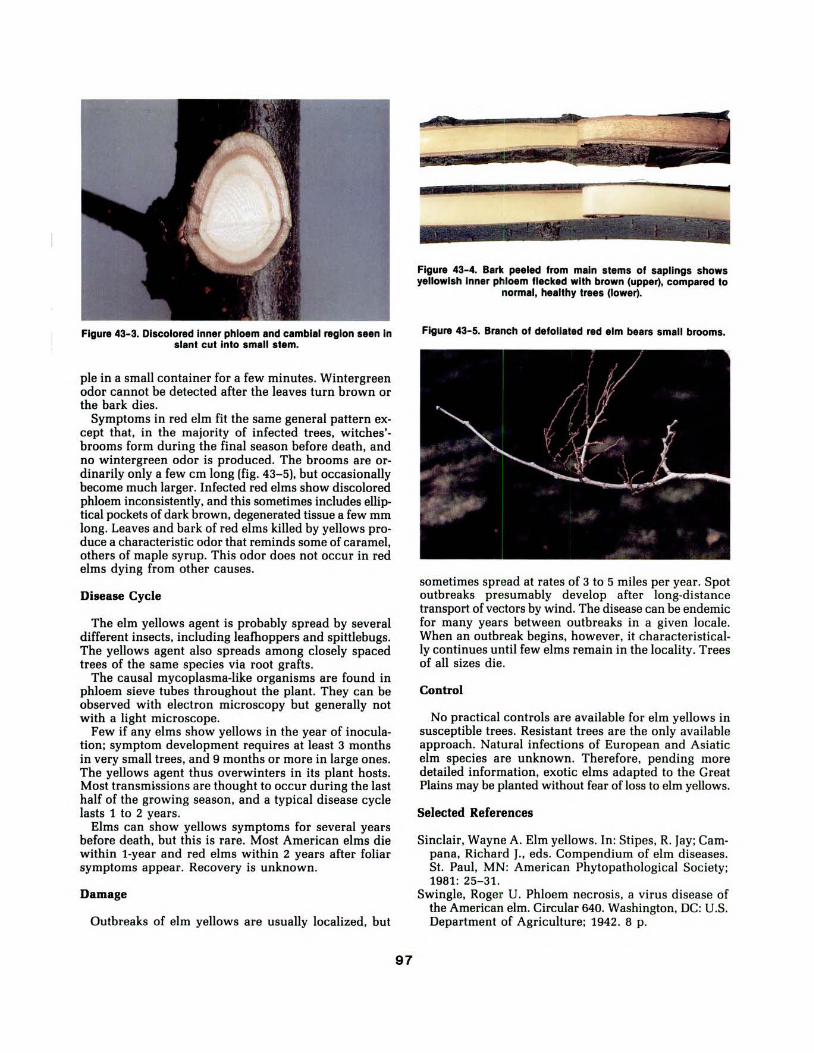
Figure 43-3. Discolored Inner phloem and cambial region seen In slant cut Into small stem.
pie in a small container for a few minutes. Wintergreen odor cannot be detected after the leaves turn brown or the bark dies.
Symptoms in red elm fit the same general pattern ex· cept that, in the majority of infected trees, witches'· brooms form during the final season before death, and no wintergreen odor is produced. The brooms are ordinarily only a few em long (fig. 43-5), but occasionally become much larger. Infected red elms show discolored phloem inconsistently, and this sometimes includes elliptical pockets of dark brown, degenerated tissue a few mm long. Leaves and bark of red elms killed by yellows produce a characteristic odor that reminds some of caramel, others of maple syrup. This odor does not occur in red elms dying from other causes.
Disease Cycle
The elm yellows agent is probably spread by several different insects, including leafhoppers and spittlebugs. The yellows agent also spreads among closely spaced trees of the same species via root grafts.
The causal mycoplasma-like organisms are found in phloem sieve tubes throughout the plant. They can be observed with electron microscopy but generally not with a light microscope.
Few if any elms show yellows in the year of inoculation; symptom development requires at least 3 months in very small trees, and 9 months or more in large ones. The yellows agent thus overwinters in its plant hosts. Most transmissions are thought to occur during the last half of the growing season, and a typical disease cycle lasts 1 to 2 years.
Elms can show yellows symptoms for several years before death, but this is rare. Most American elms die within 1-year and red elms within 2 years after foliar symptoms appear. Recovery is unknown.
Damage
Outbreaks of elm yellows are usually localized, but
97
Figure 43-4. Bark peeled from main stems of saplings shows yellowish Inner phloem flecked with brown (upper), compared to
normal, healthy trees (lower).
Figure 43-5. Branch of defoliated red elm bears small brooms.
sometimes spread at rates of 3 to 5 miles per year. Spot outbreaks presumably develop after long-distance transport of vectors by wind. The disease can be endemic for many years between outbreaks in a given locale. When an outbreak begins, however, it characteristically continues until few elms remain in the locality. Trees of all sizes die.
Control
No practical controls are available for elm yellows in susceptible trees. Resistant trees are the only available approach. Natural infections of European and Asiatic elm species are unknown. Therefore, pending more detailed information, exotic elms adapted to the Great Plains may be planted without fear of loss to elm yellows.
Selected References
Sinclair, Wayne A. Elm yellows. In: Stipes, R. Jay; Campana, Richard J., eds. Compendium of elm diseases. St. Paul, MN: American Phytopathological Society; 1981: 25-31.
Swingle, Roger U. Phloem necrosis, a virus disease of the American elm. Circular 640. Washington, DC: U.S. Department of Agriculture; 1942. 8 p.
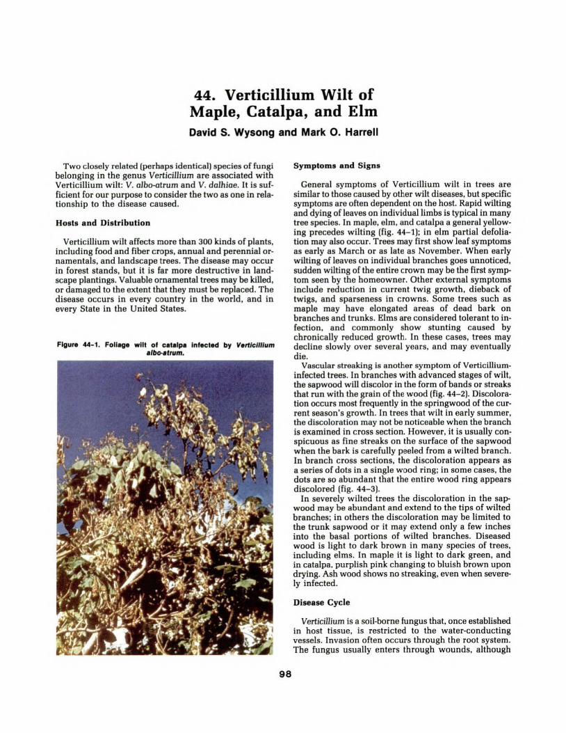
44. Verticillium Wilt of Maple, Catalpa, and Elm David S. Wysong and Mark 0 . Harrell
Two closely related (perhaps identical) species of fungi belonging in the genus Verticillium are associated with Verticillium wilt: V. albo-atrum and V. dolhioe. It is sufficient for our purpose to consider the two as one in relationship to the disease caused.
Hosts and Distribution
Verticillium wilt affects more than 300 kinds of plants, including food and fiber crops, annual and perennial ornamentals, and landscape trees. The disease may occur in forest stands, but it is far more destructive in landscape plantings. Valuable ornamental trees may be killed, or damaged to the extent that they must be replaced. The disease occurs in every country in the world, and in every State in the United States.
Figure 44-1. Foll~ge wilt of cetelp• Infected by Vertic/Ilium albo-afrum.
98
Symptom s and Signs
General symptoms of Verticillium wilt in trees are similar to those caused by other wilt diseases, but specific symptoms are often dependent on the host. Rapid wilting and dying of leaves on individual limbs is typical in many tree species. In maple, elm, and catalpa a general yellowing precedes wilting (fig. 44-1); in elm partial defoliation may also occur. Trees may first show leaf symptoms as early as March or as late as November. When early wilting of leaves on individual branches goes unnoticed, sudden wilting of the entire crown may be the first symptom seen by the homeowner. Other external symptoms include reduction in current twig growth, dieback of twigs, and sparseness in crowns. Some trees such as maple may have elongated areas of dead bark on branches and trunks. Elms are considered tolerant to infection, and commonly show stunting caused by chronically reduced growth. In these cases, trees may decline slowly over several years, and may eventually die.
Vascular streaking is another symptom of Verticilliuminfected trees. In branches with advanced stages of wilt, the sapwood will discolor in the form of bands or streaks that run with the grain of the wood (fig. 44-2). Discoloration occurs most frequently in the springwood of the current season's growth. In trees that wilt in early summer, the discoloration may not be noticeable when the branch is examined in cross section. However, it is usually conspicuous as fine streaks on the surface of the sapwood when the bark is carefully peeled from a wilted branch. In branch cross sections, the discoloration appears as a series of dots in a single wood ring; in some cases, the dots are so abundant that the entire wood ring appears discolored (fig. 44-3).
In severely wilted trees the discoloration in the sapwood may be abundant and extend to the tips of wilted branches; in others the discoloration may be limited to the trunk sapwood or it may extend only a few inches into the basal portions of wilted branches. Diseased wood is light to dark brown in many species of trees, including elms. In maple it is light to dark green, and in catalpa. purplish pink changing to bluish brown upon drying. Ash wood shows no streaking, even when severely infected.
Disease Cycle
Verticillium is a soil-borne fungus that, once established in host tissue, is restricted to the water-conducting vessels. Invasion often occurs through the root system. The fungus usually enters through wounds, although

wounds are not necessary for infection. After colonization, the fungus can spread throughout the plant either by spores transported with the sap stream or by direct extension of vegetative mycelium. Optimum temperature for growth of the fungus in plants is 65° - 72°F. If the plant is killed by Verticillium, the pathogen survives in the roots and trunk and can remain viable fo r several years.
In addition to asexual spores (conidia}, the fungus also produces resting structures called microsclerotia, which allow the fungus to persist in the soil for long periods separate from its parasitized host. Conidia do not survive for more than a few weeks in soils. Microsclerotia a re produced readily at 70°- 85°F and are most abundant in the top 12 inches of soil of all types.
The dispersal of resting structures is a major deterrent to control of Verticillium wilt. One of the most important means of dispersal is by movement of soil, such as when trees are transplanted from nurseries to landscape sites. Microsclerotia are carried in root balls or on bare roots of infected trees, and contaminate the soil where the trees are planted. In nursery fields the microsclerotia can be spread by normal tillage operations. They may also spread in soil that adheres to equipment used in an infested field.
Damage
Damage due to infection by Verticillium is variable, and depends upon age of the host and species affected. It is not uncommon for trees with trunk diameters of 1 to 2 inches to be killed within 1 year of infection. Older trees may live several to many years following infection, but typically they gradually deteriorate over time. The disease has a greater impact on nursery seedlings and trees in landscape situations than on trees in forest stands.
Control
The best method of controlling Verticillium wilt in trees and shrubs is prevention. Avoid planting susceptible trees in soil where other plants are known to have died from the disease. Fertilization may help prevent the disease in landscape planting, or in some cases, help affected trees recover. Wilt severity is increased by "high nitrogen" fertilizers. "Balanced" fertilizers such as 10-10-10 (N-P-K} are recommended. Infected trees should also be watered every 10 - 14 days with the equivalent of 2 inches of rainfall. Low soil moisture sometimes causes the wilt symptoms to be more severe.
Dead branches showing severe wilt symptoms should be removed or pruned back to wood showing no vascular streaking. Sterilize pruning tools with rubbing alcohol or sodium hypochlorite after each use while pruning to avoid transmitting the fungus. Pruning diseased branches will not eliminate the fungus from the trunk or roots, however. All dead branches or dead wood should be burned, not buried. A note of caution on pruning: branches showing slight wilting of leaves should not be
removed immediately; they may recover in response to water and fertilizer treatments.
Because Verticillium is a vascular wilt pathogen. commonly used fungicides applied to topical surfaces a re not effective. A systemic fungicide (one that is absorbed and translocated within the host plant} is necessary to reach the site where the pathogen is active. Mertect (thiabendazole} and Benlate (benomyl} gave some degree of protection to Russian-olive and sugar maple seedlings when applied as a soil drench two weeks after soil infestation. In the same study, foliar applications of these fungicides did not control the disease. The conclusion of this study was that chemical control of Verticillium wilt, even with systemic fungicides, is not practical when treating established trees.
Selected References
Carter, J. Cedric. Diseases of Midwest trees. Urbana: University of Illinois Press; 1979. 168 p.
Himelick, Eugene B.; Neely, Dan. Verticillium wilt. In: Stipes, R. Jay; Campana, Richard J., eds. St. Paul, MN: Compendium of elm diseases. American Phytopathological Society; 1981: 22-24.
Smith, Larry D. Verticillium wilt of landscape trees. Journal of Arboriculture. 5(9}: 193-197; 1979.
99
Figure 44-2. Bark removed from maple stem to reveal vascular discoloration caused by V. albo·atrum.
Figure 44-3. Brown streaking of elm Infected with V. albo·atrum.

45. Oak Wilt DavidS. Wysong and Edward M. Sharon
The oak wilt pathogen, Ceratocystis fagacearum, is believed to be native to the United States and to have originated in the Upper Mississippi Valley. Disease survey records as early as 1912 describe mortality in oak stands in Minnesota and Wisconsin similar to mortality now known to be caused by the oak wilt fungus. Mortality was attributed to several factors, such as drought or Armillaria root rot, until 1940 when the causal agent was identified as a vascular fungus.
Hosts and Distribution
Oak wilt ranges from Minnesota east to Pennsylvania,
south to South Carolina and Tennessee, west to central Texas, and north through Kansas and Nebraska. The disease has been present in Nebraska for over 20 years, but it has been confirmed in only seven counties that border the Missouri River.
Known hosts of the oak wilt fungus include 36 species of oak and six species closely related to oak. All species of oak tested have proven susceptible to the fungus either by natural infection or by artificial inoculation. Species of the red oak group (black, northern red, pin, and scarlet) are more susceptible than are species of the white oak group (bur, post, and white). The six closely related species include Chinese, American, and Spanish che·stnut, Allegheny and bush chinkapin, and tanbark-oak.
Figure 45-1. Typical bronzing symptoms on outer portions of leaves of red oak.
100

Figure 45-2. Foliage of entire crown of red oak wilted within a few weeks after appearance of Initial symptoms.
Symptoms and Signs
Foliage symptoms of oak wilt occur from early June until leaf coloration begins in the fall. Symptoms in the red oak group are slightly different from those in the white oak group.
Early foliage symptoms in the red oak group include wilting, bronzing, and premature defoliation at the branch tips in the upper tree crown. Wilt symptoms rapidly progress down through the crown. Wilted leaves turn dull green, bronze, or tan beginning at the outer portions of the leaf. The base of the leaf and the portion around the main vein are the last to change color (fig. 45-1). Foliage of affected trees commonly wilts within a few weeks after first symptoms appear (fig. 45-2). Brown to black discoloration develops in vascular tissues in the outer sapwood where the tree produces tyloses and gums {fig. 45-3). Red oaks do not recover once infected. Trees infected late in the growing season may produce some leaves on the lower branches the following spring, but these soon wither and die.
Early symptoms in the white oak group are characterized by a wilt of foliage of individual branches
in the crown. Leaves may turn yellow, but necrosis is usually limited to the margins of the blade. Affect·ed leaves normally remain attached, and their coloration is similar to normal fall coloration {fig. 45-4). The eventual death of individual branches results in a stag-headed appearance of the crown over a period of several years. Some trees in this group of oaks may die within 2-4 years or longer after infection, but one third to one half of the infected trees may recover and grow normally.
Mycelial masses, or mats, of the fungus may form under the bark within several months after death of the infected tree {fig. 45-5). The fungus mats, which vary in size from one to four inches in length, raise and crack the bark as they enlarge. The black and gray fungus mats give off a fruit-like odor that is very attractive to insects. While the mats are common on infected trees in the red oak group, they are rarely produced on infected white oaks.
Disease Cycle
Oak wilt is caused by the fungus Ceratocystis fagacearum, which grows in the functional vessels of the sapwood. The tree responds to infection by producing
101

tyloses in the vessels; these occlusion bodies restrict the tree's water supply. Hence, the disease is strictly a vascular wilt.
By the time symptoms become apparent in the crown of an infected red oak, the fungus is present in the conducting vessels throughout the tree. The fungus in the branches dies a few weeks after the tree dies; the fungus remains alive for several months to a year in the trunk and root system. The fungus produces spores in fungal mats that develop under, and eventually rupture the bark of, recently killed trees. Several insects, particularly oak bark and sap-feeding beetles, are attracted to the fungal mats because of the fermenting odor. As the beetles crawl over these mats, spores of the fungus adhere to their bodies. The beetles fly to other oak trees, feed on the sap flow from fresh wounds, and thereby spread the fungus to these trees. Squirrels and birds may also be potential vectors of the fungus.
Insect transmission of the fungus among trees in the white oak group is very limited, because fungal mats are rarely produced on infected white oaks.
Root grafts are an important means of fungus transmission between trees. Roots of oaks form natural grafts. Once the fungus enters a tree it can spread to nearby trees through these grafts. This method of transmission has been observed in both red and white oak groups.
' :~
Figure 45-3. Brown discoloration of sapwood of Infected stem.
Figure 45-4. Foliage symptoms on trees of white oak group. Leaves remain attached and their discoloration Is similar to normal fall
coloration.
102

Damage
Oak wilt continues to threaten oak forests and landscape plantings throughout their native range. Fortunately, catastrophic losses to the disease, feared by many in its early history, have not materialized. Still, thousands of oaks succumb to oak wilt annually, primarily in forest stands in Wisconsin, Minnesota, and Iowa. A number of States (Pennsylvania, West Virginia, Maryland, Kentucky, Tennessee, and North Carolina) implemented extensive oak wilt control programs in the 1950's and 1960's. Appraisal of these programs suggests that damage from oak wilt would have been much higher in the absence of such efforts, and therefore were justified.
Control
There is no known way of saving infected oaks, at least not red oaks. Control must be aimed at halting the spread of the fungus to healthy trees by (1) preventing root graft transmission, and (2) reducing fungal mat production on recently killed trees. Insect control is impractical.
Over 90 percent of diseased trees probably become infected through root grafts. Spread through such unions can be prevented by mechanically trenching around infected trees after first symptoms develop. A trench 3 feet deep and several inches wide between diseased and healthy trees immediately disrupts root grafts between these adjacent trees. The trench can be refilled because new root grafts will not form between the dead or dying tree and nearby healthy trees.
An alternative method of disrupting root grafts involves the use of SMDC (sodium N-methyldithiocarbamate), sold under various trade names as Vapam, or Trimaton. This fumigant kills the roots in a narrow strip and prevents the fungus from reaching healthy trees. Holes are dug 15 to 18 inches deep and an inch or two in diameter, spaced 6 inches to a foot apart, along a line midway between the diseased tree and adjacent healthy trees. One part of SMDC is mixed with three parts of water, and the diluted solution is poured into each hole. The holes must be closed immediately after application. Some temporary injury may occur in the healthy tree, particularly if the chemical barrier is closer than 10-15 feet from it, because of chemical uptake and limited root kill. Soil texture, temperature, and moisture influence the rate of uptake and effectiveness of treatment.
The second control technique is aimed at preventing spore formation on diseased trees. Spores are produced for only a brief period after the tree dies. The fungus cannot be isolated from the above-ground parts of dead trees when the moisture content of the wood is less than 20 percent. Therefore, any treatment that hastens the drying of wood tissue will tend to reduce sporulation. Deep mechanical girdling of the trunk soon after the disease is diagnosed will hasten drying of the wood and prevent mat formation. Experiments with chemical poisons, such as pressure-injected cacodylic acid, have demonstrated effective root kill and hastened drying.
Finally, infected trees should be cut down and destroyed. They may be burned, buried, or processed in-
Figure 45-5. G111y mycellll m1t of 01k wilt fungus. The met develops under the b1rt of de1d trees.
to chips for mulching purposes. Members of the white oak group can be used at any time, while red oaks should only be used when they are beyond the stage of producing or harboring spores. If there is potential for sporulation, firewood cut from infected trees should have bark removed or be covered with 4 mil polyethylene until used. Logs from freshly cut timber may be fumigated with methyl bromide under plastic cover for 3 days. Salvaged timber can be milled for lumber, but waste slabs and wood should be chipped, burned, or buried.
Selected References
Appel, D. N.; Fox, T.; Drees, C. Frost. The occurrence of oak wilt in Texas. (Abstract). Phytopathology. 73: 833; 1983.
French, D. W.; Stienstra, Ward C. Oak wilt disease. Extension Folder 310. St. Paul: University of Minnesota; 1975. 6 p.
Himelick, E. B.; Fox, Howard W. Experimental studies on control of oak wilt disease. Illinois Natural History Survey Bulletin 680. 1961. 48 p. ·
Ostrofsky, William D.; Wysong, DavidS. Oak wilt. Forest Pests of Nebraska Leaflet 8. Lincoln: University of Nebraska; 1977. 2 p.
103

46. Mimosa Wilt Mark W. Andrews
The mimosa or silk tree, a native of eastern Asia, is widely grown as an ornamental and has become naturalized in the southeastern United States and in the southern Great Plains. The tree is valued for rapid growth, graceful foliage, colorful flowers, and capacity to thrive in unfavorable soil conditions. Mimosa wilt, caused by Fusarium oxysporum f. sp. perniciosum, is a destructive vascular wilt disease of this ornamental.
Hosts and Distribution
Mimosa wilt was first observed in North Carolina in 1953, and since has been found from New York to Florida and into Mississippi and Louisiana.
In inoculation tests, Albizia julibrissin, A. lophantha, A. kalkora, and A. procera were susceptible to the causal fungus; A. thorelii and A. pudica were resistant. Seedlings of native leguminous species Cercis canadensis, Gleditsia triacanthos, and Robinia pseudoacacia were found to be resistant to the pathogen.
Symptoms and Signs
The first external symptom is wilting of the leaves. The leaves then become dry and shrivelled, usually remaining green but sometimes yellow. The leaves fall from the tree soon after they dry (fig. 46-1). The defoliated parts then die; usually the entire tree is dead within 1 year
Figure 46-1. Mimosa tree exhibiting foliar wilt and dleback symptoms.
104
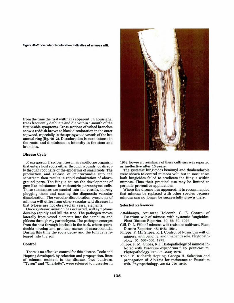
Figure 46-2. Vascular discoloration Indicative of mimosa wilt.
from the time the first wilting is apparent. In Louisiana, trees frequently defoliate and die within 1-month of the first visible symptoms. Cross sections of wilted branches show a reddish-brown to black discoloration in the outer sapwood, especially in the springwood vessels of the last annual ring (fig. 46-2). Discoloration is most intense in the roots, and diminishes in intensity in the stem and branches.
Disease Cycle
F. oxysporum f. sp. perniciosum is a soilborne organism that enters host roots either through wounds, or directly through root hairs or the epidermis of small roots. The production and release of microconidia into the sapstream then results in rapid colonization of aboveground parts. The fungus causes the development of gum-like substances in vasicentric parenchyma cells. These substances are exuded into the vessels, thereby plugging them and causing the diagnostic vascular discoloration. The vascular discoloration symptoms of mimosa wilt differ from other vascular wilt diseases in that tyloses are not observed in vessel elements.
Once systemic invasion has occurred, wilt symptoms develop rapidly and kill the tree. The pathogen moves laterally from vessel elements into the cambium and phloem through ray parenchyma. The pathogen emerges from the host through lenticels in the bark, where sporodochia develop and produce masses of macroconidia. During this time the roots decay and the fungus is released into the soil.
Control
There is no effective control for this disease. Toole and Hepting developed, by selection and propagation, lines of mimosa resistant to the disease. Two cultivars, "Tyron" and "Charlotte", were released to nurseries in
1949; however, resistance of these cultivars was reported as ineffective after 15 years.
The systemic fungicides benomyl and thiabendazole were shown to control mimosa wilt, but in most cases both fungicides failed to eradicate the fungus within mimosa. Thus their practical use may be limited to periodic preventive applications.
Where the disease has appeared, it is recommended that mimosa be replaced with other species because mimosa can no longer be successfully grown there.
Selected References
Attabhanyo, Anusorn; Holcomb, G. E. Control of Fusarium wilt of mimosa with systemic fungicides. Plant Disease Reporter. 60: 56- 59; 1976.
Gill, D. L. Wilt of mimosa wilt-resistant cultivars. Plant Disease Reporter. 48: 648; 1964.
Phipps, P. M.; Stipes, R. J. Control of Fusarium wilt of mimosa with benomyl and thiabendazole. Phytopathology. 65: 504-506; 1975.
Phipps, P.M.; Stipes, R. J. Histopathology of mimosa infected with Fusarium oxysporum f. sp. perniciosum. Phytopathology. 66: 839-843; 1976.
Toole, E. Richard; Hepting, George H. Selection and propagation of Albizzia for resistance to Fusarium wilt. Phytopathology. 39: 63-70; 1949.
105

47. Phymatotrichum Root Rot Glenn W. Peterson and Charles Maier
Phymatotrichum root rot is a major disease of taprooted plants in certain areas of the southwestern United States.
Hosts and Distribution
The fungus Phymatotrichum omnivorum has a wide host range that includes most of the species used in Great Plains tree plantings. It affects several tree species in parts of Arizona, Oklahoma, and Texas, where it is particularly damaging to cotton (fig. 47-1).
Symptoms and Signs
Diseased seedlings seldom show p,ronounced advanced indications but die suddenly, with a typical darkening and wilting of the leaves. Older trees may at first show a reduction in growth and viigor. The leaves may become yellow to bronze and either gradually drop off, giving the crown a thin appearance (cottonwood), or they may remain attached, become dark, dry, curl up, and cling abnormally long to the branches (Russian-olive).
Infected roots commonly are rotted and the epidermis is shrunken and shrivelled. For a few species such as Siberian elm, however, the decay has the characteristics of a wet rot.
Disease Cycle
The fungus has three distinct stages. The sterile mycelium usually appears in the form of buff-colored strands composed of one or more large central hyphae and surrounded by a varying number of small, irregular, thick-walled hyphae. Sclerotia are small, buff, compact bodies ranging up to a grain kernel in size. They are produced from enlargements of the rhizomorphs and, like the rhizomorphs, may remain dormant indefinitely. The spore stage appears on the surface of the soil in the form of a fluffy white mat of mycelia. The mat may be from 1 to 12 inches in diameter. The spore stage generally is considered functionless; infection has not been obtained by inoculations with spore suspensions.
The fungus in any form survives freezing for only a day or two; it is restricted to a region bounded on the north roughly by the latitude of southern Oklahoma.
106
Figure 47-1. Cotton killed by Phymatotrlchum omnivorum.

Damage
The fungus is commonly found in patches in field crops such as cotton (fig. 47- 1). The most damage occurs where trees have been planted in patches where the fungus has infested the soil. Windbreaks established in areas where the patches occur commonly have gaps where the fungus has killed or reduced the vigor of the trees (fig. 47-2).
Control
The impact of this disease can be reduced either by avoiding infested areas when planting trees and/or by planting resistant species. Infested spots could be detected readily when cotton was the crop most commonly planted, but now they are more difficult to detect because resistant crops, such as milo, are widely planted in the Phymatotrichum root rot zones of Oklahoma and Texas. In the early 1940's a n evaluation of tree species in some Oklahoma and Texas windbreaks revealed that the following species were resistant: common hackberry, desert-willow, western soapberry, eastern redcedar, and Rocky Mountain juniper. The following species were intermediate in susceptibility: ailanthus, apricot, green ash, northern catalpa, American sycamore, French tamarisk, eastern black walnut, Russian mulberry, and Austrian pine.
Figure 47-2. Gap In windbreak caused by P. omnlvorum.
Selected References
Peltier, George L. Distribution and prevalence of Ozonium root rot in the shelter-belt zone of Texas. Phytopathology. 27: 145-158; 1937.
Peltier, George L.; Schroeder, F. R.; Wright, Ernest. Distribution and! prevalence of Ozonium root rot in the shelterbelt planting area of Oklahoma. Phytopathology. 29: 485-490; 1938.
Taubenhaus, J. J.; Ezekiel, Walter N. A rating of plants with reference to their relative resistance or susceptibility to Phymatotrichum root rot. Bull. 527. College Station: Texas Agricultural Experimental Station; 1936. 52 p.
Wright, Ernest. First progress report on the Phymatotrichum root rot losses in experimental windbreaks of Oklahoma and Texas. Plant Disease Reporter. 24: 13-20; 1940.
Wright, Ernest; Wells, H. R. Tests on the adaptability of trees and shrubs to shelterbelt planting on certain Phymatotrichum root rot infested soils of Oklahoma and Texas. journal of Forestry. 46: 256-262; 1948.
107

48. Armillaria Root Rot Lloyd R. Fuller and Robert L. James
Fungi of the genus Armillaria are common facultative parasites on more than 600 plant species throughout the world. Root diseases caused by these fungi are responsible for considerable economic damage, especially within plantations, orchards, or vineyards. Many forest conifers and hardwoods, as well as ornamental shrubs and trees, are susceptible to these pathogens.
Hosts and Distribution
Armillaria occurs sporadically throughout the Great Plains. This disease has been reported on over 25 plant species widely planted in landscapes and windbreaks. Some susceptible plants commonly grown in the Great Plains are: boxelder, paper birch, Peking cotoneaster, Tatarian honeysuckle, apple, ponderosa pine, Scots pine, Quercus sp., Prunus sp., and other members of the rose family (Rosaceae).
Figure 48-1. Chlorotic foll~e of ponderou pine Infected with Armillaria.
Symptoms and Signs
Crown symptoms resemble those of other associated root disorders: reduced height growth, chlorotic foliage, premature foliage drop, dieback, and mortality (fig. 48-1). One or more of these symptoms may be present on a single tree. In addition, hosts infected at the root collar may have external basal resinosis or gummosis.
Armillaria produces a characteristic cream-colored mycelial fan beneath the bark, in the cambial zone, on roots, and at the root collar (fig. 48-2). Rhizomorphs, tough shoestring-like structures made up of hyphae that darken with age, may be found on root surfaces, in adjacent soil, or under loose bark (fig. 48-3).
The most positive signs of Armillaria infection are the production of fruiting bodies (fig. 48- 4), and their characteristic appearance in culture. Armillaria produces an edible, honey-colored mushroom in small to large clusters on living and dead hosts or on soil near buried wood. This mushroom may be present from late summer through fall if moisture is adequate. Armillaria mushrooms may be identified by their honey-yellow caps, 1-5 inches across; fine, cinnamon-colored erect hairs over the cap; white spores; and a persistent ring or annulus on the stem. A yellow-capped variant is often observed on hardwoods; a pinkish-brown variant is common on conifers.
Disease Cycle
During favorable late summer or fall weather, Armillaria may be a prolific spore producer. However, unlike many other decay fungi, the spores of Armillaria rarely initiate disease. Armi.llaria spreads primarily as vegetative mycelium, either by root-to-root contact or by rhizomorphs.
Rhizomorphs can grow considerable distances through the soil, and initiate parasitic infections or saprophytic colonization. When a rhizomorph encounters a host root or root collar, it adheres to the bark surface. Subsequently, by direct pressure and enzymatic action, mycelial strands colonize susceptible host cambium and initiate canker formation. These cankers usually do not progress into lethal infections on healthy, vigorous hosts, but remain quiescent until the host dies naturally, the host vigor decreases significantly, or the host is overwhelmed by numerous parasitic attacks. Successfully attacked hosts usually do not die until infections girdle the root collar cambium.
Following successful parasitic infection, Armillaria begins saprophytic colonization of woody tissues beneath infected cambium. In some conifers and hardwoods, Armillaria causes wood decay without parasitic cambial infections. In this latter case, Armillaria does not
108

appear to kill trees directly, but may predispose them to wind throw.
Damage
Young trees are most likely to be killed by Armillaria. Trees 15 to 20 years old are more tolerant to parasitic attacks. Armillaria, in general, is not damaging unless hosts are under unusual stress, such as by extended droughts, competition for light and nutrients, insect attacks, or infections by other root pathogens.
Control
Control of root diseases over extensive acreage is seldom possible without considerable effort and cost. Valuable ornamental, shade, and orchard trees, adjacent to infected trees, may be protected by careful removal of infected stump and root material from the soil. Chemicals have been used to sanitize infested soil and wood, but only professional chemical applicators should apply these chemicals near healthy trees.
Planting immune or highly resistant species is the most promising and long lasting approach to Armillaria root rot control. Lists of resistant plants that are well adapted to a locale can be obtained from local extension agents or from the reference by Raabe and McCain (1967).
Make sure planting stock is disease-free. Reduce foliage so crowns are in balance with root systems. Prepare the planting site adequately. In general, to minimize the impact of root diseases, maintain host vigor through adequate and timely applications of water and fertilizers.
Selected References
U. S. Department of Agriculture. Index of plant diseases in the United States. Agric Handb. 165. Washington, DC: U. S. Department of Agriculture; 1960. 531 p.
Miller, Orson K., Jr. Mushrooms of North America. New York: E. P. Dutton & Co.; 1972. 360 p.
Figure 48-2. Mycelial fans of Armillaria in cambial zone of ponderosa pine.
Figure 48-3. Rhlzomorphs of Armillaria on root surface under bark of host.
Raabe, R. D. Host lists of the root rot fungus Armillariella me/leo. Hilgardia. 33: 25-88; 1962.
Raabe, R. D.; McCain, A. H. Plants resistant or susceptible to Armillaria root rot. Agricultural Extension Service Bulletin AXT - 6. Davis: University of California; 1967. 6 p.
Figure 48-4. Large fruiting bodies of Armillaria on bark at root col· lar of ponderosa pine.
109

49. Nematodes of Broadleaf Trees
Jerry W. Riffle and Joseph M. Krupinsky
The root zones of trees in nurseries, windbreaks, and
natural stands in the Great Plains are inhabited by
various microorganisms, including nematodes (fig. 49-1).
Nematodes are an important part of the microscopic
fauna of these sites; those associated with tree roots in-
clude plant-parasitic, mycophagous, bacteriophagous,
and predaceous forms. Plant-parasitic nematodes punc-
ture and feed on root cells, and contribute to poor growth
and premature decline of trees.
Hosts and Distribution
Cottonwood, green ash, golden willow, and post oak
are favored hosts of the dagger nematode, Xiphinema
americanum. This nematode is widespread and is prob-
ably present in most windbreaks on the Great Plains.
This association with trees and the high populations in
weed-free windbreaks indicate that various other tree
species used in such plantings may be hosts for this
nematode. Preferring undisturbed sites, dagger nema-
todes seldom are a problem in nurseries in the Great
Plains.
Roots of apple, black locust, black walnut, American
elm (fig. 49-2), catalpa, green ash, and multiflora rose
have been parasitized by root-knot nematodes,
Meloidogyne spp., in nurseries in the southern Great
Plains. Root-knot nematodes rarely are found on trees
in windbreak plantings, however. Apple, black locust,
and black walnut also have been parasitized by root-
Figure 49-1. The principal parts of a nematode (female).
lesion nematodes, Pratylenchus spp., in nurseries. Root-
lesion nematodes also cause damage to spruces, pines,
and junipers (see Article 64). American elm has been
damaged by lance (Hoplolaimus sp.) and spiral
(Helicotylenchus spp.) nematodes in nurseries. Other
forms that occasionally injure tree roots include stunt
(Tylenchorhynchus sp.), ring (Criconemoides sp.), and
stubby-root (Trichodorus sp.) nematodes.
Symptoms and Signs
Above-ground symptoms on seedlings parasitized by
nematodes resemble those on plants lacking an adequate
and properly functioning root system. Affected seedlings
are characterized by low vigor, stunting, and small,
discolored foliage. Below-ground symptoms on feeder
roots include root swellings, root proliferation, surface
necrosis, lesions, and stubby-root condition resulting
from root-tip injury and cessation of growth. Heavily
parasitized root systems are discolored and poorly
developed. Similar root symptoms occur on established
trees.
Diseases caused by nematodes usually cannot be
diagnosed by the symptoms described above because
other soil-borne pathogens and some environmental fac-
tors cause similar symptoms. Various types of injury may
be present on roots because of the combined effects of
more than one nematode species; for example lesions
and stubby-root condition may be present on roots
parasitized by both root-lesion and stubby-root
nematodes. In addition, nematode injury may increase
the incidence of other root diseases by facilitating entry
of their causal organisms.
Disease Cycle
The life cycle of plant-parasitic nematodes consists of
eggs, four larval stages, and adults. Females lay eggs that
hatch into juveniles. These juveniles are similar to adults
in appearance and structure in most species; during their
growth and development they undergo four molts. After
the fourth molt, the individual is an adult. Mature
females then lay eggs and the cycle is repeated. Root-
knot nematode larvae of both sexes undergo the same
development into the last larval stage. At the final molt,
the male emerges as a slender eel-shaped adult, while
the female becomes pear-shaped or sometimes almost
spherical.
There are two general modes of feeding among plant-
parasitic nematodes. Ectoparasitic species, such as dag-
ger nematodes, live in the soil and feed externally on suc-
culent root tissue. Through their stylets they inject
digestive enzymes into root tissues, making the contents
of cells easier to ingest and assimilate. Endoparasites,
such as root-knot and root-lesion nematodes, enter and
110
49. Nematodes of Broadleaf Trees Jerry W. Riffle and Joseph M. Krupinsky
The root zones of trees in nurseries, windbreaks, and natural stands in the Great Plains are inhabited by various microorganisms, including nematodes (fig. 49-1). Nematodes are an important part of the microscopic fauna of these sites; those associated with tree roots include plant-parasitic, mycophagous, bacteriophagous, and predaceous forms. Plant-parasitic nematodes puncture and feed on root cells, and contribute to poor growth and premature decline of trees.
Hosts and Distribution
Cottonwood, green ash, golden willow, and post oak are favored hosts of the dagger nematode, Xiphinema americanum. This nematode is widespread and is probably present in most windbreaks on the Great Plains. This association with trees and the high populations in weed-free windbreaks indicate that various other tree species used in such plantings may be hosts for this nematode. Preferring undisturbed sites, dagger nematodes seldom are a problem in nurseries in the Great Plains.
Roots of apple, black locust, black walnut, American elm (fig. 49-2), catalpa, green ash, and multiflora rose have been parasitized by root-knot nematodes, Meloidogyne spp., in nurseries in the southern Great Plains. Root-knot nematodes rarely are found on trees in windbreak plantings, however. Apple, black locust, and black walnut also have been parasitized by root-
Figure 49-1. The principal parts of a nematode (female).
lesion nematodes, Pratylenchus spp., in nurseries. Rootlesion nematodes also cause damage to spruces, pines, and junipers (see Article 64). American elm has been damaged by lance (Hoplolaimus sp.) and spiral (Helicotylenchus spp.) nematodes in nurseries. Other forms that occasionally injure tree roots include stunt (Tylenchorhynchus sp.), ring (Criconemoides sp.), and stubby-root (Trichodorus sp.) nematodes.
Symptoms and Signs
Above-ground symptoms on seedlings parasitized by nematodes resemble those on plants lacking an adequate and properly functioning root system. Affected seedlings are characterized by low vigor, stunting, and small, discolored foliage. Below-ground symptoms on feeder roots include root swellings, root proliferation, surface necrosis, lesions, and stubby-root condition resulting from root-tip injury and cessation of growth. Heavily parasitized root systems are discolored and poorly developed. Similar root symptoms occur on established trees.
Diseases caused by nematodes usually cannot be diagnosed by the symptoms described above because other soil-borne pathogens and some environmental factors cause similar symptoms. Various types of injury may be present on roots because of the combined effects of more than one nematode species; for example lesions and stubby-root condition may be present on roots parasitized by both root-lesion and stubby-root nematodes. In addition, nematode injury may increase the incidence of other root diseases by facilitating entry of their causal organisms.
110
Disease Cycle
The life cycle of plant-parasitic nematodes consists of eggs, four larval stages, and adults. Females lay eggs that hatch into juveniles. These juveniles are similar to adults in appearance and structure in most species; during their growth and development they undergo four molts. After the fourth molt, the individual is an adult. Mature females then lay eggs and the cycle is repeated. Rootknot nematode larvae of both sexes undergo the same development into the last larval stage. At the final molt, the male emerges as a slender eel-shaped adult, while the female becomes pear-shaped or sometimes almost spherical.
There are two general modes of feeding among plantparasitic nematodes. Ectoparasitic species, such as dagger nematodes, live in the soil and feed externally on succulent root tissue. Through their stylets they inject digestive enzymes into root tissues, making the contents of cells easier to ingest and assimilate. Endoparasites, such as root-knot and root-lesion nematodes, enter and

Figure 49-2. Roots of American elm Infected with root-knot nematode. Left, nonlnfested roots; right, Infested roots with swollen
Ups.
complete most of their life cycles inside roots. The secretion injected into surrounding cells by root-knot nematodes modifies the development and maturation of those cells, causing them to form specialized tissue containing "giant cell" (fig. 49-3).
Damage
Endoparasitic nematodes feed on and move through tissues of the root, causing mechanical damage. Rootlesion and lance nematodes may cause substantial mechanical injury to rootlets. Much of the damage by both endo- and ectoparasitic nematodes are due to reaction of root tissues to secretions injected into roots while the nematodes are feeding. The specialized tissue induced by root-knot nematodes disrupts root absorption and translocation of moisture and nutrients. Weakened root systems may result in enhanced winter injury, increased susceptibility to canker pathogens, branch dieback, and premature decline.
Nematodes have caused severe damage to broadleaf seedlings in some Great Plains nurseries and to trees in windbreaks. The extent of injury to seedlings depends upon type of nematode, degree of infestation, and species and age of seedlings. Damage in infested seedbeds usually is first evident in irregularly shaped spots. These spots enlarge and coalesce over a number of years as nematodes multiply and spread throughout the seedbed. Pathogenic fungi often enter roots through wounds made by nematodes, and the resulting disease complex often causes considerably more loss to a seedling crop than would be caused by either acting alone.
Many windbreaks in the northern Great Plains are planted in habitats favorable for dagger nematodes. Native grasses and alfalfa are hosts of this nematode, and trees planted on such sites are exposed immediately to high nematode populations. In these situations premature tree decline is common, and occasionally tree mortality is high 2 or 3 years after planting. Populations as high as 13,300 per pint of soil have been reported
Figure 49-3. Root·knot nematode female (lower right) with giant cells developed In clusters Immediately adjacent to Its Up region.
The sedentary nematodes feed on these giant cells.
around roots of cottonwood in windbreaks in South Dakota.
Control
Plant-parasitic nematodes can be controlled by cultural or chemical methods. Crop rotation with non-host cover crops may be used to reduce populations of certain species. For example, where root-knot nematodes are a problem, rotation with fescue (a non-host) is recommended over soybeans (a host). Summer fallow, accompanied by frequent tilling, will reduce populations of nematodes by starvation and desiccation in areas of low or seasonal rainfall and high soil temperatures.
Preplant soil fumigation provides excellent control of nematodes in nurseries. Volatile, halogenated hydrocarbons such as methyl bromide are widely used. In South Dakota, soil fumigation of windbreak planting sites with 1,3-dichloropropene and related chlorinated C hydrocarbons (1,3-D) increased growth of green ash and golden willow over a 4-year period on land infested initially with low populations of dagger nematodes, but did not affect growth of cottonwood, Siberian peashrub, or honeylocust. Root-knot nematode on hardwood seedlings can be controlled by dipping infected roots in organophosphate nematicides prior to transplanting.
Selected References
Malek, R. B. The dagger nematode, Xiphinema americanum, associated with decline of shelterbelt trees in South Dakota. Plant Disease Reporter. 52: 795-798; 1968.
Malek, R. B.; Smolik, J. D. Effect of Xiphinema americanum on growth of shelterbelt trees. Plant Disease Reporter. 59: 144-148; 1975.
Thorne, Gerald; Malek, Richard B. Nematodes of the Northern Great Plains. Part 1 Tylenchida (Nemata: Secernentea). Tech. Bull. 31. Brookings: South Dakota Agricultural Experiment Station; 1968. 111 p.
111

50. Phomopsis Blight of Junipers Glenn W. Peterson
Phomopsis blight has been a serious problem for more than 75 years in nurseries producing juniper seedlings and grafts (fig. 50-1).
Hosts and Distribution
Phomopsis juniperovora, the fungus causing this disease, is widespread in the Great Plains (fig. 50-2). Losses have been most severe in seedling and transplant beds of eastern redcedar and Rocky Mountain juniper. Other junipers are susceptible, as are some species in the genera Chamaecyparis, Cupressus, and Thuja.
Symptoms and Signs
P. juniperovora initially infects foliage, then spreads to and sometimes kills stem tissues. Newly developing needles are especially susceptible while they are still in the yellowish-green stage; after needles become a normal, deep green, they are no longer susceptible. Small yellow spots appear on young needles of eastern redcedar and Rocky Mountain juniper within 3 to 5 days after infection. The fungus ramifies within infected needles and rapidly invades and girdles young stems. When a side shoot is infected, the fungus progresses to the main stem, which it may girdle if the stem is less than 1 em in diameter. The portion of the seedling above the girdled area then dies.
Figure 50-1. Phomopsls damage In juniper seedbeds.
Figure 50-2. States In which P. Juntperorora Is present (gray).
At first, infected tissues turn light green but rapidly change to the characteristic red-brown color of dead shoots, which finally turn ashen gray. Lesions on larger stems frequently develop into cankers, but the stems are not girdled. The fungus does not spread far below the cankers.
Disease Cycle
Spores produced in fruiting bodies (pycnidia) formed on leaves and stems of seedlings infected the previous year are the most important source of inoculum early in the growing season. Pycnidia with viable spores may develop within 3 to 4 weeks after seedlings become infected, but usually are not well developed until infected tissues have dried considerably. Pycnidia are found most commonly on tissues that have turned ashen gray. The pycnidia are embedded at first in needles and stems, but partially erupt through the epidermis (fig. 50-3). Two types of spores (alpha and beta) develop in the same or different pycnidia (fig. 50-4) and are extruded in whitish tendrils. The alpha spores are colorless, one-celled, ellipsoid, contain two oil globules, and commonly are 7.5 to 10 by 2.2 to 2.8 ~m; the beta spores are colorless, onecelled, filamentous, slightly curved, and commonly 20.2 to 26.9 by 1 ~nt. The fungus can produce spores for as long as 2 years in dead parts of infected plants.
Spores are dispersed primarily by rain splash. Infection is caused by alpha-spores; the filamentous betaspores do not germinate. Only a short period of high humidity is neeriAd for infection to occur; for example, seedlings exposed to 100 percent relative humidity and 75°F for only 7 hours can become infected. Spore germination, germ-tube development, and infection are optimum near 75°F, but disease development is enhanced by higher temperatures (90°F).
112

Damage
Total loss of first-year seedlings is common in epidemic years if control measures are not used. Losses are particularly high in areas where water tends to stand, and in beds of new seedlings adjacent to infected older seedlings. Some of the worst epidemics occur late in the growing season, when there is a late flush of growth on juniper seedlings.
Survival of even lightly blighted nursery stock is very poor, because new shoots continue to be infected by spores produced on old infected tissues.
When junipers in landscape plantings become infected, they may become unsightly because of numerous dead branch tips (fig. 50-5}. Older trees seldom are killed because only small-diameter stems are girdled. For this reason, Phomopsis blight does not cause significant damage in natural stands of junipers.
Control
Because susceptible new foliage and viable fungus spores are present throughout the growing season in juniper seedling beds, protective fungicides need to be applied regularly during this season. Benomyl is the only chemical currently registered for control of Phomopsis blight. This chemical applied at 7- to 10-day intervals, combined with a vigorous schedule of roguing infected seedlings over the same interval, will give excellent control.
Other actions can be taken to reduce losses. Sowing juniper seed adjacent to beds containing juniper stock should be avoided if possible. Poorly drained areas should be avoided because losses are often greater where water tends to stand. If overhead sprinklers are used, seedlings should be irrigated so that water on seedlings dries before nightfall. Because shading frames increase the length of time that moisture remains on foliage, they should not be used unless absolutely necessary. Junipers or other hosts of this fungus should not be used in nursery windbreaks or in landscape plantings on nursery
Figure 50-3. Pycnldla on leaves and branches.
I
\ ~,
)T (
/ /A6'
... Figure 50-4. Alpha (ellipsoidal) and beta (long) spores of P.
jun#perorora.
grounds, because they may be a source of inoculum (spores) for nursery stock. Such trees are more likely to be extensively infected if pruning results in development of juvenile foliage.
Susceptibility to P. juniperovora varies considerably among junipers. Research is seeking to determine if there is useable genetic resistance to P. juniperovora among and within progenies from select eastern redcedar trees.
Selected References
Otta, J. D. Benomyl and thiophanate methyl control Phomopsis blight of eastern redcedar in a nursery. Plant Disease Reporter. 58: 476-477; 1974.
Peterson, Glenn W. Infection of Juniperus virginiana and ]. scopulorum by Phomopsis juniperovora. Phytopathology. 63: 246- 251; 1973.
Peterson, Glenn W. Phomopsis blight of junipers. In: Forest nursery diseases in the United States. Agric. Handb. 470. Washington, DC: U.S. Department of Agriculture; 1975: 76-79.
Peterson, Glenn W.; Hodges, C. S. Jr. Phomopsis blight of junipers. Forest Insect and Disease Leaflet 154. Washington, DC: U.S. Department of Agriculture, Forest Service; 1982. 7 p.
113
Figure 50-5. Branch tips of 7·year-old Juniperus rlrglnlana damaged by P. Junlperorora.

51. Cercospora Blight of Junipers Glenn W. Peterson and DavidS. Wysong
Junipers and other members of the Cupressaceae family are infected by two closely related needle-blighting fungi, Cercospora sequoiae and Cercospora sequoiae var. juniperi.
Hosts and Distribution
In the Great Plains, C. sequoiae var. juniperi has severely damaged eastern redcedar and Rocky Mountain juniper in well-established windbreaks and other plantings. The distribution of these two fungi in the central United States is shown in figure 51-1.
Figure 51-1. Geographic distribution of Cercospora sequoiae and C. sequoiae var. juniperi. C. sequoiae dots; C. sequo/ae var. juniper/
gray; both black.
Symptoms and Signs
Cercospora blight is readily distinguished from Phomopsis and Kabatina blights of junipers. The
Figure 51-2. Typical appearance of Infected Rocky Mountain juniper.
branches of Cercospora infected trees usually will be devoid of foliage near their bases but will have healthy foliage on their tips (fig. 51-2); branches of trees infected with Phornopsis and Kabatina will have dead tips.
Juniper foliage is of three types: (1) whip leaves characteristic of long shoot growth on the ends of secondary and tertiary branches; (2) spur leaves characteristic of short (spur) branches; and (3) juvenile leaves characteristic of seedlings.
Early symptoms are bronzed tips of leaves on spur shoots. Subsequently these leaves become entirely bronzed, then necrotic. Commonly, all leaves of a branchlet are affected. Tnfer.tP.cl foliage on branchlets usually dies in late September.
Affected branchlets drop from trees in October and November, resulting in the typical appearance of Cercospora infected trees-the extremities of the branches bear healthy green foliage and the inner crown is devoid of foliage. Following severe infection, juvenile foliage commonly develops on branches that previously have had only spur and whip foliage.
Disease Cycle
Spores of the fungus (fig. 51-3) are dispersed from late April through October. Dispersal may not be abundant until late May or June, and no spores are dispersed during rainless periods. There is little or no long-distance wind dispersal of spores; no spores were collected in traps located 6 feet from severely infected trees. Moisture is required for spores to germinate and for the fungus to penetrate foliage. Infection has been severe when rainfall during the growing season was at or above average. Infection was slight during the drought years of 1975 and 1976 in eastern Nebraska.
Junipers in eastern Nebraska are infected first in early to midsummer. The period between initial infection and first appearance of symptoms is between 2 and 3 weeks.
Figure 51-3. Spores of C. sequolae var. juniper/.
114

The disease develops more rapidly in juvenile leaves than in spur leaves. Also, both current-year and previousyears' juvenile leaves become infected, whereas only the previous-years' spur leaves become infected.
Fruiting bodies (sporodochia) (fig. 51-4) resulting from current-season's infection were observed in both spur foliage and juvenile foliage in September. They sporulated readily when incubated at 75°F and 100 percent relative humidity for 18 hours.
Damage
The disease is found more frequently, develops more rapidly, and causes greater mortality in ]. scopuJorum than in J. virginiono. This fungus has not been a threat to production of juniper seedlings in the Great Plains; it is seldom found on nursery seedlings. However, Cercosporo infection has been commonly observed in nurseries on grafted selections of junipers, particularly selections of ]. scopuJorum that have been kept in the nursery for 5 or 6 years.
Tips of secondary and tertiary branches on 10-yearold ]. virginiana were free of infection for an average distance of 1.5 feet. The disease had extended an average distance of 11 inches along the branches the previous year. Lack of infection on tips may be a result of less moisture on the outermost foliage because of more rapid drying. Whip foliage, which develops on branch tips, also may be resistant to infection.
Trees planted in north-south rows had much more infection on the west side than on the east side. The longer persistence of moisture from dew or evening rains on the west side probably accounts for the higher levels of infection.
Control
Because whip and spur foliage are not infected before late June (and then only previous years' foliage become infected), a highly persistent fungicide applied before late June could protect trees with only spur and whip foliage for the entire season. Because of fungicide weathering, however, an additional application in late July usually is required.
Because both current-year and previous years' juvenile foliage become infected, juniper trees containing juvenile leaves would require additional fungicide applications. Bordeaux mixture (8-8-100) provided a high degree of disease supression in control tests.
Park managers following control procedures outlined above have controlled Cercospora blight on ]. scopulorum and on J. virginian a since 1973. The timing of fungicide applications (figure 51-5) was developed from tests in eastern Nebraska. Timing should be modified slightly in other areas-earlier applications in southern locations.
Cercospora blight is found more frequently in new plantings of ]. scopulorum than in new plantings of ]. virginiono. Where Cercospora may be a problem, it would be better to plant ]. virginiana.
Genetic resistance to Cercospora is currently being evaluated.
Figure 51- 4. Fruiting bodies of C. sequoiae var. juniperi showing fuzzy gray appearance of conidia and conidiophores.
Figure 51-5. Schedule for developing programs for control of Cer· cospora blight.
Cf'h C)'ioPt.'• $[{Ires Ate D•5Pt'f5ed
"'•lt..tllr,lt'C!IOn 01 Pr~"10us Years Sour lea .. es ·-C.-n Occ:ur
f115 t Fung1C•O~ AU[IiiCcJ,IIOn • Se<:onu Fun(ltC ·IL' Apr••t(ilton • Certosuoq~ Symptoms D••velop
Ap r tltay June July Aug Sep1 Ocl Nov
Selected References
Hodges, Charles S. Comparison of four similar fungi from Juniperus and related conifers. Mycologia. 54: 62-69; 1962.
115
Peterson, Glenn W. Control of juniper blight caused by Cercospora sequoiae juniperi. American Nurseryman. 145(12): 13, 50-51; 1977.
Peterson, Glenn W. Epidemiology and control of a blight of Juniperus virgin iana caused by Cercospora scquoiac var. juniperi. Phytopathology. 67: 234- 238; 1977.
Peterson, Glenn W. Pine and juniper diseases in the Great Plains. Gen. Tech. Rep. RM-86. Fort Collins, CO: U.S. Department of Agriculture, Forest Service, Rocky Mountain Forest and Range Experiment Station; 1981. 47 p.
Peterson, Glenn W.; Wysong, DavidS. Cercospora blight of junipers: Damage and control. Plant Disease Reporter. 52: 361-362; 1968.

52. Kabatina Tip Blight of Junipers
Andrea Ostrofsky and Glenn W. Peterson
Eastern redcedar and Rocky Mountain juniper, native to the Great Plains, are important trees in windbreak, wildlife, and landscape plantings. Branch tips of both tree species have been damaged by the fungus, Kabatina juniperi.
Hosts and Distribution
Inoculation studies and observations by European workers indicate that, in addition to infecting Juniperus virginian a and J. scopulorum, the fungus may infect many other junipers including J. deppeana, ]. squamata, ]. chinensis, J. communis, J. sabina, and ]. horizontalis. Kobotino juniperi was found recently on ornamental junipers in Ontario. In the United States, the fungus has been found in Nebraska, Indiana, Wisconsin, New Jersey, New Hampshire, and Maine, and is probably more widespread.
The closely related Kobotina thujoe is a pathogen of Thuja, Chomoecyporis, and Cupressus. Some consider the two fungi similar enough to be the same species, referring to the juniper pathogen as K. thujoe var. juniperi. Further studies of isolates from both Europe and North America are needed to clarify the taxonomy of the genus.
Symptoms and Signs
Symptoms of infection become apparent in early spring, before shoot growth begins. When healthy
1116
juniper foliage loses its winter coloration and turns green, diseased foliage turns yellow-brown (fig. 52-1). The discolored portion of infected branch tips averages about 5 inches long in eastern Nebraska. Small, black fruiting bodies called acervuli are usually present in a sunken grayish area at the base of the discoloration (figs. 52-2, 52-3).
The rounded to ellipsoidal fruiting bodies may be found beneath the host epidermis as early as February. They break through the host epidermis beginning in March, and are numerous in April and May; numbers decrease throughout the summer. Occasionally acervuli can be found in September and October.
Conidia are produced successively at the tips of tapered spore-bearing cells that cover the surface of the acervulus (fig. 52-4). The conidia (4.5-8 ~m by 2.3- 3 ~m) are hyaline, ellipsoid, and unicellular. The presence of conidia and spore-bearing cells results in a granular appearance of the surface of the acervulus (fig. 52-3).
Disease Cycle
K. juniperi enters plants through wounds. Many insects are known to feed on the foliage and branches of junipers, including larvae of species of Dichomerus and Contarinia. These insects make wounds through which the fungus could infect the trees.
In Nebraska, symptoms on naturally infected junipers
Figure 52-1. Branches of eastern r11dcedar Infected with Kabatlna Juniper/. Note the discoloration and dleback of the branch t ips.

Figure 52-2. Base of discolored zone of Infected eastern redcedar branch. Note the numerous small, dark acervuli.
appear in the spring on tissues produced the previous year. This occurrence, plus the absence of acervuli during the winter, suggests that infection occurs during the previous growing season. The fungus, which can survive cold temperatures, probably overwinters within the branch lets.
The fungus can be grown in culture. Colony characteristics vary with isolate and medium, but colonies are usually flat, dark, and sometimes have sparse aerial mycelium. Spores are produced on denticles (tooth-like projections) on the sides of vegetative hyphae. Large numbers of spores form mucoid masses on the colony surface.
The optimum temperature for both germination and growth of Nebraska isolates of K. juniperi is 75°F, although germination, growth, and infection can take place at cooler temperatures. High relative humidity (95 percent or greater) also favors infection.
Damage and Control
Any disease that reduces the growth or esthetic value of junipers in the Great Plains warrants attention. Kobotino thujoe, closely related to K. juniperi, has caused serious shoot mortality in members of the Cupressaceae. Current levels of infection of junipers by K. juniperi in the Great Plains, however, are not serious enough to warrant control in windbreak or farmstead plantings. Control in landscape plantings may be desirable for esthetic reasons, but no specific techniques have yet been developed. Control of wounding agents such as insects may reduce infection levels. Pruning and destruction of
Figure 52-3. Acervuli of K. juniper# that have broken through the host epidermis. Note the granular appearance of the surface. (Ap·
proximately X100).
Figure 52-4. Cross section through an acervulus of K.Juniperi. One· celled spores are formed on the surface of the acervulus. [Approx·
lmately XSOO).
infected branchlets may reduce levels of inoculum, but would result in more wounded tissue. European studies indicate that K. thujoe is sensitive to the fungicide mancozeb. The unsightly appearance of infected trees is often improved when lower branches elongate and the dead branch tips fall from the tree.
Selected References
Funk, A.; Molnar, A. C. Kobotino thujoe on yellow cedar in British Columbia nurseries. Canadian Forestry Service Bimonthly Research Notes. 28: 16-17; 1972.
Ostrofsky, Andrea; Peterson, Glenn W. Occurrence of Kobotino juniperi on Juniperus virginiana in eastern Nebraska. Plant Disease Reporter. 61: 512-513; 1977.
Ostrofsky, Andrea; Peterson, Glenn W. Etiologic and cultural studies of Kobotino juniperi. Plant Disease. 65: 908-910; 1981.
Perry, R. G.; Peterson, J. L. Susceptibility and response of juniper species to Kabatina juniperi infection in New Jersey. Plant Disease. 66: 1189- 1191; 1982.
117

53. Brown Spot Needle Blight of Pines
Albert G. Kais and Glenn W. Peterson
Brown spot needle blight has long been a problem in the South. Within the last 20 years it has become a problem in the North, particularly in Christmas tree plantings.
Figure 53-1 . Symptoms on Scots pine needles.
Figure 53-2. Brown spot on Scots pine tree.
Hosts and Distribution
The brown spot fungus, Scirrhia acicola, attacks 32 species of pine in 25 States from coast-to-coast. It is most common in the South, where it primarily attacks longleaf pine and, secondarily, other southern pine species. In the North it attacks most pines, particularly Scots pine in the mid- and north central States. Short-needled Scots pine varieties (Spanish and French green) are more susceptible than long-needled varieties (Austrian hills and German).
Symptoms and Signs
Initial symptoms appear on pine in eastern Nebraska during July. Yellow spots develop on needles and often become resin-soaked. These develop into brownish spots and conspicuous bands on Scots and ponderosa pine needles (fig. 53-1). Infected needles of all ages then start dying from the tips backward until the entire needle turns brown, and drops during the fall. The majority of infected Scots pine needles are found on lower branches on the north side of the tree (fig. 53-2}. Although this pattern can result in bare branches, buds can usually produce new foliage the following spring. In severe cases, however, branches are killed.
Although the fungus has two types of spores in the South (conidia and ascospores), only the conidial spore stage (asexual) occurs in the Great Plains. Positive identification requires laboratory examination of conidia found in the fruiting bodies (acervuli). These are cylindrical, curved, 1-4 septate, 19-35 by 3.5-4.0 ILm, and olive-green to brown (fig. 53-3). The fungus grows slowly on malt agar, forming conidia in an olive-green to black gelatinous matrix (fig. 53-4).
Disease Cycle
Sticky conidia oozing from fruiting bodies are responsible for disease build-up on trees and for tree-to-tree spread (fig. 53- 5). These conidia are spread by rain splash, animals, and man, particularly when shearing wet foliage in Christmas tree plantations.
Conidia germinate and enter needles via stomata. The major infection period for pines in the Great Plains is June-July, although some infection can occur through September. New fruiting bodies with mature conidia are found on needles in late August, where they overwinter. With moisture present, the conidia germinate to infect developing, susceptible needles as they emerge in early summer (fig. 53-6}. Mature needles are less susceptible to infection.
118

Damage
Brown spot has been reported to reduce total annual growth of southern pines by more than 16 million cubic feet. It is most severe on longleaf pine, inhibiting growth and causing mortality. Stem growth of heavily infected seedlings may be delayed 10 or more years.
In the North, the disease is a severe problem on certain varieties of Scots pine and other pine species used in Christmas tree plantations. Thousands of dollars have been lost annually in Christmas tree sales because excessive needle drop made trees unmerchantable. In the Great Plains, brown spot is a problem on ponderosa pine and on some varieties of Scots pine in landscape, windbreak, and Christmas tree plantings.
Control
Cultural practices, fungicidal sprays, and use of lesssusceptible varieties are means for effective control of brown spot in the Great Plains. Cultural practices are: (1) use healthy nursery stock, (2) eliminate small pockets of infected trees, (3) do not plant seedlings next to older pine windbreaks, and (4) do not shear infected trees or plantations during wet weather. Fungicidal sprays of chlorothalonil (Bravo) or Bordeaux mixture, both registered by the EPA, provide excellent disease control. A first spray should be applied when new needles are nearly half grown. During wet years, or in severely infected plantations, a second spray should be applied 3-4 weeks later. Growers should use the long-needled varieties of Scots pine (Austrian hills and German) in Christmas tree plantations. Finally, to prevent catastrophic losses, tree growers should not plant all of their land to one species or variety of pine.
Selected References
Nicholls, Thomas H.; Brown, H. Daniel. How to identify Lophodermium and brown spot diseases on pine. St. Paul, MN: U.S. Department of Agriculture, Forest Service, North Central Forest Experiment Station; 1972. 5 p .
Peterson, Glenn W. Pine and juniper diseases in the Great Plains. Gen. Tech. Rep. RM-86. Fort Collins, CO: U.S. Department of Agriculture, Forest Service, Rocky Mountain Forest and Range Experiment Station; 1981. 47 p.
Phelps, W. R.; Kais; A. G.; Nicholls, T. H. Brown-spot needle blight of pines. Forest Insect and Disease Leaflet 44. Washington, DC: U.S. Department of Agriculture, Forest Service; 1978. 8 p.
Skilling, D. D.; Nicholls, T. H. Brown spot needle disease- biology and control in Scotch pine plantations. Forest Service Res. Pap. NC- 109. St. Paul, MN: U.S. Department of Agriculture, Forest Service, North Central Forest Experiment Station; 1974. 19 p.
Siggers, Paul V. The brown spot needle blight of pine seedlings. Tech. Bull. 870. Washington, DC: U.S. Department of Agriculture; 1944. 36 p.
I Figure 53-3. Conidia of Sclrrhla aclcola (left).
Figure 53-4. S. ac/cola growing on malt agar (right).
Figure 53-5. Fruiting bodies on needles (below).
$Jt0U GliMINAfiON
/ l CONIDIA
Figure 53-6. Disease cycle on Scots pine.
119

54. Dothistroma Blight of Pines Glenn W. Peterson and David S. Wysong
Dothistroma blight is a devastating foliar disease of a wide range of pine species. The causal fungus, Dothistroma pini, infects and kills needles.
Hosts and Distribution
D. pini occurs in several plains States {fig. 54-1). Twenty pine species and hybrids are known hosts in the United States. In the central and eastern United States, the fungus is found most often in plantings of Austrian and ponderosa pines, which are highly susceptible. The fungus has not been reported in natural pine stands in the Eastern and Central States.
_----r! ~ .~~~\ F\\j~- '-~~ \..j
Figure 54-1. States In which D. pin/Is present (gray).
Symptoms and Signs
Early symptoms consist of deep-green bands and yellow and tan spots on needles. The deep green color of bands does not last, and can be detected only at the onset of symptom development. Later, the spots and bands turn brown to reddish brown (fig. 54-2). The bands are brighter red and more numerous on pines in California, Oregon, Washington, and Idaho, where this
Figure 54-2. Spots and bands on Infected pine needles.
disease is often referred to as the "red band" disease. The ends of infected needles progressively turn light
green, tan, and brown, with the base of the needles remaining green {fig. 54-3). Needles may develop extensive necrosis (browning) 2 to 3 weeks after the first appearance of symptoms. Spores of the fungus are borne in fruiting bodies (acervuli) which develop below the epidermis of needles. As the acervuli enlarge, they split the epidermis longitudinally along two sides of the acervulus (fig. 54-4). Infection is typically most severe in the lower crown. Infected needles drop prematurely. Infected second-year needles are cast before infected current-year (first-year) needles. In some seasons, second-year needles are cast in the late fall of the year they became infected. In other seasons, loss of secondyear needles is not extensive until late the following spring or early summer. Needles that become infected the year they emerge often are not shed until late summer the following year.
Disease Cycle
120
The fungus has both a sexual stage (Scirrhia pini) and
Figure 54-3. Typical appearance of Infected needles: needle tips brown, needle bases green.

Figure 54- 4. Fruiting bodies of D. pin/ raising the epidermis on a needle.
an asexual stage (Dothistroma pini). In the United States, the sexual stage has been found only in Alaska, California, and Oregon. The stromata of the sexual stage produce ascospores, whose role in the development of epidemics is not known, whereas much is known about the role of conidia (spores produced by the asexual stage) in disease development (fig. 54-5).
Figure 54-5. Conidia of D. pin/.
The stromatic acervuli in which conidia are formed may develop sufficiently in the fall to raise and split the needle epidermis. They generally do not mature and produce conidia until the following spring. The conidia, which are exposed as the epidermis is raised, are released during wet weather and dispersed by rainsplash any time during the growing season. Thus, new infections can occur any time it rains from May to October. However, symptoms do not appear on newly infected needles until early fall in the Central United States.
Two growing seasons are required for completion of the life cycle in most areas of the United States (in California and Oregon, the cycle may be completed in 1 year).
Although the fungus has seldom been detected in young seedlings in nurseries, epidemics in isolated new plantings in the central Great Plains indicate that trees infected in the nursery must have been responsible. The fungus is common on older transplants in nurseries that produce pines for landscape plantings.
Damage
Successive years of severe infection result in decreased growth and, ultimately, death. The disease makes pines in landscapes unsightly and in Christmas tree plantings unmarketable. Premature defoliation caused by this
fungus has resulted in complete failure of most ponderosa pine plantings in States east of the Great Plains. In the central and southern Great Plains, D. pini damages Austrian and ponderosa pines in Christmas tree, windbreak, and landscape plantings. In California, Oregon, and Washington, the fungus damages plantings of lodgepole and Monterey pines. Infection by D. pini occurs sporadically in natural stands of lodgepole and ponderosa pines in Idaho, Montana, and Washington.
Control
Copper fungicides effectively prevent infection by D. pini. Bordeaux mixture applied twice in the growing season has provided good protection of pines in the Central United States. Chlorothalonil also is registered for control of Dothistroma blight only in the North Central States.
The first application (mid-May) protects needles from previous seasons; the second application protects current-year needles. When control is intended for plantings of Austrian or ponderosa pines, the second application can be made after considerable new growth has occurred because current-year needles of these species initially resist infection and do not become susceptible until midsummer (July).
Effective control also has been obtained in plantings in the Central United States with a single application made after considerable growth has occurred (early June). There is some risk in this procedure, since infection could occur in previous years' needles before the early June application. A single application will control this disease on trees that do not have susceptible currentyear needles. Many Christmas tree growers in the Central United States are effectively controlling Dothistroma with a single fungicide application.
Annual spraying may not be necessary in park, residential, and similar types of plantings. If infection occurs during a year in which fungicide has not been applied, fungicide can be applied the next year with confidence that good control will be obtained. If little or no infection occurs the year fungicide was not applied, spraying can be skipped for another year. Christmas tree growers should probably spray annually, however,.
The use of genetic resistance looks promising for preventing or reducing damage by this fungus. Seed from a Yugoslavian source, which has shown high resistance, is currently used to produce Austrian pines for Great Plains plantings. Recently, several geographic sources of ponderosa pine have been identified as having high resistance. Needles of all ages are highly resistant on some trees. On other trees, current-year needles are resistant, but older needles are susceptible.
Selected References
Peterson, Glenn W. Dothistroma needle blight of Austrian and ponderosa pines: epidemiology and control. Phytopathology. 57: 437-441; 1967.
Peterson, Glenn W. Resistance to Dothistromo pini within geographic seed sources of Pinus ponderosa. Phytopathology. 74: 956-960; 1984.
121

55. Naemacyclus (Cyclaneusma) Needle Cast of Pines
Glenn W. Peterson and James A. Walla
Needle cast caused by Naemacyclus minor (Cyclaneusma minus) occasionally causes damage to Scots pines in young plantings in the Great Plains.
Hosts and Distribution
N. minor infects several pines including Scots, Austrian, and ponderosa pines. Distribution in the Great Plains is seemingly sporadic. Young plantations of Scots pines have been infected in North Dakota, South Dakota, Nebraska, and Kansas. The fungus has also been found on ponderosa pine in Nebraska and North Dakota.
Symptoms and Signs
The largest number of needles develop symptoms during September-November of their second growing season; however, some infected needles may not develop symptoms until spring, summer, or fall of the third growing season. The first symptoms appear as small, light green spots which gradually lighten and coalesce, turning the entire needle a dusty yellow (figs. 55-1, 55-2) with distinct transverse brown bars. Needles becoming symp-
tomatic during the summer usually are cast during the summer or fall. Needles becoming symptomatic during the summer or fall may be cast during the fall or winter, or may remain attached to the tree through the following spring. Off-white, waxy fruiting bodies develop on the symptomatic needles, usually within 1 month after symptoms appear (fig. 55-3). Fruiting bodies are particularly distinctive when they swell during wet weather; they are often conspicuous on recently cast needles.
Symptoms of this disease may be confused with natural needle senescence or damage caused by aphids. The presence of apothecia are diagnostic of Naemacyclus.
Disease Cycle
According to research in Pennsylvania, Scots pine needles become susceptible in July of the first growing season, and remain susceptible until they are naturally cast. Four distinct infection periods have been defined in Pennsylvania. The first infection period is from midJuly to August, and usually accounts for 5 percent of total infection; the second is from September through November, and may account for 0-60 percent (usually
122
Figure 55-1. Scots pine In· fected with Naemacyclus needle cast fungus.

Figure 55-2. Scots pine Infected with Naemacyclus needle cast fungus.
about 5 percent) of total infection; the third may occur in late November-early December, and usually is insignificant. These three infection periods during the first growing season usually account for 10-35 percent of the total infection, but some years account for 60 percent of the total infection. The fourth infection period usually begins in early April and extends through June of the second growing season; this period normally accounts for 50 percent of total infection.
Apothecia develop within 15 months of infection. The spores produced in apothecia are dispersed by wind after rainfall.
Damage
This fungus has caused extensive damage to Scots pines in Christmas tree plantings in some eastern and central States, but damage has been slight in the Great Plains. Severe infection results in yellow needles and reduced foliage, thereby reducing windbreaking ability, growth, and aesthetic value. The most extensive damage observed in the Great Plains was on Scots pine in neglected Christmas tree plantings in South Dakota, and in a crowded 11-year-old planting in North Dakota. The fungus has been found on cast needles in a 10-year-old planting of ponderosa pine in Nebraska; no damage was apparent.
Figure 55-3. Fruiting bodies of Naemacyc/us minor
Control
Control by fungicides is possible but several applications are needed. In Pennsylvania chlorothalonil (Bravo 500) applied four times between late March and early October has given good control.
Silvicultural practices that increase air movement through plantings, or increase distances between susceptible trees should reduce amount of infection.
There is considerable variation in susceptibility to this fungus among Scots pine provenances; scientists in Pennsylvania are seeking to identify resistant individuals for use in seed orchards.
Selected References
DiCosmo, F.; Peredo, H.; Minter, D. W. Cyclaneusmo gen. nov., Naemacyclus and Lasiostictis, a nomenclatural problem resolved. European Journal of Forest Pathology. 13: 206-212; 1983.
Kistler, B. R.; Merrill, W. Etiology, symptomology, epidemiology and control of Naemacyclus needlecast of Scotch pine. Phytopathology. 68: 267-271; 1978.
Merrill, W.; Kistler, B. R.; Zang, L.; Bowen, K. Infection periods in Naemacyclus needlecast of Scots pine. Plant Disease. 64: 759-761; 1980.
Wenner, N. G.; Merrill, W. Cyclaneusma needlecast in Pennsylvania: a review. In: Peterson, G. W., tech. coord. Recent research on conifer needle diseases. Proceedings of the International Union of Forestry Research Organizations Working Party on Needle Diseases Conference; 1984 October 14-18; Gulfport, MS. Gen. Tech. Rep. W0-50. Washington, DC: U.S. Department of Agriculture, Forest Service; 1985: 35-40.
123

56. Rhizosphaera Needle Cast of Spruce
Carroll D. Skilling and James A. Walla
Rhizosphaera needle cast is caused by the fungus Rhizosphoero kolkhoffii. The disease was first observed on ornamental blue spruce in Connecticut in 1938.
Hosts and Distribution
The primary host for Rhizosphoero is blue spruce. Although it also infects white spruce, its variety Black Hills spruce, and Engelmann spruce, it is not considered a serious problem on these species. Douglas-fir has also been reported as a host. The disease is primarily a problem in nurseries, ornamental plantings, and Christmas tree plantations. It has been found in natural stands, but apparently the fungus causes little damage under these conditions.
The disease is currently causing serious damage in blue spruce Christmas tree plantations in Wisconsin, Michigan, Minnesota, Indiana, and Pennsylvania. A
Figure 56-1. Fruiting bodies of Rh/zosphaera kalkhotf/1 protruding from stomata appear as small black dots.
1972 survey of 98 ornamental nurseries in Wisconsin showed the disease present on 12 percent of the blue spruce stock in 18 nurseries. The disease has also been found on ornamental blue spruce in Connecticut, New York, Massachusetts, Virginia, North Carolina, North Dakota, South Dakota, Quebec, and New Brunswick, and on natural blue spruce stands in Arizona. It has been found in natural Engelmann spruce stands in Colorado.
Symptoms and Signs
R. kolkhoffii infects blue spruce of all ages. Although infection takes place in the spring, the first symptoms are not visible until the following spring. Small dark brown or black spherical fruiting bodies (pycnidia) emerge through the stomata of infected needles (fig. 56- 1). They can easily be seen with a hand lens throughout the year. Infected needles turn yellow in July and purplish brown by late August of the year following infection (fig. 56-2). The infected needles usually fall off in late summer of their second growing season, although some adhere over winter and produce spores the following spring. Needles on the lowest branches are usually infected first and the disease gradually progresses up the tree (fig. 56- 3}. On severely infected branches only current-year needles will be present as the second-year needles drop off in early summer.
Disease Cycle
The pycnidia on infected needles release conidia in late spring during periods of wet weather. Hyaline conidia (4 by 8 !Lm) are dispersed primarily by rainsplash to infect the newly emerging needles. Pycnidia will emerge from the stomata of these newly infected needles the following spring to start a new life cycle.
Damage
Although some tree mortality has been observed under epidemic conditions, the primary damage involves premature needle cast. Branches defoliated each year for 3 to 4 years will die. The trees become unsightly for ornamental purposes and are unmerchantable for quality Christmas trees. In the Great Plains, heavy infection occurs primarily in years with greater-than-normal spring rainfall.
Control
Cultural- The most important control procedure for this disease is to plant only healthy stock. Blue spruce
124

Figura 56- 2. Infected naadlas eventually tum purplish-brown.
seedlings from nurseries should be inspected carefully for fruiting bodies of Hhizosphaera within the stomata. Infected seedlings should be returned to the nursery. Blue spruce plantations should be examined frequently for signs of Rhizosphaera needle cast. If the trees seem to be dropping their needles prematurely, a careful inspection should be made for the fruiting bodies of Hhizosphoero in the stomata of the needles. These can be observed throughout the year. Early identification of this problem will result in easier control as light infections can be controlled with only one or two fungicide sprays.
During shearing operations, healthy Christmas tree plantations should be sheared first to avoid the possibility of spreading fungus spores on the workers' clothes or shearing tools. Shearing tools can be sterilized by dipping in denatured alcohol for 3 minutes. Infected trees should not be sheared when the foliage is wet because the conidia are more easily spread to healthy trees at this time.
If possible, do not place new plantings adjacent to established trees. Do not bring infected needles into the area (such as spruce Christmas trees or branches). Promote good air circulation by maintaining open spacing and by mowing grass and brush.
Figura 56-3. Rhlzosphaara needle cast damage Is most severe on lower portion of traa.
Chemical-Infected trees can be sprayed with Bordeaux mixture 8-8-100 or chlorothalonil (Bravo). Both are effective and are registered for use. Fungicides applied in early June and again in early July give the most economical control in Minnesota and Wisconsin. In areas with a different growing season the spray schedule should be adjusted so that a protective spray is applied when the new needles are half elongated and again when needles are fully elongated. Two years of fungicide spraying will usually restore moderately infected trees to full foliage quality. Heavily infected stands will require more spraying. Early treatment will usually control Rhizosphaera needle cast after only one year's spraying. This may be the only treatment necessary until final harvest.
Selected References
Anonymous. Plant Industry Report, Wisconsin Department of Agriculture 10(1): 3-4; 1974.
Hawksworth, Frank G.; Staley, John M. Rhizosphoera kalkhoffii on spruce in Arizona. Plant Disease Reporter. 52: 804-805; 1968.
Waterman, Alma M. Rhizosphoera k alkhoffi associated with a needle cast of Piceo pungens. Phytopathology. 37: 507-511; 1947.
125

57. Western Gall Rust of Pines Glenn W. Peterson and James A. Walla
Western gall rust is a disease that affects pines in nurseries and in plantings in the Great Plains.
Hosts and Distribution
The western gall rust fungus, Endocronartium (PeridermiumJ harknessii, infects many native hard pines, including ponderosa, lodgepole, and jack pines, as well as the exotic Scots and mugo pines. Western gall rust generally is found throughout the pine forests of western and northern North America, the northern Great Lakes region, and in scattered locations in eastern North America. The fungus occurs in natural stands and stringers of ponderosa pine in and adjacent to the western Great Plains.
Symptoms and Signs
The disease is characterized by globose to pear-shaped galls (fig. 57-1) on branches and stems of pines of all ages (fig. 57-2). These galls are most conspicuous in the spring, when the gall surface ruptures, exposing bright orange spores (fig. 57-3). Cankers sometimes form on main stems; such cankers are usually associated with branch galls adjacent to main stems. Witches'-brooms sometimes develop following infection.
Disease Cycle
Spores form in the spring in galls on pine branches and stems. They are dispersed in May and June, and infect current-year shoots. Galls form mostly in the sum-
Figure 57-1. Galls of various shape on ponderosa pine branches Infected by Endocronart/um harkness//.
Figure 57-2. Galls on Scots pine Infected by E. harkness//.
mer following the year of infection. Galls formed in the summer produce spores the following spring. Thus the time from infection to first sporulation is usually 2 years. Spores can directly infect other pines rather than go through an alternate host. Spores continue to be formed in galls year-after-year.
Damage
Because the fungus spreads from pine to pine, even a single infected seedling can ultimately result in an epidemic of western gall rust in plantings. Stem galls (fig. 57-4} may make up to 10 percent of nursery seedlings unsalable. Galls may weaken the stems, resulting in wind breakage. Abundant galls on branches may cause tree stunting or mortality. Pines in landscape and
126

Christmas tree plantings may lose value because of tree stunting, witches'-brooms, and dieback of tips.
Control
The most effective and economical control in nurseries is to remove the source of infecting spores. All gallbearing pines within a half mile of the nursery should either be pruned free of all galls or felled. All gall-bearing seedlings should be destroyed. This sanitation measure should be done before gaUs sporulate in the spring.
Reducing the impact of this disease in infected plantations is difficult. Removal of gall-bearing branches probably would somewhat reduce the level of infection, but eradication of the fungus by removal of galls is not feasible , because of latent infections. A single application of maneb at the beginning of spore release has prevented infection of Scots pines in Christmas tree plantings in Pennsylvania.
Prevention is the best method for control in field plantings. If possible, obtain stock from a nursery that is free of the disease. Inspect trees for galls when planted and again the following two springs.
Figure 57-4. Galls on lodgepole pine seedlings Infected with E. harkness/1.
Figure 57-3. Gall with surface removed, exposing masses of spores.
There is considerable variation in susceptibility to this pathogen among geographic sources of ponderosa pine. Some highly resistant sources have been identified. Use of this resistance could reduce damage.
Selected References
Peterson, Glenn W. Dispersal of aeciospores of Peridermium harknessii in central Nebraska. Phytopathology. 63: 170-172; 1973.
Walla, J. A.; Stack, R. W. Western gall rust in North Dakota. Plant Disease Reporter. 63: 432-433;1979.
Hiratsuka, Y. Endocronartium, a new genus for autoecious pine stem rusts. Canadian Journal of Botany. 47: 1493-1495; 1969.
Hiratsuka, Y.; Powell, J. M. Pine stem rusts of Canada.
127
Canadian Forest. Serv. Tech. Rep. 4. Environment Canada; 1976. 83 p.

58. Diplodia Blight of Pines Glenn W. Peterson and David W. Johnson
The fungus Diplodia pinea (Sphaeropsis sapinea) damages plantings of both exotic and native pine species in the United States. The effects of this disease are most severe in landscape, windbreak, and park plantings in the central and eastern United States. The fungus is seldom found in natural pine stands.
Hosts and Distribution
D. pinea is known to occur in 30 eastern and central States and in Hawaii and California (fig. 58-1). The fungus infects more than 20 pine species; it is frequently reported on Scots, red, ponderosa, and Mugo pines in the United States. It is a ser ious problem on Austrian pine, which, since the early 1900's, has been widely used in landscape, windbreak, and park plantings in the central and eastern United States.
Figure 58-1. Dlplodla p/nea occurs In shaded states.
Symptoms and Signs
The most conspicuous symptom of Diplodia blight is brown, stunted new shoots with short, brown needles (fig. 58- 2). Needles on infected new shoots often become discolored (tan, brown) while still encased in fascicle sheaths. Presence of resin droplets and one or a few very short needles are usually the first indications that a new shoot is infected. Entire new shoots are killed rapidly by the fungus.
New shoots throughout the c rown may be infected, although damage is generally first evident in the lower crown. Usually infection varies considerably among major branches. Occasionally, after 2 or 3 successive years of infection, there is extensive killing of branches at the top of trees. Repeated infections reduce growth, deform trees, and ultimately kill them.
Seed cones of Austrian, ponderosa, and Scots pines are susceptible to D. pinea their second year, but not the first.
Disease Cycle
Small, black fruiting bodies (pycnidia), in which spores develop, form on needles, fascicle sheaths, scales of second-year seed cones, and bark. The fruiting bodies can be seen with a lOX hand lens. These black bodies, which erupt through the epidermis, usually a re numerous at the base of needles (fig. 58- 3) and on scales of second-year seed cones (fig. 58-4). Fruiting bodies are found easily on short needles of shoots infected the previous year , particularly on those that have turned ashen-gray and are easy to detach. When rainfall is above normal in late summer, unusually high numbers of pycnidia may develop on current-year needles and secondyear cones. In most years, however, pycnidia are not numerous on these needles and cones until the following spring.
Highly moist conditions are needed for infection. Large numbers of spores (fig. 58-5), are dispersed only during rainy periods and high relative humidities are required for spores to germinate and for germ tubes to grow and penetrate needles and shoots. If rain is sparse when new shoots are highly susceptible, infection levels usually are very low. Once the fungus penetrates needles, however, tissues are rapidly destroyed, resulting in stunted shoots and needles.
New shoots of Austrian, ponderosa, and Scots pines are most susceptible during a 2-week period starting when buds begin to open, and continue to be susceptible until about mid-June. Symptoms on new shoots can readily be detected in late May; extent of infection can be effectively determined in late June or july.
Figure 58-2. New shoots of Austrian pine killed by D. p/nea.
128

Figure 58-3. Fruiting bodies (pycnldla) of D. plnea at the base of Austrian pine needle.
Second-year seed cones are initially infected in late May. Nur_nerous fruiting bodies develop on these cones, and the mcreased damage to older trees is probably related to this fungus buildup. Infected seed cones are o~ten observed on otherwise healthy pines, which indicates that, on older pines, inoculum builds up on seed cones before new shoots are infected extensively.
Although unwounded new shoots can be infected D. pineo infects both current-year and older tissues thro~gh wounds. D. pineo may severely damage trees wounded by ha~l or insec~s. Tissues wounded during pruning or shearmg operatiOns may also become infected. Wounded tissues remain vulnerable to D. pineo infection for several (at least 12) days.
Damage
Although pines of all ages are susceptible to D. pineo, damage is more severe in older plantings. In Great Plains windb.reaks that were 2~ to 22 years old, only a few pines were mfected by D. pmeo. Incidence and damage increased as the trees approached 30 years of age. Damage often is severe in pines that have an abundance of second-year seed cones.
D. pinea kills current-year shoots, major branches, and ultimately entire trees. Damage may be confined to the new shoots, particularly on trees with shoots infected for the first time. The fungus will infect older stem tissues, but the way this occurs is not always evident. Commonly, when new shoots are killed, only a small percentage of the subtended stem tissue and second-year needles show evidence of infection. On severely damaged trees, however, the fungus usually can be isolated from all segments of major branches.
Control
Infection of new shoots can be reduced significantly by fungicide applied during the 2-week period when shoots are highly susceptible to infection. This period, approximately from the third week in April through the first week of May in eastern Nebraska, begins with the ~pening of buds. During this short period, two applications of 4-4-50 Bordeaux mixture (4 lb. copper sulfate, 4 lb. hydrated lime, and 50 gal. water] approximately 1 week apart are more effective than one application.
Fungicide applied during late April and early May to protect new shoots does not prevent infection of seed
Figure 58-4. Pycnldla on an Austrian pine seed cone Infected by D. plnea (left); unlnfected cone (right).
cones. ~hus, it would probably not be practical to try to reduce moculum (spores) on seed cones with protective fu~gi~ides, because one or more additional fungicide apphcahons would be required. Removal of infected branches may be justified on the basis of improving tree appearance, but probably will not reduce the amount of infection significantly.
Pru.ning or shearing in qhristmas tree or other pine plantmgs should be avOided when conditions are favorable for infection because of danger of infection through wounds.
Young pines in plantings and pine seedlings in nursery beds usually become infected if they are located near old cone-bearing pines. Either the old infected pines should be removed, or pine seedling beds or plantings should not be located near them.
Information on resistance to D. pineo among geographic seed sources of pine species is too limited for use in making recommendations for planting. However ~cots pine is often recommended for landscape planting~ 1~ eastern Nebraska because it is damaged less by D. pmea.than the frequently planted Austrian and ponderosa pmes.
Selected References
Peterson, Glenn W. Infection, epidemiology, and control of diplodia blight of Austrian, ponderosa and Scots pines. Phytopathology. 67: 511-514; 1977.
Figure 58-5. Spores of D. plnea.
129

59. Sirococcus Shoot Blight of Spruce
Kathryn Robbins and Edward M. Sharon
Sirococcus shoot blight, caused by the fungus Sirococcus strobilinus, affects conifers throughout the North Temperature Zone of North America and Europe. The disease chiefly affects current year's shoots.
Hosts and Distribution
Sirococcus shoot blight occurs in the United States and Canada on several species of pine, spruce, and hemlock. In the Great Plains, this disease occurs on blue spruce and Norway spruce in Kansas, and has been reported on blue spruce in South Dakota.
The disease is not common in the Great Plains, but its distribution and importance could increase if infected nursery stock is planted.
Symptoms and Signs
The fungus causes tip dieback and cankers on current year's growth. Foliage distal to the infection becomes chlorotic, dies, turns reddish-brown, and is shed (figs. 59-1, 59-2). Infected elongating shoots may curl and become hook-shaped.
Symptoms of this disease are similar to those of winter drying or frost injury. Shoots killed by Sirococcus are usually scattered, however, and fewer in number than the more uniform pattern of injury associated with winter drying or frost.
Small black fruiting bodies (pycnidia) form on bud scales and other parts of dead shoots (fig. 59-3). Spores (conidia) in thes·e fruiting bodies are hyaline, fusiform, 2-celled, and approximately 2 by 12 J'm.
130
Figures 59-1, 59-2. Infected blue spruce shoots from which tollage has been cast.
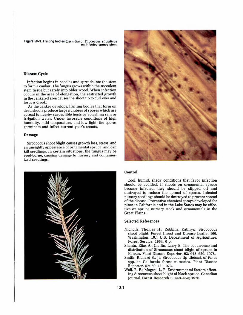
Figure 59-3. Fruiting bodies (pycnidla) of Sirococcus strobil/nus on infected spruce stem.
Disease Cycle
Infection begins in needles and spreads into the stem to form a canker. The fungus grows within the succulent stem tissue but rarely into older wood. When infection occurs in the area of elongation, the restricted growth in the cankered area causes the shoot tip to curl over and form a crook.
As the canker develops, fruiting bodies that form on dead shoots produce large numbers of spores which are spread to nearby susceptible hosts by splashing rain or irrigation water. Under favorable conditions of high humidity, mild temperature, and low light, the spores germinate and infect current year's shoots.
Damage
Sirococcus shoot blight causes growth loss, stress, and an unsightly appearance of ornamental spruce, and can kill seedlings. In certain situations, the fungus may be seed-borne, causing damage to nursery and containerized seedlings.
131
Control
Cool, humid, shady conditions that favor infection should be avoided. If shoots on ornamental spruce become infected, they should be clipped off and destroyed to reduce the spread of spores. Infected nursery seedlings should be destroyed to prevent spread of the disease. Preventive chemical sprays developed for pines in California and in the Lake States may be effective on spruce nursery stock and ornamentals in the Great Plains.
Selected References
Nicholls, Thomas H.; Robbins, Kathryn. Sirococcus shoot blight. Forest Insect and Disease Leaflet 166. Washington, DC: U.S. Department of Agriculture, Forest Service; 1984. 6 p.
Shahin, Elias A.; Claflin, Larry E. The occurrence and distribution of Sirococcus shoot blight of spruce in Kansas. Plant Disease Reporter. 62: 648-650; 1978.
Smith, Richard S., Jr. Sirococcus tip dieback of Pinus spp. in California forest nurseries. Plant Disease Reporter. 57: 69-73; 1973.
Wall, R. E.; Magasi, L. P. Environmental factors affecting Sirococcus shoot blight of black spruce. Canadian Journal Forest Research 6: 448- 452; 1976.

60. Cytospora Canker of Spruce
James A. Wall~ and Frederick J. Crowe
Cytospora canker, caused by the fungus Vulsu kunzei (asexual stage Cytospora kunzei), is a destructive disease of spruce in the northern Great Plains. The fungus is synonymous with Leucostomu kunzei (asexual stage, Leucocytospora kunzeij.
Hosts and Distribution
This pathogen attacks many species of spruce, including blue spruce and its cultivars, white spruce and its variety Black Hills spruce, Norway spruce, and Engelmann spruce. Cytospora canker also occurs on Douglas-fir. It has been reported throughout the upper Midwest and Northeast United States and adjacent Canada, and in the mountains of Colorado. In the Great Plains, it occurs in the prairie provinces of Canada, and in Montana and North and South Dakota.
Symptoms and Signs
Symptoms start on lower branches and spread to other branches laterally and upward in the tree (fig.
60-1). Needles on infected branches die and turn brown, and resin exudes from cankered areas. After a few months infected needles drop off, and white or light blue patches of resin become obvious on the dead bark of larger branches (fig. 60-2). There is little external evidence of the canker margin, but it can be found by exposing the inner bark. Infected bark tissue and cambium are brown in contrast to the normal light color of healthy tissue. The wood beneath infected bark is not discolored.
Asexual fruiting bodies (pycnidia) of the fungus develop in infected bark but are usually not visible on the bark surface. Superficial cuts in cankered bark will expose the small (1-3 mm dia.) black pycnidia (fig. 6Q-3). Orange spore masses or tendrils may exude from the pycnidia during wet weather. The spores (conidia) are hyaline, allantoid (sausage-shaped), 1-celled, and 4-6 by 11-'m.
Sexual fruiting bodies (perithecia) are sometimes associated with pycnidia, but they are usually on branches that have been dead for several years. Perithecia are smaller (0.2-0.6 mm dia.) than pycnidia and are grouped in a black stroma. Ascospores from
132
Figure 60-1. Colorado blue spruce trees with dying and dead lower branches killed by Cytospora kunzel.

perithecia are hyaline, allantoid, 1-celled, and 5-9 by 1.5 ~m.
Because Cytosporo will grow in bark killed by other agents, its presence alone does not mean the fungus killed the branches.
Disease Cycle
Spores from fruiting bodies on cankered branches are spread to the same or other trees by rainsplash, wind, insects, birds, and man. Infection occurs through wounds. The fungus grows and kills the bark, then expands until the entire branch is dead. Fruiting bodies form in infected bark. The fungus overwinters as fruiting bodies and mycelium in cankered bark.
Workers in Michigan found that conidia are released during all seasons except winter, and that ascospores are released only in the spring. They found that only the ascospore stage was infective. Other workers have found that the conidial stage is also infective, however.
Cytosporo is often believed to damage only trees weakened by environmental stress, especially drought. Water stress increases susceptibility to infection, but apparently healthy trees also develop cankers when inoculated. Because Cytospora canker is rare on spruce in the southern Great Plains, other factors also may be involved.
Damage
Damage may occur in ornamental, plantation, or windbreak situations. The disease destroys the symmetry of spruce trees, reduces their effectiveness in blocking wind, snow, and noise, and in time, may kill them. Norway spruce is particularly prone to stem cankering in some areas, and thus is most likely to be killed. Blue spruce is more susceptible to damage from branch cankers than other species. Damage is most often seen on large trees, but may occur on young trees if they are planted on poor sites or are near infected large trees.
Control
Healthy trees are less susceptible to infection; thus they should be managed for optimum vigor. If possible, water and fertilize trees as needed. Reduce chances of infection by preventing wounds and by maintaining good air circulation around the trees. Do not bring infected branches into the area (such as spruce Christmas trees or branches). Blue and Norway spruce are most susceptible to damage, so consider planting other species or varieties if Cytospora canker is a threat.
If trees become infected, prune diseased branches and dispose of them as soon as possible. Cut back to the nearest living lateral beyond the canker or to the trunk. Pruning is best done in late winter before spores are released, but may be done during dry periods whenever dead branches are found. Disinfect pruning tools with rubbing alcohol after each cut, and apply a fungicidal wound dressing to all pruning wounds.
Protective fungicides have been recommended for control of Cytospora canker on spruce, but none are specifically labeled for this purpose. Information on the efficacy of the newer systemic fungicides against this disease is limited.
Selected References
Funk, A. Canker diseases of spruce. Pest Leaflet FPL 65. Canadian Forestry Service; 1978. 4 p.
Kamiri, Lewis K.; Laemmlen, Franklin F. Effects of drought-stress and wounding on Cytospora canker development on Colorado blue spruce. Journal of Arboriculture. 7: 113-116; 1981.
Schoeneweiss, Donald F. Drought predisposition to Cytospora canker in blue spruce. Plant Disease. 67: 383-385; 1983.
Waterman, Alma M. The relation of Valsa kunzei to cankers on conifers. Phytopathology. 45: 686-692; 1955.
133
Figure 60-2. Resin exudes from cankered branches and Is readily visible on dead bart a few months after Infected needles are cast.
Figure 60-3. Black pycnldla of C. kunzel occur In brown cankered Inner bart of Infected spruce stems.

61. Antrodia Stem Decay of Eastern Redcedar
Edward M. Sharon and Jerry W. Riffle
Eastern redcedar has a wide distribution in the Great Plains, and is found on many soil types. Its hardiness and excellent survival on arid sites has made it a major component of field windbreaks in the Great Plains. Antrodia juniperina (referred to as Daedalea juniperina in older literature) causes stem decay in living eastern redcedar.
Hosts and Distribution
A. juniperina occurs on living or dead eastern redcedar in eight eastern States, plus Missouri, Oklahoma, Kansas, Nebraska, North Dakota, Colorado, Arizona, and Oregon. It also has been found on alligator, Utah, and one-seeded juniper in western United States. Its distribution is erratic in the Great Plains, and its occurrence is not correlated with the distribution or prevalence of its host.
Symptoms and Signs
A. juniperina can be detected by the corky, sessile, sporocarps with daedaloid pore surfaces that form under dead branch stubs or along the callus margin of a wound (figs. 61-1, 61-2). The pores are large and angular, 12 to 18 per inch. The sporocarps are annual or perennial. The pileus of new sporocarps varies from light buff to cinnamon buff. It turns gray to black with weathering, and is indistinctly zonate. In the absence of sporocarps, check the ends of poorly healed branch stubs and stumps; thick, buff-colored mycelial felts develop in shrinkage cracks of decayed wood.
A. juniperina, a brown-rot fungus, utilize cellulose and other polysaccharides in wood, and primarily causes heart rot of affected stems. The residue left after decay is a brown crumbly mass. The brown-rot fungi do not produce extracellular phenol oxidases, and generally give negative oxidase tests on gallic and tannic acid medium or with gum guaiac solution.
Figure 61-1. Sporocarps of Anftodla/unlpetina on stem of eastern redcedar, Woodwatd County, Oklahoma.
134

Disease Cycle
Little information is available on the disease cycle of A. juniperino. Dead branches and wounds provide entry for the fungus. The fungus slowly invades and kills stem tissues, and a brown cubical rot develops.
The fungus can be cultured on malt agar and potato dextrose agar. Growth at 75°F is slow, reaching 14-15 mm in 7 days on malt agar. Antrodio and Doedoleo appear similar in culture, but the lack of chlamydospores places A. juniperina in the genus Antrodio rather than in the genus Daedalea.
The basidiospores of A. juniperino are 6-7 JLm long by 2-3 JLm wide, narrowly ellipsoid, somewhat arcuate toward the apiculus, smooth, hyaline, and thin-walled.
Damage
Young trees are usually free from decay. A windbreak may begin to deteriorate, however, if infected eastern redcedars are a major component. Stems of infected trees may become hollow and break during windstorms because of extensive decay of wood.
Control
Tree losses caused by stem decay fungi can be reduced by the following measures:
Figure 61-2. Enlarged view of daedalold pore surface.
1. Prevent wounds. In windbreaks and woodlots, limit the activities of animals, especially cattle, by fencing. Little can be done about the weather; however, after a storm prune broken limbs and remove jagged edges from limb and bole wounds to enhance callus formation.
2. Sanitize. Prune trees to remove dead, dying, or diseased portions of the tree.
3. Plan and implement improvement cuts. Cut and remove infected trees, and replant immediately to maintain effectiveness of windbreaks.
Selected References
Gilbertson, R. L. North American wood-rotting fungi that cause brown rots. Mycotaxon. 12: 372- 416; 1981.
Lowe, Josiah L.; Gilbertson, Robert L. Synopsis of the Polyporaceae of the Western United States and Canada. Mycologia. 53: 474- 511; 1961.
Murrill, William Alphonso. Polyporaceae. North American Flora. 9: 1-131; 1908.
Niemela, T.; Ryvarden, L. Studies in the Aphyllophorales of Africa. IV: Antrodia juniperina, new for East Africa. Transactions of the British Mycological Society. 65(2): 427-432; 1975.
Overholts, Lee Oras. Polyporaceae of the United States, Alaska, and Canada. Ann. Arbor: Univ. Michigan Press; 1953. 468 p.
135

62. Gymnosporangium Rusts of Junipers
James A. Walla and Jerry W. Riffle
Gymnosporangium species that infect junipers are commonly called "cedar-apple rusts."
Hosts and Distribution
Several Gymnosporangium species occur in the Great Plains (table 62-1). These species must alternately infect two hosts to complete their life cycle. In the Great Plains, the telial stage is always on juniper species and the aecial stage is always on rosaceous species.
Symptoms and Signs
Symptoms on junipers vary, depending on the rust species involved. They include small lesions on needles, globular branch galls (fig. 62-1), fusiform stem swellings (fig. 62-2), gall-like branch knots (fig. 62-3), or witches'brooms (fig. 62-4). During wet periods, especially in spring, one to many orange, gelatinous, fingerlike, telial horns extrude from these structures.
Symptoms on rosaceous hosts are rust lesions on leaves, fruit, petioles, or new twigs. Yellow to orange lesions containing black pimple-like pycnia develop first (fig. 62-5); then tubelike aecia form in the same lesions.
Disease Cycle
After spring rains (March-June), telial horns extrude from each lesion, gall, swelling, or broom on junipers.
Figure 62-1. Orange tellal homs of a Gymnosporanglum species extruding from a branch gall on eastern redcedar.
Teliospores in the horns germinate to produce basidiaspores, which are carried by wind or insects to nearby rosaceous hosts. After infection of susceptible host tissue by basidiospores, pycnia develop in yellow to orange lesions, followed by production of aecia. Aeciospores are wind-blown to junipers in the same growing season from late spring to early fall, depending on species of rust fungus. After infection of the juniper. galls or other structures form. The orange gelatinous telial horns appear on them either the following spring or one year later. The cycle is then repeated.
Damage
Damage due to massive infection of rosaceous hosts can influence yield (current and following year), fruit quality, vigor, and esthetic value. Apples are particularly subject to these effects.
Damage to junipers is usually slight. If massive infection occurs, reduction in vigor, growth rate, and esthetic value can result. Rust fungi that cause either stem swellings or knotlike branch galls kill branches above the infected points. Fungi that cause witches'- brooms can deform trees.
Control
Several control methods are available, depending on individual situations. Similar control methods should be
136
Figure 62-2. Dry orange tellal masses of a Gymnosporanglum species extruding from a branch with a stem swelling.

Table 62-1. Some Gymnosporangium species in the Great Plains.
Species of Great Plains Symptoms on Major aecial rust fungus distribution junipers hosts1
junlperi-vlrglnlanae Widespread Branch gall Apple, crabapple connersii Northern Plains Branch gall Hawthorn corniculans Northern Plains Branch gall Juneberry trachysorum Southern Plains Branch gall Hawthorn bethelii All except Texas Gall-like branch knots Hawthorn globosum Widespread Gall-like branch knots Hawthorn nelsonii Western and Gall·like woody Juneberry
Northern Plains knots or no symptoms clavi pes Widespread Stem swellings gracile Texas Stem swellings,
Witches'·brooms nidus-avis Widespread Stem swellings,
Witches'-brooms clavariforme Widespread Stem swellings
Wltches'-brooms exlguum Southern Plains Foliar lesions
1 Other Rosaceae may be aecial hosts for some of these rusts.
effective for all Gymnosporangium rusts with similar life cycles.
Two hosts are required to complete the life cycle; eradication of one host from the vicinity will eliminate new infections. A 2-mile separation usually will minimize rust infections.
In some situations, control may be achieved by pruning affected parts from the Juniperus host. Do not plant susceptible varieties near alternate hosts. Susceptibility of available varieties of both hosts has been determined for some Gymnosporangium rusts.
Several preventative fungicides are registered for control of some rust fungi on both hosts. They may be applied on orchard and ornamental trees. The rosaceous
Figure 62-4. Wltches'·broom on Rocky Mountain juniper caused by G. nldus·a~ls.
Many Rosaceae Juneberry
Juneberry
Juneberry
Hawthorn Figure 62- 3. Orange tellal horns of a Gymnosporanglum species extruding from a gall·llke branch knot on eastern
red cedar.
host must be protected in the spring, starting before flowering and continuing until telial horns on juniper become inactive (usually 4 to 6 weeks). The Juniperus host must be protected when aecia have formed on the corresponding rosaceous host.
Selected References
Himelick, E. B.; Neely, Dan. Juniper hosts of cedar-apple and cedar-hawthorne rust. Plant Disease Reporter. 44: 109-112; 1960.
Kern, F. D. A revised taxonomic account of Gymnosporangium. University Park: Pennsylvania State University Press; 1973. 134 p.
Figure 62-5. Rust lesions on juneberry leaves caused by G. nldus·a~ls.
137

63. Pine Wilt Disease Jerry W. Riffle and Frederick J. Crowe
The pine wood nematode. Bursaphelenchus xylophilus. causes a disease of pines called pine wilt. This nematode is unusual. compared to other plant-parasitic nematodes, because it is a pathogen in above-ground parts of trees, is transmitted by insects, and does not enter the soil. B. xylophilus was first reported in the United States in 1931 in logs of longleaf pine. However, it was not recognized as a potential pathogen of pines in this country until1979 when it was found associated with dying Scots and Austrian pines in Columbia, Missouri.
Hosts and Distribution
Twenty-two species of Pinus, one species of Abies and Pseudotsuga, and two each of Larix, Picea, and Cedrus are known hosts of the pine wood nematode in the United States. The ability of this nematode to kill native North American pines growing in forests has not been established, but evidence suggests that it can kill exotic pines such as Scots, Austrian. and Japanese black pines in the United States. particularly on off-site, high-stress locations. Most pine mortality has occurred in landscape plantings, but the nematode also has caused mortality in windbreaks. Christmas tree, and recreational plantings. Scots pine, an important landscape and popular Christmas tree species, is the most commonly reported host. In the Great Plains, B. xylophilus has been found on Scots, Japanese black, Austrian, eastern white, and loblolly pines. Greenhouse inoculation tests show that the five species most susceptible to B. xylophilus are jack, shortleaf. Monterey, sugar, and Scots pines.
Nationally, B. xylophilus has been found in 34 States, including all states in the Great Plains except North Dakota. However, typical symptoms of pine wilt have not been observed throughout the geographical range of B. xylophilus. The nematode can reproduce on fungi associated with dead, stressed, and live pines, and thus can be found in pines killed by other agents. The widespread distribution and host range of this nematode suggest it is endemic to the United States.
Symptoms and Signs
The first symptom of pine wilt is a marked reduction in flow of oleoresin that occurs before external symptoms are apparent. Transpiration from foliage decreases, then stops 3 to 4 weeks after infection. Foliage rapidly yellows and browns as sapwood moisture decreases {fig. 63- 1). Needles show definite wilt only in long, soft needled species such as white pine. Foliar symptoms may progress uniformly through the tree or branch by branch, largely depending on the size of the tree and the season of death. Trees may die from midsummer to late fall or from late winter to late spring. The rapid death
contrasts with the slow decline caused by pathogenic fungi such as Diplodia pinea, or by unfavorable environmental conditions.
Disease Cycle
The biology of B. xylophilus and basic information on the disease cycle have been determined by scientists in Japan where pine wilt has been a serious problem for many years. The cerambycid beetle Monochamus alternotus is the principal vector that transmits B. xylophilus to native Japanese pines. Adult beetles infested with B. xylophilus emerge from dead pines in May and June, fly to healthy pine trees, and begin maturation feeding on phloem of young pine shoots (fig. 63-2). Immature nematodes leave the tracheae of the beetle, enter feeding wounds, molt to the adult stage, mate, and reproduce rapidly in the resin canals of the pine host during the
138
Figure 63-1. Scots pine tree showing brown foliage symptoms of pine wilt disease.

propagation phase of the life cycle (fig. 63-3). Under optimum conditions, growth of nematodes from eggs to adults is completed in 5 days. Within 4 to 5 weeks, infected pines exhibit reduced oleoresin flow and transpiration, and large numbers of nematodes are present throughout trees as wilting and yellowing of foliage become noticeable.
Trees die within 3 months after becoming infected. Nematodes continue to reproduce for several months on fungi, principally the blue stain fungi, that invade the dead trees. Nematode development then converts to a dispersal phase as the wood dries. During this phase, the nematodes molt from second-stage propagative larvae to third-stage dispersal larvae. These larvae contain high levels of lipids, are resistant to adverse environmental conditions, and do not feed. During early spring these larvae molt from the dispersal stage to the fourth larval stage, called dauer larvae. Cerambycid beetles are attracted to dying trees and oviposit in them. The cerambycid larvae, known as pine sawyers,bore into the wood, overwinter, and pupate in the spring. Dauer larvae of B. xylophilus enter adult beetles prior to their emergence. These beetles transmit the nematodes to healthy trees when they begin maturation feeding.
The development of pine wilt differs in the United States. B. xylophilus is carried by Monochomus corolinensis, M. mutator, M. nototus, M. obtusus, M. scutellatus,and M. titillotor in the eastern United States. The principal vector in pine wilt appears to be M. corolinensis (fig. 63- 4). Investigations in Minnesota, Wisconsin, and Iowa have shown that B. xylophilus is transmitted to dying trees and cut timber of native pine species by M.
Figure 63-2. Maturation feeding by M. alfernatus on Japanese black pine.
Figure 63- 3. Longitudinal section of pine wood showing pinewood nematodes In pine resin canals.
corolinensis, M. scutellotus, and M. mutator during oviposition. Thus B. xylophilus may be present in conifers dying as a result of any cause. This pattern of attack may explain the association of B. xylophilus with trees in the north-central region that are stressed by various pathogens and insects, but Jack typical symptoms of pine wilt.
Damage
Pine wilt poses a potential threat to susceptible species of pines, especially to Scots pines, growing on poor sites. Pine wilt is not a significant problem in native pine forests.
Control
Control measures are not justified in forest situations. In established landscapes, windbreaks, and Christmas tree plantings, destroy recently infected trees by cutting and burning to eliminate the breeding habitat of beetle vectors and to kill larvae and pupae of vectors before they emerge. Control known pathogens and insect pests of pines to avoid stressing trees and attracting vectors that could transmit nematodes to stressed trees during oviposition. In high value areas, keep trees well pruned of dead and diseased branches.
Selected References
Kondo, Eizo; Foudin, Arnold; Linit, Marc; Smith, Mike; Bolla, Robert; Winter, Rudy; Dropkin, Victor. Pine wilt disease-nematological, entomological, and biochemical investigations. Publication SR 282. Columbia: University of Missouri Agricultural Experiment Station; 1982. 56 p.
Malek, Richard B.; Appleby, James E. Epidemiology of pine wilt in Illinois. Plant Disease. 68: 180- 186; 1984.
Wingfield, M. J.; Blanchette, R. A.; Nicholls, T. H. Is the pine wood nematode an important pathogen in the United States? Journal of Forestry. 82: 232-235; 1984.
Figure 63-4. Monochamus caro//nensls Is the principal vector of the pinewood nematode In the United States.
139

64. Root Lesion Nematodes in Junipers and Pines
Glenn W. Peterson and Jerry W. Riffle
A survey in Great Plains tree nurseries in 1960 revealed that conifer seedlings were more commonly damaged by root lesion nematodes than by any other group of nematodes.
Hosts and Distribution
The root lesion nematode encountered most frequently in the 1960 survey was Pratylenchus penetrans. This nematode was found in the roots of seedlings in central and eastern Nebraska nurseries.
P. penetrans has a wide host range, which includes field crops as well as trees. In central Nebraska, this nematode was found in roots of eastern redcedar, Rocky Mountain juniper, white spruce, blue spruce, Austrian pine, and ponderosa pine.
Figure 64-1. Eastern redcedar damaged by root lesion nematodes; left Is non-Infected, right Is Infected.
Symptoms and Signs
Reduced growth of seedlings is the most obvious symptom in nurseries (fig. 64-1). See Article 49 for general symptoms on infected seedlings. Commonly, stunted seedlings are in irregular patches. When nursery soil infested with root lesion nematodes is fumigated, there are occasional areas where fumigation is not complete (due to misses or torn plastic sheets); infected, low vigor seedlings can often be found in these areas (fig. 64-2).
Infected seedlings that are stunted have a reduced root system, and usually develop new, fleshy roots in response to the depleted root system. These fleshy roots are lighter colored and larger in diameter than adjacent older roots. A check for the presence of root lesion nematodes by standard extraction procedures is best done on these fleshy roots.
Figure 64-2. Damage to eastern redcedar seedlings where soli fumigation not complete.
140

Figure 64-3. Damage to an oat cover crop In a nursery block Infested with root lesion nematodes; healthy oats In non-Infested area
(background).
Older, established trees that are growing in soil infested with root lesion nematodes may not show external evidence of infection, even though their roots are infected.
Disease Cycle
Root lesion nematodes are soil borne. They enter and complete a part of their life cycle inside feeder roots. They persist in the soil, where they overwinter even in northern nurseries. Thus the source of infection is nematodes present in the soil when seedlings are established. Nematode populations can increase on some cover crops (fig. 64-3).
Damage
Root lesion nematodes feed and move within roots, thereby damaging roots and reducing the growth of seedlings. Established pines, junipers, and spruce in nursery windbreaks, landscapes, and field plantings are also subject to infection, but damage is not obvious. In a central Nebraska nursery root lesion nematodes were numerous in soil beneath eastern redcedar windbreaks. Although damage to windbreak trees was not obvious; there was extensive damage to seedlings in adjacent nursery beds (fig. 64-4).
Control
Damage by root lesion nematodes can be reduced by treating nursery soil with fumigants such as methyl bromide (fig. 64-5). Fumigation drastically reduces the population of nematodes, but it does not completely eradicate them from the soil. Seedlings in the central Great Plains can be grown in fumigated soil for 2 years without significant damage, but third-year seedlings are likely to be severely damaged.
In nurseries, damage from root lesion nematodes can be reduced by soil fumigation, by use of cover crops that
Figure 64-4. Eastem redcedar seedling damage In beds adjacent to windbreak of eastem redcedar Infected with root lesion
nematodes.
are resistant, and by use of non-susceptible trees in nursery windbreaks.
Selected References
Dunn, R. A.; Mai, W. F. Reproduction of Pratylenchus penetrans in roots of seven cover crop species in greenhouse experiments. Plant Disease Reporter. 57: 728-730; 1973.
Peterson, Glenn W. Response of ponderosa pine seedlings to soil fumigants. Plant Disease Reporter. 54: 572-575; 1970.
Peterson, Glenn W. Root lesion nematode infestation and control in a Plains forest tree nursery. Research Note RM-75. Fort Collins CO: U.S.Department of Agriculture, Forest Service, Rocky Mountain Forest and Range Experiment Station; 1962. 2 p.
Figure 64-5. Control of root lesion nematodes by fumigation of soli with methyl bromide.
141
GLOSSARY
ACERVULUS (LI)—a small subcuticular or subepider-
mal cushionlike asexual fruiting body, without a cover-
ing of fungus tissue, producing conidia in a moist mass
which escapes through a break in the host tissue.
AECIOSPORE—one of several kinds of spores produced
by a rust fungus. Formed in and released from a
fruiting structure called an aecium.
AECIUM (IA)—a cuplike fruiting structure produced by
certain rusts, in which chains of spores (aeciospores)
are developed.
AGAR—a substance from certain red algae used to make
culture media into gels upon which microorganisms
are grown.
ALLANTOID—slightly curved with rounded ends;
sausage-like in form.
ALPHA-SPORE—a fertile spore of the asexual stage of
the Diaporthaceae (Phomopsis) that is fusoid to oblong
and biguttulate; BETA-SPORE, usually hooked shaped,
is produced in addition.
ALTERNATE HOST—one or the other of the two unlike
host plants parasitized by a heteroecious fungus such
as a typical rust fungus, i.e., either the juniper or ap-
ple host of the cedar-apple rust fungus.
ANAEROBE—A microorganism that can live and grow
where there is no free oxygen.
ANAMORPH—The asexual form of a fungus (e.g. that
characterized by conidiomata).
ANNULUS—a ring-like partial veil around the stem of
a fruiting body after expansion of the pileus (cap).
ANTHRACNOSE—a type of plant disease which typ-
ically is a leaf and twig blight. Common on many
hardwoods.
APOTHECIUM(IA)—a cup or saucerlike sexual fruiting
body which produces ascospores.
ASCOSPORES—a spore produced in an ascus (see
ascus).
ASCUS(I)—a sac-like cell of the perfect stage of the
Ascomycetes in which ascospores—usually eight—are
produced.
ASEXUAL STAGE—a stage in the life cycle of a fungus
in which spores are produced without a previous sex-
ual fusion; also called imperfect stage.
BASIDIOSPORE—the spore produced by the sexual
stage of the Basidiomycetes.
BASIDIUM(IA)—a cell, usually terminal, in which
nuclear fusion and meiosis occur and each of the four
haploid nuclei pass into one of four forming spores.
BETA-SPORE—see alpha-spore.
BLIGHT—a general term for a plant disease causing
rapid death or dieback.
BORDEAUX MIXTURE—a fungicidal spray used for
controlling plant diseases. A common mixture is
4-4-50, which means 4 lbs. copper sulfate, 4 lbs. lime,
and 50 gal. water. BROOM—an abnormally dense
mass of host branches and foliage in which the typical
host growth pattern is lost.
BROWN ROT—decay caused by fungi that degrade
cellulose, do not produce extracellular phenoloxidases,
common and generally give negative oxidase tests.
CANKER—a definite relatively localized necrotic lesion
primarily of the bark and cambium.
CARPOGIUM(IA)—the female sex organ.
CHLOROSIS—an abnormal yellowing of the foliage.
CHLOROTHALONIL—a relatively broad spectrum
organic fungicide. A foliar fungicide.
CHLOROTIC—abnormally yellow.
CLEISTOTHECIUM(IA)—a closed fruiting body,
without an ostiole, containing asci.
CONIDIOMA(MATA)—A specialized multi-hyphal,
conidia-bearing structure.
CONIDIOPHORE—a specialized hypha bearing conidia.
CONIDIUM(IA)—a spore formed asexually, usually at
the tip or side of a hypha.
CONK—a type of fruiting (spore-forming) structure
formed by certain fungi, usually wood rotters (Basidio-
mycetes). It is often bracket-like, and is referred to as
"sporocarp," "sporophore," "fruiting body," "carpo-
phore," "fructification," "basidiocarp," "punk,"
"bracket," and "shelf."
CONTEXT (OF SPOROCARP)—the inner tissue of the
pileus (cap), i.e., the tissue lying between the upper sur-
face and the tube or pore layer. The context often is
designated as the "trama" of the pileus.
COREMIUM(IA)—a specialized multi-hyphal structure
composed of a compact group of erect and sometimes
fused conidiophores bearing conidia at the apex or on
both apex and sides.
COVER CROP—a crop, natural or introduced, that is
grown alternately with the main crop. Used to prevent
erosion and improve soil characteristics.
CULTIVAR—an assemblage of cultivated individuals
distinguished by any useful, reproducible character.
CUTTING—detached portion of stem or other plant part
which, when rooted, produces a whole plant.
DAEDALOID-PORES—with elongated and sinuous
mouths.
DECAY—the decomposition of plant tissue by fungi and
other microorganisms.
DEFOLIATION—loss of current year's or past years'
foliage.
DIEBACK—the death of parts of a tree or plant usually
from the top downward.
DISEASE—unfavorable change of the function or form
of a plant from normal, caused by a pathogenic agent
or unfavorable environment.
DISEASE CYCLE—the chain of events in the develop-
ment of a disease.
EFFUSED-REFLEXED—spread out over a substratum
and turned back at the margin to form a pileus (cap).
EPIDEMIC—pertaining to a disease which has built up
rapidly and reached injurious levels.
EPIDERMIS—the outermost layer of cells on the primary
plant body.
EPIPHYTE—a plant that grows on another plant but is
not a parasite and produces its own food by photo-
synthesis.
EXUDATE—matter which oozes out or is secreted.
142
GLOSSARY ACERVULUS (Ll)-a small subcuticular or subepider
mal cushionlike asexual fruiting body, without a covering of fungus tissue, producing conidia in a moist mass which escapes through a break in the host tissue.
AECIOSPORE-one of several kinds of spores produced by a rust fungus. Formed in and released from a fruiting structure called an aecium.
AECIUM (IA)-a cuplike fruiting structure produced by certain rusts, in which chains of spores (aeciospores] are developed.
AGAR-a substance from certain red algae used to make culture media into gels upon which microorganisms are grown.
ALLANTOID-slightly curved with rounded ends; sausage-like in form.
ALPHA-SPORE-a fertile spore of the asexual stage of the Diaporthaceae (Phomopsis) that is fusoid to oblong and biguttulate; BET A-SPORE, usually hooked shaped, is produced in addition.
ALTERNATE HOST -one or the other of the two unlike host plants parasitized by a heteroecious fungus such as a typical rust fungus, i.e., either the juniper or apple host of the cedar-apple rust fungus.
ANAEROBE-A microorganism that can live and grow where there is no free oxygen.
ANAMORPH-The asexual form of a fungus (e.g. that characterized by conidiomata).
ANNULUS-a ring-like partial veil around the stem of a fruiting body after expansion of the pileus (cap).
ANTHRACNOSE-a type of plant disease which typically is a leaf and twig blight. Common on many hardwoods.
APOTHECIUM(IA)-a cup or saucerlike sexual fruiting body which produces ascospores.
ASCOSPORES-a spore produced in an ascus (see ascus).
ASCUS(I)-a sac-like cell of the perfect stage of the Ascomycetes in which ascospores-usually eight-are produced.
ASEXUAL STAGE-a stage in the life cycle of a fungus in which spores are produced without a previous sexual fusion; also called imperfect stage.
BASIDIOSPORE-the spore produced by the sexual stage of the Basidiomycetes.
BASIDIUM(IA]-a cell, usually terminal, in which nuclear fusion and meiosis occur and each of the four haploid nuclei pass into one of four forming spores.
BET A-SPORE-see alpha-spore. BLIGHT -a general term for a plant disease causing
rapid death or dieback. BORDEAUX MIXTURE-a fungicidal spray used for
controlling plant diseases. A common mixture is 4-4-50, which means 4 lbs. copper sulfate, 4 lbs. lime, and 50 gal. water. BROOM-an abnormally dense mass of host branches and foliage in which the typical host growth pattern is lost.
BROWN ROT -decay caused by fungi that degrade cellulose, do not produce extracellular phenoloxidases, common and generally give negative oxidase tests.
CANKER-a definite relatively localized necrotic lesion primarily of the bark and cambium.
CARPOGIUM(IA)-the female sex organ. CHLOROSIS-an abnormal yellowing of the foliage. CHLOROTHALONIL-a relatively broad spectrum
organic fungicide. A foliar fungicide. CHLOROTIC-abnormally yellow. CLEISTOTHECIUM(IA)-a closed fruiting body,
without an ostiole, containing asci. CONIDIOMA(MA T A)-A specialized multi-hyphal,
conidia-bearing structure. CONIDIOPHORE-a specialized hypha bearing conidia. CONIDIUM(IA)-a spore formed asexually, usually at
the tip or side of a hypha. CONK-a type of fruiting (spore-forming) structure
formed by certain fungi, usually wood ratters (Basidiomycetes). It is often bracket-like, and is referred to as "sporocarp," "sporophore," "fruiting body," "carpophore," "fructification," "basidiocarp," "punk," "bracket," and "shelf."
CONTEXT (OF SPOROCARP)-the inner tissue of the pileus (cap), i.e., the tissue lying between the upper surface and the tube or pore layer. The context often is designated as the "trama" of the pileus.
COREMIUM(IA)-a specialized multi-hyphal structure composed of a compact group of erect and sometimes fused conidiophores bearing conidia at the apex or on both apex and sides.
COVER CROP-a crop, natural or introduced, that is grown alternately with the main crop. Used to prevent erosion and improve soil characteristics.
CUL TIV A R-an assemblage of cultivated individuals distinguished by any useful, reproducible character.
CUTTING-detached portion of stem or other plant part which, when rooted, produces a whole plant.
DAEDALOID-PORES-with elongated and sinuous mouths.
DECAY -the decomposition of plant tissue by fungi and other microorganisms.
DEFOLIATION-loss of current year's or past years' foliage.
DIEBACK-the death of parts of a tree or plant usually from the top downward.
DISEASE-unfavorable change of the function or form of a plant from normal, caused by a pathogenic agent or unfavorable environment.
DISEASE CYCLE-the chain of events in the development of a disease.
EFFUSED-REFLEXED-spread out over a substratum and turned back at the margin to form a pileus (cap).
EPIDEMIC-pertaining to a disease which has built up rapidly and reached injurious levels.
EPIDERMIS-the outermost layer of cells on the primary plant body.
EPIPHYTE-a plant that grows on another plant but is not a parasite and produces its own food by photosynthesis.
EXUDATE-matter which oozes out or is secreted.
142

FACULTATIVE PARASITE—an organism which is nor-
mally saprophytic but which is capable of living as a
parasite only when unfavorable conditions predispose
the host so that it is unusually susceptible.
FACULTATIVE SAPROPHYTE—an organism which is
normally parasitic but which is capable of living as a
saprophyte.
FALLOW—cultivated land allowed to lie idle or
unplanted during the growing season.
FASCICLE SHEATH—a sheath around the base of a
cluster or bundle of needles.
FEEDER ROOT—succulent actively growing rootlets of
plants.
FLAGELLUM(LA)—a whiplike part of some cells,
especially of certain bacteria that is an organ of
locomotion.
FOLIAGE—leaves of a plant or tree.
WHIP LEAVES—juniper foliage characteristic of long
shoot growth on the ends of secondary and tertiary
branches.
SPUR LEAVES—juniper foliage characteristic of short
(spur) branches.
JUVENILE LEAVES—juniper foliage characteristic of
seedlings.
FRUITING BODY—any of a number of kinds of
reproductive structures which produces spores. See
conk.
FUNGICIDE—chemical which is toxic to fungi.
PROTECTANT—fungicides applied to foliage^of plant
in advance of a pathogen in order to prevent infection.
ERADICANT—fungicides, applied to foliage of a plant,
that have a direct effect upon organisms which have
already invaded the host.
SYSTEMIC—fungicides that are absorbed into the
tissues of plants and are toxic to fungi.
FUNGUS—any of a number of organisms considered by
some authorities to be lower plants which lack
chlorophyll.
GALL(BURLS)—pronounced swellings on woody plants
caused by certain insects and disease organisms.
GIRDLED—to destroy or remove the tissue, particular-
ly living tissue in a rough ring around a stem, branch,
or root.
GUMMOSIS—the giving off of gummy substances as a
result of cell degeneration.
HAUSTORIUM(IA)—absorbing organ originating on a
hypha of a parasite and penetrating into a cell of the
host.
HOST—the plant on or in which a pathogen exists.
HOST RANGE—all hosts which a particular pathogen
attacks.
HOST SPECIFIC—a term used to describe those patho-
gens which attack only certain species of hosts.
HYALINE—transparent, having no color.
HYMENIUM—the general region of the sporocarp bear-
ing the basidia, that is, the layer of tubes. The actual
spore-producing layer, made up of basidia and what-
ever type of sterile organ that may be present with
them and forming a layer lining the inside of the tubes.
IMPERFECT STAGE—that part of the life cycle of a
fungus in which only conidia and no sexual spores are
produced. Syn. asexual.
INFECT—to invade and cause a disease.
INFECTION COURT—the area in which the pathogen
first established itself on or in the host.
INFEST—to be present within an area in such numbers
as to be a disease hazard.
INOCULATE—to place a pathogen on or in a host in a
position in which it is capable of causing a disease.
INOCULUM—the spores, mycelium, sclerotia, or other
propogules of a pathogen which initially infect a host
or crop.
INTERNODE—the portion of the stem between two
nodes.
LATENT INFECTION—an established infection which
does not show its presence.
LEAF ABSCISSION—the normal separation of leaves
from plants by the development of a thin layer of pithy
cells at the base of their petioles.
LEAF PETIOLE—the slender, usually cylindrical portion
of a leaf, which supports the blade and is attached to
the stem.
LEAF SPOT—a leaf disease characterized by numerous
distinct lesions.
LESION—a defined necrotic area.
LIFE CYCLE—the stage or series of stages in fungi be-
tween one spore form and the development of the
same spore again.
MACROCONIDIA—the larger of two types of conidia
by certain fungi, such as Fusarium spp.
MICROCONIDIA—the smaller of two types of conidia
produced by certain fungi.
MUSHROOM—any of various rapid-growing, fleshy
fungi that typically have a stalk capped with an um-
brellalike top.
MYCELIUM(IA)—a mass of hyphae which forms the
vegetative filamentous body of a fungus.
MYCOPLASMA—a type of disease-causing organism
similar to a bacterium, but lacking a true cell wall.
MYCOPLASMA LIKE ORGANISM (MLO)—an organ-
ism with apparent features of mycoplasma, but not
proven to be mycoplasma.
NECROSIS—death of plant cells usually resulting in
darkening of the tissue.
NEMATODE—a roundworm with a long, cylindrical,
unsegmented body.
PARASITE—an organism living on and nourished by
another living organism.
PATHOGEN—an organism which causes a disease.
PATHOGENIC—capable of causing a disease.
PEDUNCLE—the supporting axis of a single flower or
a flower cluster.
PERFECT STAGE—the stage in which the sexual spore
stage is produced.
PERENNIAL—continuing growth from year to year.
PERIDERM—the outer protective layer in older stems,
consisting of the phellogen and its derivative tissues,
phellem and phelloderm.
PERITHECIUM(IA)—a closed flasklike sexual fruiting
body formed by certain Ascomycetes in which
ascospores are produced.
PERITRICHOUS—having flagella evenly distributed
over the entire surface of the cell of a bacterium.
143
FACULTATIVE PARASITE-an organism which is normally saprophytic but which is capable of living as a parasite only when unfavorable conditions predispose the host so that it is unusually susceptible.
FACULTATIVE SAPROPHYTE-an organism which is normally parasitic but which is capable of living as a saprophyte.
FALLOW-cultivated land allowed to lie idle or unplanted during the growing season.
FASCICLE SHEATH-a sheath around the base of a cluster or bundle of needles.
FEEDER ROOT -succulent actively growing rootlets of plants.
FLAGELLUM(LA)-a whiplike part of some cells, especially of certain bacteria that is an organ of locomotion.
FOLIAGE-leaves of a plant or tree. WHIP LEA YES-juniper foliage characteristic of long shoot growth on the ends of secondary and tertiary branches. SPUR LEA YES-juniper foliage characteristic of short (spur) branches. JUVENILE LEA YES-juniper foliage characteristic of seedlings.
FRUITING BODY-any of a number of kinds of reproductive structures which produces spores. See conk.
FUNGICIDE-chemical which is toxic to fungi. PROTECT ANT -fungicides applied to foliage'of plant in advance of a pathogen in order to prevent infection. ERADICANT -fungicides, applied to foliage of a plant, that have a direct effect upon organisms which have already invaded the host. SYSTEMIC-fungicides that are absorbed into the tissues of plants and are toxic to fungi.
FUNGUS-any of a number of organisms considered by some authorities to be lower plants which lack chlorophyll.
GALL(BURLS)-pronounced swellings on woody plants caused by certain insects and disease organisms.
GIRDLED-to destroy or remove the tissue, particularly living tissue in a rough ring around a stem, branch, or root.
GUMMOSIS-the giving off of gummy substances as a result of cell degeneration.
HAUSTORIUM(IA)-absorbing organ originating on a hypha of a parasite and penetrating into a cell of the host.
HOST -the plant on or in which a pathogen exists. HOST RANGE-all hosts which a particular pathogen
attacks. HOST SPECIFIC-a term used to describe those patho
gens which attack only certain species of hosts. HYALINE-transparent, having no color. HYMENIUM-the general region of the sporocarp bear
ing the basidia, that is, the layer of tubes. The actual spore-producing layer, made up of basidia and whatever type of sterile organ that may be present with them and forming a layer lining the inside of the tubes.
IMPERFECT STAGE-that part of the life cycle of a fungus in which only conidia and no sexual spores are produced. Syn. asexual.
143
INFECT -to invade and cause a disease. INFECTION COURT-the area in which the pathogen
first established itself on or in the host. INFEST -to be present within an area in such numbers
as to be a disease hazard. INOCULATE-to place a pathogen on or in a host in a
position in which it is capable of causing a disease. INOCULUM-the spores, mycelium, sclerotia, or other
propogules of a pathogen which initially infect a host or crop.
INTERNODE-the portion of the stem between two nodes.
LATENT INFECTION-an established infection which does not show its presence.
LEAF ABSCISSION-the normal separation of leaves from plants by the development of a thin layer of pithy cells at the base of their petioles.
LEAF PETIOLE-the slender, usually cylindrical portion of a leaf, which supports the blade and is attached to the stem.
LEAF SPOT -a leaf disease characterized by numerous distinct lesions.
LESION-a defined necrotic area. LIFE CYCLE-the stage or series of stages in fungi be
tween one spore form and the development of the same spore again.
MACROCONIDIA-the larger of two types of conidia by certain fungi, such as Fusarium spp.
MICROCONIDIA-the smaller of two types of conidia produced by certain fungi.
MUSHROOM-any of various rapid-growing, fleshy fungi that typically have a stalk capped with an umbrellalike top.
MYCELIUM(IA)-a mass of hyphae which forms the vegetative filamentous body of a fungus.
MYCOPLASMA-a type of disease-causing organism similar to a bacterium, but lacking a true cell wall.
MYCOPLASMA LIKE ORGANISM (MLO)-an organism with apparent features of mycoplasma, but not proven to be mycoplasma.
NECROSIS-death of plant cells usually resulting in darkening of the tissue.
NEMATODE-a roundworm with a long, cylindrical, unsegmented body.
PARASITE-an organism living on and nourished by another living organism.
PATHOGEN-an organism which causes a disease. PATHOGENIC-capable of causing a disease. PEDUNCLE-the supporting axis of a single flower or
a flower cluster. PERFECT STAGE-the stage in which the sexual spore
stage is produced. PERENNIAL-continuing growth from year to year. PERIDERM-the outer protective layer in older stems,
consisting of the phellogen and its derivative tissues, phellem and phelloderm.
PERITHECIUM(IA)-a closed flasklike sexual fruiting body formed by certain Ascomycetes in which ascospores are produced.
PERITRICHOUS-having flagella evenly distributed over the entire surface of the cell of a bacterium.
PHELLODERM—cells cut off centripetally by the
phellogen.
PHELLOGEN—a lateral meristem that cuts off phello-
derm and phellem; the cork cambium.
PHELLUM—the suberized tissue produced by the cork
cambium in the bark.
PHLOEM—the tissue of the inner bark responsible for
the transport of elaborate food stuffs.
PILEUS(EI)—the upper surface of a sporocarp.
PLEOMORPHIC—the condition in which a fungus has
two or more very different morphological forms.
PORE SURFACE—the lower surface of the sporocarp in
specimens mature enough to have a tube layer. It is
the surface at which the pores open.
PRIMARY INFECTION—infection of a host by primary
inoculum, for example, from overwintering spores.
PROLIFERATE—to grow by budding in quick suc-
cession.
PRUNE—to remove dead or living parts from a plant to
improve its form.
PUSTULE—a small, sometimes colored, blister-like
swelling.
PYCNIDIOSPORE—an asexual spore or conidium pro-
duced within a pycnidium.
PYCNIDIUM(IA)—an asexual type of fruiting body,
typically flask shaped, in which asexual spores or con-
idia are produced.
PYCNIOSPORE—a specialized spore produced in a pyc-
nium by the rust fungi.
PYCNIUM(IA)—a designation for the spermogonium of
the rust fungi.
PYRIFORM—pear-shaped.
RELATIVE HUMIDITY—the amount of moisture in the
air as compared with the maximum amount that the
air contains at the same temperature, expressed as a
percentage.
RESISTANT—able to withstand without serious injury,
attack by an organism, or damage by a nonliving agen-
cy but not immune from such attacks.
RESUPINATE SPOROCARP—a sporocarp in which the
entire structure lies flat on the substratum, i.e. without
forming a bracket-like or shelflike body.
RHIZOMORPH—a thick strand of vegetative hyphae in
which the hyphae have lost their individuality.
RHIZOSPHERE—the soil near a living root.
ROT—see decay.
SAPROPHYTE—an organism using dead organic
material as food.
SAPWOOD—the soft wood just beneath the bark of a
tree.
SCLEROTIUM(IA)—a firm, frequently rounded multi-
cellular resting structure produced by fungi.
SECONDARY INFECTION—when a host becomes
diseased, the organism commonly produces another
crop of spores or infective bodies which serve to cause
secondary infection.
SEPTATE—having cross walls which divide hyphae or
spores into a number of separate cells.
SEPTUM—the cross wall which divides a hypha or spore
into two or more distinct cells.
SETA(AE)—conspicuous, conical or lance-shaped,
brown, sterile organs found in the hymenium of some
fungal species.
SESSILE SPOROCARP—a sporocarp which takes the
form of a knob or bracket or shelf. The sporocarp has
no stem or stalk, and the point of attachment to the
substratum is typically lateral.
SEXUAL STAGE—the stage in the life cycle of a fungus
in which spores are produced after sexual fusion. Syn;
perfect stage.
SHEARING—cutting foliage and stem from trees with
shears.
SHELTERBELT—see windbreak.
SIGNS OF A PATHOGEN—any observable part of a
pathogen.
SILVICULTURE—the art of establishing, growing, and
regenerating a forest.
SPERMATIUM(IA)—a non-motile, uninucleate, spore-
like male structure which empties its contents into a
receptive female structure during plasmogamy.
SPERMOGONIUM(IA)—a structure which contains
minute, rod-shaped, or oval spore-like bodies which
in some cases have proved to be functional spermatia.
SPIROPLASM—a spiral shaped bacterium without a cell
wall. Spiroplasms are related to mycoplasmas.
SPORE-the reproductive structure of the fungi and other
lower plants.
SPORE HORNS—a tendril-like mass of forced-out
spores.
SPORODOCHIUM(IA)—a cushion-shaped stroma cov-
ered with conidiophores. Also called a cirrus.
SPORULATE—to produce and release spores.
STOOL BEDS—nursery beds containing plants from
which cuttings are obtained during the dormant
season for vegetative propagation.
STOMA(MATA)—a pore in the leaf epidermis, sur-
rounded by two guard cells, leading into an intercell-
ular space within the plant.
STROMA(MATA)—a mass of fungal hyphae packed
together to form a hard crust in or on which fruiting
bodies are formed.
SUSCEPTIBLE—unable to withstand attack by an
organism or damage by a nonliving agency without
serious injury.
SYMPTOM—the evidence of disturbance in the normal
development and function of a host plant, i.e.,
chlorosis, necrosis, galls, brooms, stunting, etc.
SYSTEMIC—affecting or distributed throughout the
whole plant body.
TELIOSPORE—the spore of the rust fungi from which
the perfect stage of the basidium and basidispore arise.
TELIUM(IA)—an aggregration of teliospores of the rust
fungi.
TENDRIL—a threadlike part of a spore-horn.
TUBE LAYER OF SPOROCARP—a layer of vertically
placed tubes attached to the lower surface of the con-
text of a sporocarp. See hymenium.
UREDIOSPORES—one of the many spore stages pro-
duced by the rust fungi in their life cycle. These spores
are produced in a fruiting body called an uredium.
144
PHELLODERM-cells cut off centripetally by the phellogen.
PHELLOGEN-a lateral meristem that cuts off phelloderm and phellem; the cork cambium.
PHELLUM-the suberized tissue produced by the cork cambium in the bark.
PHLOEM-the tissue of the inner bark responsible for the transport of elaborate food stuffs.
PILEUS(EI)-the upper surface of a sporocarp. PLEOMORPHIC-the condition in which a fungus has
two or more very different morphological forms. PORE SURFACE-the lower surface of the sporocarp in
specimens mature enough to have a tube layer. It is the surface at which the pores open.
PRIMARY INFECTION-infection of a host by primary inoculum, for example, from overwintering spores.
PROLIFERATE-to grow by budding in quick succession.
PRUNE-to remove dead or living parts from a plant to improve its form.
PUSTULE-a small, sometimes colored, blister-like swelling.
PYCNIDIOSPORE-an asexual spore or conidium produced within a pycnidium.
PYCNIDIUM(IA)-an asexual type of fruiting body, typically flask shaped, in which asexual spores or conidia are produced.
PYCNIOSPORE-a specialized spore produced in a pycnium by the rust fungi.
PYCNIUM(IA)-a designation for the spermogonium of the rust fungi.
PYRIFORM-pear-shaped. RELATIVE HUMIDITY -the amount of moisture in the
air as compared with the maximum amount that the air contains at the same temperature, expressed as a percentage.
RESIST ANT -able to withstand without serious injury, attack by an organism, or damage by a nonliving agency but not immune from such attacks.
RESUPINATE SPOROCARP-a sporocarp in which the entire structure lies flat on the substratum, i.e. without forming a bracket-like or shelflike body.
RHIZOMORPH-a thick strand of vegetative hyphae in which the hyphae have lost their individuality.
RHIZOSPHERE-the soil near a living root. ROT -see decay. SAPROPHYTE-an organism using dead organic
material as food. SAPWOOD-the soft wood just beneath the bark of a
tree. SCLEROTIUM(IA)-a firm, frequently rounded multi
cellular resting structure produced by fungi. SECONDARY INFECTION-when a host becomes
diseased, the organism commonly produces another crop of spores or infective bodies which serve to cause secondary infection.
SEPT ATE-having cross walls which divide hyphae or spores into a number of separate cells.
SEPTUM-the cross wall which divides a hypha or spore into two or more distinct cells.
SET A( A E)-conspicuous. conical or lance-shaped, brown, sterile organs found in the hymenium of some fungal species.
SESSILE SPOROCARP-a sporocarp which takes the form of a knob or bracket or shelf. The sporocarp has no stem or stalk, and the point of attachment to the substratum is typically lateral.
SEXUAL STAGE-the stage in the life cycle of a fungus in which spores are produced after sexual fusion. Syn; perfect stage.
SHEARING-cutting foliage and stem from trees with shears.
SHEL TERBEL T -see windbreak. SIGNS OF A PATHOGEN-any observable part of a
pathogen. SILVICULTURE-the art of establishing, growing, and
regenerating a forest. SPERMATIUM(IA)-a non-motile, uninucleate, spore
like male structure which empties its contents into a receptive female structure during plasmogamy.
SPERMOGONIUM(IA)-a structure which contains minute, rod-shaped, or oval spore-like bodies which in some cases have proved to be functional spermatia.
SPIROPLASM-a spiral shaped bacterium without a cell wall. Spiroplasms are related to mycoplasmas.
SPORE-the reproductive structure of the fungi and other lower plants.
SPORE HORNS-a tendril-like mass of forced-out spores.
SPORODOCHIUM(IA)-a cushion-shaped stroma covered with conidiophores. Also called a cirrus.
SPORULATE-to produce and release spores. STOOL BEDS-nursery beds containing plants from
which cuttings are obtained during the dormant season for vegetative propagation.
STOMA(MA T A)-a pore in the leaf epidermis, surrounded by two guard cells, leading into an intercellular space within the plant.
STROMA(MA T A)-a mass of fungal hyphae packed together to form a hard crust in or on which fruiting bodies are formed.
SUSCEPTIBLE-unable to withstand attack by an organism or damage by a nonliving agency without serious injury.
SYMPTOM-the evidence of disturbance in the normal development and function of a host plant, i.e .. chlorosis, necrosis, galls, brooms, stunting, etc.
SYSTEMIC-affecting or distributed throughout the whole plant body.
TELIOSPORE-the spore of the rust fungi from which the perfect stage of the basidium and basidispore arise.
TELIUM{IA)-an aggregration of teliospores of the rust fungi.
TENDRIL-a threadlike part of a spore-horn. TUBE LAYER OF SPOROCARP-a layer of vertically
placed tubes attached to the lower surface of the context of a sporocarp. See hymenium.
UREDIOSPORES-one of the many spore stages produced by the rust fungi in their life cycle. These spores are produced in a fruiting body called an uredium.
144

UREDIUM(IA)—one of the many types of fruiting bodies
formed by the rust fungi in their life cycle.
Urediospores are formed in this fruiting body.
VECTOR—an organism, usually an insect, that transmits
a pathogen from one host to another.
VEGETATIVE MYCELIUM—a mass of hyphae con-
stituting the body of the fungus and without spores.
VIRULENT—vigorously pathogenic.
WETWOOD—a discolored, water-soaked condition of
the heartwood of some trees presumably caused by
bacterial fermentation. Often associated with distinc-
tive odor, gas production, and an exudation called
slime flux.
WHITEROT—decay caused by fungi that attack all chief
constituents of wood and leave a whitish or light col-
ored residue. Affected wood is often fibrous or spongy
in texture.
WINDBREAK—a row or rows of trees that serve as a pro-
tection from wind.
WITCHES'-BROOM—an abnormal cluster of twigs and
branches caused by certain pathogens.
XYLEM—the woody conducting tissues of the stem and
root.
Index To Host Plants With Scientific Equivalents
Host
Ailanthus, Ailanthus altissima (Mill.) Swingle
Albizzia, Albizia spp
Mimosa, A. j'ulibrissin Durazz
A. kaikora (Roxb.) Prain
A. lophantha (Venten) Macbr. ...
A. procera (Roxb. Benth.)
A. pudica L
A. thorelii Pierre
Alder, Alnus sp
Almond, flowering, Primus triloba Lindl. . . .
Amelosorbus sp
Apple, Malus sp
14
14,
35,
Apricot, Primus armeniaca L
Arborvitae, Thuja sp
Ash, Fraxinus sp 7, 14,
12
Black, F. nigra Marsh
Green, F. pennsylvanica Marsh 6,
White, F. americana L
Aspen, Populus sp 1,
Bigtooth, P. grandidentata Michx
Quaking, P. tremuioides Michx
Autumn-olive, Elaeagnus umbellata Thunb
Basswood, Tilia americana L 7,
Beech, Fagus sp
Birch, Betula sp 12,
Paper, B. papyri/era Marsh
Buckthorn, European, Rhamnus cathartica L.
Buffaloberry, Shepherdia sp •
Russet, S. canadensis (L.) Nutt
Silver, S. argentea (Pursh.) Nutt
Caragana, Caragana arborescens Lam
Catalpa, Northern, Catalpa speciosa Warden
14,
Cherry, Prunus sp 8, 9,
Carolina cherry-laurel, P. caroliniana Ait
Cherry-laurel, P. laurocerasus L
Chokecherry, P. virginiana L 9, 10
Hollyleaf cherry, P. ilici/olia (Nutt.) D. Dietr
Nanking cherry, P. tomentosa Thunb
Sour cherry, P. cerasus L
Western sandcherry, P. besseyi Bailey
20, 47
46
46
46
46
46
46
46
8, 12
34
39
15, 34,
39, 48,
49, 62
34, 47
50, 52
37, 38,
44
6
14, 37,
47, 49
27, 37
26, 38
26
2, 26
18
14, 31
12, 31
14, 15,
37, 38
48
38
13, 37
13
13, 37
3, 4
44, 47,
49
10, 14,
30, 32
30
34
32, 34
30
9
10
34
Article Number Chinkapin, Castanopsis sp
Allegheny, Castanea pumila Miller
Bush, C. sempervirens (Kell.) Dudley
Golden, C. chrysophylla (Dougl.) A. DC
Chokeberry, Aronia sp
Christmasberry, Photinia sp
Cotoneaster, Cotoneaster sp
Peking, C. acutinfolia L
Corktree, Amur, Phellodendron amurense Rupr. .
Crabapple, Malus sp
Crataegomesphilus sp
Cypress, Cuppressus sp
Desertwillow, Chilopsis linearis (Cav.) Sweet ....
Dogwood, flowering, Cornus florida L
Douglas-fir, Pseudotsuga menziesii (Mirb.) Franco
Elm, Ulmus sp
American, U. americana L.
30.
7, 8,
15,
41,
16,
41,
cedar, U. crassifolia Nutt
Chinese, U. parvifolia Jacq
English, U. procera Salisb
Evergreen, U. parvi/olia Jacq
Japanese, U. japonica (Rehd.) Sarg.
Red, U. rubra Muhl
Rock, U. thomasii Sarg
U. rubra x U. pumila hybrid
September, U. serotina Sarg
Siberian, U. pumila L
Slippery, U. rubra Muhl
Smooth leaved, U. carpinofolia Ruppins ex. Suckow
U. villosa Brandis
Winged, U. alato Michx
White, U. americana L
Elm zelkova, Zelkova carpini/olia (Pall.) C. Koch.
Fir, Abies sp
Flowering-quince, Chaenomeles spp
Gooseberry, Ribes sp
Hackberry, Celtis occidentals L
Hawthorn, Crataegus sp. ...
Hemlock, Tsuga (Endl.) Carr.
Hickory, Carya sp
41
41
14,
29,
41
16,
41,
14,
35
12,
45
45
45
12
10, 39
39
35
48
14
35, 62
39
50, 53
47
20
56, 60
12, 14,
16, 29,
42, 43,
44
27, 29,
42, 43,
49
42, 43
41
16
41
41
42, 43
41
43
41, 43
16, 17,
41, 42,
43, 47
42, 43
16
41
41, 43
27, 29,
42, 43,
49
41
14
34, 39
38
36, 41,
47
39, 62
59
Chestnut, Castanea sp
American, C. dentata (Marsh.) Borkh
Chinese, C. moliissima Bl
Spanish, C. sativa Mill
12, 45
45
45
45
Mockernut, C. tomentosa Nutt
Pecan, C. illinoensis (Wangenh.) K. Koch.
Shagbark, C. ovata (Mill.) K. Koch
14, 20,
27
27
30
27
145
UREDIUM(IA)-one of the many types of fruiting bodies formed by the rust fungi in their life cycle. Urediospores are formed in this fruiting body.
VECTOR-an organism, usually an insect, that transmits a pathogen from one host to another.
VEGETATIVE MYCELIUM-a mass of hyphae constituting the body of the fungus and without spores.
VIRULENT -vigorously pathogenic. WETWOOD-a discolored, water-soaked condition of
the heartwood of some trees presumably caused by bacterial fermentation. Often associated with distinctive odor, gas production, and an exudation called slime flux.
WHITEROT -decay caused by fungi that attack all chief constituents of wood and leave a whitish or light colored residue. Affected wood is often fibrous or spongy in texture.
WINDBREAK-a row or rows of trees that serve as a protection from wind.
WITCHES'-BROOM-an abnormal cluster of twigs and branches caused by certain pathogens.
XYLEM-the woody conducting tissues of the stem and root.
Index To Host Plants With Scientific Equivalents Host Article Number
Ailanthus, Ailanthus aJtissima (Mill.) Swingle ...... . Albizzia, AJbizio spp. . ......................... .
Mimosa, A. julibrissin Durazz. . ................ . A. kaJkora (Roxb.) Prain .............. . A. Jophantha (Venten) Macbr .......... . A. procera (Roxb. Benth.) ............. . A. pudico L. ........................ . A. thorelii Pierre .................... .
Alder, Alnus sp ................................ . Almond, flowering, Prunus trilobo Lind!. ......... . AmeJosorbus sp. . .............................. . Apple, Malus sp ............................... .
Apricot, Prunus armeniaca L. ................... . Arborvitae, Thujo sp. . ......................... . Ash, Froxinus sp ............................... .
Black, F. nigra Marsh ......................... . Green, F. pennsylvanica Marsh ................. .
White, F. americana L. . ...................... . Aspen, Populus sp. . ........................... .
Bigtooth, P. grondidenlolo Michx. . ............. . Quaking, P. lremuJoides Michx ................. .
Autumn-olive, EJaeagnus umbelloto Thunb ........ . Basswood, Tilio americana L. ................... . Beech, Fagus sp. . ............................. . Birch, Betula sp. . ............................. .
Paper, B. papyrifera Marsh .................... . Buckthorn, European, Rhamnus calharlica L ....... . Buffaloberry, Shepherdia sp ......... : . .......... .
Russet, S. canadensis (L.) Nut!. . ............... . Silver, S. argenlea (Pursh.) Nutt ................ .
Caragana, Coragana arborescens Lam. . ........... . Catalpa, Northern, Catalpa specioso Warden ...... .
Cherry, Prunus sp .............................. .
Carolina cherry-laurel, P. coroliniana Ait. ....... . Cherry-laurel, P. Jaurocerasus L. ............... . Chokecherry, P. virginiana L. .................. . Hollyleaf cherry, P. ilicifolia (Nutt.) D. Dietr ..... . Nanking cherry, P. tomentosa Thunb ........... . Sour cherry, P. cera sus L. ..................... . Western sandcherry, P. besseyi Bailey .......... .
Chestnut, Castanea sp. . ........................ . American, C. dentato (Marsh.) Borkh ........... . Chinese, C. mollissimo Bl. ..................... . Spanish, C. sativa Mill. ....................... .
14, 20, 47 46 46 46 46 46 46 46
8, 12 34 39
14, 15, 34, 35, 39, 48,
49, 62 34, 47 50, 52
7, 14, 37, 38, 44
6 6, 7, 14, 37,
38, 47, 49 6, 27, 37
1, 25, 26, 38 26
2, 26 18
7, 12, 14, 31 12, 31
12, 14, 15, 37, 38
48 38
13, 37 13
13, 37 3, 4
14, 44, 47, 49
8, 9, 10, 14, 30, 32
30 34
9, 10, 32, 34 30
9 10 34
12, 45 45 45 45
Chinkapin, Castanopsis sp. . .................... . Allegheny, Castanea pumila Miller ............. . Bush, C. sempervirens (Kell.) Dudley ........... . Golden, C. chrysophyllo [Doug!.) A. DC. . ........ .
Chokeberry, Aronia sp. . ........................ . Christmasberry, Photinia sp ..................... . Cotoneaster, Cotoneaster sp. . ................... .
Peking, C. acutinfolia L. ...................... . Corktree, Amur, Phellodendron amurense Rupr .... . Crabapple, Malus sp ............................ . Crotaegomesphilus sp. . ........................ : . Cypress, Cuppressus sp. . ....................... . Desertwillow, Chilopsis linearis (Cav.) Sweet ...... . Dogwood, flowering, Comus florida L. ........... . Douglas-fir, Pseudotsugo menziesii (Mirb.) Franco .. . Elm, Ulmus sp. . .............................. .
American, U. americana L.
cedar, U. crassifolia Nutt ...................... . Chinese, U. parvifolia Jacq. . .................. . English, U. procera Salish. . ................... . Evergreen, U. parvifolia Jacq .................. . Japanese, U. japonica (Rehd.) Sarg .............. . Red, U. rubra Muhl .......................... . Rock, U. thomasii Sarg ........................ . U. rubra x U. pumila hybrid ................... . September, U. serotina Sarg ................... . Siberian, U. pumila L. . ....................... .
Slippery, U. rubra Muhl. ...................... . Smooth leaved, U. carpinofolia Ruppins ex. Suckow U. villosa Brandis ............................ . Winged, U. alato Michx ....................... . White, U. americana L. . ...................... .
Elm zelkova, Zelkova carpinifolia (Pall.) C. Koch ... . Fir, Abies sp .................................. . Flowering-quince, Chaenomeles spp. . ............ . Gooseberry, Ribes sp ........................... . Hackberry, Celtis occidentalis L .................. .
Hawthorn, Crataegus sp. . ...................... . Hemlock, Tsuga (End!.) Carr .................... . Hickory, Coryo sp. . ........................... .
Mockernut, C. tomentosa Nutt. . ............... . Pecan, C. illinoensis (Wangenh.) K. Koch ........ . Shagbark, C. ovalo [Mill.) K. Koch .............. .
145
45 45 45 12
10, 39 39 35 48 14
35, 62 39
30, 50, 53 47 20
56, 60 7, 8, 12, 14,
15, 16, 29, 41, 42, 43,
44 16, 27, 29, 41, 42, 43,
49 41, 42, 43
41 16 41 41
41, 42, 43 41 43
41, 43 14, 16, 17, 29, 41, 42,
43, 47 41, 42, 43
16 41
41, 43 16, 27, 29, 41, 42, 43,
49 41 14
34, 39 38
14, 36, 41, 47
35, 39, 62 59
12, 14, 20, 27 27 30 27

Holly, Ilex sp 12
American, I. opaca Ait 20
Honeylocust, Gleditsia triacanthos L 12, 14, 27,
37, 46, 49
Native thorny, G. triancanthos var. triancanthos ... 27
Oriental, G. japonica Miq 27
Thorny and podless, G. triancanthos var. inermis Willd. 27
Honeysuckle, Lonicera sp 11
Amur, L. maackii Maxim 11
Tatarian, L. tatarica L 11, 48
Hophornbeam, Ostrya sp 12
Hornbeam, Carpinus sp 12
Horsechestnut, Aesculus hippocastanum L 14
Incense cedar, Libocedrus decurrens Torr 30
Juneberry, Amelanchier sp 62
Juniper, Juniperus sp 15, 30, 39,
49, 51, 62
African, ]. procera Hochst. ex. Endl 62
Alligator, J. deppeana Steud 52, 61
Ashe, J. ashei Buchholz 62
California, J. California Carr 62
Canary Islands, J. cedrus Webb. & Birth 62
Chinese, J. chinensis L 52, 62
Common, J. communis L 52
Creeping, J. horizontals Moench 52, 62
Eastern redcedar, J. virginiana L 30, 47, 50,
51, 52, 61,
62, 64
One-seeded, J. monosperma (Engelm.) Sarg 51, 61
Rocky mountain, J. scopulorum Sarg 47, 50, 51,
52, 62
Savin, J. sabina L 52, 62
Singleseed, J. squamata D. Don 52
Utah, J. osteosperma (Torr.) Little 61
Larch, Larix sp 1
Lilac, Syringa spp 12, 38
Linden, Tiiia sp 14
Locust, Robinia sp 38, 46, 49
(Black, R. pseudoacacia L.)
London plane-tree (an interspecific hybrid) 7
Maple, Acer sp 3, 4, 7, 8,
12, 14, 15,
31, 37, 38,
44
Persimmon, Common, Diospyros virginiana L 20
Pine, Pinus sp 14, 15, 49,
58, 59
Austrian, P. nigra Arnold 47, 54, 55,
58, 63, 64
Bristlecone, P. aristata Engelm 40
Eastern white, P. strobus L 63
Jack, P. banksiana Lamb 57, 63, 64
Japanese black, P. thunbergiana Franco 63
Loblolly, P. taeda L 63
Longleaf, P. palustris Mill 53, 63
Lodgepole, P. contorta Dougl 54, 57
Monterey, P. radiata D. Don 54, 63
Mugo, P. mugo Turra 57, 58
Ponderosa, P. ponderosa Dougl. ex Laws 48, 53, 54,
55, 57, 58,
64
Red, P. resinosa Ait 58
Scots, P. sylvestris L 6, 48, 53, 55,
58
Shortleaf, P. echinata Mill 63
Sugar, P. lambertiana Dougl 63
Plum, Prunus sp 10, 30, 32,
33, 38
American, P. americana Marsh 10, 32, 33,
38
Cherry, P. divaricata Ledeb 30
Chickasaw, P. angusti/olia Marsh 33, 38
Damson, P. instititia L 32
Hortulan, P. hortulana Bailey var. mineri Bailey . . 33
Prunus salicina Lindl. (includes cultivars Formosa,
President, Early Italian, Shiro, Santa Rosa) 32
P. domestica L. (includes cultivars Stanley and
Bradshaw) 32
Wildgoose, P. munsonia Wright & Hedr 33
Poison ivy, Rhus toxicodendron L 13
Poison oak, Rhus diversiloba Torr. & Gray 13
Poplar, Populus sp 1, 3, 14, 15,
21, 22, 23,
25, 29, 30,
37, 38, 49
Balsam, P. balsami/era L 22, 24, 25,
26
Black, P. trichocarpa Torr. & Gray 22, 25
Black poplar, P. nigra L 22
Cottonwood, P. deltoides Bartr 1, 15, 47
Eastern, P. deltoides Bartr 2, 14, 21, 22,
23
Gray, P. canascens (Ait.) Sm 31
Japanese poplar, P. maximowiczii Henry 22
Largetooth, P. grandidentata Michx 24, 26
Laurel poplar, P. lauri/olia Ledeb 22
Lombardy, P. nigra cv. italica Muenchh 21, 24, 25
Mighty-Mo, P. deltoides (central Missouri selection) 21
Narrowleaf, P angusti/olia James 2, 25
Nor'easter, P. deltoides x P. nigra cv. Volga 21
Norway, P. canadensis eugenci Hort. Simon-Louis
ex. C. Koch 21
Plains, P. deltoides var. occidentals Rydb 25
Platte, P. deltoides (Nebraska selection) 21
Populus x canadensis Muench. cv. serotina erecta . 30
Quaking, P. tremuloides Michx 1, 2, 24, 25,
26
Amur, A. ginnala Maxim 14
Bigleaf, A. macrophyllum Pursh 5
Boxelder, A. negundo L 14, 37, 48
Mountain, A. spicatum Lam 4
Norway, A. platanoides L 5
Red, A. rubrum L 4, 5
Silver, A. saccharinum L 4, 5, 14, 15
Soft, A. saccharinum L 4, 5, 14, 15
Sugar, A. saccharum Marsh 4, 5, 14, 44
Sycamore, A. pseudoplatanus L 5
Mountain-ash, Sorbus sp 35, 39
Mulberry, Morus sp 14, 29
Russian, M. alba f. tartarica Seringe 47
Oak, Quercus sp 7, 8, 12, 14,
15, 18, 20,
31, 37, 38,
45, 48
Black, Q. velutina Lam 31, 45
Bur, Q. macrocarpa Michx 8, 27, 45
Live, Q. virginiana Mill 18
Northern red, Q. rubra L 31, 45
Pin, Q. palustris Muenchh 8, 14, 15, 45
Post, Q. stellata Wagenh 45, 49
Scarlet, Q. coccinea Muenchh 31, 45
Southern red, Q. falcata Michx 31
White, Q. alba L 7, 31, 40, 45
Osage-orange, Madura pomi/era (R.F.) Schneid 47
Peach, Prunus persica Batsch 8, 10, 15, 62
Pear, Pyrus sp 34, 35, 39
Callery, P. calleryana Decne 35
Common, P. communis L 35
Robusta, P. nigra x P. deltoides 30
Siouxland, P. deltoides 21
Valley, P. /remontii var. wislizenii S. Wats 21
White, P. alba L 21
Pyracantha, Pyracantha sp 35
Quince, Cydonia sp 35, 39
Common, C. oblonga Mill 39
Redbud, Cercis canadensis L 14, 46
Redwood, Sequoia semepervirens (D. Don) Endl 40
Rose, Rosa sp 48, 62
Multiflora, R. multiflora Thunb 49
Russian-olive, Elaeagnus angusti/olia L 17, 18, 19,
44, 47
146
Holly, llex sp .................................. . American, I. opaca Ait. . ...................... .
Honeylocust, Gleditsia triacanthos L. . ............ .
Native thorny, G. triancanthos var. triancanthos .. . Oriental, G. japonica Miq ..................... . Thorny and podless, G. triancanthos var. inermis Willd.
Honeysuckle, Lonicera sp. . ..................... . Amur, L. maackii Maxim. . .................... . Tatarian, L. tatarica L. . ...................... .
Hophornbeam, Oslrya sp. . ..................... . Hornbeam, Carpinus sp. . ....................... . Horsechestnut, Aesculus hippocastanum L ......... . Incense cedar, Libocedrus decurrens Torr ......... . juneberry, Amelanchier sp ...................... . juniper, Juniperus sp. . ......................... .
African, J. procera Hochst. ex. Endl. ........... . Alligator, J. deppeana Steud ................... . Ashe, J. ashei Buchholz ....................... . California, J. california Carr. . ................. . Canary Islands, J. cedrus Webb. & Birth. . ....... . Chinese, J. chinensis L. . ...................... . Common, J. communis L. ..................... . Creeping, J. horizontalis Moench ............... . Eastern redcedar, J. virginiana L ............... .
One-seeded, J. monosperma (Engelm.) Sarg. . .... . Rocky mountain, J. scopulorum Sarg. . .......... ,
Savin, J. sabina L. . .......................... . Singleseed, J. squamata D. Don ................ . Utah, J. osteosperma (Torr.) Little .............. .
Larch, Larix sp. . .............................. . Lilac, Syringa spp .............................. . Linden, Tilia sp. . ............................. . Locust, Robinia sp. . ........................... .
(Black, R. pseudoacacia L.) London plane-tree (an interspecific hybrid) ....... . Maple, Acer sp. . .............................. .
Amur, A. ginnala Maxim ...................... . Bigleaf, A. macrophyllum Pursh. . .............. . Boxelder, A. negundo L ....................... . Mountain, A. spicatum Lam. . ................. . Norway, A. platanoides L. .................... . Red, A. rubrum L. . .......................... . Silver, A. saccharinum L ...................... . Soft, A. saccharinum L ........................ . Sugar, A. saccharum Marsh ................... . Sycamore, A. pseudoplatanus L. . .............. .
Mountain-ash, Sorbus sp. . ...................... . Mulberry, Morus sp. . .......................... .
Russian, M. alba f. larlarica Seringe ............ . Oak, Quercus sp. . ............................. .
Black, Q. velutina Lam ........................ . Bur, Q. macrocarpa Michx .................... . Live, Q. virginiana Mill. ...................... . Northern red, Q. rubra L. ..................... . Pin, Q. paluslris Muenchh. . ................... . Post, Q. slellala Wagenh ...................... . Scarlet, Q. coccinea Muenchh .................. . Southern red, Q. falcala Michx. . .............. . White, Q. alba L. . ........................... .
Osage-orange, Madura pomifera (R.F.) Schneid .... . Peach. Prunus persica Batsch. . .................. . Pear, Pyrus sp ................................. .
Callery, P. calleryana Decne ................... . Common. P. communis L ...................... .
12 20
12, 14, 27, 37, 46, 49
27 27 27 11 11
11, 48 12 12 14 30 62
15, 30, 39, 49, 51, 62
62 52, 61
62 62 62
52, 62 52
52, 62 30, 47, 50, 51, 52, 61,
62, 64 51, 61
47, 50, 51, 52, 62 52, 62
52 61
1 12, 38
14 38, 46, 49
7 3, 4, 7, 8,
12, 14, 15, 31, 37, 38,
44 14
5 14, 37, 48
4 5
4, 5 4, 5, 14, 15 4, 5, 14, 15 4, 5, 14, 44
5 35, 39 14, 29
47 7, 8, 12, 14,
15, 18, 20, 31, 37, 38,
45, 48 31, 45
8, 27, 45 18
31, 45 8, 14, 15, 45
45, 49 31, 45
31 7, 31, 40, 45
47 8, 10, 15, 62
34, 35, 39 35 35
Persimmon, Common, Diospyros virginiana L. . .... . Pine, Pinus sp. . ............................ · .. .
Austrian, P. nigra Arnold ..................... .
Bristlecone, P. aristata Engelm. . ............... . Eastern white, P. strobus L. . .................. . jack, P. banksiana Lamb. . .................... . Japanese black, P. thunbergiana Franco ......... . Loblolly, P. laeda L ........................... . Longleaf, P. paluslris Mill. . ................... . Lodgepole, P. conlorla Dougl. ................. . Monterey, P. radiala D. Don .................. . Mugo, P. mugo Turra ........................ . Ponderosa, P. ponderosa Dougl. ex Laws ........ .
20 14, 15, 49,
58, 59 47, 54, 55, 58, 63, 64
40 63
57, 63, 64 63 63
53, 63 54, 57 54, 63 57, 58
48, 53, 54, 55, 57, 58,
64 58 Red, P. resinosa Ail.
Scots, P. sylvestris L.
Shortleaf, P. echinala Mill. . ................... . Sugar, P. lambertiana Dougl. .................. .
6, 48, 53, 55, 58 63 63
Plum, Prunus sp. . ............................. .
American, P. americana Marsh. . .............. .
Cherry, P. divaricata Ledeb .................... . Chickasaw, P. angustifolia Marsh ............... . Damson, P. instititia L. . ...................... . Hortulan, P. harlulana Bailey var. mineri Bailey .. Prunus salicina Lindl. (includes cultivars Formosa,
President, Early Italian, Shiro, Santa Rosa) ..... P. domestica L. (includes cultivars Stanley and
Bradshaw) ................................ . Wildgaose, P. munsonia Wright & Hedr ......... .
Poison ivy, Rhus loxicodendron L ................ . Poison oak, Rhus diversiloba Torr. & Gray ........ . Poplar, Populus sp ............................. .
Balsam, P. balsamifera L. . .................... .
Black, P. lrichacarpa Torr. & Gray ............. . Black poplar, P. nigra L ....................... . Cottonwood, P. delloides Bartr ................. . Eastern, P. deltoides Bartr ..................... .
Gray, P. canascens (Ait.) Sm ................... . Japanese poplar, P. maximowiczii Henry ........ . Largetooth, P. grandidenlala Michx. . ........... . Laurel poplar, P. laurifolia Ledeb ............... . Lombardy, P. nigra cv. italica Muenchh ......... . Mighty-Mo. P. delloides (central Missouri selection) Narrowleaf, P anguslifolia james ............... . Nor'easter, P. delloides x P. nigra cv. Volga ..... . Norway, P. canadensis eugenci Hart. Simon-Louis
ex. C. Koch ............................... . Plains, P. deltoides var. occidenlalis Rydb ........ . Platte, P. deltoides (Nebraska selection) ......... . Populus x canadensis Muench. cv. serolina erecla . Quaking, P. lremuloides Michx. . ............... .
Robusta, P. nigra x P. delloides ................ . Siouxland, P. delloides ........................ . Valley, P. fremonlii var. wislizenii S. Wats ....... . White, P. alba L. . ........................... .
Pyracantha, Pyracantha sp. . .................... . Quince, Cydonia sp. . .......................... .
Common, C. oblonga Mill. . ................... . Redbud, Cercis canadensis L. . ................... . Redwood, Sequoia semepervirens (D. Don) Endl. ... . Rose, Rosa sp. . ............................... .
Multiflora, R. multiflora Thunb. . .............. . Russian-olive, Elaeagnus anguslifolia L. ........... .
146
10, 30, 32, 33, 38
10, 32, 33, 38 30
33, 38 32 33
32
32 33 13 13
1, 3, 14, 15, 21, 22, 23, 25, 29, 30, 37, 38, 49
22, 24, 25, 26
22, 25 22
1, 15, 47 2, 14, 21, 22,
23 31 22
24, 26 22
21, 24, 25 21
2, 25 21
21 25 21 30
1, 2, 24, 25, 26 30 21 21 21 35
35, 39 39
14, 46 40
48, 62 49
17, 18, 19, 44, 47

Serviceberry, Amelanchier spp 39, 62
Siberian peashrub, Caragana arborescens Lam 3, 4, 38, 49
Skunkbush sumac, Rhus trilobata Nutt 13
Soapberry, western, Sapindus drummondii Hook. & Arn. 47
Spruce, Picea sp 14, 49, 59
Blackhills, P. glauca var. densata Bailey 56, 60
Colorado blue, P. pungens Engelm 14, 56, 59,
60, 64
Engelmann, P. engelmannii (Parry) Engelm 56, 63
Norway, P. abies (L.) Karst 59, 60
White, P. glauca (Moench) Voss 56, 60, 64
Sumac, Rhus sp 13
Skunkbush, R. trilobata Nutt 13
Sycamore, Platanus sp 7, 12, 14, 18,
31
American, P. occidentalis L 7, 14, 18, 20,
31, 47
Tamarisk, French, Tamarix pentandra Pall 47
Tanbark-oak, Lithocarpus densiflorus (Hook. & Arn.)
Rehd. 45
Walnut, Juglans sp 7, 12, 14, 15,
20
Eastern black, J. nigra L 7, 19, 38, 47,
49
Index To Plant Diseases And Pathogens
Disease Or Pathogen Article Number
Acinetobacter sp. (Brison & Prevot) 29
Agrobacterium radiobacter (Beijerinck & Van Delden)
Conn 30
Agrobacterium sp. Conn 29
Agrobacterium tume/aciens (Smith & Townsend) Conn 30, 62
Air pollutant damage 14
Anthracnose diseases 7
Antrodia juniperina (Murr.) Niem. & Ryv 61
Apiosporina morbosa (Schw.) Arx
(Syn. Dibotryon morbosum (Schw.) Th. & Syd.) 32
Armillaria root rot 45, 48
Bacillus sp 29
Black knot 32
Black leaf spot 7
Blossom blight 34
Botryodiplodia canker 16, 19, 20
Botryodiplodia disease 18
Botryodiplodia hypodermia (Sacc.) Petr. & Syd 16, 17
Botryodiplodia theobromae Pat 17, 18, 19,
20
White-cedar, Chamaecyparis sp 50, 52
Willow, Salix sp 1, 14, 21, 27,
28, 35, 38
Bebb, S. bebbiana Sarg 38
Black, S. nigra Marsh 21
Diamond, S. rigida Muhl 38
Golden, S. alba var. vitellina (L.) J. Stokes 38, 49
Missouri River, S. eriocephala Michx 38
Peach leaf, S. amygdaioides Anderss 21
Weeping, S. babyfonica L 21
White, S. alba L 33
Yew, Taxus sp 15
Botryosphaeria rhodina (Berk, and Curt.) v. Arx .... 20
Brown rot canker 34, 61
Brown rot fungi 37
Brown spot needle blight 53
Bursaphelenchus xylophilus (Steiner & Buhrer) Nickle 63
Cedar apple rust 39, 62
Cephalosporium wilt 42
Cerambycids 63
Ceratocystis /agacearum (Bretz) Hunt 45
Ceratocystis ulmi (Buism.) C. Moreau (Syn. Ophiostoma
ulmi (Buism.) 41
Cercospora blight 51
Cercospora sp. Fr 51
C. sequoiae Ell. & Ev 51
C. sequoiae var. juniperi Ell. & Ev 51
Cherry leaf spot 9
Chlorosis 15
Coccomyces hiemalis Higgins 9
Coccomyces lutescens Higgins 9
Collar blight 35
Contact herbicides 14
Crown gall 30
Cryptodiaporthe populea (Sacc.) Butin 24
Cryptosphaeria canker 25
Cryptosphaeria populina (Pers.) Sacc 25
Cyclaneusma minus (Butin) DiCosmo, Peredot & Minter 55
Cylindrosporium leaf spot 13
Cylindrosporium shepherdiae Sacc 13
Cylindrosporium sp. Unger 9
Cylindrosporium toxicodendri Curt, ex Ell. & Ev 13
Cytospora canker 21, 60
Cytospora chrysosperma Pers. ex Fr 21, 25
Cytospora kunzei Sacc 60
Cytospora sp. Ehrenberg 3, 21, 23, 42
Daedalia ambigua Berk. (Daedalea elegans Spreng.:Fr.) 28
Daedalia con/ragosa Bolt. (Daedaleopsis con/ragosa
(Bolt.:Fr.) Schroet.) 28
Daedalia stem decay 28, 61
Daedalia unicolor Bull. (Cerrena unicolor (Bull.:Fr.) Murr.) 28
Datronia scutellata (Syn. Fomes scutellatus (Schw.) Cooke) 37
Diaporthe eres Nits 23
Diaporthe medusaea N.T 23
Diaporthe santonensis Sacc 23
Dibotryon morbosum (Schw.) Th. & Syd 32
Diplodia blight 58
Diplodia natalensis P. Evans 20
Diplodia pinea (Desm.) Kickx (Sphaeropsis sapinea
(Fr.) Dyke & Sutton) 58, 63
Diplodia theobromae (Pat.) Now 20
Dothichiza canker 24
Dothichiza populea Sacc. & Br 24
Dothiorella wilt 42
Dothiorella ulmi Verrall & May 42
Dothistroma blight 54
Dothistroma pini Hulbary 54
Dutch elm disease 7, 41
Elm dieback 42
Elm yellows 41, 43
Endocronartium harknessii (J.P. Moore) Y. Hirats. ... 57
Enterobacter cloacae (Jordan) Hormaeche & Edwards 29
( = Erwinia nimipressuralis Carter)
Environmental stress 40
Erwinia amylovora (Burrill) Winslow et al 35
Ervvinia nimipressuralis Carter (See E. cloacaej 29
Fire blight (See E. amylovora) 35
Fomes /omentarius (L. ex Fr.) Kickx 37
Fomitopsis caj'anderi (Karst.) Kolt. & Pouz.
(Syn. Fomes subroseus (Weir) Overh.) 37
Fomitopsis melliae (Underw.) Gilb.
(Syn. Fomes melliae (Underw.) Murr.) 35
Fruit blight 35
Fusarium sp. Link 3, 23
Fusarium oxysporum f. sp. perniciosum (Hept.) Toole 46
Ganoderma applanatum (Pers.:Wallr.) Pat.
(Syn. Fomes applanatus (Per. ex Wallr.)) 37
147
Serviceberry, Amelonchier spp ................... . Siberian peashrub, Corogono orborescens Lam. . ... . Skunkbush sumac, Rhus triloboto Nutt. ........... . Soapberry, western, Sopindus drummondii Hook. & Arn. Spruce, Piceo sp. . ............................. .
Blackhills, P. glauco var. densoto Bailey ......... . Colorado blue, P. pungens Engelm. . ............ .
Engelmann, P. engelmonnii (Parry] Engelm ...... . Norway, P. abies (L.] Karst. ................... . White, P. glauco (Moench] Voss ............... .
Sumac, Rhus sp. . ............................. . Skunkbush, R. lriloboto Null. . ................. .
Sycamore, Platanus sp .......................... .
American, P. occidentolis L .................... .
Tamarisk, French, Tomorix pentondro Pall. . ...... .
39, 62 3, 4, 38, 49
13 47
14, 49, 59 56, 60
14, 56, 59, 60, 64 56, 63 59, 60
56, 60, 64 13 13
7, 12, 14, 18, 31
7, 14, 18, 20, 31, 47
47
Tanbark-oak, Lithocorpus densif!orus (Hook. & Arn.] Rehd. 45 Walnut, Juglons sp .............................. 7, 12, 14, 15,
20 Eastern black, J. nigra L. ...................... 7, 19, 38, 47,
49 White-cedar, Chomoecyporis sp. . . . . . . . . . . . . . . . . . . 50, 52 Willow, Salix sp ................................ 1. 14, 21, 27,
Bebb, S. bebbiono Sarg. . ...................... . Black, S. nigra Marsh. . ....................... . Diamond, S. rigido Muhl. ..................... . Golden, S. olbo var. vitelline (L.] J. Stokes ....... . Missouri River, S. eriocepholo Michx. . ......... . Peach leaf, S. omygdoloides Anderss. . .......... . Weeping, S. bobylonico L. ..................... . White, S. alba L .............................. .
Yew, Taxus sp. . .............................. .
28, 3ri, 38 38 21 38
38, 49 38 21 21 38 15
Index To Plant Diseases And Pathogens Disease Or Pathogen Article Number
Acinetobocler sp. (Brison & Prevo!] .............. . Agroboclerium radiobacter (Beijerinck & Van Delden]
Conn ....................................... . Agrobacterium sp. Conn ........................ . Agrobaclerium tumefaciens (Smith & Townsend] Conn Air pollutant damage .......................... . Anthracnose diseases .......................... . Anlradia juniperina (Murr.] Niem. & Ryv .......... . Apiosporina morbosa (Schw.] Arx
(Syn. Dibolryon morbosum (Schw.] Th. & Syd.] ... . Armillaria root rot ............................. . Bacillus sp .................................... . Black knot .................................... . Black leaf spot ................................ . Blossom blight ................................ . Botryodiplodia canker ......................... . Botryodiplodia disease ......................... . Bolryodiplodia hypodermic (Sacc.) Petr. & Syd. . ... . Botryodiplodio theobromae Pat. .................. .
Botryosphaeria rhodina (Berk. and Curt.] v. Arx ... . Brown rot canker ............................. . Brown rot fungi ............................... . Brown spot needle blight ....................... . Bursaphelenchus xylophilus (Steiner & Buhrer] Nickle Cedar apple rust .............................. . Cephalosporium wilt ........................... . Cerambycids .................................. . Ceralocyslis fagaceorum (Bretz] Hunt ............. . Cerolocyslis ulmi (Buism.] C. Moreau (Syn. Ophioslomo
ulmi (Buism.] ................................ . Cercospora blight ............................. . Cercosporo sp. Fr .............................. .
C. sequoiae Ell. & Ev ......................... . C. sequoiae var. juniperi Ell. & Ev .............. .
Cherry leaf spot ............................... . Chlorosis ..................................... . Coccomyces hiemalis Higgins ................... . Coccomyces lulescens Higgins ................... . Collar blight .................................. . Contact herbicides ............................. . Crown gall ................................... . Cryptodiuporthe populeo (Sacc.] Butin ............ . Cryptosphaeria canker ......................... . Cryplosphoeria populina (Pers.] Sacc .............. . Cyclaneusma minus (Butin] DiCosmo, Peredot & Minter Cylindrosporium leaf spot ...................... .
29
30 29
30, 62 14
7 61
32 45, 48
29 32
7 34
16, 19, 20 18
16, 17 17, 18, 19,
20 20
34, 61 37 53 63
39, 62 42 63 45
41 51 51 51 51
9 15
9 9
35 14 30 24 25 25 55 13
Cylindrosporium shepherdiae Sacc. . .............. . Cylindrosporium sp. Unger ..................... . Cylindrosporium laxicodendri Curt. ex Ell. & Ev .... . Cytospora canker .............................. . Cytosporo chrysospermo Pers. ex Fr. . ............ . Cytospora kunzei Sacc. . ........................ . Cytospora sp. Ehrenberg ....................... . Daedalia ambigua Berk. (Daedalea elegans Spreng.:Fr.] Daedalia confragosa Bolt. (Daedaleopsis confragasa (Bolt.:Fr.] Schroet.] ........................... .
Daedalia stem decay ........................... . Daedalia unicolor Bull. (Cerrena unicolor (Bull.:Fr.] Murr.] Dalronia scutellala (Syn. Fornes scutellatus (Schw.] Cooke] Diaparthe eres Nits ............................. . Diaporlhe medusaea N.T ........................ . Diaporthe santonensis Sacc. . .................... . Dibotryon morbosum (Schw.] Th. & Syd ........... . Diplodia blight ................................ . Diplodia natalensis P. Evans .................... . Dipladia pinea (Desm.] Kickx (Sphaeropsis sapinea (Fr.] Dyke & Sutton] .......................... .
Diplodia theobromae (Pat.] Now .................. . Dothichiza canker ............................. . Dothichiza populea Sacc. & Br. . ................. . Dothiorella wilt ............................... . Dothiorella ulmi Verrall & May .................. . Dothistroma blight ............................. . Dothislroma pini Hulbary ....................... . Dutch elm disease ............................. . Elm dieback .................................. . Elm yellows .................................. . Endocronartium harknessii (J.P. Moore] Y. Hirats ... . Enterobacter cloacae (Jordan] Hormaeche & Edwards
( = Erwinia nimipressuralis Carter] Environmental stress .......................... . Erwinia amylovora (Burrill] Winslow et al. ........ . Erwinia nimipressuralis Carter (See E. cloacae) .... . Fire blight (See E. omylovora) ................... . Fornes fomenlarius (L. ex Fr.) Kickx .............. . Fomilopsis cajanderi (Karst.] Kolt. & Pouz. (Syn. Fornes subroseus (Weir] Overh.] ............ .
Fomilopsis melliae (Underw.] Gilb. (Syn. Fornes melliae (Underw.) Murr.) ........... .
Fruit blight ................................... . Fusarium sp. Link ............................. .
Fusarium oxysporum f. sp. perniciosum (Hept.) Toole Ganoderma applanatum (Pers.:Wallr.) Pat.
(Syn. Fornes applanatus (Per. ex Wallr.J] ......... .
147
13 9
13 21, 60 21, 25
60 3, 21, 23, 42
28
28 28, 61
28 37 23 23 23 32 58 20
58, 63 20 24 24 42 42 54 54
7, 41 42
41, 43 57 29
40 35 29 35 37
37
35 35
3, 23 46
37

Ganoderma lobatum (Schw.) Atk.
(Syn. Fames lobatus (Schw.) Cooke)
Gloeosporium apocryptum Ell. & Ev
Gloeosporium aridium Ell. & Ev
Gloeosporium leaf spot and anthracnose
Gnomonia leaf spot
Gnomonia leptostyla (Fr.) Ces. & deNot
Gnomonia plalani Kleb
Gnomonia quercina Kleb
Gnomonia tiliae Kleb
Gnomonia uimea (Schw.) Thum
Guignardia sp. Viala & Ravaz
Gymnosporangium rusts
Gymnosporangium sp. Hedwig
G. bethelii Kern
G. clavari/orme (Jacq.) D.C
G. clavipes C. & P
G. connersii
G. corniculans Kern
G. exiguum Kern
G. globosum Farl
G. gracile Pat
G. juniperi-virginianae Schw
G. nelsonii Arth
G. nidus-avis Thaxt
G. trachysorum Kern
Gyrostroma austro-americanum Seeler
Heart rot - Daedalea and Trametes
Helicotylenchus spp. Steiner (Golden)
Herbicide damage
Herpobasidium de/ormans Gould
Honeysuckle leaf blight
Hoplolaimus sp. Cobb
Hormonal herbicides
Hypoxylon atropunctatum (Schw. ex Fr.) Cke
Hypoxylon canker
Hypoxyion mammatum (Wahl.) Miller
Insolibasidium deformans
Kabatina juniperi Schneider & v. Arx
Kabatina thujae Schneider & v. Arx
Kabatina thu/ae var. juniperi (Schneider & v. Arx) Morelet
Kabatina tip blight
Lasiodiplodia theobromae (Pat.) Griffon & Maubl. . . .
Leaf blisters
Leaf rust
Leaf scorch
Leaf spots
Leucocytospora kunzei Hohnel
Leucostoma kunzei (Nitschke) Hohnel
Libertella sp. Desm
Maple tar spot
Marssonina leaf blotch
Marssonina leaf spot
Marssonina sp
M. brunnea (Ell. & Ev.) Sacc
M. fraxini Ell. & Davis
M. populi (Lib.) Magn
MCPA herbicide damage
Melampsora medusae Thuem
Melampsora rust
Melampsora sp. Castaigne
Melasmia acerina Lev
Meloidogyne spp. Goeldi
Microsphaera alni D.C. ex Wint
Mimosa wilt
Monilinia /ructicola (Wint.) Honey
37
7
7
7
7
7
7
7
7
7
4
62, 63
62, 63
62
62
39, 62
62
62
62
39, 62
62
39, 62
62
62
62
27
28
49
14
11
11
49
14
31
26, 31
26
11
52
52
52
51, 52
20
8
1
14, 15
9
60
60
25
5
24
2
2, 24
2
6
2
14
1
24
24
5
49
12
46
34
10
Mycoplasma
Mycoplasma-like organisms (MLO)
Mycosphaerella effigurata (Schw.) House
Mycosphaerella /raxinicola (Schw.) House
Mycosphaerella leaf spot
Mycosphaerella populorum Thompson
Naemacyclus minor Butin
Naemacyclus needle cast
Native elm wilt
36, 43
3, 22
55
55
42
Nectria cinnabarina Tode ex Fr 17, 27, 40
Nematodes 49. 63
Dagger, Xiphinema americanum Cobb 49
Lance, Hoplolaimus sp. Cobb 49
Root-knot, Meloidogyne spp. Goeldi 49
Root-lesion, Pratylenchus spp. Filipjev 49
Spiral. Helicotylenchus spp. Steiner (Golden) 49
Non-hormonal type herbicides 14
Oakleaf blister 8
Oak wilt 45
Oxyporus populinus (Schum.: Fr.) Donk
(Syn. Fomes connatus (Weinm.) Gill.) 37
Paraquat 14
Peach leaf curl 8
Perenniporia /raxinophila
(Syn. Fomes /raxinophilus (Peck) Sacc.) 37
Perenniporia ellisiana (Syn. Fomes ellisianus Anderson) 37
Perenniporia ohiense (Syn. Fomes ohiensis (Berk.) Murr.) 37
Peridermium harknessii J.P. Moore 57
Phellinus conchatus (Pers. ex Fr.) Quel 38
Phellinus everhartii (Ell. & Gall.) Ames 38
Phellinus gilvus (Schw.) Pat 38
Phellinus igniarius (L ex Fr.) Quel 38
Phellinus punctatus (Fr.) Pilat 38
Phellinus ribis (Schum. ex Fr.) Quel 38
Phellinus robineae (Murr.) A. Ames
(Syn. Fomes rimosus (Berk.) Cooke) 38
Phellinus stem decays 38
Phellinus tremulae (Bond.) Bond. & Boriss 38
Phellinus tuberculosus (Bawmg) Niemala
(Syn. Phellinus pomaceus (Pers. ex S.F. Gray) Maire) 38
Phellinus weirianus (Bres.) Gilbn 38
Phenoxy herbicides 14
Phloem necrosis 43
Phoma sp. (Fr.) Desmaziere 42
Phomopsis blight 50, 51
Phomopsis canker 19, 23
Phomopsis arnoldiae Sutton (Syn. Phomopsis elaeagni) 17, 19
Phomopsis elaeagni (Carter & Sacamano) Arnold & Carter 17, 19
Phomopsis juniperovora Hahn 50
Phomopsis macrospora Kobayashi & Chiba 23
Phomopsis sp. Sacc 3
Phyllosticta /raxini Ell. & Mort 6
Phyllosticta gallarum Theum 4
Phyllosticta leaf spot 4
Phyllosticta minima (Berk. & Curt.) Ell. & Ev 4
Phyllosticta viridis Ell. & Kell 6
Phymatotrichum root rot 47
Phymatrotrichum omnivorum (Shear) Duggar 47
Piggotia /raxini B. & C 6
Plum pockets 8, 33
Pine wilt disease 63
Pinewood nematode 63
Poria stem decays 11, 12
Powdery mildew 12
Powdery mildew fungus 11, 12
Pratylenchus sp. Filipjev 49
P. penetrans (Cobb) Filipjev & Stekhoven 64
Pseudomonas sp. Migula 29
Pseudomonas syringae pv. syringae (Van Hall) Dye . . 9
Quince rust 39
Red band disease 54
Rhizosphaera kalkhoffii Bub 56
Rhizosphaera needle cast 56
Rhytisma acerinum Pers. ex Fr 5
Root-lesion nematodes 64
Rust fungi 62
Scirrhia acicola (Dearn.) Siggers 53
Scirrhia pini Funk & Parker 54
Sclerotina /ructicola (Wint.) Rehm 34
Septoria aceris (Lib.) Berk. & Br 3
Septoria canker 22
Septoria caraganae (Jacz.) Diel 3
Septoria leaf disease 23
Septoria leaf spots 3
148
Ganoderma lobatum (Schw.) Atk. (Syn. Fornes lobatus (Schw.) Cooke] ............. .
Gloeosparium apocryptum Ell. & Ev .............. . Gloeosporium aridium Ell. & Ev .................. . Gloeosporium leaf spot and anthracnose ......... . Gnomonia leaf spot ............................ . Gnomonia leptoslyla (Fr.) Ces. & deNot. .......... . Gnomonia platani Kleb. . ....................... . Gnomonia quercina Kleb. . ...................... . Gnomonia tiliae Kleb. . ......................... . Gnomonia ulmea (Schw.] Thurn .................. . Guignardia sp. Viala & Ravaz ................... . Gymnosporangium rusts ....................... . Gymnosporangium sp. Hedwig .................. .
G. bethelii Kern. . ............................ . G. clavariforme (Jacq.] D.C ..................... . G. clavipes C. & P ............................ . G. connersii ................................. . G. corniculans Kern. . ........................ . G. exiguum Kern. . ........................... . G. globosum Farl. ............................ . G. gracile Pat. . .............................. . G. juniperi-virginianae Schw ................... . G. nelsonii Arth. . ............................ . G. nidus-avis Thaxt. . ......................... . G. trachysorum Kern. . ........................ .
Gyrostroma austro-americanum Seeler ............ . Heart rot · Daedalea and Trametes ............... . Helicotylenchus spp. Steiner (Golden) ............ . Herbicide damage ............................. . Herpobasidium deformans Gould ................ . Honeysuckle leaf blight ........................ . Hoplolaimus sp. Cobb .......................... . Hormonal herbicides .......................... . Hypoxylon atropunctatum (Schw. ex Fr.] Cke ...... . Hypoxylon canker ............................. . Hypoxylon mammatum (Wahl.] Miller ............ . Insolibasidium deformans ....................... . Kabatina juniperi Schneider & v. Arx ............ . Kabatina thujae Schneider & v. Arx .............. . Kabatina thujae var. juniperi (Schneider & v. Arx] Morelet Kabatina tip blight ............................. . Lasiodiplodia theobromae (Pat.] Griffon & Maubl. .. . Leaf blisters .................................. . Leaf rust ..................................... . Leaf scorch ................................... . Leaf spots ................................... · · Leucocytospora kunzei Hohnel .................. . Leucostoma kunzei (Nitschke] Hohnel ............ . Libertella sp. Desm. . ........................... . Maple tar spot ................................ . Marssonina leaf blotch ......................... . Marssonina leaf spot ........................... . Marssonina sp. . ............................... .
M. brunnea (Ell. & Ev.] Sacc ................... . M. fraxini Ell. & Davis ....................... . M. populi (Lib.) Magn ......................... .
MCPA herbicide damage ....................... . Melampsora medusae Thuem. . .................. . Melampsora rust .............................. . Melampsora sp. Castaigne ...................... . Melasmia acerina Lev .......................... . Meloidogyne spp. Goeldi ........................ . Microsphaera alni D.C. ex Wint .................. . Mimosa wilt .................................. . Monilinia fructicola (Wint.] Honey ............... . Mycoplasma .................................. . Mycoplasma-like organisms (MLO] .............. . Mycosphaerella effigurata (Schw.] House .......... . Mycosphaerella fraxinicola (Schw.) House ......... . Mycosphaerella leaf spot ....................... . Mycosphoerella populorum Thompson ............ . Naemacyclus minor Bulin ....................... . Naemacyclus needle cast ....................... . Native elm wilt ............................... .
37 7 7 7 7 7 7 7 7 7 4
62, 63 62, 63
62 62
39, 62 62 62 62
39, 62 62
39, 62 62 62 62 27 28 49 14 11 11 49 14 31
26, 31 26 11 52 52 52
51, 52 20
8
14, 15 9
60 60 25
5 24
2 2, 24
2 6 2
14 1
24 24
5 49 12 46 34 10
36, 43 6 6 6
3, 22 55 55 42
Nectria cinnabarina Tode ex Fr .................. . Nematodes ................................... .
Dagger, Xiphinema americanum Cobb .......... . Lance, Hoplolaimus sp. Cobb .................. . Root-knot, Meloidogyne spp. Goeldi ............ . Root-lesion, Pratylenchus spp. Filipjev .......... . Spiral, Helicotylenchus spp. Steiner (Golden] ..... .
Non-hormonal type herbicides .................. . Oakleaf blister ................................ . Oak wilt ..................................... . Oxyporus populinus (Schum.: Fr.] Donk (Syn. Fornes connatus (Weinm.] Gill.] ............ .
Paraquat ..................................... . Peach leaf curl ................................ . Perenniporia fraxinophila (Syn. Fornes fraxinophiJus (Peck] Sacc.] .......... .
Perenniporia ellisiana (Syn. Fornes ellisianus Anderson) Perenniporia ohiense (Syn. Fornes ohiensis (Berk.) Murr.) Peridermium harknessii J.P. Moore ............... . Phellinus conchatus (Pers. ex Fr.) Quel. ........... . Phellinus everhartii (Ell. & Gall.) Ames ........... . Phellinus gilvus (Schw.) Pat. .................... . Phellinus igniarius (L ex Fr.] Quel. ............... . Phellinus punctatus (Fr.] Pilat ................... . Phellinus ribis (Schum. ex Fr.] Quel .............. . Phellinus robineae (Murr.] A. Ames (Syn. Fornes rimosus (Berk.) Cooke) ............. .
Phellinus stem decays .......................... . Phellinus lremulae (Bond.) Bond. & Boriss ......... . Phellinus tuberculosus (Bawmg] Niemala (Syn. Phellinus pomaceus (Pers. ex S.F. Gray] Maire)
Phellinus weirianus (Bres.) Gilbn ................. . Phenoxy herbicides ............................ . Phloem necrosis ............................... . Phoma sp. (Fr.) Desmaziere ..................... . Phomopsis blight .............................. . Phomopsis canker ............................. . Phomopsis arnoldiae Sutton (Syn. Phomopsis elaeagni) Phomopsis elaeagni (Carter & Sacamano] Arnold & Carter Phomopsis juniperovora Hahn ................... . Phomopsis macrospora Kobayashi & Chiba ........ . Phomopsis sp. Sacc. . .......................... . Phyllosticta fraxini Ell. & Mort. .................. . Phyllosticla gallarum Theum. . ................... . Phyllosticta leaf spot ........................... . Phyllosliclo minima (Berk. & Curt.) Ell. & Ev ....... . Phyllosticta viridis Ell. & Kell. ................... . Phymatotrichum root rot ....................... . Phymatrotrichum omnivorum (Shear] Duggar ...... . Piggotia fraxini B. & C .......................... . Plum pockets ................................. . Pine wilt disease .............................. . Pinewood nematode ........................... . Poria stem decays ............................. . Powdery mildew .............................. . Powdery mildew fungus ........................ . PratyJenchus sp. Filipjev ........................ .
P. penetrans (Cobb] Filipjev & Stekhoven ........ . Pseudomonas sp. Migula ....................... . Pseudomonas syringae pv. syringae (Van Hall) Dye .. Quince rust ................................... . Red band disease .............................. . Rhizosphaera kalkhoffii Bub. . ................... . Rhizosphaera needle cast ....................... . Rhytisma acerinum Pers. ex Fr .................. . Root-lesion nematodes ......................... . Rust fungi .................................... . Scirrhia acicola (Dearn.) Siggers ................. . Scirrhia pini Funk & Parker ..................... . Sclerotina fructicoJa (Wint.) Rehm. . .............. . Septaria aceris (Lib.) Berk. & Br .................. . Septaria canker ............................... . Septaria caraganae (Jacz.] Diel. .................. . Septaria leaf disease ........................... . Septaria leaf spots ............................. .
148
17, 27, 40 49, 63
49 49 49 49 49 14
8 45
37 14
8
37 37 37 57 38 38 38 38 38 38
38 38 38
38 38 14 43 42
50, 51 19, 23 17, 19 17, 19
50 23
3 6 4 4 4 6
47 47
6 8, 33
63 63
11, 12 12
11, 12 49 64 29
9 39 54 56 56
5 64 62 53 54 34
3 22
3 23
3

Septoria musiva Peck
Sirococcus shoot blight
Sirococcus strobilinus Preuss
Slime flux
Sphaeropsis sp. Leveille
Sphaerotheca phytophila Kell. & Swing
Stegophora ulmea (Schw.: Fr.) Syd. & Syd
Sycamore anthracnose
Taphrina caerulescens (Mont. & Desm.) Tul
Taphrina communis (Sadeb.) Grs
Taphrina pruni (Fckl.) Tul
Tar spot
Thyronectria austro-americana (Speg.) Seeler
Thyronectria canker
Trametes brown rot
Trametes hispida Bagl. (Funalia gallica (Fr.) Bond. &
Sing.)
Trametes malicola Berk. & Curt. (Antrodia malicola
(Berk. & Curt.) Donk)
Trametes mollis (Sommert.) Fr. (Datronia mollis
(Sommert.:Fr.) Donk)
Trametes rigida Berk. & Mont.
(Coriolus floccosa (Jungh.) Ryv.)
Trametes sepium Berk. (Antrodia albida (Fr.:Fr.) Donk)
3, 22
52
59
29
42
36
7
7
8
33
33
5
27
27
30
28
28
28
28
28
Trametes stem decay 28
Trametes suaveolens (L.:Fr.) Fr 28
Trametes trogii Berk. (FunaIia trogii (Berk.) Bond. & Sing.) 28
Tubercularia canker 17, 19
Tubercularia ulmea Carter 17, 19
Tubercularia vulgaris Tode: Fr 17, 27, 40
Twig blight 34
Valsa kunzei Fr 60
Valsa sorida Nits 21
Valsa spp 21
Verticillium sp. Nees 44
V. albo-atrum Reinke & Berth 44
V. dalhiae Kleb 44
Verticillium wilt 42, 44
Viruses 36
Western gall rust 4, 57
Western X-disease 10
Wetwood 29
White-mottled heart rot 30
White rot fungi 37
Witches'-broom 36
Xanthomonas sp. Dowson 29
Xiphinema americanum Cobb 49
2,4-D Herbicide damage 14
Index To Insect Vectors
Insect
Article Number
Beetles
Lesser European elm bark, Scolytus multistriatus
(Marsham) 41
Native elm bark, Hylurgopinus ru/ipes Eichh. ... 41
Oak bark, Pseudopityophthorus minutissimum
(Zimm.) 45
Sap-feeding, Nitidulidae 45
Colladonus geminatus (Van Duzee) 10
Colladonus montanus (Van Duzee) 10
Contarinia sp 52
Dichomerus sp 52
Eriophyid mite, Aceria sp 36
Leafhoppers, Cicadellidae 10, 43
Monochamus sp 63
Pine sawyer, M. alternatus (Hope) 63
M. carolinensis (Oliv.) 63
M. mutator (Say) 63
Obtuse sawyer, M. obtusus (Casey) 63
Whitespotted pine sawyer, M. scutellatus (Say) .. 63
Southern pine sawyer, M. titillator (F.) 63
Plum curculio, Conoirachelus nenuphar (Herbst.) . . 34
Spittlebugs, Cercopidae 43
Stink bug, Dixippus sp 34
U. S GOVERNMENT PRINTING OFFICE 1986—671-338 REGION NO
149
Septaria musiva Peck .......................... . Sirococcus shoot blight ........................ . Sirococcus strobilinus Preuss .................... . Slime flux .................................... . Sphaeropsis sp. Leveille ........................ . Sphaerotheca phytophila Kell. & Swing ............ . Stegophora ulmea (Schw.: Fr.) Syd. & Syd ......... . Sycamore anthracnose ......................... . Taphrina caerulescens (Mont. & Desm.) Tul. ....... . Taphrina communis (Sadeb.) Grs. . ............... . Taphrina pruni (Fckl.) Tul. ...................... . Tar spot ..................................... . Thyronectria austro-americana (Speg.) Seeler ...... . Thyronectria canker ........................... . Trametes brown rot ........................... . Trametes hispida Bag!. (Funalia gallica (Fr.) Bond. & Sing.) ....................................... .
Trametes malicola Berk. & Curt. (Antrodia malicola [Berk. & Curt.) Donk) ......................... .
Trametes mollis [Sommert.) Fr. [Datronia mollis [Sommert.:Fr.) Donk) .......................... .
Trametes rigida Berk. & Mont. [Coriolus floccosa (Jungh.) Ryv.) ................. .
Trametes sepium Berk. (Antrodia albida [Fr.:Fr.) Donk)
3, 22 52 59 29 42 36
7 7 8
33 33
5 27 27 30
28
28
28
28 28
Trametes stem decay .......................... . Trametes suaveolens [L.:Fr.) Fr ................... . Trametes lrogii Berk. (Funalia lrogii [Berk.) Bond. & Sing.) Tubercularia canker ........................... . Tubercularia ulmea Carter ...................... . Tubercularia vulgaris Tode: Fr ................... . Twig blight ................................... . Valsa kunzei Fr ................................ . Valsa sorida Nits ............................... . Valsa spp ..................................... . Verticillium sp. Nees ........................... .
V. albo-atrum Reinke & Berth. . ................ . V. dalhiae Kleb. . ............................ .
Verticillium wilt .............................. . Viruses ...................................... . Western gall rust .............................. . Western X-disease ............................. . Watwood ..................................... . White-mottled heart rot ........................ . White rot fungi ............................... . Witches'-broom ............................... . Xanthomonas sp. Dawson ...................... . Xiphinema americanum Cobb ................... . 2,4-D Herbicide damage ........................ .
Index To Insect Vectors Insect Article Number
Beetles Lesser European elm bark, Scolytus multistriatus
(Marsham) ............................. . Native elm bark, Hylurgopinus rufipes Eichh. . .. Oak bark, Pseudopilyophthorus minulissimum
[Zimm.) ............................... . Sap-feeding, Nitidulidae ..................... .
Colladonus geminatus [Van Duzee) ............. . Colladonus monlanus [Van Duzee) .............. . Contarinia sp. . .............................. . Dichomerus sp. . ............................. . Eriophyid mite, Aceria sp. . ................... . Leafhoppers, Cicadellidae ..................... . Monochamus sp. . ............................ .
Pine sawyer, M. alternalus [Hope) ............ . M. carolinensis [Oiiv.) ........... . M. mutator [Say) ............... .
Obtuse sawyer, M. obtusus [Casey) ........... . Whitespotted pine sawyer, M. scutellatus (Say) .. Southern pine sawyer, M. litillator [F.) ........ .
Plum curculio, Conolrachelus nenuphar [Herbst.) .. Spittlebugs, Cercopidae ....................... . Stink bug, Dixippus sp. . ...................... .
41 41
45 45 10 10 52 52 36
10, 43 63 63 63 63 63 63 63 34 43 34
U S GOVERNMENT PRINTING OFFICE· 1986--671-338 REGION NO 8
149
28 28 28
17. 19 17. 19
17, 27, 40 34 60 21 21 44 44 44
42, 44 36
4, 57 10 29 30 37 36 29 49 14

eO • -H-
as co «
£D 0
^6^
»u.S
m . to
to co N-
CO C C
m3 3
.2 "3 o
» "C B»
13 O-fl
Sfc 8
8^* d
., tech.
. RM-1
Service
86. 149
u
eg
< a- in
1*
BOS £ ..
43
c . g c
<
,Gle
Tech
r- F
tatio
C . 3V3
f i fijfi «-
So o g
03 ,« *n c
..•- < 03
* co jj a
£Z o *
u « C 03
In 45 03 (30
.£.cj E e
'-J fi co
«-»s«
ds.Sq 3
„: 03 co to
SSsS
« ° o a
03 s; bt^j
o
(i
O <U in 0012
U .2 73 co B
â– o-a w^^
C co ^
0 03 o co 3 TJ
- *5 £2 03 ™ 03
— C u -fa £ TJ
03 « S •S •-
g ^2 «,« 03
2 TJ 3 B o s
2 B £ o £ ro
tj * !S '-5
•~* ^ "-« -^N *—
TJ â„¢ i3 "B 43 O
- -n fi £ « .2
co ^ B Jj a, -jg
r- O O co *n u
•s ^»b f i S
-o"2 co 03 e»ts
B * „ -2 CO 03
co A £■$ » >
co „, C £ § h-
E 22 03 J£ -2 u
B â– * OOTJ 00 03
O . co B . co
"J233 3
E "E » TJ > --
o u ja
e
co
co eU-
"co
-3
CO
•**
CO
o
CO
U C
Q. 03
m °°
"_§
to -B
co
. - Q,
05 - TJ *3
3 co ^ O co
a co 5 u U o
K 8 8 Btj-b
CO
4*
CO
CD
b
43
TJ
C
co
fi
'3
3
03
o
>>
O0
o
43
"co
a
CO
03
CO
03
-a
03
03
E-
co
â– E
o
03
03 _;
CO
03
u
O
co
03
co B
03 S3
C
3
«J u
TJ
O
O
b
O
03
CN
u
43
|
u
s
03
OS
£
d
B OS
03
B
03 45
u
u
03
..
H
c
o
B
CO
03
u
03
>>
M
u
O
o.
15
> ri
(n
03 CO
CO S
h_ OS
CO r-c
03
f i S
f. .2
03 CO
5?w
O co
fc. 03
"B W
B CO
O 03
U .co
-a "3
B »«
CO Q3
It ^4—
.31
u
03 m
CO fH
03 T3
2 B
CO
CO
O
n
C
CO
oo
03
X
03
T3
C
co
U
to
as
43
<
CO oj
3
E
03 U
u
03
*-»
(-(
fi
PL, 2
00
fcH
<:
03
5 cu
1*H
O
X
w
^S
E
03
^ £
03
no
cu w
03* «
s
E
CO
E^
CO
05
a
-o
-a
00 |
-a c
E co
CO 43
S e
E i<
° S
CO 03
.43
o u
03
2 8 "^
u «- co -a
E?|2.§
O ^ —i
*-> CO CO 03
2 c U u
5 2 S
h 'li 43 O
B 2|^2
ot«2 *
U 3 ««- co
>> *<
u
03 73
CO 03
« >
CO 03
c S 5 -
03 JL -2 o
00^ 00 03
co C ^ co
o J? < .s
•; u "o -a
.O CO 03 c
43 fi
U t0 Q. 03
i- OS'S
.2 «
os .5
Q co
o
00
o
O «.£
•fi . O Q.
sl fi
co C u o
co co T3 "H
— 03 fi 0
CO
EQ-3 S2
CO
CO
03
«H
43
T3
c
co
'3
3
03
o
>.
oo
_o
o
43
CO
a
+->
CO
03
03
CO
CO
03
03
03
(h
H
in
"2
o
03
co . • +-
03 OO
03
D H
â– so
S 2
co B
03 Z3
co "E
o
o
CJ
43
U
03
•=*- 03
E OS
C .
03 43
3 8
BH
o C
03 U
03 co"
0- fi
^ £
03* 03
S2
2.5
c
3
O
a
O
^ o
is
u
03 co
W W
+- 03
CO 1H
03
° s
fc. .2
03 CO
3 fi
u g
"E E
°o"E
•< 03
c^ fi-
°«
E CD
03 00
E e
-fi CO
5«
a-o
O co
(3 03
*-• CO
E co
O 03
U co
TJ "3
S »-
TO QJ
Q3 tf3 W
"» fi
O *
C *
00^
co E
co .- —
TJ .
co
CO
03
4 a 2 5
u
ca
b
CO
43
<
03 m
CO rH
03 TJ
.2 fi
TJ *
B f
i
5>g
"•a
TJ g
E CO
CO 43
2co
c ^r
5 S
E^
CO g
.43
S "C
o u
T1 CO
3 03
43-a
C 03
2 3
T3 CO
- 03
i2 **
» 3
f i 2
m CO
X 03
CO
u
o
2 -
CO en
â„¢ 03
CO TJ
_ E-
CO CO 03
E .2 '
CO
3
•B y c
" 3 uN co
03 -3
tN
CO £
CO '
2 43
C %
03 '
ti
CO 03
CO >
CO
OOTJ 00 03
CO E . CO
•^t]"S
â– S CJ TJ TJ
O CO 03 q
43 3 32 *
TJ
43 E
O co
1~, m ** B
O CO q. 03
•-" • . 00
5 â– $
c U -3 co
•s . p a
1 TJ C
! g fi CO
I 43 Q N-
co
03
cnS O
CO t, CJ o
co co TJ -e
5-Sgo
CO
03
u
43
TJ
E
'3
N—
CO
03
In
o
>i
00
_o
o
43
*->
ca
a.
+-1
CO
03
CO
CO
03
03
03
CO
"2
O
S3
Soo
03 >-:
O
U
03
CO
CO
CO
03
Ln
O
E
'3
O co
C 03 5
*- co â„¢
co
E fi
03 43 3
•So 2
CO
TJ
u>
O
O
u
43
U
03
u
o
[IN
OS
CN
rN
I
"".OS
g: d
â–º* 03
C OS
c .
03 43
>>
-^!
U
O
*d
> <-l
u
03 CO
CO to
N- 03
CO ri
03
b fi"
CJ
ca
u
N^
CO
43
03 43 ££, O
"3 b
u g
E E
00 -e
< 03
â– - s-
e
o
« cu
o3U
03 „j
0- C
> D,
o b
Q co
E 03
EB
i«
OnTJ
o fi
a ca
E 03
03 frN
03 03
S.S
E CO
O 03
cj co
TJ TJ
E u
CO Q3
• NN
fi
sis
c^"
03 m
CO rH
03 TJ
.2 B
TJ *
fi p
00f
i
TJ g
C co
ca 43
fi *
E t
f i S
.43
O u
S3 co
3 O3
43 -O
°E 03
.2 3
â– P CO
03
tN
"co 3
O "?
03 CO CO
O ><
O E
-2 SPa
CO
^TJ
CO -- —
CO
TJ
03
CJ ^3 « TJ
2 03
CO co
E2
>> 2 2 e
& 43 —' .3
o s —1
n-» co CO 03
3 E U -
2 f i fi
h 343
CO
CJ JO
CO
M #t '—I
«2 «
3
03 TJ
CO 03
CO "~
N>N CO
I- u
CO 03
CO >
CO
co j-
S * b ~
03 â– 45 U
OOTJ 00 03
CO C . co
•2^<"S
*i U TJ TJ
O CO 03 c
4343^
co
« ^N
TJ
45 E
co
U CO n, 03
3 t. 00
1-9
p ffl 5
CO
fiUT<
.S . P
CO
TJ 2
03
CO
a
CO
SCO
03
E
03 £ E
»5 o
CO t. CJ
03 U _ _a
CO CO TJ 4-
5tj §2
4«!
co
03
u
43
TJ
E
a
'3
3
03
U
Q0
_o
"3
45
3
a
CO
03
CO
03
CO
03
03
CO
TJ
k.
O
03
Rif
fle,
Jer
ry W
.; P
eter
son,
Gle
nn W
., te
ch.
coor
ds.
Dis
ease
s of
tre
es
in t
he G
reat
Pla
ins.
Gen
. T
ech.
Rep
. R
M-1
29.
For
t C
olli
ns,
CO
: U
.S.
Dep
artm
ent
of A
gric
ultu
re,
For
est
Ser
vice
, R
ocky
Mou
ntai
n F
ores
t an
d R
ange
Exp
erim
ent
Sta
tion
; 19
86.
149
p.
Ab
stra
ct
Hos
ts,
dist
ribu
tion
, sy
mpt
oms
and
sig
ns,
dise
ase
cycl
e, a
nd
con
trol
m
easu
res
are
desc
ribe
d fo
r 46
har
dw
oo
d a
nd
15
coni
fer
dise
ases
. D
isea
ses
in w
hich
abi
otic
age
nts
are
cont
ribu
tory
fac
tors
als
o ar
e de
scri
bed.
Col
or a
nd b
lack
-and
-whi
te i
llus
trat
ions
tha
t st
ress
dia
gnos
is
and
cont
rol
are
prov
ided
. A
glo
ssar
y of
tech
nica
l te
rms
and
ind
exes
to
hos
ts,
path
ogen
s, a
nd i
nse
ct v
ecto
rs a
lso
are
incl
uded
.
Key
wor
ds:
Tre
e di
seas
es,
fore
st p
atho
logy
, Gre
at P
lain
s, w
ind
bre
aks
Rif
fle,
Jer
ry W
.; P
eter
son,
Gle
nn W
., te
ch.
coor
ds.
Dis
ease
s of
tre
es
in t
he
Gre
at P
lain
s. G
en.
Tec
h. R
ep.
RM
-129
. F
ort
Col
lins
, C
O:
U.S
. D
epar
tmen
t of
Agr
icul
ture
, F
ores
t S
ervi
ce,
Roc
ky M
ou
nta
in F
ores
t an
d R
ange
Exp
erim
ent
Sta
tion
; 19
86.
149
p.
Ab
stra
ct
Hos
ts, d
istr
ibut
ion,
sym
ptom
s an
d s
igns
, dis
ease
cyc
le,
and
con
trol
m
easu
res
are
desc
ribe
d fo
r 46
har
dw
oo
d a
nd
15
con
ifer
dis
ease
s.
Dis
ease
s in
whi
ch a
biot
ic a
gent
s ar
e co
ntri
buto
ry f
acto
rs a
lso
are
desc
ribe
d. C
olor
and
bla
ck-a
nd-w
hite
ill
ustr
atio
ns t
hat
stre
ss d
iagn
osis
an
d c
ontr
ol a
re p
rovi
ded.
A g
loss
ary
of
tech
nica
l te
rms
and
ind
exes
to
hos
ts,
path
ogen
s, a
nd i
nsec
t ve
ctor
s al
so a
re i
nclu
ded.
Key
wor
ds:
Tre
e di
seas
es,
fore
st p
atho
logy
, Gre
at P
lain
s, w
indb
reak
s
I
--1
--
Rif
fle,
Jer
ry W
.; P
eter
son,
Gle
nn W
., te
ch.
coor
ds.
Dis
ease
s of
tre
es
in t
he
Gre
at P
lain
s. G
en.
Tec
h. R
ep.
RM
-129
. F
ort
Col
lins
, C
O:
U.S
. D
epar
tmen
t o
f A
gric
ultu
re,
For
est
Ser
vice
, R
ocky
Mo
un
tain
Fo
rest
an
d R
ange
Ex
per
imen
t S
tati
on;
1986
. 14
9 p.
Ab
stra
ct
Hos
ts,
dist
ribu
tion
, sy
mpt
oms
and
sig
ns,
dise
ase
cycl
e, a
nd c
ontr
ol
mea
sure
s ar
e de
scri
bed
for
46 h
ard
wo
od
an
d 1
5 co
nife
r di
seas
es.
Dis
ease
s in
wh
ich
abi
otic
age
nts
are
con
trib
uto
ry f
acto
rs a
lso
are
desc
ribe
d. C
olor
an
d b
lack
-and
-whi
te i
llus
trat
ions
tha
t st
ress
dia
gnos
is
and
cont
rol
are
prov
ided
. A
glo
ssar
y of
tech
nic
al t
erm
s an
d in
dexe
s to
hos
ts,
path
ogen
s, a
nd
ins
ect
vect
ors
also
are
inc
lude
d.
Key
wor
ds:
Tre
e di
seas
es,
fore
st p
atho
logy
, Gre
at P
lain
s, w
indb
reak
s
I
--
--
--
--
--
--
--
--
--
--
--
--
--
--
--
~
Rif
fle,
Jer
ry W
.; P
eter
son,
Gle
nn W
., te
ch.
coor
ds.
Dis
ease
s o
f tr
ees
in t
he
Gre
at P
lain
s. G
en.
Tec
h. R
ep.
RM
-129
. F
ort
Col
lins
, C
O:
U.S
. D
epar
tmen
t o
f A
gric
ultu
re,
For
est
Ser
vice
, R
ocky
Mo
un
tain
For
est
and
Ran
ge E
xp
erim
ent
Sta
tion
; 19
86.
149
p.
Ab
stra
ct
Hos
ts,
dist
ribu
tion
, sym
ptom
s an
d s
igns
, di
seas
e cy
cle,
and
con
trol
m
easu
res
are
desc
ribe
d fo
r 46
har
dw
oo
d a
nd
15
coni
fer
dise
ases
. D
isea
ses
in w
hic
h a
biot
ic a
gent
s ar
e co
ntr
ibu
tory
fac
tors
als
o ar
e de
scri
bed.
Col
or a
nd
bla
ck-a
nd-w
hite
ill
ustr
atio
ns t
hat
stre
ss d
iagn
osis
an
d c
ontr
ol a
re p
rovi
ded.
A g
loss
ary
of te
chn
ical
ter
ms
and
ind
exes
to
hos
ts,
path
ogen
s, a
nd
ins
ect
vect
ors
also
are
inc
lude
d.
Key
wor
ds:
Tre
e di
seas
es,
fore
st p
atho
logy
, G
reat
Pla
ins,
win
db
reak
s

Rocky Mountains
Southwest
Great Plains
u. S. Department of Agriculture Forest Service
Rocky Mountain Forest and Range Experiment Station
The Rocky Mountain Station is one of eight regional experiment stations, plus the Forest Products Laboratory and the Washington Office Staff, that make up the Forest Service research organization.
RESEARCH FOCUS
Research programs at the Rocky Mountain Station are coordinated with area universities and with other institutions. Many studies are conducted on a cooperative basis to accelerate solutions to problems involving range, water, wildlife and fish habitat, human and community development, timber, recreation, protection, and multiresource evaluation.
RESEARCH LOCATIONS
Research Work Units of the Rocky Mountain Station are operated in cooperation with universities in the following cities:
Albuquerque, New Mexico Flagstaff, Arizona Fort Collins, Colorado' Laramie, Wyoming Lincoln, Nebraska Rapid City, South Dakota Tempe, Arizona
'Station Headquarters: 240 W. Prospect St., Fort Collins, CO 80526