Discovery and structure-activity relationships of 6-(benzoylamino)benzoxaboroles as orally active...
26
Discovery and Structure−Activity Relationship of Potent and Selective Covalent Inhibitors of Transglutaminase 2 for Huntington’s Disease Michael E. Prime, † Ole A. Andersen, † John J. Barker, † Mark A. Brooks, † Robert K. Y. Cheng, † Ian Toogood-Johnson, † Stephen M. Courtney, † Frederick A. Brookfield, † Christopher J. Yarnold, † Richard W. Marston, † Peter D. Johnson, † Siw F. Johnsen, † Jordan J. Palfrey, † Darshan Vaidya, † Sayeh Erfan, † Osamu Ichihara, † Brunella Felicetti, † Shilpa Palan, † Anna Pedret-Dunn, † Sabine Schaertl, ‡ Ina Sternberger, ‡ Andreas Ebneth, ‡ Andreas Scheel, ‡ Dirk Winkler, ‡ Leticia Toledo-Sherman, § Maria Beconi, § Douglas Macdonald, § Ignacio Muñ oz-Sanjuan, § Celia Dominguez, § and John Wityak* ,§ † Evotec (UK) Ltd., 114 Milton Park, Abingdon, OX14 4SA, U.K. ‡ Evotec AG, Schnackenburgallee 114, Hamburg, Germany § CHDI Management/CHDI Foundation, Los Angeles, California, United States * S Supporting Information ABSTRACT: Tissue transglutaminase 2 (TG2) is a multifunctional protein primarily known for its calcium-dependent enzymatic protein cross-linking activity via isopeptide bond formation between glutamine and lysine residues. TG2 overexpression and activity have been found to be associated with Huntington’s disease (HD); specifically, TG2 is up-regulated in the brains of HD patients and in animal models of the disease. Interestingly, genetic deletion of TG2 in two different HD mouse models, R6/1 and R6/2, results in improved phenotypes including a reduction in neuronal death and prolonged survival. Starting with phenylacrylamide screening hit 7d, we describe the SAR of this series leading to potent and selective TG2 inhibitors. The suitability of the compounds as in vitro tools to elucidate the biology of TG2 was demonstrated through mode of inhibition studies, characterization of druglike properties, and inhibition profiles in a cell lysate assay. ■ INTRODUCTION Tissue transglutaminase 2 (TG2, TGM2, human gene no. 7052) is a multifunctional enzyme primarily known for its calcium- dependent intra- and intermolecular cross-linking activity via isopeptide bond formation between a γ-carboxamide of a glutamine residue of the acyl-donor substrate and an ε-amino group of a lysine residue of the acyl-acceptor substrate. 1 TG2 has also been reported to display simple amidase, GTPase, ATPase, and protein disulfide isomerase activities. 2−4 The protein is the most ubiquitously expressed of the transglutaminase family and has been observed to be localized in the cytosol, cell nucleus, cell membrane, and extracellular compartments. 5,6 Genetic deletion of TG2 in mice suggests a role for TG2 activity in mitochondrial energy function. 7 TG2 overactivity has been most closely associated with Celiac disease and Huntington’ s disease, and there is growing support for roles in inflammation and cancer. 8−11 TG2 has been reported to exist in at least two major con- formations. These are the transamidation competent Ca 2+ - activated open form and the guanosine nucleotide bound, non- transamidating closed form. An open-inactive form charac- terized by formation of a Cys370−Cys371 cysteine bridge has also been described. 12 The “open” and “closed” forms are the most studied, and X-ray crystal structures have been reported for each. Liu et al. reported a GTP-bound form having a compact shape with the transamidation catalytic site obscured within the core domain (Protein Data Bank accession number 1KV3). 13 An open form was reported by Pinkas et al. (Protein Data Bank accession number 2Q3Z) of an enzyme−inhibitor com- plex in which TG2 has undergone a substantial conformational rearrangement in which it possesses a linear arrangement of the constituent domains. 14 The structure of the open form protein was obtained in the presence of 10 mM Ca 2+ and the pentapeptide gluten protein mimic 1 bound covalently to the catalytic cysteine (Cys277). The open form is favored under high Ca 2+ con- centration, while the closed form predominates in the presence of GTP/GDP. Several groups have reported TG2 inhibitors (Figure 1); these include gluten inspired pentapeptides such as 1 or func- tionalized amino acids or dipeptides such as 2−4 bearing functionality capable of reacting chemically with biological nucleophiles. 14−17 Also known are nonpeptidic, small molecule hydrazides represented by LDN-27219 (5) 18 and aryl amino- ethyl ketones represented by 6. 19 Detailed mechanism of inhibition studies failed to conclusively identify the site of binding of 5, and it was reported to bind “at the enzyme’s GTP Received: April 22, 2011 Published: January 6, 2012 Article pubs.acs.org/jmc © 2012 American Chemical Society 1021 dx.doi.org/10.1021/jm201310y | J. Med. Chem. 2012, 55, 1021−1046
-
Upload
independent -
Category
Documents
-
view
15 -
download
0
Transcript of Discovery and structure-activity relationships of 6-(benzoylamino)benzoxaboroles as orally active...

Discovery and Structure−Activity Relationship of Potent andSelective Covalent Inhibitors of Transglutaminase 2 for Huntington’sDiseaseMichael E. Prime,† Ole A. Andersen,† John J. Barker,† Mark A. Brooks,† Robert K. Y. Cheng,†
Ian Toogood-Johnson,† Stephen M. Courtney,† Frederick A. Brookfield,† Christopher J. Yarnold,†
Richard W. Marston,† Peter D. Johnson,† Siw F. Johnsen,† Jordan J. Palfrey,† Darshan Vaidya,†
Sayeh Erfan,† Osamu Ichihara,† Brunella Felicetti,† Shilpa Palan,† Anna Pedret-Dunn,† Sabine Schaertl,‡
Ina Sternberger,‡ Andreas Ebneth,‡ Andreas Scheel,‡ Dirk Winkler,‡ Leticia Toledo-Sherman,§
Maria Beconi,§ Douglas Macdonald,§ Ignacio Munoz-Sanjuan,§ Celia Dominguez,§ and John Wityak*,§
†Evotec (UK) Ltd., 114 Milton Park, Abingdon, OX14 4SA, U.K.‡Evotec AG, Schnackenburgallee 114, Hamburg, Germany§CHDI Management/CHDI Foundation, Los Angeles, California, United States
*S Supporting Information
ABSTRACT: Tissue transglutaminase 2 (TG2) is a multifunctionalprotein primarily known for its calcium-dependent enzymatic proteincross-linking activity via isopeptide bond formation between glutamineand lysine residues. TG2 overexpression and activity have been foundto be associated with Huntington’s disease (HD); specifically, TG2is up-regulated in the brains of HD patients and in animal models ofthe disease. Interestingly, genetic deletion of TG2 in two different HD mouse models, R6/1 and R6/2, results in improvedphenotypes including a reduction in neuronal death and prolonged survival. Starting with phenylacrylamide screening hit 7d, wedescribe the SAR of this series leading to potent and selective TG2 inhibitors. The suitability of the compounds as in vitro toolsto elucidate the biology of TG2 was demonstrated through mode of inhibition studies, characterization of druglike properties,and inhibition profiles in a cell lysate assay.
■ INTRODUCTIONTissue transglutaminase 2 (TG2, TGM2, human gene no. 7052)is a multifunctional enzyme primarily known for its calcium-dependent intra- and intermolecular cross-linking activity viaisopeptide bond formation between a γ-carboxamide of aglutamine residue of the acyl-donor substrate and an ε-aminogroup of a lysine residue of the acyl-acceptor substrate.1 TG2 hasalso been reported to display simple amidase, GTPase, ATPase,and protein disulfide isomerase activities.2−4 The protein is themost ubiquitously expressed of the transglutaminase family andhas been observed to be localized in the cytosol, cell nucleus, cellmembrane, and extracellular compartments.5,6 Genetic deletionof TG2 in mice suggests a role for TG2 activity in mitochondrialenergy function.7 TG2 overactivity has been most closely associatedwith Celiac disease and Huntington’s disease, and there is growingsupport for roles in inflammation and cancer.8−11
TG2 has been reported to exist in at least two major con-formations. These are the transamidation competent Ca2+-activated open form and the guanosine nucleotide bound, non-transamidating closed form. An open-inactive form charac-terized by formation of a Cys370−Cys371 cysteine bridge hasalso been described.12 The “open” and “closed” forms are themost studied, and X-ray crystal structures have been reportedfor each. Liu et al. reported a GTP-bound form having a compact
shape with the transamidation catalytic site obscured withinthe core domain (Protein Data Bank accession number 1KV3).13
An open form was reported by Pinkas et al. (Protein DataBank accession number 2Q3Z) of an enzyme−inhibitor com-plex in which TG2 has undergone a substantial conformationalrearrangement in which it possesses a linear arrangement of theconstituent domains.14 The structure of the open form proteinwas obtained in the presence of 10 mM Ca2+ and the pentapeptidegluten protein mimic 1 bound covalently to the catalytic cysteine(Cys277). The open form is favored under high Ca2+ con-centration, while the closed form predominates in the presenceof GTP/GDP.Several groups have reported TG2 inhibitors (Figure 1);
these include gluten inspired pentapeptides such as 1 or func-tionalized amino acids or dipeptides such as 2−4 bearingfunctionality capable of reacting chemically with biologicalnucleophiles.14−17 Also known are nonpeptidic, small moleculehydrazides represented by LDN-27219 (5)18 and aryl amino-ethyl ketones represented by 6.19 Detailed mechanism ofinhibition studies failed to conclusively identify the site ofbinding of 5, and it was reported to bind “at the enzyme’s GTP
Received: April 22, 2011Published: January 6, 2012
Article
pubs.acs.org/jmc
© 2012 American Chemical Society 1021 dx.doi.org/10.1021/jm201310y | J. Med. Chem. 2012, 55, 1021−1046

site or a site that regulates binding of GTP”.20 The mechanismof inhibition of 6 has not been reported. Limited selectivity dataare available for some of these compounds against at most oneor two transglutaminase isoforms.As mentioned, TG2 has been implicated in the pathology
of HD, an autosomal dominant, progressive neurodegenerativedisease that is characterized clinically by motor, cognitive,and behavioral deficits. TG2 expression and transglutaminaseactivity have been shown to be increased in the brains of HDpatients and in HD mouse models. This enhanced activity hasbeen correlated with impaired mitochondrial function and showngenetically to modify the progression of the pathogenic pheno-type against the R6/1 and R6/2 HD mouse models.21−25
Specifically, genetic deletion of TG2 in the context of thesemodels leads to improved phenotypes including a reduction inneuronal death, improvement of motor function, and prolongedsurvival. With the caveat in mind that genetic deletion of aprotein may not have the same effect as inhibition of its activity,we sought to identify potent, selective TG2 inhibitors torecapitulate the results of genetic deletion experiments in proofof concept studies in animal models of HD.Because the transamidation activity of TG2 is mediated by
an active site cysteine, it was expected that a high throughputscreening (HTS) strategy would identify electrophilic andotherwise reactive compounds in addition to amine-bearingcompounds that could compete for the lysine-bearing sub-strate.26,27 While compounds bearing reactive functionalitywould be unsuitable starting points for a medicinal chemistryeffort directed toward identifying a drug development candi-date, these types of hits were deemed acceptable for our moremodest goal of identifying an in vivo tool for target validationpurposes, provided they possessed adequate chemical stabilityand selectivity. In particular, the selectivity profile across othertransamidating and active-site cysteine-containing enzymes isan important factor in the development of TG2 inhibitors forboth target validation and therapeutic applications. Forexample, inhibition of TG1 could result in icthyosis whileinhibition of factor XIIIa might be expected to result in com-promised blood clot stabilization and increased bleeding.28−30
To interrogate general chemical reactivity, selectivity, and kinetics
of inhibition, specific assays that were previously described wereused to triage hits resulting from the HTS.27
■ RESULTS AND DISCUSSIONHit Identification and Characterization. An HTS
campaign to identify inhibitors of TG2 transamidation activitywas run against the Evotec screening collection of 283K com-pounds. The screen resulted in 2473 primary hits displaying atleast 28% inhibition at 10 μM compound concentration, fora hit rate of 0.9%. The HTS was run at the EC50 of Ca2+
activation for TG2 transamidation, as it was postulated thatunder these conditions TG2 would be present for inhibitionin both the open and closed conformations. In principle, wehoped to identify hits that acted through either the cysteinecontaining transamidation site or the GTPase site. In practice,however, we did not identify any hits that were competitivewith GTP for the closed form. Consistent with this, the X-raystructural data indicated that the guanosine nucleotide bindingsite is extremely small and unlikely to accommodate moleculesof the size typified in the screening set. Furthermore, sub-sequent virtual screening efforts based on this structure werealso unsuccessful in identifying hits that were competitive withGTP (data not shown). In addition to the HTS, virtual screen-ing using the 2Q3Z open form structure and a fragment-basedscreen were applied to identify hits. After hit confirmation fromfresh powder samples, four chemotypes represented by com-pounds 7a−d emerged as potential starting points for medicinalchemistry (Figure 2). As an aside, all of the hits described re-
sulted from physical compound screening; our attempts atvirtual screening did not yield additional hits.During hit profiling, 7a was determined to have a TG2 IC50
of 5.7 μM. Through kinetics studies 7a was found to havecompetitive properties toward the dansyllysine substrate (KxD)as indicated by a Km increase with unchanged Vmax (data notshown). This is consistent with a mode of inhibition wherebythis compound acts as a pseudo-inhibitor or alternative acyl-acceptor substrate. While attracted by the selectivity of 7a forTG2, the high molecular weight of this hit (MW = 594) was asignificant concern. Its viability as a starting point for medicinal
Figure 1. Known inhibitors of TG2.
Figure 2. TG2 screening hits and selectivity profiles.
Journal of Medicinal Chemistry Article
dx.doi.org/10.1021/jm201310y | J. Med. Chem. 2012, 55, 1021−10461022

chemistry optimization rested on being able to improve ligandefficiency. These attempts proved unsuccessful, as a loss ofactivity was observed upon paring back the molecular weight ofthe compound. In support of the kinetics studies, protection orremoval of the benzylic amine resulted in a complete loss ofactivity. In addition, since TG2 can catalyze deamidation andtransamidation reactions, the potency and efficacy of inhibit-ing TG2 subsequent to formation of the key thioester boundenzyme−substrate intermediate might not match the potencyand efficacy resulting from inhibiting thioester formation.Because of this concern and the poor ligand efficiency, 7a fellfrom consideration for further medicinal chemistry efforts. Thechemotype exemplified by bromothiophene 7b were amidesthat based upon visual inspection were suspected of havingpoor chemical stability. Compound 7b had a TG2 IC50 of 8.7 μMand was inactive in TG3 and caspase 3 counterassays whentested at 80 and 50 μM, respectively.27 The chemotype fellfrom consideration when unacceptable chemical reactivity wasconfirmed, as 7b reacted with methanol and readily formed aconjugate with glutathione (GSH).31 The pyrimidotriazine 7cwas representative of many others in the set of primary hitsand had a TG2 IC50 of 0.025 μM. While the activity of this com-pound was attractive, the chemotype is a known fluorophore32
and has been shown to participate in thiol capture processes.33
These compounds were eliminated from further considerationonce it was determined that 7c had an IC50 of 0.003 μM in thecaspase 3 counterassay and had potent activity versus othertransglutaminase isoforms.Finally, 4-bromophenylacrylamide 7d was identified
which, as shown in Figure 2, displayed weak activity againstTG2 (IC50 = 3.3 μM) with an encouraging selectivity profileagainst the other transglutaminases and complete selectivityversus caspase 3 when tested at 50 μM. Upon variation of theincubation time of 7d in the transamidation assay, the com-pound displayed time dependent inhibition consistent with irre-versible inhibition. In addition, 7d appeared to have acceptablestability and did not react with methanol or readily conjugateGSH. Preliminary evidenced suggested that the site of inhibi-tion was Cys277 (see Supporting Information). We report herethe SAR study of this chemotype, including the mode ofinhibition, computational models to help rationalize the SAR,transglutaminase selectivity, cell lysate TG2 data, and DMPKprofiling results of selected compounds.Synthesis of Inhibitors. General methods used for the
synthesis of the phenylacrylamide and heteroarylacrylamidecompounds are illustrated in Schemes 1−4. As shown in
Scheme 1, commercially available nitrobenzenes 8 were re-duced to the anilines 9 using reduced iron. Subsequent acylation
using acryloyl chloride provided the acrylamides 10 in goodyield.Synthesis of phenylacrylamides bearing a sulfonamide moiety
began by treating commercially available 4-nitrophenylsulfonylchloride 11 with a diverse set of amines in the presence ofdiisopropylethylamine (DIPEA) in tetrahydrofuran (THF) toafford the sulfonamides 12 (Scheme 2). Subsequent reductionof the nitro group with iron gave the anilines 13, which werethen acylated using acryloyl chloride to provide the acrylamides14 in good yield.Synthesis of a wide range of substituted piperazines began
with acid catalyzed deprotection of the tert-butoxycarbamate14a to afford piperazine 15 (Scheme 3). Base promoted acyla-tion of 15 was achieved using a range of chloroformates, carbamoylchlorides, and acid chlorides, resulting in carbamates 16a−ab, ureas17a−e, and amides 18a−d, respectively. Under slightly moreforcing conditions, piperazine 15 could be alkylated using a rangeof alkyl halides to give alkyl or cycloalkyl substituted piperazines19a,b.
Scheme 1. General Synthesis of Simple Phenylacrylamidesa
aReagents and conditions: (a) Fe, NH4Cl (aq), EtOH/H2O (5:1),80 °C, 2 h; (b) acryloyl chloride, DIPEA, THF, 18 h.
Scheme 2. General Synthesis of Sulfonamide-BearingPhenylacrylamidesa
aReagents and conditions: (a) DIPEA, THF, 2 h; (b) Fe, NH4Cl (aq),EtOH/H2O (5:1), 80 °C, 2 h; (c) acryloyl chloride, DIPEA, THF, 18 h.
Scheme 3. General Synthesis of Sulfonamidopiperazinesa
aReagents and conditions: (a) 20% TFA/DCM, 4 h; (b) DIPEA,THF, RCO2Cl, 2−10 h; (c) DIPEA, THF, R1R2NCOCl, 2−10 h; (d)DIPEA, THF, RCOCl, 2−10 h; (e) K2CO3, DMF, RX, 50 °C, 18 h.
Journal of Medicinal Chemistry Article
dx.doi.org/10.1021/jm201310y | J. Med. Chem. 2012, 55, 1021−10461023

Synthesis of analogues in which the phenylene group wasreplaced with a heterocycle (Scheme 4) began with chlorosulfonyla-tion of either 2-aminopyridine (20) or 2-aminopyrimidine (21)with chlorosulfonic acid in the presence of thionyl chloride.Base promoted sulfonamide formation then gave the substitutedpiperazine heterocyclic amines 22 and 23. The pyridinyl deriv-atives 24 were prepared in good yield using a range of acylatingagents and DIPEA in dichloromethane (DCM) at −78 °C;however, the pyrimidinyl derivatives 25 required more forcingconditions (NaH in THF at ambient temperature) to providemodest yields of the desired products.Computational Models. While we were able to replicate
the published 2Q3Z crystal structure with peptide 1,14 we wereunable to obtain X-ray quality crystals of inhibitor−TG2 com-plexes in the presence of any of our small molecule TG2 inhib-itors despite significant efforts (data not shown). We attemptedcocrystallization experiments with multiple compounds fromour series using the hanging drop method. That compoundswere bound to TG2 was confirmed using mass spectrometry.We suspect that the additional molecular contacts made by thepeptide based inhibitors are necessary to stabilize the openform in a conformation favorable for crystallization. As a surro-gate, design concepts were drawn by analyzing the interactionsbetween the peptidic ligand 1 and TG2 found in the X-raystructure of the enzyme−inhibitor complex (Figure 3). As thestructure of the peptidic ligand 1 bound in TG2 is a product ofan irreversible reaction and there is no equilibrium between thebound and unbound state, the interactions found in the X-raystructure do not directly affect the affinity (reactivity) of 1 toTG2. However, we assumed that the covalently bound struc-ture is a reasonable surrogate for the transition state or theprereaction complex, from which we obtained information onkey nonbonding interactions. For studying interactions of thisnature, molecular mechanics (MM) based potential functionsare generally used. However, the accuracy of the MM methodis often questionable, particularly where charge transfer andpolarization are important for ligand binding. Moreover, nu-merous kinds of nonclassical intermolecular forces that mayplay an important role in inter- and intramolecular interactions
are not parametrized in the MM potential functions. For thisreason the use of ab initio quantum mechanical (QM) methodsis highly desirable for a more accurate understanding of ligand−protein interactions. Until recently, the application of QMmethods to rational structure-based drug design (SBDD) hasbeen hampered by the extremely large computational resourcerequired because of the scaling issues in application of themethod to large biomolecular systems.The fragment molecular orbital (FMO) method was pro-
posed by Kitaura et al. to circumvent the scaling issue asso-ciated with the QM calculations of large molecular systems.34,35
The FMO method divides large molecular systems into num-bers of fragments, and the molecular orbital calculations areperformed on the fragments (monomer) and the fragment pairs(dimers). The total energy can be obtained as the summationof the monomer energies and the pair interaction energies(PIE). This method is well-suited for parallel computing andresults in a dramatic decrease of computational time. Frag-mentation of the protein molecule is usually done with oneamino acid residue or ligand as the fragmentation unit. Detailedpairwise interaction information between the protein residuesand the ligand is obtained as well as the total energy of thesystem.Because of the large size of the TG2 protein, the complex
that consisted of the residues within a 4 Å distance from theligand became the basis for the calculation (70 fragments, 1208atoms). The details of the procedures are described in theExperimental Section. The FMO calculation was performed atthe MP2/6-31G** theory level, and the interaction energiesbetween each fragmented section of the ligand and TG2 aresummarized in Table 1. Detailed interaction profile diagrams
Scheme 4. General Synthesis of Heterocyclic Acrylamidesa
aReagents and conditions: (a) ClSO3H, SOCl2, 150 °C, 2 h (X = CH)or 72 h (X = N); (b) DIPEA, THF, 2 h; (c) acryloyl chloride, DIPEA,DCM, −78 °C, 3 h; (d) NaH, acryloyl chloride, THF, 24 h.
Figure 3. Structure of the peptide ligand 1 bound to TG2 and itsfragmentation for FMO calculation (blue = nitrogen, red = oxygen,yellow = sulfur; TG2 is depicted in indigo, and the peptide ligand 1 ismulticolored with Lpro1 in green, Lgln2 in blue, Lleu3 in purple,Lpro4 in yellow, and Lphe5 in pink).
Journal of Medicinal Chemistry Article
dx.doi.org/10.1021/jm201310y | J. Med. Chem. 2012, 55, 1021−10461024

are also available in the Supporting Information. The interac-tion energy between covalently bonded fragments has no realphysical meaning. Therefore, the interaction energy betweenLgln2 and Cys277 was omitted from the table.The FMO calculation revealed that a large part of theoretical
nonbonding interactions comes from the Lphe5 and Lgln2fragments (38% and 29% of the total, respectively). Lphe5 wasfound to be particularly important, providing a strong hydrogenbond to the backbone NH of Ile331 and also efficiently fillingthe space within the lipophilic region, which consists mainly ofPhe316, Leu312, Ile331, and Leu420 (human TG2 number-ing). Although the resulting energetic contribution to thebinding for Lpro4 did not seem to be as significant, the distinctshape of Lpro4 was considered critical for placing the keyLphe5 residue into this lipophilic region. The N-terminal Lpro1also seemed to have minimal contribution to the total interac-tion energy. The interaction energy profile of the peptidicligand 1 was found be in good agreement with the inhibitoryactivity data for truncated analogues of 1. For example, removalof the C-terminal phenylalanine from 1 resulted in a total lossof TG2 activity, whereas the N-terminal proline could beremoved with minimal loss of activity (data not shown).In order to further enhance the selectivity profile for TG2
versus other related transglutaminases, homology modeling ofthe other transglutaminase isoforms was performed. Significantdifferences exist between the isoforms, particularly in the lipo-philic region of the active sites of the enzymes which could beexploited to develop molecules with improved selectivity (Figure 4).In the 1−TG2 complex structure the C-terminal phenylalanineresidue of the peptidic ligand is placed in its highly lipophiliccavity.14 For the isofoms of TG2, TG1, TG3, and FXIIIa, majordifferences are found in this region at residue positions 312 and420.29,36,37 The homology model of TG3 reveals that thisisoform has a serine at position 312 and a histidine residue atposition 420 that occupies the space in the middle of the cavity,rendering this pocket unsuitable for the binding of large lipo-philic groups (Figure 4C). The selectivity profiles of the inhib-itors presented in Table 3 are in agreement with this model.TG1 and FXIIIa have lipophilic pockets similar to that of TG2in size. Docking of the pentapeptide ligand to the homologymodels of these two latter isoforms suggests that the C-terminalphenylalanine can be readily accommodated within their lipo-philic regions. However, although they do not explicitly presentthe side chain functional groups within the pocket, TG1 and
FXIIIa have polar residues at the bottom of the pocket(Gln312, Thr420 for FXIIIa and Thr312, Lys420 for TG1(parts B and D of Figure 4, respectively).
Simple Arylacrylamide SAR. Because of the time-dependent nature of irreversible inhibition, the IC50 mightnot provide a reliable assessment of biochemical activity, sinceit is measured at a single time point. This was a concernespecially for slower binding inhibitors; therefore, compoundswere spot checked during the course of the SAR study toensure that the 30 min compound incubation used to obtainthe IC50 values was adequate to show full inhibition of TG2.For many of the compounds irreversible inhibition constants(Ki) were measured from the monoexponential curves of theenzymatic reaction.38 A typical example is shown for compound7d in Figure 5. Pseudo-first-order inactivation rate constants(kobs) were calculated from these curves (Figure 5A). Thesevalues were then used to construct a plot of kobs versus inhibitorconcentration as shown in Figure 5B. Linear regression of thedata from this plot yielded the inhibition constants kcat, kinact,and Ki from which kinact/Ki was derived and reported in thetables. The graph in Figure 6 shows a log−log plot of therelationship between kinact/Ki and IC50 for all compounds uponwhich irreversible inhibition constants were measured, demonstrat-ing a high correlation between these values. Similar high correlationwas also observed between kinact/Ki and IC50 for TG1, TG3, andFXIIIa (available as Supporting Information), validating the use ofthe IC50 for both the primary (TG2) and secondary assays inguiding medicinal chemistry.An initial SAR survey around compound 7d led to the series
of analogues shown in Table 2. Replacement of the acrylamidewith a vinylsulfonamide resulted in 10a having 5-fold lower
Table 1. Summary of the Theoretical Interaction Energiesbetween Each Fragmented Section of Ligand 1 and TG2
fragment
interactionenergy
(kcal mol−1)a
contribution to thetotal interactionenergy (%) key interacting residuesb
Lphe5 −64.3 38 Ile331 (HB), Trp332(vdW), Asn333 (vdW),Ala304 (vdW)
Lpro4 −17.0 10 Trp332 (vdW), Asn333(vdW), Phe334 (vdW)
Lleu3 −23.3 14 Asn333 (HB)Lgln2 −48.1 29 Trp241, (HB), Gln276
(HB), Asp333 (HB),His335
Lpro1 −15.9 9 NAaThe interaction energies considered were nonbonding interactionsonly, and the covalent bonding energy between Lgln2 and Cys277 wasexcluded from the calculation. bHB = hydrogen bond. vdW = van derWaals interaction. NA = not applicable.
Figure 4. Homology models of the transglutaminase isoforms TG1,TG3, and FXIIIa highlighting residues lining the lipophilic pocket(green = peptide ligand 1, blue = nitrogen, red = oxygen, yellow =sulfur, gray = carbon): (A) active site of TG2 in the open conforma-tion, PDB code 2Q3Z14 (with peptide inhibitor 1 in green), showingthe presence of Leu312 and Leu420; (B) homology model of theactive site of FXIIIa showing the presence of Gln312 and Thr420 inthe lipophilic pocket; (C) homology model of the active site of TG3showing the presence of Ser312 and His420 in the lipophilic pocket;(D) homology model of the active site of TG1 showing Thr312 andLys420.
Journal of Medicinal Chemistry Article
dx.doi.org/10.1021/jm201310y | J. Med. Chem. 2012, 55, 1021−10461025

potency than 7d. As expected, reduction of the acrylamide tothe propionamide caused a complete loss of activity for 10b,while inserting a methylene spacer into the aniline C−N bondin 10c also resulted in a large decrease in activity. Comparisonof the 2-bromo and 3-bromo analogues 10d and 10e with 7dshowed that 3-bromo and 4-bromo were equipotent while the2-bromo analogue was inactive. In a comparison of substituentsat the 4-position (10f−o), there was in general a strong correla-tion of TG2 potency with the electron withdrawing ability ofthe substituent.Molecular docking was used to aid inhibitor design, prioritize
compound targets for synthesis, and help in the understandingof binding interactions between our inhibitors and TG2. Theopen form of TG2 in complex with compound 1 (PDB code2Q3Z) was prepared for docking. Given that our inhibitorscovalently bind to the catalytic cysteine, Cys277, the covalentdocking procedure provided by Gold was used for routinedocking in which both protein and ligand contain the linkingsulfur atom of Cys277. This required that the structures of theligands be prepared as their corresponding thiol addition prod-ucts with one open valence at sulfur. The docking procedureconfirmed that the terminal vinylic carbon of the acrylamidemoiety was indeed the site of reactivity with Cys277. In addi-tion, docking of compound 10g suggested hydrogen bondinginteractions between the acrylamide CONH with the sidechains of Trp241 and Gln276 (Figure 7A). In addition to theseinteractions, the model suggested that the sulfonyl might beengaged in a hydrogen bonding interaction with the side chainof Asn333. Furthermore, it was apparent that the sp3 hybridiza-tion of the sulfonamide appeared capable of directing substituentstoward the lipophilic region. Thus, significant activity and selec-tivity improvements might be achieved by extending the sul-fonamide to better occupy this region and take advantage ofthis hydrophobic pocket and its potential for offering selectivityagainst other transglutaminases.
Extending the Sulfonamide into the LipophilicRegion. On the basis of examination of the binding modeproposed for our inhibitors, we began to investigate extendingthe sulfonamide using a diaminoalkylene linker to tether alipophilic group in order to reach the lipophilic region. Asshown in Table 3, many examples of this set showed signif-icantly improved TG2 activity and selectivity compared to 10g.All of the compounds showed improved selectivity against TG1and FXIIIa and were inactive against TG3. The activity andselectivity of peptide 1 are shown for comparison. Given thattransglutaminase isoform testing occurred over several daysfrom the same DMSO stock solutions beginning with TG2followed by other TG isoforms over the course of days, onecould hypothesize that the observed selectivity was a result ofcompound degradation in the DMSO solution. To address this,we reassayed selected inhibitors from the same stocks againstTG2 after the selectivity panel was completed and were able toconfirm TG2 potency, thus verifying the previously observedselectivity.Benzyl carbamate 16a was one of the earliest potent com-
pounds synthesized and displayed TG2 IC50 of 0.12 μM and anirreversible inhibition constant kinact/Ki of 1087 M−1 s−1. Whendocked into TG2, 16a appeared to conserve all interactionsof 10g while positioning the Cbz group for interaction withthe lipophilic region (Figure 7B). The Boc-derivative 14a wasequipotent with 16a, while piperazine 15, which lacksa lipophilic substituent, was equipotent with 10g. Of theaminopyrrolidines (16b−e), 3(R)-aminopyrrolidine 16b wasequipotent with 16a, with the other isomers being 4- to 5-foldless potent. Of the remaining compounds from Table 3, onlythe diazabicyclooctane 16f showed TG2 activity comparable tothat of 16a. Rendering the linker acyclic (16g), constraining thepiperazine (16h), increasing the linker length by one to twoatoms (16i−l), or increasing the ring size by 1 to thehomopiperazine (16m) all resulted in decreased TG2 potency.Interestingly, the m-phenylacrylamide 16n was only 3-fold lesspotent than 16a.The importance of the putative hydrogen bonding interac-
tions between the acrylamide amide and Trp241/Gln276 isillustrated by the N-methylation of 14a to afford acrylamide 26and the isomer of 16a, piperazine 27 (Figure 8). Compound26 was approximately 40-fold less potent compared to 14a.Similarly, compound 27 in which the piperazinyl and phenylmoieties of 16a are transposed was approximately 25-fold lesspotent than 16a. Taken together, these results suggest the im-portance of the acrylamide-Trp241/Gln276 interaction; how-ever, confirmation of this and other aspects of the binding
Figure 5. Determination of irreversible inhibition constants for 7d: (A) exponential inactivation of TG2 by 7d at varied concentration; a mono-exponential association equation (solid line) was used to fit kobs; (B) plot of kobs versus inhibitor concentration.
Figure 6. Correlation of TG2 inhibition log IC50 versus the irreversibleinhibition log kinact/Ki (r
2 = 0.95).
Journal of Medicinal Chemistry Article
dx.doi.org/10.1021/jm201310y | J. Med. Chem. 2012, 55, 1021−10461026

model would require additional experimentation, such as X-raycrystallography of an inhibitor−protein complex.Defining the SAR of the Covalent Warhead. The SAR
with respect to the acrylamide moiety was next investigated(Table 4). Because of the weak TG2 activity observed for most
of these compounds, irreversible inhibition constants weredetermined for many of them, the results of which continued toreinforce the good correlation observed between the two inhibi-tion constants and ensure that weak activity was not due tounfavorable kinetics of inhibition. Surprisingly, racemic epoxide28b showed a 750-fold lower potency against TG2 relative to28a. A series of acrylamides bearing substituents at the α- and/or β-positions was next explored (28c−k). Other thanβ-Z-chloroacrylamide 28j, none of these compounds showedcomparable or improved TG2 potency relative to 14a. Some-what surprisingly, a series comprised of ketones, esters, andnitriles (28l−q) were all determined to be completely inactiveversus TG2. These results perhaps underscore fundamentaldifferences in the topography of the catalytic tunnel proximal toCys277 with the corresponding catalytic region in the cathepsins,which can accommodate and react with these warheads.39 Activatednitriles are particularly good inhibitors of the cathepsins, whichhave a more open architecture in the region of the active site. Incontrast, the catalytic tunnel of TG2 as exemplified in the2Q3Z structure appears much narrower. In light of this, themicromolar TG2 potency of acetylene 28r, which would not bepredicted as being a particularly good Michael acceptor, isnoteworthy. Reversing the polarity of the Michael acceptor asin 28s and 28t also proved to be unrewarding. Only slightlybetter was tethering a diazoketone as in peptide 1; compounds28u and 28v had TG2 potencies of 4 and 16 μM, respectively.An interesting finding was the 50-fold potency disparity
between 28j and the β-E-chloro analogue 28k. This differencemay be rationalized based on modeling the interactions in thecatalytic region as shown in Figure 9. In preparing for such ananalysis several assumptions were made: (1) the carbonyloxygen of the acrylamide needs stabilization by one or moreH-bond donor residues; (2) the carbonyl and double bond arerequired to be coplanar; (3) the acrylamide is required to be inthe s-cis conformation, (4) the steric bulk of the chlorine atomhas to be accommodated in the small catalytic cavity, (5) theincoming thiol needs to attack perpendicular to the plane of theacrylamide. With these assumptions in place β-chloroacryla-mide fragments with (S)- and (R)-configurations were eachcovalently docked to TG2. In examination of the (S)-configuration adduct that would arise from Z-alkene 28j, itwas noted that in the (S)-configuration the carbonyl oxygen isstabilized by Trp241 in the transition state and the carbonyland (ex)double bond are nearly coplanar. By comparison,binding interactions between TG2 and the (R)-adduct thatwould result from E-alkene 28k were less energetically favored
Table 2. TG2 Activity of Simple Arylacrylamidesa
aValues accompanied by standard deviation were averaged from atleast two independent experiments; they were otherwise obtained in asingle determination. NT = not tested.
Figure 7. Binding model for interaction of 10g (in indigo, part A) and16a (in green, part B) with TG2.
Journal of Medicinal Chemistry Article
dx.doi.org/10.1021/jm201310y | J. Med. Chem. 2012, 55, 1021−10461027

and do not benefit from stabilization of the transition state bythe protein.SAR of the Phenylene Linker. In a comparison of peptide
1 with piperazine 16a, it is immediately apparent that theN-acetylPro residue of the peptide inhibitor has no counterpartin 16a. Furthermore, deletion of the Pro residue from 1 re-sulted in a nominal 3-fold loss in TG2 activity but moreimportantly significantly reduced transglutaminase selectivity(data not shown). As depicted in Figure 10, examination of thehomology model suggests that FXIIIa is considerably morepolar than TG2 in the region proximal to the N-acetylPro:Tyr169, Ser252, and Tyr334 in FXIIIa versus Gln169, Met252,and Phe334 in TG2. Substitution at the 2- or 3-position of the
phenyl moiety of 16a was identified as a design strategy topotentially take advantage of these differences (Table 5). It wasquickly determined that substitution at the 2-position was notwell-tolerated (29a and 29b). For the 3-position, methoxy(29c) or methyl (29d) resulted in an order of magnitude lossof potency relative to 14a. Substitution with 3-trifluoromethyl(29e), 3-fluoro (29f), or 3-chloro (29g) was better toleratedwith respect to TG2 activity; however, decreased selectivityagainst FXIIIa was observed. As highlights to this, 14a and 16awere 3- to 4-fold more active on TG2 than FXIIIa while 29e−gwere approximately 2- to 4-fold more active on FXIIIa thanTG2.
Beyond Cbz: Filling the Lipophilic Region for Activityand Selectivity. For the lipophilic tail, replacement of the Cbzmoiety with a variety of substituted benzyl carbamates resultedin compounds 16o−ab (Table 6). Many of these showed com-parable or improved TG2 potency when compared to 16a.The 4-fluoro and the 2,3- and 3,5-difluorobenzyls 16o−q allshowed improved TG2 potency, with 16o showing TG2 IC50 of0.046 μM. Ortho substituents, as in 16r−u were well-toleratedbut showed at best a 2-fold improvement in TG2 potency for16u. Of the 1- and 2-naphthylmethyl carbamates 16v and 16w,1-naphthyl 16v showed a slight improvement in TG2 potencywhile 16w was approximately 2-fold less potent relative to 16a.In comparing the 2-, 3-, and 4-phenoxybenzyl carbamates, the3- and 4-derivatives 16x and 16y each showed 2-fold improve-ment in potency, while 16t had potency comparable to thatof 16a. Moving away from the benzyl carbamate motif led toalkyl carbamates 16z−ab. Unexpectedly, methyl carbamate 16zwas found to be equipotent with 16a. Ethyl carbamate 16aaimproved on this potency by nearly 2-fold, and cyclopentylcarbamate 16ab showed a further incremental improvement.These results may indicate that the sp2-hybridized aryl moietiesof the benzyl type carbamates are not fully engaged with thelipophilic region and that sp3-hybridized groups might makestronger interactions. Amides 18a−c were prepared to inves-tigate the contribution of the carbamate linkage to TG2 activityand to reduce the number of rotatable bonds. Compared to16a, amide 18a displayed a slight decrease in TG2 potency,whereas trans-2-phenylcyclopropylamide 18b, which wasprepared as the racemic mixture, was 2- to 3-fold more potentthan 16a. Cyclopropylamide 18c, which lacks the pendantphenyl group, was equipotent with 16a.A series of lipophilic N,N-dialkylureas (17a−e) were next
evaluated (Table 6). These compounds show improved TG2potency compared to 16a, with octahydroisoquinoline 17ashowing TG2 IC50 of 0.020 μM and greater than 10-fold selec-tivity versus FXIIIa. Similar results were seen with the lipophilic
Table 3. TG2 Activity and Transglutaminase Selectivity ofSulfonamidesa
aValues accompanied by standard deviation were averaged from atleast two independent experiments; they were otherwise obtained in asingle determination. NT = not tested.
Figure 8. Inhibitors designed to interrogate specific putative hydrogenbonding interactions with Trp241/Gln276 of TG2.
Journal of Medicinal Chemistry Article
dx.doi.org/10.1021/jm201310y | J. Med. Chem. 2012, 55, 1021−10461028

amides 28a and 18d, with the adamantyl derivative 18d show-ing a TG2 IC50 of 0.010 μM and 18-fold selectivity versusFXIIIa. With large lipophilic groups as are present in 18d,ligand efficiency becomes a concern relative to compounds such
as 17c and 28a; however, further transglutaminase profilingrevealed that 18d had superior selectivity across all isoforms.Adamantylamide 18d was 340-fold selective for TG2 versus TG1and >8000-fold selective for TG2 versus TG3, while it had a
Table 4. TG2 Activity and Transglutaminase Selectivity of Acrylamide Replacementsa
aValues accompanied by standard deviation were averaged from at least two independent experiments; they were otherwise obtained in a singledetermination. NT = not tested.
Figure 9. Models for the binding of β-chloroacrylamides 28j and 28k (magenta) with TG2 (indigo): (A) covalent adduct of (S)-chloride fragmentand TG2 showing hydrogen bonding interactions of carbonyl with Trp241 and Gln276; (B) covalent adduct of (R)-chloride fragment and TG2showing lack of stabilization from Trp241 and Cys277.
Journal of Medicinal Chemistry Article
dx.doi.org/10.1021/jm201310y | J. Med. Chem. 2012, 55, 1021−10461029

TG6 IC50 of 0.84 μM (84-fold selectivity). In addition, whentested against mouse TG2, 18d showed an IC50 of 0.016 μM.That no significant species difference was demonstrated for inhi-bition of the mouse and human isoforms is important for poten-tial in vivo efficacy studies, given that our HD models are mousemodels. If inhibition were significantly weaker in the mouse, highertissue exposure levels would be required to affect pharmacody-namic and efficacy readouts.A limited number of heterocyclic replacements intended to
probe the contribution of the carbonyl group of the terminallipophilic acyl moiety to transglutaminase activity were alsoinvestigated (Table 7). Pyridin-2-yl analogues 14b−f showedimproved potency versus 16a, with 3-methylpyridin-2-yl 14chaving TG2 IC50 of 0.025 μM and 14-fold selectivity againstFXIIIa. The requirement of the carbonyl or a carbonyl mimeticfor potent inhibition of TG2 and selectivity over FXIIIa wasunderscored by comparison of unsubstituted phenyl 14g and2-trifluoromethylphenyl 14h with pyridines 14b and 14e, re-spectively, which were 3- to 4-fold more potent against TG2.The significance of the carbonyl was further underscored by
alkyl-substituted analogues 19a and 19b. These compoundslack the terminal carbonyl but still bear a pendent phenyl sub-stituent that could interact with the lipophilic region. Thesewere 3- to 4-fold less potent against TG2 than 16a, suggestingthe importance of a lipophilic group attached to a carbonylmoiety or carbonyl mimetic for a productive interaction withthe lipophilic pocket.
Replacement of the Phenylene Moiety with Pyridineand Pyrimidine. As a final chapter of the SAR investigation,replacement of the phenylene moiety with 2-pyridin-5-yl and2-pyrimidin-5-yl heterocycles was investigated (Table 8). Overall,the pyrimidines 24a−d and pyrimidines 25a−c were approx-imately equipotent for TG2 as their phenylene counterparts14a, 16a, 18c, and 18d. Strikingly, selectivity versus FXIIIaimproved across the board with these changes, going from amaximum of 18-fold selectivity for 18d to 145- and 175-foldfor 24d and 25c, respectively. Rationalizing these selectivityfindings using our FXIIIa homology model would be purespeculation, as the residues that are expected to stabilize thebinding interactions, Trp241 and Gln276, are conservedbetween TG2 and FXIIIa. While encouraged by the improvedselectivity, concern was mounting that increased electronega-tivity of the acrylamide might render it unacceptably reactivetoward other biological nucleophiles. To investigate, the half-lives of three respective compounds, 14a (phenylene), 24a(2-pyridin-5-yl), and 25a (2-pyrimidin-5-yl), were determinedin the presence of 5 mM GSH as a prototypical biological nu-cleophile.31 The rate of disappearance of compound 14a withglutathione was relatively low as indicated by an observed half-life of >24 h. By comparison 24a and 25a reacted much morerapidly, in line with the increased electronegativity of theheterocycle, showing half-lives for the disappearance of parentof 1 and 0.5 h, respectively. Although compounds like 24aand 25a demonstrated that a high degree of selectivity forinhibition of TG2 was possible, their reactivity in the presenceof glutathione severely restricted their utility.
Cell Lysate Activity of TG2 Inhibitors.We had previouslydeveloped a cellular assay measuring transamidation based onincorporation of radioactive putrescine in HEK cells stablyexpressing TG2 (HEK-TG2).27 Our inability to show potent(IC50 < 1 μM) inhibition of transamidation in this cell line withhighly potent irreversible inhibitors from numerous chemicalclasses,27 including those from the present study, was cause for
Figure 10. Homology model of FXIIIa (magenta) overlaid on the2Q3Z TG2 structure (green) with peptide ligand 1 (gray).
Table 5. Effect of Phenyl Substituents on Transglutaminase Activitya
R1 R2 compd TG2 IC50 ± SD (μM) TG1 IC50 (μM) TG3 IC50 (μM) FXIIIa IC50 (μM)
Boc H 14a 0.12 ± 0.00 4.9 >80 0.43Cbz H 16a 0.12 ± 0.05 3.7 >80 0.42Cbz 2-N(CH3)CH2CH(CH3)2 29a 76 ± 21 14 >80 >80Cbz 2-F 29b 13 ± 4 20 >80 16Boc 3-OCH3 29c 2.5 ± 0.1 19 >80 0.74Boc 3-CH3 29d 0.91 ± 0.12 6.5 >80 0.21Cbz 3-CF3 29e 0.18 ± 0.00 1.4 >80 0.039Boc 3-F 29f 0.19 2.4 >80 0.051Boc 3-Cl 29g 0.11 ± 0.06 0.95 69 0.066
aValues accompanied by standard deviation were averaged from at least two independent experiments; they were otherwise obtained in a singledetermination.
Journal of Medicinal Chemistry Article
dx.doi.org/10.1021/jm201310y | J. Med. Chem. 2012, 55, 1021−10461030

serious concern (data not shown). Additionally, establishing apotency rank ordering from the assay was made impossible,since the IC50 values obtained were all in the range of 5−20 μMregardless of biochemical potency. In unsuccessful attemptsto address this we investigated variables such as Ca2+ concentra-tion, substrate, and compound incubation times. In additionto our HEK-TG2 cellular assay, also without success, we (1)employed a striatal cell line expressing mutant huntingtin as a“stressor”, (2) attempted to exploit the finding that TG2 inhibi-tion results in protection from 3-nitropropionic acid mediatedtoxicity,40 (3) employed an assay in SH-SY5Y cells using en-dogenously expressed TG2,41 and (4) attempted to measurethe effect of TG2 activity on formation of γ-Glu-ε-Lys (GGEL).42
Intractable technical issues were noted with several of thesealternatives.Unable to identify potent activity in a whole cell assay, we
took a step back and focused on developing a cell lysate modelfrom our HEK-TG2 cells as a surrogate. As shown in Table 9,markedly improved potency was observed in cell lysates when
compared to the whole cell assay, compounds displayed areasonable translation of biochemical potency to lysate potency,and the correlation allowed them to be rank ordered. Notably,the assay required 10 mM calcium ion concentration to observegood translation. Reducing the calcium ion concentration by10-fold begins to negatively affect potency (data not shown).These results diminish concerns that compound-centric effectssuch as nonspecific binding to cellular proteins or poor solubilitymight be the root cause of weak activity in the whole cell assayand show that the compounds have submicromolar potency ina more complex assay system. While the translation and corre-lation between biochemical and lysate were reasonable, thephysiological relevance of this assay is low; using it as a modelto help predict the exposure necessary for in vivo efficacy wouldbe inadvisable.
DMPK Profiling. Evaluation of druggability characteristicscontinued with mouse plasma stability, Caco2 permeability,and mouse liver microsomal profiling of selected compounds(Table 10). In general, the compounds showed reasonable
Table 6. TG2 Activity and Selectivity of Selected Carbamates, Ureas, and Amidesa
aValues accompanied by standard deviation were averaged from at least two independent experiments; they were otherwise obtained in a singledetermination. NT = not tested.
Journal of Medicinal Chemistry Article
dx.doi.org/10.1021/jm201310y | J. Med. Chem. 2012, 55, 1021−10461031
protein binding, low solubility, and low to moderate Caco2permeability. From this short survey the phenylacrylamides16n, 15, 18c, 18d, and 28a had plasma half-lives of more than2 h. The pyridines and pyrimidine (24a, 24c, and 25a) hadplasma half-lives on the order of 30 min or less. While wecannot be certain of the cause of the shorter plasma half-lives ofthese compounds, shorter half-lives could have been predictedfrom their half-lives in the presence of 5 mM glutathione.Evidence in support of the acrylamide amide linkage as the siteof metabolic instability was then obtained by testing the plasmastability of the aniline analogue 30 of cyclopropylamide 18c.Aniline 30 showed no indication of metabolism over 24 h andappeared stable indefinitely in mouse plasma. This putativemetabolite was determined to be inactive, as expected, whentested against TG2 at 80 μM. Compounds with reasonableplasma stability were progressed to mouse liver microsomalstability studies. Unfortunately, the results of these tests sug-gested that with the exception of 15, all compounds werepredicted to be rapidly cleared in vivo.
■ CONCLUSIONS
Evidence for direct covalent binding of screening hit 7d atCys277 of TG2 was obtained through inhibition studies withwild type and mutant forms of TG2. Using the published open-form X-ray crystal structure and with the aid of various com-putational models, we improved on the TG2 potency of 7d byover 2 orders of magnitude while improving selectivity for othertransglutaminase isoforms. Notable in this regard is 18d, which
had TG2 IC50 of 10 nM and had 18, 84, 340, and >8000-foldselectivity against FXIIIa, TG6, TG1, and TG3, respectively.We further demonstrated that the compounds were stable inDMSO solution over the course of the selectivity evaluation,establishing that the observed selectivity was not an artifact ofcompound handling. To establish the relevance of compoundIC50 values for what are time-dependent inhibitors, a correlation
Table 7. TG2 Activity and Selectivity of Selected Aryl,Heteroaryl, and Alkyl Substituted Piperazinesa
aValues accompanied by standard deviation were averaged from atleast two independent experiments; they were otherwise obtained in asingle determination.
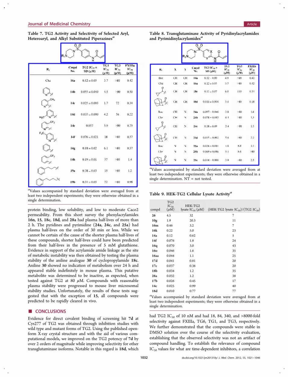
Table 8. Transglutaminase Activity of Pyridinylacrylamidesand Pyrimidinylacrylamidesa
aValues accompanied by standard deviation were averaged from atleast two independent experiments; they were otherwise obtained in asingle determination. NT = not tested.
Table 9. HEK-TG2 Cellular Lysate Activitya
compd
TG2IC50(μM)
HEK-TG2lysate IC50 (μM) (HEK-TG2 lysate IC50)/(TG2 IC50)
26 4.5 32 710g 1.9 20.3 1116m 0.46 3.2 716k 0.22 5.0 2314a 0.12 0.62 514f 0.076 1.8 2416q 0.070 3.0 4316o 0.046 1.6 3516aa 0.044 1.1 2517d 0.041 0.81 2017c 0.037 0.36 2018b 0.034 1.2 3528a 0.032 1.2 3817b 0.026 0.45 1714c 0.025 0.99 4018d 0.010 0.77 77
aValues accompanied by standard deviation were averaged from atleast two independent experiments; they were otherwise obtained in asingle determination.
Journal of Medicinal Chemistry Article
dx.doi.org/10.1021/jm201310y | J. Med. Chem. 2012, 55, 1021−10461032

was established between these values and the irreversible inhibi-tion constants for several transglutaminase isoforms, with thefinding that the IC50 values are an accurate means to rank-ordercompound potency and selectivity.In a cellular lysate assay a reasonable translation of bio-
chemical activity to lysate potency was achieved and IC50 < 1 μMwas observed for several compounds; however, this assayrequired a nonphysiologically high Ca2+ concentration. Addi-tionally, DMPK profiling showed that the compounds haverelatively poor solubility, poor to moderate permeability, andpoor plasma stability, this last deficit being attributed to thelabile acrylamide amide linkage. The phenylacrylamides dis-played low reactivity in the presence of GSH, belying theiracrylamide functionality.Our inability to potently inhibit TG2 transamidation in
whole cells using irreversible inhibitors has caused us to re-evaluate our effort. Efficacy end points in HD mouse modelsrequire 6 months or more of dosing. To manage risk in runningsuch trials and to facilitate interpretation of results, it is imper-ative that compounds possess cellular activity and pharmaco-kinetic exposure levels in brain to cover, preferentially, somemultiple of the cellular activity. This is to facilitate the modula-tion of pharmacodynamic markers needed to demonstrate invivo target engagement. Given the weak cellular potency andpoor plasma stability, it is unlikely that any of the inhibitorsdescribed would have adequate brain exposure to test ourhypothesis. For this to occur, a better understanding of intra-cellular TG2 biology is likely needed, and perhaps only thencan a physiologically meaningful transamidation-dependentcellular assay readout be validated. Such an assay is needed tofacilitate compound selection for DMPK and in vivo studies.Since our current understanding of the biology is limiting inHD, we are continuing in our efforts to deepen this under-standing while working to address the plasma stability issue. Wewill report our findings in due course. While these studies areongoing, the current inhibitors may represent attractive agentsfor studies of TG2’s role in cancer or celiac disease.
■ EXPERIMENTAL SECTIONCloning, Expression, and Purification of Transglutaminases.
The full length (Met-1 to Ala-687) human TG2 PCR product wassubcloned into the pET28a vector (Novagen) using the Nde I andHind III restriction sites for expression in E. coli, incorporating an
N-terminal His-tag, generating the plasmid pET28a:hTG2. Muta-genesis of TG2 to generate the C285A point mutation was performedusing a Quikchange kit (Stratagene). Complementary primers that in-clude sequences flanking the site of the mutation (C285A_sense,CGCCGCCGTGGCCGCCACAGTGCTGAGG; C285A_antisense,CCTCAGCACTGTGGCGGCCACGGCGGCG) were used in amutagenesis reaction according to the manufacturer’s instructions,using pET28a:hTG2 as template DNA, yielding the plasmidpET28a:TG2_C285A.
The constructs were transformed into E. coli Rosetta 2 (DE3) cells,and cells were grown overnight at 37 °C in 6 L of 2xYT medium con-taining 50 μg/mL kanamycin. The cells were grown to OD600 = 0.6before addition of 100 μM isopropyl-β-D-thiogalactopyranoside toinduce expression at 20 °C overnight. The cells were harvested bycentrifugation, flash frozen in liquid nitrogen, thawed at 37 °C, andresuspended in 90 mL of lysis buffer (50 mM potassium phosphate,pH 8.0, 300 mM NaCl, and 1 EDTA-free protease inhibitor tablet(Roche)) plus 4 μL of 250 U/μL benzonase (Merck) at 4 °C. Lysiswas achieved using 5 × 30 s bursts of sonication on ice. The lysate wasthen centrifuged at 28000g and the supernatant recovered. The samplewas loaded onto a column of 5 mL of TALON resin at 2 mL/min andeluted with 50 mM potassium phosphate, pH 8.0, 150 mM imidazole.Fractions were collected and loaded onto a 1 mL anion exchangecolumn at 4 mL/min in 20 mM Tris, pH 7.2, 1 mM DTT, and 1 mMEDTA and eluted using a 1 M NaCl gradient. Selected fractions wereloaded onto a Superdex 200, 26/60 gel filtration column pre-equilibratedin buffer (20 mM Tris, pH 7.2, 150 mM NaCl, 1 mM DTT, and 1 mMEDTA) at 2.4 mL/min. The pure fractions were verified by SDS−PAGEand pooled. Complex formation with 7d was done as previouslydescribed.14 Briefly, 50 molar excess of CHDI-00201241 wasincubated with 5 mg of pure TG2 in incubation buffer (20 mMTris-HCl, pH 7.2, 1 mM DTT, 1 mM EDTA, 150 mM NaCl, 10 mMCaCl2) at room temperature for 30 min and then at 4 °C overnight.Fractions were loaded onto a second 1 mL anion exchange column at4 mL/min in 20 mM Tris, pH 7.2, 1 mM DTT, and 1 mM EDTA andeluted using a 1 M NaCl gradient. Selected fractions were pooled.
The full length mouse TG2 (Met-1 to Ala-686) PCR product wassubcloned into the vector pETIJ-HV (Evotec in-house vector) usingthe EcoRI and Not I restriction sites for expression in E. coli incor-porating an N-terminal His-tag. The construct was transformed intoE. coli Rosetta 2 (DE3) cells, and cells were grown overnight at 37 °Cin 6 L of 2xYT medium containing 50 μg/mL kanamycin. The cellswere grown to OD600 = 0.6 before addition of 500 μM isopropyl-β-D-thiogalactopyranoside to induce expression at 25 °C overnight. Thecells were harvested by centrifugation, flash frozen in liquid nitrogen,thawed at 37 °C, and resuspended in 80 mL of lysis buffer (50 mMpotassium phosphate, pH 8.0, 300 mM NaCl, and 1 non-EDTA
Table 10. DMPK Profile of Selected Inhibitorsa
compdTG2 IC50(μM)
aq sol.(mg/mL)
Hu PPBb (%unbound)
PSA(Å2)
cLogPpH 7
Caco2 Papp(A−B)(nm s−1)
Caco2 Papp(B−A)(nm s−1)
mouse plasmaT1/2 (min)
mLM Clint(mL min−1 kg−1)
mLM Clint(L h−1 kg−1)
16n 0.072 0.01 2 96 3.9 NT NT 126 2465 14815 1.3 1.04 4 79 1.0 0 9 162 <50.8 <3.018c 0.11 0.05 11 87 1.2 65 115 209 119.9 ± 68.8 7.2 ± 4.118d 0.010 0.01 2 87 3.2 102 193 248 3203 19228a 0.032 0.05 7 87 1.62 136 97 305 228.7 13.724a 0.097 0.04 NT 108 2.1 NT NT 31.5 NT NT24c 0.18 0.25 96 99 0.26 11 6 33.5 NT NT25a 0.026 0.07 NT 108 2.1 NT NT 14.3 NT NT30 >80 1.8 NT 84 0.71 NT NT >240 NT NT
aValues accompanied by standard deviation were averaged from at least two independent experiments; they were otherwise obtained in a singledetermination. bHuman plasma protein binding. NT = not tested.
Journal of Medicinal Chemistry Article
dx.doi.org/10.1021/jm201310y | J. Med. Chem. 2012, 55, 1021−10461033

protease inhibitor tablet (Roche)) plus 4 μL of 250 U/μL benzonase(Merck) at 4 °C. Lysis was achieved by five steps of 30 s sonicationon ice. The lysate was centrifuged at 16 500 rpm for 60 min using aBeckman Avanti 30 centrifuge and an F0850 rotor and the supernatantrecovered. The sample was loaded onto a 5 mL TALON column at2 mL/min and eluted with 50 mM potassium phosphate, pH 8.0,150 mM imidazole. Fractions were collected and loaded onto a 1 mLanion exchange column at 4 mL/min in 20 mM Tris, pH 7.2, 1 mMDTT, and 1 mM EDTA and eluted using a 1 M NaCl gradient.Selected fractions were loaded onto a Superdex 200, 26/60 gel filtra-tion column pre-equilibrated in buffer (50 mM potassium phosphate,pH 8.0, 300 mM NaCl) at 2.0 mL/min. The pure fractions wereanalyzed using SDS−PAGE and pooled.Additionally, human TG1, human TG3, human TG6, and human
factor XIIIa were purchased from Zedira (Darmstadt, Germany).Transamidation Inhibition Assays for Recombinant TG and
TG2 HEK Cell Lysates. The fluorescent transamidation assay wasperformed as described.27 In summary, 8 μM N,N-dimethylated casein(NMC) and 16 μM dansyl-labeled amine nucleophile (KxD) wereused as substrates in 25 mM Hepes, pH 7.4, 250 mM NaCl, 2 mMMgCl2, 10 mM CaCl2, 0.2 mM DTT, and 0.05% Pluronic F-127 at37 °C. A kinetic measurement was recorded (Safire or Ultra, Tecan;excitation, 350 nm; emission, 535 nm), and the reaction velocityderived from a linear fit was used as a measure for enzyme activity. Alldata points were normalized between 0% and 100% inhibition usingthe appropriate positive (full inhibition) and negative (no inhibition)controls. IC50 values were determined using an automated in-housedata evaluation software package by means of a hyperbolic bindingmodel with a variable Hill slope. Assay conditions were similar forrecombinant TG isoforms TG2 (human and mouse), TG1, and TG6apart from CaCl2 concentrations, which were adjusted at half-maximaltransamidation activity (0.5 mM for hTG2 and TG6; 0.2 mM formTG2; 0.05 mM for TG1), and enzyme concentrations (20 nM hTG2and TG6, 5 nM mTG2, 10 nM TG1). Factor XIII was activated using0.1 μg/μL thrombin (Sigma) in 35 mM Tris, pH 8.0, for 20 min at30 °C, and the transamidation reaction was performed with 20 nMfactor XIIIa in 50 mM Tris, pH 8.0, 1.25 mM CaCl2, 0.05% PluronicF-127, 0.2 mM DTT. TG3 was activated with 0.02 μg/μL thrombinunder the same conditions as factor XIII, and assay conditions were10 nM TG3 in 50 mM Hepes, pH 8.0, 20 mM CaCl2, 0.2 mM DTT,0.05% Pluronic F-127.For the lysate assay, HEK cells engineered to express human TG2
were cultured as previously described.27 Cellular lysates were preparedby washing cultured cells twice with ice-cold PBS and harvesting themwith a rubber policeman. The cells were then pelleted by centrifuga-tion and lysed in 10 mM Tris-HCl, pH 7.5, 10 mM CaCl2, 1 mM EDTA,1 mM PMSF with protease inhibitors (Roche protease inhibitor cocktail)followed by sonication using a Branson Sonifier 450 with 50 pulses (dutycycle 20, output control 6). The protein content was determined usingthe Bradford method, and 1.5 μg/μL total protein was used in thefluorescent transamidation assay as described above.MALDI ToF MS Analysis. Mass spectometry analyses were
performed using a Voyager-DE PRO BioSpectrometry workstation(matrix assisted laser desorption ionization time-of-flight massspectrometer, PerSeptive Biosystems). Intact apoprotein and proteincomplexes with assumed covalent binders were measured following asample workup procedure. An amount of 2 μL of the protein solution(1 μg/μL protein in 20 mM Tris buffer, pH 7.2, 1 mM DTT, 1 mMEDTA, 150 mM NaCl) was placed on a nitrocellulose membrane filter(Millipore, 9004-70-O) and dialyzed against 100 mL of deionizedwater. After 30 min 1 μL of the sample was taken and mixed with 1 μLof sinapinic acid solution (hydroxycinnamic acid, supernatant of a10 mg/mL solution in 70% acetonitrile, 30% proteomics grade water,and 0.1% TFA) used as a matrix. Resulting samples were placed on aMALDI target plate and allowed to cocrystallize through evaporationat room temperature. Measurements were performed in linear detec-tion mode in the mass range of 10 000 to 100 000 Da. Data analysiswas performed using Data Explorer, version 4.0.0, data analysis soft-ware (PerSeptive Biosystems). For analysis of the potentially modifiedamino acid residues a tryptic digestion of the apoprotein with the
protein inhibitor complexes was performed. An amount of 100 μL ofeach sample (1 μg/μL protein in 20 mM Tris buffer, pH 7.2, 1 mMDTT, 1 mM EDTA, 150 mM NaCl) was acidified with 1 μL of100 mM HCl, and 5 μL of a 1 mg/mL mass spectrometry grade trypsinsolution (Sigma) was added. Following an overnight incubation at37 °C the reaction was terminated by addition of 100 μL of acetonitrile.Samples were subsequently evaporated under reduced pressure andredissolved in 10 μL of water. To remove remaining buffer salts inter-fering with the sample ionization, all samples were desalted usingZipTip μ-C18 desalting tips (Millipore). Final MALDI samples wereprepared by mixing 1 μL of the desalted digest with 1 μL of α-cyano-4-hydroxycinnamic acid (supernatant of a 10 mg/mL solution in 70%acetonitrile, 30% proteomics grade water, and 0.1% TFA). Spectrawere recorded in linear detection mode in the mass range of 500−5000 Da. For data analysis expected peptide fragments were comparedwith experimentally determined m/z values.
Molecular Docking Procedures. The X-ray structure data ofTG2 with peptide ligand 1 covalently bound (PDB code 2Q3Z) wereused for docking. The structure was prepared by removing all watermolecules and the peptide ligand. Hydrogen atoms were added whereneeded, and the protonation state of charged residues was adjusted toreflect their state at pH7 using the Protonate 3D tool implemented inthe MOE molecular modeling package (Chemical Computing GroupInc.). Amino acid residues within 13 Å of the sulfur atom of Cys277were defined as the binding site. The docking was accomplished usingthe covalent docking option of the GOLD docking software (version4.0 or 4.1).43 A covalent link for use with individual ligands was used inthis work wherein both the protein and the ligand incorporate thelinking sulfur atom of Cys277. This required that the structures of theligands be prepared as their corresponding thiol addition productswith one open valence at the sulfur. Other settings were set at thedefault values. The bound structure of peptide ligand 1 to TG2 wasreproduced with satisfactory accuracy (rmsd of Cα of 0.68 Å).
Computational Method for FMO Calculation. All themolecular modeling work necessary for preparation of the inputstructures for the FMO calculations was carried out in the MOEmolecular modeling package. TG2−peptide ligand 1 complex structurewas prepared based on the published X-ray data (PDB code 2Q3Z). Atruncated partial structure of the TG2−peptidic ligand 1 complex wasprepared by selecting the amino acid residues within 4 Å from 1.The new C-terminals created by truncation were modeled asN-methylamides, and acetyl groups were added to the N-terminals.Hydrogen atoms were added, and the protonation state of the acidicand basic amino acid residues were adjusted at pH 7 using the Protonate3D tool within MOE. The positions of the hydrogen atoms in the com-plex were optimized using the MMFF94x force field in the presence ofthe Born continuous water model while fixing the coordinates of theheavy atoms. The protein was fragmented using a one-fragment-per-residue fragmentation scheme, and the peptide ligand was fragmentedsimilarly into five sections as shown in Figure 3. The FMO calculationsin this work were performed using GAMESS software (April 2008version)44 at the MP2/6-31G** theory level. The calculations were runon PC clusters consisting of 24−40 CPU cores. The input files wereprepared using Facio software45,46 after preprocessing of the structure inMOE.
Homology Model Construction. The sequences of human TG1(P22735), TG3 (Q08188), and FXIII (P00488) were retrieved fromthe Swiss-Protein Database. All of the molecular modeling studieswere carried out using the MOE software. The structure of TG2deposited in the protein data bank (PDB code 2Q3Z) was used as thestructure template for homology model construction. Ten modelswere constructed for each transglutaminase isoform, and the modelswith the lowest Coulomb and generalized Born47 interaction energieswere further optimized using force field energy minimization followedby short molecular dynamics (MD) simulations (simulated annealing).The coordinates of the α-carbons of the amino acids were fixed duringthe MD simulations.
General Synthetic Procedures. Commercially available reagentsand solvents (HPLC grade) were used without further purification. 1HNMR spectra were recorded on a Bruker DRX 500 MHz spectrometer
Journal of Medicinal Chemistry Article
dx.doi.org/10.1021/jm201310y | J. Med. Chem. 2012, 55, 1021−10461034

or Bruker DPX 250 MHz spectrometer in deuterated solvents, and 13CNMR spectra were recorded on a Bruker DPX 250 MHz spectrometerequipped with a B-VT 3300 variable temperature controller unit (hightemperature only) and a four nucleus (QNP) switchable probe forobservation of 1H, 13C, 19F, 31P with 2H lock in deuterated solvents.Chemical shifts (δ) are in parts per million. Thin-layer chromatog-raphy (TLC) analysis was performed with Kieselgel 60 F254 (Merck)plates and visualized using UV light.Analytical HPLC−MS was performed on Shimadzu LCMS-2010EV
systems using reverse phase Atlantis dC18 columns (3 μm, 2.1 mm ×50 mm), gradient 5−100% B (A = water/0.1% formic acid, B =acetonitrile/0.1% formic acid) over 3 min, injection volume of 3 μL,flow of 1.0 mL/min. UV spectra were recorded at 215 nm using aWaters 2788 dual wavelength UV detector. Mass spectra were ob-tained over the range m/z 150−850 at a sampling rate of 2 scans persecond using Waters LCT or analytical HPLC−MS on ShimadzuLCMS-2010EV systems using reverse phase Water Atlantis dC18columns (3 μm, 2.1 mm × 100 mm), gradient 5−100% B (A = water/0.1% formic acid, B = acetonitrile/0.1% formic acid) over 7 min,injection volume of 3 μL, flow of 0.6 mL/min. UV spectra wererecorded at 215 nm using a Waters 2996 photodiode array. Data wereintegrated and reported using Shimadzu PsiPort software. Accuratemass measurement was carried out using a Waters Micromass LCTPremier orthogonal acceleration time-of-flight mass spectrometer4 GHz TDC with LockSpray enable. All compounds display purityof >95% as determined by this method, unless stated otherwise.N-(4-Bromophenyl)acrylamide (7d). 4-Bromoaniline (0.3 g,
1.74 mmol) was dissolved in THF (5 mL). To this was addeddiisopropylethylamine (0.5 mL, 5.2 mmol) in one portion followed bythe dropwise addition of acryloyl chloride (0.19 mL, 1.91 mmol), andthe mixture was stirred at room temperature under a nitrogen atmo-sphere for 3 h. The THF was removed under vacuum, and theresulting crude material was diluted with DCM (20 mL) and washedsequentially with NaOH (1 M solution, 10 mL), HCl (1 M solution,10 mL), and brine (10 mL) before being dried (MgSO4), filtered, andconcentrated to give the title compound (0.34 g, 90% yield) as a whitepowder. MS (ES+) m/z (M + 1) 228; HRMS (ES+) m/z 225.9859(225.9868 calcd for C9H8BrNO, M + H); 1H NMR (500 MHz,DMSO) 10.28 (s, 1H), 7.59−7.71 (m, 2H), 7.46−7.56 (m, 2H), 6.35−6.48 (m, 1H), 6.22−6.32 (m, 1H), 5.69−5.84 (m, 1H); 13C NMR(126 MHz, DMSO-d6) δ 163.2, 138.4, 131.6, 127.3, 121.2, 115.1.Ethenesulfonic Acid (4-Bromophenyl)amide (10a). MS (ES+)
m/z (M + 1) 263; δH (500 MHz, DMSO-d6) 7.41−7.55 (2H, m),7.03−7.14 (2H, m), 6.77 (dd, J = 16.39, 9.93 Hz, 1H), 5.97−6.17 (2H, m).N-(4-Bromophenyl)propionamide (10b).MS (ES+) m/z (M + 1)
230; HRMS (ES+) m/z 228.0034 (228.0024 calcd for C9H10BrNO,M + H); 1H NMR (500 MHz, DMSO) 9.98 (s, 1H) 7.58 (d, J = 9.92Hz, 2H), 7.48 (d, J = 9.92 Hz, 2H), 2.28 (q, J = 6.2 Hz, 2H), 1.04(t, J = 6.2 Hz, 3H); 13C NMR (126 MHz, DMSO-d6) δ 172.2, 138.7,131.5, 120.9, 114.4, 29.5, 9.6.N-(4-Bromobenzyl)acrylamide (10c). MS (ES+) m/z (M + 1)
241, 243; HRMS (ES+) m/z 240.0027 (240.0024 calcd forC10H10BrNO, M + H); 1H NMR (500 MHz, DMSO) 7.79 (br s,1H), 6.61−6.73 (m, 2H), 6.38 (d, J = 8.39 Hz, 2H), 5.37−5.49(m, 1H), 5.22−5.33 (m, 1H), 4.79 (dd, J = 10.07, 2.14 Hz, 1H), 3.47(d, J = 6.10 Hz, 2H); 13C NMR (126 MHz, DMSO-d6) δ 164.7, 138.8,131.5, 131.2, 129.6, 125.6, 119.9, 41.5.N-(3-Bromophenyl)acrylamide (10d). MS (ES+) m/z (M + 1)
227, 229; HRMS (ES+) m/z 225.9877 (225.9868 calcd for C9H8BrNO,M + H); 1H NMR (500 MHz, DMSO) 10.31 (s, 1H), 8.05 (s, 1H),7.62−7.52 (m, 1H), 7.30−7.22 (m, 2H), 6.48−6.24 (m, 2H), 5.83−5.78(m, 1H); 13C NMR (126 MHz, DMSO-d6) δ 163.4, 140.6, 131.5, 130.8,127.6, 126.1, 121.6, 118.1.N-(2-Bromophenyl)acrylamide (10e). MS (ES+) m/z (M + 1)
227, 229; δH (500 MHz, DMSO-d6) 8.84 (1H, br s), 6.75−6.87 (2H, m),6.55 (td, J = 7.67, 1.45 Hz, 1H), 6.32 (td, J = 7.71, 1.68 Hz, 1H), 5.74(dd, J = 17.01, 10.30 Hz, 1H), 5.43 (dd, J = 17.01, 1.91 Hz, 1H),4.86−5.01 (1H, m).N-(4-Fluorophenyl)acrylamide (10f). MS (ES+) m/z (M + 1)
166; HRMS (ES+) m/z 166.0675 (166.0668 calcd for C9H8FNO,
M + H); 1H NMR (500 MHz, DMSO) 10.21 (s, 1H), 7.68−7.61(m, 2H), 7.19−7.11 (m, 2H), 6.42−6.23 (m, 2H), 5.74−5.71 (m, 1H);13C NMR (126 MHz, DMSO-d6) δ 163.1, 158.1 (d, J = 249 Hz),135.4, 131.7, 127.0, 121.1, 115.4 (d, J = 22 Hz).
N-[4-(Pyrrolidine-1-sulfonyl)phenyl]acrylamide (10g). MS(ES+) m/z (M + 1) 281; HRMS (ES+) m/z 281.0950 (281.0960calcd for C13H16N2O3S, M + H); 1H NMR (500 MHz, DMSO) 10.56(s, 1H), 7.84−7.96 (m, 2H), 7.77 (d, J = 8.67 Hz, 2H), 6.41−6.52(m, 1H), 6.28−6.38 (m, 1H), 5.84 (d, J = 9.93 Hz, 1H), 3.12 (t, J =6.62Hz, 4H), 1.59−1.71 (m, 4H); 13C NMR (126 MHz, DMSO-d6) δ163.7, 143.0, 131.4, 130.2, 128.6, 128.1, 119.2, 47.8, 24.7.
N-[4-Methanesulfonylphenyl]acrylamide (10h). MS (ES+)m/z (M + 1) 226; HRMS (ES+) m/z 226.0544 (226.0538 calcd forC10H11NO3S, M + H); 1H NMR (500 MHz, DMSO) 9.70 (br s, 1H),6.89−7.08 (m, 4H), 5.49−5.65 (m, 1H), 5.35−5.46 (m, 1H), 4.94 (dd,J = 10.09, 1.89 Hz, 1H), 2.23−2.29 (m, 3 H); 13C NMR (126 MHz,DMSO-d6) δ 163.9, 143.7, 135.1, 131.5, 128.4, 119.3, 44.0.
N-[4-Nitrophenyl]acrylamide (10i).MS (ES+) m/z (M + 1) 193;HRMS (ES+) m/z 193.0622 (193.0613 calcd for C9H8N2O3, M + H);1H NMR (500 MHz, DMSO) 10.76 (s, 1H), 8.19−8.30 (m, 2H),7.86−7.97 (m, 2H), 6.30−6.55 (m, 2H), 5.87 (dd, J = 10.09, 1.73 Hz,1 H); 13C NMR (126 MHz, DMSO-d6) δ 163.9, 145.2, 142.4, 131.2,128.6, 125.0, 119.1.
N-[4-Trifluoromethylphenyl]acrylamide (10j). MS (ES+) m/z(M + 1) 216; HRMS (ES+) m/z 216.0627 (216.0636 calcd forC10H8F3NO, M + H); 1H NMR (500 MHz, DMSO) 10.50 (s, 1H),7.88 (d, J = 8.51 Hz, 2H), 7.70 (d, J = 8.51 Hz, 2H), 6.41−6.52(m, 1H), 6.23−6.38 (m, 1H), 5.83 (dd, J = 10.09, 1.89 Hz, 1H); 13C NMR(126 MHz, DMSO-d6) δ 163.66, 142.59, 131.46, 127.94, 126.15,125.46, 123.35, 119.26.
N-Phenylacrylamide (10k). MS (ES+) m/z (M + 1) 148; HRMS(ES+) m/z 148.0772 (148.0762 calcd for C9H9NO, M + H); 1H NMR(500 MHz, DMSO) 10.13 (s, 1H), 7.68−7.63 (m, 2H), 7.32−7.29(m, 2H), 7.08−7.04 (m, 1H), 6.42−6.23 (m, 2H), 5.81−5.78 (m, 1H);13C NMR (126 MHz, DMSO-d6) δ 163.2, 139.0, 131.9, 128.8, 126.9,123.5, 119.3.
N-[4-Toluyl]acrylamide (10l). MS (ES+) m/z (M + 1) 162;HRMS (ES+) m/z 162.0923 (162.0919 calcd for C10H11NO, M + H);1H NMR (500 MHz, DMSO) 10.05 (s, 1H), 7.55 (d, J = 8.35 Hz,2H), 7.06−7.17 (m, 2H), 6.42 (dd, J = 17.02, 10.09 Hz, 1H), 6.24 (dd,J = 17.02, 2.05 Hz, 1H), 5.67−5.79 (m, 1H), 2.21−2.31 (m, 3H); 13CNMR (126 MHz, DMSO-d6) δ 162.9, 136.5, 132.4, 132.0, 129.2,126.6, 119.3, 20.5.
N-[4-Phenoxyphenyl]acrylamide (10m). MS (ES+) m/z(M + 1) 240; HRMS (ES+) m/z 240.1032 (240.1025 calcd forC15H13NO2, M + H); 1H NMR (500 MHz, DMSO) 10.56 (s, 1H),7.68−7.64 (m, 2H), 7.33−7.31 (m, 2H), 7.11−7.08 (m, 1H), 7.01−6.95 (m, 4H), 6.52−6.28 (m, 2H), 5.72 (m, 1H).
N-[4-Methoxyphenyl]acrylamide (10n).MS (ES+) m/z (M + 1)178; HRMS (ES+) m/z 178.0876 (178.0868 calcd for C10H11NO2,M + H); 1H NMR (500 MHz, DMSO) 10.02 (s, 1H), 8.07 (s, 1H),7.34−7.57 (m, 4H), 6.40 (dd, J = 17.02, 10.09 Hz, 1H), 6.22 (dd, J =17.02, 1.89 Hz, 1H), 5.64−5.79 (m, 1H), 3.33 (br s, 4H), 1.77−1.90(m, 4H); 13C NMR (126 MHz, DMSO-d6) δ 162.7, 155.4, 132.2,132.0, 126.3, 120.8, 113.9, 55.2.
4-[4-(2-Fluoroacryloylamino)benzenesulfonyl]piperazine-1-carboxylic Acid tert-Butyl Ester (28c). MS (ES+) m/z (M + 23)436; HRMS (ES+) m/z 314.0973 (314.0975 calcd for C18H24FN3O5S,M + H, loss of tert-butyl); 1H NMR (500 MHz, DMSO) 10.71(s, 1H), 8.01 (d, J = 8.80 Hz, 2H), 7.72 (d, J = 8.80 Hz, 2H), 5.68−5.86 (m, 1H), 5.51 (dd, J = 15.59, 3.85 Hz, 1H), 2.83 (t, J = 4.68 Hz,4H), 1.33 (s, 9H); 13C NMR (126 MHz, DMSO-d6) δ 157.4 (d, J =192.5 Hz), 154.5, 153.4, 142.3, 129.7, 128.7, 120.5, 100.9 (d, J = 14.7Hz), 79.3, 45.8, 27.9.
4-[4-(2-Methylacryloylamino)benzenesulfonyl]piperazine-1-carboxylic Acid tert-Butyl Ester (28d). MS (ES+) m/z (M + 23)432; δH (500 MHz, DMSO-d6) 10.24 (1H, s), 7.97 (d, J = 8.80 Hz,2H), 7.69 (d, J = 8.62 Hz, 2H), 5.86 (1H, s), 5.61 (1H, s), 3.39 (4H,br s), 2.82 (t, J = 4.68 Hz, 4H), 1.96 (3H, s), 1.34 (9H, s).
Journal of Medicinal Chemistry Article
dx.doi.org/10.1021/jm201310y | J. Med. Chem. 2012, 55, 1021−10461035

4-(4-But-2-enoylaminobenzenesulfonyl)piperazine-1-car-boxylic Acid tert-Butyl Ester (28f). MS (ES+) m/z (M + 23) 432;HRMS (ES+) m/z 310.1213 (310.1225 calcd for C19H27N3O5S, M + H,loss of tert-butyl); 1H NMR (500 MHz, CDCl3) 7.61−7.81 (m, 4H), 7.42(s, 1H), 6.92−7.16 (m, 1H), 5.98 (dd, J = 15.13, 1.56 Hz, 1H), 3.51(t, J = 4.77 Hz, 4H), 2.96 (br s, 4H), 1.96 (dd, J = 6.88, 1.38 Hz, 3H), 1.42(s, 9H); 13C NMR (126 MHz, DMSO-d6) δ 167.4, 153.4, 143.6, 140.0,128.6, 128.5, 121.0, 119.7, 79.3, 45.8, 27.9, 18.6.4-[4-(2-Methylbut-2-enoylamino)benzenesulfonyl]-
piperazine-1-carboxylic Acid tert-Butyl Ester (28g). MS (ES+)m/z (M + 23) 446; HRMS (ES+) m/z 324.1384 (324.1382 calcd forC20H29N3O5S, M + H, loss of tert-butyl); 1H NMR (500 MHz,DMSO) 10.10 (s, 1H), 7.93 (d, J = 8.83 Hz, 2H), 7.64 (d, J = 8.83 Hz, 2H),6.35−6.60 (m, 1H), 3.41−3.49 (m, 4H), 2.79 (t, J = 4.87 Hz, 4H),1.70−1.86 (m, 6H), 1.28−1.43 (m, 9H); 13C NMR (126 MHz,DMSO-d6) δ 168.3, 153.4, 143.9, 132.4, 131.6, 128.6, 128.1, 119.5,79.3, 45.8, 27.9, 13.9, 12.5.4-[4-(4,4,4-Trifluorobut-2-enoylamino)benzenesulfonyl]-
piperazine-1-carboxylic Acid tert-Butyl Ester (28h). MS (ES+)m/z (M + 23) 486; HRMS (ES+) m/z 364.0939 (364.0943 calcd forC19H24F3N3O5S, M + H, loss of tert-butyl); 1H NMR (500 MHz,DMSO) 11.00 (s, 1H), 7.91 (d, J = 8.62 Hz, 2H), 7.74 (d, J = 8.62 Hz,2H), 6.86−7.09 (m, 2H), 3.40−3.45 (m, 4H), 2.84 (br s, 4H), 1.34(s, 9H); 13C NMR (126 MHz, DMSO-d6) δ 161.0, 153.4, 142.6, 132.7(q, J = 6 Hz), 129.5, 129.0, 127.4 (q, J = 34 Hz), 123.0 (q, J = 270 Hz),119.5, 79.4, 45.8, 43.0 (br), 42.0 (br), 27.9.4-[4-(4 ,4 ,4-Tr i f luoro-3-methylbut-2-enoylamino)-
benzenesulfonyl]piperazine-1-carboxylic Acid tert-Butyl Ester(28i). MS (ES+) m/z (M + 23) 500; HRMS (ES+) m/z 378.1096(378.1099 calcd for C20H26F3N3O5S, M + H, loss of tert-butyl); 1HNMR (500 MHz, DMSO) 7.89 (d, J = 8.62 Hz, 2H), 7.72 (d, J = 8.80 Hz,2H), 6.72 (s, 1H), 3.40−3.50 (m, 4H), 2.83 (t, J = 4.77 Hz, 4H), 2.25(s, 3H), 1.34 (s, 9H); 13C NMR (126 MHz, DMSO-d6) δ 162.7,153.4, 142.8, 129.1, 129.0, 125.1 (q, J = 6 Hz), 119.2, 79.3, 45.7,27.9, 11.7.4-[4-(Ethoxycarbonylmethylamino)benzenesulfonyl]-
piperazine-1-carboxylic Acid tert-Butyl Ester (28m). MS (ES+)m/z (M + 23) 450; δH (500 MHz, DMSO-d6) 7.42 (d, J = 8.83 Hz,2H), 7.02 (t, J = 6.23 Hz, 1H), 6.69 (d, J = 8.83 Hz, 2H), 4.13 (q, J =7.09 Hz, 2H), 4.01 (d, J = 6.15 Hz, 2H), 3.39−3.42 (4H, m), 2.75(t, J = 4.89 Hz, 4H), 1.35 (9H, s).4-[4-(2-Cyanoacetylamino)benzenesulfonyl]piperazine-1-
carboxylic Acid tert-Butyl Ester (28o). MS (ES+) m/z (M + 23)431; HRMS (ES+) m/z 309.1033 (309.1021 calcd for C18H24N4O5S,M + H, loss of tert-butyl); 1H NMR (250 MHz, DMSO) 10.74(s, 1H), 7.60−7.86 (m, 4H), 3.97 (s, 2H), 3.38−3.46 (m, 4H), 2.73−2.87(m, 4H), 1.32 (s, 9H); 13C NMR (126 MHz, DMSO-d6) δ 162.0, 153.4,142.7, 129.1, 129.0, 119.1, 115.6, 79.3, 45.7, 27.9, 27.1.N-(4-Aminophenyl)acrylamide. N-[4-Nitrophenyl]acrylamide
(10i, 0.38 g, 1.98 mmol) was suspended in a 5:1 mixture of ethanoland water (10 mL). To this solution was added iron powder (0.22 g,4.00 mmol) followed by saturated ammonium chloride solution(1 mL), and the mixture was heated to 80 °C for 3 h. After this time,the reaction mixture was cooled to room temperature and filteredthrough a pad of Celite. The Celite was washed with ethanol (10 mL)and ethyl acetate (50 mL), and the solution was concentrated undervacuum. The resulting residue was partitioned between DCM (50 mL)and water (20 mL). The organic layer was separated, dried with MgSO4,filtered, and concentrated to afford the title compound (0.32 g, 99%yield) as a white solid. MS (ES+) m/z (M + H)+ 163. δH (500 MHz,DMSO-d6) 9.73 (1H, s), 7.30 (d, J = 8.67 Hz, 2H), 6.47−6.58 (2H, m),6.37 (dd, J = 17.02, 10.25 Hz, 1H), 6.16 (dd, J = 16.95, 1.81 Hz, 1H),5.65 (dd, J = 10.09, 1.73 Hz, 1H), 4.90 (2H, br s).Pyrrolidine-1-carboxylic Acid (4-Acryloylaminophenyl)-
amide (10o). Triphosgene (0.05 g, 0.17 mmol) was added in oneportion to a stirred solution of N-(4-aminophenyl)acrylamide (0.1 g,0.57 mmol) and diisopropylethylamine (0.09 mL, 0.57 mmol) in THF(5 mL), and the resulting mixture was stirred at room temperatureunder a nitrogen atmosphere for 5 min. After this time pyrrolidine(0.04 mL, 0.57 mmol) was added in one portion and the resultingmixture was stirred at room temperature under a nitrogen atmosphere
for 18 h. After this time the reaction mixture was concentrated, and theresidue was purified using flash column chromatography (elution, 1%methanol, 99% DCM) to give the title compound (0.06 g, 45% yield)as a white powder. MS (ES+) m/z (M + 1) 260; HRMS (ES+) m/z260.1390 (260.1399 calcd for C14H17N3O2, M + H); 1H NMR (500MHz, DMSO) 10.02 (s, 1H), 8.07 (s, 1H), 7.37−7.56 (m, 4H), 6.40(dd, J = 17.02, 10.09 Hz, 1H), 6.22 (dd, J = 17.02, 1.89 Hz, 1H),5.64−5.81 (m, 1H), 3.28−3.35 (m, 4H), 1.75−1.93 (m, 4H); 13CNMR (126 MHz, DMSO-d6) δ 162.7, 154.0, 136.4, 133.1, 132.0,126.3, 120.0, 119.5, 45.7, 25.0.
4-[4-(3-(Z)-Chloroacryloylamino)benzenesulfonyl]-piperazine-1-carboxylic Acid tert-Butyl Ester (28j). 3-Chloroa-crylic acid (0.12 g, 1.1 mmol) followed by pyridine (0.3 mL) wasadded sequentially portionwise to a stirred solution of 4-(4-amino-benzenesulfonyl)piperazine-1-carboxylic acid tert-butyl ester (0.1 g,0.29 mmol) in THF/DMA (3:2, 5 mL) at room temperature. To thismixture was added EDC (0.29 g, 1.5 mmol) in one portion, and themixture was stirred at room temperature under a nitrogen atmospherefor 1 h. After this time the mixture was diluted with ethyl acetate (50 mL)and washed with water (100 mL), saturated NaHCO3 (100 mL), andHCl (100 mL, 2M). The organic layer was separated, dried (MgSO4),filtered, and concentrated under vacuum and the resulting residue waspurified using flash column chromatography (elution, 40% ethyl acetate,60% heptane) to give the title compound (0.009 g, 5% yield) as a whitesolid. MS (ES+) m/z (M + 23) 452; δH (250 MHz, DMSO-d6) 10.66(1H, s), 7.88 (d, J = 8.68 Hz, 2H), 7.69 (d, J = 8.68 Hz, 2H), 7.00 (d, J =8.07 Hz, 1H), 6.57 (d, J = 7.77 Hz, 1H), 3.40−3.57 (4H, m), 2.77−2.89(5H, m), 1.33 (10H, s).
4-[4-(3-(E )-Chloroacryloylamino)benzenesulfonyl]-piperazine-1-carboxylic Acid tert-Butyl Ester (28k). MS (ES+)m/z (M + 23) 452, 453; δH (250 MHz, DMSO-d6) 10.65 (1H, br s),7.83−7.92 (2H, 2), 7.69 (d, J = 8.83 Hz, 2H), 7.47 (1H, s), 6.63 (d, J =13.10 Hz, 1H), 2.72−2.96 (6H, m), 1.32 (11H, s).
4-[4-(2-Oxopropionylamino)benzenesulfonyl]piperazine-1-carboxylic Acid tert-Butyl Ester (28p). MS (ES+) m/z (M + 23)434; δH (500 MHz, DMSO-d6) 10.88 (1H, s), 8.09 (d, J = 8.80 Hz,2H), 7.65−7.79 (3H, m), 3.44−3.50 (4H, m), 2.83 (t, J = 4.77 Hz,4H), 2.44 (3H, s), 1.34 (9H, s).
4-[4-(3-Cyanomethylureido)benzenesulfonyl]piperazine-1-carboxylic Acid tert-Butyl Ester (28l). Phosgene (20% solution intoluene, 0.34 mL, 1.7 mmol) was added dropwise to a stirred solu-tion of 4-(4-aminobenzenesulfonyl)piperazine-1-carboxylic acid tert-butyl ester (0.18 g, 5.3 mmol) and diisopropylethylamine (0.18 mL,6.4 mmol) in THF (5 mL) at room temperature. To this mixture wasadded aminoacetonitrile (0.035 g, 6.4 mmol) in one portion, and themixture was stirred at room temperature under a nitrogen atmosphereovernight. After this time the mixture was concentrated, diluted withDCM (20 mL), and washed with water (10 mL), saturated NaHCO3(10 mL), and HCl (10 mL, 2 M solution). The organic layer wasseparated, dried (MgSO4), filtered, and concentrated under vacuumand the resulting residue was purified using flash column chroma-tography (elution, 70% DCM, 30% ethyl acetate) to give the title com-pound (0.045 g, 20% yield) as a white solid. MS (ES+) m/z (M + 23)446; HRMS (ES+) m/z 424.1646 (424.1655 calcd for C18H25N5O5S,M + H); 1H NMR (250 MHz, DMSO) 9.52 (s, 1H), 7.52−7.69(m, 4H), 6.91 (t, J = 5.94 Hz, 1H), 4.13 (d, J = 5.79 Hz, 2H), 3.21−3.29(m, 4H), 2.69−2.84 (m, 4H), 1.32 (s, 9H); 13C NMR (126 MHz,DMSO-d6) δ 154.4, 153.4, 144.5, 128.9, 126.6, 118.3, 117.6, 79.2, 45.8,28.4, 27.9.
4-(4-Nitrobenzenesulfonyl)piperazine-1-carboxylic AcidBenzyl Ester. Diisopropylethylamine (0.82 mL, 4.95 mmol) wasadded in one portion to a stirred solution of piperazine-1-carboxylicacid benzyl ester (1.0 g, 4.5 mmol) in DCM (10 mL) at roomtemperature. To this mixture was added 4-nitrophenylsulfonyl chloride(1.1 g, 4.95 mmol) in one portion, and the mixture was stirred at roomtemperature under a nitrogen atmosphere for 1 h. After this time thesolvent was removed under vacuum and the resulting residue waspurified using flash column chromatography (elution, 70% heptane,30% ethyl acetate) to give the title compound (1.82 g, 100% yield) as awhite solid. tR = 1.43 min; MS (ES+) m/z (M + Na+) 428.
Journal of Medicinal Chemistry Article
dx.doi.org/10.1021/jm201310y | J. Med. Chem. 2012, 55, 1021−10461036

4-(4-Cyanobenzenesulfonyl)piperazine-1-carboxylic Acidtert-Butyl Ester (28q). MS (ES+) m/z (M + 1) 252; HRMS (ES+)m/z 252.0800 (252.0807 calcd for C16H21N3O4S, M + H, loss of tert-butyl); 1H NMR (500 MHz, DMSO) 8.14 (d, J = 8.25 Hz, 2H), 7.91(d, J = 8.25 Hz, 2H), 3.39 (br s, 4H), 2.92 (t, J = 4.86 Hz, 4H), 1.35(s, 9H); 13C NMR (126 MHz, DMSO-d6) δ 153.3, 139.2, 133.7, 128.3,117.7, 115.8, 79.4, 45.6, 27.9.4-(4-Aminobenzenesulfonyl)piperazine-1-carboxylic Acid
Benzyl Ester. 4-(4-Nitro-benzenesulfonyl)piperazine-1-carboxylicacid benzyl ester (1.82 g, 4.5 mmol) was suspended in a 5:1 mixtureof ethanol and water (30 mL). To this solution was added iron powder(0.65 g, 11.7 mmol) followed by saturated ammonium chloridesolution (1 mL), and the mixture was heated to 80 °C for 3 h. Afterthis time, the reaction mixture was cooled to room temperature andfiltered through a pad of Celite, the Celite was washed with ethanol(10 mL) and ethyl acetate (50 mL), and the solution was concentratedunder vacuum. The resulting residue was partitioned between DCM(50 mL) and water (20 mL). The organic layer was separated, driedwith MgSO4, filtered, and concentrated to afford the title com-pound (1.5 g, 89% yield) as a white solid. tR = 1.29 min; MS (ES+)m/z (M + H)+ 376.4-(4-Acryloylaminobenzenesulfonyl)piperazine-1-carboxylic
Acid Benzyl Ester (16a). 4-(4-Aminobenzenesulfonyl)piperazine-1-carboxylic acid benzyl ester (0.25 g, 0.67 mmol) was dissolved in THF(10 mL). To this was added diisopropylethylamine (0.33 mL, 1.9mmol) in one portion followed by the dropwise addition of acryloylchloride (0.06 mL, 0.74 mmol), and the mixture was stirred at roomtemperature under a nitrogen atmosphere for 3 h. The THF wasremoved under vacuum and the resulting crude material was purifiedusing column chromatography (elution, 20% heptane, 80% ethyl acetate)to give the title compound (57 mg, 20% yield) as a white powder. MS(ES+) m/z (M + 1) 430; HRMS (ES+) m/z 430.1431 (430.1437 calcdfor C21H23N3O5S, M + H); 1H NMR (500 MHz, DMSO) 10.60(s, 1H) 7.92 (d, J = 8.70 Hz, 2H), 7.70 (d, J = 8.85 Hz, 2H), 7.18−7.44(m, 5H), 6.41−6.52 (m, 1H), 6.27−6.38 (m, 1H), 5.84 (dd, J = 10.07,1.83 Hz, 1H), 5.02 (s, 2H), 3.48 (br s, 4H), 2.87 (br s, 4H); 13C NMR(126 MHz, DMSO-d6) δ 163.8, 154.2, 143.4, 136.6, 131.4, 128.9,128.6, 128.4, 128.2, 127.9, 127.6, 119.2, 66.5, 45.7, 42.8.4-(4-Acryloylaminobenzenesulfonyl)piperazine-1-carboxylic
Acid tert-Butyl Ester (14a). MS (ES+) m/z (M + 23) 418; HRMS(ES+) m/z 418.1400 (418.1413 calcd for C18H25N3O5SNa, M + Na);1H NMR (500 MHz, DMSO) 10.60 (br s, 1H), 7.92 (d, J = 8.67 Hz,2H), 7.70 (d, J = 8.67 Hz, 2H), 6.27−6.54 (m, 2H), 5.84 (dd, J =10.17, 1.50 Hz, 1H), 3.38−3.45 (m, 4H), 2.83 (t, J = 4.65 Hz, 4H),1.36 (s, 9H); 13C NMR (126 MHz, DMSO-d6) δ 163.7, 153.4, 143.3,131.3, 128.9, 128.6, 128.1, 119.1, 79.3, 45.7, 43.0, 41.9, 27.9.3-(4-Acryloylaminobenzenesulfonylamino)-(S)-pyrrolidine-
1-carboxylic Acid Benzyl Ester (16b). MS (ES+) m/z (M + 1) 430;HRMS (ES+) m/z 430.1441 (430.1437 calcd for C21H23N3O5S, M + H);1H NMR (500 MHz, DMSO) 10.54 (s, 1H), 7.92 (d, J = 8.65 Hz,1H), 7.85 (d, J = 8.65 Hz, 2H), 7.76 (d, J = 8.65 Hz, 2H), 7.39−7.26(m, 5H), 6.50−6.27 (m, 2H), 5.83 (d, J = 1.52 Hz, 2H), 5.02 (d, J =1.52 Hz, 2H), 3.71−3.62 (m, 1H), 3.34 (obscured, 2H), 3.28−3.21(m, 1H), 3.11−3.04 (m, 1H), 1.91−1.84 (m, 1H), 1.72−1.61 (m, 1H);13C NMR (126 MHz, DMSO-d6) δ 163.7, 153.8, 142.6, 137.0, 134.9,131.4, 119.2, 119.1, 65.8, 52.3, 51.5, 51.3, 50.8, 44.0, 43.5, 31.3, 30.5.3-(4-Acryloylaminobenzenesulfonylamino)-(S)-pyrrolidine-
1-carboxylic Acid Benzyl Ester (16c). MS m/z (M + 1) 430;HRMS (ES+) m/z 430.1449 (430.1437 calcd for C21H23N3O5S, M + H);1H NMR (500 MHz, DMSO) 9.72 (s, 1H), 7.08 (d, J = 8.54 Hz, 2H),6.92 (d, J = 8.39 Hz, 2H), 6.44−6.58 (m, 6H), 5.57−5.70 (m, 1H),5.42−5.54 (m, 1H), 5.00 (dd, J = 10.15, 1.60 Hz, 1H), 3.94−4.25(m, 2H), 3.02 (sxt, J = 5.92 Hz, 1H), 2.27−2.46 (m, 3H), 2.19 (dd, J =10.15, 4.65 Hz, 1H), 1.02−1.10 (m, 1H), 0.76−0.89 (m, 1H), 0.38−0.51 (m, 1H); 13C NMR (126 MHz, DMSO-d6) δ 163.9, 155.8, 143.3,137.0, 131.6, 130.1, 128.8, 128.5, 128.3, 128.1 (two peaks), 119.3, 65.6,53.0, 50.3, 46.4, 30.4.3-(4-Acryloylaminobenzenesulfonylamino)-(R)-pyrrolidine-
1-carboxylic Acid Benzyl Ester (16d). MS (ES+) m/z (M + 23)430; HRMS (ES+) m/z 430.1435 (430.1437 calcd for C21H23N3O5S,
M + H); 1H NMR (500 MHz, DMSO) 10.54 (s, 1H), 7.84−8.00(m, 3H), 7.78 (d, J = 8.70 Hz, 2H), 7.22−7.42 (m, 5H) 6.40−6.52(m, 1H), 6.23−6.37 (m, 1H), 5.83 (d, J = 10.07 Hz, 1H), 5.02 (d, J =11.44 Hz, 2H), 3.66 (d, J = 3.66 Hz, 1H), 3.19−3.27 (m, 1H), 3.00−3.15 (m, 1H), 1.83−1.97 (m, 1H), 1.62−1.79 (m, 1H).
3-(4-Acryloylaminobenzenesulfonylamino)-(R)-pyrrolidine-1-carboxylic Acid Benzyl Ester (16e). MS (ES+) m/z (M + 23)430; HRMS (ES+) m/z 430.1434 (430.1437 calcd for C21H23N3O5S,M + H); 1H NMR (500 MHz, DMSO) 9.73 (br s, 1H), 7.09 (d, J =8.62 Hz, 2H), 6.93 (d, J = 8.44 Hz, 2H), 6.36−6.64 (m, 7H), 5.43−5.70 (m, 2H), 5.01 (dd, J = 10.18, 1.56 Hz, 1H), 3.98−4.27 (m, 2H),3.03 (sxt, J = 5.94 Hz, 1H), 2.11−2.47 (m, 4H), 1.09 (dq, J = 13.18,6.79 Hz, 1H), 0.83 (dq, J = 12.56, 6.20 Hz, 1H); 13C NMR (126 MHz,DMSO-d6) δ 163.7, 155.6, 143.1, 136.8, 131.4, 129.9, 128.6, 128.4,128.1, 127.9, 119.1, 65.4, 52.9, 50.1, 46.2, 30.2.
5-(4-Acryloylaminobenzenesulfonyl)hexahydropyrrolo[3,4-c]pyrrole-2-carboxylic Acid Benzyl Ester (16f). MS (ES+) m/z(M + 1) 456; HRMS (ES+) m/z 456.1585 (456.1593 calcd forC23H25N3O5S, M + H); 1H NMR (250 MHz, DMSO) 9.74 (s, 1 H),7.02−7.15 (m, 2 H), 6.87−6.98 (m, 2 H), 6.37−6.61 (m, 5 H), 5.40−5.71 (m, 2 H), 4.92−5.07 (m, 1 H), 4.06−4.25 (m, 2 H), 2.59 (d, J =7.01 Hz, 2 H), 2.43 (d, J = 7.31 Hz, 2 H), 2.16 (d, J = 8.68 Hz, 4 H),1.85−2.03 (m, 2 H); 13C NMR (126 MHz, DMSO-d6) δ 163.7, 153.7,143.2, 137.0, 131.4, 129.3, 128.8, 128.4, 128.1, 127.8, 127.4, 119.1,65.8, 52.0, 50.0, 49.4, 41.6, 40.7.
{2-[(4-Acryloylaminobenzenesulfonyl)methylamino]ethyl}-methylcarbamic Acid Benzyl Ester (16g). MS (ES+) m/z (M + 1)432; δH (500 MHz, CDCl3) 7.72−7.56 (5H, m), 7.31−7.18 (5H, m),6.44−6.15 (2H, m), 5.77−5.72 (1H, m), 5.06 (2H, s), 3.45−3.36 (2H, m),3.16−3.03 (2H, m), 2.94−2.91 (3H, m), 2.74 (3H, s), 2.55 (2H, s).
5-(4-Acryloylaminobenzenesulfonyl)-2,5-diazabicyclo-[2.2.1]heptane-2-carboxylic Acid tert-Butyl Ester (16h). MS(ES+) m/z (M + 23) 430; δH (500 MHz, CDCl3) 7.86−7.77 (4H, m),7.58 (1H, s), 6.51−6.25 (2H, m), 5.86 (d, J = 10.2 Hz, 1H), 4.47 (2H, s),3.51−3.13 (4H, m), 1.74−1.66 (1H, m), 1.46−1.24 (1H, m).
N-[4-(4-Pyridin-2-ylpiperazine-1-sulfonyl)phenyl]acrylamide(14b). MS (ES+) m/z (M + 1) 373; HRMS (ES+) m/z 373.1351(373.1334 calcd for C18H20N4O3S, M + H); 1H NMR (500 MHz,CDCl3) 8.15 (dd, J = 4.58, 1.47 Hz, 1H), 7.69−7.81 (m, 4H), 7.44−7.58 (m, 2H), 6.58−6.69 (m, 2H), 6.50 (d, J = 16.87 Hz, 1H), 6.27(dd, J = 16.87, 10.27 Hz, 1H), 5.86 (d, J = 10.27 Hz, 1H), 3.60−3.71(m, 4H), 3.08−3.17 (m, 4H); 13C NMR (126 MHz, DMSO-d6) δ163.7, 158.3, 147.5, 143.3, 137.7, 131.4, 128.9, 128.6, 128.2, 119.2,113.6, 107.5, 45.6, 44.0.
N-{4-[4-(3-Methylpyridin-2-yl)piperazine-1-sulfonyl]phenyl}-acrylamide (14c). MS (ES+) m/z (M + 1) 387; HRMS (ES+) m/z387.1480 (387.1491 calcd for C19H22N4O3S, M + H); 1H NMR (500MHz, CDCl3) 8.11−8.16 (m, 1H), 7.73−7.85 (m, 4H), 7.58 (s, 1H),7.40 (d, J = 6.97 Hz, 1H), 6.88 (dd, J = 7.15, 4.95 Hz, 1H), 6.51 (dd,J = 16.87, 0.92 Hz, 1H), 6.29 (dd, J = 16.87, 10.27 Hz, 1H), 5.80−5.93(m, 1H), 3.11−3.33 (m, 8H), 2.19 (s, 3H); 13C NMR (126 MHz,DMSO-d6) δ 163.8, 160.4, 145.0, 143.3, 139.4, 131.4, 129.0, 128.8,128.2, 124.4, 119.2, 118.4, 48.5, 46.0, 17.7.
N-{4-[4-(6-Methylpyridin-2-yl)piperazine-1-sulfonyl]phenyl}-acrylamide (14d). MS (ES+) m/z (M + 1) 387; HRMS (ES+) m/z387.1494 (387.1491 calcd for C19H22N4O3S, M + H); 1H NMR (500MHz, CDCl3) 7.69−7.82 (m, 4H), 7.60 (s, 1H), 7.37 (dd, J = 8.22,7.46 Hz, 1H), 6.44−6.55 (m, 2H), 6.39 (d, J = 8.38 Hz, 1H), 6.20−6.34 (m, 1H), 5.85 (dd, J = 10.13, 1.29 Hz, 1H), 3.58−3.69 (m, 4H),3.06−3.16 (m, 4H), 2.37 (s, 3H); 13C NMR (126 MHz, DMSO-d6) δ163.7, 157.8, 155.8, 143.3, 138.0, 131.4, 128.9, 128.6, 128.2, 119.2,112.7, 104.2, 45.6, 43.9, 24.2.
N-{4-[4-(3-Trifluoromethylpyridin-2-yl)piperazine-1-sulfonyl]phenyl}acrylamide (14e). MS (ES+) m/z (M + 1) 441;HRMS (ES+) m/z 441.1220 (441.1208 calcd for C19H19F3N4O3S,M + H); 1H NMR (500 MHz, CDCl3) 8.38−8.48 (m, 1H), 7.71−7.90(m, 5H), 7.48 (s, 1H), 7.04 (dd, J = 7.70, 4.77 Hz, 1H), 6.51 (d, J =16.69 Hz, 1H), 6.28 (dd, J = 16.87, 10.27 Hz, 1H), 5.87 (d, J = 10.64 Hz,1H), 3.31−3.41 (m, 4H), 3.11−3.24 (m, 4H); 13C NMR (126 MHz,DMSO-d6) δ 163.8, 158.8, 151.7, 143.4, 137.7 (q, J = 5 Hz), 131.4,
Journal of Medicinal Chemistry Article
dx.doi.org/10.1021/jm201310y | J. Med. Chem. 2012, 55, 1021−10461037

129.0, 128.7, 128.2, 123.8 (q, J = 273 Hz), 119.2, 118.7, 116.1 (q, J =31 Hz), 49.9, 45.9.N-{4-[4-(6-Trifluoromethylpyridin-3-yl)piperazine-1-
sulfonyl]phenyl}acrylamide (14f). MS (ES+) m/z (M + 1) 441; δH(500 MHz, MeOD) 8.33 (1H, s), 7.91 (d, J = 8.85 Hz, 2H), 7.68−7.81(m, 3H), 6.88 (d, J = 9.16 Hz, 1H), 6.34−6.52 (2H, m), 5.83 (dd, J =8.62, 3.13 Hz, 1H), 3.70−3.83 (4H, m), 3.05−3.14 (4H, m).N-[4-(4-Phenylpiperazine-1-sulfonyl)phenyl]acrylamide
(14g). MS (ES+) m/z (M + 1) 372; HRMS (ES+) m/z 372.1377(372.1382 calcd for C19H21N3O3S, M + H); 1H NMR (500 MHz,DMSO) 10.59 (s, 1H), 7.94 (d, J = 8.70 Hz, 2H), 7.75 (d, J = 8.70 Hz,2H), 7.20 (t, J = 7.93 Hz, 2H), 6.90 (d, J = 8.09 Hz, 2H), 6.80 (t, J =7.25 Hz, 1H), 6.40−6.51 (m, 1H), 6.25−6.35 (m, 1H), 5.84 (dd, J =10.07, 1.68 Hz, 1H), 3.16−3.25 (m, 4H), 2.95−3.05 (m, 4H); 13CNMR (126 MHz, DMSO-d6) δ 163.7, 150.4, 143.4, 131.4, 129.0,128.5, 128.2, 119.7, 119.2, 116.2, 47.9, 45.8.N-{4-[4-(2-Trifluoromethylphenyl)piperazine-1-sulfonyl]-
phenyl}acrylamide (14h). MS (ES+) m/z (M + 1) 440; HRMS(ES+) m/z 440.1273 (440.1256 calcd for C20H20F3N3O3S, M + H); 1HNMR (500 MHz, DMSO) 7.72−7.90 (m, 4H), 7.47−7.65 (m, 3H),7.35 (d, J = 7.89 Hz, 1H), 7.21−7.30 (m, 3H), 6.52 (d, J = 16.87 Hz,1H), 6.30 (dd, J = 16.87, 10.27 Hz, 1H), 5.88 (d, J = 10.27 Hz, 1H),3.17 (br s, 4H), 3.01 (t, J = 4.77 Hz, 4H).N-{4-[4-(3-Phenylpropyl)piperazine-1-sulfonyl]phenyl}-
acrylamide (19a). MS (ES+) m/z (M + 1) 415; HRMS (ES+) m/z414.1844 (414.1851 calcd for C22H27N3O3S, M + H); 1H NMR (500MHz, DMSO) 10.59 (s, 1H), 7.93 (d, J = 8.70 Hz, 2H), 7.70 (d, J =8.70 Hz, 2H), 7.05−7.30 (m, 5H), 6.41−6.53 (m, 1H), 6.26−6.37(m, 1H), 5.84 (dd, J = 10.15, 1.60 Hz, 1H), 2.86 (br s, 4H), 2.53−2.56(m, 2H), 2.35−2.46 (m, 4H), 2.26 (t, J = 7.10 Hz, 2H), 1.65 (quin, J =7.36 Hz, 2H); 13C NMR (126 MHz, DMSO-d6) δ 163.7, 143.3, 142.0,131.4, 128.9, 128.6, 128.3, 128.2, 125.6, 119.1, 56.5, 51.6, 45.9, 32.7,28.0.N-{4-[4-(4-Phenylbutyl)piperazine-1-sulfonyl]phenyl}-
acrylamide (19b). MS (ES+) m/z (M + 1) 428; δH (250 MHz,DMSO-d6) 9.75 (1H, s), 7.09 (d, J = 8.68 Hz, 2H), 6.86 (d, J = 8.83 Hz,2H), 6.22−6.47 (5H, m), 5.39−5.76 (2H, m), 4.88−5.08 (1H, m),1.93−2.09 (4H, m), 1.33−1.58 (6H, m), 0.32−0.75 (5H, m).4 - [ 4 -A c r y l o y l am ino - 3 - ( i s obu t y lme thy l am ino ) -
benzenesulfonyl]piperazine-1-carboxylic Acid Benzyl Ester(29a). MS (ES+) m/z (M + 1) 515; HRMS (ES+) m/z 515.2345(515.2328 calcd for C26H34N4O5S, M + H); 1H NMR (500 MHz,DMSO) 9.32 (s, 1H), 7.56 (d, J = 8.80 Hz, 1H), 7.25−7.41 (m, 5H),7.00 (dd, J = 8.99, 2.93 Hz, 1H), 6.89 (d, J = 2.93 Hz, 1H), 6.49 (dd,J = 17.06, 10.27 Hz, 1H), 6.21 (dd, J = 17.06, 1.47 Hz, 1H), 5.74 (d,J = 10.09 Hz, 1H), 5.04 (s, 2H), 3.41 (br s, 4H), 3.17 (d, J = 7.15 Hz,2H), 2.85−3.02 (m, 8H), 1.98 (dquin, J = 13.58, 6.83, 6.83, 6.83, 6.83 Hz,1H), 0.79−0.95 (m, 7H); 13C NMR (126 MHz, DMSO-d6) δ 163.7,154.1, 146.7, 136.6, 131.9, 129.2, 128.9, 128.4, 127.9, 127.6, 126.8, 122.8,116.4, 110.7, 66.5, 59.4, 45.1, 42.9 (br), 26.8, 20.1.4-[4-(Acryloylmethyl-amino)benzenesulfonyl]piperazine-1-
carboxylic Acid tert-Butyl Ester (26). Sodium hydride (0.050 g,26 mmol) was added in one portion to a stirred solution of 4-(4-acryloylaminobenzenesulfonyl)piperazine-1-carboxylic acid tert-butylester (14a, 0.1 g, 0.25 mmol) in THF (1 mL). The mixture wasstirred at room temperature for 5 min before methyl iodide (0.05 g,0.25 mmol) was added in one portion, and stirring continued for 1 h.After this time, the reaction mixture was concentrated under vacuumand the resulting residue purified using preparative HPLC to give thetitle compound (6.6 mg, 7% yield) as a white solid. MS (ES+) m/z(M + 23) 432; δH (500 MHz, MeOD) 7.69−7.82 (2H, m), 7.33−7.50(2H, m), 6.01−6.27 (2H, m), 5.50−5.61 (1H, m), 3.41 (4H, br s),3.29 (3H, s), 2.90 (t, J = 5.04 Hz, 4H), 1.31 (9H, s).N-[4-(Piperazine-1-sulfonyl)phenyl]acrylamide (15). Tri-
fluoroacetic acid (3 mL) was added in one portion to a solution of4-(4-acryloylaminobenzenesulfonyl)piperazine-1-carboxylic acid tert-butyl ester (3, 1.23 g, 3.11 mmol) in DCM (6 mL). The resulting mix-ture was stirred at room temperature for 2 h under a nitrogen atmo-sphere, after which time, the mixture was concentrated under vacuumto give the title compound (0.9 g, 98% yield) as an orange foam whichwas used without further purification. MS (ES+) m/z (M + 1) 296; δH
(500 MHz, DMSO-d6) 10.66 (1H, s), 8.62 (1H, br s), 7.96 (d, J = 8.67 Hz,2H), 7.76 (d, J = 8.67 Hz, 2H), 6.41−6.54 (1H, m), 6.28−6.39(1H, m), 5.86 (dd, J = 10.17, 1.50 Hz, 1H), 3.15−3.25 (4H, m), 3.08(4H, br s).
4-Benzyloxycarbonylaminopiperidine-1-carboxylic Acidtert-Butyl Ester. Diisopropylethylamine (0.9 mL, 5.5 mmol) wasadded in one portion to a stirred solution of 4-aminopiperidine-1-carboxylic acid tert-butyl ester (1.0 g, 5.0 mmol) in THF (10 mL) atroom temperature. To this mixture was added benzyl chloroformate(0.78 mL, 5.5 mmol) dropwise, and the mixture was stirred at roomtemperature under a nitrogen atmosphere for 18 h. After this time themixture was diluted with ethyl acetate (50 mL) and washed with water(100 mL), saturated NaHCO3 (100 mL), and HCl (100 mL, 2M).The organic layer was separated, dried (MgSO4), filtered, and con-centrated under vacuum. The resulting residue was purified using flashcolumn chromatography (elution, 50% heptane, 50% ethyl acetate) togive the title compound (1.2 g, 73% yield) as a colorless oil. MS (ES+) m/z(M + 23) 357.
Piperidin-4-ylcarbamic Acid Benzyl Ester. HCl (10 mL, 4 Msolution in dioxane) was added in one portion to a stirred solution of4-benzyloxycarbonylaminopiperidine-1-carboxylic acid tert-butyl ester(1.2 g, 3.6 mmol) in dioxane (5 mL), and the suspension was stirred atroom temperature under a nitrogen atmosphere for 3 h. After this timethe mixture was concentrated under vacuum and the resulting solidwas collected by filtration, washed with TBME (3 × 100 mL), anddried under vacuum to give the title compound (0.74 g, 88% yield) asa white solid. MS (ES+) m/z (M + 1) 236.
[1-(4-Nitrobenzenesulfonyl)piperidin-4-yl]carbamic AcidBenzyl Ester. Diisopropylethylamine (1.04 mL, 7.0 mmol) wasadded in one portion to a stirred solution of piperidin-4-yl-carbamicacid benzyl ester (0.74 g, 3.1 mmol) in THF (10 mL) at roomtemperature. To this mixture was added 4-nitrophenylsulfonyl chloride(0.77 g, 3.5 mmol) in one portion, and the mixture was stirred at roomtemperature under a nitrogen atmosphere for 1 h. After this time thesolvent was removed under vacuum and the resulting residue waspartitioned between ethyl acetate (50 mL) and HCl (50 mL, 1 Msolution). The organic layer was separated and washed with brine(50 mL) before being dried (MgSO4), filtered, and concentratedunder vacuum. The resulting white solid was suspended in heptane,collected by filtration, and washed with water (50 mL), heptane(50 mL), and TBME (50 mL) before being dried under vacuum toafford the title compound (1.3 g, 100% yield) as a white solid. MS(ES+) m/z (M + 1) 420.
[1-(4-Aminobenzenesulfonyl)piperidin-4-yl]carbamic AcidBenzyl Ester. [1-(4-Nitrobenzenesulfonyl)piperidin-4-yl]carbamicacid benzyl ester (1.3 g, 3.5 mmol) was suspended in a 5:1 mixtureof ethanol and water (30 mL). To this solution was added iron powder(0.48 g, 9.1 mmol) followed by saturated ammonium chloride solu-tion (1 mL), and the mixture was heated to 80 °C for 3 h. After thistime, the reaction mixture was cooled to room temperature andfiltered through a pad of Celite, the Celite was washed with ethanol(10 mL) and ethyl acetate (50 mL), and the solution was concentratedunder vacuum. The resulting residue was partitioned between DCM(50 mL) and water (20 mL). The organic layer was separated, driedwith MgSO4, filtered, and concentrated to afford the title compound(1.25 g, 99% yield) as a brown solid. MS (ES+) m/z (M + 1) 390.
[1-(4-Acryloylaminobenzenesulfonyl)piperidin-4-yl]-carbamic Acid Benzyl Ester (16k). [1-(4-Aminobenzenesulfonyl)-piperidin-4-yl]carbamic acid benzyl ester (1.29 g, 3.3 mmol) wasdissolved in DCM (20 mL). To this was added diisopropylethylamine(1.64 mL, 9.9 mmol) in one portion followed by the dropwise additionof acryloyl chloride (0.3 mL, 3.6 mmol), and the mixture was stirred atroom temperature under a nitrogen atmosphere for 18 h. The THFwas removed under vacuum and the resulting crude material waspurified using preparative HPLC to give the title compound (10.4 mg,1% yield) as a white powder. MS (ES+) m/z (M + 1) 444; δH (250MHz, DMSO-d6) 10.56 (1H, s), 7.89 (d, J = 8.83 Hz, 2H), 7.68 (d, J =8.68 Hz, 2H), 7.20−7.42 (m6H, m), 6.19−6.56 (2H, m), 5.74−5.90(1H, m), 4.96 (2H, s), 3.40−3.50 (2H, m), 2.40 (2H, br s), 1.78 (d,J = 9.59 Hz, 2H), 1.26−1.53 (2H, m).
Journal of Medicinal Chemistry Article
dx.doi.org/10.1021/jm201310y | J. Med. Chem. 2012, 55, 1021−10461038

[1-(4-Acryloylaminobenzenesulfonyl)piperidin-4-ylmethyl]-carbamic Acid Benzyl Ester (16i). MS (ES+) m/z (M + 1) 458; δH(500 MHz, DMSO-d6) 10.57 (1H, s), 7.90 (d, J = 8.70 Hz, 2H), 7.69(d, J = 8.70 Hz, 2H), 7.22−7.40 (6H, m), 6.41−6.53 (1H, m), 6.22−6.38 (1H, m), 5.84 (dd, J = 10.07, 1.68 Hz, 1H), 4.96−5.02 (2H, m),3.59 (d, J = 11.60 Hz, 2H), 2.86 (t, J = 6.26 Hz, 2H), 2.18 (t, J = 11.06 Hz,2H), 1.67 (d, J = 11.75 Hz, 2H), 1.33 (d, J = 10.68 Hz, 1H), 1.05−1.20(2H, m).4-[(4-Acryloylaminobenzenesulfonylamino)methyl]-
piperidine-1-carboxylic Acid Benzyl Ester (16j). MS (ES+) m/z(M + 1) 458; HRMS (ES+) m/z 458.1738 (458.1750 calcd forC23H27N3O5S, M + H); 1H NMR (500 MHz, DMSO) 9.66 (s, 1H),6.86−7.05 (m, 4H), 6.71 (t, J = 6.18 Hz, 1H), 6.40−6.58 (m, 6H),5.54−5.68 (m, 1H), 5.40−5.50 (m, 1H), 4.98 (dd, J = 10.07, 1.83 Hz,1H), 4.20 (s, 2H), 3.11 (d, J = 13.28 Hz, 2H), 1.70−2.02 (m, 4H),0.59−0.83 (m, 3H), 0.11 (qd, J = 12.21, 4.27 Hz, 2H); 13C NMR(126 MHz, DMSO-d6) δ 163.8, 154.5, 142.5, 137.2, 135.0, 131.6, 128.6,128.1, 128.0, 127.8, 127.7, 119.3, 66.2, 47.9, 43.5, 35.8, 29.1.4-(4-Acryloylaminobenzenesulfonyl)[1,4]diazepane-1-car-
boxylic Acid Benzyl Ester (16m). MS (ES+) m/z (M + 1) 444; δH(500 MHz, DMSO-d6) 10.55 (1H, s), 7.83−7.93 (2H, m), 7.75 (dd,J = 8.47, 4.65 Hz, 2H), 7.26−7.40 (5H, m), 6.23−6.51 (2H, m), 5.72−5.86 (2H, m), 4.94−5.11 (2H, m), 3.51 (dt, J = 11.71, 5.82 Hz, 2H),3.41 (d, J = 8.39 Hz, 3H), 1.67−1.78 (2H, m).4-(3-Acryloylaminobenzenesulfonyl)piperazine-1-carboxylic
Acid Benzyl Ester (16n).MS (ES+) m/z (M + 1) 430; HRMS (ES+)m/z 430.1432 (430.1437 calcd for C21H23N3O5S, M + H); 1H NMR(500 MHz, DMSO) 10.55 (s, 1H), 8.15 (s, 1H), 7.94 (d, J = 8.25 Hz,1H), 7.61 (t, J = 7.98 Hz, 1H), 7.42 (d, J = 7.70 Hz, 1H), 7.25−7.35(m, 5H), 6.27−6.48 (m, 2H), 5.83 (dd, J = 10.09, 1.65 Hz, 1H), 5.02(s, 2H), 3.49 (br s, 4H), 2.90 (br s, 4H); 13C NMR (126 MHz,DMSO-d6) δ 163.6, 154.2, 139.9, 136.6, 135.2, 131.4, 130.1, 128.4,127.9, 127.6, 123.6, 122.2, 117.7, 66.5, 45.7, 42.8.4-(4-Nitrobenzenesulfonylamino)piperidine-1-carboxylic
Acid tert-Butyl Ester. 4-Nitrophenylsulfonyl chloride (1.1 g,5.4 mmol) was added in one portion to a stirred solution of4-aminopiperidine-1-carboxylic acid tert-butyl ester (1.0 g, 4.9 mmol)and diisopropylethylamine (1.3 mL, 7.3 mmol) in THF (10 mL). Theresulting mixture was stirred at room temperature under a nitrogenatmosphere for 1 h, after which time the solvent was removed undervacuum to give the title compound (1.92 g, 100% yield) as an orangesolid which was used without further purification. MS (ES+) m/z (M +23) 408.4-Nitro-N-piperidin-4-ylbenzenesulfonamide. HCl (20 mL, 4
M solution in dioxane) was added in one portion to a stirred solutionof 4-(4-nitrobenzenesulfonylamino)piperidine-1-carboxylic acid tert-butyl ester (1.92 g, 4.9 mmol) in dioxane (5 mL), and the suspensionwas stirred at room temperature under a nitrogen atmosphere for 3 h.After this time the mixture was concentrated under vacuum and theresulting solid was collected by filtration, washed with TBME (3 × 100 mL),and dried under vacuum to give the title compound (1.4 g, 100% yield) as awhite solid. MS (ES+) m/z (M + 1) 286.4-(4-Nitrobenzenesulfonylamino)piperidine-1-carboxylic
Acid Benzyl Ester. Benzyl chloroformate (0.7 mL, 4.9 mmol) wasadded dropwise to a cool (0 °C) solution of 4-nitro-N-piperidin-4-ylbenzenesulfonamide (1.4 g, 4.9 mmol) and diisopropylethylamine(1.6 mL, 9.8 mmol) in THF (10 mL). The resulting mixture waswarmed to room temperature and stirred under a nitrogen atmospherefor 18 h. After this time, the mixture was diluted with ethyl acetate (50 mL)and washed with water (50 mL). The organic layer was separated,dried (MgSO4), filtered, and concentrated under vacuum. Theresulting residue was purified using flash column chromatography(elution, 50% heptane, 50% ethyl acetate) to give the title compound(0.98 g, 48% yield) as a white solid. MS (ES+) m/z (M + 1) 420.4-(4-Aminobenzenesulfonylamino)piperidine-1-carboxylic
Acid Benzyl Ester. 4-(4-Nitrobenzenesulfonylamino)piperidine-1-carboxylic acid benzyl ester (0.98 g, 2.4 mmol) was suspended in a 5:1mixture of ethanol and water (30 mL). To this solution was added ironpowder (0.34 g, 6.1 mmol) followed by saturated ammonium chloridesolution (1 mL), and the mixture was heated to 80 °C for 3 h. Afterthis time, the reaction mixture was cooled to room temperature and
filtered through a pad of Celite. The Celite was washed with ethanol(10 mL) and ethyl acetate (50 mL), and the solution was concentratedunder vacuum. The resulting residue was partitioned between DCM(50 mL) and water (20 mL). The organic layer was separated, driedwith MgSO4, filtered, and concentrated to afford the title compound(1 g, 100% yield) as a brown solid. MS (ES+) m/z (M + 1) 390.
4-(4-Acryloylaminobenzenesulfonylamino)piperidine-1-car-boxylic Acid Benzyl Ester (16l). 4-(4-Aminobenzenesulfonyl-amino)piperidine-1-carboxylic acid benzyl ester (1 g, 2.4 mmol) wasdissolved in THF (10 mL). To this was added diisopropylethylamine(1.27 mL, 7.2 mmol) in one portion followed by the dropwise additionof acryloyl chloride (0.2 mL, 2.6 mmol), and the mixture was stirred atroom temperature under a nitrogen atmosphere for 3 h. The THF wasremoved under vacuum and the resulting crude material was purifiedusing column chromatography (elution, 40% heptane, 60% ethyl acetate)to give the title compound (89 mg, 8% yield) as a white powder. MS(ES+) m/z (M + 1) 444; HRMS (ES+) m/z 444.1603 (444.1593 calcdfor C22H25N3O5S, M + H); 1H NMR (500 MHz, DMSO) 10.58(s, 1H), 7.70−7.95 (m, 5H), 7.28−7.47 (m, 5H), 6.30−6.58 (m, 2H),5.84−5.97 (m, 1H), 5.10 (s, 2H), 3.84 (d, J = 13.43 Hz, 2H), 3.13−3.26 (m, 1H), 2.95 (d, J = 7.17 Hz, 2H), 1.63 (dd, J = 12.89, 3.13 Hz,2H), 1.17−1.39 (m, 2H); 13C NMR (126 MHz, DMSO-d6) δ 163.6,154.3, 142.4, 136.9, 136.1, 131.4, 128.4, 128.0, 127.8, 127.5, 119.1,66.1, 49.7, 42.0, 32.0.
N-[4-(4-Cyclopentanecarbonylpiperazine-1-sulfonyl)-phenyl]acrylamide (28a). Diisopropylethylamine (0.24 mL, 1.36mmol) was added in one portion to a stirred solution of N-[4-(piperazine-1-sulfonyl)phenyl]acrylamide (15, 0.1 g, 0.34 mmol) inTHF (5 mL). The mixture was stirred at room temperature for 5 minbefore cyclopentanecarbonyl chloride (0.06 g, 0.44 mmol) was addeddropwise, and the resulting mixture was stirred at room temperatureunder a nitrogen atmosphere for 18 h. The crude reaction mixture wasconcentrated under vacuum and the residue was purified usingpreparative HPLC to give the title compound (0.012 g, 11% yield) as awhite powder. MS (ES+) m/z (M + 1) 392; HRMS (ES+) m/z392.1649 (392.1644 calcd for C19H25N3O4S, M + H); 1H NMR (500MHz, DMSO) 10.61 (s, 1H), 7.92 (d, J = 8.80 Hz, 2H), 7.70 (d, J =8.80 Hz, 2H), 6.40−6.54 (m, 1H), 6.27−6.36 (m, 1H), 5.84 (dd, J =10.09, 1.83 Hz, 1H), 3.51−3.66 (m, 4H), 2.78−2.96 (m, 4H), 1.64−1.79 (m, 2H), 1.37−1.61 (m, 7H); 13C NMR (126 MHz, DMSO-d6) δ173.5, 163.7, 143.4, 131.4, 128.9, 128.6, 128.2, 119.2, 46.2 (br), 45.8(br), 44.1 (br), 40.4 (br), 39.9, 29.5, 25.6.
4-(4-Acryloylaminobenzenesulfonyl)piperazine-1-carboxylicAcid Methyl Ester (16z). MS (ES+) m/z (M + 23) 354; HRMS(ES+) m/z 354.1126 (354.1124 calcd for C15H19N3O5S, M + H); 1HNMR (500 MHz, DMSO) 10.57 (s, 1H), 7.91 (d, J = 8.83 Hz, 2H),7.69 (d, J = 8.67 Hz, 2H), 6.41−6.51 (m, 1H), 6.27−6.38 (m, 1H),5.83 (dd, J = 10.17, 1.65 Hz, 1H), 3.54 (s, 3H), 3.43 (t, J = 4.65 Hz,4H), 2.85 (t, J = 4.73 Hz, 4H); 13C NMR (126 MHz, DMSO-d6)δ 163.8, 154.8, 143.4, 131.4, 128.9, 128.7, 128.2, 119.2, 52.5, 45.6,42.6 (br).
4-(4-Acryloylaminobenzenesulfonyl)piperazine-1-carboxylicAcid Ethyl Ester (16aa). MS (ES+) m/z (M + 23) 368; HRMS(ES+) m/z 368.1294 (368.1280 calcd for C16H21N3O5S, M + H); 1HNMR (500 MHz, DMSO) 10.58 (s, 1H), 7.91 (d, J = 8.83 Hz, 2H),7.69 (d, J = 8.83 Hz, 2H), 6.41−6.50 (m, 1H), 6.28−6.37 (m, 1H),5.83 (dd, J = 10.09, 1.73 Hz, 1H), 3.98 (q, J = 7.09 Hz, 2H), 3.43 (br s,4H), 2.85 (t, J = 4.81 Hz, 4H), 1.03−1.18 (m, 3H); 13C NMR (126MHz, DMSO-d6) δ 163.7, 154.3, 143.4, 131.4, 128.9, 128.6, 128.2,119.2, 61.0, 45.7, 42.6 (br), 14.5.
4-(4-Acryloylaminobenzenesulfonyl)piperazine-1-carboxylicAcid Cyclopentyl Ester (16ab). MS (ES+) m/z (M + 23) 408;HRMS (ES+) m/z 408.1604 (408.1593 calcd for C19H25N3O5S, M + H);1H NMR (500 MHz, DMSO) 10.58 (s, 1H), 7.91 (d, J = 8.67 Hz,2H), 7.69 (d, J = 8.83 Hz, 2H), 6.42−6.50 (m, 1H), 6.27−6.36(m, 1H), 5.77−5.88 (m, 1H), 4.91 (dt, J = 5.60, 3.03 Hz, 1H), 3.41(t, J = 4.89 Hz, 4H), 2.84 (t, J = 4.49 Hz, 4H), 1.68−1.78 (m, 2H), 1.43−1.63 (m, 6H); 13C NMR (126 MHz, DMSO-d6) δ 163.7, 154.1, 143.4,131.4, 128.9, 128.6, 128.2, 119.2, 77.5, 45.7, 42.5 (br), 32.3, 23.1.
Journal of Medicinal Chemistry Article
dx.doi.org/10.1021/jm201310y | J. Med. Chem. 2012, 55, 1021−10461039

N-{4-[4-(3-Phenylpropionyl)piperazine-1-sulfonyl]phenyl}-acrylamide (18a). MS (ES+) m/z (M + 23) 428; HRMS (ES+) m/z428.1628 (428.1644 calcd for C22H25N3O4S, M + H); 1H NMR (500 MHz,DMSO) 10.64 (s, 1H), 7.93 (d, J = 8.70 Hz, 2H), 7.68 (d, J = 8.70 Hz,2H), 7.01−7.22 (m, 5H), 6.42−6.53 (m, 1H), 6.26−6.38 (m, 1H),5.85 (d, J = 11.29 Hz, 1H), 3.45−3.61 (m, 4H), 2.66−2.87 (m, 6H),2.56 (t, J = 7.71 Hz, 2H); 13C NMR (126 MHz, DMSO-d6) δ 170.17,163.93, 143.57, 141.30, 131.52, 129.06, 128.71, 128.55, 128.41, 128.30,125.99, 119.39, 46.08, 45.98, 44.33, 33.78, 30.87.N-{4-[4-(2-Phenylcyclopropanecarbonyl)piperazine-1-
sulfonyl]phenyl}acrylamide (18b).MS (ES+) m/z (M + 23) 440; δH(500 MHz, DMSO-d6) 10.58 (1H, s), 7.91 (d, J = 8.67 Hz, 2H), 7.64−7.76 (2H, m), 7.04−7.31 (5H, m), 6.40−6.52 (1H, m), 6.24−6.35(1H, m), 5.83 (dd, J = 10.09, 1.73 Hz, 1H), 3.46−3.84 (4H, m), 2.73−3.02 (4H, m), 2.24 (t, J = 6.70 Hz, 2H), 1.28−1.36 (1H, m), 1.15(td, J = 7.21, 3.86 Hz, 1H).N-[4-(4-Cyclopropanecarbonylpiperazine-1-sulfonyl)-
phenyl]acrylamide (18c). MS (ES+) m/z (M + 23) 364; HRMS(ES+) m/z 364.1336 (364.1331 calcd for C17H21N3O4S, M + H); 1HNMR (500 MHz, DMSO) 10.60 (s, 1H), 7.92 (d, J = 8.83 Hz, 2H),7.71 (d, J = 8.83 Hz, 2H), 6.42−6.52 (m, 1H), 6.21−6.36 (m, 1H),5.84 (dd, J = 10.17, 1.81 Hz, 1H), 3.47−3.85 (m, 4H), 2.72−3.02(m, 4H), 1.81−1.96 (m, 1H), 0.59−0.71 (m, 4H); 13C NMR (126 MHz,DMSO-d6) δ 171.1, 163.7, 143.4, 131.4, 128.9, 128.6, 128.2, 119.2, 46.3(br), 45.8 (br), 44.1 (br), 40.7 (br), 10.2, 7.1.N-{4-[4-(Pyrrolidine-1-carbonyl)piperazine-1-sulfonyl]-
phenyl}acrylamide (17e). MS (ES+) m/z (M + 1) 393; δH (500MHz, DMSO-d6) 10.58 (1H, s), 7.92 (d, J = 8.83 Hz, 2H), 7.70 (d, J =8.83 Hz, 2H), 6.40−6.52 (1H, m), 6.23−6.37 (1H, m), 5.83 (dd, J =10.09, 1.73 Hz, 1H), 3.11−3.27 (8H, m), 2.79−2.96 (4H, m), 1.60−1.75 (4H, m).N-{4-[4-(Adamantane-1-carbonyl)piperazine-1-sulfonyl]-
phenyl}acrylamide (18d). MS (ES+) m/z (M + 1) 458; HRMS(ES+) m/z 458.2121 (458.2114 calcd for C24H31N3O4S, M + H); 1HNMR (500 MHz, DMSO) 10.57 (s, 1H), 7.89 (d, J = 8.67 Hz, 2H),7.67 (d, J = 8.83 Hz, 2H), 6.38−6.48 (m, 1H), 6.21−6.34 (m, 1H),5.81 (dd, J = 10.09, 1.73 Hz, 1H), 3.63 (br s, 4H), 2.81 (t, J = 4.33 Hz,4H), 1.47−1.95 (m, 14H); 13C NMR (126 MHz, DMSO-d6) δ 174.6,163.8, 143.4, 131.4, 128.9, 128.5, 128.2, 119.2, 46.2, 43.9 (br), 40.9,38.3, 35.9, 27.8.4-[4-(1-Oxobut-2-ynylamino)benzenesulfonyl]piperazine-1-
carboxylic Acid tert-Butyl Ester (28r). But-2-ynoic acid (0.19 g,2.36 mmol) followed by pyridine (0.6 mL) was added sequentiallyportionwise to a stirred solution of 4-(4-aminobenzenesulfonyl)-piperazine-1-carboxylic acid tert-butyl ester (0.2 g, 0.59 mmol) inTHF/DMA (3:2, 5 mL) at room temperature. To this mixture wasadded EDC (0.6 g, 3.1 mmol) in one portion, and the mixture wasstirred at room temperature under a nitrogen atmosphere for 1 h. Afterthis time the mixture was diluted with ethyl acetate (50 mL) andwashed with water (100 mL), saturated NaHCO3 (100 mL), and HCl(100 mL, 2M). The organic layer was separated, dried (MgSO4),filtered, and concentrated under vacuum and the resulting residue waspurified using flash column chromatography (elution, 20% ethylacetate, 80% DCM) to give the title compound (0.05 g, 19% yield) asa white solid. MS (ES+) m/z) (M + 23) 430; δH (250 MHz, DMSO-d6)11.07 (1H, s), 7.76−7.89 (2H, m), 7.66 (d, J = 8.68 Hz, 2H), 2.79 (t, J =4.64 Hz, 4H), 1.94−2.16 (3H, m), 1.32 (9H, s).4-(4-But-2-enoylaminobenzenesulfonyl)piperazine-1-car-
boxylic Acid tert-Butyl Ester (28e). Lindlar’s catalyst (0.005 g,0.001 mmol) was added in one portion to a stirred solution of 4-[4-(1-oxobut-2-ynylamino)benzenesulfonyl]piperazine-1-carboxylic acid tert-butyl ester (0.05 g, 0.11 mmol) in methanol (5 mL) at room tempera-ture. The resulting mixture was then stirred at room temperatureunder a hydrogen atmosphere for 2 h. After this time, the reactionmixture was filtered through Celite and concentrated under vacuum togive the title compound (0.02 g, 40% yield) as a white solid. MS (ES+)m/z (M + 23) 432; HRMS (ES+) m/z 310.1232 (310.1225 calcd forC19H27N3O5S, M + H, loss of tert-butyl); 1H NMR (500 MHz,DMSO) 10.44 (s, 1H), 7.89 (d, J = 8.62 Hz, 2H), 7.67 (d, J = 8.62 Hz,2H), 6.31 (dq, J = 11.42, 7.26 Hz, 1H), 6.04 (dd, J = 11.37, 1.65 Hz, 1H),3.39 (br s, 3H), 2.82 (t, J = 4.68 Hz, 4H), 2.13 (dd, J = 7.15, 1.47 Hz, 3H),
1.34 (s, 10H); 13C NMR (126 MHz, DMSO-d6) δ 165.0, 153.4, 143.7,142.6, 128.8, 128.1, 123.3, 118.8, 79.3, 45.8, 27.9, 15.0.
4-Nitro-2-trifluoromethylbenzenesulfonyl Chloride. Sodiumnitrite (1.8 g, 26 mmol) was added portionwise to a stirred solution of4-nitro-2-trifluoromethylphenylamine (5.0 g, 24 mmol) in acetic acid(37 mL) and HCl (concentrated, 7.5 mL) while maintaining thetemperature below 15 °C. This solution was then added dropwise to astirred solution of saturated sulfur dioxide, copper(II) chloride (0.6 g,4.5 mmol), and water (7.5 mL) in acetic acid (24 mL) at 5 °C. Thereaction mixture was allowed to warm to room temperature andpoured over ice−water and stirred for a further 15 min. The resultingprecipitate was collected by filtration, washed with water, and driedovernight in a vacuum oven to give the title compound (3.5 g, 71%yield) as a yellow solid which was used without further purification.
4-(4-Nitro-2-trifluoromethylbenzenesulfonyl)piperazine-1-car-boxylic Acid Benzyl Ester.Diisopropylethylamine (1.2 mL, 7.0 mmol)was added in one portion to a stirred solution of piperazine-1-carboxylic acid benzyl ester (0.76 g, 3.5 mmol) in DCM (15 mL) atroom temperature. To this mixture was added 4-nitro-2-trifluorome-thylbenzenesulfonyl chloride (1.0 g, 3.5 mmol) in one portion, and themixture was stirred at room temperature under a nitrogen atmospherefor 1 h. After this time, the mixture was diluted with DCM (20 mL)and washed with HCl (1 M, 20 mL) and NaOH (1 M, 20 mL). Theorganic layer was separated, dried (MgSO4), filtered, and concentratedunder vacuum to give the title compound (1.43 g, 87% yield) as apurple solid. MS (ES+) m/z (M + 23) 496; δH (500 MHz, DMSO-d6)8.65−8.60 (2H, m), 8.31−8.33 (1H, m), 7.38−7.29 (5H, m), 5.07(2H, m), 3.56−3.46 (4H, m), 3.30−3.25 (4H, m).
4-(4-Amino-2-trifluoromethylbenzenesulfonyl)piperazine-1-carboxylic Acid Benzyl Ester. 4-(4-Nitro-2-trifluoromethylbenze-nesulfonyl)piperazine-1-carboxylic acid benzyl ester (0.5 g, 1.0 mmol)was suspended in a 5:1 mixture of ethanol and water (20 mL). To thissolution was added iron powder (0.16 g, 2.6 mmol) followed bysaturated ammonium chloride solution (1 mL), and the mixture washeated to 80 °C for 3 h. After this time, the reaction mixture wascooled to room temperature and filtered through a pad of Celite, theCelite was washed with ethanol (10 mL) and ethyl acetate (50 mL),and the solution was concentrated under vacuum. The resultingresidue was partitioned between DCM (50 mL) and water (20 mL).The organic layer was separated, dried with MgSO4, filtered, andconcentrated to afford the title compound (0.44 g, 94% yield) as ayellow solid. MS (ES+) m/z (M + 1) 444.
4-(4-Acryloylamino-2-trifluoromethylbenzenesulfonyl)-piperazine-1-carboxylic Acid Benzyl Ester (29e). 4-(4-Amino-2-trifluoromethylbenzenesulfonyl)piperazine-1-carboxylic acid benzylester (0.44 g, 0.11 mmol) was dissolved in THF (15 mL). To thissolution was added diisopropylethylamine (0.52 mL, 0.33 mmol) inone portion followed by the dropwise addition of acryloyl chloride(0.09 mL, 0.11 mmol), and the mixture was stirred at room tempera-ture under a nitrogen atmosphere for 3 h. The THF was removedunder vacuum and the resulting crude material was purified usingpreparative HPLC to give the title compound (70 mg, 14% yield) as awhite solid. MS (ES+) m/z (M + 23) 520; δH (250 MHz, DMSO-d6)10.31 (1H, s), 7.57−7.78 (4H, m), 7.27−7.50 (5H, m), 6.69 (dd, J =16.75, 10.36 Hz, 1H), 6.03 (dd, J = 16.75, 2.28 Hz, 1H), 5.63 (dd, J =10.51, 2.28 Hz, 1H), 5.17 (2H, s), 3.59 (4H, br s), 2.84 (t, J =4.72 Hz, 4H).
4-(4-Acryloylamino-3-fluorobenzenesulfonyl)piperazine-1-carboxylic Acid Benzyl Ester (29b). MS (ES+) m/z (M + 1) 448;HRMS (ES+) m/z 448.1349 (448.1342 calcd for C21H22FN3O5S, M + H);1H NMR (250 MHz, DMSO) 9.63 (s, 1H), 7.87−8.04 (m, 1H),7.54−7.68 (m, 2H), 7.24−7.42 (m, 5H), 6.42−6.60 (m, 1H), 6.15−6.31 (m, 1H), 5.73−5.88 (m, 1H), 5.02 (s, 2H), 3.41 (br s, 4H), 2.97(t, J = 4.95 Hz, 4H); 13C NMR (126 MHz, DMSO-d6) δ 163.8, 158.3(d, J = 248 Hz), 154.1, 136.5, 132.1 (d, J = 3 Hz), 131.4, 129.6 (d, J =8 Hz), 129.4 (d, J = 6 Hz), 128.4, 128.0, 127.9, 127.6, 121.3 (d, J = 23 Hz),116.4 (d, J = 27 Hz), 66.5, 45.1, 42.9 (br).
4-(4-Acryloylamino-2-methoxybenzenesulfonyl)piperazine-1-carboxylic Acid tert-Butyl Ester (29c). MS (ES+) m/z (M + 23)448; HRMS (ES+) m/z 326.1170 (326.1175 calcd for C19H27N3O6S,
Journal of Medicinal Chemistry Article
dx.doi.org/10.1021/jm201310y | J. Med. Chem. 2012, 55, 1021−10461040

M + H, loss of tert-butyl); 1H NMR (500 MHz, DMSO) 10.54(s, 1H), 7.62−7.72 (m, 2H), 7.28−7.38 (m, 1H), 6.39−6.53 (m, 1H),6.27−6.35 (m, 1H), 5.79−5.86 (m, 1H), 3.86 (s, 3H), 3.38−3.44 (m, 4H),2.95−3.13 (m, 4H), 1.37 (s, 9H); 13C NMR (126 MHz, DMSO-d6)δ 163.8, 157.4, 153.6, 145.0, 131.9, 131.4, 128.2, 119.3, 110.4, 103.0, 79.3,55.8, 45.5, 28.0.4-(4-Acryloylamino-2-methylbenzenesulfonyl)piperazine-1-
carboxylic Acid tert-Butyl Ester (29d). MS (ES+) m/z (M + 23)432; δH (500 MHz, DMSO-d6) 10.49 (1H, s), 7.61−7.82 (3H, m),6.41−6.51 (1H, m), 6.25−6.36 (1H, m), 5.83 (dd, J = 10.09, 1.65 Hz,1H), 3.38 (4H, br s), 2.93−3.02 (4H, m), 2.54 (3H, s), 1.37 (9H, s).4-(4-Acryloylamino-2-fluorobenzenesulfonyl)piperazine-1-
carboxylic Acid tert-Butyl Ester (29f). MS (ES+) m/z (M + 23)436; HRMS (ES+) m/z 310.1228 (310.1225 calcd for C19H27N3O5S,M + H, loss of tert-butyl); 1H NMR (250 MHz, DMSO) 7.80 (dd, J =12.94, 1.98 Hz, 1H), 7.66 (t, J = 8.38 Hz, 1H), 7.38 (dd, J = 8.76, 1.90 Hz,1H), 6.21−6.40 (m, 2H), 5.74 (dd, J = 6.24, 5.63 Hz, 1H), 3.31−3.48(m, 5H), 3.01 (t, J = 4.72 Hz, 4H), 1.24−1.37 (m, 9H); 13C NMR(126 MHz, DMSO-d6) δ 163.7, 153.5, 143.1, 138.6, 131.4, 131.3, 128.8,128.1, 122.4, 116.4, 79.3, 45.0, 28.0, 20.7.4-(4-Acryloylamino-2-chlorobenzenesulfonyl)piperazine-1-
carboxylic Acid tert-Butyl Ester (29g). MS (ES+) m/z (M + 1)423; δH (250 MHz, DMSO-d6) 10.68 (1H, s), 8.07 (d, J = 2.13 Hz,1H), 7.90 (d, J = 8.83 Hz, 1H), 7.70 (dd, J = 8.76, 2.06 Hz, 1H), 6.24−6.53 (2H, m), 5.74−5.96 (1H, m), 3.33−3.42 (4H, m), 3.02−3.18(4H, m), 1.36 (9H, s).4-(4-Acryloylaminobenzenesulfonyl)piperazine-1-carboxylic
Acid 2,3-Difluorobenzyl Ester (16o). Phosgene (0.5 mL, 20%solution in toluene) was added in one portion to a stirred solution of2,3-difluorobenzyl alcohol (0.2 g, 1.36 mmol) in THF (2 mL), and theresulting mixture was stirred at room temperature under a nitrogenatmosphere for 3 h. After this time the reaction mixture was concentratedunder vacuum and the resulting residue was diluted with DMF (2 mL)and added dropwise to a stirred solution of N-[4-(piperazine-1-sulfonyl)-phenyl]acrylamide (15, 0.2 g, 0.68 mmol) and diisopropylethylamine(0.35 mL, 2.04 mmol) in DMF (4 mL). The resulting mixture wasstirred at room temperature under a nitrogen atmosphere for 18 h.After this time the reaction mixture was concentrated under vacuumand the residue was purified using preparative HPLC to give the titlecompound (0.029 g, 12% yield) as a white powder. MS (ES+) m/z(M + 1) 466; δH (500 MHz, DMSO-d6) 10.58 (1H, s), 7.92 (d, J =8.35 Hz, 2H), 7.70 (d, J = 8.20 Hz, 2H), 7.35−7.45 (1H, m), 7.14−7.27 (2H, m), 6.43−6.53 (1H, m), 6.30−6.40 (1H, m), 5.85 (d, J =10.09 Hz, 1H), 5.11 (2H, s), 3.47 (4H, br s), 2.87 (4H, br s).4-(4-Acryloylaminobenzenesulfonyl)piperazine-1-carboxylic
Acid 4-Fluorobenzyl Ester (16p). MS (ES+) m/z (M + 1) 448; δH(500 MHz, DMSO-d6) 10.61 (1H, br s), 7.93 (d, J = 8.32 Hz, 2H),7.69 (d, J = 8.32 Hz, 2H), 7.38−7.31 (2H, m), 7.16−7.11 (2H, m),6.50−6.27 (2H, m), 5.83 (d, J = 10.12 Hz, 1H), 4.98 (2H, s), 3.51−3.42 (4H, m), 2.89−2.81 (4H, m).4-(4-Acryloylaminobenzenesulfonyl)piperazine-1-carboxylic
Acid 3,5-Difluorobenzyl Ester (16q). MS (ES+) m/z (M + 1) 466;HRMS (ES+) m/z 466.1233 (466.1248 calcd for C21H21F2N3O5S,M + H); 1H NMR (500 MHz, DMSO) 10.57 (s, 1H), 7.91 (d, J = 8.83Hz, 2H), 7.70 (d, J = 8.67 Hz, 2H), 7.12−7.19 (m, 1H), 7.05 (d, J =6.46 Hz, 2H), 6.41−6.51 (m, 1H), 6.27−6.38 (m, 1H), 5.83 (dd, J =10.17, 1.66 Hz, 1H), 5.03 (s, 2H), 3.49 (br s, 4H), 2.89 (t, J = 4.81 Hz,4H); 13C NMR (126 MHz, DMSO-d6) δ 163.7, 162.3 (dd, J = 247, 14Hz), 153.8, 143.4, 141.2 (t, J = 10 Hz), 131.4, 128.9, 128.6, 128.2,119.2, 110.4 (dd, J = 20, 6 Hz), 103.2 (t, J = 26 Hz), 65.1, 45.6, 42.7(br).4-(4-Acryloylaminobenzenesulfonyl)piperazine-1-carboxylic
Acid 2-Chlorobenzyl Ester (16r). MS (ES+) m/z (M + 1) 464;HRMS (ES+) m/z 464.1058 (464.1047 calcd for C21H22ClN3O5S,M + H); 1H NMR (500 MHz, DMSO) 10.60 (br s, 1H), 7.91 (d, J = 8.67Hz, 2H), 7.69 (d, J = 8.67 Hz, 2H), 7.27−7.50 (m, 3H), 6.41−6.51(m, 1H), 6.26−6.37 (m, 1H), 5.80−5.89 (m, 1H), 5.09 (s, 2H), 3.48(br s, 3H), 2.87 (d, J = 4.26 Hz, 3H); 13C NMR (126 MHz, DMSO-d6)δ 163.7, 153.9, 143.4, 133.9, 132.9, 132.4, 131.3, 129.9, 129.9, 129.3, 128.9,128.5, 128.2, 127.3, 119.2, 64.0, 45.6, 42.8.
4-(4-Acryloylaminobenzenesulfonyl)piperazine-1-carboxylicAcid 2-Trifluoromethylbenzyl Ester (16s).MS (ES+) m/z (M + 1)498; HRMS (ES+) m/z 498.1298 (498.1311 calcd forC22H22F3N3O5S, M + H); 1H NMR (500 MHz, DMSO) 10.60 (br s,1H), 7.91 (d, J = 8.83 Hz, 1H), 7.48−7.79 (m, 4H), 6.40−6.53 (m, 1H),6.26−6.37 (m, 1H), 5.78−5.92 (m, 1H), 5.18 (s, 2H), 3.48 (br s, 4H),2.87 (br s, 4H); 13C NMR (126 MHz, DMSO-d6) δ 162.6, 152.6,142.3, 133.3, 131.7, 130.2, 128.9, 127.7, 127.5, 127.4, 127.1, 125.4(q, J = 30 Hz), 124.9 (q, J = 6 Hz), 123.0 (q, J = 274 Hz), 118.0, 62.0,44.5, 41.6 (br).
N-(4-{4-[2-(2-Phenoxyphenyl)acetyl]piperazine-1-sulfonyl}-phenyl)acrylamide (16t). MS (ES+) m/z (M + 1) 506; HRMS(ES+) m/z 506.1755 (506.1750 calcd for C27H27N3O5S, M + H); 1HNMR (500 MHz, DMSO) 10.62 (s, 1H), 7.93 (d, J = 8.83 Hz, 2H),7.66 (d, J = 8.83 Hz, 2H), 7.15−7.31 (m, 4H), 6.91−7.08 (m, 2H),6.72−6.85 (m, 3H), 6.43−6.55 (m, 1H), 6.25−6.39 (m, 1H), 5.77−5.92 (m, 1H), 3.43−3.66 (m, 6H), 2.74 (t, J = 4.73 Hz, 3H); 13CNMR (126 MHz, DMSO-d6) δ 168.4, 163.8, 156.9, 154.2, 143.4,131.9, 131.4, 129.7, 128.9, 128.5, 128.3, 127.5, 123.6, 122.9, 119.2,118.8, 117.8, 45.8, 45.7, 44.5, 40.5, 33.6.
4-(4-Acryloylaminobenzenesulfonyl)piperazine-1-carboxylicAcid 2-Methylbenzyl Ester (16u). MS (ES+) m/z (M + 1) 444;HRMS (ES+) m/z 444.1597 (444.1593 calcd for C22H25N3O5S,M + H); 1H NMR (500 MHz, DMSO) 10.58 (s, 1H), 7.91 (d, J = 8.67Hz, 2H), 7.68 (d, J = 8.83 Hz, 2H), 7.00−7.29 (m, 4H), 6.40−6.50(m, 1H), 6.26−6.37 (m, 1H), 5.83 (dd, J = 10.17, 1.66 Hz, 1H), 5.02(s, 2H), 3.46 (br s, 4H), 2.80−2.95 (m, 4H), 2.22 (s, 3H); 13C NMR(126 MHz, DMSO-d6) δ 163.8, 154.1, 143.4, 136.4, 134.4, 131.4,130.1, 128.9, 128.6, 128.6, 128.2, 128.1, 125.8, 119.2, 65.1, 45.7, 42.7,18.4.
4-(4-Acryloylaminobenzenesulfonyl)piperazine-1-carboxylicAcid Naphthalen-1-ylmethyl Ester (16v).MS (ES+) m/z (M + 23)502; δH (500 MHz, DMSO-d6) 10.61 (1H, br s), 7.80−7.99 (5H, m),7.67 (d, J = 8.67 Hz, 2H), 7.29−7.59 (4H, m), 6.42−6.53 (1H, m),6.27−6.39 (1H, m), 5.85 (dd, J = 10.09, 1.73 Hz, 1H), 5.48 (2H, s),3.45 (4H, br s), 2.69−2.96 (4H, m).
4-(4-Acryloylaminobenzenesulfonyl)piperazine-1-carboxylicAcid Naphthalen-2-ylmethyl Ester (16w). MS (ES+) m/z (M + 23)502; HRMS (ES+) m/z 480.1577 (480.1593 calcd for C25H25N3O5S,M + H); 1H NMR (500 MHz, DMSO) 10.61 (br s, 1H), 7.64−7.97(m, 11H), 7.38−7.59 (m, 4H), 6.26−6.53 (m, 2H), 5.80−5.91(m, 1H), 5.19 (s, 2H), 3.51 (br s, 4H), 2.89 (t, J = 4.89 Hz, 4H); 13CNMR (126 MHz, DMSO-d6) δ 163.8, 154.2, 143.4, 134.2, 132.7,132.5, 131.4, 128.9, 128.6, 128.2, 128.1, 127.8, 127.6, 126.4, 126.3,126.2, 125.6, 119.7, 66.6, 45.7, 42.8.
N-(4-{4-[2-(3-Phenoxyphenyl)acetyl]piperazine-1-sulfonyl}-phenyl)acrylamide (16x). MS (ES+) m/z (M + 1) 506; δH (500MHz, DMSO-d6) 10.59 (1H, s), 7.91 (d, J = 8.67 Hz, 2H), 7.67 (d, J =8.83 Hz, 2H), 7.35 (t, J = 7.88 Hz, 2H), 7.22 (t, J = 7.88 Hz, 1H), 7.11(t, J = 7.33 Hz, 1H), 6.75−7.01 (5H, m), 6.26−6.51 (2H, m), 5.83(dd, J = 10.09, 1.73 Hz, 1H), 3.65 (2H, s), 3.53 (d, J = 3.78 Hz, 4H),2.72−2.90 (4H, m).
N-(4-{4-[2-(4-Phenoxyphenyl)acetyl]piperazine-1-sulfonyl}-phenyl)acrylamide (16y).MS (ES+) m/z (M + 1) 506; δH (500 MHz,DMSO-d6) 10.59 (1H, s), 7.91 (d, J = 8.67 Hz, 2H), 7.69 (d, J =8.83 Hz, 2H), 7.36 (t, J = 7.88 Hz, 2H), 7.06−7.20 (3H, m), 6.79−7.00 (4H, m), 6.40−6.50 (1H, m), 6.25−6.37 (1H, m), 5.83 (dd, J =10.17, 1.81 Hz, 1H), 3.50−3.68 (6H, m), 2.75−2.88 (4H, m).
N-{4-[4-(Octahydroisoquinoline-2-carbonyl)piperazine-1-sulfonyl]phenyl}acrylamide (17a). MS (ES+) m/z (M + 1) 461; δH(500 MHz, DMSO-d6) 10.59 (1H, s), 7.92 (d, J = 8.83 Hz, 2H), 7.70(d, J = 8.83 Hz, 2H), 6.39−6.53 (1H, m), 6.25−6.36 (1H, m), 5.71−5.89 (1H, m), 3.06−3.28 (5H, m), 2.72−2.95 (6H, m), 1.05−1.80(12H, m).
N-{4-[4-(Octahydroquinoline-1-carbonyl)piperazine-1-sulfonyl]phenyl}acrylamide (17b). MS (ES+) m/z (M + 1) 461;HRMS (ES+) m/z 461.2210 (461.2223 calcd for C23H32N4O4S,M + H); 1H NMR (500 MHz, DMSO) 10.59 (s, 1H), 7.92 (d, J = 8.67Hz, 2H), 7.70 (d, J = 8.67 Hz, 2H), 6.41−6.52 (m, 1H), 6.25−6.37(m, 1H), 5.83 (dd, J = 10.17, 1.66 Hz, 1H), 3.59 (d, J = 12.14 Hz, 1H),3.04−3.28 (m, 5H), 2.74−2.93 (m, 5H), 1.06−1.81 (m, 13H); 13C
Journal of Medicinal Chemistry Article
dx.doi.org/10.1021/jm201310y | J. Med. Chem. 2012, 55, 1021−10461041

NMR (126 MHz, DMSO-d6) δ 163.7, 162.7, 143.4, 131.4, 128.9,128.7, 128.2, 119.2, 54.7, 46.1, 45.6, 41.3, 34.6, 31.0, 25.5, 25.4, 23.6,23.3, 19.9.N-{4-[4-(Piperidine-1-carbonyl)piperazine-1-sulfonyl]-
phenyl}acrylamide (17c). MS (ES+) m/z (M + 1) 407; HRMS(ES+) m/z 407.1746 (407.1753 calcd for C19H26N4O4S, M + H); 1HNMR (500 MHz, DMSO) 10.58 (s, 1H), 7.92 (d, J = 8.83 Hz, 2H),7.70 (d, J = 8.67 Hz, 2H), 6.44−6.52 (m, 1H), 6.27−6.37 (m, 1H),5.78−5.88 (m, 1H), 3.16 (t, J = 4.81 Hz, 4H), 2.98−3.08 (m, 4H),2.86 (d, J = 4.26 Hz, 4H), 1.33−1.56 (m, 6H); 13C NMR (126 MHz,DMSO-d6) δ 163.7, 162.8, 143.4, 131.4, 128.9, 128.6, 128.2, 119.2,47.0, 45.8, 45.6, 25.2, 24.1.N-{4-[4-(4,4-Difluoropiperidine-1-carbonyl)piperazine-1-
sulfonyl]phenyl}acrylamide (17d). MS (ES+) m/z (M + 1) 443;HRMS (ES+) m/z 443.1564 (443.1565 calcd for C19H24F2N4O4S,M + H); 1H NMR (500 MHz, DMSO) 10.66 (s, 1H), 7.98 (d, J = 8.80 Hz,2H), 7.77 (d, J = 8.80 Hz, 2H), 6.47−6.56 (m, 1H), 6.34−6.44(m, 1H), 5.90 (dd, J = 10.18, 1.74 Hz, 1H), 3.21−3.33 (m, 8H), 2.93(d, J = 4.40 Hz, 4H), 1.89−2.03 (m, 4H).6-Aminopyridine-3-sulfonyl Chloride. 2-Aminopyridine (3.0 g,
32.0 mmol) was added portionwise to neat chlorosulfonic acid (18.0mL, 270.0 mmol) at 0 °C. Upon complete addition the mixture washeated to reflux for 2 h, after which time the hot mixture was pouredonto ice (600 mL) and the solution carefully neutralized by the por-tionwise addition of solid NaHCO3. The resulting mixture was extractedwith ethyl acetate (3 × 200 mL), and the organic layer was separated,dried (MgSO4), filtered, and concentrated under vacuum. The resultingsolid was recrystallized from heptane/diethyl ether to afford the titlecompound (2.23 g, 36% yield) as a white solid. δH (250 MHz, CDCl3)8.63 (1H, s), 7.91−7.85 (1H, m), 6.50−6.47 (1H, m), 5.24 (2H, br s).4-(6-Aminopyridine-3-sulfonyl)piperazine-1-carboxylic Acid
tert-Butyl Ester. Diisopropylethylamine (0.4 mL, 2.4 mmol) wasadded in one portion to a stirred solution of piperazine-1-carboxylicacid tert-butyl ester (0.41 g, 2.2 mmol) in THF (10 mL) at roomtemperature. To this mixture was added 6-aminopyridine-3-sulfonylchloride (0.5 g, 2.4 mmol) in one portion, and the mixture was stirredat room temperature under a nitrogen atmosphere for 1 h. After thistime, the resulting solid was collected by filtration, washed with ether,and dried under vacuum to afford the title compound (0.49 g, 86%yield) as a white solid. MS (ES+) m/z (M + 1) 261.4-(6-Acryloylaminopyridine-3-sulfonyl)piperazine-1-carbox-
ylic Acid tert-Butyl Ester (24a). 4-(6-Aminopyridine-3-sulfonyl)-piperazine-1-carboxylic acid tert-butyl ester (0.67 g, 0.51 mmol)was dissolved in DCM (5 mL). To this solution was addeddiisopropylethylamine (0.17 mL, 1.0 mmol) in one portion, and theresulting mixture was cooled to −78 °C, followed by the dropwiseaddition of acryloyl chloride (0.05 mL, 0.61 mmol), and the mixturewas stirred at room temperature under a nitrogen atmosphere for 3 h.The reaction mixture was then diluted with DCM (20 mL), and theorganic layer was washed sequentially with water (20 mL) and brine(20 mL) before being dried (MgSO4), filtered, and concentratedunder vacuum. The resulting residue was purified using flash columnchromatography (elution:, 20% heptane, 80% ethyl acetate) to give thetitle compound (0.1 g, 45% yield) as a white solid. MS (ES+) m/z(M + 1) 397; HRMS (ES+) m/z 397.1541 (397.1546 calcd forC17H24N4O5S, M + H); 1H NMR (500 MHz, DMSO) 11.28 (s, 1H),8.65 (d, J = 2.21 Hz, 1H), 8.41 (d, J = 8.83 Hz, 1H), 8.15 (dd, J = 8.91,2.44 Hz, 1H), 6.65 (dd, J = 17.02, 10.09 Hz, 1H), 6.38 (dd, J = 16.95,1.66 Hz, 1H), 5.78−5.94 (m, 1H), 3.41 (br s, 4H), 2.92 (t, J = 4.89 Hz,4H), 1.35 (s, 9H); 13C NMR (126 MHz, DMSO-d6) δ 164.3, 155.3,153.4, 147.5, 138.3, 131.0, 129.2, 126.3, 113.3, 79.3, 45.6, 27.9.4-(6-Acryloylaminopyridine-3-sulfonyl)piperazine-1-carbox-
ylic Acid Benzyl Ester (24b). MS (ES+) m/z (M + 1) 431; δH (500MHz, CDCl3) 8.56 (d, J = 2.02 Hz, 1H), 8.41 (d, J = 8.80 Hz, 1H),8.12 (1H, s), 7.95 (dd, J = 8.80, 2.38 Hz, 1H), 7.20−7.31 (5H, m),6.39−6.52 (1H, m), 6.12−6.31 (1H, m), 5.79−5.90 (1H, m), 4.95−5.07 (2H, m), 3.55 (t, J = 4.86 Hz, 4H), 2.96 (4H, br s).N-[5-(4-Cyclopropanecarbonylpiperazine-1-sulfonyl)-
pyridin-2-yl]acrylamide (24c). MS (ES+) m/z (M + 1) 365; HRMS(ES+) m/z 365.1284 (365.1284 calcd for C16H20N4O4S, M + H); 1H
NMR (500 MHz, MeOD) 8.68 (d, J = 2.52 Hz, 1H), 8.46 (d, J = 8.83 Hz,1H), 8.13 (dd, J = 8.83, 2.52 Hz, 1H), 6.31−6.65 (m, 2H), 5.86 (dd,J = 9.77, 2.05 Hz, 1H), 3.86 (br s, 2H), 3.63−3.75 (m, 2H), 2.99−3.19(m, 4H), 1.82−1.97 (m, 1H), 0.66−0.88 (m, 4H); 13C NMR (126 MHz,DMSO-d6) δ 171.1, 164.3, 155.3, 147.5, 138.3, 131.0, 129.2, 126.3, 113.3,46.0, 45.6, 44.1, 40.7, 10.3, 7.1.
N-{5-[4-(Adamantane-1-carbonyl)piperazine-1-sulfonyl]-pyridin-2-yl}acrylamide (24d). MS (ES+) m/z (M + 1) 459; δH(500 MHz, DMSO-d6) 11.28 (1H, s), 8.64 (d, J = 2.21 Hz, 1H), 8.41(d, J = 8.98 Hz, 1H), 8.14 (dd, J = 8.91, 2.44 Hz, 1H), 6.64 (dd, J =17.02, 10.25 Hz, 1H), 6.37 (dd, J = 17.02, 1.42 Hz, 1H), 5.86 (dd, J =10.17, 1.50 Hz, 1H), 3.68 (4H, br s), 2.92 (d, J = 4.26 Hz, 4H), 1.92(3H, br s), 1.80 (6H, br s), 1.56−1.70 (6H, m).
2-Aminopyrimidine-5-sulfonyl Chloride. 2-Aminopyrimidine(3.0 g, 32.0 mmol) was added portionwise to neat chlorosulfonicacid (18.0 mL, 270.0 mmol) at 0 °C. Upon complete addition, thionylchloride (10.0 mL, 137.0 mmol) was added and the mixture washeated to 150 °C for 92 h. After this time the hot mixture was poureddropwise onto ice (600 mL) and the resulting mixture was extractedwith ethyl acetate (3 × 200 mL). The organic layer was separated,dried (MgSO4), filtered, and concentrated under vacuum. The re-sulting solid was recrystallized from heptane/diethyl ether to afford thetitle compound (1.5 g, 25% yield) as a white solid. δH (500 MHz,CDCl3) 8.72 (2H, s), 5.79 (2H, br s).
4-(2-Aminopyrimidine-5-sulfonyl)piperazine-1-carboxylicAcid tert-Butyl Ester. Diisopropylethylamine (0.9 mL, 5.4 mmol)was added in one portion to a stirred solution of piperazine-1-carboxylicacid tert-butyl ester (0.45 g, 2.4 mmol) in DCM (10 mL) at roomtemperature. To this mixture was added 2-aminopyrimidine-5-sulfonylchloride (0.4 g, 2.4 mmol) in one portion, and the mixture was stirred atroom temperature under a nitrogen atmosphere for 1 h. After this time,the resulting solid was collected by filtration, washed with ether, anddried under vacuum to afford the title compound (0.44 g, 56% yield) asa white solid. m/z (ES+) (M + 1) 357.
4-(2-Acryloylaminopyrimidine-5-sulfonyl)piperazine-1-car-boxylic Acid tert-Butyl Ester (25a). Sodium hydride (0.058 g,1.46 mmol) was added in one portion to a stirred solution of 4-(2-aminopyrimidine-5-sulfonyl)piperazine-1-carboxylic acid tert-butylester (0.250 g, 0.73 mmol) in THF (5 mL), and the mixture stirred for5 min. After this time, acryloyl chloride (0.145 g, 1.60 mmol)was added dropwise and the reaction mixture was stirred at roomtemperature overnight before being diluted with ethyl acetate (50 mL)and washed sequentially with water (20 mL) and brine (20 mL) beforebeing dried (MgSO4), filtered, and concentrated under vacuum. Theresulting residue was purified using preparative HPLC to give the titlecompound (0.002 g, 1% yield) as an off white solid. MS (ES+) m/z(M + 23) 420; δH (500 MHz, DMSO-d6) 11.41 (1H, br s), 8.85−9.01(2H, m), 6.71 (dd, J = 17.10, 10.17 Hz, 1H), 6.36 (dd, J = 17.02, 1.58 Hz,1H), 5.78−5.92 (1H, m), 3.42−3.50 (4H, m), 3.01 (t, J = 4.81 Hz,4H), 1.36 (9H, s).
4-(2-Acryloylaminopyrimidine-5-sulfonyl)piperazine-1-car-boxylic Acid Benzyl Ester (25b). MS (ES+) m/z (M + 1) 432; δH(500 MHz, MeOD) 8.72−8.86 (2H, m), 7.11−7.27 (5H, m), 6.47−6.58 (1H, m), 6.37−6.46 (1H, m), 5.73−5.87 (1H, m), 4.98 (2H, s),3.51 (4H, br s), 2.99 (4H, br s).
N-{5-[4-(Adamantane-1-carbonyl)piperazine-1-sulfonyl]-pyrimidin-2-yl}acrylamide (25c). MS (ES+) m/z (M + 1) 460; δH(500 MHz, CDCl3) 8.81 (2H, s), 8.21 (1H, s), 6.73 (dd, J = 16.96,10.36 Hz, 1H), 6.43−6.59 (1H, m), 5.89 (d, J = 11.19 Hz, 1H), 3.74(t, J = 4.58 Hz, 4H), 2.90−3.09 (4H, m), 1.91−2.00 (4H, m), 1.84(d, J = 2.02 Hz, 7H), 1.54−1.75 (7H, m).
4-(4-Carboxybenzenesulfonyl)piperazine-1-carboxylic Acidtert-Butyl Ester. Diisopropylethylamine (2.0 mL, 10.8 mmol) wasadded in one portion to a stirred solution of piperazine-1-carboxylicacid tert-butyl ester (1.0 g, 5.38 mmol) in THF (20 mL) at room tem-perature. To this mixture was added 4-(chlorosulfonyl)benzoic acid(1.18 g, 5.38 mmol) portionwise, and the mixture was stirred at roomtemperature under a nitrogen atmosphere for 1 h. After this time themixture was diluted with DCM (100 mL) and washed with water(50 mL) before being dried (MgSO4), filtered, and concentrated to
Journal of Medicinal Chemistry Article
dx.doi.org/10.1021/jm201310y | J. Med. Chem. 2012, 55, 1021−10461042

give the title compound (1.41 g, 71% yield) as a white solid. MS (ES+) m/z(M + 23) 393.4-[4-(Cyanomethylcarbamoyl)benzenesulfonyl]piperazine-
1-carboxylic Acid tert-Butyl Ester (28n). Diisopropylethylamine(0.36 mL, 2.16 mmol) was added in one portion to a stirred solutionof 4-(4-carboxybenzenesulfonyl)piperazine-1-carboxylic acid tert-butylester (0.2 g, 0.54 mmol), HATU (0.23 g, 0.59 mmol), and amino-acetonitrile (0.061 g, 1.8 mmol) in DMF (5 mL), and the mixture wasstirred at room temperature under a nitrogen atmosphere for 18 h.After this time the mixture was concentrated and the resulting residuediluted with DCM (100 mL) and washed with water (50 mL) beforebeing dried (MgSO4), filtered, and concentrated. The resulting residuewas purified using flash column chromatography (elution, 70% ethylacetate, 30% heptane) to give the title compound (0.1 g, 45% yield) asa white solid. MS (ES+) m/z (M + 23) 431; δH (500 MHz, DMSO-d6)9.50 (1H, br s), 8.10 (d, J = 8.44 Hz, 2H), 7.87 (d, J = 8.44 Hz, 2H),4.36 (2H, s), 3.40 (4H, br s), 2.89 (t, J = 4.68 Hz, 4H), 1.34 (9H, s).4-[4-(Cyanomethylcarbamoyl)benzenesulfonyl]piperazine-
1-carboxylic Acid tert-Butyl Ester (28b). Oxone (0.12 g, 0.19mmol) was added portionwise to a stirred solution of N-[4-(4-cyclo-pentanecarbonylpiperazine-1-sulfonyl)phenyl]acrylamide (28a, 0.05 g,0.13 mmol) and sodium bicarbonate (0.05 g, 0.57 mmol) in a 1:1mixture of acetone−water (2 mL), and the mixture was stirred at roomtemperature under a nitrogen atmosphere for 30 min. After this timethe mixture was poured onto water (10 mL) and extracted with ethylacetate (3 × 20 mL). The organic extracts were combined and washedwith saturated sodium sulfite (20 mL) and brine (20 mL) before beingdried (MgSO4), filtered, and concentrated. The resulting residue waspurified using flash column chromatography (elution, 1% methanol,99% DCM) to give the title compound (0.1 g, 45% yield) as a whitesolid. MS (ES+) m/z (M + 23) 414; δH (500 MHz, CDCl3) 7.94 (1H, s),7.51−7.72 (4H, m), 3.64 (2H, br s), 3.48−3.57 (3H, m), 3.08(t, J = 5.04 Hz, 1H), 2.86−2.97 (5H, m), 2.71 (quin, J = 7.84 Hz, 1H),1.56−1.74 (7H, m), 1.41−1.53 (8H, m).4-(4-Carboxymethylbenzenesulfonyl)piperazine-1-carbox-
ylic Acid Benzyl Ester. Diisopropylethylamine (0.93 mL, 5.33mmol) was added in one portion to a stirred solution of piperazine-1-carboxylic acid benzyl ester (0.47 g, 2.13 mmol) in THF (10 mL) atroom temperature. To this mixture was added 4-(chlorosulfonyl)-phenylacetic acid (0.5 g, 2.13 mmol) in one portion, and the mixturewas stirred at room temperature under a nitrogen atmosphere for 1 h.After this time the solvent was removed under vacuum and theresulting residue was dissolved in DCM (50 mL) and washed withHCl (1M, 50 mL) before being dried (MgSO4), filtered, and con-centrated to give the title compound (0.72 g, 88% yield) as a whitefoam. MS (ES+) m/z (M + 1) 419.4-[4-(2-Hydroxyethyl)benzenesulfonyl]piperazine-1-carbox-
ylic Acid Benzyl Ester. Borane (1 M solution, 3.0 mL, 2.96 mmol)was added dropwise to a cool (0 °C), stirred solution of 4-(4-carboxymethylbenzenesulfonyl)piperazine-1-carboxylic acid benzyl ester(0.62 g, 1.48 mmol) in THF (5 mL), and the mixture was stirred at thistemperature under a nitrogen atmosphere for 2 h. After this time thereaction mixture was diluted with ethyl acetate (50 mL) and washed withsaturated sodium bicarbonate (50 mL) and water (50 mL) before beingdried (MgSO4), filtered, and concentrated. The resulting residue waspurified using flash column chromatography (elution, 20% heptane, 80%ethyl acetate) to give the title compound (0.32 g, 54% yield) as a yellowoil. MS (ES+) m/z (M + 23) 426.4-[4-(2-Oxoethyl)benzenesulfonyl]piperazine-1-carboxylic
Acid Benzyl Ester. Pyridinium chlorochromate (0.05 g, 0.25 mmol)was added portionwise to a cool (0 °C), stirred solution of 4-[4-(2-hydroxyethyl)benzenesulfonyl]piperazine-1-carboxylic acid benzylester (0.1 g, 0.25 mmol) in DCM (5 mL) under a nitrogen atmo-sphere. The mixture was allowed to warm to room temperature beforebeing stirred at this temperature overnight. After this time the reactionmixture was concentrated and the resulting residue was purified usingflash column chromatography (elution, 60% heptane, 40% ethylacetate) to give the title compound (0.025 g, 25% yield) as a whitesolid. MS (ES+) m/z (M + 23) 425.
4-[4-(3-Ethoxycarbonylallyl)benzenesulfonyl]piperazine-1-carboxylic Acid Benzyl Ester (28t). (Triphenyl-λ5-phos-phanylidene)acetic acid ethyl ester (0.08 g, 0.24 mmol) was addedportionwise to a stirred solution of 4-[4-(2-oxoethyl)benzenesulfonyl]-piperazine-1-carboxylic acid benzyl ester (0.06 g, 0.16 mmol) intoluene (2 mL), and the mixture was stirred at room temperatureunder a nitrogen atmosphere for 24 h. After this time the reactionmixture was concentrated and the resulting residue was purified usingflash column chromatography (elution, 60% heptane, 40% ethylacetate) to give the title compound (0.028 g, 36% yield) as a whitesolid. MS (ES+) m/z (M + 23) 495; δH (500 MHz, DMSO-d6) 7.63−7.75 (4H, m), 6.46−6.71 (2H, m), 5.01 (2H, s), 4.02−4.20 (2H, m),3.41−3.54 (4H, m), 2.88 (4H, br s), 1.15−1.29 (3H, m).
4-[4-(2-Ethoxycarbonylvinyl)benzenesulfonyl]piperazine-1-carboxylic Acid Benzyl Ester (28s). MS (ES+) m/z (M + 23) 481;δH (500 MHz, DMSO-d6) 8.06 (d, J = 8.20 Hz, 2H), 7.76−7.83 (3H, m),7.27−7.44 (5H, m), 6.87 (1H, s), 5.08 (2H, s), 4.28 (q, J = 6.94 Hz, 2H),3.54 (4H, br s), 2.92−3.03 (4H, m), 1.31−1.35 (2H, m).
4-[4-(3-Diazo-2-oxopropyl)benzenesulfonyl]piperazine-1-carboxylic Acid Benzyl Ester (28v). Thionyl chloride (0.47 mL)was added dropwise to a stirred solution of 4-(4-carboxy-methylbenzenesulfonyl)piperazine-1-carboxylic acid benzyl ester(0.2 g, 0.47 mmol) in toluene (3 mL) at room temperature beforebeing heated to reflux for 1 h. After this time, the mixture was cooledto room temperature and concentrated. The resulting residue wasdissolved in ether (5 mL) and added portionwise to a 2 M solution ofdiazomethane in ether (20 mL), and the mixture was stirred at roomtemperature under a nitrogen atmosphere for 12 h. The resulting solidprecipitate was collected by filtration, washed with ether (50 mL), anddried under vacuum to give the title compound (0.055 g, 26% yield)as a white solid. MS (ES+) m/z (M + 23) 465; HRMS (ES+) m/z443.1393 (443.1389 calcd for C21H22N4O5S, M + H); 1H NMR (500 MHz,DMSO) 7.96 (d, J = 8.24 Hz, 2H), 7.52 (d, J = 8.24 Hz, 2H), 7.33−7.15 (m, 5H), 6.127 (s, 1H), 5.02 (s, 2H), 3.81 (s, 2H), 3.56−3.51(m, 4H), 2.92−2.85 (m, 4H); 13C NMR (126 MHz, DMSO-d6) δ191.7, 154.1, 141.0, 136.5, 133.1, 130.5, 128.4, 127.9, 127.6, 68.5, 54.9,46.0, 45.6, 42.6 (br).
4-[4-(4-Diazo-3-oxobutyl)benzenesulfonyl]piperazine-1-car-boxylic Acid Benzyl Ester (28u). MS (ES+) m/z (M + 23) 479;HRMS (ES+) m/z 457.1534 (457.1546 calcd for C22H24N4O5S, M + H);1H NMR (500 MHz, DMSO) 7.64 (d, J = 8.09 Hz, 2H), 7.50(d, J = 8.09 Hz, 2H), 7.24−7.39 (m, 5H), 6.11 (br s, 1H), 5.02 (s, 2H),3.47 (br s, 4H), 2.80−2.98 (m, 6H), 2.69 (br s, 2H).
4-(4-Nitrobenzenesulfonyl)piperazine-1-carboxylic Acidtert-Butyl Ester. Diisopropylethylamine (3.14 mL, 18.0 mmol) wasadded in one portion to a stirred solution of piperazine-1-carboxylicacid tert-butyl ester (3.05 g, 16.4 mmol) in DCM (20 mL) at roomtemperature. To this mixture was added 4-nitrophenylsulfonyl chloride(3.99 g, 18.01 mmol) portionwise, and the mixture was stirred at roomtemperature under a nitrogen atmosphere for 1 h. After this time themixture was diluted with DCM (100 mL) and washed with water (50 mL)before being dried (MgSO4), filtered, and concentrated to give the titlecompound (5.45 g, 90% yield) as a white solid. MS (ES+) m/z (M + 1)372.
1-(4-Nitrobenzenesulfonyl)piperazine. Trifluoroacetic acid(25 mL) was added dropwise to a cool (0 °C), stirred solution of4-(4-nitrobenzenesulfonyl)piperazine-1-carboxylic acid tert-butyl ester(5.45 g, 14.6 mmol) in DCM (25 mL) under a nitrogen atmosphere.The mixture was allowed to warm to room temperature and stirredovernight at this temperature. After this time the mixture was con-centrated to give the title compound (3.95 g, 99% yield) as a yellowsolid. MS (ES+) m/z (M + 1) 272.
1-[4-(4-Nitrobenzenesulfonyl)piperazin-1-yl]propenone.1-(4-Nitrobenzenesulfonyl)piperazine (0.5 g, 1.9 mmol) was dissolvedin THF (5 mL). To this was added diisopropylethylamine (0.98 mL,5.7 mmol) in one portion followed by the dropwise addition ofacryloyl chloride (0.16 mL, 2.09 mmol), and the mixture was stirred atroom temperature under a nitrogen atmosphere for 3 h. The THF wasremoved under vacuum, and the resulting crude material was dilutedwith DCM (20 mL) and washed sequentially with NaOH (1 M solution,
Journal of Medicinal Chemistry Article
dx.doi.org/10.1021/jm201310y | J. Med. Chem. 2012, 55, 1021−10461043

10 mL), HCl (1 M solution, 10 mL), and brine (10 mL) before beingdried (MgSO4), filtered, and concentrated to give the title compound(0.22 g, 37% yield) as a white powder. MS (ES+) m/z (M + 1) 326.1-[4-(4-Aminobenzenesulfonyl)piperazin-1-yl]propenone.
1-[4-(4-Nitrobenzenesulfonyl)piperazin-1-yl]propenone (0.22 g, 0.68mmol) was suspended in a 5:1 mixture of ethanol and water (30 mL).To this solution was added iron powder (0.44 g, 1.8 mmol) followedby saturated ammonium chloride solution (1 mL), and the mixturewas heated to 80 °C for 3 h. After this time, the reaction mixture wascooled to room temperature and filtered through a pad of Celite, theCelite was washed with ethanol (10 mL) and ethyl acetate (50 mL),and the solution was concentrated under vacuum. The resulting re-sidue was partitioned between DCM (50 mL) and water (20 mL). Theorganic layer was separated, dried with MgSO4, filtered, andconcentrated to afford the title compound (0.18 g, 89% yield) as awhite solid. MS (ES+) m/z (M + H)+ 296.[4-(4-Acryloylpiperazine-1-sulfonyl)phenyl]carbamic Acid
Benzyl Ester (27). Diisopropylethylamine (0.22 mL, 1.2 mmol)was added in one portion to a stirred solution of 1-[4-(4-amino-benzenesulfonyl)piperazin-1-yl]propenone (0.18 g, 0.6 mmol) in THF(10 mL) at room temperature. To this mixture was added benzylchloroformate (0.13 mL, 0.72 mmol) portionwise, and the mixture wasstirred at room temperature under a nitrogen atmosphere for 1 h. Afterthis time the mixture was diluted with DCM (100 mL) and washedwith water (50 mL) before being dried (MgSO4), filtered, and con-centrated. The resulting residue was purified using flash columnchromatography (elution, 100% ethyl acetate) to give the title com-pound (0.021 g, 8% yield) as a white solid. MS (ES+) m/z (M + 1)430; δH (250 MHz, DMSO-d6) 10.31 (1H, s), 7.58−7.76 (4H, m),7.26−7.53 (5H, m), 6.69 (dd, J = 16.75, 10.36 Hz, 1H), 6.03 (dd, J =16.75, 2.28 Hz, 1H), 5.63 (dd, J = 10.51, 2.28 Hz, 1H), 5.17 (2H, s),3.50−3.69 (4H, m), 2.84 (t, J = 4.72 Hz, 4H).Incubations in Hepatic Microsomes. Test compound (1 μM)
was incubated in 0.5 mg/mL of pooled male rat, mouse, or humanliver microsomes (obtained commercially) in 0.1 M phosphate bufferat 37 °C. After the mixture was prewarmed for 5 min, reactions wereinitiated by the addition of NADPH (1 mM). Aliquots (80 μL),samples were taken from the incubation at 0 (control), 5, 10, 20, and40 min, and the reaction was immediately terminated by adding thealiquot into 80 μL of acetonitrile containing an analytical internalstandard (IS). Samples were centrifuged and the supernatant fractionsanalyzed by LC−MS/MS with multiple reaction monitoring (MRM).All incubations were performed in duplicate using testosterone as thepositive control. The MRM area response ratio of the analyte over ISfor time = 0 min control was set to 100%. The relative decrease inMRM area ratio intensity over time against that of the control (percentparent decrease) was used to determine the half-life (t1/2) ofmemantine in the incubation. Half-life values were calculated fromthe relationship t1/2 (min) = −0.693/λ, where λ is the slope of the lnconcentration vs time curve. The intrinsic clearance (Clint) wascalculated as Clint = (0.693 × incubation volume (μL))/(t1/2 (min) × mgof microsomal protein)Bidirectional Caco Permeability. CacoReady 24-transwell plates
with plated Caco cells were purchased from Advancell and wereprepared for the experiments following manufacturer’s instructions.Test compound and reference compounds (propranolol, digoxin, andvinblastine) were added to either the apical or basolateral chambers ofthe transwell plate assembly at a concentration of 10 μM prepared inHanks’ balanced salt solution containing 25 mM HEPES (pH 7.4).Lucifer yellow (LY) was added to the donor buffer in all wells to assessviability of the cell layer. Since LY cannot freely permeate lipophilicbarriers, results are rejected when a high degree of LY transport (>100 nm/s)was observed. After 1 h of incubation at 37 °C, aliquots were takenfrom both chambers and added to acetonitrile containing an analyticalinternal standard in a 96-well plate. Concentrations of compound inthe samples were measured by high performance liquid chromatography/mass spectrometry (LC−MS/MS).Papp values were calculated from the relationship Papp =
([compound]acceptor final × Va)/(A × time([compound]donor initial)),
wherein Va is the volume in the acceptor compartment and A is thesurface area on the cell membrane.
The efflux ratio (Er) was calculated from the relationship Er =Papp(B to A)/Papp(A to B), wherein the following assay acceptancecriteria were used: propranolol, Papp(A>B) ≥ 20 (×10−6 cm/s);vinblastine, Papp(A>B) < 5 (×10−6 cm/s) with Er ≥ 5; Lucifer yellowpermeability of ≤100 nm/s.
Kinetic Solubility (2% DMSO). By use of a 10 mM stock solutionof each compound in 100% DMSO, dilutions are prepared to atheoretical concentration of 200 μM in phosphate buffered saline(PBS), pH 7.4 (2% DMSO final concentration), and in 100% DMSO.An aliquot of the 200 μM DMSO solution is then further diluted to10 μM, and all dilutions (n = 2 in 96-well plates) were allowed toequilibrate at room temperature on an orbital shaker for 2 h. The PBSdilutions are filtered using a Multiscreen HTS solubility filter plate(Millipore), and filtrates are analyzed by LC−UV and LC−MS. Theconcentration of compound in the PBS filtrate is determined bycomparing the UV absorbance peak with that of the two DMSO dilu-tions which are used as calibration standards. Mass spectrometry isused to confirm the presence of the expected molecular ion in the UVpeak measured. The effective range of the assay is 5−200 μM, andcompounds returning values close to the upper limit may have muchhigher solubility.
Plasma Protein Binding. Compounds (10 μM) are added toplasma (n = 2) and dialyzed, using a 96-well equilibrium dialysis device(HT Dialysis), against phosphate buffered saline (pH 7.4) for 6 h at37 °C. After incubation the contents of each plasma and buffer com-partment are removed and mixed with equal volumes of control bufferor plasma as appropriate to maintain matrix similarity for analysis.Plasma proteins are then precipitated by the addition of acetonitrilecontaining analytical internal standard and centrifuged, and thesupernatant is removed for analysis by mass spectrometry (LC−MS/MS).The percentage drug bound is determined using the followingrelationship, where total plasma concentration and free concentrationare the MS responses obtained from analysis of the plasma and buffercompartments, respectively: % bound = (total plasma concentration −free concentration) × 100 total plasma concentration. Compoundrecovery in the assay and stability in plasma were determined simul-taneously in the same assay.
■ ASSOCIATED CONTENT
*S Supporting InformationInteraction profile diagrams of TG2 with peptide 1 generatedusing the fragment molecular orbital (FMO) method and kinact/Kiversus IC50 correlation data for TG1, TG3, and FXIIIa consistingof data tables and attending graphs. This material is available freeof charge via the Internet at http://pubs.acs.org.
■ AUTHOR INFORMATION
Corresponding Author*Phone: (310) 342-5518. Fax: (310) 342-5519. E-mail:[email protected].
■ ACKNOWLEDGMENTS
The authors thank Wayne Thomas and Jonathan Austin forDMPK profiling of program compounds.
■ ABBREVIATIONS USED
TG2, transglutaminase 2; FXIIIa, factor XIIIa; HD, Huntington’sdisease; HTS, high-throughput sceening; GSH, glutathione; DMPK,drug metabolism and pharmacokinetics; MALDI ToF MS, matrix-assisted laser desorption/ionization time of flight mass spectrometry;MM, molecular mechanics; QM, quantun mechanical; FMO,fragment molecular orbital; SBDD, structure-based drug discovery;PIE, pair interaction energies; HEK, human embryonic kidney 293;
Journal of Medicinal Chemistry Article
dx.doi.org/10.1021/jm201310y | J. Med. Chem. 2012, 55, 1021−10461044

HEK-TG2, human embryonic kidney cells stably expressing TG2;PMSF, phenylmethylsulfonyl fluoride
■ REFERENCES(1) Greenberg, C. S.; Birckbichler, R. H. Transglutaminases:multifunctional cross-linking enzymes that stabilize tissues. FASEB J.1991, 5, 3071−3077.(2) Im, M.-J.; Riek, P.; Graham, R. A novel guanine nucleotide-binding protein coupled to the α1-adrenergic receptor. J. Biol. Chem.1990, 265, 18952−18960.(3) Lai, T.-S.; Slaughter, T.; Peoples, K.; Hettasch, J.; Greenburg, C.Regulation of human tissue transglutaminase function by magnesium−nucleotide complexes. J. Biol. Chem. 1998, 273, 1776−1781.(4) Hasegawa, G.; Suwa, M.; Ichikawa, Y.; Ohtsuka, T.; Kumagai, S.;Kikuchi, M.; Sato, Y.; Saito, Y. A novel function of tissue-typetransglutaminase: protein disulfide isomerase. Biochem. J. 2003, 373,793−803.(5) Appelt, D. M.; Kopen, G. C.; Boyne, L. J.; Balin, B. J. Localizationof transglutaminase in hippocampal neurons: implications forAlzheimer’s disease. J. Histochem. Cytochem. 1996, 44, 1421−1427.(6) Aeschlimann, D.; Kaupp, O.; Paulsson, M. Transglutaminase-catalyzed matrix cross-linking in differentiating cartilage: identificationof osteonectin as a major glutaminyl substrate. J. Cell Biol. 1995, 129,881−892.(7) Mastroberardino, P. G.; Farrace, M.; Viti, I.; Pavone, F.; Fimia,G.; Melino, G.; Kodolfo, C.; Piacentini, M. Tissue transglutaminasecontributes to the formation of disulphide bridges in proteins ofmitochondrial respiratory complexes. Biochim. Biophys. Acta 2006,1757, 1357−1365.(8) Molberg, O.; McAdam, S.; Sollid, L. Role of tissue trans-glutaminase in celiac disease. Journal of Paediatr., Gastroenterol. Nutr.2000, 30, 232−240.(9) Mangala, L.; Mehta, K. Tissue Transglutaminase (TG2) inCancer Biology. In Transglutaminases: The Family of Enzymes withDiverse Functions; Mehta, K., Eckert, R., Eds.; Karger: Basel,Switzerland, 2005; pp 125−138.(10) Siegel, M.; Khosla, C. Transglutaminase 2 inhibitors and theirtherapeutic role in disease states. Pharmacol. Ther. 2007, 115, 232−245.(11) Jeitner, T.; Pinto, J.; Krasnikov, B.; Horswill, M.; Cooper, A.Transglutaminases and neurodegeneration. J. Neurochem. 2009, 109,160−166.(12) Stamnaes, J.; Pinkas, D. M.; Fleckenstein, B.; Khosla, C.; Sollid,L. M. Redox regulation of transglutaminase 2 activity. J. Biol. Chem.2010, 285, 25402−25409.(13) Liu, S.; Cerione, R. A.; Clardy, J. Structural basis for the guaninenucleotide-binding activity of tissue transglutaminase and its regulationof transamidation activity. Proc. Natl. Acad. Sci. U.S.A. 2002, 99, 2743−2747.(14) Pinkas, D.; Strop, P.; Brunger, A.; Khosla, C. Transglutaminase2 undergoes a large conformational change upon activation. PLOS Biol.2007, 5, 2788−2796.(15) Pardin, C.; Gillet, S. M. F. G.; Keillor, J. W. Synthesis andevaluation of peptidic irreversible inhibitors of tissue transglutaminase.Bioorg. Med. Chem. 2006, 14, 8379−8385.(16) Watts, R. E.; Siegel, M.; Khosla, C. Structure−activityrelationship analysis of the selective inhibition of transglutaminase 2by dihydroisoxazoles. J. Med. Chem. 2006, 49, 7493−7501.(17) Griffin, M.; Mongeot, A.; Collighan, R.; Saint, R. E.; Jones,R. A.; Coutts, I. G. C.; Rathbone, D. L. Synthesis of potent water-solubletissue transglutaminase inhibitors. Bioorg. Med. Chem. Lett. 2008, 18,5559−5562.(18) Duval, E.; Case, A.; Stein, R. L.; Cuny, G. D. Structure−activityrelationship study of novel tissue transglutaminase inhibitors. Bioorg.Med. Chem. Lett. 2005, 15, 1885−1889.(19) Ozaki, S.; Ebisui, E.; Hamada, K.; Goto, J.-I.; Suzuki, A. Z.;Terauchi, A.; Mikoshiba, K. Potent transglutaminase inhibitors, arylβ-aminoethyl ketones. Bioorg. Med. Chem. Lett. 2010, 20, 1141−1144.
(20) Case, A.; Stein, R. L. Kinetic analysis of the interaction of tissuetransglutaminase with a nonpeptidic slow-binding inhibitor. Biochem-istry 2007, 46, 1106−1115.(21) Mastroberardino, P. G.; Iannicola, C.; Nardacci, R.; Bernassola,F.; DeLaurenzi, V.; Melino, G.; Moreno, S.; Pavone, F.; Oliverio, S.;Fesus, L.; Piacentini, M. Tissue transglutaminase ablation reducesneuronal death and prolongs survival in a mouse model ofHuntington’s disease. Cell Death Differ. 2002, 9, 873−880.(22) Bailey, C.; Johnson, G. Tissue transglutaminase contributes todisease progression in the R6/2 Huntington’s disease mouse model viaaggregate-independent mechanisms. J. Neurochem. 2005, 92, 83−92.(23) Chun, W.; Lesort, M.; Tucholski, J.; Faber, P. W.; MacDonald,M. E.; Ross, C. A.; Johnson, G. V. W. Tissue transglutaminaseselectively modifies proteins associated with truncated mutantHuntingtin in intact cells. Neurobiol. Dis. 2001, 8, 391−404.(24) Ruan, Q.; Quintanilla, R. A.; Johnson, G. V. Type 2transglutaminase differentially modulates striatal cell death in thepresence of wild type or mutant huntingtin. J. Neurochem. 2007, 102,25−36.(25) Cariello, L.; de Cristofaro, T.; Zanetti, L.; Cuomo, T.; Di Maio,L.; Campanella, G.; Rinaldi, S.; Zanetti, P.; Di Lauro, R.; Varrone, S.Transglutaminase activity is related to CAG repeat length in patientswith Huntington’s disease. Hum. Genet. 1996, 98, 633−635.(26) Lai, T.-S.; Liu, Y.; Tucker, T.; Daniel, K. R.; Sane, D. C.; Toone,E.; Burke, J. R.; Strittmatter, W. J.; Greenberg, C. S. Identification ofchemical inhibitors to human tissue transglutaminase by screeningexisting drug libraries. Chem. Biol. 2008, 15, 969−978.(27) Schaertl, S.; Prime, M.; Wityak, J.; Dominguez, C.; Munoz-Sanjuan, I.; Pacifici, R.; Courtney, S.; Scheel, A.; Macdonald, D. Aprofiling platform for the characterisation of transglutaminase 2 (TG2)inhibitors. J. Biomol. Screening 2010, 15, 478−487.(28) Huber, M.; Rettler, I.; Bernasconi, K.; Frenk, E.; Lavrijsen, S. P.;Ponec, M.; Bon, A.; Lautenschlager, S.; Schorderet, D. F.; Hohl, D.Mutations of keratinocyte transglutaminase in lamellar ichthyosis.Science 1995, 267, 525−528.(29) Boeshans, K. M.; Mueser, T. C.; Ahvazi, B. A three-dimensionalmodel of the human transglutaminase 1: insights into the under-standing of lamellar ichthyosis. J. Mol. Model. 2007, 13, 233−246.(30) Seitz, R.; Duckert, F.; Lopaciuk, S.; Muszbek, L.; Rodeghiero, F.;Seligsohn, U. ETRO working party on factor XIII questionnaire oncongenital FXIII deficiency in Europe: status and perspective. Semin.Thromb. Hemostasis 1996, 22, 415−425.(31) MacFaul, P. A.; Morley, A. D.; Crawford, J. J. A simple in vitroassay for assessing the reactivity of nitrile containing compounds.Bioorg. Med. Chem. Lett. 2009, 19, 1136−1138.(32) Simeonov, A.; Jadhav, A.; Thomas, C. J.; Wang, Y.; Huang, R.;Southall, N. T.; Shinn, P.; Smith, J.; Austin, C. P.; Auld, D. S.; Inglese,J. Fluorescence spectroscopic profiling of compound libraries. J. Med.Chem. 2008, 51, 2363−2371.(33) Lor, L. A.; Schneck, J.; McNulty, D. E.; Diaz, E.; Brandt, M.;Thrall, S. H.; Schwartz, B. A simple assay for detection of small-molecule redox activity. J. Biomol. Screening 2007, 12, 881−890.(34) Kitaura, K.; Ikeo, E.; Asada, T.; Nakano, T.; Uebayasi, M.Fragment molecular orbital method: an approximate computationalmethod for large molecules. Chem. Phys. Lett. 1999, 313, 701−706.(35) Fedorov, D. G.; Kitaura, K. Extending the power of quantumchemistry to large systems with the fragment molecular orbitalmethod. J. Phys. Chem. 2007, 111, 6904−6914.(36) Ahvazi, B.; Boeshans, K. M.; Rastinejad, F. The emergingstructural understanding of transglutaminase 3. J. Struct. Biol. 2004,147, 200−207.(37) Yee, V. C.; Pedersen, L. C.; Trong, I. L.; Bishop, P. D.;Stenkamp, R. E.; Teller, D. C. Three-dimensional structure of atransglutaminase: human blood coagulation factor XIII. Proc. Natl.Acad. Sci. U.S.A. 1994, 91, 7296−7300.(38) Halim, D.; Caron, K.; Keillor, J. W. Synthesis and evaluation ofpeptidic maleimides as transglutaminase inhibitors. Bioorg. Med. Chem.Lett. 2007, 17, 305−308.
Journal of Medicinal Chemistry Article
dx.doi.org/10.1021/jm201310y | J. Med. Chem. 2012, 55, 1021−10461045

(39) Boyd, M. J.; Crane, S. N.; Robichaud, J.; Scheigetz, J.; Black,W. C.; Chauret, N.; Wang, Q.; Masse, F.; Oballa, R. M. Investigationof ketone warheads as alternatives to the nitrile for preparation ofpotent and selective cathepsin K inhibitors. Bioorg. Med. Chem. Lett.2009, 19, 675−679.(40) McConoughey, S. J.; Basso, M.; Niatsetskaya, Z. V.; Sleiman,S. F.; Smirnova, N. A.; Brett C. Langley, B. C.; Mahishi, L.; Cooper,A. J. L.; Antonyak, M. A.; Cerione, R. A.; Li, B.; Starkov, A.; Chaturvedi, R.;Beal, M. F.; Coppola, G.; Geschwind, D. H.; Ryu, H.; Li, X.; Lismaa, S. E.;Pallos, J.; Pasternack, R.; Hils, M.; Fan, J.; Lynn A. Raymond, L. A.; Marsh,J. L.; Thompson, L. M.; Ratan, R. R. Inhibition of transglutaminase 2mitigates transcriptional dysregulation in models of Huntington disease.EMBO Mol. Med. 2010, 2, 1−22.(41) Chun, W.; Lesort, M.; Tucholski, J.; Faber, P. W.; MacDonald,M. E.; Ross, C. A.; Johnson, G. V. W. Tissue transglutaminaseselectively modifies proteins associated with truncated mutanthuntingtin in intact cells. Neurobiol. Dis. 2001, 8, 391−404.(42) Jeitner, T. M.; Matson, W. R.; Folk, J. E.; Blass, J. P.; Cooper,A. J. L. Increased levels of γ-glutamylamines in Huntington diseaseCSF. J. Neurochem. 2008, 106, 37−44.(43) Verdonk, M. L.; Cole, J. C.; Hartshorn, M. J.; Murray, C. W.;Taylor, R. D. Improved protein−ligand docking using GOLD. Proteins:Struct., Funct., Genet. 2003, 52, 609−623.(44) Schmidt, M. W.; Baldridge, K. K.; Boatz, J. A.; Elbert, S. T.;Gordon, M. S.; Jensen, J. H.; Koseki, S.; Matsunaga, N.; Nguyen, K. A.;Su, S.; Windus, T. L.; Dupuis, M.; Montgomery, J. A. General atomicand molecular electronic structure system. J. Comput. Chem. 1993, 14,1347−1363.(45) Suenaga, M. Facio: new computational chemistry environmentfor PC GAMESS. J. Comput. Chem. Jpn 2005, 4, 25−32.(46) Suenaga, M. Development of GUI for GAMESS/FMOCalculation. J. Comput. Chem. Jpn 2008, 7, 33−53.(47) Labute, P. The generalized Born/volume integral implicitsolvent model: estimation of the free energy of hydration usingLondon dispersion instead of atomic surface area. J. Comput. Chem.2008, 29, 1693−1698.
Journal of Medicinal Chemistry Article
dx.doi.org/10.1021/jm201310y | J. Med. Chem. 2012, 55, 1021−10461046