Direct regulation of Arp2/3 complex activity and function by the actin binding protein coronin
12
The Rockefeller University Press, 0021-9525/2002/12/993/12 $5.00 The Journal of Cell Biology, Volume 159, Number 6, December 23, 2002 993–1004 http://www.jcb.org/cgi/doi/10.1083/jcb.200206113 JCB Article 993 Direct regulation of Arp2/3 complex activity and function by the actin binding protein coronin Christine L. Humphries, 1 Heath I. Balcer, 2 Jessica L. D’Agostino, 2 Barbara Winsor, 3 David G. Drubin, 4 Georjana Barnes, 4 Brenda J. Andrews, 1 and Bruce L. Goode 2 1 Department of Molecular and Medical Genetics, University of Toronto, Ontario M5S 1A8, Canada 2 Biology Department and Rosenstiel Center, Brandeis University, Waltham, MA 02454 3 Centre National de Recherche Scientifique, Université Louis Pasteur, Strasbourg, France 4 Department of Molecular and Cell Biology, University of California, Berkeley, CA 94720 echanisms for activating the actin-related protein 2/3 (Arp2/3) complex have been the focus of many recent studies. Here, we identify a novel mode of Arp2/3 complex regulation mediated by the highly conserved actin binding protein coronin. Yeast coronin (Crn1) physically associates with the Arp2/3 complex and inhibits WA- and Abp1-activated actin nucleation in vitro. The inhibition occurs specifically in the absence of preformed actin filaments, suggesting that Crn1 may restrict Arp2/3 complex activity to the sides of filaments. The inhibitory activity of Crn1 resides in its coiled coil domain. Localization of Crn1 to actin patches in vivo and association of Crn1 M with the Arp2/3 complex also require its coiled coil domain. Genetic studies provide in vivo evidence for these interactions and activities. Overexpression of CRN1 causes growth arrest and redistribution of Arp2 and Crn1p into aberrant actin loops. These defects are suppressed by deletion of the Crn1 coiled coil domain and by arc35-26, an allele of the p35 subunit of the Arp2/3 complex. Further in vivo evidence that coronin regulates the Arp2/3 complex comes from the observation that crn1 and arp2 mutants display an allele- specific synthetic interaction. This work identifies a new form of regulation of the Arp2/3 complex and an important cellular function for coronin. Introduction Many cellular processes, including cell locomotion, vesicle and organelle transport, endocytosis, cytokinesis, and polarized cell growth, require dynamic remodeling of the actin cyto- skeleton. All of these processes involve spatially controlled assembly and reorganization of actin networks in response to cellular cues. However, the exact mechanisms regulating these spatial and temporal changes are not well understood. In recent years, the actin-related protein 2/3 (Arp2/3)* complex has emerged as a central effector of actin assembly that receives multiple signal inputs (for review see Higgs and Pollard, 2001). The Arp2/3 complex is composed of seven evolutionarily conserved subunits: two actin-related proteins (Arp2 and Arp3) and five other subunits, which in yeast are called Arc40, Arc35, Arc18, Arc19, and Arc15. In all organisms examined, the Arp2/3 complex localizes to sites of dynamic actin assembly. In yeast, the Arp2/3 complex localizes to cortical actin patches, highly motile filamentous actin structures (for reviews see Pruyne and Bretscher, 2000; Goode and Rodal, 2001). Mutations in different subunits of the yeast Arp2/3 complex disrupt actin organization, actin patch motility, and actin-dependent processes such as endocytosis, cell polarity development, and organelle inheritance (for review see Goode and Rodal, 2001). The Arp2/3 complex has two established and apparently coupled activities, actin nucleation and actin filament branching (for reviews see Cooper et al., 2001; Borths and Welch, 2002; Kreishman-Deltrick and Rosen, 2002). The Arp2/3 complex can bind to the side of an existing (mother) filament and nucleate the formation of a new (daughter) filament at a 70 angle, leading to the formation of branched filament networks (Mullins et al., 1998). Alone, the Arp2/3 complex has relatively weak actin nucleation activity. Activation is achieved by two complementary mechanisms: (1) association of the complex with the side of an actin filament and (2) interactions with an activator protein, such as SCAR/WASp, myosin I, Abp1, cortactin, and Pan1 (for reviews see Olazabal and Machesky, 2001; Schafer, 2002). Coronin is a conserved component of the actin cytoskeleton found in all eukaryotes examined from yeast to mammals, Address correspondence to Bruce Goode, Rosenstiel Center, Brandeis University, 415 South Street, Waltham, MA 02454. Tel.: (781) 736- 2451. Fax: (781) 736-2405. E-mail: [email protected] C.L. Humphries and H.I. Balcer contributed equally to this work. *Abbreviation used in this paper: Arp2/3, actin-related protein 2/3. Key words: actin; yeast; coronin; Arp2/3 complex; coiled coil on April 3, 2014 jcb.rupress.org Downloaded from Published December 23, 2002
-
Upload
independent -
Category
Documents
-
view
5 -
download
0
Transcript of Direct regulation of Arp2/3 complex activity and function by the actin binding protein coronin

The Rockefeller University Press, 0021-9525/2002/12/993/12 $5.00The Journal of Cell Biology, Volume 159, Number 6, December 23, 2002 993–1004http://www.jcb.org/cgi/doi/10.1083/jcb.200206113
JCB
Article
993
Direct regulation of Arp2/3 complex activity and function by the actin binding protein coronin
Christine L. Humphries,
1
Heath I. Balcer,
2
Jessica L. D’Agostino,
2
Barbara Winsor,
3
David G. Drubin,
4
Georjana Barnes,
4
Brenda J. Andrews,
1
and Bruce L. Goode
2
1
Department of Molecular and Medical Genetics, University of Toronto, Ontario M5S 1A8, Canada
2
Biology Department and Rosenstiel Center, Brandeis University, Waltham, MA 02454
3
Centre National de Recherche Scientifique, Université Louis Pasteur, Strasbourg, France
4
Department of Molecular and Cell Biology, University of California, Berkeley, CA 94720
echanisms for activating the actin-related protein2/3 (Arp2/3) complex have been the focus ofmany recent studies. Here, we identify a novel
mode of Arp2/3 complex regulation mediated by the highlyconserved actin binding protein coronin. Yeast coronin(Crn1) physically associates with the Arp2/3 complex andinhibits WA- and Abp1-activated actin nucleation in vitro.The inhibition occurs specifically in the absence of preformedactin filaments, suggesting that Crn1 may restrict Arp2/3complex activity to the sides of filaments. The inhibitoryactivity of Crn1 resides in its coiled coil domain. Localizationof Crn1 to actin patches in vivo and association of Crn1
M
with the Arp2/3 complex also require its coiled coil domain.Genetic studies provide in vivo evidence for these interactionsand activities. Overexpression of
CRN1
causes growth arrestand redistribution of Arp2 and Crn1p into aberrant actinloops. These defects are suppressed by deletion of the Crn1coiled coil domain and by
arc35-26,
an allele of the p35subunit of the Arp2/3 complex. Further in vivo evidencethat coronin regulates the Arp2/3 complex comes from theobservation that
crn1
and
arp2
mutants display an allele-specific synthetic interaction. This work identifies a newform of regulation of the Arp2/3 complex and an importantcellular function for coronin.
Introduction
Many cellular processes, including cell locomotion, vesicleand organelle transport, endocytosis, cytokinesis, and polarizedcell growth, require dynamic remodeling of the actin cyto-skeleton. All of these processes involve spatially controlledassembly and reorganization of actin networks in response tocellular cues. However, the exact mechanisms regulatingthese spatial and temporal changes are not well understood.In recent years, the actin-related protein 2/3 (Arp2/3)* complexhas emerged as a central effector of actin assembly that receivesmultiple signal inputs (for review see Higgs and Pollard, 2001).
The Arp2/3 complex is composed of seven evolutionarilyconserved subunits: two actin-related proteins (Arp2 andArp3) and five other subunits, which in yeast are calledArc40, Arc35, Arc18, Arc19, and Arc15. In all organismsexamined, the Arp2/3 complex localizes to sites of dynamicactin assembly. In yeast, the Arp2/3 complex localizes to
cortical actin patches, highly motile filamentous actin structures(for reviews see Pruyne and Bretscher, 2000; Goode andRodal, 2001). Mutations in different subunits of the yeastArp2/3 complex disrupt actin organization, actin patchmotility, and actin-dependent processes such as endocytosis,cell polarity development, and organelle inheritance (for reviewsee Goode and Rodal, 2001).
The Arp2/3 complex has two established and apparentlycoupled activities, actin nucleation and actin filamentbranching (for reviews see Cooper et al., 2001; Borths andWelch, 2002; Kreishman-Deltrick and Rosen, 2002). TheArp2/3 complex can bind to the side of an existing(mother) filament and nucleate the formation of a new
(daughter) filament at a 70
�
angle, leading to the formationof branched filament networks (Mullins et al., 1998).Alone, the Arp2/3 complex has relatively weak actin nucleationactivity. Activation is achieved by two complementarymechanisms: (1) association of the complex with the side ofan actin filament and (2) interactions with an activator protein,such as SCAR/WASp, myosin I, Abp1, cortactin, and Pan1(for reviews see Olazabal and Machesky, 2001; Schafer, 2002).
Coronin is a conserved component of the actin cytoskeletonfound in all eukaryotes examined from yeast to mammals,
Address correspondence to Bruce Goode, Rosenstiel Center, BrandeisUniversity, 415 South Street, Waltham, MA 02454. Tel.: (781) 736-2451. Fax: (781) 736-2405. E-mail: [email protected]
C.L. Humphries and H.I. Balcer contributed equally to this work.*Abbreviation used in this paper: Arp2/3, actin-related protein 2/3.Key words: actin; yeast; coronin; Arp2/3 complex; coiled coil
on April 3, 2014
jcb.rupress.orgD
ownloaded from
Published December 23, 2002

994 The Journal of Cell Biology
|
Volume 159, Number 6, 2002
where it localizes to sites of dynamic actin assembly (for re-view see de Hostos, 1999). In budding yeast, coronin-nullmutants have no overt phenotype, but overexpression of thecoronin gene (
CRN1)
is lethal and disrupts actin organiza-tion. In addition, genetic interactions with
act1-159
and
cof1-22
suggest that Crn1 regulates some aspect of actin assemblyand/or turnover (Goode et al., 1999). In
Dictyostelium discoi-deum
, coronin mutants display defects in cell migration, cy-tokinesis, phagocytosis, and fluid phase endocytosis (de Hos-tos et al., 1993). In cultured
Xenopus
cells, overexpression of acoronin fragment causes severe defects in cell migration andspreading (Mishima and Nishida, 1999).
The biochemical properties of coronin support the notionthat it regulates actin assembly and organization. In vitro, pu-rified coronin binds specifically to filamentous actin, bundlesactin filaments, and weakly promotes actin assembly (Goodeet al., 1999; Asano et al., 2001). The amino terminus of coro-nin contains five to six
�
-propeller–like WD repeats thatform the actin binding domain (Goode et al., 1999). The car-boxy terminus is comprised of a “unique” region, which ishighly variable among species, and a short conserved coiledcoil domain (residues 603–651 in yeast coronin). The coiledcoil domain is required for coronin dimerization and actin fil-ament bundling in vitro (Goode et al., 1999; Asano et al.,2001). In
Xenopus
cells, deletion of the coiled coil domaincauses mislocalization of coronin, suggesting that dimeriza-tion, or other interactions of the coiled coil domain, is neces-sary for its proper localization and function (Mishima andNishida, 1999). However, the exact function of coroninwithin the actin cytoskeleton has remained unclear.
Here, we identify a molecular function for yeast coronin(Crn1). We provide multiple lines of biochemical and ge-netic evidence that Crn1 associates with and regulates theArp2/3 complex through an interaction of its coiled coil do-main. These studies reveal an important cellular function forCrn1 and novel aspects of Arp2/3 complex regulation.
Results
Crn1 physically associates with the Arp2/3 complex
To better understand the cellular function of yeast coronin(Crn1), we sought to identify Crn1-interacting partners.Wild-type cell extracts were fractionated on sucrose gradi-ents by velocity sedimentation and the migration patterns ofactin, Crn1, and numerous other actin-associated proteinswere determined by immunoblotting. Fig. 1 A shows thedata for actin, Crn1, and Arp2p (a component of the Arp2/3complex). The vast majority of actin (43 kD) migrated to aposition consistent with actin monomers. Crn1 migrationhad two distinct peaks, with
�
40% of the Crn1 peaking ata position consistent with Crn1 monomers (72 kD) and
�
60% of the Crn1 peaking at a position suggesting acomplex of 250–300 kD. Immunoblotting with antibodiesagainst numerous actin-associated proteins (Aip1, Cof1,Cap2, Pfy1, Sac6, Sla2, Srv2, Tpm1, and Twf1; unpub-lished data) revealed that only one, Arp2, comigrated withCrn1 in the 250–300-kD range. This raised the possibilitythat Crn1 and the Arp2/3 complex physically associate.
Next, we tested the ability of Crn1 to coimmunoprecipi-tate with the Arp2/3 complex from cell lysates. We inte-
grated an epitope tag (3xHA) at the carboxy terminus of
ARP2
. The tagged protein was the only source of Arp2p incells and fully complemented growth at 16–37
�
C with novisible defects in actin organization (unpublished data). Tocontrol for potential nonspecific interactions from coprecip-itation of actin filaments with the Arp2/3 complex, we in-cluded 40
�
M latrunculin A in the immunoprecipitation re-actions. Immunoblotting confirmed the absence of actin inthe pellets (unpublished data). As shown in Fig. 1 B, overhalf of the Crn1 in cells coimmunoprecipitated with theArp2/3 complex, similar to the fraction of Crn1 that comi-grated with the Arp2/3 complex in sucrose gradients (Fig. 1A). Thus, by two independent approaches (velocity sedi-mentation and coimmunoprecipitation), Crn1 was found toassociate with the Arp2/3 complex.
As an additional test of the interaction, we compared Arp2migration in extracts from wild-type and
crn1
-null cells frac-
Figure 1. Crn1 physically associates with the Arp2/3 complex. (A) Comigration of Crn1 and Arp2 by sedimentation velocity. Yeast cell lysates were fractionated on sucrose gradients by overnight high speed centrifugation, and then fractions were collected. Samples of each fraction were blotted and probed with antibodies for yeast actin, Crn1, and Arp2. Size standards were fractionated in parallel: BSA (60 kD), Catalase (240 kD), and Thyroglobulin (760 kD). (B) Coimmunoprecipitation of Crn1 with Arp2–HA. Yeast cell lysates expressing a carboxy-terminal–tagged Arp2–HA fusion protein were incubated with anti-HA antibody–coated beads (�) or control beads with no antibody (�). Beads were pelleted, and equivalent loads of pellets and supernatants were blotted and probed with anti-Crn1 or anti-Arp2 antibodies. (C) Comparison of Arp2 migration in wild-type and crn1-null cell extracts fractionated by sedimentation velocity. Arp2 signal was quantified by immunoblotting and densitometry. The distribution was compared for wild-type and crn1-null yeast extracts fractionated on sucrose gradients.
on April 3, 2014
jcb.rupress.orgD
ownloaded from
Published December 23, 2002

Coronin inhibition of Arp2/3 complex |
Humphries et al. 995
tionated on sucrose gradients. Arp2 migration exhibited asubstantial shift and narrowing of its peak in the
crn1
-null ly-sate, consistent with a loss of mass from a large subset of theArp2/3 complex in cells (Fig. 1 C). We have determined thatCrn1 and the Arp2/3 complex have a similar abundance inyeast (Arp2/3 complex is slightly more abundant than Crn1;unpublished data). This, combined with the data in Fig. 1 C,indicates that
�
25% of the cellular pool of the Arp2/3 com-plex is stably associated with Crn1.
Next, we tested if the Crn1–Arp2/3 complex interaction isdirect. To accomplish this, we purified HA-tagged Arp2/3complex on HA antibody–coated beads (Fig. 2). The beadswere washed in high salt to remove Arp2/3 complex–associ-ated factors, such as coronin, Abp1, and Las17. The purifiedmaterial has the characteristic gel band pattern of the Arp2/3complex subunits. Further, mass spectrometry analysis ofthe complex released from beads verifies that it is the Arp2/3complex, and the released complex is active in promoting ac-tin nucleation (unpublished data). As shown in Fig. 2, puri-fied Crn1 binds to HA–Arp2/3 complex beads, but not tocontrol beads (HA antibody, but no Arp2/3 complex). Thisdemonstrates that the Crn1–Arp2/3 complex interaction isdirect. The binding saturated at a molar stoichiometry of
�
1:1 Crn1 to Arp2/3 complex, and the addition of higherconcentrations of Crn1 to the reactions did not increase theamount of Crn1 bound (unpublished data).
The coiled coil domain of Crn1 is required for association with the Arp2/3 complex and Crn1 localization in vivo
In a two hybrid screen using the Arc35/p35 subunit of theArp2/3 complex as bait, we identified a specific interactionwith a carboxy-terminal fragment of Crn1. Sequencing oftwo independently selected plasmids revealed the same frag-
ment of Crn1, encoding residues 466–651 (see Materialsand methods). This raised the possibility that the carboxyterminus of Crn1 might be important for mediating physicalinteractions with the Arp2/3 complex. To test this hypothe-sis, we examined the ability of Crn1 fragments to coimmu-noprecipitate with the Arp2/3 complex. Low copy plasmidsexpressing fragments of Crn1 were transformed into a
crn1
-null strain carrying a 3xHA epitope tag integrated at the car-boxy terminus of
ARP2
. Cell lysates from these strains wereused for immunoprecipitation assays. Full-length Crn1,Crn1 (1–600), and Crn1 (400–651) were expressed to simi-lar levels (Fig. 3 A; whole cell extract blot). As shown in Fig.3 A, Crn1 and Crn1 (400–651) coimmunoprecipitated withthe Arp2/3 complex, but Crn1 (1–600) did not. These datashow that the coiled coil domain–containing carboxy termi-nus of Crn1 is both required and sufficient for associationwith the Arp2/3 complex in vivo.
Next, we examined the localization of Crn1 and Crn1fragments in cells by immunofluorescence with anti-Crn1antibodies. Crn1 localized to actin patches, as expected, butCrn1 (1–600) and Crn1 (400–651) localized primarily tothe cytoplasm, with only faint residual actin patch staining(Fig. 3 B). The localization of Crn1 (1–600) to the cyto-plasm was unexpected, given that this construct binds to ac-tin filaments in vitro (Goode et al., 1999), and promptedus to examine the localization patterns of Crn1 and Crn1(1–600) by an independent approach. We integrated GFP tagsat the
CRN1
locus carboxy terminus after the codons for res-idues 600 and 650, generating strains that express Crn1–GFP and Crn1 (1–600)–GFP fusion proteins, respectively.Immunoblotting with Crn1 and GFP antibodies confirmedthat these constructs were expressed at normal levels andwere the only source of Crn1 in cells (unpublished data). Asshown in Fig. 3 C, Crn1–GFP localizes to cortical actinpatches, and Crn1 (1–600)–GFP localizes primarily to thecytoplasm, confirming the immunofluorescence data. Theseresults demonstrate that neither the actin binding domainnor the Arp2/3 complex–interacting carboxy terminus ofCrn1 is sufficient for localization in vivo.
The coiled coil domain is required for defects in actin organization and cell growth caused by Crn1 overproduction
Deletion of the
CRN1
gene in yeast causes no overt growthphenotype or defects in actin organization (Heil-Chapde-laine et al., 1998; Goode et al., 1999). However, as shown inFig. 4 A, galactose promoter–driven overexpression of un-tagged Crn1 causes severe defects in actin organization andarrest of cell growth. Cells overproducing Crn1 are swollen,have depolarized actin patches, and form spiraled or loopedactin structures (Fig. 4 B). The actin loops do not appear tobe cable like, because they do not label with tropomyosinantibodies (a cable-specific marker) and they form in the ab-sence of any functional formin proteins, Bnr1 and Bni1 (un-published data). The actin loops also are distinct from theactin bars formed in cells overproducing a GST–Crn1 fu-sion protein (Goode et al., 1999), because unlike the bars,the loops label with rhodamine phalloidin. These aberrantactin loops were detected in 36% of cells overproducing
Figure 2. Direct interactions between Crn1, Arp2/3 complex, and actin filaments. Coomassie-stained gel of HA–Arp2/3 complex isolated on HA antibody–coupled beads (lane 1); 1 �M Crn1 cosediments with HA–Arp2/3 complex beads (lane 2), but not with control beads (lane 3). Gel migration positions are labeled for Crn1 (arrow) and Arp2/3 complex subunits (lines).
on April 3, 2014
jcb.rupress.orgD
ownloaded from
Published December 23, 2002

996 The Journal of Cell Biology
|
Volume 159, Number 6, 2002
Crn1, but never in control cells (
�
100 cells scored in threeseparate experiments).
To define the part of Crn1 that mediates these defects, weexamined cells overproducing different Crn1 fragmentsfrom the galactose-inducible promoter. Whereas cells over-expressing full-length Crn1 showed growth arrest on galac-tose media, cells carrying vector alone or pGAL-Crn1 (1–600)were viable (Fig. 4 A). Further, these cells did not containthe aberrant actin loop structures found in cells overexpress-ing full-length Crn1 (unpublished data). Immunoblottingconfirmed that Crn1 and Crn1 (1–600) were overexpressedto similar levels in these strains, well above endogenousCrn1 expression levels (Fig. 4 C). Thus, the coiled coil do-main is required for the growth arrest and formation of actinloop structures caused by Crn1 overexpression. This findingraises the possibility that interactions between the coiled coildomain of Crn1 and the Arp2/3 complex lead to these de-fects in cell growth and actin loop formation. We were un-able to determine if overproduction of Crn1 (400–651) wassufficient to cause the defects, because this construct was notsuccessfully overproduced; the overexpression levels of thisconstruct were similar to endogenous Crn1 in wild-type cells(Fig. 4 C).
To test whether Crn1 becomes mislocalized upon overpro-duction, we examined Crn1 localization by immunofluores-cence in the Crn1-overexpressing cells (Fig. 5). In cells carry-ing an empty vector, endogenous Crn1 colocalized with actin
patches as expected. However, in cells overproducing Crn1,Crn1 was found to associate with actin patches and loopstructures (Fig. 5 A). Treatment of these cells with latruncu-lin A, an actin monomer sequestering agent, caused Crn1staining to shift to the cytoplasm, demonstrating that the lo-calization of Crn1 to both structures depends on filamentousactin (Fig. 5 B). Costaining with actin and Crn1 antibodiesconfirmed that Crn1 localizes to the same aberrant actinloops that form as a result of Crn1 overexpression (Fig. 5 C).
We also examined the localization of Arp2–YFP in cellsoverexpressing Crn1 (Fig. 6). In control cells, Arp2–YFP lo-calized to actin patches, similar to Arp2 immunostaining(Moreau et al., 1996). However, in strains overproducingCrn1, Arp2–YFP also localized to looped structures. Impor-tantly, two other actin patch components, Abp1 and cap-ping protein, remained localized to actin patches in cellsoverexpressing Crn1 (Fig. 6). Similarly, Las17–GFP re-mained localized to actin patches and was not recruited tothe actin loops in cells overexpressing Crn1 (unpublisheddata). These data demonstrate that recruitment of Arp2 toCrn1-induced looped structures is specific. They also pro-vide further in vivo support for a physical interaction be-tween Crn1 and the Arp2/3 complex.
Genetic interactions between
CRN1
,
ARC35
, and
ARP2
Given that the lethality caused by
CRN1
overexpression re-quires its coiled coil domain and that this region of Crn1 in-
Figure 3. Domain requirements of Crn1 for association with the Arp2/3 complex and localization to actin patches in vivo. (A) Coimmunoprecipitation of Crn1 with the Arp2/3 complex is dependent on the Crn1 coiled coil domain. Arp2–HA was immuno-precipitated using anti-HA antibodies from lysates of crn1-null cells transformed with low copy plasmids expressing full-length Crn1, Crn1 (1–600), and Crn1 (400–651). Whole cell extracts and pellets from the immunoprecipitations were blotted and probed with anti-Crn1 antibodies. (B) Localization of full-length Crn1, Crn1 (1–600), or Crn1 (400–651) in the same cells by immunofluorescence microscopy using anti-Crn1 antibodies. (C) Localization of Crn1–GFP and Crn1 (1–600)–GFP fusion proteins in live cells.
on April 3, 2014
jcb.rupress.orgD
ownloaded from
Published December 23, 2002

Coronin inhibition of Arp2/3 complex |
Humphries et al. 997
teracts with Arc35 in the two hybrid assay, we reasoned thatmutations in
arc35
might be able to suppress the lethal ef-fects of
CRN1
overexpression. To test this hypothesis, weused a collection of temperature-sensitive
arc35
alleles gen-erated by random mutagenesis (unpublished data). Wetransformed the integrated
arc35
mutant strains with aGAL–
CRN1
overexpression plasmid or vector alone. Thetransformed cells were diluted serially, spotted on glucoseand galactose media, and grown for 3 d at a range of temper-atures. Most of the
arc35
mutant strains, independent of
CRN1
overexpression, exhibited normal growth at 28
�
C on glucosemedium but grew poorly, if at all, on galactose medium rela-tive to an isogenic wild-type
ARC35
strain (Fig. 7 A, top).One allele carrying pGAL-
CRN1
,
arc35-26
grew well on ga-lactose. Fig. 7 A shows the data for wild-type
ARC35
,
arc35-26
, and one of the many nonsuppressing
arc35
alleles,
arc35-12
. This result shows that
arc35-26
strongly sup-presses the
CRN1
overexpression defects, and, reciprocally,
CRN1
overexpression suppresses
arc35-26
growth defects ongalactose (compare with
arc35-26
carrying empty vector ongalactose; Fig. 7 A, top right).
We next explored the possibility of genetic interactionsbetween
CRN1
and the genes encoding other subunits ofthe Arp2/3 complex. We performed directed crosses be-
tween a
crn1
-null and a number of published
arp2
alleles,the
arc40-40
allele (Tong et al., 2001), and several unpub-lished
arp2
alleles (a gift from H. Xu and C. Boone, Uni-versity of Toronto). These crosses revealed an allele-spe-cific genetic interaction between the
crn1
-null mutant and
arp2-21
, a temperature-sensitive mutant (Fig. 7 B).
arp2-21
mutant cells exhibit normal growth at 30
�
C, are par-tially compromised for growth at 34
�
C, and are dead at37
�
C, whereas
crn1
�
-null cells exhibit normal growth atall temperatures. However,
crn1
�
arp2-21
double mutantcells are severely compromised for growth at 34
�
C.Rhodamine phalloidin staining showed that
crn1
�
arp2-21
cells have similar defects in actin organization (highly de-polarized actin patches) to
arp2-21
cells (unpublisheddata). These data, combined with the suppression analysisabove, strongly support an in vivo functional interactionbetween Crn1 and the Arp2/3 complex in a similar physio-logical process.
Figure 4. Overexpression of Crn1 is lethal and causes the formation of aberrant actin loop structures. (A) Growth on glucose- and galactose-containing media of cells carrying empty vector or plasmids expressing CRN1 and CRN1 (1–600) under control of the GAL promoter. Cells were serially diluted, spotted onto plates, and grown for 3 d. (B) Rhodamine phalloidin staining of abnormal actin structures in cells overproducing Crn1. Cells carrying pGAL-CRN1 were grown to log phase in glucose and the expression of CRN1 was induced by growth in galactose-containing medium for 4 h. Then, cells were fixed and actin organization was examined by rhodamine phalloidin staining. (C) Immunoblot of total cellular extracts from strains expressing Crn1 from a low copy plasmid (see Fig. 3) and induced to overexpress different Crn1 constructs by growth in galactose-containing medium. The blot was probed with rabbit anti-Crn1 antibodies.
Figure 5. Localization of overproduced Crn1 to aberrant actin loop structures. (A) Cells carrying pGAL-CRN1 or empty vector were grown to log phase in glucose medium, and then cultures were shifted to galactose-containing medium for 4 h to induce over-expression of CRN1. The cells were fixed and Crn1 localization was determined by immunofluorescence using rabbit anti-Crn1 antibodies. (B) Localization of Crn1 in latrunculin A–treated cells. Cells carrying pGAL-CRN1 or empty vector grown as above and shifted to galactose-containing medium for 4 h were treated for 15 min with 100 �M Lat A dissolved in DMSO or an equal volume of DMSO. Crn1 localization was determined by immunofluorescence as above. (C) Coimmuno-fluorescence of Crn1 and actin in cells overexpressing CRN1 under control of the GAL promoter.
on April 3, 2014
jcb.rupress.orgD
ownloaded from
Published December 23, 2002
998 The Journal of Cell Biology | Volume 159, Number 6, 2002
The carboxy terminus of Crn1 inhibits actin nucleation of the Arp2/3 complexTo investigate the biochemical basis of our in vivo observa-tions, we compared the nucleation activities of purifiedArp2/3 complex in the presence and absence of Crn1. Thepolymerization of actin alone is slow (Fig. 8 A, curve F), re-flecting an inherently poor nucleation activity of purified ac-tin monomers. However, the addition of 20 nM Arp2/3complex plus 200 nM activating (WA) fragment of Las17/Bee1 (the yeast homologue of WASp) stimulated rapid actinnucleation (Fig. 8 A, curve B). The addition of 500 nMCrn1 greatly extended the lag phase and reduced the rate ofWA-activated Arp2/3 complex–mediated actin assembly(Fig. 8 A, curve D). This effect is not the result of interac-tions of Crn1 with actin, because the addition of 500 nMCrn1 to actin alone caused a modest increase in the rate ofactin assembly, consistent with previous reports (Goode etal., 1999). These data show that Crn1 directly inhibits theactin nucleation activity of WA-activated Arp2/3 complex.
To define the part of Crn1 that inhibits the Arp2/3 com-plex activity, we tested the effects of Crn1 fragments in theseassays. Truncation of the coiled coil domain (residues 601–651) abolished the effects (Fig. 8 A, curve A), whereas a car-boxy-terminal fragment of Crn1 (residues 400–651) showeda similar activity to full-length Crn1 (Fig. 8 A, curve C). As
shown in Fig. 8 B, the effects of Crn1 (400–651) are doseresponsive, with a half-maximal concentration of �100 nM(Fig. 8 C). Crn1 (400–651) also inhibited Abp1-activatedArp2/3 complex (Fig. 8 D). Importantly, this Crn1 frag-ment has no detectable affinity for actin (Goode et al.,1999), indicating that inhibition is direct.
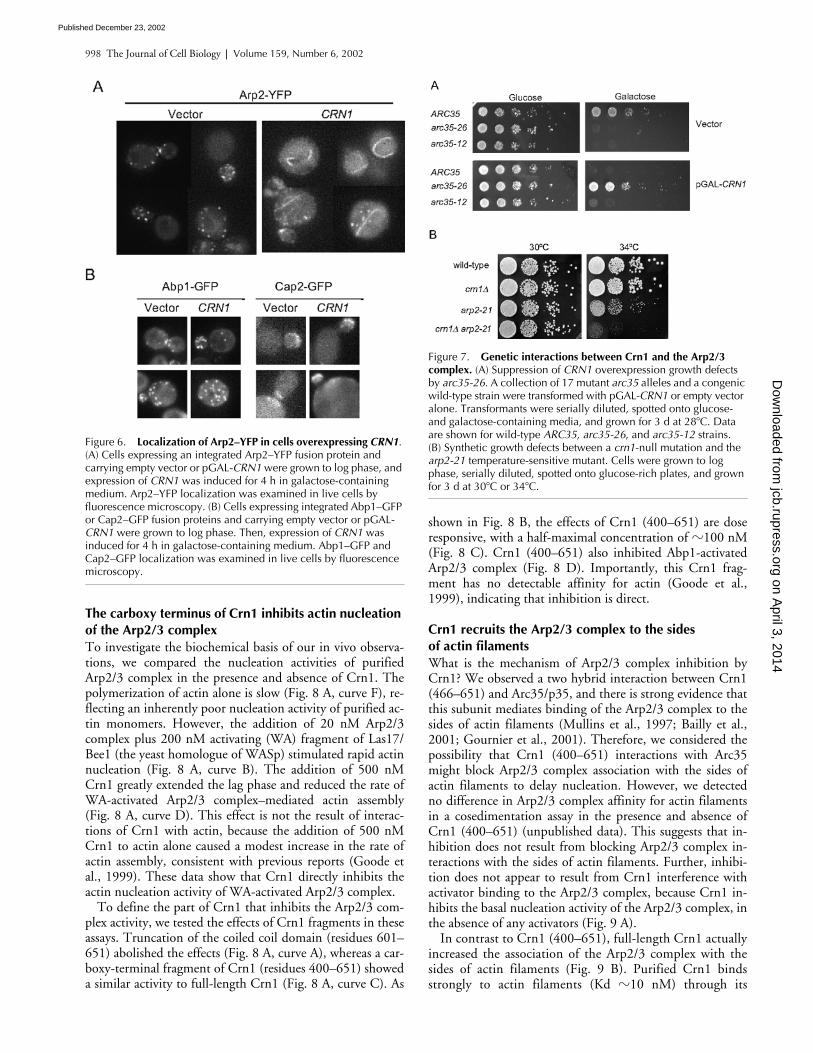
Crn1 recruits the Arp2/3 complex to the sides of actin filamentsWhat is the mechanism of Arp2/3 complex inhibition byCrn1? We observed a two hybrid interaction between Crn1(466–651) and Arc35/p35, and there is strong evidence thatthis subunit mediates binding of the Arp2/3 complex to thesides of actin filaments (Mullins et al., 1997; Bailly et al.,2001; Gournier et al., 2001). Therefore, we considered thepossibility that Crn1 (400–651) interactions with Arc35might block Arp2/3 complex association with the sides ofactin filaments to delay nucleation. However, we detectedno difference in Arp2/3 complex affinity for actin filamentsin a cosedimentation assay in the presence and absence ofCrn1 (400–651) (unpublished data). This suggests that in-hibition does not result from blocking Arp2/3 complex in-teractions with the sides of actin filaments. Further, inhibi-tion does not appear to result from Crn1 interference withactivator binding to the Arp2/3 complex, because Crn1 in-hibits the basal nucleation activity of the Arp2/3 complex, inthe absence of any activators (Fig. 9 A).
In contrast to Crn1 (400–651), full-length Crn1 actuallyincreased the association of the Arp2/3 complex with thesides of actin filaments (Fig. 9 B). Purified Crn1 bindsstrongly to actin filaments (Kd �10 nM) through its
Figure 6. Localization of Arp2–YFP in cells overexpressing CRN1. (A) Cells expressing an integrated Arp2–YFP fusion protein and carrying empty vector or pGAL-CRN1 were grown to log phase, and expression of CRN1 was induced for 4 h in galactose-containing medium. Arp2–YFP localization was examined in live cells by fluorescence microscopy. (B) Cells expressing integrated Abp1–GFP or Cap2–GFP fusion proteins and carrying empty vector or pGAL-CRN1 were grown to log phase. Then, expression of CRN1 was induced for 4 h in galactose-containing medium. Abp1–GFP and Cap2–GFP localization was examined in live cells by fluorescence microscopy.
Figure 7. Genetic interactions between Crn1 and the Arp2/3 complex. (A) Suppression of CRN1 overexpression growth defects by arc35-26. A collection of 17 mutant arc35 alleles and a congenic wild-type strain were transformed with pGAL-CRN1 or empty vector alone. Transformants were serially diluted, spotted onto glucose- and galactose-containing media, and grown for 3 d at 28�C. Data are shown for wild-type ARC35, arc35-26, and arc35-12 strains. (B) Synthetic growth defects between a crn1-null mutation and the arp2-21 temperature-sensitive mutant. Cells were grown to log phase, serially diluted, spotted onto glucose-rich plates, and grown for 3 d at 30�C or 34�C.
on April 3, 2014
jcb.rupress.orgD
ownloaded from
Published December 23, 2002
Coronin inhibition of Arp2/3 complex | Humphries et al. 999
amino terminus (Goode et al., 1999), whereas yeast Arp2/3complex binds weakly to the sides of actin filaments (Kd�2–3 �M), similar to Arp2/3 complex isolated from otherspecies (discussed in Goode et al., 2001). As shown in Fig.9 B, Arp2/3 complex cosedimentation with 2 �M actin fil-aments increases significantly in the presence of 2 �MCrn1. Thus, Crn1 recruits the Arp2/3 complex to the sidesof actin filaments. Further, these effects do not result fromactin filament bundling by Crn1, because a different actinbundling protein (Sac6/fimbrin) had no effect in this assay(unpublished data).
We also tested the ability of Crn1 to inhibit Arp2/3-medi-ated actin assembly in the presence of preformed actin fila-ments. When 500 nM preassembled actin filaments wasadded to 2 �M actin monomers and WA-activated Arp2/3
complex, the lag phase was nearly eliminated (Fig. 9 C,curve A), consistent with previous reports (Machesky et al.,1997). When we further added 500 nM Crn1 (400–651), aconcentration that dramatically inhibits Arp2/3 complex inthe absence of filaments (Fig. 8 A, compare curves B and C),there was little, if any, inhibition detected (Fig. 9 C, curveB). Thus, Crn1 suppresses the Arp2/3 complex specificallyin the absence of actin filaments.
DiscussionCoronin is a ubiquitous component of the eukaryotic actincytoskeleton and localizes to sites of dynamic actin assemblyin cells (for review see de Hostos, 1999). However, since itsidentification almost 10 yr ago, the precise function of coro-
Figure 8. The coiled coil domain of Crn1 inhibits Arp2/3 complex–mediated actin nucleation. (A) Inhibition of Arp2/3 complex by the carboxy terminus of Crn1. Assembly kinetics for 2 �M monomeric actin in the presence or absence of 20 nM Arp2/3 complex, 200 nM WA fragment of Las17/Bee1, and 500 nM Crn1 or Crn1 fragments. Curve A, Arp2/3 complex, WA, and Crn1 (1–600); curve B, Arp2/3 complex and WA; curve C, Arp2/3 complex, WA, and 500 nM Crn1 (400–651); curve D, Arp2/3 complex, WA, and 500 nM Crn1 (1–651); curve E, Arp2/3 complex; curve F, actin alone. (B) Dose-responsive inhibition of the Arp2/3 complex by Crn1 (400–651). The graph shows assembly of 2 �M monomeric actin (10% pyrene labeled) in the presence of 20 nM Arp2/3 complex, 200 nM WA, and 0–1,000 nM Crn1 (400–651). Curve A, no Crn1 added; curve B, 67.5 nM Crn1 (400–651); curve C, 250 nM Crn1 (400–651); curve D, 500 nM Crn1 (400–651); curve E, 1,000 nM Crn1 (400–651); curve F, actin alone. (C) Graph showing the concentration-dependent effects of Crn1 (400–651) on the rate of Arp2/3 complex–nucleated actin assembly; the data is from B (see Materials and methods). (D) Crn1 inhibits Abp1-stimulated Arp2/3 complex activity. Monomeric actin (2 �M) assembled with 50 nM Arp2/3 complex, 500 nM Crn1 (400–651), 50 nM Abp1, or 200 nM WA. Curve A, Arp2/3 and Abp1; curve B, Arp2/3 and WA; curve C, Arp2/3, WA, and Crn1 (400–651); curve D, Arp2/3, Abp1, and Crn1 (400–651); curve E, Arp2/3; curve F, actin alone.
on April 3, 2014
jcb.rupress.orgD
ownloaded from
Published December 23, 2002

1000 The Journal of Cell Biology | Volume 159, Number 6, 2002
nin in the actin cytoskeleton has remained poorly under-stood. Here, we have made an important step toward defin-ing the cellular function of yeast coronin (Crn1). We showthat (a) Crn1 and the Arp2/3 complex physically and func-tionally interact in vivo, (b) the interaction is mediated bythe coiled coil domain of Crn1, which is evolutionarily con-served, and (c) Crn1 strongly modulates Arp2/3 complex ac-tivity, inhibiting actin nucleation in the absence of pre-formed actin filaments. This work defines a new mode of
regulation for the Arp2/3 complex and identifies Crn1 as thefirst direct inhibitor of the Arp2/3 complex.
Physical and genetic interactions between Crn1 and the Arp2/3 complexWe have found a strong physical association between Crn1and the Arp2/3 complex, as demonstrated by a variety of as-says, including comigration on sucrose gradients, coimmu-noprecipitation, two hybrid analysis, and direct binding ofpurified proteins. These interactions and the effects of Crn1on Arp2/3 complex activity are mediated by the coiled coildomain of Crn1. Our attempts by blot overlay assays to mapthe specific subunit(s) of the Arp2/3 complex that bindsCrn1 have been unsuccessful thus far (unpublished data).However, our two hybrid data suggest that Arc35 may be animportant target of Crn1 binding, and this is supported bythe observation that an arc35 allele suppresses the growthdefects caused by CRN1 overexpression.
Over 50% of the cellular Crn1 is bound to the Arp2/3complex, suggesting that the cellular functions of Crn1 andthe Arp2/3 complex are closely linked. This is supported byin vivo evidence, including synthetic defects between crn1-null mutants and arp2-21 and suppression by arc35-26 ofdefects caused by CRN1 overexpression. Further, localiza-tion of Crn1 to cortical actin patches in vivo depends onboth its actin binding domain and its Arp2/3 complex–interacting coiled coil domain. A similar observation wasmade in cultured Xenopus cells, where deletion of the coiledcoil domain caused mislocalization of coronin (Mishima andNishida, 1999). Because coronin forms coiled coil–depen-dent homodimers in vitro (Goode et al., 1999; Asano et al.,2001), it was postulated that coronin dimerization may benecessary for localization. However, our findings raise thepossibility that interactions of the coiled coil domain withthe Arp2/3 complex may contribute to localization. Impor-tantly, these models are not mutually exclusive; coronin lo-calization may require both homodimerization and interac-tions with the Arp2/3 complex. Another important pointraised here and in the above-mentioned study is that actinbinding alone is not sufficient to localize coronin to actin fil-ament structures in vivo. Therefore, associations betweencoronin and actin may be regulated in vivo.
A mechanism for Crn1 inhibition of Arp2/3 complex activityWe found that Crn1 inhibits WA-activated Arp2/3-medi-ated actin nucleation and that these effects are dose respon-sive and mediated by the Crn1 coiled coil domain. Theaddition of 0.5 �M Crn1 (400–651) virtually abolishesWA- and Abp1-activated Arp2/3 complex activity. Impor-tantly, this activity is independent of Crn1 interactions withactin, because Crn1 (400–651) has no actin binding affinity(Goode et al., 1999). Therefore, the inhibition of the Arp2/3complex by Crn1 is direct.
To explore the mechanism of inhibition, it is helpful toreview current models for Arp2/3 complex activation (for re-views see Cooper et al., 2001; Borths and Welch, 2002;Kreishman-Deltrick and Rosen, 2002). Recently, the crystalstructure of the inactive Arp2/3 complex and cryo-electron
Figure 9. Crn1 recruits the Arp2/3 complex to the sides of actin filaments. (A) Assembly kinetics of 2 �M monomeric actin (10% pyrene labeled) in the presence or absence of 50 nM Arp2/3 complex and 500 nM Crn1 (400–651). Curve A, Arp2/3 complex; curve B, actin alone; curve C, Crn1 (400–651); curve D, Arp2/3 complex and Crn1 (400–651). (B) Coomassie-stained gel of pellets and supernatants from cosedimentation assays containing 2 �M F-actin, 0.5 �M Arp2/3 complex, and/or 2 �M full-length Crn1. (C) The addition of preformed actin filaments overrides Crn1 inhibition. Assembly kinetics for 2 �M monomeric actin plus 0.5 �M preformed actin filaments, with 20 nM Arp2/3 complex, 200 nM WA fragment of Las17/Bee1, and/or 500 nM Crn1 (400–651). Curve A, F-actin, Arp2/3, and WA; curve B, Arp2/3, WA, and Crn1 (400–651); curve C, 500 nM Crn1 (400–651); curve D, F-actin.
on April 3, 2014
jcb.rupress.orgD
ownloaded from
Published December 23, 2002

Coronin inhibition of Arp2/3 complex | Humphries et al. 1001
micrograph structures of the activated complex alone and atfilament branch points were reported (Robinson et al.,2001; Volkmann et al., 2001). Together, these studies sug-gest that association of the Arp2/3 complex with the sides ofactin filaments and interactions with an activator converge,inducing allosteric changes in the complex that repositionArp2 and Arp3 into a nucleation-competent actin-likedimer. The p35/Arc35 subunit is strongly implicated inphysically linking the Arp2/3 complex to the sides of actinfilaments (Mullins et al., 1997; Bailly et al., 2001; Gournieret al., 2001). Thus, interactions between p35 and the side ofa filament may transduce one set of conformational changes,while interactions between an activator and other subunitsof the complex may transduce a complementary set of con-formational changes.
Initially, we considered a simple model for inhibition byCrn1, in which Crn1 competes with and displaces activatorsfrom the Arp2/3 complex. However, our results are incon-sistent with this model. First, Crn1 suppresses the inherentactin nucleation of the Arp2/3 complex alone, in the absenceof any activators. Second, Crn1 does not have an acidic “A”motif, found in and required for association with the Arp2/3complex in all known activators (for review see Cooper etal., 2001). Third, Crn1 interacts genetically and by two hy-brid assay with p35/Arc35, in contrast to activators, whichare implicated in binding to four different subunits: Arp2,Arp3, p40/Arc40, and p21/Arc18 (Mullins et al., 1997; Ma-chesky and Insall, 1998; Zalevsky et al., 2001). Fourth, in-creasing the concentration of WA in the reactions fails tooverride Crn1 inhibition (unpublished data). Thus, all ofour data point to a functional interaction between the coiledcoil domain of Crn1 and the p35 subunit of the Arp2/3complex, via a distinct interface from the activators.
A second model we considered for inhibition was thatCrn1 (400–651) might interfere with Arp2/3 complex bind-ing to the sides of actin filaments. However, Crn1 (400–651)did not affect Arp2/3 complex association with filament sides,and, in fact, full-length Crn1 increased association of theArp2/3 complex with the sides of actin filaments (Fig. 2 B).
What does the arc35-26 suppression data tell us about themechanism of inhibition? The allele-specific inhibition ofCRN1 overexpression defects strengthens our hypothesisthat Crn1 interactions with the Arp2/3 complex occurthrough the Arc35/p35 subunit. Intriguingly, arc35-26 sup-presses the growth defects associated with Crn1 overexpres-sion, and, reciprocally, CRN1 overexpression suppresses thegrowth defects of arc35-26. This cosuppression suggests ahighly specific functional interaction between Crn1 andArc35. In future work, isolating the Arp2/3 complex fromarc35-26 mutant cells and studying its activities may providevaluable insights into Crn1 action. In addition, defining theresidues in p35/Arc35 that mediate Crn1 interactions maylend important clues to the mode of inhibition.
The cellular role of Crn1 in regulating the Arp2/3 complexWe have shown that Crn1 inhibits the actin nucleation ac-tivity of the Arp2/3 complex specifically in the absence ofactin filaments via its coiled coil domain and recruits theArp2/3 complex to the sides of actin filaments via its actin
binding domain. Both of these activities may be used in vivoto direct the Arp2/3 complex activity to the sides of preexist-ing actin filaments, promoting the formation of filamentnetworks. Such a function might be important during cellu-lar processes that rely on the rapid formation of actin net-works, including cell locomotion and intracellular transportof vesicles and organelles. Consistent with this possibility,loss of coronin function in Dictyostelium and Xenopus cellshas been shown to cause defects in cell migration and/or en-docytosis (de Hostos et al., 1993; Mishima and Nishida,1999). Next, it will be important to assess whether coronin–Arp2/3 complex interactions are conserved in other organ-isms and determine the cellular consequences of disruptingsuch interactions. Already, there are indications that theinteraction may be conserved, because substoichiometricamounts of coronin have been shown to copurify with theArp2/3 complex from human neutrophils (Machesky et al.,1997). Perhaps the most significant challenge for the futurewill be to determine how the Arp2/3 complex integrates somany different signals, from (a) multiple activators, (b)coronin, and (c) binding to the side of an actin filament, tospatially and temporally control actin nucleation in the cell.
Materials and methodsStrains and mediaThe Saccharomyces cerevisiae strains used in this study are listed in TableI. Standard methods were used to generate strains with integrated tags(GFP and HA epitope) at the carboxy termini of CRN1 and ARP2 and astrain with a GFP tag integrated after residue 600 in CRN1 (Longtine et al.,1998). A strain with a CRN1 gene deletion and an HA epitope tag inte-grated at the carboxy terminus of ARP2 (BGY704) was generated by cross-ing strains BGY26 and BAY1412. The resulting diploids were sporulated,tetrads were dissected, and haploids with the BGY704 genotype were se-lected. The presence of the ARP2–HA epitope tag and loss of the CRN1gene were confirmed by immunoblotting with anti-Crn1 and anti-HA anti-bodies. Standard methods were used for growth and transformation ofyeast (Guthrie and Fink, 1991). For yeast growth assays, cultures weregrown to log phase, serially diluted, spotted on plates, and grown for 3 d.
Plasmid constructionAll plasmids used in this study are listed in Table II. To express full-lengthCrn1 and Crn1 (1–600) on CEN plasmids under the control of the CRN1promoter, we constructed pBG290 and pBG291 and pBG296, respec-tively. The designated regions of the CRN1 open reading frame with 300bp of 5 untranslated sequence upstream were PCR amplified using high fi-delity polymerase and subcloned into the BamH1–Not1 sites of pRS316.To express different parts of Crn1 in yeast, we constructed pBG226,pBG289, pBG290, and pBG291 by subcloning BamH1–Nsi1 CRN1 frag-ments from plasmids pBG203 and pBG206, respectively, into p425GAL1and p415MET25. For overexpression of full-length Crn1, Crn1 (1–600), orCrn1 (400–651) under control of the GAL promoter, we constructedpBG222, pBG223, and pBG224. The designated regions of the CRN1open reading frame were amplified by PCR using high fidelity polymeraseand subcloned into BamH1–Not1 sites of p425GAL1 or p426GAL1. Fortwo hybrid analysis, an Arc35 insert was excised as a BamH1–Xho1 frag-ment from pEG202-END9 (a gift from C. Schaerer-Brodbeck, University ofBasel, Basel, Switzerland) and cloned into BamH1–Sal1 sites of pAS2 toyield pAS2-ARC35. The plasmid was shown to express a functional fusionprotein by complementation of the 37�C growth defect of arc35-1(Schaerer-Brodbeck and Riezman, 2000). All plasmids were sequenced toconfirm that the CRN1 coding sequences contained no mutations.
Antibody preparation and immunoblottingTwo different Crn1 antibodies were used, a mouse polyclonal anti-Crn1antibody previously described (Goode et al., 1999) and a rabbit polyclonalanti-Crn1 antibody generated here (Faculty of Medicine, University of Tor-onto). The rabbits were immunized with a GST–Crn1 fusion protein ex-pressed and purified from Escherichia coli (Goode et al., 1999), and the
on April 3, 2014
jcb.rupress.orgD
ownloaded from
Published December 23, 2002

1002 The Journal of Cell Biology | Volume 159, Number 6, 2002
antibodies were affinity purified from serum (Measday et al., 1994). Likethe mouse anti-Crn1 antibody, the rabbit anti-Crn1 antibody recognizes an85-kD band on blots of wild-type total cellular protein that is absent fromcrn1-null lanes (not depicted). Proteins were detected on blots using1:1,000 mouse anti-Crn1, 1:5,000 rabbit anti-Crn1, and 1:1,000 rabbitanti-Arp2 (Moreau et al., 1996). HA–Arp2 was detected using 1:10,000mouse anti-HA antibody conjugated to HRP (Covance; Denver, CO). Forall other blots, 1:10,000 HRP-conjugated secondary antibody was used.Signals were detected by ECL from Amersham Biosciences.
Protein purificationThe Arp2/3 complex was purified from yeast (Goode et al., 2001). Crn1,Crn1 (1–600), and Crn1 (400–651) were purified from E. coli (Goode et al.,1999). The carboxy-terminal WA fragment of Las17/Bee1 was purified fromE. coli (Winter et al., 1999). Unlabeled and pyrene-labeled rabbit skeletalmuscle actin were purchased from Cytoskeleton, Inc. Monomeric actin wasreconstituted from a lyophilized state as per the manufacturer’s instructions,diluted to 25–50 �M in G-buffer (5 mM Tris-HCl , pH 7.5, 0.2 mM DTT, 0.2mM ATP, 0.2 mM CaCl2), incubated overnight on ice, and cleared by cen-trifugation for 1 h at 4�C, 90,000 rpm in a TLA100 rotor (Beckman Coulter).
Sucrose gradient fractionation of yeast lysates11-ml sucrose gradients (3–30%) were poured in 12-ml ultra clear tubesfor an SW41 rotor (Beckman Coulter). Crude cell lysates were preparedfrom wild-type and crn1-null yeast as previously described (Goode et al.,
1999). Lysates were precleared by centrifugation for 15 min, 70,000 rpm,4�C in a TLA100.3 rotor (Beckman Coulter). 400 �l supernatant or highmolecular weight gel filtration size standards (Amersham Biosciences)were layered over each gradient. Samples were centrifuged for 15 h at34,000 rpm, 4�C in an SW41 rotor, and 0.4-ml fractions were collected.Samples of each fraction were run on SDS-PAGE gels, blotted, and probedwith antibodies to determine the positions of proteins in the gradients.
Coimmunoprecipitation assaysFor coimmunoprecipitation assays, we used yeast strains with an inte-grated carboxy-terminal 3xHA epitope tag on ARP2. Cells were grown tolog phase, washed, frozen, and lysed as previously described (Goode etal., 1999). 1 g of cell lysate was added to 1 ml HEK buffer (20 mM Hepes,pH 7.5, 1 mM EDTA, 50 mM KCl), supplemented with 1mM DTT, 0.1%NP-40 detergent, and a standard cocktail of protease inhibitors (Goode etal., 1999). The lysate was thawed to 4�C and precleared by centrifugationfor 15 min at 4�C, 80,000 rpm in a TLA100.3 rotor. The supernatant washarvested and preabsorbed with CL4B protein A–Sepharose (AmershamBiosciences) for 1 h at 4�C. The beads were pelleted and 500 �l of super-natant was added to 2.5 �l of HA.11 monoclonal antibody (5–7 mg/ml as-cites fluid; Covance) and incubated for 1 h at 4�C. Next, we added 20 �l ofCL4B protein A–Sepharose (preswollen in HEK buffer) and incubated for1 h at 4�C with mixing. The beads were washed twice in HEK buffer andthen once in HEK buffer supplemented with 0.5% NP-40, and sampleswere prepared in SDS sample buffer for immunoblotting.
Table I. Strains used in this study
Name Genotype Source
BGY006 MAT, his3-11,15, ura3-52, leu2-3,112, ade2-1, trp1-1, GAL�, CRN1-GFP::HIS3MX6 This studyBGY008 MAT, his3-11,15, ura3-52, leu2-3,112, ade2-1, trp1-1, GAL�, CRN1(1-600)-GFP::HIS3MX6 This studyBGY026 MAT, his3-11,15, ura3-52, leu2-3,112, ade2-1, trp1-1, GAL�, ARP2-3xHA::HIS3MX6 This studyBGY029 MAT, his3-11,15, leu2-3,112, ade2-1, trp1-1, GAL�, ura3�::ACT1pr-ABP1-GFP::URA3 This studyBGY046 MATa, leu2-3,112, ura3-52, ade2-101, ade3-130, crn1�::LEU2 This studyBGY654 MAT, ARP2-TEV-3xHA::HIS3MX6, leu2D::ARC35::LEU2, arc35�::KANMX6, ura3-52, his3�200 D. Robinsa
BGY661 MAT, ARP2-TEV-3xHA::HIS3MX6, leu2�::arc35-12::LEU2, arc35�::KANMX6, ura3-52, his3�200 D. Robinsa
BGY670 MAT, ARP2-TEV-3xHA::HIS3MX6, leu2�::arc35-26::LEU2, arc35�::KANMX6, ura3-52, his3�200 D. Robinsa
BGY704 MAT, his3-11,15, ura3-52, leu2-3,112, ade2-1, trp1-1, psi�, ssd�, GAL�, ARP2-3xHA::HIS3MX6, crn1�::KANMX6
This study
BGY714 MATa, ARP2-YFP::HIS3MX6, ura3-52, his3�200, ade2-1, leu2-3,112 This studyBGY721 MAT, arp2-21::URA3, crn1�::KANMX6, leu2�0 This studyBAY263 MATa, trp1�63, GAL2�, ura3-52, lys2-801, ade2-107, his3�200, leu2-�1 Measday et al., 1994BAY1355 MATa, crn1�::URA3, trp�63, GAL2�, ura3-52, lys2-801, ade2-107, his3�200, leu2-�1 This studyBAY1412 MATa, crn1�::KANMX6, his3�1, leu2�0, met15�0, ura3�0 Research GeneticsBAY1674 MAT, arp2-21::URA3, mfa1�::MFA1pr-HIS3, can1�, his3�1, leu2�0, MET15�, lys2�0 C. Booneb
BAY3029 MAT, arc40-40::URA3, mfa1�::MFAprHIS3, can1�, his3�1, leu2�0, MET15�, lys2�0 Tong et al., 2001YJC1265 Ura3-52/ura3-53, leu2-3,112/leu2-3,112, trp1�::CAP2-GFP::TRP1, trp1�::CAP2-GFP::TRP1 Waddle et al., 1996
aBrandeis University.bUniversity of Toronto.
Table II. Plasmids used in this study
Name Insert Vector Reference
pBG203 GST–CRN1 pGAT2 Goode et al., 1999pBG205 GST–CRN1 (400–651) pGAT2 Goode et al., 1999pBG206 GST–CRN1 (1–600) pGAT2 Goode et al., 1999pBG222 CRN1 pRS426GAL1 This studypBG223 CRN1 (1–600) pRS426GAL1 This studypBG224 CRN1 (400–651) pRS426GAL1 This studypBG290 CRN1 pRS415MET25 This studypBG291 CRN1 (1–600) pRS415MET25 This studypBG294 CRN1 pRS316 This studypBG295 CRN1 (1–600) pRS316 This studypBG298 CRN1 (400–651) pRS415MET25 This studypAS2��::ARC35 ARC35 pAS2�� This study
on April 3, 2014
jcb.rupress.orgD
ownloaded from
Published December 23, 2002
Coronin inhibition of Arp2/3 complex | Humphries et al. 1003
Binding interactions between Crn1 and the Arp2/3 complexTo test direct binding between Crn1 and the Arp2/3 complex, we assayedthe cosedimentation of purified Crn1 with purified HA-tagged yeast Arp2/3complex immobilized on beads. The Arp2/3 complex–loaded beads wereprepared as previously described (Goode et al., 2001), yielding a bead sus-pension of 1 �M Arp2/3 complex. 10 �l of Arp2/3-loaded beads or controlbeads (no Arp2/3) was included in a 100-�l reaction in HEK buffer con-taining 1 �M Crn1. The final concentration of the Arp2/3 complex in thereactions was 0.1 �M. Reactions were incubated for 20 min at 4�C, thebeads were washed, and the bound proteins were removed with SDS sam-ple buffer (without reducing agent). Samples were run on 12% SDS-PAGEgels and stained with Coomassie blue.
Two hybrid analysisA yeast cDNA library in pGAD-GH was transformed into the Y190 yeaststrain containing pAS2-ARC35, and a nonsaturating Gal two hybrid screenwas performed as previously described (Madania et al., 1999). Transfor-mants were selected on �TRP, �LEU, �HIS, �30 mM 3-aminotriazolemedium, and 24 clones were isolated. After a series of secondary tests,four clones remained, two of which were found to contain a fragment ofCrn1 encoding its carboxy-terminal 186 residues (466–651).
Fluorescence light microscopyActin and Crn1 organization was examined in cells overproducing full-length Crn1, Crn1 (1–600), and Crn1 (400–651) under control of theGAL1/10 promoter, from plasmids pBG222, pBG223, and pBG224. Cellswere grown at 30�C in selective glucose medium to early log phase,washed, transferred to selective galactose medium, grown for 4 h at 30�C,fixed, and prepared for immunofluorescence (Ayscough and Drubin,1997; Lee et al., 1998). To disrupt the actin cytoskeleton in cells, log-phase yeast cultures were treated with 100 �M latrunculin A for 15 minbefore chemical fixation. We also determined the localization of full-length Crn1, Crn1 (1–600), and Crn1 (400–651) expressed in cells fromlow copy plasmids (pBG290, pBG291, and pBG298) under the control ofthe MET25 promoter (Fig. 3 B). Similar results were obtained for low copyplasmids expressing full-length Crn1 and Crn1 (1–600) under the controlof its own promoter: pBG294 and pBG295 (not depicted). For immunoflu-orescence detection of Crn1 and actin, we used a 1:500 dilution of rabbitanti-Crn1 and a 1:2,000 dilution of guinea pig anti-Act1 (Mulholland et al.,1994). Cells were imaged on a Leica DM-LB microscope. Images werecaptured with a Micromax 1300y high-speed digital camera (Princeton In-struments) and analyzed with Metaview software (Universal ImagingCorp.). The localization of GFP and YFP was examined in yeast cellsgrown to log phase.
Actin assembly kineticsActin assembly was monitored by the pyrene–actin fluorescence assay aspreviously described (Goode et al., 2001), using a final concentration of 2�M actin in 70-�l reactions unless otherwise indicated. In brief, 56.5 �l ofmonomeric actin (10% pyrene-labeled, 90% unlabeled) in G buffer wasmixed with 10 �l HEKG5 buffer (HEK buffer � 5% glycerol) or differentcombinations of proteins in HEKG5 buffer. The reaction was mixed imme-diately with 3.5 �l 20� initiation buffer (1 M KCl, 40 mM MgCl2, 10 mMATP) in a quartz fluorometry cuvette (3-mm light path; Hellma). Pyrene–actin fluorescence was monitored by excitation at 365 nM and emission at407 nM in a fluorescence spectrophotometer (Photon Technology Interna-tional) held at the constant temperature of 25�C. For seeded reactions, ac-tin was preassembled to steady state for 1 h. Then, 2 �M monomeric actin(10% pyrene labeled) was mixed with 500 nM preassembled actin fila-ments in the presence or absence of different proteins.
Actin filament recruitment assaysBinding of the Arp2/3 complex to the sides of actin filaments was mea-sured as previously described (Goode et al., 2001). In Fig. 9 B, 2 �M full-length Crn1 and/or 0.5 �M Arp2/3 complex were added to 2 �M preas-sembled actin filaments. After a 20-min incubation, the reactions werecentrifuged for 20 min in a TLA100 rotor (Beckman Coulter). Equal loadsof pellets and supernatants were fractionated on 12% SDS-PAGE gels andstained with Coomassie blue.
We are very grateful to A. Rodal for advice throughout the study, C.Boone, S. Hippenmeyer and H. Riezman (University of Basel), D. Robins,and H. Xu for sharing unpublished reagents, J. Wong, P. Dumoulin, and S.Thuault for technical assistance, and C. Boone, F. Chang, A. Goodman, D.Pellman, A. Rodal, and I. Sagot for helpful comments on the manuscript.
This work was supported by an award from the Canadian Institutes ofHealth Research to C. Humphries, an operating grant from the NationalCancer Institute of Canada with funds from the Canadian Cancer Society toB. Andrews, support from the Centre National de Recherche Scientifiqueand the Association pour la Recherche sur le Cancer to B. Winsor, anda grant from the National Institutes of Health (NIIH) to G. Barnes(GM47842). B. Goode was supported by a Pew Scholars award, a BasilO’Conner award, and the NIH (GM63691).
Submitted: 26 June 2002Revised: 30 September 2002Accepted: 7 November 2002
ReferencesAsano, S., M. Mishima, and E. Nishida. 2001. Coronin forms a stable dimer
through its C-terminal coiled coil region: an implicated role in its localiza-tion to cell periphery. Genes Cells. 6:225–235.
Ayscough, K.R., and D.G. Drubin. 1997. Immunofluorescence microscopy ofyeast cells. In Cell Biology, A Laboratory Handbook. Vol. 2. J. Celis, editor.Academic Press. San Diego, CA. 477–485.
Bailly, M., I. Ichetovkin, W. Grant, N. Zebda, L.M. Machesky, J.E. Segall, and J.Condeelis. 2001. The F-actin side binding activity of the Arp2/3 complex is es-sential for actin nucleation and lamellipod extension. Curr. Biol. 11:620–625.
Borths, E.L., and M.D. Welch. 2002. Turning on the Arp2/3 complex at atomicresolution. Structure (Camb). 10:131–135.
Cooper, J.A., M.A. Wear, and A.M. Weaver. 2001. Arp2/3 complex: advances onthe inner workings of a molecular machine. Cell. 107:703–705.
de Hostos, E.L. 1999. The coronin family of actin-associated proteins. Trends CellBiol. 9:345–350.
de Hostos, E.L., C. Rehfuess, B. Bradtke, D.R. Waddell, R. Albrecht, J. Murphy, andG. Gerisch. 1993. Dictyostelium mutants lacking the cytoskeletal protein coroninare defective in cytokinesis and cell motility. J. Cell Biol. 120:163–173.
Goode, B.L., and A.A. Rodal. 2001. Modular complexes that regulate actin assem-bly in budding yeast. Curr. Opin. Microbiol. 4:703–712.
Goode, B.L., J.J. Wong, A.C. Butty, M. Peter, A.L. McCormack, J.R. Yates, D.G.Drubin, and G. Barnes. 1999. Coronin promotes the rapid assembly andcross-linking of actin filaments and may link the actin and microtubule cy-toskeletons in yeast. J. Cell Biol. 144:83–98.
Goode, B.L., A.A. Rodal, G. Barnes, and D.G. Drubin. 2001. Activation of theArp2/3 complex by the actin filament binding protein Abp1p. J. Cell Biol.153:627–634.
Gournier, H., E.D. Goley, H. Niederstrasser, T. Trinh, and M.D. Welch. 2001.Reconstitution of human Arp2/3 complex reveals critical roles of individualsubunits in complex structure and activity. Mol. Cell. 8:1041–1052.
Guthrie, C., and R. Fink. 1991. Guide to yeast genetics and molecular biology.Methods Enzymol. 194:1–933.
Heil-Chapdelaine, R.A., N.K. Tran, and J.A. Cooper. 1998. The role of Saccharo-myces cerevisiae coronin in the actin and microtubule cytoskeletons. Curr.Biol. 8:1281–1284.
Higgs, H.N., and T.D. Pollard. 2001. Regulation of actin filament network forma-tion through ARP2/3 complex: activation by a diverse array of proteins.Annu. Rev. Biochem. 70:649–676.
Kreishman-Deltrick, M., and M.K. Rosen. 2002. Ignition of a cellular machine.Nat. Cell Biol. 4:E31–E33.
Lee, J., K. Colwill, V. Aneliunas, C. Tennyson, L. Moore, Y. Ho, and B. Andrews.1998. Interaction of yeast Rvs167 and Pho85 cyclin-dependent kinase com-plexes may link the cell cycle to the actin cytoskeleton. Curr. Biol. 8:1310–1321.
Longtine, M.S., A. McKenzie III, D.J. Demarini, N.G. Shah, A. Wach, A. Brachat,P. Philippsen, and J.R. Pringle. 1998. Additional modules for versatile andeconomical PCR-based gene deletion and modification in Saccharomycescerevisiae. Yeast. 14:953–961.
Machesky, L.M., and R.H. Insall. 1998. Scar1 and the related Wiskott-Aldrichsyndrome protein, WASP, regulate the actin cytoskeleton through the Arp2/3complex. Curr. Biol. 8:1347–1356.
Machesky, L.M., E. Reeves, F. Wientjes, F.J. Mattheyse, A. Grogan, N.F. Totty, A.L.Burlingame, J.J. Hsuan, and A.W. Segal. 1997. Mammalian actin-related pro-tein 2/3 complex localizes to regions of lamellipodial protrusion and is com-posed of evolutionarily conserved proteins. Biochem. J. 328:105–112.
Madania, A., P. Dumoulin, S. Grava, H. Kitamoto, C. Scharer-Brodbeck, A. Sou-lard, V. Moreau, and B. Winsor. 1999. The Saccharomyces cerevisiae homo-logue of human Wiskott-Aldrich syndrome protein Las17p interacts withthe Arp2/3 complex. Mol. Biol. Cell. 10:3521–3538.
on April 3, 2014
jcb.rupress.orgD
ownloaded from
Published December 23, 2002

1004 The Journal of Cell Biology | Volume 159, Number 6, 2002
Measday, V., L. Moore, J. Ogas, M. Tyers, and B. Andrews. 1994. The PCL2(ORFD)-PHO85 cyclin-dependent kinase complex: a cell cycle regulator inyeast. Science. 266:1391–1395.
Mishima, M., and E. Nishida. 1999. Coronin localizes to leading edges and is in-volved in cell spreading and lamellipodium extension in vertebrate cells. J.Cell Sci. 112:2833–2842.
Moreau, V., A. Madania, R.P. Martin, and B. Winson. 1996. The Saccharomycescerevisiae actin-related protein Arp2 is involved in the actin cytoskeleton. J.Cell Biol. 134:117–132.
Mulholland, J., D. Preuss, A. Moon, A. Wong, D. Drubin, and D. Botstein. 1994.Ultrastructure of the yeast actin cytoskeleton and its association with theplasma membrane. J. Cell Biol. 125:381–391.
Mullins, R.D., W.F. Stafford, and T.D. Pollard. 1997. Structure, subunit topol-ogy, and actin-binding activity of the Arp2/3 complex from Acanthamoeba.J. Cell Biol. 136:331–343.
Mullins, R.D., J.A. Heuser, and T.D. Pollard. 1998. The interaction of Arp2/3complex with actin: nucleation, high affinity pointed end capping, and for-mation of branching networks of filaments. Proc. Natl. Adad. Sci. USA. 95:6181–6186.
Olazabal, I.M., and L.M. Machesky. 2001. Abp1p and cortactin, new “hand-holds” for actin. J. Cell Biol. 154:679–682.
Pruyne, D., and A. Bretscher. 2000. Polarization of cell growth in yeast. J. Cell Sci.113:571–585.
Robinson, R.C., K. Turbedsky, D.A. Kaiser, J.B. Marchand, H.N. Higgs, S. Choe,and T.D. Pollard. 2001. Crystal structure of Arp2/3 complex. Science. 294:1679–1684.
Schafer, D.A. 2002. Coupling actin dynamics and membrane dynamics during en-docytosis. Curr. Opin. Cell Biol. 14:76–81.
Schaerer-Brodbeck, C., and H. Riezman. 2000. Saccharomyces cerevisiae Arc35pworks through two genetically separable calmodulin functions to regulatethe actin and tubulin cytoskeletons. J. Cell Sci. 113:521–532.
Tong, A.H., M. Evangelista, A.B. Parsons, H. Xu, G.D. Bader, N. Page, M. Robin-son, S. Raghibizadeh, C.W. Hogue, H. Bussey, et al. 2001. Systematic geneticanalysis with ordered arrays of yeast deletion mutants. Science. 294:2364–2368.
Volkmann, N., K.J. Amann, S. Stoilova-McPhie, C. Egile, D.C. Winter, L. Hazel-wood, J.E. Heuser, R. Li, T.D. Pollard, and D. Hanein. 2001. Structure ofArp2/3 complex in its activated state and in actin filament branch junctions.Science. 293:2456–2459.
Waddle, J.A., T.S. Karpova, R.H. Waterston, and J.A. Cooper. 1996. Movementof cortical actin patches in yeast. J. Cell Biol. 132:861–870.
Winter, D.C., E.Y. Choe, and R. Li. 1999. Genetic dissection of the budding yeastArp2/3 complex: a comparison of the in vivo and structural roles of individ-ual subunits. Proc. Natl. Acad. Sci. USA. 96:7288–7293.
Zalevsky, J., L. Lempert, H. Kranitz, and R.D. Mullins. 2001. Different WASPfamily proteins stimulate different Arp2/3 complex-dependent actin-nucle-ating activities. Curr. Biol. 11:1903–1913.
on April 3, 2014
jcb.rupress.orgD
ownloaded from
Published December 23, 2002