Direct and indirect contribution of bone marrow-derived cells to cancer
19
DIRECT AND INDIRECT CONTRIBUTION OF BONE MARROW DERIVED CELLS TO CANCER Ian Guest 1 , Zoran Ilic 2 , Jun Ma 2 , Denise Grant 2 , Gennadi Glinsky 2 , and Stewart Sell 1,2,* 1 Department of Molecular Medicine, Wadsworth Center, New York State Department of Health, Albany, NY, 12201 2 Ordway Research Institute, Albany, NY, 12208 Summary Stromal-epithelial interactions may control the growth and initiation of cancers. Here we not only test the hypothesis that bone marrow derived cells may effect development of cancers arising from other tissue cells by forming tumor stroma, but also that sarcomas may arise by transformation of stem cells from the bone marrow and epithelial cancers may arise by transdifferentiation of bone marrow stem cells to epithelial cancers. Lethally irradiated female FVB/N mice were restored with bone marrow (BM) transplants from a male transgenic mouse carrying the polyoma middle T- oncoprotein under the control of the mouse mammary tumor virus promoter (MMTV-PyMT) and followed for development of lesions. Eight of 8 lethally irradiated female FVB/N recipient mice, restored with BM transplants from a male MMTV-PyMT transgenic mouse, developed Ychromosome negative (Y−) cancers of various organs surrounded by Y+ stroma. One of the female FVB/N recipient mice also developed fibrosarcoma and one a diploid breast adenocarcinoma (BCA) containing Ychromosomes. In contrast, only 1 of 12 control female mice restored with normal male bone marrow developed a tumor (lymphoma) during the same time period.. These results indicate not only that the transgenic bone marrow derived stromal cells may indirectly contribute to development of tumors in recipient mice, but also that sarcomas may arise by transformation of bone marrow stem cells and that breast cancers arise by transdifferentiation of bone marrow stem cells, presumably by mesenchymal-epithelial transition. Keywords Cancer; Stem Cells; Bone Marrow Transplantation; Breast Cancer Introduction Circulating bone marrow derived stem cells (BMDSCs) have been reported to give rise to epithelial cells [1–3] and can also give rise to cancers in various epithelial organs. For example, following the finding in female recipients of male BM grafts that the epithelium of the damaged human GI tract is repopulated by male cells [4], it was reported that C57BL/6 female mice transplanted with ROSA26 male bone marrow cells and infected with Helicobacter pyloris develop gastric carcinomas arising from the ROSA 26 donor male cells [5]. In addition, in a human T-lymphocyte virus transgenic rat model, hematopoietic progenitor cells give rise to epithelial thymomas [6]. A skin carcinoma in a human female Corresponding Author (To whom reprint request should be addressed): Stewart Sell, M.D., New York State Health Department, Wadsworth Center, P.O. Box 509, Room C-551, Empire State Plaza, Albany, NY 12201, Phone: 518-474-0547; FAX 518-473-2900/518-402-5381, [email protected]. NIH Public Access Author Manuscript Int J Cancer. Author manuscript; available in PMC 2011 May 15. Published in final edited form as: Int J Cancer. 2010 May 15; 126(10): 2308–2318. doi:10.1002/ijc.24946. NIH-PA Author Manuscript NIH-PA Author Manuscript NIH-PA Author Manuscript
Transcript of Direct and indirect contribution of bone marrow-derived cells to cancer

DIRECT AND INDIRECT CONTRIBUTION OF BONE MARROWDERIVED CELLS TO CANCER
Ian Guest1, Zoran Ilic2, Jun Ma2, Denise Grant2, Gennadi Glinsky2, and Stewart Sell1,2,*1Department of Molecular Medicine, Wadsworth Center, New York State Department of Health,Albany, NY, 122012Ordway Research Institute, Albany, NY, 12208
SummaryStromal-epithelial interactions may control the growth and initiation of cancers. Here we not onlytest the hypothesis that bone marrow derived cells may effect development of cancers arising fromother tissue cells by forming tumor stroma, but also that sarcomas may arise by transformation ofstem cells from the bone marrow and epithelial cancers may arise by transdifferentiation of bonemarrow stem cells to epithelial cancers. Lethally irradiated female FVB/N mice were restored withbone marrow (BM) transplants from a male transgenic mouse carrying the polyoma middle T-oncoprotein under the control of the mouse mammary tumor virus promoter (MMTV-PyMT) andfollowed for development of lesions. Eight of 8 lethally irradiated female FVB/N recipient mice,restored with BM transplants from a male MMTV-PyMT transgenic mouse, developedYchromosome negative (Y−) cancers of various organs surrounded by Y+ stroma. One of thefemale FVB/N recipient mice also developed fibrosarcoma and one a diploid breastadenocarcinoma (BCA) containing Ychromosomes. In contrast, only 1 of 12 control female micerestored with normal male bone marrow developed a tumor (lymphoma) during the same timeperiod.. These results indicate not only that the transgenic bone marrow derived stromal cells mayindirectly contribute to development of tumors in recipient mice, but also that sarcomas may ariseby transformation of bone marrow stem cells and that breast cancers arise by transdifferentiationof bone marrow stem cells, presumably by mesenchymal-epithelial transition.
KeywordsCancer; Stem Cells; Bone Marrow Transplantation; Breast Cancer
IntroductionCirculating bone marrow derived stem cells (BMDSCs) have been reported to give rise toepithelial cells [1–3] and can also give rise to cancers in various epithelial organs. Forexample, following the finding in female recipients of male BM grafts that the epithelium ofthe damaged human GI tract is repopulated by male cells [4], it was reported that C57BL/6female mice transplanted with ROSA26 male bone marrow cells and infected withHelicobacter pyloris develop gastric carcinomas arising from the ROSA 26 donor male cells[5]. In addition, in a human T-lymphocyte virus transgenic rat model, hematopoieticprogenitor cells give rise to epithelial thymomas [6]. A skin carcinoma in a human female
Corresponding Author (To whom reprint request should be addressed): Stewart Sell, M.D., New York State Health Department,Wadsworth Center, P.O. Box 509, Room C-551, Empire State Plaza, Albany, NY 12201, Phone: 518-474-0547; FAX518-473-2900/518-402-5381, [email protected].
NIH Public AccessAuthor ManuscriptInt J Cancer. Author manuscript; available in PMC 2011 May 15.
Published in final edited form as:Int J Cancer. 2010 May 15; 126(10): 2308–2318. doi:10.1002/ijc.24946.
NIH
-PA Author Manuscript
NIH
-PA Author Manuscript
NIH
-PA Author Manuscript
receiving a kidney graft from a male donor contained Y+ cells in tumor nests [7], andvarious other cancers in human females receiving male donor bone marrow transplants havebeen found to contain about 1:10,000 cancer cells with the Ychromosome, suggesting aprocess called developmental mimicry [8]. We originally sought to test the hypothesis thatBMDSCs could give rise to liver cancer. However, we found that only about 1/20,000 livernuclei of female recipients could be demonstrated to contain a Ychromosome, even afterliver injury {9,10}, and that the model system that we attempted to use for this approach wasnot suitable. Thus, not only was BMDSC transformation to epithelial cells extremely rare inthe liver, but also the hepatocarcinogenic regimens to be used were highly toxic to theDDPIV- rats that we proposed to use as recipients.
Therefore, we changed directions to test whether BMDSCs from a male donor carrying atransgene with a strong oncogene under the control of a promoter activated in the mammarygland, would give rise to breast cancer when they were transplanted into irradiated wild-typefemale recipients. If the BMDSCs circulate to the breast and transdifferentiate into breastepithelial tissue, then the MMTV promoter would be activated, the PyMT oncoproteinexpressed, and BCA would develop. We reasoned that this model would be much morelikely to show the rare event of transdifferentiation required for BMDSCs to becomeepithelial cancers than the BM to liver cancer model.
Bone marrow transplantation to irradiated recipients includes at least two stem cellpopulations, hematopoietic and mesenchymal. The hematopoietic stem cells restore theblood cells of the irradiated recipient; the mesenchymal stem cells replace the tissue stromaof the recipient, usually over a longer period of time than that required for blood cellreplacement. After transplantation of MMTV-PyMT male bone marrow into femalerecipients we found that much of the mesenchymal stroma and endothelial cells werereplaced by donor cells within a few months. Then, 1 recipient mouse developed a Y+fibrosarcoma, and 1 recipient female mouse developed a Y+ breast cancer. In addition,seven of the 8 recipient mice developed cancers of diverse origin that did not contain theYchromosome, but were surrounded by Y+ stroma and some developed cancers with littleor no stroma. This implies at least five possible mechanisms leading to cancers in therecipient mice: 1) directly from recipient tissue without involvement of transplanted cells 2)from recipient cells influenced by transplanted mesenchymal cells; 3) by fusion of BMDSCswith host cells; 4) by transformation of mesenchymal stem cells; and 5) bytransdifferentiation of BMDSCs to breast cells.
Materials and methodsMice
Transgenic FVB.Cg-Tg(ACTB-EGFP)B5Nagy/J (FVB.EGFP) mice, stock # 003516, andwildtype FVB/NJ (FVB) mice, stock # 001800, were purchased from Jackson Laboratories(Bar Harbor, Maine). Male transgenic FVB/N-Tg(MMTV-PyMT)634Mul (FVB.PyMT)mice, strain # 01XE3, were obtained from the NCI Mouse Repository (Frederick, MD).Genotyping of FVB.PyMT mice for presence of the polyoma virus middle T antigenoncogene was performed by PCR on DNA extracted from tail tips, following standardinstructions and with the use of primer sequences obtained from NCI. Primers (5’-GGAAGCAAGTACTTCACAAGGG-3’, 5’-GGAAAGTCACTAGGAGCAGGG-3’) wereobtained from Integrated DNA Technologies (San Diego, CA). Mice were housed in theWadsworth Center’s animal care facility on a 12 hr light/dark cycle, with controlledtemperature and humidity, and free access to food and water. All animal experiments wereapproved by the Wadsworth Center’s Animal Care and Use Committee (IACUC).
Guest et al. Page 2
Int J Cancer. Author manuscript; available in PMC 2011 May 15.
NIH
-PA Author Manuscript
NIH
-PA Author Manuscript
NIH
-PA Author Manuscript

Bone Marrow TransplantationBM cells were isolated from femurs and tibias of 10–12 week-old male FVB.PyMT donorssacrificed by CO2 exposure/cervical dislocation. Erythrocytes were lysed with ammoniumchloride, and cell viability was checked by trypan blue exclusion; exclusion was routinely >95%. Cells were suspended in Hank’s balanced salt solution and injected in a volume of 100µl via a 27-gauge needle.
A total of eight recipient female FVB mice, 8 weeks old, were irradiated (900–1,050rads; 137Cs source; Isomedix, Parsippany, NJ) and transplanted intravenously within 2 hrswith 1–2 × 10⁶ unfractionated male-donor BM cells. Engraftment was considered completeat 1 month post-irradiation survival.
ImmunohistochemistryAt autopsy, various tissues were collected and either fixed in 10% buffered formalin or snapfrozen in Tissue-Tek OCT compound (Sakura Finetek USA, Torrance, CA) by dropping into2-methylbutane (EMD Chemicals Inc., Gibbstown, NJ) on dry ice; tissues were then storedat −80°C. The following antibodies were used: anti-cytokeratin (CK)14 (catalogue #MCA890; AbD Serotec, Raleigh, NC), anti-CK5/6 (catalogue # MAB1620) and anti-CK18(catalogue # CBL177); both from Chemicon Corporation, Temecula, CA., anti-panCK(catalogue # VP-C419; Vector Laboratories, Burlingame, CA), anti-p63 (catalogue #MS-1081; Thermo-Fisher Scientific, Fremont, CA), anti-alpha-smooth muscle actin(catalogue # A2547; Sigma Corporation, St. Louis, MO), anti-CD49f (catalogue # 313605,Biolegend Co., San Diego, CA), and anti-Polyoma virus, Middle T-antigen (catalogue #NB100-2749; Novus Biologicals, Littleton, CO); secondary antibody biotinylated goat anti-rat; Jackson ImmunoResearch, West Grove, PA.; catalogue number 112-065-167).
Paraffin-embedded tissues were sectioned at 5 µm, attached to charged slides and rehydratedthrough xylene and a graded ethanol series to water. Slides were subjected to antigenretrieval by boiling in a pH 6 citrate buffer for 5 min. Sections were blocked in 2% normaldonkey serum and then incubated overnight at 4°C with the primary antibody. Followingbiotinylated secondary antibody incubation (60 min at room temperature) and extravidin-peroxidase (Sigma, catalogue # E2886), slides were developed in diaminobenzidine andmounted in Permount (Fisher Scientific, Pittsburgh, PA).
In Situ HybridizationFormalin-fixed, paraffin-embedded sections were deparaffinized through xylene washes andrehydrated through a graded ethanol series. A FITC-labeled probe for the mouseYchromosome (catalogue # 1189-YMF-01; Cambio Ltd., Cambridge, UK) was hybridizedto pepsin-treated sections for 10 minutes at 60°C, followed by overnight incubation at 37°C,according to the manufacturer’s directions. Amplification of the probe was performed byprimary (goat anti-fluorescein; Rockland, Gilbertsville, PA., catalogue # 600-101-096) andsecondary (fluorescein-conjugated donkey anti-goat; Jackson Immunoresearch, West Grove,PA., catalogue number 705-095-147) antibody enhancement. Nuclei were stained with 4’,6-diamidine-2-phenylindole dihydrochloride (catalogue # 236276; Roche Applied Sciences,Indianapolis, IN.). Images were captured on an Olympus BX 51 microscope equipped withfluorescence detection and Optronics PictureFrame Version 1.2 software.
Tumor Harvest and DissociationBreast tumors from MMTV-PyMT FVB/N (002374, Jackson Laboratory, Bar Harbor, ME,http://www.jax.org) transgenic mice were harvested when the tumors were approximately 1–2 cm3 (2–2.5 g), minced with a razor blade, and suspended into Dulbecco’s Modified Eagle
Guest et al. Page 3
Int J Cancer. Author manuscript; available in PMC 2011 May 15.
NIH
-PA Author Manuscript
NIH
-PA Author Manuscript
NIH
-PA Author Manuscript
Medium (DMEM) with 10% calf serum, 10ng/ml mouse epidermal growth factor (EGF) and5ug/ml bovine insulin for culture.
Cell Staining and Flow CytometryCells were stained at a concentration of 1 X 10⁶ cells per 100 ul of HBSS with 2% heat-inactivated calf serum (HICS). Antibodies at appropriate dilutions (CD24-fluoresceinisothiocyanate (FITC), Biolegend Inc; CD49f-Alexa Fluor® 647, Biolegend Inc; CD45-Pacific Blue, Biolegend Inc; CD44-Pacific Blue, Biolegend Inc; CD29-phycoerythrin (PE),Biolegend Inc; Sca-1-phycoerythrin (PE), Biolegend Inc; CD45-phycoerythrin (PE),Biolegend Inc; CD31-phycoerythrin (PE), Biolegend Inc; Ter119-allophycocyanin (APC),Biolegend Inc) were added. Staining duration was for 30 minutes on ice, with light agitationof the staining vessels every 5 minutes. Cells were then washed with staining medium andresuspended in staining medium. The stained specimens were then analyzed usingFACSVantage (BD Biosciences, San Diego, http://www.bdbiosciences.com) or FACSAriawith either Diva or CellQuest software (BD Biosciences). Cells with appropriate CD24status were then collected. A small sample of the double-sorted cells was reanalyzed forpurity. The cell counter of the flow cytometers was used to determine cell numbers. Cellswere collected into HBSS with 2% HICS. Epithelial cells wereCD24(+)CD49f(+)CD44lowCD29(+)Sca-1lowCD45(−)CD31(−)Ter119(−); mesenchymalcells were CD24(−)CD49f(−)CD44highCD29(+)Sca-1highCD45(−)CD31(−)Ter119(−).
Culture of Tumor Derived Fibroblasts (Mesenchymal cells)Over 70 cell lines from MMTV-PyMT and EGFP MMTV-PyMT tumors have been derived,cloned and transplanted in FVB/N mice following proven culture techniques 20. Cellscultured from primary tumors were found to express three phenotypes by flow cytometry:CD 24−44lo; CD24+CD44; and a mixture. The cell lines were characterized as epithelialcell-like (ECL) or mesenchymal cell-like (MCL) by inverted phase microscopy. ECL cells(doubling time 24–48hr) grow more rapidly than MCL cells (doubling time 48–72hr). TheECLs are all CD24+CD44loCD29+CD49f+Sca-1Lo, and the MCLs areCD24−CD44hiCD29+CD49f-Sca-1Hi. The lines of mixed cell origin have been sorted; theCD44lo cells are ECL and the CD44hi cells are MCL (data not shown) indicating a mixtureof cell types in the original culture. Mesenchymal cell line 393 was analyzed by PCR andRT-PCR for PyMT and mRNA.
Reverse Transcription Polymerase Chain Reaction (RT-PCR) and Polymerase ChainReaction (PCR)
Total RNA and DNA were purified from cells using RNeasy Mini Kit and DNeasy Blood &Tissue Kit according to the manufacturer’s instructions (Qiagen). USB two-step RT-PCR kitwas used to perform the analysis according to the manufacturer’s instructions (USB).Primers for mouse genes: PyMT transgene, sense primer 5’-GGAAGCAAGTACTTCACAAGGG-3’, antisense primer 5’-AAAGTCACTAGGAGCAGGG-3’; and b-actin, sense primer 5’-CCAGAGCAAGAGAGGTATC-3’, antisense primer 5’-GACCAGAGGCATACAGGGAC-3’.
Laser Capture of tissuesFormalin fixed paraffin embedded tissues were sectioned into 6 um segments and mountedonto Membrane slides (Zeiss). The tissues were rapidly hydrated through two changes ofxylene, 100%, 95%, 75% ethanols, and finally dH2O. For contrast, the slides were stained intoluene blue for three minutes before washing in water and dehydrating through two changesof 95% and 100% ethanol and xylene. Xylene exposure was kept to a minimum to prevent
Guest et al. Page 4
Int J Cancer. Author manuscript; available in PMC 2011 May 15.
NIH
-PA Author Manuscript
NIH
-PA Author Manuscript
NIH
-PA Author Manuscript
the membranes from dissolving. Slides were cut in close temporal proximity to laser capture.Tissues were captured using a PALM MicroBeam workstation (Zeiss). The microdissectedtissue fragments were collected into 200 uL AdhesiveCaps, positioned over the slides usingthe RoboCap arm attachment on the workstation. Samples were placed on ice untilpurification.
DNA and RNA purificationDNA was extracted from the microdissected tissues using the QIAamp DNA Micro Kit(Qiagen). Samples were incubated at 56° C for four hours to ensure that the proteinase Kreaction sufficiently digested the tissues. Carrier RNA was added to the samples to increasebinding efficiency to the MinElute columns during the wash steps. Samples were elutedusing Buffer ATE from the QIAamp DNA FFPE Tissue Kit (Qiagen), which is a low-EDTAelution buffer optimized for sensitive downstream applications. RNA was isolated fromseparate samples of microdissected tissues using the RNeasy FFPE Kit (Qiagen). CarrierRNA was also added to these samples, which did not effect downstream applications.
Alternatively, RNA was isolated from whole tissue samples frozen in OCT. Prior toextraction, frozen sections were made from stock samples of experimental tissues to identifykey areas of tissue for excision. Quickly, the sample was thawed from OCT and placed ondry ice. Tissue samples were ground to a fine powder by mortar and pestle under liquidnitrogen and lysed immediately using the mirVana total RNA isolation kit (Ambion). Thispreparation allowed for phenol-chloroform extraction of the RNA, avoiding anycomplications with fatty tissue hindering column purification. The RNA samples were alsoDNase treated with Turbo DNA-free (Ambion) prior to any reverse transcriptase reaction toremove any possible DNA contamination.
cDNA prep and qPCRReverse transcription of RNA samples was performed using the High Capacity RNA-to-cDNA Master Mix (Applied Biosystems), performing both RT+ (with enzyme) and RT-(without enzyme) reactions. Power SYBR Green PCR Master Mix (Applied Biosystems)was used to amplify the cDNA and DNA isolated from the laser microdissected samples,which contains AmpliTaq Gold DNA Polymerase to give greater specificity with lowtemplate concentrations. Primers for qPCR of the Polyomavirus Middle T antigen gene weredesigned using the Primer-BLAST website, blasting against the representative region in theMurine polyomavirus (a 1.33kb region from base 175 to 1502): Forward PyMT primer:CTG CTG GAA GAA GAC GAA AT; Reverse PyMT Primer: GTT GCA TTG AAT GAGCTC TG; PCR product size 343 base pairs. The PyMT primers were found to be specific tothe template, with no other targets found after blasting against the normal mouse genome.The qPCR samples were run on the 7900HT Fast Real-Time PCR System (AppliedBiosystems) using the 96-well block, with a dissociation curve at the completion of 40cycles.
qPCR Data AnalysisResults were analyzed both by the SDS2.2 software and gel electrophoresis of the qPCRproducts. dCt values were generated normalizing to B-Actin Ct values. The experimentalsamples were compared for the PyMT primer set, using the positive and negative controls asthreshold values. Any values falling between the control range were considered weaklypositive. qPCR products for the SRY primer set were run on 1.2% agarose gels to confirmpositive results. Weak bands were considered weakly positive. We also used peak analysisto predict results pending confirmation of gel electrophoresis.
Guest et al. Page 5
Int J Cancer. Author manuscript; available in PMC 2011 May 15.
NIH
-PA Author Manuscript
NIH
-PA Author Manuscript
NIH
-PA Author Manuscript
ResultsDevelopment of tumors
Untreated Female MMTV-PyMT mice begin to develop palpable masses by 4–5 weeks ofage and large multifocal breast cancers by 10–12 weeks of age. Male mice develop palpablemasses about 3 months of age and large cancers by about 5 months of age. The histologicappearance includes a rage of ductal and glandular structures similar to human breastcancers (Supplemental Figure 1). As expected breast cancer cells of primary tumors arisingin male mice stain for Ychromosome, tumors from the female mice are negative(Supplemental Figure 2). Tumors in other organs are not seen at the time that the transgenicmice die from breast cancer.
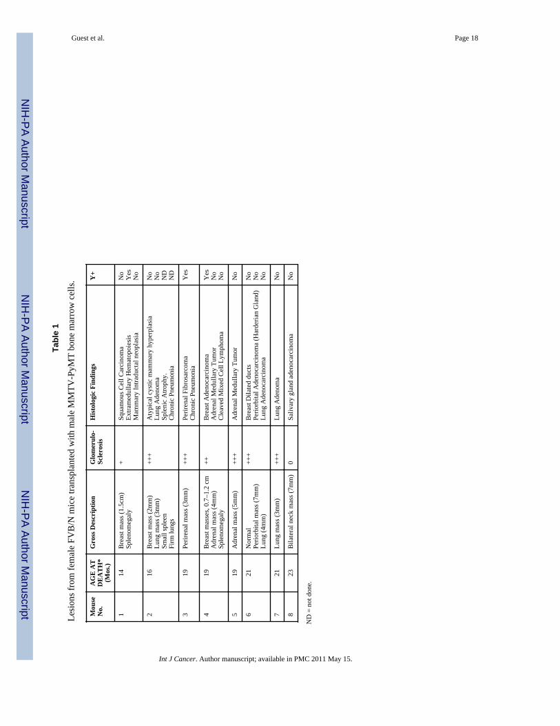
A summary of the results of an experiment in which a total of 8 female FVB/N mice wereirradiated with 900 to 1050 rads at 2 months of age and rescued by transplantation of 1–2 X10⁶ BM cells from transgenic MMTV-PyMT FVB/N male donors is presented in Table 1.The BM-transplanted mice ranged in age from 14 to 22 months at the time of euthanasia as aresult of the size of the tumor masses or weight loss (in accord with the approved IACUCanimal welfare procedures). Examples of the tumors found are illustrated in Figure 1, A–H.These include a squamous cell carcinoma of the skin (found in the breast), lung adenomas, alung adenocarcinoma, adrenal medullary tumors, a Harderian gland adenocarcinoma, aperirenal fibrosarcoma, a diffuse mixed cell cleaved lymphoma [11], and a salivary glandadenocarcinoma, as well as one breast adenocarcinoma (BCA), one cystic adenomatousmammary hyperplasia and two mammary intraductal neoplasias (MIN).
The number of tumors in the experimental mice as compared to age matched untreatedcontrols and to a group of control WT female mice transplanted with WT bone marrow isgiven in Table 2. Statistical analysis using the two-tailed Mann-Whitney non-parametric testshows that the incidence of tumors in the experimental group is greater that in either controlgroup. The incidence of tumors in the WT to WT BM transplant group is marginallydifferent from that of the untreated controls. In historical controls 61% of 116 female FVB/N mice euthanized at 24 months had cancers, but there were no Harderian gland tumors,fibrosarcomas, salivary gland tumors or breast lesions [12]. In human studies, the cancer riskis 8.3 times higher in bone marrow transplant patients than in untransplanted normalcontrols [13], but the tissue origins of the human tumors (melanoma, oral cavity, liver, brain,and thyroid) are very different from the types of tumors seen in our study. The incidence oftumors in the WT control group is essentially the same as in the WT to WT group.
GlomeruloscerosisThe finding of severe glomerulosclerosis (Figure 1, I; for grading see Supplemental Figure3) in the bone marrow recipients is unexpected. None of the sacrificed untreated controlmice had glomerulosclerosis and glomerulosclerosis was not reported in the study that weused for comparison [12]. The possible role of irradiation and either normal BMtransplantation or transgenic BM transplantation in causing glomerulosclerosis is not known.However, most of the WT to WT BM transplant mice died with glomerulosclerosis andchronic pneumonia. Five of the PyMT to WT experimental group were euthanized due towasting apparently due to glomerulosclerosis and pneumonia; the tumors in 3 of these mice(#s 2, 5, and 7) were incidental. One of the reasons for the larger number of cancers in theWT compared to the WT to WT BM transplant group is that the mice in the WT groupsurvived much longer than the mice in the WT to WT group, so that the WT to WT BMtransplant group may not have lived long enough to get cancer.
Guest et al. Page 6
Int J Cancer. Author manuscript; available in PMC 2011 May 15.
NIH
-PA Author Manuscript
NIH
-PA Author Manuscript
NIH
-PA Author Manuscript
Ychromosome positive tumorsOf all of the tumors in the experimental group only the fibrosarcoma in recipient mouse 2and the breast cancer in recipient mouse 4 were positive for the presence of Ychromosomesby FISH analysis. Over 80% of the cells in these tumors were Y+ (Figure 1,N–S). Thefibrosarcoma in mouse 2 contained fibroblastic and liposarcomatous zones, and both zoneswere Y+ The margin of the tumor contained a layer of lymphocytes. These were clearlyseparated from the fibroblastic part of the tumor and uniformly contained Y-chromosomes(Supplemental figure 4)..
None of the other 9 tumors had Ychromosomes in the tumor cell nuclei (For examples seeFigure 1, J–M), and are most likely recipient derived. In all tissues of the recipients, therewere Y+ cells that could be identified by morphology as mononuclear cells, endothelial cellsand stromal cells, with by far the greatest number being stromal fibroblasts. For example,after transplantation of male bone marrow cells, the female recipient liver contained manyYchromosome positive endothelial, stellate, and Kuppfer cells, but less than 1/20,000positive hepatocytes (Figure 2, A,B). It is of interest that some of the recipient derivedtumors was surrounded by Y+ stromal cells (Figure 2, C,D), suggesting that the transgenicstromal cells may influence the malignant transformation of the recipient tissues. Todetermine if there might be ectopic expression of the PyMT oncoprotein, the onco-antigen intissues was stained by immunoperoxidase (Figure 2, E–H), by analysis of fibroblastscultured from a primary tumors in a PyMT mouse (Supplemental Figure 5) and by q PCRfor mRNA (see below). The antigen was readily seen in the primary breast cancer from thedonor mice and in the breast cancer in mouse 4, but was not seen in the other tumors. Inaddition, oncoprotein was not detected in the tumor stroma.
Presence of the transgene and transgene productThe transgene was readily identified in tumor stromal cells in cultured in vitro, but mRNAexpression was not detected in stromal cells isolated from primary tumors (SupplementalFigure 5). Molecular analysis on laser captured tissue to determine the presence of PyMTDNA and mRNA was not definitive. For example, as expected the breast cancer in mouse 4was strongly positive for PyMT DNA and moderately positive for mRNA. The Y+firbosarcoma was moderately positive for DNA, but weakly postitive to negative for mRNA.The squamous cell carcinoma (mouse 1) and the adrenal tumor (mouse 5) were also weaklypositive for PyMT DNA, and for mRNA. Detection of PyMT DNA and mRNA at low levelscould be due to tissue sampling as it is essentially impossible to extract tumor tissue withoutstroma. Samples believed to contain only stroma were weakly to moderately positive forDNA, and weakly positive for mRNA. Thus, low levels of production of PyMT by stromacannot be ruled out, but the accuracy of this method for detecting low levels of mRNA isquestionable.
Ploidy AnalysisTo test for the possibility of fusion of bone marrow derived cells with breast cells in thebreast cancer from mouse 4, DNA ploidy analysis was done on all tumors. The result forselected tumors is shown in Figure 2, I–L). This analysis definitively demonstrated that thebreast cancer from mouse 4, as well as the other cancers, was diploid, and no evidence offusion of donor and recipient cells (14–17) was found.
Tests for tumor cells in the donor bone marrowNext we tested the possibility that the bone marrow from the donor male mice containedearly breast cancer cells that metastasized from occult primary tumors at the time when theBM was taken for transplantation. Using cytokeratin (CK) staining for BCA cells it was
Guest et al. Page 7
Int J Cancer. Author manuscript; available in PMC 2011 May 15.
NIH
-PA Author Manuscript
NIH
-PA Author Manuscript
NIH
-PA Author Manuscript
shown that CK+ cells in the BM of human patients with BCA, also had a stem cellphenotype [18]. The BM of female MMTV-PyMT mice contains CK+ presumptive BCAmetastatic cells as early as 4 weeks of age [19]. The BM used in the present experiment wastaken from the male MMTV-PyMT donor at 12 weeks of age when no gross or microscopicbreast lesions were found in the donor. To test directly for occult metastatic cells, weexamined the BM of 12 week and 5 month old male MMTV-PyMT mice for the presence ofCD49f, a well known marker for mouse breast stem cells [20] and for BCAs [21], by directexamination of the BM and by flow cytometry. We were unable to detect any CD49fpositive cells in the male BM (Figure 2, M–P), even when the 5 month old mouse had largebreast masses. Thus, it is very unlikely that the bone marrow used for transplantation, whichwas taken at from MMTV-PyMT males at 12 weeks of age when no tumors were palpable,contained tumor cells. From these data we conclude that the breast cancer in femalerecipient #4 was directly derived from transplanted male bone marrow cells.
LymphomaAn additional finding of interest was a diffuse cleaved lymphoma [11] present in the spleenof the same mouse that had the BCA. This lymphoma, as well as an adrenal medullar tumorin mouse 4, was negative for Ychromosome. The lymphoma cells had medium sized cleavednuclei. In the older literature this type of lymphoma in the mouse was called reticulum cellsarcoma. The Y− tumor cells are surrounded by and infiltrated with Y+ lymphocytes in thespleen (Supplemental Figure 6). The adjacent follicles in the spleen contain many Y+ cellsand there are positive cells admixed with the lymphoma cells due to inclusion of non-lymphoma lymphocytes within the tumor. The lymphoma cells stained with anti-CD3 andnot with anti-CD45R or B220, indicating that it is a T-cell lymphoma. The finding that thelymphoma did not arise from the transplanted male BM suggests that either some femaleBM stem cells remained after the irradiation, or else that the lymphoma arose fromprogenitor cells later during lymphocyte development and not directly derived from the bonemarrow stem cells.
DiscussionThe development of a fibrosarcoma and a breast cancer derived from donor bone marrowafter irradiation and transplantation, as well as other cancers not derived from the bonemarrow donor suggests both an indirect microenvironmental effect of transgenicmesenchymal stem cells, as well as a direct transformation and a mesenchymal-epithelialtransition of pluripotent bone marrow derived stem cells.
The microenvironment is critical for tumor initiation (22,23), as well as for growth (22),invasion (24) and metastasis (25) of cancer. Recently the role of the tissue niche for normaldevelopment and tissue renewal has been critically evaluated and a role for the initiation andgrowth of cancer restated (26,27). For breast cancer (BCA), Bissell and co-workers havetermed the relationship between the tissue stromal cells and the mammary epithelial cells(MEC) as “dynamic reciprocity” (28). The development of BCA is associated with adramatic loss or aberration of basement membrane and myoepithelial cells and the gain ofperitumoral myofibroblasts (29–31). The present results suggest that the presence ofMMTV-PyMT bone marrow derived stroma induces various cancers in wild type recipientmice. At this time, it is not clear how this is accomplished. So far, we have been unable todemonstrate the presence of the polyoma-middle-T-antigen either in the tumors or in thestroma cells by immunolabeling and cultured transgenic tumor fibroblast cultures do notcontain mRNA for PyMT. However, the most likely explanation is that there is a low levelof ectopic expression by the stromal cells that induces malignant changes in the associatedtissues. This possibility is supported by a very low level of mRNA for PyMT in the Y-tumors and in some of the stroma. Thus, one possible explanation for the effect of the
Guest et al. Page 8
Int J Cancer. Author manuscript; available in PMC 2011 May 15.
NIH
-PA Author Manuscript
NIH
-PA Author Manuscript
NIH
-PA Author Manuscript
stroma is a low level of PyMT production that somehow induces tumors in recipient tissues.However, it is also possible that qPCR analysis is giving false positives when pushed todetect very low levels. Also, if the stromal cells are producing mRNA for PyMT, we wouldexpect to see Y+ sarcomas as in mouse 3, instead of Y− tumors of other tissues.
The Y+ fibrosarcoma suggests that in this mouse the transplanted BM mesenchymal celldirectly transformed to produce this cancer. This suggests that there may have beenexpression of the PyMT gene in this tumor. However, qPCR for mRNA was equivocal. Alow level was detected, much lower than that found in the Y+ BCA.
The presence of a Y+ BCA in the female recipient of a BM transplant from a male mouseproves the principle that cells in the BM marrow can give rise to breast cancer. There are atleast four possible explanations for this: 1) mesenchymal-epithelial transition of BMmesenchymal stem cells to breast cells, with activation of the MMTV promoter andexpression of the PYMT transgene [5–6]; 2) fusion of cells from the BM with breast cells,and activation of the transgene [14–17); 3) developmental mimicry [8]; and 4) the presenceof occult metastatic BCA cells in the BM donor that recirculate and home to the breast of therecipient [18,19. At this point, mesenchymal-epithelial transition is the most likelyexplanation. If there had been an early fusion event followed by reduction division, the cellsof the BCA could be diploid, but this sequence of events would require that the diploid cellsof the tumor proliferate at a higher rate and crowd out the polyploid cells. Reductiondivision has been proposed as a possibility in the liver cells of experimental tyrosinenemicmice with severe liver injury restored by normal BMDCs [32]. However, the liver is knownto contain polyploid cells that undergo reduction division in proliferative responses to liverinjury [33], a response not seen in most other tissues. In models where fusion events havebeen described, most of the fused cells persist in the tissues without reduction division [32].The number of Y+ positive cells in the tumor is much too high to explained bydevelopmental mimicry, in which only about 1:10,000 cells in a tumor is reported to bederived from a BMDC [8]. However, it is possible that a single BMDC was influenced bynormal mammary epithelial cells (MEC), such that the BMDC acquired the properties of theMEC and activated the MMTV promoter. Thus fusion, developmental mimicry, and occulttumor cells in the BM each appear highly unlikely.
From these considerations, we conclude that the BCA found in mouse 4 arose from amesenchymal stem cell in the BM that underwent mesenchymal-epithelial transition toactivate the MMTV promoter, expressed the PyMT antigen and underwent malignanttransformation. This is not without precedent. After treatment of BM cells with 3’-methycholanthere the transformed BM cells formed many tumor types including epithelialtumors in vitro and on subcutaneous transplantation in vivo [34]. Using DNA chimerism,Golfinopouloss et al. [35] reported a donor-derived breast cancer in a woman who received aBM transplant from her sister 14 years earlier. However in a study of 37 patients whodevelopment various cancers after BM transplantation Au et al. [36] found no donor derivedtumors.
Epithelial-mesenchymal transition (EMT) is defined as the loss of epithelial characteristicsand the acquisition of a mesenchymal phenotype. EMT is an essential component ofmetazoan embryogenesis [37]. In breast cancer EMT is associated with increasedaggressiveness, invasiveness and metastasis [38]. However, it is debated as to whether EMTis an example of transdifferentiation of epithelial cells to mesenchymal cells [39], or anexpression of the pluripotency of breast cancer stem cells [40]. In any case, EMT representsa progression of a BCA to a more malignant phenotype, leading to expression ofmesenchymal-like products and behavior. One of the observations that confound proponentsof EMT is the fact that metastatic breast cancers have histologic and metabolic features
Guest et al. Page 9
Int J Cancer. Author manuscript; available in PMC 2011 May 15.
NIH
-PA Author Manuscript
NIH
-PA Author Manuscript
NIH
-PA Author Manuscript

similar to the primary epithelial tumor. This is explained by a process whereby EMT-mediated invasion metastases is followed by a reverse mesenchymal-epithelial transition(MET) to allow colonization of secondary sites [40]. The ability of the cancer cells duringthis process to express both epithelial and mesenchymal phenotypes was referred to as a“metastable phenotype” [41]. Our results support the hypothesis that mesenchymal cellsmay transdifferentiate into breast cancer cells or contribute to development of BCA by cell-cell interactions or fusion. Fusion of human BCA cells with mouse stromal cells afterinjection of the human BCA cells into the mammary glands of nude mice has been reported[42]. However, since the BCA in mouse #4 is diploid, there is no evidence for fusion as themechanism in this study.
The polyoma middle T-antigen transgenic mouse (FVB.PyMT) model used in the presentexperiments is a widely used model of human breast cancer, enabling the analysis of genesand pathways involved in tumor progression from premalignant disease to metastases [43–47]. Mammary gland tumors occur spontaneously in all homozygous carriers, with palpabletumors appearing in females as early as 4–5 weeks of age and in males at a median age of 83days [43]. The appearance of BCA correlates with transgene expression in the breastepithelium. Lower amounts of the middle T antigen found in salivary glands, ovaries andepididymis are not accompanied by any growth abnormalities [43], but hyperplasia andtumors in the lung and salivary gland can be induced in MMTV-RARβ4 transgenic micetreated with dexamethasone [48]. We did find one salivary gland adenocarcinoma, but it didnot contain Ychromosomes or express PyMT antigen (mouse 8).
Thus we conclude that the transgenic BMD mesenchymal cells may indirectly contribute todevelopment of tumors derived from the recipient mice by creating a transformingmicroenvironment, and directly contribute by transformation of BMD mesenchymal cells tosarcoma and by mesenchymal-epithelial transition to form breast cancers. However,transdifferentiation of BMD hematopoietic stem cells cannot be ruled out at this time.
Brief Statements
1. Bone marrow derived cells may indirectly contribute to development of cancersof other organs by supplying stroma that supports development of cancer.
2. Bone marrow mesenchymal stem cells may transform to form sarcomas
3. Bone marrow stem cells may transdifferentiate to become breast cancer.
Supplementary MaterialRefer to Web version on PubMed Central for supplementary material.
Abbreviations used
BCA Breast cancer
BM Bone marrow
BMDCs Bone marrow derived cells
CD45 Marker for hematopoietic cells
CD49f Marker for breast stem cells and breast cancer cells
cDNA Complementary DNA
DNA Deoxyribonucleic acid
Guest et al. Page 10
Int J Cancer. Author manuscript; available in PMC 2011 May 15.
NIH
-PA Author Manuscript
NIH
-PA Author Manuscript
NIH
-PA Author Manuscript
FISH Fluorescence in-situ hybridization
FVB/N Inbred mouse strain
GI Gastrointestinal
HICS Heat-inactivated calf serum
HSC Hematopoietic stem cells
IACUC Institutional animal care and use committee
MSC Mesenchymal stem cells
MMTV Mouse mammary tumor virus (promoter)
mRNA Messenger RNA
PCR Polymerase chain reaction
PyMT Polyoma virus middle T oncoprotein
qPCR Quantitative polymerase chain reaction
RT-PCR Reverse transcriptase polymerase chain reaction
AcknowledgmentsWe thank Merrill Ross of Albany Medical College for the ploidy analysis and Anna Glinsky of Ordway ResearchInstitute for advice on flow cytometry.
Grant Support: The authors thank the NIH for continued support which was used to start this work (1-R01-CA112481 and 1-R01-AG023510), and to NYSTEM C023063, which allowed us to complete it. The contents ofthis manuscript are solely the responsibility of the authors and do not necessarily represent the official views ofNIH or NYSTEM.
References1. Sell S. Adult Stem Cell Plasticity. Stem Cell Reviews 2005;1:1–77. [PubMed: 17132868]2. Herzog EL, Chai L, Krause DS. Plasticity of marrow-derived stem cells. Blood 2003;102:3483–
3493. [PubMed: 12893756]3. Eisenberg LM, Eisenberg CA. Stem cell plasticity, cell fusion, and transdifferentiation. Birth
Defects Res Part C Embryo Today 2003;69:209–218.4. Okamoto R, Yajima T, Yamazaki M, Kanai T, Mukai M, Okamoto S, Ikeda Y, Hibi T, Inazawa J,
Watanabe M. Damaged epithelia regenerated by bone marrow-derived cells in the humangastrointestinal tract. Nature Med 2002;8:1011–1017. [PubMed: 12195435]
5. Houghton JM, Stoicov C, Nomura S, Rogers AB, Carlson J, Li H, Cai X, Fox JG, Goldenring JR,Wang TC. Gastric cancer originating from bone marrow-derived cells. Science 2004;306:1568–1571. [PubMed: 15567866]
6. Tsuchikawa T, Ikeda H, Kikuchi K, Tsuji T, Baba T, Ishizu A, Tanaka Y, Kato H, Toshiki T.Hematopoietic progenitor cells as possible origins of epithelial thymoma in a human T lymphocytevirus type I pX gene transgenic rat model. Lab Investig 2004;84:245–252. [PubMed: 14688799]
7. Aractingi S, Kanitakis J, Euvard S, Danff CL, Peguillet I, Khosrotehrani K, Lantz O, Carosella ED.Skin carcinoma arising from donor cells in a kidney transplant recipient. Cancer Res 2005;65:1755–1760. [PubMed: 15753371]
8. Cogle CR, Theise ND, Fu DT, Ucar D, Lee S, Guthrie SM, Lonergan J, Rybka W, Krause DS, ScottEW. Bone marrow contributes to epithelial cancers in mice and humans as developmental mimicry.Stem Cells 2007;25:1881–1887. [PubMed: 17478582]
Guest et al. Page 11
Int J Cancer. Author manuscript; available in PMC 2011 May 15.
NIH
-PA Author Manuscript
NIH
-PA Author Manuscript
NIH
-PA Author Manuscript
9. Theise ND, Badve S, Saxena R, Henegariu O, Sell S, Crawford J, Krause DS. Derivation ofhepatocytes from bone marrow cells in mice after radiation-induced myeloablation. Hepatology2000;31:235–240. 2000. [PubMed: 10613752]
10. Swenson ES, Guest I, Ilic Z, Mazzeo M, Lizardi P, Hardiman C, Sell S, Krause DS. Hepatocytenuclear factor-1 as a marker of epithelial phenotype reveals marrow–derived hepatocytes, but notduct cells, after liver injury in mice. Stem Cells 2008;26:1768–1777. PMID: 18467658. [PubMed:18467658]
11. Lukes RJ, Collins RD. Immunologic characterization of human malignant lymphomas. Cancer1974;34:1488–1503. [PubMed: 4608683]
12. Mahler JF, Stokes W, Mann PC, Takaoka M, Maronpot RR. Spontaneous lesions in aging FVB/Nmice. Toxicologic Pathology 1996;24:710–716. [PubMed: 8994298]
13. Curtis RE, Rowlings PA, Deeg HJ, Shriner DA, Socie G, Travis LB, Horowitz MM, WitherspoonRP, Hoover RN, Sobocinski KA, Fraumeni JF Jr, Boice JD Jr. Solid cancers after bone marrowtransplantation. N Eng J Med 1997;336:897–904.
14. Kerbel RS, Lagarde AE, Dennis JW, Donaghue TP. Spontaneous fusion in vivo between normalhost and tumor cells: possible contribution to tumor progression and metastasis studies with alectin-resistant mutant tumor. Mol Cell Biol 1983;3:523–538. [PubMed: 6687920]
15. Larizza L, Schirrmacher V. Somatic cell fusion as a source of genetic rearrangement leading tometastatic variants. Cancer Metastasis Rev 1984;3:193–222. [PubMed: 6388823]
16. Pawelek JM. Tumor cell hybridization and metastasis revisited. Melanoma Res 2000;10:507–514.[PubMed: 11198471]
17. Pawelek JM. Tumour-cell fusion as a source of myeloid traits in cancer. Lancet Oncol 2005;6:988–993. [PubMed: 16321767]
18. Balic M, Lin H, Young L, Hawes D, Giuliano A, McNamara G, Datar RH, Cote RJ. Most earlydisseminated cancer cells detected in bone marrow of breast cancer patients have a putative breastcancer stem cell phenotype. Clin Cancer Res 2006;12:5615–5621. [PubMed: 17020963]
19. Husemann Y, Geigl JB, Schubert F, Musiani P, Meyer M, Burghart E, Forni G, Eils R, Fehm T,Riethmuller G, Klein CA. Systemic spread is an early step in breast cancer. Cancer Cells2008;13:58–68.
20. Matulka LA, Triplett AA, Wagner KU. Parity-induced mammary epithelial cells are multipotentand express cell surface markers associated with stem cells. Develop Biol 2007;303:29–44.[PubMed: 17222404]
21. Stingl J, Eirew P, Ricketson I, Shackleton M, Vaillant F, Choi D, Li HI, Eaves CJ. Purification andunique properties of mammary epithelial stem cells. Nature 2006;439:993–997. [PubMed:16395311]
22. Liotta LA, Kohn EC. The microenvironment of the tumour-host interface. Nature 2001;411:375–379. [PubMed: 11357145]
23. Kalluri R, Zeisberg M. Fibroblasts in cancer. Nat Rev Cancer 2006;6:392–401. [PubMed:16572188]
24. DeWever O, Marell M. Role of tissue stroma in cancer cell invasion. J Pathol 2003;200:429–447.[PubMed: 12845611]
25. Fidler IJ. The organ microenvironment and cancer metastasis. Differentiation 2002;70:498–505.[PubMed: 12492492]
26. Moore KA, Lemischka IR. Stem cells and their niches. Science 2006;311:1880–1885. [PubMed:16574858]
27. Li L, Neaves WB. Normal stem cells and cancer stem cells: the niche matters. Cancer Res2006;66:4553–4557. [PubMed: 16651403]
28. Nelson CM, Bissell MJ. Of extracellular matrix, scaffolds, and signaling: tissue architectureregulates development, homeostasis, and cancer. Annu Rev Cell Dev Biol 2006;22:287–309.[PubMed: 16824016]
29. Bissell MJ, Radisky DC, Rizki A, Weaver VM, Petersen OW. The organizing principle:microenvironmental influences in the normal and malignant breast. Differentiation 2002;70:537–546. [PubMed: 12492495]
Guest et al. Page 12
Int J Cancer. Author manuscript; available in PMC 2011 May 15.
NIH
-PA Author Manuscript
NIH
-PA Author Manuscript
NIH
-PA Author Manuscript
30. Weigelt B, Bissell MJ. Unraveling the microenvironmental influences on the normal mammarygland and breast cancer. Seminars in Cancer Biology 2008;18:311–321. [PubMed: 18455428]
31. Ronnov-Jessen L, Bissell MH. Breast cancer by proxy: can the microenvironment be both thecause and consequence? Trends Mol Med 2009;15:5–13. [PubMed: 19091631]
32. Wang X, Willenbring H, Akkari Y, Torimaru Y, Foster M, Al-Dhalimy M, Lagasse E, Finegold M,Olson S, Grompe M. Cell fusion is the principal source of bone-marrow-derived hepatocytes.Nature 2003;422:897–901. [PubMed: 12665832]
33. Scott RJ, Chakroborty S, Sell S, Hunt JM, Dunsford HA. Change in the ploidy state of liver cellsduring chemical hepatocarcinogenesis and its relationship to the increased expression ofalphafetoprotein. Cancer Research 1989;49:6085–6090. [PubMed: 2477151]
34. Liu C, Chen Z, Shen X, Zhang T, Lu Y. Multiple tumor types may originate from bone marrow-derived cells. Neoplasia 2006;8:716–724. [PubMed: 16984729]
35. Golfinopoulos V, Pentheroudakis G, Kamakari S, Metaxa-Mariatou V, Pavlidis N. Donor-derivedbreast cancer in a bone marrow transplantation recipient. Breast Cancer Res Treat. 2008 DOI.1007/s10549-008-9922-7.
36. Au WY, Chan EC, Pang A, Lie AK, Liang R, Yuen AP, Shek TW, Kwong YL. Nonhematologicalmalignancies after allogeneic hematopoietic stem cell transplantation: incidence and molecularmonitoring. Bone Marrow Transplant 2004;34:981–985. [PubMed: 15502854]
37. Lee JM, Dedhar S, Kalluri R, Thompson EW. The epithelial-mesenchymal transition: new insightsin signaling , development and disease. J. Cell Biol 2006;172:973–981. [PubMed: 16567498]
38. Sarrio D, Bodriguez-Pinilla M, Hardisson D, Cano A, Moreno-Bueno G, Palacios J. Epithelial-mesenchymal transition in breast cancer relates to the basal-like phenotype. Cancer Res2008;68:989–997. [PubMed: 18281472]
39. Thompson EW, Newgreen DF. Carcinoma invasion and metastasis: a role for epithelial-mesenchymal transition? Cancer Res 2005;65:5991–5995. [PubMed: 16024595]
40. Tarin D. The fallacy of epithelial mesenchymal transition in neoplasia. Cancer Res 2005;65:5996–6000. [PubMed: 16024596]
41. Côme C, Arnoux V, Bibeau F, Savagner P. Roles of the transcription factors snail and slug duringmammary morphogenesis and breast carcinoma progression. J Mammary Gland Biol Neoplasia2004;9:183–193. [PubMed: 15300012]
42. Jacobsen BM, Harrell JC, Jedlicka P, Borges VF, Varella-Garcia M, Horwitz KB. Spontaneousfusion with, and transformation of mouse stroma by, malignant human breast cancer epithelium.Cancer Res 2006;66:8274–8279. [PubMed: 16912208]
43. Callahan R, Smith GH. MMTV-induced mammary tumorigenesis: gene discovery, progression tomalignancy and cellular pathways. Oncogene 2000;19:992–1001. [PubMed: 10713682]
44. Maglione JE, Moghanaki D, Young LJ, Manner CK, Ellies LG, Joseph SO, Nicholson B, CardiffRD. MacLeod: Transgenic Polyoma middle-T mice model premalignant mammary disease.Cancer Res 2001;61:8298–8305. [PubMed: 11719463]
45. Lin EY, Jones JG, Li P, Zhu L, Whitney KD, Muller WJ, Pollard JW. Progression to malignancy inthe polyoma middle T oncoprotein mouse breast cancer model provides a reliable model forhuman diseases. Am J Pathol 2003;163:2113–2126. [PubMed: 14578209]
46. Qiu TH, Chandramouli GV, Hunter KN, Alkharouf NW, Green JE, Liu ET. Global expressionprofiling identifies signatures of tumor virulence in MMTV-PyMT-transgenic mice: correlation tohuman disease. Cancer Res 2004;64:5973–5981. [PubMed: 15342376]
47. Maglione JE, McGoldrick ET, Young LJ, Namba R, Gregg JP, Liu L, Moghanaki D, Ellies LD,Borowsky AD, Cardiff RD, MacLeod CL. Polyomavirus middle T-induced mammaryintraepithelial neoplasia outgrowths: single origin, divergent evolution, and multiple outcomes.Mol Cancer Ther 2004;3:941–953. [PubMed: 15299077]
48. Berard, Kl; Gaboury, L.; Landers, M.; De Repentigny, Y.; Houle, B.; Kothary, R.; Bradley, WEC.Hyperplasia and tumours in lung, breast and other tissues in mice carrying a RARβ4-liketransgene. EMBO J 1994;13:5570–5580. [PubMed: 7988554]
Guest et al. Page 13
Int J Cancer. Author manuscript; available in PMC 2011 May 15.
NIH
-PA Author Manuscript
NIH
-PA Author Manuscript
NIH
-PA Author Manuscript
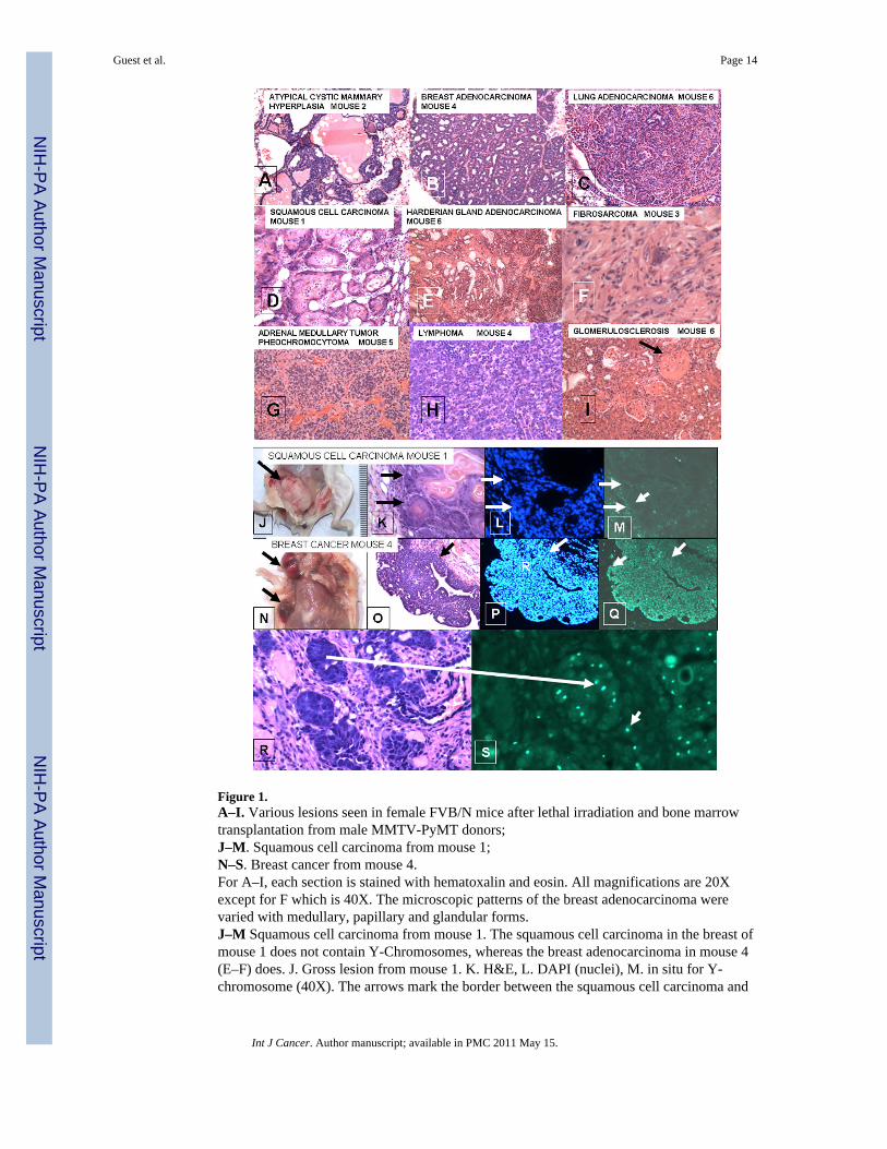
Figure 1.A–I. Various lesions seen in female FVB/N mice after lethal irradiation and bone marrowtransplantation from male MMTV-PyMT donors;J–M. Squamous cell carcinoma from mouse 1;N–S. Breast cancer from mouse 4.For A–I, each section is stained with hematoxalin and eosin. All magnifications are 20Xexcept for F which is 40X. The microscopic patterns of the breast adenocarcinoma werevaried with medullary, papillary and glandular forms.J–M Squamous cell carcinoma from mouse 1. The squamous cell carcinoma in the breast ofmouse 1 does not contain Y-Chromosomes, whereas the breast adenocarcinoma in mouse 4(E–F) does. J. Gross lesion from mouse 1. K. H&E, L. DAPI (nuclei), M. in situ for Y-chromosome (40X). The arrows mark the border between the squamous cell carcinoma and
Guest et al. Page 14
Int J Cancer. Author manuscript; available in PMC 2011 May 15.
NIH
-PA Author Manuscript
NIH
-PA Author Manuscript
NIH
-PA Author Manuscript
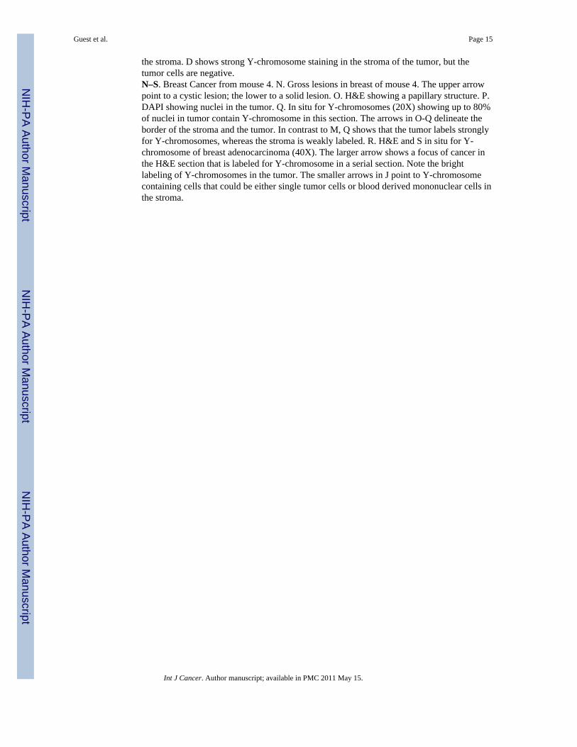
the stroma. D shows strong Y-chromosome staining in the stroma of the tumor, but thetumor cells are negative.N–S. Breast Cancer from mouse 4. N. Gross lesions in breast of mouse 4. The upper arrowpoint to a cystic lesion; the lower to a solid lesion. O. H&E showing a papillary structure. P.DAPI showing nuclei in the tumor. Q. In situ for Y-chromosomes (20X) showing up to 80%of nuclei in tumor contain Y-chromosome in this section. The arrows in O-Q delineate theborder of the stroma and the tumor. In contrast to M, Q shows that the tumor labels stronglyfor Y-chromosomes, whereas the stroma is weakly labeled. R. H&E and S in situ for Y-chromosome of breast adenocarcinoma (40X). The larger arrow shows a focus of cancer inthe H&E section that is labeled for Y-chromosome in a serial section. Note the brightlabeling of Y-chromosomes in the tumor. The smaller arrows in J point to Y-chromosomecontaining cells that could be either single tumor cells or blood derived mononuclear cells inthe stroma.
Guest et al. Page 15
Int J Cancer. Author manuscript; available in PMC 2011 May 15.
NIH
-PA Author Manuscript
NIH
-PA Author Manuscript
NIH
-PA Author Manuscript
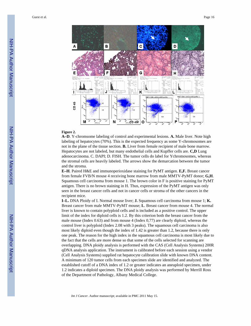
Figure 2.A–D. Y-chromsome labeling of control and experimental lesions. A. Male liver. Note highlabeling of hepatocytes (70%). This is the expected frequency as some Y-chromosomes arenot in the plane of the tissue section. B. Liver from female recipient of male bone marrow.Hepatocytes are not labeled, but many endothelial cells and Kupffer cells are. C,D Lungadenocarcinoma. C. DAPI; D. FISH. The tumor cells do label for Ychromosomes, whereasthe stromal cells are heavily labeled. The arrows show the demarcation between the tumorand the stroma.E–H. Paired H&E and immunoperioxidase staining for PyMT antigen. E,F. Breast cancerfrom female FVB/N mouse 4 receiving bone marrow from male MMTV-PyMT donor; G,H.Squamous cell carcinoma from mouse 1. The brown color in F is positive staining for PyMTantigen. There is no brown staining in H. Thus, expression of the PyMT antigen was onlyseen in the breast cancer cells and not in cancer cells or stroma of the other cancers in therecipient mice.I–L. DNA Ploidy of I. Normal mouse liver; J. Squamous cell carcinoma from mouse 1; K.Breast cancer from male MMTV-PyMT mouse; L. Breast cancer from mouse 4. The normalliver is known to contain polyploid cells and is included as a positive control. The upperlimit of the index for diploid cells is 1.2. By this criterion both the breast cancer from themale mouse (Index 0.63) and from mouse 4 (Index 0,77) are clearly diploid, whereas thecontrol liver is polyploid (Index 2.08 with 3 peaks). The squamous cell carcinoma is alsomost likely diploid even though the index of 1.42 is greater than 1.2, because there is onlyone peak. The reason for the high index in the squamous cell carcinoma is most likely due tothe fact that the cells are more dense so that some of the cells selected for scanning areoverlapping. DNA ploidy analysis is performed with the CAS (Cell Analysis Systems) 200RqDNA analysis application. The instrument is calibrated before each session using a vendor(Cell Analysis Systems) supplied rat hepatocyte calibration slide with known DNA content.A minimum of 120 tumor cells from each specimen slide are identified and analyzed. Theestablished cutoff of a DNA index of 1.2 or greater indicates an aneuploid specimen, under1.2 indicates a diploid specimen. The DNA ploidy analysis was performed by Merrill Rossof the Department of Pathology, Albany Medical College.
Guest et al. Page 16
Int J Cancer. Author manuscript; available in PMC 2011 May 15.
NIH
-PA Author Manuscript
NIH
-PA Author Manuscript
NIH
-PA Author Manuscript
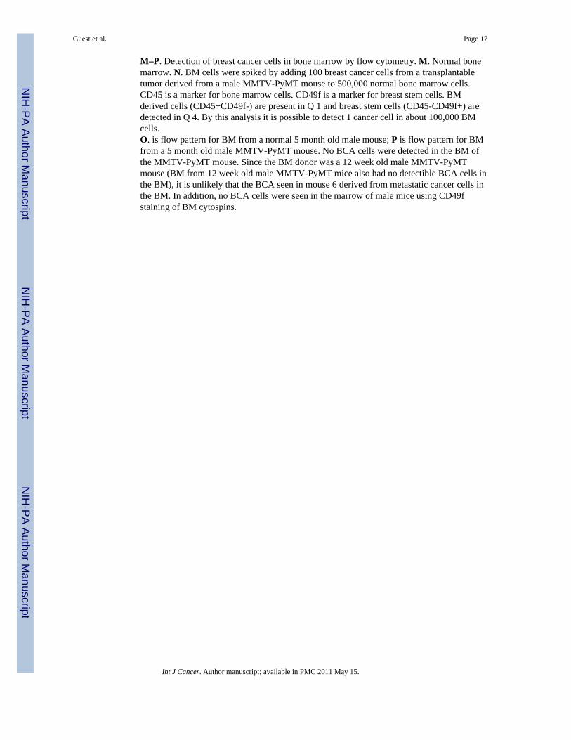
M–P. Detection of breast cancer cells in bone marrow by flow cytometry. M. Normal bonemarrow. N. BM cells were spiked by adding 100 breast cancer cells from a transplantabletumor derived from a male MMTV-PyMT mouse to 500,000 normal bone marrow cells.CD45 is a marker for bone marrow cells. CD49f is a marker for breast stem cells. BMderived cells (CD45+CD49f-) are present in Q 1 and breast stem cells (CD45-CD49f+) aredetected in Q 4. By this analysis it is possible to detect 1 cancer cell in about 100,000 BMcells.O. is flow pattern for BM from a normal 5 month old male mouse; P is flow pattern for BMfrom a 5 month old male MMTV-PyMT mouse. No BCA cells were detected in the BM ofthe MMTV-PyMT mouse. Since the BM donor was a 12 week old male MMTV-PyMTmouse (BM from 12 week old male MMTV-PyMT mice also had no detectible BCA cells inthe BM), it is unlikely that the BCA seen in mouse 6 derived from metastatic cancer cells inthe BM. In addition, no BCA cells were seen in the marrow of male mice using CD49fstaining of BM cytospins.
Guest et al. Page 17
Int J Cancer. Author manuscript; available in PMC 2011 May 15.
NIH
-PA Author Manuscript
NIH
-PA Author Manuscript
NIH
-PA Author Manuscript
NIH
-PA Author Manuscript
NIH
-PA Author Manuscript
NIH
-PA Author Manuscript
Guest et al. Page 18
Tabl
e 1
Lesi
ons f
rom
fem
ale
FVB
/N m
ice
trans
plan
ted
with
mal
e M
MTV
-PyM
T bo
ne m
arro
w c
ells
.
Mou
seN
o.A
GE
AT
DE
AT
H*
(Mos
.)
Gro
ss D
escr
iptio
nG
lom
erul
o-Sc
lero
sis
His
tolo
gic
Find
ings
Y+
114
Bre
ast m
ass (
1.5c
m)
Sple
nom
egal
y+
Squa
mou
s Cel
l Car
cino
ma
Extra
med
ulla
ry H
emat
opoi
esis
Mam
mar
y In
tradu
ctal
neo
plas
ia
No
Yes
No
216
Bre
ast m
ass (
2mm
)Lu
ng m
ass (
3mm
)Sm
all s
plee
nFi
rm lu
ngs
+++
Aty
pica
l cys
tic m
amm
ary
hype
rpla
sia
Lung
Ade
nom
aSp
leni
c A
troph
y.C
hron
ic P
neum
onia
No
No
ND
ND
319
Perir
enal
mas
s (3m
m)
+++
Perir
enal
Fib
rosa
rcom
aC
hron
ic P
neum
onia
Yes
419
Bre
ast m
asse
s, 0.
7–1.
2 cm
Adr
enal
mas
s (4m
m)
Sple
nom
egal
y
++B
reas
t Ade
noca
rcin
oma
Adr
enal
Med
ulla
ry T
umor
Cle
aved
Mix
ed C
ell L
ymph
oma
Yes
No
No
519
Adr
enal
mas
s (5m
m)
+++
Adr
enal
Med
ulla
ry T
umor
No
621
Nor
mal
Perio
rbita
l mas
s (7m
m)
Lung
(4m
m)
+++
Bre
ast D
ilate
d du
cts
Perio
rbita
l Ade
noca
rcin
oma
(Har
deria
n G
land
)Lu
ng A
deno
carc
inom
a
No
No
No
721
Lung
mas
s (3m
m)
+++
Lung
Ade
nom
aN
o
823
Bila
tera
l nec
k m
ass (
7mm
)0
Saliv
ary
glan
d ad
enoc
arci
nom
aN
o
ND
= n
ot d
one.
Int J Cancer. Author manuscript; available in PMC 2011 May 15.

NIH
-PA Author Manuscript
NIH
-PA Author Manuscript
NIH
-PA Author Manuscript
Guest et al. Page 19
Table 2
Incidence of cancers in control and experimental groups.
GROUP INCIDENCE % TIME TO DEATH
MEAN RANGE
1. PyMT to WT 8/8 100 19 14–23
2. WT to WT 1/16 6 13.5 6–21
3. UNTREATED 7/21 33 26 18–30
STATISTICAL ANALYSIS.
1 VS. 2 (p<0.001)
1 VS. 3 (p<0.001)
2 VS. 3 (p<0.065)
Int J Cancer. Author manuscript; available in PMC 2011 May 15.