Briefing: Xpert MTB/RIF for rapid diagnosis of TB and MDR-TB
Upload
khangminh22Category
view
0download
0
38
1. ABSTRACT
Tuberculosis (TB) is an infectious disease caused by different strains of Mycobacterium tuberculosis complex. TB is a curable infection if diagnosed correctly and timely. Late diagnosis and improper treatment may lead to relapse or its escalation to MDR / XDR-TB. TB with HIV co-infection is difficult to diagnose by conventional set-up. Also, detection of child TB and extrapulmonary TB has its own set of problems and is not straightforward to diagnose.
The increasing complexity of TB due to the advent of new circulating strains and invasion to the regions where it was non-existent or thought to be eradicated is putting a severe strain on the health management services and making it an unmanageable pandemic. This increasing complexity has led to the spurt in the development of TB diagnostics platforms. This review focuses on the new emerging technologies that have changed the diagnostics landscape.
Diagnosis of TB: From conventional to modern molecular protocols
Pranveer Singh1, Vikas Kumar Saket2, Ramsevak Kachhi2
1Department of Zoology, Mahatma Gandhi Central University (MGCU) Motihari-845401, Bihar, India, 2Department of Zoology, Indira Gandhi National Tribal University (IGNTU), Amarkantak, 484887 (MP), India
TABLE OF CONTENTS
1. Abstract2. Introduction
2.1. Search strategy and selection criteria3. Sputum smear microscopy (SSM)4. Imaging based diagnosis: future of automated microscopy5. Sample collection6. Latent tuberculosis and skin testing
6.1. Culture based drug resistance testing (DST)6.2. Phage amplification technique (PAT)
7. Genotypic methods8. Immunological protocols
8.1. TB biomarker assays8.2. Interferon-Gamma Release Assays (IGRA)
9. Volatile organic chemicals (VOCs)10. Nucleic acid amplification tests (NAAT)
10.1. Commercial NAATs based protocols10.2. Xpert MTB/RIF assay for the diagnosis of PTB10.3. Xpert MTB/RIF assay for the diagnosis of extrapulmonary tuberculosis (EPTB)10.4. Semi-automated NAATs for use in peripheral laboratories10.5. Spoligotyping 10.6. Line Probe Assay (LPA)10.7. Microarray-based platforms
11. Whole genome sequencing11.1. New sequencing protocols
12. Assays being developed / evaluated upcoming/pipeline technologies13. Conclusions14. Acknowledgements 15. References
[Frontiers In Bioscience, Elite, 11, 38-60, Jan 1, 2019]

Diagnostic approaches in tuberculosis
39 © 1996-2019
2. INTRODUCTION
Tuberculosis (TB) is a transmissible disease caused by the bacterium Mycobacterium tuberculosis (MTB). The bacterium normally targets the lungs causing pulmonary TB (PTB). It can also infect and spread to the other parts of the body like kidney, spine and brain causing extra pulmonary TB (EPTB) there. It usually spreads through the air by means of respiratory droplets through the sneeze or cough of an infected person. For diagnosis of pulmonary TB (PTB) which is more common, samples from respiratory tract like sputum, induced sputum, broncho aleveolar lavage or lung biopsy is usually done. Diagnosis of extrapulmonary TB (EPTB) depends on the site of infection, e.g. stool sample for intestinal TB to detect Mycobacterium avium as in the case of AIDS patients; other samples may include biopsies, aspirates, pus, urine, and sterile body fluids like cerebrospinal, synovial, pleural, pericardial, and peritoneal fluids (1-3).
Tuberculosis continues to be the major cause of mortality despite all sorts of developments in medicare including diagnostics and medicines. It poses continued threat to the health programs and services in resource poor set-ups. Co-infection with HIV is the new factor that is responsible for the escalation of TB (9% of global TB incidence) (4). Majority of infection in human results in an asymptomatic latent infection and about one in ten latent infection progresses to active TB if left untreated, killing more than 50 percent of those infected. The situation becomes aggravated further due to its co-infection with HIV and incomplete treatment worsening it further to relapse cases and emergence of drug resistance in the form of multi drug resistant (MDR) and extensively drug resistant (XDR)-TB. MDR-TB develops due to bacterial resistant to first line TB drugs like rifampicin (RIF) and isoniazid (INH), while XDR-TB occurs due to bacterial resistant to fluoroquinone (FLQ), amikacin (AMK), kanamycin (KAN) and Capreomycin (CPM). Tuberculosis is difficult to diagnose due to the late onset of symptoms, so that a person acts as a reservoir of TB bacilli with potential to infect others. Similarly, co-infection with HIV makes TB difficult to detect via conventional sputum smear microscopy (SSM) / X-ray. Late diagnosis coupled with incomplete treatment escalates it to MDR and XDR-TB. Although MDR-TB is curable via second line anti-TB drugs, therapy is ineffective, marred with toxic effects and require almost 2 years of treatment which is certain to have its side effect (1-3).
20th century has seen spurt of development in TB diagnostics and medicare from conventional sputum microscopy to Nucleic acid amplification test (NAAT) based molecular and biosensing based protocols, culminating into genome sequencing. This has generated a ray of hope for the timely and accurate diagnosis and treatment, hence eradication of TB.
However, it is still the giant killer and major cause of mortality and morbidity. Main cause of concern is the emergence of TB bacilli in the regions where it is thought to be completely eradicated. Irrespective of the evolution of fancy and improvised technologies for TB diagnosis SSM and culture based (Lowenstein-Jensen (LJ) media) are still the most reliable and preferred diagnostics of choice for detecting AFB causing active TB, particularly for resource limiting set up of developing countries (2-5).
Timely detection and effective treatment are essential for managing tuberculosis. However, the rate of global case detection is considerably low. The diagnosis of pediatric TB and EPTB is particularly difficult and there are, as yet, no rapid screening tests that can help finding active cases in the target population. Tuberculosis diagnosis is primarily based on the culture method and non-culture methods. Recently, it is complemented by cutting edge molecular and genome sequencing techniques that have increased the sensitivity of TB diagnosis manifold and also provide epidemiological information about it prevalence, INTRODUCTION of new strains or cause of relapse etc (2-3).
This systematic review has incorporated a decadal growth in TB diagnostics from conventional SSM to NAAT based molecular and sequencing based protocols. The review also highlights the TB diagnostics in varied set-ups focusing on challenges of customized diagnostics. In addition, review elucidates on currently available and upcoming protocols and barriers to their successful uptake. The review further focuses on the advantage and disadvantages of each technique / protocol and intends to complement earlier reviews and reports, stimulating discussion and informing potential opportunities for market intervention to improve access to effective TB diagnostics.
2.1. Search strategy and selection criteria
The references for this review were identified through searches of PubMed for articles published from 2005 to 2016 by the use of terms “Tuberculosis”, “diagnostics”, “DST”, “SSM”, “NAAT”. Relevant articles published during the aforementioned period were identified through searches from secondary sources (published reports, peer reviewed papers, WHO policies, UNITAID landscape report, and developer’s website).
3. SPUTUM SMEAR MICROSCOPY (SSM)
Time tested diagnosis for active tuberculosis (TB) involves detection of Mycobacterium tuberculosis complex bacilli in specimens from the respiratory tract for PTB or specimens from other bodily sites for EPTB. Microscopic analysis of sputum smear detects AFB by

Diagnostic approaches in tuberculosis
40 © 1996-2019
Ziehl-Neelsen(Z-N) staining (6). Sputum test is good indicator of bacterial load hence the infectivity. It is the most common, rapid and economical diagnostic method for active PTB diagnosis but it has lower sensitivity, when bacterial load is less than 10,000 bacilli / ml of sputum (Table 1). However, sensitivity can be improved by fluorescent staining (auramine-rhodamine) coupled with LED microscopes but it fails to distinguish between tuberculous and nontuberculous bacteria. Other limitations include prevalent epidemiological factors in the region of interest and skill of laboratory technician (7-8) (Table 1). Laserson et al. (2005) (9) in their study of Viatnamese immigrants have observed that incubation of sputum with detergent C18-Carboxypropylbetaine (CB-18) for 24 hours improves the sensitivity of AFB staining by 20-30% when compared to direct auramine and Z-N staining (9). Sputum analysis cannot detect EPTB. There might be a possibility that bacilli resides in cavities of lungs and may not be there in sputum. Decontamination process of sputum for mycobacterial culture may lead to the loss of bacilli from the sputum.
In addition, HIV patients and children might found it difficult to produce sputum. These constraints severely limit the utility of this very simple and economical diagnostics method10.
TBDx developed by signature mapping medical sciences, USA is an automated high-thoughput system with the capacity of 200 slides. It utilizes robotic loading of stained slides. Results are rapid in few minutes in the form of high resolution digital image (8, 11-12) (Table 1).
4. IMAGING BASED DIAGNOSIS: FUTURE OF AUTOMATED MICROSCOPY
Priority based treatment depending on the severity of symptoms have the potential to discriminate amongst those at risk from the people bearing symptoms of PTB but do not actually have TB. Chest X-ray (CXRs) serves as a potent algorithm in mathematical analysis of TB epidemiology. CXR integrated with user friendly, robust interface has evolved into an automated
1. SSM Developed by Robert Koch (1870)
Imaging Sputum Bacterial loadindicator, Rapid,economical
Fails to differentiate between tubercularand non- tuberculour, bias of technician, cannot detectEPTB
Lower, could beincreased byfluorescent staining or byincubating with CB-18 (20-30%)
Low
2. TBDx Signature mapping medical sciences, USA
Imaging Sputum High-thoughput (200slides), roboticloading of stained slides, output asdigitized images
High tier infrastructure and trained manpowerrequired, a limitationin low economical set-up
High High
3. CellScope UC Berkeley /CITRIS /Banatao Instit.
Fluoresence Microscope
Blood sample
High resolution enlarged digitizedimage
Lack ofaccess in under developed countries,Insufficient studies tovalidate its utility
Sufficientstudy /data required
Sufficient study /data required
4. CXRsalgorithm CAD4TB
Imaging Chest X-ray
Rapid detection of active TB particularly in HIV co-infection, can be interfaced with other such platform, detects both active and passive TB
Trained expertise required
High High
5. Easyport able DCXR
(Delft ImagingSystems)
Imaging Chest X- ray
Capacity to hold 50,000 DCXR images, can be operated from a laptop
Trained expertise required
High High
6. riView-TB Advenio (Chandigarh, India)
Imaging,CAD4TB enabledautomated imageanalys issoftware similar toDCXR
Chest X-ray
Easy to interpret images, simultaneos diagnosis of pneumo nia and silicosis to complementPTB diagnosis
Prior training in clinical radiography required
High High
Table 1. Imaging techniques

Diagnostic approaches in tuberculosis
41 © 1996-2019
algorithm to quantify abnormalities from digital CXR (DCXR) images. DCXR can be utilized for both active and passive TB diagnosis. It can be interfaced with other such platforms like as Xpert® MTB/RIF for rapid detection of active TB particularly in HIV co-infection setup. It is an economical tool with high sensitivity and specificity and could be a tool of need in low resource set-up (Table 1). Delft Imaging Systems (Netherlands) have come up with a software to score CXR image at the rate of one per minute. Fourth version of Computer aided detection for tuberculosis (CAD4TB) (Delft Imaging Systems) can also be integrated with DCXR platform (13-14). EasyPortable DCXR (Delft Imaging Systems) fits the requirement of limited resource set up. It includes an X-ray detector, CAD4TB software and Picture Archive and Communication System (PACS) with a capacity to hold 50,000 DCXR images and can operate from a laptop (2) (Table 1).
Advenio (Chandigarh, India) has launched riView-TB, a CAD4TB enabled automated image analysis software similar to DCXR. This platform provides easy to interpret images without prior training in clinical radiography. It is slated to be improvised for simultaneous diagnosis of pneumonia and silicosis to complement PTB diagnosis (2) (Table 1).
CellScope, a mobile cell microscope (CITRIS, University of California, Berkeley, San Francisco) is another innovation in the series of next generation microscopy. It is a fluorescence microscope that gives output in the form of high resolution enlarged digitized fluorescent images of blood samples to detect rod shaped bacterium (8, 15) (Table 1).
5. SAMPLE COLLECTION
A good quality sample in sufficient amount is a prime requirement for accurate diagnosis. This becomes a limitation with children and HIV patients who cannot cough up enough sputum. Lung flute from Medical Acoustics (USA) allows the patient to collect their own sample when they are not able to expectorate enough volume. It consists of a plastic device held via lips to exhale air into it. This generates vibrations in the lungs that soften and liquefy the sputum enabling patient to cough out more sputum (16).
Deton Corp. (USA) has come up with one liter bag to collect microdroplets presumed to contain MTB bacilli. These microdroplets are used for diagnosis (3). DNA Genotek Inc. (Ottawa, Canada) has developed OMNIgene® SPUTUM to liquefy and decontaminate the sputum. This facilitates its transport without cold chain for further diagnosis via microscopy, culture, Xpert® MTB/RIF and other molecular diagnostic protocols. prepIT® MAX technology from DNA Genotek Inc. (Ottawa, Canada) enables release of nucleic acid from MTBC cells via chemical lysis (3).
PrimeStore Molecular Transport Medium® (PS-MTM®) from Longhorn Vaccines & Diagnostics (USA) provides a NAAT enabled solution to inactivate, lyse and stabilize MTB DNA without cold chain. The genetic material produced can be utilized for range of other TB tests. PS-MTM® has further diversified into range of products like PrimeSwab™ and PS-MTM® for sample preparation, PrimeSwab™ and PS-MTM® for DNA extraction (PrimeXtract™) and PrimeMix® for real time PCR assay. Molzym GmbH & Co. KG (now Molzym) has launched the MTB-DNA Blood kit (25 to 50 reactions) that removes excess human genomic DNA and concentrates MTB DNA (3).
6. LATENT TUBERCULOSIS AND SKIN TESTING
Tuberculosis may remain latent in an infected patient before reactivation. This is one amongst the most challenging aspect in TB diagnostics. The advent of ELISPOT assay has provided promise to test for latency. This test involves the quantification of interferon production from whole blood or peripheral blood mononuclear cells (PBMC) stimulated with either purified protein derivative (PPD) or specific antigens (17-19). Currently available QuantiFERON®-TB Gold Test (Cellestis Ltd, WA, USA) for latent and active TB utilizes ESAT-6 or CFP-10 antigens (absent from BCG vaccine) (Table 4). Although the assay got the FDA approval it is yet to be commercialized. MPB skin patch test is another test with very high specificity and sensitivity (20-21). This test is free from reader bias, do not give false positive results due to booster phenomena, free from the effect of prior BCG vaccination and require only one patient visit to give sample and get result (22) (Table 4).
6.1. Culture based drug resistance testing (DST)
Increasing global burden of TB has diversified the diagnostic protocols for TB detection. Various phenotypic methods that include nitrate reductase assay (NRA / Griess method), thin-layer agar (TLA), colour test (Color Test), microscopic observation drug susceptibility assay (MODS), colorimetric redox indicator (CRI) method and phage-based assays can be directly set on the clinical samples and do not have to undergo sample preparation like decontamination process (2, 23-24). These methods can detect MDR-TB including resistance to the second line drugs like INH and RIF. Among different phenotypic methods available, MODS is widely validated and has perfect synergy with conventional drug susceptibility test for INH, RIF and MDR-TB (100%, 97% and 99%). MODS is rapid, economical and works well with all types of primary clinical samples and isolates. MODS depend on light microscopy. However, it requires a requisite expertise that involves extensive manpower training (25-27). The effectiveness of MODS for HIV

Diagnostic approaches in tuberculosis
42 © 1996-2019
co-infection is not yet established. Further, this test is only approved for INH and RIF leaving other first and second line TB drugs. TLA has also shown the similar promise for the detection of MDR-/XDR-TB colour test, MTB complex and resistance to INH, RIF and ciprofloxacin (CFP) in cultures (27), details given in Table 4.
TK MEDIUM® SLC-L (Salubris Inc. USA) has developed a liquid media that provides an advantage over conventional decontamination method using sodium hydroxide and N-acetyl cysteine. It does
not require centrifugation step thus completing the whole process in around 25 minutes. It shows better sensitivity over LJ for MTB detection (2) (Table 4). Gonzalo et al (2014) (27) have used Biphasic media assay (BMA) having two media L-J and Kirchner medium in tubes with 66mg/ml of pyrazinamide (PZA) to grow PZA susceptible and resistant strains of MTB (27). Reproducible results have shown the majority of starins to be susceptible to PZA while few were resistant. BMA might be a useful low cost, culture based alternative for DST for detecting PZA resistance particularly where MDR-TB is prevalent.
Table 4. Culture based phenotypic assays
S.No. Assay Manufacturer Method Material Advantage Disadvantage Sensitivity % (95% CI)
Specif city % (95% CI)
1. MODS MODS (Hardy diagnostics)
DST Sputum Directly set on the clinical samples and do not have to undergo sample preparation like decontamination process, detect MDR-TB including resistance to the second line drugs like INH and RMP; Handling risk is entirely avoided, Safer as performed in closed system
Daily observation of culture, risk of laboratory transmission, requirement of inverted microscope, require extensive manpower training
High High
2. NRA To be marketed DST on LJ medium With KNO3 (ability of MTB to reduce nitrate to nitrite)
Sputum Simple to perform with good results, inexpensive reagents, advantage in resource limited settings
Culture time is 3-4 times of MODS, Require mature MTB culture to open to add specific reagent
High High
3. TLA To be marketed DST on solid medium
Sputum Rapid, Accurate, Standard microscope Required which is Easily available, cost effective
Contamination rate is higher, data on DST is lacking
Low Low
4. CRI To be marketed Indirect, done on MTB isolates grown from conventional culture
Sputum Less equipment and infrastructu, Consumables that are readily available
Biosafety level 3 required, additional staff skill
High High
5. TKME DIU M® SLC-L
Salubris Inc. USA
Colorimetry Sputum Rapid, does not require centrifugation step, no media preparation required, lower contamination
Lack of automation, Disadvantageous in terms of turn-around time, Future studies in differential settings are required
Better sensitivity over LJ for MTB detection
High
6. Accuprobe Gen-Probe Inc Chemilumi nescent acridinium ester-labelled (AE)-DNA Probes (AE-MAC) probe
Sputum Simple and rapid, Readily available In market, Validated
Labor intensive High High

Diagnostic approaches in tuberculosis
43 © 1996-2019
6.2. Phage amplification technique (PAT)
Phage Amplification Technique (PAT) is a bacteriophage based test that can detect MTB in sputum. Non-pathogenic mycobacteria (sensor cells) are used as a negative control. The phage replicate, infect and lyse the sensor cells leaving zones of clearing (holes) in the agar conforming that sputum contains MTB. This diagnostic protocol is rapid (2 days), sensitive, and requires less infrastructure. However, it can only be applied to sputum samples and requires expert manpower (28).
7. GENOTYPIC METHODS
Genotypic methods target the nucleic acid of the mycobacterium. Ribosomal rRNA, which has universal occurrence, abundance, and conserved specific sequences can be a very useful genetic marker. Accuprobe (Gen-Probe Inc) detection method targets rRNA. This method can detect M tuberculosis complex, M avium, M intracellulare, M avium complex, M gordonae, and M kansasii. Accuprobe is simple and rapid with accuracy around 90%. However, it covers a limited range of species (Table 4). Peptide nucleic acids (PNA) based assay provides another approach to differentiate M. tuberculosis complex from NTM cultures29. PCR based restriction enzyme analysis of the hsp65 also provides an economical alternative for mycobacterium detection from culture. However, technique is complicated and labor intensive (22,30). Similarly, sequencing 16sRNA can also provide rapid detection of mycobacterium but it is labor intensive (22, 31).
8. IMMUNOLOGICAL PROTOCOLS
Detection of antibodies like lipoarabinomannans (LAM, cell wall component of Mycobacteria), A60, 38Kd and 16 Kd against MTB is a simple and economical method for EPTB detection. However, inconsistency in results as covered by meta-analyses and systematic reviews hampers its commercialization (3,32-33).
Determine™ TB LAM Ag rapid assay (Alere Inc.) is an immune-chromatographic strip that detects LAM antigen in urine. Surprisingly, a ‘negative result’ does not rule out the possibility of TB. Test is also vulnerable to ‘false positives’ due to contamination with dust or faeces. For test to be accurate, the CD4 counts/mm3 should not be less than 100 and best results are obtained with CD4 counts around 200 counts/mm (3, 32-35). It detects NTMs in addition to MTBC along with the site of mycobacterial infection (Table 5).
8.1. TB biomarker assays
The T-cell activation marker-tuberculosis (TAM-TB) assay is a flow cytometry based protocol
that detects fluorescently stained TB antigen-specific CD4 T-cells counterstained with an antibody to CD127. It measures the level of CD127 in the CD4 cells challenged with TB antigens (36) (Table 5).
8.2. Interferon-Gamma Release Assays (IGRA)
This test utilizes the ability of MTB antigens like Early Secretory Antigen Target 6 (ESAT-6) and Culture Filtrate Protein 10 (CFP-10) to stimulate the host for the production of interferon –gamma (IFN-γ). These antigens are absent in NTM or in BCG vaccine hence can differentiate between latent TB infection, BCG immunization and NTM infection. This procedure is simple and rapid. The commercially available assays for IFN-γ are; the Quantiferon-TB Gold (Cellestis) and T Spot-TB test (Oxford Immunotech). Intergam Rapid Immuno Suspension Array, IRISA-TB (Antrum Biotech Ltd, South Africa) detects IFN- γ in pleural, pericardial, ascitic, and cerebrospinal fluid. Testing IFN-γ has higher sensitivity for pleural TB detection as compared to the Xpert MTB / RIF (8,37) (Table 5).
9. VOLATILE ORGANIC CHEMICALS (VOCs)
Analysis of metabolic pathways for TB detection is another breakthrough in technology that is rapid, safe and useful for TB diagnosis in children and HIV patients. Maiga et al (2012) (6) have developed Rabbit urease breath test to analyze the sensitivity and specificity of urease based detection of M. tuberculosis (6). This test involves administration of urea labeled with isotope as substrate. Urea metabolized to 13C-CO2 can be detected in exhaled breaths using IR spectrometers. Signal shows good relation with bacterial load that served well for diagnostic purposes and treatment monitoring (6). The test is under clinical trial to evaluate its efficacy.
BACTEC TB 460 radiometric system from Becton Dickinson, Sparks, MD, USA facilitates rapid detection of M. tuberculosis thus expediting evidence based therapy (6). Liquid crystal display (LCD) based TB Breathalyser from Rapid Biosensor Systems Ltd takes less than 4 minutes to detect active TB bacilli based on the MTB specific antigen in sputum and gives output as TB positive or negative (2).
10. NUCLEIC ACID AMPLIFICATION TESTS (NAAT)
PCR is commonly used for nucleic acid amplification tests (NAAT). NAAT can detect single copy of nucleic acid in a clinical sample. However, presence of PCR inhibitors in samples can significantly compromise the sensitivity. Also, processing of samples may lead to the loss of nucleic acids (38-42). This technique is further complemented by fluorescent probe based detection or melts curve analysis,

Diagnostic approaches in tuberculosis
44 © 1996-2019
Table 5. Immunological biomarkers
S.N. Assay Manufacturer Method Material Advantage Disadvantage Sensitivity % (95% CI)
Specif city % (95% CI)
1. Determine™ TB LAM Ag Rapid assay
Alere Inc. Immunochromatographic strip
Urine Simple and economical method, detects TB in HIV positive patients, EPTB, NTMs, MTBC along with the site of mycobacterial infection
Inconsistency in results, negative result’ does not rule out the possibility of TB, Test is also vulnerable to ‘false positives’ due to contamination with dust or faeces, test to be accurate, the CD4 counts/mm3 should not be less than 100
Increasd when combined with sputum microscopy
High
2. TB Rapid Screen Tashima Inc Global BioDiagnostics, USA, with FIND, Switzerland
One-step Chromatographic immunoassay, which specifically detect the antibodies to Mycobacterum tuberculosis in human serum or plasma
Blood Rapid, simple and easy to use, Suitable to use in peripheral laboratories, use low-cost fluorescence reader
Limited to the qualitative detection Of antibodies to M. tuberculosis in human serum and plasma, Qualitative test, Negative result does not rule out TB, Need confirmation from other tests
High High
3. TAM-TB assay Ludwig- Maximilians University of Munich (LMU), Germany
Flow cytometry, fluorescently stained TB antigen-specific CD4T-cells
Blood Quick and efficient diagnosis, Paediatric TB
Need optimization for TB detection in HIV kids and reduced cost
High High
4. Intergam Rapid Immuno Suspension array, IRISA-TB
Antrum Biotech Ltd, South Africa
Measures IFN-® in pleural, pericardial, ascitic, cerebrospinal fluid
IFN- γ In pleural, pericardial, ascitic, and cerebrospinal fluid
Rapid assay, simple and flexible format for simple or multiple assays, Test can be manual or automated
Antigen- specific T- cell IGRAs were limited by suboptimal accuracy and the inability to isolate sufficient mononuclear cells to perform the assay
High High
5. T-SPOT®TB Oxford Immunotec, UK
ELISPOT / IGRA assay Blood Rapid, not influenced by previous BCG vaccination
Lower sensitivity for patients with prior BCG, diagnostic value of this test in detecting active TB approached 90% in sensitivity, specificity, limitation in detecting pediatric TB
High relative to TST
High relative to TST
6. QuantiFERON®-TB Gold Test
Cellestis Ltd, WA, USA
ELISPOT assay ESAT-6 or CFP-10
Results are not subject to reader bias, no booster phenomena (i.e., increased sensitivity on subsequent test, leading to false-positive results), prior BCG vaccination and require only one patient visit
Not tested for infants, Kids, Pregnant women
High High
i.e. real time PCR (RT-PCR). PCR based protocol can differentiate between wild type and mutated sequence. Other methodologies include ligase chain reaction (LCR), strain displacement amplification (SDA), loop-mediated isothermal amplification (LAMP) and transcription mediated amplification (TMA) (43-45). These molecular techniques simultaneously detects resistance genes as well, e.g. DNA probe and DNA sequencing of MTB gene such as catalase (katG) or RNA polymerase (rpoB).
Mutations in katG and rpoB lead to the resistance to INH and RIF respectively. NAAT based methods are more suited for academic / research purposes than for commercial clinical purpose.
10.1. Commercial NAATs based protocols
COBAS® TaqMan® MTB Test (Roche Diagnostics) is a RT-PCR based high throughput detection protocol (100 samples/day) that targets the

Diagnostic approaches in tuberculosis
45 © 1996-2019
gene responsible for 16S RNA. This protocol requires liquefied, decontaminated and concentrated human respiratory samples like sputum and bronchial alveolar lavages. This test is highly specific to the MTBC (2) (Table 6).
The RealTime MTB from Abbott Molecular is also an RT-PCR based assay. It can be interfaced with fully automated DNA extraction platform. It detects MTBC from sputum or bronchoalveolar lavage (46) (Table 6).
GenoXtract®96 with the FluoroCycler® 96 from Hain Lifescience is a 96 well format extraction and amplification platform provided with the the GenoLyse® and FluoroLyse® for nucleic acid extraction for LPAs or MTB assay (2). The Fluorotype® MTB (Hain Lifescience) assay provides PCR based amplification of MTBC DNA. The amplicons are detected via molecular beacons and melt curve analysis (47). Fluorotype® MDRTB is an improvised version of Fluorotype® MTB that detects RIF and INH resistance in addition to MTBC detection (2) (Table 6).
Hain Lifescience has also come up with a multiplexed assay that detects MTBC and also RIF and INH resistance like the previous case. This protocol utilizes a novel PCR technique known as linear-after-the-exponential (LATE) PCR coupled with ‘lights on’ or ‘light off’ probe technology for precise identification of mutations. This protocol creates specific fingerprints for each allelic variant of drug resistant mutations (48) (Table 6).
Tosoh Bioscience (Japan) has developed TRCRapid® MTB assay that targets MTBC 16S RNA. The protocol includes amplification and transcription-reverse transcription concerted (TRC) reaction (49). NanoBioSys Inc. (Republic of Korea) has reported a microfluidic chip (LabChip G2-3) based sample processing and amplification protocol (2, 50). Detection is via RT-PCR and takes approximately 30 minutes (Table 6).
Seegene (Republic of Korea) provides a highly multiplexed RT-PCR based NAAT assay. This provides a range of Anyplex™ series of assays that includes the Anyplex™ MTB/NTM for MTBC and NTM; the Anyplex™ plus MTB/NTM/MDR-TB for MTBC, NTM and genotyping RIF and INH resistance; and the Anyplex™ II MTB/MDR/XDR-TB that detects MTBC, genotypes MDR via typing for INH and RIF resistance alleles and also 13 alleles linked with XDR-TB (7 alleles for FLQ and 6 for injectable drugs). This platform detects MTBC and also XDR if the sample is MTBC and MDR positive (50-51) (Table 6).
Xiamen Zeesan Biotech Co. Ltd (China) has developed MeltPro® Drug-Resistant TB Testing Kits as
RT-PCR enabled detection of resistant alleles to RIF and INH. It is undergoing further refinement to detect alleles resistant to ethambutol (EMB), streptomycin (STR) and fluoroquinolones (FLQs) (52-53) (Table 6).
10.2. Xpert MTB/RIF assay for the diagnosis of PTB
Xpert® MTB/RIF (Cepheid, Sunnyvale, CA, USA) is modular / single cartridge based NAAT platform to diagnose MTB. It involves semi-nested RT-PCR based amplification of rpoB and genotypes RIF resistant alleles. Melt curve analysis of amplicons generate fluoremetric fingerprints for five molecular probes in real time, which discriminate allelic variant in each probe region. This platform is accurate and sensitive and suits to the requirement of low income, limited resource set-up. This platform can process 16 to 1000 clinical samples (INFINITI® Xpert®). WHO has approved Xpert® MTB/Rif for the diagnosis of PTB, EPTB, HIV associated TB and TB meningitis in adults and children (2) (Table 6).
Xpert MTB/RIF assay provides a rapid and automated molecular test for pulmonary TB with the sensitivity of 89%, and specificity of 99% (54). However, this test is yet to be approved for the detection of non-respiratory clinical samples (55). Truenat MTB (Molbio Diagnostics, India) is a chip enabled RT-PCR operated on a portable device. Results are out in an hour. Nucleic acid is extracted from sputum utilizing a nano-particle based procedure. It shows the specificity similar to the Xpert MTB/RIF (56) (Table 6). EasyNAT Diagnostic Kit detects MTB complex. It is an isothermal amplification kit and results are qualitative in terms of visual bands. For thinned and concentrated sputum the sensitivity is comparable to culture on LJ medium (57-58) (Table 6). The VereMTB assay (Veredus Laboratories, Singapore) assay is more for academic / research purpose. It is a chip enabled platform and combines PCR with microarray technology to detect M. tuberculosis, Mycobacterium avium, Mycobacterium intracellulare, Mycobacterium simiae / kansasii / scrofulaceum, Mycobacterium abscessus / chelonae, Mycobacterium xenopi, and M. fortuitum in addition to RIF and INH (8,59) (Table 6). GeneXpert® Omni Cepheid Inc. is a mobile operated device that uses the cartridges already in use with the Xpert® platforms (e.g. MTB/RIF). This platform offers miniaturized and durable assay. It can store upto 20,000 results (2) (Table 6).
10.3. Xpert MTB/RIF assay for the diagnosis of extrapulmonary tuberculosis (EPTB)
Globally, PTB is more common as compared to the EPTB, which is nearly 25% of all TB cases. However, EPTB has higher prevalence in children and in HIV-infected individuals (62-63). EPTB diagnosis is marred with the lack of accuracy, pace and requisite expertise,

Diagnostic approaches in tuberculosis
46 © 1996-2019
Tabl
e 6.
NA
AT b
ased
mol
ecul
ar d
iagn
ostic
s
S.N
o.A
ssay
Man
ufac
ture
rM
etho
dM
ater
ial
Adv
anta
geD
isad
vant
age
Sens
itivi
ty,%
(9
5% C
I)Sp
ecif
city
%
(95%
CI)
1.*B
-SM
A R
T(L
abC
orp)
,US
AP
CR
+ D
ST,
MTB
det
ectio
n,
Res
ista
nce
to P
ZMS
putm
Und
erde
ve lo
pmen
tU
nder
dev
e lo
pmen
tU
nder
deve
lopm
ent
Und
erde
velo
pmen
t
2.iC
ubat
eSys
tem
iCub
ate,
US
AM
ultip
lex
PC
RS
putu
mH
ighl
y in
tegr
ated
Sin
gle
plat
form
for
mul
tiple
ass
ays
forr
esis
tanc
e, T
B,
NTB
bac
illi
For r
esea
rch
use
only
NA
NA
3.C
OB
AS
®Ta
qMan
®M
TBTe
stR
oche
Dia
gnos
tics
RT-
PC
RS
putu
m a
nd b
ronc
hial
al
veol
arLa
vage
s 16
S R
NA
Eas
e to
use
, Rap
id, R
educ
ed
cont
amin
ati o
n, E
nhan
ced
qual
ity,
Aut
omat
ed, H
igh
thro
ughp
ut
Onl
y sp
utum
or B
AL
lique
fied,
deco
ntam
inat
ed
and
conc
entra
ted
usin
g N
ALC
-NaO
H m
etho
ds,
fals
e ne
gativ
e re
sults
due
to
PC
R in
hibi
tors
Hig
hH
ighl
y sp
ecifi
c to
th
e M
TBC
4.R
ealT
ime
MTB
Abb
ottM
olec
ular
RT-
PC
RS
putu
m a
nd b
ronc
hial
al
veol
arla
vage
sIn
terfa
ced
with
fully
Aut
omat
ed D
NA
ex
tratc
tion
plat
form
, det
ectio
n of
8-
mem
bers
of M
TBC
, No
cros
s re
activ
ity w
ith N
TB
Not
sui
ted
for r
esou
rce
limite
d pe
riphe
ral
labo
rato
ries
Hig
hH
igh
5.G
enoX
ract
® 9
6H
ain
Life
scie
nce
LATE
–P
CR
am
plifi
catio
n,
mol
ecul
arbe
acon
sand
mel
tcur
ve
anal
ysis
Pul
mo
nary
and
ext
rapu
lmon
ary
sam
ples
,sto
ol, b
lood
,urin
e,
seru
m,s
wab
s or
cel
lpel
lets
Effi
cien
t ext
ract
ion,
mul
tifun
ctio
nal,
flexi
ble
proc
essi
ng,u
ser f
riend
ly, R
IF
and
INH
resi
stan
ce in
addi
tion
to
MTB
Cde
tect
ion
Not
sui
ted
for r
esou
rce
limite
dPer
iphe
ral
labo
rato
ries
Hig
hH
igh
6.TR
C R
apid
®M
TBas
say
Toso
h B
iosc
ienc
, To
kyo,
Jap
anTr
ansc
riptio
n-re
vers
etra
nscr
iptio
n co
ncer
ted(
TRC
)rea
ctio
nR
espi
rato
ryan
d no
n-re
spira
tory
Rap
id te
chni
quef
or th
e de
tect
ion
ofM
TBC
in re
spira
tory
,Sm
ear
posi
tive
andS
mea
r neg
ativ
enon
-re
spira
tory
spec
imen
s, fe
asib
le fo
r pe
riphe
ralla
bora
torie
s
Cro
ss-r
eact
ions
are
po
ssib
le w
hen
a hi
gh
num
ber o
f M.a
vium
and
M
. mar
inum
isol
ates
are
pr
esen
t
Hig
h fo
rres
pira
tory
sa
mpl
es th
an n
on-
resp
irato
ry
Hig
h fo
rres
pira
tory
sa
mpl
es th
an n
on-
resp
irato
ry
7.La
b C
hipG
2-3
Nan
oBio
Sys
Inc.
(Rep
ublic
of K
orea
)R
T-P
CR
Spu
tum
Ultr
a fa
st c
ost-E
ffect
ive
quan
titat
ivea
naly
sis,
take
s 30
min
utes
on
ly, E
asy-
to-u
se, U
ser-
frien
dly
softw
are
Trai
ned
expe
rtise
, in
frast
ruct
ural
set
-up
requ
ired
Hig
hH
igh
8.A
nypl
ex™
serie
sS
eege
ne (R
epub
lic o
f K
orea
)M
ultip
lexR
T-P
CR
with
DP
O™
an
d TO
CE
™te
chno
logi
esR
espi
rato
ry,n
on-r
espi
rato
ry
sam
ples
Aut
omat
edliq
uid
hand
lingw
orks
tatio
n fro
m n
ucle
ic a
cid
extra
ctio
n to
P
CR
set
up, Q
uick
and
accu
rate
RT-
P
CR
optim
ized
to m
ultip
lepa
thog
en
dete
ctio
n,S
peci
aliz
ed in
mul
tiple
test
s,
Ana
lyze
upt
o 94
sam
ples
Lim
itatio
ns
rega
rdin
gdet
ectio
n of
IN
Hre
sist
ance
Hig
hH
igh
9.M
eltP
ro®
Dru
g-R
esis
tant
TB
Test
ingK
itsX
iam
en
Zees
anB
iote
ch C
o.
Ltd
(Chi
na)
RT-
PC
RS
putu
mH
igh
accu
racy
, sho
rt te
stin
g tu
rn-
arou
ndtim
e, lo
w u
nit p
rice,
exce
llent
to
ol fo
rMD
R /X
DR
-TB
Sig
nific
anth
eter
ogen
eity
fo
r the
dete
ctio
n of
K
AN
resi
stan
ce
Hig
hH
igh
10.
MFq
RT-
PC
RQ
iage
n, G
erm
eny
Mul
ti-Fl
uore
scen
ce q
uant
itatie
RT-
PC
RS
putu
m, c
ultu
reis
olat
esE
ffici
ent,
accu
rate
,relia
ble,
eas
y-op
erat
ed m
etho
d fo
r det
ectio
n of
RIF
an
d IN
H re
sist
ance
,dis
tinct
ion
of M
TC
and
NTM
of c
linic
alis
olat
es
Fals
e ne
gativ
e an
dfal
se
posi
tive
resu
lts,F
urth
er
stud
ies
arer
equi
red
to
optim
ize
this
met
hod
and
eval
uate
its
perfo
rman
ce
Hig
hH
igh
11.
Xpe
rt®M
TB/R
IFC
ephe
id, S
unny
val,
CA
, US
AR
T-P
CR
Mel
t cur
vean
alys
isR
espi
rato
rycl
inic
alsa
mpl
esS
uits
to th
e re
quire
men
t of
low
inco
me,
lim
itedr
esou
rce
set-
up,d
iagn
osis
of P
TB, E
PTB
, HIV
as
soci
ated
TB
and
TB
men
ingi
tisin
ad
ults
and
chi
ldre
n
Test
yet
to b
e ap
prov
ed
for t
he d
etec
tion
of n
on-
resp
irato
ry c
linic
alsa
mpl
es
Hig
hH
igh
12.
True
natM
TBM
olbi
o D
iagn
ostis
, In
dia
RT-
PC
Rsp
utum
Por
tabl
e de
vice
,rapi
d, q
uick
and
ac
cura
teE
xpen
sive
, Sam
ple
care
ne
eded
,Lar
ge s
tudi
es
are
requ
ired
to o
btai
n be
tter e
stim
ates
of t
he
perfo
rman
ce
Hig
hH
igh
Diagnostic approaches in tuberculosis
47 © 1996-2019
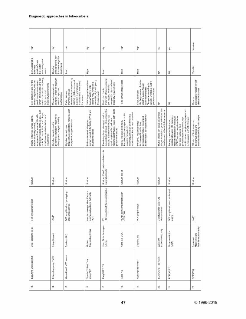
13.
Eas
yNAT
Dia
gnos
ic K
itU
star
Bio
tech
nolo
gyIs
othe
rmal
ampl
ifica
tion
Spu
tum
Use
ful i
n lo
w-r
esou
rces
set
ting,
si
mpl
e an
d ea
sy-to
-use
, eq
uipm
ent-f
ree,
com
patib
le w
ith
othe
ram
plifi
catio
n te
chno
logi
es, s
uch
as P
CR
, LA
MP,
SD
A, H
AD
, Rap
id,
resu
lts a
re q
ualit
ativ
ein
term
s of
vi
sual
ban
ds
Low
det
ectio
n ra
te fo
r the
sm
ear-
neg
ativ
e, c
ultu
re-
posi
tive
sput
um s
ampl
es,
leng
th o
ftim
e ne
eded
to g
et
resu
lts, t
echn
ical
com
plex
ity,
high
cos
ts a
nd
bios
afet
yreq
uire
men
ts
Low
, Acc
epta
ble
fors
mea
r po
sitiv
eCas
es
but l
owfo
r sm
earn
egat
ive
case
s
Hig
h
14.
Eik
en’s
Loop
amp™
MTB
Eik
en (J
apan
)LA
MP
Spu
tum
Hig
h tie
r lab
orat
oryt
o th
e pe
riphe
ralla
bora
tory
, rob
ustn
ess
ofeq
uipm
ent,
reag
ent s
tabi
lity
Man
ual p
repa
ratio
nof
sam
ples
, pre
treat
edsp
utum
re
sulte
d in
poor
er
perfo
rman
ce
Hig
h fo
r sm
earp
ositi
ve, l
ow
for s
mea
rneg
ativ
e sp
ecim
ens
Hig
h
15.
Gen
edriv
e® M
TB a
ssay
Epi
stem
(UK
)P
CR
am
plifi
catio
n, g
enot
ypin
g,
sequ
ence
anal
ysis
Spu
tum
Hig
h tie
r lab
orat
oryt
o th
eper
iphe
ralla
bora
tory
, rob
ustn
esso
f eq
uipm
ent,r
eage
nt s
tabi
lity
Faile
d to
mee
t th
ePer
form
ance
st
anda
rdsr
ecom
men
ded
by
WH
O fo
r SM
repl
acem
ent,
Epi
stem
is w
orki
ng o
n m
odifi
catio
ns to
impr
ove
the
assa
y
Low
Low
16.
True
lab™
Rea
l Tim
e m
icro
PC
RM
olbi
o D
iagn
ostic
s(In
dia)
Nan
otec
hnol
ogy,
Mic
roE
lect
ro-
mec
hani
cals
yste
ms
(ME
-MS
)P
CR
Spu
tum
Fully
aut
omat
ed,In
tegr
ated
sy
stem
,robu
st, A
fford
able
,GP
RS
and
bl
ueto
othe
nabl
ed
Sw
itchi
ng o
n th
eana
lyze
r be
fore
Fini
shin
g ch
ip
load
ing
may
rend
erch
ip
unus
able
,Hig
h st
ringe
ncy
for c
hip
usag
e
Hig
hH
igh
17.
Eas
yNAT
™ T
BU
star
Bio
tech
nolo
gies
(C
hina
)R
T-P
CR
coup
ledw
ithflu
ores
cent
prob
eS
putu
m /F
NA
B p
erip
hera
ltube
rcul
ous
lym
phad
eniti
sP
rom
isin
g di
agno
stic
algo
rithm
for
susp
ecte
dchi
ldho
od T
Bly
mph
aden
itis,
su
its to
the
limite
d re
sour
ce s
et
up o
f per
iphe
ral l
abor
ator
ies
requ
iring
cent
rifug
es, w
ater
bat
h et
c to
pe
rform
the
assa
y
Res
trict
ed d
ue to
th
elen
gth
of ti
me
need
edto
ge
t res
ults
, tec
hnic
al
com
plex
ity, h
igh
cost
s an
d bi
osaf
ety
requ
irem
ents
Low
Hig
h
18.
Ale
re™
qA
lere
Inc.
US
AN
EA
R-is
othe
rmal
ampl
ifica
tion
of T
B D
NA
Spu
tum
/Blo
odH
ighl
y in
tegr
ate,
auto
mat
e,
Por
tabl
e,W
alk
away
ope
ratio
n,N
o co
mpl
ex p
roce
dure
s,
min
imal
qual
ifica
tion
and
train
ing
requ
irem
ent,
Rap
id c
ase
dete
ctio
n
Tedi
ousm
ulti-
step
proc
ess
Hig
hH
igh
19.
Gen
eXpe
rt® O
mni
Cep
heid
Inc.
PC
R a
mpl
ifica
tion
Spu
tum
Por
tabl
e, P
rove
ncar
tridg
e te
chno
logy
,Dur
able
, Low
po
wer
cons
umpt
ion,
Inte
grat
ed
batte
ry,c
loud
- bas
edco
nnec
tivity
Sho
rt ca
rtrid
ge
life,
Req
uire
men
t of s
tabl
e el
ectri
c su
pply,
Ann
ual
calib
ratio
nreq
uire
d,
Tem
pera
ture
cei
ling
is
criti
cal,
unre
liabl
e in
HIV
po
sitiv
econ
ditio
n
Hig
hH
igh
20.
EO
S C
AP
E-T
BS
yste
mW
ave
80
Bio
scie
nces
(US
A)
Gen
otyp
ingR
IF a
nd F
LQ
resi
stan
talle
les
Spu
tum
Mul
tiple
test
s ca
n be
run
in p
aral
lel,
sing
le a
naly
zer t
o ge
nera
tere
sults
that
ca
n be
use
d w
ith m
ultip
lepr
oces
sors
NA
NA
NA
21P
CR
(XC
R™
)Fl
uoro
cent
ric In
c.
(US
A)
PC
R a
mpl
ifica
tiona
nd is
othe
rmal
te
stin
gS
putu
mB
atte
ry o
pera
tedd
evic
e fo
r ra
pida
mpl
ifica
tion
(10
min
utes
), re
al ti
med
etec
tion
of in
divi
dual
X
CR
™as
says
, no
prim
erdi
mer
, lo
w c
ost r
eage
nts
and
porta
blei
nstru
men
ta ti
on, h
igh
mul
tiple
x an
d si
mpl
esam
plep
repa
ratio
n,re
sist
ance
to P
CR
inhi
bito
rs
NA
NA
NA
22.
TOP
-PC
RE
pice
ntre
/M
édec
inss
ans
Fron
tière
sLab
orat
ory
NA
ATS
putu
mTB
rule
out
” tes
t, m
inim
um
back
grou
ndno
ise
and
max
imum
spec
ifici
ty in
its
outp
ut
Req
uire
pr
ospe
ctiv
eval
idat
ion
with
cl
inic
al o
utco
mes
Varia
ble
Varia
ble

Diagnostic approaches in tuberculosis
48 © 1996-2019
which usually requires invasive procedures. Absolute culture specificity can be achieved by complementing the routine diagnostic protocols with antigen tests or NAATs. In case of TB meningitis the low culture yield can be enhanced in terms of sensitivity by replicating the CSF examination or by complementing it with NAAT (62-63). High performance liquid chromatography (HPLC) based detection of tuberculostearic acid in CSF is highly sensitive but requires expert manpower and costly laboratory set up (22,64).
10.4. Semi-automated NAATs for use in peripheral laboratories
The biggest challenge in molecular diagnostics of TB is to shift the set up from the higher tier laboratory to the peripheral laboratories. This may represents a diversity of issues that may range from the availability of sufficient infrastructure, robustness of equipment, reagent stability, user expertise in addition to improved performance over SSM (65-66). Four different platforms like the Loopamp™ MTBC assay from Eiken (Japan); Genedrive® MTB assay from Epistem (UK); Truelab™ RealTime micro PCR System from Molbio Diagnostics (India); and the EasyNAT™ TB assay from Ustar Biotechnologies (China) are sighted with potential for delivery (2).
Eiken’s Loopamp™ MTB provides a (LAMP) based amplification assay with better sensitivity over SSM. This assay involves manual preparation of samples which are subjected to RT-PCR. Genedrive® MTB assay provides for MTBC diagnostics only with future plan of including genotyping MDR-TB through RIF resistance. This assay involves manual preparation of samples followed by RT-PCR. Truelab™ RealTime micro PCR involves semi automated DNA extraction and amplification for MTBC detection. Truelab™ QUATTRO is an improvisation over Truelab™ RealTime micro PCR in being fully automated and integrated system. EasyNAT™ TB suits to the limited resource set up of peripheral laboratories requiring, centrifuges, water bath etc to perform the assay (2) (Table 6).
Alere™ q platform is highly integrated and automated NAAT for diagnosis and assessing the viral load of HIV-1 and 2 (67). It is a cartridge based system, where cartridge is equipped with the reagents to liquefy and inactivate the sample. This is followed by extraction of MTBC-DNA and amplification. This assay involves nicking enzyme amplification reaction (NEAR), an isothermal amplification of TB-DNA (2) (Table 6).
GenePOC Diagnostics (Canada) offers an automated platform that uses RT-PCR and fluorescence detection for TB diagnostics (2). TBDx system from Keck Graduate Institute (KGI; USA) provides a low cost instrumentation where all process starting from
sample processing occurs on single cartridge platform. Nucleic acid preparation is simplified as it does not require chaotropic salts and organic solvents (2). The Northwestern Global Health Foundation (USA) in partnership with Quidel Inc. (USA) is coming up with an integrated MTBC assay. The assay involves lysing MTBC cells via heat block followed by purifying DNA using immiscible oil interface (68) (Table 6).
Point-of-Need (PON) Qiagen offers isothermal nucleic acid amplification assay for the detection of PTB and MDR-TB. Q-POC™ platform, under development from QuantuMDx (UK) offers diagnosis for PTB. The assay involves coughing the sputum in a cup that contains reagents for its liquification and decontamination. This is followed by concentration of MTBC cells via paramagnetic bead concentration step and quick nucleic acid amplification (4 minutes) via isothermal asymmetric PCR (2).
EOSCAPE-TB System from Wave 80 Biosciences (USA) offers detection of MTBC via genotyping RIF and FLQ resistant alleles. Using this platform multiple tests can be run in parallel and single analyzer generates results that can be used with multiple processors (2) (Table 6).
PCR (XCR™) assay from Fluorocentric Inc. (USA) offers a RT-PCR based assay that works via proprietary primer design algorithms. It is powered by a battery operated device for rapid amplification (10 minutes) and real time detection of individual XCR™ assays (2) (Table 6). Co-diagnostics (USA) and Thisis (a spinoff company from Boston University School of Medicine, USA) offers a RT-PCR based platform for detection of MTBC via 16S RNA gene. This platform can be integrated with variety of other RT-PCR platforms. It is claimed to detect MTBC at LOD of 4 cfu and can genotype MTBC into five geno-groups using totally optimized PCR (TOP) (Table 6). Scanogen, though not strictly a NAAT assay detects MTBC specific DNA from body fluids using single molecule scanning technique without PCR amplification. This platform claims to have minimum background noise and maximum specificity in its output (2).
10.5. Spoligotyping
Spoligotyping is very useful technique in providing epidemiological information on the causative strain. It utilizes amplification of polymorphic direct repeat sequences in M. tuberculosis genome. Higher cost of laboratory infrastructure and requirement of trained expertise are biggest limitations (5,69).
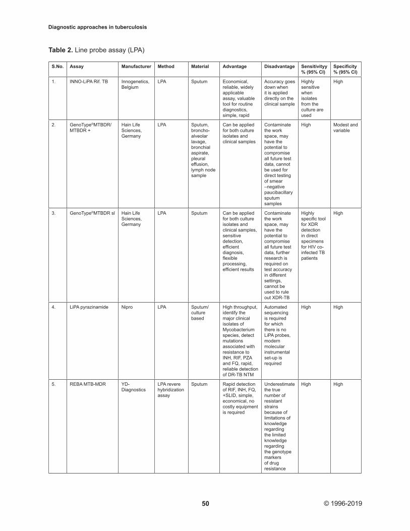
10.6. Line Probe Assay (LPA)
LPAs or reverse blot hybridization assays are a relatively low-cost tool and have been developed
Diagnostic approaches in tuberculosis
49 © 1996-2019
for a variety of areas around MTB diagnostics, including MTBC-specific assays, speciation of NTMs and assays that genotype first or second-line drug resistance alleles. DNA extraction can be manual or automated and DNA is amplified by asymmetric PCR to create a population of biotin-labelled single-stranded (ssDNA) amplicons. Oligonucleotide capture probes with sequences specific to the target amplicons are printed as an array of stripes on a nitrocellulose strip. The amplification mixture is then exposed to a strip under conditions to optimize the specific hybridization of amplicons to their sequence match probe printed on the nitrocellulose. Detection of hybridization of amplicons to the probe stripe is achieved via a streptavidin-linked enzyme that creates a colorimetric reaction visualized as defined stripes that corresponds to the presence of TB and a sensitive or resistant genotype (22). The developed LPAs are read manually with comparison to a printed score chart or can be scanned and automatically analysed by attendant software. The methodology requires open tubes containing amplified DNA, which has potential to contaminate the workspace thus compromising all future test data. The automated workstations alleviate this to some degree by providing controlled conditions that contain reaction mixtures (2).
Commercially available LPAs for the detection of TB, MDR/XDR-TB are: INNO-LiPA Rif. TB (Innogenetics, Belgium) for RIF, GenoType®MTBDR/MTBDRplus for RIF and INH and Geno-Type® MTBDRsl (both Hain Lifescience, Germany) for FLQ, injectables, second line drugs and ETM (22). INNO-LiPA Rif. TB is highly sensitive when isolates from culture are used. However, the accuracy goes down when it is applied directly on clinical samples. GenoType®MTBDR/MTBDRplus can be applied for both culture isolates and clinical samples. GeneXpert MTB/ RIF utilizes heminested RT-PCR to amplify specific sequence of the rpoB gene of M. tuberculosis. The amplicons are then probed with molecular beacons for mutations within RIF resistance determining region (6, 70-72) (Table 2).
MTBDRsl v1.0. and MTBDRsl v2.0. can detect resistance to FLQ, KAN, STR and XDR-TB. MTBDRsl v2.0. is equipped with 27 probes to detect resistance to second line drugs and also targets new gene regions for improved detection of FLQ and KAN (73). LPAs show high sensitivity and give better results when TB culture or SSM positive samples are used as a substrate (Table 2). These are economical when compared to culture based DST (3). However, it is practically impossible to determine accurately resistant genotypes for many drugs like pyrazinamide (PZA). Silent mutations in drug sensitive strains can be misinterpreted as drug resistance leading to avoidable treatment regimens (2).
10.7. Microarray-based platforms
Microarray based platforms acts as portable LPA. Microarrays have high potential for MTB diagnosis, speciation to analyze variation in drug resistant alleles. The probes utilized in microarray can be printed as defined spots and due to their small size more spots can be printed to analyze amplified DNA. The INFINITI® PLUS Analyzer platform from Autogenomics is one such platform. INFINITI® High Throughput System (HTS) and MDR-TB BioFilmChip® for the detection of MTBC and first-line drug (RIF, INH and PZA) are high-throughput array based platform (Table 3). CapitalBio has launched twin microrray based platforms, i.e. Mycobacterium Identification Array Kit for MTB diagnostics and NTM speciation and Drug Resistance Detection Array Kit for MTBC diagnosis and for genotyping RIF and INH resistance (2, 74) (Table 3).
The TruArray® MDR-TB microarray is fluorescent probe based single tube multiplexed asymmetric PCR reaction. It can detect MTB and M. avium along with resistant alleles to RIF and INH (2). VereMTB™ Detection Kit from Veredus Laboratory is solely for research purpose. This array based platform can detect MTBC through IS6110. In addition the array can detect MTBC, speciate eight NTMSs (M. avium, M. simiae, M. intracellulare, M. Kansasii, M. abscessus, M. scrofulaceum, M. chelonae, M. xenopi) and probe alleles associated with RIF and INH resistance (74) (Table 3).
HYDRA 1K developed by Stanford University (USA) together with Insilixa Inc. (USA) is an array based platform to detect PTB MDR / XDR-TB through the genotyping of mutations. This uses complementary metal-oxide semiconductor (CMOS) technology for digital imaging on the array. PCR amplification happens in the array area so that binding with amplicon with probes is measured in real time that saves lot of time and energy. CMOS enables this platform to have melt curve analysis of amplicon binding at each probe spot. It is developed to detect PTB, MDR / XDR-TB. However, higher cost of instrument and other peripherals makes this technique less likely to commercialize (2, 74) (Table 3).
11. WHOLE GENOME SEQUENCING
Whole genome sequencing (WGS) is recently applied to analyze molecular evolution of M. tuberculosis Beijing strain and appearance of an Iberico-American strain in Tibet. WGS provides greater resolution when compared to mycobacterial interspersed repetitive unit-variable-number tandem repeats (MIRU-VNTR, the existing protocol for molecular genotyping of circulating strains) (75-77).
Diagnostic approaches in tuberculosis
50 © 1996-2019
Table 2. Line probe assay (LPA)
S.No. Assay Manufacturer Method Material Advantage Disadvantage Sensitivityy % (95% CI)
Specificity % (95% CI)
1. INNO-LiPA Rif. TB Innogenetics, Belgium
LPA Sputum Economical, reliable, widely applicable assay, valuable tool for routine diagnostics, simple, rapid
Accuracy goes down when it is applied directly on the clinical sample
Highly sensitive when isolates from the culture are used
High
2. GenoType®MTBDR/MTBDR +
Hain Life Sciences, Germany
LPA Sputum, broncho-alveolar lavage, bronchial aspirate, pleural effusion, lymph node sample
Can be applied for both culture isolates and clinical samples
Contaminate the work space, may have the potential to compromise all future test data, cannot be used for direct testing of smear –negative paucibacillary sputum samples
High Modest and variable
3. GenoType®MTBDR sl Hain Life Sciences, Germany
LPA Sputum Can be applied for both culture isolates and clinical samples, sensitive detection, efficient diagnosis, flexible processing, efficient results
Contaminate the work space, may have the potential to compromise all future test data, further research is required on test accuracy in different settings, cannot be used to rule out XDR-TB
Highly specific tool for XDR detection in direct specimens for HIV co-infected TB patients
High
4. LiPA pyrazinamide Nipro LPA Sputum/culture based
High throughput, identify the major clinical isolates of Mycobacterium species, detect mutations associated with resistance to INH, RIF, PZA and FQ, rapid, reliable detection of DR-TB NTM
Automated sequencing is required for which there is no LiPA probes, modern molecular instrumental set-up is required
High High
5. REBA MTB-MDR YD-Diagnostics
LPA revere hybridization assay
Sputum Rapid detection of RIF, INH, FQ, +SLID, simple, economical, no costly equipment is required
Underestimate the true number of resistant strains because of limitations of knowledge regarding the limited knowledge regarding the genotype markers of drug resistance
High High

Diagnostic approaches in tuberculosis
51 © 1996-2019
QIAxel automated genotyping system from Qiagen provides high-throughput genotyping of MTBC hence providing epidemiological informations like prevalence or introduction of a particular genotype. This protocol targets variable sized regions of MTB genomic DNA using
MIRU-VNTR approach. This platform is further improvised to detect RIF and INH resistant alleles (80-81).
Unlike other bacterial pathogens that require drug resistance through plasmid acquisition, transposon or phage mediated elements, M. tuberculosis acquire drug resistance through chromosomal mutations particularly, single-nucleotide polymorphisms (SNPs) (80-81). This makes identification of drug resistant alleles difficult particularly for those lying outside of drug resistant hotspots, e.g. RIF resistance-determining region for RIF or the alleles that lie outside of rpoB and associated with resistance (77). Newer approaches
Table 3. Microarray based diagnostic platforms
S.No. Assay Manufacturer Method Material Advantage Disadvantage Sensitivity, % (95% CI)
Specif city, % (95% CI)
1. INFINITI® PLUS
Autogenomics Microarray platform
Clinical samples
High- throughput array, high performance genetic marker detection with varying options in automation and throughput
Can only be used with human nasopharyn geal aspirates, negative results do not preclude infection
NA NA
2. VereMTB assay
Veredus Laboratories, Singapore
PCR-Micro array
Sputum /culture based
Rapid detection, differentiation, identific ation of MTBC, Resistance to RIF, INH non-MTBC species, diagnosing patients after treatment failure and relapse
More for academic / Research purpose
High High
3. VereMTB™ Detection Kit
Veredus Laboratory
Array based platform
Sputum / culture based
Detect MTBC, speciate eight NTMSs
Research purpose only
High High
4. HYDRA1K Stanford University (USA) together with Insilixa Inc. (USA)
Array based platform
Sputum / culture based
Miniaturization Saves time and energy
Higher cost of instrument
NA NA
5. TruArray® MDR-TB microarray
Akonni Biosystems
Fluorescent probe based Single tube multipled asymmetric PCR reaction
Bacterial culture / sputum
Minitaurize, Bench top test, single integrated work-flow approach, accurate, rapid, broad coverage and field ready
Research purpose only
High High
6. Twin Microrray
CapitalBio Mycobacterium identification array kit for MTB, NTM speciation, drug resistance, MTBC diagnosis, genotyping RIF, INH resistance
Clinical specimens
Both MTC and NTM rapid system, sensitive, reliable, fast, spinal tuberculosis
Infrastructure and trained personnel required reduces its clinical utility in poverty- stricken zones, Additional large-scale studies are needed to evaluate its diagnostic performanc in spinal tuberculosis
High, variable for different clinical specimes
High,variable For different clinical specimens

Diagnostic approaches in tuberculosis
52 © 1996-2019
to detect drug resistance using Next-generation-sequencing (NGS) involves PZA resistance via deletions, STR, INH and screening allelic variation associated with resistance to novel drugs (80,82-85). NGS is very useful in determining whether the case of relapse is because of infection with new strain or incomplete treatment (86). WGS is already applied to the patients with MDR-TB (87-88). McKinsey has even listed NGS as one of the 12 technologies that will transform our lives.
11.1. New sequencing protocols
MiSeq from Illumina, USA facilitates focused applications like targeted gene sequencing, metagenomics, small genome sequencing, targeted gene expression, amplicon sequencing and HLA typing. Only 10ng of DNA is required as starting material. MiSeq gives output of 15 Gb with 25 M sequencing reads of 2x300 bp read lengths). Personal Genome Machine® (PGM™) System (Life Technologies Incorporated; USA) is the conjugation of semiconductor sequencing technology with natural biochemistry to provide real-time chemical information as digital data in less than 3 hours. This technique is accessible to all kind of set-ups. Use of simple natural sequencing chemistry eliminates the requirement of costly optics and complicated chemistries to measure DNA extension. 454 FLX Junior (Roche Diagnostics; Switzerland) provides rapid and robust platform to sequence and assembly genome of any size without the need for supplementary Sanger data. It provides the output having double (1000 bp) the read length of the Titanium kits, hence an improvisation over current GS FLX. The applications like transcriptome and amplicon sequencing enable an all inclusive picture of gene space of organisms. GeneReader from Qiagen can complement available protocols for resistance determination. It can detect allelic variants at 1% allelic frequency. Data from liquid biopsy pipeline show 100% conformity with PCR-based variant detection methods. GnuBIO from Bio-Rad Laboratories is a novel low cost desktop sequencer. It comes up with a highly integrated platform that incorporates all the functions of DNA sequencing like target selection, DNA amplification, DNA sequencing and analysis into a single, integrated system. Single user interface with single step process ensures rapid genomic results in hours. GridION™ and the Min- ION™ from Oxford Nanopore Technologies, UK facilitates rapid assembly of genome data for a MDR isolate, which elucidates the distribution and underlying mechanism of drug resistance. It enables the selection of specific DNA molecules for sequencing by reversing the driving voltage across individual nanopores (89).
12. ASSAYS BEING DEVELOPED / EVALUATED UPCOMING/PIPELINE TECHNOLOGIES
There is a co-evolution of sort between TB bacilli invading to newer regions and newer
techniques / protocols as a counter offensive. Many such existing protocols are improvised and upgraded in response to the challenges and threat posed by rapidly emerging and evolving drug resistant TB bacilli. Following protocols / platforms are under development to spearhead the fight of humanity against TB:
A combine of nano- and biosensing technology is being evaluated for its use as a portable, rapid on-the-spot biosensor for MTB detection. For instance, detection of trans-renal DNA opens up new avenues for TB detection at molecular level. This technique is not yet commercialized due to the problems in the development of TB detection / readout assays.
High-resolution melting (HRM) curve analysis coupled with closed-tube RT-PCR provides a good screening method with a positive predictive value (PPV) of 100% and negative predictive value (NPV) of 99.9.%, for high throughput screening of large number of specimens in any TB laboratory. Improvized amplification techniques in conjunction with ‘molecular beacon’ approach (LATE-PCR) offer future advancements, particularly in drug resistance analysis (90-91).
IR spectrophotometer based detection of isotopic urea in exhaled breath is another future technique that holds promise. It utilizes MTB urease as a bacterial virulence factor (6).
A biophotonic detection platform is being developed that utilizes reporter enzyme fluorescence to detect β-lactamase produced by MTB. This innovative technology is now being adapted for point of care (POC) use.
Becton Dickinson is in the process of designing an automated platform that will do the staining of AFB in sputum in addition to imaging. This platform will provide the sensitivity similar to the fluorescent microscopy at the rate of processing around 40 slides per day with slide scanning algorithm for scoring (2).
Xiamen Zeesan Biotech Co. Ltd (China) has developed MeltPro® Drug-Resistant TB Testing Kits as RT-PCR enabled detection of resistant alleles to RIF and INH. It is undergoing further improvisation to detect alleles resistant to ethambutol (EMB), streptomycin (STR) and fluoroquinolones (FLQs) (52-53, 92).
Genedrive® MTB assay is upgraded to include genotyping MDR-TB through RIF resistance in addition to MTBC diagnosis. This assay involves manual preparation of samples followed by RT-PCR115 (93-94).
The Northwestern Global Health Foundation (USA) in partnership with Quidel Inc. (USA) is coming

Diagnostic approaches in tuberculosis
53 © 1996-2019
up with an integrated MTBC assay. The assay involves lysing MTBC cells via heat block followed by purifying DNA using immiscible oil interface (68).
Q-POC™ platform, under development from QuantuMDx (UK) offers diagnosis for PTB.
The assay involves coughing the sputum in a cup that contains reagents for its liquification and decontamination. This is followed by concentration of MTBC cells via paramagnetic bead concentration step and quick nucleic acid amplification (4 minutes) via isothermal asymmetric PCR (2).
Tangen Biosciences Inc. (USA) is going to launch a fully integrated TB detection platform. This platform will have provision for sputum collection in a cup to capture MTB cells followed by lysis and isothermal amplification via LAMP. It comes up with the storage capacity of 10,000 results (2).
13. CONCLUSIONS
Accurate and timely diagnosis of TB is the only requirement for its control and management. Currently, TB diagnosis and treatment is plagued by factors, like inability to diagnose correctly latent and active TB; lack of precise diagnostic protocols for EPTB, pediatric TB and TB associated with HIV; prolonged diagnostic protocol aggravating the otherwise curable infection and escalating it to MDR/XDR-TB; absence of readily available testable biomarkers; Inaccessibility to the POC diagnostic facilities in the peripheral laboratories. These are further challenged by the rapidly emerging resistant strains of TB bacilli in the regions where it is thought to be eradicated or where it was non -existent historically. Need of the hour is the tools and techniques that are accessible, accurate, rapid, economical and most importantly rapid as time lag in diagnosis aggravates the infection. Although TB diagnostics have seen tremendous upsurge in the newer technologies and protocols from imaging, molecular NAAT based, immunological, culture based phenotypic methods, array based to genome sequencing protocols. Unfortunately, they offer promise in some aspect but lack or have severe limitation in other aspects, which limits their applicability on ‘one -shoe-fit-all’ basis. Another, problem with newer protocols is the reluctance of user groups to embrace it either due to lengthy approval mechanism, lack of WHO approval in Toto or simply the cost and expertise involved. There is urgent need to evolve the mechanism to accept the newer protocols on the emergency basis. As an extension to the problem, many small companies with promising diagnostic kits (in terms of good performance on heterogeneous set-ups) do not have sufficient funds to market their products or scale up their production. Also, most of the newer technologies are accepted in the developed
countries but there is a need to evolve the technologies compatible to the requirement of resource limited developing countries. There is a dire need of all inclusive diagnostic protocols with applicability in wide spectrum of patients and set-ups with high sensitivity and specificity. A dynamic understanding of existing and forthcoming technologies is key in facilitating access to appropriate TB diagnostic tools through market-based interventions. As such, this review is intended to be a living document, updated as the TB diagnostics market evolves, to highlight potential.
14. ACKNOWLEDGEMENTS
We thankfully acknowledged the Research grant from DST-NRDMS and UGC Startup, Government of India. Funder has no direct or indirect role in plan, design or writing of the review. Grant was solely for research in TB epidemiology. PS: Planning of Review article, literature search, collection and writing. VKS: Literature search, collection of study materials and review. RSK: Literature search and collection of study materials.
15. REFERENCES
1. Drobniewski, F., V. Nikolayevsky, Y. Balabanova, D. Bang, and D. Papaventsis: Modern laboratory diagnosis of tuberculosis. Lancet Infect Dis 3, 141–147 (2003)
DOI: 10.1016/S1473-3099(03)00544-9
2. UNITAID: Tuberculosis Diagnostics Technology and Market Landscape, 4th edition. World Health Organization. (2015)
3. World Health Organization: Non-commercial culture and drug susceptibility testing methods for screening patients at risk for multidrug-resistant tuberculosis: policy statement. WHO/HTM/TB/2011.9. Geneva, Switzerland: WHO (2011)
4. Lessem, E: The tuberculosis diagnostics pipeline. HIV & TB Pipeline Report. (2016)
5. Kent, P.T., and G.P. Kubica: Centers for Disease Control. Public Health Mycobacteriology: A Guide for the Level III Laboratory. Atlanta, GA, Centers for Disease Control (1985)
6. Maiga, M., A. Abaza, and W.R. Bishai: Current tuberculosis diagnostic tools and role of urease breath test. Ind J Med Res 135, 731-6 (2012)
7. Master, R.N: Clinical microbiology
procedures handbook. American Society for

Diagnostic approaches in tuberculosis
54 © 1996-2019
Microbiology. J Bacteriol 191, 4714–4721 (1992)
8. McNerney, R., J. Cunningham, P. Hepple, and A. Zumla: New tuberculosis diagnostics and rollout International. J Infect Dis 32, 81–86 (2015)
DOI: 10.1016/j.ijid.2015.01.012
9. Laserson, K.F., N.T. Yen, C.G. Thornton, V.T. Mai, W. Jones, D.Q. An, N.H. Phuoc, N.A. Trinh, D.T. Nhung, T.X. Lien, N.T. Lan, C. Wells, N. Binkin, M. Cetron, and S.A. Maloney: Improved sensitivity of sputum smear microscopy after processing specimens with C18-carboxypropylbetaine to detect acid-fast bacilli: a study of United States-bound immigrants from Vietnam. J Clin Microbiol 43, 3460-2 (2005)
DOI: 10.1128/JCM.43.7.3460-3462.2005
10. Jaiswal, S., S. Jay Prakash, B. Sharma: Standard Diagnostic Procedure for Tuberculosis: A Review. Res Rev J Life Sc 1-10 (2013)
11. Lewis, J.J., V.N. Chihota, M. van der Meulen, P.B. Fourie, K.L. Fielding, A.D. Grant, S.E. Dorman, and G.J. Churchyard: Proof-of-concept’’ evaluation of an automated sputum smear microscopy system for tuberculosis diagnosis. PLoS One 7, e50173 (2012)
DOI: 10.1371/journal.pone.0050173
12. Divekar, A., C. Pangilinan, G. Coetzee, T. Sondh, F.Y. Lure, and S. Kennedy: Automated detection of tuberculosis on sputum smeared slides using stepwise classification. Proc SPIE Med Imag Conf (2012)
DOI: https://doi.org/10.1117/12.910484
13. Hogeweg, L., C. Sanchez, P. Maduskar, R. Philipsen, A. Story, R. Dawson, G. Theron, K. Dheda, L. Peters-Bax, and B. van Ginneken: Automatic detection of tuberculosis in chest radiographs using a combination of textural, focal, and shape abnormality analysis. IEEE Trans Med Imag 34, 2429-42 (2015)
DOI: 10.1109/TMI.2015.2405761
14. Muyoyeta, M., P. Maduskar, M. Moyo, N. Kasese, D. Milimo, R. Spooner, N. Kapata, L. Hogeweg, B. van Ginneken, and H. Ayles H: The sensitivity and of using a computer aided diagnosis program for automatically scoring chest X-rays of presumptive TB patients compared with Xpert MTB/RIF in Lusaka Zambia. PLoS One 9, e93757 (2014)
DOI: 10.1371/journal.pone.0093757
15. Tapley, A., N. Switz, C. Reber, J.L. Davis, C. Miller, J.B. Matovu, W. Worodria, L. Hunag, D.A. Fletcher, and A. Cattamanchi: Mobile digital fluorescence microscopy for diagnosis of tuberculosis. J Clin Microbiol 51,1774–8 (2013)
DOI: 10.1128/JCM.03432-12
16. Fujita, A., K. Murata, and M. Takamori: Novel method for sputum induction using the Lung Flute in patients with suspected pulmonary tuberculosis. Respirol 14, 899–902 (2009)
DOI: 10.1111/j.1440-1843.2009.01584.x
17. Arend, S.M., A.C. Engelhard, G. Groot, K. de Boer, P. Andersen, T.H. Ottenhoff, and J.T. van Dissel: Tuberculin skin testing compared with T-cell responses to Mycobacterium tuberculosis-specific and nonspecific antigens for the detection of latent infection in persons with recent tuberculosis contact. Clin Diagn Lab Immunol 8, 1089–96 (2001)
DOI: 10.1128/CDLI.8.6.1089-1096.2001
18. Doherty, T.M., A. Demissie, J. Olobo, D. Wolday, S. Britton, T. Equale, P. Ravn, and P. Andersen: Immune responses to the Mycobacterium tuberculosis-specific antigen ESAT-6 signal subclinical infection among contacts of tuberculosis patients. J Clin Microbiol 40, 704–06 (2002)
DOI: 10.1128/JCM.40.2.704-706.2002
19. Lalvani, A., A.A. Pathan, H. Durkan, K.A. Wilkinson, A. Whelan, J.J. Deeks, W.H. Reece, M. Latif, G. Pasvol, and A.V. Hill: Enhanced contact tracing and spatial tracking of Mycobacterium tuberculosis infection by enumeration of antigenspecific T-cells. Lancet 357, 2017–21 (2001)
DOI: 10.1016/S0140-6736(00)05115-1
20. Nakamura, R.M., M.A. Velmonte, K. Kawajiri, C.F. Ang, R.A. Frias, M.T. Mendoza, J.C. Montoya, Honda, S. Haga, and I. Toida: MPB64 mycobacterial antigen: a new skin-test reagent
21. Through patch method for rapid diagnosis of active tuberculosis. Int J Tuberc Lung Dis 2, 541–46 (1998)
22. Nakamura, R.M., L. Einck, M.A. Velmonte, K.
Kawajiri, C.F. Ang, C.E. Delasllagas, and C.A. Nacy CA: Detection of active tuberculosis by an MBP64 transdermal patch: a field study. Scan J Infect Dis 33, 405–07 (2001)
DOI: 10.1080/00365540152029846

Diagnostic approaches in tuberculosis
55 © 1996-2019
23. Drobniewski, F., V. Nikolayevskyy, H. Maxeiner, Y. Balabanova, N. Casali, I. Kontsevaya, and O. Ignatyeva: Rapid diagnostics of tuberculosis and drug resistance in the industrialized world: clinical and public health benefits and barriers to implementation BMC Med 11, 190 (2013)
DOI: 10.1186/1741-7015-11-190
24. Balabanova, Y., F. Drobniewski, V. Nikolayevskyy, A. Kruuner, N. Malomanova, T. Simak, N. Ilyina, S. Zakharova, N. Lebedeva, H.L. Alexander, R. O’Brien, H. Sohn, A. Shakhmistova, and I. Fedorin: An integrated approach to rapid diagnosis of tuberculosis and multidrug resistance using liquid culture and molecular methods in Russia. PLoS One 4, e7129 (2009)
DOI: 10.1371/journal.pone.0007129
25. Leung, E., J. Minion, A. Benedetti, M. Pai, and D. Menzies: Microcolony culture techniques for tuberculosis diagnosis: a systematic review. Int J Tuberc Lung Dis 16, 16– 23 (2012)
DOI: 10.5588/ijtld.10.0065
26. Minion, J., E. Leung, D. Menzies, and M. Pai: Microscopic-observation drug susceptibility and thin layer agar assays for the detection of drug resistant tuberculosis: a systematic review and metaanalysis. Lancet Infect Dis 10, 688–98 (2010)
DOI: 10.1016/S1473-3099(10)70165-1
27. Toit, K., S. Mitchell, Y. Balabanova, C.A. Evans, T. Kummik, V. Nikolayevskyy, and F. Drobniewski: The colour test for drug susceptibility testing of Mycobacterium tuberculosis strains. Int J Tuberc Lung Dis 16, 1113-8 (2012)
DOI: 10.5588/ijtld.11.0609
28. Gonzalo, X., F. Drobniewski, S. Hoffner, and J. Werngren: Evaluation of a biphasic media assay for pyrazinamide drug susceptibility testing of Mycobacterium tuberculosis. J Antimicrob Chemother 69, 3001-3005 (2014).
DOI: 10.1093/jac/dku230
29. Washinton, W., A. Stephen, J. William, E. Koneman, G. Procop, P. Schreckenberger, and G. Woods: Koneman’s Colour Atlas and Textbook of Diagnostic Microbiology. Lippincott Publishing 1066-117 (2006).
30. Drobniewski, F.A., P.G. More, and G.S. Harris: Differentiation of Mycobacterium tuberculosis
complex and nontuberculous mycobacterial liquid cultures by using peptide nucleic acid-fluorescence in situ hybridization probes. J Clin Mirobiol 38, 444–47 (2000)
31. Telenti, A., F. Marchesi, M. Balz, F. Bally, E.C. Bottger, and T. Bodmer: Rapid identification of mycobacteria to the species level by polymerase chain reaction and restiction enzyme analysis. J Clin Microbiol 31, 175–78 (1993)
32. Kunnath-Velayudhan, S., H. Salamon, H.Y.
Wang, A.L. Davidow, D.M. Molina, V.T. Huynh, D.M. Cirillo, G. Michel, E.A. Talbot, M.D. Perkins, P.L. Felgner, X. Liang, and M.L. Gennaro: Dynamic antibody responses to the Mycobacterium tuberculosis proteome. Proc Natl Acad Sci USA 107, 14703– 8 (2010)
DOI: 10.1073/pnas.1009080107
33. Steingart, K.R., L.L. Flores, N. Dendukuri, I. Schiller, S. Laal, A. Ramsay, P.C. Hopewell, and M. Pai: Commercial serological tests for the diagnosis of active pulmonary and extra-pulmonary tuberculosis: an updated systematic review and meta-analysis. PLoS Med 8, e1001062 (2011)
DOI: 10.1371/journal.pmed.1001062
34. Kroidl, I., P. Clowes, J. Mwakyelu, L. Maboko, A. Kiangi, A. Rachow, K. Reither, J. Jung, A. Nsojo, E. Saathoff, and M. Hoelscher: Reasons for false-positive lipoarabinomannan ELISA results in a Tanzanian population. Scand J Infect Dis 46,144–8 (2014)
DOI: 10.3109/00365548.2013.853133
35. Nicol, M.P., V. Allen, L. Workman, W. Isaacs, J. Munro, S. Pienaar, F. Black, L. Adonis, W. Zemanay, Y. Ghebrekristos, and H.J. Zar: Urine lipoarabinomannan testing for diagnosis of pulmonary tuberculosis in children: a prospective study. Lancet Glob Health 2, e278–84 (2014)
DOI: 10.1016/S2214-109X(14)70195-0
36. Sun, D., S. Dorman, M. Shah, Y.C. Manabe, V.M. Moodley, M.P. Nicol, and D.W. Dowdy: Cost utility of lateral-flow urine lipoarabinomannan for tuberculosis diagnosis in HIV infected African adults. Int J Tuberc Lung Dis 17:552–8 (2013)
DOI: 10.5588/ijtld.12.0627
37. Portevin, D., F. Moukambi, P. Clowes, A. Bauer, M. Chachage, N.E. Ntinginya, E. Mfinanga, K. Said, F. Haraka, A. Rachow, E.

Diagnostic approaches in tuberculosis
56 © 1996-2019
Saathoff, M. Mpina, L. Jugheli, F. Lwilla, B.J. Marais, M. Hoelscher, C. Daubenberger, K. Reither, and C. Geldmacher: Assessment of the novel T-cell activation marker-tuberculosis assay for diagnosis of active tuberculosis in children: a prospective proof-of-concept study. Lancet Infect Dis 2014;14:931-8 (2014)
DOI: 10.1016/S1473-3099(14)70884-9
38. Meldau, R., J. Peter, G. Theron, G. Calligaro, B. Allwood, G. Symons, H. Khalfey, G. Ntombenhle, U. Govender, A. Binder, R. van Zyl-Smit, and K. Dheda: Comparison of same day diagnostic tools including Gene Xpert and unstimulated IFNgamma for the evaluation of pleural tuberculosis: a prospective cohort study. BMC Pulm Med 14, 58 (2014)
DOI: 10.1186/1471-2466-14-58
39. Hur, M., H.W. Moon, Y.M. Yun, T.Y. Kang, H.S. Kim, K.M. Lee, S.H. Kang, and E.H. Lee: Detection of tuberculosis using artus M. tuberculosis PCR Kit and COBAS AMPLICOR Mycobacterium tuberculosis Test. Int J Tuberc Lung Dis 15,795– 798 (2011)
DOI: 10.5588/ijtld.10.0367
40. Kim, J.H., Y.J. Kim, C.S. Ki, J.Y. Kim, and N.Y. Lee: Evaluation of Cobas TaqMan MTB PCR for detection of Mycobacterium tuberculosis. J Clin Microbiol 49, 173–176 (2011)
DOI: 10.1128/JCM.00694-10
41. Ling, D.I., L.L. Flores, L.W. Riley, and M. Pai M: Commercial nucleic-acid amplification tests for diagnosis of pulmonary tuberculosis in respiratory specimens: meta-analysis and meta regression. PLoS One 3, e1536 (2008)
DOI: 10.1371/journal.pone.0001536
42. Mitarai, S., M. Okumura, E. Toyota, T. Yoshiyama, A. Aono, A. Sejimo, Y. Azuma, K. Sugahara, T. Nagasawa, N. Nagayama, A. Yamane, R. Yano, H. Kokuto, K. Morimoto, M. Ueyama, M. Kubota, R. Yi, H. Ogata, S. Kudoh, and T. Mori: Evaluation of a simple loop-mediated isothermal amplification test kit for the diagnosis of tuberculosis. Int J Tuberc Lung Dis 15,1211–1217 (2011)
DOI: 10.5588/ijtld.10.0629
43. Wang, J.Y., L.N. Lee, H.L. Hsu, P.R. Hsueh, and K.T. Luh: Performance assessment of the DR. MTBC Screen assay and the BD ProbeTec ET system for direct detection of Mycobacterium tuberculosis in respiratory
specimens. J Clin Microbiol 44, 716–719 (2006)
DOI: 10.1128/JCM.44.3.716-719.2006
44. Boehme, C.C., M.P. Nicol, P. Nabeta, J.S. Michael, E. Gotuzzo, R. Tahirli, M.T. Gler, R. Blakemore, W. Worodria, C. Gray, L. Huang, T. Caceres, R. Mehdiyev, L. Raymond, A. Whitelaw, K. Sagadevan, H. Alexander, H. Albert, F. Cobelens, H. Cox, D. Alland, and M.D. Perkins: Feasibility, diagnostic accuracy, and effectiveness of decentralised use of the Xpert MTB/ RIF test for diagnosis of tuberculosis and multidrug resistance: a multicentre implementation study. Lancet 377, 1495–1505 (2011)
DOI: 10.1016/S0140-6736(11)60438-8
45. Pietzka, A.T., A. Indra, A. Stoger, J. Zeinzinger, M. Konrad, P. Hasenberger, F. Allerberger, and W. Ruppitsch: Rapid identification of multidrug-resistant Mycobacterium tuberculosis isolates by rpoB gene scanning using high resolution melting curve PCR analysis. J Antimicrob Chemother 63,1121–1127 (2009)
DOI: 10.1093/jac/dkp124
46. Ribeiro, F.K., V. Dettoni Vdo, R.L. Peres, S.A. Vinhas, T.R. Có, R. Dietze, and M. Palaci: Evaluation of a commercial test based on ligase chain reaction for direct detection of Mycobacterium tuberculosis in respiratory specimens. Rev Soc Bras Med Trop 37, 431–435 (2004)
DOI: 10.1590/S0037-86822004000600001
47. Chen, J.H., K.K. She, T.C. Kwong, O.Y. Wong, G.K. Siu, C.C. Leung, K.C. Chang, C.M. Tam, P.L. Ho, V.C. Cheng, K.Y. Yuen, W.C. Yam: Performance of the new automated Abbott Real-Time MTB assay for rapid detection of Mycobacterium tuberculosis complex in respiratory specimens. Eur J Clin Microbiol Infect Dis 34, 1827-32 (2015)
DOI: 10.1007/s10096-015-2419-5
48. Hofmann-Thiel, S., and H. Hoffmann: Evaluation of Fluorotype MTB for detection of Mycobacterium tuberculosis complex DNA in clinical specimens from a low-incidence country. BMC Infect Dis 14, 59 (2014)
DOI: 10.1186/1471-2334-14-59
49. Rice, J.E., A.H. Reis Jr, L.M. Rice, R.K. Carver-Brown, and L.J. Wangh: Fluorescent signatures for variable DNA sequences. Nucleic Acids Res 40, e164 (2012)
DOI: 10.1093/nar/gks731

Diagnostic approaches in tuberculosis
57 © 1996-2019
50. Takakura, S., S. Tsuchiya, Y. Isawa, K. Yasukawa, T. Hayashi, M. Tomita, K. Suzuki, T. Hasegawa, T. Tagami, A. Kurashima, and S. Ichiyama: Rapid detection of Mycobacterium tuberculosis in respiratory samples by transcription-reverse transcription concerted reaction with an automated system. J Clin Microbiol 43, 5435–9 (2005)
DOI: 10.1128/JCM.43.11.5435-5439.2005
51. Lee, S.H., S.W. Kim, S. Lee, E. Kim, D.J. Kim, S. Park, E.J. Lee, S.Y. Lee, J.S. Lee, C.S. Lim, W.K. Kim, and K.H. In: Rapid detection of Mycobacterium tuberculosis using a novel ultrafast chip-type real-time polymerase chain reaction system. Chest 146, 1319–26 (2014)
DOI: 10.1378/chest.14-0626
52. Hwang, I.T., Y.J. Kim, S.H. Kim, C.I. Kwak, Y.Y. Gu, and J.Y. Chun: Annealing control primer system for improving specificity of PCR amplification. Biotechniques 35, 1180– 4 (2003)
53. Hu, S., G. Li, H. Li, X. Liu, J. Niu, S. Quan, F. Wang, H. Wen, Y. Xu, and Q. Li: Rapid detection of isoniazid resistance in Mycobacterium tuberculosis isolates by use of real-time-PCR-based melting curve analysis. J Clin Microbiol 52,1644–52 (2014)
DOI: 10.1128/JCM.03395-13
54. Zhang, T., S. Hu, G. Li, H. Li, X. Liu, J. Niu, F. Wang, H. Wen, Y. Xu, and Q. Li: Evaluation of the MeltPro TB/STR assay for rapid detection of streptomycin resistance in Mycobacterium tuberculosis. Tuberculosis (Edinb) 95,162–9 (2015)
DOI: 10.1016/j.tube.2014.12.004
55. Steingart, K.R., I. Schiller, D.J. Horne, M. Pai, C.C. Boehme, and N. Dendukuri: Xpert1 MTB/RIF assay for pulmonary tuberculosis and rifampicin resistance in adults. Cochrane Database Syst Rev 1, CD009593 (2014)
56. Weyer, K., F. Mirzayev, G.B. Migliori, W. Van Gemert, L. D’Ambrosio, M. Zignol, K. Floyd, R. Centis, D.M. Cirillo, E. Tortoli, C. Gilpin, J. de Dieu Iragena, D. Falzon, and M. Raviglione: Rapid molecular TB diagnosis: evidence, policy making and global implementation of Xpert MTB/RIF. Eur Respir J 42, 252–271 (2013)
DOI: 10.1183/09031936.00157212
57. Nikama, C., M. Kazia, C. Nairb, M. Jaggannathb, M. Manoj, R. Vinaya, A. Shetty, and C. Rodrigues: Evaluation of the
Indian TrueNAT micro RT-PCR device with GeneXpert for case detection of pulmonary tuberculosis. Int J Mycobacteriol 3, 205–10 (2014)
DOI: 10.1016/j.ijmyco.2014.04.003
58. Ou, X., Y. Song, B. Zhao, Q. Li, H. Xia, Y. Zhou, Y. Pang, S. Wang, Z. Zhang, S. Cheng, C. Liu, and Y. Zhao: A multicenter study of crosspriming amplification for tuberculosis diagnosis at peripheral level in China. Tuberculosis (Edinb) 94, 428–33 (2014)
DOI: 10.1016/j.tube.2014.04.006
59. Bholla, M., N. Kapalata, E. Masika, H. Chande, L. Jugheli, M. Sasamalo, T.R. Glass, H.P. Beck, and K. Reither: Evaluation of Xpert® MTB/RIF and Ustar EasyNAT™ TB IAD for diagnosis of tuberculous lymphadenitis of children in Tanzania: a prospective descriptive study. BMC Infect Dis 16, 246 (2016)
DOI: 10.1186/s12879-016-1578-z
60. Moore, D.A., C.A. Evans, R.H. Gilman, L. Caviedes, J. Coronel, A. Vivar, E. Sanchez, Y. Piñedo, J.C. Saravia, C. Salazar, R. Oberhelman, M.G. Hollm-Delgado, D. LaChira, A.R. Escombe, and J.S. Friedland: Microscopic-observation drug-susceptibility assay for the diagnosis of TB. Laboratories V. VereMTB product sheet. Rapid detection, differentiation and identification of MDR-TB. Singapore: Verdus Laboratories; 2014. N Engl J Med 355, 1539- 50 (2006)
DOI: 10.1056/NEJMoa055524
61. Jones, B.E., S.M. Young, D. Antoniskis, P.T. Davidson, F. Krammer, and F. Barnes: Relationship of the manifestations of tuberculosis to CD4 cell counts in patients with human immunodeficiency virus infection. Am Rev Respir Dis 148, 1292–1297 (1993)
DOI: 10.1164/ajrccm/148.5.1292
62. Nelson, L.J., and C.D. Wells: Global epidemiology of childhood tuberculosis. Int J Tuberc Lung Dis 8, 636–647 (2004)
63. Marais S., G. Thwaites, J.F. Schoeman, M.E.
Török, U.K. Misra, K. Prasad, P.R. Donald, R.J. Wilkinson, and B.J. Marais: Tuberculous meningitis: a uniform case definition for use in clinical research. Lancet Infect Dis 10, 803–812 (2010)
DOI: 10.1016/S1473-3099(10)70138-9
64. Pai, M., L.L. Flores, N. Pai, A. Hubbard, L.W. Riley, and J.M. Colford Jr: Diagnostic
Diagnostic approaches in tuberculosis
58 © 1996-2019
accuracy of nucleic acid amplification tests for tuberculous meningitis: a systematic review and meta-analysis. Lancet Infect Dis 3, 633–643 (2003)
DOI: 10.1016/S1473-3099(03)00772-2
65. Elias, J., J.P. Coning De, S.A. Vorster, and H.F. Joubert: The rapid and sensitive diagnosis of tuberculous meningitis by the detection of tuberculostearic acid in cerebrospinal fluid using gas chromatography mass spectrometry with selective ion monitoring. Clin Biochem 22, 463–67 (1989)
DOI: 10.1016/S0009-9120(89)80099-2
66. Denkinger, C.M., and M. Pai: Point-of-care tuberculosis diagnosis: are we there yet? Lancet Infect Dis 12, 169–70 (2012)
DOI: 10.1016/S1473-3099(11)70257-2
67. Niemz, A., and D.S. Boyle: Nucleic acid testing for tuberculosis at the point-of-care in high-burden countries. Expert Rev Mol Diagn 12, 687–701 (2012)
DOI: 0.1586/erm.12.71
68. Jani, I.V., B. Meggi, N. Mabunda, A. Vubil, N.E. Sitoe, O. Tobaiwa, J.I. Quevedo, J.D. Lehe, O. Loquiha, L. Vojnov, and T.F. Peter: Accurate early infant HIV diagnosis in primary health clinics using a point-of-care nucleic acid test. J Acquir Immune Defic Syndr 67, e1–e4 (2014)
DOI: 10.1097/QAI.0000000000000250
69. Sur, K., S.M. McFall, E.T. Yeh, S.R. Jangam, M.A. Hayden, S.D. Stroupe, and D.M. Kelso: Immiscible phase nucleic acid purification eliminates PCR inhibitors with a single pass of paramagnetic particles through a hydrophobic liquid. J Mol Diagn 12, 620–8 (2010)
DOI: 10.2353/jmoldx.2010.090190 70. Kamerbeek, J., L. Schouls, A. Kolk, M. van
Agterveld, D. van Soolingen, S. Kuijper, A. Bunschoten, H. Molhuizen, R. Shaw, M. Goyal, and J. van Embden: Simultaneous detection and strain differentiation of Mycobacterium tuberculosis for diagnosis and epidemiology. J Clin Microbiol 35, 907- 14 (1997)
71. Ling, D.I., A.A. Zwerling, and M. Pai: GenoType MTBDR assays for the diagnosis of multidrug-resistant tuberculosis: a metaanalysis. Eur Respir J 32, 1165-74 (2008)
DOI: 10.1183/09031936.00061808
72. Marras, S., H. El-Hajj, M.T. Taylor, and W. McMillan: Rapid and sensitive detection of Mycobacterium DNA using Cepheid SmartCycler® and Tube Lysis System. Clin Chem 47, 1917-8 (2001)
73. Morgan, M., S. Kalantri, L. Flores, and M. Pai: A commercial line probe assay for the rapid detection of rifampicin resistance in Mycobacterium tuberculosis: a systematic review and metaanalysis. BMC Infect Dis 5, 62 (2005)
DOI: 10.1186/1471-2334-5-62
74. Tagliani, E., A.M. Cabibbe, P. Miotto, E. Borroni, J.C. Toro, M. Mansjo, S. Hoffner, D. Hillemann, A. Zalutskaya, A. Skrahina, and D.M. Crillo: Diagnostic Performance of the new version of GenoType MTBDRsl (V2.0.) Assay for detection of resistance to Fluoroquinolones and Second Line Injectable Drugs: a Multicenter study. J Clin Microbiol 53, 2961-9 (2015)
DOI: 10.1128/JCM.01257-15
75. Lazzeri, E., F. Santoro, M.R. Oggioni, F. Iannelli, and G. Pozzi: Novel primer-probe sets for detection and identification of mycobacteria by PCR-microarray assay. J Clin Microbiol 50, 3777–9 (2012)
DOI: 10.1128/JCM.02300-12
76. Mokrousov, I., W.W. Jiao, K. Wan, and A. Shen: Stranger in a strange land: Ibero-American strain of Mycobacterium tuberculosis in Tibet, China. Infect Genet Evol 26, 323–6 (2014)
DOI: 10.1016/j.meegid.2014.06.011
77. Merker, M., C. Blin, S. Mona, N. Duforet-Frebourg, S. Lecher, E. Willery, M.G. Blum, S.R. Gerdes, I. Mokrousov, E. Aleksic, C.A. Begues, A. Antiriens, E.A. Kopec, M. Ballif, F. Barletta, H.P. Beck, C.E. Barry, M. Bonnet, E. Borroni, I.C. Herrero, D. Cirillo, H. Cox, S. Crowe, V. Crudu, R. Dier, F. Drobniewski, M.F. Dufaux, S. Gangneux, S. Ghebremichael, M. Hanekom, S. Hoffner, W.W. Jiao, S. Kalon, T.A. Kohl, I. Kontsevaya, T. Lilebaek, S. Maeda, V. Nikolayevskyy, M. Rasmussen, N. Rastogi, S. Samper, E.S. Padilla, B. Savic, I.C. Shamputa, A. Shen, L.H. Sng, P. Stakenas, K. Toit, F. Varaine, D. Vukovic, C. Wahl, R. Warren, P. Supply, S. Niemann, and T. Wirth: Evolutionary history and global spread of the Mycobacterium tuberculosis Beijing lineage. Nat Genet 2015;47:242–9.
DOI: 10.1038/ng.3195

Diagnostic approaches in tuberculosis
59 © 1996-2019
78. Jamieson, F.B., S. Teatero, J.L. Guthrie, A. Neemuchwala, N. Fittipaldi, and C. Mehaffy: Whole-genome sequencing of the Mycobacterium tuberculosis Manila sublineage results in less clustering and better resolution than mycobacterial interspersed repetitive-unit-variable-number tandem-repeat (MIRU-VNTR) typing and spoligotyping. J Clin Microbiol 52, 3795–8 (2014)
DOI: 10.1128/JCM.01726-14
79. Gauthier, M., F. Bidault, A. Mosnier, N. Bablishvili, N. Tukvadze, S. Somphavong, P. Paboriboune, O. Ocheretina, J.W. Pape, G. Paranhos-Baccala, and J.L. Berland: Highthroughput mycobacterial interspersed repetitive-unit-variable-number tandem-repeat genotyping for Mycobacterium tuberculosis epidemiological studies. J Clin Microbiol 53, 498–503 (2015)
DOI: 10.1128/JCM.01611-14
80. Matsumoto, T., Y. Koshii, K. Sakane, T. Murakawa, Y. Hirayama, H. Yoshida, M. Kurowava, Y. Tamura, T. Nagai, I. Kawase: A novel approach to automated genotyping of Mycobacterium tuberculosis using a panel of 15 MIRU VNTRs. J Microbiol Methods 2013;93:239–41.
DOI: 10.1016/j.mimet.2013.03.022
81. Martinez, E., N. Holmes, P. Jelfs, and V. Sintchenko: Genome sequencing reveals novel deletions associated with secondary resistance to pyrazinamide in MDR Mycobacterium tuberculosis. J Antimicrob Chemother 70, 2511-4 (2015)
DOI: 10.1093/jac/dkv128
82. O’Malley, T., and E. Melief: Isolation and characterization of compound-resistant isolates of Mycobacterium tuberculosis. Meth Mol Biol 1285, 317–28 (2015)
DOI: 10.1007/978-1-4939-2450-9_19
83. Ioerger, T.R., T. O’Malley, R. Liao, K.M. Guinn, M.J. Hickey, N. Mohaideen, K.C. Murphy, H.I.M. Boshoff, V. Mizrahi, E.J. Rubin, C.M. Sassetti, C.E. Barry III, D.R. Sherman, T. Parish, and J.C. Sacchettini: Identification of new drug targets and resistance mechanisms in Mycobacterium tuberculosis. PLoS One 8, e75245 (2013)
DOI: 10.1371/journal.pone.0075245
84. McGrath, M., N.C. Gey van Pittius, P.D. van Helden, R.M. Warren, and D.F. Warner: Mutation rate and the emergence of drug
resistance in Mycobacterium tuberculosis. J Antimicrob Chemother 69, 292–302 (2014)
DOI: 10.1093/jac/dkt364
85. Ramasubban, G., D. Lakshmipathy, U. Vetrivel, L.T. Kulandai, H.N. Madhavan, R. Sridhar, and N. Meenakshi: Wholegenome sequencing of streptomycin-resistant Mycobacterium tuberculosis isolate VRFCWCF MRTB 180 reveals novel and potential mutations for resistance. Genome Announc 2, e00919-14 (2014)
DOI: 10.1128/genomeA.00919-14
86. Shekar, S., Z.X. Yeo, J.C. Wong, M.K. Chan, D.C. Ong, P. Tongyoo, S.Y. Wong, and A.S. Lee: Detecting novel genetic variants associated with isoniazid-resistant Mycobacterium tuberculosis. PLoS One 9, e102383 (2014)
DOI: 10.1371/journal.pone.0102383
87. Guerra-Assuncao, J.A., R.M. Houben, A.C. Crampin, T. Mzembe, K. Mallard, F. Coll, P. Khan, L. Banda, A. Chiwaya, R.P. Pereira, R. McNerney, D. Harris, J. Parkhill, T.G. Clark, and J.R. Glynn: Recurrence due to relapse or reinfection with Mycobacterium tuberculosis: a whole-genome sequencing approach in a large, population-based cohort with a high HIV infection prevalence and active follow-up. J Infect Dis 211, 1154–63 (2015)
DOI: 10.1093/infdis/jiu574
88. Outhred, A.C., P. Jelfs, B. Suliman, G.A. Hill-Cawthorne, A.B. Crawford, B.J. Marais, and V. Sintchenko: Added value of whole-genome sequencing for management of highly drug-resistant TB. J Antimicrob Chemother 70, 1198–1202 (2015)
89. Witney, A.A., K.A. Gould, A. Arnold, D. Coleman, R. Delgado, J. Dhillon, M.J. Pond, C.F. Pope, T.D. Planche, N.G. Stoker, C.A. Cosgrove, P.D. Butcher, T.S. Harrison, and J. Hinds: Clinical application of whole-genome sequencing to inform treatment for multidrug-resistant tuberculosis cases. J Clin Microbiol 53, 1473–83 (2015)
DOI: 10.1128/JCM.02993-14
90. Rodriguez, J.G., C. Pino, A. Tauch, and M.I. Murcia: Complete Genome Sequence of the Clinical Beijing- Like Strain Mycobacterium tuberculosis 323 Using the PacBio Real-Time Sequencing Platform. Genome Announc 3, e00371-15 (2015)
DOI: 10.1128/genomeA.00371-15
Diagnostic approaches in tuberculosis
60 © 1996-2019
91. Chen, X., F. Kong, Q. Wang, C. Li, J. Zhang, and G.L. Gilbert: Rapid detection of isoniazid, rifampin and ofl oxacin resistance in Mycobacterium tuberculosis clinical isolates using high-resolution melting analysis. J Clin Microbiol 49, 3450–7 (2011)
DOI: 10.1128/JCM.01068-11
92. Casali, N., A. Broda, J. Rice, R. Mistry, C. Volpe, L. Wangh, J. Lewington, and F. Drobniewski: A novel single-tube PCR assay for rapid identification of MDR-TB. London, UK: 22nd European Congress of Clinical Microbiology and Infectious Diseases, 1– 4 April (2012) (Abstract P1919)
93. Pang, Y., H. Dong, Y. Tan, Y. Deng, X. Cai, H. Jing, H. Xia, Q. Li, X. Ou, B. Su, X. Li, Z. Zhang, J. Li, J. Zhang, S. Huan, and Y. Zhao: Rapid diagnosis of MDR and XDR tuberculosis with the MeltPro TB assay in China Sci Rep 6, 25330 (2016)
94. Shenai, S., D.T. Armstrong, E. Valli, D.L. Dolinger, L. Nakiyingi, R. Dietze, M.P. Dalcolmo, M.P. Nicol, W. Zemanay, Y. Manabe, D.J. Hadad, P. Marques-Rodrigues, M. Palaci, R.L. Peres, M. Gaeddert, S. Armakovitch, B.A. Nonyane, C.M. Denkinger, P. Banada, M.L. Joloba, J. Ellner, C. Boehme, D. Alland, and S.E. Dorman: Analytical and clinical evaluation of the Episteme Genedrive assay for detection of Mycobacterium tuberculosis. J Clin Microbiol 54, 1051-7 (2016)
DOI: 10.1128/JCM.02847-15
Abbreviations: CXRs, Chest X-ray; CAD4TB, Computer aided detection for tuberculosis; DST, Drug susceptibility test; PAT, Phage Amplification Technique; PNA, Peptide nucleic acids; LAM, lipoarabinomannans; TAM-TB, T-cell activation marker-tuberculosis; IGRA, Interferon-gamma release assays; ESAT-6, Early secretory antigen target 6; LAMP, loop-mediated isothermal amplification; LCR, ligase chain reaction; LPA, line probe assay; WGS, Whole genome sequencing; MIRU-VNTR, mycobacterial interspersed repetitive unit-variable-number tandem repeats; SNP, single-nucleotide polymorphism.
Key Words: Mycobacterium tuberculosis complex, MDR, XDR-TB, extrapulmonary TB, EPTB, pulmonary TB, PTB, sputum smear microscopy (SSM), Nucleic acid amplification tests, NAAT, Review
Send correspondence to: Pranveer Singh, Department of Zoology, Mahatma Gandhi Central
University (MGCU) Motihari-845401, Bihar, India, Tel: 91-9424930522, Fax: 91-9109303833, E-mail: [email protected]
Copyright © 2022 FDOKUMEN