Development and validation of a fluorogenic assay to measure separase enzyme activity
11
Development and validation of a fluorogenic assay to measure Separase enzyme activity Dipanjan Basu * , Nenggang Zhang * , Anil K. Panigrahi, Terzah M. Horton, and Debananda Pati ** Department of Pediatric Hematology/Oncology, Texas Children's Cancer Center, Baylor College of Medicine, 6621 Fannin St., MC3-3320, Houston, TX, 77030 Abstract Separase, an endopeptidase, plays a pivotal role in the separation of sister chromatids at anaphase by cleaving its substrate cohesin Rad21. Recent study suggests that Separase is an oncogene. Overexpression of Separase induces aneuploidy and mammary tumorigenesis in mice (Zhang et al., 2008, PNAS 105:13033). Separase is also overexpressed and mislocalized in a wide range of human cancers including breast, prostate and osteosarcoma. Currently, there is no quantitative assay to measure Separase enzymatic activity. To quantify Separase enzymatic activity, we have designed a fluorogenic assay in which 7-amido-4-methyl coumaric acid (AMC) conjugated Rad21 mitotic cleavage site peptide (Ac-Asp-Arg-Glu-Ile-Nle-Arg-AMC) is used as the substrate of Separase. We have used this assay to quantify Separase activity during cell cycle progression, and in a panel of human tumor cell lines as well as leukemia patient samples. Keywords Separase; Rad21; enzyme assay Introduction Accurate chromosomal segregation in each cell cycle relies on a conserved protein complex called cohesin and an endopeptidase called Separase. Separase is a CD clan endopeptidase (also known as Esp1 or Separin in yeast) 1-3 , which is bound with its inhibitory chaperon Securin (also known as PTTG in human and Pds1 in yeast) prior to anaphase 1, 4, 5 . In metaphase, ubiquitin-mediated degradation of the securin by Anaphase-Promoting Complex/ Cyclosome (APC/C)-Cdc20 ubiquitin-ligase releases and activates Separase. The activated Separase proteolytically cleaves cohesin Rad21 at two sites, resulting in the removal of cohesin from chromosomes and separation of the sister chromatids 1, 6-9 . Recent studies demonstrate that overexpression of Separase induces aneuploidy (aberrant chromosome number) and mammary tumorigenesis in mice 10 ). siRNA mediated knockdown of Separase and Separase deficient mouse embryonic fibroblasts also results in genomic **All correspondence should be sent to: Dr. Debananda Pati, Department of Pediatric Hematology/Oncology, Baylor College of Medicine, 6621 Fannin St., MC3-3320, Houston, TX, 77030, USA, Tel: 832-824-4575, Fax: 832-825-4651, E-Mail: E-mail: [email protected]. * Contributed equally Publisher's Disclaimer: This is a PDF file of an unedited manuscript that has been accepted for publication. As a service to our customers we are providing this early version of the manuscript. The manuscript will undergo copyediting, typesetting, and review of the resulting proof before it is published in its final citable form. Please note that during the production process errors may be discovered which could affect the content, and all legal disclaimers that apply to the journal pertain. NIH Public Access Author Manuscript Anal Biochem. Author manuscript; available in PMC 2010 September 15. Published in final edited form as: Anal Biochem. 2009 September 15; 392(2): 133–138. doi:10.1016/j.ab.2009.05.046. NIH-PA Author Manuscript NIH-PA Author Manuscript NIH-PA Author Manuscript
Transcript of Development and validation of a fluorogenic assay to measure separase enzyme activity
Development and validation of a fluorogenic assay to measureSeparase enzyme activity
Dipanjan Basu*, Nenggang Zhang*, Anil K. Panigrahi, Terzah M. Horton, and DebanandaPati**Department of Pediatric Hematology/Oncology, Texas Children's Cancer Center, Baylor College ofMedicine, 6621 Fannin St., MC3-3320, Houston, TX, 77030
AbstractSeparase, an endopeptidase, plays a pivotal role in the separation of sister chromatids at anaphaseby cleaving its substrate cohesin Rad21. Recent study suggests that Separase is an oncogene.Overexpression of Separase induces aneuploidy and mammary tumorigenesis in mice (Zhang et al.,2008, PNAS 105:13033). Separase is also overexpressed and mislocalized in a wide range of humancancers including breast, prostate and osteosarcoma. Currently, there is no quantitative assay tomeasure Separase enzymatic activity. To quantify Separase enzymatic activity, we have designed afluorogenic assay in which 7-amido-4-methyl coumaric acid (AMC) conjugated Rad21 mitoticcleavage site peptide (Ac-Asp-Arg-Glu-Ile-Nle-Arg-AMC) is used as the substrate of Separase. Wehave used this assay to quantify Separase activity during cell cycle progression, and in a panel ofhuman tumor cell lines as well as leukemia patient samples.
KeywordsSeparase; Rad21; enzyme assay
IntroductionAccurate chromosomal segregation in each cell cycle relies on a conserved protein complexcalled cohesin and an endopeptidase called Separase. Separase is a CD clan endopeptidase(also known as Esp1 or Separin in yeast) 1-3, which is bound with its inhibitory chaperonSecurin (also known as PTTG in human and Pds1 in yeast) prior to anaphase 1, 4, 5. Inmetaphase, ubiquitin-mediated degradation of the securin by Anaphase-Promoting Complex/Cyclosome (APC/C)-Cdc20 ubiquitin-ligase releases and activates Separase. The activatedSeparase proteolytically cleaves cohesin Rad21 at two sites, resulting in the removal of cohesinfrom chromosomes and separation of the sister chromatids 1, 6-9.
Recent studies demonstrate that overexpression of Separase induces aneuploidy (aberrantchromosome number) and mammary tumorigenesis in mice 10). siRNA mediated knockdownof Separase and Separase deficient mouse embryonic fibroblasts also results in genomic
**All correspondence should be sent to: Dr. Debananda Pati, Department of Pediatric Hematology/Oncology, Baylor College ofMedicine, 6621 Fannin St., MC3-3320, Houston, TX, 77030, USA, Tel: 832-824-4575, Fax: 832-825-4651, E-Mail: E-mail:[email protected].*Contributed equallyPublisher's Disclaimer: This is a PDF file of an unedited manuscript that has been accepted for publication. As a service to our customerswe are providing this early version of the manuscript. The manuscript will undergo copyediting, typesetting, and review of the resultingproof before it is published in its final citable form. Please note that during the production process errors may be discovered which couldaffect the content, and all legal disclaimers that apply to the journal pertain.
NIH Public AccessAuthor ManuscriptAnal Biochem. Author manuscript; available in PMC 2010 September 15.
Published in final edited form as:Anal Biochem. 2009 September 15; 392(2): 133–138. doi:10.1016/j.ab.2009.05.046.
NIH
-PA Author Manuscript
NIH
-PA Author Manuscript
NIH
-PA Author Manuscript
instability 11-13. Studies indicate that Separase protein is overexpressed and mislocalized ina wide range of human cancers including breast, prostate and osteosarcoma 10 (Meyer et al.,Clinical Cancer Research, In Press). Furthermore, Separase overexpression strongly correlateswith relapse, metastasis and lower 5 year overall survival rate in breast and prostate cancerpatients (Meyer et al, Clinical Cancer Research, In Press).
Although Separase activity can be assessed by immunoblot 14, 15, there is currently no othermethod available to quantitatively estimate Separase enzymatic activity. We report thedevelopment of a simple, sensitive, robust quantitative assay for Separase using a fluorogenicpeptide containing the Separase cleavage site of Rad21 as a substrate. This assay can be usedspecifically to measure Separase activity from cells and tumor specimens.
Materials And MethodsPreparation of cytostatic factor (CSF) extracts from Xenopus egg
CSF extracts were prepared as described 18. Briefly, frogs were induced to lay eggs withinjection of 125U hCG to the dorsal sac. Eggs were collected the next morning and dejelliedwith 2% cysteine in 1× MMR (pH7.8). Eggs were packed evenly by a brief spin at 500 rpmfollowed by a second spin at 10,000 rpm for 10 min. Clear extract with floating membraneswere collected by side puncture and collected in a fresh tube. Extracts were supplemented withCSF energy mix (40mM Phosphocreatine, 4mM ATP, 0.2mg/ml Creatine Phosphokinase),34nM cyclinBΔ90 and rotated at room temperature for 15 min. Anaphase of the CSF wasinitiated by adding Ca2+ up to 0.6mM and incubating for 20 min at room temperature. Beforeused for activating Separase, the activity of CSF was confirmed using Securin degradationassay.
Overexpression, immunoprecipitation and activation of SeparaseWe obtained active Separase from human cells as previously described 14, 15. Briefly, ZZTEV4-Separase (the construct was kindly provided by H. Zou, Southwest Medical Center,Dallas, TX) was expressed in 293T cells, and immunoprecipitated from exponentially growingcells using IgG conjugated Sepharose 6 (GE Healthcare). To release active Separase from itsinhibitory chaperone Securin, we activated Separase by incubating IgG-Sepharose 6 containingZZ TEV4-Separase with anaphase initiated Xenopus cytostatic factor (CSF) to degrade Securinat room temperature for 1h. Activated Separase protein was eluted from the beads using TEVprotease (Invitrogen, Carlsbad, CA).
Separase Protease assaySeparase cleavage of Rad21 was performed as described 14 with minor modifications. Fivemicroliters of activated Separase was mixed with 1μl recombinant Rad21 protein (expressedand purified from sf9 cells) and incubated in 20μl cleavage assay buffer (30mM HEPES-KOHpH 7.7, 50mM NaCl, 25mM NaF, 25mM KCl, 5mM MgCl2, 1.5mM ATP, 1mM EGTA) for1hr at 37°C. The cleavage of Rad21 was detected by immunoblotting with Rad21 mAb.
For the fluorescence assays, same cleavage buffer was used in a 50μl reaction volume. Half toone microliter (∼33.5ng) of activated Separase was combined with 2μl of 10mM Rad21-MCApeptide (Ac-Asp-Arg-Glu-Ile-Nle-Arg-MCA) (Peptide International, Louisville, KY) andincubated for 3 hrs at 37°C. At the end of the reaction, fluorescence was measured byspectrofluorometry (model: Spectrafluorplus, TECAN, Mannedorf, Switzerland) at λex 405nmand λem 465nm.
Basu et al. Page 2
Anal Biochem. Author manuscript; available in PMC 2010 September 15.
NIH
-PA Author Manuscript
NIH
-PA Author Manuscript
NIH
-PA Author Manuscript
Cell culture and synchronizationMitotic cell lysates were prepared from HeLa cells according to Gaglio et al., 1995 with minormodifications 19. Briefly, HeLa cells were synchronized with a double thymidine block, andarrested at G2/M using nocodazole (40ng/ml final concentration) 6hr after release from thethymidine block. After 4hr of nocodazole arrest, mitotic cells were collected by mitotic shakeoff and released from nocodazole arrest. Typically 105 cells were collected at 0, 60, 80, 100,120 and 140 min after nocodazole release. Cell lysate was prepared as described by Zhang etal.20.
Leukemic cell lines (ATCC, Manassas, VA) were cultured in RPMI 1640 supplemented with10% bovine fetal serum (Invitrogen, Carlsbad, CA) to a density of 0.5-1.0×10⁶ cells/ml. Tenmillion cells were pelleted, washed 3 times in cold PBS before making the protein lysates.Bone marrow aspirates from pediatric patients with newly-diagnosed AML were collectedprior to administration of chemotherapy using an IRB-approved protocol according toinstitutional guidelines. Mononuclear cells were isolated using Lymphoprep (Axis-Shield,Oslo, Norway), washed twice with PBS, and 1×10⁷ cells frozen as cell pellets at -80°C. Lysateswere prepared as described previously20.
ResultsIsolation and activation of Separase enzyme from human cells
Since the development of an in vitro Separase activity assay is limited by the availability ofactive enzyme, we used ZZ TEV4-Separase expressed in 293T cells as a source of Separaseenzyme. Separase was immunoprecipitated using the IgG conjugated Sepharose 6 and elutedfrom the beads by cleaving the epitope using TEV protease. Because Separase is inactive whenbound to its inhibitory chaperon Securin, the immunoprecipitated Separase was incubated inXenopus egg extracts (pretreated with Ca2+ for APC activation) to degrade Securin andactivating Separase. We resolved the activated Separase preparation on SDS-PAGE, andperformed Coomassie staining (Figure 1A) as well as immunostaining with Separase mAb(Figure 1B) to estimate the concentration of Separase in the sample. According to the totalimmunoprecipitated protein loaded, and the intensity ratio of Separase bands to the total bandsfrom the Coomassie-stained gel, the concentration of Separase was estimated to be ∼33.5ngper microliter of Separase preparation. Activated Separase was tested fore activity in a Rad21cleavage assay (Fig. 1C), and used as an enzyme source in developing the fluoregenic assay.Activated Separase successfully cleaved full length recombinant Rad21 protein, generatingexpected mitotic cleavage fragments in vitro that were detected on immunoblot using Rad21antibody (Fig 1C).
Designing a synthetic fluorogenic Rad21 mitotic cleavage site peptide as Separase substrateThe specific mitotic Separase cleavage sites of cohesin Rad21 have been mapped, and it isknown that active Separase cleaves Rad21 at two specific sites bearing the consensus Glu-X-X-Arg near the N-terminal and C-terminal end of the protein respectively 15. We have designeda Rad21 mitotic cleavage site peptide (Ac-Asp-Arg-Glu-Ile-Nle-Arg-MCA) conjugated tomethyl coumaric acid (MCA) and tested this peptide as a substrate for Separase activity (Figure2A).
The standard curve of AMC fluorescence for the peptide-MCA showed a detectable differencein the concentration range of 11.0 to 300μM. 7-amido-4-methyl coumaric acid (AMC)fluorescence shows a concentration dependent linear increase in fluorescence at this rangewhile the Rad21 peptide-MCA fluorescence did not increase over background (Fig. S1). Thisset of studies suggested that any product (AMC) formed due to the enzyme activity will bedistinct and can be detected by fluorescence from the substrate once its concentration is greater
Basu et al. Page 3
Anal Biochem. Author manuscript; available in PMC 2010 September 15.
NIH
-PA Author Manuscript
NIH
-PA Author Manuscript
NIH
-PA Author Manuscript
than 11.0μM. Thus, any fluorescence produced in the reaction was due to AMC and not fromthe substrate peptide under these conditions.
Establishment of Separase activity assay using Rad21-peptidyl MCA as substrateIn a set of preliminary experiments, activated Separase was used to digest the synthetic Rad21substrate, and fluorescence measured as the concentration of free AMC liberated, indicatingcleavage of the scissile bond. Un-activated Securin-bound Separase preparation had noappreciable fluorescence activity in this assay (Figure 2B). We tested a range of enzyme andsubstrate concentrations to determine the minimum amount of enzyme and substrate requiredin a single reaction to obtain appreciable fluorescence intensity over the background. Thesestudies indicated that a minimum of 0.5μl (16.75ng) of active enzyme (total 58μg IP proteinequivalent) and 2 nanomoles Rad21-MCA peptide substrate in a 50μl reaction volume arenecessary to distinguish fluorescence due to enzyme activity over the nonspecific background(data not shown).
Assay OptimizationTo optimize the amount of active enzyme in a reaction, experiments were performed todetermine the correlation between concentration of activated Separase and the formation ofAMC. Twenty nanomoles of substrate was incubated in the presence and absence of increasingconcentration of activated or inactive (Securin bound) Separase for 3h at 37°C. Fluorescencelinearly correlated with activated Separase concentration with a positive correlation (r2 =0.9894) over the range of 0-134 ng active protein. Securin-bound Separase demonstrated onlybackground level of fluorescence with increased concentration (Figure 2B). The linearrelationship between the amount of active Separase and the amount of fluorescence was usedto calculate amount of active Separase in the range of 0–35,000 relative fluorescence units atexcitation and emission wavelength of λex = 405nm, λem = 465nm, respectively. To producean appreciable difference over the un-activated negative controls, a minimum of ∼33.5ngactivated Separase/μl was required (Fig2B), and this was used as our standard experimentalcondition.
It can be noted that considerably less amount of activated enzyme preparation (∼5 fold) isrequired in the fluorescence assay to cleave Rad21 peptide compared to the amount necessaryfor cleaving the full length recombinant Rad21 in the conventional cleavage assay (Fig1C),indicating higher sensitivity of the fluorogenic assay compared to the conventional cleavageassay.
To further optimize the assay, time and temperature dependence of AMC release after theRad21-peptidyl MCA cleavage by activated Separase enzyme were studied over a period of9h at 4°C, 23°C, 30°C, 37°C and 55°C (Figure 2C). Although the optimal temperature for theassay was observed to be at physiological temperature of 37°C, the assay tolerated a wide rangeof temperature from 30°C – 55°C. The assay reaction proceeded at a slower kinetic rate at roomtemperature (23°C) and had no appreciable activity at 4°C (Figure 2C). Based on theseobservations, an incubation time of 3h at 37°C was used as standard experimental conditionfor subsequent studies.
Optimal cleavage activity in terms of released AMC was studied over a pH range of 6.0 – 9.0.Maximum AMC release was obtained at pH of 7.7 (Figure 2D).
Enzyme KineticsActivated Separase enzyme was incubated with increasing concentration of Rad21 peptidyl-MCA (100-3200μM) for a period of 3h at 37°C. AMC release was corrected for nonspecificbackground measured in the absence of the enzyme. The ability of Separase to cleave the
Basu et al. Page 4
Anal Biochem. Author manuscript; available in PMC 2010 September 15.
NIH
-PA Author Manuscript
NIH
-PA Author Manuscript
NIH
-PA Author Manuscript
synthetic peptide in vitro follows distinct Michaelis-Menten kinetics and was saturated at asubstrate concentration of >1000μM Rad21-MCA (Figure 3A). We calculated a maximumvelocity Vmax of 1.25 nmoles per hour and a KM of 2.93 × 10⁻⁴ M (Figure 3B).
Enzyme stabilityWe tested the stability of the activated enzyme using freshly prepared as well as several storageconditions, including multiple freeze-thaw cycles, to assess enzyme stability. Allowing theactivated enzyme to stay for 48hrs at room temperature (23°C) reduced its activity byapproximately 17% whereas activity reduced only 12% when stored at 4°C. However, storagebelow -20°C for a week had no significant effect on Separase activity. The activated enzymecould tolerate up to three freeze-thaw cycles without significant loss in activity (Figure S2A).
Assay variabilityTo assess inter-assay and intra-assay variability significant difference between the positive andnegative controls was calculated using Z′ values (Fig S2B). Z′ compares the mean value of themaximum signal control to the mean value of the minimum control, and will have a highervalue when (a) there is a wide separation band between maximum and minimum controls and(b) the standard deviations are low (Zhang et al. 1999). Both the substrate and enzyme aloneas well as the buffer yielded Z′ values greater than 0.5 (Z'substrate only = 0.6513, Z′Separase only= 0.7141, Z′buffer = 0.7314) when compared with positive control, indicating acceptablevariability.
Validation of the Separase activity assaySpecificity of the assay
To validate Separase activity assay is substrate specific, we compared Rad21-peptidyl MCAand LEHD-CHO-MCA. The latter is a Caspase 9/6 specific substrate. Incubating under similarconditions in the presence and absence of activated Separase, the caspase substrate LEHA-AMC did not produce any detectable fluorescence over the negative control, indicatingSeparase could not cleavage LEHA-AMC (Figure S3).
Separase activity at the metaphase to anaphase transitionTo measure the Separase enzymatic activity during mitotic progression of the cell cycle, wesynchronized HeLa cells with double thymidine block followed by nocodazole arrest. Separaseactivity significantly increased after 60 minutes of nocodazole release and then declined in agraded fashion over next 40 min and remained stable thereafter (Figure 4A). To corroborateSeparase activation at the metaphase-anaphase transit with other cell cycle events, weexamined the Securin and cyclinB levels in these cells at the same time points after nocodazolerelease (Figure 4B). As expected, Western blot analysis showed a time dependent degradationof Securin and cyclin B starting 60 min after release, and was nearly undetectable by 100 min.Therefore, the fluorogenic assay can successfully detect Separase in these synchronized cellswhen most of the cells are at metaphase to anaphase transition.
Because we have recently demonstrated that Separase is overexpressed in many tumor samples,we used several malignant leukemic cell lines to measure Separase activity (Figure 4C). Abasal level of Separase activity was expected since they are rapidly dividing cells. However,significantly higher Separase activity could be detected in two (HEL and HL60) of the sevencell lines tested, indicating increased Separase activity in acute myeloid leukemia (AML) cells.We extended our assay to quantify Separase activity in six pediatric acute myelogenousleukemia (AML) patient samples. Compared to normal individuals, the AML samples studiedshowed significantly greater Separase activity than the control (Figure 4D).
Basu et al. Page 5
Anal Biochem. Author manuscript; available in PMC 2010 September 15.
NIH
-PA Author Manuscript
NIH
-PA Author Manuscript
NIH
-PA Author Manuscript
DiscussionAlthough attempts have been made to quantify Separase from cells and tissues, thoseapproaches mostly relied on modifications of Separase protein (eg phosphorylation) and wereunable to measure its enzymatic activity 16. Our fluorescence based assay is a simple, sensitive,specific, inexpensive and user friendly method. Importantly the assay is a robust assay thattolerates a wide range of physical parameters and can effectively detect a wide range ofSeparase activity. The KM value (a measure of the affinity of the enzyme for its substrate) ofSeparase was comparable to that as observed for other enzymes belonging to the same familyeg. Caspase 317.
Separase has been identified as an important regulator of metaphase-anaphase transition invertebrate cells. We have shown recently that overexpression of Separase leads to aneuploidyand tumor development in murine mammary glands 10. We also found Separase isoverexpressed in a number of human cancers including breast, prostate and osteosarcoma(Meyer et al., Clinical Cancer Research, in Press). Here we show higher Separase enzymaticactivity in leukemia samples. Given the tight regulatory mechanisms that lead to a very fine-tuned protease activity of the protein for a brief window during the cell cycle, it would be worthlooking at the activity profile of this protease in different tumors. Separase activity profile intumors may provide insights into the process of chromosomal instability, tumorigenesis andtumor progression. In addition to its potential clinical diagnostic and prognostic tool to assesstumor status, Separase assay can also be used to develop a high throughput screen for inhibitorsof Separase. Separase assay could be an important tool for researchers as well as cliniciansstudying sister chromatid cohesion, aneuploidy and cancer.
Supplementary MaterialRefer to Web version on PubMed Central for supplementary material.
AcknowledgmentsThis study was supported by Award Number 1RO1 CA109330 from the National Cancer Institute to D. Pati.
AbbreviationsAMC
7-amido-4-methyl coumaric acid
CSF Xenopus cytostatic factor
MCA methyl coumaric acid
Reference List1. Ciosk R. An ESP1/PDS1 complex regulates loss of sister chromatid cohesion at the metaphase to
anaphase transition in yeast. Cell 1998;93:1067–1076. [PubMed: 9635435]2. Uhlmann F, Lottspeich F, Nasmyth K. Sister-chromatid separation at anaphase onset is promoted by
cleavage of the cohesin subunit Scc1. Nature 1999;400:37–42. [PubMed: 10403247]3. Uhlmann F, Wernic D, Poupart MA, Koonin EV, Nasmyth K. Cleavage of cohesin by the CD clan
protease separin triggers anaphase in yeast. Cell 2000;103:375–386. [PubMed: 11081625]
Basu et al. Page 6
Anal Biochem. Author manuscript; available in PMC 2010 September 15.
NIH
-PA Author Manuscript
NIH
-PA Author Manuscript
NIH
-PA Author Manuscript
4. Zou H, McGarry TJ, Bernal T, Kirschner MW. Identification of a vertebrate sister-chromatid separationinhibitor involved in transformation and tumorigenesis. Science 1999;285:418–422. [PubMed:10411507]
5. Leismann O, Herzig A, Heidmann S, Lehner CF. Degradation of Drosophila PIM regulates sisterchromatid separation during mitosis. Genes Dev 2000;14:2192–2205. [PubMed: 10970883]
6. Cohen-Fix O, Peters JM, Kirschner MW, Koshland D. Anaphase initiation in Saccharomyces cerevisiaeis controlled by the APC-dependent degradation of the anaphase inhibitor Pds1p. Genes Dev1996;10:3081–3093. [PubMed: 8985178]
7. Funabiki H. Cut2 proteolysis required for sister-chromatid seperation in fission yeast. Nature1996;381:438–441. [PubMed: 8632802]
8. Jallepalli PV, Lengauer C. Chromosome segregation and cancer: cutting through the mystery. Nat RevCancer 2001;1:109–117. [PubMed: 11905802]
9. Zhan Q. The gadd and MyD genes define a novel set of mammalian genes encoding acidic proteinsthat synergistically suppress cell growth. Mol Cell Biol 1994;14:2361–2371. [PubMed: 8139541]
10. Zhang N. Overexpression of Separase induces aneuploidy and mammary tumorigenesis. Proc NatlAcad Sci U S A 2008;105:13033–13038. [PubMed: 18728194]
11. Waizenegger I, Gimenez-Abian JF, Wernic D, Peters JM. Regulation of human separase by securinbinding autocleavage. Curr Biol 2002;12:1368–1378. [PubMed: 12194817]
12. Kumada K. The selective continued linkage of centromeres from mitosis to interphase in the absenceof mammalian separase. J Cell Biol 2006;172:835–846. [PubMed: 16533944]
13. Wirth KG. Separase: a universal trigger for sister chromatid disjunction but not chromosome cycleprogression. J Cell Biol 2006;172:847–860. [PubMed: 16533945]
14. Stemmann O, Zou H, Gerber SA, Gygi SP, Kirschner MW. Dual inhibition of sister chromatidseparation at metaphase. Cell 2001;107:715–726. [PubMed: 11747808]
15. Hauf S, Waizenegger IC, Peters JM. Cohesin cleavage by separase required for anaphase andcytokinesis in human cells. Science 2001;293:1320–1323. [PubMed: 11509732]
16. Gerber SA, Rush J, Stemman O, Kirschner MW, Gygi SP. Absolute quantification of proteins andphosphoproteins from cell lysates by tandem MS. Proc Natl Acad Sci U S 2003;100:6940–6945.
17. Karki P, Lee J, Shin SY, Cho B, Park IS. Kinetic comparison of procaspase-3 and caspase-3. ArchBiochem Biophys 2005;442:125–132. [PubMed: 16140256]
18. Murray A. Cell cycle checkpoints. Curr Opin Cell Biol 1994;6:872–876. [PubMed: 7880536]19. Gaglio T, Saredi A, Compton DA. NuMA is required for the organization of microtubules into aster-
like mitotic arrays. J Cell Biol 1995;131:693–708. [PubMed: 7593190]20. Zhang N. A handcuff model for the cohesin complex. J Cell Biol 2008;183:1019–1031. [PubMed:
19075111]
Basu et al. Page 7
Anal Biochem. Author manuscript; available in PMC 2010 September 15.
NIH
-PA Author Manuscript
NIH
-PA Author Manuscript
NIH
-PA Author Manuscript

Figure 1.Isolation and activation of Separase. The Separase was immunoprecipitated from 293T andactivated with Xenopus egg extract. (A-B). Estimation of active Separase protein concentration.Five, 10 and 15μl (equivalent to 3.8, 7.6 and 11.4μg input protein) of the activated Separasepreparation were resolved on a 7% SDS-PAGE. The protein bands were visualized withCoomassie staining (A) and the Separase bands were probed with anti-Separase mAb onimmunoblot (B). Full length and the autocleaved Separase bands vs. total band intensities onCoomassie gel were quantified using Imagequant software. Activated Separase proteinconcentration was calculated as a fraction of Separase band optical density over the total bandintensities from the Coomassie gel and estimated to be ∼33.5ng/μl. (C). Immunoblot of Rad21cleavage assay using activated Separase. The assay was performed at 37°C for 1h. The proteinwas resolved on 8% SDS-PAGE. Full length Rad21 and cleavage fragments (arrows) werevisualized with anti-Rad21 monoclonal antibody.
Basu et al. Page 8
Anal Biochem. Author manuscript; available in PMC 2010 September 15.
NIH
-PA Author Manuscript
NIH
-PA Author Manuscript
NIH
-PA Author Manuscript
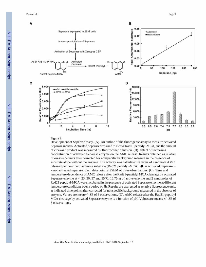
Figure 2.Development of Separase assay. (A). An outline of the fluorogenic assay to measure activatedSeparase in vitro. Activated Separase was used to cleave Rad21 peptidyl-MCA, and the amountof cleavage product was measured by fluorescence emission. (B). Effect of increasingconcentration of activated Separase enzyme on the AMC release. Results obtained as relativefluorescence units after corrected for nonspecific background measure in the presence ofsubstrate alone without the enzyme. The activity was calculated in terms of nanomole AMCreleased per hour per nanomole substrate (Rad21 peptidyl-MCA). ◆ = activated Separase, ▪= not activated separase. Each data point is ±SEM of three observations. (C). Time andtemperature dependence of AMC release after the Rad21-peptidyl MCA cleavage by activatedSeparase enzyme at 4, 23, 30, 37 and 55°C. 16.75ng of active enzyme and 2 nanomoles ofRad21 peptidyl-MCA were incubated in the presence of activated Separase enzyme at differenttemperature conditions over a period of 9h. Results are expressed as relative fluorescence unitsat indicated time points after corrected for nonspecific background measured in the absence ofenzyme. Values are mean+/- SE of 3 observations. (D). AMC release after the Rad21-peptidylMCA cleavage by activated Separase enzyme is a function of pH. Values are means +/- SE of3 observations.
Basu et al. Page 9
Anal Biochem. Author manuscript; available in PMC 2010 September 15.
NIH
-PA Author Manuscript
NIH
-PA Author Manuscript
NIH
-PA Author Manuscript
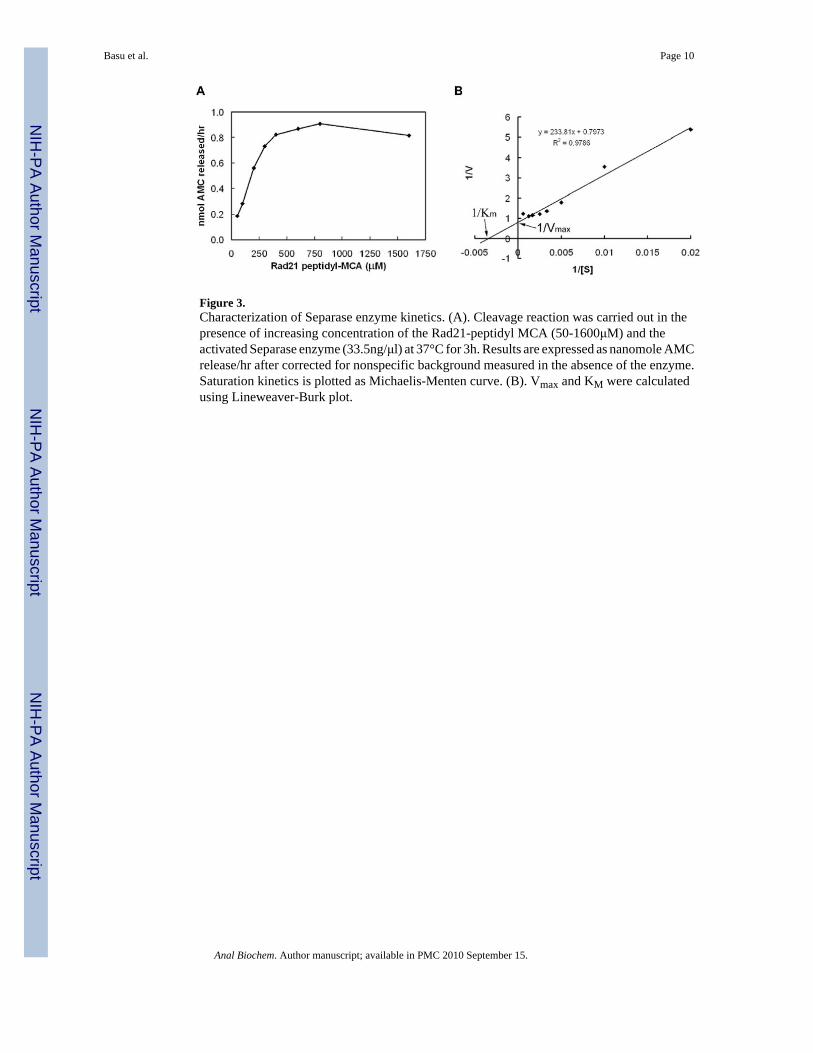
Figure 3.Characterization of Separase enzyme kinetics. (A). Cleavage reaction was carried out in thepresence of increasing concentration of the Rad21-peptidyl MCA (50-1600μM) and theactivated Separase enzyme (33.5ng/μl) at 37°C for 3h. Results are expressed as nanomole AMCrelease/hr after corrected for nonspecific background measured in the absence of the enzyme.Saturation kinetics is plotted as Michaelis-Menten curve. (B). Vmax and KM were calculatedusing Lineweaver-Burk plot.
Basu et al. Page 10
Anal Biochem. Author manuscript; available in PMC 2010 September 15.
NIH
-PA Author Manuscript
NIH
-PA Author Manuscript
NIH
-PA Author Manuscript
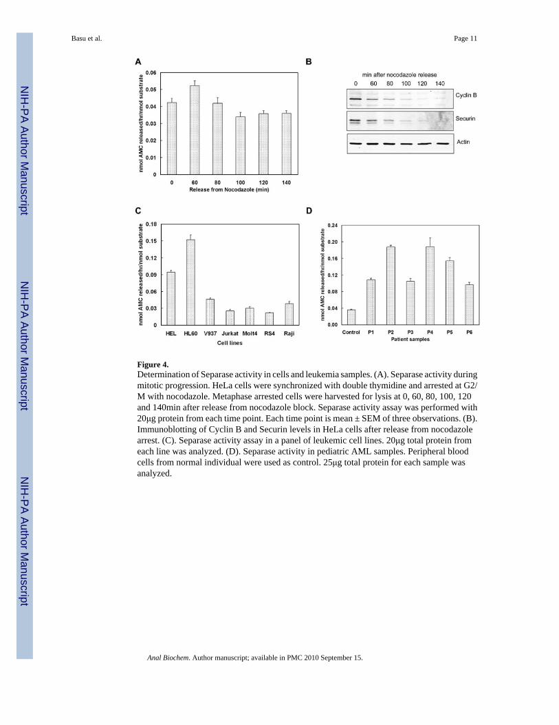
Figure 4.Determination of Separase activity in cells and leukemia samples. (A). Separase activity duringmitotic progression. HeLa cells were synchronized with double thymidine and arrested at G2/M with nocodazole. Metaphase arrested cells were harvested for lysis at 0, 60, 80, 100, 120and 140min after release from nocodazole block. Separase activity assay was performed with20μg protein from each time point. Each time point is mean ± SEM of three observations. (B).Immunoblotting of Cyclin B and Securin levels in HeLa cells after release from nocodazolearrest. (C). Separase activity assay in a panel of leukemic cell lines. 20μg total protein fromeach line was analyzed. (D). Separase activity in pediatric AML samples. Peripheral bloodcells from normal individual were used as control. 25μg total protein for each sample wasanalyzed.
Basu et al. Page 11
Anal Biochem. Author manuscript; available in PMC 2010 September 15.
NIH
-PA Author Manuscript
NIH
-PA Author Manuscript
NIH
-PA Author Manuscript