Development and in vivo in vitro characterization of timolol maleate in-situ gel
16
This article can be downloaded from www.ijpbs.net P - 248 RESEARCH ARTICLE International Journal of Pharma and Bio Sciences DEVELOPMENT AND IN VIVO IN VITRO CHARACTERIZATIONS OF TIMOLOL MALEATE IN-SITU GELS K. S. RATHORE* B.N.Girls’ College of Pharmacy, Udaipur-Raj. 313002 *Corresponding author PHARMACEUTICS ABSTRACT Ocular drug delivery is one of the most fascinating and challenging endeavours facing the research world of pharmaceutics. The poor uptake of many ocular drugs is mainly due to the rapid elimination process by tear turnover. A prolonged precorneal residence time would result in higher absorption for some drugs, a prolonged duration of their therapeutic effect. Recently, in-situ gel formulations have extensively been studied to enhance ocular bioavailability and duration of the drug therapy. It is known that in-situ gels are retained better in the eye than the conventional ocular dosage forms like eyedrops as solutions and suspensions. The phase-change polymers which trigger the drug release in response to external stimuli are the most investigated in controlled drug delivery. Various polymers undergo sol-gel transition due to physical (temperature) or chemical (pH, ions) stimuli when instilled into cul-de-sac of eye. The present work describes the formulation and evaluation of an in-situ ocular delivery system of an antiglaucomatous agent, timolol maleate, which is used in the treatment of primary open angle glaucoma, based on pH-triggered gelation. Poly acrylic acid was used as the gelling agent in combination with hypromellose, which acted as a viscolyzer. The developed formulation was therapeutically efficacious, non-irritant, stable and provided sustained release of the drug over a long period and shelf-life determined by Arrhenius equation was 1.6 years. Intra ocular pressure determined with Schiotz tonometer and eye irritation. study. conducted on albino rabbits by Draize technique. The developed system is thus a viable alternative to conventional formulations.
Transcript of Development and in vivo in vitro characterization of timolol maleate in-situ gel
This article can be downloaded from www.ijpbs.net
P - 248
RESEARCH ARTICLE
International Journal of Pharma and Bio Sciences
DEVELOPMENT AND IN VIVO IN VITRO CHARACTERIZATIONS OF TIMOLOL
MALEATE IN-SITU GELS
K. S. RATHORE*
B.N.Girls’ College of Pharmacy, Udaipur-Raj. 313002
*Corresponding author
PHARMACEUTICS
ABSTRACT
Ocular drug delivery is one of the most fascinating and challenging endeavours facing the research world of pharmaceutics. The poor uptake of many ocular drugs is mainly due to the rapid elimination process by tear turnover. A prolonged precorneal residence time would result in higher absorption for some drugs, a prolonged duration of their therapeutic effect. Recently, in-situ gel formulations have extensively been studied to enhance ocular bioavailability and duration of the drug therapy. It is known that in-situ gels are retained better in the eye than the conventional ocular dosage forms like eyedrops as solutions and suspensions. The phase-change polymers which trigger the drug release in response to external stimuli are the most investigated in controlled drug delivery. Various polymers undergo sol-gel transition due to physical (temperature) or chemical (pH, ions) stimuli when instilled into cul-de-sac of eye. The present work describes the formulation and evaluation of an in-situ ocular delivery system of an antiglaucomatous agent, timolol maleate, which is used in the treatment of primary open angle glaucoma, based on pH-triggered gelation. Poly acrylic acid was used as the gelling agent in combination with hypromellose, which acted as a viscolyzer. The developed formulation was therapeutically efficacious, non-irritant, stable and provided sustained release of the drug over a long period and shelf-life determined by Arrhenius equation was 1.6 years. Intra ocular pressure determined with Schiotz tonometer and eye irritation. study. conducted on albino rabbits by Draize technique. The developed system is thus a viable alternative to conventional formulations.
This article can be downloaded from www.ijpbs.net
P - 249
KEY WORDS
Timolol maleate, ocular drug delivery systems, glaucoma, in-situ gel, pH-triggered in-situ gel, sol to gel, gelation, hypromellose, polyacrylic acid.
INTRODUCTION
Eye drops that are conventional ocular
drug delivery systems often consequence in reduced bioavailability and therapeutic response because high lachrymal turnover and dynamics cause quick precorneal removal of the drug 1. A high frequency of eye drop instillation is associated with patient non-compliance. Inclusion of excess drug in the formulation in an effort to prevail over bioavailability problems is potentially risky if the drug solution drained from the eye is systemically absorbed from the nasolachrymal duct 2. A significant enhances in the precorneal residence time of drugs and as a result bioavailability can be achieved by using delivery systems based on the model of in-situ gel formation. These systems consist of polymers that show sol-to-gel phase transitions due to a change in a specific physicochemical parameter (pH, temperature) in their environment; the cul-de-sac in this case3. Depending on the method employed to cause sol-to-gel phase transition on the eye surface, the following three types of systems are recognized: pH triggered systems 4-6 (e.g. polyacrylic acid, cellulose acetate hydrogen phthalate latex, pseudolatexes), temperature-dependent systems 7-8 (e.g. pluronics, cellulose derivatives, xyloglucans and tetronics) and ion-activated systems 9-10 (e.g. alginates, gelrite, carrageenans and gellan gum).
The objective of the present work was to develop a pH-triggered in-situ gelling ophthalmic delivery system of timolol maleate (TM) that is a non-selective adrenergic receptor antagonist that acts at both the β-1 and β-2-adrenergic sites to effectively reduce intraocular pressure 11. The topical ophthalmic dose of timolol maleate is 1–2 drops of 0.5% solution in the affected eye(s) every 4 h or hourly in the case of primary open angle glaucoma. A combination of polyacrylic acid
(carbopol) and hypromellose (HPMC) was investigated as vehicle for the formulation of eye drops of timolol maleate (0.5%, w/v) which would gel when instilled into the eye and provide sustained release of the drug 12. Timolol maleate (TM) is the most commonly used drug that treats the open-angle glaucoma. As systemic absorption of timolol maleate may cause respiratory and cardiovascular side effects, it is imperative to diminish the systemic absorption and improve ocular bioavailability of timolol maleate 13. Two leads that are currently being explored are bioadhesive and phase transition systems as follows:
(1) Bioadhesive systems can be either polymeric solutions or micro-particle suspensions. They are retained in the cul-de-sac by adhesive bonds established with the mucins or the epithelium 14.
(2) Phase transition systems are instilled in a liquid form and shift to the gel or solid phase once in the cul-de-sac. Two such systems have been reported. Poloxamer-407 solution viscosity increases when its temperature is raised to the eye temperature and cellulose acetate phthalate (CAP) latex coagulates when its native pH of 4.5 is raised by the tear fluid to pH 7.4. Both systems are characterized by a high polymer concentration (25% Poloxamer, 30% CAP). In addition, the surfactive properties of the former and the low pH of the latter may be detrimental to ocular tolerance 15-16.
Gelling capacity was determined by placing a drop of the system in a vial containing 2.0 ml of simulated tear fluid freshly prepared and equilibrated at 37°C and visually assessing the gel formation and noting the time for gelation and the time taken for the gel formed to dissolve. The composition of simulated tear fluid (STF) 17 used was sodium chloride: 0.670 g, sodium bicarbonate: 0.200
This article can be downloaded from www.ijpbs.net
P - 250
g, calcium chloride.2H2O: 0.008 g, purified water q.s.: 100.0 g. The viscosity was measured using a Brookfield Synchroelectric viscometer (RVT model) in the small volume adaptor. The viscosity measured at 20 rpm was used for purposes of comparative evaluation 18.
MATERIALS AND METHODS
Timolol maleate (supplied by Allergan
Ind. Ltd., Mumbai and Agaaz Pharmaceuticals, Ahmedabad as a free drug sample), polyacrylic acid (SD Fine Chem. Ltd., Mumbai), hypromellose (HPMC from Otto Kemi, Mumbai), Benzalkonium chloride (Loba Chemie, Mumbai), ethylcellulose membrane from Axiva® and goat cornea from local butcher shop. All the other chemicals were of extra pure reagent grade and were used as received. Purified water was used throughout the experiments. Albino rabbits of either sex, weighing 1.5-2.5 kg were used for the ocular irritation and intra ocular pressure study. The study was approved by the Institutional Animal Ethics Committee (IAEC) on ethics in care and use of laboratory animals operating under Legislation of India. Preparation of in-situ gelling system: Aqueous solutions of different concentrations of polyacrylic acid and hypromellose (codes ISF1, ISF2, ISF3… ISF15) were prepared by dissolving the polymers in distilled water with aid of gentle heat and constant stirring. Combinations of polyacrylic acid and hypromellose were evaluated for gelling capacity in order to identify the composition suitable for use as in-situ gelling systems. The gelling capacity was determined by placing a drop of the system in a vial containing 2.0 ml of simulated tear fluid (STF) freshly prepared and equilibrated at 37°±2°C and visually assessing the gel formation and noting the time for gelation and the time taken for the gel formed to dissolve 19. The detailed procedure for preparing the in-situ gel-forming system of timolol maleate is outlined as below. Hypromellose
was added to distilled water with the aid of stirring and allowed to hydrate. Polyacrylic acid was sprinkled over this solution and allowed to hydrate overnight. The solution was stirred with an overhead stirrer. Benzalkonium chloride was then added. The drug solution was added to the polyacrylic acid-hypromellose solution under constant stirring until a uniform solution was obtained. The developed formulations were filled in 5.0 ml capacity amber glass vials, closed with gray butyl rubber closures. The formulations, in their final pack, were subjected to terminal sterilization by autoclaving at 121ºC and 15 psi for 20 min 20. Evaluations of the formulations: The developed formulations were evaluated for drug content by UV spectrophotometer at 294nm for timolol maleate, viscosity, clarity (by visual observation against a black and white background in a well lit cabinet), pH (digital pH meter), sol-gel transition and sterility.
Rheological studies: The two most important prerequisites of gelling system are viscosity and gelling capacity (speed and extent of gelation). The formulation should have an optimal viscosity, which will permit its trouble-free instillation into eye as a liquid (drops), which will then endure rapid sol-to-gel transition due to pH changes. Moreover, to assist sustained release of drug to the ocular tissue, the in-situ formed gel should protect its integrity without dissolving or eroding for a prolonged period of time. All the formulations gelled straight away (within a minute) on contact with simulated tear fluid. By visual scrutiny, the formulations formed a translucent matrix on addition to the simulated tear fluid21. The rheological studies of the samples were carried out using Brookfield viscometer. The viscosity of solution was determined using RVT spindle No. 2 whereas the viscosity of gel was determined using RVT spindle No.6. The temperature of the samples was maintained at
37ºC ± 0.50ºC using a hot water bath. The shear rate was altered by changing the speed (in rpm) and corresponding shear
This article can be downloaded from www.ijpbs.net
P - 251
stress was noted from dial scale reading. The dial scale reading multiplied by factor (obtained from table) gave the viscosity in centipoises. The shear stress was then plotted against shear rate to obtain rheogram. The formulation was then poured into an ointment jar and the pH rose to 7.4 by adding 0.5 M NaOH and viscosity was measured using RVT spindle No.6. The viscosity measured at 20 rpm used for purpose of comparative evaluations 22-23. Effect of varying concentration of polymers on gel rheology: In one experiment the effect of varying polyacrylic acid concentration viz. 0.3%, 0.4% and 0.5% on the rheological behavior of 1% hypromellose were studied. In other experiment the effect of varying hypromellose concentration viz. 0.5%, 1.0% and 1.5% on the rheological behavior of 0.4% polyacrylic acid were studied. The shear stress vs. shear rate flow curve was compared to determine the similarity and/or difference in rheological properties of polymer solution. Effect of temperature cycle on gel rheology23: In order to determine if the freezing had any determined effect on sol to gel transformation, the prepared sample (pH 4.0) was freezed and thawed at room temperature. After thawing the pH of solution was adjusted to 7.4. The sample solution containing optimized 0.4% polyacrylic acid and 1% hypromellose at pH 4.0 was exposed to 50ºC for 8 hours and then freezed immediately for another 8 hours. The freezed sample was thawed at room temperature and the pH was adjusted to 7.4. The rheological property of this sample was compared with the sample of same composition which was not subjected to temperature cycling. Effect of sterilization cycle on gel rheology: One of the most important requirements for ophthalmic product is that it must be sterile. In order to determine, if the sample can be sterilized and to find that if the
sterilization cycle has any detrimental effect on gel rheology, the sample at pH 4.0 was subjected to sterilization in autoclave, at 121ºC, 15 psi for 15 minute. After sterilization the pH of solution was adjusted to 7.4 with 0.5M sodium hydroxide. The shear stress vs. shear rate flow curve of sample subjected to sterilization cycle at pH 7.4 was compared with sample not subjected to sterilization cycle.
Content uniformity: The vials (n=3) containing the preparation were shaken for 2-3 min and 100.0 µl of the preparation were transferred aseptically to sterile 25.0ml volumetric flasks with a micropipette and the final volume made up with phosphate buffer pH 7.4. The solution was filtered through 0.45µm membrane and the concentration of timolol maleate was determined at 294 nm, using double beam UV spectrophotometer.
In vitro release and kinetic studies24: The in-vitro diffusion studies were carried out using fabricated open flow through assembly (specially designed glass cylinder open at both ends) and semi-permeable cellophane membrane. Cellophane membrane, previously soaked overnight in simulated tear fluid was mounted by tied and sandwiching between the donor and receiver compartment. The 0.5 ml aliquot of donor solution was placed on top of cellophane membrane. Aliquots of medium (3.0 ml) were withdrawn at selected time intervals and replaced by 3.0 ml of freshly prepared simulated tear fluid through sampling port for analysis. The samples were filtered through 0.45 µm membranes, diluted suitably and analyzed by UV spectrophotometer at 294 nm for timolol maleate. Same procedure is repeated with cornea obtained from fresh goat eye which is obtained from butcher’s shop. The cornea was carefully removed along with 2-4mm of the surrounding sclera tissue. These were then placed in freshly prepared solution of 0.9% sodium chloride. The semi-permeable membrane acts as cornea epithelium. The entire surface of membrane was in contact
This article can be downloaded from www.ijpbs.net
P - 252
with the receptor compartment containing 25.0 ml of isotonic buffer pH 7.4. The receptor compartment was continuously stirred at 50 rpm using magnetic stirrer. The temperature maintained was37±2°C. The study was carried out for 5 h. Experimental procedure: Fresh cornea was mounted by sandwiching the surrounding sclera tissue between the donor and receiver compartment. The 0.5 ml aliquot of donor solutions (0.5% w/v solution of timolol maleate) was placed on top of the cornea. At 30, 60, 90, 120, 150, 180…300 minutes, 3.0 ml of sample was withdrawn from receptor volume (phosphate buffer saline pH 7.4) though sampling port for analysis. The receptor volume was then replaced with 3.0 ml of fresh phosphate buffer saline pH7.4. The absorbance of the withdrawn sample was measured after suitable dilution at 294nm to estimate timolol maleate. The experiment was carried out in triplicate and average values are reported. A comparative permeation study of prepared viscous system with the marketed preparation was done in order to evaluate the release of drug from the polyacrylic acid gel. In-vivo studies: Ocular irritation studies were performed according to Draize technique on six adult male normotensive (18.3±1.8 mmHg) albino rabbits, each weighing 2.0–2.5 kg. The sterile formulations were instilled twice a day for a period of 21 days and the rabbits were observed periodically for redness, swelling, watering of the eye. Evaluation was done as per Draize technique25.
Intra ocular pressure studies perform on the rabbits were provided with food and water ad libitum in a temperature-controlled room (18–24°C). They were exposed to 12 h light and 12 h dark cycles. Intra ocular pressure was measured using a Schiotz tonometer, by the same operator, using the same tonometer after instilling a drop of sensorcaine (a local anaesthetic, xylocaine 0.5%-1%). All the measurements were made three times, at each interval, and a mean of these was taken. All measurement periods
began during the same hour on each day. The animals used were accustomed to the experimental procedure26. The only restraint was the hands of the investigator lightly laid on the back and shoulders of the rabbits. Rabbits that showed a consistent difference in intra ocular pressure between the eyes during the baseline measurements or any signs of eye irritation were excluded from the study. Formulations were instilled topically into the upper quadrant of the eye and the eye was manually blinked three times; one eye received 50 ml of the suspension, and the contralateral eye served as the control.
Repeated administration: After measuring the baseline intra ocular pressure, 50 µl of selected formulation was instilled into the right eye for five times at an interval of 5 min. Then intra ocular pressure was measured at 15, 30, 60, 120, 180 and 300 min from the time of instillation of the last drop. Each formulation was tested on a group of at least six healthy male rabbits. Each animal was given a washout period of 6 days after every treatment. The change in intra ocular pressure (∆IOP) was determined according to the following equation:
∆IOP=IOPDosed eye-IOPControl eye Isotonicity study:
Isotonicity is imperative feature of the ocular preparations. Isotonicity has to be maintained to check tissue damage or irritation of eye. ISG12.1 was subjected to isotonicity testing, since they show superior release characteristics and gelling capacity and the required viscosity. Formulations were mixed with few drops of blood and observed under microscope at 45X magnification and compared with standard marketed ophthalmic formulation containing timolol maleate. The shape of blood cell was compared with standard marketed ophthalmic formulation containing timolol maleate 27.
Accelerated stability studies: Formulated gel preparations were kept at different temperature conditions like 25°C to 28°C ambient temperature (temperature in the working area), 4±1°C (refrigerated
This article can be downloaded from www.ijpbs.net
P - 253
temperature) and 37±2°C (temperature in the incubator) for 6w. The following parameters of the gel such as color, consistency, drug content and degradation rate constant (K) were studied.
To assess the shelf life, the samples were subjected to stability studies. Selected
sterilized formulations were stored at 4±1ºC
(refrigerated temperature), 37±1ºC (ambient
temperature) and 45±1ºC (extreme temperature) for a period of 3 months and analyzed at intervals of 7, 14, 28, 42, 60 and 90 days. The formulations were evaluated at periodic intervals for drug content (by UV Spectrophotometer), clarity, pH, sol-gel transition, rheology, in-vitro drug release and sterility. The shelf-life of the developed formulation was calculated using the Arrhenius method. A graph of log percent of drug remaining vs. temperature was plotted from which shelf life were calculated 28.
RESULTS AND DISCUSSION
The aim of the in-situ gelling system is to enhance drug availability for prolong period of time and hence improve bioavailability of ocular drugs in glaucomatous conditions. The use of polyacrylic acid in-situ gelling systems is substantiated by the property of its solutions to transform into stiff gels when the pH is raised. However, the concentration of polyacrylic acid required to form stiff gel results in highly acidic solutions which are not easily neutralized by the buffering action of the lachrymal fluid. A reduction in polyacrylic acid concentration without compromising the gelling capacity and rheological properties of the delivery system may be achieved by the addition of viscosity enhancing polymers such as hypromellose. The two main prerequisites of an in-situ gelling system are viscosity and gelling capacity. The formulation should have an
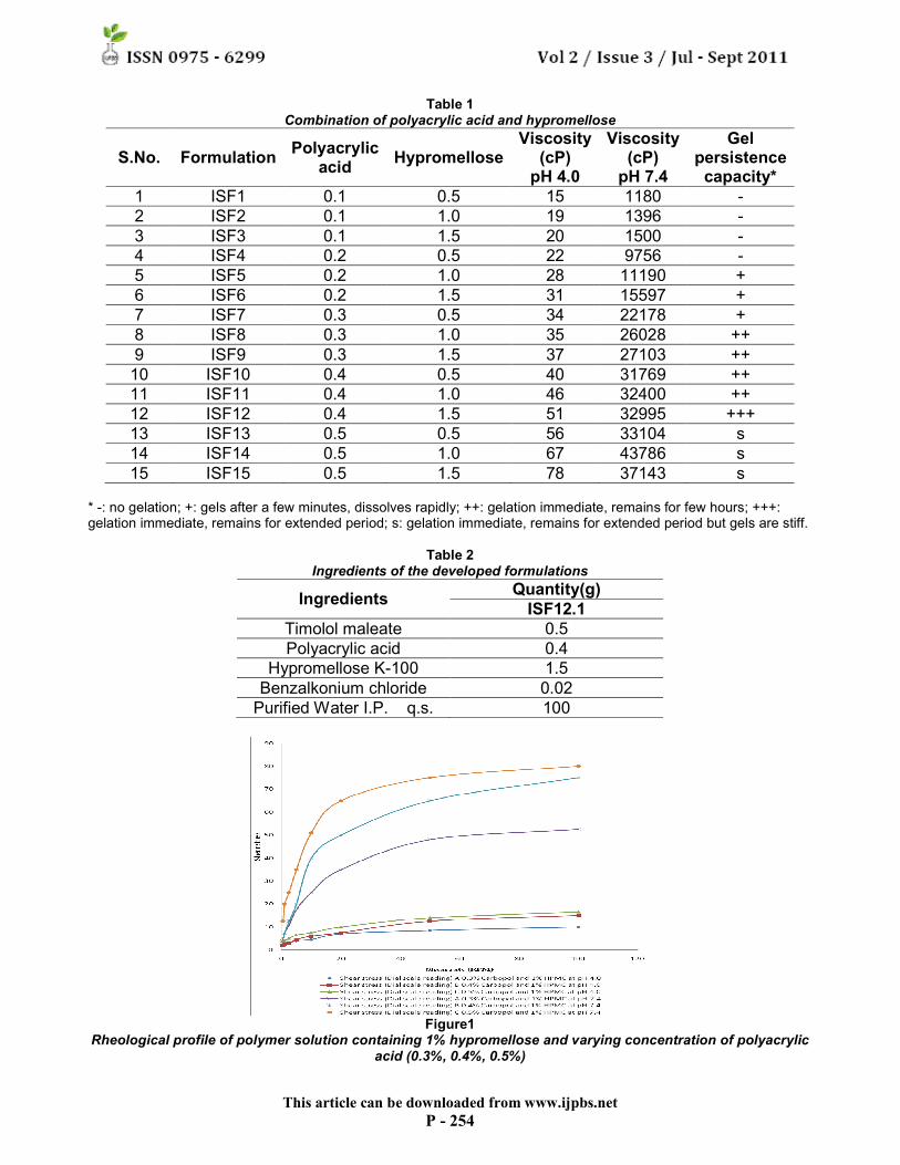
optimum viscosity that will allow easy installation into the eye as a liquid (drops) which would undergo a rapid sol-to-gel transition (triggered by a raise in pH 4.0 to 7.4). Additionally, the gel formed in-situ should preserve in integrity without dissolving or eroding for a prolonged period of time 9. Table 1 shows the gel persistence capacity and viscosity of formulations ISF1 to ISF 15 before and after the adjusting of pH. The solution with low polyacrylic acid concentration (0.1 to 0.2%) does not yield a gel. The higher polyacrylic acid concentration (greater than 0.5%) yield a very stiff gel, but high polyacrylic acid concentration is not easily neutralized by weak buffering action of lachrymal fluids. A concentration of 1.5% hypromellose and 0.4% polyacrylic acid (code ISF 12) was selected as it has satisfactory attributes of viscosity and gelling capacity. The formula for ISF12.1 is listed in Table.2. For primary evaluations visual appearance, pH and clarity observed and all preparations are found to be transparent and clear and their pH is also close to neutral range.
The effect of changing the composition on the rheological properties of the aqueous solution containing both polyacrylic acid and hypromellose was examined. Fig. 1 shows the shear stress vs. Shear rate flow curves for solution containing 1% hypromellose with varying polyacrylic acid concentration. Higher shear stress responses and yield point values are observed with increased polyacrylic acid concentration, both at pH4.0 and pH7.4. A similar trend of increase in shear stress responses and yield point values is seen with increase in polyacrylic acid concentration in the range examined. These studies demonstrated that solution of desirable rheological properties can be prepared by appropriately choosing the concentration of the polymers 12.
This article can be downloaded from www.ijpbs.net
P - 254
Table 1 Combination of polyacrylic acid and hypromellose
S.No. Formulation Polyacrylic
acid Hypromellose
Viscosity (cP) pH 4.0
Viscosity (cP) pH 7.4
Gel persistence capacity*
1 ISF1 0.1 0.5 15 1180 -
2 ISF2 0.1 1.0 19 1396 -
3 ISF3 0.1 1.5 20 1500 -
4 ISF4 0.2 0.5 22 9756 -
5 ISF5 0.2 1.0 28 11190 +
6 ISF6 0.2 1.5 31 15597 +
7 ISF7 0.3 0.5 34 22178 +
8 ISF8 0.3 1.0 35 26028 ++
9 ISF9 0.3 1.5 37 27103 ++
10 ISF10 0.4 0.5 40 31769 ++
11 ISF11 0.4 1.0 46 32400 ++
12 ISF12 0.4 1.5 51 32995 +++
13 ISF13 0.5 0.5 56 33104 s
14 ISF14 0.5 1.0 67 43786 s
15 ISF15 0.5 1.5 78 37143 s * -: no gelation; +: gels after a few minutes, dissolves rapidly; ++: gelation immediate, remains for few hours; +++: gelation immediate, remains for extended period; s: gelation immediate, remains for extended period but gels are stiff.
Table 2 Ingredients of the developed formulations
Ingredients Quantity(g)
ISF12.1
Timolol maleate 0.5
Polyacrylic acid 0.4
Hypromellose K-100 1.5
Benzalkonium chloride 0.02
Purified Water I.P. q.s. 100
Figure1
Rheological profile of polymer solution containing 1% hypromellose and varying concentration of polyacrylic acid (0.3%, 0.4%, 0.5%)
This article can be downloaded from www.ijpbs.net
P - 255
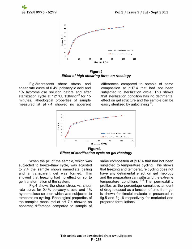
Figure2 Effect of high shearing force on rheology
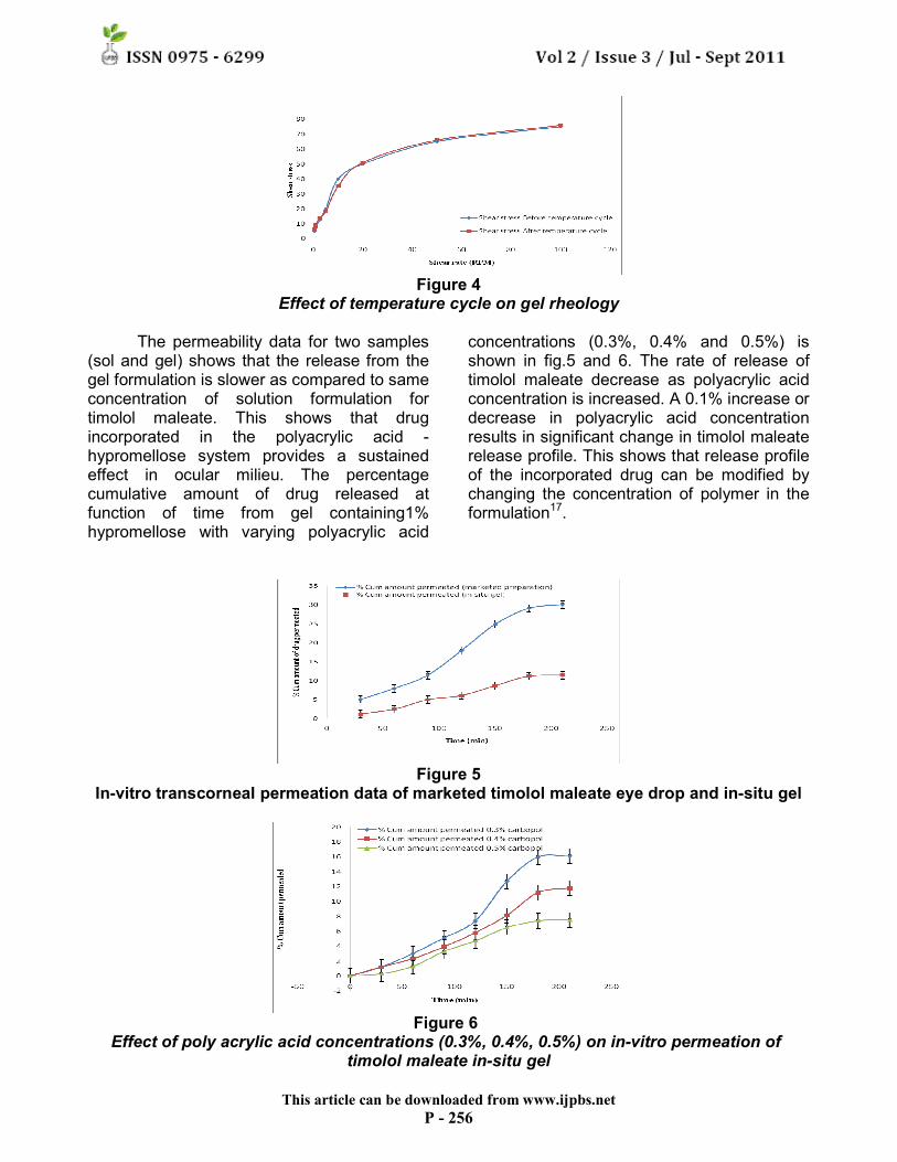
Fig.3represents shear stress and
shear rate curve of 0.4% polyacrylic acid and 1% hypromellose solution before and after sterilization cycle at 121°C, 15lb/inch2 for 15 minutes. Rheological properties of sample measured at pH7.4 showed no apparent
differences compared to sample of same composition at pH7.4 that had not been subjected to sterilization cycle. This shows that sterilization condition has no detrimental effect on gel structure and the sample can be easily sterilized by autoclaving 15.
Figure3
Effect of sterilization cycle on gel rheology
When the pH of the sample, which was subjected to freeze-thaw cycle, was adjusted to 7.4 the sample shows immediate gelling and a transparent gel was formed. This showed that freezing had no effect on sol to gel transformation of the system.
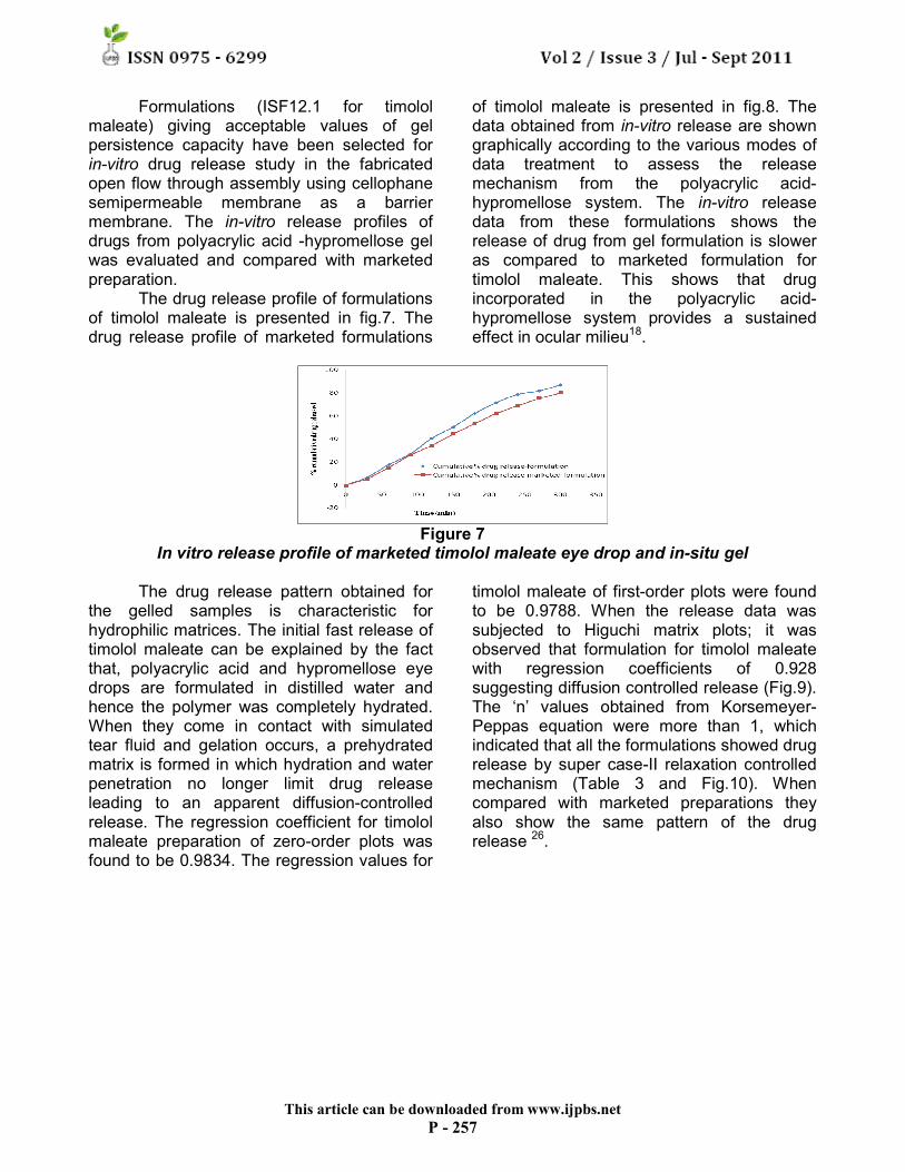
Fig.4 shows the shear stress vs. shear rate curve for 0.4% polyacrylic acid and 1% hypromellose solution which was subjected to temperature cycling. Rheological properties of the samples measured at pH 7.4 showed on apparent difference compared to sample of
same composition at pH7.4 that had not been subjected to temperature cycling. This shows that freezing and temperature cycling does not have any detrimental effect on gel rheology and the preparation can withstand the extreme temperature conditions [16].The permeability profiles as the percentage cumulative amount of drug released as a function of time from gel is shown for timolol maleate is presented in fig.5 and fig. 6 respectively for marketed and prepared formulations.
This article can be downloaded from www.ijpbs.net
P - 256
Figure 4
Effect of temperature cycle on gel rheology The permeability data for two samples
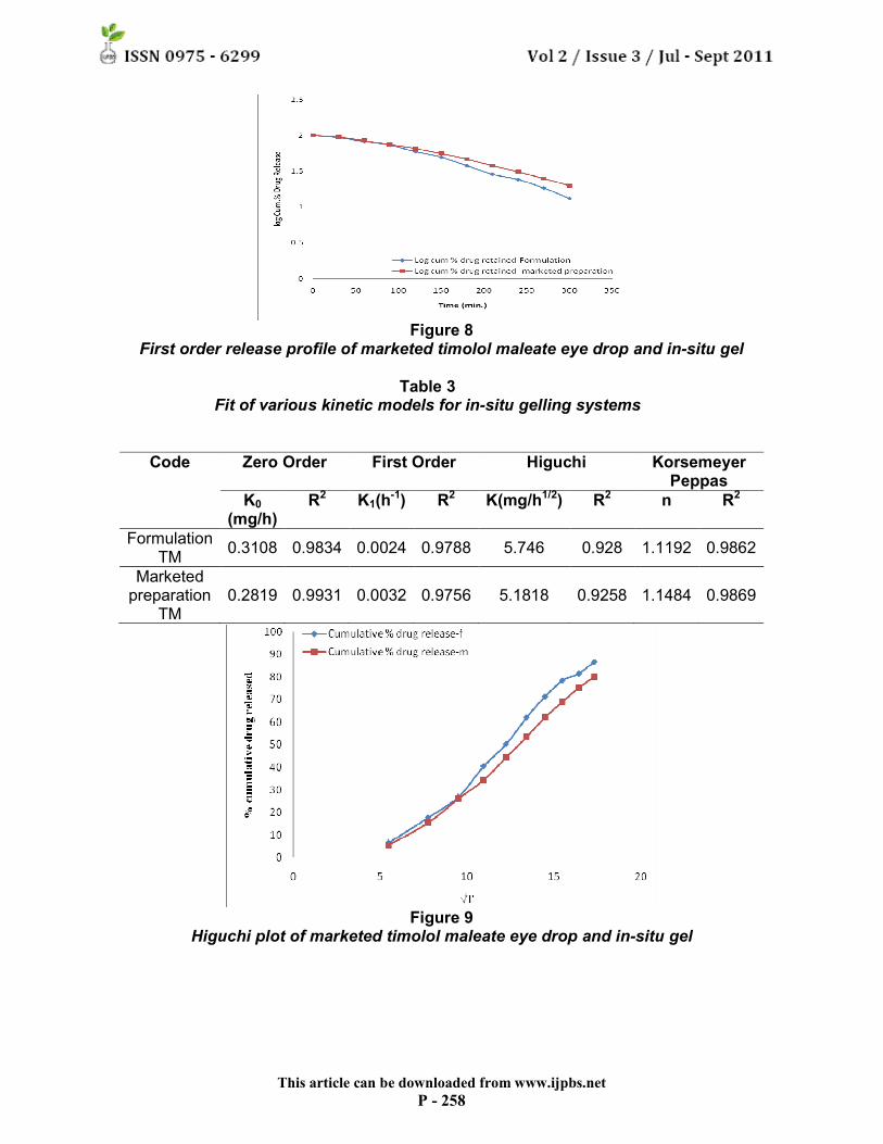
(sol and gel) shows that the release from the gel formulation is slower as compared to same concentration of solution formulation for timolol maleate. This shows that drug incorporated in the polyacrylic acid -hypromellose system provides a sustained effect in ocular milieu. The percentage cumulative amount of drug released at function of time from gel containing1% hypromellose with varying polyacrylic acid
concentrations (0.3%, 0.4% and 0.5%) is shown in fig.5 and 6. The rate of release of timolol maleate decrease as polyacrylic acid concentration is increased. A 0.1% increase or decrease in polyacrylic acid concentration results in significant change in timolol maleate release profile. This shows that release profile of the incorporated drug can be modified by changing the concentration of polymer in the formulation17.
Figure 5 In-vitro transcorneal permeation data of marketed timolol maleate eye drop and in-situ gel
Figure 6
Effect of poly acrylic acid concentrations (0.3%, 0.4%, 0.5%) on in-vitro permeation of timolol maleate in-situ gel
This article can be downloaded from www.ijpbs.net
P - 257
Formulations (ISF12.1 for timolol maleate) giving acceptable values of gel persistence capacity have been selected for in-vitro drug release study in the fabricated open flow through assembly using cellophane semipermeable membrane as a barrier membrane. The in-vitro release profiles of drugs from polyacrylic acid -hypromellose gel was evaluated and compared with marketed preparation.
The drug release profile of formulations of timolol maleate is presented in fig.7. The drug release profile of marketed formulations
of timolol maleate is presented in fig.8. The data obtained from in-vitro release are shown graphically according to the various modes of data treatment to assess the release mechanism from the polyacrylic acid-hypromellose system. The in-vitro release data from these formulations shows the release of drug from gel formulation is slower as compared to marketed formulation for timolol maleate. This shows that drug incorporated in the polyacrylic acid-hypromellose system provides a sustained effect in ocular milieu18.
Figure 7
In vitro release profile of marketed timolol maleate eye drop and in-situ gel The drug release pattern obtained for
the gelled samples is characteristic for hydrophilic matrices. The initial fast release of timolol maleate can be explained by the fact that, polyacrylic acid and hypromellose eye drops are formulated in distilled water and hence the polymer was completely hydrated. When they come in contact with simulated tear fluid and gelation occurs, a prehydrated matrix is formed in which hydration and water penetration no longer limit drug release leading to an apparent diffusion-controlled release. The regression coefficient for timolol maleate preparation of zero-order plots was found to be 0.9834. The regression values for
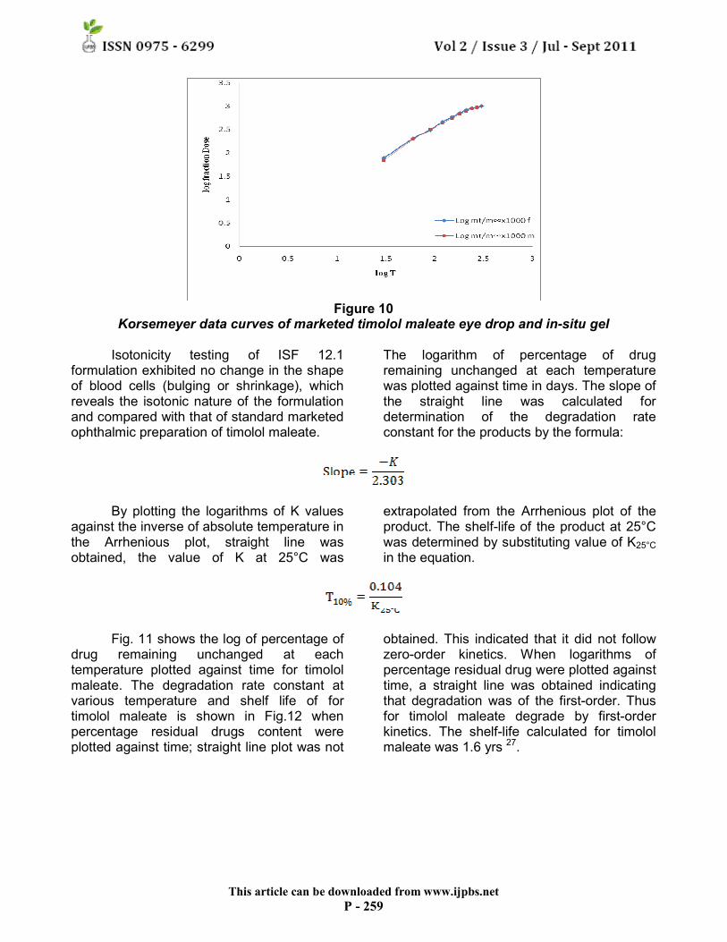
timolol maleate of first-order plots were found to be 0.9788. When the release data was subjected to Higuchi matrix plots; it was observed that formulation for timolol maleate with regression coefficients of 0.928 suggesting diffusion controlled release (Fig.9). The ‘n’ values obtained from Korsemeyer-Peppas equation were more than 1, which indicated that all the formulations showed drug release by super case-II relaxation controlled mechanism (Table 3 and Fig.10). When compared with marketed preparations they also show the same pattern of the drug release 26.
This article can be downloaded from www.ijpbs.net
P - 258
Figure 8 First order release profile of marketed timolol maleate eye drop and in-situ gel
Table 3
Fit of various kinetic models for in-situ gelling systems
Code Zero Order First Order Higuchi Korsemeyer Peppas
K0 (mg/h)
R2 K1(h-1) R2 K(mg/h1/2) R2 n R2
Formulation TM
0.3108 0.9834 0.0024 0.9788 5.746 0.928 1.1192 0.9862
Marketed preparation
TM 0.2819 0.9931 0.0032 0.9756 5.1818 0.9258 1.1484 0.9869
Figure 9 Higuchi plot of marketed timolol maleate eye drop and in-situ gel
This article can be downloaded from www.ijpbs.net
P - 259
Figure 10 Korsemeyer data curves of marketed timolol maleate eye drop and in-situ gel
Isotonicity testing of ISF 12.1
formulation exhibited no change in the shape of blood cells (bulging or shrinkage), which reveals the isotonic nature of the formulation and compared with that of standard marketed ophthalmic preparation of timolol maleate.
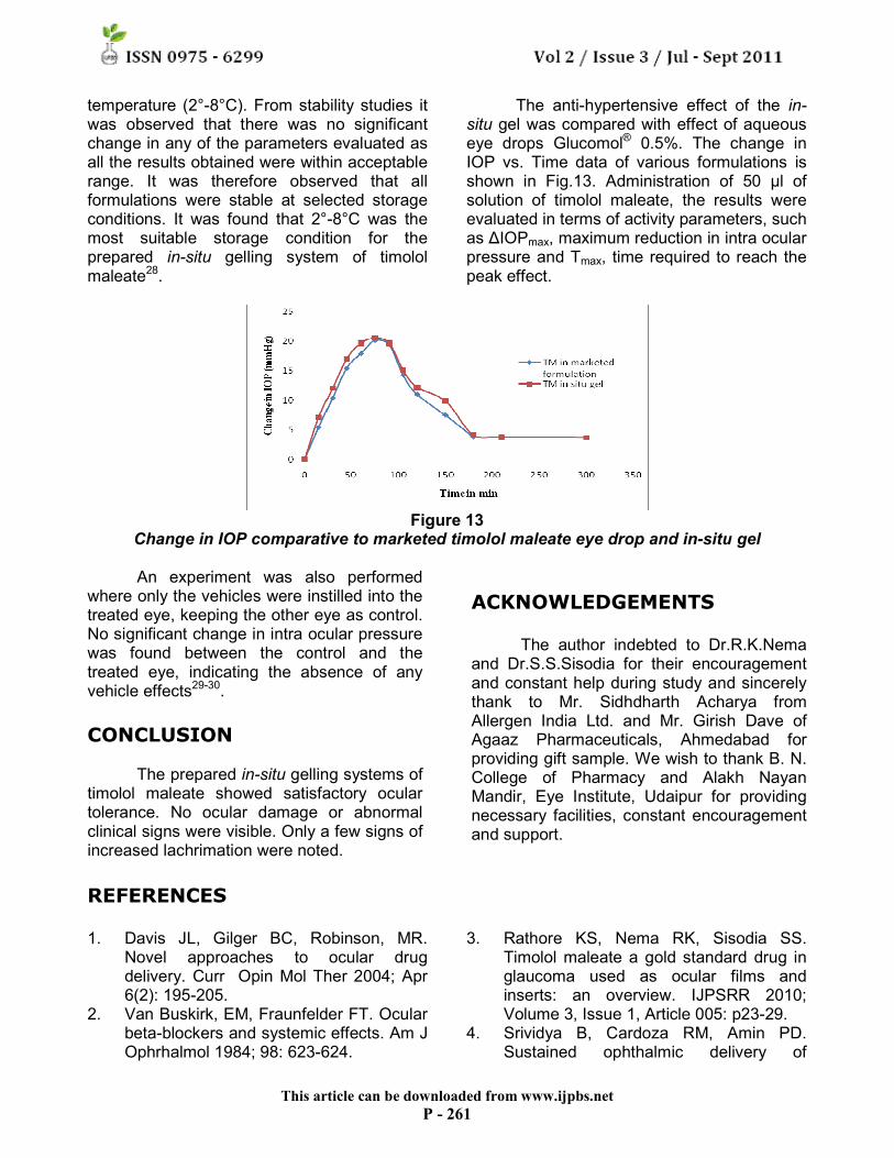
The logarithm of percentage of drug remaining unchanged at each temperature was plotted against time in days. The slope of the straight line was calculated for determination of the degradation rate constant for the products by the formula:
By plotting the logarithms of K values
against the inverse of absolute temperature in the Arrhenious plot, straight line was obtained, the value of K at 25°C was
extrapolated from the Arrhenious plot of the product. The shelf-life of the product at 25°C was determined by substituting value of K25°C
in the equation.
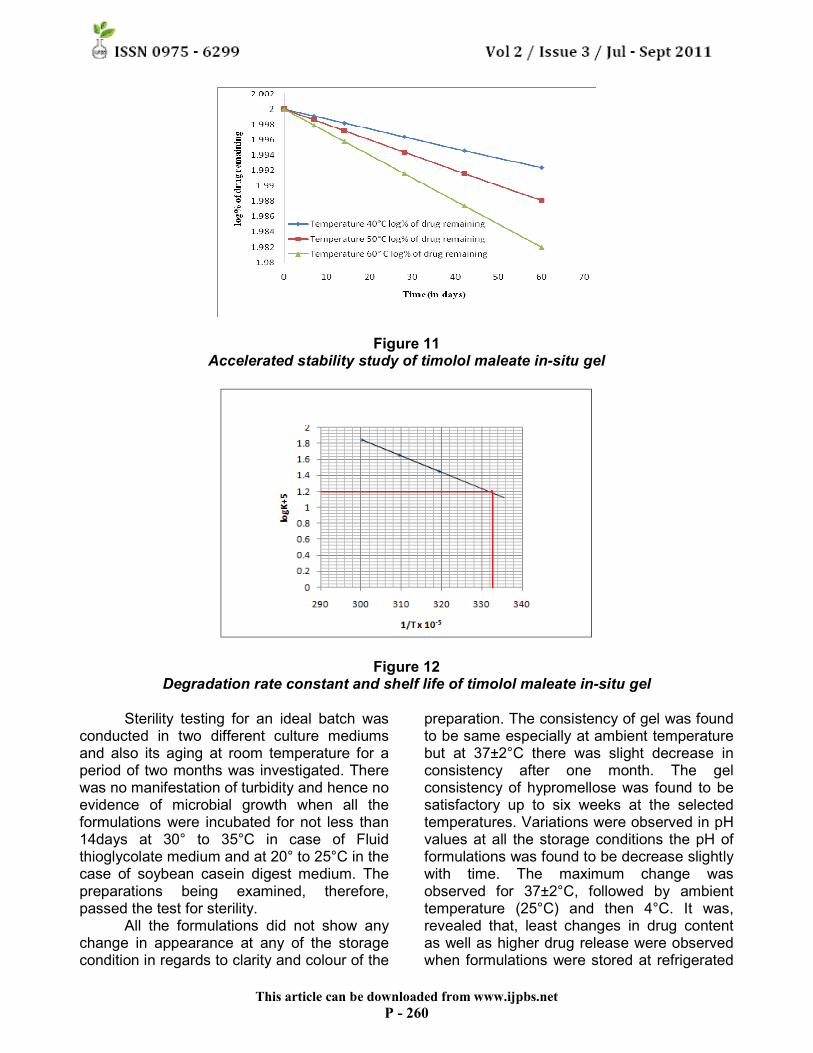
Fig. 11 shows the log of percentage of
drug remaining unchanged at each temperature plotted against time for timolol maleate. The degradation rate constant at various temperature and shelf life of for timolol maleate is shown in Fig.12 when percentage residual drugs content were plotted against time; straight line plot was not
obtained. This indicated that it did not follow zero-order kinetics. When logarithms of percentage residual drug were plotted against time, a straight line was obtained indicating that degradation was of the first-order. Thus for timolol maleate degrade by first-order kinetics. The shelf-life calculated for timolol maleate was 1.6 yrs 27.
This article can be downloaded from www.ijpbs.net
P - 260
Figure 11 Accelerated stability study of timolol maleate in-situ gel
Figure 12 Degradation rate constant and shelf life of timolol maleate in-situ gel
Sterility testing for an ideal batch was
conducted in two different culture mediums and also its aging at room temperature for a period of two months was investigated. There was no manifestation of turbidity and hence no evidence of microbial growth when all the formulations were incubated for not less than 14days at 30° to 35°C in case of Fluid thioglycolate medium and at 20° to 25°C in the case of soybean casein digest medium. The preparations being examined, therefore, passed the test for sterility.
All the formulations did not show any change in appearance at any of the storage condition in regards to clarity and colour of the
preparation. The consistency of gel was found to be same especially at ambient temperature but at 37±2°C there was slight decrease in consistency after one month. The gel consistency of hypromellose was found to be satisfactory up to six weeks at the selected temperatures. Variations were observed in pH values at all the storage conditions the pH of formulations was found to be decrease slightly with time. The maximum change was observed for 37±2°C, followed by ambient temperature (25°C) and then 4°C. It was, revealed that, least changes in drug content as well as higher drug release were observed when formulations were stored at refrigerated
This article can be downloaded from www.ijpbs.net
P - 261
temperature (2°-8°C). From stability studies it was observed that there was no significant change in any of the parameters evaluated as all the results obtained were within acceptable range. It was therefore observed that all formulations were stable at selected storage conditions. It was found that 2°-8°C was the most suitable storage condition for the prepared in-situ gelling system of timolol maleate28.
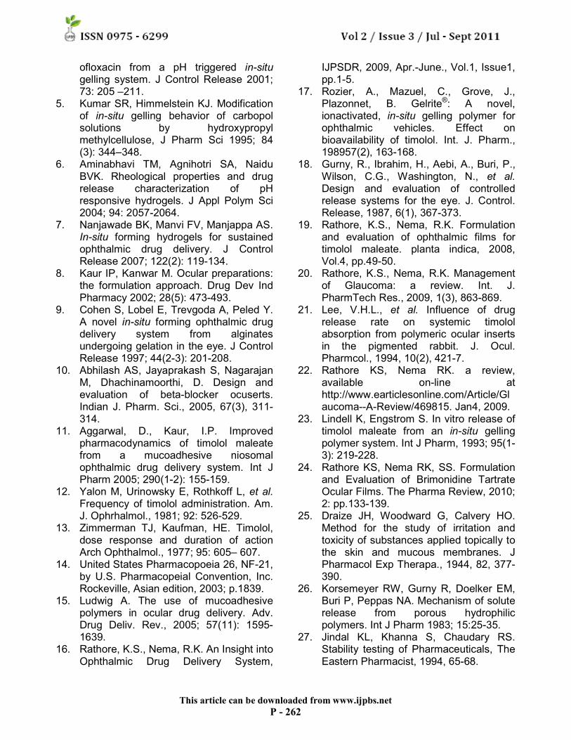
The anti-hypertensive effect of the in-situ gel was compared with effect of aqueous eye drops Glucomol® 0.5%. The change in IOP vs. Time data of various formulations is shown in Fig.13. Administration of 50 µl of solution of timolol maleate, the results were evaluated in terms of activity parameters, such as ∆IOPmax, maximum reduction in intra ocular pressure and Tmax, time required to reach the peak effect.
Figure 13
Change in IOP comparative to marketed timolol maleate eye drop and in-situ gel
An experiment was also performed where only the vehicles were instilled into the treated eye, keeping the other eye as control. No significant change in intra ocular pressure was found between the control and the treated eye, indicating the absence of any vehicle effects29-30.
CONCLUSION
The prepared in-situ gelling systems of
timolol maleate showed satisfactory ocular tolerance. No ocular damage or abnormal clinical signs were visible. Only a few signs of increased lachrimation were noted.
ACKNOWLEDGEMENTS
The author indebted to Dr.R.K.Nema
and Dr.S.S.Sisodia for their encouragement and constant help during study and sincerely thank to Mr. Sidhdharth Acharya from Allergen India Ltd. and Mr. Girish Dave of Agaaz Pharmaceuticals, Ahmedabad for providing gift sample. We wish to thank B. N. College of Pharmacy and Alakh Nayan Mandir, Eye Institute, Udaipur for providing necessary facilities, constant encouragement and support.
REFERENCES
1. Davis JL, Gilger BC, Robinson, MR.
Novel approaches to ocular drug delivery. Curr Opin Mol Ther 2004; Apr 6(2): 195-205.
2. Van Buskirk, EM, Fraunfelder FT. Ocular beta-blockers and systemic effects. Am J Ophrhalmol 1984; 98: 623-624.
3. Rathore KS, Nema RK, Sisodia SS. Timolol maleate a gold standard drug in glaucoma used as ocular films and inserts: an overview. IJPSRR 2010; Volume 3, Issue 1, Article 005: p23-29.
4. Srividya B, Cardoza RM, Amin PD. Sustained ophthalmic delivery of
This article can be downloaded from www.ijpbs.net
P - 262
ofloxacin from a pH triggered in-situ gelling system. J Control Release 2001; 73: 205 –211.
5. Kumar SR, Himmelstein KJ. Modification of in-situ gelling behavior of carbopol solutions by hydroxypropyl methylcellulose, J Pharm Sci 1995; 84 (3): 344–348.
6. Aminabhavi TM, Agnihotri SA, Naidu BVK. Rheological properties and drug release characterization of pH responsive hydrogels. J Appl Polym Sci 2004; 94: 2057-2064.
7. Nanjawade BK, Manvi FV, Manjappa AS. In-situ forming hydrogels for sustained ophthalmic drug delivery. J Control Release 2007; 122(2): 119-134.
8. Kaur IP, Kanwar M. Ocular preparations: the formulation approach. Drug Dev Ind Pharmacy 2002; 28(5): 473-493.
9. Cohen S, Lobel E, Trevgoda A, Peled Y. A novel in-situ forming ophthalmic drug delivery system from alginates undergoing gelation in the eye. J Control Release 1997; 44(2-3): 201-208.
10. Abhilash AS, Jayaprakash S, Nagarajan M, Dhachinamoorthi, D. Design and evaluation of beta-blocker ocuserts. Indian J. Pharm. Sci., 2005, 67(3), 311-314.
11. Aggarwal, D., Kaur, I.P. Improved pharmacodynamics of timolol maleate from a mucoadhesive niosomal ophthalmic drug delivery system. Int J Pharm 2005; 290(1-2): 155-159.
12. Yalon M, Urinowsky E, Rothkoff L, et al. Frequency of timolol administration. Am. J. Ophrhalmol., 1981; 92: 526-529.
13. Zimmerman TJ, Kaufman, HE. Timolol, dose response and duration of action Arch Ophthalmol., 1977; 95: 605– 607.
14. United States Pharmacopoeia 26, NF-21, by U.S. Pharmacopeial Convention, Inc. Rockeville, Asian edition, 2003; p.1839.
15. Ludwig A. The use of mucoadhesive polymers in ocular drug delivery. Adv. Drug Deliv. Rev., 2005; 57(11): 1595-1639.
16. Rathore, K.S., Nema, R.K. An Insight into Ophthalmic Drug Delivery System,
IJPSDR, 2009, Apr.-June., Vol.1, Issue1, pp.1-5.
17. Rozier, A., Mazuel, C., Grove, J., Plazonnet, B. Gelrite®: A novel, ionactivated, in-situ gelling polymer for ophthalmic vehicles. Effect on bioavailability of timolol. Int. J. Pharm., 198957(2), 163-168.
18. Gurny, R., Ibrahim, H., Aebi, A., Buri, P., Wilson, C.G., Washington, N., et al. Design and evaluation of controlled release systems for the eye. J. Control. Release, 1987, 6(1), 367-373.
19. Rathore, K.S., Nema, R.K. Formulation and evaluation of ophthalmic films for timolol maleate. planta indica, 2008, Vol.4, pp.49-50.
20. Rathore, K.S., Nema, R.K. Management of Glaucoma: a review. Int. J. PharmTech Res., 2009, 1(3), 863-869.
21. Lee, V.H.L., et al. Influence of drug release rate on systemic timolol absorption from polymeric ocular inserts in the pigmented rabbit. J. Ocul. Pharmcol., 1994, 10(2), 421-7.
22. Rathore KS, Nema RK. a review, available on-line at http://www.earticlesonline.com/Article/Glaucoma--A-Review/469815. Jan4, 2009.
23. Lindell K, Engstrom S. In vitro release of timolol maleate from an in-situ gelling polymer system. Int J Pharm, 1993; 95(1-3): 219-228.
24. Rathore KS, Nema RK, SS. Formulation and Evaluation of Brimonidine Tartrate Ocular Films. The Pharma Review, 2010; 2: pp.133-139.
25. Draize JH, Woodward G, Calvery HO. Method for the study of irritation and toxicity of substances applied topically to the skin and mucous membranes. J Pharmacol Exp Therapa., 1944, 82, 377-390.
26. Korsemeyer RW, Gurny R, Doelker EM, Buri P, Peppas NA. Mechanism of solute release from porous hydrophilic polymers. Int J Pharm 1983; 15:25-35.
27. Jindal KL, Khanna S, Chaudary RS. Stability testing of Pharmaceuticals, The Eastern Pharmacist, 1994, 65-68.
This article can be downloaded from www.ijpbs.net
P - 263
28. Urtti A, Pipkin, JD, Rork G, Repta AJ. Controlled drug delivery devices for experimental ocular studies with timolol 1. In vitro release studies, Int Pharm, 1990, 61, 235-240.
29. Rathore KS, Nema RK. Review on Ocular inserts. Int J PharmTech Res, 2009; 1(2), 164-169.
30. Shedden AH. Timolol maleate in gel-forming solution: a novel formulation of timolol maleate. Intl. J. Ophthalmol., 1994, 10, 332–336.