Development and characterization of sol–gel silica–alumina composite coatings on AISI 316L for...
7
Development and characterization of sol–gel silica–alumina composite coatings on AISI 316L for implant applications S.K. Tiwari ⁎ , T. Mishra, M.K. Gunjan, A.S. Bhattacharyya, T.B. Singh, R. Singh National Metallurgical Laboratory, Jamshedpur-831007, India Received 10 October 2006; accepted in revised form 15 February 2007 Available online 23 February 2007 Abstract Sol–gel alumina coatings were deposited on medical grade stainless steel (AISI 316L) with an intermediate layer of silica by dip-coating method. The coatings obtained were homogeneous, crack-free and consisted of low crystalline γ-Al 2 O 3 along with some boehmite phase. EDAX revealed the presence of only Al in the film. The corrosion performance of alumina-coated stainless steel was evaluated by electrochemical polarization, open-circuit potential measurement and chronoamperometry in Ringer's solution. Coating has shown to enhance the pitting potential of AISI 316L by ∼ 470 mV and reduced passive current ≤ 10 − 9 A cm − 2 . The formation of thermodynamically stable silica–alumina interface was proposed to account for enhanced corrosion protection behaviour of the coating. © 2007 Elsevier B.V. All rights reserved. Keywords: Stainless steel; Coating; Sol–gel; Aluminium oxide; Corrosion resistance; Bioimplant 1. Introduction Metallic materials such as Ti, Ti-alloy, Co–Cr alloy and stainless steel (AISI 316L) are used as biomaterials due to their superior tensile and fatigue strength, and fracture toughness as compared to nonmetals such as polymeric and ceramic [1]. However, metallic materials corrode by aggressive biofluid and release metallic ions which resulted in the reduction of their biocompatibility. The biocompatibility and corrosion resistance of these implants are primarily determined by their constituent material and surface micro structural properties such as rough- ness, grain size, etc. Among the metallic materials, AISI 316L stainless steel is most commonly employed for temporary devices such as fracture plates, bone screws and hip nails due to its low cost and acceptable biocompatibility [2,3]. However, it has been often reported to suffer from severe crevice and galvanic corrosion, primarily due to the presence of occluded sites and high chloride concentration in physiological fluid [4]. The corrosion of the stainless steel implant releases metal ions such as Fe, Ni and Cr, which produce local systematic effects and thereby plays a role in prosthetic loosening. A study showed that AISI 316L stainless steel produces corrosion products above certain non-lethal concentrations and thereby disturb the proliferation/differentiation relationship of osteo- blastic human alveolar bone cell cultures in a dose dependent manner [5]. Therefore, major interest in stainless steel research has now been focused on to improve corrosion resistance (lo- calized and generalized mechanism) and to prevent the metal ions from diffusing to nearby tissues. Furthermore, adhesion between implant and bone would not be desirable to avoid difficulty during removal of the implant (temporary) after the service. A simple way of preventing corrosion or metal ion diffusion may be a hermetic sealing of the surface with materials stable against diffusion of corrosive species such as water, oxygen, acids, bases or combination of them. Various surface modifi- cation techniques, such as plasma ion implantation [6–8], laser melting [9–11] and laser surface alloying [12,13], physical and chemical vapor deposition (PVD and CVD) [14,15], thermal oxidation [16], electrochemical surface modification/anodizing [17], have been tried out to improve wear, corrosion, and fret- ting resistance of orthopaedic implants. However, each of these methods has limitations concerning the performance of tailored surfaces and their complex operating procedures. Sol–gel thin Surface & Coatings Technology 201 (2007) 7582 – 7588 www.elsevier.com/locate/surfcoat ⁎ Corresponding author. Tel.: +91 657 2271709-14x2101. E-mail address: [email protected] (S.K. Tiwari). 0257-8972/$ - see front matter © 2007 Elsevier B.V. All rights reserved. doi:10.1016/j.surfcoat.2007.02.026
-
Upload
independent -
Category
Documents
-
view
3 -
download
0
Transcript of Development and characterization of sol–gel silica–alumina composite coatings on AISI 316L for...

201 (2007) 7582–7588www.elsevier.com/locate/surfcoat
Surface & Coatings Technology
Development and characterization of sol–gel silica–alumina compositecoatings on AISI 316L for implant applications
S.K. Tiwari ⁎, T. Mishra, M.K. Gunjan, A.S. Bhattacharyya, T.B. Singh, R. Singh
National Metallurgical Laboratory, Jamshedpur-831007, India
Received 10 October 2006; accepted in revised form 15 February 2007Available online 23 February 2007
Abstract
Sol–gel alumina coatings were deposited on medical grade stainless steel (AISI 316L) with an intermediate layer of silica by dip-coatingmethod. The coatings obtained were homogeneous, crack-free and consisted of low crystalline γ-Al2O3 along with some boehmite phase. EDAXrevealed the presence of only Al in the film. The corrosion performance of alumina-coated stainless steel was evaluated by electrochemicalpolarization, open-circuit potential measurement and chronoamperometry in Ringer's solution. Coating has shown to enhance the pitting potentialof AISI 316L by ∼470 mVand reduced passive current ≤10−9 A cm−2. The formation of thermodynamically stable silica–alumina interface wasproposed to account for enhanced corrosion protection behaviour of the coating.© 2007 Elsevier B.V. All rights reserved.
Keywords: Stainless steel; Coating; Sol–gel; Aluminium oxide; Corrosion resistance; Bioimplant
1. Introduction
Metallic materials such as Ti, Ti-alloy, Co–Cr alloy andstainless steel (AISI 316L) are used as biomaterials due to theirsuperior tensile and fatigue strength, and fracture toughness ascompared to nonmetals such as polymeric and ceramic [1].However, metallic materials corrode by aggressive biofluid andrelease metallic ions which resulted in the reduction of theirbiocompatibility. The biocompatibility and corrosion resistanceof these implants are primarily determined by their constituentmaterial and surface micro structural properties such as rough-ness, grain size, etc. Among the metallic materials, AISI 316Lstainless steel is most commonly employed for temporarydevices such as fracture plates, bone screws and hip nails due toits low cost and acceptable biocompatibility [2,3]. However, ithas been often reported to suffer from severe crevice andgalvanic corrosion, primarily due to the presence of occludedsites and high chloride concentration in physiological fluid [4].The corrosion of the stainless steel implant releases metal ionssuch as Fe, Ni and Cr, which produce local systematic effects
⁎ Corresponding author. Tel.: +91 657 2271709-14x2101.E-mail address: [email protected] (S.K. Tiwari).
0257-8972/$ - see front matter © 2007 Elsevier B.V. All rights reserved.doi:10.1016/j.surfcoat.2007.02.026
and thereby plays a role in prosthetic loosening. A studyshowed that AISI 316L stainless steel produces corrosionproducts above certain non-lethal concentrations and therebydisturb the proliferation/differentiation relationship of osteo-blastic human alveolar bone cell cultures in a dose dependentmanner [5]. Therefore, major interest in stainless steel researchhas now been focused on to improve corrosion resistance (lo-calized and generalized mechanism) and to prevent the metalions from diffusing to nearby tissues. Furthermore, adhesionbetween implant and bone would not be desirable to avoiddifficulty during removal of the implant (temporary) after theservice.
A simple way of preventing corrosion or metal ion diffusionmay be a hermetic sealing of the surface with materials stableagainst diffusion of corrosive species such as water, oxygen,acids, bases or combination of them. Various surface modifi-cation techniques, such as plasma ion implantation [6–8], lasermelting [9–11] and laser surface alloying [12,13], physical andchemical vapor deposition (PVD and CVD) [14,15], thermaloxidation [16], electrochemical surface modification/anodizing[17], have been tried out to improve wear, corrosion, and fret-ting resistance of orthopaedic implants. However, each of thesemethods has limitations concerning the performance of tailoredsurfaces and their complex operating procedures. Sol–gel thin
7583S.K. Tiwari et al. / Surface & Coatings Technology 201 (2007) 7582–7588
film processing represents an alternative method to obtain thedesired coatings [18]. The attractiveness of the process lies intailoring functional properties of coatings by changing precur-sors, thermal treatments or addition of particles in sol. The mainadvantage of the process is the ability to form inorganic struc-tures at a relatively low temperature and to produce thin homo-geneous films on large scale [19].
Efforts have been made to enhance the corrosion resistanceof metallic implants by depositing ZrO2 [20,21], silica [22–24],alumina [25–27], etc., through sol–gel route. Amongst these,alumina particularly appears promising due to its high resis-tance to wear, corrosion, and good thermal barrier properties[28]. Masalski et al. [26] obtained amorphous Al2O3 film onAISI 316L stainless steel through sol–gel dip-coating followedby heating at 500 °C; coating showed to be stable during 1000 hof exposure in Ringer's solution. The potentiodynamic results,however, showed severe fluctuations in current, indicating in-stability and less protective coating at higher electrochemicalpotentials. It seems that the stable interface between coating andsubstrate is essential for coating to be better stable that mayisolate the underlying metal from corrosive solution. It has beenobserved [29] that alumina silica mixed compounds has muchlower free energy values than the boehmite. This leads to theconcept that an alumina/silica interface should be thermody-namically stable against corrosion.
Keeping above facts in view, attempts have been made todeposit SiO2/Al2O3 film on AISI 316L through sol–gel routewith an intent to enhance the stability of coating to improvelocalized corrosion resistance of AISI 316L for biomedicalapplications. The stable composite coating was also aimed toprevent leaching of undesirable metallic elements from goinginto the solution, essential for biocompatible coating. Thecoatings obtained were characterized for homogeneity by SEMand AFM. The chemical composition of the coating wasanalyzed by EDAX. Adherence of coating was determined byscratch test method. The purity and phases were analyzedthrough FTIR and XRD. The coated 316L samples wereevaluated for corrosion resistance in physiological conditionsthrough open circuit potential measurement, polarization testsand chronoamperometry. The solution after corrosion tests wassubjected to metal ions analyses for possible leaching andcompared with the uncoated AISI 316L.
2. Experimental procedure
2.1. Sample preparation
Stainless steel 316L (elemental composition in wt.%: C—0.03, Cr—18.0, Ni—12.0, Mo—2.45, Mn—1.70, P—0.04,S—0.01, Si—0.16, Fe–Bal.) was selected as substrate, as itis commonly used for biomedical application. Solution an-nealed SS plate was cut into 25 mm×10 mm×3 mm sizecoupons. Coupons were polished using silicon carbide papersranging from 120–1200 grit. The polished specimens washedwith detergent solution, degreased with acetone and thor-oughly washed with distilled water, were kept in ethanolbefore coating.
2.2. Preparation of sol and coating
Aluminium isopropoxide (ACROS) and tetraethyl–orthosi-licate (Aldrich) were used as source of Al2O3 and SiO2, res-pectively. The alumina sol was prepared by hydrolysis and polycondensation of aluminium isopropoxide catalysed by HNO3 asreported elsewhere [26]. The ethanolic silica sol was preparedby mixing silane (TEOS) with ethanol and water: silane/distilled water/ethanol=5/5/90% (v/v). Both sols were kept atleast for four days with intermittent stirring before coating.Before alumina coating, the AISI 316L specimens kept in ethanolwere dipped in silica sol, withdrawn slowly (0.5 mm/s) and driedat 100 °C. The process was repeated two times. The Al2O3
coatings were deposited on SiO2 coated specimen by dip-coatingtechnique at a pulling speed of 0.8 to 1 mm/s. Dip-coatedsamples were dried in air before heating at 300 °C for 15 min.The process of deposition, drying and heat treatment were re-peated four times to increase the thickness of coating and finallyheated at 500 °C for 2 h.
2.3. Physical characterization and adhesion test
The sol was dried at 100 °C in an oven for 48 h. The solidphase content was grind and heated at 500 °C for 2 h to getsimilar phase as in the coating. The phase composition of thepowder, heated at 500 °C, was determined by X-ray diffrac-tometer (XRD, SIEMENS, D-500) using a Cu–Kα radiations.The diffraction intensity was measured by the scanningtechnique in the range of 2θ=4°–70°. FTIR spectra was re-corded using a NICOLET-7500 spectrometer in the wave num-ber ranging from 400 to 4000 cm−1, with KBr pellets. Thetopography of the coating on AISI 316L was studied by usingScanning Electron Microscopy (JEOL 840A, Japan) andAtomic Force Microscopy (AFM, SPA-400, SEIKO, Japan).Composition of the film was obtained through EDAX. Coatingthickness and the roughness of the film was determined bymeans of Taylor Hobson surface profilometer.
Adhesion test was carried out by driving a Rockwell Cdiamond indenter (120° angle, 200 μm radius) using computercontrolled scratch tester (Model TR101, DUCON). Thespecimen was mounted on the test table, which was slid at aspeed of 0.2 mm/s. The load on the indenter was increasedlinearly at a rate of 10 N/mm.
2.4. Electrochemical characterization
Electrochemical measurements were made in Ringer's solu-tion prepared by adding 9 g/l NaCl, 0.17 g/l CaCl2, 0.42 g/l KCl,and 2.0 g/l NaHCO3 (AR grade chemicals) to distilled water.The pH of the solutions was maintained by adding requisiteamount of NaOH or HCl. Polarization data were generatedusing a computer controlled Potentiostat/Galvanostat (GamryInstruments, USA). The corrosion test cell was the classicconfiguration of three electrodes (graphite as counter, a satu-rated calomel electrode as reference and the coated metallicsample as working electrode). Polarization curves were ob-tained by a potential sweep in the noble direction at a constant
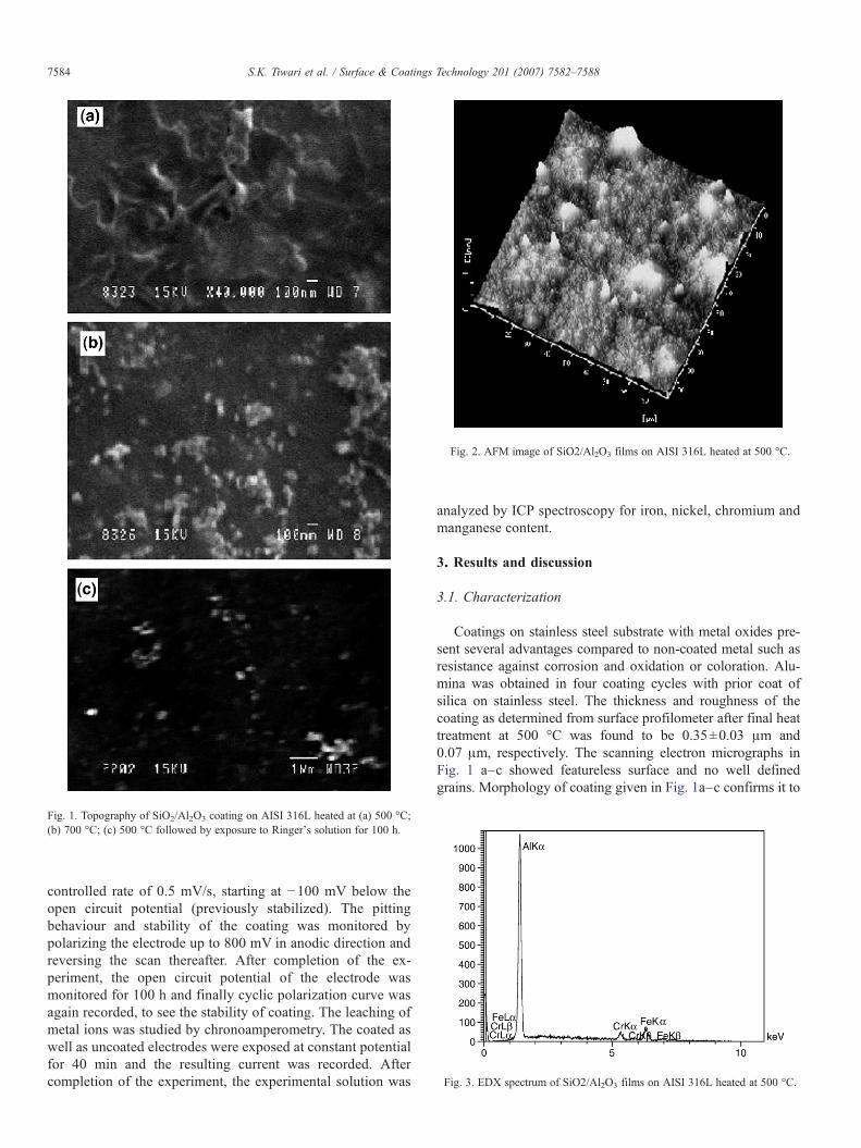
Fig. 1. Topography of SiO2/Al2O3 coating on AISI 316L heated at (a) 500 °C;(b) 700 °C; (c) 500 °C followed by exposure to Ringer's solution for 100 h.
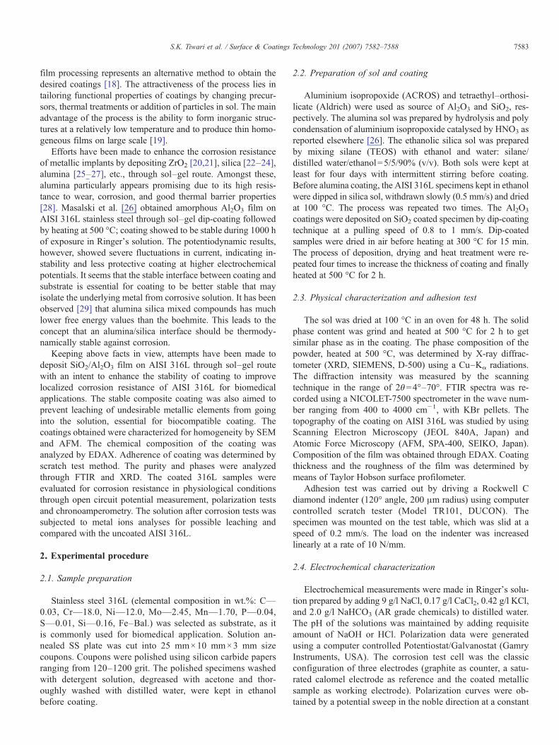
Fig. 2. AFM image of SiO2/Al2O3 films on AISI 316L heated at 500 °C.
Fig. 3. EDX spectrum of SiO2/Al2O3 films on AISI 316L heated at 500 °C.
7584 S.K. Tiwari et al. / Surface & Coatings Technology 201 (2007) 7582–7588
controlled rate of 0.5 mV/s, starting at −100 mV below theopen circuit potential (previously stabilized). The pittingbehaviour and stability of the coating was monitored bypolarizing the electrode up to 800 mV in anodic direction andreversing the scan thereafter. After completion of the ex-periment, the open circuit potential of the electrode wasmonitored for 100 h and finally cyclic polarization curve wasagain recorded, to see the stability of coating. The leaching ofmetal ions was studied by chronoamperometry. The coated aswell as uncoated electrodes were exposed at constant potentialfor 40 min and the resulting current was recorded. Aftercompletion of the experiment, the experimental solution was
analyzed by ICP spectroscopy for iron, nickel, chromium andmanganese content.
3. Results and discussion
3.1. Characterization
Coatings on stainless steel substrate with metal oxides pre-sent several advantages compared to non-coated metal such asresistance against corrosion and oxidation or coloration. Alu-mina was obtained in four coating cycles with prior coat ofsilica on stainless steel. The thickness and roughness of thecoating as determined from surface profilometer after final heattreatment at 500 °C was found to be 0.35±0.03 μm and0.07 μm, respectively. The scanning electron micrographs inFig. 1 a–c showed featureless surface and no well definedgrains. Morphology of coating given in Fig. 1a–c confirms it to

Fig. 4. Diffraction pattern of dried alumina gel powder heated at 500 °C for 2 h.
Fig. 6. Scratch test on SiO2/Al2O3/ AISI 316L heated at 500 °C for 2 h.(a) Tangential and Normal load as a function of scratch length; (b) Coef-ficient of friction vs Vertical load.
7585S.K. Tiwari et al. / Surface & Coatings Technology 201 (2007) 7582–7588
be homogeneous and crack-free. Heating at higher temperature(700 °C for 2 h) also did not indicate any structural change inthe coating (Fig. 1b). No deterioration in the coating was visibleand it remained intact even after 100 h of exposure to Ringer'ssolution followed by cyclic polarization (Fig. 1c). AFM analysisindicated coating consisting of self-agglomerated nanoparticleswithout any defects (Fig. 2). The EDAX analysis of the filmindicated the presence of Al along with small amount of Cr andFe (Fig. 3). The small amount of Cr and Fe indicated in the filmmay be originated from substrate due to penetration of X-rays asthe film thickness is less than 0.5 μm.
X-ray diffractogram of gel powder heated at 500 °C (Fig. 4)showed two γ-alumina peaks, (400) and (440) [ICDD, File 29-63] along with (020) and (120) peaks of boehmite. This suggeststhat the boehmite was not fully transformed to γ-alumina at500 °C. The grazing incidence (GI) XRD of coated AISI 316Lalso indicated the presence of γ-alumina (as observed in Fig. 4)in addition to base alloy. The FTIR spectra of powder, obtainedby drying sol at 100 °C and heated at 500 °C are shown in Fig. 5.IR spectra of the sample dried at 100 °C exhibited a band at1072 cm−1 with a shoulder at 1162 cm−1, which are attributed tothe symmetrical and anti symmetrical AlOH bending modes.The stretching Al–O modes of AlO6 groups are found below900 cm−1 (894, 737 and 638 cm−1). The two broad bands at 894and 494 cm−1 in the sample treated at 500 °C are attributed totwo coordinations of Al atoms in γ-Al2O3 [29]. The band at
Fig. 5. FTIR spectra of alumina gel samples heated for 2 h at (a) 100 °C and(b) 500 °C.
1390 cm−1, which disappeared after heating at 500 °C corre-sponds to NO3
− group from HNO3 added for peptization. Theabsorption band at 1650 cm−1 appeared in both samples is due tomoisture in the sample [30]. Hence the IR data also support theXRD pattern suggesting the partial formation of γ-Al2O3.
The durability and functionality of film is critically depen-dent on the adhesion between the film and the underlyingsubstrate. The results of adhesion of the coating to AISI 316L,determined by scratch test are shown in Fig. 6. Fig. 6a displaysapplied normal load and resulting tangential force as function ofscratch track length for SiO2/Al2O3 coating on AISI 316Lheated at 500 °C. The first part where the applied normal load isless than the critical load, a linear behaviour of the tangentialforce with applied load is observed. The change in slope of thecurve indicates the detachment of the coating from the substrate.However, it is difficult to determine transition points in theabove curve. The coefficient of friction μeff=FH/FV, plottedagainst vertical load, FV, yields a useful plot as shown inFig.6 (b). The plot clearly demonstrates a transition in the linearfrictional behaviour of the Al2O3/SiO2/AISI 316L system. Thecritical load determined on four samples was 24±2 N. Simi-lar results for adhesion strength failure of Al2O3 films havealso been reported in literature [31,32]. Phani et al. [31] have

Fig. 7. Potentiodynamic polarization curves for coated AISI 316L in Ringer'ssolution at 25 °C.
Fig. 9. Potentiodynamic polarization curves for coated and bare AISI 316L inRinger's solution at 37 °C.
7586 S.K. Tiwari et al. / Surface & Coatings Technology 201 (2007) 7582–7588
deposited thin films of Al2O3 on polished silica glass sub-strates by sol–gel dip-coating technique followed by annealingat 200–800 °C or exposure to microwave radiation at differentpowers. The critical load up to 25 N for adhesion strength failurehas been reported for both annealed and microwave irradiatedfilms.
3.2. Electrochemical characterization
Fig. 7 shows the potentiodynamic polarization behaviour ofAl2O3 coated AISI 316L at 25 °C with and without an inter-mediate layer of SiO2. The presence of silica intermediate layerhas shifted the breakdown potential of Al2O3 coating from670 mV to 1030 mV. The higher breakdown potential of silicaalumina coating may be explained on the basis of much lowerfree energy values of alumina silica mixed compounds (suchas −6901 and −2616 kJ mol−1 for Mullite and Sillimanite,respectively) than boehmite (−985 kJ mol−1 ) alone [33]. Thepresence of SiO2 film between substrate and Al2O3 coatingwould have produced a thermodynamically stabilized silica alu-mina interface that showed better stability against corrosion ascompared to alumina alone [33]. Therefore, in all further studies,silica was coated on AISI 316L prior to alumina deposition.
The influence of final heat treatment of the coating on thepolarization behaviour in Ringer's solution at 37 °C is shown inFig. 8. The coating heated at 300 °C for 2 h showed a brief
Fig. 8. Potentiodynamic polarization curves for SiO2/Al2O3 films on AISI 316Lin Ringer's solution at 37 °C.
passivation (up to 153 mV) followed by gradual increase incurrent. Slow increase in current even up to 1.0 V, unlike fasterincrease above break down potential showed by uncoatedsubstrate, indicates slow absorption of electrolyte through thepores in the oxide coating. The coatings heated at 500 °C and700 °C for 2 h displayed barrier type of behaviour. Lattershowed lower passivation current and breakdown potentialhigher by about 155 mV than that treated at 500 °C. The changein polarization behaviour with increase in final heat treatmenttemperature from 300 to 700 °C may be attributed to densi-fication of the coating at higher temperatures. The reproduc-ibility of the coating heated at 700 °C, however, was only 33%,unlike at 500 °C where coating was 100% reproducible. Thisprobably would have been due to the formation of defects athigher temperature. The optimum temperature, therefore, wasconcluded to be 500 °C and was considered as a final heattreatment step for other studies.
The comparison of corrosion protection ability of the presentcoating with uncoated and only silica coated SS-316L is pre-sented in Fig. 9. It appears that the silica coating alone was notdense enough to protect the substrate from corrosion. With theincrease in anodic potential, the defects were created in thecoating and caused breakdown potential to lower by about310 mV, as compared to uncoated AISI 316L. Depositing Al2O3
film on silica–coated steel and subsequent heating at 500 °Cshifted the pitting potential of AISI 316L by 480 mV in nobledirection.
The coating performance was also investigated in theRinger's physiological solution at different pH — 4.0, 7.4,and 9.0. This was done as pH in the body has been reported tovary during surgery and also widely differs among variousparts of the body [34]. The anodic polarization behaviour ofcoating was almost identical at three different pH valuesexcept that the shift of corrosion potential (155 mV) in positiveside with decrease in pH to 4.0. More noble Ecorr with decreasein pH from 7.4 to 4.0 may be explained by considering the pHdependent Nernst equation, E(V)=1.23–0.059 pH, of the fol-lowing reaction:
1=2 O2 þ 2Hþ þ 2e− ¼ H2O

Fig. 11. Chronoamperometric evaluations of coated and bare SS316L inRinger's solution at 37 °C.
7587S.K. Tiwari et al. / Surface & Coatings Technology 201 (2007) 7582–7588
It is clear from above equation that the decrease in pH willresult in nobler redox potential of oxygen reduction whichgoverns the corrosion kinetics in such solutions. Noble redoxpotential of O2 reduction will shift the corrosion potential ofAl2O3 coated AISI 316L in more positive direction.
The stability of the coating was checked by immersing thecoated AISI 316L in solution for about an hour, so as to attainthe stable OCP (∼+260 mV), followed by polarizing it an-odically to +800 mV (close to but below break down potential)and back to OCP. The polarization curve is shown in Fig. 10 (a).The same specimen, after polarization, was left in the solutionfor about 100 h to record any unusual fluctuations in the po-tential (OCP) due to peeling/dissolving of the coating. It wasassumed that polarizing the coated AISI 316L to high anodicpotential (such as close to the pitting potential) may destabilizethe coating or metal/coating interface according to known filmbreakdown mechanism [35]. The OCP measurement for such along time (100 h), however, did not show significant variation inOCP (Fig. 10b), indicating the coating yet intact. When thesame specimen, was subjected again to polarization high in theanodic regions, the nature of the curve appeared to remainunchanged as shown in Fig. 10a. This observation, thus, con-firms the stability or resistance of coating against any degra-dation in the aqueous medium.
Fig. 10. Performance of SiO2/Al2O3 on AISI 316L as a function of immersionperiod in Ringer's solution at 25 °C. (a) Polarization curves; (b) OCP variation.
The release of deleterious metal ions was studied by allowingboth bare and alumina coated AISI 316L to corrode at +500 mV(above the pitting potential of bare AISI 316L) for 40 min. Thepotential was decided so as to produce significant amount ofmetal ions to compare the two specimens (coated and uncoated)in terms of their emitted ionic concentration. It is an importantaspect of bio-implants that influences their performance andbiocompatibility in the body. While corroding at +500 mV, theresultant current was recorded till 40 min and shown in Fig. 11.The current was lower for coated electrode by six orders ofmagnitude than for uncoated one. The solutions in whichsamples were corroded were submitted for analyses (using ICP)to detect and measure the various metal ions (such as Fe, Cr, Niand Mo). The quantities of metal ions leached out fromuncoated electrode were: Fe ∼0.073 mg, Cr ∼0.016 mg, Ni∼0.008 mg and Mo ∼0.005 mg, whereas no such metal ionswere found from the solution from corrosion of coated AISI316L. These results were compared with the blank solution(fresh Ringer's solution without exposing the coated oruncoated AISI 316L) where no such ions were noticed. Metalions were absent in the solution after coated SS was forced tocorrode at even higher anodic potential (+750 mV) for 40 min.These results, however, support the application of SiO2/Al2O3
coating in preventing the unwanted metal ions from going intothe body and hence suitable for bio-implants.
4. Conclusion
A systematic study was made to develop corrosion resistantsilica/alumina coating on AISI 316L for implant applications.The coating was homogeneous with thickness ∼0.35±0.03 μm and showed significant scratch resistance. The pittingpotential of AISI 316L after coating was shifted by about470 mV towards positive side in physiological Ringer'ssolution. The change in pH did not influence the performanceof the coating. The coated AISI 316L electrodes was stable anddid not degrade after long time of exposure in the Ringer'ssolution. Coating was able to prevent the leaching of undesiredmetal ions from the substrate at even high anodic potential (upto +750 mV), where bare AISI 316L released significant metalcontents.
7588 S.K. Tiwari et al. / Surface & Coatings Technology 201 (2007) 7582–7588
Acknowledgement
The authors are grateful to the Director, National Metallur-gical Laboratory, Jamshedpur for his encouragement and per-mission to publish this work.
References
[1] M. Sumita, T. Hanawa, S.H. Teoh, Mater. Sci. Eng., C, Biomim. Mater.,Sens. Syst. 24 (2004) 753.
[2] J. Brunski, in: B. Ratner, A. Hoffman, F. Schoen, J. Lemons (Eds.),Biomaterial Science: An Introduction to Material in Medicine, AcademicPress, San Diego, 1996, p. 37.
[3] M. Sumita, Orthop. Surg. 48 (1997) 927.[4] H. Placko, S. Brown, J. Payer, J. Biomed. Mater. Res. 17 (1983) 655.[5] M.A. Costa, M.H. Fernandes, J. Mater. Sci., Mater. Med. 11 (2000) 141.[6] P.K. Chu, J.Y. Chen, L.P. Wang, N. Huang, Mater. Sci. Eng., R Rep. 36
(2002) 143.[7] S. Mandl, B. Rauschenbach, Surf. Coat. Technol. 156 (2002) 276.[8] H. Schmidt, C. Konetschny, U. Fink, Mater. Sci. Technol. 14 (1998) 592.[9] H. Badekas, C. Panagopoulos, S. Ecomnomou, J. Mater. Process. Technol.
44 (1994) 54.[10] T.M. Yue, J.K. Yu, Z. Mei, H.C. Man, Mater. Lett. 52 (2002) 206.[11] R. Singh, N.B. Dahotre, Surf. Eng. 21 (2005) 297.[12] E. Gyorgy, A. Perez Del Pino, P. Sera, J.L. Merenza, Surf. Coat. Technol.
173 (2003) 265.[13] H.C. Man, N.Q. Zhao, Z.D. Cui, Surf. Coat. Technol. 192 (2005) 341.[14] A. Matthews, Advances in surface processing technology, in: K.M.
Strafford, P.K. Data, J.S. Gray (Eds.), Surface Engineering Practice, EllisHorwood Ltd, New York, 1990, p. 33.
[15] C.E. Morosanu, Thin Films by Chemical Vapour Deposition, Elsevier,Amsterdam, 1990, p. 31.
[16] M.C. Garcia-Alonso, L. Saldana, G. Valles, J.L. Gonzalez-Carrasco, J.Gonzalez-Cabrero, M.E. Martinez, E. Gil-Garay, L. Munuera, Biomater-ials 24 (2003) 19.
[17] B. Yang, M. Uchida, Hyun-Min Kim, X. Zhang, T. Kokubo, Biomaterials25 (2004) 1003.
[18] M. Guglielmi, J. Sol-Gel Sci. Technol. 8 (1997) 443.[19] K. Izumi, M. Murakami, T. Deguchi, A. Morita, J. Am. Ceram. Soc. 72
(1989) 1465.[20] W. Liu, Y. Chen, C. Ye, P. Zhang, Ceram. Int. 28 (2002) 349.[21] A. Balamurugan, S. Kannan, S. Rajeswari, Mater. Lett. 57 (2003) 4202.[22] P. Galliano, J.J. de Damborenea, M.J. Pascual, A. Duran, J. Sol-Gel Sci.
Technol. 13 (1998) 723.[23] D.C.L. Vasconcelos, J.A.N. Carvalho, M. Mantel, W.L. Vasconcelos,
J. Non-Cryst. Solids 273 (2000) 135.[24] J. Gallardo, A. Durán, J.J. de Damborenea, Corros. Sci. 46 (2004) 795.[25] M.Atik, P. de LimaNeto, L.A. Avaca,M.A.Aegerter, Ceram. Int. 21 (1995)
403.[26] J. Masalski, J. Gluszek, J. Zabrzeski, K. Nitsch, P. Gluszek, Thin Solid
Films 349 (1999) 186.[27] H.M. Hawthorne, A. Neville, T. Troczynski, X. Hu, M. Thammachart, Y.
Xie, J. Fu, Q. Yang, Surf. Coat. Technol. 176 (2004) 243.[28] M. Natali, G. Carta, V. Rigato, G. Rossello, G. Salmaso, P. Zanella,
Electrochim. Acta 50 (2005) 4615.[29] G. Urretavizcaya, A.L. Cavalieri, J.M. Porto López, I. Sobrados, J. Sanz,
J. Mater. Synth. Process. 6 (1998) 1.[30] Chih-Peng Lin, Shaw-Bing Wen, J. Am. Ceram. Soc. 85 (2002) 1467.[31] A.R. Phani, S. Santucci, J. Non-Cryst. Solids 352 (2006) 4093.[32] T. Hübert, S. Svoboda, B. Oertel, Surf. Coat. Technol. 201 (2006) 487.[33] H. Schmidt, S. Langenfeld, R. Naβ, Mater. Des. 18 (1997) 309.[34] K.J. Bundy, Crit. Rev. Biomed. Eng. 22 (1994) 139.[35] J.R. Galvele, in: R.P. Frankenthal, J. Kruger (Eds.), Passivity of Metals,
The Electrochemical Society, Pennington, NJ, 1978, p. 285.