Determination of the Effects of Different Maillard Reaction ...
206
University of Arkansas, Fayetteville University of Arkansas, Fayetteville ScholarWorks@UARK ScholarWorks@UARK Graduate Theses and Dissertations 12-2017 Determination of the Effects of Different Maillard Reaction Determination of the Effects of Different Maillard Reaction Products on the Taxonomic Composition of the Gut Microbiota Products on the Taxonomic Composition of the Gut Microbiota Nesreen Hamdan ALJahdali University of Arkansas, Fayetteville Follow this and additional works at: https://scholarworks.uark.edu/etd Part of the Biochemistry Commons, Cell Biology Commons, and the Molecular Biology Commons Citation Citation ALJahdali, N. H. (2017). Determination of the Effects of Different Maillard Reaction Products on the Taxonomic Composition of the Gut Microbiota. Graduate Theses and Dissertations Retrieved from https://scholarworks.uark.edu/etd/2569 This Dissertation is brought to you for free and open access by ScholarWorks@UARK. It has been accepted for inclusion in Graduate Theses and Dissertations by an authorized administrator of ScholarWorks@UARK. For more information, please contact [email protected].
-
Upload
khangminh22 -
Category
Documents
-
view
4 -
download
0
Transcript of Determination of the Effects of Different Maillard Reaction ...

University of Arkansas, Fayetteville University of Arkansas, Fayetteville
ScholarWorks@UARK ScholarWorks@UARK
Graduate Theses and Dissertations
12-2017
Determination of the Effects of Different Maillard Reaction Determination of the Effects of Different Maillard Reaction
Products on the Taxonomic Composition of the Gut Microbiota Products on the Taxonomic Composition of the Gut Microbiota
Nesreen Hamdan ALJahdali University of Arkansas, Fayetteville
Follow this and additional works at: https://scholarworks.uark.edu/etd
Part of the Biochemistry Commons, Cell Biology Commons, and the Molecular Biology Commons
Citation Citation ALJahdali, N. H. (2017). Determination of the Effects of Different Maillard Reaction Products on the Taxonomic Composition of the Gut Microbiota. Graduate Theses and Dissertations Retrieved from https://scholarworks.uark.edu/etd/2569
This Dissertation is brought to you for free and open access by ScholarWorks@UARK. It has been accepted for inclusion in Graduate Theses and Dissertations by an authorized administrator of ScholarWorks@UARK. For more information, please contact [email protected].
Determination of the Effects of Different Maillard Reaction Products on the Taxonomic
Composition of the Gut Microbiota
A dissertation submitted in partial fulfillment
of the requirements for the degree of
Doctor of Philosophy in Cell and Molecular Biology
by
Nesreen ALJahdali
College of Education and Science
Bachelor of Science in Biology, 2002
King Abdul Aziz University
Master of Science in Biology-Animal Ecology, 2007
December 2017
University of Arkansas
This dissertation is approved for recommendation to the Graduate Council.
_______________________________
Dr. Franck Carbonero
Dissertation Director _______________________________ ______________________________ Dr. Steve Ricke Dr. Sami Dridi
Committee Member Committee Member _______________________________
Dr. Charles Rosenkrans
Committee Member
ABSTRACT:
The Maillard Reaction (MR) is a non-enzymatic chemical reaction which results in linkage
between the amino group of amino acids and the carbonyl group of reduced sugars. This reaction
generates Maillard reaction products (MRPs) which are not present naturally in foods, and are
responsible for a range of colors, odors, flavors, and other sensory properties. Conflicting reports
of MRPs impacts on human health are probably due to the fact that bioconversion of these
digestible molecules by the gut microbiota has been marginally taken into account. This study
aimed to determine the effects of different MRPs on rodent’s gut microbiota through16S rRNA
amplicon sequencing over three different studies. Study 1 focused on the impact of
NƐCarboxymethyllysine (CML) on the composition of mice gut microbiota and potential
association with severity of experimental colitis. Study 2 focused on the impact of bread
melanoidins on the composition of healthy and experimental colitis mice gut microbiota. Study 3
focused on the impact of consumption of increasing amounts of malt melanoidins on mice gut
microbiota. It was found that CML induced limited changes in gut microbiota profiles of healthy
mice, but was found to significantly relieve the bacterial dysbiosis imparted by one (but not the
other) inflammation-inducing chemical, especially the Proteobacteria bloom. Bread crust model
(high in melanoidins) showed significant decreases of Bacteroides spp. and Enterobacteriaceae,
while it increased Faecalibacterium spp. Also, bread crust model limited to increase
Enterobacteriaceae in colitis model. High amounts of malts rich melanoidins rapidly and
persistently led to significantly different gut microbiota profiles. There was a trend for decrease of
Lactobacillus and Ruminococcus and increase of Akkermansia and Bifidobacterium with higher
amounts of dietary melanoidins. We concluded that CML and melanoidins are not detrimental in
terms of their impact on the gut microbiota, and that they may even have prebiotic properties.

ACKNOWLEDGEMENTS:
I would like to express my special thanks of gratitude to my advisor Dr. Franck Carbonero
who gave me the opportunity to work on this project and welcomed me in his lab and office. His
knowledge and experiences are always invaluable to increase my knowledge and success in my
educational pursuits. He is always willing to assist me with scheduling, research, and guidance
over the years. I could not have been as successful as I am right now without his help through
every step.
I would also like to thank Dr. Pauline Anton-Gay and Dr. Pascale Gadonna at LaSalle
University for constructive discussions that helped me to do this wonderful project on the topic of
Maillard Reaction Products and for the valuable information provided by them in their respective
fields. I would also like to thank my advisory committee members: Professor Steve Ricke, Dr.
Sami Dridi, and Dr. Charles Rosenkrans for supporting my career. I would like to thank Dr. Ricke’s
lab for providing equipment, especially Dr. Peter Rubinelli and Thomas Flecker.
I would also like to thank the Cell and Molecular Biology program for providing me with
an outstanding education throughout my PhD degree. I also extend my gratitude to all people at
University of Arkansas for their advice and guidance without which this project would not have
been possible. My thanks and appreciations also go to King Abdul Aziz University in Saudi Arabia
for supporting and encouraging which helped me in completion of my degree. Lastly, I thank
almighty, my family, friends, and lab members for their constant encouragement without which
this assignment would not be possible.

TABLE OF CONTENT:
General Introduction……………………………………………………………............................1
References…………………………………………………………………………………………5
Chapter one: Review of the Literature.............................................................................................7
1.1 Abstract………………………………………………………………………………………..8
1.2 Introduction……………………………………………………………………………………9
1.3 The Chemistry of Maillard Reaction Products……………………….…………...…………10
1.3.1 Maillard reaction in food…………………………………………………………………..10
1.3.2 Generation of Maillard Reaction Products in vivo...………………….…………………...11
1.3.3 Important Maillard Reaction Product Molecules..................................................................12
1.4 Maillard Reaction Products Impact on Nutrition and Health…………………….………….18
1.4.1 Consequences of the Maillard Reaction in Nutrition………………………………………18
1.4.2 Effect of Maillard Reaction Products on Health…………………………………………...20
1.5 MRPs and Gut Microbiome and Metabolome……………………………………………….26
1.5.1 Human Gut Microbiome and Metabolome……………………………….………………..26
1.5.2 Known Microbial Interactions between Microbes and MRPs…………….……………….27
1.6 Conclusions and Perspectives……………………………………………….……………….33
1.7 References……………………………………………………………………………………35
Chapter Two: Research Article……………………………………………………………….….55
2-1 Abstract………………………………………………………………………………………56
2.2 Introduction………………………………..…………………………………………………58
2.3 Materials and Methods……………………………………………………………………….60
2.3.1 Chemicals…………………………………………………………………………………..60
2.3.2 Animals and Study design…………………………………………………………………60
2.3.3 Experimental Colitis……………………………………………….………………………61
2.3.4 DNA Extraction and Amplicon Sequencing Strategy……………….…………………….63
2.3.5 Library Preparation and Quality Control…………………………….…………………….64
2.3.6 Bioinformatics and Statistical Analyses…………………………………………………...64
2.4 Results……………………………………………………………………………………….65
2.4.1 Effect of CML on Weight Change and Food Intake in Healthy and Colitic Mice……...…65
2.4.2 Effect of CML on Macroscopic Lesions and Neutrophil Infiltration in Healthy and Colitic
Mice…………………………………………………...…………………………………………65
2.4.3 Gut Microbiota Analyses...………………………………………………………….……..66
2.4.3.1 Impact of CML on the Gut Microbiota of Healthy Mice……………………………..….66
2.4.3.2 Impact of TNBS and DSS on the Gut Microbiota……………………………………….66
2.4.3.3 Impact of CML on the Gut Microbiota of DSS and TNBS-Treated Mice………………68
2.5 Discussion and Conclusion…………………………………………………………………..68
2.6 References……………………………………………………………………………………74
Chapter Three: Research Article…………………………………………………………………93
3.1 Abstract……………………………………………………………………………….......….94
3.2 Introduction..............................................................................................................................95
3.3 Material and Methods..............................................................................................................96
3.3.1 Bread Preparation…………………………………………………………………………..96
3.3.2 Animals and Study design…………………………………………………………………97
3.3.3 DNA Extraction and PCR Amplifications (Polymerase Chain Reaction)…………………98
3.3.4 Library Preparation………………………………………………………………………...98
3.3.5 Sequencing, Bioinformatics and Statistical Analyses……………………………………...99
3.4 Results and Discussion………………………………….………………………………….100
3.4.1 Impact of TNBS treatment on the gut microbiota……………..…………………………101
3.4.2 Impact of MFC on the rats gut microbiota………………………………………………..102
3.4.3 Impact of BCM on the rats gut microbiota……………………………………………….104
3.5 Conclusions…………………………………...…………………………………………….107
3.6 References…………………………………………..………………………………………108
Chapter Four: Research Article…………………..…………….………………………………126
4.1 Abstract………………………………………….………………………………………….127
4.2 Introduction............................................................................................................................128
4.3 Material and Methods………………………………………………………………...…….130
4.3.1 Experimental Animals……………………………………………………………………130
4.3.2 Experimental Design………………………………………………………………...……130
4.3.3 Fecal Short Chain Fatty Acids (SCFAs) Quantification………………………………….131
4.3.4 DNA Extraction and PCR Amplifications………………………….…………………….131
4.3.5 Libraries Preparation and Sequencing……………………………………………………132
4.3.6 Bioinformatics and Statistical Analyses………………………………………………….133
4.4 Results………………………………………………………...…………………………….133
4.4.1 Impact of diet on Feed Intake and Average Daily Gain………………………………….133
4.4.2 Consequence of SCFAs of Melanoidin Malts on Healthy Mice………………………….134
4.4.3 Gut microbiota analyses…………………………………………………………..………135
4.5 Discussion and Conclusion…………………………………………………………………138
4.6 References………………………………………………..…………………………………143
General Conclusion……………………………………………………………………………..192
Appendix………………..………………………………………………………………………193
LIST OF TABLES
Table 1.1 Examples of MRPs content of commonly consumed foods……………………….….49
Table 1.2 Previous reports on the impact of MRPs on microorganisms...………………………51
Table 1.3 Current available data of the effect of MRPs on colonic microbiota…………………52
Table 2.1 Effect of NƐCarboxymethyllysine treatment on weight change, food intake,
macroscopic lesions, MDS and myeloperoxidase activity, MPO (U/mg protein) observed
following either DSS or TNBS experimental colitis………………………………………….…80
Table 4.1 Temporal variation of Feed intake for all mice considered as one group. Data are
expressed as mean ± SEM. A p value <0.05 was considered to be significant difference
(indicated by superscript letters)………………………………………….…………………….148
Table 4.2: Effect of diet on average daily gain. Data are expressed as mean ± SEM………….149
Table 4.3: Effects of melanoidin malts on short-chain fatty acids. Data are expressed as mean ±
SEM…………………………………………………………………………………………….150
LIST OF FIGURES
Figure 1.1: Maillard Reaction Products stages (adapted from the initial description by Hodge in
1953 to reflect current knowledge)………………………………………………………………53
Figure 2.1A: NƐ-Carboxymethyllysine (CML) consumption results in modest and inconsistent
changes in the healthy mice gut microbiota………………………..…………………………….81
Figure 2.1B-D: NƐ-Carboxymethyllysine (CML) consumption results in modest and inconsistent
changes in the healthy mice gut microbiota……………………………………………………...83
Figure 2.2A: DSS and TNBS experimental colitis results in dramatic and distinct alterations of
the healthy mice gut microbiota……………………………………………………………….…84
Figure 2.2B-E: DSS and TNBS experimental colitis results in dramatic and distinct alterations of
the healthy mice gut microbiota………………………………………………………………….87
Figure 2.3: CML differentially modulates the limitation of DSS and TNBS alterations of the
mice gut microbiota…………….………………….…………………………………………….88
Figure 2.4A-D: CML has a limited impact on TNBS-induced gut microbiota dysbioses……….90
Figure 2.5A-D: CML consumption significantly modulates DSS-treated mice gut microbiota...92
Figure 3.1: Impact of bread crust model (BCM) and melanoidin-free control (MFC) on the gut
microbiota of the healthy and TNBS-treated rats (NMDS plot)……..…………………………113
Figure 3.2: Impact of bread crust model (BCM) and melanoidin-free control (MFC) on the gut
microbiota of healthy and TNBS-treated rats in Phyla level…...………………………………116
Figure 3.3: Impact of bread crust model (BCM) and melanoidin-free control (MFC) on the gut
microbiota of healthy and TNBS-treated rats in genera of Bacteroidetes……………………...118
Figure 3.4: Impact of bread crust model (BCM) and melanoidin-free control (MFC) on the gut
microbiota of healthy and TNBS-treated rats in family of Proteobacteria
(Enterobacteriaceae)....................................................................................................................119
Figure 3.5: Impact of bread crust model (BCM) and melanoidin-free control (MFC) on the gut
microbiota of healthy and TNBS-treated rats in genera of Actinobacteria and
Verrucomicrobia..........................................................................................................................120
Figure 3.6 (A): Impact of bread crust model (BCM) and melanoidin-free control (MFC) on the
gut microbiota of healthy and TNBS-treated rats in genera of Firmicutes…………………......122
Figure 3.6 (B): Impact of bread crust model (BCM) and melanoidin-free control (MFC) on the
gut microbiota of healthy and TNBS-treated rats in genera of Firmicutes……………………..124
Figure 3.6 (C): Impact of bread crust model (BCM) and melanoidin-free control (MFC) on the
gut microbiota of healthy rats and TNBS-treated mice in genera of Firmicutes……………….125
Figure 4.1: Impact of increasing dietary melanoidin malts on the composition of the gut
microbiota (NMDS)…………………………………………………………………………….152
Figure 4.2: Impact of the consumption of malts (regardless of melanoidins content) on the
abundant phyla during the study (pie chart)……………………………………………………154
Figure 4.3: Impact of increasing dietary melanoidin malts of the composition of the gut
microbiota at phylum level during the study…………………………………………………...156
Figure 4.4: Impact of the consumption of melanoidin malts (regardless of melanoidins content)
on the abundant genera during the study……………………………………………………….158
Figure 4.5 A: Impact of melanoidin malts on responsive genera relative abundance among the
Firmicutes (Lachnospiraceae)…………………………………………………………………..160
Figure 4.5 B: Impact of melanoidin malts on responsive genera relative abundance among the
Firmicutes (Lachnospiraceae)…………………………………………………………………..162
Figure 4.5 C: Impact of melanoidin malts on responsive genera relative abundance among the
Firmicutes (Lactobacillaceae)………………………………………..…………………………164
Figure 4.5 D: Impact of melanoidin malts on responsive genera relative abundance among the
Firmicutes (Ruminococcaceae)…………………………………………………………………166
Figure 4.5 E: Impact of melanoidin malts on responsive genera relative abundance among the
Firmicutes (Ruminococcaceae)…………………………………………………………............168
Figure 4.6: Impact of melanoidin malts on responsive genera relative abundance among the
Bacteroidetes……………………………………………………………………………………171
Figure 4.7 A: Impact of melanoidin malts on responsive genera relative abundance among the
Actinobacteria phyla (Bifidobacterium)………………………………………………………..173
Figure 4.7 B: Impact of melanoidin malts on responsive genera relative abundance among the
Verrumicrobia phyla (Akkermansia)…………………………………………………………...175
Figure 4.7 C: Impact of melanoidin malts on responsive genera relative abundance among the
Proteobacteria phyla (Parasutturella)…………………………………………………………177
Figure 4.8 A: Impact of different portions of dietary melanoidin malts on responsive genera
relative abundance among Firmicutes phyla (ClostridiumXIVb) throughout study. Significant
difference (P<0.005) are indicated by different letters...............................................................179
Figure 4.8 B: Impact of different portions of dietary melanoidin malts on responsive genera
relative abundance among Firmicutes phyla (Dorea) throughout study. Significant difference
(P<0.005) are indicated by different letters…………………………………….…………….181
Figure 4.8 C: Impact of different portions of dietary melanoidin malts on responsive genera
relative abundance among Firmicutes phyla (Lactobacillus) throughout study. Significant
difference (P<0.005) are indicated by different letters………………………………………..183
Figure 4.8 D: Impact of different portions of dietary melanoidin malts on responsive genera
relative abundance among Bacteroidetes phyla (Alistipes) throughout study. Significant
difference (P<0.005) are indicated by different letters………………………………………...185
Figure 4.8 E: Impact of different portions of dietary melanoidin malts on responsive genera
relative abundance among Actinobacteria phyla (Bifidobacteria) throughout study. Significant
difference (P<0.005) are indicated by different letters……………………...………………….187
Figure 4.8 F: Impact of different portions of dietary melanoidin malts on responsive genera
relative abundance among Verrucomicrobia phyla (Akkermansia) throughout study. Significant
difference (P<0.005) are indicated by different letters………………...……………………….189

Figure 4.8 G: Impact of different portions of dietary melanoidin malts on responsive genera
relative abundance among Proteobacteria phyla (Parasutterella) throughout study. Significant
difference (P<0.005) are indicated by different letters……………………………………….191
LIST OF PUBLISHED PAPERS:
Chapters 1 and 2 comes from the published paper:
ALjahdali, N., & Carbonero, F. (2017). Impact of maillard reaction products on nutrition and
health: Current knowledge and need to understand their fate in the human digestive system.
Critical Reviews in Food Science and Nutrition, 1-14. doi:10.1080/10408398.2017.1378865
[doi]
ALJahdali, N., Gadonna-Widehem, P., Delayre-Orthez, C., Marier, D., Garnier, B., Carbonero,
F., & Anton, P. M. (2017). Repeated oral exposure to N epsilon-carboxymethyllysine, a maillard
reaction product, alleviates gut microbiota dysbiosis in colitic mice. Digestive Diseases and
Sciences, doi: 10.1007/s10620-017-4767-8 [doi]
1
GENERAL INTRODUCTION:
A clarification in terms of microbes used in the field of science and medicine is necessary
before delving into the literature. Microbiome, the most commonly used term, refers to the whole
genomic content of microorganisms in a given environment. Microbiota should be used when only
the taxonomy of the microorganisms is surveyed. Microflora has been and is still used, most often
by clinicians, but it is an incorrect term as microorganisms cannot be considered as plants.
Metabolome is used to refer to the total composition of metabolites present in organs and fluids,
typically measure in blood or urine (less often in fecal samples). Evidence is rapidly emerging that
the gut microbiota has a strong association with health. The gut microbiota has been demonstrated
to play an important role in the gut maturation, development of innate immunity, production of
vitamins, and dietary energy harvest. The human body hosts up to 100 trillion (1014) microbes,
with the majority residing in the gastrointestinal tract (GIT) of humans and animals. This complex
community consists of taxa from across the tree of life, bacteria, Archaea, eukaryotes (fungi and
protozoa), and viruses, greatly impacting human physiology (Walter & Ley, 2011).
The majority of microbiota in the GIT are bacteria, especially anaerobic bacteria. The
bacterial phyla that are consistently identified in human stool or intestinal bioptic samples are
Bacteroidetes, Firmicutes, Proteobacteria, Verrucomicrobia, and Actinobacteria. There is a strong
difference in microbial load between upper GI with amount 102-104 cells/ ml, and the lower GI
with increased number in the small intestine (104-105 cells/ml) but especially in the colon (large
intestine, 1011 cells/ml (Walter & Ley, 2011). The generally symbiotic nature between the host and
microbiota are described in terms of nutrient exchange. The function of gut microbiota is involved
in energy harvest and storage, as well as in a variety of metabolites (Gill et al., 2006). Gut
2
microbiota plays an essential role in degrading undigested dietary elements and producing a vast
array of metabolites, which can influence the benefit of the epithelium tissue in the GIT as well as
the immune system (Martin, Miquel, Ulmer, Langella, & Bermudez-Humaran, 2014; Nicholson et
al., 2012). It has been shown that the composition of the microbiota is relatively stable within
healthy adult individuals through time (Caporaso et al., 2011). However, among the environmental
and genetic factors, dietary habits play an important role in alteration of the gut microbiota
composition so that the colonic microbiota are linked in the context of health and disease of human
and animal.
The variation in the gut microbiota has been associated with long-term or short-term dietary
habits. To confirm this hypothesis, the prevalence of Bacteroidetes is associated with animal-
based diet. In contrast, the dominance of Prevotella is associated with carbohydrates-based diet
(Wu et al., 2011). Additionally, in an animal-based diet, the prevalence of bile-tolerant bacteria,
such as Alistipes, Bilophila, and Bacteroises increased whereas the dominance of metabolizing
plant polysaccharide microorganism decreased (David et al., 2014). The Western diet- typically is
described by higher consumption of red meat, animal fats, low fiber, and high sugar-has become
an increasingly popular diet choice in the last decade. Different food preparation methods, such as
roasting, frying, and toasting, generate Maillard reaction products (MRPs). Maillard reaction (MR)
is non-enzymatic modification occurring between the carbonyl group of reducing sugar molecules
with the amino group of amino acids, which produce low-weight and high-weight molecules that
are not naturally present in foods. These molecules have been found in more than 200 food items
within the Western diet and are responsible for the aromas, colors, and tastes of foods (Goldberg
et al., 2004; Hull, Woodside, Ames, & Cuskelly, 2012). For example, coffee and bread are the
major source of melanoidins, which generate in the last stage of the MR (Fogliano & Morales,
3
2011). Fried and broiled meat are rich in advanced glycation end products (AGEs), which generate
in the intermediate stage of the MR (Van Nguyen, 2006). Data from metabolic transit found that
the dietary of NƐCarboxymethyllysine (CML) and melanoidins recovered in urine and feces, but
the majority of them are not yet accounted for (Faist & Erbersdobler, 2001).
Because MRPs are largely present in the Western diet, they may be considered as one of
the dietary elements that have both a beneficial and detrimental impact on human health,
specifically through the gut microbiota. CML was found degraded by gut microbiota through in
vitro studies (Hellwig et al., 2015; Seiquer, Rubio, Jesus Peinado, Delgado-Andrade, & Pilar
Navarro, 2014). In addition to CML, melanoidins have been used as a carbon source and increased
growth rates of some bacteria, such as Bifidobacteria, Bacteroides and prevotella (Borrelli &
Fogliano, 2005; Reichardt, Gniechwitz, Steinhart, Bunzel, & Blaut, 2009). Because the impact of
MRPs on gut microbiota have been studied most commonly in vitro models, there still remain gaps
of knowledge about the effect of MRPs on gut microbiota in an in vivo study.
Objective:
The main objectives of this study were to determine the effect of different Maillard reaction
products on the composition of mice gut microbiota by sequencing 16S rRNA gene. The objective
was aimed at testing three different studies:
1- To determine the effect of NƐCarboxymethyllysine (CML) on the composition of mice gut
microbiota and potential association with severity of experimental colitis.
2- To determine the effect of bread melanoidins on the composition of mice gut microbiota
and potential positive modulation of the microbiota profile associated with experimental
colitis.
4
3- To determine the effect of increased concentration of melanoidin-rich malts on the
composition of mice gut microbiota and potential prebiotic effects.

5
References:
Borrelli, R., & Fogliano, V. (2005). Bread crust melanoldins as potential prebiotic
ingredients. Molecular Nutrition & Food Research, 49(7), 673-678.
doi:10.1002/mnfr.200500011
Caporaso, J. G., Lauber, C. L., Costello, E. K., Berg-Lyons, D., Gonzalez, A., Stombaugh,
J., Knight, R. (2011). Moving pictures of the human microbiome. Genome Biology, 12(5),
R50. doi:10.1186/gb-2011-12-5-r50
David, L. A., Maurice, C. F., Carmody, R. N., Gootenberg, D. B., Button, J. E., Wolfe, B.
E., Turnbaugh, P. J. (2014). Diet rapidly and reproducibly alters the human gut microbiome.
Nature, 505(7484), 559-+. doi:10.1038/nature12820
Faist, V., & Erbersdobler, H. (2001). Metabolic transit and in vivo effects of melanoidins
and precursor compounds deriving from the maillard reaction. Annals of Nutrition and
Metabolism, 45(1), 1-12. doi:10.1159/000046699
Fogliano, V., & Morales, F. J. (2011). Estimation of dietary intake of melanoidins from
coffee and bread. Food & Function, 2(2), 117-123. doi:10.1039/c0fo00156b
Gill, S., Pop, M., DeBoy, R., Eckburg, P., Turnbaugh, P., Samuel, B.,Nelson, K. (2006).
Metagenomic analysis of the human distal gut microbiome. Science, 312(5778), 1355-1359.
doi:10.1126/science.1124234
Goldberg, T., Cai, W., Peppa, M., Dardaine, V., Baliga, B., Uribarri, J., & Vlassara, H.
(2004). Advanced glycoxidation end products in commonly consumed foods. Journal of the
American Dietetic Association, 104(8), 1287-1291. doi:10.1016/j.jada.2004.05.214
Hellwig, M., Bunzel, D., Huch, M., Franz, C. M. A. P., Kulling, S. E., & Henle, T. (2015).
Stability of individual maillard reaction products in the presence of the human colonic
microbiota. Journal of Agricultural and Food Chemistry, 63(30), 6723-6730.
doi:10.1021/acs.jafc.5b01391
Hull, G. L. J., Woodside, J. V., Ames, J. M., & Cuskelly, G. J. (2012). N-epsilon-
(carboxymethyl)lysine content of foods commonly consumed in a western style diet. Food
Chemistry, 131(1), 170-174. doi:10.1016/j.foodchem.2011.08.055
Martin, R., Miquel, S., Ulmer, J., Langella, P., & Bermudez-Humaran, L. G. (2014). Gut
ecosystem: How microbes help us. Beneficial Microbes, , 1-15. doi:10.3920/BM2013.0057
Nicholson, J. K., Holmes, E., Kinross, J., Burcelin, R., Gibson, G., Jia, W., & Pettersson, S.
(2012). Host-gut microbiota metabolic interactions. Science, 336(6086), 1262-1267.
doi:10.1126/science.1223813
6
Reichardt, N., Gniechwitz, D., Steinhart, H., Bunzel, M., & Blaut, M. (2009).
Characterization of high molecular weight coffee fractions and their fermentation by human
intestinal microbiota. Molecular Nutrition & Food Research, 53(2), 287-299.
doi:10.1002/mnfr.200700509
Seiquer, I., Rubio, L. A., Jesus Peinado, M., Delgado-Andrade, C., & Pilar Navarro, M.
(2014). Maillard reaction products modulate gut microbiota composition in adolescents.
Molecular Nutrition & Food Research, 58(7), 1552-1560. doi:10.1002/mnfr.201300847
Van Nguyen, C. (2006). Toxicity of the AGEs generated from the maillard reaction: On the
relationship of food-AGEs and biological-AGEs. Molecular Nutrition & Food Research,
50(12), 1140-1149. doi:10.1002/mnfr.200600144
Walter, J., & Ley, R. (2011). The human gut microbiome: Ecology and recent evolutionary
changes. Annual Review of Microbiology, Vol 65, 65, 411-429. doi:10.1146/annurev-micro-
090110-102830
Wu, G. D., Chen, J., Hoffmann, C., Bittinger, K., Chen, Y., Keilbaugh, S. A., . . . Lewis, J.
D. (2011). Linking long-term dietary patterns with gut microbial enterotypes. Science,
334(6052), 105-108. doi:10.1126/science.1208344
7
CHAPTER ONE
REVIEW OF THE LITERATURE
Impact of Maillard Reaction products on Nutrition and Health: Current knowledge and
need to understand their fate in the human digestive system
ALJahdali Nesreen1 and Carbonero Franck1,2*
1Cell and Molecular Biology Program; University of Arkansas, AR, USA
2Department of Food Science; University of Arkansas, AR, USA
Published:
ALjahdali, N., & Carbonero, F. (2017). Impact of maillard reaction products on nutrition and
health: Current knowledge and need to understand their fate in the human digestive system.
Critical Reviews in Food Science and Nutrition, 1-14. doi:10.1080/10408398.2017.1378865
[doi]
8
1.1 Abstract:
The Maillard Reaction (MR) is a non-enzymatic chemical reaction which results in the
linkage between the amino group of amino acids and the carbonyl group of reduced sugars. MR
products (MRPs) are common components of processed foods, mainly as a result of heating,
especially in the Western diet. MRPs are classified as into three stages: the initial, intermediate,
and final stage, indicative of increased complexity and size, incurring different flavor, aroma, and
texture. MRPs presence are known to reduce the nutritional quality of foods, particularly by
reducing protein digestibility. Early reports have linked MRPs, especially advanced glycation end-
products (AGEs) present in high concentration in the typical Western diet, to health conditions and
diseases. However, conflicting data have since been reported, and only a few (acrylamide,
heterocyclic amines and 5-Hydroxymethylfurfural) MRPs have documented potential toxic or
carcinogenic effects. High molecular weight MRPs are not available for direct absorption in the
higher gastrointestinal tract, and are thus mostly metabolized by resident colonic microbes. MRPs
have been the subject of sparse research interest in comparison with other non-digestible dietary
elements. In this review, we outline the state of knowledge on MRPs in nutrition and health, and
highlight the need to develop the limited knowledge on their impact on the gut microbiota and
which metabolites derive from MRPs fermentation.
Keywords: Maillard reaction products, Gut microbiota, Metabolomics, Advanced glycation end-
products
9
1.2 Introduction:
Western diet is becoming the dominant diet worldwide, and this trend is suspected to play
an important role in the rise of western diseases. Western diet is characterized by higher intakes of
red meat, fast foods, high-fat dairy products, fried and baked foods, high-sugar drinks, and a
reduced intake of fibers and whole grains. While higher intake of simple sugars and fat are well
known to increase disease and health condition risks, there are also specific dietary elements that
have been reported as detrimental. In this review, we will focus on Maillard Reaction Products
(MRP), a relatively large class of molecules formed by linkage between carbohydrates and
proteins/peptides. MRPs are known to occur in high levels in typical Western diet foodstuffs
resulting from different food preparation methods, such as roasting, frying, and toasting. While
early studies on MRPs have pointed to their role as biomarkers of Western diet consumption and
potential correlation with disease risk; there is currently no consensus on the role of MRPs in
human health.
Although it has been known for decades that a symbiotic relationship exists between the
host and microbiota, it is only recently that analytic tools have allowed for precise characterization
of both microbiota members and their metabolites. It is now well established that colonic microbes
play an essential role in degrading undigested dietary elements and produce a vast array of
metabolites. Diet-microbiota interactions are increasingly investigated in the context of health and
disease (human and animal), with a focus on cancer, inflammatory and metabolic diseases, obesity
and more recently cognition and neurology. Surprisingly, MRPs and MRPs-rich food interaction
with the gut microbiota have received little attention from researchers in comparison with other
dietary elements.

10
The purpose of this review is to outline the current knowledge on MRPs in the context of
nutrition and health, and provide an overview of the scarce knowledge on metabolic impacts,
microbiota interaction and metabolomics. We will conclude by summarizing the aspects for which
extensive knowledge is available, and state the research directions that need to be undertaken to
complete our knowledge of MRPs metabolic impacts.
1.3 The Chemistry of Maillard Reaction Products:
1.3.1 Maillard reaction in food
The Maillard reaction (MR) was first described by Louis Camille Maillard in 1912, as the
non-enzymatic chemical reaction between the carbonyl group of reducing sugar molecules with
the amino group of amino acids occurring during processing and storage of foods. This reaction
depends on physical parameters, such as heating, hydration, pH, and NH2 requirements in order to
form complex compounds that are not naturally in foods and are responsible for a range of colors,
odors, flavors, and palatability. Thus, these molecules have positive or negative biological actions.
MR is divided into three stages: initial, intermediate, and final stage (Hodge. 1953) as
described in Figure1.1. In the initial stage, colorless products such as sugar-amine condensation
and Amadori rearrangement products are produced. In the intermediate stage, yellow or colorless
(with strong UV absorption) compounds are produced, including 5-Hydroxymethylfurfural,
reductone, and dicarbonyl compounds. In the final stage, brown color compounds are produced,
such as melanoidins. The coloration occurs during heat pyrolysis of sugar, due to a pH reaction on
the carbonyl group of sugar, while amino acids are not directly responsible for coloration (Adrian.
1974). The characteristic color in foodstuff, such as coffee, malt, bread, cocoa, and other roasted
foods is the result of melanoidins, which are brown nitrogen-containing high molecular weight
11
pigments (Bastos et al. 2012). In addition to desirable color, the intermediate and final stages are
the most important for developing flavor and aroma, through Strecker degradation (Somoza. 2007,
Ames. 1990). MR can also affect the texture of food through protein cross-linking (Gerrard. 2002).
1.3.2 Generation of Maillard Reaction Products in vivo
In this review, we will focus on dietary MRPs. However, it is worth noting that MRPs have
also been shown to be produced endogenously in humans. The knowledge on endogenous MR is
reviewed extensively in Tessier (2010). The first report of MR in vivo was the glycation of aging
proteins (Monnier and Cerami. 1981). In biological systems, this reaction is mainly implicated in
protein modification, and divided into early and advance reaction stages. In the early stage, the
formation of the Schiff base occurs, which is the interaction between the amine group of proteins
with the reducing sugar, which generates α-dicarbonely compounds, or rearranges into the
Amadori product. In the advance stage, the Amadori product undergoes rearrangements, which
forms advanced glycation end products (AGEs) (Brownlee et al. 1984). The AGEs that have been
detected in tissue protein are NƐCarboxymethyllysine (CML), Pentosidins, and Glucosepane, and
CML was the first AGEs isolated and characterized in vivo (AHMED et al. 1986). The receptor of
AGEs (RAGE) is a multi-ligand member of a cell (Schmidt et al. 2000). Previous studies
demonstrated that CML/RAGE plays an important role in the induction of a calcification cascade
in diabetes (Wang et al. 2016). Thus, AGEs are known as metabolic products of glucose toxicity
and play a significant role in the development of metabolic diseases (Wang et al. 2012).

12
1.3.3 Important Maillard Reaction Product Molecules
Evidence indicates that the most important Maillard reaction products in common diets are
NƐFructoselysine (furosine), 5-Hydroxymethylfurfural (HMF), acrylamide, heterocyclic amines,
advanced glycation end products (AGEs), and melanoidins. They all impact the nutritional quality
of foodstuffs and biological systems either positively or negatively, as reviewed by Tuohy et al.
(2006). Table (1.1) summarizes the example of MRPs content of commonly consumed foods.
1.3.3.1 NƐFructoselysine (Furosine) (FL):
The α-amino and Ɛ-amino group of lysine interact with reducing sugar, such as glucose,
fructose, and maltose to form glycosylamine that undergo Amadori rearrangement products (ARP)
in the early stage of the Maillard reaction (Hodge. 1953). Amadori products are measured as
NƐfructoselysine because it was the first MRPs identified in foods, and is used as an indicator of
the nutritional quality of foods. Moreover, Furosine (FL) amount is used to estimate protein
damage caused by heating in the initial stage of MR in cereal products, such as pasta and bread
(Erbersdobler and Somoza. 2007, Delgado-Andrade et al. 2005, Resmini et al. 1991). For example,
low FL values may indicate a decrease in pasta quality due to exposure to low temperatures
(Garcia-Banos et al. 2004). Temperature and time play an important role in the rise or decline of
FL content in foods. For example, FL levels of soybean was high in extrusion treatments (66.55
µg/g) at 140 °C for 20-30s, followed by infrared heating (63.93 µg/g) at 110 °C for 50s, and
microwave heating (56.07 µg/g) at 115C°for 5min (Zilic et al. 2014). Heating foods for a long
time decreases the level of FL which gives rise to other products in the intermediate stage
(Erbersdobler and Faist. 2001).
13
1.3.3.2 5-Hydroxymethylfurfural (HMF):
5-Hydroxymethylfurfural (HMF) is produced in the intermediate stage of the Maillard
reaction, and it forms in carbohydrate-rich food during acid-catalyzed dehydration of the Schiff
base of furfural (Hodge. 1953) (Figure 1.1). HMF is a widely used marker of the nutritional quality
of foods, such as baked diets and coffee, and it is not present in raw and fresh foods (Erbersdobler
and Somoza. 2007). The concentration of HMF increases as thermal treatments or storage time of
foodstuffs increase. Specifically, a positive correlation has been found between HMF content and
the development of browning color so that reducing the heating period might be possible to reduce
the concentration of HMF (Capuano et al. 2008). In addition to temperature, increasing pH plays
an important role in decreasing the quantity of HMF in bakery products (Gokmen et al. 2007).
Moreover, the type of sugar results in various quantities of HMF molecules. For example, hexose
produces 4 to 5 times more HMF than pentose in baked foods. In addition to the type of sugars,
the presence of certain amino acids, such as leucine, valine, and methionine can be linked to the
concentration of HMF molecules in food products (Adrian. 1974). HMF is also formed through
the caramelization of sugars (Capuano and Fogliano. 2011). HMF has been found in different
quantities in various foods. The concentration of HMF in dried fruits and caramel are high, but
bakery foods and coffee are the major sources of HMF intake (Capuano and Fogliano. 2011,
Murkovic and Pichler. 2006). It has been reported that coffee is the main source of HMF; the
concentration of HMF in natural, blend, roasted and soluble coffee were 110, 625, 1734, 2480 mg
HMF/kg, respectively (Arribas-Lorenzo and Morales. 2010).
14
1.3.3.3 Acrylamide:
Acrylamide, which is generated during the intermediate stage of the Maillard reaction,
results from the interaction between asparagine and reducing sugars, such as fructose and glucose,
in heat treated bakery products and starchy foods (Hodge. 1953) (Figure 1.1). A diversity of
chemical pathways lead to the formation of acrylamide in carbohydrate-rich foods (Granvogl and
Schieberle. 2006, Granvogl and Schieberle. 2007). However, the major pathways are through
Amadori products that degrade to form dicarbonyl compounds, which react with asparagine via
Strecker degradation; or by the interaction of reducing sugar and asparagine to form the Schiff
base without Amadori product (Granvogl and Schieberle. 2007, Granvogl and Schieberle. 2006).
Like HMF, the formation of acrylamide is dependent on the type and concentrations of sugars,
amino acids, temperature, and time. A positive correlation has been found between acrylamide
levels and heating-time during baking of biscuits at 200 degrees °C and in potato chips that were
fried at more than 248 °F (Nguyen et al. 2016, Tareke et al. 2002). Moreover, the interaction
between glucose and asparagine generated the highest concentration of acrylamide, compared to
fructose and asparagine (Capuano and Fogliano. 2011). Indeed, adding asparginase might control
acrylamide content in potato products (Zyzak et al. 2003). Unlike microwaved and boiled foods,
the highest acrylamide concentration is formed through roasting, frying, and baking methods. The
highest level of acrylamide was found in fried potato products. For instance, the average level of
acrylamide found in potato crisps was 628 µg/kg, compared to biscuits, bread, and coffee, which
were 317 µg/kg, 136 µg/kg, and 253 µg/kg, respectively (Capuano and Fogliano. 2011).

15
1.3.3.4 Heterocyclic amines (HCAs):
Heterocyclic amines (HCAs), produced in the intermediate stage of the Maillard reaction,
result from the reaction between reducing sugar, amino acids, and their precursor creatine (a
nitrogenous organic acid found naturally in muscles). To illustrate, the fragmentation of Amadori
products can form various dicarbonyl compounds that can act with amino acids and creatine to
form HCAs (JAGERSTAD et al. 1991, Tuohy et al. 2006) (Figure 1.1). Increasing temperature
and time play an important role in generating HCAs, which are mainly found in muscle foods,
such as beef, pork, chicken, and fish. The most common of HCAs found and studied in foods are
2-amino-1-methyl-6-phenylimidazo [4,5-b]pyridine (PhIP), 2-amino-3-methyl-imidazo [4,5-
f]quinoline (IQ), 2-amino-3-methylimidazo [4,5-f]quinoxaline (IQx), 2-amino-3,4-
dimethylimidazo [4,5-f]quinoline (MeIQ), 2-amino-3,8-dimethylimidazo [4,5-f]quinoxaline
(MeIQx) and 2-amino-3,4,8-trimethyl-imidazo [4,5-f]quinoxaline (DiMeIQx) (Knize et al.
1994,Puangsombat et al. 2012).
The levels of HCAs in cooked meat depends on the type of meat and meat preparation
methods. It has been reported that well done cooked beef had a higher concentration of HCAs,
compared to medium cooked beef. Moreover, the highest level of total content of HCAs was
quantified in fried bacon (17.59 ng/g), compared to fried pork (13.91 ng/g), fried beef (8.92 ng/g),
or fried chicken (7.06 ng/g) (Puangsombat et al. 2012). In addition to total content of HCAs, high
concentrations of PhIP were found in fried Tilapia (10.89 ng/g), followed by MeIQx (4.00 ng/g)
and DiMeIQx (3.57 ng/g) in fried bacon, but IQ was not identified (Puangsombat et al. 2012).
However, another study found that the high levels of IQ was10.5 ng/g in well-done fried bacon,
which had high content of fat (Johansson and Jägerstad. 1994). It has also been reported that fried
16
meat produced the highest concentration of HCAs, compared to baked meat (Puangsombat et al.
2012).
1.3.3.5 Advanced Glycation End products (AGEs):
The interaction between glucose and protein or glucose and lipid generate advanced
glycation end products (AGEs) that are also known as advanced Maillard reaction products
(Obrien and Morrissey. 1989). AGEs are generated in the intermediate stage of the Maillard
reaction. The degradation of Amadori products generate reactive dicarbonyl compounds that react
further with amino acids to form irreversible and highly stable advance glycation end products
(AGEs) (Tuohy et al. 2006,Cho et al. 2007) (Figure 1.1). AGEs are also produced endogenously
through glycation metabolic pathways (Monnier and Cerami. 1981). It has been found that Western
diet is rich in AGEs, so this review concentrates on food-derived AGEs that have been detected
and measured in more than 200 food items (Goldberg et al. 2004). The highest content of AGEs
was found in fat food items, such as butter with a mean of 100± 19 kilounits (kU)/g, compared to
carbohydrate diet that contained the lowest levels of AGEs with a mean of 3.4±1.8 k U/g (Goldberg
et al. 2004). Moreover, the heating period and preparation methods appear to be more critical to
form AGE. For example, the highest content of AGE was found in grilled foods at temperatures
of 230 °C for short time, compared to boiled foods at 100°C for long periods (Goldberg et al.
2004). There are many types of AGEs, and the most commonly studied are NƐCarboxymethyllsine
(CML) (non-cross-linking), pyrraline and pentosidine (cross-linking), which are most widely used
as indicators of the nutritional quality of foodstuffs (Erbersdobler and Somoza. 2007).
NƐCarboxymethyllsine (CML) is the most important bioactive marker of MRPs and a
commonly measured AGE not only in food items (Goldberg et al. 2004) but also in biospecimens
17
(Uribarri et al. 2003, Hofmann et al. 2002, Tessier et al. 2016). CML can be produced by the
reaction of the carbonyl group of glyoxal from dicarbonyl componds with an epsilon-amino group
of the lysine or by Amadori rearrangement products that act as a precursor of CML (HODGE.
1953, Tuohy et al. 2006). Besides furosine, CML was found to be a useful indicator of protein
damage during the late stage of the Maillard reaction (Van Nguyen. 2006). Hull et al (2012)
determined the CML content in 257 foods that are typically consumed in Western style diets.
1.3.3.6 Melanoidins:
Melanoidins, which are the final products of MR, are heterogeneous, insoluble, nitrogen-
containing high molecular weight molecules that are generated in the advanced stage of MR.
Melanoidins are formed by dehydration, rearrangements, isomerization, and condensation of low
molecules of MRPs formed in the intermediate stage (Hodge. 1953). To illustrate, during the
intermediate stage, dicarbonyl compounds, aldehydes, and furfural are generated, which react with
each other to form aldol condensation products that react with amino acid to give rise to low
molecular weights of MRPs, leading to the high molecular weights of melanoidins (HODGE.
1953) (Figure 1.1). Temperature and time appear to be significant factors affecting molecular
weight, while pH plays an essential role in the chemical structure of melanoidins (Wang et al.
2011). The color of melanoidins that are found in coffee, malt, bread crust, cocoa, and honey,
derive from the polymerization of MRPs (Hofmann. 1998, Hofmann. 1999). The highest amount
of melanoidins was found in sourdough loaves (30 g per 100 g of crust), compared to soluble
coffee (22.8 g per 100 g of coffee), but the quantity of melanoidins depends on the type of bread
and the degree of roasting in coffee (Fogliano and Morales. 2011).
18
1.4 Maillard Reaction Products Impact on Nutrition and Health
1.4.1 Consequences of the Maillard Reaction in Nutrition:
Western diet, also known as the American standard diet, is characterized by higher intakes
of red meat, high-fat dairy products, fried and baked foods, high-sugar drinks, and a reduced intake
of fibers and whole grains. MRPs, which are not naturally present in foods, are common in the
Western diet (Hull et al. 2012). More than 200 staple food items of the typical Western diet contain
measurable MRPs. These MRPs are the result of different food preparation methods, such as
roasting, frying, and toasting, which are responsible for the aromas, colors, and tastes of foods
(Goldberg et al. 2004, Hull et al. 2012,Zilic et al. 2014). For example, coffee and bread are the
major source of Melanoidins (Fogliano and Morales. 2011), fried chicken and broiled beef are rich
in AGEs (Van Nguyen. 2006), and HCAs are found in high concentration in cooked meat
(Tamanna and Mahmood. 2015).
The typical exposure to several dietary MRPs has been reported by different survey-based
studies. The estimation of dietary exposure to HMF from coffee was 5.26 mg HMF/day (Arribas-
Lorenzo and Morales. 2010). The mean daily HCAs intake from meat products in a typical western
diet was estimated at 450 ng per day-1, including mainly PhIP, MeIQx and DiMeIQx (Keating et
al. 1999). The average daily intake level of HCAs in the Malaysia population was 553.7 ng per
capita day, and the highest level was PhIP followed by MeIQx and MeIQ (Jahurul et al. 2010).
Based on the Spanish National consumption database, dietary exposure to acrylamide from potato
crisps (based on a 3-day food record) was 0.053 µg/kg body weight for the adult population (17–
60 years) and 0.142 µg/kg body weight for children (7–12 years), similar to other European
countries (Arribas-Lorenzo and Morales. 2009). CML exposure from a MRP-high diet was shown
19
to be 11.28 mg/day, while a MRP-low diet resulted in exposure of 5.36 mg/day in adolescents aged
11-14 years old (Delgado-Andrade et al. 2012). Dietary melanoidins represent the most abundant
MRP in the human diet and ranges between 10-12 g per day for individuals consuming a typical
western diet (Fogliano and Morales. 2011, Pastoriza and Rufian-Henares. 2014). For example, the
estimation of dietary melanoidins from coffee ranged between 0.5 to 2.0 g per day. The intake of
melanoidins in bread and dry biscuits ranged between 1.8-15 g and 3.2-8.5 g per day, respectively
(Fogliano and Morales. 2011).
When foodstuffs undergo MR, the nutritional value of food is reduced, and some proteins
are lost or become non-digestible, as reviewed by Tuohy et al. (2006). For example, exposing
glucose and lysine to different heating periods caused loss of lysine (Adrian. 1974). Moreover,
protein efficiency ratio (PER) decreases during MR. For example, the interaction between glycine
and glucose reduced the PER by 22%, which reduced digestibility of nitrogen and metabolism of
proteins measured in animals (Adrian. 1974). Increased amount of nitrogen in stool samples were
also measured in young people who consumed a MRPs-rich diet (Seiquer et al. 2006). MRPs
presence also affects trace element bioavailability. In an in-vitro study, the presence of MRPs in
the diet (brown diet) reduced iron bioavailability (Mesias Garcia et al. 2009). MRPs decreased the
digestion of magnesium in MRP-fed rats by 13%, compared to non-MRP-fed animals (Delgado-
Andrade et al. 2007). Moreover, phosphorus bioavailability was linked to the consumption of a
diet rich in MRPs (Delgado-Andrade et al. 2011). However, some reports indicate that melanoidins
are likely to play a significant role in the binding of dietary metals; thereby, leading to antioxidant
and antimicrobial properties (Morales et al. 2012). In particular, melanoidins that were prepared
from glucose and glycine (GG) had a high chelating affinity towards copper (iron II) (32%),

20
compared to melanoidins obtained from lactose and lysine (LL) and lactose N-acetyllysine (LLa)
(Borrelli et al. 2002).
1.4.2 Effect of Maillard Reaction Products on Health:
The major concern arising from the Maillard reaction is the formation of compounds that
are not naturally present in foodstuff. Time, high temperature, and other parameters generate
products that may have detrimental health effects, such as mutagenicity, carcinogenicity,
cytotoxicity, and metabolic diseases, or beneficial impacts such as antioxidant, antimicrobial, and
antihypertensive properties.
1.4.2.1 Toxicity and Carcinogenicity:
The MRPs that have been reported to possess toxic and carcinogenetic properties are HMF,
Acrylamide, HCAs, and AGEs (Tuohy et al. 2006). HMF is considered a toxicological compound
because it can be converted into 5‐sulphooxymethylfurfural (SMF) by sulfotransferase (SURH et
al. 1994) and into 5-chloromethylfurfural (CMF) via allylic chlorination (Surh and Tannenbaum.
1994). Both compounds are known to be toxic and mutagenic. The highest daily exposure to
dietary HMF was estimated as 8.57 mg HMF/day (Arribas-Lorenzo and Morales. 2010), and since
the oral LD50 was found to be 3.1 g/kg body weight in rats (Ulbricht et al. 1984), it can be
considered that normal HMF intake may only represent a long term health risk. HMF was also
shown to induce aberrant crypt foci of the colon in experimental animals (Archer et al. 1992). Skin
papillomas caused by HMF have been reported in studies on rodents (Surh et al. 1994). Moreover,
DNA damage, cytotoxicity of the kidney, and mutagenicity of the liver have been reported for
HMF in mammalian cells (Schoental et al. 1971, Janzowski et al. 2000, Capuano and Fogliano.
21
2011, Lee et al. 1995). Specifically, HMF decreased the amount of glutathione, which is an
important antioxidant that prevents damage to cellular components by reactive oxygen species
(Lee et al. 1995).
Acrylamide was listed as a food-borne toxicant in 2002 by the Swedish National Food
Administration, and it is considered a potentially carcinogenic and toxic compound (Tareke et al.
2002). As summarized in a review by Capuano and Fogliano. 2011, several studies demonstrated
that acrylamide possesses cytotoxic, genotoxic, and tumorigenic activities. In a study using
rodents, the exposure of acrylamide in different amounts led to an increase in the risk of developing
cancer in the lung, thyroid, skin, and pancreas (Beland et al. 2013). Previous studies indicated that
the metabolism of acrylamide further converted to N-acetyl-S-(3-amino-3-oxopropyl)-cysteine
(AAMA), and the oxidation of AAMA into AAMA-sulfoxide induced kidney and bladder toxicity
(Ramu et al. 1995, Capuano and Fogliano. 2011). However, the actual mechanisms responsible for
dietary acrylamide carcinogenicity are still not well documented (Capuano and Fogliano. 2011,
Tuohy et al. 2006).
Because heterocyclic amines (HCAs) are known as mutagenic and carcinogenic
compounds, several studies indicated that red meat might be a risk factor for colorectal cancer
(Cross and Sinha. 2004). HCAs are converted into genotoxic compounds by hepatic cytochrome
P-450 1A2 enzyme (CYP1A2), which is activated by many factors, such as HCAs-rich diet.
Specifically, CYP1A2 converted dietary HCAs into MeIQx and PhIP that are found in human
urine (Boobis et al. 1994). In 1993, MeIQ, MeIQx, and PhIP were categorized as carcinogenic
compounds by the International Agency for Research on Cancer, and IQ might also be a human
carcinogen. PhIP, but not IQ, has been shown to induce colon tumors in rodents (Canzian et al.
22
1994). Moreover, liver tumors were induced in mice fed 0.06% of MeIQx that was extracted from
foods (Ohgaki et al. 1987), and 0.03% of MeIQ that was isolated from broiled sardines induced
tumors in various organs, such as the Zymbal gland, oral cavity, colon, skin, and mammary gland
of the rat (Kato et al. 1989). Intestinal tumors were found in Nagase analbuminemic rats that were
fed 0.04% to 0.01% of PhIP (Ochiai et al. 1991). Colonic aberrant crypt (AC) was found in the
large intestine of rodents after 12 weeks of PhIP oral administration (Takahashi et al. 1991).
The potential role of endogenous AGEs and RAGE receptors in cancer risk has been
extensively studied (Yamagishi et al. 2015). However, the pathological implications regarding the
dietary AGEs and development of colorectal cancer risks have become more controversial.
Elevated glyceraldehyde –AGEs levels were associated with the risk of rectal cancer, but were not
linked to the risk of colon cancer based on 1,055 colorectal cancer cases (Kong et al. 2015).
Increased risk of pancreatic cancer was found to correlate with dietary CML-AGE consumption,
particularly in male pancreatic cancer patients (Jiao et al. 2015). In contrast, melanoidins, mainly
from coffee, have generally been reported as potentially protective against cancer (Vitaglione et
al. 2012, Gasscht et al. 2015, Ludwig et al. 2014). In vitro studies have shown significant anti-
proliferative effects of melanoidins from heated potato fiber (Langner et al. 2013, Langner et al.
2011), miso and soy sauce (Kamei et al. 1997) and coffee (Vitaglione et al. 2012). However,
because melanoidins are likely to behave similarly to fiber in the colonic microbial ecosystem, it
has been suggested that most anti-cancer properties may derive from microbial fermentation
metabolites (Ludwig et al. 2014, Jaquet et al. 2009).
23
1.4.2.2 Metabolic and Cardiovascular Diseases:
The more common emerging evidence of MRPs in the pathogenesis of metabolic and
cardiovascular diseases are dietary AGEs through their binding with the receptor for advanced
glycation end products (RAGEs) (Goldin et al. 2006,Grillo and Colombatto. 2008,Hartog et al.
2007). The binding of AGE-RAGE in the endothelial cells activates the transcription nuclear
factor-kappa B (NF-κB), which induces pro-inflammatory cytokines and up regulates
inflammation, notably in association with the development of diabetes and cardiac dysfunction
(Hartog et al. 2007, Goldin et al. 2006). AGEs have been used as health biomarkers of several
human diseases and conditions (Tessier and Birlouez-Aragon. 2012), such as inflammatory
processes (Van Puyvelde et al. 2014), cardiovascular and metabolic diseases (Prasad et al.
2012,Yamagishi et al. 2017,de Vos et al. 2016) and aging (Wagner et al. 2016). Cai et al (2004)
found that a high-AGE diet enhanced low-density lipoprotein (LDL), which induces vascular
toxicity through protein kinase stimulants in diabetic patients. In addition to heart failure, dietary
AGEs were shown to induce Type 1 diabetes in non-obese-diabetic mice (Peppa et al. 2003). A
diet high in AGEs induced inflammatory mediators such as TNF-α, which contributes to the
development of diabetes (Vlassara et al. 2003). In addition, a reduction in dietary AGE intake led
to lower levels of circulating AGE and improved insulin sensitivity in db/db mice (Hofmann et al.
2002) and reduced possibly cardiovascular associated mortality in renal failure patients (Uribarri
et al. 2003). AGEs were found to be involved in aging and in neurodegenerative pathways were
reviewed by Grillo and Colombatto. (2008).
CML has been identified in tissues (Wang et al. 2012), plasma (Teerlink et al. 2004), urine,
and feces (Delgado-Andrade et al. 2012). Although, CML is produced within the organism
24
endogenously (AHMED et al. 1986), several studies indicate that a significant correlation exists
between dietary AGE content and CML serum in health people, as reviewed by Uribarri et al.
(2005). A recent study carried out by Tessier et al (2016) found that the accumulation of dietary
CML-fed mice was high in the kidney, intestine, and lungs, compared to native CML-fed mice.
Serum levels of CML were found significantly higher in patients with diabetes, compared to
healthy subjects (Jara et al. 2012). Pyrraline was found in the extracellular matrix of glomerular
and arteriolar renal tissues from both diabetic and aged nondiabetic people (Monnier et al. 1992).
The highest level of pentosidine was found in lens proteins of diabetic and uremic patients
(Monnier et al. 1992).
1.4.2.3 Antioxidant, Antimicrobial and Antihypertensive Activities:
The beneficial effects of antioxidant properties of MRPs have been detected in some
compounds, such as FL, HMF, and melanoidins. Amadori compounds might exert a moderate
effect on the antioxidant activity of dehydrated onion and garlic during storage (Moreno et al.
2006). The pro-oxidant properties were observed in the early stages (FL) of pasta (Anese et al.
1999). Beside other wide range of products, HMF was found to play an important role in the
antioxidant capacity of honey (Gheldof et al. 2002). Although the early and intermediate MRPs
were shown to exert moderate antioxidant activity (Rufian-Henares and Delgado-Andrade. 2009),
melanoidins are believed to be the major antioxidant MRPs (Rufian-Henares and Morales. 2007b).
Melanoidins are known as antioxidants, thus, several studies point out that food
melanoidins could prevent gastrointestinal tract cancers (Morales et al. 2012). Melanoidins,
extracted from different foods, such as roasted barley (malts) (Milic et al. 1975), cocoa (Hofmann.
1999), bread crust, and coffee (Fogliano and Morales. 2011), have been shown to enhance
25
antioxidant capacity (Somoza et al. 2005). For example, a significant increase of antioxidant
activity was reported in the plasma of healthy people after an intake of 200 ml coffee (Natella et
al. 2002). This result was in agreement with those reported by Vitaglione et al (2010),
demonstrating a decrease in liver damage in rodents fed melanoidins extracted from coffee. In
addition to coffee, malt and bread crust were found to increase the activity of chemopreventive
enzymes of the kidney and liver and to decrease oxidative stress levels in the plasma of rodents
(Somoza et al. 2005). The beneficial effects of MRPs on the antimicrobial and antihypertensive
properties have been studied with melanoidins (Rufian-Henares and Morales. 2007a, Wang et al.
2011). Coffee melanoidins demonstrasted higher antimicrobial activities towards Geobacillus
stearothermophylus var. calidolactis (Rufian-Henares and Morales. 2006). Melanoidin fractions
were shown to suppress Helicobacter pylori infection in vitro and in vivo studies (Hiramoto et al.
2004). Moreover, water-soluble melanoidins were shown to possess antimicrobial properties
towards pathogenic E.coli strains by disrupting their membranes (Rufian-Henares and Morales.
2008). Data from in vitro and in vivo studies indicated that melanoidins fractions from bread crust
and coffee have a prebiotic activity similar to that of dietary fiber (Wang et al. 2011, Jaquet et al.
2009). For example, bread crust stimulated growth of beneficial bacteria, such as Bifidobacterium
spp (Borrelli and Fogliano. 2005). The antihypertensive activity of melanoidins isolated from
coffee and beer has been investigated only through in vitro ACE-inhibitory activity (Rufian-
Henares and Morales. 2007b).
26
1.5 MRPs and Gut Microbiome and Metabolome
1.5.1 Human Gut Microbiome and Metabolome
In the last decade, the human microbiome/microbiota has received extreme attention from
basic and medical scientists, and it is now well established that the human body hosts up to 100
trillion (1014) microbes. The vast majority of them are located in the human gastrointestinal tract
(GIT), which has become the most investigated microbial ecosystem (Ley et al. 2006). While
microbiota composition is subject to strong individuality, the core human gut microbiota can be
defined (Turnbaugh et al. 2009, Arumugam et al. 2011). The vast majority of colonic
microorganisms depend on undigested dietary elements to support their metabolic needs, but some
genera have also evolved to utilize other microbial by-products or host-derived compounds
(Carbonero et al. 2012). The potential involvement of the gut microbiome has been extensively
studied and reviewed for diseases, such as intestinal cancer (Candela et al. 2014,O'Keefe et al.
2015), inflammatory bowel diseases (Wehkamp and Frick. 2017), diabetes and metabolic
syndrome, obesity (Delzenne et al. 2015,Kahn and Flier. 2000) and more recently brain diseases
(Fung et al. 2017).
Studies revealed a high level of variability in microbiota due to dietary habits, including
short and long term dietary habits that impact the gut microbiome (Ley et al. 2006). For example,
it has been reported that long-term diets were associated with the type of enterotypes of gut
microbiota, but short-term diets were correlated with gut microbiota composition (Wu et al. 2011).
Wu et al (2011) found that the prevalence of Bacteroides enterotype was strongly associated with
the consumption of animal protein and saturated fats, but the dominance of the Prevotella
enterotype was linked to a carbohydrate-based diet. Consequently, the interaction between diet and
27
the gut microbiome has been involved in both etiology and preservation from diseases (Louis et
al. 2007, O'Keefe. 2016, O'Keefe. 2008).
Gut bacteria degrade undigested foods by two main metabolic pathways: saccharolytic and
proteolytic. On the one hand, saccharolytic bacterial species, such as Bacteroides spp,
Bifidobacterium spp, Ruminococus spp, Peptostreptococcus spp and Roseburia intestinalis
hydrolyze non-digestable carbohydrates into monomeric sugars that convert to beneficial products,
such as short-chain fatty acids (SCFAs), principally acetate, propionate, and butyrate (Gibson and
Roberfroid. 1995,Duncan et al. 2002). On the other hand, microbial metabolism of proteins, such
as Bacteroides spp, Propionbacterium spp, Eubacterium spp, and Peptococcus spp degrade
peptide and amino acids into a variety of products including short or branched-chain fatty acids,
and other metabolite compounds, some of which are potentially toxic, such as uremic toxins
(Evenepoel et al. 2009, Macfarlane et al. 1986), phenols and amines. While metabolites from these
two pathways are arguably dominant in terms of abundance, the complete metabolome comprises
at least tens of thousands of different molecules (42,003 in the most recent version of the Human
Metabolome Database) (Wishart et al. 2016). Since MRPs are molecules with both carbohydrate
and proteic structures, it is likely that there are less bacterial members able to degrade them, and
that microbial consortia are probably needed to fully metabolize them to end-products.
1.5.2 Known Microbial Interactions between Microbes and MRPs
1.5.2.1 Impact of MRPs on Food-Associated Microbes
The impact of MRPs and associated environmental parameters on microorganisms has
been studied mostly by culture-dependent studies, as reviewed in (Helou et al. 2014). The first
28
study was by Hachisuka (1955), describing the impact of heat treatments on germination spores of
bacteria. The germination time of Bacillus subtilis spores decreased after exposing media to heat
treatments (Hachisuka et al. 1955). This finding was in agreement with the study reported by
Viswanathan and Sarma (1957), describing an inhibitory growth of Lactobacillus bulgaricus in
heated milk powders. On the contrary, Foster observed the growth of lactic acid bacteria in heated
milk (Foster. 1952). Lately, some studies have attempted to shed light on the effect of MRPs on
microorganisms. For instance, Stecchini et al (1991) found that MRPs inhibited the growth of
food-poisoning microorganism, such as Staphylococcus aureus, Salmonella Typhimurium, and
Salmonella Enteritidis. Several studies indicated microorganisms that were isolated from different
environments were able to degrade and use MRPs from different stages as shown in (Table 1.2).
FL was shown to be preferentially used as a carbon source by Salmonella Typhimurium in
batch cultures, compared to AGEs and melanoidins (Chalova et al. 2012). Moreover, Escherichia
coli were found to use FL as an energey substrate. Escherichia coli has fructoselysine-6-phosphate
deglycase enzyme that catalyzes the ATP-dependent phosphorylation of fructoselysine to
fructoselysine 6-phosphate, and subsequently to lysine and glucose 6-phosphate. Thus, this
enzyme reached high activity levels during fructoselysine utilization (Wiame et al. 2002). Another
study identified glucoselysine-6-phosphate deglycase produced by Enterococcus faecium to
convert fructoselysine into lysine and glucose 6-phosphate, which was used as an energy source
(Wiame et al. 2005).
Among intermediate MRPs, it was found that SMF and CMF, which are derived from
HMF, had direct mutagenicity towards Salmonella Typhimurium (Sommer et al. 2003). In addition
to HMF, the formation of acrylamide during the deep-frying of French fries can be effectively
29
lowered by prior lactic acid fermentation carried out by Lactobacillus plantarum (Baardseth et al.
2006). Moreover, a recent study described the reduction of acrylamide formation in wheat biscuits
by lactic acid bacteria fermentation, including Lactobacillus sakei, Pediococcus pentosaceus and
Pediococcus acidilactici (Bartkiene et al. 2016). In a recent review, several studies indicated that
acrylamide was catalyzed to ammonia and acrylic acid by some microorganisms, which produce
amidases (an enzyme found in some microbes) (Duda-Chodak et al. 2016). In addition to above,
data from microorganism studies found that some gram- positive and gram- negative bacteria could
detoxify HCAs by binding of HCAs to the peptidoglycan layer and the outer membrane of
microbes under physiologically conditions, which have been reviewed in details by Knasmuller et
al (2001).
In the advanced products of MR, the reduction of AGEs and melanoidins levels were 37%
and 15%, respectively, after incubating with Salmonella Typhimurium in batch cultures (Chalova
et al. 2012). Inhibition of microbial growth by MRPs has been studied (Einarsson et al. 1983).
High molecular weight MRPs inhibited the growth of Bacillus subtilis, Escherichia coli, and
Staphylococcus aureus, compared to low molecular weight MRPs (Einarsson et al. 1983). These
results are in agreement with studies by Rufian-Henares and Morales (2006), demonstrating higher
antimicrobial activity was found in high molecular weight of melanoidins, such as coffee. This
approach was successfully tested with darker coffee that exhibited high antimicrobial activity
against E.coli, and reported that melanoidins can damage both inner and outer membranes of the
pathogenic bacteria strain (E.coli) (Rufian-Henares and Morales. 2008). Other studies showed that
the antimicrobial activity of melanoidins were higher towards gram-positive microorganisms
compared to gram-negative microbes (Rufian-Henares and Morales. 2006, Rufian-Henares and
Morales. 2007a,b).

30
1.5.2.2 Known Microbial Metabolites of MRPs:
1.5.2.2.1 Metabolite of Amadori Products:
The available data for metabolism of early MRPs found that the urinary excretion of
ingested fructolysine was 60-80% in rats and 3-10% in humans (Faist and Erbersdobler. 2001). It
has been reported that the intestinal absorption rate of Ɛ-fructoselysine was higher than α-
fructoselysine (Erbersdobler et al. 1981). Another study found that excretion of NƐFructoselysine
in urine and feces was very low 3.68% in humans and 11.2%, in rats (Erbersdobler and Faist.
2001). Thus, several studies indicated that the remainder of NƐFructoselysine was more likely to
be degraded by intestinal microorganisms or accumulate in different tissues, according to a review
(Faist and Erbersdobler. 2001).
1.5.2.2.2 Metabolite of Advance MRPs (Pre-Melanoidins):
HMF is converted to 5-hydroxymethyl-2-furoic acid (HMFA), during its metabolism and
is excreted through urine in mammals (Godfrey et al. 1999, Husoy et al. 2008). Acrylamide is
converted into other substances, such as glycidamide or conjugated with glutathione (GSH). Both
glycidamide and GSH are further converted into N-acetyl-S-(3-amino-3-oxopropyl)-cysteine
(AAMA) and other substances that are excreted with urine (Boettcher et al. 2006). The excretion
of CML in feces was high for rich-MRP (3.52 mg/day), compared to low-MRP (1.23 mg/day).
However, the elimination of CML in urine was not significantly different between high and low
MRPs (Delgado-Andrade et al. 2012). The large amounts of dietary CML recovered in urine
(accounted for 26–29%) and in feces (accounted for 15–22%), but more than 50% of CML was
not yet accounted for, which might be degraded by the intestinal microbiota (Ames. 1990).
31
1.5.2.2.3 Metabolites of Melanoidins:
The urinary excretion rate of melanoidins depends on molecular weight. To clarify, the rate
of excretion of high molecular weight (HMW) melanoidins was 4.3%, compared to low molecular
weight (LMW), which was 27% (Finot and Magnenat. 1981). Importantly, several studies
indicated that major dietary sources of melanoidins remain in the gastrointestinal tract where they
exhibit biological action, according to a review by Tagliazucchi and Bellesis (2015).
1.5.2.3 The Limited Knowledge on the Impact of MRPs on Gut Microbiota
Most research focused on impact of dietary MRPs using in vitro assays using fermentation
with human fecal samples or in vivo models by means of animal studies. In the early observation
of the effect of MRPs on the gut microorganism in vitro study, Jemmali (1969) observed increases
in the growth rates of three Lactobacilli strains, but no effect on E.coli growth in batch cultures of
MRPs (Jemmali. 1969). Moreover, Horikoshi et al (1981) detected the impact of browning
products, prepared from D-glucose and glycine, on the growth both aerobic and anaerobic
Lactobacilli in the microflora of rats. From the small number of in vitro studies, it appears that
MRPs stages influence the response of gut microbiota members (Table 1.3).
As stated previously, excretion of NƐFructoselysine (FL) in urine and feces is very low
(Erbersdobler and Faist. 2001), and it has been shown that the human colonic microbiota can
degrade FL after 4 hours of anaerobic incubation with human fecal samples (Hellwig et al. 2015).
Moreover, gut bacteria related to Intestinimonas AF211 were found to contain genes coding for a
butyrate-acetoacetic-CoA transferase that can convert Amadori products into butyrate in the human
intestine (Bui et al. 2015). A negative correlation between the fecal of Bifidobacteria counts and
32
Amadori product was found in a study using human fecal samples, but no correlations were
discovered between cecal Bifidobacteria numbers of rats and Amadori product (Seiquer et al.
2014).
Fecal suspensions of NƐCarboxymethyllsine (CML) and pyrraline (PYR), the type of AGEs
produced from the intermediate stage, were degraded by human gut microbiota after 24 hours
(Hellwig et al. 2015). However, CML did not impact the growth rates of three strains of E.coli that
were isolated from human and piglet feces, and there was no degradation of CML observed in the
presence of E.coli (Helou et al. 2014). The number of Lactobacilli and CML intake correlated
negatively for both human and animal studies (Seiquer et al. 2014). In addition to CML, negative
correlations were found between Hydroxymethylfurfural (HMF) intake and Lactobacilli,
Escherichia, and Shigella counts both in vivo (animal) and in vitro (human) studies (Seiquer et al.
2014). Moreover, HMF was converted into furfural alcohol, which is less toxic after it was
incubated with enteric bacteria, such as Klebsiella, Enterobacter, and Escherichia in short time
incubations. According to authors, these biotransformations might be valuable in the detoxification
of furfural compounds (Boopathy et al. 1993). IQ, a known HCA as mutagenic compound, was
converted into innocuous metabolite structures after incubation with human fecal samples (Bashir
et al. 1987). However, activation of IQ by Eubacterium and Clostridium into potentially mutagenic
7-hdroxy “IQ” compounds has also been shown in Salmonella (Vantassell et al. 1990).
Data from animal studies show that melanoidins escape digestion and pass into the lower
gastrointestinal tract (Finot and Magnenat. 1981). Subsequently, they are likely to be degraded by
gut microorganisms (Faist and Erbersdobler. 2001). Indeed, melanoidins have been shown to have
potential prebiotic activity (Wang et al. 2011). For instance, an increase in the number of gut
33
bacteria was observed during fermentation with bread melanoidins, which could be used as sources
of carbon and nitrogen, particularly Bifidobacteria strains, which had the highest growth among
anaerobic bacteria (Borrelli and Fogliano. 2005). Moreover, because melanoidin fractions were
found in coffee (Daglia et al. 2008), melanoidins might increase the proportion of Bacteroides-
prevotella, compared to total cell counts after healthy human fecal samples were incubated with
different roasted coffee (Reichardt et al. 2009). According to their findings, the composition of
human fecal microbiota was changed during incubation with coffee. This result was in accordance
with those studied by Jaquet et al (2009), showing increase in Bifidobacterium spp and metabolic
activity in healthy people after a three week test period of the consumption of coffee.
1.6 Conclusions and Perspectives:
The chemistry of dietary MRPs is relatively well known, but the biological impact of these
molecules is less understood. While acrylamide, HCA and HMF have relatively well established
detrimental health properties, the normal dietary exposure to these MRPs is arguably low, even in
the case of the Western diet. More in vitro or animal studies using relevant concentrations of
MRPs, or actual MRP-rich food products are needed in order to better assess the status of MRPs
towards different health conditions and diseases.
A significant fraction of dietary MRPs are large molecules that mostly escape digestion,
but little is known about their fate in the gastrointestinal tract and their interaction with microbiota.
In vitro models have been used most commonly to study the impact of MRPs on gut microbiota.
However, it can be argued that in vitro models would actually be better suited to determine the
metabolite profiles resulting from microbiota fermentation of MRPs. In vivo studies are greatly
needed to track the impact of MRPs on gut microbiota as well as biomarkers of health.

34
Acknowledgments:
This work was supported in part by an Arkansas Biosciences Institute grant to FC. The
authors want to thank Drs Pauline Anton-Gay and Pascale Gadonna at LaSalle University for
constructive discussions that helped to develop the focus of this review.
35
1.7 References
Adrian, J. (1974). Nutritional and physiological consequences of the Maillard reaction. World
review of nutrition and dietetics. 19: 71-122.
Ahmed, M., Thorpe, S., and Baynes, J. (1986). Identification of n-epsilon-carboxymethyllysine
as a degradation product of fructoselysine in glycated protein. J Biol Chem. 261: 4889-4894.
Ahmed, N., Mirshekar-Syahkal, B., Kennish, L., Karachalias, N., Babaei-Jadidi, R., and
Thornalley, P. (2005). Assay of advanced glycation endproducts in selected beverages and food
by liquid chromatography with tandem mass spectrometric detection. Molecular Nutrition &
Food Research. 49: 691-699.
Ames, J. M. (1990). Control of the Maillard reaction in food systems. Trends Food Sci Technol.
1: 150-154.
Anese, M., Nicoli, M., Massini, R., andLerici, C. (1999). Effects of drying processing on the
Maillard reaction in pasta. Food Res Int. 32: 193-199.
Archer, M., Bruce, W., Chan, C., Corpet, D., Medline, A., Roncucci, L., Stamp, D., and Zhang,
X. (1992). Aberrant crypt foci and microadenoma as markers for colon cancer. Environ Health
Perspect. 98: 195-197.
Arribas-Lorenzo, G. and Morales, F. J. (2009). Dietary exposure to acrylamide from potato
crisps to the Spanish population. Food Addit Contam Part A Chem Anal Control Expo Risk
Assess. 26: 289-297.
Arribas-Lorenzo, G. and Morales, F. J. (2010). Estimation of dietary intake of 5-
hydroxymethylfurfural and related substances from coffee to Spanish population. Food and
Chemical Toxicology. 48: 644-649.
Arumugam, M., Raes, J., Pelletier, E., Le Paslier, D., Yamada, T., Mende, D. R., Fernandes, G.
R., Tap, J., Bruls, T., Batto, J., Bertalan, M., Borruel, N., Casellas, F., Fernandez, L., Gautier, L.,
Hansen, T., Hattori, M., Hayashi, T., Kleerebezem, M., Kurokawa, K., Leclerc, M., Levenez, F.,
Manichanh, C., Nielsen, H. B., Nielsen, T., Pons, N., Poulain, J., Qin, J., Sicheritz-Ponten, T.,
Tims, S., Torrents, D., Ugarte, E., Zoetendal, E. G., Wang, J., Guarner, F., Pedersen, O., de Vos,
W. M., Brunak, S., Dore, J., Weissenbach, J., Ehrlich, S. D., Bork, P., and MetaHIT Consortium.
(2011). Enterotypes of the human gut microbiome. Nature. 473: 174-180.
Assar, S. H., Moloney, C., Lima, M., Magee, R., and Ames, J. M. (2009). Determination of N (E
>)-(carboxymethyl) lysine in food systems by ultra performance liquid chromatography-mass
spectrometry. Amino Acids. 36: 317-326.
Baardseth, P., Blom, H., Skrede, G., Mydland, L., Skrede, A., and Slinde, E. (2006). Lactic acid
fermentation reduces acrylamide formation and other Maillard reactions in French fries. J Food
Sci. 71: C28-C33.
36
Bartkiene, E., Jakobsone, I., Pugajeva, I., Bartkevics, V., Zadeike, D., and Juodeikiene, G.
(2016). Reducing of acrylamide formation in wheat biscuits supplemented with flaxseed and
lupine. LWT-Food Science and Technology. 65: 275-282.
Bashir, M., Kingston, D., Carman, R., Van Tassell, R., and Wilkins, T. (1987). Anaerobic
metabolism of 2-amino-3-methyl-3H-imidazo [4, 5-f] quinoline (IQ) by human fecal flora.
Mutation Research Letters. 190: 187-190.
Bastos, D. M., Monaro, É, Siguemoto, É, and Séfora, M. (2012). Maillard reaction products in
processed food: pros and cons. In: Food Industrial Processes-Methods and Equipment, In Tech.
Beland, F. A., Mellick, P. W., Olson, G. R., Mendoza, M. C. B., Marques, M. M., and Doerge,
D. R. (2013). Carcinogenicity of acrylamide in B6C3F (1) mice and F344/N rats from a 2-year
drinking water exposure. Food and Chemical Toxicology. 51: 149-159.
Boettcher, M. I., Bolt, H. M., Drexler, H., and Angerer, J. (2006). Excretion of mercapturic acids
of acrylamide and glycidamide in human urine after single oral administration of deuterium-
labelled acrylamide. Arch Toxicol. 80: 55-61.
Boobis, A. R., Lynch, A. M., Murray, S., de la Torre, R., Solans, A., Farre, M., Segura, J.,
Gooderham, N. J., and Davies, D. S. (1994). CYP1A2-catalyzed conversion of dietary
heterocyclic amines to their proximate carcinogens is their major route of metabolism in humans.
Cancer Res. 54: 89-94.
Boopathy, R., Bokang, H., and Daniels, L. (1993). Biotransformation of Furfural and 5-
Hydroxymethyl Furfural by Enteric Bacteria. J Ind Microbiol. 11: 147-150.
Borrelli, R. and Fogliano, V. (2005). Bread crust melanoldins as potential prebiotic ingredients.
Molecular Nutrition & Food Research. 49: 673-678.
Borrelli, R., Fogliano, V., Monti, S., and Ames, J. (2002). Characterization of melanoidins from
a glucose-glycine model system. European Food Research and Technology. 215: 210-215.
Brownlee, M., Vlassara, H., and Cerami, A. (1984). Nonenzymatic glycosylation and the
pathogenesis of diabetic complications. Ann Intern Med. 101: 527-537.
Bui, T. P. N., Ritari, J., Boeren, S., de Waard, P., Plugge, C. M., andde Vos, W. M. (2015).
Production of butyrate from lysine and the Amadori product fructoselysine by a human gut
commensal. Nature Communications. 6.
Cai, W., He, J., Zhu, L., Peppa, M., Lu, C., Uribarri, J., and Vlassara, H. (2004). High levels of
dietary advanced glycation end products transform low-density lipoprotein into a potent redox-
sensitive mitogen-activated protein kinase stimulant in diabetic patients. Circulation. 110: 285-
291.
37
Candela, M., Turroni, S., Biagi, E., Carbonero, F., Rampelli, S., Fiorentini, C., and Brigidi, P.
(2014). Inflammation and colorectal cancer, when microbiota-host mutualism breaks. World
Journal of Gastroenterology. 20: 908-922.
Canzian, F., Ushijima, T., Serikawa, T., Wakabayashi, K., Sugimura, T., and Nagao, M. (1994).
Instability of microsatellites in rat colon tumors induced by heterocyclic amines. Cancer Res. 54:
6315-6317.
Capuano, E., Ferrigno, A., Acampa, I., Ait-Ameur, L., and Fogliano, V. (2008). Characterization
of the Maillard reaction in bread crisps. European Food Research and Technology. 228: 311-
319.
Capuano, E. and Fogliano, V. (2011). Acrylamide and 5-hydroxymethylfurfural (HMF): A
review on metabolism, toxicity, occurrence in food and mitigation strategies. LWT-Food Science
and Technology. 44: 793-810.
Carbonero, F., Benefiel, A. C., Alizadeh-Ghamsari, A. H., and Gaskins, H. R. (2012). Microbial
pathways in colonic sulfur metabolism and links with health and disease. Frontiers in
Physiology. 3: UNSP 448.
Chalova, V. I., Hernandez-Hernandez, O., Muthaiyan, A., Sirsat, S. A., Natesan, S., Luz Sanz,
M., Javier Moreno, F., O'Bryan, C. A., Crandall, P. G., and Ricke, S. C. (2012). Growth and
transcriptional response of Salmonella Typhimurium LT2 to glucose-lysine-based Maillard
reaction products generated under low water activity conditions. Food Res Int. 45: 1044-1053.
Cho, S., Roman, G., Yeboah, F., and Konishi, Y. (2007). The road to advanced glycation end
products: A mechanistic perspective. Curr Med Chem. 14: 1653-1671.
Cross, A. J. and Sinha, R. (2004). Meat-related mutagens/carcinogens in the etiology of
colorectal cancer. Environ Mol Mutagen. 44: 44-55.
Daglia, M., Papetti, A., Aceti, C., Sordelli, B., Gregotti, C., and Gazzani, G. (2008). Isolation of
High Molecular Weight Components and Contribution to the Protective Activity of Coffee
against Lipid Peroxidation in a Rat Liver Microsome System. J Agric Food Chem. 56: 11653-
11660.
de Vos, L. C., Lefrandt, J. D., Dullaart, R. P. F., Zeebregts, C. J., and Smit, A. J. (2016).
Advanced glycation end products: An emerging biomarker for adverse outcome in patients with
peripheral artery disease. Atherosclerosis. 254: 291-299.
Delgado-Andrade, C., Rufian-Henares, J., and Morales, F. (2005). Fast method to determine
furosine in breakfast cereals by capillary zone electrophoresis. European Food Research and
Technology. 221: 707-711.
38
Delgado-Andrade, C., Seiquer, I., andNavarro, M. P. (2007). Effects of consumption of Maillard
reaction products on magnesium digestibility and tissue distribution in rats. Food Sci Technol
Int. 13: 109-115.
Delgado-Andrade, C., Seiquer, I., Mesias Garcia, M., Galdo, G., and Pilar Navarro, M. (2011).
Increased Maillard reaction products intake reduces phosphorus digestibility in male adolescent
mice. Nutrition. 27: 86-91.
Delgado-Andrade, C., Tessier, F. J., Niquet-Leridon, C., Seiquer, I., and Pilar Navarro, M.
(2012). Study of the urinary and faecal excretion of N (epsilon)-carboxymethyllysine in young
human volunteers. Amino Acids. 43: 595-602.
Delzenne, N. M., Cani, P. D., Everard, A., Neyrinck, A. M., and Bindels, L. B. (2015). Gut
microorganisms as promising targets for the management of type 2 diabetes. Diabetologia. 58:
2206-2217.
Duda-Chodak, A., Wajda, L., Tarko, T., Sroka, P., and Satora, P. (2016). A review of the
interactions between acrylamide, microorganisms and food components. Food & Function. 7:
1282-1295.
Duncan, S., Hold, G., Barcenilla, A., Stewart, C., and Flint, H. (2002). Roseburia intestinalis sp
nov., a novel saccharolytic, butyrate-producing bacterium from human faeces. Int J Syst Evol
Microbiol. 52: 1615-1620.
Einarsson, H., Snygg, B., and Eriksson, C. (1983). Inhibition of bacterial-growth by maillard
reaction-products. J Agric Food Chem. 31: 1043-1047.
Erbersdobler, H. F., Brandt, A., Scharrer, E., and Von Wangenheim, B. (1981). Transport and
metabolism studies with fructose amino acids. Prog Food Nutr Sci. 5: 257-263.
Erbersdobler, H. F. and Somoza, V. (2007). Forty years of furosine - Forty years of using
Maillard reaction products as indicators of the nutritional quality of foods. Molecular Nutrition
& Food Research. 51: 423-430.
Erbersdobler, H. and Faist, V. (2001). Metabolic transit of Amadori products. Nahrung-Food.
45: 177-181.
Evenepoel, P., Meijers, B. K. I., Bammens, B. R. M., and Verbeke, K. (2009). Uremic toxins
originating from colonic microbial metabolism. Kidney Int. 76: S12-S19.
Faist, V. and Erbersdobler, H. (2001). Metabolic transit and in vivo effects of melanoidins and
precursor compounds deriving from the Maillard reaction. Annals of Nutrition and Metabolism.
45: 1-12.
Finot, P. and Magnenat, E. (1981). Metabolic Transit of Early and Advanced Maillard Products.
Progress in Food and Nutrition Science. 5: 193-207.
39
Fogliano, V. and Morales, F. J. (2011). Estimation of dietary intake of melanoidins from coffee
and bread. Food & Function. 2: 117-123.
Foster, E. (1952). The Effect of heat on milk as a culture medium for lactic acid bacteria. J Dairy
Sci. 35: 988-997.
Fung, T. C., Olson, C. A., and Hsiao, E. Y. (2017). Interactions between the microbiota, immune
and nervous systems in health and disease. Nat Neurosci. 20: 145-155.
Garcia-Banos, J., Corzo, N., Sanz, M., andOlano, A. (2004). Maltulose and furosine as indicators
of quality of pasta products. Food Chem. 88: 35-38.
Gasscht, F., Dicato, M., and Diederich, M. (2015). Coffee provides a natural multitarget
pharmacopeia against the hallmarks of cancer. Genes and Nutrition. 10: 51.
Gerrard, J. (2002). Protein-protein crosslinking in food: methods, consequences, applications.
Trends Food Sci Technol. 13: 391-399.
Gheldof, N., Wang, X., and Engeseth, N. (2002). Identification and quantification of antioxidant
components of honeys from various floral sources. J Agric Food Chem. 50: 5870-5877.
Gibson, G. and roberfroid, M. (1995). Dietary modulation of the human colonic microbiota -
introducing the concept of prebiotics. J Nutr. 125: 1401-1412.
Godfrey, V., Chen, L., Griffin, R., Lebetkin, E., and Burka, L. (1999). Distribution and
metabolism of (5-hydroxymethyl) furfural in male F344 rats and B6C3F1 mice after oral
administration. Journal of Toxicology and Environmental Health-Part a. 57: 199-210.
Gokmen, V., Acar, O. C., Koksel, H., and Acar, J. (2007). Effects of dough formula and baking
conditions on acrylamide and hydroxymethylfurfural formation in cookies. Food Chem. 104:
1136-1142.
Goldberg, T., Cai, W., Peppa, M., Dardaine, V., Baliga, B., Uribarri, J., and Vlassara, H. (2004).
Advanced glycoxidation end products in commonly consumed foods. J Am Diet Assoc. 104:
1287-1291.
Goldin, A., Beckman, J. A., Schmidt, A. M., and Creager, M. A. (2006). Advanced glycation end
products - Sparking the development of diabetic vascular injury. Circulation. 114: 597-605.
Granvogl, M. and Schieberle, P. (2007). Quantification of 3-aminopropionamide in cocoa, coffee
and cereal products. European Food Research and Technology. 225: 857-863.
Granvogl, M. and Schieberle, P. (2006). Thermally generated 3-aminopropionamide as a
transient intermediate in the formation of acrylamide. J Agric Food Chem. 54: 5933-5938.
40
Grillo, M. A. and Colombatto, S. (2008). Advanced glycation end-products (AGEs):
involvement in aging and in neurodegenerative diseases. Amino Acids. 35: 29-36.
Hachisuka, Y., Asano, N., Kato, N., Okajima, M., Kitaori, M., and Kuno, T. (1955). Studies on
spore germination. I. Effect of nitrogen sources on spore germination. J Bacteriol. 69: 399-406.
Hartog, J. W. L., Voors, A. A., Bakker, S. J. L., Smit, A. J., and van Veldhuisen, D. J. (2007).
Advanced glycation end-products (AGES) and heart failure: Pathophysiology and clinical
implications. European Journal of Heart Failure. 9: 1146-1155.
Hellwig, M., Bunzel, D., Huch, M., Franz, C. M. A. P., Kulling, S. E., and Henle, T. (2015).
Stability of Individual Maillard reaction products in the presence of the human colonic
microbiota. J Agric Food Chem. 63: 6723-6730.
Helou, C., Marier, D., Jacolot, P., Abdennebi-Najar, L., Niquet-Leridon, C., Tessier, F. J.,
andGadonna-Widehem, P. (2014). Microorganisms and maillard reaction products: a review of
the literature and recent findings. Amino Acids. 46: 267-277.
Hiramoto, S., Itoh, K., Shizuuchi, S., Kawachi, Y., Morishita, Y., Nagase, M., Suzuki, Y.,
Nobuta, Y., Sudou, Y., Nakamura, O., Kagaya, I., Goshima, H., Kodama, Y., Icatro, F.,
Koizumi, W., Saigenji, K., Miura, S., Sugiyama, T., andKimura, N. (2004). Melanoidin, a food
protein-derived advanced maillard reaction product, suppresses Helicobacter pylori in vitro and
in vivo. Helicobacter. 9: 429-435.
Hodge, J. (1953). Dehydrated foods - chemistry of browning reactions in model systems. J Agric
Food Chem. 1: 928-943.
Hofmann, S., Dong, H., Li, Z., Cai, W., Altomonte, J., Thung, S., Zeng, F., Fisher, E., and
Vlassara, H. (2002). Improved insulin sensitivity is associated with restricted intake of dietary
glycoxidation products in the db/db mouse. Diabetes. 51: 2082-2089.
Hofmann, T. (1999). Structure, color, and formation of low- and high-molecular weight products
formed by Maillard-type reactions. Abstracts of Papers of the American Chemical Society. 218:
U32-U32.
Hofmann, T. (1998). Studies on melanoidin-type colorants generated from the Maillard reaction
of protein-bound lysine and furan-2-carboxaldehyde - chemical characterisation of a red
coloured domaine. Zeitschrift Fur Lebensmittel-Untersuchung Und-Forschung A-Food Research
and Technology. 206: 251-258.
Horikoshi, M., Ohmura, M., Gomyo, T., Kuwabara, Y., and Ueda, S. (1981). Effects of
browning products on the intestinal microflora of the rat. Progress in Food and Nutrition
Science. 5: 223-228.

41
Hull, G. L. J., Woodside, J. V., Ames, J. M., and Cuskelly, G. J. (2012). N-epsilon-
(carboxymethyl) lysine content of foods commonly consumed in a Western style diet. Food
Chem. 131: 170-174.
Husoy, T., Haugen, M., Murkovic, M., Joebstl, D., Stolen, L. H., Bjellaas, T., Ronningborg, C.,
Glatt, H., and Alexander, J. (2008). Dietary exposure to 5-hydroxymethylfurfural from
Norwegian food and correlations with urine metabolites of short-term exposure. Food and
Chemical Toxicology. 46: 3697-3702.
Jagerstad, M., Skog, K., Grivas, S., and Olsson, K. (1991). Formation of heterocyclic amines
using model systems. Mutat Res. 259: 219-233.
Jahurul, M. H. A., Jinap, S., Ang, S. J., Abdul-Hamid, A., Hajeb, P., Lioe, H. N., and Zaidul, I.
S. M. (2010). Dietary exposure to heterocyclic amines in high-temperature cooked meat and fish
in Malaysia. Food Additives and Contaminants Part A-Chemistry Analysis Control Exposure &
Risk Assessment. 27: 1060-1071.
Janzowski, C., Glaab, V., Samimi, E., Schlatter, J., and Eisenbrand, G. (2000). 5-
hydroxymethylfurfural: assessment of mutagenicity, DNA-damaging potential and reactivity
towards cellular glutathione. Food and Chemical Toxicology. 38: 801-809.
Jaquet, M., Rochat, I., Moulin, J., Cavin, C., and Bibiloni, R. (2009). Impact of coffee
consumption on the gut microbiota: A human volunteer study. Int J Food Microbiol. 130: 117-
121.
Jara, N., Leal, M. J., Bunout, D., Hirsch, S., Barrera, G., Leiva, L., and de la Maza, M. P. (2012).
Dietary intake increases serum levels of carboxymethil-lysine (CML) in diabetic patients.
Nutricion Hospitalaria. 27: 1272-1278.
Jemmali, M. (1969). Influence of the Maillard reaction products on some bacteria of the
intestinal flora. J Appl Bacteriol. 32: 151-155.
Jiao, L., Stolzenberg-Solomon, R., Zimmerman, T. P., Duan, Z., Chen, L., Kahle, L., Risch, A.,
Subar, A. F., Cross, A. J., Hollenbeck, A., Vlassara, H., Striker, G., and Sinha, R. (2015). Dietary
consumption of advanced glycation end products and pancreatic cancer in the prospective NIH-
AARP Diet and Health Study. Am J Clin Nutr. 101: 126-134.
Johansson, M. A. and Jägerstad, M. (1994). Occurrence of mutagenic/carcinogenic heterocyclic
amines in meat and fish products, including pan residues, prepared under domestic conditions.
Carcinogenesis. 15: 1511-1518.
Kahn, B. and Flier, J. (2000). Obesity and insulin resistance. J Clin Invest. 106: 473-481.
Kamei, H., Koide, T., Hashimoto, Y., Kojima, T., Umeda, T., andHasegawa, M. (1997). Tumor
cell growth-inhibiting effect of melanoidins extracted from miso and soy sauce. Cancer
Biotherapy and Radiopharmaceuticals. 12: 405-409.
42
Kato, T., Migita, H., Ohgaki, H., Sato, S., Takayama, S., andSugimura, T. (1989). Induction of
tumors in the Zymbal gland, oral cavity, colon, skin and mammary gland of F344 rats by a
mutagenic compound, 2-amino-3,4-dimethylimidazo[4,5-f]quinoline. Carcinogenesis. 10: 601-
603.
Keating, G., Layton, D., andFelton, J. (1999). Factors determining dietary intakes of heterocyclic
amines in cooked foods. Mutat Res -Genet Toxicol Environ Mutag. 443: 149-156.
Knasmuller, S., Steinkellner, H., Hirschl, A., Rabot, S., Nobis, E., andKassie, F. (2001). Impact
of bacteria in dairy products and of the intestinal microflora on the genotoxic and carcinogenic
effects of heterocyclic aromatic amines. Mutat Res -Fundam Mol Mech Mutag. 480: 129-138.
Knize, M., Dolbeare, F., Carroll, K., Moore, D. 2. n., andFelton, J. (1994). Effect of cooking
time and temperature on the heterocyclic amine content of fried beef patties. Food and Chemical
Toxicology. 32: 595-603.
Kong, S. Y., Takeuchi, M., Hyogo, H., McKeown-Eyssen, G., Yamagishi, S., Chayama, K.,
O'Brien, P. J., Ferrari, P., Overvad, K., Olsen, A., Tjonneland, A., Boutron-Ruault, M., Bastide,
N., Carbonnel, F., Kuehn, T., Kaaks, R., Boeing, H., Aleksandrova, K., Trichopoulou, A.,
Lagiou, P., Vasilopoulou, E., Masala, G., Pala, V., De Magistris, M. S., Tumino, R., Naccarati,
A., Bueno-De-Mesquita, H. B., Peeters, P. H., Weiderpass, E., Quiros, J. R., Jakszyn, P.,
Sanchez, M., Dorronsoro, M., Gavrila, D., Ardanaz, E., Rutegard, M., Nystroem, H., Wareham,
N. J., Khaw, K., Bradbury, K. E., Romieu, I., Freisling, H., Stavropoulou, F., Gunter, M. J.,
Cross, A. J., Riboli, E., Jenab, M., andBruce, W. R. (2015). The association between
glyceraldehyde-derived advanced glycation end-products and colorectal cancer risk. Cancer
Epidemiology Biomarkers & Prevention. 24: 1855-1863.
Langner, E., Nunes, F. M., Pozarowski, P., Kandefer-Szerszen, M., Pierzynowski, S. G., and
Rzeski, W. (2013). Melanoidins isolated from heated potato fiber (Potex) affect human colon
cancer cells growth via modulation of cell cycle and proliferation regulatory proteins. Food and
Chemical Toxicology. 57: 246-255.
Langner, E., Nunes, F. M., Pozarowski, P., Kandefer-Szerszen, M., Pierzynowski, S. G., and
Rzeski, W. (2011). Antiproliferative activity of melanoidins isolated from heated potato fiber
(potex) in glioma cell culture model. J Agric Food Chem. 59: 2708-2716.
Lee, Y., Shlyankevich, M., Jeong, H., Douglas, J., and Surh, Y. (1995). Bioactivation of 5-
hydroxymethyl-2-furaldehyde to an electrophilic and mutagenic allylic sulfuric-acid ester.
Biochem Biophys Res Commun. 209: 996-1002.
Ley, R., Peterson, D., and Gordon, J. (2006). Ecological and evolutionary forces shaping
microbial diversity in the human intestine. Cell. 124: 837-848.
Louis, P., Scott, K. P., Duncan, S. H., and Flint, H. J. (2007). Understanding the effects of diet
on bacterial metabolism in the large intestine. J Appl Microbiol. 102: 1197-1208.
43
Ludwig, I. A., Clifford, M. N., Lean, M. E. J., Ashihara, H., and Crozier, A. (2014). Coffee:
biochemistry and potential impact on health. Food & Function. 5: 1695-1717.
Macfarlane, G., Cummings, J., andAllison, C. (1986). Protein degradation by human intestinal
bacteria. Microbiology. 132: 1647-1656.
Mesias Garcia, M., Seiquer, I., Delgado-Andrade, C., Galdo, G., and Pilar Navarro, M. (2009).
Intake of Maillard reaction products reduces iron bioavailability in male adolescents. Molecular
Nutrition & Food Research. 53: 1551-1560.
Milic, B., Grujicinjac, B., Piletic, M., Lajsic, S., and Kolarov, L. (1975). Melanoidins and
carbohydrates in roasted barley. J Agric Food Chem. 23: 960-963.
Monnier, V. and Cerami, A. (1981). Non-enzymatic browning in vivo - an aging process for
long-lived proteins. Experientia. 37: 629-630.
Monnier, V., Sell, D., Nagaraj, R., Miyata, S., Grandhee, S., Odetti, P., and Ibrahim, S. (1992).
Maillard reaction-mediated molecular damage to extracellular-matrix and other tissue proteins in
diabetes, Aging, and Uremia. Diabetes. 41: 36-41.
Morales, F. J., Somoza, V., and Fogliano, V. (2012). Physiological relevance of dietary
melanoidins. Amino Acids. 42: 1097-1109.
Moreno, F. J., Corzo-Martinez, M., del Castillo, M. D., and Villamiel, M. (2006). Changes in
antioxidant activity of dehydrated onion and garlic during storage. Food Res Int. 39: 891-897.
Murkovic, M. and Pichler, N. (2006). Analysis of 5-hydroxymethylfurfual in coffee, dried fruits
and urine. Molecular Nutrition & Food Research. 50: 842-846.
Natella, F., Nardini, M., Giannetti, I., Dattilo, C., and Scaccini, C. (2002). Coffee drinking
influences plasma antioxidant capacity in humans. J Agric Food Chem. 50: 6211-6216.
Nguyen, H. T., Van der Fels-Klerx, H. J. (Peters, R. J. B., and Van Boekel, M. A. J. S. (2016).
Acrylamide and 5-hydroxymethylfurfural formation during baking of biscuits: Part I: Effects of
sugar type. Food Chem. 192: 575-585.
Obrien, J. and Morrissey, P. (1989). Nutritional and toxicological aspects of the Maillard
browning reaction in foods. Crit Rev Food Sci Nutr. 28: 211-248.
Ochiai, M., Ogawa, K., Wakabayashi, K., Sugimura, T., Nagase, S., Esumi, H., and Nagao, M.
(1991). Induction of intestinal adenocarcinomas by 2‐amino‐l‐methyl‐6‐phenylimidazo‐ [4, 5‐b]
pyridine in nagase analbuminemic rats. Jap J Cancer Res. 82: 363-366.
Ohgaki, H., Hasegawa, H., Suenaga, M., Sato, S., Takayama, S., and Sugimura, T. (1987).
Carcinogenicity in mice of a mutagenic compound, 2-amino-3,8-dimethylimidazo[4,5-
f]quinoxaline (MeIQx) from cooked foods. Carcinogenesis. 8: 665-668.
44
O'Keefe, S. J. D. (2016). Diet, microorganisms and their metabolites, and colon cancer. Nature
Reviews Gastroenterology & Hepatology. 13: 691-706.
O'Keefe, S. J. D. (2008). Nutrition and colonic health: the critical role of the microbiota. Curr
Opin Gastroenterol. 24: 51-58.
O'Keefe, S. J. D., Li, J. V., Lahti, L., Ou, J., Carbonero, F., Mohammed, K., Posma, J. M.,
Kinross, J., Wahl, E., Ruder, E., Vipperla, K., Naidoo, V., Mtshali, L., Tims, S., Puylaert, P. G.
B., DeLany, J., Krasinskas, A., Benefiel, A. C., Kaseb, H. O., Newton, K., Nicholson, J. K., de
Vos, W. M., Gaskins, H. R., and Zoetendal, E. G. (2015). Fat, fibre and cancer risk in African
Americans and rural Africans. Nature Communications. 6: 6342.
Pastoriza, S. and Rufian-Henares, J. A. (2014). Contribution of melanoidins to the antioxidant
capacity of the Spanish diet. Food Chem. 164: 438-445.
Peppa, M., He, C., Hattori, M., McEvoy, R., Zheng, F., and Vlassara, H. (2003). Fetal or
neonatal low glycotoxin environment prevents autoimmune diabetes in NOD mice. Diabetes. 52:
1441-1448.
Prasad, A., Bekker, P., and Tsimikas, S. (2012). Advanced glycation end products and diabetic
cardiovascular disease. Cardiol Rev. 20: 177-183.
Puangsombat, K., Gadgil, P., Houser, T. A., Hunt, M. C., and Smith, J. S. (2012). Occurrence of
heterocyclic amines in cooked meat products. Meat Sci. 90: 739-746.
Ramu, K., Fraiser, L. H., Mamiya, B., Ahmed, T., and Kehrer, J. P. (1995). Acrolein
mercapturates: synthesis, characterization, and assessment of their role in the bladder toxicity of
cyclophosphamide. Chem Res Toxicol. 8: 515-524.
Reichardt, N., Gniechwitz, D., Steinhart, H., Bunzel, M., and Blaut, M. (2009). Characterization
of high molecular weight coffee fractions and their fermentation by human intestinal microbiota.
Molecular Nutrition & Food Research. 53: 287-299.
Resmini, P., Pagani, A., and Pellegrino, L. (1991). Evaluation of the thermal damage to pasta by
determination of epsilon-furoylmethyl-lysine (furosine) by HPLC. Tecnica Molitoria. 41: 821-
826.
Rufian-Henares, J. and Morales, F. (2006). A new application of a commercial microtiter plate-
based assay for assessing the antimicrobial activity of Maillard reaction products. Food Res Int.
39: 33-39.
Rufian-Henares, J. A. and Delgado-Andrade, C. (2009). Effect of digestive process on Maillard
reaction indexes and antioxidant properties of breakfast cereals. Food Res Int. 42: 394-400.
45
Rufian-Henares, J. A. and Morales, F. J. (2008). Antimicrobial activity of melanoidins against
Escherichia coli is mediated by a membrane-damage mechanism. J Agric Food Chem. 56: 2357-
2362.
Rufian-Henares, J. A. and Morales, F. J. (2007a). Antimicrobial activity of melanoidins. J Food
Qual. 30: 160-168.
Rufian-Henares, J. A. and Morales, F. J. (2007b). Functional properties of melanoidins: In vitro
antioxidant, antimicrobial and antihypertensive activities. Food Res Int. 40: 995-1002.
Schmidt, A., Yan, S., Yan, S., andStern, D. (2000). The biology of the receptor for advanced
glycation end products and its ligands. Biochimica Et Biophysica Acta-Molecular Cell Research.
1498: 99-111.
Schoental, R., Hard, G., and Gibbard, S. (1971). Histopathology of renal lipomatous tumors in
rats treated with natural products pyrrolizidine alkaloids and alpha, beta-unsaturated aldehydes.
Jnci-Journal of the National Cancer Institute. 47: 1037.
Seiquer, I., Diaz-Alguacil, J., Delgado-Andrade, C., Lopez-Frias, M., Hoyos, A., Galdo, G., and
Navarro, M. (2006). Diets rich in Maillard reaction products affect protein digestibility in
adolescent males aged 11-14 y. Am J Clin Nutr. 83: 1082-1088.
Seiquer, I., Rubio, L. A., Jesus Peinado, M., Delgado-Andrade, C., andPilar Navarro, M. (2014).
Maillard reaction products modulate gut microbiota composition in adolescents. Molecular
Nutrition & Food Research. 58: 1552-1560.
Sommer, Y., Hollnagel, H., Schneider, H., and Glatt, H. (2003). Metabolism of 5-
hydroxymethyl-2-furfural (HMF) to the mutagen 5-sulfoxymethyl-2-furfural (SMF) by
individual sulfotransferases. Naunyn-Schmiedeberg's Arch Pharmacol. 367: R166-R166.
Somoza, V., Wenzel, E., Lindenmeier, M., Grothe, D., Erbersdobler, H., and Hofmann, T.
(2005). Influence of feeding malt, bread crust, and a pronylated protein on the activity of
chemopreventive enzymes and antioxidative defense parameters in vivo. J Agric Food Chem. 53:
8176-8182.
Somoza, V. (2007). The Maillard reaction in food and medicine. Molecular Nutrition & Food
Research. 51: 381-382.
Stecchini, M., Giavedoni, P., Sarais, I., and Lerici, C. (1991). Effect of Maillard reaction-
products on the growth of selected food-poisoning microorganisms. Lett Appl Microbiol. 13: 93-
96.
Surh, Y., Liem, A., Miller, J., and Tannenbaum, S. (1994). 5-Sulfooxymethylfurfural as a
possible ultimate mutagenic and carcinogenic metabolite of the Maillard reaction-product, 5-
hydroxymethylfurfural. Carcinogenesis. 15: 2375-2377.
46
Surh, Y. and Tannenbaum, S. (1994). Activation of the Maillard reaction-product 5-
(hydroxymethyl) furfural to strong mutagens via allylic sulfonation and chlorination. Chem Res
Toxicol. 7: 313-318.
Tagliazucchi, D. and Bellesia, A. (2015). The gastro-intestinal tract as the major site of
biological action of dietary melanoidins. Amino Acids. 47: 1077-1089.
Takahashi, S., Ogawa, K., Ohshima, H., Esumi, H., Ito, N., and Sugimura, T. (1991). Induction
of aberrant crypt foci in the large intestine of f344 rats by oral administration of 2‐amino‐l‐methyl‐6‐phenylimidazo [4, 5‐b] pyridine. Jap J Cancer Res. 82: 135-137.
Tamanna, N. and Mahmood, N. (2015). Food processing and maillard reaction products: effect
on human health and nutrition. International Journal of Food Science. 2015: 1-6.
Tareke, E., Rydberg, P., Karlsson, P., Eriksson, S., and Tornqvist, M. (2002). Analysis of
acrylamide, a carcinogen formed in heated foodstuffs. J Agric Food Chem. 50: 4998-5006.
Teerlink, T., Barto, R., Ten Brink, H., andSchalkwijk, C. (2004). Measurement of n-epsilon-
(carboxymethyl) lysine and n-epsilon-(carboxyethyl) lysine in human plasma protein by stable-
isotope-dilution tandem mass spectrometry. Clin Chem. 50: 1222-1228.
Tessier, F. J. (2010). The Maillard reaction in the human body. The main discoveries and factors
that affect glycation. Pathologie Biologie. 58: 214-219.
Tessier, F. J. and Birlouez-Aragon, I. (2012). Health effects of dietary Maillard reaction
products: the results of ICARE and other studies. Amino Acids. 42: 1119-1131.
Tessier, F. J., Niquet-Leridon, C., Jacolot, P., Jouquand, C., Genin, M., Schmidt, A., Grossin, N.,
and Boulanger, E. (2016). Quantitative assessment of organ distribution of dietary protein-bound
C-13-labeled N-epsilon-carboxymethyllysine after a chronic oral exposure in mice. Molecular
Nutrition & Food Research. 60: 2446-2456.
Tuohy, K. M., Hinton, D. J. S., Davies, S. J., Crabbe, M. J. C., Gibson, G. R., and Ames, J. M.
(2006). Metabolism of Maillard reaction products by the human gut microbiota - implications for
health. Molecular Nutrition & Food Research. 50: 847-857.
Turnbaugh, P. J., Hamady, M., Yatsunenko, T., Cantarel, B. L., Duncan, A., Ley, R. E., Sogin,
M. L., Jones, W. J., Roe, B. A., Affourtit, J. P., Egholm, M., Henrissat, B., Heath, A. C., Knight,
R., and Gordon, J. I. (2009). A core gut microbiome in obese and lean twins. Nature. 457: 480-
U7.
Ulbricht, R., Northup, S., and Thomas, J. (1984). A Review of 5-hydroxymethylfurfural (hmf) in
parenteral solutions. Fundamental and Applied Toxicology. 4: 843-853.
Uribarri, J., Cai, W., Sandu, O., Peppa, M., Goldberg, T., andVlassara, H. (2005). Diet-derived
advanced glycation end products are major contributors to the body's AGE pool and induce
47
inflammation in healthy subjects. Maillard Reaction: Chemistry at the Interface of Nutrition,
Aging, and Disease. 1043: 461-466.
Uribarri, J., Peppa, M., Cai, W., Goldberg, T., Lu, M., He, C., andVlassara, H. (2003).
Restriction of dietary glycotoxins reduces excessive advanced glycation end products in renal
failure patients. Journal of the American Society of Nephrology. 14: 728-731.
Van Nguyen, C. (2006). Toxicity of the AGEs generated from the Maillard reaction: On the
relationship of food-AGEs and biological-AGEs. Molecular Nutrition & Food Research. 50:
1140-1149.
Van Puyvelde, K., Mets, T., Njemini, R., Beyer, I., and Bautmans, I. (2014). Effect of advanced
glycation end product intake on inflammation and aging: a systematic review. Nutr Rev. 72: 638-
650.
Vantassell, R., Kingston, D., and Wilkins, T. (1990). Metabolism of dietary genotoxins by the
human colonic microflora - the fecapentaenes and heterocyclic amines. Mutat Res. 238: 209-221.
Viswanathan, L. and Sarma, P. (1957). Growth Inhibitor of L-Bulgaricus-09. Nature. 180: 1370-
1371.
Vitaglione, P., Fogliano, V., and Pellegrini, N. (2012). Coffee, colon function and colorectal
cancer. Food & Function. 3: 916-922.
Vitaglione, P., Morisco, F., Mazzone, G., Amoruso, D. C., Ribecco, M. T., Romano, A.,
Fogliano, V., Caporaso, N., andD'Argenio, G. (2010). Coffee reduces liver damage in a rat
model of steatohepatitis: the underlying mechanisms and the role of polyphenols and
melanoidins. Hepatology. 52: 1652-1661.
Vlassara, H., Cai, W., Crandall, J., Goldberg, T., Oberstein, R., Dardaine, V., Peppa, M.,
andRayfield, E. (2003). Inflammatory mediators are induced by dietary glycotoxins, a major risk
factor for diabetic angiopathy (vol 99, pg 15596, 2002). Proc Natl Acad Sci U S A. 100: 763-763.
Wagner, K., Cameron-Smith, D., Wessner, B., and Franzke, B. (2016). Biomarkers of Aging:
From Function to Molecular Biology. Nutrients. 8: 338.
Wang, H., Qian, H., andYao, W. (2011). Melanoidins produced by the Maillard reaction:
structure and biological activity. Food Chem. 128: 573-584.
Wang, Z., Jiang, Y., Liu, N., Ren, L., Zhu, Y., An, Y., andChen, D. (2012). Advanced glycation
end-product N-epsilon-carboxymethyl-Lysine accelerates progression of atherosclerotic
calcification in diabetes. Atherosclerosis. 221: 387-396.
Wang, Z., Li, L., Du, R., Yan, J., Liu, N., Yuan, W., Jiang, Y., Xu, S., Ye, F., Yuan, G., Zhang,
B., and Liu, P. (2016). CML/RAGE signal induces calcification cascade in diabetes. Diabetology
& Metabolic Syndrome. 8: 83.

48
Wehkamp, J. and Frick, J. (2017). Microbiome and chronic inflammatory bowel diseases.
Journal of Molecular Medicine-Jmm. 95: 21-28.
Wiame, E., Delpierre, G., Collard, F., and Van Schaftingen, E. (2002). Identification of a
pathway for the utilization of the Amadori product fructoselysine in Escherichia coli. J Biol
Chem. 277: 42523-42529.
Wiame, E., Lamosa, P., Santos, H., and Van Schaftingen, E. (2005). Identification of
glucoselysine-6-phosphate deglycase, an enzyme involved in the metabolism of the fructation
product glucoselysine. Biochem J. 392: 263-269.
Wishart, D. S., Mandal, R., Stanislaus, A., and Ramirez-Gaona, M. (2016). Cancer metabolomics
and the human metabolome database. Metabolites. 6.
Wu, G. D., Chen, J., Hoffmann, C., Bittinger, K., Chen, Y., Keilbaugh, S. A., Bewtra, M.,
Knights, D., Walters, W. A., Knight, R., Sinha, R., Gilroy, E., Gupta, K., Baldassano, R., Nessel,
L., Li, H., Bushman, F. D., and Lewis, J. D. (2011). Linking long-term dietary patterns with gut
microbial enterotypes. Science. 334: 105-108.
Yamagishi, S., Matsui, T., andFukami, K. (2015). Role of receptor for advanced glycation end
products (RAGE) and Its ligands in cancer risk. Rejuvenation Research. 18: 48-56.
Yamagishi, S., Nakamura, N., and Matsui, T. (2017). Glycation and cardiovascular disease in
diabetes: A perspective on the concept of metabolic memory. Journal of Diabetes. 9: 141-148.
Zilic, S., Mogol, B. A., Akillioglu, G., Serpen, A., Delic, N., andGokmen, V. (2014). Effects of
extrusion, infrared and microwave processing on Maillard reaction products and phenolic
compounds in soybean. J Sci Food Agric. 94: 45-51.
Zyzak, D. V., Sanders, R. A., Stojanovic, M., Tallmadge, D. H., Eberhart, B. L., Ewald, D. K.,
Gruber, D. C., Morsch, T. R., Strothers, M. A., Rizzi, G. P., and Villagran, M. D. (2003).
Acrylamide formation mechanism in heated foods. J Agric Food Chem. 51: 4782-4787.
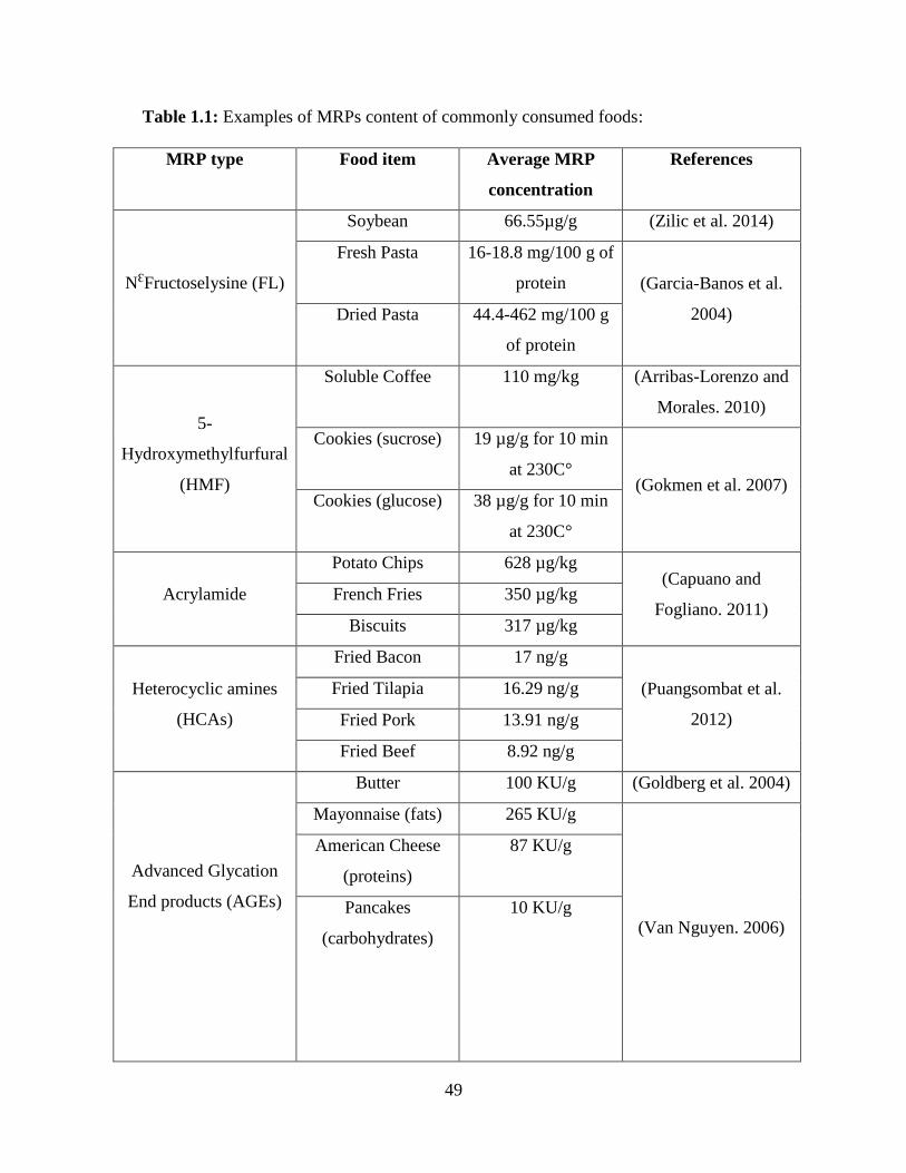
49
Table 1.1: Examples of MRPs content of commonly consumed foods:
MRP type Food item Average MRP
concentration
References
NƐFructoselysine (FL)
Soybean 66.55µg/g (Zilic et al. 2014)
Fresh Pasta 16-18.8 mg/100 g of
protein (Garcia-Banos et al.
2004) Dried Pasta 44.4-462 mg/100 g
of protein
5-
Hydroxymethylfurfural
(HMF)
Soluble Coffee 110 mg/kg (Arribas-Lorenzo and
Morales. 2010)
Cookies (sucrose) 19 µg/g for 10 min
at 230C° (Gokmen et al. 2007)
Cookies (glucose) 38 µg/g for 10 min
at 230C°
Acrylamide
Potato Chips 628 µg/kg (Capuano and
Fogliano. 2011) French Fries 350 µg/kg
Biscuits 317 µg/kg
Heterocyclic amines
(HCAs)
Fried Bacon 17 ng/g
(Puangsombat et al.
2012)
Fried Tilapia 16.29 ng/g
Fried Pork 13.91 ng/g
Fried Beef 8.92 ng/g
Advanced Glycation
End products (AGEs)
Butter 100 KU/g (Goldberg et al. 2004)
Mayonnaise (fats) 265 KU/g
(Van Nguyen. 2006)
American Cheese
(proteins)
87 KU/g
Pancakes
(carbohydrates)
10 KU/g

50
Table 1.1 (Cont.)
NƐCarboxymethyllsine
(CML)
Sterilized Milk 2066 nM of protein (Ahmed et al. 2005)
Pasteurized Milk 887 nM of protein
Raw Milk 337 nM of protein
White Bread Crust 382 mg/kg of protein
(Assar et al. 2009) Wholemeal Bread
Crust
329 mg/kg of protein
Doner Kebab 2357.87 mg/kg of
protein (Hull et al. 2012)
Melanoidins
Sourdough Loaves 30 g/100g of crust (Fogliano and
Morales. 2011) Soluble Coffee 22 g/100 g
Roasted Barley 4.15% of 0.7 to1.0 kg (Milic et al. 1975)
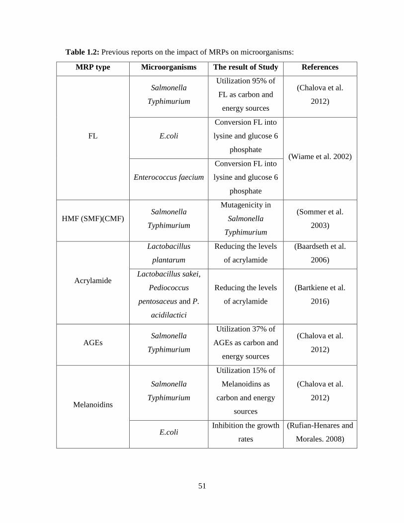
51
Table 1.2: Previous reports on the impact of MRPs on microorganisms:
MRP type Microorganisms The result of Study References
FL
Salmonella
Typhimurium
Utilization 95% of
FL as carbon and
energy sources
(Chalova et al.
2012)
E.coli
Conversion FL into
lysine and glucose 6
phosphate (Wiame et al. 2002)
Enterococcus faecium
Conversion FL into
lysine and glucose 6
phosphate
HMF (SMF)(CMF) Salmonella
Typhimurium
Mutagenicity in
Salmonella
Typhimurium
(Sommer et al.
2003)
Acrylamide
Lactobacillus
plantarum
Reducing the levels
of acrylamide
(Baardseth et al.
2006)
Lactobacillus sakei,
Pediococcus
pentosaceus and P.
acidilactici
Reducing the levels
of acrylamide
(Bartkiene et al.
2016)
AGEs Salmonella
Typhimurium
Utilization 37% of
AGEs as carbon and
energy sources
(Chalova et al.
2012)
Melanoidins
Salmonella
Typhimurium
Utilization 15% of
Melanoidins as
carbon and energy
sources
(Chalova et al.
2012)
E.coli Inhibition the growth
rates
(Rufian-Henares and
Morales. 2008)
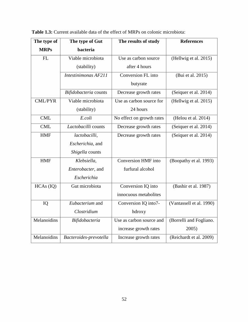
52
Table 1.3: Current available data of the effect of MRPs on colonic microbiota:
The type of
MRPs
The type of Gut
bacteria
The results of study References
FL Viable microbiota
(stability)
Use as carbon source
after 4 hours
(Hellwig et al. 2015)
Intestinimonas AF211 Conversion FL into
butyrate
(Bui et al. 2015)
Bifidobacteria counts Decrease growth rates (Seiquer et al. 2014)
CML/PYR Viable microbiota
(stability)
Use as carbon source for
24 hours
(Hellwig et al. 2015)
CML E.coli No effect on growth rates (Helou et al. 2014)
CML Lactobacilli counts Decrease growth rates (Seiquer et al. 2014)
HMF lactobacilli,
Escherichia, and
Shigella counts
Decrease growth rates (Seiquer et al. 2014)
HMF Klebsiella,
Enterobacter, and
Escherichia
Conversion HMF into
furfural alcohol
(Boopathy et al. 1993)
HCAs (IQ) Gut microbiota Conversion IQ into
innocuous metabolites
(Bashir et al. 1987)
IQ Eubacterium and
Clostridium
Conversion IQ into7-
hdroxy
(Vantassell et al. 1990)
Melanoidins Bifidobacteria Use as carbon source and
increase growth rates
(Borrelli and Fogliano.
2005)
Melanoidins Bacteroides-prevotella Increase growth rates (Reichardt et al. 2009)
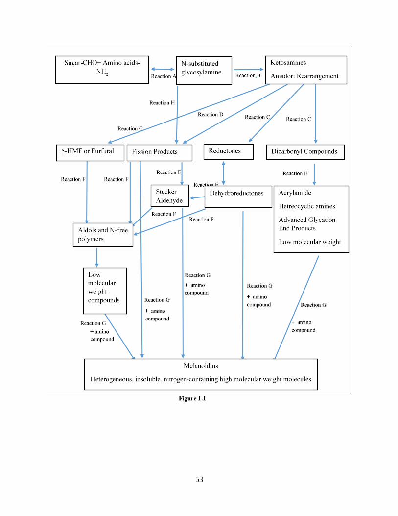
53
54
Figure 1.1: Maillard Reaction Products stages (adapted from the initial description by Hodge in
1953 to reflect current knowledge). 1. Early stage: condensation of the carbonyl group of
reducing sugar with the amino group of amino acids (Reaction A), resulting in N-substituted
glycosylamine and water. 2. Intermediate stages: Conversion of glycosylamine through
Amadori rearrangement to form ketosamines (Reaction B) and other products. Amadori products
are dehydrated and hydrolyzed to form 5-Hydroxymethylfurfural (HMF) (Reaction C), which
gives rise to either Aldose or N-free polymers (Reaction F). Reductones can be formed from either
dehydration of sugars or Amadori product (Reaction C) leading to Aldose and N-free polymers
(Reaction F) or Stecker Aldehydes (Reaction E). Stecker aldehydes are formed by fragmentation
of amino acids, which enter browning reactions either by the aldehyde formed that can take part
in aldol condensation which forms nitrogen-free polymers (Reaction F). Amadori product and N-
substituted glycosylamines can be fragmented fission products (Reaction D and H). In addition,
fragments of MRPs produce reactive dicarbonyl compounds that can act as precursors of
acrylamide, heterocyclic amines, advanced glycation end products (AGEs), and low molecular
weight compounds (Reaction C). 3. Advanced stages: Melanoidins include a wide array of
heterogeneous colored molecules Dehydroreductones, fission and dicarbonyl compounds,
furfural and Aldose react with amino acids (Reaction G) to form melanoidins.
55
CHAPTER TWO
RESEARCH ARTICLE
Repeated oral exposure to N-Carboxymethyllysine, a Maillard Reaction Product,
alleviates gut microbiota dysbiosis in colitic mice
Nesreen ALJahdali MS1, Pascale Gadonna-Widehem PhD2, Carine Delayre-Orthez PhD², David
Marier MS², Benjamin Garnier MS², Franck Carbonero PhD 1,3* and Pauline M Anton PhD2.
1Cell and Molecular Biology Program; University of Arkansas, AR, USA
2Expression des Gènes et régulation Epigénétique par l’Aliment UP 2012.10.101. ; Institut
Polytechnique UniLaSalle, Beauvais, France
3Department of Food Science; University of Arkansas, AR, USA
This work was supported by the Picardie Regional Council of France (ETAMIN project) and by
an Arkansas Biosciences Institute grant.
Published:
ALJahdali, N., Gadonna-Widehem, P., Delayre-Orthez, C., Marier, D., Garnier, B., Carbonero,
F., & Anton, P. M. (2017). Repeated oral exposure to N epsilon-carboxymethyllysine, a maillard
reaction product, alleviates gut microbiota dysbiosis in colitic mice. Digestive Diseases and
Sciences, doi: 10.1007/s10620-017-4767-8 [doi]

56
2.1 Abstract:
Background: Diet is suggested to participate in the etiology of Inflammatory Bowel
Diseases (IBD). Repeated exposure to Maillard reaction products (MRPs), molecules resulting
from reduction reactions between amino acids and sugars during food heating, have been reported
to be either potentially detrimental or beneficial to health.
Aims: the aim of this study is to determine the effect of repeated oral ingestion of
NCarboxymethyllysine, an advanced MRP, on the onset of two models of experimental IBD and
on the composition of the gut microbiota of mice.
Methods: Mice received either saline (control) or NCarboxymethyllysine daily for 21
days. For the last week of treatment, each group was split into sub-groups, receiving Dextran
sulfate sodium salt (DSS) or Trinitrobenzene sulfonic acid (TNBS) to induce colitis. The intensity
of the inflammation was quantified and cecal microbiota was characterized by bacterial 16S rRNA
amplicon sequencing.
Results: Daily oral administration of NCarboxymethyllysine did not induce intestinal
inflammation and had limited impact on the gut microbiota composition (Bacteroidaceae increase,
Lachnospiraceae decrease). DSS and TNBS administration resulted in an expected moderate
experimental colitis with a shift of Bacteroidetes/Firmicutes ratio and a significant Proteobacteria
increase, but with distinct profiles: different Proteobacteria taxa for DSS, mainly
Enterobacteriaceae for TNBS. While NCarboxymethyllysine exposure failed to prevent the
inflammatory response, it allowed maintenance of healthy gut microbiota profiles in DSS (but not
TNBS) treated mice.

57
Conclusions: Repeated oral exposure to CML limits the dysbiosis in experimental colitis.
IBD patients may modulate their microbiota profile by regulating the level and type of dietary
MRP consumption.
Keywords: Maillard Reaction Products - N-Carboxymethyllysine – Intestinal
microbiome – Inflammation – Dextran Sulfate Sodium salt colitis – TriNitroBenzene Sulfonic acid
colitis

58
2.2 Introduction:
Gut microbiota has been the focus of the exponentially increasing numbers of studies
because of its purported role in a variety of diseases [1-3]. Gut microbiota (or microbiome) is the
complex community of microorganisms, such as Archaea, protozoans, fungi, and for the vast
majority of bacteria that inhabit the gastrointestinal tract (GIT) of humans and other animals, which
mainly ferment and degrade undigested dietary compounds [3, 4]. Arguably dietary habits are
considered to be one of the main factors contributing to the diversity and shaping of human gut
microbiota, since there is growing evidence that human associated microbes have been lost through
the transition from ancestral to Western diet [5-7]. Inflammatory Bowel Diseases (IBD) and colon
cancer are well known to be epidemiologically associated with Western diet consumption [5, 6],
and gut microbiota dysbiosis (imbalanced composition) has been shown to be a potential
contributor to their associated inflammatory process [8, 9]. Gut microbiota dysbiosis has also been
shown in different animal models of IBD, including Trinitrobenzene sulfonic acid (TNBS) and
Dextran sulfate sodium salt (DSS)-induced colitis in mice [10-12]. These two models have been
widely used to study intestinal inflammation since they induce different immune, mucosal and
microbiota alterations that reflect the clinical signs observed in IBD patients. While literature
depicts links between diet composition and the intestinal microbiome [13], there are few data
supporting the potential links between the mode of food preparation, such as food heating and
chronic gut inflammation.
Heat processing of food generates several neoformed compounds among which Maillard
reaction products (MRPs), a group of several compounds that influence the color, flavor, texture
and nutritional value of food, are suspected to have health implications [14]. The intensity of such
heating determines the types of MRPs present in food, being classified as initial (Amadori
59
Compounds), intermediate (i.e N-Carboxymethyllysine, acrylamide, etc.) and final stage
(melanoidins, etc.) [15]. MRPs are also produced in living bodies. To avoid any confusion, food
intermediate compounds are named dietary advanced glycation end products (dietary AGEs) to
discriminate them from endogenous AGEs. It has been established that Western diet, richer in
lipids and proteins, is a significant source of dietary AGEs known to be a chronic risk for
cardiovascular and kidney damage [16]. Among the two types of AGEs, N-Carboxymethyllysine
(CML) – a product of both lipoxidation and glycoxidation [17] –is considered to be the most
important biomarker of MRP commonly measured in human blood and urine. CML levels are also
used to infer the amount of AGEs consumed by individuals [18, 19]. Endogenous CML has been
reported to be a reliable biomarker of oxidative stress and of long-term damage to proteins [20].
In some studies, CML has been associated with various diabetes complications[21-24] and age-
related health conditions [25-27] while, in others, CML has been reported to be not correlated or
negatively correlated with inflammation [28-30]. Dietary CML, present in more than 200 food
items [16, 31], can be produced either by Amadori rearrangement products or by Amadori
compound degradation, such as Fructoselysine (FL) that further reacts with proteins to form
irreversible and highly stable compounds [31]. Since circulating CML is known to derive either
from endogenous Maillard reaction, or CML present in food, or of conversion of AGEs by the gut
microbiota [32, 33], the potential contribution of dietary CML on the onset of the inflammatory
process has been hard to determine.
As MRPs may be largely present in the Western diet, they may be considered as one of the
many dietary elements potentially incurring detrimental impact on human health. However, their
fate in the digestive tract, specifically through the gut microbiota has been sparsely studied, and
mainly in vitro [34-36]. The gut microbiota was shown to degrade MRPs, such as CML, pyrraline

60
(PYR) and maltosine (MAL) after anaerobic batch incubations with fecal suspension from healthy
humans and they used them as a source of energy, carbon, and nitrogen [34]. Using quantitative
polymerase chain reaction (qPCR) analysis, no differences were found in the total bacteria of
adolescents who consumed white diet (low MRPs) versus brown diet (high MRPs), but a
significant decrease was shown when rats consumed similar diets [37]. Recently, we have
determined that ingestion of highly heated food protected mice against an experimental colitis and
dysbiosis [38]. This food contained higher levels of dietary AGEs (such as CML), but also higher
levels of final stages MRPs (i.e., melanoidins). In summary, all these studies relied on culture-
dependent or targeted quantitative PCR approaches, but none were aimed at enabling an extensive
and specific characterization of CML (or other MRPs) impact on the gut microbiota.
In order to gain a better understanding, this study aimed to determine the effect of repeated
daily exposure to CML alone not only on the onset of the colonic inflammatory reaction, but also
on the expected intestinal dysbiosis observed following both TNBS and DSS-induced
experimental colitis in mice.
2.3 Materials and Methods:
2.3.1 Chemicals:
N-Carboxymethyllysine (CML) was obtained from Polypeptide laboratories in Sigma-
Aldrich in France. Dextran sulfate sodium salt (DSS) was purchased from MP Biomedicals while
Trinitrobenzene sulfonic acid (TNBS) was purchased from Sigma-Aldrich in France.
2.3.2 Animals and Study Design:
Fifty male Balb/c mice 7 weeks old (18-20 g) were housed in stainless steel cages under a
controlled temperature (21 ± 1°C) and a 12H light-dark cycle, with free access to food and water.
Experiments were conducted at the Animal House Unit of LaSalle Beauvais after receiving the
61
prior approval of both the animal protocol review committee and the Picardy Counsil of Veterinary
Office in France (C60-200-01). Mice were split in two groups and orally received CML (1.6
mg/kg/d po – 50 µl/mouse/d) or saline (50 µl/mouse/d) orally every day for 3 weeks. The last week
of the experiments, each group was divided into three subgroups: two (n=9/subgroup) submitted
to either Dextran Sulfate Sodium (DSS) or TriNitroBenzene Sulfonic acid (TNBS) colitis, and one
serving as a control (n=7/subgroup).
At the end of the experiment, animals were sacrificed by cervical dislocation under general
anesthesia (ketamine/xylazine 100 mg/kg i.p. 50% v/v). A midline laparotomy was then realized,
macroscopic lesions were assessed and then pieces of colon were immediately removed and snap
frozen until further analysis. To perform the analysis of the incidence of each treatment on the
microbiota profile, caeca were ligated at both the ileo-caecal and the caeco-colonic junctions
before their removal, and their contents were immediately collected under anaerobic conditions
and snap frozen until further determination.
2.3.3 Experimental Colitis
Colitis models: In order to confirm that the effects of CML on colonic inflammation were
not model-dependent, colitis was induced using two models widely used in the literature. The
TNBS solution was diluted in 50% ethanol (100mg/kg, 50 µL IR) before being injected
intrarectally (IR) via lubricated silicone tubing (4cm from the anus) under general anesthesia
(ketamine/xylazine 100 mg/kg, 50% v/v, 1mL/kg ip). Animals were then kept head-down on a
heating pad until full recovery from the anesthesia [39].
DSS (36-42000 Da) was solubilized in the drinking water (3% w/v) and was administered for five
days. Bottles were changed every other day. For the last 2 days of the experiment, DSS was
removed from the bottles [40].
62
Macroscopic lesions: The level of macroscopic lesions was represented as macroscopic
damage scores (MDS) and expressed in arbitrary units (AU) according to the literature. Briefly,
DSS lesions severity was evaluated based on weight loss (from 0 non loss – to 4 more than 20%
loss), stool consistency (from 0 consistent to 4 diarrhoea), occult or gross bleeding (from 0 none
to 4 gross bleeding) and gross blood content (from 0 non blood to 3 blood in more than 2/3 of the
colon) [40, 41]. TNBS-induced lesions were evaluated based on the level of ulceration (from 0
normal appearance to 5 inflammation on more than 1cm), presence of adhesions (from 0 none to
2 major) and diarrhea (0 no – 1 yes) [39].
Myeloperoxidase Activity: The inflammatory reaction is a process associated with immune
cell activation and neutrophil infiltration. Myeloperoxidase (MPO) is exclusively found in
neutrophil cytoplasmic granules, and thus considered a reliable marker for quantifying the level of
inflammatory reaction [42]. MPO activity assay was first described by Bradley et al [43]. MPO
was extracted from colonic tissues collected at sacrifice as previously described [44].
Briefly, tissue samples were lysed in a phosphate buffer (50mM, pH=6) containing
hexadecyl trimethyl ammonium bromide (0.5% m/v) with a polytron (PT 1200E, Kinematica AF,
Switzerland). Homogenates were then submitted to 3 cycles of freezing and thawing (-80°C – 10
min / 37°C for 15 min), and then sonicated (Bioblock scientific, France) before centrifugation
(6000 g – 4°C – 15 min). Supernatants were collected to measure MPO activity and protein
concentration. MPO activity was assessed on supernatants diluted on a reaction buffer containing
O-dianisidine dihydro-chloride (1mg/mL) and hydrogen peroxide (5 x 10⁻⁴ % v/v); purified MPO
(1 U/mL) from human leucocytes (Sigma Aldrich, France) as a standard. Absorbance was
measured at 450 nm after 15 minutes of incubation at room temperature. Supernatants total protein
content was assessed based on Lowry method following the manufacturer’s recommendations (Bio
63
Rad DC Protein Assay, France). The results were expressed as MPO units/mg protein where 1 unit
of MPO is the amount of peroxidase necessary for transforming 1 µmol of hydrogen peroxide in
1 min at 25°C.
2.3.4 DNA Extraction and Amplicon Sequencing Strategy
Genomic DNA was extracted from cecal samples. Briefly, 100 mg cecal content and 1 mL
of ASL buffer (Qiagen, Courtaboeuf, France) were pooled in 2 mL Lysing Matrix E tubes (MP
Biomedicals, Santa Ana, USA). After mixing for 1 min with a vortex, the tubes were heated at
95°C for 5 min in a Thermomixer (Eppendorf, Montesson, France) then cooled in ice for 5 min.
The tubes were transferred into the FastPrep® (MP Biomedicals, Santa Ana, USA) bead-beater
for 2 runs of 1 min each at 6 m/s. After centrifugation (16000g at 4℃ for 1 min), the total DNA
was extracted from the obtained lysates using a commercial kit (QIAamp DNA stool Mini Kit)
following the manufacturer’s protocol [42].
Illumina (San Diego, USA) MiSeq sequencing was used to investigate the gut microbiota
by targeting the V4 region of the bacterial 16S rRNA gene of each group following the dual-
indexed strategy developed by Kozich et al [45]. PCR amplifications were performed in 25 μL
reactions with 22 μL of KAPA Hifi master mix (KAPA Biosystems, Wilmington, USA), 1 μL of
DNA template and 2 μL of index primers. PCR was performed with initial denaturation at 95 °C
for 3 min, followed by 25 cycles of denaturation at 98 °C for 30 sec, primer annealing at 55 °C for
30 sec, and extension at 72 °C for 1 min, with final extension at 72 °C for 7 min. PCR amplicons
were examined on agarose gel electrophoresis by using 6 μL of ethidium bromide fluorescent dye
to confirm the successful PCR.
64
2.3.5 Library Preparation and Quality Control:
Normalization of PCR products was performed with a SequalPrep kit (Invitrogen)
following the manufacturer’s instructions. Normalized amplicons were pooled and checked on
agarose gels. Gel extraction of the correct amplicon was achieved by running agarose gel
electrophoresis on 100 μL DNA, then extracting DNA from excised bands using the Min Elute
Gel Extraction kit (Qiagen, Hilden, Germany). Library quality checks were performed by means
of real-time q-PCR (RT-q-PCR) and TapeStation (Agilent®, Santa Clara, USA) to determine the
concentration and size of DNA libraries, respectively. Q-PCR was performed with the KAPA
Library Quantification Kit (KAPA Biosystems) following the manufacturer’s recommendations.
The q-PCR plate was placed in a thermocycler for initial activation at 95 °C for 5 min followed by
35 cycles. Amplicon size was determined again after gel extraction on the TapeStation (378 bp
and 372 bp).
2.3.6 Bioinformatics and Statistical Analyses:
Data relating to inflammation are expressed as mean ±stand error of the mean (SEM). They
were analyzed using Graph Pad Prism Software. Macroscopic lesion scores were compared using
Kruskal-Wallis nonparametric test followed by Dunn’s multiple comparisons post hoc test. All
other data were submitted to analysis of variance (ANOVA) followed by Tukey post hoc test. P
value <0.05 was considered to be significant.
All sequences were analyzed by using the Quantitative Insight into Microbial Ecology
(QIIME) software package following the script in Ref. [46], which performs microbial community
analysis. The results of microbiota counts were analyzed by Kruskal-Wallis test using past3
software. P value of <0.05 was considered to be significant between each group. Microbiota data
were visualized by Non-Metric Multidimensional Scaling (NMDS) using the Bray-Curtis

65
similarity index. The significance of clustering by subgroups was tested by analysis of similarities
(ANOSIM) with the Bray-Curtis similarity index. P value <0.05 was considered to be significant.
2.4 Results:
2.4.1 Effect of CML on Weight Change and Food Intake in Healthy and Colitic Mice:
Since both the colitis models used here are known to induce weight loss, we expressed the
weight variation as the percentage of final weight on initial weight (D1 = day 1) for each animal.
A result over 100 % reflects weight gain. As expected, DSS resulted in significant (P<0.001)
weight loss as compared to control animals (92 ± 2.39 % vs 104.1 ± 1.25 %) and CML significantly
limited the weight loss in DSS treated mice (101.3 ± 1.69 % vs 92 ± 2.39 %; P<0.001) (Table 2.1).
DSS colitis was not associated with any modification of food intake. Following TNBS exposure,
mice lost up to 20% of initial weight (85.09 ± 1.52% vs 104.1 ± 1.25 %; p<0.001), a loss which
was associated with a significant (P<0.001) reduction of food consumption (2.10 ± 0.26 g vs 3.88
± 0.24 g in controls) (Table 2.1). However, in this model of colitis, CML failed to limit this loss
since mice lost up to 25% in comparison with the CML treated group (81.62 ± 0.82 % vs 107.2 ±
1.41 % in controls; P<0.001), while mice had a normal food intake (2.48 ± 0.41 g vs 3.57 ± 0.21
g in the CML group) (Table 2.1).
2.4.2 Effect of CML on Macroscopic Lesions and Neutrophil Infiltration in Healthy and
Colitic Mice:
CML administration during three weeks did not induce any macroscopic lesions (Table
2.1). By contrast, DSS colitis was associated with significant (P<0.001) lesions (3.12 ± 0.29 AU
vs 0.17 ± 0.16 AU in controls), which were not limited by ingestion of CML (3.69 ± 0.16 AU vs
3.12 ± 0.29 AU in the DSS group) (Table 2.1). Significant lesions (P<0.01) were also observed in
mice submitted to TNBS colitis (6.30 ± 0.89 AU vs 0.17 ± 0.17 AU in controls), which were also
66
not limited by CML ingestion (7.75 ± 0.39 AU vs 6.3 ± 0.89 AU in the TNBS group) (Table 2.1).
Furthermore, daily exposure to CML did not result in any neutrophil infiltration. However, MPO
activity was significantly increased (P<0.01) after DSS (0.77 ± 0.07 U/mg vs 0.25 ± 0.02 U/mg de
proteins in controls) and TNBS (0.64 ± 0.07 U/mg vs 0.25 ± 0.02 U/mg proteins) was exposure as
expected (Table 2.1). In mice submitted to either DSS or TNBS, CML repeated ingestion was
unable to limit the inflammatory reaction (0.70 ± 0.07 U/mg vs 0.77 ± 0.07 U/mg proteins in DSS
group and 0.54 ± 0.06 U/mg vs 0.64 ± 0.07 U/mg proteins in the TNBS group) (Table 2.1).
2.4.3 Gut Microbiota Analyses:
A total of 2,102,977 sequences from 123 samples (17097 ± 10992 per sample) were
obtained.
2.4.3.1 Impact of CML on the Gut Microbiota of Healthy Mice:
At the phylum level, CML consumption did not affect gut microbiota (Figure 2.1A). Using
multivariate analyses at different taxonomic levels, only slight (not statistically significant by
ANOSIM) differences in gut microbiota were found between control and CML mice gut
microbiota at the genus level (Figure 2.1A). However, CML consumption significantly increased
the relative abundance of several Bacteroidetes families (Bacteroidaceae, Odoribacteraceae and
the numerically marginal Porphyromonadaceae) (Figure 2.1B), as well as, the Desulfovibrionaceae
(Proteobacteria) (Figure 2.1D) and Dorea (Firmicutes) (Figure 2.1C). CML consumption resulted
in a significant decrease in Lachnospiraceae (Firmicutes) (Figure 2.1C) and the low abundant
Sutterella (Proteobacteria) (Figure 2.1D).
2.4.3.2 Impact of TNBS and DSS on the Gut Microbiota:
At the phylum level, similar trends were observed for TNBS and DSS treatments (Figure
2.2A). As indicated on the pie charts, DSS treatment resulted in a significant decrease of Firmicutes
67
(from 77 to 34%) and increase in Bacteroidetes (from 20 to 41%) and Proteobacteria (from 1 to
12%). TNBS treatment resulted in a significant decrease of Firmicutes (from 77 to 39%) and an
increase in Bacteroidetes (from 20 to 42%) and Proteobacteria (from 1 to 14%). On multivariate
analyses at different taxonomic levels, significant differences were observed at all taxonomic
levels between control, TNBS, and DSS groups (Figure 2.2A; ANOSIM P<0.05 for each
comparison). Consequently, the different treatments led to distinct dysbiotic microbiota
compositions.
DSS treatment resulted in a sharp significant increase of Bacteroidaceae and
Porphyromonodaceae, while Prevotellaceae were significantly depleted (Figure 2.2B).
Lachnospiraceae, but not Ruminococcaceae, were also decreased (Figure 2.2C). DSS treatment
resulted in significant (P<0.05) increases of a wide range of Proteobacteria members (Figure
2.2D), with a much less marked increase in Enterobacteriaceae than under TNBS (Figure 2.2D),
as well as a notable increase in Actinomycetales (Figure 2.2E).
TNBS treatment also resulted in a sharp significant increase of Bacteroidaceae and
Prevotellaceae (Figure 2.2B), and somewhat unexpectedly Lactobacillus and Enterococcus
(Figure 2.2C). It decreased very significantly (P<0.05) the relative abundance of Lachnospiraceae
and Ruminococcacceae (Figure 2.2C).The significant (P<0.05) increase in Proteobacteria induced
by TNBS was solely due to a massive increase of Enterobacteriaceae reaching 13.5% (Figure
2.2D), but other Proteobacteria were not affected or even decreased significantly
(Desulfovibrionaceae) (Figure 2.2D). Finally, TNBS significantly (P<0.05) reduced
Coriobacteriacease (P<0.05), while had no incidence on Actinomycetales (Figure 2.2E).
68
2.4.3.3 Impact of CML on the Gut Microbiota of DSS and TNBS-Treated Mice:
At the phylum level, no effect of CML was observed (Figure 2.3) in TNBS-treated mice,
with Firmicutes slightly decreasing (39 to 36%), Bacteroidetes stable (42%) and Proteobacteria
slightly increasing (14 to 18%) as indicated on pie charts. In contrast, CML consumption
significantly modulated the gut microbiota towards the control/CML state, with increased
Firmicutes (34 to 58%), and decreased Bacteroidetes (41 to 32%) and Proteobacteria (12 to 3%).
On multivariate analyses at different taxonomic levels, no differences were observed between
control and CML mice exposed to TNBS colitis (Figure 2.3). On the other hand, very significant
(ANOSIM p<0.05) differences between control and CML mice exposed to DSS colitis was
observed (Figure 2.3). For the TNBS mice, no significant difference was observed for genera
representing more than 0.001% of the total microbiota (Figure 2.4B and 2.4C). Intriguingly, CML
consumption appeared to result in a numerical increase of Enterobacteriaceae (Figure 2.4D) and
to some extent Bacteroidaceae (Figure 2.4A).
CML consumption significantly (P<0.05) reverted the increase in Bacteroidaceae and
Porphyromonodaceae (Figure 2.5A) and Actinomycetales (Figure 2.5C) due to DSS treatment. It
also decreased the abundance of all Proteobacteria groups associated with DSS-treatment, with the
exception of Desulfovibrionaceae (Figure 2.5D). These trends were associated with increases in
Lachnospiraceae, Ruminococcaceae and other Clostridiales (Figure 2.5B).
2.5 Discussion and Conclusion:
Maillard reaction products (MRPs) have been subject of negative perception by consumers
due to early reports of detrimental effects [14, 47, 48]. Acrylamide, an AGE well known as a
carcinogen, for which there is substantial evidence for detrimental health impacts, has been
69
associated with ocular and renal diabetic complications [22-25, 49, 50]. However, novel evidence
has suggested that MRPs, due to the wide range of compounds that can be formed, should not be
considered as a collective of detrimental class of molecules. In fact, beneficial impacts on health,
and potential antioxidant properties have been reported [51, 52]. We report herein that repeated
oral ingestion of CML, a major marker of MRPs in food, does not compromise mucosal integrity
of the colon since we observed neither macroscopic lesions nor colonic inflammation in healthy
mice exposed to CML during 21 days. CML has been commonly used as a biomarker for AGEs
in food and in circulatory blood. However, little is known about its effects on health. In high-fat
diet fed rats, CML consumption led to an increase in LDL, fasting glucose and energy expenditure,
and to the alteration of liver and renal function[53]. Here we did not observe any impact of CML
consumption on mice general health, but it should be noted that the dose of CML we used was 30
times lower than in the previous study and is more representative of a regular daily exposure, and
that the animals were fed a standard diet.
We also report that repeated consumption of moderate amounts of CML has limited
impacts on the taxonomic composition of gut microbiota of normal healthy mice. The impact of
MRPs on gut microbiota has been scarcely studied, mainly in vitro studies. In fact, only very recent
studies have shown that gut microbiota can degrade selected glycated amino acids, including CML
[34], and metabolites from MRP bacterial degradation could not be characterized with available
Mass Spectrometry instruments. A bifidogenic impact was reported in another in vitro assay with
an Amadori product resulting from a protein glycated with galactose, lactulose, and galacto-
oligosaccharides [54]. However, this result is more probably due to the known bifidogenic impact
of those “prebiotic” polysaccharides. In the present work, we observed a limited impact of CML
consumption on the murine gut microbiota. It appears that CML was preferentially fermented by
70
Bacteroides species, which may be in line with previous reports of Bacteroides growth from
advanced MRPs [55] and bread crusts rich in some melanoidins [56]. The significant increase of
Desulfovibrio further suggests that CML stimulated fermentation rates in the mouse cecum [4, 57].
Nonetheless, it should be noted that the observed changes were modest numerically, possibly
indicating that CML can be fermented by a relatively wide diversity of bacterial species.
In this study, we also compared the dysbiosis TNBS versus DSS. Gut microbiota unbalance
is a known contributor of IBD [58, 59], and also suspected to be directly involved in the
inflammatory process of both these chemically induced colitis models [11]. Furthermore, links
between gut microbiota disruption and diet are regularly suggested in the literature. However,
somewhat surprisingly, there is little knowledge regarding typical gut microbiota responses to DSS
or TNBS insult, since those models are commonly used in conjunction with other parameters (diet,
genetic alteration, etc.). DSS and TNBS are two of the most popular chemicals used to mimic
human inflammatory bowel disease in mice. DSS treatment results in chronic intestinal
inflammation by disruption of the colonic epithelial barrier, resulting in Ulcerative Colitis (UC)-
like damages and immune response [41]. It has also been shown to be suitable to study epithelial
repair mechanisms [60]. On the other hand, TNBS initiates acute IL-12 driven (Th1 immune
response emphasis) intestinal inflammation, more similar to Crohn’s Disease (CD) symptoms and
etiology [61]. The TNBS model is useful to study T helper cell-dependent mucosal immune
responses [62] and many important aspects of gut inflammation, including cytokine secretion
patterns, mechanisms of oral tolerance, and cell adhesion. Recent literature has been reported that
both models result in increased abundance of Enterobacteriaceae and Bacteroides [11], with
Clostridium spp. and Akkermansia spp. also reported for DSS [63]. We report herein strongly
contrasting alteration of gut microbiota between TNBS and DSS models. As already shown in a

71
previous work, we indeed observed Enterobacteriaceae and Bacteroidaceae (mainly Bacteroides)
as the main groups involved in TNBS colitis [44]. Their increase was correlated with a sharp
decrease of Lachnospiraceae and Ruminococcaceae, two of the families most commonly
associated with production of anti-inflammatory short-chain fatty acids (butyrate in particular).
Remarkably, Lactobacillus and Enterococcus, two genera generally considered beneficial also
increased significantly in TNBS-exposed mice. Lactobacillus spp. in particular are known for their
ability to control pathogenic Enterobacteriaceae numbers, associated with the induction/progress
of the inflammatory process. Since Enterobacteriaceae were clearly not inhibited, it is likely that
Lactobacillus absolute numbers did not increase, but were maintained relative to other taxa. DSS
treatment only resulted in a modest increase in Enterobacteriaceae, but also in a general increase
of a wide range of Proteobacteria, suggesting an increased role for nonpathogenic species in
inflammatory response. On the other hand, DSS also resulted in less marked depletion of
Firmicutes, with maintenance of Ruminococcaceae levels. In slurries derived from Ulcerative
Colitis patients, glycated bovine serum albumin was found to increase the numbers of Clostridia,
sulfate-reducing bacteria and Bacteroides with the decrease of Bifidobacteria[64]. Overall, it
appears that the bacterial dysbiosis incurred by DSS in comparison to the TNBS is due to the
different course of the inflammatory process.
CML consumption had virtually no impact on the gut microbiota dysbiosis and on the
inflammatory reaction of TNBS-treated mice. In contrast, CML limited the severity of weight loss
and the gut microbiota dysbiosis in DSS-treated mice, but not the inflammatory response. Overall,
these data suggest that CML has the potential to improve the DSS dysbiosis, by incurring more
significant changes than in the healthy microbiota. Remarkably, the changes in Bacteroidetes and
Firmicutes brought by CML in DSS animals were opposite to those observed in healthy mice.
72
Firmicutes, specifically Lachnospiraceae, Ruminococcaceae (Oscillospira and Ruminococcus
being the most abundant genera) and Clostridiales, were strongly stimulated by CML consumption
only in DSS-treated mice, while initial levels were comparable in both models. While Oscillospira
is still a poorly understood member of the mammalian gut microbiota, there is evidence that they
can consume host-derived glycoproteins [65]. Thus, they may be primary degraders of dietary
CML as well as other MRPs. The beneficial effect of CML in this case probably results from the
control of the Proteobacteria bloom, which may be direct [35] or indirect through competitive
exclusion. The observation of opposite dynamics for Bacteroidaceae (Bacteroides) after CML
consumption demonstrates that different species are stimulated by DSS and TNBS, and that the
species involved in DSS colitis may not be able to utilize CML as well as those associated with
TNBS colitis.
Overall, we show here that consumption of CML, a surrogate of Advanced Glycation End-
products, has little impact on healthy gut microbiota, but alleviates gut microbiota dysbiosis in
DSS-treated mice only. These data confirm that determining the health effects of MRPs is more
complex than it would seem. They cannot be analyzed as a whole but rather studied one by one
under clearly specified conditions. We may also suggest that the response of animals could be
different if CML was given in already colitic animals. It is possible that selected MRPs may be
used for preventive purposes, with anti-inflammatory potential mediated by gut microbiota.
However, a better understanding of MRPs fate in the gut microbiota environment will be needed
before suggesting any health claims. Their effects on altered intestinal mucosa needs further
investigation to make food heating recommendations especially for patients suffering from chronic
inflammation.
73
Personal Acknowledgments:
The authors thank Camille Boroch for her help in conducting the animal experiments. This
work is dedicated to the memory of our wonderful colleague, Pr. Abalo Chango who is at the origin
of this international cooperative work.
Conflicts of interest:
The authors declare that they have no conflict of interest
Ethical Approval:
All applicable national and/or institutional guidelines for the care and use of animals were
followed. All procedures performed in studies involving animals were in accordance with the
ethical standards of the institutions or practice at which the studies were conducted.

74
2.6 References:
1. Cryan JF, Dinan TG. Mind-altering microorganisms: The impact of the gut microbiota on
brain and behaviour. Nature reviews neuroscience. 2012;13:701-712.
2. Everard A, Cani PD. Diabetes, obesity and gut microbiota. Best practice & research Clinical
gastroenterology. 2013;27:73-83.
3. Flint HJ, Scott KP, Louis P, Duncan SH. The role of the gut microbiota in nutrition and health.
Nature Reviews Gastroenterology and Hepatology. 2012;9:577-589.
4. Carbonero F, Benefiel AC, Alizadeh-Ghamsari AH, Gaskins HR. Microbial pathways in
colonic sulfur metabolism and links with health and disease. Front Physiol. 2012;3:448.
doi:10.3389/fphys.2012.00448 [doi].
5. Ou J, Carbonero F, Zoetendal EG, et al. Diet, microbiota, and microbial metabolites in colon
cancer risk in rural Africans and African Americans. Am J Clin Nutr. 2013;98:111-120.
doi:10.3945/ajcn.112.056689 [doi].
6. O’Keefe SJ, Li JV, Lahti L, et al. Fat, fibre and cancer risk in african americans and rural
africans. Nature communications. 2015;6:6342.
7. Gomez A, Petrzelkova KJ, Burns MB, et al. Gut microbiome of coexisting BaAka pygmies
and Bantu reflects gradients of traditional subsistence patterns. Cell reports. 2016;14:2142-2153.
8. Shen W, Wolf PG, Carbonero F, et al. Intestinal and systemic inflammatory responses are
positively associated with sulfidogenic bacteria abundance in high-fat-fed male C57BL/6J mice.
J Nutr. 2014;144:1181-1187. doi:10.3945/jn.114.194332 [doi].
9. Devkota S, Wang Y, Musch MW, et al. Dietary-fat-induced taurocholic acid promotes
pathobiont expansion and colitis in Il10-/-mice. Nature. 2012;487:104-108.
10. He Q, Li X, Liu C, et al. Dysbiosis of the fecal microbiota in the TNBS-induced crohn’s
disease mouse model. Appl Microbiol Biotechnol. 2016;100:4485-4494.
11. Gkouskou KK, Deligianni C, Tsatsanis C, Eliopoulos AG. The gut microbiota in mouse
models of inflammatory bowel disease. Front Cell Infect Microbiol. 2014;4:28.
doi:10.3389/fcimb.2014.00028 [doi].
12. Munyaka PM, Rabbi MF, Khafipour E, Ghia J. Acute dextran sulfate sodium (DSS)‐induced
colitis promotes gut microbial dysbiosis in mice. J Basic Microbiol. 2016;56:986-998.
13. Pabby V, Friedman S. Diet affects symptoms and medication response in inflammatory
bowel disease. Dig Dis Sci. 2013;58:1173-1174. doi:10.1007/s10620-013-2619-8.
75
14. Wautier MP, Tessier FJ, Wautier JL. Advanced glycation end products: A risk factor for
human health. Ann Pharm Fr. 2014;72:400-408. doi:10.1016/j.pharma.2014.05.002 [doi].
15. de Oliveira FC, Coimbra, Jane Sélia dos Reis, de Oliveira EB, Zuñiga ADG, Rojas EEG.
Food protein-polysaccharide conjugates obtained via the maillard reaction: A review. Crit Rev
Food Sci Nutr. 2016;56:1108-1125.
16. Goldberg T, Cai W, Peppa M, et al. Advanced glycoxidation end products in commonly
consumed foods. J Am Diet Assoc. 2004;104:1287-1291.
17. Fu MX, Requena JR, Jenkins AJ, Lyons TJ, Baynes JW, Thorpe SR. The advanced glycation
end product, nepsilon-(carboxymethyl)lysine, is a product of both lipid peroxidation and
glycoxidation reactions. J Biol Chem. 1996;271:9982-9986.
18. Macías-Cervantes MH, Rodríguez-Soto JMD, Uribarri J, Díaz-Cisneros FJ, Cai W, Garay-
Sevilla ME. Effect of an advanced glycation end product-restricted diet and exercise on
metabolic parameters in adult overweight men. Nutrition. 2015;31:446-451.
19. Roncero-Ramos I, Delgado-Andrade C, Tessier FJ, et al. Metabolic transit of nε-
carboxymethyl-lysine after consumption of AGEs from bread crust. Food & Function.
2013;4:1032-1039.
20. Schleicher ED, Wagner E, Nerlich AG. Increased accumulation of the glycoxidation product
N(epsilon)-(carboxymethyl)lysine in human tissues in diabetes and aging. J Clin Invest.
1997;99:457-468. doi:10.1172/JCI119180 [doi].
21. Jara N, Leal M, Bunout D, et al. Dietary intake increases serum levels of carboxymethil-
lysine (CML) in diabetic patients. Nutricion Hospitalaria. 2012;27.
22. Llaurado G, Ceperuelo-Mallafre V, Vilardell C, et al. Advanced glycation end products are
associated with arterial stiffness in type 1 diabetes. J Endocrinol. 2014;221:405-413.
doi:10.1530/JOE-13-0407 [doi].
23. Mishra N, Saxena S, Shukla RK, et al. Association of serum N ε-carboxy methyl lysine with
severity of diabetic retinopathy. J Diabetes Complications. 2016;30:511-517.
24. Van Eupen MG, Schram MT, Colhoun HM, Scheijen JL, Stehouwer CD, Schalkwijk CG.
Plasma levels of advanced glycation endproducts are associated with type 1 diabetes and
coronary artery calcification. Cardiovascular Diabetology. 2013;12:149.
25. Dammann P, Sell DR, Begall S, Strauch C, Monnier VM. Advanced glycation end-products
as markers of aging and longevity in the long-lived ansell’s mole-rat (fukomys anselli). Journals
of Gerontology Series A: Biomedical Sciences and Medical Sciences. 2011;67:573-583.
26. Van Puyvelde K, Mets T, Njemini R, Beyer I, Bautmans I. Effect of advanced glycation end
product intake on inflammation and aging: A systematic review. Nutr Rev. 2014;72:638-650.
76
27. Whitson HE, Arnold AM, Yee LM, et al. Serum carboxymethyl-lysine, disability, and frailty
in older persons: The cardiovascular health study. Journals of Gerontology Series A: Biomedical
Sciences and Medical Sciences. 2013;69:710-716.
28. Poulsen MW, Bak MJ, Andersen JM, et al. Effect of dietary advanced glycation end products
on postprandial appetite, inflammation, and endothelial activation in healthy overweight
individuals. Eur J Nutr. 2014;53:661-672.
29. Semba RD, Gebauer SK, Baer DJ, et al. Dietary intake of advanced glycation end products
did not affect endothelial function and inflammation in healthy adults in a randomized controlled
trial. J Nutr. 2014;144:1037-1042. doi:10.3945/jn.113.189480 [doi].
30. Gaens KH, Ferreira I, van de Waarenburg MP, et al. Protein-bound plasma nepsilon-
(carboxymethyl)lysine is inversely associated with central obesity and inflammation and
significantly explain a part of the central obesity-related increase in inflammation: The hoorn and
CODAM studies. Arterioscler Thromb Vasc Biol. 2015;35:2707-2713.
doi:10.1161/ATVBAHA.115.306106 [doi].
31. Delgado-Andrade C. Carboxymethyl-lysine: Thirty years of investigation in the field of AGE
formation. Food & Function. 2016;7:46-57.
32. Tessier FJ, Niquet‐Léridon C, Jacolot P, et al. Quantitative assessment of organ distribution
of dietary protein‐bound 13C‐labeled nɛ‐carboxymethyllysine after a chronic oral exposure in
mice. Molecular Nutrition & Food Research. 2016;60:2446-2456.
33. Tuohy KM, Hinton DJ, Davies SJ, Crabbe MJC, Gibson GR, Ames JM. Metabolism of
maillard reaction products by the human gut microbiota–implications for health. Molecular
nutrition & Food Research. 2006;50:847-857.
34. Hellwig M, Bunzel D, Huch M, Franz CM, Kulling SE, Henle T. Stability of individual
maillard reaction products in the presence of the human colonic microbiota. J Agric Food Chem.
2015;63:6723-6730.
35. Helou C, Marier D, Jacolot P, et al. Microorganisms and maillard reaction products: A
review of the literature and recent findings. Amino Acids. 2014;46:267-277.
36. Helou C, Denis S, Spatz M, et al. Insights into bread melanoidins: Fate in the upper digestive
tract and impact on the gut microbiota using in vitro systems. Food & Function. 2015;6:3737-
3745.
37. Seiquer I, Rubio LA, Peinado MJ, Delgado‐Andrade C, Navarro MP. Maillard reaction
products modulate gut microbiota composition in adolescents. Molecular Nutrition & Food
Research. 2014;58:1552-1560.
38. Anton PM, Craus A, Niquet-Léridon C, Tessier FJ. Highly heated food rich in maillard
reaction products limit an experimental colitis in mice. Food & Function. 2012;3:941-949.
77
39. Morris GP, Beck PL, Herridge MS, Depew WT, Szewczuk MR, Wallace JL. Hapten-induced
model of chronic inflammation and ulceration in the rat colon. Gastroenterology. 1989;96:795-
803.
40. Okayasu I, Hatakeyama S, Yamada M, Ohkusa T, Inagaki Y, Nakaya R. A novel method in
the induction of reliable experimental acute and chronic ulcerative colitis in mice.
Gastroenterology. 1990;98:694-702.
41. Chassaing B, Aitken JD, Malleshappa M, Vijay‐Kumar M. Dextran sulfate sodium (DSS)‐induced colitis in mice. Current Protocols in Immunology. 2014:15.25. 1-15.25. 14.
42. Kato Y. Neutrophil myeloperoxidase and its substrates: Formation of specific markers and
reactive compounds during inflammation. Journal of Clinical Biochemistry and Nutrition.
2016;58:99-104. doi:10.3164/jcbn.15-104.
43. Bradley PP, Priebat DA, Christensen RD, Rothstein G. Measurement of cutaneous
inflammation: Estimation of neutrophil content with an enzyme marker. J Invest Dermatol.
1982;78:206-209.
44. Ettreiki C, Gadonna-Widehem P, Mangin I, Coeffier M, Delayre-Orthez C, Anton PM.
Juvenile ferric iron prevents microbiota dysbiosis and colitis in adult rodents. World J
Gastroenterol. 2012;18:2619-2629. doi:10.3748/wjg.v18.i21.2619 [doi].
45. Kozich JJ, Westcott SL, Baxter NT, Highlander SK, Schloss PD. Development of a dual-
index sequencing strategy and curation pipeline for analyzing amplicon sequence data on the
MiSeq illumina sequencing platform. Appl Environ Microbiol. 2013;79:5112-5120.
doi:10.1128/AEM.01043-13.
46. Kuczynski J, Stombaugh J, Walters WA, González A, Caporaso JG, Knight R. Using QIIME
to analyze 16S rRNA gene sequences from microbial communities. Current Protocols in
microbiology. 2012;Chapter 1:Unit 1E.5.
47. Henle T. Dietary advanced glycation end products–a risk to human health? A call for an
interdisciplinary debate. Molecular Nutrition & Food Research. 2007;51:1075-1078.
48. Thornalley PJ. Dietary AGEs and ALEs and risk to human health by their interaction with
the receptor for advanced glycation endproducts (RAGE)–an introduction. Molecular Nutrition
& Food Research. 2007;51:1107-1110.
49. Angoorani P, Ejtahed H, Mirmiran P, Mirzaei S, Azizi F. Dietary consumption of advanced
glycation end products and risk of metabolic syndrome. Int J Food Sci Nutr. 2016;67:170-176.
50. Davis KE, Prasad C, Vijayagopal P, Juma S, Imrhan V. Advanced glycation end products,
inflammation, and chronic metabolic diseases: Links in a chain? Crit Rev Food Sci Nutr.
2016;56:989-998.
78
51. Ames JM. Evidence against dietary advanced glycation endproducts being a risk to human
health. Molecular Nutrition & Food Research. 2007;51:1085-1090.
52. Šebeková K, Somoza V. Dietary advanced glycation endproducts (AGEs) and their health
effects–PRO. Molecular Nutrition & Food Research. 2007;51:1079-1084.
53. Li M, Zeng M, He Z, et al. Effects of long-term exposure to free N ε-(carboxymethyl) lysine
on rats fed a high-fat diet. J Agric Food Chem. 2015;63:10995-11001.
54. Hernandez-Hernandez O, Sanz ML, Kolida S, Rastall RA, Moreno FJ. In vitro fermentation
by human gut bacteria of proteolytically digested caseinomacropeptide nonenzymatically
glycosylated with prebiotic carbohydrates. J Agric Food Chem. 2011;59:11949-11955.
55. Reichardt N, Gniechwitz D, Steinhart H, Bunzel M, Blaut M. Characterization of high
molecular weight coffee fractions and their fermentation by human intestinal microbiota.
Molecular Nutrition & Food Research. 2009;53:287-299.
56. Borrelli RC, Fogliano V. Bread crust melanoidins as potential prebiotic ingredients.
Molecular Nutrition & Food Research. 2005;49:673-678.
57. Carbonero F, Benefiel AC, Gaskins HR. Contributions of the microbial hydrogen economy
to colonic homeostasis. Nature Reviews Gastroenterology and Hepatology. 2012;9:504-518.
58. Packey CD, Sartor RB. Commensal bacteria, traditional and opportunistic pathogens,
dysbiosis and bacterial killing in inflammatory bowel diseases. Curr Opin Infect Dis.
2009;22:292-301. doi:10.1097/QCO.0b013e32832a8a5d [doi].
59. Kang S, Denman SE, Morrison M, et al. Dysbiosis of fecal microbiota in crohn's disease
patients as revealed by a custom phylogenetic microarray. Inflamm Bowel Dis. 2010;16:2034-
2042.
60. Williams KL, Fuller CR, Dieleman LA, et al. Enhanced survival and mucosal repair after
dextran sodium sulfate–induced colitis in transgenic mice that overexpress growth hormone.
Gastroenterology. 2001;120:925-937.
61. Jurjus AR, Khoury NN, Reimund J. Animal models of inflammatory bowel disease. J
Pharmacol Toxicol Methods. 2004;50:81-92.
62. Neurath MF, Fuss I, Kelsall BL, Stuber E, Strober W. Antibodies to interleukin 12 abrogate
established experimental colitis in mice. J Exp Med. 1995;182:1281-1290.
63. Berry D, Kuzyk O, Rauch I, et al. Intestinal microbiota signatures associated with
inflammation history in mice experiencing recurring colitis. Front Microbiol. 2015;6:1408.
doi:10.3389/fmicb.2015.01408 [doi].
79
64. Mills D, Tuohy K, Booth J, et al. Dietary glycated protein modulates the colonic microbiota
towards a more detrimental composition in ulcerative colitis patients and non‐ulcerative colitis
subjects. J Appl Microbiol. 2008;105:706-714.
65. Konikoff T, Gophna U. Oscillospira: A central, enigmatic component of the human gut
microbiota. Trends Microbiol. 2016;24:523-524.
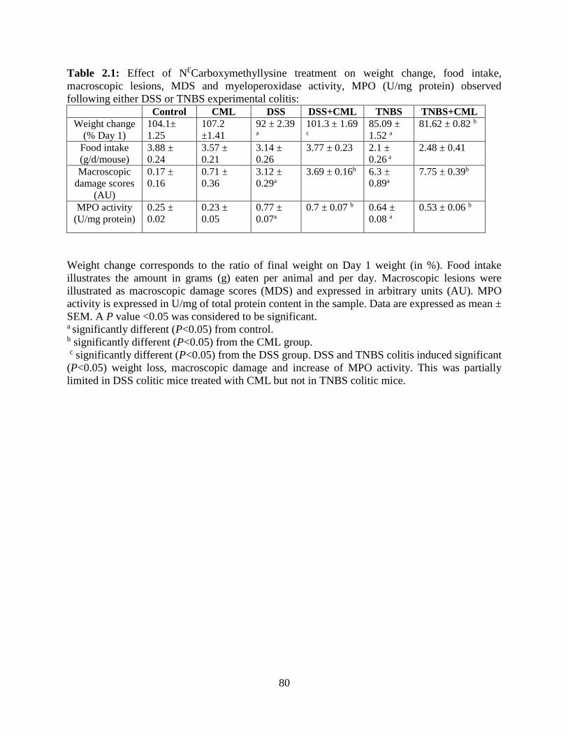
80
Table 2.1: Effect of NƐCarboxymethyllysine treatment on weight change, food intake,
macroscopic lesions, MDS and myeloperoxidase activity, MPO (U/mg protein) observed
following either DSS or TNBS experimental colitis: Control CML DSS DSS+CML TNBS TNBS+CML
Weight change
(% Day 1)
104.1±
1.25
107.2
±1.41
92 ± 2.39 a
101.3 ± 1.69 c
85.09 ±
1.52 a
81.62 ± 0.82 b
Food intake
(g/d/mouse)
3.88 ±
0.24
3.57 ±
0.21
3.14 ±
0.26
3.77 ± 0.23 2.1 ±
0.26 a
2.48 ± 0.41
Macroscopic
damage scores
(AU)
0.17 ±
0.16
0.71 ±
0.36
3.12 ±
0.29a
3.69 ± 0.16b 6.3 ±
0.89a
7.75 ± 0.39b
MPO activity
(U/mg protein)
0.25 ±
0.02
0.23 ±
0.05
0.77 ±
0.07a
0.7 ± 0.07 b 0.64 ±
0.08 a
0.53 ± 0.06 b
Weight change corresponds to the ratio of final weight on Day 1 weight (in %). Food intake
illustrates the amount in grams (g) eaten per animal and per day. Macroscopic lesions were
illustrated as macroscopic damage scores (MDS) and expressed in arbitrary units (AU). MPO
activity is expressed in U/mg of total protein content in the sample. Data are expressed as mean ±
SEM. A P value <0.05 was considered to be significant. a significantly different (P<0.05) from control. b significantly different (P<0.05) from the CML group.
c significantly different (P<0.05) from the DSS group. DSS and TNBS colitis induced significant
(P<0.05) weight loss, macroscopic damage and increase of MPO activity. This was partially
limited in DSS colitic mice treated with CML but not in TNBS colitic mice.
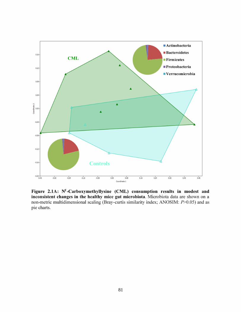
81
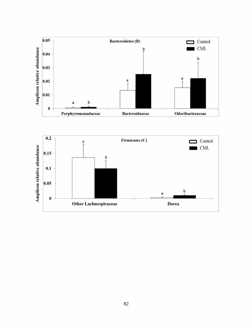
82

83

84
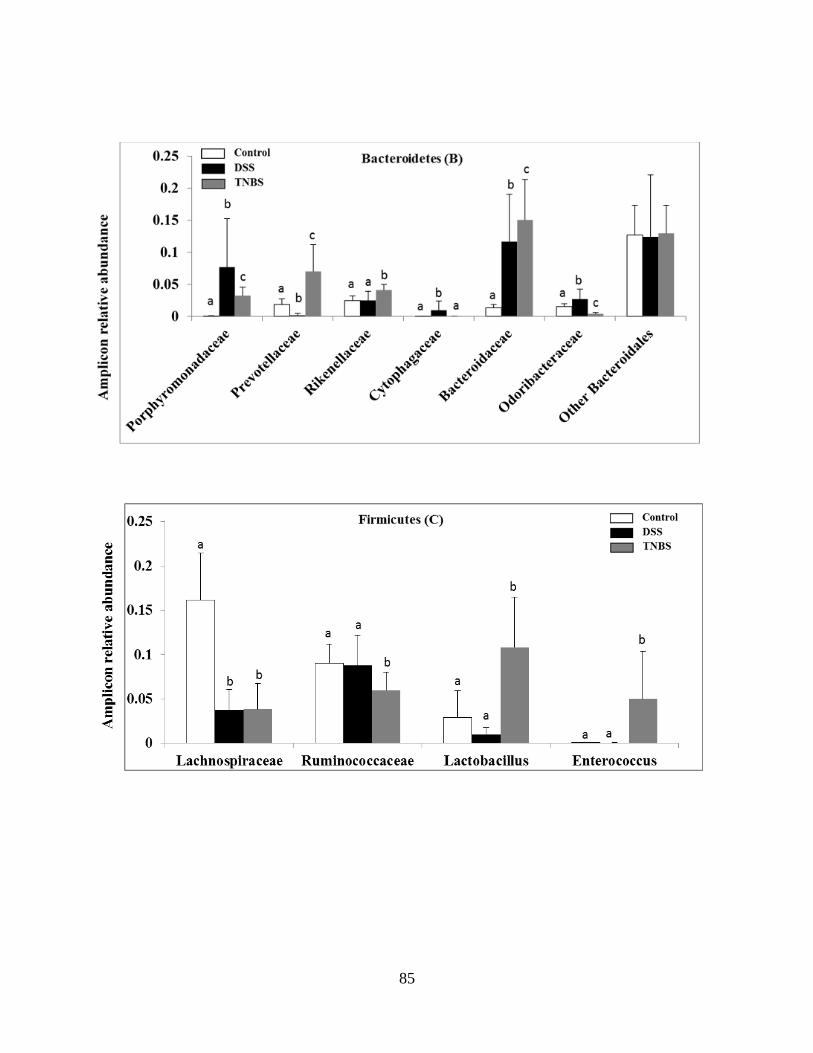
85
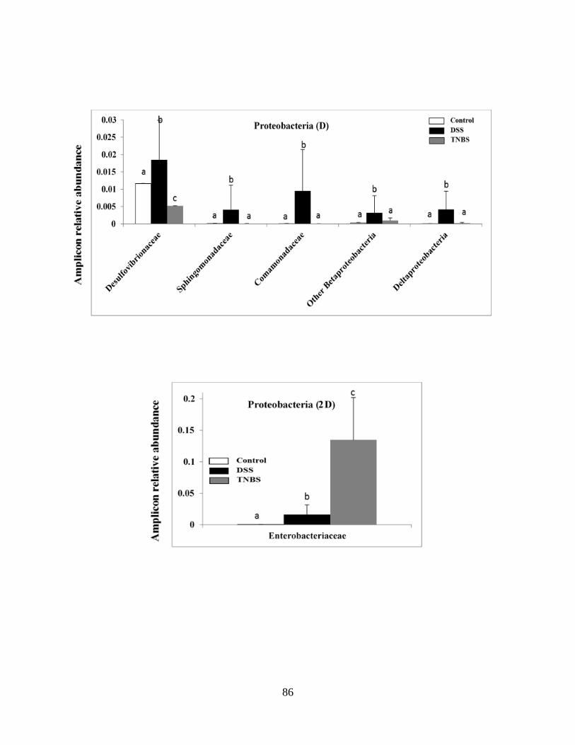
86
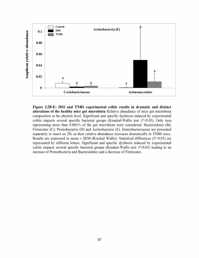
87
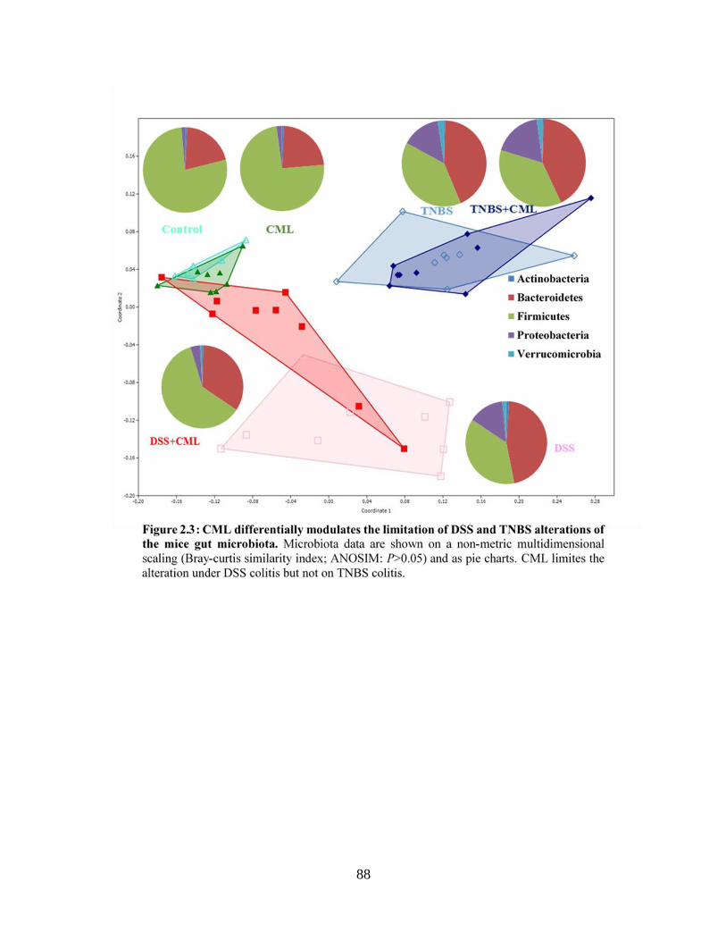
88
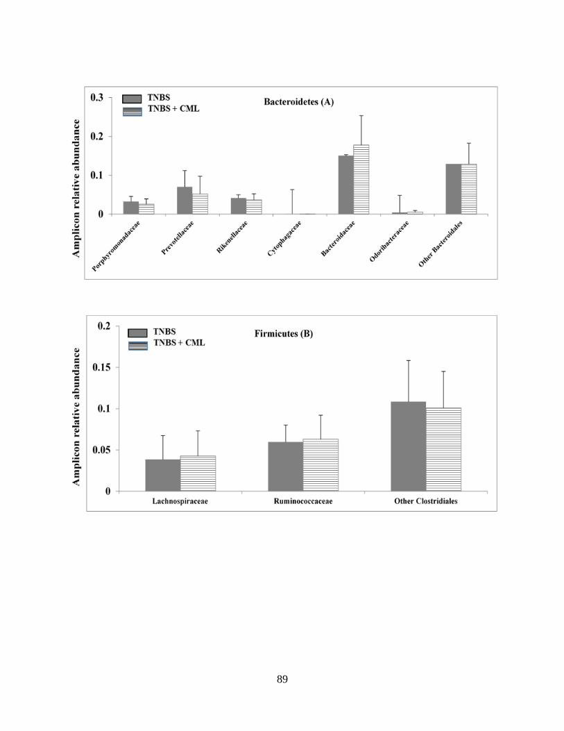
89
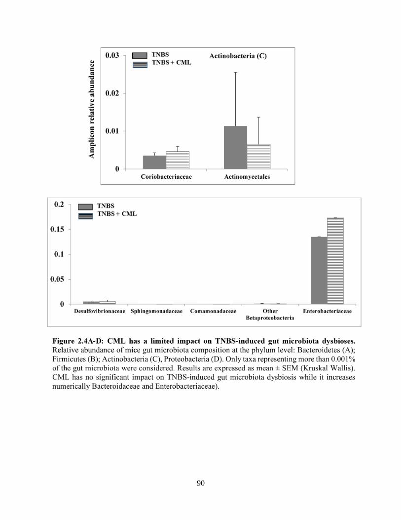
90

91

92
93
CHAPTER THREE
RESEARCH ARTICLE
Impact of melanoidins from bread crust model on the gut microbiota of healthy and
experimental low-grade colitis model rats
ALJahdali Nesreen,1* Helou Cynthia,,2,3 Anton Pauline M,2 Tessier Frederic J,2,4 Carbonero
Franck,1,5 Gadonna-Widehem Pascale2
1Cell and Molecular Biology Program; University of Arkansas, AR, USA
2Unité Transformation & Agro-Ressources, Institut Polytechnique Unilasalle, Beauvais, France
3 Université Saint Joseph de Beyrouth, Faculté de Pharmacie, Département de Nutrition, Beirut,
Lebanon
4University Lille, Inserm, CHU Lille, U995-LIRIC-Lille Inflammation Research International
Center, F-59000 Lille, France
5Department of Food Science; University of Arkansas, AR, USA
Corresponding author: Franck Carbonero

94
3.1 Abstract:
Bread melanoidins are produced during the last stage of the Maillard reaction (MR). The
high amounts of melanoidins are found in bread crust, compared to bread crumbs. Melanoidins are
known to have a high molecular weight (HMW), which are indigestible and reach the large
intestine. Several studies showed that melanoidins are fermented by gut microbiota. The aim of
this study was to determine the effect of bread melanoidins on the composition of rat’s gut
microbiota and potential positive modulation of the microbiota profile associated with
experimental colities. Forty eight rats received either pellet (chow), bread crust model (BCM), or
melanoidin-free control (MFC) for 29 days. In the last week, each group was split into sub-groups,
receiving Trinitrobenzene sulfonic acid (TNBS) to induce colitis. The consumption of BCM and
MFC resulted in a significant decrease of Bacteroides and Enterobacteriaceae, compared to the
control. However, the BCM limited to increase Enterobacteriaceae TNBS model. Significant
increases were observed in Faecalibacterium in BCM model. In contrast, the relative abundances
of Biofidobacteria and Lactobacillus were decreased in rats that consumed BCM, compared to a
group that consumed MFC.
Keywords: Maillard reaction products, Bread Melanoidins, Gut microbiota
95
3.2 Introduction
Melanoidins are polymeric, high molecular weight, brown compounds produced by the
Maillard reaction during thermal processing of foods containing amino acids and reduced sugars
(HODGE, 1953). Melanoidins are particularly prevalent in the Western diet, with high amounts
reported in bakery products, roasted coffee (Fogliano & Morales, 2011), roasted barley (Milic,
Grujicinjac, Piletic, Lajsic, & Kolarov, 1975), and many other foods subjected to intense heating
(Hofmann, Bors, & Stettmaier, 1999). The chemical structure of melanoidins depends on the types
of sugars, proteins, and physical parameters, such as temperature, heating time, pH, water activity,
and other factors (Martins & van Boekel, 2003). Coffee and bread melanoidins have been given
more attention in research due to their nutrition and health properties (Fogliano & Morales, 2011;
Hofmann et al., 1999), and several studies reported that food melanoidins possess antioxidant,
antihypertensive, antimicrobial, and prebiotic properties (ALJahdali & Carbonero, 2017; Wang,
Qian, & Yao, 2011).
Bread melanoidins are only found in the crust, and the amount of melanoidins measured in
the crust of sourdough loaves, sliced bread, and baguette were 30, 18, and 14 / 100 g, respectively
(Fogliano & Morales, 2011). The intake of bread melanoidins ranged between 1.8-15.0 g per day
(Fogliano & Morales, 2011). The principal constituents of bread melanoidins are starch and
proteinaceous material (Hofmann et al., 1999). However, because bread melanoidins are insoluble
high molecular weight molecules, they are poorly absorbed through digestive processes and reach
the large intestine mostly intact (Helou et al., 2017; Helou et al., 2015; Tagliazucchi & Bellesia,
2015). It is now well established that undigested dietary elements are subject to intense
fermentation by resident microbes, and there has been extensive research on the impact of diet on
the human and animal gut microbiota (O'Keefe et al., 2015; Su et al., 2015). However, the majority
96
of studies on the diet, and the gut microbiota have focused on the basic nutrients: carbohydrates
and fibers (Martinez, Kim, Duffy, Schlegel, & Walter, 2010; Zhang et al., 2016), proteins (Louis,
Hold, & Flint, 2014) and fat (Devkota et al., 2012), while Maillard reaction products in general
and melanoidins in particular have been sparsely studied (ALJahdali & Carbonero, 2017). Previous
studies investigated the impact of bread melanoidins on the composition of gut microbiota in vitro.
In one in vitro study, bread crust melanoidins were found to increase the abundance of
Bifidobacterium species (Borrelli & Fogliano, 2005), while a recent study in vivo showed that
bread melanoidins had no bifidogenic effect (Helou et al., 2017). Also, an in vitro study showed
that bread melanoidins were able to inhibit the development of Enterobacteriaceae in stool cultures
(Helou et al., 2015). The in vitro results suggest that bread crust melanoidins may act as prebiotics;
however, in vivo studies are crucial to strengthen conclusions on prebiotic properties. The aim of
this study was to determine the impact of model bread crusts containing melanoidins or not on the
composition of the gut microbiota of healthy and chemically-induced colitic rats.
3.3 Material and Methods
3.3.1 Bread Preparation
Fiber-free models of bread crust were prepared as described previously (Helou et al., 2017).
Briefly, for the bread crust model (BCM) starch, gluten, D-glucose and water (52.25%, 10.5%,
2.75%, and 34.5% of the dough weight, respectively) were homogenized and kneaded with a
Kenwood Cooking Chef KM089 premium (De Longhi Kenwood, Clichy, France) and baked at
220 °C for 16 min in an Air-o-Speed ventilated oven (Electrolux, France). For the melanoidin-free
control (MFC), the same preparation was followed, except that gluten was heated separately to
prevent melanoidin formation.

97
3.3.2 Animals and Study design
Experiments were conducted at the Animal House Unit of LaSalle Beauvais under approval
from the local animal protocol review committee (C60-200-01) and the French Ministry of Higher
Education, Research and Innovation (CEEA n°116 and MENESR n°1688.V02) (Helou et al.,
2017). Forty eight male Sprague-Dawley rats 7 weeks old (75-99 g) purchased from Harlan
Laboratories (Ganat, France) were housed in stainless steel cages under controlled temperature (21
± 1°C) and 12H light-dark cycle with unlimited access to food and water. During the first week,
all rats were fed normal A04 pellets (Safe, Epinay sur Orge, France) and tap water ad libitum.
After this adaptation week, the rats were divided into three groups of 16: one received A04 pellets,
the others received pellets of A04 supplemented with, BCM or MFC pellets for 29 days ad libitum.
BCM and MFC pellets were obtained from a special line by mixing of A04 powder with 13%
(w/w) of BCM or MFC followed by compaction (Safe, Epinay sur Orge, France). During the last
week, the three groups were divided into two sub-groups (6 groups, n=8 in each group): control
and Trinitrobenzene sulfonic acid (TNBS) colitis. The TNBS solution (Sigma-Aldrich) was diluted
in 50% ethanol (40 mg/kg – 50 µL IR) before being injected intrarectally (IR) with a lubricated
silicone tubing (4cm from the anus) under general anesthesia (ketamine/xylazine 100 mg/kg – 50%
v/v – 1mL/kg i.p.). Animals were then kept head down on a heating pad until full recovery from
the anesthesia.
At the end of the experiment, animals were sacrificed by cervical dislocation under general
anaesthesia (ketamine/xylazine – 100 mg/kg i.p. – 50% v/v). A midline laparotomy was then
completed, macroscopic lesions were assessed and pieces of the colon were immediately removed
and snap frozen until further analysis. To perform the analysis of the incidence of each treatment
on the microbiota profile, caeca were ligated at both the ileo-caecal and the caeco-colonic junctions
98
before their removal and their contents were immediately collected under anaerobic conditions and
snap frozen until further determination.
3.3.3 DNA Extraction and PCR Amplifications (Polymerase Chain Reaction)
Genomic DNA was extracted from ceca samples. Briefly, 100 mg cecal content and 1 mL
of ASL buffer (Qiagen, Courtaboeuf, France) were pooled in 2 mL Lysing Matrix E tubes (MP
Biomedicals, Santa Ana, USA). After mixing for 1 min with a vortex, the tubes were heated at
95°C for 5 min in a Thermomixer (Eppendorf, Montesson, France) and were then cooled in ice for
5 min. The tubes were transferred into the FastPrep® (MP Biomedicals, Santa Ana, USA) bead-
beater for 2 runs of 1 min each at 6 m/s. After centrifugation (16000g at 4℃ for 1 min), the total
DNA was extracted from the obtained lysates using a commercial kit (QIAamp DNA stool Mini
Kit) following the manufacturer’s protocol.
Dual-index PCR targeting the V4 region of the bacterial 16S rRNA gene was performed
following previously described protocols (Kozich, Westcott, Baxter, Highlander, & Schloss,
2013). Briefly, PCR reactions were conducted in 27 μL reaction containing: 2 μL of DNA
template; 2 μL of index primers; and 23 μL of AccuPrime™ Taq DNA Polymerase (Invitrogen,
USA) following the manufacturer’s protocol. Amplifications were performed by initial
denaturation at 95°C for 3 min, followed by 25 cycles of denaturation at 98°C for 30 sec, primer
annealing at 55°C for 30 sec, and extension at 72°C for 1 min. PCR amplicons were examined on
agarose gel electrophoresis by using 6 μL of ethidium bromide fluorescent dye to confirm the
success of the PCR.
3.3.4 Library Preparation
Normalization of the PCR products was performed with a SequalPrep kit (Invitrogen)
following the manufacturer’s instructions. Normalized amplicons were pooled and checked on
99
agarose gels. Gel extraction of the correct amplicon was accomplished by running 100 μL of DNA
on an agarose gel electrophoresis, and then extracting DNA from excised bands with the Min Elute
Gel Extraction kit (Qiagen, Hilden, Germany).
Library quality checks using by means of real-time q-PCR and TapeStation (Agilent®,
Santa Clara, USA) were performed to know the concentration and the size of DNA libraries,
respectively. Q-PCR was performed with the KAPA Library Quantification Kit (KAPA
Biosystems) following the manufacturer’s recommendations. The dilutions were prepared from
the gel extraction by taking 5 μL of DNA and adding it to 45 μL of H2O, and dilution was created
starting from: 1:10, 1:100, 1:1000, 1:2000, and 1:4000. Then, the q-PCR plate was prepared by
running 4 μL triplicate of standards with 6 μL of Primer premix and 4 μL triplicate of library
dilutions with 6 μL of Primer premix. The Q-PCR plate was placed in the thermocycler by
following initial activation at 95 °C for 5 min and 35 cycles. Amplicon size was determined again
after gel extraction on the TapeStation, which was 421pb.
3.3.5 Sequencing, Bioinformatics and Statistical Analyses
Amplicon pools were diluted to 0.70 nM with 0.2 N fresh NaOH following recommended
Illumina protocols. Denatured libraries were diluted to 6 pm as a final concentration, with addition
of 15 pm of Phix control V3. The diluted denatured libraries were loaded on an Illumina MiSeq
sequencing cartridge V2-500 cycles, with index primer, Read 1, and Read 2 sequencing primers
(Kozich et al., 2013). The runs were monitored with Sequence Analysis Viewer with particular
emphasis on appropriate cluster density (900-1000k/mm2) and quality scores (final >Q30 score of
>80%).
All sequences were downloaded from the Illumina Basespace server in Fastq files format.
All reads were analyzed using the MOTHUR software package 1.39.5 following the Illumina SOP
100
script (Schloss et al., 2009). All reads were carried out using the SILVA database as a reference
for assignation of operational taxonomic units (OTUs) with 97% of identity. The results of bacteria
counts were analyzed by Kruskal-Wallis and Mann-Whitney pairwise with 𝑃 < 0.05 , which was
considered to indicate a significant difference among groups. Additionally, we used Non-Metric
Multidimensional scaling (NMDS) based on count-distance metrics (Bray-Curtis similarity index;
ANOSIM:𝑝 < 0.05) considered to be significant similar between groups using Past3 software
(Hammer, Harper, & Ryan, 2001). 8,383,167 raw sequences were obtained from 47 samples, and
6,328,091 high-quality sequences were used for bioinformatics analyses, with an average of
74031± 28677 reads per sample.
3.4 Results and Discussion
Dietary high molecular weight of MRPs are largely insoluble in water. They are
indigestible molecules that remain in the gastrointestinal tract, which is the major site for biological
activities. To illustrate, melanoidins bind to dietary metals, thereby, leading to antioxidant and
antimicrobial properties (Morales, Somoza, & Fogliano, 2012). Melanoidin molecules have been
shown to suppress Helicobacter pylori infection in vitro and in vivo studies (Hiramoto et al., 2004).
The results of studies carried out on MRPs remain controversial impact on health due to the fact
that bioconversion of these digestible molecules by the gut microbiota (Hellwig et al., 2015;
Seiquer, Rubio, Jesus Peinado, Delgado-Andrade, & Pilar Navarro, 2014). Gut microbiota have
been shown to use bread melanoidins as a carbon and nitrogen source and had bifidogenic effects
in vitro study (Borrelli & Fogliano, 2005), while another in vitro study showed the inhibition of
enterobacteria (Helou et al., 2015). The objective of this study was to determine the effect of a
bread melanoidin model on the composition of rat’s gut microbiota and potential positive
modulation of the microbiota profile associated with experimental colitis.
101
3.4.1 Impact of TNBS treatment on the gut microbiota
There were slight differences (not statistically significant) at all taxonomic levels between
control and TNBS groups (Figure 3.1A). At the phylum level, the relative abundances of
Firmicutes were lower in the TNBS group, compared to the control (Figure 3.2; P<0.05). However,
there were no significant differences in the relative abundances of Bacteroidetes, Proteobacteria,
Actinobacteria, and Verrucomicrobia between control and TNBS groups (Figure 3.2). There were
no significant differences between TNBS and control groups of the relative abundance of several
Bacteroidetes genera (Parabacteroides, Bacteroides, and Prevotella) (Figure 3.3), as well as, the
Enterobacteriaceae (Proteobacteria) (Figure 3.4), Bifidobacterium (Actinobacteria), Akkermansia
(Verrucomicrobia) (Figure 3.5), and several Firmicutes genera (Oscillibacter, Faecailibacterium,
Blautia, ClostridiumXIVa, Dorea, Lactobacillus, and ClostridiumXI) (Figure 3.6 A, B, C).
However, the relative abundances of Lachnospiraceae (Firmicutes) were lower in the
TNBS group, compared to the control (Figure 3.4; 3.6 B; P <0.05). In a previous study, TNBS
resulted in decreases of Firmicutes and Lachnospiraceae (ALJahdali et al., 2017). However, this
study and many others (Ettreiki et al., 2012) also reported that Proteobacteria, specifically
Enterobacteriaceae bloom was the most notable impact of TNBS treatment. The absence of such
a bloom in our study suggests that the TNBS treatment was not as strong in inducing colitis and
dysbiosis as expected. Indeed, low-grade inflammation was observed, with no weight loss or
changes in feed intake, but significant alteration of inflammation markers (Data not shown).
102
3.4.2 Impact of MFC on the rats gut microbiota
By using the NMDS plots, slight differences (not statistically significant) were observed at
all taxonomic levels between MFC and control groups (Figure 3.1B). At the phylum level, there
were no significant differences of Firmicutes, Bacteroidetes, Proteobacteria, and Verrucomicrobia
between control and MFC, but a significant increase of Actinobacteria was observed in MFC,
compared to the control (Figure 3.2B; P> 0.05). The current findings are in line with the previous
report that demonstrates no impact of MFC (low on melanoidins) on Firmicutes, Bacteroidetes and
lactic flora using a real time quantitative polymerase chain reaction and cultivation approaches in
an in vivo study (Helou et al., 2017). However, this study found that the relative abundance of
Actinobacteria was higher in MFC, compared to the control, which may be in line with the previous
report of increases in the proportion of the population of Bifidobacteria (Actinobacteria) in rats
that consumed whey-fortified breads (low on melanoidins) (Wronkowska et al., 2017).
There were no significant differences among MFC and control groups in the relative
abundance of several Firmicutes genera (Oscillibacter, Faecailibacterium, Blautia,
ClostridiumXIVa, Dorea, and ClostridiumXI) (Figure 3.6 A, B, C), as well as, Akkermansia
(Verrucomicrobia; Figure 3.5), and Prevotella (Bacteroidetes; Figure 3.3). Significant decreases
of Parabacteroides and Bacteroides were observed in MFC group, compared to the control (Figure
3.3; P<0.05). Decreased proportions of Parabacteroides and Bacteroides were also seen in rumen
microbiome of buffaloes fed wheat and maize grain (Kala et al., 2017). However, increases the
proportions of the relative abundances of Bacteroides and Parabacteroides were found in mice
fed high proteins and low carbohydrates (Kim, Kim, & Park, 2016). Also, a significant decrease
of Enterobacteriaceae (Proteobacteria) was observed in rats that were fed MFC, compared to the
control (Figure 3.4; P<0.05). The current findings are in line with the previous report that the

103
relative abundance of enterobacteria decreased in individuals who consumed bread (Helou et al.,
2015).
However, the consumption of MFC resulted in a significant increase of Lactobacillus,
compared to the control (Figure 3.6 C; P>0.05), which may be in line with the previous report of
increases in the proportion of the intestinal Lactobacillus spp. after adding pre-melanoidins
products (low on melanoidins) in batch cultures (Jemmali, 1969). The relative abundances of
Bifidobacterium (Actinobacteria) were significantly different the in the MFC group, compared to
the control (Figure 3.5; P>0.05). A significant increase of Bifidobacterium was observed in mice
fed a MFC diet (Helou et al., 2017).
By using the NMDS plots, there were slight differences between TNBS and MFC-TNBS
group at all taxonomic levels (Figure 3.1C). At the phylum level, the relative abundances of
Firmicutes, Bacteroidetes, Proteobacteria, and Actinobacteria showed no significant differences
between TNBS and MFC-TNBS groups (Figure 3.2). However, there were significant differences
in the relative abundance of Verrucomicrobia between TNBS and MFC-TNBS groups (Figure 3.2;
P>0.05). There were no significant differences of Firmicutes, Proteobacteria, Verrucomicrobia,
and Actinobacteria between MFC and MFC-TNBS groups (Figure 3.2). However, the relative
abundances of Bacteroidetes were significantly different in the MFC-TNBS group, compared to
MFC group (Figure 3.2; P>0.05).
There were no significant differences among TNBS and MFC-TNBS groups in the relative
abundances of several Bacteroidetes genera (Parabacteroides, Bacteroides, and Prevotella)
(Figure 3.3), as well as, Enterobacteriaceae (Proteobacteria; Figure 3.4), and several Firmicutes
genera (Faecailibacterium, Blautia, ClostridiumXIVa, Dorea, and ClostridiumXI) (Figure 3.6 A,
B, C). However, the relative abundances of Akkermansia (Verrucomicrobia) were significantly
104
lower in the MFC-TNBS group, compared to the TNBS group (Figure 3.5; P>0.05). Akkermansia
muciniphila had beneficial effects on the immune system by alleviating inflammation (Wu et al.,
2017). The high abundance of Akkermansia muciniphila was found in humanized rats fed
inulin and arabinoxylans, which are important ingratiates in baked products (Van den Abbeele et
al., 2011).
The abundance of Oscillibacter and Lactobacillus (Firmicutes Figure 3.6A C; P>0.05) as
well as Bifidobacterium (Actinobacteria; Figure 3.5; P>0.05) were higher in the MFC-TNBS
group, compared to TNBS group. There were no significant differences between MFC and MFC-
TNBS groups in the relative abundance of Prevotella (Bacteroidetes; Figure 3.3),
Enterobacteriaceae (Proteobacteria; Figure 3.4), Akkermansia (Verrucomicrobia; Figure 3.5),
Bifidobacterium (Actinobacteria; Figure 3.5), and several genera of Firmicute (Oscillibacter,
Faecailibacterium, ClostridiumXIVa, Dorea, Lactobacillus and ClostridiumXI) (Figure 3.6 A B
C). However, significant increases in Parabacteroides and Bacteroides (Bacteroidetes) were
present in the MFC-TNBS group, compared to the MFC group (Figure 3.3 P>0.05). Also, the
relative abundances of Ruminococcaceae and Blautia (Firmicutes) were increased significantly in
the MFC-TNBS group, compared to the MFC group (Figure 3.6A B P>0.05).
3.4.3 Impact of BCM on the rat’s gut microbiota
By using the NMDS plots, there were significant differences between control and BCM
groups at all taxonomic levels (Figure 3.1B; ANOSIM P<0.05). At the phylum level, the
consumption of BCM resulted in a significant increase of Firmicutes, compared to the control
(Figure 3.2; P<0.05). However, there were decreased abundances of Bacteroidetes and
Verrucomicrobia, compared to the control group (Figure 3.2; P<0.05). There were no significant
differences in the relative abundances of Proteobacteria and Actinobacteria between BCM and
105
control groups (Figure 3.2). A decrease of Bacteroidetes was detected in individuals who
consumed refined wheat breads (Lappi et al., 2013). In contrast, there was no impact of phyla level
in mice that consumed BCM, compared to the control group (Helou et al., 2017).
The relative abundance of Parabacteroides (Bacteroidetes; Figure 3), Bifidobacterium
(Actinobacteria; Figure 3.5), and several Firmicutes genera (ClostridiumXIVa, Dorea, and
ClostridiumXI) were not significantly different between BCM and control groups (Figure 3.6A B
C). BCM consumption resulted in significant increase of Ruminococcaceae, Oscillibacter,
Faecalibacterium, Lachnospiraceae, and Blautia (Firmicutes; Figure 3.6A B P>0.05), which may
be in line with a previous study of an increase of Oscillibacter found in individuals who consumed
diets high in resistant starch (RS) and carbohydrate diets (Walker et al., 2011). Faecalibacterium
prausnitzii is important commensal bacterium produced butyrate and known for their ability of
anti-inflammatory effects in the gut (Sokol et al., 2008).
However, there were significant decreases of the relative abundance of Prevotella and
Bacteroides (Bacteroidetes; Figure 3.3 P<0.05), as well as, Enterobacteriaceae (Proteobacteria;
Figure 3.4 P<0.05), Akkermansia (Verrucomicrobia; Figure 3.5 P<0.05) and Lactobacillus
(Firmicutes; Figure 3.6 C P<0.05) in the group that consumed BCM, compared to the control
group. In the same line to our results, a limited growth of Enterobacteriacea and Bacteroides spp.
was present after 10 hours of fermentation on bread crust melanoidins in an in vitro study (Borrelli
& Fogliano, 2005). There were no significant differences between Enterobacteriacea and
Bacteroides spp in mice that consumed bread crust, compared to a control group (Delgado-
Andrade et al., 2017). However, there was significant decrease of Lactobacillus spp. in mice that
were fed bread crust diet, compared to a control group (Delgado-Andrade et al., 2017). Also,
Lactobacillus spp. showed a low ability to use bread melanoidins for their growth (Borrelli &

106
Fogliano, 2005). Species related to Bacteroides and Prevotella decreased in individuals who
consumed refined wheat breads (Lappi et al., 2013). On the other hand, another study on the
consumption of coffee melanoidins resulted in increases of species belonging to Bacteroides-
Prevotella in an in vitro study (Gniechwitz, Reichardt, Blaut, Steinhart, & Bunzel, 2007).
By using the NMDS plots, there were significant differences between TNBS and BCM-
TNBS groups at all taxonomic levels (Figure 3.1C; ANOSIM P<0.05). At the phylum level, the
relative abundances of Firmicutes and Verrucomicrobia showed significant differences between
TNBS and BCM-TNBS groups (Figure 3.2; P<0.05). However, there were no significant
differences of Bacteroidetes, Proteobacteria, and Actinobacteria between the BCM-TNBS group
and the TNBS group (Figure 3.2). There were no significant differences of Firmicutes,
Bacteroidetes, Proteobacteria, Verrucomicrobia, and Actinobacteria between BCM and BCM-
TNBS groups (Figure 3.2). Highly heated food rich in Maillard reaction products protected against
inflammation of experimental colitis in mice (Anton, Craus, Niquet-Leridon, & Tessier, 2012).
We showed that consumption of BCM had to limit the gut microbiota dysbiosis in BCM-TNBS
groups. NƐ-carboxymethyllysine, an advanced Maillard reaction products, alleviates gut
microbiota dysbiosis in colitic mice (ALJahdali et al., 2017).
There were no significant differences between TNBS and BCM-TNBS groups in the
relative abundances of several Bacteroidetes genera (Parabacteroides, Bacteroides, and
Prevotella) (Figure 3.3), as well as, Bifidobacterium (Actinobacteria; Figure 5), and several
Firmicutes genera (Oscillibacter, Faecailibacterium, Blautia, Dorea, Lactobacillus and
ClostridiumXI) (Figure 3.6 A, B, C). The relative abundances of Enterobacteriaceae
(Proteobacteria; Figure 3.4 P<0.05) and Akkermansia (Verrucomicrobia; Figure 3.5 P<0.05) were
significantly lower in the BCM-TNBS group, compared to the TNBS group. NƐ-
107
carboxymethyllysine, an advanced Maillard reaction products, limited Enterobacteriaceae
dysbiosis in colitic mice (ALJahdali et al., 2017). Akkermansia spp. are known as mucin-degrading
bacteria that consumed glycated proteins as an energy source (Tailford, Crost, Kavanaugh, & Juge,
2015). Akkermansia muciniphila is known to possess anti-inflammatory effects. However, the
abundances of Lachnospiraceae (Firmicutes) and Clostridium XIVa were higher in the BCM-
TNBS group, compared to the TNBS group (Figure 3.6 B; P <0.05).
There were no significant differences between BCM and BCM-TNBS groups in the
relative abundance of Parabacteroides (Bacteroidetes; Figure 3.3), Enterobacteriaceae
(Proteobacteria; Figure 3.4), Akkermansia (Verrucomicrobia; Figure 3.5), Bifidobacterium
(Actinobacteria; Figure 3.5), and several genera of Firmicute (Blautia, ClostridiumXIVa, and
Dorea) (Figure 3.6 B). Significant increases of Prevotella and Bacteroides (Bacteroidetes) were
present in the BCM-TNBS group, compared to the BCM group (Figure 3.3 P>0.05). Also, the
relative abundances of Ruminococcaceae and several genera of Firmicutes (Oscillibacter and
Faecailibacterium) were significantly higher in the BCM group, compared to the BCM-TNBS
group (Figure 3.6A P>0.05). Conversely, the relative abundances of ClostridiumXIVa,
Lactobacillus and ClostridiumXI were significantly higher in BCM-TNBS group, compared to the
BCM group (Figure 3.6 B&C P >0.05).
3.5 Conclusions
We concluded that MFC increased the relative abundances of Bifidobacterium spp. and
Lactobacillus spp., while BCM increased beneficial microorganisms Faecalibacterium spp. In
addition, both MFC and crust had to limit increases of Enterobacteriaceae. The BCM -TNBS group
might be able to limit gut microbiota dysbiosis in colitic mice.
108
3.6 References
ALJahdali, N., & Carbonero, F. (2017). Impact of Maillard reaction products on nutrition
and health: Current knowledge and need to understand their fate in the human digestive
system. Critical Reviews in Food Science and Nutrition, 1-14.
doi:10.1080/10408398.2017.1378865 [doi]
ALJahdali, N., Gadonna-Widehem, P., Delayre-Orthez, C., Marier, D., Garnier, B.,
Carbonero, F., & Anton, P. M. (2017). Repeated oral exposure to N epsilon-
carboxymethyllysine, a Maillard reaction product, alleviates gut microbiota dysbiosis in
colitic mice. Digestive Diseases and Sciences, doi:10.1007/s10620-017-4767-8 [doi]
Anton, P. M., Craus, A., Niquet-Leridon, C., & Tessier, F. J. (2012). Highly heated food
rich in Maillard reaction products limit an experimental colitis in mice. Food & Function,
3(9), 941-949. doi:10.1039/c2fo30045a
Borrelli, R., & Fogliano, V. (2005). Bread crust melanoldins as potential prebiotic
ingredients. Molecular Nutrition & Food Research, 49(7), 673-678.
doi:10.1002/mnfr.200500011
Delgado-Andrade, C., Pastoriza de la Cueva, S., Peinado, M. J., Rufian-Henares, J. A.,
Navarro, M. P., & Rubio, L. A. (2017). Modifications in bacterial groups and short chain
fatty acid production in the gut of healthy adult rats after long-term consumption of dietary
maillard reaction products. Food Research International (Ottawa, Ont.), 100(Pt 1), 134-142.
doi:S0963-9969(17)30320-4 [pii]
Devkota, S., Wang, Y., Musch, M. W., Leone, V., Fehlner-Peach, H., Nadimpalli, A.,
Chang, E. B. (2012). Dietary-fat-induced taurocholic acid promotes pathobiont expansion
and colitis in Il10(-/-) mice. Nature, 487(7405), 104-+. doi:10.1038/nature11225
Ettreiki, C., Gadonna-Widehem, P., Mangin, I., Coeffier, M., Delayre-Orthez, C., & Anton,
P. M. (2012). Juvenile ferric iron prevents microbiota dysbiosis and colitis in adult rodents.
World Journal of Gastroenterology, 18(21), 2619-2629. doi:10.3748/wjg.v18.i21.2619 [doi]
Fogliano, V., & Morales, F. J. (2011). Estimation of dietary intake of melanoidins from
coffee and bread. Food & Function, 2(2), 117-123. doi:10.1039/c0fo00156b
Gniechwitz, D., Reichardt, N., Blaut, M., Steinhart, H., & Bunzel, M. (2007). Dietary fiber
from coffee beverage: Degradation by human fecal microbiota. Journal of Agricultural and
Food Chemistry, 55(17), 6989-6996. doi:10.1021/jf070646b [doi]
Hammer, Ř, Harper, D., & Ryan, P. (2001). PAST: Paleontological Statistics Software
Package for Education and Data Analysis–Palaeontol.Electron.4: 9pp,
Hellwig, M., Bunzel, D., Huch, M., Franz, C. M. A. P., Kulling, S. E., & Henle, T. (2015).
Stability of individual Maillard reaction products in the presence of the human colonic
109
microbiota. Journal of Agricultural and Food Chemistry, 63(30), 6723-6730.
doi:10.1021/acs.jafc.5b01391
Helou, C., Anton, P. M., Niquet-Leridon, C., Spatz, M., Tessier, F. J., & Gadonna-
Widehem, P. (2017). Fecal excretion of Maillard reaction products and the gut microbiota
composition of rats fed with bread crust or bread crumb. Food & Function, 8(8), 2722-2730.
doi:10.1039/c7fo00430c
Helou, C., Denis, S., Spatz, M., Marier, D., Rame, V., Alric, M., Gadonna-Widehem, P.
(2015). Insights into bread melanoidins: Fate in the upper digestive tract and impact on the
gut microbiota using in vitro systems. Food & Function, 6(12), 3737-3745.
doi:10.1039/c5fo00836k
Hiramoto, S., Itoh, K., Shizuuchi, S., Kawachi, Y., Morishita, Y., Nagase, M., Kimura, N.
(2004). Melanoidin, a food protein-derived advanced maillard reaction product, suppresses
helicobacter pylori in vitro and in vivo. Helicobacter, 9(5), 429-435. doi:10.1111/j.1083-
4389.2004.00263.x
Hodge, J. (1953). Dehydrated foods - chemistry of browning reactions in model systems.
Journal of Agricultural and Food Chemistry, 1(15), 928-943. doi:10.1021/jf60015a004
Hofmann, T., Bors, W., & Stettmaier, K. (1999). Radical-assisted melanoidin formation
during thermal processing of foods as well as under physiological conditions. Journal of
Agricultural and Food Chemistry, 47(2), 391-396. doi:10.1021/jf980627p
Jemmali, M. (1969). Influence of the Maillard reaction products on some bacteria of the
intestinal flora. The Journal of Applied Bacteriology, 32(2), 151-155.
Kala, A., Kamra, D. N., Kumar, A., Agarwal, N., Chaudhary, L. C., & Joshi, C. G. (2017).
Impact of levels of total digestible nutrients on microbiome, enzyme profile and degradation
of feeds in buffalo rumen. Plos One, 12(2), e0172051. doi:10.1371/journal.pone.0172051
Kim, E., Kim, D. B., & Park, J. Y. (2016). Changes of mouse gut microbiota diversity and
composition by modulating dietary protein and carbohydrate contents: A pilot study.
Preventive Nutrition and Food Science, 21(1), 57-61. doi:10.3746/pnf.2016.21.1.57 [doi]
Kozich, J. J., Westcott, S. L., Baxter, N. T., Highlander, S. K., & Schloss, P. D. (2013).
Development of a dual-index sequencing strategy and curation pipeline for analyzing
amplicon sequence data on the MiSeq illumina sequencing platform. Applied and
Environmental Microbiology, 79(17), 5112-5120. doi:10.1128/AEM.01043-13
Lappi, J., Salojarvi, J., Kolehmainen, M., Mykkanen, H., Poutanen, K., de Vos, W. M., &
Salonen, A. (2013). Intake of whole-grain and fiber-rich rye bread versus refined wheat
bread does not differentiate intestinal microbiota composition in Finnish adults with
metabolic syndrome. Journal of Nutrition, 143(5), 648-655. doi:10.3945/jn.112.172668
110
Louis, P., Hold, G. L., & Flint, H. J. (2014). The gut microbiota, bacterial metabolites and
colorectal cancer. Nature Reviews Microbiology, 12(10), 661-672. doi:10.1038/nrmicro3344
Martinez, I., Kim, J., Duffy, P. R., Schlegel, V. L., & Walter, J. (2010). Resistant starches
types 2 and 4 have differential effects on the composition of the fecal microbiota in human
subjects. Plos One, 5(11), e15046. doi:10.1371/journal.pone.0015046
Martins, S., & van Boekel, M. (2003). Melanoidins extinction coefficient in the
glucose/glycine Maillard reaction. Food Chemistry, 83(1), 135-142. doi:10.1016/S0308-
8146(03)00219-X
Milic, B., Grujicinjac, B., Piletic, M., Lajsic, S., & Kolarov, L. (1975). Melanoidins and
carbohydrates in roasted barley. Journal of Agricultural and Food Chemistry, 23(5), 960-
963. doi:10.1021/jf60201a031
Morales, F. J., Somoza, V., & Fogliano, V. (2012). Physiological relevance of dietary
melanoidins. Amino Acids, 42(4), 1097-1109. doi:10.1007/s00726-010-0774-1
O'Keefe, S. J. D., Li, J. V., Lahti, L., Ou, J., Carbonero, F., Mohammed, K., . . . Zoetendal,
E. G. (2015). Fat, fibre and cancer risk in african americans and rural africans. Nature
Communications, 6, 6342. doi:10.1038/ncomms7342
Schloss, P. D., Westcott, S. L., Ryabin, T., Hall, J. R., Hartmann, M., Hollister, E. B., . . .
Weber, C. F. (2009). Introducing mothur: Open-source, platform-independent, community-
supported software for describing and comparing microbial communities. Applied and
Environmental Microbiology, 75(23), 7537-7541. doi:10.1128/AEM.01541-09 [doi]
Seiquer, I., Rubio, L. A., Jesus Peinado, M., Delgado-Andrade, C., & Pilar Navarro, M.
(2014). Maillard reaction products modulate gut microbiota composition in adolescents.
Molecular Nutrition & Food Research, 58(7), 1552-1560. doi:10.1002/mnfr.201300847
Sokol, H., Pigneur, B., Watterlot, L., Lakhdari, O., Bermudez-Humaran, L. G., Gratadoux,
J., Langella, P. (2008). Faecalibacterium prausnitzii is an anti-inflammatory commensal
bacterium identified by gut microbiota analysis of crohn disease patients. Proceedings of the
National Academy of Sciences of the United States of America, 105(43), 16731-16736.
doi:10.1073/pnas.0804812105
Su, L., Liu, Y., Liu, H., Wang, Y., Li, Y., Lin, H. -., Song, A. -. (2015). Linking
lignocellulosic dietary patterns with gut microbial enterotypes of tsaitermes ampliceps and
comparison with mironasutitermes shangchengensis. Genetics and Molecular Research,
14(4), 13954-13967. doi:10.4238/2015.October.29.16
Tagliazucchi, D., & Bellesia, A. (2015). The gastro-intestinal tract as the major site of
biological action of dietary melanoidins. Amino Acids, 47(6), 1077-1089.
doi:10.1007/s00726-015-1951-z
111
Tailford, L. E., Crost, E. H., Kavanaugh, D., & Juge, N. (2015). Mucin glycan foraging in
the human gut microbiome. Frontiers in Genetics, 6, 81. doi:10.3389/fgene.2015.00081
Van den Abbeele, P., Gerard, P., Rabot, S., Bruneau, A., El Aidy, S., Derrien, M.,
Possemiers, S. (2011). Arabinoxylans and inulin differentially modulate the mucosal and
luminal gut microbiota and mucin-degradation in humanized rats. Environmental
Microbiology, 13(10), 2667-2680. doi:10.1111/j.1462-2920.2011.02533.x
Walker, A. W., Ince, J., Duncan, S. H., Webster, L. M., Holtrop, G., Ze, X., Flint, H. J.
(2011). Dominant and diet-responsive groups of bacteria within the human colonic
microbiota. ISME Journal, 5(2), 220-230. doi:10.1038/ISMEJ.2010.118
Wang, H., Qian, H., & Yao, W. (2011). Melanoidins produced by the mail lard reaction:
Structure and biological activity. Food Chemistry, 128(3), 573-584.
doi:10.1016/j.foodchem.2011.03.075
Wronkowska, M., Soral-Smietana, M., Zdunczyk, Z., Juskiewicz, J., Jadacka, M.,
Majkowska, A., & Dajnowiec, F. J. (2017). Effect of acid whey-fortified breads on caecal
fermentation processes and blood lipid profile in rats. The British Journal of Nutrition,
118(3), 169-178. doi:10.1017/S0007114517001921 [doi]
Wu, W., Lv, L., Shi, D., Ye, J., Fang, D., Guo, F., Li, L. (2017). Protective effect of
akkermansia muciniphila against immune-mediated liver injury in a mouse model. Frontiers
in Microbiology, 8, 1804. doi:10.3389/fmicb.2017.01804 [doi]
Zhang, L., Mu, C., He, X., Su, Y., Mao, S., Zhang, J., Zhu, W. (2016). Effects of dietary
fibre source on microbiota composition in the large intestine of suckling piglets. FEMS
Microbiology Letters, 363(14), fnw138. doi:10.1093/femsle/fnw138

112

113
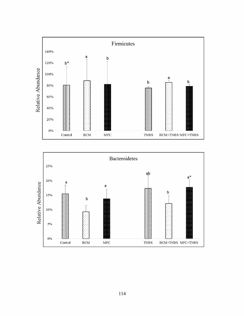
114
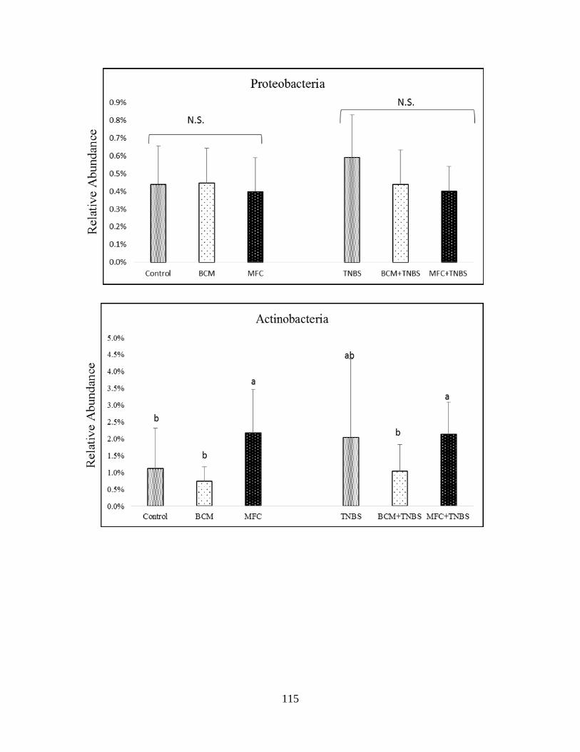
115
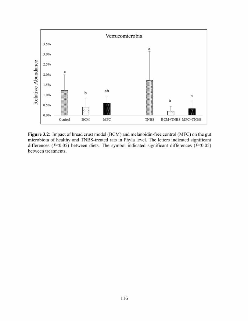
116
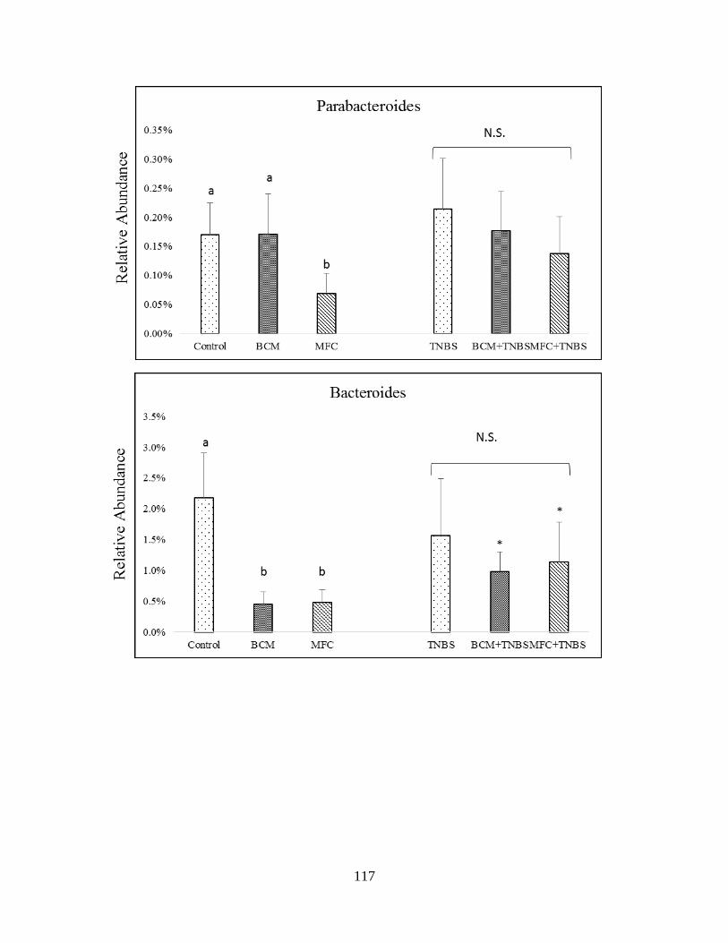
117

118
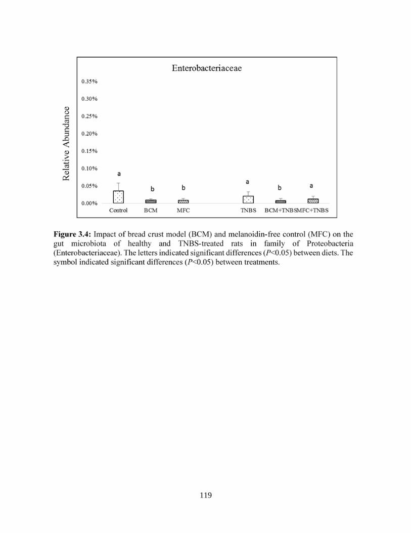
119

120
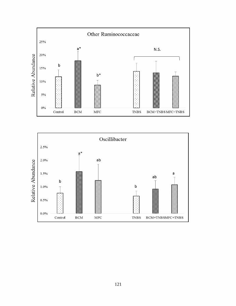
121
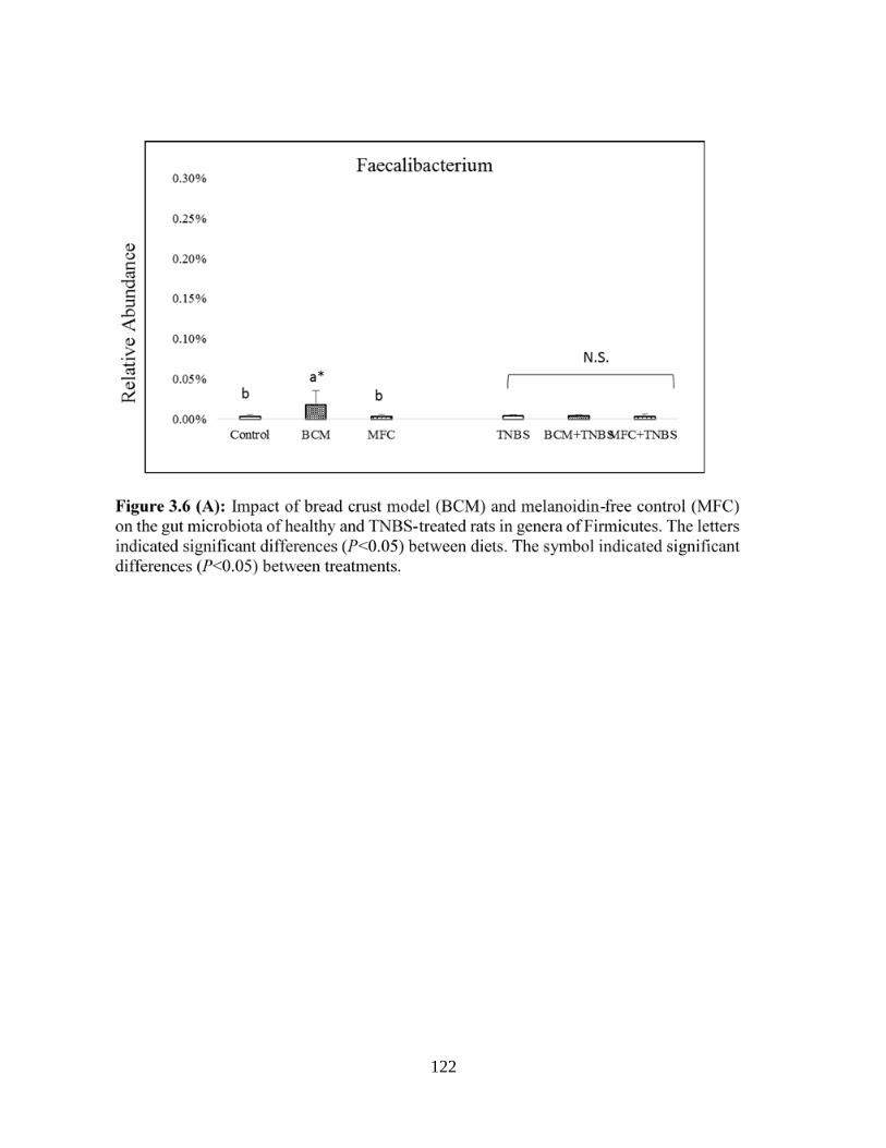
122
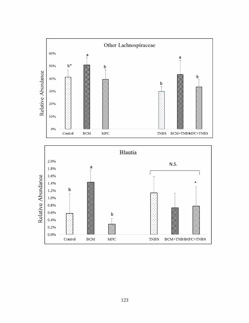
123
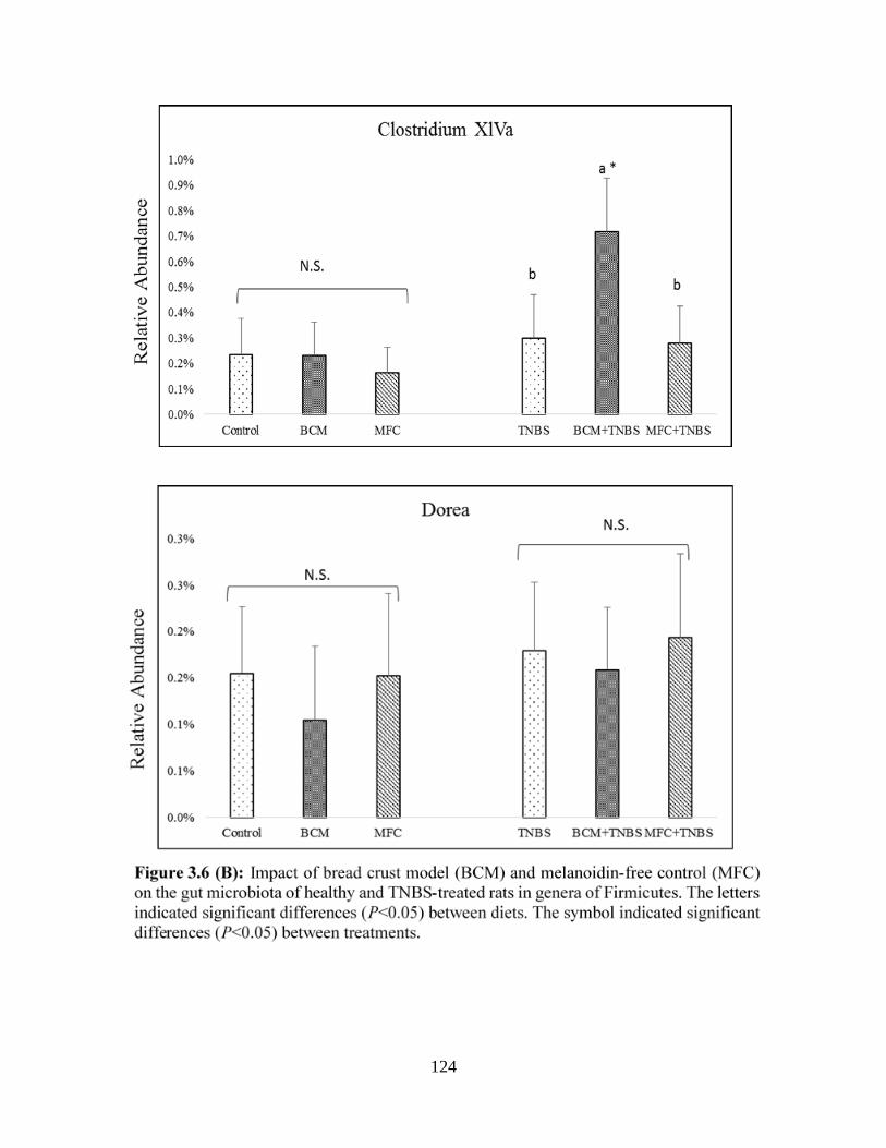
124
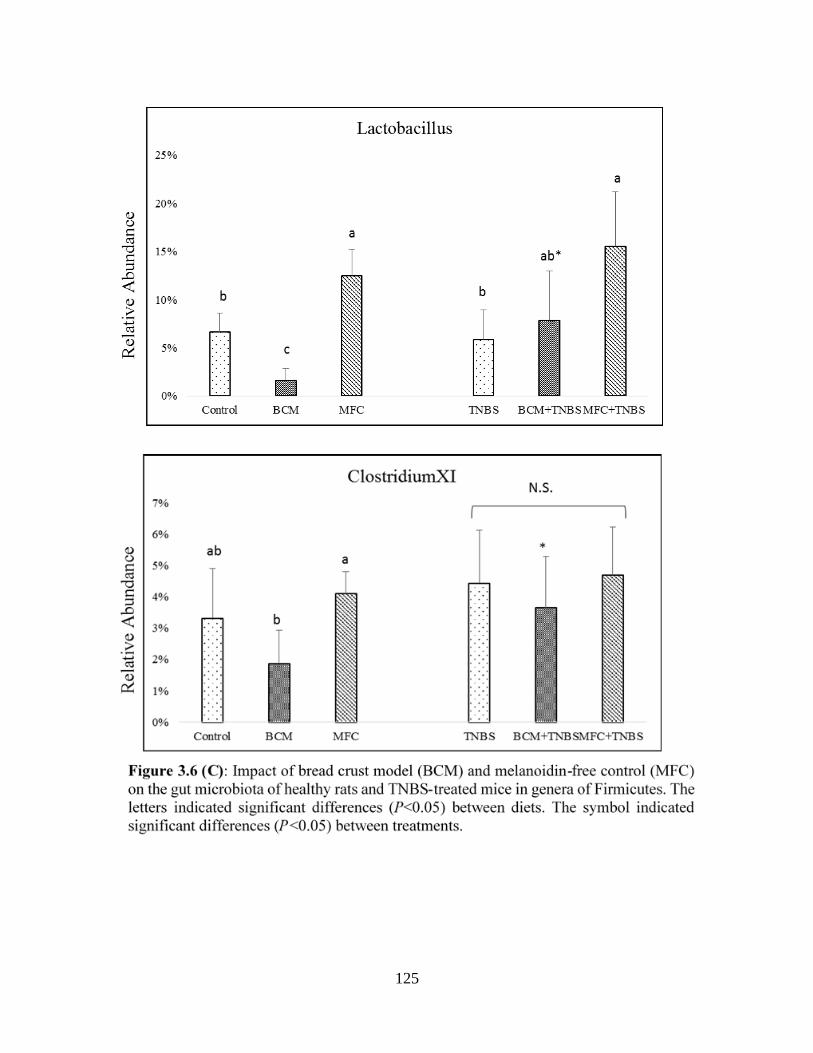
125
126
CHAPTER FOUR
RESEARCH ARTICLE
Modulation of Mice Gut Microbiota by Increasing Amounts of Dietary melanoidins from
barley malts
ALJahdali Nesreen 1* and Carbonero Franck 1,2*
1Cell and Molecular Biology Program; University of Arkansas, AR, USA
2Department of Food Science; University of Arkansas, AR, USA
*Corresponding author:
Franck Carbonero
Department of Food Science
University of Arkansas
2650 Young Avenue
Fayetteville, AR 72704
(+1) 479-575-6822
Email: [email protected]
127
4.1 Abstract:
Melanoidins are the final Maillard reaction products produced in food by prolonged and
intense heating, and are protein-carbohydrate complexes with high molecular weights. Dietary
melanoidins from bread crust and coffee have been reported as harmless and even potentially
beneficial. Here, we assessed the impact of consumption of increasing amounts of malt
melanoidins on mice gut microbiota, another dietary source that has not been studied so far.
Seventy five mice were divided equally into 5 groups, the control group consumed a diet with 0%
of melanoidin-rich malts, and other groups received melanoidin malts in increments of 25%, up to
100% melanoidin malts. Feces were sampled at days 0, 1, 2, 3, 7, 14 and 21, and microbial DNA
was extracted from fecal sample. Microbiota was determined V4 bacterial 16S rRNA amplicon
sequencing, and short-chain fatty acids (SCFA) by Gas Chromatography. The consumption of
malts resulted in a significant decrease of Firmicutes and a distinctive increase of Bacteroidetes,
Actinobacteria, Verrucomicrobia, and Proteobacteria during the study. Increased melanoidins was
found to result in significantly divergent gut microbiota profiles all along the study, as well as to
maintain SCFA production to the baseline levels. The relative abundance of Dorea, Oscillibacter,
and Alisitpes were decreased, but the relative abundance of Lactobacillus, Parasutterella,
Akkermansia, Bifidobacterium, and Barnesilla were increased throughout the study.
Bifidobacterium spp. and Akkermansia spp. were significantly increased in mice consuming the
highest melanoidins amounts. The results support the hypothesis that malt melanoidins have
remarkable prebiotic properties that may be valorized in food development.
Keywords: Maillard reaction products, Melanoidins, Short-chain fatty acids, Gut microbiota,
Prebiotic
128
4.2 Introduction:
The Maillard reaction (MR) generates several low weight molecules from reducing sugars
and amino acids during food thermal processing and storage, such as Amadori rearrangement
products, furfural, reductones, and other dicarbonyl compounds. These low weight compounds are
often recombined through a range of advanced MR to form Melanoidins 1, which are the final
products of the MR 2. Melanoidins are brownish, heterogonous, insoluble molecules, and were
traditionally considered as high molecular weight (HMW) molecules 3,4, but recent reports have
shown that melanoidins also include a low molecular weight (LMW) fraction 5. Melanoidins
produced in foods are predominantly HMW, and melanoidins is molecular weight that is directly
correlated with heating intensity and time 6. For example, the average molecular weight (MW) of
unroasted malts were <10 Kilodalton (kDa), whereas the roasted malts were around 320 kDa 7.
The chemical structures of melanoidins are complex and difficult to determine, but the
concentration of sugars and amino acids of roasted barley have been identified 8. During the
roasting of barley, significant increases of total sugar, dextrin, and melanoidins were detected,
while hemicellulose and starch significantly decreased 8.
In contrast with other MRPs, melanoidins are generally considered harmless, and even
potentially beneficial to human health 9. Though some studies reported that dietary melanoidins
might display moderate genotoxicity and cytotoxicity effects 10,11, several studies reported
potential health benefits including antioxidant, antihypertensive, antimicrobial, and prebiotic
properties of food melanoidins 12,13. Data from metabolic transit studies showed that melanoidins
can escape digestion and pass into the upper gastrointestinal tract (GIT), where they are likely
subject to fermentation by resident gut microbes 14,15. To illustrate, only 27% of the LMW

129
constituents of melanoidin products were absorbed in the intestine; and only 4.3% of the HMW
melanoidins were excreted in feces and urine 16.
It has actually been suggested that dietary melanoidins may possess prebiotic properties 6,
due to their structural similarities with fibers17. Interestingly, studies on the interaction between
gut microbiota and melanoidins have initially focused on antimicrobial activity, predominantly in
batch cultures 18-20. For instance, data from in vitro and in vivo studies showed that melanoidins
could suppress Helicobacter pylori infection 21. Moreover, melanoidins were shown to kill
Escherichia coli by causing irreversible changes in both the inner and outer membranes 22. While
knowledge on the role of the gut microbiota in non-digestible polysaccharides and fiber
fermentation has become extensive 23,24, gut microbiota fermentation of MRPs has been scarcely
studied 25, with even less knowledge on melanoidins 26. In an in vitro study, melanoidins were
shown to increase the growth of gut anaerobes during mixed culture growth 27. Bifidobacteria
strains were shown to use bread melanoidins as a carbon source in batch cultures 26, while coffee
melanoidins increased the number of anaerobic bacteria belonging to Bacteroides-prevotella
during fermentation in an in vitro study 28. Another food rich in melanoidins is beer, where
melanoidins are present in malts, and HMW melanoidins being are more abundant in kilned malts
29; and this melanoidins source has never been assessed for its effect on the gut microbiota. The
objective of this study was to determine the impact of long-term consumption of increasing
melanoidins concentration from barley malts on the gut microbiota and fermentation patterns of
healthy mice.
130
4.3 Material and Methods:
4.3.1 Experimental Animals:
The animal study was conducted at the Animal House Facility of the University of
Arkansas after receiving approval from the Institutional Animal Care and Use Committee
(IACUC). Seventy-five male mice (Mus musculus strain C57BL/6J) aged 8 weeks (20g) were
purchased from Jackson Laboratory (Farmington, USA). Mice were housed in stainless steel cages
under controlled temperature (70 °F) and a 12 h light-dark cycle, with free access to water and
food. Before dietary intervention, mice were provided with Teklad (standard) 40 g of chow pellets
(Envigo, Madison, WI).
4.3.2 Experimental Design:
Melanoidin-free and melanoidin-rich barley malts were purchased from Weyermann
Company (Northern Brewer, USA). Melanoidin-free malts were considered as normal barley
grains pre-germinated to release saccharolytic and amylolytic enzymes for beer brewing purposes
and containing low amounts of LMW melanoidins (Briess 2-row Malt). Melanoidin-rich malts
were considered as enriched in HMW melanoidins due to intense toasting (Weyermann®
Melanoidin) resulting in grains browning and specific organoleptic properties 8,30. Mice were
assigned to receive 40 g of melanoidin malts in the first week. The malts portion was increased by
20g increments each consecutive week 23. They were divided into groups of five with a total of 15
mice in each group, and each major group was split into five sub-groups of three individuals:
Group (1). Melanoidin-free malts (0% melanoidins) only.
Group (2). 75% of melanoidin-free malts and 25% of melanoidin-rich malts.

131
Group (3). 50% of melanoidin-free malts and 50% of melanoidin-rich malts
Group (4). 25% of melanoidin-free malts and 75% of melanoidin-rich malts
Group (5). Melanoidin-rich malts (100% melanoidins) only.
Malts were added in stainless cages twice a week, and the unconsumed amounts were measured
before adding new malts. Body weight was measured at 7, 18, and 25 days. Mice were transferred
from stainless steel cages to metabolic cages (Tecniplast Cdd, 170013) for 6 hours/day in order to
collect feces at day 0, 1, 2, 3, 7, 14 and 21. Day 0 samples represented the baseline, with all groups
previously fed chow pellets. After that, each group was provided with their specific melanoidin
malts amounts, over the 21 days.
4.3.3 Fecal Short Chain Fatty Acids (SCFAs) Quantification:
Short chain fatty acids (SCFAs), specifically acetate, propionate, and butyrate, were
measured by gas chromatography (GC) for 0% and 100% groups. Briefly, 1 g of fecal samples
were transferred into centrifuge tubes, and 9 ml of distilled water were added. After vortexing and
centrifugation, 900 μL of the supernatant were transferred into 2 ml tubes containing 100 μL of
buffer containing the internal standard 4-methyl-valeric acid (50mM), and meta-phosphoric acid
(50%) and copper sulfate (1.56mg/ml). After vortexing and centrifugation, 1 μL of samples were
loaded to GC. SCFAs concentrations were estimated from integration of peak areas in relation
with acetate, propionate, and butyrate standards (Sigma-Aldrich, Germany) 31.
4.3.4 DNA Extraction and PCR Amplifications:
Genomic DNA was extracted from mice fecal samples using commercial QIAamp DNA
stool Mini Kit (Qiagen, Germany) following the manufacturer’s protocol, with addition of bead-
132
beating at 5 m/s for 60 seconds twice 32. PCR amplifications were performed in 25 μL reactions
with: 1 μL of DNA template, 2 μL of universal primer (8F and 1541R), and 22 μL of KAPA HiFi
mastermix (KAPA Biosystems, Wilmington, USA), followed by agarose gel electrophoresis by
using 1 μL of SYBR safe DNA Gel Stain (Thermo Fisher Scientific, USA) fluorescent dye to
confirm the success of the PCR. PCR index was accomplished by targeting the V4 region of the
bacterial 16S rRNA gene 33. Briefly, PCR dual-indexed strategy was performed in 27 μL reaction
components with: 2 μL of DNA template; 2 μL of index primers; and 23 μL of AccuPrime™ Taq
DNA Polymerase (Invitrogen, USA) following the manufacture’s protocol. Amplifications were
performed by initial denaturation at 95 °C for 3 min, followed by 25 cycles of denaturation at 95
°C for 30 sec, primer annealing at 55 °C for 30 sec, and extension at 72 °C for 1 min.
4.3.5 Libraries Preparation and Sequencing:
Illumina MiSeq sequencing was used to study the composition of gut microbiota by
targeting the V4 region of the bacterial 16S ribosome RNA gene of each group following the dual-
indexed strategy 33. Normalization of the PCR products was completed to elute short primers,
unincorporated dNTPs, enzymes, short-failed PCR products, and salts from PCR reactions using
Invitrogen SequalPrep kits following the manufacturer’s protocol. Q-PCR was performed with the
PerfeCta NGS library quantification kits (Quanta Biosciences, USA) following the manufacturer’s
protocol. Quality check was also performed on a Tape-Station 2100 (Agilent, USA) to provide the
exact size of DNA, which were 394 base pair and 424 base pair.
Libraries were pooled, denatured with NaOH, and diluted to 0.75 nM following
recommended Illumina protocols. Pooled denatured libraries were diluted to 6 pm as a final
concentration, with the addition of 20 pm of Phix V3. The diluted denatured libraries were loaded
on an Illumina MiSeq sequencing cartridge V3-600 cycles, with the addition of 3 sequencing
133
primers due to the use of different indices as described by Kozich et al (2013). The runs were
monitored with Sequence Analysis Viewer with particular emphasis on appropriate cluster density
(700-800k/mm2) and quality scores (final >Q30 score of >70%).
4.3.6 Bioinformatics and Statistical Analyses:
FASTQ files were readily demultiplexed by the built-in BaseSpace Sequence Hub program
and downloaded from the BaseSpace website. 16S amplicon reads were analyzed by using the
MOTHUR software package 1.39.5 following the Illumina SOP 34
(https://www.mothur.org/wiki/MiSeq_SOP). Briefly, sequences were screened and aligned to the
Silva database for 16S r RNA gene sequences. Subsequently, Operational Taxonomic Units
(OTUs) were picked and assigned to taxonomic groups. Resulting OTUs and taxonomic tables
were exported to Excel sheets for basic analysis.
The results of feed intake, average daily gain, and short-chain fatty acids were analyzed by
analysis of variance (ANOVA) followed by a Tukey post hoc test with P <0.05 as considered to
be significant difference between groups. The results of microbiota counts were analyzed by
Kruskal-Wallis and Mann-Whitney pairwise with 𝑃 < 0.05 as considered to be significant
difference between groups and by Non-Metric Multidimensional scaling (NMDS) based on count-
distance metrics (Bray-Curtis similarity index; ANOSIM: 𝑃 < 0.05) considered to be significant
similarities between groups using Past3 software 35.
4.4 Results:
4.4.1 Impact of diet on Feed Intake and Average Daily Gain:
Feed intake varied considerably during the study for all mice. A significant increase was
observed between 4 and 7 day, followed by a return to the baseline feed intake afterwards.
134
Intriguingly, feed intake decreased significantly after day 18 (Table 4.1). Dietary treatments had
little impact on feed intake; with only slight (but significant) increase of feed intake for the 25%
group (0.54±0.02 g/day) relative to groups that consumed 0% and 100% melanoidin malts
(0.48±0.02 g/day and 0.44±0.02 g/day).
Average daily weight gain (ADG) varied during the study (P<0.05; Table 4.2).The 0% and
25% groups had significantly lower ADG during the first 7 days, and significantly higher ADG in
the last 7 days. The 50 and 75% groups had completely inverse dynamics in ADG (Table 4.2). The
100% group maintained relatively low ADG throughout the study.
4.4.2 Consequence of SCFAs of Melanoidin Malts on Healthy Mice:
The quantity of SCFAs measured in feces from 0% and 100% groups are shown in Table
3 from day 0 to day 21. Overall the total SCFAs production decreased significantly in mice that
received 0%, but they remained stable in mice fed 100% of melanoidin malts over the experiment.
There were no apparent differences in the proportion of acetate in mice fed 0% and 100% at days
0, 3, 7, and 14. However, there were significant differences of acetate in mice that received 0% of
melanoidin malts with mice that consumed 100% of melanoidin malts at day 21 (P<0.05; Table
4.3) and consumption of melanoidins maintained acetate production at stable levels, while
consumptions of non-melanoidin malts resulted in significant decrease. Significant decreases in
the quantity of propionate were observed in mice fed 0% at days 14 and 21 and in mice that
consumed 100% of melanoidin malts at days 7, 14, and 21. The amount of propionate between 0%
and 100% of melanoidin malts was significantly different at day 21 (P<0.05; Table 4.3), and also
melanoidin-malt consumption allowed for maintenance of higher propionate production.
Significant decreases in the amount of butyrate were observed in mice fed 0% at days 14 and 21
and in mice that consumed 100% of melanoidin malts at days 3, 7, 14, and 21. The quantity of

135
butyrate between 0% and 100% of melanoidin malts was significantly different at day 14 (P<0.05;
Table 4.3).
4.4.3 Gut microbiota analyses:
From 175 samples, a total of 10,123,928 raw sequences were obtained, of which 9,290,708
high quality reads were used for further analysis. Samples yielding less than 3000 high-quality
reads (n=9) were discarded; the remaining samples analyzed had an average 50, 20, 20 ± 37, 097
SE reads per sample. All reads were analyzed together in the MOTHUR1.39.5 pipeline.
4.4.3.1 Impact of diet on gut microbiota profiles and dynamics:
The NMDS plots for the five days 0, 3, 7, 14, and 21 showed that the gut microbiota profiles
of the five groups became distinctly different at days 3, 7, 14, and 21 (ANOSIM P<0.05), while
they were not distinguishable on day 0 (Figure 4.1). On day 3, the groups that consumed higher
percentage of melanoidin malts (75% and 100%) were significantly different from the other groups
(0% and 25%), while the 50% group was not significantly different from any group (Figure 4.1;
ANOSIM P<0.05). On day 7, the groups that consumed melanoidin malts (25%, 50%, 75%, and
100%) were all significantly different from the group that consumed 0% of melanoidin malts
(Figure 4.1; ANOSIM P<0.05). On day 14, the 75% and 100% groups clustered significantly apart
from the 0% and 25% groups (Figure 4.1; ANOSIM P<0.05). Long-term (Day 21) consumption
of melanoidins resulted in significant clustering distinguishing the 50%, 75%, and 100% groups
on one side, and the 0% and 25% groups on the other side (Figure 4.1; ANOSIM P<0.05).
4.4.3.2 Impact of melanoidin malts on the composition of the gut microbiota:
Overall, the most abundant phyla detected were Firmicutes, Bacteroidetes, Actinobacteria,
Verrucomicrobia, and Proteobacteria. The consumption of any combination of malts resulted in a
136
significant decrease of Firmicutes. Conversely, malts consumption resulted in a distinctive
increase of Bacteroidetes, Actinobacteria, Verrucomicrobia, and Proteobacteria during the study
(Figure 4.2). Although there were no or slight differences among groups for the three most
abundant phyla, long-term melanoidin consumption resulted in a significant increase of
Actinobacteria in the group that received 100% of melanoidin malts (Figure 4.3). Increased
abundance of Verrucomicrobia was observed in groups fed 50%, 75%, and 100% of melanoidin
malts at day 21 (Figure 4.3). Regardless of treatment, the abundance of Dorea, Oscillibacter, and
Alisitpes were decreased, but the relative abundance of Lactobacillus, Paresutterella,
Akkermansia, Bifidobacterium, and Barnesilla were increased during the study (Figure 4.4). In
addition, there were no significant differences at day 0 among groups of all genera, except that the
25% group had significantly higher Bacteroides and Parasutterella and lower Alistipes.
Several genera among the Firmicutes were found to be significantly affected by the amount
of melanoidins from day 3 to day 21. The relative abundance of Clostridium XIVa generally
increased at days 3 and 7, with significantly lower abundances in high melanoidins groups, but the
differences decreased over the long-term (Figure 4.5A). The abundance of Dorea was higher at
day 0 but after melanoidin malts consumption, Dorea decreased and resulted in slight or non-
significant differences among groups at days 3, 7, 14, and 21 (Figure 4.5B). Clostridium XIVb,
Roseburia, and Lactobacillus resulted in gradual significant increases at days 3, 7, 14, and 21.
Lower abundances of Clostridium XIVb and Lactobacillus were found in the high melanoidin
groups compared to other groups at days 7 and 14 (Figure 4.5B & C). The 75% and 100% groups
had a higher abundance of Roseburia at day 14 (Figure 4.5B). The relative abundance of
Oscillibacter decreased at days 3, 7, 14, and 21, but there was no significant differences among
groups (Figure 4.5D). Long-term melanoidin consumption resulted in a significant decrease of
137
Pseudoflavonifractor at day 21, and there were no significant differences among groups (Figure
4.5E). Ruminococcus was found in high abundance at days 14 and 21 in the 0% group, which was
significantly different from the 100% group (Figure 4.5E). ClostridiumIV showed a slight
difference among groups at days 3 and 7 (Figure 4.5E).
The melanoidin malt consumption resulted in significant increases in Barnesiella
(Bacteroidetes) at days 7, 14, and 21. Barnesiella abundance was significantly higher in low
percentage melanoidin malts (Figure 4.6). The relative abundance of Alistipes was significantly
depleted during the study at days 3, 7, 14, and 21 although abundances were slightly different
among groups (Figure 4.6). The relative abundance of Bacteroides resulted in an increase during
the study and a sharp increase showed in the 25% group (Figure 4.6). The abundant genus of
Actinobacteria was Bifidobacterium, which increased throughout the study, with significantly
higher abundance for the 100% group that consumed melanoidin malts (Figure 4.7A). The
consumption of melanoidin malts resulted in a gradual increase of Akkermansia at days 3, 7, 14,
and 21. There was significant difference among groups at days 3 and 21, and the high abundance
of Akkermansia was in the group that consumed 100% of melanoidin malts (Figure 4.7B). The
responsive genus among Proteobacteria was Parasutterella; which exhibited a sharp significant
increase throughout the study, but we observed only slight differences among groups (Figure
4.7C).
The relative abundance of several Firmicutes genera (Clostridium XIVb and Lactobacillus)
(Figure 8 A&C), as well as Bifidobacterium (Figure 8E), and Akkermansia (Figure 8F) were
increased significantly in all groups that consumed 0%, 25%, 50%, 75%, and 100% of melanoidin
malts throughout the study from day 0 to day 21, and there were slight or significant difference
among days (P < 0.005). Significant increases in Parasutterella were observed in the mice that

138
were fed 0%, 25%, and 50% of melanoidin malts during the study, while the relative abundance
of Parasutterella was extremely low in groups that received 75% and 100% throughout the study
(Figure 8G). The relative abundance of Dorea (Firmicutes; Figure 8B) and Alistipes
(Bacteroidetes; Figure 8D) were decreased gradually through the study from 0 day to 21 day in all
mice that were fed 0%, 25%, 50%, 75%, and 100% of melanoidin malts, and there were significant
differences between days (P < 0.005).
4.5 Discussion and Conclusion:
The purpose of this study was to investigate the impact of melanoidins-rich malts, which
may represent a major source of specific dietary melanoidins for humans, on the composition of
the gut microbiota and their potential prebiotic effects. During food processing, melanoidins are
the final products of the Maillard reaction and have been suggested to exert health benefits due to
their purported antioxidant, antimicrobial, antihypertensive, and prebiotic properties 6,36.
Melanoidins isolated from coffee and biscuits have been shown to damage the outer membrane of
a pathogenic E.coli strain 22. In addition, roasted barley produced strong antioxidant properties 37.
It has been reported that a large proportion of the HMW melanoidins are excreted in feces and
urine 14. We noted the brownish color of the feces and urine from mice that fed 100% of melanoidin
malts, compared to mice that consumed 0% of melanoidin malts after 21 days in the present study.
Several studies reported that the dietary melanoidins could escape digestion and pass through the
gastrointestinal tract where they may be fermented by the intestinal microbiota 14,17,38. Dietary
melanoidins have already been suggested as behaving like dietary fiber by enhancing the growth
of beneficial gut bacteria 36.
The consumption of coffee melanoidins had no effect on the weight gain of rats that were
being fed a high-fat diet 39. However, the consumption of germinated barley (malt) resulted in
139
significant decreases in body weight in mice 40. In this study, we found that the average daily gain
was initially increased but subsequently lowered by the consumption of melanoidins-rich malts,
confirming that dietary melanoidins have limited impact on weight gain.
The quantification of SCFAs in feces is a useful index of the fermentative potential of the
gut microbiota. In this study, the proportion of acetate was stable during the study, but the quantity
of propionic and butyrate was decreased. The concentration of butyrate, acetate, and propionate
were higher in mice that consumed 100% of melanoidin malts, compared to 0% of melanoidin
malts at day 14 and 21, respectively. The total SCFAs were stable in mice that received 100% of
melanoidin malts (4.68 mmol/L), compared to mice that consumed 0% of melanoidin malts (2.29
mmol/L) at 21 days with a significant decrease. Along the same line as, the total production of
SCFAs was increased in rats that were fed bread crust (81.0 μmol/g), compared to controls
(42.7μmol/g) 41.There was a significant increase of propionate in rats that received bread crust, but
butyrate and acetate were decreased 41. These results are in favor of the hypothesis that melanoidins
modulate the gut microbiome and fermentation patterns in a similar fashion than dietary fibers.
In this study, the relative abundance of Firmicutes decreased, but Bacteroidetes,
Verrucomicrobia, Acinobacteria, and Proteobacteria increased during the study, which is
consistent with a previous study that included higher abundance of Verrucomicrobia and
Acinobacteria and lower abundance of Firmicutes in rats fed barley malt 23. The impact of two
barley products, whole-grain barley and barely malt with different chemical structures, on cecal
microbiota in rats fed a high-fat diet has been reported 23. Zhong et al found that consumption of
whole-grain barley resulted in the increase of Akkermansia spp. and Ruminococcus spp. while
Roseburia spp. and Lactobacillus spp. were more abundant in rats fed barley malt, compared to
the control group that was enriched in Oscillospira spp. and Dorea spp. 23. We observed that
140
Ruminococcus spp. and Lactobacillus spp. had a higher abundance in mice fed 0% of melanoidin
malts. However, there was a significant decrease of Dorea and Oscillibacter throughout the study,
which may be in line with a previous study of the reduction of some genera belonging to
Firmicutes, such as Dorea after oral supplementation of glutamine 42. A significant increase of
Roseburia spp. was observed during the study, which may be in line with a study on Roseburia
growth in healthy humans who consumed whole-grain barley for 60 days 43. The effects of dietary
fiber sources, alfalfa diet, showed an increase of Clostridium cluster XIVb, compared to the pure
cellulose diet of suckling piglets 44. We indeed observed increases of Clostridium cluster XIVb
during the study with slight differences among groups.
In this present study, we observed a significant increase of Bacteroides spp. in mice that
were fed fewer melanoidin malts, especially 25% of melanoidin malts which may be in line with
the previous in vitro report of an increase in the proportion of Bacteroides spp. in light and medium
roasted coffee, compared to dark roasted coffee 28. Bacteroides spp. are known for their ability to
ferment different mucin polysaccharides because they possess a wide range of carbohydrate-
depolymerizing enzymes 45. Moreover, in the present study, we observed the significant difference
of average daily gain and proportion consumed found in mice that consumed 25%, which had a
higher abundance of Bacteroides spp. The relative abundance of genus Barnesiella spp. were
found at low levels, which makes up less than one percent of an individual’s total gut bacteria;
they, in particular, are known for their ability to control the spread of highly antibiotic-resistant
bacteria 46,47. Significant decreases of Barnesiella were detected in a guinea pig model fed Western
diets associated with metabolic syndrome 48. However, dietary resistant starch resulted in
significant increases of Barnesiella, Ruminococcus, and Bifidobacterium in a rodent colitis-
associated colorectal cancer model, which suggested resistant starch might have a beneficial effect
141
on patients with ulcerative colitis 49. In the present study, significant increases of Barnesiella were
observed, especially in mice fed 0% and 25% at 21 days. Decreased abundance of Alistipes showed
in the present study. Wang et al similarly showed that oligosaccharide treatment decreased the
levels of Alistipes of mice with constipation 50.
Akkermansia spp. are known as mucin-degrading bacteria that use glycated proteins as an
energy source 51. Akkermansia muciniphila is known for their ability of anti-inflammatory effects
in the intestine. A significant decrease of A. muciniphila was found in colitic mice 52. We detected
a high abundance of Akkermansia spp in mice that were fed melanoidin-rich malts.
Bifidobacterium spp. were also found in high abundance in the melanoidin-rich-malt group.
Dietary fiber can enhance the growth of Bifidobacterium spp.36. Coffee consists of soluble fiber,
mainly galactomannans and arabinogalactans 28. The roasted coffee silverskin, which has 60%
total dietary fiber, enhanced preferential growth of Bifidobacterium spp. in vitro compared to other
anaerobic bacteria 53. An increase in the population of Bifidobacterium spp. was also shown after
the consumption of coffee in humans 54. In addition to coffee, bread crust melanoidins promoted
the growth of Bifidobacterium spp. using a static batch culture 26. The type of melanoidins plays
an important role in enhancing the growth of Bifidobacterium spp. Coffee melanoidins are
characterized by a considerable carbohydrate, but bread crust melanoidins consist of the
prevalence of amino acids 26. Thus, the coffee melanoidins increased the growth of
Bifidobacterium spp., compared to bread crust melanoidins 26. The structures of melanoidin malts
are similar to coffee melanoidins by containing a considerable amount of carbohydrates and fibers.
Furthermore, distinct increases of Parasutterella, known as saccharolytic strain, were detected
during study, which might be in line with previous reports of the proportion of Parasutterella that
were elevated by carbohydrate solutions consumption on rodent models 55.
142
We conclude that the long term consumption of melanoidin malts increased beneficial
microorganisms, such as Bifidobacterium, Akkermansia, and Lactobacillus although there were no
significant differences in the population of Lactobacillus between groups that consumed 0% and
100% of melanoidin malts. These results confirm that gut microbiota responds differently to
different melanoidins-rich food, and that melanoidins-rich malts appear to exert particularly
beneficial changes, a property that could potentially lead to the development of novel prebiotic
foods.
Acknowledgments: The authors want to thank Drs Pauline Anton-Gay and Pascale Gadonna for
their collaboration on related projects that inspired the design of this study.
This study was funded by a grant from the Arkansas Bioscience Institute.
143
4.6 References
1. Bekedam EK, Roos E, Schols HA, van Boekel MAJS, Smit G. Low molecular weight
melanoidins in coffee brew. J Agric Food Chem 2008 11; 56(11):4060-7.
2. Hodge J. Dehydrated foods - chemistry of browning reactions in model systems. J Agric
Food Chem 1953;1(15):928-43.
3. Hofmann T. Characterization of the most intense coloured compounds from maillard
reactions of pentoses by application of colour dilution analysis. Carbohydr Res 1998
15;313(3-4):203-13.
4. Hofmann T. Structure, color, and formation of low- and high-molecular weight products
formed by maillard-type reactions. Abstracts of Papers of the American Chemical Society
1999 AUG 22;218:U32-.
5. Cosovic B, Vojvodic V, Boskovic N, Plavsic M, Lee C. Characterization of natural and
synthetic humic substances (melanoidins) by chemical composition and adsorption
measurements. Org Geochem 2010 FEB;41(2):200-5.
6. Wang H, Qian H, Yao W. Melanoidins produced by the mail lard reaction: Structure and
biological activity. Food Chem 2011 OCT 1;128(3):573-84.
7. Coghe S, Adriaenssens B, Delvaux F. Fractionation of colored maillard reaction products
from dark specialty malts. Journal of the American Society of Brewing Chemists
2004;62(2):79-86.
8. Milic B, Grujicinjac B, Piletic M, Lajsic S, Kolarov L. Melanoidins and carbohydrates in
roasted barley. J Agric Food Chem 1975;23(5):960-3.
9. ALJahdali N, Carbonero F. Impact of maillard reaction products on nutrition and health:
Current knowledge and need to understand their fate in the human digestive system. Crit
Rev Food Sci Nutr 2017 Sep 13:1-14.
10. Glosl S, Wagner K, Draxler A, Kaniak M, Lichtenecker S, Sonnleitner A, Somoza V,
Erbersdobler H, Elmadfa I. Genotoxicity and mutagenicity of melanoidins isolated from a
roasted glucose-glycine model in human lymphocyte cultures, intestinal caco-2 cells and in
the salmonella typhimurium strains TA98 and TA102 applying the AMES test. Food and
Chemical Toxicology 2004 SEP;42(9):1487-95.
11. Taylor J, Demyttenaere J, Tehrani K, Olave C, Regniers L, Verschaeve L, Maes A,
Elgorashi E, Van Staden J, De Kimpe P. Genotoxicity of melanoidin fractions derived from
a standard glucose/glycine model. J Agric Food Chem 2004 JAN 28;52(2):318-23.
12. Delgado-Andrade C. Maillard reaction products: Some considerations on their health
effects. Clinical Chemistry and Laboratory Medicine 2014 JAN;52(1):53-60.
144
13. Moreira ASP, Nunes FM, Domingues MR, Coimbra MA. Coffee melanoidins:
Structures, mechanisms of formation and potential health impacts. Food & Function 2012
SEP;3(9):903-15.
14. Faist V, Erbersdobler H. Metabolic transit and in vivo effects of melanoidins and
precursor compounds deriving from the maillard reaction. Annals of Nutrition and
Metabolism 2001 JAN-FEB;45(1):1-12.
15. Fogliano V, Morales FJ. Estimation of dietary intake of melanoidins from coffee and
bread. Food & Function 2011 FEB;2(2):117-23.
16. Somoza V. Five years of research on health risks and benefits of maillard reaction
products: An update. Molecular Nutrition & Food Research 2005 JUL;49(7):663-72.
17. Tagliazucchi D, Bellesia A. The gastro-intestinal tract as the major site of biological
action of dietary melanoidins. Amino Acids 2015 JUN;47(6):1077-89.
18. Einarsson H, Snygg B, Eriksson C. Inhibition of bacterial-growth by maillard reaction-
products. J Agric Food Chem 1983;31(5):1043-7.
19. Rufian-Henares J, Morales F. A new application of a commercial microtiter plate-based
assay for assessing the antimicrobial activity of Maillard reaction products. Food Res Int
2006;39(1):33-9.
20. Rufian-Henares JA, Morales FJ. Antimicrobial activity of melanoidins. J Food Qual
2007 APR;30(2):160-8.
21. Hiramoto S, Itoh K, Shizuuchi S, Kawachi Y, Morishita Y, Nagase M, Suzuki Y,
Nobuta Y, Sudou Y, Nakamura O, et al. Melanoidin, a food protein-derived advanced
maillard reaction product, suppresses helicobacter pylori in vitro and in vivo. Helicobacter
2004 OCT;9(5):429-35.
22. Rufian-Henares JA, Morales FJ. Antimicrobial activity of melanoidins against
Escherichia Coli is mediated by a membrane-damage mechanism. J Agric Food Chem 2008
APR 9;56(7):2357-62.
23. Zhong Y, Nyman M, Fak F. Modulation of gut microbiota in rats fed high-fat diets by
processing whole-grain barley to barley malt. Molecular Nutrition & Food Research 2015
OCT;59(10):2066-76.
24. Casterline JL, Oles CJ, Ku Y. In vitro fermentation of various food fiber fractions. J
Agric Food Chem 1997;45(7):2463-7.
25. Tuohy KM, Hinton DJS, Davies SJ, Crabbe MJC, Gibson GR, Ames JM. Metabolism of
Maillard reaction products by the human gut microbiota - implications for health. Molecular
Nutrition & Food Research 2006 SEP;50(9):847-57.
145
26. Borrelli R, Fogliano V. Bread crust melanoldins as potential prebiotic ingredients.
Molecular Nutrition & Food Research 2005 JUL;49(7):673-8.
27. Ames J, Wynne A, Hofmann A, Plos S, Gibson G. The effect of a model melanoidin
mixture on faecal bacterial populations in vitro. Br J Nutr 1999 DEC;82(6):489-95.
28. Reichardt N, Gniechwitz D, Steinhart H, Bunzel M, Blaut M. Characterization of high
molecular weight coffee fractions and their fermentation by human intestinal microbiota.
Molecular Nutrition & Food Research 2009 FEB;53(2):287-99.
29. Rodhouse L, Carbonero F. Overview of craft brewing specificities and potentially
associated microbiota. Crit Rev Food Sci Nutr 2017 Sep 14:0.
30. Carvalho DO, Correia E, Lopes L, Guido LF. Further insights into the role of
melanoidins on the antioxidant potential of barley malt. Food Chem 2014 OCT 1;160:127-
33.
31. Tangerman A, Nagengast F. A gas chromatographic analysis of fecal short-chain fatty
acids, using the direct injection method. Anal Biochem 1996 APR 5;236(1):1-8.
32. de Lipthay JR, Enzinger C, Johnsen K, Aamand J, Sørensen SJ. Impact of DNA
extraction method on bacterial community composition measured by denaturing gradient gel
electrophoresis. Soil Biol Biochem 2004;36(10):1607-14.
33. Kozich JJ, Westcott SL, Baxter NT, Highlander SK, Schloss PD. Development of a
dual-index sequencing strategy and curation pipeline for analyzing amplicon sequence data
on the MiSeq illumina sequencing platform. Appl Environ Microbiol 2013
SEP;79(17):5112-20.
34. Schloss PD, Westcott SL, Ryabin T, Hall JR, Hartmann M, Hollister EB, Lesniewski
RA, Oakley BB, Parks DH, Robinson CJ, et al. Introducing mothur: Open-source, platform-
independent, community-supported software for describing and comparing microbial
communities. Appl Environ Microbiol 2009 Dec;75(23):7537-41.
35. Hammer Ř, Harper D, Ryan P. PAST: Paleontological Statistics Software Package for
Education and Data Analysis–Palaeontol.Electron.4: 9pp 2001.
36. Morales FJ, Somoza V, Fogliano V. Physiological relevance of dietary melanoidins.
Amino Acids 2012 APR;42(4):1097-109.
37. Papetti A, Daglia M, Aceti C, Quaglia M, Gregotti C, Gazzani G. Isolation of an in vitro
and ex vivo antiradical melanoidin from roasted barley. J Agric Food Chem 2006 FEB
22;54(4):1209-16.
38. Finot P, Magnenat E. Metabolic transit of early and advanced maillard products.
Progress in Food and Nutrition Science 1981;5(1-6):193-207.
146
39. Vitaglione P, Morisco F, Mazzone G, Amoruso DC, Ribecco MT, Romano A, Fogliano
V, Caporaso N, D'Argenio G. Coffee reduces liver damage in a rat model of steatohepatitis:
The underlying mechanisms and the role of polyphenols and melanoidins. Hepatology 2010
NOV;52(5):1652-61.
40. Kanauchi O, Oshima T, Andoh A, Shioya M, Mitsuyama K. Germinated barley
foodstuff ameliorates inflammation in mice with colitis through modulation of mucosal
immune system. Scand J Gastroenterol 2008;43(11):1346-52.
41. Delgado-Andrade C, Pastoriza de la Cueva S, Peinado MJ, Rufian-Henares JA, Navarro
MP, Rubio LA. Modifications in bacterial groups and short chain fatty acid production in
the gut of healthy adult rats after long-term consumption of dietary Maillard reaction
products. Food Res Int 2017 Oct;100(Pt 1):134-42.
42. Zambom de Souza AZ, Zambom AZ, Abboud KY, Reis SK, Tannihao F, Guadagnini D,
Saad MJA, Prada PO. Oral supplementation with L-glutamine alters gut microbiota of obese
and overweight adults: A pilot study. Nutrition 2015 JUN;31(6):884-9.
43. Martinez I, Lattimer JM, Hubach KL, Case JA, Yang J, Weber CG, Louk JA, Rose DJ,
Kyureghian G, Peterson DA, et al. Gut microbiome composition is linked to whole grain-
induced immunological improvements. Isme Journal 2013 FEB;7(2):269-80.
44. Zhang L, Mu C, He X, Su Y, Mao S, Zhang J, Smidt H, Zhu W. Effects of dietary fibre
source on microbiota composition in the large intestine of suckling piglets. FEMS Microbiol
Lett 2016 JUL;363(14):fnw138.
45. Salyers A, Vercellotti J, West S, Wilkins T. Fermentation of mucin and plant
polysaccharides by strains of bacteroides from human colon. Appl Environ Microbiol
1977;33(2):319-22.
46. Ubeda C, Bucci V, Caballero S, Djukovic A, Toussaint NC, Equinda M, Lipuma L, Ling
L, Gobourne A, No D, et al. Intestinal microbiota containing Barnesiella species cures
vancomycin-resistant enterococcus faecium colonization. Infect Immun 2013
Mar;81(3):965-73.
47. Wylie KM, Truty RM, Sharpton TJ, Mihindukulasuriya KA, Zhou Y, Gao H, Sodergren
E, Weinstock GM, Pollard KS. Novel bacterial taxa in the human microbiome. PloS One
2012;7(6):e35294.
48. Al K, Sarr O, Dunlop K, Gloor GB, Reid G, Burton J, Regnault TRH. Impact of birth
weight and postnatal diet on the gut microbiota of young adult guinea pigs. Peerj 2017 JAN
3;5:e2840.
49. Hu Y, Le Leu RK, Christophersen CT, Somashekar R, Conlon MA, Meng XQ, Winter
JM, Woodman RJ, McKinnon R, Young GP. Manipulation of the gut microbiota using
147
resistant starch is associated with protection against colitis-associated colorectal cancer in
rats. Carcinogenesis 2016 APR;37(4):366-75.
50. Wang L, Hu L, Yan S, Jiang T, Fang S, Wang G, Zhao J, Zhang H, Chen W. Effects of
different oligosaccharides at various dosages on the composition of gut microbiota and
short-chain fatty acids in mice with constipation. Food & Function 2017 MAY;8(5):1966-
78.
51. Tailford LE, Crost EH, Kavanaugh D, Juge N. Mucin glycan foraging in the human gut
microbiome. Frontiers in Genetics 2015 MAR 19;6:81.
52. Shang Q, Sun W, Shan X, Jiang H, Cai C, Hao J, Li G, Yu G. Carrageenan-induced
colitis is associated with decreased population of anti-inflammatory bacterium, akkermansia
muciniphila, in the gut microbiota of C57BL/6J mice. Toxicol Lett 2017 Sep 5;279:87-95.
53. Borrelli R, Esposito F, Napolitano A, Ritieni A, Fogliano V. Characterization of a new
potential functional ingredient: Coffee silverskin. J Agric Food Chem 2004 MAR
10;52(5):1338-43.
54. Jaquet M, Rochat I, Moulin J, Cavin C, Bibiloni R. Impact of coffee consumption on the
gut microbiota: A human volunteer study. Int J Food Microbiol 2009 MAR 31;130(2):117-
21.
55. Noble EE, Hsu TM, Jones RB, Fodor AA, Goran MI, Kanoski SE. Early-life sugar
consumption affects the rat microbiome independently of obesity. J Nutr 2017
JAN;147(1):20-8.
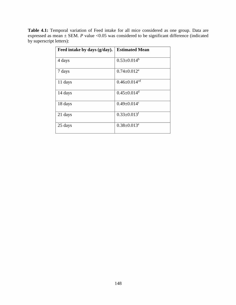
148
Table 4.1: Temporal variation of Feed intake for all mice considered as one group. Data are
expressed as mean ± SEM. P value <0.05 was considered to be significant difference (indicated
by superscript letters):
Feed intake by days (g/day). Estimated Mean
4 days 0.53±0.014b
7 days 0.74±0.012a
11 days 0.46±0.014cd
14 days 0.45±0.014d
18 days 0.49±0.014c
21 days 0.33±0.013f
25 days 0.38±0.013e
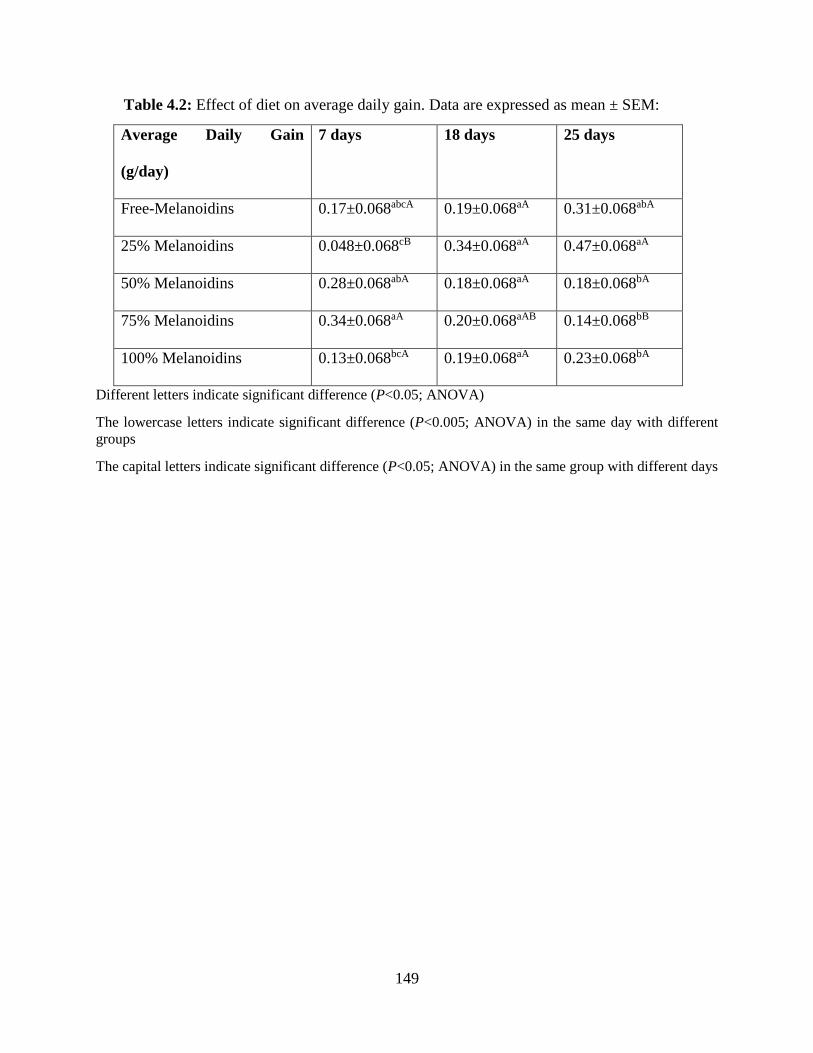
149
Table 4.2: Effect of diet on average daily gain. Data are expressed as mean ± SEM:
Average Daily Gain
(g/day)
7 days 18 days 25 days
Free-Melanoidins 0.17±0.068abcA 0.19±0.068aA 0.31±0.068abA
25% Melanoidins 0.048±0.068cB 0.34±0.068aA 0.47±0.068aA
50% Melanoidins 0.28±0.068abA 0.18±0.068aA 0.18±0.068bA
75% Melanoidins 0.34±0.068aA 0.20±0.068aAB 0.14±0.068bB
100% Melanoidins 0.13±0.068bcA 0.19±0.068aA 0.23±0.068bA
Different letters indicate significant difference (P<0.05; ANOVA)
The lowercase letters indicate significant difference (P<0.005; ANOVA) in the same day with different
groups
The capital letters indicate significant difference (P<0.05; ANOVA) in the same group with different days
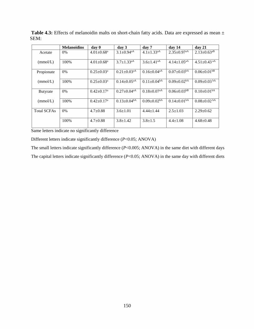
150
Table 4.3: Effects of melanoidin malts on short-chain fatty acids. Data are expressed as mean ±
SEM:
Melanoidins day 0 day 3 day 7 day 14 day 21
Acetate
(mmol/L)
0% 4.01±0.68a 3.1±0.94aA 4.1±1.33aA 2.35±0.97aA 2.13±0.63aB
100% 4.01±0.68a 3.7±1.33aA 3.6±1.41aA 4.14±1.05aA 4.51±0.43 aA
Propionate
(mmol/L)
0% 0.25±0.03a 0.21±0.03aA 0.16±0.04aA 0.07±0.03bA 0.06±0.01bB
100% 0.25±0.03a 0.14±0.05aA 0.11±0.04bA 0.09±0.02bA 0.09±0.03 bA
Butyrate
(mmol/L)
0% 0.42±0.17a 0.27±0.04aA 0.18±0.07aA 0.06±0.03bB 0.10±0.01bA
100% 0.42±0.17a 0.13±0.04bA 0.09±0.02bA 0.14±0.01bA 0.08±0.02 bA
Total SCFAs 0% 4.7±0.88 3.6±1.01 4.44±1.44 2.5±1.03 2.29±0.62
100% 4.7±0.88 3.8±1.42 3.8±1.5 4.4±1.08 4.68±0.48
Same letters indicate no significantly difference
Different letters indicate significantly difference (P<0.05; ANOVA)
The small letters indicate significantly difference (P<0.005; ANOVA) in the same diet with different days
The capital letters indicate significantly difference (P<0.05; ANOVA) in the same day with different diets

151

152
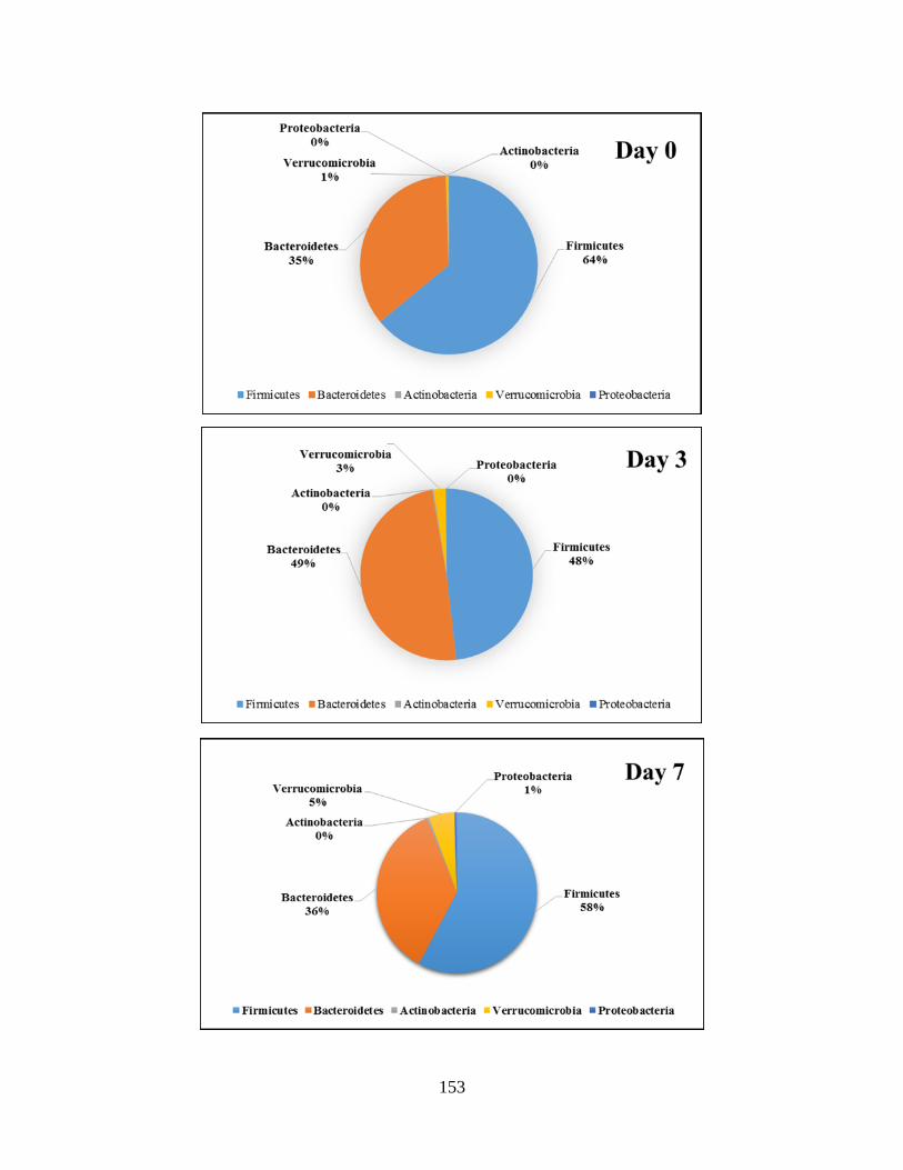
153
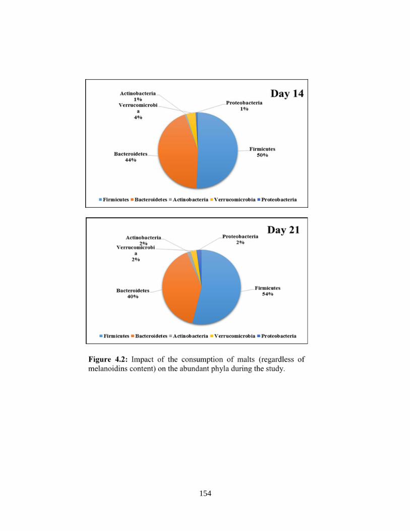
154
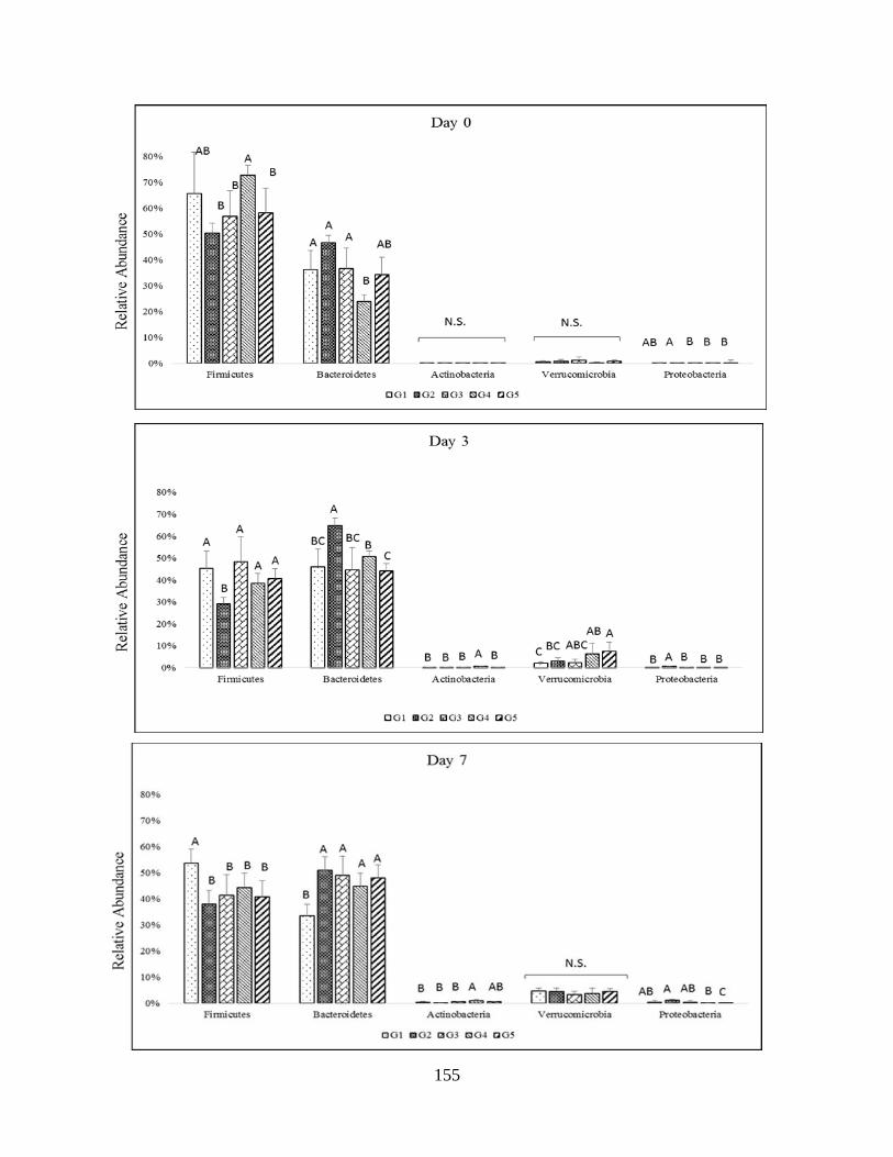
155
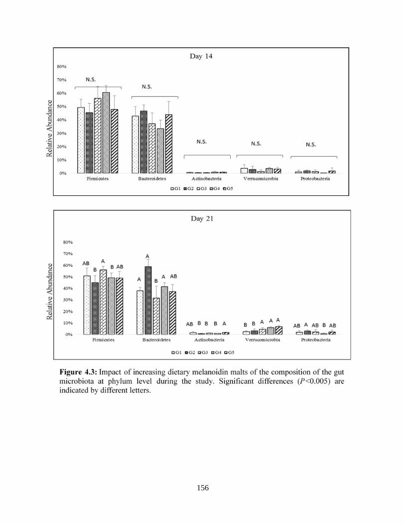
156
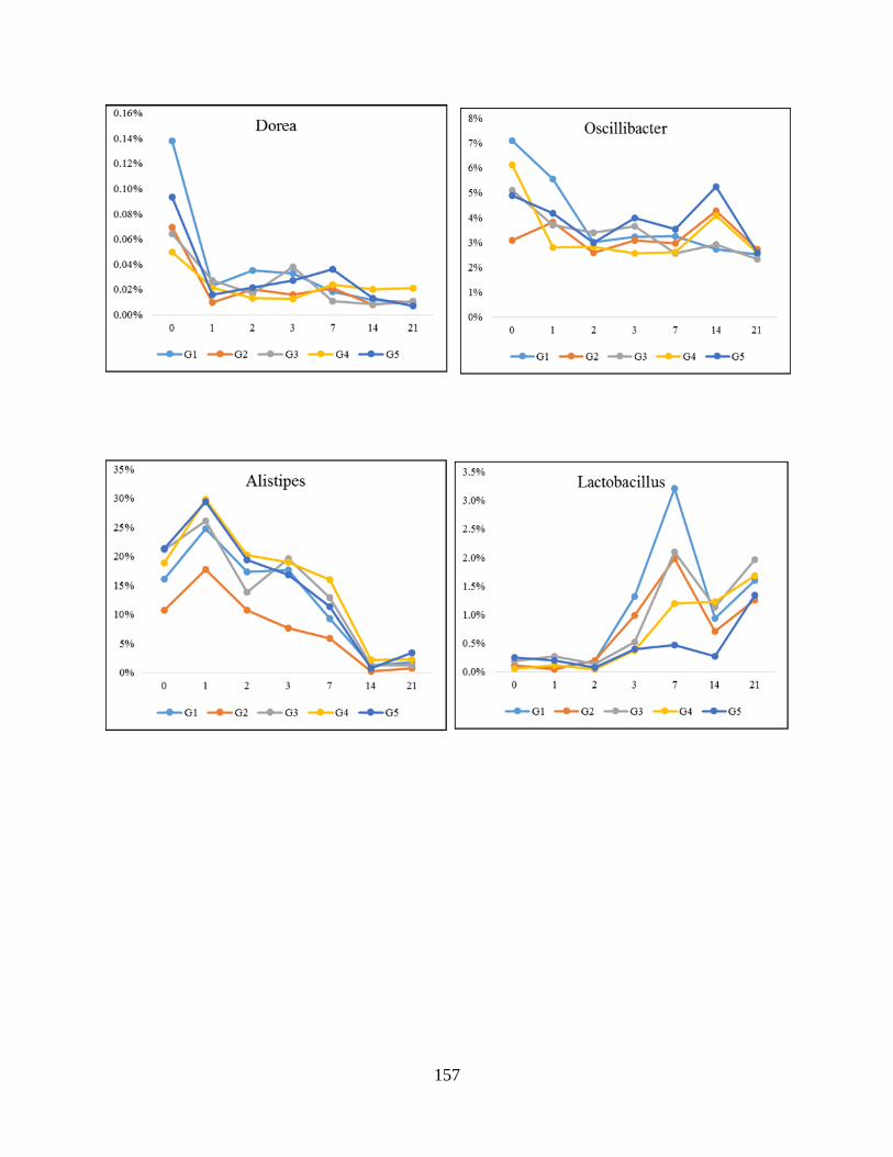
157
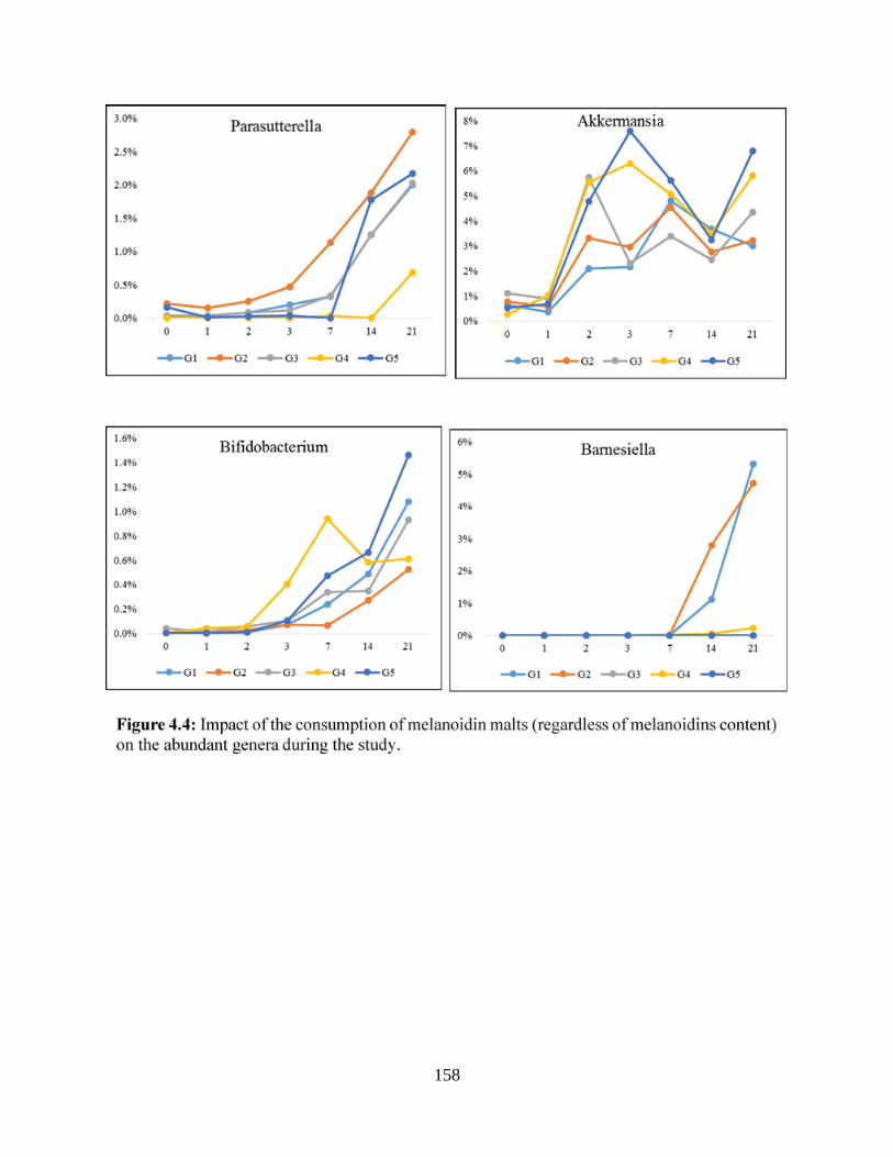
158

159
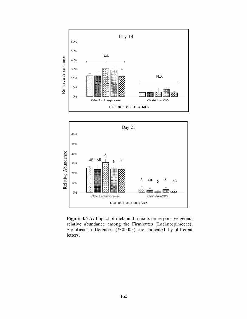
160
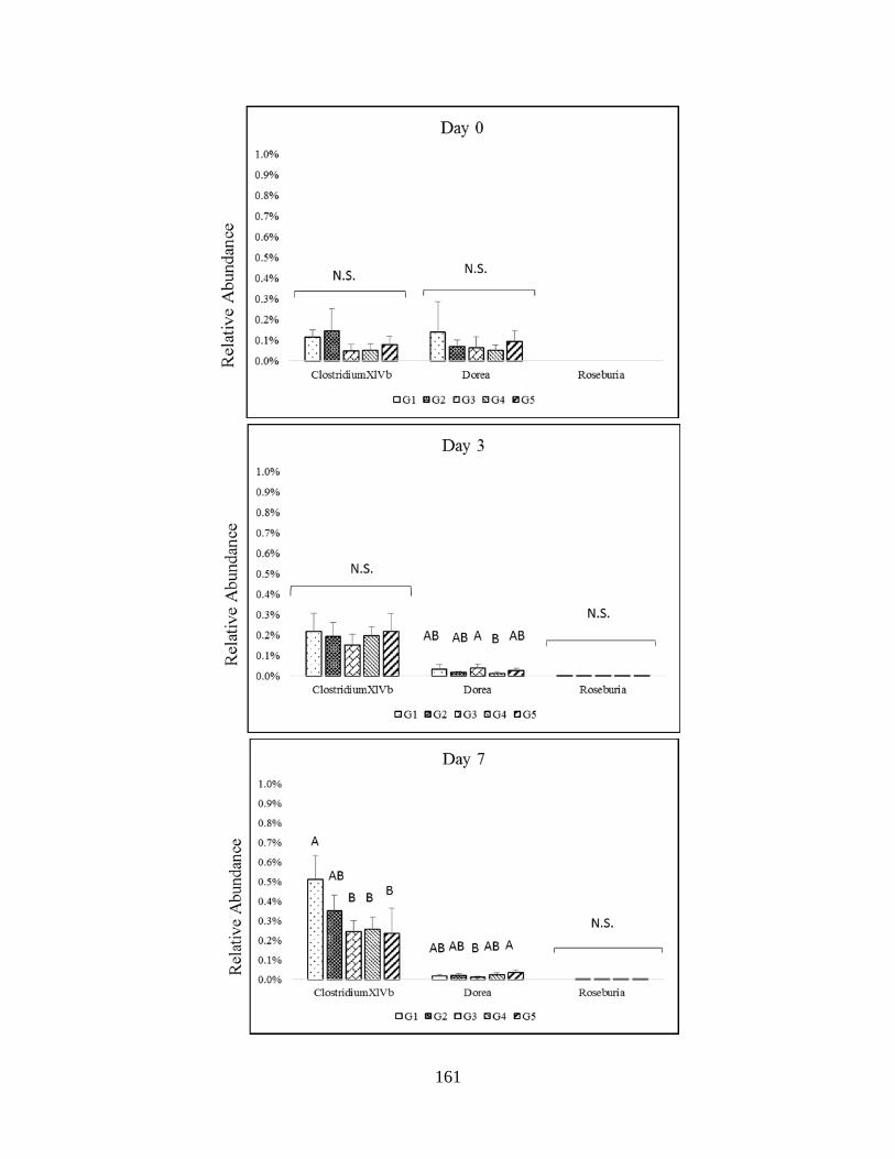
161
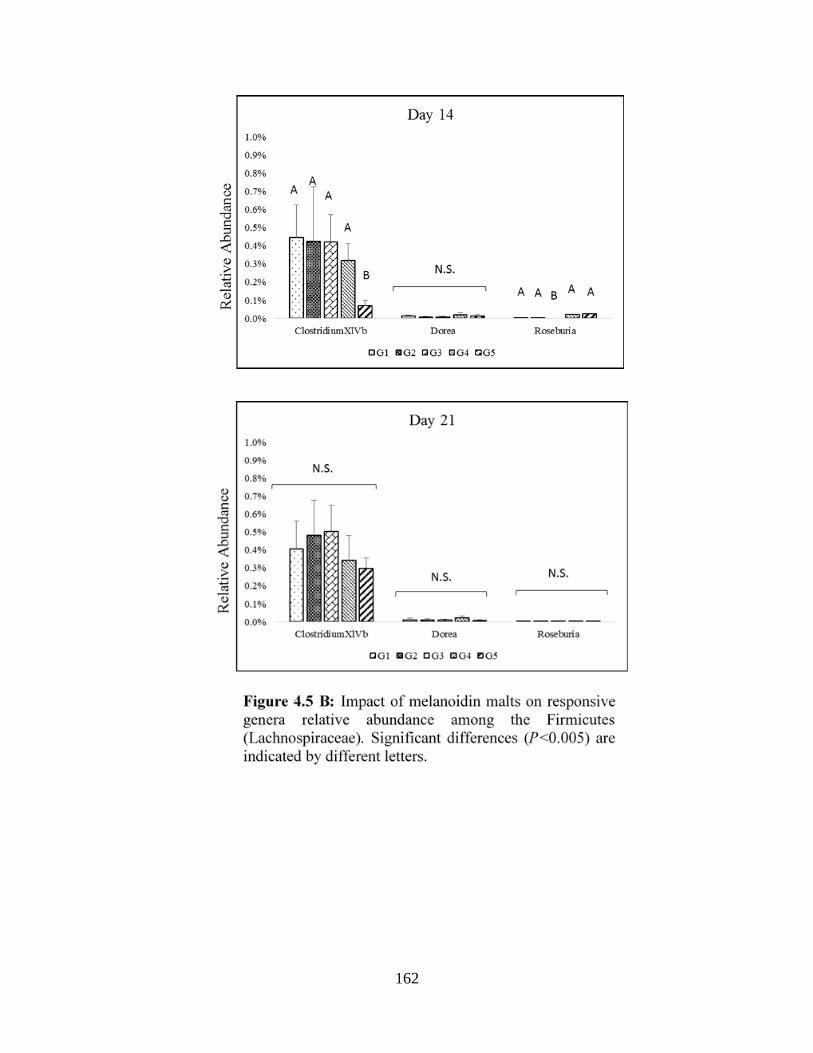
162
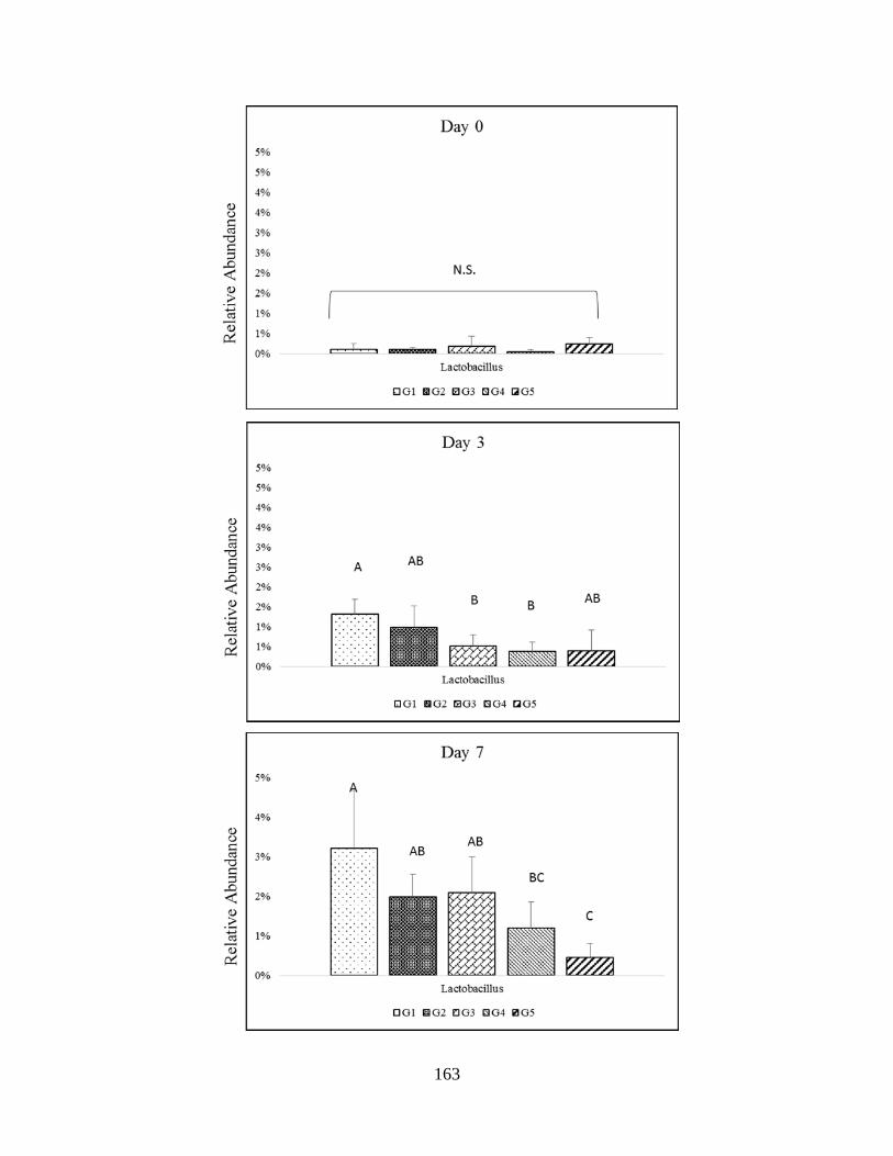
163
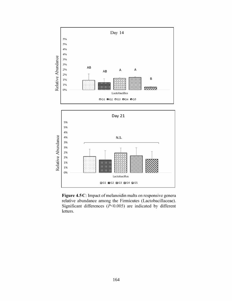
164

165
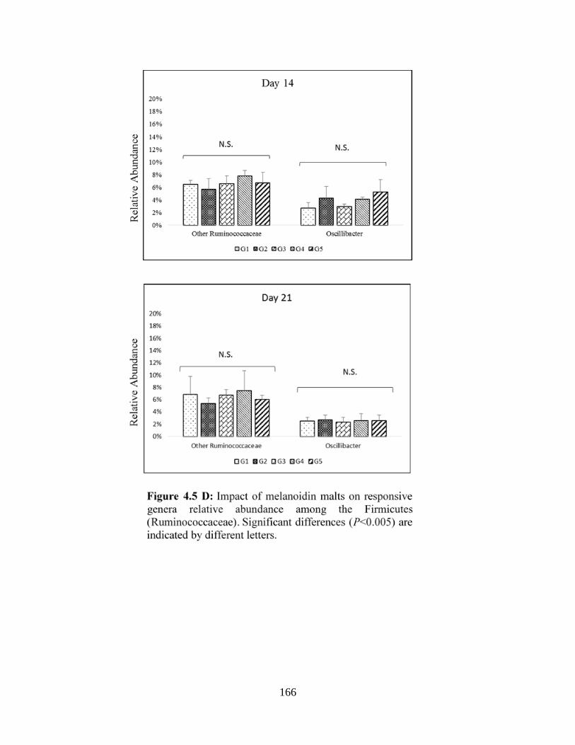
166
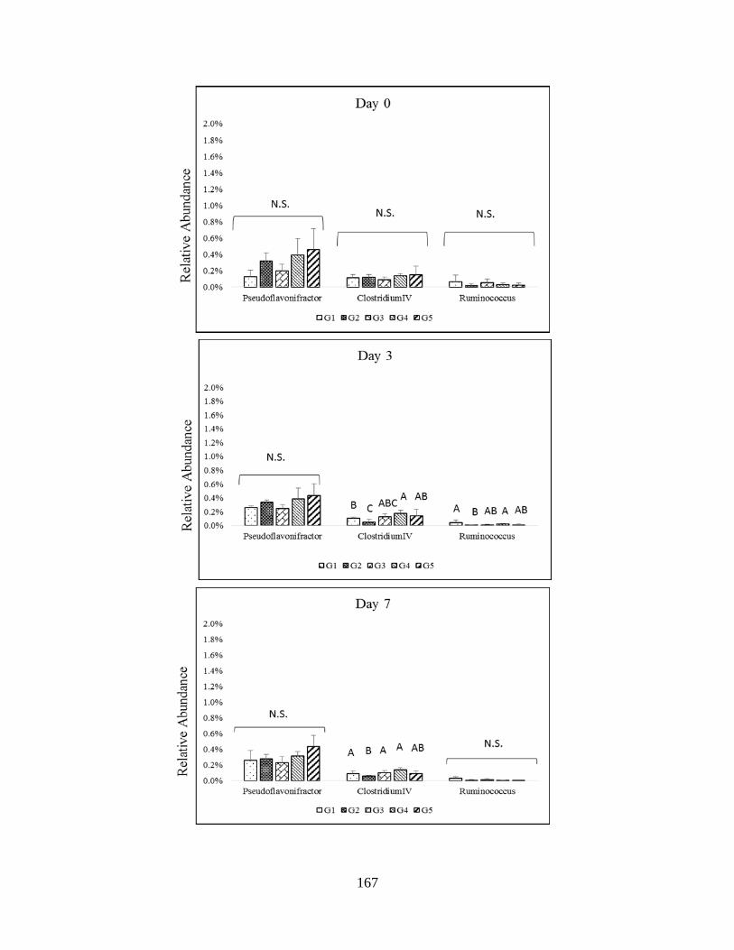
167

168
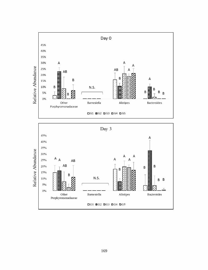
169
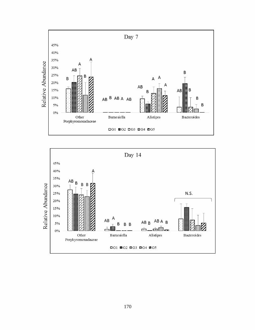
170
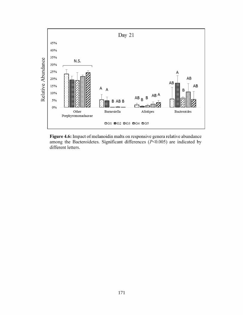
171

172
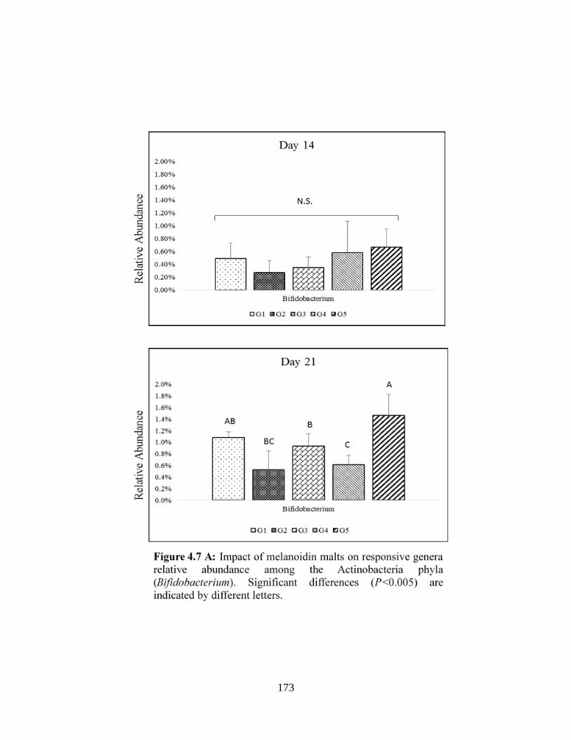
173
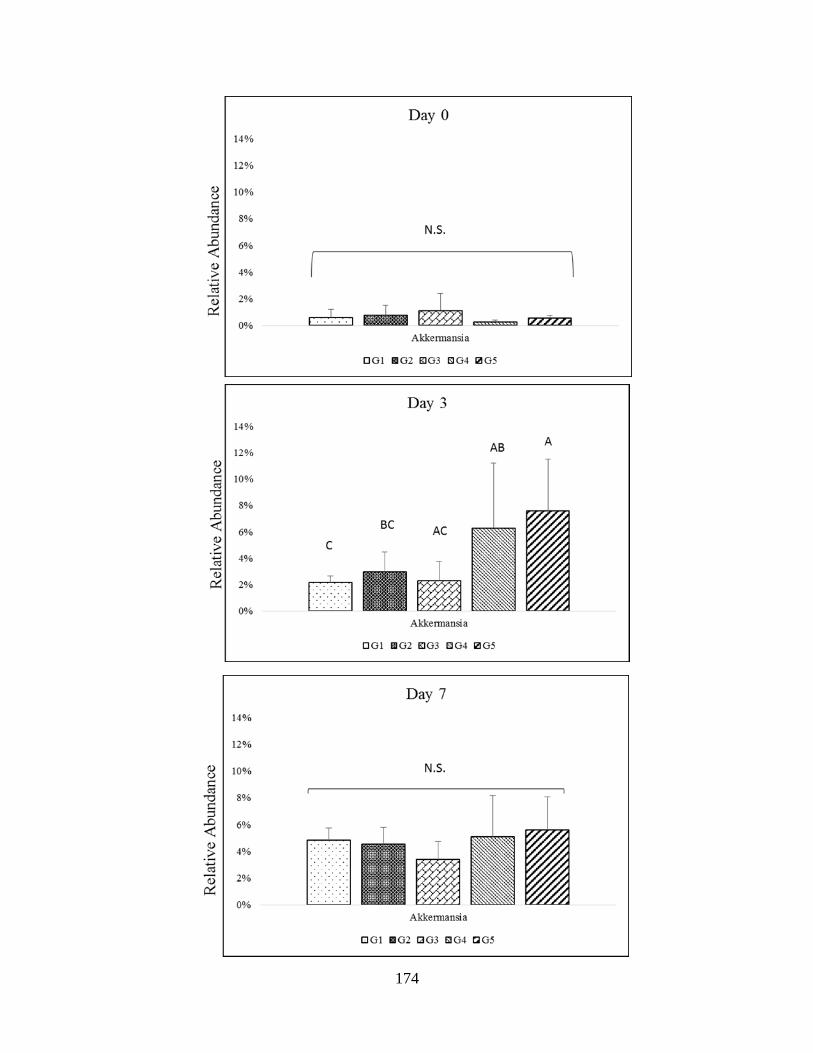
174

175
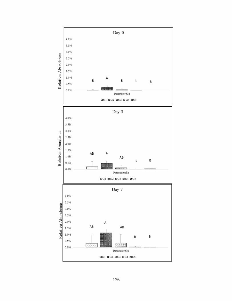
176
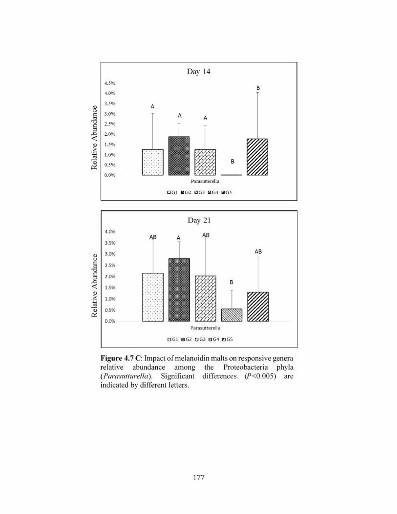
177
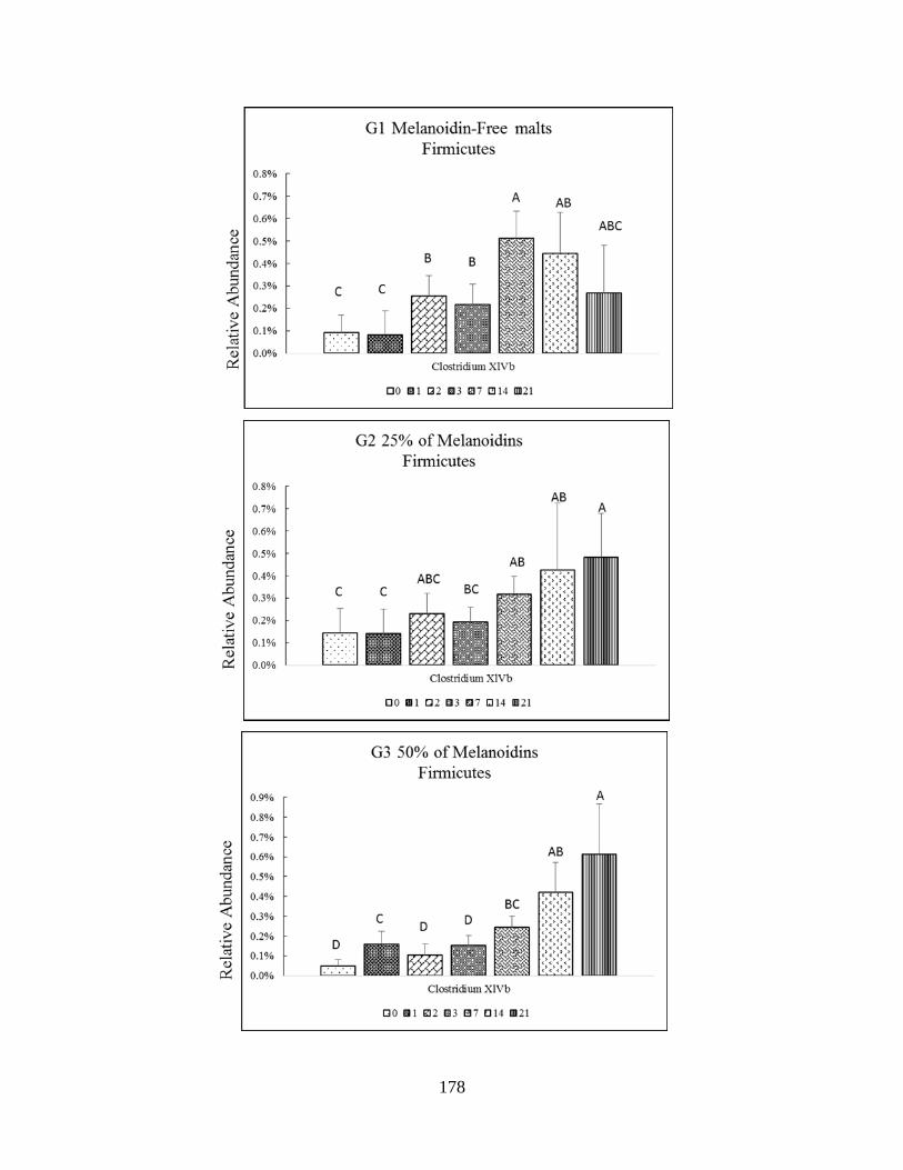
178
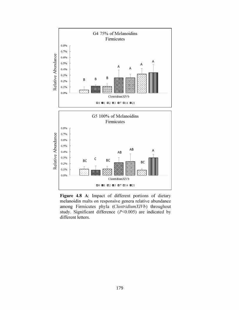
179

180
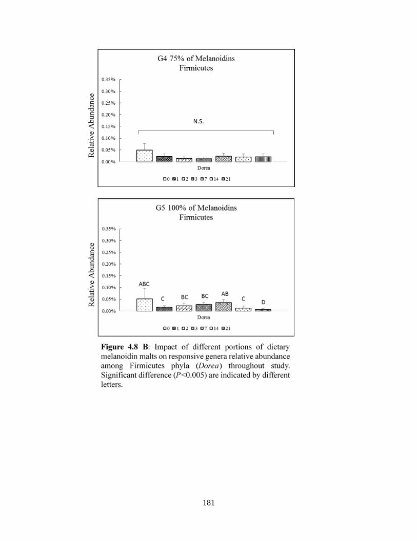
181
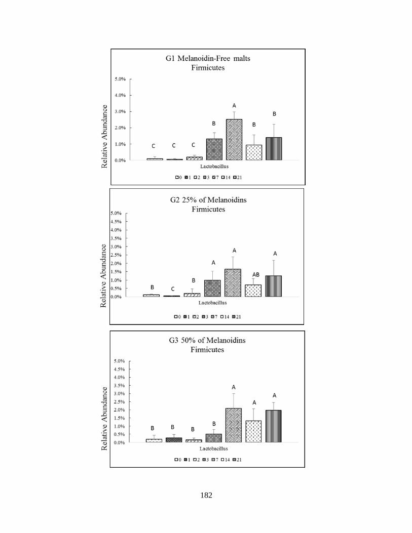
182

183
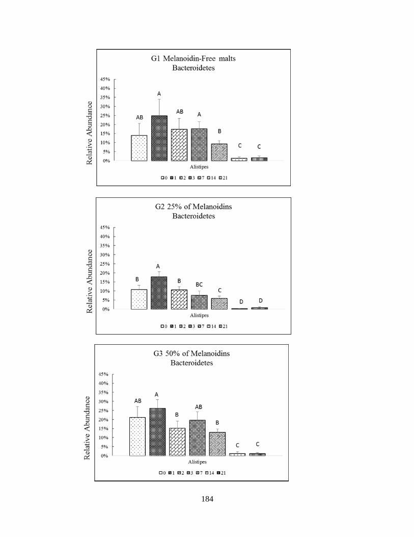
184
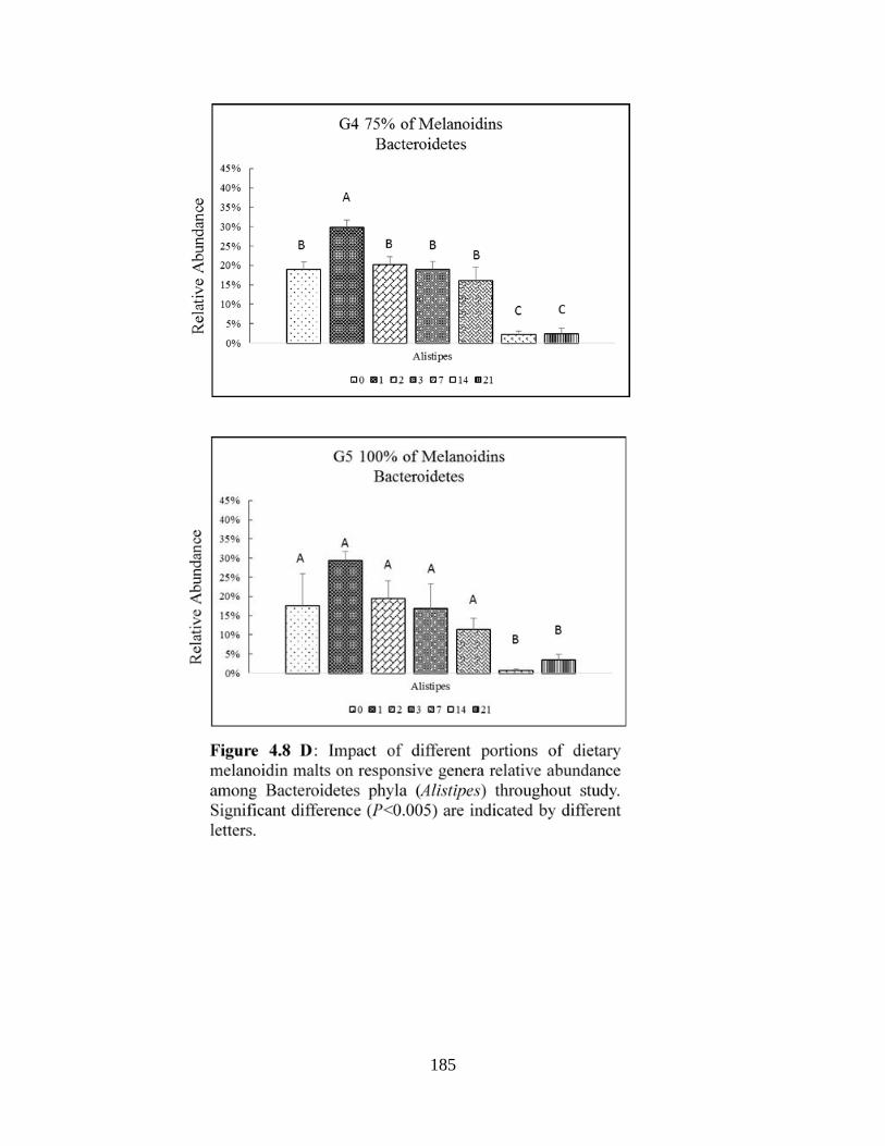
185

186

187
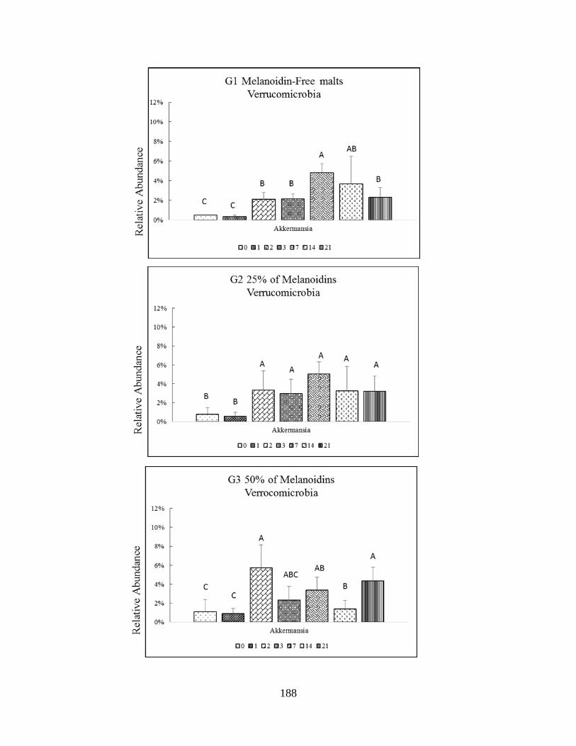
188

189
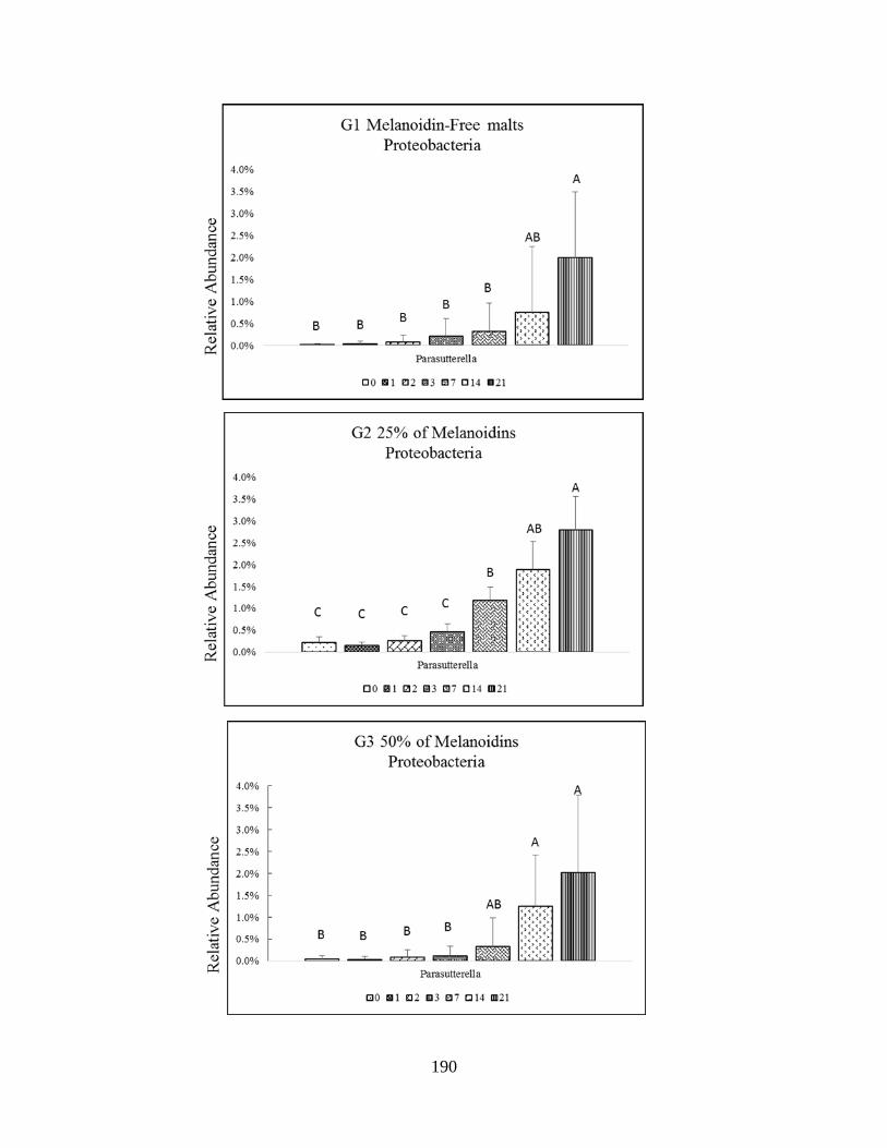
190
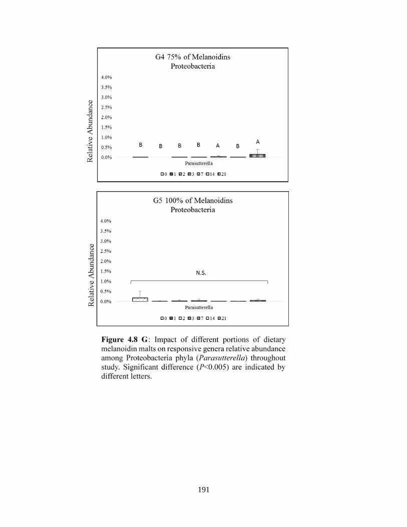
191
192
GENERAL CONCLUSION:
Gut microbiota plays an important role in maintaining human health, providing energy for
hosts, and developing the immune system. Considerable attention has been dedicated to the study
of the impact diet on gut microorganisms of humans and rodents. However, Western diets gained
more attentions due to the changing composition of gut microbiota. Data from food sciences
indicated that Western diets are rich in MRPs. Up to now, analyzing data on the actual impact of
dietary MRPs on intestinal microbiota was scarce. While recent studies have provided robust
evidence on the influence MRPs on gut bacteria in vitro studies, there are still a lot of gap in the
current knowledge related to the effect dietary MRPs on gut microbiota in vivo studies. In this
project, we assessed the impact of different MRPs (CML, bread melanoidins, and malt
melanoidins) on the murine gut microbiota through three studies.
We concluded that the composition of gut microbiota of CML group was similar to the
composition of gastrointestinal microbiota control group. Additionally, CML inhibited dysbiosis
gut microbiota in DSS not TNBS models. This project showed that bread melanoidins have no
bifidogenic effect, while malt melanoidins have bifidogenic effect. Both bread and malt
melanoidins have no impact to increase Lactobacillus. Melanoidins appeared to have a prebiotic-
like impact for the malts and inhibition of potentially pathogenic Proteobacteria for bread crust.
Thus, melanoidins structure are extremely variable from one food to another, therefore additional
studies will be needed to better assess their potential beneficial properties on gut health. Finally,
we found that CML and melanoidins have no negative effects on the gut microbiota.
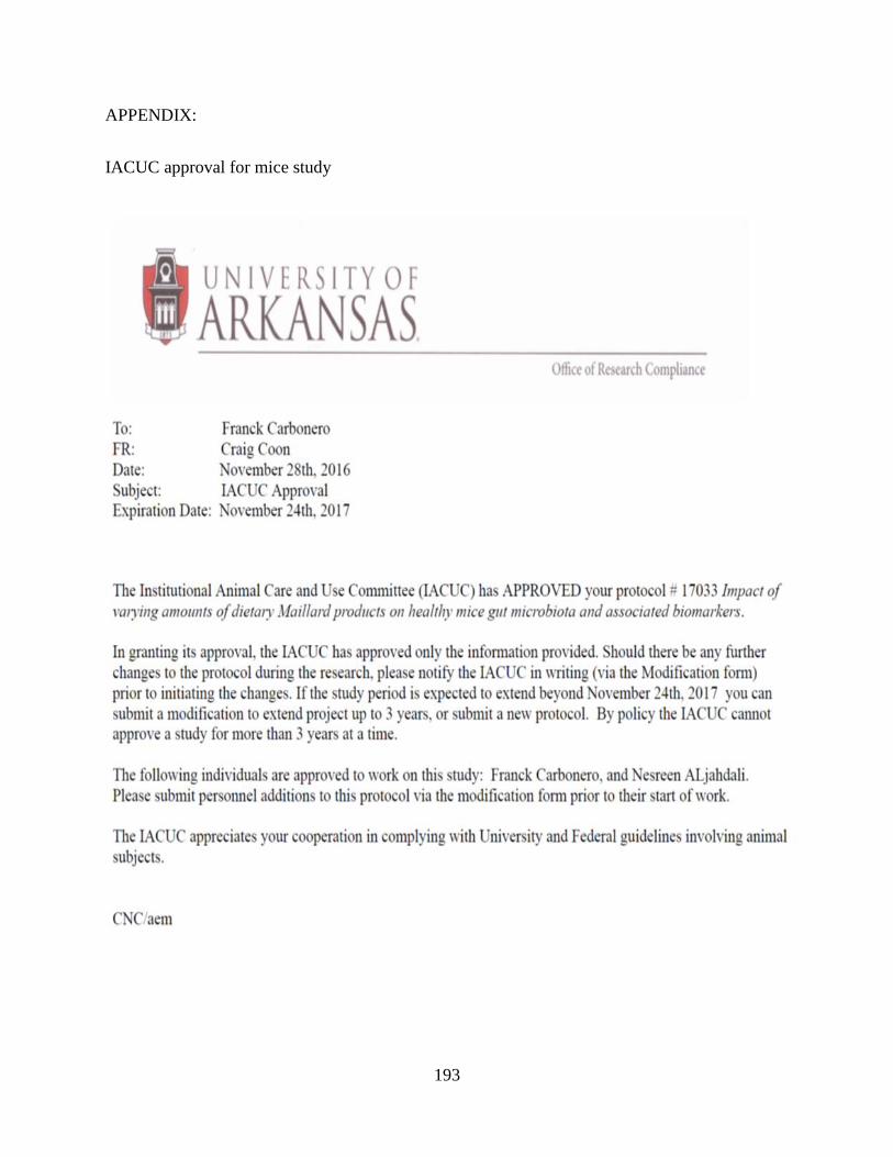
193
APPENDIX:
IACUC approval for mice study