DETECTING DISEASED IMAGES BY USING SEMI â•fi ...
7
International Journal of Computer and Communication International Journal of Computer and Communication Technology Technology Volume 6 Issue 3 Article 12 July 2015 DETECTING DISEASED IMAGES BY USING SEMI – SUPERVISED DETECTING DISEASED IMAGES BY USING SEMI – SUPERVISED LEARNING LEARNING DINESH KUMAR A Department of Computer Science and Engineering, Karpagam University, Coimbatore, Tamil Nadu, INDIA, [email protected] SHAHUL HAMMED Department of Computer Science and Engineering, Karpagam University, Coimbatore, Tamil Nadu, INDIA, [email protected] HANAH AYISHA V HYDER ALI Department of Computer Science and Engineering, Karpagam University, Coimbatore, Tamil Nadu, INDIA, [email protected] NIDHYA R Department of Information Technology, Anna University Chennai, Regional Office: Coimbatore, Tamil Nadu, INDIA, [email protected] Follow this and additional works at: https://www.interscience.in/ijcct Recommended Citation Recommended Citation A, DINESH KUMAR; HAMMED, SHAHUL; HYDER ALI, HANAH AYISHA V; and R, NIDHYA (2015) "DETECTING DISEASED IMAGES BY USING SEMI – SUPERVISED LEARNING," International Journal of Computer and Communication Technology: Vol. 6 : Iss. 3 , Article 12. DOI: 10.47893/IJCCT.2015.1305 Available at: https://www.interscience.in/ijcct/vol6/iss3/12 This Article is brought to you for free and open access by the Interscience Journals at Interscience Research Network. It has been accepted for inclusion in International Journal of Computer and Communication Technology by an authorized editor of Interscience Research Network. For more information, please contact [email protected].
-
Upload
khangminh22 -
Category
Documents
-
view
2 -
download
0
Transcript of DETECTING DISEASED IMAGES BY USING SEMI â•fi ...
International Journal of Computer and Communication International Journal of Computer and Communication
Technology Technology
Volume 6 Issue 3 Article 12
July 2015
DETECTING DISEASED IMAGES BY USING SEMI – SUPERVISED DETECTING DISEASED IMAGES BY USING SEMI – SUPERVISED
LEARNING LEARNING
DINESH KUMAR A Department of Computer Science and Engineering, Karpagam University, Coimbatore, Tamil Nadu, INDIA, [email protected]
SHAHUL HAMMED Department of Computer Science and Engineering, Karpagam University, Coimbatore, Tamil Nadu, INDIA, [email protected]
HANAH AYISHA V HYDER ALI Department of Computer Science and Engineering, Karpagam University, Coimbatore, Tamil Nadu, INDIA, [email protected]
NIDHYA R Department of Information Technology, Anna University Chennai, Regional Office: Coimbatore, Tamil Nadu, INDIA, [email protected]
Follow this and additional works at: https://www.interscience.in/ijcct
Recommended Citation Recommended Citation A, DINESH KUMAR; HAMMED, SHAHUL; HYDER ALI, HANAH AYISHA V; and R, NIDHYA (2015) "DETECTING DISEASED IMAGES BY USING SEMI – SUPERVISED LEARNING," International Journal of Computer and Communication Technology: Vol. 6 : Iss. 3 , Article 12. DOI: 10.47893/IJCCT.2015.1305 Available at: https://www.interscience.in/ijcct/vol6/iss3/12
This Article is brought to you for free and open access by the Interscience Journals at Interscience Research Network. It has been accepted for inclusion in International Journal of Computer and Communication Technology by an authorized editor of Interscience Research Network. For more information, please contact [email protected].

Detecting Diseased images by using Semi – Supervised Learning
DETECTING DISEASED IMAGES BY USING SEMI – SUPERVISED LEARNING
DINESH KUMAR A1, SHAHUL HAMMED2, HANAH AYISHA V HYDER ALI3 & NIDHYA R4
1,2,3,4Department of Computer Science and Engineering, Karpagam University, Coimbatore, Tamil Nadu, INDIA
5Department of Information Technology, Anna University Chennai, Regional Office: Coimbatore, Tamil Nadu, INDIA E-mail: [email protected], [email protected], [email protected], [email protected]
Abstract— The goal of semi-supervised image segmentation is to obtain the segmentation from a partially labeled image. By utilizing the image manifold structure in labeled and unlabeled pixels, semi-supervised methods propagates the user labeling to the unlabeled data, thus minimizing the need for user labeling. Several semi-supervised learning methods have been proposed in the literature. In this paper, we consider the delinquent of segmentation of large collections of images and the classification of images by allied diseases. We are detecting diseased images by the process of segmentation and classification. The segmentation used in this paper has two advantages. First, user can specify what they want by highly controlling the segmentation. Another is, at initial stage this model requires only minimum tuning of model parameters. Once initial tuning is done, the setup can be used to automatically segment a large collection of images that are distinct but share similar features. And for classification of diseases, a manifold learning method, called parameter-free semi-supervised local Fisher discriminant analysis is used. This method preserves the global structure of unlabeled samples in addition to separating labeled samples in different classes from each other. The semi-supervised method has an analytic form of the globally optimal solution, which can be computed efficiently by Eigen decomposition. Espousal experiments on various collections of biological images suggest that the proposed model is effective for segmentation with classification and is computationally efficient.
Keywords-Biological image segmentation; interactive; microscopy images; multiple images; dimensionality reduction; semi-supervised local Fisher discriminant analysis; parameter free; uncorrelated constraint;
I. INTRODUCTION
Image segmentation is a challenging task and
remains an open problem in image processing. Unsupervised methods explore the intrinsic data structure to segment the image into regions with different statistics. However, these methods often fail to achieve the desired result, especially if the desired segmentation includes regions with very different characteristics. On the other extreme, supervised image segmentation methods first learn a classifier from a labeled training set. Although these methods are likely to perform better, marking the training set is very time consuming. Semi-supervised image segmentation methods circumvent these problems by inferring the segmentation from partially labeled images. The key difference from supervised learning is that semi supervised methods utilize the data structure in both the labeled and unlabeled data points. Hence, the main advantage of semi-supervised image segmentation methods is that they take advantage of the user markings to direct the segmentation, while minimizing the need for user labeling. Image segmentation is used in many areas, including computer vision, computer graphics, and medical imaging, to name a few. Fully automatic image segmentation has many fundamental difficulties and is still a very hard problem. Therefore, the segmentation problem is hostile modeled if no additional knowledge about the desired segmentation is given. However, in many applications, such as cell segmentation in microscopy
images and organ segmentation in medical images, the kind of objects and segmentation of interest are known in advance. It is therefore enticing to design segmentation methods that allow the user to specify what they want. In semiautomatic and interactive image segmentation, the user marks some sample pixels from each class of objects. The computational algorithm then computes a classification of other pixels. This way, the resulting segmentation is highly controllable by the user and thereby eliminates much ambiguity in defining a partition. Because of their anticipated properties, there have been increasing activities in the research community to develop interactive semiautomatic image segmentation algorithm.
II. MATERIALS AND METHODS
A. For Segmentation
In this section, we present the formulation of the proposed model. For ease of presentation, we illustrate the two-image multiple-class case. This two-image model can be used to segment a collection of images one at a time. The generalization to multiple-image multiple-class case is clear. To improve the performance, I consider the algorithm BBx- index. BBx-index keeps multiple tree versions. Each tree entry has the form< x rep; tstart; tend; pointer >, where x rep is the transformed 1D object location value using an SFC, tstart and tend are the insert and update times of the object, respectively. Each tree corresponds to a timestamp signature being the end
206International Journal of Computer and Communication Technology (IJCCT), ISSN: 2231-0371, Vol-6, Iss-3
Detecting Diseased images by using Semi – Supervised Learning
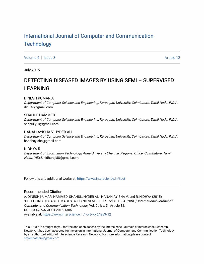
timestamp of a phase when the tree is built and a lifespan being the minimum start time and the maximum end time of all the entries in that tree. Unlike the Bx-tree that concatenates the timestamp signature and the 1D transformed value, the BBx-index maintains a separate tree for each timestamp signature, and models the moving objects from the past to the future. Insertion is the same as in the Bx-tree. Instead of deleting an object, update sets the end time of the object to the current time, followed by an insertion of the updated object into the latest tree. For automated segmentation of the vasculature in retinal images. The method produces segmentations by classifying each image pixel as vessel or nonvessel, based on the pixel’s feature vector. Feature vectors are composed of the pixel’s intensity and continuous two-dimensional Morlet wavelet transform responses taken at multiple scales. The Morlet wavelet is capable of tuning to specific frequencies, thus allowing noise filtering and vessel enhancement in a single step.
ALGORITHM:
Step 1: Calculate X1,1(autonomous of ג and µ). Step 2: Calculate Y1,1 (reliant on ג) Step 3: Resolve the linear system
Val – STΩ1 SΩ1Y1,1 a1|n
Where, n = 1,2,…,N-1.
Step 4: Calculate X2,2 and X2,1 (Autonomous of ג and µ)
Step 5: Calculate Y2,1 and Y2,2(reliant on ג and µ) Step 6: Resolve the linear system
Val – STΩ2 SΩ2Y2,2a2|n = ST
Ω2 SΩ2Y2,1 a1|n
Where n = 1,2,…,N-1. Algorithm 1: Algorithm for Segmentation We use a Bayesian classifier with class-
conditional probability density functions (likelihoods) described as Gaussian mixtures, yielding a fast classification, while being able to model complex decision surfaces and compare its performance with the linear minimum squared error classifier. The probability distributions are estimated based on a training set of labeled pixels obtained from manual segmentations. B. For Classification
Fisher Discriminant Analysis (FDA) is a powerful and popular method for dimensionality reduction and classification which has unfortunately poor performances in the cases of label noise and sparse labeled data. To overcome these limitations, we propose a probabilistic framework for FDA and
extend it to the semi-supervised case. Experiments on real world datasets show that the proposed approach works as well as FDA in standard situations and outperforms it in the label noise and sparse label cases.
In Image Classification, dimension reduction methods have been widely employed in this field. The goal of dimensionality reduction is to reduce complexity of input data while some desired intrinsic information of the data is preserved. Manifold learning is a perfect tool for data mining that discovers the structure of high-dimensional data sets and provides better understanding of the data. The representative methods include locally linear embedding (LLE), isometric mapping (Isomap), Laplacian eigenmaps, and local tangent space alignment (LTSA), etc. These nonlinear methods are defined only on training data, and the issue of how to map new test data remains difficult. Therefore, they cannot be applied directly to recognition problems. Recently, some algorithms resolve this difficulty by finding a mapping on the whole data space, not just on training data; for instance, locality preserving projection (LPP) and neighborhood preserving embedding (NPE). As these methods are originally unsupervised, some discriminative manifold learning methods have been proposed recently to exploit both geometrical and discriminant information for dimensionality reduction, resulting in better performance for classification. However, in the real world, the labeled examples, especially Image data examples are often very difficult and expensive to obtain. The traditional supervised methods cannot work well when lack of training examples; in contrast, unlabeled examples can be easily obtained. Therefore, in such situations, it can be beneficial to incorporate the information which is contained in unlabeled examples into a learning problem, i.e., semi-supervised learning (SSL) should be applied instead of supervised learning. Semi-supervised local Fisher discriminant analysis (SELF) is a recently proposed semi-supervised manifold learning method to exploit both geometrical and discriminant information simultaneously for dimension reduction, which preserves the global structure of unlabeled samples in addition to separating labeled samples in different classes from each other. It has demonstrated better performance than some manifold learning methods such as locality preserving projection and local Fisher discriminant analysis (LFDA) for classification. The existing SELF method, however, needs one additional prespecified parameter to balance the optimization objective, which is data dependent and very difficult to optimize for different applications. Moreover, features extracted by SELF are mutually correlated and some redundant information may be contained. To address the two shortcomings, we propose, in this paper, a parameter-free semi-supervised local Fisher discriminant analysis (pSELF) method by designing a new
207International Journal of Computer and Communication Technology (IJCCT), ISSN: 2231-0371, Vol-6, Iss-3
Detecting Diseased images by using Semi – Supervised Learning
difference-based optimization objective function with uncorrelated constraint for Image data classification. Experimental results demonstrate that the proposed method is efficient for discriminant dimensionality reduction and Image data classification.
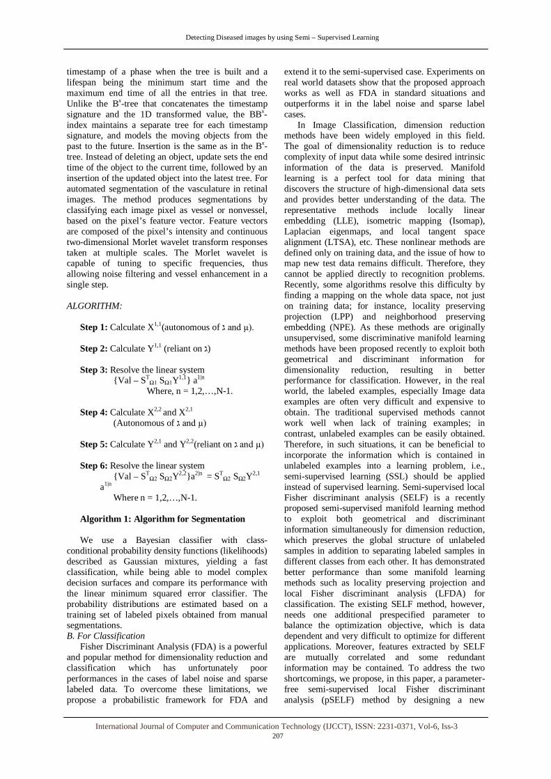
A manifold learning method, called parameter-free semi-supervised local Fisher discriminant analysis (pSELF), is proposed to map the gene expression data into a low-dimensional space for tumor classification. Motivated by the fact that semi supervised and parameter-free are two desirable and promising characteristics for dimension reduction, a new difference-based optimization objective function with unlabeled samples has been designed.
INPUT:
1. Working out samples (x1,k1),…,(xp,kp),xp+1,…,xi, xp ∈ Rj. 2. Dimensionality of embedding space f(1≤ f ≤ j).
OUTPUT: 1. The optimal transformation matrix Q ∈ Rj x f.
2. The f-dimensional embedding coordinates Y for the input points X.
ALGORITHM:
Step 1: Compute the local between-class weight matrix Wkb and the local within-class weight matrix Wkw. Step 2: Construct the local between-class scatter matrix Skb and the local within-class scatter matrix Skw and the total scatter matrix Sg Step 3: Calculate the eigenvectors corresponding to the largest eigenvalues of (Skb - Skw )q = גSqv and use them to form the optimal projection vector. Step 4: The coordinates of the j points in the f-dimensional embedding space are given by the column vectors of Y= QGX.
Algorithm 2: Algorithm for Classification
The proposed method preserves the global
structure of unlabeled samples in addition to separating labeled samples in different classes from each other. The semi-supervised method has an analytic form of the globally optimal solution, which can be computed efficiently by Eigen decomposition.
III. RESULTS AND DISCUSSION In this section, we evaluate the performance of the
proposed multiple-image segmentation model empirically using three image data sets. All algorithms are implemented in MATLAB 2010 with a Core i7 3.07-GHz machine.
A. Image Data Sets In the first experiment, a set of 58 breast cancer
cell images of various sizes in is used. Overall, these images have similar features. However, some contain benign cells, whereas some contain both benign and malignant cells, so that there are some variations among them. A manual segmentation of the cell nuclei which is used as the ground truth to evaluate the accuracy of our method. In the second experiment, a set of 20 retinal images of size 292x283, which is called DRIVE, is used. Some images show some sign of early diabetic retinopathy, whereas others do not in the third experiment, a set of 30 cross sections of a human retina with size 256x384 in is used. Color distortion due to staining and shape variation of the layers makes the segmentation task very challenging.
Method: The 20 simulated images are segmented one at a time using the proposed multiple-image model, with the image in a fully labeled sample.
Results: We observe that the proposed method is able to classify much of the alien object as retina, and the accuracy is not affected at all. This is mainly due to the high color contrast between the yellowish alien object and the blood vessels and due to the presence of the yellowish optic disc on the right in the training data. Observe that the accuracy drops between Image 4 and Image 5. The decrease in the accuracy is mainly due to the misclassification of pixels in the reddish circular disc. For the first four images, the proposed method is able to classify much of the alien object as retina. It shows that the proposed method has a certain degree of tolerance. However, when the intensity of the alien object becomes too close to that of the blood vessel pixels (i.e., Image 5 to Image 10), the object is classified as blood vessel by the properties of the proposed model. B. Testing Method
For each experiment, one image is served as the labeled image, and the remaining images are segmented using the proposed semisupervised multiple-image model (MI). For the third experiment, only 50% of the labels are used for computing P2,1 (i.e., |S| = 0.5M min1 <= m<=M | 1|m|).
The parameters ג and µ for the similarity measures are calibrated using one labeled image and one unlabeled image. They are then used throughout the whole image data set. Before segmenting the images, a color transfer is applied to each unlabeled image so that its color histogram matches that of the labeled image. This is to correct the difference in brightness and contrast between images. To validate our method, we compare it with four other methods, namely, the classical support vector machine (SVM), the -nearest neighbor (KNN), the –means (KMeans), and the semisupervised -means (SSKMeans). The first two methods are supervised methods; the third is unsupervised; the fourth is semisupervised. For the supervised and semisupervised methods, the labeled pixels are used as the training set. The accuracy,
208
International Journal of Computer and Communication Technology (IJCCT), ISSN: 2231-0371, Vol-6, Iss-3
Detecting Diseased images by using Semi – Supervised Learning
which is computed as the number of correctly classified pixels divided by the total number of pixels, and the -measure, which is computed as the harmonic mean of precision and recall for the foreground object, i.e.,
2 precision x recall
F = 2 precision + recall
are used as a measure of segmentation accuracy.
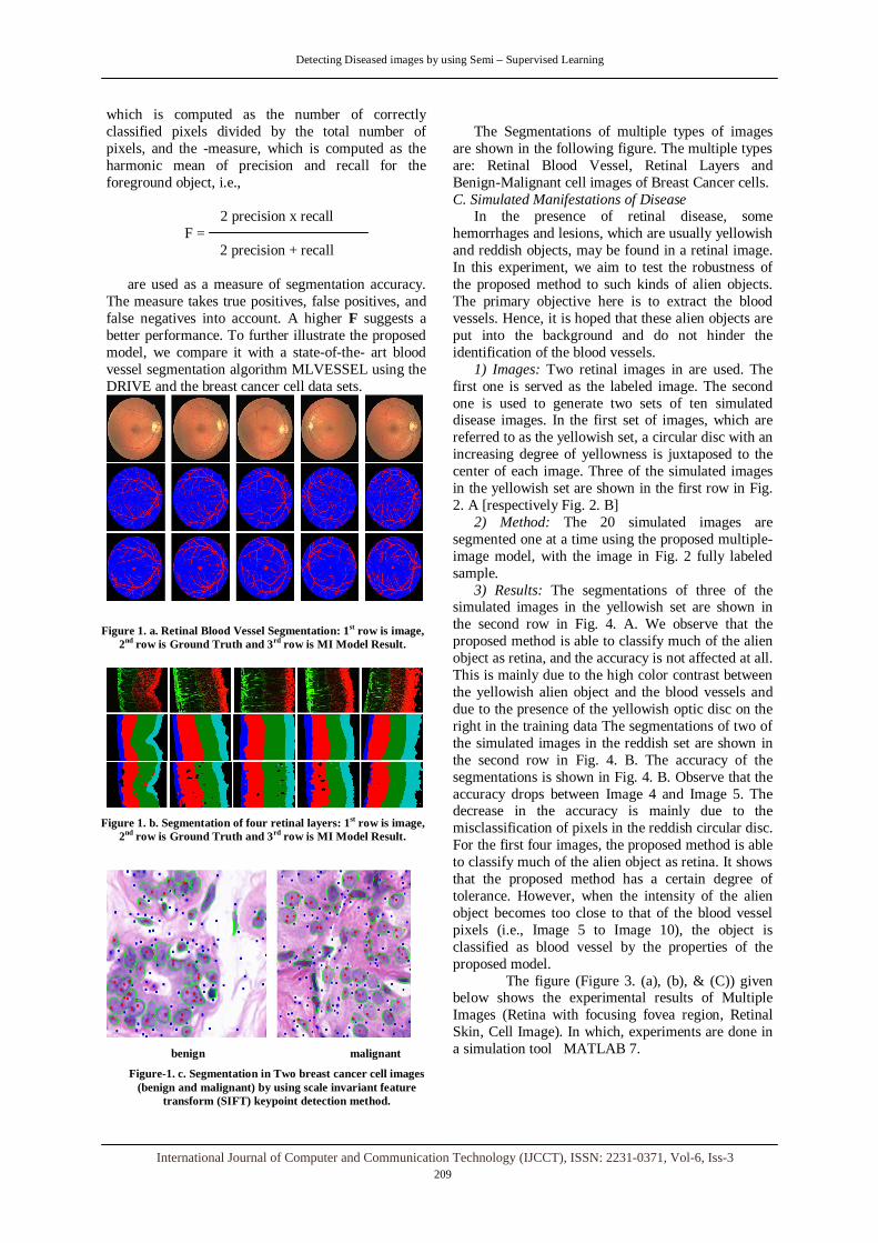
The measure takes true positives, false positives, and false negatives into account. A higher F suggests a better performance. To further illustrate the proposed model, we compare it with a state-of-the- art blood vessel segmentation algorithm MLVESSEL using the DRIVE and the breast cancer cell data sets.
Figure 1. a. Retinal Blood Vessel Segmentation: 1st row is image, 2nd row is Ground Truth and 3rd row is MI Model Result.
Figure 1. b. Segmentation of four retinal layers: 1st row is image, 2nd row is Ground Truth and 3rd row is MI Model Result.
benign malignant
Figure-1. c. Segmentation in Two breast cancer cell images (benign and malignant) by using scale invariant feature
transform (SIFT) keypoint detection method.
The Segmentations of multiple types of images
are shown in the following figure. The multiple types are: Retinal Blood Vessel, Retinal Layers and Benign-Malignant cell images of Breast Cancer cells. C. Simulated Manifestations of Disease
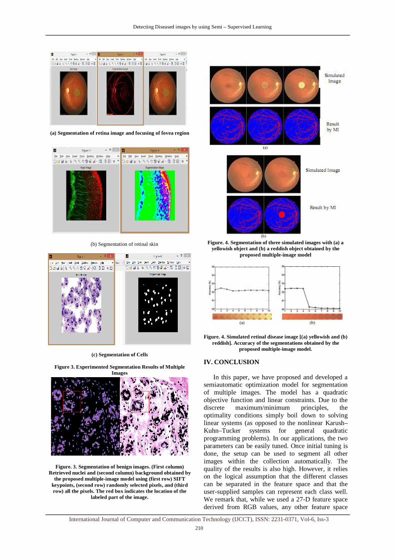
In the presence of retinal disease, some hemorrhages and lesions, which are usually yellowish and reddish objects, may be found in a retinal image. In this experiment, we aim to test the robustness of the proposed method to such kinds of alien objects. The primary objective here is to extract the blood vessels. Hence, it is hoped that these alien objects are put into the background and do not hinder the identification of the blood vessels.
1) Images: Two retinal images in are used. The first one is served as the labeled image. The second one is used to generate two sets of ten simulated disease images. In the first set of images, which are referred to as the yellowish set, a circular disc with an increasing degree of yellowness is juxtaposed to the center of each image. Three of the simulated images in the yellowish set are shown in the first row in Fig. 2. A [respectively Fig. 2. B]
2) Method: The 20 simulated images are segmented one at a time using the proposed multiple-image model, with the image in Fig. 2 fully labeled sample.
3) Results: The segmentations of three of the simulated images in the yellowish set are shown in the second row in Fig. 4. A. We observe that the proposed method is able to classify much of the alien object as retina, and the accuracy is not affected at all. This is mainly due to the high color contrast between the yellowish alien object and the blood vessels and due to the presence of the yellowish optic disc on the right in the training data The segmentations of two of the simulated images in the reddish set are shown in the second row in Fig. 4. B. The accuracy of the segmentations is shown in Fig. 4. B. Observe that the accuracy drops between Image 4 and Image 5. The decrease in the accuracy is mainly due to the misclassification of pixels in the reddish circular disc. For the first four images, the proposed method is able to classify much of the alien object as retina. It shows that the proposed method has a certain degree of tolerance. However, when the intensity of the alien object becomes too close to that of the blood vessel pixels (i.e., Image 5 to Image 10), the object is classified as blood vessel by the properties of the proposed model.
The figure (Figure 3. (a), (b), & (C)) given below shows the experimental results of Multiple Images (Retina with focusing fovea region, Retinal Skin, Cell Image). In which, experiments are done in a simulation tool MATLAB 7.
209International Journal of Computer and Communication Technology (IJCCT), ISSN: 2231-0371, Vol-6, Iss-3
Detecting Diseased images by using Semi – Supervised Learning
(a) Segmentation of retina image and focusing of fovea region
(b) Segmentation of retinal skin
(c) Segmentation of Cells
Figure 3. Experimented Segmentation Results of Multiple Images
Figure. 3. Segmentation of benign images. (First column) Retrieved nuclei and (second column) background obtained by
the proposed multiple-image model using (first row) SIFT keypoints, (second row) randomly selected pixels, and (third row) all the pixels. The red box indicates the location of the
labeled part of the image.
Figure. 4. Segmentation of three simulated images with (a) a
yellowish object and (b) a reddish object obtained by the proposed multiple-image model
Figure. 4. Simulated retinal disease image [(a) yellowish and (b) reddish]. Accuracy of the segmentations obtained by the
proposed multiple-image model.
IV. CONCLUSION
In this paper, we have proposed and developed a semiautomatic optimization model for segmentation of multiple images. The model has a quadratic objective function and linear constraints. Due to the discrete maximum/minimum principles, the optimality conditions simply boil down to solving linear systems (as opposed to the nonlinear Karush–Kuhn–Tucker systems for general quadratic programming problems). In our applications, the two parameters can be easily tuned. Once initial tuning is done, the setup can be used to segment all other images within the collection automatically. The quality of the results is also high. However, it relies on the logical assumption that the different classes can be separated in the feature space and that the user-supplied samples can represent each class well. We remark that, while we used a 27-D feature space derived from RGB values, any other feature space
210
International Journal of Computer and Communication Technology (IJCCT), ISSN: 2231-0371, Vol-6, Iss-3
Detecting Diseased images by using Semi – Supervised Learning
that can separate the classes can be used. And also we extend the feature as, an improved semi-supervised manifold learning method, called parameter-free semi-supervised local fisher discriminant analysis, for image expression classification, which exploits both statistically uncorrelated and parameter-free characteristics. pSELF can preserve the global structure of unlabeled samples in addition to separating labeled samples in different classes from each other, so it efficiently extracts the discriminant information in the low-dimensional embedding space and addresses the semi-supervised learning problem for image expression classification. Experimental results on synthetic data and three well-known gene expression data sets demonstrate the effectiveness of the proposed pSELF algorithm. In this paper, the intrinsic structure preserved by pSELF is only the global structure of samples. Investigating that whether pSELF can preserve local structures together with unlabeled samples is an interesting future work. And also we can extend it as remote automatic diagnosis system
REFERENCES
[1] A. Blake, C. Rother, M. Brown, P. Perez, and P. Torr,
“Interactive image segmentation using an adaptive GMMRF model,” in Proc. ECCV, 2004, pp. 428–441.
[2] Y. Boykov and V. Kolmogorov, “An experimental comparison of min-cut/max-flow algorithms for energy minimization in vision,”IEEE Trans. Pattern Anal. Mach. Intell., vol. 26, no. 9, pp. 1124–1137,Sep. 2004.
[3] Y. Boykov and M. P. Jolly, “Interactive graph cuts for optimal boundary and region segmentation of objects in -d images,” in Proc.IEEE Int Conf. Comput. Vis., 2001, pp. 105–112.
[4] Q. Cheng, “A Sparse Learning Machine for High-Dimensional Data with Application to Microarray Gene Analysis,” IEEE/ACM Trans. Computational Biology and Bioinformatics, vol. 7, no. 4, pp. 636-646, Oct.-Dec. 2010.
[5] M. Belkin and P. Niyogi, “Laplacian Eigenmaps for Dimensionality Reduction and Data Representation,” Neural Computation, vol. 15, no. 6, pp. 1373-1396, 2003.K. Elissa, “Title of paper if known,” unpublished.
[6] G. Gilboa and S. Osher, “Nonlocal linear image regularization and supervised segmentation,” Multiscale Model. Simul., vol. 6, no. 2, pp. 595–630, 2007.
[7] J. Guan and G. Qiu, “Interactive image matting using optimization,” Sch. Comput. Sci. Inf. Technol., Univ. Nottingham, University Park,U.K., Tech. Rep. 01-2006, 2006.
[8] J. Guan and G. Qiu, “Interactive image segmentation using optimization with statistical priors,” in Proc. ECCV, Graz, Austria, 2006.
[9] N. Houhou, X. Bresson, A. Szlam, T. F. Chan, and J.-P. Thiran,Semisupervised segmentation based on non-local continuous min-cut,” in Proc. SSVM, 2009, pp. 112–123.
[10] Y. Li, J. Sun, C. K. Tang, and H. Y. Shum, “Lazy snapping,” ACM Trans. Graph., vol. 23, no. 3, pp. 303–308, Aug. 2004.
[11] Y.P. Li, X.H. Hu, H.F. Lin, and H.Yang, “A Framework for Semisupervised Feature Generation and Its Applications in Biomedical Literature Mining,” IEEE/ACM Trans. Computational Biology and Bioinformatics, vol. 8, no. 2, pp. 294-307, Mar./Apr.2011.
[12] M. K. Ng, G. Qiu, and A. M. Yip, “Numerical methods for interactive multiple-class image segmentation problems,” Int. J. Imag. Syst. Technol., vol. 20, no. 3, pp. 191–201, Sep. 2010.
[13] C. Rother, V. Kolmogorov, and A. Blake, “Grabcut: Interactive foreground extraction using iterated graph cuts,” ACM Trans. Graph., vol.23, no. 3, pp. 309–314, Aug. 2004.
[14] S.T. Roweis and L.K. Saul, “Nonlinear Dimensionality Reduction by Locally Linear Embedding,” Science, vol. 290, no. 5500, pp. 2323-2326, 2000.
[15] A. Protiere and G. Sapiro, “Interactive image segmentation via adaptive weighted distances,” IEEE Trans. Image Process., vol. 16, no. 4, pp. 1046–1057, Apr. 2007.
[16] J.B. Tenenbaum, V. de Silva, and J.C. Langford, “A Global Geometric Framework for Nonlinear Dimensionality Reduction,” Science, vol. 290, no. 5500, pp. 2319-2323, 2000.
[17] F. Wang, C. Zhang, H. Shen, and J. Wang, “Semi-supervised classification using line”.
[18] J.Wang andM. Cohen, “An iterative optimization approach for unified image segmentation and matting,” in Proc. IEEE Int. Conf. Comput. Vis., 2005, pp. 936–943.
[19] S. Yu and J. Shi, “Segmentation given partial grouping constraints,” IEEE Trans. Pattern Anal. Mach. Intell., vol. 26, no. 2, pp. 173–183, Feb. 2004.
[20] L.X. Zhao and Z.Y. Zhang, “Supervised Locally Linear Embedding with Probability-Based Distance for Classification,” Computers and Math. with Applications, vol. 57, no. 6, pp. 919- 926, 2009.
[21] Z.Y. Zhang and H.Y. Zha, “Principal Manifolds and Nonlinear Dimension Reduction via Local Tangent Space Alignment,” SIAM J. Scientific Computing, vol. 26, no. 1, pp. 313-338, 2005.
211International Journal of Computer and Communication Technology (IJCCT), ISSN: 2231-0371, Vol-6, Iss-3