Scanner characterization for color measurement and diagnostics

IEEE TRANSACTIONS ON NUCLEAR SCIENCE, VOL. 60, NO. 3, JUNE 2013 1645
Design Optimization of a Time-Of-Flight,Breast PET Scanner
Eunsin Lee, Member, IEEE, Matthew E. Werner, Joel S. Karp, Fellow, IEEE, andSuleman Surti, Senior Member, IEEE
Abstract—A dedicated breast positron emission tomography(PET) scanner with limited angle geometry can provide flexibilityin detector placement around the patient as well as the ability tocombine it with other imaging modalities. A primary challenge ofa stationary limited angle scanner is the reduced image quality dueto artifacts present in the reconstructed image leading to a loss inquantitative information. Previously, it has been shown that usingtime-of-flight (TOF) information in image reconstruction can helpreduce these image artifacts arising due to missing angular projec-tions. Our goal in this work is to optimize the TOF, breast scannerdesign by performing studies for estimating image uniformity andlesion activity uptake as a function of system timing resolution,scanner angular coverage and shape. Our results show that (i)1.5 1.5 15 mm lutetium oxy-orthosilicate (LSO) crystalsprovide a high spatial resolution and system sensitivity relativeto clinical scanners, (ii) 2/3 angular coverage scanner design withTOF timing resolution less than 600 ps is appropriate for pro-viding a tomographic image with fewer artifacts and good lesionuptake estimation relative to other partial ring designs studiedin this work, (iii) a flat scanner design with 2/3 angular coverageis affected more by larger parallax error than a curved scannergeometry with the same angular coverage, but provides moreuniform lesion contrast estimate over the imaging field-of-view(FOV), (iv) 2/3 angular coverage, flat, 300 ps TOF scanner design(for short, practical scan times of min per breast) providessimilar precision of contrast recovery coefficient (CRC) values toa full curved, non-TOF scanner, and (v) employing depth-of-inter-action (DOI) measuring detector and/or implementing resolutionmodeling (RM) in image reconstruction lead to improved andmore uniform spatial resolution and lesion contrast over the wholeFOV.
Index Terms—Breast scanner, positron emission tomography(PET), time-of-flight (TOF).
I. INTRODUCTION
A DEDICATED breast positron emission tomography(PET) scanner can be used for accurately characterizing
and monitoring response of early stage breast tumor withhigh spatial resolution and sensitivity relative to a clinical,
Manuscript received November 08, 2012; revised February 15, 2013 andApril 01, 2013; accepted April 09, 2013. Date of publication May 02, 2013;date of current version June 12, 2013. This work was supported by the NationalInstitutes of Health under Grants R01-EB009056 and R01-CA113941.E. Lee was with the Department of Radiology, University of Pennsylvania,
Philadelphia, PA 19104 USA. He is now with the Department of Radiation On-cology, University of Pennsylvania, Philadelphia, PA 19104 USA (e-mail: [email protected]).M. E.Werner and S. Surti are with the Department of Radiology, University of
Pennsylvania, Philadelphia, PA 19104 USA (e-mail: [email protected]; [email protected]).J. S. Karp is with the Department of Radiology and the Department of Physics
and Astronomy, University of Pennsylvania, Philadelphia, PA 19104 USA.Digital Object Identifier 10.1109/TNS.2013.2257849
whole-body PET scanner. In recent years, [1]–[8], dedicatedbreast PET devices have been developed aiming to improvespatial resolution and sensitivity over clinical, whole-body PETscanner for breast imaging. Commercially an FDA approved,dedicated device (PEM Flex Solo II, Naviscan PET System)is currently available, which performs focal-plane tomographyfor image reconstruction. Geometric flexibility in terms ofdetector placement around the breast and axilla, the variabilityof the detector separation for different breast sizes as well as thepotential to combine with other imaging modalities, providesa compelling rationale to develop a dedicated breast scannerwith a limited angular geometry. A major challenge of thelimited angle scanner design is the impact on image qualitydue to incomplete angular coverage, producing artifactualtomographic images with limited quantification capability. Inorder to achieve improved high quality tomographic images,detector rotation can be considered. But, incorporating detectorrotation makes the scanner design more complex and limits thesystem flexibility as well. In past work [9], we have shown thatusing time-of-flight (TOF) information in image reconstructionhelps lead to fewer image artifacts with good lesion uptakeestimation in the limited angle scanner geometry.Our goal in this work is to perform design optimization for a
TOF, breast PET scanner design by systematically evaluatingthe reconstructed images using fundamental metrics such asthe mean-squared-error (MSE) to quantify the integrity of theimage. In contrast to our previous study our lesion contrast re-covery coefficient (CRC) evaluation investigates the impact ofparallax error, limited angular coverage, and a depth-of-interac-tion (DOI) measurement capability by using lesions placed atmultiple positions within the imaging field-of-view (FOV). Inaddition, in the current work we extend our system design eval-uation to dual flat detectors, which have a practical advantageover curved detectors in terms of mechanical assembly. Finally,we also investigate the impact of incorporating point-spread-function (PSF) modeling in the image reconstruction algorithmfor resolution recovery in order to maintain the high spatial res-olution throughout the FOV of a small diameter scanner usinglong crystals for high sensitivity.
II. METHODS
A. Scanner Design and Simulation Setup
System design simulations were performed using theGEANT4 Application in Tomographic Emission (GATE)Monte Carlo simulations package [10] in order to understandthe benefit of TOF in the reconstruction of limited angle PETdata. In our simulations four different scanner geometries were
0018-9499/$31.00 © 2013 IEEE
1646 IEEE TRANSACTIONS ON NUCLEAR SCIENCE, VOL. 60, NO. 3, JUNE 2013
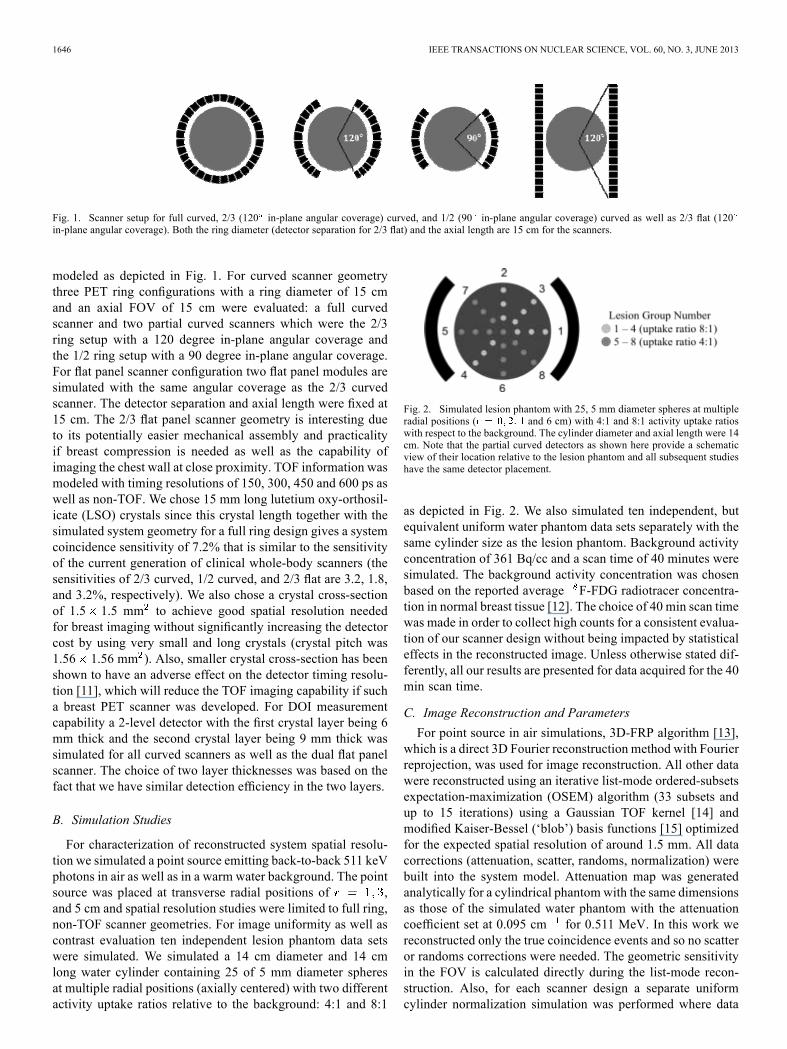
Fig. 1. Scanner setup for full curved, 2/3 (120 in-plane angular coverage) curved, and 1/2 (90 in-plane angular coverage) curved as well as 2/3 flat (120in-plane angular coverage). Both the ring diameter (detector separation for 2/3 flat) and the axial length are 15 cm for the scanners.
modeled as depicted in Fig. 1. For curved scanner geometrythree PET ring configurations with a ring diameter of 15 cmand an axial FOV of 15 cm were evaluated: a full curvedscanner and two partial curved scanners which were the 2/3ring setup with a 120 degree in-plane angular coverage andthe 1/2 ring setup with a 90 degree in-plane angular coverage.For flat panel scanner configuration two flat panel modules aresimulated with the same angular coverage as the 2/3 curvedscanner. The detector separation and axial length were fixed at15 cm. The 2/3 flat panel scanner geometry is interesting dueto its potentially easier mechanical assembly and practicalityif breast compression is needed as well as the capability ofimaging the chest wall at close proximity. TOF information wasmodeled with timing resolutions of 150, 300, 450 and 600 ps aswell as non-TOF. We chose 15 mm long lutetium oxy-orthosil-icate (LSO) crystals since this crystal length together with thesimulated system geometry for a full ring design gives a systemcoincidence sensitivity of 7.2% that is similar to the sensitivityof the current generation of clinical whole-body scanners (thesensitivities of 2/3 curved, 1/2 curved, and 2/3 flat are 3.2, 1.8,and 3.2%, respectively). We also chose a crystal cross-sectionof 1.5 1.5 mm to achieve good spatial resolution neededfor breast imaging without significantly increasing the detectorcost by using very small and long crystals (crystal pitch was1.56 1.56 mm ). Also, smaller crystal cross-section has beenshown to have an adverse effect on the detector timing resolu-tion [11], which will reduce the TOF imaging capability if sucha breast PET scanner was developed. For DOI measurementcapability a 2-level detector with the first crystal layer being 6mm thick and the second crystal layer being 9 mm thick wassimulated for all curved scanners as well as the dual flat panelscanner. The choice of two layer thicknesses was based on thefact that we have similar detection efficiency in the two layers.
B. Simulation Studies
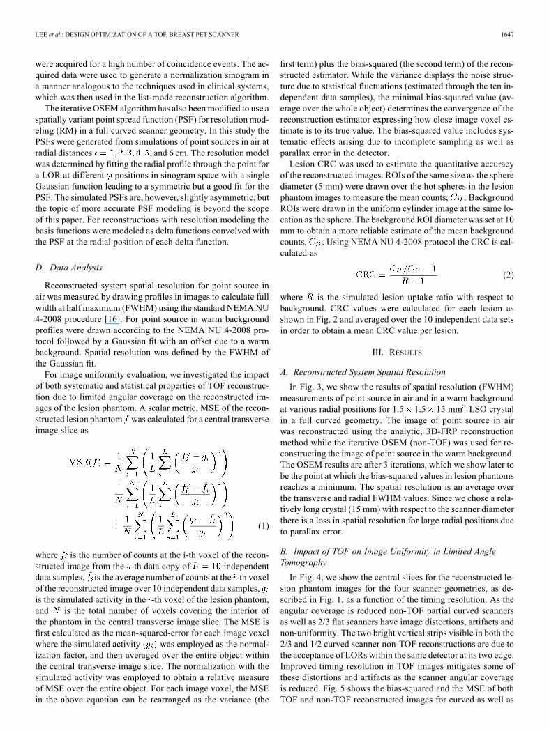
For characterization of reconstructed system spatial resolu-tion we simulated a point source emitting back-to-back 511 keVphotons in air as well as in a warm water background. The pointsource was placed at transverse radial positions of ,and 5 cm and spatial resolution studies were limited to full ring,non-TOF scanner geometries. For image uniformity as well ascontrast evaluation ten independent lesion phantom data setswere simulated. We simulated a 14 cm diameter and 14 cmlong water cylinder containing 25 of 5 mm diameter spheresat multiple radial positions (axially centered) with two differentactivity uptake ratios relative to the background: 4:1 and 8:1
Fig. 2. Simulated lesion phantom with 25, 5 mm diameter spheres at multipleradial positions ( and 6 cm) with 4:1 and 8:1 activity uptake ratioswith respect to the background. The cylinder diameter and axial length were 14cm. Note that the partial curved detectors as shown here provide a schematicview of their location relative to the lesion phantom and all subsequent studieshave the same detector placement.
as depicted in Fig. 2. We also simulated ten independent, butequivalent uniform water phantom data sets separately with thesame cylinder size as the lesion phantom. Background activityconcentration of 361 Bq/cc and a scan time of 40 minutes weresimulated. The background activity concentration was chosenbased on the reported average F-FDG radiotracer concentra-tion in normal breast tissue [12]. The choice of 40 min scan timewas made in order to collect high counts for a consistent evalua-tion of our scanner design without being impacted by statisticaleffects in the reconstructed image. Unless otherwise stated dif-ferently, all our results are presented for data acquired for the 40min scan time.
C. Image Reconstruction and Parameters
For point source in air simulations, 3D-FRP algorithm [13],which is a direct 3D Fourier reconstruction method with Fourierreprojection, was used for image reconstruction. All other datawere reconstructed using an iterative list-mode ordered-subsetsexpectation-maximization (OSEM) algorithm (33 subsets andup to 15 iterations) using a Gaussian TOF kernel [14] andmodified Kaiser-Bessel (‘blob’) basis functions [15] optimizedfor the expected spatial resolution of around 1.5 mm. All datacorrections (attenuation, scatter, randoms, normalization) werebuilt into the system model. Attenuation map was generatedanalytically for a cylindrical phantomwith the same dimensionsas those of the simulated water phantom with the attenuationcoefficient set at 0.095 cm for 0.511 MeV. In this work wereconstructed only the true coincidence events and so no scatteror randoms corrections were needed. The geometric sensitivityin the FOV is calculated directly during the list-mode recon-struction. Also, for each scanner design a separate uniformcylinder normalization simulation was performed where data
LEE et al.: DESIGN OPTIMIZATION OF A TOF, BREAST PET SCANNER 1647
were acquired for a high number of coincidence events. The ac-quired data were used to generate a normalization sinogram ina manner analogous to the techniques used in clinical systems,which was then used in the list-mode reconstruction algorithm.The iterative OSEM algorithm has also beenmodified to use a
spatially variant point spread function (PSF) for resolutionmod-eling (RM) in a full curved scanner geometry. In this study thePSFs were generated from simulations of point sources in air atradial distances , and 6 cm. The resolution modelwas determined by fitting the radial profile through the point fora LOR at different positions in sinogram space with a singleGaussian function leading to a symmetric but a good fit for thePSF. The simulated PSFs are, however, slightly asymmetric, butthe topic of more accurate PSF modeling is beyond the scopeof this paper. For reconstructions with resolution modeling thebasis functions were modeled as delta functions convolved withthe PSF at the radial position of each delta function.
D. Data Analysis
Reconstructed system spatial resolution for point source inair was measured by drawing profiles in images to calculate fullwidth at half maximum (FWHM) using the standard NEMANU4-2008 procedure [16]. For point source in warm backgroundprofiles were drawn according to the NEMA NU 4-2008 pro-tocol followed by a Gaussian fit with an offset due to a warmbackground. Spatial resolution was defined by the FWHM ofthe Gaussian fit.For image uniformity evaluation, we investigated the impact
of both systematic and statistical properties of TOF reconstruc-tion due to limited angular coverage on the reconstructed im-ages of the lesion phantom. A scalar metric, MSE of the recon-structed lesion phantom was calculated for a central transverseimage slice as
(1)
where is the number of counts at the i-th voxel of the recon-structed image from the -th data copy of independentdata samples, is the average number of counts at the -th voxelof the reconstructed image over 10 independent data samples,is the simulated activity in the -th voxel of the lesion phantom,and is the total number of voxels covering the interior ofthe phantom in the central transverse image slice. The MSE isfirst calculated as the mean-squared-error for each image voxelwhere the simulated activity was employed as the normal-ization factor, and then averaged over the entire object withinthe central transverse image slice. The normalization with thesimulated activity was employed to obtain a relative measureof MSE over the entire object. For each image voxel, the MSEin the above equation can be rearranged as the variance (the
first term) plus the bias-squared (the second term) of the recon-structed estimator. While the variance displays the noise struc-ture due to statistical fluctuations (estimated through the ten in-dependent data samples), the minimal bias-squared value (av-erage over the whole object) determines the convergence of thereconstruction estimator expressing how close image voxel es-timate is to its true value. The bias-squared value includes sys-tematic effects arising due to incomplete sampling as well asparallax error in the detector.Lesion CRC was used to estimate the quantitative accuracy
of the reconstructed images. ROIs of the same size as the spherediameter (5 mm) were drawn over the hot spheres in the lesionphantom images to measure the mean counts, . BackgroundROIs were drawn in the uniform cylinder image at the same lo-cation as the sphere. The background ROI diameter was set at 10mm to obtain a more reliable estimate of the mean backgroundcounts, . Using NEMA NU 4-2008 protocol the CRC is cal-culated as
(2)
where is the simulated lesion uptake ratio with respect tobackground. CRC values were calculated for each lesion asshown in Fig. 2 and averaged over the 10 independent data setsin order to obtain a mean CRC value per lesion.
III. RESULTS
A. Reconstructed System Spatial Resolution
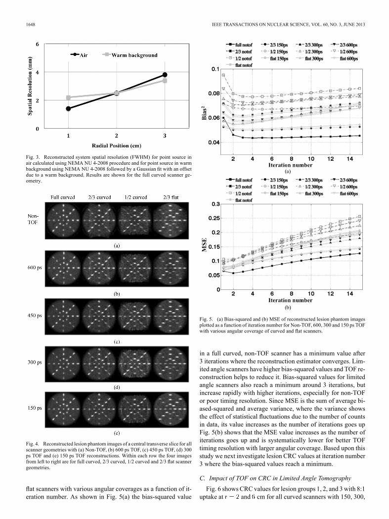
In Fig. 3, we show the results of spatial resolution (FWHM)measurements of point source in air and in a warm backgroundat various radial positions for 1.5 1.5 15 mm LSO crystalin a full curved geometry. The image of point source in airwas reconstructed using the analytic, 3D-FRP reconstructionmethod while the iterative OSEM (non-TOF) was used for re-constructing the image of point source in the warm background.The OSEM results are after 3 iterations, which we show later tobe the point at which the bias-squared values in lesion phantomsreaches a minimum. The spatial resolution is an average overthe transverse and radial FWHM values. Since we chose a rela-tively long crystal (15 mm) with respect to the scanner diameterthere is a loss in spatial resolution for large radial positions dueto parallax error.
B. Impact of TOF on Image Uniformity in Limited AngleTomography
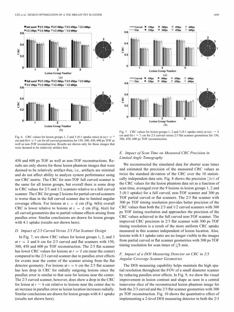
In Fig. 4, we show the central slices for the reconstructed le-sion phantom images for the four scanner geometries, as de-scribed in Fig. 1, as a function of the timing resolution. As theangular coverage is reduced non-TOF partial curved scannersas well as 2/3 flat scanners have image distortions, artifacts andnon-uniformity. The two bright vertical strips visible in both the2/3 and 1/2 curved scanner non-TOF reconstructions are due tothe acceptance of LORs within the same detector at its two edge.Improved timing resolution in TOF images mitigates some ofthese distortions and artifacts as the scanner angular coverageis reduced. Fig. 5 shows the bias-squared and the MSE of bothTOF and non-TOF reconstructed images for curved as well as
1648 IEEE TRANSACTIONS ON NUCLEAR SCIENCE, VOL. 60, NO. 3, JUNE 2013
Fig. 3. Reconstructed system spatial resolution (FWHM) for point source inair calculated using NEMA NU 4-2008 procedure and for point source in warmbackground using NEMA NU 4-2008 followed by a Gaussian fit with an offsetdue to a warm background. Results are shown for the full curved scanner ge-ometry.
Fig. 4. Reconstructed lesion phantom images of a central transverse slice for allscanner geometries with (a) Non-TOF, (b) 600 ps TOF, (c) 450 ps TOF, (d) 300ps TOF and (e) 150 ps TOF reconstructions. Within each row the four imagesfrom left to right are for full curved, 2/3 curved, 1/2 curved and 2/3 flat scannergeometries.
flat scanners with various angular coverages as a function of it-eration number. As shown in Fig. 5(a) the bias-squared value
Fig. 5. (a) Bias-squared and (b) MSE of reconstructed lesion phantom imagesplotted as a function of iteration number for Non-TOF, 600, 300 and 150 ps TOFwith various angular coverage of curved and flat scanners.
in a full curved, non-TOF scanner has a minimum value after3 iterations where the reconstruction estimator converges. Lim-ited angle scanners have higher bias-squared values and TOF re-construction helps to reduce it. Bias-squared values for limitedangle scanners also reach a minimum around 3 iterations, butincrease rapidly with higher iterations, especially for non-TOFor poor timing resolution. Since MSE is the sum of average bi-ased-squared and average variance, where the variance showsthe effect of statistical fluctuations due to the number of countsin data, its value increases as the number of iterations goes upFig. 5(b) shows that the MSE value increases as the number ofiterations goes up and is systematically lower for better TOFtiming resolution with larger angular coverage. Based upon thisstudy we next investigate lesion CRC values at iteration number3 where the bias-squared values reach a minimum.
C. Impact of TOF on CRC in Limited Angle Tomography
Fig. 6 shows CRC values for lesion groups 1, 2, and 3 with 8:1uptake at and cm for all curved scanners with 150, 300,
LEE et al.: DESIGN OPTIMIZATION OF A TOF, BREAST PET SCANNER 1649
Fig. 6. CRC values for lesion groups 1, 2 and 3 (8:1 uptake ratio) at (a)cm and (b) cm for all curved geometries for 150, 300, 450, 600 ps TOF aswell as non-TOF reconstruction. Results are shown only for those images thatwere deemed to be relatively artifact free.
450 and 600 ps TOF as well as non-TOF reconstructions. Re-sults are only shown for those lesion phantom images that weredeemed to be relatively artifact-free, i.e., artifacts are minimaland do not affect ability to analyze system performance usingour CRC metric. The CRC for non-TOF full curved scanner isthe same for all lesion groups, but overall there is some dropin CRC values for 2/3 and 1/2 scanners relative to a full curvedscanner. The CRC for group 2 lesions for partial curved scannersis worse than in the full curved scanner due to limited angularcoverage effects. For lesions at cm (Fig. 6(b)) overallCRC is lower relative to lesions at cm (Fig. 6(a)) forall curved geometries due to partial volume effects arising fromparallax error. Similar conclusions are drawn for lesion groupswith 4:1 uptake (results not shown here).
D. Impact of 2/3 Curved Versus 2/3 Flat Scanner Design
In Fig. 7, we show CRC values for lesion groups 1, 2, and 3at and cm for 2/3 curved and flat scanners with 150,300, 450 and 600 ps TOF reconstruction. The 2/3 flat scannerhas lower CRC values for lesions at cm (near the center)compared to the 2/3 curved scanner due to parallax error effectsfor events near the center of the scanner arising from the flatdetector geometry. For lesions at cm the 2/3 flat scannerhas less drop in CRC for radially outgoing lesions since theparallax error is similar to that seen for lesions near the center.The 2/3 curved scanner, however, does show a drop in the CRCfor lesion at cm relative to lesions near the center due toan increase in parallax error as lesion location increases radially.Similar conclusions are drawn for lesion groups with 4:1 uptake(results not shown here).
Fig. 7. CRC values for lesion groups 1, 2 and 3 (8:1 uptake ratio) at (a)cm and (b) cm for 2/3 curved versus 2/3 flat scanner geometries for 150,300, 450, 600 ps TOF reconstruction.
E. Impact of Scan Time on Measured CRC Precision inLimited Angle Tomography
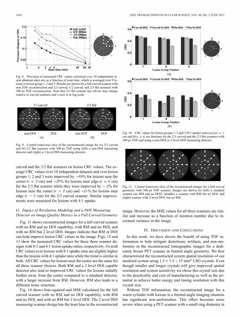
We reconstructed the simulated data for shorter scan timesand estimated the precision of the measured CRC values astwice the standard deviation of the CRC over the 10 statisti-cally independent data sets. Fig. 8 shows the precision ofthe CRC values for the lesion phantom data set as a function ofscan time, averaged over the 9 lesions in lesion groups 1, 2 and3 (8:1 uptake) for a full curved, non-TOF scanner and 300 psTOF partial curved or flat scanners. The 2/3 flat scanner with300 ps TOF timing resolution provides better precision of theCRC values than both the 2/3 and 1/2 curved scanners with 300ps TOF timing resolution and approaches the precision of theCRC values achieved in the full curved non-TOF scanner. Theimproved CRC precision in 2/3 flat scanner with 300 ps TOFtiming resolution is a result of the more uniform CRC uptakemeasured in this scanner independent of lesion location. Also,lesions with 4:1 uptake ratio are no longer visible in the imagesfrom partial curved or flat scanner geometries with 300 ps TOFtiming resolution for scan times of min.
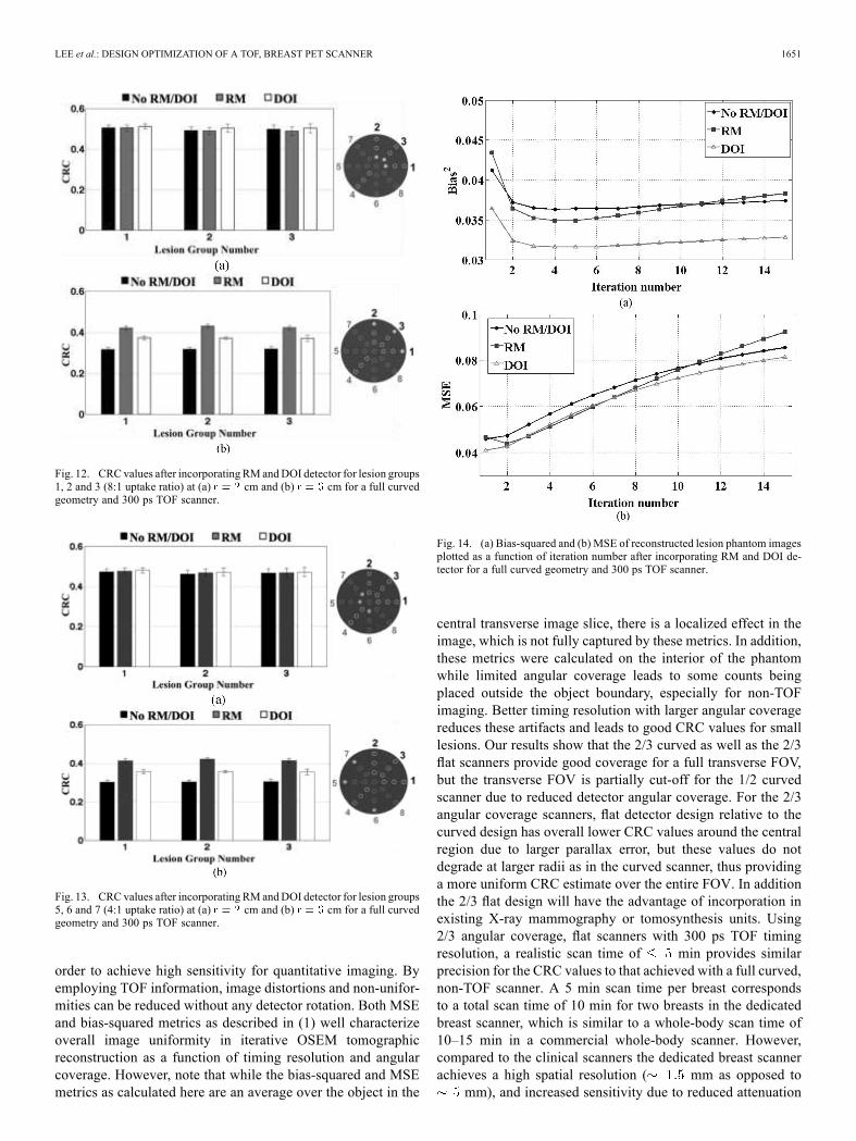
F. Impact of a DOI Measuring Detector on CRC in 2/3Angular Coverage Scanner Geometries
The DOI measuring capability helps maintain the high spa-tial resolution throughout the FOV of a small diameter scannerby reducing parallax error effects. In Fig. 9, we show the visualimprovement in lesion contrast and shape as seen in a centraltransverse slice of the reconstructed lesion phantom image forboth the 2/3 curved and the 2/3 flat scanner geometries with 300ps TOF reconstruction. Fig. 10 shows the quantitative effect ofimplementing a 2-level DOI measuring detector in both the 2/3
1650 IEEE TRANSACTIONS ON NUCLEAR SCIENCE, VOL. 60, NO. 3, JUNE 2013
Fig. 8. Precision of measured CRC values estimated over 10 independent le-sion phantom data sets as a function of scan time, which is averaged over 9 le-sions in lesion group 1, 2 and 3. Results are shown for a full curved scanner withnon-TOF reconstruction and 2/3 curved, 1/2 curved, and 2/3 flat scanners with300 ps TOF reconstruction. Note that 2/3 flat scanner has off-set time stampsrelative to curved scanners and x-axis is in log-scale.
Fig. 9. Central transverse slice of the reconstructed image for (a) 2/3 curvedand (b) 2/3 flat scanners with 300 ps TOF using (left) a non-DOI measuringdetector and (right) a 2-level DOI measuring detector.
curved and the 2/3 flat scanners on lesion CRC values. The av-erage CRC values over 10 independent datasets and over lesiongroups 1, 2 and 3 were improved by % for lesions near thecenter ( cm) and % for lesions near edge ( cm)for the 2/3 flat scanner while they were improved by % forlesions near the center ( cm) and % for lesions nearedge ( cm) for the 2/3 curved scanner. Similar improve-ments were measured for lesions with 4:1 uptake.
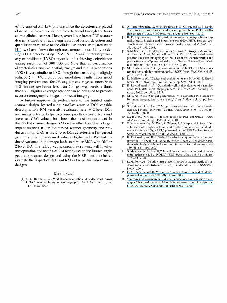
G. Impact of Resolution Modeling and a DOI MeasuringDetector on Image Quality Metrics in a Full Curved Geometry
Fig. 11 shows reconstructed images for a full curved scannerwith no RM and no DOI capability, with RM and no DOI, andwith no RM but 2 level DOI. Images indicate that RM or DOIcan help improve lesion CRC values in the image. Figs. 12 and13 show the measured CRC values for these three scanner de-signs with 8:1 and 4:1 lesion uptake ratios, respectively. OverallCRC values over lesions with 8:1 uptake ratio are slightly higherthan the lesions with 4:1 uptake ratio while the trend is similar inboth. All CRC values for lesions near the center are the same forall three scanner choices. Both RM and a 2 level DOI capabledetector also lead to improved CRC values for lesions radiallyfurther away from the center compared to a standard detector,with a larger increase from RM. However, RM also leads to adifferent noise structure.Fig. 14 shows bias-squared and MSE calculated for the full
curved scanner with no RM and no DOI capability, with RMand no DOI, and with no RM but 2 level DOI. The 2 level DOImeasuring scanner design has the least bias in the reconstructed
Fig. 10. CRC values for lesion groups 1, 2 and 3 (8:1 uptake ratio) at (a)cm and (b) cm (bottom) for the 2/3 curved and the 2/3 flat scanners with300 ps TOF and using a non-DOI or 2-level DOI measuring detector.
Fig. 11. Central transverse slice of the reconstructed images for a full curvedgeometry with 300 ps TOF scanner. Images are shown for (left) a standardscanner (no RM and no DOI), (middle) a scanner with RM but no DOI, and(right) scanner with 2-level DOI, but no RM.
image. However, the MSE values for all three scanners are sim-ilar and increase as a function of iteration number due to in-creased variance in the image.
IV. DISCUSSION AND CONCLUSIONS
In this work, we have shown the benefit of using TOF in-formation to help mitigate distortions, artifacts, and non-uni-formity in the reconstructed tomographic images for a dedi-cated, breast PET scanner in limited angle geometry. We firstcharacterized the reconstructed system spatial resolution of ourmodeled system using 1.5 1.5 15 mm LSO crystals. Eventhough smaller and longer crystals will give improved spatialresolution and system sensitivity we chose this crystal size dueto the practicality and cost of manufacturing as well as the po-tential to achieve better energy and timing resolution with thiscrystal size.Without TOF information, the reconstructed image for a
warm cylinder with lesions in a limited angle imaging situationhas significant non-uniformities. This effect becomes moresevere when using a PET scanner with a small ring diameter in
LEE et al.: DESIGN OPTIMIZATION OF A TOF, BREAST PET SCANNER 1651
Fig. 12. CRC values after incorporating RM andDOI detector for lesion groups1, 2 and 3 (8:1 uptake ratio) at (a) cm and (b) cm for a full curvedgeometry and 300 ps TOF scanner.
Fig. 13. CRC values after incorporating RM andDOI detector for lesion groups5, 6 and 7 (4:1 uptake ratio) at (a) cm and (b) cm for a full curvedgeometry and 300 ps TOF scanner.
order to achieve high sensitivity for quantitative imaging. Byemploying TOF information, image distortions and non-unifor-mities can be reduced without any detector rotation. Both MSEand bias-squared metrics as described in (1) well characterizeoverall image uniformity in iterative OSEM tomographicreconstruction as a function of timing resolution and angularcoverage. However, note that while the bias-squared and MSEmetrics as calculated here are an average over the object in the
Fig. 14. (a) Bias-squared and (b) MSE of reconstructed lesion phantom imagesplotted as a function of iteration number after incorporating RM and DOI de-tector for a full curved geometry and 300 ps TOF scanner.
central transverse image slice, there is a localized effect in theimage, which is not fully captured by these metrics. In addition,these metrics were calculated on the interior of the phantomwhile limited angular coverage leads to some counts beingplaced outside the object boundary, especially for non-TOFimaging. Better timing resolution with larger angular coveragereduces these artifacts and leads to good CRC values for smalllesions. Our results show that the 2/3 curved as well as the 2/3flat scanners provide good coverage for a full transverse FOV,but the transverse FOV is partially cut-off for the 1/2 curvedscanner due to reduced detector angular coverage. For the 2/3angular coverage scanners, flat detector design relative to thecurved design has overall lower CRC values around the centralregion due to larger parallax error, but these values do notdegrade at larger radii as in the curved scanner, thus providinga more uniform CRC estimate over the entire FOV. In additionthe 2/3 flat design will have the advantage of incorporation inexisting X-ray mammography or tomosynthesis units. Using2/3 angular coverage, flat scanners with 300 ps TOF timingresolution, a realistic scan time of min provides similarprecision for the CRC values to that achieved with a full curved,non-TOF scanner. A 5 min scan time per breast correspondsto a total scan time of 10 min for two breasts in the dedicatedbreast scanner, which is similar to a whole-body scan time of10–15 min in a commercial whole-body scanner. However,compared to the clinical scanners the dedicated breast scannerachieves a high spatial resolution ( mm as opposed to
mm), and increased sensitivity due to reduced attenuation
1652 IEEE TRANSACTIONS ON NUCLEAR SCIENCE, VOL. 60, NO. 3, JUNE 2013
of the emitted 511 keV photons since the detectors are placedclose to the breast and do not have to travel through the torsoas in a clinical scanner. Hence, overall our breast PET scannerdesign is capable of achieving improved lesion detection andquantification relative to the clinical scanners. In related work[11], we have shown through measurements our ability to de-sign a PET detector using 1.5 1.5 12 mm lutetium-yttriumoxy-orthosilicate (LYSO) crystals and achieving coincidencetiming resolution of 300–400 ps. Note that in performancecharacteristics such as spatial, energy and timing resolutionsLYSO is very similar to LSO, though the sensitivity is slightlyreduced % . Since our simulation results show goodimaging performance for 2/3 angular coverage scanners withTOF timing resolution less than 600 ps, we therefore thinkthat a 2/3 angular coverage scanner can be designed to provideaccurate tomographic images for breast imaging.To further improve the performance of the limited angle
scanner design by reducing parallax error, a DOI capabledetector and/or RM were also evaluated here. A 2 level DOImeasuring detector helps overcome parallax error effects andincreases CRC values, but shows the most improvement inthe 2/3 flat scanner design. RM on the other hand has a largerimpact on the CRC in the curved scanner geometry and pro-duces similar CRC as the 2 level DOI detector in a full curvedgeometry. The bias-squared value is higher with RM but re-duced variance in the image leads to similar MSE with RM or2 level DOI in a full curved scanner. Future work will involveincorporation and testing of RM techniques in the limited anglegeometry scanner design and using the MSE metric to betterevaluate the impact of DOI and RM in the partial ring scannerdesigns.
REFERENCES[1] S. L. Bowen et al., “Initial characterization of a dedicated breast
PET/CT scanner during human imaging,” J. Nucl. Med., vol. 50, pp.1401–1408, 2009.
[2] A. Vandenbroucke, A. M. K. Foudray, P. D. Olcott, and C. S. Levin,“Performance characterization of a new high resolution PET scintilla-tion detector,” Phys. Med. Biol., vol. 55, pp. 5895–5911, 2010.
[3] R. R. Raylman et al., “The positron emission mammography/tomog-raphy breast imaging and biopsy system (PEM/PET): Design, con-struction and phantom-based measurements,” Phys. Med. Biol., vol.53, pp. 637–653, 2008.
[4] S.M. Srinivas, R. Freifelder, J. Saffer, C. Cardi,M. Geagan,M.Werner,A. Kent, A. Alavi, M. Schnall, and J. S. Karp, “A dedicated breastpositron emission tomography (B-PET) scanner: Characterization andpilot patient study,” presented at the IEEENuclear Science Symp.Med-ical Imaging Conf., San Diego, CA, USA, 2006.
[5] M. C. Abreu et al., “Design and evaluation of the Clear-PEM scannerfor positron emission mammography,” IEEE Trans. Nucl. Sci., vol. 53,pp. 71–77, 2006.
[6] L. Moliner et al., “Design and evaluation of the MAMMI dedicatedbreast PET,” Med. Phys., vol. 39, no. 9, pp. 5393–5404, 2012.
[7] B. Ravindranath et al., “Quantitative clinical evaluation of a simulta-neous PET/MRI breast imaging system,” in J. Nucl. Med. Meeting Ab-stract, 2012, vol. 53, p. 1217.
[8] M. Lima et al., “Clinical performance of 2 dedicated PET scannersfor breast imaging: Initial evaluation,” J. Nucl. Med., vol. 53, pp. 1–9,2012.
[9] S. Surti and J. S. Karp, “Design considerations for a limited angle,dedicated breast, TOF PET scanner,” Phys. Med. Biol., vol. 53, pp.2911–2921, 2008.
[10] S. Jan et al., “GATE: A simulation toolkit for PET and SPECT,” Phys.Med. Biol., vol. 49, pp. 4543–4561, 2004.
[11] S. Krishnamoorthy, M. Kaul, R. Wiener, J. S. Karp, and S. Surti, “De-velopment of a high-resolution and depth-of interaction capable de-tector for time-of-flight PET,” presented at the IEEE Nuclear ScienceSymp. Medical Imaging Conf., Valencia, Spain, 2011.
[12] K. R. Zasadny and R. L. Wahl, “Standardized uptake value of normaltissues in PET with 2-[fluorine-18]-fluoro-2-deoxy-D-glucose: Varia-tions with body weight and a method for correction,” Radiology, vol.189, pp. 847–850, 1993.
[13] S. Matej and R. M. Lewitt, “Direct Fourier reconstruction with Fourierreprojection for full 3-D PET,” IEEE Trans. Nucl. Sci., vol. 48, pp.1378–1385, 2001.
[14] L. M. Popescu, “Iterative image reconstruction using geometrically or-dered subsets with list-mode data,” presented at the IEEE NSS/MIC,Rome, 2004.
[15] L. M. Popescu and R. M. Lewitt, “Tracing through a grid of blobs,”presented at the IEEE NSS/MIC, Rome, 2004.
[16] “Performance measurements of small animal positron emission tomo-graphs,” National Electrical Manufacturers Association, Rosslyn, VA,USA, 2008NEMA Standards Publication NU 4-2008.
Copyright © 2022 FDOKUMEN