Design and Synthesis of Human ABCB1 (P-Glycoprotein) Inhibitors by Peptide Coupling of Diverse...
15
Design and Synthesis of Human ABCB1 (P-Glycoprotein) Inhibitors by Peptide Coupling of Diverse Chemical Scaffolds on Carboxyl and Amino Termini of (S)‑Valine-Derived Thiazole Amino Acid Satyakam Singh, † Nagarajan Rajendra Prasad, ‡,§ Eduardo E. Chufan, ‡ Bhargav A. Patel, † Yi-Jun Wang, † Zhe-Sheng Chen, † Suresh V. Ambudkar,* ,‡ and Tanaji T. Talele* ,† † Department of Pharmaceutical Sciences, College of Pharmacy and Health Sciences, St. John’s University, 8000 Utopia Parkway, Queens, New York 11439, United States ‡ Laboratory of Cell Biology, Center for Cancer Research, National Cancer Institute, National Institutes of Health, Bethesda, Maryland 20892, United States ABSTRACT: P-glycoprotein (P-gp) serves as a therapeutic target for the development of multidrug resistance reversal agents. In this study, we synthesized 21 novel compounds by peptide coupling at corresponding carboxyl and amino termini of (S)-valine- based bis-thiazole and monothiazole derivatives with diverse chemical scaffolds. Using calcein-AM efflux assay, we identified compound 28 (IC 50 = 1.0 μM) carrying 3,4,5-trimethoxybenzoyl and 2-aminobenzophenone groups, respectively, at the amino and carboxyl termini of the monothiazole zwitter-ion. Compound 28 inhibited the photolabeling of P-gp with [ 125 I]- iodoarylazidoprazosin with IC 50 = 0.75 μM and stimulated the basal ATP hydrolysis of P-gp in a concentration-dependent manner (EC 50 ATPase = 0.027 μM). Compound 28 at 3 μM reduced resistance in cytotoxicity assay to paclitaxel in P-gp- expressing SW620/Ad300 and HEK/ABCB1 cell lines. Biochemical and docking studies showed site-1 to be the preferable binding site for 28 within the drug-binding pocket of human P-gp. ■ INTRODUCTION P-glycoprotein (P-gp, ABCB1 or MDR1) is a cell membrane ATP-binding cassette (ABC) transporter that is linked with the development of multidrug resistance (MDR) in cancer cells. Resistance to chemotherapy in cancer cells is a serious issue which can arise due to numerous mechanisms, including the overexpression of ABC drug transporters such as P-gp and breast cancer resistance protein (BCRP, ABCG2, or MXR). These transporters have been shown to contribute to decreased intracellular concentrations of chemotherapeutic drugs. 1,2 P-gp utilizes energy derived from ATP hydrolysis to efflux an extremely diverse set of hydrophobic compounds typified by different chemical classes such as amphipathic, neutral, or weakly basic agents including a number of recently developed tyrosine kinase inhibitors. 3 Human P-gp is composed of two transmembrane domains (TMDs), each containing six helices and two nucleotide-binding domains (NBDs). The TMDs house the drug/substrate-binding sites and a translocation conduit. 4 Moreover, the TMD region contains a large hydrophobic drug-binding site that is capable of binding two to three molecules simultaneously. 5,6 Drug transport process by P-gp is driven by ATP binding/hydrolysis to the drug-binding competent state which results in a rotation of the NBDs followed by adoption of a closed conformation and eventually NBD dimer returns to the open conformation after ATP hydrolysis. 7 The closed conformation of NBD dimer is responsible for the substrate translocation in the TMDs’ drug-binding sites, thus initiating the release of the substrate to the extracellular side of the membrane. 7 The ABC transporter-mediated MDR hinders the clinical cure of cancer by chemotherapy. Therefore, many research groups have been focusing on developing P-gp inhibitors to enhance the therapeutic concentration of chemotherapeutic drug inside cancer cells. Because of the unavailability of high- resolution 3D-crystal structure of human P-gp, we used the Received: December 20, 2013 Published: April 28, 2014 Article pubs.acs.org/jmc © 2014 American Chemical Society 4058 dx.doi.org/10.1021/jm401966m | J. Med. Chem. 2014, 57, 4058−4072
-
Upload
independent -
Category
Documents
-
view
4 -
download
0
Transcript of Design and Synthesis of Human ABCB1 (P-Glycoprotein) Inhibitors by Peptide Coupling of Diverse...

Design and Synthesis of Human ABCB1 (P-Glycoprotein) Inhibitorsby Peptide Coupling of Diverse Chemical Scaffolds on Carboxyl andAmino Termini of (S)‑Valine-Derived Thiazole Amino AcidSatyakam Singh,† Nagarajan Rajendra Prasad,‡,§ Eduardo E. Chufan,‡ Bhargav A. Patel,† Yi-Jun Wang,†
Zhe-Sheng Chen,† Suresh V. Ambudkar,*,‡ and Tanaji T. Talele*,†
†Department of Pharmaceutical Sciences, College of Pharmacy and Health Sciences, St. John’s University, 8000 Utopia Parkway,Queens, New York 11439, United States‡Laboratory of Cell Biology, Center for Cancer Research, National Cancer Institute, National Institutes of Health, Bethesda,Maryland 20892, United States
ABSTRACT: P-glycoprotein (P-gp) serves as a therapeutic target for the development of multidrug resistance reversal agents. Inthis study, we synthesized 21 novel compounds by peptide coupling at corresponding carboxyl and amino termini of (S)-valine-based bis-thiazole and monothiazole derivatives with diverse chemical scaffolds. Using calcein-AM efflux assay, we identifiedcompound 28 (IC50 = 1.0 μM) carrying 3,4,5-trimethoxybenzoyl and 2-aminobenzophenone groups, respectively, at the aminoand carboxyl termini of the monothiazole zwitter-ion. Compound 28 inhibited the photolabeling of P-gp with [125I]-iodoarylazidoprazosin with IC50 = 0.75 μM and stimulated the basal ATP hydrolysis of P-gp in a concentration-dependentmanner (EC50 ATPase = 0.027 μM). Compound 28 at 3 μM reduced resistance in cytotoxicity assay to paclitaxel in P-gp-expressing SW620/Ad300 and HEK/ABCB1 cell lines. Biochemical and docking studies showed site-1 to be the preferablebinding site for 28 within the drug-binding pocket of human P-gp.
■ INTRODUCTIONP-glycoprotein (P-gp, ABCB1 or MDR1) is a cell membraneATP-binding cassette (ABC) transporter that is linked with thedevelopment of multidrug resistance (MDR) in cancer cells.Resistance to chemotherapy in cancer cells is a serious issuewhich can arise due to numerous mechanisms, including theoverexpression of ABC drug transporters such as P-gp andbreast cancer resistance protein (BCRP, ABCG2, or MXR).These transporters have been shown to contribute to decreasedintracellular concentrations of chemotherapeutic drugs.1,2 P-gputilizes energy derived from ATP hydrolysis to efflux anextremely diverse set of hydrophobic compounds typified bydifferent chemical classes such as amphipathic, neutral, orweakly basic agents including a number of recently developedtyrosine kinase inhibitors.3 Human P-gp is composed of twotransmembrane domains (TMDs), each containing six helicesand two nucleotide-binding domains (NBDs). The TMDshouse the drug/substrate-binding sites and a translocationconduit.4 Moreover, the TMD region contains a large
hydrophobic drug-binding site that is capable of binding twoto three molecules simultaneously.5,6 Drug transport process byP-gp is driven by ATP binding/hydrolysis to the drug-bindingcompetent state which results in a rotation of the NBDsfollowed by adoption of a closed conformation and eventuallyNBD dimer returns to the open conformation after ATPhydrolysis.7 The closed conformation of NBD dimer isresponsible for the substrate translocation in the TMDs’drug-binding sites, thus initiating the release of the substrate tothe extracellular side of the membrane.7
The ABC transporter-mediated MDR hinders the clinicalcure of cancer by chemotherapy. Therefore, many researchgroups have been focusing on developing P-gp inhibitors toenhance the therapeutic concentration of chemotherapeuticdrug inside cancer cells. Because of the unavailability of high-resolution 3D-crystal structure of human P-gp, we used the
Received: December 20, 2013Published: April 28, 2014
Article
pubs.acs.org/jmc
© 2014 American Chemical Society 4058 dx.doi.org/10.1021/jm401966m | J. Med. Chem. 2014, 57, 4058−4072

recently corrected crystal structure of mouse P-gp as a templateto develop homology model of human P-gp,8 which waseventually used for understanding the binding mode of
synthesized P-gp inhibitors. Numerous strategies such asrandom and focused screening, systematic chemical modifica-tions, and combinatorial chemistry have been applied to
Figure 1. (A) Structures of the reported potent P-gp inhibitors containing chemical fragments such as methoxy substituted tetrahydroisoquinoline,various methoxy substituted aryl rings, biphenyl, and benzophenone. (B) Structures of compounds 1 and 1a−1d.
Journal of Medicinal Chemistry Article
dx.doi.org/10.1021/jm401966m | J. Med. Chem. 2014, 57, 4058−40724059

develop the first three generations of P-gp inhibitors; however,they suffer from toxicity and drug−drug interactions. Althoughthe first three generations of P-gp and/or ABCG2 modulators(valspodar, biricodar, laniquidar, zosuquidar, elacridar, andtariquidar) designed to reverse MDR failed in various stages ofclinical trials, ABC transporters undoubtedly play a crucial rolein the development of MDR, and researchers should notunderestimate their importance. The clinical failures were onlypartly associated with ABC transporter modulation.9 They werealso due to inadequate bioavailability at tumor sites,9
nonselective inhibition of P-gp across all tissues including theblood−brain barrier, simultaneous inhibition of the drug-metabolizing enzyme CYP3A4,10 and interference with thefunction of other ABC transporters that play a crucial role inantitumor immune response.11 Another problem was inappro-priate selection of the patient population.12
With more carefully designed clinical trials, fourth generationP-gp modulators with low toxicity may be found to circumventmodulator-associated problems. New approaches leading to thedevelopment of fourth generation P-gp inhibitors exhibitinghigh P-gp selectivity and potency are highly desirable. One suchapproach is to append various chemical fragments that arefrequently seen in P-gp inhibitors, to a novel chemotype such asthiazole amino acid. Using this approach, chemical fragmentsfrom reported potent P-gp inhibitors, as shown in Figure 1A,are inserted into chemically modified natural products.The first available cocrystal structures of murine P-gp bound
to cyclic selenazole derivatives 1c (QZ59Se-SSS)13 and 1d(QZ59Se-RRR)13 provided significant insight into various drug-binding sites of P-gp transporter (Figure 1B). The cyclic trimercompound 1a (QZ59S-SSS), which is bioisosteric to compound1c, exhibited an IC50 value of 2.7 μM, whereas 1b, an isostere of1d, showed an IC50 of 8.4 μM against mouse P-gp effluxfunction (Figure 1B).14 Because of this distinguishable ability ofP-gp for stereoisomers of cyclic peptides, we maintained (S)chirality for all the synthesized compounds. Moreover, severalnatural products have been shown to contain thiazole ring.15
Our recent study encompasses the investigation of theinhibitory effect of (S)-valine-based thiazole derivativesincluding compound 1a on human P-gp efflux activity.16 Theresults obtained from the exploration indicated the linearthiazole chain (linear trimer, 1; IC50 = 1.5 μM) to be equallyeffective as that of 1a (IC50 = 1.5 μM) in terms of inhibition ofefflux activity of human P-gp. Also, we noticed that deviationfrom the trimer size (three consecutive thiazole units) of thethiazole chain had a detrimental effect on the inhibitory activity.Docking analysis of compound 1 within the drug-bindingpocket of homology-modeled human P-gp showed interactionswith certain key amino acid residues.16 The terminal thiazolefragments of compound 1 were found to capture hydrogenbonding and electrostatic interactions with the glutamine
(Q990 and Q725) and tyrosine (Y307 and Y953) residueswhile being predominantly surrounded by hydrophobic aminoacid side chains in the drug-binding pocket of P-gp. On thebasis of these results, we decided to optimize compound 1 byreplacing the terminal thiazole units with chemical scaffoldscontaining mono-, di-, and tri-methoxy aryl rings. For a fewanalogues, we also borrowed chemical fragments from thereported P-gp inhibitory structures that might increase the π−πinteraction surface area within the drug-binding pocket of P-gp.Our choice of fragments was based on the chemical moietieswhich are frequently observed in various reported preclinicaland clinical candidates such as tariquidar (XR9576),17
elacridar,18 LY402913,19 reserpine,20 XR9051,21 saracatinib,22
galloyl-based inhibitors,23 and benzophenone derivatives24 asillustrated in Figure 1A. Additionally, Didziapetris et al.25
estimated the “rule of fours” which states that the compoundswith (N+O) ≥ 8, MW > 400, and acid pKa > 4 are likely to actas P-gp substrates. Therefore, we synthesized a series of (S)-valine derived thiazole analogues by extending the carboxyl andthe amino termini of monomer (mono thiazole unit) as well asdimer (bis thiazole unit), taking into account the trimer size aswell as the “rule of fours”. This optimization strategy wassupported by the inhibitory activity of two representativecompounds (2 and 3) mentioned in our recent report.16
Certain critical aspects were considered for the selection offragments for extensions: (a) the coupled fragments mustimpart the desired hydrophobicity (ClogP = 3−6),26−28 (b) theselected fragments should present hydrogen bond acceptoratoms such as oxygen and nitrogen, and (c) both amino andcarboxyl termini should be attached to various methoxy-substituted moieties and extended aromatic fragments toestablish critical electrostatic interactions with hydrogen bonddonor residues and the π-stacking interactions at the drug-binding pocket of the P-gp. Additionally in this regard, thepresence of the methoxy groups has been shown previously toincrease selective affinity toward P-gp.23 The synthesizedderivatives were assessed for their inhibitory efficacies on theP-gp transport function by calcein-AM assay. Reversal of MDRdue to ABCB1 inhibition in SW620/Ad300 and HEK/ABCB1cell lines was also investigated using paclitaxel in the presenceof compounds 28, 1a, and cyclosporine A. The effect ofcompound 28 was tested with biochemical studies includingphotolabeling with [125I]-iodoarylazidoprazosin (IAAP) andmeasurement of ATPase activity of P-gp using crudemembranes of High Five insect cells expressing this transporter.Furthermore, to obtain insights into binding interactions ofthese analogues within the large drug-binding pocket, weperformed docking of compounds on all the possible bindingsites of the homology model of human P-gp.
Scheme 1. Synthesis of Monomer Acid Derivativea
aReagents and conditions: (i) HCTU, HOBt, DIEA, DMA, rt, 18 h.
Journal of Medicinal Chemistry Article
dx.doi.org/10.1021/jm401966m | J. Med. Chem. 2014, 57, 4058−40724060

■ RESULTS AND DISCUSSION
Chemistry. A library of 21 thiazole-based compoundstargeted for P-gp efflux inhibition was synthesized as shown inSchemes 1−5. The synthesis of compounds 2 and 3 along withthat of the required precursors (4, 6, 10, and 13) has beendescribed in our recent report.16 Target compounds 5, 7−9,11−12, and 14−15 were synthesized by peptide couplingreactions using thiazole acids (4 and 10) and thiazole amines (6and 13) with various chemical scaffolds containing amine andacid derivatives, respectively. Schemes 1 and 2 shows thesynthesis of monomer derivatives. Monomer acid 4 was reactedwith a piperidine derivative (5a) to obtain compound 5 in thepresence of the coupling agents HCTU, HOBt, and DIEA, asshown in Scheme 1. Monomer amine 6 was coupled with acidderivatives 7a and 8a to obtain compounds 7 and 8,
respectively (Scheme 2). Alternatively, compound 9 wasprepared by reacting trimethoxybenzoyl chloride (9a) with amonomer amine (6) using DIEA in THF. Our next objectivewas to synthesize dimer derivatives that could mimic the trimerstructure using compounds 10 and 13. In Scheme 3, a dimeracid (10) was coupled with di- and tri-methoxy anilines (11aand 12a), respectively, to obtain compounds 11 and 12.Consequently, compounds 14 and 15 were synthesized byreaction of a dimer amine (13) with acid chlorides 14a and 15ausing DIEA in THF (Scheme 4). Further, target compounds17−29 were synthesized starting from compound 9 by use ofthe coupling reagents HCTU, HOBt, and DIEA in DMA(Scheme 5). These target compounds were the size of a trimermolecule, with both ends featuring chemical scaffolds borrowedfrom potent P-gp modulators. At first, the ethyl ester present in
Scheme 2. Synthesis of Monomer Amine Derivativesa
aReagents and conditions: (i) HCTU, HOBt, DIEA, DMA, rt, 18 h; (ii) DIEA, THF, rt, 12 h.
Scheme 3. Synthesis of Dimer Acid Derivativesa
aReagents and conditions: (i) HCTU, HOBt, DIEA, DMA, rt, 18−24 h.
Scheme 4. Synthesis of Dimer Amine Derivativesa
aReagents and conditions: (i) DIEA, THF, rt, 12 h.
Journal of Medicinal Chemistry Article
dx.doi.org/10.1021/jm401966m | J. Med. Chem. 2014, 57, 4058−40724061
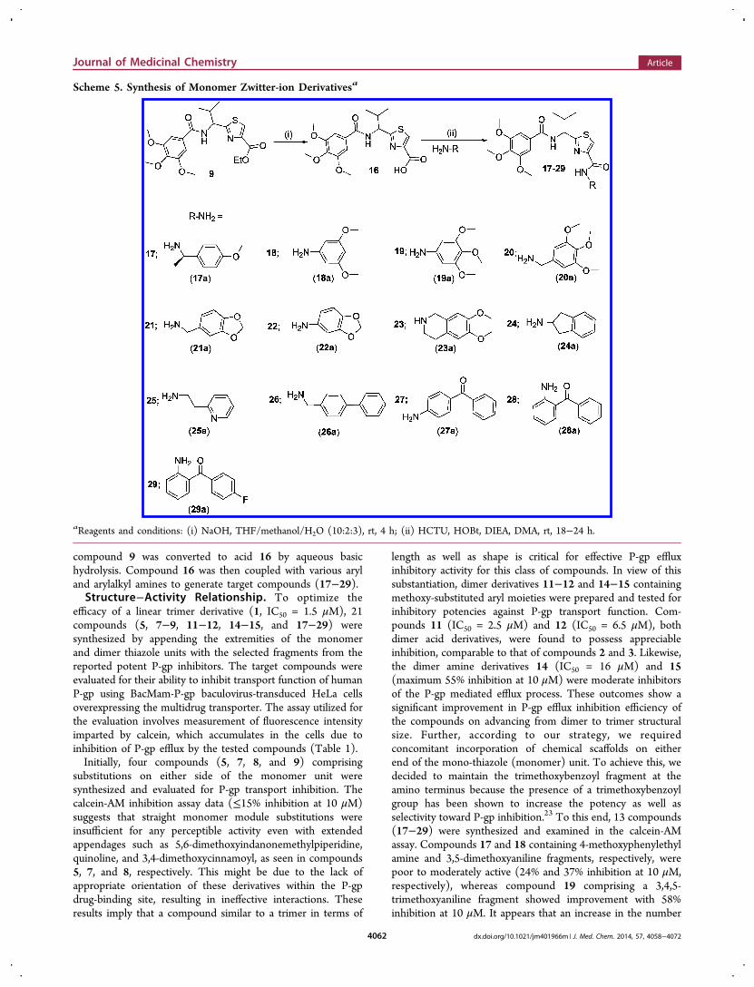
compound 9 was converted to acid 16 by aqueous basichydrolysis. Compound 16 was then coupled with various aryland arylalkyl amines to generate target compounds (17−29).Structure−Activity Relationship. To optimize the
efficacy of a linear trimer derivative (1, IC50 = 1.5 μM), 21compounds (5, 7−9, 11−12, 14−15, and 17−29) weresynthesized by appending the extremities of the monomerand dimer thiazole units with the selected fragments from thereported potent P-gp inhibitors. The target compounds wereevaluated for their ability to inhibit transport function of humanP-gp using BacMam-P-gp baculovirus-transduced HeLa cellsoverexpressing the multidrug transporter. The assay utilized forthe evaluation involves measurement of fluorescence intensityimparted by calcein, which accumulates in the cells due toinhibition of P-gp efflux by the tested compounds (Table 1).Initially, four compounds (5, 7, 8, and 9) comprising
substitutions on either side of the monomer unit weresynthesized and evaluated for P-gp transport inhibition. Thecalcein-AM inhibition assay data (≤15% inhibition at 10 μM)suggests that straight monomer module substitutions wereinsufficient for any perceptible activity even with extendedappendages such as 5,6-dimethoxyindanonemethylpiperidine,quinoline, and 3,4-dimethoxycinnamoyl, as seen in compounds5, 7, and 8, respectively. This might be due to the lack ofappropriate orientation of these derivatives within the P-gpdrug-binding site, resulting in ineffective interactions. Theseresults imply that a compound similar to a trimer in terms of
length as well as shape is critical for effective P-gp effluxinhibitory activity for this class of compounds. In view of thissubstantiation, dimer derivatives 11−12 and 14−15 containingmethoxy-substituted aryl moieties were prepared and tested forinhibitory potencies against P-gp transport function. Com-pounds 11 (IC50 = 2.5 μM) and 12 (IC50 = 6.5 μM), bothdimer acid derivatives, were found to possess appreciableinhibition, comparable to that of compounds 2 and 3. Likewise,the dimer amine derivatives 14 (IC50 = 16 μM) and 15(maximum 55% inhibition at 10 μM) were moderate inhibitorsof the P-gp mediated efflux process. These outcomes show asignificant improvement in P-gp efflux inhibition efficiency ofthe compounds on advancing from dimer to trimer structuralsize. Further, according to our strategy, we requiredconcomitant incorporation of chemical scaffolds on eitherend of the mono-thiazole (monomer) unit. To achieve this, wedecided to maintain the trimethoxybenzoyl fragment at theamino terminus because the presence of a trimethoxybenzoylgroup has been shown to increase the potency as well asselectivity toward P-gp inhibition.23 To this end, 13 compounds(17−29) were synthesized and examined in the calcein-AMassay. Compounds 17 and 18 containing 4-methoxyphenylethylamine and 3,5-dimethoxyaniline fragments, respectively, werepoor to moderately active (24% and 37% inhibition at 10 μM,respectively), whereas compound 19 comprising a 3,4,5-trimethoxyaniline fragment showed improvement with 58%inhibition at 10 μM. It appears that an increase in the number
Scheme 5. Synthesis of Monomer Zwitter-ion Derivativesa
aReagents and conditions: (i) NaOH, THF/methanol/H2O (10:2:3), rt, 4 h; (ii) HCTU, HOBt, DIEA, DMA, rt, 18−24 h.
Journal of Medicinal Chemistry Article
dx.doi.org/10.1021/jm401966m | J. Med. Chem. 2014, 57, 4058−40724062

of methoxy groups on the phenyl ring of the compoundsenhances the binding affinity for P-gp. However, compound 20,with a 3,4,5-trimethoxybenzyl amine fragment, lost the P-gpinhibitory activity (4% inhibition at 10 μM). Compounds 21
and 22 with methylenedioxybenzyl amine and methylenedioxyaniline showed 20% and 40% inhibition of P-gp, respectively.Comparing compounds 19 with 20 and 21 with 22, theinsertion of a methylene spacer between the aryl and the amine
Table 1. (S)-Valine-Based Thiazole Derivatives As Inhibitors of P-gp Transport Function
aBacMam-P-gp baculovirus-transduced HeLa cells were incubated with 0.5 μM calcein-AM for 10 min at 37 °C under dark in the presence andabsence of 10 μM (S)-valine-based thiazole derivatives. Percentage transport inhibition was derived by considering the level of inhibition obtainedwith the standard inhibitor, tariquidar at 1 μM, and the values ± SD shown are the average of two independent experiments done in triplicate at 10μM concentrations of inhibitors. bIC50 values shown in brackets (±SD) were determined by at least two independent experiments done in triplicate.cCompounds reported in our recently published work.16 dLigand efficiency was calculated using formula, LE = (−RT ln IC50/number of non-hydrogen atoms); IC50 is in molar, R = 1.987 kcal K−1 mol−1, and T = 300 K.
Journal of Medicinal Chemistry Article
dx.doi.org/10.1021/jm401966m | J. Med. Chem. 2014, 57, 4058−40724063

group proved detrimental for the P-gp inhibitory activity. Thisfinding suggests potential steric clashes within the drug-bindingpocket of P-gp for compounds 20 and 21 resulting from theintroduction of the methylene spacer group. The 6,7-dimethoxytetrahydroisoquinoline group containing compound23 was found to be devoid of P-gp inhibitory activity (16% at10 μM). Furthermore, incorporation of a 2-aminoindanesubstitution resulted in moderate activity of compound 24(47% inhibition at 10 μM); however, incorporation of 2-aminoethylpyridine (25) and 4-phenylbenzyl amine (26) werefound to have a detrimental effect on P-gp inhibitory activity(5% and 23% inhibition at 10 μM, respectively), supporting ourprevious observation of the unfavorable effect of an alkyl spacergroup. Weak inhibition of calcein-AM transport by compounds22, 23, and 24 indicates a potential steric hindrance by thebicyclic ring structure at the drug-binding pocket of P-gp.Compound 27, containing a 4-aminobenzophenone substitu-tion, lacks any significant inhibitory activity (18% at 10 μM),while compound 28 with a 2-aminobenzophenone substitutionwas found to have efficient P-gp inhibitory activity with IC50value of 1 μM. Also, compound 29 showed appreciableinhibition (54% inhibition at 10 μM) of P-gp transport activity.Compound 27, with a benzoyl group at the para-position,
might be similar to compounds 22, 23, and 24 with respect tosteric hindrance. Moreover, effective inhibitory data forcompounds 28 and 29 indicates that it is essential for inhibitorsto have an angular shape to increase the contact surface with P-gp. Compound 28, bearing the trimethoxybenzoyl group at theamino terminus and 2-aminobenzophenone at the carboxylterminus, proved to be the most effective P-gp inhibitor (IC50 =1.0 μM) among the synthesized compounds, as evident fromthe dose−response curve shown in Figure 2B. Panel A ofFigure 2 shows a representative histogram with low, moderate,and high inhibitory activity of selected thiazole derivatives,whereas panel B shows the concentration-dependent inhibitionof calcein-AM efflux by compounds 11, 12, 14, and 28. Weattempted to correlate the pIC50 values of compounds 1, 2, 3,11, 12, 14, and 28 with their respective ClogP values 5.81, 5.22,4.93, 5.47, 5.87, 5.03, and 5.49 in order to appraise the functionof lipophilicity for P-gp inhibition. However, we were unable tofind any significant correlation within the narrow range ofClogP values, which could be a result of a small sample size.Among various parameters that govern P-gp inhibition, onesuch parameter is ClogP, which should be in the range of 3−6as mentioned earlier. Therefore, apart from ClogP otherparameters, such as a molecular framework including hydrogen
Figure 2. Inhibition of calcein-AM transport. BacMam P-gp baculovirus-transduced HeLa cells were assayed for calcein-AM transport (A) in thepresence of selected derivatives at 10 μM and (B) in the presence of increasing concentrations of 11, 12, 14, and 28. (A) Representative histogramsshow low, moderate, and high P-gp inhibitory activity of selected thiazole derivatives. Tariquidar (1 μM), a known inhibitor of P-gp, was used forcomparison. The values shown are the average of two independent experiments each done in triplicate. (B) Concentration-dependent inhibition ofcalcein-AM efflux by selected derivatives was studied. The average values from two independent experiments each done in triplicate were plotted andthe IC50 values for compounds 11 (filled squares), 12 (filled diamonds), 14 (filled triangles), and 28 (filled circles) are 2.5, 6.5, 16, and 1 μM,respectively.
Table 2. Reversal Effect of Compounds 28, 1a, and Cyclosporine A on the Cytotoxicity of Paclitaxel to SW620, SW620/Ad300,HEK293, and HEK/ABCB1 Cell Lines
SW620 SW620/Ad300 HEK293 HEK/ABCB1
compd IC50 ± SEMa (μM) FRb IC50 ± SEMa (μM) FRb IC50 ± SEMa (μM) FRb IC50 ± SEMa (μM) FRb
paclitaxel 0.006 ± 0.001 [1.0] 4.019 ± 0.215 [669.8] 0.049 ± 0.004 [1.0] 4.138 ± 0.132 [84.4]+ 28(1 μM) 0.007 ± 0.001 [1.2] 0.186 ± 0.016 [31.0] 0.048 ± 0.003 [1.0] 0.633 ± 0.042 [12.9]+ 28 (3 μM) 0.006 ± 0.001 [1.0] 0.051 ± 0.005 [8.5] 0.047 ± 0.003 [1.0] 0.154 ± 0.015 [3.1]+ 28 (10 μM) 0.006 ± 0.001 [1.0] 0.011 ± 0.001 [1.8] 0.048 ± 0.004 [1.0] 0.098 ± 0.004 [2.0]+ 1a (0.3 μM) 0.006 ± 0.001 [1.0] 0.706 ± 0.068 [117.7] 0.049 ± 0.003 [1.0] 1.230 ± 0.063 [25.1]+ 1a (1 μM) 0.006 ± 0.001 [1.0] 0.340 ± 0.035 [56.7] 0.048 ± 0.005 [1.0] 0.780 ± 0.047 [15.9]+ 1a (3 μM) 0.007 ± 0.001 [1.2] 0.125 ± 0.011 [20.8] 0.048 ± 0.004 [1.0] 0.256 ± 0.024 [5.2]+ CsA (3 μM) 0.006 ± 0.001 [1.0] 0.079 ± 0.007 [13.2] 0.050 ± 0.003 [1.0] 0.205 ± 0.017 [4.2]
aIC50, concentration of indicated compound required for 50% inhibition of cell survival were calculated from the killing curves shown in Figure 3.Mean values (±SEM) are from four independent experiments, each performed in triplicate. bFR: fold-resistance was calculated by dividing the IC50value for paclitaxel of SW620 (or HEK293) and SW620/Ad300 (or HEK/ABCB1) cells in the absence or presence of compounds 28, 1a, andcyclosporine A by IC50 value for paclitaxel of SW620 (or HEK293) cells. CsA, cyclosporine A.
Journal of Medicinal Chemistry Article
dx.doi.org/10.1021/jm401966m | J. Med. Chem. 2014, 57, 4058−40724064

Figure 3. Effect of compounds 28, 1a, and cyclosporine A (CsA) on ABCB1-mediated resistance to paclitaxel in ABCB1 overexpressing drugselected (A) and transfected (B) cell lines. (A) Concentration-dependent curves of paclitaxel with or without compounds 28, 1a, and CsA at 3 μM inparental SW620 and ABCB1 overexpressing SW620/Ad300 cells. The IC50 values of SW620/Ad300 cell line were compared with those of parentalSW620 cells (see Table 2). (B) Concentration-dependent curves of paclitaxel with or without compounds 28, 1a, and CsA at 3 μM in parentalHEK293/pcDNA3.1 and ABCB1 overexpressing HEK/ABCB1 cells. The IC50 values of HEK/ABCB1 cell line were compared with those ofparental HEK293 cells (see Table 2). Points with error bars represent the mean ± SEM. The figure is a representative of four independentexperiments, each done in triplicate.
Journal of Medicinal Chemistry Article
dx.doi.org/10.1021/jm401966m | J. Med. Chem. 2014, 57, 4058−40724065

bonding ability and interaction surface area, also play criticalrole in inhibition of P-gp transport function. Ligand efficiencycalculations showed a marginal improvement for compound 28(0.20) as compared to compound 1 (0.17) (Table 1). Inaddition, the molecular weight decreased by 119 Da fromcompound 1 to benzophenone analogue 28.Effect of Compounds 28 and 1a on ABCB1-Mediated
Resistance to Paclitaxel in ABCB1 Overexpressing Drug-Selected Cell Lines. Does compound 28 reverse resistance toanticancer drugs in P-gp overexpressing cell lines? To addressthis question, we performed MDR reversal experiments. Toselect a nontoxic or relatively low drug concentration forcompounds 28 and 1a, cytotoxicity assays were performed onparental and P-gp-overexpressing cell lines (data not shown).On the basis of these results, concentrations of 10 μM for 28and 3 μM for 1a demonstrated >85% cell survival.To determine whether 28 and 1a could reverse P-gp-
mediated MDR, cell survival assays were performed in thepresence or absence of 28 and 1a, using the parental SW620and drug-selected SW620/Ad300 cell lines. The drug-selectedSW620/Ad300 cell line showed 688.2-fold resistance topaclitaxel, as compared to the parental SW620 cell line(Table 2). Compound 1a, at 0.3, 1, and 3 μM, significantlydecreased the resistance of the SW620/Ad300 cell line topaclitaxel from 669.8-fold to 117.7-, 56.7-, and 20.8-fold,respectively, compared to SW620 cell line (Table 2).Compound 28, at 1, 3, and 10 μM, further reduced theresistance of SW620/Ad300 cell line to paclitaxel from 669.8-fold to 31.0-, 8.5-, and 1.8-fold, respectively, compared toSW620 cell line (Table 2). Moreover, at 10 μM, compound 28can almost completely reverse the ABCB1-mediated drugresistance in SW620/Ad300 cells. As expected, a knownmodulator of P-gp, cyclosporine A (3 μM) significantlydecreased the resistance of SW620/Ad300 to 13.2-fold forpaclitaxel as compared to the control SW620 cell line (Table2). Interestingly, at 3 μM, compound 28 showed more potentreversal activity than cyclosporine A and 1a (Figure 3A). Theseresults suggest that compound 28 and 1a have the potential toenhance the sensitivity of P-gp-overexpressing drug-selectedcell lines to anticancer drug substrates.Effect of Compounds 28 and 1a on P-gp-Mediated
Resistance to Paclitaxel in P-gp-Overexpressing Trans-fected Cell Lines. There are multiple factors contributing tothe drug resistance mechanisms in drug-selected cell lines.Therefore, we directly determined whether compounds 28 and1a reverse P-gp-mediated MDR by performing cell survivalassays in the presence or absence of compounds 28 and 1a,using the parental HEK293 cell line and ABCB1-transfectedHEK/ABCB1 cell line. The transfected HEK/ABCB1 cell lineshowed 84.5-fold resistance to paclitaxel, as compared to theparental HEK293 cell line (Table 2). Compound 1a, at 0.3, 1,and 3 μM, significantly decreased the resistance of HEK/ABCB1 cell line to paclitaxel from 84.4-fold to 25.1-, 15.9-, and5.2-fold, respectively, compared to HEK293 cell line (Table 2).Compound 28, at 1, 3, and 10 μM, further reduced theresistance of the HEK/ABCB1 cell line to paclitaxel to 12.9-,3.1-, and 2.0-fold, respectively, compared to HEK293, for whichresistance was not significantly altered by compound 28 (Table2). At 10 μM concentration, compound 28 can almostcompletely reverse ABCB1-mediated drug resistance in HEK/ABCB1 cells. Being a positive control, cyclosporine A (3 μM)significantly decreased the resistance of HEK/ABCB1 to 4.2-fold for paclitaxel as compared to HEK293, for which drug
sensitivity was not significantly altered (Table 2). Interestingly,at 3 μM, compound 28 demonstrated more potent reversalactivity than cyclosporine A and 1a (Figure 3B). These resultssuggest that compounds 28 and 1a enhance the sensitivity of P-gp-overexpressing transfected cell lines to paclitaxel.
Effect of Compound 28 on IAAP PhotoaffinityLabeling of P-gp. To assess the interaction of compound28 at the drug-binding pocket of P-gp, its effect on biochemicalassays including photolabeling of P-gp with [125I]-IAAP andATP hydrolysis by P-gp was determined. Both compounds 28and 1a inhibited the photoaffinity labeling of P-gp with [125I]-IAAP in a concentration-dependent manner, reaching amaximum of 90% inhibition at concentrations higher than 5μM (see Figure 4A, data shown only for compound 28).16 Theconcentration required for 50% inhibition of photolabeling bycompound 28 is 0.72 μM (Figure 4A). These results clearlyshow that compound 28, similar to the other modulators, binds
Figure 4. Effect of compound 28 on the photoaffinity labeling andATPase activity of P-gp. (A) Compound 28 inhibits the photoaffinitylabeling of P-gp with [125I]-IAAP. Crude membranes of High Fiveinsect cells expressing P-gp (65 μg protein/100 μL) in 50 mM MES-Tris pH 6.8 were incubated with increasing concentrations ofcompound 28 (0−20 μM), at 37 °C for 10 min. Samples were thentransferred to 4 °C bath, and 4−5 nM [125I]-IAAP was added undersubdued light. The samples were photo-cross-linked with [125I]-IAAPas described previously.36 A representative autoradiogram from one oftwo independent experiments is shown. In the graph, points representthe average of two independent experiments. The data were fitted (R2
= 0.94) with a one-phase decay equation using GraphPad Prism 6.01.(B) Compound 28 stimulates the basal ATPase activity of ABCB1.Crude membranes of High Five insect cells expressing P-gp (10 μgprotein/100 μL) were incubated with increasing concentrations ofcompound 28 (0−10 μM), in the presence and absence of sodiumorthovanadate (0.3 mM), in ATPase assay buffer as describedpreviously.37 The data obtained with compound 28 up to 2.5 μMconcentration are shown in (B). Points with error bars represent themean and SEM of three independent experiments. The data werefitted (R2 = 0.92) with the Michaelis−Menten equation usingGraphPad Prism 6.01.
Journal of Medicinal Chemistry Article
dx.doi.org/10.1021/jm401966m | J. Med. Chem. 2014, 57, 4058−40724066

at the drug-binding pocket located in the transmembranedomains of human P-gp.Effect of Compound 28 on the Basal ATPase Activity
of P-gp. The effect of compound 28 on the basal ATPaseactivity of P-gp was investigated in crude membranes of HighFive cells expressing this transporter. Compound 28 stimulatedthe ATP hydrolysis by P-gp up to 2-fold (see Figure 4B), atconcentrations ranging from 0.5 to 2.5 μM. At higherconcentrations, although basal ATPase activity was stillstimulated, the extent of the stimulation was slightly lower,approximately 1.8-fold. The apparent affinity or concentrationrequired for 50% stimulation of ATP hydrolysis was calculatedwith a concentration up to 2.5 μM, and the EC50 value was0.027 μM. These data demonstrate that compound 28 is apotent stimulator of the ATPase activity of P-gp.Glide-XP (Schrodinger, LLC., New York, NY, 2013) docking
experiments were performed to understand the molecularinteractions of these compounds within the drug-binding sitesof P-gp. Docking experiments were targeted to all possiblebinding sites of P-gp, as proposed by Shi et al. (site-1, site-2,site-3, and site-4) using “Extra Precision” (XP) glide mode.29
Analysis of the binding energy data indicated site-1 as thepreferred site of binding. For closer inspection of the possiblebinding conformation of compound 28 within site-1, induced-fit docking was performed to emulate the flexible receptorbinding model, as suggested by Loo et al.30 It may be notedthat the actual binding mode can be discerned through site-directed mutagenesis and/or cocrystal structural studies ofhuman P-gp in the presence of compound 28.Docking Interaction of Compound 28 with Homol-
ogy-Modeled Human P-gp. The binding interactions ofcompound 28 were analyzed within site-1 of homology-modeled human P-gp (Figure 5A,B). Compound 28 isstabilized through specific interactions such as hydrogenbonding and nonspecific interactions such as hydrophobicinteractions with residues in the drug-binding pocket of P-gp.The hydrogen bond acceptor oxygen atom at the para-positionof the trimethoxyphenyl moiety showed hydrogen bondinginteraction with the side chain of Q990 (H3CO---HN-Q990).The amide bond between the trimethoxyphenyl ring and thethiazole ring forms a hydrogen bond with Q725 (NH---OCNH2-Q725). The two phenyl rings of the benzophenonegroup interacts with F336 through π−π stacking in which thephenyl group attached to the thiazole ring via amide linkagedoes so in a parallel displaced (offset) setting while the distalbenzoyl group forms an edge-to-face contact. Additionally, thebenzoyl group forms similar π−π stacking interactions with theside chain phenyl ring of Y310 and F335. The amide bondconnecting the benzophenone and the thiazole group is in closeproximity to S979. Compound 28 was able to establish somenonspecific interactions with the surrounding hydrophobicresidues. The trimethoxyphenyl, benzophenone, isopropyl, andthiazole groups are mainly stabilized through hydrophobiccontacts within the large hydrophobic pocket formed by theside chains of M69, F72, F303, I306, Y307, F314, L332, L339,I340, F343, A729, F732, F759, Y953, F957, F978, F983, M986,and A987.Analysis of the binding model of 28 at site-1 of P-gp attempts
to rationalize its effective P-gp inhibitory activity as well asprovides a potential possibility for further optimization withrespect to the residues around it and substitution pattern on N-terminal benzoyl and C-terminal benzophenone moieties. Forexample, the N-benzoyl group may be scanned for various
Figure 5. Induced-fit docking model of compound 28 at site-1 ofhuman P-gp homology model. (A) A portion of the transmembraneregion of homology modeled human P-gp is shown in ribbonpresentation. Selected amino acids are depicted as sticks with theatoms colored as carbon, purple−blue; hydrogen, white; nitrogen,blue; oxygen, red; sulfur, yellow), whereas the inhibitor is shown as aball and stick model with the same color scheme as above exceptcarbon atoms are represented in orange. Hydrogen bonds are shownas black dashes. The ribbon representation for portions of TM3 andTM6 was undisplayed for better view. (B) A two-dimensional ligand−receptor interaction diagram with important interactions observed inthe docked complex of compound 28 with the drug-binding siteresidues of human P-gp is shown. The amino acids within 5 Å areshown as colored bubbles, cyan indicates polar, and green indicateshydrophobic residues. Hydrogen bonds are shown by purple dottedarrows, while π-stacking aromatic interactions are shown by greenlines.
Journal of Medicinal Chemistry Article
dx.doi.org/10.1021/jm401966m | J. Med. Chem. 2014, 57, 4058−40724067

combinations of hydroxyl and methoxy groups to captureelectrostatic interactions with proximal polar residues such asN721, Q838, N839, N842, and Q990. Because the C-terminalbenzophenone group occupied a subsite formed by side chainsof aromatic residues, the benzoyl portion of the benzophenonemoiety may be similarly scanned for substitutions with smallelectron donating/withdrawing groups to capture aromaticinteractions through inductive increase in π−π stacking.
■ CONCLUSIONSWe synthesized a series of (S)-valine derived thiazole analoguesthat are comparable in size to a trimer length, composed ofvarious chemical scaffolds to inhibit human P-gp efflux activity.This investigation resulted in identification of compound 28bearing a trimethoxyphenyl ring and a benzophenone moiety atthe amino and carboxyl termini of the monothiazole zwitter-ion, respectively. Intracellular accumulation of calcein (fromcalcein-AM) in P-gp-transduced HeLa cells clearly demon-strated that compound 28 inhibits the transport activity of P-gp.Compound 28 was found to be superior to 1a and cyclosporineA for reversal of resistance to paclitaxel in both SW620/Ad300and HEK/ABCB1 cell lines. Moreover, compound 28 did notexhibit any toxicity up to 10 μM concentration in the cell linesstudied in this report. Future in vivo reversal effects of 28 willprove its worth as an effective MDR reversal agent forcombination with conventional chemotherapy. The inhibitionof IAAP-labeling and stimulation of basal ATP hydrolysis of P-gp provide further evidence that the effect observed in intactcells is mediated by the specific interaction of compound 28 atthe drug-binding pocket of P-gp. Consistent with cell- andmembrane-based assays, docking analysis revealed interactionsof compound 28 within site-1 in the drug-binding pocket ofhomology-modeled human P-gp. Further efforts will be focusedon optimization of the substitution pattern on the terminalphenyl ring of the benzophenone moiety to obtain not onlyhighly potent P-gp inhibitors but also to develop photoactivablederivatives for identification of residues in site-1 that interactwith the inhibitor.
■ EXPERIMENTAL SECTIONGeneral Synthesis. Chemicals were purchased from Aldrich
Chemical Co. (Milwaukee, WI), AK scientific (Union City, CA),Oakwood Products (West Columbia, SC), Alfa Aesar (Ward Hill,MA), and TCI America (Portland, OR) and were used as received. Allcompounds were checked for homogeneity by TLC using silica gel as astationary phase. Melting points were determined on a Thomas−Hoover capillary melting point apparatus and were uncorrected. NMRspectra were recorded on a Bruker 400 Avance DPX spectrometer (1Hat 400 MHz) outfitted with a z-axis gradient probe. The chemical shiftsfor 1H NMR were reported in parts per million (δ ppm) downfieldfrom tetramethylsilane (TMS) as an internal standard. The 1H NMRdata are reported as follows: chemical shift, multiplicity s (singlet), d(doublet), t (triplet), dd (doublet of doublets), m (multiplet), and bs(broad singlet). Flash chromatography was performed using silica gel(0.060−0.200 mm) obtained from Dynamic adsorbents. The purity ofall target compounds was assessed using an Agilent 1260 InfinityHPLC system. The column used was a C18 reverse phase column(Phenomenex-Kinetex, 150 mm × 4.6 mm, 5 μ, 100 Å, serial no.660057-3) eluting with an isocratic mobile phase (acetonitrile/water60:40) at a flow rate of 1.0 mL/min, and samples were monitored atUV = 254 nm. All tested compounds were confirmed to be ≥95% purebased on the area of the major peak when compared to the totalcombined area.Synthesis. The synthesis and characterization data for compounds
2, 3, 4, 6, 10, and 13 was described in our recent report.16
Method A. General Procedures for Peptide Coupling ofThiazole Amino Acid Units to Linear Oligomers.16 Di-isopropylethylamine (DIEA) (1.5 equiv) was added to a well-stirredsuspension of carboxylic acid derivatives in anhydrous N,N-dimethylacetamide (DMA). Upon cooling the reaction mixture to 0°C, HCTU (1.5 equiv) and HOBt (1.5 equiv) were sequentiallyadded. The reaction mixture was allowed to stir at 0 °C for 10 min andthen treated with a precooled solution of the corresponding amines(1.2 equiv) in DMA. The reaction mixture was stirred at rt, and aftercompletion as monitored by TLC, the reaction mass was concentratedin vacuo. The solution was partitioned between ethyl acetate andaqueous citric acid (10% w/v), and the separated aqueous phase wasextracted again with ethyl acetate. The organic extracts were combinedand washed sequentially with saturated aqueous sodium bicarbonate,water, and brine, then dried over sodium sulfate, and the solvent wasremoved under reduced pressure. The residue was purified by flashchromatography on silica gel using n-hexane−ethyl acetate (1:1) aseluent to provide desired peptide products.
Method B. General Procedures for Peptide Coupling ofThiazole Amines with Acyl Chlorides. To the cooled suspension ofthiazole amine derivatives (0 °C) in anhydrous THF were added DIEA(1.5 equiv) and commercially available acyl chlorides (1.2−1.5 equiv).The reaction mixture was then allowed to warm to rt and stirred for 12h. The solvent was then removed by evaporation, and the resultingreaction mass was diluted with ethyl acetate. The ethyl acetate layerwas then washed with aqueous citric acid (10% w/v), saturated sodiumbicarbonate, water, and brine. The organic fractions were dried overanhydrous sodium sulfate and evaporated under vacuum. The crudeproduct was purified by flash chromatography on silica gel using n-hexane−ethyl acetate (1:1) as eluent to obtain the required peptides.
(1-(4-(4-(5,6-Dimethoxy-1-oxo-2,3-dihydro-1H-inden-2-yl)-methyl)piperidine-1-carbonyl)thiazol-2-yl)-(S)-2-methylpro-pylcarbamic Acid tert-Butyl Ester (5). Compounds 4 (0.30 g, 0.99mmol) and 5a (0.65 g, 1.99 mmol) were reacted together by followingmethod A to obtain compound 5 as a white solid (0.638 g, 85%); mp80−84 °C; Rf = 0.25 (EtOAc/n-hexane 1:1). 1H NMR (400 MHz;CDCl3; TMS) δ 7.91 (s, 1H), 7.71 (d, 1H, J = 6.6 Hz), 7.09 (s, 1H),7.06 (s, 1H), 4.61 (t, 1H, J = 7.1 Hz), 3.86 (s, 3H), 3.79 (s, 3H), 3.21−3.28 (m, 3H), 2.66−2.76 (m, 4H), 2.45−2.47 (m, 1H), 1.71−1.78 (m,4H), 1.40 (s, 9H), 1.24−1.31 (m, 3H), 0.88 (dd, 6H, J = 17.7 Hz, J =5.5 Hz). m/z (ESI-MS) 594.33 (C30H41N3O6SNa requires 594.27, [M+ Na]+). HPLC tR = 4.7 min, purity 100%.
(S)-2-{2-Methyl-1-[(quinoline-3-carbonyl)amino]propyl}-thiazole-4-carboxylic Acid Ethyl Ester (7). Compounds 6 (0.30 g,1.31 mmol) and 7a (0.453 g, 2.62 mmol) were reacted togetheraccording to method A to obtain compound 7 as yellow foam (0.443g, 88%); Rf = 0.40 (EtOAc/n-hexane 1:1). 1H NMR (400 MHz;CDCl3; TMS) δ 9.38 (s, 1H), 8.68 (s, 1H), 8.16 (d, 1H, J = 2.8 Hz),8.13 (s, 1H), 7.96 (d, 1H, J = 8.2 Hz), 7.83 (t, 1H, J = 7.5 Hz), 7.64 (t,1H, J = 7.5 Hz), 7.55 (d, 1H, J = 8.48 Hz), 5.50 (t, 1H, J = 8.68 Hz)4.44 (q, 2H, J = 5.8 Hz), 2.53−2.59 (m, 1H), 1.44 (t, 3H, J = 5.8 Hz),1.13 (d, 6H, J = 6.5 Hz). m/z (ESI-MS) 384.33 (C20H22N3O3Srequires 384.13, [M + H]+). HPLC tR = 3.3 min, purity 99%.
(E)-2-{1-[3-(3,4-Dimethoxyphenyl)acryloylamino]-(S)-2-methylpropyl}thiazole-4-carboxylic Acid Ethyl Ester (8). Com-pounds 6 (0.30 g, 1.31 mmol) and 8a (0.328 g, 1.57 mmol) werereacted together as per method A to obtain compound 8 as a light-yellow foam (0.462 g, 84%); Rf = 0.50 (EtOAc/n-hexane 1:1). 1HNMR (400 MHz; CDCl3; TMS) δ 8.30 (s, 1H), 7.90 (1H, s), 7.53 (d,1H, J = 15.68 Hz), 7.15−7.16 (m, 1H), 7.13 (d, 1H, J = 8.3 Hz), 6.95(d, 1H, J = 8.2 Hz), 6.68 (d, 1H, J = 15.64 Hz), 5.23 (d, 1H, J = 6.7Hz), 4.37 (q, 2H, J = 7.12 Hz), 3.85 (s, 3H), 3.84 (s, 3H), 2.44−2.42(m, 1H), 1.38 (t, 3H, J = 7.0 Hz), 1.03 (d, 3H, J = 6.7 Hz), 0.98 (d,3H, J = 6.7 Hz). m/z (ESI-MS) 441.25 (C21H26N2O5SNa requires441.16, [M + Na]+). HPLC tR = 3.4 min, purity 99%.
(S)-2-[2-Methyl-1-(3,4,5-trimethoxybenzoylamino)propyl]-thiazole-4-carboxylic Acid Ethyl Ester (9). Compounds 6 (2 g,8.77 mmol) and 9a (2.42 g, 10.52 mmol) were reacted together byfollowing method B to obtain compound 9 as a white solid (3.03 g,82%); mp 144−150 °C; Rf = 0.60 (EtOAc/n-hexane 1:1). 1H NMR
Journal of Medicinal Chemistry Article
dx.doi.org/10.1021/jm401966m | J. Med. Chem. 2014, 57, 4058−40724068

(400 MHz; CDCl3; TMS) δ 9.03 (d, 1H, J = 8.12 Hz), 8.45 (s, 1H),7.23 (s, 2H), 5.07 (t, 1H, J = 8.16 Hz), 4.30 (q, 2H, J = 6.4 Hz), 3.83(s, 6H), 3.70 (s, 3H), 2.47−2.55 (m, 1H), 1.31 (t, 3H, J = 5.8 Hz),1.05 (d, 3H, J = 6.1 Hz), 0.90 (d, 3H, J = 6.24 Hz). m/z (ESI-MS)445.33 (C20H26N2O6SNa requires 445.15, [M + Na]+). HPLC tR = 3.7min, purity 99%.[1-(4-{1-[4-(2,4-Dimethoxyphenylcarbamoyl)thiazol-2-yl]-
(S ) - 2 -methy lp ropy l ca rbamoy l } th i azo l -2 -y l ) - (S ) - 2 -methylpropyl]carbamic Acid tert-Butyl Ester (11). Compounds10 (0.10 g, 0.21 mmol) and 11a (0.065 g, 0.42 mmol) were reactedtogether as per method A to obtain compound 11 as a white solid(0.104 g, 81%); mp 70−72 °C; Rf = 0.55 (EtOAc/n-hexane 1:1). 1HNMR (400 MHz; CDCl3; TMS) δ 9.10 (s, 1H), 8.13 (s, 1H), 8.08 (s,1H), 7.90 (d, 1H, J = 9.12 Hz), 7.61 (s, 1H), 7.27 (s, 1H), 7.09 (d, 1H,J = 8.76 Hz), 6.89 (d, 1H, J = 8.6 Hz), 5.41−5.36 (m, 1H), 5.13 (bs,1H), 3.96 (s, 3H), 3.91 (s, 3H), 2.60−2.55 (m, 1H), 2.39 (bs, 1H),1.48 (s, 9H), 1.10 (d, 6H, J = 6.2 Hz), 1.04 (d, 3H, J = 6.68 Hz), 0.98(t, 3H, J = 7.12 Hz). m/z (HRMS) 640.2236 (C29H39N5O6S2Narequires: 640.2239, [M + Na]+). HPLC tR = 7.6 min, purity 95%.[ 2 - M e t h y l - 1 - ( 4 - { 2 - m e t h y l - 1 - [ 4 - ( 3 , 4 , 5 -
t r im e t h o x y p h e n y l c a r b amo y l ) t h i a z o l - 2 - y l ] - ( S ) -propylcarbamoyl}thiazol-2-yl)-(S)-propyl]carbamic Acid tert-Butyl Ester (12). Compounds 10 (0.10 g, 0.21 mmol) and 12a(0.077 g, 0.42 mmol) were reacted together following method A toobtain compound 12 as oil (0.11 g, 71%); Rf = 0.45 (EtOAc/n-hexane1:1). 1H NMR (400 MHz; CDCl3; TMS) δ 8.09 (s, 1H), 8.06 (s, 1H),7.84 (d, 1H, J = 9.4 Hz), 7.65 (s, 1H), 7.29 (s, 1H), 6.62 (s, 1H),6.61(s, 1H), 5.36 (m, 1H), 4.66 (m, 1H), 3.88 (s, 6H), 3.78 (s, 3H),2.54−2.49 (m, 1H), 2.30 (bs, 1H), 1.48 (s, 9H), 1.05−0.90 (m, 12H).m/z (HRMS) 670.2348 (C30H41N5O7S2Na requires: 670.2345, [M +Na]+). HPLC tR = 8.0 min, purity 95%.2- [1 - ( {2 - [1 - (3 ,4 -D imethoxybenzoy lamino) - (S ) -2 -
me thy lp ropy l ] t h i a zo l e - 4 - c a rbony l } am ino ) - (S ) - 2 -methylpropyl]thiazole-4-carboxylic Acid Ethyl Ester (14).Compounds 13 (0.1 g, 0.24 mmol) and 14a (0.058 g, 0.29 mmol)were reacted together according to method B to obtain compound 14as a white foam (0.096 g, 69%); Rf = 0.40 (EtOAc/n-hexane 1:1). 1HNMR (400 MHz; CDCl3; TMS) δ 8.12 (d, 1H, J = 8.5 Hz), 8.05 (d,1H, J = 3.2 Hz), 7.96 (t, 1H, J = 6.2 Hz), 7.49 (d, 1H, J = 5.8 Hz), 7.40(dd, 1H, J = 8.3 Hz, 2.0 Hz), 6.92 (dd, 1H, J = 8.3 Hz, 4.4 Hz), 6.80(m, 1H), 5.40 (m, 1H), 5.31 (m, 1H), 3.95−3.93 (m, 8H), 2.64−2.58(m, 1H), 2.54−2.47 (m, 1H), 1.25 (bs, 3H), 1.98−0.97 (m, 12H). m/z(HRMS) 597.1826 (C27H34N4O6S2Na requires: 597.1817, [M +Na]+). HPLC tR = 3.5 min, purity 95%.2 - [ ( S ) - 2 -Me t h y l - 1 - ( { 2 - [ - ( S ) - 2 -me t h y l - 1 - ( 3 , 4 , 5 -
trimethoxybenzoylamino)propyl]thiazole-4-carbonyl}amino)-propyl]thiazole-4-carboxylic Acid Ethyl Ester (15). Compounds13 (0.1 g, 0.24 mmol) and 15a (0.063 g, 0.29 mmol) were reactedtogether following method B to obtain compound 15 as a yellow solid(0.106 g, 72%); mp 58−62 °C; Rf = 0.40 (EtOAc/n-hexane 1:1). 1HNMR (400 MHz; CDCl3; TMS) δ 8.12 (d, 1H, J = 7.3 Hz), 8.07 (d,1H, J = 2.16 Hz), 8.02−7.94 (m, 1H,), 7.11 (s, 1H), 7.08 (s, 1H), 6.89(dd, 1H, J = 12.7 Hz, 9.1 Hz), 5.42−5.31 (m, 2H), 3.96−3.89 (m,11H), 2.61−2.47 (m, 2H), 1.27 (t, 3H, J = 5.8 Hz), 1.12−0.97 (m,12H). m/z (ESI-MS) 627.28 (C28H36N4O7S2Na requires 627.19, [M +Na]+). HPLC tR = 3.6 min, purity 99%.2-[(S)-2-Methyl-1-(3,4,5-trimethoxybenzoylamino)propyl]-
thiazole-4-carboxylic Acid (16). Compound 9 (2.2 g, 5.20 mmol)was added to the solvent mixture [THF:methanol:water (10:2:3)], andcooled to 0 °C. Sodium hydroxide (10 equiv) was added, and themixture was stirred at rt for 12 h. The reaction mixture was thenconcentrated in vacuo and partitioned between ethyl acetate (30 mL)and water (20 mL). The aqueous phase containing compound wascollected and acidified to pH 4 with 10% potassium hydrogen sulfateand then extracted with ethyl acetate (3 × 20 mL). Organic fractionswere dried over sodium sulfate and concentrated under reducedpressure to yield the intermediate compound 16 as a white solid (1.88g, 92%); Rf = 0.20 (MeOH/CH2Cl2 5:95). 1H NMR (400 MHz;DMSO-d6) δ 12.34 (s, 1H), 8.10 (s, 1H), 7.27 (s, 1H), 7.08 (s, 2H),5.36 (t, 1H, J = 7.1 Hz), 3.93 (s, 6H), 3.88 (s, 3H), 2.47−2.55 (m,1H), 1.08 (d, 3H, J = 8.0 Hz), 0.99 (d, 3H, J = 6.5 Hz).
2-[(S)-2-Methyl-1-(3,4,5-trimethoxybenzoylamino)propyl]-thiazole-4-carboxylic Acid [1-(4-Methoxyphenyl)ethyl]amide(17). Compounds 16 (0.1 g, 0.25 mmol) and 17a (0.046 g, 0.30mmol) were reacted together following method A to obtaincompound 17 as a light-yellow solid (0.098 g, 74%); mp 50−54 °C;Rf = 0.45 (EtOAc/n-hexane 1:1). 1H NMR (400 MHz; CDCl3; TMS)δ 8.01 (s, 1H), 7.41 (d, 1H, J = 7.7 Hz), 7.32 (d, 1H, J = 8.3 Hz),7.01−7.07 (m, 2H), 6.86−6.89 (m, 2H), 6.67−6.79 (m, 2H), 5.38−5.26 (m, 2H), 3.90 (s, 6H), 3.87 (s, 3H), 3.80 (s, 3H), 2.47−2.45 (m,1H), 1.59 (d, 3H, J = 6.4 Hz), 1.05 (d, 6H, J = 7.2 Hz). m/z (ESI-MS)550.17 (C27H33N3O6SNa requires 550.21, [M + Na]+). HPLC tR = 3.9min, purity 97%.
2-[(S)-2-Methyl-1-(3,4,5-trimethoxybenzoylamino)propyl]-thiazole-4-carboxylic Acid (3,5-Dimethoxyphenyl)amide (18).Compounds 16 (0.1 g, 0.25 mmol) and 18a (0.046 g, 0.30 mmol)were reacted together using method A to obtain compound 18 as alight-yellow solid (0.099 g, 74%); mp 124−130 °C; Rf = 0.55 (EtOAc/n-hexane 1:1). 1H NMR (400 MHz; CDCl3; TMS) δ 9.05 (s, 1H),8.12 (s, 1H), 7.06 (s, 2H), 6.93 (s, 2H), 6.66 (t, 1H, J = 8.7 Hz), 6.28(s, 1H), 5.41 (dd, 1H, J = 9.33 Hz, 6.6 Hz), 3.89−3.92 (m, 9H), 3.81(s, 6H), 2.49−2.58 (m, 1H), 1.06−1.10 (m, 6H). m/z (HRMS)552.1770 (C26H31N3O7SNa requires: 552.1780, [M + Na]+). HPLC tR= 4.3 min, purity 100%.
2-[(S)-2-Methyl-1-(3,4,5-trimethoxybenzoylamino)propyl]-thiazole-4-carboxylic Acid (3,4,5-Trimethoxyphenyl)amide(19). Compounds 16 (0.1 g, 0.25 mmol) and 19a (0.055 g, 0.30mmol) were reacted together following method A to obtaincompound 19 as off-white solid (0.106 g, 75%); mp 58−62 °C; Rf= 0.40 (EtOAc/n-hexane 1:1). 1H NMR (400 MHz; CDCl3; TMS) δ9.03 (s, 1H), 8.15 (s, 1H), 7.06 (s, 2H), 7.02 (s, 2H), 6.61 (d, 1H, J =8.68 Hz), 5.45 (dd, 1H, J = 9.3 Hz, 6.6 Hz), 3.94 (s, 6H), 3.91 (s, 9H),3.86 (s, 3H), 2.52−2.60 (m, 1H), 1.09−1.12 (m, 6H). m/z (HRMS)582.1900 (C27H33N3O8SNa requires: 582.1886, [M + Na]+). HPLC tR= 3.3 min, purity 99%.
2-[(S)-2-Methyl-1-(3,4,5-trimethoxybenzoylamino)propyl]-thiazole-4-carboxylic Acid 3,4,5-Trimethoxybenzylamide (20).Compounds 16 (0.1 g, 0.25 mmol) and 20a (0.059 g, 0.30 mmol)were reacted together by following method A to obtain compound 20as a light-yellow solid (0.104 g, 72%); mp 76−80 °C; Rf = 0.30(EtOAc/n-hexane 1:1). 1H NMR (400 MHz; CDCl3; TMS) δ 8.07 (s,1H), 7.57 (t, 1H, J = 5.8 Hz), 7.01 (s, 2H), 6.68 (d, 1H, J = 8.9 Hz),6.58 (s, 2H), 5.37 (dd, 1H, J = 8.9 Hz, 6.5 Hz), 4.56 (t, 2H, J = 5.0Hz), 3.88 (s, 9H), 3.84 (s, 6H), 3.83 (s, 3H), 2.48−2.43 (m, 1H), 1.05(d, 3H, J = 6.8 Hz), 1.01 (d, 3H, J = 6.8 Hz). m/z (ESI-MS) 596.25(C28H35N3O8SNa requires 596.21, [M + Na]+). HPLC tR = 2.7 min,purity 100%.
2-[(S)-2-Methyl-1-(3,4,5-trimethoxybenzoylamino)propyl]-thiazole-4-carboxylic Acid (Benzo[1,3]dioxol-5-ylmethyl)-amide (21). Compounds 16 (0.1 g, 0.25 mmol) and 21a (0.045 g,0.30 mmol) were reacted together following method A to obtaincompound 21 as a dark-brown solid (0.105 g, 79%); mp 54−58 °C; Rf= 0.35 (EtOAc/n-hexane 3:2). 1H NMR (400 MHz; CDCl3; TMS) δ8.07 (s, 1H), 7.49 (t, 1H, J = 5.8 Hz), 7.00 (s, 2H), 6.85 (s, 1H), 6.81(s, 1H), 6.76 (d, 1H, J = 8.1 Hz), 6.62 (d, 1H, J = 8.8 Hz), 5.95 (s,2H), 5.35 (dd, 1H, J = 9.1 Hz, 6.4 Hz), 4.55 (t, 2H, J = 8.1 Hz), 3.88(s, 9H), 2.41−2.49 (m, 1H), 1.04 (d, 3H, J = 6.8 Hz), 1.00 (d, 3H, J =6.8 Hz). m/z (ESI-MS) 550.25 (C26H29N3O7SNa requires 550.17, [M+ Na]+). HPLC tR = 3.1 min, purity 100%.
2-[(S)-2-Methyl-1-(3,4,5-trimethoxybenzoylamino)propyl]-thiazole-4-carboxylic Acid Benzo[1,3]dioxol-5-ylamide (22).Compounds 16 (0.1 g, 0.25 mmol) and 22a (0.041 g, 0.30 mmol)were reacted together following method A to obtain compound 22 as alight-yellow solid (0.087 g, 67%); mp 60−64 °C; Rf = 0.50 (EtOAc/n-hexane 3:2). 1H NMR (400 MHz; CDCl3; TMS) δ 9.00 (s, 1H), 8.12(s, 1H), 7.42 (s, 2H), 7.02 (s, 1H), 6.97 (d, 1H, J = 6.72 Hz), 6.80 (d,1H, J = 8.3 Hz), 6.62 (d, 1H, J = 8.7 Hz), 5.98 (s, 2H), 5.40 (dd, 1H, J= 8.8 Hz, 6.4 Hz), 3.92 (s, 6H), 3.90 (s, 3H), 2.48−2..57 (m, 1H), 1.08(t, 6H, J = 6.9 Hz). m/z (HRMS) 536.1478 (C25H27N3O7SNarequires: 536.1467, [M + Na]+). HPLC tR = 3.5 min, purity 99%.
N-{1-[4-(6,7-Dimethoxy-3,4-dihydro-1H-isoquinoline-2-carbonyl)thiazol-2-yl]-(S)-2-methylpropyl}-3,4,5-trimethoxy-
Journal of Medicinal Chemistry Article
dx.doi.org/10.1021/jm401966m | J. Med. Chem. 2014, 57, 4058−40724069

benzamide (23). Compounds 16 (0.1 g, 0.25 mmol) and 23a (0.058g, 0.30 mmol) were reacted together as per method A to obtaincompound 23 as a light-yellow solid (0.116 g, 81%); mp 62−64 °C; Rf= 0.30 (EtOAc/n-hexane 3:2). 1H NMR (400 MHz; CDCl3; TMS) δ7.83 (s, 1H), 7.06 (s, 2H), 6.90 (d, 1H, J = 8.64), 6.65 (s, 1H), 6.59 (s,1H), 5.44−5.48 (m, 1H), 4.80−4.88 (m, 2H), 3.89 (s, 9H), 3.86 (s,6H), 2.83−2.88 (m, 2H), 2.46−2.52 (m, 1H), 1.8 (s, 2H), 1.03−1.07(m, 6H). m/z (ESI-MS) 592.17 (C29H35N3O7SNa requires 592.22, [M+ Na]+). HPLC tR = 2.8 min, purity 100%.2-[(S)-2-Methyl-1-(3,4,5-trimethoxybenzoylamino)propyl]-
thiazole-4-carboxylic Acid Indan-2-ylamide (24). Compounds 16(0.1 g, 0.25 mmol) and 24a (0.040 g, 0.30 mmol) were reactedtogether following method A to obtain compound 24 as a light-yellowsolid (0.117 g, 91%); mp 74−78 °C; Rf = 0.50 (EtOAc/n-hexane 3:2).1H NMR (400 MHz; CDCl3; TMS) δ 8.04 (s, 1H), 7.40 (d, 1H, J =7.84), 7.17−7.24 (m, 4H), 6.99 (s, 2H), 6.55 (d, 1H, J = 8.64 Hz),5.32 (dd, 1H, J = 8.9 Hz, 6.6 Hz), 4.89−4.95 (m, 1H), 3.89 (s, 6H),3.88 (s, 3H), 3.46 (m, 2H), 2.98 (m, 2H), 2.49 (m, 1H), 1.04 (d, 3H, J= 6.76 Hz), 1.04 (d, 3H, J = 6.68 Hz). m/z (HRMS) 532.1893(C27H31N3O5SNa requires: 532.1882, [M + Na]+). HPLC tR = 4.1min, purity 99%.2-[(S)-2-Methyl-1-(3,4,5-trimethoxybenzoylamino)propyl]-
thiazole-4-carboxylic Acid (2-Pyridin-2-yl-ethyl)amide (25).Compounds 16 (0.1 g, 0.25 mmol) and 25a (0.037 g, 0.30 mmol)were reacted together by using method A to obtain compound 25 as alight-yellow oil (0.119 g, 94%); Rf = 0.20 (EtOAc/n-hexane 3:2). 1HNMR (400 MHz; CDCl3; TMS) δ 8.40 (s, 1H), 8.14 (s, 1H), 7.92 (d,1H, 10.3 Hz), 7.59 (td, 1H, J = 7.7 Hz, 6.4 Hz), 7.38 (d, 1H, J = 8.1Hz), 7.14−7.18 (m, 3H), 7.08−7.11 (m, 1H), 5.35−5.39 (m, 1H)3.87−3.89 (m, 9H), 3.77−3.82 (m, 2H), 3.06 (t, 2H, J = 6.4 Hz),2.43−2.51 (m, 1H), 1.07 (d, 3H, J = 6.8 Hz), 1.02 (d, 3H, J = 6.7 Hz).m/z (ESI-MS) 499.42 (C25H31N4O5S requires 499.19, [M + H]+).HPLC tR = 2.2 min, purity 99%.2-[(S)-2-Methyl-1-(3,4,5-trimethoxybenzoylamino)propyl]-
thiazole-4-carboxylic Acid (Biphenyl-4-ylmethyl)amide (26).Compounds 16 (0.1 g, 0.25 mmol) and 26a (0.055 g, 0.30 mmol)were reacted together following method A to obtain compound 26 as awhite solid (0.120 g, 85%); mp 70−74 °C; Rf = 0.50 (EtOAc/n-hexane3:2). 1H NMR (400 MHz; CDCl3; TMS) δ 8.11 (s, 1H), 7.57−7.60(m, 5H), 7.42−7.47 (m, 4H), 7.38 (d, 1H, J = 7.4 Hz), 7.02 (s, 2H),6.66 (d, 1H, J = 7.6 Hz), 5.34−5.38 (m, 1H), 4.67−4.69 (m, 2H), 3.88(s, 9H), 2.41−2.49 (m, 1H), 1.01−1.05 (m, 6H). m/z (ESI-MS)560.17 (C31H34N3O5S requires 560.21, [M + H]+). HPLC tR = 6.3min, purity 100%.2-[(S)-2-Methyl-1-(3,4,5-trimethoxybenzoylamino)propyl]-
thiazole-4-carboxylic Acid (4-Benzoylphenyl)amide (27). Com-pounds 16 (0.1 g, 0.25 mmol) and 27a (0.060 g, 0.30 mmol) werereacted together following method A to obtain compound 27 as awhite solid (0.118 g, 81%); mp 72−76 °C; Rf = 0.50 (EtOAc/n-hexane3:2). 1H NMR (400 MHz; DMSO-d6) δ 10.49 (s, 1H), 9.00 (d, 1H, J= 8.4 Hz), 8.46 (s, 1H), 8.06 (d, 2H, J = 8.7 Hz), 7.81 (d, 2H, J = 8.6Hz), 7.75 (d, 2H, J = 7.1 Hz), 7.65−7.70 (m, 1H), 7.57 (t, 2H, J = 7.6Hz), 7.24 (s, 2H), 5.24 (t, 1H, J = 8.2 Hz), 3.85 (s, 6H), 3.71 (s, 3H),2.38−2.46 (m, 1H), 1.08 (d, 3H, J = 6.6 Hz), 0.98 (d, 3H, J = 6.7 Hz).m/z (ESI-MS) 574.17 (C31H32N3O6S requires 574.19, [M + H]+).HPLC tR = 6.8 min, purity 96%.2-[(S)-2-Methyl-1-(3,4,5-trimethoxybenzoylamino)propyl]-
thiazole-4-carboxylic Acid (2-Benzoylphenyl)amide (28). Com-pounds 16 (0.1 g, 0.25 mmol) and 28a (0.060 g, 0.30 mmol) werereacted together using method A to obtain compound 28 as a light-yellow solid (0.097 g, 67%); mp 78−82 °C; Rf = 0.45 (EtOAc/n-hexane 3:2). 1H NMR (400 MHz; DMSO-d6) δ 11.73 (s, 1H), 9.02(d, 1H, J = 7.9 Hz), 8.52 (d, 1H, J = 8.5 Hz), 8.39 (s, 1H), 7.65−7.73(m, 4H), 7.55 (t, 3H, J = 7.7 Hz), 7.24−7.29 (m, 3H), 5.17 (t, 1H, J =8.3 Hz), 3.83 (s, 6H), 3.71 (s, 3H), 2.38−2.46 (m, 1H), 1.09 (d, 3H, J= 6.7 Hz), 1.02 (d, 3H, J = 6.7 Hz). m/z (HRMS) 596.1851(C31H31N3O6SNa requires: 596.1831, [M + Na]+). HPLC tR = 9.4min, purity 96%.2-[(S)-2-Methyl-1-(3,4,5-trimethoxybenzoylamino)propyl]-
thiazole-4-carboxylic Acid [2-(4-Fluorobenzoyl)phenyl]amide
(29). Compounds 16 (0.1 g, 0.25 mmol) and 29a (0.065 g, 0.30mmol) were reacted together following method A to obtaincompound 29 as a light-yellow solid (0.095 g, 64%); mp 90−94 °C;Rf = 0.40 (EtOAc/n-hexane 3:2). 1H NMR (400 MHz; CDCl3; TMS)δ 12.29 (s, 1H), 8.81 (d, 1H, J = 8.2 Hz), 8.20 (s, 1H), 7.63−7.68 (m,3H), 7.57 (d, 1H, J = 7.1 Hz), 7.50 (d, 1H, J = 8.8 Hz), 7.29 (s, 1H),7.20 (s, 2H), 7.15 (t, 2H, J = 8.2 Hz), 5.51 (dt, 1H, J = 6.2 Hz), 3.91(s, 3H), 3.85 (s, 6H), 2.55−2.47 (m, 1H), 1.11 (d, 3H, J = 6.8 Hz),1.06 (d, 3H, J = 6.7 Hz). m/z (HRMS) 614.1736 (C31H30FN3O6SNarequires: 614.1737, [M + Na]+). HPLC tR = 9.5 min, purity 100%.
Biological Procedures. Chemicals. Dulbecco’s Modified Eagle’sMedium (DMEM), fetal bovine serum (FBS), penicillin/streptomycin,and trypsin 0.25% were products of Hyclone, Thermo Scientific(Logan, UT). Phosphate buffered saline (PBS) 20× concentrate (PH7.5) was purchased from AMRESCO (Solon, OH). Paclitaxel,cyclosporine A, paraformaldehyde, 3-(4,5-dimethylthiazol-yl)-2,5-diphenyltetrazolium bromide (MTT), dimethyl sulfoxide (DMSO),and other chemicals were obtained from Sigma Chemical Co. (St.Louis, MO). [125I]-Iodoarylazidoprazosin (2200 Ci/mmol) wasobtained from PerkinElmer Life Sciences (Wellesley, MA). OPSYSmicroplate reader was purchased from Dynex Technologies (Chantilly,VA).
Cell Lines and Cell Culture. HeLa cells were cultured in DMEMmedia supplemented with 10% FBS, 1% glutamine, and 1% penicillin.The human colon cancer cell line SW620 and its doxorubicin-selectedP-gp-overexpressing subline SW620/Ad30031 were cultured at 37 °C,5% CO2, with DMEM containing 10% FBS and 1% penicillin/streptomycin. The human embryo kidney parental cell line HEK293and HEK/ABCB1 transfected with human ABCB1 cDNA weremaintained in the DMEM with G418 (2 mg/mL) and cultured in amanner similar to that of the above cell lines. All cells were grown asadherent monolayers in drug-free culture media for more than 2 weeksbefore the assay.
Transduction of HeLa Cells with BacMam-P-gp Baculovirus.HeLa cells were transduced with BacMam WT-P-gp virus, which wasadded at a titer of 10−15 viral particles per cell.32 After an hour,DMEM medium was added and the cells were incubated further. Then10 mM butyric acid was added after 3−4 h, and the cells were grownovernight at 37 °C. The cells were trypsinized, washed, counted, andanalyzed by flow cytometry for inhibition of transport function of WTP-gp by the derivatives.
Calcein-AM Efflux Assay. The ability of the synthesized derivativesto inhibit the transport function of P-gp was checked using flowcytometry as described previously.33,34 Briefly, the transfected cellswere trypsinized and incubated with various concentrations of thederivatives followed by calcein-AM (0.5 μM).32 The cells wereanalyzed after washing with cold PBS. Fluorescence produced bycalcein was measured on a FACSort flow cytometer equipped with a488 nm argon laser and 530 nm bandpass filter. The results arereported as an average of two independent experiments, each done intriplicates. The IC50 of these derivatives was calculated using GraphPadPrism 5.0.
Cytotoxicity Determination by MTT Assay. We used a modifiedMTT colorimetric assay to detect the sensitivity of cells to anticancerdrugs in vitro.35 Briefly, cells were seeded in 180 μL of medium in 96-well plates in triplicate at 5000−6000 cells/well and incubated at 37°C, 5% CO2, for 24 h to allow the cells to attach to the wells. Cells in96-well plates were preincubated with or without the reversal agents(20 μL/well) for 2 h, and then different concentrations ofchemotherapeutic drugs (20 μL/well) were added into designatedwells. After 72 h of incubation at 37 °C, 20 μL of MTT solution (4mg/mL) was added to each well. The plates were further incubated at37 °C for 4 h, allowing viable cells to change the yellow-colored MTTinto dark-blue formazan crystals. Subsequently, the MTT/medium wasremoved from each well without disturbing the cells, and 100 μL ofDMSO was added into each well. Plates were placed on a shaking tableto thoroughly mix the formazan into the solvent. Finally, theabsorbance was determined at 570 nm by Opsys microplate reader(Dynex Technologies, Chantilly, VA). The MTT assays were
Journal of Medicinal Chemistry Article
dx.doi.org/10.1021/jm401966m | J. Med. Chem. 2014, 57, 4058−40724070

performed four times independently, and each independent experi-ment was done in triplicate.Photoaffinity Labeling of ABCB1 with [125I]-Iodoarylazidoprazo-
sin. ABCB1-transfected High Five insect cell membranes expressingABCB1 (P-gp) (65 μg protein/100 μL) were incubated with varyingconcentrations of compound 28 (0−20 μM) for 10 min at 37 °C in 50mM MES-Tris pH 6.8. [125I]-IAAP (2200 Ci/mmol; 4−6 nM) wasadded and membranes incubated for an additional 5 min with minimalexposure to light. The samples were then illuminated with a 365 nmUV lamp for 10 min and photoaffinity labeling of P-gp with [125I]-IAAP was determined as previously described.36 Results from twoindependent experiments are reported (Figure 4A).ATP Hydrolysis by P-gp. The vanadate-sensitive ATPase activity of
P-gp in crude membranes of High Five insect cells, in the presence ofconcentrations of compound 28 ranging from 0 to 10 μM, wasmeasured as previously described.37 Three independent experiments induplicates were carried out and results are reported as mean ± SEM(Figure 4B).Molecular Modeling. Ligand Preparation. The structures of the
linear thiazole derivatives were built using the fragment dictionary ofMaestro v9.5 and energy minimized by Macromodel program v10.1(Schrodinger, LLC, New York, NY, 2013). LigPrep v2.7 tool was usedto generate low energy 3D conformers of the minimized structures.Default settings were used, but the “Generate Tautomers” option wasnot selected. The resultant ligand structures were eventually docked atall four grids (site-1 to site-4) generated on homology-modeled humanP-gp.Homology Modeling. The refined crystal structure of mouse P-gp
in complex with compounds 1c (PDB ID: 4M2T)8 and 1d (PDB ID:4M2S)8 served as templates to generate ligand-bound homologymodels of human P-gp. The alignment of human P-gp and mouse P-gpsequences resulted in 87% sequence identity and 93% similarity. Theprotocol for homology modeling using the default parameters of Primev3.3 implemented in Maestro v9.5 (Schrodinger, LLC, New York, NY,2013) is essentially the same as reported earlier.29 Validation of thegenerated homology models was performed using Ramachandran plotanalysis, which suggested more than 91% residues in the core allowedregion, 5−8% residues in the allowed regions, and <0.7% residues inthe sterically disallowed regions. The backbone root-mean-squaredeviation (RMSD) was calculated for the homology models from thecorresponding experimental structures. The RMSD was found to beless than 0.33 Å for all of the generated models, which is not surprisingconsidering that mouse P-gp and human P-gp share high (93%)sequence similarity. The homology modeled human P-gp generatedfrom the mouse model in complex with 1c and 1d were used astemplates for generating grids for site-2 and site-1, respectively. Thehuman P-gp homology model based on mouse P-gp in apoproteinstate was generously provided by Dr. S. Aller and was used to generategrids for site-3 and site-4.Protein Preparation and Grid Generation. All homology models
were refined by default parameters in Protein Preparation Wizardimplemented in Maestro v9.5 and Impact program v6.0 (Schrodinger,LLC, New York, NY, 2013), in which the protonation states of theionizable residues were adjusted to the dominant ionic forms atphysiological pH. On the basis of refined human P-gp homologymodel, different receptor grids were generated by selecting 1c (site-2)and 1d (site-1) bound ligands, all amino acid residues that are knownto contribute to verapamil binding (site-3), and two residues (Phe728and Val982) common in the previous three sites (site-4).Docking Protocol. To determine the probable binding site for
compound 28, the LigPrep derived ligand structure was docked at eachof the generated grids (site-1 to site-4) of P-gp using the “ExtraPrecision” (XP) mode of Glide program v6.0 (Schrodinger, LLC, NewYork, NY, 2013) with the default parameters. The analysis based onthe glide scores revealed site-1 as the preferred site of binding for theseligands. For appropriate prediction of the binding conformation ofcompound 28 at site-1, induced-fit docking was carried out using thedefault parameters in the protocol “IFD” implemented in SchrodingerSuite 2013-2. The refined human P-gp homology model generatedfrom 1d bound mouse P-gp and the LigPrep-derived ligand structure
for compound 28 was used as input for grid generation and ligands tobe docked, respectively. In this, the centroid of the ligand in therefined protein structure was selected to generate grid. The top scoringconformation of 28 at site-1 of human P-gp was used for graphicalanalysis. Computational work was carried out on a Dell Precision 490ndual processor with the Linux OS (Ubuntu 12.04 LTS).
■ AUTHOR INFORMATIONCorresponding Authors*For S.V.A.: phone, +1 301 402 4178; fax, +1 301 435 8188; E-mail, [email protected].*For T.T.T.: phone, +1 718 990 5405; fax, +1 718 990 1877; E-mail, [email protected] Address§For N.R.P.: Department of Biochemistry and Biotechnology,Annamalai University, Annamalainagar 608 002, Tamilnadu,India.Author ContributionsThe manuscript was written through contributions of all theauthors. All authors have given approval to the final version ofthe manuscript.NotesThe authors declare no competing financial interest.
■ ACKNOWLEDGMENTSThis research was supported by the Department ofPharmaceutical Sciences of St. John’s University and St.John’s University Seed Grant no. 579-1110 to T.T.T. Drs.N.R.P., E.E.C., and S.V.A. were supported by the IntramuralResearch Program of the NIH, National Cancer Institute,Center for Cancer Research. Financial support from IndianCouncil of Medical Research, New Delhi, in the form ofInternational Fellowship for Young Indian BiomedicalScientists to Dr. N.R.P. is gratefully acknowledged (Indo/FRC/452(Y-19)/2012-13IHD). We acknowledge Drs. Susan E.Bates and Robert W. Robey (NCI, NIH, Bethesda, MD) forproviding SW620 and SW620/Ad300 cell lines. We thank Dr.Stephen Aller (The University of Alabama at Birmingham,Birmingham, AL) for generously providing human apo-state P-gp homology model. We thank George Leiman for editorialassistance. S.S. and T.T.T. are thankful to Dr. Sanjai Kumar andDibyendu Dana (Queens CollegeCUNY, Queens, NY) forassisting in ESI-MS and HRMS analyses.
■ ABBREVIATIONS USEDABC, ATP-binding cassette; calcein-AM, calcein acetoxyme-thylester; HCTU, O-(6-chlorobenzotriazol-1-yl)-N,N,N′,N′-tetramethyluronium hexafluorophosphate; HOBt, 1-hydroxy-benzotriazole; IAAP, iodoarylazidoprazosin; IPTG, isopropyl β-D-1-thiogalactopyranoside; MDR, multidrug resistance; MES, 2-(N-morpholino)ethanesulfonic acid; P-gp, P-glycoprotein; TM,transmembrane
■ REFERENCES(1) Gottesman, M. M. Mechanisms of cancer drug resistance. Annu.Rev. Med. 2002, 53, 615−627.(2) Gottesman, M. M.; Fojo, T.; Bates, S. E. Multidrug resistance incancer: role of ATP-dependent transporters. Nature Rev. Cancer 2002,2, 48−58.(3) Ambudkar, S. V.; Dey, S.; Hrycyna, C. A.; Ramachandra, M.;Pastan, I.; Gottesman, M. M. Biochemical, cellular, and pharmaco-logical aspects of the multidrug transporter. Annu. Rev. Pharmacol.Toxicol. 1999, 39, 361−398.
Journal of Medicinal Chemistry Article
dx.doi.org/10.1021/jm401966m | J. Med. Chem. 2014, 57, 4058−40724071

(4) Ernst, R.; Kueppers, P.; Stindt, J.; Kuchler, K.; Schmitt, L.Multidrug efflux pumps: substrate selection in ATP-binding cassettemultidrug efflux pumpsfirst come, first served? FEBS J. 2010, 277,540−549.(5) Dey, S.; Ramachandra, M.; Pastan, I.; Gottesman, M. M.;Ambudkar, S. V. Evidence for two nonidentical drug-interaction sitesin the human P-glycoprotein. Proc. Natl. Acad. Sci. U. S. A. 1997, 94,10594−10599.(6) Orlowski, S.; Mir, L. M.; Belehradek, J., Jr; Garrigos, M. Effects ofsteroids and verapamil on P-glycoprotein ATPase activity: progester-one, desoxycorticosterone, corticosterone and verapamil are mutuallynon-exclusive modulators. Biochem. J. 1996, 317 (Pt 2), 515−522.(7) Higgins, C. F. Multiple molecular mechanisms for multidrugresistance transporters. Nature 2007, 446, 749−757.(8) Li, J.; Jaimes, K. F.; Aller, S. G. Refined structures of mouse P-glycoprotein. Protein Sci. 2014, 23, 34−46.(9) Robert, J.; Jarry, C. Multidrug resistance reversal agents. J. Med.Chem. 2003, 46, 4805−4817.(10) Wandel, C.; Kim, R. B.; Kajiji, S.; Guengerich, P.; Wilkinson, G.R.; Wood, A. J. P-glycoprotein and cytochrome P-450 3A inhibition:dissociation of inhibitory potencies. Cancer Res. 1999, 59, 3944−3948.(11) Bohme, M.; Buchler, M.; Muller, M.; Keppler, D. Differentialinhibition by cyclosporins of primary-active ATP-dependent trans-porters in the hepatocyte canalicular membrane. FEBS Lett. 1993, 333,193−196.(12) Shukla, S.; Ohnuma, S.; Ambudkar, S. V. Improving cancerchemotherapy with modulators of ABC drug transporters. Curr. DrugTargets 2011, 12, 621−630.(13) Aller, S. G.; Yu, J.; Ward, A.; Weng, Y.; Chittaboina, S.; Zhuo,R.; Harrell, P. M.; Trinh, Y. T.; Zhang, Q.; Urbatsch, I. L.; Chang, G.Structure of P-glycoprotein reveals a molecular basis for poly-specificdrug binding. Science 2009, 323, 1718−1722.(14) Tao, H.; Weng, Y.; Zhuo, R.; Chang, G.; Urbatsch, I. L.; Zhang,Q. Design and synthesis of Selenazole-containing peptides forcocrystallization with P-glycoprotein. ChemBioChem 2011, 12, 868−873.(15) Bagley, M. C.; Dale, J. W.; Merritt, E. A.; Xiong, X. Thiopeptideantibiotics. Chem. Rev. 2005, 105, 685−714.(16) Singh, S.; Prasad, N. R.; Kapoor, K.; Chufan, E. E.; Patel, B. A.;Ambudkar, S. V.; Talele, T. T. Design, Synthesis, and BiologicalEvaluation of (S)-Valine Thiazole-Derived Cyclic and NoncyclicPeptidomimetic Oligomers as Modulators of Human P-Glycoprotein(ABCB1). ChemBioChem 2014, 15, 157−169.(17) Kelly, R. J.; Draper, D.; Chen, C. C.; Robey, R. W.; Figg, W. D.;Piekarz, R. L.; Chen, X.; Gardner, E. R.; Balis, F. M.; Venkatesan, A.M.; Steinberg, S. M.; Fojo, T.; Bates, S. E. A Pharmacodynamic Studyof Docetaxel in Combination with the P-glycoprotein AntagonistTariquidar (XR9576) in Patients with Lung, Ovarian, and CervicalCancer. Clin. Cancer Res. 2011, 17, 569−580.(18) Bardelmeijer, H. A.; Beijnen, J. H.; Brouwer, K. R.; Rosing, H.;Nooijen, W. J.; Schellens, J. H.; van Tellingen, O. Increased oralbioavailability of paclitaxel by GF120918 in mice through selectivemodulation of P-glycoprotein. Clin. Cancer Res. 2000, 6, 4416−4421.(19) Norman, B. H.; Gruber, J. M.; Hollinshead, S. P.; Wilson, J. W.;Starling, J. J.; Law, K. L.; Self, T. D.; Tabas, L. B.; Williams, D. C.; Paul,D. C.; Wagner, M. M.; Dantzig, A. H. Tricyclic isoxazoles are novelinhibitors of the multidrug resistance protein (MRP1). Bioorg. Med.Chem. Lett. 2002, 12, 883−886.(20) Pearce, H. L.; Safa, A. R.; Bach, N. J.; Winter, M. A.; Cirtain, M.C.; Beck, W. T. Essential features of the P-glycoprotein pharmaco-phore as defined by a series of reserpine analogs that modulatemultidrug resistance. Proc. Natl. Acad. Sci. U. S. A. 1989, 86, 5128−5132.(21) Mistry, P.; Plumb, J.; Eccles, S.; Watson, S.; Dale, I.; Ryder, H.;Box, G.; Charlton, P.; Templeton, D.; Bevan, P. B. In vivo efficacy ofXR9051, a potent modulator of P-glycoprotein mediated multidrugresistance. Br. J. Cancer 1999, 79, 1672−1678.(22) Liu, K. J.; He, J. H.; Su, X. D.; Sim, H. M.; Xie, J. D.; Chen, X.G.; Wang, F.; Liang, Y. J.; Singh, S.; Sodani, K.; Talele, T. T.;
Ambudkar, S. V.; Chen, Z. S.; Wu, H. Y.; Fu, L. W. Saracatinib(AZD0530) is a potent modulator of ABCB1-mediated multidrugresistance in vitro and in vivo. Int. J. Cancer 2013, 132, 224−235.(23) Pellicani, R. Z.; Stefanachi, A.; Niso, M.; Carotti, A.; Leonetti,F.; Nicolotti, O.; Perrone, R.; Berardi, F.; Cellamare, S.; Colabufo, N.A. Potent galloyl-based selective modulators targeting multidrugresistance associated protein 1 and P-glycoprotein. J. Med. Chem.2012, 55, 424−436.(24) Jabeen, I.; Pleban, K.; Rinner, U.; Chiba, P.; Ecker, G. F.Structure−activity relationships, ligand efficiency, and lipophilicefficiency profiles of benzophenone-type inhibitors of the multidrugtransporter P-glycoprotein. J. Med. Chem. 2012, 55, 3261−3273.(25) Didziapetris, R.; Japertas, P.; Avdeef, A.; Petrauskas, A.Classification analysis of P-glycoprotein substrate specificity. J. DrugTarget 2003, 11, 391−406.(26) Klopman, G.; Shi, L. M.; Ramu, A. Quantitative structure−activity relationship of multidrug resistance reversal agents. Mol.Pharmacol. 1997, 52, 323−334.(27) Crivori, P.; Reinach, B.; Pezzetta, D.; Poggesi, I. Computationalmodels for identifying potential P-glycoprotein substrates andinhibitors. Mol. Pharmaceutics 2006, 3, 33−44.(28) Pajeva, I. K.; Globisch, C.; Wiese, M. Combined pharmacophoremodeling, docking, and 3D QSAR studies of ABCB1 and ABCC1transporter inhibitors. ChemMedChem 2009, 4, 1883−1896.(29) Shi, Z.; Tiwari, A. K.; Shukla, S.; Robey, R. W.; Singh, S.; Kim, I.W.; Bates, S. E.; Peng, X.; Abraham, I.; Ambudkar, S. V.; Talele, T. T.;Fu, L. W.; Chen, Z. S. Sildenafil reverses ABCB1- and ABCG2-mediated chemotherapeutic drug resistance. Cancer Res. 2011, 71,3029−3041.(30) Loo, T. W.; Bartlett, M. C.; Clarke, D. M. Simultaneous bindingof two different drugs in the binding pocket of the human multidrugresistance P-glycoprotein. J. Biol. Chem. 2003, 278, 39706−39710.(31) Bates, S. E.; Lee, J. S.; Dickstein, B.; Spolyar, M.; Fojo, A. T.Differential modulation of P-glycoprotein transport by protein kinaseinhibition. Biochemistry 1993, 32, 9156−9164.(32) Shukla, S.; Schwartz, C.; Kapoor, K.; Kouanda, A.; Ambudkar, S.V. Use of baculovirus BacMam vectors for expression of ABC drugtransporters in mammalian cells. Drug Metab. Dispos. 2012, 40, 304−312.(33) Tiberghien, F.; Loor, F. Ranking of P-glycoprotein substratesand inhibitors by a calcein-AM fluorometry screening assay. AnticancerDrugs 1996, 7, 568−578.(34) Robey, R. W.; Steadman, K.; Polgar, O.; Morisaki, K.; Blayney,M.; Mistry, P.; Bates, S. E. Pheophorbide a is a specific probe forABCG2 function and inhibition. Cancer Res. 2004, 64, 1242−1246.(35) Shi, Z.; Peng, X. X.; Kim, I. W.; Shukla, S.; Si, Q. S.; Robey, R.W.; Bates, S. E.; Shen, T.; Ashby, C. R., Jr.; Fu, L. W.; Ambudkar, S. V.;Chen, Z. S. Erlotinib (Tarceva, OSI-774) antagonizes ATP-bindingcassette subfamily B member 1 and ATP-binding cassette subfamily Gmember 2-mediated drug resistance. Cancer Res. 2007, 67, 11012−11020.(36) Sauna, Z. E.; Ambudkar, S. V. Evidence for a requirement forATP hydrolysis at two distinct steps during a single turnover of thecatalytic cycle of human P-glycoprotein. Proc. Natl. Acad. Sci. U. S. A.2000, 97, 2515−2520.(37) Ambudkar, S. V. Drug-stimulatable ATPase activity in crudemembranes of human MDR1-transfected mammalian cells. MethodsEnzymol. 1998, 292, 504−514.
Journal of Medicinal Chemistry Article
dx.doi.org/10.1021/jm401966m | J. Med. Chem. 2014, 57, 4058−40724072


![3-[(2-HYDROXYBENZYLIDENE) AMINO]PHENYL}IMINO)](https://static.fdokumen.com/doc/165x107/631c6e3f7051d371800f7901/3-2-hydroxybenzylidene-aminophenylimino.jpg)