Delivery of curcuminoids by buttermilk - Minerva Access
168
Delivery of curcuminoids by buttermilk Shishan Fu Submitted in total fulfillment of the requirements of the degree of Doctor of Philosophy August 2015 Faculty of Veterinary and Agricultural Sciences The University of Melbourne
-
Upload
khangminh22 -
Category
Documents
-
view
2 -
download
0
Transcript of Delivery of curcuminoids by buttermilk - Minerva Access

Delivery of curcuminoids by
buttermilk
Shishan Fu
Submitted in total fulfillment of the requirements of the degree of Doctor of Philosophy
August 2015
Faculty of Veterinary and Agricultural Sciences The University of Melbourne

i
ABSTRACT
Curcuminoids are phenolic compounds with desirable therapeutic functions. There is an
interest in the development of functional foods containing these bioactives. Curcuminoids
have low aqueous solubility and high susceptibility to degradation. This may compromise
the oral bioavailability and their application in functional foods. The aim of this work was
to investigate the use of buttermilk as a carrier to deliver curcuminoids into aqueous
products.
The association of curcuminoids with buttermilk proteins, and buttermilk as a whole were
evidenced by fluorescence measurements. Non-covalent binding between added
curcuminoids and buttermilk proteins were confirmed. These interactions resulted in
curcuminoids moving into a more hydrophobic environment with the consequent
improvement of their solubility in aqueous systems at neutral pH. Additionally, the
associations of curcuminoids with the buttermilk components improved their stability at
neutral pH. The partitioning of curcuminoids into protein and lipid rich fractions
separated from buttermilk indicated that solids in casein rich fraction has an excellent
carrying capacity, followed by the cream and serum fractions.
The bioaccessibility of curcuminoids added as a powder into buttermilk (17.6 mg/100 g
buttermilk) was significantly (p<0.05) increased after simulated gastrointestinal
(enzymatic) digestion in simulated gastric fluids (SGF) and simulated intestinal fluids
(SIF), compared with curcuminoids suspended in buffer alone as a powder. Further
increment in curcuminoid bioaccessibility was obtained when they were dissolved in
ethanol prior to mixing with buttermilk. Lipids in buttermilk can promote the solubility of
curcuminoids in aqueous intestinal environment, and improve their bioaccessibility. Here,
the inhibitory effects of curcuminoids on digestibility of protein and lipids were
insignificant (p>0.05) when the level of addition was 0.06 mg curcuminoids/mL
digestion fluid.
The bioaccessibility of curcuminoids incorporated into buttermilk yogurt (300 mg/100 g
buttermilk) either as a powdered preparation or pre-dissolved in ethanol, prior to yogurt
manufacture was investigated. Data revealed increment in fermentation time and

ii
viscosity of the stirred yogurt, and a decrease in the lactic acid producing bacterial counts
in the presence of curcuminoids. The bioaccessibility of curcuminoids after in vitro
enzymatic digestion of the curcuminoid yogurts fortified as a powdered form was 6.2%
and fortified as ethanol dissolved curcuminoids was 7.3%. However, when powdered
curcuminoids was suspended in buffer (pH 6.8, no ethanol), the bioaccessibility was only
0.4% after sequential exposure to simulated gastric and intestinal fluids containing
digestive enzymes.
Curcuminoid fortified yogurts were manufactured by adding curcuminoids (300 mg/100
g buttermilk) either as a powder before yogurt fermentation (CURPYB), or as a pre-
dissolved in ethanol form before (CUREYB) and after yogurt fermentation (CUREYA).
Curcuminoids in ethanol was also added to a buffer solution (pH 6.8) as a control. All
samples were subjected to SGF and SIF (enzymatic) digestion in vitro and centrifuged to
obtain sediment, which contained the non-bioaccessible curcuminoids. This sediment was
then mixed with faecal slurries, and fermented in vitro. The % curcuminoid converted by
the bacteria in faeces was determined. The trend for the extent of conversion of
curcuminoids by faecal bacteria (as a % of the total curcuminoids in the sediment prior
addition to the faecal slurry) was as follows: CUREYB (21.3%) < CURPYA (37.5%) <
CUREYA (41.1%) < CUR in buffer with the greatest % of conversion (43.1%). It was
suggested that curcuminoids in the yogurt matrix that might be transferred into colonic
fluid would be available for conversion by human bacteria. The addition of powdered
curcuminoids into buttermilk prior to yogurt fermentation is a feasible approach for
preparing curcuminoid-fortified yogurt.
In conclusion, buttermilk can be used as a delivery system for curcuminoids. Buttermilk
protected curcuminoids from aqueous degradation during yogurt manufacture as well as
during in vitro SGF and SIG (enzymatic) digestion in simulated gastric and intestinal
fluids. In the presence of buttermilk, the in vitro bioaccessibility of curcuminoids was
increased. Non-bioaccessible curcuminoids after in vitro SGF and SIF (enzymatic)
digestion were transferred into a faecal slurry medium and consequently converted by gut
bacteria during in vitro fermentation. Both the bioaccessibility of curcuminoids, which
influences uptake of curcuminoids in the upper gastrointestinal tract, and the converted

iii
curcuminoids by gut bacteria are expected to play an important role in the functionality of
curcuminoids in health promotion or disease prevention.

iv
DECLARATION
This is to certify that
I This thesis compromise only my original work towards the PhD except where
indicated in the Preface;
II Due acknowledgement has been made in the test to all other material used;
III This thesis is fewer than 100,000 words in length, exclusive of words in tables, maps,
bibliographies and appendices.
Singed:
(Shishan Fu)
v
LIST OF PUBLICATIONS
This work is based on following publications and conference posters. Additional
unpublished data is also presented.
Published papers:
I Fu, S., Shen, Z., Ajlouni, S., Ng, K., Sanguansri, L., & Augustin, M. A. (2014).
Interactions of buttermilk with curcuminoids. Food Chemistry, 149, 47 - 53.
II Fu, S., Augustin, M. A., Shen, Z., Ng, K., Sanguansri, L., & Ajlouni, S. (2015).
Bioaccessibility of curcuminoids in buttermilk in simulated gastrointestinal digestion
models. Food Chemistry, 179, 52 - 59.
Accepted manuscripts:
I Fu, S., Sanguansri, L., Shen, Z., Ng, K., Augustin, M. A. & Ajlouni, S. (2015).
Enhanced bioaccessibility of curcuminoids in buttermilk yogurt in comparison to
curcuminoids in aqueous dipersions. Accepted by Journal of Food Science (12/2015)
Conference posters:
I Fu, S., Shen, Z., Ajlouni, S., Ng, K., Sanguansri, L., & Augustin, M. A. The potential
of buttermilk as a carrier for curcuminoids. 45th AIFST Conference. 2013, Brisbane,
Australia (Poster).
II Fu, S., Augustin, M. A., Shen, Z., Ng, K., Sanguansri, L., & Ajlouni, S. In vitro
bioaccessibility of curcuminoids in buttermilk. 46th AIFST Conference. 2014,
Melbourne, Australia (Poster).

vi
ACKNOWLEDGEMENTS
I would like to thank my supervisors Said Ajlouni, Mary Ann Augustin, Ken Ng, Zhiping
Shen and Luz Sanguansri, who very kindly taught me and cared for me during the past
three years. I would like to specially thank my supervisor Said who kindly gave me this
opportunity to be his PhD student and introduced me to meet other lovely supervisors.
Under his silent care and support, I was able to concentrate on learning without worries.
Mary Ann Augustin is the person that I want to say “thank you” as well. She always
guides me to learn things quickly and show me the effective working styles. Besides, I
had many improvements under her requirements and patient help. Zhiping, Luz and Ken
always give me help as long as I ask for, no matter in study or in life. I’d like to thank
you all again and nothing would be achieved without you being around me. Here, I will
say “thank you” to my parents who always support me and love me no matter they had
good times or bad times. I also want to thanks my pervious boss Yiping Ren who
provided me many opportunities to meet excellent scientists and open my eyes to see the
world. The time I worked in China was wonderful and unforgettable because of him and
colleges.
There are many lovely people that I met in Melbourne giving me selfless help in the past,
who are Lijiang Cheng, Jenny Favaro, Thu McCann, Rangika Weerakkody, Sara
Sayanjali, Eva Ye, Liang Zhao and many others.

vii
CONTENTS
Abstract……………………………………………………………………………..........i
Declaration…………………………………………………………………………...... iv
List of publications………………………………………………………………......….v
Acknowledgements…..…………………………………………………………...…... vi
List of Tables…………………………………………………………………...…….... x
List of Figures……………………………………………………………………....... xii
Chapter 1 – Introduction
1.1 Background to research project……………………………………………....….....1
1.2 Research questions……………………………………………….…………...…....2
1.3 Outline of the thesis...………………………………….…..………....……...…......3
Chapter 2 – Literature review
2.1 Plant polyphenols………………………………………………………...…….......4
2.2 Turmeric and curcuminoids………………………………………………...….…..6
2.3 Chemical and physical properties of curcuminoids…………………...….........….11
2.3.1 General information of curcuminoids…………………………...……...11
2.3.2 The solubility of curcuminoids………………………………...….……12
2.3.3 The stability of curcuminoids…………………………………...….…..14
2.3.4 Delivery of curcuminoids………………………………...……...….......17
2.4 The bioavailability and bioaccessibility of curcuminoids………………...…...….18
2.5 Buttermilk…………………………………………………………………...…….21
2.5.1 Buttermilk and its composition…………….……………………............21
2.5.2 Physical functionality of buttermilk……………………….…....………23
2.5.3 Applications of buttermilk………………………………….……..…….25
2.6 Delivery of curcuminoids using milk-based systems…………………...………. .26
2.7 Alteration on properties of dairy products by fortified polyphenols…….……...…30
Chapter 3 – General materials and methods
3.1 Experimental design…………………………………………………...….……..32

viii
3.1.1 Interaction of buttermilk with curcuminoids….………………..….….…32
3.1.2 In vitro bioaccessibility of curcuminoids in buttermilk……...…….…....36
3.1.3 Properties and bioaccessibility of curcuminoid yogurts…….…..……....38
3.1.4 In vitro colonic fermentation of non-bioaccessible curcuminoids….........39
3.2 Materials…………………………………………………………………….….….41
3.3 Methods……………………………………………………………….…...…..…..42
3.3.1 Determination of curcuminoids………………………….……….....…...42
3.3.2 Extraction of curcuminoids………………………………………..…….45
3.3.3 Development of in vitro simulated gastrointestinal digestion models….48
3.3.4 In vitro simulated colonic fermentation model…………….…….……..49
3.3.5 Lipolysis evaluated by gas chromatography………….……..……..……52
3.3.6 Proteolysis evaluated by gel electrophoresis……….………..………….52
3.3.7 Analysis of yogurt properties………………….…………...…..……..…53
Chapter 4 – Interaction of buttermilk with curcuminoids
4.1 Introduction..............................................................................................................54
4.2 Paper.................................................................................…………………..……..54
Chapter 5 – Bioaccessibility of curcuminoids in buttermilk in simulated
gastrointestinal digestion models
5.1 Introduction.....................................................…………………….…..…......……62
5.2 Paper.........................................................................................................................62
Chapter 6 – Enhanced bioaccessibility of curcuminoids in buttermilk yogurt in
comparison to curcuminoids in aquesou dispersions
6.1 Introduction.............................................................................................................71
6.2 Manuscript...............................................................................................................72
Chapter 7 – In vitro conversion of curcuminoids delivered in buttermilk yogurts by
bacteria in human faeal fermentation
7.1 Introduction............................................................................................................99
7.2 Manuscript........................................................................................................... 100

ix
Chapter 8 – Conclusion and recommendations
8.1 Conclusion.........................................................................................................126
8.2 Recommendations for future work....................................................................127
List of references

x
LIST OF TABLES
Chapter 2
Table-2.1 Phenolic compounds and examples of their typical sources (pp. 4)
Table-2.2 Molecular targets of curcumin (pp. 8)
Table-2.3 Chemical characteristics of curcuminoids (pp. 11)
Table-2.4 The apparent solubility of curcuminoid powder (or curcumin) in aqueous
solutions (pp. 12)
Table-2.5 Half-life (t1/2) for the degradation of curcumin at 37 oC (pp. 16)
Table-2.6 The gross composition (%) on a dry material basis of buttermilk and
skimmed milk (pp. 21)
Chapter 3
Table-3.1 The HPLC gradient program used in determining curcuminoids (pp. 42)
Table-3.2 The extraction methods and recoveries of curcuminoids (pp. 47)
Chapter 4 (in Paper)
Table-1 Composition of fractions upon ultracentrifugation of mixtures of
buttermilk (5% total solid, w/w) with curcuminoids (12 µM) (pp. 59)
Chapter 5 (in Paper)
Table-1 The individual free fatty acids (%) in the digested buttermilk with
curcuminoid (BM/CUR) and neat buttermilk (BM) samples (pp. 69)
Table-2 The percentage of curcuminoids remaining after in vitro digestion of
buttermilk-curcuminoids and neat curcuminoid samples containing
ethanol (2%, v/v) (pp. 69)
Chapter 6
Table-6.1 Fermentation time and properties of yogurts (pp. 80)
Table-6.2 Total curcuminoids remaining in yogurt and after sequential exposure to
simulated gastric and intestinal fluids (pp. 86)
Table-6.3 Bioaccessibility of curcuminoids after sequential exposure of samples to
simulated gastric and simulated intestinal fluids (pp. 90)

xi
Chapter 7
Table-7.1 Comparison of the in vitro bioaccessibility and stability of CUR between
experimental trails in Chapter 6 and Chapter 7 (pp. 110)
Table-7.2 pH changes during in vitro faecal slurry fermentation of centrifuged
sediments obtained from sample (sample source) after SGF and SIF
treatment (pp. 111)
Table-7.3 The total anaerobic and aerobic bacteria counts in vitro faecal slurry
fermentation of centrifuged sediments obtained from the sample (sample
sources) after exposure to SGF and SIF (pp. 114)
Table-7.4 The amount of CUR in the sample after exposure to SGF and SIF and after 24
h in vitro faecal slurry fermentation (pp. 116)

xii
LIST OF FIGURES
Chapter 2
Figure-2.1 Structures of some flavonoids and condensed tannins. (pp. 6)
Figure-2.2 Curcuma longa and their prodcuts. (pp. 7)
Figure-2.3 Turmeric roots, turmeric spice, refined turmeric extracts and the chemical
structure of curcuminoids. (pp. 11)
Figure-2.4 Chemical structures of curcumin and its degradation products. (pp. 16)
Figure-2.5 The basic events describing the fate of nutrients: (1) liberation, the release of
a compound from food matrix to become available for absorption
(bioaccessibility); (2) absorption, the movement of a compound from the
site of administration to the blood circulation; (3) distribution, the process
by which a compound diffuses or is transferred from the intravascular
(blood) to the extra-vascular space (body tissues); (4) metabolism, the
biochemical conversion or transformation of a compound into a form that is
easier to eliminate; and (5) excretion, the elimination of unchanged
compound or metabolites from the body, mainly via biliary, or pulmonary
pathway. (pp. 18)
Chapter 3
Figure-3.1 The experimental design for estimation of binding affinities between
curcuminoids and buttermilk compounds. The protein intrinsic fluorescence
(tryptophan and tyrosine residues) is measured at excitation wavelength
λ280nm. The curcuminoids intrinsic fluorescence is measured at exciation
wavelength λ420nm. (pp. 34)
Figure-3.2 The experimental design for partitioning of curcuminoids in separated
fractions. (pp. 35)
Figure-3.3 The experimental design for stability of curcuminoids in the presence of
buttermilk. (pp. 35)

xiii
Figure-3.4 The experimental design for bioaccessibility of curcuminoids in buttermilk
in simulated gastrointeisnal digestion models. (pp. 37)
Figure-3.5 The experimental design for the properties and bioacccessibility of
curcuminoid yogurt. (pp. 39)
Figure-3.6 The experimental design for in vitro faecal slurry fermentation of non-
bioaccessible curcuminoids. (pp. 40)
Figure-3.7 The HPLC chromatogram of curcumin standard (46 µg/mL in ethanol; the
top profile) and Bio-curcumin® (21.1 µg/mL in ethanol the bottom profile).
(pp. 43)
Figure-3.8 The scan profile of UV spectrum of Bio-curcumin® in ethanol (100%) and
the specific maximum absorption was at 425 nm. (pp. 44)
Figure-3.9 Florescence spectra of curcuminoids (3.7 µg/mL, ~10 µM) at an excitation
wavelength at 420 nm in 100% ethanol (v/v) and 70% ethanol (v/v). (pp. 45)
Figure-3.10 The procedure of digestion in the gastrointestinal tract. (pp. 51)
Chapter 4 (in Paper)
Figure-1 Fluorescence spectra of curcuminoids (9 µM) in (a) 10 mM phosphate buffer,
(b) 0.005% (TS, w/w) buttermilk and (c) 0.5% (TS, w/w) buttermilk at an
excitation wavelength � ex 420 nm. All mixtures were at pH 6.8 and
contained 2% (v/v) ethanol. (pp. 57)
Figure-2 The fluorescence spectra of (a) buttermilk (5% TS, w/w) with and without
added curcuminoids, (b) skimmed buttermilk with and without added
curcuminoids, (c) cream with and without added curcuminoids. Spectra
were obtained at an excitation wavelength �ex of 280 nm where a-g
represents samples with 0, 2.7, 4.5, 7.2, 9.0, 13.5 and 18.0 µM curcuminoids.
All mixtures were at pH 6.8 and contained 2% (v/v) ethanol. Inserts: Stern-
Volmer plots. (pp. 58)
Figure-3 The modified Stern-Volmer plots for the binding between curcuminoids (0 –
18 µM) and proteins in buttermilk (5% total solid, w/w), skimmed
buttermilk and cream. (pp. 59)
Figure-4 Fluorescence spectra of curcuminoids at an excitation wavelength of �ex of
420nm in 5% buttermilk (total solid, w/w, pH 6.8) containing 2% (v/v) of

xiv
ethanol. The curcuminoid concentrations were 0.5, 0.9, 1.4, 1.8, 2.3, 2.7, 3.7,
4.5, 7.2 and 9.0 µM (a)-(j). Insets: Plot of 1/ (F-Fo) versus 1/
[Curcuminoids]. (pp. 59)
Figure-5 Stability of curcuminoid components in Bio-curumin® (370 µg/mL) in (A)
10 mM phosphate buffer and (B) 5% (total solid, w/w) buttermilk during
storage at 4 oC. All mixtures were at pH 6.8 and contained 2% (v/v) ethanol.
(pp. 60)
Chapter 5 (in Paper)
Figure-1 The bioaccessibility of curcuminoids in the SGF and SIF digested neat
curcuminoid and buttermilk-curcuminoid samples containing various
concentrations of bile extracts [0 and 2.5 mg bile extract/mL sample (fasted
states); 10 and 40 mg bile extract/mL samples (fed states)]. Columns with
different superscripts (a-g) for treatments that contain ethanol are
significantly different. (pp. 66)
Figure-2 The bioaccessibility of curcuminoids in the SGF and SIF digested neat
curcuminoid and buttermilk-curcuminoid samples with and without ethanol
(2%, w/w). Columns with different superscripts (a-d) for treatments that
contain ethanol are significantly different at p<0.05. CUR is an abbreviation
of curcuminoids. (pp. 67)
Figure-3 SDS electrophoresis gels of buttermilk with and without curcuminoids. Lane
1: buttermilk (undigested); Lanes 2 & 3: digested buttermilk and digested
buttermilk –curcuminoids without bile extract; Lanes 4 & 5: digested
buttermilk and digested buttermilk-curcuminoids with 2.5 mg/mL bile
extract; Lane 6 & 7: digested buttermilk and digested buttermilk-
curcuminoids with 10 mg/mL bile extract; Lane 8 & 9: digested buttermilk
and digested buttermilk-curcuminoids with 40 mg/mL bile extract; Lane 10:
intestinal fluid containing pepsin and pancreatin; Lane 11: bile extract. (pp.
67)
Figure-4 Total free fatty acids (%) released from in SGF and SIF digested buttermilk
and buttermilk-curcuminoids (buttermilk-CUR) samples containing various
concentrations of bile extract. Buttermilk used contained 9.36% FFA. The
contribution of % FFA from bile extract (40 mg/ml) without buttermilk was

xv
6.3%. Columns with different superscript (a-c) are significantly different at
p<0.05. (pp. 68)
Chapter 6
Figure-6.1 Yogurt preparation procedures. (pp. 76)
Chapter 7
Figure-7.1 The procedure of in vitro faecal slurry fermentation. (pp. 105)
Figure-7.2 HPLC-DAD chromatogram of CUR and unknown compounds before and
after 24 h in vitro faecal slurry fermentation. (A) Sample before in vitro
faecal slurry fermentation detected at λ 260 nm and inserted chromatogram (a)
CUR detected at λ 425 nm; (B) Sample after in vitro faecal slurry fermentation
detected at λ 260 nm and inserted chromatogram (b) CUR detected at λ 425 nm.
Peak A is bis-demethoxycurcumin; Peak B is demethoxycurcumin; Peak C
is curcumin. (C) Sample blank (detected at λ 260 nm). (pp. 117)

1
CHAPTER 1 INTRODUCTION
1.1 Background to research project
Bioactive compounds are constituents that typically occur in small quantities in plants
and provide some health benefits beyond nutrition (Kris-Etherton et al. 2002).
Curcuminoids are bioactive phenolic compounds isolated from turmeric (Curcuma
longa) roots. There is an increasing interest in incorporating these substances into food
systems for the development of functional foods. The major limitation with the
applicability of curcuminoids is their low aqueous solubility, susceptibility to
degradation, and poor oral bioavailability. It is hypothesised that some of these
limitations can be overcome by using appropriate food carriers as delivery systems.
Previous studies have tested the use of proteins, liposomes, and polysaccharides as
carriers for bioactive compounds (Onoue er al. 2010; Sahu et al. 2008). Improving the
limitations associated with the introduction of curcuminoids into foods by using an
appropriate food matrix as a delivery system is likely to provide significant advantages
to the functional food industry.
Buttermilk is a by-product remaining after butter manufacture. The lower market prices
for buttermilk powder compared to skim milk powder have promoted its potential
applications in many processed foods. Milk proteins, such as casein proteins, α-
lactoglobulin and bovine serum albumins have been found to be effective carriers for
curcumin (or curcuminoids) by forming complexes (Sneharani et al. 2010; Leung &
Kee, 2009; Bourassa et al. 2010; Tapal &Tiku, 2012). Such interactions between milk
components and curcuminoids can make buttermilk as a carrier for curcuminoids and
may increase its oral bioavailability.
Studying the solubility, stability, in vitro bioaccessibility of curcuminoids in their parent
forms and total potential bioavailability of curcuminoids, including bioaccessible
curcuminoids and converted curcuminoids by gut bacteira should be able to illustrate

2
the effectiveness of using buttermilk compounds as carriers and the effects of buttermilk
on the potential absorption of curcuminoids in a gastrointestinal tract. Incorporation of
curcuminoids into the real dairy products will provide the possibility of development of
functional foods from a theoretical perspective and find the direction for successful
industrial applications.
1.2 Research questions
To find out whether buttermilk can be good delivery system for curcuminoids, the
following research questions will be investigated:
I How do curcuminoids interact with buttermilk compounds?
The binding affinities between curcuminoids and buttermilk as a whole and
buttermilk component in separated fractions (i.e. cream and skim fractions) will
be determined by fluorescence spectroscopy. The partitioning of curcuminoids
in buttermilk fractions (i.e. cream, milk serum and casein micelle fractions)
obtained by physical separation using ultra-centrifugation will be tested.
II What would be the in vitro bioaccessibility of curcuminoids after exposure to
simulated gastric and intestinal fluids in the presence of buttermilk?
The research will examine whether buttermilk can increase the curcuminoid
bioaccessibility and consequently be used in specific functional foods. The
solubility of curcuminoids after enzymatic digestion in simulated gastric and
intestinal fluids of delivered curcuminoids in buttermilk and the effect of
curcuminoids on the digestibility of buttermilk in fasted states (simulating
conditions before meal) and fed states (simulating conditions after meal) will be
evaluated.
III How would the curcuminoids/buttermilk combination function in a food system?
The influence of curcuminoids in the production of curcuminoid fortified yogurt
and the bioaccessibility and stability of curcuminoids when delivered in
buttermilk yogurt will be examined in a simulated enzymatic digestion model
under the fed state condition by sequential exposure to SGF and SIF. The effect

3
of curcuminoids on the yogurt fermentation procedure and the texture of the
finished yogurt will also be measured.
IV Do non-bioaccessible curcuminoids after sequential exposure to SGF and SIF
interact with colonic bacteria?
The influence of gut bacteria on the in vitro conversion of non-bioaccessibile
curcuminoids will be examined by measurement of the amount of converted
curcuminoids after 24 h in vitro colonic fermentation with human faecal bacteria.
1.3 Outline of the Thesis
There are seven chapters in this thesis. Following the introduction in Chapter 1, in
Chapter 2 (Literature Review), the concept of polyphenols and curcuminoids, the
solubility, stability and poor bioavailability of curcuminoids without a carrier are
reviewed. The composition of buttermilk and its functional properties (emulsification,
acid gelling and foaming) are described. The impacts of the interaction between
buttermilk and polyphenols (e.g. curcuminoids) are also discussed in this Chapter. In
Chapter 3 (General Materials and Methods) the experimental design and the methods
applied for the analyses are detailed. The interaction of curcuminoids with buttermilk
are reported and discussed in Chapter 4. The bioaccessibility of curcuminoids in
buttermilk using simulated gastrointestinal (enzymatic) digestion models, involving
sequential exposure to simulated gastric and intestinal fluid, is determined and it is
reported in Chapter 5. Chapter 6, the feasibility of incorporating curcuminoids in
buttermilk yogurt is reported and discussed. In Chapter 7 the extent of conversion of
curcuminoids by gut bacteria (from human faecal slurry) is determined and discussed.
In Chapter 8 (Conclusion and Future Directions), the main results are briefly discussed,
the research questions are addressed, and the future directions are suggested.

4
CHAPTER 2 LITERATURE REVIEW
2.1 Plant polyphenols
Phenolics (e.g. polyphenols) are produced as secondary metabolites by plants, which
can range from simple molecules to highly polymerized compounds. The phenolic
compounds include the substance with an aromatic ring bearing one or more hydroxyl
groups. The classification of simple phenols and polyphenols are based on the number
of phenol units in the molecule (Khoddami et al. 2013). The main sources of dietary
polyphenols are vegetable, fruits and botanical beverages. Some dietary polyphenols are
found in all plant products, whereas others are specific to particular foods like
flavanones in citrus fruits, isoflavones in soya and phloridzin in apples (Table-2.1)
(Manach et al. 2004).
Table-2.1 Phenolic compounds and examples of their typical sources
Phenolic compounds Typical sources
Flavones Apigenin, luteolin
Red pepper, cellery
Isoflavones Diadzin, Genistin Glycetin
Soybean
Flavonols Quercetin, myricetin
Onion, broccoli, bean, apple
Flavanonol Taxifolin
Citrus fruits
Anthocyanidins Pelargonidin, Cyanidin Delphinidin
Plum, grape, strawberry, raspberry, bilberry
Flavanols Catechin, epicatechin
Tea, red wine, chocolate
Flavanones Naringin, hesperestin
Citrus fruits
Tannins Ellagitannins
Grapes and peaunts

5
Plant lignans Syringaresinol Secoisolariciresinol
Flaxseed, rey, fruits
In plants, (poly)phenols contribute to pigmentation, growth, reproduction and resistance
to pathogens (Lattanzio et al. 2008). For humans, the potential protective effects of
dietary polyphenols on the age-related chronic diseases have been recognized based on
in vitro cells studies and in epidemiologic studies (Yamagata et al. 2015; Santhakumar
et al. 2014). By far, over 8000 phenolic compounds are identified. Although the
classification of phenolic compounds is complicated, it usually divides these
compounds based on the basic skeleton from C6 to (C6-C3)n (Figure-2.1). The majority
of phenolic compounds presented in vegetables and fruits are flavonoids (Bravo, 1998).
However, there are several non-flavonoids identified in plants, for examples, resveratrol,
curcumin, rosmarinic acid and secoisolariciresinol (Tsao, 2010).

6
Figure-2.1 Structures of some flavonoids and condensed tannins.
2.2 Turmeric and curcuminoids
Turmeric (Curcuma longa) is a plant of the ginger family (Zingiberaceae) that generally
grows in South Asia, especially in southeast India. The turmeric root is a “ginger-like”
root with brownish skin and orange pulp (Figure-2.2). It is used as cooking herbals to
give yellow colour and specific flavour as well as to preserve fishes and meats. In
Indian and China, turmeric root has been used as medical applications for hundred years
with the functions of wound healing, blood cleaning and treatment of gastrointestinal
diseases (Prasad & Aggarwal, 2011).
!
([!
!
#
#
#
#
#
#
#
#
#
#
#
3%28',# F*# /&'80&8',;# (<# ;(6,# <.-$("(%4;I# :5,"(.%0# -0%4;# -"4# 0("4,";,4#&-""%";*#
@+.8()%&
@+ +
++@
+@
.
/
A
BC
AD
.DED4 9
C
<&(,+.8()%&
=.#"D%#)
+@+
++@
40#$%)#)
@+.8()(+&
@+ +
+@+
+@
A
BC
+@
4 9
C
E3%-2%'#)
F.-#)$%)#)
@+.8.)()%&
@+ +
+@+
A
BC
+@
4 9
C
?GAH9.'%21#)
@+.8.)(+&
@+ +
@+
A
BC
+@
+@
4 9
C
9/.)#"#)
4)'1(2/.)#"#)&
91+(-($%)#2.2#"
@+ +
@+
+@F
@++@
+@+
@+
@@
@
+ +@
+@
+
+@
+@
+@
+@+)
++
+
++
+
+))+
)++)
@+
@+
@++@
+@
+@@+
@+@+
@+
@+
I++.$#'.))#)
9()"%)&%"!'.))#)&
61%)(+#2 .2#"&
+@
+@
+@
4 9
C 4 9
C
!
([!
!
#
#
#
#
#
#
#
#
#
#
#
3%28',# F*# /&'80&8',;# (<# ;(6,# <.-$("(%4;I# :5,"(.%0# -0%4;# -"4# 0("4,";,4#&-""%";*#
@+.8()%&
@+ +
++@
+@
.
/
A
BC
AD
.DED4 9
C
<&(,+.8()%&
=.#"D%#)
+@+
++@
40#$%)#)
@+.8()(+&
@+ +
+@+
+@
A
BC
+@
4 9
C
E3%-2%'#)
F.-#)$%)#)
@+.8.)()%&
@+ +
+@+
A
BC
+@
4 9
C
?GAH9.'%21#)
@+.8.)(+&
@+ +
@+
A
BC
+@
+@
4 9
C
9/.)#"#)
4)'1(2/.)#"#)&
91+(-($%)#2.2#"
@+ +
@+
+@F
@++@
+@+
@+
@@
@
+ +@
+@
+
+@
+@
+@
+@+)
++
+
++
+
+))+
)++)
@+
@+
@++@
+@
+@@+
@+@+
@+
@+
I++.$#'.))#)
9()"%)&%"!'.))#)&
61%)(+#2 .2#"&
+@
+@
+@
4 9
C 4 9
C
@+.8()%&
@+ +
++@
+@
.
/
A
BC
AD
.DED4 9
C
<&(,+.8()%&
=.#"D%#)
+@+
++@
40#$%)#)
@+.8()(+&
@+ +
+@+
+@
A
BC
+@
4 9
C
E3%-2%'#)
F.-#)$%)#)
@+.8.)()%&
@+ +
+@+
A
BC
+@
4 9
C
?GAH9.'%21#)
@+.8.)(+&
@+ +
@+
A
BC
+@
+@
4 9
C
9/.)#"#)
4)'1(2/.)#"#)&
91+(-($%)#2.2#"
@+ +
@+
+@F
@++@
+@+
@+
@@
@
+ +@
+@
+
+@
+@
+@
+@+)
++
+
++
+
+))+
)++)
@+
@+
@++@
+@
+@@+
@+@+
@+
@+
I++.$#'.))#)
9()"%)&%"!'.))#)&
61%)(+#2 .2#"&
+@
+@
+@
4 9
C 4 9
C

7
Figure-2.2 Curcuma longa and their products.
In turmeric root, curcuminoids are major polyphenols and have been associated with the
prevention and treatments of various diseases including neurological diseases,
pulmonary diseases, diabetes, rheumatic diseases and infectious diseases (Hinojosa &
Aggarwal, 2014). For example, Alzheimer’s disease is an age-related neurodegenerative
disorder, which is widely believed to be driven by the inflammation and amyloid β-
peptide production/deposition in the brain (Murphy & LeVine III, 2010). In several in
vivo animal studies, curcumin was shown to decrease the levels of insoluble amyloid β-
peptide, soluble amyloid β-peptide and amyloid β-peptide plaque burden in many
affected brain regions (Hamaguchi, Ono & Yamada, 2010). In a current clinical trail of
curcuminoids in patients with Alzheimer disease, the biomarkers for Alzheimer’s
disease were significantly affected with increased levels of serum amyloid β-peptide
after oral administration of formulated curcuminoids (1.0 to 4.0 g) for 6 months (Baum
et al. 2008). By far, curcumin/turmeric have been shown a wide range of
pharmacological activities including anti-inflammatory, anti-oxidant activity, anti-
mutagenic, anti-metastatic, anti-angiogenic anti-bacterial and anti-cancer effects
!"#$%&'()!*$+*,%-%*.)'./)01*/2&-)32'(%-4)!*.-1*()*5)621$#1%&)7!21&2$')(*.8')9:;) 0"'1$'-%&'()!1*+,<)=>??<)@*(2$#)=)))!"!
"#$%"&'&$'$%(! )*#*+! ,-./01! -./0/23! 40250678! 9+! "! :*+;<$1!
(;:(;=%#! ,#3! %+! "><*! $'! %#?;(*!"&'&$'+%+! "#?!@"+!"#$%"#)%'5)*#%(!"($%A%$B!40C1!0D78!
E;:=*:%(! *F$:"($+! ':! $@*! "($%A*! (;:(;=%#'%?+! @"A*! "<+'!
+@'G#!@*&"$'5! "#?!(":?%'&:'$*($%A*! 40H1!0I71!@B&')<B(*=%(!
40J1! KL71! "#$%5"=B<'%?')*#%(! 4K271! "#$%M;#)"<! 4K071! &":"+%$%5
(%?"<! 4KK1! K671! "#$%'F%?"#$! 4KC1! KD71! "#?! (@*='5:*+%+$"#(*!
"#?! :"?%'5:*+%+$"#(*! "($%A%$%*+! 42D78! N*(*#$! !"# $!%&'! "#?! !"#
$!$'!+$;?%*+!"#?!(<%#%("<!$:%"<+!%#!.@%#"!"#?!OP9!+;))*+$!$@"$!
(;:(;=%#!=%)@$! >*! '#*! 'M! $@*! ='+$! &:'=%+%#)! ('=&';#?+!
M':! $@*! ?*A*<'&=*#$! 'M! 9<Q@*%=*:R+! ?%+*"+*! $@*:"&%*+! 4KH78!
9((;=;<"$%#)! *A%?*#(*+! +;))*+$! $@"$! (;:(;=%#! ,#3! ="B!
:*);<"$*! <%&%?!=*$">'<%+=1!G@%(@!&<"B+! "! (*#$:"<! :'<*! %#! $@*!
?*A*<'&=*#$!'M!'>*+%$B!"#?!%$+!('=&<%("$%'#+!4KI78!N*(*#$<B1!
%$!G"+!M';#?!$@"$! (;:(;=%#!,#3!"#?!?*=*$@'FB(;:(;=%#!,!3!
("#! :*?;(*! <*"?5%#?;(*?! =*=':B! ?*M%(%$+! %#! :"$*+! 4KJ78!
E;:=*:%(! '%<+S'<*':*+%#! ':! "!="T':! ('=&';#?!(&5$;:=*:'#*!
,""3! @"A*! +@'G#! "#$%=%(:'>%"<! 46L56K71! <":A%(%?"<! 46671! "#?!
"#$%'F%?"#$! "($%A%$%*+! 46C78! U++*#$%"<! '%<+! 'M! )*&+*,(! "<+'!
*F*:$+!$:%)<B(*:%?*5<'G*:%#)!"($%A%$B!'#!+*:;=!"+!G*<<!"+!<%A*:!$:%)<B(*:%?*+!46D78!!
)*&+*,(# -'".(! %+! 'M$*#! (;<$%A"$*?! $'! @":A*+$! :@%Q'=*+!
,V%)8!#3!M':!):';#?!$;:=*:%(!&'G?*:!"+!"!+&%(*!"#?!M''?!('<5
':%#)!")*#$!,;+*?!"<'#*!':!%#!=;+$":?!&"+$*!':!(;::B!&'G?*:38!
E@*!&<"#$!@"+!"<+'!>**#!:*(')#%Q*?!"+!"!&@":="(*;$%("<!(:'&!
M':!&:'?;($%'#!'M!+$"#?":?%Q*?!$@*:"&*;$%(!*F$:"($+!,PEU+3!':!
+="<<!$@*:"&*;$%(!='<*(;<*+!,PEW+3!46H78!X#?%"!%+!$@*!<":)*+$!
&:'?;(*:!'M!$;:=*:%(!+;&&<B%#)!'A*:!JLY!'M! $@*!G':<?Z+!?*5
="#?!46I78!E@*!(';#$:B!&:'?;(*?!">';$!H2D1JLL!W$!'M! $;:5
=*:%(!M:'=!"&&:'F%="$*<B!2D21KLL!@*($":*+!'M! (:'&+!?;:%#)!
0LL650LLC!46J78!.@%#"!"<+'!@"+!(;<$%A"$*?!$;:=*:%(!M':!>'$@!
?'=*+$%(!;+*!"#?!*F&':$8!E@*:*!":*!">';$!HL!(;<$%A":+!':!A"5
:%*$%*+! 'M! )/# -'".(! (;<$%A"$*?! %#! X#?%"1! +'=*! %=&':$"#$! :*5
)%'#"<!$:"?*!A":%*$%*+!'M!$;:=*:%(!":*![N"T"&;:%Z1![\;))%:"<"Z1!
[.;??"&&"@Z1! [-*:@"=&;:Z1! [U:'?*Z1! []%Q"=">"?Z1! [^'5
:"&;$Z1! [^"+$;:%Z1! [.@"B"Z1! [^'?;:Z1! [P"<*=Z1! [_"%)'#Z1!
[9<<*&&*BZ1! [^":;:Z1! [E*`;:&*$"Z! 4078! E;:=*:%(! %+! A"<;*?!
&:%=":%<B! M':! (;:(;=%#! ,#38!E@;+1! (;:(;=%#! ,#3! ('#$*#$!@"+!
>**#!"#!%=&':$"#$!M"($':!%#!?*A*<'&%#)!"#?!+*<*($%#)!(;<$%A":!
':! A":%*$B! M':! $;:=*:%(! &:'?;($%'#! "#?! %#! ?*$*:=%#%#)! $@*!
&:%(*! 'M! $;:=*:%(! 4CL78! V':! *F"=&<*1! aEP52L! "#?! aEP5061!
$G'!(<'#*+!G*:*!+*<*($*?!M':!:@%Q'=*!B%*<?!"#?!@%)@!?:B!:*5
('A*:B!"#?!>'$@!("#!B%*<?!J8KY!(;:(;=%#!,#3!4C278!.;:(;=%#!
,#3!("#!"<+'!>*!&:'?;(*?!>B!(@*=%("<!+B#$@*+%+!>;$!$@*!+B#5
$@*$%(!(;:(;=%#!,#3!%+!#'$!;+*?!"+!"!M''?!"??%$%A*8!E@*!="%#!&@":="(*;$%("<!&:'?;($+! M:'=! $;:=*:%(! ":*!?:%*?!G@'<*! :@%5
Q'=*+1! ):';#?! $;:=*:%(1! $;:=*:%(! '%<+1! $;:=*:%(! '<*':*+%#1!
"#?! (;:(;=%#! ,="B>*! "($;"<<B! =%F$;:*! 'M! $@:**! (;:(;=%5#'%?+3!46J1!C07!,$%&'()#38!
*+,-.*/0)*123$.$4,2$3)
bM!22L!+&*(%*+!'M! $@*!)*#;+!)*&+*,(!/81!'#<B!">';$!0L!
+&*(%*+! @"A*! >**#! +$;?%*?! &@B$'(@*=%("<<B! 4CK78! )*&+*,(#
-'".(! %+! $@*! ='+$! (@*=%("<<B! %#A*+$%)"$*?! +&*(%*+! 'M! )*&0
+*,(8!E'!?"$*1! "$! <*"+$!0KC! ('=&';#?+1!&:%=":%<B!&@*#'<%(!
('=&';#?+! "#?! $*:&*#'%?+! @"A*! >**#! %?*#$%M%*?1! %#(<;?%#)!
?%":B<@*&$"#'%?+! ,%#(<;?%#)! ('=='#<B! `#'G#! "+! (;:(;=%5
#'%?+31! ?%":B<&*#$"#'%?+1! ='#'$*:&*#*+1! +*+c;%$*:&*#*+1!?%$*:&*#*+1!$:%$*:&*#'%?+1!"<`"<'%?1!"#?!+$*:'<+1!*$(8!!
56(78'9:)*8;<8=7>?)
A%'14("#+-'.*%/,)'./)A%'14(+#.-'.*%/,)
bA*:! KLL! ?%":B<@*&$"#'%?+! @"A*! >**#! :*&':$*?! %#! $@*!M"=%<B!d%#)%>*:"(*"*!"#?! +'=*!#'#5(<'+*<B!:*<"$*?!M"=%<%*+!4C678!.;:(;=%#'%?+!>*<'#)!$'! $@*!):';&!'M!?%":B<@*&$"#'%?+!,':!?%&@*#B<@*&$"#'%?+3!@"A%#)!"#!":B<5.H5":B<!+`*<*$'#!,#@#"38!E@*+*!B*<<'G!&%)=*#$+!":*!;+;"<<B!;+*?!"+!M''?!('<':%#)!")*#$+!"#?!$@*B!":*!$@*!="%#!"($%A*!('=&';#?+!%#! $;:=*:%(8!O+;"<<B1! $@*+*!&'<B&@*#'<+!":*!&:*+*#$! %#!K52CY!'M!$;:=*:%(!:@%Q'=*+!G%$@!(;:(;=%#!,#3!"+!$@*!&:%#(%&"<!('=&';#?8!.;:5(;=%#! ,.02e0LbC3! ,#31! "<+'!`#'G#!"+!?%M*:;<'B<!=*$@"#*!':!21D5@*&$"?%*#*5K1C5?%'#*521H5>%+,65@B?:'FB5K5=*$@'FB&@*5#B<35,2U1DU31! G"+! %+'<"$*?! %#! 2I2C! 46J7! "#?! %$+! (@*=%("<!+$:;($;:*!G"+! ?*$*:=%#*?! %#! 2J2L! 4CC78! E@*! ('=&';#?! %+! "!B*<<'G5':"#)*!&'G?*:!G%$@!"!='<*(;<":!G*%)@$!'M!KDI8KH8!X$!%+! G"$*:! %#+'<;><*! >;$! ("#! >*! ?%++'<A*?! G*<<! %#! *$@"#'<1!=*$@"#'<1! "($'#*1! "#?! ?%=*$@B+;<M'F%?*8! .'==*:(%"<! f(;:5(;=%#g! %+!;+;"<<B! "!=%F$;:*!'M! $@:**!(;:(;=%#'%?+8!V':! *F5"=&<*1!$@*!('=&'+%$%'#!'M!"!('==*:(%"<!f(;:(;=%#g!%+!">';$!H28CY! (;:(;=%#! ,(;:(;=%#! X3! ,#31! 2J86Y! ?*=*$@'FB(;:(;5=%#! ,(;:(;=%#! XX3! ,!31! "#?! J82Y! >%+?*=*$@'FB(;:(;=%#!,(;:(;=%#!XXX3!,A3!4CD78!E@*+*!$@:**!="T':!(;:(;=%#'%?+!":*!"<+'!M';#?!%#!+'=*!'$@*:!+&*(%*+!'M!)*&+*,(!>;$!@"A*!<'G*:!('#(*#$:"$%'#+1! *8)81! )/# (,(1(! N'F>8! 4CH71! )/# (2&*.!"'3(!
!
!!B9CD) E#FD!)*&+*,(# -'".(! %+! &:%=":%<B! (;<$%A"$*?! M':! $;:=*:%(! :@%5
Q'=*+!"#?!$@*%:!&:'?;($+!,E@*!;&&*:!&%($;:*!+@'G+!$@*!&<"#$+!(;<$%5
A"$*?!"$!$@*!PV9!W"+$!9:>':*$;=1!P$*&@*#!V8!9;+$%#!P$"$*!O#%A*:5
+%$B!%#!]"(')?'(@*+1!E*F"+1!OP9!"#?!$@*!<'G*:!&%($;:*!+@'G+!:@%5
Q'=*+!"#?!):';#?!$;:=*:%(!"+!G*<<!"+!(;::B!&'G?*:8!a@'$'+!>B!P8h8!/%38!
Curcuma longa
Turmeric roots
Ground turmeric Curry powder

8
(Maheshwari et al. 2006; Chattopadhyay et al. 2004). Curcumin can interact with a
range of moleculars with biological functions such as kinases, receptors and
inflammatory cytokines (Table-2.2). This nteraction alters the major signaling pathways
responsible for disease. For instance, curcumin can inhibit TNF-α, proinflammatory
cytokines and chemokines, including IL-6, IL-8, macrophage inflammatory protein-la,
IL-1B and NF-κB-regulated gene prodcuts, all of which can induce inflammation (Sung
et al. 2012).
Table-2.2 Molecular targats of curcumin (Grynkiewicz & Slifirski, 2012).
Molecular target
Down-regulation
Up-regulation
Enzymes Telomerase DNA Pol COX-2 TMMP-3 MMP Src-2 ATPase GST ATFase FPT GCL ODC AATF-1 NQO-1 Desalurasc PhP D 5-LOX INOS
Kinases FAK PKA Pp 60c-tk AAK IL-1R AK MAPK JAK PKB Ca2+ PK EGFR-κ ERK PAK PTK Phk
JNK
Receptors Fas R IL-B R IR CXCR 4 EPCP LDLR H2R ITR EGFR AHR HER-2 AR ER-α
DR-5
Growth factors CTGF NGF FGF TGF-β1 EGF TF VEGH PDGF HGF
Nrf-2 ERE

9
Inflammatory cytokines TNF-α IL-5 MaIP IL-6 MIP IL-8 MCP IL-12 IL-1 IL-18 IL-2
Others MDRP VCAM-1 IAP-1 Hsp-70 ELAM-1 Bcl-2 Cyclin D1 ICAM-1 Bcl-XI uPA
DEF-40 P53
Abbreviations: AP-1, activating protein-1; AR, androgenreceptor; Arh-R, aryl hydrocarbon receptor;
ATPase, a class of enzymes that catalyze the decomposition of adenosine triphosphate;cAK,
autophosphorylation-activated protein kinase; Bcl-2, B-cell lymphoma protein 2; Bcl-xL, anti-
apoptotic protein; CBP, CREB-binding protein; Ca2+PK/CDPK, Ca2+ -dependent protein kinase; cPK,
protamine kinase; CTGF, connective tissue growth factor; COX-2, cyclooxygenase-2; CXCR4
alpha-chemokine receptor; DFF40, DNA fragmentation factor, 40-kd subunit; DR-5, death receptor-
5; EGF, epidermal growth factor; EGF-R, EGF-receptor; EGFRK, EGF receptor-kinase; Egr-1, early
growth response gene-1; ELAM-1, endothelial leukocyte adhesion molecule-1; EPC-R, endothelial
protein C-receptor; ErRE, electrophile response element; ER, estrogen receptor; ERK, extracellular
receptor kinase; FAK, focal adhesion kinase; Fas-R, Fas receptor; FGF, fibroblast growth factor;
FPTase, farnesyl protein transferase; Gcl, glutamatecysteine ligase; GST, gluthathione-s-transferase;
H2-R, histamine (2)-receptor; HGF, hepatocytegrowth factor; HO, hemeoxygenase; HSP-70, heat
shock protein 70; iNOS, inducible nitric oxide synthase; IAP, inhibitory apoptosis protein; IARK,
interleukin-1 receptor-associated kinase; ICAM-1, intracellular adhesion molecule-1; InsP3-R,
inositol 1,4,5-triphosphate receptor; IL, interleukin; IL-1, receptor-associated kinase; IL-8-R,
interleukin-8-receptor; IR, integrin receptor; JAK, janus kinase; JNK, c-jun N-terminal kinase; LDL-
R, low density lipoprotein-receptor; LOX, lipoxygenase; MAPK, mitogen-activated protein kinase;
MCP, monocyte chemoattractant protein; MDP, multidrug resistance; MIF, migration inhibition
protein; MIP, macrophage inflammatory protein; MMP, matrix metalloproteinase; NAT, arylamine
N-acetyltransferases; Nrf-2, nuclear factor erythroid 2-related factor; NF-κB, nuclear factor-kappaB;
NGF, nerve growth factor; Notch-1, highly conserved cell signaling system; NQO-1, Nrf-2, nuclear
factor erythroid 2-related factor; Notch-1, highly conserved cell signaling system; NAD(P)H

10
dehydrogenase, quinone 1; ODC, Ornithine decarboxylase; STAT, signal transducers and activators
of transcription; PDGF, platelet-derived growth factor; PhK, phosphorylase kinase; PKA, protein
kinase A; PKB, protein kinase B; PKC, protein kinase C; pp60c-src, a nonreceptor protein tyrosine
kinase c-Src, cellular src kinase; PPAR-γ, peroxisome preoliferator-activated receptor γ; PTK,
protein tyrosine kinase; SHP-2, Src homology 2 domaincontaining tyrosine phosphatase 2; STAT,
signal transducers and activators of transcription; TGF-1, transforming growth factor-1; TIMP,
tissue inhibitor of metalloproteinase-3; TK, protein tyrosine kinase; TNF, tumor necrosis factor; uPA,
urkinase-type plasminogen activator, VCAM-1, vasculartor; uPA, urokinase-type plasminogen
activator, VCAM-1, vascular cell adhesion molecule-1; VEGF, vascular endothelial growth factor.
The major curcuminoid compounds isolated from turmeric root are curcumin,
demethoxycurcumin and bis-demethoxycurcumin (Figure-2.3). The extractable
curcuminoids mainly occur in amounts of 0.3% to 8% (w/w) of dried roots. The
extraction efficiency of curcuminoids depends on the extraction solvents, the procedures
and the species of turmeric (Zhan et al. 2011). Niranjan and co-authors (2013) reported
that the concentration (w/w) of curcumin in their nine samples ranged from 0.5% to 2%,
demethoxycurcumin 0.1% to 0.9%, and bis-demethoxycurcumin 0.1% to 0.5% of dried
root powder. Commercial curcuminoids are yellow crystalline powder that contains
about 70% – 80% (w/w) curcumin, 15% – 25% (w/w) demethoxycurcumin, and 3% –
10% (w/w) bis-demethoxycurcumin (Quitschke, 2012). Besides these major
components, cyclocurcumin is a new curcuminoid that isolated from the mother liquid
by repeated purification, meanwhile minor amounts of curcuma essential oils and resins
naturally occurring in commercial turmeric extracts may be present (Zhan et al. 2011).

11
Figure-2.3 The turmeric roots, turmeric spice, refined turmeric extracts and the
chemical structure of curcuminoids.
2.3 Chemical and physical properties of curcuminoids
2.3.1 General information of curcuminoids
Curcumin, demethoxycurcumin and bis-demethoxycurcumin are low-molecular weight
fluorescent molecules with two ferulic acids (Grynkiewicz, 2012). The general
information of curcuminoids as chemical compounds is listed in Table-2.3.
Table-2.3 Chemical characteristics of curcuminoids from PubChem
Curcuminoids Chemical formula CAS number Formula weight
(g/mol)
Curcumin C21H20O6 458-37-7 368
Demethoxycurcumin C20H18C5 33171-16-3 338
Bis-demethoxycurcumin C19H16O4 33171-05-0 308

12
2.3.2 The solubility of curcuminoids
Curcuminoids are often referred to as lipophilic compounds as they can be solubilized
in organic solvents such as acetone, ethanol, dimethyl sulfoxide (DMSO), and dimethyl
formamide. However, curcuminoids do not show significant solubility in non-polar
solvents such as diethylether, mineral oil and some vegetable oil (Smith & Hong-Shum,
2011). The octanol-water partition coefficients (log P) of curcuminoids have been
reported as 2.92 for curcumin, 3.08 for demethoxycurcumin and 3.32 for bis-
demethoxycurcumin (Wang et al. 2014). A log P value of 5 is considered as an upper
limit of desired lipophilicity and the compounds with higher lipophilicity have lower
aqueous solubility. Therefore, the solubility of curcuminoid compounds can be
described as amphipathic. The actual quantitative solubility of curcuminoid powder in
organic solvents has not been systematically determined, but it generally ranges from 1
– 30 mM, which is equivalent to 0.4 – 11 mg/mL (Quitschke, 2012). In aqueous
solutions, curcuminoid compounds are more soluble in alkaline condition than that in
neutral or acidic conditions (Tønnesen, 2002) (Table-2.4).
Table-2.4 The apparent solubility of curcuminoids (or curcumin) in aqueous solutions
Materials Dissolution media Apparent solubility
Curcumina
Plain water, 37 oC 152 µg/mL
0.1 N HCL, 37 oC 132 µg/mL
Phosphate buffer pH 7.4, 37 oC 348 µg/mL
Acetate buffer pH 4, 37 oC 231 µg/mL
Curcuminb Plain buffer, pH 5 11 ng/mL
Curcuminoid
powderc
Phosphate buffer containing ethanol
(2%, v/v), pH 6.8 0.4 mg/mL
a Rahman et al. 2009; b Tønnesen et al. 2002; c Sayanjali et al. 2014

13
The crystal structure is another factor limiting the aqueous solubility. Commercial
curcuminoid powder is a crystalline solid that has a long-range order in any of the three
physical dimensions via intermolecular hydrogen bonding (O-H���O) (Zhang & Zhou,
2009). Curcuminoid solids have multiple polymorphic forms and the compactness of
the polymorph structures significantly impact their dissolution rates (Mishra et al. 2014;
Sanphui et al. 2011a; Sanphui et al. 2011b). The three different polymorphic structures
of curcuminoids that were made by recrystallization in different solvents (ethanol and
methanol) and co-crystallization of curcuminoids with artemisinin in methanol can yield
different hardness and dissolution rates, and affect the apparent solubility. The looser
structure of curcuminoids in ethanol solution (40%, w/w) compared to untreated solid
curcuminoid powder is responsible for the increased apparent solubility in ethanolic
solutions (Mishra et al. 2014). It was suspected that the relatively low solubility of
crystal curcuminoids was due to the presence of the O-H���O hydrogen-bonded
tetramer formed by four curcuminoids molecules, which may hinder solvent-solute
interactions. It is also possible that weaker O-H���O bonds may enhance the solvent-
solute interactions, which results in the relative higher solubility (Mishra et al. 2014).
Adding other compounds, such as resorcinol and pyrogallol to curcumin co-crystals
improved dissolution and increased apparent solubility by 2 – 3 times in ethanol
solution (40%, w/w) compared to the natural curcumin crystals (Sanphui et al. 2011a).
The changes in shape and/ or binding affinities between polymers may influence the
molecular mobility, and result in different dissolution rates and apparent solubility.
Hence, solids with an amorphous structure that does not exhibit long-range order in any
of the three physical dimensions always have greater dissolution rates (or apparent
solubility) than non-amorphous crystals in any solvents (Zhang & Zhou, 2009). The
apparent solubility of DMSO-dissolved curcuminoids was significantly enhanced in
fetal calf serum medium (5 µL/mL) and bovine serum albumin medium (10%) at pH 7.4,
14
compared to solid curcuminoids presented in the same solvents without DMSO
treatment (Quitschke, 2008). Thus enhancement on solubility may be due to the
differences in crystal structure between the added solids curcuminoids and the
curcuminoids pre-dissolved in DMSO. The later curcuminoids is likely to undergo a
disorganized event yielding an amorphous solid with solvent molecules trapped
between the particles (Quitschke, 2008).
In conclusion, curcuminoid solubility in aqueous environment is affected by many
factors including the lipophilicity of the solvent, the pH of solutions, and the physical
structure of curcuminoids.
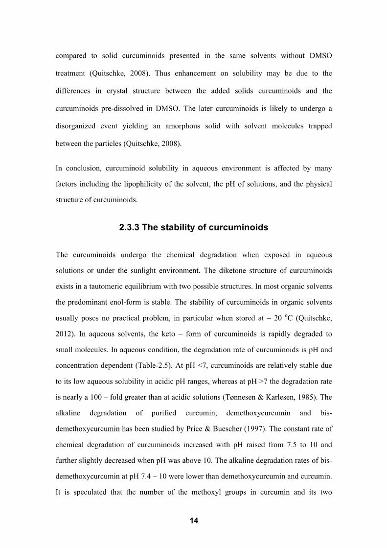
2.3.3 The stability of curcuminoids
The curcuminoids undergo the chemical degradation when exposed in aqueous
solutions or under the sunlight environment. The diketone structure of curcuminoids
exists in a tautomeric equilibrium with two possible structures. In most organic solvents
the predominant enol-form is stable. The stability of curcuminoids in organic solvents
usually poses no practical problem, in particular when stored at – 20 oC (Quitschke,
2012). In aqueous solvents, the keto – form of curcuminoids is rapidly degraded to
small molecules. In aqueous condition, the degradation rate of curcuminoids is pH and
concentration dependent (Table-2.5). At pH <7, curcuminoids are relatively stable due
to its low aqueous solubility in acidic pH ranges, whereas at pH >7 the degradation rate
is nearly a 100 – fold greater than at acidic solutions (Tønnesen & Karlesen, 1985). The
alkaline degradation of purified curcumin, demethoxycurcumin and bis-
demethoxycurcumin has been studied by Price & Buescher (1997). The constant rate of
chemical degradation of curcuminoids increased with pH raised from 7.5 to 10 and
further slightly decreased when pH was above 10. The alkaline degradation rates of bis-
demethoxycurcumin at pH 7.4 – 10 were lower than demethoxycurcumin and curcumin.
It is speculated that the number of the methoxyl groups in curcumin and its two

15
derivatives (demethoxycurcumin and bis-demethoxycurcumin) affects their aqueous
degradation rates (Price & Buescher, 1997). In addition, curcuminoids at a high
concentration in aqueous environment decomposed more readily than at low
concentration (Pfeiffer et al. 2003).
The loss of curcuminoids was also found when exposed to light. After 1-day exposure
to sunlight, 20% (w/w) curcuminoids in methanol solution was lost. The major
degradation products were found and tentatively identified as 4-hydroxybenzoic acid,
vanillic acid, vanillin, 4-hydroxybenzaldehyde, ferulic acid, and feruilc aldehyde. These
degradation substances were also found in turmeric root powder stored under sunlight
for 1 to 4 days (Schieffer, 2002). When curcuminoid powder protected by an aluminum
foil bag, the contents of curcuminoids remained stable for 4 months (Surojanametakul et
al. 2010). The main decomposition products of curcuminoids were ferulic acid and
feruloylmethane (mostly yellow to brownish-yellow), and further hydrolysed to vanillin
after incubation of curcumin in phosphate buffer (pH 7.2) for 8 h at 37 oC (Figure-2.3)
(Wang et al. 1997). Vanillic acid is other degradation product of curcumin, which was
detected and identified by liquid chromatography-mass spectrometry (Suresh et al.
2009). However, there is a debate on one of the intermediate degradation products
(Metzler et al. 2013). Gordon and Schneider (2012) claimed that trans-6- (4’ –
hydroxyl-3’-methoxyphenyl)-2,4-dioxo-5-hexenal (Figure-2.4) tentatively identified
using liquid chromatography-mass spectrometry by Wang and co-authors (1997) should
be one of the bicyclopentadiones arising through autoxidation. The lack of
curcuminoids stability is a crucial issue as it affects the bioavailability (plasma
concentration of parent compounds) as the rapidly degradation of curcuminoids in
aqueous condition at pH 6.8 – 7.4, which is the small intestine and body condition.

16
Table-2.5 Half-life (t1/2) for the degradation of curcumin at 37 oC (Wang et al. 1997)
pH Buffer systems t1/2, min
3.0 0.1 M Citrate-phosphate 118.6
5.0 0.1 M Citrate-phosphate 199.1
6.0 0.1 M Phosphate 195.6
6.5 0.1 M Phosphate 153.0
6.8 0.1 M Phosphate 39.7
7.2 0.1 M Phosphate 9.4
8.0 0.1 M Phosphate 1.0
10.0 0.1 M Carbonate 14.0
Figure-2.4 Chemical structure of curcumin and some degradation products (Metzler et
al. 2012).

17
2.3.4 Delivery of curcuminoids
Curcuminoids have the ability to bind directly to a diverse range of macromolecules due
to their two hydrophobic phenyl domains and carbonyl functional groups. These groups
are located at the ends and in the centre of the molecule, which can participate in
hydrogen bonding with a target macromolecule (Gupta et al. 2013). The use of
copolymer micelles, liposomes, polymeric nanoparticles, lipid-based nanoparticles,
hydrogel based encapsulation, protein-based matrix phospholipids have been reported to
deliver curcuminoids into the aqueous phase, whilst protected them from degradation at
neutral pH (Anand et al. 2007; Tapal & Tiku, 2012).
Formulated curcuminoids have an increased solubility compared to the raw powder. It
was shown a 13 x 105 fold increase in curcumin solubility when formulated with
polymeric micellar containing methoxy poly (ethylene-glycol)-block-polycaprolactone
diblock copolymers (MePEG-b-PCL) (Letchford et al. 2008) and a 3 – fold increase in
aqueous solubility for a curcumin-phospholipid complex compared to free curcumin
(Maiti et al. 2007). A recent investigation by Tapal & Tiku (2012) revealed significant
improvement in curcumin solubility in the presence of soy protein isolate.
Enhanced stability of curcuminoids can be achieved by formulation with lipids, proteins
and surfactant micelles (Anand et al. 2007). When curcuminoids bound with proteins or
partitioned into lipid phases, the aqueous stability was significantly enhanced (Barik et
al. 2003; Kim et al. 2011). The stability of a complex of curcumin with �-lactoglobulin
was effectively increased by 6.7 times, compared with curcumin alone in aqueous
solutions (Sneharani et al. 2010). Moreover, chemical degradation of curcuminoids can
also be diminished by interactions with surfactant aggregates such as micelles (Wang et
al. 2009) and in the chitosan-Tween 80 system (Boruah et al. 2012). These formulated
curcuminoid preparations with enhanced solubility and stability in aqueous systems is
expected to gain significant use as a novel ingredient to produce functional foods, as
well as drugs for disease treatments.

18
2.4 The bioavailability and bioaccessibility of curcuminoids
Bioavailability is used to evaluate the nutritional value of a given compound to the
human body. On oral consumption, the bioavailability of micronutrient and
phytochemicals (e.g. polyphenols) is described as the concentration of a given
compound or its metabolite at the target organ (Holst & Williasom, 2008). There are
numerous factors correlated with oral bioavailability including solubility, chemical
stability, uptake, metabolism, and excretion of the compound (s) (Anand et al. 2007).
The poor absorption from the gastrointestinal tract after oral administration is strongly
related to a poor bioavailability and it mainly due to (i) the low water solubility, as only
these solubilized compound (s) can be available for absorption in the small intestine,
and (ii) instability against gastrointestinal fluids (Takahashi et al. 2009) (Figure-2.5).
Figure-2.5 The basic events describing the fate of nutrients: (1) liberation, the release
of a compound from food matrix to become available for absorption

19
(bioaccessibility); (2) absorption, the movement of a compound from the
site of administration to the blood circulation; (3) distribution, the process
by which a compound diffuses or is transferred from the intravascular
(blood) to the extra-vascular space (body tissues); (4) metabolism, the
biochemical conversion or transformation of a compound into a form that is
easier to eliminate; and (5) excretion, the elimination of unchanged
compound or metabolites from the body, mainly via biliary, or pulmonary
pathway (Holst & Williamson, 2008).
In an early study, curcuminoid solids were suspended in arachis oil (1:1000, w/w) and
administered to rats by gavages in a single dose. The majority of that curcuminoids was
excreted in the faeces within 3 days, while negligible amounts (<0.1%) were measured
in the urine (Wahlström & Blennow, 1978). Similar observations were found in human
studies. The plasma curcumin concentrations after oral administration of curcumin
ranged from undetectable to the highest levels of about 1.75 µM (Quitschke, 2012). One
of the reasons regarding these low blood curcumin concentrations after oral ingestion is
the poor aqueous solubility of curcumin in the intestinal environment. It can be
overcome by using appropriate delivery systems, which enable to stabilize the target
compounds, whilst promote their solubility. Rat orally receiving curcumin formulated
with phosphatidylcholine showed 5-fold increase in serum curcumin concentrations
than those receiving standard curcumin (Quitschke, 2012). Additionally, lecithin –
piperine formulations containing curcuminoids and curcurminoids encapsulated in
cellulose have been reported to enhance bioavailability after oral administration in
humans (Antony et al. 2008; Vitaglione et al. 2012).
The metabolites of dietary polyphenols may be bioactive and have potential health
effects (Sies, 2010). The polyphenols are extensively modified after absorption, and
could be also be hydrolysed in the small intestine or in the colon. All these
modifications deeply affect the biological activity of polyphenols. Consequently, the

20
compounds that reach cells and tissues are chemically, biologically and functionally
different from the original form (D’Archivio et al. 2010). There is evidence that
curcuminoids were mainly converted into two metabolites (dihydrocurcumin,
tetrahydrocurcumin) and one catabolite (ferulic acid) by interaction with purified human
colon bacteria strains in vitro (Tan et al. 2014). These converted curcuminoids may be
re-absorbed in the large intestine and contribute to the bioactive functions on their own
way. The oral bioavailability of curcuminoids incorporated into bread was evaluated by
identification and quantification of curcuminoids, curcuminoid metabolites (i.e.
curcumin-glucuronide, hexahydrocurcumin-glucuronide) and breakdown phenolic acids
(i.e. ferulic acid and vanillic acid) in serum, urinary and feces within 24 h. Over 24 h,
the overall bioavailable curcuminoids including total curcuminoids, curcuminoid
glucuronides and phenolic acids taken account of 0.09% of dose ingested for free
curcuminoids and 0.17% of dose (1 g curcuminoids in a 100g potion) ingested for
formulated curcuminoids (Vitaglione et al. 2012).
The bioaccessibility is a prerequisite to bioavailability, which typically evaluates the
percentage of solubilized target compound(s) in the gastrointestinal fluids. The
assessment of bioaccessibility is used in functional food design, in fortification of food
formula with bioactive compounds, and in studying the effect of food processing on
nutritional quality (Fernández-Garćia et al. 2009). In a food system, it will assess the
dissociation, solubilisation and transportation of these bound target compound (s) from
the delivered matrix, prior to their uptake across a physiological membrane (Fernández-
Garćia et al. 2009). It is considered the absorption of lipophilic compounds relating with
the solubility of these compounds in the intestinal environment via partitioning into bile
salts-micelles (Porter et al. 2007). Therefore, the solubilized compounds in the intestinal
environment are considered as the bioaccessible ones. Several formulated curcuminoids
in an oil-in-water emulsion and nanoemulsion (Ahmed et al. 2012; Yu & Huang, 2012),
oil-in-water organogel (Yu et al. 2012), and nanostructured lipid carriers (Aditya et al.

21
2013) have been evaluated under in vitro simulated gastrointestinal (enzymatic)
digestion and the percentage of solubilized curcuminoids as the bioaccessibility (%)
determined. The bioaccessibility of curcuminoids reached 60% after in vitro
gastrointestinal digestion of organogel formulated curcuminoids in the fed state, which
was significantly increased compared with neat curcuminoids with the 5%
bioaccessibility obtained under the same digestion treatment (Yu et al. 2012).
2.5 Buttermilk
2.5.1 Buttermilk and its composition
Buttermilk is a by-product after butter manufacturing. Buttermilk differs from non-fat
milk (i.e. skim milk) in that it contains phospholipids and special proteins from the milk
fat globule membrane, whereas non-fat milk does not (Contarini & Povolo, 2013;
Sodini, 2006) (Table-2.6). Buttermilk can be generally divided into the following
categories: (i) traditional buttermilk is the liquid left over from churning butter, which is
also called “sweet buttermilk”; (ii) cultured buttermilk is known as a fermented dairy
product, which is produced by lactic acid fermentation of skim milk or milk containing
less fat than whole milk (Libudzisz & Stepaniak, 2002). In the following texts,
buttermilk refers to sweet buttermilk only.
Table-2.6 The gross composition (%) on a dry material basis of buttermilk and skimmed
milk (Sodini, 2006).
Samples Total N Fats Phospholipids Lactose Ash
Skim milk powder 35.9 0.3 ND2 55.8 8.0
Buttermilk powder
BM1 32.9 5.7 1.29 53.8 7.6
BM2 33.1 7.2 1.34 52.4 7.3
BM3 31.5 13.1 1.27 48.7 6.7
BM4 27.8 22.3 1.15 43.7 6.2
BM5 14.1 15.5 1.87 63.4 7.0

22
1 Samples: One commercial skim milk and five buttermilk (BM) powders. Three buttermilks were
obtained from sweet buttermilk (BM1, BM2 and BM3), one buttermilk sample was obtained from
cultured cream (BM4), and one was obtained from whey cream (BM5). Two buttermilk were
commercial (BM 1 and BM2), and three were manufactured at a pilot-scale (BM3, BM4 and BM5). 2ND: Not determined.
Buttermilk contains lactose, caseins, serum proteins, lipids and minerals. The chemical
composition of buttermilk depends significantly on the butter-making technology. The
impact of processing method, a high-speed churning (1000 – 2500 rpm) and a low-
speed churning (20 – 30 rpm for 40 – 60 min), have been reported to yield different
buttermilk compositions (Gassi et al. 2008).
Compared to the skim milk, the high content of milk fats, especially milk fat globule
membrane in buttermilk makes this dairy ingredient interesting for use as a functional
ingredient (Table-2.6). The high content lipids presented in buttermilk powder could
give desirable tasty and texture to end-prodcuts. For example, the addition of buttermilk
(4.8% wt.) to low fat yogurt yields a soft and smooth product with highest flavor and
smoothness scores and overall acceptability in sensory evaluation of yogurts made with
no-fat milk powder and buttermilk powder (Trachoo & Mistry, 1998). Moreover, the
quantity of vitamin A, �-tocopherol and cholesterol per gram of fat was 9 times higher
in the buttermilk than in its butter (Tylkin et al. 1975). A part of these substances in the
membrane layer were removed together with the hydrophilic part during the butter
making process. Similarly, the content of retinol per gram of fat in buttermilk was
significantly higher (p <0.05) than that in butter oil (Zahar et al. 1995). In addition,
lipids are considered to be beneficial to human health (Dewettinck et al. 2008; Spitsberg
et al. 2005). Enzymatic hydrolysed lipids such as diacylglycerides, monoglycerides and
phospholipids could promote the fat soluble nutrient absorption in the small intestine
(Porter et al., 2007). As a by-prodcut, buttermilk is a vauleable dairy ingredient because
of its natural properties and its positive impact on flavour.

23
2.5.2 Physical Functionality of buttermilk
(a) Emulsification
Emulsification is an ability to stabilize interfaces. It is the major desirable functionality
for cream, ice cream, confectionery, bakery and fish/meat products (Augustin &
Udabage, 2007; Ihara et al. 2011). Emulsion stability is strongly related with the type of
protein present, as well as the surface changes of the surface proteins. Buttermilk has
been reported to have higher emulsifying properties than milk and whey because of the
presence of milk fat globule membrane components (Sodini et al. 2006). An early study
by Kanno (1989) showed that only 5% of milk fat globule membrane was needed to
emulsify 25% milk fat (i.e. 40 mg milk fat globule membrane /g fat) and the emulsion
was stable within a temperature range of 4 oC – 55 oC, and at a pH of either 4 or
between 6 - 9. Similarly, the addition of buttermilk improved the emulsifying property
and acid tolerance of milk cream, especially the buttermilk with higher phospholipids
content (Ihara et al. 2011). The sole addition of phospholipids has no effect on the
emulsifying property and acid tolerance of cream, suggesting that these improved
functions may contribute to the formation of complexes of phospholipids and proteins
in buttermilk (Ihara et al. 2011).
(b) Foaming
A foam is a colloidal dispersion of air in a liquid phase. Protein forms foams by rapid
absorbing and unfolding at the interface of air and liquid, and forming a cohesive film
and this viscous-elastic film is attributed to protein-protein interaction, such as
electrostatic and hydrophobic interactions (Dickinson, 2010). The foaming capacity of
buttermilk is significantly lower than skim milk and whey powder with 1.5 to 2.5 mL of
foaming per mL of liquid, compared with 2 to 4 mL for skim milk and whey,
respectively. The smaller foaming capacity of buttermilk is probably due to its high

24
contents of the phospholipids, which have antifoaming properties (Wong & Kitts, 2003).
Moreover, among the various buttermilks, buttermilk containing high fat exhibited
lower foaming capacity than that containing low fat. In addition, the ratio of
phospholipids to protein can also affect the foaming properties (Sodini et al. 2006).
(c) Gelling
Gelling is a property related to hydration and the formation of a network, increasing the
viscosity of a solution (Augustin & Udabage, 2007). It is the major functional property
in yogurt and cheese manufacturing because gels produce the required textural
characteristics and retain water, fat and other components. The gel formed by milk
proteins is mostly irreversible and results from enzymatic reactions (rennet-induced),
heat-induced interactions, cation (calcium) interactions and the combined reaction
mechanisms. The strength of whey protein gels can be significantly affected by �-
lactoglobulin content in whey proteins, but there is no significant relationship to �-
lactoglobuin content. The stability of a whey protein gel is related to the disulfide
bonds and sulfhydryl groups, which link the proteins via covalent bonds to form
network. Casein micelles can form gels through acidification or renneting. The
properties of casein gels are affected by heating conditions, the concentration of casein,
pH, ionic strength and concentration of additives such as polyphenols (Hayes et al.
1968; O’connell & Fox, 2001). The acid gelation of milk proteins is a process
involving the formation of lactic acid by bacteria and the reduction in surface change
on casein micelles from high net negative charge at pH 6.7 to close to no net charge at
pH 4.6 (isoelectric point). This surface change allows casein micelles to aggregate
through hydrophobic and electrostatic bonds, forming protein gels (Lucey & Singh,
2003). The mechanism of polyphenols affecting milk gelation is due to alteration of
whey protein thermal denaturation and surface properties of casein micelle. The

25
interaction of proteins with polyphenols may improve the thermal stability of protein
(Ozdal et al. 2013), and inhibit the dissociation of κ-casein protein from the casein
micelles (Schamberger & Labuza, 2007). A typical milk gel caused by acid gelation is
yogurt and its important property is the ability of retaining water in the gel structure.
The yogurt gel matrix consisited of protein chains aggregates and casein micelles
associated with denatured whey protein at the micellar surface, and the fat globules can
be embedded in the protein network (Sandoval-Castilla et al. 2004). The addition of
casein or whey proteins would alter the yogurt texture (Keogh & O’kennedy, 1998).
The interaction between denatured whey proteins and casein is responsible for the
formation of network that affects the texture of yogurt. As the modified casein to whey
protein ratio varied from 4.7:1 to 0.5:1, the maximum gel strength of the yogurt
increased as the casein to whey protein ratio decrease (Puvanenthiran et al. 2001). In
addition, yogurt with increasing fat content had higher viscosity and lower relative
particle size, whilst the lipid content in yogurt promoted the overall flavor by
increasing the volatile intensity of the lipophilic compounds (Brauss et al. 1999).
Therefore, compared to the yogurt made with skim milk, the buttermilk yogurt could
have desirable favors due to the high lipid content in buttermilk.
2.5.3 Applications of buttermilk
Traditionally, buttermilk has been used in animal feeding systems as an excellent
supplemental protein source for growing aminals (Flanders & Gillespie, 2015). In the
bakery industry, the addition of buttermilk has a positive effect on baked product
appearance and flavour development (Bilgin et al. 2006). The development of modern
food technology promotes the innovation in new food ingredients, and the proper use of
their functional properties may lead to new ingredients. Buttermilk can be extracted,

26
purified and fractionated in very distinct food and non-food applications. The potential
utilization of milk fat globule membrane isolated from buttermilk was widely
investigated due to its high content of phospholipids and specific proteins (Sodini et al.
2006). It has been suggested the milk fat globule membrane material could be
considered as natural emulsifying agents, and it can be used as a carrier for many
bioactives (e.g. polyphenols), boosting their bioavailability (Sodini et al. 2006;
Spitsberg, 2005). The use of buttermilk as a whole to carry bioactive compounds,
especially polyphenols has not been reported extensively. The potential of this
application will be described in the following section (2.6 Delivery of curcuminoids
using milk-based systems) and further investigated in this thesis.
2.6 Delivery of curcuminoids using milk-based
systems
The basic purpose of designing delivery systems for polyphenols is to improve their
bioavailability via increasing the aqueous solubility and stability. In theory, such a
delivery system will focus on the existing data of the literature, which mainly describes
the impact of a single buttermilk component on polyphenols such as curcumin(oids)
bioavailability.
Milk proteins can enhance solubility and stability of some polyphenols by forming
complexes. �s1–Casein, one of the major protein components of the casein fraction of
milk, has been reported to bind to curcumin by hydrophobic interactions with two
binding sites, one with high affinity (2.0 × 10⁶ M-1) and the other with low affinity (6.3
× 104 M-1). Such interactions significantly enhanced the stability of curcumin in
aqueous condition (Sneharani et al. 2009). Curcumin also forms a complex with casein
micelles with the binding constant of 1.5 × 10⁴ M-1. Casein micelles-curcumin complex

27
exhibited similar cytotoxic effects on HeLa cells compared to an equal dose of free
curcumin (Sahu et al. 2008). � -lactoglobulin, � -lactalbumin, lactoferrin, holo-
lactoferrin, apo-lactoferrin and whey protein isolates have been reported to interact with
resveratrol (the lipophilic polyphenol enriched in wine). Interaction between resveratrol
and �-lactoglobulin provided a slight increase in the photostability of resveratrol and a
significant increase in its solubility (Liang et al. 2007). Such protein-bound polyphenols
has a greater aqueous stability when exposed in simulated gastrointestinal fluids
compared to the neat polyphenols. It is likely that milk protein has protective effects on
polyphenols, correspondently promoting the intestinal absorption (Gallo et al. 2013).
Phospholipids are used to deliver lipophilic compounds as the polar functionalities of
the lipophilic compound can interact with the polar head of a phospholipid via hydrogen
bonds (Bombardelli, 1991; Bombardelli et al. 1994; Bombardelli & Stelta, 1991).
Interactions of curcumin with phosphatidylcholine micelles from egg yolk,
phospholipids from soybean lecithin, L-� -phosphatidylcholine from soybean and
lecithin from Phospholipon 85G have been estimated (Began et al. 1999; Liu et al.
2012; Takahashi et al. 2009; Niu et al. 2012). These studies concluded that liposomal
curcumin was significantly increased in stability and can enhance in oral bioavailability.
Moreover, the liposomes obtained from the phospholipid fraction from milk fat globule
membrane material were more stable than the soybean-based liposomes. They showed
less change in average diameter, morphology, and free fatty acid release, and better
membrane integrity during digestion in a simulated intestinal fluid (Liu et al. 2012). In
in vivo studies, formulated curcumin with phospholipids showed a maximum plasma
curcumin level of 600 ng/mL after 2.33 h of oral administration as opposed to that of
free curcumin having maximum plasma concentration of 267 ng/mL after 1.62 h of oral
dosing. Similarly, plasma levels for curcumin after administration of Meriva®
(formulated curcuminoids within phospholipids) were 5-fold higher than the equivalent

28
values after unformulated curcumin dosing (Marczylo et al. 2007). It is highly expected
that buttermilk proteins and lipids can advance the oral bioavailability of curcuminoids
when delivered in buttermilk.
Effect of milk proteins on the polyphenol absorption is debated so far, which mainly
concerns whether milk proteins reduce the biological accessibility of the polyphenols
when ingested them together. Serafini and co-authors (2003) showed an inhibitory
absorption of (-) epicatechin (the dietary flavonoids from chocolate) into the blood
stream within 4 h when ingested dark chocolate with milk or ingestion of milk
chocolate. Thus reduction of (-) epicatechin absorption could be related to binding
interactions between flavonoids and milk proteins that occurred in food processing or
human digestion (Serafini et al. 2003). In in vitro simulated digestion studies, 17% and
27% of 5’-chlorogenic acid (the main polyphenol in coffee) were bound with intestinal
digested skimmed milk and caseins, respectively. This finding suggested that 5’-
chlorogenic acid could remain bound to milk proteins at the end of in vitro digestion
process with a consequent result in modification of coffee polyphenols absorption in the
body (Dupas et al. 2006). Some observations regarding influence of milk proteins on
oral bioavailability of certain polyphenols were that milk proteins might delay the
absorption of those polyphenols in a gastrointestinal tract, but not compromise the
overall absorption (Vitaglione et al. 2013; Scalbert, & Williamson, 2000). The study on
effect of cream on the absorption and metabolism of strawberry anthocyanins found that
the delayed plasma absorption (Tmax) of anthocyanins metabolites when eaten
strawberry with cream, but no effect on the plasma maximum concentration (Cmax) of
their metabolites (Mullen et al. 2008). It may be contributed by the alteration of gastric
emptying or mouth to cecum transit time (Mullen et al. 2008). In the other human study,
after ingestion of orange juice that contains hesperetin-7-�-rutinoside and naringenin-

29
7-� -rutinoside as major polyphenols, the Cmax of accumulated hesperetin-7-� -
glucuronide and unidentified hesperetin-�-glucuronide in plasma was detected at 4.4 h
(Tmax). This Tmax indicated that orange polyphenols were absorbed in the colon (Mullen
et al. 2008). When the orange juice was indigested with full-fat yogurt, the
pharmacokinetic parameters (Cmax, Tmax and AUC) in the plasma were insignificantly
different compared with the values obtained from control groups that ingested orange
juice without full-fat yogurt (Mullen et al. 2008). It is likely that milk proteins could
alter the metabolism of polyphenols in the gastrointestinal tract, and enhances the re-
absorption of polyphenols in the large intestine via the action of colonic microbiota
(Roowi et al. 2009; Scalbert & Williamson, 2000). It should bear in mind that the
intestinal absorption and metabolism of polyphenols in the body and the impact of food
matrix on their bioavailability significantly depend on individuals. It is because of the
varied structures of different types of polyphenols with their individual physicochemical
proterties (Augustin & Sanguansri, 2014). For example, epicatechins and
epigallocatechins are absorbed in the small intestine, whereas chlorogenic acid,
anthocyanins are substantially metabolized in the large intestine (Ausustin &
Sanguansri, 2014). Water-soluble polyphenols such as tea catechins are required to be
protected against oxidation and under high-pH conditions. The amphipathic
polyphenols (e.g., resveratrol, curcumin) need to improve their solubility in aqueous
media as well as be protected from aqueous degradation. The delivery system designed
for a polyphenol should depend on case-by-case basis.

30
2.7 Effect of added polyphenols on the properties of
dairy products
In recent years, the value of phenolic compounds has been highlighted because they
have been applied in milk and dairy products as nutraceutical additives. The addition of
phenolic compounds alters the functional properties of milk such as acid gelation, heat
and storage stability. Milk fortified with tannic acid or gallic acid (phenolic compounds)
had a faster gelation time, compared with the control without addition of these phenolic
compounds. And the gallic acid fortified milk showed an increase in water binding
capacity (Harbourne et al. 2011). The addition of phenolic compounds positively
influenced the heat stability of milk. The addition of caffeic acid at 5.5 mM markedly
enhanced the heat stability of milk at 140 oC, and inhibited the dissociation of �-
casein-rich protein from the casein micelles via the reduction of the reactive lysine and
sulphydryl content. It was suggested applying green tea flavonoids into the ultra-high-
temperature treated milk (UHT milk) to control Maillard browning during storage
(Schamberger & Labuza, 2007). In fact, the real taste of some polyphenols is bitter and
astringent. The sensory changes resulted from adding polyphenols to milk and dairy
products should be considered. The study on the effect of phenolic compounds extracted
from olive vegetable water on functional milk beverage suggested that the
concentrations of 100 to 200 mg/L of olive phenolic compounds in functional milk
beverage were acceptable based on triangle and paired comparison tests (Servili et al.
2011). The benefits of adding polyphenol have been reported in cheese production. The
addition of polyphenols derived from wood smoke imparted desirable sensory attributes
to certain cheeses, which was traditionally made by direct smoking (O’Connell & Fox,
2001). However, negative effects of phenolic compounds (grape and pomegranate

31
extracts, 2.5 mL/kg of 10% solution) added to sheep milk for making strained yogurts
have been patented. The sensory acceptance of strained yogurt containing phenolic
compounds significance decreased when comparing to the control samples. It may be
due to the original taste of polyphenols as astringent and bitterness (Ersöz et al. 2011).

32
CHAPTER 3 GENERAL MATERIALS AND
METHODS
3.1 Experimental Design
All experiments listed in the following sections were repeated at least twice, and three
measurements were recorded within each experimental trial. The data used in Chapter 4,
5, 6 and 7 are the average of at least three measurements.
3.1.1 The interaction between buttermilk and curcuminoids
(a) Thebindingaffinitybetweencurcuminoidsandbuttermilk
The protein quenching study was used to assess the protein-ligand interaction, and the
apparent binding constant was obtained and used to describe the strength of the affinity
between protein and ligands. The fluorescence measurement is used to estimate the
interaction between compounds containing fluorophores. The fluorophores’ structure
changes in result of interactions alter the intensity of fluorescence. Both curcuminoid
compounds and proteins have their intrinsic fluorescence. The apparent binding
constant of buttermilk compounds and curcuminoids and the number of binding sites
were determined following the method of Liang and co-authors (2008) with some
modifications. Quenching data obtained at an excitation wavelength � 280nm were
utilized to estimate the number of binding sites, and the apparent binding constants of
curcuminoids with proteins. These studies concerning on the binding between
curcuminoids and buttermilk compounds in separated cream fraction and serum fraction
were also carried out.
(b)Partitioningofcurcuminoidsbetweenproteinandlipidfractions

33
The partitioning of curcuminoids in three fractions (i.e. cream, serum and casein)
separated from whole buttermilk was evaluated to further understand the associations of
curcuminoids with casein, whey proteins and buttermilk lipids, mainly the milk fat
globule membrane lipids. Curcuminoid-buttermilk mixture was subjected to
ultracentrifuge and obtained those separated fractions, which contained curcuminoids.
The quantity of curcuminoids in those separated fractions was measured using an
HPLC-DAD assay.
(c) The aqueous stability of curcuminoids in buttermilk
The aqueous stability of curcuminoids in buttermilk was determined at 4 oC for up to 6
days and it was compared with curcuminoids in buffer alone under the same storage
condition. The change of curcuminoids in buttermilk and buffer within incubation time
was measured at 0, 1, 2, 3 and 6 days. The amount of curcuminoids was measured using
an HPLC-DAD assay.
Figure-3.1-3.3 illustrates the different steps in curcuminoid buttermilk interaction,
partitioning and stability studies.

34
Figure-3.1 The experimental design for estimation of binding affinities between
curcuminoids and buttermilk compounds. The protein intrinsic fluorescence
(tryptophan and tyrosine residues) is measured at excitation wavelength
λ280nm. The curcuminoids intrinsic fluorescence is measured at exciation
wavelength λ420nm.
Buttermilk
Separate into fractions
Casein pellet
Serum Cream Buttermilk
Mix with curcuminoids (0-14 µM)
Binding affinities
(fluorescence methods)

35
Figure-3.2 The experimental design for partitioning of curcuminoids in separated
fractions.
Figure-3.3 The experimental design for stability of curcuminoids in the presence of
buttermilk.
Buttermilk-curcuminoids
mixture
Separate into fractions
Casein pellet
Serum Cream Buttermilk-
curcuminoids mixture
Extract curcuminoids from each fraction and whole buttermilk
Amount of curcuminoids in each
fraction and whole buttermilk
(HPLC-DAD)
Buttermilk-curcuminoids
mixture
Curcuminoids in buffer (pH 6.8)
Incubation at 4 oC
for 6 days
Amount of curcuminoids
before and after incubation
(HPLC-DAD)

36
3.1.2 Bioaccessibility of curcuminoids in buttermilk 1
(a) The bioaccessibility of curcuminoids in buttermilk 2
The bioaccessibility was reported as % of solubilized curcuminoids presented in 3
micellar aqueous phase of enzymatic-digested sample in SGF and SIF and evaluated in 4
comparison with the curcuminoids in buffer. This solubilized curcuminoids in 5
gastrointestinal fluids were separated from enzymatic-digested sample by centrifugation 6
and considered the bioaccessible curcuminoids. The simulated gastrointestinal 7
(enzymatic) digestion models used in this bioaccessibility study were developed and 8
shown in Section 3.4.3. The quantities of curcuminoids in enzymatic-digested samples 9
and in micellar aqueous phase were determined by an HPLC-DAD assay. 10
(b) The stability of curcuminoids in in vitro gastrointestinal (enzymatic) digestion 11
The stability of curcuminoids expressed as a % of remaining curcuminoids in digested 12
samples was determined by comparing the difference of curcuminoids presented in 13
gastrointestinal fluids before and after in vitro continuous digestion for 2 h in gastric 14
fluids and 3 h in intestinal fluids at 37 oC. 15
(c) The digestibility of buttermilk containing curcuminoids 16
The difference in proteolysis and lipolysis between buttermilk-curcuminoids and 17
buttermilk alone were compared. The proteolysis and lipolysis were evaluated by SDS-18
PAGE and GC analysis following the methods published by Gallier and co-authors 19
(2012) and Shen and co-authors (2014) with some modifications. 20
Figure-3.4 illustrates the different steps in bioaccessibility studies. 21

37
Figure-3.4 The experimental design for bioaccessibility of curcuminoids in buttermilk in
simulated gastrointestinal (enzymatic) digestion models.
Buttermilk-curcuminoids
Curcuminoids in buffer
In vitro gastrointestinal digestion in simulated digestion models
(fasted and fed states)
Digestibility of proteins with and
without curcuminoids (SDS-PAGE)
Digestibility of lipids with and without
curcuminoids (Lipolysis by GC)
Solubilized and total curcuminoids in digestion fluids
(HPLC/Visible-UV)
Buttermilk alone
Bioaccessibility of curcuminoids in buttermilk and buffer In vitro stability of curcuminoids in buttermilk and buffer

38
3.1.3 Properties and bioaccessibility of curcuminoid yogurts
(a) Physical and microbiological properties of curcuminoid yogurts
Curcuminoid fortified yogurts were manufactured by adding curcuminoids (300
mg/100g buttermilk) into buttermilk (total solids 14%, w/w). The measured properties
included total solids, apparent viscosity, counts of lactic acid bacteria, and fermentation
time (the time for yogurts to reached pH 4.6) were carried out in curcuminoid fortified
yogurts and non-fortified yogurts. The effect of curcuminoids on the properties of
buttermilk yogurts was evaluated.
(b) The bioaccessibility of curcuminoids in buttermilk yogurts
As mentioned before the bioaccessibility was expressed as % of solubilized
curcuminoids presented in micellar aqueous phase of gastrointestinal digested sample
and evaluated in comparison with the curcuminoids in buffer alone. Meanwhile, the
bioaccessibility of curcuminoids in buttermilk yogurts fortified with either curcuminoid
powder or curcuminoids dissolved in ethanol prior to addition, was measured and
compared to estimate the function of ethanol (2%, w/w) on the curcuminoid
bioaccessibility.
(c) The stability of curcuminoids in buttermilk yogurt manufacture and in in vitro
gastrointestinal (enzymatic) digestion
The stability of curcuminoids in fortified buttermilk yogurt was reported as a % of
curcuminoids presented in end products (e.g. curcuminoid yogurts). The chemical
stability of curcuminoids when exposed to gastrointestinal fluids was expressed as a %
of remaining curcuminoids in digested samples that were subjected to simulated
continuous digestion for 2 h in gastric fluids and 3h in intestinal fluids at 37 oC.
Figure-3.4 illustrates the different steps in the bioaccessibility of curcuminoids in a
buttermilk yogurt food system.

39
Figure-3.5 The experimental design for the properties and bioaccessibility of
curcuminoid yogurt.
3.1.4 In vitro faecal slurry fermentation of non-bioaccessible
curcuminoids
To understand whether the insoluble curcuminoids (non bioaccessible) remaining in the
centrifuged sediment obtained upon centrifugation of samples previously exposed to
simulated gastric and intestinal fluids can interact with faecal bacteria, the simulated in
vitro human faecal slurry fermentation study was used to examine the amount of
curcuminoids reduced when exposed to human faecal slurries. The approach used with
in vitro fermentation was according to the method of Dall’Asta and co-authors (2012).
The amounts of curcuminoids presented in fermentation mixtures were quantified at 0
Buttermilk yogurt (2% w/w EtOH)
Buttermilk yogurt fortified with curcuminoids (2% w/w EtOH)
Buttermilk yogurt (No EtOH)
Buttermilk yogurt fortified with curcuminoids (No EtOH)
*Total solids *Apparent viscosity *Lactic acid producing bacterial counts *Fermentation time (pH reached to 4.6)
Characteristics of yogurt making and properties of yogurts
Curcuminoids in buffer (2% w/w EtOH)
Curcuminoids in buffer (No EtOH)
Stability of curcuminoids during yogurt making
Stability of curcuminoids in in vitro gastrointestinal digestion
Bioaccessibility of curcuminoids after in vitro gastrointestinal
digestion

40
and 24 h. Figure-3.4 illustrates the different steps in the in vitro faecal slurry
fermentation of non-bioaccessible curcuminoids.
Figure-3.6 The experimental design for in vitro faecal slurry fermentation of non-
bioaccessible curcuminoids.

41
3.2 Materials
A turmeric extract (Bio-curcumin®) used throughout this study was kindly donated by
Arjuna Natural Extracts Limited (Alwaye, Kerala, India). According to the supplier’s
specification sheet, this turmeric extract contains 95.8% of total curcuminoid complex,
which consists of curcumin, curcuminoids, and volatile oils from turmeric. The amount
of curcumin, demethoxycurcumin and bis-demethoxycurcumin in Bio-curcumin® were
quantified by an HPLC-DAD method published by Jayaprakasha and co-authors (2002)
with some modifications. The Bio-curcumin® was sub-packed into small bags (~100g /
bag), and vacuum-sealed. Each sub-packed turmeric extract powder was wrapped with
aluminium foil and stored in a cool room (4 oC) until used.
A curcumin standard, with a purity of 99% curcuminoids (93% curcumin and 7%
demethoxycurcumin) was purchased from Sigma-Aldrich (Sydney, Australia) and used
for quantification. The curcumin standard was stored at -20 oC.
Buttermilk powder was obtained from Warrnambool Cheese and Butter Factory
(Allansford, Victoria, Australia). According to the manufacturer’s specification, the
buttermilk powder contained 32.5% protein, 8.4% fat, 50.1% lactose and 2.9% moisture.
Buttermilk powder was sub-packed into the plastic bags with zip (~500g / bag) and
double packed with aluminium bags outside with seals. Packed buttermilk was stored in
a cool room (4 oC) during the study period. Other materials used in main experiments
were detailed in relevant chapter (4, 5 and 6) under Material and Methods.

42
3.3 Methods
3.3.1 Determination of curcuminoids
Curcuminoids were identified and quantified using (a) HPLC-DAD (Jayaprakasha et al.
2002), (b) UV-VIS spectrophotometer (Silva-Buzanello et al. 2014) and (c)
fluorescence methods (Yazdi & Corredig, 2012). Quantification was performed using
an external standard technique.
(a) HPLC-DAD method
The separation of curcumin, demethoxycurcumin and bis-demethoxycurcumin was
achieved using a C18 silica saturator column (4.6 mm x 250 mm, particle size 5 �m,
SUPELCO, USA) equipped with a Shimadzu HPLC System (Japan). Acetonitrile with
methanol (10%, v/v) and acetic acid (2%, v/v) (mobile phase A), and MilliQ-water with
acetic acid (2%, v/v) (mobile phase B) were used as running mobile phases. The
gradient elution program of mobile phases was presented in Table-3.1. The HPLC
standard profiles of curcuminoids in curcumin standard (from Sigma) and Bio-
curcumin® (from Arjuna Natural Extracts Limited) were presented in Figure-3.7. The
limit of quantification (LQD) was ~2 µg/mL for curcumin and demethoxycurcumin in
ethanol (100%). Bio-curcumin® was further used as an external standard for
quantifications in further experiments.
Table-3.1 The HPLC gradient program used in determining curcuminoids
Time (min)
Acetonitrile (88%), methanol (10%) and acetic acid (2%)
( %, v/v)
MilliQ-water (98%) and acetic acid (2%)
(%, v/v)
Flow rate (mL/min)
0.00 35 65 1.00
10.00 45 55 1.00 20.00 45 55 1.00
25.00 35 65 1.00

43
Figure-3.7 The HPLC chromatogram of curcumin standard (46 µg/mL in ethanol; the
top profile) and Bio-curcumin® (21.1 µg/mL in ethanol; the bottom profile).
Minutes2 4 6 8 10 12 14 16 18 20 22 24
mAu
0
50
100
150
200
250
300
350
400
450
mA
u
0
50
100
150
200
250
300
350
400
450
1
3.7
40
1
.78
0
Pe
ak
@ 1
4.6
64
Min
ute
s 1
4.7
56
8
.38
4
1
5.8
12
8
9.8
36
Curcumin
Demethoxycurcumin
Minutes2 4 6 8 10 12 14 16 18 20 22 24
mAu
0
20
40
60
80
100
120
140m
Au
0
20
40
60
80
100
120
140
Pe
ak @
14
.24
4 M
inu
tes
1
3.8
92
3
.58
6P
ea
k @
14
.66
4 M
inu
tes
1
4.9
04
1
7.5
33
1
5.9
48
7
8.8
80
Curcumin
Demethoxycurcumin
Bis1demethoxycurcumin

44
(b) UV-VIS spectrophotometer method
The UV-VIS spectrophotometer method is an alternative approach to determine the total
curcuminoids. It was used when the concentration of curcuminoids was close or lower
than the limit of detection (LQD) of the HPLC method. Curcumin, demethoxycurcumin
and bis-demethoxycurcumin in ethanol have specific UV absorptions at 420 nm, 425
nm and 430 nm, respectively (Jayaprakasha et al. 2002). The overall specific UV
absorption for Bio-curcumin® in ethanol (100%) was 425 nm (Figure-3.8).
Figure-3.8 The scan profile of UV spectrum of Bio-curcumin® in ethanol (100%) and
the specific maximum absorption was at 425 nm.
(c) Fluorescence measurement
The fluorescence method is a sensitive approach to quantify total curcuminoids.
Curcuminoids (in ethanol 100% and 70%) were identified by an emission wavelength at
540 nm when excitation at 420 nm (Figure-3.9). The advantages of fluorescence
measurement are, the easy and fast operation, and the ability to quantitate extra low
amounts of tested compounds, which can facilitate the measurement of curcuminoids at
nanogram levels (Mazzarino et al. 2010).
425nm&

45
Figure-3.9 Fluorescence spectra of curcuminoids (3.7 µg/mL, ~10 µM) at an excitation
wavelength of 420 nm in 100% ethanol (v/v) and 70% ethanol (v/v).
Considering the fluorescence quenching effect, the relationship between fluorescence
intensity (excitation at 420 nm and emission at 540 nm), and the concentration of
curcuminoids from 0 - 20 µg/mL in 80% ethanol (v/v) were estimated and illustrated in
Figure-3.9. The use of 80% ethanol (v/v) in this testing was due to the fact that
curcuminoids were extracted in 80% ethanol (v/v) in experiment of buttermilk-
curcuminoids interaction. An appropriate quantification range for curcuminoids in 80%
ethanol (v/v) was between 0 - 4.0 µg/mL. The maximum concentration of curcuminoids
in 80% ethanol (v/v) was 5.0 µg/mL by using the fluorescence method.
3.3.2 Extraction of curcuminoids
Curcuminoids can be dissolved in organic solvents, such as acetone, alcohol, ethylene
dichloride, dimethyl sulfoxide (DMSO), and dimethylformamide. The extraction
0
50
100
150
200
250
450 500 550 600 650 700 750
Inte
nsity
(a.u
)
Emission wavelength (nm)
10 µM curcuminoids in 100 % ethanol (v/v)
10 µM curcuminoids in 70 % ethanol (v/v)
Max. emission wavelength at 540 nm
Max. emission wavelength at 540 nm

46
efficiencies of curcuminoids from turmeric samples have been reported in the literature,
and showed that methanol, acetone, ethyl acetate and chloroform have greater results
than hexane with total extracted curcuminoids of 4.31 mg, 3.49 mg, 3.20 mg, 3.09 mg
and 0.90 mg, respectively (Revathy et al. 2011). The extraction methods used in this
thesis were developed and validated using spiking experiments (Table-3.2).

47
Table-3.2 The extraction methods and recoveries of curcuminoids. *Curcuminoids were spiked before heating.

48
3.3.3 Development of in vitro simulated gastrointestinal digestion models
The use of simulated gastrointestinal digestion is an applicable approach to evaluate the
bioaccessibiltiy of target compound (s) in a food matrix. Several parameters, such as the
concentrations of pepsin, pancreatin, bile salts, CaCl2, ionic strength and pH are
important in the bioaccessibility studies (Li et al. 2011; Hur et al. 2011).
In this study, the preparation of simulated gastric fluid (SGF) and the concentration of
pepsin were followed the U.S. Pharmacopeia Convention guidelines (United States
Pharmacopeia Convention, 2009). While the conditions applied in the intestinal
digestion were simulated according to the previous publications on bioccessibility of
lipophilic substances in oil-in-water systems (Li et al. 2011; Salvia-Trujillo et al. 2013).
The concentrations of pancreatin and bile extract used in this study were modified as
they strongly affect the digestibility of milk proteins and lipids and also influence the
solubility of lipophilic compounds in the intestinal fluids.
Curcuminoid-buttermilk was subjected to gastric digestion (2 h, 37 oC) with pepsin (3.2
mg/mL of gastric fluids) and intestinal digestion (3 h, 37 oC) with various
concentrations of pancreatin (0.2 mg – 12 mg /mL of gastrointestinal fluids) and bile
extract (5 mg and 20 mg/mL of gastrointestinal fluids). The procedure of the gastric and
intestinal digestion was shown in Figure-3.10. The percentage of bioaccessible
curcuminoids after digestion of the curcuminoid-buttermilk mixtures was determined as
the solubilized curcuminoids (remaining after removing insoluble materials by
centrifugation). Considering the values of solubilized curcuminoids after
gastrointestinal digestions, the concentration of pancreatin was selected as 1.2 mg/mL
of gastrointestinal fluids. Meanwhile, the effect of bile extract on bioaccessibility of
curcuminoids (as % of solubilized curcuminoids after gastrointestinal (enzymatic)
digestion) was significant as increasing bile salt micelles can increase the solubility
capacity of lipophilic substances in gastrointestinal fluids. The concentration of bile
extract used in this study was ranged from 0 to 5 mg / mL and 10 to 40 mg / mL of
gastrointestinal fluids to simulate the fasted state (before meal condition) and the fed
state (after meal condition), respectively (Porter et al. 2007).

49
3.3.4 In vitro colonic fermentation model
The composition of fermentation medium (1 L) used was prepared according to
Dall’Asta and co-authors (2012). This fermentation medium was sterilized at 121 oC for
20 min before use. A 20% (w/w) fresh faecal slurry was prepared by homogenizing 10 g
of faeces in 40 g of sterilized phosphate buffered saline for 2 min using a stomacher
mixer. The pH of sterilized phosphate buffered saline was adjusted to 7.0 before faecal
slurry preparation. The sample used in in vitro colonic fermentation was subjected to
gastrointeisnal (enzymatic) digestion prior to the in vitro fermentation (Figure-3.10).
To investigate a range of pharmacological effects from the interplay between gut
microbiota and polyphenols, the studies on the interaction of polyphenols and colonic
bacteria have been demonstrated through in vitro. Using similar in vitro animal/human
assays, in batch culture fermentations, gastrointestinal simulators, animal model studies
and human intervention studies have been reported by Dueñas et al. (2015). Although in
vivo human or animal intervention trials are physiologically relevant to study both
polyphenol metabolism and microbial modulation, in vitro tools have been designed to
simulate intestinal conditions for easy to operate and cost-effective (Macfarlane &
Macfarlane, 2007). The complexity of in vitro gut models is diverse, ranging from
simple static modes (batch culture fermentations, short-term fermentations, < 48h) to
advanced continuous models (gastrointestinal simulators, long-term fermentations, >1
week). The simple, static gut model approach is primarily used to assess the stability of
polyphenols in the presence of human-derived gut microbiota and to evaluate which
environmental conditions favor or limit polyphenol bioconversion. The advanced
continuous mode is generally designed to evaluate the modulation of gut microbiate by
polyphenols. In this study, static batch culture fermentation was used to investigate how
the polyphenols (curcuminoids) could be degraded (or conversed) by the gut microbial
communities.
The static batch culture fermentation model is a closed system using sealed bottles or
reactors containing suspensions of feacal material that are maintained under anaerobic
conditions. As general characteristics, feacal fermentations employed feaces
concentration ≤10% (w/v), pH, nutrition addition and anaerobiosis lasted 48 h
maximum (Dueñas et al. 2015). However, the in vitro simulated gut fermentation are

50
uncontrolled and need to be of short period. Firstly, to avoid selection of non-
representative microbial populations, which could lead to distortions in fermentation
profiles (Macfarlane & Macfarlane, 2007). Secondly, faecal flora may not be
representative of colonic flora and flora quickly become disturbed after removal from
biotic and abiotic constraints of the human gut (Rumney & Rowland, 1992).
Feacal sample was collected from individuals at age 30-35. The volunteers were in good
health and had not ingested antibiotics for at least 3 months before the study. Samples
were collected, on site, on the day of the experiment and were used immediately. The
samples were initially diluted 1:20 (w/w) with sterilized phosphate buffered saline (pH
7.0) containing L-cysteine HCl (0.05%, w/v) and homogenized using a stomacher mixer
for 2 min. The centrifuged sediment obtained from enzymatic-digested yogurt-
curcuminoid samples were mixed with i) 5 mL of sterilized fermentation medium and 5
mL of faecal slurry (20% w/w); or ii) 10 mL of sterilized fermentation medium (pH
7.0); or iii) 10 mL of sterilized phosphate buffered saline (pH 7.0). All these mixtures
(in 15 mL tubes) were flashed with N2 and closed with caps prior to place into an
incubation jar with AnaerGen gas generator and anaerobic indicator. The in vitro
colonic fermentation was done anaerobically for 24 h at 37 oC.

51
Figure-3.10 The digestion procedures in the simulated gastrointestinal tract.
!
"#!
!"#$ !"#$%&'($%&'()*&+,$-+%(.)$&,$/(.0*&+,$*+$'0)*/+&,*()*&,0.$123)&+.+'34$%&'()*&+,$0,%$-(*05+.&)-$+6$12(,+.&7$7+-1+8,%)$
$%&! %'()*! +),-./0*-&,-0*)1! 2%3,0/1/+34! -%&! 50+&,-0/*! /6! 7).8/%35.)-&,!)*5!-%&!(&-)8/10,(!/6!2%&*/107!7/(2/'*5,!).&!5&,7.08&5!0*!-%&!6/11/90*+!,&7-0/*,!0*!/.5&.!-/!&1'705)-&!-%&!7%/07&,!.&+).50*+!-%&!.&)+&*-,4!&*:3(&,4!/2&.)-0*+! 2;! <)1'&,! )*5! 0*7'8)-0/*! -0(&,! /6! &)7%! ,-)+&! /6! -%&! !"# $!%&'!50+&,-0/*! (/5&1,=! $%&! ,0-&,! /6! -%&! +),-./0*-&,-0*)1! -.)7-! 9%07%! ).&!,0('1)-&5! 83! -%&! !"# $!%&'! &*:3()-07! (/5&1! )*5! -%&! 7/1/*! (/5&1! ).&!011',-.)-&5!0*!>0+'.&!"=!
!!!!!!!!!!!!!!!!!
#
(!)*&+#,-#./+#0!%+0#'1#%/+#)20%&'!"%+0%!"23#%&24%#0!5*32%+6#78#%/+#6+$+3'9+6#0*!<0-./#+":852%!4#6!)+0%!'"#2"6#4'3'"#5'6+30-#
!"#$% &#%'()#"
*+,-./0,&)$,1234,506,(*&
*+,-./0,&)$,1234,506,(*&
*+,5.05,7,1234,506,(*&
*+,5.05,7,1234,506,(*&
1,7,1234,506,(*&1,7,1234,506,(*&
8#&*"'8#&*"'
9'(&'$%#%):$9'(&'$%#%):$
47'&);#"#$#"<='=47'&);#"#$#"<='=
9(1)&,
:&.($07&%)90,7/(0*&,;87&,
4'$%()>?@#%):$AB)#"<=)=4'$%()>?@#%):$AB)#"<=)=
*+,-./0,&)$,1234,506,(*&
*+,-./0,&)$,1234,506,(*&
*+,5.05,7,1234,506,(*&
*+,5.05,7,1234,506,(*&
1,7,1234,506,(*&9'(&'$%#%):$
1234C,D06,(*&C,6E5F,71,7,1234,506,(*&
9(''G'EB(<)$@
8#&*"'8#&*"'
9'(&'$%#%):$9'(&'$%#%):$
47'&);#"#$#"<='=47'&);#"#$#"<='=
<0.&=0/3!>0-3.0)(
4'$%()>?@#%):$AB)#"<=)=4'$%()>?@#%):$AB)#"<=)=HIJ,K666ED6666L
!)#*!+,- ?@AB;CDEF$GEH?<DEI@ HE>*/07* !)#*!+,-#J?K;?@DCDEI@
8#&*"'8#&*"'
M,D-.2N,7?&#$,>#';#",)$:;?"?& )$,;#(O:$#%'E*7:=*7#%',O?>>'(,P)%7,&);(:$?%()'$%=
9(''G'EQ()'Q =#&*"' 5,&",:>,=?=*'$=):$
4#(O:7<Q(#%'= R)@$#$= 87:(%E;7#)$>#%%< #;)Q=
*+,-.0E2.0
In vitro gastrointestinal (enzymatic) digestion
In vitro colonic fermentation
Gastrointestinal tract
Buttermilk or yogurt
Initial pH 1.2 2 h 37 oC 100 rpm
Initial pH 6.8 3 h 37 oC 100 rpm
Centrifugation
Sediments Supernatant
Colonic fermentation
Chemical analysis
Pepsin
Pancreatin Bile extract (fed state)
Sediments
Fermentation Initial pH 6.8 - 7.0
37 oC 0 - 24 h anaerobic condition
+ 10% human faecal slurry in fermentation medium
Chemical analysis (0 and 24 h)
Microbes enumeration (0 and 24 h)

52
3.3.5 Lipolysis of buttermilk determined by gas chromatography
The lipolysis in buttermilk was analyzed and reported as the percentage of total free
fatty acids released from lipids in digested buttermilk. The total lipids were extracted
from digested sample using isopropyl alcohol (first extract), hexane (second extract) and
hexane with sodium chloride (third extract). Two equal portions of extracted lipids were
methylated under acidic and alkaline conditions to obtain the total methylated fatty
acids (glycerol bound and free fatty acids) and total methylated glycerol bound fatty
acids. Gag chromatography (GC) analyzed these methyl ester samples individually
(Shen et al. 2014).
Lipid digestibility (lipolysis) was expressed as the percentage of free fatty acids (FFA)
released in total fatty acids (FA) using following equation (Shen et al. 2014):
!!" % = 1 −()*+,-.)/.012!"
3.45)!" ×100%
The detailed methods on extraction, methylation, GC running conditions and
calculations are described in Chapter 4.
3.3.6 Proteolysis of buttermilk determined using gel electrophoresis
The proteolysis of buttermilk proteins was evaluated using sodium dodecyl sulfate
polyacrylamide gel electrophoresis (SDS-PAGE). A NuPAGE 4-12% Bis-Tris Gel (Life
TechnologiesTM, Australia) was used to load digested samples. Digested samples and
undigested samples were mixed with SDS sample buffer and dithiothreitol prior to
heating treatment. As polyphenols can alter the heating stability of milk proteins
(O’Connell & Fox, 1999), a relative higher heating temperature (100 oC) was applied to
denature proteins in digested and undigested samples. Denatured samples were further
loaded into the NuPAGE 4-12% Bis-Tris Gel and ran at 180 V for 25 min. The detailed
method of SDS-PAGE was described in Chapter 4.

53
3.3.7 Analysis of yogurt properties
The apparent viscosity was measured using a Brookfield Viscometer Brookfield with a
helipath spindle following the method described by Williams and co-authors (2003).
This method was explained in detail in Chapter 6.
The total solids were measured using a moisture analyzer. The total solids was
determined by drying samples at 90 oC and expressed as a % of dry matter in the sample
(International Organization for Standardization, 2005).
Total counts of lactic acid producing bacteria were measured using a MRS agar under
anaerobic conditions (Ding & Shah, 2009). It was subscripted in the section of
examination of lactic acid producing bacterial (LAB) in Material and Method in Chapter
6.
54
CHAPTER 4 INTERACTION OF CURCUMINOIDS AND BUTTERMILK
4.1 Introduction
Chapter 4 investigated the interaction of curcuminoids with different fractions of
buttermilk. Results have been published in the paper entitled “Interactions of Buttermilk
with Curcuminoids” by Shishan Fu1,2, Zhiping Shen2, Said Ajlouni1, Ken Ng1, Luz
Sanguansri2 and Mary Ann Augustin2 in Food Chemistry, 2014
4.2 Paper

55
Due to publisher's copyright, the following article has been removed from pp.55-61. Fu, S., Shen, Z., Ajlouni, S., Ng, K., Sanguansri, L., & Augustin, M.A.
(2014). Interactions of buttermilk with curcuminoids. Food Chemistry, 149, 47-53. doi:10.1016/j.foodchem.2013.10.049

56
Due to publisher's copyright, the following article has been removed from pp.55-61. Fu, S., Shen, Z., Ajlouni, S., Ng, K., Sanguansri, L., & Augustin,
M.A. (2014). Interactions of buttermilk with curcuminoids. Food Chemistry, 149, 47-53. doi:10.1016/j.foodchem.2013.10.049

57
Due to publisher's copyright, the following article has been removed from pp.55-61. Fu, S., Shen, Z., Ajlouni, S., Ng, K., Sanguansri, L., & Augustin, M.A.
(2014). Interactions of buttermilk with curcuminoids. Food Chemistry, 149, 47-53. doi:10.1016/j.foodchem.2013.10.049

58
Due to publisher's copyright, the following article has been removed from pp.55-61. Fu, S., Shen, Z., Ajlouni, S., Ng, K., Sanguansri, L., & Augustin,
M.A. (2014). Interactions of buttermilk with curcuminoids. Food Chemistry, 149, 47-53. doi:10.1016/j.foodchem.2013.10.049

59
Due to publisher's copyright, the following article has been removed from pp.55-61. Fu, S., Shen, Z., Ajlouni, S., Ng, K., Sanguansri, L., & Augustin, M.A. (2014).
Interactions of buttermilk with curcuminoids. Food Chemistry, 149, 47-53. doi:10.1016/j.foodchem.2013.10.049

60
Due to publisher's copyright, the following article has been removed from pp.55-61. Fu, S., Shen, Z., Ajlouni, S., Ng, K., Sanguansri, L., & Augustin, M.A.
(2014). Interactions of buttermilk with curcuminoids. Food Chemistry, 149, 47-53. doi:10.1016/j.foodchem.2013.10.049

61
Due to publisher's copyright, the following article has been removed from pp.55-61. Fu, S., Shen, Z., Ajlouni, S., Ng, K., Sanguansri, L., & Augustin, M.A. (2014).
Interactions of buttermilk with curcuminoids. Food Chemistry, 149, 47-53. doi:10.1016/j.foodchem.2013.10.049

62
CHAPTER 5 BIOACCESSIBILITY OF CURCUMINOIDS IN BUTTERMILK IN SIMULATED
GASTROINTESTINAL DIGESTION MODELS
5.1 Introduction
Chapter 5 investigated the influence of buttermilk to the bioaccessibility of
curcuminoids using simulated gastrointestinal digestion models. Results have been
published in the paper entitled “Bioaccessibility of curcuminoids in buttermilk in
simulated gastrointestinal digestion models” by Shishan Fu1,2, Mary Ann Augustin2,
Zhiping Shen2, Ken Ng1, Luz Sanguansri2 and Said Ajlouni1 in Food Chemistry, 2015
5.2 Paper

63
Due to publisher's copyright, the following article has been removed from pp.63-70. Fu, S., Augustin, M.A., Shen, Z., Ng, K., Sanguansri, L., & Ajlouni, S. ( 2015)
Bioaccessibility of curcuminoids in buttermilk in simulated gastrointestinal digestion models. Food Chemistry, 179, 52-59. doi:10.1016/
j.foodchem.2015.01.126

64
Due to publisher's copyright, the following article has been removed from pp.63-70. Fu, S., Augustin, M.A., Shen, Z., Ng, K., Sanguansri, L., & Ajlouni,
S. ( 2015) Bioaccessibility of curcuminoids in buttermilk in simulated gastrointestinal digestion models. Food Chemistry, 179, 52-59. doi:10.1016/
j.foodchem.2015.01.126

65
Due to publisher's copyright, the following article has been removed from pp.63-70. Fu, S., Augustin, M.A., Shen, Z., Ng, K., Sanguansri, L., & Ajlouni, S.
( 2015) Bioaccessibility of curcuminoids in buttermilk in simulated gastrointestinal digestion models. Food Chemistry, 179, 52-59. doi:10.1016/
j.foodchem.2015.01.126

66
Due to publisher's copyright, the following article has been removed from pp.63-70. Fu, S., Augustin, M.A., Shen, Z., Ng, K., Sanguansri, L., & Ajlouni,
S. ( 2015) Bioaccessibility of curcuminoids in buttermilk in simulated gastrointestinal digestion models. Food Chemistry, 179, 52-59. doi:10.1016/
j.foodchem.2015.01.126

67
Due to publisher's copyright, the following article has been removed from pp.63-70. Fu, S., Augustin, M.A., Shen, Z., Ng, K., Sanguansri, L., & Ajlouni, S.
( 2015) Bioaccessibility of curcuminoids in buttermilk in simulated gastrointestinal digestion models. Food Chemistry, 179, 52-59. doi:10.1016/
j.foodchem.2015.01.126

68
Due to publisher's copyright, the following article has been removed from pp.63-70. Fu, S., Augustin, M.A., Shen, Z., Ng, K., Sanguansri, L., & Ajlouni, S. ( 2015)
Bioaccessibility of curcuminoids in buttermilk in simulated gastrointestinal digestion models. Food Chemistry, 179, 52-59. doi:10.1016/j.foodchem.2015.01.126

69
Due to publisher's copyright, the following article has been removed from pp.63-70. Fu, S., Augustin, M.A., Shen, Z., Ng, K., Sanguansri, L., & Ajlouni, S. ( 2015)
Bioaccessibility of curcuminoids in buttermilk in simulated gastrointestinal digestion models. Food Chemistry, 179, 52-59. doi:10.1016/j.foodchem.2015.01.126

70
Due to publisher's copyright, the following article has been removed from pp.63-70. Fu, S., Augustin, M.A., Shen, Z., Ng, K., Sanguansri, L., & Ajlouni, S.
( 2015) Bioaccessibility of curcuminoids in buttermilk in simulated gastrointestinal digestion models. Food Chemistry, 179, 52-59. doi:10.1016/
j.foodchem.2015.01.126

71
CHAPTER 6 ENHANCED BIOACCESSIBILITY OF CURCUMINOIDS IN BUTTERMILK YOGHURT IN
COMPARISON TO CURCUMINOIDS IN AQUEOUS DISPERSIONS
6.1 Introduction
Chapter 6 investigated the addition of curcuminoids on the texture of buttermilk yogurts
and the bioaccessibility of curcuminoids in buttermilk yogurt matrix. Shishan Fu1,2, Luz
Sanguansri2, Zhiping Shen2, Ken Ng1, Mary Ann Augustin2 and Said Ajlouni1 prepare
this paper. It has been accepted by the Journal of Food Science.

72
6.2 Manuscript
Abstract
Curcuminoids have low bioavailability due to low aqueous solubility. We compared the
bioaccessibility of curcuminoids delivered in buttermilk yogurt to that of curcuminoid
powder in an aqueous dispersion. Buttermilk containing added curcuminoids (300
mg/100 g, 0.3% w/w) was used for yogurt manufacture. We measured % curcuminoids
remaining in yogurts after manufacture and after exposure to simulated gastrointestinal
fluids, and the in vitro bioaccessibility of the curcuminoids. Curcuminoids were stable
during yogurt manufacture. At the end of in vitro digestion, ~11% of the curcuminoids
delivered in yogurt was degraded compared to <1% for curcuminoids in an aqueous
dispersion. However, curcuminoids delivered in yogurt was 15-fold more bioaccessible
than curcuminoids in aqueous dispersion. The small change in yogurt properties
(decrease in total lactic acid bacteria counts of <1 log and increased viscosity) on
addition of curcuminoids has to be balanced against the benefits of increased
bioaccessibility of curcuminoids when delivered in yogurts.
Keywords: Curcuminoids, buttermilk yogurt, in vitro bioaccessibility, stability,
viscosity

73
1. Introduction
Curcuminoids are polyphenols found in turmeric roots (Curcuma longa). Curcuminoids
are primarily comprised of curcumin, demethoxycurcumin and bis-demethoxycurcumin.
As the curcuminoids have many health-promoting properties (Gupta and others 2013),
there is interest in the development of functional foods containing these compounds.
Curcuminoids have poor aqueous solubility (Tønnesen, 2002). Solubilized
curcuminoids are prone to degradation in an aqueous environment at neutral and
alkaline pH (Tønnesen and Karlsen 1985; Wang and others 1997). The poor aqueous
solubility and stability of curcuminoids contribute to its poor oral bioavailability (Gupta
and others 2013).
The solubility and stability of curcuminoids may be increased when they are dispersed
in matrices such as lipid-based emulsions (Yu and Huang 2012; Ahmed and others
2012), modified starch (Yu and Huang 2010), hydroxypropyl methyl cellulose (Chuah
and others 2014), milk proteins (Yazdi and Corredig 2012) and buttermilk (Fu and
others 2014). The bioavailability may also be increased when formulated in appropriate
delivery systems. For example, lecithin-piperine formulations containing curcuminoids
and curcuminoids encapsulated in cellulose have been reported to enhance
bioavailability after oral administration in humans (Antony and others 2008; Vitaglione
and others 2012).
Buttermilk is a by-product of butter making containing protein, phospholipids, fat,
lactose and minerals. We have previously shown that buttermilk solubilizes and
stabilizes curcuminoids (Fu and others 2014). As curcuminoids are more stable at acid
pH compared to neutral and alkaline pH (Tønnesen and Karlsen 1985; Wang and others
1997), the option of using acidic foods to deliver curcuminoids is a strategy that may
prevent degradation of the curcuminoids. In this work, we examined the effects of
incorporation of curcuminoids in buttermilk prior to yogurt manufacture. Yogurt was
chosen as it has a healthy image and in addition, it is expected that curcuminoids will
be stable against degradation when bound to the milk proteins and in the acid
environment of the yogurt (pH 4.6).
The direct addition of bioactives into dairy systems affects the processability of the
formulation and the properties of the final food product (Augustin and others 2010).
74
For example, da Silva ang others (2015) reprted that addition of grape extracts to milk
changed the rennet-induced milk clotting time. Adding turmeric extract (0.1-1.0% w/w)
to yogurt milk increased the firmness of set yogurt (Foda and others 2007) while
adding green tea and Pu-erh tea infusion (5-10% v/v) to yogurt milk altered the texture
of yogurt (Najgebauer-Lejko and others 2014). Incorporating encapsulated peanut
sprout extract enriched in resveratrol (0.25-1.00% w/w) resulted in a decrease in
viscosity of stirred yogurt (Lee and others 2013). These experimental observations
suggest that the influence of polyphenols on the quality of dairy products is complex
and related to the type of polyphenol added, the fortification level and the method of
addition of the polyphenol.
Bioaccessibility relates to that fraction of a compound that is released from its matrix
into the gastrointestinal tract and is available for absorption (Fernández-García and
others 2013). The food matrix may influence the bioaccessibility and bioavailability of
bioactives. The higher bioaccessibility of green tea polyphenols in milk and yogurt
compared to cheese was related to the relative higher digestibility of milk and yogurt at
the end of in vitro gastrointestinal digestion (Lamothe and others 2014). These authors
also showed that dairy matrices improved the stability of green tea polyphenols during
the intestinal phase (Lamothe and others 2014).
In this study, we compare the bioaccessibility of curcuminoids delivered in buttermilk
yogurt to that of curcuminoid powder in an aqueous dispersion. The curcuminoids were
dispersed in buttermilk prior to yogurt manufacture and we measured % curcuminoids
remaining in yogurts after manufacture. The in vitro bioaccessibility of the
curcuminoids in the yogurt and that of powdered curcuminoids dispersed in buffer was
determined. In addition, in order to understand if providing the curcuminoids in a more
soluble form altered the bioaccessibility, the effects of pre-dissolving the curcuminoids
in ethanol (EtOH) on the bioaccessibility of the neat curcuminoids or when added to
buttermilk prior to yogurt manufactured were examined. The effect of the added
curcuminoids on yogurt properties (total lactic acid bacteria counts and viscosity) was
also determined.
2. Materials and Methods
2.1 Materials

75
Buttermilk powder (32.5% protein, 8.4% fat, 50.1% lactose and 2.9% moisture
according to the specifications) was obtained from Warrnambool Cheese and Butter
Factory (Allansford, Victoria, Australia). Curcuminoids was obtained in the form of a
powdered turmeric extract (Bio-curcumin®) from Arjuna Natural Extracts Ltd. (Alva,
Kerala, India). Bio-curcumin® powder contained 88% (w/w) curcuminoids, (70% (w/w)
curcumin, 16% (w/w) demethoxycurcumin and 2% (w/w) bis-demethoxycurcumin) (Fu
and others 2014).
The ABT-5 starter culture containing Lactobacillus acidophilus, Bifidobacterium spp.,
and Streptococcus thermophiles was from Chr. Hansen (Hørsholm, Denmark). Buffered
peptone water, de Man’s Rogosa and Sharpe broth (MRS�a non-selective medium for
growth of lactic acid bacteria), agar and an AnaerGen gas generator for 2.5 L Jar were
from Thermo Scientific (Oxoid Ltd. Japan).
Pepsin from porcine gastric mucosa (P7000, Lot.019K1146, 453 units/mg solid and
1079 units/mg protein), pancreatin from porcine pancreas (P7545, Lot.064K1451,
8×U.S.P specification), sodium chlorine (NaCl), sodium hydroxide (NaOH), disodium
phosphate (Na2HPO4), monosodium phosphate (NaH2PO4) and calcium chloride
(CaCl2) were from Sigma Aldrich (St.Louis, MO, USA). The porcine bile extract was
from Santa Cruz Biotechnology (Texas, USA). Food-grade EtOH was obtained from
Wilmar BioEthanol (Victoria, Australia). Acetonitrile (HPLC grade), methanol (HPLC
grade) and acetone were from Merck (Darmstadt, Germany). Acetic acid (purity 99.7%)
was from Optigen Scientific (NSW, Australia).
2.2 Preparation of yogurt
A buttermilk dispersion (14% total solids, w/w) was prepared by reconstituting 142.3 g
of buttermilk powder in MilliQ-water, which was made up to 1000 g. The powder was
dispersed in the water at 45 oC with stirring using an overhead stirrer (Heidolph RZR
2051 control, Germany) at 1000 rpm for 30 min. The dispersion was then stored at 4 oC
overnight for more complete hydration. The chosen fortification level of curcuminoids
in yogurt was 300 mg/ 100 g yogurt (0.3% w/w). The level chosen for fortification of
yogurt was within the oral daily dosage (0.036 to 8 g/day) of curcuminoids (or curcumin)
previously used in various clinical trials. The nutritional recommendation of

76
curcuminoid dosage for healthy individuals is one 900 mg curcuminoids/day, which can
be divided over a number of meals (CurcuminHealth.infor 2014).
For curcuminoid enriched buttermilk dispersions, the curcuminoids were added to
buttermilk either (i) directly as powdered curcuminoids or (ii) curcuminoids pre-
dissolved in EtOH (8.3 g of Bio-curcumin® in 50 g of EtOH ultrasonicated for 2 min
using an ultrasonic bath (Ultrasonics, Australia). The additions of EtOH were such that
the final concentration of EtOH in the yogurt milk was 2% (w/w). For the manufacture
of the yogurt, buttermilk preparations were warmed up to 60 oC, mixed at maximum
speed for 20 min (Silverson L4R mixer, USA) and then heat treated (85 oC for 30 min)
prior to cooling to 43 oC and addition of cultures to form Yogurt A, B, C & D (Figure
6.1). Two independent replicates were carried out.
Figure-6.1 Preparation of yogurts.
An ABT-5 culture was prepared by mixing 0.2 g of culture granules in 10 mL of
buttermilk dispersion (14% total solids, w/w) and stirring for 15 min in an ice bath. This
culture solution was prepared freshly prior to fermentation. The ABT-5 culture was

77
added at a level of 0.2 g/L of yogurt buttermilk. The buttermilk was sub-sampled (50
mL) into separate plastic containers and incubated at 43 oC until pH reached 4.6. These
set yogurts were put into the ice water bath for 30 min, stirred at 200 rpm for 20 s using
a mixer (Heidolph RZR 2050, Germany) and then stirred manually (~20 times) to obtain
a uniform product. The stirred yogurts were stored in a cool room (4 oC) overnight. All
analysis was completed within 2 days of yogurt manufacture. The total solids of the
yogurts were estimated using a moisture analyser (Sartorius AG, Germany).
2.3 Analysis of yogurt
The apparent viscosity of stirred yogurts was measured using a Brookfield Viscometer
(DV II model, Brookfield Engineering Laboratories Inc., Mass. US) with a helipath
spindle (No. D) (30 rpm at 4 oC). The apparent viscosity was taken as the measurement
recorded 10 s after the spindle had penetrated into the sample.
For the examination of lactic acid producing bacteria (LAB), a sample of yogurt (1.0 ±
0.02 g) was diluted with peptone buffer (0.1% w/w, pH 7.2) to obtain 10-1 to 10-4 serial
dilutions. Inocula of these diluted samples (100 µL) were spread-plated onto pre-
prepared MRS agar plates (52 g of MRS broth and 10 g of agar per litre MilliQ-water).
The plates were incubated anaerobically at 37 oC for 48 h following a previously
published method (Ding and Shah 2009). Plates containing 25-250 colonies were
enumerated and the total count of LAB was expressed as log10 CFU/g yogurt.
2.4 Analysis of curcuminoids
Yogurt milk or yogurt (~1.0 g) were mixed with 5 mL acetone and ultrasonicated for 10
min in an ultrasonic bath (Ultrasonics Australia) prior to centrifugation at 4000 rpm
(2270 g) for 10 min at room temperature (~22 °C) using a Eppendorf Centrifuge 5702
(Eppendorf, US).
The aqueous acetone supernatant containing the solubilised curcuminoids was
recovered. The pellet was re-extracted twice with aqueous acetone. All the aqueous
acetone extracts were combined for analysis of curcuminoids using an HPLC-DAD as
described previously (Fu and others 2014). This provides an estimation of the stability
of the curcuminoids to degradation during yogurt manufacture. Separate spiking
experiments were carried out using the same extraction procedures to determine the

78
recovery efficiency for unheated buttermilk and heated (85 °C/30 min) buttermilk. The
recovery efficiency for both unheated and heated buttermilk was 96-97%.
2.5 In vitro digestion
Samples exposed to in vitro digestion included (i) curcuminoid powder in phosphate
buffer (pH 6.8), (ii) curcuminoids in phosphate buffer containing EtOH (pH 6.8, 2%
w/w EtOH), (iii) curcuminoid enriched yogurt C (curcuminoids added as a powder to
buttermilk), and (iv) curcuminoid enriched yogurt D (curcuminoids pre-dissolved in
EtOH prior to the addition to buttermilk). The sample (5 g) was mixed with 15 mL of
simulated gastric fluid (SGF) containing 2 g NaCl and 7 mL 37% w/v HCl per liter (pH
1.23) and 3.2 mg/mL pepsin, and incubated in a water bath with 100 rpm at 37 oC for 2
h (United States Pharmacopeia Convention 2009). After exposure to SGF, the mixture
was adjusted to pH 6.5 using 1 M NaOH and mixed with 9.6 mL of simulated intestinal
fluid (SIF) containing 3 mL of 2 M NaCl, 0.3 mL of 0.075 M CaCl2 and 6.3 mL of 36.5
mg/mL bile extract in 5 mM phosphate buffer (Fu and others 2015). The pH was
adjusted to 6.8 and then 5.4 mL of 10 mg/mL pancreatin in phosphate buffered saline
was added. Samples were incubated at 37 oC, 100 rpm for 3 h and then placed in an ice
bath to arrest the enzyme activity.
At the end of the in vitro digestion period curcuminoids were extracted from the whole
digested mixture with acetone and quantified using HPLC-DAD as outlined previously
(Fu and others 2015). This provided an estimate of the undegraded curcuminoids. The
resistance of the curcuminoids to degradation after sequential exposure to SGF and SIF
calculated using equation 1.
Stability of curcuminoids (%) = 89::;<=>?=>@AB;AC:?D@9ABABEABCAED:FDC:9@GHD89::;<=>?=>@AB;AC:AB:9@GHDID<;?DCAED:FA;B
×100% (1)
The method used for assessment of bioaccessibility of the curcuminoids was based on a
previously published procedure used for assessing the bioaccessibility of β-carotene
(Salvia-Trujillo and others 2013) and also used previously for examining the
bioaccessibility of curcuminoids carried in buttermilk (Fu and others 2015). Briefly, the
samples after digestion were centrifuged at 4500 rpm (2700 g) for 40 min using an
Eppendorf Centrifuge 5702 (Eppendorf, US). The supernatant (1 mL), which contained

79
the solubilised (or bioaccessible) curcuminoids, was mixed with 5 mL of acetone and
quantified by HPLC-DAD (Fu and others 2014). As the HPLC-DAD methods was not
sensitive enough for the sample containing powdered curcuminoids, the curcuminoids
concentration in this acetone/supernatant mixture was quantified by absorbance at 425
nm using a UV-Vis spectrometer. The bioaccessibility was expressed as the following
equation:
Bioaccessibility (%) = 89::;<=>?=>@AB;AC:AB:>GD?B9F9BF89::;<=>?=>@AB;AC:AB:9@GHDID<;?DCAED:FA;B
×100% (2)
2.6 Statistical analysis
Statistical analysis was performed using one-way ANOVA, and Tukey test was used to
separate the means at 95% confidence level. The statistical computation was processed
using a Vassarstats system (http://vassarstats.net/).
3 Results and Discussion
3.1 Yogurt manufacture and properties
All yogurt milks contained 14.1 ± 0.1% total solids (w/w) and were fermented to ~ pH
4.6. The fermentation time ranged from 4 to 5.1 h, with curcuminoid enriched
buttermilk containing 2% w/w EtOH (Yogurt D) requiring the longest fermentation time
(5.1 h) (Table 6.1). There was significant (p<0.05) variations in yogurt apparent
viscosity between the different yogurts (Table 1). The viscosity ranged from (1.86 ±
0.08) x 103 mPas in the control yogurt without curcuminoids or EtOH (Yogurt A) to
(2.60 ± 0.12) x 103 mPas in curcuminoid enriched yogurt (2% w/w EtOH) (Yogurt D).
The viscosity of the stirred yogurt was significantly increased (p<0.05) but LAB counts
were significantly lower (p<0.05) in curcuminoid enriched yogurt preparations. Further
the increase in viscosity and lower LAB counts were significantly (p<0.05) higher when
curcuminoids were pre-dissolved in EtOH prior to addition to buttermilk, compared to
when added directly to buttermilk as a powder. Both EtOH and curcuminoids have anti-
microbial activities and inhibited LAB fermentation with consequent effects on the
textural properties of the yogurts.

80
Table-6.1 Fermentation time and properties of yogurts
1Fermentation time was the time required to reach pH 4.6 at 43°C; Numbers in columns with different superscripts are significantly
different at p<0.05.
Yoghurt Total solids in yogurt
(%, w/w)
Fermentation time1
(h)
pH of yoghurt after overnight
storage at 7-8°C
Apparent viscosity
(mPas)
Total LAB counts
(log10 CFU/g yogurt)
Yogurt A (Buttermilk solids only) 13.9 ± 0.1a 4.0 4.53 ± 0.02 (1.86 ±0.08) × 103a (6.4 ± 0.1) d
Yogurt B (2% w/w EtOH) (EtOH added to buttermilk)
13.9 ± 0.2a 4.5 4.56 ± 0.02 (2.09 ± 0.09) × 103b (6.1 ± 0.4)c
Yogurt C - Curcuminoid enriched yogurt (Curcuminoid powder added to buttermilk)
14.1 ± 0.1a 4.5 4.61 ± 0.02 (2.27 ± 0.11) × 103c (5.8 ± 0.0)b
Yogurt D - Curcuminoid enriched yogurt (2% w/w EtOH) (Curcuminoids in EtOH prior to addition to buttermilk)
14.3 ± 0.1a 5.1 4.57 ± 0.01 (2.60 ± 0.12) × 103d (5.7 ± 0.1)a

81
3.1.1 Viscosity
The viscosity of yogurt affects its sensory properties and is therefore an important
contributor to sensory appeal of the product. There was an inverse relationship between
LAB counts and viscosity. Lower viscosity might be expected when LAB counts are
higher as there is an increased proteolysis of buttermilk proteins. The heat treatment
applied to the buttermilk denatures the whey protein, which causes an increase in the
viscosity of yogurt (Lee and Lucey, 2010). Another factor affecting viscosity is the state
of aggregation of the proteins induced by the type of heat treatment given to yogurt milk,
where yogurt made from UHT milk has a weaker body than those made from the
conventional process where low temperature-long time (85 °C / 30 min) is used
(Parnell-Clunies 1988). This may be related to the fact that there is increased
dissociation of κ-casein from the micelle during UHT treatment compared to
conventional heat treatment (Singh and Fox 1985). Both the extent of whey protein
denaturation and the state of aggregation of proteins are likely to be affected by the
presence of EtOH and added curcuminoids.
The apparent viscosity of yogurt is a significant parameter reflecting the acid gelation of
casein particles and denatured whey proteins. In a milk system, the acid gelation is a
process involving the formation of lactic acid by bacteria and the redcution in surface
change on casein micells from high net negative charge at pH 6.7 to close to no net
charge at pH 4.6 (isoelectric point). This surface change allows casein micelles to
aggregate through hydrophobic and electrostatic bonds, forming protein gels (Lucey &
Singh, 2003). A change in the surface properties of casein micelles can modify viscosity.
Heat treatment is an essential step in the preparation of the yogurt milk. Heating causes
denatured whey proteins to associate with κ-casein on the micelle and this modifies the
surface of the micelle (Dalgleish and Corredig 2012). The presence of polyphenols such
as caffeic acid in milk inhibits dissociation of κ-casein from the casein micelles on
heating (O’Connell and Fox 1999 &2001). A change in ratio of κ-casein on the casein
micelle and in the serum phase will also be expected to have an effect on viscosity. In
addition, tea polyphenols decrease β-lactoglobulin denaturation temperature and
increase the exposure of hydrophobic groups (Staszewski and others 2012). The added
curcuminoids may have a similar effect on whey protein denaturation, and thereby alter
yogurt texture. Another possible contributory factor to such small increased viscosity in

82
the presence of curcuminoids is the enhanced hydrogen-bonding network resulting from
protein-polyphenol interactions (Harbourne and others 2011).
The presence of EtOH (2%, w/w) in the buttermilk enhanced the apparent viscosity in
yogurts (Yogurts B & D, Table 6.1). The addition of EtOH to milk causes the collapse
of the hairy layer and promotes micelle-micelle interactions (Horne 1984). Others found
that the addition of 5% EtOH (v/v) to milk before adding rennet increased coagulation
time and the elastic modulus (G’) of rennet gels (O’Connell and others 2006). The
addition of higher concentrations of EtOH to yogurt (2.5 – 7.5% v/v) after fermentation
has also been shown to increase the apparent viscosity of yogurt (Mena and Aryana
2012).
3.1.2 LAB bacteria
The level of LAB, the major bacteria species in yogurt, was monitored. The LAB
population, as measured by the number of colony forming units (CFU), was lower in
yogurts made from buttermilk containing curcuminoids (Yogurt C & Yogurt D) or from
buttermilk with EtOH (Yogurt B) compared to the control yogurt from buttermilk alone
(Yogurt A). The total LAB count in plain yogurt (Yogurt A) was (6.4 ± 0.1) log10
CFU/g which declined to (6.1 ± 0.4) log10 CFU/g in yogurt made from buttermilk with
2% w/w EtOH (Yogurt B). However, the lower LAB counts were more pronounced in
yogurts made with curcuminoid powder added to buttermilk (Yogurt C) and
curcuminoids pre-dissolved in EtOH prior to addition to buttermilk (Yogurt D), being
(5.8 ± 0.0) and (5.7 ± 0.1) log10 CFU/g respectively (Table 6.1).
The lower LAB counts in yogurt containing EtOH (Yogurt B), powdered curcuminoids
(Yogurt C), and curcuminoids pre-dissolved in EtOH (Yogurt D), compared to the
yogurt without curcuminoids or EtOH (Yogurt A) clearly demonstrated adding EtOH
(2% w/w) and curcuminoids (300 mg / 100 g yogurt milk, 0.3%, w/w) statistically
(P<0.05) reduced microbial growth (Table 6.1). However, it should be highlighted that
such reduction in lactic acid bacteria counts (0.7 log CFU/g) in Yogurt C containing
curcuminoids is not that important from a microbiological point of view. The highest
decline in LAB counts amongst all yogurts was 0.78 log CFU/g yogurt in the presence
of curcuminoids and EtOH (Yogurt D). It was noted that a small inhibition of bacterial
growth caused an increase in the fermentation time. The time required to reach the

83
targeted pH value (4.6) increased by 0.5 h in the presence of EtOH (Yogurt B) or
curcuminoids (Yogurt C), while in the presence of both curcuminoids and EtOH, the
fermentation time increased by 1.1 h (Yogurt D) compared with the control without
curcuminoids or EtOH (Yogurt A) (Table 6.1). These observations are consistent, since
lower LAB counts would require longer time to achieve the same level of fermentation.
The decreased number of colony counts of LAB (Lactobacillus acidophilus,
Bifidobacterium spp., and Streptococcus thermophiles) and increased yogurt
fermentation time in the presence of EtOH (2%, w/w) is related to the stress that EtOH
has on bacterial physiology. An inhibitory effect of EtOH (1% to 3%, v/v) on the
growth of Lactobacillus acidophilus has been reported. The presence of EtOH increases
bacteriocin biosynthesis and under stress conditions, the bacterial growth is slower,
which means less energy for growth and more available energy for bacteriocin
production (Zamfir and Grosu-Tudor 2009). It is possible that adding EtOH may
stimulate the production of other compounds in addition to bacteriocins during
fermentation, which also impact on viscosity.
It is anticipated that the inhibitory effect of antimicrobial phenolic antioxidants against
microbial growth will be directly related to the applied dose. In our study, a single dose
of curcuminoids (0.3% w/w) was used. The viability of LAB, such as Streptococcus
mutants and Lactobacillus acidophilus was decreased in the presence of 0.075-0.1%
(w/v) curcuminoids (Araújo and others 2012). An array of polyphenols inhibited the
growth of Salmonella entritidis, Staphylococcus aureus, Listeria monocytogenes and
fungi in milk (O’Connell and Fox 2001). These effects were related to the inhibition of
bacterial enzymes, alterations in cell wall permeability, reduction in the surface and/or
interface tension, and chelation of essential minerals (O’Connell and Fox 2001). The
impact of polyphenols on the yogurt bacteria during fermentation can also be affected
by type and source of added antioxidants. For example, adding berry polyphenols that
are enriched with cyanidin before fermentation promote the fermentative activities of
Streptococcus thermophiles and Lactobacillus delbrueckii subsp. bulgaricus, but adding
purified cyanidin may be detrimental to the survival of these starter cultures (Sun-
Waterhouse and others 2013). Similarly, enriching milk with oleuropein (olive
polyphenols) did not affect the growth of Lactobacillus plantarum and Lactobacillus
brevis during fermentation (Zoidou and others 2014).

84
3.2 Stability of curcuminoids
The stability of curcuminoids, as estimated by the amount of original curcuminoids
remaining, after yogurt manufacture and after sequential exposure of yogurts to
simulated gastric fluid (SGF) and simulated intestinal fluid (SIF) are shown in Table 6.2.
3.2.1 Percentage of curcuminoids remaining in yogurt after manufacture
The measured % curcuminoids remaining in yogurt after manufacture was between
95.1-96.3% (Yogurt C & D) (Table 6.2). Considering that the recovery of curcuminoids
from the original buttermilk was between 96-97%, the data suggested that there was
minimal degradation of curcuminoids during yogurt manufacture. This is due to the
reducing pH during yogurt fermentation and the protection of curcuminoids when
carried in buttermilk. Curcuminoids are more stable as pH is reduced (Wang and
others1997). We have previously shown that curcuminoids are protected against
degradation in buttermilk (2%, v/v EtOH) (Fu and others 2014).
Separate experiments showed that only 84% of the original amount of curcuminoid
added was recovered when curcuminoids were dispersed in buffer alone (300 mg /100 g
buffer, pH 6.8, EtOH 2% w/w) and subjected to the same heat treatment as the
curcuminoids in the presence of buttermilk during the preparation of the yogurt. This
showed that the buttermilk markedly improved the stability of curcuminoids against
degradation during heating in yogurt manufacture.
3.2.3 Percentage of curcuminoids remaining after exposure to simulated digestive
fluids
The total amount of curcuminoids in the digesta after sequential exposure to simulated
gastric and intestinal fluids provides an estimate of the resistance to curcuminoids to
chemical degradation as the enzymes present in the simulated gastric and intestinal
fluids are not expected to degrade the curcuminoids. Powdered curcuminoids dispersed
in buffer without EtOH was most stable to sequential exposure to SGF and SIF, with
99.4% of the original amount recovered (Table 6.2). Curcuminoids pre-dissolved in
EtOH prior to addition to aqueous buffer was the least stable to sequential exposure to
SGF and SIF, with only 80.8% of the original amount recovered. Curcuminoids have
limited solubility in aqueous solution therefore maintain stability, but their solubility is
85
increased in the presence of 2% w/w EtOH (Sayanjali and others 2014). The stability of
curcuminoids was significantly improved in the presence of buttermilk in the yogurt
samples, with 88.5-89.5% of the original amount recovered. The percentage of
curcuminoids degraded in the simulated gastric and intestinal fluids was ~11% and
independent of whether curcuminoids were added as a powder or solubilized in EtOH
when added to buttermilk prior to fermentation.
Curcuminoids carried by buttermilk yogurt were more stable to degradation during in
vitro digestion treatment compared to when curcuminoids were pre-solubilized in EtOH
(Table 6.2). Insoluble curcuminoids presented in phosphate buffer were resistant to
degradation, possibly because of its low solubility in the fluids. The improved stability
of curcuminoids against degradation in the presence of buttermilk yogurt when exposed
to the chemical environment in simulated intestinal fluids can be ascribed to the
protection afforded to the curcuminoids when they are bound to the milk proteins (Fu
and others 2014).
Another factor that could have contributed to improved stability of curcuminoids
against degradation when presented to simulated gastrointestinal fluids is the lower pH
of the digested yogurt fluids (pH 6.4) compared to that of curcuminoids presented in
buffer where the pH of the digested samples were higher (pH 6.8). The half-life (t1/2) of
curcuminoids stability was 3.5-fold greater in pH 6.8 than pH 7.0 at 37 oC and 10-fold
greater in pH 6.8 than pH 6.5 at 37 oC, respectively (Wang and others 1997; Suresh and
others 2013). Compared to the effects of the matrix on curcuminoids degradation, the
presence of EtOH in the yogurt milk (2%, w/w) had little effect on the stability of the
curcuminoids during in vitro digestion.
3.3 Bioaccessibility of curcuminoids
Bioaccessibility is evaluated as the portion of solubilised compounds remaining in the
aqueous phase after removal of the insoluble materials by centrifugation. Compounds in
the aqueous fraction after digestion are considered bioaccessible as they are available
for absorption by epithelial cells of the small intestine. Table 6.3 compares the initial
amount of curcuminoids in the samples presented to sequential SGF & SIF treatment
and their partitioning into the aqueous fraction as bioaccessible curcuminoids.

86
Table-6.2 Total curcuminoids remaining in yogurt and after sequential exposure to simulated gastric and intestinal fluids
Sample
Amount curcuminoids remaining in yogurt
Amount curcuminoids remaining after sequential exposure of sample
to SGF & SIF
(mg/100 g sample)
(%) (mg/100 g sample)
(%)
Curcuminoid in buffer (pH 6.8) (Curcuminoid powder added to buffer)
n/a n/a
298.0 ± 10.0c
99.4 ± 4.6
Curcuminoid in buffer (pH 6.8, 2% w/w EtOH) (Curcuminoid dissolved in EtOH prior to addition to buffer)
n/a n/a
242.3 ± 7.1a
80.8 ± 2.4
Yogurt C - Curcuminoid enriched yogurt (Curcuminoid powder added to buttermilk)
283.3 ± 4.7a
95.1 ± 2.8
261.7 ± 3.8b
88.5 ± 3.1
Yogurt D - Curcuminoid enriched yogurt (2% w/w EtOH) (Curcuminoids in EtOH prior to addition to buttermilk)
288.1 ± 8.1a
96.3 ± 2.7
255.7 ± 1.4b
89.5 ± 5.6
Curcuminoids were added to the sample at a level of 300 mg/100 g and data given are measured values; Numbers in columns with different
superscripts are significantly different at p<0.05.

87
3.3.1 Bioaccessibility of neat curcuminoids – Effects of presence of EtOH
The bioaccessibility of the neat curcuminoid samples subjected to sequential SGF and
SIF digestion were: (0.4 ± 0.1)% (curcuminoids in buffer, pH 6.8) and (12.4 ± 0.9)%
(curcuminoids in buffer, pH 6.8, 2% w/w EtOH). Clearly, EtOH solubilization of
curcuminoids significantly increased their partitioning into the aqueous fraction
compared to powdered curcuminoids in the simulated digestion model. These
differences are related to the solubility of the neat curcuminoids, as poorly soluble
components are not bioaccessible. It is well known that poorly soluble components are
not bioaccessible (Zhang and Zhou 2009).
In our previous study, the bioaccessible curcuminoids after in vitro gastrointestinal
digestion (in the fed state with 10 mg bile extract /mL digested fluids) were (45.6 ± 3.0)%
for neat curcuminoids with EtOH (2% v/v) when the curcuminoid concentration was
0.06 mg/mL final digesta (Fu and others 2015). This value is different to that obtained
in the present study ((12.4 ± 0.9)% in curcuminoid-buffer with EtOH, 2% w/w) where
the level of curcuminoids was higher in the final digesta (0.4 mg /mL of final digesta).
3.3.2 Bioaccessibility of curcuminoids in yogurt – Effects of pre-dissolution of
curcuminoids in EtOH
Yogurt D made from buttermilk with curcuminoids pre-solubilized in EtOH had a
insignificant difference of bioaccessibility ((7.3 ± 0.2)%) compared to Yogurt C ((6.2 ±
0.2)%) made from buttermilk containing curcuminoid powder. It shown that the
bioaccessibility of curcuminoids delivered in both the enriched yogurts was little
affected by the mode of incorporation of the curcuminoids into the buttermilk in the
manufacture of yogurts.
3.3.3 Factors affecting the bioaccessibility of curcuminoids
The bioaccessibility of curcuminoids in the upper gastrointestinal tract was an interplay
between the nature of the curcuminoids, the susceptibility of curcuminoids to
degradation, the digestibility of the yogurt and the partitioning of the curcuminoids
between the yogurt matrix and the aqueous phase of the digestive fluids.
Bioaccessibility is influenced by the initial aqueous solubility as well as molecular
mobility. An amorphous compound with high molecular mobility has a faster

88
dissolution rate than that of crystal polymorphs (Zhang and Zhou 2009). The
commercial curcuminoids powder used in this study is crystalline (Sayanjali and others
2014). In the crystalline state, the molecules are highly organized and the intermolecular
hydrogen bonds conceal the polar groups and results in poor aqueous dissolution by
resisting interactions with water (Jagannathan and others 2012).
The low bioaccessibility of the curcuminoids (6-7%) when delivered in a yogurt matrix
may in part be due to the undissolved curcuminoids and/or the inhibition of proteolysis
of milk proteins. The incomplete release of curcuminoids from proteins and peptides is
another factor that diminishes the apparent bioaccessibility measured in our study.
Indeed, it has been shown by Gallo and others (2013) that cocoa polyphenols remain
bound to digested protein peptides (~ KDa 3000) after in vitro tryptic digestion of β-
lactoglobulin. Appearently, the protein/peptide precipitation via interaction with
polyphenols at the intestinal stage is one of causing factors for low bioaccessibility.
After gastrointestinal digestion of curcuminoids-buttermilk mixture, curcuminoid-
peptide complexes that resulted from incomplete digestion or re-association between
free curcuminoids and peptides may present in the digestive fluids. Charlton and othres
(2002) reported that polyphenols bind to the proline-rich peptide in three distinct phases.
(1) Interaction of the peptide and polyphenol to form soluble aggregates. (2) A second
polyphenol coates the peptide molecule leading to a doubling in the size of the complex,
and the complex becomes insoluble. (3) A spontaneous aggregation of these insoluble
aggregates into larger complexes leading into a phase separation process, and decrease
protein digestibility. Charlton and othres (2002) suggested also that the pH dependence
of aggregation in the range 3.8-6.0 is unlikely to be due to the polyphenol, and is more
likely to be due to the C-terminal protein carboxyl group.
Additionally, polyphenols may inhibit digestive enzymes (Cheynier 2012). Polyphenols
may inhibit trypsin, amylase and lipase, an effect that may be ascribed to non-specific
binding interactions between polyphenols and enzymes (Bohn 2014). This can lead to
incomplete digestion of protein with a consequent decrease in bioaccessibility.
Electrophoresis of yogurt previously subjected to in vitro digestion showed that
increasing the levels of curcuminoids added to simulated gastric and intestinal fluids
decreased the extent of proteolysis (data not shown). In addition, the binding of

89
curcuminoids to enzymes decreases the amounts that are free to move into the bile-salt
micelles.
There is a number of interacting factors that govern the release of curcuminoids into the
aqueous phase and their transfer into bile-salt micelles. In the presence of buttermilk
components, the solubility of the curcuminoids is increased due to their interaction with
the buttermilk proteins and solubilization in the fat component of the buttermilk. This
possibly increased the molecular mobility of the curcuminoids in the presence of
buttermilk components in the yogurt compared to that of crystalline curcuminoids, with
consequent effects on the bioaccessibility in yogurt compared to powdered
curcuminoids in buffer.

90
Table-6.3 Bioaccessibility of curcuminoids after sequential exposure of samples to SGF and SIF
Curcuminoids were added to the sample at a level of 300 mg/100 g; Numbers in columns with different superscripts are significantly different at
p<0.05.
Sample Amount of curcuminoids in sample before
digestion
(mg/100 g sample)
Bioaccessible
(mg/100 g sample)
curcuminoids
(%)
Curcuminoid in buffer (pH 6.8) (Curcuminoid powder added to buffer)
300.0 ± 0.1 1.3 ± 0.5a 0.4 ± 0.1a
Curcuminoid in buffer (pH 6.8, 2% w/w EtOH) (Curcuminoid dissolved in EtOH prior to addition to buffer)
300.2 ± 0.1 37.2 ± 2.8b 12.4 ± 0.9d
Yogurt C - Curcuminoid enriched yogurt (Curcuminoid powder added to buttermilk)
283.3 ± 4.7 19.2 ± 0.9c 6.2 ± 0.2b
Yogurt D - Curcuminoid enriched yogurt (2% w/w EtOH) (Curcuminoids in EtOH prior to addition to buttermilk)
288.1 ± 8.1 22.0 ± 0.3c 7.3 ± 0.2c

91
The increased solubility of curcuminoids in buffer in the presence of EtOH (2% w/w)
compared to that of the powdered curcuminoids in buttermilk also explains the higher
bioaccessibility of curcuminoids in EtOH. This is likely to be as a result of a conversion
into an amorphous state in the presence of EtOH. It has been suggested that the
curcuminoids become amorphous when they are re-precipitated from ethanolic solutions
(Quitschke 2008). The relationship between the reorganization of curcuminoids and
their dissolution rates (or solubility) in aqueous solvents has been discussed (Mishra and
Sanphui 2014). In the absence of the buttermilk components, the EtOH-solubilized or
amorphous curcuminoids are more available to interact with the bile salt micelles.
4. Conclusion
The most important and practical finding from the bioaccessibility data is that the
incorporation of powdered curcuminoids into buttermilk prior to yogurt manufacture
results in a 15-fold increase in bioaccessibility of curcuminoids compared to that of neat
curcuminoids dispersed in aqueous buffer, although the enhanced bioaccessibility of
curcuminoids was still low (~6%) when they were delivered in yogurt. However, it
should be noted that polyphenols that are transferred into the colon are degraded by gut
microflora and the degradation products contribute to the bioactivity of these
compounds in the body. The small change in yogurt properties (prolonged fermentation
time due to the suppression of lactic acid bacteria growth and increased viscosity) upon
addition of curcuminoids has to be balanced against the benefits of increased
bioaccessibility of curcuminoids when delivered in yogurts. Future work on the in vitro
colonic fermentation of curcuminoids in buttermilk yogurts and the ability of buttermilk
yogurt to deliver curcuminoids into the body should be examined in animal models or in
clinical trials.
Acknowledgements
The authors thank the assistance of Li Jiang Cheng for helping with methods used in
yogurt preparation and analysis.

92
References
Ahmed, K., Li, Y., McClements., D. J., & Xiao, H. (2012). Nanoemulsion-and
emulsion-based delivery systems for curcumin: encapsulation and release
properties. Food Chemistry, 132, 799 - 807.
Antony, B., Merina, B., Iyer, V. S., Judy, N., Lennertz, K., & Joyal, S. (2008). A pilot
cross-over study to evaluate human oral bioavailability of BCM-95® CG
(Biocurcumax™), a novel bioenhanced preparation of curcumin. Indian
Journal of Pharmaceutical Sciences, 70, 445 - 449.
Araújo, N. C., Fontana, C. R., Bagnato, V. S., & Gerbi, M. E. (2012). Photodynamic
effects of curcumin against cariogenic pathogens. Photomedicine and Laser
Surgery, 30, 393 - 409.
Augustin, M. A., & Sanguansri, L. (2015). Challenges and Solutions to Incorporation of
Nutraceuticals in Foods. Annual Review of Food Science and Technology, 6,
463 - 477.
Augustin, M. A., Sanguansri, L., & Oliver, C. M. (2010). Functional properties of milk
constituents: Application for microencapsulation of oils in spray-dried
emulsions - A mini review. Dairy Science & Technology, 90, 137 - 146.
Bohn, T. (2014). Dietary factors affecting polyphenol bioavailability. Nutrition Reviews,
72, 429 - 452.
Charlton, A. J., Baxter, N. J., Khan, M. L., Moir, A. J., Haslam, E., Davies, A. P. and
Williamson, M. P., 2002. Polyphenol/peptide binding and precipitation.
Journal of Agricultural and Food Chemistry, 50, 1593 - 1601.
Cheynier, V. (2012). Phenolic compounds: from plants to foods. Phytochemistry
Reviews, 11, 153 - 177.

93
Chuah, A. M., Jacob, B., Jie, Z., Ramesh, S., Mandal, S., Puthan, J. K., Deshpande, P.,
Vaidyanathan, V. V., Gelling, R. W., Patel, G., Das, T., & Shreeram, S. (2014).
Enhanced bioavailability and bioefficacy of an amorphous solid dispersion of
curcumin. Food Chemistry, 156, 227 - 233.
CurcuminHealth.infor. (2014). The Coeerct Curcumin Dosage.
<http://curcuminhealth.info/category/buy-curcumin-supplements/the-correct-
curcumin-dosage/>.
da Silva, D.F., Matumoto-Pintro, P.T., Bazinet, L., Couillard, C. and Britten, M., (2015).
Effect of commercial grape extracts on the cheese-making properties of milk.
Journal of dairy science, 98, pp.1552 - 1562.
Dalgleish, D. G., & Corredig, M. (2012). The structure of the casein micelle of milk and
its changes during processing. Annual Review Food Science and Technology, 3,
449 - 467.
Del Rio, D., Rodriguez-Mateos, A., Spencer, J.P.E., Tognolini, M., Borges, G. &
Crozier, A. (2013) Dietary (poly)phenolics in Human Health: Structure,
Bioavailabiliy, and Evidence of Protective Effects Against Chronic Diseases.
Antioxidants & Redox Signaling, 18, 1818 - 1892.
Ding, W. K., & Shah, N. P. (2009). Effect of various encapsulating materials on the
stability of probiotic bacteria. Journal of Food Science, 74, M100 - 107.
Fernández-García, E., Carvajal-Lérida, I., & Pérez-Gálvez, A. (2009). In vitro
bioaccessibility assessment as a prediction tool of nutritional efficiency.
Nutrition Research, 29, 751 - 760.
Foda, M. I., Abdel-Aziz, M. A., & Awad, A. A. (2007). Chemical, rheological and
sensory evaluation of yoghurt supplemented with turmeric. International
Journal of Dairy Science, 2, 252 - 259.

94
Fu, S., Augustin, M. A., Shen, Z., Ng, K., Sanguansri, L., & Ajlouni, S. (2015).
Bioaccessibility of curcuminoids in buttermilk in simulated gastrointestinal
digestion models. Food Chemistry, 179, 52 - 59.
Fu, S., Shen, Z., Ajlouni, S., Ng, K., Sanguansri, L., & Augustin, M. A. (2014).
Interactions of buttermilk with curcuminoids. Food Chemistry, 149, 47 - 53.
Gallo, M., Vinci, G., Graziani, G., De Simone, C., & Ferranti, P. (2013). The interaction
of cocoa polyphenols with milk proteins studied by proteomic techniques.
Food Research International, 54, 406 - 415.
Gupta, S. C., Patchva, S., & Aggarwal, B. B. (2013). Therapeutic roles of curcumin:
lessons learned from clinical trials. The AAPS Journal, 15, 195 - 218.
Harbourne, N., Jacquier, J. C., & O’Riordan, D. (2011). Effects of addition of phenolic
compounds on the acid gelation of milk. International Dairy Journal, 21, 185 -
191.
Horne, D. S. (1984). Steric effects in the coagulation of casein micelles by ethanol.
Biopolymers, 23, 989 - 993.
Jagannathan, R., Abraham, P. M., & Poddar, P. (2012). Temperature-dependent
spectroscopic evidences of curcumin in aqueous medium: A mechanistic study
of its solubility and stability. Journal of Physical Chemistry B, 116, 14533 -
14540.
Lamothe, S., Azimy, N., Bazinet, L., Couillard, C., & Britten, M. (2014). Interaction of
green tea polyphenols with dairy matrices in a simulated gastrointestinal
environment. Food & Function, 5, 2621 - 2631.
Lee, Y. K., Al Mijan, M., Ganesan, P., Yoo, S., & Kwak, H. S. (2013). The
physicochemical properties of yoghurt supplemented with microencapsulated
peanut sprout extract, a possible functional ingredient. International Journal of
Dairy Technology, 66, 417 - 423.

95
Lee, W. J. & Lucey, J. A. (2010). Formation and physical properties of yogurt. Asian-
Australasian Journal of Animal Sciences, 23, 1127 - 1136.
Lucey, J. A. and Singh, H., (2003). Acid coagulation of milk. In Advanced Dairy
Chemistry—1 Proteins (pp. 1001-1025). Springer US.
Mena, B., & Aryana, K. J. (2012). Influence of ethanol on probiotic and culture bacteria
Lactobacillus bulgaricus and Streptococcus thermophilus within a therapeutic
product. Journal of Medical Microbiology, 2, 70 - 76.
Mishra, M. K., Sanphui, P., Ramamurty, U., & Desiraju, G. R. (2014). Solubility-
Hardness Correlation in Molecular Crystals: Curcumin and Sulfathiazole
Polymorphs. Crystal Growth & Design, 14, 3054 - 3061.
Najgebauer‐Lejko, D., Żmudziński, D., Ptaszek, A., & Socha, R. (2014). Textural
properties of yogurts with green tea and Pu-erh tea additive. International
Journal of Food Science & Technology, 49, 1149 - 1158.
O'Connell, J. E., & Fox, P. F. (1999). Proposed mechanism for the effect of polyphenols
on the heat stability of milk. International Dairy Journal, 9, 523 - 536.
O'Connell, J. E., & Fox, P. F. (2001). Significance and applications of phenolic
compounds in the production and quality of milk and dairy products: a review.
International Dairy Journal, 11, 103 - 120.
O'Connell, J. E., Saracino, P., Huppertz, T., Uniake, T., De Kruif, C. G., Kelly, A. L., &
Fox, P. F. (2006). Influence of ethanol on the rennet-induced coagulation of
milk. Journal of Dairy Research, 73, 312 - 317.
Ozdal, T., Capanoglu, E. and Altay, F., (2013). A review on protein–phenolic
interactions and associated changes. Food Research International, 51,954-970.
Parnell-Clunies, E., Kakuda, Y., & Cazzola, F. (1988). Gelation profiles of yogurt as
affected by heat treatment of milk. Journal of Dairy Science, 71, 582 - 588.

96
Quitschke, W. W. (2008). Differential solubility of curcuminoids in serum and albumin
solutions: implications for analytical and therapeutic applications. BMC
Biotechnology, 8, 84 - 87.
Salvia-Trujillo, L., Qian, C., Martín-Belloso, O., & McClements, D. J. (2013). Influence
of particle size on lipid digestion and β-carotene bioaccessibility in emulsions
and nanoemulsions. Food Chemistry, 141, 1472 - 1480.
Sayanjali, S., Sanguansri, L., Buckow, R., Gras, S. L., & Augustin, M. A. (2014). Oat
fiber as a carrier for curcuminoids. Journal of Agriculture and Food Chemistry,
62, 12172 - 12177.
Singh, H., & Fox, P. F. (1985). Heat stability of milk: pH-dependent dissociation of
micellar κ-casein on heating milk at ultra high temperatures. Journal of Dairy
Research, 52, 529 - 538.
Staszewski, M., Jara, L. F., Ruiz, G. T. L. A., Jagus, J. R, Carvalho, E. J., & Pilosof, R.
M. A. (2012). Nanocomplex formation between β-lactoglobulin or
caseinomacropeptide and green tea polyphenols: Impact on protein gelation
and polyphenols antiproliferative activity. Journal of Functional Foods, 4, 800
- 809.
Sun-Waterhouse, D., Zhou, J., & Wadhwa, S. S. (2013). Drinking yoghurts with berry
polyphenols added before and after fermentation. Food Control, 32, 450 - 460.
Suresh, K., Yogesh, C., Priyanka, B., Khushbu, S., Manisha, B., & Hiranandani, H. L.
H. (2013). Enhancement of solubility and dissolution rate of curcumin by solid
dispersion technique. International Research Journal of Pharmacy, 4, 226 -
232.
Tønnesen, H. H. (2002). Solubility, chemical and photochemical stability of curcumin
in surfactant solutions. Studies of curcumin and curcuminoids, XXVIII. Die
Pharmazie, 57, 820 - 824.

97
Tønnesen, H. H., & Karlsen, J. (1985). Studies on curcumin and curcuminoids.
Zeitschrift für Lebensmittel – Untersuchung und - Forschung, 180, 402 - 404.
United States Pharmacopeial Convention. (2009). Simulated gastric fluids. In United
States Pharmacopeial (Eds.), United States Pharmacopeia and National
Formulary (pp. 701). Rockville, FL: United States Pharmacopeial Convention.
Vitaglione, P., Lumaga, R. B., Ferracane, R., Radetsky, I., Mennella, I., Schettino, R.,
Koder, S., Shimoni, E., & Fogliano, V. (2012). Curcumin bioavailability from
enriched bread: the effect of microencapsulated ingredients. Journal of
Agriculture and Food Chemistry, 60, 3357 - 3366.
Wang, Y. J., Pan, M. H., Cheng, A. L., Lin, L. I., Ho, Y. S., Hsieh, C. Y., & Lin, J. K.
(1997). Stability of curcumin in buffer solutions and characterization of its
degradation products. Journal of Pharmaceutical and Biomedical Analysis, 15,
1867 - 1876.
Yazdi, S. R., & Corredig, M. (2012). Heating of milk alters the binding of curcumin to
casein micelles. A fluorescence spectroscopy study. Food Chemistry, 132,
1143 - 1149.
Yu, H., & Huang, Q. (2010). Enhanced in vitro anti-cancer activity of curcumin
encapsulated in hydrophobically modified starch. Food Chemistry, 119, 669 -
674.
Yu, H., & Huang, Q. (2012). Improving the oral bioavailability of curcumin using novel
organogel-based nanoemulsions. Journal of Agriculture and Food Chemistry,
60, 5373 - 5379.
Zamfir, M., & Grosu-Tudor, S. (2009). Impact of stress conditions on the growth of
Lactobacillus acidophilus IBB 801 and production of acidophilin 801. The
Journal of General and Applied Microbiology, 55, 277 - 282.

98
Żbikowski, Z., Żbikowska, A., & Baranowska, M. (1998). The effect of degree of whey
protein denaturation and conditions of milk preparation on functional
properties of yoghurt. Nahrung, 42, 250 - 251.
Zhang, G. Z., & Zhou, D. (2009). Crystalline and Amorphous Solids, In Y. Chen, G. Z.
Zhang., L. Liu, W. Porter (Eds.), Developing Solid Oral Dosage Forms (pp. 25
– 60), USA, FL: Academic Press.
Zoidou, E., Magiatis, P., Melliou, E., Constantinou, M., Haroutounian, S., &
Skaltsounis, A. L. (2014). Oleuropein as a bioactive constituent added in milk
and yogurt. Food Chemistry, 158, 319 - 324.

99
CHAPTER 7 IN VITRO CONVERSION OF
CURCUMINOIDS DELIVERED IN BUTTERMILK
YOGURTS USING HUMAN FAECAL AND IN VITRO FERMENTATION
7.1 Introduction
Chapter 7 investigated the conversion of non-bioaccessible curcuminoids by human
faecal bacteria and the total potential bioavailability of curcuminoids in buttermilk
yogurt. Shishan Fu, Mary Ann Augustin, Luz Sanguansri, Ken Ng and Said Ajlouni
contributed to this chapter.

100
7.2 Manuscript
Abstract
Curcuminoid (CUR) fortified yogurt, intended as a functional food, was manufactured
by adding powdered CUR to buttermilk (300 mg CUR/100 g buttermilk) before yogurt
fermentation (CURPYB). The curcuminoid-enriched yogurt was exposed sequentially
to simulated gastric fluid (SGF) and simulated intestinal fluid (SIF), the bioaccessible
CUR removed by centrifugation and the sediment fermented by a human faecal slurry in
vitro. In order to examine if the limited solubility of powdered curcumin in yogurt
influenced the extent of CUR conversion in the faecal slurry, CUR pre-dissolved in
ethanol (EtOH) prior to addition to buttermilk (CUREYB) or to the yogurt after
fementation (CUREYA), was compared to that of curcumin powder added to yogurt
before fermentation (CURPYB). The % conversion of curcumin by the faecal slurry
after 24 hr fermentation (as a % of CUR in sediment) showed the following order
CUREYB (21.3%) < CURPYB (37.5%) < CUREYA (41.1%) < CUR in EtOH in buffer
(43.1%) while the order of total potential bioavailability (ie. bioaccessible CUR + CUR
converted as a percentage of total CUR added to yogurt) was CUREYB (23.2%) <
CURPYB (33.6%) < CUREYA (34.2%) < CUR in EtOH in buffer (37.6%). The
bioaccessability and total potential bioavailability of CUR from CURPYB and
CUREYA and CUR in EtOH in buffer were not significantly different (p>0.05).
However, CUR dissolved in ethanol and added to yogurt before fermentation
(CUREYB) was the only treatment, which was significantly different (p<0.05) from all
other treatments. The results suggest that the addition of CUR to buttermilk as powder
(without the need for pre-dissolution in EtOH) for the manufacture of CUR fortified
yogurt could be the recommended strategy to deliver CUR through food.
Keywords: Curcuminoids, buttermilk yogurt, faecal slurry, in vitro fermentation,
conversion.

101
1. Introduction
Curcuminoids (CUR) are bioactive polyphenols isolated from turmeric (Curcuma
longa) root. CUR is associated with health promotion and disease prevention, as
evidenced in many in vitro cells and epidemiological studies (Gupta, Patchva &
Aggarwal, 2013). The food fortified with bioactive polyphenols in efficacious amounts
will have healthy functions, whereas it requires designing an appropriate food matrix to
maximize the bioavailability of added polyphenols (Augustin & Sanguansri, 2014).
The bioavailability of polyphenols is strongly related with the absorption in the
gastrointestinal tract. CUR has low aqueous solubility and high susceptibility to
degradation in neutral and alkaline conditions. These features strongly limit the
absorption of CUR and their application in functional foods. Buttermilk is a by-product
from butter manufacture and contains milk proteins, lipids and lactose. Buttermilk can
be a carrier for CUR via binding interaction between buttermilk proteins and CUR, and
it promoted the bioaccessibility of CUR (i.e. a % of soluble CUR in gastrointestinal
fluids that is available to be absorbed) and aqueous stability during in vitro
gastrointestinal digestion (Fu et al. 2014; Fu et al. 2015). Buttermilk yogurt has
potential for delivering CUR and may enhance their oral bioavailability. In addition the
yogurt matrix stabilizes CUR during manufacture and storage (our work). It may be a
good matrix for delivery since CUR is stable at low pH (Tønnesen & Karlesen, 1985).
The food matrix influences the bioavailability of fortified polyphenols by the alteration
of the intestinal absorption of added polyphenols in their parent forms and their
bioactive metabolites. Serafini and co-authors (2003) confirmed the reduction of (-)
epicatechin (flavonoids in chocolate) absorption into the blood stream (4 h) on ingestion
of dark chocolate and milk chocolate. Binding interactions between flavonoids and milk
proteins that occur during food processing and/or human digestion may influence
absorption (Serafini et al. 2003). In a recent human study, ingestion of formulated CUR
enriched bread resulted in the serum AUC (0 – 6 h) of CUR and CUR metabolites that
were significantly improved, with decreased total phenolic acids in serum AUC (0 – 6 h),
in comparison to that obtained from control groups that ingested raw CUR powder. It
was suggested that CUR in bread was protected from intestinal degradation, thus
increasing their amount in blood in the original chemical form (Vitaglione et al. 2012).
The presence of phenolic acids such as vanillic acid and ferulic acid in the biological

102
sample after 24 h was attributed to the metabolism of non-absorbed CUR by gut
bacteria (Vitaglione et al. 2012).
The influence of yogurt as a delivery system, and the bioavailability of CUR under such
conditions remain largely unknown. This was a preliminary study aimed at examining
the conversion of CUR in a human faecal fermentation system. CUR - enriched yogurt,
intended as a functional food, was manufactured by adding powdered curcuminoids to
buttermilk (300 mg curcuminoids/100 g buttermilk with a total solid content of 14%)
before yogurt (CURPYB) fermentation. The CUR - enriched yogurt was exposed
sequentially to simulate gastric fluid (SGF) and simulated intestinal fluid (SIF), the
bioaccessible CUR removed by centrifugation and the sediment fermented by human
faecal slurry in vitro. In order to examine if the limited solubility of powdered curcumin
in yogurt influenced the extent of CUR conversion in the faecal slurry, CUR conversion
in yogurt systems, where CUR was pre-dissolved in ethanol (EtOH) prior to addition to
buttermilk (CUREYB) or to the finished yogurt (CUREYA), was compared to that of
curcumin powder-enriched yogurt.
2. Materials and methods
2.1 Chemicals and reagents
The chemicals in this study were previously given in Chapter 6, except the chemicals
used in preparation of fermentation medium, which were buffered peptone,
bacteriological peptone, yeast extract, tryptone, de Man’s Rogosa and Sharpe agar
(MRS), nutrient agar, anaerobic indicating paper and an AnaerGen gas generator for 2.5
L jar from Thermo Scientific (Oxoid Ltd. Japan). Soluble starch, pectin, mucin, bile salts
and guar were from Sigma Aldrich (Sydney, Australia).
2.2 Manufacture of CUR enriched yogurts and CUR in buffer (2% w/w EtOH)
2.2.1 Yogurt milk preparation
The yogurt milk was prepared by a dispersion of buttermilk (14% w/w, total solids).
The preparation method of yogurt milk was followed as that stated in Chapter 6 (section
2.2).
2.2.2 Yogurts fortified with powdered CUR and EtOH dissolved CUR

103
The fortification level in CUR enriched yogurts was 300 mg/100 g yogurt milk. All
manufactured yogurts were kept at 4 oC for future digestion experiments within 2 days.
(a) Addition of CUR before yogurt fermentation
The CUR fortified yogurts CURPYB (addition of a powdered CUR) and CUREYB
(addition of an EtOH dissolved CUR) were manufactured according to the previously
procedure detailed in Chapter 6 (Section 2.2).
(b) Addition of CUR after yogurt fermentation
The yogurt CUREYA was prepared by directly adding EtOH dissolved CUR into the
stirred yogurt, which was manufactured by fermentation of yogurt milk with ABT – 5
culture (0.2% v/v) until pH reached 4.6. The manual stirring was required to obtain the
uniform distribution of CUR in yogurt matrix.
2.2.3 CUR in buffer (2% w/w EtOH)
CUR in buffer (2% w/w EtOH) was prepared by mixing EtOH pre-dissolved CUR with
buffer solution. CUR in buffer (2% w/w EtOH) was prepared fresh prior to subjecting to
in vitro gastrointestinal digestion.
2.3 In vitro bioaccessibility and stability of CUR after exposure to SGF and SIF
2.3.1 In vitro gastrointestinal digestion with SGF and SIF
CUR fortified yogurts (CURPYB, CUREYB and CUREYA) (5.0 ± 0.5 g) and CUR in
buffer (2% w/w EtOH) (5 mL) were subjected to SGF and SIF using the method
described in Chapter 6 (Section 2.2).
2.3.2 Stability and bioaccessibility of CUR after exposure to SGF and SIF
The procedures on estimation of the stability and bioaccessibility of CUR when exposed
to SGF and SIF fluids were previously given in Chapter 6 except the type of the
centrifuge and the size of the sample were different. In this experiment (Figure-7.1), the
whole sample after exposure to SGF and SIG (~ 36 mL) was centrifuged at 2700 × g for
40 min at 22 oC (JH – 6, Beckman, Germany) to obtain the supernatant that contains
soluble CUR (bioaccessible) and the residual pellet that contain insoluble CUR. The
residual pellets (sediments) were kept at - 20 oC and future used for in vitro
fermentation.

104
To estimate of the resistance of CUR to aqueous chemical degradation, the amounts of
CUR in the sample before and after SGF and SIF treatment were measured and
calculated using equation (1):
Stability of CUR (%) = !"#$%&#()*+,-"./%/%0/%1/0-2&-12."34-!"#$%&#()*+/%2."34-5-(#,-1/0-2&/#%
×100% (1)
The CUR presented in centrifuged supernatant was bioaccessible and the
bioaccessibility of CUR was expressed using equation (2):
Bioaccessibility (%) =!"#$%&#()*+/%2$3-,%.&.%&(;<#.==-22/54-)!"#$%&#()*+/%2."34-5-(#,-1/0-2&/#%
×100% (2)
2.4 The conversion of CUR during in vitro fermentation with and without human
faecal slurry (10% w/w)
2.4.1 Collection and preparation of faecal slurries
The Research Ethics Board of the University of Melbourne approved this study
(REB#1543640). A healthy donor, who has not taken any antibiotics 3 months perior to
sample collection, provided fresh faecal samples. Fresh faecal samples were
immediately placed into a plastic bag following by removing the air from the bag. A
20% (w/w) faecal slurry was prepared by homogenizing 10 g of faeces in 40 g of
sterilized phosphate buffered saline for 2 min using a stomacher mixer (Bag Mixer®
400P, Interscience France). The pH of sterilized phosphate buffered saline was adjusted
to 7.0 before faecal slurry preparation.
2.4.2 Preparation of fermentation medium
The composition of fermentation medium (1 L) was modified according to Dall’Asta
and co-author (2012), and contained 5 g of soluble starch, 5 g of peptone, 5 g of
tryptone, 4.5 of yeast extract, 4.5 g of NaCl, 4.5 g of KCl, 2 g of petin, 4 g of mucin, 3 g
of casein, 1.5 g of NaHCO3, 0.8 g of L-cysteine HCl, 1.23 g of MgSO4• 7H2O, 1.0 g of
guar, 0.5 g of KH2PO4, 0.5 g of K2HPO4, 0.4 g of bile salts, 0.11 g of CaCl2 and 1mL of
Tween 80. The fermentation medium was sterilized at 121 oC for 20 min before use.
The pH of the sterilized fermentation medium was 7.0 at 25 oC.
2.4.3 Preparation of fermentation samples
The fermentation samples were prepared by mixing homogenized sediments (0.4 ±
0.1g) containing CUR (2.5 ± 0.5 mg) obtained from digested samples with either 10 mL

105
of sterilized fermentation medium and faecal slurry (20%, w/w) at ratio of 1:1 or 10 mL
of sterilized fermentation medium and sterilized phosphate buffered saline at the same
ratio. All fermentation samples were placed in 15 mL tubes, flashed with N2 and closed
with caps prior to anaerobic incubation using a 2.5 L incubation jar containing
AnaerGen gas generator and anaerobic indicating paper. The fermentation samples were
anaerobically incubated at 37 oC for 24 h (Figue-7.1). The pH and the amount of CUR
presented in fermentation samples at 0 h and 24 h were determined. The number of
bacteria introduced into fermentation samples and remained after 24 h were also
enumerated. All experiments were replicated with duplicate measurements (Dall’Asta et
al. 2012).
Figure-7.1 The procedure of in vitro faecal slurry fermentation.
2.4.4 pH measurement before and after in vitro fermentation
The pH of fermentation samples was measured using a calibrated pH meter (HI9124,
Hanna instruments, Victoria, Australia) at 25 oC.

106
2.4.5 Enumeration of faecal bacteria and bacteria in fermented samples
For the examination of the major anaerobes and aerobes in fresh faecal slurry (20% w/w)
and fermented samples, a sample (1.0 ± 0.2 g) was serially diluted with sterile peptone
water (0.1%) to obtain 10-1 to 10-6 serial dilutions. Inocula of these diluted samples (100
µL) were spread-plated onto either the pre-prepared MRS agar plates (62 g of MRS agar
per litre MilliQ-water) or the Nutrient agar plates (28 g of Nutrient agar per litre MilliQ-
water). The MRS plates were incubated anaerobically at 37oC for 48 h and the Nutrient
plates were incubated aerobically at 37oC for 48 h. Plates containing 25 - 250 colonies
were enumerated and the total counts of anaerobes and aerobes were expressed as log10
CFU/g.
2.4.6 CUR conversion of centrifuged sediments obtained after exposure to SGF
and SIF using faecal slurries bacteria
The curcuminoids in the sedimented portion after centrifugation of samples exposed to
SGF and SIF was further subjected to fermentation using human faecal slurry in vitro.
The sediments from CUR fortified yogurts and CUR in buffer (2% w/w EtOH) were
used in in-vitro colonic fermentation as these insoluble portions from samples exposed
to SGF and SIF are the portions that are expected to move into the colon and interact
with gut bacteria. A portion of the sediment was used. The amount of sediment sampled
was such that the sediment exposed to the faecal slurry contained the same amount of
CUR. The sediment portion was fermented by the human faecal slurry (10% w/w) and
the percentage of CUR converted during in vitro fermentation was calculated as the %
of CUR reduced at 0 h and 24 h (Jazayeri et al. 2009). It was expressed as in equation
(3):
The % of converted (fermented) CUR = ?@ABCDAEFGHICEJK@JCDLDIACML@NOJPQR?@ABCDAEFGHICEJK@JCDLDIACML@NOJMSTQ
?@ABCDAEFGHICEJK@JCDLDIACML@NOJPQ×100%
(3)
The total CUR converted (fermented) in the centrifuged sediments was calculated from
the reduction in CUR after fermentation of sediments within 24 h. It was expressed
using equation (4).
The total fermented CUR (mg/100 g yogurt or 100 mL buffer)

107
= (Total amount CUR after SGF and SIF – Amount CUR in the supernatant) × % CUR
converted = (Amount CUR in sediment at t=0) × % CUR converted (4)
Where % CUR converted is as given in equation (3)
2.4.7 Total potential bioavailability of CUR
The potential bioavailability of CUR refers to the CUR in their parent form and the
converted CUR by gut bacterial metabolism, all of which may contribute to their
bioactiveties (Holst & Williasorn, 2008). In this in vitro study, the total potential
bioaccessible CUR is assumed as the sum of bioaccessible CUR and the converted CUR
that obtained after in vitro SGF and SIF treatment and in vitro faecal slurry fermentation
respectively. The total bioavailability of CUR was assessed as a % of total potential
bioaccessibile CUR in original added amount.
2.5 Determination of CUR using an HPLC-DAD
2.5.1 Extraction of CUR
The sample (1.0 g or 1 mL) containing CUR was extracted with acetone for 1 – 3 times
as needed. The extraction procedures were detailed in Chapter 6. A sample blank that
was prepared by mixing faecal slurry (20%, w/w) and fermentation medium at ratio of
1:1 was processed the extraction procedures as above and used as an HPLC profile
background.
2.5.2 Quantification of CUR using an HPLC-DAD
All extracted samples were analysed by an HPLC-DAD assay at λ425 nm. The HPLC
operation program was previously given in Chapter 6 (section 2.4).
2.6 Chromatographic analysis of CUR and relevant converted products during in
vitro faecal slurry fermentation
Chromatographic analysis of compounds including CUR and relevant converted
products was preformed on an HPLC-DAD assay. The separation was carried out using
a C18 column (150 mm × 4.6 mm × 5 µm, Varian®) connected to a guard column
(Phenomenex®, USA) and maintained at 20 oC in a temperature jacket during analysis.
The compounds were eluted with a gradient mobile phase as described in Chapter 6.

108
The gradient used was as follows: 0 – 5 min, 90% B decreased to 70% B; 5 – 30 min,
70% B decreased to 30% B; 30 – 35 min, hold at 70% B; 35 – 40 min, 30% B increased
to 90% B as initial ratio. Compounds eluted were simultaneously detected at λ425nm for
CUR compounds and λ260nm for CUR and their relevant compounds.
2.7 Statistic analysis
One-way ANOVA and Turkey test were used to test the significance at 95% confidence
level. The statistical computation was processed by a Vassarstats system.
3. Results
3.1 In vitro stability and bioaccessibility of CUR during in vitro SGF and SIF
digestion
3.1.1 The stability of CUR
The stability of CUR during the in vitro SGF and SIF treatment were 76 – 88%, which
were consistent with the previously obtained results in Chapter 6, where values ranged
from 81% to 89% (Table 7.1). It was obvious that the stability of CUR was significantly
enhanced with 82% – 88% when CUR was delivered in yogurt compared to CUR in
buffer (2% w/w, EtOH) with 76%. These results have been fully discussed in the last
study (Chapter 6).
3.1.2 The bioaccessibility of CUR
When exposed CUR fortified yogurts (~300 mg/ 100g yogurt milk) in SIF and SGF
fluids the amount of bioaccessible CUR were 13.7 ± 1.1, 16.3 ± 2.9 and 23.0 ± 3.9
mg/100 g yogurt for CURPYB (addition of powdered CUR into buttermilk before
yogurt fermentation), CUREYB (addition of EtOH dissolved CUR into buttermilk
before yogurt fermentation) and CUREYA (addition of EtOH dissolved CUR after
yogurt fermentation), respectively. The differences in bioaccessiblity values in
CURPYB and CUREYB were insignificant (p >0.05) with the values of 4.6% and 5.4%
for this trial (Trail 2, Table 7.1). It was noticed that these levels of bioaccessibility (4.6
– 5.4%) in this experiment (Trial 2) was slightly lower than values obtained previously
in Chapter 6 where the bioaccessibility for corresponding samples was 6.2 – 7.4% (Trial
1, Table 7.1). This is possibly due to the systematic errors introduced as a result of the
use of a different centrifuge in the two experiments and the size of the sample. The

109
yogurt CUREYA has a significantly greater (p<0.05) bioaccessibility with 7.7%
compared to CURPYB (4.6%) and CUREYB (5.4%) and also significantly less (p
<0.05) than the value obtained from the CUR in buffer (2% w/w EtOH) (10.9%). It is
conceivable that CUR may re-associate with digested peptides during in vitro digestion
(Dupas et al. 2006).
3.2 The conversion of CUR during in vitro faecal slurry fermentation
3.2.1 Changes in pH during in vitro fermentation
The pH in fermentation samples with and without faecal slurry (10% w/w) was
measured at the start of the experiment and after 24 h of anaerobic incubation at 37 oC.
The initial pH in fermentation samples containing faecal slurry was 6.8 – 6.9 and that
without faecal slurry was 7.0 – 7.1 (Table 7.2). The slight difference was due to the
addition of faecal slurry. The pH of original faecal slurry (20% w/w) was 6.8 at 25 oC.
The pH in all fermented samples with and without faecal slurry was lowered to 5.5 – 5.8
after 24 h incubation (Table 7.2). The major factor tending to reduce colonic pH is the
production of short chain fatty acids (SCFA) by bacterial fermentation of dietary
carbohydrate energy sources (Walker et al. 2005). In in vitro fermentation without
faecal slurries, it is likely that extra bacteria that were introduced to the fermentation
samples contributed the decrease of pH such as Firmicutes and Proteobactria from
porcine bile (Jiménez et al. 2014) and the lactic acid bacteria from yogurt.

110
Table-7.1 Comparison of the in vitro bioaccessibility and stability of CUR between experimental trials in Chapter 6 and Chapter 7
Samples*
(CUR adding methods)
In vitro bioaccessibility of CUR (%) after exposure to SGF and SIF
Stability of CUR (%) after exposure to SGF and SIF
Trial 1 (Chapter 6) Trial 2 (Chapter 7) Trial 1 (Chapter 6) Trial 2 (Chapter 7)
CURPYB (CUR powder added to buttermilk before yogurt manufacture)
6.2 ± 0.2a 4.6 ± 0.4a 88.5 ± 2.4b 82.4 ± 3.9b
CUREYB (2% w/w EtOH) (EtOH dissolved CUR added to buttermilk before yogurt manufacture)
7.4 ± 0.2b 5.4 ± 1.0a 89.5 ± 5.6b 88.2 ± 2.3b
CUREYA (2% w/w EtOH) (EtOH dissolved CUR added after yogurt manufacture)
n/d 7.7 ± 1.3b n/d 86.1 ± 2.6b
CUR in buffer (pH 6.8, 2% w/w EtOH) (CUR dissolved CUR added to buffer)
12.4 ± 0.9c 10.9 ±0.2c 80.8 ± 2.4a 75.8 ± 2.7a
Columns with different superscripts (a-c) are significantly different at p <0.05; * The original added amount of CUR in trial 1 samples were 300.2 mg/100 g yogurt milk and in trial 2 samples were 299.2 mg/100 g yogurt milk.

111
Table-7.2 pH changes during in vitro faecal slurry fermentation of centrifuged sediments obtained from sample (sample source) after SGF and
SIF treatment
Sample sources (CUR adding methods)
In vitro fermentation of sediments separated from the sample
after exposure to SGF and SIF
In vitro faecal slurry fermentation In vitro fermentation
t = 0 t = 24 t = 0 t = 24
CURPYB (CUR powder added to buttermilk before yogurt manufacture)
6.8 ± 0.0 5.7 ± 0.0 7.1 ± 0.0 5.7 ± 0.1
CUREYB (2% w/w EtOH) (EtOH dissolved CUR added to buttermilk before yogurt manufacture)
6.9 ± 0.0 5.7 ± 0.1 7.1 ± 0.0 5.8 ±0.0
CUREYA (2% w/w EtOH) (EtOH dissolved CUR added after yogurt manufacture)
6.8 ± 0.0 5.6 ± 0.1 7.1 ± 0.0 5.6 ± 0.1
CUR in buffer (2% w/w EtOH) (EtOH dissolved CUR added to buffer)
6.8 ± 0.0 5.7 ± 0.1 7.0 ± 0.0 5.5 ± 0.1

112
3.2.2 The bacteria counts in faecal slurry and fermented samples
The counts of anaerobic and aerobic bacteria presented in fresh faecal slurry (20% w/w)
were enumerated. The anaerobic bacteria count in the faecal slurry (20% w/w) was 9.1
± 0.3 log10 CFU/mL, while aerobic count showed 5.9 ± 0.2 log10 CFU/mL. The initial
faecal bacteria counts introduced to the fermentation samples were more than 106 before
in vitro fermentation with 6.5 log10 CFU/mL for anaerobes and 4.6 log10 CFU/mL for
aerobes. The anaerobic and aerobic bacteria counts in fermented samples were
measured in all samples after 24 h of faecal slurry fermentation. Data in Table 7.3
revealed that the total aerobic and anaerobic counts (log10 CFU/mL) were not affected
by source of the tested sediments (CUR fortified yogurts and CUR in buffer (2% w/w
EtOH)). The average anaerobic counts in the fermentated sample for CUR fortified
yogurts ranged from 5.5 ± 0.2 to 5.7 ± 0.1 as compared with 5.5 ± 0.1 for CUR in buffer
(2% w/w EtOH). The aerobic counts in all treatments were similar to those of anaerobic
counts, and ranged from 5.8 ± 0.1 to 6.0 ± 0.0 in all CUR-yogurt samples. Statistical
analysis showed no significant differences (p>0.05) among all these values. Since the in
vitro simulated aerobic fermentation model is a closed system, it could lead re-
modulation of faecal microbial populations for a long-term incubation (Macfarlane &
Macfarlane, 2007). However, the 24 h fermentation could lead to minor distortions in
fermentation profiles. Data in Table 7.3 showed also that anaerobic counts were
decreased by one log CFU/mL, while aerobic counts increased by one log CFU/mL after
24 h of fermentation. Beside faecal bacteria, the bacteria from centrifuged sediments
(e.g. lactic acid bacteria and bacteria from porcine bile) may also contribute to overall
counts of microbes after in vitro faecal slurry fermentation.
3.2.3 Conversion (fermentation) of CUR in sediments obtained from samples
already exposed to SGF and SIF digestion
Gut bacteria have ability to metabolize polyphenols in the colon, transforming them into
other bioactive compounds (Del Rio et al. 2010). In this experiment, the conversion of
CUR by human gut bacteria was addressed and the insoluble fractions obtained from
samples after exposure to SGF and SIF (i.e sediment) was used for in vitro fermentation.
For standardisation of the amount of CUR presented, portions of samples containing
equal amounts of CUR were used in all faecal slurry experiments. The loss of CUR

113
during 24 h in vitro fermentation with and without faecal slurry (10% w/w) was
presented in Table-7.4.
The significant decrease of CUR (21.4% – 43.1%) in the presence of faecal slurry (10%
w/w), as compared with minor reduction of CUR (0.6% – 4.5%) in the absence of faecal
slurry (10% w/w) clearly indicated the effect of the gut bacteria on the reduction of
CUR. The pH decrease from 7.0 to 5.5 during fermentation helps stabilize CUR against
chemical degradation since they are stable at acid pH (Tønnesen, 2002; Wang et al.
1997). Therefore, the loss of CUR during faecal slurry fermentation is expected to be
related to the bacterial metabolism of CUR. Vitaglione et al. (2012) and Tan et al.
(2014) reported that ferulic acid was among the CUR metabolites. In Figure 7.2, the
HPLC chromatogram of CUR and other compounds presented in samples before and
after 24 h faecal slurry fermentation clearly indicated that the peak of CUR (at 21.5
min) decreased over fermentation time, whilst the other compounds eluted at 11.4 min,
7.98 min and 1.58 min increased. The increased compounds may be more water-soluble.
These metabolites were not identified in our study due to time and sources limitation. It
is recommended that future study should identify these compounds in order to better
understand the exact mechanism of CUR metabolism and conversion during faecal
slurry fermentation.
There were 41.1% – 21.4% of CUR converted in CUR fortified yogurts with the highest
conversion in CUREYA (CUR dissolved EtOH added after yogurt manufacture),
followed by CURPYB (CUR powder added to buttermilk before yogurt manufacture)
and the lowest was in CUREYB (EtOH dissolved CUR added to buttermilk before
yogurt manufacture) (Table 7.4). In comparison to the conversion of CUR between
CUR delivered in yogurts and CUR in buffer (2% w/w EtOH), there were insignificant
differences (p >0.05) among CURPYB, CUREYA and CUR in buffer (2% w/w EtOH).
When EtOH dissolved CUR was added into buttermilk before yogurt fermentation
(CUREYB), the conversion of CUR was the lowest (p<0.05) in all samples with the
corresponding value of 21.4%. The significant variation on the CUR converting-ability
can affect the total amounts of convertible CUR when the sediments of digested yogurts
reached to the colon.

114
Table-7.3 The total anaerobic and aerobic bacterial counts in vitro faecal slurry fermentation of centrifuged sediments obtained from
the samples already exposed to SGF and SIF digestion
β The values of initially introduced faecal bacteria (anaerobic and aerobic) in fermentation samples were calculated from
the original faecal slurry (20%, w/w).
Sample sources (CUR adding methods)
Anaerobic bacteria counts (log10 CFU/mL)
Aerobic bacteria counts (log10 CFU/mL)
Initial counts from faecal slurry (t = 0)β
t = 24
Initial counts from faecal slurry (t = 0)β
t = 24
CURPYB (CUR powder added to buttermilk before yogurt manufacture)
6.5 ± 0.3
6.5 ± 0.3
6.5 ± 0.3
6.5 ± 0.3
5.6 ± 0.2a 4.6 ± 0.2
4.6 ± 0.2
4.6 ± 0.2
4.6 ± 0.2
5.8 ± 0.1b
CUREYB (2% w/w EtOH) (EtOH dissolved CUR added to buttermilk before yogurt manufacture)
5.7 ± 0.1a 5.9 ± 0.2b
CUREYA (2% w/w EtOH) (EtOH dissolved CUR added after yogurt manufacture)
5.7 ± 0.1a 6.0 ± 0.0b
CUR in buffer (2% w/w EtOH) (EtOH dissolved CUR added to buffer)
5.5 ± 0.1a 5.8 ± 0.1b

115
3.2.3 Amounts of converted CUR in yogurts fortified with powdered CUR and
EtOH dissolved CUR
The amounts of converted CUR were 86.9 (CURPYB), 53.1 (CUREYB), and 79.2 mg
(CUREYA), when adjusted for fermenting whole sediments obtained from 100 g yogurt
after exposure to SGF and SIF. The fortified yogurt CUREYB (addition of EtOH
dissolved CUR before yogurt manufacture) has the lowest amount with 53.1 mg/100 g
yogurt, whereas it was shown that there was no statically difference (p>0.05) between
CURPYB, CUREYA and CUR in buffer (2% w/w EtOH) with 79.9 mg/100mL (Table-
7.4). It was suggested that the addition of CUR pre-dissolved in EtOH to the buttermilk
before yogurt fermentation could alter CUR structure that became more available for
metabolism by colon bacteria and influence the overall uptake of CUR when delivered
in buttermilk yogurt.
3.2.4 Total potential bioavailability of CUR delivered in buttermilk yogurts
The total potential bioavailability of CUR refers to the total bioaccessible CUR that is
available for small intestinal absorption (i.e bioaccessible CUR) and the total converted
CURs that are subsequently absorbed in the large intestine. In this in vitro study, the
total amount of bioavailable CUR ranged from 69.4 to 102.2 mg /100 g fortified yogurts.
The total bioavailability of CUR (69.4 mg/100 g yogurt) in the yogurt CUREYB
(addition of EtOH dissolved CUR into buttermilk before yogurt manufacture) was
smaller compared to that in the yogurt CURPYB (addition of powdered CUR into
buttermilk before yogurt manufacture) (100.6 mg/100 g yogurt) and CUREYA (addition
of EtOH dissolved CUR after yogurt manufacture) (102.2 mg/100 g yogurt). This
indicated that the enhanced interaction between CUR and yogurt matrix in the presence
of EtOH limited the overall bioavailability of CUR. The total potential bioavailability of
CUR in fortified yogurts made by adding powdered CUR before yogurt fermentation or
adding EtOH dissolved CUR after yogurt manufacture were not significantly different
(p>0.05) from that obtained in CUR in buffer (2% w/w EtOH) with the corresponding
value of 112.6 mg/100 mL. The in vitro bioavailability of CUR when delivered in
buttermilk yogurt were 23.2% (CUREYB), 33.6% (CURPYB) and 34.2% (CUREYA)
when CUR was incorporated at a level of 300 mg/100 g yogurt (0.3% w/w).

116
Table-7.4 The amount of CUR in the various samples after exposure to SGF and SIF and after 24 h in vitro faecal slurry fermentation
*The original added amount of CUR in samples was 299.2 mg/100 g yogurt milk; #The amount of CUR at t0 was the remaining CUR in the centrifuged sediments obtained from sample after exposure to SGF and SIF, which equalled to the difference of total remaining CUR and bioaccessible CUR. The values in the bracket are the standard deviation and the columns with different superscripts (a – c) are significant different at p <0.05.
!
Samples*
(CUR adding method)
Sample after exposure to
SGF and SIF
In vitro fermentation of sediments separated from
the sample after exposure to SGF and SIF !
Total potential bioavailability
In vitro faecal slurry fermentation In vitro fermentation
Total remaining
CUR
Bioaccessible CUR
t# =0
t =24
Amount converted
Total converted
(%)
t# = 0 t = 24 Total
converted
(%)
(mg/100 g yogurt or buffer)
(%) (mg/100 g yogurt or buffer) (mg/100 g yogurt or
buffer) !
(mg/100 g yogurt or buffer)
CURPYB (CUR powder added into buttermilk before yogurt manufacture)
245.4 (12.6)
13.7 (1.1) 231.7 144.7
(20.3)
86.9b (17.6)
37.5b (7.6) 231.7 228.7
(9.8) 1.7a (5.1)
100.6 b (18.2)
33. 6b (6.1)
CUREYB (2% w/w EtOH) (EtOH dissolved CUR added into buttermilk before yogurt manufacture)
264.9 (6.1)
16.3 (2.9) 248.6 195.5
(20.3)
53.1a (20.3)
21.4a (8.2) 248.6 237.3
(17.2) 4.5a (2.0)
69.4 a (19.0)
23.2a (6.4)
CUREYA (2% w/w EtOH) (CUR dissolved EtOH added after yogurt manufacture)
260.8 (7.6)
23.0 (3.9) 237.8 158.6
(3.0)
79.2b
(3.0)
41.1b (10.3) 237.8 237.2
(7.6) 1.6a (7.4)
102.2 b
(5.1)
34. 2b (1.7)
CUR in buffer (pH 6.8, 2% w/w EtOH) (EtOH dissolved CUR added to buffer)
223.0 (7.8)
32.7 (0.6) 185.2 105.3
(17.2)
79.9b (17.2)
43.1b (9.3) 185.2 184.1
(14.0) 0.6a (7.6)
112.6b (16.6)
37. 6b (5.6)

117
Figure-7.2 HPLC-DAD chromatogram of CUR (black arrow) and unknown compounds
(red arrows) before and after 24 h in vitro faecal slurry fermentation. (A)
Sample before in vitro faecal slurry fermentation detected at λ260 nm and
inserted chromatogram (a) CUR detected at λ425 nm; (B) Sample after in vitro
faecal slurry fermentation detected at λ260 nm and inserted chromatogram (b)
CUR detected at λ425 nm. Peak A is bis-demethoxycurcumin; Peak B is
demethoxycurcumin; Peak C is curcumin; (C) Sample blank (detected at λ260
nm)
RT: 0.00 - 39.98
0 5 10 15 20 25 30 35Time (min)
0
100000
200000
300000
400000
500000
600000
700000
800000
900000
1000000
1100000
1200000
1300000
1400000
1500000
1600000
uAU
2.23 2.38
7.96
21.48 39.97
NL:1.66E6Channel B UV 1before_150522180926
C:\Xcalibur\...\1afrer_150522172837 22/05/2015 5:28:37 PM
RT: 0.00 - 39.98
0 5 10 15 20 25 30 35Time (min)
0
100000
200000
300000
400000
500000
600000
700000
800000
900000
1000000
1100000
1200000
1300000
1400000
1500000
1600000
uAU
2.26 2.44
7.95
1.59
11.41
NL:1.65E6Channel B UV 1afrer_150522172837
RT: 0.00 - 39.98
0 5 10 15 20 25 30 35Time (min)
-180000
-160000
-140000
-120000
-100000
-80000
-60000
-40000
-20000
0
20000
40000
60000
80000
100000
120000
140000
160000
180000
200000
220000
uAU
21.48
1.671.84
2.15 20.9039.32
NL:2.34E5Channel C UV 1before_150522180926
RT: 0.00 - 39.98
0 5 10 15 20 25 30 35Time (min)
-180000
-160000
-140000
-120000
-100000
-80000
-60000
-40000
-20000
0
20000
40000
60000
80000
100000
120000
uAU
21.47
1.671.84
2.16
NL:1.88E5Channel C UV 1afrer_150522172837
(A) Before in vitro faecal slurry fermentation
(B) After 24 h in vitro faecal slurry fermentation
Peak A
Peak B
Peak C
Peak A Peak B
Peak C
!a)
(b)
"#$#%&#'(%)*%+#,'
"#$#%&#'(%)*%+#,'
RT: 0.00 - 39.98
0 5 10 15 20 25 30 35Time (min)
0
100000
200000
300000
400000
500000
600000
700000
800000
900000
1000000
1100000
1200000
1300000
1400000
1500000
1600000
uAU
2.17 2.38
1.227.97
NL:1.63E6Channel B UV sampleblank_150522164743
(C) Sample blank

118
4 Discussion
The uptake of polyphenols (e.g. CUR) after ingestion is related to the small intestinal
absorption of polyphenols in their parent form and the large intestinal absorption of
their bioactive metabolites via the action of gut bacteria. The impact of buttermilk
yogurt as a delivery system on the overall absorption of CUR in their parent form and
CUR metabolites was evaluated using an in vitro simulated gastrointestinal digestion
model under the fed state and an in vitro simulated fermentation model using human
faeces.
4.1 The in vitro stability and bioaccessibility of CUR
The presence of yogurt matrix significantly improved CUR stability in the intestinal
fluids, meanwhile the bioaccessibility of CUR was obviously impeded when CUR was
incorporated into buttermilk yogurts by either fortification of EtOH dissolved CUR or
powdered CUR, compared with the value obtained from neat CUR (CUR in buffer, 2%
w/w EtOH). These observations were consistent with our pervious results as was fully
discussed in Chapter 6.
4.2 The impact of faceal bacteria on CUR in centrifuged sediments obtained from
SGF and SIF treated yogurt samples
The metabolism of CUR by pure culture isolated from human faeces was estimated by
Tan et al. (2014) and the ferulic acids was identified as the major cleavage products
from CUR after 36 h fermentation. Escherichia coli was identified as one of the faecal
bacteria with the highest activity on CUR-converting and the active CUR-converting
enzyme isolated from E.coli was found and named as NADPH-dependent
curcumin/dihydrocurcumin reductase (curA) (Hassaninasab et al. 2011).
The impact of yogurt matrix on CUR metabolism by human faecal bacteria was
evaluated by comparison of the % of CUR lost within 24 h in vitro fermentation as the
reduction of CUR was due to the conversion of CUR by gut bacteria. When CUR
introduced into dairy matrix, gut bacteria converting CUR in centrifuged sediments
obtained from CUR fortified yogurts after exposure of SGF and SIF could comprise a
two-step action, CUR being released from CUR-protein/CUR-peptide complexes by
colonic proteolysis and then metabolized by CUR converting bacteria. The observation

119
of insignificant difference (p>0.05) on the % of CUR converted in yogurt CURPYB
(addition of CUR powder before yogurt fermentation), CUREYA (addition of EtOH
dissolved CUR after yogurt manufacture) and CUR in buffer (2% w/w EtOH) suggested
that colonic proteolysis enabled the hydrolysis CUR-protein/CUR-peptide complexes
that prevented the release of CUR during in in vitro digestion using pepsin and
pancreatin (trypsin, chymotrypsin and elastase) in SGF and SIF. The protease activity in
the large intestine had been reported to be of bacterial origin such as Bacteroides spp.
and Propionibacterium spp. (Gibson et al. 1988; Macfarlane et al. 1985). A study by
Macfarlane & Allison (1986) indicated also that the major bacterial proteolytic enzymes
were serine and cysteine proteases that enable to breakdown protein residuals at
different active sites.
The inhibition of CUR-conversion by faecal bacteria in CUREYB (addition of CUR
dissolved EtOH into buttermilk prior yogurt fermentation) is likely due to the
diminished protein/peptide degradation by faecal bacteria. Factors that may affect that
breakdown of protein/peptide include pH, number of gut bacteria, solubility of
protein/peptids and structure of protein/peptids (Gibson et al. 1988; Macfarlane &
Allison, 1986). As the pH in the fermentation medium and the number of faecal bacteria
were similar in all fermentation samples before the start of fermentation (0 h), it was
suggested that the presence of large peptides due to the incomplete digestion of
CUREYB might be the major reason as the large peptides could be restricted access to
the bacterial proteases.
The effect of pre-dissolution of CUR in EtOH on the bacterial conversion of CUR in
yogurts was dependent on the whether CUR was strongly bound with buttermilk
compounds in the yogurt system before digestion. In our previous study (Chapter 4), it
was demonstrated that CUR interacted with buttermilk compounds. The strength of
binding affinities could be enhanced when CUR – buttermilk mixture was subjected to
heating treatment (80 oC, 10 min) (Yazdi & Corredig, 2012). Conseuqntly, it was
anticipated that the heating stage (85 oC, 30 min) in the yogurt milk preparation could
have increased the binding interactions between CUR and buttermilk compounds. It is
possible that the interactions are enhanced in the presence of solubilized CUR pre-
dissolved in EtOH that is added to the buttermilk prior to yogurt fermentation. The
changes in conformational structure of proteins when interacting with polyphenols may
alter the protein degradation in vitro gastrointestinal digestion, yielding large peptides

120
that were unable to be hydrolysed by human gut bacteria (Macfarlane & Allison, 1986).
However, addition of EtOH dissolved CUR to stirred yogurt after fermentation did not
seem to alter the interactions between CUR compounds and yogurt matrix, thereby
having little influence on the release of CUR from yogurt matrix during in vitro
gastrointestinal digestion and in the in vitro faecal slurry fermentation.
It is also possible that CUR-peptide adducts may be reformed during in vitro colonic
fermentation, which impede the CUR converting into ferulic acid. The CUR adduct
(CUR-L-cysteine) was firstly found in fermented CUR sample with pure bacterial
culture (Escherichia fergusonii, ATC 35469, E.coli strains ATCC 8739 and DH10B)
and identified by accurate mass FT-ICR-MS (Tan et al. 2014). Whether there is less
conversion of CUR in CURYEB caused by formation of CUR-peptide adducts remains
unknown. It is recommended that future works should emphasize on the analysis of
CUR metabolites and the impact of yogurt matrix on the CUR metabolism in the colon.
4.3 Total potential bioavailability of CUR delivered in buttermilk yogurts
The overall uptake of CUR should include CUR and their bioactive metabolites
converted by gut bacteria in the colon. When CUR delivered in buttermilk yogurt (300
mg/ 100 g yogurt), the total amounts of bioaccessible CUR and that of CUR converted
by faecal bacteria were equivalent to 23% – 34% of original added amount. In this in
vitro study, it was shown that a large portion of CUR (18% – 29%) was converted by
gut bacteria compared to the bioaccessible CUR (4.6% – 7.7%) that may be absorbed in
the small intestine. A pervious human study, which examined the bioavailability of
formulated CUR from enriched bread, showed that phenolic acids (e.g. ferulic acid,
vanillic acid and diHPA) were the major matebolists of CUR that were intenstively
absorbed through the large intestine (Vitaglione et al. 2012). It is commonly know that
the oral bioavailability of powdered CUR is extremely low due to their low aqueous
solubility (Quitschke, 2012). When CUR is incorporated into buttermilk yogurt, the
total potential bioavailability of powdered CUR reached 34% of original added CUR,
suggesting that buttermilk can enhance the aqueous solubility of CUR and further
increase the total bioavailable CUR in the gastrointestinal tract.
Overall, the bioaccessability and total potential bioavailability of CUR from CURPYB
and CUREYA and CUR in EtOH in buffer was not statistically different (p>0.05).
Howevevr, CUR from CUREYB was the only sample, which was statistically different

121
(p<0.05) from the other treatments. However, whether these differences of total
potential bioavailability of curcuminoids among treatments could reflect the biological
difference in animal models/ clinical trials is unknown. The precented results suggest
that addition of CUR to buttermilk as powder (without the need for pre-dissolution in
EtOH) for the manufacture of CUR fortified yogurt is a strategy that can be used to
deliver CUR through food.

122
References
Augustin, M. A., & Sanguansri, L. (2015). Challenges and solutions to incorporation of
nutraceuticals in foods. Annual Review Food Science Technology, 6, 463 - 477.
Bohn, T. (2014). Dietary factors affecting polyphenol bioavailability. Nutrition reviews,
72, 429-452.
Dall'Asta, M., Calani, L., Tedeschi, M., Jechiu, L., Brighenti, F., & Del Rio, D. (2012).
Identification of microbial metabolites derived from in vitro fecal fermentation
of different polyphenolic food sources. Nutrition, 28, 197 - 203.
Dupas, C. J., Marsset‐Baglieri, A. C., Ordonaud, C. S., Ducept, F. M., & Maillard, M. N.
(2006). Coffee antioxidant properties: effects of milk addition and processing
conditions. Journal of Food Science, 71, S253 - S258.
Fernández-García, E., Carvajal-Lérida, I., & Pérez-Gálvez, A. (2009). In vitro
bioaccessibility assessment as a prediction tool of nutritional efficiency.
Nutrition Research, 29, 751 - 760.
Fu, S., Augustin, M. A., Shen, Z., Ng, K., Sanguansri, L., & Ajlouni, S. (2015).
Bioaccessibility of curcuminoids in buttermilk in simulated gastrointestinal
digestion models. Food Chemistry, 179, 52 - 59.
Fu, S., Shen, Z., Ajlouni, S., Ng, K., Sanguansri, L., & Augustin, M. A. (2014).
Interactions of buttermilk with curcuminoids. Food Chemistry, 149, 47 - 53.
Gibson, S. A., McFarlan, C., Hay, S., & Macfarlane, G. T. (1989). Significance of
microflora in proteolysis in the colon. Applied and Environmental
Microbiology, 55, 679 - 683.
Gupta, S. C., Patchva, S., & Aggarwal, B. B. (2013). Therapeutic roles of curcumin:
lessons learned from clinical trials. The AAPS Journal, 15, 195-218.

123
Hassaninasab, A., Hashimoto, Y., Tomita-Yokotani, K., & Kobayashi, M. (2011).
Discovery of the curcumin metabolic pathway involving a unique enzyme in an
intestinal microorganism. Proceedings of the National Academy of Sciences,
108, 6615 - 6620.
Jazayeri, S.D., Mustafa, S., Manap, M. Y., Ali, A. M., Ismail, A., Faujan, N. H., &
Shaari, M. Y. (2009). Survival of Bifidobacteria and other selected intestinal
bacteria in TPY medium supplemented with curcumin as assessed in vitro.
International Journal of Probiotics Prebiotics, 4, 15 - 22.
Jiménez, E., Sánchez, B., Farina, A., Margolles, A. & Rodríguez, J. M. (2014).
Characterization of the bile and gall bladder microbiota of healthy pigs.
MicrobiologyOpen, 3, 937 - 949.
Lamothe, S., Azimy, N., Bazinet, L., Couillard, C., & Britten, M. (2014). Interaction of
green tea polyphenols with dairy matrices in a simulated gastrointestinal
environment. Food & Function, 5, 2621 - 2631.
Macfarlane, G. T., & Allison, C. (1986). Utilization of protein by human gut bacteria.
FEMS microbiology letters, 38, 19 - 24.
Macfarlane, G. T., Cummings, J. H., & Allison, C. (1985). Protein degradation by
human intestinal bacteria. Journal of General Microbiology, 132, 1647 - 1656.
Quitschke, W. W. (2008). Differential solubility of curcuminoids in serum and albumin
solutions: implications for analytical and therapeutic applications. BMC
Biotechnol, 8, 84-97.
Salvia-Trujillo, L., Qian, C., Martín-Belloso, O., & McClements, D. J. (2013). Influence
of particle size on lipid digestion and β-carotene bioaccessibility in emulsions
and nanoemulsions. Food Chemistry, 141, 1472 - 1480.

124
Scalbert, A., Morand, C., Manach, C., & Rémésy, C. (2002). Absorption and
metabolism of polyphenols in the gut and impact on health. Biomedicine &
Pharmacotherapy, 56, 276 - 282.
Serafini, M., Bugianesi, R., Maiani, G., Valtuena, S., De Santis, S., & Crozier, A.
(2003). Plasma antioxidants from chocolate. Nature, 424, 1013 - 1013.
Tan, S., Rupasinghe, T. W., Tull, D. L., Boughton, B., Oliver, C., McSweeny, C., Gras,
L. S., & Augustin, M. A. (2014). Degradation of curcuminoids by in vitro pure
culture fermentation. Journal of Agricultural and Food Chemistry, 62, 11005 -
11015.
Tønnesen, H. H., & Karlsen, J. (1985). Studies on curcumin and curcuminoids.
Zeitschrift für Lebensmittel-Untersuchung und Forschung, 180, 402 - 404.
United States Pharmacopeial Convention. (2009). Simulated gastric fluid, In United
States Pharmacopeia (32th Ed), Rockville, MD, USA: United States
Pharmacopeia Convention.
Vitaglione, P., Barone Lumaga, R., Ferracane, R., Radetsky, I., Mennella, I., Schettino,
R., Koder, S., Shimoni, E., & Fogliano, V. (2012). Curcumin bioavailability
from enriched bread: the effect of microencapsulated ingredients. Journal of
Agricultural and Food Chemistry, 60, 3357 - 3366.
Walker, A. W., Duncan, S. H., Leitch, E. C. M., Child, M. W. & Flint, H. J. (2005). pH
and peptide supply can radically alter bacterial populations and short-chain
fatty acid ratios within microbial communities from the human colon. Applied
and Environmental Microbiology, 71, 3692 - 3700.
Wang, Y. J., Pan, M. H., Cheng, A. L., Lin, L. I., Ho, Y. S., Hsieh, C. Y., & Lin, J. K.
(1997). Stability of curcumin in buffer solutions and characterization of its
degradation products. Journal of Pharmaceutical and Biomedical Analysis, 15,
1867 - 1876.

125
Yazdi, S. R., & Corredig, M. (2012). Heating of milk alters the binding of curcumin to
casein micelles. A fluorescence spectroscopy study. Food Chemistry, 132,
1143 - 1149.
Zhang, G. Z., & Zhou, D. (2009). Crystalline and Dosage Forms. In Qiu, Y., Chen, Y.,
Zhang, G. Z.; Liu, L., Porter. W. R. Developing Solid Oral Dosage Forms (pp.
25 – 60). USA: Academic Press.

126
CHAPTER 8 CONCLUSION AND
RECOMMENDATIONS FOR FUTURE WORK
8.1 Conclusion
Turmeric roots are the traditional cooking herbs for flavour, color and preservation
additives. It has been used for medicine purposes in India and China for thousand years.
The major polyphenols isolated from the turmeric roots, curcuminoids, are considered
as the powerful bioactive compounds with multiple therapeutic functions as evidenced
in clinical trials.
The potential of using buttermilk as a delivery matrix for curcuminoids has been
evaluated in Chapter 4. The buttermilk proteins including casein and serum proteins
enable the interaction with curcuminoids, and protect them from aqueous degradation at
neutral pH. It was suggested that the association of curcuminoids with buttermilk
compounds increased their aqueous solubility and stability.
The bioaccessibility and stability of curcuminoids were assessed in the simulated
gastrointestinal (enzymatic) digestion models with the fasted states and the fed states.
When the buttermilk-curcuminoid mixture was exposed to simulated gastric fluids and
intestinal fluids, buttermilk increased the bioaccessible curcuminoids (as expressed as
aqueous solubilized curcuminoids). The lipids in buttermilk promoted the curcuminoids
moving into the aqueous phase. During the simulated gastrointestinal digestion,
buttermilk showed protective effects on curcuminoids in the intestinal environment by
reducing the chemical degradation. Such a protective effect might be due to the binding
between curcuminoids and the digested protein peptides or even the interaction with
digestive enzymes. However, adding curcuminoids to buttermilk had little influence on
the proteolysis and lipolysis of buttermilk in this case.
The preparation of curcuminoid fortified yogurts and the bioaccessibility of
curcuminoids in yogurt system were examined. The addition of curcuminoids altered
the texture of yogurt and influenced lactic acid bacterial fermentation that was
evidenced as the prolonged fermentation time. Adding curcuminoids (300 mg/100 g
yogurt) increased yogurt apparent viscosity, extended the fermentation time (the time

127
that pH of yogurt reached to 4.6), and reduced the total lactic acid producing bacteria
counts in the end products. The bioaccessibility of curcuminoids was significantly
increased when delivered via yogurt. However, the difference between pre-dissolved
curcuminoid powder in ethanol prior to addition to yogurt milk and the addition of
curcuminoid powder into yogurt milk directly was small. A large amount of
curcuminoids remained in insoluble forms after digestion, which may be casued by
binding with digested protein peptides or digestive enzymes. It is possible that a part of
curcuminoid bound to protein particles may be absorptive in the small intestine as long
as the particles are at appropriate size. An earlier study by Reineke et al. (2013) reported
that nonbiodegradable polystyrene MSs with mean diameters of 500 nm to 5 µm can be
detected in the liver, kidneys and lungs in rats after oral adminstration. The
curcuminoids, which were not bioaccessible after simulated gastric and intestinal fluid
exposure, will be transferred into the large colon, where they may be degraded by the
colonic microbiota.
The insoluble fraction (sediment obtained upon centrifugation of sample after exposure
to simulated gastric and intestinal fluids) was fermented in the presence of human faecal
slurry. Results revealed that there was degradation of curcuminoids in the presence of
human faecal slurry. Several unknown compounds were detected and the amount of
those substances was increased when curcuminoids cotents decrease during
fermentation time (24 h). Consequently, it was concluded that curcuminoids could be
converted into more water-soluble compounds in the human gut.
8.2 Recommendations for future work
Curcuminoid fortified buttermilk yogurt could be used as functional foods when it is
added in efficacious amounts. Based on the current results from in vitro bioaccessibility
and in vitro colonic fermentation studies, we have observed that both buttermilk and
buttermilk yogurt were able to improve the bioavailability of curcuminoids. However, it
was suggested that adding EtOH dissolved curcuminoids into buttermilk before yogurt
fermentation might compromise the overall absorption in the intestine. The reason for
this finding is still not clear. It may be related to the formation of covalent bonds
between curcuminoid compounds and other compounds from yogurt matrix or the
fermenation medium. It is also possible due to the extent of the protein hydrolysis by

128
intestinal enzymes and faecal bacterial enzymes. Thereby, the accurate identification of
curcuminoid relevant products during in vitro fermentation of curcuminoids alone and
curcuminoids delivered in buttermilk yogurt should be carried out to answer the
question on the microbial transformations of curcuminoids. The quantification of such
fermentation product compounds could be useful to assess the bioavailable
curcuminoids. The analysis of the degree of protein hydrolysis during in vitro
fermentation could also be useful for future identification of various curcuminoids
released during digestion of milk-based delivery systems. Last but not least, it is
necessary to evulate this in vitro fermentation model to examine whether the observed
findings could be altered by changing the condition of fermentation models.
As the purpose of this study is to develop the functional foods, the changes in microbial
groups and the microbial metabolites by the addition of polyphenols are another
contributory factor to the human health (Sánchez-Patán et al. 2012). Function of
curcuminoid-enriched yogurt could also be evulated by assessing the alteration in the
growth of gut bacteria (e.g Bifidobacterium spp., Lactobacillus/Enterococcus spp.,
Clostridium histolyticum group and Bacterodies spp.,) in the presence of curcuminoids
and changes in the formation of microbial metabolites during feacal fermentation of
curcuminoids. Overall, the test of nutritional value and bioavailability of curcuminoid-
enriched yogurt should be carried out in vivo using aminal models or clinical trials.

129
LIST OF REFERENCES
Acharya, D. P., Sanguansri, L., & Augustin, M. A. (2013). Binding of resveratrol with
sodium caseinate in aqueous solutions. Food Chemistry, 141, 1050 - 1054.
Aditya, N. P., Shim, M., Lee, I., Lee, Y., Im, M. H., & Ko, S. (2013). Curcumin and
genistein coloaded nanostructured lipid carriers: in vitro digestion and
antiprostate cancer activity. Journal of Agricultural and Food Chemistry, 61,
1878-1883.
Ahmed, K., Li, Y., McClements, D. J., & Xiao, H. (2012). Nanoemulsion-and
emulsion-based delivery systems for curcumin: encapsulation and release
properties. Food Chemistry, 132, 799 - 807.
American Oil Chemists’ Society (AOCS). (2005). Determination of cis-, trans-,
saturated, monounsaturated and polyunsaturated fatty acids in vegetable or
non-ruminant animal oils and fats by capillary GLC method. AOCS Press,
Champaign.
Anand, P., Kunnumakkara, A. B., Newman, R. A., & Aggarwal, B. B. (2007).
Bioavailability of curcumin: problems and promises. Molecular
pharmaceutics, 4, 807 - 818.
Antony, B., Merina, B., Iyer, V. S., Judy, N., Lennertz, K., & Joyal, S. (2008). A pilot
cross-over study to evaluate human oral bioavailability of BCM-95® CG
(Biocurcumax™), a novel bioenhanced preparation of curcumin. Indian
Journal of Pharmaceutical Sciences, 70, 445.
Antony, S., Elumalai, S., & Benny, M. (2011). Isolation, purification and identification
of curcuminoids from turmeric (Curcuma longa L.) by column
chromatography. Journal of Experimental Sciences, 2, 21 – 25.
Araújo, N. C., Fontana, C. R., Bagnato, V. S., & Gerbi, M. E. M. (2012). Photodynamic
effects of curcumin against cariogenic pathogens. Photomedicine and Laser
Surgery, 30, 393 - 399.

130
Augustin, M. A., & Udabage, P. (2007). Influence of processing on functionality of
milk and dairy proteins. Advances in Food and Nutrition Research, 53, 1 - 38.
Augustin, M. A., Sanguansri, L., Rusli, J. K., Shen, Z., Cheng, L. J., Keogh, J., &
Clifton, P. (2014). Digestion of microencapsulated oil powders: in vitro
lipolysis and in vivo absorption from a food matrix. Food Function, 5, 2905 -
2912.
Augustin, M. A., Sanguansri, L., & Oliver, C. M. (2010). Functional properties of milk
constituents: Application for microencapsulation of oils in spray-dried
emulsions–A minireview. Dairy Science & Technology, 90, 137 - 146.
Augustin, M.A. and Sanguansri, L., (2015). Challenges and Solutions to Incorporation
of Nutraceuticals in Foods. Annual review of food science and technology, 6,
463 - 477.
Barik, A., Priyadarsini, K. I., & Mohan, H. (2003). Photophysical Studies on Binding of
Curcumin to Bovine Serum Albumin. Photochemistry and Photobiology, 77,
597 - 603.
Baum, L., Lam, C. W. K., Cheung, S. K. K., Kwok, T., Lui, V., Tsoh, J., Lam, L.,
Leung, V., Hui, E., Ng, C. & Woo, J. (2008). Six-month randomized, placebo-
controlled, double-blind, pilot clinical trial of curcumin in patients with
Alzheimer disease. Journal of clinical psychopharmacology, 28, 110-113.
Began, G., Sudharshan, E., Udaya Sankar, K., & Appu Rao, A. G. (1999). Interaction of
curcumin with phosphatidylcholine: a spectrofluorometric study. Journal of
Agricultural and Food Chemistry, 47, 4992 - 4997.
Bilgin, B., Daglioglu, O., & Konyali, M. (2006). Functionality of bread made with
pasteurized whey and/or buttermilk. Italian Journal of Food Science, 18, 277 -
286.

131
Bohn, T. (2014). Dietary factors affecting polyphenol bioavailability. Nutrition
Reviews, 72, 429 - 452.
Bombardelli, E. (1991). Phytosome: new cosmetic delivery system. Bollettino Chimico
Farmaceutico, 130, 431 - 438.
Bombardelli, E., & Spelta, M. (1991). Phospholipid-polyphenol complexes: a new
concept in skin care ingredients. Cosmetics and Toiletries, 106, 69 - 76.
Bombardelli, E., Cristoni, A., & Morazzoni, P. (1994). Phytosomes in functional
cosmetics, Fitoterapia, 65, 387 - 401.
Boruah, B., Saikia, P. M., & Dutta, R. K. (2012). Binding and stabilization of curcumin
by mixed chitosan–surfactant systems: A spectroscopic study. Journal of
Photochemistry and Photobiology A: Chemistry, 245, 18 - 27.
Bourassa, P., Kanakis, C. D., Tarantilis, P., Pollissiou, M. G., & Tajmir-Riahi, H. A.
(2010). Resveratrol, Genistein, and Curcumin Bind Bovine Serum Albumin,
The Journal of Physical Chemistry B, 114, 3348 - 3354.
Brauss, M. S., Linforth, R. S., Cayeux, I., Harvey, B. & Taylor, A. J., (1999). Altering
the fat content affects flavor release in a model yogurt system. Journal of
Agricultural and Food Chemistry, 47, 2055 - 2059.
Bravo, L. (1998). Polyphenols: chemistry, dietary sources, metabolism, and nutritional
significance. Nutrition reviews, 56, 317 - 333.
Chattopadhyay, I., Biswas, K., Bandyopadhyay, U. & Banerjee, R. K., (2004). Turmeric
and curcumin: Biological actions and medicinal applications. Current science,
87, 44 - 53.
Charlton, A. J., Baxter, N. J., Khan, M. L., Moir, A. J., Haslam, E., Davies, A. P. &
Williamson, M.P. (2002). Polyphenol/peptide binding and precipitation.
Journal of Agricultural and Food Chemistry, 50, 1593 - 1601.

132
Chainani-Wu, N., Collins, K., & Silverman, S. (2012). Use of curcuminoids in a cohort
of patients with oral lichen planus, an autoimmune disease. Phytomedicine, 19,
418 - 423.
Cheynier, V. (2012). Phenolic compounds: from plants to foods. Phytochemistry
Reviews, 11, 153 - 177.
Christie, W. W. (1986). The positional distribution of fatty acids in triglycerides.
Analysis of Oils and Fats, 313 - 339.
Chuah, A. M., Jacob, B., Jie, Z., Ramesh, S., Mandal, S., Puthan, J. K., Deshpande, P.,
Vaidyanathan, V. V., Gelling, W. R., Patel, G., Das, T., & Shreeram, S. (2014).
Enhanced bioavailability and bioefficacy of an amorphous solid dispersion of
curcumin. Food Chemistry, 156, 227 - 233.
Combs Jr, G. F. (1991). The vitamins: fundamental aspects in nutrition and health.
American Journal of Clinical Nutrition, 53, 755 - 763.
Contarini, G., & Povolo, M. (2013). Phospholipids in milk fat: composition, biological
and technological significance, and analytical strategies. International journal
of molecular sciences, 14, 2808 - 2831.
Cretu, R., Dima, C., Bahrim, G., & Dima, Ş. (2011). Improved solubilization of
curcumin with a microemulsification formulation. Annals of the University
dunarea de Jos of Galati. Fascicle VI: Food Technology, 35, 46 – 55.
CurcuminHealth.infor, The correct curcumin dosage. CurcuminHealth.infor, URL
(http://curcuminhealth.info/category/buy-curcumin-supplements/the-correct-
curcumin-dosage/) (accessed 16 March 2014).
D’Archivio, M., Filesi, C., Varì, R., Scazzocchio, B., & Masella, R. (2010).
Bioavailability of the polyphenols: status and controversies. International
Journal of Molecular Sciences, 11, 1321 - 1342.

133
Dalgleish, D. G., & Corredig, M. (2012). The structure of the casein micelle of milk and
its changes during processing. Annual Review of Food Science and
Technology, 3, 449 - 467.
Dall'Asta, M., Calani, L., Tedeschi, M., Jechiu, L., Brighenti, F., & Del Rio, D. (2012).
Identification of microbial metabolites derived from in vitro fecal fermentation
of different polyphenolic food sources. Nutrition, 28, 197 - 203.
Del Rio, D., Costa, L. G., Lean, M. E. J., & Crozier, A. (2010). Polyphenols and health:
what compounds are involved? Nutrition, Metabolism and Cardiovascular
Diseases, 20, 1 - 6.
Dewettinck, K., Rombaut, R., Thienpont, N., Le, T. T., Messens, K., & Van Camp, J.
(2008). Nutritional and technological aspects of milk fat globule membrane
material. International Dairy Journal, 18, 436 - 457.
Dickinson, E. (2010). Food emulsions and foams: stabilization by particals. Current
Opinion in Colloid & Interface Science, 15, 40 - 49.
Ding, W. K., & Shah, N. P. (2009). Effect of various encapsulating materials on the
stability of probiotic bacteria. Journal of Food Science, 74, M100 - M107.
Dupas, C. J., Marsset‐Baglieri, A. C., Ordonaud, C. S., Ducept, F. M., & Maillard, M.
N. (2006). Coffee antioxidant properties: effects of milk addition and
processing conditions. Journal of Food Science, 71, S253 - S258.
Dueñas, M., Muñoz-González, I., Cueva, C., Jiménez-Girón, A., Sánchez-Patán, F.,
Santos-Buelga, C., Moreno-Arribas, M. & Bartolomé, B. (2015). A survey of
modulation of gut microbiota by dietary polyphenols. BioMed research
international, 2015 (2015), Article ID 850902.

134
Elling, J. L., Duncan, S. E., Keenan, T. W., Eigel, W. N., & Boling, J. (1996).
Composition and Microscopy of Reformulated Creams from Reduced‐Cholesterol Butter oil. Journal of Food Science, 61, 48 - 53.
Ersöz, E., Kınık, Ö., Yerlikaya, O., & Açu, M. (2011). Effect of phenolic compounds on
characteristics of strained yoghurts produced from sheep milk. African Journal
of Agricultural Research, 6, 5351 - 5359.
Etcheverry, P., Grusak, M. A., & Fleige, L. E. (2011). Application of in vitro
bioaccessibility and bioavailability methods for calcium, carotenoids, folate,
iron, magnesium, polyphenols, zinc, and vitamins B6, B12, D, and E. Frontiers
in physiology, 3, 317 - 317.
Fernández-García, E., Carvajal-Lérida, I., & Pérez-Gálvez, A. (2009). In vitro
bioaccessibility assessment as a prediction tool of nutritional efficiency.
Nutrition Research, 29, 751 - 760.
Flanders, B. F., & Gillespie, R. J. (2015). Anatomy, physiology, feeding and nutrition.
In F. B. Flanders & J. R. Gillespie, Modern livestock and poutry production
(pp. 407). Canada: Cengage Learning. Inc.
Foda, M. I., Abdel-Aziz, M. A., & Awad, A. A. (2007). Chemical, rheological and
sensory evaluation of yoghurt supplemented with turmeric. International
Journal of Dairy Science, 2, 252 - 259.
Fogliano, V., Corollaro, M. L., Vitaglione, P., Napolitano, A., Ferracane, R., Travaglia,
F. Travaglia & Gibson, G. (2011). In vitro bioaccessibility and gut
biotransformation of polyphenols present in the water‐insoluble cocoa fraction.
Molecular Nutrition & Food Research, 55, S44 - S55.
Fu, S., Augustin, M. A., Shen, Z., Ng, K., Sanguansri, L., & Ajlouni, S. (2015).
Bioaccessibility of curcuminoids in buttermilk in simulated gastrointestinal
digestion models. Food Chemistry, 179, 52 - 59.

135
Fu, S., Shen, Z., Ajlouni, S., Ng, K., Sanguansri, L., & Augustin, M. A. (2014).
Interactions of buttermilk with curcuminoids. Food chemistry, 149, 47 - 53.
Gallier, S., Cui, J., Olson, T. D., Rutherfurd, S. M., Ye, A., Moughan, P. J., & Singh, H.
(2013). In vivo digestion of bovine milk fat globules: Effect of processing and
interfacial structural changes. I. Gastric digestion. Food Chemistry, 141, 3273 -
3281.
Gallier, S., Ye, A., & Singh, H. (2012). Structural changes of bovine milk fat globules
during in vitro digestion. Journal of Dairy Science, 95, 3579 - 3592.
Gallo, M., Vinci, G., Graziani, G., De Simone, C., & Ferranti, P. (2013). The interaction
of cocoa polyphenols with milk proteins studied by proteomic techniques.
Food Research International, 54, 406 - 415.
Gassi, J. Y., Famelart, M. H., & Lopez, C. (2008). Heat treatment of cream affects the
physicochemical properties of sweet buttermilk. Dairy Science and
Technology, 88, 369-385.
Gibson, S. A., McFarlan, C., Hay, S., & Macfarlane, G. T. (1989). Significance of
microflora in proteolysis in the colon. Applied and Environmental
Microbiology, 55, 679 - 683.
Gordon, O. N., & Schneider, C. (2012). Vanillin and ferulic acid are not the major
degradation products of curcumin. Trends in Molecular Medicine, 18, 361.
Gupta, S. C., Patchva, S., & Aggarwal, B. B. (2013). Therapeutic roles of curcumin:
lessons learned from clinical trials. The AAPS Journal, 15(1), 195-218.
Gupta, S. C., Prasad, S., Kim, J. H., Patchva, S., Webb, L. J., Priyadarsini, I. K., &
Aggarwal, B. B. (2011). Multitargeting by curcumin as revealed by molecular
interaction studies. Natural Product Reports, 28, 1937 - 1955.

136
Grynkiewicz, G., & Slifirski, P. (2012). Curcumin and curcuminoids in quest for
medicinal status. Acta Biochimica Polonica, 59, 201 - 212.
Han, J., Britten, M., St-Gelais, D., Champagne, C. P., Fustier, P., Salmieri, S. &
Lacroix, M., 2011. Polyphenolic compounds as functional ingredients in
cheese. Food chemistry, 124, 1589 - 1594.
Hamaguchi, T., Ono, K. & Yamada, M., (2010). REVIEW: Curcumin and Alzheimer's
disease. CNS neuroscience & therapeutics, 16, 285 - 297.
Harbourne, N., Jacquier, J. C., & O’Riordan, D. (2011). Effects of addition of phenolic
compounds on the acid gelation of milk. International Dairy Journal, 21, 185 -
191.
Hassaninasab, A., Hashimoto, Y., Tomita-Yokotani, K., & Kobayashi, M. (2011).
Discovery of the curcumin metabolic pathway involving a unique enzyme in an
intestinal microorganism. Proceedings of the National Academy of Sciences,
108, 6615 - 6620.
Hayes, J.F., Southby, P.M. and Muller, L.L., 1968. Factors affecting the viscosity of
caseinates in dispersions of high concentrations. Journal of Dairy Research,
35(01), pp.31-47.
He, Q., Lv, Y., & Yao, K. (2007). Effects of tea polyphenols on the activities of α-
amylase, pepsin, trypsin and lipase. Food Chemistry, 101, 1178 - 1182.
Hinojosa, M. & Aggarwal, B. B. (2014). 21 Curcumin for Prevention and Treatment of
Chronic Diseases. In D. Ghpsh, D. Bagchi & T. Konishi, Clinical Aspects of
Functional Foods and Nutraceuticals (pp. 303). US: CRC Press.
Holm, R., Müllertz, A., & Mu, H. (2013). Bile salts and their importance for drug
absorption. International Journal of Pharmaceutics, 453, 44 - 55.

137
Holst, B., & Williamson, G. (2008). Nutrients and phytochemicals: from bioavailability
to bioefficacy beyond antioxidants. Current Opinion in Biotechnology, 19, 73 -
82.
Horne, D. S. (1984). Steric effects in the coagulation of casein micelles by ethanol.
Biopolymers, 23, 989 - 993.
Hur, S. J., Lim, B. O., Decker, E. A., & McClements, D. J. (2011). In vitro human
digestion models for food applications. Food Chemistry, 125, 1 - 12.
Ihara, K., Ochi, H., Saito, H., & Iwatsuki, K. (2011). Effects of buttermilk powders on
emulsification properties and acid tolerance of cream. Journal of Food Science,
76, C265 - C271.
International Organization for Standardiation. (2005). Yogurt -- Determination of total
solids content (Reference method IDF 151:2005).
Lee, W. J. & Lucey, J. A. (2010). Formation and physical properties of yogurt. Asian-
Australasian Journal of Animal Sciences, 23, 1127 - 1136.
Jagannathan, R., Abraham, P. M., & Poddar, P. (2012). Temperature-dependent
spectroscopic evidences of curcumin in aqueous medium: A mechanistic study
of its solubility and stability. The Journal of Physical Chemistry B, 116, 14533
- 14540.
Jayaprakasha, G. K., Jagan Mohan Rao, L., & Sakariah, K. K. (2002). Improved HPLC
method for the determination of curcumin, demethoxycurcumin, and
bisdemethoxycurcumin. Journal of Agricultural and Food Chemistry, 50, 3668
- 3672.
Jazayeri, S.D., Mustafa, S., Manap, M. Y., Ali, A. M., Ismail, A., Faujan, N. H., &
Shaari, M. Y. (2009). Survival of Bifidobacteria and other selected intestinal
bacteria in TPY medium supplemented with curcumin as assessed in vitro.
International Journal of Probiotics Prebiotics, 4, 15 - 22.

138
Jiménez, E., Sánchez, B., Farina, A., Margolles, A. & Rodríguez, J. M. (2014).
Characterization of the bile and gall bladder microbiota of healthy pigs.
MicrobiologyOpen, 3, 937 - 949.
Kannoa, C. (1989). Emulsifying properties of bovine milk fat globule membrane in milk
fat emulsion: conditions for the reconstitution of milk fat globules. Journal of
Food Science, 54, 1534 - 1539.
Keogh, M. K., & O'kennedy, B. T. (1998). Rheology of stirred yogurt as affected by
added milk fat, protein and hydrocolloids. Journal of Food Science, 63, 108 -
112.
Kim, C. Y., Bordenave, N., Ferruzzi, M. G., Safavy, A., & Kim, K. H. (2011).
Modification of curcumin with polyethylene glycol enhances the delivery of
curcumin in preadipocytes and its antiadipogenic property. Journal of
Agricultural and Food Chemistry, 59, 1012 - 1019.
Khoddami, A., Wilkes, M. A. & Roberts, T. H. (2013). Techniques for analysis of plant
phenolic compounds. Molecules, 18, 2328 - 2375.
Kossena, G. A., Boyd, B. J., Porter, C. J., & Charman, W. N. (2003). Separation and
characterization of the colloidal phases produced on digestion of common
formulation lipids and assessment of their impact on the apparent solubility of
selected poorly water‐soluble drugs. Journal of Pharmaceutical Sciences, 92,
634 - 648.
Kris-Etherton, P. M., Hecker, K. D., Bonanome, A., Coval, S. M., Binkoski, A. E.,
Hilpert, K. F., Griel, A. E., & Etherton, T. D. (2002). Bioactive compounds in
foods: their role in the prevention of cardiovascular disease and cancer. The
American journal of medicine, 113, 71-88.
139
Lattanzio, V., Kroon, P.A., Quideau, S. & Treutter, D. (2008). Plant phenolics—
secondary metabolites with diverse functions. Recent advances in polyphenol
research, 1, 1-35.
Lamothe, S., Azimy, N., Bazinet, L., Couillard, C., & Britten, M. (2014). Interaction of
green tea polyphenols with dairy matrices in a simulated gastrointestinal
environment. Food & Function, 5, 2621 - 2631.
Libudzisz, Z. & Stepaniak, L. (2002). Buttermilk. In W. J. Fuquay, F. P. Fox., & H. L.
P. McSweeney. Encyclopedia of Dairy Sciences (pp. 1045 – 1049), England:
Academic Press Inc.
Lee, J. H., & Choung, M. G. (2011). Determination of curcuminoid colouring principles
in commercial foods by HPLC. Food Chemistry, 124, 1217 - 1222.
Lee, Y. K., Al Mijan, M., Ganesan, P., Yoo, S., & Kwak, H. S. (2013). The
physicochemical properties of yoghurt supplemented with microencapsulated
peanut sprout extract, a possible functional ingredient. International Journal of
Dairy Technology, 66, 417 - 423.
Letchford, K., Liggins, R., & Burt, H. (2008). Solubilization of hydrophobic drugs by
methoxy poly (ethylene glycol)‐block‐polycaprolactone diblock copolymer
micelles: Theoretical and experimental data and correlations. Journal of
Pharmaceutical Sciences, 97, 1179 - 1190.
Leung, M. H. & Kee, T. W. (2009). Effective stabilization of curcumin by association
to plasma proteins: human serum albumin and fibrinogen. Langmuir, 25, 5773
- 5777.
Li, Y., Hu, M., & McClements, D. J. (2011). Factors affecting lipase digestibility of
emulsified lipids using an in vitro digestion model: proposal for a standardised
pH-stat method. Food Chemistry, 126, 498 - 505.

140
Liang, L., Tajmir-Riahi, H. A., & Subirade, M. (2007). Interaction of β-lactoglobulin
with resveratrol and its biological implications. Biomacromolecules, 9, 50 - 56.
Liu, W., Ye, A., Liu, C., Liu, W., & Singh, H. (2012). Structure and integrity of
liposomes prepared from milk-or soybean-derived phospholipids during in
vitro digestion. Food Research International, 48, 499 - 506.
Mackie, A., & Macierzanka, A. (2010). Colloidal aspects of protein digestion. Current
Opinion in Colloid & Interface Science, 15, 102 - 108.
Macfarlane, G. T., & Allison, C. (1986). Utilization of protein by human gut bacteria.
FEMS microbiology letters, 38, 19 - 24.
Macfarlane, G. T., & Macfarlane, S. (2007). Models for intestinal fermentation:
association between food components, delivery systems, bioavailability and
functional interactions in the gut. Current opinion in biotechnology, 18, 156 -
162.
Maheshwari, R. K., Singh, A. K., Gaddipati, J. and Srimal, R. C. (2006). Multiple
biological activities of curcumin: a short review. Life sciences, 78, 2081 - 2087.
Maiti, K., Mukherjee, K., Gantait, A., Saha, B. P., & Mukherjee, P. K. (2007).
Curcumin–phospholipid complex: preparation, therapeutic evaluation and
pharmacokinetic study in rats. International Journal of Pharmaceutics, 330,
155 - 163.
Manach, C., Scalbert, A., Morand, C., Rémésy, C., & Jiménez, L. (2004). Polyphenols:
food sources and bioavailability. The American journal of clinical nutrition, 79,
727-747.
Marczylo, T. H., Verschoyle, R. D., Cooke, D. N., Morazzoni, P., Steward, W. P., &
Gescher, A. J. (2007). Comparison of systemic availability of curcumin with
that of curcumin formulated with phosphatidylcholine. Cancer Chemotherapy
and Pharmacology, 60, 171 - 177.

141
Mazzarino, L., Bellettini, I. C., Minatti, E., & Lemos-Senna, E. (2010). Development
and validation of a fluorimetric method to determine curcumin in lipid and
polymeric nanocapsule suspensions. Brazilian Journal of Pharmaceutical
Sciences, 46, 219 - 226.
McDougall, G. J., & Stewart, D. (2005). The inhibitory effects of berry polyphenols on
digestive enzymes. Biofactors, 23, 189 - 195.
Mena, B., & Aryana, K. J. (2012). Influence of ethanol on probiotic and culture bacteria
Lactobacillus bulgaricus and Streptococcus thermophilus within a therapeutic
product. Journal of Medical Microbiology, 2, 70 – 76.
Metzler, M., Pfeiffer, E., Schulz, S. I., & Dempe, J. S. (2013). Curcumin uptake and
metabolism. Biofactors, 39, 14 - 20.
Mishra, M. K., Sanphui, P., Ramamurty, U., & Desiraju, G. R. (2014). Solubility-
hardness correlation in molecular crystals: curcumin and sulfathiazole
polymorphs. Crystal Growth & Design, 14, 3054 - 3061.
Mullen, W., Archeveque, M. A., Edwards, C. A., Matsumoto, H., & Crozier, A. (2008).
Bioavailability and metabolism of orange juice flavanones in humans: impact
of a full-fat yogurt. Journal of Agricultural and Food Chemistry, 56, 11157 -
11164.
Mullen, W., Borges, G., Donovan, J. L., Edwards, C. A., Serafini, M., Lean, M. E., &
Crozier, A. (2009). Milk decreases urinary excretion but not plasma
pharmacokinetics of cocoa flavan-3-ol metabolites in humans. The American
Journal of Clinical Nutrition, 89, 1784 - 1791.
Mullen, W., Edwards, C. A., Serafini, M., & Crozier, A. (2008). Bioavailability of
pelargonidin-3-O-glucoside and its metabolites in humans following the
ingestion of strawberries with and without cream. Journal of Agricultural and
Food Chemistry, 56, 713 - 719.

142
Murphy, M.P. & LeVine III, H. (2010). Alzheimer’s disease and the β-amyloid peptide.
Journal of Alzheimer's disease: JAD, 19, 311.
Najgebauer‐Lejko, D., Żmudziński, D., Ptaszek, A., & Socha, R. (2014). Textural
properties of yogurts with green tea and Pu‐erh tea additive. International
Journal of Food Science & Technology, 49, 1149 - 1158.
Niu, Y., Wang, X., Chai, S., Chen, Z., An, X., & Shen, W. (2012). Effects of curcumin
concentration and temperature on the spectroscopic properties of liposomal
curcumin. Journal of Agricultural and Food Chemistry, 60, 1865 - 1870.
Niranjan, A., Singh, S., Dhiman, M., & Tewari, S. K. (2013). Biochemical Composition
of Curcuma longa L. Accessions. Analytical Letters, 46, 1069-1083.
O'Connell, J. E., & Fox, P. F. (1999). Proposed mechanism for the effect of polyphenols
on the heat stability of milk. International Dairy Journal, 9, 523 - 536.
O'Connell, J. E., Saracino, P., Huppertz, T., Uniake, T., De Kruif, C. G., Kelly, A. L., &
Fox, P. F. (2006). Influence of ethanol on the rennet-induced coagulation of
milk. Journal of Dairy Research, 73, 312 - 317.
O’connell, J. E., & Fox, P. F. (2001). Significance and applications of phenolic
compounds in the production and quality of milk and dairy products: a review.
International Dairy Journal, 11, 103 - 120.
Onoue, S., Takahashi, H., Kawabata, Y., Seto, Y., Hatanaka, J., Timmermann, B., &
Yamada, S. (2010). Formulation design and photochemical studies on
nanocrystal solid dispersion of curcumin with improved oral bioavailability.
Journal of Pharmaceutical Sciences, 99, 1871 - 1881.
Ozdal, T., Capanoglu, E. and Altay, F., (2013). A review on protein–phenolic
interactions and associated changes. Food Research International, 51,954-970.

143
Parnell-Clunies, E., Kakuda, Y., & Cazzola, F. (1988). Gelation profiles of yogurt as
affected by heat treatment of milk. Journal of Dairy Science, 71, 582 - 588.
Pfeiffer, E., Höhle, S., Solyom, A. M., & Metzler, M. (2003). Studies on the stability of
turmeric constituents. Journal of Food Engineering, 56, 257 - 259.
Phan, T. T. Q., Asaduzzaman, M., Le, T. T., Fredrick, E., Van der Meeren, P., &
Dewettinck, K. (2013). Composition and emulsifying properties of a milk fat
globule membrane enriched material. International Dairy Journal, 29, 99 -
106.
Piper, D. W., & Fenton, B. H. (1965). pH stability and activity curves of pepsin with
special reference to their clinical importance. Gut, 6, 506.
Porter, C. J., Trevaskis, N. L., & Charman, W. N. (2007). Lipids and lipid-based
formulations: optimizing the oral delivery of lipophilic drugs. Nature Reviews
Drug Discovery, 6, 231 - 248.
Prasad, S., & Aggarwal, B. B. (2011). Turmeric, the golden spice: from traditional
medicine to modern medicine. In Benzie I, F. F., & Wachtel-Galar, S. Herbal
Medicine: Biomolecular and Clinical Aspects (pp. 264). Boca Raton: CRC
Press.
Price, L. C., & Buescher, R. W. (1997). Kinetics of alkaline degradation of the food
pigments curcumin and curcuminoids. Journal of Food Science, 62, 267 - 269.
Puvanenthiran, A., Williams, R. P. W. & Augustin, M. A., (2002). Structure and visco-
elastic properties of set yoghurt with altered casein to whey protein ratios.
International Dairy Journal, 12, 383 - 391.
Qian, C., Decker, E. A., Xiao, H., & McClements, D. J. (2012). Nanoemulsion delivery
system: Influence of carrier oil on β-carotene bioaccessibility, Food Chemistry,
135, 1440 - 1447.

144
Quiles, J. M., Mesa, M. D., Ramirez-Tortosa, C. L., Aguilera, C. M., Battino, M., Gil,
A., & Ramirez-Tortosa, C. (2002). Curcuma longa extract supplementation
reduces oxidative stress and attenuates aortic fatty streak development in
rabbits. Arteriosclerosis, Thrombosis, and Vascular Biology, 22, 1225 - 1231.
Quitschke, W. W. (2012). Bioavailability and metabolism of curcuminoids. In
Diederich, M., & Noworyta, K. Natural compounds as inducers of cell death
(pp. 95-124). Netherlands: Springer.
Quitschke, W. W. (2008). Differential solubility of curcuminoids in serum and albumin
solutions: implications for analytical and therapeutic applications. BMC
Biotechnol. 8, 84 - 97.
Rahman, S. M. H., Telny, T. C., Ravi, T. K., & Kuppusamy, S. (2009). Role of
surfactant and pH in dissolution of curcumin. Indian Journal of
Pharmaceutical Sciences, 71, 139.
Reineke, J. J., Cho, D.Y., Dingle, Y. T., Morello, A. P., Jacob, J., Thanos, C. G. &
Mathiowitz, E., (2013). Unique insights into the intestinal absorption, transit,
and subsequent biodistribution of polymer-derived microspheres. Proceedings
of the National Academy of Sciences, 110, 13803 - 13808.
Roowi, S., Mullen, W., Edwards, C. A., & Crozier, A. (2009). Yoghurt impacts on the
excretion of phenolic acids derived from colonic breakdown of orange juice
flavanones in humans. Molecular Nutrition & Food Research, 53, S68 - S75.
Rumney, C. J. & Rowland, I. R. (1992). In vivo and in vitro models of the human
colonic flora. Critical Reviews in Food Science & Nutrition, 31, 299 - 331.
Salvia-Trujillo, L., Qian, C., Martín-Belloso, O., & McClements, D. J. (2013). Influence
of particle size on lipid digestion and β-carotene bioaccessibility in emulsions
and nanoemulsions. Food Chemistry, 141, 1472 - 1480.

145
Sandoval-Castilla, O., Lobato-Calleros, C., Aguirre-Mandujano, E. & Vernon-Carter,
E.J., (2004). Microstructure and texture of yogurt as influenced by fat replacers.
International Dairy Journal, 14, 151 - 159.
Sanphui, P., Goud, N. R., Khandavilli, U. R., & Nangia, A. (2011a). Fast dissolving
curcumin cocrystals. Crystal Growth & Design, 11, 4135 - 4145.
Sanphui, P., Goud, N. R., Khandavilli, U. R., Bhanoth, S., & Nangia, A. (2011b). New
polymorphs of curcumin. Chemical Communications, 47, 5013 - 5015.
Santhakumar, A. B., Bulmer, A. C., & Singh, I. (2014). A review of the mechanisms
and effectiveness of dietary polyphenols in reducing oxidative stress and
thrombotic risk. Journal of Human Nutrition and Dietetics, 27, 1 - 21.
Sahu, A., Kasoju, N., & Bora, U. (2008). Fluorescence study of the curcumin-casein
micelle complexation and its application as a drug carrier to cacan cells.
Biomscromolecules, 9, 2905 – 2912.
Sayanjali, S., Sanguansri, L., Buckow, R., Gras, S. L., & Augustin, M. A. (2014). Oat
fiber as a carrier for curcuminoids. Journal of Agriculture and Food Chemistry.
62, 12172 - 12177.
Sánchez-Patán, F., Cueva, C., Monagas, M., Walton, G. E., Gibson M, G. R.,
Quintanilla-López, J. E., Martín-Álvarez, J. P., Moewno-Arribas, V. M., &
Bartolomé, B. (2012). In vitro fermentation of a red wine extract by human gut
microbiota: changes in microbial groups and formation of phenolic metabolites.
Journal of agricultural and food chemistry, 60, 2136 - 2147.
Scalbert, A., Morand, C., Manach, C., & Rémésy, C. (2002). Absorption and
metabolism of polyphenols in the gut and impact on health. Biomedicine &
Pharmacotherapy, 56, 276 - 282.
Scalbert, A., & Williamson, G. (2000). Dietary intake and bioavailability of
polyphenols. The Journal of Nutrition, 130, 2073S - 2085S.

146
Schamberger, G. P., & Labuza, T. P. (2007). Effect of green tea flavonoids on Maillard
browning in UHT milk. LWT-Food Science and Technology, 40, 1410 - 1417.
Schieffer, G. W. (2002). Pressurized liquid extraction of curcuminoids and curcuminoid
degradation products from turmeric (Curcuma longa) with subsequent HPLC
assays. Journal of Liquid Chromatography & Related Technologies, 25, 3033 -
3044.
Serafini, M., Bugianesi, R., Maiani, G., Valtuena, S., De Santis, S., & Crozier, A.
(2003). Plasma antioxidants from chocolate. Nature, 424, 1013.
Servili, M., Rizzello, C. G., Taticchi, A., Esposto, S., Urbani, S., Mazzacane, F., Di
Maio, I., Selvaggini, R., Gobbetti, M., & Di Cagno, R. (2011). Functional milk
beverage fortified with phenolic compounds extracted from olive vegetation
water, and fermented with functional lactic acid bacteria. International Journal
of Food Microbiology, 147, 45 - 52.
Shen, S. Z., Apriani, C., Weerakkody, R., Sanguansri, L., & Augustin, A. M. (2011).
Food matrix effects on in vitro digestion of microencapsulated tuna oil powder.
Journal of Agricultural and Food Chemistry, 59, 8442 - 8449.
Shen, S. Z., Bhail, S., Sanguansri, L., & Augustin, M. A. (2014). Improving the
oxidative stability of krill oil-in-water emulsions. American Oil Chemists’
Society, 91, 1347 - 1354.
Sies, H. (2010). Polyphenols and health: update and perspectives. Archives of
Biochemistry and Biophysics, 501, 2 - 5.
Singh, H., & Fox, F. P. (1985). Heat stability of milk: pH-dependent dissociation of
micellar κ-casein on heating milk at ultra high temperatures. Journal of Dairy
Research, 52, 529 - 538.
147
Skrt, M., Benedik, E., Podlipnik, C., & Ulrih, P. N. (2012). Interactions of different
polyphenols with bovine serum albumin using fluorescence quenching and
molecular docking, Food Chemistry, 135, 2418 – 2424.
Smith, J., & Hong-Shum, L. (2011). Curcumin. In Smith, J., & Hong-Shum, L. Food
Addities Data Book (pp. 228). UK: Blackwell Science.
Sneharani, A. H., Karakkat, V. J., Singh, A. S. & Rao, A. G. S. A. (2010), Interaction of
curcumin with β-lactoglobulin-stability, spectroscopic analysis, and molecular
modeling of the complex, Journal of Agriculture and Food Chemistry, 58,
11130-11139.
Sneharani, H. A., Singh, A. S., & Rao, A. G. A. (2009). Interaction of αs1 –Casein with
curcumin and it biological implications. Journal of Agricultural and Food
Chemistry, 57, 10386 – 10391.
Sodini, I., Morin, P., Olabi, A., & Jiménez-Flores, R. (2006). Compositional and
functional properties of buttermilk: A comparison between sweet, sour, and
whey buttermilk. Journal of Dairy Science, 89, 525 - 536.
Spitsberg, V. L. (2005). Bovine milk fat globule membrane as a potential nutraceutical,
Journal of Dairy Science, 88, 89 – 94.
Staszewski, M., Jara, L. F., Ruiz, G. T. L. A., Jagus, J. R., Carvalho, E. J., Pilosof, R. M.
A. (2012). Nanocomplex formation between β-lactoglobulin or
caseinomacropeptide and green tea polyphenols: Impact on protein gelation
and polyphenols antiproliferative activity. Journal of Functional Foods, 4, 800
- 809.
Stojadinovic, M., Radosavljevic, J., Ognjenovic, J., Vesic, J., Prodic, I., Stanic-Vucinic,
D. & Velickovic, C. T. (2013). Binding affinity between dietary polyphenols
and β-lactoglobulin negatively correlates with the protein susceptibility to
digestion and total antioxidant activity of complexes formed. Food Chemistry,
136, 1263 - 1271.

148
Sun-Waterhouse, D., Zhou, J., Wadhwa, S. S. (2013). Drinking yoghurts with berry
polyphenols added before and after fermentation. Food Control, 32, 450 - 460.
Sung, B., Prasad, S., Yadav, V.R. and Aggarwal, B.B., (2012). Cancer cell signaling
pathways targeted by spice-derived nutraceuticals. Nutrition and cancer, 64,
173 - 197.
Suresh, D., Gurudutt, K. N., & Srinivasan, K. (2009). Degradation of bioactive spice
compound: curcumin during domestic cooking. European Food Research and
Technology, 228, 807 - 812.
Suresh, K., Yogesh, C., Priyanka, B., Khushbu, S., Manisha, B., Hiranandani, H. L. H.
(2013). Enhancement of solubility and dissolution rate of curcumin by solid
dispersion technique. Internatiaonl Research Journal of Pharmacy, 4, 226 -
232.
Surojanametakul, V., Satmalee, P., Saengprakai, J., Siliwan, D., & Wattanasiritham, L.
(2010). Preparation of curcuminoid powder from turmeric root (Curcuma
Longa Linn) for food ingredient Use. Kasetsart Journal (Nattural Science), 44,
123-30.
Takahashi, M., Uechi, S., Takara, K., Asikin, Y., & Wada, K. (2009). Evaluation of an
oral carrier system in rats: bioavailability and antioxidant properties of
liposome-encapsulated curcumin. Journal of Agricultural and Food Chemistry,
57, 9141 - 9146.
Lucey, J.A. & Singh, H., (2003). Acid coagulation of milk. In Advanced Dairy
Chemistry—1 Proteins (pp. 1001-1025). Springer US.
Tan, S., Rupasinghe, T. W., Tull, D. L., Boughton, B., Oliver, C., McSweeny, C., Gras,
L. S., & Augustin, M. A. (2014). Degradation of curcuminoids by in vitro pure
culture fermentation. Journal of Agricultural and Food Chemistry, 62, 11005 -
11015.

149
Tapal, A., & Tiku, P. K. (2012). Complexation of curcumin with soy protein isolate and
its implications on solubility and stability of curcumin. Food Chemistry, 130,
960-965.
Tønnesen, H. H. (2002). Solubility, chemical and photochemical stability of curcumin
in surfactant solutions. Studies of curcumin and curcuminoids, XXVIII. Die
Pharmazie, 57, 820 - 824.
Tønnesen, H. H., Másson, M., & Loftsson, T. (2002). Studies of curcumin and
curcuminoids. XXVII. Cyclodextrin complexation: solubility, chemical and
photochemical stability. International Journal of Pharmaceutics, 244, 127 -
135.
Tønnesen, H. H., & Karlsen, J. (1985). Studies on curcumin and curcuminoids.
Zeitschrift für Lebensmittel-Untersuchung und Forschung, 180, 402 - 404.
Trachoo, N., & Mistry, V. V. (1998). Application of ultrafiltered sweet buttermilk and
sweet buttermilk powder in the manufacture of nonfat and low fat yogurts.
Journal of Dairy Science, 81, 3163 - 3171.
Tsao, R. (2010). Chemistry and biochemistry of dietary polyphenols. Nutrients, 2, 1231
- 1246.
Tyssandier, V., Lyan, B., & Borel, P. (2001). Main factors governing the transfer of
carotenoids from emulsion lipid droplets to micelles. Biochimica ET
Biophysica Acta (BBA) -Molecular and Cell Biology of Lipids, 1533, 285 -
292.
Tylkin, V. B., Grishina, V. E., & Svidovski, A. N. (1975), Photometric investigation of
the content of fat-soluble vitamins and cholesterol in dairy products.
Tavarovedenie, 8, 42 - 44

150
United States Pharmacopeial Convention. (2009). Simulated gastric fluid, In United
States Pharmacopeia (32th Ed), Rockville, MD, USA: United States
Pharmacopeial Convention.
Vitaglione, P., Barone Lumaga, R., Ferracane, R., Radetsky, I., Mennella, I., Schettino,
R., Koder, S., Shimoni, E., & Fogliano, V. (2012). Curcumin bioavailability
from enriched bread: the effect of microencapsulated ingredients. Journal of
Agricultural and Food Chemistry, 60, 3357 - 3366.
Vitaglione, P., Barone-Lumaga, R., Ferracane, R., Sellitto, S., Morelló, J. R., Reguant-
Miranda, J., Shimoni, E., & Fogliano, V. (2013). Human bioavailability of
flavanols and phenolic acids from cocoanut creams enriched with free or
microencapsulated cocoa polyphenols. British Journal of Nutrition, 109, 1832 -
1843.
Wahlström, B., & Blennow, G. (1978). A study on the fate of curcumin in the rat. Acta
pharmacologica et toxicologica, 43, 86 - 92.
Wang, X., Kim, J. R., Lee, S. B., Kim, Y. J., Jung, M. Y., Kwon, H. W., & Ahn, Y. J.
(2014). Effects of curcuminoids identified in rhizomes of Curcuma longa on
BACE-1 inhibitory and behavioral activity and lifespan of Alzheimer’s disease
Drosophila models. BMC complementary and alternative medicine, 14, 88.
Wang, Z., Leung, M. H., Kee, T. W., & English, D. S. (2009). The role of charge in the
surfactant-assisted stabilization of the natural product curcumin. Langmuir, 26,
5520 - 5526.
Wang, P., Liu, H. J., Mei, X. Y., Nakajima, M., & Yin, L. J. (2012). Preliminary study
into the factors modulating β-carotene micelle formation in dispersions using
an in vitro digestion model. Food Hydrocolloids, 26, 427 - 433.
Wang, Y. J., Pan, M. H., Cheng, A. L., Lin, L. I., Ho, Y. S., Hsieh, C. Y., & Lin, J. K.
(1997). Stability of curcumin in buffer solutions and characterization of its

151
degradation products. Journal of Pharmaceutical and Biomedical Analysis, 15,
1867 - 1876.
Walker, A. W., Duncan, S. H., Leitch, E. C. M., Child, M. W. & Flint, H. J. (2005). pH
and peptide supply can radically alter bacterial populations and short-chain
fatty acid ratios within microbial communities from the human colon. Applied
and Environmental Microbiology, 71, 3692 - 3700.
Wilde, P. J., & Chu, B. S. (2011). Interfacial & colloidal aspects of lipid digestion.
Advances in Colloid and Interface Science, 165, 14 - 22.
Williams, R. P. W., Glagovskaia, O., & Augustin, M. A. (2003). Properties of stirred
yogurts with added starch: effects of alterations in fermentation conditions.
Australian Journal of Dairy Technology, 58, 228.
Wong, P. Y. Y., & Kitts, D. D. (2003). A comparison of the buttermilk solids functional
properties to nonfat dried milk, soy protein isolate, dried egg white, and egg
yolk powders. Journal of Dairy Science, 86, 746 - 754.
Yamagata, K., Tagami, M., & Yamori, Y. (2015). Dietary polyphenols regulate
endothelial function and prevent cardiovascular disease. Nutrition, 31, 28 - 37.
Ye, A., Cui, J., & Singh, H. (2010). Effect of the fat globule membrane on in vitro
digestion of milk fat globules with pancreatic lipase. International Dairy
Journal, 20, 822 - 829.
Yu, H., & Huang, Q. (2011). Investigation of the absorption mechanism of solubilized
curcumin using Caco-2 cell monolayers. Journal of Agricultural and Food
Chemistry, 59, 9120 - 9126.
Yu, H., & Huang, Q. (2012). Improving the oral bioavailability of curcumin using novel
organogel-based nanoemulsions. Journal of Agricultural and Food Chemistry,
60, 5373 - 5379.

152
Yu, H., Shi, K, Liu, D., & Huang, Q. (2012). Development of a food-grade organogel
with high bioaccessibility and loading of curcuminoids. Food Chemistry, 131,
48 - 54.
Yu, H., & Huang, Q. (2010). Enhanced in vitro anti-cancer activity of curcumin
encapsulated in hydrophobically modified starch. Food Chemistry, 119(2), 669
- 674.
Yu, H., & Huang, Q. (2012). Improving the oral bioavailability of curcumin using novel
organogel-based nanoemulsions. Journal of Agricultural and Food Chemistry,
60, 5373 - 5379.
Zahar, M., Smith, D. E., & Martin, F. (1995). Vitamin A distribution among fat globule
core, fat globule membrane, and serum fraction in milk. Journal of Dairy
Science, 78, 498 - 505.
Zamfir, M., & Grosu-Tudor, S. (2009). Impact of stress conditions on the growth of
Lactobacillus acidophilus IBB 801 and production of acidophilin 801. The
Journal of General and Applied Microbiology, 55, 277 - 282.
Zhan, P. Y., Zeng, X. H., Zhang, H. M., & Li, H. H. (2011). High-efficient column
chromatographic extraction of curcumin from Curcuma longa. Food Chemistry,
129, 700-703.
Zhang, G. Z., & Zhou, D. (2009). Crystalline and Dosage Forms. In Qiu, Y., Chen, Y.,
Zhang, G. Z.; Liu, L., Porter. W. R. Developing Solid Oral Dosage Forms (pp.
25 – 60). USA: Academic Press.
Zoidou, E., Magiatis, P., Melliou, E., Constantinou, M., Haroutounian, S., &
Skaltsounis, A. L. (2014). Oleuropein as a bioactive constituent added in milk
and yogurt. Food Chemistry, 158, 319 - 324.