Functional reconstitution of a tunable E3-dependent sumoylation pathway in Escherichia coli
Upload
independentCategory
view
1download
0
doi:10.1182/blood-2007-05-092130Prepublished online August 1, 2007;2007 110: 4543-4551
N. Cooper, Demetrios Petropoulos, Jeffrey J. Molldrem, Richard E. Champlin and Elizabeth J. ShpallMcNiece, Susan G. Bryan, Indreshpal Kaur, Sean Martin, Eric D. Wieder, Laura Worth, Laurence J. Krishna V. Komanduri, Lisa S. St. John, Marcos de Lima, John McMannis, Steven Rosinski, Ian
skewingcharacterized by impaired thymopoiesis and late memory T-cell Delayed immune reconstitution after cord blood transplantation is
http://bloodjournal.hematologylibrary.org/content/110/13/4543.full.htmlUpdated information and services can be found at:
(1946 articles)Transplantation � (5147 articles)Immunobiology �
(2202 articles)Free Research Articles � (3803 articles)Clinical Trials and Observations �
Articles on similar topics can be found in the following Blood collections
http://bloodjournal.hematologylibrary.org/site/misc/rights.xhtml#repub_requestsInformation about reproducing this article in parts or in its entirety may be found online at:
http://bloodjournal.hematologylibrary.org/site/misc/rights.xhtml#reprintsInformation about ordering reprints may be found online at:
http://bloodjournal.hematologylibrary.org/site/subscriptions/index.xhtmlInformation about subscriptions and ASH membership may be found online at:
Copyright 2011 by The American Society of Hematology; all rights reserved.Washington DC 20036.by the American Society of Hematology, 2021 L St, NW, Suite 900, Blood (print ISSN 0006-4971, online ISSN 1528-0020), is published weekly
For personal use only. by guest on February 14, 2014. bloodjournal.hematologylibrary.orgFrom For personal use only. by guest on February 14, 2014. bloodjournal.hematologylibrary.orgFrom

TRANSPLANTATION
Delayed immune reconstitution after cord blood transplantation is characterizedby impaired thymopoiesis and late memory T-cell skewingKrishna V. Komanduri,1 Lisa S. St. John,1 Marcos de Lima,1 John McMannis,1 Steven Rosinski,2 Ian McNiece,2,3
Susan G. Bryan,1 Indreshpal Kaur,1 Sean Martin,1 Eric D. Wieder,1 Laura Worth,1 Laurence J. N. Cooper,1
Demetrios Petropoulos,1 Jeffrey J. Molldrem,1 Richard E. Champlin,1 and Elizabeth J. Shpall1
1Department of Stem Cell Transplantation and Cellular Therapy, M. D. Anderson Cancer Center, Houston, TX; 2Joint MD, PhD Graduate Program, University ofColorado, Denver; and 3University of Miami Miller School of Medicine, FL
Advances in immune assessment, includ-ing the development of T-cell receptorexcision circle (TREC) assays of thymo-poiesis, cytokine-flow cytometry assaysof T-cell function, and higher-order pheno-typing of T-cell maturation subsets haveimproved our understanding of T-cell ho-meostasis. Limited data exist using thesemethods to characterize immune recov-ery in adult cord blood (CB) transplantrecipients, in whom infection is a leadingcause of mortality. We now report theresults of a single-center prospectivestudy of T-cell immune recovery after
cord blood transplantation (CBT) in apredominantly adult population. Our pri-mary findings include the following:(1) Prolonged T lymphopenia and com-pensatory expansion of B and naturalkiller (NK) cells was evident; (2) CB trans-plant recipients had impaired functionalrecovery, although we did observe post-transplantation de novo T-cell responsesto cytomegalovirus (CMV) in a subset ofpatients; (3) Thymopoietic failure charac-terized post-CBT immune reconstitution,in marked contrast to results in othertransplant recipients; and (4) Thymopoi-
etic failure was associated with latememory T-cell skewing. Our data suggestthat efforts to improve outcomes in adultCB transplant recipients should be aimedat optimizing T-cell immune recovery.Strategies that improve the engraftmentof lymphoid precursors, protect the thy-mus during pretransplant conditioning,and/or augment the recovery of thymopoi-esis may improve outcomes after CBT.(Blood. 2007;110:4543-4551)
© 2007 by The American Society of Hematology
Introduction
Umbilical cord blood (CB), first demonstrated to have clinicalutility by Gluckman et al as a source of hematopoietic stem cells inthe setting of Fanconi anemia,1 was later demonstrated to haveutility as a source of unrelated donor stem cells for patients lackingmatched-sibling donors.2-5 Over the past decade, a large number ofstudies have demonstrated the clinical utility of CB transplantation(CBT) as a treatment for both malignant and nonmalignant diseasesof children and adults.4,6 The establishment of international cordblood banks, advances in supportive care and donor graft selection,and novel clinical approaches aimed at improving engraftment (eg,ex vivo expansion of CB-derived progenitors7,8 and the infusion ofpooled unrelated units9) have improved outcomes and led to adramatic increase in the number of CBTs performed worldwide.
CB grafts obtained from matched unrelated donors offeradvantages over bone marrow or peripheral blood stem cells(PBSC) such as noninvasive procurement, more rapid availabil-ity without the need for the more prolonged process of screeningand obtaining stem cells from a matched unrelated donor(MUD), and the apparently greater tolerance for incompletelyhuman leukocyte antigen (HLA)–matched products.10 Theseadvantages are paramount for recipients in historically underrep-resented minority groups, for whom the prospect of locating aMUD registry donor remains relatively diminished. At ourinstitution, more than twice the proportion of CB transplantrecipients are minorities relative to MUD marrow or PBSC
recipients historically undergoing transplantations. This factunderscores the importance of improving our current ap-proaches to alternative donor transplantation for patients lack-ing matched-sibling or MUD donors. For these reasons, itremains important for us to clearly define the biologic variablesthat govern posttransplantation outcomes in these patients.
Of all the clinical challenges that face CBT clinicians, delayedimmune reconstitution remains one of the most important causes ofmorbidity and mortality11-15 (also reviewed in Szabolcs andNiedzwiecki16). Although it is increasingly appreciated that avariety of circulating peripheral blood cell subpopulations maycontribute to immune integrity, including B cells, natural killer(NK) cells, peripheral blood monocytes, and dendritic cells, it isalso generally accepted that adaptive immune responses mediatedby T cells are essential for protective immunity. As is perhaps bestillustrated by the HIV/AIDS pandemic, the selective loss of CD4�
T cells is sufficient to trigger profound immunodeficiency thatoften leads to fatal infection.17 The primary consequence of the lossof CD4� T-cell help is an attendant loss in the number and/orfunction of antigen-specific CD8� T cells, which constitute ourprimary adaptive response to pathogens, including viruses andfungi.18 A nearly universal characteristic of conditioning regimensused to prepare recipients of unrelated donor grafts is the use ofchemotherapeutic agents and/or antibodies that effectively depletethe host of mature T cells. In the setting of CBT, multiple
Submitted May 25, 2007; accepted July 23, 2007. Prepublished online as BloodFirst Edition paper, August 1, 2007; DOI 10.1182/blood-2007-05-092130.
An Inside Blood analysis of this article appears at the front of this issue.
The publication costs of this article were defrayed in part by page chargepayment. Therefore, and solely to indicate this fact, this article is herebymarked ‘‘advertisement’’ in accordance with 18 USC section 1734.
© 2007 by The American Society of Hematology
4543BLOOD, 15 DECEMBER 2007 � VOLUME 110, NUMBER 13
For personal use only. by guest on February 14, 2014. bloodjournal.hematologylibrary.orgFrom

chemotherapy drugs are typically combined with polyclonal antithy-mocyte globulin to decrease the likelihood of donor graft rejectionmediated by surviving host T cells. In this setting, T-cell reconstitu-tion after CBT inherently depends on the survival of adoptivelytransferred T cells from the CB graft or, alternately, the de novoproduction of T cells in the recipient thymus.19-22 Althoughextrathymic production of T cells has been postulated, no conclu-sive evidence exists that suggests extrathymic maturation contrib-utes significantly to de novo T-cell production in human stem celltransplant (SCT) recipients. Furthermore, CB grafts differ fromT-cell–replete PBSC grafts in that they contain fewer T cells thatare also uniformly naive (eg, antigen-inexperienced).
Here we report the results of a prospective study of T-cellimmune reconstitution in recipients of unrelated CB grafts. Weconducted a quantitative analysis of T-cell subsets using immuno-phenotyping and also performed detailed analyses of superantigen-stimulated and virus-specific T cells using cytokine flow cytometry.We also analyzed the recovery of thymopoiesis using a polymerasechain reaction (PCR)–based assessment of T-cell receptor excisioncircles (TRECs) in CB transplant recipients. Our results suggestthat in our studied population, inadequate thymic regeneration afterCBT was associated with lymphopenia, delayed functional im-mune recovery, and skewing of the T-cell compartment away fromnaive and early memory T cells.
Patients and methods
Patient selection and GVHD management
A total of 32 patients undergoing CBT at the M. D. Anderson Cancer Centerwere studied. Informed consent was obtained from all patients in accor-dance with the Declaration of Helsinki for a correlative laboratory study ofimmune reconstitution that was approved by the M. D. Anderson CancerCenter Institutional Review Board. All patients received graft-versus-hostdisease (GVHD) prophylaxis consisting of tacrolimus and low-dosemethotrexate (5 mg/m2 on post-CBT days 1, 3, and 6), in addition tothymoglobulin administered during conditioning). In the event of con-firmed or suspected GVHD, initial therapy consisted of methylprednisolone(2 mg/kg per day, with a taper based on clinical response). In the absence ofGVHD, tacrolimus was tapered after post-CBT day 100.
Immunophenotyping
One million PBMC per sample were stained with monoclonal antibodiesagainst CD4, CD8, CD45RA, and CCR7. Cells were stained in a total of50 �L staining buffer (PBS � 0.1% BSA � 0.02% sodium azide) andantibodies. Staining was carried out at room temperature for 15 minutes.Cells were then washed with 2 to 3 mL PBS, centrifuged, and resuspendedin 200 �L of 1% paraformaldehyde. Cells were acquired on a Cyan flowcytometer (Dako, Fort Collins, CO), and the resulting data were analyzedwith FlowJo software (Tree Star, Ashland, OR).
Assessment of functional T-cell recovery using CFC
One million PBMC per sample were coincubated with staphylococcalenterotoxin B (SEB; Sigma, St Louis, MO) and, in cytomegalovirus(CMV)–seropositive patients, a pool of pentadecapeptides spanning CMVpp65 (BD Pharmingen, San Jose, CA) in 96-well V-bottom tissue cultureplates in 200 �L of media (RPMI1640; GIBCO Life Technologies, GrandIsland, NY) supplemented with 10% fetal calf serum, penicillin, andstreptomycin (Sigma). After 1 hour of incubation at 37°C, 5% CO2,brefeldin A (Sigma) was added at a final concentration of 10�g/mL toenable accumulation of effector cytokines in the cytoplasm. The cells werethen incubated an additional 5 hours at 37°C, 5% CO2, after which time thecells were moved to 4°C overnight. The cells then underwent lysis andpermeabilization with FACSLyse and FACSPerm Solution II, respectively
(BD Biosciences), the following day. Cytokine flow cytometry (CFC)analyses were performed using fluorescein isothiocyanate-, phycoerythrin-,PerCP-, and allophycocyanin-conjugated monoclonal antibodies (MAb)specific for human CD4, CD8, CD69, and interferon (IFN)� (BD Bio-sciences). After staining at 4°C for 20 to 25 minutes, cells were washed,resuspended in PBS with 1% paraformaldehyde, and acquired by 4-colorflow cytometry on a FACSCalibur cytometer (BD Biosciences) andanalyzed with FlowJo software (Tree Star). For most analyses, at least30 000 total events were analyzed. Nonspecific activation of cells wastested by incubation of paired samples that were unstimulated.
Quantitative real-time PCR for TREC
Cell preparation. One million snap-frozen PBMC were lysed overnight(up to 18 hours) at 56°C in lysis buffer (LB) (0.5% Nonidet P-40, 0.1%Tween-20, and 200 �g/mL of molecular grade [nuclease-free] Proteinase Kin a standard 1� PCR Buffer containing no magnesium [Invitrogen,Carlsbad, CA]). The samples were then heat inactivated at 95°C for10 minutes. DNA was purified from cell lysates by adding a 1� volume ofphenol-chloroform-isoamyl alcohol (25:24:1; Sigma) for extraction usingPhase-Lock light gel separator tubes (Eppendorf, Westbury, NY). Theaqueous phase was then transferred to a fresh microfuge tube, and ethanolwas precipitated using a 0.2� volume of 7.5 M ammonium acetate and a2� volume of cold 100% ethanol, placement at �20°C for 1 hour, and thenspinning at 16 000g for 15 minutes. The supernatant was aspirated and theDNA pellets were dried down and reconstituted in 35 �L of Tris-EDTA(TE) buffer and stored at �20°C until use in the real-time PCR TREC assay.
Real-time PCR. Thymic function was assessed using the method ofHarris et al.23 Delta-deletion TRECs were amplified and quantified in aBiorad iCycler iQ Real-Time Detection System (BioRad Laboratories,Hercules, CA) using fluorescently labeled oligonucleotides as reporterprobes in a 50 �L PCR reaction using 2X iQ Supermix (with additionalMgCl2 to a final 3.5 mM concentration) (Biorad). Primers for the TRECsequence were 5�-CCC TTT CAA CCA TGC TGA CAC-3� (forward) and5�-GGG TGC AGG TGC CTA TGC-3� (reverse), which produced anapproximately 80-base pair product detected with the probe 5�-FAM-TCTGGT TTT TGT AAA GGT GCC CAC TCC TG-BHQ-1-3�. TRECabundance was normalized to input cell number by a parallel amplificationfor the �-globin gene. Human �-globin primers were 5�-GAA GAG CCAAGG ACA GGT ACG-3� (forward) and 5�-CCT GGG AGT AGA TTGGCC AA-3� (reverse), which produced an 85-base pair product detectedwith the probe 5�-FAM-CTG TCA TCA CTT AGA CCT CAC CCTGTG-BHQ1-3�. Primers (all from Sigma-Genosys, St Louis, MO) wereused at 10 pmol per reaction well, and probes (both from BiosearchTechnologies, Novato, CA) were used at 5 pmol per reaction well.Typically, 45 �L of the master mix (appropriate containing either TREC orglobin primers and probe) was first added to the 96-well optical grade plate,and then 5 �L per well of standards, controls, and samples were pipettedinto the plate and optical-grade tape was applied. All standards were run induplicate, and all samples were run in triplicate.
Statistical analyses
All statistical analyses were performed using Prism software (GraphPadSoftware, San Diego, CA). Intergroup comparisons were assessed using theMann-Whitney U test, with significance defined by P less than .05 using a2-tailed test. Differences in actuarial survival were determined by themethod of Kaplan and Meier.24
Results
Clinical characteristics
A total of 32 CB transplant recipients who underwent transplanta-tion during the study period were assessed, with a medianfollow-up of 221 days (Table 1).
4544 KOMANDURI et al BLOOD, 15 DECEMBER 2007 � VOLUME 110, NUMBER 13
For personal use only. by guest on February 14, 2014. bloodjournal.hematologylibrary.orgFrom

Clinical outcomes
The majority of patients experienced initial engraftment (28/32),whereas 6 subjects (19%) experienced either primary or secondarygraft failure. Neutrophil engraftment occurred at a median of22.5 days. GVHD occurred after CBT in 20/32 subjects (62.5%).Of the 20 subjects experiencing GVHD, 13 subjects (65%)experienced acute GVHD; in all but 1 of these subjects this wasgreater than grade 2. Twelve of 20 patients with GVHD experi-enced chronic GVHD (60%); 7 of these subjects experienced denovo chronic GVHD without antecedent acute GVHD. Five of12 subjects developed chronic GVHD that was classified asextensive, whereas the remainder had limited disease. Relapseoccurred in 15/32 subjects (46%) and was a major cause of death in12 subjects (37%). Infection was a major cause of death in 10
subjects (31%); in 4 of these subjects, fatal infections occurred inthe absence of relapse or GVHD whereas another of these factorscontributed to death in the remaining subjects (3 deaths each due torelapse/infection and GVHD/infection). In the 10 patients forwhom infection was a major cause of death, the primary infectionsincluded the following: suspected or documented fungal pneumo-nia (4 subjects), presumed infectious pneumonitis without adocumented pathogen (4 subjects), CMV colitis with gastrointesti-nal hemorrhage (1 subject), and polymicrobial infections includingherpes simplex virus esophagitis, CMV reactivation, and fungalpneumonia (1 subject).
Persistent lymphopenia and compensatory expansion of NKcells and B cells after CBT
Quantitation of total numbers of CD4� T cells, CD8� T cells,CD16/CD56� NK cells, and CD19� B cells was prospectivelyperformed on fresh PBMC samples collected after CBT (Figure 1).Compared with previously published normal values for healthystem cell transplant donors, baseline CD4� and CD8� T-cell countsin this group of CB transplant recipients were reduced; further-more, the typical ratio of CD4/CD8 T cells was inverted. Of note,the absolute number of B cells at baseline was not reduced relativeto that expected for healthy subjects, whereas NK-cell numberswere only slightly below expected values. By post-CBT day 30,CD4� and CD8� T-cell numbers plummeted and remained ex-tremely low for at least 6 months after CBT. After this interval,increases in absolute CD4� and CD8� T-cell counts were observedin surviving subjects; by 1year after CBT, the CD4/CD8 T-cell ratiowas clearly positive, and absolute CD8� T-cell counts approachednormal values (as shown for healthy donors previously studied byStorek et al25). In contrast, absolute numbers of NK cells immedi-ately increased over baseline values and remained increased overexpected values in control subjects throughout the study interval,though their numbers declined at 1 year in concert with substantialincreases in CD4� and CD8� T-cell numbers. B cells, like CD4�
and CD8� T cells, declined after conditioning and were signifi-cantly reduced, relative to that seen at baseline and in healthycontrols, by post-CBT day 30. Thereafter, absolute B-cell numbersexpanded dramatically, though they also declined slightly by the1-year time point, at which T-cell counts rebounded. Althoughthese results confirm that quantitative T-cell recovery is delayed
Table 1. Clinical characteristics of study subjects
Characteristic Data
Age, y* 33.5 (7-57)
Follow-up, d* 221 (31-905)
Survival, d* 224 (31-1309)
Sex, n (%)
Female 9 (28.1)
Male 23 (71.9)
Diagnosis, n (%)
ALL 10 (31.2)
AML/MDS 6 (18.8)
CLL 2 (6.2)
CML/MPD 2 (6.2)
Hodgkin’s Disease 8 (25)
NHL 4 (12.5)
Prior treatment characteristics, n
No. of prior regimens for AML subjects, n* 3 (1-8)
Hodgkin Disease/NHL patients with prior
autologous SC transplantation, n (%)
12 (100)
Preparative regimen, n (%)
Cyclophosphamide/fludarabine/TBI (200 Gy)/
thymoglobulin/rituximab
1 (3.1)
Fludarabine/IV busulfan/thymoglobulin 7 (21.9)
Fludarabine/melphalan/thymoglobulin 1 (3.1)
Fludarabine/melphalan/thymoglobulin/rituximab 2 (6.2)
Fludarabine/melphalan/thiotepa/thymoglobulin/rituximab 3 (9.4)
Fludarabine/melphalan/thiotepa/thymoglobulin 18 (56.2)
Pretransplant CMV seropositivity, n (%)
Positive 22 (69)
Negative 10 (31)
Degree of HLA match, n (%)
3/6 1 (3.1)
4/6 18 (56.3)
5/6 13 (40.6)
Total nucleated cells per kg infused (�106)* 33.5 (11.1-60.7)
CD34 infused/kg (�105)* 1.23 (0.15-49.9)
Complete blood count subsets at day 30, �109/L*
White blood count 2.25 (0.4-17.8)
Absolute neutrophil count 1.48 (0.19-16.2)
Absolute monocyte count 0.36 (0.13-1.2)
Absolute lymphocyte count 0.21 (0.1-1.15)
Complete blood count subsets at day 60, �109/L*
White blood count 4.40 (1.3-21.0)
Absolute neutrophil count 4.44 (0.08-14.91)
Absolute monocyte count 0.44 (0.01-2.45)
Absolute lymphocyte count 0.58 (0.1-2.35)
ALL represents acute lymphocytic leukemia; AML/MDS, acute myeloid leukemia/myelodysplastic syndrome; CLL, chronic lymphocytic leukemia; CML/MPD, chronicmyelogenous leukemia/myeloproliferative disease; NHL, non-Hodgkin lymphoma;and TBI, total body irradiation.
*Expressed as median (range).
Figure 1. Prolonged T lymphopenia and relative expansion of B cells and NKcells after CBT. T cells (CD3�CD4� and CD3�CD8�), B cells (CD19�), and NK cells(CD16� and/or CD56�) were prospectively measured by multiparameter flowcytometry on fresh samples. At baseline and after CBT, CD4� and CD8� T cells wererelatively reduced, whereas early B-cell and NK-cell recovery was evident. SurvivingCB transplant recipients demonstrated rebound of CD4� and CD8� T cells at laterintervals after transplantation. Horizontal lines depict normal values for healthy stemcell transplant donor previously studied by Storek et al.25 Error bars represent SEM.
IMMUNE RECOVERY AFTER CORD BLOOD TRANSPLANTATION 4545BLOOD, 15 DECEMBER 2007 � VOLUME 110, NUMBER 13
For personal use only. by guest on February 14, 2014. bloodjournal.hematologylibrary.orgFrom

after CBT, they also suggest that compensatory expansion of non-Tlymphoid cells (eg, NK cells and B cells) may accompanyT lymphopenia.
Impaired functional T-cell responses after CBT
We previously demonstrated that cytokine flow cytometry is a validmeasure of T-cell immune reconstitution and that preserved (orreconstituted) responses by CFC are associated with decreased riskof viral reactivation.18,26-28 To examine the extent of functionalimmune recovery after CBT, we stimulated PBMC from CBtransplant recipients with the superantigen SEB and, in the case ofrecipients at risk for CMV reactivation, with a mixture of pentade-capeptides spanning the dominant CMV capsid antigen pp65. Wethen assessed the fraction of CD4� and CD8� T cells capable ofresponding functionally, as assessed by intracellular measurementof IFN� production induced by superantigen or CMV stimulation.Responses in CB transplant recipients were compared with those ofother allogeneic SCT recipients assessed at the same time interval(100 days after SC transplantation) using the same assay (Figure 2).Our results demonstrate that the fraction of both CD4� and CD8�
T cells capable of producing IFN� after superantigen stimulationwas significantly reduced in CB transplant recipients, relative toother SCT patients. Similarly, the proportion of CMV-specificCD4� T cells that were functionally responsive in the CFC assaywas also dramatically reduced relative to other SCT recipients.Although CD8� CMV-specific T cells were also significantly lowerthan those of other SCT recipients, the average frequency ofCMV-specific CD8� T cells was greater than 1%, approaching thatexpected in healthy CMV-seropositive subjects. Overall, a signifi-cant proportion of CMV-seropositive recipients assessed by day100 were found to have circulating CMV-specific CD4� or CD8�
T cells (4 of 11 with positive CMV-specific CD4� T-cell responses,44%; and 6 of 12 with positive CD8� CMV-specific T cells, 50%)proving definitively that naive cord blood cells, transferred intoCBT recipients, may be effectively initiate a primary immune
response leading to CMV-specific memory T-cell generation in theearly posttransplantation interval.
Thymic regenerative failure after CBT
It has been clearly demonstrated that thymopoiesis persists to avariable extent throughout adulthood and that the recovery ofthymopoiesis after autologous and allogeneic SC transplantation isprobably an important determinant of naive T-cell recovery andT-cell receptor diversity. In prior cross-sectional studies of a widerange of allogeneic SCT recipients receiving both matched-relatedPBSC grafts as well as matched-unrelated donor marrow grafts, wefound that nearly all recipients had detectable thymic function,albeit to a varying extent, even at early time points (eg, within thefirst 60 days) after allogeneic SC transplantation (L.S.J., K.V.K.,unpublished data, June 2002). Although it is conceivable that graft-or conditioning-related factors might influence the degree ofthymopoiesis observed after CBT, we initially expected that therelatively younger age of CB transplant recipients in this prospec-tive study (33 years, Table 1) would be associated with relativelyrobust recovery of thymopoiesis. We expected that this wouldoffset, at least in part, the relatively reduced number of graft T cells,and the inherent lack of memory T cells, transferred from donors inCB transplant recipients versus allogeneic SCT recipients receivingPBSC grafts.
To assess the contribution of thymopoiesis to T-cell immunerecovery after CBT, we measured peripheral recent thymic emi-grants (RTE) by determining the number of TRECs generatedduring intrathymic TCR-� rearrangement using a real-time PCRassay, as previously developed in collaboration with Harris et al.29
Longitudinal samples were collected prospectively and weresufficient for analysis in most subjects (n 26, median age 33)who had samples collected at multiple points after CBT. We alsoassessed the number of TRECs in 2 groups of longitudinal samplescollected prospectively in autologous (n 54, median age 50) andallogeneic (n 45, median age 43) SCT recipients who underwent
Figure 2. Functional CD4� and CD8� T-cell recoveryis delayed in CB transplant recipients. We used acytokine flow cytometric assay wherein peripheral bloodmononuclear cells are stimulated with a superantigen(SEB) or an overlapping pentadecapeptide mixturederived from the CMV pp65 protein. We comparedSEB-stimulated and CMV-specific CD4� (A) and CD8�
(B) T cells at approximately post-CBT day 100 to thoseseen in other allogeneic PBSC or marrow SCT recipientspreviously assessed at the same interval using identicalmethods. In all cases, CB transplant recipients exhibitedreduced functional responses measured by the propor-tion of CD69� IFN�� T cells after stimulation, relative toallogeneic SC transplantation controls, though differ-ences in CMV-specific T-cell responses were greater inthe CD4� subset (vs the CD8� subset). These datademonstrate that a significant subset of CB transplantrecipients developed positive CMV-specific T-cell re-sponses from adoptively transferred naive cells by day100 (CD4�: 44% and CD8�: 50%). Results are shownonly for those patients surviving to day �100, who wereassessable (eg, CMV-seropositive), and who had suffi-cient events in the relevant gate to draw firm conclusionsregarding functional frequencies. Error bars representSEM.
4546 KOMANDURI et al BLOOD, 15 DECEMBER 2007 � VOLUME 110, NUMBER 13
For personal use only. by guest on February 14, 2014. bloodjournal.hematologylibrary.orgFrom

transplantation at the University of Colorado. Individual longitudi-nal results for all of the CB transplant recipients and median valuesof TREC levels for all 3 groups are shown (Figure 2A,B,respectively). In addition, we examined a group of healthy donors,matched in age to the CB transplant recipients (n 18, n 34) asa reference for the normal values expected in a healthy controlpopulation. Our results demonstrate that our heavily pretreatedCBT population had markedly impaired thymic function evenbefore transplantation, with only 6 of 21 subjects with samplescollected before transplantation having detectable thymopoiesis atbaseline. Median baseline TREC values in the 6 subjects withdetectable pretransplantation thymopoiesis (1095 TRECs/106
PBMC) were significantly lower than those in healthy donors(7357 TRECs/106 PBMC), suggesting that even those patients hadimpaired T-cell regenerative potential (Figure 3)
After transplantation, thymopoiesis was undetectable in mostCB transplant recipients. Detectable post-CBT thymopoiesis be-yond approximately post-SC transplantation day 30 was noted inonly 2 subjects; in 1, thymopoiesis recovered and was sustainedbeyond approximately 8 months after transplantation, and a secondsubject had only 1 positive post-CBT sample at approximately6 months posttransplantation. Importantly, average and medianTREC values in CB transplant recipients were significantly lowerthan those seen in autologous and allogeneic SCT recipients at alltime points assessed. Notably, the 1 patient with sustained post-CBT thymopoiesis had undetectable TRECs at baseline, demonstrat-ing no correlation between baseline thymic function and eventualthymic recovery. Because of the nearly universal lack of thymicrecovery observed, we were unable to examine donor graft (eg,infused stem-cell dose) or host-related factors governing therecovery of thymopoiesis.
Late memory skewing characterizes T-cell recovery after CBT
T-cell differentiation may be characterized by the expression ofsurface markers that demarcate naive, early, and late memory Tcells. Over the past several years, multiple models, based on theexpression of various surface markers (including the reciprocalisoforms CD45RA and CD45RO, the chemokine receptor CCR7,
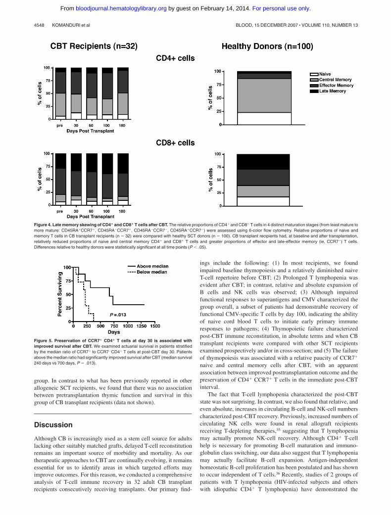
CD27, CD28, and CD57) have been proposed.30-34 Using thecombination of CCR7 and CD45RA, it is possible to define subsetsof naive cells (CD45RA�CCR7�), early or central memory cells(CD45RA�CCR7�), effector memory cells (CD45RA�CCR7�),and late effector memory cells (CD45RA�CCR7�).32 At baselineand after transplantation, the CD4� T-cell compartment in CBtrans-plant recipients was characterized by a relative paucity of naivecells, with large proportions of central and effector memory T cells.The CD8� T-cell compartment was similarly characterized byrelatively few naive cells, but with fewer central memory T cellsthan in the CD4� compartment. The bulk of CD8� T cellsexpressed markers consistent with effector memory and lateeffector memory differentiation. To determine how CB transplantrecipients compared with subjects not receiving transplants, weassessed the relative frequencies of T-cell differentiation subsets insamples collected from healthy allogeneic SCT donors. Comparedwith CB transplant recipients (n 32), healthy donor subjects hadsignificantly greater proportions of CCR7� T cells in the CD4�
compartment (wherein CCR7� T cells constituted the majority ofCD4� T cells in healthy donors) and in the CD8� compartment(where fewer cells were CCR7� in healthy subjects, thoughsimilarly increased, relative to CB transplant recipients; Figure 4).
Preservation of CCR7� CD4� T cells at day 30 predictspost-CBT survival
In an exploratory analysis, we determined whether the skewing thatwe observed, toward later memory T-cell subsets, was associatedadversely with survival after CBT. Actuarial analysis of survival,stratified by the relative preservation of CCR7� T cells within theCD4� compartment, is illustrated in Figure 5. We found that ahigher ratio of CCR7� to CCR7� CD4� T cells (ie, above themedian vs below) at day �30 was associated with significantlylonger survival (median survival 240 days vs 700 days, P .013).When we analyzed the association between CCR7� T cells withinthe CD8� T-cell compartment at day 30 and survival, no similarrelationship was seen. We also examined whether the degree ofpre-CBT thymic function was associated with survival, given theheterogeneity of baseline thymic function that we observed in this
Figure 3. Thymic regeneration failure after CBT. The frequency of recent thymic emigrants in peripheral blood was quantitated using a real-time PCR assay measuring T-cellreceptor (TCR) excision circles produced during TCR-� rearrangement. (A) Longitudinal assessment of thymic function in CB transplant recipients (n 26, medianage 33) demonstrates reduced thymic function in baseline and post-CBT thymopoietic recovery that was measurable in only 2 subjects and significant and sustained in only1 recipient. The horizontal line depicts the number of TRECs present in a group of healthy control subjects who were age-matched (n 18, median age 34). (B) Thymicregenerative failure in CB transplant recipients relative to autologous (auto) and allogeneic (allo) SCT recipients. Median values of TRECs in CBT were dramatically reducedrelative to other prospectively analyzed SCT recipients, despite a significantly reduced median age of the CBT population (median age 33 vs 43 for Colorado allogeneic SCTrecipients and 50 for Colorado autologous SCT recipients). Values for CB transplant recipients were significantly lower than other groups at all time points (P � .01).
IMMUNE RECOVERY AFTER CORD BLOOD TRANSPLANTATION 4547BLOOD, 15 DECEMBER 2007 � VOLUME 110, NUMBER 13
For personal use only. by guest on February 14, 2014. bloodjournal.hematologylibrary.orgFrom
group. In contrast to what has been previously reported in otherallogeneic SCT recipients, we found that there was no associationbetween pretransplantation thymic function and survival in thisgroup of CB transplant recipients (data not shown).
Discussion
Although CB is increasingly used as a stem cell source for adultslacking other suitably matched grafts, delayed T-cell reconstitutionremains an important source of morbidity and mortality. As ourtherapeutic approaches to CBT are continually evolving, it remainsessential for us to identify areas in which targeted efforts mayimprove outcomes. For this reason, we conducted a comprehensiveanalysis of T-cell immune recovery in 32 adult CB transplantrecipients consecutively receiving transplants. Our primary find-
ings include the following: (1) In most recipients, we foundimpaired baseline thymopoiesis and a relatively diminished naiveT-cell repertoire before CBT; (2) Prolonged T lymphopenia wasevident after CBT; in contrast, relative and absolute expansion ofB cells and NK cells was observed; (3) Although impairedfunctional responses to superantigens and CMV characterized thegroup overall, a subset of patients had demonstrable recovery offunctional CMV-specific T cells by day 100, indicating the abilityof naive cord blood T cells to initiate early primary immuneresponses to pathogens; (4) Thymopoietic failure characterizedpost-CBT immune reconstitution, in absolute terms and when CBtransplant recipients were compared with other SCT recipientsexamined prospectively and/or in cross-section; and (5) The failureof thymopoiesis was associated with a relative paucity of CCR7�
naive and central memory cells after CBT, with an apparentassociation between improved posttransplantation outcome and thepreservation of CD4� CCR7� T cells in the immediate post-CBTinterval.
The fact that T-cell lymphopenia characterized the post-CBTstate was not surprising. In contrast, we also found that relative, andeven absolute, increases in circulating B-cell and NK-cell numberscharacterized post-CBT recovery. Previously, increased numbers ofcirculating NK cells were found in renal allograft recipientsreceiving T-depleting therapies,35 suggesting that T lymphopeniamay actually promote NK-cell recovery. Although CD4� T-cellhelp is necessary for promoting B-cell maturation and immuno-globulin class switching, our data also suggest that T lymphopeniamay actually facilitate B-cell expansion. Antigen-independenthomeostatic B-cell proliferation has been postulated and has shownto occur independent of T cells.36 Recently, studies of 2 groups ofpatients with T lymphopenia (HIV-infected subjects and otherswith idiopathic CD4� T lymphopenia) have demonstrated the
Figure 5. Preservation of CCR7� CD4� T cells at day 30 is associated withimproved survival after CBT. We examined actuarial survival in patients stratifiedby the median ratio of CCR7� to CCR7�CD4� T cells at post-CBT day 30. Patientsabove the median ratio had significantly improved survival after CBT (median survival240 days vs 700 days, P .013).
Figure 4. Late memory skewing of CD4� and CD8� T cells after CBT. The relative proportions of CD4� and CD8� T cells in 4 distinct maturation stages (from least mature tomore mature: CD45RA�CCR7�, CD45RA�CCR7�, CD45RA�CCR7�, CD45RA�CCR7�) were assessed using 6-color flow cytometry. Relative proportions of naive andmemory T cells in CB transplant recipients (n 32) were compared with healthy SCT donors (n 100). CB transplant recipients had, at baseline and after transplantation,relatively reduced proportions of naive and central memory CD4� and CD8� T cells and greater proportions of effector and late-effector memory (ie, CCR7�) T cells.Differences relative to healthy donors were statistically significant at all time points (P � .05).
4548 KOMANDURI et al BLOOD, 15 DECEMBER 2007 � VOLUME 110, NUMBER 13
For personal use only. by guest on February 14, 2014. bloodjournal.hematologylibrary.orgFrom
expansion of immature/transitional B cells.37,38 Taken together,these data suggest that there may be crosstalk between the T-cell,B-cell, and NK-cell compartments in the setting of lymphopenia.The factors that govern this intercompartmental regulation (eg,cytokines including IL-737 or competition for “niches”) deservefurther study.
We observed profound delays in functional immune reconsti-tution in CBT when considered as a group. In contrast to otherallogeneic SCT recipients, subsets of CD4� and CD8� T cellscapable of responding suitably to stimulation with superantigenswere markedly reduced. Because the CFC assay measures theproportion of capable responders within a gated T-cell subpopu-lation, this approach is less vulnerable (eg, relative to conven-tional lymphoproliferative assays [LPA]) to the effects oflymphopenia,28 which usually results in diminished LPA re-sponses in patients with CD4� T-cell counts of less than50 cells/�L.39 Although it is probable that the transfer of fewerT cells in CBT (vs PBSC grafts) plays a major role in thisphenomenon, we cannot discount the effects of nonlymphoidcells (eg, myeloid subsets including monocytes and DC) onthese impaired functional responses. When we studied therecovery of CMV-specific CD4� and CD8� T-cell responses byCFC, we found that although responses were reduced in CBToverall, a subset of patients had significant frequencies offunctional CMV-specific CD4� and CD8� T cells by approxi-mately 3 months after SC transplantation. These results confirmand extend the results of Cohen et al, who recently demon-strated, using LPA assays measuring herpesvirus-specificT cells, that many CB transplant recipients had at least1 detectable assay after CBT that demonstrated successfuldonor-derived, antigen-specific T-cell recovery.40 Because CFCassays are more quantitative than LPA, our results also provide amore complete window into the quantitative recovery of CMV-specific T cells after CBT.
The most striking finding of our study was the observation thatthe recovery of thymopoiesis was markedly impaired both atbaseline and, in all but 1 subject, at all time points after CBT.Previously, we have conducted cross-sectional studies of more than140 allogeneic SCT recipients receiving transplants of eithermarrow or PBSC at our own institution; although thymopoeisiswas transiently lower after SC transplantation, we found measur-able thymopoiesis in nearly every subject, even at early intervalsafter SC transplantation. Thymopioesis peaked approximately 6 to8 months after SC transplantation and predated the recovery ofnaive CD4� and CD8� T cells. Here, we report the results of2 prospective parallel studies of adult autologous and allogeneicSC transplantation cohorts undergoing transplantation at the Univer-sity of Colorado. In both groups, consistent with our priorcross-sectional studies, we similarly detected thymic function innearly all subjects. Thus, the nearly universal lack of thymopoiesisin this group of CB transplant recipients stands out as uniquelydifferent, relative to other SC transplantaion subpopulations. Somestudies have previously suggested that thymopoiesis can be de-tected, even at early intervals, in limited numbers of long-termsurvivors of CBT.11,41 Given that other (nonsurviving) patientswere not assessed in these studies, it is impossible to know whatwould have been observed if all subjects in those studies had beenexamined prospectively. Indeed, our preliminary data suggest thatpatients who fail to preserve naive cells early after CBT are morelikely to experience mortality; furthermore, our longest survivor todate is the only patient to recover significant and sustainedthymopoiesis. Clearly, these data suggest that larger prospective
studies of thymopoietic recovery are needed, especially in adultrecipients who are known to have a diminished (though heteroge-neous) capacity to recovery thymopoiesis after SC transplantation.
Phenotypic studies using markers of T-cell maturation anddifferentiation (eg, CD45RA/RO, CD27, CCR7, and CD28)have demonstrated that multiple subcompartments of the memoryT-cell repertoire exist, with marked differences in their distribu-tion, with naive and early memory subsets retaining their ability(via CCR7 expression) to home to lymph nodes where primaryimmune responses occur.30,32 Pantaleo and colleagues haveexamined the relative subsets of T cells specific for various viralpathogens capable of secreting IFN�, IL-2, or both cytokines.42
Their results suggest that the subset of T cells that coproduceboth IL-2 and IFN� are a hallmark of protective immuneresponses to chronic viral pathogens, although cells producingIFN� alone are often present in the setting of chronic progres-sive infection (eg, as seen in HIV-1 infection). In recentpreclinical studies, we have demonstrated that the cytokinephenotype of late memory cells is distinct from that seen inearlier memory cells after a variety of stimuli, including phorbolesters (eg, phorbol 12-myristate 13-acetate), antibodies stimulat-ing the CD3/TCR complex (eg, OKT3), and superantigens (eg,SEB). Relative to early- or central memory T cells, late effectormemory cells produced are skewed toward MIP-1� and IFN�production, with few cells producing IL-2. In a murine model ofadoptive cellular therapy followed by immunization againstmelanoma-associated antigens, Restifo and colleagues demon-strated that the transfer of late-effector CD8� T cells (character-ized by the production of abundant IFN� but little IL-2) wasassociated with progressive tumor growth; in contrast, adoptivetransfer of cells with the same specificity but of a centralmemory differentiation stage resulted in tumor clearance.43
One important caveat is that the patients studied hereconsisted of a relatively heavily pretreated group of patients.This fact is reflected by the fact that half of the acute myeloidleukemia (AML) recipients received transplants with activedisease and overall had received an average of 3 prior treatmentregimens and is also demonstrated by the fact that thymopoiesisat baseline, which has previously been demonstrated to correlatewith post-CBT immune recovery in 1 pediatric study, was absentin most subjects and markedly reduced (relative to age-matchedhealthy controls) even in the minority of patients with detectablethymopoiesis before transplantation. Thus, we cannot necessar-ily generalize these results to other groups of CB transplantrecipients who are less heavily pretreated. It should also benoted that all of our recipients received regimens containingantithymocyte globulin (ATG). Although the biologic half-lifeof ATG is prolonged and is likely to have influenced both central(ie, thymic) and peripheral T cells infused within CB grafts, thisintervention is a standard component of most CBT conditioningapproaches and appears to be necessary to decrease the other-wise significant risk of graft rejection. It is also probable thatstem cell dose, which is reduced in adults relative to children,may also be an important factor in the recovery of lymphoidprecursors and thymopoietic function. Given the modest numberof patients studied to date, it will be important to conduct larger,multi-institutional studies examining a greater number of im-mune recovery endpoints before we can draw firm conclusionsregarding the broader relevance of the findings of this single-institution study. Ultimately, we need to understand how priortherapies, age, conditioning regimen, and graft characteristicsinfluence thymic regeneration and functional T-cell recovery.
IMMUNE RECOVERY AFTER CORD BLOOD TRANSPLANTATION 4549BLOOD, 15 DECEMBER 2007 � VOLUME 110, NUMBER 13
For personal use only. by guest on February 14, 2014. bloodjournal.hematologylibrary.orgFrom

Overall, our data support the generally accepted notion thatattempts to improve outcome in the setting of CBT for adults willdepend on the success of these approaches in improving T-cellimmune reconstitution. Already, encouraging progress has beenmade in the development of clinical strategies aimed at increasingstem-cell dose, including ex vivo expansion of progenitor cells44,45
and the use of pooled CB units.9,46 It will be important toprospectively assess the influence of these various approaches onthymopoietic recovery and to consider immune recovery endpointsas a primary measure of the success of these trials, in addition to thetypical primary endpoints (eg, date of neutrophil and plateletengraftment). Among approaches being considered, the use ofNotch ligand-based approaches that may preferentially expandlymphoid progenitors deserves special consideration given ourfindings.47,48 Additional strategies, already demonstrated to haveefficacy in murine models and in human clinical trials, include theuse of thymoprotective agents (eg, keratinocyte growth factor orrelated family members)49-51 and the use of thymopoietic factors(eg, androgen ablation using leuprolide, IL-7 administration, andgrowth hormone administration).52,53 Finally, the ex vivo expansionof naive cord blood cells in the presence of viral antigens may offera solution to the elusive goal of adoptively transferring antigen-specific memory to CB recipients.54,55 Although such strategies areattractive, our data also suggest that attention should be paid to theactivation status, differentiation stage, and cytokine productionsignatures of expanded cell populations, because it is possible thatin vitro stimulation might favor the production of late-stagememory cells that may be functionally ineffective or proliferativelyimpaired or even increase the otherwise manageable incidence ofGVHD, which remains one of the principal advantages of CBTversus the use of other unrelated donor grafts.
AcknowledgmentsThis work was supported in part by grants from the NationalInstitutes of Health grants RO1 CA109326 (K.V.K.) and RO1CA061508 (E.J.S.).
We thank Jeffrey Harris and Mike McCune for helping us todevelop and optimize assays of thymopoiesis and for insightfulcomments on the manuscript. We also acknowledge the technicalexpertise of Michael Thomas and Michelle Davis and thank thepatients, nurses, pharmacists, and staff of the M. D. AndersonCancer Center stem cell transplant program, without whom thisresearch would not have been possible.
Authorship
Contribution: K.V.K. designed the study, analyzed the data, andwrote the manuscript; L.S.J. performed the primary research andanalyzed the data; M.d.L. and R.E.C. designed the parent clinicaltrials; J.M. helped to design the parent clinical trials and contrib-uted clinical data; S.R. and I.M. contributed to the study of controlsubjects; S.G.B., I.K., and S.M. performed the research; E.D.W.contributed valuable flow cytometry expertise; L.W., D.P., andL.J.N.C contributed to the parent clinical trials; J.J.M. assisted withdata analysis and interpretation; and E.J.S. designed the parentclinical trials and contributed to writing the manuscript.
Conflict-of-interest disclosure: The authors declare no compet-ing financial interests.
Correspondence: Krishna V. Komanduri, Transplant Immunol-ogy Section, Department of Blood and Marrow Transplantation,M. D. Anderson Cancer Center, 1515 Holcombe Blvd, Houston,TX 77030; e-mail: [email protected].
References
1. Gluckman E, Broxmeyer HA, Auerbach AD, et al.Hematopoietic reconstitution in a patient withFanconi’s anemia by means of umbilical-cordblood from an HLA-identical sibling. N EnglJ Med. 1989;321:1174-1178.
2. Kurtzberg J, Laughlin M, Graham ML, et al. Pla-cental blood as a source of hematopoietic stemcells for transplantation into unrelated recipients.N Engl J Med. 1996;335:157-166.
3. Laughlin MJ, Barker J, Bambach B, et al. Hema-topoietic engraftment and survival in adult recipi-ents of umbilical-cord blood from unrelated do-nors. N Engl J Med. 2001;344:1815-1822.
4. Rubinstein P, Carrier C, Scaradavou A, et al. Out-comes among 562 recipients of placental-bloodtransplants from unrelated donors. N Engl J Med.1998;339:1565-1577.
5. Gluckman E, Rocha V, Chevret S. Results of un-related umbilical cord blood hematopoietic stemcell transplantation. Rev Clin Exp Hematol. 2001;5:87-99.
6. Barker JN, Davies SM, DeFor T, Ramsay NK,Weisdorf DJ, Wagner JE. Survival after transplan-tation of unrelated donor umbilical cord blood iscomparable to that of human leukocyte antigen-matched unrelated donor bone marrow: results ofa matched-pair analysis. Blood. 2001;97:2957-2961.
7. McNiece I, Kubegov D, Kerzic P, Shpall EJ,Gross S. Increased expansion and differentiationof cord blood products using a two-step expan-sion culture. Exp Hematol. 2000;28:1181-1186.
8. Hofmeister CC, Zhang J, Knight KL, Le P, Stiff PJ.Ex vivo expansion of umbilical cord blood stemcells for transplantation: growing knowledge from
the hematopoietic niche. Bone Marrow Trans-plant. 2007;39:11-23.
9. Barker JN, Weisdorf DJ, Wagner JE. Creation ofa double chimera after the transplantation of um-bilical-cord blood from two partially matched un-related donors. N Engl J Med. 2001;344:1870-1871.
10. Barker JN, Wagner JE. Umbilical cord bloodtransplantation: current state of the art. Curr OpinOncol. 2002;14:160-164.
11. Talvensaari K, Clave E, Douay C, et al. A broadT-cell repertoire diversity and an efficient thymicfunction indicate a favorable long-term immunereconstitution after cord blood stem cell trans-plantation. Blood. 2002;99:1458-1464.
12. Moretta A, Maccario R, Fagioli F, et al. Analysis ofimmune reconstitution in children undergoingcord blood transplantation. Exp Hematol. 2001;29:371-379.
13. Niehues T, Rocha V, Filipovich AH, et al. Factorsaffecting lymphocyte subset reconstitution aftereither related or unrelated cord blood transplanta-tion in children – a Eurocord analysis. Br JHaematol. 2001;114:42-48.
14. Locatelli F, Maccario R, Comoli P, et al. Hemato-poietic and immune recovery after transplantationof cord blood progenitor cells in children. BoneMarrow Transplant. 1996;18:1095-1101.
15. Thomson BG, Robertson KA, Gowan D, et al.Analysis of engraftment, graft-versus-host dis-ease, and immune recovery following unrelateddonor cord blood transplantation. Blood. 2000;96:2703-2711.
16. Szabolcs P, Niedzwiecki D. Immune reconstitu-tion after unrelated cord blood transplantation.Cytotherapy. 2007;9:111-122.
17. Zajac AJ, Blattman JN, Murali-Krishna K, et al.Viral immune evasion due to persistence of acti-vated T cells without effector function. J Exp Med.1998;188:2205-2213.
18. Ozdemir E, St John LS, Gillespie G, et al. Cyto-megalovirus reactivation following allogeneicstem cell transplantation is associated with thepresence of dysfunctional antigen-specific CD8�T cells. Blood. 2002;100:3690-3697.
19. Poulin JF, Viswanathan MN, Harris JM, et al. Di-rect evidence for thymic function in adult humans.J Exp Med. 1999;190:479-486.
20. Douek DC, McFarland RD, Keiser PH, et al.Changes in thymic function with age and duringthe treatment of HIV infection. Nature. 1998;396:690-695.
21. Lewin SR, Heller G, Zhang L, et al. Direct evi-dence for new T-cell generation by patients aftereither T-cell-depleted or unmodified allogeneichematopoietic stem cell transplantations. Blood.2002;100:2235-2242.
22. Douek DC, Vescio RA, Betts MR, et al. Assess-ment of thymic output in adults after haematopoi-etic stem-cell transplantation and prediction ofT-cell reconstitution. Lancet. 2000;355:1875-1881.
23. Teixeira L, Valdez H, McCune JM, et al. Poor CD4T cell restoration after suppression of HIV-1 repli-cation may reflect lower thymic function. AIDS.2001;15:1749-1756.
24. Kaplan E, Meier P. Nonparametric estimationfrom incomplete observations. J Am Stat Assoc.1958;53:547-581.
25. Storek J, Dawson MA, Maloney DG. Normal T, B,and NK cell counts in healthy donors at 1 year
4550 KOMANDURI et al BLOOD, 15 DECEMBER 2007 � VOLUME 110, NUMBER 13
For personal use only. by guest on February 14, 2014. bloodjournal.hematologylibrary.orgFrom

after blood stem cell harvesting. Blood. 2000;95:2993-2994.
26. Komanduri KV, Donahoe SM, Moretto WJ, et al.Direct measurement of CD4� and CD8� T-cellresponses to CMV in HIV-1-infected subjects.Virology. 2001;279:459-470.
27. Komanduri KV, Feinberg J, Hutchins RK, et al.Loss of cytomegalovirus-specific CD4� T cellresponses in human immunodeficiency virus type1-infected patients with high CD4� T cell countsand recurrent retinitis. J Infect Dis. 2001;183:1285-1289.
28. Komanduri KV, Viswanathan MN, Wieder ED, etal. Restoration of cytomegalovirus-specific CD4�T-lymphocyte responses after ganciclovir andhighly active antiretroviral therapy in individualsinfected with HIV-1. Nat Med. 1998;4:953-956.
29. Harris JM, Hazenberg MD, Poulin JF, et al. Mul-tiparameter evaluation of human thymic function:interpretation and caveats. Clin Immunol. 2005;115:138-146.
30. Champagne P, Ogg GS, King AS, et al. Skewedmaturation of memory HIV-specific CD8 T lym-phocytes. Nature. 2001;410:106-111.
31. Ruprecht CR, Gattorno M, Ferlito F, et al. Coex-pression of CD25 and CD27 identifies FoxP3�regulatory T cells in inflamed synovia. J Exp Med.2005;201:1793-1803.
32. Sallusto F, Lenig D, Forster R, Lipp M, Lanzavec-chia A. Two subsets of memory T lymphocyteswith distinct homing potentials and effector func-tions. Nature. 1999;401:708-712.
33. Brenchley JM, Karandikar NJ, Betts MR, et al.Expression of CD57 defines replicative senes-cence and antigen-induced apoptotic death ofCD8� T cells. Blood. 2003;101:2711-2720.
34. d’Angeac AD, Monier S, Pilling D, Travaglio-Enci-noza A, Reme T, Salmon M. CD57� T lympho-cytes are derived from CD57- precursors by dif-ferentiation occurring in late immune responses.Eur J Immunol. 1994;24:1503-1511.
35. Klaus G, Mostert K, Reckzeh B, Mueller TF. Phe-notypic changes in lymphocyte subpopulations inpediatric renal-transplant patients after T-celldepletion. Transplantation. 2003;76:1719-1724.
36. Cabatingan MS, Schmidt MR, Sen R, WoodlandRT. Naive B lymphocytes undergo homeostaticproliferation in response to B cell deficit. J Immu-nol. 2002;169:6795-6805.
37. Malaspina A, Moir S, Chaitt DG, et al. IdiopathicCD4� T lymphocytopenia is associated with in-creases in immature/transitional B cells and se-rum levels of IL-7. Blood. 2007;109:2086-2088.
38. Malaspina A, Moir S, Ho J, et al. Appearance ofimmature/transitional B cells in HIV-infected indi-viduals with advanced disease: correlation withincreased IL-7. Proc Natl Acad Sci U S A 2006;103:2262-2267.
39. Schrier RD, Freeman WR, Wiley CA, McCutchanJA. Immune predispositions for cytomegalovirusretinitis in AIDS. J Clin Invest. 1995;95:1741-1746.
40. Cohen G, Carter SL, Weinberg KI, et al. Antigen-specific T-lymphocyte function after cord bloodtransplantation. Biol Blood Marrow Transplant.2006;12:1335-1342.
41. Weinberg K, Blazar BR, Wagner JE, et al. Factorsaffecting thymic function after allogeneic hemato-poietic stem cell transplantation. Blood. 2001;97:1458-1466.
42. Harari A, Vallelian F, Meylan PR, Pantaleo G.Functional heterogeneity of memory CD4 T cellresponses in different conditions of antigen expo-sure and persistence. J Immunol. 2005;174:1037-1045.
43. Gattinoni L, Klebanoff CA, Palmer DC, et al. Ac-quisition of full effector function in vitro paradoxi-cally impairs the in vivo antitumor efficacy ofadoptively transferred CD8� T cells. J Clin In-vest. 2005;115:1616-1626.
44. McNiece I, Harrington J, Turney J, Kellner J,Shpall EJ. Ex vivo expansion of cord blood mono-nuclear cells on mesenchymal stem cells. Cyto-therapy. 2004;6:311-317.
45. Shpall EJ, Quinones R, Giller R, et al. Transplan-tation of ex vivo expanded cord blood. Biol BloodMarrow Transplant. 2002;8:368-376.
46. Majhail NS, Brunstein CG, Wagner JE. Doubleumbilical cord blood transplantation. Curr OpinImmunol. 2006;18:571-575.
47. Zakrzewski JL, Kochman AA, Lu SX, et al. Adop-tive transfer of T-cell precursors enhances T-cellreconstitution after allogeneic hematopoietic stemcell transplantation. Nat Med. 2006;12:1039-1047.
48. Delaney C, Varnum-Finney B, Aoyama K,Brashem-Stein C, Bernstein ID. Dose-dependenteffects of the Notch ligand Delta1 on ex vivo dif-ferentiation and in vivo marrow repopulating abil-ity of cord blood cells. Blood. 2005;106:2693-2699.
49. Min D, Panoskaltsis-Mortari A, Kuro OM, Hol-lander GA, Blazar BR, Weinberg KI. Sustainedthymopoiesis and improvement in functional im-munity induced by exogenous KGF administra-tion in murine models of aging. Blood. 2007;109:2529-2537.
50. Rossi S, Blazar BR, Farrell CL, et al. Keratinocytegrowth factor preserves normal thymopoiesis andthymic microenvironment during experimentalgraft-versus-host disease. Blood. 2002;100:682-691.
51. Alpdogan O, Hubbard VM, Smith OM, et al. Kera-tinocyte growth factor (KGF) is required for post-natal thymic regeneration. Blood. 2006;107:2453-2460.
52. Mackall CL, Fry TJ, Bare C, Morgan P, GalbraithA, Gress RE. IL-7 increases both thymic-depen-dent and thymic-independent T-cell regenerationafter bone marrow transplantation. Blood. 2001;97:1491-1497.
53. Alpdogan O, Schmaltz C, Muriglan SJ, et al. Ad-ministration of interleukin-7 after allogeneic bonemarrow transplantation improves immune recon-stitution without aggravating graft-versus-hostdisease. Blood. 2001;98:2256-2265.
54. Leen AM, Myers GD, Sili U, et al. Monoculture-derived T lymphocytes specific for multiple vi-ruses expand and produce clinically relevant ef-fects in immunocompromised individuals. NatMed. 2006;12:1160-1166.
55. Park KD, Marti L, Kurtzberg J, Szabolcs P. In vitropriming and expansion of cytomegalovirus-spe-cific Th1 and Tc1 T cells from naive cord bloodlymphocytes. Blood. 2006;108:1770-1773.
IMMUNE RECOVERY AFTER CORD BLOOD TRANSPLANTATION 4551BLOOD, 15 DECEMBER 2007 � VOLUME 110, NUMBER 13
For personal use only. by guest on February 14, 2014. bloodjournal.hematologylibrary.orgFrom
Copyright © 2022 FDOKUMEN