Influence of Transglutaminase Crosslinking on Casein Protein ...

GuardavaccaroLow, Albert J.R. Heck and Daniele Roberto Magliozzi, Jihoon Kim, Teck Yew signalingControls the Duration of mTOR-S6K
TrCP Ubiquitin Ligaseβand the SCFDegradation of Tiam1 by Casein Kinase 1Protein Synthesis and Degradation:
published online August 14, 2014J. Biol. Chem.
10.1074/jbc.M114.575571Access the most updated version of this article at doi:
.JBC Affinity SitesFind articles, minireviews, Reflections and Classics on similar topics on the
Alerts:
When a correction for this article is posted•
When this article is cited•
to choose from all of JBC's e-mail alertsClick here
http://www.jbc.org/content/early/2014/08/14/jbc.M114.575571.full.html#ref-list-1
This article cites 0 references, 0 of which can be accessed free at
at Universiteitsbibliotheek U
trecht on August 19, 2014
http://ww
w.jbc.org/
Dow
nloaded from
at Universiteitsbibliotheek U
trecht on August 19, 2014
http://ww
w.jbc.org/
Dow
nloaded from
Tiam1 degradation regulates mTOR-S6K signaling
1
Degradation of Tiam1 by Casein Kinase 1 and the SCFβTrCP Ubiquitin Ligase Controls the Duration of mTOR-S6K signaling Roberto Magliozzi1, Jihoon Kim1, Teck Yew Low2,3, Albert J. R. Heck2,3, and Daniele Guardavaccaro1*
1Hubrecht Institute-KNAW and University Medical Center Utrecht, 3584 CT Utrecht, The Netherlands 2Biomolecular Mass Spectrometry and Proteomics, Bijvoet Center for Biomolecular Research and Utrecht Institute for Pharmaceutical Sciences, Utrecht University, 3584 CH Utrecht, The Netherlands 3The Netherlands Proteomics Center, 3584 CH Utrecht, The Netherlands Running title: Tiam1 degradation regulates mTOR-S6K signaling To whom correspondence should be addressed: Daniele Guardavaccaro, Hubrecht Institute, Uppsalalaan 8, 3584 CT Utrecht, The Netherlands. Tel.: +31(0)302121932; Fax: +31(0)302516464; E-mail: [email protected] Keywords: Tiam1; ubiquitin; βTrCP; proteolysis; mTOR-S6K signaling
Background: The guanine nucleotide exchange factor Tiam1 regulates the activity of the small GTPase Rac1, a crucial regulator of cell adhesion, proliferation and survival. Results: The SCFβTrCP ubiquitin ligase in cooperation with CK1 targets Tiam1 for proteasome-dependent degradation. Conclusion: Tiam1 degradation is required to terminate the mTOR-S6K signaling pathway. Significance: Tiam1 degradation controls the duration of mTOR-S6K signaling in response to mitogens. ABSTRACT T-cell lymphoma invasion and metastasis 1 (Tiam1) is a guanine nucleotide exchange factor that specifically controls the activity of the small GTPase Rac, a key regulator of cell adhesion, proliferation and survival. Here, we report that in response to mitogens, Tiam1 is degraded by the ubiquitin-proteasome system via the SCFβTrCP
ubiquitin ligase. Mitogenic stimulation triggers the binding of Tiam1 to the F-box protein βTrCP via its degron sequence and subsequent Tiam1 ubiquitylation and proteasomal degradation. The proteolysis of Tiam1 is prevented by βTrCP silencing, inhibition of CK1 and MEK, or mutation of the Tiam1 degron site. Expression of a stable Tiam1 mutant that is unable to interact with βTrCP results in sustained activation of the mTOR/S6K signaling and increased apoptotic
cell death. We propose that the SCFβTrCP-mediated degradation of Tiam1 controls the duration of the mTOR-S6K signaling pathway in response to mitogenic stimuli. INTRODUCTION Tiam1 is a ubiquitously expressed guanine nucleotide exchange factor that specifically activates the Rho-like GTPase Rac1 in response to growth factors and cell-substrate interaction (1-3). It was originally identified in a retroviral insertional mutagenesis screen for genes that confer invasive properties to T-lymphoma cells (1). Thereafter, a plethora of studies have demonstrated that Tiam1 controls different Rac-dependent cellular processes, such as cell polarity, cell-matrix adhesion, cell-cell adhesion, cell survival and cell growth and proliferation. Hence, it does not come as a surprise that Tiam1 plays a critical role in cancer development. Tiam1 deficiency in mice protects against Ras-induced skin carcinogenesis and correlates with increased apoptosis and reduced proliferation and growth in keratinocytes (4). This is in agreement with in vitro studies reporting that Tiam1 associates with GTP-bound Ras through a Ras-binding domain and functions as an effector that mediates Ras activation of Rac (4). However, the fewer skin tumors developing in Tiam1 knock-out mice progress more frequently to malignancy indicating that loss of Tiam1 stimulates malignant
http://www.jbc.org/cgi/doi/10.1074/jbc.M114.575571The latest version is at JBC Papers in Press. Published on August 14, 2014 as Manuscript M114.575571
Copyright 2014 by The American Society for Biochemistry and Molecular Biology, Inc.
at Universiteitsbibliotheek U
trecht on August 19, 2014
http://ww
w.jbc.org/
Dow
nloaded from

Tiam1 degradation regulates mTOR-S6K signaling
2
conversion (4). Indeed, in vitro, Tiam1 is known to promote the formation of adherens junctions and the induction of an epithelial-like phenotype in mesenchymal cells (5,6) and to be required for both the formation and maintenance of cadherin-based adhesions (7). In spite of the importance of Tiam1 in cancer, little is known about the regulation of Tiam1 protein levels in normal and cancer cells. Here, we report that in response to mitogenic stimulation, Tiam1 is targeted for proteasome-dependent degradation by the SCFβTrCP ubiquitin ligase in cooperation with casein kinase 1 and the MEK/ERK pathway. We also show that ubiquitin-dependent degradation of Tiam1 is required to terminate the activation of mTOR-S6K signaling in response to mitogenic stimulation. EXPERIMENTAL PROCEDURES Cell culture and drug treatments HEK293, HEK293T, T98G and MDCK cells were cultured in Dulbecco’s modified Eagle’s medium (Invitrogen) with 10% fetal calf serum (FCS) and 100 U/ml penicillin-streptomycin. The following drugs were used: MG132 (Peptide Institute; 10 µM), D4476 (Sigma-Aldrich, 50 µM), IC261 (Sigma-Aldrich, 50 µM), cycloheximide (Sigma-Aldrich, 100 µM), PD0325901 (Sigma-Aldrich, 10 µM), U0126 (Millipore 10 µM), Rac inhibitor III EHop-016 (Millipore 50 µM), phorbol-12-myristate-13-acetate (Sigma-Aldrich, 10 ng/ml). MTT assays were carried according to the manufacturer’s protocol. Transient transfections Cells were transfected by the polyethylenimine (PEI) or liposomes-mediated (Lipofectamine 2000, Invitrogen) methods. Plasmids Full-length and N-terminally truncated (C1199) human Tiam1 carrying a C-terminal HA tag cDNAs were cloned by PCR into the EcoRV and NotI sites of pcDNA3. GFP-tagged Tiam1 was kindly provided by F. Zwartkruis. The full-length constructs were used as template to generate the Tiam1(S370A/S374A), Tiam1(S414A/S418A), Tiam1(S329A/S334A) and Tiam1(S329A/S334A/T340A) mutants using the QuickChange site-directed mutagenesis kit (Stratagene) according to manufacturer’s
directions. The oligonucleotides used were as follows. Tiam1(S370A/S373A) forward primer: 5’CTTTGTGGGCAGCGACGCCGGCAGCAGCGCCACCGGGGATGCGGCTC3’, Tiam1(S370A/S373A) reverse primer: 5’GAGCCGCATCCCCGGTGGCGCTGCTGCCGGCGTCGCTGCCCACAAAG3’; Tiam1(S414A/S418A) forward primer: 5’CAGCGATGAGCAGAGCGCCGGCACCCTGGCCTCTCCGGGCCAAGTCGGAC3’, Tiam1(S414A/S418A) reverse primer: 5’GTCCGACTTGGCCCGGAGAGGCCAGGGTGCCGGCGCTCTGCTCATCGCTG3’; Tiam1(S329A/S334A) forward primer: 5’GGCGAGGGCGCTGAGTTTGCAGACGCTGGGATTGAAGGG3’, Tiam1(S329A/S334A) reverse primer: 5’CCCTTCAATCCCAGCGTCTGCAAACTCAGCGCCCTCGCC3’; Tiam1(S329A/S334A/T340A) forward primer: 5’GAGTTTGCAGACGCTGGGATTGAAGGGGCCGCTACCGACACG-3’, Tiam1(S329A/S334A/T340A) reverse primer: 5’CGTGTCGGTAGCGGCCCCTTCAATCCCAGCGTCTGCAAACTC3’. All constructs were verified by sequencing. Biochemical methods Extract preparation, immunoprecipitation, and immunoblotting were previously described (8). Mouse monoclonal antibodies were from Invitrogen (Cul1), Sigma-Aldrich (FLAG), Santa Cruz Biotechnology (Actin, p70-S6K), Covance (HA), Cell Signaling Technology [phospho-Erk1/2(Thr202/Tyr204)], BD Biosciences (Rac1). Rabbit polyclonal antibodies were from Santa Cruz Biotechnology (Tiam1), Cell Signaling Technology [βTrCP1, Erk1/2, phospho-p70-S6K(Thr421/Ser424), phospho-p70-S6K(Thr389), cleaved caspase-3(Asp175)], Sigma-Aldrich (FLAG), Torrey Pines Biolabs (GFP). In vitro binding assay In vitro translated 35S-labeled βTrCP1 and FBXW5 were incubated with Protein G-Sepharose beads precoupled with the following Tiam1 synthetic peptides: 327-EGSEFADSGIEGAT-340, 330-EFADSGIEGATTDT-343, 327-EGpSEFADpSGIEGAT-340 or 330-EFADpSGIEGApTTDT-343 for 2 hours at 4°C. Beads were washed four times with lysis buffer
at Universiteitsbibliotheek U
trecht on August 19, 2014
http://ww
w.jbc.org/
Dow
nloaded from

Tiam1 degradation regulates mTOR-S6K signaling
3
(50 mM Tris-HCl pH 7.4, 1mM EDTA, 250 mM NaCl, 0.1% Triton Tx-100, 50 mM NaF, 1 mM DTT, 0.1 mM Na3VO4). Proteins were eluted with Laemmli buffer for 5 minutes at 95°C and subjected to SDS-PAGE followed by autoradiography. In vitro ubiquitylation assay Tiam1 ubiquitylation was performed in a volume of 10 µl containing SCFβTrCP-Tiam1 immunocomplexes, 50 mM Tris pH 7.6, 5 mM MgCl2, 0.6 mM DTT, 2 mM ATP, 1.5 ng/µl E1 (Boston Biochem), 10 ng/µl Ubc3, 2.5 µg/µl ubiquitin (Sigma-Aldrich), 1 µM ubiquitin aldehyde. The reactions were incubated at 30°C for 60 minutes and analyzed by immunoblotting. Purification of βTrCP2 interactors HEK293T cells grown in 15-cm dishes were transfected with pcDNA3-2xFLAG-2xHA-βTrCP2 and treated with 10 µM MG132 for 5 hours. Cells were harvested and subsequently lysed in lysis buffer (50 mM Tris-HCl pH 7.5, 150 mM NaCl, 1 mM EDTA, 0.5% NP40, 2 mM β-glycerophosphate plus protease and phosphatase inhibitors). βTrCP2 was immunopurified with mouse anti-FLAG M2 agarose resin (Sigma-Aldrich). After washing, proteins were eluted by competition with FLAG peptide (Sigma-Aldrich). The eluate was then subjected to a second immunopurification with anti-HA resin (12CA5 monoclonal antibody crosslinked to protein G Sepharose; Invitrogen) prior to elution in Laemmli sample buffer. The final eluate was separated by SDS-PAGE, and proteins were visualized by Coomassie colloidal blue. Bands were sliced out from the gels and subjected to in-gel digestion. Gel pieces were then reduced, alkylated and digested according to a published protocol (9). For mass spectrometric analysis, peptides recovered from in-gel digestion were separated with a C18 column and introduced by nano-electrospray into the LTQ Orbitrap XL (Thermo Fisher) with a configuration as described (10). Peak lists were generated from the MS/MS spectra using MaxQuant build 1.0.13.13, and then searched against the IPI Human database (version 3.37, 69164 entries) using Mascot search engine (Matrix Science). Carbaminomethylation (+57 Da) was set as fixed modification and protein N-terminal acetylation and methionine oxidation as variable
modifications. Peptide tolerance was set to 7 ppm and fragment ion tolerance was set to 0.5 Da, allowing 2 missed cleavages with trypsin enzyme. Finally, Scaffold (version Scaffold_3.6.1, Proteome Software Inc.) was used to validate MS/MS based peptide and protein identifications. Peptide identifications were accepted if their Mascot scores exceeded 20. Gene silencing by small interfering RNA The oligonucleotides designed for targeting both βTrCP1 and βTrCP2 are as follows: sense sequence 5’-GTGGAATTTGTGGAACATC dTdT-3’; antisense sequence 5’-GATGTTCCACAAATTCCACdTdT-3’. Cells were transfected with the oligonuclotides twice (24 and 48 hours after plating) using Oligofectamine (Invitrogen) according to manufacture's recommendations. Forty-eight hours after the last transfection, lysates were prepared and analyzed by immunoblotting. Phosphorylation analysis by mass spectrometry Samples were reduced with 10 mM DTT for 30 minutes at 60 30-minute incubation with iodoacetamide (20 mM) in the dark at room temperature. The first digestion was performed using Lys-Subsequently, the digest was diluted 5-fold using 50 mM ammonium bicarbonate to a final urea concentration of less than 2 M, and a second digestion with trypsin was performed overnight at
addition of formic acid to a final concentration of 0.1% (vol/vol). The resulting solution was desalted using 200 mg Sep-Pak C18 cartridges (Waters Corporation), lyophilized and reconstituted in 10% formic acid. LC-MSMS was performed with higher energy collision dissociated in a Q-Exactive Plus Orbitrap instrument. MS spectra to peptide sequence assignment was performed with Proteome Discoverer version 1.4, and MASCOT version 2.4 as search engine. The localization of phosphorylated sites was evaluated with PhosphoRS version 3 (11). Rac activity assay The abundance of GTP-bound Rac1 was determined as follows. Cells were lysed in lysis buffer (10 mM Tris, 150 mM NaCl, 50 mM MgCl2, 0.5% NP40) containing 2 µg of
at Universiteitsbibliotheek U
trecht on August 19, 2014
http://ww
w.jbc.org/
Dow
nloaded from
Tiam1 degradation regulates mTOR-S6K signaling
4
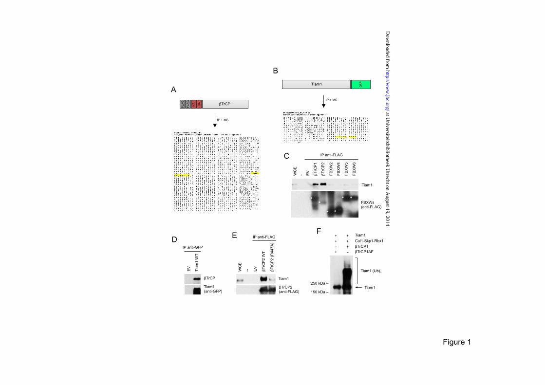
biotinylated p21-binding domain (PBD) of Pak1. The cleared supernatant was then incubated with streptavidin beads for 30 min at 4°C. Beads were washed and GTP-bound Rac1 was analyzed by immunoblotting with an anti-Rac1 antibody (Upstate). RESULTS Tiam1 interacts with βTrCP In an attempt to identify new targets of the SCFβTrCP ubiquitin ligase, we expressed βTrCP with an N-terminal FLAG-HA epitope tag in HEK293T cells and purified FLAG-HA-βTrCP immunoprecipitates, which were then analyzed by mass spectrometry. In two independent immunopurifications, peptides corresponding to Tiam1 (T-cell lymphoma invasion and metastasis 1) were recovered (Figure 1A). Conversely, when we immunopurified Tiam1 from HEK293T cells, we identified peptides corresponding to βTrCP (Figure 1B). To confirm the binding between βTrCP and Tiam1 and test its specificity, we overexpressed different FLAG-tagged F-box proteins (all containing WD40 repeats) in HEK293T cells. We then carried out FLAG immunoprecipitations to examine the interaction with endogenous Tiam1. βTrCP1 and its paralog βTrCP2 were the only proteins able to coimmunoprecipitate Tiam1 (Figure 1C). The coimmunoprecipitation of endogenous βTrCP with exogenously expressed Tiam1 was confirmed by immunoprecipitation coupled to immunoblotting (Figure 1D). It has been shown that substitution to alanine of Arg474 in βTrCP1 (or Arg447 in βTrCP2) within its WD40 β-propeller structure disrupts βTrCP interaction with the substrate (8,12). To assess if the interaction between Tiam1 and βTrCP is mediated by the βTrCP WD40 β-propeller, we immunoprecipitated FLAG-tagged wild type βTrCP2 and the βTrCP2(R447A) mutant from HEK293T cells and examined their binding to endogenous Tiam1. As shown in Figure 1E, wild type βTrCP coimmunoprecipitated with Tiam1, whereas the βTrCP2(R447A) mutant did not. These results suggest that Tiam1 is a substrate of SCFβTrCP. To test whether SCFβTrCP targets Tiam1 for ubiquitin conjugation, we reconstituted the ubiquitylation of Tiam1 in vitro. βTrCP1, but not an inactive βTrCP1(∆F box) mutant, was indeed able to efficiently ubiquitylate Tiam1 (Figure 1F).
The interaction of Tiam1 with βTrCP requires a conserved phosphodegron Substrates of SCFβTrCP share a conserved DSGXX(X)S degron that is bound by βTrCP (13-16). Two potential degron motifs are present at the N-terminus of human Tiam1, namely, 369-DSGSSS-374 and 413-SSGTLS-418 (Figure 2A). To test which of these motifs is responsible for the interaction of Tiam1 to βTrCP, we generated a number of mutants in which either Ser370/Ser374 or Ser414/Ser418 were replaced by alanine and evaluated the ability of these mutants to coimmunoprecipitate with βTrCP. Figure 2B shows that wild type Tiam1, Tiam1(S370A/S374A), and Tiam1(S414A/S418A) were all able to bind βTrCP, indicating that these serine residues do not mediate the interaction of Tiam1 with βTrCP. We then tested the requirement of a third putative Tiam1 degron motif, i.e., 329-SEFADSGIEGAT-340 (Figure 2A), for the binding of Tiam1 to βTrCP. The upstream Ser329 resembles the serine residue in Cdc25A (Ser79 preceding the conserved aspartate) whose phosphorylation is required for Cdc25A binding to βTrCP [Figure 2C and (17-19)]. We generated Tiam1 mutants in which Ser329 and Ser334 residues, alone or in combination with Thr340, were replaced by alanine. These Tiam1 mutants were not able to pull down exogenous (Figure 2D) or endogenous (Figure 2E) βTrCP in immunoprecipitation experiments. Of note, treatment of Tiam1 immunocomplexes with λ-phosphatase disrupted its binding to βTrCP (Figure 2E). Moreover, a Tiam1 mutant truncated at the N-terminus (C1199) lacking the βTrCP degron (5) did not coimmunoprecipitate with endogenous βTrCP (Figures 2F and 2G). Taken together, these results demonstrate that the association of Tiam1 with βTrCP requires the conserved degron motif SEFADSGIEGAT. As a second approach to examine whether phosphorylation of Tiam1 is required for the interaction with βTrCP, we employed an in vitro binding assay using immobilized synthetic phosphopeptides containing the βTrCP recognition domain of Tiam1. Peptides containing phosphorylated residues at positions Ser329 and Ser334 (Figure 2H) or at positions Ser334 and Thr340 (Figure 2I) efficiently associated with in vitro-translated βTrCP (but not with FBXW5),
at Universiteitsbibliotheek U
trecht on August 19, 2014
http://ww
w.jbc.org/
Dow
nloaded from
Tiam1 degradation regulates mTOR-S6K signaling
5
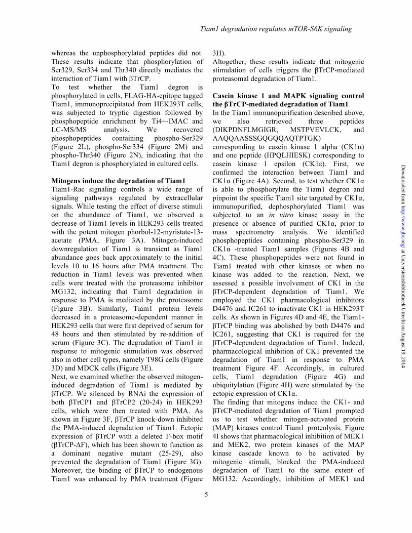
whereas the unphosphorylated peptides did not. These results indicate that phosphorylation of Ser329, Ser334 and Thr340 directly mediates the interaction of Tiam1 with βTrCP. To test whether the Tiam1 degron is phosphorylated in cells, FLAG-HA-epitope tagged Tiam1, immunoprecipitated from HEK293T cells, was subjected to tryptic digestion followed by phosphopeptide enrichment by Ti4+-IMAC and LC-MS/MS analysis. We recovered phosphopeptides containing phospho-Ser329 (Figure 2L), phospho-Ser334 (Figure 2M) and phospho-Thr340 (Figure 2N), indicating that the Tiam1 degron is phosphorylated in cultured cells. Mitogens induce the degradation of Tiam1 Tiam1-Rac signaling controls a wide range of signaling pathways regulated by extracellular signals. While testing the effect of diverse stimuli on the abundance of Tiam1, we observed a decrease of Tiam1 levels in HEK293 cells treated with the potent mitogen phorbol-12-myristate-13-acetate (PMA, Figure 3A). Mitogen-induced downregulation of Tiam1 is transient as Tiam1 abundance goes back approximately to the initial levels 10 to 16 hours after PMA treatment. The reduction in Tiam1 levels was prevented when cells were treated with the proteasome inhibitor MG132, indicating that Tiam1 degradation in response to PMA is mediated by the proteasome (Figure 3B). Similarly, Tiam1 protein levels decreased in a proteasome-dependent manner in HEK293 cells that were first deprived of serum for 48 hours and then stimulated by re-addition of serum (Figure 3C). The degradation of Tiam1 in response to mitogenic stimulation was observed also in other cell types, namely T98G cells (Figure 3D) and MDCK cells (Figure 3E). Next, we examined whether the observed mitogen-induced degradation of Tiam1 is mediated by βTrCP. We silenced by RNAi the expression of both βTrCP1 and βTrCP2 (20-24) in HEK293 cells, which were then treated with PMA. As shown in Figure 3F, βTrCP knock-down inhibited the PMA-induced degradation of Tiam1. Ectopic expression of βTrCP with a deleted F-box motif (βTrCP-ΔF), which has been shown to function as a dominant negative mutant (25-29), also prevented the degradation of Tiam1 (Figure 3G). Moreover, the binding of βTrCP to endogenous Tiam1 was enhanced by PMA treatment (Figure
3H). Altogether, these results indicate that mitogenic stimulation of cells triggers the βTrCP-mediated proteasomal degradation of Tiam1. Casein kinase 1 and MAPK signaling control the βTrCP-mediated degradation of Tiam1 In the Tiam1 immunopurification described above, we also retrieved three peptides (DIKPDNFLMGIGR, MSTPVEVLCK, and AAQQAASSSGQGQQAQTPTGK) corresponding to casein kinase 1 alpha (CK1α) and one peptide (HPQLHIESK) corresponding to casein kinase 1 epsilon (CK1ε). First, we confirmed the interaction between Tiam1 and CK1α (Figure 4A). Second, to test whether CK1α is able to phosphorylate the Tiam1 degron and pinpoint the specific Tiam1 site targeted by CK1α, immunopurified, dephosphorylated Tiam1 was subjected to an in vitro kinase assay in the presence or absence of purified CK1α, prior to mass spectrometry analysis. We identified phosphopeptides containing phospho-Ser329 in CK1α -treated Tiam1 samples (Figures 4B and 4C). These phosphopeptides were not found in Tiam1 treated with other kinases or when no kinase was added to the reaction. Next, we assessed a possible involvement of CK1 in the βTrCP-dependent degradation of Tiam1. We employed the CK1 pharmacological inhibitors D4476 and IC261 to inactivate CK1 in HEK293T cells. As shown in Figures 4D and 4E, the Tiam1-βTrCP binding was abolished by both D4476 and IC261, suggesting that CK1 is required for the βTrCP-dependent degradation of Tiam1. Indeed, pharmacological inhibition of CK1 prevented the degradation of Tiam1 in response to PMA treatment Figure 4F. Accordingly, in cultured cells, Tiam1 degradation (Figure 4G) and ubiquitylation (Figure 4H) were stimulated by the ectopic expression of CK1α. The finding that mitogens induce the CK1- and βTrCP-mediated degradation of Tiam1 prompted us to test whether mitogen-activated protein (MAP) kinases control Tiam1 proteolysis. Figure 4I shows that pharmacological inhibition of MEK1 and MEK2, two protein kinases of the MAP kinase cascade known to be activated by mitogenic stimuli, blocked the PMA-induced degradation of Tiam1 to the same extent of MG132. Accordingly, inhibition of MEK1 and
at Universiteitsbibliotheek U
trecht on August 19, 2014
http://ww
w.jbc.org/
Dow
nloaded from

Tiam1 degradation regulates mTOR-S6K signaling
6
MEK2 reduced the interaction between βTrCP and Tiam1 (Figure 4J). Tiam1 degradation is required for the termination of mTOR-S6K signaling To investigate the biological significance of Tiam1 degradation, physiological levels of wild type Tiam1 and the Tiam1(S239A/S334A) mutant were expressed in HEK293 cells. Tiam1(S239A/S334A) as well as the Tiam1 mutant truncated at the N-terminus (C1199) lacking the βTrCP binding domain were resistant to the degradation induced by mitogenic stimulation, as indicated by the half-life experiment shown in Figure 5A. As it has been shown that both Tiam1 and Rac1 control the mTOR-S6K signaling pathway, which mediates the mitogenic response in cells (30-32), we analyzed the activation of mTOR-S6K signaling in response to mitogenic stimulation in cell expressing wild type Tiam1 and the non-degradable Tiam1(S239A/S334A) mutant. As shown in Figure 5B, mitogenic stimulation of cells expressing wild type Tiam1 resulted in an increase of the mTOR-mediated phosphorylation of p70-S6K (Thr389) and its isoform p85-S6K (Thr412), which then (approximately 1 hour after PMA treatment) rapidly decreased. In contrast, cells expressing the non-degradable Tiam1(S239A/S334A) mutant displayed a sustained (at least up to 5 hours after PMA treatment) phosphorylation of p70-S6K (Thr389) and p85-S6K (Thr412) in response to mitogenic stimulation. No remarkable effect on the phosphorylation of p70-S6K (Thr389) and p85-S6K (Thr412) was observed in asynchronously growing cells (Figure 5C). The expression of the degradation-resistant Tiam1(S239A/S334A) mutant had a similar effect on the mitogen-induced activating phosphorylation of additional residues of p70-S6K, (Thr421/Ser424) and p85-S6K (Thr444/Ser447) (Figure 5B). Similar results were obtained when serum was used to stimulate cells expressing wild type Tiam1 or the Tiam1(S239A/S334A) mutant (Figure 5D). In agreement with previous reports (30), pharmacological inhibition of Rac activity prevented the induction of S6K phosphorylation following mitogenic stimulation suggesting that the mitogen-induced activation of the mTOR-S6K signaling pathway is dependent on Rac activity
(Figure 5E). Taken together, these results demonstrate that failure to degrade Tiam1 in response to mitogens results in prolonged activation of the mTOR/S6K signaling. Failure to degrade Tiam1 results in loss of cell viability and increased apoptosis Finally, we investigated the effect of defective degradation of Tiam1 on cell growth and survival. After mitogenic stimulation, cells expressing Tiam1(S239A/S334A) or Tiam1-C1199 displayed reduced cell viability when compared with cells expressing wild type Tiam1 (Figures 6A and 6B). Moreover, failure to degrade Tiam1 resulted in apoptotic cell death as shown by the induction of the cleaved active form of Caspase 3 (Figures 6C and 6D). DISCUSSION Here we have shown that Tiam1 is a novel substrate of the SCFβTrCP ubiquitin ligase, identified a conserved phosphodegron at the N-terminus of Tiam1 that is required for Tiam1 binding to βTrCP, and demonstrated that SCFβTrCP targets Tiam1 for proteasome-dependent degradation in response to mitogenic stimulation. We have also found that the mitogen-induced degradation of Tiam1 is mediated by CK1α, which phosphorylates a residue within the Tiam1 phosphodegron. Tiam1 destruction is required for terminating the activation of the mTOR-S6K signaling pathway following mitogenic stimulation. Indeed, expression of a Tiam1 mutant unable to bind βTrCP results in sustained phosphorylation of the p70-S6 kinase, a substrate of mTOR. Failure to degrade Tiam1 leads to loss of cell viability due to increased apoptosis. Interestingly, it has been recently reported that constitutive mTOR-S6K signaling sensitizes cells to p53-dependent cell death in response to stress conditions (33). In agreement with these studies, a recent report has shown that cells expressing the degradation-resistant Tiam1-C1199 mutant are more sensitive to DNA damaging drugs-induced apoptosis (34). We have shown that the βTrCP-mediated degradation of Tiam1 is blocked by pharmacological inhibition of MEK. Accordingly, the Malliri group has demonstrated that ERK1 and ERK2 associate with Tiam1 and that ERK activation is required for the vSrc-induced
at Universiteitsbibliotheek U
trecht on August 19, 2014
http://ww
w.jbc.org/
Dow
nloaded from

Tiam1 degradation regulates mTOR-S6K signaling
7
degradation of Tiam1 (35). The involvement of the MEK-ERK pathway in the destruction of Tiam1 via βTrCP also suggests that the MEK-ERK-dependent phosphorylation of Tiam1 could prime the phosphorylation of the Tiam1 degron by CK1, as it has been shown for other βTrCP substrates (16) (36,37). A phospho-specific antibody against the Tiam1 degron motif (329-pSEFADpSGIEGApT-340) is required to test (both in vitro and in cultured cells) whether Tiam1
is phosphorylated by CK1 on its degron following initial phosphorylation by MEK-ERK on different sites. In conclusion, the finding that the degradation of Tiam1, induced by CK1 in cooperation with the MAPK cascade, is needed to terminate mTOR-S6K signaling suggests a new mechanism by which the MAPK cascade controls the timing of activation of the mTOR-S6K signaling pathway.
at Universiteitsbibliotheek U
trecht on August 19, 2014
http://ww
w.jbc.org/
Dow
nloaded from

Tiam1 degradation regulates mTOR-S6K signaling
8
REFERENCES 1. Habets, G. G., Scholtes, E. H., Zuydgeest, D., van der Kammen, R. A., Stam, J. C.,
Berns, A., and Collard, J. G. (1994) Identification of an invasion-inducing gene, Tiam-1, that encodes a protein with homology to GDP-GTP exchangers for Rho-like proteins. Cell 77, 537-549
2. Mertens, A. E., Roovers, R. C., and Collard, J. G. (2003) Regulation of Tiam1-Rac signalling. FEBS Lett 546, 11-16
3. Michiels, F., Habets, G. G., Stam, J. C., van der Kammen, R. A., and Collard, J. G. (1995) A role for Rac in Tiam1-induced membrane ruffling and invasion. Nature 375, 338-340
4. Lambert, J. M., Lambert, Q. T., Reuther, G. W., Malliri, A., Siderovski, D. P., Sondek, J., Collard, J. G., and Der, C. J. (2002) Tiam1 mediates Ras activation of Rac by a PI(3)K-independent mechanism. Nature cell biology 4, 621-625
5. Hordijk, P. L., ten Klooster, J. P., van der Kammen, R. A., Michiels, F., Oomen, L. C., and Collard, J. G. (1997) Inhibition of invasion of epithelial cells by Tiam1-Rac signaling. Science 278, 1464-1466
6. Malliri, A., and Collard, J. G. (2003) Role of Rho-family proteins in cell adhesion and cancer. Curr Opin Cell Biol 15, 583-589
7. Malliri, A., van Es, S., Huveneers, S., and Collard, J. G. (2004) The Rac exchange factor Tiam1 is required for the establishment and maintenance of cadherin-based adhesions. The Journal of biological chemistry 279, 30092-30098
8. Kruiswijk, F., Yuniati, L., Magliozzi, R., Low, T. Y., Lim, R., Bolder, R., Mohammed, S., Proud, C. G., Heck, A. J., Pagano, M., and Guardavaccaro, D. (2012) Coupled Activation and Degradation of eEF2K Regulates Protein Synthesis in Response to Genotoxic Stress. Science Signaling 5, ra40
9. Shevchenko, A., Wilm, M., Vorm, O., and Mann, M. (1996) Mass spectrometric sequencing of proteins silver-stained polyacrylamide gels. Anal Chem 68, 850-858
10. Raijmakers, R., Berkers, C. R., de Jong, A., Ovaa, H., Heck, A. J., and Mohammed, S. (2008) Automated online sequential isotope labeling for protein quantitation applied to proteasome tissue-specific diversity. Mol Cell Proteomics 7, 1755-1762
11. Taus, T., Kocher, T., Pichler, P., Paschke, C., Schmidt, A., Henrich, C., and Mechtler, K. (2011) Universal and confident phosphorylation site localization using phosphoRS. J Proteome Res 10, 5354-5362
12. D'Annibale, S., Kim, J., Magliozzi, R., Low, T. Y., Mohammed, S., Heck, A. J., and Guardavaccaro, D. (2014) Proteasome-dependent Degradation of Transcription Factor AP4 (TFAP4) Controls Mitotic Division. J Biol Chem
13. Frescas, D., and Pagano, M. (2008) Deregulated proteolysis by the F-box proteins SKP2 and beta-TrCP: tipping the scales of cancer. Nat Rev Cancer 8, 438-449
14. Wu, G., Xu, G., Schulman, B. A., Jeffrey, P. D., Harper, J. W., and Pavletich, N. P. (2003) Structure of a beta-TrCP1-Skp1-beta-catenin complex: destruction motif binding and lysine specificity of the SCF(beta-TrCP1) ubiquitin ligase. Mol Cell 11, 1445-1456
15. Cardozo, T., and Pagano, M. (2004) The SCF ubiquitin ligase: insights into a molecular machine. Nat Rev Mol Cell Biol 5, 739-751
16. Magliozzi, R., Low, T. Y., Weijts, B. G., Cheng, T., Spanjaard, E., Mohammed, S., van Veen, A., Ovaa, H., de Rooij, J., Zwartkruis, F. J., Bos, J. L., de Bruin, A., Heck, A. J., and Guardavaccaro, D. (2013) Control of Epithelial Cell Migration and Invasion by the IKKbeta- and CK1alpha-Mediated Degradation of RAPGEF2. Dev Cell 27, 574-585
17. Busino, L., Donzelli, M., Chiesa, M., Guardavaccaro, D., Ganoth, D., Dorrello, N. V., Hershko, A., Pagano, M., and Draetta, G. F. (2003) Degradation of Cdc25A by beta-TrCP during S phase and in response to DNA damage. Nature 426, 87-91
at Universiteitsbibliotheek U
trecht on August 19, 2014
http://ww
w.jbc.org/
Dow
nloaded from

Tiam1 degradation regulates mTOR-S6K signaling
9
18. Jin, J., Shirogane, T., Xu, L., Nalepa, G., Qin, J., Elledge, S. J., and Harper, J. W. (2003) SCFbeta-TRCP links Chk1 signaling to degradation of the Cdc25A protein phosphatase. Genes Dev 17, 3062-3074
19. Melixetian, M., Klein, D. K., Sorensen, C. S., and Helin, K. (2009) NEK11 regulates CDC25A degradation and the IR-induced G2/M checkpoint. Nat Cell Biol 11, 1247-1253
20. Mailand, N., Bekker-Jensen, S., Bartek, J., and Lukas, J. (2006) Destruction of Claspin by SCFbetaTrCP restrains Chk1 activation and facilitates recovery from genotoxic stress. Mol Cell 23, 307-318
21. Margottin-Goguet, F., Hsu, J. Y., Loktev, A., Hsieh, H. M., Reimann, J. D., and Jackson, P. K. (2003) Prophase destruction of Emi1 by the SCF(betaTrCP/Slimb) ubiquitin ligase activates the anaphase promoting complex to allow progression beyond prometaphase. Dev Cell 4, 813-826
22. Guardavaccaro, D., Frescas, D., Dorrello, N. V., Peschiaroli, A., Multani, A. S., Cardozo, T., Lasorella, A., Iavarone, A., Chang, S., Hernando, E., and Pagano, M. (2008) Control of chromosome stability by the beta-TrCP-REST-Mad2 axis. Nature 452, 365-369
23. Mantovani, F., and Banks, L. (2003) Regulation of the discs large tumor suppressor by a phosphorylation-dependent interaction with the beta-TrCP ubiquitin ligase receptor. J Biol Chem 278, 42477-42486
24. Dorrello, N. V., Peschiaroli, A., Guardavaccaro, D., Colburn, N. H., Sherman, N. E., and Pagano, M. (2006) S6K1- and betaTRCP-mediated degradation of PDCD4 promotes protein translation and cell growth. Science 314, 467-471
25. Yaron, A., Hatzubai, A., Davis, M., Lavon, I., Amit, S., Manning, A. M., Andersen, J. S., Mann, M., Mercurio, F., and Ben-Neriah, Y. (1998) Identification of the receptor component of the IkappaBalpha-ubiquitin ligase. Nature 396, 590-594
26. Spencer, E., Jiang, J., and Chen, Z. J. (1999) Signal-induced ubiquitination of IKB by the F-box protein Slimb/ß-TrCP. Genes & Dev 13, 284-294
27. Wu, K., Fuchs, S. Y., Chen, A., Tan, P., Gomez, C., Ronai, Z., and Pan, Z. Q. (2000) The SCF(HOS/beta-TRCP)-ROC1 E3 ubiquitin ligase utilizes two distinct domains within CUL1 for substrate targeting and ubiquitin ligation. Mol Cell Biol 20, 1382-1393
28. Winston, J. T., Strack, P., Beer-Romero, P., Chu, C. Y., Elledge, S. J., and Harper, J. W. (1999) The SCFb-TRCP ubiquitin ligase complex associates specifically with phosphorylated destruction motifs in IkBa and b-catenin and stimulates IkBa ubiquitination in vitro. Genes and Devel 13, 270-283
29. Fuchs, S. Y., Chen, A., Xiong, Y., Pan, Z. Q., and Ronai, Z. (1999) HOS, a human homolog of Slimb, forms an SCF complex with Skp1 and Cullin1 and targets the phosphorylation-dependent degradation of IkappaB and beta-catenin. Oncogene 18, 2039-2046
30. Chou, M. M., and Blenis, J. (1996) The 70 kDa S6 kinase complexes with and is activated by the Rho family G proteins Cdc42 and Rac1. Cell 85, 573-583
31. Saci, A., Cantley, L. C., and Carpenter, C. L. (2011) Rac1 regulates the activity of mTORC1 and mTORC2 and controls cellular size. Mol Cell 42, 50-61
32. Buchsbaum, R. J., Connolly, B. A., and Feig, L. A. (2003) Regulation of p70 S6 kinase by complex formation between the Rac guanine nucleotide exchange factor (Rac-GEF) Tiam1 and the scaffold spinophilin. J Biol Chem 278, 18833-18841
33. Lee, C. H., Inoki, K., Karbowniczek, M., Petroulakis, E., Sonenberg, N., Henske, E. P., and Guan, K. L. (2007) Constitutive mTOR activation in TSC mutants sensitizes cells to energy starvation and genomic damage via p53. EMBO J 26, 4812-4823
34. Zhu, G., Fan, Z., Ding, M., Mu, L., Liang, J., Ding, Y., Fu, Y., Huang, B., and Wu, W. (2014) DNA damage induces the accumulation of Tiam1 by blocking beta-TrCP-dependent degradation. J Biol Chem
at Universiteitsbibliotheek U
trecht on August 19, 2014
http://ww
w.jbc.org/
Dow
nloaded from

Tiam1 degradation regulates mTOR-S6K signaling
10
35. Woodcock, S. A., Rooney, C., Liontos, M., Connolly, Y., Zoumpourlis, V., Whetton, A. D., Gorgoulis, V. G., and Malliri, A. (2009) SRC-induced disassembly of adherens junctions requires localized phosphorylation and degradation of the rac activator tiam1. Mol Cell 33, 639-653
36. Dehan, E., Bassermann, F., Guardavaccaro, D., Vasiliver-Shamis, G., Cohen, M., Lowes, K. N., Dustin, M., Huang, D. C., Taunton, J., and Pagano, M. (2009) betaTrCP- and Rsk1/2-mediated degradation of BimEL inhibits apoptosis. Mol Cell 33, 109-116
37. Duan, S., Skaar, J. R., Kuchay, S., Toschi, A., Kanarek, N., Ben-Neriah, Y., and Pagano, M. (2011) mTOR generates an auto-amplification loop by triggering the betaTrCP- and CK1alpha-dependent degradation of DEPTOR. Mol Cell 44, 317-324
at Universiteitsbibliotheek U
trecht on August 19, 2014
http://ww
w.jbc.org/
Dow
nloaded from
Tiam1 degradation regulates mTOR-S6K signaling
11
ACKNOWLEDGEMENTS We thank R. Lim, S. Rahmy for contributions, S. Ellenbroek, J. den Hertog and F. Zwartkruis for reagents, A. Malliri for suggestions and reagents. Work in D.G.’s laboratory was supported by the Royal Dutch Academy of Arts and Sciences (KNAW), the Dutch Cancer Society (KWF), the Cancer Genomics Centre and the European Union under Marie Curie Actions (FP7). T.Y.L. and A.J.R.H. were supported by the Netherlands Proteomics Center (NPC). CONFLICT OF INTEREST. The authors declare that they have no conflict of interest related to this work.
at Universiteitsbibliotheek U
trecht on August 19, 2014
http://ww
w.jbc.org/
Dow
nloaded from

Tiam1 degradation regulates mTOR-S6K signaling
12
FIGURE LEGENDS Figure 1. Tiam1 interacts with, and is ubiquitylated by, the SCFβTrCP ubiquitin ligase. (A) Peptide coverage of Tiam1 in the mass spectrometry analysis of βTrCP2 immunopurification. Amino acid sequences of detected Tiam1 peptides are highlighted in yellow. (B) Peptide coverage of βTrCP2 in the mass spectrometry analysis of Tiam1 immunopurification. Amino acid sequences of detected βTrCP2 peptides are highlighted in yellow. (C) The indicated FLAG-tagged F-box proteins containing WD40 repeats (FBXWs) or an empty vector (EV) were expressed in HEK293T cells. Forty-eight hours after transfection, cells were treated for 5 hours with the proteasome inhibitors MG132, then harvested and lysed. Whole cell extracts (WCE) were immunoprecipitated (IP) with anti-FLAG antibody and immunoblotted with antibodies specific for the indicated proteins. (D) HEK293T cells were transfected with GFP-tagged wild type Tiam1 or and empty vector. Cells were collected and lysed. Tiam1 was immunoprecipitated (IP) from whole cell extracts and immunocomplexes were analyzed by immunoblotting with antibodies specific for the indicated proteins. (E) Arginine 447 in the WD40 repeat of βTrCP2 is required for the interaction with Tiam1. HEK293T cells were transfected as indicated and analyzed as in (C). WCE: whole cell extract. WT: wild type. (F) Tiam1, Skp1, Cul1, and Rbx1 were expressed in HEK293T in the absence or presence of βTrCP1 or an inactive βTrCP1-ΔF-box mutant. After immunopurification with anti-FLAG resin, an in vitro ubiquitylation of Tiam1 was performed. Samples were analyzed by immunoblotting with an anti-Tiam1 antibody. The bracket indicates a ladder of bands corresponding to poly-ubiquitylated Tiam1. Figure 2. Phosphorylation of Ser329, Ser334 and Thr340 in Tiam1 is required for the interaction of Tiam1 with βTrCP1. (A) Schematic representation of Tiam1 functional domains and three putative βTrCP-binding motifs. (B) HEK293T cells were transfected with FLAG-tagged βTrCP1 and either HA-tagged wild type Tiam1, HA-tagged Tiam1(S370A/S374A), HA-tagged Tiam1(S414A/S418A) or an empty vector (EV). Forty-eight hours after transfection, cells were harvested and lysed. Whole cell extracts were subjected to immunoprecipitation (IP) with anti-HA resin, followed by immunoblotting with antibodies specific for the indicated proteins. (C) Alignment of the amino acid regions corresponding to the βTrCP-binding motifs in Tiam1 orthologs and previously reported βTrCP substrates, i.e., IκBα, Emi1, βcatenin, Claspin and Cdc25A. The amino acidic sequences of the phosphodegron mutants are shown below. (D) HEK293T cells were transfected with FLAG-tagged βTrCP1 and either HA-tagged wild type Tiam1, HA-tagged Tiam1(S329A/S334A), HA-tagged Tiam1(S329A/S334A/T340A) or an empty vector (EV). Forty-eight hours after transfection, cells were harvested and lysed. Whole cell extracts were subjected to immunoprecipitation (IP) with anti-FLAG resin, followed by immunoblotting with antibodies specific for the indicated proteins. (E) HEK293T cells were transfected with an empty vector (EV), HA-tagged wild type Tiam1, or HA-tagged Tiam1(S329A/S334A). Forty-eight hours after transfection, cells were harvested and lysed. Whole cell extracts were subjected to immunoprecipitation (IP) with anti-HA resin and then immunoblotted with antibodies specific for the indicated proteins. When indicated, immunocomplexes were incubated with lambda phosphatase (λPP) for 30 minutes and then washed. (F) HEK293T cells were transfected with HA-tagged wild type Tiam1, HA-tagged Tiam1(S329A/S334A) or HA-tagged Tiam1-C1199. Whole cell extracts were subjected to immunoprecipitation (IP) and immunoblotting as in (E). (G) HEK293T cells were transfected with GFP-tagged wild type Tiam1 or GFP-tagged Tiam1-C1199. Cells were collected and lysed. βTrCP was immunoprecipitated (IP) from whole cell extracts and immunocomplexes were analyzed by immunoblotting with antibodies specific for the indicated proteins. (H-I) 35S-βTrCP1 and 35S-FBXW5 were transcribed/translated in vitro and incubated with beads coupled to peptides spanning the Tiam1 degron. Beads were washed with Triton-X buffer and bound proteins were eluted and subjected to electrophoresis and autoradiography. The last two lanes correspond to 10%
at Universiteitsbibliotheek U
trecht on August 19, 2014
http://ww
w.jbc.org/
Dow
nloaded from
Tiam1 degradation regulates mTOR-S6K signaling
13
of the in vitro translated protein inputs. Peptide sequence spanning the Tiam1 degron is shown on the left. (L-N) FLAG-HA-tagged Tiam1 was immunopurified from HEK293T cells and analyzed by mass spectrometry. Ion fragmentation spectra are shown. The indicated Tiam1 tryptic peptides spanning the phosphodegron were found phosphorylated on Ser329 (L), Ser334 (M) or Thr340 (N). As shown, peptide sequences can be explained by their respective CID (collision-induced dissociation) MS/MS spectra including phospho-Ser329, phospho-Ser334 and phospho-Thr340. Figure 3. Proteasome- and βTrCP-dependent degradation of Tiam1 in response to mitogens. (A) HEK293 cells, cultured in low serum for 48 hours, were treated with PMA. Cells were collected at the indicated times and lysed. Whole cell extracts were subjected to immunoblotting with antibodies specific for the indicated proteins. Actin is shown as a loading control. (B) HEK293 cells were treated with PMA as in (A) with or without the proteasome inhibitor MG132. Whole cell extracts were analyzed as in (A). (C) HEK293 cells were either cultured in presence of serum (S) or deprived of serum for 48 hours (SD). After serum re-addition, cells were collected at the indicated times. When indicated, the proteasome inhibitor MG132 was added. Protein extracts were analyzed by immunoblotting with antibodies specific for the indicated proteins. Cul1 is shown as a loading control. (D-E) T98G cells (D) and MDCK cells (E) were treated with PMA as in (A) and with MG132 when indicated. Whole cell extracts were analyzed as in (A). (F) HEK293 cells were transfected with the indicated siRNA oligonucleotides and treated as in (A). Cul1 is shown as loading control. (G) HEK293 cells were transfected with FLAG-tagged βTrCP1-ΔF or an empty vector (EV) and treated with PMA for 2 hours. Cells were collected and lysed. Whole cell extracts were analyzed as in (A). (H) HEK293 were transfected with an empty vector or FLAG-tagged βTrCP1. Forty-eight hours after transfection, cells were treated with MG132 and PMA for 5 hours (when indicated) then harvested and lysed. Whole cell extracts were immunoprecipitated (IP) with anti-FLAG resin, and immunoblotted with anti-Tiam1 and anti-FLAG antibodies. Figure 4. CK1 and the MAPK pathway control the βTrCP-mediated degradation of Tiam1. (A) HEK293T cells were transfected with FLAG-tagged CK1α, FLAG-tagged βTrCP1 or an empty vector. Forty-eight hours after transfection, cells were treated with MG132 for 5 hour. Cells were then harvested and lysed. Whole cell extracts were immunoprecipitated (IP) with anti-FLAG resin, and immunoblotted with antibodies specific for the indicated proteins. (B-C) Tiam1 was immunopurified and dephosphorylated with lambda phosphatase prior to an in vitro kinase assay in the presence of purified CK1α. A mock reaction (no kinase) was used as a negative control. Individual mixes were subsequently trypsinized and analyzed by mass spectrometry. The Tiam1 tryptic peptide TTQDVNAGEGSEFADSGIEGATTDTDLLSR was not found to be phosphorylated in the negative control (B). When CK1α was used in the in vitro kinase assay, the same peptide was found to be phosphorylated on Ser329 (C). As shown, both peptide sequences can be clearly explained by their respective HCD MSMS spectrum (Mascot Score 139 and 92), including the pS329 site (PhosphoRS site probability = 100.0%). In these figures, [pS] denotes phosphorylated serine; [b] denotes b ions and [y] denotes y ions. (D) HEK293T cells were transfected with FLAG-tagged βTrCP1. Forty-eight hours after transfection, cells were treated with MG132 for 5 hours (when indicated) in the presence or absence of the indicated kinase inhibitors. Cells were then harvested and lysed. Whole cell extracts were immunoprecipitated (IP) with anti-FLAG resin, and immunoblotted with antibodies specific for the indicated proteins. (E) HEK293T cells were transfected with HA-tagged wild type Tiam1 or HA-tagged Tiam1(S329A/S334A). Forty-eight hours after transfection, cells were treated as in (D). Whole cell extracts were processed as in (D) except that an anti-HA resin was used for immunoprecipitation. (F) Cells were treated with PMA for 4 hours with or without the CK1 inhibitor D4476. Whole cell
at Universiteitsbibliotheek U
trecht on August 19, 2014
http://ww
w.jbc.org/
Dow
nloaded from

Tiam1 degradation regulates mTOR-S6K signaling
14
extracts were analyzed by immunoblotting. (G) HEK293 cells were transfected with plasmids expressing HA-tagged wild type Tiam1 along with plasmids expressing the indicated kinases. Twenty-four hours after transfection, cells were collected and lysed. Whole cells extracts were analyzed by immunoblotting with antibodies specific for the indicated proteins. (H) HEK293T cells were transfected with either HA-tagged wild type Tiam1 or HA-tagged Tiam1(S329A/S334A) along with Myc-tagged ubiquitin with or without CK1α. Cells were harvested and lysed in 0.1% Triton X-100 lysis buffer. Whole cell extracts were denatured by adding 1% SDS and boiling for 10 minutes. SDS was quenched and diluted. Whole cell extracts were then immunoprecipitated (IP) with anti-HA resin, and immunoblotted with anti-Myc antibodies. The bracket indicates a ladder of bands corresponding to poly-ubiquitylated Tiam1. (I) HEK293 cells were treated with PMA in the absence or presence of the indicated compounds. Cells were collected at the indicated times and lysed. Whole cell extracts were analyzed by immunoblotting. (J) HEK293T cells were transfected with FLAG-tagged βTrCP1 or FLAG-tagged βTrCP1(R474A). Forty-eight hours after transfection, cells were treated with MG132 for 5 hours in the presence or absence of the MEK inhibitor U1026. Cells were then harvested and lysed. Whole cell extracts were immunoprecipitated (IP) with anti-FLAG resin, and immunoblotted with antibodies specific for the indicated proteins. Figure 5. Tiam1 degradation controls the duration of the mitogen-induced mTOR-S6K signaling pathway. (A) HEK293 cells expressing HA-tagged wild type Tiam1, HA-tagged Tiam1(S329A/S334A), or HA-tagged Tiam1-C1199, were incubated in low serum for 24 hours and then treated with PMA and cycloheximide (CHX) for the indicated times. Cells were collected and lysed. Whole cell lysates were analyzed by immunoblotting with an anti-HA antibody. Actin is shown as a loading control. The graph shows the quantification of Tiam1 abundance relative to the amount at time 0. (B) HEK293 cells transduced with retroviruses expressing HA-tagged wild type Tiam1 or HA-tagged Tiam1(S329A/S334A) were incubated in low serum for 48 hours then treated with PMA. At the indicated times, cells were collected and lysed. Whole cell extracts were analyzed by immunoblotting with antibodies for the indicated proteins. Levels of GTP-loaded Rac1 were analyzed in a pull-down assay as described in Experimental Procedures. Actin is shown as loading control. To facilitate comparison, a dotted line separates samples from cells expressing wild type Tiam1 and samples from cells expressing Tiam1(S329A/S334A). (C) Whole cell lysates of asynchronously growing HEK293 cells expressing HA-tagged wild type Tiam1 or HA-tagged Tiam1(S329A/S334A) were analyzed by immunoblotting with antibodies for the indicated proteins. (D) As in (B) except that serum (instead of PMA) was used as mitogen. Cul1 is shown as loading control. (E) HEK293 cells were incubated in low serum for 48 hours then treated with PMA. Six hours after PMA treatment cells were analyzed by immunoblotting. Rac1 activity was assessed as in (B). Figure 6. Inhibition of Tiam1 degradation results in loss of cell viability and increased apoptosis. (A-B) HEK293 cells (A) or MDCK cells (B) transfected with HA-tagged wild type Tiam1, HA-tagged Tiam1(S329A/S334A), or HA-tagged Tiam1-C1199, were incubated in low serum for 24 hours and then treated with PMA for 6 hours. Cell viability was analyzed by MTT assay. The results are the average of three independent experiments. Error bars represent SD. (C) MDCK cells as in (B) were incubated in low serum for 24 hours and then treated with PMA for the indicated times. Cells were collected and either lysed and analyzed by immunoblotting with antibodies for the indicated proteins.
at Universiteitsbibliotheek U
trecht on August 19, 2014
http://ww
w.jbc.org/
Dow
nloaded from

Tiam1 degradation regulates mTOR-S6K signaling
15
(D) As in (C) except that cells were fixed, stained for cleaved caspase 3 and analyzed by flow cytometry. The results are the average of three independent experiments. Error bars represent SD.
at Universiteitsbibliotheek U
trecht on August 19, 2014
http://ww
w.jbc.org/
Dow
nloaded from
C
E F
Figure 1
Tiam1
βTrC
P1
βTrC
P2
FBX
W2
FBX
W5
FBX
W4
FBX
W6
EV
WC
E
IP anti-FLAG
-
FBXWs (anti-FLAG)
* *
* *
* *
Tiam1
βTrC
P2
WT
EV
βTrC
P2
(R44
7A)
WC
E
IP anti-FLAG
--
βTrCP2 (anti-FLAG)
A
B
D
150 kDa –
250 kDa – Tiam1
Tiam1 (Ub)n
+ + !+ + !- + !+ - !
Tiam1 Cul1-Skp1-Rbx1 βTrCP1 βTrCP1ΔF
Tiam
1 W
T
EV
IP anti-GFP
Tiam1 (anti-GFP)
βTrCP
βTrCP HA HA
FLA
G
FLA
G
IP + MS
Tiam1
GFP
IP + MS
at Universiteitsbibliotheek U
trecht on August 19, 2014
http://ww
w.jbc.org/
Dow
nloaded from

Figure 2
A B
D E
200 400 600 800 1000 1200 1400 1600 1800 2000 2200 2400 2600 2800 3000 3200m/z
0
5
10
15
20
25
30
35
40
45
50
55
60
65
70
75
80
85
90
95
100
Rel
ativ
e A
bund
ance
-T T Q D V N A G E G S E F A D S G I E G A T t D T D L L S R-b4
y2y3y4y5y6y7y9y11y12
b5 b22b7 b9 b13
y2
y3
y4
b4
b5
y5
y6
b7
y7
b22+++ b9
y26+++
y9
y11
y12
y19++
b21-18++
y11-17
y22++
y26++
b19 b21
y22
200 400 600 800 1000 1200 1400 1600 1800 2000 2200 2400 2600 2800 3000 3200m/z
0
5
10
15
20
25
30
35
40
45
50
55
60
65
70
75
80
85
90
95
100
Rel
ativ
e A
bund
ance
-T T Q D V N A G E G S E F A D s G I E G A T T D T D L L S R-b4
y2
b12
y3y4y5y6y7y8y9y10y11y12
b5 b6 b7 b9 b13
b4 b5y4y2
y3
y5b6
y6
b7
y7
y9
y8
b9
y10
y11
b12
y12y11
y12
200 400 600 800 1000 1200 1400 1600 1800 2000 2200 2400 2600 2800 3000 3200 3400m/z
0
5
10
15
20
25
30
35
40
45
50
55
60
65
70
75
80
85
90
95
100
Rel
ativ
e A
bund
ance
y2 y4y6
y7
y3y5
y8
y9
y10 y14
y15 y16
b2
y27+++
b14
y17b17
b17++
y13
-A K T T Q D V N A G E G s E F A D S G I E G A T T D T D L L b2
y2
b14 b17 b19b20
y3y4y5y6y7y8y9y10y11y12y14 y13y15y16y17y27
G H I
L M
EV
Tiam
1 W
T
Tiam
1(S
370A
/S37
4A)
Tiam
1(S
414A
/S41
8A)
Tiam1 (anti-HA)
IP a
nti-H
A
βTrCP1 (anti-FLAG)
Tiam
1 P
-pep
tide
Tiam
1 pe
ptid
e
βTrCP1 FBXW5
Tiam
1 P
-pep
tide
Tiam
1 pe
ptid
e
10%
inpu
t - β
TrC
P1
10%
inpu
t - F
BX
W5
<FBXW5
<βTrCP1
P-peptide: 327-EGSEFADSGIEGAT-340
peptide: 327-EGSEFADSGIEGAT-340
P P Tiam
1 P
-pep
tide
Tiam
1 pe
ptid
e
βTrCP1 FBXW5
Tiam
1 P
-pep
tide
Tiam
1 pe
ptid
e
<FBXW5
<βTrCP1
10%
inpu
t - β
TrC
P1
10%
inpu
t - F
BX
W5
P-peptide: 330-EFADSGIEGATTDT-343
peptide: 330-EFADSGIEGATTDT-343
P P
βTrCP binding sites IkBα(Hs): 28-DRGDSGLDSMKD-39 Emi1(Hs): 141-LYEDSGYSSFSL-152 βCatenin(Hs): 29-SYLDSGIHSGAT-40 Claspin(Hs): 26-SPSDSGQGSYET-37 Cdc25A(Hs): 74-MGSSESTDSGFCLDSPGPL-92 Tiam1(Hs): 326-GEGSEFADSGIEGATTDTDL-345 Tiam1(Mm): 326-GEGSEFADSGIEGATTDTDL-345 Tiam1(Rn): 325-GEGSEFADSGIEGATTDTDL-344 Tiam1(Cf): 326-GEGSEFADSGIEGATTDTDL-345 Tiam1(Gg): 328-GEGSEFTDSGIEGAAADTDL-346 Tiam1(Xt): 325-GEGSEFTDSGIEAATTDTDI-344 Tiam1(Dr): 323-GEGSEYGDSGIDGGAAEVEH-343 Mutants Tiam1(S329A/S334A): 326-GEGAEFADAGIEGATTDTDL-345 Tiam1(S329A/S334A/T340A): 326-GEGAEFADAGIEGAATDTDL-345
C
Tiam
1(S
329A
/S33
4A)
Tiam
1 W
T
Tiam
1 C
1199
IP a
nti-H
A
Tiam1 (anti-HA)
βTrCP1
F
N
Tiam
1 W
T
Tiam1 (anti-GFP)
βTrCP1 IP a
nti-β
TrC
P
Tiam
1 C
1199
IP a
nti-H
A
EV
Tiam
1 W
T
Tiam
1 (S
329A
/S33
4A)
Tiam1 (anti-HA)
βTrCP1
- - + - + λPP Tiam1 (anti-HA)
Tiam
1 (S
329/
S33
4A/T
340A
)
IP a
nti-F
LAG
EV
Tiam
1 W
T
Tiam
1(S
329A
/S33
4A)
βTrCP1 (anti-FLAG)
Cul1
Myr
1 1591 PHn CC EX PDZ PHc DH P P
329-SEFADSGIEGAT-340
369-DSGSSS-374
413-SSGTLS-418
RBD
A
at Universiteitsbibliotheek U
trecht on August 19, 2014
http://ww
w.jbc.org/
Dow
nloaded from

B
C
F
Figure 3
S SD 1 2 3 6 1 2 3 6 h after serum addition
MG132
Tiam1
Erk1/2
Phospho-Erk1/2 (Thr202/Tyr204)
Cul1
-
βTrCP1 (anti-FLAG)
Tiam1
IP anti-FLAG
- + + + + βTrCP1
+ - + - + MG132
+ - - + + PMA
H
0 1 3 5 6 8 0 1 3 5 6 8 PMA (h)
Tiam1
CTR RNAi βTrCP RNAi
βTrCP1
Cul1
G
A 0 45 60 180 45 60 180 PMA (min)
- MG132
Tiam1
Actin
Erk1/2
Phospho-Erk1/2 (Thr202/Tyr204)
Tiam1
0 3 5 7 3 5 7 PMA (h)
- MG132
Actin
D
E
S SD 3 6 10 16 PMA (h)
Erk1/2
Phospho-Erk1/2 (Thr202/Tyr204)
Tiam1
Actin
CK1α
Tiam1
0 2 4 PMA (h)
Actin
βTrCP1-ΔF (anti-FLAG)
Actin
Tiam1
EV βT
rCP1-Δ
F
- + - + PMA
at Universiteitsbibliotheek U
trecht on August 19, 2014
http://ww
w.jbc.org/
Dow
nloaded from

Figure 4
E
F
H
G A
Tiam1
βTrCP (anti-FLAG) IP
ant
i-FLA
G
+ + + + βTrCP1
+ + + MG132
+ IC261
+ D4476 0 4 4 PMA (h)
Tiam1
Actin
+ D4476
Tiam1
βTrCP (anti-FLAG) IP
ant
i-FLA
G
+ + βTrCP1
+ βTrCP1(R474A)
+ + + + MG132
+ U0126
+ Tiam1(S329A/S334A)
+ + + Tiam1 WT
+ + + + + MG132
+ D4476
+ IC261
Tiam1 (anti-HA)
βTrCP1
IP a
nti-H
A
0 45 60 180 45 60 180 45 60 180 PMA (min)
- MG132 PD0325901
Tiam1
Actin
Erk1/2
Phospho-Erk1/2 (Thr202/Tyr204)
CK
1α
EV
CK
2
Actin
Tiam1 (anti-HA)
CK1α/CK2 (anti-HA)
CK
1α
EV
Tiam1 (Ub)n
CK
1α
EV
Tiam1
(S32
9A/S33
4A)
Tiam1
WT
CK
1α
EV
βTrC
P1
Tiam1
CK1α/βTrCP1 (anti-FLAG)
IP a
nti-F
LAG
D
J
I
y -NHy
y
y yb -NH y
yb , y ³ -NH b y -H O
b , b ³
yyy
yy
y
y
y
y
200 400 600 800 1000 1200 1400 1600 1800
m/z
0.0
0.5
1.0
1.5
2.0
2.5
Inte
ns
ity [c
ou
nts
] (1
0^6
)
b -H O b -H O b -H Ob -H O y b b
y -H O b -H O bb -H O
b ² -H O
Precursor m/z= 1019.80396 (3+)
b -NH b -NH
-T T Q D V N A G E G S E F A D S G I E G A T T D T D L L S R-b2 b3 b4 b5 b6 b7 b9b8 b10 b12 b13
y2y3y4y5y6y7y8y9y10y11y12y13y14y15y16y17
b14 b17
a.
yyy -H O y y
byy -H Ob , y ³ -NH
b
yy y
yy
y
yy
y
200 400 600 800 1000 1200 1400 1600 1800
m/z
0
100
200
300
400
500
600
700
800
900
Inte
ns
ity [c
ou
nts
] (1
0^3
)
bb ²
b
y ³ O
b Ob
b
b Ob b b
Precursor m/z= 1046.45850 (3+)
-T T Q D V N A G E G pS E F A D S G I E G A T T D T D L L S R-b2 b3 b4 b5 b6 b7 b9b8 b12 b13
y2y3y4y5y6y7y8y9y10y11y12y13y14y15y16
b.C
B at U
niversiteitsbibliotheek Utrecht on A
ugust 19, 2014http://w
ww
.jbc.org/D
ownloaded from

Figure 5
A B
D
0
0.2
0.4
0.6
0.8
1
1.2
0 3 6
WT S329A/S334A C1199
Fold
Time (h)
E C
Actin
Phospho-p70 S6 Kinase (Thr389)
Phospho-p85 S6 kinase (Thr412)
p70 S6 Kinase
Tiam1 (anti-HA)
S32
9A/S
334A
WT
Actin
p70 S6 Kinase
Phospho-p70 S6 Kinase (Thr389)
Phospho-p85 S6 kinase (Thr412)
Rac1-GTP
Rac1
+ + PMA
+ RAC inhibitor
0 3 6 6 0 3 6 6 0 3 6 6 CHX + PMA (h)
S329A/S334A WT C1199
- - - + - - - + - - - + MG132
Tiam1 (anti-HA)
Actin
0 5 15 90 180 300 0 5 15 90 180 300 PMA (min)
S329A/S334A WT
Tiam1 (anti-HA)
Actin
Phospho-p70 S6 Kinase (Thr421/Ser424) Phospho-p85 S6 kinase (Thr444/Ser447)
p70 S6 Kinase
Phospho-p70 S6 Kinase (Thr389)
Phospho-p85 S6 kinase (Thr412)
Rac1-GTP
Rac1
0 0.5 1 3 5 0 0.5 1 3 5 FBS (h)
S329A/S334A WT
Tiam1 (anti-HA)
Phospho-p70 S6 Kinase (Thr389)
Phospho-p85 S6 kinase (Thr412)
Cul1
p70 S6 Kinase
at Universiteitsbibliotheek U
trecht on August 19, 2014
http://ww
w.jbc.org/
Dow
nloaded from

Figure 6
C D
A B
Tiam1 (anti-HA)
Actin
Cleaved Caspase 3
0 5 10 0 5 10 0 5 10 PMA (h)
S329A/S334A WT C1199
0
0.2
0.4
0.6
0.8
1
WT
S329A
/S33
4A
C1199
cell
viab
ility
(fol
d)
HEK293
cell
viab
ility
(fol
d)
MDCK
0
0.2
0.4
0.6
0.8
1
WT
S329A
/S33
4A
C1199
0
5
10
15
20
25
WT
S329A
/S33
4A
C1199
apop
totic
cel
ls (%
)
at Universiteitsbibliotheek U
trecht on August 19, 2014
http://ww
w.jbc.org/
Dow
nloaded from
Copyright © 2022 FDOKUMEN