Deep Learning–Based Scene Simplification for Bionic Vision
10
Deep Learning–Based Scene Simplification for Bionic Vision Nicole Han University of California Santa Barbara, CA, USA [email protected] Sudhanshu Srivastava ∗ University of California Santa Barbara, CA, USA [email protected] Aiwen Xu ∗ University of California Santa Barbara, CA, USA [email protected] Devi Klein University of California Santa Barbara, CA, USA [email protected] Michael Beyeler University of California, Santa Barbara, CA, USA [email protected] Figure 1: Retinal implant (‘bionic eye’) for restoring vision to people with visual impairment. A) Light captured by a camera is transformed into electrical pulses delivered through a microelectrode array to stimulate the retina (adapted with permission from [39]). B) To create meaningful artificial vision, we explored deep learning–based scene simplification as a preprocessing strategy for retinal implants (reproduced from doi:10.6084/m9.figshare.13652927 under CC-BY 4.0). As a proof of concept, we used a neurobiologically inspired computational model to generate realistic predictions of simulated prosthetic vision (SPV), and asked sighted subjects (i.e., virtual patients) to identify people and cars in a novel SPV dataset of natural outdoor scenes. In the future, this setup may be used as input to a real retinal implant. ABSTRACT Retinal degenerative diseases cause profound visual impairment in more than 10 million people worldwide, and retinal prostheses are being developed to restore vision to these individuals. Analogous to cochlear implants, these devices electrically stimulate surviv- ing retinal cells to evoke visual percepts (phosphenes). However, the quality of current prosthetic vision is still rudimentary. Rather than aiming to restore “natural” vision, there is potential merit in borrowing state-of-the-art computer vision algorithms as image processing techniques to maximize the usefulness of prosthetic vision. Here we combine deep learning–based scene simplification strategies with a psychophysically validated computational model ∗ Both authors contributed equally to this research. Permission to make digital or hard copies of all or part of this work for personal or classroom use is granted without fee provided that copies are not made or distributed for profit or commercial advantage and that copies bear this notice and the full citation on the first page. Copyrights for components of this work owned by others than ACM must be honored. Abstracting with credit is permitted. To copy otherwise, or republish, to post on servers or to redistribute to lists, requires prior specific permission and/or a fee. Request permissions from [email protected]. AHs ’21, February 22–24, 2021, Online © 2021 Association for Computing Machinery. ACM ISBN 978-1-4503-XXXX-X/18/06. . . $15.00 https://doi.org/10.1145/1122445.1122456 of the retina to generate realistic predictions of simulated prosthetic vision, and measure their ability to support scene understanding of sighted subjects (virtual patients) in a variety of outdoor scenar- ios. We show that object segmentation may better support scene understanding than models based on visual saliency and monocu- lar depth estimation. In addition, we highlight the importance of basing theoretical predictions on biologically realistic models of phosphene shape. Overall, this work has the potential to drastically improve the utility of prosthetic vision for people blinded from retinal degenerative diseases. CCS CONCEPTS • Human-centered computing → Accessibility technologies; Empirical studies in visualization; Usability testing. KEYWORDS retinal implant, visually impaired, scene simplification, deep learn- ing, simulated prosthetic vision, vision augmentation ACM Reference Format: Nicole Han, Sudhanshu Srivastava, Aiwen Xu, Devi Klein, and Michael Beyeler. 2021. Deep Learning–Based Scene Simplification for Bionic Vision. In Augmented Humans ’21. ACM, New York, NY, USA, 10 pages. https: //doi.org/10.1145/1122445.1122456 arXiv:2102.00297v1 [cs.CV] 30 Jan 2021
-
Upload
khangminh22 -
Category
Documents
-
view
2 -
download
0
Transcript of Deep Learning–Based Scene Simplification for Bionic Vision

Deep Learning–Based Scene Simplification for Bionic VisionNicole Han
University of CaliforniaSanta Barbara, CA, USA
Sudhanshu Srivastava∗University of CaliforniaSanta Barbara, CA, [email protected]
Aiwen Xu∗University of CaliforniaSanta Barbara, CA, [email protected]
Devi KleinUniversity of CaliforniaSanta Barbara, CA, USA
Michael BeyelerUniversity of California,Santa Barbara, CA, [email protected]
Figure 1: Retinal implant (‘bionic eye’) for restoring vision to people with visual impairment. A) Light captured by a camera istransformed into electrical pulses delivered through a microelectrode array to stimulate the retina (adapted with permissionfrom [39]). B) To create meaningful artificial vision, we explored deep learning–based scene simplification as a preprocessingstrategy for retinal implants (reproduced from doi:10.6084/m9.figshare.13652927 under CC-BY 4.0). As a proof of concept, weused a neurobiologically inspired computational model to generate realistic predictions of simulated prosthetic vision (SPV),and asked sighted subjects (i.e., virtual patients) to identify people and cars in a novel SPV dataset of natural outdoor scenes.In the future, this setup may be used as input to a real retinal implant.
ABSTRACTRetinal degenerative diseases cause profound visual impairment inmore than 10 million people worldwide, and retinal prostheses arebeing developed to restore vision to these individuals. Analogousto cochlear implants, these devices electrically stimulate surviv-ing retinal cells to evoke visual percepts (phosphenes). However,the quality of current prosthetic vision is still rudimentary. Ratherthan aiming to restore “natural” vision, there is potential merit inborrowing state-of-the-art computer vision algorithms as imageprocessing techniques to maximize the usefulness of prostheticvision. Here we combine deep learning–based scene simplificationstrategies with a psychophysically validated computational model
∗Both authors contributed equally to this research.
Permission to make digital or hard copies of all or part of this work for personal orclassroom use is granted without fee provided that copies are not made or distributedfor profit or commercial advantage and that copies bear this notice and the full citationon the first page. Copyrights for components of this work owned by others than ACMmust be honored. Abstracting with credit is permitted. To copy otherwise, or republish,to post on servers or to redistribute to lists, requires prior specific permission and/or afee. Request permissions from [email protected] ’21, February 22–24, 2021, Online© 2021 Association for Computing Machinery.ACM ISBN 978-1-4503-XXXX-X/18/06. . . $15.00https://doi.org/10.1145/1122445.1122456
of the retina to generate realistic predictions of simulated prostheticvision, and measure their ability to support scene understandingof sighted subjects (virtual patients) in a variety of outdoor scenar-ios. We show that object segmentation may better support sceneunderstanding than models based on visual saliency and monocu-lar depth estimation. In addition, we highlight the importance ofbasing theoretical predictions on biologically realistic models ofphosphene shape. Overall, this work has the potential to drasticallyimprove the utility of prosthetic vision for people blinded fromretinal degenerative diseases.
CCS CONCEPTS•Human-centered computing→ Accessibility technologies;Empirical studies in visualization; Usability testing.
KEYWORDSretinal implant, visually impaired, scene simplification, deep learn-ing, simulated prosthetic vision, vision augmentationACM Reference Format:Nicole Han, Sudhanshu Srivastava, Aiwen Xu, Devi Klein, and MichaelBeyeler. 2021. Deep Learning–Based Scene Simplification for Bionic Vision.In Augmented Humans ’21. ACM, New York, NY, USA, 10 pages. https://doi.org/10.1145/1122445.1122456
arX
iv:2
102.
0029
7v1
[cs
.CV
] 3
0 Ja
n 20
21

AHs ’21, February 22–24, 2021, Online Han et al.
1 INTRODUCTIONRetinal degenerative diseases such as retinitis pigmentosa (RP)
and age-related macular degeneration (ARMD) lead to a gradualloss of photoreceptors in the eye that may cause profound visual im-pairment in more than 10 million people worldwide. Analogous tocochlear implants, retinal neuroprostheses (also known as the bioniceye, Fig. 1A) aim to restore vision to these individuals by electri-cally stimulating surviving retinal cells to evoke neuronal responsesthat are interpreted by the brain as visual percepts (phosphenes).Existing devices generally provide an improved ability to local-ize high-contrast objects, navigate, and perform basic orientationtasks [2]. Future neural interfaces will likely enable applicationssuch as controlling complex robotic devices, extending memory, oraugmenting natural senses with artificial inputs [14].
However, despite recent progress in the field, there are still sev-eral limitations affecting the possibility to provide useful vision indaily life [8]. Interactions between the device electronics and theunderlying neurophysiology of the retina have been shown to leadto distortions that can severely limit the quality of the generatedvisual experience [7, 15]. Other challenges include how to improvevisual acuity, enlarge the field-of-view, and reduce a complex visualscene to its most salient components through image processing.
Rather than aiming to restore “natural” vision, there is potentialmerit in borrowing computer vision algorithms as preprocessingtechniques to maximize the usefulness of bionic vision. Whereasedge enhancement and contrast maximization are already routinelyemployed by current devices, relatively little work has explored theextraction of high-level scene information.
To address these challenges, we make three contributions:i. We adopt state-of-the-art computer vision algorithms to exploredeep learning–based scene simplification as a preprocessingstrategy for bionic vision.
ii. Importantly, we use an established and psychophysically vali-dated computational model of bionic vision to generate realisticpredictions of simulated prosthetic vision (SPV).
iii. We systematically evaluate the ability of these algorithms tosupport scene understanding with a user study focused on anovel dataset of natural outdoor scenes.
2 BACKGROUNDRetinal implants are currently the only FDA-approved technol-
ogy to treat blinding degenerative diseases such as RP and ARMD.Most current devices acquire visual input via an external cameraand perform edge extraction or contrast enhancement via an exter-nal video processing unit (VPU), before sending the signal throughwireless coils to a microstimulator implanted in the eye or thebrain (see Fig. 1A). This device receives the information, decodesit and stimulates the visual system with electrical current, ideallyresulting in artificial vision. Two devices are already approved forcommercial use: Argus II (60 electrodes, Second Sight Medical Prod-ucts, Inc., [26]) and Alpha-IMS (1500 electrodes, Retina Implant AG,[36]). In addition, PRIMA (378 electrodes, Pixium Vision, [24]) hasstarted clinical trials, with others to follow shortly [3, 13].
However, a major outstanding challenge in the use of thesedevices is translating electrode stimulation into a code that the braincan understand. A common misconception is that each electrode
in the grid can be thought of as a ‘pixel’ in an image [10, 11, 25,30, 33], and most retinal implants linearly translate the grayscalevalue of a pixel in each video frame to a current amplitude of thecorresponding electrode in the array [26]. To generate a complexvisual experience, the assumption then is that one simply needs toturn on the right combination of pixels.
In contrast, a growing body of evidence suggests that individualelectrodes do not lead to the perception of isolated, focal spots oflight [7, 12, 15]. Although consistent over time, phosphenes varydrastically across subjects and electrodes [7, 27] and often fail toassemble into more complex percepts [31, 41]. Consequently, retinalimplant users do not see a perceptually intelligible world [12].
A recent study demonstrated that the shape of a phosphenegenerated by a retinal implant depends on the retinal location ofthe stimulating electrode [7]. Because retinal ganglion cells (RGCs)send their axons on highly stereotyped pathways to the optic nerve,an electrode that stimulates nearby axonal fibers would be expectedto antidromically activate RGC bodies located peripheral to thepoint of stimulation, leading to percepts that appear elongated inthe direction of the underlying nerve fiber bundle (NFB) trajectory(Fig. 2, right). Ref. [7] used a simulatedmap of NFBs in each patient’sretina to accurately predict phosphene shape, by assuming that anaxon’s sensitivity to electrical stimulation:i. decays exponentially with decay constant 𝜌 as a function ofdistance from the stimulation site,
ii. decays exponentially with decay constant _ as a function ofdistance from the cell body, measured as axon path length.As can be seen in Fig. 2 (left), electrodes near the horizontal
meridian are predicted to elicit circular percepts, while other elec-trodes are predicted to produce elongated percepts that will differin angle based on whether they fall above or below the horizon-tal meridian. In addition, the values of 𝜌 and _ dictate the sizeand elongation of elicited phosphenes, respectively, which maydrastically affect visual outcomes. Understanding the qualitative
ρλ
-5000 5000
3000
-3000
x (µm)
y (µm)
Simulated retinal implant setup
Predicted visual percept
optic disc
Figure 2: A simulated map of retinal NFBs (left) can ac-count for visual percepts (right) elicited by retinal implants(reprinted with permission from [6]). Left: Electrical stimu-lation (red circle) of a NFB (black lines) could activate retinalganglion cell bodies peripheral to the point of stimulation,leading to tissue activation (black shaded region) elongatedalong the NFB trajectory away from the optic disc (white cir-cle).Right: The resulting visual percept appears elongated aswell; its shape can be described by two parameters, _ (spatialextent along the NFB trajectory) and 𝜌 (spatial extent per-pendicular to the NFB). See Ref. [6] for more information.

Deep Learning–Based Scene Simplification for Bionic Vision AHs ’21, February 22–24, 2021, Online
experience associated with retinal implants and finding ways toimprove is therefore indispensable to the development of visualneuroprostheses and related vision augmentation technologies.
3 RELATEDWORKMost retinal implants are equipped with an external VPU that is
capable of applying simple image processing techniques to the videofeed in real time. In the near future, these techniques may includedeep learning–based algorithms aimed at improving a patient’sscene understanding.
Based on this premise, researchers have developed various imageoptimization strategies, and assessed their performance by havingsighted observers (i.e., virtual patients) conduct daily visual tasksunder simulated prosthetic vision (SPV) [1, 9, 11, 21, 28, 37]. Simula-tion allows awide range of computer vision systems to be developedand tested without requiring implanted devices.
SPV studies suggest that one benefit of image processing may beto provide an importancemapping that can aid scene understanding;that is, to enhance certain image features or regions of interest, atthe expense of discarding less important or distracting information[1, 9, 17, 33]. This limited compensationmay be significant to retinalprosthesis patients carrying out visual tasks in daily life.
One of the most commonly explored strategies is to highlightvisually salient information in the scene. In biologically-inspiredmodels, visual saliency is often defined as a bottom-up process thathighlights regions whose low-level visual attributes (e.g., color,contrast, motion) may differ from their immediate surroundings.Early work used this approach to build a visual saliency map whosesalient regions coincided with the regions gazed at by human sub-jects when looking at images [29]. More recent research showedthat saliency was able to improve eye-hand coordination [20], ob-stacle avoidance [35], object detection [40], and object recognition[21, 38]. However, saliency prediction improved markedly with theadvent of deep learning models, which are commonly trained on hu-man eye movement data to predict an observer’s fixation locationswhile freely-viewing a set of images. The current state-of-the-art insaliency prediction is DeepGaze II [18], a probabilistic model thatuses transfer learning from VGG-19 pre-trained on the SALICONdataset. DeepGaze has yet to be applied to the field of bionic vision.
Current retinal prostheses are implanted in only one eye, andthus are unable to convey binocular depth cues. Previous work hastherefore explored the possibility of obtaining depth informationthrough additional peripherals, such as an RGB-D sensor, and stud-ied behavioral performance of virtual patients typically navigatingan obstacle course under SPV. For example, Ref. [30] used depthcues to generate a simplified representation of the ground to indi-cate the free space within which virtual patients could safely walkaround. Depth cues were also shown to help avoid nearby obstaclesthat are notoriously hard to detect with other computer vision al-gorithms, such as branches hanging from a tree [23]. Ref. [28] useddepth to increase the contrast of object boundaries and showed thatthis method reduced the number of collisions with ground obstacles.In addition, retinal prosthesis patients were shown to benefit fromdistance information provided by a thermal sensor when trying toavoid nearby obstacles and people [32]. However, recent advancesin deep learning enable the estimation of relative depth from single
monocular images, thereby eliminating the need of external depthsensors and peripherals. One of the most promising deep neuralnetworks is monodepth2 [16], which uses a self-supervised methodto estimate per-pixel monocular depth. Deep learning–based depthestimation has yet to be applied to the field of bionic vision.
Finally, recent advances in semantic segmentation have found ap-plication in bionic vision to simplify the representation of both out-door scenes [17] and indoor scenes [33]. The latter study combinedsemantic and structural image segmentation to build a schematicrepresentation of indoor environments, which was then shown toimprove object and room identification in a SPV task [33].
However, a common limitation of all the above studies is thattheir prosthetic vision simulation assumed that phosphenes aresmall, isolated, independent light sources. It is therefore unclearhow their findings would translate to real retinal prosthesis patients,whose phosphenes are large, elongated, and often fail to assembleinto more complex percepts [7, 12, 31, 41]. In addition, since theabove algorithmswere developed in isolation and tested on differentbehavioral tasks, a side-by-side comparison of their ability to aidscene understanding is still lacking.
To address these challenges, we used a neurobiologically inspiredcomputational model of bionic vision to generate realistic predic-tions of SPV, and applied it to several state-of-the-art computervision algorithms that might be used to aid scene understanding.To allow for a fair comparison between algorithms, we asked vir-tual patients to make perceptual judgments about natural outdoorscenes, and assessed their performance using objective metrics aswe systematically varied a number of model parameters.
4 METHODSFollowing the workflow outlined in Fig. 1B, we created SPV
videos of various outdoor scenes captured by a head-mounted cam-era (Section 4.1). We first processed the raw videos with one of fourscene simplification strategies based on state-of-the-art computervision algorithms (Section 4.2). We then fed the preprocessed videosinto a prosthetic vision simulator to simulate the artificial visionlikely to be experienced by different retinal prosthesis patients (Sec-tion 4.3). Example frames of the resulting SPV videos can be seenin Fig. 3. Finally, we conducted a user study to evaluate how wellthe resulting SPV videos could support scene understanding in avariety of outdoor scenarios (Section 4.4).
4.1 Visual StimuliStimuli consisted of 16 first-person videos (each 5 s long) recorded
on the University of California, Santa Barbara (UCSB) campus us-ing head-mounted Tobii Pro Glasses 2. All videos were recordedoutdoors in broad daylight, and were aimed at capturing scenariosthat are relevant for orientation and mobility of a retinal prosthe-sis patient (e.g., walking on a sidewalk, crossing a street, strollingthrough a park). The final dataset was carefully assembled so asto uniformly cover a variety of conditions. That is, four videos didnot include any people or cars; four videos had one or more personpresent; four videos had one or more cars present; and four videoshad both people and cars present.
The raw dataset is publicly available as part of the SupplementaryMaterial (see Section 7).

AHs ’21, February 22–24, 2021, Online Han et al.
Figure 3: Example frames of the simulated prosthetic vision (SPV) dataset. A) Example frames containing: neither people norcars (N), cars only (C), both cars and people (CP), and people only (P). B) Same example frames after being processed withdifferent scene simplification strategies (columns) and SPV of a 32 × 32 electrode array with different phosphene sizes andelongations (rows). Simulations are shown for the original masks (no SPV), small phosphenes with no axonal stimulation(𝜌 = 100 µm, _ = 0 µm), medium-sized phosphenes with intermediate axonal stimulation (𝜌 = 300 µm, _ = 1000 µm), and largephosphenes with strong axonal stimulation (𝜌 = 500 µm, _ = 5000 µm). Phosphene size and elongation drastically affect SPVquality, but previous work often ignored these parameters in their predictions.

Deep Learning–Based Scene Simplification for Bionic Vision AHs ’21, February 22–24, 2021, Online
4.2 Scene Simplification StrategiesStimuli were processed by four different scene simplification
strategies, adapted from state-of-the-art computer vision algorithms.
4.2.1 Highlighting Visually Salient Information. We used DeepGazeII [18] to highlight visually salient information in the recordedvideos. DeepGaze II produced a saliency map that assigned animportance value ∈ [0, 1] to each pixel in a given video frame. Im-portance values were then linearly mapped to stimulus amplitudesapplied to the simulated retinal implant.
4.2.2 Substituting Depth for Intensity. We used a self-supervisedmonocular depth estimation model called monodepth2 [16] (specif-ically the pre-trained mono+stereo_640x192 model) to predict aper-pixel relative depth map from each frame in the videos. Wefirst sorted the depth values of all pixels in a frame and removedall depth values above the 80th percentile (where the 0th and 100thpercentile referred to the nearest and farthest pixels to the viewer,respectively). We then applied an exponential decay on the depthvalues such that the closest pixels had grayscale value 180 and thefarthest pixels had grayscale value 0.
4.2.3 Object Segmentation. To segment objects of interest frombackground clutter, we used a combination of the scene parsingalgorithm from the MIT Scene Parsing Benchmark [43, 44] andan object segmentation algorithm called detectron2 [42]. Giventhat all the stimuli were outdoor scenes, we obtained the detectedobject binary masks that were labeled as a person, bicycle, car, orbus for each video frame. If there was no object detected in thescene, then we only represented the main structural edges from thescene-parsing algorithm. The scene-parsing algorithm sometimesproduces more than 50 parsed regions from the scene. In order toproduce less clustered output, we only preserve the regions labeledas roads or sidewalks. For the parsed regions, we then extracted thestructural edges for better visualization in the end. The resultingbinary masks were then linearly mapped to stimulus amplitudesapplied to the simulated retinal implant.
4.2.4 Combining Saliency, Depth, and Segmentation. Recognizingthe complementary qualities of the three algorithms describedabove, we wondered whether a combination of saliency, depth,and object segmentation could further improve scene understand-ing. While segmentation excels at highlighting objects of interests,it might miss regions of interest that do not have a clear semanticlabel (which would be highlighted by the more bottom-up–drivensaliency detector) or nearby obstacles (which would be highlightedby the depth algorithm). To arrive at a binary mask of salient ob-jects, we thresholded the saliency map to retain only the 10%mostsalient pixels and combined it with the object segmentation mapusing a logical OR. We then scaled the grayscale value of each pixelin the new binary mask with a quadratic function of depth, similarto the above: 𝑦 = − 45
16 (8
𝑑max−𝑑min𝑥 − 16
𝑑max−𝑑min)2 + 180.
4.3 Simulated Prosthetic VisionThe preprocessed videos were then used as input stimuli to
the pulse2percept simulator [5], which provides an open-sourceimplementation of Ref. [7] (among others). The simulator takesa downscaled version of the preprocessed image, and interprets
the grayscale value of each pixel in a video frame as a currentamplitude delivered to the simulated retinal implant. However,pulse2percept describes the output of SPV not as a pixelated image,but determines the shape of each phosphene based on the retinallocation of the simulated implant as well as model parameters 𝜌 and_ (see Section 2). As can be seen in (Fig. 2, left), electrodes near thehorizontal meridian were thus predicted to elicit circular percepts,while other electrodes were predicted to produce elongated perceptsthat differed in angle based on whether they fell above or belowthe horizontal meridian.
Importantly, 𝜌 and _ seem to vary drastically across patients[7]. Although the reason for this is not fully understood, it is clearthat the choice of these parameter values may drastically affectthe quality of the generated visual experience. To cover a broadrange of potential visual outcomes, we thus simulated nine differentconditions with 𝜌 = {100, 300, 500} and _ = {0, 1000, 5000}.
To study the effect that the number of electrodes in a retinalimplant has on scene understanding, we simulated three differentretinal implants consisting of 8× 8, 16× 16, and 32× 32 electrodesarranged on a rectangular grid. These sizes roughly correspond toexisting and near-future retinal implants.
4.4 Virtual PatientsWe recruited 45 sighted undergraduate students (ages 18–21; 31
females, 14 males) from the student pool at UCSB to act as virtualpatients in our experiments. Subjects were asked to watch SPVvideos depicting various outdoor scenes and indicate whether theybelieve people and/or cars to be present in the scene. We were pri-marily interested in investigating their perceptual performance asa function of the four different scene simplification strategies, threeretinal implant resolutions, and nine combinations of model param-eters 𝜌 and _. All experiments were performed under a protocolapproved by the university’s Institutional Review Board.
4.4.1 Experimental Setup and Apparatus. The experiment was setup using a recent online platform called SimplePhy [19]. All sub-jects completed the experiment online using a personal laptop orcomputer.
We used a between-subjects design where each subject wasrandomly assigned to one of the nine model parameter conditions(𝜌 ∈ {100, 300, 500} × _ ∈ {0, 1000, 5000}). Each condition wascompleted by five different subjects. Within each condition, eachsubject completed all 16 videos with the four scene simplificationstrategies (depth, saliency, segmentation, combination) and threeelectrode grid resolutions (8 × 8, 16 × 16, 32 × 32). Therefore, eachsubject completed 192 trials (16 × 4 × 3) in total, which took about45–60 minutes to finish.
4.4.2 Experimental Task and Procedure. Subjects underwent a shortonline practice session consisting of 8 practice trials, where theywere shown original videos from the head-mounted camera along-side their corresponding SPV videos. An example trial is shown inFig. 4. Note that the video sequences used in the practice sessiondid not appear in the actual experiment. After each video, a newscreen appeared (‘response screen’ in Fig. 4) on which subjects indi-cated whether they believed the scene contained any people or cars.Subjects also indicated their confidence on a five-level Likert scale

AHs ’21, February 22–24, 2021, Online Han et al.
Figure 4: Example trial conducted using the SimplePhy online platform [19]. After watching a five-second long video clipof a simulated prosthetic vision (SPV) outdoor scene, participants had to indicate whether they believe cars and people tobe present in the scene. Participants also indicated their confidence on a five-level Likert scale (1 = not confident at all, 5 =completely confident).
(1 = not confident at all, 5 = completely confident). Detecting carsand people is an essential component for orientation & mobility.Increasing a patient’s ability to detect and recognize moving objectsmay prevent them from dangerous situations in real-life scenarios.
4.4.3 Evaluating performance. Perceptual performancewas assessedusing the sensitivity index (𝑑 ′, “d-prime”), which is a dimensionlessstatistic from signal detection theory that can be used to measure aparticipant’s perceptual sensitivity [34]:
𝑑 ′ = 𝑍 (hit rate) − 𝑍 (false discovery rate), (1)
where the function 𝑍 (𝑝), with 𝑝 ∈ [0, 1], is the inverse of thecumulative distribution function of the Gaussian distribution. Here,the hit rate was calculated from the number of trials in which aparticipant correctly identified people or cars to be present, andthe false-discovery rate (FDR) was calculated from the number oftrials in which a participant indicated to see either people or cars,although none of them were present. A higher 𝑑 ′ indicates betterability to discriminate between trials in which a target is present(signal) and trials in which a target is absent (noise).𝑑 ′ = 0 indicatesthat a participant is performing at chance levels.
We used bootstrapping to test for statistical significance. Tenthousand bootstrap re-samples were used to estimatewithin-subjectand between-subject differences. All 𝑝 values were corrected usingFDR to control the probability of incorrectly rejecting the nullhypotheses when doing multiple comparisons [22].
For the sake of completion, perceptual performance was alsoevaluated on four common statistical indicators: accuracy (numberof correct predictions), precision (number of correct predictionsdivided by the number of all trials containing either people or cars),recall (number of correct predictions divided by the number of alltrials that should have been identified as containing either peopleor cars), and the F1 score (harmonic mean of the precision andrecall). Note that some of these are part of 𝑑 ′.
5 RESULTS5.1 Effect of Scene Simplification Strategy on
Perceptual PerformanceFig. 5 shows the perceptual performance of virtual patients as a
function of the different scene simplification strategies. Subjects per-formed best using the segmentation algorithm (𝑑 ′ : ` = 1.13, 𝜎 =
1.01). Performance based on saliency (𝑑 ′: ` = 0.07, standard de-viation 𝜎 = 0.66, 𝑝 < 0.001), depth (𝑑 ′ : ` = 0.29, 𝜎 = 0.66, 𝑝 <
0.001), and combination (𝑑 ′ : ` = 1.01, 𝜎 = 0.91, 𝑝 < 0.05) wassignificantly worse. Saliency performed worse, followed by depth(𝑝 < 0.01) and the combination algorithm (𝑝 < 0.001). Interest-ingly, the combination algorithm was not able to benefit from thecomplementary information contributed by the individual saliency,depth, and segmentation algorithms.
These findings are further corroborated by other objective mea-sures such as accuracy, precision, and recall (see Table 1) that reveal
Deep Learning–Based Scene Simplification for Bionic Vision AHs ’21, February 22–24, 2021, Online
Figure 5: Effect of Scene Simplification Strategy. The p val-ues are corrected using false-discovery rate (FDR), *<.05,**<.01, ***<.001. The error bars show the 95% confidence in-terval.
Condition Accuracy Precision Recall F1Saliency 0.51 0.53 0.46 0.46Depth 0.54 0.56 0.56 0.53
Segmentation 0.68 0.73 0.63 0.68Combination 0.66 0.72 0.62 0.67
Table 1: Virtual patient’s ability to identify people and carsin outdoor scenes using different scene simplification strate-gies (bold: best overall).
object segmentation as the most beneficial scene simplificationstrategy.
Subjects’ confidence ratings in the segmentation condition (` =
3.02, 𝜎 = 1.10) and combination condition (` = 2.96, 𝜎 = 1.08)were both significantly higher than the those in the saliency con-dition (` = 2.65, 𝜎 = 1.12) and the depth condition (` = 2.68𝜎 =
1.07; all 𝑝 < 0.001). No difference between saliency and depthcondition was found (𝑝 = 0.09).
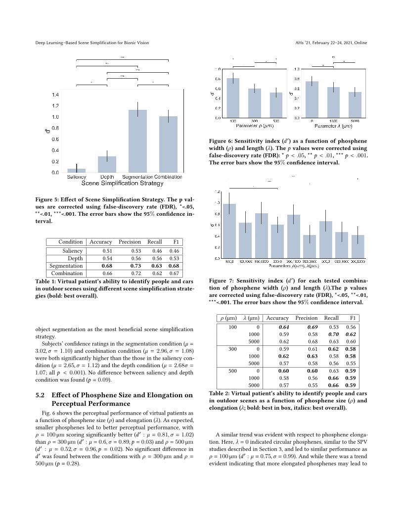
5.2 Effect of Phosphene Size and Elongation onPerceptual Performance
Fig. 6 shows the perceptual performance of virtual patients asa function of phosphene size (𝜌) and elongation (_). As expected,smaller phosphenes led to better perceptual performance, with𝜌 = 100 µm scoring significantly better (𝑑 ′ : ` = 0.81, 𝜎 = 1.02)than 𝜌 = 300 µm (𝑑 ′ : ` = 0.6, 𝜎 = 0.89, 𝑝 = 0.03) and 𝜌 = 500 µm(𝑑 ′ : ` = 0.52, 𝜎 = 0.96, 𝑝 = 0.02). No significant difference in𝑑 ′ was found between the conditions with 𝜌 = 300 µm and 𝜌 =
500 µm (𝑝 = 0.28).
Figure 6: Sensitivity index (𝑑 ′) as a function of phosphenewidth (𝜌) and length (_). The 𝑝 values were corrected usingfalse-discovery rate (FDR): * 𝑝 < .05, ** 𝑝 < .01, *** 𝑝 < .001.The error bars show the 95% confidence interval.
Figure 7: Sensitivity index (𝑑 ′) for each tested combina-tion of phosphene width (𝜌) and length (_).The p valuesare corrected using false-discovery rate (FDR), *<.05, **<.01,***<.001. The error bars show the 95% confidence interval.
𝜌 (µm) _ (µm) Accuracy Precision Recall F1100 0 0.64 0.69 0.53 0.56
1000 0.59 0.58 0.70 0.625000 0.62 0.68 0.63 0.60
300 0 0.59 0.61 0.62 0.581000 0.62 0.63 0.58 0.585000 0.57 0.58 0.56 0.55
500 0 0.60 0.60 0.63 0.591000 0.58 0.56 0.66 0.595000 0.57 0.55 0.66 0.59
Table 2: Virtual patient’s ability to identify people and carsin outdoor scenes as a function of phosphene size (𝜌) andelongation (_; bold: best in box, italics: best overall).
A similar trend was evident with respect to phosphene elonga-tion. Here, _ = 0 indicated circular phosphenes, similar to the SPVstudies described in Section 3, and led to similar performance as𝜌 = 100 µm (𝑑 ′ : ` = 0.75, 𝜎 = 0.99). And while there was a trendevident indicating that more elongated phosphenes may lead to

AHs ’21, February 22–24, 2021, Online Han et al.
Figure 8: Sensitivity index (𝑑 ′) as a function of electrode gridsize. The p values are corrected using false-discovery rate(FDR), *<.05, **<.01, ***<.001. The error bars show the 95%confidence interval.
Resolution Accuracy Precision Recall F18×8 0.57 0.59 0.57 0.54
16×16 0.61 0.62 0.64 0.6132×32 0.61 0.62 0.65 0.61
Table 3: Virtual patient’s ability to identify people and carsin outdoor scenes as a function of electrode grid size (bold:best overall).
poorer perceptual performance, this trend did not reach statisti-cal significance (_ = 1000 µm, 𝑑 ′ : ` = 0.63, 𝜎 = 0.98, 𝑝 > 0.05;_ = 5000 µm, 𝑑 ′ : ` = 0.53, 𝜎 = 0.91, 𝑝 > 0.05).
This trend could later be confirmed by investigating 𝑑 ′ acrossall nine model parameter conditions (see Fig. 7). Here we found aclear decreasing trend in 𝑑 ′ as phosphene size(𝜌) and phospheneelongation(_) increased. However, notice that 𝑑 ′ was positive in allconditions, indicating that subjects performed better than chanceeven when phosphenes were unusually large and elongated.
Similar patterns were found in all the other behavioral perfor-mance measurements (see Table 2). Overall, the highest accuracy,precision, recall, and F1 scores (italics) were achieved with thesmallest tested phosphene size (𝜌 = 100 µm), but not necessarilywith the shortest phosphene length.
Unfortunately, the subjects’ confidence ratings across conditionswith different phosphene sizes (𝜌) and phosphene elongations (_)did not show any significant difference (𝜌 = 100 : ` = 2.74, 𝜎 =
1.19; 𝜌 = 300 : ` = 2.95, 𝜎 = 1.07; 𝜌 = 500 : ` = 2.80, 𝜎 = 1.04;_ = 0 : ` = 2.60, 𝜎 = 1.14; _ = 1000 : ` = 3.18, 𝜎 = 1.14;_ = 5000 : ` = 2.71, 𝜎 = .93, all 𝑝 > .05).
5.3 Effect of Electrode Grid Size on PerceptualPerformance
Fig. 8 shows the perceptual performance of virtual patients as afunction of electrode grid size. As expected, performance improvedas the number of electrodes in the array was increased from 8 × 8(𝑑 ′ : ` = 0.47, 𝜎 = 0.87) to 16 × 16 (𝑑 ′ : ` = 0.72, 𝜎 = 0.93, 𝑝 <
0.001). However, further increasing the number of electrodes to32 × 32 did not measurably affect performance (𝑝 = 0.37).
This finding is again corroborated by accuracy, precision, recall,and F1 scores (Table 3), indicating virtually identical performancefor 16 × 16 and 32 × 32.
Again, no significant difference in confidence ratings was foundfor different electrode array resolution (8 × 8 : ` = 2.74, 𝜎 =
1.12; 16 × 16 : ` = 2.85, 𝜎 = 1.09; 32 × 32 : ` = 2.89, 𝜎 = 1.10, all𝑝 > 0.05).
6 DISCUSSION6.1 Object Segmentation May Support Scene
UnderstandingThe present study provides the first side-by-side comparison
of several deep learning–based scene simplification strategies forbionic vision. Considering a number of relevant implant configu-rations in combination with a psychophysically validated modelof SPV, we identified object segmentation as the most promisingimage processing strategy to support outdoor scene understandingof virtual patients (see Fig. 5 and Table 1). This finding is consis-tent with recent studies indicating the potential utility of semanticsegmentation for bionic vision [17, 33].
Object segmentation compared favorably with two other scenesimplification strategies: based on visual saliency and monoculardepth estimation. Whereas the saliency model struggled with thelighting conditions of the outdoor data set (often highlighting re-gions of increased contrast, and falling victim to shadows), thedepth model often failed to highlight nearby obstacles. However,these models may prove their value in less structured test environ-ments, where performance is less focused on semantic labeling andmore concerned with the decluttering of complex scenes or theavoidance of nearby obstacles.
6.2 Increased Phosphene Size ImpedesPerceptual Performance
To the best of our knowledge, this study is also the first to studySPV with a neurobiologically inspired, psychophysically validatedmodel of phosphene shape [7]. Whereas previous studies assumedthat phosphenes are isolated, focal spots of light [10, 11, 25, 30, 33],here we systematically evaluated perceptual performance acrossa wide range of common phosphene sizes (𝜌) and elongation (_).As expected, the best performance was achieved with small, cir-cular phosphenes (𝜌 = 100 µm, _ = 0; see Fig. 6), and increasingphosphene size and elongation negatively affected performance(Fig. 7). This finding suggests that future studies of SPV should takeinto account realistic phosphene shape when making predictionsand drawing conclusions.
However, it is worth mentioning that the sensitivity index (𝑑 ′)remained positive in all tested conditions, indicating that subjects

Deep Learning–Based Scene Simplification for Bionic Vision AHs ’21, February 22–24, 2021, Online
performed better than chance even when phosphenes were unusu-ally large and elongated. This result suggests that all tested scenesimplification strategies enabled the virtual patients to performabove chance levels, no matter how degraded the SPV quality.
6.3 Increasing the Number of Electrodes DoesNot Necessarily Improve Performance
As expected, perceptual performance improved as the size of theelectrode grid was increased from 8×8 to 16×16. However, furtherincreasing the number of electrodes to 32 × 32 did not measurablyaffect performance. This result is consistent with previous literaturesuggesting that number of electrodes is not the limiting factor inretinal implants [4, 8].
6.4 Limitations and Future WorkAlthough the present results demonstrate the utility of deep
learning–based scene simplification for bionic vision, there are anumber of limitations that should be addressed in future work.
First, in an effort to focus on scenes important for orientationand mobility, we limited our dataset to outdoor scenes. However,it would also be valuable to evaluate the performance of differentscene simplification strategies on indoor scenarios. Because indoorscenes have different layouts and types of objects, the algorithmsstudied here might have different performances compared to out-door scenes. For example, the saliency model might perform betterin highlighting salient regions without the interference of light andshadow contrasts.
Second, to keep the perceptual judgments amenable to quan-titative performance measures, we limited the current study to asimple detection task involving common semantic object categories(i.e., people and cars). This might explain the superior performanceof the semantic segmentation algorithm, which operates with se-mantic labels. In contrast, the depth and saliency algorithms mightprove more valuable when applied to open-ended navigation tasks.In the future, we plan to conduct such SPV studies in immersivevirtual reality (VR) to gain more comprehensive insight into thebehavioral performance of virtual patients.
Third, the present study should be understood as a first steptowards the ultimate goal of creating a retinal implant supported bydeep learning–based image preprocessing. Such a device would re-quire all processing to happen in real time at the edge. One solutioncould come in the form of low-power, low-latency neuromorphichardware coupled with an event-based vision sensor. Future it-erations of this work may include end-to-end training of scenesimplification strategies fitted to a specific implant technology oreven an individual patient. Overall this work has the potentialto drastically improve the utility of prosthetic vision for peopleblinded from retinal degenerative diseases.
7 DATA AVAILABILITYAll raw video sequences (original and preprocessed) are available
on the Open Science Framework (https://osf.io/s2udz). SPV modelswere based on the pulse2percept Python package [5]. Code usedto implement the scene simplification strategies is available onGitHub (https://github.com/bionicvisionlab/2021-han-scene-simplification, v0.1).
ACKNOWLEDGMENTSThis work was partially supported by the National Institutes of
Health (NIH R00 EY-029329 to MB). We would like to thank YaoyiBai and Sikun Lin for their contribution to an earlier version of thedepth algorithm, and Asa Young for collecting the video stimuli.We would also like to thank Dr. Miguel Lago for technical supportwith regards to the SimplePhy [19] online platform.
REFERENCES[1] W. I. Al-Atabany, T. Tong, and P. A. Degenaar. 2010. Improved content aware
scene retargeting for retinitis pigmentosa patients. Biomed Eng Online 9 (Sept.2010), 52. https://doi.org/10.1186/1475-925X-9-52
[2] Lauren N. Ayton, Nick Barnes, Gislin Dagnelie, Takashi Fujikado, Georges Goetz,Ralf Hornig, Bryan W. Jones, Mahiul M. K. Muqit, Daniel L. Rathbun, KatarinaStingl, James D. Weiland, and Matthew A. Petoe. 2020. An update on retinalprostheses. Clinical Neurophysiology 131, 6 (June 2020), 1383–1398. https://doi.org/10.1016/j.clinph.2019.11.029
[3] Lauren N. Ayton, Peter J. Blamey, Robyn H. Guymer, Chi D. Luu, David A. X.Nayagam, Nicholas C. Sinclair, Mohit N. Shivdasani, Jonathan Yeoh, Mark F.McCombe, Robert J. Briggs, Nicholas L. Opie, Joel Villalobos, Peter N. Dimitrov,Mary Varsamidis, Matthew A. Petoe, Chris D. McCarthy, Janine G. Walker, NickBarnes, Anthony N. Burkitt, Chris E. Williams, Robert K. Shepherd, Penelope J.Allen, and for the Bionic Vision Australia Research Consortium. 2014. First-in-Human Trial of a Novel Suprachoroidal Retinal Prosthesis. PLOS ONE 9, 12 (Dec.2014), e115239. https://doi.org/10.1371/journal.pone.0115239 Publisher: PublicLibrary of Science.
[4] Matthew R. Behrend, Ashish K. Ahuja, Mark S. Humayun, Robert H. Chow, andJames D. Weiland. 2011. Resolution of the Epiretinal Prosthesis is not Limited byElectrode Size. IEEE Transactions on Neural Systems and Rehabilitation Engineering19, 4 (Aug. 2011), 436–442. https://doi.org/10.1109/TNSRE.2011.2140132
[5] M. Beyeler, G. M. Boynton, I. Fine, and A. Rokem. 2017. pulse2percept: A Python-based simulation framework for bionic vision. In Proceedings of the 16th Sciencein Python Conference, K. Huff, D. Lippa, D. Niederhut, and M. Pacer (Eds.). 81–88.https://doi.org/10.25080/shinma-7f4c6e7-00c
[6] Michael Beyeler, Geoffrey M. Boynton, Ione Fine, and Ariel Rokem. 2019. Model-Based Recommendations for Optimal Surgical Placement of Epiretinal Implants.In Medical Image Computing and Computer Assisted Intervention – MICCAI 2019(Lecture Notes in Computer Science), Dinggang Shen, Tianming Liu, Terry M.Peters, Lawrence H. Staib, Caroline Essert, Sean Zhou, Pew-Thian Yap, and AliKhan (Eds.). Springer International Publishing, 394–402. https://doi.org/10.1007/978-3-030-32254-0_44
[7] Michael Beyeler, Devyani Nanduri, James D. Weiland, Ariel Rokem, Geoffrey M.Boynton, and Ione Fine. 2019. A model of ganglion axon pathways accountsfor percepts elicited by retinal implants. Scientific Reports 9, 1 (June 2019), 1–16.https://doi.org/10.1038/s41598-019-45416-4
[8] M. Beyeler, A. Rokem, G. M. Boynton, and I. Fine. 2017. Learning to see again: bio-logical constraints on cortical plasticity and the implications for sight restorationtechnologies. J Neural Eng 14, 5 (June 2017), 051003. https://doi.org/10.1088/1741-2552/aa795e
[9] Justin R. Boyle, Anthony J. Maeder, andWageehW. Boles. 2008. Region-of-interestprocessing for electronic visual prostheses. Journal of Electronic Imaging 17, 1(Jan. 2008), 013002. https://doi.org/10.1117/1.2841708 Publisher: InternationalSociety for Optics and Photonics.
[10] S. C. Chen, G. J. Suaning, J. W. Morley, and N. H. Lovell. 2009. Simulatingprosthetic vision: I. Visual models of phosphenes. Vision Research 49, 12 (June2009), 1493–506.
[11] G. Dagnelie, P. Keane, V. Narla, L. Yang, J. Weiland, and M. Humayun. 2007. Realand virtual mobility performance in simulated prosthetic vision. J Neural Eng 4,1 (March 2007), S92–101. https://doi.org/10.1088/1741-2560/4/1/S11
[12] Cordelia Erickson-Davis and Helma Korzybska. 2020. What do blind people“see” with retinal prostheses? Observations and qualitative reports of epiretinalimplant users. bioRxiv (Feb. 2020), 2020.02.03.932905. https://doi.org/10.1101/2020.02.03.932905
[13] Laura Ferlauto, Marta Jole Ildelfonsa Airaghi Leccardi, Naïg Aurelia LudmillaChenais, Samuel Charles Antoine Gilliéron, Paola Vagni, Michele Bevilacqua,Thomas J. Wolfensberger, Kevin Sivula, and Diego Ghezzi. 2018. Design andvalidation of a foldable and photovoltaic wide-field epiretinal prosthesis. NatureCommunications 9, 1 (March 2018), 1–15. https://doi.org/10.1038/s41467-018-03386-7
[14] Eduardo Fernandez. 2018. Development of visual Neuroprostheses: trends andchallenges. Bioelectronic Medicine 4, 1 (Aug. 2018), 12. https://doi.org/10.1186/s42234-018-0013-8
[15] I. Fine and G. M. Boynton. 2015. Pulse trains to percepts: the challenge of creatinga perceptually intelligible world with sight recovery technologies. Philos Trans R

AHs ’21, February 22–24, 2021, Online Han et al.
Soc Lond B Biol Sci 370, 1677 (Sept. 2015), 20140208. https://doi.org/10.1098/rstb.2014.0208
[16] C. Godard, O. M. Aodha, M. Firman, and G. Brostow. 2019. Digging Into Self-Supervised Monocular Depth Estimation. In 2019 IEEE/CVF International Confer-ence on Computer Vision (ICCV). 3827–3837. https://doi.org/10.1109/ICCV.2019.00393 ISSN: 2380-7504.
[17] Lachlan Horne, Jose Alvarez, Chris McCarthy, Mathieu Salzmann, and NickBarnes. 2016. Semantic labeling for prosthetic vision. Computer Vision and ImageUnderstanding 149 (Aug. 2016), 113–125. https://doi.org/10.1016/j.cviu.2016.02.015
[18] Matthias Kümmerer, Thomas S. A. Wallis, and Matthias Bethge. 2016. DeepGazeII: Reading fixations from deep features trained on object recognition.arXiv:1610.01563 [cs, q-bio, stat] (Oct. 2016). http://arxiv.org/abs/1610.01563arXiv: 1610.01563.
[19] Lago, Miguel. 2021. SimplePhy: An open-source tool for quick online perceptionexperiments. Behavior Research Methods (2021). https://doi.org/10.3758/s13428-020-01515-z
[20] Heng Li, Tingting Han, JingWang, Zhuofan Lu, Xiaofei Cao, Yao Chen, Liming Li,Chuanqing Zhou, and Xinyu Chai. 2017. A real-time image optimization strategybased on global saliency detection for artificial retinal prostheses. InformationSciences 415, Supplement C (Nov. 2017), 1–18. https://doi.org/10.1016/j.ins.2017.06.014
[21] Heng Li, Xiaofan Su, Jing Wang, Han Kan, Tingting Han, Yajie Zeng, and XinyuChai. 2018. Image processing strategies based on saliency segmentation for objectrecognition under simulated prosthetic vision. Artificial Intelligence in Medicine84 (Jan. 2018), 64–78. https://doi.org/10.1016/j.artmed.2017.11.001
[22] Junning Li, Yonggang Shi, and Arthur W. Toga. 2015. Controlling False DiscoveryRate in Signal Space for Transformation-Invariant Thresholding of StatisticalMaps. Information processing in medical imaging : proceedings of the ... conference9123 (July 2015), 125–136. https://doi.org/10.1007/978-3-319-19992-4_10
[23] P. Lieby, N. Barnes, C. McCarthy, Nianjun Liu, H. Dennett, J. G. Walker, V. Botea,and A. F. Scott. 2011. Substituting depth for intensity and real-time phosphenerendering: Visual navigation under low vision conditions. In 2011 Annual Inter-national Conference of the IEEE Engineering in Medicine and Biology Society. IEEE,Boston, MA, 8017–8020. https://doi.org/10.1109/IEMBS.2011.6091977
[24] H. Lorach, G. Goetz, R. Smith, X. Lei, Y. Mandel, T. Kamins, K. Mathieson, P. Huie, J.Harris, A. Sher, and D. Palanker. 2015. Photovoltaic restoration of sight with highvisual acuity. Nat Med 21, 5 (May 2015), 476–82. https://doi.org/10.1038/nm.3851
[25] Wen Lik Dennis Lui, Damien Browne, Lindsay Kleeman, Tom Drummond, andWai Ho Li. 2011. Transformative reality: Augmented reality for visual prostheses.In 2011 10th IEEE International Symposium on Mixed and Augmented Reality.253–254. https://doi.org/10.1109/ISMAR.2011.6092402 ISSN: null.
[26] Y. H. Luo and L. da Cruz. 2016. The Argus((R)) II Retinal Prosthesis System. ProgRetin Eye Res 50 (Jan. 2016), 89–107. https://doi.org/10.1016/j.preteyeres.2015.09.003
[27] Yvonne H.-L. Luo, Joe Jiangjian Zhong, Monica Clemo, and Lyndon da Cruz. 2016.Long-term Repeatability and Reproducibility of Phosphene Characteristics inChronically Implanted Argus II Retinal Prosthesis Subjects. American Journal ofOphthalmology 170 (Oct. 2016), 100–109. https://doi.org/10.1016/j.ajo.2016.07.021Publisher: Elsevier.
[28] Chris McCarthy, Janine G. Walker, Paulette Lieby, Adele Scott, and Nick Barnes.2014. Mobility and low contrast trip hazard avoidance using augmented depth.Journal of Neural Engineering 12, 1 (Nov. 2014), 016003. https://doi.org/10.1088/1741-2560/12/1/016003 Publisher: IOP Publishing.
[29] N. Parikh, L. Itti, and J. Weiland. 2010. Saliency-based image processing forretinal prostheses. Journal of Neural Engineering 7, 1 (Jan. 2010), 016006. https://doi.org/10.1088/1741-2560/7/1/016006 Publisher: IOP Publishing.
[30] Alejandro Perez-Yus, Jesus Bermudez-Cameo, Gonzalo Lopez-Nicolas, and Jose J.Guerrero. 2017. Depth and Motion Cues With Phosphene Patterns for ProstheticVision. 1516–1525. http://openaccess.thecvf.com/content_ICCV_2017_workshops/w22/html/Perez-Yus_Depth_and_Motion_ICCV_2017_paper.html
[31] J. F. Rizzo, J. Wyatt, J. Loewenstein, S. Kelly, and D. Shire. 2003. Perceptual efficacyof electrical stimulation of human retina with a microelectrode array duringshort-term surgical trials. Invest Ophthalmol Vis Sci 44, 12 (Dec. 2003), 5362–9.
[32] Roksana Sadeghi, Arathy Kartha, Michael P. Barry, Paul Gibson, Avi Caspi, ArupRoy, and Gislin Dagnelie. 2019. Thermal and Distance image filtering improveindependent mobility in Argus II retinal implant. Journal of Vision 19, 15 (Dec.2019), 23–23. https://doi.org/10.1167/19.15.23 Publisher: The Association forResearch in Vision and Ophthalmology.
[33] Melani Sanchez-Garcia, Ruben Martinez-Cantin, and Josechu J. Guerrero. 2019.Indoor Scenes Understanding for Visual Prosthesis with Fully ConvolutionalNetworks. In VISIGRAPP. https://doi.org/10.5220/0007257602180225
[34] A. J. Simpson and M. J. Fitter. 1973. What is the best index of detectability?Psychological Bulletin 80, 6 (1973), 481–488. https://doi.org/10.1037/h0035203Place: US Publisher: American Psychological Association.
[35] A. Stacey, Y. Li, and N. Barnes. 2011. A salient information processing system forbionic eye with application to obstacle avoidance. In 2011 Annual InternationalConference of the IEEE Engineering in Medicine and Biology Society. 5116–5119.
https://doi.org/10.1109/IEMBS.2011.6091267 ISSN: 1558-4615.[36] K. Stingl, K. U. Bartz-Schmidt, D. Besch, A. Braun, A. Bruckmann, F. Gekeler, U.
Greppmaier, S. Hipp, G. Hortdorfer, C. Kernstock, A. Koitschev, A. Kusnyerik, H.Sachs, A. Schatz, K. T. Stingl, T. Peters, B. Wilhelm, and E. Zrenner. 2013. Artificialvision with wirelessly powered subretinal electronic implant alpha-IMS. ProcBiol Sci 280, 1757 (April 2013), 20130077. https://doi.org/10.1098/rspb.2013.0077
[37] Victor Vergnieux, Marc J.-M. Macé, and Christophe Jouffrais. 2017. Simplifica-tion of Visual Rendering in Simulated Prosthetic Vision Facilitates Navigation.Artificial Organs 41, 9 (Sept. 2017), 852–861. https://doi.org/10.1111/aor.12868Publisher: John Wiley & Sons, Ltd.
[38] Jing Wang, Heng Li, Weizhen Fu, Yao Chen, Liming Li, Qing Lyu, Tingting Han,and Xinyu Chai. 2016. Image Processing Strategies Based on a Visual SaliencyModel for Object Recognition Under Simulated Prosthetic Vision. ArtificialOrgans 40, 1 (2016), 94–100. https://doi.org/10.1111/aor.12498 _eprint:https://onlinelibrary.wiley.com/doi/pdf/10.1111/aor.12498.
[39] J. D. Weiland and M. S. Humayun. 2005. A biomimetic retinal stimulating array.IEEE Engineering in Medicine and Biology Magazine 24, 5 (Sept. 2005), 14–21.https://doi.org/10.1109/MEMB.2005.1511496 Conference Name: IEEE Engineeringin Medicine and Biology Magazine.
[40] J. D.Weiland, N. Parikh, V. Pradeep, and G.Medioni. 2012. Smart image processingsystem for retinal prosthesis. In 2012 Annual International Conference of the IEEEEngineering in Medicine and Biology Society. 300–303. https://doi.org/10.1109/EMBC.2012.6345928 ISSN: 1558-4615.
[41] R. G. H. Wilke, G. Khalili Moghadam, N. H. Lovell, G. J. Suaning, and S. Dokos.2011. Electric crosstalk impairs spatial resolution of multi-electrode arrays inretinal implants. Journal of Neural Engineering 8, 4 (June 2011), 046016. https://doi.org/10.1088/1741-2560/8/4/046016
[42] Wu, Yuxin, Kirillov, Alexander, Massa, Francisco, Lo, Wan-Yen, and Girshick,Ross. 2019. Detectron2. https://github.com/facebookresearch/detectron2
[43] Bolei Zhou, Hang Zhao, Xavier Puig, Sanja Fidler, Adela Barriuso, and AntonioTorralba. 2016. Semantic understanding of scenes through the ade20k dataset.arXiv preprint arXiv:1608.05442 (2016).
[44] Bolei Zhou, Hang Zhao, Xavier Puig, Sanja Fidler, Adela Barriuso, and AntonioTorralba. 2017. Scene Parsing through ADE20K Dataset. In Proceedings of theIEEE Conference on Computer Vision and Pattern Recognition.