Cytoarchitecture and ultrastructure of neural stem cell niches and neurogenic complexes maintaining...
37
Cytoarchitecture and Ultrastructure of Neural Stem Cell Niches and Neurogenic Complexes Maintaining Adult Neurogenesis in the Olfactory Midbrain of Spiny Lobsters, Panulirus argus Manfred Schmidt * and Charles D. Derby Neuroscience Institute and Department of Biology, Georgia State University, Atlanta, Georgia 30303 ABSTRACT New interneurons are continuously generated in small proliferation zones within neuronal somata clusters in the olfactory deutocerebrum of adult decapod crusta- ceans. Each proliferation zone is connected to a clump of cells containing one neural stem cell (i.e., adult neu- roblast), thus forming a ‘‘neurogenic complex.’’ Here we provide a detailed analysis of the cytoarchitecture of neurogenic complexes in adult spiny lobsters, Panulirus argus, based on transmission electron microscopy and labeling with cell-type-selective markers. The clump of cells is composed of unique bipolar clump-forming cells that collectively completely envelop the adult neuro- blast and are themselves ensheathed by a layer of processes of multipolar cell body glia. An arteriole is attached to the clump of cells, but dye perfusion experiments show that hemolymph has no access to the interior of the clump of cells. Thus, the clump of cells fulfills morphological criteria of a protective stem cell niche, with clump-forming cells constituting the adult neuroblast’s microenvironment together with the cell body glia processes separating it from other tissue components. Bromodeoxyuridine pulse-chase experi- ments with short survival times suggest that adult neu- roblasts are not quiescent but rather cycle actively during daytime. We propose a cell lineage model in which an asymmetrically dividing adult neuroblast repo- pulates the pool of neuronal progenitor cells in the associated proliferation zone. In conclusion, as in mam- malian brains, adult neurogenesis in crustacean brains is fueled by neural stem cells that are maintained by stem cell niches that preserve elements of the embry- onic microenvironment and contain glial and vascular elements. J. Comp. Neurol. 519:2283–2319, 2011. V C 2011 Wiley-Liss, Inc. INDEXING TERMS: proliferation; decapod crustacean; arthropod; brain; glia Neurogenesis persists throughout adulthood and leads to continuous production of new neurons in certain brain regions. Adult neurogenesis is well documented in brains of mammals and other vertebrates, including fish, amphibians, reptiles, and birds, but it is also a constitu- tive process in brains of many arthropods (Lindsey and Tropepe, 2006). In mammalian brains, adult neurogenesis predominantly produces new local interneurons of the olfactory bulb (periglomerular cells and granule cells) and the hippocampal dentate gyrus (granule cells; Kriegstein and Alvarez-Buylla, 2009). In brains of some insect spe- cies, adult neurogenesis generates new local interneur- ons (Kenyon cells) of the mushroom bodies (Cayre et al., 1994, 1996; Gu et al., 1999; Dufour and Gadenne, 2006; Mashaly et al., 2008; Zhao et al., 2008; Ghosal et al., 2009). The mushroom bodies are large protocerebral neuropils that in most insects receive projections neu- rons (PNs) from the antennal lobes and thus represent the second stage of the central olfactory pathway; more- over, they are centers for multisensory integration (Strausfeld et al., 2009). Throughout decapod crusta- ceans, adult neurogenesis leads to the formation of new local interneurons (LNs) and projection neurons (PNs) of the olfactory deutocerebrum (midbrain), representing the first stage of the central olfactory pathway (Schmidt, 1997, 2001, 2007a; Sandeman et al., 1998, 2009; Grant sponsor: National Institutes of Health; Grant number: DC00312. *CORRESPONDENCE TO: Manfred Schmidt, Neuroscience Institute, Georgia State University, P.O. Box 5030, Atlanta, GA 30302-5030. E-mail: [email protected] V C 2011 Wiley-Liss, Inc. Received December 10, 2010; Revised March 7, 2011; Accepted April 1, 2011 DOI 10.1002/cne.22657 Published online April 26, 2011 in Wiley Online Library (wileyonlinelibrary.com) The Journal of Comparative Neurology | Research in Systems Neuroscience 519:2283–2319 (2011) 2283 RESEARCH ARTICLE
Transcript of Cytoarchitecture and ultrastructure of neural stem cell niches and neurogenic complexes maintaining...

Cytoarchitecture and Ultrastructure of Neural StemCell Niches and Neurogenic Complexes MaintainingAdult Neurogenesis in the Olfactory Midbrain ofSpiny Lobsters, Panulirus argus
Manfred Schmidt* and Charles D. Derby
Neuroscience Institute and Department of Biology, Georgia State University, Atlanta, Georgia 30303
ABSTRACTNew interneurons are continuously generated in small
proliferation zones within neuronal somata clusters in
the olfactory deutocerebrum of adult decapod crusta-
ceans. Each proliferation zone is connected to a clump
of cells containing one neural stem cell (i.e., adult neu-
roblast), thus forming a ‘‘neurogenic complex.’’ Here we
provide a detailed analysis of the cytoarchitecture of
neurogenic complexes in adult spiny lobsters, Panulirus
argus, based on transmission electron microscopy and
labeling with cell-type-selective markers. The clump of
cells is composed of unique bipolar clump-forming cells
that collectively completely envelop the adult neuro-
blast and are themselves ensheathed by a layer of
processes of multipolar cell body glia. An arteriole is
attached to the clump of cells, but dye perfusion
experiments show that hemolymph has no access to
the interior of the clump of cells. Thus, the clump of
cells fulfills morphological criteria of a protective stem
cell niche, with clump-forming cells constituting the
adult neuroblast’s microenvironment together with the
cell body glia processes separating it from other tissue
components. Bromodeoxyuridine pulse-chase experi-
ments with short survival times suggest that adult neu-
roblasts are not quiescent but rather cycle actively
during daytime. We propose a cell lineage model in
which an asymmetrically dividing adult neuroblast repo-
pulates the pool of neuronal progenitor cells in the
associated proliferation zone. In conclusion, as in mam-
malian brains, adult neurogenesis in crustacean brains
is fueled by neural stem cells that are maintained by
stem cell niches that preserve elements of the embry-
onic microenvironment and contain glial and vascular
elements. J. Comp. Neurol. 519:2283–2319, 2011.
VC 2011 Wiley-Liss, Inc.
INDEXING TERMS: proliferation; decapod crustacean; arthropod; brain; glia
Neurogenesis persists throughout adulthood and leads
to continuous production of new neurons in certain brain
regions. Adult neurogenesis is well documented in brains
of mammals and other vertebrates, including fish,
amphibians, reptiles, and birds, but it is also a constitu-
tive process in brains of many arthropods (Lindsey and
Tropepe, 2006). In mammalian brains, adult neurogenesis
predominantly produces new local interneurons of the
olfactory bulb (periglomerular cells and granule cells) and
the hippocampal dentate gyrus (granule cells; Kriegstein
and Alvarez-Buylla, 2009). In brains of some insect spe-
cies, adult neurogenesis generates new local interneur-
ons (Kenyon cells) of the mushroom bodies (Cayre et al.,
1994, 1996; Gu et al., 1999; Dufour and Gadenne, 2006;
Mashaly et al., 2008; Zhao et al., 2008; Ghosal et al.,
2009). The mushroom bodies are large protocerebral
neuropils that in most insects receive projections neu-
rons (PNs) from the antennal lobes and thus represent
the second stage of the central olfactory pathway; more-
over, they are centers for multisensory integration
(Strausfeld et al., 2009). Throughout decapod crusta-
ceans, adult neurogenesis leads to the formation of new
local interneurons (LNs) and projection neurons (PNs) of
the olfactory deutocerebrum (midbrain), representing the
first stage of the central olfactory pathway (Schmidt,
1997, 2001, 2007a; Sandeman et al., 1998, 2009;
Grant sponsor: National Institutes of Health; Grant number: DC00312.
*CORRESPONDENCE TO: Manfred Schmidt, Neuroscience Institute,Georgia State University, P.O. Box 5030, Atlanta, GA 30302-5030.E-mail: [email protected]
VC 2011 Wiley-Liss, Inc.
Received December 10, 2010; Revised March 7, 2011; Accepted April 1,2011
DOI 10.1002/cne.22657
Published online April 26, 2011 in Wiley Online Library(wileyonlinelibrary.com)
The Journal of Comparative Neurology | Research in Systems Neuroscience 519:2283–2319 (2011) 2283
RESEARCH ARTICLE
Harzsch et al., 1999; Schmidt and Harzsch 1999; Hansen
and Schmidt, 2001, 2004; Sullivan and Beltz, 2005a,b;
Sullivan et al., 2007a; Song et al., 2009; Zhang et al.,
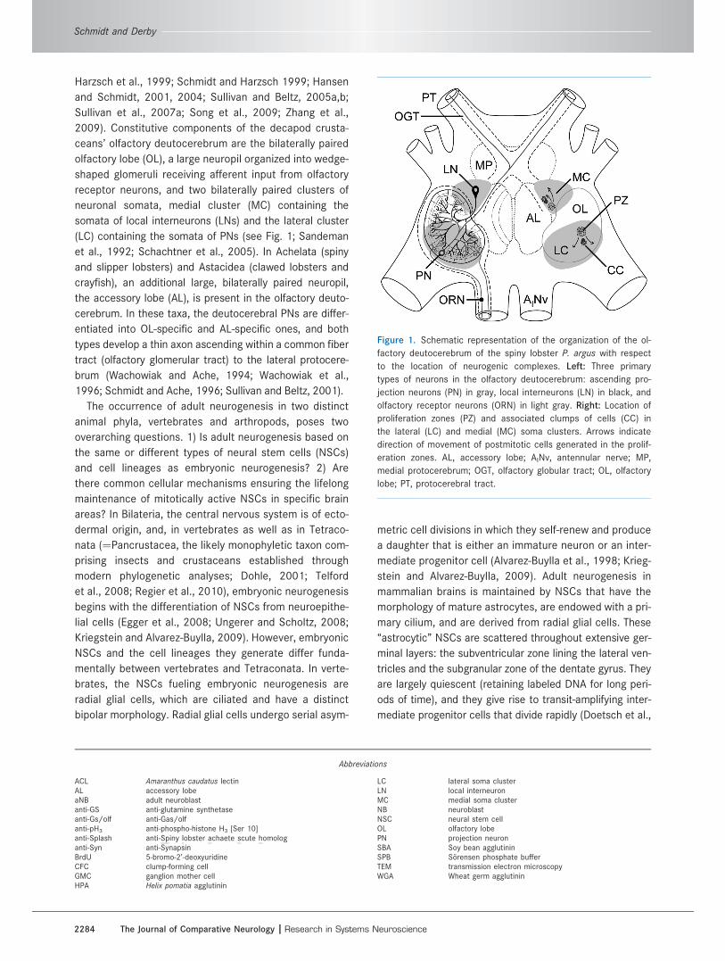
2009). Constitutive components of the decapod crusta-
ceans’ olfactory deutocerebrum are the bilaterally paired
olfactory lobe (OL), a large neuropil organized into wedge-
shaped glomeruli receiving afferent input from olfactory
receptor neurons, and two bilaterally paired clusters of
neuronal somata, medial cluster (MC) containing the
somata of local interneurons (LNs) and the lateral cluster
(LC) containing the somata of PNs (see Fig. 1; Sandeman
et al., 1992; Schachtner et al., 2005). In Achelata (spiny
and slipper lobsters) and Astacidea (clawed lobsters and
crayfish), an additional large, bilaterally paired neuropil,
the accessory lobe (AL), is present in the olfactory deuto-
cerebrum. In these taxa, the deutocerebral PNs are differ-
entiated into OL-specific and AL-specific ones, and both
types develop a thin axon ascending within a common fiber
tract (olfactory glomerular tract) to the lateral protocere-
brum (Wachowiak and Ache, 1994; Wachowiak et al.,
1996; Schmidt and Ache, 1996; Sullivan and Beltz, 2001).
The occurrence of adult neurogenesis in two distinct
animal phyla, vertebrates and arthropods, poses two
overarching questions. 1) Is adult neurogenesis based on
the same or different types of neural stem cells (NSCs)
and cell lineages as embryonic neurogenesis? 2) Are
there common cellular mechanisms ensuring the lifelong
maintenance of mitotically active NSCs in specific brain
areas? In Bilateria, the central nervous system is of ecto-
dermal origin, and, in vertebrates as well as in Tetraco-
nata (¼Pancrustacea, the likely monophyletic taxon com-
prising insects and crustaceans established through
modern phylogenetic analyses; Dohle, 2001; Telford
et al., 2008; Regier et al., 2010), embryonic neurogenesis
begins with the differentiation of NSCs from neuroepithe-
lial cells (Egger et al., 2008; Ungerer and Scholtz, 2008;
Kriegstein and Alvarez-Buylla, 2009). However, embryonic
NSCs and the cell lineages they generate differ funda-
mentally between vertebrates and Tetraconata. In verte-
brates, the NSCs fueling embryonic neurogenesis are
radial glial cells, which are ciliated and have a distinct
bipolar morphology. Radial glial cells undergo serial asym-
metric cell divisions in which they self-renew and produce
a daughter that is either an immature neuron or an inter-
mediate progenitor cell (Alvarez-Buylla et al., 1998; Krieg-
stein and Alvarez-Buylla, 2009). Adult neurogenesis in
mammalian brains is maintained by NSCs that have the
morphology of mature astrocytes, are endowed with a pri-
mary cilium, and are derived from radial glial cells. These
‘‘astrocytic’’ NSCs are scattered throughout extensive ger-
minal layers: the subventricular zone lining the lateral ven-
tricles and the subgranular zone of the dentate gyrus. They
are largely quiescent (retaining labeled DNA for long peri-
ods of time), and they give rise to transit-amplifying inter-
mediate progenitor cells that divide rapidly (Doetsch et al.,
Abbreviations
ACL Amaranthus caudatus lectinAL accessory lobeaNB adult neuroblastanti-GS anti-glutamine synthetaseanti-Gs/olf anti-Gas/olfanti-pH3 anti-phospho-histone H3 [Ser 10]anti-Splash anti-Spiny lobster achaete scute homologanti-Syn anti-SynapsinBrdU 5-bromo-2’-deoxyuridineCFC clump-forming cellGMC ganglion mother cellHPA Helix pomatia agglutinin
LC lateral soma clusterLN local interneuronMC medial soma clusterNB neuroblastNSC neural stem cellOL olfactory lobePN projection neuronSBA Soy bean agglutininSPB Sorensen phosphate bufferTEM transmission electron microscopyWGA Wheat germ agglutinin
Figure 1. Schematic representation of the organization of the ol-
factory deutocerebrum of the spiny lobster P. argus with respect
to the location of neurogenic complexes. Left: Three primary
types of neurons in the olfactory deutocerebrum: ascending pro-
jection neurons (PN) in gray, local interneurons (LN) in black, and
olfactory receptor neurons (ORN) in light gray. Right: Location of
proliferation zones (PZ) and associated clumps of cells (CC) in
the lateral (LC) and medial (MC) soma clusters. Arrows indicate
direction of movement of postmitotic cells generated in the prolif-
eration zones. AL, accessory lobe; AINv, antennular nerve; MP,
medial protocerebrum; OGT, olfactory globular tract; OL, olfactory
lobe; PT, protocerebral tract.
Schmidt and Derby
2284 The Journal of Comparative Neurology |Research in Systems Neuroscience

1999a,b; Palmer, 2000; Alvarez-Buylla et al., 2001; Merkle
et al., 2004; Breunig et al., 2008; Han et al., 2008; Mirzah-
deh et al., 2008; Kriegstein and Alvarez-Buylla, 2009).
In Tetraconata, the NSCs maintaining embryonic neuro-
genesis are large globular neuroblasts (NBs) that have no
bipolar or otherwise glial morphology and are not ciliated.
Through serial asymmetrical divisions, NBs self-renew and
produce smaller daughter cells called ganglion mother
cells (GMCs) toward the inside of the body. GMCs undergo
a terminal symmetrical division in which two immature
neurons are produced. Typically GMCs and immature neu-
rons produced by one NB stay attached to it, forming a
column or small aggregate of cells (Dohle, 1976; Doe and
Goodman, 1985; Hartenstein et al., 1987; Scholtz, 1992;
Doe et al., 1998; Harzsch, 2001; Urbach and Technau,
2003; Egger et al., 2008; Ungerer and Scholtz, 2008;
Boyan et al., 2010). In insects, some embryonic NBs
become quiescent and are reactivated during larval
stages to fuel larval neurogenesis in most parts of the
CNS (Maurange and Gould, 2005), except for the optic
lobes, where new NBs are generated from neuroepithelial
cells (Yasugi et al., 2008). Most larval NBs of insects give
rise to immature neurons by the same cell lineage as em-
bryonic NBs (Bello et al., 2008). Recently, it was discov-
ered that some embryonic and larval NBs of the insect
brain proliferate in a more complex way and give rise to
larger lineages. These NBs produce intermediate progeni-
tor cells that act as self-renewing transit-amplifying cells
that give rise to GMCs (Bello et al., 2008; Boone and Doe,
2008; Bowman et al., 2008; Izergina et al., 2009; Boyan
et al., 2010). Adult neurogenesis in the mushroom bodies
of insects is based on continued mitotic activity of a few
NBs surviving after larval development (Cayre et al., 1994,
1996, 2002; Gu et al., 1999; Dufour and Gadenne, 2006;
Mashaly et al., 2008; Zhao et al., 2008; Ghosal et al.,
2009). These adult NBs appear to generate progeny as
the canonical embryonic and larval NBs (Dufour and Gad-
enne, 2006; Zhao et al., 2008); however, the exact cell lin-
eage that they produce has not yet been established.
In the olfactory deutocerebrum of adult decapod crus-
taceans, new neurons arise in small proliferation zones of
invariant location at the inner (neuropil-facing) surface of
the neuronal soma clusters (MC, LC). The proliferating
cells in these zones are small and equivalent to GMCs in
giving rise to immature neurons through one round of
symmetrical cell divisions. Neuronal differentiation of
these cells takes months and is associated with their
translocation away form the proliferation zone into the
outer area of the respective soma cluster (Fig. 1; Schmidt,
2001; Sullivan and Beltz, 2005a). Recently, it was deter-
mined that, in adult spiny lobsters, Panulirus argus, and
crayfish, Procambarus clarkii, each proliferation zone is
associated with a particularly large proliferating cell that
has an invariant location outside the proliferation zone but
is connected to it via a strand- or duct-like structure
(Schmidt, 2007a; Song et al., 2009). Based on their large
size and privileged location, these cells were identified as
putative adult neuroblasts (aNBs). Thus, adult neurogene-
sis in the olfactory deutocerebrum of decapod crustaceans
appears to be maintained by NSCs and neural progenitor
cells that are equivalent to those fueling embryonic neuro-
genesis, NBs and GMCs, respectively.
A common theme in the de novo generation of cells in
various tissues of adult animals is that the new cells are pro-
duced by adult (somatic) stem cells residing in specialized
microenvironments within the tissues. These areas represent
stem cell niches, providing regulatory input and nutritional
support for the adult stem cells and possibly shielding them
from toxic or harmful factors (Spradling et al., 2001; Fuchs
et al., 2004; Ohlstein et al., 2004; Moore and Lemischka,
2006; Jones and Wagers, 2008; Morrison and Spradling,
2008). The NSCs in brains of adult mammals reside in stem
cell niches that form a distributed and complex structural
meshwork pervading the germinal cell layers. These NSC
niches are composed of vascular elements, glial cells, and
extracellular matrix (Palmer et al., 2000; Mercier et al.,
2002; Shen et al., 2008; Tavazoie et al., 2008). In the olfac-
tory deutocerebrum of adult P. argus and P. clarkii, each aNB
is closely associated with a morphologically unique clump of
cells that has been interpreted as a putative stem cell niche
(Fig. 1; Schmidt, 2007a,b; Sullivan et al., 2007a,b; Song
et al., 2009). Similar clumps of cells were originally identified
as ‘‘deutocerebral organs’’ in brains of several other species
of decapod crustaceans by Bazin (1970) suggesting that
they are a common component associated with adult neuro-
genesis in the olfactory deutocerebrum (Schmidt, 2007b).
The clump of cells containing the aNB, the proliferation zone
associated with it, and the duct- or strand-like structure con-
necting both compartments form a structural unit for which
we use the term neurogenic complex, as previously estab-
lished (Song et al., 2009).
Here we provide a detailed analysis of the cytoarchitec-
ture of the neurogenic complexes in olfactory deutocere-
brum of adult spiny lobsters, P. argus, based on transmis-
sion electron microscopy and labeling with cell-type-
selective markers. This study provides the foundation for
an in-depth analysis of the cellular interactions between
aNBs and their niche, utilizing the unique advantage that
they form morphologically identifiable units in the brain of
adult decapod crustaceans.
MATERIALS AND METHODS
AnimalsExperiments were performed on intermolt male and
female Caribbean spiny lobsters, Panulirus argus, ranging
Neural stem cell niches in adult Panulirus argus
The Journal of Comparative Neurology | Research in Systems Neuroscience 2285
from 50 to 80 mm in carapace length and from 120 to
400 g in weight. Since female P. argus reach sexual matu-
rity (as indicated by spawning resulting in the presence of
eggs attached to the pleopods) at a minimum size of
52 mm carapace length (Lyons et al., 1981), the experi-
mental animals likely comprised mostly adults and some
late juveniles. Most animals were obtained from the Flor-
ida Keys Marine Laboratory, shipped to Georgia State
University, and held in communal 800-L aquaria contain-
ing aerated, recirculated, filtered artificial seawater
(ASW; Instant Ocean: Aquarium Systems, Mentor, OH).
Animals were maintained in a 12-hour:12-hour light:dark
cycle and fed shrimp or squid three times per week.
Some animals were kindly provided by Dr. B.W. Ache
(University of Florida) and were kept under similar condi-
tions. Animals were anesthetized by chilling on ice for at
least 15 minutes before removal of brains or establishing
excised head preparations.
ChemicalsAll chemicals were obtained from Sigma (St. Louis,
MO) unless specified otherwise.
Transmission electron microscopyFor analysis of the ultrastructure of the neurogenic
complex in the LC of P. argus, three brains were perfusion
fixed with 5% glutaraldehyde in 0.1 M Sorensen phos-
phate buffer (SPB) containing 15% sucrose (SPBS) via the
cannulated cerebral (medial) artery in an excised head
preparation as described in detail previously (Schmidt
and Ache, 1994). Brains were removed from the head
and immersed in fixative for another 4 hours at room tem-
perature, rinsed in SPBS for 4 � 30 minutes, postfixed
in 2% OsO4 in SPBS for 2 hours, rinsed in SPBS for 4 �30 minutes, dehydrated in an ascending ethanol series,
incubated in propylene oxide for 2 � 30 minutes, and em-
bedded in Epon 812 hard with polymerization at 60�C for
18 hours. Serial ultrathin sections (90–100 nm thick)
were cut with a diamond knife (Diatome AG, Biel, Switzer-
land) on an ultramicrotome (Ultracut; Reichert-Jung,
Vienna, Austria), contrasted for 20 minutes with lead ci-
trate, and examined in a transmission electron micro-
scope (LEO 906e; LEO Elektronenmikroskopie, Oberko-
chen, Germany). Images were acquired digitally with an
attached CCD camera with 1 MP resolution. The digital
images were processed by filtering out high-frequency
noise and by adjustment of brightness and contrast with
an image analysis program (Image Pro Express, version
4.5.1.3; Media Cybernetics, Bethesda, MD) before they
were arranged into the final figures with an illustration
program (Illustrator CS3; Adobe, San Jose, CA).
Data analysisIn TEM micrographs, the size and shape of different
cell types and their nuclei were determined by tracing cell
membrane and nucleus with an image analysis program
(Image Pro Express). From the length of the outline (pe-
rimeter) and the included area, two parameters repre-
senting size and shape of the measured structures were
calculated: size is given as Feret diameter (FD; diameter
of a circle having the same area as the measured area:
FD ¼ sqrt [4 � area/p]); shape is given as roundness (R;
a dimensionless number between 0 and 1 with 1 repre-
senting a perfect circle: R ¼ [4 � p � area)/perimeter2]).
For cell types in which the size of the soma and the size
of the nucleus could be determined, the nuclear:cytoplas-
mic ratio was calculated by division of the nuclear volume
(derived from the nuclear Feret diameter, assuming a
spherical shape) by the volume of the cytoplasm (derived
by subtracting the nuclear volume from the soma volume,
which was derived from the soma Feret diameter, assum-
ing a spherical shape). The results of the measurements
are given as arithmetic mean 6 standard deviation. Two
data analysis programs were used to treat the data statis-
tically and to generate graphs (PsiPlot 7.01; Poly Software
International, Pearl River, NY; and GraphPad Prism 3.02;
GraphPad Software, San Diego, CA). The final figures con-
taining these graphs were created with an illustration pro-
gram (Illustrator CS3).
Fluorescence microscopyBrdU injection
For in vivo labeling, 5-bromo-20-deoxyuridine (BrdU)
was injected at the base of two walking legs into the
hemolymph of 18 spiny lobsters at 5 mg BrdU/100 g
body weight (0.5% BrdU in Panulirus saline: 459 mM
NaCl, 13.4 mM KCl, 13.6 mM CaCl2, 14.1 mM Na2SO4,
9.8 mM MgCl2, 3.0 mM Hepes, pH 7.4). All animals
received one BrdU injection between 8:00 and 10:00 AM,
and after a survival time of 2–10 hours their brains were
dissected and fixed for 24 hours at room temperature by
immersion in 4% paraformaldehyde in SPBS (PFA).
Perfusion of brain arteries with fluorescentdextran
In an excised head preparation of P. argus, the cerebral
artery was cannulated and the brain perfused with Panulirus
saline at a flow rate of 1 ml/min as described in detail ear-
lier (Schmidt and Ache, 1994). After 15 minutes of saline
perfusion, 1 ml Panulirus saline containing 1 mg/ml dextran
coupled to lysine-fixable tetramethylrhodamine (3,000 MW
microruby for two brains; 10,000-MW miniruby for two
brains; Invitrogen/Molecular Probes, Carlsbad, CA) was per-
fused over 5 minutes into the cerebral artery. After 5
Schmidt and Derby
2286 The Journal of Comparative Neurology |Research in Systems Neuroscience
minutes of incubation without perfusion, the brain was fixed
by immersion in PFA for 24 hours at room temperature.
Sectioning and immunocytochemistryIn addition to brains preloaded with BrdU or fluores-
cent dextran, >100 brains were fixed by immersion in
PFA for 24 hours at room temperature to be used for
immunocytochemical labeling with other antibodies or fluo-
rescent lectins. After fixation, brains were rinsed and
stored in 0.02 M SPB with 0.02% sodium azide at 4�C. For
sectioning, brains were embedded in gelatin and cut on a
vibrating microtome (VT 1000 S; Leica, Wetzlar, Germany)
in 80-lm-thick horizontal or sagittal sections as described
in detail previously (Schmidt, 2001). All of the following
treatments were performed at room temperature.
Most brains preloaded with BrdU were triple labeled
with anti-BrdU (labeling cells that were in S-phase when
BrdU was available for incorporation into newly synthe-
sized DNA), antiphosphohistone H3 (Ser 10; anti-pH3;
labeling cells that were in M-phase at the time of fixation),
and the nuclear marker Hoechst 33258 (labeling nuclei of
all cells). To overcome the common problem that the pre-
treatment of sections required for intense BrdU labeling
severely compromises general nuclear staining (Tang
et al., 2007), we developed a protocol based on the mild
digestion of double-stranded DNA with DNase I (Ye et al.,
2007). In this optimized triple-labeling protocol, free-float-
ing sections were first incubated in 2 N HCl for 20 minutes
and rinsed for 4 � 10 minutes in SPB. Afterward, sections
were incubated overnight in anti-BrdU (mouse monoclo-
nal, clone B44; BD Biosciences, Franklin Lakes, NJ; Table
1) diluted 1:150 in a DNase-containing incubation me-
dium. This medium consisted of a 1:1 mixture of SPB with
0.3% Triton X-100 (TSPB) and DNase I buffer (50 mM Tris
HCl at pH 7.5, 5 mM MgCl2, 50 lg/ml bovine serum albu-
min, and 0.3% Triton X-100) to which 10 U DNase I/ml and
10 lg/ml of a protease inhibitor cocktail (Sigma P-2724)
were added. Subsequently, sections were rinsed for 4 �30 minutes in TSPB and incubated overnight in anti-pH3
(rabbit polyclonal, No. 06-570; Upstate Biotechnology,
Lake Placid, NY; Table 1) at 1:250 dilution in TSPB to label
cells in M-phase. After the sections had been rinsed for 4
� 30 minutes in TSPB, they were incubated in a mixture of
two secondary antibodies: goat anti-mouse Cy3 (Jackson
Immunoresearch, West Grove, PA) diluted 1:400 and goat
anti-rabbit AlexaFluor-488 (Invitrogen/Molecular Probes)
or goat anti-rabbit DyLight-488 (Jackson Immunoresearch)
diluted 1:100 in TSPB. After rinsing for 3 � 30 minutes in
SPB, sections were incubated for 20 minutes in Hoechst
33258 diluted 1:150 in SPB from a stock solution of 1
mg/ml to stain nuclei. After a final rinse in SPB, sections
were coverslipped in 1:1 glycerol:SPB containing 5% diaza-
bicyclol[2.2.2]octane (DABCO) to prevent photobleaching.
Coverslips were secured with nail polish, and slides were
stored at 4�C or at �20�C (for extended periods).
Brains in which the arterial system was perfused with
fluorescent dextran were sectioned as described above.
Sections were only incubated in Hoechst 33258 and then
coverslipped.
In an effort to identify selective markers for particular
cell types or other structures in the brain of P. argus, we
screened >120 antibodies and >30 lectins by labeling
brain sections that were generated as described above.
Each antibody and lectin was tested on sections of at
least two brains containing the neurogenic complex of
MC or LC, with TSPB serving as antibody incubation me-
dium. Here, we include only those probes that are of in-
terest for this study, and the results of the entire screen
will be described in detail elsewhere. We report results
obtained with the following four antibodies (Table 1) and
four lectins: antisynapsin (anti-Syn; mouse monoclonal
from Developmental Systems Hybridoma Bank; 3C11)
diluted 1:25; antiglutamine synthetase (anti-GS; mouse
monoclonal from BD Biosciences; No. 610518) diluted
1:100; anti-Gas/olf (anti-Gs/olf; rabbit polyclonal from
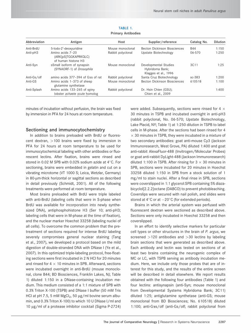
TABLE 1.
Primary Antibodies
Abbreviation Antigen Host Supplier/reference Catalog No. Dilution
Anti-BrdU 5-Iodo-20-deoxyuridine Mouse monoclonal Becton Dickinson Biosciences B44 1:150Anti-pH3 Amino acids 7–20
(ARK[pS]TGGKAPRKQLC)of human histone H3
Rabbit polyclonal Upstate Biotechnology 06-570 1:250
Anti-Syn sSmall isoform of synapsin(SYNAORF-1) of Drosophila
Mouse monoclonal Developmental StudiesHybridoma Bank;Klagges et al., 1996
3C11 1:25
Anti-Gs/olf amino acids 377–394 of Gas of rat Rabbit polyclonal Santa Cruz Biotechnology sc-383 1:200Anti-GS Amino acids 1–373 of sheep
glutamine synthetaseMouse monoclonal Becton Dickinson Biosciences 610518 1:100
Anti-Splash Amino acids 133–265 of spinylobster achaete scute homolog
Rabbit polyclonal Dr. Hsin Chien (GSU);Chien et al., 2009
1:400
Neural stem cell niches in adult Panulirus argus
The Journal of Comparative Neurology | Research in Systems Neuroscience 2287

Santa Cruz Biotechnology, Santa Cruz, CA; SC-383)
diluted 1:200; anti-spiny lobster achaete scute homolog
(anti-Splash; rabbit polyclonal antiserum raised by Dr.
Hsin Chien against the polypeptide sequence from aa
133–265 of splash expressed in Escherichia coli; Chien
et al., 2009; GenBank accession No. DQ489559) diluted
1:400; wheat germ agglutinin (WGA) labeled with Alexa-
Fluor-488 (Molecular Probes/Invitrogen) diluted 1:1,000;
Amaranthus caudatus lectin (ACL) labeled with fluores-
cein isothiocyanate (Vector, Burlingame, CA) diluted
1:1,000; Helix pomatia agglutinin (HPA) labeled with Alex-
aFluor-488 (Molecular Probes/Invitrogen) diluted 1:200;
and soy bean agglutinin (SBA) labeled with fluorescein
isothiocyanate (Vector) diluted 1:200. The primary anti-
bodies were visualized by incubating the sections with
Cy3-labeled secondary antibodies (goat anti-rabbit for
anti-Gs/olf and anti-Splash, goat anti-mouse for anti-Syn
and anti-GS; Jackson Immunoresearch) diluted 1:400 in
TSPB for at least 4 hours at room temperature. To obtain
sections double labeled with an antibody and a lectin, the
lectin was added to the medium containing the secondary
antibody. Finally, all sections were labeled with Hoechst
33258 and coverslipped as described above. In the case
of anti-GS, positive labeling was achieved only in brain
sections that had been incubated for 20 minutes in 2 N
HCl (as required for labeling with anti-BrdU) prior to incu-
bation in the primary antibody.
In controls, sections from brains of two spiny lobsters
that had received no BrdU injection were treated as for
labeling with anti-BrdU and anti-pH3 but without including
the primary anti-pH3 antibody in the incubation medium.
In these sections, no specific labeling above the auto-
fluorescence of the tissue was observed.
Antibody and lectin characterization:Western blot analysis
Except for anti-BrdU and anti-pH3 that we employed to
identify cells in S-phase (at the time of BrdU availability)
and M-phase (at the time of fixation) of the cell cycle,
respectively, the other antibodies and lectins included in
this study were used as purely morphological markers,
allowing selective labeling of cell types or tissue compo-
nents and thereby distinguishing them from each other.
We do not make any functional inferences based on the
labeling with these probes and therefore did not strive rig-
orously to establish the molecular identity of the antigens
or binding sites responsible for probe binding. To test the
specificity of anti-Gs/olf, anti-GS, and anti-Splash, we
performed Western blot analyses with these three anti-
bodies on P. argus brain extracts based on a detailed pro-
tocol published previously (Schmidt, 2007a).
Anti-BrdUDiverse antibodies against BrdU were used previously
in decapod crustacean brains to label cells in S-phase
(see references cited in the introductory paragraphs). The
monoclonal mouse anti-BrdU that we used in this study
(clone B44; BD Biosciences) was raised against a 5-iodo-
20-deoxyuridine conjugated to ovalbumin and binds to 5-
bromo-20-deoxyuridine as well as 5-iodo-20-deoxyuridine
according to the manufacturer’s information. The staining
pattern obtained with this antibody was indistinguishable
from that obtained with other BrdU antibodies in previous
studies (Schmidt, 2001, 2007a). Because no labeling was
present in brains of animals that had not received a BrdU
injection, we conclude that labeling with anti-BrdU specif-
ically labeled nuclei that incorporated BrdU into newly
synthesized DNA and thus were in the S-phase of their
cell cycle when BrdU was available.
Anti-pH3The rabbit polyclonal antibody against phosphohistone
H3 (Ser 10; No. 06-570; Upstate Biotechnology) was
raised against the synthetic peptide ARK[pS]TGG-
KAPRKQLC coupled to keyhole limpet hemocyanin
according to the manufacturer’s information. As in a pre-
vious study (Schmidt, 2007a), we found that it intensely
labels select nuclei in the neurogenic complexes (and
very occasionally in other locations) that have morphologi-
cal features (condensation, irregular shape, pairwise appo-
sition) of nuclei in various phases of mitosis. Since Western
blot analysis of P. argus brain extracts provided evidence
for the binding of this antibody to two isoforms of authentic
phosphohistone H3 (Ser 10; Schmidt, 2007a), which are
present only in the M-phase of the cell cycle (Hendzel
et al., 1997; Wei et al., 1998), we conclude that the
nuclear labeling of anti-pH3 is specific for M-phase nuclei.
However, as in a previous study (Schmidt, 2007a), we
found additional labeling of fibrous material surrounding
the clump of cells and the duct extending from it to the ad-
jacent proliferation zone. Based on two lines of evidence,
we conclude that the labeling of this material is likely
based on nonspecific binding of anti-pH3. First, a new
batch of this antibody as well as several other anti-pH3
antibodies that we tested labeled select nuclei in the neu-
rogenic complexes but not the fibrous material. Second, a
previous Western blot analysis of P. argus brain extracts
provided evidence for the nonspecific binding of this anti-
body to nonnuclear proteins of much higher molecular
weights than typical for histones (Schmidt, 2007a).
Anti-SynThe antibody against the synaptic protein synapsin was
a mouse monoclonal antibody raised against the small
isoform of synapsin (SYNAORF-1) from Drosophila. This
Schmidt and Derby
2288 The Journal of Comparative Neurology |Research in Systems Neuroscience

antibody that we obtained from the Developmental Stud-
ies Hybridoma Bank was originally developed by Dr. Erich
Buchner (Klagges et al., 1996). In Western blot analyses
of Drosophila brains, anti-syn labeled prominent protein
bands at 70, 74, and 80 kDa and a less prominent double
band at �143 kDa, indicating that four or five synapsin
isoforms are detected (Klagges et al., 1996). In Western
blot analyses of brain extracts of Coenobita clypeatus
(terrestrial hermit crab), anti-syn labeled a prominent pro-
tein band at 80–90 kDa and a weaker protein band
slightly above 148 kDa, indicating that two synapsin iso-
forms corresponding to some of the isoforms present in
Drosophila are detected (Klagges et al., 1996; Harzsch
and Hansson, 2008). Anti-syn has been shown repeatedly
to label selectively neuropil areas in the CNS of diverse
decapod crustaceans (Harzsch et al., 1997, 1998, 1999;
Sullivan et al., 2007a; Harzsch and Hansson, 2008), and
we use it here to distinguish neuropils from other tissue
components (especially neuronal soma clusters) in the
brain of P. argus.
Anti-Gs/olfThe antibody against the heterotrimeric G-protein sub-
unit Gas/olf (C-18; Santa Cruz Biotechnology; sc-383)
was an affinity-purified rabbit polyclonal antibody raised
against aa 377–394 of Gas of rat origin, mapping at the
carboxy terminus (according to information provided by
the manufacturer). In the clawed lobster Homarus ameri-
canus, Gas was identified through molecular cloning and
Western blot analysis of brain extracts with the same anti-
body as used here and showed a single band of 51.8 kDa
(Xu et al., 1997). Our Western blot analysis of P. argus
brain extracts with anti-Gs/olf diluted 1:1,000 revealed
double bands at slightly higher molecular mass (�61/63
kDa) and in addition a strongly stained band at about twice
this molecular mass (�137 kDa; Fig. 2A). Although the
bands at �61/63 kDa could be consistent with binding of
the antibody to authentic Gas given the tendency of G pro-
tein subunits to run anomalously high in SDS-PAGE gels
(Quan and Forte, 1990; Xu et al., 1997), the strong band at
�137 kDa indicates nonspecific binding of the antibody to
an unrelated protein of higher molecular mass.
Anti-GSThe monoclonal mouse antibody against glutamine
synthetase (No. 610518; BD Biosciences) was raised
against sheep glutamine synthetase (aa 1–373) accord-
ing to the manufacturer’s information. A crustacean gluta-
mine synthetase was identified through molecular cloning
in P. argus (Trapido-Rosenthal et al., 1993), and an affin-
ity-purified rabbit polyclonal antibody raised against glu-
tamine synthetase from chicken retina was used in
P. argus for Western blot analyses of brain extracts and
an immunocytochemical analysis of brain sections (Linser
et al., 1997). The Western blots revealed a protein band
at 42 kDa consistent with the expected size of P. argus
glutamine synthetase as predicted from the cDNA, and
the immunocytochemical analysis showed selective label-
ing of a particular type of glia with somata located at the
rim of neuropils and processes reaching into the neuro-
pils’ interior. From these findings, Linser et al. (1997) con-
cluded that glutamine synthetase is expressed in a partic-
ular type of glial cell and that anti-GS can serve as a glia-
specific marker. Since the affinity-purified antibody used
by Linser et al. (1997) is no longer available and the raw
GS antiserum (kindly provided by Dr. Paul Linser, Whitney
Laboratory, University of Florida) did not selectively label
glial cells in P. argus brains in preliminary trials (Schmidt,
unpublished), we used another antibody against verte-
brate GS. The choice of anti-GS (No. 610518; BD Bio-
sciences) was based on previous studies in other
decapod crustaceans in which this antibody selectively
labeled similar glial cells in the brain as described for
Figure 2. Western blot analyses of total proteins extracted from
P. argus brains with three antibodies used for the immunocyto-
chemical characterization of the neurogenic complexes. Left:
Alkaline-phosphatase-labeled molecular mass markers (indicated
in kDa) visualized by NBT/BCIP running in the same SDS-PAGE
gel as the antibody-labeled brain extract. Right: 10 lg P. argus
brain extract labeled by one of the antibodies used for immunocy-
tochemistry followed by detection through a secondary antibody
labeled with alkaline phosphatase and its visualization by NBT/
BCIP. Arrows indicate the expected molecular mass (given below
in kDa) of the target protein. A: Labeling with anti-Gs/olf revealed
a double-band at an apparent molecular mass of �61/63 kDa, a
very prominent band at �137 kDa, and a weak band at �200
kDa. Gs cloned from the American lobster H. americanus has a
molecular mass of �51.8 kDa (Xu et al., 1997). B: Labeling with
anti-GS revealed one band at the expected molecular mass of
�42 kDa that was reported for GS cloned from P. argus (Linser
et al., 1997) and an additional very prominent band at �136
kDa. C: Labeling with anti-Splash revealed at least six prominent
bands at apparent molecular masses above the predicted molecu-
lar mass (30.3 kDa) of P. argus Splash (Chien et al., 2009).
Neural stem cell niches in adult Panulirus argus
The Journal of Comparative Neurology | Research in Systems Neuroscience 2289
P. argus (Sullivan and Beltz, 2005b; Sullivan et al., 2007a;
Harzsch and Hansson, 2008). Our Western blots of
P. argus brain extracts with anti-GS diluted 1:2,000
showed one band at �42 kDa consistent with the previ-
ous report by Linser et al. (1997) and with the interpreta-
tion that it represents authentic P. argus GS (Fig. 2B).
However, our Western blots show a second and more
intensely labeled band at �120 kDa that corresponds
neither to the size of a GS subunit (at �42 kDa) nor to
the predicted size of the functional protein consisting of
eight or 10 identical subunits (Eisenberg et al., 2000; Kra-
jewski et al., 2008). An intense band of similar size was
obtained in Western blots of crayfish nerve extracts
probed with a different antibody against GS (McKinnon
et al., 1995). The intense labeling of an additional protein
band with anti-GS as seen in our Western blot analyses
makes it likely that on tissue sections anti-GS also labels
this unrelated protein in addition to labeling authentic glu-
tamine synthetase. Thus, some of the cellular labeling
achieved with anti-GS may be nonspecific.
Anti-SplashIn the context of an unrelated project aimed at the mo-
lecular identification of transcription factors involved in
crustacean adult neurogenesis, we identified a spiny lob-
ster achaete scute homolog (Splash) by molecular cloning
(Chien et al., 2009). We attempted to raise polyclonal
antibodies against Splash protein by immunizing 10 rab-
bits with different segments of the protein that were het-
erologously expressed in E. coli. We tested the suitability
of all antibodies for immunocytochemistry by labeling
sections of diverse P. argus tissues, including brain. One
antibody (anti-Splash5-5; rabbit No. 5, fifth bleed) that
was raised against a large portion of the Splash protein
that included a part of the conserved basic helix–loop–
helix domain but not the C-terminal domain (AA 133–
265) resulted in highly selective labeling of a structure
within the neurogenic complexes of the brain, and results
obtained with this antibody are reported here. Western
blot analysis of P. argus brain extracts with anti-Splash5-
5 (Fig. 2C) revealed labeling of at least six prominent pro-
tein bands, none of which had the predicted molecular
mass of P. argus Splash (�30 kDa; Chien et al., 2009).
Thus we conclude that the staining of brain sections
obtained with anti-Splash5-5 does not reflect the pres-
ence of authentic Splash protein and is nonspecific.
Wheat germ agglutininWheat germ agglutinin (WGA) selectively binds to N-ace-
tylglucosamine and N-acetylneuraminic acid (sialic acid) res-
idues. It labels neuronal elements in the CNS of insects
(Jacobs and Lakes-Harlan, 1997) but to our knowledge has
not been used for labeling in the CNS of crustaceans.
Amaranthus caudatus lectinACL preferentially binds to oligosaccharide residues
containing galactosyl (b-1,3) N-acetylgalactosamine. To
our knowledge, it has not been used for labeling in the
CNS of insects or crustaceans.
Soybean agglutininSoybean agglutinin (SBA) preferentially binds to oligo-
saccharide residues with terminal N-acetylglucosamine
and to a lesser extend to galactose residues. It labels
neuronal elements in the CNS of insects (Jacobs and
Lakes-Harlan, 1997) but to our knowledge has not been
used for labeling in the CNS of crustaceans.
Helix pomatia agglutininHelix pomatia agglutinin (HPA) selectively binds to a-N-
acetylgalactosamine residues. To our knowledge it has not
been used for labeling in the CNS of insects or crustaceans.
Confocal microscopy and image processingTo generate micrographs, fluorescently labeled sec-
tions were viewed and imaged in a confocal microscope
with two-photon attachment (LSM 510; Zeiss, Jena, Ger-
many) using the associated software package. The 488-
nm line of an argon laser was used to excite and visualize
AlexaFluor-488-labeled secondary antibodies or lectins
as well as fluorescein-labeled lectins; the 568-nm line of
a helium–neon laser was used to excite and visualize
Cy3-labeled secondary antibodies; a titanium–sapphire
two-photon laser tuned to 800 nm was used to excite and
visualize Hoechst 33258. Stacks of 0.3–1.0-lm-thick op-
tical sections covering the entire section thickness of 80
lm were collected. Substacks of these optical sections
were collapsed to produce single two-dimensional
images. The digital images were processed by filtering
out high-frequency noise and by adjustment of brightness
and contrast in Image Pro Express 4.5.1.3 before they
were arranged into the final figures in Illustrator CS3.
RESULTS
Identification of different cell types in theolfactory deutocerebrum
We used TEM and cell-type-selective labeling with anti-
bodies and lectins combined with labeling of all cell nuclei
by the nuclear marker Hoechst 33258 to identify major
cell types making up the olfactory deutocerebrum of
P. argus. The olfactory deutocerebrum of P. argus is organ-
ized into two major neuropils (olfactory lobe, OL; acces-
sory lobe, AL) composed of neuronal processes and two
large clusters of neuronal somata (lateral soma cluster,
LC; medial soma cluster, MC) per hemibrain. These major
compartments were readily identifiable by various
Schmidt and Derby
2290 The Journal of Comparative Neurology |Research in Systems Neuroscience

labeling methods, including labeling all cell nuclei with
the nuclear marker Hoechst 33258 combined with label-
ing of synaptic areas within neuropils by anti-Syn (Fig.
3A–C). Hoechst labeling revealed the LC and MC as
dense accumulations of nuclei among which the nuclei of
neuronal somata are the most numerous and are charac-
terized by an almost spherical shape and medium to
intense labeling. Numerous nuclei with other shapes and
more intense Hoechst labeling are interspersed with the
neuronal nuclei; these belong to different cell types, peri-
vascular cells forming the walls of arterioles and glial
cells. The synaptic areas within the OL and AL neuropils
were revealed by labeling with anti-Syn. Both neuropils
contain only relatively few nuclei labeled by Hoechst
33258, and these are arranged in particular patterns,
along arteries in the OL and AL and along the borders
between the neuropil layers in the AL. In addition, both
neuropils are surrounded by a dense layer of intensely
Hoechst-labeled nuclei of various shapes and sizes, all of
which belong to either perivascular cells or diverse types
of glia.
NeuronsIn the LC, somata of mature neurons were readily iden-
tifiable by TEM (Fig. 3D,E), because they are by far the
most prevalent cellular structures within this compart-
ment (Schmidt and Ache, 1994; Schmidt, 2007a). They
have a regular, slightly polyhedral cell shape (FD: 12.83 6
0.98 lm; R: 0.78 6 0.03; n ¼ 8) and contain an almost
spherical nucleus (FD: 9.07 6 0.38 lm; R: 0.86 6 0.02)
surrounded by a broad rim of cytoplasm, resulting in a low
nuclear:cytoplasmic ratio (0.60 6 0.25; Fig. 4). The elec-
tron-lucent and finely granulated nucleoplasm contains a
large nucleolus typically in a central location and small
spots of electron-dense heterochromatin that are located
mostly at the periphery. The two nuclear membranes are
of similar electron density, and the gap between them is
relatively broad and of variable thickness. The electron-
lucent cytoplasm contains many small mitochondria,
numerous cisternae of rough ER that are often expanded
and then appear electron lucent, numerous free polyribo-
somes, some large Golgi apparatuses, and a few vesicles
of medium electron density likely representing lysosomes.
In our screen of antibodies and lectins, we identified
WGA as a highly selective neuronal marker for the brain
of P. argus. WGA labeled all neuropils, fiber tracts, and
soma clusters but no other structures (Fig. 3F,G). Label-
ing intensity was considerably higher in neuropils than in
fiber tracts and soma clusters. Analysis of WGA labeling
in soma clusters revealed that the cell membrane of all
neurons was intensely WGAþ and that the cytoplasm of
some neurons contained WGAþ material with punctate
distribution. WGA labeling of neuronal cell membranes
provides a ready interpretation of the very high intensity
of WGA labeling in neuropils, insofar as these are the
compartments with the highest density of neuronal mem-
branes (Sandeman and Luff, 1973).
Cell body gliaWithin the LC and MC of P. argus, putative glial cells
were first identified based on methylene blue staining of
semithin sections (Schmidt, 2007a). Because of their
sparse but quite regular distribution within the LC and
MC, these cells were readily identifiable by TEM and by
labeling with anti-pH3 and anti-Gs/olf. The somata of
these cells were readily identifiable by TEM, having a nu-
cleus and cytoplasm of markedly higher electron density
than neuronal somata (Fig. 3H,I). From the soma, several
thin processes extend in different directions, resulting in
an astrocyte-like cell shape. These processes form thin
sheaths around individual neuronal somata, bundles of
primary neurites, and the outer surface of perivascular
cells forming the wall of arteries (Fig. 3J). We conclude,
based on these morphological criteria, that the cells in
question have a glial morphology and correspond to a
class of glial cells residing in neuronal soma clusters of
the insect CNS (cell body glia: Hoyle, 1986; cortex glia:
Awasaki et al., 2008; Doherty et al., 2009). We use the
term cell body glia for cells of this type. At least one but
usually several closely attached layers of processes of
cell body glia separate neuronal somata from each other
and from perivascular cells. The processes of cell body
glia are distinct from the cells they surround by having
higher electron density and by containing flat cisternae of
rough ER that are very electron dense. Cell body glia are
differentiated into two distinct types (Figs. 3H,I, 4). Type
1 cell body glia have a large, spherical or irregularly
shaped soma (FD: 10.82 6 2.51 lm; R: 0.61 6 0.19; n ¼11) and contain an almost spherical nucleus (FD: 7.76 6
1.76 lm; R: 0.76 6 0.11) surrounded by a broad rim of
cytoplasm, resulting in a low nuclear:cytoplasmic ratio
(0.65 6 0.27). Type 2 cell body glia have a small soma of
irregular shape (FD: 5.64 6 1.32 lm; R: 0.42 6 0.13)
that contains an irregularly shaped nucleus (FD: 4.60 6
1.30 lm; R: 0.59 6 0.09) surrounded by a very thin rim
of cytoplasm, resulting in a high nuclear:cytoplasmic ratio
(1.47 6 1.01). The nucleus of type 1 cell body glia has
finely granulated nucleoplasm, a large central nucleus,
and very little peripheral heterochromatin. The cytoplasm
of type 1 cell body glia contains numerous large mito-
chondria with low electron density, numerous free polyri-
bosomes, some Golgi apparatuses, and extensive cister-
nae of rough ER, which are very robust and of very high
electron density. The nucleus of type 2 cell body glia con-
tains large areas of very electron-dense heterochromatin
and one central or two peripheral nucleoli. The cytoplasm
Neural stem cell niches in adult Panulirus argus
The Journal of Comparative Neurology | Research in Systems Neuroscience 2291

Figure 3
Schmidt and Derby
2292 The Journal of Comparative Neurology |Research in Systems Neuroscience

of type 2 cell body glia contains some free polyribosomes
and extensive cisternae of rough ER, which are very ro-
bust and of very high electron density.
In our screen of antibodies and lectins, we identified
anti-pH3 as a highly selective and anti-Gs/olf as a moder-
ately selective marker for cell body glia (Figs. 3K–M, 6A–
F, 10L–N). Both antibodies labeled cell body glia in the
MC and LC and cells with similar morphology in other
neuronal soma clusters, in nerve roots, and in the vicinity
of neuropils. The labeling was cytoplasmic and allowed
visualization of the beginning of the processes extending
from the soma. From these findings, we conclude that
cell body glia are present in all neuronal soma clusters
but are not restricted to them. In addition to labeling cell
body glia, anti-pH3 also labeled M-phase nuclei in the
neurogenic complexes (see below; Fig. 5A,B,D). As
detailed in Materials and Methods, we conclude that the
labeling of cell body glia obtained with anti-pH3 was likely
nonspecific. In addition to labeling cell body glia, anti-Gs/
olf labeled the cell membranes of all cells in the
Figure 3. Morphology of the olfactory deutocerebrum of P. argus and identification of its major cell types by TEM and selective markers.
A–C: Main compartments of the olfactory deutocerebrum visualized by double labeling with anti-Syn and Hoechst 33258. Anti-Syn labeled
synaptic areas within neuropils: the cortex of columnar glomeruli (Gl) of the olfactory lobe (OL) and the small spherical glomeruli of the
accessory lobe (AL). Labeling with the nuclear marker Hoechst 33258 delineated the two large clusters of neuronal somata: the medial
soma cluster (MC) and the lateral soma cluster (LC) and in addition numerous cells at the rim and in the center of the OL and AL. A,B:
Horizontal section through the central aspect of the olfactory deutocerebrum with the OL at its largest extension. C: Horizontal section
through the ventral aspect of the olfactory deutocerebrum with the AL at its largest extension. Within the LC and MC, the proliferation
zones (arrows) located at the interior surface of the respective soma cluster can be distinguished based on their higher nuclear density.
D–G: Identification of neurons by TEM and labeling with WGA. D: Soma of mature neuron (N) in the LC. The soma has a regular, slightly
polyhedral shape and contains an almost spherical nucleus (black star) with a centrally located nucleolus (white star). E: Cytoplasm of
neuronal somata (N). A thick layer of cytoplasm surrounds the nucleus (black stars), and it contains numerous mitochondria and open ER
cisternae as well as some Golgi apparatuses. F,G: Distribution of WGA labeling in the MC. F: In all neuronal somata, the cell membrane is
distinctly labeled, and, in some large neuronal somata, the cytoplasm is also labeled in a punctate pattern. G: Double labeling with WGA
and Hoechst 33258 revealed that numerous WGA– cells (arrowheads) most likely representing glial cells and perivascular cells surround
large WGAþ neuronal somata. This labeling pattern establishes WGA as neuron-selective marker. H–M: Identification of cell body glia by
TEM and labeling with anti-pH3. H: Type 1 cell body glia. The soma is relatively large (in the same size range as neuronal somata) and has
an astrocyte-like, multipolar shape because of several processes extending between adjacent neuronal somata (N). It contains a thick
layer of cytoplasm with numerous mitochondria (inset, arrowheads) surrounding a regularly shaped nucleus (black star) with a central
nucleolus (white star). Cyto- and nucleoplasm are distinctly more electron dense than in neuronal somata. I: Type 2 cell body glia. The
soma is small, of irregular shape, and almost completely filled by the irregularly shaped nucleus (black star). As in type 1 cell body glia,
the soma extends several processes between adjacent neuronal somata (N) and cyto- and nucleoplasm are electron dense. J: Processes
of cell body glia (G1) are characterized by electron-dense cytoplasm containing very electron-dense and robust cisternae of rough ER. Sev-
eral layers of processes separate a neuronal soma (N) from perivascular cell processes (PVC) forming the wall of an adjacent arteriole (as-
terisk, lumen of arteriole; arrowhead, basal lamina). K–M: Double labeling with anti-pH3 and Hoechst 33258 in the medial soma cluster
(MC). Anti-pH3 selectively labeled cells that based on size, distribution, and multipolar morphology (L,M) are identified as cell body glia
(arrow, proliferation zone; arrowhead, clump of cells). N–R: Identification of type 1 neuropil glia by labeling with anti-GS and correlative
TEM. N,O: Triple labeling with anti-GS, WGA, and Hoechst 33258 revealed a population of type 1 neuropil glia at the edge of the olfactory
lobe (OL). Most of these cells are unipolar and extend one major process (arrowhead) into the WGAþ OL neuropil usually running along a
WGA– arteriole. Note that GSþ type 1 neuropil glia represent only a small fraction of the cells located at the edge of the OL neuropil
whose nuclei are visualized by Hoechst 33258. P: Double labeling with anti-GS and Hoechst 33258 revealed multipolar (arrowheads) type
1 neuropil glia (1, 2) at the edge of the median protocerebral neuropil. Q: TEM revealed that the cell layer surrounding the OL neuropil is
composed of diverse types of putative glial cells (1, 2, 3, 4) that among each other differ distinctly in nuclear morphology and/or cytoplas-
mic composition (white asterisk, nucleus of perivascular cell; black asterisk, lumen of arteriole). R: Putative glial cell at the edge of the OL
neuropil (1 in Q) that corresponds to GSþ type 1 neuropil glia in having an almost spherical nucleus, a thick layer of cytoplasm (inset:
note delicate cisternae of rough ER), and unipolar morphology. S–V: Identification of perivascular cells by TEM and labeling with ACL. S–U:
By TEM, arterioles are unequivocally identified as extracellular empty spaces representing the arteriole lumen (asterisks) enclosed by proc-
esses of perivascular cells (PVC) that are electron lucent and have a distinct cytoplasmic composition. The nuclei of perivascular cells
(black stars) have an irregular, flattened shape and contain peripheral heterochromatin. Most arterioles are simple vessels (U), but some
are complex, being composed of a vessel within a vessel (S,T), creating an inner (single asterisk) and an outer arteriole lumen (double as-
terisk). A basal lamina (arrowheads) composed of unstructured, flocculent material overlays all luminal surfaces of perivascular cell proc-
esses. Note that perivascular cells are separated from other tissue elements by an electron-dense layer (arrows) composed of processes
of cell body glia. V: Double labeling with ACL and Hoechst 33258 revealed that, in the lateral soma cluster (LC), ACL intensely labels the
net-like system of arterioles. Note that the proliferation zone (arrow) is less permeated by arterioles than the periphery of the LC and that
the clump of cells (arrowhead) is attached to an arteriole. Inset: Intense and uniform ACL labeling distinguishes perivascular cells from sur-
rounding neuronal somata. Scale bar ¼ 1 mm in A (applies to A–C); 5 lm in D,H,I,Q,S; 1 lm in E,J,T, H inset, Q inset; 100 lm in
F,G,K,N,O,V; 10 lm in L (applies to L,M); 10 lm in V inset; 50 lm in P; 2 lm in R,U.
Neural stem cell niches in adult Panulirus argus
The Journal of Comparative Neurology | Research in Systems Neuroscience 2293

Figure 4. Comparison of the main cell types present in the olfactory deutocerebrum of P. argus by quantification of the size and shape of
their somata (A,D) and nuclei (B,E) and of their nuclear:cytoplasmic ratio (C) based on TEM micrographs. Size was measured as Feret di-
ameter, shape was measured as roundness, and nuclear:cytoplasmic ratio was calculated by division of the nuclear volume by the cyto-
plasmic volume. Size and shape of somata as well as nuclear:cytoplasmic ratios were determined for seven cell types: clump-forming cells
(CFC), type 1 cell body glia (Glia1), type 2 cell body glia (Glia2), type A proliferation zone cells (PZ-A), type B proliferation zone cells (PZ-
B), type C proliferation zone cells (PZ-C), and neurons (Neur.). Size and shape of nuclei were determined for these seven cell types and in
addition for perivascular cells (PVC). (PVCs are flat, sheath-like cells without clearly delimited soma and thus soma size and roundness
could not be determined for them.) Numbers of measured cells are given as n in each column. Analysis of variance (ANOVA) showed stat-
istically significant differences between the columns in each set of data (P < 0.0001); subsequent pairwise comparisons by Tukey’s post
hoc tests yielded significant differences between many data pairs. CFCs differed significantly (P < 0.05) from all other types of cells
except type 2 cell body glia in soma and nucleus size. They also differed significantly from the other types of cells except type A and type
B proliferation zone cells in nuclear:cytoplasmic ratio (P < 0.05). Thus, CFCs stand out from the other cell types in the olfactory deuto-
cerebrum in being the smallest cells (Feret diameter of somata 4.4 6 0.8 lm; Feret diameter of nuclei 3.9 6 0 .7 lm) and in having a
very high nuclear:cytoplasmic ratio (2.2 6 0.7).
Schmidt and Derby
2294 The Journal of Comparative Neurology |Research in Systems Neuroscience

neurogenic complexes except for aNBs and the cells
located in the duct. This allowed a direct visualization of
the spatial arrangement of the different compartments
(clump of cells, duct, proliferation zone) of the neurogenic
complexes (Fig. 6A–F).
Neuropil gliaImmunocytochemical labeling with anti-GS, which is
used a selective marker for astrocytes in vertebrates
(Norenberg, 1979; Hertz et al., 1999), identified a particu-
lar class of glial cells in the olfactory deutocerebrum of
P. argus with somata at the edge of neuropils and proc-
esses extending into the neuropils (Linser et al., 1997).
Previously, Orona et al. (1990) described cells with
similar morphology as putative glial cells based on label-
ing with antihistamine, but, because antihistamine also
labels a class of local interneurons (whose somata are
located in the MC) in the olfactory deutocerebrum of
P. argus (Wachowiak and Ache, 1997), it does not repre-
sent a glia-specific marker. In our screen of antibodies and
lectins, we identified anti-GS (No. 610518; BD Bioscien-
ces) as a highly selective marker for glial cells with the
morphological features described by Linser et al. (1997).
Most GSþ glial cells had a soma located at the edge of a
neuropil and were of unipolar morphology, extending
one thick process toward the neuropil; only a small per-
centage of GSþ somata at the edge of a neuropil had two
or more processes (Fig. 3N,O). The main processes pene-
trated the neuropil, often running along arterioles before
they branched extensively, giving rise to very fine termi-
nals that evenly filled the entire neuropil volume. All neu-
ropils of the brains were surrounded by GSþ glial cells.
Among these neuropils, the OL stood out, because in the
GSþ glial cells in its surround the intensity of immuno-
staining was distinctly lower than in the other neuropils.
Some GSþ glial cells had their soma farther away from
neuropil areas. Typically, these cells were multipolar,
extending three or more main processes in different
directions (Fig. 3P). Most of these processes projected
into neuropils but some could not be traced that far. The
nucleus of all GSþ glial cells had a very regular, spherical
to slightly elliptical shape and was labeled with medium
intensity by Hoechst 33258 (Fig. 3O,P). Hoechst labeling
of all nuclei revealed that only a minor percentage
(<10%) of the cell somata present at the edge of neuro-
pils is GSþ. The nuclei of GS– cells were of diverse
shapes and sizes and often showed a higher intensity of
Hoechst labeling. Typically, the nuclei of GS– cells were
smaller and less spherical than the nuclei of GSþ glial
cells.
Analysis of the tissue composition at the edge of the
OL with TEM revealed the presence of many cells that are
not perivascular cells surrounding the lumen of arterioles
and hence must be glia (Fig. 3Q). Among these putative
glial cells, at least four different types could be distin-
guished based on pronounced differences in cell shape,
nuclear ultrastructure, and cytoplasmic composition. One
type corresponds to GSþ glial cells in morphology and fre-
quency of occurrence (<10%). These cells have a very
regularly shaped round or slightly elliptical nucleus, a
broad rim of cytoplasm, and one major process extending
from the soma toward the neuropil (Fig. 3R). The nucleus
of these cells contains small, mostly peripheral accumula-
tions of heterochromatin and a small, eccentric nucleo-
lus; both nuclear membranes are very closely attached to
each other, and the gap between them is very constant in
width. The cytoplasm of these cells is of medium electron
density and contains numerous small mitochondria of me-
dium electron density, many tight cisternae of rough ER,
and some small Golgi apparatuses. We tentatively identify
the cells with these ultrastructural features as the GSþ
glial cells and propose the term neuropil glia 1 for them to
distinguish them from other types of putative glial cells
residing at the rim of neuropils.
Arterioles and perivascular cellsThe brain of decapod crustaceans is supplied with
hemolymph via the cerebral (or median) artery that
branches into an extensive tree-like system of major and
then finer arterioles. The arterioles pervade all neuropils
and soma clusters, albeit with highly variable density (Fig.
10A,B; Sandeman, 1967; Abbott, 1971). In the LC and
the MC of P. argus, arterioles were readily identifiable by
TEM as mostly empty extracellular spaces completely
surrounded by flat processes of cells with unique ultra-
structural properties (Fig. 3S–U). Following the terminol-
ogy of Abbott (1971), we use the term perivascular cells
for these cells, because it is not established whether they
are of mesodermal origin and represent true endothelial
cells or are of ectodermal origin. Perivascular cells are
characterized by having a flattened nucleus of irregular
shape (FD: 4.89 6 0.81 lm; R: 0.60 6 0.15; n ¼ 14; Fig.
4B,E) and a finely granulated cytoplasm of low or medium
electron density. The nucleus is often bent, following the
outline of the arteriole lumen, and usually has one or sev-
eral extensive invaginations. The nucleoplasm is finely
granulated and contains peripheral accumulations of very
electron-dense heterochromatin and in some cells a cen-
tral nucleolus. The cytoplasm contains numerous small
mitochondria of medium electron density and some cis-
ternae of rough ER. The perivascular lining of the arteriole
lumen consists of at least two but usually more highly
overlapping processes of perivascular cells stacked upon
each other. The luminal surface of the innermost perivas-
cular cell process is covered by a continuous basal lamina
Neural stem cell niches in adult Panulirus argus
The Journal of Comparative Neurology | Research in Systems Neuroscience 2295

that is 30–60 nm thick but contains numerous conspicu-
ous bulges with a thickness of up to 350 nm (Fig. 3J,T,U).
The basal lamina consists of unstructured, flocculent ma-
terial of medium electron thickness and sometimes car-
ries very electron-dense spherical particles on its luminal
surface. The arteriole lumen appears mostly as electron-
lucent empty space, most likely because the hemolymph
it normally contains was flushed out during perfusion fixa-
tion. Occasionally, the arteriole lumen contains aggre-
gates of electron-dense particles and hemocytes that, by
having large electron-dense granules (FD: 1.00 6 0.15
lm; n ¼ 9) in the cytoplasm, are characterized as typical
crustacean granulocytes (Hose et al., 1990). Most arte-
riole profiles in the MC and LC are simple (Fig. 3U), but
several are complex, appearing as a vessel within a vessel
(Fig. 3S,T). These profiles correspond to type 2 and type
1 profiles described by Abbott (1971b), respectively.
Complex arterioles have an inner lumen surrounded by an
inner ring of perivascular cells and an outer lumen that
surrounds the inner ring of perivascular cells and is itself
surrounded by an outer ring of perivascular cells. All lumi-
nal surfaces of the inner and outer perivascular cells are
covered by a continuous basal lamina. The perivascular
cells that form the inner and outer ring do not
Figure 5
Schmidt and Derby
2296 The Journal of Comparative Neurology |Research in Systems Neuroscience
consistently differ in cellular appearance or the ultra-
structure of their basal lamina, suggesting that they do
not represent different cell types.
In our screen of antibodies and lectins, we identified
ACL as a moderately selective marker for perivascular
cells in the brain of P. argus. ACL intensely labeled arte-
rioles, small dots within the cytoplasm of neuronal
somata, and fine (neuronal or glial) processes within neu-
ropils. In the LC and MC, the ACLþ arterioles stood out
because of a continuous and distinctly higher intensity of
labeling than present in neuronal somata (Figs. 3V, 6B,D,
10F–H). The ACLþ dots in the cytoplasm of neuronal
somata most likely represent mitochondria, insofar as
these are the only organelles that, according to TEM,
have a corresponding size, number, and distribution.
Interestingly, ACL did not label cells in the proliferation
zones (Fig. 6B), making it a potentially useful marker for
distinguishing mature from immature neurons located in
the periphery of the proliferation zone (see below).
Triple labeling of cells in the neurogeniccomplexes with anti-BrdU, anti-pH3, andHoechst 33258
With the optimized DNase digestion protocol, we rou-
tinely achieved triple labeling in the neurogenic com-
plexes in the LC and MC of P. argus brains with anti-BrdU,
anti-pH3, and Hoechst 33258 or double labeling with
anti-BrdU and Hoechst 33258 (Fig. 5A–E). This allowed
visualization of all cell nuclei (Hoechstþ) and determina-
tion of which of them were in S-phase of the cell cycle at
the time when BrdU was available for incorporation into
newly synthesized DNA (BrdUþ) and (in brains triple la-
beled with anti-pH3) which were in M-phase of the cell
cycle at the time of fixation (pH3þ). Hoechst labeling
clearly identified the proliferation zone, the clump of cells,
the aNB it contains, and the duct connecting the clump of
cells with the proliferation zone. The proliferation zone
localized at the interior margin of both soma clusters was
characterized by containing irregularly shaped small
nuclei at a distinctly higher nuclear density than present
in the surrounding area containing less densely packed
and almost spherical nuclei of mature neuronal somata.
The clump of cells located at a characteristic position
with respect to the proliferation zone (LC: 70–100 lm
posterior-ventrally from the proliferation zone; MC: 50–
70 lm anterior-medially from the proliferation zone) was
characterized by being composed of small and irregularly
shaped nuclei that were very intensely labeled by
Hoechst and formed a compact accumulation with dis-
tinct spherical (LC) or elliptical (MC) shape (Fig. 5B,C,E).
At the pole facing the associated proliferation zone, the
clump of cells contained the markedly larger, more regularly
shaped (round or slightly elliptical), and less intensely
Figure 5. Characterization of neurogenic complexes in the lateral and medial soma cluster by fluorescent labeling. A–E: Triple labeling
with anti-BrdU, anti-pH3, and Hoechst 33258 after a single BrdU injection in the morning and 6 hours of survival time. Inset: Double label-
ing with anti-BrdU and Hoechst 33258. Micrographs represent collapsed stacks or substacks of optical sections (thickness 0.3–1.5 lm)
from 80-lm-thick sections taken with a two-photon confocal microscope at three excitation wavelengths to visualize anti-BrdU (red), anti-
pH3 (green), and Hoechst 33258 (blue). A–C: Lateral soma cluster (LC). D,E: Medial soma cluster (MC). A,D: Micrographs representing the
entire section thickness omitting the Hoechst-33258 channel for clarity. A dense group of small BrdUþ nuclei occupies the proliferation
zone (PZ). Few pH3þ nuclei (arrows) are located within the group of BrdUþ nuclei. The proliferation zone is connected by a duct whose
outer layer consists of fibrous pH3þ material (D) to the clump of cells (CC), which at its PZ-facing pole contains the large BrdUþ nucleus
of an adult neuroblast (aNB). Note a nucleus- and label-free hole in the center of the clump of cells (asterisk in A) representing the bul-
bous foot of the aNB. B,C,E: Micrographs representing collapsed stacks of two (B), three (C, inset), or five (E) optical sections with a total
thickness of 0.7 lm (B), 0.9 lm (C, inset), or 1.5 lm (E). The clump of cells (CC) consists of a cortex of small and densely packed nuclei
of clump-forming cells that differ distinctly in size and shape from the BrdUþ nucleus of the adult neuroblast (aNB) and from the nuclei of
mature or maturing neurons (N). B,C: The clump of cells is surrounded by a layer of pH3þ fibrous material that is contiguous with the ma-
terial forming the outer layer of the duct (D) connecting with the proliferation zone (PZ). Note that a pH3þ nucleus in the proliferation
zone (arrow in B) is not BrdUþ, indicating that it was not in S-phase when BrdU was present. Inset: aNB captured in telophase of mitosis.
Note that the mitotic plane is perpendicular to the long axis of the clump of cells and duct; condensed daughter nuclei (arrows).
F–L: Labeling with lectins and anti-Splash demonstrating that the bulbous foot of the adult neuroblast is immunocytochemically privileged.
Micrographs represent collapsed stacks or substacks of optical sections (thickness 0.3–1.5 lm) from 80-lm-thick sections taken with a
two-photon confocal microscope at two or three excitation wavelengths to visualize Hoechst 33258 (blue) and anti-Splash (red), SBA
(green), or HPA (green). F–I: Micrographs at low magnification demonstrate that in the lateral soma cluster (LC; F,H,I) and in the medial
soma cluster (MC; G), SBA (F,G), HPA (H), and anti-Splash (I) selectively label the center of the clump of cells (CC) containing the bulbous
foot of the adult neuroblast. J–L: Micrographs at higher magnification reveal that the lectinþ area in the nucleus-free center of the clump
of cells (CC) is surrounded by Splashþ material. Note that the large nucleus of the adult neuroblast (aNB; in insets on different optical
sections with the same orientation as in J and L; in inset K, nucleus of adult neuroblast outlined by white dots) is located at the pole of
the clump of cells opposing the nucleus-free center. Scale bars ¼ 100 lm in A (applies to A,B); 10 lm in C,E; 20 lm in C inset; 100 lm
in D; 100 lm in G (applies to F–I); 10 lm in J (applies to J–L, J,K insets).
Neural stem cell niches in adult Panulirus argus
The Journal of Comparative Neurology | Research in Systems Neuroscience 2297

Figure 6
Schmidt and Derby
2298 The Journal of Comparative Neurology |Research in Systems Neuroscience

Hoechst-labeled nucleus of the aNB (Fig. 5J, inset, L, inset).
In one LC, the Hoechst-labeled and BrdUþ aNB was cap-
tured in telophase of mitosis (Fig. 5C, inset). The duct con-
necting the clump of cells with the proliferation zone was
characterized by forming a tunnel devoid of the spherical
nuclei of mature neuronal somata surrounding it (Fig. 5B,C).
In contrast to two previous studies based on in vivo
BrdU labeling, here single BrdU injections were adminis-
tered in the morning and followed by relatively short sur-
vival times of 2–10 hours (2 hours: n ¼ 3; 4 hours: n ¼ 1;
6 hours: n ¼ 5; 9–10 hours: n ¼ 9 animals) to elucidate
which cell cycle parameters control BrdU incorporation
by aNBs. In one of the previous studies, multiple BrdU
injections over a 48-hour period resulted in the consistent
labeling of aNBs by BrdU (Schmidt, 2007a); in the other
study, single BrdU injections administered in the evening
and followed by 14 hours survival time did not result in
labeling of aNBs by BrdU (Schmidt, 2001). In the present
experiment, a significant percentage of aNBs was BrdUþ
including in brains fixed after a survival time of only 2
hours (Fig. 5A–E). Overall, 82% of the aNBs in the LC (27
of 33 LCs that could be analyzed; three LCs were lost or
damaged during tissue processing) and 50% of the aNBs
in the MC (17 of 34 MCs that could be analyzed; two MCs
were lost or damaged during tissue processing) were
BrdUþ. BrdU labeling intensity varied widely between
aNBs independent of survival time. In line with previous
reports (Schmidt and Harzsch, 1999; Schmidt, 2001), the
proliferation zones of all MCs and LCs that could be ana-
lyzed contained a densely packed group of small, robustly
BrdUþ nuclei of neuronal progenitor cells (Fig. 5A,B,D).
Within each group, BrdU labeling intensity was variable,
and in addition we observed an overall lower intensity of
BrdU labeling after the shortest survival time of 2 hours.
In a few cases, one or two large, slightly elongated nuclei
located in the duct were BrdUþ, as was also found after
multiple BrdU injections over a 48-hour period (Schmidt,
2007a). We did not observe any BrdUþ nuclei in the
clumps of cells other than those of aNBs.
In all brains triple labeled with anti-pH3 (n ¼ 12), some
nuclei in the neurogenic complexes were intensely pH3þ,
and these pH3þ nuclei were located in two characteristic
positions as was reported previously (Schmidt, 2007a). In
all analyzed LCs and in most MCs, at least one and up to
three (MC) or seven (LC) pH3þ nuclei were located in the
innermost aspect of the proliferation zone, farthest away
from the associated clump of cells (Fig. 5A,B,D). In one
LC, the aNB was pH3þ, but in no instance was a pH3þ nu-
cleus present in the duct or in any other region of the
soma clusters. The pH3þ nuclei were condensed, were of
Figure 6. Overview of ultrastructure of the neurogenic complex in the lateral soma cluster by correlation of fluorescent labeling and TEM.
A–F: Fluorescent labeling of a sagittal vibrating-microtome section through the neurogenic complex in the lateral soma cluster (LC) with
anti-Gs/olf (red in A,D–F), ACL (green in B,D), and Hoechst 33258 (blue in C–F). Micrographs represent collapsed stacks of eight (A–D) or
three (E,F) optical sections with a total thickness of 5.6 lm (A–D) or 0.9 lm (E, F). A–D: Low magnification reveals the spatial correlation
of the three main components of the neurogenic complex: proliferation zone (PZ), clump of cells (CC), and duct (D) connecting them. The
neurogenic complex stands out from the neuronal somata in its surround by being more intensely labeled by anti-Gs/olf (A), by being
ACL– (B), and by having a unique arrangement of cell nuclei (C). Note that ACL intensely labels arterioles (asterisks) one of which is
attached to the clump of cells, that anti-Gs/olf also intensely labels cell body glia (G), that the nucleus-free center of the clump of cells
contains a round area devoid of Gs/olf-like immunoreactivity (arrow), and that the duct contains a large oval nucleus (double arrowhead).
E,F: Higher magnification reveals the organization of the clump of cells (CC) at two levels of one stack of optical sections. E: Section
through the midplane of the clump of cells. Small nuclei of clump-forming cells (arrowhead) form a dense cortex around a nucleus-free
center that contains a round area that is Gs/olf– (arrow). At the origin of the duct (D), the clump-forming cells (CFC) surround the large
oval nucleus of the adult neuroblast (aNB). A cell body glial cell (G) has a denser nucleus than surrounding neurons (N), and one of its
Gs/olfþ processes connects with the duct. Approximate levels of TEM cross-sections through the duct and clump of cells (G,H) indicated
by horizontal lines. F: Section through the cortex of the clump of cells. The cortex of the clump of cells is formed by closely associated
somata of clump-forming cells (arrowheads) that have very little cytoplasm (inset: Hoechst signal omitted for clarity) surrounding their
dense nucleus. The clump of cells is surrounded by neuronal somata (N) and interspersed Gs/olfþ cell body glia (G). G–J: Low-magnifica-
tion TEM micrographs of cross-sections through the duct and the clump of cells in the lateral soma cluster at levels indicated in E. G:
Cross-section through the duct. The duct (D) is a massive strand of tissue composed of the distal processes of clump-forming cells and is
surrounded by a layer of electron-dense processes of cell body glia (arrows). A large arteriole whose lumen (asterisk) is surrounded by
processes of perivascular cells (PVC) is attached to the duct. H: Cross-section through the apex of the clump of cells. Processes of
clump-forming cells (CFC) form a thick continuous layer around the peripheral domain of the adult neuroblast (aNB) characterized by a
large nucleus with a large, peripheral nucleolus (arrowhead; arrows, layer of electron-dense processes of cell body glia surrounding the
clump of cells; asterisk, lumen of attached arteriole). I: Cross-section through the center of the clump of cells. Somata of clump-forming
cells (CFC) form a dense cortex around a nucleus free center filled by their inner processes and the bulbous foot of the adult neuroblast
(star; asterisk, lumen of attached arteriole). J: Cross-section through the bottom of the clump of cells. The contiguous cortex of the clump
of cells formed by somata of clump-forming cells (CFC) is surrounded by a layer of electron-dense processes of cell body glia (arrows)
some of which are contributed by a type 1 cell body glia in the immediate vicinity (G1). N, neuronal soma. Scale bars 100 lm in A
(applies to A–D); 10 lm in E (applies to E,F); 10 lm in inset; 10 lm in H (applies to G–J).
Neural stem cell niches in adult Panulirus argus
The Journal of Comparative Neurology | Research in Systems Neuroscience 2299

Figure 7
Schmidt and Derby
2300 The Journal of Comparative Neurology |Research in Systems Neuroscience

irregular shape, and in several cases were arranged as
opposing pairs as is typical for nuclei in various phases of
mitosis (metaphase, anaphase, telophase). In brains with
survival times from 2–6 hours, none of the pH3þ nuclei
was also BrdUþ; in brains with survival times of 9–10
hours, some of the pH3þ nuclei in the proliferation zones
of both soma clusters were also weakly BrdUþ. Anti-pH3
reliably labeled not only M-phase nuclei in the neurogenic
complexes but in addition a sheath of fibrous material
surrounding the clump of cells and the duct arising from
it (Fig. 5A–D). As detailed in Materials and Methods, we
conclude that the labeling of the fibrous material by anti-
pH3 was likely nonspecific (see also Schmidt, 2007a).
Ultrastructure of neurogenic complexin the lateral soma cluster
To elucidate the ultrastructural organization of the neu-
rogenic complex in the LC, series of ultrathin sections from
three brains of adult P. argus were analyzed. In each case,
the main compartments of the neurogenic complex—the
clump of cells, the proliferation zone, and the duct con-
necting both structures—could readily be identified based
on their specific locations and peculiar cytoarchitecture.
Clump of cells: clump-forming cellsThe clump of cells consists of an almost continuous
cortex of somata of a peculiar cell type for which we use
the term clump-forming cells (CFCs; Figs. 6I,J, 7G–N). The
somata of CFCs are small and of irregular shape (FD:
4.45 6 0.78 lm; R: 0.63 6 0.12; N ¼ 40), and they con-
tain a relatively large nucleus of highly irregular shape
(FD: 3.91 6 0.75 lm; R: 0.57 6 0.12) surrounded by a
very thin layer of cytoplasm resulting in a high nuclear:cy-
toplasmic ratio (2.23 6 0.67; Fig. 4). The cytoplasm is
distinctly more electron dense and more finely granulated
than the cytoplasm of neuronal somata and contains
numerous small mitochondria as well as extensive cister-
nae of rough ER that are more delicate than those of cell
body glia (Figs. 7I, 8G,H). The nucleus of many CFCs is
characterized by one or more deep invaginations (Fig.
7J,M,N). The nucleoplasm is of medium electron density
and is composed of a peripheral layer of more electron-
dense heterochromatin that sometimes contains one or
two nucleoli of very high electron density. The only open-
ing in the cortex of CFC somata is formed by the nucleus-
containing, main domain of the aNB located at the apex
of the clump of cells facing toward the adjacent prolifera-
tion zone (Figs. 6H, 7A). Each CFC soma has two sheath-
like and very irregularly shaped processes, a short one
reaching into the center of the clump of cells and a long
one projecting outward. The long processes wrapping
around each other in many convoluted layers together fill
the interior of the duct connecting the clump of cells with
the adjacent proliferation zone (Figs. 6G, 8D), leaving
only little extracellular space. The inward facing, short
processes of the CFCs also wrap around each other in
many convoluted layers and fill the entire center of the
clump of cells, except for a roughly spherical space
located in the apex-opposing aspect of the clump of cells
(Figs. 6I, 7C). This space is filled by the bulbous foot of
the aNB, and collectively the inward facing processes of
the CFCs completely cover it (Fig. 7C–E). Processes of all
CFCs appear to reach the bulbous foot of the aNB and par-
ticipate in the formation of this continuous cover, but,
because of the extremely convoluted contours of the CFC
Figure 7. Ultrastructure of the clump of cells: Clump-forming cells and adult neuroblast. TEM micrographs of cross-sections through the
clump of cells in the lateral soma cluster. Colorized insets highlight different cell types: adult neuroblast (magenta), clump-forming cells
(green), processes of cell body glia (blue), nuclei of all cell types (yellow). A,B: Peripheral domain of the adult neuroblast (aNB) at the
apex of the clump of cells. The electron-lucent cytoplasm of the peripheral domain of the aNB contains large electron-lucent mitochondria
(M) and numerous electron-dense granules. The peripheral domain of the aNB is completely covered by outer processes of clump-forming
cells (CFC) whose cytoplasm differs distinctly from that of the aNB in electron density and organelle composition (arrow, processes of cell
body glia; asterisk, nucleolus). C–F: Bulbous foot of the adult neuroblast (aNB) in the nucleus-free center of the clump of cells. The bul-
bous foot of the aNB is identical to the peripheral domain in having electron-lucent cytoplasm containing large electron-lucent mitochon-
dria (M) and numerous electron-dense granules. The bulbous foot of the aNB is completely covered by the inner processes of clump-
forming cells (CFC). No membrane specializations are apparent at the interface between aNB and CFCs. Occasional desmosome-like mem-
brane specializations are present between adjacent processes of different CFCs (arrowheads). G–N: Ultrastructure of clump-forming cells
(CFC). G: Cortex of the clump of cells. The cortex of the clump of cells is formed by a continuous layer of CFC somata characterized by a
relatively large and irregularly shaped nucleus and only a thin rim of cytoplasm. H,I: Nucleus-free center of the clump of cells. The nu-
cleus-free center of the clump of cells is filled by the bulbous foot of the aNB and a thick layer of inner processes of clump-forming cells
(CFC) surrounding it. The cytoplasm of the CFC processes is of medium electron density and contains small mitochondria (M) and delicate
cisternae of rough ER (arrows, processes of cell body glia; asterisk, nucleolus). J–N: Somata of clump-forming cells with cytoplasm color-
ized green to highlight the irregular and diverse shape of somata and nuclei as well as the high nuclear:cytoplasmic ratio of CFCs. Note
deep invaginations present in some CFC nuclei (arrowheads). Asterisk, nucleolus. Scale bars ¼ 1 lm A (applies to A,B); 1 lm in B (applies
to B,D); 1 lm in E–I; 1 lm in K (applies to J–N).
Neural stem cell niches in adult Panulirus argus
The Journal of Comparative Neurology | Research in Systems Neuroscience 2301

processes, this could not be delineated with certainty. At
the interface between CFC processes and the bulbous aNB
foot, the cell membranes of both cell types are smooth and
regular and do not show any obvious specializations. Rarely,
small membrane specializations resembling desmosomes
(widened intercellular cleft filled by electron-dense material)
Figure 8
Schmidt and Derby
2302 The Journal of Comparative Neurology |Research in Systems Neuroscience

are present between adjacent CFCs in the vicinity of the
bulbous foot of the aNB (Fig. 7E,F).
Clump of cells: aNBThe nucleus-containing domain of the aNB is located at
the apex of the clump of cells facing toward the adjacent
proliferation zone, and it is distinctly larger than the CFC
somata (Figs. 6H, 7A). It is completely surrounded by the
somata and outward-facing processes of CFCs. The cyto-
plasm of the aNB differs distinctly from the cytoplasm of
the surrounding CFCs in being less electron-dense and in
containing numerous electron-dense granules that have
the size and appearance of glycogen granules (Fig. 7A,B).
In addition, the aNB cytoplasm contains some small ER cis-
ternae and many large, electron-lucent mitochondria. The
nucleus of the aNB has a relatively smooth, slightly ellipti-
cal shape and is characterized by a thin seam of peripheral
heterochromatin and one very large peripherally located
nucleolus of high electron density (Figs. 6H, 7A). Aided by
the unique appearance of the aNB cytoplasm, we traced a
thin process extending from the peripheral nucleus-con-
taining domain of the aNB into the center of the clump of
cells through series of ultrathin sections. In the center of
the clump of cells, this process expands dramatically and
forms a smooth, bulb-shaped end-foot (Figs. 6I, 7C,D). The
cytoplasm of this bulbous foot is indistinguishable from
that of the peripheral, nucleus-containing domain of the
aNB. Thus, the aNB has a highly unusual hourglass-like
shape, with two almost equally large domains that are con-
nected by a thin cytoplasmic process.
DuctThe duct that arises at the apex of the clump of cells
and is filled by the bundled and highly convoluted proc-
esses of CFCs extends into and penetrates the adjacent
proliferation zone (Figs. 6G, 8A,D). Within the proliferation
zone, the duct gradually widens, and the processes of
CFCs separate into smaller strands of tissue that become
thinner until they cannot be traced farther. The duct con-
tains several large cells with elliptical, oblong shape (elon-
gated in the long axis of the duct) resembling the aNB in
cytoplasmic appearance. These cells are completely sur-
rounded by CFC processes. They have a smooth nucleus
that lacks a conspicuous nucleolus and is characterized
by extensive peripheral and central heterochromatin.
Close to the clump of cells, these oblong cells are clearly
separated from each other by intervening stretches of
duct that are devoid of them. Toward the proliferation
zone, the frequency of cells enclosed by the duct
increases, and these cells overlap with one another. With
increasing distance from the clump of cells, the cells
enclosed in the duct also gradually change their cytoplas-
mic and nuclear appearance and become indistinguish-
able from the surrounding cells of the proliferation zone.
Proliferation zoneThe proliferation zone is densely populated by small
cells, among which three types can be distinguished (Fig.
9). Type A cells are least numerous, very small, and of
somewhat irregular shape (FD: 5.73 6 0.90 lm; R: 0.68
6 0.10; N ¼ 11). They contain a relatively large nucleus
of slightly irregular shape (FD: 5.13 6 0.87 lm; R: 0.73
6 0.10) surrounded by a very thin rim of cytoplasm
resulting in a very high nuclear:cytoplasmic ratio (2.57 6
0.59; Figs. 4, 9A,C–E). The cytoplasm is of medium elec-
tron density and contains numerous fine granules and
some mitochondria but no other organelles. The nucleo-
plasm contains a small, peripheral nucleolus and large
aggregates of heterochromatin with medium electron
density, and both nuclear membranes are very tightly
attached to each other. Occasionally, cells with the cyto-
plasmic and nuclear features of type A cells were
Figure 8. Ultrastructure of the clump of cells and duct: glial sheath and association with arteriole. TEM micrographs of cross-sections
through the duct or the clump of cells in the lateral soma cluster. A: Type 2 cell body glia (G2) closely associated with the duct (D). From
the soma of the type 2 cell body glia, processes extend between surrounding type B proliferation zone cells (PZ-B) and contribute to the
glial sheath surrounding the duct (arrows). B: Type 1 cell body glia (G1) closely associated with the clump of cells (CC). Note multipolar
morphology of the soma and numerous electron-lucent mitochondria (arrowhead) in the electron-dense cytoplasm. C: Arteriole attached to
the clump of cells. A simple arteriole with a lumen (asterisk) surrounded by a layer of processes of perivascular cells (PVC) is separated
from neuronal somata (N) and from the clump of cells (CC) by a continuous layer of electron-dense processes of cell body glia (arrows).
D–F: Separation of duct and clump of cells from the attached arteriole. D,E: The outer processes of clump-forming cells (CFC) that consti-
tute the duct (D) are surrounded by a layer of electron-dense processes of cell body glia (GP; arrows). Only one process of cell body glia
(opposing arrows in E) separates the CFC processes from the perivascular cell (PVC) surrounding the lumen of the attached arteriole (as-
terisk). F: At the clump of cells, several processes of cell body glia (opposing arrows) separate the clump-forming cells (CFC) from the
perivascular cell processes (PVC) surrounding the lumen of the attached arteriole (asterisk). G,H: Separation of duct and clump of cells
from neuronal somata. G: A thick accumulation of numerous processes of cell body glia (GP) is interspersed between the outer processes
of clump-forming cells (CFC) forming the duct and adjacent neuronal somata (N). H: At the clump of cells, multiple processes of cell body
glia (GP; opposing arrows) separate the clump-forming cells (CFC) from adjacent neuronal somata (N). Scale bars ¼ 5 lm in A (applies to
A,B); 5 lm in C,D; 1 lm in E,F; 1 lm in G (applies to G,H).
Neural stem cell niches in adult Panulirus argus
The Journal of Comparative Neurology | Research in Systems Neuroscience 2303
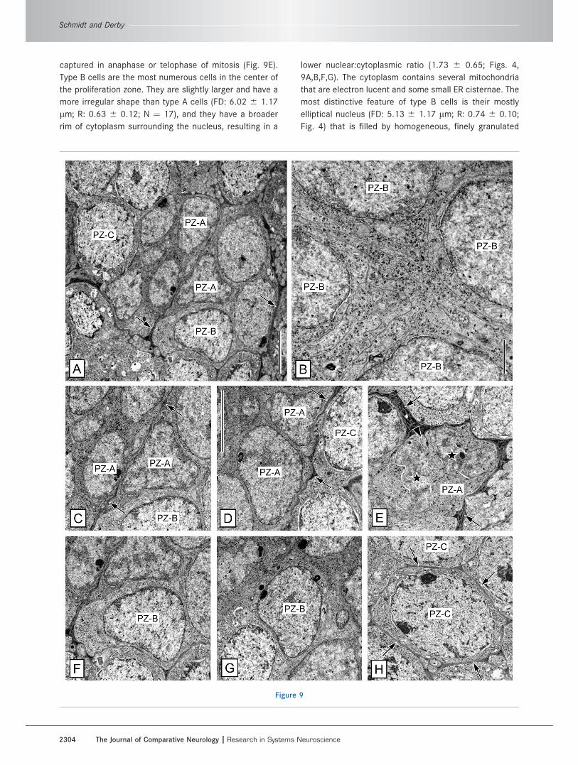
captured in anaphase or telophase of mitosis (Fig. 9E).
Type B cells are the most numerous cells in the center of
the proliferation zone. They are slightly larger and have a
more irregular shape than type A cells (FD: 6.02 6 1.17
lm; R: 0.63 6 0.12; N ¼ 17), and they have a broader
rim of cytoplasm surrounding the nucleus, resulting in a
lower nuclear:cytoplasmic ratio (1.73 6 0.65; Figs. 4,
9A,B,F,G). The cytoplasm contains several mitochondria
that are electron lucent and some small ER cisternae. The
most distinctive feature of type B cells is their mostly
elliptical nucleus (FD: 5.13 6 1.17 lm; R: 0.74 6 0.10;
Fig. 4) that is filled by homogeneous, finely granulated
Figure 9
Schmidt and Derby
2304 The Journal of Comparative Neurology |Research in Systems Neuroscience

nucleoplasm containing a small peripheral nucleolus. The
inner nuclear membrane is distinctly thicker and more
electron dense than the outer one, and both nuclear
membranes are separated by a small and very regular
gap. Type C cells are larger and of more regular shape
than type B cells (FD: 9.46 6 1.32 lm; R: 0.77 6 0.05;
n ¼ 10) and are located mostly in the periphery of the
proliferation zone (Figs. 4, 9A,H). In nuclear and cytoplas-
mic appearance, type C cells resemble somata of mature
neurons, but they are smaller and have a thinner rim of
cytoplasm, resulting in a higher nuclear:cytoplasmic ratio
(0.90 6 0.26). Their nucleus is of regular, slightly ellipti-
cal shape and contains coarsely granulated, electron-
lucent nucleoplasm with mostly peripheral heterochroma-
tin of very high electron density and at least one central
or peripheral nucleolus. The gap between the two nuclear
membranes is distinctly broader and less regular than in
type B cells. The cytoplasm of type C cells is more elec-
tron lucent than that of type A or B cells, and it contains
fine granules; some electron-lucent ER cisternae; several
large, electron-lucent mitochondria; and some electron-
dense vesicles. A distinctive morphological feature of the
central area of the proliferation zone is that type A and
type B cells are in direct contact with each other (Fig.
9B). Their cell membranes are not separated by inter-
spersed processes of cell body glia. A similar lack of glial
ensheathment was reported for neuronal progenitor cells
located centrally in the cluster of mature Kenyon cell
somata in the mushroom bodies of adult crickets
(Mashaly et al., 2008). Type C cells in the periphery of the
proliferation zone are at least partially and in many cases
completely surrounded by a thin layer of electron-dense
tissue consisting of processes of cell body glia (Fig. 9H).
Glial envelope and associated arterioleThe clump of cells and the interior of the duct arising
from it—until it reaches the proliferation zone—are com-
pletely enveloped by a layer of electron-dense tissue
separating them from other tissue components. This
layer consists of processes of cell body glia originating
from cell body glia somata of types 1 and 2 located in
the immediate vicinity of the clump of cells and the
duct (Figs. 6J, 8A,B). The entire glial envelope is formed
by many highly overlapping processes of cell body glia,
but it varies considerably in overall thickness (60 nm to
2 lm) as well as the thickness and number of layers of
glial cells contributing to it (Fig. 8D–H). A minimum of
one and maximally 10 cell body glia processes stacked
on top of each other constitute the envelope. A single
layer can be exceedingly thin (<20 nm), with no discern-
able cytoplasmic space between the cell membranes
(Fig. 8F,H), or be much thicker (up to 300 nm) and then
contain the typical cytoplasmic components of cell body
glia, in particular the robust and very electron-dense cis-
ternae of rough ER (Fig. 8D,G). When the duct reaches
the proliferation zone, the glial envelope separates into
several very irregularly shaped sheaths of tissue. These
glial sheaths are mostly interspersed between cells in
the inner and the peripheral region of the proliferation
zone, but they do not constitute a continuous and well-
demarcated boundary between these regions (Figs. 8A,
9A,C–E,H).
The clump of cells is attached to an arteriole of fairly
large width (maximum diameter �15 lm; Figs. 6H,I, 8C).
This arteriole runs along the duct until it reaches the pro-
liferation zone and then separates from it and becomes
gradually thinner (Fig. 6G). The arteriole is insulated from
the somata and processes of CFCs by the glial envelope
formed by the processes of cell body glia (Fig. 8C–F). We
did not find a consistent difference in the thickness or in
the number of glial cell layers of the glial envelope
between regions where neuronal somata are attached to
the clump of cells (Fig. 8G,H) and regions where the arte-
riole is attached to it (Fig. 8C–F).
Figure 9. Ultrastructure of the proliferation zone. TEM micrographs of sections through the proliferation zone in the lateral soma cluster.
A: Center of proliferation zone at low magnification reveals the presence of three distinct cell types: type A (PZ-A), type B (PZ-B) and type
C cells (PZ-C). Note that layers of electron-dense processes of cell body glia (arrows) only partially separate cells from each other. B: Four
neighboring type B cells (PZ-B). Type B cells are not surrounded by processes of cell body glia. They form irregularly shaped, convoluted
processes that interdigitate with processes of other type B cells. The inner nuclear membrane is distinctly thicker than the outer one. C,D:
Two examples of type A cells (PZ-A) at higher magnification. Type A cells have an irregularly shaped nucleus with large areas of diffuse
heterochromatin. Note the distinctly different appearance of type B (PZ-B) and type C (PZ-C) cells in the surround (arrows, layers of elec-
tron-dense processes of cell body glia). E: Type A cell (PZ-A) in telophase of mitosis. Condensed daughter nuclei (stars) oppose each
other. Thin cell membranes separate the two daughter cells (arrowhead; arrows indicate layers of electron-dense processes of cell body
glia). F,G: Two examples of type B cells (PZ-B). Type B cells have a thicker rim of cytoplasm and a more regularly shaped nucleus than
type A cells. Their inner nuclear membrane is distinctly thicker and more electron dense than the outer one. H: Example of a type C cell.
Type C cells (PZ-C) are larger and have a more spherical nucleus than type A and type B cells. Note that a distinctive gap separates both
nuclear membranes and that the cell is completely surrounded by a layer of electron-dense processes of cell body glia (arrows). Scale
bars ¼ 5 lm in A; 1 lm in B; 5 lm in D (applies to C–H).
Neural stem cell niches in adult Panulirus argus
The Journal of Comparative Neurology | Research in Systems Neuroscience 2305

Perfusion of fluorescent dextrans intocerebral artery
To test whether hemolymph has access to the interior
of the clump of cells, we perfused fluorescent dextrans of
different molecular weights (3,000 or 10,000) into the
brain of P. argus via the cerebral artery and immediately
afterward fixed the brain by immersion in fixative. In the
brain of treated animals (N ¼ 4, 2 for each dextran), the
branched system of arterioles was intensely labeled in
neuropils and in some soma clusters, including those of
the olfactory deutocerebrum (Fig. 10A,B). The olfactory
deutocerebrum of each hemibrain receives a major arte-
riole branch penetrating the area between OL and AL
from dorsally. This large arteriole enters the fibrous core
of the OL through the anterior foramen, which also
accommodates the passage of primary neurites of local
interneurons whose somata are located in the MC. In the
core of the OL, the large arteriole branches out into a
tree-like system of ever smaller arterioles densely perme-
ating the OL cortex and into several medium-sized arterio-
les that supply the AL, LC, and MC (Fig. 10A). The OL cor-
tex consists of wedge-shaped olfactory glomeruli
(Schmidt and Ache, 1997; Schachtner et al., 2005), but
the dense arteriole branches appear not to respect glo-
merular boundaries. In the AL, medium-sized arterioles
run in the two interfaces between the three neuropil
Figure 10
Schmidt and Derby
2306 The Journal of Comparative Neurology |Research in Systems Neuroscience
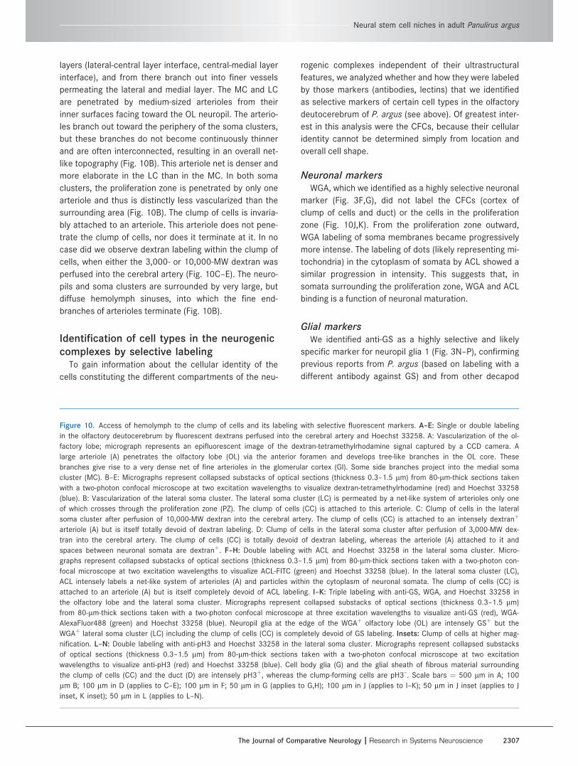
layers (lateral-central layer interface, central-medial layer
interface), and from there branch out into finer vessels
permeating the lateral and medial layer. The MC and LC
are penetrated by medium-sized arterioles from their
inner surfaces facing toward the OL neuropil. The arterio-
les branch out toward the periphery of the soma clusters,
but these branches do not become continuously thinner
and are often interconnected, resulting in an overall net-
like topography (Fig. 10B). This arteriole net is denser and
more elaborate in the LC than in the MC. In both soma
clusters, the proliferation zone is penetrated by only one
arteriole and thus is distinctly less vascularized than the
surrounding area (Fig. 10B). The clump of cells is invaria-
bly attached to an arteriole. This arteriole does not pene-
trate the clump of cells, nor does it terminate at it. In no
case did we observe dextran labeling within the clump of
cells, when either the 3,000- or 10,000-MW dextran was
perfused into the cerebral artery (Fig. 10C–E). The neuro-
pils and soma clusters are surrounded by very large, but
diffuse hemolymph sinuses, into which the fine end-
branches of arterioles terminate (Fig. 10B).
Identification of cell types in the neurogeniccomplexes by selective labeling
To gain information about the cellular identity of the
cells constituting the different compartments of the neu-
rogenic complexes independent of their ultrastructural
features, we analyzed whether and how they were labeled
by those markers (antibodies, lectins) that we identified
as selective markers of certain cell types in the olfactory
deutocerebrum of P. argus (see above). Of greatest inter-
est in this analysis were the CFCs, because their cellular
identity cannot be determined simply from location and
overall cell shape.
Neuronal markersWGA, which we identified as a highly selective neuronal
marker (Fig. 3F,G), did not label the CFCs (cortex of
clump of cells and duct) or the cells in the proliferation
zone (Fig. 10J,K). From the proliferation zone outward,
WGA labeling of soma membranes became progressively
more intense. The labeling of dots (likely representing mi-
tochondria) in the cytoplasm of somata by ACL showed a
similar progression in intensity. This suggests that, in
somata surrounding the proliferation zone, WGA and ACL
binding is a function of neuronal maturation.
Glial markersWe identified anti-GS as a highly selective and likely
specific marker for neuropil glia 1 (Fig. 3N–P), confirming
previous reports from P. argus (based on labeling with a
different antibody against GS) and from other decapod
Figure 10. Access of hemolymph to the clump of cells and its labeling with selective fluorescent markers. A–E: Single or double labeling
in the olfactory deutocerebrum by fluorescent dextrans perfused into the cerebral artery and Hoechst 33258. A: Vascularization of the ol-
factory lobe; micrograph represents an epifluorescent image of the dextran-tetramethylrhodamine signal captured by a CCD camera. A
large arteriole (A) penetrates the olfactory lobe (OL) via the anterior foramen and develops tree-like branches in the OL core. These
branches give rise to a very dense net of fine arterioles in the glomerular cortex (Gl). Some side branches project into the medial soma
cluster (MC). B–E: Micrographs represent collapsed substacks of optical sections (thickness 0.3–1.5 lm) from 80-lm-thick sections taken
with a two-photon confocal microscope at two excitation wavelengths to visualize dextran-tetramethylrhodamine (red) and Hoechst 33258
(blue). B: Vascularization of the lateral soma cluster. The lateral soma cluster (LC) is permeated by a net-like system of arterioles only one
of which crosses through the proliferation zone (PZ). The clump of cells (CC) is attached to this arteriole. C: Clump of cells in the lateral
soma cluster after perfusion of 10,000-MW dextran into the cerebral artery. The clump of cells (CC) is attached to an intensely dextranþ
arteriole (A) but is itself totally devoid of dextran labeling. D: Clump of cells in the lateral soma cluster after perfusion of 3,000-MW dex-
tran into the cerebral artery. The clump of cells (CC) is totally devoid of dextran labeling, whereas the arteriole (A) attached to it and
spaces between neuronal somata are dextranþ. F–H: Double labeling with ACL and Hoechst 33258 in the lateral soma cluster. Micro-
graphs represent collapsed substacks of optical sections (thickness 0.3–1.5 lm) from 80-lm-thick sections taken with a two-photon con-
focal microscope at two excitation wavelengths to visualize ACL-FITC (green) and Hoechst 33258 (blue). In the lateral soma cluster (LC),
ACL intensely labels a net-like system of arterioles (A) and particles within the cytoplasm of neuronal somata. The clump of cells (CC) is
attached to an arteriole (A) but is itself completely devoid of ACL labeling. I–K: Triple labeling with anti-GS, WGA, and Hoechst 33258 in
the olfactory lobe and the lateral soma cluster. Micrographs represent collapsed substacks of optical sections (thickness 0.3–1.5 lm)
from 80-lm-thick sections taken with a two-photon confocal microscope at three excitation wavelengths to visualize anti-GS (red), WGA-
AlexaFluor488 (green) and Hoechst 33258 (blue). Neuropil glia at the edge of the WGAþ olfactory lobe (OL) are intensely GSþ but the
WGAþ lateral soma cluster (LC) including the clump of cells (CC) is completely devoid of GS labeling. Insets: Clump of cells at higher mag-
nification. L–N: Double labeling with anti-pH3 and Hoechst 33258 in the lateral soma cluster. Micrographs represent collapsed substacks
of optical sections (thickness 0.3–1.5 lm) from 80-lm-thick sections taken with a two-photon confocal microscope at two excitation
wavelengths to visualize anti-pH3 (red) and Hoechst 33258 (blue). Cell body glia (G) and the glial sheath of fibrous material surrounding
the clump of cells (CC) and the duct (D) are intensely pH3þ, whereas the clump-forming cells are pH3–. Scale bars ¼ 500 lm in A; 100
lm B; 100 lm in D (applies to C–E); 100 lm in F; 50 lm in G (applies to G,H); 100 lm in J (applies to I–K); 50 lm in J inset (applies to J
inset, K inset); 50 lm in L (applies to L–N).
Neural stem cell niches in adult Panulirus argus
The Journal of Comparative Neurology | Research in Systems Neuroscience 2307

crustaceans (based on labeling with the same anti-GS as
was used here; Sullivan and Beltz, 2005b; Sullivan et al.,
2007a; Harzsch and Hansson, 2008). In our experiments,
anti-GS selectively labeled neuropil glia 1 cells at the rim
of neuropils, including the OL and AL, but it did not label
any structures within the soma clusters of the brain,
including the neurogenic complexes in the MC and LC
(Fig. 10I,K). Thus in P. argus, all cell types present in the
neurogenic complexes, including the CFCs, are GS–.
We identified anti-pH3 as a selective marker of cell
body glia (in addition to being a specific marker of nuclei
in M-phase of the cell cycle; Fig. 3K–M). Anti-pH3
intensely labeled the somata and main processes of cell
body glia in the MC and LC and in addition a sheath of fi-
brous material surrounding the clump of cells and the
duct, which was described previously based on labeling
with the same anti-pH3 (Schmidt, 2007a). We observed
several pH3þ somata of cell body glia in the vicinity of the
clump of cells and the duct with processes that extended
into the pH3þ fibrous sheath surrounding them (Fig. 10L–
N). This arrangement is congruent with the results of our
electron microscopic analysis showing that processes of
cell body glia form a continuous, multilayered sheath
around the clump of cells and the duct. Thus within the
neurogenic complexes, anti-pH3 selectively labeled the
glial sheath but no other cell types, including CFCs.
Somata and processes of cell body glia in the MC and
LC were also labeled by anti-Gs/olf, but this labeling was
not selective; anti-Gs/olf also labeled the cell membranes
of CFCs and of cells in the proliferation zone (Fig. 6A,D–
F). This distribution of labeling indicates that the three
most numerous cell types forming the neurogenic com-
plexes share an epitope to which anti-Gs/olf binds. How-
ever, our Western blot results indicate that this epitope
may not represent an authentic Gas subunit, insofar as,
in P. argus brain extracts, protein bands of distinctly
higher than predicted molecular weight were intensely la-
beled by anti-Gs/olf (Fig. 2A). Labeling with anti-Gs/olf
highlighted the entire neurogenic complex, but it did not
provide additional information about the cellular identity
of the CFCs.
Perivascular cell markerWe identified ACL as a moderately selective marker for
perivascular cells. In the MC and LC, labeling with ACL
differentiated the net-like system of arterioles that was
also labeled after perfusion of fluorescent dextrans into
the cerebral artery from neuronal somata (Figs. 3V, 10F).
This differentiation was due to an intense and continuous
labeling of the cytoplasm of perivascular cells by ACL
that was in clear contrast to the punctate ACL labeling of
neuronal somata (Figs. 6B,D, 10G,H). The neurogenic
complexes of MC and LC were completely devoid of ACL
labeling (Figs. 6B,D, 10F–H). Thus, none of the cell types
present in the neurogenic complexes of P. argus, includ-
ing the CFCs, shares ‘‘ACL-specific’’ carbohydrate resi-
dues with perivascular cells (or mature neurons). ACL
labeling of the arterioles within the MC and LC confirmed
that in both soma clusters the clump of cells is attached
to an arteriole and that there is no contiguity between the
two structures (Fig. 10G,H).
Selective labeling of the interface betweenthe bulbous foot of the aNB and CFCs
Aside from anti-BrdU and anti-pH3, none of the >150
probes that we screened for labeling components of the
neurogenic complexes labeled the main, nucleus-contain-
ing domain of the aNB located at the apex of the clump of
cells. In contrast, four of these probes (anti-Splash, SBA,
WGA, HPA) intensely labeled the region in the center of
the clump of cells that according to our electron micro-
scopic analysis is occupied by the bulbous foot of the
aNB (Fig. 5F–L). For anti-Splash and SBA, labeling of this
region in the center of the clump of cells was highly selec-
tive; neither probe labeled any other structures in the
brain (Fig. 5F,G,J,K). Within the neurogenic complexes,
WGA exclusively labeled the region in the center of the
clump of cells (Fig. 10J,K), but in addition WGA also la-
beled all neurons (see above). Within the neurogenic
complexes, HPA labeled exclusively the region in the cen-
ter of the clump of cells (Fig. 5H,L). In addition, HPA
intensely labeled neuropil regions and weakly labeled
neuronal somata in a dot-like pattern (similar to ACL
labeling). Double labeling with anti-Splash and SBA or
with anti-Splash and HPA revealed that the regions la-
beled by each of the two probes were concentric, with
very little overlap (Fig. 5J–L). In each case, a Splashþ ring
surrounded a center that was SBAþ or HPAþ. The labeled
center was almost spherical, and its rim was labeled
more intensely than its core. We conclude, based on the
electron microscopic analysis of the interior of the clump
of cells, that labeling likely is located at the interface
between the bulbous foot of the aNB and the attached
processes of CFCs. The concentric distribution of labeling
indicates that Splashþ material is located in the terminals
of the inner processes of CFCs, whereas the material that
is SBAþ, HPAþ, and WGAþ is located in the bulbous foot
of the aNB and concentrated at its cell membrane.
DISCUSSION
The detailed analysis of the cytoarchitecture of the
neurogenic complexes in olfactory deutocerebrum of
adult spiny lobsters, P. argus, based on TEM and labeling
with cell-type-selective markers leads to the conclusion
that the clump of cells associated with each proliferation
Schmidt and Derby
2308 The Journal of Comparative Neurology |Research in Systems Neuroscience

zone fulfills morphological criteria of a stem cell niche.
The clump of cells is composed of bipolar cells with
unique ultrastructure, clump-forming cells (CFCs). Collec-
tively the CFCs form a continuous envelope around the
hourglass-shaped aNB, separating it from other tissue
components, and thereby the CFCs solely constitute the
aNB microenvironment. The clump of cells is intimately
associated with glial and vascular elements and thus has
structural parallels with NSC niches in the brain of adult
mammals. Through BrdU pulse-chase experiments with
short survival times, we provide evidence that the aNBs
cycle rapidly during daytime. This proliferation kinetic dif-
fers dramatically from that of NSCs maintaining adult
neurogenesis in the mammalian brain, which are largely
quiescent (Morsehead et al., 1994; Doetsch et al.,
1999a). We propose, based on these findings, a new
model of the cell lineage and the cell movements through
which the single aNB continuously repopulates the GMC-
type neuronal progenitor cells in the attached prolifera-
tion zone.
NSC identity of aNBsTo allow the lifelong maintenance of adult neurogene-
sis in the olfactory deutocerebrum of decapod crusta-
ceans, self-renewing NSCs must be present to replenish
the pool of GMC-type neuronal progenitor in the prolifera-
tion zones, all of which ultimately undergo one terminal
symmetrical cell division (Schmidt, 2001). Previously,
BrdU labeling revealed that in adult spiny lobsters
(P. argus) and crayfish (P. clarkii) each proliferation zone
is associated with a single proliferating cell that has a dis-
tinct and invariant location, is connected to a unique
clump of cells, and has a significantly larger nucleus than
neuronal pregenitor cells or neurons (Schmidt, 2007a;
Song et al., 2009). Since large size is the identifying fea-
ture of NBs in embryonic and larval neurogenesis of crus-
taceans and insects (Hartenstein et al., 1987; Harzsch,
2001; Egger et al., 2008; Boyan et al., 2010), we pro-
posed that the single large proliferating cells are aNBs
and act as NSCs (Schmidt, 2007a; Song et al., 2009).
This interpretation, however, is not generally accepted,
and it has been proposed alternatively that the CFCs act
as NSCs (Sullivan et al., 2007a) or that in the clump of
cells hemocytes transdifferentiate into neuronal progeni-
tor cells (Zhang et al., 2009).
A defining feature of stem cells, including NSCs, is that
they undergo asymmetric cell divisions in which one
daughter cell persists as a self-renewed stem cell while
the other daughter cell gives rise to differentiated prog-
eny (Doe, 2008; Egger et al., 2008; Zhong and Chia,
2008; He et al., 2009). In revealing that the aNBs have a
unique hourglass-like shape, the present ultrastructural
analysis supports the interpretation that they act as
NSCs, since this unusual morphology indicates that the
aNB cell divisions are intrinsically asymmetric, giving rise
to two daughter cells with distinct morphology, location,
and fate. Each aNB consists of an outer domain at the ap-
ical (proliferation zone-facing) pole of the clump of cells
that contains the large nucleus and an inner domain form-
ing a bulbous foot within the clump of cells (Fig. 11A).
Since the mitotic plane of the aNB nucleus is perpendicu-
lar to the long axis of the hourglass (Schmidt, 2007a; this
study), the aNB cell division likely generates an outer
daughter cell located in the beginning of the duct and an
inner daughter cell that retains the hourglass-like shape
and remains anchored in the clump of cells with its bul-
bous foot. In this scenario, the inner daughter cell repre-
sents the self-renewed aNB, whereas the outer daughter
cell migrates through the duct to the proliferation zone
and replenishes the pool of neuronal progenitor cells (Fig.
11B). Migration of the outer daughter cells to the prolifer-
ation zone is supported by direct and indirect evidence
from crayfish (Sullivan et al., 2007a; Zhang et al., 2009).
With P. argus, we did not find evidence supporting the
alternative suggestions that the CFCs act as NSCs or that
neuronal progenitor cells are generated by transdifferen-
tiating hemocytes (Sullivan et al., 2007a; Zhang et al.,
2009). Neither in this study nor in previous studies were
any proliferating (BrdUþ) CFCs identified in adult or suba-
dult P. argus (Schmidt and Harzsch, 1999; Schmidt,
2001, 2007a). Thus it is highly unlikely that CFCs main-
tain continuous adult neurogenesis in P. argus, although it
is possible that CFCs occasionally regenerate an aNB
should its life span be limited. The possibility in P. argus
of hemocytes transdifferentiating into neuronal progeni-
tor cells within the clump of cells seems highly unlikely,
because hemocytes present within brain arterioles have
the distinct ultrastructural features of granulocytes (Hose
et al., 1990) and the clump of cells does not contain cells
with similar features.
Cell cycle dynamics of aNBs and neuralprogenitor cells
A characteristic feature of most (but not all) types of
adult stem cells, including NSCs in mammalian brains, is
to be largely quiescent and to divide only rarely (Morse-
head et al., 1994; Doetsch et al., 1999a; Fuchs, 2009).
This rare cycling is interpreted as a mechanism to mini-
mize accumulation of DNA copy errors through consecu-
tive rounds of semiconservative DNA replication and is
evident in the retention of BrdU-labeled DNA for extended
periods. To generate a sufficient number of somatic cells,
quiescent stem cells coexist with active ones and/or give
rise to transit amplifying intermediate progenitor cells
that proliferate rapidly (Doetsch et al., 1999a; Li and
Neural stem cell niches in adult Panulirus argus
The Journal of Comparative Neurology | Research in Systems Neuroscience 2309

Clevers, 2010). Previous experiments indicated that aNBs
of P. argus might be somewhat quiescent because they
were consistently labeled by BrdU after extended expo-
sure to BrdU (Schmidt, 2007a) but were generally not
labeled after single BrdU injections (Schmidt, 2001).
However, since the single BrdU injections were adminis-
tered in the evening, an alternative explanation for the
conflicting results is that aNBs preferentially divide during
daytime. To distinguish between the two alternatives, we
used single BrdU injections administered in the morning
followed by a short survival time of 2–10 hours in the
present experiments. Our finding that overall 82% of
aNBs in the LC and 50% of aNBs in the MC were labeled
by BrdU demonstrates that most aNBs are in S-phase dur-
ing daytime and that they are not quiescent. The previous
failure to label aNBs by BrdU injections in the evening
indicates that their S-phase ends earlier and that their
entire cell cycle lasts less than 24 hours. Thus, aNBs
Figure 11. A: Schematic representation of the cytoarchitecture of the clump of cells in the lateral soma cluster of P. argus. The clump of
cells is composed of bipolar clump-forming cells (CFC) whose somata form a continuous cortex around a nucleus-free center. One process
of the clump-forming cells is short and faces inward; the other one is long and faces outward. Together, the outward-facing processes
form the duct connecting with the adjacent proliferation zone. The clump-forming cells completely enclose one adult neuroblast (aNB),
which consists of two large domains connected by a thin bridge and thus has an hourglass-like shape. The peripheral domain of the adult
neuroblast contains the nucleus and is located at the apical pole of the clump of cells. The other domain forms a bulbous foot (asterisk)
within the nucleus-free center of the clump of cells and is surrounded by the inner processes of CFCs. The clump of cells and the duct
are surrounded by a continuous layer of processes of multipolar cell body glia (G). The clump of cells is associated with an arteriole (A)
whose wall is constituted by processes of perivascular cells (PVC). The duct contains elongated cells that most likely represent neuronal
progenitor cells (NPC) that are produced by asymmetric divisions of the aNB and migrate through the duct to the adjacent proliferation
zone. B: Schematic representation of the current model of cell divisions and lineages maintaining adult neurogenesis in the olfactory deu-
tocerebrum of P. argus. In each neuronal soma cluster of the olfactory deutocerebrum, production of new neurons is based on one neural
stem cell, which is an adult neuroblast (aNB) located close to the proliferation zone and embedded in a clump of cells. Through serial
asymmetric divisions, the aNB renews itself and produces differentiating daughter cells at its apical pole. Within a duct formed by proc-
esses of the clump-forming cells (CFC), the daughter cells migrate to the proliferation zone, where they replenish the pool of neuronal pro-
genitor cells (NPCs). Ultimately, neuronal progenitor cells divide once symmetrically and produce two daughter cells that represent
immature neurons (iN). Migrating out of the proliferation zone, most of the immature neurons become differentiated neurons (dN) within
months, but some die by apoptosis. We propose that, upon their arrival in the proliferation zone, daughter cells generated by the aNB
may undergo one or more fast rounds of cell divisions and thus represent transit amplifying intermediate progenitor cells, accommodating
the continuous replenishment of 30 (MC) or 100 (LC) cells in S-phase in the proliferation zone by only one aNB. For adult P. argus, there
is no evidence for proliferation of the CFCs or the cell body glia (G) surrounding the clump of cells, making it unlikely that these two cell
types contribute to maintaining adult neurogenesis.
Schmidt and Derby
2310 The Journal of Comparative Neurology |Research in Systems Neuroscience

appear to cycle quite rapidly, which is in line with the low
incidence of capturing them in mitosis (Schmidt, 2007a;
this study). Also in crayfish aNBs were consistently
labeled by single BrdU injections followed by short sur-
vival times (Song et al., 2009), indicating that rapid
cycling is a common feature of decapod crustacean
aNBs. The finding that aNBs are regularly in S-phase dur-
ing daytime but not at night implies that aNB cell divisions
are under circadian control. Circadian control of the tim-
ing of cell divisions appears to be widespread in animal
tissues (Matsuo et al., 2003), but in adult stem cells this
has been demonstrated only rarely (Lucas et al., 2008).
GMC-type neuronal progenitor cells in the proliferation
zones divide only once, and this cell cycle is extraordinary
long, likely lasting for more than 1 week (Schmidt, 1997,
2001). Hence, adult neurogenesis in the olfactory deuto-
cerebrum of decapod crustaceans appears to parallel
embryonic and larval neurogenesis of insects in several
key aspects: The NSCs maintaining insect embryonic and
larval neurogenesis are large NBs that have a short cell
cycle, have a high mitotic activity, and divide asymmetri-
cally, whereas GMCs are smaller, divide only once sym-
metrically, and have a cell cycle that is about twice as
long as in NBs (Hartenstein et al., 1987; Egger et al.,
2008; Boyan et al., 2010). However, a numerical problem
remains in understanding adult neurogenesis in the olfac-
tory deutocerebrum of decapod crustaceans; only one
aNB is present per proliferation zone, but according to
cell counts at least 10 new PNs per day are produced in
each LC (Schmidt, 1997; Sandeman et al., 1998). To gen-
erate >10 neurons per day by successive rounds of
asymmetric divisions, a single aNB would have to divide
asymmetrically at least five times per day, which is not in
line with the overall low percentage of mitotic aNBs
(Schmidt, 2007a; this study). This numerical discrepancy
suggests that an as yet unknown proliferation event is
interspersed between the asymmetric aNB divisions and
the terminal symmetrical divisions of the GMC-type neu-
ronal progenitor cells. We propose that, in parallel to
some embryonic and larval NB lineages of insects (Bello
et al., 2008; Boone and Doe, 2008; Bowman et al., 2008;
Izergina et al., 2009; Boyan et al., 2010), the differentiat-
ing daughters produced by the aNBs act as ‘‘transit-ampli-
fying intermediate progenitor cells’’ and undergo one or
more rapid cell divisions through which ultimately the
GMC-type neuronal progenitor cells are generated
(Fig. 11B). Such rapid cell divisions, which could occur in
the center of the proliferation zone containing numerous
cells in mitosis (pH3þ; Schmidt, 2007a; this study), would
have been missed by in previous BrdU pulse-chase
experiments because changes in the number of BrdUþ
cells were determined only for survival times >14 hours
(Schmidt, 2007a).
Characteristics of the stem cell niche andputative functions of the clump of cells
Densely packed clumps of small cells that form mor-
phologically unique structures in the deutocerebrum
were originally described as deutocerebral organs in vari-
ous species of decapod crustaceans (Bazin and Demeuzy,
1968; Bazin, 1970). Recently, these clumps of cells were
identified as part of the neurogenic complexes in the deu-
tocerebrum of P. argus (Schmidt, 2007a) and P. clarkii
(Sullivan et al., 2007a; Song et al., 2009; Zhang et al.,
2009). In both species, the clump of cells contains singu-
lar proliferating cells, putative aNBs, whose nuclei are sig-
nificantly larger and more spherical than the nuclei of
CFCs. The unique structure of the clumps of cells and
their association with the putative NSCs (aNBs) has led to
the proposal that they represent stem cell niches
(Schmidt, 2007a; Sullivan et al., 2007a; Song et al.,
2009). The analysis of the cytoarchitecture and ultra-
structure of the clump of cells in the LC of P. argus pre-
sented here strongly supports this conclusion. First, the
clump of cells is composed exclusively of cells of one
type—CFCs—which in ultrastructure and labeling with cell-
selective markers differ from all other cell types in the
olfactory deutocerebrum. Second, the CFCs collectively
form a continuous cell layer around the hourglass-shaped
aNB and thus are the exclusive constituents of its micro-
environment (Fig. 11A). Third, the assemblage of CFCs in
turn is ensheathed by a continuous layer of glial proc-
esses and thus separated from neurons and arterioles in
the surround. Thus, the clump of cells constitutes a
unique microenvironment around the aNB. It forms an
additional cell layer separating the aNB from surrounding
cells that is absent from the neuronal progenitor cells in
the proliferation zone, which are in direct membrane
contact with each other or with glial cells. With these char-
acteristics, the clump of cells fulfills the morphological cri-
teria of a ‘‘protective stem cell niche’’ as described for
many types of continuously replenished tissues of mam-
mals and insects (Spradling et al., 2001; Fuchs et al.,
2004; Ohlstein et al., 2004; Moore and Lemischka, 2006;
Jones and Wagers, 2008; Morrison and Spradling, 2008).
In spite of its principal correspondence to other stem
cell niches in adult tissues, the clump of cells is unique in
being composed of only one specific cell type and in
forming a morphologically distinct structure. Typically,
several cell types participate in the formation of stem cell
niches, and, like the stem cells they support, they are dis-
persed widely throughout the germinal layers of adult tis-
sues (Spradling et al., 2001; Fuchs et al., 2004; Ohlstein
et al., 2004; Moore and Lemischka, 2006; Jones and
Wagers, 2008; Morrison and Spradling, 2008). This
includes the stem cell niches in the subventricular zone
of the mammalian brain, which are composed of glial and
Neural stem cell niches in adult Panulirus argus
The Journal of Comparative Neurology | Research in Systems Neuroscience 2311

vascular cells as well as prominent extracellular matrix
structures and are widely dispersed throughout the ger-
minal layer (Mercier et al., 2002; Kerever et al., 2007;
Shen et al., 2008; Tavazoie et al., 2008). Glial and peri-
vascular cells are also closely associated with the clump
of cells in the LC of P. argus and hence appear to partici-
pate in its niche function (Fig. 11A). Extracellular matrix
structures, however, were not revealed by TEM, either
within the clump of cells or in its immediate surround. The
extracellular matrix structure closest to the aNB is the basal
lamina overlying the luminal surface of the arteriole that is
attached to the clump of cells. Since to be lined by a basal
lamina is a constitutive feature of all arterioles, we conclude
that most likely extracellular matrix structures are of no par-
ticular significance to the cell divisions of aNBs.
The close association of aNBs with clumps of cells in
the olfactory deutocerebrum of decapod crustaceans
strongly suggests that the clumps of cells have important
functions in maintaining aNBs and in supporting their
proliferative activity. Generally, a host of functions is
ascribed to stem cell niches, among them 1) nurturing
stem cells, 2) shielding stem cells against apoptotic or
differentiation stimuli as well as infectious agents, 3)
anchoring stem cells, 4) polarizing stem cells to direct
their asymmetric divisions, and 5) providing signals for
stem cell self-renewal, survival, and maintenance (Spra-
dling et al., 2001; Fuchs et al., 2004; Ohlstein et al.,
2004; Moore and Lemischka, 2006; Jones and Wagers,
2008; Morrison and Spradling, 2008). Its peculiar
cytoarchitecture and the unique shape of the single aNB
within it indicate that some if not all of these functions
might be served by the clump of cells.
1. Nurturing the aNB seems a plausible function of the
clump of cells, because it is attached to an arteriole
through which nutrients and oxygen transported via
hemolymph could be delivered. Furthermore, the bul-
bous foot of the aNB appears to provide an
increased area of aNB-CFC cell membrane apposi-
tion, which would facilitate transport through this
interface. However, our perfusion experiments indi-
cate that hemolymph has no direct access to this
interface, and our TEM analysis shows that the arte-
riole attached to the clump of cells is separated
from the CFCs by a continuous and often multilay-
ered sheath of cell body glia processes. Thus, the
delivery of nutrients from the hemolymph to the aNB
appears to require transport mechanisms in the
ensheathing cell body glia and the CFCs. As in
P. argus, the clump of cells in P. clarkii contains a
central, nucleus-free area (Song et al., 2009) that
was identified as a vascular cavity based on perfu-
sion experiments (Sullivan et al., 2007a; Sandeman
et al., 2009). Whether it is contiguous with the
lumen of neighboring arterioles remains to be deter-
mined by TEM.
2. Shielding the aNB against inappropriate signaling
molecules and/or infectious agents also appears to
be a plausible function of the clump of cells. By
forming a thick, continuous cell layer surrounding
the aNB, the CFCs constitute a unique cellular bar-
rier between the aNB and other cell types. This extra
layer likely makes the aNB less accessible to sub-
stances or particles transported in hemolymph than
other cells in the LC. Generally, there is no tight
blood–brain barrier in the CNS of decapod crusta-
ceans, and relatively large tracer molecules such as
ferritin delivered through the hemolymph have
access to neurons and glia several micrometers
away from arterioles (Abbott, 1972). This is in line
with our finding that fluorescent dextrans of 3,000
MW perfused into the cerebral artery labeled extrac-
ellular spaces between neuronal somata. That in
these experiments the clump of cells remained unla-
beled supports the notion that the highly convoluted
contours of the CFCs, their thickness, and the tight
packing of their processes around the aNB minimize
extracellular diffusion. Thus, molecular access to the
aNB would be mainly through transport across the
CFCs, making them poised to control the flow of
molecules to the aNB, either by blocking inappropri-
ate signaling molecules or infectious agents or by
facilitating the transport of nutrients or appropriate
signaling molecules. The selective labeling of the
interface between the CFCs and the bulbous foot of
the aNB with several lectins (SBA, HPA, WGA) and
anti-Splash suggests that it has a unique molecular
composition, which is in line with the assumption
that it mediates a highly specialized function, such
as the control of molecular exchange between CFCs
and the aNB.
3. Anchoring the aNB in the clump of cells is strongly
indicated by the unique hourglass-like shape of the
aNB. The bulbous foot of the aNB is tightly enclosed
by the inner processes of the CFCs, and to retract it
from the center of the clump of cells would require
extensive morphological rearrangements of these
processes. Overall, the hourglass-like shape of the
aNB suggests that it consists of two cellular domains
with different functions. The bulbous foot seems to
be specialized for anchoring the aNB in the clump of
cells and for establishing an extensive contact area
with the cell membranes of CFCs. The outer nu-
cleus-contain domain seems to be specialized for
producing daughter cells. Through this division of
Schmidt and Derby
2312 The Journal of Comparative Neurology |Research in Systems Neuroscience

labor, the aNB could undergo repetitive asymmetric
divisions without disruption of its anchoring in the
clump of cells or its supply by the CFCs.
4. Polarizing the aNB to direct its asymmetric divisions
is another plausible function of the clump of cells.
The clump of cells is polarized into an apical pole,
giving rise to the duct that connects it with the pro-
liferation zone and a basal pole facing in the oppo-
site direction. At the apical pole, the clump of cells
gives rise to the duct that connects with the prolifer-
ation zone, and it surrounds the nucleus-containing
domain of the aNB. This polarization is paralleled by
the polarization of the hourglass-shaped aNB into an
outer nucleus-containing domain and the inner bul-
bous foot. As described above, it seems likely that
differentiating daughter cells are budded off from
the apical pole of the aNB and that they maintain
this directionality when they move through the duct
toward the proliferation zone. This clear directionality
is reminiscent of how in embryonic neurogenesis
NBs bud off GMCs strictly at their neuroepithelium-
opposing pole and thereby generate columnar cell
clones toward the interior of the body (Doe and
Goodman, 1985; Hartenstein et al., 1987; Scholtz,
1992). In Drosophila the orientation of GMC genera-
tion depends on neuroepithelial cells attached to the
NB, which itself is not polarized (Siegrist and Doe,
2006). With respect to the orientation of the daugh-
ter cells budded off by the aNB, CFCs in P. argus
have a position equivalent to that of neuroepithelial
cells, suggesting that they might be involved in ori-
enting aNB cell divisions. However, the obvious mor-
phological polarization of the aNB leaves open the
possibility that orientation of the cell division axis is
an intrinsic aNB property.
5. Providing signals for aNB self-renewal, survival, and
maintenance may be a function of the clump of cells
simply because presumably no such signals can
reach the aNB without passing through or being gen-
erated by the CFCs. Whether proliferation of aNBs is
influenced by signaling molecules has not yet been
studied, but, since serotonin and nitric oxide modu-
late cell divisions in the proliferation zones (Benton
et al., 2007; 2008; Sandeman et al., 2009), it is pos-
sible that they might also affect the aNB.
Cellular identity of CFCsOne important prerequisite for understanding how the
clump of cells functions as a stem cell niche for aNBs is to
know the cellular identity of the CFCs. In P. clarkii, CFCs
were identified as glial cells mainly because they were la-
beled by anti-GS (Sullivan et al., 2007a,b). Anti-GS is a
marker for astrocytes in the mammalian brain (Norenberg,
1979), and it selectively labels a class of glial cells in the
brain of decapod crustaceans (Linser et al. 1997; Sullivan
et al., 2007a; Harzsch and Hansson, 2008). These GSþ glial
cells have somata at the rim of neuropils, and they extend
uni- or multipolar processes into the neuropils, where they
branch profusely (Linser et al., 1997). Using the same anti-
GS as was used by Sullivan et al. (2007a), we also found
distinct and selective labeling of a class of neuropil glia
(neuropil glia 1) in the brain of adult P. argus with the previ-
ously reported morphology. Morphologically these GSþ glial
cells of decapod crustaceans correspond to astrocyte-like
neuropil glia identified in insect brains (Oland et al., 1999;
Awasaki et al., 2008; Doherty et al., 2009). In our immuno-
cytochemical stainings, anti-GS did not label the CFCs of
P. argus, and from this we conclude that in P. argus the
CFCs do not have immunocytochemical properties of neuro-
pil glia 1 cells. Furthermore, the CFCs of P. argus differ dis-
tinctly from neuropil glia 1 cells in basic cell morphology
(CFCs are bipolar; neuropil glia 1 cells are usually mono- or
multipolar) and in soma ultrastructure. Since the CFCs of
P. clarkii are also bipolar (Sullivan et al., 2007a), their identi-
fication as glial cells hinging exclusively on being by anti-GS
seems premature (Song et al., 2009). This argument is sup-
ported by results of in situ hybridization with a GS-specific
probe, which labeled neuropil glia but not the CFCs (Zhang
and Beltz, 2010), and by the observation that in the hermit
crab, Coenobita clypeatus, a structure likely representing
the clump of cells is GS–, whereas neuropil glia are GSþ
(Harzsch and Hansson, 2008, Fig. 5B1).
By TEM and labeling with anti-pH3 and anti-Gs/olf, we
identified a distinct class of multipolar cell body glia resid-
ing in the LC, MC, and all other neuronal soma clusters.
In being multipolar and pH3þ, cell body glia are clearly
distinct from the bipolar and pH3– CFCs, and both cell
types differ considerably in ultrastructure. Thus, we con-
clude that CFCs are not identical to cell body glia. TEM
revealed at least three different classes of putative glial
cells with somata at the rim of neuropils in addition to pu-
tative neuropil glia 1 cells. In their ultrastructural features
and multipolar morphology, all of these putative glial cells
differ distinctly from CFCs, so we conclude that the CFCs
do not correspond to glial cells of these classes.
A previous analysis of cells in the neurogenic complex
of P. argus based on the measurement of Hoechst-labeled
nuclei showed that CFCs are similar to perivascular cells
in nuclear size and shape (Schmidt, 2007a). The possibil-
ity raised by this result that CFCs may correspond to peri-
vascular cells was not corroborated by the more detailed
analyses presented here. 1) TEM showed that CFCs and
perivascular cells differ distinctly in electron density and
organelle composition of the cytoplasm as well as in nu-
clear ultrastructure. 2) TEM also demonstrated that a
Neural stem cell niches in adult Panulirus argus
The Journal of Comparative Neurology | Research in Systems Neuroscience 2313
basal lamina covers the entire luminal surface of perivas-
cular cells but is not present on the surface of CFCs or
anywhere else in the clump of cells. 3) ACL intensely la-
beled perivascular cells, including those of the arteriole
attached to the clump of cells, but failed to label CFCs. 4)
Conversely, anti-Gs/olf intensely labeled cell membranes
of CFCs but failed to label arterioles. Collectively, these
results strongly support the notion that CFCs do not rep-
resent perivascular cells.
Besides glial and perivascular cells, neurons are the
main cell type present in the olfactory deutocerebrum.
CFCs are clearly different from neurons in basic morphol-
ogy (small bipolar vs. large unipolar) as well as in the
ultrastructure of cytoplasm and nucleus. Both cell types
are also distinctly different in labeling by cell-type-selec-
tive probes: neurons are WGAþ and ACLþ but anti-Gs/
olf–, whereas the opposite is true for CFCs. Thus, we con-
clude that CFCs are not neurons.
In conclusion, our results fail to provide evidence that
in P. argus the CFCs correspond to any of the cell types
that can be identified in the olfactory deutocerebrum
composed of several types of glia, perivascular cells, and
neurons. Thus, CFCs appear to represent a different, as
yet unidentified cell type, and we propose that they might
correspond to embryonic neuroepithelial cells. This
hypothesis is based on the observation that, after delami-
nation from the neuroepithelium, embryonic NBs of
insects remain associated with elongated processes of
neuroepithelial cells (also called sheath cells), which,
like CFCs, have distinct bipolar morphology (Doe and
Goodman, 1985). The CFCs/aNB association and the
sheath cells/embryonic NB association have a similar
morphological polarization in that, in both cases, the dif-
ferentiating NB daughters are generated at the pole of
the NB opposite where the somata of CFCs or sheath
cells are located. In embryonic neurogenesis, this polar-
ization results in the production of neurons into the inte-
rior of the body, and a corresponding directionality is
maintained in adult neurogenesis, when new neurons are
produced at more internal locations of the brain (prolifer-
ation zones) than positions occupied by the aNB. This
new hypothesis about the cellular identity of the CFCs
implies that the stem cell niche provided by the clump of
cells primarily maintains a microenvironment for the aNB
with features of the embryonic neuroepithelium.
Conclusions: Cellular basis of adultneurogenesis across animal taxa
The results of this study allow a more detailed compari-
son of the cellular basis of adult neurogenesis among the
three bilaterian taxa in which new neurons are continu-
ously generated in select brain regions of adults: decapod
crustaceans, insects, and vertebrates, in particular mam-
mals (Lindsey and Tropepe, 2006; Fig. 12). Adult neuro-
genesis in decapod crustaceans shows important com-
monalities with adult neurogenesis in insects with respect
to the identity of the NSCs and the cell lineages to which
they give rise. In both taxa, the NSCs maintaining adult
neurogenesis are represented by a few large NBs of invari-
ant location within or close to the neuronal soma clusters
in which new neurons are generated (Figs. 11B, 12B,D).
Through successive asymmetric divisions, the aNBs ulti-
mately give rise to neuronal progenitor cells (GMCs) that
undergo one terminal symmetrical division producing two
neurons. We speculate that, in decapod crustaceans, at
least some daughter cells generated by the aNBs act as
transit-amplifying intermediate progenitor cells providing a
mechanism to produce the high number of neuronal pro-
genitor cells associated with each aNB. Transit-amplifying
intermediate progenitor cells have been documented in
some embryonic and larval NB lineages of insect brains
(see above) but not in NB lineages producing the Kenyon
cells of the mushroom bodies (Bello et al., 2008; Boyan
et al., 2010), the only neurons that continue to be gener-
ated in the brain of adult insects (Cayre et al., 2002).
Thus, it seems unlikely that transit-amplifying intermediate
progenitor cells contribute to adult neurogenesis in the
brain of insects. A substantial difference between adult
neurogenesis in decapod crustaceans and insects is that,
in decapod crustaceans, but not in insects, aNBs are asso-
ciated with clumps of cells likely representing stem cell
niches. Based on the finding that, in Drosophila, glial cells
associated with larval NBs secrete glycoproteins regulat-
ing NB proliferation (Ebens et al., 1993), it has been sug-
gested that these glial cells form a stem cell niche for late
embryonic and larval NBs (Maurange and Gould, 2005).
However, glial cells are not associated with the neuronal
progenitor cells in the Kenyon cell layer of adult crickets in
a pattern suggesting that they form a stem cell niche
(Mashaly et al., 2008). We hypothesize that the presence
or lack of a stem cell niche associated with the aNBs
reflects the generally enormous difference in the duration
of adult neurogenesis between decapod crustaceans and
insects. In decapod crustaceans, adult neurogenesis per-
sists throughout the entire adult life span, which for most
species (including P. argus and P. clarkii) ranges from
years to decades (Farmer, 1973; Lyons et al., 1981; Belch-
ier et al., 1998; Sheehy et al., 1999; Maxwell et al., 2007).
In contrast, insect species with adult neurogenesis have
an adult life span is in the range of months and adult neu-
rogenesis ceases within the first weeks of adult life (Gu
et al., 1999; Zhao et al., 2008). Thus, adult neurogenesis
in insects can be interpreted as a transient continuation of
larval/pupal neurogenesis that is enabled simply by pre-
venting apoptotic cell death of the mushroom body NBs
Schmidt and Derby
2314 The Journal of Comparative Neurology |Research in Systems Neuroscience

(Siegrist et al., 2010). In contrast, adult neurogenesis in
decapod crustaceans is a much more prolonged process
and as such appears to require that a stem cell niche pro-
vide additional support for the aNBs to sustain their long-
term survival and proliferative potential.
Adult neurogenesis in decapod crustaceans and
insects appears to differ fundamentally from adult neu-
rogenesis in mammals with respect to the identity, num-
ber, and proliferation dynamics of the NSCs maintaining
it. In decapod crustaceans and insects, the NSCs main-
taining adult neurogenesis are infrequent, tetraconate-
typical NBs, characterized by being large, by not having
a differentiated cell phenotype (neither neuronal nor
glial cell morphology), and by lacking a primary cilium.
In contrast, the NSCs maintaining adult neurogenesis in
mammals have a glial cell (astrocytic) phenotype, bear a
primary cilium, and are dispersed throughout extensive
germinal layers, the subventricular zone of the lateral
Figure 12. Schematic comparison of neural stem cells and cell
lineages maintaining embryonic/larval and adult neurogenesis in
the brain of decapod crustaceans (A,B), insects (C,D), and mam-
mals (E,F). A: Embryonic and larval neurogenesis in crustaceans
is based on large, spherical neuroblasts, which through a series
of asymmetric divisions self-renew and generate ganglion mother
cells (GMC) into the interior of the body. Each GMC divides sym-
metrically once and generates two immature neurons. B: Adult
neurogenesis in decapod crustaceans is maintained by large,
hourglass-shaped adult neuroblasts (aNB), which are closely asso-
ciated with a stem cell niche composed of clump-forming cells,
glial cells, and an attached arteriole. Through a series of asym-
metric divisions the aNB self-renews and ultimately gives rise to
GMC-type neuronal progenitor cells, each of which divides sym-
metrically once and generates two immature neurons (left line-
age). We hypothesize that the daughter cells produced by the
aNB divisions likely act as transient-amplifying intermediate pro-
genitor cells (IPC), each of which produces multiple GMC-type
neuronal progenitors (right lineage). C: Embryonic and larval neu-
rogenesis in insects is mostly as in crustaceans and is based on
‘‘canonical’’ neuroblasts (left lineage). Some embryonic and larval
neuroblast lineages contain transit-amplifying intermediate pro-
genitor cells (IPC) that through asymmetric divisions self-renew
and generate GMCs (right lineage; modified from Bello et al.,
2008). D: Adult neurogenesis in insects appears to be maintained
by ‘‘canonical’’ neuroblasts and their embryo-typical lineage. E: In
embryonic neurogenesis of mammals, radial glial cells serve as
neural stem cells. Through asymmetric divisions they self-renew
and ultimately generate daughter cells that, after migrating along
the radial glial process, mature into neurons. The generation of
neurons could be direct or through transit-amplifying intermediate
progenitor cells (IPC?). One hallmark feature of radial glia cells is
to have a primary cilium reaching into the ventricle (arrowhead).
Modified from Alvarez-Buylla et al. (2001). F: Adult neurogenesis in
the SVZ of mammals is maintained by type B cells, which have an
astrocytic morphology and possess a primary cilium (arrowhead)
reaching into the ventricle. Type B cells are slowly cycling neural
stem cells, which through asymmetric divisions self-renew and gen-
erate rapidly dividing neuronal progenitor cells (C cells). C cells
give rise to immature neurons (A cells). Type B cells are associated
with morphologically complex stem cell niches, one element of
which are blood vessels directly contacted by type B cell proc-
esses. Modified from Kriegstein and Alvarez-Buylla (2009).
Neural stem cell niches in adult Panulirus argus
The Journal of Comparative Neurology | Research in Systems Neuroscience 2315

ventricles, and the subgranular region of the hippocam-
pus (Kriegstein and Alvarez-Buylla, 2009; Fig. 12F). The
aNBs of decapod crustaceans and insects appear to be
actively proliferating, insofar as they are readily labeled
by single BrdU applications (Dufour and Gadenne, 2006;
Zhao et al., 2008; Song et al., 2009; this study), whereas
NSCs of mammals are labeled only after extended expo-
sure to BrdU and thus are largely quiescent (Doetsch
et al., 1999a,b).
In spite of these substantial differences in the nature
of the NSCs, adult neurogenesis in decapod crustaceans
(but not in insects; see above) parallels adult neurogene-
sis in mammals in several ways. First, adult neurogenesis
is maintained throughout the entire adult life, which in
decapod crustaceans (see above) as well as in mammals
lasts for several years to decades (De Magalahaes and
Costa, 2009). Second, both taxa have an age-dependent
decrease in proliferative activity (Kuhn et al., 1996; Han-
sen and Schmidt, 2004; Luo et al., 2006). Third, both taxa
have stem cell niches supporting the NSCs. Likely these
common features of adult neurogenesis in decapod crus-
taceans and mammals are causally linked in that stem
cell niches are required for the long-term preservation of
NSCs but cannot completely prevent their ‘‘aging.’’
In conclusion, adult neurogenesis in the brain shows a
very basic similarity among crustaceans, insects, and
mammals, in that in each taxon the NSCs maintaining
adult neurogenesis have the same identity as those fuel-
ing embryonic (and if present larval) neurogenesis. Thus,
adult neurogenesis can be viewed as a prolonged exten-
sion of embryonic neurogenesis. Furthermore, the pres-
ence of stem cell niches associated with the NSCs fueling
adult neurogenesis in decapod crustaceans and mam-
mals, but not in insects, suggests that NSC niches are a
prerequisite for preserving the NSC proliferative activity
for years or even decades of adult life. To understand the
principle mechanisms by which this impressive feat is
achieved by neural stem cell niches, it will be informative
to compare those of decapod crustaceans and mammals,
especially because their NSCs are of different cellular
identity and their niches are of different cellular composi-
tion. The fact that, in decapod crustacean brains, the
NSCs (¼aNBs) and their stem cell niches (¼clumps of
cells) are morphologically identifiable makes them
uniquely amenable for analyzing their cellular and physio-
logical interactions.
ACKNOWLEDGMENTS
We thank Vu Ngo for help with animal care and Vivian
Ngo-Vu for excellent assistance with immunocytochemis-
try. We also thank the Developmental Studies Hybridoma
Bank for providing antisynapsin antibody.
LITERATURE CITEDAbbott NJ. 1971. The organization of the cerebral ganglion in
the shore crab, Carcinus maenas. II. The relation of intra-cerebral blood vessels to other brain elements. Z Zell-forsch 120:401–419.
Abbott NJ. 1972. Access of ferritin to the interstitial space ofCarcinus brain from intracerebral blood vessels. Tissue &Cell 4:99–104.
Alvarez-Buylla A, Garcıa-Verdugo JM, Mateo AS, Merchant-Larios H. 1998. Primary neural precursors and intermitoticnuclear migration in the ventricular zone of adult canaries.J Neurosci 18:1020–1037.
Alvarez-Buylla A, Garcıa-Verdugo JM, Tramontin AD. 2001. Aunified hypothesis on the lineage of neural stem cells. NatRev Neurosci 2:287–293.
Awasaki T, Lai S-L, Ito K, Lee T. 2008. Organization and post-embryonic development of glial cells in the adult centralbrain of Drosophila. J Neurosci 28:13742–13753.
Bazin F. 1970. Etude comparee de l’organe deutocerebral desMacroures Reptantia et des Anomures (Crustaces Decapo-des). Arch Zool Exp Gen 111:245–264.
Bazin F, Demeuzy N. 1968. Existence d’organes intracere-braux enigmatiques chez le Crustace Decapode Carcinusmaenas (L.). C R Acad Sci 267:356–358.
Belchier M, Edsman L, Sheehy MRJ, Shelton PMJ. 1998. Esti-mating age and growth in long-lived temperate freshwatercrayfish using lipofuscin. Freshwater Biol 39:439–446.
Bello BC, Izergina N, Caussinus E, Reichert H. 2008. Amplifi-cation of neural stem cell proliferation by intermediate pro-genitor cells in Drosophila brain development. Neural Dev3:5.
Benton JL, Sandeman DC, Beltz BS. 2007. Nitric oxide in thecrustacean brain: regulation of neurogenesis and morpho-genesis in the developing olfactory pathway. Dev Dyn 236:3047–3060.
Benton JL, Goergen EM, Rogan SC, Beltz BS. 2008. Hormonaland synaptic influences of serotonin on adult neurogene-sis. Gen Comp Endocrinol 158:183–190.
Boone JQ, Doe CQ. 2008. Identification of Drosophila type IIneuroblast lineages containing transit amplifying ganglionmother cells. Dev Neurobiol 68:1185–1195.
Bowman SK, Rolland V, Betschinger J, Kinsey KA, Emery G,Knoblich JA. 2008. The tumor suppressors Brat and Numbregulate transit-amplifying neuroblast lineages in Drosoph-ila. Dev Cell 14:535–546.
Boyan G, Williams L, Legl A, Herbert Z. 2010. Proliferative celltypes in embryonic lineages of the central complex of thegrasshopper Schistocerca gregaria. Cell Tissue Res 341:259–277.
Breunig JJ, Sarkisian MR, Arellano JI, Morozov YM, Ayoub AE,Sojitra S, Wang B, Flavell RA, Rakic P, Town T. 2008. Pri-mary cilia regulate hippocampal neurogenesis by mediatingsonic hedgehog signaling. Proc Natl Acad Sci U S A 105:13127–13132.
Cayre M, Strambi C, Strambi A. 1994. Neurogenesis in an adultinsect brain and its hormonal control. Nature 368:57–59.
Cayre M, Strambi C, Charpin P, Augier R, Meyer MR, EdwardsJS, Strambi A. 1996. Neurogenesis in adult insect mush-room bodies. J Comp Neurol 371:300–310.
Cayre M, Malaterre J, Scotto-Lomassese S, Strambi C,Strambi A. 2002. The common properties of neurogenesisin the adult brain: from invertebrates to vertebrates. CompBiochem Physiol B 132:1–15.
Chien H, Tadesse T, Liu H, Schmidt M, Walthall WW, Tai PC,Derby CD. 2009. Molecular cloning and characterization ofhomologs of achaete-scute and hairy-enhancer of split inthe olfactory organ of the spiny lobster Panulirus argus.J Mol Neurosci 39:294–307.
Schmidt and Derby
2316 The Journal of Comparative Neurology |Research in Systems Neuroscience

De Magalhaes JP, Costa J. 2009. A database of vertebrate lon-gevity records and their relation to other life-history traits.J Evol Biol 22:1770–1774.
Doe CQ. 2008. Neural stem cells: balancing self-renewal withdifferentiation. Development 135:1575–1587.
Doe CQ, Goodman CS. 1985. Early events in insect neurogen-esis. I. Development and segmental differences in the pat-tern of neuronal precursor cells. Dev Biol 111:193–205.
Doe CQ, Fuerstenberg S, Peng CY. 1998. Neural stem cells:from fly to vertebrates. J Neurobiol 36:111–127.
Doetsch F, Caille I, Lim DA, Garcıa-Verdugo JM, Alvarez-BuyllaA. 1999a. Subventricular zone astrocytes are neural stemcells in the adult mammalian brain. Cell 97:703–716.
Doetsch F, Garcıa-Verdugo JM, Alvarez-Buylla A. 1999b.Regeneration of a germinal layer in the adult mammalianbrain. Proc Natl Acad Sci U S A 96:11619–11624.
Doherty J, Logan MA, Tasdemir OE, Freeman MR. 2009.Ensheathing glia function as phagocytes in the adult Dro-sophila brain. J Neurosci 29:4768–4781.
Dohle W. 1976. Die Bildung und Differenzierung des postnau-plialen Keimstreifs von Diastylis rathkei (Crustacea, Cuma-cea). II. Die Differenzierung und Musterbildung desEktoderms. Zoomorphologie 84:235–277.
Dohle W. 2001. Are the insects terrestrial crustaceans? A dis-cussion of some new facts and arguments and the proposalof the proper name ‘‘Tetraconata’’ for the monophyletic unitCrustacea þ Hexapoda. Ann Soc Entomol Fr 37:85–103.
Dufour M-C, Gadenne C. 2006. Adult neurogenesis in a mothbrain. J Comp Neurol 495:635–643.
Ebens AJ, Garren H, Cheyette BNR, Zipursky SL. 1993. TheDrosophila anachronism locus: a glycoprotein secretet byglia inhibits neuroblast proliferation. Cell 74:15–27.
Egger B, Chell JM, Brand AH. 2008. Insights into neural stemcell biology from flies. Philos Trans R Soc Lond B Biol Sci363:39–56.
Eisenberg D, Gill HS, Pfluegl GMU, Rotstein SH. 2000. Struc-ture–function relationships of glutamine synthetases. Bio-chim Biophys Acta 1477:122–145.
Farmer AS. 1973. Age and growth in Nephrops norvegicus(Decapoda: Nephropidae). Mar Biol 23:315–325.
Fuchs E. 2009. The tortoise and the hair: slow-cycling cells inthe stem cell race. Cell 137:811–819.
Fuchs E, Tumbar T, Guasch G. 2004. Socializing with neigh-bors: stem cells and their niche. Cell 116:769–778.
Ghosal K, Gupta M, Killian KA. 2009. Agonistic behaviorenhances adult neurogenesis in male Acheta domesticuscrickets. J Exp Biol 212:2045–2056.
Gu SH, Tsia WH, Chiang AS, Chow YS. 1999. Mitogeniceffects of 20-hydroxyecdysone on neurogenesis in adultmushroom bodies of the cockroach, Diploptera punctata.J Neurobiol 39:264–274.
Han Y-G, Spassky N, Romaguera-Ros M, Garcıa-Verdugo JM,Aguilar A, Schneider-Maunoury S, Alvarez-Buylla A. 2008.Hedgehog signaling and primary cilia are required for the for-mation of adult neural stem cells. Nat Neurosci 11:277–284.
Hansen A, Schmidt M. 2001. Neurogenesis in the central olfac-tory pathway of the adult shore crab Carcinus maenas is con-trolled by sensory afferents. J Comp Neurol 441:223–233.
Hansen A, Schmidt M. 2004. Influence of season and environ-ment on adult neurogenesis in the central olfactory pathwayof the shore crab, Carcinus maenas. Brain Res 1025:85–97.
Hartenstein V, Rudloff E, Campos-Ortega JA. 1987. The pat-tern of proliferation of the neuroblasts in the wild-typeembryo of Drosophila melanogaster. Roux’s Arch Dev Biol196:473–485.
Harzsch S. 2001. Neurogenesis in the crustacean ventralnerve cord: homology of neuronal stem cells in Malacos-traca and Branchiopoda? Evol Dev 3:154–169.
Harzsch S, Hansson BS. 2008. Brain architechture in the ter-restrial hermit crab Coenobita clypeatus (Anomura, Coeno-bitidae), a crustacean with good aerial sense of smell.BMC Neurosci 9:58.
Harzsch S, Anger K, Dawirs RR. 1997. Immunocytochemicaldetection of acetylated a-tubulin and Drosophila synapsinin the embryonic crustacean nervous system. Int J DevBiol 41:477–484.
Harzsch S, Miller J, Benton J, Dawirs RR, Beltz B. 1998. Neu-rogenesis in the thoracic neuromeres of two crustaceanswith different types of metamorphic development. J ExpBiol 201:2465–2479.
Harzsch S, Miller J, Benton J, Beltz B. 1999. From embryo toadult: persistent neurogenesis and apoptotic cell death shapethe lobster deutocerebrum. J Neurosci 19:3472–3485.
He S, Nakada D, Morrison SJ. 2009. Mechanisms of stem cellself-renewal. Annu Rev Cell Dev Biol 25:377–406.
Hendzel MJ, Wei Y, Mancini MA, Van Hooser A, Ranalli T,Brinkley BR, Bazett-Jones DP, Allis CD. 1997. Mitosis-spe-cific phosphorylation of histone H3 initiates primarilywithin pericentromeric heterochromatin during G2 andspreads in an ordered fashion coincident with mitotic chro-mosome condensation. Chromosoma 106:348–360.
Hertz L, Dringen R, Schousboe A, Robinson SR. 1999. Astro-cytes: glutamate producers for neurons. J Neurosci Res57:417–428.
Hose JE, Martin GG, Gerard AS. 1990. A decapod hemocyteclassification scheme integrating morphology, cytochemis-try, and function. Biol Bull 178:33–45.
Hoyle G. 1986. Glial cells of an insect ganglion. J Comp Neu-rol 246:85–103.
Izergina N, Balmer J, Bello B, Reichert H. 2009. Postembryonicdevelopment of transit amplifying neuroblast lineages inthe Drosophila brain. Neural Dev 4:44.
Jacobs K, Lakes-Harlan R. 1997. Lectin histochemistry of themetathoracic ganglion of the locust Schistocerca gregariabefore and after axotomy of the tympanal nerve. J CompNeurol 387:255–265.
Jones DL, Wagers AJ. 2008. No place like home: anatomy andfunction of the stem cell niche. Nat Rev Mol Cell Biol 9:11–21.
Kerever A, Schnack J, Vellinga D, Ichikawa N, Moon C, Ari-kawa-Hirasawa E, Efird JT, Mercier F. 2007. Novel extracel-lular matrix structures in the neural stem cell nichecapture the neurogenic factor fibroblast growth factor 2from the extracellular milieu. Stem Cells 25:2146–2157.
Klagges BRE, Heimbeck G, Godenschwege TA, Hofbauer A,Pflugfelder GO, Reifegerste R, Reisch D, Schaupp M, Buch-ner S, Buchner E. 1996. Invertebrate synapsins: a singlegene codes for several isoforms in Drosophila. J Neurosci16:3154–3165.
Krajewski WW, Collins R, Holmberg-Schiavone L, Jones TA,Karlberg T, Mowbray SL. 2008. Crystal structures of mam-malian glutamine synthetases illustrate substrate-inducedconformational changes and provide opportunities for drugand herbicide design. J Mol Biol 375:217–228.
Kriegstein A, Alvarez-Buylla A. 2009. The glial nature of em-bryonic and adult neural stem cells. Annu Rev Neurosci32:149–184.
Kuhn HG, Dickinson-Anson H, Gage FH. 1996. Neurogenesis inthe dentate gyrus of the adult rat: Age-related decrease ofneuronal progenitor proliferation. J Neurosci 16:2027–2033.
Li L, Clevers H. 2010. Coexistence of quiescent and activeadult stem cells in mammals. Science 327:542–545.
Lindsey BW, Tropepe V. 2006. A comparative framework forunderstanding the biological principles of adult neurogene-sis. Prog Neurobiol 80:281–307.
Linser PJ, Trapido-Rosenthal HG, Orona E. 1997. Glutaminesynthetase is a glial-specific marker in the olfactory
Neural stem cell niches in adult Panulirus argus
The Journal of Comparative Neurology | Research in Systems Neuroscience 2317
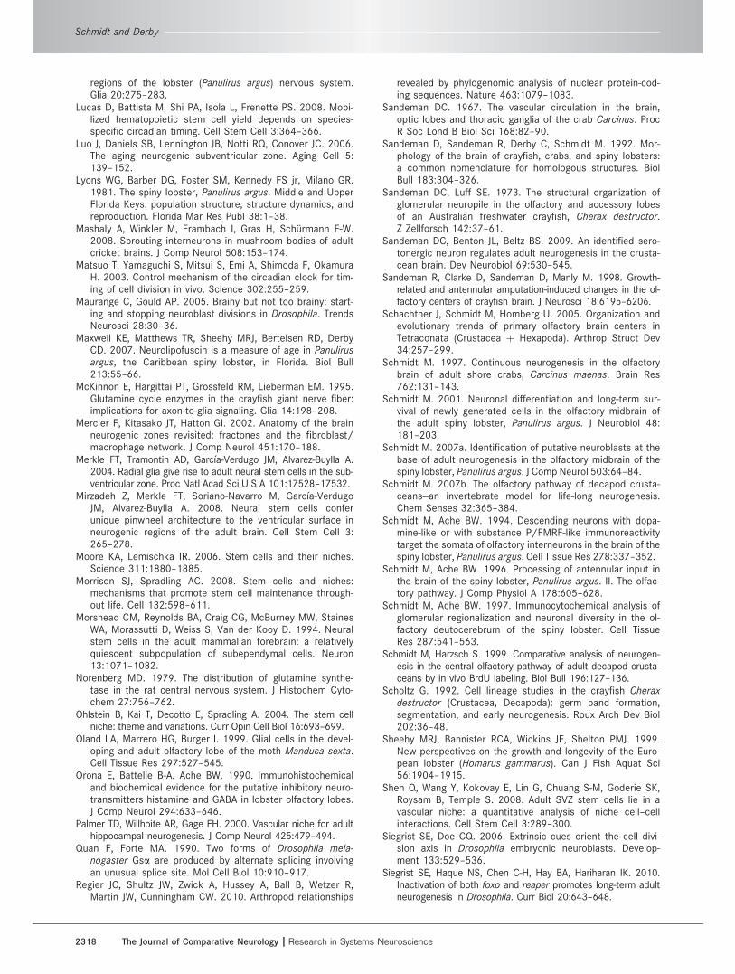
regions of the lobster (Panulirus argus) nervous system.Glia 20:275–283.
Lucas D, Battista M, Shi PA, Isola L, Frenette PS. 2008. Mobi-lized hematopoietic stem cell yield depends on species-specific circadian timing. Cell Stem Cell 3:364–366.
Luo J, Daniels SB, Lennington JB, Notti RQ, Conover JC. 2006.The aging neurogenic subventricular zone. Aging Cell 5:139–152.
Lyons WG, Barber DG, Foster SM, Kennedy FS jr, Milano GR.1981. The spiny lobster, Panulirus argus. Middle and UpperFlorida Keys: population structure, structure dynamics, andreproduction. Florida Mar Res Publ 38:1–38.
Mashaly A, Winkler M, Frambach I, Gras H, Schurmann F-W.2008. Sprouting interneurons in mushroom bodies of adultcricket brains. J Comp Neurol 508:153–174.
Matsuo T, Yamaguchi S, Mitsui S, Emi A, Shimoda F, OkamuraH. 2003. Control mechanism of the circadian clock for tim-ing of cell division in vivo. Science 302:255–259.
Maurange C, Gould AP. 2005. Brainy but not too brainy: start-ing and stopping neuroblast divisions in Drosophila. TrendsNeurosci 28:30–36.
Maxwell KE, Matthews TR, Sheehy MRJ, Bertelsen RD, DerbyCD. 2007. Neurolipofuscin is a measure of age in Panulirusargus, the Caribbean spiny lobster, in Florida. Biol Bull213:55–66.
McKinnon E, Hargittai PT, Grossfeld RM, Lieberman EM. 1995.Glutamine cycle enzymes in the crayfish giant nerve fiber:implications for axon-to-glia signaling. Glia 14:198–208.
Mercier F, Kitasako JT, Hatton GI. 2002. Anatomy of the brainneurogenic zones revisited: fractones and the fibroblast/macrophage network. J Comp Neurol 451:170–188.
Merkle FT, Tramontin AD, Garcıa-Verdugo JM, Alvarez-Buylla A.2004. Radial glia give rise to adult neural stem cells in the sub-ventricular zone. Proc Natl Acad Sci U S A 101:17528–17532.
Mirzadeh Z, Merkle FT, Soriano-Navarro M, Garcıa-VerdugoJM, Alvarez-Buylla A. 2008. Neural stem cells conferunique pinwheel architecture to the ventricular surface inneurogenic regions of the adult brain. Cell Stem Cell 3:265–278.
Moore KA, Lemischka IR. 2006. Stem cells and their niches.Science 311:1880–1885.
Morrison SJ, Spradling AC. 2008. Stem cells and niches:mechanisms that promote stem cell maintenance through-out life. Cell 132:598–611.
Morshead CM, Reynolds BA, Craig CG, McBurney MW, StainesWA, Morassutti D, Weiss S, Van der Kooy D. 1994. Neuralstem cells in the adult mammalian forebrain: a relativelyquiescent subpopulation of subependymal cells. Neuron13:1071–1082.
Norenberg MD. 1979. The distribution of glutamine synthe-tase in the rat central nervous system. J Histochem Cyto-chem 27:756–762.
Ohlstein B, Kai T, Decotto E, Spradling A. 2004. The stem cellniche: theme and variations. Curr Opin Cell Biol 16:693–699.
Oland LA, Marrero HG, Burger I. 1999. Glial cells in the devel-oping and adult olfactory lobe of the moth Manduca sexta.Cell Tissue Res 297:527–545.
Orona E, Battelle B-A, Ache BW. 1990. Immunohistochemicaland biochemical evidence for the putative inhibitory neuro-transmitters histamine and GABA in lobster olfactory lobes.J Comp Neurol 294:633–646.
Palmer TD, Willhoite AR, Gage FH. 2000. Vascular niche for adulthippocampal neurogenesis. J Comp Neurol 425:479–494.
Quan F, Forte MA. 1990. Two forms of Drosophila mela-nogaster Gsa are produced by alternate splicing involvingan unusual splice site. Mol Cell Biol 10:910–917.
Regier JC, Shultz JW, Zwick A, Hussey A, Ball B, Wetzer R,Martin JW, Cunningham CW. 2010. Arthropod relationships
revealed by phylogenomic analysis of nuclear protein-cod-ing sequences. Nature 463:1079–1083.
Sandeman DC. 1967. The vascular circulation in the brain,optic lobes and thoracic ganglia of the crab Carcinus. ProcR Soc Lond B Biol Sci 168:82–90.
Sandeman D, Sandeman R, Derby C, Schmidt M. 1992. Mor-phology of the brain of crayfish, crabs, and spiny lobsters:a common nomenclature for homologous structures. BiolBull 183:304–326.
Sandeman DC, Luff SE. 1973. The structural organization ofglomerular neuropile in the olfactory and accessory lobesof an Australian freshwater crayfish, Cherax destructor.Z Zellforsch 142:37–61.
Sandeman DC, Benton JL, Beltz BS. 2009. An identified sero-tonergic neuron regulates adult neurogenesis in the crusta-cean brain. Dev Neurobiol 69:530–545.
Sandeman R, Clarke D, Sandeman D, Manly M. 1998. Growth-related and antennular amputation-induced changes in the ol-factory centers of crayfish brain. J Neurosci 18:6195–6206.
Schachtner J, Schmidt M, Homberg U. 2005. Organization andevolutionary trends of primary olfactory brain centers inTetraconata (Crustacea þ Hexapoda). Arthrop Struct Dev34:257–299.
Schmidt M. 1997. Continuous neurogenesis in the olfactorybrain of adult shore crabs, Carcinus maenas. Brain Res762:131–143.
Schmidt M. 2001. Neuronal differentiation and long-term sur-vival of newly generated cells in the olfactory midbrain ofthe adult spiny lobster, Panulirus argus. J Neurobiol 48:181–203.
Schmidt M. 2007a. Identification of putative neuroblasts at thebase of adult neurogenesis in the olfactory midbrain of thespiny lobster, Panulirus argus. J Comp Neurol 503:64–84.
Schmidt M. 2007b. The olfactory pathway of decapod crusta-ceans—an invertebrate model for life-long neurogenesis.Chem Senses 32:365–384.
Schmidt M, Ache BW. 1994. Descending neurons with dopa-mine-like or with substance P/FMRF-like immunoreactivitytarget the somata of olfactory interneurons in the brain of thespiny lobster, Panulirus argus. Cell Tissue Res 278:337–352.
Schmidt M, Ache BW. 1996. Processing of antennular input inthe brain of the spiny lobster, Panulirus argus. II. The olfac-tory pathway. J Comp Physiol A 178:605–628.
Schmidt M, Ache BW. 1997. Immunocytochemical analysis ofglomerular regionalization and neuronal diversity in the ol-factory deutocerebrum of the spiny lobster. Cell TissueRes 287:541–563.
Schmidt M, Harzsch S. 1999. Comparative analysis of neurogen-esis in the central olfactory pathway of adult decapod crusta-ceans by in vivo BrdU labeling. Biol Bull 196:127–136.
Scholtz G. 1992. Cell lineage studies in the crayfish Cheraxdestructor (Crustacea, Decapoda): germ band formation,segmentation, and early neurogenesis. Roux Arch Dev Biol202:36–48.
Sheehy MRJ, Bannister RCA, Wickins JF, Shelton PMJ. 1999.New perspectives on the growth and longevity of the Euro-pean lobster (Homarus gammarus). Can J Fish Aquat Sci56:1904–1915.
Shen Q, Wang Y, Kokovay E, Lin G, Chuang S-M, Goderie SK,Roysam B, Temple S. 2008. Adult SVZ stem cells lie in avascular niche: a quantitative analysis of niche cell–cellinteractions. Cell Stem Cell 3:289–300.
Siegrist SE, Doe CQ. 2006. Extrinsic cues orient the cell divi-sion axis in Drosophila embryonic neuroblasts. Develop-ment 133:529–536.
Siegrist SE, Haque NS, Chen C-H, Hay BA, Hariharan IK. 2010.Inactivation of both foxo and reaper promotes long-term adultneurogenesis in Drosophila. Curr Biol 20:643–648.
Schmidt and Derby
2318 The Journal of Comparative Neurology |Research in Systems Neuroscience

Song C-K, Johnstone LM, Edwards DH, Derby CD, Schmidt M.2009. Cellular basis of neurogenesis in the brain of cray-fish, Procambarus clarkii: neurogenic complex in the olfac-tory midbrain from hatchlings to adults. Arthrop Struct Dev38:339–360.
Spradling A, Drummond-Barbosa D, Kai T. 2001. Stem cellsfind their niche. Nature 414:98–104.
Strausfeld NJ, Sinakevitch I, Brown SM, Farris SM. 2009.Ground plan of the insect mushroom body: functional andevolutionary implications. J Comp Neurol 513:265–291.
Sullivan JM, Beltz BS. 2001. Neural pathways connecting thedeutocerebrum and lateral protocerebrum in the brains ofdecapod crustaceans. J Comp Neurol 441:9–22.
Sullivan JM, Beltz BS. 2005a. Adult neurogenesis in the centralolfactory pathway in the absence of receptor neuron turnoverin Libinia emarginata. Eur J Neurosci 22:2397–2405.
Sullivan JM, Beltz BS. 2005b. Newborn cells in the adult cray-fish brain differentiate into distinct neuronal types. J Neu-robiol 65:157–170.
Sullivan JM, Benton JL, Sandeman DC, Beltz BS. 2007a. Adultneurogenesis: a common strategy across diverse species.J Comp Neurol 500:574–584.
Sullivan JM, Sandeman DC, Benton JL, Beltz BS. 2007b. Adultneurogenesis and cell cycle regulation in the crustaceanolfactory pathway: from glial precursors to differentiatedneurons. J Mol Histol 38:527–542.
Tang X, Falls DL, Li X, Lane T, Luskin MB. 2007. Antigen-retrieval procedure for bromodeoxyuridine immunolabelingwith concurrent labeling of nuclear DNA and antigens dam-aged by HCl pretreatment. J Neurosci 27:5837–5844.
Tavazoie M, Van der Veken L, Silva-Vargas V, Louissaint M,Colonna L, Zaidi B, Garcıa-Verdugo JM, Doetsch F. 2008.A specialized vascular niche for adult neural stem cells.Cell Stem Cell 3:279–288.
Telford MJ, Bourlat SJ, Economou A, Papillon D, Rota-StabelliO. 2008. The evolution of the Ecdysozoa. Philos Trans RSoc Lond B Biol Sci 363:1529–1537.
Trapido-Rosenthal HG, Linser PJ, Greenberg RM, Gleeson RA,Carr WES. 1993. cDNA clones from the olfactory organ ofthe spiny lobster encode a protein related to eukaryoticglutamine synthetase. Gene 129:275–278.
Ungerer P, Scholtz G. 2008. Filling the gap between identifiedneuroblasts and neurons in crustaceans adds new supportfor Tetraconata. Proc R Soc Lond B Biol Sci 275:369–376.
Urbach R, Technau GM. 2003. Early steps in building theinsect brain: neuroblast formation and segmental pattern-ing in the developing brain of different insect species.Arthrop Struct Dev 32:103–123.
Wachowiak M, Ache BW. 1994. Morphology and physiology ofmultiglomerular olfactory projection neurons in the spinylobster. J Comp Physiol A 175:35–48.
Wachowiak M, Ache BW. 1997. Dual inhibitory pathwaysmediated by GABA- and histaminergic interneurons in thelobster olfactory lobe. J Comp Physiol A 180:357–372.
Wachowiak M, Diebel CE, Ache BW. 1996. Functional organi-zation of olfactory processing in the accessory lobe of thespiny lobster. J Comp Physiol A 178:211–226.
Wei Y, Mizzen CA, Cook RG, Gorovsky MA, Allis CD. 1998.Phosphorylation of histone H3 at serine 10 is correlatedwith chromosome condensation during mitosis and meio-sis in Tetrahymena. Proc Natl Acad Sci U S A 95:7480–7484.
Xu F, Hollins B, Gress AM, Landers TM, McClintock TS. 1997.Molecular cloning and characterization of a lobster Gasprotein expressed in neurons of olfactory organ and brain.J Neurochem 69:1793–1800.
Yasugi T, Umetsu D, Murakami S, Sato M, Tabata T. 2008.Drosophila optic lobe neuroblasts triggered by a wave ofproneural gene expression that is negatively regulated byJAK/STAT. Development 135:1471–1480.
Ye W, Mairet-Coello G, DiCicco-Bloom E. 2007. DNAse I pre-treatment markedly enhances detection of nuclear cyclin-dependent kinase inhibitor p57Kip2 and BrdU doubleimmunostaining in embryonic rat brain. Histochem CellBiol 127:195–203.
Zhang Y, Beltz BS. 2010. Glutamine sythetase, a functionallyactive enzyme in the crayfish brain. 2010 NeuroscienceMeeting Planner. San Diego, CA: Society for Neuroscience,Program No. 737.14.
Zhang Y, Allodi S, Sandeman DC, Beltz BS. 2009. Adult neuro-genesis in the crayfish brain: proliferation, migration, andpossible origin of precursor cells. Dev Neurobiol 69:415–436.
Zhao X, Coptis V, Farris SM. 2008. Metamorphosis and adultdevelopment of the mushroom bodies of the red flour bee-tle, Tribolium castaneum. Dev Neurobiol 68:1487–1502.
Zhong W, Chia W. 2008. Neurogenesis and asymmetric celldivision. Curr Opin Neurobiol 18:4–11.
Neural stem cell niches in adult Panulirus argus
The Journal of Comparative Neurology | Research in Systems Neuroscience 2319