
CURRICULUM - eScholarship.org
59
CURRICULUM 1 Novel Emergency Medicine Curriculum Utilizing Self- Directed Learning and the Flipped Classroom Method: Hematologic/Oncologic Emergencies Small Group Module Michael G Barrie, MD * , Chad L Mayer, MD * , Colin Kaide, MD * , Emily Kauffman, DO * , Jennifer Mitzman, MD * , Matthew Malone, MD * , Daniel Bachmann, MD * , Ashish Panchal, MD * , Howard Werman, MD * , Benjamin Ostro, MD * and Andrew King, MD * * The Ohio State University Wexner Medical Center, Department of Emergency Medicine, Columbus, OH Correspondence should be addressed to Andrew King, MD, FACEP at [email protected], Twitter: @akingermd Submitted: August 6, 2018; Accepted: November 21, 2018; Electronically Published: January 15, 2019; https://doi.org/10.21980/J8VW56 Copyright: © 2019 Barrie, et al. This is an open access article distributed in accordance with the terms of the Creative Commons Attribution (CC BY 4.0) License. See: http://creativecommons.org/licenses/by/4.0/ ABSTRACT: Audience: This curriculum, created and implemented at The Ohio State University Wexner Medical Center, was designed to educate our emergency medicine (EM) residents, intern to senior resident level, as well as medical students and attending physicians. Introduction: Disorders of the hematologic system represent a wide spectrum of disease, including bleeding disorders, coagulopathies, blood cell disorders, and oncology related disorders. Hematologic related problems often complicate other disease processes, including malignancy. The number of patients diagnosed with malignancy is increasing due to many factors, including, but not limited to, an aging population and the increased ability for early detection. 1 Patients with oncologic conditions can present in a variety of ways with a variety of complications, 2 and understanding the steps in diagnosis and management are important components of any emergency physician’s training. To address this specific curricular need, we developed a flipped classroom, case-based, small group discussion series for emergency medicine learners. The flipped classroom curricular model emphasizes self- directed learning activities, followed by facilitated small group discussions pertaining to the topic reviewed. The active learning fostered by this curriculum increases faculty and learner engagement and interaction time typically absent in traditional lecture-based formats. 3-5 The application of knowledge through case studies, personal interaction with content experts, and integrated questions are effective learning strategies for emergency medicine residents. 5-7 The Ohio State University Wexner Medical Center EM residency didactic curriculum transitioned to a “flipped classroom” approach. We created this curriculum to improve our
-
Upload
khangminh22 -
Category
Documents
-
view
3 -
download
0
Transcript of CURRICULUM - eScholarship.org

CURRICULUM
1
Novel Emergency Medicine Curriculum Utilizing Self-Directed Learning and the Flipped Classroom Method: Hematologic/Oncologic Emergencies Small Group Module
Michael G Barrie, MD*, Chad L Mayer, MD*, Colin Kaide, MD*, Emily Kauffman, DO*, Jennifer Mitzman, MD*, Matthew Malone, MD*, Daniel Bachmann, MD*, Ashish Panchal, MD*, Howard Werman, MD*, Benjamin Ostro, MD* and Andrew King, MD* *The Ohio State University Wexner Medical Center, Department of Emergency Medicine, Columbus, OH Correspondence should be addressed to Andrew King, MD, FACEP at [email protected], Twitter: @akingermd Submitted: August 6, 2018; Accepted: November 21, 2018; Electronically Published: January 15, 2019; https://doi.org/10.21980/J8VW56 Copyright: © 2019 Barrie, et al. This is an open access article distributed in accordance with the terms of the Creative Commons Attribution (CC BY 4.0) License. See: http://creativecommons.org/licenses/by/4.0/
ABSTRACT: Audience: This curriculum, created and implemented at The Ohio State University Wexner Medical Center, was designed to educate our emergency medicine (EM) residents, intern to senior resident level, as well as medical students and attending physicians. Introduction: Disorders of the hematologic system represent a wide spectrum of disease, including bleeding disorders, coagulopathies, blood cell disorders, and oncology related disorders. Hematologic related problems often complicate other disease processes, including malignancy. The number of patients diagnosed with malignancy is increasing due to many factors, including, but not limited to, an aging population and the increased ability for early detection.1 Patients with oncologic conditions can present in a variety of ways with a variety of complications,2 and understanding the steps in diagnosis and management are important components of any emergency physician’s training. To address this specific curricular need, we developed a flipped classroom, case-based, small group discussion series for emergency medicine learners. The flipped classroom curricular model emphasizes self-directed learning activities, followed by facilitated small group discussions pertaining to the topic reviewed. The active learning fostered by this curriculum increases faculty and learner engagement and interaction time typically absent in traditional lecture-based formats.3-5 The application of knowledge through case studies, personal interaction with content experts, and integrated questions are effective learning strategies for emergency medicine residents.5-7 The Ohio State University Wexner Medical Center EM residency didactic curriculum transitioned to a “flipped classroom” approach. We created this curriculum to improve our

CURRICULUM
2
residency education program and to share educational resources with other EM residency programs. Our curriculum utilizes an 18-month curricular cycle to cover emergency medicine core content based on the American Board of Emergency Medicine (ABEM) EM model curriculum.8 The flipped classroom curriculum maximizes didactic time and resident engagement, fosters intellectual curiosity and active learning, and meets the needs of today’s learners.4,7,9 Objectives: We aim to teach the presentation and management of psychiatric emergencies through the creation of a flipped classroom design. This unique, innovative curriculum utilizes resources chosen by education faculty and resident learners, study questions, real-life experiences, and small group discussions in place of traditional lectures. In doing so, a goal of the curriculum is to encourage self-directed learning, improve understanding and knowledge retention, and improve the educational experience of our residents. Methods: The educational strategies used in this curriculum include small group modules authored by education faculty and content experts based on the core emergency medicine content as outlined in the ABEM model curriculum. Active participation is encouraged by the question and answer format of the Socratic Method, with an emphasis on an open and non-threatening learning environment, instead of negative “pimping” type questions which often humiliate and maintain hierarchy rather than promote learning.10,11 Small groups also focus on the synthesis and application of knowledge through the discussion of real-life experiences. The use of free open access medical education (FOAM) resources along with selected primary literature allows learners to work at their own pace and maximize autonomy. Topics: Emergency medicine, flipped classroom, medical education, hematologic emergencies, oncologic emergencies, pedagogy, teaching.

USER GUIDE
Barrie M, et al. Novel Emergency Medicine Curriculum Utilizing Self-Directed Learning and the Flipped Classroom Method: Hematologic/Oncologic Emergencies Small Group Module. JETem 2019. 4(1):C1-59. https://doi.org/10.21980/J8VW56
3
Brief introduction: Disorders of the hematologic system represent a wide spectrum of disease, including bleeding disorders, coagulopathies, blood cell disorders, and oncology related disorders. Hematologic related problems often complicate other disease processes. The incidence of new cancer diagnoses is increasing due to many factors, including an aging population and increased ability for early detection.1 An estimated 1.6 million Americans were diagnosed with a new malignancy in 2017, and for many this diagnosis was brought to light by an emergent complication.2 Nevertheless, patients with oncologic conditions can present in a variety of ways with a variety of complications, and understanding the steps in diagnosis and management are important components of any emergency physician’s training. Regarding hematologic emergencies, one study showed there were roughly 4,488 hemophilia-related ED visits annually, with 70% of patients treated and discharged.12 There were an estimated 2.8 million quarterly visits to outpatient offices for anticoagulation, including warfarin and novel oral anticoagulants (NOACs).13 Bleeding is the most common complaint of patients on anticoagulant therapy, happening 1.5%-5.2% annually with warfarin, and NOACs are responsible for 2-3% of major bleeding events.14 These examples describe some of the varied patient presentations emergency physicians will encounter. To address this specific curricular need, we developed a flipped classroom, case-based, small group discussion series for emergency medicine learners. The flipped classroom learning approach is becoming more commonly recognized as a preferred curricular model for mature learners, specifically those in medical education.15,16 This particular model is a natural fit for the hands-on, experiential emergency medicine learner.5 The active learning fostered by this curriculum increases faculty and learner engagement and interaction time, which is typically absent in traditional lecture-based formats.6,17 Education literature shows through surveys and exam scores that resident learners prefer learning activities that involve small group discussion, are case/skill based, and emphasize the application of newly obtained knowledge, and that there is less decline in performance.4,5 This educational model also provides a clear channel for the incorporation of evidence-based medicine and increases opportunities for educator-learner conversations. A successful flipped classroom curriculum fosters learner accountability and provides robust opportunities for formal
List of Resources: Abstract 1 User Guide 3 Didactics and Hands-On Curriculum Chart 9 Appendix A: Coagulation and Hemostatic Disorders
16
Appendix B: White Blood Cell and Platelet Disorders in Adults
23
Appendix C: Transfusion Complications 28 Appendix D: Complications of Sickle Cell Disorders
35
Appendix E: Red Blood Cell Abnormalities 39 Appendix F: Deep Vein Thrombosis 46 Appendix G: Oncologic Emergencies 50
Learner Audience: Medical Students, Interns, Junior Residents, Senior Residents, Attending Physicians and Faculty Members Length of Curriculum: The entire didactic curriculum was developed to utilize an 18-month curricular cycle; therefore, resident learners experience each curricular topic twice in the course of a three-year residency training. The hematologic emergencies block is a part of this overall 18-month curriculum, and consists of seven 45 to 60-minute small group sessions. Topics: Emergency medicine, flipped classroom, medical education, hematologic emergencies, oncologic emergencies, pedagogy, teaching. Objectives: Each chapter within our curriculum has individual objectives as outlined in the appendices; however, educational objectives for the overall curriculum include:
1. Resident learners will learn the core content of emergency medicine in an 18-month curriculum utilizing self-directed learning and small group discussions based on the flipped classroom model.
2. After completing the Hematologic Emergencies Module, resident learners will exhibit mastery within this content area and will critically discuss the pathophysiology, diagnosis, and treatment of various pediatric and adult hematologic emergencies including:
a. Coagulopathies and Anticoagulation Emergencies
b. WBC and Platelet Disorders
c. Transfusion Complications d. Sickle Cell Disease Complications e. Red Blood Cell Abnormalities f. Deep Vein Thromboses g. Oncologic Emergencies
USER GUIDE
Barrie M, et al. Novel Emergency Medicine Curriculum Utilizing Self-Directed Learning and the Flipped Classroom Method: Hematologic/Oncologic Emergencies Small Group Module. JETem 2019. 4(1):C1-59. https://doi.org/10.21980/J8VW56
4
assessment in various emergency medicine milestones.5,18 For these reasons, we developed a flipped classroom curriculum at The Ohio State University Wexner Medical Center. The hematologic emergencies curriculum is one of several topics in our overall didactic curriculum. Problem identification, general and targeted needs assessment: Traditional lecture-based didactics may not be the most effective or preferred method for emergency medicine resident education.7 Previously, we used a traditional lecture format in our residency curriculum despite overwhelming evidence favoring a more hands-on, “flipped classroom” approach.18,19 From the perspective of resident learners, the chance to remain fully engaged through the asking of questions developed from personal experiences, in addition to learning from the experiences of others, helps with knowledge retention.6 Both educators and learners benefit from an interactive and collaborative classroom, leading to the creation and implementation of this proposed curricular model at our emergency medicine residency program. This weekly small group curriculum replaces three hours of traditional lecture-based didactics. Learners divide into small groups, and each group is led by both a faculty leader and a designated senior resident who has spent extra time preparing; the senior resident is expected to guide the discussion with a question and answer format while the faculty member is there to add expertise and guidance. Through the curriculum, we continually seek to foster self-directed learning and increased collaboration between resident learners and education faculty members. This ensures that resident time will be maximized and learning will be more efficient and effective, providing a potential positive impact on patient care and physician wellness. Currently, only minimal flipped classroom curricular materials dedicated to the core content of emergency medicine exist. Goals of the curriculum: We aim to teach the presentation and management of hematologic emergencies through the creation of a flipped classroom design. This unique, innovative curriculum utilizes resources chosen by education faculty and resident learners, study questions, real-life experiences, and small group discussions in place of traditional lectures. In doing so, objectives of the curriculum are to encourage self-directed learning, improve understanding and knowledge retention, and improve the educational experience of our residents. Objectives of the curriculum: Each chapter within our curriculum has individual objectives as outlined in the appendices; however, educational objectives for the overall curriculum include:
1. Resident learners will learn the core content of emergency medicine in an 18-month curriculum utilizing self-directed learning and small group discussions based on the flipped classroom model.
2. After completing the Hematologic Emergencies Module, resident learners will exhibit mastery within this content area and will critically discuss the pathophysiology, diagnosis, and treatment of various pediatric and adult hematologic emergencies including:
a. Coagulopathies and Anticoagulation Emergencies 1. Describe the basic science of hemostasis. 2. Discuss how common anticoagulants work. 3. List strategies for reversal of common
anticoagulants. 4. Discuss the clinical presentation and
management of von Willebrand’s disease. 5. Discuss the clinical presentation and
management of the hemophilia A and B. b. WBC and Platelet Disorders
1. Differentiate leukemoid reaction and hyperleukocytosis.
2. Identify the presenting symptoms, treatment, and complications of treating leukostasis.
3. Define neutropenia and discuss the workup and treatment of neutropenic fever in the emergency department.
4. Discuss thrombocytosis and thrombocytopenia and identify their most common etiologies.
5. Identify the differences in diagnosis and treatment of thrombotic thrombocytopenic purpura (TTP), hemolytic uremic syndrome (HUS), heparin-induced thrombocytopenia (HIT) and idiopathic thrombocytopenia (ITP).
6. Discuss the indications for platelet transfusion in isolated thrombocytopenia.
c. Transfusion Complications 1. Review the different blood components
available for transfusion with inclusion of their typical dosing and expected effect.
2. Discuss consent procedure. 3. Review the early and late complications of
blood product transfusions. 4. Discuss the unique complications of
massive transfusions. 5. Outline the treatment approach, including
preventative strategies, for transfusion reactions.

USER GUIDE
Barrie M, et al. Novel Emergency Medicine Curriculum Utilizing Self-Directed Learning and the Flipped Classroom Method: Hematologic/Oncologic Emergencies Small Group Module. JETem 2019. 4(1):C1-59. https://doi.org/10.21980/J8VW56
5
d. Sickle Cell Disease Complications 1. Describe the pathophysiology of sickle cell
disease and its variants. 2. List and compare the typical
manifestations of sickle cell disease in childhood and adulthood.
3. List diagnostic tools in the emergency department assessment of sickle cell disease.
4. Describe the management of patients with sickle cell disease and review controversial aspects of management.
e. Red Blood Cell Abnormalities 1. Discuss the presentation, diagnosis and
management of methemoglobinemia. 2. List the causes of acquired
methemoglobinemia. 3. Review the differential diagnosis in a
patient with anemia. 4. Discuss the laboratory tests that can
differentiate different etiologies of anemia.
5. Discuss the current evidence-based guidelines for transfusion for anemia in adults.
6. Compare laboratory results and treatment in patients with anemia due to acute blood loss versus those with autoimmune hemolytic anemia.
f. Deep Vein Thromboses 1. Explain the pathophysiology and risk
factors for development of deep vein thrombosis (DVT).
2. Review the clinical presentation of DVT. 3. List approaches to diagnose DVT. 4. Discuss treatment for DVTs. 5. Review the causes and treatment of upper
extremity DVTs. g. Oncologic Emergencies
1. Discuss common oncologic emergencies. 2. Describe the presentation of the following
conditions: typhlitis, blast crisis, superior vena cava syndrome (SVC) and tumor lysis syndrome.
3. Identify a treatment strategy for typhlitis, blast crisis, SVC syndrome and tumor lysis syndrome.
Educational Strategies: (See curriculum chart) Please refer to the curriculum chart of linked objectives and educational strategies.
Evaluation and Feedback: This curriculum was literature-based and specifically designed to maximize active learning using the flipped classroom learning model. We overcame initial challenges and skepticism from both educators and learners to execute a successful, novel curricular model. Both resident learners and faculty educators have provided an overwhelming amount of positive feedback. Additionally, a survey was administered to each resident prior to initiation of the curricular innovation, and repeated at the conclusion of the first 18-month cycle. Results of this survey showed no decrease in in-service exam scores but significantly higher learner satisfaction. Learners and educators were enthusiastic about the conference structure and expressed a preference for it rather than the previous, lecture-based didactics. More recently during the second 18-month cycle of the flipped classroom curriculum, students were surveyed on their perceived quality of instruction of the various program components. From academic year 2016, 19 out of 46 residents responded to the survey. A majority of residents who responded (60.9%) reported that the small group discussions were good or excellent, compared to only 26% of residents that responded who felt that our grand rounds sessions during the same time were good or excellent. This curriculum has been delivered to two cohorts of learners, having delivered the content twice in three years with about 50 residents per cycle. Our department has 15 core faculty who participate in delivery of the content. On the most recent iteration, residents evaluated the teaching methods for the hematology/oncology block as effective, with an average rating of more than 4.6 out of 5 (4 being agree, 5 being strongly agree). The curriculum is critically evaluated and updated by education faculty members in order to ensure educational material remains current and consistent with the emergency medicine core content. References/suggestions for further reading: 1. American Cancer Society. Cancer facts and figures 2014.
https://www.cancer.org/research/cancer-facts-statistics/all-cancer-facts-figures/cancer-facts-figures-2014.html. Atlanta, GA: American Cancer Society; 2014.
2. Halfdanarson TR, Hogan WJ, Madsen BE. Emergencies in hematology and oncology. Mayo Clin Proc. 2017;92(4):609-641. doi: 10.1016/j.mayocp.2017.02.008
3. Galway LP, Corbett KK, Takaro TK, Tairyan K, Frank E. A novel integration of online and flipped classroom instructional models in public health higher education. BMC Med Educ. 2014;14:181. doi: 10.1186/1472-6920-14-181
4. Mortensen CJ, Nicholson AM. The flipped classroom stimulates greater learning and is a modern 21st century approach to teaching today's undergraduates. J Anim Sci. 2015;93(7):3722-3731. doi: 10.2527/jas.2015-9087
5. Tan E, Brainard A, Larkin GL. Acceptability of the flipped classroom approach for in-house teaching in emergency

USER GUIDE
Barrie M, et al. Novel Emergency Medicine Curriculum Utilizing Self-Directed Learning and the Flipped Classroom Method: Hematologic/Oncologic Emergencies Small Group Module. JETem 2019. 4(1):C1-59. https://doi.org/10.21980/J8VW56
6
medicine. Emerg Med Australas. 2015;27(5):453-459. doi: 10.1111/1742-6723.12454
6. Rose E, Claudius I, Tabatabai R, Kearl L, Behar S, Jhun P. The flipped classroom in emergency medicine using online videos with interpolated questions. J Emerg Med. 2016;51(3):284-291.e1. doi: 10.1016/j.jemermed.2016.05.033
7. Young TP, Bailey CJ, Guptill M, Thorp AW, Thomas TL. The flipped classroom: a modality for mixed asynchronous and synchronous learning in a residency program. West J Emerg Med. 2014;15(7):938-944. doi: 10.5811/westjem.2014.10.23515
8. Counselman FL, Babu K, Edens MA, et al. The 2016 model of the clinical practice of emergency medicine. J Emerg Med. 2017;52(6):846-849. doi: 10.1016/j.jemermed.2017.01.040
9. Moffett J. Twelve tips for "flipping" the classroom. Med Teach. 2015;37(4):331-336. doi: 10.3109/0142159X.2014.943710
10. Heitz C, Prusakowski M, Willis G, Franck C. Does the concept of the "flipped classroom" extend to the emergency medicine clinical clerkship? West J Emerg Med. 2015;16(6):851-5. doi: 10.5811/westjem.2015.9.27256
11. Kost A, Chen FM. Socrates was not a pimp: changing the paradigm of questioning in medical education. Acad Med. 2015;90(1):20-24. doi: 10.1097/ACM.0000000000000446
12. Zakieh A, Siddiqui AH. Emergency department utilization by haemophilia patients in United States. Haemophilia. 2017;23(3):e188-e193. doi: 10.1111/hae.13187
13. Barnes GD, Lucas E, Alexander GC, Goldberger ZD. National trends in ambulatory oral anticoagulant use. Am J Med. 2015;128(12):1300-1305.e2. doi: 10.1016/j.amjmed.2015.05.044
14. Dhakal P, Rayamajhi S, Verma V, Gundabolu K, Bhatt VR. Reversal of anticoagulation and management of bleeding in patients on anticoagulants. Clin Appl Thromb Hemost. 2017;23(5):410-415. doi: 10.1177/1076029616675970
15. Lee YH, Kim KJ. Enhancement of student perceptions of learner-centeredness and community of inquiry in flipped classrooms. BMC Med Educ. 2018;18:242. doi: 10.1186/s12909-018-1347-3
16. Chen KS, Monrouxe L, Lu YH, et al. Academic outcomes of flipped classroom learning: a meta-analysis. Med Educ. 2018. doi: 10.1111/medu.13616
17. Liebert CA, Lin DT, Mazer LM, Bereknyei S, Lau JN. Effectiveness of the surgery core clerkship flipped classroom: a prospective cohort trial. Am J Surg. 2016;211(2):451-457.e1. doi: 10.1016/j.amjsurg.2015.10.004
18. Sockalingam S, James SL, Sinyi R, et al. A flipped classroom approach to improving the quality of delirium care using an Interprofessional train-the-trainer program. J Contin Educ
Health Prof. 2016;36(1):17-23. doi: 10.1097/CEH.0000000000000025
19. Liebert CA, Lin DT, Mazer LM, Bereknyei S, Lau JN. Student perceptions of a simulation-based flipped classroom for the surgery clerkship: a mixed-methods study. Surgery. 2016;160(3):591-598. doi: 10.1016/j.amjsurg.2015.10.004
Additional Resources: Educational resources are available within each individual appendix of this hematologic emergencies curricular module; however, a complete list of resources and educational materials are listed below.
1. Fijnvandraat K, Cnossen MH, Leebeek FW, Peters M. Diagnosis and management of haemophilia. BMJ. 2012;344: e2707. doi: 10.1136/bmj.e2707
2. Swami A, Kaur V. Von Willebrand disease: a concise review and update for the practicing physician. Clin Appl Thromb Hemost. 2017;23(8):900-10. doi: 10.1177/1076029616675969
3. Demeyere R, Gillardin S, Arnout J, Strengers PF. Comparison of fresh frozen plasma and prothrombin complex concentrate for the reversal of oral anticoagulants in patients undergoing cardiopulmonary bypass surgery: a randomized study. Vox Sang 2010;99: 251-260. doi: 10.1111/j.1423-0410.2010.01339.x
4. Lip GY, Banerjee A, Boriani G, et al. Antithrombotic therapy for atrial fibrillation. CHEST guideline and expert panel report. CHEST. 2018;154(5):1121-1201. doi: 10.1016/j.chest.2018.07.040
5. National Hemophilia Foundation, Medical and Scientific Advisory Council. MASAC recommendations concerning products licensed for the treatment of hemophilia and other bleeding disorders. http://www.hemophilia.org/NHFWeb/MainPgs/MainNHF.aspx?menuid=57&contentid=693. Updated April 23, 2018. Accessed December 18, 2018.
6. Thompson LR, Sidley C, Kaide CG. Anticoagulation in the trauma patient. RELIAS. https://www.ahcmedia.com/articles/139803-anticoagulation-in-the-trauma-patient. Published January 1, 2017. Accessed November 19, 2018.
7. Fiutko A, Chhabra, N, Koyfman A, Long B. Managing hemophilia in the ED: all bleeding stops eventually. EmDocs Blog. http://www.emdocs.net/managing-hemophilia-in-the-ed-all-bleeding-stops-eventually/. Published June 6, 2018. Accessed November 19, 2018.
8. George JN. Clinical practice. Thrombotic thrombocytopenic purpura. N Engl J Med. 2006;354(18):1927-35. doi: 10.1056/NEJMcp053024
9. Schwartz J, Padmanabhan A, Aqui N, et al. Guidelines on the use of therapeutic apheresis in clinical practice.

USER GUIDE
Barrie M, et al. Novel Emergency Medicine Curriculum Utilizing Self-Directed Learning and the Flipped Classroom Method: Hematologic/Oncologic Emergencies Small Group Module. JETem 2019. 4(1):C1-59. https://doi.org/10.21980/J8VW56
7
Evidence-based approach from the writing committee of the American Society for Apheresis: the seventh special issue. J Clin Apher. 2016;31(3):149-162. doi: 10.1002/jca.21470
10. Taplitz RA, Kennedy EB, Bow EJ, et al. Outpatient management of fever and neutropenia in adults treated for malignancy: American Society of Clinical Oncology and Infectious Diseases Society of America clinical practice guideline update. J Clin Oncol. 2018;36(14):1443-1453. doi: 10.1200/JCO.2017.77.6211
11. Schiffer, CA. Hyperleukocytosis and leukostasis in hematologic malignancies. In: Larson RA, Rosmarin AG, eds. UpToDate. Waltham, MA: UptoDate, Inc. https://www.uptodate.com/contents/hyperleukocytosis-and-leukostasis-in-hematologic-malignancies. Updated November 27, 2017. Accessed November 19, 2018.
12. Kelton JG. Heparin-induced thrombocytopenia: an overview. Blood Rev. 2002;16(1):77-80. doi: 10.1054/blre.2001.0189.
13. Röllig C, Ehninger G. How I treat hyperleukocytosis in acute myeloid leukemia. Blood. 2015;125(21):3256-52. doi: 10.1182/blood-2014-10-551507
14. Schafer AI. Thrombocytosis. N Engl J Med. 2004;350(12):1211-9. doi: 10.1056/NEJMra035363
15. Wroblewski R, Repnshek Z. Severe transfusion reaction and their ED-focused management. emDocs. http://www.emdocs.net/severe-transfusion-reactions-ed-focused-management/. Published March 6, 2017. Accessed November 19, 2018.
16. Osterman JL, Arora S. Blood product transfusions and reactions. Hematol Oncol Clin North Am. 2014;31(6):1159-1170. doi: 10.1016/j.hoc.2017.08.014
17. American Red Cross. Risks and complications. https://www.redcrossblood.org/donate-blood/blood-donation-process/what-happens-to-donated-blood/blood-transfusions/risks-complications.html. Accessed December 22, 2018.
18. Sihler KC, Napolitano LM. Complications of massive transfusion. Chest. 2010;137(1):209-220. doi: 10.1378/chest.09-0252
19. Maxwell MJ, Wilson MJ. Complications of blood transfusion. Continuing Education in Anaesthesia, Critical Care and Pain. 2006;6(6):225-229. doi: 10.1093/bjaceaccp/mkl053
20. Bansil NH, Kim TY, Tieu L, Barcega B. Incidence of serious bacterial infections in febrile children with sickle cell disease. Clin Pediatrics (Phila). 2013;52(7):661-6. doi: 10.1177/0009922813488645
21. Glassberg J. Evidence-based management of sickle cell disease in the emergency department. Emerg Med Pract. 2011;13(8):1-20.
22. Janz TG, Hamilton GC. Anemia, polycythemia, and white blood cell disorders. In: Walls RM, Hockberger RS, Gausche-Hill M, et al. eds. Rosen’s Emergency Medicine: Concepts and Clinical Practice. 8th ed. Philadelphia, PA: Elsevier; 2014:1586-1605.
23. Novelli EM, Gladwin MT: Crises in sickle cell disease. Chest. 2016;149(4):1082-1093. doi: 10.1016/j.chest.2015.12.016
24. Lovett PB, Sule HP, Lopez BL. Sickle cell disease in the Emergency Department. Emerg Med Clin North Am. 2014;32(3):629-647. doi: 10.1016/j.emc.2014.04.011.
25. Glassberg J, Weingart S. Episode 132, Sickle cell disease update (cont. from August 2012). EMRAP. https://www.emrap.org/episode/september2012/sicklecell. Published September 2012. Accessed November 19, 2018.
26. Glassberg J, Weingart S. Episode 131, Sickle cell disease update. EMRAP. https://www.emrap.org/episode/august2012/sicklecell. Published August 2012. Accessed November 19, 2018.
27. Zetola VF. Role of TCD in sickle cell disease: A Review. Perspectives in Medicine. 2012; 265-268. doi: 10.1016/j.permed.2012.03.016
28. Farmer BM, Nelson LS. Dyshemoglobinemias. In: Tintinalli JE, Stapczynski J, Ma O, Cline DM, Cydulka RK, Meckler GD, et al. eds. Tintinalli's Emergency Medicine: A Comprehensive Study Guide. 7th ed. New York, NY: McGraw-Hill; 2011.
29. Carson JL, Carless PA, Hebert PC. Transfusion thresholds and other strategies for guiding allogeneic red blood cell transfusion. Cochrane Database Syst Rev. 2016;10:CD002042. doi: 10.1002/14651858.CD002042.pub4.
30. Nickson C. Anaemia. Life in the Fast Lane. http://lifeinthefastlane.com/ccc/anaemia/. Updated August 11, 2014. Accessed November 19, 2018.
31. Ash-Bernal R, Wise R, Wright SM. Acquired methemoglobinemia: a retrospective series of 138 cases at 2 teaching hospitals. Medicine (Baltimore). 2004;83(5):265-73. doi: 10.1097/01.md.0000141096.00377.3f
32. Falkenhahn M, Kannan S, O’Kane M. Unexplained acute severe methaemoglobinaemia in a young adult. Br J Anaesth. 2001;86(2):278-80. doi: https://doi.org/10.1093/bja/86.2.278
33. Coleman MD, Coleman NA. Drug-induced methaemoglobinaemia. Treatment issues. Drug Saf. 1996;14(6):394-405. doi: 10.2165/00002018-

USER GUIDE
Barrie M, et al. Novel Emergency Medicine Curriculum Utilizing Self-Directed Learning and the Flipped Classroom Method: Hematologic/Oncologic Emergencies Small Group Module. JETem 2019. 4(1):C1-59. https://doi.org/10.21980/J8VW56
8
199614060-00005. 34. Marik PE, Corwin HL. Efficacy of red blood cell
transfusion in the critically ill: a systematic review of the literature. Crit Care Med. 2008:36(9):2667-74. doi: 10.1097/CCM.0b013e3181844677
35. Villanueva C, Colomo A, Bosch A, et al. Transfusion strategies for acute upper gastrointestinal bleeding. N Engl J Med. 2013;368(1):11-21. doi: 10.1056/NEJMoa1211801
36. American College of Emergency Physicians (ACEP) Clinical Policies Committee. ACEP Clinical Policies Subcommittee on suspected lower-extremity deep venous thrombosis. Clinical policy: critical issues in the evaluation and management of adult patients presenting with suspected lower-extremity deep venous thrombosis. Ann Emerg Med. 2003;42(1):124-35. doi: 10.1067/mem.2003.181
37. Aguilar C, Sartori M, D’Angelo A, et al. Validation of the STA-Liatest DDi assay for exclusion of proximal deep vein thrombosis according to the latest Clinical and Laboratory Standards Institute/Food and Drug Administration guideline: results of a multicenter management study. Blood Coagul Fibrinolysis. 2018;29(6):562–566. doi: 10.1097/MBC.0000000000000750
38. Wells PS, Forgie MA, Rodger MA. Treatment of venous thromboembolism. JAMA. 2014;311(7):717-728. doi: 10.1001/jama.2014.65
39. Geerts W. Central venous catheter-related thrombosis. Hematology Am Soc Hematol Educ Program. 2014;2014(1):306-11. doi: 10.1182/asheducation-2014.1.306
40. Khan UA, Shanholtz CB, McCurdy MT. Oncologic mechanical emergencies. Hematol Oncol Clin North Am. 2017;31(6):927-940. doi: 10.1016/j.hoc.2017.08.001
41. Muslimani A, Chisti MM, Wills S, et al. How we treat tumor lysis syndrome. Oncology (Williston Park). 2011;25(4):369-375.
42. Rodrigues FG, Dasilva G, Wexner SD. Neutropenic enterocolitis. World J Gastroentero. 2017;23(1):42-47. doi: 10.3748/wjg.v23.i1.42
43. Röllig C, Ehninger G. How I treat hyperleukocytosis in acute myeloid leukemia. Blood. 2015;125(21):3246-3252. doi: 10.1182/blood-2014-10-551507

DIDACTICS AND HANDS-ON CURRICULUM
Barrie M, et al. Novel Emergency Medicine Curriculum Utilizing Self-Directed Learning and the Flipped Classroom Method: Hematologic/Oncologic Emergencies Small Group Module. JETem 2019. 4(1):C1-59. https://doi.org/10.21980/J8VW56
9
Topic Recommended
Educational Strategy Educational Content Objectives Learners Timing, resources
Needed (Space, Instructors, Equipment, citations of JETem pubs or other literature)
Recommended Assessment, Milestones Addressed
Hemostatic disorders, anticoagulation reversal, coagulopathies
“Flipped” classroom discussion of pre-reading material, case discussions, and discussion questions Encourage participants to share clinical experiences to enhance discussion 45 minutes for case and content discussion
Pathophysiology, diagnosis, and management of Hemostatic disorders
1. Describe the basic science of hemostasis.
2. Discuss how common anticoagulants work.
3. List strategies for reversal of common anticoagulants.
4. Discuss the clinical presentation and management of von Willebrand’s disease.
5. Discuss the clinical presentation and management of the hemophilia A and B.
PGY-1 PGY-2 PGY-3 Medical Students Faculty
Equipment: projector and screen preferable (instructor can pull up web images during session). Tables and space promoting small group discussion. Instructors: two faculty members or content experts. Predetermined senior resident discussion leader (optional). Timing: small group discussions involve no more than 15 learners and last about 45 minutes.
Milestone: Emergency stabilization (PC1), diagnostic studies (PC3), differential diagnosis (PC4), pharmacology (PC5), medical knowledge (MK) Assessment: Faculty evaluation of resident participation during small group activities. Evaluation: Resident evaluation of small group session content and facilitators. Yearly program evaluation of overall small group component.
DIDACTICS AND HANDS-ON CURRICULUM
Barrie M, et al. Novel Emergency Medicine Curriculum Utilizing Self-Directed Learning and the Flipped Classroom Method: Hematologic/Oncologic Emergencies Small Group Module. JETem 2019. 4(1):C1-59. https://doi.org/10.21980/J8VW56
10
White Blood Cell and platelet disorders
“Flipped” classroom discussion of pre-reading material, case discussions, and discussion questions Encourage participants to share clinical experiences to enhance discussion 45 minutes for case and content discussion
Pathophysiology, diagnosis, and management of white blood cell and platelet disorders
1. Differentiate leukemoid reaction and hyperleukocytosis
2. Identify the presenting symptoms, treatment and complications of treating leukostasis.
3. Define neutropenia and discuss the workup and treatment of neutropenic fever in the emergency department.
4. Discuss thrombocytosis and thrombocytopenia and identify their most common etiologies.
5. Identify the differences in diagnosis and treatment of thrombotic thrombocytopenic purpura (TTP), hemolytic uremic syndrome (HUS), heparin-induced thrombocytopenia (HIT) and idiopathic thrombocytopenia (ITP).
6. Discuss the indications for platelet transfusion in isolated
PGY-1 PGY-2 PGY-3 Medical Students Faculty
Equipment: projector and screen preferable (instructor can pull up web images during session). Tables and space promoting small group discussion. Instructors: two faculty members or content experts. Predetermined senior resident discussion leader (optional). Timing: small group discussions involve no more than 15 learners and last about 45 minutes.
Milestone: Emergency stabilization (PC1), diagnostic studies (PC3), differential diagnosis (PC4), pharmacology (PC5), medical knowledge (MK) Assessment: Faculty evaluation of resident participation during small group activities. Evaluation: Resident evaluation of small group session content and facilitators. Yearly program evaluation of overall small group component.

DIDACTICS AND HANDS-ON CURRICULUM
Barrie M, et al. Novel Emergency Medicine Curriculum Utilizing Self-Directed Learning and the Flipped Classroom Method: Hematologic/Oncologic Emergencies Small Group Module. JETem 2019. 4(1):C1-59. https://doi.org/10.21980/J8VW56
11
Topic Recommended Educational Strategy
Educational Content Objectives Learners Timing, resources Needed (Space, Instructors, Equipment, citations of JETem pubs or other literature)
Recommended Assessment, Milestones Addressed
thrombocytopenia.
Transfusion complications
“Flipped” classroom discussion of pre-reading material, case discussions, and discussion questions Encourage participants to share clinical experiences to enhance discussion 45 minutes for case and content discussion
Pathophysiology, diagnosis, and management of transfusion complications disorders
1. Review the different blood components available for transfusion with inclusion of their typical dosing and expected effect.
2. Discuss consent procedure.
3. Review the early and late complications of blood product transfusions.
4. Discuss the unique complications of massive transfusions.
5. Outline the treatment approach, including preventative strategies, for transfusion reactions.
PGY-1 PGY-2 PGY-3 Medical Students Faculty
Equipment: projector and screen preferable (instructor can pull up web images during session). Tables and space promoting small group discussion. Instructors: two faculty members or content experts. Predetermined senior resident discussion leader (optional). Timing: small group discussions involve no more than 15 learners and last about 45 minutes.
Milestone: Emergency stabilization (PC1), diagnostic studies (PC3), differential diagnosis (PC4), pharmacology (PC5), medical knowledge (MK) Assessment: Faculty evaluation of resident participation during small group activities. Evaluation: Resident evaluation of small group session content and facilitators. Yearly program evaluation of overall small group component.

DIDACTICS AND HANDS-ON CURRICULUM
Barrie M, et al. Novel Emergency Medicine Curriculum Utilizing Self-Directed Learning and the Flipped Classroom Method: Hematologic/Oncologic Emergencies Small Group Module. JETem 2019. 4(1):C1-59. https://doi.org/10.21980/J8VW56
12
Topic Recommended Educational Strategy
Educational Content Objectives Learners Timing, resources Needed (Space, Instructors, Equipment, citations of JETem pubs or other literature)
Recommended Assessment, Milestones Addressed
Complications of sickle cell disease
“Flipped” classroom discussion of pre-reading material, case discussions, and discussion questions Encourage participants to share clinical experiences to enhance discussion 45 minutes for case and content discussion
Pathophysiology, diagnosis, and management of complications of sickle cell disease
1. Describe the pathophysiology of sickle cell disease and its variants.
2. List and compare the typical manifestations of sickle cell disease in childhood and adulthood.
3. List diagnostic tools in the emergency department assessment of sickle cell disease.
4. Describe the management of patients with sickle cell disease and review controversial aspects of management.
PGY-1 PGY-2 PGY-3 Medical Students Faculty
Equipment: projector and screen preferable (instructor can pull up web images during session). Tables and space promoting small group discussion. Instructors: two faculty members or content experts. Predetermined senior resident discussion leader (optional). Timing: small group discussions involve no more than 15 learners and last about 45 minutes.
Milestone: Emergency stabilization (PC1), diagnostic studies (PC3), differential diagnosis (PC4), pharmacology (PC5), ultrasound (PC12), medical knowledge (MK) Assessment: Faculty evaluation of resident participation during small group activities. Evaluation: Resident evaluation of small group session content and facilitators. Yearly program evaluation of overall small group component.

DIDACTICS AND HANDS-ON CURRICULUM
Barrie M, et al. Novel Emergency Medicine Curriculum Utilizing Self-Directed Learning and the Flipped Classroom Method: Hematologic/Oncologic Emergencies Small Group Module. JETem 2019. 4(1):C1-59. https://doi.org/10.21980/J8VW56
13
Topic Recommended Educational Strategy
Educational Content Objectives Learners Timing, resources Needed (Space, Instructors, Equipment, citations of JETem pubs or other literature)
Recommended Assessment, Milestones Addressed
Red blood cell abnormalities
“Flipped” classroom discussion of pre-reading material, case discussions, and discussion questions Encourage participants to share clinical experiences to enhance discussion 45 minutes for case and content discussion
Pathophysiology, diagnosis, and management of red blood cell abnormalities disorders
1. Discuss the presentation, diagnosis and management of methemoglobinemia.
2. List the causes of acquired methemoglobinemia.
3. Review the differential diagnosis in a patient with anemia.
4. Discuss the laboratory tests that can differentiate different etiologies of anemia.
5. Discuss the current evidence-based guidelines for transfusion of anemia in adults.
6. Compare laboratory results and treatment in patients with anemia due to acute blood loss versus those with autoimmune hemolytic anemia.
PGY-1 PGY-2 PGY-3 Medical Students Faculty
Equipment: projector and screen preferable (instructor can pull up web images during session). Tables and space promoting small group discussion. Instructors: two faculty members or content experts. Predetermined senior resident discussion leader (optional) Timing: small group discussions involve no more than 15 learners and last about 45 minutes
Milestone: Emergency stabilization (PC1), diagnostic studies (PC3), differential diagnosis (PC4), pharmacology (PC5), medical knowledge (MK) Assessment: Faculty evaluation of resident participation during small group activities. Evaluation: Resident evaluation of small group session content and facilitators. Yearly program evaluation of overall small group component.

DIDACTICS AND HANDS-ON CURRICULUM
Barrie M, et al. Novel Emergency Medicine Curriculum Utilizing Self-Directed Learning and the Flipped Classroom Method: Hematologic/Oncologic Emergencies Small Group Module. JETem 2019. 4(1):C1-59. https://doi.org/10.21980/J8VW56
14
Topic Recommended Educational Strategy
Educational Content Objectives Learners Timing, resources Needed (Space, Instructors, Equipment, citations of JETem pubs or other literature)
Recommended Assessment, Milestones Addressed
Deep venous thrombosis
“Flipped” classroom discussion of pre-reading material, case discussions, and discussion questions Encourage participants to share clinical experiences to enhance discussion 45 minutes for case and content discussion
Pathophysiology, diagnosis, and management of deep venous thrombosis
1. Explain the pathophysiology and risk factors for development deep vein thrombosis (DVT).
2. Review the clinical presentation of DVT.
3. List approaches to diagnose DVT.
4. Discuss treatment for DVTs.
5. Review the causes and treatment of upper extremity DVTs.
PGY-1 PGY-2 PGY-3 Medical Students Faculty
Equipment: projector and screen preferable (instructor can pull up web images during session). Tables and space promoting small group discussion. Instructors: two faculty members or content experts. Predetermined senior resident discussion leader (optional). Timing: small group discussions involve no more than 15 learners and last about 45 minutes.
Milestone: Emergency stabilization (PC1), diagnostic studies (PC3), differential diagnosis (PC4), pharmacology (PC5), ultrasound (PC12), medical knowledge (MK) Assessment: Faculty evaluation of resident participation during small group activities. Evaluation: Resident evaluation of small group session content and facilitators. Yearly program evaluation of overall small group component.
DIDACTICS AND HANDS-ON CURRICULUM
Barrie M, et al. Novel Emergency Medicine Curriculum Utilizing Self-Directed Learning and the Flipped Classroom Method: Hematologic/Oncologic Emergencies Small Group Module. JETem 2019. 4(1):C1-59. https://doi.org/10.21980/J8VW56
15
Topic Recommended Educational Strategy
Educational Content Objectives Learners Timing, resources Needed (Space, Instructors, Equipment, citations of JETem pubs or other literature)
Recommended Assessment, Milestones Addressed
Cancer emergencies and tumor lysis syndrome
“Flipped” classroom discussion of pre-reading material, case discussions, and discussion questions Encourage participants to share clinical experiences to enhance discussion 45 minutes for case and content discussion
Pathophysiology, diagnosis, and management of oncology complications and tumor lysis syndrome
1. Discuss common oncologic emergencies.
2. Describe the presentation of the following conditions: typhlitis, blast crisis, superior vena cava syndrome (SVC) and tumor lysis syndrome.
3. Identify a treatment strategy for typhlitis, blast crisis, SVC syndrome, and tumor lysis syndrome.
PGY-1 PGY-2 PGY-3 Medical Students Faculty
Equipment: projector and screen preferable (instructor can pull up web images during session). Tables and space promoting small group discussion. Instructors: two faculty members or content experts. Predetermined senior resident discussion leader (optional). Timing: small group discussions involve no more than 15 learners and last about 45 minutes.
Milestone: Emergency stabilization (PC1), diagnostic studies (PC3), differential diagnosis (PC4), pharmacology (PC5), ultrasound (PC12), medical knowledge (MK) Assessment: Faculty evaluation of resident participation during small group activities. Evaluation: Resident evaluation of small group session content and facilitators. Yearly program evaluation of overall small group component.

DIDACTICS AND HANDS-ON CURRICULUM
Barrie M, et al. Novel Emergency Medicine Curriculum Utilizing Self-Directed Learning and the Flipped Classroom Method: Hematologic/Oncologic Emergencies Small Group Module. JETem 2019. 4(1):C1-59. https://doi.org/10.21980/J8VW56
16
Appendix A: Coagulopathies and Hemostatic Disorders Objectives
1. Review the basic science of hemostasis. 2. Discuss how common anticoagulants work. 3. List strategies for reversal of common anticoagulants. 4. Discuss the clinical presentation and management of von Willebrand’s disease. 5. Discuss the clinical presentation and management of hemophilia A and B.
Case Studies Case 1: A 75-year-old female with a past medical history of paroxysmal atrial fibrillation and carcinoma of buccal mucosa status post recent resection presents for significant intraoral bleeding. She is anticoagulated on rivaroxaban (Xaralto®). She was transferred for a higher level of care and otolaryngology evaluation. Prior to transfer, the outside hospital gave her fresh frozen plasma (FFP) to reverse her anticoagulation. Vital Signs: Blood pressure (BP) 134/52, heart rate (HR) 68, respiratory rate (RR) 16, oxygen saturation (O2sat) 100% on room air. Exam: General: Chronically ill-appearing female with dried blood around her mouth. The patient is protecting her airway during evaluation. Head, eyes, ears, nose and throat (HEENT): There is some very slow oozing of blood at the site of the surgery but no evidence of arterial bleeding. Lungs: Breath sounds are coarse bilaterally. Cardiac: No murmurs, irregular.
Question Prompts:
1. To review the basic science of hemostasis, describe where the following anticoagulants have their effects: Coumadin, heparin, low-molecular-weight heparin (LMWH), dabigatran, rivaroxaban/apixaban/edoxaban.
a. Coumadin: Inhibits the production of active forms of factors II, VII, IX, and X. It also inhibits the production of anticoagulant proteins C and S.
b. Heparin: Binds to antithrombin and inhibits factors II and X. c. LMWH: Binds to antithrombin and inhibits factors X and II in a 3:1 to 5:1 ratio such that the
factor X inhibition is the primary effect. d. Dabigatran: Inhibits factor II.

DIDACTICS AND HANDS-ON CURRICULUM
Barrie M, et al. Novel Emergency Medicine Curriculum Utilizing Self-Directed Learning and the Flipped Classroom Method: Hematologic/Oncologic Emergencies Small Group Module. JETem 2019. 4(1):C1-59. https://doi.org/10.21980/J8VW56
17
e. Rivaroxaban/apixaban/edoxaban/betrixaban: Directly inhibits factor Xa. f. Fondaparinux is a synthetic pentasaccharide and works by first binding to antithrombin,
then inhibiting factor Xa.
Image source: Joe D. Simplified coagulation cascade. In: Wikimedia Commons. https://commons.wikimedia.org/wiki/File:Coagulation_simple.svg. Published April 22, 2017. Accessed November 19, 2018. CC BY-SA 2.5.
2. List the strategies to reverse the effects of: Coumadin, heparin, LMWH, fondaparinux, dabigatran,
rivaroxaban/apixaban/edoxaban. a. Coumadin is reversed with prothrombin complex concentrates (PCCs). FFP at a dose of 10-
15 ml/kg can be used but takes longer to prepare, delivers a high fluid volume and corrects international normalized ratio (INR) significantly more slowly than PCCs.3 Prothrombin complex concentrates (PCCs) are highly purified concentrates of vitamin K-dependent clotting factors, II (prothrombin), VII, IX and X. They can be 3 factor preparations with minimal VII activity or 4 factor with good VII levels. With either FFP or PCC, vitamin K must be given to allow for sustained reversal.
b. Heparin is reversed with protamine and LMWH is partially reversed with protamine. i. Andexanet alfa, a new reversal agent, works on both direct Xa inhibitors and indirect
factor Xa inhibitors (heparin, LMWHs and fondaparinux) c. Dabigatran can be dialyzed, but rivaroxaban, apixaban and edoxaban cannot be. Remember
D for “dabigatran” and “dialysis.” i. A specific reversal agent for dabigatran is called idarucizumab (PraxBind®). It is a
monoclonal antibody to dabigatran with an affinity 350 times higher than factor II for the dabigatran molecule. It can reverse the effects of dabigatran within minutes.

DIDACTICS AND HANDS-ON CURRICULUM
Barrie M, et al. Novel Emergency Medicine Curriculum Utilizing Self-Directed Learning and the Flipped Classroom Method: Hematologic/Oncologic Emergencies Small Group Module. JETem 2019. 4(1):C1-59. https://doi.org/10.21980/J8VW56
18
It is currently the only FDA-approved agent specifically indicated for reversal of anticoagulation by dabigatran and is recommended as first line therapy in the CHEST Guidelines, 2018. Studies of idarucizumab showed almost complete reversal of anticoagulation, but to date have not shown a significant mortality benefit.4 Anti-inhibitor coagulant complex (FEIBA) is the most appropriate reversal agent at this time for dabigatran when idarucizumab is not available.
d. Rivaroxaban, apixaban and edoxaban are most appropriately reversed with four-factor prothrombin complex concentrates (PCC).
i. Now that andexanet alfa (Andexxa) is approved by the FDA for reversal of anti-Xa agents, it is now also recommended as a first-line reversal agent by the CHEST guidelines, 2018. It will be widely available in the spring of 2019.
3. Describe conditions that warrant the immediate reversal of anticoagulation and which conditions can have a “watch and wait strategy.”
a. Life-threatening bleeding such as intracranial, intraperitoneal, retroperitoneal, and gastrointestinal (GI) bleeding are the most common reasons for immediate reversal. Expanding hematomas in the mouth or retropharyngeal space may also require emergent reversal.
b. Cutaneous bleeding, low level GI bleeding, menstrual bleeding, and epistaxis are examples of bleeding that can be observed.
4. Describe situations where reversal of anticoagulation has to be strongly weighed against maintaining anticoagulation and finding alternative solutions.
a. In patients with mechanical valves and left ventricular assist devices (LVADs), the harm and benefits of reversal should be strongly considered. If needed, reversal in both of these situations is still acceptable but consideration should be given to rapidly re-anticoagulate the patient with agents like heparin as soon as possible. Furthermore, when a patient is on anticoagulation for a hypercoagulable condition, reversal can cause worsening thrombosis.
Case 2: A 3-year old boy presents with oozing blood from his mouth after falling and striking his face on the floor. The injury happened 6 hours ago and he is still bleeding. His mother states that he tends to bleed for prolonged periods from his immunization sites, but there is no history of unusual bruising or hematomas. The patient is on antibiotics for a recent ear infection. There is no known family history of a bleeding disorder. Vitals signs: HR 128, RR 20, O2sat 100% on room air. Exam: General: Alert, in no apparent distress HEENT: Two small lacerations on the inside of lower lip, oozing blood. Skin: Petechiae is noted on lower extremities. Remainder of exam within normal limits (notably, no bruises or joint swelling).
Question Prompts:

DIDACTICS AND HANDS-ON CURRICULUM
Barrie M, et al. Novel Emergency Medicine Curriculum Utilizing Self-Directed Learning and the Flipped Classroom Method: Hematologic/Oncologic Emergencies Small Group Module. JETem 2019. 4(1):C1-59. https://doi.org/10.21980/J8VW56
19
1. What is the Differential Diagnosis? a. Von Willebrand disease b. Factor VIII and IX deficiency (Hemophilia A, B) c. Lupus anticoagulant (possible), factor VIII inhibitor (rare at this age) Factors XI and XII
deficiency (rare) d. Acute idiopathic thrombocytopenic purpura e. Malignancy such as leukemia.
Labs return and show:
Hemoglobin 12.3 g/dl (10.5-13.5) Hematocrit 35.4% (33.0-39.0) WBC 7.9 per mm3 (6.0-17.5) Platelets 368 per mm3 (156-369) Prothrombin time (PT) 11.3 s (10.0-12.8) Partial thromboplastin time (PTT) 37.2 s (24.4-33.2)
2. With the lab values, what is the most likely diagnosis?
a. An inherited coagulopathy, likely Von Willebrand’s disease. 3. What are the expected laboratory abnormalities in von Willebrand Disease (vWD), hemophilia A
and hemophilia B? a. Von Willebrand disease causes platelet dysfunction (abnormal platelet adhesion studies and
prolonged bleeding time) along with an elevated PTT. b. Partial thromboplastic time (PTT) will be prolonged in hemophilia A and B but PT and
platelet function will be normal. 4. What is von Willebrand’s Factor and what are its functions?
a. Von Willebrand’s factor is a protein with two functions. It allows for the attachment of platelets to the exposed basement membranes of injured vessels AND it carries factor VIII in the blood.
5. What are the subtypes of von Willebrand disease? a. Type 1 von Willebrand disease (60%-80% of all vWD cases) is a quantitative defect which
is heterozygous for the defective gene. It can arise from failure to secrete vWF into the circulation or from vWF being cleared more quickly than normal. Decreased levels of vWF are detected at 20%-50% of normal. Many patients are asymptomatic or may have mild symptoms.
i. Desmopressin is the mainstay of treatment in mild (Type 1) to moderate disease b. Type 2 von Willebrand disease (15%-30% of cases) is a qualitative defect and the bleeding
tendency can vary between individuals. Four subtypes exist: 2A, 2B, 2M, and 2N. These subtypes depend on the presence and behavior of the underlying multimers.
c. Type 3 is the most severe form of vWD (homozygous for the defective gene) and is characterized by complete absence of production of vWF.

DIDACTICS AND HANDS-ON CURRICULUM
Barrie M, et al. Novel Emergency Medicine Curriculum Utilizing Self-Directed Learning and the Flipped Classroom Method: Hematologic/Oncologic Emergencies Small Group Module. JETem 2019. 4(1):C1-59. https://doi.org/10.21980/J8VW56
20
i. Some factor VIII preparations have demonstrated enough vWF activity to be useful in moderate to severe disease with bleeding complications (Humate-P, Alphanate).
ii. Cryoprecipitate can be used when vWF preparations are not available and bleeding is severe.5 Cryoprecipitate should be avoided if possible due to the risk of transmitting viral infections because it is a human blood product. Each bag has approximately 100 units of vWF/bag. Dose = 1 bag/10kg.
iii. Desmopressin (DDAVP) is not effective in Type III and is not indicated. 6. What is the appropriate strategy to treat bleeding in hemophilia A and B?
a. Hemophilia A i. The goal is to get the factor VIII activity to a level that will stop the type of bleeding
that the patient is having. Always assume the patient’s current level of activity is 0%.
ii. Some patients will have inhibitors and require complex treatment (for example, Anti-inhibitor coagulant complex [FEIBA] or recombinant factor VII). These patients will not respond to typical factor treatment. Hemophilia patients should be routinely screened as an outpatient for development of inhibitors.
iii. Although factor VIII concentrate is the safest (no viral transmission), FFP and cryoprecipitate can be given in an emergency. FFP = 1 unit of factor VIII activity/ml; cryoprecipitate = 100 units of factor VIII activity/bag.
iv. Each unit of factor VIII concentrate/kg body weight will raise the factor VIII activity level by 2%. If the desired level of activity is 100%, 50 units/kg must be given.
Bleeding Type U/kg Activity Needed (%)
Minor (hemarthrosis, mild soft tissue or muscular bleeds)
12.5
25%
Moderate (epistaxis, GI, hematuria, dental) 25 50% Life threatening (intracranial,
retroperitoneum, retropharyngeal, surgery) 50 100%
v. Desmopressin 0.3 mcg/kg IV/SQ can be used to stop minor bleeding in mild or
moderate hemophilia. It can raise factor VIII levels 3-fold and is thought to work by stimulating the release of vWF from endothelial cells.
b. Hemophilia B i. The treatment regimen is similar to hemophilia A except that a factor IX activity level
of 50% is usually all that is needed. ii. Treatment can be with factor IX concentrates or with FFP. Cryoprecipitate does not
contain factor IX. 7. Who is Stephen Christmas, what cause did he champion, and how did he die?
a. He was the first patient described to have hemophilia B, hence the eponym “Christmas Disease.” He was in a hemophilia ward in 1952. British doctors discovered that his blood would clot other hemophiliac’s blood and their blood would clot his. This mixing study led them to understand that he had a different factor deficiency than the other patients.

DIDACTICS AND HANDS-ON CURRICULUM
Barrie M, et al. Novel Emergency Medicine Curriculum Utilizing Self-Directed Learning and the Flipped Classroom Method: Hematologic/Oncologic Emergencies Small Group Module. JETem 2019. 4(1):C1-59. https://doi.org/10.21980/J8VW56
21
b. He became infected with human immunodeficiency virus (HIV) from factor IX transfusions before the process became recombinant.
c. He campaigned for transfusion safety, but developed acquired immunodeficiency syndrome (AIDS), which he died of in 1993.
8. What is an acquired factor VIII deficiency? a. Spontaneous inhibitors to coagulation factors are autoantibodies that usually appear in the
elderly, but may also occur in patients with immunological disorders such as lupus, lymphoma, asthma or drug reactions. Most antibodies are directed against factor VIII, but any coagulation protein may be affected.
b. Patients with antibody inhibitors do not respond well to factor replacement and may require very high doses or the use of FEIBA or recombinant factor VIIa.
c. Emicizumab (Hemlibra) is a bispecific immunoglobulin G (IgG) antibody that binds/activates factor IXa and factor X, thus mimicking the function of factor VIII cofactors, thereby activating factor X and promoting thrombin generation, despite the absence of factor VIII.
Suggested Readings: 1. Fijnvandraat K, Cnossen MH, Leebeek FW, Peters M. Diagnosis and management of haemophilia. BMJ.
2012;344: e2707. doi: 10.1136/bmj.e2707 2. Swami A, Kaur V. Von Willebrand disease: a concise review and update for the practicing physician. Clin
Appl Thromb Hemost. 2017;23(8):900-10. doi: 10.1177/1076029616675969 Additional References:
3. Demeyere R, Gillardin S, Arnout J, Strengers PF. Comparison of fresh frozen plasma and prothrombin
complex concentrate for the reversal of oral anticoagulants in patients undergoing cardiopulmonary bypass surgery: a randomized study. Vox Sang 2010;99: 251-260. doi: 10.1111/j.1423-0410.2010.01339.x
4. Lip GY, Banerjee A, Boriani G, et al. Antithrombotic therapy for atrial fibrillation. CHEST guideline and
expert panel report. CHEST. 2018;154(5):1121-1201. doi: 10.1016/j.chest.2018.07.040
5. National Hemophilia Foundation, Medical and Scientific Advisory Council. MASAC recommendations concerning products licensed for the treatment of hemophilia and other bleeding disorders. http://www.hemophilia.org/NHFWeb/MainPgs/MainNHF.aspx?menuid=57&contentid=693. Updated April 23, 2018. Accessed December 18, 2018.
6. Thompson LR, Sidley C, Kaide CG. Anticoagulation in the trauma patient. RELIAS.
https://www.ahcmedia.com/articles/139803-anticoagulation-in-the-trauma-patient. Published January 1, 2017. Accessed November 19, 2018.
7. Fiutko A, Chhabra, N, Koyfman A, Long B. Managing hemophilia in the ED: all bleeding stops eventually.

DIDACTICS AND HANDS-ON CURRICULUM
Barrie M, et al. Novel Emergency Medicine Curriculum Utilizing Self-Directed Learning and the Flipped Classroom Method: Hematologic/Oncologic Emergencies Small Group Module. JETem 2019. 4(1):C1-59. https://doi.org/10.21980/J8VW56
22
EmDocs Blog. http://www.emdocs.net/managing-hemophilia-in-the-ed-all-bleeding-stops-eventually/. Published June 6, 2018. Accessed November 19, 2018.
DIDACTICS AND HANDS-ON CURRICULUM
Barrie M, et al. Novel Emergency Medicine Curriculum Utilizing Self-Directed Learning and the Flipped Classroom Method: Hematologic/Oncologic Emergencies Small Group Module. JETem 2019. 4(1):C1-59. https://doi.org/10.21980/J8VW56
23
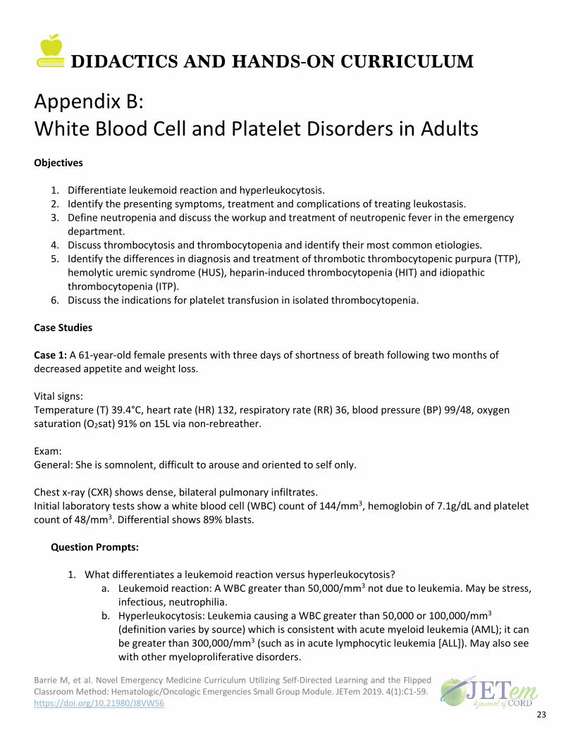
Appendix B:
White Blood Cell and Platelet Disorders in Adults Objectives
1. Differentiate leukemoid reaction and hyperleukocytosis. 2. Identify the presenting symptoms, treatment and complications of treating leukostasis. 3. Define neutropenia and discuss the workup and treatment of neutropenic fever in the emergency
department. 4. Discuss thrombocytosis and thrombocytopenia and identify their most common etiologies. 5. Identify the differences in diagnosis and treatment of thrombotic thrombocytopenic purpura (TTP),
hemolytic uremic syndrome (HUS), heparin-induced thrombocytopenia (HIT) and idiopathic thrombocytopenia (ITP).
6. Discuss the indications for platelet transfusion in isolated thrombocytopenia. Case Studies Case 1: A 61-year-old female presents with three days of shortness of breath following two months of decreased appetite and weight loss. Vital signs: Temperature (T) 39.4°C, heart rate (HR) 132, respiratory rate (RR) 36, blood pressure (BP) 99/48, oxygen saturation (O2sat) 91% on 15L via non-rebreather. Exam: General: She is somnolent, difficult to arouse and oriented to self only. Chest x-ray (CXR) shows dense, bilateral pulmonary infiltrates. Initial laboratory tests show a white blood cell (WBC) count of 144/mm3, hemoglobin of 7.1g/dL and platelet count of 48/mm3. Differential shows 89% blasts.
Question Prompts:
1. What differentiates a leukemoid reaction versus hyperleukocytosis? a. Leukemoid reaction: A WBC greater than 50,000/mm3 not due to leukemia. May be stress,
infectious, neutrophilia. b. Hyperleukocytosis: Leukemia causing a WBC greater than 50,000 or 100,000/mm3
(definition varies by source) which is consistent with acute myeloid leukemia (AML); it can be greater than 300,000/mm3 (such as in acute lymphocytic leukemia [ALL]). May also see with other myeloproliferative disorders.

DIDACTICS AND HANDS-ON CURRICULUM
Barrie M, et al. Novel Emergency Medicine Curriculum Utilizing Self-Directed Learning and the Flipped Classroom Method: Hematologic/Oncologic Emergencies Small Group Module. JETem 2019. 4(1):C1-59. https://doi.org/10.21980/J8VW56
24
2. What are the presenting symptoms for someone with severe hyperleukocytosis/leukostasis? What is the definitive treatment?
a. In severe hyperleukocytosis (eg, AML) one can see a severe symptomatic vaso-occlusive crisis such as altered mental status, acute respiratory failure, acute kidney injury, myocardial infarction, cerebrovascular accident. This symptomatic hyperleukocytosis is called leukostasis and is a medical emergency.
b. The treatment for hyperleukocytosis is leukoreduction via chemotherapy along with leukapheresis and intravenous (IV) fluid resuscitation, in close coordination with hematology.2
Case 2: A 47-year-old female with a history of recently diagnosed breast cancer presents with fever. She reports over the last 24 hours, she has been experiencing intermittent fevers and chills at home. At home she reports a temperature of 101.8°F. She is otherwise asymptomatic. She began chemotherapy treatments one week ago. Vital signs: T 102.1°F, HR 101, RR 15, BP 120/60, O2sat 100% on room air. Exam: General: Well appearing female in no distress. Chest: Heart and lung sounds are normal. A subcutaneous port in the right upper chest is palpated and nontender. Skin: no rashes, no petechia. Complete blood count (CBC) shows WBC of 400/mm3, 70% polymorphonuclear neutrophils (PMNs).
Question Prompts:
1. Define neutropenic fever and how it should be managed in the emergency department (ED). a. Neutropenic fever: Temperature greater than 38°C/100.4°F sustained over one hour, or a
single elevated temperature of 38.3°C/101.0°F with neutropenia. b. For purposes of neutropenic fever, neutropenia is defined as an absolute neutrophil count
(ANC) less than or equal to 500 cells/mm3 or ANC down trending with expectation to drop < 500 cells/mm3 within 48 hours.
i. ANC = (WBC/mm3 x (%PMNs + % bands))/100. c. In the ED, it is important to attempt to identify the source of the fever via diagnostic
workup. Administer broad-spectrum antibiotics within one hour of presentation that include pseudomonas coverage (cefepime, carbapenem, piperacillin-tazobactam), +/- vancomycin depending on suspected source of infection (concern for infected indwelling catheter, skin or soft tissue infection, pneumonia) or if the patient appears critically ill with hemodynamic instability, or if the patient has had prior resistant infections such as MRSA.3 In setting of severe penicillin/cephalosporin allergy, give aztreonam and vancomycin empirically.

DIDACTICS AND HANDS-ON CURRICULUM
Barrie M, et al. Novel Emergency Medicine Curriculum Utilizing Self-Directed Learning and the Flipped Classroom Method: Hematologic/Oncologic Emergencies Small Group Module. JETem 2019. 4(1):C1-59. https://doi.org/10.21980/J8VW56
25
d. Most patients will require admission for treatment; however, some patients may qualify for close outpatient follow up if systems are in place to provide rapid follow up and reassessment for these patients. In general, patients should be observed for at least four hours, and be at low risk for medical complications (fever responding to IV empiric antibiotics, clinically stable).3
Case 3: A 38-year-old female presents with altered mental status. Per her family, she has been progressively more confused and somnolent over the last three days. Vital signs: T 38.8°C, HR 118, RR 15, BP 120/60, O2sat 90% on room air. Exam: General: Ill-appearing, confused female. Chest: Tachycardia, no murmurs. Rales bilaterally in the lung bases. Abdomen: Diffusely tender, soft. Neurologic: Orientated to person only, follows basic commands. Waxing and waning attention. Normal cranial nerve exam. Moving all extremities spontaneously. Skin: Scattered petechiae. CBC shows normal WBC, hemoglobin of 7.0 g/dL, platelets 12/mm3. There are schistocytes on blood smear. Basic metabolic panel is notable for creatinine 3.12 mg/dL. Lactate dehydrogenase is 500 units/L.
Question Prompts:
1. What are common causes of thrombocytopenia? a. Most cases of isolated thrombocytopenia (generally considered platelet counts less than
100 x103/mm3) are acquired or secondary to chronic disease. i. Acquired causes - infectious, medication induced, pregnancy, radiation treatment,
B12 deficiency, or ii. secondary to chronic diseases - lupus, hepatitis C, human immunodeficiency virus
(HIV). 2. Identify the differences between thrombotic thrombocytopenic purpura (TTP), hemolytic uremic
syndrome (HUS), heparin-induced thrombocytopenia (HIT) and idiopathic thrombocytopenia (ITP). How does one make the diagnosis? How is each treated?
a. Thrombotic thrombocytopenic purpura (TTP). The classic pentad includes altered mental status, microangiopathic hemolytic anemia (schistocytes, increased lactate dehydrogenase, low haptoglobin), low platelets, acute kidney injury (AKI), and fever. The etiology is felt to be due to ADAMTS13 (protease) deficiency, which can be triggered by infection, drugs, transplant, and pregnancy. ADAMTS13 cleaves von Willebrand factor anchored to the endothelial surface, in circulation and at sites of vascular injury. When this is deficient, unusually large multimers of von Willebrand factor accumulate and trigger intravascular platelet aggregation and microthrombosis.

DIDACTICS AND HANDS-ON CURRICULUM
Barrie M, et al. Novel Emergency Medicine Curriculum Utilizing Self-Directed Learning and the Flipped Classroom Method: Hematologic/Oncologic Emergencies Small Group Module. JETem 2019. 4(1):C1-59. https://doi.org/10.21980/J8VW56
26
i. Diagnosis 1. Presumptive diagnosis is thrombocytopenia in the presence of
microangiopathic hemolytic anemia. 2. Definitive diagnosis is ADAMTS13 (protease) deficiency and the presence of
ADAMTS13 inhibitor (antibody) in the appropriate clinical setting. ii. Treatment: Supportive including intravenous fluids (IVF), packed red blood cell
(pRBC) and platelet transfusions as appropriate, electrolyte repletion, dialysis, and possible plasma exchange transfusion (in coordination with hematology).
b. Hemolytic uremic syndrome (HUS). Essentially can present the same as TTP. Although it is less common to have altered mental status or central nervous system components.
i. Diagnosis: Must have hemolytic anemia, low platelets and AKI. Usually seen in kids with infectious diarrhea (eg, E Coli 0157:H7 or Shigella).
ii. Treatment: Supportive including IVF, pRBC transfusions as appropriate, electrolyte repletion, dialysis, and possible plasma exchange transfusion in close coordination with hematology and pediatric nephrology. Platelet transfusions are generally ill-advised as they may enhance thrombotic events. Platelet transfusions should only be considered when there is serious, active bleeding.3
c. Heparin-induced thrombocytopenia (HIT). Most commonly seen in inpatients. i. Diagnosis: 50% decline in platelets approximately 5-10 days after exposure (seven-
fold higher with unfractionated heparin vs low molecular weight heparin) leading to thrombotic events.
ii. Treatment: Discontinue heparin and use scoring systems (such as the HIT Expert Probability Score for Heparin-Induced Thrombocytopenia) to help determine the use of alternative agents such as lepirudin, argatroban, bivalirudin, danaparoid.
d. Idiopathic Thrombocytopenia (ITP). i. Diagnosis of exclusion: isolated thrombocytopenia without meeting criteria of
another clinical condition. ii. Treatment: Most are treated medically with steroids, intravenous immunoglobulin
(IVIG) or rituximab. Occasionally splenectomy is required in refractory disease. iii. Generally, platelet transfusions are avoided in ITP and rarely do patients have
bleeding complications despite very low platelet levels. 3. When does one transfuse a patient with isolated thrombocytopenia?
a. In general, platelet transfusion is not indicated unless platelet count is less than 10 x 103/mm3 WITH signs of active bleeding. Patients with chronic ITP often have platelet counts of 10 x 103/mm3 or less without symptoms.
i. Transfusions are used pre-procedure or pre-surgery when platelets are less than 50 x 103/mm3 or in neurologic surgeries when platelets are less than 100 x 103/mm3.
4. In contrast to thrombocytopenia, what are common causes of thrombocytosis? a. Thrombocytosis is defined as platelets greater than 450 x103/mm3. There are two types of
thrombocytosis: i. Reactive or secondary (for example, iron deficiency anemia, infections, bleeding).

DIDACTICS AND HANDS-ON CURRICULUM
Barrie M, et al. Novel Emergency Medicine Curriculum Utilizing Self-Directed Learning and the Flipped Classroom Method: Hematologic/Oncologic Emergencies Small Group Module. JETem 2019. 4(1):C1-59. https://doi.org/10.21980/J8VW56
27
ii. Primary (for example, myeloproliferative disorders such as essential thrombocythemia, polycythemia vera [which also has increased hemoglobin and hematocrit, leukocytosis, normal pulse oximetry] and splenomegaly).
1. For patients with primary thrombocytosis, treatment is with low dose daily aspirin, sometimes with hydroxyurea, except polycythemia vera which is treated with phlebotomy.
Suggested Readings:
1. George JN. Clinical practice. Thrombotic thrombocytopenic purpura. N Engl J Med. 2006;354(18):1927-35.
doi: 10.1056/NEJMcp053024
Additional References:
2. Schwartz J, Padmanabhan A, Aqui N, et al. Guidelines on the use of therapeutic apheresis in clinical practice. Evidence-based approach from the writing committee of the American Society for Apheresis: the seventh special issue. J Clin Apher. 2016;31(3):149-162. doi: 10.1002/jca.21470
3. Taplitz RA, Kennedy EB, Bow EJ, et al. Outpatient management of fever and neutropenia in adults treated
for malignancy: American Society of Clinical Oncology and Infectious Diseases Society of America clinical practice guideline update. J Clin Oncol. 2018;36(14):1443-1453. doi: 10.1200/JCO.2017.77.6211
4. Schiffer, CA. Hyperleukocytosis and leukostasis in hematologic malignancies. In: Larson RA, Rosmarin AG,
eds. UpToDate. Waltham, MA: UptoDate, Inc. https://www.uptodate.com/contents/hyperleukocytosis-and-leukostasis-in-hematologic-malignancies. Updated November 27, 2017. Accessed November 19, 2018.
5. Kelton JG. Heparin-induced thrombocytopenia: an overview. Blood Rev. 2002;16(1):77-80.
doi:10.1054/blre.2001.0189
6. Röllig C, Ehninger G. How I treat hyperleukocytosis in acute myeloid leukemia. Blood. 2015;125(21):3256-52. doi: 10.1182/blood-2014-10-551507
7. Schafer AI. Thrombocytosis. N Engl J Med. 2004;350(12):1211-9. doi: 10.1056/NEJMra035363

DIDACTICS AND HANDS-ON CURRICULUM
Barrie M, et al. Novel Emergency Medicine Curriculum Utilizing Self-Directed Learning and the Flipped Classroom Method: Hematologic/Oncologic Emergencies Small Group Module. JETem 2019. 4(1):C1-59. https://doi.org/10.21980/J8VW56
28
Appendix C: Transfusion Complications Objectives
1. Review the different blood components available for transfusion with inclusion of their typical dosing and expected effect.
2. Discuss consent procedure. 3. Review the early and late complications of blood product transfusions. 4. Discuss the unique complications of massive transfusions. 5. Outline the treatment approach, including preventative strategies, for transfusion reactions.
Case Studies Case 1: A 45-year-old female on coumadin for recent deep vein thrombosis who presents with significant vaginal bleeding. Vital signs are: Blood pressure (BP) 95/60, heart rate (HR) 120, respiratory rate (RR) 20, and afebrile. Exam: General: Anxious and is uncomfortable. Cardiovascular: Tachycardia, no murmurs. Pulmonary: No tachypnea Gynecologic: Bright red blood from the cervix with blood in the vault Her Hgb results at 4.5 g/dL with an elevated international normalized ratio (INR) at 8.5; you make the decision to admit the patient to gynecology and initiate a blood transfusion. Her nurse asks for the consent for transfusion of blood products.
Question Prompts:
1. What blood products would you consider for this patient and how much would be required? a. The patient needs packed red blood cells (pRBCs) and likely fresh frozen plasma (FFP)
emergently. The blood products are typically administered as separate components of whole blood. The following is a guideline for dosing:
i. Packed red blood cells: 10 mL/kg (children) or 1 unit pRBC (adults) causes an expected 1 g/dL increase in hemoglobin (Hgb).
ii. Platelets: 5–10 mL/kg (children) or 1 “pack” (4-6 pooled units) or 1 apheresis unit (adults) produces 40–60,000/mL increase in platelets.

DIDACTICS AND HANDS-ON CURRICULUM
Barrie M, et al. Novel Emergency Medicine Curriculum Utilizing Self-Directed Learning and the Flipped Classroom Method: Hematologic/Oncologic Emergencies Small Group Module. JETem 2019. 4(1):C1-59. https://doi.org/10.21980/J8VW56
29
iii. Fresh frozen plasma (FFP): 10-15ml FFP/kg (all ages). Each unit of FFP contains approximately 200-250ml. Thus, the average 70kg adult would require 2-3 units of FFP. However, response can be variable and follow up INR/PTT should be obtained and re-dosed as required, while monitoring volume status.
iv. Cryoprecipitate: 1 unit per 5 kg for 100 mg/dL fibrinogen increase (all ages). In a 70kg patient, 5 bags (1 pool) increases fibrinogen 35 mg/dl.
b. The goals for transfusions are not the focus of this learning activity, but this patient will likely need multiple units of pRBCs, potentially requiring a “massive transfusion protocol” with equal ratios of pRBCs, platelets, and FFP as described below.
2. How do you consent the patient for transfusion? a. Consent must be obtained for transfusion of all blood products except when emergent
circumstances do not allow for consent. The discussion should focus on the most common complications, but also include the less common yet more severe reactions. These complications are always balanced by discussion of the benefits of the blood product. An example of a general consent follows:
i. Patients with blood loss causing anemia often require transfusion in the emergency department (ED). In general, this is a safe process that leads to improvement in your condition. However, there are risks associated with blood product transfusions. These can vary from simple issues that resolve without treatment, to more serious problems demanding quick and decisive interventions.
b. In the United States in 2011, adverse transfusion reactions were reported to hospital transfusion services for 0.24% of transfused components. Rates of specific reactions are as follows:
i. Hemolytic transfusion reactions occur in one per 76,000 transfused units.2 ii. Nonhemolytic febrile reactions and minor allergic reactions are the most common
transfusion reactions, each occurring in 1%-3% of all transfusions, more commonly in patients who have developed antibodies from prior transfusions.2
iii. Anaphylactic/anaphylactoid reactions occur in one per 20,000 transfused units.2 iv. Due to improved preventative measures, the incidence of graft versus host disease is
less than 0.15%. DEATH: Unfortunately, 80%-90% mortality if it occurs. v. Transfusion-related acute lung injury complicates 0.1%-0.2% of all transfusions. vi. Risk of transfusion-related hepatitis B is one per 300,000 units transfused.3 This risk
is mitigated in individuals that are vaccinated against hepatitis B. vii. Risk for hepatitis C is one per 1.5 million units transfused. Hep C occurs in 50% and
cirrhosis in 10%.3 viii. Risk of transfusion-related HIV infection is 1 per 2 million units transfused.3
ix. Above rates apply to transfusions in the United States. x. Bacterial infection related to contaminated blood products is also very low though it
carries a high risk of fulminant sepsis and high mortality if it occurs. 3. The patient continues to bleed and the hemoglobin drops further after initial transfusion, so a
massive transfusion protocol is initiated. What ratio of products is typically used for massive

DIDACTICS AND HANDS-ON CURRICULUM
Barrie M, et al. Novel Emergency Medicine Curriculum Utilizing Self-Directed Learning and the Flipped Classroom Method: Hematologic/Oncologic Emergencies Small Group Module. JETem 2019. 4(1):C1-59. https://doi.org/10.21980/J8VW56
30
transfusion? Outline the unique complications associated with massive transfusion and describe some of the preventative strategies to prevent them.
a. Massive transfusion includes use of all the blood product components that compose whole blood in a 1:1:1 ratio (pRBC:platelets:FFP). The Pragmatic, Randomized Optimal Platelet and Plasma Ratios (PROPPR) trial did not show any mortality difference between a 1:1:1 ratio and a 2:1:1 ratio; however, more patients in the 1:1:1 protocol achieved hemostasis with fewer exsanguination deaths in 24 hours.6 Massive transfusion is defined as the replacement of more than one-half of the blood volume within a 24-hour period or the replacement of 10 units of blood over the course of a few hours.
b. Complications of massive transfusion include the following: i. Coagulopathy is caused by a dilutional effect on the host's clotting factors and
platelets, as well as the lack of platelets and clotting factors in packed red blood cells.
ii. Volume overload. iii. Hypothermia. iv. Hyperkalemia may be caused by lysis of stored red cells and is more common with
irradiated red blood cells. v. Metabolic alkalosis with compensatory respiratory acidosis and hypokalemia may be
caused by citrate which is used as an anti-coagulant in stored blood. vi. Metabolic abnormalities due to citrate toxicity may occur in those with hepatic
failure, congestive heart failure, or other low-output states. It is increasingly uncommon with the use of component therapy.
c. Other considerations to decrease complications include: i. Hypothermia: Warming the room, surface warming the patient with heating
blankets, heating lamps, heating and humidifying inspired gases for ventilators, warming all IV fluids and blood products when administered.
ii. Coagulopathy and thrombocytopenia: Transfuse pRBC:FFP:platelets in 1:1:1 ratio, or use of recombinant factor VIIa as indicated.
iii. Electrolyte abnormalities: Monitor potassium, calcium, and magnesium serum concentrations closely and correct as needed.
iv. Acid-base disorders: Sodium bicarbonate or tromethamine for severe metabolic acidosis with hemodynamic instability or renal failure.
v. Multiple organ failure: Supportive care. vi. Systemic inflammatory response syndrome (SIRS): Supportive care and minimization
of transfusions once hemorrhage is controlled. vii. Infection: Maintain high index of suspicion to allow for early diagnosis and
appropriate treatment. viii. Transfusion-related acute lung injury (TRALI): Minimize transfusions once
hemorrhage is controlled, consider using pRBCs with a shorter storage time and use of FFP from men and/or nulliparous women.

DIDACTICS AND HANDS-ON CURRICULUM
Barrie M, et al. Novel Emergency Medicine Curriculum Utilizing Self-Directed Learning and the Flipped Classroom Method: Hematologic/Oncologic Emergencies Small Group Module. JETem 2019. 4(1):C1-59. https://doi.org/10.21980/J8VW56
31
Case 2: A 68-year-old male with known peptic ulcer disease and chronic idiopathic thrombocytopenia is seen in the ED for hematemesis and dizziness. Vital signs: BP 145/90, HR 72, respiratory rate (RR) 20, and afebrile. Exam: General: Well appearing, comfortable. Chest: Normal heart and lung sounds. Skin: Pale. He is found to have a Hgb of 6.0 g/dL and platelets of 4 x 10³ /mm3 (baseline 9.0 g/dL and 40 x 103 /mm3, respectively). You assume his care at sign-out after he received a transfusion of pRBCs and platelets. While awaiting an inpatient bed, his nurse notifies you of some increasing dyspnea and dropping pulse oximetry.
Question Prompts:
1. What complications are you considering for this patient? What are the typical signs and symptoms? a. This patient has a rare complication of transfusion known as transfusion related acute lung
injury (TRALI). Transfusion related acute lung injury may be caused by transfusing any plasma-containing blood product. It is caused by the interaction between the recipient's leukocytes and preexisting donor’s anti-leukocyte antibodies. This results in complement activation and increased pulmonary vascular permeability. In addition, mediators of inflammation that form while the blood is in storage are also felt to be contributory.
b. Symptoms of TRALI typically develop during or within six hours of a transfusion. Patients present with the rapid onset of dyspnea and tachypnea. There may be associated fever, cyanosis, and hypotension.
c. Clinical exam reveals respiratory distress and pulmonary crackles may be present with no signs of congestive heart failure or volume overload.
d. Chest X-ray shows evidence of bilateral pulmonary edema not associated with heart failure (non-cardiogenic pulmonary edema), with bilateral patchy infiltrates, which may rapidly progress to complete "white out," indistinguishable from acute respiratory distress syndrome (ARDS).
e. TRALI is more often seen with platelet transfusions than any other blood product because it is a pooled product from multiple donors.
2. What other acute early complications are possibly related to blood transfusion? a. Transfusion associated circulatory overload (TACO) is due to hydrostatic pulmonary edema
instead of the increased vascular permeability. These patients will appear volume overloaded on exam with increased jugular venous distension or plethoric inferior vena cava (IVC) on ultrasound.
b. Hemolytic transfusion reactions: Hemolytic transfusion reactions are the result of antibodies in the recipient's plasma directed against antigens on the donor's erythrocytes, which can cause either acute intravascular hemolysis or extravascular hemolysis.

DIDACTICS AND HANDS-ON CURRICULUM
Barrie M, et al. Novel Emergency Medicine Curriculum Utilizing Self-Directed Learning and the Flipped Classroom Method: Hematologic/Oncologic Emergencies Small Group Module. JETem 2019. 4(1):C1-59. https://doi.org/10.21980/J8VW56
32
i. Intravascular hemolysis of the donor red blood cells, most commonly from ABO incompatibility due to clerical error. This results in hemoglobinemia, hemoglobinuria, disseminated intravascular coagulation (DIC), renal failure, and complement-mediated cardiovascular collapse.
ii. Extravascular hemolysis - The recipient's antibodies to Rh or non-ABO antigens cause extravascular hemolytic reactions. These patients usually have been exposed to the antigen through previous pregnancies, transplantation, or transfusions. Antibody titers often are too low to be detected through routine antibody screening, but production of antibodies becomes amplified with re-exposure. These antibodies do not activate complement; therefore, no intravascular hemolysis occurs. Instead, the RBCs are tagged for removal by splenic macrophages.
c. Nonhemolytic febrile reactions: Nonhemolytic febrile reactions are thought to stem from the formation of cytokines during the storage of the blood. These reactions seldom proceed to hypotension or respiratory distress. Febrile non-hemolytic transfusion reaction (FNHTR) is the most common transfusion reaction, occurring during the transfusion or up to 8 hours after. Patients may also present with chills. With its similarity to acute hemolytic reaction, any fever warrants immediate discontinuation of the transfusion and testing for acute hemolytic reactions. However, FNHTR is benign, with no sequelae. Acetaminophen can be provided for symptom control.
d. Anaphylactic/anaphylactoid reactions: Anaphylactic reactions most often are observed in those patients with a hereditary immunoglobulin A (IgA) deficiency. Some of these patients have developed complement-binding anti-IgA antibodies that cause anaphylaxis when exposed to donor IgA. Proteins in the donor plasma can cause minor allergic reactions. This is an anaphylactoid reaction and is observed more frequently with components containing large amounts of plasma, such as whole blood, pooled platelets, and fresh frozen plasma.
3. What are the treatment modalities for transfusion reactions? a. The general objectives for treatment of any of the acute reactions include maintaining high
clinical suspicion for reactions; although rare, these are serious reactions. b. Report any reaction to the blood bank to allow for reporting and monitoring. c. For any reaction: stop transfusion, call blood bank, and double check that the correct
patient received the correct blood. d. Most reactions with fever will require a full laboratory work-up for signs of hemolysis and
infection: Complete metabolic panel (CMP), complete blood count (CBC), haptoglobin, direct antiglobulin test, lactate dehydrogenase (LDH), prothrombin time (PT), partial thromboplastin time (PTT), fibrinogen, type and cross and blood culture and gram stains from both patient and blood unit. Any signs of dyspnea require a chest radiograph; if there is a fever and hypotension, it is more likely to be TRALI than TACO. For all suspected transfusion reactions: consider maintaining monitoring and intravenous (IV) access, and be prepared for resuscitation.
i. Acute hemolytic reactions (antibody mediated): Immediately discontinue the transfusion while maintaining venous access for emergency management. Do not wait for testing. Anticipate hypotension, renal failure, and DIC. Treat with

DIDACTICS AND HANDS-ON CURRICULUM
Barrie M, et al. Novel Emergency Medicine Curriculum Utilizing Self-Directed Learning and the Flipped Classroom Method: Hematologic/Oncologic Emergencies Small Group Module. JETem 2019. 4(1):C1-59. https://doi.org/10.21980/J8VW56
33
intravenous fluids (IVF) and vasopressors (dopamine/norepinephrine). If there is DIC evident, prepare to provide transfusions of fresh frozen plasma, pooled cryoprecipitate for fibrinogen, and/or platelet concentrates.
ii. Non-hemolytic transfusion reaction: Treat fever with acetaminophen. It often will resolve on its own when transfusion is held. Await laboratory results. If results are reassuring, then the transfusions can be restarted at a slower rate.
iii. Allergic: Diphenhydramine as first-line for hives, rash and pruritis and IVF as needed. iv. Anaphylactic: Administer subcutaneous injection of epinephrine (0.3-0.5 mL of a
1:1000 aqueous solution). There is no evidence that steroids help but most physicians will give prednisone or hydrocortisone.
v. TRALI: Discontinue the transfusion. Keep IV access and prepare for resuscitation. Mild complications can respond to oxygen by nasal canula or nonrebreather; however, worsening symptoms will require aggressive airway management. Treat symptomatically for complications including possible volume overload and cardiogenic pulmonary edema. As with ARDS, there is no role for diuretics or steroids. Diuretics should only be considered if transfusion-associated circulatory overload is the primary consideration rather than TRALI.
4. The patient is stabilized. His family is asking if there are any delayed complications that the patient is at risk for. Please list and describe them.
a. Delayed reactions from transfusion include delayed hemolytic reactions, transfusion transmitted diseases, post-transfusion graft-versus-host disease, and autoimmunization to major or minor blood group antigens. Infectious risks are exceedingly low:
i. Graft-versus-host (GvH) disease occurs when donor lymphocytes mount an immune response against the recipient's lymphoid tissue. Normally, the donor lymphocytes are recognized as being foreign and are destroyed. In situations when the donor is immunocompromised or when the donor is homozygous and the recipient is heterozygous for an HLA (human leukocyte antigen) haplotype, these normal defense mechanisms may fail, resulting in GvH disease.
Suggested Readings: 1. Wroblewski R, Repnshek Z. Severe transfusion reaction and their ED-focused management. emDocs.
http://www.emdocs.net/severe-transfusion-reactions-ed-focused-management/. Published March 6, 2017. Accessed November 19, 2018.
2. Osterman JL, Arora S. Blood product transfusions and reactions. Hematol Oncol Clin North Am.
2014;31(6):1159-1170. doi: 10.1016/j.hoc.2017.08.014 Additional References: 3. American Red Cross. Risks and complications. https://www.redcrossblood.org/donate-blood/blood-
donation-process/what-happens-to-donated-blood/blood-transfusions/risks-complications.html. Accessed December 22, 2018.

DIDACTICS AND HANDS-ON CURRICULUM
Barrie M, et al. Novel Emergency Medicine Curriculum Utilizing Self-Directed Learning and the Flipped Classroom Method: Hematologic/Oncologic Emergencies Small Group Module. JETem 2019. 4(1):C1-59. https://doi.org/10.21980/J8VW56
34
4. Sihler KC, Napolitano LM. Complications of massive transfusion. Chest. 2010;137(1):209-220. doi: 10.1378/chest.09-0252
5. Maxwell MJ, Wilson MJ. Complications of blood transfusion. Continuing Education in Anaesthesia, Critical
Care and Pain. 2006;6(6):225-229. doi: 10.1093/bjaceaccp/mkl053
6. Taplitz RA, Kennedy EB, Bow EJ, et al. Outpatient management of fever and neutropenia in adults treated for malignancy: American Society of Clinical Oncology and Infectious Diseases Society of America clinical practice guideline update. J Clin Oncol. 2018;36(14):1443-1453. doi: 10.1200/JCO.2017.77.6211

DIDACTICS AND HANDS-ON CURRICULUM
Barrie M, et al. Novel Emergency Medicine Curriculum Utilizing Self-Directed Learning and the Flipped Classroom Method: Hematologic/Oncologic Emergencies Small Group Module. JETem 2019. 4(1):C1-59. https://doi.org/10.21980/J8VW56
35
Appendix D: Complications of Sickle Cell Disease Objectives
1. Describe the pathophysiology of sickle cell disease and its variants. 2. List and compare the typical manifestations of sickle cell disease in childhood and adulthood. 3. List diagnostic tools in the emergency department assessment of sickle cell disease. 4. Describe the management of patients with sickle cell disease and review controversial aspects of
management. Case Studies Case 1: A 4-year-old patient with a history of sickle cell disease presents with fever and congestion. Parent states that everyone in the house has had a recent upper respiratory infection. The patient is up-to-date on his vaccines. He is not on any medications. Vital Signs: Blood pressure (BP) 100/60, heart rate (HR) 130, respiratory rate (RR) 30, Temperature (T) 101.4°F Exam: General: Ill-appearing Cardiovascular: Tachycardic Pulmonary: Tachypneic Abdominal: Organomegaly without tenderness to palpation
Question Prompts:
1. What is the pathophysiology of sickle cell disease? a. Replacement of glutamic acid by valine at the sixth position from the N-terminal end of the
β chain. This leads to binding of the hemoglobin β chain in the deoxygenated state which ultimately leads to polymerization and “sickling” of the red blood cells. The result is a recurrent history of vaso-occlusive events, chronic hemolysis, thrombosis, and organ injury.
2. What are common pediatric emergency department presentations of sickle cell crisis? a. Skeletal crises are most common in children including dactylitis (painful swelling of hands
and feet). The differential diagnosis includes osteomyelitis and bone infarction. Of note, there is increased incidence of Salmonella osteomyelitis in sickle cell patients.
b. Sudden death from pneumococcal meningitis and sepsis; infections from encapsulated organisms due to functional asplenia including Staphylococcus, pneumococcus and Haemophilus influenza. As such, vaccination is particularly important in these patients; doctors recommend the pneumococcal vaccine (as early as 6 weeks), HIB (standard

DIDACTICS AND HANDS-ON CURRICULUM
Barrie M, et al. Novel Emergency Medicine Curriculum Utilizing Self-Directed Learning and the Flipped Classroom Method: Hematologic/Oncologic Emergencies Small Group Module. JETem 2019. 4(1):C1-59. https://doi.org/10.21980/J8VW56
36
schedule), meningococcus (age 2). Fever in children with sickle cell disease is a medical emergency. In addition to lab evaluation, they should receive empiric antibiotic coverage and likely admission.
c. Aplastic crisis is due to disruption of normal balance between hemolysis and red blood cell (RBC) production with increased reticulocyte counts, usually due to infection or folate deficiency. This should be suspected when labs show a decreased hemoglobin (Hgb) level and poor reticulocyte response (< 2%). In children and rarely adolescents and adults, splenic sequestration syndrome can occur with decreased Hgb by more than 2 g/dL, signs of shock, thrombocytopenia, a high reticulocyte count, and splenic enlargement.
3. What is acute chest syndrome and how is it managed? a. Acute chest syndrome is characterized by fever, cough, chest pain, dyspnea, and new
infiltrates on the chest radiograph. Infection is common in children with acute chest syndrome and declines with age.
b. Treatment includes hydration, analgesia, incentive spirometry, maintenance of adequate oxygenation and ventilation, and empirical antibiotics (ceftriaxone, add vancomycin if concern for meningitis).
c. Exchange transfusion may be indicated. 4. What group is genetically predisposed to sickle cell? Can patients who are heterozygous for HbS
have complications? a. The sickle cell gene is found in 8-10% of African Americans. b. Heterozygous sickle trait is usually asymptomatic but can have the following manifestations:
Spontaneous hematuria, renal papillary necrosis, splenic infarction, venous thromboembolism, traumatic hyphema, exertional rhabdomyolysis, and exertional sudden death. These can be exacerbated by high altitude or other hypoxic environments.
5. What labs are used for sickle cell diagnosis? What labs can help in the emergency department? a. Initial diagnosis is made on sickle prep (Sickledex) and hemoglobin electrophoresis. b. When patients present to the emergency department with complaints concerning for
complications from sickle cell disease: i. A complete blood count should be ordered, and compared to baseline. ii. A reticulocyte count can help identify complications: a low count is concerning for
aplastic crisis, while a high count is concerning for rapid hemolysis. iii. No specific testing is required (or available) to diagnose a pain crisis. The approach
should be to exclude other causes of pain (acute coronary syndrome, acute chest syndrome, deep vein thrombosis, pulmonary embolism, etc) but otherwise focus on aggressively treating the patient’s pain.
c. For febrile children, blood cultures to evaluate for bacteremia should be obtained before empiric antibiotic coverage.
Case 2: A 25-year-old male presents with pain in his lower back and bilateral lower extremities. He has sickle cell disease, hemoglobin SC variant, and has had many previous episodes. Vitals signs are within normal limits.

DIDACTICS AND HANDS-ON CURRICULUM
Barrie M, et al. Novel Emergency Medicine Curriculum Utilizing Self-Directed Learning and the Flipped Classroom Method: Hematologic/Oncologic Emergencies Small Group Module. JETem 2019. 4(1):C1-59. https://doi.org/10.21980/J8VW56
37
Exam: He has no external signs of traumatic injury.
Question Prompts:
1. What treatment should be provided to this patient? a. Oxygen supplementation if hypoxia is present. Not all patients require oxygen because it
has not been shown to improve pain symptoms or decrease admission rates. b. Pain management is important. Most patients with sickle cell disease are undertreated.
Acetaminophen and nonsteroidal anti-inflammatory drugs (NSAIDs) are problematic due to renal and liver impairment.
c. Blood transfusion is indicated for patients with symptoms of anemia, such as pre-syncope/syncope, shortness of breath or hemodynamic instability and Hgb<7g/dL. In otherwise stable, non-symptomatic patients, transfusion is generally not required even if Hgb is less than 7g/dL.
2. What areas of management are considered controversial? a. Fluids are controversial. Theoretically, euvolemic patients can show increased sickling with
aggressive fluids and some degree of occult congestive heart failure may be present, making patients prone to volume overload. Consider reserving bolus fluids for patients who are dehydrated secondary to fever, vomiting, or other losses. Additionally, when fluids are given1/2 NS should be used as sickle cell disease patients do not excrete sodium well.
3. How is sickle cell disease modified by sickle cell hemoglobin C (HbSC) disease? How about sickle thalassemia (HgS-thalassemia)?
a. Variants of HgSS are HgSC and HgS-thalassemia which cause variation in the severity of clinical manifestations. HgSS is the most severe sickle cell variant, whereas HgSC is moderately severe, and HgS-thalassemia is the least severe. HgSC and Hg-thalassemia are different variants of the β chain mutation.
b. As an example, the lifetime incidence of cerebrovascular accident per 100 patient years is 0.61 in SCD, 0.15 in HbSC disease, and 0.09 and 0.08 in sickle cell-beta (+) thalassemia and sickle cell-beta (0) thalassemia, respectively.
4. What are the common manifestations of sickle cell disease? a. In adults, manifestations are due to hemolysis or vaso-occlusive events precipitated by
infection, cold weather, and stress such as trauma. b. The most common symptom is pain found in the extremities, back, chest and abdomen.
The differential diagnosis can be broad in these cases, and includes fractures, osteomyelitis, epidural abscess, deep venous thrombosis, pneumonia, acute chest syndrome, acute coronary syndrome, pulmonary embolism, kidney stones, urinary tract infections, to name a few.
5. How common are neurologic complications in sickle cell disease patients? Which complications are most common?
a. 25% of patients with sickle cell disease will suffer neurologic manifestations by age 45.

DIDACTICS AND HANDS-ON CURRICULUM
Barrie M, et al. Novel Emergency Medicine Curriculum Utilizing Self-Directed Learning and the Flipped Classroom Method: Hematologic/Oncologic Emergencies Small Group Module. JETem 2019. 4(1):C1-59. https://doi.org/10.21980/J8VW56
38
b. Transient ischemic attack, cerebral vascular accident, and vestibular and hearing changes are the most common. Physicians should consider the use of transcranial doppler (TCD) to screen patients at high risk for stroke. These patients may benefit from frequent transfusions to prevent stroke.8 Treatment of acute stroke in a sickle cell disease patient is exchange transfusion (or simple transfusion) to HbS level < 30%. Can also consider using tissue plasminogen activator (tPA) in non-hemorrhagic strokes. However, because of the pathophysiology of the vaso-occlusive crisis, transfusion is the main priority. Administering tPA, while safe, is unlikely to provide much benefit in a sickle cell crisis.
Suggested Readings: 1. Bansil, NH, Kim TY, Tieu L, Barcega B. Incidence of serious bacterial infections in febrile children with sickle
cell disease. Clin Pediatrics (Phila). 2013;52(7):661-6. doi: 10.1177/0009922813488645
2. Glassberg J. Evidence-based management of sickle cell disease in the emergency department. Emerg Med Pract. 2011;13(8):1-20.
Additional References:
3. Janz TG, Hamilton GC. Anemia, polycythemia, and white blood cell disorders. In: Walls RM, Hockberger RS,
Gausche-Hill M, et al. eds. Rosen’s Emergency Medicine: Concepts and Clinical Practice. 8th ed. Philadelphia, PA: Elsevier; 2014:1586-1605.
4. Novelli EM, Gladwin MT: Crises in sickle cell disease. Chest. 2016;149(4):1082-1093. doi: 10.1016/j.chest.2015.12.016
5. Lovett PB, Sule HP, Lopez BL. Sickle cell disease in the Emergency Department. Emerg Med Clin North Am. 2014;32(3):629-647. doi: 10.1016/j.emc.2014.04.011
6. Glassberg J, Weingart S. Episode 132, Sickle cell disease update (cont. from August 2012). EMRAP. https://www.emrap.org/episode/september2012/sicklecell. Published September 2012. Accessed November 19, 2018.
7. Glassberg J, Weingart S. Episode 131, Sickle cell disease update. EMRAP. https://www.emrap.org/episode/august2012/sicklecell. Published August 2012. Accessed November 19, 2018.
8. Zetola VF. Role of TCD in sickle cell disease: A Review. Perspectives in Medicine. 2012; 265-268. doi: 10.1016/j.permed.2012.03.016

DIDACTICS AND HANDS-ON CURRICULUM
Barrie M, et al. Novel Emergency Medicine Curriculum Utilizing Self-Directed Learning and the Flipped Classroom Method: Hematologic/Oncologic Emergencies Small Group Module. JETem 2019. 4(1):C1-59. https://doi.org/10.21980/J8VW56
39
Appendix E: Red Blood Cell Abnormalities Objectives
1. Discuss the presentation, diagnosis and management of methemoglobinemia. 2. List the causes of acquired methemoglobinemia. 3. Review the differential diagnosis in a patient with anemia. 4. Discuss the laboratory tests that can differentiate different etiologies of anemia. 5. Discuss the current evidence-based guidelines for transfusion of anemia in adults. 6. Compare laboratory results and treatment in patients with anemia due to acute blood loss versus
those with autoimmune hemolytic anemia. Case Studies Case 1: A 27-year-old otherwise healthy male presents to the emergency department (ED) complaining of lethargy. Patient states he was in his normal state of health until earlier today when he developed generalized weakness, lightheadedness, palpitations, shortness of breath and dull mid-sternal chest pain with exertion. Review of systems reveals that the patient has had dental pain for which he has been applying Orajel (20% benzocaine) every hour for the last two days. Vital signs: Temperature (T) 98.6°F, heart rate (HR) 115, respiratory rate (RR) 28, blood pressure (BP) 120/80, oxygen saturation (O2sat) 85% on room air. Exam is remarkable: Head, eyes, ears, nose and throat (HEENT): Perioral cyanosis. Skin: Pale skin with dusky fingertips.
Question Prompts:
1. What is the most likely diagnosis? What are some clinical clues that may raise suspicion for this diagnosis?
a. This patient is suffering from methemoglobinemia likely triggered by excessive use of Orajel which contains the topical anesthetic benzocaine.
b. Benzocaine has oxidative properties and converts ferrous (Fe++) hemoglobin to ferric (Fe+++) hemoglobin. See below for pathophysiology.
c. Methemoglobinemia is rare, and as such it is easy to overlook when developing a differential. Obtaining a full and accurate history is vital to making the diagnosis. Specifically, obtaining a thorough medication history may provide the only clue to make the

DIDACTICS AND HANDS-ON CURRICULUM
Barrie M, et al. Novel Emergency Medicine Curriculum Utilizing Self-Directed Learning and the Flipped Classroom Method: Hematologic/Oncologic Emergencies Small Group Module. JETem 2019. 4(1):C1-59. https://doi.org/10.21980/J8VW56
40
diagnosis. Acquired methemoglobinemia is most commonly triggered by medications, even at appropriate therapeutic doses.
d. There are other clues that can raise suspicion for methemoglobinemia. The patients will often have oxygen saturations around 85% and the spO2% reading does not improve with supplemental oxygen. There may be abnormal discoloration of the patient’s blood during phlebotomy, which has classically been described as dark red, chocolate, or brownish in color.
2. Describe the pathophysiology and name common medications and toxins that can cause these findings.
a. Methemoglobin is an altered form of hemoglobin in which the iron atoms in the heme molecule are oxidized from the normal ferrous (Fe++) to ferric (Fe+++) form. The ferric heme of methemoglobin is unable to reversibly bind oxygen, thereby shifting the oxygen dissociation curve to the left and impairing oxygen delivery. An acute rise in the concentration of methemoglobinemia results in a functional anemia.
b. In normal homeostasis the concentration of methemoglobin is less than 1% of the total circulating hemoglobin. The concentration can rise rapidly in the setting of an offending agent. Most cases of acquired methemoglobinemia are caused by exposure to drugs or toxins with oxidant properties that acutely increase the concentration of methemoglobinemia and produce symptomatology seen with acute anemia.
c. The most common precipitating agents of methemoglobinemia are dapsone and topical anesthetics (benzocaine and lidocaine). Other triggers include anti-malarial drugs (chloroquine), antiemetics (metoclopramide), antibiotics (Sulfamethoxazole-Trimethoprim) and nitroglycerine. Common street drugs, such as heroin and cocaine, have also been reported to cause methemoglobinemia.
3. What is the confirmatory test? a. Methemoglobin should be considered in any patient who is cyanotic with a normal partial
pressure of oxygen (PaO2) on arterial blood gas. The concentration of methemoglobin can be readily obtained off a routine venous or arterial blood sample. These samples will also show high PO2 in the setting of cyanosis. This is because the patient is able to deliver a partial pressure of oxygen to the blood, but the carrying capacity of oxygen on hemoglobin is greatly decreased due to methemoglobinemia.
b. The concentration of methemoglobin is directly correlated with the severity of symptoms. Cyanosis and skin discoloration are early findings and are seen at concentrations greater than 10%. When concentrations rise to 20%-50% the patients will start to develop tissue hypoxia and associated symptoms such as fatigue, tachycardia, lightheadedness, dyspnea, and chest pain. Concentrations greater than 50%-70% can be fatal, with patients developing altered mental status, respiratory depression, and seizures.
i. Note that patients with comorbidities such as pre-existing anemia, heart, or lung disease will develop symptoms at lower methemoglobin concentrations.
4. What is the most appropriate treatment for this patient? What might you expect his pulse oximetry to read with supplemental oxygen?

DIDACTICS AND HANDS-ON CURRICULUM
Barrie M, et al. Novel Emergency Medicine Curriculum Utilizing Self-Directed Learning and the Flipped Classroom Method: Hematologic/Oncologic Emergencies Small Group Module. JETem 2019. 4(1):C1-59. https://doi.org/10.21980/J8VW56
41
a. As with any hypoxic patient, supplemental oxygen should be administered. However, patients with methemoglobinemia will not respond to supplemental oxygen and the spO2 saturation will often continue to read around 85%. This is because the estimate of the pulse oximetry is incorrect. The patient’s actual PO2 measured on an arterial blood gas (ABG) will be normal (or high if delivering supplemental oxygen), but the patient will continue to be cyanotic due to the decreased oxygen carrying capacity of hemoglobin.
b. The most important treatment is identification and discontinuation of the offending agent. Patients who are asymptomatic with methemoglobin concentrations less than 10% may not require any treatment other than discontinuation of the offending agent.
c. In patients who are symptomatic and/or those with methemoglobin concentrations greater than 20%, the therapy of choice is methylene blue (MB).
i. MB, given at a dose of 1mg/kg over 5 minutes, facilitates reduction of ferric hemoglobin back to normal ferrous hemoglobin.
ii. The response is rapid. MB can be re-dosed after one hour if the patient remains symptomatic or if methemoglobin levels remain elevated.
iii. Note that MB is a monoamine oxidase inhibitor and can precipitate serotonin syndrome in patients taking common psychiatric medications such as selective serotonin reuptake inhibitor (SSRI) and serotonin–norepinephrine reuptake inhibitor (SNRI).
iv. MB is contraindicated in patients with G6PD (glucose-6-phosphate dehydrogenase) deficiency because it causes hemolysis.
d. In cases with MB is contraindicated or unavailable IV ascorbic acid (vitamin C) may be used. e. Red blood cell transfusion or exchange transfusions are an option for patients with evidence
of shock. Hyperbaric treatment can be used as well. Case 2: A 68-year-old female with diabetes mellitus, chronic anemia, and atrial fibrillation on apixaban, complicated by known right atrial thrombus and recent cerebrovascular accident who presents from her rehabilitation facility with a chief complaint of “critical lab value.” Routine labs found her hemoglobin to be 6.0 g/dL. The patient has no acute complaints and denies any chest pain, shortness of breath, lightheadedness, or syncope. Review of systems is remarkable for dark stools for the past two days. Vital signs: T 98.6, HR 95, RR 16, BP 130/80, O2sat 98% on room air. Exam: Neurological: At baseline, non-focal Skin: pale skin Rectal: Dark brown stool that is fecal occult blood test positive. CBC confirms her hemoglobin of 6.0 g/dL, which is a decrease from her baseline of 8.0 g/dL. The remainder of her cell lines are within normal limits. Her international normalized ratio (INR) is 1.3.
Question Prompts:
DIDACTICS AND HANDS-ON CURRICULUM
Barrie M, et al. Novel Emergency Medicine Curriculum Utilizing Self-Directed Learning and the Flipped Classroom Method: Hematologic/Oncologic Emergencies Small Group Module. JETem 2019. 4(1):C1-59. https://doi.org/10.21980/J8VW56
42
1. What lab indices can help you categorize her anemia?
a. The differential diagnosis of anemia begins by classifying the patient into one of three categories: blood loss, decreased red blood cell (RBC) production, and increased RBC destruction. The complementary use of RBC indices is also used to further narrow the differential.
b. Anemia due to blood loss: This is a common scenario we encounter in the ED in patients with gastrointestinal (GI) bleeding or trauma. Complete blood count (CBC) in patients with acute blood loss anemia often have normal RBC indices.
c. Decreased RBC production: Decreased reticulocyte count i. Microcytosis: Mean corpuscular volume (MCV) less than 80 fL
1. Iron deficiency anemia: Low iron, low ferritin, high total iron binding capacity (TIBC). Before diagnosing someone with iron deficiency anemia it is important to rule out a slow GI bleed.
2. Thalassemia – depending on type, may have bizarre red cell morphology (extreme hypochromia, poikilocytosis, target cells, tear drop cells, and cell fragments). Will also have increased RBC count without changes to WBC or platelet counts. Routine anemia testing will show non-immune hemolysis, with high lactate dehydrogenase (LDH), high indirect bilirubin, low haptoglobin, and negative Coombs testing. Iron studies show increased serum iron levels, with high transferrin saturation. Hemoglobin analysis and/or genetic testing confirms the diagnosis.
3. Sideroblastic anemia (ie, lead poisoning). Consider when patient has microcytic anemia in absence of iron deficiency or thalassemia. Red cell morphology may be abnormal, with presence of siderocytes essentially pathognomonic (coarse basophilic granules in hypochromic RBC). Ring sideroblasts can be seen on bone marrow aspirate. There are congenital and acquired forms. In adults, history may show parenteral feeding without copper supplementation or excessive zinc ingestion. Excessive alcohol or medication known to cause sideroblastic anemia may be present.
4. Anemia of chronic disease: Low iron, normal or high ferritin, and low TIBC. This anemia starts as normocytic and becomes microcytic late in the disease process.
ii. Macrocytosis: MCV greater than 100 fL 1. B12 deficiency: Generally associated with neurologic deficits (loss of
proprioception, weakness, lower extremity spasticity). 2. Folate deficiency. 3. Chronic liver disease and/or alcohol abuse.
iii. Normocytic: MCV 80-100 fL 1. Anemia of chronic disease: Low iron, normal or high ferritin, and low TIBC.
This anemia starts as normocytic and becomes microcytic late in the disease process.

DIDACTICS AND HANDS-ON CURRICULUM
Barrie M, et al. Novel Emergency Medicine Curriculum Utilizing Self-Directed Learning and the Flipped Classroom Method: Hematologic/Oncologic Emergencies Small Group Module. JETem 2019. 4(1):C1-59. https://doi.org/10.21980/J8VW56
43
2. Chronic renal insufficiency: Due to decreased erythropoietin production. 3. Acute blood loss. 4. Hypothyroidism. 5. Hemolysis.
d. Increased RBC destruction: Elevated reticulocyte count i. Intravascular
1. Autoimmune hemolytic anemia. 2. Microangiopathic hemolytic anemia (thrombotic thrombocytopenic purpura
(TTP), valve disorders). ii. Extravascular
1. Usually secondary to splenic over-activity due to RBC membrane deformities, but also may be antibody-mediated.
2. Sickle cell disease. 2. Our patient’s CBC reveals an MCV of 90. What is your differential? How can you distinguish her
anemia from a hemolytic process? a. Our patient has a normocytic anemia. This is most likely due to either acute blood loss or
anemia of chronic disease which is a pre-existing condition for her. If she has renal failure you could consider decreased erythropoietin production as a cause of her anemia.
b. Both acute blood loss anemia and hemolytic anemia will have elevated reticulocytes. To differentiate a process such as acute blood loss anemia from hemolytic anemia you can order lactate dehydrogenase (LDH) and haptoglobin. In hemolysis the LDH will be elevated and the haptoglobin will be low.
i. To evaluate for microangiopathic hemolytic anemia, order a peripheral blood smear looking for schistocytes which are fragments of damaged RBCs.
3. What is the most appropriate treatment for this patient with asymptomatic anemia? How might your treatment plan change if this patient is suffering from acute autoimmune hemolytic anemia?
a. As with all patients, initial treatment should be aimed at stabilization of airway, breathing, and circulation (ABCs). Our patient’s ABCs are stable and no acute intervention is required.
b. In the anemic patient with severe symptoms (chest pain, shortness of breath, hypotension, etc) the decision to transfuse is an easy one. The decision to transfuse in an asymptomatic anemic patient is less clear and the literature is conflicting.
c. Red blood cell transfusion is not without potential complications. Besides the risk of transfusion reactions, iron overload, transfusion-related acute lung injury (TRALI), infection, and volume overload, studies have shown that transfusions in critically ill patients are associated with worse outcomes.
d. So what is the ideal threshold for RBC transfusion? i. A 2012 Cochrane review of more than 6000 patients showed that when compared to
a liberal transfusion strategy (Hgb less than 9.5-10 g/dL), a restrictive transfusion strategy (Hgb less than 7-8 g/dL) was associated with a significant reduction in in-hospital mortality (risk reduction 0.77) but no change in 30-days mortality.2
ii. A 2013 randomized control trial looked at 45-day mortality and rate of re-bleeding in 921 patients with acute blood loss anemia from upper GI bleeding. The study

DIDACTICS AND HANDS-ON CURRICULUM
Barrie M, et al. Novel Emergency Medicine Curriculum Utilizing Self-Directed Learning and the Flipped Classroom Method: Hematologic/Oncologic Emergencies Small Group Module. JETem 2019. 4(1):C1-59. https://doi.org/10.21980/J8VW56
44
showed that when compared with a liberal transfusion strategy (Hgb less than 9 g/dL), a restrictive strategy (Hgb less than 7 g/dL) was associated with a significantly lower mortality rate and also lower incidence of re-bleeding.2
iii. Overall, current literature and clinical guidelines demonstrate that using a restrictive transfusion strategy (defined as a transfusion hemoglobin threshold of less than 6-8 g/dL) does no harm and is associated with fewer blood products administered compared to a liberal transfusion strategy (Hgb less than 9-10 g/dL).
e. Autoimmune hemolytic anemia presents unique therapeutic challenges. Red blood cell transfusions in these patients may be deleterious since use of incompatible blood can worsen the hemolytic process. Steroid therapy should be initiated in the ED but can take several days to work. If the patient is unstable then RBC transfusion with unmatched RBC transfusions should be initiated. In stable patients, however, RBC transfusions should only be initiated in consultation with a hematologist.
Suggested Readings: 1. Farmer BM, Nelson LS. Dyshemoglobinemias. In: Tintinalli JE, Stapczynski J, Ma O, Cline DM, Cydulka RK,
Meckler GD, et al. eds. Tintinalli's Emergency Medicine: A Comprehensive Study Guide. 7th ed. New York, NY: McGraw-Hill; 2011.
2. Carson JL, Carless PA, Hebert PC. Transfusion thresholds and other strategies for guiding allogeneic red blood cell transfusion. Cochrane Database Syst Rev. 2016;10:CD002042. doi: 10.1002/14651858.CD002042.pub4
Additional References: 3. Villanueva C, Colomo A, Bosch A, et al. Transfusion strategies for acute upper gastrointestinal bleeding. N
Engl J Med. 2013;368(1):11-21. doi: 10.1056/NEJMoa1211801
4. Nickson C. Anaemia. Life in the Fast Lane. http://lifeinthefastlane.com/ccc/anaemia/. Updated August 11, 2014. Accessed November 19, 2018.
5. Ash-Bernal R, Wise R, Wright SM. Acquired methemoglobinemia: a retrospective series of 138 cases at 2
teaching hospitals. Medicine (Baltimore). 2004;83(5):265-73. doi: 10.1097/01.md.0000141096.00377.3f
6. Falkenhahn M, Kannan S, O’Kane M. Unexplained acute severe methaemoglobinaemia in a young adult. Br J Anaesth. 2001;86(2):278-80. doi: https://doi.org/10.1093/bja/86.2.278
7. Coleman MD, Coleman NA. Drug-induced methaemoglobinaemia. Treatment issues. Drug Saf. 1996;14(6):394-405. doi: 10.2165/00002018-199614060-00005

DIDACTICS AND HANDS-ON CURRICULUM
Barrie M, et al. Novel Emergency Medicine Curriculum Utilizing Self-Directed Learning and the Flipped Classroom Method: Hematologic/Oncologic Emergencies Small Group Module. JETem 2019. 4(1):C1-59. https://doi.org/10.21980/J8VW56
45
8. Marik PE, Corwin HL. Efficacy of red blood cell transfusion in the critically ill: a systematic review of the literature. Crit Care Med. 2008:36(9):2667-74. doi: 10.1097/CCM.0b013e3181844677

DIDACTICS AND HANDS-ON CURRICULUM
Barrie M, et al. Novel Emergency Medicine Curriculum Utilizing Self-Directed Learning and the Flipped Classroom Method: Hematologic/Oncologic Emergencies Small Group Module. JETem 2019. 4(1):C1-59. https://doi.org/10.21980/J8VW56
46
Appendix F: Deep Venous Thrombosis Objectives
1. Explain the pathophysiology and risk factors for development of deep vein thrombosis (DVT). 2. Review the clinical presentation of DVT. 3. List approaches to diagnose DVT. 4. Discuss treatment for DVTs. 5. Review the causes and treatment of upper extremity DVTs.
Case Studies Case 1: A 45-year-old female presents with unilateral leg swelling. She describes swelling from her right inguinal region all the way down to and including her foot. She denies pain in her leg but noticed the progressively worsening swelling over the last week. She has noticed some mild weight gain and bloating. She is not on birth control and has had no recent surgeries. Vital signs: Blood pressure (BP) 134/52, heart rate (HR) 68, respiratory rate (RR) 16, oxygen saturation (O2sat) 100% on room air. Exam: Extremity: Pitting edema in right lower extremity and her right calf is 5 cm more than the left calf in circumference.
Question Prompts:
1. What methods may be used to diagnose DVT and for which patients should each method be used? a. Methods of diagnosis include venous duplex ultrasound (sensitivity and specificity
approximately 95% for proximal clots), D-dimer, computed tomography (CT) venography, and magnetic resonance imaging (MRI) (for pelvic vein and vena cava evaluation).
b. Venous duplex ultrasound (US) cannot be used to evaluate for iliac or pelvic vein thrombosis.
c. Normal quantitative D-dimer is acceptable to exclude DVT in patients with low and moderate pretest probability; but it should not be used alone in higher-risk patients. In a study specifically looking at the Diagnostica Stago STA-Liatest D-Dimer assay, they found a sensitivity of 100% (95% CI 95.8%-100%) and a NPV: 100% (95% CI 99.3%-100%).2
2. How might you use risk stratification criteria or prediction models in diagnosing and managing DVT?

DIDACTICS AND HANDS-ON CURRICULUM
Barrie M, et al. Novel Emergency Medicine Curriculum Utilizing Self-Directed Learning and the Flipped Classroom Method: Hematologic/Oncologic Emergencies Small Group Module. JETem 2019. 4(1):C1-59. https://doi.org/10.21980/J8VW56
47
a. You can use a validated scoring system such as the Well’s Score to assign a level of pre-test probability to DVT. If low or moderate probability, a negative d-dimer can lower the post-test probability of DVT low enough to exclude it.
3. What is the difference between a provoked and an unprovoked DVT? a. Provoked DVTs happen as the result of transient, reversible risk factors that when gone, do
not increase the risk for recurrent DVT. This includes things like surgery, pregnancy, immobilization, etc. These patients need anticoagulation for a finite time frame, usually 3-6 months.
b. Unprovoked DVTs happen without any identifiable risk factor and have a high rate of recurrence. For patients with an unprovoked DVT or recurrent DVT, ongoing health issues that increase the risk of clot (e.g., cancer or hereditary thrombophilia), anticoagulation may be continued longer or indefinitely.
4. How would you select a method of treatment for a DVT diagnosed in the emergency department (ED)? If you initiate anticoagulation, how long might patients expect it to be continued? Are there any situations in which inpatient admission is required or intervention other than routine anticoagulation might be indicated?
a. The standard treatment is to anticoagulate the patient. The duration of treatment is at least 3 months. This duration is appropriate for provoked DVT post-surgery or post-trauma and for pregnancy-associated DVT. Unprovoked DVT usually requires longer duration of anticoagulation, possibly indefinitely.
b. Agents of choice can include: Warfarin with LMWH bridging; anti-Xa inhibitors such as rivaroxaban, apixaban and edoxaban, betrixaban; an anti-II agent (dabigatran) or LMWH alone in cancer patients.
i. Warfarin with LMWH bridge is often the default combination of medications for most uncomplicated thrombosis. This is changing now that anti-Xa agents are becoming more commonly used.
ii. Oral anti-Xa agents are becoming more popular because of their rapid onset of anticoagulation and ease of use. They can be used in all populations except in pregnant patients. These drugs are now being used successfully in cancer patients.
iii. LMWH alone is considered safe and effective and is most commonly used as a solo agent in pregnancy and in cancer patients.
iv. Dabigatran is slower in onset of anticoagulation making it less optimal for the acute treatment of thromboembolic disease. It is often used in patients with atrial fibrillation as prophylaxis.
v. Ideally, initiation of this treatment should be made in discussion with hematology or through development of an institutional protocol.
c. Patients with massive clot burden may benefit from admission and thrombolysis or embolectomy.
5. Despite very high concern for a deep venous thrombosis, a comprehensive lower extremity duplex-doppler ultrasound results on the patient with no acute DVT. Now what do you do?
a. Computed tomography (CT)of the pelvis with contrast (it is important to advise the radiologist and technician that you are looking for pelvic vein DVT or something

DIDACTICS AND HANDS-ON CURRICULUM
Barrie M, et al. Novel Emergency Medicine Curriculum Utilizing Self-Directed Learning and the Flipped Classroom Method: Hematologic/Oncologic Emergencies Small Group Module. JETem 2019. 4(1):C1-59. https://doi.org/10.21980/J8VW56
48
compressing the iliac vein in the pelvis). In the above case, the patient has ovarian cancer with pelvic vein thrombosis.
Case 2: A 35-year-old male who presents with unilateral leg swelling and pain. Vital signs: BP 120/80, HR 70, RR 16, O2sat 100% on room air. Exam: Extremity: Swelling is localized to the calf, with tenderness noted on exam. A venous duplex US shows an isolated calf vein DVT.
Question Prompts:
1. Which leg vein sounds superficial but is really considered as a deep vein? a. Superficial femoral vein is actually a part of the deep venous system.
2. What management strategies are reasonable to consider for this patient? a. Some literature suggests isolated calf thromboses can be treated with aspirin alone or no
treatment with repeat US in one week.3 However, practice is variable and, in many cases, it is best to have at least an outpatient consultation with a hematologist.
Case 3: A 45-year-old male who is admitted to the intensive care unit (ICU) develops right arm swelling five days into his stay. He has a right internal jugular central venous catheter (CVC). Vital signs: BP 122/80, HR 73, RR 16, O2sat 100% on room air. Exam: Right upper extremity exam reveals subtle swelling. The CVC insertion site is clean, dry and intact without redness, drainage or tenderness.
Question Prompts:
1. What are the two categories of causes for upper extremity DVT? a. The majority of primary upper extremity DVTs are caused by anatomic abnormalities of the
costoclavicular junction. Thoracic outlet syndrome is associated with DVT. Effort thrombosis results from repetitive injury to the vein in a tight thoracic outlet induced by arm movements, especially frequent use of arms above shoulder level. Idiopathic cases, in which no anatomic abnormalities are identified, also occur.
b. Secondary (related to another cause) upper extremity DVTs are the most common type, and CVCs are the most common cause. The overall incidence is increasing owing to increased use of CVCs, particularly peripherally-inserted central catheters (PICC).

DIDACTICS AND HANDS-ON CURRICULUM
Barrie M, et al. Novel Emergency Medicine Curriculum Utilizing Self-Directed Learning and the Flipped Classroom Method: Hematologic/Oncologic Emergencies Small Group Module. JETem 2019. 4(1):C1-59. https://doi.org/10.21980/J8VW56
49
c. Treatment of upper extremity DVTs is similar to lower extremity DVT with initiation of anticoagulation. When associated with a CVC, it does not need to be removed unless there is concern about associated infection.4
Suggested Readings: 1. American College of Emergency Physicians (ACEP) Clinical Policies Committee. ACEP Clinical Policies
Subcommittee on suspected lower-extremity deep venous thrombosis. Clinical policy: critical issues in the evaluation and management of adult patients presenting with suspected lower-extremity deep venous thrombosis. Ann Emerg Med. 2003;42(1):124-35. doi: 10.1067/mem.2003.181
Additional References: 2. Aguilar C, Sartori M, D’Angelo A, et al. Validation of the STA-Liatest DDi assay for exclusion of proximal
deep vein thrombosis according to the latest Clinical and Laboratory Standards Institute/Food and Drug Administration guideline: results of a multicenter management study. Blood Coagul Fibrinolysis. 2018;29(6):562–566. doi: 10.1097/MBC.0000000000000750
3. Wells PS, Forgie MA, Rodger MA. Treatment of venous thromboembolism. JAMA. 2014;311(7):717-728.
doi: 10.1001/jama.2014.65 4. Geerts W. Central venous catheter-related thrombosis. Hematology Am Soc Hematol Educ Program.
2014;2014(1):306-11. doi: 10.1182/asheducation-2014.1.306
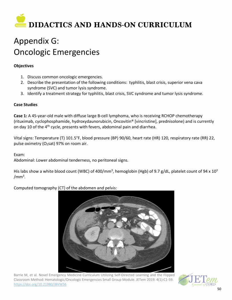
DIDACTICS AND HANDS-ON CURRICULUM
Barrie M, et al. Novel Emergency Medicine Curriculum Utilizing Self-Directed Learning and the Flipped Classroom Method: Hematologic/Oncologic Emergencies Small Group Module. JETem 2019. 4(1):C1-59. https://doi.org/10.21980/J8VW56
50
Appendix G: Oncologic Emergencies Objectives
1. Discuss common oncologic emergencies. 2. Describe the presentation of the following conditions: typhlitis, blast crisis, superior vena cava
syndrome (SVC) and tumor lysis syndrome. 3. Identify a treatment strategy for typhlitis, blast crisis, SVC syndrome and tumor lysis syndrome.
Case Studies Case 1: A 45-year-old male with diffuse large B-cell lymphoma, who is receiving RCHOP chemotherapy (rituximab, cyclophosphamide, hydroxydaunorubicin, Oncovitin® [vincristine], prednisolone) and is currently on day 10 of the 4th cycle, presents with fevers, abdominal pain and diarrhea. Vital signs: Temperature (T) 101.5°F, blood pressure (BP) 90/60, heart rate (HR) 120, respiratory rate (RR) 22, pulse oximetry (O2sat) 97% on room air. Exam: Abdominal: Lower abdominal tenderness, no peritoneal signs. His labs show a white blood count (WBC) of 400/mm3, hemoglobin (Hgb) of 9.7 g/dL, platelet count of 94 x 103 /mm3. Computed tomography (CT) of the abdomen and pelvis:

DIDACTICS AND HANDS-ON CURRICULUM
Barrie M, et al. Novel Emergency Medicine Curriculum Utilizing Self-Directed Learning and the Flipped Classroom Method: Hematologic/Oncologic Emergencies Small Group Module. JETem 2019. 4(1):C1-59. https://doi.org/10.21980/J8VW56
51
Image source: Radswiki. Typhlitis. Radiopaedia. rID: 12046. https://radiopaedia.org/cases/typhlitis. Published October 20, 2010. Accessed November 20, 2018. CC BY-NC-SA 3.0 Question Prompts:
1. Why does typhlitis happen?
a. Typhlitis is also known as neutropenic enterocolitis. b. It generally occurs 10-14 days after chemotherapy. Weakened mucosa and impaired host
defenses lead to bacterial invasion of the intestinal wall. c. It can lead to perforation, necrosis, and/or hemorrhage. d. Typhlitis almost always involves the cecum. e. It is a polymicrobial infection.
2. What are the characteristic signs and symptoms? a. Fever. b. Neutropenia. c. Abdominal pain, mostly in the right lower quadrant. d. Nausea, vomiting and diarrhea. e. Intestinal wall thickening greater than 5mm.
3. What are the possible treatment modalities? a. Management
i. Bowel rest/total parenteral nutrition. ii. Nasogastric tube.
iii. Correct coagulopathies. iv. Surgery and oncology consults. v. Strongly consider granulocyte-colony stimulating factor in consultation with
oncology. vi. Antibiotics are typically similar to febrile neutropenia coverage (gram negative),
along with anaerobic/ C. Difficile coverage. 4. Surgical Indications
a. Gastrointestinal bleeding resistant to correction of coagulopathies and complete blood count.
b. Bowel perforation. c. Clinical deterioration despite maximal medical therapy. d. Surgical approach is a two-stage right hemicolectomy.
Case 2: A 45-year-old male who presents with complaints of cough and shortness of breath. He has had low-grade fevers to 100.5°F for the past few days. He complains of a headache that was gradual in onset and nonspecific in location. He complains of some blurry vision and the feeling of general non-specific malaise. He denies chest pain, confusion, lethargy, nausea, vomiting or diarrhea. He says that he is a regular runner and that he is been unable to complete even three blocks of his usual 5-mile run. This began as a slow decline in function over the last two weeks. He tried to make an appointment with his primary care doctor but they

DIDACTICS AND HANDS-ON CURRICULUM
Barrie M, et al. Novel Emergency Medicine Curriculum Utilizing Self-Directed Learning and the Flipped Classroom Method: Hematologic/Oncologic Emergencies Small Group Module. JETem 2019. 4(1):C1-59. https://doi.org/10.21980/J8VW56
52
referred him to the emergency department for further care and evaluation for a cardiac cause of his chest pain. Vital signs: T 100.5°F, BP 145/95, HR 95, RR 30, O2sat 87% Exam: General: Appears in mild distress complaining of shortness of breath. HEENT: Gingival erythema. Lungs: Crackles in all fields bilaterally. Heart: Regular rate and rhythm, no murmurs, rubs or gallops. Abdomen: Nontender. Neurologic: The patient is alert and oriented to person, place and time. He is slow to answer questions, but exam is otherwise non-focal. Skin: Petechiae diffusely. Labs: WBC: 450 x 103 /mm3, blasts 99%, Hgb 8.5 g/dL, platelets 23 x 103 /mm3. Complete metabolic panel: Sodium 135 mEq/L Potassium 5.8 mEq/L Bicarbonate 22 mEq/L Chloride 110 mEq/L Blood urea nitrogen 35 mg/dL Creatinine 3.2 mg/dL Total Calcium 8.1 mg/dL (8-11 mg/dL) Phosphate 5.5 mg/dL (2.2-4.8 mg/dL) Lactate dehydrogenase (LDH) 600 U/L (50-150U/L) Uric Acid 6.2 mg/dL (3.5-7.7mg/dL) Peripheral smear:

DIDACTICS AND HANDS-ON CURRICULUM
Barrie M, et al. Novel Emergency Medicine Curriculum Utilizing Self-Directed Learning and the Flipped Classroom Method: Hematologic/Oncologic Emergencies Small Group Module. JETem 2019. 4(1):C1-59. https://doi.org/10.21980/J8VW56
53
Image source: The Armed Forces Institute of Pathology (AFIP). Bone marrow: acute myeloblastic leukemia without maturation (aml-m1). Wikimedia Commons. https://commons.wikimedia.org/wiki/File:AML-M1.jpg. Published July 8, 2008. Accessed November 20, 2018. Public domain.
Question Prompts:
1. What is the pathophysiology of blast crisis? a. A blast crisis is (variably) defined as greater than 20% peripheral or bone marrow blasts.
Early in hematopoiesis, the stem cell becomes one of two types of cell lines. The lymphoid cell line becomes B lymphocytes and T lymphocytes. The myeloid cell line leads to the development of platelets, red blood cells, neutrophils and other types of white blood cells.
b. The term "blast cells" refers to myeloblasts or myeloid blasts. These are the very earliest and most immature cells of the myeloid cell line. Less than 5% of the myeloid cells in the bone marrow should be blasts. There should not be any blasts found in the blood.
c. If a patient develops a myeloid blast crisis in acute myeloid leukemia (AML) or chronic myeloid leukemia (CML), there is an overproduction of abnormal myeloblasts that are not able to turn into mature WBCs. They take over the bone marrow and prevent other cell types from forming (normal WBCs, platelets and red blood cells). When the number of blasts becomes too great, they spill out into the circulation.
d. The presence of blasts is a strong indicator of the presence of leukemia. 2. What are the signs and symptoms of blast crisis?
a. Night sweats. b. Weight loss. c. Fever. d. Bone pain. e. Symptoms of anemia. f. An increased risk of infection.

DIDACTICS AND HANDS-ON CURRICULUM
Barrie M, et al. Novel Emergency Medicine Curriculum Utilizing Self-Directed Learning and the Flipped Classroom Method: Hematologic/Oncologic Emergencies Small Group Module. JETem 2019. 4(1):C1-59. https://doi.org/10.21980/J8VW56
54
g. Bleeding. 3. What are hyperleukocytosis and leukostasis?
a. Hyperleukocytosis has been variably defined as a total leukemia cell count (blast counts) greater than 50,000 or greater than 100,000. These patients are at risk for leukostasis.
b. Leukostasis is an acute emergency. It is defined as symptomatic hyperleukocytosis with neurologic and/or pulmonary involvement. It is seen in patients with AML and CML in blast crisis. It can also be seen in acute lymphoblastic leukemia (ALL) and chronic lymphocytic leukemia (CLL) but it is much less common in these leukemias.
c. Despite significantly elevated WBC counts, these patients should be treated as neutropenic since the blasts are not functional in the immune system.
4. What are the signs and symptoms of leukostasis? a. Hypoxia and the presence of pulmonary infiltrates. b. Neurologic complaints can include small vessel blockage or intracranial hemorrhage from
reperfusion injury after improvement of the leukostasis. i. Symptoms typically include visual changes, headache, dizziness, tinnitus, gait
instability, confusion, somnolence, and, occasionally, coma. c. Bleeding
i. Disseminated intravascular coagulation (DIC) can happen in acute promyelocytic leukemia (APL). Check fibrinogen and coagulation studies in these patients. Hematology should be emergently consulted for any patient with suspected APL, that is, patients with coagulopathy and abnormal WBC differential.
d. Infection because blasts are not functional WBCs. e. Fever, with as many as 80% of patients presenting with fever. It can be from infection or
from the presence of inflammation from the leukostasis. f. Spontaneous tumor lysis syndrome can happen in up to 10% of these patients.
5. What is the treatment for blast crisis with leukostasis? a. It is most important to recognize the problem and its implications. b. The primary goal of therapy is to lower the WBC count rapidly to decrease the stasis. The
details of chemotherapy treatment are beyond the scope of this discussion. Emergency management in the emergency department (ED) includes:
i. Intravenous (IV) fluid hydration to help with hemodilution. Adequate renal output should be maintained. Be aware that the chest X-ray may look like pulmonary edema. Look closely at the clinical context so as to not inappropriately withhold fluids. The pulmonary infiltrates develop from the increased cell counts and alveolar fluid related to hyperviscosity, not from increased fluid overall volume status. These patients require aggressive fluid hydration until the overall volume status is corrected.
ii. Empiric treatment with broad-spectrum antibiotics should be initiated in febrile patients.
iii. Avoid transfusions of packed red blood cells (pRBCs). Even though the patients can be anemic, pRBC transfusion will worsen the hyperviscosity. There is no agreed upon situation cutoff to use when deciding to transfuse pRBCs. As a general rule, it

DIDACTICS AND HANDS-ON CURRICULUM
Barrie M, et al. Novel Emergency Medicine Curriculum Utilizing Self-Directed Learning and the Flipped Classroom Method: Hematologic/Oncologic Emergencies Small Group Module. JETem 2019. 4(1):C1-59. https://doi.org/10.21980/J8VW56
55
is a balance of potential harms and benefits. If the patient’s blast count is extremely high (>200K), transfusion of pRBCs would be significantly more risky than if the patient has a blast count of 50K. Emergent consultation with hematology is recommended in cases where transfusion is being considered. If transfusion is critically necessary, it should be done very slowly.
iv. Bleeding can be a manifestation of a low platelet count or DIC. Early platelet transfusion should be done to maintain a platelet count of greater than 20-30 x 103 /mm3. This is very important and is not likely to increase viscosity, but can help to prevent incranial hemorrhage. Disseminated intravascular coagulation should be managed appropriately with supportive care and treatment of potential underlying inciting causes such as infection.
c. Definitive treatment involves lowering the WBC count. This is usually accomplished with induction chemotherapy. If chemotherapy must be delayed in patients with asymptomatic hyperleukocytosis, treatment with hydroxyurea can be used to lower the white count. In patients with hyperleukocytosis with symptoms of leukostasis, in whom chemotherapy must be delayed, hydroxyurea + leukapheresis may be used to rapidly lower the WBC count.
i. Leukapheresis is a procedure in which WBCs are separated from the rest of the blood. The WBCs are removed and the filtered blood is returned to the body. This is done in a continuous circuit, similar to dialysis.
ii. All of these decisions should be made in close discussion with a hematologist experienced in oncologic emergencies. These procedures may require transfer to a tertiary care center.
Case 3: A 54-year-old female presents with 2-3 weeks of shortness of breath and difficulty breathing intermittently as well as some bilateral eye swelling and upper extremity edema. She has reports blurry vision. Vital signs: BP 140/80, HR 90, RR 22, O2sat 97% on room air. Temperature 98.9°F, Exam: Head, eyes, ears, nose and throat (HEENT): Facial edema Extremities: Upper extremity edema

DIDACTICS AND HANDS-ON CURRICULUM
Barrie M, et al. Novel Emergency Medicine Curriculum Utilizing Self-Directed Learning and the Flipped Classroom Method: Hematologic/Oncologic Emergencies Small Group Module. JETem 2019. 4(1):C1-59. https://doi.org/10.21980/J8VW56
56
Photo courtesy of Dr. Colin Kaide
Question Prompts:
1. What cancers commonly cause superior vena cava (SVC) syndrome? a. Cancers cause the majority of cases (approximately 90%): The most common tumors to
involve the SVC are: i. Non-small cell and small cell lung cancer. ii. Lymphoma.
iii. Thymic neoplasms. iv. Occasionally esophageal cancer. v. Metastatic disease.
2. What else can cause it? a. Other causes of SVC syndrome
i. Indwelling catheters and pacemaker leads now accounting for a number of cases. ii. Thrombosis in hypercoagulable patients.
3. What are the classic findings? a. Facial plethora, which is often positional. b. Headache, which is often positional. c. Dyspnea and/or stridor, due to vocal cord edema. d. Dysphagia. e. Lens edema can cause blurry vision. f. Erythema and swelling of the head and neck. g. Dilated superficial veins in the neck, thorax, abdomen and upper extremities.
4. What it the treatment?

DIDACTICS AND HANDS-ON CURRICULUM
Barrie M, et al. Novel Emergency Medicine Curriculum Utilizing Self-Directed Learning and the Flipped Classroom Method: Hematologic/Oncologic Emergencies Small Group Module. JETem 2019. 4(1):C1-59. https://doi.org/10.21980/J8VW56
57
a. Treatment options include: Anticoagulation and/or thrombolysis (for patients in whom the obstruction is primarily thrombotic. Endovascular stenting, surgical bypass, radiation therapy and/or chemotherapy are usually used in tumor-related vena cava compression.
5. When is it an emergency? a. Superior vena cava syndromes from malignancy rarely need emergency treatment.
Exceptions include: i. Stridor due to central airway obstruction or severe laryngeal edema. ii. Coma or altered mental status from cerebral edema.
b. Emergency conditions may be treated with emergent radiation, chemotherapy or thrombolysis. Surgical decompression or stenting may be necessary.
Case 4: A 50-year-old male presents with weakness and altered mental status. He has a history of AML. No fevers, chills, vision changes, abdominal pain, leg swelling, bruising, hematuria, dysuria or gland swelling. He had trouble getting out of bed this morning and was unable to walk. He reports shortness of breath and a mild headache. He denies any focal weakness. Vital signs: BP 160/90, HR 90, RR 22, O2sat 97% on room air. Temperature 98.9°F, Exam: Neck: Supple, symmetrical, trachea midline and no adenopathy. Lungs: Clear to auscultation bilaterally. Heart: Regular rate and rhythm, S1, S2 normal, no murmur, click, rub or gallop. Abdomen: Soft and non-tender. Normal bowel sounds. No masses, no organomegaly. Neurologic: Alert and oriented to person, place and time. Gait normal. Reflexes and motor strength normal and symmetric. Cranial nerves 2-12 and sensation grossly intact. Labs of Interest: WBC 43,000/mm3 Potassium 7.1 mEq/L Calcium 6.1 mg/dL (8-11 mg/dL) Phosphate 8 mg/dL (2.2-4.8 mg/dL) Uric Acid 10 mg/dL (3.5-7.7mg/dL) LDH 9577 U/L (100-190 U/L)
Question Prompts:
1. What cancers usually cause tumor lysis syndrome (TLS)? a. Tumor lysis syndrome (TLS) can spontaneously happen with non-Hodgkin’s lymphoma and
with acute leukemias; it is also seen in patients with very high tumor burden that has a high chemotherapy sensitivity.
b. It sometimes happens spontaneously in large tumors that have necrosis due to outgrowing their blood supply.

DIDACTICS AND HANDS-ON CURRICULUM
Barrie M, et al. Novel Emergency Medicine Curriculum Utilizing Self-Directed Learning and the Flipped Classroom Method: Hematologic/Oncologic Emergencies Small Group Module. JETem 2019. 4(1):C1-59. https://doi.org/10.21980/J8VW56
58
2. What is the pathophysiology of TLS and what are the usual lab abnormalities found? What are the criteria for diagnosis?
a. Tumor lysis syndrome occurs when tumor cells dump their contents into the blood. b. Potassium is intracellular so lysis causes hyperkalemia. c. Deoxyribonucleic acid (DNA) is released into the blood which is the source of phosphate and
of purines, which are then metabolized to uric acid. d. Typical lab abnormalities include: elevated potassium, phosphate and uric acid along with
low calcium. This is because phosphorous combines with calcium, lowering the serum calcium and producing calcium phosphate crystals.
e. Cairo-Bishop classification is the most widely accepted definition of TLS. i. Laboratory TLS: Two or greater of following metabolic abnormalities within 3 days
prior to 7 days post-chemotherapy. 1. Potassium ³6 meq/L or 25% increase from baseline. 2. Uric acid ³8 mg/dL or 25% increase from baseline. 3. Phosphate ³4.5 mg/dL or 25% increase from baseline. 4. Calcium £7 mg/dL or 25% decrease from baseline.
ii. Clinical TLS: Laboratory TLS criteria and 1 or more of following clinical consequences. 1. Creatinine ³1.5x upper limit of normal. 2. Cardiac dysrhythmia. 3. Seizure.
3. What are the treatment strategies for managing TLS? a. The first part of treatment is to increase fluid intake to cause a urine output of 2.5 L/day in
adults. The addition of sodium bicarbonate to alkalinize the urine is controversial and should only be considered when there is a very high uric acid level.
b. Treatment with allopurinol decreases the formation of uric acid. Rasburicase facilitates the conversion of uric acid to allantoin. By preventing xanthine accumulation and by directly breaking down uric acid, rasburicase is more effective than allopurinol for the prevention and treatment of the tumor lysis syndrome.
Suggested Readings: 1. Khan UA, Shanholtz CB, McCurdy MT. Oncologic mechanical emergencies. Hematol Oncol Clin North Am.
2017;31(6):927-940. doi: 10.1016/j.hoc.2017.08.001 2. Muslimani A, Chisti MM, Wills S, et al. How we treat tumor lysis syndrome. Oncology (Williston Park).
2011;25(4):369-375. Additional References: 3. Rodrigues FG, Dasilva G, Wexner SD. Neutropenic enterocolitis. World J Gastroentero. 2017;23(1):42-47.
doi: 10.3748/wjg.v23.i1.42

DIDACTICS AND HANDS-ON CURRICULUM
Barrie M, et al. Novel Emergency Medicine Curriculum Utilizing Self-Directed Learning and the Flipped Classroom Method: Hematologic/Oncologic Emergencies Small Group Module. JETem 2019. 4(1):C1-59. https://doi.org/10.21980/J8VW56
59
4. Röllig C, Ehninger G. How I treat hyperleukocytosis in acute myeloid leukemia. Blood. 2015;125(21):3246-3252. doi: 10.1182/blood-2014-10-551507