
Current Understanding of the Molecular Actions of Vitamin D
39
PHYSIOLOGICAL REVIEWS Vol. 78, No. 4, October 1998 Printed in U.S.A. Current Understanding of the Molecular Actions of Vitamin D GLENVILLE JONES, STEPHEN A. STRUGNELL, AND HECTOR F. DeLUCA Departments of Biochemistry and Medicine, Queen’s University, Kingston, Ontario, Canada; and Department of Biochemistry, University of Wisconsin-Madison, Madison, Wisconsin I. Metabolic Activation of Vitamin D 1194 A. Introduction 1194 B. Overview of metabolism 1194 C. Hepatic 25-hydroxylation 1195 D. Renal 1a-hydroxylation 1198 E. 24-Hydroxylation 1198 II. Role of Vitamin D in Calcium Homeostasis 1200 A. Development of calcium homeostatic mechanisms 1200 B. Role of parathyroid gland and its hormone 1200 C. Physiological actions of 1a,25-dihydroxyvitamin D 3 1201 D. Role of calcitonin 1201 E. Vitamin D metabolites and other hormones 1202 F. Intestinal calcium and phosphate absorption 1202 G. Vitamin D and bone calcium mobilization 1202 H. 1a,25-Dihydroxyvitamin D 3 regulates the parathyroid gland 1203 III. Molecular Mechanism of Action at Target Cells 1203 A. Overall mechanism of transcriptional regulation by vitamin D 1203 B. Vitamin D receptors 1204 C. Retinoid X receptors and other coactivators 1207 D. Vitamin D responsive elements 1208 E. Vitamin D-dependent genes, their roles, and gene complexity 1209 F. Target cell metabolic enzymes 1210 IV. Recently Discovered Functions of 1a,25-Dihydroxyvitamin D 3 1210 A. Discovery of new target organs for 1a,25-dihydroxyvitamin D 3 1210 B. Role of vitamin D hormone in the parathyroid gland 1210 C. Role of vitamin D hormone in skin 1211 D. Role of 1a,25-dihydroxyvitamin D 3 in the immune system 1212 E. Islet cells of the pancreas 1213 F. Role of vitamin D and reproduction 1213 G. Does vitamin D play an essential role during embryonic development? 1213 H. Summary 1214 V. Vitamin D Analogs 1214 A. Development of new analogs of 1a,25-dihydroxyvitamin D 3 1214 B. Factors that alter the action of vitamin D analogs 1217 C. Future directions in vitamin D drug design 1221 Jones, Glenville, Stephen A. Strugnell, and Hector F. DeLuca. Current Understanding of the Molecular Actions of Vitamin D. Physiol. Rev. 78: 1193–1231, 1998.—The important reactions that occur to the vitamin D molecule and the important reactions involved in the expression of the final active form of vitamin D are reviewed in a critical manner. After an overview of the metabolism of vitamin D to its active form and to its metabolic degradation products, the molecular understanding of the 1a-hydroxylation reaction and the 24-hydroxylation reaction of the vitamin D hormone is presented. Furthermore, the role of vitamin D in maintenance of serum calcium is reviewed at the physiological level and at the molecular level whenever possible. Of particular importance is the regulation of the parathyroid gland by the vitamin D hormone. A third section describes the known molecular events involved in the action of 1a,25-dihydroxyvitamin D 3 on its target cells. This includes reviewing what is now known concerning the overall mechanism of transcriptional regulation by vitamin D. It describes the vitamin D receptors that have been cloned and identified and describes the coactivators and retinoid X receptors required for the function of vitamin D in its genomic actions. The presence of receptor in previously uncharted target organs of vitamin D action has led to a study of the possible function of vitamin D in these organs. A good example of a new function described for 1a,25-dihydroxyvitamin D 3 is that found in the parathyroid gland. This is also true for the role of 1193 0031-9333/98 $15.00 Copyright q 1998 the American Physiological Society / 9j0c$$oc10 09-23-98 14:33:23 pra APS-Phys Rev
-
Upload
independent -
Category
Documents
-
view
1 -
download
0
Transcript of Current Understanding of the Molecular Actions of Vitamin D
PHYSIOLOGICAL REVIEWS
Vol. 78, No. 4, October 1998Printed in U.S.A.
Current Understanding of the Molecular Actions of Vitamin D
GLENVILLE JONES, STEPHEN A. STRUGNELL, AND HECTOR F. DeLUCA
Departments of Biochemistry and Medicine, Queen’s University, Kingston, Ontario, Canada; and Department of
Biochemistry, University of Wisconsin-Madison, Madison, Wisconsin
I. Metabolic Activation of Vitamin D 1194A. Introduction 1194B. Overview of metabolism 1194C. Hepatic 25-hydroxylation 1195D. Renal 1a-hydroxylation 1198E. 24-Hydroxylation 1198
II. Role of Vitamin D in Calcium Homeostasis 1200A. Development of calcium homeostatic mechanisms 1200B. Role of parathyroid gland and its hormone 1200C. Physiological actions of 1a,25-dihydroxyvitamin D3 1201D. Role of calcitonin 1201E. Vitamin D metabolites and other hormones 1202F. Intestinal calcium and phosphate absorption 1202G. Vitamin D and bone calcium mobilization 1202H. 1a,25-Dihydroxyvitamin D3 regulates the parathyroid gland 1203
III. Molecular Mechanism of Action at Target Cells 1203A. Overall mechanism of transcriptional regulation by vitamin D 1203B. Vitamin D receptors 1204C. Retinoid X receptors and other coactivators 1207D. Vitamin D responsive elements 1208E. Vitamin D-dependent genes, their roles, and gene complexity 1209F. Target cell metabolic enzymes 1210
IV. Recently Discovered Functions of 1a,25-Dihydroxyvitamin D3 1210A. Discovery of new target organs for 1a,25-dihydroxyvitamin D3 1210B. Role of vitamin D hormone in the parathyroid gland 1210C. Role of vitamin D hormone in skin 1211D. Role of 1a,25-dihydroxyvitamin D3 in the immune system 1212E. Islet cells of the pancreas 1213F. Role of vitamin D and reproduction 1213G. Does vitamin D play an essential role during embryonic development? 1213H. Summary 1214
V. Vitamin D Analogs 1214A. Development of new analogs of 1a,25-dihydroxyvitamin D3 1214B. Factors that alter the action of vitamin D analogs 1217C. Future directions in vitamin D drug design 1221
Jones, Glenville, Stephen A. Strugnell, and Hector F. DeLuca. Current Understanding of the Molecular Actionsof Vitamin D. Physiol. Rev. 78: 1193–1231, 1998.—The important reactions that occur to the vitamin D moleculeand the important reactions involved in the expression of the final active form of vitamin D are reviewed in acritical manner. After an overview of the metabolism of vitamin D to its active form and to its metabolic degradationproducts, the molecular understanding of the 1a-hydroxylation reaction and the 24-hydroxylation reaction of thevitamin D hormone is presented. Furthermore, the role of vitamin D in maintenance of serum calcium is reviewedat the physiological level and at the molecular level whenever possible. Of particular importance is the regulationof the parathyroid gland by the vitamin D hormone. A third section describes the known molecular events involvedin the action of 1a,25-dihydroxyvitamin D3 on its target cells. This includes reviewing what is now known concerningthe overall mechanism of transcriptional regulation by vitamin D. It describes the vitamin D receptors that havebeen cloned and identified and describes the coactivators and retinoid X receptors required for the function ofvitamin D in its genomic actions. The presence of receptor in previously uncharted target organs of vitamin Daction has led to a study of the possible function of vitamin D in these organs. A good example of a new functiondescribed for 1a,25-dihydroxyvitamin D3 is that found in the parathyroid gland. This is also true for the role of
11930031-9333/98 $15.00 Copyright q 1998 the American Physiological Society
/ 9j0c$$oc10 09-23-98 14:33:23 pra APS-Phys Rev
JONES, STRUGNELL, AND DeLUCA Volume 781194
vitamin D hormone in skin, the immune system, a possible role in the pancreas, i.e., in the islet cells, and a possiblerole in female reproduction. This review also raises the intriguing question of whether vitamin D plays an importantrole in embryonic development, since vitamin D deficiency does not prohibit development, nor does vitamin Dreceptor knockout. The final section reviews some interesting analogs of the vitamin D hormone and their possibleuses. The review ends with possible ideas with regard to future directions of vitamin D drug design.
stration of new biological actions of 1,25-(OH)2D3, in particu-I. METABOLIC ACTIVATION OF VITAMIN D
lar, its effects on the regulation of growth and differentiationof certain specialized cell types (sect. IV), which represent
A. Introduction involvements of vitamin D not even envisioned when 1,25-(OH)2D3 was first discovered. Furthermore, the knowledge
An appreciation that vitamin D3 represents only a pre- of vitamin D metabolism also provided the impetus to studycursor to its functionally active form, 1a,25-dihydroxyvitamin the regulation of cytochrome P-450-containing enzymes in-D3 [1,25-(OH)2D3], is arguably one of the most important volved in the process as well as stimulating the chemicaldevelopments in vitamin research during the latter half of synthesis of a wide range of vitamin D analogs (sect. V). Thisthe 20th century. The discovery of the two activation steps review seeks to summarize our current understanding ofinvolved in the metabolism of vitamin D3 to the hormone the molecular events surrounding the physiological action of1,25-(OH)2D3 (sect. I) set the stage for the elucidation of the vitamin D in these many varied areas. For a more detailedrole of vitamin D in the physiological events involved in account of the subjects described in this review, particularlycalcium and phosphate homeostasis (sect. II). The realization those of a clinical nature, the reader is directed to a recentlythat it was the metabolites of vitamin D that were important published text (96).led to an intense focus on the molecular events surroundingthe mechanism of action of 1,25-(OH)2D3, which resulted B. Overview of Metabolismin the discovery of the vitamin D receptor (VDR) and itsinteraction with the transcriptional machinery inside vitamin Vitamin D, in the form of vitamin D3, is made from
7-dehydrocholesterol in the skin by exposure to ultravio-D target cells (sect. III). Subsequently, this led to the demon-
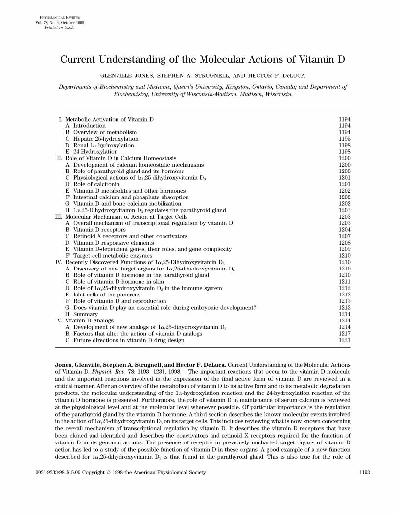
FIG. 1. A: nutritional forms of vitamin D. B: steps involved in activation of vitamin D3 molecule. Note that namesof cytochrome P-450 isoforms currently thought to be responsible for enzyme steps are also provided.
/ 9j0c$$oc10 09-23-98 14:33:23 pra APS-Phys Rev

October 1998 MOLECULAR ACTIONS OF VITAMIN D 1195
FIG. 2. Electron transport chain for mitochondrialsteroid hydroxylases. Concept for 3-dimensional arrange-ment of components of mitochondrial cytochrome P-450-containing hydroxylases is shown.
let light (270–300 nm range). Alternatively, vitamin D, in D target tissues including enterocytes, osteoblasts, kera-tinocytes, and parathyroid cells. Thus it is now known thatthe form of either vitamin D2 or vitamin D3, can be derived
from dietary sources (Fig. 1A). Both vitamin D3 and vita- CYP24 will use 1,25-(OH)2D3 as substrate also. BecauseCYP24 is widely distributed around the body, is stronglymin D2 undergo the same activation process, involving
first 25-hydroxylation in the liver, followed by 1a-hydrox- induced in target cells by 1,25-(OH)2D3, and prefers 1,25-(OH)2D3 as substrate to 25-OH-D3, its role appears to beylation in the kidney, to make the biologically active com-
pounds 1,25-(OH)2D3 and 1,25-(OH)2D2, respectively (Fig. catabolic. The enzyme CYP24 catalyzes several steps of1,25-(OH)2D3 degradation, collectively known as the C-241B). There is little evidence that these two active forms
differ in their mode of action, and because most is known oxidation pathway which starts with 24-hydroxylation andculminates in the formation of the biliary excretory form,about the synthesis and action of 1,25-(OH)2D3, this re-
view focuses on the natural D3 compound. The metabolic calcitroic acid (Fig. 3). Thus our current view is that boththe synthesis and degradation of 1,25-(OH)2D3 are tightlyactivations of vitamin D3 are carried out by specific cyto-
chrome P-450-containing enzymes, the vitamin D3-25-hy- regulated events, attesting to the fact that the concentra-tion of this potent hormone requires fine control at thedroxylase (CYP27) and possibly another P-450 in the hepa-
tocyte and the 25-hydroxyvitamin D-1a-hydroxylase cellular level, and hence a set of highly specific and finelytuned cytochrome P-450 exist for the purpose.(CYP1a) in the renal proximal tubular cell. Both of the
known hydroxylases are located in the inner mitochon-drial membrane of these cells (Fig. 2). The synthesis of
C. Hepatic 25-Hydroxylation25-hydroxyvitamin D3 (25-OH-D3) by the liver appears tobe only loosely regulated, whereas the synthesis of 1,25-(OH)2D3 by the renal 1a-hydroxylase is tightly regulated Vitamin D does not circulate for long in the blood-
stream but, instead, is immediately taken up by adiposeby the levels of plasma 1,25-(OH)2D3 and calcium. Therenal enzyme is strongly upregulated by the hormone tissue for storage or liver for further metabolism. In hu-
mans, tissue storage of vitamin D can last for months orparathyroid hormone (PTH), a point that is discussed fur-ther in section II. A third vitamin D-related mitochondrial even years. Ultimately, vitamin D3 undergoes its first step
of activation, namely, 25-hydroxylation, in the liver (28)cytochrome P-450-containing enzyme, the 25-hydroxyvi-tamin D-24-hydroxylase (CYP24), was originally believed (Fig. 1B). Early data suggested that the liver is the only
significant site of 25-hydroxylation in vivo, although thereto be exclusively located in the kidney and to be involvedonly in the metabolism of 25-OH-D3 to 24,25-dihydroxyvi- were occasional reports of intestinal and renal extracts
containing this activity (346). Research, therefore, fo-tamin D3 [24,25-(OH)2D3]. The 24-hydroxylation of 1,25-(OH)2D3 was first realized with the isolation of 1,24,25- cused on purification of the major hepatic enzyme activity.
Over the years, there has been some controversy over(OH)3D3 and subsequently shown to occur in all vitamin
/ 9j0c$$oc10 09-23-98 14:33:23 pra APS-Phys Rev

JONES, STRUGNELL, AND DeLUCA Volume 781196
FIG. 3. C-24 oxidation pathway.
whether 25-hydroxylation is carried out by one enzyme (252).1 The primary amino acid sequences of three speciesof CYP27 are depicted in Figure 4. Even though 25-hydrox-or two and whether this cytochrome P-450-based enzymeylation of a variety of vitamin D compounds, includingis found in the mitochondrial or microsomal fractions ofvitamin D3, has been clearly demonstrated in cells trans-liver. Madhok and DeLuca (207) reported that a rat liverfected with CYP27 (119), there is still some skepticism inmicrosomal system requiring NADPH, molecular oxygen,the vitamin D field that a single cytochrome P-450 cana flavoprotein, and a cytochrome P-450 was capable ofexplain all the metabolic findings observed over the past25-hydroxylation of vitamin D3, but the cytochrome P-two decades of research. The many unexplained observa-450 responsible has never been cloned. There was sometions suggesting that other cytochrome P-450 might per-speculation that the microsomal enzyme might beform 25-hydroxylation of vitamin D at nanomolar concen-CYP2C11, but this cytochrome is male specific (129) andtrations of substrate that exist in vivo include the follow-other data have also been presented that indicate thating. 1) Perfused rat liver studies by Fukushima et al. (104)human microsomes do not possess 25-hydroxylase activ-demonstrate kinetics consistent with two 25-hydroxylaseity (285). Recently, Axen et al. (9) have purified a pig liverenzyme activities: a high-affinity, low-capacity form (pre-microsomal 25-hydroxylase with an NH2-terminal se-sumably microsomal) and a low-affinity, high-capacityquence different from that of CYP2C11 and that is capableform (presumably mitochondrial; CYP27).of the 25-hydroxylation of both vitamins D2 and D3. Cur-
2) Dietary studies show regulation, albeit weak, of therently, only the mitochondrial 25-hydroxylase has beenliver 25-hydroxylase in animals given normal intakes of vita-purified to homogeneity and subsequently cloned (7, 44,
351). The cytochrome P-450 involved is known as CYP271 The term 27-hydroxylation has been suggested by a consortiumor P-450c27 because it is a bifunctional cytochrome P- of cytochrome P-450 specialists to describe terminal hydroxylation of
450 which in addition to 25-hydroxylating vitamin D3 also steroids given that methyl groups at C-26 and C-27 are indistinguishable.The old nomenclature for this was 26-hydroxylation, and the literaturecarries out side-chain hydroxylation, including 27-hydrox-contains numerous references to 26-hydroxylation of vitamin D com-
ylation of cholesterol-derived intermediates involved in pounds giving rise to 26-hydroxylated vitamin D metabolites, e.g., 25,26-(OH)2D3.bile acid biosynthesis (from which it derives its name)
/ 9j0c$$oc10 09-23-98 14:33:23 pra APS-Phys Rev
October 1998 MOLECULAR ACTIONS OF VITAMIN D 1197
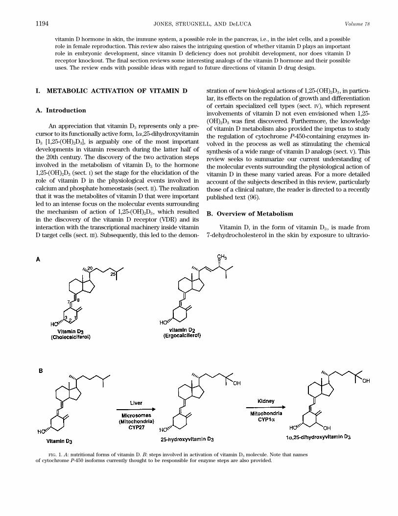
FIG. 4. Amino acid alignments of all published vitamin D-related cytochromes P-450 from various species: CYP1a(25-OH-D3 1a-hydroxylase); CYP27 (mitochondrial vitamin D3 25-hydroxylase); CYP24 (25-OH-D3 24-hydroxylase). Notehigh degree of sequence similarity between all family members, particularly toward COOH terminus of each isoform.Conserved cysteine residue in block CMGRRLAELEL in extreme COOH terminus is where heme group is covalentlybonded to protein. Slightly more NH2 terminal is putative ferredoxin-binding site involving LPLLKAVVKEVLRL. Anotherhighly conserved site is putative oxygen-binding site ELLLAGVDTVSNTL.
min D after a period of vitamin D deficiency (20), which is 25-hydroxylate vitamin D2 (119); it 24-hydroxylates vita-min D2 instead, evoking the question: Which cytochromenot explained by a transcriptional mechanism, since the
gene promoter of CYP27 lacks a vitamin D-responsive ele- P-450 synthesizes 25-OH-D2?Observations that are explained by the existence ofment (VDRE) (118) or demonstrable responsiveness to 1,25-
(OH)2D3 whereas it is regulated by bile acids (354). CYP27 include 1) the occasional reports of extrahepatic 25-hydroxylation of vitamin D3 mentioned above (346), which3) Clinical studies show no obvious 25-OH-D3 or 1,25-
(OH)2D3 deficiency occurs in patients suffering from the are consistent with the detection of CYP27 mRNA in a num-ber of extrahepatic tissues including kidney and bone (os-genetically inherited disease cerebrotendinous xanthoma-
tosis, in which CYP27 is mutated. [Although a subset of teoblast) (10, 147), and 2) the abundance of 24-hydroxylatedmetabolites [e.g., 24-OH-D2, 1,24-(OH)2D2, and 24,26-these patients suffer from osteoporosis, this is more likely
because of biliary defects leading to altered enterohepatic (OH)2D2] in the blood of vitamin D2-intoxicated animals(143, 161, 178, 320). It is worth noting from perusal of Figurecirculation of 25-OH-D3 (18).]
4) Substrate specificity studies using transfected re- 4 that the recently cloned CYP1a is more closely related toCYP27 than it is to CYP24, a surprising fact given thatcombinant human CYP27 show that the enzyme does not
/ 9j0c$$oc10 09-23-98 14:33:23 pra APS-Phys Rev

JONES, STRUGNELL, AND DeLUCA Volume 781198
CYP1a and CYP24 are both renal cytochromes P-450 and 2–4 h after exposure to 1,25-(OH)2D3 and is blocked byinhibitors of protein synthesis and transcription. In theappear to be reciprocally regulated, thereby implying CYP27
might have evolved to metabolize vitamin D after all. Thus, same model, the disappearance of the 1a-hydroxylase ismirrored by the reciprocal appearance of the renal 25-although CYP27 remains the best-characterized cytochrome
P-450 capable of 25-hydroxylation, it may not be the only OH-D3-24-hydroxylase, in effect a complete ‘‘switchover’’from 1a- to 24-hydroxylating activity in the isolated organ25-hydroxylase, and its full physiological importance re-
mains to be established. over the 4-h period. The exact reciprocal regulation of thetwo enzymes, first demonstrated in vivo by Tanaka et al.The product of the 25-hydroxylation step, 25-OH-D3,
is the major circulating form of vitamin D3 and in humans (336) two decades ago, led some workers to postulatethat the 1a- and 24-hydroxylases might share a single cyto-is present in plasma at concentrations in the range 10–40
ng/ml (25–125 nM) (140). The main reason for the stability chrome P-450 polypeptide chain, its catalytic propertiesmodified by NH2-terminal truncation (111) or regulatedof this metabolite is its strong affinity for the vitamin D-
binding (globulin) protein of blood (DBP) (70). The meta- by the phosphorylation state of the ferredoxin componentof the enzyme (300). The cloning of two distinct cyto-bolic fate of 25-OH-D3 is dependent on the calcium re-
quirements of the animal. An urgent need for calcium chromes P-450, representing 1a-hydroxylation and 24-hy-droxylation, suggests the first hypothesis to be incorrect,results in renal 1a-hydroxylation, whereas an abundance
of calcium results in 24-hydroxylation (see sect. II). These the switchover process probably being accomplished byde novo protein synthesis of the required cytochrometwo alternative pathways are discussed in turn below.P-450. The second hypothesis involving regulation of en-zyme activity through ferredoxin phosphorylation re-
D. Renal 1a-Hydroxylation mains a possibility for fine-tuning of enzyme activity, al-though the details of this remain obscure at this point.The cloning of CYP1a has already led to mapping of theThe enzyme 25-hydroxyvitamin D3-1a-hydroxylase is
responsible for the tightly regulated step that involves the human gene to chromosome 12q13.1-q13.3 (316), the samelocus as the gene defect for vitamin D dependency ricketsintroduction of a 1a-hydroxyl group into the A ring of 25-
OH-D3, thereby creating the hormone 1,25-(OH)2D3. The type I (192), a disease cured by small doses of exogenous1,25-(OH)2D3 and which had been previously postulatedspecific location of this enzyme in the kidney became
apparent (100) even before the unequivocal identification to be because of a mutated version of the 1a-hydroxylase(99). The next decade will see a clarification of the molec-of 1,25-(OH)2D3 (139). Experiments involving nephrecto-
mized animals have confirmed that the kidney as the ma- ular and clinical aspects surrounding this key regulatorystep of calcium homeostasis.jor source of the circulating pool of 1,25-(OH)2D3. The
renal 1a-hydroxylase enzyme comprises a cytochrome P- There have been indications that there are extrarenalversions of the 1a-hydroxylase existing in cells of mono-450, a ferredoxin, and a ferredoxin reductase (112) (see
Fig. 2). The cytochrome P-450 for the 1a-hydroxylase en- cyte/macrophage, placental, and keratinocyte lineages (4,22, 83). There is strong evidence that the extrarenal en-zyme, CYP1a, was recently cloned from rat, mouse, and
human (228a, 298, 316, 332), and the amino acid sequences zyme located in macrophages plays a major role in certaingranulomatous conditions (e.g., sarcoidosis), causing un-of these are compared with the other known vitamin D-
related cytochromes P-450 in Figure 4. Because of the controlled elevations of blood 1,25-(OH)2D3 levels, whichsubsequently result in troublesome hypercalcemia andclose resemblance with CYP27, it has been suggested that
CYP1a (P-4501a) be termed CYP27B1 (298). Both 1a-hy- hypercalciuria (3). The normal function of this and otherextrarenal 1a-hydroxylases remains obscure at this time,droxylases have short mitochondrial targeting sequences
but share many regions of similarity with other members although some have postulated a paracrine or autocrinerole for locally produced 1,25-(OH)2D3. Once again, it isof the family including the classical heme-, ferredoxin-,
and oxygen-binding sites indicated in Figure 4. The 1a- safe to predict that the molecular basis for various reportsof extrarenal activity in cultured cells in vitro (4, 22, 83)hydroxylase is induced by PTH through a cAMP/phospha-
tidylinositol 4,5-bisphosphate (PIP2)-mediated signal and in certain human disease states will be resolvedshortly with the cloning of the renal enzyme (298, 316,transduction mechanism that is still to be defined at the
molecular level (132). The enzyme appears to be downreg- 332), as will its mechanisms of regulation.ulated by vitamin D status, possibly through a VDR-medi-ated transcriptional mechanism involving the hormonal
E. 24-Hydroxylationproduct 1,25-(OH)2D3, although there were early claimsthat 1,25-(OH)2D3 might act directly on its own synthesisthrough an allosteric mechanism (117). Work using the The discovery of 24,25-(OH)2D3 (138, 327) predated
even the identification of 1,25-(OH)2D3 and the recognitionperfused vitamin D-deficient rat kidney (281) elegantlyshows that the downregulation of 1a-hydroxylation takes of 24-hydroxylation as a metabolic step allowed for the
/ 9j0c$$oc10 09-23-98 14:33:23 pra APS-Phys Rev

October 1998 MOLECULAR ACTIONS OF VITAMIN D 1199
search for the enzyme activity. The relative ease with tion of CYP24 from undetectable to strongly detectableexpression at the mRNA level within 4 h (297). This iswhich 24,25-(OH)2D3 was generated in such large amounts
was a clue that the metabolic step was upregulated rather consistent with the enzyme activity pattern first observedin the kidney but subsequently reported for a variety ofthan downregulated by vitamin D administration. The ear-
liest report of the 25-OH-D3-24-hydroxylase was a subcel- vitamin D target cells after initial exposure to hormone(127, 202, 209). Metabolic studies with the recombinantlular localization study (174) using vitamin D-replete
chicken kidney tissue which established that the 24-hy- CYP24 protein produced in bacteria or insect cells havebeen equally enlightening. Akiyoshi-Shibata et al. (5) anddroxylase is a mitochondrial cytochrome P-450-containing
enzyme, and this was followed by a reconstitution experi- Beckman et al. (15) have shown that CYP24 is a multicata-lytic enzyme capable of several if not all of the stepsment involving partially purified enzyme components
(248). Evidence was also emerging both in vivo and in illustrated in Figure 3. It is likely that CYP24 is able tocatalyze three successive oxidations, two at C-24 and onevitro that the 24-hydroxylase was not confined to the kid-
ney but could be found in classical vitamin D target tissues at C-23, to give an intermediate that is subsequentlycleaved by an unknown mechanism. All prevailing evi-including the intestine and bone (185, 347). In the late
1970s, it became evident that 24-hydroxylation was proba- dence suggests that C-24 oxidation is a highly efficientprocess giving rise to molecules of lower biological activ-bly only the first step in an inactivation process. First, it
was shown to be induced by 1,25-(OH)2D3 itself (172, 187, ity (e.g., calcitroic acid) such that if C-24 hydroxylationis blocked by a general cytochrome P-450 inhibitor such335, 337, 340), and the product 1,24,25-(OH)3D3 was 10
times less biologically active than 1,25-(OH)2D3 (56, 335). as ketoconazole, 1,25-(OH)2D3 hormone action is ex-tended (265). This argues that the main role of the C-24Second, 24,25-(OH)2D3 and its 1a-hydroxylated analog,
1,24,25-(OH)3D3, could be converted to further metabolic oxidation pathway is attenuation of the biological signalinside target cells.products containing a 24-oxo and/or 23-hydroxy groups as
well (237, 246, 330). The perfused rat kidney was helpful in Recently, this hypothesis was tested when St. Arnaudet al. (315) engineered a CYP24-null mouse. At this time,establishing the production of these catabolites (160) but
revealed two further pieces to the puzzle of the metabolic the results have been reported only in abstract form, butit is clear that the defect is not lethal during embryonicrole of 24-hydroxylation. First, the perfused rat kidney
allowed for a clarification of the temporal relationship of development. Instead, at least one-half the mice exhibithypercalcemia/hypercalciuria in early neonatal life andthese catabolites, in effect, suggesting the existence of a
pathway from 1,25-(OH)2D3 and/or 25-OH-D3 (160), and quickly die before weaning from nephrocalcinosis. Theother half of animals survive and appear healthy perhapssecond, the perfused kidney generated two side chain-
cleaved molecules: a 23-alcohol (159) and a 23-acid (208, due to the upregulation of some alternative vitamin D-catabolic pathway (C-26 hydroxylation or 26,23-lactone276), not observed previously in vitro. The 1a-hydroxyl-
ated 23-acid, calcitroic acid, is observed in vivo and has formation). All CYP24-null animals exhibit abnormal bonehistology, characterized by excessive unmineralized bonebeen shown to be the principal biliary excretory form of
1,25-(OH)2D3 (94). These discoveries led to the rationaliza- matrix and reminiscent of that previously observed in ex-periments involving exogenous 1,25-(OH)2D3 intoxicationtion of many findings from a number of laboratories into
a hypothesis that 24-hydroxylation is the first step of a (136). This could be caused by an inability of target cells,in this case osteoblasts, to turn off the 1,25-(OH)2D3 signaltarget cell C-24 oxidation pathway (Fig. 3) whose major
function is to convert 1,25-(OH)2D3 to calcitroic acid (208, in the absence of the C-24 oxidation pathway. An alterna-tive explanation that CYP24 is needed for the synthesis276). The demonstration of vitamin D-inducible, calcitroic
acid production in bone (UMR106) and kidney (208) was of some essential 24-hydroxylated metabolite of vitaminD [e.g., 24,25-(OH)2D3] needed for a yet to be defined rolefollowed by demonstration of vitamin D-inducible C-24
oxidation pathway activity in a number of vitamin D target in bone formation has been proposed (314) but seemsunlikely for several reasons. Among the data that arguecells including intestine (Caco-2), keratinocyte (HPKIA-
ras), and breast (T47D and MCF-7) (215, 278, 344). against such an essential role for 24-hydroxylated metabo-lites are the findings from the study of fluorinated analogsIn the early 1990s, Okuda’s group succeeded in clon-
ing the cytochrome P-450, CYP24 or P-450cc24, represent- of vitamin D (149, 234, 338). These analogs are irreversiblyblocked in the C-24 and C-23 positions of the side chaining the 24-hydroxylase (62, 148, 247). The amino acid se-
quences of three species of CYP24 are shown in Figure with one or more atoms of fluorine and therefore unableto undergo 24- or 23-hydroxylation. However, they are4. It belongs to the same subfamily as the other two known
vitamin D-related cytochromes. The structure of the gene fully biologically active in all known in vivo functionsof vitamin D. Furthermore, the biochemical machineryfor CYP24 has been described for several species (rat,
mouse, and human), and in each case shown to possess required for transducing the signal from a 24-hydroxylatedmetabolite has never been satisfactorily demonstrated intwo VDRE in its proximal promoter (249, 367, 368). These
VDRE allow for the 1,25-(OH)2D3 VDR-mediated upregula- bone (or any other) cells. Thus it appears that 24-hydrox-
/ 9j0c$$oc10 09-23-98 14:33:23 pra APS-Phys Rev

JONES, STRUGNELL, AND DeLUCA Volume 781200
ylation is not essential for vitamin D to fulfill its many is a very complex one (see Fig. 5) that involves manyhormones, most of which are known but some that arebiological roles in vertebrate biology. Therefore, evidence
from the CYP24-null mouse seems to confirm our hypothe- not. From this discussion, it will become apparent that thevitamin D endocrine system is the basic one in managingsis that the C-24 oxidation pathway is a complex, self-
induced mechanism for limiting the action of 1,25-(OH)2D3 calcium of plasma, with equally important roles for theparathyroid hormone and calcitonin.in vitamin D target cells once the initial wave of gene
expression has been initiated (see sect. III). This is dis-cussed in greater detail in section V.
B. Role of Parathyroid Gland and Its Hormone
II. ROLE OF VITAMIN D
IN CALCIUM HOMEOSTASIS For many years it has been clearly recognized thatthe parathyroid gland is the calcium-sensing organ in thebody (266, 270, 283). Thus, in response to even slight
A. Development of Calcium hypocalcemia, the parathyroid glands react within sec-Homeostatic Mechanisms onds to secrete the 84-amino acid peptide hormone PTH
(302). This hormone then initiates the sequence of eventsthat results in the mobilization of calcium to replace thatCalcium is undoubtedly one of the most tightly regu-
lated substances in plasma of higher animals (81, 270). Its which has been taken from plasma.There has been a great deal of recent effort expendedconcentration is held constantly at 1 mmol ionized cal-
cium or 10 mg/100 ml of total calcium. The ionized calcium in the direction of the calcium receptor for the parathyroidglands. The calcium receptor is well known (39, 225) andconcentration of plasma is very close to that found in
seawater (270), and it is believed that the evolution of appears to act in a cAMP-dependent mechanism to facili-tate the secretion of PTH. This calcium receptor may playthe calcium homeostatic system took place as animals
emerged from the sea into fresh water and further onto an important role in other tissues as well. Interested read-ers are directed elsewhere (301). The PTH has a lifetimeland. Very likely the dependence of a number of life’s
essential functions on calcium occurred because of the in plasma that can be measured in minutes if not seconds(238). The receptor for the PTH is known and has beenconstancy and abundance of calcium in seawater. Among
them are the neural transmission, muscle contraction (and cloned (289). This receptor is found throughout the lengthof the nephron of kidney, is not found in the intestine,relaxation), exocrine secretion, blood clotting, and the
adhesion of cells to each other. The presence of calcium and is found in the osteoblasts but not osteoclasts of theskeleton (2). In the kidney, the PTH plays an importantin abundance in seawater also made understandable the
use of calcium in the construction of structural elements role in many functions (101). Well known is that it blocksreabsorption of phosphate causing a phosphate diuresissuch as the skeleton. As animals emerged into fresh water,
resulting in a drop in ambient calcium concentration, it (39). In the proximal convoluted tubule cells, it activatesthe 25-hydroxyvitamin D-1a-hydroxylase (25-OH-D3-1a-immediately brought into need the ability to mobilize cal-
cium to meet the needs of the very critical functions such OHase) that converts 25-hydroxyvitamin D3 to the activehormone, 1,25-(OH)2D3 (109, 298, 340). As already dis-as neural transmission and muscle contraction. This only
intensified as animals emerged from fresh water onto land cussed in section I, the 25-OH-D-1a-OHase has now beencloned by three research groups (298, 316, 332) that willwhere calcium availability was even more limited than in
fresh water. Furthermore, the gravitational forces applied undoubtedly result in our understanding of the molecularmechanism whereby PTH activates the 1a-OHase. At theto the terrestrial animals must have increased the need
for a structurally sound skeleton. Thus the evolved mam- present time, it is known that PTH through cAMP (141)activates the 1a-OHase by increasing the mRNA encodingmal had many problems to solve before it could live as
we now know life. It must be able to aggressively acquire for this important enzyme (298). Whether it acts at thetranscriptional level or elsewhere remains to be deter-environmental calcium when required (81, 80, 130, 270,
283). It had to have a constant source of calcium available mined. At the same time the PTH through cAMP activatesthe 1a-OHase, it markedly suppresses the 25-OH-D-24-to plasma to support nerve and muscle functions. It also
had to be able to construct a skeleton of considerable OHase, the major enzyme involved in destruction of thevitamin D hormone as described in section I (297). Again,strength to protect the organism and to provide for motil-
ity. Finally, the reproductive needs of the animals had to the mechanism of suppression of 24-hydroxylase (24-OHase) by PTH remains unknown, although there isbe satisfied, including provision of calcium during embry-
onic and postembryonic development including construc- clearly a decrease in the mRNA encoding for the 24-OHase. These two actions result in a marked elevation oftion of an entirely new skeleton of the unborn animals. It
is, therefore, clear that the calcium homeostatic system plasma levels of 1,25-(OH)2D3 (341).
/ 9j0c$$oc10 09-23-98 14:33:23 pra APS-Phys Rev

October 1998 MOLECULAR ACTIONS OF VITAMIN D 1201
FIG. 5. Diagrammatic representation of calcium homeostatic system. PTH, parathyroid hormone; 1,25(OH)2D3,1a,25-dihydroxyvitamin D3; PTG, parathyroid gland; C cells, parafollicular cells of the thyroid that secrete calcitonin(CT).
C. Physiological Actions proceeds that is dependent on both the PTH and the vita-min D hormone (186, 362). Again, these two hormonesof 1a,25-Dihydroxyvitamin D3
acting in concert cause the reabsorption of the last 1% ofthe filtered load of calcium into the plasma compartment.The consequences of an elevation of this major cal-These sources of calcium then cause a rise in serum cal-cium mobilizing hormone are as follows: 1,25-(OH)2D3
cium that then clears the sensing point of the calciumacts by itself to initiate active intestinal calcium transportreceptor. This then shuts down the secretion of the PTH.in the small intestine (35). This system has a relativelyIt does not appear that the PTH-related protein (PTHrP)long lifetime, being measured in days (123), whereas thefunctions in this system but may play a role in abnormalother actions of 1,25-(OH)2D3 are much shorter. 1,25-calcium mobilization as, for example, in malignancy (212).(OH)2D3 also activates osteoblasts. The result of this acti-
vation is to either stimulate the osteoclast to resorb boneand/or to activate the reverse transport of calcium from
D. Role of Calcitoninthe bone fluid compartment to the plasma compartment(137, 328, 329, 339). The final result is that calcium ismobilized by the skeleton into the plasma compartment The danger to hypercalcemia is calcification of soft
tissues especially kidney, heart, aorta, and intestine, caus-by the action of the vitamin D hormone and the PTH. Ofconsiderable importance is that vitamin D-deficient ani- ing organ failure and death. To guard against hypercalce-
mia, not only is the shut-off of the parathyroid gland im-mals having abundant calcium in their bones will not mo-bilize calcium from the skeleton in response to PTH unless portant but also the turning on of the C cells or the parafol-
licular cells of the thyroid to secrete the hormonevitamin D is provided (110, 271). Similarly, parathyroidec-tomized animals cannot mobilize calcium in response to calcitonin. This is a 34-amino acid peptide hormone that
is responsible for lowering serum calcium by its action1,25-(OH)2D3 unless PTH is provided (110). Therefore, thepresence of both the PTH and the vitamin D hormone are on the skeleton (57). It directly acts on osteoclasts and
osteocytes reducing the calcium mobilizing activity andrequired for this system to operate in vivo. Whether thismechanism is through osteoclastic-mediated bone resorp- shutting down calcium coming from the skeleton (57).
Although other actions of calcitonin have been describedtion or a membrane transport phenomenon remains to bedetermined. In the distal renal tubule, another mechanism in kidney and intestine, by far the most important in the
/ 9j0c$$oc10 09-23-98 14:33:23 pra APS-Phys Rev
JONES, STRUGNELL, AND DeLUCA Volume 781202
regulation of serum calcium is that which occurs at the nal calcium absorption remains as one of the basic func-tions of 1,25-(OH)2D3. Quite independently of calcium isskeleton. There have been reports of calcitonin regulating
vitamin D metabolism (16, 105); however, these largely the role of 1,25-(OH)2D3 in stimulating intestinal absorp-tion of phosphate (126, 180). Both are active calciumare secondary to changes in parathyroid secretion, and
there is no convincing evidence that calcitonin plays any transport mechanisms, but they appear to be independentof each other (63, 126, 180). The molecular mechanism ofdirect role on regulation of the vitamin D hormonal levels.action of 1,25-(OH)2D3 in stimulating intestinal calciumabsorption and intestinal phosphate absorption remains
E. Vitamin D Metabolites and Other Hormonesunknown, despite many efforts by many investigators.1a,25-Dihydroxyvitamin D3 stimulates the production of
There has been considerable interest in other metab-calbindin D9k in mammals (342) and calbindin D28k in
olites of vitamin D playing an important role in the regula-birds (68) to appear in the intestine. In the case of the
tion of calcium mobilization in suppression of hypercalce-28k protein in birds, it is absent in deficiency and present
mia, or in bone growth. Of particular importance are thein large amounts after stimulation by vitamin D (357). A
many studies carried out on 24,25-(OH)2D3. This com-vitamin D-responsive element has been demonstrated to
pound has been alleged to be important in the regulationbe present in the calbindin D9k promoter in mammals
of calcium homeostasis (245) or in the formation of the(77) and the 28k mammalian gene (113) but that has not
skeleton (33, 230, 255) or in counteracting the hypercalce-been shown for the calbindin D28k protein of birds. The
mia activity of 1,25-(OH)2D3 (201). By now, extensive stud-exact molecular mechanism for initiating production of
ies have been carried out with fluoro analogs, namely,the calbindin D28k remains to be determined. Further-
24,24-F2-25-OH-D3 to show that hydroxylation on the 24-more, if one studies the time course of appearance of the
position has no functional significance (38, 149). Thus28k in birds as related to calcium absorption, there is no
animals grown for two generations with 24,24-F2-25-OH-clear-cut correlation (125, 312). The appearance of this
D3 as their sole source of vitamin D illustrate that 24-protein and calcium transport coincide as a function of
hydroxylation is not required either for skeletal formation,time in response to vitamin D or 1,25-(OH)2D3. However,
maintenance of calcemia, or growth of bone. More re-calcium absorption diminishes while calbindin D28k re-
cently, a knockout of the 24-hydroxylase has been re-mains high in the gut. Therefore, there appears to be at
ported, and the results are not at all clear as to whetherleast some discrepancy between calbindin D28k and cal-
24-hydroxylation plays a role except in the destruction ofcium transport. It has suggested that some other protein
the potent hormone 1,25-(OH)2D3 and its precursor, 25-or proteins are involved. This led to an analysis of a cal-
OH-D3 (313). Further work using these models is requiredcium pump in the basolateral membrane that is induced
before any conclusions can be reached.by 1,25-(OH)2D3 (357). However, the degree of induction
Other hormones such as estrogen and glucocorti-in the time course of its appearance is not certain to
coids have significant effects on bone and calcium metab-account for the role of vitamin D in calcium transport. In
olism, but they do not appear to be directly involved inshort, the molecular mechanism of action of 1,25-(OH)2D3regulating serum calcium concentration. This seemsin inducing intestinal calcium and phosphate transport is
largely to be the role of the vitamin D hormone, the PTH,largely unknown. Wasserman and Feher (357) believe
and calcitonin.there are multiple sites of action of 1,25-(OH)2D3 in intesti-nal calcium absorption. Considerable work will be re-quired before one can demonstrate the exact role of theF. Intestinal Calcium and Phosphate Absorption
calbindin proteins and the calcium pump in the vitaminD-induced calcium transport.From a historical point of view, the role of 1,25-
(OH)2D3 in intestinal absorption of calcium is perhapsbest known. Orr et al. (256) discovered that vitamin D is
G. Vitamin D and Bone Calcium Mobilizationrequired for intestinal calcium absorption many decadesago. This was reaffirmed by the work of Nicolaysen andEeg-Larsen (239), who further demonstrated that the need Even more poorly understood is the role of vitamin
D in bone resorption or bone mobilization. The idea thatfor calcium increased the ability of the animal to absorbcalcium. He postulated the existence of an endogenous vitamin D could result in the mobilization of calcium from
bone was derived from the early work of Bauer et al. (14).factor that would inform the intestine of the skeletal needsfor calcium. This basic observation was then shown to A vitamin D-deficient animal on a zero-calcium diet will
provide an increase in serum calcium at the expense ofbe primarily the vitamin D endocrine system, and 1,25-(OH)2D3 has been thought to be the agent that stimulates skeleton when given vitamin D. This mechanism requires
the presence of PTH (110). Furthermore, 1,25-(OH)2D3 isintestinal calcium absorption to meet the needs of theskeleton (34). It is, therefore, abundantly clear that intesti- clearly a stimulator of osteoclastic bone resorption in cul-
/ 9j0c$$oc10 09-23-98 14:33:23 pra APS-Phys Rev
October 1998 MOLECULAR ACTIONS OF VITAMIN D 1203
ture (269, 319). However, the osteoclast has neither a re- cium homeostatic system was discovered. Figure 5 showsall of these mechanisms that work together in the regula-ceptor to the PTH nor a receptor to the vitamin D hormone
(223). Instead, a signal appears to arise from interaction tion of serum calcium concentration.of these two hormones with the osteoblast. This signalcauses the osteoclast to resorb bone (221, 222, 328). There
III. MOLECULAR MECHANISM OF ACTIONis also the possibility that upon 1,25-(OH)2D3 and/or PTH
AT TARGET CELLSsignaling, the osteoblast may cause the transport of cal-cium from the bone fluid compartment to the plasma com-partment (333). The nature of the signal arising from stim- A. Overall Mechanism of Transcriptional
Regulation by Vitamin Dulation by the PTH and by the vitamin D hormone on theosteoblast has not been determined.
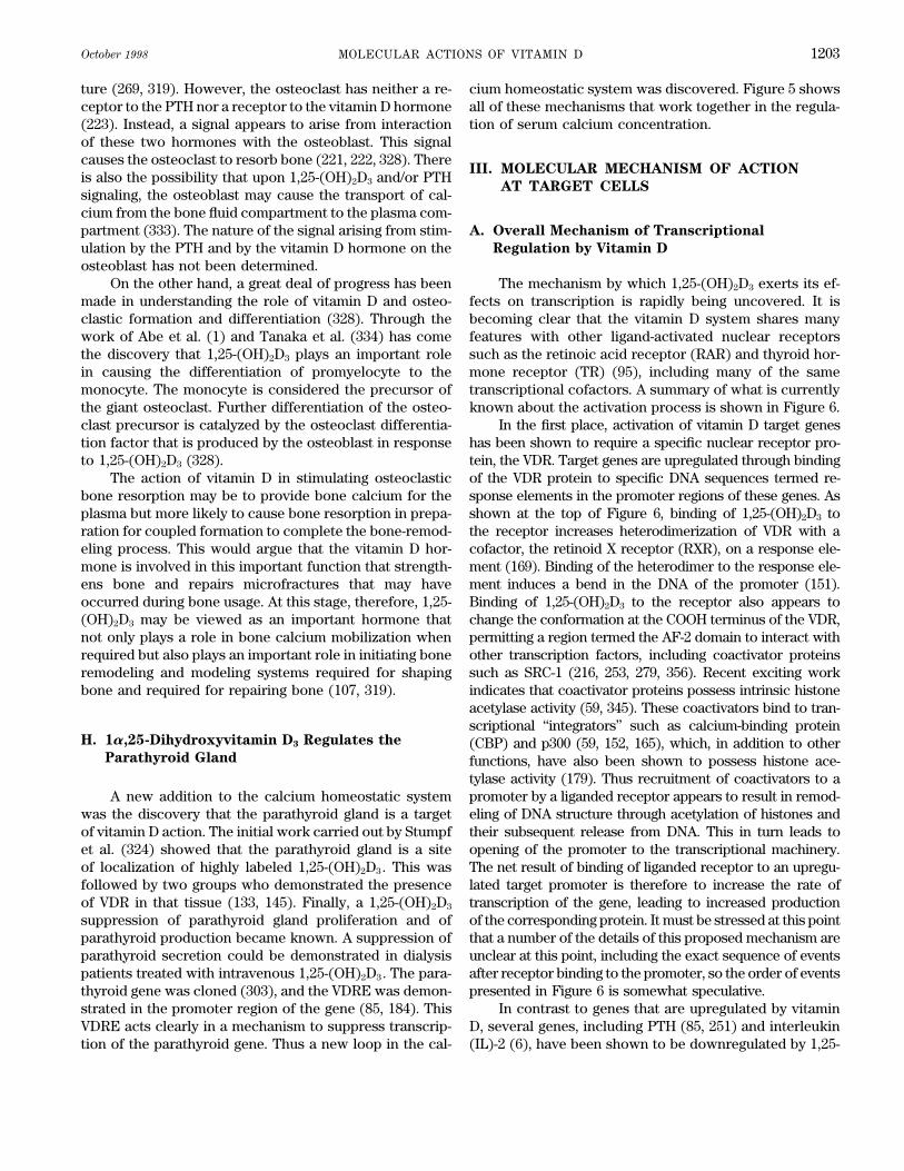
On the other hand, a great deal of progress has been The mechanism by which 1,25-(OH)2D3 exerts its ef-fects on transcription is rapidly being uncovered. It ismade in understanding the role of vitamin D and osteo-
clastic formation and differentiation (328). Through the becoming clear that the vitamin D system shares manyfeatures with other ligand-activated nuclear receptorswork of Abe et al. (1) and Tanaka et al. (334) has come
the discovery that 1,25-(OH)2D3 plays an important role such as the retinoic acid receptor (RAR) and thyroid hor-mone receptor (TR) (95), including many of the samein causing the differentiation of promyelocyte to the
monocyte. The monocyte is considered the precursor of transcriptional cofactors. A summary of what is currentlyknown about the activation process is shown in Figure 6.the giant osteoclast. Further differentiation of the osteo-
clast precursor is catalyzed by the osteoclast differentia- In the first place, activation of vitamin D target geneshas been shown to require a specific nuclear receptor pro-tion factor that is produced by the osteoblast in response
to 1,25-(OH)2D3 (328). tein, the VDR. Target genes are upregulated through bindingof the VDR protein to specific DNA sequences termed re-The action of vitamin D in stimulating osteoclastic
bone resorption may be to provide bone calcium for the sponse elements in the promoter regions of these genes. Asshown at the top of Figure 6, binding of 1,25-(OH)2D3 toplasma but more likely to cause bone resorption in prepa-
ration for coupled formation to complete the bone-remod- the receptor increases heterodimerization of VDR with acofactor, the retinoid X receptor (RXR), on a response ele-eling process. This would argue that the vitamin D hor-
mone is involved in this important function that strength- ment (169). Binding of the heterodimer to the response ele-ment induces a bend in the DNA of the promoter (151).ens bone and repairs microfractures that may have
occurred during bone usage. At this stage, therefore, 1,25- Binding of 1,25-(OH)2D3 to the receptor also appears tochange the conformation at the COOH terminus of the VDR,(OH)2D3 may be viewed as an important hormone that
not only plays a role in bone calcium mobilization when permitting a region termed the AF-2 domain to interact withother transcription factors, including coactivator proteinsrequired but also plays an important role in initiating bone
remodeling and modeling systems required for shaping such as SRC-1 (216, 253, 279, 356). Recent exciting workindicates that coactivator proteins possess intrinsic histonebone and required for repairing bone (107, 319).acetylase activity (59, 345). These coactivators bind to tran-scriptional ‘‘integrators’’ such as calcium-binding protein
H. 1a,25-Dihydroxyvitamin D3 Regulates the (CBP) and p300 (59, 152, 165), which, in addition to otherParathyroid Gland functions, have also been shown to possess histone ace-
tylase activity (179). Thus recruitment of coactivators to apromoter by a liganded receptor appears to result in remod-A new addition to the calcium homeostatic system
was the discovery that the parathyroid gland is a target eling of DNA structure through acetylation of histones andtheir subsequent release from DNA. This in turn leads toof vitamin D action. The initial work carried out by Stumpf
et al. (324) showed that the parathyroid gland is a site opening of the promoter to the transcriptional machinery.The net result of binding of liganded receptor to an upregu-of localization of highly labeled 1,25-(OH)2D3. This was
followed by two groups who demonstrated the presence lated target promoter is therefore to increase the rate oftranscription of the gene, leading to increased productionof VDR in that tissue (133, 145). Finally, a 1,25-(OH)2D3
suppression of parathyroid gland proliferation and of of the corresponding protein. It must be stressed at this pointthat a number of the details of this proposed mechanism areparathyroid production became known. A suppression of
parathyroid secretion could be demonstrated in dialysis unclear at this point, including the exact sequence of eventsafter receptor binding to the promoter, so the order of eventspatients treated with intravenous 1,25-(OH)2D3. The para-
thyroid gene was cloned (303), and the VDRE was demon- presented in Figure 6 is somewhat speculative.In contrast to genes that are upregulated by vitaminstrated in the promoter region of the gene (85, 184). This
VDRE acts clearly in a mechanism to suppress transcrip- D, several genes, including PTH (85, 251) and interleukin(IL)-2 (6), have been shown to be downregulated by 1,25-tion of the parathyroid gene. Thus a new loop in the cal-
/ 9j0c$$oc10 09-23-98 14:33:23 pra APS-Phys Rev
JONES, STRUGNELL, AND DeLUCA Volume 781204
(OH)2D3. The means by which this downregulation is car-ried out is not clear in all cases, and at least two possibilit-ies exist. The first is that, as has been proposed for the IL-2 promoter, VDR may bind to a downregulatory responseelement and disrupt the binding of upregulatory transcrip-tion factors, leading to a decrease in transcription (6). Forother downregulated genes, the situation may be quitedifferent, and binding of VDR to an inhibitory responseelement may lead to interactions with repressor proteinsthat decrease transcription of the gene. Interestingly, co-repressor proteins such as nuclear receptor corepressor(NCoR) (142) and silencing mediator for retinoic acid re-ceptor and thyroid hormone receptor (SMRT) (60) haverecently been found to bind, through intermediary pro-teins, to histone deacetylase enzymes (131, 233). Presum-ably in this situation the deacetylated histones then bindto the promoter of the downregulated gene and shut offtranscription. The VDR has not yet been reported to bindto corepressor proteins, but given the rate at which suchproteins are currently being discovered, it is possible thata corepressor that binds to VDR will be uncovered.
Phosphorylation of the receptor may also play a rolein the induction of transcription by the VDR (73), perhapsthrough modulation of the affinity of VDR for the variouscofactor proteins involved in transcription. Rapid phos-phorylation of the VDR has been shown to occur in organculture systems upon addition of ligand (41). The phos-phorylated residues have been localized to the ligand-binding domain of the protein. The exact functional conse-quences of this phosphorylation have been difficult to de-termine. Estrogen receptor phosphorylation by mitogen-activated protein kinase has been shown to have a directand measurable effect on transcription (167), but thisphosphorylation was mapped to the NH2-terminal AF-1region of the estrogen receptor, which is lacking in VDR.Effects of phosphorylation on activation of the AF-2 do-main, at the COOH terminus of the protein, have not yetbeen clearly shown for the VDR. Indeed, even the kinase(or kinases) responsible for the phosphorylation in vivohas yet to be determined. Some studies have suggestedthat serine-208 of the VDR can be phosphorylated by ca-sein kinase II and that phosphorylation of this residuemay cause increased transcriptional activity (162). Otherwork has revealed that mutation of this serine failed toaffect transcription, although alternate phosphorylationon adjacent serines was noted (135). The exact effects ofphosphorylation must await determination of the preciseamino acids phosphorylated in vivo and the functional
FIG. 6. Proposed mechanism for transcriptional upregulation of a consequences that follow from this.target gene by vitamin D receptor (VDR). After ligand binding, receptorforms a heterodimer on a response element with retinoid X receptor(RXR). Binding of coactivator protein to heterodimer-DNA complex is
B. Vitamin D Receptorsfollowed by histone acetylation and subsequent release of histones fromDNA. Transcription factors are then able to initiate transcription oftarget gene, resulting in production of corresponding protein. RNAP,
The VDR is a member of a superfamily of nuclearRNA polymerase; CBP, calcium-binding protein; DRE, vitamin D re-sponse element. receptors (95). Within this family, the VDR has the highest
/ 9j0c$$oc10 09-23-98 14:33:23 pra APS-Phys Rev

October 1998 MOLECULAR ACTIONS OF VITAMIN D 1205
FIG. 7. Schematic illustration of structure of VDR pro-tein, showing functional domains of which protein is com-posed. Regions of receptor thought to interact with tran-scription factors TFIIB and RXR are shown. COOH-termi-nal AF-2 domain is shown in black.
similarity to the subfamily that includes retinoic acid, thy- NMR and X-ray crystallography. The RXR structure showedthat the DNA-binding domain, comprised of two zinc fingerroid hormone, and peroxisome proliferator activator re-
ceptor (PPAR) receptors, to which it has sequence and motifs, consists of two a-helixes oriented at approximatelyright angles to one another (194). One helix, termed thestructural resemblance. The VDR has been cloned from
several species (12, 42, 92, 164, 219) and shows consider- orientation helix, thought to be critical for recognition ofthe receptor response element was proposed to fit into theable similarity between species in size and sequence. In
the rat, for example, the VDR protein consists of 423 major groove of the DNA and bind to a specific DNA se-quence in the response element. These domains containamino acids, with a molecular mass of Ç50 kDa, whereas
in the human the protein has an additional 4 amino acids two zinc atoms tetrahedrally coordinated to eight conservedcysteine residues. Both the zinc atoms and the cysteineat the NH2 terminus, for a total of 427.
Like the other nuclear receptors, the VDR can be residues are necessary to maintain the three-dimensionalstructure required for response element recognition anddivided by function into several domains. An illustration
of the different domains of the VDR is shown in Figure DNA binding. The amino acid sequence of the DNA-bindingdomain is similar between members of the receptor super-7. At the NH2 terminus is a truncated A/B domain of Ç20
amino acids, to which little function has yet been ascribed family, suggesting that these structures can be used as amodel for that of the VDR.for the VDR. After this, the DNA-binding domain, termed
the C domain, is located between amino acids 20 and 90. There is as yet no direct evidence for the structureof the VDR-RXR complex bound to DNA. However, theA D or hinge domain is located approximately between
amino acids 90 and 130, followed by the COOH-terminal crystal structure of the TR and RXR DNA binding domainscomplexed to a DR4 response element has been deter-E or ligand-binding domain between amino acids 130 and
423. The ligand-binding domain of the protein is a complex mined (272). With the use of this structure as a model,the VDR DNA-binding domain was hypothesized to haveregion of the protein, responsible for high-affinity binding
of ligand, for dimerization with RXR, and for binding to specific amino acid contacts with RXR: between the aspar-agine residue 14 of VDR and RXR residues glutamine-49transcription factors (95). It should be noted that exact
delineation of the division between the hinge region and and arginine-52. In addition, lysine-68 and glutamate-69 ofVDR were modeled to form salt bridges with RXR residuesthe ligand-binding domain is somewhat uncertain and is
based on deletion analysis. aspartate-39 and arginine-38, respectively. These interac-tions were thought to account for the optimal spacing ofKnowledge of the structural and functional proper-
ties of the receptor proteins has increased dramatically three residues between the direct repeats of the vitaminD response element (DR3).in recent years, after the advent of systems which allow
expression and purification of large quantities of receptor The crystal structures of the ligand-binding domains ofthe RXR-a (without ligand) (32), the RAR-g (with ligand)protein from bacteria. This has allowed a piecemeal ap-
proach to determination of receptor domain structures; (279), and the TR-a1 (with ligand) (356) have been deter-mined. The ligand-binding domains of the RXR and RARthe DNA-binding and ligand-binding domains of several
receptors have been expressed separately and purified, had been previously predicted to have a high content of a-helix (64, 204), and this was confirmed by structural analysis.and structures have been determined by NMR or X-ray
crystallography (32, 272). In contrast, to this point, infor- All three ligand-binding domains were found to share a com-mon secondary structure of 12 a-helixes, with a small con-mation concerning the VDR has come primarily from site-
directed mutagenesis, in which the receptor cDNA is mu- tent of b-sheet. A comparison of the structures of the RARand TR with that of the VDR is shown in Figure 8. Thetated and the mutant protein studied by transfection into
mammalian cells (236). No structural data are yet avail- COOH-terminal portion of the proteins, termed the AF-2domain, has been determined to be critical for transcription.able concerning the VDR, but the other receptor domains
may serve as models on which to base hypotheses about Removal of this portion of the protein results in decreasedligand-binding affinity and loss of transcriptional activation.the VDR and its properties.
Structures for the DNA-binding domains of other re- The structural data indicate that the helixes 11 and 12 ofthe RAR and TR may undergo a large conformational changeceptors, including the RXR, have been obtained using both
/ 9j0c$$oc10 09-23-98 14:33:23 pra APS-Phys Rev

JONES, STRUGNELL, AND DeLUCA Volume 781206
FIG. 8. Sequence alignment of human retinoic acid receptor (RAR)-g, rat thyroid hormone recetpor (TR)-a andrat VDR. Sequences of ligand-binding domains of these proteins were aligned using program ‘‘Pileup’’ (Genetics ComputerGroup, University of Wisconsin-Madison). Gaps introduced into sequence to optimize alignment are denoted by dots.Numbering of amino acids of each ligand-binding domain (LBD) is to right of sequences and corresponds to numberingof full-length protein. a-Helical regions of RAR and TR are underlined with solid lines; b-sheet regions are underlinedwith dashed lines. Regions of amino acid identity are enclosed with boxes. Helixes are denoted by numbers in accordancewith TR structure; sheet regions are denoted s1-s4, also according to TR structure. Amino acids in RAR and TR shownby X-ray crystallography to be in contact with ligand are shaded.
in response to ligand binding, folding up around the ligand receptor that contact the ligand remain unknown, al-though some work has been done recently on affinity la-in what the authors of the RAR paper term a mouse trap
action to form a hydrophobic pocket (279). This brings the beling of the binding site (273). Previous work with trans-fected cells has shown that deletion of the NH2-terminalamino acid residues on helix 12 which make up the AF-2
domain into position to interact with other transcription 116 amino acids of the VDR left a protein with measurableligand binding activity, whereas deletion of the NH2-termi-factors. The VDR shows sequence similarity to the RAR and
TR in this region and may be expected to possess a similar nal 160 amino acids did not (220). More recent work withfusion proteins has shown that deletion of the NH2-termi-three-dimensional structure and to undergo a similar confor-
mational change upon ligand binding. nal 124 amino acids gave rise to a functional ligand-bind-ing protein, whereas deletion to amino acid 172 did notThe wild-type VDR binds its ligand, 1,25-(OH)2D3,
with extremely high affinity, in the range of 10010 M (208, (242). From the other end, removal of the COOH-terminal20 amino acids of the VDR resulted in a 10-fold loss of282). Both the 1a- and 25-hydroxyl groups are critical for
high-affinity binding, and the absence of either results in affinity for ligand, whereas removal of more than thisnumber resulted in complete loss of ligand binding (236).approximately a 500-fold decrease in affinity of ligand for
the receptor (82). The exact amino acid residues in the Thus a core domain of Ç300 amino acids was shown to
/ 9j0c$$oc10 09-23-98 14:33:23 pra APS-Phys Rev

October 1998 MOLECULAR ACTIONS OF VITAMIN D 1207
be required for the protein to bind ligand with wild-type rapid nongenomic effects of vitamin D on calcium trans-port, termed transcaltachia (241). The physiological im-affinity. This is in accordance with the ligand-binding do-
mains of the related receptors that have been crystallized, portance of these data is unclear. Whether these effectsare mediated by the VDR or a different protein is alsowhich were all expressed in bacteria as proteins of be-
tween 250 and 300 amino acids. unclear. The development recently of a mouse model inwhich the VDR has been ablated (365) may provide someAn alignment of the VDR ligand-binding domain se-
quence with that of the RAR and TR supports the possibility insight into the relevance of these reports, and intowhether the VDR plays any role in them.that amino acids in the VDR directly in contact with ligand
begin at approximately amino acid 220. In Figure 8, amino Recently, the promoters of the mouse (150) and hu-man (226) VDR have been isolated. Some studies haveacids in contact with ligand in the RAR and TR crystal struc-
tures are shaded. Inspection of this figure shows that the suggested that the VDR is upregulated by treatment withagents such as forskolin, which induce protein kinase Acontact amino acids begin at helix 3 in both the RAR and
TR. Based on sequence alignment, the same region of the (183). Further study of these promoters will shed morelight on the means by which VDR protein levels are regu-VDR begins at approximately amino acid 220, with a leucine
residue that is conserved in all three proteins. Interestingly, lated inside target cells.work in which 10 amino acid segments of the VDR weresequentially deleted found that impairment of ligand binding
C. Retinoid X Receptors and Other Coactivatorsdid not occur until approximately amino acid 230, which isin accordance with this possibility (154).
Site-directed mutagenesis of the VDR ligand-binding In the absence of cofactor proteins, the VDR is unable,at physiological concentrations of protein, to bind to mostdomain has been performed recently on several of the
cysteine residues in the human protein (235). Alteration response elements that have been described. This has be-come clear from both in vitro gel retardation assay experi-of cysteine-288 to glycine resulted in severe attenuation
of ligand binding at room temperature, whereas the same ments and from experiments involving receptor expressionin yeast (155, 231, 310). Work with the VDR and with relatedmutation at cysteine-337 resulted in a smaller decrease in
affinity. The exact significance of this result is uncertain, receptors indicated that the RXR is the required cofactor.The RXR was isolated several years ago, and initiallyalthough a contact between cysteine-237 and carbon-13
of retinoic acid was noted in the binding of retinoic acid its ligand and functional importance were unknown (211).Subsequent work identified 9-cis-retinoic acid as a ligandto the RAR-g (279). Interestingly, in Figure 8, the corre-
sponding cysteine residue (cysteine-284) of the rat recep- for RXR (134). It also became clear that RXR plays a criticalrole in binding to DNA of several different receptors, includ-tor aligns exactly with ligand-contacting amino acids in
the RAR and TR, suggesting that this cysteine may in fact ing the VDR, the TR, the RAR, and the PPAR, among others(98, 173). In the absence of RXR, it appears that none ofdirectly contact the ligand.
The VDR is generally expressed at relatively low levels these receptors binds efficiently to their response elements.Like other receptors, the RXR can be divided into functionalin vivo. Target tissues, such as bone, kidney, and especially
intestine, may have relatively high levels of receptor (3,000– domains, including an NH2-terminal A/B domain, a C domainwhich binds to DNA, and a COOH-terminal DE domain that6,000 fmol/mg protein), but in other tissues, the levels are
generally much lower (72). The receptor has been shown binds ligand and activates transcription (58, 210). Hetero-dimerization with other receptors appears to be mediatedto be present in most tissues that have been examined,
including activated immune cells such as T cells, where it through two interfaces: one in the DNA-binding C domain(366) and another in the ligand-binding E domain (359).may play a role in modulating the levels of cytokines such
as IL-2 (6). In contrast to other receptors, such as the gluco- Interestingly, in structural studies, the RXR ligand-bindingE domain was found to crystallize as a dimer, and the dimercorticoid receptor, which is associated with a number of
proteins, including heat shock proteins, in the cytoplasm interface occurred along helices 9 and 10, exactly wheremutagenesis studies suggest RXR heterodimerizes withbefore hormone binding (146), studies with radiolabeled
1,25-(OH)2D3 have shown that the VDR is predominantly other receptors (32, 261). This region of RXR from aminoacids 389 to 429, termed the I box, is sufficient for strongnuclear (325). Little information is available concerning
whether any proteins associated with the VDR before DNA interaction between RXR and TR, but the same sequenceprovides only weak interaction with VDR. Additional NH2-binding. There are some data suggesting that many hor-
mone-binding receptors, including VDR, contain a binding terminal RXR sequence is required for strong interactionswith VDR (261), indicating a potential distinction betweensite for calreticulin in the DNA-binding domain (43). Some
reports indicate that calreticulin may bind to VDR and inhibit VDR and related receptors.Most vitamin D response elements consist of twoactivation of vitamin D target genes (358), but the physiologi-
cal significance of this is as yet unclear. half-sites of the sequence AGGTCA separated by threebases (see sect. IIID). Because RXR is required for bindingThere have been numerous reports in recent years of
/ 9j0c$$oc10 09-23-98 14:33:23 pra APS-Phys Rev
JONES, STRUGNELL, AND DeLUCA Volume 781208
to the response element, the question arose as to whether number of coactivator proteins such as SRC-1 and TIF-1 (216, 355). Other coactivator proteins that have beenone half-site was preferred by RXR, and if so, whether
this preference was for the upstream or downstream site. discovered recently, including ACTR (59) and p/CIP (345),join the rapidly enlarging group of such proteins. TheFor VDR, as for the other receptors that heterodimerize
with RXR, it appears that RXR binds to the 5*-half-site coactivators seem to fall into a family with many featuresin common, including size (Ç1,400 amino acids), structureand VDR the 3*-half-site (189, 366), although there may
be some exceptions to this rule. The polarity of this bind- (NH2-terminal Per-Arnt-Sim/basic helix-loop-helix do-mains, central receptor interaction domains) and activitying has been shown to be important for maximal gene
activation, with significantly lowered transcription rates (COOH-terminal histone acetylase domains). It seemslikely that most, if not all, of the coactivator proteins willoccurring if the response element orientation is reversed
relative to the promoter start site. Binding of the VDR- be found to interact with VDR and activate transcription.For example, both SRC-1 and TIF-1 have been shownRXR heterodimer to a response element is greatly in-
creased by 1,25-(OH)2D3 when salt concentrations are in to bind to the VDR (216, 355), whereas nuclear receptorcoactivator protein (ACTR) has been shown to enhancethe physiological range, i.e., 100–150 mM (169). Interest-
ingly, for many of the receptors that heterodimerize with transcription from a vitamin D target gene in transfectedcells (59). However, it is not yet clear whether all coactiva-RXR, the natural ligand of RXR, 9-cis-retinoic acid, may
not enhance DNA binding. Some findings indicate that tors interact with VDR equally well; some may functionmore effectively than others in the vitamin D system. AnRXR may be unable to bind ligand while in a complex with
another receptor (98). In addition, some in vitro reports additional possibility is that tissue-specific expressionmay be a significant factor in determining which coactiva-indicate that the VDR-RXR complex is disrupted by addi-
tion of 9-cis-retinoic acid (205). This contrasts with work tor works with the VDR. For example, the coactivatorACTR was shown to be expressed at relatively low levelsin transfected cells indicating that both 1a,25-(OH)2D3 and
9-cis-retinoic acid enhanced reporter gene expression in kidney, an important vitamin D target tissue (59). Thismay indicate that other coactivators play a more im-from a vitamin D-responsive promoter (291). The reason
for these conflicting results is unclear; it may be an experi- portant role in transcriptional activation of vitamin D-controlled genes, at least in this tissue.mental artifact, or it may be that the additional proteins
that interact with the VDR-RXR heterocomplex in vivo, Whether VDR interacts with corepressor proteins tobring about downregulation of target genes is currentlysuch as the coactivator/corepressor proteins, affect the
conformation of the RXR and permit ligand to bind. unclear. Like the coactivator proteins, the corepressorproteins appear to have many features in common. TheThe VDR has been shown to interact directly with a
growing number of other transcription factors, including corepressors SMRT and NCoR (142), for example, haveconsiderable sequence similarity, although NCoR pos-transcription factor IIB (TFIIB). The interaction of VDR with
TFIIB has been characterized in several reports (27, 206). sesses a large NH2-terminal region lacking in SMRT. Inaddition, both proteins appear to repress transcriptionTFIIB is a 30-kDa protein that was originally isolated as a
cofactor associated with TATA-binding protein (120). A re- through interactions with the Sin3 proteins, which bind tohistone deacetylase enzymes (131, 233). However, unlikegion of VDR of Ç70 amino acids in the D domain has been
reported to bind to a 43-residue NH2-terminal region of related receptors such as retinoic acid and TR, it is unclearwhether the VDR is bound by corepressor proteins. AtTFIIB (27, 203). The sequence of TFIIB in this region is
similar to that of a zinc finger, suggesting that the zinc finger least one corepressor protein, NCoR, has been reportednot to bind to VDR at all. NCoR binds to a conservedmotif may function in protein-protein interactions as well
as DNA binding. Interestingly, one report suggests that bind- region of the TR and RAR, termed the CoR box, to exertits effects; binding of ligand to the TR or RAR causesing of ligand to VDR causes dissociation of TFIIB from the
receptor, which may indicate a complex interaction between dissociation of NCoR from receptor. NCoR does not bindto the CoR box region of the VDR, despite the similarityproteins before the initiation of transcription (217).
Interactions between the VDR and other factors are of the VDR sequence in this region to that of TR and RAR(142). This may explain why the VDR does not seem toless well characterized, although this work is progressing,
particularly with regard to the coactivator proteins. Recent exhibit dominant negative silencing seen with both theTR and RAR (262, 286). The possibility remains that otherwork indicates that VDR binds to SUG1/TRIP1, a nuclear
protein that also binds to other receptors (355). It should corepressor proteins will be found to bind to the VDR andthat this interaction is responsible for silencing at leastbe noted that the function of SUG1 in VDR-mediated tran-
scription is unclear. SUG1 binds to the AF-2 domain of the some of the genes that are downregulated by vitamin D.VDR (216, 355), but because SUG1 has been found to be a
D. Vitamin D Responsive Elementscomponent of the proteasome complex in the nucleus, itmay function primarily in receptor degradation. As mentioned in section IIIA, responsive elements are
the sequences of DNA, isolated from the promoters of vita-The VDR has also been shown to bind to a growing
/ 9j0c$$oc10 09-23-98 14:33:23 pra APS-Phys Rev
October 1998 MOLECULAR ACTIONS OF VITAMIN D 1209
min D-responsive genes, that are bound by the VDR. One tivity (197). Thus the results of such in vitro experimentsmust be interpreted with caution and, if possible, in vivo.of the first such response elements isolated was that of the
rat osteocalcin gene (84). Work with this and other vitamin For example, the report of VDR heterodimerization withTR and RAR may potentially be evaluated through the useD response elements, and with the response elements of
other receptors, led to the demonstration that many re- transgenic animals in which RXR has been ablated, todetermine whether VDR-responsive genes can still be ex-sponse elements consisted of two repeats of the half-site
sequence AGGTCA separated by several nonspecified bases pressed. Unfortunately, ablation of RXR-a, the preferredpartner for VDR, is a lethal mutation (90), but it is possible(75, 173, 349). The variable that determines receptor speci-
ficity seems to be the number of nucleotides separating the that inducible knockouts of the RXR-a will provide insightinto whether these other receptors can heterodimerizehalf-sites. The RXR has been shown to have a preference
for two half-sites separated by a single nucleotide, termed with VDR and rescue the expression of VDR target genes.a DR-1 response element, whereas the VDR binds to a DR-3 element, the TR to a DR-4 element, and the RAR to a DR-
E. Vitamin D-Dependent Genes, Their Roles, and5 element (349). Interestingly, almost all of the naturallyGene Complexityoccurring VDRE isolated from genes upregulated by the
VDR have fallen into the DR-3 category. The negativelyregulated genes, such as those for PTH and IL-2, may not Many different genes have been shown to be respon-
sive to vitamin D. Most of these genes play a direct rolefollow this rule. Initial work with the PTH gene indicatedthat the response element in this case consisted of only a in calcium endocrinology or bone formation and were the
earliest examples of vitamin D response elements iso-single half-site. However, more recent work indicates thatthe PTH VDRE may in fact consist of two half-sites (76), lated. Examples of these include osteocalcin (84), osteo-
pontin (243), PTH (251), the hydroxylases CYP24 (368)which may leave only a few exceptions, such as the IL-2gene, to this rule (6). and CYP1a (298, 316, 332), and the calbindin genes (69).
Many of these have been reviewed recently (75). The rolesOne interesting aspect of the interaction between re-ceptor and response element is the bend in the response of many of these genes in calcium endocrinology have
become more evident in recent years. For example, theelement DNA that is induced by binding of the VDR-RXRheterodimer (151). This has been reported for binding bone protein osteocalcin is secreted by osteoblasts and
represents the single greatest protein constituent of boneof related receptors, such as the RAR, to their responseelements (71). The degree of bending induced by the pro- by mass. Expression of this protein is upregulated by vita-
min D, and it may play a significant role in maintainingtein complex is reportedly on the order of 307. Althoughthis bend is much lower in magnitude than that reported bone integrity (73). On the other hand, several genes have
been shown to be downregulated by vitamin D, includingfor binding of the TATA-binding protein to DNA (240), itis still appreciable. It is interesting to speculate on the the gene for PTH, which plays a critical role in controlling
the levels of 1,25-(OH)2D3 in serum.possible role of this induced bending, particularly in lightof the finding that coactivator proteins and their cofactors Several of the hydroxylase enzymes that act directly
on 1,25-(OH)2D3 formation and degradation have beenacetylate histones. It may be that the combination of DNAbending and histone dissociation acts to make the pro- shown to be controlled at the transcriptional level by 1,25-
(OH)2D3. In particular, the hydroxylase enzyme CYP24 ismoter of the target gene more accessible to transcriptionfactors before transcriptional activation. one of the most highly regulated genes that responds to
vitamin D. This enzyme represents the major means bySome recent reports have suggested that a greaterdegree of flexibility exists for binding of VDR to response which 1,25-(OH)2D3 is catabolized in vivo, and its impor-
tance is emphasized by recent gene knockout experimentselements. Work in vitro has suggested that VDR can bindto DR-3 response elements, such as the osteopontin in which loss of the enzyme was found to be lethal to at
least 50% of the recipient mice (313). The CYP24 gene isVDRE, as homodimers (291). Other work suggests thatTR and RAR can act as heterodimeric partners with VDR highly upregulated by 1,25-(OH)2D3 and contains two sets
of VDRE in its promoter (61, 168, 368). Both sets of ele-on a response element, instead of RXR (292). In addition,response elements that do not fall into the DR-3 category ments are required for the gene to display maximum in-
ducibility by vitamin D (168, 368). It therefore appearshave been suggested to bind VDR, including DR-6 ele-ments (264) and one element composed of an inverted that the presence of multiple copies of response elements
are one way in which vitamin D target genes can be effi-repeat of two half-site elements separated by nine bases(292). At present, the significance of this work is unclear. ciently upregulated.
In contrast to the preceding example, the hydroxy-For instance, it has been reported that although VDR canbind to the osteopontin response element as a homodi- lase enzyme CYP1a, which is responsible for catalyzing
the formation of 1,25-(OH)2D3, is highly downregulatedmer, the homodimer is transcriptionally inactive and theVDR-RXR heterodimer is required for transcriptional ac- by its product. This enzyme, expressed in the proximal
/ 9j0c$$oc10 09-23-98 14:33:23 pra APS-Phys Rev

JONES, STRUGNELL, AND DeLUCA Volume 781210
convoluted tubule of the kidney, is regulated with extreme IV. RECENTLY DISCOVERED FUNCTIONS
OF 1a,25-DIHYDROXYVITAMIN D3stringency, although the exact details of the way in whichthis regulation is exerted are unclear. The recent cloningand expression of this enzyme open the door to a more
A. Discovery of New Target Organscomplete understanding of its regulation, and details of
for 1a,25-Dihydroxyvitamin D3the way in which the gene and protein expression areregulated are sure to be forthcoming (298, 316, 332).
The first insight into possible new functions of 1,25-Another set of genes that have been shown to be
(OH)2D3 beyond its actions in the regulation of calciumregulated by vitamin D are the calbindins. In mammals,
and phosphorus as described in section II came as a resultthe calbindin D9K is expressed primarily in intestine,
of studies involving the cellular and subcellular localiza-whereas the calbindin D28K is expressed in kidney and
tion of radiolabeled 1,25-(OH)2D3 at high specific activity.other tissues. The calbindin D9K may play a role in absorp-
This allowed the administration of physiological doses oftion of calcium from the gut, whereas the calbindin D28K 1,25-(OH)2D3 and its detection by frozen section autoradi-may be involved in reabsorption of calcium from glomeru-
ography in target tissues (324, 325). The result of thislar filtrate, although this has not been definitively shown.
study in the late 1970s illustrated that not only was radio-The promoters of both of these genes have been cloned
labeled 1,25-(OH)2D3 localized in the nuclei of the entero-(74, 153, 331), and both have been reported to contain
cyte of the small intestine, the distal renal tubule of theVDRE. Recent work on targeted gene disruption of the
kidney, and the osteoblasts of bone, but were found inD28K has recently been reported in the mouse (311). This
tissues not previously considered targets of vitamin Dis not a lethal mutation, and the effects of this knockout
action. These localizations were found specific inasmuchon regulation of calcium levels will be of great interest in
as the localizations could be prevented by the administra-deciphering the role of this protein in calcium transport.
tion of unlabeled 1,25-(OH)2D3 but not 25-OH-D3. Of greatMore recent and somewhat more surprising discoveries
interest was the finding of 3H-labeled 1,25-(OH)2D3 in theof genes that are regulated by vitamin D include the discov-
nuclei of the islet cells of the pancreas, keratinocytes ofery of VDRE in the promoters for a number of genes not
skin, ovarian tissue, mammary epithelium, epithelial cellstraditionally considered vitamin D targets. These include
of the epididymis, certain neuronal tissue, promyelocytes,potential response elements in the promoters of several
macrophages, and T lymphocytes. This unexpected devel-genes, including integrin b3 (52), fibronectin (264), atrial
opment focused on the possibility that 1,25-(OH)2D3 maynatriuretic factor (163, 198), c-fos (48, 290), PTHrP (182),
carry out functions not previously appreciated and in tis-and p21 (199). The significance of the vitamin D regulation
sues not previously considered targets of vitamin D ac-of many of these genes is unclear and may prove an interest-
tion. This result was followed by the demonstration ofing area of study. In this regard, the recently developed
the VDR in the parathyroid gland (133, 145), in skin (305),mouse strain in which the receptor has been ablated may
in the thymus (267), and in ovarian cells (88). Followingprovide an interesting vehicle for assessing the contribution
this lead came the demonstration that the vitamin D hor-of the VDR to regulation of these genes (365).
mone carries out functions in many of these tissues, andstill other functions remain to be discovered.
F. Target Cell Metabolic Enzymes
The metabolism of vitamin D by target cells has been B. Role of Vitamin D Hormone
in the Parathyroid Glandshown to occur in a number of tissues, including kidney,intestine, and bone. In kidney and intestine in particular,upregulation of the 24-hydroxylase enzyme in response to Perhaps the most well-established new function of
1,25-(OH)2D3 is in the parathyroid gland. Specific localiza-1,25-(OH)2D3 treatment is rapid, occurring within 4 h, asshown by both Northern and enzymatic analyses (297). Cul- tion of 1,25-(OH)2D3 in the parathyroid gland (325) and
the presence of VDR was provided by two independenttured bone cells have also been shown to possess highlyinducible 24-hydroxylase activity (202). These cells have laboratories (133, 145). However, there had been consid-
erable controversy as to whether the 1,25-(OH)2D3 couldbeen used to isolate catabolic products of 1,25-(OH)2D3 thatcan be placed in a logical order on a degradative pathway suppress secretion or production of the PTH (114, 115,
302). Early work failed to provide support for the idealeading from the active compound to inactive excretionproducts (82). Thus it appears that the target cells for vita- that 1,25-(OH)2D3 could, in fact, suppress parathyroid se-
cretion in isolated parathyroid glands. Furthermore, 25-min D contain the means to regulate its activity at the cellu-lar level. The additional level of control that this target cell OH-D3 was continuing to be used as a suppressant of
secondary hyperparathyroidism in renal failure patientsmetabolism provides over local concentrations of 1,25-(OH)2D3 may be an important means by which tissues regu- on dialysis (274). Notably, however, large amounts of this
compound were needed to provide any degree of suppres-late the responsiveness of genes to vitamin D.
/ 9j0c$$oc10 09-23-98 14:33:23 pra APS-Phys Rev
October 1998 MOLECULAR ACTIONS OF VITAMIN D 1211
sion, usually of the order of 20 mg/day. Suppression of hyperproliferation of the keratinocyte and failure to differ-entiate is not found in vitamin D-deficient animals (79;parathyroid levels in such patients with orally adminis-
tered 1,25-(OH)2D3 was also not dramatic. However, when DeLuca, personal observations). It is possible that thedifferentiation may be triggered by more than one systeman intravenous route was used, excellent treatment of
secondary hyperparathyroidism of renal failure resulted providing a redundancy and hence the failure to observea lesion in skin as a result of vitamin D deficiency. To-(308). These results strongly suggested that 1,25-(OH)2D3
may have a direct action through its receptor in the para- gether with the differentiation of the keratinocyte comesan inhibition of proliferation. This inhibition of prolifera-thyroid glands. Thus PTH secretion by isolated parathy-
roid glands or cells could be suppressed by the direct tion has been utilized in the treatment of hyperprolifera-tive diseases of skin as, for example, psoriasis (144, 309).administration of 1,25-(OH)2D3 (82–84). This suppression
required some time to achieve. With the cloning of the Both 1,25-(OH)2D3 and analogs especially MC903 of LeoPharmaceuticals (also termed Dovenex) can be used as apreproparathyroid gene came a search for the possibility
that this gene might be under control of 1,25-(OH)2D3 significant therapy against psoriasis with as many as 70%patients responding to this treatment (44).through its receptor. Analysis of mRNA encoding the PTH
by Northern blot suggested a transcriptional suppression A number of studies have been carried out on themechanism whereby 1,25-(OH)2D3 brings about the differ-by 1,25-(OH)2D3 (284, 296, 304). Subsequently, work by
Demay et al. (85) and Silver et al. (303) have shown un- entiation of the keratinocyte. During the differentiationprocess, there is an increase in the protein involukrinequivocally the presence of a VDRE in the promoter re-
gion of the parathyroid gene. Although this was first (326). There is an increase in transglutamanase activity(326) and cornified envelope formation (263). However,thought to be a single 6-base response element, subse-
quent work has revealed that in actual fact it represents exactly how 1,25-(OH)2D3 induces differentiation of thekeratinocyte and inhibits proliferation is unknown anda DR-3 in which two repeat sequences are indeed found
at 123–108 bases 5* from the transcriptional start site (76). remains to be investigated.Finally, it has been proposed that the keratinocyteThis responsive element system causes a suppression of
expression of the preproparathyroid gene promoter. De- functions in a paracrine fashion in which 1,25-(OH)2D3 isproduced by the keratinocyte itself, and this compoundtails of the mechanism are discussed in section III.
In addition to the role of the vitamin D hormone in serves as a paracrine to stimulate differentiation of thekeratinocyte (21). If this is true, it remains to be ex-suppressing the parathyroid gene is the idea that the vitamin
D hormone also suppresses proliferation of the parathyroid plained how vitamin D-deficient animals are able to ob-tain mature and functional keratinocytes. There havegland cells. The discovery by Suda and co-workers (1, 334)
that 1,25-(OH)2D3 can suppress the growth and stimulate been very dramatic statements made that the keratino-cyte is fully able to metabolize 25-OH-D3 to 1,25-(OH)2D3cellular differentiation of the promyelocytes brought forth
the idea that 1,25-(OH)2D3 might also suppress proliferation but that it lacks the ability to 25-hydroxylate 1a-OH-D3
(21). In work carried out in the rat by two differentof the parathyroid gland cells. This rationale provides fur-ther impetus to the idea that renal osteodystrophy patients laboratories (277, 299), there was a failure to detect any
3H-labeled 1,25-(OH)2D3 in skin or other tissues in ne-should be treated with 1,25-(OH)2D3 to prevent the develop-ment of hypertrophied parathyroid glands as well as to sup- phrectomized rats given the highest specific activity (160
Ci/mmol) of 3H-labeled 25-OH-D3 available. Thus, in nor-press secretion of the PTH.mal rats, the keratinocyte was unable to produce sig-nificant amounts of 1,25-(OH)2D3 in vivo. On the other
C. Role of Vitamin D Hormone in Skin hand, there are a number of scientists who believe thatthis does, in fact, occur in vivo (21). Thus the role of1,25-(OH)2D3 in the keratinocyte under normal physio-After the discovery by Suda and co-workers (1, 334)
of the differentiative action of 1,25-(OH)2D3 on the promy- logical circumstances is unknown, and certainly the ideathat the keratinocyte in vivo is able to produce 1,25-elocytes came the application of this finding to the kera-
tinocytes. Hosomi et al. (144) probably provided the first (OH)2D3 is not universally accepted. Recently, three lab-oratory groups have succeeded in cloning the 25-OH-D-report that 1,25-(OH)2D3 induces keratinocyte differentia-
tion. These results were found by the in vitro addition of 1a-OHase (102, 103, 298, 316, 332), and one report usedPCR techniques to clone it from human keratinocytes1,25-(OH)2D3. Similar results were reported by Holick and
co-workers (309), and it is now common knowledge that (102). This could support the presence of this enzymein keratinocytes; however, Northern analysis of mousethe keratinocytes are induced to differentiate by the in
vitro addition of 1,25-(OH)2D3. Exactly how important this skin (330) did not confirm 1a-OHase mRNA in this tis-sue. Additional work will be required before this areadifferentiation effect of 1,25-(OH)2D3 is in vivo is difficult
to assess. Certainly, vitamin D-deficient animals do not is clarified. However, certainly one has to consider thekeratinocyte as a potential target of 1,25-(OH)2D3 to thehave a problem with keratinocyte differentiation. Thus
/ 9j0c$$oc10 09-23-98 14:33:23 pra APS-Phys Rev

JONES, STRUGNELL, AND DeLUCA Volume 781212
extent that analogs are therapeutically effective againstthe hyperproliferative keratinocyte disease psoriasis.
D. Role of 1a,25-Dihydroxyvitamin D3 in the
Immune System
The presence of the VDR in activated T lymphocyteswas reported by Provvedini et al. (267), who also showedthat resting lymphocytes do not express the VDR. Cer-tainly the thymus, which is a repository of immature lym-phocytes, is a source of VDR, since it was used as a sourceof receptor for measurements of 1,25-(OH)2D3 (138).Among the antigen-presenting cells, the macrophage thatis derived from the monocyte has receptors for 1,25-(OH)2D3 (20, 264). There is no doubt, therefore, that theimmune system does indeed possess receptors to 1,25-(OH)2D3. These results suggest a role for 1,25-(OH)2D3 inthe immune system. However, the role is just now begin-ning to be defined. There have been many reports usingperipheral blood lymphocytes that suggest that produc-
FIG. 9. 1,25-(OH)2D3 prevents progression of experimental autoim-tion of IL-2, tumor necrosis factor-a (TNF-a), and inter-mune encephalomyelitis (EAE) in B10.PL mice. When individual miceferon-g is suppressed by 1,25-(OH)2D3 (193). Furthermore,showed EAE symptoms of 1 or greater, they were given an intraperito-
there are reports of increased synthesis of transforming neal injection containing 300 ng 1,25-(OH)2D3 dissolved in ethanol (l)or mock injected with an equivalent amount of ethanol (s). At time ofgrowth factor-b in a number of immortalized cell lines intreatment, diet was also changed to experimental diet containing noresponse to 1,25-(OH)2D3 in vitro (177). In fact, there areadditional vitamin D or to same diet containing 20 ngrday01
rmouse01
many conflicting reports regarding the in vitro response 1,25-(OH)2D3.of the lymphocytes to 1,25-(OH)2D3. This review focuseson what is known concerning the in vivo responses ofthe immune system in the absence and presence of 1,25- measured certainly in hours, the administration of this com-
pound three times a week or every other day would not be(OH)2D3 and its analogs.In a study of delayed hypersensitivity, which is a T- expected to be sufficient (116). More recently, EAE in B10.PL
mice could be completely prevented by the addition of 1,25-helper cell lymphocyte-mediated response, vitamin D de-ficiency markedly reduces the ability of the mouse to re- (OH)2D3 to the diet of such mice. Furthermore, if the hor-
monal substance was begun after the EAE had begun, thespond (364). However, large amounts of 1,25-(OH)2D3 andits analogs will also suppress the delayed hypersensitivity development of symptoms could be prevented (Fig. 9) (49).
Current results strongly suggest that the vitamin D hormoneresponse (363). These results illustrate that the T-helpercell lymphocyte system is vitamin D responsive but that and its analogs are functioning by stimulating Th-2 T-helper
cells to produce transforming growth factor-b1 and IL-4 thatboth immunostimulation and immunosuppression can befound in the in vivo situation. Currently, there is little might serve to suppress the TNF-a and interferon-g produc-
tion by Th-1 cells (51). Similar results were obtained withor no evidence to support the idea that B cell-mediatedimmunity is affected by 1,25-(OH)2D3. another autoimmune disease, rheumatoid arthritis induced
by Borrelia burgdorferi or Lyme’s disease (50) or arthritisThe most dramatic results obtained to date in the im-mune system are those found in several autoimmune dis- produced by the injection of collagen into susceptible mice
(50). Some improvement has also been reported for the auto-eases. Most notably is the autoimmune disease known asexperimental autoimmune encephalomyelitis (EAE) that can immune induction of diabetes in the nonobese diabetic
mouse (218). The idea, therefore, that the vitamin D com-be induced in B10.PL mice or SJL mice by the injection ofmyelin basic protein together with small amounts of pertus- pounds may play a role in modulating the immune system
is strongly supported. Furthermore, there is the idea thatsis toxin (49). This treatment induces a multiple sclerosis-like disease (EAE). This disease is aggravated by agents that 1,25-(OH)2D3 and its analogs might become useful in the
prevention or treatment of autoimmune diseases.stimulate T-helper (Th)-1 cells and that increase interferon-g and TNF-a secretion (280). Injections once every other Of some interest is the idea that the immunomodula-
tory action of vitamin D might be useful in the manage-day of 1,25-(OH)2D3 in some cases produced a reduction inthe severity and in other cases produced very slight re- ment of transplant rejection. Initial work indicates that
the vitamin D hormone and its analogs would only besponses (36, 196). However, since 1,25-(OH)2D3 has a lifetime
/ 9j0c$$oc10 09-23-98 14:33:23 pra APS-Phys Rev
October 1998 MOLECULAR ACTIONS OF VITAMIN D 1213
marginally useful in prevention of transplant rejection and F. Role of Vitamin D and Reproduction
might only be considered as an adjunct to the well-estab-lished cyclosporin method of preventing transplant rejec- Because of the focus of scientists on the role of vita-tion (195). However, more recently, the administration of min D in calcium homeostasis and regulation of phos-1,25-(OH)2D3 and its analogs in the diet and thus their phate metabolism, the possibility that vitamin D might beprovision in a continuous manner has shown that it can involved in reproduction has been largely ignored in thebe much more effective than cyclosporin itself in preven- past. However, during the course of producing vitamin D-tion of transplant rejection (D. A. Hullett, M. T. Cantorna, deficient embryos came the observation that female repro-C. Redaelli, J. Humpal-Winter, H. W. Sollinger, and H. F. duction is markedly and significantly diminished in vita-DeLuca, unpublished data). It has been used effectively min D deficiency (122). Thus a reduction in fertility ofto prolong transplant survival beyond that achieved by 80% was found and could not be corrected by correctingcyclosporin in mouse embryonic cardiac transplants and the hypocalcemia (191). This defect, therefore, is quitein a vascularized cardiac transplant model in the rat. Of clearly one related to an absence of the vitamin D mole-great interest is that the prevention of transplant rejection cule. Similar female reproductive failure was noted in theby the vitamin D compounds was not accompanied by a transgenic receptor knockout mice described by Yoshi-bone loss phenomenon as is the case with cyclosporin A zawa et al. (365). Why there is a failure in the femaleand, furthermore, was not accompanied by the danger of reproductive system is not known except that it is clearopportunist infection (M. T. Cantorna, D. A. Hullett, C. that ovarian cells contain VDR (88). Furthermore, ovarianRedaelli, C. R. Brandt, J. Humpal-Winter, H. W. Sollinger, cells in vivo also accumulate 1,25-(OH)2D3 (324, 325).and H. F. DeLuca, unpublished data). However, the exact Other organs involved in female reproduction, i.e., therole of how the vitamin D hormone affects T-lymphocyte hypothalamus, also contain VDR. It has been suggestedcell populations, cytokine secretion, and production is not that the ovary therefore is a target of vitamin D action.entirely known. There has been a suppressive element In experiments carried out in the authors’ laboratory, thereported for the IL-2 gene for the VDR. Whether this is a VDR levels of the ovary are increased during estrus andsignificant mechanism or not remains to be determined. diminished during proestrus (348). These results suggest
that female rats may not ovulate properly. The infertilitybrought about by vitamin D deficiency in the female ratcan be easily corrected by the administration of 1,25-E. Islet Cells of the Pancreas(OH)2D3 (191). These results suggest a role for the vitaminD hormone and its receptor in ovarian function; however,
The intriguing finding of a nuclear localization of 1,25- the mechanism remains to be found.(OH)2D3 in the islet cells of the pancreas has led to a In the case of male reproduction, vitamin D deficiencyspeculation that vitamin D might be involved in glucose also reduces the effectiveness of the male (190). However,regulation of insulin secretion (324, 325). The presence of this diminished male fertility can be corrected by merelya VDR in these cells by now is well accepted, but it is providing additional calcium, raising plasma calcium con-unclear as to what, if any, role vitamin D plays in the centration which in turn restores fertility (190). Thus therefunctioning of the islet cells. Initial results reveal that does not appear to be a role for the vitamin D hormone invitamin D-deficient rats were unable to respond to a glu- spermatogenesis and male reproduction.cose challenge by secreting appropriate amounts of insu-lin (65). Although this could be corrected by the adminis-
G. Does Vitamin D Play an Essential Role Duringtration of 1,25-(OH)2D3, other studies suggested that thisEmbryonic Development?was mediated by the action of vitamin D in raising plasma
calcium concentration (66). The problem, therefore, isthat it is unclear whether the regulation of insulin secre- This question intrigued investigators in this field a
couple of decades ago. The interest in this possibility wastion may have a vitamin D component because of thedirect actions of calcium concentration on the functioning intensified when Suda and colleagues (1, 334) demon-
strated for the first time the role of 1,25-(OH)2D3 in promy-of the islet cells. Nevertheless, the idea that inappropriateresponses of the islet cells to a glucose challenge suggests elocyte differentiation. This was followed by work which
illustrated that cancer cell lines also responded by differ-that a role of the vitamin D hormone is involved. Thesefindings must also be coupled to the experiments carried entiating in response to 1,25-(OH)2D3 (91). This then pro-
vided the basis for believing that the vitamin D hormoneout in the nonobese diabetic mouse in which the onset ofdiabetes is delayed or retarded by the administration of is also a developmental hormone. However, vitamin D-
deficient rats are able to reproduce albeit at only 20% of1,25-(OH)2D3 (218). The relationship between vitamin Dand diabetes is certainly worthy of additional investiga- normal, but the embryos develop approximately normally
to parturition (121). Furthermore, the provision of suffi-tion.
/ 9j0c$$oc10 09-23-98 14:33:23 pra APS-Phys Rev

JONES, STRUGNELL, AND DeLUCA Volume 781214
cient nutrients to the pups allows them to develop approx- able calcemic agents capable of the correction of hypocal-cemia and bone abnormalities (rickets and osteomalacia)imately normally until weaning (37). It is at this time that
the dependency on vitamin D becomes very obvious. resulting from a variety of causes from simple vitaminD deficiency to chronic renal failure. There were earlyThese results strongly suggest that vitamin D does not
play an essential role in differentiation and development indications that patients with osteoporosis might benefitfrom the use of an active vitamin D preparation. As aduring embryogenesis. It does not exclude, however, that
the vitamin D hormone plays an important role in terminal result, it is not surprising that clinical trials of 1a-OH-D3
(129, 254), 1a-OH-D2 (106), and 1,25-(OH)2D3 (7, 9, 108,differentiation of specific cell types, as for example thedifferentiation and formation of osteoclasts. It does argue, 257, 343, 351) have been undertaken. Modest gains in bone
mineral density and reductions in fracture rates were re-however, that 1,25-(OH)2D3 is not involved in the develop-ment of major organ systems. These results are supported ported in many of these studies, and this subject has been
reviewed recently by Cali and Russell (44) and Seemanvery strongly by the receptor knockout mice and by thevitamin D-dependency rickets type II cases which are the et al. (293). Consequently, 1,25-(OH)2D3 and 1a-OH-D3 are
currently used to treat osteoporosis, particularly in Japanhuman example of receptor knockouts. In these cases,the fetuses develop normally and, in fact, appropriately and Europe, where dietary calcium intakes are often
lower and hypercalcemic side effects not so prevalent.fed, the animals will survive to day 55 (365). If in theabsence of receptor the major organ systems develop nor- Although vitamin D analogs continued to be synthe-
sized in the late 1970s, the emergence of the newer non-mally, it provides little support for the idea that vitaminD is an important developmental vitamin. One might argue classical functions of 1,25-(OH)2D3, particularly the dis-
covery of its role in controlling cell proliferation and dif-that there are nongenomic actions of 1,25-(OH)2D3; how-ever, there is insufficient evidence for this concept at this ferentiation (228, 252) (see sect. IV), led to an acceleration
in the pace of the search for new analogs. The chemicaltime as well (195). Therefore, it is the view of these au-thors that 1,25-(OH)2D3 is not a major developmental hor- synthesis of new analogs of 1,25-(OH)2D3 became a major
priority with the hope that the calcemic properties of 1,25-mone at least involved in the development of major organsystems during embryogenesis. (OH)2D3 might be separated from the antiproliferative
cell-differentiating properties (119, 157). Hundreds of mol-ecules have now been synthesized in pursuit of this goal,
H. Summary and a couple of recent reviews have attempted to classifyor list all published vitamin D analogs in a detailed fashion
Scientists are just beginning to investigate the new (20, 31, 47, 104). For the sake of brevity, we have selectedand previously unappreciated functions of the vitamin D some of the more promising of these that are under devel-hormone. They include functions in the parathyroid opment by the pharmaceutical industry in Table 1. Thoseglands, the keratinocytes of skin, the T cells and macro- selected are not the only analogs being developed, but allphages of the immune system, the islet cells of the pan- are at some advanced stage of animal or clinical testing.creas, and ovarian cells of the female. There is the possi- From perusal of the compounds in Table 1, the readerbility that additional functions will be found in such sys- will immediately notice that the side chain appears to betems as the islet cells of the pancreas, mammary cell a popular target for modification, and there is a rationalepithelium, and cells in the hair follicle system. basis for this, namely, that the VDR is relatively tolerant
of changes in this part of the molecule. However, since1980, the synthetic chemist has experimented with modi-V. VITAMIN D ANALOGSfication of every part of the vitamin D molecule in anattempt to accentuate either the antiproliferative or cal-
A. Development of New Analogs cemic properties. The full extent of these synthetic modi-of 1a,25-Dihydroxyvitamin D3 fications is best displayed as the annotated structure illus-
trated in Figure 10. Probably the most successful 1,25-(OH)2D3 analog developed thus far is the Leo antipsoriaticThe discovery of 1,25-(OH)2D3 and the other metabo-
lites of vitamin D in the early 1970s immediately led to drug calcipotriol, also known as MC903, which was thefirst vitamin D analog to reach the market for any condi-their chemical synthesis and to the first generation of
compounds suitable for hormone replacement therapy tion other than hypocalcemia or bone disease (45). Whengiven orally, calcipotriol is ineffective due to the fact that(13, 294). In addition to 1,25-(OH)2D3, most notable among
these were the prodrugs 1a-OH-D3 and 1a-OH-D2, which it is rapidly broken down (24, 354). When given topicallyas an ointment, calcipotriol survives long enough to causewere designed to overcome the need for a functional kid-
ney containing the 1a-hydroxylase enzyme, instead, de- improvement in 70% of psoriasis patients (18, 181). Both1,25-(OH)2D3 and calcipotriol are believed to be effectivepending only on the need for 25-hydroxylation (193, 258).
Synthetic 1,25-(OH)2D3 and 1a-OH-D3 proved to be valu- in psoriasis because they block hyperproliferation of kera-
/ 9j0c$$oc10 09-23-98 14:33:23 pra APS-Phys Rev
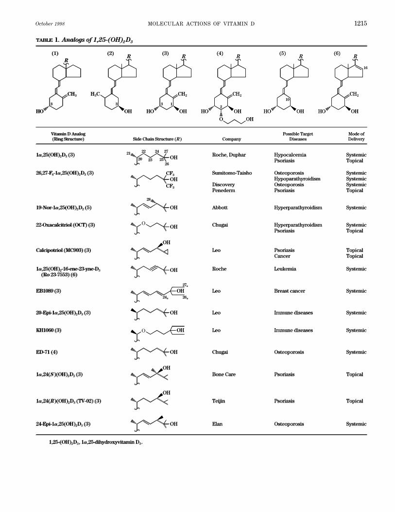
October 1998 MOLECULAR ACTIONS OF VITAMIN D 1215
R
CH3
(1)
TABLE 1. Analogs of 1,25-(OH)2 D3
HO
3 3
22
20
21
23 25
24 27
26
28
R
CH2
(3)
HO
3
R
H3C
(2)
OH
1
OH
OH
R
CH2
(4)
HO2
OH
O OH
R
CH2
(6)
HO
16
OH
R(5)
HO
10
OH
OHCF3
CF3
OH
OH
24a 26a
27a
OH
OH
OH
OH
OH
OH
Calcipotriol (MC903) (3)
OH1a,24(S )(OH)2D2 (3)
OH
1a,24(R)(OH)2D3 (TV-02) (3)
OH
1a,25(OH)2D3 (3)
26,27-F6-1a,25(OH)2D3 (3)
19-Nor-1a,25(OH)2D2 (5)
24-Epi-1a,25(OH)2D3 (3)
1,25-(OH)2D3, 1a,25-dihydroxyvitamin D3.
EB1089 (3)
1a,25(OH)2-16-ene-23-yne-D3 (Ro 23-7553) (6)
KH1060 (3)
20-Epi-1a,25(OH)2D3 (3)
ED-71 (4)
22-Oxacalcitriol (OCT) (3)
Leo
Bone Care
Teijin
Roche, Duphar
Sumitomo-Taisho Discovery Penederm
Abbott
Elan
Leo
Roche
Leo
Leo
Chugai
Chugai
Psoriasis Cancer
Psoriasis
Psoriasis
Hypocalcemia Psoriasis
Osteoporosis Hypoparathyroidism Osteoporosis Psoriasis
Hyperparathyroidism
Osteoporosis
Breast cancer
Leukemia
Immune diseases
Immune diseases
Osteoporosis
Hyperparathyroidism Psoriasis
Topical Topical
Topical
Topical
Systemic Topical
Vitamin D Analog (Ring Structure)
Side Chain Structure (R)
Company
Possible Target Diseases
Mode of Delivery
Systemic Systemic Systemic Topical
Systemic
Systemic
Systemic
Systemic
Systemic
Systemic
Systemic
Systemic Topical
O
O
R
CH3
(1)
TABLE 1. Analogs of 1,25-(OH)2 D3
HO
3 3
22
20
21
23 25
24 27
26
28
R
CH2
(3)
HO
3
R
H3C
(2)
OH
1
OH
OH
R
CH2
(4)
HO2
OH
O OH
R
CH2
(6)
HO
16
OH
R(5)
HO
10
OH
OHCF3
CF3
OH
OH
24a 26a
27a
OH
OH
OH
OH
OH
OH
Calcipotriol (MC903) (3)
OH1a,24(S )(OH)2D2 (3)
OH
1a,24(R)(OH)2D3 (TV-02) (3)
OH
1a,25(OH)2D3 (3)
26,27-F6-1a,25(OH)2D3 (3)
19-Nor-1a,25(OH)2D2 (5)
24-Epi-1a,25(OH)2D3 (3)
1,25-(OH)2D3, 1a,25-dihydroxyvitamin D3.
EB1089 (3)
1a,25(OH)2-16-ene-23-yne-D3 (Ro 23-7553) (6)
KH1060 (3)
20-Epi-1a,25(OH)2D3 (3)
ED-71 (4)
22-Oxacalcitriol (OCT) (3)
Leo
Bone Care
Teijin
Roche, Duphar
Sumitomo-Taisho Discovery Penederm
Abbott
Elan
Leo
Roche
Leo
Leo
Chugai
Chugai
Psoriasis Cancer
Psoriasis
Psoriasis
Hypocalcemia Psoriasis
Osteoporosis Hypoparathyroidism Osteoporosis Psoriasis
Hyperparathyroidism
Osteoporosis
Breast cancer
Leukemia
Immune diseases
Immune diseases
Osteoporosis
Hyperparathyroidism Psoriasis
Topical Topical
Topical
Topical
Systemic Topical
Vitamin D Analog (Ring Structure)
Side Chain Structure (R)
Company
Possible Target Diseases
Mode of Delivery
Systemic Systemic Systemic Topical
Systemic
Systemic
Systemic
Systemic
Systemic
Systemic
Systemic
Systemic Topical
O
O
/ 9j0c$$oc10 09-23-98 14:33:23 pra APS-Phys Rev

JONES, STRUGNELL, AND DeLUCA Volume 781216
FIG. 10. Structure of 1,25-(OH)2D3 showing location of carbon atoms or carbon-carbon bonds modified in variousvitamin D analogs constructed to date. Each circled carbon or bond is connected to a rectangular box which indicates1 or more successful modifications introduced into analog at that site. For sake of space, model does not include allmodifications made but seeks to give reader a flavor of extent of modifications attempted covering whole molecule.Certain popular analogs contained in Table 1 frequently contain combinations of such modifications.
tinocytes, increase differentiation of keratinocytes, and C27a; a 22-oxa group; and epimerization at the C-17 to C-20 bonds giving a side chain with the opposite orientation.help suppress local inflammatory factors through their
immunomodulatory properties. Like several other analogs The synthesis of these 20-epi-analogs (46, 320), as theyare termed, represents a successful experiment in vitamin[1,24S-(OH)2D2 and 1,24-(OH)2D3] currently being pur-
sued, calcipotriol has no 25-hydroxyl group but instead D design because it led to a family of compounds, manywith improved VDR binding compared with the naturalpossesses a 24-hydroxyl group which appears to act as a
surrogate 25-hydroxyl function. Other analogs being side chain. Very recently, several groups (188, 350) haveused this experience to go one step further and synthesizetested, such as 22-oxacalcitriol (OCT) (232), 1,25-(OH)2-
16-ene-23-yne-D3 (11), and EB1089 (23, 161), all contain so-called ‘‘butterfly’’ or ‘‘Gemini’’ analogs with both 20-epi (20S) and normal (20R) side chains. Although little ismodifications at C-22 and C-23 which affect the metabolic
stability of the molecules without adversely altering the known at this time about the biological success of thisexperiment, it is certain to lead to other more exotic dou-VDR binding. 22-Oxacalcitriol is in development for use
in secondary hyperparathyroidism, while EB1089 is in ble-side chain molecules with symmetrical and asymmet-rical structures around C-20 (Fig. 11). In the asymmetricalclinical trial for treatment of breast cancer. Another suc-
cessful modification has been the addition of carbons to double-side chain analog (Fig. 11) featuring one 23-oxaside chain and one normal side chain, X-ray structuralthe backbone or terminus of the side chain (termed ho-
mologation), examples being EB1089 and KH1060 (124). analysis (188) has revealed the orientation of the two sidechains and a measure of the range of conformationalThe latter analog epitomizes the latest generation of vita-
min D analogs, in that it contains a combination of modi- structures that can be accommodated by the ligand bind-ing pocket of the VDR, since it is known to bind both 20S-fications; in the case of KH1060, four modifications: an
extra C at C-24a; additional terminal carbons at C-26a and and 20R-analogs.
/ 9j0c$$oc10 09-23-98 14:33:23 pra APS-Phys Rev

October 1998 MOLECULAR ACTIONS OF VITAMIN D 1217
FIG. 11. X-ray crystallographic structure depicting side chains and D-ring region of a double-side chain vitamin Danalog. (Figure courtesy of Dr. Martin Calverley. Adapted from Vitamin D: Chemistry, Biology and Clinical Application
of the Steroid Hormone, edited by A. W. Norman, R. Bouillon, and M. Thomasset. Riverside: Univ. of California, 1997.)
Equally interesting and perhaps more practical from which appears to extend its lifetime in vivo. An extensivecollaboration between the De Clerq and Bouillon labora-a biological activity perspective are analogs with modi-
fications of the cis-triene, either changing the conforma- tories has generated many new analogs with modificationin the C and D rings, although at present many of thesetion of the double bonds to create molecules with an
altered cis-triene (250) or analogs that lack the C-10 to are only partially tested biologically (29, 78). One interest-ing spin-off from the latter research is the synthesis ofC-19 methylene group altogether such as 19-nor-1,25-
(OH)2D3 or 19-nor-1,25-(OH)2D2 (260). In a sense, these novel E-ring vitamin D analogs in which the C-8–14–13–17 backbone is retained with the loss of the C and D ringslatter compounds represent an extension of the experi-
ment begun over 60 years ago (97) when German chem- of the vitamin D nucleus but involving the creation of anew stabilizing E ring utilizing C-13, C-12, C-21, C-20, andists successfully stabilized the labile cis-triene of one of
the photoirradiation products of vitamin D and created C-17. The resulting 1,25-dihydroxylated E-ring analogKS176 has 10-fold lower VDR binding and 5-fold lowerthe world’s first vitamin D analog, dihydrotachysterol
(DHT). This analog DHT has an A-ring rotated 1807 and DBP binding than 1,25-(OH)2D3 but is virtually inactive incalcemic assays in vivo (29). It will be interesting to seehas lost the double bond between C-10 and C-19 but
retains the C-19 as a methyl group not a methylene, as why E-ring analogs show good in vitro activity but appearto be devoid of in vivo activity.is the case in almost all other vitamin D compounds.
Dihydrotachysterol is metabolized to 25-hydroxylatedand 1,25-dihydroxylated forms, both of which bind to
B. Factors That Alter the ActionVDR and are biologically active, explaining why DHT
of Vitamin D Analogswas once the analog of choice for hypocalcemia, particu-larly that associated with chronic renal failure (158, 268).
From the synthesis of literally hundreds of analogs hasThe experiments with 19-nor-1,25-(OH)2D2 and DHT
emerged a clearer understanding of the factors that are im-show that the 10–19 double bond and the C-19 carbon
portant in changing the action of the specific analog relativeare probably superfluous for vitamin D-dependent gene
to 1,25-(OH)2D3. These are listed in the following sections.expression. As with several other analogs, 19-nor-1,25-(OH)2D2 appears effective in the treatment of hyperpara-
1. Activating enzymesthyroidism associated with chronic renal failure (307).
Further excursions into A-ring or C- and D-ring modi- The natural hormone of vitamin D is activated bysequential steps in the liver and kidney as described infications are currently underway. One molecule into clini-
cal trial for the treatment of osteoporosis is the Chugai section I. The distribution and pharmacokinetics of 1,25-(OH)2D3 depend on these steps occurring efficiently atanalog ED-71, which is modified at C-2 of the A ring by
the addition of a hydroxypropoxy group (241), a feature substrate concentrations that circulate bound to DBP and/
/ 9j0c$$oc10 09-23-98 14:33:23 pra APS-Phys Rev
JONES, STRUGNELL, AND DeLUCA Volume 781218
or are available in the respective tissues. The strategy for DBP is inversely correlated with its rate of clearancefrom the bloodstream. Those metabolites with the strong-with most synthetic analogs is to synthesize a molecule
with both 25- and 1a-hydroxyl groups and miss out the est affinity for DBP [e.g., 25-OH-D3, 24,25-(OH)2D3, and25-OH-D3-26,23-lactone] possess the longest half-life val-need for these activation steps. The disadvantage of this
approach is that the body loses its ability to regulate the ues in the blood on the order of days (17, 26). Thosemetabolites such as 1,25-(OH)2D3 have lower affinity andamount of the active principle in the blood and target cell,
with the success of the therapy being heavily dependent half-life values on the order of hours. These basic con-cepts were confirmed by our experience with analogs ofon the pharmacokinetics of the specific analog.
On the other hand, the use of prodrugs such as 1a- 1,25-(OH)2D3.Clearly, some modifications are detrimental to DBPOH-D3 circumvents some of these problems by introduc-
ing a step of activation back into the process, albeit by a binding such as the addition of a 1a-hydroxyl group. Othermodifications such as the addition of the double bonddifferent regulatory enzyme. This activating enzyme is in
essence a ‘‘slow-release’’ mechanism for delivering the between C-22 and C-23 or the addition of a C-24 methylgroup, as in the metabolites of vitamin D2, do little tosame ‘‘active form,’’ 1,25-(OH)2D3, with altered pharmaco-
kinetics. This is because the activating enzyme has been affect binding to mammalian DBP but dramatically lowerthe binding of these compounds to avian DBP (17).changed from the tightly regulated renal 1a-hydroxylase
to the loosely regulated liver 25-hydroxylase. Although However, the findings with D2 compounds with mamma-lian DBP go against the general rule, and most 1a-hydrox-this was believed to be a simple strategy to get around
dependence on the easily damaged renal enzyme when it ylated, side chain-modified analogs have inferior DBPbinding compared with 1,25-(OH)2D3. Calcipotriol, OCT,was conceived in the 1970s, the emergence of CYP27 as
one of the enzymes responsible for the 25-hydroxylation EB1089, KH1060, and 20-epi-1,25-(OH)2D3 all bind DBPwith an affinity at least one order of magnitude less thanof vitamin D3 and 1a-OH-D3 has complicated this story.
CYP27 assumes an even greater importance for the activa- 1,25-(OH)2D3 (89, 171). One analog, the A ring-substituted,1a-hydroxylated compound ED-71, binds DBP moretion of 1a-OH-D3 because several studies have shown that
this cytochrome P-450 prefers 1a-OH-D3 as a substrate strongly than the natural hormone, a fact which dramati-cally increases its survival time in the blood and henceover vitamin D3 itself (8, 119). CYP27 is found in extrahe-
patic sites including kidney, bone, and endothelial lining its usefulness (241).The consequences of altered DBP binding are alteredcells. In theory, this makes the activation of 1a-OH-D3
possible in many sites of the body in addition to the liver tissue distribution of analogs and changes in the rates oftheir metabolic clearance and cell uptake. Reduction inand opens up the possibility that 1,25-(OH)2D3 may be
formed from 1a-OH-D3 in target cells like the osteoblast the affinity of DBP binding can lead to transport on alter-nate carriers, either albumin or lipoproteins. It has been(147) to act locally on vitamin D-dependent processes.
This theory remains to be proven in vivo. claimed that OCT derives part of its usefulness for treat-ment of hyperparathyroidism from its accumulation inAnother aspect of the activation strategy that was
overlooked at first glance was the potential of producing parathyroid tissue in vivo, possibly caused by an alteredplasma transport on lipoprotein (176).multiple active forms from the same administered pro-
drug. The analog 1a-OH-D2 has consistently proven to be Second, the rate of metabolic clearance is affectedby altering DBP affinity. Consistent with the findings forless toxic than its D3 counterpart in animal studies and
clinical trials (93, 306). The explanation for these findings vitamin D metabolites, a reduction in DBP binding for agiven vitamin D analog leads to even more rapid excretionremains obscure but may be due to the fact that 1a-OH-
D2 gives rise to two biologically active products, 1,25- than that observed for 1,25-(OH)2D3. Calcipotriol bindsDBP with an affinity two to three orders of magnitude(OH)2D2 and 1,24-(OH)2D2 (320), each with its own set of
slightly different pharmacokinetic parameters compared lower than 1,25-(OH)2D3 (171). When given systemically,calcipotriol is metabolized and excreted so rapidly that itwith 1,25-(OH)2D3 (175). CYP27 is capable of the efficient
synthesis of 1,24-(OH)2D2 (119). The enzyme responsible has no biological effects even at 50 times the dose usedfor the natural hormone. As described above, it is onlyfor the synthesis of 1,25-(OH)2D2 remains unknown.
Whether this strategy of delivering a prodrug which be- effective because it is topically administered in an oint-ment and is rapidly cleared (and metabolized) before itcause of its structure is activated by different enzymes
with altered distribution or gives rise to more than one can reach tissues involved in calcium homeostasis. ThusDBP binding is an important parameter in dictating inactive metabolite offers therapeutic advantages remains
moot at this time. vivo biological activity.Third, the affinity of the analog for DBP influences
2. Binding to DBP the rate of cell uptake. Given that it is the ‘‘free’’ fractionof the plasma-borne ligand which is believed to gain ac-It has been recognized since the discovery of the
various vitamin D metabolites that the affinity of an analog cess to the target cell and function by binding to the recep-
/ 9j0c$$oc10 09-23-98 14:33:23 pra APS-Phys Rev

October 1998 MOLECULAR ACTIONS OF VITAMIN D 1219
tor (VDR) in the classical steroid hormone model (see consistently only one or two orders of magnitude morepotent than 1,25-(OH)2D3 in gene transactivation assays,sect. III), it is therefore no surprise that this might also be
the case for vitamin D analogs. Many analogs have the a difference that could be explained by using the tran-scriptional model of analog action in conjunction withsame affinity for VDR as 1,25-(OH)2D3, which when com-
bined with a lower affinity for DBP than 1,25-(OH)2D3 pharmacokinetic considerations rather than discardingthe genomic hypothesis all together.creates an even greater downhill gradient into target cells.
This has been clearly demonstrated in cultured cells in With the finding that the liganded VDR probably func-tions transcriptionally as a vitamin D-VDR-RXR heterodimervitro where the composition of the extracellular fluid can
be easily modified. Whereas DBP in fetal calf serum (128, 322) but can form VDR-VDR homodimers (53, 54) hascome the question as to whether vitamin D analogs mightslightly retards entry of 1,25-(OH)2D3, it has no discernible
effect on the entry of 20-epi-1,25-(OH)2D3, a notoriously differ from 1,25-(OH)2D3 and favor association with VDR-VDR homodimers. Although it remains possible that vitaminpoor binder to DBP (86). There is no doubt that poor DBP
binding contributes positively to the promising profile of D analogs bound to VDR-VDR homodimers might preferen-tially act at certain VDRE in selected vitamin D-dependentmany vitamin D analogs in biological assays in vitro.
On the other hand, when this effect of DBP, greater genes (e.g., those associated with immunoregulatory or cellregulatory responses), currently the data do not supportaccess to target cells, is considered along side its other
effect, more rapid clearance at the liver, in the animal in this (128, 322). It should be noted that in a recent review(53) it was theorized that 1,25-(OH)2D3 (and thus vitaminvivo, the net effect is usually not so advantageous. This
is presumably because these two effects are counteractive D analogs) might be able to act through as many as 14different heterodimeric response element combinations toand the vitamin D analog cannot be active unless it
reaches the target cell in sufficient concentrations to gain modulate gene expression, but the physiological relevanceof the majority of these has yet to be established.greater access and produce its target cell biological ef-
fects. In the case of analogs such as ED-71, chemists have To add to the complexity of the target cell action of1,25-(OH)2D3, and thus that of vitamin D analogs, is thecreated a longer lasting analog that may give reduced
biological activity but for a longer period of time. In es- type and context of the VDRE involved (40). The work ofWilliams and co-workers (40, 166, 360) shows that com-sence, they have altered the pharmacokinetics again.pared with 1,25-(OH)2D3, KH1060 and EB1089 show differ-ent patterns of gene activation in bone marrow, osteoblas-3. Binding to VDR-RXR-VDRE complexestic cells (ROS17/2, ROS25/1, and UMR106), and intestine(HT-29 and Caco-2) which appear to be gene and cellSection III of this review has established that 1,25-
(OH)2D3 is able to work through a VDR-mediated genomic specific. These data are probably explained by thechanges in available transcription factors acting at VDREmechanism to stimulate transcriptional activity at vitamin
D-dependent genes. Whether 1,25-(OH)2D3 works through in different tissues and at different stages of cell differenti-ation (317). Furthermore, there have been reports of aother nongenomic mechanisms to produce physiologi-
cally relevant effects is a theory that, in our opinion, re- nonclassical VDRE in the promoter of the c-fos gene (48)that could theoretically respond differently to ‘‘noncal-mains unproven (244).
Much evidence exists to support the viewpoint that cemic’’ and ‘‘calcemic’’ analogs, but this has not been dem-onstrated experimentally. Furthermore, Morrison and Eis-vitamin D analogs operate by mimicking 1,25-(OH)2D3 and
using a genomic mechanism. The first clue that vitamin D man (229) showed that a noncalcemic analog such ascalcipotriol is capable of transactivating a calcemic VDREanalogs can work through a VDR-mediated transcriptional
mechanism came 15 years ago from the bone resorption such as the human osteocalcin promoter VDRE placed infront of the chloramphenicol acetyltransferase reporterstudies reported by Stern (318). Stern (318) showed that
there exists a strong correlation between chick intestinal gene and stably transfected into ROS17/2 cells. One inter-pretation of this experiment is that a noncalcemic analogVDR binding of an analog and its potency in a 45Ca rat
bone resorption assay. This suggests that a vitamin D ana- with good VDR affinity is just as calcemic as 1,25-(OH)2D3
if it can be delivered to the target cell.log is only as good as its affinity for the VDR. Certainly,there are some apparent exceptions to this general rule, A more recent hypothesis put forward to explain the
unique properties of certain vitamin D analogs is that theybut these are not discrepancies that cannot be explainedby considerations such as DBP binding or pharmacokinet- generate different conformations of the ligand-VDR-RXR
heterodimer which result in altered binding at the VDRE.ics. Preliminary results with the analogs KH1060, EB1089,and 20-epi-1,25-(OH)2D3 (25) suggested that they might be Such subtle changes in protein conformation may still be
detected by indirect biochemical means such as altered sen-active in immunoregulatory roles at concentrations ordersof magnitude below their affinities for the VDR (e.g., at sitivity to protease digestion. Recent studies have revealed
differences between 1,25-(OH)2D3-VDR-RXR and 20-epi-1,25-as low as 10015 M for KH1060 whereas it binds VDR at10011 M). More recent results suggest that KH1060 (55) is (OH)2D3-VDR-RXR or KH1060-VDR-RXR complexes to tryp-
/ 9j0c$$oc10 09-23-98 14:33:23 pra APS-Phys Rev

JONES, STRUGNELL, AND DeLUCA Volume 781220
sin digestion, suggesting subtle changes in the conformation ject to target-cell catabolism and side chain cleavage viaa 24-oxidation pathway to calcitroic acid. CYP24 carriesof the ligand binding domain after ligand binding (259, 352).
The increased resistance to trypsin digestion provided to out multiple steps in the C-24 oxidation process, is vitaminD inducible, and is present in many (if not all) vitamin DVDR by KH1060 binding is mimicked by the most active
metabolites of KH1060, suggesting that certain receptor con- target cells. We have postulated that the purpose of thiscatabolic pathway is to desensitize the target cell to con-formations might correlate with increased biological activity
(353). The use of truncated VDR mutants has allowed the tinuing hormonal stimulation by 1,25-(OH)2D3 (203). Onecan also ask the question: Are vitamin D analogs subjectwork on KH1060 to be extended and to potentially reveal
the amino acid residues involved in these conformational to the same catabolic metabolism? And, if not, are otherdrug-catabolizing systems present within vitamin D targetdifferences and in ligand binding (200). In the future, it is
to be expected that the generation of large quantities of cells to inactivate the vitamin D analog?We now have a good idea as to the answer to thesethe ligand-binding domain of the VDR using bacterial or
baculovirus-generated material (323) will permit the deter- questions, but the answers are analog specific. Certainly,there are metabolism-sensitive analogs such as calcipo-mination of the three-dimensional structure with and with-
out ligand [1,25-(OH)2D3 or vitamin D analog] by NMR or X- triol and OCT that are metabolized very rapidly by targetcell enzymes to clearly defined and unique metabolitesray crystallographic analysis. For now, we can draw on the
experience gained from related fields. If the findings from (213, 215) that resemble products of the 24-oxidation path-way for 1,25-(OH)2D3. The provision of such features asX-ray crystallographic studies of various retinoid receptors
(apo-RXR and holo-RAR ) are relevant to VDR, then the a 22-oxa group seems to accelerate the rate of metabolismof OCT, and the 24-hydroxyl group of calcipotriol is a siteorientation of the COOH terminus containing the AF-2 do-
main will turn out to be dramatically altered upon vitamin for deactivation. The rapid metabolism of calcipotriol bycultured keratinocytes in vitro (215) correlates well withD analog binding and to be very important to the interaction
with the other proteins of the transcription initiation com- the in vivo findings that it is rapidly broken down whenadministered topically and fails to reach tissues involvedplex (279). Moras and co-workers (361) have already used
computer modeling based on his RAR model to predict the in calcium homeostasis (24). One does not need to invokehepatic metabolism as the principal site of inactivation,orientation of ligands including vitamin D analogs in the
binding pocket of the VDR. He predicts that 1,25-(OH)2D3 even though this tissue can also break down calcipotriol(24, 215).and various analogs bind with side chain deep into the li-
gand-binding pocket and A ring close to the putative ‘‘trap- Analogs that appear to be slowly metabolized by tar-get cells include EB1089 and 1,24S-(OH)2D2 (156, 170,door’’ (AF-2 domain). Therefore, the observed differences
in protease resistance induced by potent 20-epi compounds 295). Both are considered more calcemic than either calci-potriol or OCT but not excessively so. These data aresuch as KH1060 (259, 352) could be important clues that
the AF-2 domain of the heterodimer-analog complex is in a consistent with metabolism-resistant analogs having alonger half-life inside the target cell as compared withslightly different conformation to that of the heterodimer-
1,25-(OH)2D3 complex. As a result, these complexes may 1,25-(OH)2D3 and a longer half-life in pharmacokineticstudies (30, 175), and possibly a higher biological activityrecruit additional transcription factors (see sect. III) that
may result in qualitative or quantitative differences in gene in vivo, although this is tempered by inferior DBP bindingand more rapid clearance from the bloodstream. Interest-expression.
There is some evidence from surface plasmon reso- ingly, both EB1089 and 1,24S-(OH)2D2 are blocked at theC-24 position and cannot enter the C-24 oxidation path-nance studies that a series of Roche analogs causes differ-
ences in the association and dissociation rates of VDR- way, leaving C-26 as the new site of hydroxylation, albeitat a slower rate (156, 170, 295). Another potent analogRXR heterodimers to artificial VDRE-containing oligonu-
cleotide anchored to gold-coated chips (67). Whether this blocked in the C-24 position is 1,25-(OH)2-16-ene-23-yne-D3, which is also subject to 26-hydroxylation (288). Stud-translates into increased stability of specific complexes on
the vitamin D-dependent gene promoter in vivo remains to ies with EB1089 using transfected human CYP27 suggestthat this cytochrome is not responsible for the 26-hydrox-be proven. In summary, researchers continue to probe the
many steps of the gene transcription activation process ylation observed (295). However, Suda and co-workers(227) have concluded that the other candidate cyto-in the hope of finding a fundamental difference between
1,25-(OH)2D3 and its analogs that may explain their im- chrome P-450, CYP24 may be responsible for 26-hydroxyl-ation of the 24-blocked analog 24,24-F2-1,25-(OH)2D3 in-proved potency or their action at only selected genes.
Thus far, they have more questions than answers. side kidney cells.Another metabolic complication brought on by the
4. Target cell and liver catabolic enzymes use of vitamin D analogs is the potential for creating‘‘active’’ rather than ‘‘inactive’’ products as a result ofAs stated in section I, much evidence has accumu-
lated to support the hypothesis that 1,25-(OH)2D3 is sub- target cell enzymes. Three examples of this have now
/ 9j0c$$oc10 09-23-98 14:33:23 pra APS-Phys Rev
October 1998 MOLECULAR ACTIONS OF VITAMIN D 1221
been documented, and this may be more common than more general catabolic enzymes found in the liver. Oneinteresting example is the enzyme that saturates the dou-we think. The potent calcemic analog 26,27-F6-1,25-
(OH)2D3 is converted both in vivo and in vitro, presum- ble bond between C-22 and C-23 in the side chain ofcalcipotriol but does nothing to the same bond in theably by the cytochrome CYP24 into 26,27-F6-1,23,25-
(OH)3D3. Sasaki et al. (287) have shown that this metabo- side chain of vitamin D2 (215). The simple replacementof the 24-methyl of the D2 side chain with a 24-hydroxyllite which accumulates in bone in vivo is fivefold more
active than 1,25-(OH)2D3 in a reporter gene expression group in calcipotriol is enough to allow the saturase en-zyme to function. As vitamin D analogs continue to devi-system. Thus a metabolite may explain the long-lasting
effects of the parent analog. In a second example, Dil- ate more from the classical structure, they become moreand more susceptible to general purpose or unrelatedworth et al. (87) recently showed that KH1060 is metabo-
lized very rapidly in vitro and in vivo into as many as 22 enzyme systems.It should be noted with regard to molecular mecha-metabolites but that some of these metabolites retain
significant biological activity in similar reporter gene nisms of action at the target cell level that metabolism isoften disregarded or given too little emphasis. Metabolicassay systems. The most abundant of these metabolites
include 26-hydroxy and 24a-hydroxy KH1060, which can assumptions are made when testing biological activity invitro that are not always valid. These include 1) the analogbe detected in the blood in vivo, are stable in vitro, and
as stated earlier can form trypsin-resistant complexes is biologically active as administered and 2) the analoghas the same stability as 1,25-(OH)2D3 in the in vitro targetwith VDR (87, 353). At the very least, these active metab-
olites of KH1060 may explain its high potency when com- cell model used, whether in in vitro organ culture, cul-tured target cell, or host cell/reporter gene construct. Thepared with 1,25-(OH)2D3 using in vitro assays. In addi-
tion, the findings may also explain the disappointing per- validity of this approach is made even more tenuous whendata acquired with different in vitro models, where meta-formance of KH1060 in vivo, since the metabolites are
not allowed to accumulate and are rapidly cleared from bolic considerations may or may not apply, are comparedwith data acquired in vivo, where metabolic considera-the bloodstream. The third example of possible activa-
tion of an analog is 3-epimerization (275). Using in vitro tions do apply. The reader is cautioned that invalid com-parisons of in vivo and in vitro data abound in this field.cultured cell and perfused kidney systems where artifi-
cial media and high analog concentrations are employed, The outcome is that a number of promising in vitro-testedvitamin D analogs have been found to be inactive whenseveral researchers have observed the 3-epimerization
of vitamin D analogs [e.g., 1,25-(OH)2D3 itself] (214, 224, tested in vivo.275). As with the example of KH1060, the claim is not
5. Relative importance of key factors on vitamin Dthat the metabolic product 3-epi-1,25-(OH)2D3 is more
analog actionactive than the parent compound, just that it retains sig-
It is evident that vitamin D analogs differ from 1,25-nificant biological activity and is more stable, not being(OH)2D3 in a variety of ways: activating enzymes, DBPsubject to the C-24 oxidation pathway which is the fateaffinity, VDR-RXR-VDRE-gene association, and catabolicthat befalls the hormone. The nature of the enzyme in-enzyme susceptibility. Any one or a combination of thesevolved is currently undefined but is reminiscent of thedifferences could be important in making a specific analogbehavior of steroidal 3a- and 3b-oxidoreductases. More-appear calcemic or noncalcemic or work in vitro but notover, the formation of 1a- and 1b-hydroxylated DHT inin vivo, or vice versa. Although it is attractive at this timerats and humans in vivo, referred to in section VA, resem-to consider as most important the subtle VDR conforma-bles the 3-epimerization of vitamin D analogs and sug-tional differences induced by analogs as compared withgests that the process may be more than just an in vitro1,25-(OH)2D3, there is a wealth of information that arguesartifact. Nevertheless, the main hurdle for proponents ofthat metabolism, transport, and pharmacokinetics play athe 3-epimerization hypothesis is that they must showmajor role in vivo.the metabolic step can occur in vivo.
With our existing in vitro cell systems, it is difficultThe final metabolic consideration is hepatic modifi-to study any one of these factors to the total exclusion ofcation. The liver is believed to be devoid of VDR and theall others. Thus it seems unlikely that we shall be able toinducible cytochrome P-450; CYP24 and cultured liverdetermine the relative importance of all of the factorscells (Hep G2 and Hep 3B) do not appear to degrade thelisted until we have a fully functional cell-free in vitrohormone 1,25-(OH)2D3 very rapidly (213, 215). However,model for transcription so that analogs can be tested with-the story with vitamin D analogs appears to be quiteout the complication of metabolic enzymes.different. Analogs such as calcipotriol and OCT are bro-
ken down by liver enzymes, these presumably contribut-C. Future Directions in Vitamin D Drug Design
ing to the rapid metabolic clearance observed in vivo(213, 215). As the analog structure is modified away from The growing availability of recombinant cytochromes
P-450 will allow for a search for potential inhibitors. Suchthe vitamin D prototype, it becomes vulnerable to other
/ 9j0c$$oc10 09-23-98 14:33:23 pra APS-Phys Rev
JONES, STRUGNELL, AND DeLUCA Volume 781222
Cloning, structure and expression of the mitochondrial cytochromespecific inhibitors of CYP24 may be of value in blockingP-450 sterol 26-hydroxylase, a bile acid biosynthetic enzyme. J.
1,25-(OH)2D3 catabolism. Modeling of the vitamin D bind- Biol. Chem. 264: 8222–8229, 1989.ing pocket of VDR, DBP, and the three vitamin D-related 8. ANDERSSON, S., I. HOLMBERG, AND K. WIKVALL. 25-Hydroxyla-
tion of C27-steroids and vitamin D3 in rat liver microsomes. J. Biol.cytochrome P-450 will become a major goal now that allChem. 258: 6777–6781, 1983.
these specific proteins have been cloned and overex- 9. AXEN, E., T. BERGMAN, AND K. WIKVALL. Microsomal 25-hydrox-pressed. Although the full-length proteins are slightly be- ylation of vitamin D2 and vitamin D3 in pig liver. J. Steroid Mol.
Biol. 51: 97–106, 1994.yond the current limits of NMR or X-ray crystallography,10. AXEN, E., H. POSTLIND, AND K. WIKVALL. Effects on CYP expres-
the ligand-binding domains or pockets are not. It is likely sion in rat kidney and liver by 1a,25-dihydroxyvitamin D3, 1a-hy-that technical problems with these procedures will be droxylase activity. Biochem. Biophys. Res. Commun. 215: 136–
141, 1995.overcome shortly and the full-length proteins can be tack-11. BAGGIOLINI, E. G., J. J. PARTRIDGE, S. J. SHIVEY, G. A. TRUITT,led. The membrane-associated region of cytochromes P- AND M. R. VSKOKOVIC. Cholecalciferol 23-yne derivatives, their
450 poses problems, but enormous strides have been pharmaceutical compositions, their use in the treatment of calcium-related diseases, and their antitumor activity, US 4,804,502. Chem.made based on models built with soluble prokaryotic iso-Abstr. 111: 58160d, 1989.forms. Although the first priority is to study 1,25-(OH)2D3 12. BAKER, A. R., D. P. MCDONNELL, M. HUGHES, T. M. CRISP, D. J.
binding to the VDR, the binding of a variety of analogs to MANGELSDORF, M. R. HAUSSLER, J. W. PIKE, J. SHINE, AND
B. W. O’MALLEY. Cloning and expression of full-length cDNA en-all these proteins is likely to follow. In the process, we willcoding human vitamin D receptor. Proc. Natl. Acad. Sci. USA 85:be able to explore the permitted boundaries for optimal 3294–3298, 1988.
protein binding. More rational vitamin D analog design 13. BARTON, D. H., R. H. HESSE, M. M. PECHET, AND E. RIZZARDO.A convenient synthesis of 1-hydroxy-vitamin D3. J. Am. Chem. Soc.will follow to take advantage of structural idiosyncrasies95: 2748–2749, 1973.of all of these key proteins. Meanwhile, the not-so-rational 14. BAUER, G. C. H., A. CARLSSON, AND B. LINDQUIST. Evaluation
synthesis of new analogs is likely to continue. of accretion, resorption, and exchange reactions in the skeleton.Kungl. Fysiograf. Sallskapets I. Lund Forhandlingar 25: 3–18,
We thank Pat Mings, Department of Biochemistry, Univer- 1955.15. BECKMAN, M., P. TADIKONDA, E. WERNER, J. M. PRAHL, S. YA-sity of Wisconsin-Madison, for expert technical help with the
MADA, AND H. F. DELUCA. Human 25-hydroxyvitamin D3-24-hy-assembly of this document; David Prosser, Department of Bio-droxylase, a multicatalytic enzyme. Biochemistry 35: 8465–8472,chemistry, Queen’s University, who provided the amino acid 1996.
alignment contained in Fig. 4 and helped with some artwork; 16. BECKMAN, M. J., J. P. GOFF, T. A. REINHARDT, D. C. BEITZ, AND
and Martin Calverley for giving permission for reproduction of R. L. HORST. In vivo regulation of rat intestinal 24-hydroxylase:potential new role of calcitonin. Endocrinology 135: 11951–1955,the X-ray structure of a double side-chain vitamin D analog.1994.We were supported by National Institutes of Health Pro-
17. BELSEY, R. E., H. F. DELUCA, AND J. T. POTTS. Selective bindinggram Project Grant DK-14881 (to H. F. DeLuca), by a fund from properties of vitamin D transport proteins in chick plasma in vitro.the Wisconsin Alumni Research Foundation (to H. F. DeLuca), Nature 247: 208–209, 1974.
18. BERGINER, V. M., S. SHANY, D. ALKALAY, J. BERGINER, S. DE-and by a grant from the Medical Research Council of CanadaKEL, G. SALEN, G. S. TINT, AND D. GAZIT. Osteoporosis and in-(to G. Jones).creased bone fractures in cerebrotendinous xanthomatosis. Metab-
olism 42: 69–74, 1993.19. BHALLA, A. K., E. P. AMENTO, T. L. CLEMENS, M. F. HOLICK,
REFERENCES AND S. M. KRANE. Specific high-affinity receptors for 1,25-dihydroxyvitamin D3 in human peripheral blood mononuclear cells.Presence in monocytes and induction in T lymphocytes following1. ABE, E., C. MIURA, H. SAKAGAMI, M. TAKEDA, K. KONNO, T.activation. J. Clin. Endocrinol. 57: 1308–1311, 1983.YAMAZAKI, S. YOSHIKI, AND T. SUDA. Differentiation of mouse
20. BHATTACHARYYA, M. H., AND H. F. DELUCA. The regulation ofmyeloid leukemia cells induced by 1a,25-dihydroxyvitamin D3.the rat liver calciferol-25-hydroxylase. J. Biol. Chem. 248: 2969–Proc. Natl. Acad. Sci. USA 78: 4990–4994, 1981.2973, 1973.2. ABOU-SAMRA, A. B., H. JUPPNER, X. F. KONG, E. SCHIPANI, A.
21. BIKLE, D. D. Vitamin D and skin. In: Vitamin D, edited by D.IIDA-KLEIN, H. KARGA, P. URENA, T. F. GARDELLA, J. T. POTTS,Feldman, F. H. Glorieux, and J. W. Pike. San Diego, CA: Academic,JR., AND H. M. KRONENBERG. Structure, function, and expression1997, chapt. 25, p. 379–394.of the receptor for parathyroid hormone and parathyroid hormone-
22. BIKLE, D. D., M. K. NEMANIC, E. A. GEE, AND P. ELIAS. 1,25-Dihy-related peptide. Adv. Nephrol. 23: 247–264, 1994.droxyvitamin D3 production by human keratinocytes: kinetics and3. ADAMS, J. S. Extrarenal production and action of active vitaminregulation. J. Clin. Invest. 78: 557–566, 1986.D metabolites in human lymphoproliferative disases. In: Vitamin
23. BINDERUP, E., M. J. CALVERLY, AND L. BINDERUP. Synthesis andD, edited by D. Feldman, F. H. Glorieux, and J. W. Pike. San Diego,biological activity of 1-hydroxylated vitamin D analogues with poly-CA: Academic, 1997, p. 903–921.unsaturated side chains. In: Vitamin D: Gene Regulation, Struc-4. ADAMS, J. S., AND M. A. GACAD. Characterization of 1a-hydroxyla-ture-function Analysis and Clinical Application, edited by A. W.tion of vitamin D3 sterols by cultured alveolar macrophages fromNorman, R. Bouillon, and M. Thomasset. Berlin: de Gruyter, 1991,patients with sarcoidosis. J. Exp. Med. 161: 755–765, 1985.p. 192–193.5. AKIYOSHI-SHIBATA, M., T. SAKAKI, Y. OHYAMA, M. NOSHIRO,
24. BINDERUP, L., AND E. BRAMM. Effects of a novel vitamin D analogK. OKUDA, AND Y. YABUSAKI. Further oxidation of hydroxycalcid-MC903 on cell proliferation and differentiation in vitro and on cal-iol by calcidiol 24-hydroxylase: a study with the mature enzymecium metabolism in vivo. Biochem. Pharmacol. 37: 889–895, 1988.expressed in Escherichia coli. Eur. J. Biochem. 224: 335–343, 1994.
25. BINDERUP, L., S. LATINI, E. BINDERUP, C. BRETTING, M. J.6. ALROY, I., T. L. TOWERS, AND L. P. FREEDMAN. TranscriptionalCALVERLEY, AND K. HANSEN. 20-Epi-vitamin D3 analogues: arepression of the interleukin-2 gene by vitamin D3: direct inhibitionnovel class of potent regulators of cell growth and immune re-of NFATp/AP-1 complex formation by a nuclear hormone receptor.sponses. Biochem. Pharmacol. 42: 1569–1575, 1991.Mol. Cell. Biol. 15: 5789–5799, 1994.
7. ANDERSSON, S., D. L. DAVIS, H. DAHLBACK, AND H. JORNVALL. 26. BISHOP, J. E., E. D. COLLINS, W. H. OKAMURA, AND A. W. NOR-
/ 9j0c$$oc10 09-23-98 14:33:23 pra APS-Phys Rev
October 1998 MOLECULAR ACTIONS OF VITAMIN D 1223
MAN. Profile of ligand specificity of the vitamin D binding protein 45. CALVERLEY, M. J. Synthesis of MC-903, a biologically active vita-min D metabolite analog. Tetrahedron 43: 4609–4619, 1987.for 1,25-dihydroxyvitamin D3 and its analogs. J. Bone Miner. Res.
9: 1277–1288, 1994. 46. CALVERLEY, M. J., E. BINDERUP, AND L. BINDERUP. The 20-epimodification in the vitamin D series: selective enhancement of27. BLANCO, J. C. G., I. M. WANG, S. Y. TSAI, M. J. TSIA, B. W. O’MAL-
LEY, P. W. JURUTKA, M. R. HAUSSLER, AND K. OZATO. Transcrip- ‘‘non-classical’’ receptor-mediated effects. In: Vitamin D: Gene
Regulation, Structure-Function Analysis and Clinical Applica-tion factor TFIIB and the vitamin D receptor cooperatively activateligand-dependent transcription. Proc. Natl. Acad. Sci. USA 92: tion, edited by A. W. Norman, R. Bouillon, and M. Thomasset. Ber-
lin: de Gruyter, 1991, p. 163–164.1535–1539, 1995.28. BLUNT, J. W., H. F. DELUCA, AND H. K. SCHNOES. 25-Hydroxycho- 47. CALVERLEY, M. J., AND G. JONES. Vitamin D. In: Antitumor Ste-
roids, edited by B. R. T. Toronto. Orlando, FL: Academic, 1992, p.lecalciferol. A biologically active metabolite of vitamin D3. Bio-
chemistry 7: 3317–3322, 1968. 193–270.48. CANDELIERE, G. A., P. W. JURUTKA, M. R. HAUSSLER, AND R.29. BOUILLON, R. Biological activities of CD-ring modified non-steroi-
dal 1,25(OH)2 vitamin D analogs: E-ring analogs. In: Vitamin D. ST-ARNAUD. A composite element binding the vitamin D receptor,retinoid X receptor a, and a member of CTF/NF-1 family of tran-Chemistry, Biology and Clinical Applications of the Steroid Hor-
mone, edited by A. W. Norman, R. Bouillon, and M. Thomasset. scription factors mediates the vitamin D responsiveness of the c-fos promoter. Mol. Cell. Biol. 16: 584–92, 1996.Riverside: Univ. of California, 1997, p. 59–64.
30. BOUILLON, R., K. ALLEWAERT, D. Z. XIANG, B. K. TAN, AND H. 49. CANTORNA, M. T., C. E. HAYES, AND H. F. DELUCA. 1,25-Dihy-droxyvitamin D3 reversibly blocks the progression of relapsing en-VAN BAELEN. Vitamin D analogs with low affinity for the vitamin
D binding protein: enhanced in vitro and decreased in vivo activity. cephalomyelitis. Proc. Natl. Acad. Sci. USA 93: 7861–7864, 1996.J. Bone Miner. Res. 6: 1051–1057, 1991. 50. CANTORNA, M. T., C. E. HAYES, AND H. F. DELUCA. 1,25-Dihy-
droxyvitamin D3 prevents and ameliorates symptoms in two experi-31. BOUILLON, R., W. H. OKAMURA, AND A. W. NORMAN. Structure-function relationships in the vitamin D endocrine system. Endocr. mental models of human arthritis. J. Nutr. 128: 68–72, 1998.Rev. 16: 200–257, 1995. 51. CANTORNA, M. T., W. D. WOODWARD, C. E. HAYES, AND H. F.
DELUCA. 1,25-Dihydroxyvitamin D3 is a positive regulator for the32. BOURGUET, W., M. RUFF, P. CHAMBON, H. GRONEMEYER, AND
D. MORAS. Crystal structure of the ligand-binding domain of the two anti-encephalitogenic cytokines TGF-b1 and IL-4. J. Immunol.160: 5314–5319, 1998.human nuclear receptor RXR-a. Nature 375: 377–382, 1995.
33. BOYAN, B. D., D. D. DEAN, V. L. SYLVIA, AND Z. SCHWARTZ. Carti- 52. CAO, X., F. P. ROSS, L. ZHANG, P. N. MACDONALD, J. CHAPPEL,AND S. L. TEITELBAUM. Cloning of the promoter for the avianlage and vitamin D: genomic and nongenomic regulation by
1,25(OH)2D3 and 24,25(OH)2D3. In: Vitamin D, edited by D. Feld- integrin b3 subunit gene and its regulation by 1,25-dihydroxyvitaminD3. J. Biol. Chem. 268: 27371–27380, 1993.man, F. H. Glorieux, and J. W. Pike. San Diego, CA: Academic, 1997,
chapt. 26, p. 395–421. 53. CARLBERG, C. Mechanisms of nuclear signalling by vitamin D3:interplay with retinoid and thyroid hormone signalling. Eur. J. Bio-34. BOYLE, I. T., R. W. GRAY, J. L. OMDAHL, AND H. F. DELUCA. Cal-
cium control of an in vivo biosynthesis of 1,25-dihydroxyvitamin D3: chem. 231: 517–527, 1995.Nicolaysen’s endogenous factor. In: Endocrinology 1971, edited by 54. CARLBERG, C., I. BENDIK, A. WYSS, E. MEIER, L. J. STURZEN-S. Taylor. London: Heinemann, 1972, p. 468–476. BECKER, J. F. GRIPPO, AND W. HUNZIKER. Two nuclear signalling
pathways for vitamin D. Nature 361: 657–660, 1993.35. BOYLE, I. T., L. MIRAVET, R. W. GRAY, M. F. HOLICK, AND H. F.DELUCA. The response of intestinal calcium transport to 25-hydro- 55. CARLBERG, C., I. S. MATHIASEN, J. H. SAURAT, AND L. BIND-xyvitamin D in nephrectomized rats. Endocrinology 90: 605–608, ERUP. The 1,25-dihydroxyvitamin D3 (VD) analogues MC903,1972. EB1089, and KH1060 activate the VD receptor: homodimers show
higher ligand sensitivity than heterodimers with retinoid X recep-36. BRANISTEANU, D. D., M. WAER, H. SOBIS, S. MARCELIS, M. VAN-DEPUTTE, AND R. BOUILLON. Prevention of murine experimental tors. J. Steroid Biochem. Mol. Biol. 51: 137–142, 1994.
56. CASTILLO, L., Y. TANAKA, H. F. DELUCA, AND N. IKEKAWA. Onallergic encephalomyelitis: cooperative effects of cyclosporine andthe physiological role of 1,24,25-trihydroxyvitamin D3. Miner. Elec-1a,25(OH)2D3. J. Neuroimmunol. 61: 151–160, 1995.trolye Metab. 1: 198–207, 1978.37. BROMMAGE, R., AND H. F. DELUCA. A maternal defect is responsi-
57. CHAMBERS, T. J., AND C. J. MAGNUS. Calcitonin alters behaviourble for the growth failure in vitamin D-deficient rat pups. Am. J.
of isolated osteoclasts. J. Pathol. 136: 27–39, 1982.Physiol. 256 (Endocrinol. Metab. 19): E216–E220, 1984.58. CHAMBON, P. A decade of molecular biology of retinoic acid recep-38. BROMMAGE, R., AND H. F. DELUCA. Evidence that 1,25-dihydroxy-
tors. FASEB J. 10: 940–954, 1996.vitamin D3 is the physiologically active metabolite of vitamin D3.Endocr. Rev. 6: 491–511, 1985. 59. CHEN, H., R. J. LIN, R. L. SCHLITZ, D. CHAKRAVART, A. NASH,
L. NAGY, M. L. PRIVALSKY, Y. NAKATANI, AND R. M. EVANS. Nu-39. BROWN, E. M., G. GAMBA, R. RICCARDI, M. LOMBARDI, R. BUT-clear receptor coactivator ACTR is a novel histone acetyltransfer-TERS, O. KIFOR, A. SUN, M. A. HEDIGER, J. LYTTON, AND J.ase and forms a multimeric activation complex with P/CAF andHEBERT. Cloning and characterization of an extracellular Ca2/-CBP/p300. Cell 90: 569–580, 1997.sensing receptor from bovine parathyroid. Nature 366: 575–580,
1993. 60. CHEN, J. D., AND R. M. EVANS. A transcriptional co-repressor thatinteracts with nuclear hormone receptors. Nature 377: 454–457,40. BROWN, G., C. M. BUNCE, D. C. ROWLANDS, AND G. R. WIL-1995.LIAMS. All-trans retinoic acid and 1,25-dihydroxyvitamin D3 co-
operate to promote differentiation of the human promyeloid leuke- 61. CHEN, K. S., AND H. F. DELUCA. Cloning of the human 1a,25-dihy-droxyvitamin D3 24-hydroxylase gene and identification of two vita-mia cell line HL60 to monocytes. Leukemia 8: 806–815, 1994.min D-responsive elements. Biochim. Biophys. Acta 1263: 1–9,41. BROWN, T. A., AND H. F. DELUCA. Phosphorylation of the 1,25-1995.dihydroxyvitamin D3 receptor. A primary event in 1,25-dihydroxyvi-
tamin D3 action. J. Biol. Chem. 265: 10025–10029, 1990. 62. CHEN, K.-S., J. M. PRAHL, AND H. F. DELUCA. Isolation and expres-sion of the human 1,25-dihydroxyvitamin D3 24-hydroxylase cDNA.42. BURMESTER, J., R. WIESE, N. MAEDA, AND H. F. DELUCA. Struc-Proc. Natl. Acad. Sci. USA 90: 4543–4547, 1993.ture and regulation of the rat 1,25-dihydroxyvitamin D3 receptor.
Proc. Natl. Acad. Sci. USA 85: 9499–9502, 1988. 63. CHEN, T. C., L. CASTILLO, M. KORYCKA-DAHL, AND H. F. DE-
LUCA. Role of vitamin D metabolites in phosphate transport of rat43. BURNS, K., B. DUGGAN, E. A. ATKINSON, K. S. FAMULSKI, M.intestine. J. Nutr. 104: 1056–1060, 1974.NEMER, R. C. BLEACKLEY, AND M. MICHALAK. Modulation of
gene expression by calreticulin binding to the glucocorticoid recep- 64. CHENG, L., A. W. NORRIS, B. F. TATE, M. ROSENBERGER, J. F.GRIPPO, AND E. LI. Characterization of the ligand binding domaintor. Nature 367: 476–480, 1994.of human retinoid X receptor a expressed in Escherichia coli. J.44. CALI, J. J., AND D. W. RUSSELL. Characterization of human sterolBiol. Chem. 269: 18662–18667, 1994.27-hydroxylase. A mitochondrial cytochrome P-450 that catalyzes
multiple oxidation reactions in bile acid biosynthesis. J. Biol. 65. CHERTOW, B. S., W. I. SIVITZ, N. G. BARANETSKY, S. A. CLARK,A. WAITE, AND H. F. DELUCA. Cellular mechanisms of insulin re-Chem. 266: 7774–7778, 1991.
/ 9j0c$$oc10 09-23-98 14:33:23 pra APS-Phys Rev

JONES, STRUGNELL, AND DeLUCA Volume 781224
lease. The effects of vitamin D deficiency and repletion of the rat transcriptional repression in response to 1,25-dihydroxyvitamin D3.Proc. Natl. Acad. Sci. USA 89: 8097–8101, 1992.insulin secretion. Endocrinology 113: 1511–1518, 1983.
66. CHERTOW, B. S., W. I. SIVITZ, N. G. BARANETSKY, M. B. COR- 86. DILWORTH, F. J., M. J. CALVERLEY, H. L. J. MAKIN, AND G.JONES. Increased biological activity of 20-epi-1,25-dihydroxyvita-DLE, AND H. F. DELUCA. Islet insulin release and net calcium reten-
tion in vitro in vitamin D-deficient rats. Diabetes 35: 771–775, 1986. min D3 is due to reduced catabolism and altered protein binding.Biochem. Pharmacol. 47: 987–993, 1994.67. CHESKIS, B., B. D. LEMON, M. R. USKOKOVIC, P. T. LOMEDICO,
AND L. P. FREEDMAN. Vitamin D3-retinoid X receptor dimerization, 87. DILWORTH, F. J., G. R. WILLIAMS, A.-M. KISSMEYER, J.LOGSTED-MIELSEN, E. BINDERUP, M. J. CALVERLEY, H. L. J.DNA binding, and transactivation are differentially affected by ana-
logs of 1,25-dihydroxyvitamin D3. Mol. Endocrinol. 9: 1814–1824, MAKIN, AND G. JONES. The vitamin D analog KH1060 is rapidlydegraded both in vivo and in vitro via several pathways: principal1995.metabolites generated retain significant biological activity. Endo-68. CHRISTAKOS, S., J. D. BECK, AND S. J. HYLLNER. Calbindin-D28K .crinology 138: 5485–5496, 1997.In: Vitamin D, edited by D. Feldman, F. H. Glorieux, and J. W.
Pike. San Diego, CA: Academic, 1997, chapt. 13, p. 209–221. 88. DOKOH, S., C. A. DONALDSON, S. L. MARION, J. W. PIKE, AND
M. R. HAUSSLER. The ovary: a target organ for 1,25-dihydroxyvita-69. CHRISTAKOS, S., R. GILL, S. LEE, AND H. LI. Molecular aspects ofmin D3. Endocrinology 112: 200–206, 1983.the calbindins. J. Nutr. 122: 678–682, 1992.
89. DUSSO, A. S., L. NEGREA, S. GUNAWARDHANA, S. LOPEZ-70. COOKE, N. C., AND J. G. HADDAD. Vitamin D binding protein. In:HILKER, J. FINCH, T. MORI, Y. NISHII, E. SLATOPOLSKY, AND A. J.Vitamin D, edited by D. Feldman, F. H. Glorieux, and J. W. Pike.BROWN. On the mechanisms for the selective action of vitamin DSan Diego, CA: Academic, 1997, p. 87–101.analogs. Endocrinology 128: 1687–1692, 1991.71. COTT, D. K., R. K. HALL, AND D. K. GRANNER. Retinoid receptors
90. DYSON, E., H. M. SUCOV, S. W. KUBALAK, G. W. SCHMID-cause distortion of the retinoic acid response element in the phos-SCHONBEIN, F. A. DELANL, R. M. EVANS, J. J. ROSS, AND K. R.phoenolpyruvate carboxykinase gene promoter. Biochem. J. 310:CHIEN. Atrial-like phenotype is associated with embryonic ventric-483–490, 1995.ular failure in retinoid X receptor a 0/0 mice. Proc. Natl. Acad.72. DAME, M. C., E. A. PIERCE, AND H. F. DELUCA. Identification ofSci. USA 92: 7386–7390, 1995.the porcine intestinal 1,25-dihydroxyvitamin D3 receptor on sodium
91. EISMAN, J. A., M. KOGA, R. L. SUTHERLAND, D. H. BARKLA, ANDdodecyl sulfate/polyacrylamide gels by renaturation and immu-P. J. M. TUTTON. 1,25-Dihydroxyvitamin D3 and the regulation ofnoblotting. Proc. Natl. Acad. Sci. USA 82: 7825–7829, 1985.human cancer cell replication. Proc. Soc. Exp. Biol. Med. 191: 221–73. DARWISH, H. M., J. K. BURMEISTER, V. E. MOSS, AND H. F. DE-
226, 1989.LUCA. Phosphorylation is involved in transcriptional activation by92. ELAROUSSI, M. A., J. M. PRAHL, AND H. F. DELUCA. The avianthe 1,25-dihydroxyvitamin D3 receptor. Biochim. Biophys. Acta
vitamin D receptors: primary structures and their origins. Proc.1167: 29–36, 1993.Natl. Acad. Sci. USA 91: 11596–11600, 1994.74. DARWISH, H. M., AND H. F. DELUCA. Identification of a 1,25-dihy-
93. ERBEN, R. G., U. BANTE, H. BIRNER, AND M. STANGASSINGER.droxyvitamin D3-response element in the 5*-flanking region of the1-Hydroxyvitamin D2 partially dissociates between preservation ofrat calbindin D9k gene. Proc. Natl. Acad. Sci. USA 89: 603–607,cancellous bone mass and effects on calcium homeostasis in ovare-1992.ctomized rats. Calcif. Tissue Int. 60: 449–456, 1997.75. DARWISH, H. M., AND H. F. DELUCA. Recent advances in the mo-
94. ESVELT, R. P., H. K. SCHNOES, AND H. F. DELUCA. Isolation andlecular biology of vitamin D action. Prog. Nucleic Acid Res. Mol.
characterization of 1a-hydroxy-23-carboxytetranorvitamin D3: aBiol. 53: 321–344, 1996.major metabolite of 1,25-dihydroxyvitamin D3. Biochemistry 18:76. DARWISH, H. M., AND H. F. DELUCA. Analysis of binding of the3977–3983, 1979.1,25-dihydroxyvitamin D3 receptor to positive and negative vitamin
95. EVANS, R. M. The steroid and thyroid hormone receptor superfam-D response elements. Arch. Biochem. Biophys. 334: 223–234, 1996.ily. Science 240: 889–895, 1988.77. DARWISH, H. M., J. KRISINGER, M. STROM, AND H. F. DELUCA.
96. FELDMAN, D., F. H. GLORIEUX, AND W. PIKE. Vitamin D. SanMolecular cloning of the cDNA and chromosomal gene for vitaminDiego, CA: Academic, 1997, p. 1–1285.D-dependent calcium-binding protein of rat intestine. Proc. Natl.
97. FIESER, L. F., AND M. FIESER. Steroids. New York: Reinhold, 1959,Acad. Sci. USA 84: 6108–6111, 1987.p. 144–146.78. DE CLERQ, P., C. D’HALLEWEYN, AND S. GABRIELS. Development
98. FORMAN, B. M., K. UMESONO, H. CHEN, AND R. M. EVANS.of CD-ring modified non-steroidal 1,25-(OH)2 vitamin D analogs. In:Unique response pathways are established by allosteric interac-Vitamin D. Chemistry, Biology and Clinical Applications of the
tions among nuclear hormone receptors. Cell 81: 541–550, 1995.Steroid Hormone, edited by A. W. Norman, R. Bouillon, and M.Thomasset. Riverside: Univ. of California, 1997, p. 3–10. 99. FRASER, D., S. W. KOOH, P. KIND, M. F. HOLICK, Y. TANAKA, AND
H. F. DELUCA. Pathogenesis of hereditary vitamin D dependency79. DELUCA, H. F. Active compounds. In: The Vitamins. Chemistry,
rickets. N. Engl. J. Med. 289: 817–822, 1973.Physiology, Pathology, Methods, edited by W. H. Sebrell, Jr., andR. S. Harris. New York: Academic, 1971, chapt. IV, p. 223–232. 100. FRASER, D. R., AND E. KODICEK. Unique biosynthesis by kidney
of a biologically active vitamin D metabolite. Nature 228: 764–766,80. DELUCA, H. F. Vitamin D: metabolism and function. In: Mono-
1970.graphs on Endocrinology, edited by F. Gross, M. M. Grumbach, A.Labhart, M. B. Lipsett, T. Mann, L. T. Samuels, and J. Zander. New 101. FRIEDLANDER, G., AND C. AMIEL. Cellular mode of action of
parathyroid hormone. Adv. Nephrol. 23: 265–279, 1994.York: Springer-Verlag, 1979, p. 1–78.81. DELUCA, H. F. The transformation of a vitamin into a hormone: 102. FU, G. K., D. LIN, M. Y. H. ZHANG, D. D. BIKLE, C. H. L. SHACK-
LETON, W. L. MILLER, AND A. A. PORTALE. Cloning of human 25-the vitamin D story. In: The Harvey Lectures. New York: Academic,1981, ser. 75, p. 333–379. hydroxyvitamin D-1a-hydroxylase and mutations causing vitamin
D-dependent rickets type 1. Mol. Endocrinol. 11: 1961–1970, 1997.82. DELUCA, H. F., AND H. K. SCHNOES. Metabolism in mechanism ofaction of vitamin D3. Annu. Rev. Biochem. 45: 631–666, 1976. 103. FU, G. K., A. A. PORTALE, AND W. L. MILLER. Complete structure
of the human gene for the vitamin D 1a-hydroxylase, P450c1a.83. DELVIN, E. E., AND A. ARABIAN. Kinetics and regulation of 25-DNA Cell Biol. 16: 1499–1507, 1997.hydroxycholecalciferol 1a-hydroxylase from cells isolated from hu-
man term decidua. Eur. J. Biochem. 164: 659–662, 1987. 104. FUKUSHIMA, M., Y. SUZUKI, Y. TOHIRA, Y. NISHII, M. SUZUKI,S. SASAKI, AND T. SUDA. 25-Hydroxylation of 1a-hydroxyvitamin84. DEMAY, M. B., J. M. GERARDI, H. F. DELUCA, AND H. M. KRO-D3 in vivo and in the perfused rat liver. FEBS Lett. 65: 211–214,NENBERG. DNA sequences in the rat osteocalcin gene that bind1976.the 1,25-dihydroxyvitamin D3 receptor and confer responsiveness
to 1,25-dihydroxybitamin D3. Proc. Natl. Acad. Sci. USA 87: 369– 105. GALANTE, L., K. W. COLSTON, S. J. MACAULEY, AND I. MACINT-YRE. Effect of calcitonin on vitamin D metabolism. Nature 238:373, 1990.271–273, 1972.85. DEMAY, M. B., M. S. KIERNAN, H. F. DELUCA, AND H. M. KRO-
NENBERG. Sequences in the human parathyroid hormone gene 106. GALLAGHER, J. C., C. W. BISHOP, J. C. KNUTSON, R. B. MAZESS,AND H. F. DELUCA. Effects of increasing doses of 1 alpha-hydroxy-that bind to the 1,25-dihydroxyvitamin D3 receptor and mediate
/ 9j0c$$oc10 09-23-98 14:33:23 pra APS-Phys Rev
October 1998 MOLECULAR ACTIONS OF VITAMIN D 1225
vitamin D2 on calcium homeostasis in postmenopausal osteopenic 128. HAUSSLER, M. R., P. W. JURUTKA, J. C. HSIEH, P. D. THOMPSON,S. H. SELZNICK, C. A. HAUSSLER, AND G. K. WHITFIELD. Newwomen. J. Bone Miner. Res. 9: 607–614, 1994.understanding of the molecular mechanism of receptor-mediated107. GALLAGHER, J. C., AND B. L. RIGGS. Action of 1,25-dihydroxyvita-genomic actions of the vitamin D hormone. Bone 17, Suppl.: 33S–min D3 on calcium balance and bone turnover and its effect on38S, 1995.vertebral fracture rate. Metabolism 39: 30–34, 1990.
129. HAYASHI, S., E. USUI, AND K. OKUDA. Sex-related difference in108. GALLAGHER, J. C., B. L. RIGGS, R. R. RECKER, AND D. GOLDGAR.vitamin D3 25-hydroxylase of rat liver mitochondria. J. Biochem.The effect of calcitriol on patients with postmenopausal osteoporo-103: 863–866, 1988.sis with special reference to fracture frequency. Proc. Soc. Exp.
Biol. Med. 191: 287–292, 1989. 130. HEANEY, R. P. Vitamin D: role in the calcium economy. In: Vita-
min D, edited by D. Feldman, F. H. Glorieux, and J. W. Pike. San109. GARABEDIAN, M., M. F. HOLICK, H. F. DELUCA, AND I. T. BOYLE.Diego, CA: Academic, 1997, chapt. 31, p. 485–497.Control of 25-hydroxycholecalciferol metabolism by the parathy-
roid glands. Proc. Natl. Acad. Sci. USA 69: 1673–1676, 1972. 131. HEINZEL, T., R. M. LAVINSKY, T.-M. MULLEN, M. SODERSTROM,C. D. LAHERTY, J. TORCHIA, W.-M. YANK, G. BRARD, S. D. NGO,110. GARABEDIAN, M., Y. TANAKA, M. F. HOLICK, AND H. F. DELUCA.AND J. R. DAVIE. A complex containing N-CoR, mSin3 and histoneResponse of intestinal calcium transport and bone calcium mobili-deacetylase mediates transcriptional repression. Nature 387: 43–zation to 1,25-dihydroxyvitamin D3 in parathyroidectomized rats.48, 1997.Endocrinology 94: 1022–1027, 1974.
132. HENRY, H. L. The 25-hydroxyvitamin D3 1a-hydroxylase. In: Vita-111. GHAZARIAN, J. G. The renal mitochondrial hydroxylases of themin D, edited by D. Feldman, F. H. Glorieux, and J. W. Pike. Sanvitamin D3 endocrine complex. How are they regulated at the mo-Diego, CA: Academic, 1997, p. 57–68.lecular level? J. Bone Miner. Res. 5: 897–903, 1990.
133. HENRY, H. L., AND A. W. NORMAN. Studies on the mechanism of112. GHAZARIAN, J. G., C. R. JEFCOATE, J. C. KNUTSON, W. ORME-action of calciferol. VII. Localization of 1,25-dihydroxy-vitamin D3JOHNSON, AND H. F. DELUCA. Mitochondrial cytochrome P-450.in chick parathyroid glands. Biochem. Biophys. Res. Commun. 62:A component of chick kidney 25-hydroxycholecalciferol-1a-hy-781–788, 1975.droxylase. J. Biol. Chem. 249: 3026–3033, 1974.
134. HEYMAN, R. A., D. J. MANGELSDORF, J. A. DYCK, R. B. STEIN,113. GILL, R. K., AND S. CHRISTAKOS. Identification of sequence ele-G. EICHELE, R. M. EVANS, AND C. THALLER. 9-Cis retinoic acidments in mouse calbindin-D28k gene that confer 1,25-dihydroxyvita-is a high affinity ligand for the retinoid X receptor. Cell 68: 397–min D3- and butyrate-inducible responses. Proc. Natl. Acad. Sci.
406, 1992.USA 84: 2984–2988, 1993.135. HILLIARD, G. M., IV, R. G. COOK, N. L. WEIGEL, AND J. W. PIKE.114. GOLDEN, P., A. GREENWALT, K. MARTIN, E. BELLORIN-FONT,
1,25-Dihydroxyvitamin D3 modulates phosphorylation of serine 205R. MAZEY, S. KLAHR, AND E. SLATOPOLSKY. Lack of direct effectin the human vitamin D receptor: site-directed mutagenesis of thisof 1,25-dihydroxycholecalciferol on parathyroid hormone secretionresidue promotes alternative phosphorylation. Biochemistry 33:by normal bovine parathyroid glands. Endocrinology 107: 602–607,4300–4311, 1994.1980.
136. HOCK, J. M., M. GUNNESS-HEY, J. POSER, H. OLSON, N. H. BELL,115. GOLDEN, P., R. MAZEY, A. GREENWALT, K. MARTIN, AND E.AND L. G. RAISZ. Stimulation of undermineralized matrix formationSLATOPOLSKY. Vitamin D: a direct effect on the parathyroid gland?by 1,25-dihydroxyvitamin D3 in long bones of rats. Calcif. TissueMiner. Electrolyte Metab. 2: 1–6, 1979.Int. 38: 79–86, 1986.116. GRAY, R. W., A. E. CALDAS, D. R. WILZ, J. LEMANN, JR., G. A.
137. HOLICK, M. F., M. GARABEDIAN, AND H. F. DELUCA. 1,25-Dihy-SMITH, AND H. F. DELUCA. Metabolism and excretion of 3H-1,25-droxycholecalciferol: metabolite of vitamin D3 active on bone in(OH)2-vitamin D3 in healthy adults. J. Clin. Endocrinol. Metab. 46:anephric rats. Science 176: 1146–1147, 1972.756–765, 1978.
138. HOLICK, M. F., H. K. SCHNOES, H. F. DELUCA, R. W. GRAY, I. T.117. GRAY, R. W., J. L. OMDAHL, J. G. GHAZARIAN, AND H. F. DELUCA.BOYLE, AND T. SUDA. Isolation and identification of 24,25-dihy-25-Hydroxycholecalciferol-1a-hydroxylase: subcellular distributiondroxycholecalciferol: a metabolite of vitamin D3 made in the kid-and properties. J. Biol. Chem. 247: 7528–7532, 1972.ney. Biochemistry 11: 4251–4255, 1972.118. GUO, Y.-D., AND G. JONES. Gene promoter for human liver vitamin
139. HOLICK, M. F., H. K. SCHNOES, H. F. DELUCA, T. SUDA, AND R. J.D3-25-hydroxylase (CYP27) lacks a classical VDRE but containsCOUSINS. Isolation and identification of 1,25-dihydroxycholecalcif-silencer sequence (Abstract). J. Bone Miner. Res. 9: S289, 1994.erol. A metabolite of vitamin D active in the intestine. Biochemistry119. GUO, Y.-D., S. STRUGNELL, D. W. BACK, AND G. JONES. Trans-10: 2799–2804, 1971.fected human liver cytochrome P-450 hydroxylates vitamin D ana-
140. HOLLIS, B. W. Detection of vitamin D and its major metabolites.logs at different side-chain positions. Proc. Natl. Acad. Sci. USA
In: Vitamin D, edited by D. Feldman, F. H. Glorieux, and J. W.90: 8668–8672, 1993.Pike. San Diego, CA: Academic, 1997, p. 587–606.120. HA, I., W. S. LANE, AND D. REINBERG. Cloning of a human gene
141. HORIUCHI, N., T. SUDA, H. TAKAHASHI, E. SHAZAWA, AND E.encoding the general transcription initiation factor IIB. Nature 352:OGATA. In vivo evidence for the intermediary role of 3*,5*cyclic689–695, 1991.AMP in parathyroid hormone-induced stimulation of 1a,25-dihy-121. HALLORAN, B. P., AND H. F. DELUCA. Vitamin D deficiency anddroxyvitamin D3 synthesis in rats. Endocrinology 101: 969–974,reproduction in rats. Science 204: 73–74, 1979.1977.122. HALLORAN, B. P., AND H. F. DELUCA. Effect of vitamin D defi-
142. HORLEIN, A. J., A. M. NAAR, T. HEINZEL, J. TORCHIA, B. GLOSS,ciency on fertility and reproductive capacity in the female rat. J.
R. KUROKAWA, A. RYAN, Y. KAMEI, M. SODERSTROM, AND C. K.Nutr. 110: 1573–1580, 1980.GLASS. Ligand-dependent repression by the thyroid hormone re-123. HALLORAN, B. P., AND H. F. DELUCA. Intestinal calcium transport:ceptor mediated by a nuclear receptor co-repressor. Nature 377:evidence for two distinct mechanisms of action of 1,25-dihydroxyvi-397–404, 1995.tamin D3. Arch. Biochem. Biophys. 208: 477–486, 1981.
143. HORST, R. L., N. J. KOSZEWSKI, AND T. A. REINHARDT. 1a-Hy-124. HANSEN, K., M. J. CALVERLEY, AND L. BINDERUP. Synthesis anddroxylation of 24-hydroxyvitamin D2 represents a minor physiologi-biological activity of 22-oxa vitamin D analogues. In: Vitamin D:
cal pathway for the activation of vitamin D2 in mammals. Biochem-Gene Regulation, Structure-Function Analysis and Clinical Ap-
plication, edited by A. W. Norman, R. Bouillon, and M. Thomasset. istry 29: 578–582, 1990.Berlin: de Gruyter, 1991, p. 161–162. 144. HOSOMI, J., J. HOSOI, E. ABE, T. SUDA, AND T. KUROKI. Regula-
125. HARMEYER, J., AND H. F. DELUCA. Calcium-binding protein and tion of terminal differentiation of cultured mouse epidermal cellscalcium absorption after vitamin D administration. Biochem. J. 170: by 1a,25-dihydroxyvitamin D3. Endocrinology 113: 1950–1957,93–102, 1969. 1983.
145. HUGHES, M. R., AND M. R. HAUSSLER. 1,25-Dihydroxyvitamin D3126. HARRISON, H. E., AND H. C. HARRISON. Intestinal transport ofphosphate: action of vitamin D, calcium, and potassium. Am. J. receptors in parathyroid glands. Preliminary characterization of
cytoplasmic and nuclear binding components. J. Biol. Chem. 252:Physiol. 201: 1007–1012, 1961.1065–1073, 1978.127. HAUSSLER, M. R. Vitamin D receptors: nature and function. Annu.
Rev. Nutr. 6: 527–562, 1986. 146. HUTCHINSON, K. A., L. C. SCHERRER, M. J. CZAR, L. F. STAN-
/ 9j0c$$oc10 09-23-98 14:33:23 pra APS-Phys Rev

JONES, STRUGNELL, AND DeLUCA Volume 781226
CATO, Y. H. CHOW, R. JOVE, AND W. B. PRATT. Regulation of GLOSS, S. C. LIN, R. HEYMAN, D. W. ROSE, AND C. K. GLASS. ACBP integrator complex mediates transcriptional activation andglucocorticoid receptor function through assembly of a receptor-
heat shock protein complex. Ann. NY Acad. Sci. 684: 35–48, 1993. AP-1 inhibition by nuclear receptors. Cell 85: 403–414, 1996.166. KANE, K. F., M. J. S. LANGMAN, AND G. R. WILLIAMS. Antiprolifer-147. ICHIKAWA, F., K. SATO, M. NANJO, Y. NISHII, T. SHINKI, N. TAKA-
HASHI, AND T. SUDA. Mouse primary osteoblasts express vitamin ative responses of two human colon cancer cell lines to vitaminD3 are differentially modified by 9-cis retinoic acid. Cancer Res.D3 25-hydroxylase mRNA and convert 1a-hydroxyvitamin D3 into
1a,25-hydroxyvitamin D3. Bone 16: 129–135, 1995. 56: 623–632, 1996.167. KATO, S., H. ENDOH, Y. MASUHIRO, T. KITAMOTO, S. UCHIY-148. ITOH, S., T. YOSHIMURA, O. IEMURA, E. YAMADA, K. TSUJI-
KAWA, Y. KOHAMA, AND T. MIMURA. Molecular cloning of 25- AMA, H. SASAKI, S. MASUSHIGE, Y. GOTOH, E. NISHIDA, AND H.KAWASHIMA. Activation of the estrogen receptor through phos-hydroxyvitamin D3 24-hydroxylase (CYP24) from mouse kidney: its
inducibility by vitamin D3. Biochim. Biophys. Acta 1264: 26–28, phorylation by mitogen-activated protein kinase. Science 270:1491–1494, 1995.1995.
149. JARNAGIN, K., R. BROMMAGE, H. F. DELUCA, S. YAMADA, AND 168. KERRY, D. M., P. P. DWIVEDI, C. N. HAHN, H. A. MORRIS, J. L.OMDAHL, AND B. K. MAY. Transcriptional synergism between vita-H. TAKAYAMA. 1- But not 24-hydroxylation of vitamin D is required
for growth and reproduction in rats. Am. J. Physiol. 244 (Endocri- min D-responsive elements in the rat 25-hydroxyvitamin D3 24-hydroxylase (CYP24) promoter. J. Biol. Chem. 271: 29715–29721,nol. Metab. 7): E290–E297, 1983.1996.150. JEHAN, F., AND H. F. DELUCA. Cloning and characterization of the
mouse vitamin D receptor promoter. Proc. Natl. Acad. Sci. USA 169. KIMMEL-JEHAN, C., F. JEHAN, AND H. F. DELUCA. Salt concentra-tion determines 1,25-dihydroxyvitamin D3 dependency of vitamin94: 10138–10143, 1997.D receptor-retinoid X receptor-vitamin D-responsive element com-151. JEHAN-KIMMEL, C., H. M. DARWISH, S. A. STRUGNELL, B. A.plex. Arch. Biochem. Biophys. 341: 75–80, 1997.WIEFLING, AND H. F. DELUCA. Binding of vitamin D receptor to
vitamin D response elements induces DNA distortion (Abstract). 170. KISSMEYER, A.-M., E. BINDERUP, L. BINDERUP, C. M. HANSEN,N. R. ANDERSEN, N. J. SCHROEDER, H. L. J. MAKIN, V. N.J. Bone Miner. Res. 11: S312, 1996.SHANKAR, AND G. JONES. The metabolism of the vitamin D analog152. JENSTER, G., T. E. SPENCER, M. M. BURCIN, S. Y. TSAI, M.-J.EB1089: identification of in vivo and in vitro metabolites and theirTSAI, AND B. W. O’MALLEY. Steroid receptor induction of genebiological activities. Biochem. Pharmacol. 53: 1087–1097, 1997.transcription: a two-step model. Proc. Natl. Acad. Sci. USA 94:
7879–7884, 1997. 171. KISSMEYER, A.-M., I. S. MATHIASEN, S. LATINI, AND L. BIND-ERUP. Pharmacokinetic studies of vitamin D analogues: relation-153. JEUNG, E. B., P. C. LEUNG, AND J. KRISINGER. The human calbin-ship to vitamin D binding protein (DBP). Endocrine 3: 263–266,din-D9k gene. Complete structure and implications on steroid hor-1995.mone regulation. J. Mol. Biol. 235: 1231–1238, 1994.
172. KLEINER-BOSSALLER, A., AND H. F. DELUCA. Formation of154. JIN, C. H., S. A. KERNER, M. H. HONG, AND J. W. PIKE. Transcrip-1,24,25-trihydroxyvitamin D3 from 1,25-dihydroxyvitamin D3. Bio-tional activation and dimerization functions in the human vitaminchim. Biophys. Acta 338: 489–495, 1974.D receptor. Mol. Endocrinol. 10: 945–957, 1996.
173. KLIEWER, S. A., K. UMESONO, D. J. MANGELSDORF, AND R. M.155. JIN, C. H., AND J. W. PIKE. Human vitamin D receptor-dependentEVANS. Retinoid X receptor interacts with nuclear receptors intransactivation in Saccharomyces cerevisiae requires retinoid Xretinoic acid, thyroid hormone and vitamin D3 signalling. Naturereceptor. Mol. Endocrinol. 10: 196–205, 1996.355: 446–449, 1992.156. JONES, G., V. BYFORD, H. L. J. MAKIN, R. KREMER, R. H. RICE,
174. KNUTSON, J. C., AND H. F. DELUCA. 25-Hydroxyvitamin D3-24-hy-L.-A. DEGRAFFENRIED, J. C. KNUTSON, AND C. W. BISHOP. Anti-droxylase. Subcellular location and properties. Biochemistry 13:proliferative activity and target cell catabolism of the vitamin D1543–1548, 1974.analog 1,24(S)-(OH)2D2 in normal and immortalized human epider-
mal cells. Biochem. Pharmacol. 52: 133–140, 1996. 175. KNUTSON, J. C., L. W. LEVAN, C. R. VALLIERE, AND C. W.BISHOP. Pharmacokinetics and systemic effect on calcium homeo-157. JONES, G., AND M. J. CALVERLEY. A dialogue on analogues: newerstasis of 1,24-dihydroxyvitamin D2 in rats: comparison withvitamin-D drugs for use in bone disease, psoriasis, and cancer.calcitriol and calcipotriol. Biochem. Pharmacol. 53: 829–837, 1997.Trends Endocrinol. Metab. 4: 297–303, 1993.
176. KOBAYASHI, T., N. TSUGAWA, T. OKANO, S. MASUDA, A. TAKEU-158. JONES, G., N. EDWARDS, D. VRIEZEN, C. PORTEOUS, D. J. H.CHI, N. KUBODERA, AND Y. NISHII. The binding properties withTRAFFORD, J. CUNNINGHAM, AND H. L. J. MAKIN. Isolation andblood proteins and tissue distribution of 22-oxa-1,25-dihydroxyvita-identification of seven metabolites of 25-hydroxydihydrotachyst-min D3, a noncalcemic analogue of 1,25-dihydroxyvitamin D3 inerol 3 formed in the isolated perfused rat kidney: a model for therats. J. Biochem. 115: 373–380, 1994.study of side-chain metabolism of vitamin D. Biochemistry 27:
7070–7079, 1988. 177. KOLI, K., AND J. KESKI-OJA. 1,25-Dihydroxyvitamin D3 enhancesthe expression of transforming growth factor b1 and its latent form159. JONES, G., K. KANO, S. YAMADA, T. FURUSAWA, H. TAKAYAMA,binding protein in cultured breast carcinoma cells. Cancer Res. 55:AND T. SUDA. Identification of 24,25,26,27-tetranor-23-(OH)D3 as a1540–1546, 1995.product of renal metabolism of 24,25-(OH)2D3. Biochemistry 23:
3749–3754, 1984. 178. KOSZEWSKI, N. J., T. A. REINHARDT, J. L. NAPOLI, D. C. BEITZ,AND R. L. HORST. 24,26-Dihydroxyvitamin D2: a unique physiologi-160. JONES, G., M. KUNG, AND K. KANO. The isolation and identificationcal metabolite of vitamin D2. Biochemistry 27: 5785–5790, 1988.of two new metabolites of 25-hydroxyvitamin D3 produced in the
kidney. J. Biol. Chem. 258: 12920–12929, 1983. 179. KOUZARIDES, T., AND A. J. BANNISTER. The CBP co-activator isa histone acetyltransferase. Nature 384: 641–643, 1997.161. JONES, G., H. K. SCHNOES, L. LEVAN, AND H. F. DELUCA. Isola-
tion and identification of 24-hydroxyvitamin D2 and 24,25- 180. KOWARSKI, S., AND D. SCHACHTER. Effects of vitamin D on phos-phate transport and incorporation into mucosal constituents of ratdihydroxyvitamin D2. Arch. Biochem. Biophys. 202: 450–457, 1980.intestinal mucosa. J. Biol. Chem. 244: 211–217, 1969.162. JURUTKA, P. W., J. C. HSIEH, S. NAKAJIMA, C. A. HAUSSLER,
G. K. WHITFIELD, AND M. R. HAUSSLER. Human vitamin D recep- 181. KRAGBALLE, K., B. T. GJERTSEN, D. DE HOOP, T. KARLSMARK,tor phosphorylation by casein kinase II at Ser-208 potentiates tran- P. C. VAN DE KERKHOF, O. LARKO, C. NIEBOER, J. ROED-PET-scriptional activation. Proc. Natl. Acad. Sci. USA 93: 3519–3524, ERSEN, A. STRAND, AND G. TIKJOB. Double-blind, right/left com-1996. parison of calcipotriol and betamethasone valerate in treatment of
psoriasis vulgaris. Lancet 337: 193–196, 1991.163. KAHLEN, J. P., AND C. CARLBERG. Functional characterization ofa 1,25-dihydroxyvitamin D3 receptor binding site in the rat atrial 182. KREMER, R., M. SEBAG, C. CHAMPIGNY, K. MEEROVITCH, G. N.natriuretic factor promoter. Biochem. Biophys. Res. Commun. 218: HENDY, J. WHITE, AND D. GOLTZMAN. Identification and charac-882–886, 1996. terization of 1,25-dihydroxyvitamin D3-responsive repressor se-
quences in the rat parathyroid hormone-related peptide gene. J.164. KAMEI, Y., T. KAWADA, T. FUKUWATARI, T. ONO, S. KATO, AND
E. SUGIMOTO. Cloning and sequencing of the gene encoding the Biol. Chem. 271: 16310–16316, 1996.mouse vitamin D receptor. Gene 152: 281–282, 1995. 183. KRISHNAN, A. V., AND D. FELDMAN. Cyclic adenosine 3*,5*-mono-
phosphate up-regulates 1,25-dihydroxyvitamin D3 receptor gene ex-165. KAMEI, Y., L. XU, T. HEINZEL, J. TORCHIA, R. KUROKAWA, B.
/ 9j0c$$oc10 09-23-98 14:33:23 pra APS-Phys Rev

October 1998 MOLECULAR ACTIONS OF VITAMIN D 1227
pression and enhances hormone action. Mol. Endocrinol. 6: 198– tor g is predominantly a-helical with a Trp residue in the ligandbinding site. J. Biol. Chem. 270: 24884–24890, 1995.206, 1992.
205. MACDONALD, P. N., D. R. DOWD, S. NAKAJIMA, M. A. GALLIGAN,184. KRONENBERG, H., T. IGARASHI, M. W. FREEMAN, T. OKAZAKI,M. C. REEDER, C. A. HAUSSLER, K. OZATO, AND M. R. HAUS-S. J. BRAND, K. M. WIREN, AND J. T. POTTS, JR. Structure andSLER. Retinoid X receptors stimulate and 9-cis retinoic acid inhib-expression of the human parathyroid hormone gene. Rec. Prog.
its 1,25-dihydroxyvitamin D3-activated expression of the rat osteo-Horm. Res. 42: 641–663, 1986.calcin gene. Mol. Cell. Biol. 13: 5907–5917, 1993.185. KUMAR, R. Metabolism of 1,25-dihydroxyvitamin D3. Physiol. Rev.
206. MACDONALD, P. N., D. R. SHERMAN, D. R. DOWD, J. R. JEFCOAT,64: 478–504, 1984.AND R. K. DELISLE. The vitamin D receptor interacts with general186. KUMAR, R. Vitamin D and the kidney. In: Vitamin D, edited by D.transcription factor IIb. J. Biol. Chem. 270: 4748–4752, 1995.Feldman, F. H. Glorieux, and J. W. Pike. San Diego, CA: Academic,
207. MADHOK, T. C., AND H. F. DELUCA. Characteristics of the rat liver1997, chapt. 17, p. 275–292.microsomal enzyme system converting cholecalciferol to 25-hydro-187. KUMAR, R., H. K. SCHNOES, AND H. F. DELUCA. Rat intestinal 25-xycholecalciferol. Evidence for participation of cytochrome P-450.hydroxyvitamin D3- and 1a,25-dihydroxyvitamin D3-24-hydroxylase.Biochem. J. 184: 491–499, 1979.J. Biol. Chem. 253: 3804–3809, 1978.
208. MAKIN, G., D. LOHNES, V. BYFORD, R. RAY, AND G. JONES. Target188. KUREK-TYRLIK, A., F. Z. MAKAEV, J. WICHA, V. ZHABINSKII, ANDcell metabolism of 1,25-dihydroxyvitamin D3 to calcitroic acid. Evi-M. J. CALVERLEY. Pivoting 20-normal and 20-epi calcitriols: syn-dence for a pathway in kidney and bone involving 24-oxidation.thesis and crystal structure of a ‘‘doucle side chain’’ analogue. In:Biochem. J. 262: 173–180, 1989.Vitamin D. Chemistry, Biology and Clinical Applications of the
209. MALLOY, P. J., J. W. PIKE, AND D. FELDMAN. Hereditary 1,25-dihy-Steroid Hormone, edited by A. W. Norman, R. Bouillon, and M.droxyvitamin D resistant rickets. In: Vitamin D, edited by D. Feld-Thomasset. Riverside: Univ. of California, 1997, p. 31–32.man, F. H. Glorieux, and J. W. Pike. San Diego, CA: Academic, 1997,189. KUROKAWA, R., J. DIRENZO, M. BOEHM, J. SUGARMAN, B.p. 765–787.GLOSS, M. G. ROSENFELD, R. A. HEYMAN, AND C. K. GLASS. Reg-
210. MANGELSDORF, D. J., AND R. M. EVANS. The RXR heterodimersulation of retinoid signalling by receptor polarity and allostericand orphan receptors. Cell 83: 841–850, 1995.control of ligand binding. Nature 371: 528–531, 1994.
211. MANGELSDORF, D. J., E. S. ONG, J. A. DYCK, AND R. M. EVANS.190. KWIECINSKI, G. G., G. I. PETRIE, AND H. F. DELUCA. Vitamin DNuclear receptor that identifies a novel retinoic acid response path-is necessary for reproductive functions in the male rat. J. Nutr.way. Nature 345: 224–229, 1990.119: 741–744, 1989.
212. MARTIN, T. J. The roles of PTHrP in normal physiology and in191. KWIECINSKI, G. G., G. I. PETRIE, AND H. F. DELUCA. 1,25-Dihy-cancer. In: The First International Forum on Calcified Tissue anddroxyvitamin D3 restores fertility of vitamin D-deficient female rats.Bone Metabolism, edited by T. Suda. Tokyo: Chugai, 1993, p. 48–Am J. Physiol. 256 (Endocrinol. Metab. 19): E483–E487, 1989.53.192. LABUDA, M., K. MORGAN, AND F. H. GLORIEUX. Mapping autoso-
213. MASUDA, S., V. BYFORD, R. KREMER, H. L. J. MAKIN, N. KUBOD-mal recessive vitamin D dependency type I to chromosome 12q14ERA, Y. NISHII, A. OKAZAKI, T. OKANO, T. KOBAYASHI, AND G.by linkage analysis. Am. J. Human Genet. 47: 28–36, 1990.JONES. In vitro metabolism of the vitamin D analog, 22-oxacalci-193. LAM, H. Y., H. K. SCHNOES, AND H. F. DELUCA. 1-Hydroxyvitamintriol, using cultured osteosarcoma, hepatoma and keratinocyte cellD2: a potent synthetic analog of vitamin D2. Science 186: 1038–lines. J. Biol. Chem. 271: 8700–8708, 1996.1040, 1974.
214. MASUDA, S., T. OKANO, M. KAMAO, R. KREMER, G. JONES, AND194. LEE, M. S., S. A. KLIEWER, J. PROVENCAL, P. E. WRIGHT, ANDT. KOBAYASHI. Different metabolism of 1,25-dihydroxyvitamin D3R. M. EVANS. Structure of the retinoid X receptor a DNA-bindingin target cells (Abstract). J. Bone Miner. Res. 11: S420, 1996.domain: a helix required for homodimeric DNA binding. Science
215. MASUDA, S., S. STRUGNELL, M. J. CALVERLY, H. L. J. MAKIN, R.260: 1117–1121, 1993.KREMER, AND G. JONES. In vitro metabolism of the anti-psoriatic195. LEMIRE, J. The role of vitamin D3 in immunosuppression: lessonsvitamin D analog, calcipotriol, in two cultured human keratinocytefrom autoimmunity and transplantation. In: Vitamin D, edited bymodels. J. Biol. Chem. 269: 4794–4803, 1994.D. Feldman, F. H. Glorieux, and J. W. Pike. San Diego, CA: Aca-
216. MASUYAMA, H., C. M. BROWNFIELD, R. ST.-ARNAUD, AND P. N.demic, 1997, chapt. 69, p. 1167–1181.MACDONALD. Evidence for ligand-dependent intramolecular fold-
196. LEMIRE, J. M. Immunomodulatory role of 1,25-dihydroxyvitamin ing of the AF-2 domain in vitamin D receptor-activated transcriptionD3. J. Cell Biochem. 49: 26–31, 1992. and coactivator interaction. Mol. Endocrinol. 11: 1507–1517, 1997.
197. LEMON, B. D., AND L. P. FREEDMAN. Selective effects of ligands 217. MASUYAMA, H., S. C. JEFCOAT, JR., AND P. N. MACDONALD. Theon vitamin D3 receptor- and retinoid X receptor-mediated gene N-terminal domain of transcription factor TFIIB is required foractivation in vivo. Mol. Cell. Biol. 16: 1006–1016, 1996. direct interaction with the vitamin D receptor and participates in
198. LI, Q., AND D. G. GARDNER. Negative regulation of the human vitamin D-mediated transcription. Mol. Endocrinol. 11: 218–228,atrial natriuretic peptide gene by 1,25-dihydroxyvitamin D3. J. Biol. 1997.Chem. 269: 4934–4939, 1994. 218. MATHIEU, C., J. LAUREYS, H. SOBIS, M. V. VANDEPUTTE, AND
199. LIU, M., M. H. LEE, M. COHEN, M. BOMMANKANTI, AND L. P. R. BOUILLON. 1,25-Dihydroxyvitamin D3 prevents insulitis in NODFREEDMAN. Transcriptional activation of the Cdk inhibitor p21 mice. Diabetes 41: 1491–1495, 1992.by vitamin D3 leads to the induced differentiation of the myelomo- 219. MCDONNELL, D. P., D. J. MANGELSDORF, J. W. PIKE, M. R.nocytic cell line U937. Genes Dev. 10: 142–153, 1996. HAUSSLER, AND B. W. O’MALLEY. Molecular cloning of comple-
200. LIU, Y.-Y., E. D. COLLINS, A. W. NORMAN, AND S. PELEG. Differen- mentary DNA encoding the avian receptor for vitamin D. Sciencetial interaction of 1,25-dihydroxyvitamin D3 analogues and their 20- 235: 1214–1217, 1987.epi homologues with the vitamin D receptor. J. Biol. Chem. 272: 220. MCDONNELL, D. P., R. A. SCOTT, S. A. KERNER, B. W. O’MALLEY,3336–3345, 1997. AND J. W. PIKE. Functional domains of the human vitamin D3 recep-
201. LLACH, F., A. S. BRICKMAN, AND J. W. COBURN. Unique effects tor regulate osteocalcin gene expression. Mol. Endocrinol. 3: 635–of 24,25-dihydroxyvitamin D3 in uremic patients. Contr. Nephrol. 644, 1989.18: 212–217, 1980. 221. MCSHEEY, P. M., AND T. J. CHAMBERS. Osteoblast-like cells in
202. LOHNES, D., AND G. JONES. Side chain metabolism of vitamin D3 the presence of parathyroid hormone release soluble factor thatin osteosarcoma cell line UMR-106. Characterization of products. stimulates osteoclastic bone resorption. Endocrinology 119: 1654–J. Biol. Chem. 262: 14394–14401, 1987. 1659, 1986.
203. LOHNES, D., AND G. JONES. Further metabolism of 1,25-dihydroxy- 222. MCSHEENY, P. M. J., AND T. J. CHAMBERS. 1,25-Dihydroxyvitaminvitamin D3 in target cells. Proceedings of the First International D3 stimulates rat osteoblastic cells to release a soluble factor thatCongress on Vitamins and Biofactors in Life Science. J. Nutr. Sci. increases osteoclastic bone resorption. J. Clin. Invest. 80: 425–Vitaminol. Special Issue: 75–78, 1992. 429, 1987.
204. LUPISELLA, J. A., J. E. DRISCOLL, W. J. METZLER, AND P. R. RE- 223. MERKE, J., G. KLAUS, U. HUGEL, R. WALDHERR, AND E. RITZ.No 1,25-dihydroxyvitamin D3 receptors on osteoclasts of calcium-CZEK. The ligand binding domain of the human retinoic acid recep-
/ 9j0c$$oc10 09-23-98 14:33:23 pra APS-Phys Rev

JONES, STRUGNELL, AND DeLUCA Volume 781228
deficient chicken despite demonstrable receptors on circulating secretion and action. In: Kidney Stones: Medical and Surgical
monocytes. J. Clin. Invest. 77: 312–314, 1986. Management, edited by F. L. Coe, M. J. Favus, C. Pak, J. Parks,224. MILLER, B. E., D. P. CHIN, AND G. JONES. 1,25-Dihydroxyvitamin and G. Preminger. New York: Raven, 1996, p. 175–199.
D3 metabolism in a human osteosarcoma cell line and human bone 239. NICOLAYSEN, R., AND N. EEG-LARSEN. The biochemistry andcells. J. Bone Miner. Res. 5: 597–607, 1990. physiology of vitamin D. Vitamin Horm. 11: 29–60, 1953.
225. MITHAL, A., O. KIFOR, I. KIFOR, P. VASSILEV, R. BUTTERS, K. 240. NIKOLOV, D. B., H. CHEN, E. D. HALAY, A. HOFFMAN, G.KRAPCHO, R. SIMIN, F. FULLER, S. C. HEVERT, AND E. M. ROEDER, AND S. K. BURLEY. Crystal structure of a human TATABROWN. The reduced responsiveness of cultured bovine parathy- box-binding protein/TATA element complex. Proc. Natl. Acad. Sci.roid cells to extracellular Ca2/ is associated with marked reduction USA 93: 4862–4867, 1996.in the expression of extracellular Ca2/-sensing receptor messenger 241. NISHII, Y., K. SATO, AND T. KOBAYASHI. The development of vita-ribonucleic acid and protein. Endocrinology 136: 3087–3092, 1995. min D analogues for the treatment of osteoporosis. Osteoporosis
226. MIYAMOTO, K., R. A. KESTERSON, H. YAMAMOTO, Y. TAKET- Int. 1, Suppl.: S190–S193, 1993.ANI, E. NISHIWAKII, S. TATSUMI, Y. INOUE, K. MORITA, E. 242. NISHIKAWA, J.-I., M. KITAUA, I. MASAYOSHI, AND T. NISHIHARA.TAKEDA, AND J. W. PIKE. Structural organization of the human Vitamin D receptor contains multiple dimerization interfaces thatvitamin D receptor chromosomal gene and its promoter. Mol. Endo- are functionally different. Nucleic Acids Res. 23: 606–611, 1995.crinol. 11: 1165–1179, 1997. 243. NODA, M., R. L. VOGEL, A. M. CRAIG, J. PRAHL, H. F. DELUCA,
227. MIYAMOTO, Y., T. SHINKI, K. YAMAMOTO, Y. OHYAMA, H. IWA- AND D. T. DENHARDT. Identification of a DNA sequence responsi-SAKI, R. HOSOTANI, T. KASAMA, H. AKAYAMA, S. YAMADA, AND ble for binding of the 1,25-dihydroxyvitamin D3 receptor and 1,25-T. SUDA. 1,25-Dihydroxyvitamin D3-24-hydroxylase (CYP24) hy- dihydroxyvitamin D3 enhancement of mouse secreted phosphopro-droxylates the carbon at the end of the side chain (C-26) of the C- tein 1 (Spp-1 or osteopontin) gene expression. Proc. Natl. Acad.24-fluorinated analog of 1,25-dihydroxyvitamin D3. J. Biol. Chem. Sci. USA 87: 9995–9999, 1990.272: 14115–14119, 1997. 244. NORMAN, A. W. Rapid biological responses initiated by 1,25-dihy-
228. MIYAURA, C., E. ABE, T. KURIBAYASHI, H. TANAKA, K. KONNO, droxyvitamin D3: a case study of transcaltachia (rapid hormonalY. NISHII, AND T. SUDA. 1,25-Dihydroxyvitamin D3 induces differen- stimulation of intestinal calcium transport). In: Vitamin D, editedtiation of human myeloid leukemia cells. Biochem. Biophys. Res. by D. Feldman, F. H. Glorieux, and J. W. Pike. San Diego, CA: Aca-Commun. 102: 937–943, 1981. demic, 1997, chapt. 15, p. 233–258.
228a.MONKAWA, T., T. YOSHIDA, S. WAKINO, T. SHINKI, H. ANA- 245. NORMAN, A. W., H. L. HENRY, AND H. H. MALLUCHE. 24R,25-Dihy-ZAWA, H. F. DELUCA, T. SUDA, M. HAYASHI, AND T. SARUTA. droxyvitamin D3 and 1,25-dihydroxyvitamin D3 are both indispens-Molecular cloning of cDNA and genomic DNA for human 25-hy- able for calcium and phosphorus homeostasis. Life Sci. 27: 229–droxyvitamin D3 1a-hydroxylase. Biochem. Biophys. Res. Com- 231, 1980.mun. 239: 527–533, 1997. 246. OHNUMA, N., AND A. W. NORMAN. Identification of a new C-23
229. MORRISON, N. A., AND J. A. EISMAN. Nonhypercalcemic 1,25- oxidation pathway of metabolism for 1,25-dihydroxyvitamin D3(OH)2D3 analogs potently induce the human osteocalcin gene pro- present in intestine and kidney. J. Biol. Chem. 257: 8261–8271,moter stably transfectd into rat osteosarcoma cells (ROSCO-2). J. 1982.Bone Miner. Res. 6: 893–899, 1991. 247. OHYAMA, Y., M. NOSHIRO, AND K. OKUDA. Cloning and expres-
230. MORTENSEN, B. M., J. O. GORGELADZE, P. T. LYNGDAL, H. P. sion of cDNA encoding 25-hydroxyvitamin D3 24-hydroxylase.AARSETH, AND K. M. GAUTVIK. Alterations in serum and urine
FEBS Lett. 278: 195–198, 1991.parameters reflecting bone turnover in uremic patients during treat- 248. OHYAMA, Y., AND K. OKUDA. Isolation and characterization of ament with 1,25-dihydroxyvitamin D3 and 24,25-dihydroxyvitamin cytochrome P-450 from rat kidney mitochondria that catalyzes theD3. Miner. Electrolyte Metab. 19: 78–85, 1993. 24-hydroxylation of 25-hydroxyvitamin D3. J. Biol. Chem. 266:
231. MUNDER, M., I. M. HERZBERG, C. ZIEROLD, V. E. MOSS, K. HAN- 8690–8695, 1991.SON, M. CLAGETT-DAME, AND H. F. DELUCA. Identification of 249. OHYAMA, Y., K. OZONO, M. UCHIDA, T. SHINKI, S. KATO, T.the porcine intestinal accessory factor that enables DNA sequence SUDA, O. YAMAMOTO, M. NOSHIRO, AND Y. KATO. Identificationrecognition by vitamin D receptor. Proc. Natl. Acad. Sci. USA 92:
of a vitamin D-responsive element in the 5’-flanking region of the2795–2799, 1995.rat 25-hydroxyvitamin D3 24-hydroxylase gene. J. Biol. Chem. 269:232. MURAYAMA, E., K. MIYAMOTO, N. KUBODERA, T. MORI, AND10545–10550, 1994.I. MATSUNAGA. Synthetic studies of vitamin D analogues. VIII.
250. OKAMURA, W. H., M. M. MIDLAND, A. W. NORMAN, M. W. HAM-Synthesis of 22-oxavitamin D3 analogues. Chem. Pharm. Bull. 34:MOND, M. C. DORMANEN, AND I. NEMERE. Biochemical signifi-4410–4413, 1986.cance of the 6-s-cis conformation of the steroid hormone 1,25-233. NAGY, L., H.-Y. KAO, D. CHAKRAVARTI, R. J. LIN, C. A. HASSIG,dihydroxyvitamin D3 based on the provitamin D skeleton. Ann. NYD. E. AYER, S. L. SCHREIBER, AND R. M. EVANS. Nuclear receptorAcad. Sci. 761: 344–348, 1995.repression mediated by a complex containing SMRT, mSin3A, and
251. OKAZAKI, T., T. IGARASHI, AND H. M. KRONENBERG. 5*-Flankinghistone deacetylase. Cell 89: 373–380, 1997.region of the parathyroid hormone gene mediates negative regula-234. NAKADA, M., Y. TANAKA, H. K. SCHNOES, H. F. DELUCA, Y. KO-tion by 1,25-(OH)2 vitamin D3. J. Biol. Chem. 263: 2203–2208, 1988.BAYASHI, AND N. IKEKAWA. Biological activities and binding prop-
252. OKUDA, K. I., E. USUI, AND Y. OHYAMA. Recent progress in enzy-erties of 23,23-difluoro-25-hydroxyvitamin D3 and its 1-hydroxy de-mology and molecular biology of enzymes involved in vitamin Drivative. Arch. Biochem. Biophys. 241: 173–178, 1985.metabolism. J. Lipid Res. 36: 1641–1652, 1995.235. NAKAJIMA, S., J. C. HSIEH, P. JURUTKA, M. A. GALLIGAN, C. A.
253. ONATE, S. A., S. Y. TSAI, M. J. TSAI, AND B. W. O’MALLEY. Se-HAUSSLER, G. K. WHITFIELD, AND M. R. HAUSSLER. Examina-quence and characterization of a coactivator for the steroid hor-tion of the potential functional role of conserved cysteine residuesmone receptor superfamily. Science 270: 1354–1357, 1995.in the hormone binding domain of the human 1,25-dihydroxyvita-
254. ORIMO, H., M. SHIRAKI, T. HAYASHI, AND T. NAKAMURA. Re-min D3 receptor. J. Biol. Chem. 271: 5143–5149, 1996.duced occurrence of vertebral crush fractures in senile osteoporo-236. NAKAJIMA, S., J. C. HSIEH, P. N. MACDONALD, M. A. GALLIGAN,sis treated with 1-(OH)-vitamin D3. Bone Miner. Res. 3: 47–52,C. A. HAUSSLER, G. K. WHITFIELD, AND M. R. HAUSSLER. The C-1987.terminal region of the vitamin D receptor is essential to form a
255. ORNOY, A., D. GOODWIN, D. NOFF, AND S. EDELSTEIN. 24,25-complex with a receptor auxiliary factor required for high affinityDihydroxyvitamin D is a metabolite of vitamin D essential for bonebinding to the vitamin D-responsive element. Mol. Endocrinol. 8:formation. Nature 276: 517–519, 1978.159–172, 1994.
256. ORR, W. J., L. E. HOLT, JR., L. WILKINS, AND F. H. BOONE. The237. NAPOLI, J. L., AND R. L. HORST. C(24)- and C(23)-oxidation, con-calcium and phosphorus metabolism in rickets, with special refer-verging pathways of intestinal 1,25-dihydroxyvitamin D3 metabo-ence to ultraviolet ray therapy. Am. J. Dis. Child. 26: 362–372,lism: identification of 24-keto-1,24,25-trihydroxyvitamin D3. Bio-
1923.chemistry 22: 5848–5853, 1983.238. NAVEH-MANY, T., AND J. SILVER. Parathyroid hormone synthesis, 257. OTT, S., AND C. H. CHESNUT. Calcitriol treatment is not effective
/ 9j0c$$oc10 09-23-98 14:33:23 pra APS-Phys Rev

October 1998 MOLECULAR ACTIONS OF VITAMIN D 1229
in post-menopausal osteoporosis. Ann. Intern. Med. 118: 267–274, 276. REDDY, G. S., AND K.-Y. TSERNG. Calcitroic acid, end product ofrenal metabolism of 1,25-dihydroxyvitamin D3 through C-24 oxida-1989.tion pathway. Biochemistry 28: 1763–1769, 1989.258. PAAREN, H. E., D. E. HAMER, H. K. SCHNOES, AND H. F. DELUCA.
Direct C-1 hydroxylation of vitamin D compounds: convenient prep- 277. REEVE, L., Y. TANAKA, AND H. F. DELUCA. Studies on the site of1,25-dihydroxyvitamin D3 synthesis in vivo. J. Biol. Chem. 258:aration of 1-hydroxyvitamin D3, 1,25-dihydroxyvitamin D3 and 1-
hydroxyvitamin D2. Proc. Natl. Acad. Sci. USA 75: 2080–2081, 1978. 3615–3617, 1983.278. REINHARDT, T. A., AND R. L. HORST. Self induction of 1,25-dihy-259. PELEG, S., M. SASTRY, E. D. COLLINS, J. E. BISHOP, AND A. W.
NORMAN. Distinct conformational changes induced by 20-epi ana- droxyvitamin D3 metabolism limits receptor occupancy and targetcell responsiveness. J. Biol. Chem. 264: 15917–15921, 1989.logues of 1,25-dihydroxyvitamin D3 are associated with enhanced
activation of the vitamin D receptor. J. Biol. Chem. 270: 10551– 279. RENAUD, J. P., N. ROCHEL, M. RUFF, V. VIVAT, P. CHAMBON,10558, 1995. H. GRONEMEYER, AND D. MORAS. Crystal structure of the RAR-
g ligand-binding domain bound to all-trans retinoic acid. Nature260. PERLMAN, K. L., R. R. SICINSKI, H. K. SCHNOES, AND H. F. DE-
LUCA. 1,25-Dihydroxy-19-nor-vitamin D3, a novel vitamin D-related 378: 681–689, 1995.compound with potential therapeutic activity. Tetrahedron Lett. 280. RENNO, T., M. KRAKOWSKI, C. PICCIRILLO, J. LIN, AND T. OW-31: 1823–1824, 1990. ENS. TNF-a expression by resident microglia and infiltrating leuko-
cytes in the central nervous system of mice with experimental261. PERLMANN, T., K. UMESONO, P. N. RANGARAJAN, B. N.FORMAN, AND R. M. EVANS. Two distinct dimerization interfaces allergic encephalomyelitis: regulation by Th1 cytokines. J. Immu-
nol. 154: 944–953, 1995.differentially modulate target gene specificity of nuclear hormonereceptors. Mol. Endocrinol. 9: 1166–1179, 1996. 281. ROSENTHAL, A. M., G. JONES, S. W. KOOH, AND D. FRASER. 25-
Hydroxyvitamin D3 metabolism by the isolated perfused rat kidney.262. PIEDRAFITA, F. J., M. A. ORTIZ, AND M. PFAHL. Thyroid hormonereceptor b mutants associated with generalized resistance to thy- Am. J. Physiol. 239 (Endocrinol. Metab. 2): E12–E20, 1980.roid hormone show defects in their ligand-sensitive repression 282. ROSS, T. K., J. M. PRAHL, AND H. F. DELUCA. Overproduction offunction. Mol. Endocrinol. 9: 1533–1548, 1995. rat 1,25-dihydroxyvitamin D3 receptor in insect cells using the bacu-
lovirus expression system. Proc. Natl. Acad. Sci. USA 88: 6555–263. PILLAI, S., AND D. D. BIKLE. Role of intracellular free calcium inthe cornified envelope formation of keratinocytes: differences in 6559, 1991.the mode of action of extracellular calcium and 1,25 dihydroxyvita- 283. RUBIN, R. P., G. B. WEISS, AND J. W. PUTNEY, JR. (Editors). Cal-
min D. J. Cell. Physiol. 146: 94–100, 1991. cium in Biological Systems. New York: Plenum, 1985, p. 1–737.264. POLLY, P., C. CARLBERG, J. A. EISMAN, AND N. A. MORRISON. 284. RUSSELL, J., D. LETTIERI, AND L. M. SHERWOOD. Suppression
Identification of a vitamin D response element in the fibronectin by 1,25(OH)2D3 of transcription of the pre-proparathyroid hormonegene that is bound by a vitamin D3 receptor homodimer. J. Cell. gene. Endocrinology 119: 2864–2867, 1986.Biochem. 60: 322–333, 1996. 285. SAAREM, K., S. BERGSETH, H. OFTEBRO, AND J. I. PEDERSEN.
265. POLS, H. A. P., J. C. BIRKENHAGEN, J. P. SCHILTE, AND T. J. VIS- Subcellular localization of vitamin D3 25-hydroxylation in humanSER. Evidence that the self-induced metabolism of 1,25-dihydroxy- liver. J. Biol. Chem. 259: 10936–10946, 1984.vitamin D3 limits the homologous up-regulation of its receptor in 286. SAITOU, M., S. NARUMIYA, AND A. KAKIZUKA. Alteration of arat osteosarcoma cells. Biochim. Biophys. Acta 970: 122–129, 1988. single amino acid residue in retinoic acid receptor causes a domi-
266. POTTS, J. T., JR. Biological surprises and evolutionary links in the nant negative phenotype. J. Biol. Chem. 269: 19101–19107, 1994.calcium field. Parathyroid hormone (PTH): past, present and future. 287. SASAKI, H., H. HARADA, Y. HANADA, H. MORINO, M. SUZAWA,In: PTH and PTHrP, edited by T. Suda. Tokyo: Chugai, 1993, p. 6– E. SHIMPO, T. KATSUMATA, Y. MASUHIRO, K. MATSUDA, AND
11. K. EBIHARA. Transcriptional activity of a fluorinated vitamin D267. PROVVEDINI, D. M., C. M. RULOT, R. E. SOBOL, C. D. TSOUKAS, analog on VDR-RXR-mediated gene suppression. Biochemistry 34:
AND S. C. MANOLAGAS. 1a,25-Dihydroxyvitamin D, receptors in 370–377, 1995.human thymic and tonsillar lymphocytes. J. Bone Miner. Res. 2: 288. SATCHELL, D. P., AND A. W. NORMAN. Metabolism of the cell dif-239–241, 1987. ferentiating agent 1,25-(OH)2-16-ene-23-yne vitamin D3 by leukemic
268. QAW, F., M. J. CALVERLEY, N. J. SCHROEDER, D. J. H. TRAF- cells. J. Steroid Biochem. Mol. Biol. 57: 117–124, 1996.FORD, H. L. J. MAKIN, AND G. JONES. In vivo metabolism of the 289. SCHNEIDER, H., J. H. FEYEN, K. SEUWEN, AND N. R. MOVVA.vitamin D analog, dihydrotachysterol. Evidence for formation of Cloning and functional expression of a human parathyroid hor-1,25- and l,25-dihydroxy-dihydrotachysterol metabolites and stud- mone receptor. Eur. J. Pharmacol. 246: 149–155, 1993.ies of their biological activity. J. Biol. Chem. 268: 282–292, 1993. 290. SCHRADER, M., J. P. KAHLEN, AND C. CARLBERG. Functional
269. RAISZ, L. G., C. L. TRUMMEL, M. F. HOLICK, AND H. F. DELUCA. characterization of a novel type of 1,25-dihydroxyvitamin D3 re-1,25-Dihydroxycholecalciferol: a potent stimulator of bone resorp- sponse element identified in the mouse c-fos promoter. Biochem.
tion in tissue culture. Science 175: 768–769, 1972. Biophys. Res. Commun. 230: 646–651, 1997.270. RASMUSSEN, H., AND H. F. DELUCA. Calcium homeostasis. Ergeb- 291. SCHRADER, M., K. M. MULLER, AND C. CARLBERG. Specificity
nisse Physiol. 53: 107–173, 1963. and flexibility of vitamin D signalling. J. Biol. Chem. 269: 5501–271. RASMUSSEN, H., H. F. DELUCA, C. ARNAUD, C. HAWKER, AND 5504, 1994.
M. VON STEDINGK. The relationship between vitamin D and para- 292. SCHRADER, M., S. NAYERI, J. P. KAHLEN, K. M. MULLER, AND
thyroid hormone. J. Clin. Invest. 42: 1940–1946, 1963. C. CARLBERG. Natural vitamin D3 response elements formed by272. RASTINEJAD, F., T. PERLMANN, R. M. EVANS, AND P. B. SIGLER. inverted palindromes: polarity-directed ligand sensitivity of vitamin
Structural determinants of nuclear receptor assembly on DNA di- D3 receptor-retinoid X receptor heterodimer-mediated transactiva-rect repeats. Nature 375: 203–211, 1995. tion. Mol. Cell. Biol. 15: 1154–1161, 1995.
273. RAY, R., N. SWAMY, P. N. MACDONALD, S. RAY, M. R. HAUSSLER, 293. SEEMAN, E., C. TSALAMANDRIS, S. BASS, AND G. PEARCE. Pres-AND M. F. HOLICK. Affinity labelling of the 1a,25-dihydroxyvitamin ent and future of osteoporosis therapy. Bone 17, Suppl.: 23S–29S,
1995.D3 receptor regulate osteocalcin gene expression. J. Biol. Chem.271: 2012–2017, 1996. 294. SEMMLER, E. J., M. F. HOLICK, H. K. SCHNOES, AND H. F. DE-
LUCA. The synthesis of 1,25-dihydroxycholecalciferol: a metaboli-274. RECKER, R., P. SCHOENFELD, J. LETTERI, E. SLATOPOLSKY, R.cally active form of vitamin D3. Tetrahedron Lett. 40: 4147–4150,GOLDSMITH, AND A. BRICKMAN. The efficacy of calcifediol in1972.renal osteodystrophy. Arch. Intern. Med. 138: 857–863, 1978.
295. SHANKAR, V. N., F. J. DILWORTH, H. L. J. MAKIN, N. J.275. REDDY, G. S., M.-L. SIU-CALDERA, I. SCHUSTER, N. ASTECKER,SCHROEDER, D. A. J. TRAFFORD, A.-M. KISSMEYER, M. J.K.-Y. TSERNG, K. R. MULRALIDHARAN, W. H. OKAMURA, J. A.CALVERLEY, E. BINDERUP, AND G. JONES. Metabolism of theMCCLANE, AND M. R. USKOKOVIC. Target tissue specific metabo-vitamin D analog EB1089 by cultured human cells: redirection oflism of 1,25-(OH)2D3 through A-ring modification. In: Vitamin D.
hydroxylation site to distal carbons of the side-chain. Biochem.Chemistry, Biology and Clinical Applications of the Steroid Hor-
Pharmacol. 53: 783–793, 1997.mone, edited by A. W. Norman, R. Bouillon, and M. Thomasset.Riverside: Univ. of California, 1997, p. 139–146. 296. SHERWOOD, L. M., AND J. RUSSELL. The role of 1,25-(OH)2D3 in
/ 9j0c$$oc10 09-23-98 14:33:23 pra APS-Phys Rev

JONES, STRUGNELL, AND DeLUCA Volume 781230
regulating parathyroid gland function. Proc. Soc. Exp. Biol. Med. and Clinical Applications of the Steroid Hormone, edited by A. W.Norman, R. Bouillon, and M. Thomasset. Riverside: Univ. of Califor-191: 233–237, 1989.nia, 1997, p. 635–639.297. SHINKI, T., C. H. HIN, A. NISHIMURA, Y. NAGAI, Y. OHYAMA, M.
NOSHIRO, K. OKUDA, AND T. SUDA. Parathyroid hormone inhibits 316. ST. ARNAUD, R., S. MESSERLIAN, J. M. MOIR, J. L. OMDAHL, AND
F. H. GLORIEUX. The 25-hydroxyvitamin D 1a-hydroxylase gene25-hydroxyvitamin D3-24-hydroxylase mRNA expression stimulatedby 1a,25-dihydroxyvitamin D3 in rat kidney but not in intestine. J. maps to the pseudovitamin D-deficiency rickets (PDDR) disease
locus. J. Bone Miner. Res. 12: 1552–1559, 1997.Biol. Chem. 267: 13757–13762, 1992.298. SHINKI, T., H. SHIMADA, S. WAKINO, H. ANAZAWA, M. HAYASHI, 317. STEIN, G. S., J. B. LIAN, J. L. STEIN, A. J. VAN WIJNEN, B. FREN-
KEL, AND M. MONTECINO. Mechanisms regulating osteoblast pro-T. SARUTA, H. F. DELUCA, AND T. SUDA. Cloning and expressionof rat 25-hydroxyvitamin D3-1a-hydroxylase cDNA. Proc. Natl. liferation and differentiation. In: Bone Biology, edited by J. P. Bilez-
ekian, G. Raisz, and G. A. Rodan. San Diego, CA: Academic, 1996,Acad. Sci. USA 94: 12920–12925, 1998.chapt. 5, p. 69–86.299. SHULTZ, T. D., F. J., H. HEATH III, AND R. KUMAR. Do tissues
other than the kidney produce 1,25-dihydroxyvitamin D3 in vivo? 318. STERN, P. A monolog on analogs. Calcif. Tissue Int. 33: 1–4, 1981.A reexamination. Proc. Natl. Acad. Sci. USA 80: 1746–1750, 1983. 319. STERN, P. H. 1,25-Dihydroxyvitamin D3 interactions with local fac-
tors in bone remodeling. In: Vitamin D, edited by D. Feldman,300. SIEGAL, N., N. WOMGSURAWAT, AND H. J. ARMBRECHT. Parathy-roid hormone stimulates dephosphorylation of the renodoxin com- F. H. Glorieux, and J. W. Pike. San Diego, CA: Academic, 1997,
chapt. 22, p. 341–352.ponent of the 25-hydroxyvitamin D3-1a-hydroxylase from rat renalcortex. J. Biol. Chem. 261: 16998–17003, 1986. 320. STRUGNELL, S., V. BYFORD, H. L. J. MAKIN, R. M. MORIARTY, R.
GILARDI, L. W. LEVAN, J. C. KNUTSON, C. W. BISHOP, AND G.301. SILVER, J., AND H. M. KRONENBERG. Parathyroid hormone—mo-lecular biology and regulation. In: Principles of Bone Biology, ed- JONES. 1,24(S)-dihydroxyvitamin D2: a biologically active product
of 1-hydroxyvitamin D2 made in the human hepatoma, Hep3B. Bio-ited by J. B. Bilezikian, L. G. Raisz, and G. A. Rodan. San Diego,CA: Academic, 1996, chapt. 24, p. 325–337. chem. J. 310: 233–241, 1995.
322. STRUGNELL, S. A., AND H. F. DELUCA. The vitamin D receptor-302. SILVER, J., E. MOALLEM, R. KILAV, E. EPSTEIN, A. SELA, AND
T. NAVEH-MANY. New insights into the regulation of parathyroid structure and transcriptional activation. Proc. Soc. Exp. Biol. Med.215: 223–228, 1997.hormone synthesis and secretion in chronic renal failure. Nephrol.
Dial. Transplant. 11, Suppl. 3: 2–5, 1996. 323. STRUGNELL, S. A., B. A. WIEFLING, AND H. F. DELUCA. VitaminD receptor ligand-binding domain expressed as a fusion protein303. SILVER, J., AND T. NAVEH-MANY. Vitamin D and the parathyroid
glands. In: Vitamin D, edited by D. Feldman, F. H. Glorieux, and (Abstract). J. Bone Miner. Res. 10: S396, 1995.J. W. Pike. San Diego, CA: Academic, 1997, chapt. 23, p. 353–367. 324. STUMPF, W. E., M. SAR, AND H. F. DELUCA. Sites of action of
1,25(OH)2 vitamin D3 identified by thaw-mount autoradiography.304. SILVER, J., T. NAVEH-MANY, H. MAYER, H. J. SCHMEIZER, AND
M. M. POPVTZER. Regulation by vitamin D metabolites of parathy- In: Hormonal Control of Calcium Metabolism, edited by D. V.Cohn, R. V. Talmage, and J. L. Matthews. Amsterdam: Excerptaroid hormone gene transcription in vivo in the rat. J. Clin. Invest.
78: 1296–1301, 1986. Medica, 1981, p. 222–229.325. STUMPF, W. E., M. SAR, F. A. REID, Y. TANAKA, AND H. F. DE-305. SIMPSON, R. U., AND H. F. DELUCA. Characterization of a receptor-
like protein for 1,25-dihydroxyvitamin D3 in rat skin. Proc. Natl. LUCA. Target cells for 1,25-dihydroxyvitamin D3 in intestinal tract,stomach, kidney, skin, pituitary and parathyroid. Science 206: 1188–Acad. Sci. USA 77: 5822–5826, 1980.1190, 1979.306. SJODEN, G., C. SMITH, V. LINDGREN, AND H. F. DELUCA. 1-Alpha-
hydroxyvitamin D2 is less toxic than 1-alpha-hydroxyvitamin D3 in 326. SU, M.-J., D. D. BIKLE, M. L. MANCIANTI, AND S. PILLAI. 1,25-Dihydroxyvitamin D3 potentiates the keratinocyte response to cal-the rat. Proc. Soc. Exp. Biol. Med. 178: 432–436, 1985.cium. J. Biol. Chem. 269: 14723–14729, 1994.307. SLATOPOLSKY, E., J. FINCH, C. RITTER, M. DENDA, J. MOR-
RISSEY, A. BROWN, AND H. F. DELUCA. A new analog of calcitriol, 327. SUDA, T., H. F. DELUCA, H. F. SCHNOES, G. PONCHON, Y. TA-NAKA, AND M. F. HOLICK. 21,25-Dihydroxycholecalciferol: a me-19-nor-1,25-dihydroxyvitamin D2 suppresses PTH secretion in ure-
mic rats in the absence of hypercalcemia (Abstract). J. Bone Miner. tabolite of vitamin D3 preferentially active on bone. Biochemistry
9: 2917–2922, 1970.Res: S167, 1995.308. SLATOPOLSKY, E., C. WEERTS, J. THIELAND, R. HORST, H. 328. SUDA, T., AND N. TAKAHASHI. Vitamin D and osteoclastogenesis.
In: Vitamin D, edited by D. Feldman, F. H. Glorieux, and J. W.HARTER, AND K. MARTIN. Marked suppression of secondary hy-perparathyroidism by I.V. administration of 1,25-dihydroxycholecal- Pike. San Diego, CA: Academic, 1997, chapt. 21, p. 329–340.ciferol in uremic patients. J. Clin. Invest. 74: 2136–2141, 1984. 329. SUDA, T., N. TAKAHASHI, AND T. J. MARTIN. Modulation of osteo-
clast differentiation: update 1995. Endocr. Rev. 4: 266–270, 1995.309. SMITH, E. L., N. C. WALWORTH, AND M. F. HOLICK. Effect of1a,25-dihydroxyvitamin D3 on the morphologic and biochemical 330. TAKAHASHI, Y., T. SUDA, S. YAMADA, H. TAKEYAMA, AND Y.differentiation of cultured human epidermal keratinocytes grown NISHII. Isolation, identification and biological activity of 25-hy-in serum-free conditions. J. Invest. Dermatol. 86: 709–714, 1986. droxyvitamin-24-oxovitamin D3: a new metabolite of vitamin D3
generated by in vitro incubation with kidney homogenates. Bio-310. SONE, T., K. OZONO, AND J. W. PIKE. A 55-kilodalton accessoryfactor facilitates vitamin D receptor binding. Mol. Endocrinol. 5: chemistry 20: 1681–1686, 1981.1578–1586, 1991. 331. TAKEDA, T., M. ARAKAWA, AND R. KUWANO. Organization and
expression of the mouse spot35/calbindin-D28k gene: identification311. SOOY, K., T. SCHERMERHORN, G. W. SHARP, N. FLEISCHER, M.SURANA, D. FUSCO-DEMANE, M. AIRAKSINEN, M. MEYER, AND of the vitamin D-responsive promoter region. Biochem. Biophys.
Res. Commun. 204: 889–897, 1994.S. CHRISTAKOS. Disruption of calbindin-D28k in the mouse (Ab-stract). J. Bone Miner. Res. 12, Suppl.: S123, 1997. 332. TAKEYAMA, K., S. KITANAKA, T. SATO, M. KOBORI, J. YANAGI-
SAWA, AND S. KATO. 25-Hydroxyvitamin D3 1a-hydroxylase and312. SPENCER, R., M. CHARMAN, P. W. WILSON, AND D. E. M. LAW-SON. The relationship between vitamin D-stimulated calcium trans- vitamin D synthesis. Science 277: 1827–1830, 1997.port and intestinal calcium-binding protein in the chicken. Bio- 333. TALMAGE, R. V. Morphological and physiological consideration in
a new concept of calcium transport in bone. Am. J. Anat. 129: 467–chem. J. 170: 93–102, 1978.476, 1970.313. ST. ARNAUD, R., A. ARABIAN, AND F. H. GLORIEUX. Abnormal
bone development in mice deficient for the vitamin D 24-hydroxy- 334. TANAKA, H., E. ABE, C. MIYAURA, T. KURIBAYASHI, K. KONNO,Y. NISHII, AND T. SUDA. 1a,25-Dihydroxycholecalciferol and a hu-lase gene (Abstract). J. Bone Miner. Res. 11: S126, 1996.man myeloid leukemia cell line (HL-60). The presence of a cytosol314. ST. ARNAUD, R., A. ARABIAN, R. TRAVERS, AND F. H. GLORIEUX.receptor and induction of differentiation. Biochem. J. 204: 713–Partial rescue of abnormal bone formation in 24-hydroxylase719, 1982.knock-out mice supports a role for 24,25-(OH)2D3 in intramembra-
nous ossification (Abstract). J. Bone Miner. Res. 12: S111, 1997. 335. TANAKA, Y., L. CASTILLO, H. F. DELUCA, AND N. IKEKAWA. The24-hydroxylation of 1,25-dihydroxyvitamin D3. J. Biol. Chem. 252:315. ST. ARNAUD, R., A. ARABIAN, R. TRAVERS, AND F. H. GLORIEUX.1421–1424, 1977.Abnormal intramembranous ossification in mice deficient for the
vitamin D 24-hydroxylase gene. In: Vitamin D. Chemistry, Biology 336. TANAKA, Y., AND H. F. DELUCA. The control of 25-hydroxyvitamin
/ 9j0c$$oc10 09-23-98 14:33:23 pra APS-Phys Rev
October 1998 MOLECULAR ACTIONS OF VITAMIN D 1231
D metabolism by inorganic phosphorus. Arch. Biochem. Biophys. conformational changes as KH1060 itself (Abstract). J. Bone Miner.
Res. 12: S453, 1997.154: 566–574, 1973.354. VLAHECEVIC, Z. R., S. K. JAIRATH, D. M. HEUMAN, R. T. STRAVITZ,337. TANAKA, Y., AND H. F. DELUCA. Stimulation of 24,25-dihydroxyvi-
P. B. HYLEMON, N. G. AVADHANI, AND W. M. PANDAK. Transcrip-tamin D3 production by 1,25-dihydroxyvitamin D3. Science 183:tional regulation of hepatic sterol 27-hydroxylase by bile acids. Am.1198–1200, 1974.J. Physiol. 270 (Gastrintest. Liver Physiol. 33): G646–G652, 1996.338. TANAKA, Y., H. F. DELUCA, Y. KOBAYASHI, T. TAGUCHI, N. IKEK-
355. VOM BAUR, E., C. ZECHEL, D. HEERY, M. J. S. HEINE, J. M. GAR-AWA, AND M. MORISAKI. Biological activity of 24,24-difluoro-25-NIER, V. VIVAT, B. LE DOUARIN, H. GRONEMEYER, P. CHAM-hydroxyvitamin D3. Effect of blocking of 24-hydroxylation on theBON, AND R. LOSSON. Differential ligand-dependent interactionsfunctions of vitamin D. J. Biol. Chem. 254: 7163–7167, 1979.between the AF-2 activating domain of nuclear receptors and the339. TANAKA, Y., H. FRANK, AND H. F. DELUCA. Biological activity of 1,25-putative transcriptional intermediary factors mSUG1 and TIF1.dihydroxyvitamin D3 in the rat. Endocrinology 92: 417–422, 1973.EMBO J. 15: 110–124, 1996.340. TANAKA, Y., R. S. LORENC, AND H. F. DELUCA. The role of 1,25-
356. WAGNER, R. L., J. W. APRILETTI, M. E. MCGRATH, B. L. WEST,dihydroxyvitamin D3 and parathyroid hormone in the regulation ofJ. D. BAXTER, AND R. J. FLETTERICK. A structural role for hor-chick renal 25-hydroxyvitamin D3-24-hydroxylase. Arch. Biochem.mone in the thyroid hormone receptor. Nature 378: 690–697, 1995.Biophys. 171: 521–526, 1975.
357. WASSERMAN, R. H., AND J. J. FEHER. Vitamin D-dependent cal-341. THAKKER, R. V., L. J. FRAHER, S. ADAMI, R. KARMALI, ANDcium-binding proteins. In: Calcium Binding Proteins and CalciumJ. L. H. O’RIORDAN. Circulating concentrations of 1,25-dihydroxy-Function, edited by R. H. Wasserman, R. A. Corradino, E. Carafoli,vitamin D3 in patients with primary hyperparathyroidism. BoneR. H. Kretsinger, D. H. MacLennan, and S. L. Siegel. New York: Else-
Miner. Res. 1: 137–142, 1986.vier/North-Holland, 1977, p. 292–302.342. THOMASSET, M. Calbindin-D9K. In: Vitamin D, edited by D. Feld- 358. WHEELER, D. G., J. HORSFORD, M. MICHALAK, J. H. WHITE, AND
man, F. H. Glorieux, and J. W. Pike. San Diego, CA: Academic, 1997, G. N. HENDY. Calreticulin inhibits vitamin D3 signal transduction.chapt. 14, p. 223–232.
Nucleic Acids Res. 23: 3268–3274, 1995.343. TILYARD, M. W., G. F. S. SPEARS, J. THOMSON, AND S. DOVEY. 359. WHITFIELD, G. K., J. C. HSIEH, S. NAKAJIMA, P. N. MACDONALD,
Treatment of post-menopausal osteoporosis with calcium. N. Engl. P. D. THOMPSON, P. W. JURUTKA, C. A. HAUSSLER, AND M. R.J. Med. 326: 357–362, 1992. HAUSSLER. A highly conserved region in the hormone-binding do-
344. TOMON, M., H. S. TENENHOUSE, AND G. JONES. Expression of main of the human vitamin D receptor contains residues vital for25-hydroxyvitamin D3-24-hydroxylase activity in Caco-2 cells. An heterodimerization with retinoid X receptor and for transcriptionalin vitro model of intestinal vitamin D catabolism. Endocrinology activation. Mol. Endocrinol. 9: 1166–1179, 1995.126: 2868–2875, 1990. 360. WILLIAMS, G. R., R. BLAND, AND M. C. SHEPPARD. Retinoids
345. TORCHIA, J., D. W. ROSE, J. INOSTROZA, Y. KAMEL, S. WESTIN, modify regulation of endogenous gene expression by vitamin D3C. K. GLASS, AND M. G. ROSENFELD. The transcriptional co-acti- and thyroid hormone in three osteosarcoma cell lines. Endocrinol-vator p/CIP binds CBP and mediates nuclear-receptor function. ogy 136: 4304–4314, 1995.Nature 387: 677–684, 1997. 361. WURTZ, J.-M., B. GUILLOT, AND D. MORAS. 3-D model of the
346. TUCKER, G., R. E. GAGNON, AND M. R. HAUSSLER. Vitamin D3- ligand-binding domain of the vitamin D receptor based on the crys-25-hydroxylase: tissue occurrence and lack of regulation. Arch. tal structure of holo-RAR. In: Vitamin D. Chemistry, Biology andBiochem. Biophys. 155: 47–57, 1973. Clinical Applications of the Steroid Hormone, edited by A. W. Nor-
347. TURNER, R. T., J. E. PUZAS, M. D. FORTE, G. E. LESTER, T. K. man, R. Bouillon, and M. Thomasset. Riverside: Univ. of California,1997, p. 165–172.GRAY, G. A. HOWARD, AND D. J. BAYLINK. In vitro synthesis of
362. YAMAMOTO, M., Y. KAWANOKE, H. TAKAHASHI, E. SHIMA-1a,25-dihydroxycholecalciferol and 24,25-dihydroxycholecalciferolZAWA, S. KIMURA, AND E. OGATA. Vitamin D deficiency and renalby isolated calvarial cells. Proc. Natl. Acad. Sci. USA 77: 5720–calcium transport in the rat. J. Clin. Invest. 74: 507–513, 1984.5724, 1980.
363. YANG, S., C. SMITH, AND H. F. DELUCA. 1a,25-Dihydroxyvitamin348. UHLAND-SMITH, A. An Investigation of the Role of Vitamin D inD3 and 19-nor-1a,25-dihydroxyvitamin D2 suppress immunoglobulinReproduction, Growth and Development (PhD thesis). Madison:production and thymic lymphocyte proliferation in vivo. Biochim.Univ. of Wisconsin, 1992.Biophys. Acta 1158: 269–286, 1993.349. UMESONO, K., K. K. MURAKAMI, C. C. THOMPSON, AND R. M.
364. YANG, S., C. SMITH, J. M. PRAHL, AND H. F. DELUCA. Vitamin DEVANS. Direct repeats as selective response elements for the thy-deficiency suppresses cell-mediated immunity in vivo. Arch. Bio-roid hormone, retinoic acid, and vitamin D3 receptors. Cell 65:chem. Biophys. 303: 98–106, 1983.1255–1266, 1991.
365. YOSHIZAWA, T., Y. HANDA, Y. UEMATSU, S. TAKEDA, K. SEKINE,350. USKOKOVIC, M. R., P. S. MANCHAND, S. PELEG, AND A. W. NOR-Y. YOSHIHARA, T. KAWAKAMI, K. ARIOKA, H. SATO, AND Y. UCHI-MAN. Synthesis and preliminary evaluation of the biological proper-YAMA. Mice lacking the vitamin D receptor exhibit impaired boneties of a 1,25-dihydroxyvitamin D3 analog with two side-chains. In: formation, uterine hypoplasia and growth retardation after wean-
Vitamin D. Chemistry, Biology and Clinical Applications of the ing. Nature Genet. 16: 391–396, 1997.Steroid Hormone, edited by A. W. Norman, R. Bouillon, and M. 366. ZECHEL, C., X. Q. SHEN, J. Y. CHEN, Z. P. CHEN, P. CHAMBON,Thomasset. Univ. of California, 1997, p. 19–21.
AND H. GRONEMEYER. The dimerization interfaces formed be-351. USUI, E., M. NOSHIRO, AND K. OKUDA. Molecular cloning of cDNA tween the DNA binding domains of RXR, RAR and TR determine
for vitamin D3 25-hydroxylase from rat liver mitochondria. Fed. the binding specificity and polarity of the full-length receptors toEur. Biol. Sci. Lett. 262: 135–138, 1990. direct repeats. EMBO J. 13: 1425–1433, 1994.
352. VAN DEM BEMD, G. C., H. A. P. POLS, J. C. BIRKENHAGER, 367. ZHOU, A., M. G. ELGORT, AND E. A. ALLEGRETO. Retinoid X re-AND J. P. VAN LEEUWEN. Conformational change and enhanced ceptor (RXR) ligands activate the human 25-hydroxyvitamin D3-24-stabilization of the vitamin D receptor by the 1,25-dihydroxy hydroxylase promoter via RXR heterodimer binding to two vitaminvitamin D3 analog, KH1060. Proc. Natl. Acad. Sci. USA 93: 10685– D-response elements and elicit additive effects with 1,25-dihydroxy-10690, 1996. vitamin D3. J. Biol. Chem. 272: 19027–19034, 1997.
353. VAN DEN BEMD, G. J. C., F. J. DILWORTH, G. R. WILLIAMS, 368. ZIEROLD, C., H. M. DARWISH, AND H. F. DELUCA. Two vitamin DH. A. P. POLS, G. JONES, AND J. P. T. M. VAN LEEUWEN. Metabo- response elements function in the rat 1,25-dihydroxyvitamin D 24-
hydroxylase promoter. J. Biol. Chem. 270: 1675–1678, 1995.lites of the 1,25-dihydroxyvitamin D3 analog KH1060 induce similar
/ 9j0c$$oc10 09-23-98 14:33:23 pra APS-Phys Rev