Current Methods to Unravel the Functional Properties ... - MDPI
27
Citation: Festa, M.; Minicozzi, V.; Boccaccio, A.; Lagostena, L.; Gradogna, A.; Qi, T.; Costa, A.; Larisch, N.; Hamamoto, S.; Pedrazzini, E.; et al. Current Methods to Unravel the Functional Properties of Lysosomal Ion Channels and Transporters. Cells 2022, 11, 921. https://doi.org/10.3390/ cells11060921 Academic Editor: Christian M. Grimm Received: 10 February 2022 Accepted: 5 March 2022 Published: 8 March 2022 Publisher’s Note: MDPI stays neutral with regard to jurisdictional claims in published maps and institutional affil- iations. Copyright: © 2022 by the authors. Licensee MDPI, Basel, Switzerland. This article is an open access article distributed under the terms and conditions of the Creative Commons Attribution (CC BY) license (https:// creativecommons.org/licenses/by/ 4.0/). cells Review Current Methods to Unravel the Functional Properties of Lysosomal Ion Channels and Transporters Margherita Festa 1,† , Velia Minicozzi 2,† , Anna Boccaccio 3 , Laura Lagostena 3 , Antonella Gradogna 3 , Tianwen Qi 4 , Alex Costa 5 , Nina Larisch 6 , Shin Hamamoto 7 , Emanuela Pedrazzini 8 , Stefan Milenkovic 9,10 , Joachim Scholz-Starke 3 , Matteo Ceccarelli 9,10 , Alessandro Vitale 8 , Petra Dietrich 6 , Nobuyuki Uozumi 11 , Franco Gambale 3 and Armando Carpaneto 3,4, * 1 Department of Biology, University of Padova, Via Ugo Bassi 58/B, 35131 Padova, Italy; [email protected] 2 INFN, Department of Physics, University of Rome Tor Vergata, Via della Ricerca Scientifica 1, 00133 Rome, Italy; [email protected] 3 Institute of Biophysics, National Research Council, Via De Marini 6, 16149 Genoa, Italy; [email protected] (A.B.); [email protected] (L.L.); [email protected] (A.G.); [email protected] (J.S.-S.); [email protected] (F.G.) 4 Department of Earth, Environment and Life Sciences (DISTAV), University of Genoa, Viale Benedetto XV 5, 16132 Genoa, Italy; [email protected] 5 Department of Biosciences, University of Milan, 20133 Milan, Italy; [email protected] 6 Department of Biology, Friedrich-Alexander-Universität Erlangen-Nürnberg (FAU), 91058 Erlangen, Germany; [email protected] (N.L.); [email protected] (P.D.) 7 Collaborative Research Institute for Innovative Microbiology, The University of Tokyo, 1-1-1 Yayoi, Bunkyo-ku, Tokyo 113-8657, Japan; [email protected] 8 Institute of Agricultural Biology and Biotechnology, National Research Council, Via Bassini 15, 20133 Milan, Italy; [email protected] (E.P.); [email protected] (A.V.) 9 Department of Physics, University of Cagliari, 09042 Monserrato, Italy; [email protected] (S.M.); [email protected] (M.C.) 10 IOM-CNR Unità di Cagliari, Cittadella Universitaria, 09042 Monserrato, Italy 11 Department of Biomolecular Engineering, Graduate School of Engineering, Tohoku University, Sendai 980-8579, Japan; [email protected] * Correspondence: [email protected]; Tel.: +39-0103538570; Fax: +39-010352169 † These authors contributed equally to this work. Abstract: A distinct set of channels and transporters regulates the ion fluxes across the lysosomal membrane. Malfunctioning of these transport proteins and the resulting ionic imbalance is involved in various human diseases, such as lysosomal storage disorders, cancer, as well as metabolic and neurodegenerative diseases. As a consequence, these proteins have stimulated strong interest for their suitability as possible drug targets. A detailed functional characterization of many lysosomal channels and transporters is lacking, mainly due to technical difficulties in applying the standard patch-clamp technique to these small intracellular compartments. In this review, we focus on current methods used to unravel the functional properties of lysosomal ion channels and transporters, stressing their advantages and disadvantages and evaluating their fields of applicability. Keywords: lysosomes; ion channels; transporters; plant vacuole; patch-clamp 1. Introduction Lysosomes are acidic organelles, pH of about 4.6 [1], considered as the digestive system of the animal cell. They act as the major compartment for detoxification of both the outer and the inner content of the cell. In fact, lysosomes represent the key players in degradation, recycling, autophagy, cell death, cell proliferation, cell defence, immunity–autoimmunity processes and therefore in maintenance of cellular homeostasis [2–5]. Cells 2022, 11, 921. https://doi.org/10.3390/cells11060921 https://www.mdpi.com/journal/cells
-
Upload
khangminh22 -
Category
Documents
-
view
6 -
download
0
Transcript of Current Methods to Unravel the Functional Properties ... - MDPI
�����������������
Citation: Festa, M.; Minicozzi, V.;
Boccaccio, A.; Lagostena, L.;
Gradogna, A.; Qi, T.; Costa, A.;
Larisch, N.; Hamamoto, S.;
Pedrazzini, E.; et al. Current Methods
to Unravel the Functional Properties
of Lysosomal Ion Channels and
Transporters. Cells 2022, 11, 921.
https://doi.org/10.3390/
cells11060921
Academic Editor: Christian
M. Grimm
Received: 10 February 2022
Accepted: 5 March 2022
Published: 8 March 2022
Publisher’s Note: MDPI stays neutral
with regard to jurisdictional claims in
published maps and institutional affil-
iations.
Copyright: © 2022 by the authors.
Licensee MDPI, Basel, Switzerland.
This article is an open access article
distributed under the terms and
conditions of the Creative Commons
Attribution (CC BY) license (https://
creativecommons.org/licenses/by/
4.0/).
cells
Review
Current Methods to Unravel the Functional Properties ofLysosomal Ion Channels and TransportersMargherita Festa 1,† , Velia Minicozzi 2,† , Anna Boccaccio 3 , Laura Lagostena 3 , Antonella Gradogna 3 ,Tianwen Qi 4 , Alex Costa 5 , Nina Larisch 6, Shin Hamamoto 7 , Emanuela Pedrazzini 8, Stefan Milenkovic 9,10,Joachim Scholz-Starke 3, Matteo Ceccarelli 9,10 , Alessandro Vitale 8, Petra Dietrich 6 , Nobuyuki Uozumi 11,Franco Gambale 3 and Armando Carpaneto 3,4,*
1 Department of Biology, University of Padova, Via Ugo Bassi 58/B, 35131 Padova, Italy;[email protected]
2 INFN, Department of Physics, University of Rome Tor Vergata, Via della Ricerca Scientifica 1,00133 Rome, Italy; [email protected]
3 Institute of Biophysics, National Research Council, Via De Marini 6, 16149 Genoa, Italy;[email protected] (A.B.); [email protected] (L.L.); [email protected] (A.G.);[email protected] (J.S.-S.); [email protected] (F.G.)
4 Department of Earth, Environment and Life Sciences (DISTAV), University of Genoa, Viale Benedetto XV 5,16132 Genoa, Italy; [email protected]
5 Department of Biosciences, University of Milan, 20133 Milan, Italy; [email protected] Department of Biology, Friedrich-Alexander-Universität Erlangen-Nürnberg (FAU),
91058 Erlangen, Germany; [email protected] (N.L.); [email protected] (P.D.)7 Collaborative Research Institute for Innovative Microbiology, The University of Tokyo, 1-1-1 Yayoi,
Bunkyo-ku, Tokyo 113-8657, Japan; [email protected] Institute of Agricultural Biology and Biotechnology, National Research Council, Via Bassini 15,
20133 Milan, Italy; [email protected] (E.P.); [email protected] (A.V.)9 Department of Physics, University of Cagliari, 09042 Monserrato, Italy; [email protected] (S.M.);
[email protected] (M.C.)10 IOM-CNR Unità di Cagliari, Cittadella Universitaria, 09042 Monserrato, Italy11 Department of Biomolecular Engineering, Graduate School of Engineering, Tohoku University,
Sendai 980-8579, Japan; [email protected]* Correspondence: [email protected]; Tel.: +39-0103538570; Fax: +39-010352169† These authors contributed equally to this work.
Abstract: A distinct set of channels and transporters regulates the ion fluxes across the lysosomalmembrane. Malfunctioning of these transport proteins and the resulting ionic imbalance is involvedin various human diseases, such as lysosomal storage disorders, cancer, as well as metabolic andneurodegenerative diseases. As a consequence, these proteins have stimulated strong interest for theirsuitability as possible drug targets. A detailed functional characterization of many lysosomal channelsand transporters is lacking, mainly due to technical difficulties in applying the standard patch-clamptechnique to these small intracellular compartments. In this review, we focus on current methodsused to unravel the functional properties of lysosomal ion channels and transporters, stressing theiradvantages and disadvantages and evaluating their fields of applicability.
Keywords: lysosomes; ion channels; transporters; plant vacuole; patch-clamp
1. Introduction
Lysosomes are acidic organelles, pH of about 4.6 [1], considered as the digestive systemof the animal cell. They act as the major compartment for detoxification of both the outerand the inner content of the cell. In fact, lysosomes represent the key players in degradation,recycling, autophagy, cell death, cell proliferation, cell defence, immunity–autoimmunityprocesses and therefore in maintenance of cellular homeostasis [2–5].
Cells 2022, 11, 921. https://doi.org/10.3390/cells11060921 https://www.mdpi.com/journal/cells

Cells 2022, 11, 921 2 of 27
Of prime importance to conduct these diverse cellular functions is a highly organizedion homeostasis and control of the organellar pH, which is achieved via a set of ion channelsand transporters [6,7]. Using proteomics approaches on purified organellar membranes,new lysosomal membrane (LM) proteins have been discovered [8,9], highlighting newfeatures and functions of these organelles.
Lysosomes are engaged in cross-talk with each other, with other organelles like mito-chondria [10] and with proteins/receptors outside the organelle, highlighting the existenceof complex cellular mechanisms of regulation.
Supporting their vital role, the correct function of these organelles is disturbed ina group of human pathologies known as lysosomal storage disorders, LSDs [11]. Morethan 60 human LSDs have been identified, which can be divided into two subgroups [12]:(i) LSDs caused by the deficiency of specific luminal enzymes; and (ii) LSDs caused bydefective lysosomal transmembrane proteins (LTPs) essential for the transport of solutes.Therefore, current research on LTPs provides the basis for a better understanding of LSDsand future therapeutic intervention. Besides their involvement in protein degradation andstorage functions, lysosome channels are also potentially essential for viral infections inhumans [13,14].
1.1. Main Families of Lysosomal Channels and Transporters
The lysosomal ion channels and transporters known so far belong to a limited numberof protein families (Table 1).
Table 1. Lysosomal channels and transporters (related references inserted in the main text).
Channel/Transporter Transported Ion(s)
CLC-6Cl−, H+
CLC-7
SLC38A7Na+, aminoacids
SLC38A9
NHE3
Na+, H+NHE5
NHE6
TPC1Na+, Ca2+
TPC2
VGCCs Ca2+
TRPML1
Na+, Ca2+, Fe2+, Zn2+, cationsTRPML2
TRPML3
P2X4
BKK+
TMEM175
LRRC8 Cl−, organic anions
V-ATPase H+
CLN7 Cl−
Members of the CLC (chloride channels and transporters) family, specifically CLC-6and CLC-7, serve to transport Cl− across the LM. CLC-7 also contributes to the efficientacidification of the lysosome [15] and has been implied in Alzheimer’s disease due to animpairment of the amyloid fibril clearance within lysosomes [16]. For a review on the CLCfamily, see [17].

Cells 2022, 11, 921 3 of 27
The SLC38 (solute carrier 38) family of sodium-amino acid co-transporters [18,19] isinvolved in the maintenance of amino acid levels inside the lysosomal lumen, which iscrucial for the regulation of cell growth and catabolism [20,21]. The SLC38A7 transporterhas been proposed as a new target for glutamine-related anti-cancer drugs, as it is requiredfor cancer cell growth [22].
NHE Na+/H+ exchangers, in particular NHE3, NHE5 and NHE6 [23] are involved inNa+ accumulation within the lysosomal lumen.
TPCs (Two Pore Channels) are cation channels involved in Ca2+ signalling and acti-vated by the ligands NAADP and PI(3,5)P2 [24–28]. TPC proteins may be possible drugtargets for the treatment of Ebola and SARS-CoV-2 viruses infection, since they controlvirus entry into the host cell [13,14,29,30]. Moreover, TPCs have been shown to be involvedin neurodegenerative Parkinson’s disease [31] and in processes of neoangiogenesis [32–34].
VGCCs (Voltage-Gated Calcium Channels) regulate the lysosomal fusion with en-dosomes and autophagosomes and are also required for synaptic vesicle fusion with theplasma membrane and neurotransmitter release [35,36].
The family of TRP (Transient Receptor Potential) channels has been reviewed in [37,38].The TRPML isoforms 1, 2 and 3 are present at the endolysosomal membrane and formnon-selective cation channels permeable to various cations (Na+, Ca2+, Fe2+, Zn2+) andactivated by PI(3,5)P2 [39]. TRPML1 mutations are involved in mucolipidosis type IV [40],an LSD presenting an impaired neurodevelopment. TRPML2 seems to be involved in theregulation of the immune response [41]. The varitint-waddler (Va) deafness mutation inTRPML3 is connected to cell degeneration [42–45].
P2X4 is a Ca2+-permeable channel in the lysosomal membrane. It was recently shownthat calcium release through P2X4 activates calmodulin to promote endolysosomal mem-brane fusion [46].
BK (calcium-activated big conductance K+ channel) has long been thought to be theonly K+ channel on the lysosome, but a new transmembrane protein, named TMEM175,has been found to be a lysosomal potassium-selective channel [47]. BK channels as well asTMEM175 may alleviate LSDs by providing positive feedback regulation of lysosomal Ca2+
release [48]. Hence, upregulating BK may be a potential therapeutic strategy. Moreoverthe TMEM175 K+ channel is supposed to be important for maintaining the membranepotential and pH stability in lysosomes [47,49] and may play a role in the pathogenesis ofParkinson’s disease [50].
Leucine-rich repeat containing family 8 (LRRC8) proteins, which form volume-regulatedCl−/anion channels (VRACs), have been shown to localize not exclusively on plasma mem-brane, but also on lysosome membranes and generate large anion currents in response tolow cytoplasmic ionic strength conditions [51].
V-type (vacuolar type) H+-ATPase is the proton pump that acidifies the lysosomallumen by transferring two protons into the lysosome for each consumed ATP molecule [52],providing the proton-motive force necessary for the function of H+-dependent exchangers.
CLN7 (Ceroid Lipofuscinosis Neuronal 7) was very recently identified as a novel en-dolysosomal chloride channel [53]. It mediates a lysosomal chloride conductance exhibitingproperties common to chloride channels. It also promotes the release of lysosomal Ca2+
through TRPML1.Lysosomal ion channels and transporters maintain the lysosome in the conditions
necessary for cell survival. Impairment in lysosomal function due to channel and trans-porter malfunctioning is involved in LSDs, neurodegenerative diseases and cancer [54].At the same time, overexpression or underexpression of lysosomal proteins may have atherapeutic function. Therefore, it is of utmost importance to be able to investigate lyso-somal channels and transporters, being also possible drug targets. However, the difficultmanipulation of these proteins due to their intracellular localization in endo-lysosomalsubmicrometric compartments represents a limiting step in such studies.
Cells 2022, 11, 921 4 of 27
1.2. Summary of the Experimental Methods to Investigate the Functional Properties of LysosomalIon Channels and Transporters
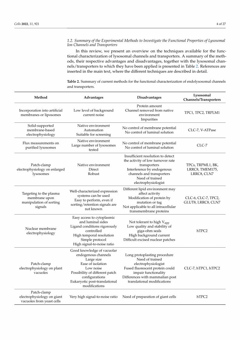
In this review, we present an overview on the techniques available for the func-tional characterization of lysosomal channels and transporters. A summary of the meth-ods, their respective advantages and disadvantages, together with the lysosomal chan-nels/transporters to which they have been applied is presented in Table 2. References areinserted in the main text, where the different techniques are described in detail.
Table 2. Summary of current methods for the functional characterization of endolysosomal channelsand transporters.
Method Advantages Disadvantages LysosomalChannels/Transporters
Incorporation into artificialmembranes or liposomes
Low level of backgroundcurrent noise
Protein amountChannel removed from native
environmentImpurities
TPC1, TPC2, TRPLM1
Solid-supportedmembrane-basedelectrophysiology
Native environmentAutomation
Suitable for screening
No control of membrane potentialNo control of luminal solution CLC-7, V-ATPase
Flux measurements onpurified lysosomes
Native environmentLarge number of lysosomes
tested
No control of membrane potentialNo control of luminal solution CLC-7
Patch-clampelectrophysiology on enlarged
lysosomes
Native environmentDirectRobust
Insufficient resolution to detectthe activity of low turnover rate
transportersInterference by endogenouschannels and transporters
Need of trainedelectrophysiologist
TPCs, TRPML1, BK,LRRC8, TMEM175,
LRRC8, CLN7
Targeting to the plasmamembrane upon
manipulation of sortingsignals
Well-characterized expressionsystems can be used
Easy to perform, even ifsorting/retention signals are
not known
Different lipid environment mayaffect activity
Modification of protein bymutation or tag
Not applicable to all intracellulartransmembrane proteins
CLC-6, CLC-7, TPC2,GLUT8, LRRC8, CLN7
Nuclear membraneelectrophysiology
Easy access to cytoplasmicand luminal sides
Ligand conditions rigorouslycontrolled
High temporal resolutionSimple protocol
High signal-to-noise ratio
Not tolerant to high VappLow quality and stability of
giga-ohm sealsHigh background current
Difficult excised nuclear patches
hTPC2
Patch-clampelectrophysiology on plant
vacuoles
Good knowledge of vacuolarendogenous channels
Large sizeEase of isolation
Low noisePossibility of different patch
configurationsEukaryotic post-translational
modifications
Long protoplasting procedureNeed of trained
electrophysiologistFused fluorescent protein could
impair functionalityDifferences with mammalian post
translational modifications
CLC-7, hTPC1, hTPC2
Patch-clampelectrophysiology on giantvacuoles from yeast cells
Very high signal-to-noise ratio Need of preparation of giant cells hTPC2
Cells 2022, 11, 921 5 of 27
2. Approaches Using Purified Proteins or Native Endolysosomal Membranes2.1. Incorporation into Artificial Membranes or Liposomes
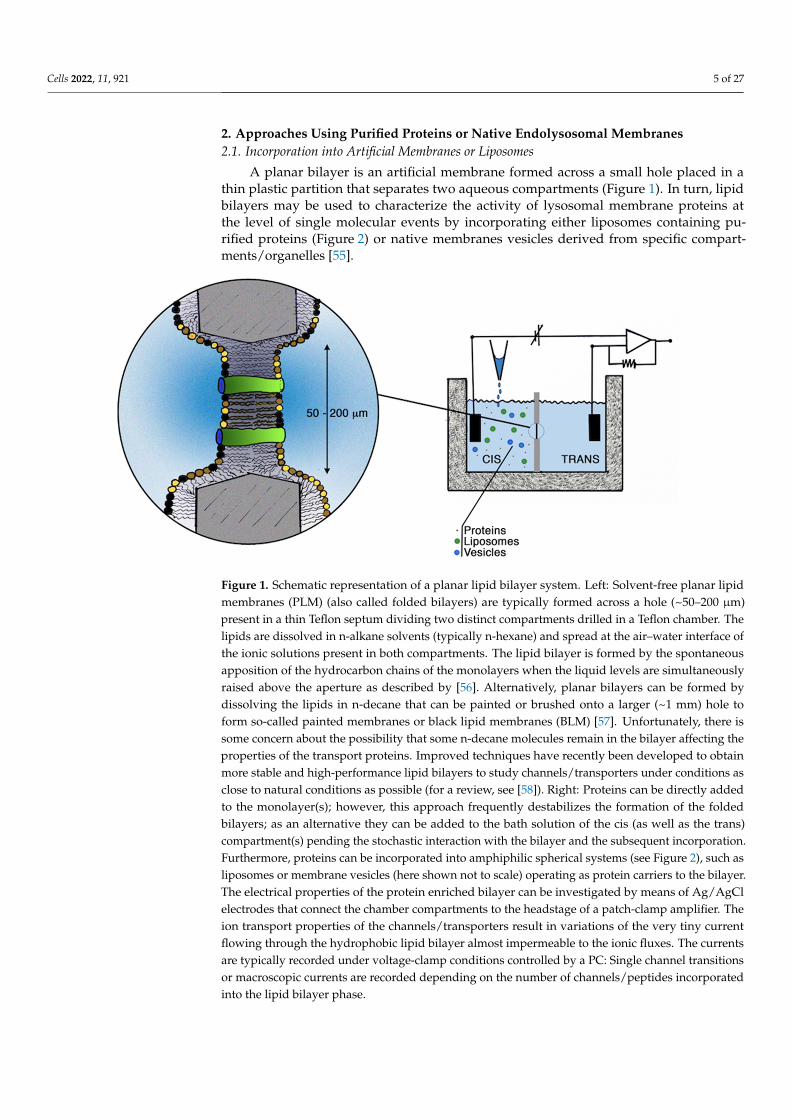
A planar bilayer is an artificial membrane formed across a small hole placed in athin plastic partition that separates two aqueous compartments (Figure 1). In turn, lipidbilayers may be used to characterize the activity of lysosomal membrane proteins atthe level of single molecular events by incorporating either liposomes containing pu-rified proteins (Figure 2) or native membranes vesicles derived from specific compart-ments/organelles [55].
Cells 2022, 11, x FOR PEER REVIEW 5 of 28
Patch-clamp electrophysiology on giant vacuoles from yeast cells
Very high signal-to-noise ra-tio
Need of preparation of giant cells
hTPC2
2. Approaches Using Purified Proteins or Native Endolysosomal Membranes 2.1. Incorporation into Artificial Membranes or Liposomes
A planar bilayer is an artificial membrane formed across a small hole placed in a thin plastic partition that separates two aqueous compartments (Figure 1). In turn, lipid bi-layers may be used to characterize the activity of lysosomal membrane proteins at the level of single molecular events by incorporating either liposomes containing purified proteins (Figure 2) or native membranes vesicles derived from specific compartments/or-ganelles [55].
Figure 1. Schematic representation of a planar lipid bilayer system. Left: Solvent-free planar lipid membranes (PLM) (also called folded bilayers) are typically formed across a hole (~50–200 μm) pre-sent in a thin Teflon septum dividing two distinct compartments drilled in a Teflon chamber. The lipids are dissolved in n-alkane solvents (typically n-hexane) and spread at the air–water interface of the ionic solutions present in both compartments. The lipid bilayer is formed by the spontaneous apposition of the hydrocarbon chains of the monolayers when the liquid levels are simultaneously raised above the aperture as described by [56]. Alternatively, planar bilayers can be formed by dis-solving the lipids in n-decane that can be painted or brushed onto a larger (~1 mm) hole to form so-called painted membranes or black lipid membranes (BLM) [57]. Unfortunately, there is some con-cern about the possibility that some n-decane molecules remain in the bilayer affecting the proper-ties of the transport proteins. Improved techniques have recently been developed to obtain more stable and high-performance lipid bilayers to study channels/transporters under conditions as close to natural conditions as possible (for a review, see [58]). Right: Proteins can be directly added to the monolayer(s); however, this approach frequently destabilizes the formation of the folded bilayers; as an alternative they can be added to the bath solution of the cis (as well as the trans) compart-ment(s) pending the stochastic interaction with the bilayer and the subsequent incorporation. Fur-thermore, proteins can be incorporated into amphiphilic spherical systems (see Figure 2), such as liposomes or membrane vesicles (here shown not to scale) operating as protein carriers to the bi-layer. The electrical properties of the protein enriched bilayer can be investigated by means of Ag/AgCl electrodes that connect the chamber compartments to the headstage of a patch-clamp am-plifier. The ion transport properties of the channels/transporters result in variations of the very tiny current flowing through the hydrophobic lipid bilayer almost impermeable to the ionic fluxes. The currents are typically recorded under voltage-clamp conditions controlled by a PC: Single channel transitions or macroscopic currents are recorded depending on the number of channels/peptides incorporated into the lipid bilayer phase.
Figure 1. Schematic representation of a planar lipid bilayer system. Left: Solvent-free planar lipidmembranes (PLM) (also called folded bilayers) are typically formed across a hole (~50–200 µm)present in a thin Teflon septum dividing two distinct compartments drilled in a Teflon chamber. Thelipids are dissolved in n-alkane solvents (typically n-hexane) and spread at the air–water interface ofthe ionic solutions present in both compartments. The lipid bilayer is formed by the spontaneousapposition of the hydrocarbon chains of the monolayers when the liquid levels are simultaneouslyraised above the aperture as described by [56]. Alternatively, planar bilayers can be formed bydissolving the lipids in n-decane that can be painted or brushed onto a larger (~1 mm) hole toform so-called painted membranes or black lipid membranes (BLM) [57]. Unfortunately, there issome concern about the possibility that some n-decane molecules remain in the bilayer affecting theproperties of the transport proteins. Improved techniques have recently been developed to obtainmore stable and high-performance lipid bilayers to study channels/transporters under conditions asclose to natural conditions as possible (for a review, see [58]). Right: Proteins can be directly addedto the monolayer(s); however, this approach frequently destabilizes the formation of the foldedbilayers; as an alternative they can be added to the bath solution of the cis (as well as the trans)compartment(s) pending the stochastic interaction with the bilayer and the subsequent incorporation.Furthermore, proteins can be incorporated into amphiphilic spherical systems (see Figure 2), such asliposomes or membrane vesicles (here shown not to scale) operating as protein carriers to the bilayer.The electrical properties of the protein enriched bilayer can be investigated by means of Ag/AgClelectrodes that connect the chamber compartments to the headstage of a patch-clamp amplifier. Theion transport properties of the channels/transporters result in variations of the very tiny currentflowing through the hydrophobic lipid bilayer almost impermeable to the ionic fluxes. The currentsare typically recorded under voltage-clamp conditions controlled by a PC: Single channel transitionsor macroscopic currents are recorded depending on the number of channels/peptides incorporatedinto the lipid bilayer phase.

Cells 2022, 11, 921 6 of 27Cells 2022, 11, x FOR PEER REVIEW 6 of 28
Figure 2. Schematic representation of liposomes containing channels/transporters. Liposomes are synthetic lipid vesicles where a lipid milieu separates the internal aqueous medium from the exter-nal ionic environment. After the dispersion of lipids in water solutions liposomes of different sizes (from 20–30 nm to several microns) and lamellarity can be obtained by changing the preparation method. Large and small unilamellar vesicles can be obtained from multilamellar vesicles by differ-ent methods such as ultrasonic treatment, or several extrusion cycles either through a small orifice under high pressure or through a polycarbonate membrane [59] (for a complete review, see Ref. [60]).Protein enriched liposomes (proteoliposomes) containing ion channels/transporters can be re-constructed from an incredible number of lipids that incorporate purified transport proteins from a variety of plasma or organellar membranes [60]. In turn, the reconstitution of cell/organellar planar membranes is achieved by the fusion of proteoliposomes to preformed PLM or BLM. Furthermore, native vesicles can be obtained by standard methods of cell fractionation, homogenization and cen-trifugation of native plasma as well as organellar membranes, including lysosomes [55,61]. Lipo-some and vesicle fusion with artificial bilayers can be facilitated by different procedures and tricks such as the presence of organic n-alkane solvents in the bilayer, the stirring of the bath solutions, the addition to the bath of millimolar concentrations of divalent cations, the existence of an osmotic pressure between the solutions present in the two compartments, the presence of nystatin and er-gosterol in the proteoliposomes or even by centrifugal forces.
Lipid bilayers have a very low level of background current noise so that it is possible to record single-channel currents. Accessibility of both sides of the bilayer and the possi-bility to clamp the membrane voltage allow studies of channel gating, ion conduction and selectivity, effect of ligands, etc.
An advantage of the bilayer approach is that it enables to examine the effect of the lipid environment on the channel, as bilayers may be formed by different types of lipids. Disadvantages include the requirement of a sufficient amount of protein, the fact that the channel is removed from its native environment and that there is no control of protein orientation within the membrane when purified protein is used. However, the main draw-back, especially when native membrane vesicles are used, is the presence of impurities, whose activity may be erroneously attributed to the protein of interest.
Single channel events of immunopurified hTPC2 and hTPC1 reconstituted in artifi-cial membranes were observed respectively by [62–64].
TRPML1 function was investigated in lipid bilayers using reconstitution of both en-dosomal vesicles derived from cells over-expressing TRPML1 and liposomes previously dialyzed with TRPML1 protein [65]. TRPML1 reconstituted in lipid bilayers showed spon-taneous cation channel activity in the presence of asymmetric K+, voltage-dependent acti-vation and multiple sub-conductance states.
Figure 2. Schematic representation of liposomes containing channels/transporters. Liposomes aresynthetic lipid vesicles where a lipid milieu separates the internal aqueous medium from the externalionic environment. After the dispersion of lipids in water solutions liposomes of different sizes (from20–30 nm to several microns) and lamellarity can be obtained by changing the preparation method.Large and small unilamellar vesicles can be obtained from multilamellar vesicles by different methodssuch as ultrasonic treatment, or several extrusion cycles either through a small orifice under highpressure or through a polycarbonate membrane [59] (for a complete review, see Ref. [60]). Proteinenriched liposomes (proteoliposomes) containing ion channels/transporters can be reconstructedfrom an incredible number of lipids that incorporate purified transport proteins from a variety ofplasma or organellar membranes [60]. In turn, the reconstitution of cell/organellar planar membranesis achieved by the fusion of proteoliposomes to preformed PLM or BLM. Furthermore, native vesiclescan be obtained by standard methods of cell fractionation, homogenization and centrifugationof native plasma as well as organellar membranes, including lysosomes [55,61]. Liposome andvesicle fusion with artificial bilayers can be facilitated by different procedures and tricks such as thepresence of organic n-alkane solvents in the bilayer, the stirring of the bath solutions, the additionto the bath of millimolar concentrations of divalent cations, the existence of an osmotic pressurebetween the solutions present in the two compartments, the presence of nystatin and ergosterol inthe proteoliposomes or even by centrifugal forces.
Lipid bilayers have a very low level of background current noise so that it is possible torecord single-channel currents. Accessibility of both sides of the bilayer and the possibilityto clamp the membrane voltage allow studies of channel gating, ion conduction andselectivity, effect of ligands, etc.
An advantage of the bilayer approach is that it enables to examine the effect ofthe lipid environment on the channel, as bilayers may be formed by different types oflipids. Disadvantages include the requirement of a sufficient amount of protein, the factthat the channel is removed from its native environment and that there is no control ofprotein orientation within the membrane when purified protein is used. However, themain drawback, especially when native membrane vesicles are used, is the presence ofimpurities, whose activity may be erroneously attributed to the protein of interest.
Single channel events of immunopurified hTPC2 and hTPC1 reconstituted in artificialmembranes were observed respectively by [62–64].
TRPML1 function was investigated in lipid bilayers using reconstitution of bothendosomal vesicles derived from cells over-expressing TRPML1 and liposomes previouslydialyzed with TRPML1 protein [65]. TRPML1 reconstituted in lipid bilayers showed
Cells 2022, 11, 921 7 of 27
spontaneous cation channel activity in the presence of asymmetric K+, voltage-dependentactivation and multiple sub-conductance states.
2.2. Solid-Supported Membrane-Based Electrophysiology
A number of different electrogenic proteins (ion pumps, transporters and channels)have been tested using solid-supported membrane (SSM) electrophysiology [66–69]. Morerecently, this technique has been applied to intracellular transporters to overcome theinaccessibilty of endomembranes [70,71].
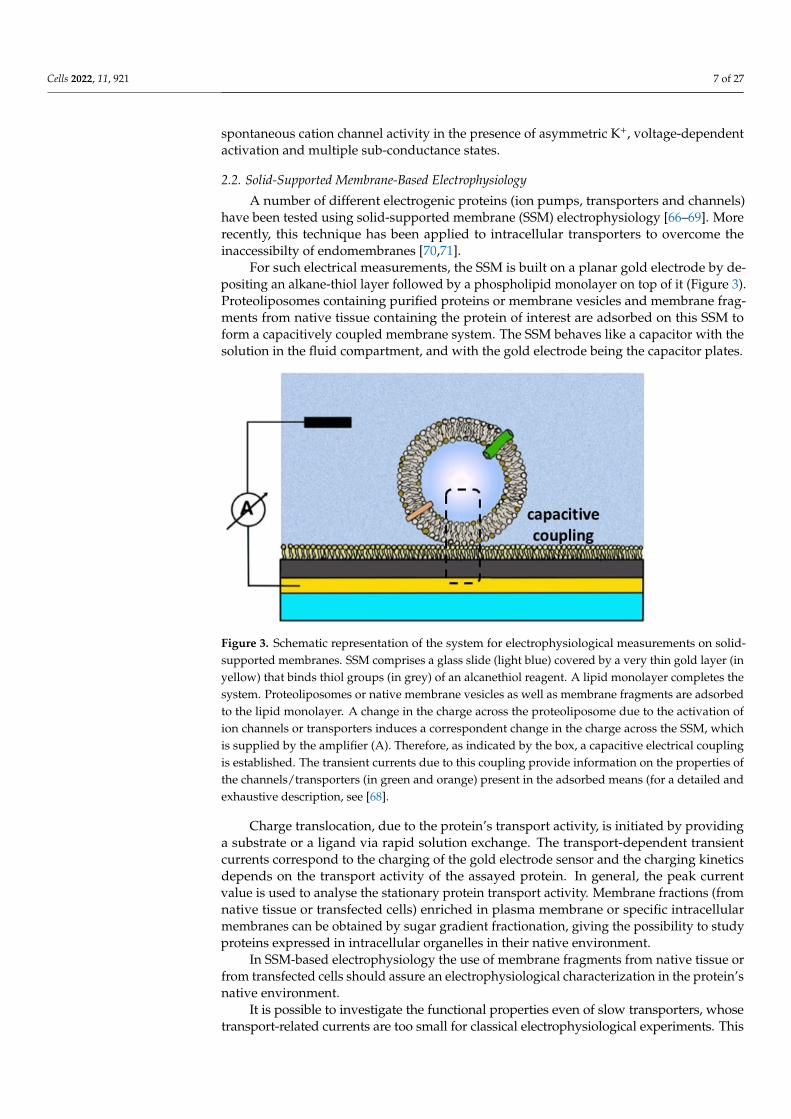
For such electrical measurements, the SSM is built on a planar gold electrode by de-positing an alkane-thiol layer followed by a phospholipid monolayer on top of it (Figure 3).Proteoliposomes containing purified proteins or membrane vesicles and membrane frag-ments from native tissue containing the protein of interest are adsorbed on this SSM toform a capacitively coupled membrane system. The SSM behaves like a capacitor with thesolution in the fluid compartment, and with the gold electrode being the capacitor plates.
Cells 2022, 11, x FOR PEER REVIEW 7 of 28
2.2. Solid-Supported Membrane-Based Electrophysiology A number of different electrogenic proteins (ion pumps, transporters and channels)
have been tested using solid-supported membrane (SSM) electrophysiology [66–69]. More recently, this technique has been applied to intracellular transporters to overcome the in-accessibilty of endomembranes [70,71].
For such electrical measurements, the SSM is built on a planar gold electrode by de-positing an alkane-thiol layer followed by a phospholipid monolayer on top of it (Figure 3). Proteoliposomes containing purified proteins or membrane vesicles and membrane fragments from native tissue containing the protein of interest are adsorbed on this SSM to form a capacitively coupled membrane system. The SSM behaves like a capacitor with the solution in the fluid compartment, and with the gold electrode being the capacitor plates.
Figure 3. Schematic representation of the system for electrophysiological measurements on solid-supported membranes. SSM comprises a glass slide (light blue) covered by a very thin gold layer (in yellow) that binds thiol groups (in grey) of an alcanethiol reagent. A lipid monolayer completes the system. Proteoliposomes or native membrane vesicles as well as membrane fragments are ad-sorbed to the lipid monolayer. A change in the charge across the proteoliposome due to the activa-tion of ion channels or transporters induces a correspondent change in the charge across the SSM, which is supplied by the amplifier (A). Therefore, as indicated by the box, a capacitive electrical coupling is established. The transient currents due to this coupling provide information on the prop-erties of the channels/transporters (in green and orange) present in the adsorbed means (for a de-tailed and exhaustive description, see [68].
Charge translocation, due to the protein’s transport activity, is initiated by providing a substrate or a ligand via rapid solution exchange. The transport-dependent transient currents correspond to the charging of the gold electrode sensor and the charging kinetics depends on the transport activity of the assayed protein. In general, the peak current value is used to analyse the stationary protein transport activity. Membrane fractions (from na-tive tissue or transfected cells) enriched in plasma membrane or specific intracellular membranes can be obtained by sugar gradient fractionation, giving the possibility to study proteins expressed in intracellular organelles in their native environment.
In SSM-based electrophysiology the use of membrane fragments from native tissue or from transfected cells should assure an electrophysiological characterization in the pro-tein’s native environment.
It is possible to investigate the functional properties even of slow transporters, whose transport-related currents are too small for classical electrophysiological experiments.
Figure 3. Schematic representation of the system for electrophysiological measurements on solid-supported membranes. SSM comprises a glass slide (light blue) covered by a very thin gold layer (inyellow) that binds thiol groups (in grey) of an alcanethiol reagent. A lipid monolayer completes thesystem. Proteoliposomes or native membrane vesicles as well as membrane fragments are adsorbedto the lipid monolayer. A change in the charge across the proteoliposome due to the activation ofion channels or transporters induces a correspondent change in the charge across the SSM, whichis supplied by the amplifier (A). Therefore, as indicated by the box, a capacitive electrical couplingis established. The transient currents due to this coupling provide information on the properties ofthe channels/transporters (in green and orange) present in the adsorbed means (for a detailed andexhaustive description, see [68].
Charge translocation, due to the protein’s transport activity, is initiated by providinga substrate or a ligand via rapid solution exchange. The transport-dependent transientcurrents correspond to the charging of the gold electrode sensor and the charging kineticsdepends on the transport activity of the assayed protein. In general, the peak currentvalue is used to analyse the stationary protein transport activity. Membrane fractions (fromnative tissue or transfected cells) enriched in plasma membrane or specific intracellularmembranes can be obtained by sugar gradient fractionation, giving the possibility to studyproteins expressed in intracellular organelles in their native environment.
In SSM-based electrophysiology the use of membrane fragments from native tissue orfrom transfected cells should assure an electrophysiological characterization in the protein’snative environment.
It is possible to investigate the functional properties even of slow transporters, whosetransport-related currents are too small for classical electrophysiological experiments. This
Cells 2022, 11, 921 8 of 27
technique is attractive in the view of establishing screening assays. The major drawback of thistechnique is that it cannot be used to apply an electrical voltage. Transporter characterizationis therefore restricted to transport modes which do not rely on a membrane potential.
SSM-based electrophysiology has been used by Obrdlik and colleagues [67] to studyV-ATPase in synaptic vesicles and Na+/K+-ATPase present in plasma membrane fractionsprepared from the rat brain.
It has also been used to characterize rat CLC-7 function and the effect of the disease-causing G213R mutation, responsible for autosomal-dominant osteopetrosis type II [68].In this study, rCLC-7 and G213R CLC-7 (which is the analogue of human G215R CLC-7)were expressed in CHO cells, alone or together with the accessory subunit OSTM1. SinceCLC-7 and G213R CLC-7 did not localize in the plasma membrane, rather respectively inlysosomes and in lysosomes and ER, they used fractions of these intracellular membranes.Lysosomes and ER membranes were enriched in different membrane fractions, as con-firmed both by fluorimetric investigation of the different membrane fractions containingfluorescently tagged CLC-7 and by densitometric analysis of CLC-7 Western blots. Themembrane fraction was adsorbed to the SSM sensor and currents were generated by fastapplication of 30 mM NaCl at different pH values. They confirmed that CLC-7 functionsas a Cl−/H+-exchanger, and screened a number of potential chloride channel inhibitors(DIDS and NS5818 inhibited the currents with relatively high affinity). They concluded thatmislocalization from lysosomes to ER rather than impaired functionality of G215R CLC-7is the primary cause of the disease.
2.3. Flux Measurements on Purified Lysosomes
Concentrative isotope uptake was previously used for measuring ion fluxes throughion channels in membrane vesicles [72]. Vesicle suspensions were incubated with the 22NaClisotope and the amount of 22Na trapped within the vesicles was measured. This procedureallowed to measure a specific 22Na uptake and to identify the fraction of vesicles containingNa+ channels among a heterogeneous vesicle population [72] Concentrative uptake of36Cl− due to a Cl− gradient was used to determine the conductance properties of Cl−
channels extracted from Torpedo plasma membrane and reconstituted into liposomes [73].The concentrative uptake method was employed to show that the Cl−/H+ antiporter CLC-7is a major chloride permeation pathway in lysosomes [15]. Lysosomes isolated from ratliver by differential sedimentation were loaded with high concentrations of unlabelledchloride and then diluted into a buffer containing 36Cl−. The rapid uptake of 36Cl−, whichwas abolished by the external addition of valinomycin, suggested the presence of a specificelectrogenic transport pathway for chloride. Additional experiments varying internalanions and in the presence of a pH gradient established the apparent permeability sequenceand showed the coupling between Cl− and proton transport. Measurements performedwith a Cl− gradient and monitoring the internal pH with the fluorescent dye 2′,7′-bis-(2-carboxyethyl)-5(6)-carboxyfluorescein (BCECF) showed that protons could move againstthe pH gradient as expected for a Cl−/H+ antiporter. Finally, the equilibrium potential forH+ flux, monitored by BCECF, was measured at a series of theoretical voltages set withK+/valinomycin. As a result of these experiments performed on isolated lysosomes, byusing concentrative 36Cl− uptake combined with fluorescence measurements of protonfluxes, the authors could establish that the lysosomal transport of Cl− and H+ is mediatedby a Cl−/H+ antiporter, identified as CLC-7 [15]. This method allows to estimate fluxes in alarge number of lysosomes under varying external conditions and maintaining the proteinsin their native membrane. It can identify ion transport mechanisms across the lysosomalmembrane, however its use to characterize in detail the functional activity of lysosomalmembrane transporters seems difficult. It requires performing radioactivity measurementsand it does not allow direct and precise control of the membrane potential preventing thestudy of the voltage dependence of the ion transport.

Cells 2022, 11, 921 9 of 27
2.4. Patch-Clamp Electrophysiology on Enlarged Lysosomes
Acidic lysosomal compartments in animal cells are small in size (diameter < 500 nm),and this property has strongly limited their use for patch-clamp studies and thus ourknowledge about the transporter composition of the lysosome membrane. The importanceof patching endolysosomal membranes relates to the fact that, in animal cells, a seriesof channels have been reported to be localized on both lysosomal and/or endosomalmembranes and have been predicted to play roles in signalling events and endomembranefusion [17,47,74–77].
A detailed protocol reporting the use of enlarged lysosomes for patch-clamp waspublished in 2017 [78], but the first patch-clamp recording on endosomal membranes wasmade possible thanks to the expression of a hydrolysis-deficient SKD1/VPS4B (E235Q)protein in HEK293 cells, which induced the formation of enlarged endosomes (3–6 µm indiameter) by blocking their transition to lysosomes, hence making them accessible to thepatch-clamp approach [79]. This strategy allowed the characterization of an endosomalCa2+ channel whose activity was affected by the luminal Cl− concentration [79]. Followingthis first report, the study of endolysosomal channels showed a strong acceleration thanksto the use of vacuolin-1, a lipid-soluble polycyclic triazine [80]. Vacuolin-1 treatment canselectively increase the size of endosomes and lysosomes from less than 0.5 µm up to5 µm [75], hence making this compartment accessible to electrophysiological recordingsafter the mechanical rupture of the plasma membrane [81], Figure 4. Unfortunately, due tothe required cell manipulation and mechanical isolation of the endolysosomes, the patch-clamp recordings could be carried out only within a limited time window, from 1 to 3 hafter isolation, with a gradually decreasing probability to form high-resistance seals withtime [81]. Nevertheless, the enlarged endolysosomes obtained by vacuolin-1 treatmentoffer a solid basis for the electrophysiological characterization of a series of endomembranechannels [25,74,76,77,82,83].
Cells 2022, 11, x FOR PEER REVIEW 9 of 28
2.4. Patch-Clamp Electrophysiology on Enlarged Lysosomes Acidic lysosomal compartments in animal cells are small in size (diameter < 500 nm),
and this property has strongly limited their use for patch-clamp studies and thus our knowledge about the transporter composition of the lysosome membrane. The im-portance of patching endolysosomal membranes relates to the fact that, in animal cells, a series of channels have been reported to be localized on both lysosomal and/or endosomal membranes and have been predicted to play roles in signalling events and endomembrane fusion [17,47,74–77].
A detailed protocol reporting the use of enlarged lysosomes for patch-clamp was published in 2017 [78], but the first patch-clamp recording on endosomal membranes was made possible thanks to the expression of a hydrolysis-deficient SKD1/VPS4B (E235Q) protein in HEK293 cells, which induced the formation of enlarged endosomes (3–6 μm in diameter) by blocking their transition to lysosomes, hence making them accessible to the patch-clamp approach [79]. This strategy allowed the characterization of an endosomal Ca2+ channel whose activity was affected by the luminal Cl− concentration [79]. Following this first report, the study of endolysosomal channels showed a strong acceleration thanks to the use of vacuolin-1, a lipid-soluble polycyclic triazine [80]. Vacuolin-1 treatment can selectively increase the size of endosomes and lysosomes from less than 0.5 μm up to 5 μm [75], hence making this compartment accessible to electrophysiological recordings af-ter the mechanical rupture of the plasma membrane [81], Figure 4. Unfortunately, due to the required cell manipulation and mechanical isolation of the endolysosomes, the patch-clamp recordings could be carried out only within a limited time window, from 1 to 3 h after isolation, with a gradually decreasing probability to form high-resistance seals with time [81]. Nevertheless, the enlarged endolysosomes obtained by vacuolin-1 treatment of-fer a solid basis for the electrophysiological characterization of a series of endomembrane channels [25,74,76,77,82,83].
Figure 4. Cartoon of the patch-clamp recording configuration on enlarged lysosomes and current convention. The patch clamp technique is applied in the whole-lysosome configuration after lyso-some enlargement by treatment with vacuolin-1. Positive currents correspond to the movement of cations from the cytosolic to the luminal side of the lysosome or to the opposite movement of anions.
Figure 4. Cartoon of the patch-clamp recording configuration on enlarged lysosomes and currentconvention. The patch clamp technique is applied in the whole-lysosome configuration after lysosomeenlargement by treatment with vacuolin-1. Positive currents correspond to the movement of cationsfrom the cytosolic to the luminal side of the lysosome or to the opposite movement of anions.

Cells 2022, 11, 921 10 of 27
Vacuolin-1 treatment was first employed for the electrophysiological characteriza-tion of TRPML1 (mucolipin 1). In 2008, Dong and colleagues [74] transiently transfectedHEK293T cells with an mCherry-TRPML1 construct and performed patch-clamp recordingson native membranes of enlarged endolysosomes in three distinct patch-clamp configura-tions: Lysosome attached, lysosome luminal-side-out and whole lysosome. These experi-ments reported the existence of significant inwardly rectifying currents mediated by thepermeation of Fe2+ [74]. The coupling of a glass chip-based method (planar patch) with thepreparation of enlarged endolysosomes led Schieder and colleagues [82] to electrophysiolog-ically characterize another organellar membrane channel: Human TPC2. Characterizationof hTPC2 and hTPC1 was also performed by directly patching enlarged endolysosomes.Wang and colleagues [25] proposed that TPC2 was activated by PI(3,5)P2 and that the majorcation fluxing through its pore was Na+. Similarly, also TPC1 was found to transport mainlyNa+ [77], supporting the view that TPC channels are PI(3,5)P2-activated Na+-selective chan-nels [25,77]. Nevertheless, other groups succeeded in the study of TPC channels in enlargedendolysosomes and reported that TPC2 were also activated by NAADP [84] and inhibitedby cytosolic and luminal magnesium [85].
3. Approaches Based on Alternative Targeting and Heterologous Expression3.1. Targeting to the Plasma Membrane upon Manipulation of Sorting Signals
Most organelles are not easily amenable to classical uptake experiments or electro-physiological recordings, hampering the analysis of intracellular ion and solute transportprocesses. One exception to this is the large central vacuole of plant and yeast cells, whichis directly and easily accessible after rupture of the plasma membrane. One possibility tocircumvent the problem of membrane accessibility is to manipulate the subcellular local-ization of intracellular transport proteins by altering their targeting route, thus redirectingthem to the vacuolar (see Section 3.3) or plasma membranes.
In the secretory or endocytic pathway, coordinated vesicle trafficking delivers trans-port proteins to the correct destination membrane. Transport vesicles recruit their trans-membrane protein cargo upon interaction of adaptor proteins with sorting or internal-ization signals within the cytoplasmic regions of the cargo proteins. Sorting or internal-ization signals consist of short, linear motifs, post-translational modifications or three-dimensional structural motifs [86–88]. Among the linear sequence motifs, dileucine-basedmotifs [DE]xxxL[LI] and tyrosine-based motifs YxxØ (where Ø represents a bulky, hy-drophobic amino acid) are common. They are recognized by clathrin adaptor complexes(AP1 to AP5), Golgi-localized, γ-ear containing, Arf-binding proteins (GGAs) and stonins1 and 2. GGAs select the dileucine-motif variant DxxLL and specifically control vesiclesorting at the trans-Golgi network [89], Figure 5. The only identified cargo for stonins isup to now synaptotagmin-1, where stonin 2 directly binds a cluster of basic residues [90].While stonins have to date only be identified in meazoans and GGAs in metazoans andfungi [91], in general the sorting motifs and trafficking mechanisms are quite well con-served among different phylogenetic groups, including mammalia, plants and yeast. Suchconservation of trafficking mechanisms can be exploited to manipulate the targeting ofintracellular transport proteins for surface expression and functional characterization.
This approach was used in 2000 as a tool for Glucose Transporter 8, GLUT8, formerlyGLUTX1; [92]. Ibberson and colleagues cloned this transporter from rat cDNA but werenot able to detect glucose uptake in GLUT8-expressing Xenopus oocytes. They suspectedan internalization signal to prevent sufficient surface expression and therefore mutateda dileucine motif at the N-terminus of GLUT8. This mutation was sufficient to redirectthe protein to the plasma membrane and established GLUT8 as a glucose transporter. Ina later study, Ref. [93] showed that the canonical dileucine motif ExxxLL is responsiblefor localization of the native GLUT8 in late endosomes and lysosomes. When a homolo-gous transporter of GLUT8 from Arabidopsis thaliana termed ESL1 (ERD Six-Like 1) wasinvestigated, uptake experiments with a mutated N-terminal dileucine-motif revealed itstransport capacities for glucose and some other monosaccharides [94]. Confocal microscopy
Cells 2022, 11, 921 11 of 27
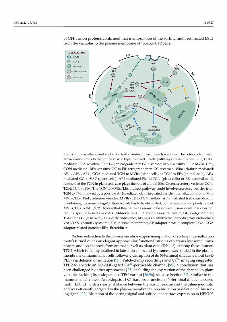
of GFP fusion proteins confirmed that manipulation of the sorting motif redirected ESL1from the vacuolar to the plasma membrane of tobacco BY2 cells.
Cells 2022, 11, x FOR PEER REVIEW 11 of 28
Figure 5. Biosynthetic and endocytic traffic routes to vacuoles/lysosomes. The color code of each arrow corresponds to that of the vesicle type involved. Traffic pathways are as follows. Blue, COPII mediated: BFA-sensitive ER to GC; anterograde intra GC cisternae; BFA insensitive ER to MVBs. Gray, COPI mediated: BFA sensitive GC to ER; retrograde intra GC cisternae. Wine, clathrin medi-ated: AP1-, AP3-, AP4-, GGA-mediated TGN to MVBs (plant cells) or TGN to EEs (animal cells); AP3-mediated GC to VAC (plant cells); AP2-mediated PM to TGN (plant cells) or EEs (animal cells). Notice that the TGN in plant cells also plays the role of animal EEs. Green, secretory vesicles: GC to TGN; TGN to PM. The TGN to MVBs/LEs indirect pathway could involve secretory vesicles from TGN to PM, followed by a possible AP2-mediated clathrin-coated vesicle internalization from PM to MVBs/LEs. Pink, retromer vesicles: MVBs/LE to TGN. Yellow: AP5-mediated traffic involved in maintaining lysosome integrity. Its exact role has to be elucidated, both in animals and plants. Vio-let: MVBs/LEs to VAC/LYS. Notice that this pathway seems to be a direct fusion event that does not require specific vesicles or coats. Abbreviations: ER, endoplasmic reticulum; GC, Golgi complex; TGN, trans-Golgi network; EEs, early endosomes; MVBs/LEs, multivesicular bodies/late endo-somes; VAC/LYS, vacuole/lysosome; PM, plasma membrane; AP, adaptor protein complex; GGA, GGA adaptor-related proteins; BFA, Brefeldin A.
This approach was used in 2000 as a tool for Glucose Transporter 8, GLUT8, formerly GLUTX1; [92]. Ibberson and colleagues cloned this transporter from rat cDNA but were not able to detect glucose uptake in GLUT8-expressing Xenopus oocytes. They suspected an internalization signal to prevent sufficient surface expression and therefore mutated a dileucine motif at the N-terminus of GLUT8. This mutation was sufficient to redirect the protein to the plasma membrane and established GLUT8 as a glucose transporter. In a later study, [93] showed that the canonical dileucine motif ExxxLL is responsible for lo-calization of the native GLUT8 in late endosomes and lysosomes. When a homologous transporter of GLUT8 from Arabidopsis thaliana termed ESL1 (ERD Six-Like 1) was inves-tigated, uptake experiments with a mutated N-terminal dileucine-motif revealed its transport capacities for glucose and some other monosaccharides [94]. Confocal micros-copy of GFP fusion proteins confirmed that manipulation of the sorting motif redirected ESL1 from the vacuolar to the plasma membrane of tobacco BY2 cells.
Protein redirection to the plasma membrane upon manipulation of sorting/internali-zation motifs turned out as an elegant approach for functional studies of various lysoso-mal transporters and ion channels from animal as well as plant cells (Table 3). Among these, human TPC2, which is mainly localized in late endosomes and lysosomes, was
Figure 5. Biosynthetic and endocytic traffic routes to vacuoles/lysosomes. The color code of eacharrow corresponds to that of the vesicle type involved. Traffic pathways are as follows. Blue, COPIImediated: BFA-sensitive ER to GC; anterograde intra GC cisternae; BFA insensitive ER to MVBs. Gray,COPI mediated: BFA sensitive GC to ER; retrograde intra GC cisternae. Wine, clathrin mediated:AP1-, AP3-, AP4-, GGA-mediated TGN to MVBs (plant cells) or TGN to EEs (animal cells); AP3-mediated GC to VAC (plant cells); AP2-mediated PM to TGN (plant cells) or EEs (animal cells).Notice that the TGN in plant cells also plays the role of animal EEs. Green, secretory vesicles: GC toTGN; TGN to PM. The TGN to MVBs/LEs indirect pathway could involve secretory vesicles fromTGN to PM, followed by a possible AP2-mediated clathrin-coated vesicle internalization from PM toMVBs/LEs. Pink, retromer vesicles: MVBs/LE to TGN. Yellow: AP5-mediated traffic involved inmaintaining lysosome integrity. Its exact role has to be elucidated, both in animals and plants. Violet:MVBs/LEs to VAC/LYS. Notice that this pathway seems to be a direct fusion event that does notrequire specific vesicles or coats. Abbreviations: ER, endoplasmic reticulum; GC, Golgi complex;TGN, trans-Golgi network; EEs, early endosomes; MVBs/LEs, multivesicular bodies/late endosomes;VAC/LYS, vacuole/lysosome; PM, plasma membrane; AP, adaptor protein complex; GGA, GGAadaptor-related proteins; BFA, Brefeldin A.
Protein redirection to the plasma membrane upon manipulation of sorting/internalizationmotifs turned out as an elegant approach for functional studies of various lysosomal trans-porters and ion channels from animal as well as plant cells (Table 3). Among these, humanTPC2, which is mainly localized in late endosomes and lysosomes, was studied in the plasmamembrane of mammalian cells following disruption of its N-terminal dileucine motif (ESE-PLL) via deletion or mutation [95]. Patch-clamp recordings and Ca2+ imaging suggestedTPC2 to encode an NAADP-gated Ca2+-permeable channel [95], a conclusion that hasbeen challenged by other approaches [25], including the expression of the channel in plantvacuoles lacking its endogeneous TPC variant [26,96]; see also Section 3.3. Similar to themammalian channels, Arabidopsis TPC1 harbors a functional N-terminal dileucine-basedmotif (EDPLI) with a shorter distance between the acidic residue and the dileucine-motifand was efficiently targeted to the plasma membrane upon mutation or deletion of this sort-ing signal [97]. Mutation of the sorting signal and subsequent surface expression in HEK293
Cells 2022, 11, 921 12 of 27
cells was furthermore used to resolve the crystal structure of Arabidopsis TPC1 [98,99], andfor cryo-electron microscopy analysis of the structure of human TPC2 [100].
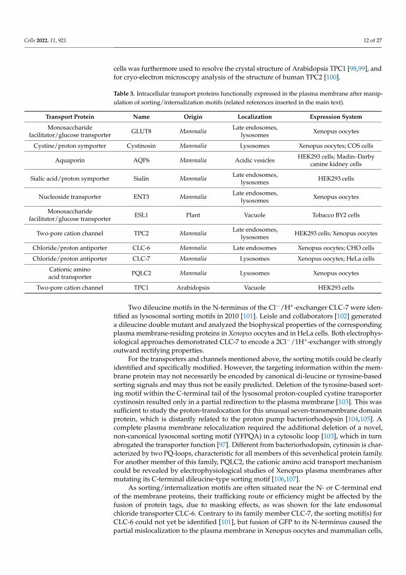
Table 3. Intracellular transport proteins functionally expressed in the plasma membrane after manip-ulation of sorting/internalization motifs (related references inserted in the main text).
Transport Protein Name Origin Localization Expression System
Monosaccharidefacilitator/glucose transporter GLUT8 Mammalia Late endosomes,
lysosomes Xenopus oocytes
Cystine/proton symporter Cystinosin Mammalia Lysosomes Xenopus oocytes; COS cells
Aquaporin AQP6 Mammalia Acidic vesicles HEK293 cells; Madin–Darbycanine kidney cells
Sialic acid/proton symporter Sialin Mammalia Late endosomes,lysosomes HEK293 cells
Nucleoside transporter ENT3 Mammalia Late endosomes,lysosomes Xenopus oocytes
Monosaccharidefacilitator/glucose transporter ESL1 Plant Vacuole Tobacco BY2 cells
Two-pore cation channel TPC2 Mammalia Late endosomes,lysosomes HEK293 cells; Xenopus oocytes
Chloride/proton antiporter CLC-6 Mammalia Late endosomes Xenopus oocytes; CHO cells
Chloride/proton antiporter CLC-7 Mammalia Lysosomes Xenopus oocytes; HeLa cells
Cationic aminoacid transporter PQLC2 Mammalia Lysosomes Xenopus oocytes
Two-pore cation channel TPC1 Arabidopsis Vacuole HEK293 cells
Two dileucine motifs in the N-terminus of the Cl−/H+-exchanger CLC-7 were iden-tified as lysosomal sorting motifs in 2010 [101]. Leisle and collaborators [102] generateda dileucine double mutant and analyzed the biophysical properties of the correspondingplasma membrane-residing proteins in Xenopus oocytes and in HeLa cells. Both electrophys-iological approaches demonstrated CLC-7 to encode a 2Cl−/1H+-exchanger with stronglyoutward rectifying properties.
For the transporters and channels mentioned above, the sorting motifs could be clearlyidentified and specifically modified. However, the targeting information within the mem-brane protein may not necessarily be encoded by canonical di-leucine or tyrosine-basedsorting signals and may thus not be easily predicted. Deletion of the tyrosine-based sort-ing motif within the C-terminal tail of the lysosomal proton-coupled cystine transportercystinosin resulted only in a partial redirection to the plasma membrane [103]. This wassufficient to study the proton-translocation for this unusual seven-transmembrane domainprotein, which is distantly related to the proton pump bacteriorhodopsin [104,105]. Acomplete plasma membrane relocalization required the additional deletion of a novel,non-canonical lysosomal sorting motif (YFPQA) in a cytosolic loop [103], which in turnabrogated the transporter function [97]. Different from bacteriorhodopsin, cytinosin is char-acterized by two PQ-loops, characteristic for all members of this sevenhelical protein family.For another member of this family, PQLC2, the cationic amino acid transport mechanismcould be revealed by electrophysiological studies of Xenopus plasma membranes aftermutating its C-terminal dileucine-type sorting motif [106,107].
As sorting/internalization motifs are often situated near the N- or C-terminal endof the membrane proteins, their trafficking route or efficiency might be affected by thefusion of protein tags, due to masking effects, as was shown for the late endosomalchloride transporter CLC-6. Contrary to its family member CLC-7, the sorting motif(s) forCLC-6 could not yet be identified [101], but fusion of GFP to its N-terminus caused thepartial mislocalization to the plasma membrane in Xenopus oocytes and mammalian cells,

Cells 2022, 11, 921 13 of 27
where it mediates Cl−/H+ antiport [108,109]. Another example for the use of a maskedsorting or retention signal for functional characterization is Aquaporin-6 (AQP6), whichwas relocalized from acidic intracellular vesicles to the plasma membrane in mammaliancells upon N-terminal fusion with a GFP- or HA-tag, whereas the C-terminal GFP fusiondid not change the localization [110,111]. Patch clamp analysis of GFP-AQP6-expressingHEK293 cells revealed a previously unknown anion transport mechanism with high nitratepermeability for AQP6.
These results underline that confocal microscopy studies using GFP fusion proteins donot always reveal the subcellular localization of the native protein, and therefore stronglysuggest using both, N- as well as C-terminal fluorophore attachments as a common standardfor tag-based localization experiments. Furthermore, surface expression of intracellulartransport proteins upon manipulation of sorting/internalization signals represents a pow-erful tool for their functional characterization, which in some cases even was the onlysuccessful approach so far. There is also one report, in which the use of inhibitors of dy-namin redirected the lysosomal K+ channel c to the plasma membrane upon expression inXenopus oocytes [112].
Nevertheless, the successful expression in a different subcellular compartment doesnot guarantee the preservation of all properties of the wild type protein in its native mem-brane. Especially, lack of cosubunits, interacting proteins or regulatory factors, as well asdifferences in the lipid composition between the plasma membrane and the specific en-domembrane might strongly affect the transport properties. It might therefore be beneficialor necessary to compare the properties of redirected proteins in several different cell typesor membrane compartments like the plasma and vacuolar membrane (see Section 3.3). Itshould also be kept in mind that although an equivalent sorting motif may be present intwo very similar transporters or channels, its mutation may not always have the sameeffect, as shown for the mammalian endosomal TPC1 versus the lysosomal TPC2 [95]. Incontrast to TPC2, TPC1 did not reach the plasma membrane after eliminating one or bothof its dileucine-based motifs.
In summary, several channels from the acidic lysosomal compartment of mammalianand plant cells were effectively redirected to the cell surface by mutation or masking oftheir lysosomal sorting/internalization signal (Table 3), which in most cases belong tothe dileucin-based motives. Progress in understanding membrane protein trafficking inthe different kingdoms will allow including further compartments of the secretory andendocytic pathway, such as the Golgi apparatus or early, late and recycling endosomes infuture membrane transport research.
3.2. Nuclear Membrane Electrophysiology
The nuclear membrane may represent a novel heterologous system to express endo-lysosomal channels and transporters, as it provides relatively easy access to both thecytoplasmic and luminal sides of the membrane, so that ionic and ligand conditions can berigorously controlled [113,114]. The technique, whose advantages and disadvantages havebeen summarized in Table 2, has been recently used to characterize the hTPC2 channel [115].In their study, the authors generated a stable DT40TKO cell line expressing hTPC2 andlacking both functional InsP3R and RyR, two intracellular Ca2+ channels. Using the nuclearmembrane patch-clamp technique, they detected a ~220 pS single-channel current activatedby NAADP with K+ as the permeant ion.
3.3. Patch-Clamp Electrophysiology on Plant Vacuoles3.3.1. Sorting Routes and Signals to the Tonoplast and the Lysosomal Membrane
While performing more functions than the animal lysosome, i.e., storage of ions/metabolitesand regulation of the turgor of the plant cell, the central lytic vacuole can be considered thelysosomal counterpart. This compartment (up to 40 µm in diameter) may occupy more than 80%of the cellular volume [116,117] in many cell types. Vacuoles are highly suitable for patch-clampstudies [118–121], because of simplicity of isolation and large size. For these reasons, many
Cells 2022, 11, 921 14 of 27
types of endogenous ion channels and transporters have been identified and characterized ingreat detail [116,122]. Besides macroscopic currents recorded in the whole-vacuole configura-tion [123,124], single channel events could be detected in excised vacuolar membrane patches,both in the cytosolic-side-out [125] and vacuolar-side-out configurations [126], Moreover, fluores-cent indicator dyes have also been employed together with the patch-clamp technique [127–131].The choice of ionic conditions and/or the use of appropriate Arabidopsis knock-out lines, allowto significantly reduce the density of the background currents, despite the presence of differenttypes of endogenous ion transport systems.
Vacuoles can be used to study endolysosomal proteins only if these are sorted to thetonoplast. The finding that the main families of channels and transporters of the LM aredelivered to the tonoplast in plant cells [26,132–134] provides exciting information andposes interesting questions on the evolutionary conservation of sorting mechanisms to theinner hydrolytic compartments.
From a topographic point of view, vacuoles and lysosomes occupy the same positionwithin the secretory pathway: In general terms, they are one of the two endpoints ofsecretory traffic, the other being the cell surface. Like other secretory proteins, proteins ofthe tonoplast and the LM start their life in the ER [135]. The best characterized pathwaysto their final destinations pass through the Golgi apparatus and the trans-Golgi network(TGN). From there, a direct route proceeds via multivesicular bodies (MVB, also termedlate endosomes) to the plant tonoplast or the LM, whereas an indirect route first leads tothe plasma membrane and then to the final destination via endocytosis and MVB [136–138](see Figure 5). Endocytosis of plasma membrane receptors and transporters for recyclingor degradation in vacuoles and lysosomes operates in all eukaryotes, however to datethe biosynthetic indirect route to the membrane of inner hydrolytic compartments hasbeen described for lysosomes [136,139] but not for vacuoles. Therefore, it is not yet knownwhether missorting to the plasma membrane, observed for a number of tonoplast proteinsupon mutagenesis or domain exchange with plasma membrane isoforms of the same genefamily, is the result of actual direct missorting from the TGN to the cell surface or reflectslack of endocytosis from the plasma membrane [92,140–142].
Alternative ramifications can bypass some of the intermediate compartments. Inplants, correct sorting of a number of tonoplast proteins is not blocked by brefeldin A or bydominant-negative mutant versions of Rab11 GTPases [143,144]. Brefeldin A causes fusionof ER and Golgi cisternae, inhibiting further anterograde Golgi-mediated traffic, whereasRab11 GTPases regulate vesicle traffic at the TGN. Therefore, part of the tonoplast proteomecan bypass the Golgi/TGN system, [137] and Figure 5. Indeed, there is growing evidencethat the tonoplast of newly formed vacuoles in meristematic plant cells originates directlyfrom the ER and is then expanded through Golgi/TGN/MVB-mediated traffic [145,146].
Membrane proteins are sorted along the endomembrane system through interactionsbetween sorting motifs in their cytosolic domains and components of the different coatcomplexes that allow membrane traffic from one compartment to another. As introduced inSection 3.1, two classes of motifs present in the cytosolic head or tail of membrane proteinshave been clearly identified as lysosomal and tonoplast sorting signals: Dileucine-based[D/E]XXXL[L/I] or DXXLL, and tyrosine-based YXXØ, where X can be any amino acid andØ represents a bulky hydrophobic residue [136,137]. These motifs interact with componentsof the AP1-4 or GGA adaptor complexes necessary to recruit clathrin or other membranecoats that promote the formation of coated vesicles, either from the TGN, the plasmamembrane or between early and late endosomes (notice that, in plant cells, the TGN alsoplay the role of animal early endosomes; see Figure 5). Dileucine-based sorting signalsfor the tonoplast are recognized by AP1 [147], AP3 [142] or AP4 [148]. AP5 is involved inmaintaining lysosome integrity [149], but its extact role has to be elucidate both in mammalsand plants.
It should also be underlined that most membrane proteins also present ER-exit motifsthat allow efficient initiation of traffic from this compartment, independently of their finaldestination. These diacidic (D/E-X-D/E), dihydrophobic or diaromatic (FF, YY, LL or

Cells 2022, 11, 921 15 of 27
FY) motifs interact with components of the COPII complex that initiates traffic from theER (Barlowe, 2005; Marti et al., 2010). Finally, the ER quality control machinery usuallyprevents misfolded or unassembled newly synthesized polypeptides from trafficking [137].Consistently with all these requirements for traffic and sorting, deletions or domain substi-tutions that destroy the tonoplast sorting signals often lead to mislocalization to the plasmamembrane [94,97,141,142], whereas those that affect the ER-exit motifs or general foldingresult in ER retention [97,141,150].
The Golgi/TGN-mediated routes to the tonoplast and lysosomal membrane seem touse conserved mechanisms and signals [1]. This occurs despite the marked architecturaldifferences in key compartments of the secretory pathway, perhaps most strikingly theGolgi apparatus: It is a single perinuclear membrane complex controlled by microtubules inmost animal cells as opposed to a system composed of hundreds of apparently independentGolgi units moving along actin filaments all over any plant cell analyzed. These features riseas yet unresolved questions about the conservation of ER-to-Golgi and Golgi-to-endosometraffic mechanisms [151,152] and may also be related to the origin of the Golgi-bypassingroutes to the tonoplast mentioned above. It has also been determined that potential N-glycosylation sequons are much less frequent in tonoplast proteins than in those of the plantplasma membrane, and that Golgi-modified Asn-linked oligosaccharide chains, abundantlypresent in plant plasma membrane proteins, are not detactable in tonoplast proteins ofthe same cells [153]. The major lysosomal membrane proteins are instead extensively N-glycosylated with oligosaccharide chains modified by Golgi enzymes. This “glycocalyx” isbelieved to protect the luminal loops of membrane proteins from degradation by lysosomalproteases [154]. In silico analysis of proteomes suggests that protection from vacuolarproteases has instead evolved by limiting the length of luminal domains of tonoplastproteins [153]. It is finally evident that a single large vacuole that occupies most of thecellular space in many fully expanded plant cells is quite different from the myriad of smalllysosomes, at least in terms of surface/volume ratio.
3.3.2. The Plant Vacuole as a Heterologous Expression System of Lysosomal Channelsand Transporters
In line with similarity discussed above in trafficking and targeting between tono-plast and lysosomal membrane proteins, mutants plants from Arabidopsis thaliana lackingspecific endogenous vacuolar channels or transporters can be used for the expression ofthe respective homologous animal lysosomal proteins (interestingly, oocytes of Xenopuslaevis are the system of choice for ion channels and transporters localized in the plasmamembrane [155–159]). Plants of Arabidopsis can be grown on soil in a growth chamberunder controlled light and temperature conditions. The cDNA of the animal intracellularchannel or transporter is cloned into a suitable plant expression vector conveying highprotein expression. Fusion with a fluorescent marker may be helpful to verify the expres-sion and localization of the protein. By using a well-established protocol [160], protoplastscan be transiently transformed; see Figure 6 for a schematic overview. The efficiency oftransformation can be estimated by GFP fluorescence of the protoplasts. After one tofour days, vacuoles can be easily released from transformed protoplasts for subsequentpatch-clamp experiments.
The patch-clamp technique, developed by Neher and Sakmann [161], consists ofestablishing a tight contact (seal) between a glass pipette with a micrometric tip and themembrane of a cell, an organelle or an intracellular compartment (a vacuole in this case).When a seal is obtained, the resistance can reach values of several billion ohms with aconsequent reduction of background noise [162] and the possibility of recording signals inthe order of femtoamperes [163]. Patch-clamp measurements on plant vacuoles can be donein these four main configurations [164,165]: (i) Vacuole attached, which allows to measurecurrents across the membrane portion directly underneath the pipette tip, but with thelimit of having no control over the content of the vacuolar lumen; (ii) whole vacuole, whichallows macroscopic current recordings mediated by channels or transporters present on

Cells 2022, 11, 921 16 of 27
the entire vacuolar membrane; (iii) cytosolic-side-out and (iv) vacuolar-side-out excisedpatches, which are useful to detect single channel currents. A major advantage of vacuolarpatch-clamp is that channel modulation by cytosolic factors can easily be studied, sincethe cytosolic side faces the bath solution which can be exchanged ad libitum during theexperiment with a dedicated perfusion system, plant vacuoles (and protoplasts) are notfirmly attached to the recording chamber as animal cells [134,166–169].
Cells 2022, 11, x FOR PEER REVIEW 16 of 28
3.3.2. The Plant Vacuole as a Heterologous Expression System of Lysosomal Channels and Transporters
In line with similarity discussed above in trafficking and targeting between tonoplast and lysosomal membrane proteins, mutants plants from Arabidopsis thaliana lacking spe-cific endogenous vacuolar channels or transporters can be used for the expression of the respective homologous animal lysosomal proteins (interestingly, oocytes of Xenopus laevis are the system of choice for ion channels and transporters localized in the plasma mem-brane [155–159]).Plants of Arabidopsis can be grown on soil in a growth chamber under controlled light and temperature conditions. The cDNA of the animal intracellular chan-nel or transporter is cloned into a suitable plant expression vector conveying high protein expression. Fusion with a fluorescent marker may be helpful to verify the expression and localization of the protein. By using a well-established protocol [160], protoplasts can be transiently transformed; see Figure 6 for a schematic overview. The efficiency of transfor-mation can be estimated by GFP fluorescence of the protoplasts. After one to four days, vacuoles can be easily released from transformed protoplasts for subsequent patch-clamp experiments.
Figure 6. Flow chart of the experimental procedures showing how Arabidopsis vacuoles can be a heterologous expression system for animal lysosomal ion channels and transporters. Transfor-mation can be performed on protoplasts from Arabidopsis wild-type plants or mutants lacking a specific endogenous channels or transporters.
The patch-clamp technique, developed by Neher and Sakmann [161], consists of es-tablishing a tight contact (seal) between a glass pipette with a micrometric tip and the membrane of a cell, an organelle or an intracellular compartment (a vacuole in this case). When a seal is obtained, the resistance can reach values of several billion ohms with a consequent reduction of background noise [162] and the possibility of recording signals in the order of femtoamperes [163]. Patch-clamp measurements on plant vacuoles can be done in these four main configurations [164,165]: (i) Vacuole attached, which allows to
Figure 6. Flow chart of the experimental procedures showing how Arabidopsis vacuoles can be aheterologous expression system for animal lysosomal ion channels and transporters. Transformationcan be performed on protoplasts from Arabidopsis wild-type plants or mutants lacking a specificendogenous channels or transporters.
By applying the patch-clamp technique in whole vacuole configuration on isolated vacuolesfrom Arabidopsis mesophyll protoplasts lacking the endogenous AtCLCa, Costa et al. [127]recorded chloride–proton exchange activity of lysosomal CLC-7 from rat, which suggested theexistence of an alternative CLC-7 operating mode, when the protein is not in complex withits auxiliary subunit OSTM1. In a similar approach, Boccaccio et al. [26] studied human TPC2channels in AtTPC1 null background plants (Arabidopsis has a single gene whose protein,AtTPC1, is modulated by several factors [170–172]). They investigated TPC current responsesto NAADP and PI(3,5)P2, underscoring the fundamental differences in the mode of currentactivation and ion selectivity between animal and plant TPC proteins and corroborating thePI(3,5)P2-mediated activation (see also [84]) and Na+ selectivity of mammalian TPC2. The endo-lysosomal hTPC1 channel was also sorted to the tonoplast, and its depencence on cytosolic andluminal calcium concentration could be fully characterized and mathematically modeled [133].

Cells 2022, 11, 921 17 of 27
3.4. Patch-Clamp Electrophysiology on Giant Vacuoles from Yeast Cells
Escherichia coli and budding yeast (Saccharomyces cerevisiae) are frequently used toheterologously express plasma membrane proteins as complement assay to analyze theirtransport activity, similarly to other commonly used heterologous expression systems,such as Xenopus laevis oocytes and mammalian cell cultures. Budding yeast, a unicellulareukaryote, has a vacuole similar to plant cells. Its cell size is too small for whole-cell orwhole- vacuolar patch-clamp measurements. Nevertheless, in 1990, vacuoles preparedfrom a tetraploid yeast strain, which are larger than haploid yeast cells, were employed andthe vacuolar cation channel YVC1 was characterized [173]. As an alternative method forpatch-clamp experiments on microorganisms, Yabe and co-workers developed a method togenerate “giant” E. coli protoplasts (spheroplasts), as large as 5 to 10 µm in diameter, bydigesting the cell wall by enzymatic treatment and addition of a peptidoglycan synthaseinhibitor in the following incubation (named Spheroplast Incubation or SI method) [174].Giant E. coli was used for patch-clamp recordings of H+ pump activity of respiratory chainF0 F1-ATPase [169]. Furthermore, the SI method was modified for yeast giant cell prepa-ration using a 1,3-β-d-glucan synthase inhibitor. Enlarged yeast contained a huge centralvacuole which apparently occupied more than 80% of the cellular volume. Using thissystem, the activity of yeast V-type H+-ATPase was evaluated directly from the vacuolarmembrane [175]. Interestingly, the elaborate methods for inactivation of individual genesencoding endogenous channels in yeast enable the vacuole membrane to convert into asuitable expression platform with low background activity, providing a high signal-to-noise ratio for precise characterization of ion channels. In fact, various plant vacuolarproteins have been characterized using a biochemical approach with yeast mutant vac-uoles [176–178]. This includes the detailed characterization of mung bean (Vigna radiata)proton-pumping pyrophosphatase (H+-PPase) [179] and the vacuole-localized K+ channelNtTPK1 from tobacco (Nicotiana tabacum cv. SR1) [180,181].
Yeast contains an ancestor of transient receptor potential (TRP) channel, designatedas TRPY1 (previously named YVC1). TRPY1 displayed a relatively large ion conductancein the vacuolar membrane. Patch-clamp recordings on yeast vacuolar membrane showedthat TRPY1 activity was activated by cytosolic Ca2+ and reducing agents and vacuolarlumenal Zn2+, where they were suppressed by phosphatidylinositol 3-phosphate andvacuolar lumenal Ca2+ [182]. TRPY1/YVC1-mediated Ca2+-regulated cation currentscould be completely eliminated in the ∆trpy1/yvc1 strain [183], which confirmed thesame feature of TRPY1/YVC1 reported by [184]. The patch-clamp recordings of NtTPK1,Figure 7, revealed high selectivity for K+ and strong activation by cytosolic acidic pH andcytosolic Ca2+, which causes an approximately two-fold increase of the current amplitudeof non-treated NtTPK1. The structural properties and Ca2+ and phosphatidyl inositolmodulation of fungal TRP homolog from Gibberella zeae as well as TRPY1/YVC1 have beencharacterized [182,185]. Importantly, the human lysosomal channel hTPC2 could be alsofunctionally characterised in this system [26].
The small genome size and rapid growth of E. coli and S. cerevisiae have contributed tothe rapid progress of molecular biology and genetics. Disruption and induction of foreigngenes is readily achieved without difficulty to obtain experimental material quickly. Exploit-ing mutant vacuoles with reduced channel background for patch-clamp experiments willenable the detailed characterization of endolysosomal ion transporters in eukaryotic cells.
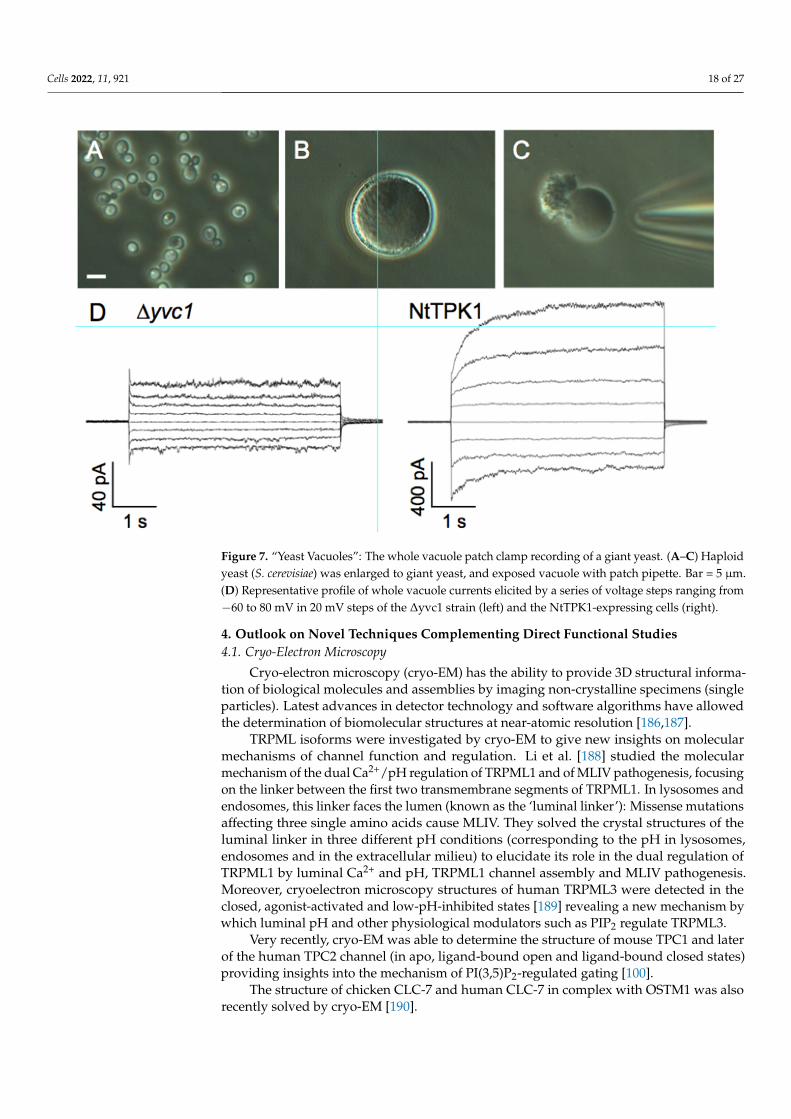
Cells 2022, 11, 921 18 of 27
Cells 2022, 11, x FOR PEER REVIEW 18 of 28
menal Zn2+, where they were suppressed by phosphatidylinositol 3-phosphate and vacu-olar lumenal Ca2+ [182]. TRPY1/YVC1-mediated Ca2+-regulated cation currents could be completely eliminated in the ∆trpy1/yvc1 strain [183], which confirmed the same feature of TRPY1/YVC1 reported by [184]. The patch-clamp recordings of NtTPK1, Figure 7, re-vealed high selectivity for K+ and strong activation by cytosolic acidic pH and cytosolic Ca2+, which causes an approximately two-fold increase of the current amplitude of non-treated NtTPK1. The structural properties and Ca2+ and phosphatidyl inositol modulation of fungal TRP homolog from Gibberella zeae as well as TRPY1/YVC1 have been character-ized [182,185]. Importantly, the human lysosomal channel hTPC2 could be also function-ally characterised in this system [26].
Figure 7. “Yeast Vacuoles”: The whole vacuole patch clamp recording of a giant yeast. (A–C) Hap-loid yeast (S. cerevisiae) was enlarged to giant yeast, and exposed vacuole with patch pipette. Bar = 5 μm. (D) Representative profile of whole vacuole currents elicited by a series of voltage steps rang-ing from −60 to 80 mV in 20 mV steps of the ∆yvc1 strain (left) and the NtTPK1-expressing cells (right).
The small genome size and rapid growth of E. coli and S. cerevisiae have contributed to the rapid progress of molecular biology and genetics. Disruption and induction of for-eign genes is readily achieved without difficulty to obtain experimental material quickly. Exploiting mutant vacuoles with reduced channel background for patch-clamp experi-ments will enable the detailed characterization of endolysosomal ion transporters in eu-karyotic cells.
4. Outlook on Novel Techniques Complementing Direct Functional Studies 4.1. Cryo-Electron Microscopy
Cryo-electron microscopy (cryo-EM) has the ability to provide 3D structural infor-mation of biological molecules and assemblies by imaging non-crystalline specimens (sin-gle particles). Latest advances in detector technology and software algorithms have al-lowed the determination of biomolecular structures at near-atomic resolution [186,187].
Figure 7. “Yeast Vacuoles”: The whole vacuole patch clamp recording of a giant yeast. (A–C) Haploidyeast (S. cerevisiae) was enlarged to giant yeast, and exposed vacuole with patch pipette. Bar = 5 µm.(D) Representative profile of whole vacuole currents elicited by a series of voltage steps ranging from−60 to 80 mV in 20 mV steps of the ∆yvc1 strain (left) and the NtTPK1-expressing cells (right).
4. Outlook on Novel Techniques Complementing Direct Functional Studies4.1. Cryo-Electron Microscopy
Cryo-electron microscopy (cryo-EM) has the ability to provide 3D structural informa-tion of biological molecules and assemblies by imaging non-crystalline specimens (singleparticles). Latest advances in detector technology and software algorithms have allowedthe determination of biomolecular structures at near-atomic resolution [186,187].
TRPML isoforms were investigated by cryo-EM to give new insights on molecularmechanisms of channel function and regulation. Li et al. [188] studied the molecularmechanism of the dual Ca2+/pH regulation of TRPML1 and of MLIV pathogenesis, focusingon the linker between the first two transmembrane segments of TRPML1. In lysosomes andendosomes, this linker faces the lumen (known as the ‘luminal linker’): Missense mutationsaffecting three single amino acids cause MLIV. They solved the crystal structures of theluminal linker in three different pH conditions (corresponding to the pH in lysosomes,endosomes and in the extracellular milieu) to elucidate its role in the dual regulation ofTRPML1 by luminal Ca2+ and pH, TRPML1 channel assembly and MLIV pathogenesis.Moreover, cryoelectron microscopy structures of human TRPML3 were detected in theclosed, agonist-activated and low-pH-inhibited states [189] revealing a new mechanism bywhich luminal pH and other physiological modulators such as PIP2 regulate TRPML3.
Very recently, cryo-EM was able to determine the structure of mouse TPC1 and laterof the human TPC2 channel (in apo, ligand-bound open and ligand-bound closed states)providing insights into the mechanism of PI(3,5)P2-regulated gating [100].
The structure of chicken CLC-7 and human CLC-7 in complex with OSTM1 was alsorecently solved by cryo-EM [190].
Cells 2022, 11, 921 19 of 27
4.2. Molecular Dynamics Simulations
X-ray crystallography and more recently cryo-EM provide us with an ever increasingnumber of atomic-resolution structures of membrane channels and transporters, allow-ing molecular dynamics (MD) simulations to study the mechanisms underlying theirbehavior [96,191,192]. Moreover, the increasing computational power permits simulationsreaching tens or hundreds of microseconds, which are time scales approaching those ofelectrophysiological measurements.
Kirsch and colleagues [96], using a combination of MD simulations and functionalanalysis of TPC2 channel mutants, showed that PI(3,5)P2 cross-links two parts of thechannel, enabling their coordinated movement during channel gating. In the last three years,several papers directly tackled the question of TPC’s function by employing moleculardocking [34] and standard MD simulations combined with extended sampling methodssuch as metadynamics [193,194]. In the first paper, it was shown that TPC2 selectivelybinds naringenin, a molecule that modulates the hTPC2 activity in human cells [14,33].By employing extended methods for sampling, the following two publications showedthat the binding of PI(3,5)P2 activates mTPC1 and hTPC2 by increasing fluctuations inthe hydrophobic gate (HG) region, and that sodium undergoes partial de-hydration whendiffusing through this region. Finally, it was found that the occupancy of the central cavity(CAV) region is key for an effective sodium transport in hTPC2. The last result clearlysuggests that at physiological concentrations optimum conductance is achieved whenhTPC2’s CAV has a single occupancy.
Regarding transporters, the V-ATPase proton pump has been recently simulated [195]pointing out that the cooperative rearrangements of the three catalytic pairs are crucial forthe rotation of the central stalk. In particular, V-ATPase interaction with lactoferrin wasstudied where it was shown that lactoferring inhibits V-ATPase by binding to the catalyticsite [196]. The latter is particularly important as it is a mean of cytosolic acidification offungi and cancer cells which leads to their subsequent death.
4.3. Genome Editing
Genome editing (also called gene editing) is a group of technologies that give scientiststhe ability to change an organism’s DNA. These technologies allow genetic material to beadded, removed or altered at particular locations in the genome. Several approaches togenome editing have been developed. A recent one, known as CRISPR, has generated alot of excitement in the scientific community because it is faster, cheaper, more accurateand more efficient than other existing genome editing methods [197]. CRISPR techniquesallow scientists to modify specific genes while sparing all others, thus clarifying the asso-ciation between a given gene and its role in the organism. One important application ofthis technology is to facilitate the creation of animal models with precise genetic changes tostudy the progress and possible treatment of human diseases. Recently, the involvement oflocalized Ca2+ release via TPC2 lysosomal channels was investigated using knockout ofTPC2 (via the generation of a tpcn2 mutant line of zebrafish using CRISPR/Cas9 gene edit-ing): A significant attenuation of slow (skeletal) muscle cell differentiation was observed,suggesting that TPC2 is involved in the maturation of muscle cells [198].
4.4. Nanoscopy
Stimulated emission depletion (STED) microscopy is one of the techniques that make upsuper-resolution microscopy. It creates super-resolution images by the selective deactivationof fluorophores, minimizing the area of illumination at the focal point and thus enhancingthe achievable resolution for a given system [199] bypassing the diffraction limit of lightmicroscopy to increase resolution. This technique was used to reveal the close physicalrelationship between clusters of ryanodine receptors (RyRs) in the terminal cisternae of thesarcoplasmic reticulum (SR) and TPC2 channels on the lysosomal membrane. It has beenproposed that TPC2-RyR clusters act as “trigger zones” in which TPCs are stimulated to create

Cells 2022, 11, 921 20 of 27
highly localized elementary Ca2+ signals that subsequently lead to the opening of the RyR inthe SR/ER membrane, resulting in global signals via Ca2+-induced Ca2+ release [198].
5. Conclusions
Lysosomal channels are emerging as fundamental proteins for cellular homeostasisand as a pharmacological target for a multitude of human pathologies. Here, we presentedthe main methods capable of revealing their functional properties. It is very likely that theadvent of new technologies will further expand our knowledge on this type of protein inorder to fully clarify their role in their metabolic context.
Funding: This work was supported by Grants-in-Aid for Scientific Research (20KK0127 and 21K19060)from the Ministry of Education, Culture, Sports, Science and Technology and Moonshot (J200002841)from JST to NU.
Institutional Review Board Statement: Not applicable.
Conflicts of Interest: Not applicable.
Abbreviations
PI(3,5)P2 phosphatidylinositol-(3,5)-bisphosphate;NAADP nicotinic acid adenine dinucleotide phosphate;ER endoplasmic reticulum;LSDs lysosomal storage disorders;VM vacuolar membrane;
References1. Xu, H.; Ren, D. Lysosomal Physiology. Annu. Rev. Physiol. 2015, 77, 57–80. [CrossRef] [PubMed]2. Luzio, J.P.; Pryor, P.R.; Bright, N.A. Lysosomes: Fusion and Function. Nat. Rev. Mol. Cell Biol. 2007, 8, 622–632. [CrossRef]
[PubMed]3. Pryor, P.R.; Luzio, J.P. Delivery of Endocytosed Membrane Proteins to the Lysosome. Biochim. Biophys. Acta BBA—Mol. Cell Res.
2009, 1793, 615–624. [CrossRef] [PubMed]4. Settembre, C.; Fraldi, A.; Medina, D.L.; Ballabio, A. Signals for the Lysosome: A Control Center for Cellular Clearance and Energy
Metabolism. Nat. Rev. Mol. Cell Biol. 2013, 14, 283–296. [CrossRef] [PubMed]5. Wu, Y.; Xu, M.; Wang, P.; Syeda, A.K.R.; Huang, P.; Dong, X.-P. Lysosomal Potassium Channels. Cell Calcium 2022, 102, 102536.
[CrossRef] [PubMed]6. Mindell, J.A. Lysosomal Acidification Mechanisms. Annu. Rev. Physiol. 2012, 74, 69–86. [CrossRef]7. Xiong, J.; Zhu, M.X. Regulation of Lysosomal Ion Homeostasis by Channels and Transporters. Sci. China Life Sci. 2016, 59, 777–791.
[CrossRef]8. Bagshaw, R.D.; Mahuran, D.J.; Callahan, J.W. A Proteomic Analysis of Lysosomal Integral Membrane Proteins Reveals the Diverse
Composition of the Organelle. Mol. Cell. Proteom. MCP 2005, 4, 133–143. [CrossRef]9. Schröder, B.; Wrocklage, C.; Pan, C.; Jäger, R.; Kösters, B.; Schäfer, H.; Elsässer, H.-P.; Mann, M.; Hasilik, A. Integral and Associated
Lysosomal Membrane Proteins. Traffic Cph. Den. 2007, 8, 1676–1686. [CrossRef]10. Repnik, U.; Turk, B. Lysosomal-Mitochondrial Cross-Talk during Cell Death. Mitochondrion 2010, 10, 662–669. [CrossRef]11. Lieberman, A.P.; Puertollano, R.; Raben, N.; Slaugenhaupt, S.; Walkley, S.U.; Ballabio, A. Autophagy in Lysosomal Storage
Disorders. Autophagy 2012, 8, 719–730. [CrossRef] [PubMed]12. Zhong, X.Z.; Yang, Y.; Sun, X.; Dong, X.-P. Methods for Monitoring Ca2+ and Ion Channels in the Lysosome. Cell Calcium 2017, 64, 20–28.
[CrossRef]13. Filippini, A.; D’Amore, A.; Palombi, F.; Carpaneto, A. Could the Inhibition of Endo-Lysosomal Two-Pore Channels (TPCs) by the
Natural Flavonoid Naringenin Represent an Option to Fight SARS-CoV-2 Infection? Front. Microbiol. 2020, 11, 970. [CrossRef][PubMed]
14. Clementi, N.; Scagnolari, C.; D’Amore, A.; Palombi, F.; Criscuolo, E.; Frasca, F.; Pierangeli, A.; Mancini, N.; Antonelli, G.; Clementi,M.; et al. Naringenin Is a Powerful Inhibitor of SARS-CoV-2 Infection in Vitro. Pharmacol. Res. 2021, 163, 105255. [CrossRef][PubMed]
15. Graves, A.R.; Curran, P.K.; Smith, C.L.; Mindell, J.A. The Cl-/H+ Antiporter ClC-7 Is the Primary Chloride Permeation Pathwayin Lysosomes. Nature 2008, 453, 788–792. [CrossRef]
16. Majumdar, A.; Capetillo-Zarate, E.; Cruz, D.; Gouras, G.K.; Maxfield, F.R. Degradation of Alzheimer’s Amyloid Fibrils byMicroglia Requires Delivery of ClC-7 to Lysosomes. Mol. Biol. Cell 2011, 22, 1664–1676. [CrossRef]
Cells 2022, 11, 921 21 of 27
17. Jentsch, T.J.; Pusch, M. CLC Chloride Channels and Transporters: Structure, Function, Physiology, and Disease. Physiol. Rev. 2018,98, 1493–1590. [CrossRef]
18. Bröer, S. The SLC38 Family of Sodium-Amino Acid Co-Transporters. Pflug. Arch. 2014, 466, 155–172. [CrossRef]19. Hägglund, M.G.A.; Sreedharan, S.; Nilsson, V.C.O.; Shaik, J.H.A.; Almkvist, I.M.; Bäcklin, S.; Wrange, O.; Fredriksson, R. Identification of
SLC38A7 (SNAT7) Protein as a Glutamine Transporter Expressed in Neurons. J. Biol. Chem. 2011, 286, 20500–20511. [CrossRef]20. Efeyan, A.; Zoncu, R.; Sabatini, D.M. Amino Acids and MTORC1: From Lysosomes to Disease. Trends Mol. Med. 2012, 18, 524–533.
[CrossRef]21. Ögmundsdóttir, M.H.; Heublein, S.; Kazi, S.; Reynolds, B.; Visvalingam, S.M.; Shaw, M.K.; Goberdhan, D.C.I. Proton-Assisted
Amino Acid Transporter PAT1 Complexes with Rag GTPases and Activates TORC1 on Late Endosomal and Lysosomal Membranes.PLoS ONE 2012, 7, e36616. [CrossRef] [PubMed]
22. Verdon, Q.; Boonen, M.; Ribes, C.; Jadot, M.; Gasnier, B.; Sagné, C. SNAT7 Is the Primary Lysosomal Glutamine Exporter Requiredfor Extracellular Protein-Dependent Growth of Cancer Cells. Proc. Natl. Acad. Sci. USA 2017, 114, E3602–E3611. [CrossRef][PubMed]
23. Donowitz, M.; Ming Tse, C.; Fuster, D. SLC9/NHE Gene Family, a Plasma Membrane and Organellar Family of Na+/H+
Exchangers. Mol. Asp. Med. 2013, 34, 236–251. [CrossRef] [PubMed]24. Calcraft, P.J.; Ruas, M.; Pan, Z.; Cheng, X.; Arredouani, A.; Hao, X.; Tang, J.; Rietdorf, K.; Teboul, L.; Chuang, K.-T.; et al. NAADP
Mobilizes Calcium from Acidic Organelles through Two-Pore Channels. Nature 2009, 459, 596–600. [CrossRef] [PubMed]25. Wang, X.; Zhang, X.; Dong, X.-P.; Samie, M.; Li, X.; Cheng, X.; Goschka, A.; Shen, D.; Zhou, Y.; Harlow, J.; et al. TPC Proteins Are
Phosphoinositide- Activated Sodium-Selective Ion Channels in Endosomes and Lysosomes. Cell 2012, 151, 372–383. [CrossRef]26. Boccaccio, A.; Scholz-Starke, J.; Hamamoto, S.; Larisch, N.; Festa, M.; Gutla, P.V.K.; Costa, A.; Dietrich, P.; Uozumi, N.; Carpaneto,
A. The Phosphoinositide PI (3, 5) P2 Mediates Activation of Mammalian but Not Plant TPC Proteins: Functional Expression ofEndolysosomal Channels in Yeast and Plant Cells. Cell. Mol. Life Sci. 2014, 71, 4275–4283. [CrossRef]
27. Zong, X.; Schieder, M.; Cuny, H.; Fenske, S.; Gruner, C.; Rötzer, K.; Griesbeck, O.; Harz, H.; Biel, M.; Wahl-Schott, C. The Two-PoreChannel TPCN2 Mediates NAADP-Dependent Ca(2+)-Release from Lysosomal Stores. Pflug. Arch. 2009, 458, 891–899. [CrossRef]
28. Grimm, C.; Holdt, L.M.; Chen, C.-C.; Hassan, S.; Müller, C.; Jörs, S.; Cuny, H.; Kissing, S.; Schröder, B.; Butz, E.; et al. HighSusceptibility to Fatty Liver Disease in Two-Pore Channel 2-Deficient Mice. Nat. Commun. 2014, 5, 4699. [CrossRef]
29. Sakurai, Y.; Kolokoltsov, A.A.; Chen, C.-C.; Tidwell, M.W.; Bauta, W.E.; Klugbauer, N.; Grimm, C.; Wahl-Schott, C.; Biel, M.;Davey, R.A. Ebola Virus. Two-Pore Channels Control Ebola Virus Host Cell Entry and Are Drug Targets for Disease Treatment.Science 2015, 347, 995–998. [CrossRef]
30. D’Amore, A.; Gradogna, A.; Palombi, F.; Minicozzi, V.; Ceccarelli, M.; Carpaneto, A.; Filippini, A. The Discovery of Naringenin asEndolysosomal Two-Pore Channel Inhibitor and Its Emerging Role in SARS-CoV-2 Infection. Cells 2021, 10, 1130. [CrossRef]
31. Hockey, L.N.; Kilpatrick, B.S.; Eden, E.R.; Lin-Moshier, Y.; Brailoiu, G.C.; Brailoiu, E.; Futter, C.E.; Schapira, A.H.; Marchant, J.S.;Patel, S. Dysregulation of Lysosomal Morphology by Pathogenic LRRK2 Is Corrected by TPC2 Inhibition. J. Cell Sci. 2015,128, 232–238. [CrossRef] [PubMed]
32. Favia, A.; Desideri, M.; Gambara, G.; D’Alessio, A.; Ruas, M.; Esposito, B.; Del Bufalo, D.; Parrington, J.; Ziparo, E.; Palombi, F.; et al.VEGF-Induced Neoangiogenesis Is Mediated by NAADP and Two-Pore Channel-2-Dependent Ca2+ Signaling. Proc. Natl. Acad. Sci.USA 2014, 111, E4706–E4715. [CrossRef] [PubMed]
33. Pafumi, I.; Festa, M.; Papacci, F.; Lagostena, L.; Giunta, C.; Gutla, V.; Cornara, L.; Favia, A.; Palombi, F.; Gambale, F.; et al.Naringenin Impairs Two-Pore Channel 2 Activity And Inhibits VEGF-Induced Angiogenesis. Sci. Rep. 2017, 7, 5121. [CrossRef]
34. Benkerrou, D.; Minicozzi, V.; Gradogna, A.; Milenkovic, S.; Bodrenko, I.V.; Festa, M.; Lagostena, L.; Cornara, L.; D’Amore, A.;Ceccarelli, M.; et al. A Perspective on the Modulation of Plant and Animal Two Pore Channels (TPCs) by the FlavonoidNaringenin. Biophys. Chem. 2019, 254, 106246. [CrossRef] [PubMed]
35. Padamsey, Z.; McGuinness, L.; Bardo, S.J.; Reinhart, M.; Tong, R.; Hedegaard, A.; Hart, M.L.; Emptage, N.J. Activity-DependentExocytosis of Lysosomes Regulates the Structural Plasticity of Dendritic Spines. Neuron 2017, 93, 132–146. [CrossRef] [PubMed]
36. Tian, X.; Gala, U.; Zhang, Y.; Shang, W.; Nagarkar Jaiswal, S.; di Ronza, A.; Jaiswal, M.; Yamamoto, S.; Sandoval, H.; Duraine, L.; et al. AVoltage-Gated Calcium Channel Regulates Lysosomal Fusion with Endosomes and Autophagosomes and Is Required for NeuronalHomeostasis. PLoS Biol. 2015, 13, e1002103. [CrossRef] [PubMed]
37. Nilius, B.; Flockerzi, V. Mammalian Transient Receptor Potential (TRP) Cation Channels. Preface. Handb. Exp. Pharmacol. 2014,223, v–vi.
38. Zheng, J. Molecular Mechanism of TRP Channels. Compr. Physiol. 2013, 3, 221–242. [CrossRef]39. Venkatachalam, K.; Wong, C.-O.; Zhu, M.X. The Role of TRPMLs in Endolysosomal Trafficking and Function. Cell Calcium 2015,
58, 48–56. [CrossRef]40. Cheng, X.; Shen, D.; Samie, M.; Xu, H. Mucolipins: Intracellular TRPML1-3 Channels. FEBS Lett. 2010, 584, 2013–2021. [CrossRef]41. Sun, L.; Hua, Y.; Vergarajauregui, S.; Diab, H.I.; Puertollano, R. Novel Role of TRPML2 in the Regulation of the Innate Immune
Response. J. Immunol. 2015, 195, 4922–4932. [CrossRef] [PubMed]42. Xu, H.; Delling, M.; Li, L.; Dong, X.; Clapham, D.E. Activating Mutation in a Mucolipin Transient Receptor Potential Channel
Leads to Melanocyte Loss in Varitint-Waddler Mice. Proc. Natl. Acad. Sci. USA 2007, 104, 18321–18326. [CrossRef] [PubMed]
Cells 2022, 11, 921 22 of 27
43. Grimm, C.; Cuajungco, M.P.; van Aken, A.F.J.; Schnee, M.; Jörs, S.; Kros, C.J.; Ricci, A.J.; Heller, S. A Helix-Breaking Mutation inTRPML3 Leads to Constitutive Activity Underlying Deafness in the Varitint-Waddler Mouse. Proc. Natl. Acad. Sci. USA 2007,104, 19583–19588. [CrossRef] [PubMed]
44. Kim, H.J.; Li, Q.; Tjon-Kon-Sang, S.; So, I.; Kiselyov, K.; Muallem, S. Gain-of-Function Mutation in TRPML3 Causes the MouseVaritint-Waddler Phenotype. J. Biol. Chem. 2007, 282, 36138–36142. [CrossRef]
45. Nagata, K.; Zheng, L.; Madathany, T.; Castiglioni, A.J.; Bartles, J.R.; García-Añoveros, J. The Varitint-Waddler (Va) DeafnessMutation in TRPML3 Generates Constitutive, Inward Rectifying Currents and Causes Cell Degeneration. Proc. Natl. Acad. Sci.USA 2008, 105, 353–358. [CrossRef]
46. Cao, Q.; Zhong, X.Z.; Zou, Y.; Murrell-Lagnado, R.; Zhu, M.X.; Dong, X.-P. Calcium Release through P2X4 Activates Calmodulinto Promote Endolysosomal Membrane Fusion. J. Cell Biol. 2015, 209, 879–894. [CrossRef]
47. Cang, C.; Aranda, K.; Seo, Y.; Gasnier, B.; Ren, D. TMEM175 Is an Organelle K+ Channel Regulating Lysosomal Function. Cell2015, 162, 1101–1112. [CrossRef]
48. Cao, Q.; Zhong, X.Z.; Zou, Y.; Zhang, Z.; Toro, L.; Dong, X.-P. BK Channels Alleviate Lysosomal Storage Diseases by ProvidingPositive Feedback Regulation of Lysosomal Ca2+ Release. Dev. Cell 2015, 33, 427–441. [CrossRef]
49. Lee, C.; Guo, J.; Zeng, W.; Kim, S.; She, J.; Cang, C.; Ren, D.; Jiang, Y. The Lysosomal Potassium Channel TMEM175 Adopts aNovel Tetrameric Architecture. Nature 2017, 547, 472–475. [CrossRef]
50. Jinn, S.; Drolet, R.E.; Cramer, P.E.; Wong, A.H.-K.; Toolan, D.M.; Gretzula, C.A.; Voleti, B.; Vassileva, G.; Disa, J.; Tadin-Strapps, M.; et al.TMEM175 Deficiency Impairs Lysosomal and Mitochondrial Function and Increases α-Synuclein Aggregation. Proc. Natl. Acad. Sci.USA 2017, 114, 2389–2394. [CrossRef]
51. Li, P.; Hu, M.; Wang, C.; Feng, X.; Zhao, Z.; Yang, Y.; Sahoo, N.; Gu, M.; Yang, Y.; Xiao, S.; et al. LRRC8 Family Proteins withinLysosomes Regulate Cellular Osmoregulation and Enhance Cell Survival to Multiple Physiological Stresses. Proc. Natl. Acad. Sci.USA 2020, 117, 29155–29165. [CrossRef] [PubMed]
52. Beyenbach, K.W.; Wieczorek, H. The V-Type H+ ATPase: Molecular Structure and Function, Physiological Roles and Regulation.J. Exp. Biol. 2006, 209, 577–589. [CrossRef] [PubMed]
53. Wang, Y.; Zeng, W.; Lin, B.; Yao, Y.; Li, C.; Hu, W.; Wu, H.; Huang, J.; Zhang, M.; Xue, T.; et al. CLN7 Is an Organellar ChlorideChannel Regulating Lysosomal Function. Sci. Adv. 2021, 7, eabj9608. [CrossRef] [PubMed]
54. Sterea, A.M.; Almasi, S.; El Hiani, Y. The Hidden Potential of Lysosomal Ion Channels: A New Era of Oncogenes. Cell Calcium2018, 72, 91–103. [CrossRef] [PubMed]
55. Gaburjakova, J.; Gaburjakova, M. Reconstitution of Ion Channels in Planar Lipid Bilayers: New Approaches. In Advances in Biomembranesand Lipid Self-Assembly; Elsevier: Amsterdam, The Netherlands, 2018; Volume 27, pp. 147–185. ISBN 978-0-12-815772-5.
56. Montal, M.; Mueller, P. Formation of Bimolecular Membranes from Lipid Monolayers and a Study of Their Electrical Properties.Proc. Natl. Acad. Sci. USA 1972, 69, 3561–3566. [CrossRef]
57. Mueller, P.; Rudin, D.O.; Tien, H.T.; Wescott, W.C. Reconstitution of Cell Membrane Structure in Vitro and Its Transformation intoan Excitable System. Nature 1962, 194, 979–980. [CrossRef]
58. Oiki, S.; Iwamoto, M. Lipid Bilayers Manipulated through Monolayer Technologies for Studies of Channel-Membrane Interplay.Biol. Pharm. Bull. 2018, 41, 303–311. [CrossRef]
59. Dalla Serra, M.; Menestrina, G. Liposomes in the Study of Pore-Forming Toxins. Methods Enzymol. 2003, 372, 99–124. [CrossRef]60. Patil, Y.P.; Jadhav, S. Novel Methods for Liposome Preparation. Chem. Phys. Lipids 2014, 177, 8–18. [CrossRef]61. Venturi, E.; Sitsapesan, R. Reconstitution of Lysosomal Ion Channels into Artificial Membranes. Methods Cell Biol. 2015, 126, 217–236.
[CrossRef]62. Pitt, S.J.; Funnell, T.M.; Sitsapesan, M.; Venturi, E.; Rietdorf, K.; Ruas, M.; Ganesan, A.; Gosain, R.; Churchill, G.C.; Zhu, M.X.; et al.
TPC2 Is a Novel NAADP-Sensitive Ca2+ Release Channel, Operating as a Dual Sensor of Luminal PH and Ca2+. J. Biol. Chem.2010, 285, 35039–35046. [CrossRef] [PubMed]
63. Pitt, S.J.; Lam, A.K.M.; Rietdorf, K.; Galione, A.; Sitsapesan, R. Reconstituted Human TPC1 Is a Proton-Permeable Ion Channeland Is Activated by NAADP or Ca2+. Sci. Signal. 2014, 7, ra46. [CrossRef] [PubMed]
64. Rybalchenko, V.; Ahuja, M.; Coblentz, J.; Churamani, D.; Patel, S.; Kiselyov, K.; Muallem, S. Membrane Potential RegulatesNicotinic Acid Adenine Dinucleotide Phosphate (NAADP) Dependence of the PH- and Ca2+-Sensitive Organellar Two-PoreChannel TPC1. J. Biol. Chem. 2012, 287, 20407–20416. [CrossRef] [PubMed]
65. Raychowdhury, M.K.; González-Perrett, S.; Montalbetti, N.; Timpanaro, G.A.; Chasan, B.; Goldmann, W.H.; Stahl, S.; Cooney, A.;Goldin, E.; Cantiello, H.F. Molecular Pathophysiology of Mucolipidosis Type IV: PH Dysregulation of the Mucolipin-1 CationChannel. Hum. Mol. Genet. 2004, 13, 617–627. [CrossRef] [PubMed]
66. Seifert, K.; Fendler, K.; Bamberg, E. Charge Transport by Ion Translocating Membrane Proteins on Solid Supported Membranes.Biophys. J. 1993, 64, 384–391. [CrossRef]
67. Schulz, P.; Garcia-Celma, J.J.; Fendler, K. SSM-Based Electrophysiology. Methods 2008, 46, 97–103. [CrossRef]68. Bazzone, A.; Costa, W.S.; Braner, M.; Călinescu, O.; Hatahet, L.; Fendler, K. Introduction to Solid Supported Membrane Based
Electrophysiology. J. Vis. Exp. JoVE 2013, 75, e50230. [CrossRef]69. Bazzone, A.; Barthmes, M.; Fendler, K. SSM-Based Electrophysiology for Transporter Research. Methods Enzymol. 2017, 594, 31–83.
[CrossRef]

Cells 2022, 11, 921 23 of 27
70. Obrdlik, P.; Diekert, K.; Watzke, N.; Keipert, C.; Pehl, U.; Brosch, C.; Boehm, N.; Bick, I.; Ruitenberg, M.; Volknandt, W.; et al.Electrophysiological Characterization of ATPases in Native Synaptic Vesicles and Synaptic Plasma Membranes. Biochem. J. 2010,427, 151–159. [CrossRef]
71. Schulz, P.; Werner, J.; Stauber, T.; Henriksen, K.; Fendler, K. The G215R Mutation in the Cl−/H+-Antiporter ClC-7 Found in ADOII Osteopetrosis Does Not Abolish Function but Causes a Severe Trafficking Defect. PLoS ONE 2010, 5, e12585. [CrossRef]
72. Garty, H.; Rudy, B.; Karlish, S.J. A Simple and Sensitive Procedure for Measuring Isotope Fluxes through Ion-Specific Channels inHeterogenous Populations of Membrane Vesicles. J. Biol. Chem. 1983, 258, 13094–13099. [CrossRef]
73. Goldberg, A.F.; Miller, C. Solubilization and Functional Reconstitution of a Chloride Channel from Torpedo Californica Electroplax.J. Membr. Biol. 1991, 124, 199–206. [CrossRef] [PubMed]
74. Dong, X.-P.; Cheng, X.; Mills, E.; Delling, M.; Wang, F.; Kurz, T.; Xu, H. The Type IV Mucolipidosis-Associated Protein TRPML1 Isan Endolysosomal Iron Release Channel. Nature 2008, 455, 992–996. [CrossRef] [PubMed]
75. Dong, X.; Shen, D.; Wang, X.; Dawson, T.; Li, X.; Zhang, Q.; Cheng, X.; Zhang, Y.; Weisman, L.S.; Delling, M.; et al. PI(3,5)P2Controls Membrane Trafficking by Direct Activation of Mucolipin Ca2+ Release Channels in the Endolysosome. Nat. Commun.2010, 1, 38. [CrossRef] [PubMed]
76. Cang, C.; Zhou, Y.; Navarro, B.; Seo, Y.-J.; Aranda, K.; Shi, L.; Battaglia-Hsu, S.; Nissim, I.; Clapham, D.E.; Ren, D. MTORRegulates Lysosomal ATP-Sensitive Two-Pore Na(+) Channels to Adapt to Metabolic State. Cell 2013, 152, 778–790. [CrossRef][PubMed]
77. Cang, C.; Bekele, B.; Ren, D. The Voltage-Gated Sodium Channel TPC1 Confers Endolysosomal Excitability. Nat. Chem. Biol. 2014,10, 463–469. [CrossRef] [PubMed]
78. Chen, C.-C.; Cang, C.; Fenske, S.; Butz, E.; Chao, Y.-K.; Biel, M.; Ren, D.; Wahl-Schott, C.; Grimm, C. Patch-Clamp Technique toCharacterize Ion Channels in Enlarged Individual Endolysosomes. Nat. Protoc. 2017, 12, 1639–1658. [CrossRef]
79. Saito, M.; Hanson, P.I.; Schlesinger, P. Luminal Chloride-Dependent Activation of Endosome Calcium Channels: Patch ClampStudy of Enlarged Endosomes. J. Biol. Chem. 2007, 282, 27327–27333. [CrossRef]
80. Huynh, C.; Andrews, N.W. The Small Chemical Vacuolin-1 Alters the Morphology of Lysosomes without Inhibiting Ca2+-Regulated Exocytosis. EMBO Rep. 2005, 6, 843–847. [CrossRef]
81. Schieder, M.; Rötzer, K.; Brüggemann, A.; Biel, M.; Wahl-Schott, C. Planar Patch Clamp Approach to Characterize Ionic Currentsfrom Intact Lysosomes. Sci. Signal. 2010, 3, pl3. [CrossRef]
82. Schieder, M.; Rötzer, K.; Brüggemann, A.; Biel, M.; Wahl-Schott, C.A. Characterization of Two-Pore Channel 2 (TPCN2)-MediatedCa2+ Currents in Isolated Lysosomes. J. Biol. Chem. 2010, 285, 21219–21222. [CrossRef] [PubMed]
83. Chen, C.-C.; Butz, E.S.; Chao, Y.-K.; Grishchuk, Y.; Becker, L.; Heller, S.; Slaugenhaupt, S.A.; Biel, M.; Wahl-Schott, C.; Grimm, C.Small Molecules for Early Endosome-Specific Patch Clamping. Cell Chem. Biol. 2017, 24, 907–916.e4. [CrossRef] [PubMed]
84. Gerndt, S.; Chen, C.-C.; Chao, Y.-K.; Yuan, Y.; Burgstaller, S.; Scotto Rosato, A.; Krogsaeter, E.; Urban, N.; Jacob, K.; Nguyen, O.N.P.; et al.Agonist-Mediated Switching of Ion Selectivity in TPC2 Differentially Promotes Lysosomal Function. eLife 2020, 9. [CrossRef] [PubMed]
85. Jha, A.; Ahuja, M.; Patel, S.; Brailoiu, E.; Muallem, S. Convergent Regulation of the Lysosomal Two-Pore Channel-2 by Mg2+,NAADP, PI(3,5)P2 and Multiple Protein Kinases. EMBO J. 2014, 33, 501–511. [CrossRef]
86. Kelly, B.T.; Owen, D.J. Endocytic Sorting of Transmembrane Protein Cargo. Curr. Opin. Cell Biol. 2011, 23, 404–412. [CrossRef]87. Staudt, C.; Puissant, E.; Boonen, M. Subcellular Trafficking of Mammalian Lysosomal Proteins: An Extended View. Int. J. Mol. Sci.
2016, 18, 47. [CrossRef]88. Traub, L.M.; Bonifacino, J.S. Cargo Recognition in Clathrin-Mediated Endocytosis. Cold Spring Harb. Perspect. Biol. 2013, 5, a016790.
[CrossRef]89. Bonifacino, J.S.; Traub, L.M. Signals for Sorting of Transmembrane Proteins to Endosomes and Lysosomes. Annu. Rev. Biochem. 2003,
72, 395–447. [CrossRef]90. Jung, N.; Wienisch, M.; Gu, M.; Rand, J.B.; Müller, S.L.; Krause, G.; Jorgensen, E.M.; Klingauf, J.; Haucke, V. Molecular Basis of
Synaptic Vesicle Cargo Recognition by the Endocytic Sorting Adaptor Stonin 2. J. Cell Biol. 2007, 179, 1497–1510. [CrossRef]91. Robinson, M.S. Forty Years of Clathrin-Coated Vesicles. Traffic Cph. Den. 2015, 16, 1210–1238. [CrossRef]92. Ibberson, M.; Uldry, M.; Thorens, B. GLUTX1, a Novel Mammalian Glucose Transporter Expressed in the Central Nervous System
and Insulin-Sensitive Tissues. J. Biol. Chem. 2000, 275, 4607–4612. [CrossRef] [PubMed]93. Augustin, R.; Riley, J.; Moley, K.H. GLUT8 Contains a [DE]XXXL[LI] Sorting Motif and Localizes to a Late Endosomal/Lysosomal
Compartment. Traffic Cph. Den. 2005, 6, 1196–1212. [CrossRef] [PubMed]94. Yamada, K.; Osakabe, Y.; Mizoi, J.; Nakashima, K.; Fujita, Y.; Shinozaki, K.; Yamaguchi-Shinozaki, K. Functional Analysis of
an Arabidopsis Thaliana Abiotic Stress-Inducible Facilitated Diffusion Transporter for Monosaccharides. J. Biol. Chem. 2010,285, 1138–1146. [CrossRef] [PubMed]
95. Brailoiu, E.; Rahman, T.; Churamani, D.; Prole, D.L.; Brailoiu, G.C.; Hooper, R.; Taylor, C.W.; Patel, S. An NAADP-Gated Two-PoreChannel Targeted to the Plasma Membrane Uncouples Triggering from Amplifying Ca2+ Signals. J. Biol. Chem. 2010, 285, 38511–38516.[CrossRef]
96. Kirsch, S.A.; Kugemann, A.; Carpaneto, A.; Böckmann, R.A.; Dietrich, P. Phosphatidylinositol-3,5-Bisphosphate Lipid-Binding-Induced Activation of the Human Two-Pore Channel 2. Cell. Mol. Life Sci. CMLS 2018, 75, 3803–3815. [CrossRef]
97. Larisch, N.; Schulze, C.; Galione, A.; Dietrich, P. An N-Terminal Dileucine Motif Directs Two-Pore Channels to the Tonoplast ofPlant Cells. Traffic Cph. Den. 2012, 13, 1012–1022. [CrossRef]

Cells 2022, 11, 921 24 of 27
98. Guo, J.; Zeng, W.; Chen, Q.; Lee, C.; Chen, L.; Yang, Y.; Cang, C.; Ren, D.; Jiang, Y. Structure of the Voltage-Gated Two-PoreChannel TPC1 from Arabidopsis Thaliana. Nature 2016, 531, 196–201. [CrossRef]
99. Ye, F.; Xu, L.; Li, X.; Zeng, W.; Gan, N.; Zhao, C.; Yang, W.; Jiang, Y.; Guo, J. Voltage-Gating and Cytosolic Ca2+ ActivationMechanisms of Arabidopsis Two-Pore Channel AtTPC1. Proc. Natl. Acad. Sci. USA 2021, 118, e2113946118. [CrossRef]
100. She, J.; Zeng, W.; Guo, J.; Chen, Q.; Bai, X.-C.; Jiang, Y. Structural Mechanisms of Phospholipid Activation of the Human TPC2Channel. eLife 2019, 8, e45222. [CrossRef]
101. Stauber, T.; Jentsch, T.J. Sorting Motifs of the Endosomal/Lysosomal CLC Chloride Transporters. J. Biol. Chem. 2010, 285, 34537–34548.[CrossRef]
102. Leisle, L.; Ludwig, C.F.; Wagner, F.A.; Jentsch, T.J.; Stauber, T. ClC-7 Is a Slowly Voltage-Gated 2Cl(-)/1H(+)-Exchanger andRequires Ostm1 for Transport Activity. EMBO J. 2011, 30, 2140–2152. [CrossRef] [PubMed]
103. Cherqui, S.; Kalatzis, V.; Trugnan, G.; Antignac, C. The Targeting of Cystinosin to the Lysosomal Membrane Requires a Tyrosine-Based Signal and a Novel Sorting Motif. J. Biol. Chem. 2001, 276, 13314–13321. [CrossRef] [PubMed]
104. Kalatzis, V.; Cherqui, S.; Antignac, C.; Gasnier, B. Cystinosin, the Protein Defective in Cystinosis, Is a H(+)-Driven LysosomalCystine Transporter. EMBO J. 2001, 20, 5940–5949. [CrossRef] [PubMed]
105. Ruivo, R.; Bellenchi, G.C.; Chen, X.; Zifarelli, G.; Sagné, C.; Debacker, C.; Pusch, M.; Supplisson, S.; Gasnier, B. Mechanism ofProton/Substrate Coupling in the Heptahelical Lysosomal Transporter Cystinosin. Proc. Natl. Acad. Sci. USA 2012, 109, E210–E217.[CrossRef] [PubMed]
106. Jézégou, A.; Llinares, E.; Anne, C.; Kieffer-Jaquinod, S.; O’Regan, S.; Aupetit, J.; Chabli, A.; Sagné, C.; Debacker, C.;Chadefaux-Vekemans, B.; et al. Heptahelical Protein PQLC2 Is a Lysosomal Cationic Amino Acid Exporter Underlying theAction of Cysteamine in Cystinosis Therapy. Proc. Natl. Acad. Sci. USA 2012, 109, E3434–E3443. [CrossRef]
107. Leray, X.; Conti, R.; Li, Y.; Debacker, C.; Castelli, F.; Fenaille, F.; Zdebik, A.A.; Pusch, M.; Gasnier, B. Arginine-Selective Modulationof the Lysosomal Transporter PQLC2 through a Gate-Tuning Mechanism. Proc. Natl. Acad. Sci. USA 2021, 118, e2025315118.[CrossRef]
108. Neagoe, I.; Stauber, T.; Fidzinski, P.; Bergsdorf, E.-Y.; Jentsch, T.J. The Late Endosomal ClC-6 Mediates Proton/ChlorideCountertransport in Heterologous Plasma Membrane Expression. J. Biol. Chem. 2010, 285, 21689–21697. [CrossRef]
109. Polovitskaya, M.M.; Barbini, C.; Martinelli, D.; Harms, F.L.; Cole, F.S.; Calligari, P.; Bocchinfuso, G.; Stella, L.; Ciolfi, A.; Niceta, M.; et al.A Recurrent Gain-of-Function Mutation in CLCN6, Encoding the ClC-6 Cl−/H+-Exchanger, Causes Early-Onset Neurodegeneration.Am. J. Hum. Genet. 2020, 107, 1062–1077. [CrossRef]
110. Beitz, E.; Liu, K.; Ikeda, M.; Guggino, W.B.; Agre, P.; Yasui, M. Determinants of AQP6 Trafficking to Intracellular Sites versus thePlasma Membrane in Transfected Mammalian Cells. Biol. Cell 2006, 98, 101–109. [CrossRef]
111. Ikeda, M.; Beitz, E.; Kozono, D.; Guggino, W.B.; Agre, P.; Yasui, M. Characterization of Aquaporin-6 as a Nitrate Channel inMammalian Cells. Requirement of Pore-Lining Residue Threonine 63. J. Biol. Chem. 2002, 277, 39873–39879. [CrossRef]
112. Pergel, E.; Veres, I.; Csigi, G.I.; Czirják, G. Translocation of TMEM175 Lysosomal Potassium Channel to the Plasma Membrane byDynasore Compounds. Int. J. Mol. Sci. 2021, 22, 10515. [CrossRef] [PubMed]
113. Mak, D.-O.D.; Vais, H.; Cheung, K.-H.; Foskett, J.K. Nuclear Patch-Clamp Electrophysiology of Ca2+ Channels. Cold Spring Harb.Protoc. 2013, 2013, 885–891. [CrossRef] [PubMed]
114. Mak, D.-O.D.; Vais, H.; Cheung, K.-H.; Foskett, J.K. Patch-Clamp Electrophysiology of Intracellular Ca2+ Channels. Cold SpringHarb. Protoc. 2013, 2013, 787–797. [CrossRef] [PubMed]
115. Lee, C.S.-K.; Tong, B.C.-K.; Cheng, C.W.-H.; Hung, H.C.-H.; Cheung, K.-H. Characterization of Two-Pore Channel 2 by NuclearMembrane Electrophysiology. Sci. Rep. 2016, 6, 20282. [CrossRef]
116. Martinoia, E.; Maeshima, M.; Neuhaus, H.E. Vacuolar Transporters and Their Essential Role in Plant Metabolism. J. Exp. Bot.2007, 58, 83–102. [CrossRef]
117. Marty, F. Plant Vacuoles. Plant Cell 1999, 11, 587–600. [CrossRef]118. Hedrich, R. Technical Approaches to Studying Specific Properties of Ion Channels in Plants. In Single-Channel Recording; Sakmann,
B., Neher, E., Eds.; Springer: Boston, MA, USA, 1995; pp. 277–305. ISBN 978-1-4419-1229-9.119. Carpaneto, A. Nickel Inhibits the Slowly Activating Channels of Radish Vacuoles. Eur. Biophys. J. EBJ 2003, 32, 60–66. [CrossRef]120. Hedrich, R.; Marten, I. 30-Year Progress of Membrane Transport in Plants. Planta 2006, 224, 725–739. [CrossRef]121. Rocchetti, A.; Sharma, T.; Wulfetange, C.; Scholz-Starke, J.; Grippa, A.; Carpaneto, A.; Dreyer, I.; Vitale, A.; Czempinski, K.;
Pedrazzini, E. The Putative K(+) Channel Subunit AtKCO3 Forms Stable Dimers in Arabidopsis. Front. Plant Sci. 2012, 3, 251.[CrossRef]
122. Martinoia, E. Vacuolar Transporters—Companions on a Longtime Journey[OPEN]. Plant Physiol. 2018, 176, 1384–1407. [CrossRef]123. Gambale, F.; Cantu, A.M.; Carpaneto, A.; Keller, B.U. Fast and Slow Activation of Voltage-Dependent Ion Channels in Radish
Vacuoles. Biophys. J. 1993, 65, 1837–1843. [CrossRef]124. Paganetto, A.; Carpaneto, A.; Gambale, F. Ion Transport and Metal Sensitivity of Vacuolar Channels from the Roots of the Aquatic
Plant Eichhornia Crassipes. Plant Cell Environ. 2001, 24, 1329–1336. [CrossRef]125. Scholz-Starke, J.; Carpaneto, A.; Gambale, F. On the Interaction of Neomycin with the Slow Vacuolar Channel of Arabidopsis
Thaliana. J. Gen. Physiol. 2006, 127, 329–340. [CrossRef] [PubMed]126. Pottosin, I.I.; Martínez-Estévez, M. Regulation of the Fast Vacuolar Channel by Cytosolic and Vacuolar Potassium. Biophys. J.
2003, 84, 977–986. [CrossRef]

Cells 2022, 11, 921 25 of 27
127. Konrad, K.R.; Hedrich, R. The Use of Voltage-Sensitive Dyes to Monitor Signal-Induced Changes in Membrane Potential-ABATriggered Membrane Depolarization in Guard Cells. Plant J. Cell Mol. Biol. 2008, 55, 161–173. [CrossRef]
128. Gradogna, A.; Scholz-Starke, J.; Gutla, P.V.K.; Carpaneto, A. Fluorescence Combined with Excised Patch: Measuring CalciumCurrents in Plant Cation Channels. Plant J. 2009, 58, 175–182. [CrossRef]
129. Carpaneto, A.; Boccaccio, A.; Lagostena, L.; Di Zanni, E.; Scholz-Starke, J. The Signaling Lipid Phosphatidylinositol-3,5-Bisphosphate Targets Plant CLC-a Anion/H+ Exchange Activity. EMBO Rep. 2017, 18, 1100–1107. [CrossRef]
130. Carpaneto, A.; Gradogna, A. Modulation of Calcium and Potassium Permeation in Plant TPC Channels. Biophys. Chem. 2018, 236, 1–7.[CrossRef]
131. Gradogna, A.; Scholz-Starke, J.; Pardo, J.M.; Carpaneto, A. Beyond the Patch-Clamp Resolution: Functional Activity of Nonelec-trogenic Vacuolar NHX Proton/Potassium Antiporters and Inhibition by Phosphoinositides. New Phytol. 2021, 229, 3026–3036.[CrossRef]
132. Costa, A.; Gutla, P.V.K.; Boccaccio, A.; Scholz-Starke, J.; Festa, M.; Basso, B.; Zanardi, I.; Pusch, M.; Schiavo, F.L.; Gambale, F.; et al.The Arabidopsis Central Vacuole as an Expression System for Intracellular Transporters: Functional Characterization of the Cl-/H+Exchanger CLC-7. J. Physiol. 2012, 590, 3421–3430. [CrossRef]
133. Lagostena, L.; Festa, M.; Pusch, M.; Carpaneto, A. The Human Two-Pore Channel 1 Is Modulated by Cytosolic and LuminalCalcium. Sci. Rep. 2017, 7, 43900. [CrossRef] [PubMed]
134. Festa, M.; Lagostena, L.; Carpaneto, A. Using the Plant Vacuole as a Biological System to Investigate the Functional Properties ofExogenous Channels and Transporters. Biochim. Biophys. Acta 2016, 1858, 607–612. [CrossRef] [PubMed]
135. Pedrazzini, E.; Vitale, A. Protein Biosynthesis and Maturation in the ER. Methods Mol. Biol. Clifton NJ 2018, 1691, 179–189.[CrossRef]
136. Braulke, T.; Bonifacino, J.S. Sorting of Lysosomal Proteins. Biochim. Biophys. Acta 2009, 1793, 605–614. [CrossRef] [PubMed]137. Pedrazzini, E.; Komarova, N.Y.; Rentsch, D.; Vitale, A. Traffic Routes and Signals for the Tonoplast. Traffic Cph. Den. 2013, 14, 622–628.
[CrossRef]138. Schwake, M.; Schröder, B.; Saftig, P. Lysosomal Membrane Proteins and Their Central Role in Physiology. Traffic Cph. Den. 2013,
14, 739–748. [CrossRef]139. Janvier, K.; Bonifacino, J.S. Role of the Endocytic Machinery in the Sorting of Lysosome-Associated Membrane Proteins. Mol. Biol.
Cell 2005, 16, 4231–4242. [CrossRef]140. Gasber, A.; Klaumann, S.; Trentmann, O.; Trampczynska, A.; Clemens, S.; Schneider, S.; Sauer, N.; Feifer, I.; Bittner, F.; Mendel, R.R.; et al.
Identification of an Arabidopsis Solute Carrier Critical for Intracellular Transport and Inter-Organ Allocation of Molybdate. Plant Biol.Stuttg. Ger. 2011, 13, 710–718. [CrossRef]
141. Maîtrejean, M.; Vitale, A. How Are Tonoplast Proteins Degraded? Plant Signal. Behav. 2011, 6, 1809–1812. [CrossRef]142. Wolfenstetter, S.; Wirsching, P.; Dotzauer, D.; Schneider, S.; Sauer, N. Routes to the Tonoplast: The Sorting of Tonoplast Transporters
in Arabidopsis Mesophyll Protoplasts. Plant Cell 2012, 24, 215–232. [CrossRef]143. Bottanelli, F.; Foresti, O.; Hanton, S.; Denecke, J. Vacuolar Transport in Tobacco Leaf Epidermis Cells Involves a Single Route for
Soluble Cargo and Multiple Routes for Membrane Cargo. Plant Cell 2011, 23, 3007–3025. [CrossRef] [PubMed]144. Gomez, L.; Chrispeels, M.J. Tonoplast and Soluble Vacuolar Proteins Are Targeted by Different Mechanisms. Plant Cell 1993,
5, 1113–1124. [CrossRef] [PubMed]145. Viotti, C. ER and Vacuoles: Never Been Closer. Front. Plant Sci. 2014, 5, 20. [CrossRef]146. Viotti, C.; Krüger, F.; Krebs, M.; Neubert, C.; Fink, F.; Lupanga, U.; Scheuring, D.; Boutté, Y.; Frescatada-Rosa, M.; Wolfenstetter, S.; et al.
The Endoplasmic Reticulum Is the Main Membrane Source for Biogenesis of the Lytic Vacuole in Arabidopsis. Plant Cell 2013, 25, 3434–3449.[CrossRef]
147. Wang, X.; Cai, Y.; Wang, H.; Zeng, Y.; Zhuang, X.; Li, B.; Jiang, L. Trans-Golgi Network-Located AP1 Gamma Adaptins MediateDileucine Motif-Directed Vacuolar Targeting in Arabidopsis. Plant Cell 2014, 26, 4102–4118. [CrossRef]
148. Müdsam, C.; Wollschläger, P.; Sauer, N.; Schneider, S. Sorting of Arabidopsis NRAMP3 and NRAMP4 Depends on AdaptorProtein Complex AP4 and a Dileucine-Based Motif. Traffic Cph. Den. 2018, 19, 503–521. [CrossRef]
149. Hirst, J.; Edgar, J.R.; Esteves, T.; Darios, F.; Madeo, M.; Chang, J.; Roda, R.H.; Dürr, A.; Anheim, M.; Gellera, C.; et al. Loss of AP-5Results in Accumulation of Aberrant Endolysosomes: Defining a New Type of Lysosomal Storage Disease. Hum. Mol. Genet.2015, 24, 4984–4996. [CrossRef]
150. Dunkel, M.; Latz, A.; Schumacher, K.; Müller, T.; Becker, D.; Hedrich, R. Targeting of Vacuolar Membrane Localized Members ofthe TPK Channel Family. Mol. Plant 2008, 1, 938–949. [CrossRef]
151. Nakano, A.; Luini, A. Passage through the Golgi. Curr. Opin. Cell Biol. 2010, 22, 471–478. [CrossRef]152. Brandizzi, F.; Barlowe, C. Organization of the ER-Golgi Interface for Membrane Traffic Control. Nat. Rev. Mol. Cell Biol. 2013, 14, 382–392.
[CrossRef] [PubMed]153. Pedrazzini, E.; Caprera, A.; Fojadelli, I.; Stella, A.; Rocchetti, A.; Bassin, B.; Martinoia, E.; Vitale, A. The Arabidopsis Tonoplast Is
Almost Devoid of Glycoproteins with Complex N-Glycans, Unlike the Rat Lysosomal Membrane. J. Exp. Bot. 2016, 67, 1769–1781.[CrossRef] [PubMed]
154. Rudnik, S.; Damme, M. The Lysosomal Membrane-Export of Metabolites and Beyond. FEBS J. 2021, 288, 4168–4182. [CrossRef][PubMed]
155. Stühmer, W. Electrophysiologic Recordings from Xenopus Oocytes. Methods Enzymol. 1998, 293, 280–300.
Cells 2022, 11, 921 26 of 27
156. Porée, F.; Wulfetange, K.; Naso, A.; Carpaneto, A.; Roller, A.; Natura, G.; Bertl, A.; Sentenac, H.; Thibaud, J.-B.; Dreyer, I. PlantK(in) and K(out) Channels: Approaching the Trait of Opposite Rectification by Analyzing More than 250 KAT1-SKOR Chimeras.Biochem. Biophys. Res. Commun. 2005, 332, 465–473. [CrossRef]
157. Nicastro, G.; Orsomando, G.; Ferrari, E.; Manconi, L.; Desario, F.; Amici, A.; Naso, A.; Carpaneto, A.; Pertinhez, T.A.; Ruggieri, S.Solution Structure of the Phytotoxic Protein PcF: The First Characterized Member of the Phytophthora PcF Toxin Family. ProteinSci. 2009, 18, 1786–1791. [CrossRef]
158. Carpaneto, A.; Koepsell, H.; Bamberg, E.; Hedrich, R.; Geiger, D. Sucrose-and H+-Dependent Charge Movements Associatedwith the Gating of Sucrose Transporter ZmSUT1. PLoS ONE 2010, 5, e12605. [CrossRef]
159. Derrer, C.; Wittek, A.; Bamberg, E.; Carpaneto, A.; Dreyer, I.; Geiger, D. Conformational Changes Represent the Rate-LimitingStep in the Transport Cycle of Maize Sucrose Transporter1. Plant Cell 2013, 25, 3010–3021. [CrossRef]
160. Yoo, S.-D.; Cho, Y.-H.; Sheen, J. Arabidopsis Mesophyll Protoplasts: A Versatile Cell System for Transient Gene ExpressionAnalysis. Nat. Protoc. 2007, 2, 1565–1572. [CrossRef]
161. Neher, E.; Sakmann, B. Single-Channel Currents Recorded from Membrane of Denervated Frog Muscle Fibres. Nature 1976, 260, 799–802.[CrossRef]
162. Hamill, O.P.; Marty, A.; Neher, E.; Sakmann, B.; Sigworth, F.J. Improved Patch-Clamp Techniques for High-Resolution CurrentRecording from Cells and Cell-Free Membrane Patches. Pflug. Arch. 1981, 391, 85–100. [CrossRef]
163. Carpaneto, A.; Geiger, D.; Bamberg, E.; Sauer, N.; Fromm, J.; Hedrich, R. Phloem-Localized, Proton-Coupled Sucrose CarrierZmSUT1 Mediates Sucrose Efflux under the Control of the Sucrose Gradient and the Proton Motive Force. J. Biol. Chem. 2005,280, 21437–21443. [CrossRef] [PubMed]
164. Carpaneto, A.; Dalla Serra, M.; Menestrina, G.; Fogliano, V.; Gambale, F. The Phytotoxic Lipodepsipeptide Syringopeptin 25Afrom Pseudomonas Syringae Pv Syringae Forms Ion Channels in Sugar Beet Vacuoles. J. Membr. Biol. 2002, 188, 237–248.[CrossRef] [PubMed]
165. Gutla, P.V.K.; Boccaccio, A.; De Angeli, A.; Gambale, F.; Carpaneto, A. Modulation of Plant TPC Channels by PolyunsaturatedFatty Acids. J. Exp. Bot. 2012, 63, 6187–6197. [CrossRef] [PubMed]
166. Carpaneto, A.; Accardi, A.; Pisciotta, M.; Gambale, F. Chloride Channels Activated by Hypotonicity in N2A Neuroblastoma CellLine. Exp. Brain Res. 1999, 124, 193–199. [CrossRef]
167. Carpaneto, A.; Naso, A.; Paganetto, A.; Cornara, L.; Pesce, E.-R.; Gambale, F. Properties of Ion Channels in the Protoplasts of theMediterranean Seagrass Posidonia Oceanica. Plant Cell Environ. 2004, 27, 279–292. [CrossRef]
168. Costa, A.; Carpaneto, A.; Varotto, S.; Formentin, E.; Marin, O.; Barizza, E.; Terzi, M.; Gambale, F.; Lo Schiavo, F. Potassium andCarrot Embryogenesis: Are K+ Channels Necessary for Development? Plant Mol. Biol. 2004, 54, 837–852. [CrossRef]
169. Bregante, M.; Yang, Y.; Formentin, E.; Carpaneto, A.; Schroeder, J.I.; Gambale, F.; Lo Schiavo, F.; Costa, A. KDC1, a CarrotShaker-like Potassium Channel, Reveals Its Role as a Silent Regulatory Subunit When Expressed in Plant Cells. Plant Mol. Biol.2008, 66, 61–72. [CrossRef]
170. Scholz-Starke, J.; Naso, A.; Carpaneto, A. A Perspective on the Slow Vacuolar Channel in Vacuoles from Higher Plant Cells.J. Chem. Inf. Model. 2005, 45, 1502–1506. [CrossRef]
171. Scholz-Starke, J.; Gambale, F.; Carpaneto, A. Modulation of Plant Ion Channels by Oxidizing and Reducing Agents. Arch. Biochem.Biophys. 2005, 434, 43–50. [CrossRef]
172. Hedrich, R.; Marten, I. TPC1-SV Channels Gain Shape. Mol. Plant 2011, 4, 428–441. [CrossRef]173. Bertl, A.; Slayman, C.L. Cation-Selective Channels in the Vacuolar Membrane of Saccharomyces: Dependence on Calcium, Redox
State, and Voltage. Proc. Natl. Acad. Sci. USA 1990, 87, 7824–7828. [CrossRef] [PubMed]174. Kuroda, T.; Okuda, N.; Saitoh, N.; Hiyama, T.; Terasaki, Y.; Anazawa, H.; Hirata, A.; Mogi, T.; Kusaka, I.; Tsuchiya, T.; et al. Patch
Clamp Studies on Ion Pumps of the Cytoplasmic Membrane of Escherichia Coli. Formation, Preparation, and Utilization of GiantVacuole-like Structures Consisting of Everted Cytoplasmic Membrane. J. Biol. Chem. 1998, 273, 16897–16904. [CrossRef] [PubMed]
175. Yabe, I.; Horiuchi, K.; Nakahara, K.; Hiyama, T.; Yamanaka, T.; Wang, P.C.; Toda, K.; Hirata, A.; Ohsumi, Y.; Hirata, R.; et al. PatchClamp Studies on V-Type ATPase of Vacuolar Membrane of Haploid Saccharomyces Cerevisiae. Preparation and Utilization of aGiant Cell Containing a Giant Vacuole. J. Biol. Chem. 1999, 274, 34903–34910. [CrossRef]
176. Gaxiola, R.A.; Rao, R.; Sherman, A.; Grisafi, P.; Alper, S.L.; Fink, G.R. The Arabidopsis Thaliana Proton Transporters, AtNhx1 andAvp1, Can Function in Cation Detoxification in Yeast. Proc. Natl. Acad. Sci. USA 1999, 96, 1480–1485. [CrossRef]
177. Liu, L.-H.; Ludewig, U.; Gassert, B.; Frommer, W.B.; von Wirén, N. Urea Transport by Nitrogen-Regulated Tonoplast IntrinsicProteins in Arabidopsis. Plant Physiol. 2003, 133, 1220–1228. [CrossRef]
178. Yamaguchi, T.; Apse, M.P.; Shi, H.; Blumwald, E. Topological Analysis of a Plant Vacuolar Na+/H+ Antiporter Reveals a LuminalC Terminus That Regulates Antiporter Cation Selectivity. Proc. Natl. Acad. Sci. USA 2003, 100, 12510–12515. [CrossRef]
179. Nakanishi, Y.; Yabe, I.; Maeshima, M. Patch Clamp Analysis of a H+ Pump Heterologously Expressed in Giant Yeast Vacuoles.J. Biochem. 2003, 134, 615–623. [CrossRef]
180. Hamamoto, S.; Marui, J.; Matsuoka, K.; Higashi, K.; Igarashi, K.; Nakagawa, T.; Kuroda, T.; Mori, Y.; Murata, Y.; Nakanishi, Y.; et al.Characterization of a Tobacco TPK-Type K+ Channel as a Novel Tonoplast K+ Channel Using Yeast Tonoplasts. J. Biol. Chem. 2008,283, 1911–1920. [CrossRef]
181. Hamamoto, S.; Yabe, I.; Uozumi, N. Electrophysiological Properties of NtTPK1 Expressed in Yeast Tonoplast. Biosci. Biotechnol.Biochem. 2008, 72, 2785–2787. [CrossRef]

Cells 2022, 11, 921 27 of 27
182. Hamamoto, S.; Mori, Y.; Yabe, I.; Uozumi, N. In Vitro and in Vivo Characterization of Modulation of the Vacuolar Cation ChannelTRPY1 from Saccharomyces Cerevisiae. FEBS J. 2018, 285, 1146–1161. [CrossRef]
183. Palmer, C.P.; Zhou, X.L.; Lin, J.; Loukin, S.H.; Kung, C.; Saimi, Y. A TRP Homolog in Saccharomyces Cerevisiae Forms anIntracellular Ca(2+)-Permeable Channel in the Yeast Vacuolar Membrane. Proc. Natl. Acad. Sci. USA 2001, 98, 7801–7805.[CrossRef] [PubMed]
184. Chang, Y.; Schlenstedt, G.; Flockerzi, V.; Beck, A. Properties of the Intracellular Transient Receptor Potential (TRP) Channel inYeast, Yvc1. FEBS Lett. 2010, 584, 2028–2032. [CrossRef] [PubMed]
185. Ihara, M.; Hamamoto, S.; Miyanoiri, Y.; Takeda, M.; Kainosho, M.; Yabe, I.; Uozumi, N.; Yamashita, A. Molecular Bases ofMultimodal Regulation of a Fungal Transient Receptor Potential (TRP) Channel. J. Biol. Chem. 2013, 288, 15303–15317. [CrossRef][PubMed]
186. Cheng, Y.; Grigorieff, N.; Penczek, P.A.; Walz, T. A Primer to Single-Particle Cryo-Electron Microscopy. Cell 2015, 161, 438–449.[CrossRef]
187. Earl, L.A.; Falconieri, V.; Milne, J.L.S.; Subramaniam, S. Cryo-EM: Beyond the Microscope. Curr. Opin. Struct. Biol. 2017, 46, 71–78.[CrossRef]
188. Li, M.; Zhang, W.K.; Benvin, N.M.; Zhou, X.; Su, D.; Li, H.; Wang, S.; Michailidis, I.E.; Tong, L.; Li, X.; et al. Structural Basis ofCa2+/PH Dual Regulation of the Endolysosomal TRPML1 Channel. Nat. Struct. Mol. Biol. 2017, 24, 205–213. [CrossRef]
189. Zhou, X.; Li, M.; Su, D.; Jia, Q.; Li, H.; Li, X.; Yang, J. Cryo-EM Structures of the Human Endolysosomal TRPML3 Channel inThree Distinct States. Nat. Struct. Mol. Biol. 2017, 24, 1146–1154. [CrossRef]
190. Schrecker, M.; Korobenko, J.; Hite, R.K. Cryo-EM Structure of the Lysosomal Chloride-Proton Exchanger CLC-7 in Complex withOSTM1. eLife 2020, 9, e59555. [CrossRef]
191. Khalili-Araghi, F.; Gumbart, J.; Wen, P.-C.; Sotomayor, M.; Tajkhorshid, E.; Schulten, K. Molecular Dynamics Simulations ofMembrane Channels and Transporters. Curr. Opin. Struct. Biol. 2009, 19, 128–137. [CrossRef]
192. Maffeo, C.; Bhattacharya, S.; Yoo, J.; Wells, D.; Aksimentiev, A. Modeling and Simulation of Ion Channels. Chem. Rev. 2012, 112, 6250–6284.[CrossRef]
193. Milenkovic, S.; Bodrenko, I.V.; Lagostena, L.; Gradogna, A.; Serra, G.; Bosin, A.; Carpaneto, A.; Ceccarelli, M. The Mechanism andEnergetics of a Ligand-Controlled Hydrophobic Gate in a Mammalian Two Pore Channel. Phys. Chem. Chem. Phys. PCCP 2020,22, 15664–15674. [CrossRef] [PubMed]
194. Milenkovic, S.; Bodrenko, I.V.; Carpaneto, A.; Ceccarelli, M. The Key Role of the Central Cavity in Sodium Transport throughLigand-Gated Two-Pore Channels. Phys. Chem. Chem. Phys. 2021, 23, 18461–18474. [CrossRef] [PubMed]
195. Isaka, Y.; Ekimoto, T.; Kokabu, Y.; Yamato, I.; Murata, T.; Ikeguchi, M. Rotation Mechanism of Molecular Motor V1-ATPaseStudied by Multiscale Molecular Dynamics Simulation. Biophys. J. 2017, 112, 911–920. [CrossRef]
196. Santos-Pereira, C.; Rocha, J.F.; Fernandes, H.S.; Rodrigues, L.R.; Côrte-Real, M.; Sousa, S.F. The Milk-Derived Lactoferrin InhibitsV-ATPase Activity by Targeting Its V1 Domain. Int. J. Biol. Macromol. 2021, 186, 54–70. [CrossRef]
197. Hsu, P.D.; Lander, E.S.; Zhang, F. Development and Applications of CRISPR-Cas9 for Genome Engineering. Cell 2014, 157, 1262–1278.[CrossRef]
198. Kelu, J.J.; Webb, S.E.; Parrington, J.; Galione, A.; Miller, A.L. Ca2+ Release via Two-Pore Channel Type 2 (TPC2) Is Requiredfor Slow Muscle Cell Myofibrillogenesis and Myotomal Patterning in Intact Zebrafish Embryos. Dev. Biol. 2017, 425, 109–129.[CrossRef]
199. Westphal, V.; Rizzoli, S.O.; Lauterbach, M.A.; Kamin, D.; Jahn, R.; Hell, S.W. Video-Rate Far-Field Optical Nanoscopy DissectsSynaptic Vesicle Movement. Science 2008, 320, 246–249. [CrossRef]