CRASH COURSE - Foundation Doctor's Guide
276
-
Upload
khangminh22 -
Category
Documents
-
view
5 -
download
0
Transcript of CRASH COURSE - Foundation Doctor's Guide

Second Edition
CRASH COURSECRASH COURSEFoundationDoctor’s GuideTO MedicineAND Surgery

Commissioning Editor: Alison Taylor
Development Editor: Kim Benson
Project Manager: Christine Johnston
Design: Stewart Larking
Illustration Manager/Illustrator: Merlyn Harvey

Second Edition
CRASH COURSECRASH COURSEFoundationDoctor’s GuideTO MedicineAND SurgeryMiles Witham BM BCh MRCP(UK)Clinical Lecturer in Ageing and Health,Section of Ageing and Health, Ninewells Hospital andRoyal Victoria Hospital, Dundee, UK
Paramjit Jeetley MB ChB MRCP(UK)Cardiology SpR, Bristol Royal Infirmary, Bristol, UK
Emily Morton MB ChBF2 House Officer, Ninewells Hospital, Dundee, UK
Edinburgh ▪ London ▪ New York ▪ Oxford ▪ Philadelphia ▪ St Louis ▪ Sydney ▪ Toronto 2008

© 2004, Elsevier Limited.
© 2008, Elsevier Limited. All rights reserved.
No part of this publication may be reproduced, stored in a retrieval system, or transmitted
in any form or by any means, electronic, mechanical, photocopying, recording or other-
wise, without the prior permission of the Publishers. Permissions may be sought directly
from Elsevier’s Health Sciences Rights Department, 1600 John F. Kennedy Boulevard,
Suite 1800, Philadelphia, PA 19103-2899, USA: phone: (þ1) 215 239 3804; fax: (þ1)
215 239 3805; or, e-mail: [email protected]. You may also complete your
request on-line via the Elsevier homepage (http://www.elsevier.com), by selecting ‘Support
and contact’ and then ‘Copyright and Permission’.
First edition 2004
Reprinted 2005
Second edition 2008
ISBN 978-0-7234-3440-5
British Library Cataloguing in Publication DataA catalogue record for this book is available from the British Library.
Library of Congress Cataloging in Publication DataA catalog record for this book is available from the Library of Congress.
NoteKnowledge and best practice in this field are constantly changing. As new research and
experience broaden our knowledge, changes in practice, treatment and drug therapy
may become necessary or appropriate. Readers are advised to check the most current
information provided (i) on procedures featured or (ii) by the manufacturer of each product
to be administered, to verify the recommended dose or formula, the method and duration
of administration, and contraindications. It is the responsibility of the practitioner, relying
on their own experience and knowledge of the patient, to make diagnoses, to determine
dosages and the best treatment for each individual patient, and to take all appropriate
safety precautions. To the fullest extent of the law, neither the Publisher nor the Authors
assumes any liability for any injury and/or damage to persons or property arising out or
related to any use of the material contained in this book.
The Publisher
Thepublisher's
policy is to usepaper manufactured
from sustainable forests
Printed in China

Foreword viiPreface viiiAcknowledgements ix
The basicsYour first day 2What to write 4What to say 20Managing your work 24Preparing patients for theatre 30What to do when things go wrong 35Asking for and getting help 38
Foundation programmeIntroduction 40Why has the training of doctorschanged? 40
People to know 40Job selection 41Competencies 42Teaching 43E-portfolio 44Significant event analysis (SEA) 45Work experience and study leave 46Specialty exams 46Feedback 46Careers advice 47What to do if you are havingproblems 47
And after the foundation years? 48Specialist and GP training(run-through training) 49
Fixed-term specialist trainingappointments (FTSTA) 49
Career posts 49Further information 49
Practical proceduresPreparing a patient for aprocedure 52
Venepuncture 54Cannulation 56Arterial blood gas (ABG)measurement 58
Urinary catheterization – male 60Urinary catheterization – female 61SC and IM injections 62ECGs and cardiac monitors 64Lumbar puncture 66Nasogastric (NG) tubes 69Pleural tap 70Giving IV drugs and fluids 73
Ascitic tap and/or drainage(Paracentesis) 74
Chest drain insertion 76Chest drain removal 78Central line placement 79
Drug issuesPain control 84Constipation 85Nausea and vomiting 86Anticoagulation 87Steroids 89Sedation 90Blood and blood products 91
Medical emergenciesCardiac arrest 94Acute MI 97Acute coronary syndromes 99Acute left ventricular failure 101Arrhythmias 103Diabetic ketoacidosis (DKA) 105Hyperosmolarnon-ketotic state (HONK) 107
Status epilepticus 108Meningococcal sepsis/meningitis 109Liver failure 111Upper GI bleed 113Acute renal failure 117Acute asthma 119Acute exacerbation of COPD 121Pulmonary embolism 123Pneumonia 126Pneumothorax 129
Surgical emergenciesThe acute abdomen 132Acute appendicitis 134Ruptured abdominal aorticaneurysm (AAA) 135
Acute pancreatitis 136Bowel obstruction 138Diverticulitis 140Perforation 142Ischaemic gut 143Lower GI bleed 144Strangulated hernia 145Biliary colic and cholecystitis 146Acute limb ischaemia 147Testicular torsion 148Acute urinary retention 149Renal colic 150
Contents
v

Medical presentationsThe sick patient 152Chest pain 154Acute breathlessness 156Hypotension 158Hypertension 160Tachycardias 162Bradycardias 164Nausea and vomiting 166Diarrhoea 168The unconscious patient 170‘Off the legs’ 172The patient with a fever 174Fits 176Overdoses 178The swollen leg 180Confusion 182Collapses and falls 185Stroke 187Headache 189
Surgical presentationsTrauma 192Head injury 194Claudication 196Prostatism and chronic retention 198Groin and scrotal lumps 200Ulcers 202Varicose veins 204
Interpreting blood resultsFull blood count 206Biochemistry 208
ECG interpretationPatient and ECG details 216Heart rate 216Heart rhythm 217Cardiac axis 218P waves 219PR interval 220QRS complex 221ST segments 222QT interval 222T waves 223Arrhythmias 224
Clinical examinationCardiovascular examination 228Respiratory examination 230Abdominal examination 232Neurological examination 234
AppendicesPrescribing hints 238Glossary of abbreviations 240
Index 245
vi

Congratulations – you made it through medical school! You’ve celebrated yourresults, changed the name on your credit cards, said farewell to many of yourfriends and hopefully managed to enjoy a few weeks of relaxation before you beginyour first job.That of course, is the tricky part – the thought of starting work as a doctor can be
very daunting, especially as it often involves moving to an unfamiliar hospital in anew town.This book can help. Like all Crash Course titles, it is designed to be complete,
clear and concise – the authors have carefully excluded superfluous material thatyou are unlikely to need in an average working week, leaving the essential informa-tion you will want to refer to every day. White coats are so last century – so we havepacked all this information into a compact, splash-resistant volume that will fit inyour pocket, bag, or work folder. This Second Edition of the title has been specifi-cally updated for the new Foundation Year 1 posts that you will be starting, as wellas including more general updates to account for changes in pharmacology andbest practice guidelines.The guide begins with the basic information you need to get started (including
what to bring with you on your first day) and then moves on to cover the commonpractical tasks that you will be performing every day as a Foundation Year 1 doctor.These are explained in a logical step-by-step manner using clear diagrams. Com-mon pitfalls are highlighted to help you avoid them. Next, the book covers theimportant medical and surgical emergencies and provides simple diagnostic criteriafollowed by a list explaining what you should do when you are first on the scene.Finally, it offers an approach to the common medical and surgical presentations,interpretation of common investigations, and clinical examination.Above all, do not panic! Your first few months as a doctor may occasionally be
terrifying, will often be tiring and will certainly be challenging, but they will alsobe among the most rewarding of your career. There is nothing quite like the realisa-tion, a few days or weeks into the job, that you can do it. This, together with theopportunities to make new friends, learn new skills and finally put your training intoaction, can make your years as a foundation doctor truly exhilarating and enjoyable.I wish you the best of luck in your future careers.
Dr Dan Horton-Szar
Fore
word
vii

The world of medical training moves on, and to reflect these changes, a new edi-tion of this Crash Course guide is required. The traditional House Officer year isnow a 2-year Foundation programme with an expanded syllabus and exposureto many more specialities. Fundamentally, however, the challenges remain thesame as they always have been: working in new environments; collaborating withnew colleagues and facing up to new clinical responsibilities. All of these areterrifying and thrilling in equal measure, though the balance tends to favour theformer initially!Our aim is that this new edition of our ‘survival guide’ continues to provide the
support and advice of its predecessor. To reflect the brave new world, there is anew chapter on the Foundation years, as well as updated chapters on medicaland surgical emergencies to incorporate current guidelines, as well as an enhancedchapter on ECG interpretation. As we have said previously, the information providedhere is almost certainly not new, but merely a reminder of knowledge that is alreadythere but just needs a little prompt in times of stress.We hope that this book will help you do more than survive the occasional stormy
voyage through your Foundation years. Medicine is an enjoyable and stimulatingcareer, and though life can be tough at times, the rewards are immense. It is impor-tant to keep things in perspective and if at all possible . . . enjoy the ride!
Miles WithamParamjit JeetleyEmily Morton
Pre
face
viii

Our immense thanks are due to Mr Bill Neary, SpR in General Surgery, Bristol RoyalInfirmary for his significant contribution to the surgical chapters.
As always, thanks to both Helen and Justine for their patience as well as to Kimfor her prompt and efficient editing.
Acknowledgements
ix

This page intentionally left blank

Thebasics
The basics
� Your first day 2� What to write 4� What to say 20� Managing your work 24� Preparing patients for theatre 30� What to do when things go wrong 35� Asking for and getting help 38
1

Your first day
Your first day at work will be a blur of new places, new faces, induction lectures andpaperwork. You will be given an enormous amount of information and the trick is toprioritize – don’t try to remember everything you are told.
To take with you on the first day:
� GMC certificate � Medical records, including hepatitis B status � Bank details� A4 size folder or notebook � Black pens � Stethoscope � Tourniquet � Pentorch � Money � This book
You might not need all of the above if the hospital has seen your details beforehand.So what is important? We suggest the following things should be your priorities:Your contract. Many hospitals are very tardy when it comes to giving you a con-tract. Demand one on the first day and make sure that you read it carefully. If youdon’t like what you see (e.g. pay banding for overtime), don’t sign it – talk to yourBMA representative first.Your timetable and rota. Find out what occurs when, and what is expected ofyou. The best thing to do is to talk to your predecessor in the job; you might havethe opportunity of shadowing them for a day or two before starting the job yourself.Find out when you are on call.Computers. Find out how to work the ward computers and apply for a passwordon the first day. Passwords often take several days to be allocated and until youhave one you will find accessing test results (and ordering tests in some hospitals)very difficult.Lists. Get a list of useful phone numbers and bleeps, including labs and radiology.Payroll. If you haven’t signed a payroll form, sign it on your first day. If you don’tgive your details to payroll, you don’t get paid – and payroll might then not be ableto pay you the first month.Occupational health. If you haven’t visited occupational health before startingthe job, arrange to see them – preferably within the first day or two. This is particu-larly important if you haven’t got a current hepatitis B status – you cannot undertakeinvasive procedures without confirmation of hepatitis B immune status.Holidays. Sit down with your colleagues in the first couple of days and decidewhen you all want to take holidays. It is much better to get this sorted out earlyon in the job; if you wait until halfway through the post, you won’t get to take allof your holiday entitlement.Living arrangements. Find out where you are staying as soon as you arrive atthe hospital. Find out where you sleep when on call, where to get food, where thesupermarket is and where the mess is.Your team and your patients. When you have sorted out the list above, goand find your team and your patients. Introduce yourself to the nurses on your homeward and get familiarized with where everything is. If you are lucky, you will haveeither spent time on the ward as a medical student or shadowed the previous foun-dation doctor. Someone from the team should be able to hand over your bleep aswell.That is quite enough for a first day. Pensions, pharmacy information, fire lectures
and other paraphernalia can wait, and be dealt with over the next few days.
2

Don’t worry about taking a lot of paper and books around with you; you will beweighed down with too much kit and things will get lost. An A4 folder or hardboundA4 notebook is ideal for storing spare forms, patient lists, timetables, etc. and is anideal place for your list of jobs to do. You can decide either to take tools such as atendon hammer and ophthalmoscope with you (but see above) or, if you need to doa neurological exam, risk having to hunt for the equipment.
3
Thebasics

What to write
General points
Note-writing is one of those tasks that is perceived to be very easy but is often doneexceptionally badly. The reason for this is that it is often not taught at medicalschool and, as a foundation doctor, you are expected to know what to write fromyour first day. There are several reasons to ensure that notes are kept well:
� They provide a clear and accurate account of the patient’s management – if it’snot written, it was not done.
� They allow communication to others of what was said and done.
� They are a legal document and can be used in court.
� They provide information for audit and research.
� There are some universal features that apply to note-making regardless ofspecialty or seniority:
– Legibility: ‘doctors’ handwriting’ is thankfully now becoming a myth, althoughthere are still examples around. You must make your writing legible. If it isnot, and what you have written is misunderstood, you will not get the benefitof the doubt. You are now also required to write in black at all times, as thisis much clearer if the notes are photocopied.
– Patient identification: every new sheet of paper in the notes should have atleast three points of identification on it: (i) patient’s name (surname andforename); (ii) patient’s date of birth; and (iii) patient’s hospital number. If thehospital number is unavailable, use the first line of the patient’s address.
– Date and time: every entry made in the notes must be dated (including theyear – patients do return) and preferably timed. Timing is especially importantwhen patients are acutely unwell or when there is a lot of activity, e.g.postoperatively.
– Abbreviations: difficult to avoid but try to just use conventional ones, e.g. FBCand COPD, and write others out.
– Signature: you must sign every entry you make in a set of notes. If yoursignature is illegible, then print your name beneath so it is clear who you are.
Clerking a patient
The basic structure of any clerking is the same. How much detail there is in eachsection is variable, but the structure should be:
� patient details
� date and time of entry
� method of referral
� history
� examination
� impression
� investigations
� plan
� signature and bleep number
4

METHOD OF REFERRALWhether the admission was as an emergency or elective/routine, and by whom thereferral was made, e.g. GP or A&E.
HISTORYTaking a history and documenting it is an art that you will develop throughout yourmedical career. The key is simplicity and clarity of thought. If your thoughts are amess, this is reflected in your note-making.If no history is available, e.g. if the patient is confused or has a reduced level of
consciousness, you should try and obtain a history from other sources, e.g. rela-tives, nursing home, etc.Presenting complaint (PC) This is usually one word – usually the patient’sown, e.g. cough or breathlessness. Sometimes there can be two or three, e.g.1. palpitations, 2. chest pain.History of presenting complaint (HPC) This expands on the presentingcomplaint or complaints. An example is if the presenting complaint is ‘Pain’, goon to describe the onset of pain. When discussing times, try to use relative valuessuch as ‘3 h ago’ or ‘2 days ago’ as opposed to ‘yesterday’.The history is where the bulk of your clerking relevant to that admission will be.
It is therefore acceptable to put in entries that will also be in other categories, suchas past medical history, social history or medication.Medication Documentation of the patient’s drugs and their doses on admissionis important because this will be what you base your prescription chart on. You willneed to phone the patient’s GP, or ask a relative to bring in any drugs a patient istaking, if the patient has not brought them to hospital or is unsure what they are.The key to the diagnosis could lie in what the patient is (or is not) taking.Allergies Allergies to drugs should always be asked about; omitting to do this ismedicolegally indefensible.Social history This section is often overlooked but might be very relevant tothe patient’s admission, particularly in older people. In this population it is importantto know the level of independence and how much the patient can do normally,e.g. wash, dress, shop. A decrease in the level of function in a short time mightbe the only feature in the history leading to the admission, and might have anorganic cause, e.g. infection.Review of systems This is a brief review of all systems not already covered bythe main part of the history. This is there for completeness and to check you haven’tmissed any clinically relevant parts of the history.
EXAMINATIONDocumentation of a clinical examination again is tailored to your history. Whereasa detailed neurological examination is mandatory for a patient presenting witha stroke, it is less relevant for a patient presenting for elective hernia repair.Nevertheless, a competent efficient examination of the major systems (CVS, respi-ratory, abdominal and nervous) should lead to concise notes that reflect yourclinical findings. These should always include vital signs such as pulse and bloodpressure – this demonstrates that you have seen the relevant measurements andacknowledged them.
5
Thebasics

A note about CNS examination
Assessing power and reflexes involves a mysterious combination of fractions andcrosses. Here is an explanation:
Grading of power5/5 Normal4/5 Slightly reduced3/5 Able to overcome gravity, i.e. able to lift off bed2/5 Unable to overcome gravity i.e. unable to lift off bed but can maintain
if raised1/5 Slight movement; muscle twitch only0/5 No movement
Grading of reflexesþþþ Exaggeratedþþ Normalþ Present with reinforcementþ/� Equivocal� Absent
IMPRESSIONThis is what you think is going on with your patient. Everything that follows is tai-lored to prove or disprove your theory. It is not a diagnosis yet and no-one is goingto shoot you if your impression is wrong.
INVESTIGATIONSAlthough some of the investigations are ‘routine’, they are there so you can startproving your impression. For example, if you have a patient who presents withleft-sided pleuritic chest pain and cough, your clinical impression might be that ofpneumonia. You might also want to exclude a pneumothorax and a pulmonaryembolism. The investigation list might therefore read something like Fig. 1.
FBC
U + Es
Glucose
Cardiac enzymes
CRP
ECG
CXR
ABGs
Consider V/Q scan
InvestigationsFig. 1 Sampleinvestigations list for apatient who presentswith left-sided pleuriticchest pain and cough.
6
WHAT
TOWRITE

PLANYour plan consists of two things:
1. your immediate management plan
2. your plan for the next 24–48 hours
This could be as simple as admitting the patient and reviewing the results of thetests with your seniors, or as complicated as administration of drugs and practicalprocedures. Try to avoid simply writing ‘senior review’ – if the patient is so sick yoursenior needs to review immediately you will not get a chance to write any notes;if you do have time, then you also have time to think about what you want to do.For example, Fig. 2 might apply to a patient with pleuritic chest pain.An example of a clerking is given in Fig. 3.
Progress notes and ward rounds
After you have clerked the patient, you will go on ward rounds, mostly under thesupervision of a more senior doctor, who will quite often expect you to write inthe notes as you go. You must therefore be aware of what is going on with thepatients’ management, and to do this you must be attentive to what is going on.If you do not understand – ask. This is particularly important on the post-take wardround, where the working diagnosis is decided. If you understand what is going onat this stage, not only will you learn how to manage patients for yourself in thefuture, but you will be able to write down the plan for the patient with confidence.Progress notes broadly fall into the same categories as clerking notes:
� date and time
� who is leading the ward round
� history
� examination
� investigations
� plan
� signature
The difference is that you might write just one line for each. The emphasis is thateach set of notes will follow on, so the reader who has never met the patient knows
Oxygen 40%
Co-Dydramol QDS
R/V with result of CXR and ABGs
If fever, do blood and sputum cultures
Consider antibiotics
R/V SHO
PlanFig. 2 Example of aplan for a patient withpleuritic chest pain.
7
Thebasics

7.2.2003
2300
John Smith
H1234567
12/3/1930
PC
HPC
Meds
PMH
SHx
RoS
E/A via A&E
72-yr-old man
Chest pain
Gradually worsening chest pain this week
Commenced at 5/7 ago
Describes as left-sided, sharp pain
No radiation
Worse on inspiration
Settled in ambulance with O2
Known angina but not similar to
previous angina pains
CABG 1998 Mild CVA post surgery residual left-sided weakness
No angina since then
Walks with stick good exercise
tolerance
Risk factors IHD: Hypertension
Chol
Ex-smoker -
10/day for >20 years
�DM �FHx
Aspirin 75 mg OD
Clopidogrel 75 mg OD
Atenolol 50 mg OD
Simvastatin 20 mg OD
�Epil / �Asthma/
COPD/ �TB/ �Jaund
Nil else of note
Independent
Lives with wife
Alcohol : 6U/week
OA back and hips pain on climbing hills. Nil else of note
Fig. 3 An example of a clerking.
8
WHAT
TOWRITE

O/E
Comfortable
Temp 37.2
CVS:
Resp:
Abdo:
CNS:
Arms
Tone
Power
Legs
Tone
Power
Reflexes
Biceps
Triceps
Supinator
Knees
Ankles
Planters
Normal proprioception
Gait not assessed
Pulse 84 reg
BP 150/70
HS I + II + systolic murmur, loudest at apex
JVP not elevated
Apex undisplaced
No carotid bruits
Peripheral pulses all present
RR = 20 Sats 95% on air
Trachea central
AE Good bilaterally
PN Resonant bilaterally
BS Vesicular R + L
Insp crackles L base
Soft non tenderNo liver/spleen/kidneys palpable
No masses palpable
BS Normal
PR Not performed
Normal eye movements
No diplopia/nystagmus
No facial asymmetry
II XII otherwise intact
Right
Normal
Normal 4/5
Normal Normal 4/5
++
++
++
++
++
↓
Left
+++
+++
+++
+++
+++
Fig. 3 Cont’d.
9
Thebasics

what your plan is and why you are doing the tests you are. Fig. 4 gives an examplefor our patient with pleuritic chest pain.An alternative scheme is ‘SOAP’:
� Subjective (i.e. how the patient feels)
� Objective (examination findings)
� Assessment (diagnosis, how things are progressing)
� Plan
Night working
Part of the job involves working in the evenings and at night. At this time, when youmight be the only doctor awake in your specialty, it is even more important to doc-ument clearly the date and time you reviewed a particular patient. The patient is
8.2.03 PTWR Dr B
0845
Pleuritic Left-sided pleuritic chest pain for 1/52.
Assocd cough.
No sputum.
Temp 38 this am. Sats 100% on 40% O2
BP 140/70 Pulse 80
Chest : Crackles, left base
CXR : L basal shadowing prob consolidation No pneumothorax seen
WCC
ECG and cardiac enzymes N
Imp : Left basal pneumonia
Plan
Continue on O2 and monitor sats
Blood and sputum cultures
PO Amoxicillin 500 mg TDS
Not for V/Q Unlikely to be PE A Jones 123
F1 to Dr B
9.2.03 WR F2
0930
Pt feels much better. Pain settled.
O/E Afebrile since yesterday pm
Chest odd crackle, left base
Plan: WCC mane
Mobilize
Check WCC mane if OK and pt well, home tom
Chase blood cultures
Will need CXR and O/P in 6/52
A Jones 123
F1 ABC
Fig. 4 Sample progress notes for a patient with pleuritic chest pain.
10
WHAT
TOWRITE

quite often not managed by your team in these situations and so it is important todocument who you are – writing your surname at the top of your entry as well assigning your notes is a good way of ensuring this (Fig. 5).
Notes of conversations
In your day-to-day life, you will communicate with others regarding the managementof patients. The range of people you will deal with is vast, from other healthcare pro-fessionals (doctors, nurses, physiotherapists, occupational therapists and socialworkers) to patients’ relatives. If any of these have an impact on your patients’ man-agement, it should be documented so a clear record of events is recorded. This isparticularly important if any facts are disputed at a later date.In all of these situations it is important to document when you had a conversa-
tion, and with whom. If a conversation with relatives and the patient takes place,note exactly who was present, e.g. ‘Discussion with patient and family – daughter(Mrs E Smith) present’.When you talk to other healthcare professionals, particularly outside your spe-
cialty, then their names should be taken so that it is clear with whom the patientwas discussed: ‘D/w Mr Shah, Surgical Reg on call’ with a time documented is bet-ter than ‘D/w Surgical Reg’. This is particularly important at night, when you mightbe required to wake someone for advice and their recollection might not be as goodas yours in the morning. Precisely document the issues raised in a conversation,particularly if discussing issues with relatives or involving consent.
Old notes and summaries
Quite often you will admit patients who have had several hospital admissions andare well known to the hospital. In this situation, obtaining the old notes is invaluablebecause they can provide you with another source of information to corroborate yourpatient’s history, particularly if this is unreliable. Secretaries and ward clerks areuseful allies in this respect and often find the means of retrieving these some-times-elusive items for you.Unfortunately, on arrival, old notes couldn’t look more different from the important
medicolegal documents they are supposed to be. However, taking the time to siftthrough the notes and summarizing the patient’s history can be extremely usefuland most senior doctors will do this routinely to get a handle on a patient’s condition.Doing this will help improve your knowledge of the patient’s history. Your summary isjust that – it need not be an essay – but a list of important milestones in a patient’s
10.2.03
10.30pm
Smith
Ward Cover F1
Asked to review patient
Patient c/o more chest pain
O/E remains afebrile
Crackles left base
Tender++ over left lower ribs
Imp Muscluloskeletal pain
Plan analgesia to co-Dydramol QDS
A Smith 456
Fig. 5 Sample notes made while on night duty.
11
Thebasics

history can sometimes reveal glaring omissions, which could earn you valuable pointson a consultant ward round, not to mention improving the care of your patient.
Filling in forms
One of the mundane tasks of any foundation doctor is form filling. This seeminglymenial task has often been felt to be beneath doctors, a task that should be dele-gated to secretaries or ward clerks. Although it is true that some of it is fairly mun-dane, only someone with adequate medical knowledge will know which test isindicated and what question they would like answered. As a result, correct form fill-ing is still an essential skill.Although forms vary from Trust to Trust, the basics are similar:
� Three forms of identification are usually needed: patient name, date of birth andhospital number (or address).
� The patient’s location, e.g. ward or outpatients.
� Your name and bleep number.
� Your signature.
If any of these are not present, the request for bloods or investigations will be sentback to you. You will have wasted their time and your own.Blood forms These are by far the most common forms you will fill in. You shouldmake absolutely sure that the patient’s details are correct, particularly when order-ing blood products. The section marked ‘Clinical Details’ should also be filled in –this can be as brief as ‘On ACE inhibitor’ if you are checking some UþEs on apatient, to more complex details for rarer tests (Fig. 6). If the test is urgent, markit so on the form and phone the lab to let them know that the sample is on its way.X-ray forms Although patient details are as important on radiology request formsas they are on blood forms, the clinical details section of this form is absolutely vital.Whatever investigation is requested, remember that the radiologists will not knowthe patient as well as you do. In fact, the only clinical information the radiologistswill have about the patient is what you write in this section – if you give them rub-bish, you will get rubbish back in the report. Radiologists need this section to
St Somewhere's Hospital Trust combinedinvestigation form
Patient name
Address
Consultant
Tests requested
FBC, U+Es, LFTs, CK, TFTs
Signature D A Kilgannon
Grade F1
A Smith
Dr Hart
Date
ID / DoB 251041 0104
GP Dr S Jones
Ward: 15
Clinical details
Chest pain ? MI. Hypothyroid
on thyroxine. Iron deficiency
anaemia
17/2/03 Bleep no. 123
Fig. 6 Example of a completed investigation form for blood tests.12
WHAT
TOWRITE

understand what information you hope to gain by performing the investigation. If youdo not ask a specific question or do not give the appropriate details, the investiga-tion and subsequent report will not be targeted to your needs.Your clinical details need not be an essay – just a summary of the patient’s clini-
cal state and the question that needs to be answered. Again, knowledge of thepatient’s working diagnosis is crucial to do this properly, so when you request a testmake sure you understand what is going on. It is far harder to explain the patient’sstate to someone else if you do not know it yourself. So:
� if requesting a chest X-ray, write: ‘SOB, cough, smoker 30/day ?infection?underlying malignancy’
� or if you are requesting a CT of the brain for a patient who has had a stroke, write:‘Left-sided weakness leg and arm. Dysarthria. Stroke – exclude haemorrhage’
Some departments require you to discuss anything but a simple X-ray with one ofthe radiologists. Don’t be scared. In your first weeks this task might seem intimidat-ing – the key is knowing why the test has been requested so that you are preparedif you are confronted with questions.Fig. 7 shows a sample X-ray form.
Drug charts
Prescribing drugs is an important job that needs to be done accurately. Again, theforms vary between hospitals but the basic layout is consistent. Patient details arefound on the front page, together with a section for drug allergies. Both of thesesections should be filled in accurately (patient’s name, ward, hospital number, etc.).
PRESCRIBINGTo prescribe any drug, certain elements should be present:
� Patient details.
� Name of the drug: try to use generic names, not trade names, e.g.metoclopramide not Maxalon. Always prescribe in capitals so there is no doubtas to what you are prescribing.
St Somewhere's Hospital Trust radiology request form
Patient name
Address
Consultant
Tests requested
CXR
Signature D A Kilgannon
Grade F1
A Smith
Dr Hart
Date
ID / DoB 251041 0104
GP Dr S Jones
Ward: 15
Clinical details
Chest pain ? MI. Breathless at rest,
fine crackles at both bases
? Pulmonary oedema
17/2/03 Bleep no. 123
Fig. 7 Sample X-ray form.13
Thebasics

� Dose of drug: make sure you know this and that it is written clearly. Make theunits clear – g or mg or mg.
� Method of delivery: PO (oral), IM (intramuscular), IV (intravenous), SC(subcutaneous), NG (via nasogastric tube). Some hospitals require you to writeabbreviations out in full.
� Frequency of administration: OD (once daily), BD (twice daily), TDS (three times aday), QDS (four times a day), prn (as required). Some hospitals discourage theuse of Latin abbreviations.
� Times of administration: there are set times for drug rounds on a ward butstating the times for drug administration is important, particularly for single dosesor once daily drugs.
� Date of prescription: give the date that the drug was first prescribed during theadmissions, not the date you rewrote the drug chart.
� Your signature: no drug can be administered without this.
Other details, such as for how long a drug is prescribed (e.g. for antibiotics), mightalso be required.
DIFFERENT TYPES OF DRUG CHARTThere are usually three ways of prescribing drugs: as one-off doses, as regulardoses or on an ‘as-required basis’. As a result, there are three sections on a drugchart for all of these.
� ‘One-off’ doses are usually prescribed on the front of the chart. This is for singledoses of medication prescribed in emergencies, when only a single dose of aparticular drug is required, or when the dose changes after each administration.Here times should be documented in 24-hour clock, or as ‘stat’ (immediate)doses (Fig. 8).
� Regular medications are prescribed inside and comprise the bulk of the chart.Again, be clear about times and what you are prescribing (Fig. 9).
� The as-required medication section is important for minor prescribing such asanalgesia and antiemetics. Always prescribe a maximum dose to preventoverdosing (Fig. 10).
When amending a drug chart, cross-out the entry clearly and rewrite the prescrip-tion. Multiple crossings out on one entry lead to errors in dispensing.
ANTICOAGULATION PRESCRIBINGMost drug charts have a separate section for prescribing unfractionated heparin andwarfarin. As the dose might change frequently, there is usually a section where you
Once only medications
Date
29/2/03
29/2/03
Medication
FUROSEMIDE
ASPIRIN
Dose
80 mg
300 mg
Route
PO
PO
Time
STAT
STAT
Signature
A Smith
A Smith
Given
1020
1022
Fig. 8 Example of a one-off (once only) drug chart.14
WHAT
TOWRITE

Regular medication
MedicationASPIRIN
Signature
Medication
FUROSEMIDE
Signature
MedicationSALBUTAMOL via
volumatic
Signature
A Smith
A Smith
A Smith
Dose
75 mg
FreqOD
Start1.2.03
Dose
80 mg
FreqBD
Start1.2.03
Dose2 puffs
FreqQDS
Start1.2.03
1 2 3 4DateTime
06000800100012001400180020002200240006000800 1000120014001800200022002400060008001000120014001800200022002400
RoutePO
Stop
RoutePO
Stop
RouteInh
Stop
Fig. 9 Example of a drug chart for regular medications.
As-required medication
MedicationPARACETAMOL
Signature A Smith
Dose500 mg 1 g
MaxFreqQDS
Start1.2.03
1 2 3 4DateTime
Route
PO
Stop
Fig. 10 As-required drug chart.15
Thebasics

can write the INR and prescribe the appropriate dose for that day. Make sure thatyou do this every day to avoid your on-call colleagues having to prescribe for you.
FLUID CHARTSPrescribing intravenous fluids is usually done on a separate fluid chart. Theseinclude prescriptions for blood products, such as blood, fresh-frozen plasma andplatelets. Again, patient details must be correct with three points of identification(see above).Conventional fluids (crystalloids) including normal saline (0.9% NaCl solution) or
5% dextrose solution, are available in 100-mL to 1-L bags. These are used forhydrating patients who require fluids and are unable to take adequate amounts bymouth, e.g. they are septic, or nil-by-mouth pre- or postoperative patients. Fluidsare usually given over 6–8 h, but can be given more quickly. Caution should beused when prescribing fluids to patients with heart failure or in the elderly – thereis the risk of the patient developing pulmonary oedema if fluids are given tooquickly. If you are in any doubt, call your senior.Sometimes, additives are given to the normal crystalloid preparations, e.g. potas-
sium, to replace losses. Be careful with these prescriptions and monitor UþEsclosely. Potassium is a dangerous drug and needs close monitoring.Bags of fluid are also used to dilute drugs that are then infused over a period up
to 24 h. Examples include amiodarone and aminophylline – make sure you are cer-tain of the infusion rates and prescribe the correct dose for that volume of fluid, e.g.the slow phase loading dose for amiodarone IV is 900 mg infused over 23 h. It istherefore diluted into 1 L normal saline and the total infused for 23 h (Fig. 11).Colloids are fluids given when resuscitating patients, e.g. in septic shock and
hypovolaemia. They remain in the circulation longer than crystalloids and come in500-mL bags that, ideally, are given through large peripheral cannulas. They canbe infused very rapidly (squeezed in over 2–3 min) or given over 20–30 min.Blood products must be prescribed meticulously – the consequences of the
wrong person receiving a transfusion can be catastrophic and medicolegallyindefensible.Blood or packed red cells are usually given over around 4 h. In the elderly, or in
patients with heart failure, they can be given over 6 h. You might also need to pre-scribe a small dose of furosemide (usually 20 mg given orally) to prevent excess fluidaccumulation if the patient is not hypovolaemic and is prone to heart failure; this isoften the case in elderly patients. Platelets and FFP are given over 20–30 min.
SLIDING SCALESInsulin sliding scales prescriptions vary from hospital to hospital but most places usean intravenous method, changing the infusion of insulin according to the patient’s
Fluid prescription for amiodarone
Fluid andvolume
0.9% saline 1L
Additive
AMIODARONE
900 mg
Infusionrate
23 hours
Signature
A Smith
Start
1305
Stop
Fig. 11 Fluid prescription chart for a maintenance dose of amiodarone.16
WHAT
TOWRITE

blood glucose. Use a fluid prescription sheet when prescribing an intravenous slidingscale. Insulin is usually given as an infusion of a short-acting insulin, e.g. Actrapid inan equal amount of fluid; this gives easy correlation between the infusion rate andthe amount of insulin given, i.e. 1 mL/h of infusion is 1 unit/h of insulin (Fig. 12).Fig. 12 is a slightly complex example, with lots of dose changes, but simpler slid-
ing scales can be used just as effectively, e.g. BM > 10 infusion rate 6 mL/h orBM < 10 infusion rate 3 mL/h.Remember, fluids are usually prescribed together with a sliding scale. Insulin also
lowers potassium in the blood so monitor UþEs in anyone on an IV sliding scale(see ‘Diabetic ketoacidosis’, p. 105).
Death certification
People die in hospitals. When they do, they create a mass of paperwork that mustbe filled in correctly to prevent any delay in what is already a difficult time forrelatives.The first stage is to declare the patient dead. Many, many stories are told of doc-
tors declaring a patient dead, only for the patient to ‘come back to life’, causingembarrassment all round.Deaths in hospital are either expected or after a failed resuscitation at cardiac
arrest. In the post-arrest situation, do not certify immediately but leave the roomand write up your notes. Return after a few minutes and examine the body at thatstage. If you are called to an expected death, go to the ward promptly so thepatient’s relatives can be called and the body then removed to the mortuary.Examination of the patient should involve:
� checking the response to a painful stimulus
� checking the major pulse – carotid or femoral – for 60 s
� listening for heart sounds
� watching and listening for breath sounds for at least 3 min
� checking pupil reaction to light
Fluid andvolume
0.9% saline
50 mL
BM
> 17
11 −17
7 −11
4 −7
3 < 4
Additive
50 U ACTRAPID
Rate of infusion
6 mL/h
4 mL/h
2 mL/h
1 mL/h
0.5 mL/h
Infusionrate
As per
sliding
scale
Signature
A Smith
A Smith
Start
1620
Stop
Fluid prescription for insulin
Fig. 12 Fluid prescription sheet for an insulin sliding scale.
17
Thebasics

If all of these are negative then fully document the time of death, the time youcertified the patient, your findings confirming death and sign your entry.
DEATH CERTIFICATESThe death certificate is a legal document and as a result must be filled in correctly,not least because bereaved relatives are usually waiting for them. They should notbe inconvenienced any more than is necessary.The first question you should ask yourself when you are required to sign a death
certificate is ‘Can I write it?’ Usually you are managing the patient and the diagnosisis clear; if the death is expected then the underlying condition is usually known andthe cause of death is obvious. If you do not know the cause of death, or cannotmake a reasonable assumption, you should discuss this with your seniors and apost-mortem examination should be made. This is done through the coroner, whowill request the post-mortem on your behalf. This must always be discussed withdeceased patients’ relatives, which can be difficult, so proceed with compassionbut always explain the importance and that a certificate cannot be issued withouta cause of death.There are other reasons why a death should be referred to the coroner. The
Office of National Statistics has given a list of these situations.When writing a death certificate, do not use abbreviations. Write dates in words
and write legibly – use capitals. The cause of death is split into several sections:
� Ia – Disease or condition directly leading to death: this is the direct cause ofdeath, e.g. myocardial infarction, bronchopneumonia. The ‘failures’, e.g. acuterenal failure, hepatic failure or congestive cardiac failure can be used but mustbe qualified in section Ib with an underlying diagnosis.
� Ib – Other disease or condition, if any leading to Ia: this is the underlying diseasecondition, e.g. chronic obstructive airways disease, ischaemic heart disease.
� Ic – Other disease or condition, if any, leading to Ib: if any underlying conditionencompasses all of the other categories, then this should be included here. Thismight include conditions that could be in sections Ia and Ib, for example:
– Ia bronchopneumonia
Ib chronic obstructive airways disease
– Ia septicaemia
Ib bronchopneumonia
Ic acute myeloid leukaemia.
� II – Other significant conditions contributing to death but not related to thedisease or condition causing it: this is any other significant comorbidity, forexample:
– Ia respiratory failure
Ib disseminated carcinomatosis
Ic carcinoma of prostate
– II chronic obstructive airways disease.
Avoid diagnoses such as ‘old age’ – the patient might have been old but was inhospital for a reason. Do not make up a diagnosis if you are not sure – contact yourseniors or discuss it with a pathologist or the coroner if you are in any doubt.
Cremation forms
As with death certificates, the expeditious completion of cremation forms is essentialto allow cremation to take place without added delay and distress to the family of
18
WHAT
TOWRITE

the deceased. It is often a good idea to fill in your part of the cremation form at thesame time as the death certificate, and with the prevalence of shift systems withinmedicine nowadays, this is doubly important. Your successor might not have lookedafter the patient and will therefore be unable to fill in the cremation form.The following are a few points worth noting when filling in cremation forms:
� You must fill in all the boxes for your part (first part) of the cremation form.Make sure that the causes of death that you put down are exactly the same asthose on the death certificate.
� You must see the body after death. Go and visit the morgue, check the faceand name tag, and feel for a pacemaker on the chest wall. Check in the notesthat no radioactive implants have been used.
� You are asked for the mode of death. This is a somewhat artificial construct,but if death was sudden, use ‘syncope’, if comatose, use ‘coma’ and if deathwas a slow decline struggling against illness, use ‘exhaustion’.
� Check the notes carefully to ensure that no operation was conducted within ayear of death. If it was, speak to your seniors.
You will receive a fee for each cremation form that you fill in. This fee is due to you,although you can waive it if you wish – you will need to discuss this with the funeraldirectors. Some hospitals take part of the fee as a spurious ‘administrative expense’;still other hospitals funnel cremation fees into the doctors’ mess. If you receive themoney, keep a note of it – the Inland Revenue need to know about your cremationfees and will chase you if you don’t declare them.
19
Thebasics

What to say
Consenting patients
In an ideal world, as a foundation doctor you would not obtain consent for opera-tions and procedures; this task is most appropriately done by the person who willbe carrying out the procedure. However, you will still be called upon to explain pro-cedures and obtain consent, and the medicolegal aspects of obtaining validinformed consent become ever more stringent as the years pass. Check your localguidance, as well as the GMC guidance on obtaining informed consent.Do you know what the procedure is? Go and see the operations and proce-dures that you will be consenting people for. If you don’t know what the procedureinvolves, you should not consent the patient.Do you know what the complications are? Nowadays, it is a good idea toknow what the complication rates within your unit are. In a couple of years’ time,you will need to know the complication rates for the operator who will performthe procedure.How should you explain the procedure? You will find that you develop a‘patter’ for the more common procedures. Do not let this hinder your ability to com-municate effectively – everyone needs a slightly different way of explaining things:
� avoid jargon
� start with why the procedure is needed
� explain the type of anaesthetic (if known; otherwise leave this to the anaesthetist)
� explain the procedure
� explain what will happen after the procedure; tell patients where they will be andwhether they will have tubes sticking out of them
� explain how long it will take to recover
� explain the complications
It is very helpful to use pictures to explain procedures. Draw a picture on the back ofthe consent form (to prove that you have used pictures) or give patients a drawing.How much should you tell patients? It used to be taught that anything belowa 1% risk of serious injury or death did not need to be disclosed to patients, in casethey were frightened unnecessarily from having vital procedures. Ethics and expec-tations have changed and the medicolegal climate is now moving towards an expec-tation that patients are told any fact that might conceivably affect their decision toconsent. Thus, all risks of death or serious injury should probably be disclosed.Be guided by the patients – if they want to know, tell them.You don’t think your patient is competent to consent Do not forcepatients to sign the form if you are not sure that they are competent. Some pro-blems are easily solved – make sure that any hearing aid is switched on and thatthe patient’s reading glasses are to hand.Confused patients or those who are too ill to give consent are a different matter.
The law states that such patients (if adults) can be treated under common lawwithout informed consent provided that such treatment is in the patient’s bestinterests. No-one other than the patient can give consent to treatment.Most hospitals have forms that relatives or other doctors can sign in such circum-
stances. Relatives cannot give consent on behalf of adults (parents can on behalf ofchildren, however) and such forms have no legal standing. Seeking assent from
20

relatives is, however, good ethical practice but the needs of the adult patient shouldalways prevail over the wishes of relatives. The exception is a patient with a relativewho holds welfare power of attorney.
Referring patients
There is nothing more irritating than receiving a referral from someone who doesn’ttell a coherent story. You will undoubtedly have to refer many patients for opinionsfrom other doctors; the following are some pointers as to how to get it right.
REFERRING BY TELEPHONE� Make sure that you know the patient’s history well, the reason for referral and
what outcome you expect from the referral. Ask your seniors if you are unsure.Have the notes in front of you when you call.
� Refer in good time. Don’t leave referrals to the end of the day, especially onFriday afternoon, unless it is unavoidable.
� Introduce yourself on the telephone and check that you are speaking to the rightperson. Even better, see if you can find them in person.
� Ask for their help. Briefly explain the main features of the case and state why youare phoning them. Do not ramble through a full history and examination.
� Record your conversation in the notes and leave a short precis of the case in thenotes, along with the latest investigation results.
REFERRING BY LETTEROccasionally, you might have to dictate or write a referral letter. The same principlesapply; give a brief account of the history, examination, investigations and course todate, and frame a question to be answered, e.g. ‘Would this woman benefit fromsurgery?’ Look at some referral letters to get an idea of how it is done and practiseusing a dictaphone (perhaps to do the occasional discharge summary – your regis-trar will probably thank you profusely).
Breaking bad news
There is no easy way to break bad news and there is no single right way of doing it.Breaking bad news is – by its very nature – a painful process, both for the recipientand the giver of the news. There is no shame at all in showing emotion during theprocess – indeed, the day you stop feeling anything when you break bad news isthe day you should leave medicine.There are a few ways that you can make the task of breaking bad news less trau-
matic for the recipient:
� Find out how much the patient knows or suspects. This is easier if you have gotto know the patient or relatives beforehand.
� Select a time and place where you have privacy and time – do not give bad newsin the middle of the ward round. Consider a private room rather than just pullingthe curtains round, and give your bleep to someone.
� Ask a member of the nursing staff to accompany you and – if the patient wishes –a close relative.
� Ensure that you know the plan and the prognosis before you start.
� Try and break the bad news in stages. Ideally, the stage should have been set inthe days leading up to the breaking of the news.
21
Thebasics

� Avoid jargon. If the patient has cancer, say ‘cancer’ – not tumour, malignancy,growth or any other euphemism.
� Be frank but not overly precise. You don’t actually know how long the patient willsurvive.
� Always leave the person with some hope, but ensure that this is not deliveredfrivolously. ‘Cheer up, it might never happen’ is clearly not going to help matters.
� No matter how busy you are, give enough time to the person. Don’t keepsneaking a look at your watch.
� Don’t be afraid of offering physical contact, but don’t feel that you have to offerthis.
� Crying, shouting, anger and laughter are all emotional responses that can beprecipitated by bad news – as is silence. Give time for the recipient to expressany emotions and don’t feel that every silence has to be filled with yourmeaningless babble.
� Deliver information a bit at a time. People in shock do not remember much(another reason to have nursing staff and relatives present). The recipient will notbe able to remember the details of the side-effects of chemotherapy if youexplain them at the same time as delivering the diagnosis of cancer.
� Finish by letting the recipient know that you will be around to answer anyquestions or to talk further. Written information can also be very helpful.
It is a good idea to go back later and check how much of the information the recip-ient took in. Better still, do this before the oncologists or surgeons come to talkabout further therapy.
Talking to relatives
Good communication extends well beyond talking to your patients – keeping therelatives in the picture is not only courteous but helps to facilitate discharge andimproves the care and support that your patients will receive both in and out of hos-pital. The following are a few points to bear in mind:
� Always ask patients whether they mind you speaking to their relatives.
� Involve the relatives early, and keep them involved. Be proactive; don’t wait forthem to come and find you.
� Listen to the concerns of relatives, for instance, about the ability of older, frailpatients to manage on their own.
� Don’t forget that relatives are carers and that they often need support to keepcaring for your patient.
� In large families, agree on one person to act as the conduit for discussion. Thiscan reduce the amount of time that you spend repeating yourself.
� Don’t forget that families have their own internal dynamics. Two halves of afamily might not speak to each other and some relatives might not want whatyour patient wants. Despite this, don’t be cynical about people’s motives – not allfamily squabbles are about father’s last will and testament. If things start to getcomplicated, get your seniors involved.
� Relatives can get angry about perceived delays in diagnosis, treatment,discharge, or about the fact that their relative is dying. Grief, fear and guilt can allplay a part and the best policy is to listen a lot, stick to the facts and write
22
WHAT
TOSAY

everything down. If genuine mistakes have been made, apologize, but do not feelthat you should make promises that you cannot personally keep, e.g. dates ofdischarge, times of investigations.
� Tell your patients how much their relatives know, and vice versa. This easescommunication within the family.
� Some families wish to withhold bad news from the patient and will ask younot to tell the patient the diagnosis or prognosis. You are not bound by theirwishes, but seek to persuade the family that disclosure would be the best policy,rather than barging in and breaking the news without the family onside.
� Write a summary of your conversations with relatives in the notes.
23
Thebasics

Managing your work
Time management
Being a foundation doctor can seem rather like being a hamster on a wheel;the harder you work, the more there is to do. The more patients you discharge,the more come in to your ward for clerking. Every time you get close to completinga task, someone bleeps you away.This state of affairs, if uncontrolled, can lead to tremendous inefficiency (nothing
is completed) and great personal dissatisfaction, either as a result of feeling that thejob isn’t done properly or because of having to work later and later. The solution istime management: remember, tasks can be divided into the four types shown inFig. 13.Clearly urgent, important tasks must be attended to immediately. What often
stops effective use of time however, is spending too much of your time doing seem-ingly urgent but somewhat less important tasks. Such tasks have to be done butcare should be taken to avoid interrupting other activities every time a new taskcrops up. In short – prioritize.One of the keys to job satisfaction, not to mention personal growth and providing
the best service possible to your patients, is spending enough time on important butnon-urgent tasks. It is not a luxury to go and read about medical advances, a newdisease or drug, or find out about something that your patient has asked you about.All these are part of your job, and you need to ensure that you spend time on theseimportant but less urgent activities. If that means delaying some urgent, less impor-tant business once in a while, so be it.Teamwork Scratch your colleagues’ backs and they will (usually) scratch yours.Whether this means taking some forms down to X-ray when they go, or holding ableep when they go to the library, give and take within your team and with yourfoundation colleagues can make life a lot easier.
Ward rounds – preparing, going on them, running them
Think for a moment what the purpose of a ward round is – it is an opportunity to goover histories and examinations, to review the latest test results and – most impor-tantly – it is the time when many decisions are taken about management. It shouldalso, of course, be a time for communicating with your patients. So what does yourconsultant need to make those decisions on the ward round?
Important
Less important
Urgent
Patient fitting
Filling in discharge summary
Non-urgent
Reading up about a rare disease you have encountered
Rewriting drug chart
Four jobs commonly performed by a foundation doctor
Fig. 13 The four types of job commonly performed by a foundation year 1 doctor (F1).24

� Ready access to the history and the examination is important, so make sure thatthe notes are all ready for inspection and that you know at least an outline ofeveryone’s story.
� Ideally, the patient needs to be present – this is not always easy, but if possibleliaise with the nursing staff and with investigations to ensure that patients areavailable to be seen. Know where on the wards the patients are – this saves atremendous amount of time. A list for each member of the team is useful.
� The consultant will certainly need access to the latest investigations – the bloods,X-rays and special tests, especially those discussed on the last ward round. It isyour job – no-one else’s – to ensure that these results are immediately to hand.Write them in the notes – a lot easier than fiddling with the computer. Use acumulative results chart, especially with conditions such as renal failure orchronic sepsis.
It is a good idea to at least try and steer the ward round. You are in charge – kickoff the discussion about each patient, recount a very brief precis of the history andprogress, and introduce the latest results. Give your team verbal prompts to helpthem remember who the patient is and remind your consultant as to what decisionsneed to be made, e.g. next investigations, starting new medication, when to dis-charge. It is also your job to write in the notes – don’t assume someone else willdo this and don’t attempt to wait until after the round to write in the notes. Asthe round progresses, make a list of jobs on your patient list. If any member of yourteam appears to be underutilized, give them something to do – writing TTOs, X-raycards or adding drugs to prescription charts. A ward round is a team enterprise.Once the round is over, go through your list of jobs. Prioritize – get the urgent
tests done first, then TTOs then routine jobs.
Discharges/TTOs (to-take-out medicines)
Nothing causes more grief among patients, relatives and nursing staff than delays indischarge because discharge medications are not ready. Although you cannot speedup the pharmacy dispensary, you can help the process by writing TTOs at the ear-liest opportunity – think about what you would want to happen if you were a patientwho had been told that you could go home that afternoon. If possible, find out whois likely to go home the day before the ward round and write the TTOs then. Somepatients will have medications changed on the ward round; write their TTOs on theround (or get your colleagues to do so). Not every patient needs TTOs – check tosee if the patient has enough medicines at home.Ensure that at least brief discharge details accompany the patient home – usually
on a copy of the TTO. A dictated discharge summary usually follows later; your reg-istrar is usually responsible for producing this.
Ordering tests
You will spend a lot of your time as a foundation doctor ordering tests, often atsomeone else’s request. The first thing to ensure, therefore, is that you know whythe test is being ordered:
� what is the differential diagnosis?
� what are the clinical features?
� why this test and not some other test?
� why now and not later (for urgent tests)?
� most important of all, how will the results change your management?
25
Thebasics

If you don’t know why you are ordering a test – ask someone. You cannot give arationale for wanting a test if you don’t know why it is being requested, andwhen it comes to talking to radiologists, endoscopists and other specialists,it is essential that you know why you are asking for something. When orderinga test:
� Don’t just put a vague explanation such as ‘collapse’ on your request forms. Themore information you can give the labs or radiology, the more useful informationwill come back to you.
� Order tests early. Find out when the phlebotomy rounds are, get the forms inwell before this, and try and anticipate what your team will order next – thissaves having to request add-on tests (labs hate these) or taking more blood.Similarly, get your ultrasound, CT and X-ray requests in before lunchtime; thiswill probably save you a day at least and, if a slot comes up in the afternoon,your patient could be first in the queue.
� Finally, go and talk to people face to face. Get to know the radiologists, echotechnicians and ultrasonographers. Ask their opinion. Make them feel valued anduse their skills. They are not just skivvies employed to do your bidding. Face-to-face contact will often open doors and speed things up in ways that areimpossible with an impersonal telephone call.
Being on call
Eat It is very easy to forget to eat when you are on call but, if you are hungry, yourperformance – in terms of speed, judgement and manual dexterity – will suffer. Youwill also feel tetchy and hard-done-by; not qualities likely to impress your patients oryour colleagues! There is very rarely anything so urgent that it cannot wait for10 min while you eat a sandwich or a microwave meal.Drink Similarly, it is easy to forget to drink and, if you are on call for 12 hours, youwill lose quite a lot of fluid. Hospitals are often hot and you are usually rushingaround. Dehydrated doctors are not effective doctors; make sure a bottle of wateris on hand.Sleep If you have the chance to get to bed, do so – unless it is likely to be forless than an hour (some people feel worse after half an hour’s sleep). Don’t sitthere watching TV; you don’t know when you will be called in the middle of thenight, so use the time to sleep when you can. Short naps at night can improveconcentration.Comfort Don’t forget to visit the toilet – this might sound stupid but it will happen,and you cannot concentrate when you are desperate to urinate.See your ward patients early in the day if you are still scheduled to cover a ward.
If you are scheduled to do clinics on your on-call day, insist that they are cancelled.You cannot do both jobs at once and it is unfair on your clinic patients if you areunable to give them 100% of your attention.Hospital at night Many hospitals are now running Hospital at Night teams tocover late evening and overnight. This is a rather different way of working thanyou will be used to during the day, as you will be working alongside senior medicalstaff (usually a registrar) but also a senior nurse, who will usually assess calls andallocate jobs to you. A few pointers to making Hospital at Night work are:
� Make sure you attend handover and have a full list of potentially unwellpatients and jobs that need to be done.
� Try and meet up with the rest of the team at least once and preferably twiceduring the night.
26
MAN
AGINGYOURWORK

� Ask for help. You will often be covering patients that you don’t know in specialitiesthat you have little experience of. You have senior staff on your team; use them.
� Remember that the nursing staff on the team are much more experienced thanyou; respect their opinion and if they ask for you to attend a patient urgently, do it!
� Make sure that you attend handover in the morning, and hand over everyone thatyou have been involved in, no matter how small the problem was. Similarly, ifunwell people did not give concern overnight, hand this over too.
MANAGING SHIFT WORK AT NIGHTShift working at nights is arduous, but there are a number of things that you can doto help yourself cope:
� Get plenty of sleep before you start a run of nights.
� Bright lights at work will make you feel more awake.
� Make sure you eat three meals a day.
� Don’t drive if you are too tired in the morning.
� Wear sunglasses on the way home to avoid bright light tricking your braininto thinking it is time to be awake.
� Go to bed as soon as you get home, in a dark room. Do not stay upwatching daytime TV.
Bleep management
The bleep, exciting as it might seem when you are a medical student, is the mosttyrannical device ever invented from the point of view of doctors. Put it in its placeas a tool, not as your master:
� Ensure that each ward that you cover has a board where non-urgent tasks canbe written. Make sure that you check the board regularly. If you are still bleepeda lot about seemingly non-urgent tasks, discuss the matter with the staffconcerned – work together to solve their problem and you will be bleeped less.
� Answer your bleep as promptly as you can but don’t allow it to interfere withimportant tasks such as talking to patients or relatives. If you have an importantconversation that you need to conduct, especially one involving bad news, leaveyour bleep elsewhere – with the ward clerk or with a colleague.
� Do not be tempted to answer your bleep when you have left the hospital – this isyour free time. If you are tempted, take the battery out of the bleep when youleave the hospital.
� Your education time should be bleep-free. Give your bleep to someone else,e.g. the postgraduate office.
Remember – the bleep is a tool. If it constantly interrupts your other tasks, youreducation and your free time, you will not work well, learn well or live well – andyou will thus be a less good doctor.
Teamwork
Almost all work in the world is performed in teams, and medicine is no exception.So why do doctors still work so dysfunctionally in a team environment? A few tipsfor successful teamwork are as follows:
� Acknowledge the skills and experience of others in the team, especially nursingand allied health professionals. They know more than you, and have been inpractice longer than you.
27
Thebasics

� Treat others as you would want them to treat you. Respect is a two-way street.
� Give credit and praise to the team when things go well.
� Acknowledge your errors, and never try to blame others. If something goeswrong, the team takes responsibility.
� Make time to get to know your fellow team members. They are human beings too.
� Talk to each other. Lack of communication is the single biggest cause of teamdysfunction.
� Ask for advice, and ask how you can do things better.
� Don’t forget that you are all working toward the same goal – the good of yourpatient. It is not about turf wars, demarcation or empire building.
Education
Your foundation posts are training posts – the GMC looks upon foundation trainingvery much as an extension of medical school. Education is your right and you shouldnot let service commitments stand in the way of receiving education and training.Although this is clearly easier said than done, there are a few things that you cando to help ensure that you are educated:
� Don’t take your bleep into lectures and educational seminars. Not only is it rudeto those presenting but, if you are contactable, someone will try and contact you– no matter how many times you ask them not to. Give your bleep to anothermember of your team, or to the postgraduate office.
� Plan your working day so that your routine jobs are done by the time theeducation session takes place.
� Keep a list of conditions seen and managed, procedures done, dilemmas faced.Start from day 1 in the job.
� Meet your educational supervisor and explain what you want. This – of course –necessitates thinking about your career goals and what you want to gain fromeach foundation post. Push to meet your supervisor early in the job; certainlywithin the first 3 weeks – time flies!
� Get to the library; little and often is usually the best plan. Even if it is only readingthe BMJ once a week, try and keep reading – perhaps read up on an interestingcase you have seen, or find the evidence behind a course of action that yourteam has taken.
� Get auditing. Although audit can be tedious, it can give you insight into manyaspects of the process of care – why we do what we do – and a good audit canhelp to effect real change in patient care. Start early and don’t rely on the auditdepartment of your hospital; it is usually grossly overstretched and far too slow toallow you to finish an audit within 6 months.
� Offer to present. Do this whether or not you are a natural public speaker; it isgood practice and a necessary skill to acquire anywhere in the working world.Also, find out if you can get help with other skills such as interview skills,computing skills, etc. If your hospital doesn’t run courses in these subjects forfoundation doctors ask why not.
Maintaining life balance
The change to a working culture, long hours and stressful situations all play a part inturning the average foundation doctor into a walking zombie. Many foundation
28
MAN
AGINGYOURWORK

doctors work excessive hours, sleep badly, drink to excess and talk about medicineto the exclusion of everything else – we know from personal experience. This isclearly unhealthy and if such a way of life continues for several years, job dissatis-faction, poor health and ruined relationships become likely.Keep a sense of balance in your life. You work long hours in a hospital – so get
out of it when you can, especially at weekends. Try and eat healthily, and drink inmoderation; not only will you feel healthier, you will work more efficiently. Get someexercise – run, swim, or just walk.Remember that there is more to life than work. Try and ensure that you don’t
just socialize with other medical staff; this might allow you to let off steam but italso leads to a one-dimensional intellectual existence and tends to lead to aself-perpetuating negative view of the world as seen through medical eyes andmedical eyes only. Keep up at least one of your former hobbies, keep in regulartouch with your family and keep a supply of good music on hand!
29
Thebasics

Preparing patients for theatre
Preoperative assessments
Most preoperative clerkings take place in a preadmission clinic a week or so beforethe operation. The purpose of these is to assess the patient regarding fitness fortheatre, perform baseline screening tests, obtain written consent and answer anylast-minute questions the patient might have. The last of these points can only bedealt with satisfactorily if you have a reasonable idea of what the operation entails,as well as an awareness of potential postoperative complications. The clerking shouldinclude:
� History: a brief history of the patient’s illness including a summary of thesymptoms and any relevant test result (including their dates).
� Any significant medical history that might influence the patient’s fitness fortheatre: e.g. cardiovascular or respiratory problems such as angina or COPD.Significant arthritis, especially in the neck, is also relevant, particularly if thepatient requires intubation.
� Any previous anaesthetic problems and a full allergy and drug history.
� Examination: a brief but thorough examination of the cardiovascular, respiratoryand abdominal systems should be made. This should include vital signs includingblood pressure. Any relevant other examination should also be made,e.g. peripheral pulses for vascular patients.
� Investigations: all patients require FBC and UþEs preoperatively. LFTs andclotting screen (INR) should be done on:
– any patient with history of liver dysfunction or alcohol excess
– any patient where there is a suspected malignancy, e.g. colectomy, thyroidresection
– any patient undergoing biliary surgery.
� Amylase should be performed pre- and postoperatively in any patient undergoingbiliary or pancreatic surgery (including ERCP).
� Blood products: minor surgery such as routine hernia repairs and varicoseveins do not require any blood products to be requested; most other surgeryshould have a group and save sample sent at preadmission clinic. It is alsoimportant to send another sample on the day the patient is admitted for surgerybecause the blood bank will not keep the original sample more than 2–3 days;it is used to screen for antibodies but the second sample is needed to cross-match any blood products required.
There are usually local guidelines for cross-matching blood for surgery, but here aresome guides:
� Most bowel/vascular/endocrine surgery: cross-match 2 units.
� Major surgery, e.g. bowel resection: cross-match 4 units.
� Major vascular surgery, e.g. AAA repair: cross-match 4 units.
� Chest X-ray: should be performed on:
– patients with any history of lung disease
– patients where metastatic spread needs to be excluded
– patients over the age of 55.
� Routine chest X-rays are unnecessary in patients under 55 if they are well.
30

� There is virtually no indication for preoperative chest X-rays in children.
� The chest X-ray should be looked at prior to the patient coming into theatre – itis very embarrassing to miss something and to be contacted by the anaesthetistafterwards!
� ECGs: should be performed in patients over the age of 65 and in all patients whohave a cardiac history, diabetes or hypertension.
Drug charts
As well as their normal drugs, patients undergoing surgery require additional drugs:
� Pre-meds: these are usually taken care of by the anaesthetist but some surgerymight require drugs, e.g. antibiotics, vitamin K prior to surgery. You shouldfamiliarize yourself with local policy.
� DVT prophylaxis: TED stockings and low molecular weight heparin, e.g.enoxaparin, are fairly standard for most surgery to prevent DVT and PE. Theseneed to be prescribed. Give heparin at 6 p.m. This allows an epidural to be givensafely for major abdominal procedures. The first dose can be given at inductionin some procedures.
� Antiemetics: cyclizine 50 mg IV, IM or orally is a good antiemetic and can beprescribed on an as-needed basis.
� Analgesia: there is no need for a patient ever to be in pain postoperatively soadequate analgesia is vital. For most major surgery, the anaesthetist willprescribe a PCA pump (patient-controlled analgesia) and will manage it in thepostoperative period. Make sure that antiemetics and analgesia (e.g. morphine)are prescribed for when the pump is discontinued. Simple analgesia should beprescribed according to the analgesic ladder with the addition of NSAIDs (wherenot contraindicated by allergy, peptic ulcer disease, heart failure or renalimpairment) for musculoskeletal pain.
Written consent
As stated previously, the reason for seeing the patient early is to consent them forsurgery. It is important that you are aware of the procedures involved, common andimportant complications and the usual length of stay in hospital. If you are unawareof these facts, ask your seniors. It is very difficult to talk to a patient in ignoranceand it is not fair on the patient; also, consent will be invalid because it is notinformed consent. Remember also to briefly mention anaesthetic complications,although most patients will be seen by an anaesthetist preoperatively.
Diabetic patients
Patients with diabetes pose specific problems when they go to theatre. They areslightly more prone to infection and their wound healing can be slower than normal.They also tend to come with a multitude of additional medical problems, e.g. ischae-mic heart disease and renal problems. The main problem tends to be good bloodsugar management perioperatively and postoperatively.Patients on tablets or diet control can be reasonably put nil by mouth from
midnight the night before their surgery. Their morning tablets should be omittedand a close eye kept on their BMs (1–2-hourly). The normal IV infusion of normalsaline should be started, although IV dextrose can be used in the event ofhypoglycaemia.
31
Thebasics

Patients on insulin should be managed slightly differently. They should take theirnormal insulin the night before surgery and check their BMs regularly in the morn-ing. In hospital they should be started on an IV infusion of dextrose and an IV insulinsliding scale (see below) – an alternative is the GKI (glucose, potassium and insulin)regime, where all three are given in a single bag. There is no place for subcutane-ous insulin sliding scales. BMs should be measured every hour and supplementaldextrose used if the patient becomes hypoglycaemic. A suggested sliding scale isshown in Fig. 14.
Patients on warfarin
Patients on warfarin also pose a problem for surgery. The crucial question is ‘why isthe patient on warfarin?’.Patients on warfarin for prosthetic heart valves should never have their warfarin
stopped without heparin cover. They should ideally be admitted a day or so beforesurgery, their warfarin stopped and IV heparin started to provide ongoing anticoagu-lation. This can then be stopped during surgery and restarted afterwards. If you arein doubt, discuss such patients with your seniors and your local cardiologist orhaematologist.Patients on warfarin for thromboembolic disease or atrial fibrillation (AF) should
be discussed with a member of the medical team on an individual basis so that theirrisk can be ascertained. Generally, patients in AF can have warfarin stopped for theperioperative period and restarted later.All patients who have had their warfarin stopped should have an urgent INR sent
preoperatively, with the result available before the patient goes to theatre.
Nil by mouth (NBM)
NBM means no food from midnight prior to the operation. It does not meanthat patients cannot take their normal medication. This is particularly importantfor cardiac drugs. Patients are allowed to have up to 60 mL of fluid for tablettaking. The obvious exception to this is diabetic patients on oral hypoglycaemicagents who are nil by mouth – the tablets can be omitted in the absenceof food.
BM
> 17
11 −17
7 −11
4 −7
< 4
Insulin (U/h)
6
4
2
1
0.5
An insulin sliding scale
Fig. 14 An example of a sliding insulin scale. 50 U Actrapid in 50 mL normal saline.
32
PREPARINGPATIEN
TSFORTHEATRE

IV fluids
Preoperative All patients should have an IV infusion of normal saline beforegoing to theatre. This is to prevent dehydration before surgery. Normal saline isthe fluid of choice (although dextrose can be used, particularly in diabetics) runningin 4–6-hourly bags. Try to avoid potassium supplementation because this bag mightneed to be run in very quickly if the patient becomes hypotensive during anaestheticinduction.Postoperative Postoperatively, the vast majority of surgical patients will needlots of fluids. Surgery, particularly bowel surgery, causes massive fluid loss intothe gut, and this needs to be replaced. A guide is the amount of fluid given peri-operatively. Look at the anaesthetic chart; for example if 6 L of fluid was given dur-ing the operation, the patient will probably need two 4-hourly bags of normal salinefollowed by a litre over 6 h and then a litre over 8 h. Again, normal saline is thefluid of choice, especially to replace losses. Normal saline can be alternated with5% dextrose for maintenance fluids – remember to add in extra normal saline toreplace losses from vomiting, diarrhoea and wound drains. UþEs should bemeasured regularly and potassium supplements given as appropriate.
Postoperative complications
Complications for specific surgery will not be dealt with here; you should discussthese with your seniors. There are, however, some common postoperative problemsthat can occur in any patient. Exclude these common causes and document them.
FEVERThe crucial question is ‘is the patient unwell?’ If a patient is well (ask yourself ‘doesthis patient look well?’ ‘does this patient have good oxygenation, pulse < 100,respiratory rate < 20, urine output > 30 mL/h?’) then only examination and culturesare needed. Treatment with antibiotics should only be for a known infection or in a sickpatient with best-guess antibiotics. Common sites for infection postoperatively are:
� Chest: examine the patient’s chest, check vital signs and O2 saturations, andperform arterial blood gases if necessary. Perform a chest X-ray.
� Urinary tract: check urine dipstick for blood/protein, and examine the patientparticularly for loin tenderness. UTIs are common after removal of urinarycatheter.
� Wounds: check wound and drain sites for cellulitis and collections.
� Check WCC and CRP, as well as blood cultures and relevant specimens forculture, before commencing any antibiotics.
� DVT/PE: thromboembolic complications can give a mild fever. Examine thepatient’s calves for swelling and tenderness, and examine the respiratory systemfor any signs of a PE. Perform a chest X-ray and arterial blood gases breathingair if there is any doubt.
� Blood transfusion: mild pyrexias can be seen during and after blood transfusions.These should be observed, the blood slowed down and paracetamol given ifappropriate.
BREATHLESSNESS� Give O2 and analgesia while you investigate the cause.
� PE and pneumonia should be excluded. Examine the patient, check O2saturations, blood gases on air as well as an ECG.
33
Thebasics

� If the patient has had a central line inserted either by internal jugular orsubclavian approach, then consider pneumothorax in your differential diagnosis.
� If a PE is clinically suspected, give treatment with IV heparin – this should bediscussed with your senior, and preferably with the medical team, becausetreating postoperative patients with heparin might cause significant bleeding.
� Wheeze might be due to bronchoconstriction or pulmonary oedema. Get a chestX-ray and try nebulized salbutamol while you wait for this.
CHEST PAIN� Give O2 and analgesia.
� PE and MI/acute coronary syndrome should be excluded. Examine the patient,check O2 saturations and blood gases on air. Do an ECG.
� Look for any signs of ongoing ischaemia or MI – call for senior help if these arepresent.
� Patients with known coronary disease can develop angina as a result of anaemiaas a result of poor oxygenation due to the low haemoglobin. This should betreated with O2 and transfusion. Troponin or CK will not be of use during theepisode; do a troponin 6–12 h later. CK is often raised from the surgery itself.
TACHYCARDIAThe most common reasons will be moderate hypovolaemia and inadequate analge-sia. Make sure the patient is comfortable and give fluids. Your task is then toexclude serious causes. Check the patient’s blood pressure and perform an ECG.If the patient is tachycardic and hypotensive, the most likely cause is bleedingand you should call for senior help while resuscitating the patient with IV colloid.Other causes of shock should be considered (e.g. sepsis, massive PE) but the initialmanagement is no different.If the patient has a normal blood pressure and has a sinus tachycardia, consider:
� infection
� PE
� bleeding
� inadequate analgesia
� dehydration
Patients commonly go into uncontrolled atrial fibrillation postoperatively. This can bedue to dehydration, electrolyte imbalance or the presence of infection or pulmonaryembolus. All of these should be excluded (FBC, UþEs, blood gases on air and ECG)and, if necessary, the patient should be rate-controlled with digoxin – 500 mg over30 min IV is a good place to start, followed by 250 mg IV 6–12 h later. Refer a patientwho remains in AF to the medical team for investigation and further management.
POOR URINE OUTPUTThis is usually due to dehydration. Most patients require large amounts of fluid post-operatively and if they become dry, their urine output decreases. Thus the treatmentis rehydration. UþEs should be checked. Other causes, such as a blocked catheter,should also be excluded – feel for a bladder and flush the catheter. If initial rehydra-tion (e.g. 500 mL colloid followed by 1 L normal saline over 1 h) is ineffective, dis-cuss with your senior.If the patient has poor urine output and pulmonary oedema, call for senior help
immediately.
34
PREPARINGPATIEN
TSFORTHEATRE

What to do when things go wrong
When things go wrong in your life
There is a reasonable chance that a major life event will happen during your foun-dation years. This might be a bereavement, the break-up of a close relationship orthe diagnosis of a serious illness. There is a great temptation to struggle on at workdespite the extra pressure ‘because you’re a doctor and that’s what doctors do’.True – that’s what doctors often do, and that is one reason why they have muchhigher suicide, divorce and alcoholism rates than most other professionals. Whensomething goes badly wrong outside your working life, it is time to look after num-ber one first. Sounds too difficult? Then think that you are failing to give yourpatients the best and safest service that you can if you are burdened with illness,or are preoccupied with grief.Talk to your friends. Tell your consultant. If your consultant is unsympathetic, tell
your educational supervisor or head of department. Tell them as early as you can,so that they have time to make arrangements to cover your leave. You are not lettingyour colleagues down by taking time off in these circumstances; on the contrary,they will appreciate being told and the sooner you can return to peak performance,the happier they will all be.
Work stress
Life is full of chronic stress, which is the condition that results when stressors can-not be controlled or resolved. Life as a foundation doctor is even more full of stressthan normal, and it can often be difficult as one of the most junior members of staffto exercise much in the way of control over these stressors.Some foundation doctors try and combat stress by working ever harder and lon-
ger; others turn to the bottle. Neither is a satisfactory solution and both are likely tomake the problem worse, not better. So what can you do?
� Make sure that you have enough relaxation time and activities outside of thework environment.
� Take physical exercise and leave enough time for a good night’s sleep.
� Try and identify the sources of stress and work with others to alleviate them.Don’t get on well with the ward sister? Try having a discussion about how youcan change or how you can be more helpful. No time for lunch? Cooperate withyour colleagues to hold each other’s bleeps.
Bullying
Although the culture of bullying used to be an integral part of medicine, the situationis slowly changing. Nevertheless, bullying is still rife in some areas of medicine. It isalways unacceptable. Every trust should have a policy regarding bullying and har-rassment – if you are being bullied, look at the policy and talk to someone. Onceagain, your educational supervisor might be a good person. Is there a pastoral tutorin the hospital who you could talk to?Many junior doctors do not talk about bullying for fear that they will not get a ref-
erence. Ask yourself what a reference from a bully says about your capacity andwillingness to get a job. Are you really that desperate? If you are good enough,you don’t need that reference. There is always someone else who can give you areference, and if your next set of employers aren’t very understanding of this, are
35
Thebasics

they the sort of people that you want to be working for anyway? Bear in mind thatthe wide range of jobs that you do as a foundation doctor means that a problemwith a single supervisor can be countered by references and evidence of compe-tency from all your other supervisors.
Whistleblowing
The GMC is quite clear that if you discover that a colleague is acting in a way that isdetrimental to the interests of patients, you have a duty to report such behaviour. Itis no longer the case that ‘you don’t grass on your colleagues’; such behaviour isnot for the profession of medicine.Before speaking out, however, be very sure that you have solid evidence with
which to back up your case. Even better, talk to your colleagues and see if theyare aware of the problem. Note down times, dates, specifics of incidents – butbeware that you do not engage in any behaviour that might be construed as libel-lous, slanderous, criminal, or that breaches patient confidentiality.Who to talk to? – a difficult question. If the problem lies outside your department,
talk to your consultant or to the medical director of your unit. Problems within yourunit are politically and personally more difficult; if possible, try and find a senior con-fidante outside your unit, perhaps the postgraduate tutor for your hospital. Some-times, you might find that the problem is institution-wide; if so, your defenceorganization might be the best people to talk to. You can always talk to the GMCdirectly if you find other avenues are blocked.Once you have brought problems to the attention of people in senior places, do
not feel that you need to sort out the whole problem by yourself – you are notexpected to have to do this as a very junior doctor. Whistleblowing is very stressfuland can make going to work an unpleasant experience. Nevertheless, you have aprofessional and moral obligation to whistleblow if needed, and it does not automat-ically mean the end of your reference, your career or your happiness. Your actionscould potentially benefit hundreds of patients.
Adverse incidents
Healthcare is immensely complex and, as with any system involving human beings,things will go wrong. An adverse incident is any event that causes the performanceof the healthcare system to deviate from optimum. You will find that this happenshundreds of times a week – from an X-ray getting lost to a patient suffering anaphy-laxis from a known drug allergy. Most adverse incidents cause little or no long-termharm; a few result in the death of a patient.Every adverse incident is a chance to change practice and improve our healthcare
system. Sadly, most adverse incidents are either ignored (if no serious harm occurs)or used as a tool to blame an individual. This is not only unfair – most incidents arethe result of many factors (e.g. resources, physical layout, training, teamwork) but italso discourages people from reporting and using adverse incidents to improvepractice.So what should you do if something goes wrong? First of all, don’t ignore it –
even if it is a small problem such as X-rays always getting lost. One day, a lostX-ray will end-up influencing a course of management that could result in apatient’s death. Second, report it. The recently formed National Patient SafetyAgency has rolled out its incident-reporting system to hospitals in England andWales; drug side-effects should be reported on the yellow cards found at the backof the BNF. Third, go back over the incident and ask yourself how you contributed toit – what you did well, what you would change next time. Could you improve your
36
WHAT
TODOWHEN
THINGSGOWRONG

knowledge? Is there a team-working problem? Did equipment fail? What can you doabout these things?Don’t forget that errors and incidents happen to everyone in medicine, so don’t
feel that you are a failure if something goes wrong. The only failure is if you andyour work environment fail to learn from the incident. Conversely, don’t judge yourcolleagues harshly when they experience adverse incidents; it could be your turntomorrow. Only spectacularly ignorant doctors never commit errors.
Complaints
Like adverse incidents, complaints are frequent and inevitable whenever humanbeings interact with each other. Some complaints are justified, others less so; justremember that for every unjustified complaint there is another person who didn’tcomplain who probably had excellent grounds to do so. A few simple rules are worthremembering when dealing with complaints:
� Listen. Don’t get defensive; just listen and allow the complainant to let off somesteam. This is sometimes all that is necessary.
� Try and summarize what the main complaints are, and what the person wouldlike to see done about the complaint.
� Apologize. Don’t just sit there and say that it isn’t your fault. Apologize on behalfof the organization. Show some empathy.
� Suggest a plan of action – things that you will personally do to try and rectify atleast some of the grievances. Stick to the plan, and report back to thecomplainant. Take some responsibility.
Make sure that you keep extensive and accurate notes of all conversations and othermatters relating to the complaint and the healthcare surrounding the complaint.Do not hesitate to involve senior staff at an early stage; get your consultant
involved. Don’t try and sort out problems that you have no jurisdiction over, e.g.issues of nursing care. Tell the ward sister or charge nurse about the problemand allow them to address problems in their area of jurisdiction.Poor communication leads to 90% of complaints, and most complaints can be
dealt with at a relatively informal level. Although the existence of any complaint sug-gests that communication has been suboptimal, further poor communication, defen-sive attitudes and a failure to take responsibility and be responsve to the wishes ofthe complainant make things much worse.
The coroner
The coroner (or in Scotland, the Procurator Fiscal) is there to investigate deaths ofunknown cause. If in doubt about a death, talk to your consultant and to the cor-oner’s officer; it is much better to err on the side of doubt than to be pulled upby the coroner later on. Most deaths referred to the coroner’s officer result in eithera death certificate being issued or a coroner’s post-mortem followed by a death cer-tificate; inquests are rare.From time to time patients will die in untoward circumstances and an inquest will
be held to ascertain the cause and circumstances of death. An inquest is not aboutestablishing guilt but it is nevertheless a judicial exercise. If you are called to giveevidence to an inquest, remember the following points:
� Good notes help a lot. Because you can’t always predict who is going to suffer anuntoward death, always keep good notes. Ensure that your writing is clear, everyentry is signed and dated, that your name is legible and that the patient’s nameis on every page.
37
Thebasics

� Involve your consultant early when things go wrong. If the patient has already died,ensure that you discuss what happene d with your consult ant at the earliestopportunity. Your consultant cannot support you withou t knowing what is going on.
� If a post-mortem occurs, go to it if you possibly can. It shows interest andconcern as to the cause of death and the gravity of the case, displays awillingness to learn from mistakes and might also make the pathologist better-disposed towards you.
� When called to give evidence, dress smartly and conservatively . Look like theprofessional you are. Stand up, speak up, then shut up. Do not give opinions ;stick to the facts. If you can’t verify what you are saying, don’ t say it.
As a junior doctor, you are not expected to face an inques t without sup port – yourconsultant is ultima tely responsible for the care of the patient. You are, however, aqualified profess ional and you should be in a position to take responsibility for youractions, and to justify those actions.
Asking for and getting help
It is alway s better to ask for help before you get into real trouble; this is true fordiagnosing patients, performing procedures, or sorting out your personal lif e orcareer. The following are a list of some organizations that can give you adviceand help on a range of problems and issues.The first stop for most of the problems you are likely to encount er shoul d be your
consultant. If you do not feel as if you can talk to your consult ant about your prob-lem, you could try:
� your educational supervisor (if this person is not also your consultant)� the consultant with responsibility for junior doctors issues or education (as k the
personnel department)
Some hospitals have named pastoral contacts that you can talk to; again, ask thepersonnel department for details.
Useful organ izations
British Medical Association. For advic e on pay, rotas and working conditions, toname but a few issues. Tel: 020 7387 4499; www.bm a.org.uk (see the websitefor lo cal offices)
Doctors Support Network. Help for doctors with men tal health problems .Tel: 07071 223 372; www.dsn.org.uk
General Medical Council. For advice on fitness to practise, regulat ory and ethicalissues, medical education and more. See the website for telep hone numbers;www.gmc-uk.o rg
Medical Defence Union . Tel: 020 7202 1500; www.t he-mdu.comMedical Protection Society. For help with medicolegal problems or potentialmedicolegal problems . Tel: 0845 605 4000; www.mps.org.uk
National Counselling Service for Sick Doctors. Tel: 0870 241 0535; www.ncssd.org.ukRoyal Medical Benevolent Fund. Helps doctors who have fallen on financialha rd s hi p. Te l : 02 0 85 40 91 94 ; ww w .r m bf . or g
Sick Doctors Trust. Helps doctors with alcohol or drug dependency.Tel: 01252 345 163; www.sick-doctors-t rust.co.uk
38
ASKINGFORAN
DGETTINGHELP

Foundatio
npro
gramme
Foundationprogramme
� Introduction 40� Why has the training of doctors
changed? 40� People to know 40� Job selection 41� Competencies 42� Teaching 43� E-portfolio 44� Significant event analysis (SEA) 45� Work experience and study leave 46� Specialty exams 46� Feedback 46� Careers advice 47� What to do if you are having problems 47� And after the foundation years? 48� Specialist and GP training
(run-through training) 49� Fixed-term specialist training
appointments (FTSTA) 49� Career posts 49� Further information 49
39

Introduction
This chapter aims to provide you with information on the new Junior Doctor trainingprogramme (the Foundation Programme) from the point of view of someone who hasexperienced the programme first hand.Firstly, remember you are still a Junior Doctor. The job you do from day to day is
essentially the same as the house officer before you, so, at times, you may feel likean overqualified ECG technician, phlebotomist or secretary. Don’t let that upset you.The biggest changes have been in your supervision and your education, and it is impor-tant to make the most of these changes in order to learn and progress to the next level.By the time you read this chapter the first Junior Doctors will have completed
years one and two of training. Consequently, the hospitals where you are workingwill have become adapted to the new system.
Why has the training of doctors changed?
In the past, there were many young doctors who spent years as a senior house offi-cer going from post to post with no end-point or career objective. The primary aimof Modernising Medical Careers (MMC) was to remove the ability to do this and toinstead create a greater number of fully trained, highly specialized doctors who candeliver the best care possible. The effect of this change will not be immediate as thefirst specialist training posts were not appointed until 2007 and those appointeddoctors will therefore not complete training for a number of years.The foundation years are aptly named and their role in MMC is to provide you
with a solid foundation from which you can build your future career. Throughoutthe foundation years your goal is to achieve a number of competencies that will pro-vide you with the basic skills you need to become a competent medical practitionerregardless of the speciality you choose. These skills will range from good commu-nication with patients and colleagues to the competent recognition and managementof the acutely unwell patient.
Another aim of the new system is to ensure that training is consistent, no matterwhich speciality or region you are in. This chapter should reflect on the programmeregardless of which Deanery you are working in.
People to know
There are a great number of people involved in running the programme in yourDeanery. However, in our experience, there are two particularly important peopleyou need to know:
Foundation tutor
At the start of the first year you will split into groups. What group you are in willdepend on the posts you have been allocated. Each tutor looks after a group ofJunior Doctors. They oversee your entire training for the 2 years. They are the peo-ple who know what you should be doing with regard to your portfolio, and will offeradvice about any programme problems you might encounter. They’ll also be theones who will e-mail you towards the end of each post, reminding you about theforms you haven’t yet submitted!
40

Educational supervisor
He or she will change with each post that you do. Generally, they will be the con-sultant for whom you work in your given post. They are less likely to know aboutsubjects relating to the Foundation Programme but will be able to help you witheveryday problems. You can approach them about anything related to that post;for example, clinical dilemmas or any problems with colleagues. They will also valueany feedback you can give on ways to improve the programme for the next trainee.
Job selection
The programme gives you the opportunity to experience a variety of specialities thatyou may otherwise not have had exposure to. You will work through six 4-monthposts over the 2 years. You will have some say in the choice of rotation, but it isdifficult to accommodate everybody’s first choice. The posts that you work in willinclude a 4-month general medical and a 4-month surgical posting plus 4 monthsof a speciality. In the first year these tend to come in groups to complement eachother. For example, a general medical posting, a surgical posting in orthopaedicsand Accident and Emergency as a speciality, would constitute a complementarygroup. A & E links well with orthopaedics for obvious reasons.In the second year there is more freedom of choice. Some specialities have a
greater number of postings available than others, so the likelihood of you getting yourchoice of post is higher. Some specialities, unfortunately, have limited numbers. Forexample, Laboratory medicine or Microbiology may only have positions for one or twotrainees every 4 months. General medicine, on the other hand, will have a far greaternumber of spaces available for every 4-month post. When you are considering what tochoose we would advise putting posts with a higher probability of you getting yourchoice at the top of the list (e.g. General Medicine). This means you are less likelyto get that one posting you’re really trying to avoid! Be prepared for disappointment;it is unlikely that you’ll get all three positions you wanted for that year.If the unthinkable does happen and you get that one post you had really been
dreading, don’t panic. It’s only 4 months of your life and it will fly by. Try not todwell on your disappointment, but instead, draw on the positive things from thejob that you will be able to use in the future. You will learn a great deal from anyspeciality that you spend time in. No matter how obscure the speciality or howfar from your career choice it may be, it will have something to offer you.You may dread that post in psychiatry, but you will come out with new and
improved skills that you can continue to use in your career: the management ofthe acutely agitated patient and improved communication skills – vital in anyspeciality.Draw the positives from your placement: aspects that you can use in any career
and skills that will make you a better doctor all round. The posts allocated to you willnot affect your application for speciality training, so don’t worry if you are not givena 4-month post in your career-choice specialty.
41
Foundatio
npro
gramme
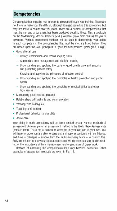
Competencies
Certain objectives must be met in order to progress through your training. These arenot there to make your life difficult, although it might seem like this sometimes, butthey are there to ensure that you learn. There are a number of competencies thatmust be met and a document has been produced detailing these. This is availableon the Modernising Medical Car eers (MMC) Website (www.mmc.nhs.uk) for you todownload. Various assessment methods will be used to demonstrate your abilityin each competency. The competencies that must be met are listed below. Theyare based upon the GMC principles in ‘good medical practice’ (www.gm c-uk.org):
� Good clinical care
– History, examination and record keeping skills
– Appropriate time management and decision making
– Understanding and applying the basis of good quality care and ensuringand promoting patient safety
– Knowing and applying the principles of infection control
– Understanding and applying the principles of health promotion and publichealth
– Understanding and applying the principles of medical ethics and otherlegal issues
� Maintaining good medical practice
� Relationships with patients and communication
� Working with colleagues
� Teaching and training
� Professional behaviour and probity
� Acute care
Your ability in each competency will be demonstrated through various methods ofassessment. An example of an assessment method is the Work Place Assessments(detailed later). There are a number to complete in year one and in year two. Youwill have to prove you are able to carry out and apply procedures with confidence,and have a colleague – anyone from the multidisciplinary team – to confirm this.Early completion of the work place assessments will demonstrate your understand-ing of the importance of time management and organization of paper work.Methods of assessing the competencies may vary between deaneries. Other
examples of assessment methods are given in Fig. 15.
42

Teaching
Teaching is compulsory and you must attend a minimum number of teaching ses-sions to become registered with the GMC after your first year. You will need to signin to teaching sessions as proof of attendance. Instead of trying to avoid these ses-sions, think positively; it gives you 2 hours every 2 weeks where you can escape thedaily routines of the ward you are working in. There will be days where you’ll begrateful that you can silence your bleeper! Also, try to organize a rota with your col-leagues to ensure that everyone gets the chance to attend teaching. Finally, remem-ber, there is much to be learned, and these lectures are actually most useful andworthwhile. For example, the teaching in your Deanery will cover a wide range ofsubjects – from your future career plans to clinical problems that you will commonlyencounter in your daily practice.
Method of assessment Description
Multi source feedback A collective view of your performance as adoctor through the opinions of other health careprofessionals
Can also be described as a mini-PAT (PeerAssessment Tool) or a TAB (Team Assessment ofBehaviour)
Direct observation ofthe doctor
Mini clinical evaluation exercise (Mini-CEX) is anevaluation by a senior member of medical staffof a clinical situation you were involved in
Direct observation of Practical Procedures(DOPS) is the assessment of a procedure carriedout by yourself. The assessor can be anymember of the multidisciplinary team (similar towork place assessments)
Case based discussion Structured discussion of clinical cases managedby yourself. This encourages reflective practiceand self evaluation
Fig. 15 Examples of assessment methods.
43
Foundatio
npro
gramme

E-portfolio
A portfolio containing information on what you have achieved in your daily work isrequired for registration and completion of both years. This can be completed on-line. Completion on-line saves a lot of time and paperwork, and by the time you readthis it is likely that everyone will be completing the portfolio on-line.The e-portfolio has a number of sections for you to complete. Examples are given
below.
Presented evidence
EDUCATIONAL LOGThis contains details of all the educational activities you have undertaken, includingteaching sessions attended and the time spent at these lectures, papers, tutorials,presentations, procedures, exams etc.
SIGNIFICANT EVENTSEvery 4 months you are required to complete either a multi-source feedback (MSF)or a significant event analysis (SEA) form. There will be one SEA in the first year andtwo in the second year. This is the minimum requirement and there is no limit to thenumber you can complete. See below for details.
PERSONAL DEVELOPMENT PLAN (PDP)This looks at the ways in which you have been assessed, how you would judge yourperformance in the assessments, and what you would like to achieve or change inyour next post. This should be filled out after each post so that you can set yourselfachievable objectives.
Workplace assessment (WPA)
These are procedures that must be completed and signed off by a colleague as detailedin the competencies above. Your colleagues will be able to complete their assessmentof you on-line, giving a score for each procedure completed satisfactorily. The assessoris someone with whom you are working. It can be anyone from the multidisciplinaryteam. For example, it would be appropriate for a member of the nursing staff to assessyour ability to give an injection safely and correctly. You will need to take responsibilityfor achieving these competencies and will have to ask to be assessed by your collea-gues. Sometimes it feels like the work place assessments interfere with your work andtake up too much time. They are the last thing you have time to think about as a juniordoctor. However, try not to leave them to the last minute, complete them early in eachpost and you will be under less stress later in the post.
Multi-source feedback (MSF)
Every alternate post, five MSF forms will need to be completed. Four of the multi-disciplinary team you have worked with in a given post will complete evaluationforms on your performance in that position. You can choose who assesses you.There has to be a variety of people evaluating you: a consultant (alternatives areGPs and Staff Grades), a senior trainee (specialist registrar), a senior nurse (orpractice nurse) and finally, anyone else of your choice (e.g. a pharmacist or aphysiotherapist). You will also need to fill out an MSF form evaluating yourself.
44

In the past it was your responsibility to hand out forms to those you wished to assessyou. Now it is much easier. Your colleagues can fill out an evaluation form on-line. Allyou need to do is advise them of the website address and provide them with your fullname and GMC number. Once they have filled out the evaluation form they simply sub-mit it. This eliminates the task of collecting the evaluation forms. Each Deanery is likelyto have a specific time scale for requesting and submitting MSF forms.
Posts and meetings
This is where the meetings with your educational supervisor will be logged. Youshould aim to meet with them at the start of the post to sign an educational agree-ment. They will complete an evaluation of you at the end of the post, called thesupervisor’s report. You can see how well they have rated you in this section ofthe e-portfolio. In this section you can also see the scores that your colleagues havegiven you in the MSF evaluations.
Significant event analysis (SEA)
A significant event is an experience at work that has affected you in some way. Itcan be positive or negative as long as it is something you have learned from andthat others can learn from.A template is available on the e-portfolio. The SEA should contain the following:
� When did the event happen?
� What happened?
� Why did it happen?
� What have you learned?
� What have you changed?
One example of a significant event would be a drug error. There are a number ofpossibilities; the wrong drug was prescribed, the wrong dose was prescribed, thepatient was given something they were known to be allergic to or the drug was pre-scribed but not given. For a SEA you need to look at the questions above, answerthem truthfully and try to learn from the experience and change your practice ifnecessary.The important point to make with relation to the SEA is not to look at it as a way
of finding something negative in your abilities as a doctor. Remember, it doesn’thave to be a negative, you can choose a positive event where the multidisciplinaryteam worked well together resulting in a positive outcome for the patient. Also itdoesn’t have to be about your practice alone but could be your team’s practice.
45
Foundatio
npro
gramme

Work experience and study leave
In the second year of training there is an opportunity to take time out of your currentjob in order to experience a ‘taster’ of the speciality you are interested in followingas a career. So, although you may not have been allocated a posting in your careerof choice, you will still have the opportunity to gain some experience in it. There is atime limit of around 5 days to these taster sessions. You will need permission fromyour tutor and from the people you are working with at the time, as you don’t wantto leave your colleagues struggling with their own work and yours in addition. Therewill also be work involved; it’s not just an easy few days off! Firstly, you have to pro-vide information to your tutor about what you will be doing, and be able to prove thatit will be of value. After completion you will be expected to provide evidence outlin-ing what you gained from the experience. This can be used in your portfolio. Anddon’t forget, the taster session is something that you need to organize yourself.Additional study days are available. The number of days may vary between Dea-
neries. Funding for study leave expenses may vary between Deaneries and somemay not provide any funding. The only funding will be for essential courses under-taken in the 2 years (both ILS and ALS as mentioned below). As with the taster ses-sion, you will have to provide evidence of the educational value of the study leave,and it will only be granted with the approval of your educational supervisor.Time is allocated specifically for the Immediate Life Support (ILS) and Advanced
Life Support (ALS) courses. As part of your training you will be expected to completeboth ILS and ALS courses. You cannot complete the first year without passing ILSand, equally, you cannot complete the second year without completing the ALScourse. These courses are paid for by your Deanery.
Specialty exams
Although it is possible to take study leave it is important to mention that, as Foun-dation Year Doctors, you are strongly discouraged from doing specialty exams. Theaim of Foundation Years is generic training and ongoing assessment, specialtyexams should be taken once you have entered into specialty training. Passing theMRCP and MRCS exams from the old training system will not provide you with anadvantage when applying for specialty training.
Feedback
In order for MMC to continue to improve and progress it is important that you pro-vide your Deanery with your opinion on Foundation. It is vital that you complete thefeedback forms for teaching sessions and the ‘end of post’ questionnaires. With theinformation you provide you can help to improve the posts you have worked in andmake them a more enjoyable experience for your colleagues who follow.
46

Careers advice
During Foundation you will have the opportunity to discuss future career plans withyour Foundation tutor. There is a specific section to complete for this in your port-folio. If you are finding it hard to make such a big decision in only 2 years then takethe opportunity to arrange a couple of taster sessions. This may help. You may findthe information below on fixed-term specialist training posts helpful if you arefinding it difficult to make a career choice.
What to do if you are having problems
One of the differences between the Foundation Programme and the old job of JuniorHouse Officer is the support that is available. If you are having problems in anyaspect of your job seek help sooner rather than later. Your educational supervisorwill be your first contact if there are problems in a specific post. If the problem ismore generalized then your Foundation tutor would be the most appropriate personto contact. The online system of NHS Dots makes it easy to contact your tutor andyour supervisor.
47
Foundatio
npro
gramme

And afte r the foundation years?
More information is now available on Speciality Training, which will follow the first 2 yearsof training. However, this information is still limited. Any information we give you now islikely to change and will continue to change. Keep up to date with plans for SpecialistTraining posts on the Modernising Medical Careers Website (www.mmc.nhs.uk).
Applica tion process
Fig. 16 shows a description of the type of posts you will be applying for.
Senior medical appointments
Postgraduate medical training
Medical school (4–6 years)
Specialist and GP trainingprogrammes
(run-through training)
Specialist and GP RegistersCCTroute
Article 14/11route
Continuing professional development
Undergraduatemedicaltraining
Arrows indicatecompetitive entry
Continuing professionaldevelopment
Foundationtraining infoundationschools
Careerposts
Fixed termspeciality training
appointments(FTSTAs)
F2
F1
Fig. 16 UK MMC career framework. (Adapte d from www.m imc.nhs.uk, withpermission.)
48

Specialist and GP training (run-throughtraining)
August 2007 will be the start date for the first entrants into Speciality Training.At the end of your foundation training you will apply for a run-through trainingprogramme in your chosen speciality. The run-through training posts will bestructured around a curric ulum with the aim of achieving a Certificate of Completi onof Traini ng (CCT) at the end the training. The length of the training post will dependon the specialit y.
Fixed-term specialist trainingappointments (F TSTA)
Most people wi ll apply for and aim to achieve a run-through training post. If you areunsuccessful in your application for run-through training there are other options.There will also be Fi xed Term Training Posts in given specialities. These are likelyto be 1 or 2 yea rs in duration. They will provide similar training to the run-throughposts but they do not offer progressive training. They will provide qualifications thatwill al low you to re-apply to enter run-through tra ining at a later stage . FTSTAs willbe a good alternative to run-through training for those of you who are not decidedon a speciality.
Career posts
The final path tha t can be taken is a career post. This is MMC’s equivalent to thecurrent Staff Grade Post. These posts do not provide formal training with the endobjective of a CCT; however, professional development and assessment will stillbe essential. You must have 3 years of postgraduate training to enter into a Careerpost.
Further information
There are a number of sources where you can obtain more detailed information:
� Modernising Medical Careers Website (www.mmc .nhs.uk)� British Medical Association Website (www.bma.org.uk)� General Medical Council Website (www.gmc-uk .org)In addition to these sources remember that your tutors are also a great source ofinformation. Talk to them, they are there to help.
49
Foundatio
npro
gramme

This page intentionally left blank

Practic
alpro
cedure
s
Practicalprocedures
� Preparing a patient for a procedure 52� Venepuncture 54� Cannulation 56� Arterial blood gas (ABG) measurement 58� Urinary catheterization – male 60� Urinary catheterization – female 61� SC and IM injections 62� ECGs and cardiac monitors 64� Lumbar puncture 66� Nasogastric (NG) tubes 69� Pleural tap 70� Giving IV drugs and fluids 73� Ascitic tap and/or drainage
(Paracentesis) 74� Chest drain insertion 76� Chest drain removal 78� Central line placement 79
51

Preparing a patient for a procedure
Laying a trolley
Laying a trolley is a vital skill. It should be performed meticulously to ensure a sterilefield.
� Collect all the equipment you need for a given procedure. You will commonly need:
– a dressing pack
– a green (21G) needle to draw up lidocaine and to inject it into deeper tissue
– an orange (27G) needle to administer local anaesthetic to the skin
– 10 mL and 20 mL syringes
– packets of gauze
– iodine or chlorhexidine solution; check that your patient is not allergic to iodine
– surgical tape
– sterile gloves
– sharps bin.
� Ask for help. You will need another pair of hands later.
� Place all the items except for the dressing pack on the bottom shelf of yourtrolley and take the trolley to the patient’s bedside.
� Whatever procedure you are about to perform, tell the patient what you are goingto do.
� Position the patient and yourself so that you are comfortable. Procedures alwaystake longer than expected.
� Place an incontinence (inco) pad under the patient. This protects the bed and thepatient from any blood and cleaning solution that you spill. The patient andnursing staff will thank you!
� Open the dressing pack with the tips of your fingers. Trying not to touch morethan the corners, open the outer packaging. There will be another packageinside, together with paper towels and a yellow bag. Take the yellow refuse bagand stick it on the end of the trolley.
� Peel open the orange needle package and drop the contents onto sterile paperwithout actually touching it. Repeat for all the other items you might need(needles, gloves, gauze, syringes, etc.).
� Wash your hands thoroughly. Dry them with paper towels and put on yourgloves. Your hands are now sterile and you cannot touch anyone or anything thatis not – this includes any part of the patient you have not cleaned with iodine orchlorhexidine.
Local anaesthetic
� Ask your assistant to pour the cleaning solution into the pot on your trolley.
� Clean the appropriate area on your patient using gauze or cotton wool. Apply thesolution in a circular motion, working from the area outwards. Give yourself alarge clean area. Do not go over where you have been with the same swab. Goover it again with a new swab if necessary. This area is now sterile and you cantouch it. Cover any remaining part of the patient that is not sterile and which youmight touch accidentally with drapes or the sterile sheet in the dressing pack.
52

� Ask your assistant to show you the bottle of local anaesthetic and to confirm itsname, strength and expiry date.
� Draw up the lidocaine in a 10 mL syringe. The maximum dose of 1% lidocaine is20 mL; for 2% lidocaine it is 10 mL.
Note: Never use lidocaine plus adrenaline when suturing extremities (fingers, toesand noses).
� Attach an orange needle and puncture the skin fairly superficially, into thedermis near the site you want to anaesthetize. Enter at 45�, aspirate back on thesyringe to check that you are not in a blood vessel and inject 1–2 mL oflidocaine. A small ‘bleb’ will appear, allowing you to push the needle in furtherand deeper. Aspirate as you go and continue to inject the local anaesthetic alongthe line of the cut or where you will suture.
� Once you have injected superficially, remove the orange needle and replace itwith a green one to anaesthetize more deeply, aspirating before you injectthroughout. This time, ensure you inject 90� to the skin.
� Allow anaesthetic to work. The reason patients sometimes complain of painafter receiving local anaesthetic is often because the anaesthetic has not hadtime to work.
Suturing
There are many ways to suture (ask any surgeon) and the best way to learn is bywatching and then doing. We will not go into further details here.
Finishing
When you have finished your procedure:
� Ensure all your sharps are thrown away. This is your responsibility alone.
� Clean the patient with normal saline and some clean swabs. Ensure all thecleaning solution and blood is cleaned off.
� Discard the inco pad.
� Put green drapes and gowns in the laundry basket. Ensure all otherdisposable items are cleaned up and put in your yellow bag. Throw this in theappropriate place and replace all other items. If you are lucky, your assistant willhelp, but don’t assume this.
53
Practic
alpro
cedure
s

Venepuncture
Equipment� Tourniquet � Gloves � Sharps box � Alcohol swab � Gauze or cotton wool� Sticking plaster � Needle and syringe or vacutainer � Blood bottles
Method
� Tell the patient what you are about to do.
� Place the patient’s arm on a pillow and make sure it is comfortable.
� Attach the tourniquet to the upper arm.
� Don your gloves.
� Examine and palpate for a vein in the antecubital fossa.
� Swab the skin with an alcohol swab and leave to dry.
� Insert the needle at 30� and advance until you feel the vein wall ‘give’ (Fig. 17).
� Attach the vacutainer tube, or gently pull back if using a syringe. (Note: For somesamples, e.g. calcium, you need to take an uncuffed sample. Release thetourniquet when you have entered the vein).
� Remove the tourniquet if still attached.
� Withdraw the needle and syringe, simultaneously pressing down on the puncturewith the gauze.
� Dispose of the needle immediately in the sharps box.
� Transfer blood to bottles if not using vacutainer.
� Apply sticking plaster.
mediancubitalVein
needle
vacutainersystem
Fig. 17 Venepuncture.
54

NotesDon’t ask the patient to bend the arm up; this does not apply pressure to thepuncture and will result in a bigger bruise
Difficult venepuncture
If you are having difficulty taking blood, abandon the vacutainer. The lack of a‘flashback’ makes finding deep-seated veins hard and the vacuum can collapsesmall veins, especially in older people.If neither antecubital fossa yields a vein, try the forearm, radial aspect of the
wrists, back of the hand or even the base of the thumb. For these small veins,try a smaller needle (23G, blue – but no smaller than this) or a butterfly system.If you still have trouble, consider using the feet – this is more painful.As a last resort, or in an emergency when the peripheral circulation is shut down,
consider a femoral stab:
� locate the femoral pulse
� swab the groin
� aim 1 cm medial to the femoral pulse, with the needle at right angles to the skin
Use of the femoral stab is a last resort – don’t make it part of your phlebotomyroutine!
Special groups
� IV drug users: if you can’t get blood from an IV drug user, ask where theveins are (some will actually take the blood for you!).
� Patients with renal disease: any patient who is on dialysis or approaching theneed for dialysis requires care in taking blood.
� Never perform venepuncture on a limb with an AV fistula (unless the renalteam tell you that it’s OK). Do not use veins at the wrist or forearm in either limb;these are vital for future fistula creation. Use the antecubital veins or veins onthe back of the hand.
55
Practic
alpro
cedure
s

Cannulation
Equipment� Tourniquet � Gloves � Sharps box � Alcohol swab � Gauze or cotton wool� 5 mL syringe with saline � Cannula � Vecafix
Method
� Tell the patient who you are and what you are going to do.
� Attach the tourniquet to the upper arm.
� Examine and palpate the arm for a vein. Look at:
– the back of the hand – this is a common site but can be fragile and mobile inthe elderly
– the antecubital fossa will always have a vein but might be uncomfortable forthe patient
– the anatomical snuffbox at wrist – ‘houseman’s friend’.
� Swab the selected vein.
� Anchor the vein and insert the cannula at 30� to the skin (Fig. 18), advance untilyou feel give in the vein wall.
� Watch for flashback at the base of the cannula.
� Pull the needle part of the cannula (the stylet) back 1–2 mm and watch bloodflow into the plastic tubing.
� Slide the needle back while advancing the cannula into the vein.
� Remove the needle completely and place the cap on the end of the cannula.
� Dispose of the needle immediately. Tape down using Vecafix.
� Flush through with 5 mL normal saline.
cannula
stylet
vein
A
cannula
stylet
vein
B
Fig. 18 Cannulation of a vein. Ensure that the stylet and cannula have entered thevein (A) before withdrawing the stylet and advancing the cannula (B).
56

Hints and tipsFinding the vein is the most important part of the exercise. If you have problems,try the following:After putting on tourniquet, ask patient to drop the hand by the side and to
clench and unclench the fist. Do something else in the meantime, e.g. draw-upyour saline, open up the cannulaIf you cannot feel a vein in one arm, do not attempt to go in blind – stop and
review the other armIf still have no luck, ask the patient to put both hands in some warm water to
help vasodilationIf really desperate, try sticking GTN patches to the antecubital fossa veinsFoot veins are sometimes used, but this can be painful for the patient and
should only be used as last resort
Special groups
The reservations made about groups such as IV drug abusers and renal patients inthe Venepuncture section also apply here.
Cannula sizes
Fig. 19 outlines the different sizes, colours and uses of some common cannulas.
Colour
Blue
Pink
Green
White
Grey
Brown
Size
22G
20G
18G
17G
16G
14G
Flow rate
36 mL/min
61 mL/min
90 mL/min
140 mL/min
200 mL/min
300 mL/min
Uses
In children and patients with difficult small veins, e.g. elderly.Only use for infusions such as N saline, standard antibioticsand heparin
Standard sizes for routine use. Useful for most infusions andblood. 18G better for transfusions
Not very commonly but usefulin patients who need largeamounts of IV fluid or for bloodtransfusions
Used for patients in shock, e.g. GI bleeds, trauma situations. Also used for peripheraladministration of certaindrugs, e.g. amiodarone,dopamine/dobutamine
Some common cannulas
Fig. 19 Examples of some common cannulas and their uses.57
Practic
alpro
cedure
s

Arterial blood gas (ABG) measurement
Equipment� ABG syringe (almost always preheparinized nowadays) � Alcohol swab � Gloves� Sharps box � Gauze or cotton wool � Adhesive tape � 27G (orange) needle
Method
� Tell the patient what you are about to do.
� Palpate the site to be sampled. In general, the radial artery is best: it is mostaccessible, less susceptible to venous sampling and easiest to clean (see below).
� Swab the skin.
� Discard the needle from the ABG pack and attach a 27G needle (orange). This ismuch less painful, obviating the need for local anaesthetic.
� Hold the syringe like a pencil and enter the skin over the site of the palpatedpulse, aiming proximally at an angle of 30� to the vertical (Fig. 20).
� Advance slowly along the line of the artery until you see a ‘flashback’. If you failto see a flashback, withdraw the needle slowly. Try palpating the artery again.
� Hold the syringe steady and allow the syringe to fill. You might need to drawback gently on the plunger if using a small needle.
� Once you have 0.5 mL of blood, withdraw the syringe and press firmly overthe puncture site with a gauze swab for 2–3 min. Attach the gauze swab tightlyto the skin with tape.
radialartery
hold syringe at 30� to the skin
Fig. 20 Radial artery sampling. Use onehand to feel the radial pulse and insertthe syringe at 30� to the skin surface.
58

� Take the needle off the syringe and dispose of it. Carefully expel any air from thesyringe (some packs have a filter that you can attach to the syringe to help withthis).
� Cap the syringe with the cap provided and tip upside down a few times to mixwell.
� Take the sample to the blood gas machine yourself or, if it needs to be sent tothe biochemistry lab, place the sample in a plastic bag with a few ice cubes.Don’t forget to put a label on the syringe.
Note: If you don’t know how to use the blood gas analyser, ask someone. They costthousands of pounds and a broken machine is extremely inconvenient.
PitfallsUnable to obtain blood: try a larger needle (blue or green); unless it is anemergency, you will have to inject local anaesthetic around the artery (be verycareful to aspirate before injecting so as to avoid injecting lidocaine into theartery. A larger needle usually allows the blood gas syringe to fill without pullingback due to the arterial pressure.Venous sampling: you might suspect this if the sample looks rather darker
than expected. Samples from hypoxic patients may well be dark, however;a better guide is if the pO2 or SaO2 are much lower than the pulse oximetryreadings – this suggests venous sampling.
Other sites
If you cannot get a radial artery sample, try a femoral artery sample:
� Use a standard 21G (green) needle; this is long enough to get to the femoralartery.
� Palpate the femoral pulse. In obese individuals, ask the patient to externallyrotate the hip – this brings the artery closer to the surface.
� Clean the skin thoroughly.
� Place a finger on either side of the femoral pulse and insert the needle betweenthe fingers, perpendicular to the skin. Beware – it is easy to give yourself aneedlestick injury here.
� Advance until flashback occurs and syringe starts to fill.
If neither the radial or femoral artery site yields a sample, you could try a brachialsample. The technique is similar to the two above but the artery is often hard to findand can be quite mobile; this technique is also more uncomfortable for the patient.Try to use a 27G (orange) needle and consider using local anaesthetic.If you can’t get a sample after two or three attempts, call your senior.
59
Practic
alpro
cedure
s

Urinary catheterization – male
Equipment� Urinary catheters – male or female length, sizes 12–16 � Catheter dressingpack � Sterile gloves � Lidocaine gel � 10 mL saline in a syringe � Urometeror urine collection bag � Saline for cleaning � Kidney bowl
Method
� Tell the patient what you are about to do.
� Open all of the necessary equipment on your trolley.
� Place a clean drape around the patient’s penis.
� Retract the foreskin and clean the glans thoroughly with saline.
� Coat the end of the catheter with lidocaine jelly. Insert the nozzle of the lidocainegel gently into the preputial orifice and squeeze.
� Give the gel time to work (a few minutes).
� Place the kidney bowl on the bed between the patient’s legs.
� Gently insert the catheter tip and advance. You might need to twist the cathetergently from side to side. Drape the other end of the catheter into the kidney bowlto catch any urine spillage.
� Push the catheter all the way in, even if urine starts to flow before it is fullyinserted.
� Inflate the retaining balloon with 10 mL of saline.
� Carefully replace foreskin.
� Attach a bag or urometer.
Potential problemsUrine doesn’t flow: either the bladder is empty or you are in a false passage. Youcan try a 50 mL flush of saline using a bladder syringe. If this can be introducedand withdrawn, you are likely to be in the bladderCatheter won’t pass: try a bigger catheter. This sounds paradoxical but a
bigger catheter is stiffer and thus might negotiate a stricture or enlarged prostate.Make sure that the lidocaine gel has had long enough to work. If passing thecatheter is painful, the perineal muscles will go into spasm, making passage ofthe catheter much more difficultBlood in the urine: a small amount of blood in the urine or around the penile
orifice indicates trauma inserting the catheter. If clots or dark blood are present inthe urine, consider a three-way catheter to allow for irrigation
If you are still having problems, talk to a urologist. A suprapubic catheter is some-times required but urologists have a number of other tricks that are useful for pass-ing catheters!
60

Urinary catheterization – female
Equipment� Urinary catheters – male or female length, sizes 12–16 � Catheter dressingpack � Sterile gloves � Lidocaine gel � 10 mL saline in a syringe � Urometeror urine collection bag � Saline for cleaning � Kidney bowl
Method
If you are asked to do this, it is usually because the nursing staff are having diffi-culty. This means that you too are likely to have difficulty!
� Tell the patient what you are going to do.
� Ensure that the perineum is well lit.
� Abduct the legs as far as possible.
� Ask an assistant to hold folds of abdominal fat out of the way if necessary.
� Look carefully to distinguish the urethral orifice from the vagina (Fig. 21).
� If in doubt as to the position of the catheter (i.e. if no urine flows), flush andwithdraw 50 mL of saline.
clitoris
urethralorifice
catheter
labiaminora
vagina
labiamajora
Fig. 21 Female catheterization.
61
Practic
alpro
cedure
s

SC and IM injections
Equipment� Syringe � Needle (27G orange or smaller for SC injection, 21–23G (green orblue) for IM injection � Medication to be injected. Read the instructions to ensurethe delivery route is permissible � 21G needle for drawing up � Alcohol swab� Gauze or cotton wool � Sharps box � Gloves
SC method
� Explain to the patient what you are about to do.
� Don gloves and draw up the medication to be given using the 21G (green)needle. Expel air from the syringe and attach the 27G needle.
� Swab the skin into a fold in a suitable site, e.g. forearm, triceps area, abdomen.Pinch the skin gently into a fold.
� Insert the needle horizontally into the fold of skin (Fig. 22).
� Draw back on the needle to ensure that you are not in a vein.
� Slowly inject the medication. Withdraw the needle and wipe the area with cottonwool.
IM method
� Explain to the patient what you are about to do.
� Don gloves and draw up the medication to be given using the 21G (green)needle. Expel air from the syringe and attach the 21G or 23G needle.
Fig. 22 Subcutaneous injection. Pincha fold of skin between the thumb andforefinger, then inject into the fold.
62

� Swab the area to be injected – upper outer quadrant of buttock, deltoid area(Fig. 23), lateral thigh.
� Insert the needle to full depth perpendicular to the skin.
� Draw back on the needle to ensure it is not in a vein.
� Slowly inject the medication. If the patient complains of discomfort, stop for a fewseconds until the pain eases, then continue more slowly.
� Withdraw the needle and wipe the area with cotton wool.
HintsIn obese individuals, a 21G needle will be needed, especially if injecting thebuttock, in order to get as deep as the muscle layer. Injection into subcutaneousfat leads to poor absorption and occasionally to inflammation of the fatAt some time, you will be asked to perform IM injection in a confused,
disturbed individual – often with the aim of sedation. This is permissible undercommon law, as long as you satisfy yourself that the patient is incapable ofinformed consent and that your actions are in the best interests of the patient.Think carefully before administering IM sedation to a confused patient who iswandering at night – ask yourself if you have been asked to do it because thenursing staff want a quiet night shift!If you do administer sedation to a disturbed patient, ensure that you put your
own safety first. A flailing arm plus a contaminated needle is a recipe for aneedlestick injury and possible blood-borne infection. Get someone to help you,perhaps by gently restraining flailing arms, or by talking to distract the patient
safe area(upper outer
quadrant)
iliaccrest
course ofsciaticnerve
Fig. 23 The site for intramuscularinjection.
63
Practic
alpro
cedure
s

ECGs and cardiac monitors
Equipment� ECG machine � Adhesive electrodes � Razor
Method
� Explain to the patient what you are about to do.
� Attach the electrodes to the skin in the positions shown (Fig. 24). You will needto shave hairy areas to get a good contact. Note that accurate placement isimportant; having the chest leads in the wrong place can lead to an anterior MIbeing diagnosed as an inferior MI!
� Switch on the ECG machine. Ensure that the mains lead does not cross theelectrode leads.
� Enter the patient details if required.
� Make sure that the FILTER button is on.
� Take the ECG (usually by pressing a button marked ‘12’ or ‘auto’).
mid-clavicular
line
mid-axillary
line
V1 V2
V3
V4
V5
V6
Fig. 24 Placement of ECG chest leads.
64

PitfallsMissing tracings: check all the lead attachments and replace any electrodes thatare not adhering well. Twist the leads within the attaching clips to ensure thatthe leads are not pulling the adhesive pads off the patientWandering baseline/irregular baseline: repeat the tracing, asking the patient
to relax and stay still. If this fails, attach the limb leads to the shoulders and hips;this cuts down on interference from limb muscle electrical signalsInterference on baseline: ensure no mains cables are crossing the ECG wires.
If you still have a problem, try and run the ECG machine on its battery ratherthan on the mains. Still got a problem? Move the ECG machine to the other sideof the bed and unplug any non-essential electrical equipment around the bedOdd axis on ECG: make sure that you have the leads attached to the correct
electrodes, especially the limb leadsIf all else fails, get another machine
65
Practic
alpro
cedure
s

Lumbar puncture
Equipment� Sterile gloves � Dressing pack � 1% or 2% lidocaine � Spinal needle – black(22G) is best, preferably with a round tip � Cleaning solution (iodine orchlorhexidine) � Gauze swabs � Micropore � Manometer and three-way tap� Three sterile specimen bottles � One glucose (oxalate – grey-topped) blood tube
Method
The most important thing to do is to position the patient correctly. Lay the patient onthe left side, with the back parallel to the bed.
� Ask the patient to maximally flex the back to open up the spinous processes.Ensure the shoulders are square to the hips.
� Make sure the patient’s back is straight and that you can feel the gaps betweenthe spinous processes (Fig. 25).
� Feel the iliac crests and draw across the back to the other iliac crest.
� Feel down the spinous processes until you feel a gap as close to the vertical lineyou have drawn as you can. This is L3–L4.
� Choose the L3–L4 space or go a space higher to L2–L3.
� Get a chair. Sit down where you will perform the procedure. Raise the bed so youare comfortable.
� Set up your trolley:
– label three specimen bottles: 1, 2, 3
– prepare your trolley
– wash your hands thoroughly.
iliac crests
legs tuckedinto abdomen
line acrossL3-L4 level
point ofentry for lumbarpuncture − mark
before cleaning the skin
Fig. 25 Positioning of the patient for lumbar puncture. (Adapted from Sprigings D,Chambers J, Jeffrey A. Acute Medicine, 2nd edition, 1995 Blackwell Science.)
66

� Open up the three-way tap and manometer. Connect up and be certain youknow which tap opens to the manometer and closes all the others, and thenclose it to the manometer and open to the ‘outside world’.
� Check that the stylet passes through the needle properly.
� Clean over L2, L3 and L4, giving yourself a large area to work with. Drape yourgreens and administer local anaesthetic at L3–L4.
� Place the spinal needle over the site of lidocaine administration, beveluppermost, and advance. Make sure the needle remains at 90� to the skin in allplanes.
� Push through several layers of tissue (Fig. 26) until you feel the needle ‘give’.Carefully remove the stylet 1–2 cm to see if you have clear CSF.
� Hold the end of the needle carefully but firmly with your left hand and pullthe stylet out with your right – cover the end of the needle with your left thumb toprevent CSF leak.
� Attach the manometer with the three-way tap turned to allow CSF to gostraight up. Measure the CSF pressure – it is in cmH2O. Anything < 20 isnormal. Give it time, especially with a small needle.
� Once the pressure is measured, you can collect CSF by turning tap so thespinal needle is open to you but the manometer is closed, retaining its columnof fluid. Have bottle 1 ready and collect 10–12 drops of CSF. Put your thumbover the end and have your assistant take bottle 1 away and pass you bottle 2.Repeat with all three bottles.
� For the glucose sample, collect the column of CSF in the manometer by closingthe spinal needle and opening the manometer to you. Beware – this comes outvery fast.
spinousprocess
aim needle craniallytowards the navel
skin and subcutaneous tissuessupraspinous ligament
interspinous ligament
L3
L4
subarachnoid spacecontaining CSF
duraextradural space
ligamentum flavum
Fig. 26 Insertion of the lumbar puncture needle.67
Practic
alpro
cedure
s

� Remove the manometer, put your thumb over the end of the spinal needle toprevent CSF loss before replacing the stylet; then remove the entire needle.
� Clean the patient’s back and put a small dressing/plaster over the needle site.Move the patient back onto the back and advise rest for 1 h – local policy mightadvise differently on this time and can be up to 4 h. Check with your hospitalpolicy.
� Send bottles 1 and 3 for cell count; this will differentiate a bloody tap from bloodin the CSF. Send bottle 2 for biochemistry and xanthochromia.
Hints and tipsIf you cannot get into the epidural space, make sure you are at 90 degrees to theskin in all planes. Sometimes you will find you have aimed skywardsIf you still have no success or hit bone, go up a space to L2–L3If you still have no success, sit the patient upright to perform the procedure.
Again, mark out landmarks using the iliac crests and aim the needle at 90�.You cannot measure the pressure but will get your samples using this method
68
LUMBAR
PUNCTURE

Nasogastric (NG) tubes
Equipment� Gloves � NG tube � Litmus paper � KY jelly � Glass of water
Method
Usually, nurses manage to do this with no problems. It is unusual for them to callyou. If they do:
� Tell the patient what you are going to do.
� Sit the patient upright.
� Cover the end of the tube lightly with lubricating jelly.
� Insert the tube into one nostril horizontally, and pass it down slowly.
� Ask the patient to start swallowing, and push the NG tube more quickly down.If the patient starts to choke, stop immediately. A glass of water can help themswallow, although most patients you want to place an NG tube in will either beunsafe to swallow water or will be nil by mouth!
� The patient should swallow the tube down. Check the position. There arediffering local policies regarding checking NG tube positioning. Check what theyare. They include:
– aspirating slowly on the end of the NG tube and checking the secretions withlitmus paper
– injecting 20 mL of air and auscultating for ‘bubbling’ over the stomach
– performing a chest X-ray to confirm the position. Specify on the X-ray requestthat it is for NG tube positioning because the X-ray used is of differentpenetration to highlight the tube.
� On the chest X-ray, look for the NG sitting in or towards the stomach bubble,beneath the diaphragm. If it deviates to the right or, less commonly, the left, it isin a bronchus and needs to be removed and re-sited.
Note: Do not attempt to pass an NG tube if there is any possibility of a skull fracture,e.g. CSF from the ear. A skull X-ray might be helpful, although this will not excludea base of skull fracture.
69
Practic
alpro
cedure
s

Pleural tap
Equipment� 1% or 2% lidocaine � Basic dressing pack with sterile towels � Sterile gloves� Iodine- or chlorhexidine-based antiseptic � Sterile universal containers � 21G(green) needles � 27G (orange) needles � 16G (grey) cannula � 10 mL syringe� 20 mL syringe � An assistant
Method
� Explain to the patient what you are about to do.
� Sit the patient on the edge of the bed, arms folded in front of the body andleaning forward across a hospital table (Fig. 27).
� Before donning your gloves, check the site of the effusion on the chest X-ray andpercuss down the chest. Place a cross on the lateral posterior aspect of the
percuss to findtop edge of effusion
Fig. 27 Position of the patient for pleural tap. (Adapted from McLatchie G. OxfordHandbook of Clinical Surgery, 1st edition, 1990, by permission of Oxford UniversityPress.)
70

chest, 3–5 cm below the level at which you can first percuss the effusion. Yourcross should be in an intercostal space, over the top surface of a rib.
� Do not proceed if you cannot confidently percuss the effusion; get an ultrasoundscan to mark the position.
� Don your gloves, place sterile towels below the target area and clean the skinwith antiseptic.
� Draw up 5–10 mL of lidocaine and raise a bleb under the skin with a 27G needle.
� Infiltrate local anaesthetic using a 21G needle, slowly working downperpendicular to the skin. Aim over the top surface of the rib, pulling back beforeinjecting anaesthetic. Stop when you aspirate fluid (Fig. 28).
� For a diagnostic tap, attach a fresh 21G needle to a 20 mL syringe, insert alongthe anaesthetized track at right angles to the skin and advance while at the sametime pulling the plunger back. When fluid is first aspirated, stop – do notadvance any further – and pull the plunger back until the syringe is full.Withdraw the needle and press over the site with gauze.
� For a therapeutic tap, attach a 50 mL Luer-lock syringe to a three-way tap.Attach a length of tubing to the side port of the three-way tap (a giving set worksreasonably well). Put the other end of the tubing in a large sterile jug.
� Insert a 16G cannula along the anaesthetized track at right angles to the skin.When you see a fluid flashback, advance the cannula a little further, then pushthe plastic cannula as far into the thorax as it will go, keeping the stylet still.As you withdraw the stylet, put a thumb over the end of the cannula – thisprevents a pneumothorax. If possible, ask the patient to breathe out and hold thebreath in expiration as you withdraw the stylet.
� Attach the three-way tap to the cannula, ensuring that the cannula does not kink.You can now either allow the fluid to drain under gravity, or you can withdraw50 mL using the syringe, then turn the three-way tap, and expel the contents ofthe syringe into the tubing.
� Once you have drained 1–1.5 L of fluid, remove the cannula and attachedequipment and press over the hole with gauze for a minute.
intercostal muscle
neurovascular bundle
needle
skinrib
lung
visceral pleura
fluid
parietal pleura
Fig. 28 Insertion of the needle for pleural tap.71
Practic
alpro
cedure
s

PitfallsNo fluid aspirated: try angling the needle down a little, to the side a little, orwithdraw the needle a little. If you still fail to aspirate fluid, start again bypercussing out the level of the effusion again. If you still cannot aspirate fluid,give up and request an ultrasound scan to locate the effusionThe patient is obese: for a diagnostic tap, try using a 16G or 18G cannula, or a
21G lumbar puncture needle; they are longer than a standard 21G needle. For atherapeutic tap, try using a long 14G cannula, or even better a cannula over styletcentral line – rare, but very useful if you can find one!Blood is aspirated: a small flash of blood is not unusual; this is caused by
nicking blood vessels in the skin. Red fluid is either blood or a bloody effusion;put a little in a dish and see if it clots. If it doesn’t clot, it is likely to be a bloodyeffusion. If it does clot, get out and seek helpThe flow rate slows down during aspiration: some effusions, especially
malignant effusions with a high protein content, can be very viscous. This canresult in the cannula becoming narrowed during aspiration, which leads todifficulty aspirating fluid. You might need to use a new cannulaThe patient is uncomfortable: if the discomfort occurs on inserting the needle,
more local anaesthetic is needed. If cough or discomfort occurs during atherapeutic aspiration, this suggests that the lung has re-expanded and thevisceral pleural is in contact with the needle. Stop the aspiration if this occursThe patient becomes unwell: vasovagal reactions are not uncommon during
pleural taps; if the patient feels faint, stop, withdraw the needle immediately, andlie the patient down. Adequate local anaesthetic can help to avoid this. Lesscommonly, the shift of the lung and mediastinum can lead to changes inautonomic tone, or to circulatory collapse. Do not aspirate more than 1–1.5 L offluid in a single sitting
Sending the fluid
Send the fluid off for microscopy, culture and sensitivity, for cytology, and for proteinand LDH levels. If TB is at all possible, request acid-fast staining. If the effusionaccompanies a pneumonia, place some of the fluid on ice and request the pH ofthe fluid – this is vital for deciding whether a chest drain is required.
What to do afterwards
Order a chest X-ray, even after a diagnostic or failed tap, to check for pneumotho-rax. A small pneumothorax does not need aspirating but might require another X-rayin a few hours to ensure that it is not increasing in size. Write a brief note in thepatient notes regarding the procedure.
72
PLEURAL
TAP

Giving IV drugs and fluids
Equipment� IV fluids � Giving set � Cannula � Syringe driver/pump
Method
You might be asked to give drugs IV. This could be as simple as giving an IV bolusthrough a cannula or giving drugs through a drip. Make sure that you read theinstructions provided carefully:
� Ensure that the cannula is in place and flushes easily with saline.
� Make sure you know what infusion rate the drug should be injected at – slowbolus through a big vein protects the veins and prevents the cannula fromtissuing.
If the drug is diluted in solution (50–500 mL normal saline) or you are asked to puta bag of fluid up on a patient:
� Put the bag of fluid up on a drip stand.
� Connect one end of the giving set to the bag and close the valve in the plastictubing.
� Squeeze the reservoir so it is half filled with fluid and the remainder drips in.
� Open the valve and let the remainder of the fluid run through so no air bubblesremain. Once there are no air pockets and fluid runs freely out of the end ofthe giving set, attach to the end of the cannula.
� Set the drip rate or put tubing through the fluid pump.
Always ask another person to check the drugs you are administering. Ask them toconfirm the drug name, dose, method of delivery and expiry date. This includes anyadditives e.g. KCl.Always check you have the right patient – check the wristband for identification.Remember that some drugs need to be administered with care, e.g. vancomycin
must be given as a slow infusion; Pabrinex and vitamin K are dangerous if given asa fast bolus. Some drugs are irritant and will need larger veins; the cannula shouldbe well flushed after administering such drugs.
73
Practic
alpro
cedure
s

Ascitic tap and/or drainage (Paracentesis)
Equipment� Dressing pack � Lots of gauze swabs � Sterile gloves � Iodine/cleaning solution� 10 mL syringe � 50 mL syringe � Green and orange needle � 1% or 2%lidocaine � Ascitic drain and bag � Scalpel � Adhesive dressing
Method
� Explain to the patient what you are going to do.
� Expose the patient’s abdomen fully and percuss out ascitic fluid. Demonstrateshifting dullness or a fluid thrill. Percuss either right or left iliac fossa – this iswhere you will insert the drain. Mark it with a pen if necessary.
� Prepare yourself and your trolley:
– wash your hands
– open up your dressing pack
– pour iodine into a pot
– put on sterile gloves.
� Draw up the lidocaine using the green needle. Replace the green needle with theorange needle and fill this too.
� Have your helper open the ascitic drain and bag for you. The ascitic drain isusually a small-bore tube with an introducer inside. It is inserted like an IVcannula. Familiarize yourself with it before proceeding.
� Prepare the skin and inject local anaesthetic with the orange and then the greenneedle. You will finally feel a give, then aspirate straw-coloured ascitic fluideasily. Withdraw the needle slightly and inject more local anaesthetic.
� Remove the needle and nick the skin over the local anaesthetic with a scalpel.
� Take the ascitic drain, with a 20 or 50 mL syringe attached, and enter the skin at90 degrees, pushing gently until ascitic fluid is again aspirated. Now, as with acannula, slowly withdraw the introducer while at the same time advancing thedrain until it is inserted (Fig. 29).
PitfallsNo aspiration: you are in the wrong place. Repercuss and try again. If you arestill unsuccessful then you might need an ultrasound to mark a spot for youNo drainage: think of the following:
� blockage – try flushing drain with sterile saline
� kink in the tube – check where tube is tied in and ensure it is not kinked
� drain has fallen out – remove the drain and, if necessary, replace withnew drain at new site
74

� Remove the introducer completely and attach it to the drainage bag. Clamp itwhen 1 L has been drained. Large-volume paracentesis can be carried out as atherapeutic measure if accompanied by intravenous volume replacement, e.g.with 1 unit of 20% albumin for each 2 L drained. Check with your seniors.
� Suture the drain in place and cover it with an adhesive dressing. Clean thepatient of remaining iodine with saline.
Simple aspiration
Diagnostic aspirations should be performed with the same care as insertion of adrain. They are simple in that they can be performed with a green needle. Enoughfluid should be taken for culture (M, C and S), protein, glucose and aFP.
insert cannulaat 90� to the
skin
Xiphoid process
inferior epigastricarteries
'safe area' forparacentesis
Fig. 29 Ascitic tap.
75
Practic
alpro
cedure
s

Chest drain insertion
Equipment� Drapes � Suture kit � Dressing pack � Gauze � Scalpel � 1% or 2% lidocaine� 2/0 silk suture on curved needle � 10 mL syringe � Orange and green needle� Sterile gloves � Iodine/cleaning agent � Chest drain (Seldinger or ‘trochar’)� Chest drain bag (for pleural effusion) or water-tight bottle containing somenormal saline or sterile water (for pneumothorax) � An assistant
Method
� Tell the patient what you are going to do.
� Confirm the position of the pneumothorax/effusion clinically and also with achest X-ray.
� Sit the patient upright, legs over the side of bed, leaning over a high table so thearms are up, the back is straight and you have access to the affected side ofthe chest. In an unwell patient you might have to perform this with the patientat 45 degrees.
� Prepare the underwater seal – fill a bottle one-third with sterile water. The end ofthe tube should be 2–3 cm into the water.
� Choose the chest drain; tailor it to the patient by looking at how much rib spaceis available. Seldinger chest drains are used much more frequently. Traditional‘trochar’ drains are increasingly becoming the remit of specialist chest teams.Seldinger drains are much less traumatic for both patients and doctors alike.Some chest physicians however still prefer larger drains, particularly for effusionsand empyemas – as a guide 24F is suitable for a pneumothorax; use 28–32F foreffusions and empyemas.
� Don your gloves and prepare your trolley so that all the items are within easy reach.
� Prepare the patient’s skin with chlorhexidine or iodine and perform pleuralaspiration in the mid-axillary line at the 4th–5th intercostal space; aim abovethe rib.
� Once pleural fluid has been located, insert more local anaesthetic – be generous.Use up to 15 mL in the area around the aspiration site. Some patients mightfind this uncomfortable, so you can give a small dose of diamorphine IV with10 mg metoclopramide IV.
SELDINGER TECHNIQUEThis technique is virtually identical to that of inserting an ascitic drain or central line.
� Take the large-bore needle provided in the chest drain pack and attach a 10 or20 mL syringe containing a small amount of the local anaesthetic. Re-insertthe needle over the previous aspiration site at 90� to the skin. Introduce theneedle until pleural fluid can readily be aspirated.
� Remove the syringe, keeping the needle very still and immediately put yourthumb over the end of the needle.
76

� Take the guidewire provided and insert through the end of the needle, andadvance until about two-thirds of the wire is inserted. The wire should advancefreely and should not be forced.
� Holding the wire, remove the needle, keeping the wire in place. Make a nickwith a scalpel where the wire enters the skin. Insert the dilator over the wire andthen remove.
� Insert the drain over the wire. Keep one hand over the wire at all times. When thedrain has been inserted, slowly withdraw the wire from the drain while advancingthe drain further. Remove the wire completely and immediately place a thumbover the end of the drain to prevent fluid leakage or the introduction of air intothe pleural space.
� Connect the end of the drain to an underwater seal.
TRADITIONAL ‘TROCHAR’ TECHNIQUE� Make a 1–2 cm incision with the scalpel in line with the ribs, remaining just
above the lower rib. Once through skin and into subcutaneous fat, blunt-dissectdown with forceps. Continue until you reach the pleura. This can be painful, sohave remaining local anaesthetic to hand and inject into pleura if necessary.
� Take the introducer out of the chest drain, so only the tubing remains. Once youhave blunt-dissected the pleura and fluid or air begins to escape, insert the draininto the hole. It should enter the pleural cavity easily, but keep a finger over theend of the drain until it is connected to the bag or underwater seal.
SECURING THE DRAIN� Place a 1-mattress suture on either side of drain and pull taut. Place another
suture in the middle of incision around the drain. Tie the two side sutures, so theskin is pulled tight, then secure around the drain by coiling round several times.Leave the central suture free, to be tied when the drain is removed.
� Place pads of gauze around drain and secure with dressing. Secure the proximalpart of the drain to the patient with tape.
� Ensure that the drain is draining/bubbling freely. Get a post-procedure chestX-ray to review the position.
Underwater seals
Underwater seals are used to drain air from the pleural cavity. They are always keptbelow the level of the chest. When the pressure of air in the pleural cavity rises, airis forced from the chest into the water, and it bubbles. Normal respiration causessome movement in air pressure and the water level oscillates accordingly – thisis known as ‘swinging’. If the fluid level does not swing, it means that the tube is:
� blocked – flushing it might help, or it might need to be replaced
� kinked – the dressing needs to be removed and the tube reviewed
� in the wrong place – it was inserted incorrectly or has fallen out; a chest X-raywill confirm.
If there is no bubbling but the tube is still swinging, the pneumothorax has probablybeen corrected. If this is confirmed by chest X-ray the drain can be removed,after 24 h.
77
Practic
alpro
cedure
s

If there is continuous bubbling and no or only partial resolution of the pneumotho-rax on chest X-ray, then there is a continuous leak and a chest physician/thoracicsurgeon should be contacted.
Chest drain removal
Once the mattress sutures are in place, removal of the drain is straightforward.
Equipment� Dressing pack � Sterile scissors � Sterile gauze � Chlorhexidine/Iodine forcleaning
Method
� Remove dressings from around the drain.
� Clean the whole area with iodine/chlorhexidine.
� Cut free the ties to the drain from side sutures but do not untie the suturesholding the skin together.
� Ask the patient to breathe in deeply and exhale fully. Get your assistant to pullthe drain out while you pull the central suture tight closed.
� Tie the suture. Clean and dress the area.
� Repeat the chest X-ray.
� Remove sutures at 1 week.
78
CHESTDRAININSERTION

Central line placement
Equipment� Central line dressing pack with gown and drapes � Sterile gloves � Iodine orchlorhexidine antiseptic 1% or 2% lidocaine � Central line (preferably at least atriple lumen line) � Saline or heparin saline to flush line � Stitch and stitchholders � Scissors � Scalpel blade � 21G (green) and 27G (orange) needles �2 � 10 mL syringes � Occlusive dressing � An assistant
Method
� Explain to the patient what you are about to do.
� Choose the site for insertion; jugular or femoral carry less bleeding risk and lowrisk of pneumothorax; subclavian is a cleaner site and is technically moredifficult; we have not covered the technique here. Small ultrasound machines areincreasingly being used to correctly identify the internal jugular vein. Training intheir use is usually available in your local Trust. For drug administration,IV access and even emergency pacing, femoral is probably the easiest site.
� Don your gloves and gown, clean and drape the site.
� Tilt the head end of the bed down by 10–15�.� Draw up 10 mL of lidocaine; raise a bleb on the skin with a 27G needle.
� Infiltrate local anaesthetic all around the site, working down towards the vein.Pull back on the plunger before injecting each time to ensure that you don’tinject into the vein.
� Have the assistant open the central line pack and take all the items out. Ensurethat the wire moves freely on its reel – you will need to advance the wire one-handed.
� Flush each port of the central line with saline or heparin saline, and close offeach line except the distal (usually brown) line; the wire threads through this line.
� Attach a syringe to the large needle provided:
– right femoral line: find the arterial pulse and enter the skin 1 cm medial tothis, at 45� to the vertical and heading parallel to the artery. Advance slowly,aspirating all the time, until you enter the vein (Fig. 30)
– right jugular line: palpate the carotid artery with your left hand, covering theartery with your fingers. Insert the needle 0.5–1 cm laterally to the artery,aiming at 45� to the vertical. In men, aim for the right nipple; in women, aimfor the iliac crest. Advance slowly, aspirating all the time, until you enter thevein (Fig. 31). If you fail to aspirate blood after entering 3–4 cm, withdraw,re-enter at the same point but aim slightly more medially.
� Once the needle is in the vein, ensure that you can reliably aspirate blood.Remove the syringe, keeping the needle very still, and immediately put yourthumb over the end of the needle.
� Insert the wire into the end of the needle, and advance the wire until at least30 cm are inserted. The wire should advance very easily – do not force it.
79
Practic
alpro
cedure
s

� Keeping one hand on the wire at all times, remove the needle, keeping the wirein place. Make a nick in the skin where the wire enters the skin. Insert the dilatorover the wire and push into the skin as far as it will go. Remove the dilator.
� Insert the central line over the wire. Keep one hand on the wire at all times.When the central line is 2 cm away from the skin, slowly withdraw the wire backthrough the central line until the wire tip appears from the line port. Hold the wirehere while you insert the line. Leave a few centimetres of the line outside theskin. Withdraw the wire and immediately clip off the remaining port.
� Attach the line to the skin with sutures. Tie loosely so as not to pinch the skin;this causes necrosis and detachment of the line. Clean the skin around the lineonce more, dry and cover with occlusive dressings.
� Ensure that you can aspirate blood from each lumen of the line, then flush eachlumen with saline or heparin saline.
� Order a chest X-ray to check for line position and pneumothorax if a jugular orsubclavian line has been inserted. Femoral lines do not need X-raying.
PitfallsCannot cannulate the vein: try another site. If you have access to a portableultrasound device, try using this
inguinal ligament
nerveartery
vein
Fig. 30 Femoral artery sampling.
80
CEN
TRAL
LINEPLACEM
ENT

Arterial puncture: this is not always obvious, especially in shocked, hypoxicpatients, when the blood can be dark and flow sluggishly. If you do hit an artery,remove the needle and press firmly for 5 min with a gauze swabVein cannulated but wire fails to advance: sometimes veins are small, tortuous
or occluded, e.g. by thrombus from previous cannulation attempts. Do not forcethe wire; try another siteLine in wrong place: occasionally, lines double back on themselves, or
subclavian lines go up into the jugular. If this occurs, either remove the line andstart again or, if you have access to fluoroscopic guidance, try and reinsert thewire, remove the line, and reposition the wireBeware using too much local anaesthetic on multiple line insertions; the
maximum dose should not really exceed 3–5 mg/kg; i.e. 10–15 mL of 2%lidocaine for a 70-kg patient
Central lines do not cure patients. They take longer to put in than you think andoften require the patient to lie flat with drapes on the head for some time. Makesure that you are treating the patient’s condition as well as inserting a central line.
clavicle
claviclesternocleidomastoid
place fingers oncarotid pulse
carotidartery
internaljugular vein
Fig. 31 Placing a central line. Insert the needle lateral to the carotid artery, aimingfor the right nipple in men or the anterior superior iliac spine in women. (Adaptedfrom Sprigings D, Chambers J, Jeffrey A. Acute Medicine, 2nd edition, 1995Blackwell Science.)
81
Practic
alpro
cedure
s

This page intentionally left blank

Dru
gissues
Drug issues
� Pain control 84� Constipation 85� Nausea and vomiting 86� Anticoagulation 87� Steroids 89� Sedation 90� Blood and blood products 91
83

Pain control
A doctor should cure sometimes, relieve often and comfort always. Pain control istherefore a vital part of your job and is often an area where you can make a realdifference as a foundation doctor. Pain is complex; different people perceive painin different ways, and therapy always needs to be individualized. The followingare some tips regarding pain control:
� Find out what the cause of the pain is, e.g. metastases, angina, broken rib.
� Don’t forget that simple measures are important, e.g. positioning of the patient.
� Anxiety makes pain worse. Listening to people in pain and letting them knowthat you are there for them is important.
� Regular analgesia to keep pain under control is better than trying to treatpain when it becomes unbearable.
The ladder of analgesia
1. Paracetamol 1 g every 6 h, to a maximum of 4 g every 24 h. This is sufficient formild pain.
2. Weak opioids, e.g. codeine 30–60 mg every 4–6 h, to a maximum of 180 mgper day.
3. Strong opioids, e.g. morphine. Start with 5 mg every 4 h. Once you have anidea of the daily required dose, change to a long-acting version, e.g. MST every12 h. Make sure that the doses are equivalent.
Adjunctive therapy
NSAIDs Good for musculoskeletal pain and pleuritic pain, e.g. pericarditis, frac-ture/sprain, gout. Use the weakest preparation that works for the shortest possibletime, e.g. ibuprofen 400 mg three times a day. Avoid if there is GI bleeding, othermajor bleeding, heart failure, low platelets, iron-deficiency anaemia. Try and avoid inindigestion; you might have to use a gastroprotective agent, e.g. a proton pumpinhibitor.Anxiolytics/sedatives Oral diazepam or, in terminal care, subcutaneous mida-zolam infusion, are good agents for dealing with severe anxiety that can exacerbatepain.Don’t forget that analgesia, especially opioid analgesia, has side-effects. Give
antiemetics and don’t start even a weak opioid without giving laxatives at the sametime.If you are having difficulty controlling pain or other bothersome symptoms, call
the palliative care team. They are usually very willing to give advice as to suitableanalgesic regimens and they always appreciate being involved at an earlier stage,rather than in the hours before someone dies.
Opiate overdose
Overdose of opiates can occur deliberately, as a suicide attempt, or because ofrapid increases in dose (as might be seen during the use of a PCA pump, for exam-ple). Look for pinpoint pupils, drowsiness and a reduced respiratory rate. Stop thepump if one is in use, and give 400–800 mg of naloxone IV or IM. Naloxone hasa relatively short half-life – be prepared to give more as it wears off and, in the caseof long-acting morphine preparations, an infusion might be necessary.
84

Constipation
Constipation is common. Many of your patients will be older and thus likely to sufferfrom constipation. If they weren’t constipated when they came into hospital, they willbe after a few days of opioids and bed rest. Keeping people mobile and encouragingthem to drink plenty of water (or keeping them otherwise well hydrated) both help toavoid constipation.There are two traps in constipation. First, is the patient constipated or is there a
bowel obstruction? Get an abdominal film if the abdomen is distended or there areother signs of obstruction. Second, constipation can hide – it might not be apparenton PR exam and, in severe cases, overflow diarrhoea can occur. Clearly, in thiscase, laxatives are needed, rather than medications to try and stem the diarrhoea.
FIRST-LINE AGENTSSelect no more than one from each group; they can be combined.
� Softeners – sodium docusate: 200 mg three times per day.
� Bulking agents – ispaghula husk, e.g. Fybogel: one sachet twice a day; sterculia,e.g. Normacol.
� Stimulants – senna: two tablets at dinnertime; bisacodyl.
� Osmotic laxatives – magnesium hydroxide: 10 mL twice a day; lactulose: 10–20 mL twice a day.
SECOND-LINE AGENTS� Movicol: contains polyethylene glycol, a water retainer and osmotic laxative.
� Codanthramer, codanthrusate: these combine a softener and danthron, apowerful stimulant. They are sometimes used in patients with terminal disease –danthron carries a theoretical risk of carcinogenicity.
� Glycerin suppository, phosphate enema: good for clearing hard stools from therectum but insufficient on their own to treat constipation higher up in the bowel.
BOWEL PREPARATIONYour surgical unit or colonoscopy unit will probably have a favourite routine forcleaning the colon. There are two common choices:
� Sodium picosulphate (Picolax): two to three sachets spread over a 24-h period. Apotent stimulant, this can cause dehydration, nausea, vomiting, abdominal painand dizziness. Make sure that your patients are well hydrated; older, frail peoplemight need an IV infusion.
� Polyethylene glycol (e.g. Klean-prep): this is taken with a lot of water (up to 4 L),thus dehydration is less of a problem. Encourage your patients to drink a glassfulevery 10–15 min; the whole 4 L should be drunk over a 6-h period, which mightbe difficult for some patients.
Neither of these preparations is designed to combat constipation.
85
Dru
gissues

Nausea and vomiting
Before unthinkingly prescribing an antiemetic, ask yourself why the patient is vomit-ing. Not only will this help you choose an appropriate antiemetic, it might allow youto remove the underlying cause (see ‘Nausea and vomiting’, p. 166).
USEFUL PREPARATIONS� Metoclopramide 10 mg 8-hourly – oral, subcutaneous, IM or IV: good for most
types of nausea and vomiting, including opioid-induced vomiting, gastric stasis(metoclopramide improves gastric emptying), chemotherapy-induced vomiting.Avoid if there is any chance of bowel obstruction or gastric outlet obstruction.
� Prochlorperazine 12.5 mg 12-hourly IM or 5 mg three times a day orally: similarto metoclopramide but without the effects on gastric motility. More useful forvestibular and cerebellar disorders than metoclopramide. Like metoclopramide, itcan induce extrapyramidal side-effects and – rarely – dystonic reactions.
� Haloperidol 1–5 mg orally or IM as required: Another alternative tometoclopramide, with a similar mode of action. Useful for intractable nausea, e.g.in cancer patients.
� Granisetron, ondansetron, tropisetron: these drugs all block 5HT3 receptors. Theyare expensive and their use is usually strictly controlled by local policies – checkyours. They are useful for chemotherapy- and radiotherapy-induced vomiting,postoperative nausea and vomiting.
� Cyclizine 50 mg three times a day orally, subcutaneously, IM or IV: useful formost types of nausea, including nausea of vestibular or cerebellar origin. Can besedating, however. Avoid in acute myocardial infarction; may have adversehaemodynamic consequences.
� Hyoscine 200–400 mg twice a day: useful for motion sickness and for reducingsecretions into an obstructed gut when surgery cannot be performed.
86

Anticoagulation
WARFARINWarfarin therapy is indicated for treatment of DVT and pulmonary embolism, forreducing the risk of stroke in atrial fibrillation and mural thrombus, to prevent recur-rence of other arterial emboli, and to prevent thrombosis of prosthetic heart valves.For most indications, including some more modern prosthetic valves, the target INRis 2–3. Older prosthetic valves and patients with recurrent thrombi due to anticar-diolipin syndrome should be maintained at an INR of 3–4:
� Contraindications to warfarin: heavy alcohol intake, increased risk of bleeding(e.g. bladder tumour, known gastric ulcer) and a tendency to fall are relativecontraindications to warfarin.
� Taking the INR: the INR should be taken in the morning, as close to 9 a.m. aspossible. Dosing nomograms do not work if the sample is taken in the afternoonor evening.
� Dosing time: warfarin should be given at 6 p.m. Make sure that you haveprescribed warfarin (usually on a separate prescription chart) before finishingyour working day.
� Commencing warfarin: patients should receive an explanation of when to takewarfarin, be told about the risks of bleeding, the need for regular blood tests(especially if they change their medications) and be given a record booklet forrecording INR and dose. Most warfarin protocols use a loading dose (e.g. 10 mgfirst day, 10 mg second day), followed by INR testing on days 3, 4 and 5; checkyour local protocol and beware using high loading doses in older patients. 5 mgper day to load is usually sufficient.
� Blood testing: FBC, INR and LFTs should be performed prior to starting warfarin;patients with liver disease might already be autoanticoagulated. Test daily fromday 3 until INR is stable, then two to three times per week, gradually reducingtesting to once every 2–4 weeks if stable.
� Drug interactions: many drugs affect warfarin function; drugs such as antibioticsand amiodarone potentiate the effect, whereas carbamazepine reduces the effectof warfarin. Measure the INR frequently whenever changing drugs – both whenstopping and starting.
� Warfarin overdose and bleeding: The British Society of Haematologyrecommends the following approach:
– INR > 6, no/minor bleeding: stop warfarin and restart when INR < 5
– INR > 8, no/minor bleeding, but other risk factors for bleeding present: give0.5–2.5 mg of oral vitamin K. Stop warfarin and restart when INR < 5.
– Major bleeding: stop warfarin. Give 5 mg vitamin K (IV or oral). Give FFP orprothrombin complex concentrate.
USE OF HEPARIN� Prophylaxis: consider using prophylactic heparin (5000 U twice daily of unfractionated
or a small daily dose of low-molecular-weight heparin) for any patient who is immobileor undergoing significant surgery. Combine with TED stockings.
� Treatment: heparin is used for the acute treatment of venous and arterialthromboembolism (compare this with ‘Warfarin’, above). It is better to uselow-molecular-weight heparin if possible because this gives more reliable dosingand does not require monitoring.
87
Dru
gissues

Unfractionated IV heparin is useful when the infusion might need to be switched offquickly, i.e. imminent surgery, high risk of bleeding. Give a bolus dose of 5000 U,then start infusion at 1000 U/h. Take the APTT ratio at 6 h, adjust the dose accordingto your local protocol. Make sure the APTT ratio is repeated 6 h after everyadjustment.
88
ANTICOAG
ULATION

Steroids
Steroids are extremely useful drugs; they are also associated with a wide range ofside-effects. Steroid therapy can be divided into short-term and long-term therapy.Short courses The most common use is for asthma. A 5–10-day course of oralprednisolone, 30–50 mg daily, is typical. Such short courses do not need to betapered unless the patient is usually on long-term steroids.Long courses Used for conditions such as temporal arteritis, inflammatory boweldisease and rheumatological diseases. If doses above 7.5 mg per day are adminis-tered for longer than a few weeks, suppression of the adrenal axis can occur. Sud-den withdrawal in this situation can precipitate an Addisonian crisis. Prevent thisfrom occurring by:
� tapering long-term steroids – do not stop suddenly
� doubling the usual dose of steroid if the patient is admitted unwell or needssurgery
� using IV hydrocortisone 50 mg four times a day if the patient is nil by mouth.
Side-effects of long-term steroids Patients develop a Cushingoid appearanceafter long-term high-dose steroids. Steroids suppress the immune system, andinfections are common in people on long-term steroids. These could involve atypicalpathogens and might present insidiously – the normal inflammatory response isdamped down.
� Steroids increase blood glucose – check for diabetes with a fasting morningglucose.
� Steroids cause osteoporosis. All patients on long-term steroids (longer than acouple of weeks) should be on a bone-protecting agent (e.g. a bisphosphonate).
� Steroids can increase the WCC; this is not reliable as a guide to infection onits own.
89
Dru
gissues

Sedation
Ask yourself why you are prescribing sedatives – is it for the patient, for your con-venience, for the nurses’ convenience or for the relatives? There are usually goodreasons not to prescribe sedatives:
� sedative agents (e.g. benzodiazepines) cause delirium, especially in olderpatients
� sedatives increase the risk of falls
� sedatives are addictive and can be very difficult to withdraw
When to prescribe sedation
� When the patient is on long-term sedatives for insomnia or anxiety: continue thesame prescription (usually a benzodiazepine). If the admission is prolonged, youcould consider trying gradually to reduce the dose and wean the patient off thesedative.
� When acute anxiety is causing symptoms: an example would be worsening ofchest pains in a patient without coronary artery disease, or anxiety worseningbone pain in metastatic malignancy. Be very careful – it is safer to assume thatsymptoms cause anxiety, rather than the reverse.
� As premedication before surgery or other invasive procedures: the anaesthetistwill often prescribe these, e.g. 5–10 mg diazepam.
� If the patient is agitated and is a danger to him- or herself and to others.Intramuscular agents, e.g. haloperidol 1–3 mg might be necessary.
� If all else has failed and the patient is sleeping badly, an occasional sleepingtablet might be necessary, e.g. temazepam 10 mg or lormetazepam 500 mcg asrequired.
When not to prescribe sedatives
� Insomnia: this is common in hospital, and sleeping tablets are not always thebest answer. Try milky drinks, low lighting, even a tot of whisky before bed.
� Agitated, wandering patient: chemical sedation or restraint often worsens thissituation, worsening confusion and precipitating falls. There are also ethicalproblems with chemical restraint. Treat the cause of the confusion if possible,nurse in familiar surroundings, avoid darkness at night and consider a floor bedto avoid falls.
Special circumstances
� Alcohol withdrawal: use benzodiazepines, and use them early – beforewithdrawal starts (48–72 h). Diazepam 10–20 mg four times a day, tapering tozero over a 10-day period, is a useful regimen.
90

Blood and blood products
Ordering and administering blood products should be done with the utmost care andattention. When requesting blood, the forms and specimen bottles should be writtencorrectly, with at least three points of identification on each. Incorrect transfusionsunfortunately still occur – do your best to minimize the risk.
Types of blood product
Packed red cells Traditional whole blood is hardly ever used. Packed red cellsshould be given to those patients who are shocked or otherwise compromised witha low haemoglobin, e.g. trauma, GI bleeds. Patients who are anaemic but are other-wise well do not need a transfusion; they need investigation of their anaemia andappropriate treatment, e.g. iron if iron deficient. If there is an underlying haematologi-cal cause for the anaemia, then discuss this with your local haematologist, who willadvise appropriately.
� Administration: usually given over 4 h via at least a green (18G) cannula. Inpatients with heart failure or the elderly, consider giving 20 mg furosemide orallywith every other bag.
Platelets Thromobocytopaenia (platelet count of < 50 � 109/L) is not an indica-tion for platelet transfusion unless active bleeding is occurring. All patients who areto undergo an invasive procedure in which platelet cover is required should be dis-cussed with your local haematologist. Equally, the underlying reason for the throm-bocytopaenia should be sought, e.g. drug reaction (heparin is a common culprit) ormarrow suppression.
� Administration: give through at least a pink (20G) cannula over 15–30 min.Usually, 1–2 units are given.
Fresh frozen plasma (FFP) This contains important clotting factors. It is com-monly used in patients with liver failure. Use only if a patient is bleeding.
� Administration: give through at least a pink (20G) cannula over 15 min.
Pure factor concentrates: Are usually used now in patients who have become over-anticoagulated with warfarin (e.g. overdose or interaction with antibiotics).
Transfusion reactions
Acute haemolytic transfusion reaction The most serious early transfusionreaction commonly occurs when there is ABO incompatibility, e.g. the wrong patientreceiving blood. It manifests itself usually in the first 15 min. The patient will sufferfrom fever, agitation, flushing, hypotension, breathlessness, abdominal pain andoozing from the cannula site. There is complement activation and DIC.
� Treatment: stop the blood transfusion. Call for senior help; carefully monitor urineoutput and give IV fluids if urine output is poor. If there is still no urine output, thepatient might be developing renal failure and will need CVP monitoring. Performan ECG, check Kþ and treat with insulin and dextrose if hyperkalaemic.
Non-haemolytic febrile transfusion reactions Fever and rigors occur inabout 1–2% of transfusions. This is usually a result of immunity to white cells devel-oped during previous transfusions or in pregnancy.
� Treatment: slow or stop the blood and give paracetamol.
91
Dru
gissues

Allergic reactions
Generalized allergic reactions, e.g. urticaria, occur just as in other drugadministrations.
� Treatment: stop the blood and give 10 mg IV chlorpheniramine � 100 mg IVhydrocortisone.
Fluid overload
Patients with heart failure and the elderly should be transfused with caution. If thereis fluid overload, e.g. pulmonary oedema, treat with IV furosemide.
Late complications
These are not common and include iron overload and antibody development (inpatients with repeated transfusions), graft versus host disease and infections.Infections, although uncommon, are often a patient’s greatest fear about a trans-
fusion. Reassure the patient that all blood is now screened for all major infections,e.g. HIV and hepatitis B. You should, however, bear in mind that transfusions shouldbe used only when absolutely necessary – there might well be undiscovered blood-borne infections.
92
BLOODAN
DBLOODPRODUCTS

Medicalemerg
encies
Medicalemergencies
� Cardiac arrest 94� Acute MI 97� Acute coronary syndromes 99� Acute left ventricular failure 101� Arrhythmias 103� Diabetic ketoacidosis (DKA) 105� Hyperosmolar non-ketotic state (HONK) 107� Status epilepticus 108� Meningococcal sepsis/meningitis 109� Liver failure 111� Upper GI bleed 113� Acute renal failure 117� Acute asthma 119� Acute exacerbation of COPD 121� Pulmonary embolism 123� Pneumonia 126� Pneumothorax 129
93

Cardiac arrest
Being prepared is key – familiarize yourself with the defibrillators in your Trust. Theolder monophasic defibrillators have hand-held paddles and use the traditional 200Jand 360J energy settings for shock delivery. The more modern biphasic defibrilla-tors are safer with pads and lower energy requirements though these settings canvary depending on the maufacturer’s recommendations. If there is any doubt, usethe energy values given below.The arrest situation itself is almost always chaotic, with people rushing about and
no apparent order to things. You will probably be the first doctor there.Organize your thoughts and then organize the arrest; the rest of your team will
not be far behind.
Diagnosis
Check that the patient has actually arrested – check the pulse and confirm that thepatient is not breathing.
What to do
� If you witness the arrest, give a precordial thump. If the arrest is unwitnessed,start chest compressions and use a bag-and-mask to ventilate (30 compressionsto 2 ventilations). Ask for the cardiac monitor and check the rhythm. Decide if therhythm is VT/VF or something else (PEA – Pulseless Electrical Activity):
– VT/VF:� Place pads/paddles on chest and give a single shock (360J).� Recommence CPR immediately (30:2) and continue for 2 min.� If VT/VF persists, give a second shock (360J). Check the pulse only if
there is a change in the rhythm. Resume CPR for 2 min.� If VT/VF persists give 1 mg Adrenaline (10 mL of 1:10 000) and give
3rd shock (360J). Recheck monitor. If still VT/VF continue CPR for2 min.
� If VT/VF persists, give Amiodarone 300 mg IV followed by 4th shock.Continue CPR for 2 min if VF/VT persists.
– PEA:� Get venous access. Give 1 mg adrenaline IV (10 mL of 1:10 000)
immediately.� Is the patient in asystole or PEA with a heart rate < 60 bpm? If so give
3 mg Atropine IV� Continue CPR (30:2) until airway is secure then perform continuous
compressions without pausing during ventilation
This should get you through the first few frantic minutes. Step back and think aboutwhat is going on:
If the patient remains in VT/VF despite 4 shocks and amiodarone:
� Continue 2-min cycles of CPR with 360J shocks in between cycles if VT/VFpersists
� Give 1 mg Adrenaline IV with every other shock
� Find out the last Kþ and Mg 2þ results and correct if necessary.
94

If the patient changes rhythm to some form of organized electrical activity –check pulse.
� A patient who remains pulseless in any rhythm other than VT/VF remains in PEA.You have more time:
– Continue 2-min cycles of CPR with 1 mg Adrenaline with every other cycle.
Think about the correctable factors to have caused PEA:
� Get a history: chances are you don’t know the patient. Tell someone to get youthe notes. Make sure they belong to the patient who has arrested.
� Go through the correctible factors in turn:
THE FOUR ‘Hs’� Hypoxia: the patient is already being ventilated but might need intubation.
� Hypovolaemia: run the fluids in quickly. Use a large peripheral cannula in a bigvein. Is the patient bleeding? If so, where from?
� Hyperkalaemia, hypocalcaemia, acidaemia: get the latest set of UþEs and sendoff blood gases
� Hypothermia
THE FOUR ‘Ts’� Tension pneumothorax: listen for reduced air movement on one side and feel for
tracheal deviation
� Cardiac tamponade
� Toxic substances or therapeutic substances in overdose
� Thromboembolic (e.g. pulmonary embolus)
COMMON CAUSES OF IN-HOSPITAL CARDIAC ARRESTS OUTSIDETHE CCU� Pulmonary embolism
� Pump failure/cardiac rupture from myocardial infarction
� Bleeding
Points to watch
No venous access Try a venflon in the femoral vein or external jugular vein – itis often distended. Give drugs down the endotracheal tube.Unclear whether rhythm is asystole or fine VF Push the gain right up on themonitor and reassess. If there is still doubt, do NOT shock the patient. Continue CPR (30:2).P-waves on monitor but no QRS complex and no output Put on tempo-rary pacing pads for external pacing. Increase the threshold until you get QRS com-plexes – this usually results in the chest muscles twitching. Call for senior help; thispatient needs a temporary pacing wire.
Successful resuscitation
You regain a cardiac output. Look at the patient:
� Is the patient beginning to breathe unaided? If not, the patient will need ITU.
� Is the patient waking up?
� Measure the blood pressure.
95
Medicalemerg
encies

� Get a 12-lead ECG and look for an underlying MI. Consider thrombolysis unlessthe arrest is very prolonged.
� Send off a blood gas and get a chest X-ray; discuss further management withyour registrar.
Failed resuscitation
Your senior should be present to make the decision to stop resuscitating. You shouldcontinue, particularly if there is a shockable rhythm. The decision to stop should bemade with all of the team managing the arrest. You need to make sure that thepatient’s relatives have been contacted. You should take notes and document thearrest – this will give time for nurses to clean the patient and prepare for family.Record the:
� time the call was put out
� circumstances in which the patient was found – witnessed or not; if not, whenthe patient was last seen
� initial rhythm
� nature and results of treatment given
� time the resuscitation attempt was abandoned.
Go back to the patient and certify death. Document the time of death.
TALKING TO THE RELATIVES� Leave your bleep with someone. Introduce yourself and explain the
circumstances of the arrest and management. Don’t use technical language. Becompassionate; they do not need gory details.
� Be prepared to take some time. You might not be able to answer all of theirquestions, particularly if it is not your patient.
Arrests are traumatic events for everyone. Don’t worry about showing emotion;someone has died and it is perfectly normal to be emotional in these circumstances.
96
CARDIACARREST

Acute MI
Assess all patients with chest pain immediately, even if only to check their vitalsigns and ECG. ‘Time is myocardium’.
Diagnosis
The diagnosis is made on the history and ECG. Cardiac enzymes (CK, CK-MB andtroponins) are unhelpful in deciding whether to thrombolyse; they are a retrospectiveguide to diagnosis. ECG criteria for thrombolysis are:
� � 2 mm ST elevation in two or more of the chest leads (V1–V6)
� �1 mm ST elevation in two or more of limb leads (II, III and aVF or I and aVL)
� New left bundle branch block.
What to do
� Take a brief history: type of pain, time of onset, previous episodes of pain,contraindications for thrombolysis, previous thrombolysis, allergies.
� Examine the patient quickly but include the vital signs (including both arm bloodpressures), JVP, heart and lung examinations, all peripheral pulses and O2saturations. If the diagnosis is clear, your plan should be:
– cardiac monitor
– 100% O2 via reservoir mask (caution if COPD)
– IV access and send bloods for FBC, clotting, UþEs, glucose, lipids. Do acapillary blood glucose (BM). Check a serum troponin 12 hours after painonset.
– 2.5–5 mg IV diamorphine or 5–10 mg IV morphine with 10 mg IVmetoclopromide for pain
– 300 mg aspirin PO stat followed by 75 mg od therafter
– 300 mg clopidogrel PO stat followed by 75 mg od for 1 month
– organize a portable chest X-ray.
� Ensure there are no contraindications for thrombolysis:
– active bleeding, e.g. trauma, melaena
– suspected aortic dissection
– prolonged resuscitation
– recent head trauma or suspicion of intracranial haemorrhage
– pregnancy
– blood pressure > 200/120; give IV nitrates to reduce blood pressure, thenthrombolyse
– recent major trauma or surgery (e.g. laparotomy within 2 weeks)
– recent stroke (within 6–8 weeks or previous haemorrhagic stroke)
– active proliferative diabetic retinopathy – discuss with your seniors.
� If no contraindications give thrombolysis. Most hospital use a bolus throbolyticbased on tissue plasminogen activator (tPA). Some may still use streptokinase(SK) 1.5 MU. Check your local guidelines.
If BM > 10, then start an insulin sliding scale even if there is no history of diabetes.Move the patient to the CCU.
97
Medicalemerg
encies

Points to watch post-thrombolysis
Hypotension This is common with streptokinase. Stop the infusion and, if theblood pressure improves, restart at half the rate. If the blood pressure remainslow, consider giving tPA. Haemorrhage is an alternative cause of hypotension.Allergic reaction Stop thrombolysis. Give IV chlorpheniramine 10 mg and IVhydrocortisone 100 mg.Reduced GCS This strongly suggests intracranial bleeding – stop thrombolysisand call your senior; an urgent CT head is needed.Bleeding Into the gut, urinary tract or from vascular puncture. Stop infusion andgive blood and FFP/aprotinin. Get advice from your local haematologist.
Points to watch post-MI
HYPOTENSIONThis might be due to bleeding or to primary cardiogenic shock.
� Give IV colloid via a large peripheral cannula in case of obvious bleeding.
� Call for senior advice if no obvious bleeding seen. Get an urgent echo to assessthe LV function.
� Repeat 12-lead ECG.
� Put in urinary catheter to measure urine hourly.
� The patient might need a central line or Swan–Ganz catheter plus inotropic support.
PULMONARY OEDEMA� Give furosemide (frusemide) IV 80 mg. Call senior help.
� Listen for new systolic murmurs of VSD or acute mitral regurgitation. Get anurgent echo.
� If not improving, give another 80 mg furosemide (frusemide) IV and start an IVnitrate infusion (50 mg GTN in 50 mL N Saline infused at 2–10 mg/h).
RIGHT VENTRICULAR INFARCTIONLow blood pressure with raised JVP, but the chest is often clear. Do a right-sidedECG and look for ST elevation in V4R; this indicates RV infarction. Give IV fluids,not diuretics in this situation. Try 250 mL of colloid over 15 min.
RHYTHM DISTURBANCESSymptomatic bradycardia Give IV atropine 0.5 mg every 5 min to a maximumof 3 mg total. If still bradycardic, proceed to temporary pacing.Complete heart block In anterior MI or if symptomatic or haemodynamicallycompromised, put in a temporary pacing wire; otherwise observe on the CCU.Ventricular tachycardia Call for senior help. If asymptomatic, give IV amiodar-one. If symptomatic, the patient needs immediate cardioversion. Note that acceler-ated idioventricular rhythm (‘slow VT’) at around 90–120 per min is common in thehours after an MI and does not usually require treatment.
CHEST PAINPericarditis ECG shows saddling of ST segments. The pain is sharp and worseon leaning forward, lying flat, and on inspiration. Give NSAIDs, e.g. diclofenac50 mg three times a day.Angina ECG shows no resolution of ST segments, or re-elevation of ST segments.Call for senior review – the patient might need rethrombolysing or urgentangioplasty.Musculoskeletal Point tenderness on chest – treat with simple analgesia.98
ACUTE
MI

Acute coronary syndromes
Acute coronary syndromes include a spectrum of patients presenting with cardiacchest pain. Included within this group are patients with typical chest pain with nodiagnostic ECG changes, patients with chest pain with serum troponin elevationand ST segment depression on their ECGs, and non-Q-wave MIs.
Diagnosis
The diagnosis is made on the history. The ECG might be normal on presentation.Initial assessment should include a review of the ECG together with a review of vitalsigns.
What to do
� Take a brief history including some specific questions:
– is the patient known to have ischaemic heart disease? If so, is the pain likenormal angina?
– if not, has the patient had this pain before? Has the patient been investigatedfor this pain? If so, what tests?
– how long has the patient had the pain? Has it become more frequentrecently? Has the patient had the pain at rest?
– does rest or GTN make the pain go away?
� Summarize the patient’s risk factors. What tablets is the patient taking? Has thepatient stopped taking them for any reason?
� Examine the patient’s cardiovascular and respiratory systems carefully. Lookparticularly for signs of aortic stenosis and anaemia (non-coronary artery diseasecauses of angina). Check blood pressure in both arms and all peripheral pulseslooking for signs of aortic dissection. Look for other causes of chest pain (e.g.COPD and pneumonia).
� Insert cannula and send bloods for FBC, UþEs, lipids, clotting. Send a serumtroponin 12 hours after pain onset.
� Look at the ECG – specifically for ST depression. Unstable angina can also shownon-specific T-wave inversion, which might be significant, especially if it isreversible.
� Chest X-ray – look for a widened mediastinum in case of aortic dissection. Checkthe apices for a small pneumothorax. Look for signs of pneumonia.
� Do arterial blood gases if O2 saturations are low (< 95%) or the history suggestsa primary lung diagnosis, e.g. pulmonary embolism, pneumonia, COPD, orpulmonary oedema.
Management
� Put the patient on a cardiac monitor. Give 100% O2, IV diamorphine 2.5–5 mgplus IV metoclopromide 10 mg and aspirin 300 mg PO stat followed by 75 mgonce daily thereafter. Get a chest X-ray if this has not been done.
99
Medicalemerg
encies

� Give either low-molecular-weight heparin (enoxaparin or tinzaparin) orunfractionated heparin 5000 U as bolus followed by 24 000 U over 24 h. TheAPTT ratio must be checked every 6 h and be between 1.5 and 2.5 if you areusing unfractionated heparin.
� If the patient is still complaining of pain, start IV nitrates 50 mg in 50 mL ofnormal saline infused at 2–10 mg/h. Titrate to pain and blood pressure, aimingto keep systolic BP > 100 mmHg.
� If the patient has ST depression on ECG, give clopidogrel 300 mg stat followedby 75 mg once daily.
� Keep on cardiac monitor if troponin is elevated.
� Commence sliding scale of insulin if the capillary blood glucose > 10.
� If not already on a beta-blocker, and there is no history of asthma, COPD or heartfailure, start a small dose (e.g. atenolol 25 mg once daily).
FURTHER MANAGEMENT� Do a troponin at 12 h post-onset of chest pain, together with a repeat ECG. Exact
timing can vary; see your local hospital policy.
� Do an ECG the following morning and daily as long as the patient remains aninpatient.
Points to watch
� Look for poor prognostic features such as:
– hypotension when in pain
– reversible ST segment elevation with pain
– pain despite high doses of IV nitrates.
� If the patient has dynamic ECG changes, i.e. ST elevation when in pain and/or adrop in blood pressure, then call for senior help. This patient might needtreatment with a GPIIb/IIIa antagonist and discussion with a specialist cardiologycentre for consideration for urgent angiography.
� If the patient is tachycardic (pulse > 90 per min) then consider giving IV beta-blockers, e.g. metoprolol 12.5 mg IV and 25 mg three times a day thereafter.Avoid if there is a history of COPD, asthma or heart failure.
100
ACUTE
CORONARYSYNDROMES

Acute left ventricular failure
Diagnosis
Patients presenting with LVF often present in the early hours of the morning. Correctinitial assessment is crucial and response to treatment is often dramatic. Look forbasal crackles, a raised JVP and cool peripheries.
What to do
� Assess the patient’s airway and vital signs. Call for senior help if the patient ishypotensive (systolic BP < 90 mmHg). If SaO2 < 90% then perform ABGs. IfpO2 < 8 kPa, pH < 7.3 or pCO2 > 6 then call for senior help.
� Examine the patient rapidly, looking for signs of pulmonary oedema. Exclude anyother diagnosis, e.g. exacerbation of COPD, pneumonia. Note that it is notuncommon for patients with pulmonary oedema to have wheeze, and inspiratorycrackles might not be heard. This can lead to a mistaken diagnosis of asthma/COPD. Check the chest X-ray if you are unsure.
� Put the patient on a cardiac monitor. Give 60–80% O2 via non-rebreathing(reservoir) bag and maintain saturations > 95% or pO2 > 8 kPa. Be cautiousin patients with COPD.
� Perform a 12-lead ECG – look for any evidence of myocardial infarction orarrhythmia. Send urgent bloods for FBC, UþEs, glucose, Mg2þ, and 12 hourtroponin.
� Get an urgent chest X-ray – look for signs of pulmonary oedema, e.g. upperlobe diversion, bilateral lung shadowing and blunting of costophrenic angles.Exclude other diagnoses, such as pneumothorax and pneumonia. Pleuraleffusions need to be tapped to identify their cause.
� If the patient is unwell, chest X-ray confirmation of pulmonary oedema is notnecessary. Treat the patient on clinical grounds.
� Give diamorphine 2.5–5 mg IV, metoclopramide 10 mg IV, furosemide(frusemide) 80 mg IV. Repeat after 30 min if necessary. If systolicBP >100 mmHg and pulmonary oedema is not settling, start a GTN infusion(50 mg GTN in 50 mL normal saline at 2–10 mg/min IV). Monitor BP closely.
� If an MI or arrhythmia is seen on the ECG, this should be treated quickly. Heparin(unfractionated or LMWH) should be considered if there is a history of chest pain,even if the ECG is normal.
� Patients with a systolic blood pressure < 90 mmHg are in cardiogenic shock.Call for senior help immediately. The patient will need a central line and youshould insert a urinary catheter to monitor the patient’s urine output. Considerinotropic support with dobutamine 5 mg/kg/min increasing slowly every 2–3 minby 2.5 mg/kg/min to a maximum of 20 mg/kg/min. Echocardiography should beperformed to look at LV function and to exclude a pericardial effusion.
� If the patient does not respond to initial therapy, or is in respiratory failure with alow pH and rising pCO2 on arterial blood gases, non-invasive positive pressureventilation is required. Talk to your seniors urgently; it is better to start thissooner rather than later.
101
Medicalemerg
encies

Some causes of pulmonary oedema:� Myocardial infarction or severe ischaemia � Arrhythmia with or withoutunderlying LV dysfunction � Acute valve lesions, e.g. aortic regurgitation due toaortic dissection or endocarditis, mitral regurgitation due to chordae rupture� Ventricular septal defect rupture post-MI � Severe aortic stenosis � Poorcompliance with drug therapy � Excess IV fluid administration, e.g. perioperatively� ‘Flash’ pulmonary oedema from renal artery stenosis � Oliguric renal failure
Once the initial attack has been treated, the cause for the acute episode should befound. Do serial ECGs to exclude any evidence of ischaemia or infarction, along withtroponin measurements at presentation and 6–12 h later if the first test is normal.Get an echocardiogram to look at LV function and valves and to check for any dete-rioration if a pre-existing condition is present. If LV function is normal, get a 24 hHolter monitor to exclude arrhythmias.
102
ACUTE
LEFT
VENTRICULARFAILURE

Arrhythmias
There are two broad categories of arrhythmias: bradyarrhythmias and tachyarrhyth-mias. Arrhythmia management can be complicated and there are plenty of cardiol-ogists who argue amongst themselves in this regard. The following are some basicrules to be applied in their diagnosis and management.
Diagnosis of tachyarrhythmias
The heart rate is fast: > 120 bpm. Make sure that there are no P waves on the ECG(i.e. that it is not a sinus tachycardia).
WHAT TO DOIn all of these patients, a full drug history should be taken, including recreationaldrug use. A 12-lead ECG should be performed and the diagnosis made. Urgentblood tests, including UþEs, Mg2þ, 12-hour troponin, should be sent.
� If the patient is compromised (systolic BP < 90 mmHg, pulmonary oedema orchest pain): call for senior help immediately – the patient is periarrest and shouldundergo DC cardioversion.
� If the patient is not compromised: identification of the rhythm is important fordiagnosis and treatment of tachyarrhythmias. There are certain questions youshould ask yourself:
– look at the QRS complex: is it narrow or broad? Narrow complex tachycardiasare SVTs. AF is a type of SVT and is irregular in its rate. The remainder areregular in nature. If the rate is � 150 per min and regular, the diagnosis ishighly likely to be atrial flutter with 2:1 block.
MANAGEMENT OF ATRIAL FIBRILLATION VENTRICULARRATE > 120 bpm� Give the patient adequate O2 and put onto a cardiac monitor.
� Load with digoxin: 500 mg stat PO (250 mg if elderly or in renal failure); 250 mgat 6 h; 250 mg at 12 h; 125 mg regular daily dose.
� Alternatives include beta-blockers, diltiazem and amiodarone.
MANAGEMENT OF SVT AND ATRIAL FLUTTER� Give the patient O2 to maintain adequate oxygenation. Put onto a cardiac
monitor.
� If there are no contraindications, e.g. asthma/COPD, give adenosine 3 mg asa rapid IV bolus while recording the patient’s rhythm. If there is no change,give 6 mg, then 9 mg, then 12 mg. Adenosine will either cardiovert therhythm or slow the rate down to demonstrate flutter waves. Warn the patientit might cause transient chest pain and a sense of feeling unwell for a fewseconds.
� If cardioversion does not occur and there are no demonstrable flutter waves,you will have to call your seniors to consider alternatives, e.g. verapamil or beta-blockers.
103
Medicalemerg
encies

BROAD COMPLEX TACHYCARDIABroad complex tachycardia is a little more difficult. The differential diagnosis is:
� Ventricular tachycardia (VT).
� SVT with aberrant conduction, e.g. pre-existing or rate-related bundle branchblock.
Features suggesting VT:� Recent MI � Previous history of IHD or VT � ECG changes suggestive of VT� Fusion beats � Capture beats � Lead concordance � Extreme axis deviation
� If you are in any doubt at all, treat as VT. Broad complex tachycardias are VT in90% of cases and the treatment for VT will often work for SVTs as well.
� If the patient is compromised, call your senior urgently and prepare for urgent DCcardioversion. If the patient is not compromised:
– give O2 and maintain adequate oxygenation. Put on a cardiac monitor andcall your senior
– give amiodarone IV as bolus then continuous infusion. The patient will usuallyrequire a central line for large venous access. Give 300 mg in 5% dextroseover 1 h, then 900 mg in 1 L 5% dextrose over 23 h
– if the patient is still in VT, alternatives such as beta-blockers or DCcardioversion should be considered.
Diagnosis of bradyarrhythmias
The heart rate is slow: < 60 bpm.
WHAT TO DOIf the patient is unwell (systolic BP < 90 mmHg, low urine output (< 30 mL/h) orreduced level of consciousness):
� Give atropine 500 mg IV. Repeat with 500 mg IV at 5-min intervals to a maximumof 3 mg. If there is no response, the patient requires temporary pacing. If thepatient is compromised, you will need to pace with external pacers on thedefibrillator.
� Think of the cause. Is it related to a recent MI, particularly an inferior MI? Whatdrugs is the patient taking, e.g. digoxin, diltiazem, verapamil, beta-blockers(including eye-drops; see ‘Bradycardias’, p. 164). Has the patient taken anoverdose? Is the patient hypothyroid? Is there any electrolyte abnormality?
Do not rush into treatment if the patient is not compromised; asymptomatic brady-cardia does not need treating. The causes (see above) should be investigated and,depending on the rhythm, should be discussed further for a permanent pacemaker.
104
ARRHYTHMIAS

Diabetic ketoacidosis (DKA)
Think of DKA in any person with type 1 diabetes who is unwell. Also consider DKA inpatients who are breathless but not hypoxic.
Diagnosis
1. Raised blood glucose (you must send a lab sample; capillary blood glucosereadings can be falsely low in DKA).
2. Ketones (ideally in the blood, but urine will do).
3. Metabolic acidosis (pH < 7.30 or bicarbonate < 22) on blood gases.
If all three of the above are present, the patient has DKA. If one is missing, the diag-nosis is not DKA.
What to do
� Quick assessment of the patient: pulse, BP, respiratory rate, conscious level,temperature. If the patient is comatose and the respiratory rate is slowing, putout a crash call – the patient is about to arrest.
� Check the airway and administer high-flow O2. Put in at least one large borecannula. Take bloods for UþEs, FBC, glucose and blood cultures. Check an ECG.
� Give 1 L of 0.9% saline as fast as possible. As this is running, set up a slidinginsulin scale. Local protocols vary but starting an insulin infusion at 6 units/h is agood start. By the time the first litre of saline has run in, the potassium resultshould be available. A suggested fluid and potassium regimen is:
– 1 L normal saline: stat
– 1 L normal saline: over 1 h
– 1 L normal saline: over 2 h
– 1 L normal saline: over 4 h
– 1 L normal saline: over 6 h
� Add potassium as follows:
– Kþ < 3.0: 40 mmol per bag
– Kþ 3.0–4.0: 30 mmol per bag
– Kþ 4.0–5.0: 20 mmol per bag
– Kþ > 5.0: none
Other aspects of management
� Check blood gases/venous bicarbonate, potassium and glucose at 1 h and every1–2 h for the first few hours – potassium levels might fall rapidly once insulin iscommenced. Do not rely on potassium measurements from the blood gasmachine. Warn the lab that the sample is on its way.
� Monitor the patient in a high-dependency environment – cardiac monitor andurinary catheter are essential. The patient might become tired after a few hoursof acidotic breathing – close observation is required to detect this. Tiring patientscan deteriorate rapidly and might require intubation.
105
Medicalemerg
encies

� Do an hourly pulse, BP and urine output. A urinary catheter should be used if thepatient is tachycardic, hypotensive, drowsy or fails to pass urine in the firstcouple of hours.
� Consider a CVP line if the systolic blood pressure is < 90, blood results suggestacute renal failure or urine output fails to pick up after a few hours.
� Do not give bicarbonate without discussing with your seniors first – fluids andinsulin are the best way to correct the acidosis. If it must be given then it isusually in patients with a pH < 7.0 (500 mL 1.26% bicarbonate solution with20 mmol KCl over 30 min IV to a maximum of 2 L) with repeat blood gases.
� Look for a precipitating cause, e.g. infection, MI. Get urine culture, blood culturesand a chest X-ray, inspect the feet and treat any infection. Perform an ECG.Nasogastric tubes are not usually needed but a few patients will have gastricstasis and vomiting, and a tube might ‘decompress’ the stomach.
� If blood glucose drops to less than 11, switch to IV dextrose but continue theinsulin infusion. You will need to reduce the rate to 2–3 units per hour.
� Most hospitals have a protocol for managing DKA; follow it, and involve yourseniors early.
Points to watch
� Most problems occur because fluids and insulin are not administered in goodtime. Get the IV access and the first bag of fluid going while you do blood gasesand send bloods off.
� DKA causes a raised WCC; this is not a reliable marker of infection.
� Amylase can rise in DKA, and DKA can cause abdominal pain. Beware making adiagnosis of acute pancreatitis unless the amylase level is very high.
� Some patients exhibit a rapid fall in glucose levels but remain acidotic andketotic. They require continuing high levels of insulin to switch off ketoneproduction; give 10% dextrose and 6–8 U/h of insulin.
� Serum sodium often rises in DKA. Do not change to 0.45% saline unless serumsodium is above 155 mmol/L.
� Fluid shifts caused by overly aggressive rehydration can lead to cerebral oedemaon rare occasions; this might be more common in children and adolescents. Bealert for headache or worsening drowsiness, especially if other biochemicalindices are improving.
� Beware older patients with high sugars and few ketones – they often have HONK,not DKA (see below). Also beware older patients on metformin, who might beacidotic with few or no ketones. They might have lactic acidosis – measure thelactate (discuss with the laboratory before sending the sample).
106
DIABETIC
KETOACIDOSIS
(DKA)

Hyperosmolar non-ketotic state (HONK)
Think of this in any type 2 diabetic who is unwell.
Diagnosis
� Raised blood glucose (on laboratory sample).
� Not acidotic on blood gases.
� Absence of large amount of ketones in urine.
� Raised osmolality (don’t request a measured osmolality). Osmolality¼ 2 ( Naþ K )þureaþ glucose. Normal ¼ 285–295 mOsm/L
What to do
� Quickly take a history and examine the patient. Check pulse, BP, and lookcarefully for a precipitating cause.
Common precipitants of HONK
� Myocardial infarction � Pneumonia � UTI � Cellulitis � Gastroenteritis � Stroke� Non-adherence to oral hypoglycaemics
� Set up an IV line and take bloods for FBC, UþEs, glucose, CRP. Pass a urinarycatheter, perform an ECG and order a chest X-ray. Take blood and urine forculture if pyrexial.
� There is less hurry to rehydrate patients with HONK but they are often moredehydrated than patients with DKA (often 9–10 L deficit). They usually have avery high sodium; beware of bringing this down too quickly. Aim to replace thefluid deficit over 48 h; 1 L of normal saline every 4 h is a good starting point.Avoid dextrose; the sodium might fall too quickly.
� Set up an IV insulin (e.g. Actrapid) infusion, running at 3 units/h. Patients withHONK are much more sensitive to insulin than patients with DKA.
� Check sodium, potassium and glucose after 1 h, then every 2–4 h.
� Give a full dose (not a prophylactic dose) of low-molecular-weight heparin.Patients with HONK are at very high risk of thromboembolism.
� Treat any infection aggressively.
Points to watch
� Consider CVP monitoring if the systolic blood pressure is < 90 mmHg, urineoutput does not improve after 2–3 L of fluid or if UþEs suggest acute renalfailure.
� Consider switching to 0.45% NaCl if the sodium level is > 155 and is not fallingwith normal saline.
� HONK has a 60% mortality rate, often due to the precipitating condition. Paint arealistic picture to the relatives.
� Blood glucose can fall very quickly once insulin is started; you might need toadjust the insulin infusion rate downwards.
107
Medicalemerg
encies

Status epilepticus
Diagnosis
The diagnosis is a clinical one. Do not assume that prolonged generalized epilepti-form seizures are pseudoseizures; always treat as status epilepticus.
What to do
� Ensure that the patient cannot be hurt during the seizure; remove nearbyobstacles.
� Give 100% O2. Check the blood glucose (BM stick). If you cannot do this,administer 50 mL of 50% dextrose IV.
� Try and obtain IV access; you will almost certainly need help with this.
� Administer 5 mg of IV diazepam. If seizure activity continues after 2–3 min,administer another 5 mg of diazepam.
� If you cannot obtain IV access quickly, give 5–10 mg of diazepam rectally as asuppository.
� If seizure activity continues, administer fosphenytoin (15 mg of phenytoinequivalent/kg at 150 mg/min) or IV phenytoin 15 mg/kg up to a maximum of 1 gover 20 min.
� If seizure activity continues, you will need expert help. Call an anaesthetist, whomight need to paralyse and intubate while giving an anaesthetic such asthiopentone. Try and contact a neurologist; further antiepileptic drugs might beneeded and, once anaesthetized, EEG monitoring is often needed to ascertainwhether seizure activity is ongoing.
WHEN THE FIT HAS STOPPED� Check the airway is patent and insert a Guedel airway if not. The patient will
almost certainly have a low GCS postictally. Check pulse, BP and O2 saturations.Take blood for UþEs, FBC, glucose, calcium and blood gases. If you are unsurewhether an attack is a pseudoseizure, ask for a prolactin level; this is transientlyraised after a genuine seizure in some but not all fits.
� Examine the patient and obtain a collateral history. Bilateral upgoing plantars anda low GCS are normal. Lateralizing signs (e.g. unilateral dilated pupil) suggest anintracranial mass lesion or bleed – get an urgent CT scan of the brain.
� Start a slow IV infusion to keep the line open.
Points to watch
� Clonic activity might die away after a while as the muscles become fatigued –look at the eyelids to see if a fine tremor persists.
� CK and WCC are often raised after a fit; a pyrexia of up to 38�C is also common.
108

Meningococcal sepsis/meningitis
Diagnosis
Patients with meningococcal sepsis can present with:
� meningitis
� rash and severe sepsis
� both of the above together.
A purpuric rash in an unwell patient is meningococcus. You must administer antibio-tics within minutes.
Meningococcaemia – what to do
� Start with ABC (see Chapter ‘Medical presentations’, p. 152 for a fullexplanation). The patient might have a compromised airway if their GCS is low.Breathing might be shallow and rapid due to metabolic acidosis. Blood pressurecould be low; tachycardia is common. Peripheries can be cool or warm.
� Put in a large bore cannula, take blood cultures and bloods for FBC, clotting(check for DIC), UþEs, CK, LFTs, glucose and CRP. Take an extra FBC tube formeningococcal PCR testing. Start a fast bag of IV saline running; run a bag ofcolloid in as quickly as possible if the systolic BP is < 90.
� Take a brief history of the condition, past medical history and drug history. Giveantibiotics as soon as possible. Cefotaxime 2 g IV is a reasonable choice; consultyour local protocol.
� Examine the neurological and abdominal systems. Meningococcal disease canproduce abdominal tenderness. Check for GCS, plantars, pupil responses, neckstiffness, photophobia and Kernig’s sign.
� Nurse the patient in a high-dependency area; tell your senior as soon as possible –it is a good idea to let ITU know that patients with meningococcal sepsis arearound, as they can deteriorate with frightening speed.
� Patients with suspected meningococcal disease should be barrier nursed untilthey have received > 24 h of IV antibiotics.
� There is no need to proceed to lumbar puncture if the patient has a purpuric rash –the diagnosis is fairly certain and time is of the essence in commencing treatment.Remember that a negative lumbar puncture does not exclude meningococcaldisease.
Points to watch
Take an arterial blood gas; metabolic acidosis is common. Insert a urinary catheter ifBP < 90, creatinine is raised, or patient has not passed urine within the first 12 h.Insert a central line if the systolic BP < 90 and does not respond to 1 L of colloid. Bythis point, you should have called your senior, who should be on the phone to theITU.
Meningitis – what to do
� If the patient has signs of meningism (neck stiffness, positive Kernig’s sign,photophobia and headache), start with ABC.
109
Medicalemerg
encies

� Does the patient have a rash? If the rash is purpuric, assume that you aredealing with meningococcal disease and proceed as above.
� Otherwise, take a history – was there a prodromal illness, any contacts withmeningitis. Any history of foreign travel or head injury? Did the headache comeon over hours, or instantly? (see ‘Headache’, p. 189 for some other usefulpointers).
� Examine the patient – abdomen and a full neurological examination. Are thereany lateralizing neurological signs to suggest intracranial bleeding orencephalitis. What is the temperature?
� Insert a cannula, take blood cultures, FBC, clotting, UþEs, CRP, glucose, LFTsand an extra FBC tube for meningococcal PCR testing.
� If the patient has a history compatible with intracranial bleeding or a reducedGCS or lateralizing neurological signs, then a CT scan of the brain is requiredbefore the lumbar puncture. If the CT scan is clear, or none of the above featuresare present, carry out a lumbar puncture. Make sure that you phone the requestto the duty microbiology technician and ask to be bleeped with the results ofmicroscopy.
� If there is any significant delay in performing the lumbar puncture (i.e. > 30 minbetween seeing the patient and doing the lumbar puncture), or the patient isunwell, give IV antibiotics. Check your local protocol or, if in doubt, give 2 g of IVcefotaxime. Do not wait for the CT, ward round, nursing handover, etc. Ifnecessary, get on and give the antibiotics yourself.
� Patients with a reduced GCS and meningitis should be discussed early with anexpert (infection physician or neurologist). Steroids or mannitol might berequired.
� If you suspect that someone is immunocompromised (e.g. leukaemia, lymphoma,chemotherapy, HIV), call for expert help early. The differential is wider, andincludes Listeria, aseptic meningitis, TB, Cryptococcus and other fungalinfections.
Points to watch
� Meningitis is painful. Give codeine-based analgesia together with paracetamol,e.g. cocodamol 30/500 mg every 6 h.
� Fitting might occur. Treat as per usual with diazepam, and phenytoin if seizureskeep occurring. Consider other contributory causes – hypoglycaemia, lowsodium, alcohol withdrawal. Consider whether this could be encephalitis –discuss with your seniors.
� Rehydrate the patient, but take care not to overhydrate.
110
MEN
INGOCOCCAL
SEPSIS/M
ENINGITIS

Liver failure
Diagnosis
Suspect this in anyone who is unwell with a previous history of liver disease, or any-one who is confused with abnormal clotting or LFTs. LFTs might be normal in liverfailure. Involve a gastroenterologist early; liver failure is complicated to manage,might require ITU involvement, and could need transplantation.
What to do
� Start with ABC. If the patient is unconscious, see ‘The unconscious patient’,p. 170.
� Take a brief history; speak to a relative if the patient is confused or comatose.Ask about alcohol use, recent bingeing, previous liver disease, any overdoses orchanges in medication. Also ask about any family history of liver disease,ingestion of wild mushrooms or herbal medicines, exposure to dry-cleaning fluid.Has the patient been hypotensive recently, or had an anaesthetic?
� Examine the patient: pulse, BP, chest examination for infection, O2 saturations.Feel for hepatomegaly, splenomegaly, ascites. Do a PR. Is asterixis present?What is the GCS? If normal, carry out a test of higher brain function (e.g. copy afive-pointed star). Are there any focal neurological signs? Patients with liverdisease are prone to intracerebral bleeding. Look for evidence of head injury.
� Gain IV access. Send bloods for FBC, INR, LFTs, albumin, calcium, glucose,magnesium, phosphate, group and save. Take blood cultures if pyrexial.
� Do a capillary blood glucose reading and recheck 1–2-hourly. Measure arterialblood gases.
� If the cause of liver failure is unknown, send bloods for paracetamol levels,autoantibody screen and hepatitis viral screen (Hep A, B, C, EBV, CMV, HSV).Consider sending blood for copper and caeruloplasmin levels. Do a pregnancytest in women of childbearing age.
� Insert a urinary catheter and send urine for microscopy and culture.
� Get a chest X-ray.
� If ascites is present, perform a diagnostic ascitic tap and send for urgent cellcount and microscopy. If the neutrophil count is > 250 per mm3, treat forspontaneous bacterial peritonitis (check local protocols but treat with broadspectrum antibiotics e.g. ceftriaxone 2 g IV od).
� Order an urgent ultrasound scan of the liver (looking for size, biliary treeobstruction and venous outflow obstruction).
General management
� Start a dextrose infusion; if BM < 4, give 50 mL 50% dextrose and then a 10%dextrose infusion; otherwise give 5% dextrose. Add potassium and magnesium ifneeded to correct electrolytes.
� Start lactulose 30 mL three times per day. Reduce the dose once diarrhoeastarts.
� Give intravenous B-complex vitamins (Pabrinex), plus thiamine and folic acid.Give 10 mg of vitamin K intravenously.
111
Medicalemerg
encies

� If paracetamol overdose is the likely cause, give intravenous N-acetylcysteine,even if > 24 h since the overdose.
� Avoid sedative drugs and medications with hepatic metabolism (see the BNF fordetails).
� Nurse in a high-dependency area, with hourly pulse, BP, urine output andneurological observations.
Hypoglycaemia Give 10% dextrose infusion. Give a bolus of 50 mL of 50% dex-trose if BM < 4.Hypotension Insert a central line (get help with this) because the INR is likely tobe elevated. Give albumin or colloid; avoid giving saline. Give blood for GI bleedingor if anaemic. If still hypotensive despite an adequate CVP, inotropic support isneeded; discuss with your senior.Poor urine output/renal failure This could be due to sepsis, dehydration,GI bleeding or hepatorenal failure. Exclude the first three (insert a central line andrehydrate) and enlist expert help; a combination of terlipressin and albumin mightimprove hepatorenal failure.Coma A reduced GCS suggests high-grade hepatic encephalopathy. Intubationand ventilation on ITU are probably needed. Talk to your seniors immediately. A sud-den deterioration might be due to intracranial bleeding or fits.Fitting Treat along standard lines (see ‘Fits’ and ‘Status epilepticus’, pp. 176,108).Bacterial peritonitis Treat with intravenous antibiotics, e.g. ciprofloxacin/ceftri-axone. If fever is present without an obvious focus, treat with broad-spectrumantibiotics.Ascites Treat with spironolactone or, if massive, by paracentesis. Discuss withyour senior; treatment of ascites can be difficult in the presence of low sodium orhaemodynamic instability. Drain fluid should be replaced with albumin at a rate of1 unit for every 2 L ascitic fluid drained. The drain should be removed at 6 h toreduce the risk of infection.Low sodium If the patient is fluid overloaded, fluid restrict to 1 L/day. Stop diure-tics if sodium is < 125 mmol/L. If the patient is not fluid overloaded, discuss with ahepatologist.
Points to watch
� Discuss all patients with decompensated liver disease with a hepatologist. Theearlier such patients are seen, the earlier they can be transferred to ITU and/or atransplant unit if necessary.
� Signs that all is not well are:
– persistent hypotension
– persistent metabolic acidosis
– worsening renal failure
– declining GCS.
� Do not correct clotting abnormalities with FFP unless: (i) the patient is bleeding;or (ii) you need to carry out an invasive procedure. The INR is one of the bestmarkers of hepatic synthetic function, and correction with FFP negates itsusefulness.
112
LIVERFAILURE

Upper GI bleed
Diagnosis
By the presence of red vomitus (haematemesis) or melaena. Coffee ground vomit isnot always due to GI bleeding; consider normal foodstuffs and obstruction as well.GI bleeds should be managed on a GI bleed unit or high-dependency unit.
What to do
� Put in two large-bore cannulae (16G or above). Take blood for FBC, group andsave, UþEs, LFTs and clotting.
� Take a quick history, looking especially for a history of alcohol abuse, liverdisease, NSAID use.
� Examine the patient, looking especially for the pulse, BP both lying and sitting orstanding, signs of liver disease including asterixis and confusion. A PR exam ismandatory.
� Start an IV infusion. If the patient has no history of liver disease, this should besaline. In patients with a history of liver disease, use 5% dextrose.
� If the patient has a systolic BP < 100, give 500 mL of colloid as quickly aspossible and ask the lab for an urgent cross-match for 4 units. Do not wait forthe FBC result. Repeat the colloid infusion if BP still < 100.
� If the systolic BP is > 100 but the pulse is > 100 per min, or there is a posturalBP drop of > 20 mmHg, run in fluids at 1 L/h until the tachycardia or posturaldrop resolves.
� Features of a significant bleed include: Age > 60, systolic BP < 100 mmHg,heart rate > 100 bpm, chronic liver disease, significant comorbidity (lung, heart,renal disease), Hb < 10 g/l
� If there are features of a significant bleed (e.g. by Blatchford or Rockall’s score),call your senior and consider the use of IV proton pump inhibitor (PPI) dependingon your local guidelines.
� If a variceal bleed is suspected, consider the use of IV PPI and IV terlipressin(2 mg IV bolus followed by 2 mg IV 6 hourly).
� Monitor pulse, BP and urine output every hour, more frequently if BP is low. If theurine output is poor, or BP is still low after 1 L of colloid, insert a CVP line. Alsoconsider inserting a CVP line if you suspect a variceal bleed.
� If urine is not passed within 3–4 h, or if BP is low, insert a urinary catheter.
GIVING BLOOD� If a patient is exsanguinating in front of you, call you senior and get someone to
fetch O negative blood from the blood fridge. Give this as fast as possible. If thepatient is shocked, give blood without waiting for the FBC.
� If the patient is not shocked, transfuse over a period of several hours, aiming foran Hb of 10 g/dL (1 unit roughly equals 1 g/dL).
� If clotting is deranged, administer IV vitamin K, together with either FFP orclotting factor concentrate (D-fix).
Endoscopy Consult your local protocol. If stable and non-variceal, endoscopy canusually be performed in the morning, once transfused. If shocked or rebleeding, try
113
Medicalemerg
encies

and resuscitate first. If bleeding is torrential, emergency endoscopy (sometimes evenat the bedside) might be required. Suspected variceal bleeds should be endoscopedurgently (ideally within 4 h). Resuscitate the patient before endoscopy if at allpossible.Surgery Inform the on-call surgical team of any significant bleed. Tell them againif a patient rebleeds after having been stable.
Points to watch
� Patients with an acute GI bleed do not drop their haemoglobin levelsimmediately; it takes several hours for haemodilution to occur. A normalhaemoglobin does not exclude a GI bleed.
� Older patients, or those on rate-limiting agents (e.g. beta-blockers), cannotproduce a tachycardic response to hypovolaemia. They thus tend to sufferhypotension after relatively small losses.
� Conversely, young patients can suffer a large blood loss before decompensatingand dropping their blood pressure; hence a normal blood pressure does notexclude a significant bleed.
Points to note with variceal bleeds� Suspect in anyone with signs of chronic liver disease
� Insert a central line; aggressive fluid and blood replacement is needed, butoverfilling increases portal pressure and might precipitate a rebleed
� GI bleeds can precipitate hepatic encephalopathy
� Correct clotting abnormalities vigorously with FFP and vitamin K
� Commence IV PPI and IV Terlipressin
� Inform the endoscopist as soon as possible – do not wait until the morning.Variceal bleeds require urgent endoscopy
114
UPPER
GIBLEED

Rockall’s score
score
Variable 0 1 2 3
Age (years) <60 60–79 �80
Shock Pulse <100
Systolic bp > 100 mmHg
Pulse � 100 bpm
Systolic Bp � 100 mmHg
Bp � 100 mmHg
Comorbidity No major comorbidity Cardiac disease, ischaemic heart
disease, any major co-morbidity
Renal failure, liver
failure, disseminated
malignancy
Diagnosis Mallory Weiss tear, no lesion
identified and SRH/blood
All other diagnoses Malignancy of upper GI tract
Major stigmata
of major GI
haemorrhage
None or dark spot only Blood in upper GI tract, adherent clot
visible or spurting vessel
Examples of
comorbidity
No or mild co-existing illness
(e.g. ECG abnormalities
without symptoms)
Moderate co-existing
illnessess (e.g. hypertension
stable with medication)
Severe coexisting illnesses (diseases
requiring immediate treatment e.g.
cardiac failure)
Life-threatening diseases
(e.g. end-stage
malignancy, renal
failure)
(Modified from Vreeburg et al. Gut 1999; 44:331–335)
115
Medicalemergencies

Predicted probabilities
Risk score Rebleeding (%) Mortality (%)
0 4.9 0
1 3.4 0
2 5.3 0.2
3 11.2 2.9
4 14.1 5.3
5 24.1 10.8
6 32.9 17.3
7 43.8 27.0
8þ 41.8 31.1
Total 18.9 10.0
116
UPPER
GIBLEED

Acute renal failure
Diagnosis
A rapidly rising creatinine and urea. Acute renal failure is accompanied by oliguria(< 400 mL/24 h) in 80% of cases.
What to do
� Take a history and examine the patient. The history is the key to eliciting thecause of the renal failure; the examination will give clues as to the hydrationstate of the patient. The drug history in particular is often of paramountimportance.
� Take blood for UþEs, FBC, calcium, phosphate, CK, glucose, CRP, ESRand autoimmune screen (ANA, RF, complement, ANCA, anti-GBM), alongwith hepatitis serology. Take urgent arterial blood gases and establishIV access.
MANAGING HYPERKALAEMIA� If the potassium is > 7, emergency measures are required to bring it down:
– give 10 mL of 10% calcium gluconate. This does not reduce the potassiumlevel; it merely stabilizes the electrical state of the cardiac myocytes
– attach an ECG monitor
– give Insulin/Dextrose (10 units Actrapid/Velosulin in 50 mL 50% Dextroseover 20 min)
� Perform an ECG. If tenting of the T waves is present, follow the above protocol.
� If the potassium is 6.0–6.9 without ECG changes, attach an ECG monitor andgive insulin and dextrose as above.
� If the potassium is 5.0–5.9, stop drugs that cause hyperkalaemia (e.g. ACEinhibitors, spironolactone) and ensure a low-potassium diet.
Managing fluid balance Insert a urinary catheter and attach to a urometer toclosely monitor output. A CVP line may be helpful to assess the patients hydrationstatus.If the CVP is low, give a bolus of 250–500 mL fluid over 15 min. If the CVP fails
to rise, or rises only transiently, repeat until CVP rises. If the CVP is high, do notadminister any fluids. If the CVP is intermediate, give a 250-mL bolus of fluid asabove.Treating acidosis Consult a renal physician urgently if there is profound acido-sis; dialysis is often a better method of dealing with this problem than the adminis-tration of bicarbonate.
TREATING THE UNDERLYING CAUSE� Obtain an urgent ultrasound of the kidneys, especially if the patient is anuric and
if serum creatinine > 400 mmol/L. Obstruction of only one kidney can causeacute renal failure if the other kidney functions poorly.
� Treat sepsis; look for a source. If blood pressure remains low despite a high CVP,commence inotropic support such as dobutamine and will need to be on a highdependency unit.
117
Medicalemerg
encies

OTHER SUPPORTIVE TREATMENT� Stop all nephrotoxic drugs, e.g. furosemide (frusemide), aminoglycosides, ACE
inhibitors, NSAIDs. Stop any drugs that could accumulate and cause furtherproblems, e.g. metformin, digoxin.
� There is no evidence to support using renal-dose dopamine, especially inestablished acute renal failure.
Points to watch
� Is it acute or chronic renal failure? Try and find some old notes or blood results.The presence of hyperkalaemia or acidosis suggests acute renal failure. Thepresence of normocytic anaemia suggests chronic renal failure, but acute renalfailure might supervene.
� Urine output versus renal failure: 20% of patients with acute renal failure havenon-oliguric renal failure. Look at the bloods, not just the urine output.
� Not everyone with oliguria has renal failure. Most patients excrete less urineovernight and older people, especially women of small stature, have relativelylow urine outputs; 750–1000 mL in 24 h is not uncommon. Such individualsmight have a urine output of < 30 mL/h for several hours overnight.
When to dialyseIn general, involve the renal physicians early – especially if you do not havedialysis on site. Absolute indications for dialysis include: � refractory fluidoverload � profound acidosis � refractory hyperkalaemia � severe uraemia(e.g. causing fits, pericarditis or reduced conscious level) � ARF unresponsive toconservative therapyIn general, there is a move towards dialysing earlier rather than later in the
course of acute renal failure, so as to avoid the complications of acidosis, highpotassium and uraemia
118
ACUTE
REN
ALFAILURE

Acute asthma
Diagnosis
Patients with asthma are usually young and present with breathlessness andwheeze. If you suspect asthma, stop and think ‘Is this COPD?’. Asthmatics are usu-ally young, have had asthma since childhood and have little or no smoking history.Late-onset asthma is less common. Patients with COPD are older, have late-onsetsymptoms and almost always have a significant smoking history.
Differential diagnosis of the breathless patient� Exacerbation of COPD � Pneumonia � Left ventricular failure � Pnuemothorax� Pulmonary embolism
What to do
� Assess the patient quickly – check ABC. Make sure the airway is patent, checkthat the vital signs are stable and examine the patient’s chest.
� Assess for the following features in particular:
– severe asthma: cannot complete sentences in one breath; respiratory rate> 25 breaths/min; pulse > 110 bpm; PEFR 33–50% best/predicted
– life-threatening signs: silent chest/feeble respiratory effort; bradycardia/hypotension; exhaustion; confusion/coma; PEFR < 33% predicted.
� If any of the latter features appear, call for senior help urgently; the patient isminutes away from a cardiorespiratory arrest.
� Give high flow O2 using a non-rebreath bag.
� Give 5 mg nebulized salbutamol and 500 mg nebulized Ipratropium with O2 asthe driving gas.
� If patient is able to swallow, give 30–40 mg prednisolone PO. If not, give 100–200 mg hydrocortisone IV.
� Perform an arterial blood gas. Patients with a low pO2 (< 8 kPa) or a normal orhigh pCO2 (4–6 kPa) are at high risk.
If patient displays severe or life-threatening features with no improvement with initialtherapy, give IV magnesium (1.2–2 g over 20 min)
� Get a chest X-ray to exclude pneumothorax or pneumonia. Routine antibiotic useis not indicated.
� Do routine bloods, including aminophylline levels and blood cultures if appropriate.
� If there is no improvement, get help. Consider using aminophylline by giving250 mg (5 mg/kg) over 15–20 min followed by an infusion of 0.5 mg/kg/h. ECGmonitoring is required. Caution is needed if the patient is already ontheophyllines – omit the bolus dose.
Points to watch
ABGs and O2 therapy in asthma Patients with asthma do not retain CO2 rou-tinely. If they do, they are very unwell and the CO2 retention is a result of
119
Medicalemerg
encies

hypoventilation (due to exhaustion) and hence poor gas exchange. Asthmatics donot depend on hypoxia for their ventilatory drive. These patients need mechanicalventilation.Some patients with COPD retain CO2 chronically and the mechanism is different –
they come to depend on hypoxia for their ventilatory drive, hence high-dose O2 ispotentially dangerous. Examples of ABGs seen in asthma are given in Fig. 32.
MILD–MODERATE ASTHMA� Most attacks of asthma that are seen will be mild to moderate in severity;
mild ¼ PEFR >75% predicted; moderate ¼ PEFR 50–75% predicted.
� Give normal inhalers if mild asthma. Observe for 1 h.
� Repeat PEFR. If:
– >75% predicted PEFR, then discharge home
– 50–75% then give nebulizers (salbutamol and ipratropium), 30–40 mgprednisolone orally.
� Repeat PEFR 30 min later. If:
– >75%, discharge with inhaled steroids
– 50–75%, observe for further 1 h and if not improved, admit
– <50% predicted, admit for observation, nebulizers, steroids, ABGs, chestX-ray and O2.
MODERATE ASTHMATreat initially with nebulizers and steroids and recheck peak flow at 1 h. If < 75%,admit for observation, nebulizers, steroids, blood gases and chest X-ray (BritishThoracic Society and Scottish Intercollegiate Guidelines Network. Guidelines forthe Management of Asthma. April 2004).
HintsIf the patient is unwell and not improving, it is better to treat aggressively and quickly.Call for senior help if you are at all worried – asthmatics can deteriorate rapidly.
pH 7.45
pO2 27.6
pCO2 2.9
Typical acute asthma
On O2 hence high pO2
Hyperventilating andblowing off CO2
pH 7.1
pO2 11.1
pCO2 7.1
Life-threateningasthma
CO2 retention andacidosis
This is peri-arrest
pH 7.35
pO2 12.2
pCO2 5.0
Severe asthma(beginning to tire)
Normal CO2 and O2
Look at the patient!
Typical ABGs in asthma
Fig. 32 Examples of typical arterial blood gases (ABGs) in asthma.120
ACUTE
ASTHMA

Acute exacerbation of COPD
Diagnosis
Patients with COPD present more commonly to hospital than asthmatics. They areolder and usually have a strong smoking history (usually 10–20 cigarettes a dayfor >20 years). Diagnosis is on the basis of increased wheeze and shortness ofbreath, accompanied by increased volume and/or increased purulence of sputum.
What to do
� Assess the patient, especially the vital signs, including GCS, respiratory rate andO2 saturations. Exclude upper airway obstruction. Confusion, exhaustion,reduced level of consciousness or low blood pressure are all signs of a life-threatening attack. If any of these features are present, call for senior help.
� Examine the patient quickly looking for signs of poor inspiratory effort, wheeze,consolidation or pulmonary oedema.
� Treat with 24–28% O2 and do baseline ABGs after 30 min of treatment.
� Give 5 mg salbutamol and 500 mg ipratropium bromide through an air-drivennebulizer. Give 2 L O2 via nasal prongs if necessary.
� Give 30–40 mg prednisolone PO (or 100–200 mg IV hydrocortisone if unable toswallow).
� Repeat nebulizers. If no improvement, the patient may need non-invasiveventilation. Contact your senior. Consider aminophylline treatment if notimproving (250–500 mg over 15–20 min if not on oral therapy, followed by aninfusion of 0.5 mg/kg/min).
� Monitor ABGs regularly, especially when titrating O2 therapy. Usually this shouldby done hourly but it might have to be done more frequently if the patient’sconscious level or O2 saturations fall (see below).
� Get a chest X-ray to look for infection, pulmonary oedema and pneumothorax.
� Perform a 12-lead ECG because patients can be in uncontrolled atrial fibrillation/flutter, which should be treated with digoxin. Salbutamol can also lower Kþ andcan trigger arrhythmias, as can aminophylline infusions, which should thereforebe subject to cardiac monitoring.
� Try to get a history from the patient or relatives. Look for features suggesting anacute exacerbation. These include worsening of a previously stable condition, e.g.reduced exercise tolerance, increased wheeze, breathlessness, sputum productionand sputum purulence. Also ask about chest tightness and ankle swelling.
� Other important features in the history include the normal condition includingexercise tolerance, presence of home nebulizers or O2, the number of previoushospital admissions for exacerbations including number of admissions requiringventilation, and the smoking history. Seriously question the diagnosis if there isno history of smoking.
Points to watch
COPD, CO2, and O2 therapy Many patients with COPD have chronically highCO2 levels and chronically low O2 levels. In acute exacerbations, however, the pHwill indicate whether this rise in CO2 is chronic or acute. If it is normal, the chronic
121
Medicalemerg
encies

hypercapnia will have been compensated for; a low pH suggests an acute rise inCO2 because there is little time for compensation.During acute exacerbations, aim for a pO2 of � 8 kPa. Titrate the O2 up from 24%
to 28% in steps (35%, 40%, 50%, 60–80%), until this level is reached without anincrease in CO2. If the CO2 does begin to rise, with inadequate oxygenation, thepatient should be considered for ventilation. Equally, if the patient is acidotic(pH < 7.3), then the patient should be considered for ventilation. The usual method forventilation nowadays is non-invasive ventilation (NIV) given via a tight-fitting facemask,rather than via an endotracheal tube; this can be quickly started in high-dependencyareas outside of the ITU. Some examples of ABGs seen in COPD are given in Fig. 33.
Antibiotics in COPDThink of infection if: � there is a history of worsening sputum production/purulence � there is a history of bronchiectasis � there is chest pain� temperature > 38�CIf you are suspicious of an infection, commence oral antibiotics (amoxicillin
500 mg three times per day or erythromycin 500 mg four times per day if penicillinallergic). If life-threatening features appear, give IV antibiotics, e.g. cefuroxime750 mg three times per day. Check your local guidelines for your hospital policy.(British Thoracic Society guidelines. Thorax 1997; 52 suppl 5:S1–S32.)
Guide to non-invasive ventilationPatients should be considered for NIV with acute acidotic exacerbations(pH < 7.35 and pCO2 > 6.0 kPa).
Use a full fitting face mask.Start NIV at EPAP þ 4 cm and IPAP þ10 cm Increase IPAP by 2 over thenext 1 hour to maximum of þ20 cm or whatever is best tolerated.
Set ventilator to timed.Aim for 1–2 L O2 with sats 85–90%.Repeat ABGs after 1 hourIf ABGs worsen, consider for ITU and invasive ventilation.
pH 7.4
pCO2 7.0
pO2 9.1
Probably normal for this patient
Chronic compensated ↑CO2
Mild hypoxia and normal pH
pH 7.1
pCO2 7.9
pO2 7.5
Type II respiratory failure
↓pH with acute CO2 retention andhypoxia
Call for help
Typical ABGs in COPD
Fig. 33 Examples of typical arterial blood gases (ABGs) in chronic obstructivepulmonary disease COPD.
122
ACUTE
EXACERBATIONOFCOPD

Pulmonary embolism
Diagnosis
Think of pulmonary embolism (PE) in any patient who presents with breathlessnessor pleuritic chest pain. It can be missed in the elderly, patients with known cardio-respiratory disease (e.g. COPD) and in patients who present only with breathless-ness. Major risks for PE include:
� Patients having undergone recent surgery (major abdominal/pelvic surgery, hip/knee replacement).
� Prolonged immobility (e.g. institutional care, prolonged hospitalization).
� Patients with a major medical illness, e.g. abdominal/pelvic/disseminatedmalignancy.
� Pregnancy or postpartum.
� Lower limb problems (fractures/varicose veins).
� Patients with previously proven DVT/PE.
Minor risks include:
� Patients with a history of long-distance travel.
� Previous history of thrombophilia or strong family history of thromboembolicdisease.
� Patients on the oral contraceptive pill or HRT are often referred but this isconsidered a minor risk factor.
Differential diagnosis for patients presenting with pleuriticchest pain:� Pneumonia � Pulmonary embolism � Pneumothorax � Musculoskeletal pain(e.g. muscle tear, costochondritis) � Pericarditis
What to do
� Look for life-threatening features: uncontrolled tachycardia (HR > 120 bpm),collapse/hypotension (BP < 90 mmHg systolic), respiratory rate > 30 per min,hypoxia (pO2 < 8 kPa). If any of these features occur, call your senior; the patientmight need thrombolysis.
� Assess the patient rapidly, looking for features of alternative diagnosis, e.g.tension pneumothorax, upper airways obstruction.
� Give O2 at 60–80% via a non-rebreathing (reservoir) bag.
� Insert a large cannula into a peripheral vein and send off bloods for FBC, UþEs,CRP, glucose, CK and clotting. Give 250 mL IV colloid if the patient ishypotensive.
� Attach the patient to a cardiac monitor.
� Get an urgent chest X-ray and 12-lead ECG. Do an arterial blood gas.
� If the patient has life-threatening features, try and confirm the diagnosis viaeither:
123
Medicalemerg
encies

– a CT Pulmonary Angiogram (CTPA) – probably the most readily available inemergencies or
– an echocardiogram – useful to look for right heart strain but also to excludeaortic dissection, pericardial tamponade and myocardial infarction.
� Some kind of investigation should be performed to confirm the diagnosis of PEand appropriate treatment given – the exception is obviously when the patient isperiarrest where clinical judgement is crucial. If the above investigations suggestpulmonary embolism, consider thrombolytic therapy – discuss with your senior.
If no features of haemodynamic compromise are present:
� Take a history, looking particularly for major risk factors. Patients can presentwith a wide spectrum of symptoms ranging from sudden collapse with chestpain, to haemoptysis, pleuritic chest pain or isolated breathlessness. It is unusualfor patients to present with a PE without breathlessness. It is also unusualto be the correct diagnosis in the absence of breathlessness or tachypnoea(RR > 20/min).
� Examine the patient looking for other reasons for their symptoms, e.g.consolidation, or confirmatory signs, e.g. a raised JVP.
� Take bloods if not already done, and perform blood gases on air. If clinicalsuspicion of PE is high (i.e. in the presence of significant risk-factors for PE),then D-dimer testing should NOT be performed. If, however, there is a low orintermediate clinical risk of PE, then a negative D-dimer may be helpful inexcluding a PE without imaging.
� Perform a chest X-ray to exclude other pathology, e.g. pneumothorax,pneumonia.
� CTPA has now superseded V/Q scanning as the most common investigation fornon-life-threatening PE, but some centres may still use perfusion scans inpatients with normal chest X-rays.
� Pulmonary angiography – the gold-standard test for a PE but is very rarelyperformed.
� Give 40–60% O2.
� Start anticoagulation with heparin:
– low-molecular-weight heparin (as per local guidelines)
– unfractionated heparin in patients in whom there may be a risk of bleeding(e.g. post-operatively): 5000 U bolus followed by 24 000 U over 24 h. EnsureAPTTr is measured at 4–6 h and is maintained at 1.5–2.5
� After confirmation of the diagnosis, patients should be anticoagulated withwarfarin for at least 6 months with a target INR of 2–3 (3–4 if recurrentPEs). If a PE is confirmed with no obvious risk factors, look for an underlyingcause, e.g. occult malignancy, thrombophilia (see ‘The swollen leg’ p. 180, formore details).
Points to watch
� Typical ABGs with a pulmonary embolism are shown in Fig. 34. The mostcommon picture is of hypoxia with mild respiratory alkalosis due to appropriatehyperventilation. Remember that blood gases might be normal following a smallperipheral embolism.
124
PULM
ONARYEM
BOLISM

� 12-lead ECG: usually shows sinus tachycardia but look for right axis deviation,right bundle branch block or signs of right heart strain. SIQIIITIII (deep S wave inlead I, Q wave and inverted T wave in lead III) occurs in people without PE and isof little diagnostic value.
� D-dimers: negative D-dimers make the diagnosis of PE very unlikely in patientswith a low or intermediate clinical risk of PE. Positive D-dimers could be due toPE but also to DVT, MI, sepsis, trauma or other acute inflammatory conditions.Patients with a high clinical risk of PE (i.e. in the presence of major risk factorsfor PE) should procede directly to imaging without D-dimer measurement. (BritishThoracic Society guidelines. Thorax 2003; 58:470–484.)
pH
pO2
pCO2
7.5
8.0
3.1
Typical ABGs in PEFig. 34 Typical arterial blood gases(ABGs) in pulmonary embolism (PE).
125
Medicalemerg
encies

Pneumonia
Diagnosis
Commonly presents with pleuritic chest pain, fever and cough. However, elderlypatients can present with less specific symptoms, such as collapse, confusion or‘off-legs’. The most common organism is still Streptococcus pneumoniae, which isusually sensitive to penicillin.
What to do
� ABC – ensure the patient’s airway is patent and check all vital signs: pulse, bloodpressure, respiratory rate and O2 saturations. Assess the pneumonia severity(CURB-65 score) and document this in the notes.
� If the patient is hypotensive (< 90 mmHg systolic or < 60 mmHg diastolic) callfor senior help – your patient might be in septic shock. Obtain IV access via alarge peripheral vein with a wide-bore cannula and give 500 mL of IV colloid over10–20 min. If the patient is elderly or has a history of heart failure, reduce therate and the amount infused.
� If O2 saturations are < 95%, perform arterial blood gases and give appropriateO2 therapy (40–60%) to maintain saturations above 92%. Be careful with O2therapy in patients with known COPD.
� Take a history, looking for features of previous lung disease, smoking historyand any other significant comorbidity. Examine the patient for signs ofconsolidation and any other cause for breathlessness, e.g. pneumothorax,pulmonary oedema.
� Obtain a chest X-ray to confirm consolidation and to assess severity, e.g. bilateraland multilobar pneumonia.
� Perform an ECG – pneumonia can cause AF or atrial flutter, especially in elderlypatients. This often resolves with treatment of the underlying infection. Lookfor evidence of ischaemia.
� Send routine bloods: FBC, UþEs, LFTs, CRP and blood cultures. Send a 10 mLclotted sample for atypical serology. If Legionella infection is suspected, send offa urine specimen for antigen testing as well as urine for pneumococcal antigentesting in severe pneumonia.
� Give IV fluids to maintain hydration and treat sepsis – patients with fever havehigher than normal insensible losses and may not drink enough to cover themand in younger patients aggressive fluid management in the presence of sepsisimproves outcome.
� If no features of severe pneumonia exist, give oral antibiotics. Ensure bloodcultures have been sent. Check with your local antibiotic policy; amoxicillin500 mg tds plus a macrolide (use erythromycin 500 mg four times per day) is agood starting point.
� If any features of severity are present (eg CURB-65 score of 2 or more), call yoursenior. Give intravenous antibiotics (e.g. IV co-amoxiclav 1.2 g or cefuroxime750 mg 8-hourly; check your local prescribing policy) and treatment with amacrolide (erythromycin or clarithromycin IV).
126

� Check the ECG. If the patient is in AF or atrial flutter, load with digoxin for ratecontrol (see ‘Arrhythmi as’, p. 103). The AF often resolves with treatment of theunderlying infection.
If pleurisy is present, give analgesis (eg NSAIDs).
Assessment of community-acquired pneumonia(CURB-65 score)Confusion/reduced level of consciousnessserum Urea > 7 mmol/L
Respiratory rate > 30/minsystolic BP < 90 mmHg � diastolic BP < 60 mmHgage � 65 years old
CURB-65 Score 0 or 1 – low risk of death; could be managed at homeCURB-65 Score 2 – should be treated in hospital and considered for HDUCURB-65 Score > 3 more – higher risk of death. Should be managed as severepneumonia on HDU/ITU
Additional clinical adverse prognostic features:� hypoxaemia: pO2 < 8 kPa
� bilateral or multilobar changes on chest X-ray
Points to watch
Monitor the clinical state, temperature, WCC and CRP. If these do not settle considerthe following:
� Wrong antibiotics: most pneumococci are sensitive to penicillin, but confirm theorganism and sensitivities with your microbiology lab. TB is becoming morecommon and must be considered, especially in patients from the Indiansubcontinent. Consider also infections related to HIV/AIDS and otherimmunocompromised conditions, e.g. Staphylococcus and Cryptococcusinfection.
� Empyema: any patient developing a pleural effusion related to the infection musthave an urgent diagnostic pleural tap performed and pH checked. If pus is foundor if the fluid pH is < 7.2, senior and specialist help should be sought becausethe patient has an empyema or complicated parapneumonic effusion and thismust be drained. A fluid pH of > 7.2 (a simple parapneumonic effusion) needsclose monitoring.
� Wrong diagnosis: consider pulmonary infarction and pulmonary oedema.Bronchial carcinoma might also be present and can coexist with the pneumoniapreventing resolution. Bronchoscopy may be required.
127
Medicalemerg
encies

Hospita l-acquired pneumon ia
Organisms causing pneumonia in hospital can be different from the ones in thecommunity. Local guideline s should be checked but ther e tend to be more penicil-lin-resistant strains in hospital so other treatments should be considered, e.g.co-amoxiclav 625 mg three times a day.
Aspira tion pneumon ia
Aspiration pneumonia occurs in patients with swallowing difficulties and a history ofunconsciousness or vomiting. Include anaerobic cover, e.g. cefuroxime plus metro-nidazole, or co-amo xiclav.(British Thoracic Society guidelines – 2004 Update. www.brit-thora cic.org/
guidelines).
128
PNEUMONIA

Pneumothorax
Diagnosis
Spontaneous pneumothorax occurs classically in young, tall, thin men, althoughit also occurs in patients with pre-existing lung disease, e.g. asthma, COPD.Procedure-related pnuemothoraces occur not uncommonly after subclavian lineinsertion, pleural biopsy and permanent pacemaker insertion.Think of this diagnosis in young, previously healthy patients presenting with sud-
den onset pleuritic chest pain and breathlessness.
What to do
� If the patient presents with a pneumothorax and BP < 90 mmHg, suspect atension pneumothorax. A patient who is haemodynamically compromised isabout to arrest:
– insert a large cannula into the second intercostal space in the midclavicularline on the side of reduced breath sounds. If there is a tension pneumothorax,the air will rush out and blood pressure will improve. Insert a chest drainimmediately.
� If the patient is haemodynamically stable, give O2 to maintain O2 saturation> 95% (be cautious in patients with COPD).
� Take a history, including details of previous pnuemothoraces and their side, pre-existing lung disease, smoking history and previous lung surgery.
� Perform a chest X-ray to confirm the diagnosis. If the X-ray looks normal, checkthe apices and right heart border and obtain inspiratory films.
� In patients with no history of lung disease, needle-aspirate the affected lung andtry to remove as much air as possible. Repeat the chest X-ray to assessprogress. If the lung has reinflated fully, repeat the chest X-ray at 7 days toconfirm reinflation. Ask the patient to avoid air travel and to return if there is anydeterioration of symptoms. If the lung has partially reinflated, considerreaspiration and maintain on O2. If the lung has not reinflated, insert a chestdrain.
� In patients with existing lung disease, small apical pneumothoraces can bemanaged conservatively and with O2. However, if there is a moderate or largepneumothorax, a chest drain should be inserted to prevent respiratorycompromise.
� See ‘Chest drain insertion’ and ‘Chest drain removal’ (p. 78) for details of how tomanage a chest drain.
Medicalemerg
encies
129

This page intentionally left blank

Surg
icalemerg
encies
Surgicalemergencies
� The acute abdomen 132� Acute appendicitis 134� Ruptured abdominal aortic
aneurysm (AAA) 135� Acute pancreatitis 136� Bowel obstruction 138� Diverticulitis 140� Perforation 142� Ischaemic gut 143� Lower GI bleed 144� Strangulated hernia 145� Biliary colic and cholecystitis 146� Acute limb ischaemia 147� Testicular torsion 148� Acute urinary retention 149� Renal colic 150
131

The acute abdomen
The vast majority of surgical emergencies involve the assessment of patients with anacute abdomen. The diagnosis is made on history, examination and keyinvestigations.
What to do
� Check ABC.
� Look for signs of haemodynamic compromise: BP < 90 mmHg systolic,pulse > 110/min, drowsiness, sweating, pallor, capillary refill > 2 s, poor urineoutput.
� If any of these features appear, the patient might be in shock and you should callfor senior help.
� Resuscitate the patient:
– give O2 via non-rebreathe mask
– get IV access – try for two large-bore cannulae, one in each antecubital fossa
– take blood for FBC, UþEs, cross-match 4 units of blood
– take blood cultures and arterial blood gases.
� Examine the patient, looking for signs of infection or blood loss.
� If the patient is haemodynamically stable, you have more time for a formalassessment.
� Give analgesia. Patients are quite often in pain when they present to hospital.The key to making the diagnosis is the ability to assess the patient accurately.This cannot be done if the patient is in pain. For severe pain, give 5–10 mgmorphine IV plus 50 mg cyclizine IV. For suspected renal colic give 100 mgdiclofenac rectally.
HISTORYTake a full history of the pain once the patient is comfortable.
� Pain described as gripping in nature, periodic and occurring in ‘waves’ is colicand means a hollow viscus may be obstructed.
� Pain that is better localized and is made worse by coughing or moving (‘the journeyinto hospital was awful, I felt every bump in the road’) suggests peritonitis.
EXAMINATIONCheck vital signs including temperature. The abdomen is divided into sections, eachof which corresponds to different organs (see ‘Abdominal examination’, p. 232). Askthe patient to lie completely flat and point with one finger to the area of maximumpain. Examine the patient’s abdomen beginning away from the most painful area.Tenderness to percussion equals peritonism. Always check for bowel sounds anddo not forget to do a rectal examination.Dipstick urine for blood and protein. Perform a pregnancy test on all premeno-
pausal women.
INVESTIGATIONS� All patients should have a cannula inserted. Take blood samples for FBC, UþEs,
LFTs, amylase, CRP and group and save in all patients.
� Check INR if you suspect liver disease or sepsis.132

� X-rays should be performed for a reason – a young person with suspectedappendicitis needs no X-rays. An abdominal X-ray is needed in patients withsuspected obstruction to look for dilation, in renal colic (KUB/CTKUB dependenton departmental policy), pancreatitis and in patients in whom the diagnosis is stillunknown. A chest X-ray is needed for diagnositic reasons (has the patientperforated a viscus) or anaesthetic reasons (known respiratory comorbidity andneeds an operation). Someone walking around the surgical admission wardwithout signs of peritonitis does not need a chest X-ray.
� Take blood cultures if the patient has a fever > 38oC.
� Any patient who is suspected of serious bleeding, e.g. ruptured ectopicpregnancy, ruptured AAA, should have 8 units of blood cross-matched. If yoususpect a patient will require a laparotomy, electronic cross match shouldsuffice. Check your local protocol however; these amounts are a guide only.
OTHER POINTSNil by mouth All patients with a surgical abdomen or who might need theatreshould be kept nil by mouth; 6 h nil by mouth are needed to safely empty the gutfor general anaesthesia. Normal oral medication should still be taken with a sip ofwaterFluids� Patients who present with a surgical abdomen are almost always dehydrated
and should be prescribed IV fluids. Most patients under the age of 70 shouldbe given 1 L IV normal saline over 4 h followed by litre bags over 6 and 8 h. Ifthe patient is unwell, 1 L of fluid should be given stat, with further fluid givenover 1, 2, 4 and 6 h.
� Be cautious in patients with known heart failure and in the elderly, but ensurethat fluid replacement is adequate; renal failure is harder to treat than fluidoverload.
� Most surgical patients are intravascularly dry and leaking plasma: give normalsaline alone initially and add potassium only after the results are known.
� Patients with diabetes should be put on an IV sliding scale of insulin with hourlyBMs and a combination of normal saline and 5% dextrose depending on theirblood sugars (use dextrose if BM < 10).
Drug chart Patients should have all of their normal medication prescribed. Dia-betics on sliding scales should have their normal insulin and oral hypoglycaemicsomitted; any patient who is hypotensive should obviously have their antihypertensivemedication stopped. Medications can be given with a sip of water even if nil bymouth.Prescribe adequate analgesia with appropriate antiemetics, e.g. diamorphine
5–10 mg IV and cyclizine 50 mg IV/PO/IM.
DVT prophylaxis
All patients should have TED stockings and most will need prophylactic heparin(omit in anyone presenting with bleeding). However, this should not be given tosomeone who is about to go to theatre and will need an epidural catheter as thiscannot be inserted if heparin has been given recently.
133
Surg
icalemerg
encies

Acute appendicitis
A very common cause for a surgical admission. Appendicitis can kill – treat it withrespect.
Diagnosis
Right iliac fossa pain, worsening with time, should be assumed to be possibleappendicitis, especially if it is accompanied by guarding and rebound tenderness,raised WCC or fever.
What to do
� Take a history. The usual history is of 1–2 days of central vague abdominalpain localizing to the right iliac fossa. Pain is usually constant with associatednausea and vomiting. Appendicitis can present atypically, e.g. righthypochondrium in pregnancy, diarrhoea in retrocaecal appendix. In women, takea gynaecological history – previous history, surgery, LMP, irregular bleeding/discharge. It is uncommon to feel hungry with appendicitis.
� Examine the patient. Patients usually have mild fever (around 37.5�C)although they can be afebrile. There is usually right iliac fossa tendernesswith guarding and rebound. Rectal examination is usually tender, particularly onthe right side. Patients who can walk around do not usually have floridappendicitis. Look at the chest, ears, nose and throat for alternative diagnoses.
� Investigations: WCC might be elevated slightly, although it can also be normal.Check urine dipstick for blood/protein. A pregnancy test in all premenopausalwomen is mandatory.
� There is no point in performing X-rays on a patient with appendicitis unless yoususpect another cause for the pain, e.g. obstruction.
� Blood cultures if fever > 37.5�C is present.
� Management: keep patient NBM and give IV fluids. Give adequate analgesia andantiemetics and get your seniors to review for surgery.
Differential diagnosis
� Mesenteric adenitis – particularly in children with recent illness.
� Gynaecological causes: infection, ruptured ectopic pregnancy, ruptured ovariancyst.
� Urinary tract infection.
� Ruptured ulcer or caecum (e.g. caecal carcinoma).
� Inflammatory bowel disease.
� Diverticulitis.
� Infective enterocolitis.
134

Ruptured abdominal aortic aneurysm (AAA)
Diagnosis
Time is critical if you suspect a patient has a ruptured AAA. The patient should getto theatre without delay – call for senior help the moment you receive the GP call.The patient should be assessed in a high-dependency area, ideally theatre recovery.The key to treatment is early senior review close to theatre. Suspect the diagnosis ina patient with collapse or hypotension and a palpable or known aneurysm.
What to do
� If the patient is shocked:
– call your senior immediately
– give the patient O2 via a non-rebreathing mask with 10 L flow
– insert two large-bore cannulae in the antecubital fossae
– take bloods for FBC, UþEs, coagulation studies and cross-match 6–8 units ofblood
– give colloid as quickly as possible through both cannulae to try and maintainthe blood pressure above 80–90 systolic. Give blood as soon as it isavailable.
� As soon as senior help arrives, get the patient to theatre. The only thing that isgoing to help the patient with a ruptured AAA is surgical intervention.
� If the patient is not shocked:
– take a very brief history: the patient might have a history of known AAA butcan present with hypovolaemic shock and the clinical signs of a rupturedaneurysm – severe back pain, distended, tender abdomen, feeling faint.There might be a history of ischaemic heart disease.
� On examination: usually a tender abdomen with a palpable pulsatile mass.Monitor vital signs frequently.
� Investigations and management: if the patient is not shocked but you still thinkthere is a ruptured aneurysm, then still call for senior help to help confirm thediagnosis and prepare the patient for theatre. Take blood as above. Imaging hasno place unless there are serious doubts about the diagnosis.
Points to watch
� If you suspect the diagnosis, act quickly. Do not be put off by the fact that thepatient seems stable at the present time; the next fall in blood pressure couldbe the last.
� A proportion of patients with a leaking or ruptured AAA will stand very littlechance of surviving surgery; especially frail patients with coexisting heart andlung disease. Discuss these patients with your senior before suggesting thatsurgery is the correct option.
135
Surg
icalemerg
encies

Acute pancreatitis
Diagnosis
Patients with pancreatitis need to be monitored very closely. They can deterioraterapidly, with multiple organ systems affected. Consider the diagnosis on any patientwith epigastric or back pain, persistent vomiting or a history of gallstones or alcohol-ism. Diagnosis is usually confirmed by an amylase of > 500 U/L; amylase can fallif presentation is late.
What to do
� Take a history. Patients present with severe central abdominal/epigastric pain,which can radiate to the back.
� Check for risk factors by thinking of the causes of pancreatitis:
Gall stones Steroids and diabetesEthanol Mumps and other viral infections such as Coxsackie
ATrauma Scorpion bites
Hyperlipidaemia and hypercalcaemiaERCPDrugs, e.g. azathioprine and thiazide diuretics.
� On examination: check for fever. Check pulse and BP for haemodynamicinstability. Check for central/epigastric abdominal tenderness. Listen to the chestfor signs of pleural and pericardial effusions; check the respiratory rate andO2 saturations.
� Investigations: these are usually geared to give an indication of the severity ofthe attack and a guide to prognosis using scoring systems such as the modifiedRanson criteria, which uses a combination of clinical and biochemical featuresto estimate severity:
– FBC: check for anaemia and DIC; a raised WCC is common
– UþEs: a raised creatinine is a poor prognostic indicator
– LFTs: an obstructive picture with raised ALP and bilirubin and normal ormildly raised AST/ALT can give a clue to the underlying cause – gallstonesmight be present. Also ask for AST and LDH
– amylase is a sensitive marker of pancreatitis but some units now use lipase
– INR
– glucose (raised in severe pancreatitis)
– calcium (low in severe pancreatitis)
– blood gases: a baseline pH, base excess and pO2 on air gives essentialinformation regarding the severity of the attack
– chest X-ray: look for signs of a pleural effusion or adult respiratory distresssyndrome
– abdominal X-ray: calcification around the pancreas suggests chronicpancreatitis.
All units should now have a risk scoring proforma based on the Ranson criteriawhich will stratify the patient into mild, moderate and severe catagories. Make surethat these are utilized. They have trigger points which will identify the patient whorequires early ITU review. Use and follow their recommendations.
136

Management
� Analgesia: 5–10 mg IV morphine stat and up to 4 hourly; 50 mg cyclizine IV/IM/PO. The patient might need a PCA pump – discuss this with your seniors.
� Oxygen: give the patient O2 via non-rebreathing mask, even if the O2 saturationsare maintained.
� Fluids: a 1-L bag of IV normal saline should be given over 1 h, then over2 h, 4 h and then 4–6-hourly thereafter. Insert a urethral catheter and monitorthe urine output hourly. Increase fluids if this is not maintained between at least30 and 60 mL an hour.
� 4-hourly BM measurements.
� Low-molecular-weight heparin and TEDs for DVT prophylaxis.
� Measure daily UþEs and amylase.
� Organize an urgent abdominal ultrasound of the pancreas and liver to look forgallstones or common bile duct dilation. If stones are seen, the patient mightneed an ERCP.
Points to watch
� If the patient is unwell and haemodynamically compromised, call for senior help.Patients with pancreatitis can deteriorate very rapidly; HDU/ITU help should besought sooner rather than later; use your local proforma to risk stratify yourpatient.
� Keep an eye on O2 saturations. Adult respiratory distress syndrome can occur;consult ITU sooner rather than later.
� Beware of raised glucose and treat with a sliding scale.
� Antibiotics are not routinely required for pancreatitis unless signs of infectionare present elsewhere (e.g. chest, UTI) or dead pancreas is seen on CTscanning – consult your seniors.
� If bleeding occurs, check clotting, D-dimers and platelets for disseminatedintravascular coagulation.
137
Surg
icalemerg
encies

Bowel obstruction
Diagnosis
The hallmarks are colicky pain, nausea and vomiting, a distended, tender abdomen,with hyperactive bowel sounds. Diagnosis is usually confirmed on abdominal X-ray.
What to do
� Take a history: look for abdominal pain, nausea and vomiting. Has thepatient ever had any abdominal surgery? Ask when the bowels last openedand when the patient last passed wind. Ask about previous symptoms such asweight loss and change in bowel habit.
� On examination, note vital signs and temperature. Check hernial orifices forstrangulated/fixed hernias, look for visible peristalsis, feel the abdomen and listenfor bowel sounds.
� Perform a rectal examination to search for any malignancy and degree ofconstipation.
� Any signs of peritonitis (guarding and rebound tenderness) suggest perforationor strangulation – both these require urgent surgical intervention. Get your seniorto confirm the diagnosis and prepare the patient for theatre.
� Investigations: a cannula should be inserted and bloods taken for FBC, UþEs,glucose, amylase; cross-match 2 units. Do an erect chest X-ray to excludeperforation, plus a plain abdominal X-ray (erect or supine). Do a blood gas;worsening acidosis suggests dying bowel.
� Keep the patient nil by mouth.
� Give 1-L bags of IV normal saline over 1, 2, 4 and 6 h.
� Give 5–10 mg IV morphine stat and up to 4-hourly with 50 mg IV cyclizine.
� Insert an NG tube.
� Omit low-molecular-weight heparin prophylaxis in case surgery is required; useTEDs for DVT prophylaxis.
� If the patient is due for a laparotomy, cross-match 4 units of blood (check localpolicy) and consent patient for theatre.
Points to note
Causes of Obstruction:Small bowel:� hernias � adhesions � Crohn’s disease � intussusception
Large bowel:� constipation � colonic carcinoma � diverticular stricture � sigmoid volvulus
Pseudo-obstruction – where no mechanical cause is found and an underlying bio-chemical, drug or medical cause (e.g. systemic infection) – causes gut paralysis.These patients have a cavernous rectum and can be relieved by a dupeza drainplaced by a finger into the rectum.
138

� Further investigations: once the patient is stable, the cause of obstruction shouldbe sought, e.g. large bowel obstruction should be investigated with double-contrast barium enema or colonoscopy.
139
Surg
icalemerg
encies

Diverticulitis
Diagnosis
Think of the diagnosis in patients with left iliac fossa pain or tenderness. Diverticulardisease can present with any number of symptoms ranging from abdominal painand vomiting to perforation and shock. Localized inflammation of one or more diver-ticulae can cause localized peritonitis, which should be treated conservatively if pos-sible, although surgical intervention will be required if it perforates or for repeatedepisodes.
What to do
� Check ABC. Patients are often elderly and do not tolerate systemic infection well.
� Check O2 saturations and give O2. Put in a large peripheral cannula and startIV fluids. If the systolic BP is less than 100 mmHg, give fast IV colloids andcall your senior.
� Take a history: patients present with lower abdominal pain, commonly inthe left iliac fossa. This can be associated with nausea and vomiting. There isoften a history of constipation. Ask women about the possibility of pregnancyand pelvic infection. Ask about comorbid disease, especially cardiorespiratorydisease.
� Check vital signs and temperature.
� Examine the abdomen for tenderness, guarding and rebound. If it is rigid withgeneralized tenderness, suspect perforation. Do a PR exam to check forbleeding, tumours and constipation.
� Take blood for FBC, UþEs, amylase, glucose, group and save.
� Check urine for protein/blood and do pregnancy test on women.
� If patient has a fever, send blood cultures.
� Perform an erect chest X-ray to exclude perforation.
� Give 5–10 mg IV morphine stat and up to 4-hourly with 50 mg IV cyclizine.
� Give normal saline IV 1-L bags 1-, 2-, 4- and 6-hourly.
� If patient has a fever, give IV cefuroxime 750 mg three times a day and IVmetronidazole 500 mg three times a day.
� Give TEDs and low-molecular-weight heparin to prevent DVT.
� If the patient has perforated, group and save and consent for laparotomy.
Points to note
DIFFERENTIAL DIAGNOSIS� Perforation of any other cause (especially sigmoid carcinoma).
� Colitis.
� In women:
– ruptured ectopic pregnancy
– pelvic inflammatory disease
– tubal abscess
– ovarian cyst (torsion).
140

� Many patients with diverticulitis settle on antibiotics. If the patient fails to settle,consider a diverticular abscess; get an urgent ultrasound scan to look for acollection.
� Diverticulitis can present with bleeding; also remember that fistulae can formbetween the bowel and the vagina or the bladder.
� Sigmoid cancer can present in an identical way to diverticular disease; follow-upexamination with barium enema is necessary to exclude this.
141
Surg
icalemerg
encies

Perforation
Diagnosis
About 70% of patients with a perforated viscus have air under the diaphragm on anerect chest X-ray. X-rays should be assessed in conjunction with the clinical signs –diffuse guarding and rebound tenderness in a very unwell patient point to the diagnosis.The crucial point in their symptoms is the rapidity of onset, ‘it happenned at ten pastone’, ‘I was just bending down to open the fridge when’, perforation is a sudden event.
What to do
� Start with ABC.
� Check for low O2 saturations and give 100% O2. Check pulse and BP; if BPis low, put in two large-bore peripheral cannulae and start a fast colloid infusion.Call your senior.
� Take a brief history: patients usually present with a short history ofgeneralized abdominal pain made worse by movement or coughing. Take afull drug history including ulcer disease, alcohol and NSAID use. Note precedingsymptoms such as weight loss, anorexia or change in bowel habit. Check for anysignificant past medical history.
� Check vital signs and temperature.
� Examine the abdomen for signs of peritonitis (tenderness, guarding, rebound).Check carefully for bowel sounds – if the patient has perforated these will usuallybe absent.
� Investigations: the diagnostic investigation is the erect chest X-ray; call forsenior help immediately if there is air under the diaphragm. If the patient cannottolerate sitting up, ask for a decubitus abdominal film (taken lying on the side).Take bloods for FBC, UþEs, glucose, LFTs, amylase. Group and save bloodready for laparotomy.
� Perform a plain abdominal X-ray.
� Keep the patient nil by mouth and consider NG tube.
� Give plenty of fluids – normal saline IV 1-L bags over 1, 2, 4 and 6 h.
� Give 5–10 mg IV morphine stat and up to 4 hourly with 50 mg IV cyclizine.
� Give antibiotics. IV cefuroxime 750 mg and IV metronidazole 500 mg.
� Consent the patient for a laparotomy.
Common causes of perforation/generalized peritonitis:� Perforated peptic ulcer (gastric or duodenal) � Gastric carcinoma � Coloniccarcinoma � Burst diverticulum � Perforated appendix
Points to watch
� Involve your seniors early – patients with perforation are very unwell and needspeedy surgery (ideally within 6–8 h of onset). Mortality rises with time.
� Absence of air under the diaphragm on chest X-ray does not exclude thediagnosis.
142

Ischaemic gut
Diagnosis
A diagnosis of an ischaemic gut is usually made once all other diagnoses have beenexcluded. The diagnosis is suggested if the patient’s symptoms are disproportionateto the abdominal signs – peritonism is a late finding. Shock is a common accompa-niment to ischaemic gut.
What to do
� Start with ABC. The patient could be unwell from another cause, e.g.hypovolaemic, cardiogenic or septic shock, leading to poor gut blood supply.
� Put in at least one large cannula.
� Ensure the patient is well oxygenated (do not rely on O2 saturations because thepatient might be shut down peripherally) and, if hypotensive, give fast IV colloid.
� Take a brief history: the typical picture is a short history of abdominal pain, whichis generalized and sometimes associated with nausea and vomiting. A pasthistory of ischaemic heart disease or atrial fibrillation might be present butischaemia may occur in the absence of these factors.
� On examination, check vital signs and temperature. There are usually veryfew signs to be found, although abdominal tenderness might be a feature.Examine the cardiovascular system, looking for atrial fibrillation, missing pulses,signs of endocarditis. Look for signs of stroke.
� Investigations:
– routine bloods for FBC, UþEs, LFTs, CRP, glucose, amylase, group and save
– arterial blood gases – look for a metabolic acidosis and for hypoxia
– premenopausal women should have a pregnancy test
– urine dipstick and MSU
– ECG – look for AF or evidence of ischaemia
– erect chest X-ray and abdominal X-ray.
� Give 100% O2.
� IV normal saline should be given in 1-L bags over 4–6 h; give faster if the patientis shocked.
� Give 5–10 mg IV morphine stat and up to 4-hourly with 50 mg IV cyclizine.
� TED stockings and low-molecular-weight heparin to prevent DVT.
� Once a diagnosis has been made, the patient should undergo an urgentlaparotomy and have the area of ischaemic gut resected.
143
Surg
icalemerg
encies

Lower GI Bleed
Diagnosis
Fresh blood passed rectally. Ensure that the blood is coming from the rectum, ratherthan the urethra or vagina.
What to do
� Start with ABC.
� Ensure that at least one large peripheral cannula is in place. Take blood for FBC,UþEs, group and save. Check pulse and blood pressure; give fast IV colloid ifpulse > 100 or systolic BP < 100.
� Take a history: fresh blood suggests a rectal or perianal bleed and is morecommon in younger patients. Partially altered blood and clots suggest colonicbleeding and tend to be found in older patients. Is there a history of anaemia?Ask about weight loss and a history of a change in bowel habit.
� Take a full drug history: is the patient on warfarin or NSAIDs? Ask aboutalcohol use.
� Check vital signs and look for signs of weight loss or bleeding elsewhere.Examine the abdomen for masses or enlarged liver or spleen. Perform a rectalexamination and examine the perianal area for evidence of fresh blood.
� Take blood for FBC, UþEs, amylase, clotting screen, group and save.
� If the patient has fresh blood present, organize a flexible sigmoidoscopy once thebleeding has settled. If the patient has altered blood PR, organize either a bariumenema or a colonoscopy once the bleeding has settled.
� Conservative management should be pursued if possible. If the patient isshocked, resuscitate with IV colloid through two large-bore cannulae and call forsenior help. Group and save and transfuse if Hb < 9.
� If the patient is stable: give fluids – IV normal saline over 4–6 h. If the patient isanaemic and requires transfusion, give blood as necessary to raise thehaemoglobin to 10 g/dL.
Points to note
Although alarming for the patient, lower GI bleeds tend to settle with conservativemanagement. Patients who undergo acute surgical intervention usually undergo atotal colectomy because the source of bleeding might not be isolated. This is bestavoided unless unquenchable haemorrhage supervenes.
Differential diagnosis of lower GI bleeding� Diverticulitis � Colorectal cancer/polyps � Haemorrhoids � Angiodysplasia� Meckel’s diverticulum
144

Strangulated hernia
Diagnosis
Diagnosis is on clinical grounds – a fixed, irreducible, tender lump in one of thehernial orifices. It might be associated with features of bowel obstruction orperitonitis.
What to do
� Start with a history: patients usually present with a known history of a herniathat becomes tender and fixed. They might also present with generalizedabdominal pain and signs of obstruction.
� Examination: examine the hernia, looking for tenderness, reducibility, colourchanges on the overlying skin and the presence of bowel sounds. Examine theabdomen for tenderness, distension and bowel sounds. Do not try too hard toreduce a tender hernia – you could merely exacerbate any soiling of theperitoneum if perforation has occurred.
� Investigations: take blood for FBC, UþEs, glucose, amylase, CRP, group andsave. Perform an abdominal X-ray to exclude any signs of small or large bowelobstruction. Perform an erect chest X-ray to look for perforation.
� Keep the patient nil by mouth.
� Give 5–10 mg IV morphine stat and up to 4-hourly with 50 mg IV cyclizine.
� Give normal saline IV 1-L bags over 4–6 h. If signs of obstruction are present,give more frequently. If the patient is shocked, give fast IV colloid and call yoursenior immediately.
� If the patient has a tender fixed hernia, this should be repaired as soon aspossible. Discuss with your seniors and consent the patient for surgery.
Points to watch
Strangulated hernia cannot be diagnosed unless you check the hernial orifices.Femoral hernias are especially likely to strangulate. Do not forget that hernias canoccur around the umbilicus, in old scars and in the obturator canal as well as inthe inguinal and femoral areas.
145
Surg
icalemerg
encies

Biliary colic and cholecystitis
Diagnosis
Suspect in patients with right upper quadrant pain. The presence of guarding,rebound tenderness, leucocytosis and fever are pointers to acute cholecystitis.
What to do
� Take a history: patients present with right upper quadrant pain, which is usuallycolicky in nature. It is associated with nausea and might have occurredpreviously on eating fatty foods. Check previous history of gallstones; also askabout alcohol consumption, ulcer disease and risk factors for previouspancreatitis. Are there any risk factors for hepatitis as an alternative diagnosis?
� Examination: check vital signs and temperature. Examine for signs of jaundiceand hepatomegaly. Examine the abdomen for right upper quadrant tendernessand guarding. Look for Murphy’s sign (pain in inspiration when palpating theright upper quadrant). Listen to the lungs for signs of a right basal pneumoniawhich can mimic cholecystitis.
� Investigations:
– take blood for FBC, CRP, UþEs, glucose, LFTs, amylase, INR, group and save
– do an erect chest X-ray and plain abdominal X-ray
– if the patient has a fever, take blood cultures
– send an MSU.
� Keep patient nil by mouth.
� Give 5–10 mg IV morphine stat and up to 4-hourly with 50 mg IV cyclizine.
� Give IV normal saline 1-L bags 2-hourly, 4-hourly then 6-hourly.
� Check FBC and CRP daily.
� If patient has a fever or a high WCC, treat as acute cholecystitis. Treat with IVcefuroxime 750 mg three times a day and IV metronidazole 500 mg three timesa day, or IV ciprofloxacin (check your local policy).
� Organize abdominal ultrasound to confirm the diagnosis and look for gallstones.A dilated common bile duct might require further investigation with ERCP.
Points to watch
Cholecystectomy can be performed early (< 48 hours) or late (> 6 weeks) in a rea-sonably straightforward manner. In between these times the decision is moredifficult.
� If patient has jaundice, fever and abdominal pain, treat for ascending cholangitiswith IV cefuroxime and metronidazole, or IV ciprofloxacin. Consider IV gentamicinin cases of severe sepsis. Get an urgent abdominal ultrasound; a blockedinfected bile duct might need ERCP to remove any stones, otherwise the infectionis unlikely to resolve.
146

Acute limb ischaemia
Diagnosis
Is on clinical grounds – rapid onset of a painful, pulseless, paraesthetic, paralysed,perishingly cold limb. The diagnosis is confirmed by Doppler studies andangiography.
What to do
� Take a brief history: sudden onset pain and numbness in a limb is typical. Askabout previous episodes and any episode of loss of motor function. Anyneurological deficit is a surgical emergency because the artery must be reopenedwithin 6 h to prevent permanent damage. Check for a previous history of AF,recent MI (mural thrombus), IHD or PVD, and previous aortic or mitral valvedisease.
� Examination: examine the limb thoroughly. It is usually mottled and dusky withpoor/absent peripheral pulses and reduced sensation. Check motor function.Examine the heart and peripheral pulses. Check for an AAA.
� Send bloods for FBC, UþEs, glucose, clotting screen. Perform an ECG looking foratrial fibrillation or recent myocardial infarction.
� Give O2.
� Give 5–10 mg IV morphine stat and up to 4-hourly with 50 mg IV cyclizine.
� Anticoagulate: give IV heparin 5000 U stat followed by an infusion of 24 000 Uover 24 h. Check APTTr every 6 h and maintain at 2–3. Low molecular weightheparin is used by some units as an alternative.
� Treatment is complex. This will need to be discussed with your seniors inconjunction with the radiologists.
� Surgery (embolectomy) is not usually considered unless the patient is known tohave had relatively normal arteries previously with no history of intermittentclaudication, there is an obvious source for the clot (e.g. a mural thrombuspostmyocardial infarction), and that contralateral pulses are all normal.
Points to watch
� Time is of the essence. Tell your senior at an early stage so that the appropriateinvestigations and interventions can be arranged within 6 h of symptom onset.
� Reperfusion problems include release of potassium and myoglobin fromdamaged tissue. Keep a close eye on potassium postoperatively and check thecreatine kinase. Ensure that the patient stays well hydrated and aim for a urineoutput of > 100 mL/h to flush myoglobin through and prevent acute renal failure.
� Late presentations might not be salvageable; amputation might be the onlyfeasible option.
147
Surg
icalemerg
encies

Testicular torsion
Diagnosis
Suspect testicular torsion on any patient with rapid onset of testicular pain, espe-cially if unilateral. Do not assume that the condition is due to epididymo-orchitis.
What to do
� Take a history: torsion usually affects males under the age of 30. The typicalpresentation is with rapid onset pain (< 30 min) and nausea. Ask if this hashappened before. Is there a history of mumps, sore throat, penile discharge orpain passing urine? Is there a history of trauma to the scrotum?
� Examination is not usually tolerated but the affected testis rides higher than theunaffected side and there can be associated right iliac fossa tenderness and loinpain.
� Inform your senior at an early stage that a possible torsion case is in thehospital – this condition should be treated as urgently as a ruptured aneurysm.
� Take routine bloods including WCC and CRP – these are usually normal.
� Check urine dipstick – this is usually also normal.
� Get an urgent testicular ultrasound once adequate analgesia has been given; thisis the best non-invasive test to distinguish from epidydimo-orchitis.
� Give 5–10 mg IV morphine stat and up to 4-hourly with 50 mg IV cyclizine.Diclofenac 75–100 mg PR is an alternative.
� If the patient has a torsion, the treatment is surgical and the patient shouldbe consented for an orchidopexy, where both testes are tied to the scrotal sac.If there is any evidence of infarction at operation, the testis is removed. Thisshould be clearly stated to the patient and documented.
Points to note
Epidydimo-orchitis – inflammation of the testes – usually presents in older men andis bilateral. There is also an associated history of infection, such as rigors with fever.The urine dipstick is positive for protein and there is an elevated WCC and raisedinflammatory markers. The treatment is bed-rest and antibiotics (such as ciproflox-acin). Mumps should be considered in young men.If there is any doubt as to the diagnosis, and testicular ultrasound is delayed,
exploratory surgery should proceed without delay. Torsion of the testis results ininfarction and death of the testicle in 6–8 h.
148

Acute urinary retention
Diagnosis
Patients present unable to pass urine with an enlarged painful bladder. The diagno-sis is on clinical grounds, confirmed by passing a urinary catheter.
What to do
� Examine the patient to confirm the presence of a bladder, then pass aurethral catheter to relieve the symptoms. Take a full history later – the patientwill never be more grateful!
� Take a history: in men, the most common cause of urinary retention is prostateenlargement. Ask about obstructive features such as nocturia, frequency, poorstream, hesitancy and postmicturition dribbling. Also ask about dysuria,haematuria and abdominal pain. In both men and women, urinary infections andconstipation can also cause retention. Bear in mind neurological causes ofretention such as spinal cord compression.
� Examination: once you have examined the patient’s abdomen and diagnosed andtreated the acute retention, perform a rectal examination to look for faecalloading and to assess prostate size.
� Investigations: check FBC and UþEs. Check CRP if you suspect infection. PSAmight not be helpful at this stage because it might be elevated by infection,rectal examination and the passage of the catheter.
� Send a catheter urine specimen for microscopy and culture.
Points to watch
� Measurement of the residual bladder volume is crucial – a large volume (> 1 L)suggests poor bladder compliance due to chronic partial obstruction.
� If the UþEs suggest acute renal failure, the patient needs an urgent renalultrasound to exclude obstruction and hydronephrosis (see ‘Acute renal failure’,p. 117, for further details).
� All patients should be admitted and their urine output monitored. They mightdevelop polyuria when their obstruction is removed, in which case they willneed IV fluid replacement to prevent dehydration. Use the amount of urinepassed in the previous hour plus 30 mL as a guide as to how much fluid (normalsaline) to give per hour if the patient is polyuric.
� Once the patient is stable, the underlying problem should be treated, e.g.constipation or UTI. Men with enlarged prostates can be discharged and broughtback to hospital for a trial without catheter. Further management can then bedecided (e.g. alpha-blockers or surgery with TURP).
� Painless retention, especially with a lax anal sphincter, suggests cordcompression or cauda equina compression. Discuss with your seniors urgentlyregarding spinal cord imaging.
Surg
icalemerg
encies
149

Renal colic
Diagnosis
Suspect the diagnosis in patients with waves of flank pain, usually accompanied bynausea and vomiting.
What to do
� Take a brief history: patients present with unilateral loin pain, which is severeand radiates down to the groin (or testes in men) with associated nausea. Theremight be a previous history of stone disease.
� Examination: patients with renal colic are frequently doubled up in pain. Giveanalgesia quickly. Check vital signs and temperature. Examine the abdomen forflank tenderness and fullness (infection or obstruction). Perform a rectalexamination in men to assess prostate size if bilateral obstruction is suspected.Always examine peripheral pulses and exclude an abdominal artery aneurysm.
� Take blood for FBC, WCC, UþEs, amylase. Check urine for blood on disptick andsend an MSU.
� Perform a KUB X-ray to look for evidence of stones. Follow along the tips of thetransverse processes to the pelvis and then along the sacroiliac joints to trace theureters. Check the renal shadows for signs of enlargement.
� If patient has a fever, send blood cultures.
� Patients will need an IVU (intravenous urogram) or more commonly a CT of thekidney, ureters and bladder (CT-KUB) to look for stones or any obstruction. Thiscan be done the next day unless there is any suggestion of infection orobstruction. In this situation the diagnosis needs to be made and the obstructionrelieved otherwise the kidney will be permanently damaged.
� Give diclofenac PR 75–100 mg 12–18-hourly; opiates can also be useful.
� Give IV normal saline 4–6-hourly to help flush the kidneys and pass the stone.
� Most stones will pass down the ureter and out in the urine with time. If it islarge, lithotripsy can be used to fracture it into smaller pieces. If there is asignificant obstruction, a percutaneous nephrostomy will need to be performed.
Points to note
� Aortic aneurysm: left-sided renal colic in older men should raise the suspicion ofa leaking abdominal aortic aneurysm. Talk to your senior urgently if you can feelan AAA.
� Other differential diagnoses include: ureteric stricture, TCC of ureter, sloughedrenal papillae and pelvic–ureteric junction obstruction.
� If the patient has a fever or is in acute renal failure, get an urgent ultrasoundto ascertain whether the kidney is completely obstructed. Kidneys that areinfected or obstructed by ureteric stones need urgent decompression, usuallywith a nephrostomy tube placed under radiological guidance.
150

Medicalpre
sentatio
ns
Medicalpresentations
� The sick patient 152� Chest pain 154� Acute breathlessness 156� Hypotension 158� Hypertension 160� Tachycardias 162� Bradycardias 164� Nausea and vomiting 166� Diarrhoea 168� The unconscious patient 170� ‘Off the legs’ 172� The patient with a fever 174� Fits 176� Overdoses 178� The swollen leg 180� Confusion 182� Collapses and falls 185� Stroke 187� Headache 189
151

The sick patient
It does not require a medical degree to recognize a sick patient; it is usually fairlyobvious. Sadly, this ability is sometimes lost during medical training, leading to delayin treatment and potentially to lost lives. Trusts are increasingly using scoringsystmes (eg Patient At Risk (PAR) score) in order to help identify the sickest patients.When assessing a patient, always start with ABC:
� Airway
� Breathing
� Circulation
Although often quoted, this rule is crucial in patient management. Quite often youwill make this judgement in a split-second – the patient is sitting up in bed, talkingand in no obvious distress – but remember to use it in all situations as a framework.Linked with ABC are vital signs. They are called vital signs for a very good reason.
Blood pressure, pulse and respiratory rate are always important in patientassessment.
Airway
Assessing airway patency Signs of complete obstruction include absentbreath sounds, paradoxical (‘seesaw’) breathing and eventually cyanosis. Partialobstruction is accompanied by stridor – do not confuse stridor with wheeze; wheezeis expiratory, stridor is inspiratory.Management If there is any sign of airways obstruction, call for senior help – thepatient will need the obstruction removed and might need ventilating. Considerinserting a Guedel airway or a nasopharyngeal airway. Don’t go on to B and C untilyou have secured the airway.
Breathing
If the patient is breathless, check the respiratory rate, O2 saturations and arterialblood gases. Examine the chest looking for signs of a pneumothorax (check formediastinal shift and a tension pneumothorax if the patient is hypotensive), pneumo-nia, left ventricular failure or COPD/asthma. Hyperventilation is often diagnosed bymore senior members of staff – take a blood gas and make sure the patient isnot hyperventilating appropriately, e.g. if hypoxic or from a metabolic acidosis. Bevery cautious about diagnosing hyperventilation due to anxiety; the patient mightdie if you are wrong.Management Treat a hypoxic patient with O2. Be cautious in patients with COPDbecause they need controlled O2 therapy with regular monitoring of their CO2.Remember, however, that patients with COPD die from hypoxia – not from hyper-capnia. If the pO2 is < 8 kPa on arterial blood gases, use a non-rebreathing maskwith the reservoir filled with O2. Ensure the O2 flow rate is 10 L/min and that thereservoir does not collapse with inspiration. If the patient is still hypoxic, call forsenior help. Many more people die of hypoxia than die of inappropriately vigorousO2 therapy. Look for the cause, i.e. clinical examination and chest X-ray, and begintreating appropriately.
Circulation
The most common vital sign to be recorded is blood pressure; when this drops, thepatient has lost a lot of circulating volume. Tachycardia is an earlier sign of
152

haemodynamic compromise and so should be taken seriously, e.g. in hypovolaemicshock. Capillary refill time (usually < 2 s) can also be helpful as an end-of-the-bedtest for circulatory compromise.Management If the patient is tachycardic, i.e. pulse > 100, with a normal bloodpressure, perform an ECG to exclude arrhythmias. If the patient is in sinus rhythmthere might be an underlying cause. These include bleeding, dehydration, infection,pulmonary embolism. Fluid resuscitation with IV colloid (given through a wide-boreperipheral cannula) should be given if hypovolaemia is suspected.If the patient is hypotensive, establish the cause, i.e. hypovolaemic, cardiogenic,
anaphylactic or septic shock. All but cardiogenic shock require IV fluid resuscitation.
Conscious level
The Glasgow Coma Scale (GCS) is universally used to describe conscious state. It isscored by assessing the patient’s best eye response, verbal response and move-ment responses. It is scored out of 15, the minimum being 3. Any patient with aGCS of < 14 should be discussed with your senior. Any drop in GCS of two or morepoints should also raise alarm bells. Any patient with a GCS < 9 should be consid-ered for ventilation because they might have lost airway protective reflexes.The GCS makes up part of the neurological observations that can be performed
on the wards. These should be done on patients with suspected cranial pathology,e.g. head injuries, stroke or intracranial haemorrhage.
Glasgow coma scale:Eyes
4 Spontaneous eye opening
3 Eyes open to voice or command
2 Eyes open to painful stimulus – usually to limb or trunk
1 Eyes remain closed
Voice
5 Orientated speech
4 Confused speech
3 Inappropriate speech – occasional words but not coherent
2 Incomprehensible speech – groans and muttering
1 No speech
Motor response
6 Obeys commands
5 Localizes to pain – uses limb to localize or fight
4 Withdrawal to painful stimulus
3 Abnormal flexor response
2 Extensor response to painful stimulus
1 No response to painful stimuliScore each section individually, then give the total, e.g. E ¼ 3; M ¼ 4; V ¼ 4;Total ¼ 11/15
153
Medicalpre
sentatio
ns

Chest pain
Assess patients with chest pain quickly. Look at their vital signs and the ECGas soon as they arrive. If they are in respiratory distress, see ‘Shortness of breath’(p. 156); if they are hypotensive, see ‘Shock’ (p. 158). Obtain IV access early in caseof cardiac arrest.
History
Take a brief history. Note the onset of pain (gradual in ischaemic pain andpneumonia) or sudden (PE, pneumothorax, aortic dissection):
� Is the pain dull and retrosternal, or pleuritic?
� When did it start? This is particularly important when considering thrombolysis.
� What makes it better or worse? (e.g. GTN, inspiration).
� Ask about associated features, e.g. nausea, vomiting, sweating, cough,haemoptysis.
� Note any risk factors for thromboembolic or cardiac disease, and note anyprevious history of angina, MI other cardiorespiratory disease.
Examination
� Do the pulse, blood pressure in each arm, respiratory rate and O2 saturations.
� Thoroughly examine the heart and lungs – are there signs of consolidation or anytenderness on pressing the chest wall?
� Do a peak flow if you suspect asthma.
� Check for leg swelling – CCF or DVT.
Investigations
� FBC, UþEs, glucose, CRP.
� 12 hour troponin.
� Arterial blood gases: if SaO2 < 95% or if the patient is tachypnoeic.
� ECG: look for evidence of MI or acute coronary syndrome (see ‘ECGabnormalities’, p. 215); arrhythmias including AF and sinus tachycardia (dueto infection or PE).
� Chest X-ray: signs of consolidation, pneumothorax, wide mediastinum, LVF oreffusion.
Management
� Resuscitate the patient if necessary – ABC.
� Establish a diagnosis – treat the cause.
� If you think that the pain is cardiac in origin give:
– O2 (100%)
– analgesia: 2.5–5 mg diamorphine IV or morphine 5–10 mg IV; 10 mgmetoclopromide IV
– 300 mg aspirin unless contraindicated.
154

� If ST elevation or new left bundle branch block: contact your senior immediatelyto discuss thrombolysis.
� If there is ST depression or deep T wave inversion give 300 mg Clopidogrel PO inaddition to aspirin.
� Otherwise, anticoagulate with heparin (unfractionated or LMWH) and start a GTNinfusion/sublingual nitrates if still in pain.
� If you think that the pain is respiratory give:
– O2 (100% unless known COPD)
– adequate analgesia – oral if possible; diamorphine in extreme situations(respiratory depressant).
� Treat the cause – anticoagulate if PE, antibiotics if fever or consolidation,aspiration or chest drain if pneumothorax on chest X-ray; diuretics for pulmonaryoedema.
Other diagnoses to consider
� Peptic ulcer disease/gastro-oesophageal reflux.
� Pericarditis (saddle-shaped ST segments on ECG).
� Aortic dissection (unequal blood pressures, wide mediastinum on chest X-ray,tearing pain to the back). Call your senior immediately.
� Cholecystitis, pancreatitis.
� Musculoskeletal chest pain (look for tenderness over chest wall that reproducespain).
� Oesophageal rupture (the patient is usually very unwell; look for mediastinal gason chest X-ray).
155
Medicalpre
sentatio
ns

Acute breathlessness
Resuscitation
� Your initial assessment should include:
– A: assessment of airway to exclude obstruction
– B: note of vital signs, i.e. respiratory rate, O2 saturations and arterial bloodgases
– C: pulse and blood pressure.
� If the patient is cyanotic with very slow breathing, put out an arrest call.
� If the patient is hypoxic, treat with O2 after taking a blood gas sample.
� Make a rapid thorough examination of the patient’s heart and lungs.
History
� Onset of breathlessness – sudden or gradual.
� Previous history of lung or heart disease, e.g. known heart failure or COPD.
� Associated symptoms, e.g. chest pain, haemoptysis, cough, temperature.
� Ask about the patient’s usual exercise tolerance and smoking history.
� Make a list of important risk factors if you suspect PE or a cardiac cause.
Examination
� Vital signs – respiratory rate, O2 saturations, BP, pulse, temperature.
� Look for signs of cyanosis and use of accessory muscles.
� Check JVP.
� Examine chest looking for signs of consolidation, LVF, pneumothorax or effusions.
� Examine the heart for gallop rhythm.
� Check legs for ankle oedema.
Investigations
� FBC, UþEs, glucose.
� 12 hour troponin.
� Arterial blood gases.
� ECG.
� Chest X-ray.
� Peak flow if the patient is known to be asthmatic.
� Sputum culture.
Diagnosis
The key to the diagnosis of acute breathlessness is the onset of symptoms.If possible, wait for the chest X-ray before initiating treatment; if the patient is
very unwell (e.g. acute pulmonary oedema), you will have to start treatment beforeradiographic confirmation of your diagnosis.
156

Diagnosis of acute breathlessness:Sudden-onset breathlessness:� pulmonary embolism � pneumothorax � pulmonary oedema
Gradual-onset breathlessness:� pneumonia � exacerbation of asthma/COPD � pulmonary oedema
Management
� Correct hypoxia, using a non-rebreathing mask if necessary. Use caution if thepatient has known or suspected COPD.
� Check ECG. Look for evidence of acute MI or arrhythmia and treat appropriately.
� Once you have made a provisional diagnosis, see the relevant chapters fortreatment advice. Do not forget that more than one diagnosis could becoexisting, especially in older people.
� Regular monitoring of the patient’s respiratory rate, O2 saturations (and peakflow if necessary) is important. Remember that O2 saturations should only beused as a guide – they can be dependent on a number of factors, e.g. contactwith the patient’s finger, peripheral circulation, presence of nail varnish.
Other diagnoses to consider
� Massive pleural effusion.
� Acute abdomen (pain and abdominal splinting cause breathlessness).
� Metabolic acidosis with compensatory hyperventilation, e.g. DKA, liver/renalfailure, aspirin/paracetamol/tricyclic antidepressant overdose.
� Pancreatitis (causes metabolic acidosis and adult respiratory distress syndrome).
� Perforated oesophagus.
157
Medicalpre
sentatio
ns

Hypotension
Patients with low blood pressure are usually very sick. Although they occasionallylook well, they must be treated quickly before they decompensate. If you are askedto review someone with a low blood pressure (< 90 mmHg systolic), you must makethis a priority. Anyone with a blood pressure of < 75 mmHg is very ill, particularly ifthey are peripherally shut down, oliguric or acidotic. They should be treated imme-diately. Call your senior.
History
Quite often, only a very brief history is available and treatment is begun simulta-neously. Ask:
� What is their normal blood pressure? Some older people run a low bloodpressure all the time, as do some young women. Interpret the blood pressure inthe light of previous readings; a ‘normal’ blood pressure might be inappropriatelylow in someone who is hypertensive.
� Is there a history of infection, allergic reaction or a cardiac event?
� Is there a history of trauma, surgery or evidence of gastrointestinal bleeding?
� Does the patient have chest pain, abdominal pain, breathlessness, cough, urinarysymptoms?
� Is the illness sudden in onset – think of massive PE, tension pneumothorax or amajor cardiac event.
� Has the patient taken any medications that could lower blood pressure, e.g. ACEinhibitors, nitrates, beta-blockers.
Examination
� Look at the patient – Is the patient cold, clammy, sweaty warm or feverish?Check capillary return and assess their level of consciousness.
� Check the blood pressure yourself with a manual BP cuff.
� Examine for tracheal deviation to exclude tension pneumothorax.
� Examine the heart for murmurs, e.g. VSD, mitral regurgitation.
� Examine the chest for signs of poor air entry, pulmonary oedema orpneumothorax.
� Examine the abdomen for tenderness/masses. If the patient is stable, performa PR examination looking for melaena or lower GI bleeding.
Investigations
� Insert two large-bore cannulae and take blood for FBC, UþEs, glucose and LFTs.
� Clotting screen and cross-match 6 units of blood if you suspect bleeding.
� ECG if you suspect cardiogenic shock; serum troponin at presentation and at12 hours if the first negative.
� Blood cultures if sepsis is suspected (pyrexia or a temperature < 36 �C).� ECG and Chest X-ray.
158

Diagnosis
� Cool peripheries, no JVP visible: hypovolaemia (e.g. bleeding, dehydration, somedrugs), septic shock.
� Cool peripheries, JVP elevated: cardiogenic (e.g. MI, PE, tamponade, VSD orvalve rupture, bradyarrhythmis or tachyarrhythmias).
� Warm peripheries, rash or wheeze: anaphylaxis.
� Warm peripheries, pyrexia: the patient is septic.
Management
� Give O2 via a non-rebreathing mask.
� Insert a urinary catheter and monitor hourly urine output. Minimum urine outputshould be 30–40 mL/h (0.5 mL/kg/h).
� Further management is dependent on the underlying causes and should beundertaken with senior supervision:
Cardiogenic shock� Transfer to CCU.
� Treat any underlying arrhythmia, e.g. ventricular tachycardia, complete heartblock.
� If ST elevation or new left bundle branch block is present on ECG,thrombolyse with tPA or a derivative (avoid streptokinase because this couldcause a further fall in blood pressure). Discuss with your registrar whether thepatients should be considered for primary angioplasty at a specialist centre.
� An echo should be performed to look at LV function and to exclude cardiactamponade, a VSD and any acute valve lesion.
� Ensure the patient is hydrated – a central line might need to be inserted but donot give fluid without consulting your senior.
� Give inotropic support, e.g. dobutamine 5–40 mg/kg/min.� Management of massive pulmonary embolism is dealt with on p. 123.
Sepsis/anaphylaxis/hypovolaemia� Adequate fluid resuscitation is crucial in these patients. Give IV colloid via large
peripheral veins.
� If you suspect bleeding (look for GI bleeding, PV bleeding or PU bleeding), giveblood as soon as you can.
� In sepsis, treat with appropriate IV antibiotics. If empirical treatment is required,a cephalosporin gives good all-round cover, e.g. IV cefuroxime 1.5 g 8-hourly.
� For anaphylaxis, give steroids, antihistamines and adrenaline if necessary.
Points to note
RARER CAUSES OF SHOCK� Acute adrenal insufficiency. If a patient with a history of Addison’s disease,
hypopituitarism or long-term steroid use is hypotensive, give IV hydrocortisone200 mg 6-hourly to cover adrenal insufficiency.
� Tension pneumothorax (see ‘Pneumothorax’, p. 129).
159
Medicalpre
sentatio
ns

Hypertension
Patients presenting with uncontrolled hypertension are unusual and should be trea-ted cautiously. Aggressive reduction in blood pressure can cause permanent neuro-logical deficit. An underlying cause should also be sought, particularly if the patientis young.
History and examination
� Check the blood pressure yourself with a manual sphygmomanometer – do nottrust electronic measurements. Check BP in both arms; take the higher reading.If systolic BP > 220 mmHg or diastolic > 130 mmHg, call your senior.
� Find out what the blood pressure usually runs at – this might not be a newfinding at all. What other cardiovascular risk factors does the patient have?
� Look for evidence of hypertensive encephalopathy – headache, nausea, vomiting,confusion, fits, focal neurology, coma. Remember subarachnoid haemorrhage orstroke as your differential diagnoses; both events will cause hypertension.Discuss this with your senior for consideration of a CT head.
� Examine for evidence of hypertensive retinopathy – papilloedema, exudates orhaemorrhages.
� Check urine for evidence of blood/protein.
� Send urgent UþEs to exclude acute renal failure.
Investigations
� FBC, UþEs, glucose, TFT.
� ECG.
� Chest X-ray.
� Consider CT head to exclude stroke, subarachnoid haemorrhage or otherintracranial pathology.
Diagnoses
� Non-compliance with medication.
� Acute aortic dissection – check BP in both arms and peripheral pulses.
� Intracranial event – subarachnoid or intracerebral bleed, ischaemic stroke.
� Acute renal failure – glomerulonephritis.
� Renal artery stenosis.
� Phaeochromocytoma.
� Conn’s syndrome.
� Cushing’s syndrome.
� Drug abuse, e.g. amphetamines, cocaine.
� Anxiety.
� Pain.
� Many patients with uncontrolled hypertension will have ‘primary’ hypertension,i.e. no underlying cause will be found.
160

Management
Always discuss treatment with your senior – rapid reduction of blood pressure canbe dangerous and cause stroke. There are very few occasions in which IV treatmentis required.
� Start by ensuring that pain and anxiety are treated. If there are no signs ofaortic dissection, encephalopathy or pulmonary oedema, there is no need toreduce the BP quickly. Watch and wait until the morning, or consider one of thefollowing therapies:
– amlodipine 5 mg once a day orally is good treatment in most situations
– metoprolol 25 mg three times a day orally
– bendroflumethazide 2.5 mg once daily
– Note: avoid using sublingual nifedipine; this can cause sudden falls in bloodpressure with the risk of watershed stroke.
� Intravenous therapy should always be reviewed by your senior and considerationshould be given to an ITU admission with intra-arterial BP monitoring. Do not useintravenous therapy unless you suspect hypertensive encephalopathy, aorticdissection or hypertensive pulmonary oedema – sudden drops in blood pressurecan cause stroke.
� GTN: 50 mg of GTN in 50 mL of normal saline starting at 2 mL/h.
� Labetalol: use if you suspect aortic dissection. Give 200 mg labetolol in 200 mLnormal saline infused at 0.25 mg/min, increasing by 0.25 mg every 15–30 minwith regular BP checks. Maximum dose 2 mg/min.
161
Medicalpre
sentatio
ns

Tachycardias
Initial assessment with a patient with an uncontrolled tachycardia (HR > 120 bpm)must always include a 12-lead ECG to ascertain the rhythm, and a blood pressuremeasurement.
Resuscitation
Start with ABC. If the patient is hypotensive (systolic BP < 90 mmHg), call yoursenior immediately. In all but sinus tachycardia, any other adverse sign, e.g.reduced level of consciousness, pulmonary oedema or chest pain, should be treatedwith DC cardioversion.
History
� Ask about symptoms of chest pain, palpitations and breathlessness. If the patienthas chest pain or breathlessness, try and time the onset of these symptoms withthe palpitations.
� Any previous episodes? Any previous history of cardiac or thromboembolicdisease?
� Full drug history, including recreational drug use. Include a smoking, caffeineand alcohol history.
Examination
� Vital signs and conscious level.
� Look for elevated JVP.
� Examine the heart for gallop and valve lesion.
� Examine the lungs for infection or pulmonary oedema.
� Examine the legs for swelling – DVT or peripheral oedema.
Investigations
� 12-lead ECG to examine rhythm and look for MI/ischaemia.
� FBC, UþEs, glucose, Mg2þ, Ca2þ, TFTs.� 12-hour serum troponin.
� Chest X-ray.
� Arterial blood gas if the O2 saturations are low or if you suspect pulmonaryembolism.
Diagnosis
Diagnosis is based on the ECG. Tachycardia can be narrow complex (QRS < 0.12 s)or broad complex (QRS > 0.12 s).
162

SINUS TACHYCARDIA� Usually a narrow complex, regular rhythm with a rate of 120 bpm or more.
� Often associated with underlying pathology – hypovolaemia, infection, PE.
ATRIAL FLUTTER� Usually a narrow complex, regular rhythm, a rate of approximately 150 bpm
suggests atrial flutter with 2:1 block.
� Can be associated with infections or PE, but can also be a primary cardiacproblem.
ATRIAL FIBRILLATION� Usually a narrow complex, irregular rhythm, rate up to 200 bpm.
� Like atrial flutter, atrial fibrillation can be due to underlying pathology or aprimary cardiac cause.
SUPRAVENTRICULAR TACHYCARDIA� Usually a narrow complex, regular rhythm, rate up to 250 bpm.
� Usually idiopathic but can be due to electrolyte disturbances, caffeine or drugingestion, infections or PE.
VENTRICULAR TACHYCARDIA� Broad complex, regular rhythm, rate up to 300 bpm.
� Always pathological. Underlying ischaemic heart disease must be excluded butcan be due to drugs, particularly those that prolong QT interval, e.g. amiodarone,phenothiazines, tricyclic antidepressants or electrolyte imbalance.
Management
� Put the patient on a cardiac monitor.
� Give the patient O2.
� Correct any electrolyte abnormalities (especially potassium and magnesium).
� Treat the underlying problem, e.g. infection, PE.
� If it is a primary arrhythmia, identify correctly (see ‘ECG abnormalities’, p. 215)and treat appropriately.
163
Medicalpre
sentatio
ns

Bradycardias
Resuscitation
Start with ABC. If the heart rate is less than 60/min, assess the rhythm with 12-leadECG and check the patient’s blood pressure. If the BP is low, call your senior imme-diately and administer atropine 0.5–1 mg IV. Repeat up to a maximum of 3 mg.If there is no improvement, the patient should have a temporary pacing wire insertedwith external pacing performed in the meantime.
History
� Any history of falls, dizziness, syncope or near syncope.
� Previous history of ischaemic heart disease.
� Full drug history, including eye-drops (beta-blocking eye-drops for glaucoma cancause bradycardia) and recreational drug use.
� Smoking and alcohol history.
Examination
� Vital signs and conscious level.
� JVP for presence of cannon waves (seen in complete heart block).
� Examine the heart for valve lesions.
� Examine the lungs for signs of pulmonary oedema.
� Examine the legs for ankle oedema.
Investigations
� 12-lead ECG to examine rhythm and look for acute MI/ischaemia.
� FBC, UþEs, glucose, Mg2þ, Ca2þ, TFT.� 12 hour troponin
� Digoxin level (if appropriate).
� Chest X-ray.
Diagnosis
Sinus bradycardia Normal PR interval, i.e. from the beginning of the P wave tothe beginning of R wave. QRS interval follows each P wave.Junctional bradycardia Absent or inverted P waves or P waves followingclosely after the QRS complex.1st degree AV block Prolonged PR interval (> 200 ms; five small squares) buteach QRS preceded by a P wave.2nd degree AV block Mobitz type 1 Gradual increase in PR interval followedby dropped beat. Also known as the Wenckebach phenomenon.2nd degree AV block Mobitz type II Normal PR interval with occasionaldropped beats; may be in a 2:1, 3:1 ratio.3rd degree (complete) AV block No association between P wave and QRScomplex.
164

Regular P waves (rate up to 150) and regular QRS complexes (rate around40/min). QRS complexes are often broad.
Causes of bradycardia� Drug overdose, e.g. beta-blockers, calcium channel antagonists
� Antiarrhythmic drugs � Digoxin toxicity � Underlying ischaemia, e.g. recent MI,(especially inferior MI) or acute coronary syndrome � Electrolyte imbalance, e.g.hyperkalaemia � Hypothermia, hypothyroidism
Many cases of heart block, especially in older people, are due to fibrosis of theconducting system. Occasionally, diseases such as endocarditis, rheumatic feveror sarcoidosis can cause heart block.
Management
� Put the patient on a cardiac monitor.
� Give the patient O2.
� Correct any electrolyte abnormalities.
� Give atropine 600 mg IV up to a maximum of 3 mg; if there is no response andpatient is compromised, call your senior because the patient might need atemporary pacing wire.
� If digoxin toxicity is suspected then correct any electrolyte abnormality (digoxintoxicity is more common in hypokalaemia, hypomagnesaemia andhypercalcaemia) and discuss with your senior regarding the use of Digibindantibodies.
� If beta-blocker overdose is suspected and there is no response to atropine,discuss with your senior and give IV glucagon 50 mg/kg as a bolus followed byan infusion of 1–5 mg/h.
� If drug treatment fails while waiting for a temporary wire, start external pacingfrom defibrillator. Sedation will usually be needed if the patient is conscious.
165
Medicalpre
sentatio
ns

Nausea and vomiting
Very commonly seen on the wards as well as in patients presenting acutely on-take,particularly the elderly. Remember, nausea or vomiting might be the only symptomof a systemic infection or acute abdomen.
History
� Are the symptoms related to food? To medication?
� Ask about other symptoms, such as headache, abdominal pain, haematemesis,diarrhoea, constipation.
� Any other systemic features: sweating, fever, chest pain.
� Past medical history: Crohn’s disease, diabetes, previous bowel surgery.
� Drug history: overdose, normal medication, recreational drug use.
� Alcohol history.
Examination
� Vital signs: pulse, BP, temperature.
� Skin: rashes or evidence of cellulitis.
� Abdominal examination: check for tenderness, distension and the presence ofbowel sounds.
� Chest examination: murmurs or evidence of infection.
� Check for conscious level and neck stiffness. Is there papilloedema orphotophobia?
Investigations
� FBC, UþEs, glucose, LFTs and calcium.
� Amylase.
� CRP.
� Erect chest X-ray and abdominal X-ray – look for perforation or bowelobstruction.
Differential diagnosis
� Gastroenteritis – usually viral.
� Drug reaction or overdose, including cytotoxic chemotherapy.
� Alcohol excess.
� Abdominal pathology: cholecystitis, pancreatitis.
� Bowel obstruction, constipation.
� Crohn’s disease, peptic ulcer disease, gastritis, ureteric colic.
� Electrolyte imbalance, e.g. hypercalcaemia.
� Nausea/vomiting might be the only symptom of infection anywhere in an elderlypatient.
� Pain and/or anxiety.
166

� Other diagnoses to bear in mind include DKA, MI and any other cause of anacute abdomen, e.g. appendicitis. In young women, don’t forget pregnancy.
Management
� Rehydrate the patient: give IV fluids, usually normal saline.
� Correct any electrolyte imbalance, particularly Kþ.� Treat symptomatically – metoclopromide 10 mg IV, orally or IM, or cyclizine
50 mg IM are good antiemetics.
� If patient is vomiting continually, keep nil by mouth and insert an NG tube.
� Treat the underlying cause, e.g. infection, pain, high calcium, gastritis.
167
Medicalpre
sentatio
ns

Diarrhoea
History
� Onset of symptoms: are symptoms recent or a long-term change in bowel habit?
� What is the frequency of bowel movement?
� What colour are the patient’s motions? Pale suggests malabsorption, e.g. coeliacdisease; darker suggests malaena or iron use; bloody suggests colitis orischaemia; green suggests infection.
� Associated symptoms: abdominal pain, vomiting, back pain.
� Recent hospital admission with antibiotic use
� Previous history: coeliac disease, Crohn’s disease, ulcerative colitis, previousbowel surgery, AF, myocardial ischaemia.
� Drug history, allergies, any recent history of foreign travel. Recent antibiotictreatment (within 3 months).
Examination
� Vital signs, including temperature and postural blood pressure.
� Evidence of anaemia, e.g. pale conjunctivae/palmar creases.
� Abdominal examination for masses or distension.
� PR examination for masses, constipation and colour/consistency of stool. Faecaloccult blood testing is invariably positive in diarrhoea and thus has limited use.
Investigations
� FBC, UþEs, glucose, LFTs, calcium, ESR, CRP and TFTs.
� Abdominal X-ray to exclude obstruction or inflammatory bowel dilatation.
� Stool cultures (�3) to exclude infection. Clostridium difficile toxin.
� Blood cultures if fever is present.
� Rigid sigmoidoscopy and biopsy might be required to clarify the diagnosis,especially if inflammatory bowel disease is suspected; talk to your seniors.
Differential diagnosis
� Gastroenteritis – usually viral but consider Salmonella and Campylobacterinfections, along with Clostridium difficile infection if following recent hospitaladmission.
� Constipation with overflow diarrhoea.
� Drug reaction.
� Electrolyte imbalance.
� Tumour with or without obstruction.
� Upper GI bleed (blood stimulates bowel motility).
� Ulcerative colitis/Crohn’s disease.
� Ischaemic colitis.
168

Management
� Patients with diarrhoea should be managed in a side room until infection hasbeen excluded.
� Fluid rehydration: IV if necessary until able to drink adequate amounts.
� Correct electrolytes: especially Kþ.� Treat the underlying cause: this might require further GI investigation if
symptoms persist, e.g. sigmoidoscopy, colonoscopy or barium enema.
� Give laxatives if you suspect constipation with overflow diarrhoea (this is commonin older people).
� Avoid giving antidiarrhoeal agents, e.g. loperamide, unless infection and acutecolitis have been excluded.
� If Clostridium difficile infection is suspected (i.e. diarrhoea after IV antibiotic use),send stool, for C. difficile toxin and culture. Commence on Metronidazole PO400 mg tds if patient unwell.
169
Medicalpre
sentatio
ns

The unconscious patient
Unconscious patients are sick patients. As with all sick patients, apply ABC first,before trying to get to the cause of the problem.
Resuscitation
� A: check the airway. Any patient with a GCS of < 9 is likely to have a threatenedairway. Try and insert a Guedel airway or nasopharyngeal tube.
� B: check the breathing, listen to the chest. Check O2 saturations and apply100% O2.
� C: circulation. Check pulse, BP and whether the extremities are warm or cold.Put in an IV line and take bloods for FBC, UþEs, LFTs, CRP, glucose, calciumand CK. Take arterial blood gases and send a sample for carboxyhaemoglobin ifCO poisoning is a possibility.
At this point, check the BM and temperature and try to obtain a brief history frombystanders or the ambulance crew. If the BM is less than 4, give 50 mL of 50%IV dextrose followed by a dextrose infusion.
History
Look especially for a history of fits, trauma or alcohol or drug intoxication. Howquickly did the person become unconscious? What was the person like before losingconsciousness?
Examination
� Look for evidence of head injury.
� Feel the abdomen. Is there any evidence of liver disease?
� Do a quick neuro exam. Are the pupils pinpoint? If so, give naloxoneimmediately. Are the pupils reactive and equal? Is there any papilloedema?
� Note the GCS. Is one side more active than the other? Are there any shakingmovements? Are the plantars upgoing? Is there neck stiffness?
� By this time, you should have enough information to attempt a diagnosis.
Diagnosis
� History of trauma, any lateralizing signs or any papilloedema: suspectintracerebral bleeding. Contact your senior immediately with a view to emergencyCT brain.
� Pyrexia, rash or neck stiffness: suspect meningitis. Call your senior, take bloodcultures and administer IV antibiotics according to meningitis protocol.
� Clear history of fitting: if the patient is a known epileptic and you notedeterioration, observe – the patient might simply be postictal. Look forprecipitants such as infection. If there is no history of epilepsy or if the patient isnot improving, proceed to CT brain.
� None of the above: await bloods. High or low sodium, raised LFTs, acute renalfailure, high or low glucose might explain the coma. Call your senior for advice asto how to proceed; possibilities include overdose, so send a urine sample for
170

toxicology, look for evidence of overdose (letter, empty bottles) and sendparacetamol and salicylate levels.
� If the GCS drops by 2 or more points, or a diagnosis has not been reached,proceed to emergency CT brain. If you still have no diagnosis after CT brain,consider lumbar puncture to rule out meningitis, encephalitis or subarachnoidhaemorrhage.
Other management
� Take an ECG and chest X-ray (for lung cancer, infection or aspiration). Inserta urinary catheter. If hypothermic, rewarm. Keep a slow IV running to ensurethe patient does not become dehydrated; overhydration is to be avoided becauseit could worsen brain injury via cerebral oedema.
� Nurse in a closely monitored area, with neurological observations every half anhour to start with.
171
Medicalpre
sentatio
ns

‘Off the legs’
‘Off the legs’ is not a diagnosis; it is a label used by those who can’t be bothered tothink further about the presentation and its contributory factors.
History
� A full history, including a history from a relative or carer.
� Is it really ‘off the legs’ or is it a fall or collapse? Is it quick or slow inonset?
� Are there symptoms of infection? Are there symptoms of malignancy?
� Is there evidence of dementia? Is there evidence of depression?
� Have the drugs changed recently? Is pain the reason for immobility? Has therebeen a fall?
Examination
� A full examination, including mental test score and full neurologicalexamination.
� Are there signs of infection? Are there signs of malignancy?
� Are there signs of a stroke?
� Is fluid overload (e.g. CCF) or dehydration present?
� Is cord compression present? This is easily missed and potentially reversible.
� Check for hip fracture and back pain.
� Check the feet!
Investigations
The potential range of investigations is vast. Tailor them according to your historyand examination. A good basic set should include:
� FBC, UþEs, LFTs, calcium, CRP, glucose.
� TFTs if these have not been done in the last 6 months.
� Digoxin level if the patient is on digoxin.
� ECG, chest X-ray.
� X-ray anywhere with bony pain (e.g. spine, hip).
� MSU if there are urinary symptoms. Blood cultures if the patient is pyrexial.
� O2 saturations and ABGs if SaO2 < 95%.
� A CT of the brain might be needed if there is a focal neurological deficit. Discussthis with your seniors.
Talk to your senior urgently if you suspect signs of cord compression (lower limbsensory impairment or weakness, especially – but not exclusively – if plantars areupgoing bilaterally). Hours can make the difference between walking again andbeing bedbound.If you have written ‘off the legs’ as your final diagnosis, go back, start again, and
look harder!
172

Diagnoses to bear in mind
� Chest infection.
� UTI.
� Stroke.
� Parkinson’s disease.
� MI.
� Drug toxicity, including alcohol.
� Electrolyte disturbance (especially hypokalaemia, high or low sodium,hypercalcaemia).
� Renal failure.
� Anaemia.
� Malignancy, including cerebral metastases and cord compression.
� Hip fracture.
� Osteoarthritis of hips or knees.
Also bear in mind that the situation might have developed gradually; progressivedementia, depression, slowly worsening OA, failure of carers to cope can all mas-querade under the label of ‘off the legs’.
Management
This depends on what diagnoses you make – there are usually several contributoryfactors, so make a list:
� If the patient is dehydrated, rehydrate. Replace potassium if low; treat highcalcium and derangements of sodium concentration according to cause.
� If stroke (see ‘Stroke’, p. 187) or MI (see ‘Myocardial infarction’, p. 97) arefound.
� Treat underlying infection.
� Involve physiotherapy, occupational therapy and social work at an early stage;they hold the key to improving mobility and maximizing independence.
� Keep in close contact with the carers and relatives, and ensure that they areinvolved in the plans for discharge well before discharge occurs.
173
Medicalpre
sentatio
ns

The patient with a fever
History
Look especially for:
� Cough, sputum, breathlessness or chest pain.
� Dysuria, frequency, loin pain.
� Any recent surgery or intravenous lines.
� Diarrhoea, abdominal pain.
� Foreign travel.
� Headache.
� Anyone close to the patient been unwell recently?
Examination
� Pulse, BP, repeat temperature.
� Respiratory rate, chest examination, O2 saturations.
� Any murmurs?
� Abdominal examination, including looking at wounds and drains for infection.
� Examine current and previous line sites carefully, especially central lines.
� Examine the legs for swelling (DVT) or cellulitis; inspect ulcers carefully.
� Look for neck stiffness, photophobia and rashes.
Investigations
� FBC, UþEs, LFTs, glucose, CRP. Blood gases if unwell or low SaO2.
� Blood cultures – take peripheral cultures, and if a central line is present, takecultures through each lumen of the line as well.
� Urine for dipstick, microscopy and culture.
� Chest X-ray.
� Swab any infected wounds or line sites. If there is a collection of pus (e.g. a boilor infected ulcer), try and collect pus for culture.
� Consider a lumbar puncture if signs of meningism are present; perform CT brainfirst if there are focal neurological signs or a reduced conscious level.
Management
� Paracetamol, fanning and cool sponging are good ways of reducing bodytemperature.
� Further treatment depends on how unwell the patient is.
� If the patient is reasonably well (i.e. pulse < 100 bpm, SBP > 100, RR < 20,not dehydrated), try and make a diagnosis before commencing antibiotic therapy.If you are happy with your diagnosis, go ahead and treat.
� If the patient is unwell, you need to get on and treat on a best-guess basis.
� Obtain IV access if not done already.
174

� Give fluids to rectify dehydration.
� If SBP < 100, give IV colloid, and consider a CVP line if BP still low. Call yoursenior.
� Insert a urinary catheter if BP is low or there is poor urine output.
� Give O2, especially if O2 saturations are low or the patient is acidotic.
� Give IV antibiotics. Your local guidelines will suggest which antibiotics are mostappropriate for a given clinical situation. If you really have no idea where theinfection is coming from, a combination such as benzylpenicillin, gentamicin andmetronidazole is often suggested.
Points to note
� Not everyone with a fever has a bacterial infection. Hospital patients can also getcolds and flu. Malignancy, drugs and inflammatory disorders cause fever as well,as can DVT, PE and MI. Think of these causes.
� Not everyone with a fever needs IV antibiotics. They are expensive and can causecomplications. Consult your local guidelines.
� If you are not sure of the diagnosis, or if someone is unwell with fever, discusswith your senior. Incorrect use of antibiotics can obscure the diagnosis but failureto give antibiotics quickly when someone is ill can lead to a rapid death.
175
Medicalpre
sentatio
ns

Fits
Ongoing fits (status epilepticus) are a medical emergency (see ‘Status epilepticus’,p. 108).
Resuscitation
If you are called to see someone who has had a fit, start with ABC:
� A: if the patient is drowsy, either place in the recovery position or consider aGuedel airway or nasopharyngeal tube.
� B: listen to the chest, check the respiratory rate, do the O2 saturations. Give O2if SaO2 < 95%.
� C: check the patient’s pulse and BP. Are the peripheries warm or cool?Obtain IV access (now – before the patient fits again!) and take bloods forFBC, UþEs, LFTs, calcium, glucose and CRP. Do a bedside Glucostixreading.
If the glucose reading is < 4, give 50 mL of 50% dextrose IV immediately.
History
If the patient is conscious – take a history:
� Does the patient remember what happened? Was there a prodrome (aura)?
� Does the patient have a history of epilepsy? Or any history of trauma, alcohol ordrug binge?
� Is there any headache or neck stiffness now?
� Were there chest pains or palpitations prior to the collapse? Many suspectedepileptiform collapses are in fact cardiovascular in nature.
Get a collateral history if at all possible:
� Was there any prodrome or aura? Did the patient twitch or jerk?
� Ask if the patient went rigid before the fit. Or did the patient changecolour?
� How quickly did the patient recover consciousness?
Examination
� Check the temperature (although this might be raised due to the fit).
� Examine the patient for infection and for any evidence of malignancy, especiallylung.
� Do a full neurological exam, looking especially for head trauma, neckstiffness, papilloedema, unilateral weakness (this can be a cause or an effect –remember Todd’s palsy). Note that plantars are often upgoing after a fit; thisalone is not a sign of an intracranial catastrophe.
Diagnosis
� Is it a fit, or is it a cardiovascular collapse?
176

Causes of fits include:� Known epilepsy � Alcohol withdrawal � Overdose (e.g. tricyclic antidepressants)� Stroke � Subarachnoid haemorrhage or other intracranial bleed � Meningitis� Encephalitis � Cerebral tumours (especially lung metastases) � Low or highsodium levels � Hypoglycaemia � Hypocalcaemia, hypomagnesaemia � Acuterenal failure � Acute liver failure
Other investigations
These will depend on whether the patient is known to have epilepsy.
IF THE PATIENT HAS EPILEPSY� Check drug levels (if the patient is on antiepileptic medication).
� Look for a precipitant, e.g. infection (chest X-ray, urine culture).
IF THE PATIENT IS WITHDRAWING FROM ALCOHOL� Check phosphate and magnesium, also INR.
IF THIS IS A FIRST FIT, NOT WITHDRAWING FROM ALCOHOL� Chest X-ray.
� CT brain (urgent if unwell, not recovering consciousness or symptoms of anintracranial bleed, otherwise next day).
� Lumbar puncture if signs of meningism and CT brain is clear.
� Urine for toxicology if overdose suspected.
Management
� Observe closely – half-hourly neurological observations to start with.
� If another fit occurs, give diazepam and consider a phenytoin infusion.
� If blood sugar was low on arrival, give a 10% dextrose infusion.
� If withdrawing from alcohol, give IV vitamins (Pabrinex) and regular diazepam(rectally if necessary).
� Give paracetamol if fever is persistent (this can be given rectally).
� Ensure you see the sodium and creatinine results.
Points to watch
� Never assume that a fit is a pseudoseizure. Leave the neurology team to makethis diagnosis – this is the safe way to proceed.
� White cell count is often elevated after a fit – this does not necessarily indicateinfection.
� Call your senior if the GCS falls rather than rises in the hours after a fit – anurgent CT brain is required.
177
Medicalpre
sentatio
ns

Overdoses
Many patients who take an overdose are not particularly unwell, but a few are veryunwell. Always start your assessment with ABC.
History
If the patient is stable, take a history:
� What did the patient take? When? Was anything else taken with it?
� Why did the patient take it? What precipitated the action? Does the patient regretit? Does the patient want to die? Did the patient leave a note? Are theresymptoms of depression or psychosis?
� Who else was around? A collateral history can be very useful, especially if thepatient is drunk, drowsy or upset.
Don’t forget to take the rest of the history as well, especially a history of previousoverdoses, depression, other drugs, history of heavy alcohol intake or liver disease.
Examination
� ABC: airway, breathing – especially respiratory rate – and circulation – pulse,BP, hydration state. Is the skin hot or cold?
� Feel the abdomen for hepatomegaly; look for jaundice and asterixis.
� Do a neurological examination; pay special attention to the pupils, plantars andGCS. Do not assume that reduced consciousness is due to alcohol alone.
Investigations
This will depend on what the overdose was. FBC, UþEs, glucose, LFTs and para-cetamol (perhaps salicylate) levels should be done on everyone.Paracetamol Check the INR. Paracetamol level after at least 4 h (it is unreliablebefore this).Tricyclics, SSRIs, neuroleptics, cardiovascular drugs, lithium Do anECG.Opioids (if drowsy), tricyclics, salicylates, unwell paracetamoloverdose Do arterial blood gases.
Management
There are too many overdose scenarios to go through comprehensively here. TOX-BASE and the BNF are both excellent sources of information on overdose effects andhow to manage specific overdoses. Some general rules are applicable however:
� Most overdoses settle with supportive management. Close observation andrehydration often suffice.
� Emetic agents should be avoided – they do not work. Gastric lavage is almostnever indicated. Activated charcoal should be given to anyone with an overdose(as long as they are able to swallow it safely and keep it down).
� Call your seniors immediately if the patient has a GCS of < 10, has signsof airway obstruction or has a low respiratory rate; intubation might berequired.
178

� Discuss with your seniors if a patient with an overdose wishes to self-discharge.You need to assess how much of a danger to him- or herself the patientpresents.
Behind every trivial, annoying, timewasting overdose case is a human being in dis-tress – for whatever reason. Treat every person who has taken an overdose with thesame courtesy and respect with which you would wish to be treated if you came intohospital with an illness. It is not your job to impose value judgements on yourpatients.Ensure that all of your overdose patients receive follow-up from the local psychi-
atric liaison service.
Manag ing paraceta mol overdo ses (Fig. 35)
� Take a blood sample at > 4 h post-overdose for paracetamol levels.
� If the time of overdose is > 8 h, start N-acetylcysteine while waiting for theresult.
� If the level is well below the line on the treatment graph, N-acetylcysteine is notrequired.
� If the patient is a chronic heavy drinker, has liver disease, is malnourished or ison enzyme-inducing drugs (e.g. phenytoin, carbamazepine), use the high-riskline on the graph.
� If the time of ingestion is uncertain, staggered, or the value is close to the line –treat with N-acetylcysteine.
� Ensure that you check the LFTs and INR on admission and at 24 h post-admission.
200
190
180
170
160
150
140
130
120
110
100
90
80
70
60
50
40
30
20
10
00 2 4 6 8 10 12 14 16 18 20 22 24
1.3
1.2
1.1
1
0.9
0.8
0.7
0.6
0.5
0.4
0.3
0.2
0.1
0
plas
ma-
para
ceta
mol
con
cent
ratio
n (m
g/L)
plas
ma-
para
ceta
mol
con
cent
ratio
n (m
mol
/L)
time (hours)
normaltreatment line
high-risktreatment line
Fig. 35 Treatment thresholds for paracetamol overdose. Redrawn with permissionfrom University of Wales College of Medicine Therapeutics and Toxicology Centre.
179
Medicalpre
sentatio
ns

The swollen leg
Not everyone with a swollen leg has a DVT. Conversely, not everyone with a DVThas a swollen leg.
History
� Is the swelling new, or has it been present for years? Is it one leg or both legs?
� Has there been any injury to the leg (torn muscle, break in the skin for cellulitis)?
� Is there a history of knee arthritis or swelling in the popliteal fossa (Baker’s cyst)?
� Has there been previous groin surgery (lymphoedema)?
� Are there any risk factors for DVT:
– travel
– previous DVT/PE
– family history of DVT/PE
– recent surgery or leg immobility (hospital, plaster cast)
– any signs of malignancy
– contraceptive pill.
Examination
� Is there a fever? Is the patient systemically unwell? Are lymph nodes palpable inthe groin?
� Is the patient short of breath? Is there chest pain? (PE).
� Is there swelling of the leg? Measure the circumference of the legs 5 cmbelow the tibial tuberosity.
� Is there point tenderness in the calf, or bruising? Is the knee arthritic?
� Is the other leg also swollen?
� Is the leg cool, warm or hot? Is it pinkish, or is it red and angry-looking?
� Look between the toes for fungal infections and skin breaks.
Diagnosis
� If there is a pyrexia > 37.5 �C, and the leg looks hot and angry, treat as cellulitis.� If there is unilateral swelling without looking red and angry, consider DVT as
a diagnosis. Most centres now use some form of scoring system to helpidentify patients with DVT (e.g. Wells score (see Box)). Patients with a low clinicalrisk and a negative D-dimer are felt to be at low risk of DVT.
Management
FOR CELLULITIS� Insert an IV line.
� Take blood for FBC, UþEs, glucose and CRP.
� Give IV antibiotics according to your local policy (usually benzylpenicillin þflucloxacillin).
180

� If fungal infection of the toes is present, treat with topical therapy,e.g. miconazole.
� Elevate the limb; cellulitis resolves more quickly this way.
� Draw a line around the extent of the erythema to monitor progression.
� Rehydrate if vomiting or dehydrated.
FOR SUSPECTED DVT� Give SC low-molecular-weight heparin (or IV unfractionated heparin if there is
risk of bleeding e.g. in post-op patients) according to your local protocol.
� Organize a duplex ultrasound scan of the lower limb veins. If clinical suspicion islow, a negative D-dimer might obviate the need for a scan because it makes thediagnosis of DVT very unlikely. However, D-dimer measurements should not beperformed if the clinical suspicion of DVT is intermediate or high.
� Take blood for FBC, UþEs, LFTs, calcium and clotting. If there is no obviousprecipitant, consider taking blood for a thrombophilia screen (check your localprotocol – some centres wait for therapy to finish before doing this).
� If there is no obvious precipitant, go back and examine the patient for featuressuggestive of malignancy. Do a rectal exam, get a chest X-ray, do a PSA (men)or CA 125 (women) and consider an abdominal ultrasound.
Points to note
Not sure whether it is cellulitis or clot? Treat both and get a duplex ultrasound scan.If the duplex ultrasound scan is negative, but your clinical suspicion remains high,either:
� get a venogram or
� repeat the ultrasound scan in a week to check for progression of the clot – thescan could have missed a below-knee DVT.
Wells score to assess clinical risk of DVTClinical featureActive malignancy 1Recently bedridden for more than 3 days or major surgery within
4 weeks1
Paralysis, paresis or recent plaster immobilization of lower limbextremity
1
Localized tenderness along distribution of deep venous system 1Entire leg swollen 1Calf swelling > 3 cm compared to asymptomatic leg 1Pitting oedema (greater in symptomatic leg) 1Collateral superfical veins (non-varicose) 1Alternative diagonosis as more likely than DVT �2
(Modified from Oudega R, et al, Ann Intern Med 2005;143:100–107)
181
Medicalpre
sentatio
ns

Confusion
Confused patients are a common problem. Confused doctors are even morecommon!
� Start by checking ABC: is the patient confused because of severe hypoxia or arethey about to have a respiratory arrest or profound hypotension?
History
� Ascertain whether the patient is confused – use the 10-point abbreviated mentaltest score.
� Take a history – the patient might be able to tell you some useful information,but check the notes and/or obtain a collateral history from relatives or nursingstaff.
� Is the confusion of recent onset? The confusion assessment method (see Box)highlights those features suggestive of acute confusion (delirium) from those ofmore chronic cognitive impairment.
� Does the patient have sensory impairments?
� Is the patient in pain? Feverish?
� What drugs is the patient on? Are any of the drugs new?
� With what problems was the patient admitted to hospital?
� Is the patient withdrawing from alcohol?
Examination
� Is the patient breathing rapidly or very slowly? Is the patient hypoxic?
� Is the patient well perfused peripherally? Is the BP low? Is the patient dehydrated?
� Is there a fever or other signs of infection, e.g. chest infection, UTI, line infection?
� Is there pain, e.g. abdominal pain, back pain?
� Is the patient constipated? In urinary retention?
� Are there neurological signs – reduced GCS, new lateralizing signs?
� Is there a tremor, hallucinations or twitching?
Investigations
� Check the arterial blood gases if hypoxic or COPD on O2; the patient might beretaining CO2.
� Send for FBC, UþEs, LFTs, glucose (and perform Glusostix testing), calcium, andCRP.
� Urine for urinalysis, microscopy and culture.
� Chest X-ray if hypoxic or any clinical signs in the chest.
� ECG: myocardial infarction can present as confusion in older people.
� If the confusion is of new onset and there are new neurological signs or noobvious cause, CT brain might be required (to look for stroke, subduralhaemorrhage). Discuss with your seniors.
182

Management
By now, you will have hopefully found several underlying causes for the confusion.Some are treatable, some might not be.Nurse the patient in a quiet environment, avoid complete darkness at night and
try and have familiar faces around, whether relatives or nursing staff. Ensure thatwandering patients are watched or are unable to leave the ward area. Some patientsmight be better nursed at ground level to avoid falls and fractures.
� Give O2 if hypoxic: the mask might not stay on for too long; nasal cannulassometimes work better.
� Rehydrate and correct hypotension.
� Give paracetamol and sponging for fever. Give adequate analgesia and givelaxatives for constipation.
� Treat any underlying condition, e.g. infection, urinary retention, low sodium,hypoglycaemia.
� Stop benzodiazepines and tricyclic antidepressants if at all possible.
� If the patient is agitated, a small dose of haloperidol (oral or IM) might benecessary. Do not use physical restraints on confused patients; they do moreharm than good.
Points to note
� Opioids can cause confusion, but so can pain. A careful balance is necessary,and can be difficult to achieve.
� Patients with underlying cognitive impairment are at increased risk of delirium.This might occur on top of known cognitive impairment or a quite minorintercurrent illness might precipitate the delirium. Sometimes, the move intounfamiliar surroundings (i.e. hospital), coupled with sensory deprivation (night-time, lost hearing aid) is sufficient to precipitate delirium.
� Alcohol withdrawal is a common cause of hallucinations and confusion.Confusion often occurs 2–3 days after cessation of drinking and can beaccompanied by tremor, fever, tachycardia and fitting (delirium tremens).Hypoglycaemia can coexist. Give regular diazepam or chlordiazepoxide, thiamineand other B-group vitamins, intravenous fluid and ensure that the patient has theopportunity to access alcohol withdrawal services if available.
183
Medicalpre
sentatio
ns

Confusion assessment method (the diagnosis of deliriumby CAM requires positive answers from feature 1 and2 and either 3 or 4)Feature 1: Acute and
fluctuuating courseIs there evidence of an acute change in mental
status from the patient’s baseline?Did the (abnormal) behaviour fluctuate during the
day, that is, tend to come and go, or increaseand decrease in severity?
Feature 2: Inattention Did the patient have difficulty focusing attention?e.g. being easily distractible, or having difficultykeeping track of what was being said?
Feature 3: Disorganizedthinking
Was the patient’s thinking disorganized orincoherent, such as rambling or irrelevantconversation, unclear or illogical flow of ideas, orunpredictable switching from subject to subject?
Feature 4: Altered levelof consciousness
How would you rate this patient’s level ofconsciousness? alert (normal), vigilant (hyperalert),lethargic (drowsy, easily aroused), stupor (difficultto arouse), or coma (unarousable)
184
CONFUSION

Collapses and falls
A huge range of conditions can lead to a collapse or a fall – falls especially arealmost always multifactorial. ‘Collapse ?cause’ is not a diagnosis; go back andreconsider if this is what you end up with.
� Start with ABC: make sure that ‘collapse’ does not mean ‘arrest’. Check thatthere is not profound hypoxia, bradycardia or hypotension and that the airway isnot compromised, e.g. by a reduced GCS.
History
Look especially for the following:
� Was it a simple fall or trip, or was there a prodrome (vertigo, chest pain, aura,palpitations)?
� What did bystanders see? The collateral history is vital and is usually moreimportant than what the patient can recollect (patients who lose consciousnessoften assume that they tripped over). Try to find out if the patient lostconsciousness, if there was any shaking, what colour the patient went and howlong it took for the patient to come round.
� Any previous occurrences? Any history of cardiovascular disease, epilepsy,stroke?
� Any sensory or balance impairment? Was the patient turning the head, or lookingup?
� Did the patient get up quickly before collapsing?
� How much alcohol does the patient usually drink?
� Does it hurt anywhere, especially wrist, head or hip?
Examination
� Is the patient bradycardic? Do lying and standing blood pressure. Is there amurmur of aortic stenosis?
� Any signs of infection?
� Do a PR; look for melaena. Is there an abdominal aortic aneurysm?
� Do a full neurological exam; has the patient had a stroke, is the patient stilldrowsy, are the pupillary responses intact?
� Palpate the head, wrists and hips – look for other signs of injury.
Investigations
� Do a capillary blood glucose test.
� Insert an IV line and take blood for FBC, UþEs, glucose and CRP. Considermeasuring serum troponin level at 12 hours after symptom onset.
� Send for a chest X-ray if chest signs or hypoxia are present.
� Do an ECG.
� Put the patient on a cardiac monitor if possible.
185
Medicalpre
sentatio
ns

Diagnosis
By now, you should be able to decide whether the event sounds most like a:
� simple trip or fall
� cardiovascular event
� neurological event.
Management
Further investigation and management depends on the most likely cause.
� Look for evidence of a myocardial infarction on the ECG.
� If the patient is hypoxic and breathless, consider and treat for a PE.
� If neurological signs are present and there is no previous diagnosis of epilepsy,get a CT of the brain.
� Look for blood loss if a postural drop is present. Rehydrate the patient andconsider why there is a postural drop. Drugs, dehydration, bleeding, autonomicdysfunction and Addison’s disease are some possible causes.
� Anyone with an episode of collapse should undergo screening for myocardialinfarction, with a repeat ECG a few hours after admission along with furthercardiac enzymes (troponin and/or CK).
� Don’t forget to X-ray the wrist, hip or any other area that is painful after a fall inwhich you suspect a fracture.
Once you have excluded serious and obvious pathology (which is your job on admit-ting the patient), other tests can be considered, e.g. Holter monitoring, carotid sinusmassage, tilt-table testing, EEG. Discuss with your seniors.People who fall regularly are at risk of breaking bones. Consider bone protection
(hip protectors, calcium/vitamin D, bisphosphonates).Falls often have many contributory causes, e.g. poor sight, poor muscle strength,
poor lighting, loose flexes and wires, in addition to the causes outlined above.Involve your local physiotherapist and occupational therapist to provide a multidisci-plinary assessment. Better still, refer the patient to your local falls clinic, if you haveone.
186
COLLAPSES
ANDFALLS

Stroke
Stroke is a focal neurological deficit of sudden onset. Loss of consciousness orgradual onset of symptoms are therefore not due to stroke; look for alternativeexplanations.
� Start with ABC: patients with large strokes and reduced GCS, or with strokesaffecting tongue and pharynx, might have airway problems. Ensure that theO2 saturations are above 95%. Ensure that there is no hypotension; do nottreat hypertension unless significant (systolic BP > 250 mmHg / diastolicBP > 140 mmHg) – then discuss with your senior.
If patients present within 3 hours of the onset of symptoms, patients should be rap-idly assessed and you should discuss with your seniors whether considerationshould be given to rapid imaging and thrombolyis if appropriate.
History
Take a history and corroborate it with the history from a carer or relative. Was theonset sudden? Ask about risk factors. What medicines is the patient on? Is there ahistory of falls? Or of heavy alcohol consumption?Ask what the patient was like before the stroke. What could the patient do? Were
there any disabilities before this stroke? Who does the patient live with?
Examination
� Perform a full neurological examination. Is there unilateral limb weakness? Is itan upper or lower motor neuron deficit (tone is often decreased in the first fewhours after a stroke)?
� Is there facial weakness? Tongue deviation? Hemianopia? Dysarthria?Dysphasia?
� Is there neglect? It can be difficult to tell the difference between neglect andhemianopia. Are there cerebellar signs? Is there a sensory deficit?
� Do a 10-point mental test score and always look in the fundi.
� Examine the heart (for AF, murmurs), chest (aspiration pneumonia, lung cancer)and abdomen.
Investigations
� Do a capillary blood glucose reading, obtain IV access and take blood for FBC,UþEs, CRP, ESR and glucose (plus INR if on warfarin). If pyrexial, take bloodcultures and send urine for microscopy and culture.
� Do an ECG and order a chest X-ray.
� If you suspect intracranial bleeding (severe headache, on warfarin, history oftrauma or falls), discuss with your seniors regarding an urgent CT brain.Otherwise, get a CT brain within 24 h.
Management
� Ensure that TED stockings are fitted.
� Treat pyrexia with fanning and paracetamol (oral or rectal). If the blood glucose is> 11 mmol/L, start an insulin sliding scale.
187
Medicalpre
sentatio
ns

� Treat fits with diazepam, or phenytoin if persistent (see ‘Fits’, p. 176).
� If possible, nurse on a stroke unit – patients have a better outcome.
� Unless the stroke is small and clearly does not affect the patient’s ability toswallow, keep nil by mouth and start maintenance IV fluids.
� Once the acute phase is over (first 24–48 h), involve the multidisciplinary team. Ifthere is any possibility of the swallowing reflex being compromised, keep nil bymouth until assessed by a speech therapist. Liaise with the speech therapistregarding the need for nasogastric tube feeding; do not postpone tube feedingfor days on end to see if the swallow returns.
FURTHER MANAGEMENT OF ISCHAEMIC STROKE� Start aspirin. Some centres give dipyridamole in addition to aspirin. Discuss with
your team regarding ACE inhibitor therapy once the acute phase is over.
FURTHER MANAGEMENT OF HAEMORRHAGIC STROKE� Check the FBC and clotting profile. Correct an elevated INR with vitamin K
and FFP; discuss with haematology regarding platelet transfusion if platelets are< 50 � 109/L.
� Contact the neurosurgery team for an opinion if the bleed is intracerebellar,intracerebral with mass effect, subdural or subarachnoid.
Diagnosis
Beware the following diagnoses, which can masquerade as a ‘simple’ stroke:
� subarachnoid haemorrhage
� subdural haemorrhage
� hypoglycaemia (check BM and give 50 mL 50% dextrose if BM < 4)
� infective endocarditis
� tumour (deficit usually occurs over a few weeks)
� fitting (Todd’s palsy in the postictal phase)
� vasculitis
� encephalitis
� dural sinus thrombosis.
Points to watch
� Do not treat hypertension unless BP is > 250/140 mmHg; seek advice if the BPis this high. If BP still high after a week, treat vigorously.
� If the patient is in AF, delay starting anticoagulation for 2 weeks, unless theneurological deficit resolves completely.
� Large strokes, especially bleeds, can cause ST elevation on the ECG and result ina rise in cardiac enzymes. This is often secondary to sympathetic activation, notnecessarily to a primary myocardial event.
� Keep the family well informed. Recovery from stroke is a long and difficultprocess for all involved. Listen to the patient and the family; be honest with thembut do not take away all hope.
188
STROKE

Headache
Headache is extremely common and most people with headache do not have asinister underlying pathology. Anyone whose headache is bad enough to come tohospital should be taken seriously, however.
History
� Where is the pain? Unilateral or bilateral, front or back?
� Was it sudden in onset? How long has it been there?
� Is it worse lying down? Is there neck pain?
� Any associated symptoms – nausea, vomiting, watering eye, flashing lights,photophobia, blackouts, fits?
� Has the patient had similar headaches before? What medications does thepatient take?
� What was the patient doing when the headache started? Is there a history oftrauma?
� Ensure that you look out for signs of other systemic illness, e.g. cancer.
Examination
� Examine cardiovascular, respiratory and abdominal systems. Check breasts if theheadache is insidious in onset and you suspect cerebral metastases. Check thetemperature.
� Look for neck stiffness, rash or photophobia.
� Is there scalp tenderness? Temporal artery tenderness? Tenderness over thesinuses?
� Are there any focal neurological deficits? Is there any evidence of confusion?
� Check the fundi for papilloedema.
Investigations
� Check a capillary blood glucose, FBC, CRP, UþEs, LFTs and ESR.
� Order a CT brain if there is evidence of confusion, focal neurological signs orpapilloedema. A sudden-onset severe headache, unlike previous headaches,could be a subarachnoid haemorrhage – an urgent CT brain is required.Remember that a negative CT brain does not rule out subarachnoidhaemorrhage; lumbar puncture is required if the scan is negative.
� If there are signs of meningism, but none of the above features, do a lumbarpuncture.
Diagnosis
Think of the following causes of headache:
� subarachnoid haemorrhage
� meningitis/encephalitis
� benign intracranial hypertension
� cerebral tumour (primary or metastatic)189
Medicalpre
sentatio
ns

� cerebral abscess
� skull fracture/head trauma
� subdural haematoma
� tension-type headache
� migraine
� cluster headache
� coital cephalgia
� sinusitis
� polymyalgia rheumatica/temporal arteritis
� coryza/influenza, other infections (e.g. malaria)
� low sodium
� acute glaucoma
� drugs (e.g. nitrates)
� neck pathology/injury.
Management
This depends very much on the cause of the headache. Diagnose and treat thecause.
ANALGESIA� Start with paracetamol and add a weak opioid, e.g. codeine 30–60 mg four
times a day. Headache requiring strong opioids (e.g. morphine) is unusual andoften indicates serious underlying pathology.
ANTIEMETICS� Give metoclopramide 10 mg every 8 h. Nausea often accompanies headache.
A quiet environment with subdued lighting often helps, especially for migraine ormeningism. Consider nursing in a side room.
Specific treatments
MIGRAINE� Use a triptan, e.g. sumatriptan. Preparations are now available as sublingual or
nasal sprays. Do not use triptans in patients with known ischaemic heart disease.Triptans are also useful for cluster headache.
PMR/TEMPORAL ARTERITIS� Discuss with your seniors regarding commencing oral steroids. The response is
usually dramatic, with cessation of symptoms within 48 h.
ACUTE GLAUCOMA� Call an ophthalmologist as a matter of urgency. Surgery might be needed.
CEREBRAL TUMOUR� Dexamethasone 4 mg four times a day, with a slow reducing course, can help
headache. Contact the oncology team to arrange palliative radiotherapy.
190
HEADACHE

Surg
icalpre
sentatio
ns
Surgicalpresentations
� Trauma 192� Head injury 194� Claudication 196� Prostatism and chronic retention 198� Groin and scrotal lumps 200� Ulcers 202� Varicose veins 204
191

Trauma
Assessing a trauma victim with multiple injuries can seem daunting – which injuryshould take priority? The American College of Surgeons devised the AdvancedTrauma Life Support course in 1989 and this has become the standard in assessingtrauma patients. The principles set the priorities for management and are validregardless of the degree of injury.You will almost never be asked to manage the care of a patient with multiple inju-
ries but it is useful to know the fundamentals and to understand the process inwhich you are playing a part.
History
In the case of a multiply injured patient, taking a brief but concise history is animportant part of the patient’s care. Clearly, a patient suffering a minor fall is goingto have very different problems from a patient involved in a road traffic accident(RTA).Take a brief history from the patient, any witnesses and the ambulance crew. Ask
three specific questions:
� What happened?
� What treatment has already been given?
� Is there any change in the patient’s condition?
This, together with a history of allergies, any significant past medical history and adrug history, can be taken within minutes of the patient presenting to hospital.
Management
Once a brief history is taken, there are four phases of management:
1. Primary survey
2. Resuscitation phase
3. Secondary survey
4. Definitive care phase.
PRIMARY SURVEYThe primary survey is a check-list for the most life-threatening conditions in order ofpriority. It should be followed in strict order:
� Airway with cervical spine support
� Breathing and ventilation
� Circulation
� Disability: assessment of neurological status
� Exposure: complete exposure of the patient for assessment in the secondarysurvey
Airway If there is any question of cervical spine injury, a hard cervical collar andsandbags should be used to immobilize the spine until a fracture can be excluded.Examination of the neck and appropriate neurology should be done together with alateral C-spine X-ray. Make sure the X-ray includes all the vertebrae down to C7 andassess their alignment.Airway should be assessed in terms of:
192

� factors affecting patency, e.g. facial trauma, presence of foreign body or blood
� evidence of airway obstruction, e.g. absent breath sounds, stridor
� methods used to clear, e.g. Guedel or nasopharyngeal airway.
Breathing� Note vital signs, i.e. respiratory rate, O2 saturation.
� Examine the chest. Make note of any chest injury, air entry, tracheal deviationand auscultate the lung fields.
Circulation� Note the vital signs, i.e. pulse, blood pressure, capillary return and skin colour.
� Examine the patient for any obvious cause of bleeding.
� Insert two large-bore venflons, take samples for FBC, UþEs, glucose, amylaseand cross-match 6–8 units of blood. Make a note of all fluids given and anyother drugs given.
Disability Conscious level can be assessed using the Glasgow Coma Scale (GCS)but on initial triaging the AVPU scale is sometimes used for speed:
� Alert
� Response to Visual stimuli
� Response to Painful stimuli
� Unresponsive
GCS should then be noted in detail.Resuscitation phase This takes place concurrently with primary survey; aseach problem is dealt with as it is assessed, i.e. airway patency is secured beforeneurological deficit is assessed. When interventions such as drains, cannulas andairway support measures are being applied, take the opportunity to document treat-ment so far and review management decisions.Then go back to ABC again – keep checking this because the patient’s condition
can change quickly.
SECONDARY SURVEYOnce the primary survey has been completed and any obviously life-threateningconditions treated, a more detailed examination of the patient can be performed.This should start at the head and neck for assessment of fractures and work downthrough chest, abdomen, pelvis and limbs. A thorough assessment should be madeand documented from the most major pelvic fracture to the smallest tendon rupture.All will require treatment at one time or another.The usual selection of X-rays should include chest, pelvis and lateral C-spine
films, together with any other radiology suggested by the clinical findings (e.g. limbX-rays, CT brain).All injuries and treatments should be clearly documented – use drawings. For
major trauma, most hospitals will have a proforma that can be easily filled in; follow-ing this method of assessment even for minor trauma can help avoid mistakes andomissions.
DEFINITIVE CARE PHASEOnce assessments have been made, any decisions regarding the patient’s furthermanagement can be taken. This often requires a multidisciplinary approach andyour senior colleagues will usually make the vast majority of decisions. Ensure thatall personnel involved in the management of the patient’s treatment have beendocumented clearly. This should include both pre- and in-hospital management.
193
Surg
icalpre
sentatio
ns

Head injury
The most important question to ask when assessing a head injury is ‘what was themechanism of injury?’.The severity of a head injury in a motor vehicle accident compared with falling out
of bed is obviously different; an injury with a blunt instrument is likely to cause askull fracture whereas a deceleration injury can cause more diffuse neuronal dam-age. Equally, other systems can be affected, e.g. a moving head hitting a stationaryobject will cause injuries to the cervical spine and head.
History
Take a history from the patient, the ambulance crew and particularly any witnesses.Note the sequence of events:
� Did the patient trip?
� Was there loss of consciousness (LOC)? Note that the patient’s version of eventsis a poor guide to this.
� Did LOC occur before or after the head injury?
� Is there any amnesia?
� How was the patient immediately after the injury? Orientated? Confused?
� Has there been a change in GCS?
� Are there any residual symptoms, e.g. confusion, paralysis, nausea, vomiting,photophobia?
� Is there any past medical history of note, e.g. epilepsy, syncope, arrhythmias,previous stroke or other neurology, e.g. Bell’s palsy, ocular surgery?
� Is the patient taking any drugs? Make a note particularly of anticoagulants suchas warfarin.
� Has the patient taken any alcohol – do not assume drowsiness and confusion inthe presence of alcohol is due to intoxication; it might mask a neurologicalproblem, e.g. subdural haematoma.
� Is there any other drug use?
Ask how the patient feels and also ask relatives or friends how the patient appearsto them. This is particularly important when assessing children.
Examination
� Assess vital signs. Note GCS and patient’s orientation. Remember that raised BPwith bradycardia is a sign of raised intracranial pressure, albeit a late sign.
� Carefully note the size and location of any lacerations, bruises or suspectedfractures. All wounds should be explored fully.
� Check ears and nose for bleeding or CSF leaks.
� Assess cranial nerves and neurology fully.
� Assess any other relevant injuries, e.g. spine dependent on mechanism of injury.Immobilize the cervical spine and exclude any injury if appropriate.
194

Investigations
Most minor head injuries do not require a skull X-ray. Any patient with LOC, reducedconscious level, any neurological deficit, vomiting or with a mechanism of injury thatcould lead to a fracture (e.g. blow with a blunt instrument) should have a skull X-ray,or more usefully, a CT of the brain.If there is any residual neurology, reduced conscious level or signs of meningism,
you should talk to your senior about organizing an urgent CT head to exclude hae-morrhage. This should also be considered if the patient is over-anticoagulated.
Management
� Most minor head injuries can be managed conservatively.
� If a fracture has been excluded and the patient is well, the patient can bedischarged to the care of a responsible adult with advice to return if there isworsening headache, symptoms of meningism or reduced conscious level.
� If the patient has drunk excess alcohol, strongly consider admission forneurological observation.
� Any residual neurology, GCS < 8 or a drop in GCS should have an urgent CTscan to exclude haemorrhage. Subdural and extradural haemorrhages can bedrained surgically at specialist centres.
� Any patient with a GCS of 8 or less should be seen by an anaesthetist,considered for intubation and managed in a critical care area.
195
Surg
icalpre
sentatio
ns

Claudication
Patients with leg pain on exercise should have peripheral vascular disease excluded.The differential diagnosis includes musculoskeletal causes.
History
� Calf or more rarely thigh pain on walking, especially on gradients, rapidly (within5 min) relieved by rest.
� Ask about risk factors and associated diseases including smoking, highcholesterol, diabetes, hypertension, cerebrovascular disease and ischaemic heartdisease.
� Take a full drug history, e.g. beta-blockers (including eye-drops; see‘Bradycardias’, p. 164).
Examination
� Check all peripheral pulses and check for aneurysms.
� Examine both legs, even if one is unaffected. Look at the skin and examinefor trophic changes, e.g. skin discoloration or ulcer formation. Is there analternative explanation for the leg pain – look for joint pains, muscle tenderness,signs of a DVT.
� Check the ankle:brachial pressure index (ABPI): this is the ratio of systolicpressure at the ankle to that of the brachial artery. Normal is 1.0; claudicationtypically occurs at a ratio of < 0.7 and patients with rest pain have a ratio of0.5 or less.
� Check for Buerger’s sign: the leg is red when dependent and becomes pale onelevation.
� Examine the rest of the cardiovascular system for murmurs and signs of heartfailure. Look for other smoking associated diseases, e.g. COPD.
Investigations
� Take blood for FBC, UþEs, ESR and cholesterol.
� Perform an ECG to look for underlying ischaemia.
� Doppler examination followed by angiography gives the information requiredregarding stenosis and distal run-off.
Diagnosis
A typical history together with Doppler or angiographic evidence of stenosed arteriesclinches the diagnosis. Other diagnoses that can be confused with peripheral vascu-lar disease include:
� muscle trauma
� myositis
� joint pains
� DVT
� diabetic neuropathy
196

� vasculitides, e.g. Raynaud’s phenomenon
� lumbosacral radiculopathy/referred back pain.
Management
� Any underlying problem should be treated, e.g. anaemia, untreated hypertensionor diabetes. Give aspirin and statin unless contraindicated.
� The patient should be strongly encouraged to stop smoking. Refer to a smokingclinic if one is available as well as an exercise clinic – both can improve exercisetolerance and may obviate the need for surgery. Consider the use of nicotinereplacement patches or bupropion.
� If the patient is found to have a discrete stenosis, angioplasty can be performed;if there is more diffuse disease with good quality arteries distal to the lesion, thenby-pass surgery can be attempted. Any patient with poor distal vessels willprobably be managed conservatively.
197
Surg
icalpre
sentatio
ns

Prostatism and chronic retention
History
Prostatism becomes more common with age. Ask about the following symptoms:
� Frequency: document how often in the day the patient passes urine.
� Nocturia: an important sign of severity and a symptom that often impinges onquality of life.
� Hesitancy.
� Poor stream.
� Postmicturition dribbling.
� Ask about previous episodes of retention and presence of haematuria. Is thereany penile discharge, or pain on passing urine?
� Ask about any other medical problems and take a drug history.
Examination
� Examine the abdomen, checking for masses, the presence of a large bladder orsigns of renal obstruction such as palpable kidneys.
� Perform a rectal examination and assess prostate size and nature – is it smoothor irregular?
� Check the cardiovascular and respiratory systems for problems such as heartfailure or COPD.
Investigations
� Routine bloods including FBC, UþEs, glucose and LFTs.
� PSA – anything below 4 is usually benign; anything above 100 is usuallyindicative of malignancy. Anything in between is non-specific but the higher thevalue, the more likely malignancy becomes.
� If renal function is abnormal, organize an urgent renal ultrasound to excludehydronephrosis.
� Urodynamic studies can be performed if there is doubt as to the diagnosis; inpractice most patients will have these done in outpatients as routine.
Diagnosis
Although benign prostatic hypertrophy is the most common cause of obstructive andirritative urinary symptoms in men, consider these other diagnoses:
� urinary tract infection (can coexist with bladder outflow obstruction)
� urethral stricture
� unstable bladder (also consider neurological disease)
� sexually transmitted diseases
� bladder stone
� bladder tumour
� heart failure with or without diuretics (as a cause of nocturia).
198

Management
� Medical treatment of benign prostatic hypertrophy is usually with alpha-blockersor finasteride (a 5-alpha reductase inhibitor).
� Surgical intervention is usually with a TURP (trans-urethral resection of theprostate).
� If the patient has evidence of carcinoma of the prostate, treatment can be withsurgery, radiotherapy or hormone manipulation, e.g. Zoladex injections.Treatment is controversial because many patients have indolent disease –watchful waiting might be as good a policy as early intervention for manypatients.
199
Surg
icalpre
sentatio
ns

Groin and scrotal lumps
History
Ask how long the lump has been present, whether it hurts, whether it moves, e.g.on coughing or lying down. Is it getting bigger?
Examination and diagnosis
Decide where the groin lump is by using the anatomical landmarks and work out adifferential diagnosis (Fig. 36).
LUMP OVER THE SUPERFICIAL INGUINAL RING� A cough impulse implies the presence of an inguinal hernia.
� Applying pressure over the internal inguinal ring will control an indirect hernia butnot a direct hernia.
LUMP OVER FEMORAL TRIANGLE� Lymph node: look for other lymph nodes or occult malignancy.
� Saphena varix: a blue vascular lump, which fills slowly. There is a palpable thrillon coughing and the lump usually reduces when the patient lies flat. Varicoseveins may or may not be present.
� Femoral hernia: usually has a cough impulse. Check for reducibility (femoralhernias are usually irreducible, so exclude strangulation and obstruction).
SCROTAL LUMP� Can you ‘get above it?’ If not then it is not confined to the scrotum and the
diagnosis is likely to be an inguinal hernia.
� If you can get above it, is the lump separate from testis:
– Epididymal cyst: one or more lumps behind the testis that transilluminate.Can be any size. These can be excised but can cause infertility.
anterior superioriliac spine
deep inguinal ring(at midpoint of
inguinal ligament)
mid-inguinal point(femoral artery,
palpablebeneath it)
pubictubercle
pubicsymphysis
femoralvein
femoralartery
femoralnerve
superficial inguinal ring
femoral canal
Fig. 36 The anatomical location of groin lumps.200

– Encysted hydrocoele of cord: usually a solitary lump that transilluminates andmoves downwards with traction of the testis. Can be excised if symptomatic.
– Varicocoele: does not transilluminate. Unflatteringly known as a ‘bag ofworms’ and is marked when standing. Left-sided varicocoeles canoccasionally be caused by renal cell carcinoma occluding the left renal vein.
� If the lump cannot be separated from testis:
– Tumour: the testis is heavy and irregular, perhaps with a secondaryhydrocoele. The patient will need an orchidectomy and possibly furtheradjunctive therapy.
– Hydrocoele: a lump that feels cystic surrounding the testis and whichtransilluminates. An ultrasound is usually needed to differentiate between asolitary hydrocoele and an underlying tumour.
– Haematocoele: similar to a hydrocoele but containing blood. Does nottransilluminate and there is usually a history of trauma or drainage of ahydrocoele.
Investigations
� If in diagnostic doubt, request an ultrasound of the scrotum; this usually enablesa definitive diagnosis to be made.
� If you suspect a testicular tumour, go back and examine the whole body forlymph nodes, abdominal masses, pleural effusions and chest signs. Discuss withyour seniors and consider sending off blood for FBC, LFTs, calcium, UþEs, aFPand bhCG. A chest X-ray will also be required, pending staging investigations(e.g. CT) if the diagnosis is confirmed.
201
Surg
icalpre
sentatio
ns

Ulcers
Ulcers are usually managed very well by nursing staff using a combination of dres-sings and topical treatments. Different types of ulcers do, however, indicate differentunderlying disease.
History
� How long has the ulcer been present? Is it getting bigger? Does it bleed or hurt?
� Is there a history of trauma?
� What other diseases are present? Ask about varicose veins, claudication andperipheral vascular disease. Ask about diabetes and other diseases affectingsensation. Is there a history of vasculitis, e.g. rheumatoid arthritis, orinflammatory bowel disease?
� Ask about drugs and other therapies that have been tried on the ulcer already. Isthe patient allergic to any topical therapies?
Examination
� Assess the position, edge and base of all ulcers. Is there necrotic tissue? Is thereslough? Is the area around the ulcer inflamed, suggesting infection?
� Assess for evidence of varicose veins, peripheral vascular disease and diabetes.Look for reduced sensation. Be aware that some ulcers are of mixed aetiologyand that not all ulcers fall into classic patterns of presentation.
Diagnosis
� Punched-out squared edges suggest ischaemia as an underlying cause. Theyhave a black, dry, tender base and can be necrotic all the way to bone. They arepainful and seen in patients with diabetes and peripheral vascular disease.Check the peripheral pulses.
� Flat, sloping edges, which are soft with sloughy, oedematous and commonlyinfected bases, suggest pressure necrosis, neuropathic ulceration or venousulceration. Neuropathic ulceration tends to be in the digits and pressure points(heels, buttocks, back and metatarsal heads). X-rays should be performed toexclude bony involvement.
� Venous ulceration tends to occur above the malleolar areas, especially the medialmalleolus.
� Raised irregular, rolled or everted edges suggest a malignant cause for the ulcer.The ulcers tend to be fixed with bloody, infected or necrotic bases andassociated lymphadenopathy. Common (sun-exposed) sites are the face andhands. Basal cell carcinoma has painless rolled edges, which are pearly whiteand slow growing. Treatment is by excision biopsy, deep curettage andadjunctive radiotherapy.
� Squamous cell carcinomas tend to bleed, have everted irregular edges and occuron multiple sites. These are excised with a wide margin with block dissection ofaffected lymph nodes. They are often sensitive to radiotherapy.
� Melanoma needs to be excluded by excision biopsy if a mole increases in size,pigmentation, begins bleeding or ulcerating, spreads or gets satellite lesions,
202

develops a red halo or becomes crusty. Wide excision must be carried out withlymph node resection if appropriate.
Investigations
� FBC, CRP, UþEs, glucose. Do an HbA1c if diabetic.
� If the ulcer is deep or inflamed, and overlies bone, X-ray the area to look forosteomyelitis.
� If you suspect vasculitis, do an ESR, rheumatoid factor, antinuclear antibody,ANCA and complement levels.
� ABPI: if abnormal, proceed to Doppler studies and possibly to angiography.
� If you suspect the ulcer is malignant, discuss biopsy with the dermatology team.
Management
Ulcer management is mired in superstition and a paucity of evidence, but a few keyprinciples hold:
� Treat the underlying cause: control glucose levels in patients with diabetes,consider revascularization in arterial disease. A few venous ulcers might respondto tying off perforating veins. Vasculitic ulcers can require immunosuppressivetherapy.
� Treat superadded infection if there is surrounding inflammation. Do not treatpositive swab cultures without other evidence of infection; colonization of ulcers,often by resistant organisms such as Pseudomonas and Staphylococcus species,is very common.
� In the absence of arterial disease, treat venous ulcers with four-layercompression bandaging.
� Aim to relieve pressure over neuropathic ulcers with orthoses and otherpressure-relieving devices.
� Keep ulcers clean and moist; avoid dressings sticking to fragile granulationtissue. There is little place for complex dressings with additives at the presenttime.
� Biopsy the ulcer edge if there is a suspicion of malignancy or vasculitis, or theulcer fails to heal without a good reason.
203
Surg
icalpre
sentatio
ns

Varicose Veins
Varicose veins are common, affecting one in three of the adult population. Mostneed no treatment but some cause symptoms of severe aching and itching, fewerstill cause skin damage. Some people suffer ulceration as a result of their varicoseveins which places an enormous burden on the health service.The wide spectrum of disease, from cosmetic worry, concern that varicosities
could cause DVT (not true) to intractable ulceration needs a varied approach.
History
Note how troublesome the veins are to the patient, if there are any problems withulcers and take a full medical and drug history.
Examination
� Look for multiple superficial engorged veins and document any overlying skinchanges.
� The level of incompetence can be estimated by using the Trendelenberg test.The leg is elevated and all the blood drained. A tourniquet is applied to the top ofthe leg and the patient asked to stand up. Rapid filling of the veins suggeststhat the level of incompetence is lower than the level of the tourniquet. The testcan then be repeated at lower levels. This test has been superseded by theuse of hand-held Doppler in the outpatient setting that can pinpoint the sourceof the venous incompetence; however, the Trendelenberg remains beloved ofsome examiners and is useful if the batteries fail in the Doppler machine.
Investigations
� Duplex scanning can be performed in some cases to help establish whether thesaphenofemoral junction is incompetent and therefore requires tying off.
Management
Patients can be managed conservatively or surgically. Surgical management is con-troversial, with many differing modalities available to destroy the troublesome veins.
204

Interp
retin
gbloodre
sults
Interpretingblood results
� Full blood count 206� Biochemistry 208
The following is a quick guide to the morecommon causes of haematological and bio-chemical derangements that you are likely tocome across in everyday practice. It is not anexhaustive list, but is meant to give you a startand prompt the next wave of investigations thatwill be needed.
205

Full blood count
Low haemoglobin
Look at the old notes and look at the MCV.
MCV LOW� Usually: iron deficiency, thalassaemia, or anaemia of chronic disease.
� Look for a source of blood loss.
� Check ferritin, iron and transferrin, depending on your local guidelines.
� If the MCV is very low for the degree of anaemia, suspect thalassaemia – ask forhaemoglobin electrophoresis.
MCV NORMAL� Anaemia of chronic disease (infection, inflammation, malignancy).
� Sideroblastic anaemia.
� Combined iron and vitamin B12/folate deficiency.
� Chronic renal failure.
� Check ESR, CRP, iron stores, vitamin B12, folate, blood film and renal function.
MCV RAISED� Vitamin B12 and/or folate deficiency.
� Reactive to acute haemorrhage.
� Alcohol plus other cause for anaemia.
� Sideroblastic anaemia.
� Hypothyroidism.
� Check vitamin B12, folate, blood film, LFTs, reticulocyte count, TFT.
Raised haemoglobin
� Polycythaemia rubra vera.
� Secondary polycythaemia (usually due to COPD, fibrotic lung disease or cyanoticheart disease).
� Dehydration.
� Check the O2 saturations and blood gases, ask for a blood film.
Low WCC
NEUTROPENIA� Overwhelming infection.
� Typhoid.
� Bone marrow failure (e.g. leukaemia, myelodysplasia).
� Chemotherapy.
� SLE.
206

LYMPHOPENIA� Infection, especially viral and including HIV.
� SLE.
� Steroids.
� Lymphoma.
� Chemotherapy/radiotherapy.
High WCC
NEUTROPHILIA� Infection.
� Inflammation (e.g. pancreatitis, vasculitis).
� Stress response (e.g. DKA, MI, fracture, bleeding).
� Malignancy.
� Steroids.
EOSINOPHILIA� Parasitic infection.
� Malignancy, especially with metastases.
� Vasculitis.
� Systemic allergic disorders.
LYMPHOCYTOSIS� Viral infection.
� TB, whooping cough, brucellosis.
� Lymphocytoid malignancy (e.g. CLL, lymphoma, Waldenstrom’smacroglobulinaemia).
Unsure as to the cause? Ask for a blood film.
Low platelets
� Artefact (due to clumping).
� Bone marrow failure (e.g. malignancy, myelodysplasia, leukaemia).
� DIC.
� Autoimmune (e.g. SLE, ITP, antiphospholipid syndrome).
High platelets
� Reactive (infection, inflammation, stress response).
� Essential thrombocytosis.
Unsure as to the cause? Ask for a blood film.For DIC, check the clotting, fibrinogen and D-dimers urgently.
207
Interp
retin
gbloodre
sults

Biochemistry
High sodium
� Almost always due to dehydration.
� Check glucose (HONK, DKA), other UþEs.
� Rehydrate.
Low sodium
Assess the patient’s clinical status. If dry, this might be due to:
� Too much dextrose, not enough saline (especially postoperatively).
� Diuretics.
� Addison’s disease.
� Acute renal failure.
� Salt-losing nephropathy.
If euvolaemic, this might be due to:
� Acute renal failure.
� Too much dextrose.
� SIADH.
� Liver disease.
� Diuretics.
� Salt-losing nephropathy.
If fluid overloaded, this might be due to:
� Cardiac failure.
� Too much dextrose.
� Liver disease.
� Nephrotic syndrome.
Look for diuretics and stop them if at all possible (but not if the patient is in CCF orliver failure), and examine what fluids have been given. Get a urine osmolality andurine sodium (don’t get a plasma osmolality – you can calculate this: [2(Naþ þ Kþ)þ urea þ glucose]. If Addison’s disease is a possibility, talk to your team about ashort Synacthen test.Note: you cannot diagnose SIADH if the patient is on diuretics or is not euvolaemic.
High potassium
Make sure you recheck this; a common cause is a lysed sample – usually becauseit is several hours old. Causes include:
� Acute renal failure.
� Potassium sparing diuretics.
� ACE inhibitors.
� Addison’s disease.
� Potassium supplements.
Check the drug chart and check the UþEs (see ‘Acute renal failure’, p. 117, formore details)
208

Low potassium
� Diuretics.
� Steroids.
� Salbutamol.
� Any severe illness, including MI and sepsis.
� Liver disease, especially alcoholic liver disease.
� Cardiac failure.
� Some forms of renal disease.
� Diarrhoea.
Look for the cause and replace the potassium. If the potassium is very low, themagnesium is likely to be low too – check this.
High calcium
Make sure that the calcium is corrected for the albumin level.
� Primary hyperparathyroidism.
� Malignancy (bony metastases, myeloma, paraneoplastic from lung cancer).
� Calcium supplements.
� Excess of vitamin D.
� Check the PTH – take this blood before treatment starts:
– if the PTH is low: do a myeloma screen, chest X-ray, history and examinationfor cancer. A bone scan will almost certainly be needed.
– if the PTH is normal or high: primary hyperparathyroidism is the likely diagnosis.
Low calcium
� Lack of vitamin D.
� Chronic renal failure.
� Acute pancreatitis.
� Rhabdomyolysis.
� Low magnesium levels.
� Hypoparathyroidism.
Check the UþEs and, if the history is suggestive, the amylase and CK. Check themagnesium. If no cause is found, check the PTH, alkaline phosphatase, phosphateand vitamin D levels.
High urea
� Renal failure.
If urea is disproportionately high relative to creatinine, suspect:
� Dehydration.
� GI bleed.
� Steroids.
209
Interp
retin
gbloodre
sults

Low urea
� Small person.
� Malnutrition.
� Liver disease.
High creatinine
� Renal failure.
� Trimethoprim.
� Eating a lot of red meat.
Low albumin
� Infection/inflammation is by far the most common cause.
� Malnutrition.
� Nephrotic syndrome.
� Liver disease.
� Protein losing enteropathy.
Check the CRP, ESR, FBC, LFTs, UþEs, urine dipstick. Do a 24-h urine collectionfor protein if proteinuria is present on dipstick.
Raised CRP
� Infection.
� Vasculitis.
� Malignancy.
� Pancreatitis.
� Trauma.
Raised ESR
As above. ESR raised with normal CRP can be caused by:
� SLE.
� Giant cell arteritis/polymyalgia rheumatica.
� Myeloma.
� Anaemia.
Raised CK
� Myocardial infarction.
� Falls or other mechanical damage to muscle.
� Rhabdomyolysis.
� Statins.
� Polymyositis.
� Hypothyroidism.
210
BIOCHEM
ISTRY

� IM injections.
� IM infection/haematoma.
Check the troponin level and ECG; if these are normal and the cause is unclear,check TFTs, ESR, CRP. Stop statins and see if CK falls.
Raised bilirubin
� Gallstones (in common bile duct).
� Pancreatic cancer.
� Liver metastases.
� Hepatitis (viral, chemical, drugs, autoimmune, alcohol).
� Alcoholic liver disease.
A lone raised bilirubin could be due to:
� Gilbert’s disease.
� Haemolysis.
A liver ultrasound is a good place to start. For a lone raised bilirubin with no previ-ous history of jaundice, check FBC and blood film.
Raised ALT
� Hepatitis.
� Can be due to gallstones, liver metastases.
ALT (unlike AST) is fairly specific for hepatocellular damage. Do a liver ultrasoundand, if the level is > 200, a hepatitis screen.
Raised alkaline phosphatase
� Liver disease (especially obstructive lesions, metastases and primary biliarycirrhosis).
� Bone disease (metastases, Paget’s disease, osteomalacia).
Try and decide from the history, examination and LFTs which is most likely.Isoenzymes (liver versus bone) can be done but are expensive and time-consuming.
� For liver disease: get a liver ultrasound, and consider autoantibodies.
� For bone disease: check calcium, vitamin D and consider a bone scan.
Arterial blood gases (ABGs)
Fig. 37 gives the ABGs for some common conditions.
CAUSES OF A METABOLIC ACIDOSISIf the cause is unclear, do UþEs, LFTs, glucose, bicarbonate and chloride. Calculatethe anion gap: [(Naþ þ Kþ) � (Cl� þ HCO3
�)]. Normal ¼ 8 to 16 mmol/L.
� Causes of a high anion gap: DKA, renal failure, lactic acidosis (from liver failure,shock, systemic inflammatory response, metformin, profound hypoxia), alcoholicketoacidosis, overdose of aspirin, methanol, ethylene glycol, tricyclicantidepressants.
� Do a salicylate level, lactate and amylase. If still in doubt, measure the serumosmolality (this will be higher than expected in methanol and ethylene glycol
211
Interp
retin
gbloodre
sults

poisoning) and consider serum ketones. Take a urine sample for a possible drugscreen later.
� Causes of a normal anion gap acidosis: usually due to excessive loss ofbicarbonate, e.g. renal tubular acidosis, pancreatic fistula, ureterosigmoidostomy,diarrhoea. Can be due to failure to excrete acid (renal tubular acidosis,acetazolamide therapy, Addison’s disease).
CAUSES OF A METABOLIC ALKALOSISVomiting, bicarbonate excess, hypokalaemia.
CAUSES OF RESPIRATORY ACIDOSISThis signifies failure to ventilate adequately; COPD is the most common cause.Exhaustion from pneumonia, pulmonary oedema and acute asthma also producesthis picture and signifies that the patient is close to a respiratory arrest and needsventilatory support. Neuromuscular problems (e.g. Guillain–Barre, myastheniagravis) and mechanical problems (chest trauma, gross obesity, severe kyphoscolio-sis) can also cause respiratory acidosis, as can sedative drugs, especially opioids.
Condition
Metabolicacidosis
Metabolicalkalosis
Respiratoryacidosis(acute)
Compensatedrespiratoryacidosis
Respiratoryalkalosis
Mixedmetabolic andrespiratoryacidosis
pH
< 7.35
> 7.45
< 7.35
7.35 −7.40
> 7.45
< 7.35
pCO2
< 4.8
> 6.1
> 6.1
> 6.1
< 4.8
> 4.8
Bicarbonate
Low
High
Normal or high
High
Normal or low
Base excess< −6
ABGs for some common conditions
Fig. 37 Arterial blood gases (ABGs) for some common conditions.
212
BIOCHEM
ISTRY

CAUSES OF A RESPIRATORY ALKALOSIS� Hypoxia (PE, asthma, infection, pneumothorax, pulmonary oedema).
� Pain, especially pleuritic or peritonitic pain.
� Aspirin overdose.
� Intracranial catastrophe, e.g. stroke, subarachnoid haemorrhage.
� Anxiety: rule out other pathology before diagnosing anxiety.
CAUSES OF A MIXED METABOLIC/RESPIRATORY ACIDOSISProfound hypoxia or circulatory collapse together with ventilatory failure, e.g. cardiacarrest, severe pulmonary oedema, severe pneumonia.
213
Interp
retin
gbloodre
sults

This page intentionally left blank

ECG
interp
retatio
n
ECGinterpretation
� Patient and ECG details 216� Heart rate 216� Heart rhythm 217� Cardiac axis 218� P waves 219� PR interval 220� QRS complex 221� ST segments 222� QT interval 222� T waves 223� Arrhythmias 224
Interpretation of the standard 12-lead ECG usuallystrikes fear into most junior (and some not sojunior) doctors. The fear of missing an importantdiagnosis means that there is a degree of nervous-ness when presented with one. But . . . look atthem you must and they really aren’t as scary asthey first appear.Although pattern recognition is important in
making an ECG diagnosis, a systematic approach toECG interpretation cannot be beaten – and it mayallow you to make a diagnosis missed by yourseniors!A detailed chapter on ECG diagnoses is beyond
the scope of this book but a basic summary isoutlined here.
215

Patient and ECG details
Read the name, date and time on the top of the ECG. Make sure they all correlatewith the patient you are dealing with. If there are a number of ECGs for a singleadmission, numbering them is useful, though writing anything else, e.g. diagnosis,or highlighting abnormalities is strongly discouraged.
Heart rate
The standard paper speed is 25 mm/s. Hence one small square on the ECG isequivalent to 0.04 s; one large square is 0.2 sThe quickest way to calculate the heart rate is to count the number of large
squares between QRS complexes and divide into 300, e.g. if there are three largesquares, the heart rate is 100 beats/min.A heart rate of > 100 bpm is a tachycardia (Fig. 38); < 60 bpm is a bradycardia
(Fig 39).
Fig. 38 Sinus tachycardia.
Fig. 39 Sinus bradycardia.
216

Heart rhythm
Is it regular or irregular?If there is any doubt, use a piece of paper to map out three or four consecutive
beats and see whether the rate is the same further along the ECG.
Regular rhythms
� P wave precedes every QRS complex with consistent PR interval is sinus rhythm.
� No discernable P wave preceding each QRS but narrow regular QRS complexesis a nodal or junctional rhythm.
Irregular rhythms
� No discernable P waves preceding each QRS complex with an irregular rate isatrial fibrillation.
� P wave preceding each QRS with consistent PR interval, the rhythm is sinusarrhythmia.
� If P waves are present but there is progressive lengthening of the PR intervalending with non-conducted P wave (‘dropped beat’) followed by a normallyconducted P wave with a shorter PR interval, the patient is in Wenckebach’s(or Mobitz type I) 2nd degree AV block.
217
ECG
interp
retatio
n

Cardiac axis
There is nothing mysterious about working out the cardiac (or QRS) axis. It repre-sents the net depolarization through the myocardium and is worked out using thelimb leads, in particular leads I and aVF. The directions of each of these leads(the cardiac vector) are summarized in Fig. 40. By convention, the direction of leadI is 0�; and aVF points down (aV‘FEET’).
The rules for working out the cardiac axis are as follows:
� Calculate the net deflection of each lead – e.g. in lead I, if there is a Q wavemeasuring three small squares and an R wave height of six small squares, thenet deflection is þ3. Do this for leads I and aVF.
� A net positive deflection goes in the direction of the vector; negative deflectionsgo in the opposite direction of the vector – e.g. net deflection of þ3 in I goes 3points in the direction of I; a net deflection of �5 in aVF goes in the oppositedeflection of the vector (i.e. upwards) by 5 points.
� The cardiac vector is therefore the sum of the individual vectors from I and aVF –e.g. þ3 in I, �5 in aVF gives a vector of about �60� (Fig. 41). A normal axis isbetween 0� and þ90�; anything to the left of 0� is termed left axis deviation;anything to the right of 90� is right axis deviation.
–60º: hence left axisdeviation
aVF
I 0º
+90º
+3
–5
Fig. 41 Example of anabnormal QRS Axis.
Normalrange
Left axisdeviation
Right axisdeviation
aVF
I 0º
+90º
II
Fig. 40 The normal QRS or cardiac axis
218

P waves
Look at the P wave shape.
� Peaked P waves (P pulmonale) suggest right atrial hypertrophy – e.g. pulmonaryhypertension or tricuspid stenosis (Fig. 42).
� Bifid broad P waves (P mitrale) suggests left atrial hypertrophy – e.g. mitralstenosis (Fig. 43).
Fig. 42 Tall P wave.
Fig. 43 Bifid P wave.
219
ECG
interp
retatio
n

PR interval
The PR interval is measured from the beginning of the P wave to the R wave and isusually 1 large square in duration (0.2 s). A short PR interval represents rapid con-duction across the AV node, usually through an accessory pathway (e.g. Wolff–Parkinson–White syndrome) (Fig. 44).
A long PR interval (>1 large square) but preceding every QRS complex by thesame distance is first degree AV block (Fig. 45). This is usually not significant,though it is worth checking the patient’s drug history for beta-blockers or rate-limiting calcium antagonists, e.g. verapamil and diltiazem.
A PR interval that lengthens with each consecutive QRS complex, followed by aP wave which has no QRS complex and then by a P wave with a short PR interval,is Wenckebach’s (or Mobitz type I) 2nd degree AV block (Fig. 46).
If the P waves that are followed by a QRS complex have a normal PR interval,with the occasional non-conducted P wave – i.e. a P wave with no subsequentQRS complex (a ‘dropped beat’), the rhythm is said to be Mobitz type II 2nd degreeAV block (Fig. 47).
Fig. 44 Short PR interval.
Fig. 45 First degree AV block.
Fig. 46 Second degree AV block; Mobitz I.
Fig. 47 Second degree AV block;Mobitz II.
220

If the P waves regularly fail to conduct, say every 2 or 3 beats, the patient is saidto be in 2:1 (or 3:1 etc.) heart block.If the P waves are regular (usually at a rate of about 90) and the QRS complexes
are regular (heart rate about 40 bpm), but there is no association between thetwo, then the rhythm is complete (or 3rd degree) AV block (Fig. 48). This rhythmwill need to be discussed with your seniors as will usually require cardiac pacing,and if the patient is compromised, e.g. hypotensive, will need insertion of atemporary pacing wire.
QRS complex
First, look at the width of the QRS, then the morphology.Normal QRS duration is less than three small squares (0.12 s) and represents
normal conduction through the AV node and the bundle of His.A broad QRS complex signifies either:
1. The beat is ventricular in origin, e.g. an ectopic beat, or
2. There is a bundle branch block.
A broad QRS complex with an RSR pattern in V1 represents right bundle branchblock.A broad QRS with an ‘M’ pattern in lead I represents left bundle branch block
(Fig. 49).
The first negative deflection of a QRS complex is the Q wave. If the Q wave is> 2 mm (two small squares), it is considered pathological (Fig. 50).
Fig. 48 Third degree AV block.
Fig. 49 Wide QRS (showing LBBB in lead 1).
Fig. 50 Deep Q wave.
221
ECG
interp
retatio
n

ST segments
There are basically three abnormalities seen in the ST segement:
1. ST depression – could signify cardiac ischaemia (Fig. 51).
2. ST elevation – highly suggestive of infarction (Fig. 52).
3. ‘Saddle shaped’ – concave ST segments usually seen across all the ECG,suggesting a diagnosis of pericarditis.
If there is any evidence of ST segment abnormality, particularly in the context of apatient with chest pain, seek senior advice at once.It is important to note that ST segments are abnormal and cannot be interpreted
in patients with bundle branch block, especially LBBB.
QT interval
The QT interval is usually about 0.4 s (two large squares) and is important as pro-longation can lead to serious ventricular arrhythmias such as torsades de pointes. Itcan be prolonged for several reasons – including drugs such as amiodarone, sotaloland some anti-histamines – so a drug history is crucial if this abnormality is seen. Afamily history of sudden cardiac death is also important as a congenital long QT syn-drome may be present.
Fig. 51 ST depression.
Fig. 52 ST elevation.
222

T waves
T waves should be upright in all leads other than leads III and V1 where an invertedT wave can be a normal variant.Tall tented T waves could represent hyperkalaemia (Fig. 53).
T wave inversion can represent coronary ischaemia, previous infarction or elec-trolyte abnormality such as hypokalaemia (Fig. 54).
Fig. 53 Tall T wave.
Fig. 54 T wave inversion.
223
ECG
interp
retatio
n

Arrhythmias
Arrhythmias – be they tachyarrhythmias or bradyarrhythmias – should be treated inthe context of the patient’s clinical condition. Thus, vital signs – e.g. blood pressure,oxygen saturations, conscious level – should all be known before treatment is insti-tuted. You are much more concerned about the patient with a heart rate of 140 bpmand a systolic blood pressure of 60 mmHg than you are about the patient with aheart rate of 40 bpm with a normal blood pressure eating their lunch.
Bradyarrhythmias
Any heart rate below 60 bpm is considered a bradycardia. This heart rhythm couldbe anything from a sinus bradycardia or atrial fibrillation with a low ventricular rateto some form of heart block as outlined above.Treatment of a compromised patient usually requires the insertion of a temporary
pacing wire and your seniors should be alerted immediately in the event.
Tachyarrhythmias
Patients with tachyarrhythmias and any evidence of haemodynamic compromiseshould be considered for emergency DC cardioversion.There are broadly two types of tachyarrythmia:
1. Narrow complex (QRS duration <120 ms)
2. Broad complex (QRS duration >120 ms)
Narrow complex tachycardias are supra-ventricular (i.e. above the ventricle) in ori-gin. If the rhythm is irregular, the likely diagnosis is that of atrial fibrillation with afast ventricular response (Fig. 55).
If the rhythm is regular with a heart rate of about 150 bpm, there is a strong like-lihood that the rhythm is atrial flutter with 2:1 AV block (Fig. 56). Administration ofadenosine (6–12 mg IV) can be useful in increasing the degree of AV block andreveal underlying flutter waves.
Fig. 55 Atrial fibrillation.
Fig. 56 Atrial flutter with 2:1 block.
224

If the rhythm is regular with a high rate (150–220 bpm), the likely diagnosis is asupraventricular tachycardia (either an AV nodal re-entrant tachycardia (AVNRT)(Fig. 57) or an AV re-entrant tachycardia (AVRT).
Broad complex tachycardias are either:
� ventricular in origin (ventricular tachycardia (VT)) or
� supra-ventricular in origin but have a pre-existing or rate-related bundle branchblock. An old ECG may be helpful in this group.
As a general rule, any patient with a history of ischaemic heart disease with a broadcomplex tachycardia should be treated as if it were VT unless proven otherwise. Callyour senior.The following are useful features to look out for that favour a diagnosis of VT over
an SVT with bundle branch block:
� capture beats – normal narrow complex beats ‘captured’ between the broadbeats of the tachycardia (Fig. 58).
� fusion beats – similar to capture beats but the narrow QRS complex issuperimposed by the broad ventricular beats.
� extreme axis deviation, usually left axis deviation.
� chest lead concordance – the chest leads V1–V6 all point the same way, i.e. allpositive deflections or all predominantly negative directions.
Fig. 57 AV re-entrant tachycardia.
Fig. 58 Ventricular tachycardia with capturebeat.
225
ECG
interp
retatio
n

This page intentionally left blank

Clin
icalexaminatio
n
Clinicalexamination
� Cardiovascular examination 228� Respiratory examination 230� Abdominal examination 232� Neurological examination 234
227

Cardiovascular examination
Nail changes
� Splinter haemorrhages: infective endocarditis, trauma, vasculitis.
� Clubbing: infective endocarditis, cyanotic congenital heart disease.
Hands
� Janeway lesions: infective endocarditis.
� Osler’s nodes: infective endocarditis.
Pulse
� Slow rising: aortic stenosis.
� Collapsing: aortic regurgitation.
� Bisferiens: mixed aortic valve disease.
� Alternans: severe heart failure.
� Radio-radial delay: dissecting aortic aneurysm, subclavian stenosis.
� Radio-femoral delay: dissecting aortic aneurysm, coarctation of the aorta,iliofemoral stenosis.
Blood pressure
� High: idiopathic, anxiety, pain, Cushing’s syndrome, acromegaly, Conn’ssyndrome, renal artery stenosis, renal failure, fluid overload,phaeochromocytoma, coarctation of the aorta, porphyria.
� Low: shock states, young person, heart failure, Addison’s disease,antihypertensives.
� Wide pulse pressure: aortic regurgitation, isolated systolic hypertension.
� Narrow pulse pressure: hypovolaemia, aortic stenosis.
� Malar flush: pulmonary hypertension (e.g. mitral stenosis, PPH); carcinoidsyndrome.
JVP
� Elevated: right heart failure, constrictive pericarditis, large pericardial effusion,left-to-right shunting, SVC obstruction.
� Giant V waves: tricuspid regurgitation.
� Rapid Y descent: constrictive pericarditis.
� Cannon waves: ventricular tachycardia, complete heart block.
Apex beat
� Displaced: CCF, mitral regurgitation, aortic regurgitation, ipsilateral lung fibrosisor collapse; contralateral pleural effusion or tension pneumothorax.
� Tapping: mitral stenosis.
228

� Volume overloaded (dilatation): CCF, mitral regurgitation, aortic regurgitation.
� Pressure overloaded (hypertrophy): hypertension, aortic stenosis, HOCM.
Heart sounds (HS)
� Loud 1st HS: mitral stenosis, prosthetic valve, atrial myxoma.
� Soft 1st HS: mitral regurgitation, calcified mitral stenosis, CCF, pericardialeffusion, COPD.
� Loud 2nd HS: prosthetic valve, hypertension (loud A2), pulmonary hypertension(loud P2).
� Soft 2nd HS: aortic stenosis, aortic regurgitation, CCF, pericardial effusion,COPD.
� Fixed splitting on inspiration: atrial septal defect.
� Reversed splitting on inspiration: aortic stenosis, left bundle branch block,HOCM.
� 3rd HS: normal in young patients, CCF.
� 4th HS: hypertension, HOCM.
Systolic murmur
� Ejection systolic: aortic sclerosis, high output states (flow murmur), aorticstenosis, pulmonary stenosis, coarctation of the aorta, HOCM.
� Pansystolic: mitral regurgitation, tricuspid regurgitation, VSD.
� Late systolic: mitral valve prolapse.
Diastolic murmur
� Early diastolic: aortic regurgitation, pulmonary regurgitation.
� Mid-diastolic: mitral stenosis.
� Murmur present throughout the cardiac cycle: mixed aortic or mixed mitral valvedisease, patent ductus arteriosus, AV fistula.
Ankle oedema
� Bilateral: CCF, venous insufficiency, hypoalbuminaemia.
� Unilateral: DVT, cellulitis, trauma, ruptured Baker’s cyst, lymphatic obstruction.
229
Clin
icalexaminatio
n

Respiratory examination
Nail changes
Clubbing: bronchial carcinoma, lung abscess, empyema, bronchiectasis (includingcystic fibrosis), fibrotic lung disease.
Asterixis
Carbon dioxide retention (also renal failure, hepatic failure).
Pulse
Bounding in carbon dioxide retention.
JVP
Elevated in cor pulmonale.
Trachea
� Deviated towards areas of collapse and fibrosis (especially in the upper lobe).
� Deviated away from effusions, tension pneumothorax.
Enlarged lymph nodes
Lung cancer, head and neck cancer, lymphoma, intra-abdominal malignancy,infection.
Reduced expansion
� Unilateral: fibrosis, lobectomy/pneumonectomy, diaphragmatic paralysis,effusion, pneumothorax, consolidation, collapse.
� Bilateral: pulmonary oedema, COPD/asthma, ankylosing spondylitis and othermusculoskeletal conditions.
Dullness to percussion
Collapse, consolidation, effusion, fibrosis, tumour.
Hyperresonance to percussion
Pneumothorax.
Reduced air entry
Consolidation, fibrosis, collapse, effusion, bronchoconstriction or partial occlusion ofbronchus, lobectomy, pneumothorax, pulmonary oedema.
Crackles
Fibrosis, infection, bronchiectasis, pulmonary oedema.
230

Vocal Resonance
� Increased: consolidation, occasionally collapse, top edge of effusion.
� Decreased: effusion, pneumothorax, collapse.
231
Clin
icalexaminatio
n

Abdominal examination
Nail changes
� Koilonychia: iron deficiency.
� Leuchonychia: low albumin.
� Clubbing: inflammatory bowel disease, coeliac disease, cirrhosis, GI lymphoma.
Yellow sclera
Jaundice (haemolysis, biliary obstruction, hepatocellular damage).
Furred Tongue
Dry patient, mouth breathing.
ABDOMINAL TENDERNESSGeneralized (usually with guarding): perforated viscus, intraperitoneal bleeding.RUQ Cholecystitis, right lower lobe pneumonia, hepatitis, rapidly enlarging liver(e.g. metastases), pyelonephritis.LUQ Pancreatitis, splenic rupture, pyelonephritis, left lower lobe pneumonia.Epigastric Gastritis, peptic ulcer, pancreatitis, cholecystitis, gastroenteritis.Right flank Pyelonephritis, hepatitis, enlarging liver, cholecystitis, ureteric colic.Left flank Pyelonephritis, diverticulitis, colitis, ureteric colic, leaking abdominalaortic aneurysm.Umbilical Obstruction, gastroenteritis, tender abdominal aneurysm, tender lymphnodes, pancreatitis.RIF Appendicitis, UTI, mesenteric adenitis, caecal tumour, ileitis (e.g. Crohn’s dis-ease), gastroenteritis, ovarian torsion, PID, ureteric colic.LIF Diverticulitis, sigmoid tumour, colitis, UTI, ovarian torsion, PID, ureteric colic.Suprapubic UTI, urinary retention, PID, gastroenteritis.Groin Incarcerated/strangulated hernia, lymphadenopathy, muscle strain.Scrotum Testicular torsion, epididymo-orchitis, scrotal trauma, strangulatedhernia.
Abdominal masses
RUQ Hepatomegaly, colonic tumour, large kidney, enlarged gall bladder.LUQ Splenomegaly, pancreatic pseudocyst, colonic tumour, large kidney.Epigastric Gastric cancer, lymph nodes, colonic tumour, pancreatic pseudocyst,pancreatic tumour, divarication of the recti.Right flank Large kidney, colonic tumour. Grossly enlarged liver.Left flank Large kidney, colonic tumour, aortic aneurysm.Umbilical Aortic aneurysm, lymph nodes, umbilical/paraumbilical hernia. Grosslyenlarged spleen.RIF Caecal tumour, appendix mass, Crohn’s mass, ovarian mass, hernia.LIF Sigmoid tumour, diverticular mass, ovarian mass, hernia, constipation.Suprapubic Enlarged bladder, pregnant uterus, large fibroid.Groin Inguinal hernia, femoral hernia, lymph nodes.Scrotum Inguinal hernia, epidydmal cysts, testicular tumour, hydrocele,varicocele.
232

Causes of hepatomegaly
Note: measure the span – as chest hyperexpansion can push the liver caudally.
� Riedel’s lobe.
� Malignancy (hepatocellular carcinoma, metastases).
� Right-sided heart failure.
� Hepatitis (viral, alcoholic, toxic, autoimmune).
� Early cirrhosis (liver shrinks in advanced disease).
� Fatty liver.
� Budd–Chiari syndrome.
� Biliary obstruction.
� Leukaemia, lymphoma, myeloproliferative disease.
� Liver abscess.
� Schistosomiasis.
� Amyloidosis.
Causes of splenomegaly
� Massive: CML, myelofibrosis, malaria, schistosomiasis, Gaucher’s disease.
� Other: as above, plus AML, CLL, lymphoma, myeloma, portal hypertension,amyloidosis, infections (SBE, viral hepatitis, glandular fever, typhoid, TB,brucellosis), ITP, Felty’s syndrome, SLE, vitamin B12 deficiency, thalassaemia,other haemolytic anaemias.
Causes of enlarged kidneys
The kidneys might be palpable (especially on the right) in thin individuals. Causes ofenlargement are:
� polycystic kidneys
� renal tumour
� simple renal cyst
� hydronephrosis
� amyloidosis
� single functioning kidney.
233
Clin
icalexaminatio
n

Neurological examination
Cranial nerves
I Base of skull fracture, frontal tumour, Kallman’s syndrome.
II MS, retinal pathology (e.g. diabetes), retro-orbital tumour, pituitary tumour.
III Diabetes, vasculitis, posterior communicating artery aneurysm, base ofskull tumour, midbrain stroke, syphilis.
IV Diabetes, vasculitis, cavernous sinus disorders (tumour, thrombus,aneurysm), trauma.
V Bulbar palsy, cerebellopontine angle tumour, cavernous sinus pathology,trauma, base-of-skull tumours, sarcoidosis, lateral medullary stroke.
VI Diabetes, vasculitis, raised ICP, pontine stroke, MS, brainstem tumour,cavernous sinus disorders.
VII Bell’s palsy, stroke, sarcoidosis, meningoencephalitis, vasculitis, diabetes,Lyme disease, MS, parotid tumour, skull-base tumour, Guillain–Barresyndrome, Ramsay–Hunt syndrome, temporal bone infection.
VIII Acoustic neuroma, trauma.
IX Medullary stroke, base-of-skull tumours, trauma.
X Medullary stroke, base-of-skull tumours, trauma, lung cancer, thyroidsurgery or thyroid tumour, aortic arch aneurysm, mediastinal lymphoma,motor neuron disease.
XI Stroke, trauma, base-of-skull tumour, bulbar palsy.
XII Stroke, motor neuron disease, vertebral artery aneurysm, syringobulbia,MS.
Causes of cerebellar dysfunction (nystagmus, ataxia,dysdiadochokinesis, past pointing)
� Stroke.
� Encephalitis.
� MS.
� Alcohol.
� Tumour.
� Inherited cerebellar ataxias (e.g. Friedreich’s).
� Paraneoplastic (e.g. lung cancer).
Causes of lower motor neuron dysfunction
� Nerve root entrapment.
� Peripheral neuropathy.
� Peripheral nerve trauma.
� Carpal tunnel syndrome.
� Motor neuron disease.
234

Causes of upper motor neuron dysfunction
� Stroke.
� Cerebral tumour or abscess.
� MS.
� Spinal cord compression.
� Encephalitis.
� Motor neuron disease (ALS).
Causes of absent ankle reflexes with upgoing plantars
� Vitamin B12 deficiency.
� Motor neuron disease.
� Syphilis (tabes dorsalis).
� Diabetes.
� Lesion at the conus medullaris.
� Friedrich’s ataxia.
Causes of peripheral neuropathy – predominantly motor
� Charcot–Marie–Tooth disease.
� Guillain–Barre syndrome.
� Lead poisoning.
� Porphyria.
Causes of peripheral neuropathy – predominantly sensory
� Diabetes.
� Alcohol.
� Vasculitis.
� Amyloidosis.
� Leprosy.
� Paraneoplastic syndrome.
� Sarcoidosis.
� Vitamin B12 deficiency.
� Other B-group vitamin deficiency.
� Drugs (e.g. isoniazid, nitrofurantoin).
� Paraproteinaemia.
235
Clin
icalexaminatio
n

This page intentionally left blank

Appendices
Appendices
� Prescribing hints 238� Glossary of abbreviations 240
237

Prescribing hints
This list is to serve only as a quick reminder during your first few days on thewards to help ease your transition into becoming a fully-fledged prescriber. Thereare a very large number of drugs available and the range, and latest guidance onhow to use them, changes frequently. This list is not intended to be an alternativeto the BNF, but rather to offer some general examples of some of the drugs youare likely to find yourself prescribing most frequently. Before prescribing any drugyou are unfamiliar with, we strongly advise you to consult the BNF, especiallyif the patient is a child, is on other medication, is pregnant, or has liver or renaldisease. Every hospital will have its own policies: remember to check these beforeprescribing.
Antibiotics
Amoxicillin 500 mg TDS PO. Pneumonia and UTIsClarithromycin 500 mg BD PO. Pneumonia, Penicillin allergyTrimethoprim 200 mg BD PO. UTI, sometimes chest infectionsFlucloxacillin 500 mg QDS PO. Skin infections (usually together with Amoxicil-lin); 0.5–1 g QDS IV. Severe skin infections (together with Benzyl penicillin)Benzyl penicillin 1.2–2.4 g QDS IV. Severe skin infectionsCefuroxime 750 mg TDS IV. Severe pneumonia or UTI
Analgesia (often prn)
Paracetamol 1 g QDS POCo-dydramol I-II QDS POCo-codamol I-II QDS PODiclofenac 50 mg TDS PO. Good for musculoskeletal pain. Do not use with renalfailure or peptic ulcer disease. Caution in elderly and in heart failureOramorph 5–10 mL PODiamorphine 2.5–5 mg IV/SC. Good for cardiac chest painMorphine 5–10 mg IV/SC/IM. Good for surgical or orthopaedic pain
Night sedation
Temazepam 10–20 mg PO nocteZopiclone 7.5 mg PO nocteDiazepam 2–4 mg TDS PO. For severe persistent anxiety
Sedation
Haloperidol 0.5–5 mg PO/IM
Acute chest drugs
Salbutamol 2.5–5 mg nebulized QDS (can give 2-hourly)Ipratropium bromide 250–500 mg nebulized QDS (can be mixed withsalbutamol)Prednisolone 30–60 mg POHydrocortisone 100–200 mg IV (if patient cannot swallow pills or absorb oralmedication)
238

Cardiac drugs
Aspirin 300 mg stat in acute MI or Acute Coronary SyndromeHeparin 5000 U stat IV followed by 24 000 U/24 hours titrated to APTTRLow-molecular-weight heparin See your local protocol for dose and frequencyClopidogrel 300 mg stat then 75 mg per day in acute coronary syndromes withST depression or troponin riseGlyceryl trinitrate 50 mg in 50 mL of normal saline IV – infused at2–10 mL/h, titrate to pain and BPDigoxin 500 mg stat, 500 mg after 6–8 hours, then 125–250 mg per day. PO orIV in 100 mL of normal saline. For fast AF
Laxatives
Senna I-II PO nocteCo-danthrusate I-II nocteMagnesium hydroxide 10–20 mL PO, 1–2 times per dayPhosphate enema 1 PR nocte
Suggested insulin sliding scale
50 units of Actrapid in 50 mL of Normal saline.
BM >17, use 6 mL/h
BM 11–17, use 4 mL/h
BM 7–11, use 2 mL/h
BM 4–7, use 1 mL/h
BM <4, use 0.5 mL/h, with 5% dextrose infusion
Rates will require adjustment depending on how much insulin the patient usuallyrequires, and the clinical situation (may require more if DKA or has intercurrentillness). Saline or dextrose should be run concurrently to recover and/or maintainhydration.
239
Appendices

Glossary of abbreviations
aFP alpha fetoproteinbhCG beta-human chorionic gonadotrophinmg microgramA&E Accident and EmergencyAAA abdominal aortic aneurysmABC airways, breathing, circulationABG arterial blood gasABO referring to the A, B and O blood groupsABPI ankle:brachial pressure indexACE angiotensin-converting enzymeAF atrial fibrillationAIDS acquired immunodeficiency syndromeALP alkaline phosphataseALS amyotrophic lateral sclerosisALT alanine aminotransferaseAML acute myeloblastic leukaemiaANA antinuclear antibodyANCA antineutrophil cytoplasmic antibodiesAPTT activated partial thromboplastin timeAPTTr APTT ratioARF acute renal failureAV arteriovenousBD twice dailyBMA British Medical AssociationBMJ British Medical JournalBNF British National FormularyBP blood pressurebpm beats per minuteCa calciumCCF congestive cardiac failureCCU critical care unitCK creatine kinaseCK-MB creatine kinase-MB fractionCl chlorideCLL chronic lymphocytic leukaemiacm centimetreCML chronic myeloid leukaemiaCNS central nervous systemCO carbon monoxideCO2 carbon dioxideCOPD chronic obstructive pulmonary diseaseCRP C-reactive proteinCSF cerebrospinal fluidCT computed tomographyCVA cerebrovascular accidentCVP central venous pressureCVS cardiovascular systemCXR chest X-rayD/w discussion withDIC disseminated intravascular coagulation
240

DKA diabetic ketoacidosisDVT deep vein thrombosisECG electrocardiogramEEG electroencephalogramERCP endoscopic retrograde cholangiopancreatographyESR erythrocyte sedimentation rateFBC full blood countFFP fresh frozen plasmaGBM glomerular basement membraneGCS Glasgow Coma ScaleGI gastrointestinalGMC General Medical CouncilGP general practitionerGTN glyceryl trinitrateh hoursHbA1c glycosylated haemoglobinHCO3 bicarbonateHDU high dependency unitHIV human immunodeficiency virusHOCM hypertrophic obstructive cardiomyopathyHONK hyperosmolar non-ketotic stateHPC history of presenting complaintHR heart rateHRT hormone replacement therapyHVS high vaginal swabICP intracranial pressureIHD ischaemic heart diseaseIM intramuscularINR international normalized ratioITP idiopathic thrombocytopenic purpuraITU intensive care unitIV intravenousIVU intravenous urogramJVP jugular venous pressureK potassiumKCl potassium chloridekg kilogramkPa kilopascalsKUB kidneys, ureters, bladderL litreLBBB left bundle branch blockLDH lactate dehydrogenaseLFT liver function testLIF left iliac fossaLMP last menstrual periodLMWH low-molecular-weight heparinLOC loss of consciousnessLUQ left upper quadrantLV left ventricularLVF left ventricular failureLVH left ventricular hypertrophyMCV mean cell volume
241
Appendices

Mg magnesiummg milligramMI myocardial infarctionmin minutesmL millilitremm millimetremmHg millimetres of mercurymOsm/L milliosmoles per litreMS multiple sclerosisMSU midstream urineNa sodiumNaCl sodium chlorideNBM nil by mouthNG nasogastricNIV non-invasive ventilationnocte at nightNSAIDs non-steroidal anti-inflammatory drugsO2 oxygenOA osteoarthritisOD once dailyPC presenting complaintPCA patient-controlled analgesiapCO2 partial pressure of carbon dioxidePCR polymerase chain reactionPE pulmonary embolismPEA pulseless electrical activityPEFR peak expiratory flow ratePID pelvic inflammatory diseasePMR polymyalgia rheumaticaPO oralpO2 partial pressure of oxygenPPH primary pulmonary hypertensionPR per rectumPRHO pre-registration house officerprn as requiredPSA prostate-specific antigenPTH parathyroid hormonePU per urethrumPV per vaginumPVD peripheral vascular diseaseQDS four times a dayRF rheumatoid factorRIF right iliac fossaRR respiratory rateRTA road traffic accidentRUQ right upper quadrantRV right ventriculars secondsSC subcutaneousSaO2 oxygen saturationSBE subacute bacterial endocarditisSBP systolic blood pressure
242
GLOSSARYOFAB
BREVIATIONS

SHO senior house officerSIADH syndrome of inappropriate antidiuretic
hormone releaseSK streptokinaseSLE systemic lupus erythematosusSOB short of breathstat immediatelySTD sexually transmitted diseaseSVC superior vena cavaSVT supraventricular tachycardiaTB tuberculosisTCC transitional cell carcinomaTDS three times a dayTFT thyroid function testtPA tissue plasminogen activatorTTO to take outTURP transurethral resection of the prostateU unitsU+E urea and electrolyteUTI urinary tract infectionV/Q scan ventilation perfusion isotope scanVF ventricular fibrillationVSD ventricular septal defectVT ventricular tachycardiaVT/VF ventricular tachycardia/ventricular fibrillationWCC white cell count
243
Appendices

This page intentionally left blank

Please note that page references relating to non-textual content such as Figures orTables are in italic print.
A
AAA (abdominal aortic aneurysm),ruptured, 135
ABC (Airway, Breathing, Circulation)abdomen, acute, 132confusion, 180diverticulitis, 140falls, 185ischaemic gut, 143liver failure, 111lower GI bleed, 143meningococcal sepsis/
meningitis, 109perforation, 142pneumonia, 126sick patient, 152stroke, 187
abdomen, acutebreathlessness, 157drug chart, 133examination, 132fluids, 133history taking, 132investigations, 132–133nil by mouth, 133recommended treatment, 132–133
abdominal aortic aneurysm (AAA),ruptured, 135
abdominal examinationepigastric area, 232groin, 232hepatomegaly, 233kidneys, enlarged, 233masses, 232right and left flank, 232right and left iliac fossa, 232right and left upper quadrant, 232scrotum, 232splenomegaly, 233suprapubic area, 232tenderness, 232umbilical area, 232
ABGs see arterial blood gases (ABGs)
acidosismetabolic see metabolic acidosisrenal failure, acute, 117respiratory, 212
acute haemolytic transfusionreactions, 91
acute limb ischaemia, 147adjunctive therapy, pain control, 84adrenal insufficiency, acute, 159adrenaline, cardiac arrest, 95Advanced Life Support (ALS)
courses, 46Advanced Trauma Life Support,
American College ofSurgeons, 192
adverse incidents, 36–37Airway, Breathing, Circulation see ABC
(Airway, Breathing, Circulation)airwaysassessment/management, 152Guedel, 108, 152left ventricular failure, acute, 101reduced air entry, 230trauma, 192–193
alanine aminotransferase (ALT),raised, 211
albumin, 208alcohol withdrawal, 90, 177, 183allergies, 5, 92, 98ALS (Advanced Life Support)
courses, 46ALT (alanine aminotransferase),
raised, 211alternans, heart failure, 228amiodarone, 16, 98, 103, 104amlodipine, 161amoxicillin, 238amylase, 30, 136amyloidosis, 233anaemia, 99anaesthesia, local, 52–53analgesiaabdomen, acute, 132
Index
Index
245

analgesia (contd )chest pain, 98coronary syndromes, acute, 100drug charts, 31headache, 190ladder of, 84meningitis, 110morphine see morphinepancreatitis, acute, 137prescribing, advice, 238
anaphylaxis, 159aneurysm, 135, 150angina, 98, 99angioplasty, myocardial
infarction, 98ankle oedema, cardiovascular
examination, 229ankle reflexes, absent, 235antibiotics
chronic obstructive pulmonarydisease, 122
diverticulitis, 141fever, 175incorrect, in pneumonia, 127liver failure, 112meningococcaemia, 109pancreatitis, acute, 137perforation, 142pneumonia, 126, 127prescribing, advice, 238
anticoagulants, 14, 87–88, 147antidepressant overdoses, 178antiemetics, 31, 187anxiety, 84, 90, 166anxiolytics, 84aortic aneurysm, 150aortic dissection, 99, 155aortic regurgitation, 228aortic stenosis, 99, 228apex beat, cardiovascular
examination, 228–229appendicitis, acute, 133, 134application process, 48APTT ratio, acute coronary
syndromes, 100arrhythmias
bradyarrhythmias, 104, 224left ventricular failure, acute, 101tachyarrhythmias
see tachyarrhythmiasarterial blood gases (ABGs)
asthma, 119, 120chronic obstructive pulmonary
disease, 121, 122
common conditions, 212coronary syndromes,
acute, 99equipment, 58meningococcal sepsis, 109metabolic acidosis, 211–212
and respiratory acidosis, 213metabolic alkalosis, 212method, 58–59pitfalls, 59pulmonary embolism, 123respiratory acidosis, 212respiratory alkalosis, 212–213sites, 59
ascending cholangitis, 146ascites, liver failure, 111, 112ascitic tap/drainageequipment, 74liver failure, 112method, 74–75pitfalls, 74simple aspiration, 75
aspiration pneumonia, 128aspirin, 239assessment methods, examples, 43assistance, asking for/obtaining, 38asterixis, respiratory
examination, 230asthmaacute, 119–120and chronic obstructive pulmonary
disease, 119diagnosis, 119late-onset, 119mild-moderate, 120moderate, 120oxygen treatment, 119–120precautions, 119–120recommended treatment, 119respiratory acidosis, 212severe, 119steroid treatment, 89see also COPD (chronic obstructive
pulmonary disease)asystole rhythm, in cardiac
arrest, 95atenolol, acute coronary
syndromes, 100atrial fibrillation (AF), 32, 103atrial flutter, management, 102atropine, 98, 104AV block, bradycardia, 164AVPU scale, trauma, 193
246
Index

B
bad news, breaking, 21–22barium enema, 139, 141, 144bendroflumethazide, 161benzodiazepines, 90benzyl penicillin, 238beta-blockers, 100, 103, 104biliary colic, 146biliary obstruction, 233bilirubin, 211biochemistry
alanine aminotransferase (ALT),raised, 211
albumin, 210bilirubin, raised, 211calcium, 209C-reactive protein (CRP), 210creatine kinase (CK), 210–211erythrocyte sedimentation rate
(ESR), 210potassium, 208–209sodium, 208urea, 209–210
bisferiens, mixed aortic valvedisease, 228
bladder examinations, urinaryretention, 149
bleedingabdomen, acute, 133cardiac arrest, 95diverticulitis, 141GI see lower GI bleed; upper GI
bleedlower GI bleed, 143meningitis, 110myocardial infarction, 98pancreatitis, acute, 137variceal bleeds, 114
bleep management, 27blood
cross-matching, 30, 131giving, in upper GI bleed, 113–114transfusion reactions, 91in urine, 60
blood forms, 12blood pressure
cardiovascular examination, 228high see hypertensionleft ventricular failure, acute, 101low see hypotensionpancreatitis, acute, 137upper GI bleed, 113
blood products, 16, 30, 91
blood testsacute limb ischaemia, 147anticoagulants, 87asthma, 119diverticulitis, 140haemoglobin, high or low, 206interpretation of results, 206–207lower GI bleed, 144MVC, levels of, 206perforation, 142pneumonia, 126pulmonary embolism, 124renal colic, 150renal failure, acute, 117testicular torsion, 148WCC, low, 206–207see also investigations
bowel obstructioncauses, 138diagnosis, 138investigations, 138, 139precautions, 138–139pseudo-obstruction, 138recommended treatment, 138
bradyarrhythmias, 104, 224bradycardiacauses, 165diagnosis, 164–165examination, 164history taking, 164investigations, 164junctional, 164management, 165myocardial infarction, 98resuscitation, 164sinus, 164
breathing, 152, 193breathlessnessacute, 156–158in asthma see asthmain COPD see chronic obstructive
pulmonary diseasediagnosis, 156
differential, 157examination, 156gradual-onset, 157history taking, 156investigations, 156management, 157postoperative complications, 33–34resuscitation, 156sudden-onset, 157
British Society of Haematology, 87bronchial carcinoma, 127
247
Index

Budd-Chiari syndrome, 233bulking agents, 85bullying, workplace, 35–36
C
calcium levels, 209Campylobacter infections, 168cancer see malignancycannulation
cannula sizes and uses, 57coronary syndromes,
acute, 99equipment, 56ischaemic gut, 143lower GI bleed, 144meningitis, 110meningococcal sepsis, 109method, 56–57pulmonary embolism, 123special groups, 57
capillary refill time, 153capture beats, tachyarrhythmias, 225carbon dioxide, and COPD, 121cardiac arrest
bleeding, 95diagnosis, 94in-hospital, outside CCU, 95precautions, 95pulmonary embolism, 95Pulseless Electrical Activity, 94pump failure, 95recommended treatment, 94–95resuscitation, 95–96tension pneumothorax, 95VT/VF rhythm, 94
cardiac axis, 218cardiac monitors, 64
coronary syndromes, acute, 99diabetic ketoacidosis, 105left ventricular failure, acute, 101pulmonary embolism, 123see also electrocardiograms (ECGs)
cardiac tamponade, 95cardiogenic shock, 101, 159cardiovascular drugs, overdose, 178cardiovascular examination
apex beat, 228–229blood pressure, 228diastolic murmur, 229hands, 228heart sounds, 229JVP, 228nail changes, 228
pulse, 228systolic murmur, 229
career posts, 49careers advice, 47catheterization see urinary
catheterizationCCT (Certificate of Completion of
Training), 49cefotaxime, meningococcaemia, 109cefuroxime, IVbowel obstruction, 140lower GI bleed, 146perforation, 142prescribing, 238
cellulitis, 107, 180–181central line placementequipment, 78illustrated, 81method, 79–80, 81pitfalls, 80
central nervous system (CNS),examination, 6
cerebellar dysfunction, causes, 234cerebral tumour, 190Certificate of Completion of Training
(CCT), 49chest drain insertionequipment, 76method, 76–77securing of drain, 77Seldinger technique, 76–77‘trochar’ technique,
traditional, 77underwater seals, 77–78
chest drain removal, 78chest lead concordance, 225chest paindifferential diagnosis, 155examination, 154history taking, 154investigations, 154management, 154–155musculoskeletal, 155myocardial infarction, 98postoperative complications, 34pulmonary embolism, 123
chest X-raysasthma, 119chronic obstructive pulmonary
disease, 121coronary syndromes, acute, 99diverticulitis, 140left ventricular failure, acute, 101liver failure, 111
248
Index

chest X-rays (contd )perforation, 142pneumonia, 124pneumothorax, 129pulmonary embolism, 124
chlorhexidine, chest drains, 76chlorpheniramine, 92, 98cholecystectomy, 146cholecystitis, 146, 155chronic obstructive pulmonary disease
see COPD (chronic obstructivepulmonary disease)
circulation, 152–153, 193cirrhosis of liver, 233clarithromycin, 238claudication
diagnosis, 196–197examination, 196history taking, 196investigations, 196management, 197
clerkingexample, 8–10history taking, 5impression, 6investigations, 6plans, 7progress notes, 7referral method, 5structure, 4ward rounds, 7
clinical examinationabdomen, acute, 132abdominal aortic aneurysm (AAA),
ruptured, 135acute limb ischaemia, 147appendicitis, acute, 134biliary colic/cholecystitis, 146bowel obstruction, 138cardiovascular, 228–229chest pain, 154claudication, 196clerking, 5confusion, 182diarrhoea, 168diverticulitis, 140falls, 185fever, patient with, 174fitting, 176head injury, 194headache, 189hernia, strangulated, 145hypertension, 160hypotension, 158
ischaemic gut, 143liver failure, 111meningitis, 110nausea and vomiting, 166overdoses, 178preoperative assessments, 30prostatism/chronic retention, 198renal colic, 150renal failure, acute, 117respiratory, 230–231stroke, 187swollen leg, 180of unconscious patient, 170urinary retention, acute, 149varicose veins, 204
clopidogrel, 97, 99, 239Clostridium difficile infection, 168, 169clubbing of nails, 228co-codamol, 238codanthramer, 85co-danthrusate, 239codeine, 84co-dydramol, 238cognitive impairment, confusion, 183colectomy, 144colic, 132
see also renal coliccolloids, 16colonoscopy, 139, 144coma, 112
see also GCS (Glasgow Coma Scale)communicationbad news, breaking, 21–22consent see consentrelatives, talking to, 22–23
competence to consent, 20–21complaints, 37complications, 33–34, 92confusionassessment method, 184examination, 182history taking, 182investigations, 182management, 183precautions, 183
consciousness level, 153consent to medical treatment, 20–21, 31constipationbowel obstruction, 138bowel preparation, 85with diarrhoea, overflow, 168first-line agents, 85second-line agents, 85
conversations, notes of, 11
249
Index

COPD (chronic obstructive pulmonarydisease)
acute exacerbation, 122arterial blood gases (ABGs), 122and asthma, 119see also asthmadiagnosis, 121precautions, 121–122recommended treatment, 121respiratory acidosis, 212
coronary syndromes, acutediagnosis, 99management, 99–100precautions, 100recommended treatment, 99
coroners, 37–38CPR, cardiac arrest, 94, 95cranial nerves, neurological
examination, 234C-reactive protein (CRP), 210creatine kinase (CK), 210–211cremation forms, 18–19crystalloids, 16CT Pulmonary Angiogram (CTPA), 124CURB–65 score, pneumonia, 127cyclizine
abdomen, acute, 132, 133biliary colic/cholecystitis, 146diverticulitis, 140hernia, strangulated, 145ischaemic gut, 143nausea and vomiting, 86perforation, 142testicular torsion, 148
D
death certification/certificates, 17–18deep vein thrombosis see DVT (deep
vein thrombosis)dehydration, 34dexamethasone, 190dextrose, 31, 32, 111diabetic ketoacidosis (DKA)
diagnosis, 105management, 105–106precautions, 106recommended treatment, 105
diabetic patients, 31–32, 133dialysis, indications for, 118diamorphine
abdominal aortic aneurysm,ruptured, 133
chest drain insertion, 76
left ventricular failure, acute, 101myocardial infarction, 97prescribing advice, 238
diarrhoeadifferential diagnosis, 168examination, 168history taking, 168investigations, 168management, 169overflow, 85, 168
diastolic murmur, cardiovascularexamination, 229
diazepam, 84, 90, 108, 238diclofenac, 150, 238differential diagnosisappendicitis, acute, 134asthma, 119chest pain, 155diarrhoea, 168diverticulitis, 140–141lower GI bleed, 143nausea and vomiting, 166–167pulmonary embolism, 123
digoxin, 103, 239diltiazem, 103disability, trauma, 193discharges, 25dissecting aortic aneurysm, 228diverticulitisand appendicitis, 134diagnosis, 140differential diagnosis, 140–141precautions, 140–141recommended treatment, 140
DKA see diabetic ketoacidosis (DKA)Doppler imaging, varicose veins, 204drug charts, 13–17abdomen, acute, 133anticoagulants, prescribing, 14as-required, 15example, 15fluid charts, 16prescribing, 13–14sliding scales, 16–17theatre preparation, 31types, 14
drugs see medicationduplex scanning, varicose veins, 204DVT (deep vein thrombosis)drug charts, 31as surgical emergency, 133swollen leg, 180, 181warfarin, 87Wells score to assess, 181
250
Index

E
ECGs see electrocardiograms (ECGs)echocardiogram, 124echocardiography, acute left ventricular
failure, 101, 102education, 28educational log, e-portfolio, 44educational supervisors, 41electrocardiograms (ECGs)
arrhythmias, 224–225cardiac axis, 218chronic obstructive pulmonary
disease, 121coronary syndromes, acute, 99equipment, 64heart rate, 216heart rhythms, regular/irregular, 217interpretation, 215–225left ventricular failure, acute, 101method, 64myocardial infarction, 97, 98P waves, 219pitfalls, 65pneumonia, 125PR interval, 220–221pulmonary embolism, 124QRS complex, 221QT interval, 222renal failure, acute, 117ST segments, 222T waves, 223unconsciousness, 171
emergencies see medical emergencies;surgical emergencies
employment contract, 2empyema, 127encysted hydrocoele of cord, 201endoscopy, 113–114endotracheal tube, 95enterocolitis, infective, 134eosinophilia, 207epididymal cyst, 200epidydimo-orchitis, 148epigastric area, abdominal
examination, 232epilepsy, 170, 177e-portfolio, 44–45equipment
arterial blood gases (ABGs), 58ascitic tap/drainage, 74cannulation, 56central line placement, 79chest drain insertion, 76
chest drain removal, 78ECGs/cardiac monitors, 64IV drugs, giving, 73lumbar puncture, 66nasogastric (NG) tubes, 69pleural tap, 70urinary catheterization, 60, 61venepuncture, 55
erythrocyte sedimentation rate(ESR), 210
examination see clinical examinationextreme axis deviation, 225eyes, GCS, 153
F
fallsABC, 185diagnosis, 186examination, 185history taking, 185investigations, 185management, 186
family, talking to, 22–23fatty liver, 233feedback, Foundation Programme, 46femoral artery sample, ABG
measurement, 59femoral hernias, 145femoral triangle, lump over, 200feverabdomen, acute, 132diverticulitis, 140examination, 174fitting, 176history taking, 174investigations, 174management, 174–175postoperative complications, 33precautions, 175renal colic, 150
FFP (fresh frozen plasma), 91finishing procedures, 53first day, 2–31st degree AV block, 164fittingalcohol withdrawal, 177causes, 177diagnosis, 176epilepsy, 177examination, 176history taking, 176investigations, 177liver failure, 112
251
Index

fitting (contd )meningitis, 110resuscitation, 176status epilepticus, 108and unconsciousness, 170
Fixed Term Training Posts, 49flucloxacillin, 238fluid charts, 16fluids
abdomen, acute, 133balancing, in renal failure, 117IV see IV fluidslower GI bleed, 144overload, 92pancreatitis, acute, 137perforation, 142pleural tap, 72
forms, completing, 12–13fosphenytoin, 108Foundation Programme, 40–49
assessment methods, examples, 43careers advice, 47competencies, 42educational supervisors, 41e-portfolio, 44–45feedback, 46following, 48job selection, 41Junior Doctors, 40posts and meetings, 45significant event analysis (SEA),
44, 45specialty exams, 46study leave, 46support available, 47teaching, 43training of doctors, changes, 40tutors, 40work experience, 46
fresh frozen plasma (FFP), 91FTSTAs (Fixed-Term Specialist Training
Appointments), 49full blood count, 206–207furosemide, 92, 98, 101fusion beats, tachyarrhythmias, 225
G
gastroenteritis, 166, 168GCS (Glasgow Coma Scale)
conscious level, 153myocardial infarction, 98reduced, 108, 110, 170trauma, 193
gentamicin, IV, 146GKI (glucose, potassium and insulin),
diabetic patients, 32Glasgow Coma Scale see GCS
(Glasgow Coma Scale)glaucoma, acute, 190glucose, raised, 105, 137glyceryl trinitrate, 239GMC (General Medical Council),
registration with, 43GP training, 49granisetron, 86groin and scrotal lumpsabdominal examination, 232diagnosis, 200–201examination, 200–201femoral triangle, lump over, 200history taking, 200investigations, 201
Guedel airways, 108, 152gynaecological diseases, 134
H
haemodynamic compromise, 132, 137haemoglobin, high or low, 206haemorrhagic stroke, 188haloperidol, 86, 238hands, cardiovascular
examination, 228head injuryexamination, 194history taking, 194investigations, 195management, 195see also headache
headachediagnosis, 189examination, 189history taking, 189investigations, 189management, 190specific treatments, 190see also head injury
heart block, complete, 98heart failure, right-sided, 233heart sounds, cardiovascular
examination, 229heparinacute left ventricular failure, 101coronary syndromes, acute, 100deep vein thrombosis, 133diverticulitis, 140DVT (deep vein thrombosis), 31
252
Index

heparin (contd )ischaemic gut, 143pancreatitis, acute, 137prescribing advice, 239pulmonary embolism, 124use of, 87–88
hepatic encephalopathy, 112hepatitis, 233hernia
femoral, 200inguinal, 200strangulated, 145
high dependency, 105, 109history taking
abdomen, acute, 132acute limb ischaemia, 147appendicitis, acute, 134biliary colic/cholecystitis, 146bowel obstruction, 138chest pain, 154chronic obstructive pulmonary
disease, 121claudication, 196clerking, 5confusion, 182coronary syndromes, acute, 99diarrhoea, 168diverticulitis, 140falls, 185fever, patient with, 174fitting, 176groin and scrotal lumps, 200head injury, 194headache, 189hernia, strangulated, 145hypertension, 158hypotension, 158ischaemic gut, 143liver failure, 111lower GI bleed, 144meningitis, 110meningococcal sepsis, 109nausea and vomiting, 166overdoses, 178pancreatitis, acute, 136perforation, 142pneumonia, 126preoperative assessments, 30prostatism/chronic retention, 198pulmonary embolism, 124renal colic, 150renal failure, acute, 117stroke, 187swollen leg, 180
of unconscious patient, 170upper GI bleed, 113urinary retention, acute, 149varicose veins, 204
HONK (hyperosmolar non-ketoticstate), 107
HPC (history of presentingcomplaint), 5
hydrocortisone, 92, 98, 119, 238hyoscine, 86hyperglycaemia, 31hyperkalemia, 117, 118, 223hyperosmolar non-ketotic state
(HONK), 107hypertensiondiagnosis, 160examination, 160history taking, 160management, 161
hypertrophy, right atrial, 219hyperventilation, 124, 152hypoglycaemia, 112, 183hypokalaemia, 95hypotensiondiagnosis, 159examination, 158history taking, 158investigations, 158liver failure, 112management, 159myocardial infarction, 98
hypothermia, 95hypovolaemia, 95, 159hypoxia, 95
I
iliofemoral stenosis, 228ILS (Immediate Life Support)
courses, 46IM method, injections, 62–63Immediate Life Support (ILS)
courses, 46inflammatory bowel disease, 89, 134,
166inguinal ring, superficial, 200inhalers, 120injections, 62–63insomnia, sedation for, 90insulin, 32sliding scales, 16, 100, 133, 239
intestines, obstruction of see bowelobstruction
intracranial bleed, 98
253
Index

intravenous therapy, hypertension, 161investigations
abdomen, acute, 132–133abdominal aortic aneurysm (AAA),
ruptured, 135appendicitis, acute, 134biliary colic/cholecystitis, 146bowel obstruction, 138, 139chest pain, 154claudication, 196clerking, 6confusion, 182diarrhoea, 168falls, 185fever, patient with, 174fitting, 177groin and scrotal lumps, 201head injury, 195headache, 189hernia, strangulated, 145hypotension, 158ischaemic gut, 143nausea and vomiting, 166overdoses, 178pancreatitis, acute, 136perforation, 142pregnancy testing, 132preoperative assessments, 30prostatism/chronic retention, 198stroke, 187ulcers, 203urinary retention, acute, 149varicose veins, 204see also blood tests; clinical
examinationipratropium bromide, 121, 238ischaemic colitis, 168ischaemic gut, 143ischaemic stroke, 188IV fluids
equipment, 73method, 73myocardial infarction, 98pneumonia, 126preoperative and postoperative, 33upper GI bleed, 113
IVU (intravenous urogram), 150
J
Janeway lesions, hands, 228job selection, 41Junior Doctors, 40JVP (jugular venous pressure), 228, 230
K
ketones, 105kidneys, 117, 150, 233
L
labetalol, 161lactulose, 111laparotomybowel obstruction, 138diverticulitis, 140ischaemic gut, 143perforation, 142
large bowel, obstructed, 138laxatives, 85, 169, 239left ventricular failure, acutediagnosis, 101pulmonary oedema, 101recommended treatment, 101–102
leg, swollencellulitis, 180–181diagnosis, 180DVT, suspected/assessment, 181examination, 180history taking, 180management, 180–181precautions, 181
leukaemia, 233lidocaine, 53life balance, maintaining, 28–29lithium overdoses, 178liver failurediagnosis, 110management, 111–112precautions, 112recommended treatment, 111
local anaesthesia, 52–53lormetazepam, 90low blood pressure see hypotensionlower GI bleed, 143lower motor neuron dysfunction,
causes, 234lumbar punctureequipment, 66meningitis, 110meningococcal sepsis, 109method, 66–68
LVF see left ventricular failure, acutelymph nodes, 200enlarged, 230
lymphocytosis, 207lymphoma, 233lymphopenia, 207
254
Index

M
magnesium hydroxide, 85, 239Malar flush, blood pressure, 228malignancy
bronchial carcinoma, 127cerebral tumour, 190diarrhoea, 168and diverticulitis, 140, 141groin and scrotal lumps, 201hepatomegaly, 233melanomas, 202sigmoid cancer, 140, 141squamous cell carcinomas, 202
management of workbleep management, 27on call, 26–27discharges, 25education, 28life balance, maintaining, 28–29shift work, 27teamwork, 27–28tests, ordering, 25–26time management, 24ward rounds, 24–25
MCV (mean cell volume), lowhaemoglobin, 206
medical emergenciesarrhythmias, 103–104asthma, acute, 119–120cardiac arrest, 94–96COPD, 121–122coronary syndromes, acute, 99–100diabetic ketoacidosis (DKA), 105–106hyperosmolar non-ketotic state
(HONK), 107left ventricular failure,
acute, 101–102liver failure, 111–112meningitis, 109–110meningococcal sepsis, 109myocardial infarction, 97–98pneumonia, 127–128pneumothorax, 129pulmonary embolism, 95, 123–125renal failure, acute, 117–118status epilepticus, 108upper GI bleed, 113–114see also surgical emergencies
medical presentationsbradycardia see bradycardiabreathlessness see breathlessnesschest pain see chest painconfusion, 182–184
diarrhoea see diarrhoeafalls, 185–186fitting see fittingheadache, 189–190hypertension see hypertensionhypotension see hypotensionleg, swollen, 180–182nausea and vomiting see nausea
and vomiting‘off the legs’, 172–174overdoses see overdosesshock see shocksick patient, 152–153stroke see stroketachycardia see tachycardiatemperature, high see feverunconscious patient, 170–171
medicationantibiotics see antibioticsasthma, 119cardiac drugs, 239chest drugs, 238clerking, 5constipation, 85drug interactions, 87laxatives, 239nausea and vomiting, 86pain control, 84
meningitisprecautions, 110recommended treatment, 109–110unconsciousness, 170
meningococcal sepsis, 109mesenteric adenitis, 134metabolic acidosisbreathlessness, 157causes, 211–212diabetic ketoacidosis, 105meningococcal sepsis, 109
metabolic alkalosis, 212metoclopramide, 76, 86, 101, 190metronidazole, 140, 146, 169metroprolol, 161MI, acute see myocardial infarctionmigraine, 190MMC (Modernising Medical Careers)career framework, 48career posts, 49feedback, 46training of doctors, changes, 40Website, 42
Modernising Medical Careerssee MMC (ModernisingMedical Careers)
255
Index

morphineabdomen, acute, 132acute limb ischaemia, 147biliary colic/cholecystitis, 146diverticulitis, 140hernia, strangulated, 145ischaemic gut, 143myocardial infarction, 97pain control, 84perforation, 142prescribing advice, 238testicular torsion, 148see also analgesia
motor responses, GCS, 153movicol, 85multi-source feedback (MSF), 44–45Murphy’s sign, 146musculoskeletal symptoms, chest
pain, 98myocardial infarction
acute, 97–98allergic reaction, 98cardiac rupture, 95chest pain, 98diagnosis, 97hyperosmolar non-ketotic state
(HONK), 107hypotension, 98left ventricular failure, acute, 101pulmonary oedema, 98recommended treatment, 97rhythm disturbances, 98right ventricular infarction, 98
N
N-acetylcysteine, 112, 179nail changes, clinical examination, 228,
232nail changes, respiratory
examination, 230naloxone, opitate overdose, 84nasogastric (NG) tubes, 69nausea and vomiting
differential diagnosis, 166–167drugs for, 86examination, 166history taking, 166investigations, 166management, 167renal colic, 150upper GI bleed, 113see also abdomen, acute;
constipation; diarrhoea
NBM (nil by mouth)abdomen, acute, 133biliary colic/cholecystitis, 146bowel obstruction, 138hernia, strangulated, 145perforation, 142theatre preparation, 32
nebulizers, 120, 121neuroleptics, overdose, 178neurological examinationankle reflexes, absent, 235cerebellar dysfunction, 233cranial nerves, 234lower motor neuron
dysfunction, 234peripheral neuropathy, 235power, grading, 6reflexes, grading, 6upper motor neuron
dysfunction, 235neutropenia, 206neutrophilia, 207NG (nasogastric) tubes, 69night working, 10–11, 27–28nil by mouth (NBM) see NBM (nil by
mouth)nitrates, acute coronary
syndromes, 100non-haemolytic febrile transfusion
reactions, 91note-taking, 4–19clerking, 4–7, 8–10conversations, 11death certification, 17–18drug charts, 13–17, 15form of notes, 3forms, completing, 12–13good practice, 4old notes and summaries, 11–12progress notes, 7
NSAIDs, 84
O
obese patients, injections, 63occupational health, 2oesophagus, 157rupture, 155
‘off the legs’diagnosis, 173examination, 172history taking, 172investigations, 172management, 173
256
Index

oliguria, 118ondansetron, 86opiate overdose, 84, 178opioids, 84, 183oramorph, 238Osler’s nodes, hands, 228osteoporosis, 89overdoses
cardiovascular drugs, 178examination, 178history taking, 178investigations, 178lithium, 178management, 178–179nausea and vomiting, 166neuroleptics, 178opiate, 84, 178paracetamol, 112, 178, 179salicylates, 178SSRIs, 178tricyclics, 178
oxygen treatmentacute limb ischaemia, 147asthma, 119–120and chronic obstructive pulmonary
disease, 121–122diverticulitis, 140fever, 175ischaemic gut, 143pancreatitis, acute, 137pneumonia, 126
P
P waves, 95, 219packed red cells, 16, 91pain control, 84
see also analgesiapalliative care team, 84pancreatitis, acute
breathlessness, 157causes, 136diagnosis, 136management, 137precautions, 137recommended treatment, 136
PAR (Patient At Risk) score, 152paracentesis see ascitic tap/drainageparacetamol, 84, 174, 190, 238
overdose, 112, 178, 179parapneumonic effusion,
pneumonia, 127passwords, computer, 2Patient At Risk (PAR) score, 152
PC (presenting complaint), 5PCA pump, 84PDP (personal development plan), 44PE see pulmonary embolismPEA (Pulseless Electrical Activity), 94peak flow readings, 120penicillin, 126percussion, dullness or hyperresonance
to, 230perforation, 140, 142, 157pericarditis, 98, 155peripheral neuropathy, motor and
sensory, 235peritonitisabdomen, acute, 132bacterial, 112bowel obstruction, 138generalized, 142and perforation, 142
personal development plan (PDP), 44phenytoin, IV, 108phosphate enema, 239Picolax (sodium picosulphate), 85plans, 7platelets, 91platelets, high or low, 207pleural effusion, massive, 157pleural tapequipment, 70fluid, sending, 72method, 70–71pitfalls, 72pneumonia, 127procedures following, 72
pleurisy, 127PMR/temporal arteritis, 190pneumoniaaspiration, 128community-acquired,
assessment, 127diagnosis, 126
incorrect, 127empyema, 128hospital-acquired, 128hyperosmolar non-ketotic state
(HONK), 107precautions, 127recommended treatment, 126–127respiratory acidosis, 212
pneumothoraxchest drain insertion, 76diagnosis, 129recommended treatment, 129tension, 159
257
Index

polethylene glycol, 85positive pressure ventilation, 101postoperative complications, 33–34potassium
acute limb ischaemia, 147diabetic ketoacidosis (DKA), 105levels, 208–209renal failure, acute, 117
PR interval, 220–221practical procedures, 52–81prednisolone, 89, 119, 120, 121, 238pregnancy testing, 132pre-meds, 31, 90preoperative assessments, 30–31prescribing
advice, 238–239anticoagulants, 14–15drug charts, 13–14sedatives, 90
primary cardiogenic shock, 98problems
adverse incidents, 36–37assistance, asking for/obtaining, 38bullying, workplace, 35–36complaints, 37urinary catheterization, 60whistleblowing, 36work stress, 35
prochlorperazine, 86progress notes, 7prophylaxis, 87prostatism/chronic retention
diagnosis, 198examination, 198history taking, 198investigations, 198management, 199
pulmonary angiography, 124pulmonary embolism
cardiac arrest, 95diagnosis, 123pleuritic chest pain, differential
diagnosis for patient with, 123precautions, 124–125recommended treatment, 123–124
pulmonary infarction, 127pulmonary oedema
causes, 102left ventricular failure, acute, 101and pneumonia, 127respiratory acidosis, 212
pulse checks, 95, 228, 230Pulseless Electrical Activity (PEA), 94pump failure, in cardiac arrest, 95
Q
QRS complex, 95, 103, 221see also cardiac axis
QT interval, 222questionnaires, ‘end of post’, 46
R
radial artery sample, ABGmeasurement, 59
radio-femoral delay, 228radio-radial delay, 228Ranson criteria, pancreatitis, 136rectal examination, 138referral method, 5referral method, telephone/letter, 21relatives, talking to, 22–23renal colic, 150renal cyst, 233renal failure, acutediagnosis, 117dialysis, indications for, 118fluid balance, managing, 117and liver failure, 112precautions, 118renal colic, 150supportive treatment, 118underlying cause, treating, 117
renal tumour, 233respiratory alkalosis, 212–213respiratory examinationasterixis, 230asthma see asthmabreathlessness, 156chest pain, 154chronic obstructive pulmonary
disease see COPD (chronicobstructive pulmonary disease)
crackles, 230JVP, 230lymph nodes, enlarged, 230nail changes, 230percussion, dullness or
hyperresonance to, 230pulse checks, 230reduced air entry, 230reduced expansion, unilateral/
bilateral, 230trachea, 230vocal resonance, 231
resuscitationabdomen, acute, 132bradycardia, 164
258
Index

resuscitation (contd )breathlessness, 156failed, 96fitting, 176successful, 95–96tachycardia, 162trauma, 193of unconscious patient, 170
review of systems, 5rheumatological diseases, steroids, 89rhythm disturbances, myocardial
infarction, 98Riedel’s lobe, 233right flank, abdominal
examination, 232right upper quadrant (RUQ), abdominal
examination, 232right ventricular infarction, 98risk factors
acute coronary syndromes, 99pulmonary embolism, 123
Rockall’s score, 115
S
salbutamol, 119, 121, 238salicylates, overdose, 178saline, 100, 105, 144Salmonella, 168saphena varix, 200SC method, injections, 62schistosomiasis, 233scrotum, examination, 232SEA (significant event analysis), 44, 452nd degree AV block Mobitz types 1
and II, 164sedatives, 84, 90, 212, 238seizures see fittingSeldinger technique, chest drain
insertion, 76–77senna, 239sepsis, 108, 117, 159shift work, 10–11, 27shock
abdominal aortic aneurysm (AAA),ruptured, 135
adrenal insufficiency, acute, 159cardiogenic, 101, 159ischaemic gut, 142tension pneumothorax, 159
sick patient, 152–153side-effects
analgesia, 84sedatives, 90
steroids, long-term, 89warfarin, 87
sigmoid cancer, 140, 141sigmoidoscopy, 144significant event analysis (SEA), 44, 45sinus bradycardia, 164sinus tachycardia, 163skin cancers, 202sleeping problems, 90small bowel, obstructed, 138social history, note-taking, 5sodium docusate, 85sodium levels, 112, 208sodium picosulphate (Picolax), 85special groups, 55, 57speciality exams, 46Speciality Training, 48, 49spironolactone, liver failure, 112SSRIs, overdose, 178ST segments, 222Staff Grade Posts, 49starting work, 2–3status epilepticus, 108steroids, 88, 89, 120stimulants, bowel, 85strangulated hernia, 145streptokinase, 98stridor, 152strokediagnosis, 188examination, 187haemorrhagic, management, 188history taking, 187investigations, 187ischaemic, management, 187–188management, 187–188precautions, 188
study leave, 46subclavian stenosis, 228subcutaneous midazolam infusion, 84suppositories, 85suprapubic area, abdominal
examination, 232supraventricular tachycardia, 163surgerypostoperative complications, 33–34preoperative assessments, 30–31preparing for theatre see theatre
preparationupper GI bleed, 114
surgical emergenciesabdomen, acute, 132–133abdominal aortic aneurysm (AAA),
ruptured, 135
259
Index

surgical emergencies (contd )acute limb ischaemia, 147appendicitis, acute, 134biliary colic/cholecystitis, 146bowel obstruction, 138–139diverticulitis, 140–141ischaemic gut, 143lower GI bleed, 144pancreatitis, acute, 136perforation, 142renal colic, 150strangulated hernia, 145testicular torsion, 148urinary retention, 149
surgical presentationsclaudication, 196–197head injury, 194–195prostatism/chronic
retention, 198–199trauma, 192–193
suturing, 53SVTs, management, 103Swan-Ganz catheter, myocardial
infarction, 98symptomatic bradycardia, myocardial
infarction, 98systolic murmur, cardiovascular
examination, 229
T
T waves, 223tachyarrhythmias
atrial fibrillation ventricular rate,management, 103
atrial flutter, management, 103broad complex, 224, 225capture beats, 225chest lead concordance, 225extreme axis deviation, 225fusion beats, 225narrow complex, 224recommended treatment, 103SVTs, management, 103tachycardia, broad complex, 104
tachycardiaatrial fibrillation, 163atrial flutter, 163broad complex, 104circulation, 152–153diagnosis, 162examination, 162history taking, 162investigations, 162
management, 163myocardial infarction, 98postoperative complications, 34pulmonary embolism, 123resuscitation, 162sinus, 162supraventricular, 163ventricular, 98, 163
teamwork, 27–28teamwork, time management, 24TED stockingsabdomen, acute, 133diverticulitis, 140ischaemic gut, 143stroke, 187
temazepam, 90, 238temperature, high see fevertemporal arteritis, steroids, 89tension pneumothorax, 95, 159testicular torsion, 148tests, ordering, 25–26theatre preparation, 30–34drug charts, 31preoperative assessments, 30–31written consent, 31see also NBM (nil by mouth)
thiopentone, 1083rd degree AV block (complete), 164thromboembolic disease, 32, 95thrombolysis, myocardial infarction, 97thromobocytopaenia, 90time management, 24timetable/rota, 2tissue plasminogen activator (tPA), 97tongue, furred, 232toxic substances, 95tPA (tissue plasminogen activator), 97trachea, respiratory examination, 230training of doctorscareer posts, 49changes, 40Fixed-Term Specialist Training
Appointments, 49General Practice, 49Speciality, 48, 49
transfusion reactions, 91traumaAVPU scale, 193definitive care phase, 193history taking, 192management, 192–193primary survey, 192resuscitation, 193secondary survey, 193
260
Index

trauma (contd )unconscious patient, 170
tricyclics, overdose, 178trimethoprim, 238triptans, 190‘trochar’ technique, traditional, 77trolley, laying, 52tropisetron, 86troponin, elevated, 100TTOs (to-take-out medicines), 25tumours see malignancytutors, 40
U
ulcerative colitis, 168ulcers
diagnosis, 202–203examination, 202history taking, 202investigations, 203management, 203peptic, 155ruptured, 134
ultrasound scanningbiliary colic/cholecystitis, 146kidneys, 117liver, 111, 211pancreatitis, acute, 137testicular torsion, 148
umbilical area, abdominalexamination, 232
unconscious patientdiagnosis, 170–171examination, 170history taking, 170liver failure, 112management, 171resuscitation, 170see also GCS (Glasgow Coma Scale);
strokeunderwater seals, chest drains, 76,
77–78Unit of Application (UoA), 48upper GI bleed
diagnosis, 113diarrhoea, 168precautions, 114recommended treatment, 113–114see also lower GI bleed
upper motor neuron dysfunction,causes, 235
urea levels, 209–210urinary catheterization
deep vein thrombosis, 133diabetic ketoacidosis, 105equipment, 60, 61female, 61male, 60methods, 60, 61problems, 60renal failure, acute, 117
urinary retention, acutediagnosis, 149precautions, 149recommended treatment, 149
urinary tract infections, 134urineblood in, 60, 132checking in testicular torsion, 148output, 34, 91, 112
V
variceal bleeds, 114varicose veins, 204variocoele, 201venepuncturedifficult, 55equipment, 54method, 54special groups, 55
venous access, lack of in cardiacarrest, 95
venous sampling, 59ventilation, COPD, 122ventricular septal defect rupture, 102ventricular tachycardia (VT), 98, 163vital signs, 152abdomen, acute, 132bowel obstruction, 138ischaemic gut, 143lower GI bleed, 144myocardial infarction, 97perforation, 142
vitamin therapy, 111vocal resonance, 230, 231voice, GCS, 153vomiting see nausea and vomitingV/Q scanning, 124VT see ventricular tachycardia (VT)VT/VF rhythm, 94
W
ward rounds, 7, 24–25warfarin, 32, 87, 124whistleblowing, 36
261
Index

white cell count (WCC)high, 207low, 206–207
Wolff-Parkinson-White syndrome, 220work experience, 46work stress, 35workplace assessment (WPA), 44writing of notes see note-taking
X
X-raysabdomen, acute, 133chest see chest X-raysc-spine, 192
forms, 12–13pancreatitis, acute, 136perforation, 142renal colic, 150
Y
yellow sclera, abdominalexamination, 232
Z
zopiclone, 238
262
Index

Normal values
Reference values may vary so check with your local laboratory
Haematologyerythrocyte sedimentation rate (ESR) . . . <20 mm/hour
ferritin
female . . . . . . . . . . . . . . . . . . . . . . . . . . . . . 6–110 mg/Lmale . . . . . . . . . . . . . . . . . . . . . . . . . . . . . . . 20–260 mg/Lpostmenopausal . . . . . . . . . . . . . . . . . . . . 12–230 mg/L
haemoglobin
male . . . . . . . . . . . . . . . . . . . . . . . . . . . . . . . 13.5–17.7 g/dL
female . . . . . . . . . . . . . . . . . . . . . . . . . . . . . 11.5–16.5 g/dL
iron . . . . . . . . . . . . . . . . . . . . . . . . . . . . . . . . . . 13–32 mmol/L
mean corpuscular volume (MCV) . . . . . . . 80–96 fL
platelet count . . . . . . . . . . . . . . . . . . . . . . . . . 150–400 � 109/L
serum B12 . . . . . . . . . . . . . . . . . . . . . . . . . . . . 160–925 ng/L
serum folate . . . . . . . . . . . . . . . . . . . . . . . . . . 2.9–18 mg/L
white blood cell count (WBC) . . . . . . . . . . 4–11 � 109/L
Coagulationactivated partial thromboplastintime (APTT) . . . . . . . . . . . . . . . . . . . . . . . . . . 23–31 s
prothrombin time. . . . . . . . . . . . . . . . . . . . . . 12–16 s
Serum biochemistryg-glutamyl transpeptidase
female . . . . . . . . . . . . . . . . . . . . . . . . . . . . . 7–32 U/L
male . . . . . . . . . . . . . . . . . . . . . . . . . . . . . . . 11–58 U/L
alanine aminotransferase . . . . . . . . . . . . . 4–50 U/L
albumin . . . . . . . . . . . . . . . . . . . . . . . . . . . . . . 35–50 g/L
alkaline phosphatase . . . . . . . . . . . . . . . . . 39–117 U/L
amylase . . . . . . . . . . . . . . . . . . . . . . . . . . . . . . 24–125 U/L
asparate aminotransferase . . . . . . . . . . . . 12–40 U/L
bicarbonate . . . . . . . . . . . . . . . . . . . . . . . . . . . 22–30 mmol/L
bilirubin . . . . . . . . . . . . . . . . . . . . . . . . . . . . . . <17 mmol/L
c-reactive protein . . . . . . . . . . . . . . . . . . . . . . <6 mg/L
calcium . . . . . . . . . . . . . . . . . . . . . . . . . . . . . . . 2.20–2.67 mmol/L
chloride . . . . . . . . . . . . . . . . . . . . . . . . . . . . . . 98–106 mmol/L
creatinine. . . . . . . . . . . . . . . . . . . . . . . . . . . . . 79–118 mmol/L
glucose (fasting) . . . . . . . . . . . . . . . . . . . . . . 4.5–5.6 mmol/L
magnesium . . . . . . . . . . . . . . . . . . . . . . . . . . . 0.7–1.1 mmol/L
phosphate . . . . . . . . . . . . . . . . . . . . . . . . . . . . 0.8–1.5 mmol/L
potassium . . . . . . . . . . . . . . . . . . . . . . . . . . . . 3.5–5.0 mmol/L
sodium . . . . . . . . . . . . . . . . . . . . . . . . . . . . . . . 135–146 mmol/L
urea . . . . . . . . . . . . . . . . . . . . . . . . . . . . . . . . . . 2.5–6.7 mmol/L

Adult Advanced Life Support AlgorithmReproduced with permission of the Resuscitation Council (UK) [www.resus.org.uk].
Unresponsive?
Open airwayLook for signs of life
CPR 30:2Until defibrillator/monitor
attached
*Reversible causesHypoxiaHypovolaemiaHypo/hyperkalaemia/metabolicHypothermia
Tension pneumothoraxTamponade, cardiacToxinsThrombosis (coronary or pulmonary)
CallResuscitation
Team
Shockable(VF/pulseless VT)
1 Shock150-360 J biphasic
or360 J monophasic
Immediatelyresume
CPR 30:2for 2 min
Non-shockable(PEA/asystole)
Immediatelyresume
CPR 30:2for 2 min
Assessrhythm
During CPR:Correct reversible causes*Check electrode positionand contactAttempt/verify: IV accessairway and oxygenGive uninterruptedcompressions whenairway secureGive adrenalineevery 3-5minConsider: amiodarone,atropine, magnesium

Duties of a Doctor
Patients must be able to trust doctors with their lives and well-
being. To justify that trust, we as a profession have a duty to
maintain a good standard of practice and care, and to show
respect for human life. In particular as a doctor you must:
▪ make the care of your patient your first concern;
▪ treat every patient politely and considerately;
▪ respect patients’ dignity and privacy;
▪ listen to patients and respect their views;
▪ give patients information in a way they can understand;
▪ respect the rights of patients to be fully involved in decisions
about their care;
▪ keep your professional knowledge and skills up to date;
▪ recognize the limits of your professional competence;
▪ be honest and trustworthy;
▪ respect and protect confidential information;
▪ make sure that your personal beliefs do not prejudice your
patients’ care;
▪ act quickly to protect patients from risk if you have good
reason to believe that you or a colleague may not be fit to
practise;
▪ avoid abusing your position as a doctor; and
▪ work with colleagues in the ways that best serve patients’
interests.
In all these matters you must never discriminate unfairly against
your patients or colleagues. And you must always be prepared to
justify your actions to them.
Duties of aDoctor is reproduced with kind permission of the GeneralMedical Council.