Covid-19 and Critical Care - World Siva web site
614
PUBLISHED BY THE ORGANISING COMMITTEE OF Dr. Banambar Ray Dr. Khusrav Bajan Dr. Prashant Nasa Dr. D P Samaddar Dr. Kedar Toraskar Dr. Abhijeet Raha EDITORS ON COVID AND CRITICAL CARE 2021 UPDATE BOOK ON Covid-19 and Critical Care (Agra Branch)
-
Upload
khangminh22 -
Category
Documents
-
view
1 -
download
0
Transcript of Covid-19 and Critical Care - World Siva web site
PUBLISHED BY THEORGANISING COMMITTEE OF
Dr. Banambar RayDr. Khusrav BajanDr. Prashant NasaDr. D P SamaddarDr. Kedar ToraskarDr. Abhijeet Raha
EDITORS
ON COVID AND CRITICAL CARE
2021
UPDATE BOOK ONCovid-19 and Critical Care
(Agra Branch)

i
UPDATE BOOK ONCovid-19 and Critical Care
EDITORS
Dr. Banambar Ray
Dr. Khusrav Bajan
Dr. Prashant Nasa
Dr. D P Samaddar
Dr. Kedar Toraskar
Dr. Abhijeet Raha

ii
© W4C 2021
No part of this book may be reproduced, or transmitted in any form or by any means, electronic or mechanical, including photocopy without written permission from Organising Committee of W4C 2021
The editors have checked the information provided in the book and to the best of their knowledge, it is as per the standards accepted at the time of publication. However, in view of the changes in medical knowledge and the possibility of human error, there could be variance. Hence readers are requested to confirm information, particularly laboratory values and drug dosages from other sources as well, the reader is also strongly urged to consult the drug company’s printed instructions before administering any of the drugs recommended.
PUBLISHED by
Organising Committee of WORLD CONFERENCE ON COVID AND CRITICAL CARE 2021
C R I T ICAL CA RE FOUN DAT ION A-670, Shiv Marg, Malviya Nagar, Jaipur, Rajasthan Mobile: +91 93525 45803 • e-mail: [email protected]
DISTRIBUTED by
THE NATIONAL BOOK DEPOT11/1 Rakhangi Mahal, Acharya Donde Marg, Opp Wadia Children Hospital, Parel, Mumbai - 400 012Tel. 022-2413 1354 • +91 97570 01960 • e-mail : [email protected]
Design, Print & Bind: Urvi Compugraphics, Mumbai - 400 013. • Tel. 022-2494 5863 • e-mail: [email protected]

iii
From Desk of EditorDr Banambar RayMD, FICCM Head of Critical Care Medicine, SUM Ultimate Medicare, A Unit of SOA, Bhubaneswar
Preface
W4C Update Book is an addition to a handful of books on the invisible enemy “the CORONA VIRUS” causing COVID-19. This is being published on the occasion of 1st world congress on covid critical care (W4C) under the auspices of Critical Care Foundation and Indian Society of Critical Care Medicine (ISCCM). The book has been published within a record time of less than 3 months with authors from all across the world with majority from India. An honest attempt has been made
to cover almost all aspects of COVID-19 from pre-infection transmission to post-infection devastation. The 1st edition of this book is an attempt to involve stalwarts who have been the frontliners in COVID-19 management in the eventful one year and 10 months gone-by. The authors have toiled to collect as much information available on the subject and put those up only with sole aim of dissipating information comprehensively to the world community so as to make the health care professionals better prepared for tomorrow; in the process they have quoted ‘work’ of others after giving due reference and credit to the original source. The editors, on their part, have taken utmost care to preserve the ethical core value of the printed material. The last section (section 16) is devoted to preparation of some of the Asian countries in combating COVID-19 and it shows how some of the resource limited countries have been doing so well in accomplishing their objective. The book contains some of the already published material (with the permission of the authors) with the sole aim of dissipating the information and benefitting the communities.
I would like to place on record my deep gratitude to all my colleagues in the editorial board without whose untiring efforts, it could not have seen the light of the day. Had I not had the opportunity of working with them in this project, I would have missed to know their brilliance in compilation of such highly technical information. They have worked 24 hours X 7 for bringing out this book.

iv
I am indebted to the Organizing committee of World Congress on Covid & Critical Care (W4C), specially to its creator Dr Naredra Rungta, for giving us this unique opportunity to edit this book. This book has a scope to be revised in future years since the disease is unique, evolving and the world is continuing to be bewildered.
v
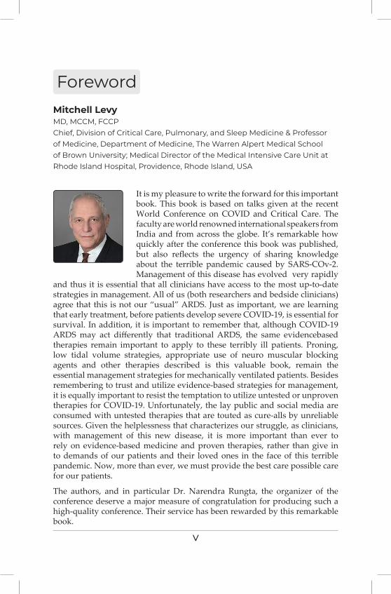
Mitchell LevyMD, MCCM, FCCP Chief, Division of Critical Care, Pulmonary, and Sleep Medicine & Professor of Medicine, Department of Medicine, The Warren Alpert Medical School of Brown University; Medical Director of the Medical Intensive Care Unit at Rhode Island Hospital, Providence, Rhode Island, USA
Foreword
It is my pleasure to write the forward for this important book. This book is based on talks given at the recent World Conference on COVID and Critical Care. The faculty are world renowned international speakers from India and from across the globe. It’s remarkable how quickly after the conference this book was published, but also reflects the urgency of sharing knowledge about the terrible pandemic caused by SARS-COv-2. Management of this disease has evolved very rapidly
and thus it is essential that all clinicians have access to the most up-to-date strategies in management. All of us (both researchers and bedside clinicians) agree that this is not our “usual” ARDS. Just as important, we are learning that early treatment, before patients develop severe COVID-19, is essential for survival. In addition, it is important to remember that, although COVID-19 ARDS may act differently that traditional ARDS, the same evidencebased therapies remain important to apply to these terribly ill patients. Proning, low tidal volume strategies, appropriate use of neuro muscular blocking agents and other therapies described is this valuable book, remain the essential management strategies for mechanically ventilated patients. Besides remembering to trust and utilize evidence-based strategies for management, it is equally important to resist the temptation to utilize untested or unproven therapies for COVID-19. Unfortunately, the lay public and social media are consumed with untested therapies that are touted as cure-alls by unreliable sources. Given the helplessness that characterizes our struggle, as clinicians, with management of this new disease, it is more important than ever to rely on evidence-based medicine and proven therapies, rather than give in to demands of our patients and their loved ones in the face of this terrible pandemic. Now, more than ever, we must provide the best care possible care for our patients.
The authors, and in particular Dr. Narendra Rungta, the organizer of the conference deserve a major measure of congratulation for producing such a high-quality conference. Their service has been rewarded by this remarkable book.

vi
BK Rao (Padma Bhushan Recipient)Chairman, Institute of Critical Care and Emergency Medicine, Sir Ganga Ram Hospital, Delhi; Chairman, National Accreditation Board for Hospitals and Healthcare Providers (NABH); Former Chairman, Board of Management, Sir Ganga Ram Hospital, Delhi; Former Member, Board of Governors, Medical Council of India
Foreword
The ‘UPDATE BOOK on Covid-19 and Critical Care’ is a compendium of invaluable information gained by the world during its fight against Covid-19.
The book’s 75 chapters are well organized in 16 sections. These chapters provide up-to-date and comprehensive coverage of a wide spectrum of issues related to Covid-19 and its management.
This book is being published in the wake of a recent well organized World Conference on Covid-19 and Critical care. No wonder that while the book has a rich academic and scientific flavor, it also gives a glimpse of the Covid management practices in the real world.
I congratulate the publishers, editors and authors for this unique achievement and hope that the book’s readers will be well informed about the future challenges and opportunities in the management of Covid-19.
vii
Dr Pravin AminMD FCCM FNCS
Foreword
I am delighted to present the Update book on Covid-19 and Critical Care published by the organising committee of the World Conference on Covid and Critical Care 2021. The editors Dr Ray, Dr Bajan, Dr Nasa, Dr Samaddar, Dr Toraskar and Dr Raha have admirably compiled, exhaustively and comprehensibly all aspects of this dreadful disease. I would also like to thank all the authors for their contribution and valuable documentation of published scientific data
in their manuscripts. I believe that it is important to have this book which promotes highquality research and intellectual output of authors. This book aims to bridge the gap between research and practice in the field of Critical Care among Covid-19 patients, thus providing an opportunity to the authors to propagate their high-quality scientific accomplishments to a wider audience. These resources presents a large dataset of broad utility, interest and significance to the medical community at large. Finally the articles are an authoritative, balanced and scholarly survey of recent developments in the field of Covid-19 in the Critically ill. I am certain that this book will be a great resource for critical care personal’s caring for Covid-19 patients in the ICU.

viii
Gratitude
The Organizing committee expresses deep sense of gratitude to the Editors of Update on Covid and Critical Care that, they have brought put a wonderful, rare and very informative updated literature about what is happening around the world in the field of Covid care in ICU and outside. Team of editors namely Dr Banambar Ray, Dr Prashant Nasa, Dr Khushrav Bajan, Dr DP Samaddar, Dr Kedar Toraskar and Dr Abhijit Raha deserve round of applause from all who are benificeiries of this update. Our thanks are also due to all the contributors, the forward writers, M/S Urvi, Mr H N Trivedi in particular for doing a great task. What is more important is that the whole task was achieved in a short period of 10 weeks which is a rare feat indeed.
Dr Narendra Rungta • Dr Ranvir Singh Tyagi Dr Lalit Singh • Dr Diptimala Agarwal • Dr Rakesh TyagiORGANISING COMMITTEE
ix
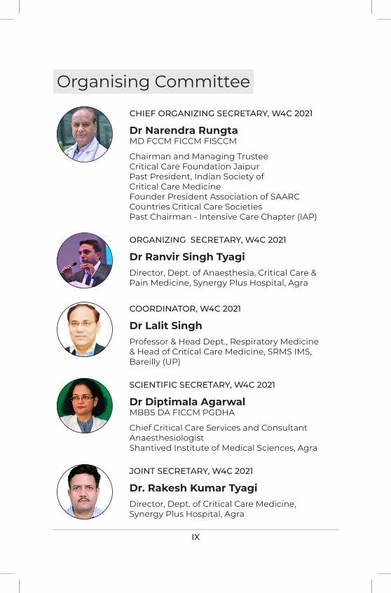
Organising Committee
CHiEF ORGANiZiNG SECRETARY, W4C 2021
Dr Narendra RungtaMD FCCM FICCM FISCCM
Chairman and Managing Trustee Critical Care Foundation Jaipur Past President, Indian Society of Critical Care Medicine Founder President Association of SAARC Countries Critical Care SocietiesPast Chairman - Intensive Care Chapter (IAP)
ORGANiZiNG SECRETARY, W4C 2021
Dr Ranvir Singh TyagiDirector, Dept. of Anaesthesia, Critical Care & Pain Medicine, Synergy Plus Hospital, Agra
COORDiNATOR, W4C 2021
Dr Lalit Singh Professor & Head Dept., Respiratory Medicine & Head of Critical Care Medicine, SRMS IMS, Bareilly (UP)
SCiENTiFiC SECRETARY, W4C 2021
Dr Diptimala AgarwalMBBS DA FICCM PGDHA
Chief Critical Care Services and Consultant AnaesthesiologistShantived Institute of Medical Sciences, Agra
JOiNT SECRETARY, W4C 2021
Dr. Rakesh Kumar TyagiDirector, Dept. of Critical Care Medicine,Synergy Plus Hospital, Agra

x
Editors and Co-EditorsEDITORS
Dr Banambar Ray
Dr Khusrav Bajan
Dr Prashant Nasa
CO-EDITORS
Dr DP Samaddar
Dr Kedar Toraskar
Dr Abhijeet Raha
xi
Diptimala Agarwal, MBBS DA FICCM PGDHA
Chief Critical Care Services and Consultant Anaesthesiologist, Shantived Institute of Medical Sciences, Agra
Madhulata Agarwal, MD Medicine
Assistant Professor, Department: Department of Medicine, S. M. S. Medical college and attached hospitals, Jaipur
Asif Ahmed, DNB Anaesthesiology (Gold Medal), IDCCM
Senior Consultant & Head of Department, Critical Care Medicine, Tata Main Hospital, Jamshedpur (Jharkhand)
Syed Moied Ahmed, MD, PhD
Professor, Anaesthesiology, JNMC, AMU, Akigarh, UP
Neelmani Ahuja Consultant Critical Care Medicine, Holy Family Hospital, Delhi
Ajith Kumar AK, MD, DNB, EDIC, FICCM
Senior Consultant & Ex- Head, Critical Care Medicine, Manipal Hospitals, Bangalore
Maher Al Bahrani, MD
Khusrav Beji Bajan, MD, EDIC
Consultant – Intensivist & HOD – Emergency Medicine, Critical Care & Emergency Medicine, P. D. Hinduja Hospital & Medical Reaserch Centre, Mumbai
Purabi Baral, MBBS, MD Microbiology
Associate Consultant Microbiology and Infection Control Officer, Microbiology, SUM Ultimate Hospital, Bhubaneswar
Rakhee Baruah, MD, IDCCM
Senior Consultant, Dept of Critical Care Medicine, Health City Hospital, Guwahati
Contributors
xii
Ashish BhallaDepartment of Internal Medicine, Post graduate Institute of Medical education and Research, Chandigarh
Vaibhav Bhargava, MD, FNB (Critical Care Medicine), EDIC
Senior Consultant, Critical Care Medicine, Incharge MICU, Critical Care Medicine, Eternal Hospital, Jaipur
Gunjan Chanchalani, MD, FNB, IDCCM, IFCCM
Consultant, Critical Care, Mumbai
Siddharth Chand, MD
Resident, Department of Medicine, Maulana Azad Medical College, New Delhi
Juhi Chandwani, FFARCS, EDIC
Consultant Critical Care, Anaesthesia and Critical Care, Royal Hospital, PO Box 1331, CPO Seeb 111, Muscat, Oman
Ranajit Chatterjee, MRCPI, EDIC, DA
Anaesthesia and Intensive care, Swami Dayanand Hospital, Delhi
Alisha Chaudhury, MD (Pulmonary Medicine), IDCCM (Indian Diplomate of Critical Care Medicine)
Junior Consultant, Critical Care Medicine, SUM Ultimate Medicare Hospital, A Unit of SOA, Bhubaneswar
Munish Kumar Chauhan, M.D.(Anesthesiology), EDIC
Senior Consultant, Department of Critical Care Medicine, Venkateshwar Hospital, Dwarka, Delhi
Rajesh Chawla, MD, FCCM, FCCP, EDIC
Senior Consultant, Respiratory and Critical Care Medicine, Indraprastha Apollo Hospital, Delhi
Contributors
xiii
Anirban Hom Choudhuri, MD, FICCM
Director Professor, Anaesthesiology & Intensive Care, GB Pant Hospital, New Delhi
Michael D. Christian, MD, FCCM
London’s Air Ambulance, Barts Health NHS Trust, London, United Kingdom
Pradeep Michael D’costa, D.N.B. (MED), CERTIFICATE IN CRITICAL CARE (ISCCM)
Physician Intensivist, Critical Care, Sahyadri Hospital (Nagar Road Branch), K.E.M Hospital, Pune
Mradul Kumar Daga, MD, FCCP, FRCP, FACP
Professor, Internal medicine and Infectious Diseases, Institute of Liver and Biliary Sciences, New Delhi
Beena Daniel, MBBS, DA
Consultant Intensivist, Critical Care, MEDICOVER Hospital, Aurangabad
Arnab DasguptaMD Anaesthesiology & Critical Care, PDF Neuroanaesthesiology
Senior Consultant, Neuroanaesthesiology & Critical Care, Fortis Hospital, Noida
Sananta Kumar DashFCICM (Fellow of College of Intensive Care Medicine), FNB
Staff Specialist, Intensive Care Unit, Mackay Base Hospital, Mackay, Queensland
Rimita Dey, FNB (Critical Care), EDIC
Senior Consultant and HOD, Department of Critical Care, Ruby General Hospital, Kolkata
Pratibha Dileep, MD
Head of the Department, Critical Care Medicine, Zydus Hospital, Ahmedabad
Contributors
xiv
Subhal Dixit, MD, FCCM, FICCM
Director, Critical Care, MJM and Sanjeevan Hospitals, Pune
Anand Waman Dongre, MBBS, MD (ANAESTHESIOLOGY)
Director, Critical Care, Swastik Critical Care Hospital, Nagpur
Yutika Anand Dongre, MBBS
Medical Officer, Medicine, NCI Hospital, Nagpur
Rakesh Garg, MD, DNB, FICCM
Additional Professor, Department of Onco-Anaesthesiology and Palliative Medicine Hospital - Dr BRAIRCH, All India Institute of Medical Sciences, New Delhi
Suneel Kumar Garg, MD, FNB, IFCCM, EDIC, FICCM, FCCP, FCCM
Founder & MD, Saiman Healthcare, New Delhi
Neeru Gaur, D.N.B, I.D.C.C.M
Consultant Intensivist, Anaesthesia and Critical Care, Midland Healthcare and Research Center, Lucknow
Ravi Gaur, M.D, D.N.B
Associate Professor, Physical Medicine and Rehabilitation, A.I.I.M.S, Jodhpur
Prasanta Kumar Gogoi, MBBS, DA
Senior Consultant and Head, OT & Anaesthesia, Ayursundra Superspecialty Hospital, Guwahati
Abhinav GuptaConsultant, Intensive Care Unit, Homerton University Hospital NHS Foundation Trust, London
Bikram Kumar Gupta, MD, PDCC, EDIC
Head, Department of Critical Care Medicine, Heritage Institute of Medical Sciences, Varanasi
Contributors
xv
Vivek Gupta, DA, DNB
Cardiac Anaesthesia and Intensive Care, Dayanand Medical College and Hospital, Ludhiana
Paula Mehdat Ibrahim Azer HannaSpecilaist, Critical Care, Mediclinic Parkview Hospital, Dubai
Satoru HashimotoDepartment of Anesthesia and Intensive Care Medicine, Kyoto Prefectural University of Medicine
Bharat JagiasiHead Critical Care, Reliance Hospital, Mumbai
Avinash Jain, DM (Clinical Immunology & Rheumatology), IRF (UoB, UK)
Sr Prof, Medicine, Medicine, SMS Medical Hospital, Jaipur
Ravi JainCritical Care Medicine, Mahatma Gandhi Medical College and Hospital, Jaipur
Bhavin Jankharia, MD, MBBS
Consultant Radiologist, Radiology, Picture This, Mumbai
Bijal Jankharia, MD DMRD
Consultant radiologist and sonologist, Picture This (Jankharia Imaging Center)
Yash Javeri, IDCCM, FICCM
Head CCM and Emergency Medicine, Regency Super Specialty Hospital – Lucknow
Sujeet Ashok Joshi, MD (Anaesthesia), IDCCM
Consultant, Critical Care Medicine, Tata Main Hospital, Jamshedpur
Contributors
xvi
Gavin M. Joynt, MBBCh
Department of Anaesthesia and Intensive Care, The Chinese University of Hong Kong, Hong Kong, China
Deven Juneja, FNB, EDIC
Associate Director, Institute of Critical Care Medicine, Max Super Speciality Hospital, Saket, New Delhi
Kamal KajalAssociate Professor, Department of Anesthesia and critical care, Post graduate Institute of Medical education and Research, Chandigarh
Sandeep Kantor, FCCM, FICCM
Consultant Critical Care, Department of Anaesthesia and Critical Care, Royal Hospital, PO Box 1331, CPO Seeb 111, Muscat, Oman
Rashmi Kapoor, MBBS, MD
Head Department of Pediatrics, Director Division of Pediatric Critical Care and Pulmonology, Regency Hospital Ltd.
Sunil Karanth, MD, FNB, EDIC, FCICM
Head of Dept & Senior Intensivist, Manipal Hospital, Bangalore; Adjuvant Professor in Critical care medicine at Manipal University
Geeta Karki Professor, Department of Anesthesia, Shri Ram Murti Smarak Institute of Medical Sciences, Bareilly
Tami Karni, M.D., M.H.A.
Head, Breast Care Institute, Assaf Harofe Medical Center, Zerifin, Israel; Chairman, Ethics Bureau of the Israel Medical Association, Ramat Gan, Israel
Sandeep Kataria, MBBS MD
Senior Resident, Anaesthesiology & Intensive Care, GB Pant Hospital, New Delhi
Contributors
xvii
Harjinder Kaur, MS
Consultant, Gynaecology & Obstetrics, Mahila Chikitsalaya, SMS Medical College, Jaipur
Kiranpreet Kaur, DA, DNB
Professor, Anaesthesiology & Critical Care, Pt. BDS, PGIMS at University of Health Sciences, Rohtak
Anand Shantaram Kawade, MD, DCH
Consultant Pediatrician and Neonatalogist, Consultant Paediatric Research, Core Vaccine Research Unit (CVRU), Vadu Rural Health Program, King Edward Memorial Hospital Research Centre, Pune
Faryal Khamis, MD
Reshu Gupta Khanikar, DA, IDCCM
Senior Consultant and HOD, Dept of Critical Care Medicine, Health City Hospital, Guwahati
Khalid Khatib, MD, FICCM, FICP
Professor, Medicine, SKN Medical College, Pune
Utkarsh KhattriSenior Resident, Department of Respiratory Medicine, Shri Ram Murti Smarak Institute of Medical Sciences, Bareilly
Young Sam Kim, MD, PhD
Division of Pulmonology, Department of Internal Medicine, Severance Hospital, Yonsei University College of Medicine, Seoul, Korea
Younsuck Koh, MD, PhD
Department of Pulmonary and Critical Care Medicine, Asan Medical Center, University of Ulsan College of Medicine, Seoul, Republic of Korea
Contributors
xviii
Kalpana KrishnareddyConsultant and Head of Department, Mediclinic Parkview Hospital, Adjunct Clinical Assistant Professor, Mohammed Bin Rashid University, Dubai
Arun Kumar, MBBS, MD Anesthesiology
Director, Medical Intensive care Unit, Fortis Hospital Mohali
Mritunjay Kumar, MD, DNB, FIACTA
Assistant Professor, Anaesthesiology, Pain Medicine and Critical Care, All India Institute of Medical Sciences, New Delhi
Prashant Kumar, MD, MBA, FICCM, Commonwealth Fellow
Professor, Anaesthesiology & Critical Care, Pt. BDS, PGIMS at University of Health Sciences, Rohtak
Rahul Kumar, MD Anaesthesia, IFCCM
Critical Care & Emergency Medicine, Sir Ganga Ram Hospital, New Delhi
Sunny Kumar, MD, FNB
Consultant, Department of Pulmonology and Critical Care, Fortis Hospital, Noida
Ephrat Levy-Lahad, M.D.
Medical Ethics Unit, Shaare Zedek Medical Center, Jerusalem, Israel; Co-Chairman, Israel National Bioethics Council, Jerusalem, Israel; Medical Genetics Institute, Shaare Zedek Medical Center, Jerusalem, Israel; Faculty of Medicine, Hebrew University of Jerusalem, Jerusalem, Israel
Ayush Lohiya, MD
Assistant Professor (Public Health), Super Speciality Cancer Institute & Hospital, Lucknow
Kishore Mangal, MD, IFCCM, EDIC
Senior Consultant, Department of Critical Care Medicine, Eternal Hospital, Jaipur
Contributors
xix
Raj Kumar ManiCritical Care and Pulmonology, Yashoda super Specialty Hospital, Kaushambi, Ghaziabad
Dinesh MathurConsultant and Head, Department of Dermatology, Rajasthan Hospital, Jaipur
Indubala Maurya, MD, DNB
Associate Professor, Anaesthesiology, Super Speciality Cancer Institute & Hospital, Lucknow
Seema Mehta, MS, FICOG
Senior Professor, Gynaecology & Obstetrics, Mahila Chikitsalaya, SMS Medical College, Jaipur
Sudhir Mehta, MD (Medicine) FRCP
Sr Prof, Medicine, Medicine, SMS Medical Hospital, Jaipur
Radha MG, DNB (Internal Medicine), FNB (Critical Care), IDCCM
Consultant Intensivist, Critical Care, Ramaiah Memorial Hospital, Bangalore
Anand Mishra, DNB (Anaesthesia), IDCCM
Consultant Critical Care, Critical Care, SUM Ultimate Medicare, Bhubaneswar
Bhavya Naithani, MD, PDCC
Head, Department of Critical Care Medicine, Heritage Institute of Medical Sciences, Varanasi
Monish Nakra, M.D. (Anesthesiology), IDCCM
Senior Consultant and Head, Department of Critical Care Medicine, Venkateshwar Hospital, Dwarka, Delhi
Prashant Nasa, FNB, EDIC
Head of the Department, Critical Care Medicine, NMC Specialty Hospital, Dubai
Contributors
xx
Joseph L. Nates, MD, MBA, CMQ, MCCM
Critical Care Department, The University of Texas MD Anderson Cancer Center, Houston, TX
Balikrishna Nimavat, EDIC, EDAIC
Consultant, Critical Care, Sir HN Reliance Hospital, Mumbai
Prasanta Kumar Panda, MBBS, MD
Sr. Consultant, Microbiology and Infection Control, SUM Ultimate Medicare, a Unit of SOA, Bhubaneswar
Rajesh Kumar Pande, MD, PDCC
Senior Director & HOD, Critical Care Medicine, BLK Max Superspeciality Hospital, New Delhi
Maitree Pandey, MD
Director Professor & Head of Department, Anaesthesiology & Critical Care, Lady Hardinge Medical College, New Delhi
Sunghoon Park, MD, PhD
Department of Pulmonary, Allergy and Critical Care Medicine, Hallym University Sacred Heart Hospital, Anyang, Republic of Korea
Quirino Piacevoli, MD, PhD
Professor and Head Department of Anesthesia and Intensive Care ACO San Filippo Neri Hospital, Roma, Italia
Gowri Sayi Prasad, MS DNB
Incharge, Obstetric Intensive Care Unit, Diamond Superspeciality Hospital, Kolhapur
Sayi Prasad, MD DNB
Director, Department of Critical Care Medicine, Diamond Superspeciality Hospital, Kolhapur
Contributors
xxi
Parikshit Prayag, American Board certified in Internal Medicine (AB IM), American Board certified in Infectious Diseases (AB ID), American transplantation society fellowship in Transplant Infectious Diseases
Consultant, Transplant Infectious Diseases, Deenanath Mangeshkar Hospital, Pune
Geetarani Purohit, MBBS, MD Microbiology
Department of Microbiology, Vikash Multispeciality Hospital, AH-46, Barahaguda Canal Chowk, Bargarh
Abhijeet Raha, MD, PDF
Senior Consultant Neurocritical Care, SUM Ultimate Medicare (a Unit of ‘SOA’), Bhubaneswar
Monika Rajani, MD, ACME
Associate Professor, Microbiology, Mayo Institute of Medical Sciences, Barabanki, Lucknow
Banambar Ray, MD, FICCM
Head of Critical Care Medicine, SUM Ultimate Medicare, A Unit of SOA, Bhubaneswar
Sumit RaySenior Consultant & Head, Critical Care Medicine, Holy Family Hospital, Delhi
Jordi Rello, MD, PhD
Clinical Research/epidemiology in pneumonia and sepsis, Vall d’Hebron Institute of Research (VHIR), Barcelona, Spain; Centro de Investigacion Biomedica en Red en Efermedades Respirato rias (CIBERES), Instituto de Salud Carlos III, Barcelona, Spain; Clinical Research, CHU Nîmes, NÎmes, France
Narendra Rungta, MD FCCM FICCM FISCCM
Chairman and Managing Trustee, Critical Care Foundation, Jaipur; Past President, Indian Society of Critical Care Medicine; Founder President, Association of SAARC Countries Critical Care Societies; Past Chairman - Intensive Care Chapter (IAP)
Contributors
xxii
Shrikant Saharshabudhe, MD (Chest), IDCCM
Director and Head, Department of Pulmonology and Critical Care Medicine, Senior Consultant Intensivist and Pulmonologist, Pulmonology and Critical Care Medicine, MEDICOVER Hospital, Aurangabad
Tapas Kumar Sahoo, Canadian Critical Care Fellowship (University of Toronto)
Senior Consultant & Head, Critical Care Medicine, Medanta Hospital, Ranchi
Arun Kumar Sahu, MD, FNB
Consultant Critical Care Medicine, Critical Care Medicine, SUM Ultimate Medicare, Bhubaneswar
DP Samaddar, MD, FICCM
Director Medical Affairs, Administration, CCU, Academics, Ruby General Hospital, Kolkatta
Sujay Samanta, MD, DM
Consultant In Charge Critical Care, Critical Care, Ruby General Hospital, Kolkata
Parang Sanghavi, MD, MBBS
Consultant Radiologist, Radiology, Picture This, Mumbai
Chaitri Shah, MBBS MD IDCC
Additional Medical Suprentendent, Dr M K Shah Medical College and Research Center, Ahmedabad
Nimit Ashwinbhai Shah, MD, MPH
Consulting Physician, Department: Internal Medicine, Zydus Hospital, Ahmedabad
Ritesh Shah, MD, IDCCM
Director, Critical Care Unit, Sterling Hospitals, Vadodara
Contributors
xxiii
Raman Sharma, MD Medicine FRCP
Professor, Department: Department of Medicine, S. M. S. Medical College and attached hospitals, Jaipur
Prakash Shastri, MD, FRCA, FICCM
Vice Chairman & Senior Consultant, Critical Care Medicine, Sir Gangaram Hospital, New Delhi
Gil Siegal, M.D., Ph.D.
Director, Center for Health Law & Bioethics, Faculty of Law, Ono Academic College, Kiryat Ono, Israel
Aanchal Singh, MD, FICCM
Specialist Pulmonary Medicine, Critical Care Medicine, NMC Specialty Hospital, Dubai
Lalit SinghProfessor & Head, Department of Respiratory Medicine & Critical Care Medicine, Shri Ram Murti Smarak Institute of Medical Sciences, Bareilly
Vinod Kumar Singh, MD. FRCP (Edin)
Senior consultant & director ECMO program, Critical Care Medicine, Sir Ganga Ram Hospital, New Delhi
YP Singh, M.D., FICCM, FCCM (American Board)
Senior Director & HOD, Critical Care Medicine, Max Super Specialty Hospital, I.P. Extension, Delhi
Vinay Singhal, M.D.(Anaesthesiology), IDCCM
Additional Director & Head, Department of Critical care medicine, Fortis Hospital, Ludhiana
Sharmili Sinha, MD, EDIC
Critical Care Medicine, Apollo Hospitals, Bhubaneswar
Contributors
xxiv
Vandana Sinha, M.D. Anaesthesiology
Medical Director & Director Department of Critical care, Department of Critical Care, Ayursundra Superspecialty Hospital, Guwahati
Mrinal Sircar, MD, EDIC
Director and head of Department, Pulmonology and Critical Care, Fortis Hospital, Noida
Kanwalpreet Sodhi, MD, FNB, EDIC, FCICM (Aus)
Chairman of Critical Care Service, Manipal Health Enterprises (P) Ltd, Senior Consultant and Head of Dept, Critical Care Medicine, Manipal Hospital, Old Airport Road, Bangalore
Charles L. Sprung, M.D.
Faculty of Medicine, Hebrew University of Jerusalem, Jerusalem, Israel; Department of Anesthesiology, Critical Care Medicine and Pain, Hadassah Medical Center, Jerusalem, Israel
Avraham Steinberg, M.D.
Medical Ethics Unit, Shaare Zedek Medical Center, Jerusalem, Israel; Co-Chairman, Israel National Bioethics Council, Jerusalem, Israel
Akhil Taneja, M.D., IFCCM, EDIC
Principal Consultant, Critical Care Medicine, Max Super Specialty Hospital, I.P. Extension, Delhi
Kedar Krishnarao Toraskar, MD EDIC
Director Critical Care, Critical Care Unit, Wockhardt Hospital, South Mumbai
Robert D. Truog, MD
Center for Bioethics, Department of Global Health and Social Medi cine, Harvard Medical School, Boston, MA; Department of Anesthesiology, Critical Care, and Pain Medicine, Boston Children’s Hospital, Boston, MA
Contributors

xxv
Resham Vasani, MD, DNB, FCPS, DDV
Consultant Dermatologist, Bhojani Clinic, Matunga, Mumbai; Research Associate, B J Wadia Hospital for Children, Parel, Mumbai; Clinical Associate Honorary, Nowrosjee Wadia Maternity Hospital, Parel, Mumbai
Ashoo Wadehra, M.D. Anaesthesiology, MBBS
Principal Consultant, Neuroanaesthesiology and Critical care, Max super speciality Hospital, Saket, New Delhi
Kapil Zirpe, MD CHEST, FICCM, FCCM, FSNCC
Head, Neuro Intensive Care, Ruby Hall, Pune
Noam Zohar, Ph.D.
Head, Advanced Studies in Bioethics, Faculty of Philosophy, Bar-Ilan University, Ramat Gan, Israel
Contributors

xxvi
Content
SECTION 1: Epidemiology and Variants of SARS-CoV-2 / COVID-19
• Preface Banambar Ray ...................................................................................................3
1. Epidemiology and Variants of SARS-CoV-2 / COVID-19 Prasanta Kumar Panda, Banambar Ray ........................................................5
SECTION 2: Pathophysiology
• Preface Prashant Nasa .................................................................................................15
2. Pathogenesis of COVID-19 Ajith Kumar AK, Radha MG ........................................................................17
3. Factors Affecting Severity of COVID-19 Kamal Kajal, Ashish Bhalla ...........................................................................24
4. Types of Lung Involvement in COVID-19 Ritesh Shah .....................................................................................................40
5. Effects on Other Organ Systems - COVID-19 - Neurological Involvement Asif Ahmed, Sujeet Ashok Joshi ..................................................................46
6. COVID-19-Cardiovascular Involvement Rimita Dey, DP Samaddar ............................................................................51
7. Haematological Data Interpretation (Non-Thrombotic) of COVID-19 Illness Sujay Samanta .................................................................................................56
8. Gastrointestinal and Hepatic Involvement in COVID-19 Subhal Dixit, Khalid Khatib ..........................................................................61

xxvii
Content
9. Dermatological Manifestations of COVID -19 – An Overview Resham Vasani, Dinesh Mathur ...................................................................65
SECTION 3: Diagnosis
• Preface Abhijeet Raha ..................................................................................................85
10. Challenges with Conventional and Novel Diagnostics of COVID-19 Purabi Baral, Geetarani Purohit ...................................................................87
11. Chest CT Scans and Radiographs in Covid-19 Bhavin Jankharia, Parang Sanghavi, Bijal Jankharia, ................................94
12. Point of Care Ultrasonography in COVID 19 Kedar Krishnarao Toraskar ........................................................................107
13. Uses of Biomarkers for Treatment Modification and Prognostication Raman Sharma, Madhulata Agarwal ........................................................113
14. Limitations of Biomarkers in COVID-19 Kapil Zirpe, Balikrishna Nimavat ..............................................................119
SECTION 4-A: Management - Respiratory Support in COVID-19
• Preface Kedar Toraskar .............................................................................................129
15. Methods of Oxygen Therapy and Targets Banambar Ray, Anand Mishra ...................................................................131
16. HFNC for COVID -19 Anand Waman Dongre, Yutika Anand Dongre ......................................135

xxviii
Content
17. Use of NIV in Covid 19 Pradeep Michael D'costa .............................................................................142
18. Early Vs Late Intubation for COVID-19 Prashant Kumar, Kiranpreet Kaur .............................................................148
19. Invasive Mechanical Ventilation in COVID 19 Neelmani Ahuja, Sumit Ray .......................................................................153
SECTION 4-B: Management - Strategies to Improve Oxygenation
• Preface Kedar Toraskar .............................................................................................161
20. Proning in Covid 19: an Interplay of Art and Science Khusrav Beji Bajan .......................................................................................163
21. ECMO in COVID 19 Sunil Karanth ................................................................................................173
22. Extra Corporeal CO2 Removal in Covid-19 ARDS Vinod Kumar Singh, Rahul Kumar ...........................................................179
23. Extracorporeal Therapies in COVID 19 (Excluding ECMO and ECCO2R) Ranajit Chatterjee .........................................................................................185
SECTION 4-C: Management - Current Position of Various Drugs & Adjuvants in Covid-19 Treatment
• Preface Kedar Toraskar .............................................................................................193
xxix
24. Corticosteroids and Covid-19 Prashant Nasa, Bharat Jagiasi .....................................................................195
25. Monoclonal Antibodies YP Singh, Akhil Taneja ................................................................................199
26. Antiviral Therapy (except Monoclonal Antibodies) Shrikant Saharshabudhe, Beena Daniel ....................................................205
27. IL6 Antagonists in Covid-19 Mrinal Sircar, Sunny Kumar .......................................................................213
28. Other Immunomodulators (Ulinastatin, Cytosorb, Colchicine, Sepsivac) for COVID-19 Yash Javeri, Monika Rajani .........................................................................220
29. Adjuvant Therapies in the Management of Covid-19 Gunjan Chanchalani, Kanwalpreet Sodhi ................................................225
30. Convalescent Plasma and Immunoglobulins Monish Nakra, Munish Kumar Chauhan .................................................230
SECTION 5: Critical Care Pearls
• Preface Prashant Nasa ...............................................................................................239
31. “FASTHUG Revisited in COVID-19” Neeru Gaur, Ravi Gaur ...............................................................................241
32. Infection Control Practices in ICU for Covid-19 Prashant Nasa, Ravi Jain .............................................................................252
33. Antimicrobial Stewardship in the COVID Pandemic Narendra Rungta, Parikshit Prayag...........................................................257
34. Secondary Bacterial Infections with Covid-19 Abhinav Gupta .............................................................................................263
Content
xxx
35. Venous Thromboembolism (VTE) in Covid 19: Risk Assessment, Prevention and Management Paula Mehdat Ibrahim Azer Hanna, Kalpana Krishnareddy ................270
36. Air Leaks in COVID-19 Deven Juneja, Prashant Nasa ......................................................................274
37. Communication in COVID 19 Sandeep Kantor, Juhi Chandwani .............................................................278
38. Visiting Policies in ICU During Covid-19 Sharmili Sinha, Mritunjay Kumar ..............................................................285
39. Do’s and Don’ts in the Management of Covid-19 Vaibhav Bhargava, Kishore Mangal ..........................................................294
SECTION 6: Long or Post Covid Conditions
• Preface Khusrav Beji Bajan .......................................................................................305
40. New or Ongoing Symptoms of Disease Bikram Kumar Gupta, Bhavya Naithani ..................................................307
41. Post-COVID 19 Syndrome Abhijeet Raha, Alisha Chaudhury .............................................................314
42. COVID Associated Mould Infection – a Double Whammy Prakash Shastri .............................................................................................321
43. Effects of Prolonged Hospitalization in COVID-19 Infection Rajesh Kumar Pande, Maitree Pandey ......................................................325
44. Palliation and End-of-Life Issues in COVID-19 Raj Kumar Mani ...........................................................................................329
45. Law and Medical Ethics in Context to Covid 19 Era Chaitri Shah ...................................................................................................334
Content
xxxi
SECTION 7: COVID-19 with Special Situations
• Preface Abhijeet Raha ................................................................................................341
46. Management of a COVID-19 Patient Requiring Surgery or Urgent Invasive Intervention(s) Arnab Dasgupta, Ashoo Wadehra .............................................................343
47. Management of COVID-19 in Pregnant Women Seema Mehta, Harjinder Kaur ...................................................................348
48. Diagnosis and Management of Multi System Inflammatory Syndrome (MIS-C) in Children Rashmi Kapoor .............................................................................................355
SECTION 8: Home Care and Rehabilitation
• Preface D P Samaddar ...............................................................................................369
49. Home Oxygen Therapy Vandana Sinha, Prasanta Kumar Gogoi ....................................................371
50. Physical and Mental Rehabilitation Sayi Prasad, Gowri Sayi Prasad .................................................................374
51. Organisation of Home-Care for COVID-19 Suneel Kumar Garg ......................................................................................379
SECTION 9: Vaccination
• Preface Diptimala Agarwal .......................................................................................389
Content
xxxii
52. Types of COVID-19 Vaccines and Immune Escape Mechanisms Anand Shantaram Kawade .........................................................................391
53. COVID-19 Vaccination Breakthrough Infections Rakesh Garg, Indubala Maurya, Ayush Lohiya ......................................397
SECTION 10: Clinical Recurrence of Covid 19 Infection
• Preface Banambar Ray ...............................................................................................407
54. Viral Relapse or Reinfection/ Inflammatory Rebound Mradul Kumar Daga, Siddharth Chand ...................................................409
55. COVID and Autoimmunity Sudhir Mehta, Avinash Jain ........................................................................413
SECTION 11: Organization for Pandemic
• Preface Khusrav Beji Bajan .......................................................................................423
56. Structure, Process Modifications and Preparedness for Pandemic Nimit Ashwinbhai Shah, Pratibha Dileep ................................................425
57. Protection of the Frontliners Khusrav Beji Bajan .......................................................................................430
58. Changing ICU Practices During COVID-19 Sananta Kumar Dash, Tapas Kumar Sahoo ..............................................433
59. The Toll of Pandemic- Non-Covid-19 Emergencies at Bay Lalit Singh, Utkarsh Khattri, Geeta Karki .................................................438
Content
xxxiii
SECTION 12: Miscellaneous
• Preface Banambar Ray ...............................................................................................445
60. Autopsies Findings Review in SARS – CoV – 2 Patients Quirino Piacevoli ..........................................................................................447
61. Burn-Out Among Intensivists Vinay Singhal, Arun Kumar .......................................................................452
62. ICU Care in Non-ICU Settings Anirban Hom Choudhuri, Sandeep Kataria ............................................457
SECTION 13: Research in COVID 19
63. Landmark Trials That Changed Practice in the Management of COVID-19 Prashant Nasa, Aanchal Singh, Rajesh Chawla .......................................465
SECTION 14: Some Thought Provoking Cases That We Encountered
• Preface Banambar Ray ...............................................................................................473
64. Delayed Pulmonary Embolism in Covid 19 an Unexpected and Bitter Foe Banambar Ray, Alisha Chaudhury ............................................................475
65. Interesting Cases of Covid-19- Complicated Leaks in the Lungs Syed Moied Ahmed, Rakesh Garg .............................................................479
Content
xxxiv
66. Spontaneous Pneumothorax with Bronchopleural Fistula in COVID-19 Pneumonia – A Nightmare Arun Kumar Sahu, Abhijeet Raha .............................................................483
67. COVID and Complicated Pregnancy Reshu Gupta Khanikar, Rakhee Baruah ...................................................489
SECTION 15: Lessons Learnt and being Learnt from COVID-19 Pandemic
68. Lessons Learnt and being Learnt from COVID-19 Pandemic DP Samaddar, Rimita Dey ..........................................................................499
SECTION 16: Covid Experience Across Asia
• Preface Banambar Ray, Abhijeet Raha ....................................................................515
69 CoVID-19 in Sri Lanka .........................................................................................................................517
70. COVID-19; Oman Experience Maher Al Bahrani, Faryal Khamis, Sandeep Kantor ...............................531
71. Coronavirus Disease-19 in South Korea Sunghoon Park, Young Sam Kim, Younsuck Koh ...................................534
72. The Japanese Experience COVID-19 Waves in Japan Satoru Hashimoto ........................................................................................540
Content
xxxv
73. Israeli Position Paper: Triage Decisions for Severely Ill Patients During the COVID-19 Pandemic. Joint Commission of the Israel National Bioethics Council, the Ethics Bureau of the Israel Medical Association and Representatives from the Israeli Ministry of Health* Avraham Steinberg, Ephrat Levy-Lahad, Tami Karni, Noam Zohar, Gil Siegal, Charles L. Sprung and the Israel National Bioethics Council, the Ethics Bureau of the Israel Medical Association, and Representatives from the Israeli Ministry of Health ............................................................................544
74. Israeli Position Paper Triage Decisions for Severely Ill Patients During the COVID-19 Pandemic Avraham Steinberg, Ephrat Levy-Lahad, Tami Karni, Charles L. Sprung ...................................................................564
75. Adult ICU Triage During the Coronavirus Disease 2019 Pandemic: Who Will Live and Who Will Die? Recommendations to Improve Survival* Charles L. Sprung, Gavin M. Joynt, Michael D. Christian, Robert D. Truog, Jordi Rello, Joseph L. Nates ..........................................568
Content

Epidemiology and Variants of SARS-CoV-2 / COVID-19
SECTION 1

3
Section 1 - Epidemiology and Variants of SARS-CoV-2 / COVID-19SECTION EDITOR
Dr Banambar RayMD, FICCM Head of Critical Care Medicine, SUM Ultimate Medicare, A Unit of SOA,Bhubaneswar
Preface
The Book begins with this section and the sole topic in this section is “Epidemiology and Variants” of SARS-COVID-2. This deals with the fundamental aspects of Corona virus, its geographical distribution, transmission and prevention. It also describes the various variants, clarifies various types and describes the meanings of mutation and lineage. The description is by no means exhaustive and easy to understand the less frequently visited aspects of corona virus’s origin and spread.

5
Epidemiology and variants of SARS-Cov-2 / COviD-19 1.
Prasanta Kumar Panda, Banambar Ray
INTRODUCTIONSARS-CoV-2 are human and animal pathogens. A novel coronavirus, which caused a cluster of pneumonia cases in Wuhan, China, was identified there in December 2019. It soon led to a global pandemic.1,2 The disease was designated as COVID-19, in February 2020, which stands for coronavirus disease 2019, by World Health Organization (WHO) 2,3 COVID-19 caused by the virus is designated as “severe acute respiratory distress syndrome corona virus-2 (SARS-CoV-2)”. Previously it was known as 2019-nCoV.2,3
VIROLOGYSARS-CoV-2 are enveloped positive-stranded RNA viruses.
Coronavirus, which causes COVID-19, is a beta coronavirus. It belongs to the same subgenus of severe acute respiratory distress syndrome (SARS) virus proven by sequencing and genetic analysis.4-7 it was proposed by the Coronavirus Study Group of the International Committee on Taxonomy of Viruses that this virus could be designated as severe acute respiratory distress syndrome coronavirus-2 (SARS-CoV-2).4-7 The primary source revealed that it has the closest RNA sequence similarity to bat corona virus. Whether COVID-19 virus is transmitted directly from bats or through some other mechanism is unknown.4-8
EPIDEMIOLOGYGeographical Distribution and Case Burden
A large number of new cases are being reported around the globe, following the first case report from Wuhan, by the end of 2019. Worldwide, almost from every country, more than 200 million cases have been reported till date.5,9,10
Transmission
“Man to Man” respiratory transmission was proven to be the primary cause of transmission of SARS-CoV-2.5,9,10 It is thought to occur mainly through the following:
W4C COVID-19 UPDATE BOOK
6
• close-range contact (approximately 2 meters) through respiratory particles.
• Infected person with coughs, sneezes or talks can infect another person on direct contact with the mucous membrane.
• if a person’s hands after being contaminated by these secretions or after touching contaminated surfaces, touch his / her eyes, nose or mouth.
Period of Greatest Infectiousness
When viral RNA levels from upper respiratory specimens are the highest, infected individuals are more likely to be contagious and that is in the earlier stages of illness. Transmission of the virus is rare after 7 to 10 days of illness, particularly for otherwise immunocompetent patients with non-severe infection.5,9,10
Transmission Based on Exposure Type
The risk of transmission from an individual with SARS-CoV-2 infection varies according to following factors:5
• type and duration of exposure
• preventive measures taken
• amount of virus in respiratory secretions.
Many individuals do not transmit SARS-CoV-2 to others and epidemiologic data suggest that fewer number of cases may cause secondary infections.5,9,10
Variants of Concern
Due to increased transmissibility SARS-CoV-2 has emerged and spread globally in a short time. Possible immune escape (fooling the immune system) due to emergence of mutant strains is another concern for some recently isolated variants, such as Delta B.1.617.2, AY.1 and AY.2 variant11
Immune Response and Risk of Reinfection
Once infected, the person might remain immune for a period of 6 to 8 months. It has been observed that the re-infection in such persons is low for the next few months.5,9,10
Prevention
Personal Preventive Measures
Residents are encouraged to practice social distancing if community transmission of SARS-CoV-2 is present by, avoiding crowds and maintaining a distance of six feet (approx. two meters) from others when in public. In particular, individuals should avoid close contacts with ill individuals who are also encouraged to wear masks.2,3,12

W4C COVID-19 UPDATE BOOK
7
Additional recommendation to reduce transmission of infection are as follows:2,3,12
• Hand washing, particularly after touching surfaces in public; hand sanitizers containing 60% alcohol is a recommended alternative. Wash hands if they are visibly dirty
• Respiratory hygiene (e.g., covering the cough or sneeze). Avoid touching the face.
• Adequate ventilation of indoor spaces can be ensured by
– keeping the doors and windows open.
– exhausting the inside air.
– using air conditioning fans continuously
– using portable high-efficiency particulate air (HEPA) filtration systems.
• Frequently touched objects and surfaces are to be disinfected as per center for disease control (CDC) guidelines.
Social / Physical Distancing
In locations where there is community transmission of SARS-CoV-2, social and physical distancing needs to be practiced both in indoor and outdoor spaces by maintaining a minimum distance. The optimal distance is not clearly defined; social distancing of six feet / two meters is recommended by CDC, whereas three feet / one meter is recommended by WHO. Close-range contact is to be avoided with an individual with infection, which is thought to be the primary risk for exposure to SARS-CoV-2.2,3,12
Screening in High-Risk Settings
Screening for SARS-CoV-2 infection with serial viral testing is recommended in long-term care facilities to quickly identify cases so that infected individuals can be isolated, contacts can be quarantined, and outbreaks can be prevented.
Although antigen tests are generally less sensitive than Nucleic Acid Amplification Test (NAAT), modeling studies have suggested that if the frequency of testing is high enough, cumulative infection rates can be reduced by utilizing tests with low sensititvity.2,3,12
Other Public Health Measures
An outbreak of public health emergency of international concern was declared by the WHO, on January 30, 2020. All the countries were requested to take action in detecting and preventing the spread by March 2020, as it was characterized as a pandemic in order to emphasize the gravity of the menace.
W4C COVID-19 UPDATE BOOK
8
In addition to personal preventive measures (e.g., masks, hand hygiene, respiratory etiquette, and environmental disinfection), transmission reduction strategies include social / physical distancing orders / instructions.2,3,12 Those are as follows:
• stay-at-home orders
• school, venue, and nonessential business closure
• ban on public gatherings
• ravel restriction with exit and / or entry screening
• identification and isolation of infected from non-infected individuals.
VARIANTSDetails of SARS-CoV-2 variants are under investigation (Table 1):2,4,10,13
SARS-COV-2 VARIANT CLASSIFICATION AND DEFINITIONS• Mutation:
– It refers to a single change in the genetic code of the virus.
– Frequent mutations not necessarily change the characteristics of the virus.14
• Lineage:
– Closely related group of viruses showing common ancestry.
– COVID-19 is caused by different mutant strains of SAR-CoV-2.14
• Variant: Viral genomic code containing one or more mutations.14
• Variant of interest (VOI):
o These are variants having specific genetic markers; have high receptor binding, cause decreased protected immunity against previous infection or immunization, decrease efficacy of treatment and potential negative diagnostic impact, increase transmission and disease progression.14
• Variant of Concern (VOC)
– The key features of few variants are easy transmissibility, severe clinical presentations, and significant decreased immune response, reduced effectiveness of drugs or immunization. laboratory detection failures are marked in few variants.14
• Variant of High Consequence (VOHC)
– Preventive measures or medical counter measures have significantly
W4C COVID-19 UPDATE BOOK
9
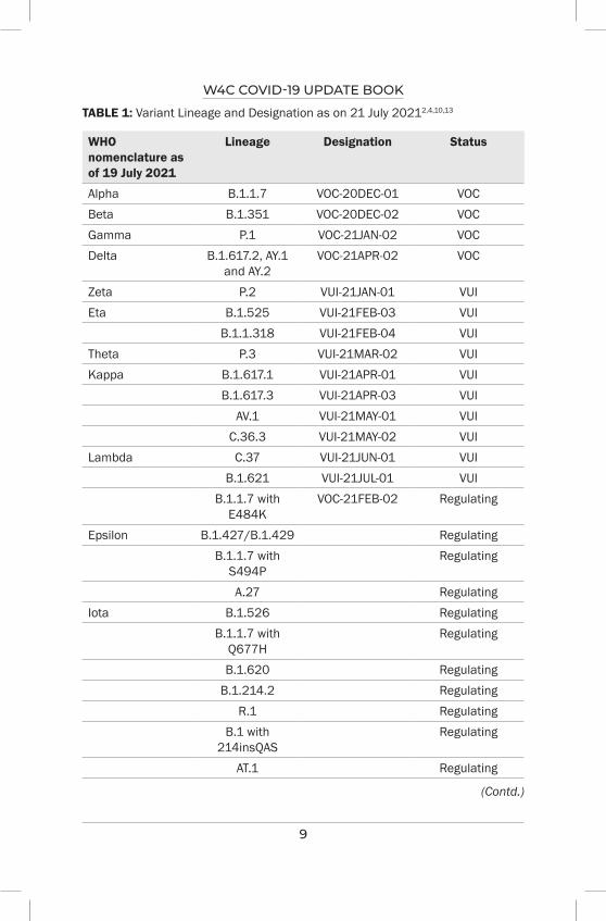
TABLE 1: Variant Lineage and Designation as on 21 July 20212,4,10,13
WHO nomenclature as of 19 July 2021
Lineage Designation Status
Alpha B.1.1.7 VOC-20DEC-01 VOCBeta B.1.351 VOC-20DEC-02 VOCGamma P.1 VOC-21JAN-02 VOCDelta B.1.617.2, AY.1
and AY.2VOC-21APR-02 VOC
Zeta P.2 VUI-21JAN-01 VUIEta B.1.525 VUI-21FEB-03 VUI
B.1.1.318 VUI-21FEB-04 VUITheta P.3 VUI-21MAR-02 VUIKappa B.1.617.1 VUI-21APR-01 VUI
B.1.617.3 VUI-21APR-03 VUIAV.1 VUI-21MAY-01 VUI
C.36.3 VUI-21MAY-02 VUILambda C.37 VUI-21JUN-01 VUI
B.1.621 VUI-21JUL-01 VUIB.1.1.7 with
E484KVOC-21FEB-02 Regulating
Epsilon B.1.427/B.1.429 RegulatingB.1.1.7 with
S494PRegulating
A.27 RegulatingIota B.1.526 Regulating
B.1.1.7 with Q677H
Regulating
B.1.620 RegulatingB.1.214.2 Regulating
R.1 RegulatingB.1 with
214insQASRegulating
AT.1 Regulating
(Contd.)
W4C COVID-19 UPDATE BOOK
10
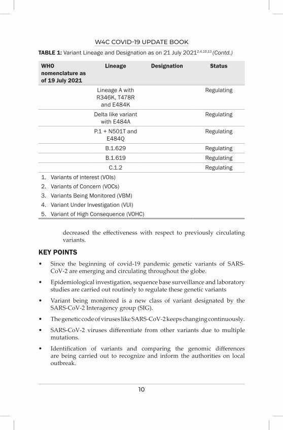
WHO nomenclature as of 19 July 2021
Lineage Designation Status
Lineage A with R346K, T478R
and E484K
Regulating
Delta like variant with E484A
Regulating
P.1 + N501T and E484Q
Regulating
B.1.629 RegulatingB.1.619 Regulating
C.1.2 Regulating1. Variants of interest (VOIs)2. Variants of Concern (VOCs)3. Variants Being Monitored (VBM)4. Variant Under Investigation (VUI)5. Variant of High Consequence (VOHC)
TABLE 1: Variant Lineage and Designation as on 21 July 20212,4,10,13 (Contd.)
decreased the effectiveness with respect to previously circulating variants.
KEY POINTS• Since the beginning of covid-19 pandemic genetic variants of SARS-
CoV-2 are emerging and circulating throughout the globe.
• Epidemiological investigation, sequence base surveillance and laboratory studies are carried out routinely to regulate these genetic variants
• Variant being monitored is a new class of variant designated by the SARS-CoV-2 Interagency group (SIG).
• The genetic code of viruses like SARS-CoV-2 keeps changing continuously.
• SARS-CoV-2 viruses differentiate from other variants due to multiple mutations.
• Identification of variants and comparing the genomic differences are being carried out to recognize and inform the authorities on local outbreak.
W4C COVID-19 UPDATE BOOK
11
SUMMARY AND PRACTICE POINTS• infected patients are protected from the disease for 6 to 8 months
mediated through immune response
• mask-wearing in public
• diligent hand washing
• respiratory hygiene
• physical distancing
• Avoiding crowds and close contact with ill individuals.
• Transmission, prognosis, vaccines, drugs, laboratory tools and social measures are particular to different variants
• Specific VOIs and VOCs are prioritized for global monitoring.
REFERENCES1. Zhu N, Zhang D, Wang W, et al. A Novel Coronavirus from Patients with Pneumonia in
China, 2019. N Engl J Med 2020; 382:727.2. World Health Organization. http://www.who.int/dg/speeches/detail/who-director- general-s-
remarks-at-the-media-briefing-on-2019-ncov-on-11-february-2020 (Accessed on February 12, 2020).
3. World Health Organization. Novel Coronavirus (2019-nCoV) technical guidance. https://www.who.int/emergencies/diseases/novel-coronavirus-2019/technical-guidance (Accessed on February 14, 2020).
4. Hoffmann M, Kleine-Weber H, Schroeder S, et al. SARS-CoV-2 Cell Entry Depends on ACE2 and TMPRSS2 and Is Blocked by a Clinically Proven Protease Inhibitor. Cell 2020; 181:271. https://www.who.int/en/activities/tracking-SARS-CoV-2-variants/ (Accessed on June 07, 2021).
5. Zhou B, Thao TTN, Hoffmann D, et al. SARS-CoV-2 spike D614G change enhances replication and transmission. Nature 2021; 592:122.
6. Klumpp-Thomas C, Kalish H, Hicks J, et al. Effect of D614G Spike Variant on Immunoglobulin G, M, or A Spike Seroassay Performance. J Infect Dis 2021; 223:802.
7. Coronaviridae Study Group of the International Committee on Taxonomy of Viruses. The species severe acute respiratory syndrome-related coronavirus: classifying 2019-nCoV and naming it SARS-CoV-2. Nat Microbiol 2020; 5:536.
8. Zhou P, Yang XL, Wang XG, et al. A pneumonia outbreak associated with a new coronavirus of probable bat origin. Nature 2020; 579:270.
9. Korber B, Fischer WM, Gnanakaran S, et al. Tracking Changes in SARS-CoV-2 Spike: Evidence that D614G Increases Infectivity of the COVID-19 Virus. Cell 2020; 182:812.Plante JA, Liu Y, Liu J, et al. Spike mutation D614G alters SARS-CoV-2 fitness. Nature 2021; 592:116.
10. Dougherty K, Mannell M, Naqvi O, et al. SARS-CoV-2 B.1.617.2 (Delta) Variant COVID-19 Outbreak Associated with a Gymnastics Facility - Oklahoma, April-May 2021. MMWR Morb Mortal Wkly Rep 2021; 70:1004.
11. Lu R, Zhao X, Li J, et al. Genomic characterization and epidemiology of 2019 novel coronavirus: implications for virus origins and receptor binding. Lancet 2020; 395:565. Perlman S. Another Decade, another Coronavirus. N Engl J Med 2020; 382:760.
W4C COVID-19 UPDATE BOOK
12
12. Centers for Disease Control and Prevention. 2019 Novel coronavirus, Wuhan, China. Information for Healthcare Professionals. https://www.cdc.gov/coronavirus/2019-nCoV/hcp/index.html (Accessed on February 14, 2020).
13. https://assets.publishing.service.gov.uk/government/uploads/system/uploads/attachment_data/file/991343/Variants_of_Concern_VOC_Technical_Briefing_14.pdf (Accessed on June 07, 2021).
14. https://www.cdc.gov/coronavirus/2019-ncov/variants/variant-info.html SARS-CoV-2 Variant Classifications and Definitions Updated Oct. 4, 2021.
PathophysiologySECTION 2

15
Section 2: PathophysiologySECTION EDITOR
Dr Prashant NasaFNB, EDIC Head of the Department, Critical Care Medicine, NMC Specialty Hospital, Dubai
Preface
There has been considerable progress in understanding the pathophysiology of COVID-19. The research in the last few months on SARS-CoV-2 suggests that the host immune system play a pivotal role in the pathogenesis of COVID-19. The pathophysiology of COVID-19 is not only the direct impact of viral invasion or cytotoxicity but also reflects a dysregulated host immune response to SARS-CoV-2. The spectrum of clinical features in COVID-19 span from mild (limited upper respiratory
tract symptoms) to critical disease (ARDS, sepsis, or septic shock) requiring respiratory or other organs support. From the inception of the pandemic, there has been extensive research on the identification of risk factors for the prediction of progression to severe disease. Various meta-analyses were performed to identify risk factors for the severity of COVID-19 involving demographics, comorbid illness, or genetic phenotypes of the patients. The respiratory system is the primary target organ for SARS-CoV-2. However, COVID-19 is not an exclusive pulmonary disease and instead involves multi-organ systems. This section on the pathophysiology of COVID-19 summarizes the current evidence on pathogenesis, risk factors, and spectrum of pulmonary manifestations of COVID-19. The section also includes a chapter on other organs involved in COVID-19 with special chapters on neurological and dermatological manifestations of COVID-19.

17
Pathogenesis of COviD-19 2.
INTRODUCTIONSince December 2019, COVID-19 pandemic has caused devastating consequences across the globe. The disease is caused by SARS–CoV-2 virus, an enveloped, single stranded RNA virus. In the last two years, with Covid-19 infections causing diverse outcomes have probably created an opportunity for the medical fraternity to understand and develop newer insights into the disease per se though many uncertainties still prevail. Medical community is beginning to understand the dynamics of infectivity and transmissibility to some extent, though many aspects of pathogenesis remain elusive, and there are significant knowledge gaps in understanding the pathogenesis. Understanding pathogenesis is a key aspect in developing therapeutic measures for any medical disorder. Emerging evidence indicates that COVID-19 has distinctive pathophysiological features that set the disease apart from respiratory failures of other origin. The majority of infected covid patients are mild (80%) with self-limited course, and only a tiny percentage of patients progress to severe form(15%), and 5% develop ARDS and multi-organ failure.1
Advanced age, male sex and comorbities enchance the risk for severe disease. About 15-30% of the hospitalized people develop COVID-19 associated acute respiratory distress syndrome (CARDS). Autopsies in patients with severe SARS-CoV-2 infection revealed diffuse alveolar damage consistent with ARDS, but there was a higher thrombus burden in pulmonary capillaries.
In acute phase, respiratory manifestations are common (first four weeks from the onset of symptoms), though extra-pulmonary features are also reported. Long COVID or post covid syndrome in a minority of patients (10%) suggests a complex clinical picture, probably due to dysregulated host response to infection characterised by immunoinflammatory derangements.2
PATHOGENESISSARS-CoV-2 and the Human cell
There are five stages in the life cycle of SARS-CoV-2 virus, which include
Ajith Kumar AK, Radha MG
W4C COVID-19 UPDATE BOOK
18
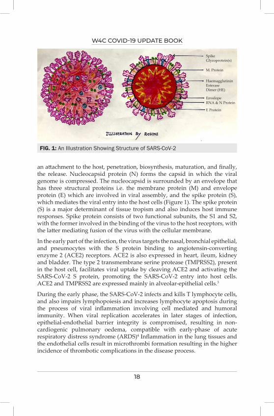
an attachment to the host, penetration, biosynthesis, maturation, and finally, the release. Nucleocapsid protein (N) forms the capsid in which the viral genome is compressed. The nucleocapsid is surrounded by an envelope that has three structural proteins i.e. the membrane protein (M) and envelope protein (E) which are involved in viral assembly, and the spike protein (S), which mediates the viral entry into the host cells (Figure 1). The spike protein (S) is a major determinant of tissue tropism and also induces host immune responses. Spike protein consists of two functional subunits, the S1 and S2, with the former involved in the binding of the virus to the host receptors, with the latter mediating fusion of the virus with the cellular membrane.
In the early part of the infection, the virus targets the nasal, bronchial epithelial, and pneumocytes with the S protein binding to angiotensin-converting enzyme 2 (ACE2) receptors. ACE2 is also expressed in heart, ileum, kidney and bladder. The type 2 transmembrane serine protease (TMPRSS2), present in the host cell, facilitates viral uptake by cleaving ACE2 and activating the SARS-CoV-2 S protein, promoting the SARS-CoV-2 entry into host cells. ACE2 and TMPRSS2 are expressed mainly in alveolar-epithelial cells.3
During the early phase, the SARS-CoV-2 infects and kills T lymphocyte cells, and also impairs lymphopoiesis and increases lymphocyte apoptosis during the process of viral inflammation involving cell mediated and humoral immunity. When viral replication accelerates in later stages of infection, epithelial-endothelial barrier integrity is compromised, resulting in non-cardiogenic pulmonary oedema, compatible with early-phase of acute respiratory distress syndrome (ARDS)4 Inflammation in the lung tissues and the endothelial cells result in microthrombi formation resulting in the higher incidence of thrombotic complications in the disease process.
FIG. 1: An Illustration Showing Structure of SARS-CoV-2
SpikeGlycoprotein(s)
M. Protein
HaemagglutininEsteraseDimer (HE)
EnvelopeRNA & N Protein
E Protein
W4C COVID-19 UPDATE BOOK
19
Host Response to SARS-CoV-2
Invasion of the virus in the upper respiratory tract results in release of C-X-C motif chemokine ligand 10 (CXCL-10) and interferons (INF- β and IFN-λ) from the cells. [5] Most of the patients do not progress beyond this stage.
About 20 % of the patients with SARS-CoV-2 develop severe disease with lung parenchymal involvement. The virus and secondary trigger immune responses to the lung parenchymal destruction, which recruit macrophages and monocytes that fight the infection, resulting in the release of cytokines and priming of adaptive T and B cell immune responses. This process helps in containing the infection in most cases. However, severe lung and even systemic pathology could occur in some cases due to a dysfunctional immune response.
The integrity of the epithelial-endothelial barrier gets compromised with accelerated viral replication. Infection of pulmonary capillary endothelial cells accelerates the inflammatory response with the resultant influx of monocytes and neutrophils. The virus-laden pneumocytes release cytokines and inflammatory markers. An early-phase ARDS results from pulmonary oedema filling the alveolar spaces with hyaline membranes. Bradykinin-dependent lung angioedema is also contributory to the disease process. Cumulatively, endothelial barrier disruption, dysfunctional alveolar-capillary oxygen transmission, and decreased oxygen diffusion capacity attribute to COVID-19 ARDS. There is greater vasculopathy characterised by macro- and micro-thrombosis, endothelial injury, vascular dilation and abnormal angiogenesis, and earlier SARS injury in COVID-19 compared to H1N1 influenza ARDS.6,7
Cytokine Storm
The SARS-CoV-2 infection could result in the release of various cytokines including IL-1β, IL-6, IL-12, IL-18, IL-33, IFN-α, IFN-γ, TNF-α, and transforming growth factor (TGF) β. Chemokine ligands such as CCL2, CCL3, CCL5, CXCL8, CXCL9, and CXCL10 are also released. The massive release of cytokines can cause septic shock, multi-organ failure and death. Non -survivors have been noted to have persistence of viral RNA, probably indicating a correlation between virus persistence and poor disease outcome.8 Secondary HLH has been noted in patients with COVID with increased IL-2, IL-7, GCSF, CXCL 10, MCP-1, and MIP-α.
Humoral and cell-mediated immunity: Antibodies to receptor binding domain of spike protein have been shown to have neutralising activity against infection, which could protect up to one year. CD4 + and CD8+ T cells play a crucial role in SARS CoV-2 infection wherein the former activates the B -cells to produce virus-specific antibody while the latter kills virus-infected cells. Both above processes can contribute to lung injury.
W4C COVID-19 UPDATE BOOK
20
Effect of SARS CoV-2 on extrapulmonary organs
Fulminant activation of coagulation pathway with consumption of clotting factors in severe COVID-19 promotes the formation of microthrombi both in the arteries and veins, resulting in deep vein thrombosis, pulmonary embolism; cerebral, myocardial infarction and limb ischemia. Moreover, ACE-2 receptors are also expressed in the heart, renal tubular and intestinal epithelium, endothelium, and pancreas, which potentially invade these organs, resulting in multi-organ failure. Viral sepsis could also contribute to multi-organ dysfunction
COVID-19 disease evolves in many phases, from asymptomatic phase to involvement of upper respiratory tract in majority of patients, to lower respiratory tract involvement in some, and progression to acute respiratory distress syndrome in subset of patients at risk.
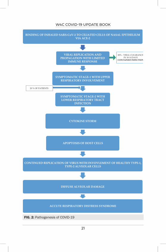
Respiratory droplets and aerosols from infected patients lead to the acquisition and transmission of COVID-19. S1 protein on SARS COVID-19 binds to ACE-2, functional receptor on the nasal epithelium and pulmonary epithelial cells in the host with resultant conformational change post fusion.9 Subsequently, replication and local propagation depending on the immune response in the host determines the progression and course of the disease.
COVID-19 infected patients have a wide range of presentations from asymptomatic to severe respiratory symptoms with multi-organ failure. The mean incubation period for COVID-19 is five days (average 2-7 days).10 Viral load in the upper respiratory tract is maximum around the onset of symptoms, and viral shedding begins approximately 2 to 3 days before symptoms make the person contagious.11
In the asymptomatic phase, the virus via respiratory aerosols in the upper respiratory tract binds to epithelial cells and undergoes replication and propagation along with infection of ciliated cells in the conducting airways.12 With the limited immune response, this stage lasts up to 2 weeks.
The symptomatic stage-1 virus migrates from the nasal epithelium to the upper respiratory tract via the conducting airways. Due to upper respiratory tract involvement, symptoms like fever, malaise and dry cough are manifested. During this stage, there is a greater immune response involving the release of C-X-C motif chemokine ligand 10 (CXCL-10) and interferons (IFN-beta), INF- gamma from the virus-infected cells.13 A further immune response is contained in the majority, thereby limiting the disease progression at this stage.
In symptomatic stage-2, there is lower respiratory tract involvement and progression to acute respiratory distress syndrome. About one-fifth of the infected patients progress to this stage of disease and develop severe symptoms. This stage is characterised by a “cytokine storm” with persistent
W4C COVID-19 UPDATE BOOK
21
BINDING OF INHALED SARS-CoV-2 TO CILIATED CELLS OF NASAL EPITHELIUMVIA ACE-2
VIRAL REPLICATION ANDPROPAGATION WITH LIMITED
IMMUNE RESPONSE
SYMPTOMATIC STAGE-1 WITH UPPERRESPIRATORY INVOLVEMENT
SYMPTOMATIC STAGE-2 WITHLOWER RESPIRATORY TRACT
INFECTION
CYTOKINE STORM
APOPTOSIS OF HOST CELLS
CONTINUED REPLICATION OF VIRUS WITH INVOVLEMENT OF HEALTHY TYPE-1,TYPE-2 ALVEOLAR CELLS
DIFFUSE ALVEOLAR DAMAGE
ACCUTE RESPIRATORY DISTRESS SYNDROME
80% - VIRAL CLEARANCEIN 10-14 DAYS
CONTAINED INFECTION
20 % OF PATIENTS
FIG. 2: Pathogenesis of COVID-19
W4C COVID-19 UPDATE BOOK
22
epithelial injury by sequestered inflammatory cells and viral replication resulting in loss of both type 1 and type 2 pneumocytes, which culminates in diffuse alveolar damage and acute respiratory distress syndrome.14
CARDS: Covid-19 associated ARDS
Nasal epithelium and respiratory tract epithelium are rich in ACE-2 and TMPRSS2 receptors through which Covid gains entry into the host. Following viral cell entry and SARS-CoV-2 replication, extensive tissue damage of endothelial and epithelial structures can occur, which results in increased permeability and alveolar and interstitial accumulation (oedema) of protein-rich fluids.15
The initial exudative phase is characterised by type 2 pneumocyte tropism, hyaline membrane formation, surfactant inactivation , culminating in diffuse alveolar damage. Cytokine driven irreversible destruction of the pulmonary architecture and pulmonary vasculopathy with endothellitis, microangiopathy, thrombosis results in proliferation and fibrosis of CARDS (Figure 2).16
COVID-19 INDUCED EXTRA-PULMONARY ORGAN DYSFUNCTION17
Mechanisms of Covid -19 Induced Heart Disease
The potential mechanisms of SARS-CoV2 in the induction of heart disease are not clear. However, different pathways were defined for COVID-19 pathogenesis: (1) viremia and direct infection of lung and heart; (2) recruitment of the innate immune system by macrophages and cytokine storm; (3) adaptive immune system activation.
COVID-19 induces direct myocardial inflammation. Indirect effects of infection including, cytokine storm, endothelial dysfunction, leucocytes infiltration, and formation of microvascular thrombosis lead to cardiac dysfunction.
Mechanisms of Hematologic Abnormalities
Direct effects on lymphocytes and cytokine storm leading to lymphoid apoptosis both lead to lymphopenia. Cytokine storm affecting the spleen, lymphoid organs result in an altered lymphoid turnover.
Coagulation abnormalities are multifactorial conditions associated with the combination of inflammation, activation of platelet, endothelial dysfunction.
CONCLUSIONCOVID-19, mainly in severe cases and the lung, involves different organs such as the heart, liver, kidney, and haematological and nervous system, inducing multi-organ failure. SARS-COV2 may directly invade the host cells of different organs through the ACE2 receptor due to the presence of this receptor in these organs. On the other hand, activating the complement system, cytokine storm,
W4C COVID-19 UPDATE BOOK
23
dysregulated immune responses, coagulation dysfunction, and infiltration of inflammatory cells in SARS-CoV2 infection can induce multi-organ failure in these patients.
REFERENCES 1. Zhonghua 2020 Feb 145-151: Epidemiology working group for NCIP Epidemic response
CCDC 2. Marcin F Osuchowski –COVID-19. Pathophysiology of acute disease. Lancet Resp Med Jun
21,9(6), 622-642. 3. Hoffmann M, Kleine-Weber H, Schroeder S, et al. SARS-CoV-2 cell entry depends on ACE2
and TMPRSS2 and is blocked by a clinically proven protease inhibitor. Cell 2020; 181:271-280.4. Xu Z, Shi L, Wang Y, et al. Pathological findings of COVID-19 associated with acute respiratory
distress syndrome. Lancet Respir Med 2020; 8:420-422.5. Tang N, Chan P, Wong C, To K, Wu A, Sung Y et al. Early Enhanced Expression of Interferon-
Inducible Protein-10 (CXCL-10) and Other Chemokines Predicts Adverse Outcome in Severe Acute Respiratory Syndrome. Clinical Chemistry 2005; 51:2333- 2340.
6. Xu Z, Shi L, Wang Y, Zhang J, Huang L, Zhang C et al. Pathological findings of COVID- 19 associated with acute respiratory distress syndrome. The Lancet Respiratory Medicine 2020; 8:420-422.
7. Carsana L, Sonzogni A, Nasr A, Rossi R, Pellegrinelli A, Zerbi P et al. Pulmonary post- mortem findings in a series of COVID-19 cases from northern Italy: a two-centre descriptive study. The Lancet Infectious Diseases 2020; 20:1135-1140.
8. Zhou Y, Fu B, Zheng X, Wang D, Zhao C, Qi Y et al. Pathogenic T-cells and inflammatory monocytes incite inflammatory storms in severe COVID-19 patients. National Science Review 2020; 7:998-1002.
9. Wan Y Shang Receptor recognition by novel Corona virus from Wuhan. J Vilology 2020; 94. 10. Lauer S, Grantz K, Bi Q, Jones F, Zheng Q, Meredith H et al. The Incubation Period of
Coronavirus Disease 2019 (COVID-19) From Publicly Reported Confirmed Cases: Estimation and Application. Annals of Internal Medicine 2020; 172:577-582.
11. He X, Lau E, Wu P, Deng X, Wang J, Hao X et al. Temporal dynamics in viral shedding and transmissibility of COVID-19. Nature Medicine 2020; 26:672-675.
12 . Sims Role of ciliated cells in viral spread in the conducting airways of the lungs. J Virology 2005; 79:1511-24.
13. Tang, early enhanced expression of interferon inducible protein-10. Clin Chem 2005; 51:2333-40.
14. XU Z, pathological findings of CARDS. Lancet 2020; 8:420-2.15. Bourgonje AR, Abdulle AE, Timens W, et al. Angiotensin-converting enzyme 2 (ACE2),
SARS-CoV-2 and the pathophysiology of coronavirus disease 2019 (COVID-19). J Pathol 2020; 251:228-248.
16. Fabro AT. Pulmonary pathology of ARDS in COVID-19: a pathological review for clinicians. Respir Med 2020; 176:106239.
17. COVID-19 and multiorgan failure: A narrative review on potential mechanisms. J Mol Histo 2020; 1-16 : Tahmineh Mukthari.
24
Factors Affecting Severity of COviD-19 3.
Corona viruses are the group of viruses affecting humans and animals. They are responsible for the current pandemic that was first identified as the cases of pneumonia in Wuhan, China. The specific virus implicated is described as severe acute respiratory syndrome coronavirus 2 (SARS-CoV-2). These viruses have immense ability to evolve over time and pose significant challenge to treatment options. The predominant prevalent variant of concern in India is Delta (B.1.617.2) and is highly infectious and associated with elevated risk of hospitalization and mortality.1 Many patients are asymptomatic and almost 33 % of people had never developed any symptoms reported in one review.2 Nevertheless these patients may have objective radiological abnormalities on computed tomography (CT chest), with majority being manifested as typical peripheral ground glass opacities.3 Among symptomatic patients, mild disease was found in 81 % of patients, severe disease in 14 % and critical disease in 5 % of patients.4 Symptoms and signs of severe illness includes dyspnea, high respiratory rate ≥ 30/min, blood oxygen saturation ≤ 93% on room air and /or lung infiltrates > 50% within 24 to 48 hours.5 These were the patients who got hospitalized and posed high burden to the health care facilities outrun their capacities in terms of workforce and infrastructure.
Critically ill COVID –19 patients experience respiratory impairment to the extent that they require mechanical ventilation and possibly later shock, multi organ dysfunction, disseminated intravascular coagulation and mortality. It is essential to understand the possible risk factors accompanied by the disease immunopathology to find high risk patients to prioritize the treatment strategies to prevent progression of disease and worse outcomes. The diversity of populations in terms of demographics and lifestyles influences the disease occurrence and severity. The population in developing countries like India are affected differently compared to Western countries like Europe and USA. Indians having high share of comorbidities than England were associated with 6.26 % lower risk of mortality thus reflecting the influence of other unknown high-risk factors contributing to mortality as well.6 In this chapter, we try to incorporate a comprehensive evaluation of risk factors associated
Kamal Kajal, Ashish Bhalla

W4C COVID-19 UPDATE BOOK
25
with the severity of COVID –19 illness across the world with special emphasis on India.
RISK FACTORS AFFECTING SEVERITYSevere illness requiring hospitalization can occur in seemingly healthy individuals of any age group but there are certain factors that are potentially associated with the occurrence of severe disease in population. The early identification of these patient specific risk factors may help in instituting early surveillance and aggressive monitoring of patients for signs of deterioration for rationale care at the earliest. Based on the risk factors reported in large cohort studies, several prediction models have been made for simple and rapid identification of high-risk patient populations.7 The major point to emphasize is that some studies reported the risk factors in general without any relation to the severity of illness. The following are the risk factors:
I. Demographic Factors:
a. Advancing Age and Male Sex
Covid 19 disease can affect any age group. Although middle age and old age group are highly susceptible. Older populations are more likely to have severe disease requiring hospital admission. Substantial number of cohort studies done globally reported median age ranged from 49 to 54 years. 4 Almost 87 percent of patients were between 30 to 79 years in China, 67 percent of patients in USA were more than 45 years like China. In contrast, only 5 % of patients aged between 18 to 34 years were hospitalized for COVID-19 in a large health care database (China) with a mortality rate of 2.7 percent.4 Older age group patients are more prone to get hospitalized with high incidence of getting admitted in intensive care unit. In another study, median age for patients receiving ICU care was higher (66 years vs 51 years) compared to hospitalized patients who did not require ICU care.3 This age group is also associated with significantly higher mortality with case fatality rates of 8 and 15 percent respectively among patients aged 70 to 79 years and 80 years or older. This is considerably higher than overall case fatality rate for entire cohort.4 The mortality risk was also found to be 20-fold higher in patients more than 80-year-old compared to 50 to 59 years old.
Apart from mortality, older age group is also more vulnerable to receive invasive mechanical ventilation and least likely to get weaned from mechanical ventilation compared to younger age group. During the first wave, children and adolescents having symptomatic infections were relatively uncommon and the symptomatic ones usually had mild disease with only small proportion (e.g., < 2 percent) experiencing severe disease, even lesser numbers having a fatal disease. The other prominent demographic factor was the
W4C COVID-19 UPDATE BOOK
26
higher prevalence of patients with male sex, which is not difficult to explain as they are likely to be exposed more. In a US single-center study, 83.8% of patients receiving invasive mechanical ventilation were males.8 In contrast to these findings from the west, Indian patients were considerably younger compared to western countries, may be responsible for lesser mortality encountered in them. 9,10
b. Ethnicity
There is considerable heterogeneity in the disease prevalence among different ethnic populations across the world. Blacks and southeast Asians were having higher mortality risk compared to white population (adjusted hazard ratio [aHR] 1.48,95%CI: 1.29−1.69; and 1.45, 95%CI: 1.32−1.58, respectively) reported in larger retrospective cohort study.11 Nonwhite races are also more susceptible to enhance risk of hospitalization with higher all-cause mortality due to Covid 19. However, one study reported decreased of death among black population in patients with end stage kidney disease hospitalized with Covid 19. Although there is data suggestive of minor contributions of ethnicity in disease severity, yet, there is disproportionately higher reported prevalence of severe disease among non-white races. On available evidence, it is difficult to implicate race and ethnic diversity. More studies are needed to establish the impact of socio economic factors and co-morbid illness among various races on the outcome in Covid 19.
II. Symptoms
1. Fever
Fever is the most widely reported symptom in COVID-19 infection, more so in hospitalized patients compared to non-hospitalized individuals. Persistent high-grade fever for more than a week is very suggestive of a robust immune response with a very high likelihood of severe illness. A recent study in COVID-19 patients with fever, demonstrated higher levels of inflammatory cytokines like IL 6 as compared to those without fever. IL 6 is also considered to be the major cytokine implicated in cytokine storm. Studies had also showed increased risk of mechanical ventilation (aHR: 2.31; 95%CI: 1.95−2.75) and mortality in febrile than afebrile patients.12 (aHR: 1.51, 95% CI: 1.32−1.72) Fever is also consistently being reported as presenting symptom in non-severe patients, but fever greater than 38.5 C on admission is more likely to be associated with poor outcomes and is an indeed important risk factor for severe illness.12
2. Dyspnea / shortness of beath
Apart from fever, patients with shortness of breath had higher
W4C COVID-19 UPDATE BOOK
27
propensity to get admitted in hospital and are more likely to need invasive mechanical ventilation.12 In addition dyspnea at presentation has been reported to be associated with mortality (aHR: 1.78; 95%CI: 1.53−2.07).12 Covid –19 patients with more than two co-morbidities were more likely have dyspnea on admission compared to the ones with single co-morbid illness (55.4% vs 34.1%).13 These findings suggest that the presence of dyspnea is an important and significant risk factor for hospitalization. It is also associated with a higher likelihood of mortality in severely ill patients.
3. Gastrointestinal symptoms
Gastrointestinal symptoms including nausea, vomiting, and diarrhea were more often reported symptoms, apart from fever and shortness of breath, among hospitalized Covid –19 patients. In one study, nausea (aHR: 1.56; 95%CI: 1.11−2.19) and diarrhea (aHR: 1.57; 95%CI: 1.21-2.02) were accompanied with elevated risk of mechanical ventilation.12 Currently available data suggest correlation of gastrointestinal symptoms at presentation with increased severity of COVID-19.
III. Comorbidities
Patients with multiple co-morbidities have been reported to get significantly affected with severe disease, resulting in high mortality. The United States CDC has developed a list of comorbidities that have been associated with severe disease. There is scarcity of literature about the robustness of association of these risk factors with the disease severity. In an analysis of nearly 300,000 confirmed COVID-19 cases, the mortality rate was found to be 12 times more among patients with reported co-morbidities compared with those having none.14 In an Italian study of 355 Covid –19 infected patients who died, the mean number of preexisting co-morbidities was 2.7.15 Advancing age, a significant confounding factor among patients with multiple co-morbidities, does strongly influence overall mortality. The combination of these two factors resulted in severe COVID 19 with case fatality rates of up to 55 % among patients in long term care facilities in USA. 16
Studies have also reported independent association of type of comorbidity with the disease severity and mortality on multivariate analysis. Some of the important co morbid conditions are e discussed here in details (Table 1).
1. Hypertension
Hypertension was widely seen in patients having severe disease. The association has been uniform across the globe. The prevalence of hypertension was significantly higher among cohort of severely

W4C COVID-19 UPDATE BOOK
28
ill patients needing ICU care than among those who were managed in non-ICU settings (58.3% vs. 21.6%; P < .001) by Wang et al in a Chinese study.17 Hypertension was found to be an independent risk factor for severe Covid –19 on multivariate analysis of disease severity and mortality by studies from Li et al (OR: 2.01; P = .003) and Huang et al 1.562 (P = .092), respectively.18,19
Based on this strong association, the US CDC, as well as Indian council of medical research (ICMR) guidelines for the management of COVID 19 included hypertension as an important risk factor for severe illness. This high predisposition could be attributed to the imbalance of two major renin angiotensin aldosterone pathways, that is, upregulation of ACE/Angiotensin II and downregulation of ACE 2/Angiotensin. The benefits of ACE inhibitors in reducing mortality among hypertensive severe Covid –19 patients compared to patients on other antihypertensive regimens support the above hypothesis, despite initial speculation about enhanced risk with these agents. It is now recommended that hypertensive patients continue to take ACE/ARB inhibitors for the control of hypertension, if there is no compelling reason to discontinue them during acute COVID 19 illness (Hypotension, Acute kidney injury). Other anti-hypertension medications were poorly associated with susceptibility and mortality of COVID-19. These findings support the adequate control of blood pressure is paramount in curtailing the severity of Covid –19 disease, making it one of the important modifiable risk factors.
2. Diabetes Mellitis
Like Hypertension, both Type 1 and 2 Diabetes were also the common co-morbidities encountered in Covid 19 patients due to their higher prevalence in general population. Diabetes mellitus, therefore, was also suggested to be an important risk factor for severe Covid 19 disease. The Association of diabetes as a risk factor was observed uniform across the world among critically ill patients. A meta-analysis demonstrated higher risk of severe disease/ mortality and ICU admissions in diabetic patients affected with Covid-19 infection (Risk ratio [RR]: 2.96; 95% CI: 2.31−3.79).20 Patients with higher baseline HbA1c had higher risk of death and significantly higher rates of composite outcomes (Invasive ventilation, ICU admission) with OR 5.47 (95%CI 1.56-19.82).21
There was also enhanced risk of developing new onset diabetes in patients admitted with Covid 19 and during post covid sequel. Hyperglycemia manifesting during hospital stay (stress/steroid induced) was also a significant contributor to mortality in patients with severe Covid –19 disease (HR:3.29; 95%CI: 0.65-16.6).22
W4C COVID-19 UPDATE BOOK
29
The underlying mechanisms was postulated to be the increased upregulation of ACE 2 receptor that resulted in chronic inflammation, endothelial cell activation and insulin unresponsiveness injuring alveolar capillary barrier. Overall, diabetes has significant impact on the Covid –19 severity and mortality.
3. Obesity
High BMI plays a prominent role in risk of fatal outcomes due to Covid –19. Obese patents were at increased risk of hospitalization and severity in large cohort study. Compared to other comorbidities, younger obese population less than 50 years had more likelihood of having higher mortality compared to older populations (aHR: 5.02 and 13.80, respectively).23 These patients are also more vulnerable to longer hospital stay and higher probability of requiring mechanical ventilation. Obese male patients were more susceptible to severe disease as compared to female obese patients.
The management of patients with high BMI in ICU is incredibly challenging considering the impaired chest wall elastance and reduced respiratory compliance. These patients also were known to exhibit high levels of inflammatory cytokine and resultant severe storm. Obese patients had more prothrombotic state resulting in multi organ dysfunction, a hallmark of the severe disease. In addition, the association of obesity with diabetes and hypertension also potentiates the overall risk of severe Covid 19 disease.
The pathophysiological changes may be attributed to overexpression of ACE 2 in adipocytes but their involvement in causation of severe disease remains to be elucidated. Molecular studies also proved higher expression of integrin α, nuclear factor of activated T cells 1 (NFATC1) in lung alveolar lavage samples in obese patients as compared to nonobese individuals.24 All the above factors predispose obese patients to the more severe Covid 19 disease with poor outcomes.
4. Preexisting Respiratory Disease
There is remarkable controversy about the influence of preexisting respiratory disease like allergy and asthma, chronic obstructive pulmonary disease and interstitial lung disease. Recent literature has negated a possibility of COPD being an important predisposing factor for severe Covid –19 disease, but the outcomes in COPD patients once infected with Covid 19 virus, were poor. These patients were at an elevated risk of ICU admission and invasive mechanical ventilation (aOR:1.49).25 COPD patients have over-expression of ACE 2 receptors in the airways that are negatively correlated with the forced expiratory volume in the first second (FEV 1) %. Patients with
W4C COVID-19 UPDATE BOOK
30
COPD were also proved to have higher levels of pro-inflammatory cytokines like tumor necrosis factor-α (TNF-α), IL-10, IL-8, and IL-6 and reduction of CD 4+ and CD 8 + T cells compared to asthma patients.
Like COPD, current evidence is still controversial on the association between asthma and severe Covid 19. Large cohort studies from China and New York showed low prevalence of asthma or allergic disease in Covid –19 disease, indicating some immunity. Another prospective study reported lower incidence of severe complications due to Covid –19 infection in established asthmatic patients. In contrast to these observations, UK biobank study indicated positive correlation of bronchial asthma with the severity of COVID –19 disease.26 Maintaining adequate asthma control with appropriate drugs is essential, as poor asthma control may predispose to severe disease. uncontrolled/ poorly controlled asthma poses a significant challenge during management of these patients in ICU settings. There is little information available about the contribution of COVID - 19 on the long-term lung function in patients with pre existing bronchial asthma and COPD. Also little is known about influence of different asthma phenotypes and treatment regimen on outcomes of patients with COVID 19 disease. Future research is needed to provide more information of the pathophysiology of COVID –19 in asthma.
In contrast to asthma, patients with diagnosed interstitial lung disease are more vulnerable to progress to severe disease requiring ICU support due to preexisting restrictive ventilation and already compromised alveolar capillary barrier. These patients tend to be more hypoxemic and have underlying pulmonary hypertension due to chronic hypoxia posing significant challenges for the critical care physicians. It is recommended to continue immunosuppressive therapy in ILD patients, as it may be protective in preventing proinflammatory cytokines induced damage early in severe Covid –19 disease. Overall, the prognosis of the severe COVID –19 disease in patients with lung lesions has a case fatality rates varying from 30 % to 60 % in different studies. 27
5. Chronic Liver Disease (CLD) and Chronic Kidney Disease (CKD)
The prevalence of CLD reported in patients with COVID -19 is 2 % - 11 %. Covid –19 virus is heaptotropic and can directly cause liver dysfunction in around 14 % - 53 % of patients. 28 CLD patients with high MELD score are more vulnerable to severe Covid –19 infection due to deranged immune function and higher chances for progressing to decompensation. Autoimmune liver disease patients on immunosuppressive therapy are more prone to higher risk for
W4C COVID-19 UPDATE BOOK
31
ICU admission and mortality due to underlying immunosuppressive state. One study from US found that preexisting CLD was associated with higher fatalities (RR: 2.8; 95% CI 1.9−4.0, P < .001) as compared to patients without chronic liver disease.29 The underlying chronic kidney disease influence on the Covid –19 severity is less studied in literature. This could be due to lack of patient data on the preexisting kidney function of affected patients. Based on limited data, patients with preexisting CKD are at higher risk for ICU admission and mortality. These patients tend to decompensate with minimal metabolic derangements. Poor outcomes can also be implicated to the presence of multiple co-morbidities in CKD patients like underlying diabetes, hypertension and heart failure.
6. Cancer and Chemotherapy/Immunnodeficency States
Patients with underlying cancers and hematologic malignancies are susceptible to the SARS-CoV-2 infection due to compromised immunity.30 They are at higher risk for severe disease in large studies from China, but later matched cohort studies did not find higher risk of ICU admission, intubation or death.31 Particularly, patients with advanced age, elevated IL6, high D dimer, advanced malignancy were identified as prominent risk factors for COVID –19 severity and mortality. A few studies have documented advanced age (>65 years), use of immune checkpoint inhibitors as independent risk factors for the severity of disease.32 The conflicting results of studies may be attributed to impact of old age and more comorbidities in patients having underlying malignancy.
Immunodeficient states like HIV was not documented to be associated with severe Covid-19 as previously thought due to low immunity. Initial studies do point towards higher risk but recent metanalysis of 25 studies with 252 HIV patients does not find any enhanced risk of severe disease and mortality following COVID 19 disease.
33 The plausible explanation may be the suppression of coronavirus replication by antiretroviral therapy or poor immunological response without any significant cytokine surge. Nevertheless, these patients are susceptible to longer duration of illness considering the immunosuppressive state and continue to shed virus for a longer duration.
7. Pregnancy
The physiological changes in pregnancy in the respiratory system and immune changes makes them susceptible to Covid- 19 infection. The prevalence of Covid 19 infection in pregnant patients in USA is 9 % and 14.43 % in India.34 Pregnant patients with Covid –19 more likely to progress to severe disease (especially during the second
W4C COVID-19 UPDATE BOOK
32
wave in India) and higher rates of ICU admission compared to non-pregnant patients. Taken together, currently available data showed a higher risk of ICU admission in pregnant women affected with COVID-19.
IV. Viral Load
Higher viral RNA levels have been found in respiratory specimens of severely ill compared with patients of milder disease. The data somehow is inconsistent with some studies have failed to show any association of viral load with severity of disease. In contrast, detection of viral copies of RNA in the blood specimens have been associated with severe disease including multiorgan failure and mortality.35 The highest duration of viral load in the samples was detected during the first week of symptom onset, then nadirs at around 20 days in one study.36 However, in old age patients Wang et al reported high viral load and exposure to large number of infective SARS –CoV –2 as predictors of severe disease and mortality.17
Severely ill, immunosupressed patients had longer viral shedding of up to 20 to 40 days in various tissues after the onset of symptoms. High risk patients have very low cycle threshold (CT) values and up to 60 times higher mean viral load compared to milder disease patients. Studies have incorporated criterion to stratify viral load based on CT value, high viral load high viral load when Ct value < 25−27, medium viral load, Ct value 25-32 and low viral load, Ct value > 30-32. Presence of old age, significant comorbidities, the use of inhaled /nasal steroids /oral steroids are considered risk factors for high viral load. Site specific viral load also influences the severity of illness as high viral load in the alveoli at the onset of ARDS was positively correlated with progression of disease as shown by Blot et al.37
V. Laboratory Indicators for Severe COVID –19 Illness
There are diverse laboratory features that characterize the severe illness. Some features are found to be consistently associated with the severity as shown in Table 2.
1. Leucocyte counts/Lymphocyte counts/Platelet counts: Viral infections can produce wide derangements in the blood counts and its subsets. Leukocytosis is frequently associated with COVID –19 disease course and is frequent in patients requiring ICU admission. A meta-analysis also showed that COVID-19 patients in the severe group compared to mild group tended to have higher leukocyte counts (pooled mean difference: 1.32; 95%CI: 0.62−2.02; P < .00001).38 The increased leucocyte counts along with sustained lymphopenia in severe Covid –19 patients is suggestive of hyperinflammatory state signifying severe illness and substantial risk of mortality. This increased neutrophil lymphocyte ratio has proved to be independent
W4C COVID-19 UPDATE BOOK
33
predictor of disease severity in these patients.39 Sustained lymphopenia is the earliest marker for progression to severe illness that has been reported in plethora of studies. The possible reasons could be due to viral attachment and exudation of lymphocytes into the inflammatory lung tissues. Wang et al in their study evaluated the lymphocyte subset alteration and found significantly lower total lymphocytes, CD4+ T cells, CD8+ T cells and B cells.40 Interestingly, higher lymphocyte count was identified as a risk factor for patients with recurrence of SARS-CoV-2 RNA positivity. Future research will direct us towards better understanding of the factors that affect lymphocytes, particularly T lymphocyte counts and their association with disease severity in COVID-19. This may help in treatment management of patients.
Thrombocytopenia is often encountered in critically ill COVID –19 patients. The distinct reasons proposed are reduced platelet production, enhanced destruction and more consumption contributing to low platelet count.41 Low platelet counts were usually associated with high mortality and were often multi-factorial in late stage of illness. Taken together findings of leukocytosis, lymphopenia and thrombocytopenia portends poor predictors indicating a more severe illness and poorer prognosis.
2. Other biomarkers: Various biomarkers including D dimers, C – Reactive Protein (CRP), Interleukin –6 and Lactate dehydrogenase (LDH) are found to be associated with severity of Covid -19 illness. These are discussed separately.
a. D Dimers: The elevation of D dimer is quite common abnormality in COVID –19 patients and is reflective of higher thromboembolic risk in patients with severe Covid-19 patients. D-dimer > 0.5 mg/L is associated with severe disease of COVID-19 and in addition D dimer > 2 .0 mg/dl was an independent predictor of increased mortality at baseline (OR 10.7, 95%CI: 1.10−94.38).42 Dynamic changes of serum D-dimer may be more strongly associated with disease severity and outcome of COVID-19. Falling levels of D-dimer was seen in recovered patients, independent of anticoagulating therapy, while a continuous increase in the levels of D-dimer was predictive of a higher risk of thromboembolism and adverse outcomes. Monitoring the dynamic variations of D-dimer is a useful diagnostic tool in predicting the prognosis of COVID-19 patients, and peak D-dimer levels were strongly associated with mortality in COVID-19 patients.
b. C Reactive Protein (CRP): High levels of CRP in the serum is an important risk factor for progression of disease and mortality. It
W4C COVID-19 UPDATE BOOK
34
is extremely sensitive marker for cytokine storm in Covid –19 patients and CRP cutoff of 35 discriminate between severe and non- severe Covid 19 pneumonia. One metanalysis of 32 studies, higher CRP values were shown to have four-fold elevated risk for adverse outcomes compared to patients with near normal values.43 The Laboratory analysis of CRP values of patients requiring ICU admission showed overall increase in first week with peaking at day 2 and 3. Patients with high CRP values of more than 75 who present with severe respiratory impairment requiring high flow nasal cannula/Noninvasive ventilation/Invasive ventilation provides threshold for institution of IL 6 inhibitor therapy to prevent cytokine storm induced damage.
c. Interleukin 6: SARS-CoV-2 can induce activation of macrophages with production of proinflammatory mediators such as IL 6, IL-1β, enhanced apoptosis of infected cells and leads to cytokine storm. IL 6 is believed to be primary inflammatory marker in the cytokine storm and its level are higher in non survivors compared to survivors cases. The recommended cutoff of serum IL6 in high risk severe Covid -19 is reported to be greater than 55 pg/ml.44 Values above 100 pg/ml were definitely associated with poor prognosis and increased mortality. IL 6 antagonists, like tocilizumab, has been a potential treatment to mitigate cytokine storm and preventing the severe ill patients to progress to invasive ventilation and mortality.
d. Lactate dehydrogenase (LDH): Numerous studies have documented higher LDH levels in severely ill Covid -19 patents. The mean value of LDH was 1.54 times higher in severe case than patients having non-severe disease 344.48U/L vs 224.20U/L; 95%CI: 307.08−381.88U/L and 205.33−243.07U/L, respectively). 45 Various mathematical models also portray LDH as valuable biomarker for monitoring in severe Covid -19 patients.
e. Ferritin: High levels of ferritin was associated with mortality. It is frequently correlated with the cytokine storm syndrome that may cause multiorgan syndrome. In one study, hyperferritinaemia > 500 µg/L was observed in all severe patients at admission. This cut-off has excellent prognostic predictive accuracy in discriminating patients with severe clinical conditions. (AUC 0.939, CI: 0.894−0.985; P < .001).46
3. Chest CT patterns/imaging patterns
COVID 19 disease manifests primarily in the lungs causing pneumonia and imaging modalities like Chest x ray and CT are the principal investigational tools to assess the extent of involvement of pulmonary disease. Chest CT, although not routinely recommended, has been used extensively as initial diagnostic tool to detect early

W4C COVID-19 UPDATE BOOK
35
TABLE 1: Comorbidities classified as Risk factors for severe disease
Established high risk comorbidities based on high quality evidence (Metanalysis/systematic review)
COPD, interstitial lung disease, pulmonary fibrosis, pulmonary hypertension
Cerebrovascular disease
Diabetes mellitus, (type 1 and type 2 MalignancyHeart failure, Cardiomyopathy and Coronary artery disease
Use of corticosteroids/Immunosuppressants
ObesityPregnancySolid organ transplantChronic kidney disease
Possible risk factors (Evidence is mixed)
Asthma Liver diseasesHypertension ImmunodeficienciesCystic fibrosis
progression of disease but has a very low specificity.47 The lung imaging patterns recorded have been diverse. The predominantly observed radiological changes include sub-pleural ground glass opacity and multi focal consolidation. Li et al, developed a lung lesion score based on CT imaging to assess the severity and observed that high imaging scores correlated very well with poor prognosis.48 Other frequent CT findings associated with severe Covid -19 disease include bronchopneumonia, linear opacities and crazy paving patterns. Mediastinal lymphadenopathy and significant pleural effusion were not commonly encountered. These findings also correlate very well with advanced age, raised inflammatory markers like CRP, D Dimer and raised Blood urea nitrogen (BUN) in one study.
VI. Future Prospectives and Conclusion
It is of great importance for a clinician to identify risk factors for the progression of COVID 19 disease to severe and critical illness in admitted patients. There are multiple risk factors that have been reported in large number of studies. Some of these factors are robust in predicting severe illness and others are not yet considered as important independent risk factor correlating with disease severity in available medical literature. There is a need for a robust predicting model that can integrate all possible
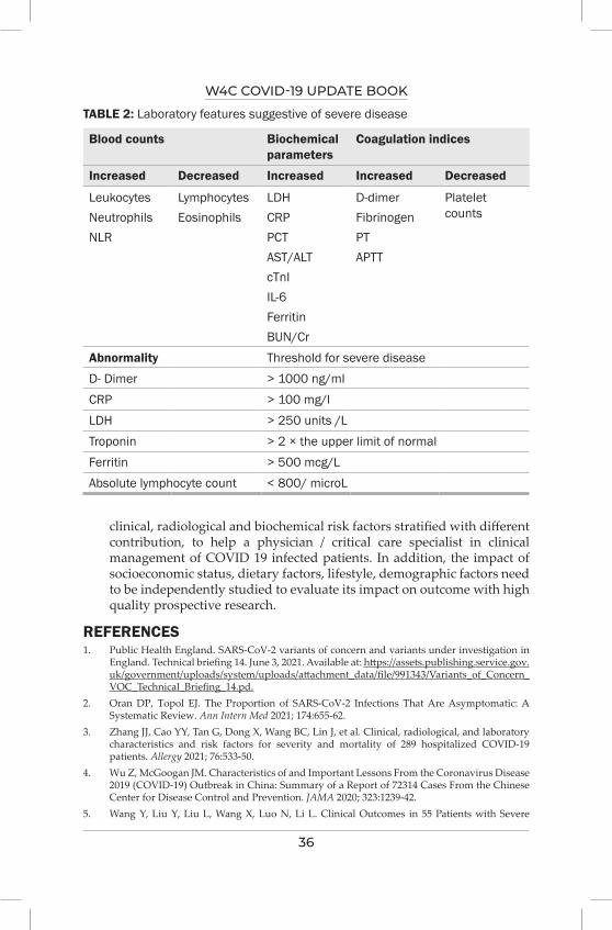
W4C COVID-19 UPDATE BOOK
36
TABLE 2: Laboratory features suggestive of severe disease
Blood counts Biochemical parameters
Coagulation indices
Increased Decreased Increased Increased Decreased
LeukocytesNeutrophilsNLR
LymphocytesEosinophils
LDHCRPPCTAST/ALTcTnIIL-6FerritinBUN/Cr
D-dimerFibrinogenPTAPTT
Platelet counts
Abnormality Threshold for severe diseaseD- Dimer > 1000 ng/ml CRP > 100 mg/l LDH > 250 units /L Troponin > 2 × the upper limit of normal Ferritin > 500 mcg/LAbsolute lymphocyte count < 800/ microL
clinical, radiological and biochemical risk factors stratified with different contribution, to help a physician / critical care specialist in clinical management of COVID 19 infected patients. In addition, the impact of socioeconomic status, dietary factors, lifestyle, demographic factors need to be independently studied to evaluate its impact on outcome with high quality prospective research.
REFERENCES1. Public Health England. SARS-CoV-2 variants of concern and variants under investigation in
England. Technical briefing 14. June 3, 2021. Available at: https://assets.publishing.service.gov.uk/government/uploads/system/uploads/attachment_data/file/991343/Variants_of_Concern_VOC_Technical_Briefing_14.pd.
2. Oran DP, Topol EJ. The Proportion of SARS-CoV-2 Infections That Are Asymptomatic: A Systematic Review. Ann Intern Med 2021; 174:655-62.
3. Zhang JJ, Cao YY, Tan G, Dong X, Wang BC, Lin J, et al. Clinical, radiological, and laboratory characteristics and risk factors for severity and mortality of 289 hospitalized COVID-19 patients. Allergy 2021; 76:533-50.
4. Wu Z, McGoogan JM. Characteristics of and Important Lessons From the Coronavirus Disease 2019 (COVID-19) Outbreak in China: Summary of a Report of 72314 Cases From the Chinese Center for Disease Control and Prevention. JAMA 2020; 323:1239-42.
5. Wang Y, Liu Y, Liu L, Wang X, Luo N, Li L. Clinical Outcomes in 55 Patients with Severe
W4C COVID-19 UPDATE BOOK
37
Acute Respiratory Syndrome Coronavirus 2 Who Were Asymptomatic at Hospital Admission in Shenzhen, China. J Infect Dis 2020; 221:1770-4.
6. Novosad P, Jain R, Campion A, Asher S. COVID-19 mortality effects of underlying health conditions in India: a modelling study. BMJ Open 2020; 10:e043165.
7. Wynants L, Van Calster B, Collins GS, Riley RD, Heinze G, Schuit E, et al. Prediction models for diagnosis and prognosis of covid-19: systematic review and critical appraisal. BMJ 2020; 369:m1328.
8. Mughal MS, Kaur IP, Jaffery AR, Dalmacion DL, Wang C, Koyoda S, et al. COVID-19 patients in a tertiary US hospital: Assessment of clinical course and predictors of the disease severity. Respir Med 2020; 172:106130.
9. Soni SL, Kajal K, Yaddanapudi LN, Malhotra P, Puri GD, Bhalla A, et al. Demographic & clinical profile of patients with COVID-19 at a tertiary care hospital in north India. Indian J Med Res 2021; 153:115-25.
10. Mohan A, Tiwari P, Bhatnagar S, Patel A, Maurya A, Dar L, et al. Clinico-demographic profile & hospital outcomes of COVID-19 patients admitted at a tertiary care centre in north India. Indian J Med Res 2020; 152:61-9.
11. Price-Haywood EG, Burton J, Fort D, Seoane L. Hospitalization and Mortality among Black Patients and White Patients with Covid-19. N Engl J Med 2020; 382:2534-43.
12. Ioannou GN, Locke E, Green P, Berry K, O’Hare AM, Shah JA, et al. Risk Factors for Hospitalization, Mechanical Ventilation, or Death Among 10131 US Veterans With SARS-CoV-2 Infection. JAMA Netw Open 2020; 3:e2022310.
13. Guan WJ, Liang WH, Zhao Y, Liang HR, Chen ZS, Li YM, et al. Comorbidity and its impact on 1590 patients with COVID-19 in China: a nationwide analysis. Eur Respir J 2020; 55.
14. Stokes EK, Zambrano LD, Anderson KN, Marder EP, Raz KM, El Burai Felix S, et al. Coronavirus Disease 2019 Case Surveillance - United States, January 22-May 30, 2020. MMWR Morb Mortal Wkly Rep 2020; 69:759-65.
15. Onder G, Rezza G, Brusaferro S. Case-Fatality Rate and Characteristics of Patients Dying in Relation to COVID-19 in Italy. JAMA 2020; 323:1775-6.
16. McMichael TM, Currie DW, Clark S, Pogosjans S, Kay M, Schwartz NG, et al. Epidemiology of Covid-19 in a Long-Term Care Facility in King County, Washington. N Engl J Med 2020; 382:2005-11.
17. Wang D, Hu B, Hu C, Zhu F, Liu X, Zhang J, et al. Clinical Characteristics of 138 Hospitalized Patients With 2019 Novel Coronavirus-Infected Pneumonia in Wuhan, China. JAMA 2020; 323:1061-9.
18. Li X, Xu S, Yu M, Wang K, Tao Y, Zhou Y, et al. Risk factors for severity and mortality in adult COVID-19 inpatients in Wuhan. J Allergy Clin Immunol 2020; 146:110-8.
19. Huang S, Wang J, Liu F, Liu J, Cao G, Yang C, et al. COVID-19 patients with hypertension have more severe disease: a multicenter retrospective observational study. Hypertension Research 2020; 43:824-31.
20. Guo L, Shi Z, Zhang Y, et al. Comorbid diabetes and the risk of disease severity or death among 8807 COVID-19 patients in China: A meta-analysis. Diabetes Res Clin Pract 2020; 166:108346.
21. Zhang Y, Li H, Zhang J, Cao Y, Zhao X, Yu N, et al. The clinical characteristics and outcomes of patients with diabetes and secondary hyperglycaemia with coronavirus disease 2019: A single-centre, retrospective, observational study in Wuhan. Diabetes Obes Metab 2020; 22:1443-54.
22. Russell B, Moss C, Papa S, Irshad S, Ross P, Spicer J, et al. Factors Affecting COVID-19 Outcomes in Cancer Patients: A First Report From Guy’s Cancer Center in London. Front Oncol 2020; 10:1279.
23. Fresan U, Guevara M, Elia F, Albeniz E, Burgui C, Castilla J, et al. Independent Role of Severe Obesity as a Risk Factor for COVID-19 Hospitalization: A Spanish Population-Based Cohort Study. Obesity (Silver Spring) 2021; 29:29-37.
W4C COVID-19 UPDATE BOOK
38
24. Radzikowska U, Ding M, Tan G, Zhakparov D, Peng Y, Wawrzyniak P, et al. Distribution of ACE2, CD147, CD26, and other SARS-CoV-2 associated molecules in tissues and immune cells in health and in asthma, COPD, obesity, hypertension, and COVID-19 risk factors. Allergy 2020; 75:2829-45.
25. Attaway AA, Zein J, Hatipoglu US. SARS-CoV-2 infection in the COPD population is associated with increased healthcare utilization: An analysis of Cleveland clinic’s COVID-19 registry. EClinicalMedicine 2020; 26:100515.
26. Elliott J, Bodinier B, Whitaker M, Delpierre C, Vermeulen R, Tzoulaki I, et al. COVID-19 mortality in the UK Biobank cohort: revisiting and evaluating risk factors. Eur J Epidemiol 2021; 36:299-309.
27. Esposito AJ, Menon AA, Ghosh AJ, Putman RK, Fredenburgh LE, El-Chemaly SY, et al. Increased Odds of Death for Patients with Interstitial Lung Disease and COVID-19: A Case-Control Study. Am J Respir Crit Care Med 2020; 202:1710-3.
28. Zhang C, Shi L, Wang FS. Liver injury in COVID-19: management and challenges. Lancet Gastroenterol Hepatol 2020; 5:428-30.
29. Singh S, Khan A. Clinical Characteristics and Outcomes of Coronavirus Disease 2019 Among Patients With Preexisting Liver Disease in the United States: A Multicenter Research Network Study. Gastroenterology 2020; 159:768-71 e3.
30. Liang W, Guan W, Chen R, Wang W, Li J, Xu K, et al. Cancer patients in SARS-CoV-2 infection: a nationwide analysis in China. Lancet Oncol 2020; 21:335-7.
31. Gagandeep Brar, Laura C. Pinheiro, Michael Shusterman, Brandon Swed, Evgeniya Reshetnyak, Orysya Soroka, Frank Chen, Samuel Yamshon, John Vaughn, Peter Martin, Doru Paul, Manuel Hidalgo, and Manish A. Shah. Journal of Clinical Oncology 2020; 33:3914-3924.
32. Luo J, Rizvi H, Egger JV, Preeshagul IR, Wolchok JD, Hellmann MD. Impact of PD-1 Blockade on Severity of COVID-19 in Patients with Lung Cancers. Cancer Discov 2020; 10:1121-8.
33. Mirzaei H, McFarland W, Karamouzian M, Sharifi H. COVID-19 Among People Living with HIV: A Systematic Review. AIDS Behav 2021; 25:85-92.
34. Nayak AH, Kapote DS, Fonseca M, Chavan N, Mayekar R, Sarmalkar M, et al. Impact of the Coronavirus Infection in Pregnancy: A Preliminary Study of 141 Patients. J Obstet Gynaecol India 2020; 70:256-61.
35. Hogan CA, Stevens BA, Sahoo MK, Huang C, Garamani N, Gombar S, et al. High Frequency of SARS-CoV-2 RNAemia and Association With Severe Disease. Clin Infect Dis 2021; 72:e291-e5.
36. Pujadas E, Chaudhry F, McBride R, Richter F, Zhao S, Wajnberg A, et al. SARS-CoV-2 viral load predicts COVID-19 mortality. Lancet Respir Med 2020; 8:e70.
37. Liu Y, Yan LM, Wan L, Xiang TX, Le A, Liu JM, et al. Viral dynamics in mild and severe cases of COVID-19. Lancet Infect Dis 2020; 20:656-7.
38. Blot M, Jacquier M, Manoha C, Piroth L, Charles PE, Pneumochondrie study g. Alveolar SARS-CoV-2 Viral Load Is Tightly Correlated With Severity in COVID-19 ARDS. Clin Infect Dis 2021; 72:e446-e7.
39. Huang G, Kovalic AJ, Graber CJ. Prognostic Value of Leukocytosis and Lymphopenia for Coronavirus Disease Severity. Emerg Infect Dis 2020; 26:1839-41.
40. Chen R, Sang L, Jiang M, Yang Z, Jia N, Fu W, et al. Longitudinal hematologic and immunologic variations associated with the progression of COVID-19 patients in China. J Allergy Clin Immunol 2020; 146:89-100.
41. Wang F, Nie J, Wang H, Zhao Q, Xiong Y, Deng L, et al. Characteristics of Peripheral Lymphocyte Subset Alteration in COVID-19 Pneumonia. J Infect Dis 2020; 221:1762-9.
42. Xu P, Zhou Q, Xu J. Mechanism of thrombocytopenia in COVID-19 patients. Ann Hematol 2020; 99:1205-8.
43. Lala A, Johnson KW, Januzzi JL, Russak AJ, Paranjpe I, Richter F, et al. Prevalence and Impact of Myocardial Injury in Patients Hospitalized With COVID-19 Infection. J Am Coll Cardiol 2020; 76:533-46.

W4C COVID-19 UPDATE BOOK
39
44. Malik P, Patel U, Mehta D, Patel N, Kelkar R, Akrmah M, et al. Biomarkers and outcomes of COVID-19 hospitalisations: systematic review and meta-analysis. BMJ Evid Based Med 2021; 26:107-8.
45. Aziz M, Fatima R, Assaly R. Elevated interleukin-6 and severe COVID-19: A meta-analysis. J Med Virol 2020; 92:2283-5.
46. Bao J, Li C, Zhang K, Kang H, Chen W, Gu B. Comparative analysis of laboratory indexes of severe and non-severe patients infected with COVID-19. Clin Chim Acta 2020; 509:180-94.
47. Gandini O, Criniti A, Ballesio L, Giglio S, Galardo G, Gianni W, et al. Serum Ferritin is an independent risk factor for Acute Respiratory Distress Syndrome in COVID-19. J Infect 2020; 81:979-97.
48. Hope MD, Raptis CA, Henry TS. Chest Computed Tomography for Detection of Coronavirus Disease 2019 (COVID-19): Don’t Rush the Science. Ann Intern Med 2020; 173:147-8.
49. Zhang K, Liu X, Shen J, Li Z, Sang Y, Wu X, et al. Clinically Applicable AI System for Accurate Diagnosis, Quantitative Measurements, and Prognosis of COVID-19 Pneumonia Using Computed Tomography. Cell 2020; 181:1423-33 e11.
40
Types of Lung involvement in COviD-19
INTRODUCTIONInfection with SARS-COv-2 can affect many organs of the body, lungs being the most common. In COVID-19, pneumonia is the most frequent cause for hospital admission, and hypoxia is the most common sign on arrival to the hospital during this pandemic. The infection may progress and lead to Acute Respiratory Distress Syndrome (ARDS), a form of lung injury resulting in poor gas exchange at the alveolar level. The clinical spectrum of ARDS varies from a mild disease requiring no Oxygen supplementation to a severe disease requiring respiratory support in any form.
Various investigators proposed different phenotypes of COVID-19 ARDS (CARDS) based on the clinical, radiology, outcome, the severity of inflammation and even response to fluid management strategies.1,2,3 In CARDS, the observation of disproportionate hypoxia (popularly known as ‘Happy hypoxia’) in context to respiratory symptoms (rate, level of distress) may suggest different pathophysiological mechanisms as the ‘traditional ARDS’ almost always presents with symptomatic hypoxia. The variation in respiratory mechanics, i.e. hypoxia with near-normal respiratory system compliance, response to a prone position, and PaCO2 level, were observed in COVID-19 pneumonia. These findings probably suggest different phenotypic characteristics of the disease.
PATHOPHYSIOLOGY OF CARDSThe SARS-CoV-2 infection progresses through four phases:4
Phase 1: Invasion into the cell and viral replication
The virus enters the cell via ACE-2 receptors situated on the goblet and ciliated cells in the nose, mouth and tongue.
Phase 2: Immune response
The lymphocytes produce IgM-type antibodies first and later IgG type. It may be dysregulated in patients of COVID-19.
Ritesh Shah
4.
W4C COVID-19 UPDATE BOOK
41
Phase 3: Inflammation of the lung causing Pneumonia
Destruction of pneumocytes causes a reduction in surfactant in the alveoli, which ultimately invites infiltration of neutrophils and macrophages. This leads to the release of pro-inflammatory cytokines causing vessel injury, capillary leakage and resultant oedema. This oedema compresses alveoli, and due to lack of surfactant, it leads to the collapse of the alveoli, causing hypoxia.
Phase 4: CARDS
The formation of microthrombi in small blood vessels in the lungs and fibrin clots in alveoli significantly affect gas exchange, causing hypoxia. The ‘unchecked’ ACE-1 because of the downregulation of ACE-2 (the epithelial-endothelial crosstalk hypothesis by Jain et al.)5 further results in vessel injury (endothelial damage), leaky vessels, and disrupted vessels pulmonary vasoregulation, which eventually worsen hypoxia and cause multi-organ failure.
The tidal strains induced by either patient (P-SILI: Patient Self-Inflicted Lung Injury) or ventilator (VILI) worsen the overall progression of the disease.
Overall, The airspace VILI stresses on vessels, the progression of the viral disease inciting inflammation and oedema promoting thrombogenesis, dysregulated cytokine release, right ventricular overload and dysfunction result in systemic organ dysfunction and its consequences.
PROPOSED PHENOTYPES DURING COVID-19 PANDEMICGattinoni et al6 hypothesized that the different COVID-19 patterns depend on the interaction between three factors: (a) the severity of infection, the host response, physiological reserve and comorbidities; (b) the ventilatory responsiveness to hypoxaemia; (c) the time between onset of the disease and arrival at the hospital.
They hypothesized two different phenotypes of COVID-19 pneumonia (Figue 1):
1. ‘L’ type (Low Elastance)
a. High or near-normal compliance
b. Low ventilation-to-perfusion (V/Q) ratio because of loss of hypoxic pulmonary vasoconstriction and loss of regulation of perfusion
c. Low lung weight because alveoli are still not fluid-filled in early disease
d. Low lung recruitability because of less number of collapsed alveoli
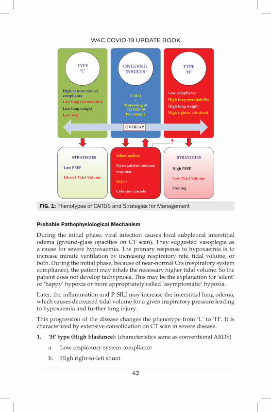
W4C COVID-19 UPDATE BOOK
42
Probable Pathophysiological Mechanism
During the initial phase, viral infection causes local subpleural interstitial odema (ground-glass opacities on CT scan). They suggested vasoplegia as a cause for severe hypoxaemia. The primary response to hypoxaemia is to increase minute ventilation by increasing respiratory rate, tidal volume, or both. During the initial phase, because of near-normal Crs (respiratory system compliance), the patient may inhale the necessary higher tidal volume. So the patient does not develop tachypnoea. This may be the explanation for ‘silent’ or ‘happy’ hypoxia or more appropriately called ‘asymptomatic’ hypoxia.
Later, the inflammation and P-SILI may increase the interstitial lung odema, which causes decreased tidal volume for a given inspiratory pressure leading to hypoxaemia and further lung injury.
This progression of the disease changes the phenotype from ‘L’ to ‘H’. It is characterized by extensive consolidation on CT scan in severe disease.
1. ‘H’ type (High Elastance): (characteristics same as conventional ARDS)
a. Low respiratory system compliance
b. High right-to-left shunt
FIG. 1: Phenotypes of CARDS and Strategies for Management
TYPE‘L’
ONGOINGINSULTS
TYPE‘H’
High or near normalcompliance P-SILI
+Worsening of
COVID-19Pneumonia
STRATEGIES STRATEGIES
High PEEP
Proning
Low Tidal Volume
Low PEEP
Liberal Tidal Volume
Inflammation
Dysregulated immune
Cytokine cascade
response
Sepsis
Low compliance
High lung recruitability
High lung weight
High right to left shunt
Low lung recruitability
Low lung weight
Low V/Q
OVERLAP
W4C COVID-19 UPDATE BOOK
43
c. High lung weight secondary to increased odema
d. High lung recruitability as more recruitable collapsed alveoli are available
Considering the above phenotypes, Marini and Gattinoni proposed different management strategies or principles:7
a. In spontaneously breathing patients, the goal is to prevent P-SILI while maintaining the gas exchange. So the efforts should be to decrease vigorous breathing by giving supplemental Oxygen, HFNC, NIV or awake prone positioning.
b. In patients with Type ‘L’, use lower PEEP (<10cmH2O) and liberal tidal volume (7-9ml/kg). Higher PEEP or inverse ratio ventilation may redirect blood flow away from the open alveoli without benefiting gas exchange. Furthermore, PEEP can produce haemodynamic instability. Proning should be done in refractory cases only. Early intubation needs to be considered
c. In patients with Type ‘H’, use higher PEEP (≤ 15 cmH2O), lower tidal volume (5-7 ml/kg) and routine proning
d. During weaning, the transition to spontaneous breathing should be slower and cautious to prevent increased oxygen demand, lung odema and P-SILI.
e. Efforts should be made to minimize transpulmonary and vascular stresses and reduce oxygen demand
There is considerable overlap between these two phenotypes, and it is time-related. Bos et al8 argued against this phenotyping by stating that the ‘premature’ phenotyping exacerbates our inherent susceptibility to cognitive biases and it requires robust data in a clinical setting in a consistent manner. So it should not itself change clinical practice but instead inform prospective, phenotype-aware trials.
Cherian et al9 concluded that a tailored phenotypic approach to management, guided by pathophysiology, would be more appropriate than a syndromic approach.
Panwar et al10 (on behalf of LUNG SAFE investigators and the ESICM trial group) undertook a secondary analysis (from the LUNG SAFE study) of patients with ARDS based on Crs, degree of hypoxemia and related outcomes to confirm phenotypic presence before COVID-19 pandemic. They grouped patients based on Crs (<40 or type ‘H’, 40-50 or intermediate and ≥ 50ml/cm of H2O or type ‘L’). They concluded that approximately one in eight patients had preserved Crs (type ‘L’), PaO2/FiO2 and Crs were dissociated, Lower Crs was independently associated with higher mortality, and the Crs-mortality
W4C COVID-19 UPDATE BOOK
44
lacked a clear transition threshold under 100 ml/cm H2O suggesting that such set thresholds are quite arbitrary.
In their study, Grieco et al11 concluded that there is no major physiological difference between CARDS and ARDS due to other aetiology. It shows the same characteristics like heterogeneity in respiratory mechanics and variable recruitability aeration related to the degree of hypoxemia.in respiratory mechanics, variable recruitability , aeration related to degree of hypoxemia.
Though there are different opinions regarding the phenotypes (‘L’ and ‘H’) and types of lung involvements in CARDS, different respiratory mechanics may be present at different times in the same patient. The most important thing is to realize the severity of the ongoing situation and have a tailor-made therapy, intervention and management plan.
CONCLUSION AND PRACTICE POINTS1. One must understand the heterogeneous nature of the disease CARDS
2. Timely supplemental Oxygen and respiratory support are the key to prevent mortality
3. More aggressive modalities (including higher PEEP, lower tidal volume, proning) are needed as Crs deteriorates. In the early phase (Type ‘L’), one can use low PEEP with liberal tidal volume with caution
4. An aggressive tailor-made treatment keeping ‘less is more’ principle in mind usually turns out an optimal strategy
5. Ultimately, what matters is the excellent bedside quality critical care!
REFERENCES1. Shankar-Hari M, McAuley DF. Acute respiratory distress syndrome phenotypes and
identifying treatable traits: the dawn of personalized medicine for ARDS. Am J Respir Crit Care Med 2017; 195:280–281.
2. Famous KR, Delucchi K, Ware LB, Kangelaris KN, Liu KD, Thompson BT, et al.; ARDS Network. Acute respiratory distress syndrome subphenotypes respond differently to randomized fluid management strategy. Am J Respir Crit Care Med 2017; 195:331–338.
3. Viswan A, Ghosh P, Gupta D, Azim A, Sinha N. Distinct metabolic endotype mirroring acute respiratory distress syndrome (ARDS) subphenotype and its heterogeneous biology. Sci Rep 2019;9:2108.
4. Vinod Nikhra; Respiratory Manifestations in COVID-19 and ‘Long Covid’: The Morbidity, Complications and Sequelae; Biomedical journal of Scientific and Technical Research, June 2021
5. Amit Jain and D.John Doyle: Stages or phenotypes? A critical look at COVID-19 pathophysiology; Intensive Care Med 2020; 46:1494-1495
6. Luciano Gattinoni, Davide Chiumello, Pietro Caironi, Mattia Busana, Federica Romitti, Luca Brazzi and Luigi Camporota; COVID-19 pneumonia: different respiratory treatments for different phenotypes? Intensive Care Med. 2020; 46:1099-1102
7. Marini JJ, Gattinoni L. Management of COVID-19 respiratory distress. JAMA 2020; 323:2329–2330

W4C COVID-19 UPDATE BOOK
45
8. Bos LDJ, Sinha P, Dickson RP. The perils of premature phenotyping in COVID-19: a call for caution. Eur Respir J 2020; 56: 2001768
9. Cherian R, Chandra B, Tung ML, et al. COVID-19 conundrum: clinical phenotyping based on pathophysiology as a promising approach to guide therapy in a novel illness Eur Respir J 2020; 56: 2002135
10. Rakshit Panwar, Fabiana Madotto, John G. Laffey, and Frank M. P. van Haren; on behalf of the LUNG SAFE Investigators and the ESICM Trials Group;Compliance Phenotypes in Early Acute Respiratory Distress Syndrome before the COVID-19 Pandemic: American Journal of Respiratory and Critical Care Medicine Volume 202 Number 9 | November 1 2020
11. Domenico Luca Grieco, Filippo Bongiovanni, Lu Chen, Luca S. Menga, Salvatore Lucio Cutuli, Gabriele Pintaudi et al: Respiratory physiology of COVID-19- induced respiratory failure compared to ARDS of other etiologies: Critical Care 2020) 24:529
46
Effects on Other Organ Systems - COviD-19 -
Neurological involvement
INTRODUCTIONCOVID 19 infection is a serious condition caused by the novel coronavirus termed as “severe acute respiratory syndrome coronavirus-2” (SARS-CoV-2). Ever since the first case that was detected in Wuhan, China in December 2019, it has engulfed the world over and was declared a pandemic by WHO in March 2020.1 With around 210 million people affected worldwide and approximately 4.4 million deaths and still counting, this once-in-a-century pandemic has been a major challenge for healthcare all over.
Initially, what was assumed to be primarily a respiratory function involvement, it is now clear that it can affect any system of the body and involvement of the nervous system is common.
THE MANIFESTATIONSNeurological manifestations can involve both the central as well as peripheral nervous systems and can be classified as common or uncommon presentations. Starting with the common and mild symptoms like headache, myalgia, loss of taste & smell, which are self-reported, the spectra could extend to clinically captured serious and severe neurological and neuropsychiatric manifestations. The list includes both common and rarely found disorders of the central & peripheral nervous system like impaired consciousness, delirium, stroke, seizure, meningitis, encephalitis, necrotizing encephalopathy, Guillain-Barre Syndrome, acute demyelinating encephalomyelitis, isolated cranial neuropathies, critical illness neuropathy and critical illness myopathy. Chou et al, reported that the presence of a neurological manifestation increased the risk of hospital death, and within the group, clinically captured symptoms had a higher risk of death as compared to the self-reported symptoms.2
INCIDENCEMost of the information regarding neuropsychiatric manifestation comes from
Asif Ahmed, Sujeet Ashok Joshi
5.
W4C COVID-19 UPDATE BOOK
47
case reports or cohort studies. With passing time and more reports flowing in, an increasing trend is seen in the patients having neurological symptoms & syndromes. Chou et al in a large cohort of 3743 patients with Covid19 infection, 82% of patients suffering from neurological manifestations. 37% of patients had symptoms of headache, while 26% of patients presented with loss of taste & smell. Acute encephalopathy at 49% accounted for the majority of the manifestations of the admitted patients. Another 17% had coma, 6% had a stroke, while the prevalence of meningitis or encephalitis was lesser (0.5%).3
RISK FACTORSNeurological complications in COVID-19 patients more common in patients having co-morbidities like hypertension, diabetes mellitus, obesity, hypercholesterolemia and elderly age groups.4 The presence of a pre-existing neurological disorder was also found to be associated with an increased risk of developing neurological syndrome in COVID-19 patients, as per a recent and large cohort. An important finding was that the presence of neurological manifestations & syndrome increased the risk of hospital mortality by five folds in these patients, apart from the length of stay & morbidity. Higher mortality was also seen in the elderly population, males & patients with high body mass index.
PATHOPHYSIOLOGYBoth direct and indirect causes have been attributed to the development of neurological complications & manifestations in patients suffering from Covid19 infection. The SARS COV-2 virus, for gaining entry into the cells, utilises the ACE-2 receptors, which apart from being present in lung tissues and epithelial cells, are also seen in the brain and its cerebral capillaries. The spike proteins are supposed to be responsible for this new affinity of the virus to these receptors. It has been suggested that the virus spreads through the hematogenous route and crosses the blood-brain barrier. Some also believe that the virus can enter trans-neurally through the cribriform plate. Autopsies have also revealed the presence of viral particles in the endothelial cells of the brain. Another possible cause is the alteration in blood pressure leading to cerebrovascular complications and this again is related to the downregulation of ACE-2 receptors, which is responsible for BP control.
Coagulopathy associated with COVID-19 is another risk factor that can lead to stroke and other cardiovascular events. Organ dysfunction with uremia & deranged liver enzymes can also contribute to encephalopathy. Cytokine storm with its accompaniment of vascular permeability, coagulopathy and organ dysfunction can also be a causative factor, just as it is for all COVID-19 complications. Post-Infection autoimmune reactions can also have an impact on neural cells leading to the permanent problem.5,6
W4C COVID-19 UPDATE BOOK
48
INVESTIGATIONSIn addition to the routine diagnostics performed for a COVID19 patient, efforts should be made to identify the peripheral and central nervous system manifestations, which normally gets overshadowed by pulmonary complications and related care. It includes clinical assessment for consciousness levels, delirium, seizures and focal neurological signs. The test modalities used are Cerebro-spinal fluid analysis and few authors recommend performing RT-PCR test on CSF samples in suspected cases of encephalitis, delirium & polyneuropathy.7 EEG should be performed in cases of delirium, failed a wake-up test and status epilepticus. A focal neurological symptom should warrant cerebral & spinal imaging like CT scan or MRI.
MANAGEMENT OF SPECIFIC CAUSESNeurological management must be dealt with as per the specific manifestations and should include the basic neuro-critical care principles to maintain the normal milieu.
Some of the disorders seen are as follows:
• Loss of smell & taste: Anosmia and hyposmia could be the first symptoms of COVID-19 infection in healthy individuals. Taste disorders are also common. These symptoms are self-limiting, but if they persist beyond four weeks, then re-evaluation is required.
• Neuromotor disease: Raised Creatinine kinase levels along with muscle pain and fatigue are commonly seen in COVID-19 patients. ICU acquired weakness is also seen in patients being treated in Intensive care units and is dependent on the severity & stay in ICU. Myasthenia Gravis has also been reported in few cases. The investigations should include clinical examination, EMG & CSF study. In patients on Immunotherapy, therapy should not be discontinued, considering the risk & benefit.
• Seizures: Seizures could happen as primary events or less commonly in patients with known seizure disorders. COVID-19 patients develop seizures because of hypoxemia, severe inflammation, metabolic derangement, electrolyte imbalance, meningoencephalitis and organ failure. EEG is helpful to identify seizures, while imaging can be used for detecting the lesions like stroke or encephalitis. CSF study can confirm the diagnosis of meningoencephalitis. Treatment of seizures should be as per standard guidelines considering the drug interactions off the medications used.
• Guillain-Barre Syndrome (GBS): GBS has been described in several cases in COVID 19 infection and the Miller Fischer variant has also been reported. Symptoms can range from mild sensory deficits to severe quadriparesis and could manifest within 5-10 days to weeks after the infection. Nerve
W4C COVID-19 UPDATE BOOK
49
roots are typically involved CSF study reveals elevated protein with a normal cell count (albumin cytologic dissociation). Treatment is not different from the usual therapy of intravenous immunoglobulin (IVIG) or plasmapheresis. Data does not support any association between the severity of COVID-19 disease and GBS.8
• Acute Disseminated EncephaloMyelitis (ADEM): There are few reports of ADEM and it was seen in middle and old age group, with a varied presentation. CSF study can reveal pleocytosis, while MRI should be done to check lesions in the basal ganglia. High dose steroids (1- 2 grams per day) remains the mainstay of treatment, while IVIG is used for the patients who do not respond to steroids.9
• Meningo-encephalitis: With very few cases reported, meningoencephalitis could be caused by either direct infection or post-infection autoimmune mediated. Clinical signs could be variable, including cognitive disturbances & seizures. There are no specific EEG or MRI findings. CSF study could show lymphocytic pleocytosis. If symptoms persist and a definitive pathogen is not found, then steroid therapy for three to five days is also advocated.
• Encephalopathy: The causes for encephalopathy are multiple and could be triggered by COVID-19 infection i.e. direct infection, sepsis, metabolic disturbances, multi-organ failure and cytokine storm. Signs and symptoms spread over a wide range (disturbance of consciousness to seizures and focal neurological deficits). CSF study, EEG and imaging are the diagnostic modalities used. Treatment is purely symptomatic.
• Stroke: COVID-19 is associated with an increased risk of cardiovascular diseases and it includes both ischemic stroke and hemorrhagic stroke. Ischemic stroke was seen in 1.6% to 5% of admitted COVID-19 patients, however, the causal relationship has been questioned. The incidence of hemorrhagic stroke is less common.
• However, the presence of a past history of stroke was associated with increased severity and mortality. Diagnosis & treatment should be as per standard guidelines & protocols and not different for COVID-19 patients. Hospitals should continue with the focused care of stroke patients, which had got affected during the pandemic earlier.10
SUMMARYThough COVID-19 presents as a pulmonary manifestation most of the time, emerging evidence from case series and cohorts highlight that neurological and neuropsychiatric manifestations are also common, and the spectra are wide. The presence of a neurological manifestation or syndrome can lead to increased morbidity & mortality. Hence it should not be neglected or overlooked. Increased awareness of the disease process, a focused approach
W4C COVID-19 UPDATE BOOK
50
towards early diagnosis and application of general principles of neurocritical care can be helpful in identifying and managing these cases.
REFERENCES1. World Health Organization. Rolling updates on coronavirus disease (COVID-19). Updated 24
April 2020. 2. Ling Mao , Huijuan Jin , Mengdie Wang , Yu Hu , Shengcai Chen , Quanwei He ,et al Neurologic
manifestations of hospitalized patients with coronavirus disease 2019 in Wuhan, China. JAMA Neurol 1127,2020
3. Sherry H, Y.Chou, Ettore Beghi, Raimund Helbok, Elena Moro, Joshua Sampson, PhD Valeria Altamirano, MS et al Global Incidence of Neurological Manifestations Among Patients Hospitalized With COVID-19—A Report for the GCS- NeuroCOVID Consortium and the ENERGY Consortium. JAMA Netw Open 2021; 4:e2112131.
4. Alberto Romagnolo, Roberta Balestrino, Gabriele Imbalzano, Giovannino Ciccone, Franco Riccardini, Carlo Alberto Artusi, et al Neurological comorbidity and severity of COVID-19, J Neurol 2021; 268:762–769.
5. Meng-Yuan Li , Lin Li , Yue Zhang , Xiao-Sheng Wang ; Expression of the SARS-CoV-2 cell receptor gene ACE2 in a wide variety of human tissues. Infect Dis Poverty 2020; 9:45.
6. Zubair AS, McAlpine LS, Gardin T, Farhadian S, Kuruvilla DE, Spudich S. et al Neuropathogenesis and neurologic manifestations of the coronaviruses in the age of coronavirus disease 2019: a review. JAMA Neurol 2020.
7. Ariane Lewis , Jennifer Frontera , Dimitris G Placantonakis , Jennifer Lighter , Steven Galetta , Laura Balcer et al, Cerebrospinal fluid in COVID-19 - A systematic review of the literature – Journal of the Neurological Sciences 2021; 421:117316.
8. Gupta, A.; Paliwal, V.K.; Garg, R.K. et al. Is COVID-19-related Guillain-Barre syndrome different, Brain Behav. Immun 2020; 87:177–178.
9. Peter Berlit , Julian Bösel , Georg Gahn , Stefan Isenmann , Sven G Meuth , Christian H Nolte ,et al “Neurological manifestations of COVID-19” - guideline of the German society of neurology. Neurol Res Pract 2020; 2:51.
10. Jing Zhao, Hang Li, David Kung, Marc Fisher, Ying Shen, Renyu Liu; Impact of the COVID-19 epidemic on stroke care and potential solutions. Stroke (2020).
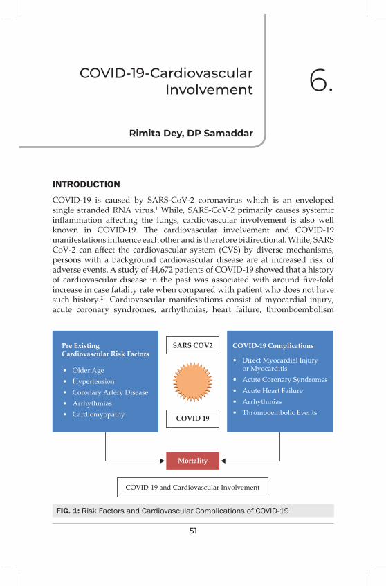
51
COviD-19-Cardiovascular involvement 6.
INTRODUCTIONCOVID-19 is caused by SARS-CoV-2 coronavirus which is an enveloped single stranded RNA virus.1 While, SARS-CoV-2 primarily causes systemic inflammation affecting the lungs, cardiovascular involvement is also well known in COVID-19. The cardiovascular involvement and COVID-19 manifestations influence each other and is therefore bidirectional. While, SARS CoV-2 can affect the cardiovascular system (CVS) by diverse mechanisms, persons with a background cardiovascular disease are at increased risk of adverse events. A study of 44,672 patients of COVID-19 showed that a history of cardiovascular disease in the past was associated with around five-fold increase in case fatality rate when compared with patient who does not have such history.2 Cardiovascular manifestations consist of myocardial injury, acute coronary syndromes, arrhythmias, heart failure, thromboembolism
Rimita Dey, DP Samaddar
FIG. 1: Risk Factors and Cardiovascular Complications of COVID-19
Pre ExistingCardiovascular Risk Factors
SARS COV2
Mortality
COVID-19 and Cardiovascular Involvement
COVID 19
• Older Age• Hypertension• Coronary Artery Disease• Arrhythmias• Cardiomyopathy
COVID-19 Complications
• Direct Myocardial Injury or Myocarditis• Acute Coronary Syndromes• Acute Heart Failure• Arrhythmias• Thromboembolic Events
W4C COVID-19 UPDATE BOOK
52
and as a part of multisystem inflammatory syndrome in children and adults (MIS-C and MIS-A). Although not a direct effect of the virus per se, we need to be aware of CVS involvement due to diverse drug interactions used in treating COVID-19 disease.
SARS-COV-2 AND ACE2 INTERACTIONAngiotensin-converting enzyme 2 (ACE2) has been proposed to be the site of entry of SARS-CoV-2 [3]. Viral S protein priming is done by the trans membrane serine protease (TMPSS2) which helps it to bind to ACE2 receptors. ACE2 is found on the surface of lung epithelial cells and enterocytes of small intestine. ACE2 breaks down Angiotensin II which is pro inflammatory. Inhibition of ACE2 is proposed as an important factor in systemic inflammation with cytokinin release resulting in acute respiratory distress syndrome (ARDS) and multiorgan dysfuction. After entry into the human host the virus predominantly affects the lungs and causes systemic inflammation. The various ways in which the CVS may be affected are discussed below.
Myocardial injury and myocarditis
As evidenced by the rise in cardiac biomarkers and electrocardiographic abnormalities, acute myocardial injury was found in 7-20% of patients in various studies, associated with a significantly worse prognosis4 Cardiac Troponins T and I were the biomarkers commonly studied. A study found that the death rate in patients with elevated levels of cardiac troponin T was 37.5%, whereas, in patients with underlying cardiovascular comorbidities plus elevated levels of cardiac troponin T, it was 69.4%, which was almost double. There are autopsy reports that reveal mild inflammation and viral RNA in the hearts of patients with COVID-19.5 However whether these patients had myocarditis or whether these findings were because of systemic inflammation is unclear.
Acute coronary syndrome (ACS)
Like SARS and influenza, COVID-19 can be associated with ACS. Systemic inflammation can cause plaque rupture leading to ACS Activated macrophages secrete collagenases that degrade collagen, a major constituent of the fibrous cap of atherosclerotic plaque, causing plaque rupture. Other possible mechanisms are involved coronary vasospasm and formation of microthrombi owing to inflammation and cytokine storm. In a study of patients with COVID-19 and ST-elevation myocardial infarction, 17 out of 24 patients showed a culprit lesion in coronary angiography that required revascularization. However, 24 out of these 28 patients had yet not received a positive COVID -19 report at the time of angiography suggesting that COVID-19 may cause ACS without inflammation also.6
Arrhythmias and sudden cardiac death
Arrhythmias and sudden cardiac deaths are reported in COVID-19 patients.
W4C COVID-19 UPDATE BOOK
53
The proposed mechanisms include hypoxia, inflammatory stress, and abnormal metabolism. Though sinus tachycardia was the most frequent finding resulting from multiple causes like hypoperfusion, fever, hypoxia, anxiety etc., dysrhythmias associated with an elevation of cardiac troponins were more suggestive of myocardial injury, acute myocarditis, and ACS.7
Acute heart failure
Some patients of COVID-19 presented with acute heart failure. The incidence of heart failure was as high as 23% -25% in various studies with cardiomyopathy occurring in about 33 % of patients.8 Among those who presented with acute onset of heart failure, nearly half did not have a history of cardiovascular disease before. While the understanding of this entity is essential in managing patients with COVID-19 and avoiding overaggressive fluid management, particularly in ARDS where right heart failure is a genuine concern, it is not clear that whether heart failure is a new entity or an unmasking of previous cardiovascular disease in a vast majority of patients.8
Coagulation abnormalities and thromboembolism
Patients with COVID-19 have coagulation abnormalities which can cause thromboembolism. Patients have elevated D-dimer and reduced platelet count. Studies have demonstrated an association between elevated D dimer and severity of the disease. Levels of fibrinogen and factor VIII were also elevated. All of these predispose a patient of COVID-19 to hypercoagulability and contribute to the development of diverse cardiovascular manifestations of COVID-19. Venous and pulmonary thromboembolism are common complications of COVID-19 disease.9 Severe inflammation and endothelium damage induced by COVID-19 in combination with underlying co morbidities predispose them to this hypercoagulable state.
Cardiovascular involvement as a part of MIS-C/MIS-A
A new phenomenon caused by SARS CO V2 infection can lead to hyperinflammatory syndrome with features similar to Kawasaki disease, including coronary artery abnormalities. This was particularly common in children, especially adolescents with a fever, variable rash, conjunctivitis, peripheral edema, joint pain, and even heart failure with low ejection fraction. Most of them had been exposed to COVID-19 when they remained asymptomatic but were tested negative for COVID RT PCR on presentation. This phenomenon has been noticed in few adults also which is now being named MIS-A.10
Cardiovascular involvement due to drug-disease interaction
The role of ACE inhibitors and angiotensin II receptors blockers in disease progression or prevention remain largely unknown. Some of the antiviral drugs which are used in an attempt to treat COVID-19 are known to induce cardiotoxicity.
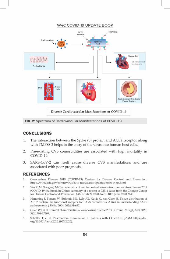
W4C COVID-19 UPDATE BOOK
54
CONCLUSIONS1. The interaction between the Spike (S) protein and ACE2 receptor along
with TMPSS 2 helps in the entry of the virus into human host cells.
2. Pre-existing CVS comorbidities are associated with high mortality in COVID-19.
3. SARS-CoV-2 can itself cause diverse CVS manifestations and are associated with poor prognosis.
REFERENCES1. Coronavirus Disease 2019 (COVID-19) Centers for Disease Control and Prevention.
https://www.cdc.gov/coronavirus/2019-ncov/cases-updates/cases-in-us.html2. Wu Z.,McGoogan J.M.Characteristics of and important lessons from coronavirus disease 2019
(COVID-19) outbreak in China: summary of a report of 72314 cases from the Chinese Center for Disease Control and Prevention. JAMA.Feb 24 2020 doi:10.1001/jama.2020.2648
3. Hamming I, Timens W, Bulthuis ML, Lely AT, Navis G, van Goor H. Tissue distribution of ACE2 protein, the functional receptor for SARS coronavirus. A first in understanding SARS pathogenesis. J Pathol 2004; 203:631-637.
4. Guan WJ, et at. Clinical characteristics of coronavirus disease 2019 in China. N Engl J Med 2020; 382:1708-17209.
5. Schaller T, et al. Postmortem examination of patients with COVID-19. JAMA https://doi.org/10.1001/jama.2020.8907(2020).
FIG. 2: Spectrum of Cardiovascular Manifestations of COVID-19
EpithelialCell
TMPRSS2ACE-2Receptor
S-glycoprotein
Arrhythmia
Myocarditis
Inflammation ofheart muscle
DVT
HF
Bloodclots
Acute Coronary Syndrome/Plaque Rupture
Diverse Cardiovascular Manifestations of COVID-19
SARS-CoV-2
W4C COVID-19 UPDATE BOOK
55
6. Stefanini GG, et al. ST- elevation myocardial infarction in patients with COVID-19: clinical and angiographic outcomes. Circulation 2020; 141:2113-2116.
7. Driggin E, Madhavan MV, Bikdeli B. Cardiovascular considerations for patients, health care workers, and health systems during the coronavirus disease 2019 (COVID-19) pandemic. J Am Coll Cardiol.Mar 18 2020 [ pii:S0735-1097(20)34637-4,in press]
8. Brit L, Brady WJ, Gottlieb M. Cardiovascular complications in COVID-19. American Journal of Emergency Medicine 2020.
9. Nishiga M, Wang DW, Han Y, Lewis DB, Joseph C. COVID-19 and cardiovascular disease:from basicmechanisms to clinical perspectives. Nature Reviews 2020; 17:543-558.
10. Multisystem Inflammatory Syndrome in Adults (MIS-A).https://www.cdc.gov.
56
Haematological Data interpretation
(Non-Thrombotic) of COviD-19 illness
INTRODUCTIONTo our understanding, Coronavirus Disease 2019 (COVID-19) is caused by severe acute respiratory syndrome coronavirus (SARS-CoV-2), which mostly involves the respiratory system and multisystem involvements, including the hematopoietic system. There are various haematological abnormalities, including coagulation disorder associated with COVID-19 illness, which is important for prognostication. Common hematologic manifestations include blood cell abnormalities like lymphopenia, neutrophilia, thrombocytopenia as well as thrombotic manifestations. Lymphopenia and thrombocytopenia were also reported earlier by other coronavirus infections such as severe acute respiratory syndrome coronavirus (SARS-CoV-1) & Middle East respiratory syndrome coronavirus (MERS-CoV). There are ample pieces of evidence on several COVID-19 associated biomarkers of increased clinical significance like inflammatory marker i.e. acute phase reactive protein, cytokines and marker of coagulation disorder. This comprehensive review tries to gather the latest evidence on various haematological manifestations (non-thrombotic), including its clinical implications.
BLOOD CELL ABNORMALITIESLymphopenia
Lymphopenia is a common blood count abnormality found in more than 80% of Covid-19 patients on presentation1 and correlated with disease severity and add significant prognostic importance.2 Particularly, there is a significant decrease in T lymphocyte cells (mainly CD8 + T cells).
The pathophysiology of lymphopenia is often multifactorial in COVID 19 patients and the proposed mechanisms are such as
Sujay Samanta
7.
W4C COVID-19 UPDATE BOOK
57
a. Infection of hematopoietic stem cell
b. Infection of spleen & lymphoid organs
c. Direct cytotoxic effects
d. Cell apoptosis due to TNF alfa & other cytokines
e. Lactic acidosis.
Evidences: In the two different study by Guan et al.1 and Wu et al.3 clinical data showed the incidence of lymphopenia of 83.2% and 64% respectively on admission which predicted severity and increased risk of ARDS.
Another data from Wuhan, China showed lymphopenia in 70.3% of patients, and it was associated with multiorgan failure and persisted until death.4
In the analysis from Singapore, lymphopenia was in 28% of patients on admission, where peripheral smear identified a subset of reactive lymphocytes and lymphoplasmacytoid.5
In two clinical data from USA by Arentz et al and Bhatraju et al found lymphopenia in 67% and 75% of COVID 19 critically ill patients respectively consistent with Chinese reports.
Tan et al. provided one of the earlier dynamic models based on peripheral blood counts named TLM (for Time-LYM%) and assessed lymphocyte counts at two different points in time: Days 10–12 and 17–19, in which the lymphocyte percentage less than 20% and 5% respectively had the worst clinical outcomes.2
NEUTROPHILIA & NEUTROPHIL-TO-LYMPHOCYTE RATIO (NLR)Median nadir of the absolute lymphocyte count (ALC) was significantly lower in patients requiring admission to Intensive Care Unit (ICU) (0.4 vs 1.2 x10⁹/L) as was neutrophilia (11.6 vs 3.5 x10⁹/L) on the other hand.5 Two different studies from Wuhan, China pointed out that lymphopenia and higher Neutrophil-to-Lymphocyte Ratio (NLR) may be a critical predictor for assessing disease severity in patients with COVID 19.6,7
Thrombocytopenia
Thrombocytopenia is frequently seen in COVID-19 patients and appears to be independently linked to poor clinical outcomes such as the risk of admission to ICU, mechanical ventilation or death.
Pathogenesis
Thrombocytopenia is usually multifactorial in COVID 19 patients and common mechanisms are
a. destruction of platelets by autoantibodies

W4C COVID-19 UPDATE BOOK
58
b. infection of hematopoietic precursors
c. consumption of platelet
Evidence
Mild thrombocytopenia (100-150 x109/L) has been found up to 20-36% of COVID-19 cases, while severe thrombocytopenia (<50 x109/L) is an uncommon finding.1,5
In a meta-analysis of nine studies, thrombocytopenia was found significantly
more pronounced in severe COVID-19 cases and independently associated with increased mortality.8 In a large study of 1476 patients with COVID-19, thrombocytopenia was present in 10.7% of survivors versus 72.3% of non-survivors (p<.001).9
One study in 30 COVID 19 patients looked into dynamic changes in the platelet count as well as platelet-to lymphocyte ratio (PLR). Interestingly PLR was found to be an independent predictor of a more severe disease.
Liu et al. reported that thrombocytopenia at admission was linked to a three-fold increase in mortality while the increase of platelets were found to be associated with a 40% decrease in mortality.
Anaemia
As per current evidences, anaemia is uncommon feature of COVID-19 even in severe cases.1,5
PERIPHERAL BLOOD FILM AND MORPHOLOGICAL FEATURESExamination of the peripheral blood smear remains a crucial part of the hematological assessment. It includes an increased frequency of reactive and plasmacytoid lymphocytes,5 significant left-shifted granulopoiesis with hypergranular occasionally vacuolated neutrophils and leucoerythroblastic features. The presence of schistocytes or red cell fragments has not been reported.
BLOOD MARKERS (ACUTE PHASE REACTIVE PROTEIN & OTHER MARKERS)Acute phase markers such as C - reactive protein (CRP), procalcitonin, Erythrocyte Sedimentation Rate (ESR), Lactate Dehydrogenase (LDH) and elevated ferritin has been associated with increased mortality in COVID-19.5,8
A systematic review and meta-analysis by T I Hariyanto et al10 on inflammatory and hematologic markers showed the following findings:
a. CRP level: High CRP level with an optimal cutoff of 33.55 mg/L discriminates between severe and non-severe COVID 19 illness with a
W4C COVID-19 UPDATE BOOK
59
sensitivity and specificity of 89.5% & 89.5% respectively(AUC= 0.922, p < 0.001).
b. Albumin level: Low albumin level with an optimal cutoff of 38.85 g/L, differentiates between severe and non-severe COVID 19 illness with a sensitivity and specificity of 66.7% & 93.3% respectively(AUC = 0.827, p = 0.002).
c. Lactate dehydrogenase (LDH) level: High LDH level with an optimal cutoff of 263.5 μg/L, discriminate well between severe and non-severe COVID 19 illness with a sensitivity and specificity of 87.5% & 75% respectively(AUC = 0.844, p = 0.001).
d. Procalcitonin level: High procalcitonin level with an optimal cutoff of 0.065 ng/mL differentiates well between severe and non-severe COVID 19 illness with a sensitivity and specificity of 75% & 81.2% respectively (AUC = 0.891, p ≤ 0.001).
CYTOKINE STORMSevere COVID 19 illness is often associated with marked elevation of pro-inflammatory markers such as IL-1β, IL-2, IL-4, IL-6, IL-10, TNF-α and IFNγ leading to a state of disordered and exaggerated immune response, often known as the “cytokine storm”. Increased IL-6 levels have been associated with increased risk of death.3
Multisystem Inflammatory Syndrome (MIS)
Two different studies from Wuhan, China pointed out that lymphopenia and higher Neutrophil-to-Lymphocyte Ratio (NLR) may be a critical predictor for assessing disease severity in patients with COVID 19.
Secondary HLH (sHLH) is probably uncommon feature of severe COVID-19 illness. Only a few case series reported histologic evidence of haemophagocytosis in pulmonary and hilar lymph nodes from autopsies of patients who died from COVID-19.
CONCLUSIONSBased on evolving evidences, it is currently understood that COVID 19 is associated with immune dysregulation, hyperinflammation and haemostatic abnormalities leading to multi-system involvement or dysfunction in due course of illness. Common haematological parameters such as absolute lymphocyte count, neutrophil-to-lymphocyte ratio and biomarkers like CRP, ferritin, LDH, D-dimers and cytokine such as IL-6 are of great prognostic importance. They predict morbidity, mortality and identify high risk patients requiring intensive care while guide the management of COVID-19 patients also. Future researches can be directed on the role of mesenchymal stem cells as a therapy of COVID-19 patients.
W4C COVID-19 UPDATE BOOK
60
PRACTICE MESSAGE:Monitoring of haematological parameters like absolute lymphocyte count, neutrophil-to-lymphocyte ratio and biomarker such as CRP, ferritin, LDH, D-dimers, IL-6 play crucial role in therapy and prognostication.
REFERENCES1. Guan W, Ni Z, Hu Y, Liang W, Ou C, He J et al. Clinical characteristics of coronavirus disease
2019 in China. N Engl J Med 2020; 382:1708–1720. https://doi.org/10.1056/nejmoa20020322. Tan L, Wang Q, Zhang D, Ding J, Huang Q, Tang YQ et al. Lymphopenia predicts disease
severity of COVID-19: a descriptive and predictive study. Signal Transduct Target Ther 2020; 5:33. doi: 10.1038/s41392-020-0148-4.
3. Wu C, Chen X, Cai Y, Xia J, Zhou X, Xu S et al. Risk Factors Associated With Acute Respiratory Distress Syndrome and Death in Patients With Coronavirus Disease 2019 Pneumonia in Wuhan, China. JAMA Intern Med 2020; 180:934-943. doi: 10.1001/jamainternmed.2020.0994.
4. Wang D, Hu B, Hu C, Zhu F, Liu X, Zhang J et al. Clinical Characteristics of 138 Hospitalized Patients With 2019 Novel Coronavirus-Infected Pneumonia in Wuhan, China. JAMA 2020; 323:1061-1069. doi: 10.1001/jama.2020.1585.
5. Fan BE, Chong VCL, Chan SSW, Lim GH, Lim KGE, Tan GB et al. Hematologic parameters in patients with COVID-19 infection. Am J Hematol 2020; 95:E131-E134. doi: 10.1002/ajh.25774.
6. Qin C, Zhou L, Hu Z, Zhang S, Yang S, Tao Y et al. Dysregulation of Immune Response in Patients With Coronavirus 2019 (COVID-19) in Wuhan, China. Clin Infect Dis 2020; 71:762-768. doi: 10.1093/cid/ciaa248.
7. Liu L, Zheng Y, Cai L, Wu W, Tang S, Ding Y et al. Neutrophil-to-lymphocyte ratio, a critical predictor for assessment of disease severity in patients with COVID-19. Int J Lab Hematol 2021; 43:329-335. doi: 10.1111/ijlh.13374.
8. Lippi G, Plebani M, Henry BM. Thrombocytopenia is associated with severe coronavirus disease 2019 (COVID-19) infections: A meta-analysis. Clin Chim Acta 2020; 506:145-148. doi: 10.1016/j.cca.2020.03.022.
9. Yang X, Yang Q, Wang Y, Wu Y, Xu J, Yu Y et al. Thrombocytopenia and its association with mortality in patients with COVID-19. J Thromb Haemost 2020; 18:1469-1472. doi: 10.1111/jth.14848.
10. Hariyanto TI, Japar KV, Kwenandar F, Damay V, Siregar JI, Lugito NPH et al. Inflammatory and hematologic markers as predictors of severe outcomes in COVID-19 infection: A systematic review and meta-analysis. Am J Emerg Med 2021; 41:110-119. doi: 10.1016/j.ajem.2020.12.076.
61
Gastrointestinal and Hepatic involvement in COviD-19
INTRODUCTIONCOVID-19, the disease caused by the SARS-CoV-2 virus, is primarily a disease that affects the respiratory tract even though in some patients it may significantly affect other organ systems. The involvement of gastrointestinal (GI) and hepatic systems may be seen in approximately 50% of cases.1 Some patients demonstrate GI involvement very early in the illness, even before or along with the respiratory symptoms.2-5 The most common symptom denoting GI involvement is diarrhea. Other symptoms include nausea, vomiting loss of appetite and abdominal pain.
Hepatic involvement may range from mild (slight elevation of liver enzymes &/or bilirubin) to severe hepatic injury (> 3 times elevation of transaminases &/or alkaline phosphatase) and is seen in 15-20% of cases.6-8
MECHANISM OF GI AFFECTION BY COVID-19 VIRUSThe SARS-CoV-2 virus on entering the body attaches to the cellular ACE2 receptors. It does so via the spike protein facilitating entry into the cells. This helps in viral replication and spread of the virus throughout the body. ACE2 receptors are present in large numbers on the lung epithelial cells, esophageal cells, glandular cells of upper GI tract including stomach and duodenum and the absorptive cells of ileum and colon. The ACE2 receptors carry out various functions in the GI tract, including regulation of amino acids metabolism, intestinal flora, and innate immunity of GI tract.9,10 Hence viral affection of the GI tract leads to symptoms such as abdominal pain and diarrhea. The SARS-CoV-2 virus causes liver affection by either hepatic tropism or direct cytopathic mechanism.11 ACE2 receptors are concentrated in the cholangiocytes, and this may be the reason why some patients also develop liver injury.12
POSSIBILITY AND ROLE OF FECAL-ORAL TRANSMISSION OF SARS -COV2 VIRUSOf all patients infected with SARS-CoV-2 virus approximately 30-50%
Subhal Dixit, Khalid Khatib
8.
W4C COVID-19 UPDATE BOOK
62
will excrete the virus in stools.1 However presence of virus in stools does not translate into existence of GI symptoms. Hence, a correlation between presence of virus in stools and the existence of GI symptoms could not be demonstrated.1 The ability of RT-PCR for detection of SARS-CoV-2 virus in anal swab or stool is comparable to results obtained by nasopharyngeal swab testing.13,14 The virus may be detected in stools even after upper respiratory samples become negative for the virus.15 There is a fear that this may lead to transmission of the virus, but this needs further evidence. Healthcare who handle stool samples and sanitation workers need to be careful till this issue is resolved.
GI MANIFESTATIONS OF COVID-19GI symptoms of COVID-19 are generally nonspecific and include reduced appetite, nausea, vomiting, diarrhea and abdominal pain.
GI MANIFESTATIONS ANYTIME DURING THE COURSE OF THE ILLNESSThe reported prevalence of some or the other GI symptoms (during the entire duration of illness) is 3-80%. Of the various GI symptoms, frequency of Anorexia (40-50%) was the most followed by diarrhea (2-50%), nausea & vomiting (1-30%) and abdominal pain (2-6%) in decreasing order.16 In children, GI symptoms have been reportd more often than adults, especially in children who develop multisystem inflammatory syndrome. Some children may exhibit signs and symptoms of pseudoappendicitis.17
GI MANIFESTATIONS AND TIME COURSE OF ILLNESSDiarrhea may occur as a presenting symptom of COVID-19, occurring during first week (mean 3.3. days) after infection with the SARS-CoV-2 virus ranging from. A patient is likely to present with diarrhea if there is high expression of ACE2 receptors in the small intestine.5 The occurrence of diarrhea has a widely reported occurrence in studies ranging from 3-50%.1,5 Lack of precise criteria for the definition of diarrhea may also be contributing to this. Hence, in the present scenario, when confronted with a patient presenting with diarrhea, besides other routine causes of diarrhea, clinicians should also suspect COVID-19 as a cause of these symptoms. These patients may lack associated fever and cough which may or may not develop later.1,18,19
HEPATIC MANIFESTATIONSA systematic review of 62 studies of patients with Covid-19 reported prevalence of hepatic manifestations in 24%of patients.20 Patients had elevated transaminases (AST and ALT)
W4C COVID-19 UPDATE BOOK
63
CONCLUSIONPatients of COVID-19 may develop GI and Hepatic symptoms or some of the patients may present with GI symptoms. Clinicians caring for these patients should have a high index of suspicion so as not to miss any patient of COVID-19 presenting with or developing atypical symptoms. This is important for diagnosis and isolation of such patients.
REFERENCES 1. Pan L, Mu M, Yang P, et al. Clinical characteristics of COVID-19 patients with digestive
symptoms in Hubei, China: a descriptive, cross-sectional, multicenter study. Am J Gastroenterol 2020; 115:766-773. doi: 10.14309/ajg.0000000000000620
2. Wang D, Hu B, Hu C, et al. Clinical characteristics of 138 hospitalized patients with 2019 novel coronavirus-infected pneumonia in Wuhan, China. JAMA 2020; 323:1061-1069. doi: 10.1001/jama.2020.1585
3. Pazgan-Simon M, Rorat M, Buczy ´ nska I, Zi ´ nczuk A, Simon K. Gastrointestinal symptoms as the first, atypical indication of severe acute respiratory syndrome coronavirus 2 infection. Pol Arch Intern Med 2020; 130:338-339. doi: 10.20452/pamw.15278
4. Wong SH, Lui RN, Sung JJ. Covid-19 and the digestive system. J Gastroenterol Hepatol 2020; 35:744-748. doi: 10.1111/jgh.15047
5. Liang W, Feng Z, Rao S, et al. Diarrhoea may be underestimated: a missing link in 2019 novel coronavirus. Gut 2020; 69:1141-1143. doi: 10.1136/gutjnl-2020-320832
6. Guan, W.-J., Ni, Z.-Y., & Hu, Y. (2020). Clinical characteristics of 2019 novel coronavirus infection in China. N Engl J Med 2020; 382:1708e1720.
7. Huang, C., Wang, Y., Li, X., et al. (2020). Clinical features of patients infected with 2019 novel coronavirus in Wuhan, China. Lancet 2020; 395:497e506.
8. Mao, R., Qiu, Y., He, J. S., et al. (2020). Manifestations and prognosis of gastrointestinal and liver involvement in patients with COVID-19: a systematic review and meta-analysis. Lancet Gastroenterol Hepatol 2020; 5:667e678.
9. Hashimoto T, Perlot T, Rehman A, et al. ACE2 links amino acid malnutrition to microbial ecology and intestinal inflammation. Nature 2012; 487:477-481. doi: 10.1038/nature11228
10. Chen H, Xuan B, Yan Y, et al. Profiling ACE2 expression in colon tissue of healthy adults and colorectal cancer patients by single-cell transcriptome analysis. medRxiv. Preprint posted online February 23, 2020. doi: 10.1101/2020.02.15.20023457
11. Nardo AD, Schneeweiss-Gleixner M, Bakail M, Dixon ED, Lax SF, Trauner M. Pathophysiological mechanisms of liver injury in COVID-19. Liver Int 2021; 41:20-32. doi: 10.1111/liv.14730
12. Chai X, Hu L, Zhang Y, et al. Specific ACE2 expression in cholangiocytes may cause liver damage after 2019-nCoV infection. bioRxiv. Preprint posted online February 4, 2020. doi: 10.1101/2020.02.03.931766
13. Li LY, Wu W, Chen S, et al. Digestive system involvement of novel coronavirus infection: prevention and control infection from a gastroenterology perspective. J Dig Dis 2020; 21:199-204. doi: 10.1111/1751-2980.12862
14. Gao QY, Chen YX, Fang JY. 2019 novel coronavirus infection and gastrointestinal tract. J Dig Dis 2020; 21:125-126. doi: 10.1111/1751-2980.12851
15. Cheung KS, Hung IFN, Chan PPY, et al. Gastrointestinal manifestations of SARS-CoV-2 infection and virus load in fecal samples from a Hong Kong cohort: systematic review and meta-analysis. Gastroenterology 2020; 159:81-95. doi: 10.1053/j.gastro.2020.03.065
16. Tian Y, Rong L, Nian W, et al. Review article: gastrointestinal features in COVID-19 and the possibility of faecal transmission. Aliment Pharmacol Ther 2020; 51:843-851. doi: 10.1111/apt.15731
W4C COVID-19 UPDATE BOOK
64
17. Bolia R, Ranjan R, Bhat NK. Recognising the gastrointestinal manifestation of pediatric coronavirus disease 2019. Indian J Pediatr 2020; 1-2. doi: 10.1007/s12098-020-03481-y
18. Cholankeril G, Podboy A, Aivaliotis VI, et al. High prevalence of concurrent gastrointestinal manifestations in patients with severe acute respiratory syndrome coronavirus 2: early experience from California. Gastroenterology 2020; 159:775-777. doi: 10.1053/j.gastro.2020.04.008
19. Holshue ML, DeBolt C, Lindquist S, et al. First case of 2019 novel coronavirus in the United States. NEngl J Med 2020; 382:929-936. doi: 10.1056/NEJMoa2001191
20. Dorrell RD, Dougherty MK, Barash EL, Lichtig AE, Clayton SB, Jensen ET. Gastrointestinal and hepatic manifestations of COVID-19: A systematic review and meta-analysis. JGH Open 2020; 5:107–15. doi: 10.1002/jgh3.12456. Epub ahead of print.
65
Dermatological Manifestations of COviD -19
– An Overview9.
INTRODUCTION Since March 2020, more than 1500 articles relating to dermatology and COVID-19 have been published1. The frequency of cutaneous manifestations of COVID-19 has ranged from 0.2 to 20.4% in patients. The timing, causality and the association of the cutaneous manifestations with the severity is difficult to ascertain. The COVID science is still in the evolutionary stage. The information in this chapter is primarily aimed to provide a brief and visual overview of the dermatological manifestations seen in this pandemic. A detailed review is out of scope of this chapter.
The dermatological manifestations of COVID 19 can be broadly classified as2 –
I. Those induced by the virus itself and occurring as paraviral or epiphenomena
II. Post COVID dermatological manifestations
III. Dermatoses induced by the medications used in COVID 19
IV. Dermatoses due to use of personal protective equipment or sanitization
V. Dermatological side effects on account of vaccination
VI. Dermatological effects on account of the Lockdown situation
Each of the manifestations are discussed below:
I. Dermatoses induced by the virus itself or are epiphenomena
The dermatoses that fall into this category are not very outlined since there is still lack of clarity whether these dermatological manifestations are induced by the virus itself or are paraviral phenomena or drug-induced. But nonetheless, literature is evolving and it would be prudent to enlist them at this stage.
Resham Vasani, Dinesh Mathur

W4C COVID-19 UPDATE BOOK
66
FIG. 1: Maculopapular Exanthem Seen in the Setting of COVID19 (Image Courtesy – Dr Chetan Rajput)
FIG. 2A, B: Pityriasis Rosea Like Eruption in a Patient with Positive Nasopharyngeal PCR
FIG. 3: Erythema Multiforme in a COVID + Infant (Image Courtesy – Dr. Bhushan Bawankar)

W4C COVID-19 UPDATE BOOK
67
These dermatoses can be divided as:
A. Inflammatory conditions – These include:
a. Confluent/erythematous/maculopapular /morbilliform rashes
• This subgroup forms the most common of all the dermatological manifestations of COVID-19
• These exanthems may result from hematogenous dissemination of the virus through the cutaneous vasculature and the immunologic reaction to the viral particles
• These rashes generally appear after the COVID-19 symptoms
• The distribution is usually generalised, symmetrical and can have a centrifugal progression
• These inflammatory rashes can have varied presentations like morbilliform [Figure 1], pityriasis rosea like [Figure 2], erythema multiforme like [Figure 3], perifollicular lesions and erythema elevatum diutinum like lesions.2
• Pruritus can be a feature of most of these presentations
• In addition to the histopathological findings of a superficial perivascular infiltrate of lymphocytes /neutrophils, vascular damage is documented.3
• This presentation is associated with intermediate severity of COVID-19
• Mean length of the eruption is usually 8.6 days.3
• The treatment of the eruptions depends upon the severity. Mild rashes can be treated with topical corticosteroids while severe cases will warrant the use of oral steroids
b. Urticarial eruptions
• The incidence of urticarial eruptions occurring in the setting of COVID 19 is about 19%.
• The urticarial symptoms may reflect the cutaneous expression of the angiotensin converting enzyme 2 receptors and a direct stimulation of the mast cell degradation via complement activation.
• Acral /truncal/generalised lesions can be present with or without associated angioedema.

W4C COVID-19 UPDATE BOOK
68
• The urticarial eruptions can occur before or after the symptoms.
• Clustering of cases in a family suggests associated with COVID-19.
• This presentation is associated with intermediate severity of COVID-19.
• Mean length of the eruption is 6.8 days.3
• Urticarial vasculitis should be suspected if the lesions persist for more than 24 hours, the symptoms include burning and pain more than itching, if there are associated systemic symptoms, and if the lesions resolve with residual hyperpigmentation or purpuric staining.
• Second-generation antihistamines have a good safety profile when up dosed to up to four times the standard dose in COVID situations as well.
c. Vesicular or varicella like presentations
• The incidence of this presentation is 9 %2
• Mean age of presentation is 45 to 60 years
• There can be a delay of 8-10 days between the appearance of COVID 19 symptoms and the appearance of these lesions, but in some cases the eruptions can precede the COVID-19 symptoms by 48 hours
• These lesions can mimic varicella. Certain pointers that help differentiation from varicella are – Lesions of varicella appear in crops and hence are in different stages of evolution while in vesicular lesions in COVID-19 are usually in the same state of evolution. Pruritus can be an associated feature of vesicular lesions in COVID-19.
• On histopathology, the vesicular lesions show acantholysis. Most of the times the PCR for COVID-19 of the blister fluid does not show positivity for COVD-19 and hence it is yet unproven whether these lesions are induced by the COVID-19 virus itself or are because of reactivation of herpes viruses4.
• This presentation is associated with intermediate severity of COVID-19
B. Vasculopathic manifestations – These include:
a. Pseudochillblains
W4C COVID-19 UPDATE BOOK
69
• This presentation is more common in adolescents and young adults
• The lesions involve the toes and feet, less frequently fingers and toes
• The clinical presentation is – erythematous, violaceous, or purpuric patches and swelling with involvement of periungal and subungal skin. Occasionally, vesiculobullous lesions or crusted lesions are seen. There is a clear demarcation at the metatarsophalagenal level.
• This is a delayed manifestation and occurs on an average 9 days (3 to 30days) post the onset of COVID-19. It is considered secondary to delayed immune mediated response involving the small cutaneous capillaries.
• The general health of the patients with this presentation is usually excellent and the symptoms are generally mild or none.
• On histopathology, there is prominent vascular damage and there are certain cases that have demonstrated the presence of the SARS-CoV-2 virus within the endothelial cells causing endotheliitis.5
• Intriguingly, the Nasopharyngeal PCR and blood serologies are negative. This is presumed on account of the robust interferon γ response produced by the individual that gets rid of the virus before the body can mount an immune response. Other thought process is the production of IgA antibodies which are produced in this condition which generally go undetected as a part of the diagnostic process.6
• Outcome is excellent with recovery in 4-8 weeks.
• Link of this presentation with COVID-19 is yet conclusively proven but likely.6
b. Livedo Reticularis, Livedo Racemosa and Purpuric lesions
• Regular, lace like, network of non fixed, dusky patches forming complete rings surrounding a pale centre are seen in livedo reticularis. This presentation can be associated with intermediate severity of COVID-197
• Non symmetric, localised, mostly unilateral and irregular network with broken rings is seen with livedo racemosa. This presentation is associated with a high severity of COVID 19
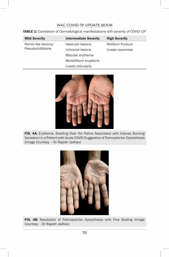
W4C COVID-19 UPDATE BOOK
70
TABLE 1: Correlation of Dermatological manifestations with severity of COVID-198
Mild Severity Intermediate Severity High Severity
Pernio like lesions/Pseudochillblains
Vesicular lesionsUrticarial lesionsMacular erythema Morbilliform eruptions Livedo reticularis
Retiform PurpuraLivedo racemosa
FIG. 4A: Erythema, Swelling Over the Palms Associated with Intense Burning Sensation in a Patient with Acute COVID Suggestive of Palmoplantar Dysesthesia (Image Courtesy – Dr Rajesh Jadhav)
FIG. 4B: Resolution of Palmoplantar Dysesthesia with Fine Scaling (Image Courtesy – Dr Rajesh Jadhav)
W4C COVID-19 UPDATE BOOK
71
• Presence of low grade vascular inflammation and vasodilation caused by direct SARS-CoV-2 infection of endothelial cells or vessel associated smooth muscle cells is thought to cause the presentation of livedo reticularis and livedo racemose.
• The vascular damage in the COVID pathophysiology is also responsible for the localised or generalised presentation of purpuric lesions.
Correlation of the Dermatological Presentation with the severity of COVID 19 is mentioned in Table 1
Other presentations:
• Palmoplantar Dysesthesia
The presentation has been seen in the acute phase of COVID 19 where patient presents with intense burning sensation with itching over the palms with associated swelling [Figure 4a]. This presentation is self limiting and resolves with palmoplantar desquamation [Figure 4b] and acral hyperpigmented macules.9
This presentation has been reported with Zika, Chikungunya and HIV. Fine desquamation is COVID-19 associated cases, versus larger scales and digital involvement seen with palmoplantar desquamation post bacterial superantigens
• Periorbital dyschromia – This has been reported as an early sign of COVID-19.10
• Atypical erythema nodosum like Sweet syndrome and erythema nodosum are reported with concurrent onset of COVID-19 symptoms.11
• Reactivation of herpes simplex infections, varicella, herpes zoster has been reported possibly on account of lymphopenia seen as a part of COVID-19.12
Oral manifestations reported in patients who are COVID-19 positive are – erythema on the tongue, erosions on the palate, fissuring at the angles of the mouth and geographic tongue.13 The mechanism for these manifestations are unclear. These may be due to direct SARS-CoV-2 virus, reactivation of other viruses such as HSV/VZV, drug induced or confinement.
Nail Manifestations seen in COVID-19 patients – ‘Transverse red bands of the nail’ – also referred to as ‘red
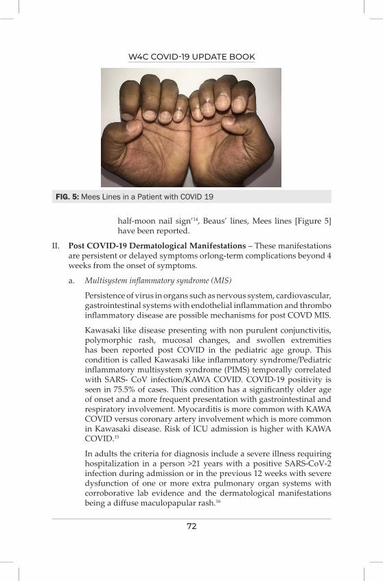
W4C COVID-19 UPDATE BOOK
72
half-moon nail sign’14, Beaus’ lines, Mees lines [Figure 5] have been reported.
II. Post COVID-19 Dermatological Manifestations – These manifestations are persistent or delayed symptoms orlong-term complications beyond 4 weeks from the onset of symptoms.
a. Multisystem inflammatory syndrome (MIS)
Persistence of virus in organs such as nervous system, cardiovascular, gastrointestinal systems with endothelial inflammation and thrombo inflammatory disease are possible mechanisms for post COVD MIS.
Kawasaki like disease presenting with non purulent conjunctivitis, polymorphic rash, mucosal changes, and swollen extremities has been reported post COVID in the pediatric age group. This condition is called Kawasaki like inflammatory syndrome/Pediatric inflammatory multisystem syndrome (PIMS) temporally correlated with SARS- CoV infection/KAWA COVID. COVID-19 positivity is seen in 75.5% of cases. This condition has a significantly older age of onset and a more frequent presentation with gastrointestinal and respiratory involvement. Myocarditis is more common with KAWA COVID versus coronary artery involvement which is more common in Kawasaki disease. Risk of ICU admission is higher with KAWA COVID.15
In adults the criteria for diagnosis include a severe illness requiring hospitalization in a person >21 years with a positive SARS-CoV-2 infection during admission or in the previous 12 weeks with severe dysfunction of one or more extra pulmonary organ systems with corroborative lab evidence and the dermatological manifestations being a diffuse maculopapular rash.16
FIG. 5: Mees Lines in a Patient with COVID 19
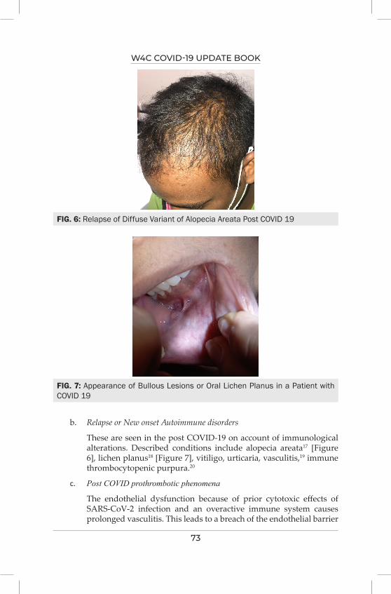
W4C COVID-19 UPDATE BOOK
73
b. Relapse or New onset Autoimmune disorders
These are seen in the post COVID-19 on account of immunological alterations. Described conditions include alopecia areata17 [Figure 6], lichen planus18 [Figure 7], vitiligo, urticaria, vasculitis,19 immune thrombocytopenic purpura.20
c. Post COVID prothrombotic phenomena
The endothelial dysfunction because of prior cytotoxic effects of SARS-CoV-2 infection and an overactive immune system causes prolonged vasculitis. This leads to a breach of the endothelial barrier
FIG. 6: Relapse of Diffuse Variant of Alopecia Areata Post COVID 19
FIG. 7: Appearance of Bullous Lesions or Oral Lichen Planus in a Patient with COVID 19
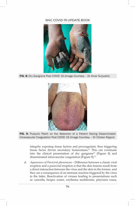
W4C COVID-19 UPDATE BOOK
74
integrity exposing tissue factors and procoagulant, thus triggering tissue factor driven secondary homeostasis.21 This can eventuate into the clinical presentation of dry gangrene22 [Figure 8] and disseminated intravascular coagulation [Figure 9].23
d. Appearance of Paraviral phenomena – Difference between a classic viral eruption and a paraviral eruption is that the skin lesions result from a direct interaction between the virus and the skin in the former, and they are a consequence of an immune reaction triggered by the virus in the latter. Reactivation of viruses leading to presentations such as varicella, herpes zoster, erythema multiforme, pityriasis rosea,
FIG. 8: Dry Gangrene Post COVID 19 (Image Courtesy – Dr Amar Surjushe)
FIG. 9: Purpuric Patch on the Abdomen of a Patient Having Disseminated Intravascular Coagulation Post COVID 19 (Image Courtesy – Dr Chetan Rajput)
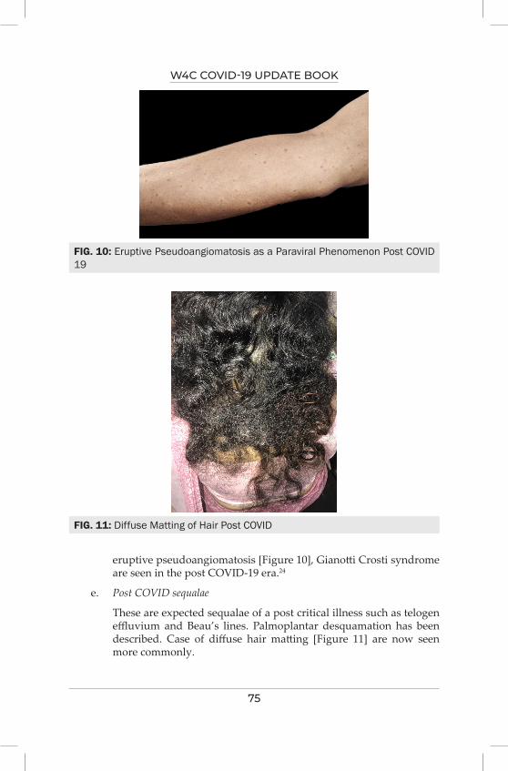
W4C COVID-19 UPDATE BOOK
75
eruptive pseudoangiomatosis [Figure 10], Gianotti Crosti syndrome are seen in the post COVID-19 era.24
e. Post COVID sequalae
These are expected sequalae of a post critical illness such as telogen effluvium and Beau’s lines. Palmoplantar desquamation has been described. Case of diffuse hair matting [Figure 11] are now seen more commonly.
FIG. 10: Eruptive Pseudoangiomatosis as a Paraviral Phenomenon Post COVID 19
FIG. 11: Diffuse Matting of Hair Post COVID
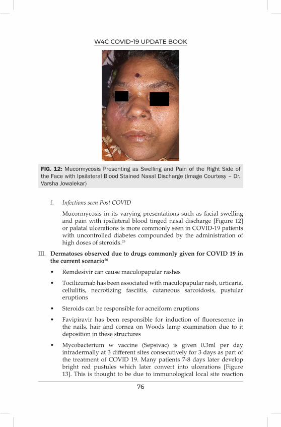
W4C COVID-19 UPDATE BOOK
76
f. Infections seen Post COVID
Mucormycosis in its varying presentations such as facial swelling and pain with ipsilateral blood tinged nasal discharge [Figure 12] or palatal ulcerations is more commonly seen in COVID-19 patients with uncontrolled diabetes compounded by the administration of high doses of steroids.25
III. Dermatoses observed due to drugs commonly given for COVID 19 in the current scenario26
• Remdesivir can cause maculopapular rashes
• Tocilizumab has been associated with maculopapular rash, urticaria, cellulitis, necrotizing fasciitis, cutaneous sarcoidosis, pustular eruptions
• Steroids can be responsible for acneiform eruptions
• Favipiravir has been responsible for induction of fluorescence in the nails, hair and cornea on Woods lamp examination due to it deposition in these structures
• Mycobacterium w vaccine (Sepsivac) is given 0.3ml per day intradermally at 3 different sites consecutively for 3 days as part of the treatment of COVID 19. Many patients 7-8 days later develop bright red pustules which later convert into ulcerations [Figure 13]. This is thought to be due to immunological local site reaction
FIG. 12: Mucormycosis Presenting as Swelling and Pain of the Right Side of the Face with Ipsilateral Blood Stained Nasal Discharge (Image Courtesy – Dr. Varsha Jowalekar)

W4C COVID-19 UPDATE BOOK
77
or an inadvertent subcutaneous injection rather than intradermal injection. This adverse effect can be managed effectively with oral analgesics and topical corticosteroids.
FIG. 13: Tender Subcutaneous Nodules with Ulceration at the site of the SEPSIVAC Vaccine as a Treatment of COVID 19 (Image Courtesy – Dr Chetan Rajput)
FIG. 14: Aquagenic Acral Keratoderma Due to Repeated Hand Washing in the COVID Era
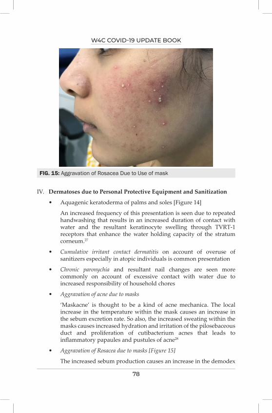
W4C COVID-19 UPDATE BOOK
78
IV. Dermatoses due to Personal Protective Equipment and Sanitization
• Aquagenic keratoderma of palms and soles [Figure 14]
An increased frequency of this presentation is seen due to repeated handwashing that results in an increased duration of contact with water and the resultant keratinocyte swelling through TVRT-1 receptors that enhance the water holding capacity of the stratum corneum.27
• Cumulative irritant contact dermatitis on account of overuse of sanitizers especially in atopic individuals is common presentation
• Chronic paronychia and resultant nail changes are seen more commonly on account of excessive contact with water due to increased responsibility of household chores
• Aggravation of acne due to masks
‘Maskacne’ is thought to be a kind of acne mechanica. The local increase in the temperature within the mask causes an increase in the sebum excretion rate. So also, the increased sweating within the masks causes increased hydration and irritation of the pilosebaceous duct and proliferation of cutibacterium acnes that leads to inflammatory papaules and pustules of acne28
• Aggravation of Rosacea due to masks [Figure 15]
The increased sebum production causes an increase in the demodex
FIG. 15: Aggravation of Rosacea Due to Use of mask
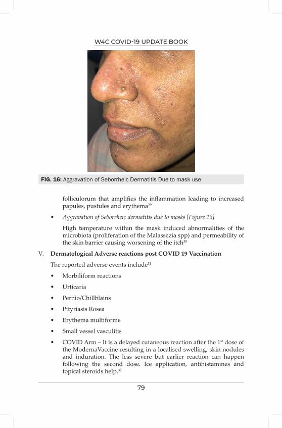
W4C COVID-19 UPDATE BOOK
79
folliculorum that amplifies the inflammation leading to increased papules, pustules and erythema29
• Aggravation of Seborrheic dermatitis due to masks [Figure 16]
High temperature within the mask induced abnormalities of the microbiota (proliferation of the Malassezia spp) and permeability of the skin barrier causing worsening of the itch30
V. Dermatological Adverse reactions post COVID 19 Vaccination
The reported adverse events include31
• Morbiliform reactions
• Urticaria
• Pernio/Chillblains
• Pityriasis Rosea
• Erythema multiforme
• Small vessel vasculitis
• COVID Arm – It is a delayed cutaneous reaction after the 1st dose of the ModernaVaccine resulting in a localised swelling, skin nodules and induration. The less severe but earlier reaction can happen following the second dose. Ice application, antihistamines and topical steroids help.32
FIG. 16: Aggravation of Seborrheic Dermatitis Due to mask use

W4C COVID-19 UPDATE BOOK
80
• SARS-CoV-2 virus spike protein related delayed inflammatory reaction to hyaluronic acid dermal fillers has been reported which needs to be treated with a high dose of corticosteroids with concurrent ACE2 upregulation. High dose intralesional hyaluronidase is used to dissolve the hyaluronic acid filler33
• Vaccine induced thrombocytopenia leading to petechial rash
VI. Effects of Lockdown
• The stress and anxiety of the confinement during the lockdown coupled with the delayed diagnosis and consultations are thought to be the factors that are responsible for exacerbation of pre existing dermatoses like psoriasis, atopic dermatitis, lichen planus and urticaria
• The confinement during the lockdown allowed more walking barefoot at home and sitting for long hours that complicated into regular presentation of patients with corns and callosities
• Younger age group receiving online education are doomed to sedentary life at home leading to increase in weight gain due to changes in nutrition and lifestyle causing increased incidence of striae distensae34
• Traumatic anserine folliculosis is reported on account of assuming positions while using laptops and tablets as part of online education and entertainment35
• Stress of the lockdown has also resulting in the increased incidence of stress aggravated disoders such as lichen simplex chronicus, acne excoree.
CONCLUSION COVID 19 dermatology is an evolving science. With more and more inputs into the national registry we shall have better idea about the manifestations and their association with the severity of COVID 19.
REFERENCES1. Centre of Evidence Based Dermatology. CEBD Coronavirus Dermatology Resource.
https://www.nottingham.ac.uk/research/groups/cebd/resources/Coronavirusresource/Coronavirushome.aspx access on February 18th 2021.
2. Galván Casas C, Català A, Carretero Hernández G, Rodríguez-Jiménez P, Fernández-Nieto D, Rodríguez-Villa Lario A et al. Classification of the cutaneous manifestations of COVID -19: a rapid prospective nationwide consensus study in Spain with 375 cases. British Journal of Dermatology 2020; 183:71-77.
3. Genovese G, Moltrasio C, Berti E, Marzano AV. Skin Manifestations Associated with COVID-19: Current Knowledge and Future Perspectives. Dermatology 2021; 237:1-12.
4. Tatu AL, Baroiu L, Fotea S, Anghel L, Drima Polea E, Nadasdy T, Chioncel V, Nwabudike LC. A Working Hypothesis on Vesicular Lesions Related to COVID-19 Infection, Koebner
W4C COVID-19 UPDATE BOOK
81
Phenomena Type V, and a Short Review of Related Data. Clin Cosmet Investig Dermatol 2021; 14:419-423.
5. Colmenero I, Santonja C, Alonso-Riaño M, Noguera-Morel L, Hernández-Martín A, Andina D, Wiesner T, Rodríguez-Peralto JL, Requena L, Torrelo A. SARS-CoV-2 endothelial infection causes COVID-19 chilblains: histopathological, immunohistochemical and ultrastructural study of seven paediatric cases. Br J Dermatol 2020; 183:729-737.
6. Vázquez-Osorio I, Rocamonde L, Treviño-Castellano M, Vázquez-Veiga H, Ginarte M. Pseudo-chilblain lesions and COVID-19: a controversial relationship. International Journal of Dermatology 2021; 60:754-756.
7. Khalil S, Hinds B, Manalo I, Vargas I, Mallela S, Jacobs R. Livedo reticularis as a presenting sign of severe acute respiratory syndrome coronavirus 2 infection. JAAD Case Reports 2020; 6:871-874.
8. Jamshidi P, Hajikhani B, Mirsaeidi M, Vahidnezhad H, Dadashi M, Nasiri M. Skin Manifestations in COVID-19 Patients: Are They Indicators for Disease Severity? A Systematic Review. Frontiers in Medicine 2021; 8.
9. Nuno-Gonzalez A, Magaletsky K, Feito Rodríguez M, Mayor Ibarguren A, Beato MJ, Ruiz Bravo E, Herranz Pinto P. Palmoplantar erythrodysesthesia: a diagnostic sign of COVID-19. J Eur Acad Dermatol Venereol 2021; 35e247-e249.
10. Kalner S, Vergilis I. Periorbital erythema as a presenting sign of COVID-19. JAAD Case Reports 2020; 6:996-998.
11. Taşkın B, Vural S, Altuğ E, Demirkesen C, Kocatürk E, Çelebi İ et al. Coronavirus 19 presenting with atypical Sweet’s syndrome. Journal of the European Academy of Dermatology and Venereology 2020; 34.
12. Diez-Domingo J, Parikh R, Bhavsar AB, Cisneros E, McCormick N, Lecrenier N. Can COVID-19 Increase the Risk of Herpes Zoster? A Narrative Review. Dermatol Ther (Heidelb) 2021; 11:1119-1126.
13. Farid H, Khan M, Jamal S, Ghafoor R. Oral manifestations of Covid-19-A literature review. Rev Med Virol 2021; e2248.
14. Méndez-Flores S, Zaladonis A, Valdes-Rodriguez R. COVID-19 and nail manifestation: be on the lookout for the red half-moon nail sign. International Journal of Dermatology 2020; 59:1414-1414.
15. Jain S, Sen S, Lakshmivenkateshiah S, Bobhate P, Venkatesh S, Udani S, Shobhavat L, Andankar P, Karande T, Kulkarni S. Multisystem Inflammatory Syndrome in Children With COVID-19 in Mumbai, India. Indian Pediatr 2020; 57:1015-1019.
16. Chow EJ. The Multisystem Inflammatory Syndrome in Adults With SARS-CoV-2 Infection—Another Piece of an Expanding Puzzle. JAMA Netw Open 2021; 4:e2110344.
17. Rinaldi F, Trink A, Giuliani G, et al. Italian Survey for the Evaluation of the Effects of Coronavirus Disease 2019 (COVID-19) Pandemic on Alopecia Areata Recurrence. Dermatol Ther (Heidelb) 2021; 11:339–345.
18. Merhy R, Sarkis A, Kaikati J, El Khoury L, Ghosn S, Stephan F. New-onset cutaneous lichen planus triggered by COVID-19 vaccination. Journal of the European Academy of Dermatology and Venereology 2021.
19. Camprodon Gómez M, González-Cruz C, Ferrer B, et al. Leucocytoclastic vasculitis in a patient with COVID-19 with positive SARS-CoV-2 PCR in skin biopsy. BMJ Case Reports CP 2020; 13:e238039.
20. Levraut M, Ottavi M, Lechtman S, Mondain V, Jeandel P. Immune thrombocytopenic purpura after COVID-19 infection. International Journal of Laboratory Hematology 2020; 43.
21. Shah P, Lo Sicco K, Caplan AS, Femia AN, Zampella JG. Dermatologists’ Role in the Diagnosis and Management of Coronavirus Disease 2019 Coagulopathy. Am J Clin Dermatol 2020; 21:599-600.
W4C COVID-19 UPDATE BOOK
82
22. Novara E, Molinaro E, Benedetti I, Bonometti R, Lauritano EC, Boverio R. Severe acute dried gangrene in COVID-19 infection: a case report. Eur Rev Med Pharmacol Sci 2020; 24:5769-5771.
23. Singh P, Schwartz R. Disseminated intravascular coagulation: A devastating systemic disorder of special concern with COVID -19. Dermatologic Therapy 2020; 33.
24. Lipsker D. Paraviral eruptions in the era of COVID-19: Do some skin manifestations point to a natural resistance to SARS-CoV-2?. Clin Dermatol 2020; 38:757-761.
25. Gupta A, Sharma Aman, Chakrabarti A. The emergence of post-COVID-19 mucormycosis in India. Indian Journal of Ophthalmology 2021; 69:1645-1647.
26. Martinez-Lopez A, Cuenca-Barrales C, Montero-Vilchez T, Molina-Leyva A, Arias-Santiago S. Review of adverse cutaneous reactions of pharmacologic interventions for COVID-19: A guide for the dermatologist. J Am Acad Dermatol 2020; 83:1738-1748.
27. Karagün E. Aquagenic acrokeratoderma due to frequent handwashing during the COVID-19 pandemic outbreak. Dermatol Ther 2021; 34:e14796.
28. Han C, Shi J, Chen Y, Zhang Z. Increased flare of acne caused by long-time mask wearing during COVID-19 pandemic among general population. Dermatol Ther 2020; 33:e13704.
29. Chiriac AE, Wollina U, Azoicai D. Flare-up of Rosacea due to Face Mask in Healthcare Workers During COVID-19. Maedica (Bucur) 2020; 15:416-417.
30. Veraldi S, Angileri L, Barbareschi M. Seborrheic dermatitis and anti-COVID-19 masks. Journal of Cosmetic Dermatology 2020; 19:2464-2465.
31. Sun Q, Fathy R, McMahon DE, Freeman EE. COVID-19 Vaccines and the Skin: The landscape of cutaneous vaccine reactions worldwide [published online ahead of print, 2021 May 31]. Dermatol Clin 2021.
32. Kempf W, Kettelhack N, Kind F, Courvoisier S, Galambos J, Pfaltz K. ‘COVID arm’ – histological features of a delayed-type hypersensitivity reaction to Moderna mRNA-1273 SARS-CoV2 vaccine. Journal of the European Academy of Dermatology and Venereology 2021.
33. Michon A. Hyaluronic acid soft tissue filler delayed inflammatory reaction following COVID-19 vaccination - A case report. J Cosmet Dermatol 2021; 20:2684-2690.
34. Ertugrul G, Aktas H. Indirect skin sign of COVID-19 days: Striae rubrae. J Cosmet Dermatol 2020; 19:2161. doi:10.1111/jocd.13607.
35. Dethe G, Vasani R, Farande P, Barve A. Traumatic anserine folliculosis. Think smartphones, curious habits and postures!. Clinical and Experimental Dermatology 2021.
DiagnosisSECTION 3

84
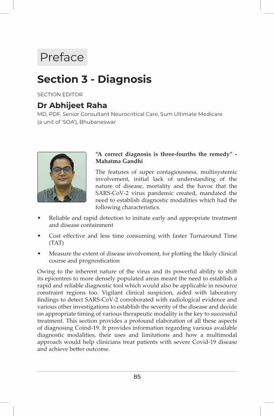
85
Section 3 - DiagnosisSECTION EDITOR
Dr Abhijeet RahaMD, PDF, Senior Consultant Neurocritical Care, Sum Ultimate Medicare (a unit of ‘SOA’), Bhubaneswar
Preface
“A correct diagnosis is three-fourths the remedy” - Mahatma Gandhi
The features of super contagiousness, multisystemic involvement, initial lack of understanding of the nature of disease, mortality and the havoc that the SARS-CoV-2 virus pandemic created, mandated the need to establish diagnostic modalities which had the following characteristics.
• Reliable and rapid detection to initiate early and appropriate treatment and disease containment
• Cost effective and less time consuming with faster Turnaround Time (TAT)
• Measure the extent of disease involvement, for plotting the likely clinical course and prognostication
Owing to the inherent nature of the virus and its powerful ability to shift its epicentres to more densely populated areas meant the need to establish a rapid and reliable diagnostic tool which would also be applicable in resource constraint regions too. Vigilant clinical suspicion, aided with laboratory findings to detect SARS-CoV-2 corroborated with radiological evidence and various other investigations to establish the severity of the disease and decide on appropriate timing of various therapeutic modality is the key to successful treatment. This section provides a profound elaboration of all these aspects of diagnosing Coind-19. It provides information regarding various available diagnostic modalities, their uses and limitations and how a multimodal approach would help clinicians treat patients with severe Covid-19 disease and achieve better outcome.


87
Challenges with Conventional and Novel Diagnostics of COviD-19
10.
INTRODUCTIONOn 31 December 2019, 27 cases of respiratory illness of unknown etiology were detected in Wuhan Province of China. Metagenomic RNA sequencing from these patients clinical samples identified this pathogen as a new RNA virus related to the family Coronaviridae which was later on designated as ‘2019-nCoV’ or Novel CoV-19. On 11 February 2020, International Virus Classification Commission renamed this as ‘‘severe acute respiratory syndrome coronavirus 2 (SARSCoV-2)’’ on the basis of its more than 89% genomic similarity with a SARS-like bat corona virus which belongs to Sarbeco virus subgenus and Betacoronavirus genus. The disease caused by SARSCoV-2 was named as COVID-191.
The pandemic potential of SARS CoV-2 and its contribution to health and economy requires development and application of possible newer diagnostic methods. Timely and accurate diagnosis even in asymptomatic individuals, effective treatment, and prevention of transmission are the keys for managing COVID-19.
Cell culture and next generation sequencing evolved as first diagnostics to characterize SARS CoV-2. Presently, molecular tests along with radiological and clinical investigations are mainstay of COVID-19 diagnostics. Serological assays are useful for mass screening and checking efficacy of COVID vaccines. Many novel diagnostics with highly accurate, rapid results with potential of being used as point of care test are in pipeline.
A new frontier in diagnostic innovation is to develop novel cost-effective point-of-care test (POCT) kits for confirmation of SARS-CoV-2 infection with rapid turn-around time and highly accurate result. Faster and accurate results will help in controlling the pandemic by early isolation of source and contact tracing2.
This chapter will highlight the advantages and shortcomings of conventional
Purabi Baral, Geetarani Purohit
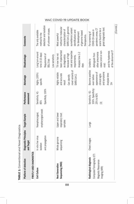
W4C COVID-19 UPDATE BOOK
88
TAB
LE 1
: Con
vent
iona
l and
Nov
el D
iagn
ostic
s
Plat
form
of de
tect
ion
Diag
nosti
c Prin
ciples
an
d Tar
get
Targ
et Sa
mple
Perfo
rman
ce
asse
ssme
nt
Adva
ntag
eDi
sadv
anta
geCo
mmen
ts
FIRS
TLY U
SED
DIAG
NOST
ICS
Cell C
ultur
e In
vitro
live v
irus
isolat
ionan
d prop
agati
on
Naso
phar
ynge
al,oro
phar
ynge
al sw
abSe
nsitiv
ity: 4
1-85
%Sp
ecific
ity: 1
00%
High
ly (10
0%)
spec
ificLo
ng tu
rn-ar
ound
tim
e (5-1
5 day
s)Re
quire
ment
of BS
L-3 la
b Lo
w se
nsitiv
ity
The o
nly av
ailab
le me
thod f
or the
de
tectio
n and
isola
tion
of un
know
n viru
ses.
Next
Gen
erat
ion
Sequ
encin
g (NG
S)Wh
ole ge
nome
se
quen
cing
Uppe
r and
lowe
r res
pirato
ry tra
ct sa
mples
High
ly rel
iable
and a
ccur
ate
result
Can i
denti
fy no
vel v
arian
ts of
SARS
CoV-2
.
Till n
ow ha
s not
been
imple
mente
d as
diag
nosti
cs
beca
use o
f high
co
st an
d exp
ertis
e req
uirem
ents.
Analy
sis tim
e 1-2
days
Most
powe
rful
metho
d for
molec
ular
char
acter
izatio
n of
SARS
CoV-2
, gen
omic
surve
illanc
e (va
riant
identi
ficati
on) a
nd
for de
velop
ment
of ge
nome
base
d the
rape
utics
.Ra
diolog
ical d
iagno
sisCo
mpute
d Tom
ogra
phy (
CT)
Magn
etic R
eson
ance
Im
aging
(MRI
)
Ches
t imag
esLu
ngs
Sens
itivity
: 61-
100%
. Spe
cificit
y: 25
% to
83%
[3]
Beco
me po
sitive
ev
en be
fore
appe
aran
ce of
cli
nical
signs
an
d sym
ptoms
Analy
sis tim
e: 1h
r
Unab
le to
distin
guish
from
oth
er pn
eumo
nia
case
s (vir
al or
non-
viral)
an
d the
hyste
resis
of the
abno
rmal
CT
For s
creen
ing of
CO
VID-19
case
s in
pand
emic
area
s, CT
sc
ans f
ound
to be
a gre
at dia
gnos
tic to
ol.
(Con
td.)

W4C COVID-19 UPDATE BOOK
89
Plat
form
of de
tect
ion
Diag
nosti
c Prin
ciples
an
d Tar
get
Targ
et Sa
mple
Perfo
rman
ce
asse
ssme
nt
Adva
ntag
eDi
sadv
anta
geCo
mmen
ts
APPR
OVED
DIA
GNOS
TICS
Nucle
ic ac
id am
plific
ation
te
st (N
AAT)
(Rev
erse-t
rans
cripti
on
polym
erase
chain
reac
tion
(RT-P
CR)
Spec
ific pr
imer-
probe
ba
sed
detec
tion o
f vira
l RNA
Naso
phar
ynge
al,
Orop
haryn
geal,
Na
sal s
wab,
Sputu
m,
Endo
trach
eal
aspir
ates,
Bron
choa
lveola
r lav
age fl
uid, a
nd
naso
phar
ynge
alwa
sh/a
spira
te or
nasa
l asp
irate
Sens
itivity
: 95.2
%(8
6.7–9
8.3%)
Spec
ificity
: 98.9
%(97
.3–99
.5%)
Gold
stand
ard
test fo
r SAR
S-Co
V-2
Cann
ot dis
tingu
ish
betw
een v
iable
and
unvia
ble vi
rus
Analy
sis tim
e is 4
–6
hrs,
and s
ample
-to-
result
turn
aroun
d tim
e is m
ore th
an
24 hr
s
Futur
e pers
pecti
ve:
Using
less
inva
sive
samp
les (S
aliva
)
Rapid
Ant
igen t
est (
RAT)
Later
al flo
w as
say (
LFA)
Immu
noflu
oresc
ence
assa
y (IF
A)
Uses
macro
molec
ules t
hat
spec
ificall
y bind
to
viral
antig
en
Naso
phar
ynge
al,oro
phar
ynge
al sw
ab
(or wash
) nas
al sw
ab
(anter
iorna
res or
mid-
turbin
ate),
nasa
l was
h, sp
utum
Sens
itivity
: 56.2
%(2
9.5–7
9.8%)
Spec
ificity
: 99.5
%(9
8.1–9
9.9%)
Rapid
Resu
lt (3
0 mins
)No
expe
rtise
req
uirem
ent
Cann
ot dis
tingu
ish
betw
een v
iable
and
unvia
ble vi
rus
Symp
tomati
c RAT
ne
gativ
e cas
es to
be
confi
rmed
by
RT PC
R
Now
Self-t
est R
AT ki
ts av
ailab
le
TAB
LE 1
: Con
vent
iona
l and
Nov
el D
iagn
ostic
s (C
ontd
.)
(Con
td.)
W4C COVID-19 UPDATE BOOK
90
Plat
form
of de
tect
ion
Diag
nosti
c Prin
ciples
an
d Tar
get
Targ
et Sa
mple
Perfo
rman
ce
asse
ssme
nt
Adva
ntag
eDi
sadv
anta
geCo
mmen
ts
Antib
ody a
ssay
En
zyme l
inked
Im
muno
sorb
ent a
ssay
(EL
ISA),
LFA
IFA Chem
ilumi
nesc
ence
Im
muno
assa
y (CL
IA)
Uses
vira
l prot
eins t
o de
termi
neif t
he pa
tient
has
deve
loped
antib
odies
toSA
RS-C
oV-2
Veno
us/C
apilla
ry wh
ole bl
ood,
Plasm
a, se
rum
Sens
itivity
: 91.4
%(87
.0–94
.4) at
15–2
1 day
sSp
ecific
ity: >
98%
Rapid
resu
lt (30
mi
ns)
Test
beco
mes
posit
ive af
ter 3-
4 da
ys of
infec
tion
Poor
clinic
al sp
ecific
ity (M
ore
false
posit
ive
result
s)
Appro
ache
s may
be
come
an im
porta
nt too
l to
deter
mine
vacc
ineeffi
cacy
NOVE
L DIAG
NOST
ICS
Reve
rse tr
ansc
riptio
n Lo
op- m
ediat
ed Is
otherm
al am
plific
ation
(RT-L
AMP)
Isothe
rmal
ampli
ficati
onNa
soph
aryn
geal/
oro
phar
ynge
al sw
ab/
Nasa
l swa
b
Sens
itivity
: 100
%Sp
ecific
ity: 1
00%
(as co
mpar
ed to
RT
-PCR
[4]
Point
-of-ca
re tes
t (rap
id res
ult
withi
n 13 m
in)
One t
est p
er ru
n
Cluste
red Re
gular
ly Int
erspa
ced
Shor
t Pali
ndrom
ic Re
peats
(C
RISP
R) ba
sed a
ssay
s
Later
al flo
w as
say
isothe
rmal
ampli
ficati
on
Resp
irator
y sam
ples
Sens
itivity
: 95%
Spec
ificity
: 100
% [5
]
Do no
t req
uire
comp
lex
instru
menta
tion.
Rapid
resu
lt (w
ithin
1hr)
Stan
dardi
zatio
n of
protoc
ols is
a ve
ry im
porta
nt fac
tor fo
r eff
ectiv
e dete
ction
(sa
me re
sult b
y all
opera
tors)
with
this
syste
m.6
An in
nova
tive
appro
ach t
hat h
as
rapid
ly pro
gress
ed.
It has
the p
otenti
al for
fur
ther im
prove
ment.
TAB
LE 1
: Con
vent
iona
l and
Nov
el D
iagn
ostic
s (C
ontd
.)
(Con
td.)
W4C COVID-19 UPDATE BOOK
91
Plat
form
of de
tect
ion
Diag
nosti
c Prin
ciples
an
d Tar
get
Targ
et Sa
mple
Perfo
rman
ce
asse
ssme
nt
Adva
ntag
eDi
sadv
anta
geCo
mmen
ts
Matri
x-ass
isted
lase
r de
sorp
tion/
ioniza
tion
mass
spec
trome
try (M
ALDI
-M
S)
Detec
ts the
pres
ence
of
viral
protei
ns/p
eptid
es
using
mass
and c
harge
Naso
phar
ynge
al/ora
l sw
absa
liva,
inves
tigate
d for
SARS
-CoV-2
but
amen
dable
to an
y spe
cimen
Sens
itivity
: 90.4
% Sp
ecific
ity: 1
00%7
High
spec
ificity
(10
0%)
Rapid
resu
lt (2
-3 mi
ns)
Has t
he
poten
tial o
f as
ympto
matic
dia
gnos
is of
COVID
-19 in
pa
tients
.
High
cost
of the
eq
uipme
ntIt c
an be
a no
vel
appro
ach
for m
ass
scree
ning
Bios
enso
rsUs
e the
princ
iple
of ele
ctroc
hemi
cal
reacti
ons w
hen v
iral
RNA o
r prot
eins
bind w
ith sp
ecific
an
tibod
ies or
prob
es
Seru
m/pla
sma
Sens
itivity
: 86.4
-93
.7% Sp
ecific
ity:
90.61
00%
Low
costs
of
analy
sis
Fast
result
Can b
e use
d as a
PO
CT
Brea
th tes
tMe
asur
e vola
tile
organ
ic co
mpou
nds
and n
itric
oxide
Brea
th sa
mples
Un
der d
evelo
pmen
t
Skin
test
Meas
ure C
OVID
-19
asso
ciated
dy
slipid
emia
Sebu
m sw
abUn
der d
evelo
pmen
t
TAB
LE 1
: Con
vent
iona
l and
Nov
el D
iagn
ostic
s (C
ontd
.)

W4C COVID-19 UPDATE BOOK
92
and novel diagnostic methods under development for diagnosis of SARS CoV-2 (Table 1).
CONCLUSIONThe pandemic potential of SARS CoV-2 and its contribution to health and economy requires development and application of possible newer diagnostic methods [8]. The diagnostic assays for SARS CoV-2 are classified into five major categories: (i) cell culture, (ii) Next-Generation Sequencing (NGS) (iii) Nucleic acid amplification test (NAAT) (iv) Serological methods, (v) Radiological investigations. The traditional, cell culture method, though the only method for isolation and characterization of a newly emerged virus, drawbacks of less specificity, long incubation period and BSL-3 requirement made this undesirable test for routine use on urgent basis. Next generation sequencing (NGS) plays very important role in pandemic situation with the advantage of identifying COVID-19 variant strains. This is very accurate and reliable diagnostics, still have limitations of high cost and expert requirements.
Presently nucleic acid detection methods (RT-PCR) is considered gold standard test for COVID-19 diagnosis, but have the limitations of high cost laboratory set up, expert requirements and analysis time of 3-4hrs. For increasing testing and decreasing turn-around time, molecular tests were transferred from laboratory to POCT. Here came with the implementation of Reverse Transcription Loop-mediated Isothermal Amplification (RT-LAMP) or SARS CoV-2 diagnosis, with advantage of rapid result (within 13 mins). Another innovation in molecular test with rapid turnaround time (1 hr) is Clustered regularly interspaced short palindromic repeats (CRISPR) based diagnostics.
Radiological investigation (CT/MRI lungs) is a necessary auxiliary COVID-19 diagnostic method; sensitivity increases if findings are combined with RT-PCR results.
Apart from these diagnostic strategies, Serological assays i.e antigen and antibody detection kits available in different platforms (LFA, ELISA, IFA, CLIA) are useful for screening of SARS CoV-2 infection in huge population.
Other novel approaches in SARS CoV-2 as POCT diagnostics include MALDI-MS, Biosensors, Aptamer based nano-biosensors, Breath tests and Skin test. Molecular tests using less invasive saliva samples may have a major potential for COVID-19 mass screening.
REFERENCES1. Wu F, Su Z, Yu B, Yan-Mei C, Wen W, Zhi-Gang S, et al. A new coronavirus associated with
human respiratory disease in China. Nature 2020; 12:265–9.2. Koetter P, Pelton M, Gonzalo J, Du P, Exten C, Bogale K, et al. Implementation and Process of
a COVID-19 Contact Tracing Initiative: Leveraging Health Professional Students to Extend the Workforce During a Pandemic. American Journal of Infection Control 2020; 48:1451−6. https://doi.org/10.1016/j.ajic.2020.08.012
W4C COVID-19 UPDATE BOOK
93
3. Park JY, Freer R, Stevens R, Soneji N, Jones N. The accuracy of chest CT in the diagnosis of COVID-19: An umbrella review. The Centre for Evidence based Medicine. March 4, 2021. https://www.cebm.net/covid-19/the-accuracy-of-chest-ct-in-the-diagnosis-of-covid-19-an-umbrella-review/
4. Inaba M, Higashimoto Y, Toyama Y, Horiguchi T, Hibino M, Iwata M, et al. Diagnostic accuracy of LAMP versus PCR over the course ofSARS-CoV-2 infection. International Journal of Infectious Diseases 2021;107:195–200.
5. Broughton JP, Deng X, Yu G, Fasching CL, Servellita V, Singh J et al. Nat Biotechnol 2020, DOI: 10.1038/s41587-020-0513-4.
6. Rahimi H, Salehiabar M, Barsbay M, Ghaffarlou M, Kavetskyy T, Sharaf A, et al. CRISPR Systems for COVID-19 Diagnosis. ACS Sensors. https://dx.doi.org/10.1021/acssensors.0c02312
7. Singh P, Chakraborty R, Marwal R, Radhakrishan VS, Bhaskar AK, Vashisht H et al. A rapid and sensitive method to detect SARS-CoV-2 virus using targeted-mass spectrometry. Journal of Proteins and Proteomics https://doi.org/10.1007/s42485-020-00044-9
8. Dollman NL, Griffin JH, Downard KM. Detection, Mapping, and Proteotyping of SARS-CoV-2 Coronavirus with High Resolution Mass Spectrometry. ACS Infect. Dis https://dx.doi.org/10.1021/acsinfecdis.0c00664
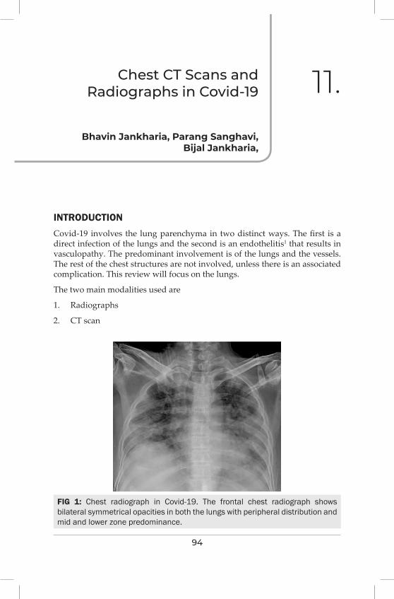
94
Chest CT Scans and Radiographs in Covid-19 11.
INTRODUCTIONCovid-19 involves the lung parenchyma in two distinct ways. The first is a direct infection of the lungs and the second is an endothelitis1 that results in vasculopathy. The predominant involvement is of the lungs and the vessels. The rest of the chest structures are not involved, unless there is an associated complication. This review will focus on the lungs.
The two main modalities used are
1. Radiographs
2. CT scan
Bhavin Jankharia, Parang Sanghavi, Bijal Jankharia,
FIG 1: Chest radiograph in Covid-19. The frontal chest radiograph shows bilateral symmetrical opacities in both the lungs with peripheral distribution and mid and lower zone predominance.
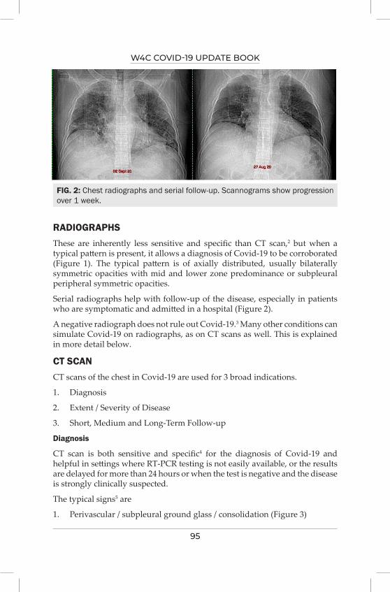
W4C COVID-19 UPDATE BOOK
95
RADIOGRAPHSThese are inherently less sensitive and specific than CT scan,2 but when a typical pattern is present, it allows a diagnosis of Covid-19 to be corroborated (Figure 1). The typical pattern is of axially distributed, usually bilaterally symmetric opacities with mid and lower zone predominance or subpleural peripheral symmetric opacities.
Serial radiographs help with follow-up of the disease, especially in patients who are symptomatic and admitted in a hospital (Figure 2).
A negative radiograph does not rule out Covid-19.3 Many other conditions can simulate Covid-19 on radiographs, as on CT scans as well. This is explained in more detail below.
CT SCANCT scans of the chest in Covid-19 are used for 3 broad indications.
1. Diagnosis
2. Extent / Severity of Disease
3. Short, Medium and Long-Term Follow-up
Diagnosis
CT scan is both sensitive and specific4 for the diagnosis of Covid-19 and helpful in settings where RT-PCR testing is not easily available, or the results are delayed for more than 24 hours or when the test is negative and the disease is strongly clinically suspected.
The typical signs5 are
1. Perivascular / subpleural ground glass / consolidation (Figure 3)
FIG. 2: Chest radiographs and serial follow-up. Scannograms show progression over 1 week.
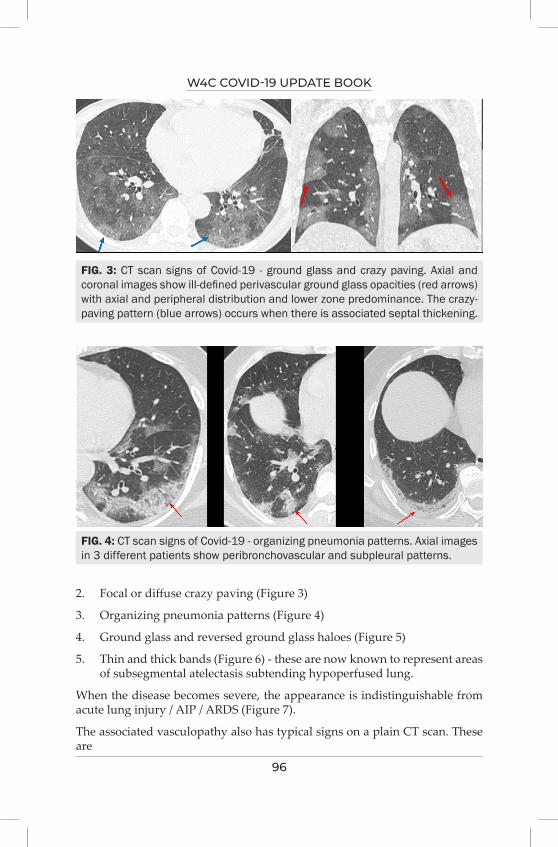
W4C COVID-19 UPDATE BOOK
96
2. Focal or diffuse crazy paving (Figure 3)
3. Organizing pneumonia patterns (Figure 4)
4. Ground glass and reversed ground glass haloes (Figure 5)
5. Thin and thick bands (Figure 6) - these are now known to represent areas of subsegmental atelectasis subtending hypoperfused lung.
When the disease becomes severe, the appearance is indistinguishable from acute lung injury / AIP / ARDS (Figure 7).
The associated vasculopathy also has typical signs on a plain CT scan. These are
FIG. 3: CT scan signs of Covid-19 - ground glass and crazy paving. Axial and coronal images show ill-defined perivascular ground glass opacities (red arrows) with axial and peripheral distribution and lower zone predominance. The crazy-paving pattern (blue arrows) occurs when there is associated septal thickening.
FIG. 4: CT scan signs of Covid-19 - organizing pneumonia patterns. Axial images in 3 different patients show peribronchovascular and subpleural patterns.
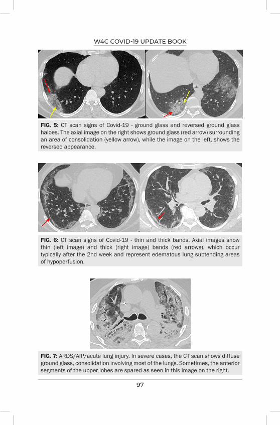
W4C COVID-19 UPDATE BOOK
97
FIG. 5: CT scan signs of Covid-19 - ground glass and reversed ground glass haloes. The axial image on the right shows ground glass (red arrow) surrounding an area of consolidation (yellow arrow), while the image on the left, shows the reversed appearance.
FIG. 6: CT scan signs of Covid-19 - thin and thick bands. Axial images show thin (left image) and thick (right image) bands (red arrows), which occur typically after the 2nd week and represent edematous lung subtending areas of hypoperfusion.
FIG. 7: ARDS/AIP/acute lung injury. In severe cases, the CT scan shows diffuse ground glass, consolidation involving most of the lungs. Sometimes, the anterior segments of the upper lobes are spared as seen in this image on the right.
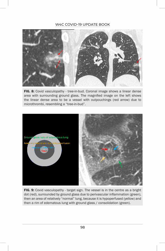
W4C COVID-19 UPDATE BOOK
98
FIG. 8: Covid vasculopathy - tree-in-bud. Coronal image shows a linear dense area with surrounding ground glass. The magnified image on the left shows the linear dense area to be a vessel with outpouchings (red arrow) due to microthrombi, resembling a “tree-in-bud”.
FIG. 9: Covid vasculopathy - target sign. The vessel is in the centre as a bright dot (red), surrounded by ground glass due to perivascular inflammation (green), then an area of relatively “normal” lung, because it is hypoperfused (yellow) and then a rim of edematous lung with ground glass / consolidation (green).
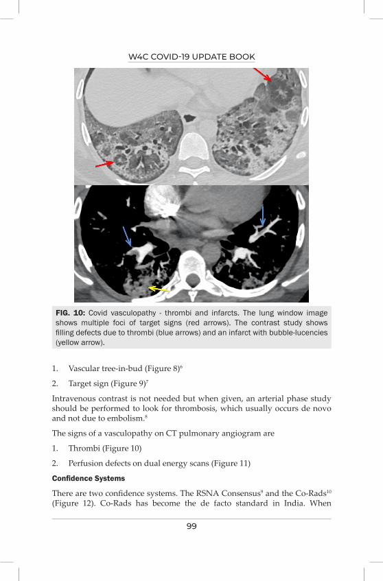
W4C COVID-19 UPDATE BOOK
99
1. Vascular tree-in-bud (Figure 8)6
2. Target sign (Figure 9)7
Intravenous contrast is not needed but when given, an arterial phase study should be performed to look for thrombosis, which usually occurs de novo and not due to embolism.8
The signs of a vasculopathy on CT pulmonary angiogram are
1. Thrombi (Figure 10)
2. Perfusion defects on dual energy scans (Figure 11)
Confidence Systems
There are two confidence systems. The RSNA Consensus9 and the Co-Rads10 (Figure 12). Co-Rads has become the de facto standard in India. When
FIG. 10: Covid vasculopathy - thrombi and infarcts. The lung window image shows multiple foci of target signs (red arrows). The contrast study shows filling defects due to thrombi (blue arrows) and an infarct with bubble-lucencies (yellow arrow).
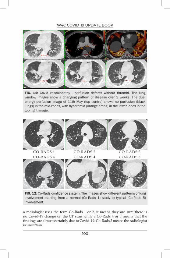
W4C COVID-19 UPDATE BOOK
100
FIG. 11: Covid vasculopathy - perfusion defects without thrombi. The lung window images show a changing pattern of disease over 3 weeks. The dual energy perfusion image of 11th May (top centre) shows no perfusion (black lungs) in the mid-zones, with hyperemia (orange areas) in the lower lobes in the top right image.
FIG. 12: Co-Rads confidence system. The images show different patterns of lung involvement starting from a normal (Co-Rads 1) study to typical (Co-Rads 5) involvement.
a radiologist uses the term Co-Rads 1 or 2, it means they are sure there is no Covid-19 change on the CT scan while a Co-Rads 4 or 5 means that the findings are almost certainly due to Covid-19. Co-Rads 3 means the radiologist is uncertain.
CO-RADS 1CO-RADS 4
CO-RADS 2CO-RADS 4
CO-RADS 3CO-RADS 5
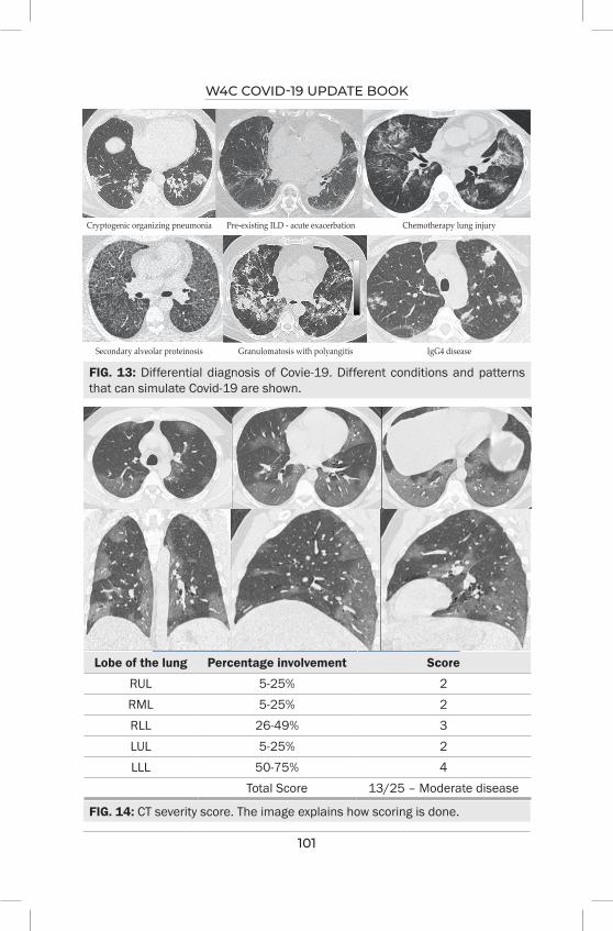
W4C COVID-19 UPDATE BOOK
101
FIG. 13: Differential diagnosis of Covie-19. Different conditions and patterns that can simulate Covid-19 are shown.
FIG. 14: CT severity score. The image explains how scoring is done.
Lobe of the lung Percentage involvement Score
RUL 5-25% 2RML 5-25% 2RLL 26-49% 3LUL 5-25% 2LLL 50-75% 4
Total Score 13/25 – Moderate disease
Cryptogenic organizing pneumonia
Secondary alveolar proteinosis Granulomatosis with polyangitis IgG4 disease
Pre-existing ILD - acute exacerbation Chemotherapy lung injury
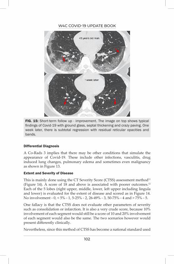
W4C COVID-19 UPDATE BOOK
102
Differential Diagnosis
A Co-Rads 3 implies that there may be other conditions that simulate the appearance of Covid-19. These include other infections, vasculitis, drug induced lung changes, pulmonary edema and sometimes even malignancy as shown in Figure 13.
Extent and Severity of Disease
This is mainly done using the CT Severity Score (CTSS) assessment method11 (Figure 14). A score of 18 and above is associated with poorer outcomes.12 Each of the 5 lobes (right upper, middle, lower, left upper including lingula and lower) is evaluated for the extent of disease and scored as in Figure 14. No involvement - 0, < 5% - 1, 5-25% - 2, 26-49% - 3, 50-75% - 4 and > 75% - 5.
One fallacy is that the CTSS does not evaluate other parameters of severity such as consolidation or infarction. It is also a very crude score, because 10% involvement of each segment would still be a score of 10 and 20% involvement of each segment would also be the same. The two scenarios however would present differently clinically.
Nevertheless, since this method of CTSS has become a national standard used
FIG. 15: Short-term follow up - improvement. The image on top shows typical findings of Covid-19 with ground glass, septal thickening and crazy paving. One week later, there is subtotal regression with residual reticular opacities and bands.
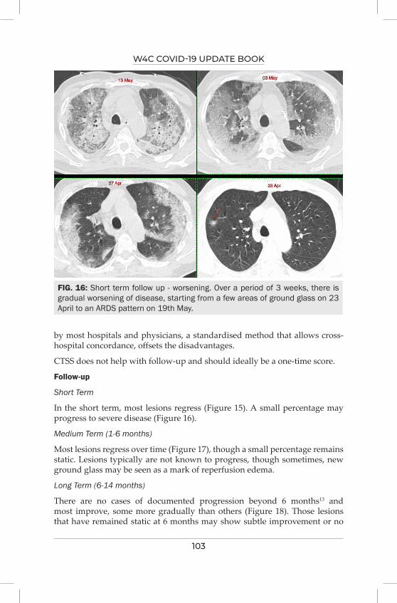
W4C COVID-19 UPDATE BOOK
103
by most hospitals and physicians, a standardised method that allows cross-hospital concordance, offsets the disadvantages.
CTSS does not help with follow-up and should ideally be a one-time score.
Follow-up
Short Term
In the short term, most lesions regress (Figure 15). A small percentage may progress to severe disease (Figure 16).
Medium Term (1-6 months)
Most lesions regress over time (Figure 17), though a small percentage remains static. Lesions typically are not known to progress, though sometimes, new ground glass may be seen as a mark of reperfusion edema.
Long Term (6-14 months)
There are no cases of documented progression beyond 6 months13 and most improve, some more gradually than others (Figure 18). Those lesions that have remained static at 6 months may show subtle improvement or no
FIG. 16: Short term follow up - worsening. Over a period of 3 weeks, there is gradual worsening of disease, starting from a few areas of ground glass on 23 April to an ARDS pattern on 19th May.
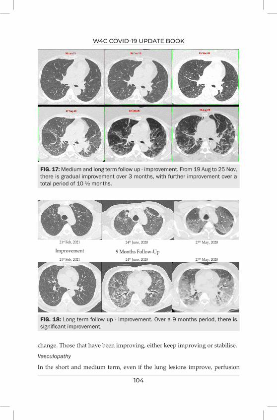
W4C COVID-19 UPDATE BOOK
104
FIG. 17: Medium and long term follow up - improvement. From 19 Aug to 25 Nov, there is gradual improvement over 3 months, with further improvement over a total period of 10 ½ months.
FIG. 18: Long term follow up - improvement. Over a 9 months period, there is significant improvement.
change. Those that have been improving, either keep improving or stabilise.
Vasculopathy
In the short and medium term, even if the lung lesions improve, perfusion
21st Feb, 2021 24th June, 2020
24th June, 2020
27th May, 2020
27th May, 202021st Feb, 2021
Improvement 9 Months Follow-Up
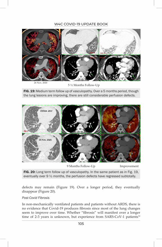
W4C COVID-19 UPDATE BOOK
105
defects may remain (Figure 19). Over a longer period, they eventually disappear (Figure 20).
Post-Covid Fibrosis
In non-mechanically ventilated patients and patients without ARDS, there is no evidence that Covid-19 produces fibrosis since most of the lung changes seem to improve over time. Whether “fibrosis” will manifest over a longer time of 2-3 years is unknown, but experience from SARS-CoV-1 patients14
FIG. 19: Medium term follow up of vasculopathy. Over a 5 months period, though the lung lesions are improving, there are still considerable perfusion defects.
FIG. 20: Long term follow up of vasculopathy. In the same patient as in Fig. 19, eventually over 9 ½ months, the perfusion defects have regressed subtotally.
26 Nov, 20205 ½ Months Follow-Up
9 Months Follow-Up Improvement
W4C COVID-19 UPDATE BOOK
106
suggests that most patients tend to improve.
REFERENCES1. Lang M, Som A, Carey D, Reid N, Mendoza DP, Flores EJ, Li MD, Shepard JO, Little BP.
Pulmonary Vascular Manifestations of COVID-19 Pneumonia. Radiol Cardiothorac Imaging 2020; 2:e200277.
2. Borakati A, Perera A, Johnson J, Sood T. Diagnostic accuracy of X-ray versus CT in COVID-19: a propensity-matched database study. BMJ Open 2020; 10:e042946.
3. Vancheri SG, Savietto G, Ballati F, Maggi A, Canino C, Bortolotto C, Valentini A, Dore R, Stella GM, Corsico AG, Iotti GA, Mojoli F, Perlini S, Bruno R, Preda L. Radiographic findings in 240 patients with COVID-19 pneumonia: time-dependence after the onset of symptoms. Eur Radiol 2020; 30:6161-6169.
4. Kovács A, Palásti P, Veréb D, Bozsik B, Palkó A, Kincses ZT. The sensitivity and specificity of chest CT in the diagnosis of COVID-19. Eur Radiol 2021; 31:2819-2824.
5. Hani C, Trieu NH, Saab I, Dangeard S, Bennani S, Chassagnon G, Revel MP. COVID-19 pneumonia: A review of typical CT findings and differential diagnosis. Diagn Interv Imaging 2020; 101:263-268.
6. Patel BV, Arachchillage DJ, Ridge CA, Bianchi P, Doyle JF, Garfield B, Ledot S, Morgan C, Passariello M, Price S, Singh S, Thakuria L, Trenfield S, Trimlett R, Weaver C, Wort SJ, Xu T, Padley SPG, Devaraj A, Desai SR. Pulmonary Angiopathy in Severe COVID-19: Physiologic, Imaging, and Hematologic Observations. Am J Respir Crit Care Med 2020; 202:690-699.
7. Müller CIS, Müller NL. Chest CT target sign in a couple with COVID-19 pneumonia. Radiol Bras 2020; 53:252-254.
8. Cavagna E, Muratore F, Ferrari F. Pulmonary Thromboembolism in COVID-19: Venous Thromboembolism or Arterial Thrombosis? Radiol Cardiothorac Imaging 2020; 2:e200289.
9. Simpson S, Kay FU, Abbara S, Bhalla S, Chung JH, Chung M, Henry TS, Kanne JP, Kligerman S, Ko JP, Litt H. Radiological Society of North America Expert Consensus Statement on Reporting Chest CT Findings Related to COVID-19. Endorsed by the Society of Thoracic Radiology, the American College of Radiology, and RSNA - Secondary Publication. J Thorac Imaging 2020; 35:219-227.
10. Prokop M, van Everdingen W, van Rees Vellinga T, Quarles van Ufford H, Stöger L, Beenen L, Geurts B, Gietema H, Krdzalic J, Schaefer-Prokop C, van Ginneken B, Brink M; COVID-19 Standardized Reporting Working Group of the Dutch Radiological Society. CO-RADS: A Categorical CT Assessment Scheme for Patients Suspected of Having COVID-19-Definition and Evaluation. Radiology 2020; 296:E97-E104
11. Pan F, Ye T, Sun P, Gui S, Liang B, Li L, Zheng D, Wang J, Hesketh RL, Yang L, Zheng C. Time Course of Lung Changes at Chest CT during Recovery from Coronavirus Disease 2019 (COVID-19). Radiology 2020; 295:715-721.
12. Francone M, Iafrate F, Masci GM, Coco S, Cilia F, Manganaro L, Panebianco V, Andreoli C, Colaiacomo MC, Zingaropoli MA, Ciardi MR, Mastroianni CM, Pugliese F, Alessandri F, Turriziani O, Ricci P, Catalano C. Chest CT score in COVID-19 patients: correlation with disease severity and short-term prognosis. Eur Radiol 2020; 30:6808-6817.
13. Caruso D, Guido G, Zerunian M, Polidori T, Lucertini E, Pucciarelli F, Polici M, Rucci C, Bracci B, Nicolai M, Cremona A, De Dominicis C, Laghi A. Postacute Sequelae of COVID-19 Pneumonia: 6-month Chest CT Follow-up. Radiology 2021 Jul 27:210834.
14. Zhang P, Li J, Liu H, Han N, Ju J, Kou Y, Chen L, Jiang M, Pan F, Zheng Y, Gao Z, Jiang B. Long-term bone and lung consequences associated with hospital-acquired severe acute respiratory syndrome: a 15-year follow-up from a prospective cohort study. Bone Res 2020; 8:8.
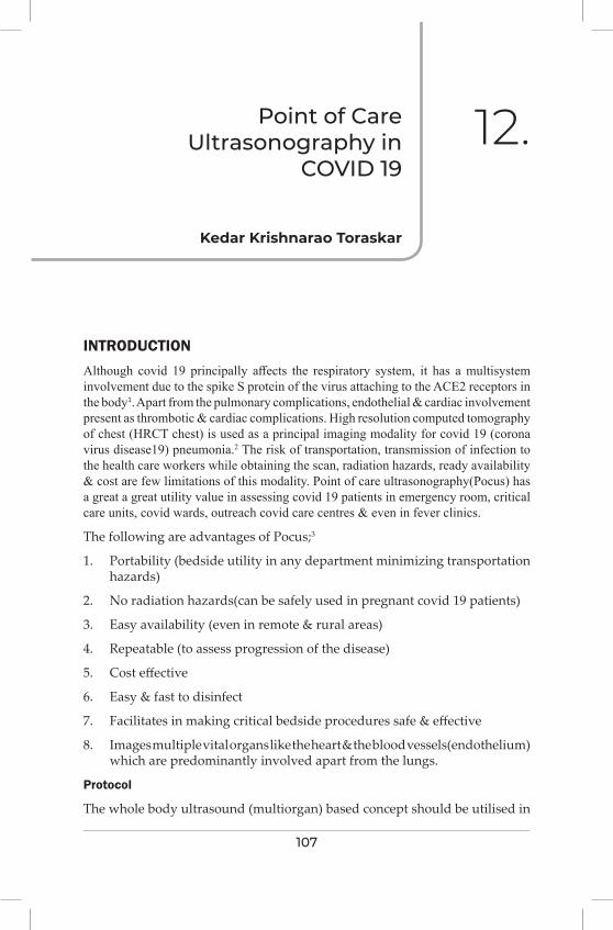
107
Point of Care Ultrasonography in
COviD 1912.
INTRODUCTIONAlthough covid 19 principally affects the respiratory system, it has a multisystem involvement due to the spike S protein of the virus attaching to the ACE2 receptors in the body1. Apart from the pulmonary complications, endothelial & cardiac involvement present as thrombotic & cardiac complications. High resolution computed tomography of chest (HRCT chest) is used as a principal imaging modality for covid 19 (corona virus disease19) pneumonia.2 The risk of transportation, transmission of infection to the health care workers while obtaining the scan, radiation hazards, ready availability & cost are few limitations of this modality. Point of care ultrasonography(Pocus) has a great a great utility value in assessing covid 19 patients in emergency room, critical care units, covid wards, outreach covid care centres & even in fever clinics.
The following are advantages of Pocus;3
1. Portability (bedside utility in any department minimizing transportation hazards)
2. No radiation hazards(can be safely used in pregnant covid 19 patients)
3. Easy availability (even in remote & rural areas)
4. Repeatable (to assess progression of the disease)
5. Cost effective
6. Easy & fast to disinfect
7. Facilitates in making critical bedside procedures safe & effective
8. Images multiple vital organs like the heart & the blood vessels(endothelium) which are predominantly involved apart from the lungs.
Protocol
The whole body ultrasound (multiorgan) based concept should be utilised in
Kedar Krishnarao Toraskar

W4C COVID-19 UPDATE BOOK
108
order to insonate the lung (lung ultrasound) which is the primary organ along with the heart & the deep venous analysis. This is performed like a modified BLUE protocol which evaluates the severity of the covid pneumonia along with the cardiac involvement with basic focussed cardiac sonography or echocardiography (eyeballing only) & two point compression ultrasound to screen the common femoral & popliteal veins.4 This should be time dependent to minimise exposure to the patients & thereby screen more number of patients.
Lung ultrasound (LUS)
Compared to conventional chest radiographs LUS is more sensitive in diagnosing covid 19 pneumonia apart from other pulmonary disorders like pleural effusion, consolidation & alveolar interstitial syndrome4.
The common findings seen in covid 19 pneumonia are irregular pleural line (break in pleura) [Figure 1] with presence of sliding along with subpleural consolidations & B lines predominantly seen in the prone blue points & PLAPS (zone 4/5/6) with an asymmetrical involvement of both the lungs4. The B lines vary as per the severity of the pneumonia, coalescing [Figure 2] or separated (correlating with the ground glass opacities on the HRCT). Large band like B lines called as “light beam B lines” [Figure 3] have been classically described.5 Consolidation (shred or tissue like sign or dynamic air bronchogram sign)4 is uncommonly seen & suggests secondary bacterial infection or progression to ARDS. Pleural effusions are very uncommon & if present are mostly related to cardiogenic pulmonary oedema (either covid 19 cardiac involvement or a pre-existing cardiac aliment).The findings in the previously described viral pneumonias correlate with the findings of LUS in covid 19.5
Acute deterioration due to pneumothorax which is one of the known complication of covid 19 pneumonia as well as mechanical ventilation can
FIG. 1: Thickened, Irregular (Break In) Pleura
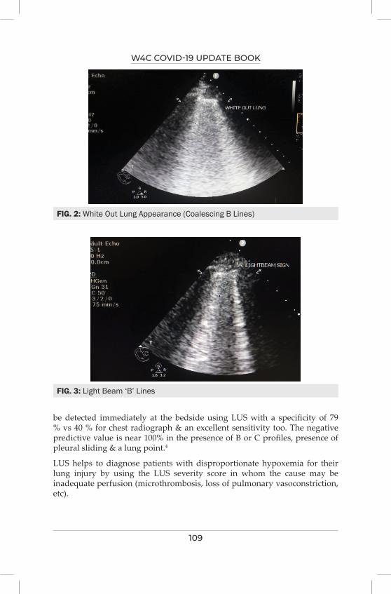
W4C COVID-19 UPDATE BOOK
109
be detected immediately at the bedside using LUS with a specificity of 79 % vs 40 % for chest radiograph & an excellent sensitivity too. The negative predictive value is near 100% in the presence of B or C profiles, presence of pleural sliding & a lung point.4
LUS helps to diagnose patients with disproportionate hypoxemia for their lung injury by using the LUS severity score in whom the cause may be inadequate perfusion (microthrombosis, loss of pulmonary vasoconstriction, etc).
FIG. 2: White Out Lung Appearance (Coalescing B Lines)
FIG. 3: Light Beam ‘B’ Lines

W4C COVID-19 UPDATE BOOK
110
Focussed cardiac ultrasound (Focus)
This involves echocardiographic screening by eyeballing method to pick up covid related cardiac complications like stress cardiomyopathy, myocarditis, myocardial infarction, etc. A dedicated echocardiography is advocated for all patients with significant findings on the Focus. American and European Echocardiography societies recommend Focus & echocardiography as a diagnostic/monitoring tool in COVID-196. Apart from diagnostic evaluation Focus helps in hemodynamic management including fluid resuscitation of severely ill covid 19 patients.
Deep venous analysis
Deep venous two point compression ultrasound is an integral part of pocus examination in all covid 19 patients since incidence of deep vein thrombosis (DVT) & pulmonary thromboembolism (PTE) is high due to covid 19 coagulopathy linked to immunothrombosis despite of standard thromboprophylaxis.7
Apart from DVT & PTE, spontaneous echo contrast [Figure 4] in the deep veins has been reported by Dugar & colleagues initially in covid 19 patients which can be a precursor for thrombosis & may warrant anticoagulation.8
Ultrasound screening twice a week can detect DVT & thereby prevent PTE & deaths in critically ill covid 19 patients though routine daily screening may not be helpful.9
Other utility of Pocus
1. Pocus covid 19 protocol helps in taking decisions related to mechanical
FIG. 4: Slow Flow seen in Sapheno-Femoral Junction. FA – Femoral Artery, FV – Femoral Vein
W4C COVID-19 UPDATE BOOK
111
ventilation like proning, recruitment maneuvers, optimising peep in critically ill patients requiring respiratory support.
2. It helps in guiding fluid management which can be critical in patients with severe covid 19.LUS alone may not be reliable due to the B lines seen secondary to covid pneumonia hence Focus findings & echocardiography guided dynamic indices of fluid responsiveness can be effective used.
3. It helps in optimizing weaning in mechanically ventilated patients by assessing the LUS score, the LV function (systolic & diastolic) & diaphragmatic function in patients who are difficult to wean.
4. It helps to assess other secondary organ dysfunction like AKI, covid hepatopathy, gastrointestinal complications like intestinal ischemia & neurological complications like stroke.
5. Bedside procedures like central venous & arterial cannulation, percutaneous tracheostomies & paracentesis are guided by Pocus making them safe & effective as compared to the blind approach.
Technology & tools
Portable laptop/smart phone based devices with optimal imaging capabilities which can be protected from viral contamination can be used.
Multifrequency probes are ideal to visualise both the deep & superficial structures but the phase array probe can be used for both LUS & Focus. Some experts advocate the microconvex probe as an all in one probe for pocus which can be cost effective.10
The pocus images/clips should be recorded, stored & archived in a standardised protocol for medicolegal purposes, remote guidance & second opinion wherever required.
The users should stringently follow the manufacturers guidelines for disinfection of the ultrasound machine & the probes.
LIMITATIONS1. Subcutaneous emphysema makes it difficult to obtain images due to E
line artifacts.4
2. It requires expertise in image acquisition & interpretation since it is an operator dependent modality.
CONCLUSIONA multiorgan pocus protocol is an ideal imaging tool to diagnose, assess severity, monitor & effectively treat covid 19 patients. Being portable, bedside, rapid, repeatable & goal directed, it helps to take management decisions which can potentially change outcomes in covid 19 patients.
W4C COVID-19 UPDATE BOOK
112
PRACTICE POINTS1. Multiorgan pocus should be utilised for initial assessment & guiding
various procedures for all moderate to severe covid 19 patients to aid diagnostic & management decisions along with the history, clinical examination & biochemical parameters
2. It can be easily repeated as per the clinical needs of the patient to assess the disease progression which is dynamic in nature & time dependent.
3. It helps to prevent injudicious use of HRCT chest at the same time maintain imaging accuracy & also mitigate risk of transmission of this deadly virus.
ABBREVIATIONSHigh resolution computed tomography of chest : HRCT chest
Point of care ultrasonography :Pocus
Focussed cardiac sonography : Focus
Lung ultrasound :LUS
Deep vein thrombosis : DVT
Pulmonary thromboembolism: PTE
REFERENCES1. Wu Z, McGoogan JM. Characteristics of and Important Lessons From the Coronavirus Disease
2019 (COVID-19) Outbreak in China: Summary of a Report of 72314 Cases From the Chinese Center for Disease Control and Prevention. JAMA 2020.
2. Fiala MJ. Ultrasound in COVID-19: a timeline of ultrasound findings in relation to CT. Clin Radiol 2020; 75:553-554.
3. Hussain, A., Via, G., Melniker, L. et al. Multi-organ point-of-care ultrasound for COVID-19 (PoCUS4COVID): international expert consensus. Crit Care 2020; 24:702.
4. Lichtenstein, D.A. Lung ultrasound in the critically ill. Ann Intensive Care 2014; 4:1. 5. Volpicelli G, Lamorte A, Villén T. What’s new in lung ultrasound during the COVID-19
pandemic. Intensive Care Med 2020; 46:1445–8.6. Johry AGB, Kirkpatrick JN, Lanspa M, Mulvagh S, Thamman R. ASE Statement on point-of-care
ultrasound (PoCUS) during the 2019 novel coronavirus pandemic. J Am Soc Echocardiography 2020, in press.
7. Cui S, Chen S, Li X, Liu S, Wang F. Prevalence of venous thromboembolism in patients with severe novel coronavirus pneumonia. J Thromb Haemost 2020; 18:1421-1424.
8. Dugar S, Duggal A, Bassel A, Soliman M, Moghekar A. Spontaneous echo contrast in venous ultrasound of severe COVID-19 patients. Intensive Care Med 2020; 46:1637-1639.
9. Arabi YM, Burns KEA, Alsolamy SJ, Alshahrani MS, Al-Hameed FM, et al. Surveillance or no surveillance ultrasonography for deep vein thrombosis and outcomes of critically ill patients: a pre-planned sub-study of the PREVENT trial. Intensive Care Med 2020; 46:737–46.
10. Volpicelli G, Elbarbary M, Blaivas M, Lichtenstein DA, Mathis G, Kirkpatrick AW, Melniker L, Gargani L, Noble VE, Via G, et al. International evidence-based recommendations for point-of-care lung ultrasound. Intensive Care Med 2012; 38:577–91.
113
Uses of Biomarkers for Treatment Modification and
Prognostication13.
INTRODUCTIONCOVID-19 infection is a mild infection in majority, however, in a small percentage it is a multisystem disorder characterized by a complex interplay between immunological, inflammatory, and coagulation cascade triggered by the “cytokine storm”. The complex pathophysiology of the disease along with genetic and acquired differences in host immune system leads to the heterogenous manifestations ranging from mild illness to life threatening critical disease. The unprecedented pandemic crisis with an outstretched health system and an unpredictable course of COVID-19, necessitates an urgent research for potential biomarkers for accurate and timely diagnosis, risk stratification, effective management, prognostication, and timely discharge. Biomarkers are measurable biological characteristics of a disease used to determine presence, severity, optimal management, prognosis, and further prevention. Biomarkers are indispensable and are the “holy grail” in diagnosis and management of current pandemic crisis.
BIOMARKERS IN COVID-19Clinical assessment albeit indispensable, laboratory parameters or biomarkers provide additional objective assessment and significantly impact management and prognosis of COVID-19. Extensive research and knowledge of pathophysiology of the complex virus-host interaction in COVID has given us a gamut of biomarkers of different classes (Figure 1) to effectively and timely manage patients and save innumerable lives. There are several pros to use of biomarkers in terms of diagnosis, assessment of severity, prognosis, therapeutics and correlation with clinical phenotypes of the disease.
Biomarkers of viral detection
First and foremost is the SARS-CoV-2 RNA detected using RT-PCR (Real time- polymerase chain reaction) and newer methods like digital PCR and CRISPR
Raman Sharma, Madhulata Agarwal
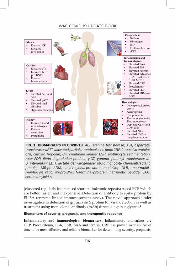
W4C COVID-19 UPDATE BOOK
114
(clustered regularly interspaced short palindromic repeats) based PCR1 which are better, faster, and inexpensive. Detection of antibody to spike protein by ELISA (enzyme linked immunosorbent assay). The novel approach under investigation is detection of glycans on S protein for viral detection as well as treatment using monoclonal antibody (mAb) directed against glycans.2
Biomarkers of severity, prognosis, and therapeutic response
Inflammatory and immunological biomarkers: Inflammatory biomarkers are CRP, Procalcitonin, IL-6, ESR, SAA and ferritin; CRP has proven over course of time to be most effective and reliable biomarker for determining severity, prognosis,
Muscle: Elevated CK Elevated
myoglobin
Figure 1. Biomarkers in COVID-19 ALT, alanine transferase; AST, aspartate transferase; aPTT, activated partial thromboplastin time; CRP, C-reactive protein; cTn, cardiac Troponin; CK, creatinine kinase; ESR, erythrocyte sedimentation rate; FDP, fibrin degradation product; �-GT, gamma glutamyl transferase; IL-6, interleukin; LDH, lactate dehydrogenase; MCP, monocyte chemoattractant protein; MR-pro-ADM, mid-regional-pro-adrenomedullin; NLR, neutrophil-lymphocyte ratio; NT-pro-BNP, N-terminal-pro-brain natriuretic peptide; SAA, serum amyloid A
Cardiac: Elevated cTn Elevated NT-
pro-BNP Elevated
homocysteine
Liver: Elevated AST and
ALT Elevated γ-GT Elevated total
bilirubin Hypoalbuminemia
Kidney: Elevated blood
urea nitrogen Elevated
creatinine Proteinuria
Hematological: Leucopenia/Leukoc
ytosis Neutrophilia Lymphopenia Thrombocytopenia/
Thrombocytosis Depleted CD4+ and
CD8+ cells Elevated NLR Elevated CRP to
lymphocyte ratio
Inflammation and Immunological: Elevated SAA Elevated ESR Elevated Ferritin Elevated cytokines
(IL-6, IL-2R, IL-8, IL-10, MCP-1
Elevated CRP Procalcitonin Elevated LDH Elevated MR-pro-
ADM
Coagulation: D-dimer Fibrinogen FDP Prothrombin time aPTT
FIG. 1: BIOMARKERS IN COVID-19. ALT, alanine transferase; AST, aspartate transferase; aPTT, activated partial thromboplastin time; CRP, C-reactive protein; cTn, cardiac Troponin; CK, creatinine kinase; ESR, erythrocyte sedimentation rate; FDP, fibrin degradation product; γ-GT, gamma glutamyl transferase; IL-6, interleukin; LDH, lactate dehydrogenase; MCP, monocyte chemoattractant protein; MR-pro-ADM, mid-regional-pro-adrenomedullin; NLR, neutrophil-lymphocyte ratio; NT-pro-BNP, N-terminal-pro-brain natriuretic peptide; SAA, serum amyloid A
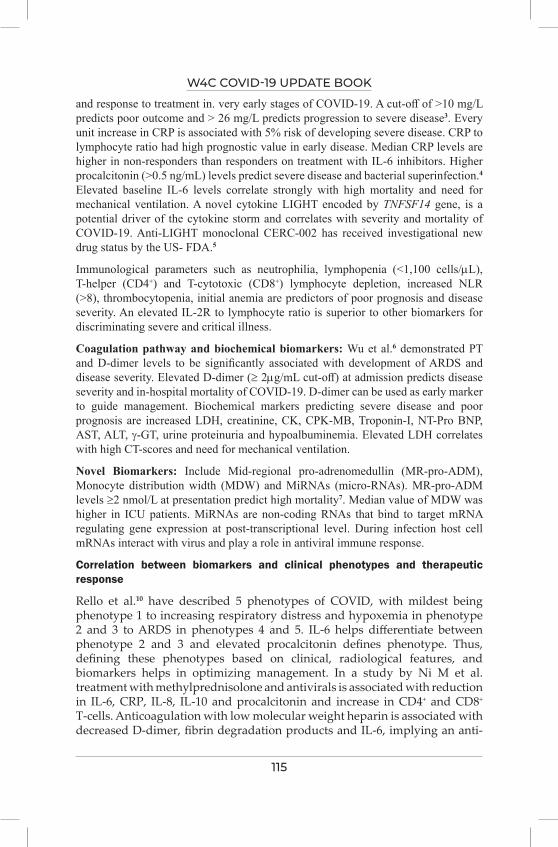
W4C COVID-19 UPDATE BOOK
115
and response to treatment in. very early stages of COVID-19. A cut-off of >10 mg/L predicts poor outcome and > 26 mg/L predicts progression to severe disease3. Every unit increase in CRP is associated with 5% risk of developing severe disease. CRP to lymphocyte ratio had high prognostic value in early disease. Median CRP levels are higher in non-responders than responders on treatment with IL-6 inhibitors. Higher procalcitonin (>0.5 ng/mL) levels predict severe disease and bacterial superinfection.4 Elevated baseline IL-6 levels correlate strongly with high mortality and need for mechanical ventilation. A novel cytokine LIGHT encoded by TNFSF14 gene, is a potential driver of the cytokine storm and correlates with severity and mortality of COVID-19. Anti-LIGHT monoclonal CERC-002 has received investigational new drug status by the US- FDA.5
Immunological parameters such as neutrophilia, lymphopenia (<1,100 cells/µL), T-helper (CD4+) and T-cytotoxic (CD8+) lymphocyte depletion, increased NLR (>8), thrombocytopenia, initial anemia are predictors of poor prognosis and disease severity. An elevated IL-2R to lymphocyte ratio is superior to other biomarkers for discriminating severe and critical illness.
Coagulation pathway and biochemical biomarkers: Wu et al.6 demonstrated PT and D-dimer levels to be significantly associated with development of ARDS and disease severity. Elevated D-dimer (≥ 2µg/mL cut-off) at admission predicts disease severity and in-hospital mortality of COVID-19. D-dimer can be used as early marker to guide management. Biochemical markers predicting severe disease and poor prognosis are increased LDH, creatinine, CK, CPK-MB, Troponin-I, NT-Pro BNP, AST, ALT, γ-GT, urine proteinuria and hypoalbuminemia. Elevated LDH correlates with high CT-scores and need for mechanical ventilation.
Novel Biomarkers: Include Mid-regional pro-adrenomedullin (MR-pro-ADM), Monocyte distribution width (MDW) and MiRNAs (micro-RNAs). MR-pro-ADM levels ≥2 nmol/L at presentation predict high mortality7. Median value of MDW was higher in ICU patients. MiRNAs are non-coding RNAs that bind to target mRNA regulating gene expression at post-transcriptional level. During infection host cell mRNAs interact with virus and play a role in antiviral immune response.
Correlation between biomarkers and clinical phenotypes and therapeutic response
Rello et al.10 have described 5 phenotypes of COVID, with mildest being phenotype 1 to increasing respiratory distress and hypoxemia in phenotype 2 and 3 to ARDS in phenotypes 4 and 5. IL-6 helps differentiate between phenotype 2 and 3 and elevated procalcitonin defines phenotype. Thus, defining these phenotypes based on clinical, radiological features, and biomarkers helps in optimizing management. In a study by Ni M et al. treatment with methylprednisolone and antivirals is associated with reduction in IL-6, CRP, IL-8, IL-10 and procalcitonin and increase in CD4+ and CD8+ T-cells. Anticoagulation with low molecular weight heparin is associated with decreased D-dimer, fibrin degradation products and IL-6, implying an anti-
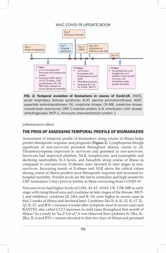
W4C COVID-19 UPDATE BOOK
116
inflammatory effect.
THE PROS OF ASSESSING TEMPORAL PROFILE OF BIOMARKERSAssessment of temporal profile of biomarkers along course of illness helps predict therapeutic response and prognosis (Figure 2). Lymphopenia though significant in non-survivors persisted throughout disease course in all. Thrombocytopenia improved in survivors and persisted in non-survivors. Survivors had improved platelets, NLR, lymphocytes, and eosinophils and declining neutrophils, IL-6 levels, and basophils along course of illness as compared to non-survivors. D-dimers were elevated in later stages in non-survivors. Increasing trends of D-dimer and NLR above the critical values during course of illness predicts poor therapeutic response and increased in-hospital mortality. Ferritin levels are the last to normalize and high sensitivity CRP normalizes 5 days prior to ferritin in those recovering from COVID-19.
Non-survivors had higher levels of LDH, ALAT, ASAT, CK, CPK-MB in early stage with rising blood urea and creatinine in later stages of the disease. MCP-1 and inhibitory cytokines (IL-1RA and IL-10) were higher in severe cases in first 2 weeks of illness and declined later. Cytokines like IL-6, IL-12, IL-17, IL-1β, IL-27, and IFN-γ increase 4 weeks after symptom onset in severe cases and RANTES, also called CCL5 increases in mild cases throughout first month of illness.8 In a study by Xu Z S et al.9 it was observed that cytokines IL-1Rα, IL-2Rα, IL-6 and IFN-γ remain elevated in first five days of illness and persisted
Figure. 2: Temporal evolution of biomarkers in course of Covid-19 ARDS, acute respiratory distress syndrome; ALAT, alanine aminotransferase; ASAT, aspartate aminotransferase; CK, creatinine kinase; CK-MB, creatinine kinase muscle-brain isoenzyme; CRP, C-reactive protein; IL-6, interleukin; LDH, lactate dehydrogenase; MCP-1, monocyte chemoattractant protein 1.
Day 1 Onset of symptoms
< 7 days: Low to normal total
leucocyte and lymphocyte counts
Increased LDH, ASAT, ALAT, D-dimer, CRP, procalcitonin, CK, CK-MB: Early markers of severe disease & mortality
7-14 days: At 8-9 days
leucopenia and lymphopenia reach nadir
Thrombocytopenia Increased IL-6, IL-
10, IL1RA, MCP-1
> 14 days: Increasing leucocyte,
lymphocyte and platelet counts predict recovery and declining counts predict mortality
Day 10-11 Admission to Intensive care unit
Day 9 ARDS
Day 7 Hospital admission
Day 8 Shortness of breath
Day 14
FIG. 2: Temporal evolution of biomarkers in course of Covid-19. ARDS, acute respiratory distress syndrome; ALAT, alanine aminotransferase; ASAT, aspartate aminotransferase; CK, creatinine kinase; CK-MB, creatinine kinase muscle-brain isoenzyme; CRP, C-reactive protein; IL-6, interleukin; LDH, lactate dehydrogenase; MCP-1, monocyte chemoattractant protein 1.
W4C COVID-19 UPDATE BOOK
117
beyond 14 days amongst non-survivors.
CONCLUSIONWorld is facing the challenging crisis of COVID-19 pandemic with limited health resources. COVID is an illness with heterogenous spectrum varying with age and underlying co-morbidities. Biomarkers will be the potential key in bringing a paradigm shift in early diagnosis, risk stratification, management, prognostication, as well as further prevention to curb this unprecedented crisis. Although, clinical assessment is indispensable at every step, panel of biomarkers rather than a single biomarker shall provide more reliable information and improvise decision making with regard to disease management. Several biomarkers like CRP, NLR, procalcitonin and D-dimer have proven clinical usefulness in management and prognostication of COVID-19. Further research is needed to study correlation of biomarkers with viral load, effect of treatment and temporal evolution and to find novel potential biomarkers.
Practice Points• In asymptomatic and mild cases of COVID-19, no investigations are
needed.• No single biomarker is sufficient to judge severity, treatment response
or prognosis and a panel is needed. Patients with mild disease and co-morbidities or those with moderate disease require a complete blood count, creatinine, liver function tests, and CRP at baseline. If any of these is abnormal, further panel of PT, INR, D-dimer, ferritin, LDH, Procalcitonin and cardiac biomarkers (Troponin-I and CPKMB) must be assessed. Persisting symptoms beyond 1 week of illness, require reassessment of CBC and CRP.
• In severe cases of COVID-19, a panel of CBC with platelet count, NLR, CRP, liver function tests, creatinine, serum albumin, LDH, procalcitonin, ferritin and cardiac biomarkers is recommended for therapeutic management and prognostication.
• Critically ill patients require monitoring with help of serum IL-6 and serial lactate levels.
• Monitoring of response in hospitalized patients is done by repeating CBC and CRP. in every 48-72 hours.
REFERENCES 1. Maxmen A. Faster, better, cheaper: the rise of CRISPR in disease detection. Nature 2019;
566:437.2. Pinto D, Park Y-J, Beltramello M, et al. Cross-neutralization of SARS-CoV-2 by a human
monoclonal SARS-CoV antibody. Nature 2020.3. Huang I, Pranata R, Lim MA, Oehadian A, Alisjahbana B. C-reactive protein, procalcitonin,
D-dimer, and ferritin in severe coronavirus disease- 2019: a meta-analysis. Ther Adv Respir Dis
W4C COVID-19 UPDATE BOOK
118
(2020) 14:1753466620937175. doi: 10.1177/1753466620937175.4. Wang G, Wu C, Zhang Q, Wu F, Yu B, Lv J, et al. C-Reactive protein level may predict the risk
of COVID-19 aggravation. Open Forum Infect Dis (2020) 7:ofaa153. doi:10.1093/ofid/ofaa153.5. [press release]. Cerecor Announces FDA Clearance of IND for CERC-002 in COVID-19
Induced ARDS. Cerecor 2020.6. Wu X, Liu L, Jiao J, Yang L, Zhu B, Li X. Characterisation of clinical, laboratory and imaging
factors related to mild vs. severe covid-19 infection: a systematic review and meta-analysis. Ann Med 2020; 52:334–344. doi: 10.1080/07853890.2020.1802061.
7. Spoto S, Agro FE, Sambuco F, Travaglino F, Valeriani E, Fogolari M, et al. High value of mid-regional proadrenomedullin in COVID-19: a mark- er of widespread endothelial damage, disease severity and mortality. J Med Virol 2020; 93:2820-7.
8. Zhao Y, Qin L, Zhang P, Li K, Liang L, Sun J, et al. Longitudinal COVID-19 profiling associates IL-1RA and IL-10 with disease severity and RANTES with mild disease. JCI Insight 2020; 5:e139834. doi: 10.1172/jci.insight139834.
9. Xu ZS, Shu T, Kang L, Wu D, Zhou X, Liao BW, et al. Temporal profiling of plasma cytokines, chemokines and growth factors from mild, severe and fatal COVID-19 patients. Signal Transduct Target Therapy 2020; 5:100. doi: 10.1038/s41392-020-0211-1.
10. Rello J, Storti E, Belliato M, Serrano R. Clinical phenotypes of SARS-CoV-2: implications for clinicians and researchers. Eur Respir J 2020; 55:2001028. doi: 10.1183/1399300301028-2020.
119
Limitations of Biomarkers in COviD-19 14.
INTRODUCTION A biomarker in literature defined as a “characteristic that can be objectively measured and evaluated as an indicator of normal biological and pathological processes, or pharmacological responses to a therapeutic intervention”.1 WHO has expanded the definition on wider vision by quoting “almost any measurement reflecting an interaction between a biological system and a potential hazard, which may be chemical, physical, or biological. The measured response may be functional and physiological, biochemical at the cellular level, or at molecular interaction.”2 In COVID-19 biomarkers varies from simple complete blood count, liver and renal function test to measurement of extensive study of cytokines/chemokines level.
Biomarkers can be useful in COVID-19 disease for early diagnosis of disease/cytokine syndrome, classification of disease severity, prognostication, resource allocation in hospital, admission/discharge criteria, optimise therapies and to detect therapy response.3 But it should be kept in mind that they come with their limitations and non-specificity.
PATHOPHYSIOLOGY/ SCIENTIFIC RATIONALE/ BIOLOGICAL PLAUSIBILITY OF BIOMARKERS IN COVID-19As the science evolve and COVID-19 era passing by we got the idea that COVID-19 is not just respiratory illness but it is multi-system disease. Not only ARDS but it is responsible for systemic inflammation, coagulopathy and immunomodulation. Chemistry between different host response and virus give rise to variety of presentation, illness and outcome. Sometime this uncontrolled pro-inflammatory process give rise to chemokine/cytokine surge known as ‘cytokine storm’. COVID-19 also responsible for endothelial damage give rise to micro-thrombosis and compromised tissue perfusion. Direct viral damage plus decrease production/proliferation of lymphocyte by cytokines/interleukins give rise to one of the predominant finding of
Kapil Zirpe, Balikrishna Nimavat
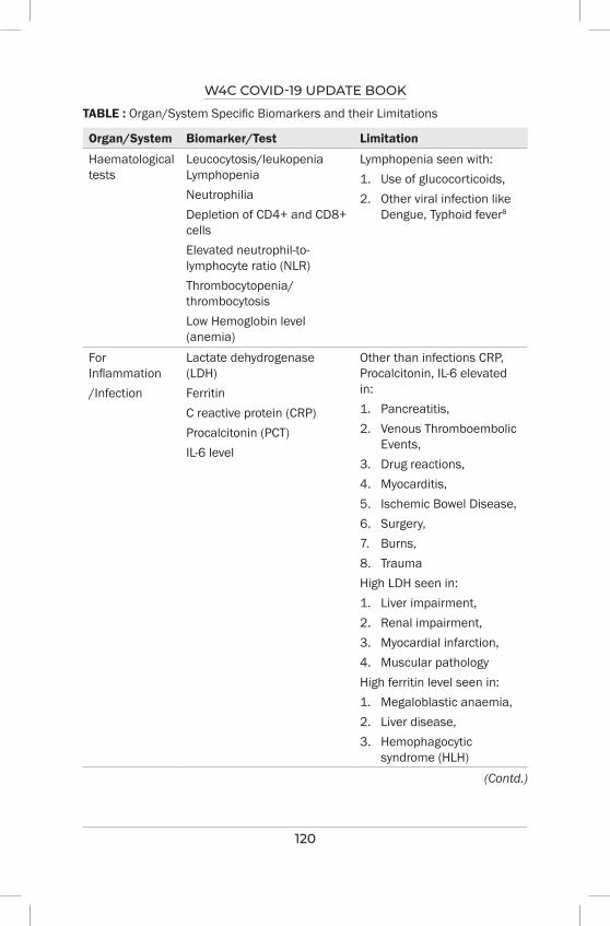
W4C COVID-19 UPDATE BOOK
120
TABLE : Organ/System Specific Biomarkers and their Limitations
Organ/System Biomarker/Test Limitation
Haematological tests
Leucocytosis/leukopenia Lymphopenia Neutrophilia Depletion of CD4+ and CD8+ cells Elevated neutrophil-to-lymphocyte ratio (NLR) Thrombocytopenia/thrombocytosisLow Hemoglobin level (anemia)
Lymphopenia seen with:1. Use of glucocorticoids,2. Other viral infection like
Dengue, Typhoid fever8
For Inflammation/Infection
Lactate dehydrogenase (LDH)Ferritin C reactive protein (CRP)Procalcitonin (PCT)IL-6 level
Other than infections CRP, Procalcitonin, IL-6 elevated in:1. Pancreatitis,2. Venous Thromboembolic
Events,3. Drug reactions,4. Myocarditis, 5. Ischemic Bowel Disease, 6. Surgery,7. Burns,8. TraumaHigh LDH seen in:1. Liver impairment,2. Renal impairment,3. Myocardial infarction, 4. Muscular pathologyHigh ferritin level seen in:1. Megaloblastic anaemia,2. Liver disease,3. Hemophagocytic
syndrome (HLH)(Contd.)
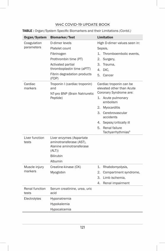
W4C COVID-19 UPDATE BOOK
121
Organ/System Biomarker/Test Limitation
Coagulation parameters
D-dimer levels Platelet countFibrinogen Prothrombin time (PT) Activated partial thromboplastin time (aPTT)Fibrin degradation products (FDP)
High D-dimer values seen in:Sepsis, 1. Thromboembolic events,2. Surgery,3. Trauma, 4. DIC,5. Cancer
Cardiac markers
Troponin I (cardiac troponin) and NT-pro BNP (Brain Natriuretic Peptide)
Cardiac troponin can be elevated other than Acute Coronary Syndrome are:1. Acute pulmonary
embolism 2. Myocarditis 3. Cerebrovascular
accidents 4. Sepsis/critically ill 5. Renal failure
Tachyarrhythmias9
Liver function tests
Liver enzymes (Aspartate aminotransferase (AST), Alanine aminotransferase (ALT)) Bilirubin Albumin
Muscle injury markers
Creatine-kinase (CK) Myoglobin
1. Rhabdomyolysis,2. Compartment syndrome, 3. Limb ischemia,4. Renal impairment
Renal function tests
Serum creatinine, urea, uric acid
Electrolytes Hyponatremia Hypokalemia Hypocalcemia
TABLE : Organ/System Specific Biomarkers and their Limitations (Contd.)
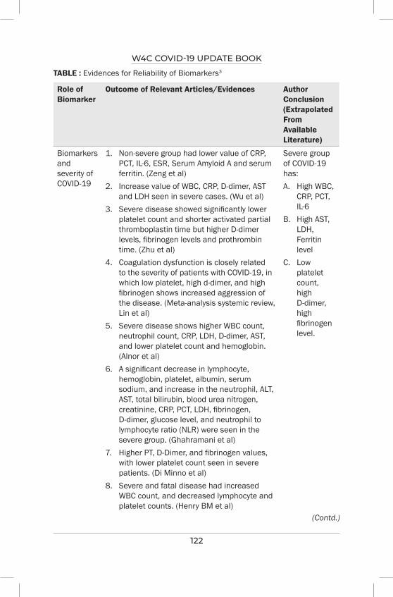
W4C COVID-19 UPDATE BOOK
122
TABLE : Evidences for Reliability of Biomarkers3
Role of Biomarker
Outcome of Relevant Articles/Evidences Author Conclusion (Extrapolated From Available Literature)
Biomarkers and severity of COVID-19
1. Non-severe group had lower value of CRP, PCT, IL-6, ESR, Serum Amyloid A and serum ferritin. (Zeng et al)
2. Increase value of WBC, CRP, D-dimer, AST and LDH seen in severe cases. (Wu et al)
3. Severe disease showed significantly lower platelet count and shorter activated partial thromboplastin time but higher D-dimer levels, fibrinogen levels and prothrombin time. (Zhu et al)
4. Coagulation dysfunction is closely related to the severity of patients with COVID-19, in which low platelet, high d-dimer, and high fibrinogen shows increased aggression of the disease. (Meta-analysis systemic review, Lin et al)
5. Severe disease shows higher WBC count, neutrophil count, CRP, LDH, D-dimer, AST, and lower platelet count and hemoglobin. (Alnor et al)
6. A significant decrease in lymphocyte, hemoglobin, platelet, albumin, serum sodium, and increase in the neutrophil, ALT, AST, total bilirubin, blood urea nitrogen, creatinine, CRP, PCT, LDH, fibrinogen, D-dimer, glucose level, and neutrophil to lymphocyte ratio (NLR) were seen in the severe group. (Ghahramani et al)
7. Higher PT, D-Dimer, and fibrinogen values, with lower platelet count seen in severe patients. (Di Minno et al)
8. Severe and fatal disease had increased WBC count, and decreased lymphocyte and platelet counts. (Henry BM et al)
Severe group of COVID-19 has:A. High WBC,
CRP, PCT, IL-6
B. High AST, LDH, Ferritin level
C. Low platelet count, high D-dimer, high fibrinogen level.
(Contd.)
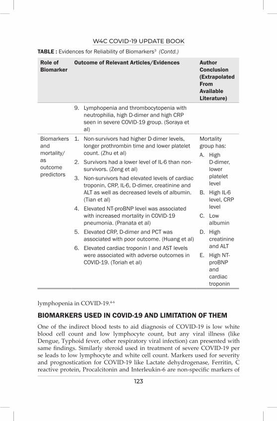
W4C COVID-19 UPDATE BOOK
123
Role of Biomarker
Outcome of Relevant Articles/Evidences Author Conclusion (Extrapolated From Available Literature)
9. Lymphopenia and thrombocytopenia with neutrophilia, high D-dimer and high CRP seen in severe COVID-19 group. (Soraya et al)
Biomarkers and mortality/ as outcome predictors
1. Non-survivors had higher D-dimer levels, longer prothrombin time and lower platelet count. (Zhu et al)
2. Survivors had a lower level of IL-6 than non-survivors. (Zeng et al)
3. Non-survivors had elevated levels of cardiac troponin, CRP, IL-6, D-dimer, creatinine and ALT as well as decreased levels of albumin. (Tian et al)
4. Elevated NT-proBNP level was associated with increased mortality in COVID-19 pneumonia. (Pranata et al)
5. Elevated CRP, D-dimer and PCT was associated with poor outcome. (Huang et al)
6. Elevated cardiac troponin I and AST levels were associated with adverse outcomes in COVID-19. (Toriah et al)
Mortality group has:A. High
D-dimer, lower platelet level
B. High IL-6 level, CRP level
C. Low albumin
D. High creatinine and ALT
E. High NT-proBNP and cardiac troponin
TABLE : Evidences for Reliability of Biomarkers3 (Contd.)
lymphopenia in COVID-19.4-6
BIOMARKERS USED IN COVID-19 AND LIMITATION OF THEMOne of the indirect blood tests to aid diagnosis of COVID-19 is low white blood cell count and low lymphocyte count, but any viral illness (like Dengue, Typhoid fever, other respiratory viral infection) can presented with same findings. Similarly steroid used in treatment of severe COVID-19 per se leads to low lymphocyte and white cell count. Markers used for severity and prognostication for COVID-19 like Lactate dehydrogenase, Ferritin, C reactive protein, Procalcitonin and Interleukin-6 are non-specific markers of

W4C COVID-19 UPDATE BOOK
124
inflammatory process. It is very difficult to differentiate inflammation from infection with help of biomarkers. High values can be seen in non-infectious conditions like pancreatitis, deep vein thrombosis, drug reaction, trauma and burns. Pattern of inflammatory markers or cytokines are also not well defined by literature. Cut-off for high values also very vague without any scientific evidence. Some of the studies shows that circulating cytokine level in COVID-19 are low compared to bacterial infection and those seen in critical ill patients.7
ROLE OF BIOMARKERS FOR TREATMENT MODIFICATIONS1. D-dimer level helps in risk stratification and titration of anticoagulation
treatment in COVID-19 patients.10
2. Based on IL-6 level, lung findings and severity of disease decision of Tocilizumab should be considered.11-13
3. Based on Biomarker profile/pattern COVID ARDS having different phenotypes, that helps to personalised the therapy.14
LIMITATION OF BIOMARKER AS SURROGATE AND CLINICAL END POINT15
Biomarkers are considered as surrogate end points so they may not always truly represent patient clinical condition or disease state/progress. Secondly before selection of any biomarker its validity and relevance should be assessed. It is highly possible that biomarker guided therapy or normalising value may not translated to clinical meaningful outcome if they are not truly representative (having low validity or relevance).
PRACTICE POINTS/GENERAL RECOMMENDATION1. It is always advisable to do serial values of particular biomarker and
watch for trend rather than single value.
2. Cut-off value is not well defined for biomarkers in COVID-19 (like for IL-6, CRP, Procalcitonin, D-dimer, lymphopenia). More robust trials/analysis needed to find out particular cut-off value specific for COVID-19.
3. IL-6 is not specific for cytokine storm, which panel of interleukin or what combinations of biomarkers going to decide cytokine storm is also not well established. Particular pattern should be identified for COVID-19 and cytokine storm.
4. No investigations needed for asymptomatic patients or patient having mild disease with no comorbid condition.
5. For mild disease with comorbidity basic tests like complete blood count, liver and renal function test should be done. Value of CRP trend also helps to guide therapy.
W4C COVID-19 UPDATE BOOK
125
6. For severe disease along with basic investigations coagulation profile (PT/INR, aPTT, fibrinogen and D-dimer), markers for inflammation (like serum ferritin, LDH, CRP, PCT, IL-6) and cardiac biomarkers (NT-pro-BNP and troponin I) should be sent. Trend of CRP, D-dimer and IL-6 should help the physician to guide therapy.
SUMMARYGuiding treatment based on just biomarkers without clinical condition/context of disease is like shooting arrow in dark. Biomarkers should be always interpreted with caution and with trend results not the single value. With above mentioned limitations, they are quite helpful in guiding disease management and prognostication of COVID-19 disease.
REFERENCES1. Biomarkers and surrogate endpoints: preferred definitions and conceptual framework. Clin
Pharmacol Ther [Internet]. 2001 Jan 1 [cited 2021 Aug 28];69(3):89–95. Available from: https://pubmed.ncbi.nlm.nih.gov/11240971/
2. Organization WH. Biomarkers In Risk Assessment: Validity And Validation. Environ Heal [Internet]. 2001 [cited 2021 Aug 29];144. Available from: https://apps.who.int/iris/handle/10665/42363
3. Samprathi M, Jayashree M. Biomarkers in COVID-19: An Up-To-Date Review. Front Pediatr 2021; 0:972.
4. MC, FF, MR, EC, SG, CL. Immune response in COVID-19: addressing a pharmacological challenge by targeting pathways triggered by SARS-CoV-2. Signal Transduct Target Ther [Internet]. 2020 Dec 1 [cited 2021 Aug 29];5(1). Available from: https://pubmed.ncbi.nlm.nih.gov/32467561/
5. WB M. Thromboinflammation in COVID-19 acute lung injury. Paediatr Respir Rev [Internet] 2020 Sep 1 [cited 2021 Aug 29];35:20–4. Available from: https://pubmed.ncbi.nlm.nih.gov/32653469/
6. ET, IN-S, IE, EK, TN S, MP, et al. Hematological findings and complications of COVID-19. Am J Hematol [Internet] 2020 Jul 1 [cited 2021 Aug 29]; 95:834–47. Available from: https://pubmed.ncbi.nlm.nih.gov/32282949/
7. Kox M, Waalders NJB, Kooistra EJ, Gerretsen J, Pickkers P. Cytokine Levels in Critically Ill Patients With COVID-19 and Other Conditions. JAMA [Internet] 2020 Oct 20 [cited 2021 Sep 29];324(15):1565–7. Available from: https://jamanetwork.com/journals/jama/fullarticle/2770484
8. AR, NK, AB, KL, LM. [Lymphocytopenia: aetiology and diagnosis, when to think about idiopathic CD4(+) lymphocytopenia?]. La Rev Med interne [Internet] 2012 Nov [cited 2021 Sep 29];33(11):628–34. Available from: https://pubmed.ncbi.nlm.nih.gov/22658164/
9. Elnahar Y, Daoko J, Kersh K El, Kam JC, Sarraf C, Shamoon F. False elevation of cardiac markers: importance of recognition. Res Reports Clin Cardiol [Internet] 2011 Mar 23 [cited 2021 Aug 30];2:37–40. Available from: https://www.dovepress.com/false-elevation-of-cardiac-markers-importance-of-recognition-peer-reviewed-fulltext-article-RRCC
10. CS, CW, HW, CY, FC, FZ, et al. The Potential of Low Molecular Weight Heparin to Mitigate Cytokine Storm in Severe COVID-19 Patients: A Retrospective Cohort Study. Clin Transl Sci [Internet] 2020 Nov 1 [cited 2021 Aug 29];13(6):1087–95. Available from: https://pubmed.ncbi.nlm.nih.gov/32881340/
11. Kucukoglu K, Faydalı N, Bul D. What are the drugs having potential against COVID-19? Med Chem Res [Internet] 2020 Nov 1 [cited 2021 Aug 29];29(11):1. Available from: /pmc/articles/PMC7481551/
12. RM P, NC S, NW VH, RM F, JP, VD, et al. Tocilizumab as a Therapeutic Agent for Critically Ill
W4C COVID-19 UPDATE BOOK
126
Patients Infected with SARS-CoV-2. Clin Transl Sci [Internet] 2020 Sep 12 [cited 2021 Aug 29]; Available from: https://pubmed.ncbi.nlm.nih.gov/32918792/
13. BF. COVACTA trial raises questions about tocilizumab’s benefit in COVID-19. Lancet Rheumatol [Internet] 2020 Oct [cited 2021 Aug 29];2(10):e592. Available from: https://pubmed.ncbi.nlm.nih.gov/32929415/
14. Rello J, Storti E, Belliato M, Serrano R. Clinical phenotypes of SARS-CoV-2: implications for clinicians and researchers. Eur Respir J [Internet] 2020 [cited 2021 Aug 29];55(5). Available from: /pmc/articles/PMC7236837/
15. Strimbu K, Tavel JA. What are Biomarkers? Curr Opin HIV AIDS [Internet] 2010 Nov [cited 2021 Aug 29];5(6):463. Available from: /pmc/articles/PMC3078627/.
ManagementRESPIRATORY SUPPORT IN COVID-19
SECTION 4-A

128
129
Section 4 - ManagementSECTION EDITOR
Dr Kedar ToraskarMD EDIC Director Critical Care, Critical Care Unit, Wockhardt Hospital, South Mumbai
Preface
SUB SECTION - RESPIRATORY SUPPORT IN COVID-19Respiratory support in form of oxygenation & ventilation is an integral part of therapy in patients with covid 19 presenting with hypoxemia & respiratory failure. The pandemic highlighted the importance of critical care units in managing the critically ill covid 19 patients with respiratory failure. The concept of happy
hypoxia was first coined for covid 19 patients who classically presented with minimal symptoms disproportionate to the hypoxemia.
Oxygen is a licensed drug & should be prescribed judiciously. All of us at some point of time faced oxygen shortage during the second wave of this deadly pandemic. It has reiterated the need for oxygen audits which need to be revamped to conserve this life saving elixir which is often abused. Finally, we all need to know that oxygen is supportive & not curative. So, we need to hunt for the cause of hypoxemia & treat it effectively rather than being complacent after starting supplemental oxygen. This is also true for mechanical ventilation which just buys us time till the underlying disease process is treated effectively.
High flow nasal cannula (HFNC) was reinvented during this pandemic as an effective means of delivering high concentration of humidified oxygen with a low peep to obviate intubation & invasive ventilation in some subset of patients with covid ARDS. But unfortunately, it bleeds oxygen & is not available universally especially in remote rural areas. Similarly noninvasive ventilation (NIV) does help in select group of patients with covid ARDS. Again, delaying intubation & invasive mechanical ventilation leads to poorer outcomes especially in patients who have failed a trial of NIV or HFNC.

130
The debate about early versus late intubation is always on for ARDS patients & the key is to follow the physiological principles & protocols guiding intubation & ventilation. Inadvertent delay leads to poorer outcomes & increase in the mortality which was highlighted in this pandemic. Gattinoni & colleagues introduced the concept of Covid ARDS (CARDS) L & H phenotypes in the early phase of the pandemic which gave us insights into ventilating these different phenotypes. But looking back, the CARDS lung is always a combination of both the phenotypes which overlap with each other.
This section covers all the aspects of oxygenation & respiratory support holistically.
131
Methods of Oxygen Therapy and Targets
INTRODUCTIONSARS-COVID-19 pandemic taught the medical fraternity amongst many things precision oxygen therapy not only to its professionals but also to millions of lay public as well. Never before oxygen consumption was observed in such a large scale in hospitals, homes and even in the transit vehicles. Shortages of oxygen in many parts of the world was a reality all of us saw and heard. Methods of oxygen delivery have been devised and even targets revised keeping conservation in mind in many circumstances, hither to unheard of, while delivering care. Need for oxygen therapy has often been the deciding factor for type of care in COVID-19 like “no impending need for oxygen” could be treated in home isolation and “a need for oxygen therapy” would require a hospital admission as it creates panic in the minds of the patient and the family members.
TYPES OF OXYGEN THERAPY
Under normo-baric conditions, there are broadly two types of devices such as (i) variable performance device and ii) fixed performance device. Variable performance devices are the ones which deliver variable inspired oxygen concentrations (FiO2) due to changes in minute ventilation, faulty position and at times displacement of the devices (nasal catheter, nasal prongs, face mask and non-rebreathing mask). These are also called low flow systems. Fixed performance devices are the ones which deliver fixed FiO2 which is determined as per settings in the devices (venturi mask, high flow nasal canula called HFNC, non-invasive ventilation called NIV, invasive ventilation called IPPV and extra corporeal membrane oxygenation called ECMO). Fixed performance devices are described separately in this book and therefore the focus will be on the variable performance devices. Methods are also classified as per “dependence” of the patient on oxygen requirement: (i) low dependency (supplemental oxygen alone in a spontaneously breathing patient), (ii) medium dependency (supplemental oxygen + a degree of assistance such as CPAP in a spontaneously breathing patient) and (iii) high dependency
Banambar Ray, Anand Mishra
15.
W4C COVID-19 UPDATE BOOK
132
(supplemental oxygen + full respiratory support such as noninvasive / invasive positive pressure ventilation i.e. NIPPV / IPPV). Medium and high dependency oxygen therapy devices are separately discussed in this book.
WHEN OXYGEN THERAPY IS NECESSARY?Pulse oximeter reading generally guides oxygen therapy. SpO2 of > 95% in an adult is considered safe and < 92% requires oxygen administration. An arterial oxygen pressure of 75-100 mm Hg is normally present in blood. However, a pressure of < 60 mm Hg (hypoxemic respiratory failure) signals oxygen administration. Oxygen is required in COVID-19 patients, generally in the second week, particularly when they are dyspnec. These patients need to be hospitalized as they can worsen to a stage when they will require higher modes of oxygen therapy such as HFNC, non-rebreathing mask, non-invasive ventilation, intubation and invasive ventilation.
MODES OF OXYGEN THERAPY (FOR LOW FLOW DEVICES)Nasal canula, face mask, partial rebreathing mask and non-rebreathing mask come under this category.
Nasal canula: it is the commonest oxygen therapy device which delivers a flow up to 5.0 litres / minute and the FiO2 is up to 0.4 (21% + 4 x litres of flow). It is most comfortable, light weight, without need for humidification and it allows patient to talk, eat and drink. It is also of low cost. Aerosol generation, an important consideration in Covid 19 patients, is minimal in this device. It, how ever, can cause irritation, abrasion in the nostrils and it can not be used in nasal obstruction. It can not be used for higher oxygen eeds.
Face mask: Its a transparent mask with two side holes for air dilution and expiration. It has a dead space of 100-250 mls. Oxygen flow should be 4-8 litres (not more and not less). A flow < 4.0 litres causes rebreathing and > 8.0 litres does not improve FiO2. A flow of 5.0 litres/ min gives an FiO2 of 0.35-0.4. It is a variable FiO2 depending on the minute ventilation. A higher minute ventilation can also cause rebreathing. It is good for mouth breathers and less expensive. Covid19 patients often require high oxygen flow and have high minute ventilation making this device useless at an advanced stage of the disease. It is also sometimes uncomfortable, difficult to maintain position and tight seal, to speak, eat and to drink.
Partial rebreathing mask: It is a mask with reservoir bag of 1.0 litre capacity. Oxygen flows directly to the reservoir bag which swells up during exhalation. Its designed in such a way that initial part of exhalation (anatomical dead space gas) also comes to the reservoir bag thereby conserving oxygen. Minimum flow should be 8.0 litres and reservoir should be 1/2 to 2/3 full at all times. FiO2 delivered is 0.6-0.8. its advantage is that patient does not breathe room air unless oxygen supply is cut off when he can breathe room air through exhalation port.
W4C COVID-19 UPDATE BOOK
133
Non-rebreathing mask (NRBM): There are one- way valves in the exhalation ports, usually two, between the reservoir bag and mask which allow only exhalation gas to go out. There is no air dilution except when oxygen flow stops and patient can breathe air through one of the ports which does not have a valve and allows room air to enter the system. Flow rates of 10-15 litres can be given to raise FiO2 up to 1.0. This is often necessary for severe COVID-19 patients. This requires a tight seal and not suitable for long term use. It can disadvantage the patient for eating, talking and can cause suffocation. It can cause high aerosol generation and It is expensive. This however can prevent a COVID-19 patient from getting intubated.
TARGETS OF OXYGEN THERAPY IN COVID-19Optimal SpO2 level in COVID-19 is yet not decided. In a non-covid ARDS trial, comparison between conservative oxygen therapy (SpO2 target 88-92%) and liberal oxygen therapy (SpO2 target ≥ 96%) revealed an increased mortality at 90 days (risk difference of 14%; 95% CI 0.7 to 27%) and a trend towards increased mortality at 28 days (risk difference of 8%; 95% CI -5 to 21%).¹ In a meta-analysis from 25 randomized control trials involving non-covid patients, a liberal oxygen therapy with a median SpO2 of 96%, was associated with increased risk of in-hospital mortality as opposed to conservative oxygen therapy (RR 1.21; 95% CI 1.03-1.43).² So the confusion remains as to which oxygen therapy is better: conservative or liberal.
A target of ≥ 94% SpO2 is recommended by WHO for COVID hypoxemia and respiratory distress.³ Face mask with or without reservoir bag is recommended over nasal canula for the latter’s aerosol generation potential.⁴ Once patient is comfortable, a oxy-Hb saturation > 90% in adults (92-95% in pregnancy) is desirable.³,⁵ In adult COVID-19 patients with acute hypoxemic failure, SpO2 should not be > 96%.⁶ A COPD patient should have a target of SpO2 between 88 and 92%.³
CONCLUSIONOxygen Therapy is the main stay in COVID-19 treatment. In some of the mild and moderate cases, low flow systems, otherwise called variable performance devices, are good enough with less discomfort to the patient and less possibility of aerosolization in the care environment. WHO gives a SpO2 target of 92-96% while specifies ≥94% during resuscitation and > 90 after stabilization.
PRACTICE POINTS1. Target a SpO2 of ≥ 94% during resuscitation of hypoxemic respiratory
failure or respiratory distress in COVID-19.
2. Once patient is stabilized SpO2 of > 90% is acceptable.
3. For a COPD COVID-19 patient, SpO2 target is 88-92% and for a pregnant COVID patient, it is 92-95%.

W4C COVID-19 UPDATE BOOK
134
4. Face mask with or without reservoir bag is better than a nasal catheter as a device for oxygen therapy.
REFERENCES1. Barrot L, Astar P, Mauny F, et al. Liberal or conservative oxygen therapy for acute respiratory
distress syndrome. N Eng j Med 2020; 382:999-1008. Available at: https://www.ncbi.nlm.nih.gov/pubmed/32160661.
2. Chu DK, Kim LH, Young PJ, et al. Mortality and morbidity in acutely ill adults treated with liberal vs conservative oxygen therapy (IOTA): a systematic review and meta-analysis. Lancet 2018; 391:1693-1705. Available at: https://www.ncbi.nlm.nih.gov/pubmed/29726345.
3. World Health Organisation. Clinical Management of Severe Acute Respiratory Infection (SARI) when COVID-19 Disease is Suspected- Interim Guidance, WHO, 13 March 2020
4. Lazzeri M, Lanza A, Bellini R, et al. Respiratory physiotherapy in patients with COVID-19 infection in acute setting: a position paper of the Italian Association of Respiratory Physiotherapists (ARIR). Monaldi Archives for Chest Disease 2020; 90(1). doi:10.4081/monaldi.2020.1285.
5. Metro North, Interim infection prevention and control guidelines for the management of COVID-19 in healthcare settings, 2020: https://www:health.qld.gov.au/_data/assets/pdf_file/0038/939656/qh-covid-19-infection-control-guidelines.pdf.
6. Alhazzani W, Moller M, Arabi Y, et al. Surviving sepsis campaign: Guidelines of the Management of Critically Ill Adults with Coronavirus Disease 2019 (COVID-19). Intensive Care Med 2020; 46:854-887. doi: 10.1007/s00134-020-06022-5. Epub 2020 Mar 28
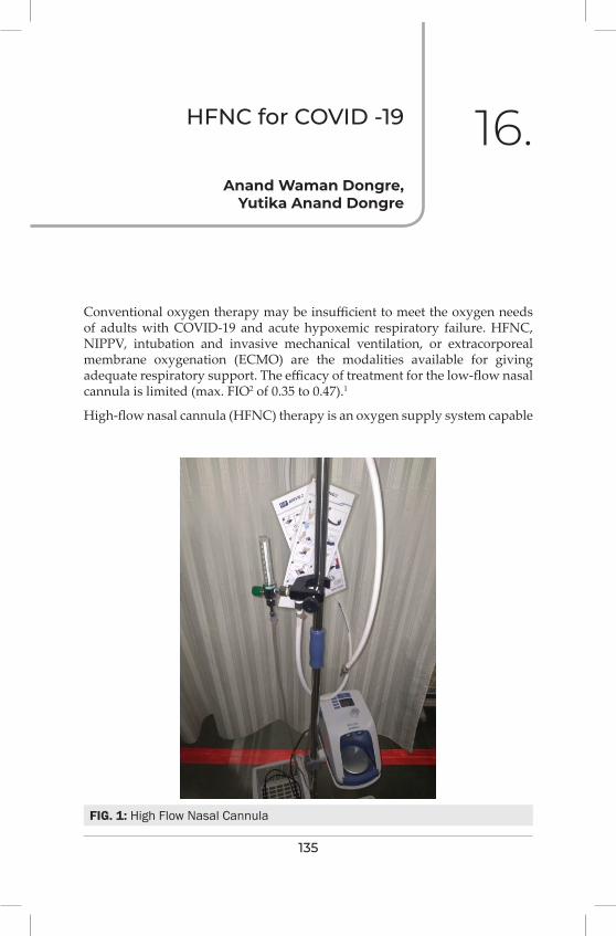
135
HFNC for COviD -19 16.
Conventional oxygen therapy may be insufficient to meet the oxygen needs of adults with COVID-19 and acute hypoxemic respiratory failure. HFNC, NIPPV, intubation and invasive mechanical ventilation, or extracorporeal membrane oxygenation (ECMO) are the modalities available for giving adequate respiratory support. The efficacy of treatment for the low-flow nasal cannula is limited (max. FIO2 of 0.35 to 0.47).1
High-flow nasal cannula (HFNC) therapy is an oxygen supply system capable
Anand Waman Dongre, Yutika Anand Dongre
FIG. 1: High Flow Nasal Cannula
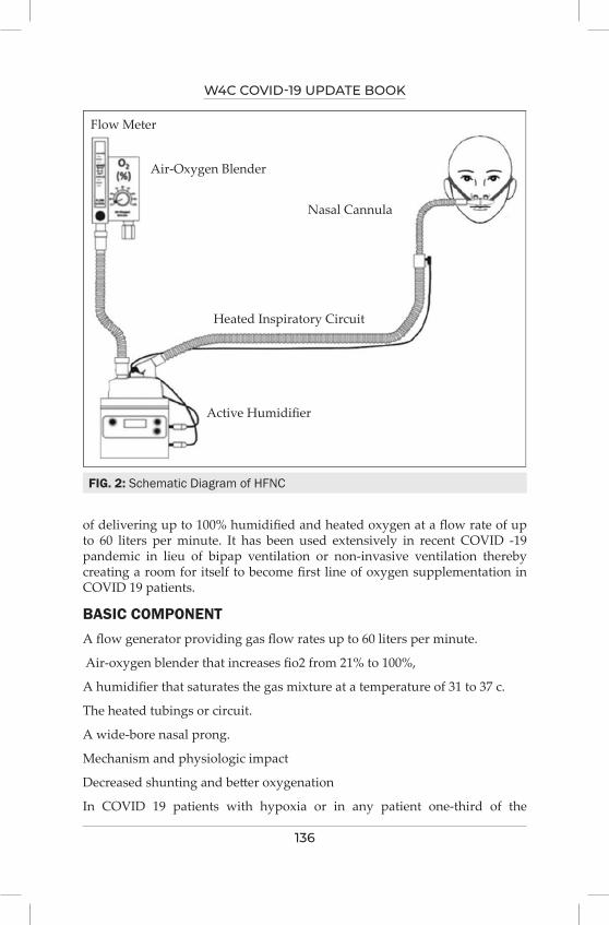
W4C COVID-19 UPDATE BOOK
136
of delivering up to 100% humidified and heated oxygen at a flow rate of up to 60 liters per minute. It has been used extensively in recent COVID -19 pandemic in lieu of bipap ventilation or non-invasive ventilation thereby creating a room for itself to become first line of oxygen supplementation in COVID 19 patients.
BASIC COMPONENT A flow generator providing gas flow rates up to 60 liters per minute.
Air-oxygen blender that increases fio2 from 21% to 100%,
A humidifier that saturates the gas mixture at a temperature of 31 to 37 c.
The heated tubings or circuit.
A wide-bore nasal prong.
Mechanism and physiologic impact
Decreased shunting and better oxygenation
In COVID 19 patients with hypoxia or in any patient one-third of the
FIG. 2: Schematic Diagram of HFNC
Flow Meter
Air-Oxygen Blender
Nasal Cannula
Heated Inspiratory Circuit
Active Humidifier
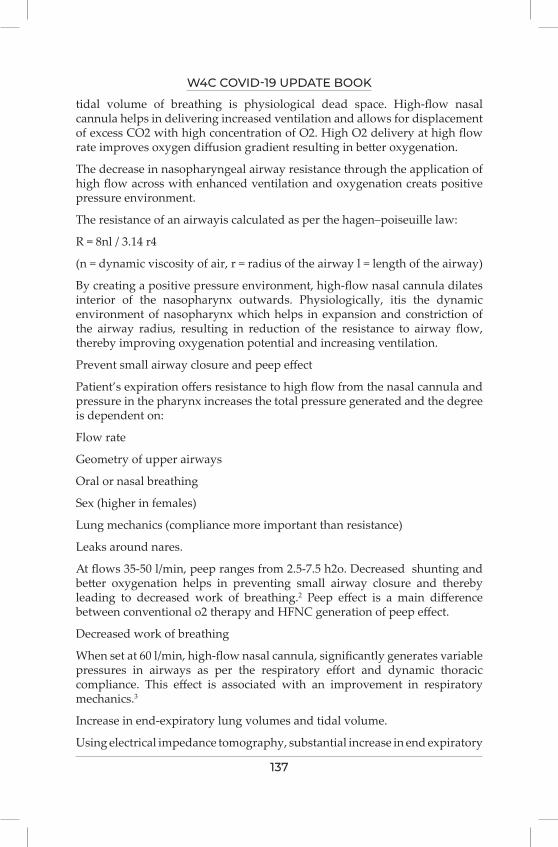
W4C COVID-19 UPDATE BOOK
137
tidal volume of breathing is physiological dead space. High-flow nasal cannula helps in delivering increased ventilation and allows for displacement of excess CO2 with high concentration of O2. High O2 delivery at high flow rate improves oxygen diffusion gradient resulting in better oxygenation.
The decrease in nasopharyngeal airway resistance through the application of high flow across with enhanced ventilation and oxygenation creats positive pressure environment.
The resistance of an airwayis calculated as per the hagen–poiseuille law:
R = 8nl / 3.14 r4
(n = dynamic viscosity of air, r = radius of the airway l = length of the airway)
By creating a positive pressure environment, high-flow nasal cannula dilates interior of the nasopharynx outwards. Physiologically, itis the dynamic environment of nasopharynx which helps in expansion and constriction of the airway radius, resulting in reduction of the resistance to airway flow, thereby improving oxygenation potential and increasing ventilation.
Prevent small airway closure and peep effect
Patient’s expiration offers resistance to high flow from the nasal cannula and pressure in the pharynx increases the total pressure generated and the degree is dependent on:
Flow rate
Geometry of upper airways
Oral or nasal breathing
Sex (higher in females)
Lung mechanics (compliance more important than resistance)
Leaks around nares.
At flows 35-50 l/min, peep ranges from 2.5-7.5 h2o. Decreased shunting and better oxygenation helps in preventing small airway closure and thereby leading to decreased work of breathing.2 Peep effect is a main difference between conventional o2 therapy and HFNC generation of peep effect.
Decreased work of breathing
When set at 60 l/min, high-flow nasal cannula, significantly generates variable pressures in airways as per the respiratory effort and dynamic thoracic compliance. This effect is associated with an improvement in respiratory mechanics.3
Increase in end-expiratory lung volumes and tidal volume.
Using electrical impedance tomography, substantial increase in end expiratory
W4C COVID-19 UPDATE BOOK
138
lung volumes, improvement in gas transfer and increase in lung volumes is seen with HFNC compared with low-flow devices.4
ADVANTAGES Better communication- HFNC provides better communication environment because of comfortable nasal interface as compared to non invasive ventilation facial interface.
Less mask removal – as these patients requires a longer duration of O2 support, frequent interferences seen in bipap ventilation with in facial interface is not required leading to uninterrupted oxygenation and patient compliance.
Enhanced patient comfort and compliance
OPTIMAL HUMIDITY CONTROL Absence of nasal or oral cavity drying due to humid and heated gasflow reduces the discomfort making this therapy more compliant even for longer duration.
Lack of humidity control leads to excessive water loss and inceased airway resistance, may induce bronchospasm and increased work of breathing. Also, it may lead to sub epithelial vascular congestion thereby causing loss of cilia and sloughing of epithelium. Decreased nasal and respiratory mucociliary clearance (up to complete cessation) are very often seen if humidity of flow gas is not taken care of especially in COVID patients.
INITIATION Patients are explained about nasal canula placement and instructed to breath at ease with closed mouth.
FIG. 3: HFNC Application on a Patient with COVID 19
W4C COVID-19 UPDATE BOOK
139
Initial flow rate FIO2 is selected are accessed on patient’s respiratory demand (e.g., respiratory rate, work of breathing, accessory muscle use) to achieve adequate oxygen saturation (84 –to 92% ).
Intiation flow increased in increments of 5l/min according to patient needs..
FIO2 is setat 21-100% to achieve spo2of more than 84% to 92%.
Ensure a propertubing support to avoid pull on nasal cannula.
Start with lower flow (20-35l/min).
On weaning off: decrease fio2 first then flow.
Indication in COVID patients-
HYPOXEMIC RESPIRATORY FAILURECompared to standard oxygen therapy, HFNC provides significant less non- invasive ventilation and less intubation rate with more ventilator-free days.5
HFNC can be regarded as a first-line treatment for patients with mild to moderate hypoxemic COVID 19 patients with acute respiratory failure, HFNC up to 7 days usually is free of frequent interruption due to intolerance or non compliance as seen with non invasive ventilation.
HFNC helps in reducing breathing frequency, SPO2, grdes of dyspnoea, supraclavicular retraction, heart rate and improves thoraco-abdominal synchrony. Delivered FIO2 is close toactual FI02 values. Timely intubation remains a challenging clinical dilemma as delaying intubation beyond 48 hours prolong ventilation and increases mortality. The respiratory rate–oxygenation (ROX) index, a clinical index to identify patients on HFNC likely to need mechanical vetilation is defined as the ratio of SPo2/FIo2 to respiratory rate. A ROX index greater than 4.88 after 12 hours of HFNC therapy indicates that a patient is unlikely to need mechanical ventilation (positive predictive value, 89%) 6.
Also clinical signs for respiratory failure should be
Taken into consideration as well.
Patients who are unlikely to be benefitted are those having paco2 > 45 mmhg, glasgow coma score of < 12, in shock, neutropenia (<500mm3) , cardiogenic pulmonary edema and requiring intubation.
CLINICAL SIGNS OF HFNC FAILURE 7
Persistent high RR.
Declining mental status.
Persistent hemodynamic instability.
W4C COVID-19 UPDATE BOOK
140
Presence of non-pulmonary organ failure.
Thoraco-abdominal asynchrony.
Ongoing hypoxemia.
OTHER INDICATIONS FOR APPLICATIN OF HFNCAcute hypoxemic respiratory failure
Acute pulmonary edema/ heart failure
Chronic obstructive pulmonary disease
Pre and post-extubation oxygenation
Obstructive sleep apnea
Do not intubate the patient
Post-surgical respiratory failure
Use in the emergency department.
PRACTICE POINTS-COVID 19High flow nasal canula is a simple and effective method for delivering oxygen therapy whenever saturation is below 92%.
It is better than conventional, low-flow devices in terms of comfort, gas exchange, and respiratory rate.
Compared to non invasive ventilation in COVID 19 patient, it is better tolerated with less interruptions and can be used for a longer periods although high consumption of oxygen is a matter of concern in covid pandemic.
If used judiciously, will help in improving oxygenation at ease for maximum number of patients during pandemic.
REFERENCES1. High flow nasal canula, Sharma S, Danckers M, Sanghavi D, Chakraborty RK. CBI bookshelf.
A service of the national library of medicine, national institutes of health. Statpearls [internet]. Treasure island (FL): statpearls publishing; 2021 jan.
2. Effects of high-flow nasal cannula on end-expiratory lung impedance in semi-seated healthy subjects. Gustavo AP, Thille AW, Vasquez DN, Pratto RA, Quiroga CM, Andrich ME, Dorado JH, Gomez RS, D’annunzio PA, Scapellato JL. dante intile 3 affiliations expand pmid: 29945910 doi: 10.4187/respcare.06031
3. Effects of high-flow nasal cannula on the work of breathing in patients recovering from acute respiratory failure august 2017critical care medicine 45(12):1 delorme et al, doi:10.1097/ccm.0000000000002693
4. Corley A, Caruana LR, Barnett AG, Tronstad O, Fraser JF. Oxygen delivery through high-flow nasal cannulae increase end-expiratory lung volume and reduce respiratory rate in post-cardiac surgical patients. Br J Anaesth 2011; 107:998–1004.
5. Physiologic effects of high-flow nasal cannula in acute hypoxemic respiratory failure.
W4C COVID-19 UPDATE BOOK
141
Tommasomauri, Turrini C, Nildeeronia, Grasselli G, Volta CA, Bellani G, Pesenti A. affiliations expand pmid: 27997805 doi: 10.1164/rccm.201605-0916oc
6. Roca O, Messika J, Caralt B, García-De-Acilu M, Sztrymf B, Ricard JD, et al. Predicting success of high-flow nasal cannula in pneumonia patients with hypoxemic respiratory failure: the utility of the rox index. J Crit Care 2016; 35:200–205.
7. High-flow nasal cannula oxygen in adults: an evidence-based assessment. Drake MG. 2017https://doi.org/10.1513/annalsats.201707-548fr pubmed: 29144160.
142
Use of Niv in Covid 19
INTRODUCTIONNoninvasive ventilation has traditionally been reserved for acute hypoxemic respiratory failure mild cases. A short trial of NIV usually preceded the decision to go for invasive ventilation. At the beginning of the COVID pandemic, a lot of recommendations actually were against the use of NIV due to the fear of generation of aerosols and hence the risk of infection to the health care worker.
As the pandemic progressed, the understanding of the benefit of NIV in these critically ill group of patients was actually improved beyond doubt and it was included in the guidelines.
It has gained immense usage not only in mild – moderate but also moderate-severe cases of ARDS (limited data). This has prevented invasive ventilation in a substantial number of cases and hence also the risks associated with invasive ventilation like infection, barotraumatic.
PATHOPHYSIOLOGYA constantly applied positive pressure could be helpful in recruiting collapsed lung segments and hence help achieve reasonable oxygenation, patient comfort. In a recent article, a description of 2 different subtypes of COVID infected lungs was made. The L type lung and the H type lung.
Patients usually exhibit a mixture of the 2 or may progressively merge into each other. The L group of patients are the ones whose lungs are “elastance of low type” &
compliance of the lungs is reasonable. They show benefit with higher tidal volumes (7-8 ml/kg IBW). This group may exhibit some benefit from appropriately timed NIV.
Due to worsening of the lung pathology these patients progress to the TYPE H. The H type lung are the ones who have “elastance of high type” with worsening lung compliance.
Pradeep Michael D'costa
17.
W4C COVID-19 UPDATE BOOK
143
show benefit with the traditional lower tidal volumes and respond well to PEEP.3
POSSIBLE RISKSThere has been a lot of speculation about the possible spread of the virus by the aerosolized route. It was postulated about the possible risks to the health care provider with the use of NIV due to the propensity to generate a large number of aerosols. The SARS CoV-2 virus particle measures around 60 to 140 nanometers. It has been postulated that the number of exhaled particles increases with an expiratory positive pressure of more than 5 cm of water.3
This number of exhaled particles reduced substantially at a distance of around 20 cm from the subject. Hence if the health care personnel are appropriately protected (good PPE-protective eye wear, fully protective masks, preferably N 95 type, and impervious gowns with face shields) the risk of spread is low. If a protective filter is applied on the expiratory limb of the NIV circuit, the risks could be reduced even further.
If the NIV system is used with non-vented masks and a viral filter is applied at the expiration valve level, risks of transmission are low. A vented mask system (venturi mask) increases the risks. A postulation that “leak” around the NIV circuit may also lead to increased aerosol generation must be kept in mind and efforts to minimize leak must be taken. The leakage flow estimated however was only around 4 inches. Similarly, nozzle type nebulization systems have been postulated to increase aerosols close to the patient, hence special care requires to be taken in these situations by the health care provider.1,3
The most risks of exposure are from the procedure of intubation, hence most precautions must be taken around this period.3
INDICATIONSVery careful patient selection is the key to a successful NIV use. Patient must be fully conscious and cooperative; the hemodynamic parameters must be reasonable and the hypoxia not too severe (some references mention po2/fio2 <150)
Once NIV is applied a very close watch must be kept on hemodynamic parameters, oxygenation parameters (ABG can be used), level of consciousness, secretions possibly hampering ventilation, and worsening in any of these should immediately guide the provider to proceed with invasive ventilatory strategies. Some authors suggest avoiding NIV if the SAPS score is more than 343,4
PROCEDURENoninvasive ventilation modes commonly include CPAP (continuous positive airway pressure) & BIPAP (bilevel positive airway pressure)
W4C COVID-19 UPDATE BOOK
144
In CPAP, a constant flow of gases is delivered at a predecided pressure. This remains constant during both inspiration and expiration. For a patient of mild type 1 respiratory failure settings of around 10cm water have been recommended as initial pressure, with titration based on individualized pattern.
In BIPAP, we set both the inspiratory and expiratory levels (IPAP and EPAP) The CPAP delivers a constant flow of oxygen at a prescribed pressure, measured in cmH20, which remains constant during inspiration and expiration. The inspiratory pressures are commonly set so as to achieve adequate tidal volumes (range-12-35 cm of water). The expiratory pressures work just like the use of PEEP, preventing collapse of the fluid filled alveoli. This setting usually begins with a setting of around 5 cm of water and titration based on patient parameters. The difference between these 2 levels of positive airway pressure should be at least 6-8 cms of water. The Rise time allows the required pressure to be built up over the initial few minutes of ventilation till the desired parameters are reached. For example if we use a 30% rise setting, it will take up 30 % of the time allotted for inspiration before the peak pressure is reached.
NIV DELIVERY2 types of interfaces are commonly in use, the helmet and mask (Figure 1) over face type. Some users prefer the helmet (Figure 2) over the mask due to the better tolerance, maybe more effective delivery of positive pressure, and better patient comfort.
The mask on the other hand is associated with more nasal sores, face irritation, leaks, unexpected disconnections and patient compliance is quite variable.
PROBLEMS WITH USEThe common issues faced by the provider are
a. Damage to the skin, commonly nose bridge sores and necrosis, cheek skin peeling off.
This may be reduced by using a reduced pressure mask or a high-volume low-pressure type mask.
A gentle massage over the area at regular intervals with the application of soothing creams to these damaged areas may reduce discomfort.
b. Redness of eyes
c. Distension of stomach occurs due to gulping of air and can be discomforting to the patient.
If not attended to may lead to vomiting, aspiration and respiratory distress.
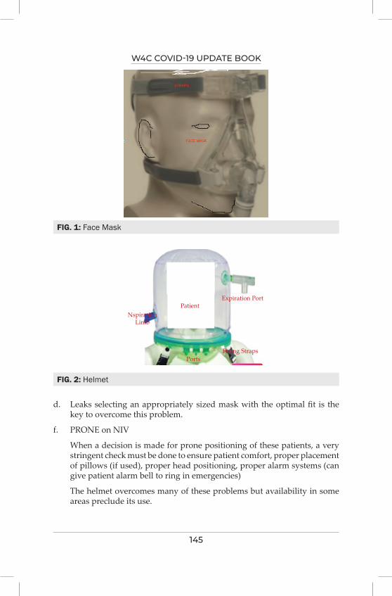
W4C COVID-19 UPDATE BOOK
145
d. Leaks selecting an appropriately sized mask with the optimal fit is the key to overcome this problem.
f. PRONE on NIV
When a decision is made for prone positioning of these patients, a very stringent check must be done to ensure patient comfort, proper placement of pillows (if used), proper head positioning, proper alarm systems (can give patient alarm bell to ring in emergencies)
The helmet overcomes many of these problems but availability in some areas preclude its use.
FIG. 1: Face Mask
FIG. 2: Helmet
PatientNspiration
Limb
PortsFixing Straps
Expiration Port
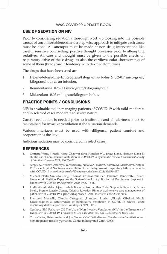
W4C COVID-19 UPDATE BOOK
146
USE OF SEDATION ON NIVPrior to considering sedation a thorough work up looking into the possible causes of uncomfortableness, and a step wise approach to mitigate each cause must be done. All attempts must be made at non drug interventions like careful sensitive counselling, positive thought processes prior to attempting sedatives. All care and thought must be given to the possible effects on respiratory drive of these drugs as also the cardiovascular shortcomings of some of them (bradycardic tendency with dexmedetomidine).
The drugs that have been used are
1. Dexmedetomidine-1microgram/kilogram as bolus & 0.2-0.7 microgram/ kilogram/hour as an infusion.
2. Remifentanil-0.025-0.1 microgram/kilogram/hour
3. Midazolam- 0.05 milligram/kilogram bolus,
PRACTICE POINTS / CONCLUSIONSNIV is a valuable tool in managing patients of COVID 19 with mild-moderate and in selected cases moderate to severe nature.
Careful evaluation is needed prior to institution and all alertness must be maintained for invasive ventilation if the situation demands.
Various interfaces must be used with diligence, patient comfort and cooperation is the key.
Judicious sedation may be considered in select cases.
REFERENCES1. Zhufeng Wang, Yingzhi Wang, Zhaowei Yang, Hongkai Wu, Jingyi Liang, Hanwen Liang Et
al, The use of non-invasive ventilation in COVID-19: A systematic review International Society of Infectious Diseases 2021; 106:254-261.
2. Sergey N. Avdeev, Andrey I. Yaroshetskiy, Natalia A. Tsareva, Zamira M. Merzhoeva, Natalia V. Trushenko,et al Noninvasive ventilation for acute hypoxemic respiratory failure in patients with COVID-19 :American Journal of Emergency Medicine 2021; 39:154–157
3. Michael Pfeifer,Santiago Ewig, Thomas Voshaar, Winfried Johannes Randerath, Torsten Bauer et al, Position Paper for the State-of-the-Art Application of Respiratory Support in Patients with COVID-19 Respiration 2020; 99:521–541.
4. Ludhmila Abrahão Hajjar , Isabela Bispo Santos da Silva Costa, Stephanie Itala Rizk, Bruno Biselli, Brenno Rizerio Gomes, Cristina Salvadori Bittar et al,:Intensive care management of patients with COVID-19: a practical approach . Ann. Intensive Care 2021; 11:36.
5. Francesco Menzella, Claudia Castagnetti ,Francesco Livrieri ,Giorgia Gibellini ,Nicola Facciolongo et al :effectiveness of noninvasive ventilation in COVID-19 related- acute respiratory distress syndrome Clin Respir J 2021; 00:1–9
6. Nasibova EM, Pashayev CN The Use of Non-Invasive Ventilation (NIV) in the Treatment of Patients with COVID-19. J Intensive & Crit Care 2020; 6:5. doi:10.36648/2471-8505.6.2.5
7. Chris Carter, Helen Aedy, and Joy Notter: COVID-19 disease: Non-Invasive Ventilation and high frequency nasal oxygenation: Clinics in Integrated Care 100006
W4C COVID-19 UPDATE BOOK
147
8. Dan Longrois, Giorgio Conti, Jean Mantz, Andreas Faltlhauser, Riku Aantaa and Peter Tonner:Sedation in non-invasive ventilation: do we know what to do (and why)? Multidisciplinary Respiratory Medicine 2014; 9:56
9. Tobin MJ, Jubran A, Laghi F. Noninvasive strategies in COVID-19: epistemology, randomised trials, guidelines, physiology. Eur Respir J 2021; 57:2004247 [https://doi.org/10.1183/ 13993003.04247-2020].
148
Early vs Late intubation for COviD-19 18.
BURDENCorona virus disease storm hit the World in the first months of 2020. Originating in Wuhan, China, coronavirus 2 (SARS-Cov-2) virus has spread to all corners of the globe in under 4 months and is repeatedly showing surges across the globe.
PATHOPHYSIOLOGYThe main manifestation of COVID-19 is acute hypoxemic respiratory failure requiring respiratory support somewhat similar to acute lung injury presenting with respiratory failure. When severe enough, placed in a category called acute respiratory distress syndrome (ARDS).1
In ARDS, the timing of intubation could also be associated with clinical outcomes. It has been reported that ARDS patients undergoing late intubation had markedly higher mortality rates compared to those who were intubated early in the course of the illness.1 The recommendation from the Surviving Sepsis Campaign panel is that “the management of mechanically ventilated patients with COVID-19 should be on similar lines as other patients with acute respiratory failure in the ICU”.2
However, COVID-19 pneumonia, despite falling in most of the circumstances under the Berlin definition of ARDS, has distinctive features of severe hypoxemia often associated with near normal respiratory system compliance.3 This peculiar combination was never seen in severe ARDS before. Severely hypoxemic patients of Covid 19, despite sharing a single etiology have variable spectrum, ranging from asymptomatic infection to ARDS and even death. Symptoms vary from normal breathing (“silent” hypoxemia) to significant dyspnea; hypocapnia to normo/hypercapnia.4
CLINICAL MANAGEMENTMost of the COVID-19 patients are managed with supplemental oxygen therapy, but few patients with the severe respiratory failure may require tracheal intubation. Tracheal tube placement helps in regulating oxygen, pressure and volume control, in addition to stabilising an airway. Prolonged
Prashant Kumar, Kiranpreet Kaur
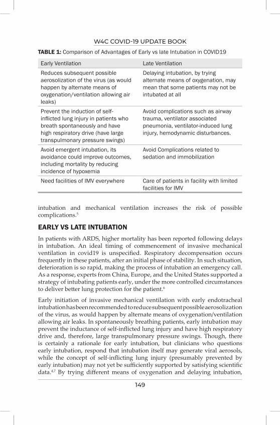
W4C COVID-19 UPDATE BOOK
149
intubation and mechanical ventilation increases the risk of possible complications.5
EARLY VS LATE INTUBATIONIn patients with ARDS, higher mortality has been reported following delays in intubation. An ideal timing of commencement of invasive mechanical ventilation in covid19 is unspecified. Respiratory decompensation occurs frequently in these patients, after an initial phase of stability. In such situation, deterioration is so rapid, making the process of intubation an emergency call. As a response, experts from China, Europe, and the United States supported a strategy of intubating patients early, under the more controlled circumstances to deliver better lung protection for the patient.6
Early initiation of invasive mechanical ventilation with early endotracheal intubation has been recommended to reduce subsequent possible aerosolization of the virus, as would happen by alternate means of oxygenation/ventilation allowing air leaks. In spontaneously breathing patients, early intubation may prevent the inductance of self-inflicted lung injury and have high respiratory drive and, therefore, large transpulmonary pressure swings. Though, there is certainly a rationale for early intubation, but clinicians who questions early intubation, respond that intubation itself may generate viral aerosols, while the concept of self-inflicting lung injury (presumably prevented by early intubation) may not yet be sufficiently supported by satisfying scientific data.4,7 By trying different means of oxygenation and delaying intubation,
TABLE 1: Comparison of Advantages of Early vs late Intubation in COVID19
Early Ventilation Late VentilationReduces subsequent possible aerosolization of the virus (as would happen by alternate means of oxygenation/ventilation allowing air leaks)
Delaying intubation, by trying alternate means of oxygenation, may mean that some patients may not be intubated at all
Prevent the induction of self-inflicted lung injury in patients who breath spontaneously and have high respiratory drive (have large transpulmonary pressure swings)
Avoid complications such as airway trauma, ventilator associated pneumonia, ventilator-induced lung injury, hemodynamic disturbances.
Avoid emergent intubation, its avoidance could improve outcomes, including mortality by reducing incidence of hypoxemia
Avoid Complications related to sedation and immobilization
Need facilities of IMV everywhere Care of patients in facility with limited facilities for IMV
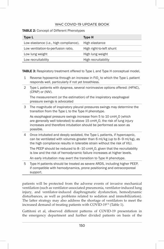
W4C COVID-19 UPDATE BOOK
150
TABLE 2: Concept of Different Phenotypes
Type L Type H
Low elastance (i.e., high compliance), High elastanceLow ventilation-to-perfusion ratio, High right-to-left shuntLow lung weight High lung weightLow recruitability High recruitability
patients will be protected from the adverse events of invasive mechanical ventilation (such as ventilator-associated pneumonia, ventilator-induced lung injury, and ventilator-induced diaphragmatic dysfunction, hemodynamic disturbances, as well as problems related to sedation and immobilization). The latter strategy may also address the shortage of ventilators to meet the increased demand of treating patients with COVID-197,8 (Table 1).
Gattitoni et al, observed different patterns of COVID-19 presentation in the emergency department and further divided patients on basis of the
TABLE 3: Respiratory treatment offered to Type L and Type H conceptual model,
1 Reverse hypoxemia through an increase in FiO2 to which the Type L patient responds well, particularly if not yet breathless.
2 Type L patients with dyspnea, several noninvasive options offered: (HFNC), (CPAP) or (NIV). The measurement (or the estimation) of the inspiratory esophageal pressure swings is advocated
3 The magnitude of inspiratory pleural pressures swings may determine the transition from the Type L to the Type H phenotype. As esophageal pressure swings increase from 5 to 10 cmH2O (which are generally well tolerated) to above 15 cmH2O, the risk of lung injury increases and therefore intubation should be performed as soon as possible.
4 Once intubated and deeply sedated, the Type L patients, if hypercapnic, can be ventilated with volumes greater than 6 ml/kg (up to 8–9 ml/kg), as the high compliance results in tolerable strain without the risk of VILI. The PEEP should be reduced to 8–10 cmH2O, given that the recruitability is low and the risk of hemodynamic failure increases at higher levels. An early intubation may avert the transition to Type H phenotype.
5 Type H patients should be treated as severe ARDS, including higher PEEP, if compatible with hemodynamics, prone positioning and extracorporeal support.

W4C COVID-19 UPDATE BOOK
151
interconnection between three factors: (1) the host response, severity of infection, physiological reserve of the patient, and associated comorbidities; (2) the ventilatory responsiveness of the patient to hypoxemia; (3) the time elapsed between the disease onset and subsequent reporting in the hospital. The interactivity between the above enumerated factors, led to the development of a time-related disease spectrum within two primary “phenotypes”: Type L, and Type H (Table 2, 3).4
A systemic review and meta-analysis of 12 studies, involved 8944 critically ill patients with COVID-19, regardless of early/late intubation (i.e., based on a specific time threshold from ICU admission or prior to trial of HFNC/NIV) used in this meta-analysis, no statistically significant difference was found on all-cause mortality between patients with severe COVID-19 undergoing early versus late intubation.9
Use of HFNC proved to be beneficial in critically ill patients with hypoxemic respiratory failure and appeared to be successful as a first-line treatment in ARDS. Literature support that, many survivors initially were non-intubated, and a many survived without endotracheal intubation. Patients managed with HFNC stays more comfortable than other modalities. In addition, the risk of air or contact surface contamination by HFNC compared to a conventional oxygen mask remains unproven. Further development of objective criteria like the ROX index may be helpful in predicting the need for intubation. The ROX index is simply based on three parameters RR, SpO2 and FiO2 ([SpO2/FiO2]/ respiratory rate). It can prove useful and handy tool in assessing the patient for intubation.10
SUMMARYBecause of the need for adherence to airborne precautions and personal protective equipment, medical staff involved in the management of patients with COVID-19 find difficult to deal with cases, quickly in the event of a sudden deterioration. Moreover, emergency intubation may increase the risk of nosocomial infection of healthcare providers, so treatment guidelines recommend intubation in a controlled setting if the respiratory status worsens. Therefore, decision on the timing of intubation should be thoroughly evaluated, and a predictive model that can identify critically ill COVID-19 patients at risk for respiratory deterioration that requires intubation is needed. Suitable modes of various oxygen therapies including HFNC, NIV should be considered prior and correlated with clinical condition, before directly intubating a covid19 patient for mechanical ventilation. Decision of the optimal time of intubation for covid patient should be based not only on a single factor of “time” rather the chronology of symptoms, duration of illness, respiratory rate, disease severity as defined by the oxygen impairment, lung infiltration extent and use of objective criteria like ROX index using RR, SpO2 and FiO2 as predictive parameters to determine the patients at high risk.
W4C COVID-19 UPDATE BOOK
152
PRACTICE POINTSOur current practice and strategy is to delay intubation if it appears clinically safe and feasible. This ideally requires, close and meticulous monitoring of the patient in an intensive care unit, and physicians able to respond rapidly and intubate. It is also important that ICU that cares for COVID-19 patients should secure a sufficient number of HFNCs/ NIVs that seems as important as the procurement of ventilators.
REFERENCES1. Kangelaris KN, Ware LB, Wang CY, Janz DR, Hanjing Z, Matthay M.A et al. Timing of
Intubation and Clinical outcomes in Adults with ARDS. Crit. Care Med 2016; 44:120.2. Alhazzani W, Møller MH, Arabi YM, Loeb M, Gong MN, Fan E et al. Surviving Sepsis
Campaign: guidelines on the management of critically ill adults with Coronavirus Disease 2019 (COVID-19). Intensive Care Med 2020; 46:854-87. https://doi.org/10.1007/s00134-020-06022-5.
3. Force ARDSDT, Ranieri VM, Rubenfeld GD, Thompson BT, Ferguson ND, Caldwell E, Fan E, Camporota L, Slutsky AS. Acute respiratory distress syndrome: the Berlin definition. JAMA 2012; 307:2526–33.
4. Gattinoni L, Chiumello D, Caironi P Busana M, Romitti F, Brazzi L. et al. COVID-19 pneumonia: different respiratory treatments for different phenotypes? Intensive Care Med 2020; 46:1099–1102. https://doi.org/10.1007/s00134-020-06033-2.
5. Tobin MJ. Basing respiratory management of COVID-19 on physiological principles. Am J Respir Crit Care Med 2020; 201:1319-20 https://doi.org/10.1164/rccm.202004-1076ED.
6. Zuo M, Huang Y, Ma W, Xue Z, Zhang J, Gong Y et al. Expert recommendations for tracheal intubation in critically ill patients with noval coronavirus disease 2019. Chin Med Sci J 2020; 35:105–109.
7. Siempos II, Xourgia E, Ntaidou TK, Zervakis D, Magira EE, Kotanidou A et al. Effect of Early vs. Delayed or No Intubation on Clinical Outcomes of Patients With COVID-19: An Observational Study. Front Med 2020; 7:614152. doi:10.3389/fmed.2020.614152.
8. Rola P, Farkas J, Spiegel R, Kyle-Sidell C, Weingart S, Duggan L et al. Rethinking the early intubation paradigm of COVID-19: Time to change gears? Clin Exp Emerg Med 2020; 7:78–80.
9. Papoutsi, E, Giannakoulis, VG, Xourgia E, Routs C, Kotanidou A, Siempos II. Effect of timing of intubation on clinical outcomes of critically ill patients with COVID-19: a systematic review and meta-analysis of non-randomized cohort studies. Crit Care 2021; 25:121. https://doi.org/10.1186/s13054-021-03540-6.
10. Prower E, Grant D, Bisquera A, Breen CP, Camporota L, Gavrilovski M, et al. The ROX index has greater predictive validity than NEWS2 for deterioration in Covid-19. EClinicalMedicine 2021; 35:1-9. https://doi.org/10.1016/j.eclinm.2021.100828.
153
invasive Mechanical ventilation in COviD 19 19.
INTRODUCTIONCOVID 19 has emerged as one of the foremost challenges faced by the whole world. Approximately 5.7 % of the patients may present with severe COVID 19 requiring ICU care. Data from Lombardy, Italy reveals that almost 88.4 % patients required intubation and mechanical ventilation,1 In another study done by Auld et al,2 almost 76 % patients required invasive mechanical ventilation and the mortality for such patients is approximately 35 %.
COVID ARDSCOVID 19 presents as a spectrum of disease ranging from mild illness to severe respiratory failure COVID ARDS (CARDS) requiring oxygen support in the form of HFNC, NIV or Invasive mechanical ventilation.
PATHOPHYSIOLOGYPost mortem biopsy in patients with severe COVID 19 have revealed diffuse alveolar damage, interstitial and alveolar oedema, hyaline membrane formation, endothelitis with platelet- fibrin thrombi and fibrosis.3 Gattinoni4 has described a spectrum of CARDS and has classified patients according to H and L type based on lung compliance of COVID 19 patients.
o The H type:
High lung elastance (low compliance), with high right to left shunt, high lung weight
Such patients respond to recruitment maneuvers, relatively higher PEEP and prone positioning
o The L type:
Low lung elastance, (preserved compliance), low lung weight. These patients have a predominant involvement of vascular endothelium with pulmonary vasculature microthrombi and consequent ventilation perfusion mismatch. Such patients usually do not respond to recruitment
Neelmani Ahuja, Sumit Ray
W4C COVID-19 UPDATE BOOK
154
maneuvers or high PEEP, Prone positioning may still be beneficial by improving ventilation perfusion matching.
RESPIRATORY MECHANICS IN COVID 19 ARDSIn a cohort study by Schenck et al,5 ventilator variables for patients with severe COVID 19 were described. The median plateau pressure was around 25 (21-29) cm/H2O and median tidal volumes of 7.01 (6.13, 8.10) ml/kg of predicted body weight. The median driving pressure of 14 (11- 17.2) cm/H20, a median PEEP of 10 (8-12 cm H2O) and a static compliance 28 (23-38ml/cm H2O) was recorded which are similar to LUNG SAFE study.
It appears from the evidence that CARDS is heterogeneous and patients may not fit into a single entity of H or L type but may have a dynamic spectrum of lung mechanics of ARDS depending on the time of intubation. The same has been described by Grasselli et al6 in their study.
INVASIVE MECHANICAL VENTILATION IN COVID ARDS-It is suggested to ventilate the patients of CARDS keeping in mind the basic principles of lung protective ventilation aimed at reducing ventilator induced lung injury. Each patient requires a tailored approach for mechanical ventilation to decide regarding tidal volume, respiratory rate, PEEP and FiO2.
Tidal Volume
Low tidal volume ventilation (4-8 ml/kg Predicted Body weight) is the accepted norm with the aim to target a plateau pressure less than 30 cm H2O and a driving pressure of less than 15 cm H2O. [7]
PEEP
A higher PEEP can prevent atelectasis and collapse of the alveoli at end expiration promoting arterial oxygenation, reduce lung stress and strain, and maintain a homogeneous ventilation. Too high a PEEP may lead to alveolar overdistention leading to ventilator induced lung injury and haemodynamic instability. Optimal PEEP titration is guided by various methods-
• PEEP- FiO2 table
• Optimal Compliance method
• PV curve
• Driving Pressure
• Stress Index
• Transpulmonary pressures
• CT imaging
W4C COVID-19 UPDATE BOOK
155
Respiratory Rate
Respiratory rate needs to be adjusted to allow the CO2 to be washed out effectively. Permissive hypercapnia may be allowed with an acceptable pH>7.2.
Refractory Hypoxemia in COVID ARDS
Some patients may present with refractory Hypoxemia despite optimized ventilator settings. The following salvage therapies may be used in such patients-
Proning
Patients with severe ARDS have benefitted from early application of proning sessions with a mortality benefit at 28 days and 90 days. The mechanisms leading to improvement in hypoxemia include homogenization of lung ventilation, improved ventilation perfusion matching secondary to reduced ventral-dorsal transpulmonary pressure difference and reduced lung compression due to mediastinum. In a multicentric retrospective evaluation of CARDS patients by Langer et al,8 they found an improved oxygenation in majority of patients that underwent proning.
Neuromuscular Blocking Agents (NMBA)
Patients with a P/F ratio less than 150 who are mechanically ventilated, early administration of neuromuscular blockers (NMBs) was associated with a better adjusted 90 day mortality. However, a recent trial9 conducted to ascertain the role of NMBs, did not find any significant difference in 90 day mortality between those who received continuous cisatracurium infusion versus those who without routine neuromuscular blockade and lighter sedation targets. Patients who received continuous NMBs were less physically active and had more adverse cardiovascular events than those patients which did not receive NMBs. Hence it is suggested to avoid their routine and if needed, boluses should be preferred over continuous infusions.
Recruitment maneuvers
Recruitment maneuvers are used to apply transiently elevated airway pressure in mechanically ventilated patients to recruit collapsed alveoli. In CARDS patients, pneumothorax, pneumomediastinum and subcutaneous emphysema are commonly seen.10 Recruitment maneuvers can put patients at risk of barotrauma. It is suggested against the routine use of recruitment maneuvers in CARDS patients as found in the results of ART trial.
Inhaled pulmonary vasodilators
Inhaled pulmonary vasodilators such as inhaled prostacyclins (epoprostenol) and inhaled Nitric oxide have been used as rescue strategies in refractory hypoxemia in severe ARDS. They dilate pulmonary vessels in the well
W4C COVID-19 UPDATE BOOK
156
ventilated lung units and reduce shunt fraction and pulmonary hypertension. Inhaled Epoprostenol may be helpful in improving oxygenation in at least 50 % of the recipients with a median change of 9 mm Hg in P/F ratio.11 In a retrospective observational study using inhaled nitric oxide in patients with severe COVID 19 with refractory hypoxemia, more than half of the patients did not show an improvement in P/F ratio.12
ECMO
Patients with severe CARDS which have refractory hypoxemia despite optimal ventilatory settings and rescue therapies (including proning), or have respiratory acidosis and/ or inability to achieve lung protective ventilation despite may be considered for ECMO. There is paucity of data regarding outcomes in CARDS patients treated with ECMO. There is paucity of data regarding optimal ventilation settings in ARDS patients on ECMO. The underlying principles of mechanical ventilation in patients on ECMO is using Ultra lung protective ventilation with tidal volume < 4 ml/kg and minimal respiratory rate and limiting PIP to between 20-25 cm, maintaining minimum FiO2 on ventilator and maintain adequate level of PEEP to prevent reabsorption atelectasis and using transpulmonary pressure monitoring to prevent overdistension.13 Driving pressure during ventilation of such patients may be an independent factor associated with in- hospital mortality and warrants monitoring.14
CONCLUSIONThe ventilation strategies for these patients are challenging and underlying principles of lung protective ventilation form the core for ventilating these patients. Recruitment maneuvers may not be used routinely as CARDS patients are more prone to barotrauma. Proning and ECMO may be used in patients with refractory hypoxemia. Ultra lung protective ventilation and monitoring driving pressure is essential for patients on ECMO.
PRACTICE POINTS• CARDS patients may present a dynamic spectrum of lung mechanics
ranging from Type H to Type L.
• Invasive mechanical ventilation must follow the underlying principles of lung protective ventilation with a tidal volume of 4-8 ml/ kg body weight and targeted plateau pressure< 30 cm H20 and driving pressures <15 cm H2O and an optimal PEEP.
• Refractory hypoxemia may be treated with proning, NMBs, inhaled pulmonary vasodilators, recruitment maneuvers (not used routinely) and ECMO.
W4C COVID-19 UPDATE BOOK
157
• Patients on ECMO are treated with Ultra lung protective ventilation with Vt< 4 ml/kg and PIP between 20-25 cm H2O and monitoring driving pressures.
REFERENCES1. Grasselli G, Zangrillo A, Zanella A, et al. “Baseline characteristics and outcomes of 1591
patients infected with SARS-CoV-2 admitted to ICUs of the Lombardy Region, Italy,”. JAMA 2020; 323:1574.
2. Auld SC, Caridi-Scheible M, Blum JM, Robichaux C, Kraft C, Jacob JT, Jabaley CS, Carpenter D, Kaplow R, Hernandez-Romieu AC, Adelman MW, Martin GS, Coopersmith CM, Murphy DJ; and the Emory COVID-19 Quality and Clinical Research Collaborative. ICU and Ventilator Mortality Among Critically Ill Adults With Coronavirus Disease 2019. Crit Care Med 2020; 48:e799-e804.doi: 10.1097/CCM.0000000000004457.PMID: 32452888;PMCID:PMC7255393.
3. Borczuk AC, Salvatore SP, Seshan SV, Patel SS, Bussel JB, Mostyka M, Elsoukkary S, He B, Del Vecchio C, Fortarezza F, Pezzuto F, Navalesi P, Crisanti A, Fowkes ME, Bryce CH, Calabrese F, Beasley MB. COVID-19 pulmonary pathology: a multi-institutional autopsy cohort from Italy and New York City. Mod Pathol 2020; 33:2156-2168. doi: 10.1038/s41379-020-00661-1. Epub 2020 Sep 2. PMID: 32879413; PMCID: PMC7463226.
4. Gattinoni L, Coppola S, Cressoni M, Busana M, Rossi S, Chiumello D. COVID-19 Does Not Lead to a “Typical” Acute Respiratory Distress Syndrome. Am J Respir Crit Care Med 2020; 201:1299-1300. doi: 10.1164/rccm.202003-0817LE. PMID: 32228035; PMCID: PMC7233352.
5. Edward J. Schenck, Katherine Hoffman, Parag Goyal, Justin Choi, Lisa Torres, Kapil Rajwani, Christopher W. Tam , Natalia Ivascu, Fernando J. Martinez , and David A. Berlin. Respiratory Mechanics and Gas Exchange in COVID-19–associated Respiratory Failure. Annals of the American Thoracoic Society. https://doi.org/10.1513/AnnalsATS.202005-427RL.
6. Grasselli G, Tonetti T, Protti A, Langer T, Girardis M, Bellani G, Laffey J, Carrafiello G, Carsana L, Rizzuto C, Zanella A, Scaravilli V, Pizzilli G, Grieco DL, Di Meglio L, de Pascale G, Lanza E, Monteduro F, Zompatori M, Filippini C, Locatelli F, Cecconi M, Fumagalli R, Nava S, Vincent JL, Antonelli M, Slutsky AS, Pesenti A, Ranieri VM; COVID-19 Spanish ICU Network. Clinical features, ventilatory management, and outcome of ARDS caused by COVID-19 are similar to other causes of ARDS. Respir Med 2020; 8:1201-1208. doi: 10.1016/S2213-2600(20)30370-2. Epub 2020 Aug 27. PMID: 32861276; PMCID: PMC7834127.
7. Acute Respiratory Distress Syndrome Network, Brower RG, Matthay MA, Morris A, Schoenfeld D, Thompson BT, Wheeler A. Ventilation with lower tidal volumes as compared with traditional tidal volumes for acute lung injury and the acute respiratory distress syndrome. N Engl J Med 2000; 342:1301-8. doi: 10.1056/NEJM200005043421801. PMID: 10793162.
8. Langer T, Brioni M, Guzzardella A, Carlesso E, Cabrini L, Castelli G, Dalla Corte F, De Robertis E, Favarato M, Forastieri A, Forlini C, Girardis M, Grieco DL, Mirabella L, Noseda V, Previtali P, Protti A, Rona R, Tardini F, Tonetti T, Zannoni F, Antonelli M, Foti G, Ranieri M, Pesenti A, Fumagalli R, Grasselli G; PRONA-COVID Group. Prone position in intubated, mechanically ventilated patients with COVID-19: a multi-centric study of more than 1000 patients. Crit Care 2021; 25:128. doi: 10.1186/s13054-021-03552-2. PMID: 33823862; PMCID: PMC8022297.
9. National Heart, Lung, and Blood Institute PETAL Clinical Trials Network, Moss M, Huang DT, Brower RG, Ferguson ND, Ginde AA, Gong MN, Grissom CK, Gundel S, Hayden D, Hite RD, Hou PC, Hough CL, Iwashyna TJ, Khan A, Liu KD, Talmor D, Thompson BT, Ulysse CA, Yealy DM, Angus DC. Early Neuromuscular Blockade in the Acute Respiratory Distress Syndrome. N Engl J Med 2019; 380:1997-2008. doi: 10.1056/NEJMoa1901686. Epub 2019 May 19. PMID: 31112383; PMCID: PMC6741345.
10. Martinelli AW, Ingle T, Newman J, Nadeem I, Jackson K, Lane ND, Melhorn J, Davies HE, Rostron AJ, Adeni A, Conroy K, Woznitza N, Matson M, Brill SE, Murray J, Shah A, Naran R, Hare SS, Collas O, Bigham S, Spiro M, Huang MM, Iqbal B, Trenfield S, Ledot S, Desai S, Standing L, Babar J, Mahroof R, Smith I, Lee K, Tchrakian N, Uys S, Ricketts W, Patel ARC, Aujayeb A, Kokosi M, Wilkinson AJK, Marciniak SJ. COVID-19 and pneumothorax: a
W4C COVID-19 UPDATE BOOK
158
multicentre retrospective case series. Eur Respir J 2020; 56:2002697. doi: 10.1183/13993003.02697-2020. PMID: 32907891; PMCID: PMC7487269.
11. Sonti R, Pike CW, Cobb N. Responsiveness of Inhaled Epoprostenol in Respiratory Failure due to COVID-19. J Intensive Care Med 2021; 36:327-333. doi: 10.1177/0885066620976525. Epub 2020 Nov 25. PMID: 33234007; PMCID: PMC7724253.
12. Longobardo A, Montanari C, Shulman R, Benhalim S, Singer M, Arulkumaran N. Inhaled nitric oxide minimally improves oxygenation in COVID-19 related acute respiratory distress syndrome. Br J Anaesth 2021; 126:e44-e46. doi: 10.1016/j.bja.2020.10.011. Epub 2020 Oct 14. PMID: 33138964; PMCID: PMC7556790.
13. Schmidt M, Pellegrino V, Combes A, Scheinkestel C, Cooper DJ, Hodgson C. Mechanical ventilation during extracorporeal membrane oxygenation. Crit Care 2014; 18:203. doi: 10.1186/cc13702. PMID: 24447458; PMCID: PMC4057516.
14. Serpa Neto A, Schmidt M, Azevedo LC, Bein T, Brochard L, Beutel G, Combes A, Costa EL, Hodgson C, Lindskov C, Lubnow M, Lueck C, Michaels AJ, Paiva JA, Park M, Pesenti A, Pham T, Quintel M, Marco Ranieri V, Ried M, Roncon-Albuquerque R Jr, Slutsky AS, Takeda S, Terragni PP, Vejen M, Weber-Carstens S, Welte T, Gama de Abreu M, Pelosi P, Schultz MJ; ReVA Research Network and the PROVE Network Investigators. Associations between ventilator settings during extracorporeal membrane oxygenation for refractory hypoxemia and outcome in patients with acute respiratory distress syndrome: a pooled individual patient data analysis : Mechanical ventilation during ECMO. Intensive Care Med 2016; 42:1672-1684. doi: 10.1007/s00134-016-4507-0. Epub 2016 Sep 1. PMID: 27586996; PMCID: PMC7094949.
SECTION 4-B
ManagementSTRATEGIES TO
IMPROVE OXYGENATION
SECTION 4-B

161
Section 4 - ManagementSECTION EDITOR
Dr Kedar ToraskarMD EDIC Director Critical Care, Critical Care Unit, Wockhardt Hospital, South Mumbai
Preface
SUBSECTION ON STRATEGIES TO IMPROVE OXYGENATION CARDS as any other viral ARDS requires strategies to improve oxygenation. Due to the sheer volumes of patients presenting with respiratory failure, the skills in managing ARDS improved during the pandemic despite of the inadequate manpower. Simple cost-effective protocols like covid associated repositioning
& proning (CARP) based on physiological principles of proning were devised. Though the evidence for CARP is not robust, it has shown significant benefit in improving oxygenation in patients who are awake & cooperative.
Extracorporeal membrane oxygenation (ECMO) & extracorporeal carbon dioxide removal techniques (ECCO2R) were used in a limited manner due to manpower, logistical & cost constraints in the pandemic. Patient selection & right timing (early initiation) is the key for successful outcomes for both these strategies. Extracorporeal therapies other than ECMO & ECCO2R like hemoperfusion & continuous plasma filtration absorption (CPFA) with or with CRRT (continuous renal replacement therapy) has helped in reduction in severity of disease. These help in removal of toxins leading in Covid 19 patients with cytokine storm, hepatic failure & AKI. But robust data is again lacking to support these therapies.
This subsection has covered all the salient points regarding these strategies to improve oxygenation.

162
163
Proning in Covid 19: an interplay of Art and
Science20.
‘The Art of PRONING’ Begins with An Understanding of ‘The Science Behind It.’
INTRODUCTIONProning a patient with ARDS has been long practiced in the ICU with improvement in oxygenation and mortality. This was demonstrated in the PROSEVA study in NEJM in 2013, where noticeable improvement was shown when patients were proned for 16-hour sessions. The physiological improvement post proning, in the oxygenation and ventilation explains the desirable improvement in ARDS patients. Since COVID-19 has been a disease predominantly affecting the lung causing the L and H type of ARDS, proning has been a very useful modality in the treatment of COVID – 19 disease.
We must understand that proning is a very useful manoeuvre across the entire spectrum of COVID -19 patients, right from its use as a COVID Awake Repositioning / Proning (CARP) protocol in awake hypoxemic patients, those on oxygen therapy, those on High Flow Nasal Cannula (HFNC) and non-invasive ventilation (NIV), those on invasive mechanical ventilation and finally to those on Extracorporeal membrane oxygenation (ECMO).
PHYSIOLOGICAL EFFETS OF PRONING• Optimisation of pulmonary ventilation/perfusion ratio (V/Q) matching
(increased blood flow to the dependent lung)
• Increase in Functional residual capacity (FRC)
• Reduced atelectasis
• Facilitates secretion drainage
• Less lung deformation in prone position (increased homogeneity), leading to improved ventilation
Khusrav Beji Bajan

164
• Abdomen is less likely to distend when in prone position, thus increasing the FRC.
MECHANICS OF PHYSIOLOGICAL IMPROVEMENT DUE TO PRONINGWhen the patient is in a prone position, the following occurs:
• Heart sits against sternum (rather than left lung), causing the lung to be less compressed.
• Decreased trans-pleural pressure gradient between dependent and non-dependent lung in the prone position.
• Plateau pressure is more uniformly distributed when prone, thereby leading to more uniform alveolar ventilation.
• Recruitment manoeuvres have shown to be more effective in the prone position.
• Alterations in chest wall mechanics augment lung inflation at lower pressures.
• Dorsoventral orientation of large airways may also be seen.
TIPS TO MAKE ‘PRONING’ A SUCESSFUL ‘ART’1. Development & customisation of Proning protocol as per clinical
setting:
• Document should include step by step instructions and checklists for safe proning and un-proning for an invasively ventilated ARDS patient.
Special emphasis may be on:
– Supplies for the procedure.
– Role designation during the procedure
– Care of medical devices and pressure points.
2. Ensure appropriate equipments are available during the procedure:
• Flat sheets
• Pressure injury prevention devices/ dressing like gel or foam face cushions, form dressings etc must be available.
• Pillows
• ECG leads
• Long tubings/ extensions for medical devices.
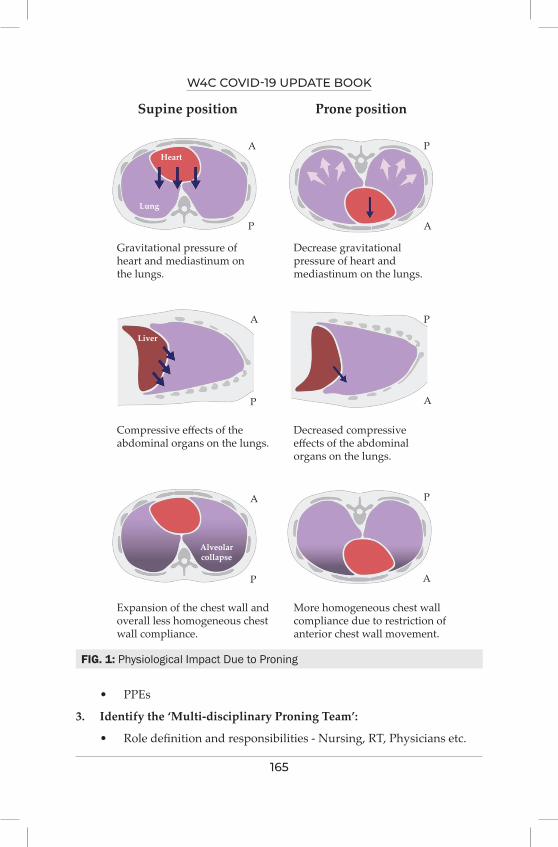
W4C COVID-19 UPDATE BOOK
165
FIG. 1: Physiological Impact Due to Proning
• PPEs
3. Identify the ‘Multi-disciplinary Proning Team’:
• Role definition and responsibilities - Nursing, RT, Physicians etc.
Supine position
Gravitational pressure ofheart and mediastinum onthe lungs.
Compressive effects of theabdominal organs on the lungs.
Expansion of the chest wall andoverall less homogeneous chestwall compliance.
More homogeneous chest wallcompliance due to restriction ofanterior chest wall movement.
Decreased compressiveeffects of the abdominalorgans on the lungs.
Decrease gravitationalpressure of heart andmediastinum on the lungs.
Prone position
A P
AP
A
A
A
AP
P
P
P
Heart
Lung
Liver
Alveolarcollapse
W4C COVID-19 UPDATE BOOK
166
– Based on their experience, staffing availability etc. (e.g., daytime vs afterhours).
• Key responsibilities include:
– Airway management
– Venous and arterial line management
– Management of bladder catheters and chest tubes
– Monitoring cardiorespiratory status
4. Clearly identify indications for proning:
• Prone positioning should be considered in patients who are invasively mechanically ventilated and have moderate to severe ARDS with refractory Hypoxemia [PaO2/FiO2] ratio ≤ 150.
• Prone positioning is also recommended and indicated for patients with burns, skin flaps and posterior wounds.
5. Clearly identify contra-indications for the procedure:
• Untrained healthcare team.
• Increased intracranial Pressure
• Increased abdominal Pressure
• Abdominal and chest wounds
• C-spine precautions
• Extreme obesity
• Haemodynamic instability
• Pregnant women
6. Identify a standard time to prone and un-prone patients:
• Proning should be incorporated into the daily ICU routine. Eg: 2pm to next day 8 am as routine.
• It is also a practice to have multi-disciplinary Proning team/s to undertake proning procedures in succession for multiple patients.
• It is essential to ensure that, after hours staff is as comfortable in proning as daytime staff, for the acutely hypoxemic patient in an emergency situation.
7. Modify care to mitigate adverse events:
Following undesirable side effects of proning should be looked out for:
W4C COVID-19 UPDATE BOOK
167
• Pressure trauma at these particular sites is common:
– Ocular / blindness, orbital skin necrosis
– Bridge of nose / mentum
– Humeral head / knees
– Breast implants / male genitals
• Decreased enteral nutrition
• Difficultly in performance of critical procedures or reintubation
• Cardiac arrest
In order to mitigate these adverse effects, care should be taken to deploy pillows and cushions appropriately whilst positioning the patients, completing all necessary and anticipated procedures before deciding to prone, whilst maintaining hemodynamic stability of patient.
8. Utilize ‘Simulation’ based training:
• Even low fidelity simulations using mannequins may assist to train the multi-disciplinary team in procedural safety.
• Interdisciplinary team to improve communication (closed-loop communication) during the procedure.
• Augment team dynamics.
9. Procedural audit/ feedback/ de-briefing:
This may include:
• Formal (surveys, semi-structured interviews).
• Informal (staff, observers).
10. Plan for special circumstances:
• Obese patients: Proning process should be initiated with all safety precautions for patient and healthcare team.
• Renal replacement therapy:
– Proning is practiced with least difficulty in patients with a internal jugular HD catherter in situ.
• Cardiopulmonary arrest while in the prone position:
– Cardiopulmonary resuscitation can be performed effectively in the prone position while awaiting help to un-prone the patient.
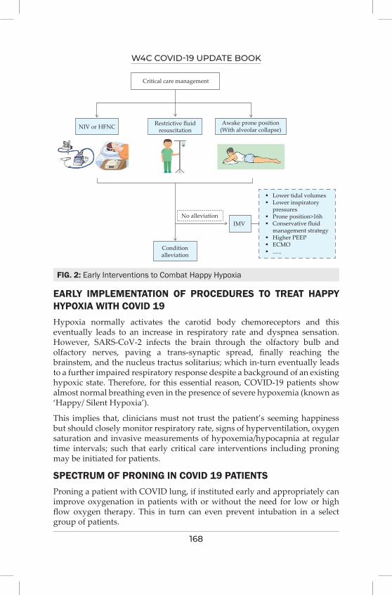
W4C COVID-19 UPDATE BOOK
168
FIG. 2: Early Interventions to Combat Happy Hypoxia
EARLY IMPLEMENTATION OF PROCEDURES TO TREAT HAPPY HYPOXIA WITH COVID 19Hypoxia normally activates the carotid body chemoreceptors and this eventually leads to an increase in respiratory rate and dyspnea sensation. However, SARS-CoV-2 infects the brain through the olfactory bulb and olfactory nerves, paving a trans-synaptic spread, finally reaching the brainstem, and the nucleus tractus solitarius; which in-turn eventually leads to a further impaired respiratory response despite a background of an existing hypoxic state. Therefore, for this essential reason, COVID-19 patients show almost normal breathing even in the presence of severe hypoxemia (known as ‘Happy/ Silent Hypoxia’).
This implies that, clinicians must not trust the patient’s seeming happiness but should closely monitor respiratory rate, signs of hyperventilation, oxygen saturation and invasive measurements of hypoxemia/hypocapnia at regular time intervals; such that early critical care interventions including proning may be initiated for patients.
SPECTRUM OF PRONING IN COVID 19 PATIENTSProning a patient with COVID lung, if instituted early and appropriately can improve oxygenation in patients with or without the need for low or high flow oxygen therapy. This in turn can even prevent intubation in a select group of patients.
Critical care management
NIV or HFNC Restrictive fluidresuscitation
Awake prone position(With alveolar collapse)
No alleviationIMV
Conditionalleviation
Lower tidal volumes Lower inspiratory pressures Prone position>16h Conservative fluid management strategy Higher PEEP ECMO ......
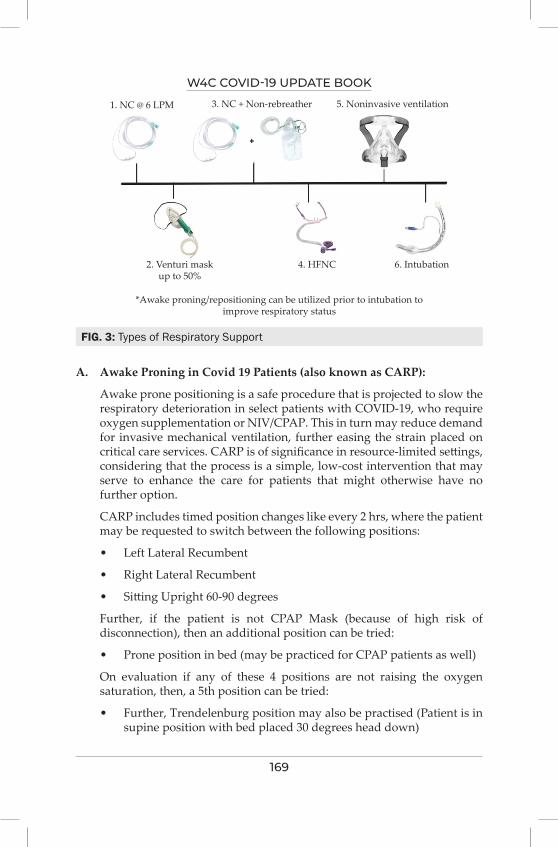
W4C COVID-19 UPDATE BOOK
169
A. Awake Proning in Covid 19 Patients (also known as CARP):
Awake prone positioning is a safe procedure that is projected to slow the respiratory deterioration in select patients with COVID-19, who require oxygen supplementation or NIV/CPAP. This in turn may reduce demand for invasive mechanical ventilation, further easing the strain placed on critical care services. CARP is of significance in resource-limited settings, considering that the process is a simple, low-cost intervention that may serve to enhance the care for patients that might otherwise have no further option.
CARP includes timed position changes like every 2 hrs, where the patient may be requested to switch between the following positions:
• Left Lateral Recumbent
• Right Lateral Recumbent
• Sitting Upright 60-90 degrees
Further, if the patient is not CPAP Mask (because of high risk of disconnection), then an additional position can be tried:
• Prone position in bed (may be practiced for CPAP patients as well)
On evaluation if any of these 4 positions are not raising the oxygen saturation, then, a 5th position can be tried:
• Further, Trendelenburg position may also be practised (Patient is in supine position with bed placed 30 degrees head down)
FIG. 3: Types of Respiratory Support
1. NC @ 6 LPM 3. NC + Non-rebreather
2. Venturi maskup to 50%
*Awake proning/repositioning can be utilized prior to intubation toimprove respiratory status
4. HFNC 6. Intubation
5. Noninvasive ventilation
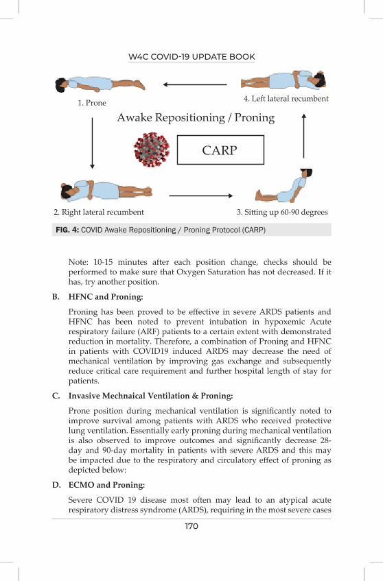
W4C COVID-19 UPDATE BOOK
170
Note: 10-15 minutes after each position change, checks should be performed to make sure that Oxygen Saturation has not decreased. If it has, try another position.
B. HFNC and Proning:
Proning has been proved to be effective in severe ARDS patients and HFNC has been noted to prevent intubation in hypoxemic Acute respiratory failure (ARF) patients to a certain extent with demonstrated reduction in mortality. Therefore, a combination of Proning and HFNC in patients with COVID19 induced ARDS may decrease the need of mechanical ventilation by improving gas exchange and subsequently reduce critical care requirement and further hospital length of stay for patients.
C. Invasive Mechnaical Ventilation & Proning:
Prone position during mechanical ventilation is significantly noted to improve survival among patients with ARDS who received protective lung ventilation. Essentially early proning during mechanical ventilation is also observed to improve outcomes and significantly decrease 28-day and 90-day mortality in patients with severe ARDS and this may be impacted due to the respiratory and circulatory effect of proning as depicted below:
D. ECMO and Proning:
Severe COVID 19 disease most often may lead to an atypical acute respiratory distress syndrome (ARDS), requiring in the most severe cases
FIG. 4: COVID Awake Repositioning / Proning Protocol (CARP)
1. Prone
2. Right lateral recumbent 3. Sitting up 60-90 degrees
4. Left lateral recumbent
Awake Repositioning / Proning
CARP
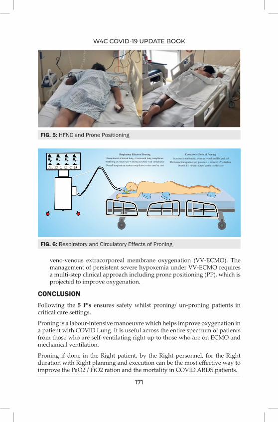
W4C COVID-19 UPDATE BOOK
171
veno-venous extracorporeal membrane oxygenation (VV-ECMO). The management of persistent severe hypoxemia under VV-ECMO requires a multi-step clinical approach including prone positioning (PP), which is projected to improve oxygenation.
CONCLUSIONFollowing the 5 P’s ensures safety whilst proning/ un-proning patients in critical care settings.
Proning is a labour-intensive manoeuvre which helps improve oxygenation in a patient with COVID Lung. It is useful across the entire spectrum of patients from those who are self-ventilating right up to those who are on ECMO and mechanical ventilation.
Proning if done in the Right patient, by the Right personnel, for the Right duration with Right planning and execution can be the most effective way to improve the PaO2 / FiO2 ration and the mortality in COVID ARDS patients.
FIG. 5: HFNC and Prone Positioning
FIG. 6: Respiratory and Circulatory Effects of Proning
Respiratory Effects of ProningRecruitment of dorsal lung increased lung compliance
Stiffening of chest wall decreased chest wall complianceOverall respiratory system compliance varies case by case
Circulatory Effects of Proning
Increased intrathoracic pressure reduced RV preloadDecreased transpulmonary pressure reduced RV afterload
Overall RV cardiac output varies case by case
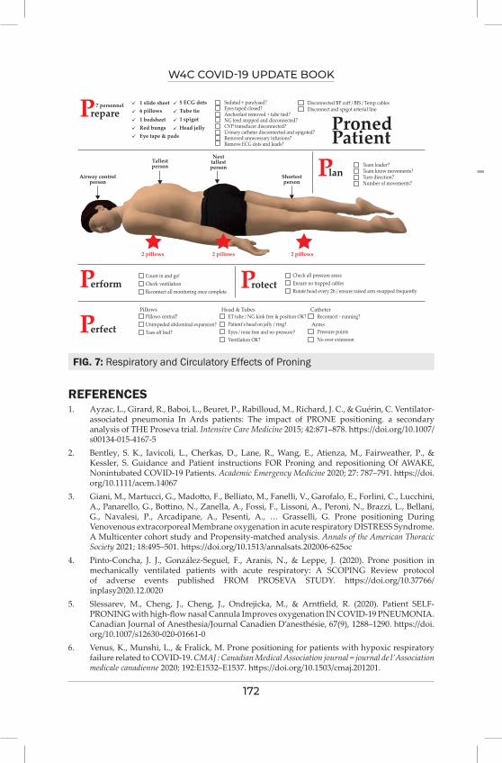
W4C COVID-19 UPDATE BOOK
172
REFERENCES1. Ayzac, L., Girard, R., Baboi, L., Beuret, P., Rabilloud, M., Richard, J. C., & Guérin, C. Ventilator-
associated pneumonia In Ards patients: The impact of PRONE positioning. a secondary analysis of THE Proseva trial. Intensive Care Medicine 2015; 42:871–878. https://doi.org/10.1007/s00134-015-4167-5
2. Bentley, S. K., Iavicoli, L., Cherkas, D., Lane, R., Wang, E., Atienza, M., Fairweather, P., & Kessler, S. Guidance and Patient instructions FOR Proning and repositioning Of AWAKE, Nonintubated COVID-19 Patients. Academic Emergency Medicine 2020; 27: 787–791. https://doi.org/10.1111/acem.14067
3. Giani, M., Martucci, G., Madotto, F., Belliato, M., Fanelli, V., Garofalo, E., Forlini, C., Lucchini, A., Panarello, G., Bottino, N., Zanella, A., Fossi, F., Lissoni, A., Peroni, N., Brazzi, L., Bellani, G., Navalesi, P., Arcadipane, A., Pesenti, A., … Grasselli, G. Prone positioning During Venovenous extracorporeal Membrane oxygenation in acute respiratory DISTRESS Syndrome. A Multicenter cohort study and Propensity-matched analysis. Annals of the American Thoracic Society 2021; 18:495–501. https://doi.org/10.1513/annalsats.202006-625oc
4. Pinto-Concha, J. J., González-Seguel, F., Aranis, N., & Leppe, J. (2020). Prone position in mechanically ventilated patients with acute respiratory: A SCOPING Review protocol of adverse events published FROM PROSEVA STUDY. https://doi.org/10.37766/inplasy2020.12.0020
5. Slessarev, M., Cheng, J., Cheng, J., Ondrejicka, M., & Arntfield, R. (2020). Patient SELF-PRONING with high-flow nasal Cannula Improves oxygenation IN COVID-19 PNEUMONIA. Canadian Journal of Anesthesia/Journal Canadien D’anesthésie, 67(9), 1288–1290. https://doi.org/10.1007/s12630-020-01661-0
6. Venus, K., Munshi, L., & Fralick, M. Prone positioning for patients with hypoxic respiratory failure related to COVID-19. CMAJ : Canadian Medical Association journal = journal de l’Association medicale canadienne 2020; 192:E1532–E1537. https://doi.org/10.1503/cmaj.201201.
FIG. 7: Respiratory and Circulatory Effects of Proning
PPronedPatient
repare
Perform Protect
Perfect
Plan
7 personnel 1 slide sheet 5 ECG dots Sedated + paralysed?Eyes taped closed?Anchorfast removed + tube tied?NG feed stopped and disconnected?CVP transducer disconnected?Urinary catheter disconnected and spigoted?Removed unnecessary infusions?Remove ECG dots and leads?
Count in and go!Check ventilationReconnect all monitoring once complete
Pillows central?Unimpeded abdominal expansion?Toes off bed?
ET tube / NG kink free & position OK?Patient’s head on jelly / ring?Eyes / nose free and no pressure?Ventilation OK?
Reconnect - running?
Pressure pointsNo over extension
Pillows Head & Tubes
Arms
Catheter
Check all pressure areasEnsure no trapped cablesRotate head every 2h / ensure raised arm swapped frequently
Disconnected BP cuff / BIS / Temp cables
Team leader?Team know movements?Turn direction?Number of movements?
Disconnect and spigot arterial lineTube tie1 spigotHead jelly
6 pillows1 bedsheetRed bungsEye tape & pads
Airway controlperson
2 pillows 2 pillows 2 pillows
Tallestperson
Nexttallestperson
Shortestperson

173
ECMO in COviD 19
INTRODUCTIONThe pandemic of COVID-19 which commenced in December of 2019 in Wuhan, China has ravaged the world involving over 190 countries with more than 21 crore cases and over 45 lakh deaths until September of 2021.1 At least 15 to 30% of patients will develop severe Acute Respiratory Distress Syndrome (ARDS).2 Besides ARDS, COVID-19 illness from a pathophysiological stand point produces cytokine storm and hypercoagulability which has come to become one of the important hallmarks of the disease.3, 4 Gattanoni et al described 2 types of respiratory failure associated with COVID-19 illness: firstly, L-type characterized by low elastance, high compliance and low recruitability followed of H-type is characterized by high elastance, low compliance and high recruitability.5 Clinical phenotypic presentation may be a spectrum varying from L-type to H-type or any other combination in between these two ends of the spectrum. At times the L-type may progress to H-type due to progression of the illness or patient related Self-induced lung injury (P-SILI). Early intubation may be useful in patients with L-type to prevent worsening of Lung injury. On the other hand, H-type which is more suggestive of classical ARDS would be ideally suited for Extracorporeal Membrane Oxygenation (ECMO).
EVOLUTION OF ECMO IN COVID-19In the early part of the pandemic use of ECMO on patients was based on experience and best practices derived from ARDS of other etiologies. The results of ECMO at the start of the pandemic was not encouraging prompting different organizations to even not favour the use of ECMO.6 But more recent data indicates a better outcome with ECMO in patients with ARDS due to COVID-19 illness having a mortality varying from 31-45%. This outcome is nearly consistent with mortality seen in other forms of ARDS needing ECMO.7,8 Despite the wide variation in the outcomes seen with the use of ECMO for severe COVID-19, a recent meta-analysis of many studies in the first year of the pandemic showed a mortality of 37.1% with the use of ECMO indicating that this form of support is feasible in severe ARDS patients with COVID-19 illness (Figure 1).9 In most patients (> 90%) Veno-venous ECMO (VV-ECMO)
Sunil Karanth
21.

W4C COVID-19 UPDATE BOOK
174
is needed as support for the severe ARDS in patients with COVID 19 disease. The remaining will require Veno-arterial ECMO (VA-ECMO) as a form of mechanical circulatory support due to the cardiovascular complications of COVID-19 like myocarditis, stress cardiomyopathy, arrhythmias, acute coronary syndromes etc.10 Modifications of the mode of ECMO such as Veno-arterio venous ECMO (V-AV) may be needed if COVID-19 afflicts both the respiratory and circulatory systems. However, the use of VA or VAV ECMO is associated with a significantly poorer outcome and higher in-hospital mortality rate (Hazard ratio of 1.85 and CI of 1.25-2.97).8 The factors indicating poor outcome for COVID-19 patients needing ECMO include old age, low PaO2/FiO2 ratio, immunocompromised state, comorbidities and the need for VA ECMO.
PATIENTS FOR WHOM ECMO CAN BE CONSIDERED IN COVID-19 ILLNESSECMO being a complex, expensive form of support with limited availability and need for expertise, the choice to go on ECMO should be made judiciously. This is particularly true when a pandemic is ongoing. ECMO is preferably used on younger patients, previously healthy, with single organ failure and have the best possible chance of recovery. The ECMO is used as a “rescue therapy” to allow lung healing or a bridge to definitive therapy like transplant. It is
FIG. 1: Proportion of Non-Survivors Among Coronavirus Disease 2019 Patients Requiring Extracorporeal Membrane Oxygenation Support (Adapted from Reference 9)
Study
Random effects modelHeterogeneity: I2 = 52.8%
1896
Non-survivors Total Mortality (%) Mortality (%) Weight95% CI
Akhtar 2021Alnababteh 2020Barbaro 2020Charlton 2020Cousin 2020Falcoz 2020Guihaire 2020Huette 2020Jackel 2020Jang 2020Jozwiak 2020Le Breton 2020Masur 2020Mustafa 2020Roedl 2020Schmidt 2020Shih 2020Takeda 2020Yang 2020Zayat 2020Zeng 2020Zhang 2020
46
36616166448
106256
132516
1201285
14
1813
10353430172412151911131240208337
37021171243
22.246.235.447.153.335.316.733.353.352.654.515.441.715.065.030.143.232.457.147.141.732.6
3.3%2.6%
12.3%5.1%4.7%3.2%4.1%2.4%2.9%3.4%2.3%2.6%2.4%5.6%3.6%7.9%5.3%
11.3%3.7%3.2%2.4%5.8%
[ 6.4; 47.6][19.2; 74.9][32.4; 38.4][29.8; 64.9][34.3; 71.7][14.2; 61.7][ 4.7; 37.4][ 9.9; 65.1]
[26.6; 78.7][28.9; 75.6][23.4; 83.3][ 1.9; 45.4]
[15.2; 72.3][ 5.7; 29.8]
[40.8; 84.6][20.5; 41.2][27.1; 60.5][27.7; 37.5][34.0; 78.2][23.0; 72.2][15.2; 72.3][19.1; 48.5]
37.1 100.0%[32.3; 42.0]
0 20 40 60 80 100
W4C COVID-19 UPDATE BOOK
175
important to emphasize on the word rescue therapy and hence ECMO has to be considered only when all modalities of standard therapies have failed including optimal ventilation strategies, PEEP, neuromuscular blockers and prone positioning. Yet another fact to be considered is the timing of initiation. A recent study indicated that in young patients, with no comorbidities initiation of ECMO within 7 days of intubation resulted in reduction of mortality from 65% to 45%.10 Based on these principles, the recommended indications for ECMO include:
1. Refractory ARDS despite optimal ventilatory strategies
2. Prolonged mechanical ventilation for < 7 days - especially with high levels of ventilatory support like FiO2 > 0.9 and plateau pressure consistently > 30 cm water
3. Use of ECMO should be indicated when the risk of death is 50% and should be considered when the risk of death reaches or exceeds 80%.
• Mortality risk greater than 50% is measured as PaO2/FiO2 > 90% and/or Murray score 2–311
• Mortality risk greater than 80% is measured as PaO2/FiO2 > 90% and/or Murray score 3–4 despite optimal care for 6 h or less
• Earlier use of ECMO after respiratory failure onset (1–2 days) is more likely to benefit patients with COVID-19
4. Severe Air leak syndrome
5. Complicated with severe myocarditis or severe cardiogenic shock
• Cardiogenic shock is defined as CI < 1.8 L/min/m2 MAP < 60 mm Hg with maximum dose of vasoactive drugs (Noradrenaline >1 mcg/kg/min) or Intra-Aortic Balloon Pump
MODE OF ECMO FOR COVID-191. V-V ECMO: Majority of patients with severe COVID-19 illness have
severe respiratory failure needing V-V ECMO as the primary mode of support. The underlying principle on providing V-V ECMO in severe ARDS is to provide “rest to the lungs” and thus providing additional time for the lungs to recover. V-V ECMO facilitates gas-exchange and has no role for hemodynamic support. The oxygenation is dependant on the blood flow, while the carbon-dioxide level depends on the sweep flow. In catabolic or obese patients blood flow as high as 5 litres/min may be often needed to maintain oxygenation. In V-V ECMO blood is accessed from the Femoral or Internal Jugular vein and returned back through a Femoral or Internal Jugular venous cannula. The most common routes used are access from the Right Femoral vein and return from the Right Internal Jugular vein. Its important to ensure that large cannulae of
W4C COVID-19 UPDATE BOOK
176
greater than 23 F are used especially for drainage to prevent the need for an additional cannula when high blood flows are needed. Its important to ensure optimal distance between the drainage and return cannulae to prevent recirculation, which could compromise the gas exchange.12
2. V-A ECMO: This mode is apt for patients needing mechanical circulatory support due to any form of cardiogenic shock. In the context of COVID-19, it is mostly useful in patients with severe cardiac dysfunction such as myocarditis, stress cardiomyopathy, acute coronary syndrome etc. It is useful in the setting of cardiac arrest (E-CPR) where in initiation of ECMO is done during a cardiac arrest. The most ideal accesses chosen is the femoral vein for access and the femoral artery for the return cannula. The Internal Jugular vein (IJV) may also be chosen as an alternative vein for access. In the experience so far VA or V-VA ECMO contributes to < 10% of the total ECMO for COVID-19 patients.
3. V-VA ECMO: This mode of ECMO is used sparingly in clinical settings. In the setting of COVID-19 its is used as a pre-emptive strategy in patients developing acute obstructive shock (like Pulmonary embolism), stress cardiomyopathy or other cardiac complications. It is also used as a strategy of improving oxygenation to the upper half of the body in patients developing differential hypoxia (Details in the section on complications) on patients with VA-ECMO. V-VA ECMO encompasses having a single drainage cannula which accesses blood from the patient’s venous side, while the return post-oxygenation is split with a Y-connector and returned to the arterial and venous side of the patient. In the V-VA ECMO the flow through the two return circuits is measured separately and titrated to achieve optimal lung and heart support.
COMPLICATIONS OF ECMO IN COVID-19 PATIENTSThe most important and most common complications of ECMO in COVID-19 include the paradoxical problems of bleeding and thrombosis. As it is well known, COVID-19 disease is a severely pro-thrombotic state resulting in a risk of venous thrombosis in 33% and pulmonary embolism in 29% of patients on ECMO in COVID-19 illness. Besides the illness, ECMO itself can affect blood coagulation function.13 The initial pro-coagulant activity triggered by the ECMO circuit, with advancing time becomes less profound due to irreversible binding of the clotting factors to the circuit. Though the circuit triggers release of inflammatory mediators, with the early use of ECMO and relief of hypoxemia in the overall context inflammatory markers actually show an improvement after initiation of ECMO.
Other mechanical problems faced due to the presence of ECMO or its circuit include pump failure, oxygenator dysfunction and circuit embolism. The incidence of mechanical complications needing circuit change or oxygenator change is much higher in COVID-19 illness to the tune of 28% as compared to
W4C COVID-19 UPDATE BOOK
177
6% seen in ECMO patients with non-COVID illnesses.8
Pertaining to VA-ECMO a common complication seen is differential hypoxia or two-cycle syndrome. In this complication, clinically hypoxemia is noted in the upper half of the body as compared to the lower half. This has the risk of developing cerebral hypoxia. This occurs due to a high afterload that arises consequent to a recovering left ventricle which pumps against the flow of peripheral VA-ECMO causing hypoxia in the upper half of the body. This complication is overcome by reassessing the need for VA-ECMO, conversion to V-V or V-AV ECMO, adjusting the ventilator to improve oxygenation of the blood through returning to the left side of the heart from the lungs or improving ECMO flow with full drainage of the Superior venacava. Monitoring for differential hypoxia is done by measuring the saturation in the right upper limb for all patients with peripheral VA ECMO.
Yet another common complication specific to VA ECMO includes arterial insufficiency of the limb having the return cannula in the artery. This is often overcome by placing a distal limb perfusion catheter distal to the ECMO return cannula ensuring optimal flow to the distal part of the corresponding limb.
CONCLUSIONECMO can be recommended as an option that can be used as a rescue therapy in severe COVID-19 patients. Unlike data from the initial months of the pandemic, the use of VV ECMO for severe COVID-19 related ARDS, is associated with satisfactory outcomes almost comparable with others forms of ARDS requiring ECMO. It is prudent to consider ECMO early in patients who fulfil the criteria of requiring this form of mechanical support. Over 90% of patients needing ECMO require VV ECMO while the remaining need VA or V-AV ECMO for circulatory support. Judicious and appropriate choice of patient selection needing ECMO is important to ensure good outcomes for this intense form of mechanical support, especially in these resource-limited pandemic times.
REFERENCES1. http://covid19.who.int2. Coronavirus resource center of Johns Hopkins University. https://coron avirus.jhu.edu/map.
html.3. Rieder M, Wengenmayer T, Staudacher D, Duerschmied D, Supady A. Cytokine adsorption in
patients with severe COVID-19 pneumonia requiring extracorporeal membrane oxygenation. Crit Care 2020; 24:435.
4. Ruan Q, Yang K, Wang W, Jiang L, Song J. Clinical predictors of mortality due to COVID-19 based on an analysis of data of 150 patients from Wuhan, China. Intensive Care Med 2020; 46:846–8.
5. Gattinoni L, Chiumello D, Caironi P, Busana M, Romitti F, Brazzi L, Camporota L. COVID-19 pneumonia: different respiratory treatments for different phenotypes? Intensive Care Med 2020; 46:1099–102.
W4C COVID-19 UPDATE BOOK
178
6. Henry BM, Lippi G: Poor survival with extracorporeal membrane oxygenation in acute respiratory distress syndrome (ARDS) due to coronavirus disease 2019 (COVID-19): Pooled analysis of early reports. J Crit Care 2020; 58:27–28.
7. Schmidt M, Hajage D, Lebreton G, et al; Groupe de Recherche Clinique en REanimation et Soins intensifs du Patient en Insuffisance Respiratoire aiguE (GRC-RESPIRE) Sorbonne Université; Paris-Sorbonne ECMO-COVID investigators: Extracorporeal membrane oxygenation for severe acute respiratory distress syndrome associated with COVID-19: A retrospective cohort study. Lancet Respir Med 2020; 8:1121–1131.
8. Barbaro RP, MacLaren G, Boonstra PS, et al; Extracorporeal Life Support Organization: Extracorporeal membrane oxygenation support in COVID-19: An international cohort study of the Extracorporeal Life Support Organization registry. Lancet 2020; 396:1071–1078.
9. Ramanathan K, Shekar K, Ruiyang L, Barbaro RP, Wong SN et al. Extracorporeal membrane oxygenation for COVID-19: a systematic review and meta-analysis. Crit Care 2021; 25:211 https://doi.org/10.1186/s13054-021-03634-1
10. Shaef S, Brenner SK, Gupta S, O’Gara BP, Krajewski ML, Charytan DM, Chaudhry S, Mirza SH, Peev V, Anderson M, et al. Extracorporeal membrane oxygenation in patients with severe respiratory failure from COVID19. Intensive Care Med 2021; 47:208–21.
11. Villar J, Ambros A, Soler JA, Martinez D, Ferrando C, Solano R, Mosteiro F, Blanco J, Martin-Rodriguez C, Fernandez MM, et al. Age, PaO2/FIO2, and plateau pressure score: a proposal for a simple outcome score in patients with the acute respiratory distress syndrome. Crit Care Med 2016; 44:1361–9.
12. MacLaren G, Combes A, Bartlett RH. Contemporary extracorporeal membrane oxygenation for adult respiratory failure: life support in the new era. Intensive Care Med 2012; 38:210–20.
13. Beyls C, Huette P, Abou-Arab O, Berna P, Mahjoub Y. Extracorporeal membrane oxygenation for COVID-19-associated severe acute respiratory distress syndrome and risk of thrombosis. Br J Anaesth 2020; 125:e260–2.
179
Extra Corporeal CO2 Removal in Covid-19 ARDS
INTRODUCTIONCOVID-19 pandemic has seen a surge of patients with acute respiratory distress syndrome (ARDS) in intensive care units across the globe. Lung-protective ventilation with low Vt has become a cornerstone of management in patients with acute respiratory distress syndrome. However, a consequence of low-Vt ventilation is hypercapnia. Permitting hypercapnia with the use of lower tidal volumes might mitigate the initial risk of ventilator-induced lung injury and be well tolerated. But it is also associated with a number of detrimental effects and acceptable degree of hypercapnia in a patient will depend in part on any associated metabolic acidosis or haemodynamic instability.1,2
Extracorporeal carbon dioxide removal is now considered as an option if the arterial pH is unable to be maintained at 7.25 or above due to hypercapnia using lung protective tidal volumes and pressures.3
As experience of managing patients with COVID-19-associated ARDS has grown, so too have efforts to classify patients according to respiratory system mechanics, with a view to optimizing ventilator management. Gattinoni and colleagues has suggested the use of tidal volumes greater than 6 mL/kg predicted body weight for patients with type L COVID-19-associated ARDS who develop hypercapnia.4 However, regardless of the mechanism, the disproportionately high rate of barotrauma seen with COVID-19 must be kept in consideration while setting mechanical ventilation in such patients. From recently published data, it appears that the respiratory mechanics of COVID-19 acute respiratory distress syndrome (ARDS) could be like non COVID-19 ARDS and current recommendations that support the use of low tidal volume ventilation is also applicable in COVID-19 ARDS patients.5
HAPPY HYPOXIA AND CO2 RETENTION IN COVID ARDS The fundamental mechanisms of hypoxemia are common to all pulmonary disease. In ARDS, the primary mechanisms of hypoxemia are shunt and V/Q mismatch. The discordance between the severity of hypoxemia and the
Vinod Kumar Singh, Rahul Kumar
22.
W4C COVID-19 UPDATE BOOK
180
relatively mild respiratory discomfort reported by the COVID-19 patients contrasts with the experience of physicians usually treating critically ill patients in respiratory failure. Happy or silent hypoxemia is not exclusively seen in COVID-19, but may also occur in patients with atelectasis, intrapulmonary shunt (i.e. arteriovenous malformations) or right-to-left intra cardiac shunt.6 The arterial hypoxemia in covid ARDS is induced by intrapulmonary shunting, dysregulated hypoxic pulmonary vasoconstriction, impaired lung diffusion, and formation of intravascular microthrombi. As in the first few days of the disease, the lung mechanics are well-preserved and there is no increased airway resistance or dead space ventilation. Patients may have relatively preserved pulmonary compliance when compared with typical ARDS due to other pathologies, but may still develop significant hypercapnia due to an increase in dead space ventilation fraction. These patients when put on low tidal volume ventilation develops hypercarbia and acidosis despite adequate oxygenation. Application of ECCO2R in this subset of COVID-19 who are intolerant to optimum LPV due to hypercarbia and acute acidosis with relatively preserved oxygenation might gain both time and opportunity in the treatment, down-regulate the ventilator parameters, reduce the incidence of VILI and achieve favourable therapeutic outcomes.
TECHNICAL AND PHYSIOLOGICAL ASPECTS OF EXTRACORPOREAL CO2 REMOVALThe concept of extracorporeal CO2 removal ECCO2R has been utilized in ARDS to facilitate “ultra-low” volume mechanical ventilation in cases with refractory respiratory acidosis.ECCO2R is a technique of partial respiratory support that achieves removal of CO2 from the blood through a low blood flow (0.4–1 L/min) extracorporeal circuit, without significant effect on blood oxygenation. Veno-venous extracorporeal membrane oxygenation (VV ECMO) is a form of extracorporeal life support that provides full respiratory support including both oxygenation and CO2 removal. In cases of severe hypoxemia, it allows for full support of oxygenation. Hence, the removal of CO2 currently represents an intermediate step between conventional ventilatory support and ECMO. This is so because the technique is able to replace more than 50% of ventilatory demand, and therefore allows a reduction of the conventional minute ventilation requirements. The fundamental technical difference with respect to ECMO is the reduced blood flow involved (300–800ml/min), which is enough for eliminating most of the CO2 produced by metabolism. The main advantage of using a lower blood flow is that we can use smaller-calibre cannulas, with improved anticoagulation control.
TECHNICAL CONSIDERATIONSTechnical simplification has caused the development and potential applications of extracorporeal CO2 extraction (ECCO2R) to advance quickly, avoiding some initial problems associated to ECMO. An ECCO2R circuit consists of

W4C COVID-19 UPDATE BOOK
181
a percutaneously placed drainage cannula placed in a large central vein (or artery), a membrane lung, and a return cannula into the venous system. In the case of arteriovenous (AV) systems, the patient’s blood pressure provides the driving pressure across the membrane. Venovenous systems require a pump to be placed within the circuit.7
Low-flow ECCO2R using CRRT is feasible to provide either standalone ECCO2R or ECCO2R combined with RRT (Figure 1). This minimally invasive approach leads to efficient CO2 removal in the setting of moderate ARDS.ICU with available dialysis may apply RRT platform-driven ECCO2R to limit ventilator-induced lung injury or rescue uncontrollable respiratory acidosis even in situations where “standard” ECCO2R consoles are not available.7
Anticoagulation is maintained with unfractionated heparin to a target aPTT of 1.5 - 2.0X baseline. A bolus of heparin is suggested at the time of cannulation.
BLOOD FLOW RATES AND TREATMENT GOALSBlood flow rate is the main determinant of CO2 removal rate. Data suggest that a blood flow rate of 250 mL/min removes 40–60 mL CO2/min [27–32], accounting for 20–25% of total CO2 production in patients at rest, whereas an increase in the blood flow rate up to 1000 mL/min removes approximately 150 mL CO2/min. In most patients, this blood flow rate is enough to remove
FIG. 1: Low Flow ECCO2R Using RRT Machine
[Fig 1]
Membrane oxygenator
Low-flow ECCO2R usingRRT machine
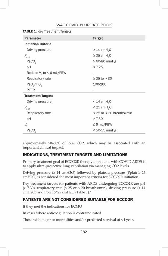
W4C COVID-19 UPDATE BOOK
182
approximately 50–60% of total CO2, which may be associated with an important clinical impact.
INDICATIONS, TREATMENT TARGETS AND LIMITATIONS Primary treatment goal of ECCO2R therapy in patients with COVID ARDS is to apply ultra-protective lung ventilation via managing CO2 levels.
Driving pressure (≥ 14 cmH2O) followed by plateau pressure (Pplat; ≥ 25 cmH2O) is considered the most important criteria for ECCO2R initiation.
Key treatment targets for patients with ARDS undergoing ECCO2R are pH (> 7.30), respiratory rate (< 25 or < 20 breaths/min), driving pressure (< 14 cmH2O) and Pplat (< 25 cmH2O (Table 1).3
PATIENTS ARE NOT CONSIDERED SUITABLE FOR ECCO2RIf they met the indications for ECMO
In cases where anticoagulation is contraindicated
Those with major co morbidities and/or predicted survival of < 1 year.
TABLE 1: Key Treatment Targets
Parameter Target
Initiation Criteria
Driving pressure ≥ 14 cmH2OPplat ≥ 25 cmH2O
PaCO2 > 60-80 mmHgpH < 7.25Reduce VT to < 6 mL/PBW -Respiratory rate ≥ 25 to > 30PaO2/FiO2 100-200PEEP -
Treatment Targets
Driving pressure < 14 cmH2OPplat < 25 cmH2O
Respiratory rate < 25 or < 20 breaths/minpH > 7.30
VT ≤ 6 mL/PBWPaCO2 < 50-55 mmHg
W4C COVID-19 UPDATE BOOK
183
EVIDENCES A prospective multicenter international phase II study aimed to assess the feasibility and safety of extracorporeal carbon dioxide removal (ECCO2R) to facilitate ultraprotective ventilation (VT 4 mL/kg and Pplat ≤ 25 cmH2O) in patients with moderate ARDS. The authors concluded that the use of ECCO2R to facilitate ultraprotective ventilation was feasible.
In the recent round table of European experts on ECCO2R, an agreement was made that the main treatment goal of ECCO2R therapy in patients with ARDS was to carry out ultraprotective lung ventilation through handling CO2 levels. Driving pressure with plateau pressure optimization was fixed as the principal criteria for ECCO2R introduction.3
Recently published REST trial in which patients requiring mechanical ventilation for acute hypoxemic respiratory failure, lower tidal volume ventilation facilitated by extracorporeal carbon dioxide removal, compared with standard care and did not result in a reduction in mortality at 90 days. However, due to early termination, the study may have been underpowered to detect a clinically important difference.8
In this retrospective case series of 29 patients, we have demonstrated the efficacy of ECCO2R using the Hemolung RAS to improve respiratory acidosis in patients with severe hypercapnic respiratory failure due to COVID-19. This is the first reported use of ECCO2R in the United States for this patient population.9,10
PRACTICE POINTS1. ECCO2-R appears to be an effective therapy for acute hypercarbia in
patients with moderate to severe ARDS.
2. It enables to optimize lung protective ventilation.
3. At the present time, however, the evidence is lacking that supports more widespread use, and ECCO2-R should be considered a research and rescue tool rather than an accepted clinical procedure.
4. More robust research using randomized controlled trials targeting meaningful outcomes (duration of mechanical ventilation, length of stay, and mortality) is urgently needed in COVI19 patients with ARDS.
REFERENCES 1. Wu Z, McGoogan JM. Characteristics of and important lessons from the coronavirus disease
2019 (COVID-19) outbreak in China: summary of a report of 72314 cases from the Chinese Center for Disease Control and Prevention. JAMA. 2020. 10.1001/jama.2020.2648.
2. The Acute Respiratory Distress Syndrome Network, Ventilation with lower tidal volumes as compared with traditional tidal volumes for acute lung injury and the acute respiratory distress syndrome. New Engl J Med 2000; 342:1301–1308.
3. Combes A, Auzinger G, Capellier G, du Cheyron D, Clement I, Consales G, et al. ECCO2R
W4C COVID-19 UPDATE BOOK
184
therapy in the ICU: consensus of a European round table meeting. Crit Care 2020; 24:490.4. Camporota L, Vasques F, Sanderson B, Barrett NA, Gattinoni L. Identification of
pathophysiological patterns for triage and respiratory support in COVID-19. Lancet Respir Med 2020; 8:752–4.
5. Ziehr DR, Alladina J, Petri CR, et al. Respiratory pathophysiology of mechanically ventilated patients with COVID-19: a cohort study. Am J Respir Crit Care Med 2020; 201:1560-1564.
6. Tobin MJ, Laghi F, Jubran A. Why COVID-19 silent hypoxemia is baffling to physicians. Am J Respir Crit Care Med 2020; [cited 2020 Jun 23]
7. Giraud, R., Banfi, C., Assouline, B. et al. The use of extracorporeal CO2 removal in acute respiratory failure. Ann. Intensive Care 2021; 11:43.
8. The REST Randomized Clinical Trial James J. McNamee, MB, ChB1,2; Michael A. Gillies, MD3; Nicholas A. Barrett, MB, ChB4; et al JAMA. Published online August 31, 2021. doi:10.1001/jama.2021.13374
9. Combes, A., Fanelli, V., Pham, T. et al. Feasibility and safety of extracorporeal CO2 removal to enhance protective ventilation in acute respiratory distress syndrome: the SUPERNOVA study. Intensive Care Med 2019; 45:592–600.
10. Akkanti, Bindu MD, FCCP1; Jagpal, Sugeet MD2; Darwish, Ribal MD, FCCM3; Saavedra Romero, Ramiro MD4 et al. Physiologic Improvement in Respiratory Acidosis Using Extracorporeal Co2 Removal With Hemolung Respiratory Assist System in the Management of Severe Respiratory Failure From Coronavirus Disease 2019, Critical Care Explorations March 2021 - Volume 3 - Issue 3 - p e0372

185
Extracorporeal Therapies in COviD 19 (Excluding ECMO
and ECCO2R)
INTRODUCTIONThe critically ill COVID 19 patient may develop multiple organ dysfunction syndromes (MODS), which is characterized by global hemodynamic derangement and organ hypo perfusion along with an uncontrolled and marked inflammatory response unresponsive to pharmacological management. This deterioration of one organ’s function may lead to dysfunction or damage to other organs,1 which is labelled as organ cross-talk such as cardiorenal syndrome, hepatorenal syndrome etc. The mortality may go as high as 90% if three or more organs are affected.2 Though not understood completely, several mechanism has been identified in pathogenesis and disease progression of COVID-19 which include direct virus-induced cytotoxicity in (angiotensin converting enzyme) ACE2-expressing cells, dysregulation of the renin angiotensin aldosterone system (RAAS) as a result of virus-mediated ACE2 down regulation, altered immune responses, injury to endothelial cell leading to thrombo-inflammation and tissue fibrosis in the late stages.3 These patients may get benefited with extracorporeal organ support therapies as adjuvant therapy such as therapeutic plasma exchange (TPE) and immunoadsorption (IA) may be helpful in removing cytokines and/or endotoxins and may be an effective means to protect organ functions and survival in covid 19 patients (Figure 1).4
ACUTE KIDNEY INJURY (AKI) AND CRRT IN COVID 19AKI with various degree of severity has been seen in critically ill covid 19 patients with an incidence of around 5%. The exact mechanism of AKI in covid 19 patients is still unclear however the possible mechanism may include local replication of SARS CoV2 in proximal tubular epithelial cells leading to AKI, activation of immune response followed by cytokine release. This increase in inflammatory mediators (IL1, IL6, IL 12, TNF α etc) induce an uncontrolled systemic inflammatory response, which leads to an increase in renal vascular permeability and decrease effective circulation capacity,
Ranajit Chatterjee, Vivek Gupta
23.
W4C COVID-19 UPDATE BOOK
186
contributing to renal insult. Thirdly, hypovolemia (absolute or relative) in these patients can lead to renal insufficiency and may lead to acute tubular necrosis.5 A genetic predisposition has also been shown to play in the renal injury. The treatment with high doses and/or long-term use of corticosteroids to alleviate the SIRS increases the risk of infection and metabolic disorders which can further worsen the renal insult.
CRRT can support critically ill COVID-19 patients with AKI or MODS by haemodynamic stabilization and metabolic correction along with reduction in inflammatory mediators.6 Its precision in volume control in hemodynamically unstable patients makes it an obvious choice in critically ill COVID 19 patients. The various therapies which can be helpful include continuous veno-venous hemofiltration (CVVH), continuous veno-venous hemodialysis (CVVHD) and continuous veno-venous hemodiafiltration (CVVHDF). SCUF comes handy in patients of pulmonary oedema with unstable hemodynamics. The prescription of therapy depends on the status of the patient. CVVHD and CVVHDF modalities with pre-filter replacement of fluid decrease filtration fraction and reduce the chance of circuit clotting.7 Diffusive therapies are effective for removal of solutes having small molecular weights including urea, creatinine and potassium. However higher molecular weight solutes such as cytokines can be removed with convective therapies. A reduction in inflammatory mediators along with improved oxygenation has been shown with high-flux (HF) and high-cut-off (HCO) membranes when used with CRRT in critically ill patients in ICU. The timing of CRRT initiation and treatment dose are important factors related with prognosis in critically ill COVID-19 patients. Though there is there is no specific recommendation for the timing of early CRRT initiation however initiation within 72 hours of the disease progression helps in blocking response and may improve the prognosis.8 Covid 19 patients who are on ECMO support showed a significant reduction in level of inflammatory mediators and reduction of ground glass opacities on computed tomography (CT) scan when combined with CRRT.9 The use of increased doses of unfractionated heparin (UFH)or the combination of regional citrate anticoagulation (RCA) and heparin can prevent premature filter clotting, thereby prolonging filter life.10 Moreover a vascular access with uninterrupted flow is an important way to enhance the filter life. Right internal jugular vein is the preferred choice due to its straight course followed by the femoral vein. However a conclusive recommendation can be made only after a randomized controlled trial (RCT).
THERAPEUTIC PLASMA EXCHANGE (TPE)Plasma exchange is another therapeutic blood purification strategy which removes the pathological plasma from the patient’s blood, replace with desired amount of recommended solution or plasma thus helping in reduction of pathological damage.11 Several studies have demonstrated the beneficial effect of TPE for critically ill COVID-19 patients. These studies have demonstrated

W4C COVID-19 UPDATE BOOK
187
improvement in organ function, decreased mediator levels, higher extubation rate and lower mortality. However antibodies against SARS-CoV-2 are also filtered along with the inflammatory mediators, which is unfavourable.12
Endothelial activation and blood hypercoagulability along with increased von Willebrand factor (VWF) and D-dimers levels are seen in Covid 19 patients. TPE may be helpful in reducing VWF and D-dimers levels and may improve the survival.13 There are certain limitations with TPE in critically ill COVID-19 patients, the production of cytokines does not stop, which may damage the organs in a short span of time.14 Moreover TPE may be associated with hypocalcemia when replacing fluids and patient may present with twitching of hands and feet and/or arrhythmia. There is possibility of anaphylactic reaction due to patient’s red blood cells and the donor plasma. These complications may be fatal in critically ill COVID-19 patients. Convalescent plasma carries specific neutralizing antibodies, and using TPE combined with convalescent plasma may be both an effective and a safe method.15 However no significant difference in improving survival rate and other clinical outcomes with TPE
COVID 19 PATIENTS IN ICU
Acute Kidney Injury Cytokine Storm
(With or without AKI) Worsening RIFLE's/AKIN/KDIGO
Class Volume Overload
Metabolic derangements Electrolyte disturbances
YES NO
TPE or HA (With or without CRRT)
Consider for CRRT (Choose the therapy
according to CRRT goals) Diffusive/ convective/
combination or ultrafiltration
Medical Management
Monitor and ifcondition worsens
FIG. 1: Propose Algorithm for Use of Extracorporeal Therapies in Critically Ill COVID 19 Patients (Excluding ECMO & ECCO2R)

W4C COVID-19 UPDATE BOOK
188
was found in a recently published randomized controlled, open-label trial.
HEMOADSORPTION (HA)Hemoperfusion (HP) and continuous plasma filtration absorption (CPFA) have been used in the management of critically ill patients. HP helps in cytokines, endotoxins and other inflammatory mediator removal and helps in management of septic shock. HP has been used in management of critically ill COVID-19 patients16 which has helped in reduction in severity of disease and also help in removal of toxins leading to acute hepatic failure in Covid 19 patients.17 Limited data is available for using IA in critically ill Covid 19 patients, many such patients have received IA treatment and achieved certain effects. CPFA is another extracorporeal modality, which can effectively remove cytokines from the blood and also help in regulating the volume status and stabilize the interior milieu of body. CPFA may also be a promising modality in improving symptoms of COVID-19 patients.
CONCLUSIONAn uncontrolled inflammatory response seen in critically ill COVID-19 patients may lead to variable degrees of organ dysfunction including acute kidney injury and even MODS. The limited available literature suggests a potential role of extra corporeal therapies in managing critically ill patients. These therapies help in removing inflammatory cytokines and toxins, which can significantly improve the clinical symptoms, prevent further organ damage and reduce mortality. However, we still need multicentre trials not only to identify the optimal therapy but also to learn the criteria for right modality of extracorporeal therapy.
REFERENCES1. Ziesmann, M.T. and Marshall, J.C. Multiple Organ Dysfunction: The Defining Syndrome of
Sepsis. Surgical Infections 2018; 19:184-190. 2. Knaus, W.A., Draper, E.A., Wagner, D.P., et al. APACHE II: A Severity of Disease Classification
System. Critical Care Medicine 1985; 13:818-829.3. Gupta, A., Madhavan, M. V., Sehgal, K., Nair, N et al.Extrapulmonary manifestations of
COVID-19. Nat Med 2020; 26:1017–1032.4. Ronco C, Bagshaw SM, Bellomo R, et al., 2020. Extracorporeal blood purification and
organ support in the critically ill patient during covid-19 pandemic: Expert review and recommendation. Blood Purif 2020; 1-11.
5. Na KR, Kim HR, Ham Y, et al. Acute kidney injury and kidney damage in covid-19 patients. J Korean Med Sci 2020; 35:e257.
6. Fu D, Yang B, Xu J, et al. Covid-19 infection in a patient with end-stage kidney disease. Nephron, 2020; 144:245-247.
7. Nadim MK, Forni LG, Mehta RL, et al. Covid-19-associated acute kidney injury: Consensus report of the 25th acute disease quality initiative (adqi) workgroup. Nat Rev Nephrol 2020; 16:747-764.
8. Pan PH, Song C, Lu RL. [the timing of continuous renal replacement therapy in severe covid-19]. Zhonghua Jie He He Hu Xi Za Zhi 2020; 43:721-724.
9. Zou H, Li S. Ecmo/crrt combined support in the treatment of critically ill patients with novel
W4C COVID-19 UPDATE BOOK
189
coronavirus pneumonia. Eur Heart J Case Rep 2020; 4:1-3. 10. Deep A, Bansal M, Ricci Z. Acute kidney injury and special considerations during renal
replacement therapy in children with coronavirus disease-19: Perspective from the critical care nephrology section of the european society of paediatric and neonatal intensive care. Blood Purif 2020; 1-11.
11. Grazioli A, Athale J, Tanaka K, et al. Perioperative applications of therapeutic plasma exchange in cardiac surgery: A narrative review. J Cardiothorac Vasc Anesth 2020; 34:3429-3443.
12. Honore PM, Mugisha A, Kugener L, et al. Therapeutic plasma exchange as a routine therapy in septic shock and as an experimental treatment for covid-19: We are not sure. Crit Care 2020; 24:226.
13. Zachariah U, Nair SC, Goel A, et al. Targeting raised von willebrand factor levels and macrophage activation in severe covid-19: Consider low volume plasma exchange and low dose steroid. Thromb Res 2020; 192:2.
14. Daoud AM, Soliman KM, Ali HK. Potential limitations of plasmapheresis in treatment of covid-19 patients: How to overcome them? Ther Apher Dial 2020; 1
15. Jaiswal V, Nasa P, Raouf M, et al. Therapeutic plasma exchange followed by convalescent plasma transfusion in critical covid-19-an exploratory study. Int J Infect Dis 2021; 102:332-334.
16. Safari S, Salimi A, Zali A, et al. Extracorporeal hemoperfusion as a potential therapeutic option for severe covid-19 patients; a narrative review. Arch Acad Emerg Med 2020; 8:e67.
17. Esmaeili Vardanjani A, Ronco C, Rafiei H, et al. Early hemoperfusion for cytokine removal may contribute to prevention ofintubation in patients infected with covid-19. Blood Purif 2021; 50:257-260.

SECTION 4-C
ManagementCURRENT POSITION OF VARIOUS
DRUGS & ADJUVANTS IN COVID-19 TREATMENT
SECTION 4-C

193
Section 4 - ManagementSECTION EDITOR
Dr Kedar ToraskarMD EDIC Director Critical Care, Critical Care Unit, Wockhardt Hospital, South Mumbai
Preface
SUBSECTION ON CURRENT POSITION OF VARIOUS DRUGS & ADJUVANTS IN COVID 19 TREATMENTThis subsection highlights some real gamechangers like steroids, IL6 antagonists, monoclonal antibodies & antivirals. It also covers some of the other agents like hydroxychloroquine, azithromycin, ivermectin, convalescent plasma, vitamin C, zinc, etc. which
showed some promise but lost out due to lack of data & results to back them up. Some other controversial immunomodulators like colchicine, ulinastatin, cytosorb filter have also been discussed in this subsection.
Unindicated use of steroids in high doses for longer duration led to a spate of secondary bacterial & fungal infections & poor outcomes. The mucormycosis epidemic in India associated with covid 19 was partly due to the misuse of steroids apart from the effects of the virus itself on the immune system. Judicious use of low dose short course of steroids only for those patients with hypoxemia in the inflammatory phase should be advocated.
Antivirals like remdesivir have shown promise but unfortunately majority of trials & studies failed to back it up. Molnupiravir which is a new oral antiviral in the pipeline is showing good promising results in the ongoing phase 3 trials.
Monoclonal antibody cocktail has shown good results backed up with some promising trial results but its use has been limited due to the exorbitant cost. These agents have got an emergency approval for post-exposure prophylaxis for certain individuals who are at high risk of acquiring SARS-CoV-2 infection and if infected are at high risk of progressing to serious illness.
Convalescent plasma showed initial promise but unfortunately recent large
194
trials have conclusively proven that it is not beneficial & its use in covid 19 therapy has been rightly given up.
Finally in this pandemic when our understanding of this novel & deadly virus & therapies for the same are constantly changing we need to stick to those therapies with robust scientific evidence so that we “DON’T CAUSE HARM” by treating overzealously.
195
Corticosteroids and Covid-19
INTRODUCTIONThe dysregulated host immune response to severe acute respiratory syndrome coronavirus 2 (SARS-CoV-2) plays a crucial role in the pathophysiology of coronavirus disease 2019 (COVID-19). The progression of COVID-19 to severe disease is a multisystem organ disease and the lung being the lead target. Severe COVID-19 characterized by severe tachypnea (respiratory rate >30 /min), hypoxemia (SpO2 <90% on room air) and presence of acute respiratory distress syndrome (ARDS), sepsis or septic shock is the most common indication for ICU admission and mortality. Corticosteroids modulate the immune response and can prevent or mitigate its deleterious effect in various diseases. Corticosteroids use in ARDS had been under intense debate over decades. Recently, there is evidence supporting corticosteroids in moderate to severe ARDS. In a meta-analysis, corticosteroids use in non-COVID-19 ARDS, found to significantly reduce duration of invasive mechanical ventilation (IMV) [mean difference-4.93 days, 95% confidence interval (CI) -7.81 to -2.06 days, P<0.001] and all-cause mortality [risk ratio 0.75 (95% CI 0.59-0.95), P <0.001].1
EVIDENCE ON CORTICOSTEROIDS Corticosteroid was the first effective treatment discovered for the management of COVID-19. The large open-label multicenter randomized controlled trial (RCT) (RECOVERY) from United Kingdom in 6,425 patients with COVID-19, demonstrated dexamethasone use can reduce 28-day mortality vs standard of care (SOC) [22.9% vs 25.7%, P<0.001, rate ratio 0.83 (0.75-0.93)]. The survival benefit was greatest in patients on IMV at randomization [29.3% vs 41.4%, rate ratio 0.64 (0.51-0.81)]. No survival benefit and signal towards harm was observed in patients without oxygen [17.8% vs 14.8%, rate ratio 1.19 (0.91–1.55)].2 The dose of oral or intravenous dexamethasone used was 6 mg/day and duration ten days. After the RECOVERY trial, most of the ongoing trials on corticosteroids were stopped prematurely because of obvious ethical concerns. The metanalysis of seven RCTs on corticosteroids in critically ill
Prashant Nasa, Bharat Jagiasi
24.
W4C COVID-19 UPDATE BOOK
196
patients with COVID-19 (n=1703), found reduce mortality in patients on IMV compared to SOC [30% vs 38% odd ratio (OR) 0.69 (0.55-0.86)] and in patients not on IMV [23% vs 42%, OR-0.41 (0.19-0.88)]. A majority of the patients included in the meta-analysis were from the RECOVERY trial (59%), only dexamethasone was found to reduce mortality significantly [OR 0.64 (0.50–0.82); P< 0.001].3
The RECOVERY trial changed the management of COVID-19 with use of corticosteroid in patients requiring oxygen became SOC. However, few unanswered questions after RECOVERY trial were on right timing, effect of a higher dose, duration, and other types of corticosteroids.
Timing: In a small retrospective study from China, early use of corticosteroids in COVID-19 was found to delay viral clearance, fever resolution, increase length of hospitalization, or risk of secondary infection.4 A recent meta-analysis found no prolongation in SARS-CoV-2 clearance with a low dose of corticosteroids.5 However, there is a trend towards higher mortality in the RECOVERY trial, with dexamethasone use in patients not on oxygen.2 Corticosteroids should be only be used for patients with COVID-19 requiring oxygen or signs of disease progression.3,7
Dose: RECOVERY trial used dexamethasone at 6 mg/day in patients with COVID-19. Smaller non-randomized cohort studies reported survival benefit with higher doses of corticosteroid in patients with COVID-19. However, a large (non-peer-reviewed, preprint) RCT did not show a significant difference in outcome between low (6 mg/day) vs high (12 mg/day) dose of dexamethasone, despite a trend towards lower mortality with the higher dose.7
Duration: The RECOVERY trial used dexamethasone for ten days.2 A longer duration (beyond ten days) of corticosteroids is proposed for patients with persistent clinical severity or ground-glassing on imaging. Some physicians also consider prolonged duration of corticosteroids for prevention of fibrosis after severe disease. However, prolonged use of corticosteroids may also increase the risk of adverse effects like opportunistic infections or neuromuscular abnormalities. At present, there is no quality evidence to support use of corticosteroids more than ten days. Longer duration of corticosteroids, if considered, requires a close monitoring of adverse effects.
Type of corticosteroids: World Health Organization (WHO) REACT metanalysis concluded corticosteroids benefit in COVID-19 was a class effect.3 Though the significant mortality benefit was only reported with dexamethasone because of a higher proportion of patients from the RECOVERY trial. In case of non-availability of dexamethasone, an equivalent dose of other corticosteroids may be considered (Prednisone 40 mg, Methylprednisolone 32 mg and Hydrocortisone 160 mg). Single or twice daily is recommended with long-acting corticosteroids like dexamethasone (half-life 36-72 hours),

W4C COVID-19 UPDATE BOOK
197
methylprednisolone or prednisolone (half-life 12-36 hours). Hydrocortisone (half-life 8-12 hours) is administered in two to four divided doses per day.8
Inhaled Corticosteroids
Inhaled corticosteroids (ICS) showed in non-human studies to downregulate the expression of ACE-2 receptors and impaired SARS-CoV-2 growth. However, no outcome benefit and risk of hospitalization was seen in the patients on long-term ICS.9 At present, the evidence is insufficient for routine use of ICS in the treatment of COVID-19.8
Adverse Effects
Corticosteroids are double-edged swords. Adverse effects like hyperglycemia, secondary infections, neuro-psychiatry effects, avascular necrosis are well known. However, with a low dose and limited duration of corticosteroids, studies have not reported significantly increased adverse effects.6 While opportunistic infections like mucormycosis and aspergillosis in patients with COVID-19 are linked to uncontrolled diabetes mellitus and abuse of corticosteroids.10
CONCLUSION Corticosteroids are a mainstay in the management of COVID-19. Corticosteroids should be used only in patients with severe COVID-19, increasing oxygen requirement at an equivalent dose of dexamethasone 6mg/day for ten days.
PRACTICE POINTS• Corticosteroids are recommended to manage patients with COVID-19
requiring oxygen support or signs of clinical worsening.
• Corticosteroids should not be used in non-severe COVID-19 or patients who do not require oxygen.
• The current evidence supports oral or intravenous dexamethasone at 6mg/day for ten days in most patients with severe COVID-19.
• Caution for a higher dose and prolonged duration is required to prevent adverse effects of corticosteroids.
REFERENCES1. Mammen MJ, Aryal K, Alhazzani W, Alexander PE. Corticosteroids for patients with acute
respiratory distress syndrome: a systematic review and meta-analysis of randomized trials. Pol Arch Intern Med 2020; 130:276-286. doi: 10.20452/pamw.15239.
2. Horby P, Lim WS, Emberson JR, Mafham M, Bell JL, Linsell L, et al. Dexamethasone in Hospitalized Patients with Covid-19. N Engl J Med 2021; 384:693-704. doi: 10.1056/NEJMoa2021436.
3. Sterne JAC, Murthy S, Diaz JV, Slutsky AS, Villar J, Angus DC, et al. Association Between Administration of Systemic Corticosteroids and Mortality Among Critically Ill Patients With
W4C COVID-19 UPDATE BOOK
198
COVID-19: A Meta-analysis. JAMA 2020; 324:1330-1341. doi: 10.1001/jama.2020.17023. 4. Li Q, Li W, Jin Y, Xu W, Huang C, Li L, et al. Efficacy Evaluation of Early, Low-Dose, Short-
Term Corticosteroids in Adults Hospitalized with Non-Severe COVID-19 Pneumonia: A Retrospective Cohort Study. Infect Dis Ther 2020; 9:823-836. doi: 10.1007/s40121-020-00332-3.
5. Nasa P, Chaudhry D, Govil D, Daga MK, Jain R, Chhallani AA, et al. Expert Consensus Statements on the Use of Corticosteroids in Non-severe COVID-19. Indian J Crit Care Med 2021; https://www.ijccm.org/doi/IJCCM/pdf/10.5005/jp-journals-10071-23923
6. Cano EJ, Fonseca Fuentes X, Corsini Campioli C, et al. Impact of Corticosteroids in Coronavirus Disease 2019 Outcomes: Systematic Review and Meta-analysis. Chest 2021; 159:1019-1040. doi:10.1016/j.chest.2020.10.054
7. Munch MW, Myatra SN, Vijayaraghavan BKT, Saseedharan S, Benfield T, Wahlin RR, et al. Dexamethasone 12 mg versus 6 mg for patients with COVID-19 and severe hypoxia: an international, randomized, blinded trial. doi: https://doi.org/10.1101/2021.07.22.21260755
8. CDC. COVID-19 treatment guidelines. Corticosteroids. https://www.covid19treatmentguidelines.nih.gov/therapies/immunomodulators/corticosteroids/ Accessed on August 20, 2021.
9. Schultze A, Walker AJ, MacKenna B, Morton CE, Bhaskaran K, Brown JP, et al. Risk of COVID-19-related death among patients with chronic obstructive pulmonary disease or asthma prescribed inhaled corticosteroids: an observational cohort study using the OpenSAFELY platform. Lancet Respir Med 2020; 8:1106-1120. doi: 10.1016/S2213-2600(20)30415-X.
10. Al-Tawfiq JA, Alhumaid S, Alshukairi AN, Temsah MH, Barry M, Al Mutair A, et al. COVID-19 and mucormycosis superinfection: the perfect storm. Infection 2021 Jul 24:1–21. doi: 10.1007/s15010-021-01670-1.
199
Monoclonal Antibodies
INTRODUCTIONAs the rapid spread of COVID-19 resulted in aworldwide crisis, a variety of treatment options were developed or repurposed. Monoclonal antibodies (mAbs)* are a class of antiviral that can bind and neutralize the virus in infected patients.1 B cells of convalescent patients or humanized mice are the source of these recombinant proteins.
Three anti-SARS-CoV-2 mAb therapies have been granted emergency use authorization (EUA) for treatment of non-hospitalized patients with mild-to-moderate COVID-19
1. Casirivimab with Imdevimab (REGN- COV2 therapy)
2. Bamlanivimab with Etesevimab
3. Sotrovimab:
RATIONALE Antigen- specific mAbs* or polyclonal antibodies which are derived from non- human or human sources, produces passive immunization after infusion.
Polyclonal antibodies from animal origin are primary source of antisera, but there is risk of ‘serum sickness’ after repeated exposures, as the recipient may generate an immune response against antibodies of non- human origin. These risks are nullified with the use of convalescent plasma therapy (CPT)* from human patients.
Early use of plasma after the onset of symptoms appears efficacious, but antibody titers of convalescent plasma vary considerably.2,3
High titer of neutralizing antibodies in mAbs as compared to CPT* results in superior efficacy and precise neutralizing capacity, while overcoming limitations intrinsic to CPT (risk of blood-borne diseases, risk of low antibody titers, as well as variable epitope specificity).
YP Singh, Akhil Taneja
25.

W4C COVID-19 UPDATE BOOK
200
MECHANISMThe SARS-CoV-2 has RNA genome that encodes four major structural proteins i.e., spike (S), envelope (E), membrane (M), and nucleocapsid (N), and nonstructural and accessory proteins. The spike protein which is further divided into S1 and S2 units, mediate host cell attachment and invasion. S1 attaches to Angiotensin-Converting Enzyme 2 (ACE2) on the host cell through its receptor-binding domain (RBD), thus initiating a conformational change in S2 that results in virus-host cell membrane fusion and viral entry4. Monoclonal antibodies target the receptor- binding domain (RBD) of the spike (S) protein and neutralize the ability of the virus to bind and fuse with the target host cell (Figure 1).
CASIRIVIMAB WITH IMDEVIMAB (REGN- COV-2 THERAPY)Recombinant human monoclonal antibodies bind to non-overlapping epitopes of the spike protein of SARS-CoV-2.
Casirivimab plus Imdevimab combination recently received EUA by FDA as post-exposure prophylaxis for
1. Individuals who are at high risk of acquiring SARS-CoV-2 infection
FIG. 1: Mechanism of Action of Antibody Cocktails
SARS COV-2Imdevimab
Casirivimab
Etesevimab
mAb binds to S proteinand prevents viral bindingand fusion with host cell
Bamlanivimab
S Protein
RBD
RBD
ACE-2
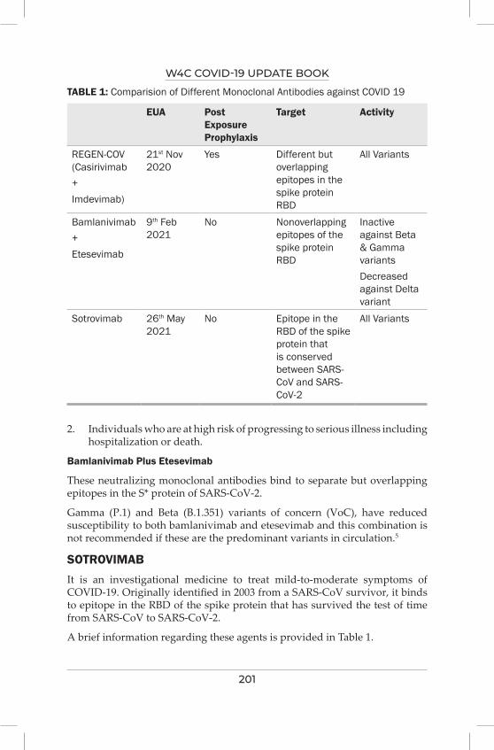
W4C COVID-19 UPDATE BOOK
201
2. Individuals who are at high risk of progressing to serious illness including hospitalization or death.
Bamlanivimab Plus Etesevimab
These neutralizing monoclonal antibodies bind to separate but overlapping epitopes in the S* protein of SARS-CoV-2.
Gamma (P.1) and Beta (B.1.351) variants of concern (VoC), have reduced susceptibility to both bamlanivimab and etesevimab and this combination is not recommended if these are the predominant variants in circulation.5
SOTROVIMAB It is an investigational medicine to treat mild-to-moderate symptoms of COVID-19. Originally identified in 2003 from a SARS-CoV survivor, it binds to epitope in the RBD of the spike protein that has survived the test of time from SARS-CoV to SARS-CoV-2.
A brief information regarding these agents is provided in Table 1.
TABLE 1: Comparision of Different Monoclonal Antibodies against COVID 19
EUA Post Exposure Prophylaxis
Target Activity
REGEN-COV (Casirivimab+Imdevimab)
21st Nov 2020
Yes Different but overlapping epitopes in the spike protein RBD
All Variants
Bamlanivimab +Etesevimab
9th Feb 2021
No Nonoverlapping epitopes of the spike protein RBD
Inactive against Beta & Gamma variantsDecreased against Delta variant
Sotrovimab 26th May 2021
No Epitope in the RBD of the spike protein that is conserved between SARS-CoV and SARS-CoV-2
All Variants
W4C COVID-19 UPDATE BOOK
202
RECOMMENDATIONSApproved for the treatment of mild to moderate coronavirus disease 2019 (COVID-19) in adult and pediatric patients (12 years of age and older weighing at least 40 kg) with positive results of direct SARS-CoV-2 viral testing, and who are at high risk for progression to severe COVID-19, including hospitalization or death:
Casirivimab 600 mg + Imdevimab 600 mg IV infusion
or
Sotrovimab 500 mg IV infusion
When using Casirivimab plus Imdevimab :
If IV infusions are not feasible or would cause a delay in treatment, Casirivimab 600 mg plus Imdevimab 600 mg administered by four subcutaneous (SQ) injections (2.5 mL per injection) at four different sites can be used as an alternative.
If IV infusions are delaying the treatment, Casirivimab 600 mg plus Imdevimab 600 mg administered by four subcutaneous (SQ) injections (2.5 mL per injection) at four different sites.
Patients at High Risk of Progression to Severe COVID-19
1. Older age (for example ≥65 years of age)
2. Obesity or being overweight (for example, adults with BMI >25 kg/m2, or if 12 to 17 years of age
3. Pregnancy
4. Chronic kidney disease
5. Diabetes
6. Immunosuppressive disease or immunosuppressive treatment
7. Essential hypertension & other heart diseases including ischemic heart disease & congenital heart diseases.
8. Chronic pulmonary illness (including chronic obstructive airway obstructive airway diseases, interstitial lung disease, cystic fibrosis and pulmonary hypertension)
9. Sickle cell disease
10. Neurodevelopmental disorders (for example, cerebral palsy) or other conditions that confer medical complexity (for example, genetic or metabolic syndromes and severe congenital anomalies)

W4C COVID-19 UPDATE BOOK
203
11. Patients with medical illness requiring home ventilation or with tracheostomy, feeding gastrostomy, etc. [not related to COVID 19]
PRACTICE POINTS1. When using monoclonal antibodies, treatment should be started as soon
as possible after the patient receives a positive result on a SARS-CoV-2 antigen or nucleic acid amplification test (NAAT) and within 10 days of symptom onset.
2. EUAs do not authorize the use of anti-SARS-CoV-2 monoclonal antibodies for the following patients:
a. Those hospitalized for COVID-19
b. Those who require oxygen therapy due to COVID-19
c. Those who are on chronic oxygen therapy due to an underlying non-COVID-19-related comorbidity and who require an increase in oxygen flow rate from baseline because of COVID-19
3. The use of anti-SARS-CoV-2 monoclonal antibodies should be considered for patients with mild to moderate COVID-19 who are hospitalized for a reason other than COVID-19 if they otherwise meet the EUA criteria for outpatient treatment.
4. Patients should be monitored during the IV infusion or SQ injections and for at least 1 hour after the infusion or injections are completed.
5. Hypersensitivity (anaphylaxis and infusion-related reactions) and rash, diarrhea, nausea, dizziness, and pruritis are common side effects
6. IgG products have been safely used in pregnant patients. Although there is a paucity on pregnancy-specific data on the use of these monoclonal antibodies, these products should not be withheld when indicated.
7. SARS-CoV-2 vaccination should be deferred for ≥90 days in people who have received anti-SARS-CoV-2 monoclonal antibodies as the antibody treatment may interfere with vaccine-induced immune responses.
8. Vaccination status should not affect treatment decisions, including the use of and timing of treatment with monoclonal antibodies.
CONCLUSIONThe sudden spread of the COVID-19 pandemic inspired an accelerated need to identify effective therapies that can reduce the morbidity and mortality associated with COVID-19. SARS-CoV-2 monoclonal antibodies provide a potential prophylactic and therapeutic option when appropriately timed in carefully selected patients
W4C COVID-19 UPDATE BOOK
204
Initial clinical trial data coming from various trials support further investigation of neutralizing mAbs to determine the optimal dosing regimen, time window and subset of patients who will benefit the most.
*mAbs: Monoclonal antibodies
*EUA: Emergency use authorization
*CPT: Convalescent plasma therapy
*RBD: Receptor Binding Domain
*S: Spike Protein
REFERENCES1. Taylor, P.C., Adams, A.C., Hufford, M.M. et al. Neutralizing monoclonal antibodies for
treatment of COVID-19. Nat Rev Immunol 2021; 21:382–393.2. Ko, J. H. et al. Challenges of convalescent plasma infusion therapy in Middle East respiratory
coronavirus infection: a single centre experience. Antivir Ther 2018; 23:617–622. 3. Zhang, J. S. et al. A serological survey on neutralizing antibody titer of SARS convalescent
sera. J Med Virol 2005; 77:147–150.4. Jiang S, Hillyer C, Du L. Neutralizing antibodies against SARS-CoV-2 and other human
coronaviruses. Trends Immunol 2020; 41:355-359.5. COVID-19 Treatment Guidelines Panel. Coronavirus Disease 2019 (COVID-19)
Treatment Guidelines. National Institutes of Health. Available at https://www.covid19treatmentguidelines.nih.gov/. Accessed [25/8/2021].
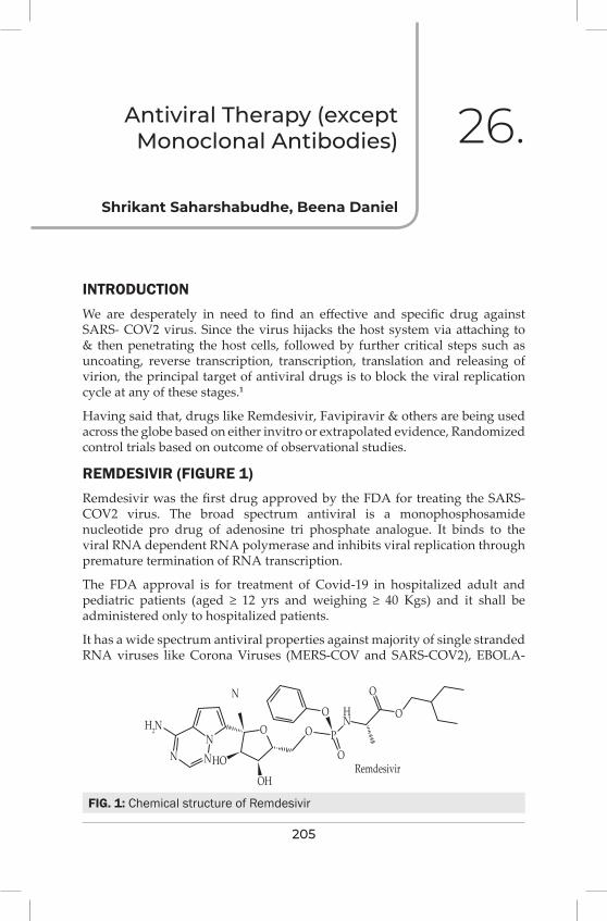
205
Antiviral Therapy (except Monoclonal Antibodies) 26.
INTRODUCTIONWe are desperately in need to find an effective and specific drug against SARS- COV2 virus. Since the virus hijacks the host system via attaching to & then penetrating the host cells, followed by further critical steps such as uncoating, reverse transcription, transcription, translation and releasing of virion, the principal target of antiviral drugs is to block the viral replication cycle at any of these stages.1
Having said that, drugs like Remdesivir, Favipiravir & others are being used across the globe based on either invitro or extrapolated evidence, Randomized control trials based on outcome of observational studies.
REMDESIVIR (FIGURE 1) Remdesivir was the first drug approved by the FDA for treating the SARS- COV2 virus. The broad spectrum antiviral is a monophosphosamide nucleotide pro drug of adenosine tri phosphate analogue. It binds to the viral RNA dependent RNA polymerase and inhibits viral replication through premature termination of RNA transcription.
The FDA approval is for treatment of Covid-19 in hospitalized adult and pediatric patients (aged ≥ 12 yrs and weighing ≥ 40 Kgs) and it shall be administered only to hospitalized patients.
It has a wide spectrum antiviral properties against majority of single stranded RNA viruses like Corona Viruses (MERS-COV and SARS-COV2), EBOLA-
Shrikant Saharshabudhe, Beena Daniel
FIG. 1: Chemical structure of Remdesivir
H2N
N
OH
HN
PO O
O
O
O
O
Remdesivir
N
N
NHO
W4C COVID-19 UPDATE BOOK
206
MARBURG Virus and others.1,2 It decreases the viral load in nasopharyngeal and oropharyngeal swabs after 12 days of its administration.3 The first case of Covid-19 in the USA was recovered using intravenous Remdesivir.3
DOSING Various multinational randomized open labeled trials have used RDV in the dose of 200 mg IV on DAY 1 and then 100 mg IV daily for 9 days. but the present consensus for dosing is 200 mg IV on Day 1 and 100 mg IV for 4 Days.6,5,10
ADVERSE EFFECTS1. Nausea
2. Increase in Transaminase levels
3. Increase in Prothrombin time (without change in INR)
4. Hypersensitivity Reactions
MONITORINGLFT and PT should be obtained in all patients before RDV is administered and during treatment is clinically indicated.
RDV may need to be discontinued if Alanine Transaminase (ALT) levels increase to >10 times the upper limit of normal.7
SPECIAL SITUATIONSa. Renal Insufficiency: Each vial of RDV contains a compound by the name
of SULPHOBUTYLETHERBETACYCLODEXTRINSODIUM (SBECD) which is abnormally accumulated in patients with renal impairment and may result in renal and liver toxicities.7
Lyophilized powder formulations contain less SBECD and can be preferred in patients with renal impairment. RDV is not recommended for patients with eGFR of < 30 ml/min due to lack of data.8
b. Pregnancy: Its compassionate use in pregnancy was well tolerated and with low rate of serious adverse events.9 It should not be withheld for pregnant patients if otherwise indicated.
c. Children: A clinical trial is currently evaluating the pharmacokinetics of RDV in children.
DRUG INTERACTIONSChloroquine / Hydroxy chloroquine may decrease the antiviral activity of RDV, hence co administration of these drugs is not recommended.7 RDV is not expected to have any significant interactions with Oseltamivir or Baloxavir.
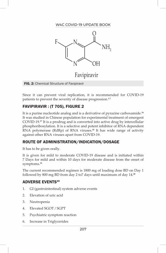
W4C COVID-19 UPDATE BOOK
207
Since it can prevent viral replication, it is recommended for COVID-19 patients to prevent the severity of disease progression.4,5
FAVIPIRAVIR : (T 705), FIGURE 2It is a purine nucleotide analog and is a derivative of pyrazine carboxamide.16 It was studied in Chinese population for experimental treatment of emergent COVID-19.17 It is a prodrug and is converted into active drug by intercellular phosphoribosylation. It is a selective and potent inhibitor of RNA dependent RNA polymerase (RdRp) of RNA viruses.18 It has wide range of activity against other RNA viruses apart from COVID-19.
ROUTE OF ADMINISTRATION/INDICATION/DOSAGEIt has to be given orally.
It is given for mild to moderate COVID-19 disease and is initiated within 7 Days for mild and within 10 days for moderate disease from the onset of symptoms.19
The current recommended regimen is 1800 mg of loading dose BD on Day 1 followed by 800 mg BD from day 2 to7 days until maximum of day 14.20
ADVERSE EVENTS20
1. GI (gastrointestinal) system adverse events
2. Elevation of uric acid
3. Neutropenia
4. Elevated SGOT / SGPT
5. Psychiatric symptom reaction
6. Increase in Triglycerides
FIG. 2: Chemical Structure of Favipiravir
F NNH2
O
OHN
Favipiravir
W4C COVID-19 UPDATE BOOK
208
SPECIAL SITUATIONSIt is contraindicated in pregnant and lactating women, in patients with hypersensitivity, severe hepatic impairment and severe renal function impairment. It should be administered with care in patients with Gout or history of Gout with hyperuricemia. Pediatric clinical studies are not yet conducted.
DRUG INTERACTIONSPotential interactions can be seen with pyrazinamide, repaglimide, theophylline, faviclovir, sulindac, acetaminophen21 and with drugs that inhibit aldehyde oxidase.
SUMMARYSeveral Clinical trials evaluating the use of RDV for the treatment of COVID-19 are currently underway or in development. We can search RDV clinical trials on www.clinicaltrials.gov, – which is resource provided by the US National Library of Medicine. As I am writing this, there are about 3, 87,641 research studies across 219 countries.
The major confidence for use of RDV in moderate to severe COVID-19 has come from Adaptive COVID-19 treatment Trial (ACTT-1).11 It is multinational; placebo controlled double blind RCT in hospitalized patients. It showed that in patients with severe COVID-19, RDV reduced time to clinical recovery. The clinical benefit of RDV was apparent in hospitalized patients who were on supplemental oxygen. Although 10 Days course was advocated in ACT T-1, Goldman and group has clearly demonstrated that there is no significant difference between a 5 Day and a 10 Days administration of RDV. (12) Other Major clinical trials include the WHO Solidarity trials which showed that RDV did not decrease in hospital mortality in hospitalized patient when compared to local SOC (standard of care), multicenter, placebo, controlled double blind RCT in hospitalized patients with severe COVID-19 from China,14 a open label randomized trial in hospitalized patients15 and so on.
At present around 27 studies for Favipiravir, including RCTs are ongoing in countries such as China, Japan, Italy, USA, UK, Canada. Its prophylactic role in COVID-19 is currently being explored in an ongoing clinical study in Canada and USA. The phase 3 PEPCO (Post Exposure prophylaxis for COVID-19) will look at asymptomatic individuals with direct exposure (within 72 Hours) to an infected individuals.
The prominent studies are 3 PRESECO in USA studies by Cai et al, Chen et al, Doi et al, Rattanampanan et al. However a large RCTs are required to demonstrate whether use of Favipiravir translates into clinical benefits.
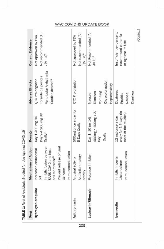
W4C COVID-19 UPDATE BOOK
209
TAB
LE 1
: Res
t of A
ntiv
irals
Stu
died
for U
se A
gain
st C
OVID
19
Dru
gM
echa
nism
of A
ctio
nD
osag
eAd
vers
e Ef
fect
sCu
rren
t Evi
denc
e
Hyd
roxy
chlo
roqu
ine
Incr
ease
d en
doso
mal
pH In
hibi
ts fu
sion
bet
wee
n SA
RS C
OV-2
and
hos
t ce
ll m
embr
ane22
Prev
ent r
elea
se o
f vira
l ge
nom
eIm
mun
omod
ulat
ion
Day
1 4
00 m
g BD
D
ay 2
-4 2
00 m
g BD
Oral
ly24
QTC
Prol
onga
tion
Tors
ade
de p
oint
esVe
ntric
ular
Arr
hyth
mia
Card
iac
deat
hs23
Not
app
rove
d by
FDA
Not
reco
mm
ende
d (A
I) , (
A II
a)6
Azith
rom
ycin
Antiv
iral a
ctiv
ityAn
ti-in
flam
mat
ory
activ
ity
500m
g on
ce a
day
for
5 D
ays
Oral
lyQT
C Pr
olon
gatio
nN
ot a
ppro
ved
by F
DAN
ot re
com
men
ded
(AI)
, (A
II a)
6
Lopi
navi
r/R
itona
vir
Prot
ease
Inhi
bito
rD
ay 1
- 10
(or 1
4)40
0mg
/ 10
0mg
x 2/
D
ay O
rally
Nau
sea
Dia
rrhe
aVo
miti
ngQt
c pr
olon
gatio
nH
epat
otox
icity
Not
Rec
omm
ende
d (A
I) ,(A
III)6
Iver
mec
tinIn
hibi
ts Im
pert
in
1het
erod
imer
Imm
unom
odul
atio
n
12 m
g on
ce a
day
or
ally
for 3
-5 d
ays
(in
mos
t of t
he s
tudi
es)
Diz
zine
ssPr
uriti
sN
ause
aD
iarr
hea
Insu
ffici
ent e
vide
nce
to
reco
mm
end
eith
er fo
r or
aga
inst
its
Use (C
ontd
..)
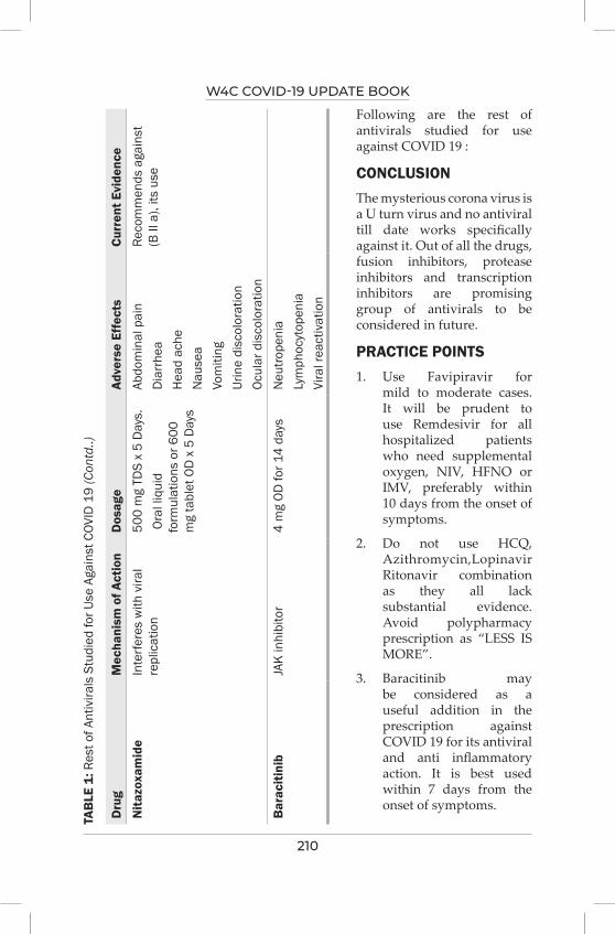
W4C COVID-19 UPDATE BOOK
210
TAB
LE 1
: Res
t of A
ntiv
irals
Stu
died
for U
se A
gain
st C
OVID
19
(Con
td..)
Dru
gM
echa
nism
of A
ctio
nD
osag
eAd
vers
e Ef
fect
sCu
rren
t Evi
denc
e
Nita
zoxa
mid
eIn
terfe
res
with
vira
l re
plic
atio
n50
0 m
g TD
S x
5 D
ays.
Ora
l liq
uid
form
ulat
ions
or 6
00
mg
tabl
et O
D x
5 D
ays
Abdo
min
al p
ain
Dia
rrhe
aH
ead
ache
Nau
sea
Vom
iting
Urin
e di
scol
orat
ion
Ocul
ar d
isco
lora
tion
Reco
mm
ends
aga
inst
(B
II a
), its
use
Bar
aciti
nib
JAK
inhi
bito
r4
mg
OD fo
r 14
days
Neu
trope
nia
Lym
phoc
ytop
enia
Vira
l rea
ctiv
atio
n
Following are the rest of antivirals studied for use against COVID 19 :
CONCLUSIONThe mysterious corona virus is a U turn virus and no antiviral till date works specifically against it. Out of all the drugs, fusion inhibitors, protease inhibitors and transcription inhibitors are promising group of antivirals to be considered in future.
PRACTICE POINTS1. Use Favipiravir for
mild to moderate cases. It will be prudent to use Remdesivir for all hospitalized patients who need supplemental oxygen, NIV, HFNO or IMV, preferably within 10 days from the onset of symptoms.
2. Do not use HCQ, Azithromycin,Lopinavir Ritonavir combination as they all lack substantial evidence. Avoid polypharmacy prescription as “LESS IS MORE”.
3. Baracitinib may be considered as a useful addition in the prescription against COVID 19 for its antiviral and anti inflammatory action. It is best used within 7 days from the onset of symptoms.
W4C COVID-19 UPDATE BOOK
211
ABBREVIATIONSACE 2 – angiotensin converting enzyme 2
BD – twice a day
COVID 19 – corona virus disease 19
eGFR – estimated glomerular filtration rate
E – envelope protein
ERGIC – endoplasmic reticulum –golgi apparatus compartment
FDA – Food and drug administration
HFNO – High flow nasal oxygen
IMV – Invasive mechanical ventilation
INR – international normalized ratio
IV – intravenous
JAK – janus kinase
LFT – Liver function test
MERS Cov – Middle east respiratory syndrome corona virus
M pro – main protease
M – membrane protein
NIV – Non invasive ventilation
N – nucleocapsid
OD – once a day
PRESECO – preventing severe COVID disease
PT – prothrombin time
QTc – corrected QT interval
RCT – randomized control trial
RNA – Ribonucleic acid
RDV – Remdesivir
SARS –COV 2 – Severe Acute respiratory syndrome corona virus
SGOT – serum glutamic oxaloacetic transaminase
SGPT – serum glutamine pyruvic transaminase
S – glycoprotein spike
W4C COVID-19 UPDATE BOOK
212
TDS – three times a day
TMPRSS2 – transmembrane serine protease 2
USA – United states of America
UK – united kingdom
WHO – world health organization
REFERENCES1. Frediansyah A, Tiwari R, Sharun K, Dhama K, Harapan H. Antivirals for COVID-19: A critical
review. Clinical Epidemiology and Global Health 2021; 9:90-8.2. Beigel JH, Tomashek KM, Dodd LE. Remdesivir for the Treatment of Covid-19—final Report.
October 8, 2020 DOI: 10.1056. NEJMoa2007764.3. Adamsick ML, Gandhi RG, Bidell MR, Elshaboury RH, Bhattacharyya RP, Kim AY, Nigwekar
S, Rhee EP, Sise ME. Remdesivir in patients with acute or chronic kidney disease and COVID-19. Journal of the American Society of Nephrology 2020; 31:1384-6.
4. Hoffman C; Covid Reference solution 2020-21 (Treatment); www.covidreference.com; accessed on 30-Aug-21
5. Li G, De Clercq E. Therapeutic options for the 2019 novel coronavirus (2019-nCoV). Nature Reviews Drug Discovery 2020; 19:149-50.
6. Udwadia ZF, Singh P, Barkate H, Patil S, Rangwala S, Pendse A, Kadam J, Wu W, Caracta CF, Tandon M. Efficacy and safety of favipiravir, an oral RNA-dependent RNA polymerase inhibitor, in mild-to-moderate COVID-19: A randomized, comparative, open-label, multicenter, phase 3 clinical trial. International Journal of Infectious Diseases 2021; 103:62-71.
7. Joshi S, Parkar J, Ansari A, Vora A, Talwar D, Tiwaskar M, Patil S, Barkate H. Role of favipiravir in the treatment of COVID-19. International Journal of Infectious Diseases 2020.
8. Goldman JD, Lye DC, Hui DS, Marks KM, Bruno R, Montejano R, Spinner CD, Galli M, Ahn MY, Nahass RG, Chen YS. Remdesivir for 5 or 10 days in patients with severe Covid-19. New England Journal of Medicine 2020; 383:1827-37.
9. Nguyen LS, Dolladille C, Drici MD, Fenioux C, Alexandre J, Mira JP, Moslehi JJ, Roden DM, Funck-Brentano C, Salem JE. Cardiovascular toxicities associated with hydroxychloroquine and azithromycin: an analysis of the World Health Organization Pharmacovigilance Database. Circulation 2020; 142:303-5.
213
iL6 Antagonists in Covid-19
INTRODUCTIONCOVID-19 (coronavirus disease 2019) syndrome, caused by SARS-CoV-2 (severe acute respiratory syndrome coronavirus-2), can manifest as an asymptomatic infection or as mild, moderate or severe disease and may be complicated by ARDS (acute respiratory distress syndrome) and multi-organ dysfunction. Patients with critical COVID-19 are not able to implement an efficient adaptive immune response and there is predominant innate immune response leading to development of a hyperinflammatory state called cytokine storm.1 IL-6 (interleukin-6) plays a key pathogenic role in COVID-19 associated cytokine storm. Thus, it represents a suitable therapeutic target. It is hypothesized that modulating the levels of IL-6 or its effects may be beneficial in COVID-19.
There are two classes of IL-6 inhibitors:
1. Anti-IL-6 receptor monoclonal antibodies: Tocilizumab, Sarilumab
2. Anti-IL-6 monoclonal antibodies: Siltuximab2
ROLE OF CYTOKINE STORM IN COVID-19SARS-CoV-2 gains entry into the alveolar epithelial cells by binding to their ACE2 (angiotensin converting enzyme 2) receptors. This leads to activation of pathogenic Th1 (T-helper 1) cells and monocytes/macrophages. This, along with differentiation of B lymphocytes into plasma cells producing anti-SARS-CoV-2 IgM and IgG antibodies, leads to effective resolution of viral infection in most of the patients. However, in some cases of severe COVID-19, dysfunctional immune response occurs leading to release of a large quantities of pro-inflammatory cytokines (Figure 1).1
Role of IL-6 in SARS-CoV-2-Induced Cytokine Storm
IL-6 plays a key role in cytokine storm associated with COVID-19. It is a pleotropic cytokine and exerts its biological effects on several cellular targets (Figure 2).
Mrinal Sircar, Sunny Kumar
27.
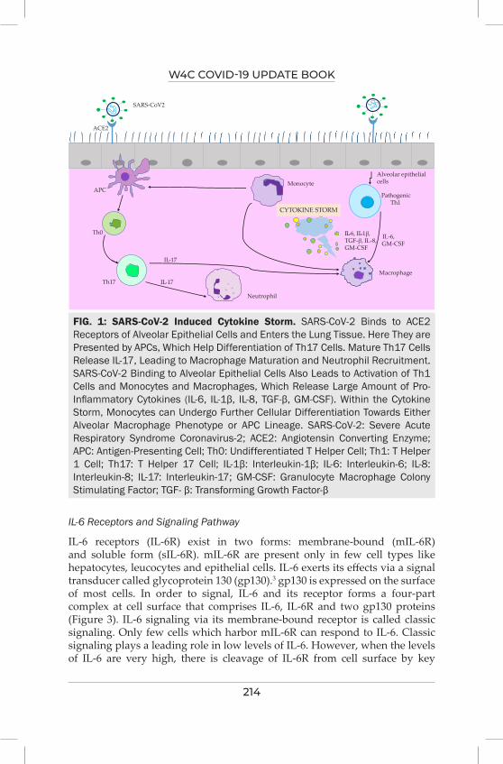
W4C COVID-19 UPDATE BOOK
214
FIG. 1: SARS-CoV-2 Induced Cytokine Storm. SARS-CoV-2 Binds to ACE2 Receptors of Alveolar Epithelial Cells and Enters the Lung Tissue. Here They are Presented by APCs, Which Help Differentiation of Th17 Cells. Mature Th17 Cells Release IL-17, Leading to Macrophage Maturation and Neutrophil Recruitment. SARS-CoV-2 Binding to Alveolar Epithelial Cells Also Leads to Activation of Th1 Cells and Monocytes and Macrophages, Which Release Large Amount of Pro-Inflammatory Cytokines (IL-6, IL-1β, IL-8, TGF-β, GM-CSF). Within the Cytokine Storm, Monocytes can Undergo Further Cellular Differentiation Towards Either Alveolar Macrophage Phenotype or APC Lineage. SARS-CoV-2: Severe Acute Respiratory Syndrome Coronavirus-2; ACE2: Angiotensin Converting Enzyme; APC: Antigen-Presenting Cell; Th0: Undifferentiated T Helper Cell; Th1: T Helper 1 Cell; Th17: T Helper 17 Cell; IL-1β: Interleukin-1β; IL-6: Interleukin-6; IL-8: Interleukin-8; IL-17: Interleukin-17; GM-CSF: Granulocyte Macrophage Colony Stimulating Factor; TGF- β: Transforming Growth Factor-β
CYTOKINE STORM
IL-6, IL-1β,
GM-CSF
Alveolar epithelial cells
Pathogenic Th1
Macrophage
IL-6, GM-CSF
Neutrophil
Th17
Th0
APC
ACE2
SARS-CoV2
Monocyte
IL-17
IL-17
Figure1
TGF-β, IL-8,
IL-6 Receptors and Signaling Pathway
IL-6 receptors (IL-6R) exist in two forms: membrane-bound (mIL-6R) and soluble form (sIL-6R). mIL-6R are present only in few cell types like hepatocytes, leucocytes and epithelial cells. IL-6 exerts its effects via a signal transducer called glycoprotein 130 (gp130).3 gp130 is expressed on the surface of most cells. In order to signal, IL-6 and its receptor forms a four-part complex at cell surface that comprises IL-6, IL-6R and two gp130 proteins (Figure 3). IL-6 signaling via its membrane-bound receptor is called classic signaling. Only few cells which harbor mIL-6R can respond to IL-6. Classic signaling plays a leading role in low levels of IL-6. However, when the levels of IL-6 are very high, there is cleavage of IL-6R from cell surface by key
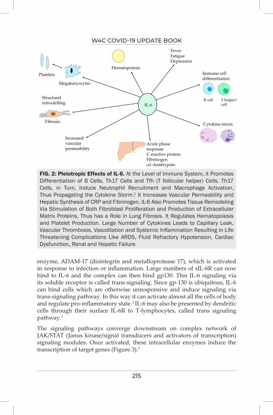
W4C COVID-19 UPDATE BOOK
215
enzyme, ADAM-17 (disintegrin and metalloprotease 17), which is activated in response to infection or inflammation. Large numbers of sIL-6R can now bind to IL-6 and the complex can then bind gp130. This IL-6 signaling via its soluble receptor is called trans-signaling. Since gp-130 is ubiquitous, IL-6 can bind cells which are otherwise unresponsive and induce signaling via trans-signaling pathway. In this way it can activate almost all the cells of body and regulate pro-inflammatory state.3 IL-6 may also be presented by dendritic cells through their surface IL-6R to T-lymphocytes, called trans signaling pathway.1
The signaling pathways converge downstream on complex network of JAK/STAT (Janus kinase/signal transducers and activators of transcription) signaling modules. Once activated, these intracellular enzymes induce the transcription of target genes (Figure 3).3
IL-6T helper17cell
B cell
Immune cell differentiation
Cytokine storm
Acute phase responseC reactive proteinFibrinogenα1-Antitrypsin
Increased vascular permeability
Structural remodelling
Fibrosis
Megakaryocytes
PlateletsHematopoiesis
FeverFatigueDepression
Figure2FIG. 2: Pleiotropic Effects of IL-6. At the Level of Immune System, it Promotes Differentiation of B Cells, Th17 Cells and Tfh (T follicular helper) Cells. Th17 Cells, in Turn, Induce Neutrophil Recruitment and Macrophage Activation, Thus Propagating the Cytokine Storm.1 It Increases Vascular Permeability and Hepatic Synthesis of CRP and Fibrinogen. IL-6 Also Promotes Tissue Remodeling Via Stimulation of Both Fibroblast Proliferation and Production of Extracellular Matrix Proteins, Thus has a Role in Lung Fibrosis. It Regulates Hematopoiesis and Platelet Production. Large Number of Cytokines Leads to Capillary Leak, Vascular Thrombosis, Vasodilation and Systemic Inflammation Resulting in Life Threatening Complications Like ARDS, Fluid Refractory Hypotension, Cardiac Dysfunction, Renal and Hepatic Failure
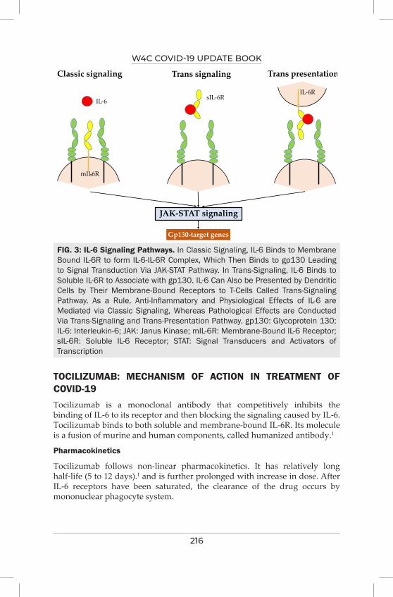
W4C COVID-19 UPDATE BOOK
216
TOCILIZUMAB: MECHANISM OF ACTION IN TREATMENT OF COVID-19Tocilizumab is a monoclonal antibody that competitively inhibits the binding of IL-6 to its receptor and then blocking the signaling caused by IL-6. Tocilizumab binds to both soluble and membrane-bound IL-6R. Its molecule is a fusion of murine and human components, called humanized antibody.1
Pharmacokinetics
Tocilizumab follows non-linear pharmacokinetics. It has relatively long half-life (5 to 12 days).1 and is further prolonged with increase in dose. After IL-6 receptors have been saturated, the clearance of the drug occurs by mononuclear phagocyte system.
FIG. 3: IL-6 Signaling Pathways. In Classic Signaling, IL-6 Binds to Membrane Bound IL-6R to form IL-6-IL-6R Complex, Which Then Binds to gp130 Leading to Signal Transduction Via JAK-STAT Pathway. In Trans-Signaling, IL-6 Binds to Soluble IL-6R to Associate with gp130. IL-6 Can Also be Presented by Dendritic Cells by Their Membrane-Bound Receptors to T-Cells Called Trans-Signaling Pathway. As a Rule, Anti-Inflammatory and Physiological Effects of IL-6 are Mediated via Classic Signaling, Whereas Pathological Effects are Conducted Via Trans-Signaling and Trans-Presentation Pathway. gp130: Glycoprotein 130; IL-6: Interleukin-6; JAK: Janus Kinase; mIL-6R: Membrane-Bound IL-6 Receptor; sIL-6R: Soluble IL-6 Receptor; STAT: Signal Transducers and Activators of Transcription
Classic signaling Trans signaling Trans presentation
IL-6
mIL-6R
sIL-6RIL-6R
JAK-STAT signaling
Gp130-target genesFigure 3
W4C COVID-19 UPDATE BOOK
217
Dose
In COVID-19, tocilizumab has been used in various studies in dose of 8mg/kg actual body weight (maximum 800mg/dose) in 0.9% saline infused over one hour. If there is inadequate response, a second dose is given after 12 to 24 hours.2,4,5
EVIDENCE FOR THE USE OF TOCILIZUMAB IN COVID-19Several initial retrospective observational studies have suggested an effect on mortality with tocilizumab.3 However, later prospective clinical trial data have been less convincing.3 Of particular interest are two randomized, double-blind, placebo-controlled trials – COVACTA6 and EMPACTA7 trials, which have shown no significant effect on 28-day mortality.
However, two recent randomized, controlled trials, RECOVERY5 and REMAP-CAP4 have shown decrease in mortality with tocilizumab use. Published in May 2021, RECOVERY5 trial included 4116 patients and suggested that 28-day mortality and receipt of invasive mechanical ventilation was significantly lower in subset of patients who received tocilizumab and steroids. The above effects were seen in groups with different disease severities. Similarly, in REMAP-CAP4 trial, 90-day mortality was significantly lower in IL-6 antagonist group (tocilizumab, sarilumab) compared to standard care. Median number of organ support-free days were more in tocilizumab and sarilumab groups.
Making interpretation from these disparate results can be difficult. Differences among trials include enrollment criteria, timing of initiation of therapy and background care. It is to note that both REMAP-CAP4 and RECOVERY5 were open-label trials, whereas COVACTA6 was a double-blind study. Only a minority of patients (19.4%) in COVACTA7 received steroids. In contrast, 93% and 82% of all patients in REMAP-CAP and RECOVERY trial, respectively, were receiving steroid therapy. Whether, tocilizumab is beneficial in absence of steroids remains unclear.
A recent REACT meta-analyses8 (July 2021) included 27 trials and 10930 patients. It showed 28-day mortality benefit (22% in IL-6 antagonist group versus 25% in usual care or placebo). Another meta-analysis by Avni et.al.9 (May 2021) included eight RCTs (6481 patients) and showed that tocilizumab was associated with reduction in 28-30-day mortality in severe non-critical COVID-19 infections. However, among critically-ill patients, and when steroids were used, no mortality benefit was demonstrated.
ADVERSE EFFECTSVarious adverse effects noted with tocilizumab include serious infections, gastrointestinal perforations particularly diverticulitis, infusion reactions (hypertension, headache), anaphylaxis, thrombocytopenia, leucopoenia and trans-aminitis.1
W4C COVID-19 UPDATE BOOK
218
IL-6 MEASUREMENT AND INTERPRETATIONMeasuring IL-6 concentrations and interpreting the results is not completely straightforward. It peaks at different stages in course of illness. Delay in processing of blood samples also influences measurements since IL-6 is released spontaneously from blood cells over time and also more so in samples which are agitated during transport. Immune cells are also sensitive to activation by temperature changes while transport.3 Isolated IL-6 elevation does not always imply that patient’s clinical condition is deteriorating and it might be indicative of inflammatory response.3
CONCLUSIONLatest research shows that tocilizumab when used along with steroids has a mortality benefit if used early in COVID-19. Some areas of controversies still remain and need further research including optimal timing of therapy, whether therapy should be guided by biomarkers (e.g. IL-6, CRP) or clinical criteria and identifying the patient phenotype who will benefit most from the therapy. Data for other IL-6 inhibitors is also limited and needs research.
PRACTICE POINTS1. Tocilizumab along with steroid is beneficial in severe COVID-19 and has
been recommended by WHO [10] and National institute of health (NIH) COVID-19 guidelines.2
2. It should be used early in course of disease and as early as possible (within 24 hours) after rapid respiratory decompensation.2
3. Tocilizumab administration should not be guided based solely on IL-6 levels, rather other biomarkers (like CRP, ferritin, LDH etc.) and clinical status should also be taken into account.
4. It is contraindicated in serious infections including tuberculosis, invasive fungal and other opportunistic infections, absolute neutrophil count < 0.5×10⁹/L, platelet count < 50000/µL, AST, ALT more than five times the upper limit of normal.
SUMMARYCytokine storm plays an important factor in rapid disease progression in COVID-19 infection. IL-6 being the key cytokine involved, IL-6 antagonists (most notably tocilizumab) appear promising in the treatment of severe COVID-19 infection. Initial clinical studies on tocilizumab were less convincing, whereas the recent trials (REMAP-CAP and RECOVERY) have shown mortality benefit with tocilizumab when used along with steroids. Since there is convincing evidence for benefit of steroids in COVID-19, IL-6 antagonist therapy should only be used in combination with steroid therapy. Timing of therapy is crucial and should coincide with the clinical worsening.
W4C COVID-19 UPDATE BOOK
219
Therapy is not free of adverse effects, which should be considered while prescribing.
REFERENCES1. Pelaia C, Calabrese C, Garofalo E, Bruni A, Vatrella A, Pelaia G. Therapeutic Role of
Tocilizumab in SARS-CoV-2-Induced Cytokine Storm: Rationale and Current Evidence. Int J Mol Sci 2021; 22:3059. doi: 10.3390/ijms22063059.
2. COVID-19 Treatment Guidelines Panel. Coronavirus Disease 2019 (COVID-19) Treatment Guidelines. National Institutes of Health. Available at https://www.covid19treatmentguidelines.nih.gov/. Accessed August 28 2021.
3. McElvaney OJ, Curley GF, Rose-John S, McElvaney NG. Interleukin-6: obstacles to targeting a complex cytokine in critical illness. Lancet Respir Med 2021; 9:643-654. doi: 10.1016/S2213-2600(21)00103-X.
4. The REMAP-CAP Investigators. Interleukin-6 receptor antagonists in critically ill patients with Covid-19. N Engl J Med. 2021; published online Feb 25. doi: 10.1056/NEJMoa2100433.
5. RECOVERY Collaborative Group. Tocilizumab in patients admitted to hospital with COVID-19 (RECOVERY): a randomised, controlled, open-label, platform trial. Lancet 2021; 397:1637–45. doi: 10.1016/S0140-6736(21)00676-0.
6. Rosas IO, Bräu N, Waters M, Go RC, Hunter BD, Bhagani S, et al. Tocilizumab in Hospitalized Patients with Severe Covid-19 Pneumonia. N Engl J Med 2021; 384:1503-1516. doi: 10.1056/NEJMoa2028700.
7. Salama C, Han J, Yau L, Reiss WG, Kramer B, Neidhart JD, et al. Tocilizumab in Patients Hospitalized with Covid-19 Pneumonia. N Engl J Med 2021; 384:20-30. doi: 10.1056/NEJMoa2030340.
8. WHO Rapid Evidence Appraisal for COVID-19 Therapies (REACT) Working Group. Association Between Administration of IL-6 Antagonists and Mortality Among Patients Hospitalized for COVID-19: A Meta-analysis. JAMA 2021; 326:499-518. doi: 10.1001/jama.2021.11330.
9. Avni T, Leibovici L, Cohen I, Atamna A, Guz D, Paul M, et al. Tocilizumab in the treatment of COVID-19 - a meta-analysis. QJM 2021 May 19:hcab142. doi: 10.1093/qjmed/hcab142.
10. World Health Organization. Therapeutics and COVID-19: living guideline. WHO. Available at https://www.who.int/publications/i/item/WHO-2019-nCoV-therapeutics-2021.2. Accessed August 28 2021.
220
Other immunomodulators (Ulinastatin, Cytosorb,
Colchicine, Sepsivac) for COviD-19
“Except on few occasions, the patient appears to die from the body’s response to infection rather than from it.” Sir William Osler – 1904
LEARNING OBJECTIVES• Understand pathophysiogenesis of covid in context of immune-
dysfunction
• Understand the available options for immunomodulation and their clinical plausibility
• Pros and cons of immunomodulation
• Summary
• Practice Points
BACKGROUNDThe clinical plausibility of using immunomodulators for Covid-19 is strong. Reprofiling a drug or experimental agent for a concrete disease is a complex task that requires time and fundamental dedication, and the repurposed pipeline is finite. The current immunomodulators for the treatment of COVID-19 and the criteria of drug selection are still debatable. Potential drug candidates for COVID-19 therapy are continuously been substituted and changed.
Many of these therapeutics mediate individual components of the immune response rather than leading to broad immune suppression. Modulators that can suppress the hyperinflammatory host response without significantly affecting viral clearance may result in the most benefit. Many drugs were repurposed often with higher dose/duration and the notable example is a
Yash Javeri, Monika Rajani
28.
W4C COVID-19 UPDATE BOOK
221
steroid. Steroids can lessen mortality and this finding serves as an initial proof-of-concept that immunomodulation can prove beneficial in the management of COVID-19. Presently, the practice of immunomodulation is highly variable and mostly non-evidence-based.
PATHOPHYSIOLOGY The presence of immunometabolic reprogramming in patients with severe COVID-19 characterizes the disease. Different phases of covid demand s different immunological interventions. Coronavirus is the inciting agent, but the immune response which follows causes organ severity. Timelines of covid disease courses are ill-defined. Immune state and degree of severity remain a guesstimate with no holy grail for diagnostics or therapeutics. COVID-19 is a dynamic disease and we need to study patients in various phases of disease to judiciously use immunomodulation. Initial hyperactivation of innate immune response is clinically interpreted as a pro-inflammatory cytokine production phase. Patients with severe COVID-19 infections show excessive inflammation including overexpression of IL-1β, IL-2, IL-6, and TNF-alpha, in the early phase of the disease. The exaggerated innate response is followed by an inappropriate switch to the adaptive response, which leads to immune system exhaustion.
AIM OF IMMUNOMODULATION –HALT THE PROGRESS OF THE DISEASE You cannot stop the waves but you can learn how to surf
The lack of successful treatment and the initial absence of vaccines prompted the scientific community to explore other avenues.
TREAT THE PATHOGEN & THE HOSTIn order to control the inflammatory cascaded following viral infection appropriate antiviral therapy is pivotal. While modulating the overactive cytokine cascade, we need to attain a fine balance to sustain an adequate inflammatory response for pathogen clearance. These therapies include broadly immunosuppressive approaches (eg, glucocorticoids and calcineurin inhibitors) and targeted immunomodulatory therapies (eg, anti-cytokines and Janus kinase inhibitors).
IMMUNOMODULATION IN COVID-HOPE OR HYPEVarious immunomodulators are being used “off-label” with variable success, to blunt the hyper inflammation. The need for immunomodulation in the hypercytokinemic phase of Covid? It’s a burning question with a clear and a simple answer, a big yes. The right question is what works? Steroids and antibody cocktail has the largest body of evidence and for the rest of immunomodulators, the jury is still out. What, when, and where to
W4C COVID-19 UPDATE BOOK
222
use these repurposed drugs are still unanswered questions. Though the consolidated shreds of evidence of the benefit of these repurposed therapies are yet to be established, the risk-benefit ratio is to be taken into consideration. Immunosuppression, secondary infections, metabolic, and other side effects need to be thoroughly investigated.
Few Therapies are Discussed in this Chapter
Ulinastatin: is an intrinsic broad-spectrum protease inhibitor used clinically as treatment for circulatory shock, sepsis, and ARDS. Ulinastatin dampens inflammatory responses via multifunctional therapeutic mechanisms. In a Chinese study, when Ulinastatin was given in addition to standard care to patients with COVID-19, by day seven all patients had their clinical symptoms synchronously relieved, WBC counts and the percentage of lymphocytes returned to normal, and in 83.3% of patients, CRP decreased significantly. There was also improvement in the peripheral oxygen saturation and 66.7% of patients did not need further oxygen therapy. ICU admissions, mechanical ventilation, ECMO therapy, or renal replacement therapy were also not required in these patients. Ulinastatin treatment in patients with COVID-19 was safe with rapid improvement of clinical symptoms, blood parameters, and absorption of the pulmonary lesions. The evidence is still not strong to advocate routine use.
Extracorporeal Therapies (ECT): have been used on compassionate grounds to achieve immunohomeostasis in severe to critical Covid ARDS and worsening shock are common indications to utilize this therapy in covid. Extracorporeal hemoadsorption therapy in SARS-CoV-2 positive patients with hyper inflammation and moderate ARDS has been tried. The evidence remains limited even for critically ill covid patients.
Thymosin Alpha 1: Immune enhancing actions of thymosin alpha 1 are activation of dendritic cells, increasing natural killer activity, increased levels of CD8 cells and CD4 cells, and increase in expression of Interleukin (IL) 2 and Interferon (IFN)-alpha. In various clinical studies of COVID-19, it offered several benefits such as reduction of mortality, restoration of lymphocytopenia, restoration of T cells activity, shortens viral RNA shedding, reduced progression of the disease, reduce hospital stay and modulation of the immune response homeostasis, and the cytokine storm. The evidence is still scant, anecdotal, and very weak.
Colchicine: Colchicine has several potential mechanisms of action, including reduction of chemotaxis, inhibit inflammasome signaling, and decrease the production of cytokines such as interleukin-1 beta.3 The anti-inflammatory properties (as well as the drug’s limited immunosuppressive potential, widespread availability, and favorable safety profile) prompted use in covid. There is insufficient evidence for the COVID-19 NIH Treatment Guidelines Panel to recommend either for or against the use of colchicine for the treatment
W4C COVID-19 UPDATE BOOK
223
of non-hospitalized patients with COVID-19. The Panel recommends against the use of colchicine for the treatment of hospitalized patients with COVID-19 (AI).
Sepsivac: contains mycobacterium w, an immunomodulator that is a non-pathogenic mycobacterium. It is a known immunomodulator that acts through the toll-like receptors (TLRs) pathway and modulates the T cell responses of the host cells. We need more of investigator driven large RCT to develop scientific evidence for sepsivac.
INDICATIONS OF IMMUNOMODULATORS IN COVIDThough the indication for a steroid is clearly defined, for most other immunomodulators the timelines and indications are vague. ARDS, severe covid, hypercytokinemia, and sepsis are common indications. Some clinicians utilize them for the prevention of the worsening of hypercytokinemia. Looking at the magnitude of the disease, indications could be varied ranging from moderate to critical disease. Presently, most therapies are being used on compassionate grounds. Avoiding progression of severity is a fascinating indication from the public health point of view especially when critical care resources are overwhelmed.
IMMUNOMODULATION IN EBM ERAIn the world of EBM, immunomodulators have a very difficult task to prove themselves. Mainly single-center or small studies show therapeutic benefit on the outcome, and these positive results are often contradicted by large multicenter trials. Bias and commercial interest shouldn’t destroy the ethical fabric of research methodology. Regardless, from a pathophysiological lens, some of the immunomodulatory modalities have a firm pathophysiological rationale. We need to avoid making the same mistakes that we have made in sepsis research for decades resulting in no licensed treatments for sepsis. Surrogate endpoints need to be incorporated into immunomodulatory drug trials.
MAKING A MAGIC BULLETThere will never be one right treatment or a “magic bullet” for any disease. As much as we would like reductionist science to be true—that we can identify a single cause leading to an effect; that things are black and white; that a drug or device is good or bad; that there is one right answer—it’s not how our real, biological world works. Continued research is needed to refine current treatment candidates and develop new drugs.
SUMMARYTimelines for immunomodulatory discovery have been fast-tracked and shortened. Some therapies showed benefit in a sizeable class of patients. Gaps
W4C COVID-19 UPDATE BOOK
224
in our knowledge and clinical utility for immunomodulation still are wide. Immunomodulatory drugs should be used judiciously. We have faced many failures in our drug discoveries for Covid-19. The search is still on….
“Tardiorasuntremedia quam mala” Remedies are slower in their operation than a disease
Publius Cornelius Tacitus (c. 55 - c. 117) Roman historian and politician
PRACTICE POINTSImmunomodulation is an essential part of therapeutics armamentarium for covid-19. The timeline for therapies needs a better definition in light of disease dynamics and clinical data. Theragnostic guidance should be used to build robust evidence. “Let the cure be not worse than the disease.” Indiscriminate use of such drugs needs to be discouraged. The Trident approach-The right choice of antiviral, thrombo-Immunomodulation, and organ support are three arms of the trident approach in Covid-19. Multidisciplinary, personalized, and individualized management is pivotal.
REFERENCES1. Javeri, Yash. Covid-19: are we treating the wrong disease, the wrong way with the wrong
attitude? Journal of Thoracic Disease and Cardiothoracic Surgery. 2020; 1:01-02. 10.31579/2693-2156/007.
2. Jagiasi B, Nasa P, Chanchalani G, et al. Variation in therapeutic strategies for the management of severe COVID-19 in India: A nationwide cross-sectional survey [published online ahead of print, 2021 Jun 25]. Int J ClinPract. 2021; e14574. doi:10.1111/ijcp.14574
3. Jamilloux Y, Henry T, Belot A, et al. Should we stimulate or suppress immune responses in COVID-19? Cytokine and anti-cytokine interventions. Autoimmun Rev 2020; 19:102567.
4. Z. Xu, L. Shi, Y. Wang, J. Zhang, L. Huang, C. Zhang, et al. Pathological findings of COVID-19 associated with acute respiratory distress syndrome. Lancet Respir Med 2020; 8:420–422.
5. Zhang X, Zhu Z, Jiao W, Liu W, Liu F, Zhu X. Ulinastatin treatment for acute respiratory distress syndrome in China: a meta-analysis of randomized controlled trials. BMC Pulm Med 2019; 19:196.
6. Ronco C, Navalesi P, Vincent JL. Coronavirus epidemic: preparing for extracorporeal organ support in intensive care. Lancet Resp Med 2020; 8:240-241.
7. Wu M, Ji JJ, Zhong L, Shao ZY, Xie QF, Liu ZY, Wang CL, Su L, Feng YW, Liu ZF, Yao YM. Thymosin α1 therapy in critically ill patients with COVID-19: A multicenter retrospective cohort study. IntImmunopharmacol 2020; 88:106873.
8. U.S. Department of Health and Human Services. (n.d.). Colchicine. National Institutes of Health. https://www.covid19treatmentguidelines.nih.gov/therapies/immunomodulators/colchicine/. Accessed September 9, 2021.
9. Ingale A, Ingale F, Kunwar B, et al. Role of Mycobacterium w for the Treatment of COVID-19: An Observational Study. J Assoc Physicians India. 2021; 69:19-22.
10. Javeri, Yash ,Rajani, Monika, Wattal Chand, Rao BK ,Ray Pallab ,Nambi P & Oberoi, Jaswinder. Sepsis –Who Cares? International Journal of Pulmonology and Infectious Diseases 2017; 1:1-4. 10.15226/2637-6121/1/1/00103.
225
Adjuvant Therapies in the Management of Covid-19
INTRODUCTIONTreatment of COVID-19 has been a challenge for clinicians, in view of the limited therapeutic options available. Clinical research in search of treatment modalities, found effective in other pathologies, has been fast paced and lead to repurposing of multiple therapies for the treatment of COVID-19. Most clinical trials have been cohort studies or small RCTs, with subsequent trials giving controversial results to the previous. Few adjuvant therapies which have been found to be beneficial in the treatment of COVID-19, have been discussed in this article.
1. Hydroxychloroquine (HCQ) & chloroquine (CQ), Azithromycin, Ivermectin, Tetracyclines: Although exact mechanism is not clear, hydroxychloroquine & chloroquine enhance endosomal pH and affect fusion of SARS-COV2 in the host cells, and also possibly interfere with the IL-6 pathway, thus diminishing the cytokine storm. Combination of HCQ, with azithromycin and zinc was also used, but later withdrawn due to risk of fatal arrhythmias. However, after the results of the Recovery trial1, HCQ use was largely discouraged.
Ivermectin is an antiparasitic against many tropical diseases, and causes death of the parasite by paralyzing the parasite. It also acts against some RNA viruses like HIV, flavivirus, influenza, dengue, Zika, West Nile virus, some bacteria, and a few cancer cells. However, in-vitro studies have shown that a dose up to 10 times higher in a single time (~120 mg) than the safely tolerable USFDA-approved dose is needed for its activity against SAS-CoV-2 virus.2 Several clinical trials are underway (ClinicalTrails.gov) to assess the its in-vivo efficacy and safety.
Several pre-clinical studies reported the efficacy of tetracyclines (Doxycycline/Minocycline). Initial case-series on high-risk, COVID-19 patients with lung dysfunctions, have shown rapid clinical improvement with the use of Doxycycline.3 Several trials assessing the safety and efficacy
Gunjan Chanchalani, Kanwalpreet Sodhi
29.
W4C COVID-19 UPDATE BOOK
226
of Doxycycline against COVID-19 virus are underway. (ClinicalTrials.gov)
2. Nutrients: Zinc regulates the Sirtuin 1 (SIRT1) activity and thus decreases ACE-2 expression and viral entry into the host cells, and has been found to mitigate excessive inflammation during COVID-19 infection. A RCT of use of Zinc in COVID-19, found it safe, feasible and with minimal side effects,4 and a systemic review found a dose of 50 mg/day of elemental zinc supplementation to show a positive effect in CRP levels.5
Intake of 1 to 2 g per day of vitamin C demonstrated efficacy both in CRP and endothelial function,5 and an intake of 50,000 IU/month of vitamin D showed efficacy in CRP.5
3. Statins: Statin use may lower the risk of developing severe ARDS in patients with SARS-CoV-2 infection, by inhibiting HMG-CoA– reductase inhibitors. In a meta-analysis of 2398 western patients , statin use had 40% 40% lower odds of progressing toward severe illness or death.6
4. Melatonin: Melatonin lowers the risk of the entrance of the SARS-CoV-2 virus into cells, reduces uncontrolled hyper-inflammation and the activation of immune cells, limits the damage of tissues and multiorgan failure due to the action of free radicals, and reduce ventilator-induced lung injury and the risk of disability resulting from fibrotic changes within the lungs. It has has a very high safety profile. The dosage of melatonin ranging from 5 to 25 mg /day has shown good evidence of efficacy in CRP, TNF and IL6.5
5. Antioxidants: Many clinical features of SARS-CoV-2 infection show that there is overproduction of reactive oxygen species which induces oxidative stress responses and contribute to acute lung injury. Thereby antioxidation therapy can be used as a potential treatment strategy. Varying antioxidant therapies can be composed of reduced GSH, N-acetylcysteine, superoxide dismutase, bovine lactoferrin and immunoglobulin as in whey protein isolate. The polyphenols from plants have antioxidant, anti-inflammatory, and antiviral propertries; Epigallocatechin-3-gallate (EGCG) and thymoquinone, have antiviral activity due to the ability to activate the transcription factor Nrf2.7 The combination of vitamin C, curcumin, and glycyrrhizic acid, promotes interferons production and regulates the inflammatory response.8 A clinical trial (GSHSOD-COVID) is ongoing which may provide more scientific basis for the use of antioxidants in COVID-19 patients. . (ClinicalTrials.gov)
6. N-Acetyl Cysteine (NAC): Small case studies have shown NAC to decrease hospital admission, mechanical ventilation and mortality in COVID-19 disease. Numerous mechanisms have been postulated for use of NAC in mild to severe COVID. NAC, a precursor of antioxidant
W4C COVID-19 UPDATE BOOK
227
glutathione, improves cell mediated immunity (CMI) by increasing pro-inflammatory cytokines (IL-8,IL-6,CXCL-10,CCL-5) production. It can potentially decrease viral replication of SARS CoV-2 because of the ability to negatively regulate NF-κB. Guthappa has shown that by binding to Cys-145, an active site of Mpro, NAC could inhibit its protease activity and inhibit viral replication. It can also change the redox balance inside neutrophils by replenishing reduced glutathione (GSH) and reduce ROS production. High dose NAC improves adaptive immunity directly by increasing glutathione levels in lymphocytes and modulating neutrophil functions.9 There are six ongoing RCTs that can throw more light on utility of NAC in Covid-19. The recommended dose for mild disease is 600mg BD orally; for severe COVID-19 - oral 1200mg BD/ NAC inhalation; and for COVID ARDS, 100mg/kg/day for 3days. (clinicaltrials.gov)
7. Intravenous immunoglobulin (IVIG): IVIG has wide theurepeutic benefits in the treatment of many inflammatory, infectious, and autoimmune diseases. Initial multicenter cohort study of 325 adult patients with critical COVID-19 showed improved prognosis.10 Later small RCTs showed reduction in mortality rate and improvement in hypoxia, need for ventilator and length of hospital stay. IVIG is one of the drugs recommended in the list of selective methods in the WHO COVID-19 therapeutic guidelines.11
8. Nebulised interferon beta-1a: A clinical study showed significantly lower interferon activity in patients with severe COVID-19 disease, hence the rationale to use interferon beta-1a. The nebulized route of delivery helps achieve sufficiently high concentrations of the same in the lungs, resulting in a robust local antiviral response.
A small randomized double blind, placebo controlled, phase 2 pilot trial with intention to treat analysis of 98 patients, showed greater odds of improvement and more rapid improvement from SARS-CoV-2 infection with use of Nebulized interferon beta-1a.12
9. Ozone: Ozone has been shown to be effective in early stages of COVID-19 hypoxia before the need for invasive ventilation. It can activate both cellular and humoral immunity. Immunological role is chiefly due to
• Modulation of cytokines and interferons (IF)
• Induction of γ-IF, IL-2
• Decreasing α-TNF
• Anti-inflammatory action on proteosome and inflammatory cascade by inhibiting NF-κB
It also has antiviral activity due to inhibition of viral replication and direct inactivation of viruses. Through the oxidation of double bonds i.e.
W4C COVID-19 UPDATE BOOK
228
by lipid and protein peroxidation, ozone inactivates the viruses. Thereby, Ozone is used as antiviral drug enhancer as an adjuvant to the antivirals. It is a cheap Covid adjuvant therapy in research that can be administered direct intravenous/ as major auto-haemotherapy or extravascular blood ozonation. In a small RCT of 28 patients, 30-day mortality in the ozone group was 8.3% vs 10% in the control. Ozone therapy was found to impact the need for ventilator support, which was not statistically significant.13
10. Hyperbaric Oxygen Therapy (HBOT): Recently 5 case reports have created interest in use of HBOT to treat Covid-19 associated hypoxia. The theoretical basis is the increase in plasma oxygen due to HBO which mobilizes the stem cells and inhibits the inflammatory cascade. It also interferes with interstitial fibrosis development in lungs, delaying onset of severe interstitial pneumonia and decrease the risk of MOF. A major multicentric RCT on use of HBOT in Covid-19 is ongoing which can prove its utility (HBOTCOVID-19).
CONCLUSIONIn the quest for finding an effective treatment against the novel COVID-19 virus, multiple adjuvant treatments have been proposed with varying results. Large RCTs are needed to assess the therapeutic benefit of each and many are underway (ClinicalTrials.gov). A phase II, open-label, adaptive, controlled, multicentre clinical trial with 5 arms with a target of 3100 patients , evaluating the efficacy of various adjuvants is in process.14
PRACTICE POINTS1. Most adjuvant drugs are under research and randomised trials are
needed to give a conclusive evidence
2. Using adjuvant drugs with possibility of harm, and doubtful advantage as per trials is not advisable
3. However, nutrient supplements and statins, can be administered after weighing the risk – benefit ratio.
REFERENCES1. RECOVERY Collaborative Group et al. “Effect of Hydroxychloroquine in Hospitalized
Patients with Covid-19.” The New England journal of medicine vol. 383,21 (2020): 2030-2040. doi:10.1056/NEJMoa2022926.
2. Chaccour C, Hammann F, Ram S, et al. Ivermectin and COVID-19: keeping rigor in times of urgency. Am J Trop Med Hyg. 2020;102(6):1156–1157.
3. Yates PA, Newman SA, Oshry LJ, et al. Doxycycline treatment of high-risk COVID-19-positive patients with comorbid pulmonary disease. Ther Adv Respir Dis. 2020;14:1–5.
4. Patel, Oneel et al. “A pilot double-blind safety and feasibility randomized controlled trial of high-dose intravenous zinc in hospitalized COVID-19 patients.” Journal of medical virology vol. 93,5 (2021): 3261-3267. doi:10.1002/jmv.26895.
5. Corrao, Salvatore et al. “Does Evidence Exist to Blunt Inflammatory Response by
W4C COVID-19 UPDATE BOOK
229
Nutraceutical Supplementation during COVID-19 Pandemic? An Overview of Systematic Reviews of Vitamin D, Vitamin C, Melatonin, and Zinc.” Nutrients vol. 13,4 1261. 12 Apr. 2021, doi:10.3390/nu13041261.
6. Onorato, Diletta et al. “Protective Effects of Statins Administration in European and North American Patients Infected with COVID-19: A Meta-Analysis.” Seminars in thrombosis and hemostasis vol. 47,4 (2021): 392-399. doi:10.1055/s-0040-1722307.
7. Mendonca, Patricia, and Karam F A Soliman. “Flavonoids Activation of the Transcription Factor Nrf2 as a Hypothesis Approach for the Prevention and Modulation of SARS-CoV-2 Infection Severity.” Antioxidants (Basel, Switzerland) vol. 9,8 659. 24 Jul. 2020, doi:10.3390/antiox9080659.
8. Chen, Liang et al. “A Novel Combination of Vitamin C, Curcumin and Glycyrrhizic Acid Potentially Regulates Immune and Inflammatory Response Associated with Coronavirus Infections: A Perspective from System Biology Analysis.” Nutrients vol. 12,4 1193. 24 Apr. 2020, doi:10.3390/nu12041193
9. Jorge-Aarón RM, Rosa-Ester MP. N-acetylcysteine as a potential treatment for COVID-19. Future Microbiol. 2020;15:959–962.
10. Shao Z, Feng Y, Zhong L, et al. Clinical efficacy of intravenous immunoglobulin therapy in critical ill patients with COVID-19: a multicenter retrospective cohort study. Clin&Transl Immunology. 2020.
11. World Health Organization. Global Surveillance for human infection with coronavirus disease (COVID-19), WHO/2019-nCoV/SurveillanceGuidance/2020.6, Mar 13, 2020. Access from website: https://www.who.int/publications-detail/global-surveillanceforhuman-infection-with-novel-coronavirus-(2019-ncov)14;9:e1192. Doi: 10.1002/cti2.1192.
12. Monk, Phillip D et al. “Safety and efficacy of inhaled nebulised interferon beta-1a (SNG001) for treatment of SARS-CoV-2 infection: a randomised, double-blind, placebo-controlled, phase 2 trial.” The Lancet. Respiratory medicine vol. 9,2 (2021): 196-206. doi:10.1016/S2213-2600(20)30511-7.
13. Araimo, Fabio et al. “Ozone as adjuvant support in the treatment of COVID-19: A preliminary report of probiozovid trial.” Journal of medical virology vol. 93,4 (2021): 2210-2220. doi:10.1002/jmv.26636.
14. Ader, Florence, and Discovery French Trial Management Team. “Protocol for the DisCoVeRy trial: multicentre, adaptive, randomised trial of the safety and efficacy of treatments for COVID-19 in hospitalised adults.” BMJ open vol. 10,9 e041437. 21 Sep. 2020, doi:10.1136/bmjopen-2020-041437.

230
Convalescent Plasma and immunoglobulins 30.
INTRODUCTIONConvalescent plasma(CP) and hyperimmune immunoglobulin, are mechanisms of passive immunization which entails collection of plasma, with preformed antibodies, from a recently recovered individual and transfusing it into another individual (Figure 1). Convalescent plasma has
Monish Nakra, Munish Kumar Chauhan
FIG. 1: Overview of convalescent plasma (CP) therapy. The Figure shows the process of CP donation and its use in a patient with COVID-19. CP contains over a thousand types of proteins. Apart from the antibodies against SARS-CoV-2, various other proteins may contribute to the beneficial effects of CP administration, including anti-inflammatory cytokines, complement and clotting factors. Source: Ref. 3
Whole blood
Cellularcomponent
Plasma
Apheresis MachineContinuous centrifugation
Convalescent plasmainfusion
Recipient - Individual currently ill with COVID-19 (Therapeutic use) ORIndividual with high risk exposure to COVID-19 (Prophylactic use)
A. SSInorganic saltsB. Organic compoundsC. WaterD. > 1000 types of proteins Antibodies (neutralizing, IgG, IgM and others) Anti-inflammatory cytokines Defensins Pentraxins Complement Coagulation factors (Procoagulant as well as anticoagulant factors such as protein C, protein S) Albumin Others, e.g. soluble ACE2 receptor
Donor - Recently recoveredfrom COVID-19
Convalescent plasma

W4C COVID-19 UPDATE BOOK
231
Immunomodulation: Control of hyperactiveinnate system and stimulation
of protective adaptive system and restorationof balance
Soluble ACE2 receptors
Antigen presentation toadaptive immune system
ADCC antibodydependant, cell
mediatedcytotoxicity
Neutralization ofproinflammatory cytokines
and complement
Direct neutralization ofSARS-CoV-2 by
neutralizing antibodies
Opsonisation
Anti-inflammatorycytokines N
ON
AN
TIBO
DY
MED
IATE
D A
CTI
ON
SA
NTI
BOD
Y M
EDIA
TED
AC
TIO
NS
Clotting factors
CONVALESCENTPLASMA
been used effectively in diphtheria, tetanus, pneumococcal pneumonia, meningococcemia, poliomyelitis, measles and rabies.It has also been used in recent outbreaks, including the H1N1 influenza, SARS, Middle East Respiratory Syndrome (MERS) and Ebola.
PRINCIPLES OF CONVALESCENT PLASMA THERAPYRationale for Use of Convalescent Plasma Use in COVID-19
CP confers immune-protection by numerous mechanisms (Figure 2). They can be divided into Antibody Mediated Actions and Non-Antibody Mediated Actions. The Antibody mediated actions are Direct neutralization of SARS-Cov-2 by neutralizing antibodies (NAbs), Neutralization of proinflammatory cytokines and complement, Antibody Dependant Cellular Cytotoxicity, Antigen presentation to adaptive immune system and Opsonisation. The Non-Antibody Mediated actions are Immunomodulation, Anti-inflammatory cytokines, Soluble ACE2 receptors and Clotting Factors.
FIG. 2: Mechanisms of action of convalescent plasma (CP). The Figure depicts the multiple possible mechanisms of action of CP. Of note, there are multiple non-antibody-based as well as non-viral neutralization-based mechanisms of action. Hence, CP with low or absent anti-SARS-CoV-2 antibodies can theoretically provide beneficial effect when administered to COVID-19 patients. ADCC, antibody dependent cellular cytotoxicity. Source: Ref. 3
W4C COVID-19 UPDATE BOOK
232
Antiviral Activity of Convalescent Plasma
The antiviral activity of CP is quantified by the neutralizing antibodies. However, these assays are not easily available. Hence, serological assays are commonly used as alternatives.They measure the IgG antibodies against the receptor-binding portion of the viral spike protein (S-RBD).
CP Donor
A CP donor is an adult who has recovered from COVID-19 in recent past and has been asymptomatic for at least 14 days. Nowadays, it is not imperative to have a negative PCR donation of CP. The recommended time for donation is around 3-4 months after onset of symptoms. Titres of anti-SARS-CoV-2 antibodies shows variability among donors. Though Nab titres is the preferred modality for basing donor selection, but this is not feasible in India presently. Hence, most clinical protocols are not utilising quantification of antibody titres for CP therapy. This approach was followed by the EUa of Israel and USA and in both of them the antibodies titre was evaluated after the intervention.1 PLACID trial, which was conducted by The Indian Council of Medial Research (ICMR), adopted this same approach and determined antibody titres after the intervention.2
Process of Plasma Donation
Donation is a voluntary act and it should comply with all the prevailing criteria as per the local regulatory bodies.Single donor, willing to donate multiple times, can do so at a gap of seven days.CP is frozen at <−30°C within 8-24 h of being collected.3
Ideal Patient Selection for CP
The prevailing evidence recommends use of CP early in the course of the illness (within 72 hours after the onset of symptoms) and not later than 10 days. Also, it should be given in mild to moderate disease and not in severe disease. High-Titres of Antibodies were shown in some studies to offer better protection and more effective therapy.
RISKS OF CONVALESCENT PLASMA USECOVID-19 patients with their compromised respiratory reserve are especially vulnerable to transfusion-related acute lung injury (TRALI) and transfusion-associated circulatory overload (TACO), which may be difficult to distinguish from the progression of COVID-19. For all other side effects the incidence has been relatively infrequent.
EVIDENCE FOR USE OF CONVALESCENT PLASMAThe largest data of CP use in COVID-19 was reported from the USA, where more than 80,000 units of CP were infused.1 It was noticed that infusion of CP in three days of onset of illness resulted in a seven-day mortality benefit (8.7
W4C COVID-19 UPDATE BOOK
233
vs. 11.9%, P<0.001) compared to anytime later.
The earliest reported RCT on CP use in COVID-19 was an open-label RCT from epicenter of pandemic in Wuhan.4 This trial showed that the use of high titre CP units resulted in a higher rate of clinical improvement, shorter time taken to symptom resolution and viral clearance only in the severely ill but not so in the critically ill. The ConCOVID trial from Netherlands5 predominantly studied patients with mild illness admitted early in the course of disease. Mortality benefit was observed in the study arm (26 vs. 14%). The ICMR-sponsored PLACID trial2 did not report any difference in mortality or progression to severe disease. The antibody titres of CP were done post-hoc. The median NAb titres of CP were around 1:40.
In the Argentinian trial, INFANT-COVID, use of high titre CP, early in the disease process (within 72 hours of diagnosis), in elderly patients with mild COVID disease were studied.6 There was a reported decreased progression to severe disease.
In the landmark RECOVERY trial,7 use of high-titre CP in hospitalized patients with COVID-19 in the UK, were evaluated (n = 11,558). There were no observed differences in either the 28-day mortality (24% vs. 24%) and the proportion of patients discharged within 28 days (66% in CP arm vs. 67% in usual care arm), between the CP arm and the usual care arm.
Another recent large multinational open label CONCOR-1 study8 reported that when the use of CP was compared with standard care in COVID 19 hospitalised patients, the studied end points of intubation or death occurred in 32.4% in the CP treated group vs 28.0% in the standard care group. This study also showed that use of CP resulted in more serious adverse events (33.4% vs. 26.4%, p=0.034). They concluded that the use of CP with unfavourable antibody profiles can result in worsened clinical outcomes.
IMMUNOGLOBULIN IN COVID-19Cao W et al9 evaluated the effectiveness of Intra venous immunoglobulin (IVIG) administered within two weeks of disease onset in severe COVID 19 patients at a total dose of 2 g/kg body weight, in addition to standard care. They reported a decreased 28-day mortality, more prominent with those treated at earlier stage of illness and with no comorbidities.
Shao et al. conducted a multicenter retrospective cohort study on a mix of severely to critically ill 325 COVID-19 patients. The study reported a similar 28-day mortality rate of 13% among both groups but a decreased 60-day mortality rates when the outcomes were analyzed after adjusting severity of illness of both groups. The study also demonstrated that IVIG dosage of > 15 g/d and administration duration of ≤ 7 days after hospital admission may show improved efficacy of the therapy.
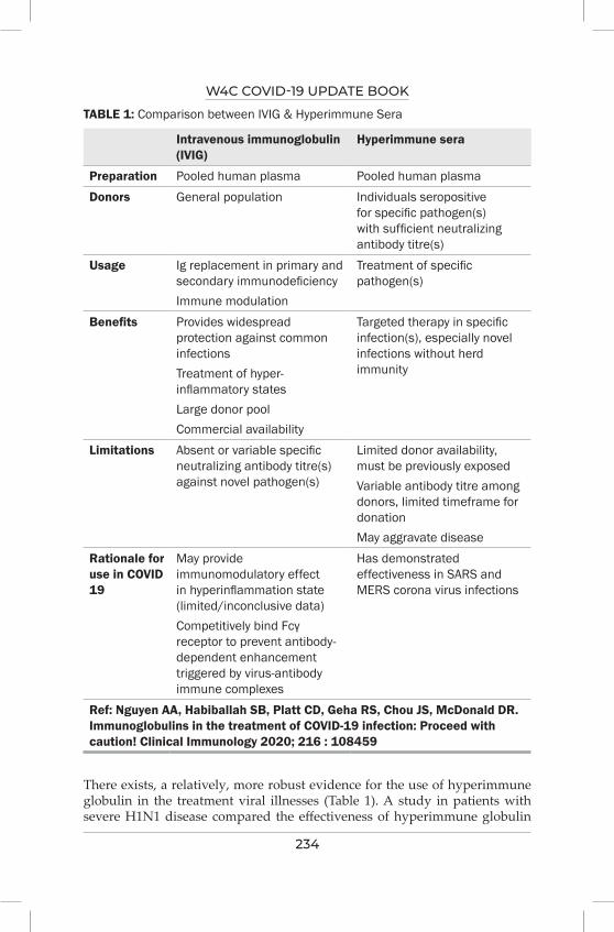
W4C COVID-19 UPDATE BOOK
234
TABLE 1: Comparison between IVIG & Hyperimmune Sera
Intravenous immunoglobulin (IVIG)
Hyperimmune sera
Preparation Pooled human plasma Pooled human plasmaDonors General population Individuals seropositive
for specific pathogen(s) with sufficient neutralizing antibody titre(s)
Usage Ig replacement in primary and secondary immunodeficiencyImmune modulation
Treatment of specific pathogen(s)
Benefits Provides widespread protection against common infectionsTreatment of hyper-inflammatory statesLarge donor poolCommercial availability
Targeted therapy in specific infection(s), especially novel infections without herd immunity
Limitations Absent or variable specific neutralizing antibody titre(s) against novel pathogen(s)
Limited donor availability, must be previously exposedVariable antibody titre among donors, limited timeframe for donationMay aggravate disease
Rationale for use in COVID 19
May provide immunomodulatory effect in hyperinflammation state (limited/inconclusive data)Competitively bind Fcγ receptor to prevent antibody-dependent enhancement triggered by virus-antibody immune complexes
Has demonstrated effectiveness in SARS and MERS corona virus infections
Ref: Nguyen AA, Habiballah SB, Platt CD, Geha RS, Chou JS, McDonald DR. Immunoglobulins in the treatment of COVID-19 infection: Proceed with caution! Clinical Immunology 2020; 216 : 108459
There exists, a relatively, more robust evidence for the use of hyperimmune globulin in the treatment viral illnesses (Table 1). A study in patients with severe H1N1 disease compared the effectiveness of hyperimmune globulin
W4C COVID-19 UPDATE BOOK
235
obtained from H1N1 survivors versus use of IVIG in H1N1 patients and conclusively showed a reduced a viral load and increased survival in the group receiving hyperimmune globulin.10
CONCLUSIONThe search for an effective, low-cost therapeutic option with wide and easy applicability has led us to use a large number of molecules with no success. The evidence for use of high titres CP in COVID 19 is moderate especially in the subgroup of patients in early phase of the disease with mild to moderate illness. There may be concerns as highlighted in the CONCOR-1 study with use of CP especially when there is no discernable benefit from the therapy. The evidence for use of IVIG in COVID 19 is again limited to mild disease and early in the course of illness. There are a large number of ongoing RCT’s evaluating and comparing the use of CP as well as IVIG in various sub groups of patients with COVID 19 and it would only a matter of time before we have clear guidelines on their use with robust evidence.
PRACTICE POINTS1. CP offers Antibody and Non-Antibody mediated Immune protection.
2. CP to be used only in the mild to moderate form of disease.
3. CP to be used early on in the course of the illness, preferably by Day 3 of symptom onset.
4. Any use of CP with unfavourable antibody profile may result in adverse outcomes.
5. IVIG can only be justified in a limited subset of patients in early phase of disease with mild severity of illness.
REFERENCES1. Joyner MJ, Senefeld JW, Klassen SA, Mills JR, Johnson PW, Theel ES, et al. Effect of
convalescent plasma on mortality among hospitalized patients with COVID-19: Initial three-month experience. medRxiv 2020. doi: 10.1101/2020.08.12.20169359
2. Agarwal A, Mukherjee A, Kumar G, Chatterjee P, Bhatnagar T, Malhotra P, et al. Convalescent plasma in the management of moderate COVID-19 in adults in India: open label phase II multicentre randomised controlled trial (PLACID Trial). BMJ 2020; 371:m3939. doi: 10.1136/bmj.m3939.
3. Khaire NS, Jindal N, Yaddanapudi LN, Sachdev S, Hans R, Sachdeva N, et al. Use of convalescent plasma for COVID-19 in India: A review & practical guidelines. Indian J Med Res 2021; 153:64-85.
4. Li L, Zhang W, Hu Y, Tong X, Zheng S, Yang J, et al. Effect of convalescent plasma therapy on time to clinical improvement in patients with severe and life-threatening COVID-19: A randomized clinical trial. JAMA 2020; 324:460-70.
5. Gharbharan A, Jordans CC, GeurtsvanKessel C, den Hollander JG, Karim F, Mollema FP, et al. Convalescent plasma for COVID-19. A randomized clinical trial. medRxiv2020.
6. Libster R, Pérez MG, Wappner D, Coviello S, Bianchi A, Braem V, et al. Early high-titre plasma therapy to prevent severe COVID-19 in older adults. N Engl J Med 2021.
W4C COVID-19 UPDATE BOOK
236
7. Horby PW, Estcourt L, Peto L, Emberson JR, Staplin N, Spata E, et al. Convalescent plasma in patients admitted to hospital with COVID-19 (RECOVERY): a randomised, controlled, open-label, platform trial. medRxiv [Preprint]. (2021). doi: 10.1101/2021.03.09.21252736
8. Bégin P, Callum J, Heddle NM, Cook R, Zeller MP, Tinmouth A, et al. Convalescent plasma for adults with acute COVID-19 respiratory illness (CONCOR-1): study protocol for an international, multicentre, randomized, open-label trial. Trials 202; 22:323. doi: 10.1186/s13063-021-05235-3.
9. Cao W, Liu X, Hong K, Ma Z, Zhang Y, Lin L, Han Y, Xiong Y, Liu Z, Ruan L and Li T. High-Dose Intravenous Immunoglobulin in Severe Coronavirus Disease 2019: A Multicenter Retrospective Study in China. Front Immunol 2021; 12:627844.
10. Hung IFN, To KKW, Lee CK, Lee KL, Yan WW, Chan K, et al., Hyperimmune IV immunoglobulin treatment: a multicenter double-blind randomized controlled trial for patients with severe 2009 influenza a (H1N1) infection. Chest 2013; 144:464–473.
Critical Care PearlsSECTION 5

239
Section 5: Critical Care PearlsSECTION EDITOR
Dr Prashant NasaFNB, EDIC Head of the Department, Critical Care Medicine, NMC Specialty Hospital, Dubai
Preface
Critical care medicine has made remarkable progress in improving patient’s outcomes in the last few decades. There is considerable research on the holistic approach to a critically ill patient. The critical condition of the patient and its management needs considerable attention to various factors besides primary organ support. Feeding, glycemic control, pain or sedation management, prophylaxis for aspiration, ulcer or deep venous thrombosis are essential components of
the treatment plan for any critically ill patient in ICU. The management of COVID-19 caused an unprecedented burden on the core strategies of critical care practice. The section includes a chapter on the application of FAST-HUG principles for the management of patients with COVID-19. Infection control of COVID-19 was a major challenge from the inception of the pandemic to protect health care workers and other patients from cross-infection of SARS-CoV-2. Besides, patients of COVID-19 are at the same or increased risk of hospital-acquired infections. In the absence of effective treatment, past experience of superadded secondary bacterial or fungal infections in other respiratory viral diseases led to misuse or abuse of antimicrobials in COVID-19. The principles of antimicrobial stewardship are applicable to COVID-19 as for any other infectious disease in ICU. There are separate chapters in the section on infection control strategies for COVID-19 patients in ICU; risk factors, epidemiology, and management of secondary bacterial infections with COVID-19; antimicrobial therapy, including stewardship.
COVID-19 is a prothrombotic disease with an increased incidence of thromboembolic events reported in various studies. The management of venous thromboembolism in patients with COVID-19 attracted considerable attention from clinicians and researchers alike. The section includes a chapter on risk assessment, prevention, and treatment of venous

240
thromboembolism in COVID-19. There is also a separate chapter on air-leak with COVID-19, encompassing serious complications like pneumothorax, pneumomediastinum, and pneumopericardium with COVID-19. The patients’ family members are an integral part of the holistic management of critically ill patients. The fear of cross-infection with SARS-CoV-2 inundated ICUs with a surge of patients, and infection prevention measures like personal protective equipment’s created barriers to effective communication among healthcare workers or patients’ family members. The visiting policy for family members has seen a significant shift towards restriction to the “no-visitor” policy during this pandemic. The global impact of this pandemic with the digital era led to an unprecedented surge of information sharing among the people. The unregulated social media platforms and pre-release of results of ongoing research through media led to misinformation or false representation of facts and knowledge. We tried to cover these important aspects of management of COVID-19 through chapters on communication in ICU, visitor’s policy, and Do or Don’t for management of COVID-19.
241
“FASTHUG Revisited in COviD-19” 31.
Moderate to severe cases of COVID-19 illness and severe acute respiratory infection (SARI) requires hospitalization and oxygen support, and few of them need admission to intensive care unit. In most of the severe cases, COVID-19 illness manifest with pro-inflammatory and inflammatory cascade which is further complicated by the acute respiratory distress syndrome (ARDS), secondary infections, sepsis, septic shock, thromboembolism, acute kidney injury, acute cardiac events, stroke and, multiorgan failure. Management includes various aspects and one of them is commonly performed as FAST HUGS which is well known and very important structured as well as systematic method of quick, clinical bed side assessment. JL Vincent was honoured to describe the “FAST HUG” mnemonic to identify and check the key aspects in managing critically ill in an Intensive Care Unit (ICU)1. This aims to reduce the burden of ICU acquired complications for every patient as a mental checklist to ensure the completion of all the elements of routine care.
In the past few decades critical care has evolved from basic fundamentals to most advanced care, monitoring as well as treatment options but one thing remained common since many years and revisited again in COVID era in its new form as “FAST HUGS BID” with many add on variation in the components as described later in this chapter. During pandemic due to SARS-CoV, many new therapies and trails were tried but basic care and FASTHUGS adds on to patient recovery as well as satisfaction. This golden mnemonic revisited during COVID-19 illness and proved beneficial as part of daily assessment of every patient admitted. Routine elements of care can be broadly defined as elements of supportive and preventative care for a critically ill patient which are standardised, regardless of the presenting pathology and completed daily.
MODIFICATIONS OF “FASTHUG”:1. FAST HUGS1
2. FAST HUGS BID2
Neeru Gaur, Ravi Gaur
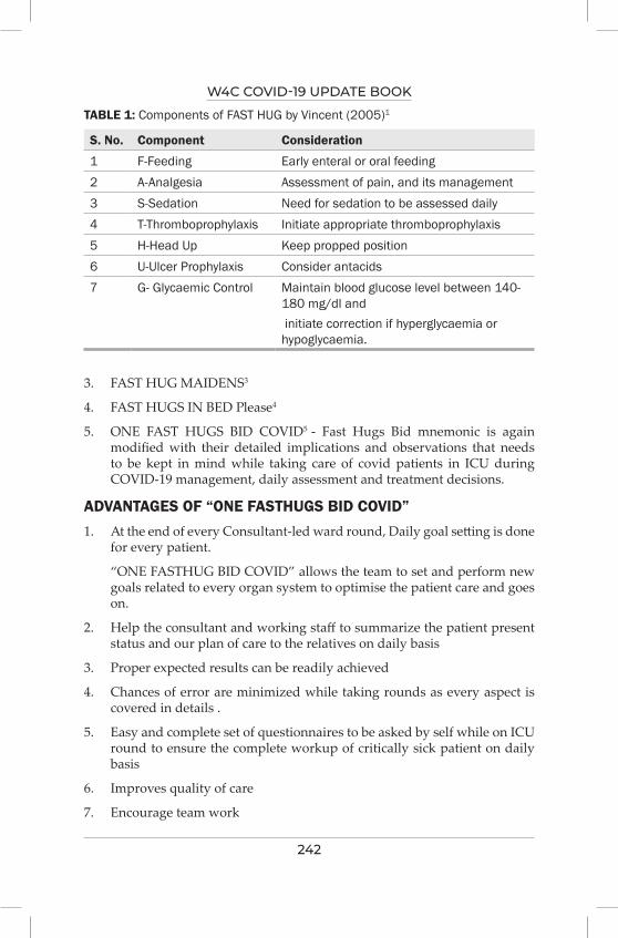
W4C COVID-19 UPDATE BOOK
242
3. FAST HUG MAIDENS3
4. FAST HUGS IN BED Please4
5. ONE FAST HUGS BID COVID5 - Fast Hugs Bid mnemonic is again modified with their detailed implications and observations that needs to be kept in mind while taking care of covid patients in ICU during COVID-19 management, daily assessment and treatment decisions.
ADVANTAGES OF “ONE FASTHUGS BID COVID”1. At the end of every Consultant-led ward round, Daily goal setting is done
for every patient.
“ONE FASTHUG BID COVID” allows the team to set and perform new goals related to every organ system to optimise the patient care and goes on.
2. Help the consultant and working staff to summarize the patient present status and our plan of care to the relatives on daily basis
3. Proper expected results can be readily achieved
4. Chances of error are minimized while taking rounds as every aspect is covered in details .
5. Easy and complete set of questionnaires to be asked by self while on ICU round to ensure the complete workup of critically sick patient on daily basis
6. Improves quality of care
7. Encourage team work
TABLE 1: Components of FAST HUG by Vincent (2005)1
S. No. Component Consideration
1 F-Feeding Early enteral or oral feeding2 A-Analgesia Assessment of pain, and its management 3 S-Sedation Need for sedation to be assessed daily4 T-Thromboprophylaxis Initiate appropriate thromboprophylaxis5 H-Head Up Keep propped position 6 U-Ulcer Prophylaxis Consider antacids 7 G- Glycaemic Control Maintain blood glucose level between 140-
180 mg/dl and initiate correction if hyperglycaemia or hypoglycaemia.
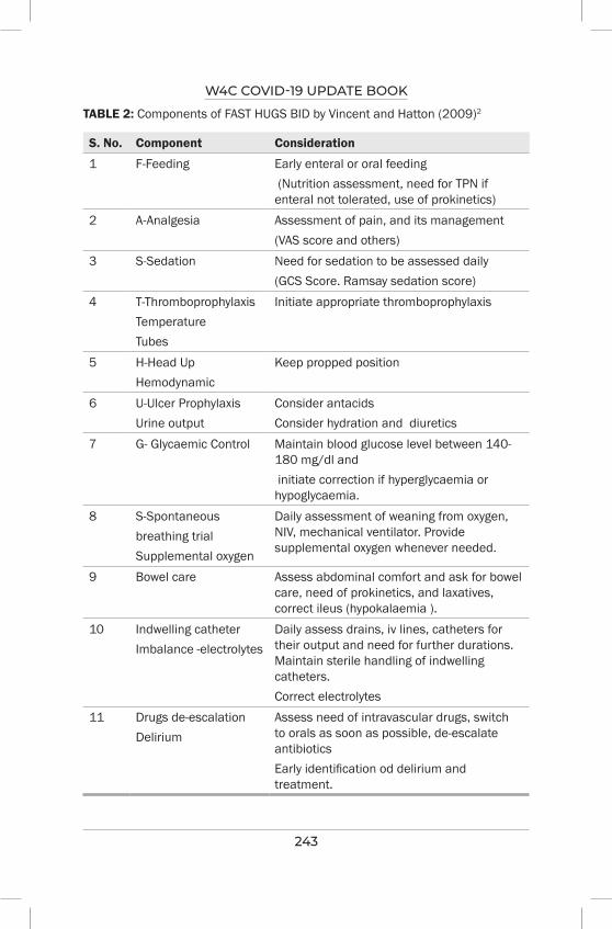
W4C COVID-19 UPDATE BOOK
243
TABLE 2: Components of FAST HUGS BID by Vincent and Hatton (2009)2
S. No. Component Consideration
1 F-Feeding Early enteral or oral feeding (Nutrition assessment, need for TPN if enteral not tolerated, use of prokinetics)
2 A-Analgesia Assessment of pain, and its management (VAS score and others)
3 S-Sedation Need for sedation to be assessed daily(GCS Score. Ramsay sedation score)
4 T-ThromboprophylaxisTemperatureTubes
Initiate appropriate thromboprophylaxis
5 H-Head UpHemodynamic
Keep propped position
6 U-Ulcer ProphylaxisUrine output
Consider antacids Consider hydration and diuretics
7 G- Glycaemic Control Maintain blood glucose level between 140-180 mg/dl and initiate correction if hyperglycaemia or hypoglycaemia.
8 S-Spontaneous breathing trialSupplemental oxygen
Daily assessment of weaning from oxygen, NIV, mechanical ventilator. Provide supplemental oxygen whenever needed.
9 Bowel care Assess abdominal comfort and ask for bowel care, need of prokinetics, and laxatives, correct ileus (hypokalaemia ).
10 Indwelling catheterImbalance -electrolytes
Daily assess drains, iv lines, catheters for their output and need for further durations. Maintain sterile handling of indwelling catheters.Correct electrolytes
11 Drugs de-escalationDelirium
Assess need of intravascular drugs, switch to orals as soon as possible, de-escalate antibiotics Early identification od delirium and treatment.

W4C COVID-19 UPDATE BOOK
244
TABLE 3: Components of FAST HUGS MAIDEN
S. No. Component Consideration
1 F-Feeding Early enteral or oral feeding2 A-Analgesia Assessment of pain, and its
management 3 S-Sedation Need for sedation to be assessed
daily4 T-Thromboprophylaxis Initiate appropriate
thromboprophylaxis and consider renal function test while dosing.
5 H-Hyperactive / hypoactive delirium
Manage ICU delirium, irritability, depression and insomnia
6 U-Ulcer Prophylaxis Consider antacids 7 G- Glycaemic Control Maintain blood glucose
level between 140-180 mg/dl and initiate correction if hyperglycaemia or hypoglycaemia.
8 M-Medication reconciliation Re check the complete prescription and orders
9 A-Antibiotics or anti-infectives Consider early initiation of broad-spectrum antibiotics.
10 I-Indications for Medications
Consider medications to support underlying chronic illness.
11 D-Drug dosing Prescribe proper drug dosage individually
12 E-Electrolytes Correct dyselectrolytemia13 N-No drug interactions, allergies,
duplication, and side effectsCheck all the drugs - interactions, allergies, duplication, And side effects
14 S-stop dates Stop drugs on specifics timings.
8. Maintain good patient -doctor- relatives communications
EVIDENCE TO SUPPORT 1. Ward round checklists found to be completed for 93% of the rounds, and
the discussions which took place around this were viewed favourably by all.6
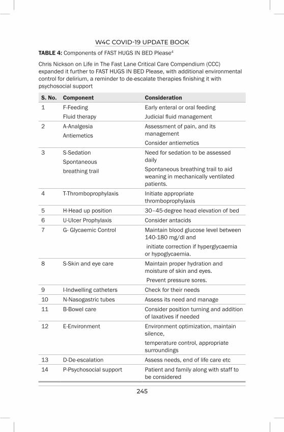
W4C COVID-19 UPDATE BOOK
245
TABLE 4: Components of FAST HUGS IN BED Please4
Chris Nickson on Life in The Fast Lane Critical Care Compendium (CCC) expanded it further to FAST HUGS IN BED Please, with additional environmental control for delirium, a reminder to de-escalate therapies finishing it with psychosocial support
S. No. Component Consideration
1 F-FeedingFluid therapy
Early enteral or oral feedingJudicial fluid management
2 A-Analgesia Antiemetics
Assessment of pain, and its managementConsider antiemetics
3 S-SedationSpontaneous breathing trail
Need for sedation to be assessed dailySpontaneous breathing trail to aid weaning in mechanically ventilated patients.
4 T-Thromboprophylaxis Initiate appropriate thromboprophylaxis
5 H-Head up position 30–45-degree head elevation of bed6 U-Ulcer Prophylaxis Consider antacids 7 G- Glycaemic Control Maintain blood glucose level between
140-180 mg/dl and initiate correction if hyperglycaemia or hypoglycaemia.
8 S-Skin and eye care Maintain proper hydration and moisture of skin and eyes. Prevent pressure sores.
9 I-Indwelling catheters Check for their needs 10 N-Nasogastric tubes Assess its need and manage11 B-Bowel care Consider position turning and addition
of laxatives if needed12 E-Environment Environment optimization, maintain
silence, temperature control, appropriate surroundings
13 D-De-escalation Assess needs, end of life care etc14 P-Psychosocial support Patient and family along with staff to
be considered
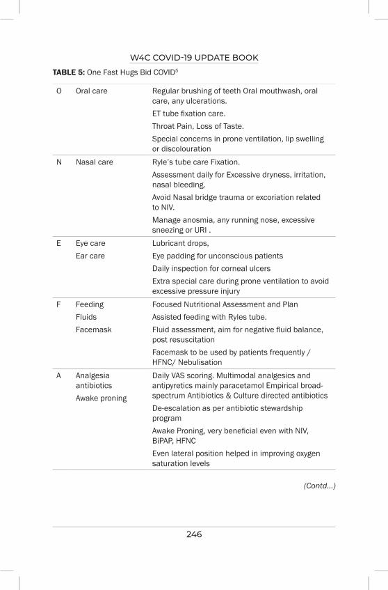
W4C COVID-19 UPDATE BOOK
246
TABLE 5: One Fast Hugs Bid COVID5
O Oral care Regular brushing of teeth Oral mouthwash, oral care, any ulcerations.ET tube fixation care.Throat Pain, Loss of Taste.Special concerns in prone ventilation, lip swelling or discolouration
N Nasal care Ryle’s tube care Fixation.Assessment daily for Excessive dryness, irritation, nasal bleeding.Avoid Nasal bridge trauma or excoriation related to NIV.Manage anosmia, any running nose, excessive sneezing or URI .
E Eye careEar care
Lubricant drops,Eye padding for unconscious patientsDaily inspection for corneal ulcersExtra special care during prone ventilation to avoid excessive pressure injury
F FeedingFluidsFacemask
Focused Nutritional Assessment and PlanAssisted feeding with Ryles tube.Fluid assessment, aim for negative fluid balance, post resuscitationFacemask to be used by patients frequently / HFNC/ Nebulisation
A Analgesia antibioticsAwake proning
Daily VAS scoring. Multimodal analgesics and antipyretics mainly paracetamol Empirical broad-spectrum Antibiotics & Culture directed antibioticsDe-escalation as per antibiotic stewardship programAwake Proning, very beneficial even with NIV, BiPAP, HFNCEven lateral position helped in improving oxygen saturation levels
(Contd...)
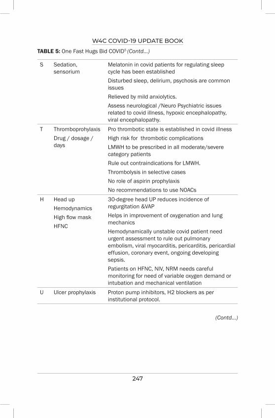
W4C COVID-19 UPDATE BOOK
247
TABLE 5: One Fast Hugs Bid COVID5 (Contd...)
S Sedation, sensorium
Melatonin in covid patients for regulating sleep cycle has been establishedDisturbed sleep, delirium, psychosis are common issues Relieved by mild anxiolytics.Assess neurological /Neuro Psychiatric issues related to covid illness, hypoxic encephalopathy, viral encephalopathy.
T ThromboprohylaxisDrug / dosage / days
Pro thrombotic state is established in covid illnessHigh risk for thrombotic complicationsLMWH to be prescribed in all moderate/severe category patientsRule out contraindications for LMWH.Thrombolysis in selective casesNo role of aspirin prophylaxisNo recommendations to use NOACs
H Head upHemodynamicsHigh flow maskHFNC
30-degree head UP reduces incidence of regurgitation &VAPHelps in improvement of oxygenation and lung mechanicsHemodynamically unstable covid patient need urgent assessment to rule out pulmonary embolism, viral myocarditis, pericarditis, pericardial effusion, coronary event, ongoing developing sepsis.Patients on HFNC, NIV, NRM needs careful monitoring for need of variable oxygen demand or intubation and mechanical ventilation
U Ulcer prophylaxis Proton pump inhibitors, H2 blockers as per institutional protocol.
(Contd...)
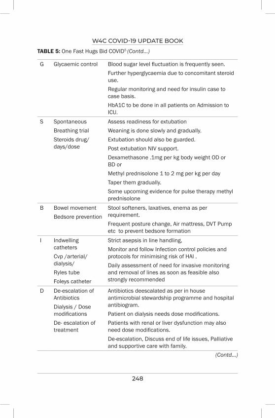
W4C COVID-19 UPDATE BOOK
248
TABLE 5: One Fast Hugs Bid COVID5 (Contd...)
G Glycaemic control Blood sugar level fluctuation is frequently seen.Further hyperglycaemia due to concomitant steroid use.Regular monitoring and need for insulin case to case basis.HbA1C to be done in all patients on Admission to ICU.
S SpontaneousBreathing trialSteroids drug/days/dose
Assess readiness for extubationWeaning is done slowly and gradually.Extubation should also be guarded.Post extubation NIV support.Dexamethasone .1mg per kg body weight OD or BD or Methyl prednisolone 1 to 2 mg per kg per dayTaper them gradually.Some upcoming evidence for pulse therapy methyl prednisolone
B Bowel movementBedsore prevention
Stool softeners, laxatives, enema as per requirement.Frequent posture change, Air mattress, DVT Pump etc to prevent bedsore formation
I Indwelling cathetersCvp /arterial/dialysis/Ryles tubeFoleys catheter
Strict asepsis in line handling,Monitor and follow Infection control policies and protocols for minimising risk of HAI .Daily assessment of need for invasive monitoring and removal of lines as soon as feasible also strongly recommended
D De-escalation of AntibioticsDialysis / Dose modificationsDe- escalation of treatment
Antibiotics deescalated as per in house antimicrobial stewardship programme and hospital antibiogram.Patient on dialysis needs dose modifications.Patients with renal or liver dysfunction may also need dose modifications.De-escalation, Discuss end of life issues, Palliative and supportive care with family.
(Contd...)
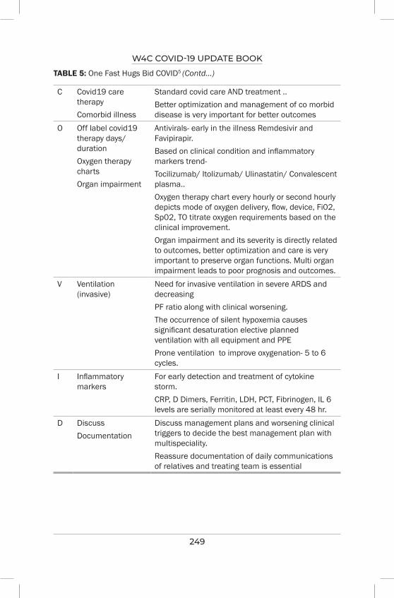
W4C COVID-19 UPDATE BOOK
249
TABLE 5: One Fast Hugs Bid COVID5 (Contd...)
C Covid19 care therapyComorbid illness
Standard covid care AND treatment ..Better optimization and management of co morbid disease is very important for better outcomes
O Off label covid19 therapy days/ durationOxygen therapy chartsOrgan impairment
Antivirals- early in the illness Remdesivir and Favipirapir.Based on clinical condition and inflammatory markers trend- Tocilizumab/ Itolizumab/ Ulinastatin/ Convalescent plasma..Oxygen therapy chart every hourly or second hourly depicts mode of oxygen delivery, flow, device, Fi02, Sp02, TO titrate oxygen requirements based on the clinical improvement.Organ impairment and its severity is directly related to outcomes, better optimization and care is very important to preserve organ functions. Multi organ impairment leads to poor prognosis and outcomes.
V Ventilation (invasive)
Need for invasive ventilation in severe ARDS and decreasingPF ratio along with clinical worsening.The occurrence of silent hypoxemia causes significant desaturation elective planned ventilation with all equipment and PPEProne ventilation to improve oxygenation- 5 to 6 cycles.
I Inflammatory markers
For early detection and treatment of cytokine storm.CRP, D Dimers, Ferritin, LDH, PCT, Fibrinogen, IL 6 levels are serially monitored at least every 48 hr.
D DiscussDocumentation
Discuss management plans and worsening clinical triggers to decide the best management plan with multispeciality.Reassure documentation of daily communications of relatives and treating team is essential

W4C COVID-19 UPDATE BOOK
250
2. Interprofessional communication and education was thought to have improved 6.
3. Decrease in VAP rates and ICU stay as found after the introduction of “ FASTHUG”, from 19.3/1000 ventilator-days to 7.3/1000 ventilator-days7.
PRACTICE POINTS1. To summarize management plan for COVID patients, always remember
there is no emergency in pandemic and ensure appropriate safety for self and surroundings.
2. Use proper PPE with the help of BUDDY approach in Donning and Doffing.
3. Remember ABCDEF approach and ensure their needs for individual patient status, during daily ICU rounds of COVID patients with respiratory failure
• Airway- airway assessment and maintenance
• Breathing – maintain oxygenation and ventilation
• Circulation – monitor hemodynamic and maintain optimum hemodynamic parameters
• Definite proning – awake proning encouraged early. Daily monitoring and care of prone mechanically ventilated patient.
• ECMO – Patient selection for ECMO need to be assessed early
• FASTHUGS- Played an important role as new “ONE FASTHUGS BID COVID” in patients with moderate to severe COVID illness.
FASTHUG evolved and revisited with its modified version as ONE FASTHUGS BID COVID or FASTHUGS BID found to be very useful with better and complete assessment of patients of covid disease admitted in intensive care unit on case-to-case basis daily, this is also an important tool for overall assessment, as well as treatment modification plans, goal settings and its execution. Documentation and considering all points as One Fast Hugs Bid COVID daily provide better care to covid patients, family and also ease of learning as well as working for everyone.
REFERENCES1. Vincent JL. Give your patient a fast hug (at least) once a day. Critical Care Medicine 2005;
33:1225-9.2. Vincent III WR, Hatton KW. Critically ill patients need “FAST HUGS BID” (an updated
mnemonic). Critical Care Medicine 2009; 37:2326-7.3. Mabasa VH, Malyuk DL, Weatherby EM, Chan A. A standardized, structured approach to
identifying drug-related problems in the intensive care unit: FASTHUG-MAIDENS. The Canadian Journal of Hospital Pharmacy 2011; 64:366.
W4C COVID-19 UPDATE BOOK
251
4. Dr. Chris Nickson, last update July 23, 2019, Life in the Fastlane – https://litfl.com/fast-hugs-in-bed-please/.
5. Kumar DA, Dey D, Sabharwal M, Sarkar D, Kumar A. One Fast Hugs Bid COVID: COVID Care Bundle Modified Mnemonic to Facilitate COVID 19 Critical Care Medicine. Journal of Medical Science and Clinical Research 2020; 8:1-3.
6. Duan E, Centofanti J, Hoad N, Swinton M, Perri D, Waugh L, Cook DJ. Use Of A Daily Goals Checklist For Morning Intensive Care Unit Rounds: A Mixed-Methods Study. InC106. Interprofessional Topics in the Provision of Critical Care 2014 May (pp. A5250-A5250). American Thoracic Society.
7. Papadimos TJ, Hensley SJ, Duggan JM, Khuder SA, Borst MJ, Fath JJ, Oakes LR, Buchman D. Implementation of the” FASTHUG” concept decreases the incidence of ventilator-associated pneumonia in a surgical intensive care unit. Patient Safety in Surgery 2008; 2:1-6.
252
infection Control Practices in iCU for Covid-19
INTRODUCTIONThe coronavirus disease 2019 (COVID-19) has led to a dual strike on health care facilities (HCF) worldwide. Firstly, HCFs were overwhelmed with a surge of patients, and secondly, cross-transmission of contagion to health care workers (HCW) caused a severe shortage of human resources. The protection of HCWs and patients without COVID-19 became imperative during the COVID-19 pandemic. Various academic societies and public health care agencies globally issued infection control guidelines based on the consensus or experience from previous pandemics given limited data on the transmission of severe acute respiratory syndrome coronavirus-2 (SARS-CoV-2).1-3
HCWs are at increased risk of contracting SARS-CoV-2 while providing care to the sick. Frontline HCWs who are in direct contact with patients are at even higher risk.4 From the inception of the COVID-19 pandemic, public health agencies advocated droplets as the primary mode of transmission of SARS-CoV-2. Face masks, social distance and hand hygiene were recommended interventions to prevent human to human transmission. Emerging evidence favours the airborne transmission of SARS-CoV-2, especially in indoors settings, crowded places or inadequate ventilation.5 On-going uncertainty has led to significant structural and organizational changes in healthcare facilities, as many global health agencies now accept airborne transmission mode.2-3 Apart from COVID-19, the rise in hospital-acquired infections (HAI) is another concern during the pandemic.6 The aetiology of increased HAI is likely to be multifactorial, overwhelmed HCFs with an unexpected surge in patients, inadequate staffing, sub-optimal infection prevention or breach in infection control practices amidst the fear of self-contamination.
The standard precautions of physical distancing, wearing of face masks, hand hygiene practices, and decontaminating contact surfaces remain at the core of any COVID-19 infection control guidelines.3 Few recommendations changed with emerging data supporting airborne transmission of SARS-CoV-2. Intensive care units (ICU) are involved in the management of critically
Prashant Nasa, Ravi Jain
32.
W4C COVID-19 UPDATE BOOK
253
ill patients with COVID-19. HCWs working in ICU are vulnerable to a higher risk of cross-infection. The factors responsible include patients with severe COVID-19 who are overtly symptomatic, require frequent aerosol generating procedures (AGP) and finally, close contact by HCWs for frequent monitoring. There is a dearth of recommendations on infection control of SARS-CoV-2 in ICU. The following recommendations apply to the patients with COVID-19 admitted in ICU and can be divided broadly into three sections: 1. Patients and procedures 2. Health care workers 3. Environmental disinfection
PATIENTS AND PROCEDURESPlacement of Patients
• Patients with suspected or confirmed SARS-CoV-2 infection should be placed in an airborne infection isolation room (AIIR) if available or in a single room (cubicle) with the door closed.
• AIIR should be prioritised for patients undergoing AGPs
• Cohorting patients with COVID-19 should be done as a last resort. However, patients with suspected and confirmed SARS-CoV-2 infection should not be cohorted.
• Limit the transport or transfer of patients outside the room only if medically essential.
Visiting Policy
• Limit visitors to the ICU by frequency, duration and number except in particular circumstances like end-of-life care or vulnerable patients (paediatrics or elderly).
• Communication with family members can be performed with alternative mechanisms like cell phones or tablets, using video-call applications.
• Risk-assessment, based on factors like age, health status, or vaccination status of the visitor and ability to comply with precautions.
Aerosol Generating Procedures Public health agencies advised extra precautions for certain medical procedures called AGPs, which increase the risk of aerosolisation of microbes.2,3 Aerosols are microparticles that can float in the air, travelling distances (> 6 feet) more extensive than droplets and increasing cross-transmission risk. The recommendations for such procedures include patient placement in an AIIR consisting of frequent air changes (12 or more) and negative differential pressure, N95 respirator for HCWs.
The procedures include manual ventilation, tracheal intubation or extubation, non-invasive positive pressure ventilation, tracheotomy, cardiopulmonary resuscitation, bronchoscopy, sputum induction and nebulisation.7

W4C COVID-19 UPDATE BOOK
254
However, the list of such procedures is not comprehensive and is based on limited evidence or simulation studies with conflicting results. The simulation studies recently found a higher viral load with fine aerosols produced during talking, singing or breathing than coarse aerosols.8 Experts suggested other factors to be considered along with the type of procedure like forced air, distance, duration and degree of symptoms or severity of the patient’s disease.9
Respiratory Sample Collection
• Respiratory sample collection in a suspected or confirmed patient is an AGP.
• HCW involved in sampling should wear PPE appropriate for AGPs.
Infection Prevention Bundles and Antimicrobial Stewardship
There is a surge in HAIs (catheter-related bloodstream infections, catheter-associated urinary tract infections or ventilator-associated pneumonia) during the COVID-19 pandemic. A simultaneous increase in the prevalence of multidrug-resistant organisms, including MRSA, vancomycin-resistant enterococcus and gram-negative organisms, is noted.6 The empirical antibiotic prescription is also significantly higher in patients with COVID-19 despite a lower coexistent community-acquired microbial infections.
The components of infection prevention bundles and antimicrobial stewardship should be part of ICU management of patients with COVID-19 as other critically ill.
HEALTH CARE WORKERSThe emerging evidence supports the aerosolization of SARS-CoV-2 especially with indoor settings and inadequate ventilation. The risk of aerosolization of SARS-CoV-2 is not only with conventional aerosol generating procedures (AGPs) but even with deep coughing or breathing.
Implement Universal Infection Control Measures
HCWs must follow standard precautions of face mask and physical distancing at all places in the hospital including restrooms or staff-cafeteria. The patients and visitors should also follow similar precautions inside the hospital.
Personal Protective Equipment
• The HCWs involved in the care of suspected or confirmed patients with COVID-19 should don appropriate personal protective equipment (PPE), well fitted N95 masks and eye protection (goggles or face shield), gown, and gloves
• Periodic training of donning and doffing of PPE should be provided to HCWs.
W4C COVID-19 UPDATE BOOK
255
• Doffing of PPE is equally crucial as donning and has a higher risk of contamination.
Hand Hygiene
• HCP should perform hand hygiene during five moments as in non-COVID-19 patients (before and after contact with the patient, before doing any procedure, after touching patient surroundings)
• Hand hygiene after doffing PPE is crucial.
• Hand hygiene should be performed with at least 60-95% alcohol or washing hands with soap and water for 20 seconds.
Vaccination
• COVID-19 vaccines are the most effective prevention measures that reduce the risk of severe COVID-19, hospitalisation and mortality.
• HCWs working in critical care should be vaccinated for COVID-19 for to reduce the risk of cross-infection and to protect themselves
ENVIRONMENTAL DISINFECTION• The medical equipment used for patients with suspected or confirmed
SARS-CoV-2 infection should be dedicated and preferably disposable.
• The cleaning and disinfection of non-dedicated and non-disposable medical equipment should be done according to manufacturer’s instructions and hospital policies.
• Use of Environmental protection agency (EPA)-registered and hospital-grade agents for cleaning and disinfection procedures.
• The management of medical waste, laundry, utensils involved for food, should be performed in accordance with routine procedures.
CONCLUSIONCompliance with infection control practices during COVID-19 is essential to ensure safety of the patients and HCWs. The infection control practices in ICU should be based on current recommendation and need to be reviewed periodically with emerging evidence.
PRACTICE POINTS 1. Patients with COVID-19 should preferably be placed in an AIIR room or
a single room with a closed door.2. Suspected and confirmed patients of COVID-19 should not be cohorted
together.3. N95 mask and face shield are essential components of PPE for HCWs
involved in the management of patients with COVID-19
W4C COVID-19 UPDATE BOOK
256
4. All HCWs involved should be vaccinated against COVID-19
5. Principles of hand hygiene, infection prevention bundles and antimicrobial stewardship is the same as in patients without COVID-19
REFERENCES1. Sharma J, Nasa P, Reddy KS, Kuragayala SD, Sahi S, Gopal P, Chaudhary D, Dixit SB,
Samavedam S. Infection Prevention and Control for ICU during COVID-19 Pandemic: Position Paper of the Indian Society of Critical Care Medicine. Indian J Crit Care Med 2020; 24:S280-S289. doi: 10.5005/jp-journals-10071-23607.
2. World Health Organisation. Infection prevention and control guidance - (COVID-19). Available at : https://www.who.int/westernpacific/emergencies/covid-19/technical-guidance/infection-prevention-control Accessed on 05 September 2021.
3. CDC. Interim Infection Prevention and Control Recommendations for Healthcare Personnel During the Coronavirus Disease 2019 (COVID-19) Pandemic. Available at: https://www.cdc.gov/coronavirus/2019-ncov/hcp/infection-control-recommendations.html Accessed on 05 September 2021.
4. Gómez-Ochoa SA, Franco OH, Rojas LZ, Raguindin PF, Roa-Díaz ZM, Wyssmann BM, et al. COVID-19 in Health-Care Workers: A Living Systematic Review and Meta-Analysis of Prevalence, Risk Factors, Clinical Characteristics, and Outcomes. Am J Epidemiol 2021; 190:161-175. doi: 10.1093/aje/kwaa191.
5. Wang CC, Prather KA, Sznitman J, Jimenez JL, Lakdawala SS, Tufekci Z, et al. Airborne transmission of respiratory viruses. Science 2021; 373:eabd9149. doi: 10.1126/science.abd9149.
6. Baker MA, Sands KE, Huang SS, Kleinman K, Septimus EJ, Varma N, et al. The Impact of COVID-19 on Healthcare-Associated Infections. Clin Infect Dis 2021 Aug 9:ciab688. doi: 10.1093/cid/ciab688.
7. Nasa P, Azoulay E, Khanna AK, Jain R, Gupta S, Javeri Y, et al. Expert consensus statements for the management of COVID-19-related acute respiratory failure using a Delphi method. Crit Care 2021; 25:106. doi: 10.1186/s13054-021-03491-y.
8. Coleman KK, Tay DJW, Sen Tan K, Ong SWX, Son TT, Koh MH, et al. Viral Load of SARS-CoV-2 in Respiratory Aerosols Emitted by COVID-19 Patients while Breathing, Talking, and Singing. Clin Infect Dis 2021 Aug 6:ciab691. doi: 10.1093/cid/ciab691.
9. Klompas M, Baker M, Rhee C. What Is an Aerosol-Generating Procedure? JAMA Surg 2021; 156:113-114. doi: 10.1001/jamasurg.2020.6643.
257
Antimicrobial Stewardship in the COviD Pandemic
INTRODUCTIONThe current pandemic has seen an exorbitant rise in post-COVID bacterial and fungal infections in India and across the globe. As a result, antibacterial and antifungal drugs have been liberally used in COVID patients, and most of this usage has been empiric. This has been reflected in worsening susceptibility patterns. A recent retrospective study of secondary infections in patients admitted in intensive care units (ICUs), and wards of ten hospitals of the Indian Council of Medical Research (ICMR) antimicrobial resistance and surveillance network found higher mortality among patients who developed post-COVID secondary infections (56.7 % as against 10.6% in total admitted COVID-19 patients).1 This study also showed that there was an alarming rate of carbapenem resistance in A. baumannii (92.6%) and K. pneumoniae (72.8%).1 This situation demands urgent institution of antimicrobial stewardship programs across the country to avoid a catastrophe in the future.
WHAT ARE THE DOWNSIDES OF UNREGULATED ANTIMICROBIAL USE?Though lifesaving, antibiotics can have their perils when used irrationally and in an unstructured fashion. These include:
● Increased rates of resistance
● Increase in the cost of healthcare
● Increase in the lengths of hospital stay
● Clostridioides difficile (C. difficile) infection
● Side effects and toxicities
WHAT IS ANTIMICROBIAL STEWARDSHIP (AMS) PROGRAM? Antimicrobial stewardship program is a structured multifactorial program
Narendra Rungta, Parikshit Prayag
33.
W4C COVID-19 UPDATE BOOK
258
that aims to rationalize antibiotic usage, thus promoting better patient outcomes, reducing the misuse and inappropriate usage of antimicrobials, and improving the rates of resistance and spread of multidrug-resistant organisms. It does not hinder individual patient care or outcomes of patients with sepsis. In fact, it promotes better patient practices by optimizing antibiotic usage. The COVID pandemic has only highlighted the need for implementing stewardship practices further.
WHAT ARE THE KEY COMPONENTS OF A STEWARDSHIP PROGRAM?A. Commitment of the hospital leadership:
A vital aspect of this program is the leadership commitment towards this cause. It involves aspects like:
● Dedicating personnel to this cause
● Having a spearhead (chief steward to champion this cause)
● Ensuring uniformity amongst the prescribing physicians
● Not letting individual egos hinder a larger cause
● Setting up the necessary technology and infrastructure
● Defining short term and long-term goals
● Regular meetings to assess the progress made towards realizing these goals
B. Accountability:
The hospital needs to appoint a committee spearheaded by a physician/intensivist/Infectious diseases (ID) specialist in conjunction with a pharmacist. This committee needs to be held accountable, and in turn, they must ensure individual accountability from the prescribing doctors. Auditing of prescribing practices must be carried out regularly to ensure that guidelines are being followed.
TABLE 1: Vital Components of a Stewardship Program
● Commitment of the hospital leadership● Accountability ● Roping in pharmacy expertise ● Implementation (in the COVID era in India) ● Data collection and tracking ● Regular reporting ● Education
W4C COVID-19 UPDATE BOOK
259
C. Roping in pharmacists:
This is a crucial component, often neglected in many hospitals across the country. We need to train pharmacists in stewardship, involve them in this process, and empower them to give feedback to clinicians. This can work at various levels:
● Regulating at the level of pharmacy - cross-checking while dispensing drugs
● Daily pharmacist rounds in conjunction with clinicians
● Alerting physicians about prescribing practices
● Reviewing periodic data of all higher antimicrobials prescribed
● Educating clinicians
● Reviewing the durations of antimicrobials
● Continuously monitoring opportunities to step down empiric therapy
● De-escalation based on cultures
D. Actual implementation in the COVID era
The COVID pandemic has led to increased empiric therapy. Urgent steps need to be taken to halt the menace of antimicrobial resistance.
● Filling out antimicrobial forms: Often clinicians resist filling out detailed forms, citing their busy schedules. We must have a robust system in place to record the rationale behind antibiotic prescriptions. This gives us a great opportunity to audit practices. Every stewardship committee must make a list of the antimicrobials that need to be audited. Every time an antimicrobial from this list is prescribed, a detailed explanation must be given (preferably electronically) that can be studied later. A team of experts (ID specialists, pharmacists, physicians, and intensivists) must review these forms regularly and provide feedback to the clinicians.
● ID consultations: For some higher antimicrobials, an ID consult should be mandated or seriously considered. These can include (but may not be limited to):
– Polymyxins
– Ceftazidime-avibactam
– Daptomycin
– Echinocandins
– Amphotericin B

W4C COVID-19 UPDATE BOOK
260
– Carbapenems (however, considering the enormous usage, feasibility may be an issue)
Ideally, the ID consultant should at least give principal approval (if it is impossible to assess the patient physically). This involves developing a robust system and having ID experts available.
● De-escalation practices: Often antimicrobials are started for empiric reasons, especially in COVID patients who have received immunomodulatory therapy. There must be a system to check if appropriate changes have been made after 48-72 hours once more data is available. This involves daily pharmacist rounds to see if continuation of therapy is warranted. There must be a system to immediately alert the treating team if there is an opportunity to de-escalate therapy.
E. Data collection and tracking:
It is essential to have an in-depth database about antibiotic prescriptions and usage in the hospital. It would be better if categorized data is available, for e.g., for a particular floor or department. This makes it easier to spot the outliers and give constructive feedback. Data must also be available about the preoperative and postoperative antimicrobial practices. The microbiology data must be integrated into a stewardship program so that local resistance patterns are thoroughly known.
F. Regular reporting:
It is important to disseminate this data periodically. The clinicians should be aware of:
● Local resistance patterns
● Recommended antibiotics in particular situations
● Data regarding their prescriptions and practices
● Compliance rates - how many consultants filled out the forms, how many antibiotics were approved, how many patients were seen by ID consultants.
When such data is discussed in periodic stewardship meetings, practices can change over a period.
G. Education:
Ultimately, educating the prescribers is an essential part of a stewardship program. The physicians must know of resistance mechanisms, recommended therapies, guidelines, and sites of penetration of antibiotics to make informed decisions about starting antimicrobials. They must also be educated about the markers used for various infections and de-escalation based on these markers. They must also be familiar with an antibiogram and should feel comfortable interpreting it.
W4C COVID-19 UPDATE BOOK
261
SPECIFIC ISSUES IN COVID • Are we overusing antifungals?
A lot of COVID-19 patients are started on empiric antifungal therapy. When starting any patient on antifungal therapy, certain key questions must be asked:
● Does the patient have risk factors for fungal infection?
● Is the clinical picture compatible with fungal infection?
● If yes, what is the likely fungus? The approach to a yeast infection like candida will be very different as compared to mold infections.
● Does the patient need mold active coverage? We must know the spectra of antifungals.
● What is the candida score?
● Do we have access to markers like galactomannan/beta-d-glucan? Do we know enough about them to interpret them or is help needed?
Only after we have thought thoroughly about these questions should we decide regarding antifungals.
• Does candida in respiratory cultures need to be treated?
Patients with COVID, especially in the ICU, can be colonized with candida species due to prior immunomodulatory therapy and glycemic issues. Candida in respiratory cultures generally should not be treated. It should be used to assess the patient’s risk for developing invasive candidiasis and calculating the candida score.
Does the patient need Methicillin-Resistant Staphylococcus Aureus (MRSA) coverage?
Several COVID or post-COVID patients are started on anti- MRSA agents like teicoplanin.
We must have a thorough knowledge of the local MRSA rates. It is also essential to consider the clinical and radiographic picture while making this decision.
• Does a patient with COVID need antibacterial agents initially?
This has been a common practice during the times of COVID. A patient in the early stages of COVID, especially when presenting from the community is unlikely to have a concomitant bacterial infection.3 Unfortunately, patients getting admitted into hospitals get started on antibacterial without a sound reason for doing so. There must be a stringent protocol about antibacterial usage in COVID patients, especially those not admitted in the ICU.
W4C COVID-19 UPDATE BOOK
262
• Are we justified in using carbapenems as the first antibiotic in the hospital?
Patients with COVID often get started on carbapenems when they worsen in terms of their respiratory status. This must be analyzed very carefully. The local bacterial prevalence and susceptibility patterns, the patient’s risk factors for acquiring infections with resistant pathogens, the clinical and radiographic picture must be all considered before starting carbapenems.
SUMMARYThe COVID pandemic has witnessed alarming numbers as far as resistant bacterial infections are concerned. We need to have robust systems in place and set up AMS programs across the country. This pandemic must be used as a wake-up call. Hospital managements need to be cognizant of this problem. Involving experts and creating an infrastructure for stewardship programs is the absolute need of the hour. We must act before it’s too late!
PRACTICE POINTS1. There must be a stringent protocol about antibacterial usage in COVID
patients, especially those not admitted in the ICU.
2. Local antibiogram, risk factor for multi-drug resistance and clinical condition of patient should be considered before starting broad-spectrum antimicrobials
3. Candida in respiratory cultures generally should not be treated.
4. Risk factors, clinical condition or urgency, type of species, risk prediction scores and biomarkers like galactomannan must be considered before starting empirical antifungal agents in COVID-19 patients.
REFERENCES1. Vijay S, Bansal N, Rao BK, Veeraraghavan B, Rodrigues C, Wattal C, Goyal JP, Tadepalli K,
Mathur P, Venkateswaran R, Venkatasubramanian R, Khadanga S, Bhattacharya S, Mukherjee S, Baveja S, Sistla S, Panda S, Walia K. Secondary Infections in Hospitalized COVID-19 Patients: Indian Experience. Infect Drug Resist. 2021; 14:1893-1903. https://doi.org/10.2147/IDR.S299774
2. Arastehfar A, Carvalho A, Nguyen MH, Hedayati MT, Netea MG, Perlin DS, Hoenigl M. COVID-19-Associated Candidiasis (CAC): An Underestimated Complication in the Absence of Immunological Predispositions? J Fungi (Basel) 2020; 6:211. doi: 10.3390/jof6040211. PMID: 33050019; PMCID: PMC7712987.
3. Lansbury L, Lim B, Baskaran V, Lim WS. Co-infections in people with COVID-19: a systematic review and meta-analysis. J Infect. 2020; 81:266-275. doi:10.1016/j.jinf.2020.05.046
263
Secondary Bacterial infections with Covid-19
COVID-19 can cause co-infections with bacteria, viruses, and fungus. Secondary bacterial infections have been associated with significant mortality and morbidity in influenza pandemics. Bacterial infections may be co-infections and secondary/superinfections. The CDC defines superinfections as “an infection following a previous infection especially when caused by microorganisms that are resistant or have become resistant to the antibiotics used earlier”, while a co-infection is one occurring concurrently with the initial infection, the difference being purely temporal.1
Viral respiratory infections predispose patients to bacterial infections, and that these co-infections have a worse outcome than that of either infection on its own. Most of the deaths occurring during the 1918–9 influenza pandemic were due to secondary bacterial infections, rather than the effects of an inherently hypervirulent virus causing a rapidly progressive, fatal pneumonitis2 and 34% of 2009 pandemic influenza A(H1N1) with secondary infections were managed in intensive care units worldwide.3 Disruption of airway epithelium and its barrier function due to viral-induced immune-mediated damage, and dysregulation of both, the innate and adaptive immune responses are thought to promote the colonization of various bacteria.4
In a prospective study by International Severe Acute Respiratory and Emerging Infections Consortium (ISARIC) from 260 hospitals in the UK, data of 48,902 patients admitted between February and June 2020 was analysed. Of total microbiological investigations recorded in 17.7% patients (8649 patients), 70.6% infections were secondary, occurring more than two days after hospital admission. Enterobacteriaceae and S aureus were most common in secondary respiratory infections and Escherichia coli and S aureus were most common for Bloodstream infections. The studies showed a frequent use of broad-spectrum agents and carbapenems. β-lactam–β-lactamase inhibitor combinations were among the top three most prescribed antimicrobials, accounting for 30.0% of total prescriptions (co-amoxiclav 19.2%, piperacillin–tazobactam 10.8%). Most of the infections are secondary. Elevated C-reactive protein and radiological pulmonary infiltrates are often used to differentiate bacterial from viral causes
Abhinav Gupta
34.

W4C COVID-19 UPDATE BOOK
264
in community-acquired pneumonia. However, both findings are commonly present in patients with COVID-19.5
The incidence of co-infections with bacteria have been variously reported. In a systematic review by Lansbury et al in their analysis of 30 studies including 3834 patients, 7% of hospitalised COVID-19 patients had a bacterial co-infection. A higher proportion of ICU patients (14%) had bacterial co-infections than patients in mixed ward/ICU settings. The most frequently detected bacterial pathogens included Mycoplasma pneumoniae followed by Pseudomonas aeruginosa and Haemophilus influenzae (12%).6
In a meta-analysis of 6639 articles, the pooled prevalence of co-infection was 19% and that of superinfection was 24%. Pooled prevalence of pathogen type stratified by co- or superinfection were, viral co-infections - 10%, viral superinfections - 4%, bacterial co-infections - 8%, bacterial superinfections - 20%, fungal co-infections - 4% and fungal superinfections - 8%. Patients with a co-infection or superinfection had higher odds of dying than those who only had SARS-CoV-2 infection (odds ratio = 3.31, 95% CI: 1.82–5.99). Compared to those with co-infections, patients with superinfections had a higher prevalence of mechanical ventilation (45% [95% CI: 33%-58%] vs. 10% [95% CI: 5%-16%]), but patients with co-infections had a greater average length of hospital stay than those with superinfections (mean = 29.0 days, standard deviation [SD] = 6.7 vs. mean = 16 days, SD = 6.2, respectively).7
Langford and colleagues reported a pooled prevalence of confirmed bacterial infections of 8·0%, with a higher prevalence of secondary infections (16·0%) than co-infections (4·9%). Inflammatory serological markers that are usually associated with bacterial infection, such as raised procalcitonin and C-reactive protein, may appear in patients with COVID-19 without a corresponding bacterial co-infection occurring.8
In a study in Israel, on comparison of clinical course and mortality in coronavirus patients on comparison with influenza patients, COVID-19 patients had higher rates of bacterial infections than influenza patients (12.6% vs. 8.7%). The time from admission to bacterial growth was longer in COVID-19 compared to influenza patients and late infections (> 48 h after admission) with gram-positive bacteria were more common in COVID-19 patients. Secondary infection was associated with a higher risk of death in both groups. The association with death remained significant upon adjustment to age and clinical parameters in COVID-19 but not in influenza infection.9
The incidence of co-infection reported is variable and could be up to 50% in non-survivors, as mentioned by Lai et al.10
PATHOGENESISViral infection may uncover bacterial receptors mediating bacterial attachment. Co-infection may result in an exuberant inflammatory response. The type of
W4C COVID-19 UPDATE BOOK
265
immune response induced by SARS-CoV-2 may enable bacteria to flourish in the lungs. Bacterial colonisation may predispose to SARS-CoV-2 infection because the innate immune host defences may be down-regulated enabling virus survival, growth and pathology. Co-infection may exacerbate the tissue damage; and the exuberant inflammatory response may further amplify the lung damage triggered by SARS-CoV-2. Airway dysfunction, cytopathology and tissue destruction induced by SARS-CoV-2 infection or during bacterial co-infection may facilitate systemic dissemination of the virus and/or bacterial co-pathogens, dramatically increasing the risk of blood infections and sepsis.
Research on the very closely related SARS-CoV has established that multiple viral structural and non-structural proteins antagonise IFN responses. Reduced levels of type I IFNs are associated with increased susceptibility to secondary bacterial infections. It is expected that SARS-CoV-2 also deploys many proteins, such as the conserved NSP1, ORF6 and N, to blunt IFN production and signalling. Additionally, type I and III IFNs produced following bacterial infection may facilitate SARS-CoV-2 infection because the ACE2 receptor used by the virus is an IFN-stimulated gene although whether IFN-mediated ACE2 up-regulation results in enhanced virus entry and infection is still unknown.11
CO-INFECTIONS WITH COVID-19 IN CHILDREN Fewer studies are reported to describe co-infections in children with Covid-19. It has been suggested that epidemiological, clinical and radiological characteristics are distinct from adults. Co-infections are more common than in adults, and the pneumococcus plays an important role in the development of lower respiratory tract infections associated in paediatric with COVID-19.12 Elevated procalcitonin, and a consolidation with a surrounding halo sign, may be more common than in adults, possibly representing a typical sign in paediatric patients.13
TUBERCULOSIS AND COVID 19 INFECTIONChen et al., described patients with active or latent TB more susceptible, and the symptom progression of the COVID-19 was more rapid and more severe in these patients.14 In a case control study of 49 patients recruited by the Global Tuberculosis Network (GTN) in eight countries and three continents, the authors grouped patients into cases with TB before COVID-19, those with COVID-19 followed by TB. Though the study mentions that this co-infection may be purely incidental, there was concern about the high mortality of that study of 12.3% in the cases with apparent co-infections, which is higher than that for COVID-19 alone. However, any contribution of COVID-19 to TB pathogenesis cannot be excluded or confirmed.15
CO INFECTIONS WITH VIRUSESLai et al reported Influenza A as one of the most common co-infective viruses,
W4C COVID-19 UPDATE BOOK
266
which may have caused initial false-negative results of real-time reverse-transcriptase polymerase chain reaction for severe acute respiratory syndrome coronavirus 2 (SARS-CoV-2). Laboratory and imaging findings alone cannot help distinguish co-infection from SARS-CoV-2 infection.10
FUNGAL INFECTIONS WITH COVIDThe most common fungal infections in patients with COVID-19 include aspergillosis or invasive candidiasis. These fungal co-infections are reported with increasing frequency and can be associated with severe illness and death. Awareness of the possibility of fungal co-infection is essential to reduce delays in diagnosis and treatment in order to help prevent severe illness and death from these infections.
A higher mortality has been reported in patients with Covid 19 associated Pulmonary Aspergillosis (CAPA) than in patients without Aspergillosis (44 % v 19 %).21 CAPA is defined as Invasive Pulmonary Aspergillosis in temporal proximity to a preceding SARS-CoV-2 infection.16 Three different grades: possible, probable, and proven, have been suggested. Any of the following clinical findings: refractory fever for more than 3 days or a new fever after a period of defervescence of longer than 48 h during appropriate antibiotic therapy, in the absence of any other obvious cause; worsening respiratory status (e.g., tachypnoea or increasing oxygen requirements); haemoptysis; and pleural friction rub or chest pain, can trigger diagnostic investigations for CAPA in patients with refractory respiratory failure for more than 5–14 days despite receiving all support recommended for patients with COVID-19 who are critically ill. The diagnostic investigations can include CT, supplemented with sampling from the lower respiratory tract under appropriate precautions for infection control. CT-guided biopsy or bronchoscopy should be considered if the benefits outweigh the risks for the patient or the risk of transmission. PCR that is specific to bacteria and Aspergillus spp., testing for galactomannan in bronchoalveolar lavage or non-bronchoscopic lavage fluid, and, if available, testing with the aspergillus LFA or LFD should be done for respiratory specimen. Voriconazole or Isavuconazole is recommended as first-line treatment for possible, probable, and proven CAPA.16
Mucormycosis has been reported in patients with severe COVID-19 infection who lacked other classical mucormycosis risk factors, such as diabetes, conditions or medications that weaken the immune system. Early diagnosis and treatment are key to improving outcomes for patients with COVID-19–associated mucormycosis. Clinicians should consider the possibility of mucormycosis in patients with severe COVID-19 even when patients lack classical risk factors for this disease. Biomarkers for diagnosing invasive aspergillosis, such as beta-d-glucan and galactomannan, are typically negative in patients with mucormycosis. The treatment for mucormycosis frequently involves aggressive surgical intervention and treatment with
W4C COVID-19 UPDATE BOOK
267
antifungals, including amphotericin B, posaconazole, or isavuconazole. Voriconazole is not recommended for treating mucormycosis.17
ANTIBIOTIC THERAPY AND STEWARDSHIPEarly in the pandemic, empiric treatment for bacterial and fungal secondary infections in hospitalized COVID-19 patients was common. 71.9% of patients hospitalized with COVID-19 before mid-April 2020 received antibiotics, even though only 6.9% of these admissions were also associated with bacterial infections, as was found in the meta-analysis by Langford et al.8
Blood and sputum cultures before empirical antimicrobial treatment and incorporating trends in inflammatory markers into decision making could also support judicious antimicrobial use. The absence of an elevated white cell count at baseline and antimicrobial-associated C-reactive protein reduction can exclude co-infection in around 50% of patients with COVID-19. Procalcitonin might be an additional decision-making adjunct to identify patients with a reduced likelihood of bacterial infection.
As per ISARIC WHO CCP-UK study, as Gram-negative organisms and S aureus are important pathogens, these should be considered when designing empirical antimicrobial guidelines. The authors found geographical heterogeneity in antimicrobial usage, reflecting variations in regional and local practices.5
The reason that it is important to identify whether co-infections do occur in patients with COVID-19 and whether this would justify the need for initial empiric antibiotic treatment, is due to concerns of complications and adverse events that may occur with the routine use (and overuse) of antibiotics, with subsequent development of resistant hospital-acquired, bacterial and fungal pathogens, which are contrary to antimicrobial stewardship program aims and principles. Many of the pandemic viral pneumonias have similar clinical and radiological features that may make it difficult to distinguish from other common bacterial, viral or fungal causes of pneumonia, as well as tuberculosis, and make it difficult to determine who should, or should not, get antibiotics, in addition to treatment for COVID infection, especially without additional testing.18
Regular review of drug charts with discontinuation of antimicrobials if co-infection is deemed unlikely, supported by negative microbiological investigations, are also key to minimise unnecessary antimicrobial exposure. When antimicrobials are required, the choice of antimicrobial should be tailored to likely pathogens and local resistance patterns.
Unfortunately, as the pandemic continues, we anticipate a significant increase in antimicrobial resistance through the heavy use of antibiotics in COVID-19 patients. Antimicrobial stewardship programs have been shown to optimize antimicrobial use, improve patient outcomes and reduce harms

W4C COVID-19 UPDATE BOOK
268
from excess use, such as antimicrobial resistance. However, due to diversion of stewardship efforts to pandemic responsibilities and away from core activities, inpatient antibiotic use may have proceeded unchecked for several months, potentially contributing to harms such as antimicrobial resistance.19 The long-term impact of antibiotic overuse during COVID-19 remains to be seen.20
The literature does not support routine use of empiric antibiotics in the management of confirmed COVID-19 infection. Objective findings that increase the concern for bacterial superinfection include rise in leukocyte counts, lobar consolidation or evidence of necrotizing infection on chest imaging and recrudescence of fever after initial defervescence. Fungal superinfection (with Aspergillus) is also a concern, but the true incidence has not been defined; risk factors for fungal superinfection include steroid use, invasive catheters and prolonged mechanical ventilation. Antimicrobial stewardship programs can help optimize antimicrobial use during the pandemic. Continued investigation into optimal antimicrobial stewardship program interventions to limit antibiotic overuse during the COVID-19 pandemic is warranted.20
PRACTICE POINTS1. Secondary bacterial infections in Covid-19 is associated with increased
morbidity and mortality.
2. Correlation with inflammatory markers (WBC Count, CRP) and procalcitonin can help in guiding antibiotic therapy. Radiological findings of superadded infections may be masked in COVID-19.
3. Avoid empiric antibiotic therapy and promote antibiotic stewardship to avoid antimicrobial resistance.
REFERENCES1. Antibiotic resistance threats in the United States, 2013. U. S Department of Health and Human
Services. Centers for Disease Control and Prevention (accessed from https://www.cdc.gov/drugresistance/pdf/ar-threats-2013-508.pdf).
2. Morens DM, Taubenberger JK, Fauci AS. Predominant role of bacterial pneumonia as a cause of death in pandemic influenza: implications for pandemic influenza preparedness. J Infect Dis 2008; 198:962–70. https://doi.org/10.1086/591708
3. Chertow DS, Memoli MJ. Bacterial coinfection in influenza: a grand rounds review. JAMA 2013; 309:275–82. pmid:23321766
4. McCullers, J. A. The co-pathogenesis of influenza viruses with bacteria in the lung. Nat. Rev. Microbiol 2014; 12:252–262. https:// doi.org/ 10. 1038/ nrmic ro3231
5. Russell CD, Fairfield CJ, Drake TM, et al on behalf of the ISARIC4C investigators. Co-infections, secondary infections, and antimicrobial use in patients hospitalised with COVID-19 during the first pandemic wave from the ISARIC WHO CCP-UK study: a multicentre, prospective cohort study. Lancet Microbe 2021; 2:e354–65. https://doi.org/10.1016/ S2666-5247(21)00090-2
6. Lansbury L , Limb B , Baskarana V , Shen Lim W. Co-infections in people with COVID-19: a systematic review and meta-analysis. Journal of Infection 2020; 81: 266–275.
W4C COVID-19 UPDATE BOOK
269
7. Musuuza JS, Watson L, Parmasad V, Putman-Buehler N, Christensen L, et al. Prevalence and outcomes of co-infection and superinfection with SARS-CoV-2 and other pathogens: A systematic review and meta-analysis. PLOS ONE 2021;16:e0251170. https://doi.org/10.1371/journal.pone.0251170
8. Langford BJ, So M, Raybardhan S, et al.Bacterial co-infection and secondary infection in patients with COVID-19: a living rapid review and meta-analysis.Clin Microbiol Infect 2020; 26:1622-1629
9. Shafran, N., Shafran, I., Ben-Zvi, H. et al. Secondary bacterial infection in COVID-19 patients is a stronger predictor for death compared to influenza patients. Sci Rep 2021; 11:12703. https://doi.org/10.1038/s41598-021-92220-0
10. Lai CC, Wang CY, Hsueh PR. Co-infections among patients with COVID-19: the need for combination therapy with non-anti-SARS-coV-2 agents? J Microbiol Immunol Infect 2020; 53:505-512. https://doi.org/10.1016/j.jmii.2020.05.013.
11. Bengoechea JA, Bamford CGG. SARS-CoV-2, bacterial co-infections, and AMR: the deadly trio in COVID-19? EMBO Mol Med 2020; 12:e12560 https://doi.org/10.15252/emmm.202012560
12. Wu Q, Xing Y, Shi L, Li W, Gao Y, Pan S, Wang Y, Wang W, Xing Q. Co-infection and other clinical characteristics of COVID-19 in children. Pediatrics 2020; 146:e20200961. https://doi.org/10.1542/peds.2020-0961.
13. Xia W, Shao J, Guo Y, Peng X, Li Z, Hu D. Clinical and CT features in pediatric patients with COVID-19 infections: different points from adults. Pediatric Pulmonol 2020; 55:1169–74. https://doi.org/10.1002/ppul.24718
14. Chen Y, Wang Y, Fleming J, Yu Y, Gu Y, Liu C, et al. Active or latent tuberculosis increases susceptibility to COVID-19 and disease severity. MedRxiv 2020. https://doi.org/10.1101/2020.03.10.20033795
15. Tadolini M, Codecasa LK, García-García JM, Blanc FX, Borisov S, Alffenaar JW, et al. Active tuberculosis, sequelae and COVID-19 co-infection: first cohort of 49 cases. Eur Respir J 2020; 56:2001398. https://doi.org/10.1183/13993003.01398-2020
16. Koehler, P, Bassetti M, Chakrabarti, A et al. Defining and managing COVID-19-associated pulmonary aspergillosis: the 2020 ECMM/ISHAM consensus criteria for research and clinical guidance. Lancet Infect Dis 2021; 21: e149–62 Published Online December 14, 2020 https://doi.org/10.1016/ S1473-3099(20)30847-1
17. CDC. Fungal disease and Covid 19. Available on https://www.cdc.gov/fungal/covid-fungal.html accessed on September 19,2021
18. Feldman, C., Anderson, R. The role of co-infections and secondary infections in patients with COVID-19. Pneumonia 13, 5 (2021). https://doi.org/10.1186/s41479-021-00083-w
19. Nori P, Szymczak W, Puius Y, et al. Emerging Co-Pathogens: New Delhi Metallo-beta-lactamase producing Enterobacterales Infections in New York City COVID-19 Patients. Int J Antimicrob Agents 2020; 56:106179. doi:10.1016/j.ijantimicag.2020.106179
20. IDSA. Co-infection and Antimicrobial Stewardship. Available on https://www.idsociety.org/covid-19-real-time-learning-network/disease-manifestations--complications/co-infection-and-Antimicrobial-Stewardship/ accessed on September 19,2021
21. Bartoletti M, Pascale R, Cricca M, et al. Epidemiology of invasive pulmonary aspergillosis among COVID-19 intubated patients: a prospective study. Clin Infect Dis 2020; ciaa1065. https://doi.org/10.1093/cid/ciaa1065.
270
venous Thromboembolism (vTE) in Covid 19: Risk
Assessment, Prevention and Management
Respiratory involvement remains the fundamental feature of COVID-19 disease however, coagulation dysfunction leading to both venous and arterial thromboembolism and its associated increased mortality risk is now well established. The reported incidence of VTE in COVID-19 ranges from 0%-8% in general wards, and 16 to 35% in the ICU setting.1,2 Pulmonary embolism remains the most commonly reported form of thrombosis involving small to medium sized vessels which occurs even in the absence of deep venous thrombosis suggesting possible in situ micro-thrombosis.
PATHOPHYSIOLOGYThe pathophysiology of thrombosis in COVID-19 is complex, multifactorial and unique. Immune thrombosis due to endothelial inflammation and platelet mediated thrombosis has been attributed as major factors for thromboembolism in patients with COVID-19. It is well established that COVID-19 infects the endothelium with an associated immune response and subsequent activation of inflammatory pathways resulting in dysregulated cytokines release, leukocyte activation, neutrophil, extracellular traps (NET), complement deposition, and platelet activation and consumption. SARSCov-2 also induces pyroptosis (an inflammatory form of programmed cell death) which in turn increases release of pro-inflammatory cytokines and subsequent inflammation. The combination of endothelial dysfunction and pyroptosis has been linked to increased systemic thrombotic events.3
Hypercoagulability due to increased concentrations of coagulation factors, acquired antiphospholipid antibodies, and decreased concentrations of endogenous anticoagulant proteins may further contribute to the occurrence of VTE.
Paula Mehdat Ibrahim Azer Hanna, Kalpana Krishnareddy
35.
W4C COVID-19 UPDATE BOOK
271
Another mechanism for VTE in COVID-19 is hypoxia and is a common feature especially in patients with severe disease. Hypoxemia has been shown to stimulate the expression of hypoxia inducible factor which activates coagulation proteins and platelets. Hypoxia Inducible factors has also been shown to promote inflammation and increase the viscosity of blood thereby further increasing the risk of thrombosis.4 Hypoxia also increases tissue factor expression and inhibit endogenous protective functions (increase in plasminogen activator inhibitor1 (PAI-1) and inhibition of anticoagulant protein S). The tissue factor released from cytokine damaged endothelial cells and activated factor XII released from NETs results in activation of extrinsic and intrinsic clotting pathways respectively both which leads to thrombin generation and enhanced thrombosis.4
RISK ASSESSMENTThe risk of VTE is much higher in COVID-19 patients compared to non-COVID patients and this risk increases with increasing severity of the disease. A recent metanalysis identified increasing age, obesity and critically ill patients as major risk factors for VTE.5 In particular obesity has been identified as a major risk factor as the adipose tissue has been proposed as a potent inflammatory reservoir for replication of SARS-Cov2. Immobilization, smoking, chronic kidney disease, previous history of VTE, hypoxia, sepsis, preeclampsia, and postpartum infection are other common risk factors for VTE in COVID-19 patients.6,7 It is important to note that VTE can occur despite prophylaxis with low molecular weight heparin or unfractionated heparin as shown by Lodigiani et al.2
A number of risk assessment tools have been developed to quantify the risk of VTE in COVID-19 patients and these include: Geneva score, IMPROVE, IMPROVEDD, Padua and Wuhan scores.5 These scores have shown to have good sensitivity and specificity in predicting the risk of VTE. D- dimer levels are elevated in patients with COVID-19 infection with levels being markedly higher in patients with VTE compared to those who did not. Hence D-dimer levels 2-fold above the upper limit of normal has been used in patients without VTE to predict those at highest risk of development of VTE.8
PREVENTION AND TREATMENTThe current recommendation is to use chemical prophylaxis for VTE prevention for all hospitalized COVID-19 patients in the absence of contraindications. Low Molecular Weight Heparin (LMWH) or fondaparinux is recommended over unfractionated heparin in view of once daily dosing strategy, less risk of binding to acute phase proteins and heparin induced thrombosis. Direct oral anticoagulants are not recommended for prophylaxis in view of drug interactions particularly with antiviral medications. Given the high incidence of VTE in critically ill COVID-19 patients despite standard prophylaxis, both intermediate and therapeutic-dose of LMWH has been proposed. However,
W4C COVID-19 UPDATE BOOK
272
the evidence for its use does not support in view of increased risk of bleeding complications with no clear outcome benefit.9 Currently optimal prophylactic dose for anticoagulation is still under evaluation.
As the pathophysiology of thromboembolism in COVID-19 involves platelet hyperreactivity, antiplatelet therapy for prophylaxis of VTE is currently being evaluated.
As immune dysregulation plays a major role in thrombosis, alternative strategies targeting immune system in the form of steroids and IL-6 inhibitors has been suggested in addition to routine thromboprophylaxis.10
TREATMENTTherapeutic anticoagulation is the cornerstone of VTE treatment. The choice of anticoagulation depends on presence of end organ dysfunction such as renal and hepatic dysfunction, thrombocytopenia and hemodynamic stability. In patients with VTE and the presence of hemodynamic instability and those in need of invasive procedures, intravenous unfractionated heparin is preferred due to ease of administration, shorter half- life, better bioavailability, easy titrability and monitoring. For all other patients in the in- hospital settings, use of LMWH is preferred, whereas DOACs may be preferred as transition to outpatient setting and upon discharge from hospital. Finally, the total duration of treatment of the VTE event in COVID-19 is not clear but a duration of ≥3 months is widely used.5,9 Thrombolysis therapy is recommended for patients with massive or sub-massive pulmonary embolism with hemodynamic instability. In patients where anticoagulation or thrombolysis is contraindicated, mechanical thrombectomy or catheter based local thrombolysis should be considered.
CONCLUSIONCOVID-19 is often complicated by venous and arterial thrombotic events despite prophylactic anticoagulation. Although the exact cause is not clear, endothelial inflammation with subsequent activation of inflammation, platelet related thrombosis and hypoxia seem to be major factors in the pathophysiology of VTE. All hospitalized patients with COVID-19 should receive prophylactic anticoagulation. However, the optimal dose of anticoagulation is still a subject of debate. More randomized trials and definitive guidelines addressing timing, choice, duration and dosage of anticoagulation are needed.
LEARNING POINTS• The Incidence of venous thromboembolism is high in patients with
COVID-19
• Immune thrombosis and hypoxia play a major role in the pathophysiology
W4C COVID-19 UPDATE BOOK
273
• Increased severity of illness, hypoxia, age and obesity are the major risk factors
• All hospitalized patients with COVID-19 should receive VTE prophylaxis
• LMWH is recommended for both prophylaxis and treatment in most patients
REFERENCES1. Tang N, Li D, Wang X, Sun Z. Abnormal coagulation parameters are associated with poor
prognosis in patients with novel coronavirus pneumonia. J Thromb Haemost 2020; 18:844–7.2. Lodigiani C, Iapichino G, Carenzo L, Cecconi M, Ferrazzi P, et al. COVID-19 Task Force
Venous and arterial thromboembolic complications in COVID-19 patients admitted to an academic hospital in Milan, Italy. Thromb Res 2020; 191:9–14.
3. Luis Ortega-Paz, Davide Capodanno ; Gilles Montalescot ,Dominick J. Angiolillo . Coronavirus Disease 2019–Associated Thrombosis and Coagulopathy: Review of the Pathophysiological Characteristics and Implications for Antithrombotic Management. J Am Heart Assoc 2021; 10:e019650.
4. Schulman S. COVID-19, Prothrombotic factors and venous thromboembolism. Semin Thromb Hemost 2020: doi:10.1055/s-0040-1710337.
5. Sam Schulman, Yu Hu, and Stavros Konstantinides. Venous Thromboembolism in COVID-19 Thromb Haemost 2020; 120:1642–1653
6. Chen H, Guo J, Wang C, et al. Clinical characteristics and intrauterine vertical transmission potential of COVID-19 infection in nine pregnant women: a retrospective review of medical records. Lancet 2020; 395:809–815.
7. Vaughan CJ, Cronin H, Ryan PM, Caplice NM. Obesity and COVID-19: a Virchow’s triad for the 21st century. Thromb Haemost 2020. Doi: 10.1055/s-0040-1714216.
8. Cohen AT, Spiro TE, Spyropoulos AC, et al. D-dimer as a predictor of venous thromboembolism in acutely ill, hospitalized patients: a subanalysis of the randomized controlled MAGELLAN trial. J Thromb
9. Jackson SP, Darbousset R, Schoenwaelder SM. Thromboinflammation: challenges of therapeutically targeting coagulation and other host defense mechanisms. Blood 2019; 133:906- 18.
10. Gregory Piazza, MD, MS1; David A. Morrow, MD, MPH2. Diagnosis, Management, and Pathophysiology of Arterial and Venous Thrombosis in COVID-19. JAMA. 2020; 324:2548-2549.
274
Air Leaks in COviD-19
INTRODUCTIONThe respiratory system is the primarytarget of coronavirus disease 2019(COVID-19). Pulmonary air leaks are an under-recognized complications of COVID-19, which may further complicate the clinical course.The overallcase fatality rate with COVID-19 is 2–3% but mayincrease to 43% (29%–58%) in patients with complications or those requiring invasivemechanical ventilation (IMV).1
EPIDEMIOLOGYThe exact incidence of air leaks in COVID-19 remains unknown. The incidence of air leak withCOVID-19-related acute respiratory distress syndrome (ARDS) on IMV has been reported tobe seven times higher than that for non-COVID-19ARDS and is associated with higher mortality despitethe use of lung-protective ventilation.2,3 Although, majority of cases occur in severe or critical COVID-19, it may also occur in patients with mild or moderate disease.4 Pneumothorax is the most commonly reported air leak, but patients may also develop pneumomediastinum,pneumoperitoneum, pneumopericardium or only subcutaneous emphysema. The useofpositive pressure ventilation (PPV) is not a necessity to develop an air leak.Instead,it is reported that almost half of thepatients who developed air leak may be breathing spontaneously.4
PATHOPHYSIOLOGYCertain risk factors make the lungs more prone to air leaks. These include underlying diseased lung, tobacco smoking, illicitdrug abuse (like marijuana, cocaine, heroin), or vigorous inspiratory manoeuvres (like Valsalva).
The exact pathophysiology for air leaks with COVID-19 is unclear.The higher incidence of air leak in COVID-19-relatedARDS independent of transpulmonary pressure andvarious cases of spontaneous air leak without anyprevious risk factors suggest pathogenesis beyond pulmonarybarotrauma. The primary target site of severe acute respiratory syndrome coronavirus 2 (SARSCoV-2) is angiotensin-converting enzyme-2 (ACE-2)receptors which are mainly expressed in type II pneumocytesbesides vascular endothelium,
Deven Juneja, Prashant Nasa
36.
W4C COVID-19 UPDATE BOOK
275
myocardium, proximal tubules of kidneys, and intestines.5 TheACE-2 has a protective effect on lung inflammation,fibrosis, and pulmonary arterial hypertension, whileSARS-CoV-2 interaction with ACE-2 may result inits down-regulation. The lung damage in COVID-19is the resultant of host-triggered dysregulated immuneresponse causing extensive inflammation and eventualfibrosis.6 The inflammation and imbalanceof cytokines may cause alveolar or pleural walldamage and rupture during spontaneous breathinginduced by swings in transpulmonary pressure (orlung stress), also known as patient self-inflicted lunginjury (P-SILI).7 The peripheral distribution oflesions (subpleural consolidations) in COVID-19 andhigh shear force due to increased respiratory rate orP-SILI may also cause pneumothorax.
The other proposedhypothesis for air leak could be the Mecklineffect, causing a large pressure gradient between marginalalveoli and surrounding structures. The marginalalveoli are close to the pulmonary blood vessels, pleura,and bronchioles separated only by an interstitium. The temporary increased intra-alveolar pressure during a cough or deep breathing may rupture the damagedmarginal alveoli, developing interstitium emphysemawhich can then traverse through broncho vascularsheath into pericardium, mediastinum, peritoneum, orsubcutaneous space.2,7
Another mechanism is the progression of the inflammatoryphase of illness to cavitating lung disease and the formation of micro or macro cysts. The chest tomography (CT) thoraxin COVID-19shows ground-glass opacities, vascular engorgement consolidation, and linear opacities.8 Rarely, cystic spaces or cavitation may also be present.8 The transient increase in transpulmonarypressure either due to bout of cough or deep inspiration may rupture these cystic lesions and progress toair leak traversing along the broncho vascular sheath.
CLINICAL FEATURESClinical features are similar to any other condition leading to air leak. The event of air leak is heralded either with symptoms of respiratory distress or worsened preexistingrespiratory symptoms in the majority of cases.4 Symptoms may include chest pain, breathlessness, or cough. Patients on ventilator support may show worsening of respiratory parameters, oxygen desaturation, increased oxygen requirement, shock,or increased ventilator pressures. On examination, there may be tachypnea, tachycardia, hypotension, swelling of chest or neck, reduced chest movement, absent breath sounds, or muffled heart sounds. Crepitus may be felt over the chest or neck in the presence of subcutaneous emphysema. However, manycases may be asymptomatic, especially those with underlying pneumomediastinum or small pneumothorax.4 However, close monitoring is still required as these cases may show sudden worsening,mainlyif PPV is applied.
W4C COVID-19 UPDATE BOOK
276
MANAGEMENTClinical examination, chest x-ray, bedside lung ultrasound and CT thorax may all help in establishing a diagnosis of air leak. All patients with suspected or proven air leaks must be managed in intensive care units (ICUs). Air leaks may lead to life-threatening complications like tension pneumothorax, which must be managed urgently with needle thoracostomy followed by intercoastal drainage. Mild pneumothorax, pneumomediastinum and subcutaneous emphysema may be managed conservatively, keeping a close watch on signs for any clinical deterioration. PPV should be avoided in such patients, if possible, and use of venturi mask, non-rebreathing mask, or high flow nasal cannula might be preferable.
Patients with underlying lung disease, and those on PPV, are at risk of developing recurrent or persistent air leak (PAL), defined as aleak that persists more than five to seven days. Treatment depends on the condition of the underlying lungs and the origin of an air leak. Conventionally, a surgical repair ofPAL was considered the gold standard. However, surgicalrepair may not be feasible in critically ill patientswith COVID-19-related ARDS and increasing morbidity or mortality. Other minimally invasive management options like the use of sealants, Watanabe spigots, metal coils, or alcohol sclerosis of the airways have been tried in other patients with variable success.9
An ICD for a prolonged durationwith an expectant resolution is considered the preferredtreatment.9
Overall prognosis of patients with air leaks is guarded. Development of air leaks has been associated with a higher need for IMV, longer ICU length of stay, andhigher mortality rates.3,10
CONCLUSIONSThe air leak in COVID-19 can be the cause of acuteworsening of respiratory condition. The air leaks candevelop independently of the severity of disease orPPV and the absence of traditional risk factors likesmoking and respiratory disease. The exact pathophysiologyof air leak with COVID-19 is unclear, and futurehistopathological studies may provide a better understanding.
The morbidity, mortality and length of hospitalization is significantly higher in patients with airleaks. The management optionsinclude a prolonged ICD or minimally surgical optionsfor PAL who failed expectant treatment.
PRACTICE POINTS1. There is a high incidence of air leaks in patients with COVID-19 infection.
2. Air leaks may develop even when patient is breathing spontaneously.
3. These patients may have high mortality rate.

W4C COVID-19 UPDATE BOOK
277
4. High index of suspicion should be exercised, for enabling early diagnosis and interventions.
REFERENCES1. Chang R, Elhusseiny KM, Yeh YC, et al. COVID-19 ICU and mechanical ventilation patient
characteristics and outcomes – a systematic review and meta-analysis. PLoS One 2021; 16: e0246318.
2. Lemmers DHL, Abu Hilal M, Bna` C, et al. Pneumomediastinum and subcutaneous emphysema in COVID-19: barotrauma or lung frailty? ERJ Open Res 2020; 6:00385-2020.
3. Belletti A, Palumbo D, Zangrillo A, et al.; COVID-BioB Study Group. Predictors of pneumothorax/pneumomediastinum in mechanically ventilated COVID-19 patients. J CardiothoracVascAnesth 2021 Feb 6:S1053-0770(21) 00103-8.
4. Nasa P, Juneja D, Jain R. Air leak with COVID-19 - A meta-summary. Asian Cardiovasc Thorac Ann 2021 Jul 11:2184923211031134.
5. Zou X, Chen K, Zou J, et al. Single-cell RNA-seq data analysis on the receptor ACE2 expression reveals the potential risk of different human organs vulnerable to 2019-nCoV infection. Front Med 2020; 14:185–192.
6. Conti P, Ronconi G, Caraffa A, et al. Induction of proinflammatory cytokines (IL-1 and IL-6) and lung inflammation by Coronavirus-19 (COVI-19 or SARS-CoV-2): anti-inflammatory strategies. J Biol RegulHomeost Agents 2020; 34:327–331.
7. Elhakim TS, Abdul HS, Pelaez Romero C, et al. Spontaneous pneumomediastinum, pneumothorax and subcutaneous emphysema in COVID-19 pneumonia: a rare case and literature review. BMJ Case Rep 2020; 13:e239489.
8. Kong W and Agarwal PP. Chest imaging appearance of COVID-19 infection. RadiolCardiothorac Imaging 2020; 2:e200028.
9. Dugan KC, Laxmanan B, Murgu S, et al. Management of persistent air leaks. Chest 2017; 152:417–423.
10. Ozsoy IE, Tezcan MA, Guzeldag S, et al. Is spontaneous pneumomediastinum a poor prognostic factor in Covid-19? J Coll Physicians Surg Pak 2021; 31:132–137.
278
Communication in COviD 19 37.
“Never let a good crisis go to waste -- Rahm Emanuel”
INTRODUCTION We live in unprecedented modern times experiencing how an outbreak of coronavirus disease 2019 (COVID-19), caused by severe acute respiratory syndrome coronavirus 2 (SARS-CoV-2), is causing disruption to personal lives and wrecking national economies and social fabric across the globe. The increasing incidence of COVID-19 cases world over has been followed by a wave of disinformation that has led many to believe the whole pandemic to be a ploy, challenging policy responses resulting from intense mistrust and concern among natives. This is even more relevant, during the delivery of care to the patients admitted in critical care units in the hospitals, as communication plays a major role for patient-physician and physician-relative relationships. Lack of a clear honest communication can hamper development of trust among the seekers and providers of care and lead to unnecessary complaints and litigations. Globally, authorities are using public communication to mitigate disinformation. There is no ‘best practice’ for communication during a public health emergency especially during pandemic, but past experience has offered some solutions that contribute to a successful strategy.
PREPARING FOR CRISES: LESSONS FROM COVID-19Global Perspective
Last year and half, through a lengthy and extensive engagement with governments of the world, what has become increasingly apparent is that policymakers are hamstrung by disinformation campaign. There are limited resources on how to acquire the relevant information and effectively communicate to public at large, about the science of the disease, and ways to prevent the spread including vaccinations.
The freely available online disinformation/misinformation about the coronavirus which is intentional spreading of false or misleading information or unintentional sharing of fake news, are both, a serious threat to public health. Widely used social media platforms have complicated the public
Sandeep Kantor, Juhi Chandwani
W4C COVID-19 UPDATE BOOK
279
health response, causing rampant commotion, affecting many lives during the pandemic.1 Some of this unfounded medical advice is provided by individuals posing as medical experts or falsely attributing such information to health and research institutions, making it harder to discern its validity.2
The rapid development of vaccines against COVID19 is marvelous and extraordinary achievement. Therein, lies the challenges of successfully vaccinating the global population, which include mass scale production to distribution, deployment, and most importantly, mitigating vaccine hesitancy, myths and misinformation campaign by few, about developmental or regulatory processes being bypassed in vaccine approval.3
Challenges in Delivery of Health Care
Impact of Hospital Visitor Restrictions During the COVID-19 Pandemic
The justification for “no visitor” policy adopted during the COVID-19 pandemic was to prevent entry of SARS-CoV-2 into the hospitals and vice versa to limit the risk of transmission to the community. This has resulted in disruption of communication to the families and face to face meetings. Consequently, family members and patients were often separated from each other.4 However, restricted visiting policy is a known risk factor for delirium5, and literature suggests extended visiting policies can strongly reduce both the incidence and length of delirium.6
Now, with better understanding of virus dynamics and mass vaccination, both healthcare workers and patients/relatives have revised visiting policies with risk management strategies, considering safety of visitors and staff, thus improving patient outcomes.
Disruption of Normal Services due to COVID -19 Related Resource Limitation
The World Health Organization (WHO), after the large expansion of SARS-CoV-2 virus, declared the state of pandemic by coronavirus 2019 disease (COVID-19) on March 11, 2020. As part of mitigation response, lockdowns were announced, causing disruption of daily life of public at large as well as most health care systems. This affected care of both infected patients and routine non-COVID-19 patients as well. However, the use of virtual consultations through telemedicine and communication enabled specialists to see their patients and provided ongoing care. Recent practices involved reviewing patients virtually if they have a chronic stable disease and allow in-person appointments for new patients or those who require further physical evaluation.7 These virtual clinics also protect patients who are at higher risk of contracting COVID-19 by avoiding hospital visits especially those who are immunosuppressed.8
Communication in the Intensive Care Unit During COVID-19
The COVID-19 pandemic has necessitated the widespread posting of non-
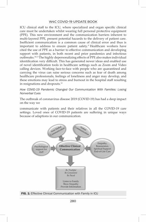
W4C COVID-19 UPDATE BOOK
280
ICU clinical staff to the ICU, where specialized and organ specific clinical care must be undertaken whilst wearing full personal protective equipment (PPE). This new environment and the communication barriers inherent to multi-layered PPE, present potential hazards to the delivery of patient care. Inefficient communication is a common cause of clinical error and thus is important to address to ensure patient safety.9 Healthcare workers have cited the use of PPE as a barrier to effective communication and developing rapport with patients, in both recent and prior pandemics and infectious outbreaks.10,11 The highly depersonalizing effects of PPE also makes individual identification very difficult. This has generated newer ideas and enabled use of novel identification tools in healthcare settings such as Zoom and Video calling devices. Working face-to-face with people who are quarantined and carrying the virus can raise serious concerns such as fear of death among healthcare professionals, feelings of loneliness and anger may develop, and these emotions may lead to stress and burnout in the hospital staff resulting in resignations and dropouts.12
How COVID-19 Pandemic Changed Our Communication With Families: Losing Nonverbal Cues
The outbreak of coronavirus disease 2019 (COVID-19) has had a deep impact on the way we
communicate with patients and their relatives in all the COVID-19 care settings. Loved ones of COVID-19 patients are suffering in unique ways because of adaptions in our communication.
FIG. 1: Effective Clinical Communication with Family in ICU
Non-Verbal
Communication
Psychologicalsupport for
staff
Empathy,Reassurance,
Spiritual SupportEnd-of-Life Care
Be TruthfulBe Consistent
Be Paced
Listen to FamilyShow Empathy
Provide Information
VerbalCommunication
Effective ClinicalCommunication
W4C COVID-19 UPDATE BOOK
281
Communication between teams and patients along with their relatives is of paramount importance in ICU settings wherein relatives consider communication skills just as importance as physical examination or even more so.
Restrictive ICU visiting policy has also affected the healthcare professionals (HCP) communication with family members. The unprecedented surge of critically ill patients inundating ICUs made the process of communication for HCPs with family members stressful and demanding. Informally, some ICU teams are communicating with families during the COVID-19 pandemic by telephone updates, dedicated family communication teams (reference persons), and video calling.13 Many units developed communication teams and were innovative in their creation. Despite innovative solutions, the situation has remained challenging, and many clinicians expressed their dissatisfaction with the service they were able to deliver for patients, families, and staff alike. There were also significant policy changes in communication with family members on informed consent and end-of-life (EOL) care or do-not-attempt to resuscitate (DNAR) directives being discussed remotely, which has aggravated anxiety levels amongst HCPs.
Practice points: Elements of Effective Communication Strategies
1. Tackling Misinformation
• Long-term regulatory action is the need of the hour to address the structural factors that facilitates an online environment of misinformation and disinformation
• In the near term, social media platforms must develop policies that reduces the spread of fast-moving COVID-19 related misinformation and disinformation. These steps must include detection, labeling, suspension of algorithmic amplification and the platforms should develop privacy-sensitive features to scan draft posts especially related to the healthcare and provide right information to users, with provision to run relevant fact checks on healthcare topics.
2. Vaccine acceptance
• The stakeholders should invest in rapidly building an evidence base effective strategies for COVID-19 vaccine promotion and acceptance, with transparency towards acknowledging the unique circumstances around COVID-19 vaccination. They should understand community needs and perceptions and formulate effective promotion and delivery strategies.14
3. Telecommunication and e-medicine
• Communication involving telemedicine during the COVID-19 epidemic has been the first line of defense for HCPs to slow the
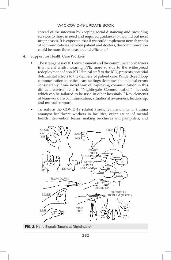
W4C COVID-19 UPDATE BOOK
282
spread of the infection by keeping social distancing and providing services to those in need and required guidance to the mild but most urgent cases. It is expected that if we could implement new channels of communications between patient and doctors, the communication could be more fluent, easier, and efficient.15
4. Support for Health Care Workers
• The strangeness of ICU environment and the communication barriers is inherent whilst wearing PPE, more so due to the widespread redeployment of non-ICU clinical staff to the ICU, presents potential detrimental effects to the delivery of patient care. While closed loop communication in critical care settings decreases the medical errors considerably,16 one novel way of improving communication in this difficult environment is “Nightingale Communication” method, which can be tailored to be used in other hospitals.17 Key elements of teamwork are communication, situational awareness, leadership, and mutual support.
• To reduce the COVID-19 related stress, fear, and mental trauma amongst healthcare workers in facilities, organization of mental health intervention teams, making brochures and pamphlets, and
FIG. 2: Hand Signals Taught at Nightingale17
GOODOKYES
BAD
UP DOWN
BACKREPEAT
DO AGAIN
SLOW DOWN
SPEED UPTHERE IS A
PROBLEM (POINT)
TIMEOUTLESS
MORE
NO
STOP
STARTNEXT
KEEP GOING
W4C COVID-19 UPDATE BOOK
283
delivery of range of mental services including counseling and psychotherapy can be offered to employees and their families.18
Health care leaders should communicate clearly and often with staff and provide accurate information about what to expect and solutions to help them work effectively.
1. Support for Families and Relatives
• Family members should be allowed in ICU, even if only for short periods. Visits to Covid or non-Covid ICUs under supervision, do not pose any additional safety risk to patients or visitors.
• In Covid ICUs, family members must be protected with PPE with clear instructions for donning-and-doffing protocols, just like healthcare workers.
• In non-Covid ICUs, further health surveillance, appropriate protection, and rapid nasopharyngeal swabbing should be considered to protect inpatients from further SARS-CoV-2 Infection. All relatives should be explained very clearly about the risks involved in accessing areas intended for the care of patients with a contagious infectious disease, and if protocolized informed consent can be taken.19
• Family-centered care is more, if not less, important during a pandemic. As families often have limited face-to-face contact at the point of entry, a website should provide additional information including visitation policies, communication tools etc. The information brochures on visiting and infection containment measures should also empower patients and families to anticipate and prepare for next steps.20
REFERENCES1. Seitz, A. (2020), Virus misinformation flourishes in online protest groups, https://apnews.
com/5862a9201c7b1bea62069a9c5e5fbb1c 2. NHS England (2020), NHS takes action against coronavirus fake news online, https://www.
england.nhs.uk/2020/03/nhs-takes-action-against-coronavirus-fake-news-online/3. Enhancing public trust in covid-19 vaccination: the role of governments© OECD 2021, http://
www.oecd.org/termsandconditions4. Kentish-Barnes N, Degos P, Viau C, Pochard F, Azoulay E. “It was a nightmare until I saw
my wife”: the importance of family presence for patients with COVID-19 hospitalized in the ICU. Intensive Care Med 2021; 47:792–794.
5. Pun BT, Badenes R, La Calle GH, Orun OM, Chen W, Raman R et al. Prevalence and risk factors for delirium in critically ill patients with COVID-19 (COVID-D): a multicentre cohort study. Lancet Respir Med 2021; 9,239-250.
6. Van Rompaey B, Elseviers MM, Schuurmans MJ, Shortridge-Baggett LM, Truijen S, Bossaert L. Risk factors for delirium in intensive care patients: a prospective cohort study. Crit Care 2009; 13:R77.
7. National Health Service England. Clinical guide for the management of remote consultations and remote working in secondary care during the coronavirus pandemic (2020). https://www.

W4C COVID-19 UPDATE BOOK
284
england.nhs.uk/coronavirus/wp-content/uploads/sites/52/2020/03/specialty-guide-surgery-and-coronavirus-v1-16-march-2020.pdf
8. Pablos JL, Galindo M, Carmona L, Lledo A, Reteurto M, Blanco R et al. Clinical outcomes of hospitalised patients with COVID-19 and chronic inflammatory and autoimmune rheumatic diseases: a multicentric matched cohort study. Annals of the Rheumatic Diseases 2020; 79:1544-1549.
9. Clapper TC, Ching K. Debunking the myth that the majority of medical errors are attributed to communication. Med Educ 2020; 54:74–81.
10. Boon DS, Vallenas C, Ferri M, Norris SL. Incorporating health workers’ perspectives into a WHO guideline on personal protective equipment developed during an Ebola virus disease outbreak (2018). [version 2; peer review: 2 approved]. F1000Res; 7: 45.
11. Seale H, Corbett S, Dwyer DE, MacIntyre CR. Feasibility exercise to evaluate the use of particulate respirators by emergency department staff during the 2007 influenza season. Infect Control Hosp Epidemiol 2009; 30:710–2.
12. Xiang YT, Yang Y, Li W, Zhang L, Zhang Q, Cheung T, et al. Timely mental health care for the 2019 novel coronavirus outbreak is urgently needed. Lancet Psychiatry (2020), 7, 228–229.
13. Boulton AJ, Jordan H, Adams CE, Polgarova P, Morris AC, Arora N. Intensive care unit visiting and family communication during the COVID-19 pandemic: A UK survey. Journal of the Intensive Care Society 2021; 0:1-4.
14. National Academies of Sciences, Engineering, and Medicine; Health and Medicine Division; Board on Population Health and Public Health Practice; Board on Health Sciences Policy; Committee on Equitable Allocation of Vaccine for the Novel Coronavirus; Kahn B, Brown L, Foege W, et al., editors. Framework for Equitable Allocation of COVID-19 Vaccine. Washington (DC): National Academies Press (US); 2020 Oct 2. 7, Achieving Acceptance of COVID-19 Vaccine. Available from: https://www.ncbi.nlm.nih.gov/books/NBK564098/
15. Vidal-Alaball J, Acosta-Roja R, Pastor Hernández N, Sanchez Luque N, Morrison D, Narejos Pérez S, et al. Telemedicine in the face of the COVID-19 pandemic 2020; 52:418-422.
16. Salik I, Ashurst JV. Closed Loop Communication Training in Medical Simulation. PMID: 31751089.
17. Shurlock J, Rudd J, Jeanes A, Iacovidou A, Creta A, Kanthasamy V et al. Communication in the intensive care unit during COVID-19: early experience with the Nightingale Communication Method. Int J Qual Health Care 2020; 3:mzaa162.
18. Çelmeçe Nuriye, Menekay Mustafa. The Effect of Stress, Anxiety and Burnout Levels of Healthcare Professionals Caring for COVID-19 Patients on Their Quality of Life. Frontiers in Psychology 2020; 11:3329-3335. Available from: https://www.frontiersin.org/article/10.3389/fpsyg.2020.597624
19. Mistraletti G, Giannini A, Gristina G, Malacarne P, Mazzon D, Cerruti E et al. Why and how to open intensive care units to family visits during the pandemic. Crit Care 2021; 25:191-196.
20. Curtis JR, Kross EK, Stapleton RD. The importance of addressing advance care planning and decisions about do-not-resuscitate orders during novel coronavirus 2019. (COVID-19) JAMA 2020; 323:1771-1772.
285
visiting Policies in iCU During Covid-19
INTRODUCTION Critically ill patients are quite a vulnerable group of patients, requiring physical, psychological and emotional support other than medical therapy. They are often unable to communicate or express themselves, placing the onus on clinicians in intensive care units (ICUs) to engage family members. Involving family members in the care and decision making of critically ill patients especially for the paediatric age group, terminally ill or altered mental status etc. are becoming a norm worldwide. However, the novel coronavirus disease (COVID-19) pandemic has dramatically altered this lookout. In the prevailing circumstances, the visitation policies of different ICUs have got substantial variation to create a fine balance between minimising the interhospital & community transmission and providing compassionate care at the available resources.
THE RATIONALE FOR DEVISING NEW VISITING POLICIES IN ICU Visiting their relative, participating in their care & decision making, the satisfaction of physically seeing each other and regularly communicating with the ICU medical & nursing teams is crucial to coping in a family with critically ill patients. It is also an important part of the grieving process in end of life care. Although visits by a patient’s next of kin and family is an accepted practice of normal ICU care,1 risk of transmission or acquisition of COVID-19 infections in overburdened health care facilities with limited personal protection equipments (PPEs), hospital beds, ventilators and medicines have forced many hospitals to impose restrictive visitation policy. The fear among patients’ family members about their safety has also resulted in a decreased number of visits.
On the other hand, restrictive visitation policies can lead to myriads of unintended consequences like increased stress & anxiety among family members because of unpredictability, vulnerability and lethality associated with the COVID-19 pandemic and feeling of being left alone and isolated among patients, which may further result in anxiety, depression and even
Sharmili Sinha, Mritunjay Kumar
38.
W4C COVID-19 UPDATE BOOK
286
delirium in them. The presence of visitors is associated with less fear, reduced delirium, and faster recovery.2,3 Family members at times also help in improving hospital safety surveillance and detect medical errors.4
TRENDS IN VISITATION POLICIESJaswaney R et al5 analysed public-facing visitor restriction policies during the first 3 months of the pandemic. They included a cohort of the 70 large General acute care hospitals representing 23 states across all 4 major regions of the United States. Sixty-five of the 70 hospitals reviewed had public-facing visitor restriction policies. Forty-nine of these 65 policies had general “no-visitor” statements, whereas 16 allowed at least 1 visitor to accompany all patients. Sixty-three of 65 hospitals included exceptions to their visitor restriction policies. Setting-specific exceptions included paediatrics, obstetrics/gynaecology, emergency department, behavioural health, inpatient rehabilitation, surgery, and outpatient clinics. Exceptions that applied across settings included patients at end of life and patients with disabilities.
Fiest et al6 conducted an environmental scan of Canadian hospital visitation policies throughout the first wave of the pandemic and found that most Canadian hospitals had public-facing visitor restriction policies with specific exception categories, most commonly for patients at end-of-life, patients requiring assistance, or COVID-19 positive patients (varying from not allowed to case-by-case).
Muniraman et al 7 did a cross-sectional survey of parents and families of infants hospitalised in the six tertiary level neonatal units, four from the UK and two from the USA between 1 May 2020 and 21 August 2020. Visitation limited to a single visitor with no restrictions on duration was the most frequently reported policy. Visitation policies were perceived as being restrictive by 62% (138/219) of the respondents with 37% (80/216) reporting being able to visit less often than desired, 41% (78/191) reporting being unable to bond enough and 27% (51/191) reporting not being able to participate in their baby’s daily care. Mild to severe impact on breastfeeding was reported by 36% (75/209) of respondents.
Andrist et al 8 advocated that visitation restriction policies be implemented by independent, medically knowledgeable decision-making bodies, with the informed participation of patients and their families.
Many institutes have come up with their visitation policy for ICU patients, but, applying any particular institution’s policy universally is not feasible. In an attempt to help different hospital setups develop their visitation policy and to reduce the risk of transmission of the COVID-19 virus, to visitors of patients with suspected or confirmed COVID-19. the Centers for Disease Control and Prevention (CDC) has come up with guidance to healthcare facilities on the management of visitors.9 (Table 1)
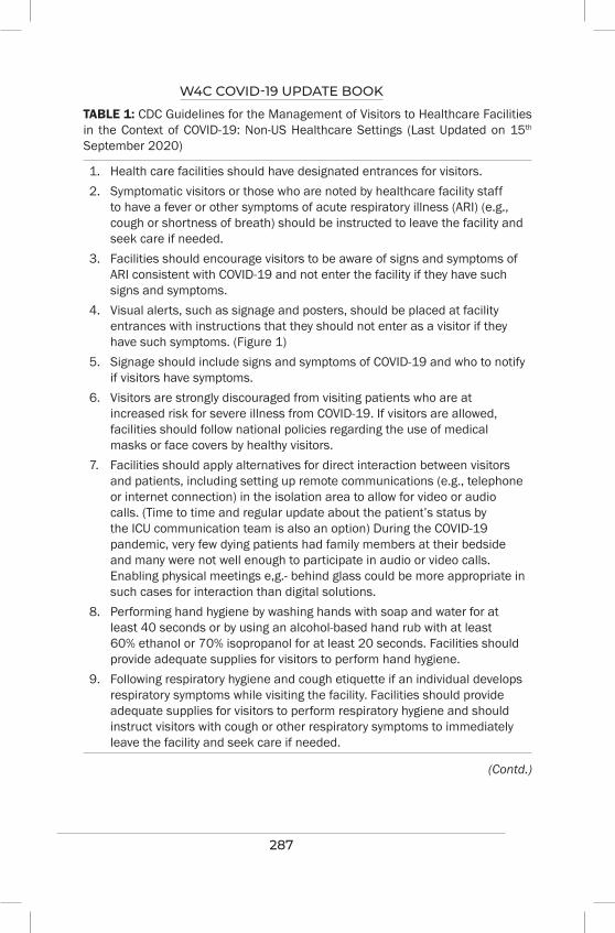
W4C COVID-19 UPDATE BOOK
287
TABLE 1: CDC Guidelines for the Management of Visitors to Healthcare Facilities in the Context of COVID-19: Non-US Healthcare Settings (Last Updated on 15th September 2020)
1. Health care facilities should have designated entrances for visitors.2. Symptomatic visitors or those who are noted by healthcare facility staff
to have a fever or other symptoms of acute respiratory illness (ARI) (e.g., cough or shortness of breath) should be instructed to leave the facility and seek care if needed.
3. Facilities should encourage visitors to be aware of signs and symptoms of ARI consistent with COVID-19 and not enter the facility if they have such signs and symptoms.
4. Visual alerts, such as signage and posters, should be placed at facility entrances with instructions that they should not enter as a visitor if they have such symptoms. (Figure 1)
5. Signage should include signs and symptoms of COVID-19 and who to notify if visitors have symptoms.
6. Visitors are strongly discouraged from visiting patients who are at increased risk for severe illness from COVID-19. If visitors are allowed, facilities should follow national policies regarding the use of medical masks or face covers by healthy visitors.
7. Facilities should apply alternatives for direct interaction between visitors and patients, including setting up remote communications (e.g., telephone or internet connection) in the isolation area to allow for video or audio calls. (Time to time and regular update about the patient’s status by the ICU communication team is also an option) During the COVID-19 pandemic, very few dying patients had family members at their bedside and many were not well enough to participate in audio or video calls. Enabling physical meetings e,g.- behind glass could be more appropriate in such cases for interaction than digital solutions.
8. Performing hand hygiene by washing hands with soap and water for at least 40 seconds or by using an alcohol-based hand rub with at least 60% ethanol or 70% isopropanol for at least 20 seconds. Facilities should provide adequate supplies for visitors to perform hand hygiene.
9. Following respiratory hygiene and cough etiquette if an individual develops respiratory symptoms while visiting the facility. Facilities should provide adequate supplies for visitors to perform respiratory hygiene and should instruct visitors with cough or other respiratory symptoms to immediately leave the facility and seek care if needed.
(Contd.)
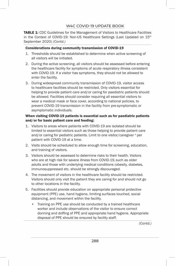
W4C COVID-19 UPDATE BOOK
288
Considerations during community transmission of COVID-191. Thresholds should be established to determine when active screening of
all visitors will be initiated.2. During the active screening, all visitors should be assessed before entering
the healthcare facility for symptoms of acute respiratory illness consistent with COVID-19. If a visitor has symptoms, they should not be allowed to enter the facility.
3. During widespread community transmission of COVID-19, visitor access to healthcare facilities should be restricted. Only visitors essential for helping to provide patient care and/or caring for paediatric patients should be allowed. Facilities should consider requiring all essential visitors to wear a medical mask or face cover, according to national policies, to prevent COVID-19 transmission in the facility from pre-symptomatic or asymptomatic individuals.
When visiting COVID-19 patients is essential such as for paediatric patients and/or for basic patient care and feeding:1. Visitors to areas where patients with COVID-19 are isolated should be
limited to essential visitors such as those helping to provide patient care and/or caring for pediatric patients. Limit to one visitor/caregiver a per patient with COVID-19 at a time.
2. Visits should be scheduled to allow enough time for screening, education, and training of visitors.
3. Visitors should be assessed to determine risks to their health. Visitors who are at high risk for severe illness from COVID-19, such as older adults and those with underlying medical conditions (obesity, diabetes, immunosuppressed etc. should be strongly discouraged.
4. The movement of visitors in the healthcare facility should be restricted. Visitors should only visit the patient they are caring for and should not go to other locations in the facility.
5. Facilities should provide education on appropriate personal protective equipment (PPE) use, hand hygiene, limiting surfaces touched, social distancing, and movement within the facility.
• Training on PPE use should be conducted by a trained healthcare worker and include observations of the visitor to ensure correct donning and doffing of PPE and appropriate hand hygiene. Appropriate disposal of PPE should be ensured by facility staff.
TABLE 1: CDC Guidelines for the Management of Visitors to Healthcare Facilities in the Context of COVID-19: Non-US Healthcare Settings (Last Updated on 15th September 2020) (Contd.)
(Contd.)
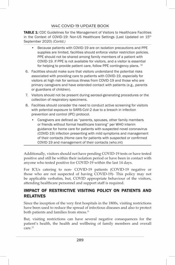
W4C COVID-19 UPDATE BOOK
289
• Because patients with COVID-19 are on isolation precautions and PPE supplies are limited, facilities should enforce visitor restriction policies. PPE should not be shared among family members of a patient with COVID-19. If PPE is not available for visitors, and a visitor is essential for helping to provide patient care, follow PPE contingency plans. 10
6. Facilities should make sure that visitors understand the potential risks associated with providing care to patients with COVID-19, especially for visitors at high risk for serious illness from COVID-19 and those who are primary caregivers and have extended contact with patients (e.g., parents or guardians of children).
7. Visitors should not be present during aerosol-generating procedures or the collection of respiratory specimens.
8. Facilities should consider the need to conduct active screening for visitors with potential exposure to SARS-CoV-2 due to a breach in infection prevention and control (IPC) protocol.
• Caregivers are defined as “parents, spouses, other family members or friends without formal healthcare training” per WHO interim guidance for home care for patients with suspected novel coronavirus (COVID-19) infection presenting with mild symptoms and management of their contacts (Home care for patients with suspected or confirmed COVID-19 and management of their contacts (who.int)
TABLE 1: CDC Guidelines for the Management of Visitors to Healthcare Facilities in the Context of COVID-19: Non-US Healthcare Settings (Last Updated on 15th September 2020) (Contd.)
Additionally, visitors should not have pending COVID-19 tests or have tested positive and still be within their isolation period or have been in contact with anyone who tested positive for COVID-19 within the last 14 days.
For ICUs catering to non- COVID-19 patients (COVID-19 negative or those who are not suspected of having COVID-19)- This policy may not be applicable verbatim, but, COVID appropriate behaviour of the visitors, attending healthcare personnel and support staff is required.
IMPACT OF RESTRICTIVE VISITING POLICY ON PATIENTS AND RELATIVES Since the inception of the very first hospitals in the 1800s, visiting restrictions have been used to reduce the spread of infectious diseases and also to protect both patients and families from stress.11
But, visiting restrictions can have several negative consequences for the patient’s health, the health and wellbeing of family members and overall care.12
W4C COVID-19 UPDATE BOOK
290
IMPACT ON PATIENTSPhysical health consequences
1. An increased level of observed physical pain13
2. Reduced personal hygiene maintenance & self-care14, 15
3. Reduced oral intake especially in the elderly14, 16
4. Reduced access to medications compared to the non-COVID-19 period17
Mental health consequences
1. Increased levels of perceived loneliness, depressive symptoms, agitation and aggression13,14,15
2. Reduced cognitive functions and loss of memory 13,14,18
3. Reduced access to their parents led to trust issues among children19
4. Decreased overall satisfaction level17
IMPACT ON FAMILY MEMBERS1. Not being physically present for their hospitalised family member
created worries, anxiety, sadness and a need for more information and updates on the family member’s condition7,15,17,20
2. A lower general psychosocial wellbeing score7,14 and an increased need for psychosocial support
3. Anger 21 and stress due to uncertainty7,20
4. Feeling of failing their loved ones.17 On the contrary, “virtual visits”, using audio and video calling or time update from the ICU team helped in overcoming this feeling.22
5. Decreased bonding and strained relationship
6. Long term mental health issues in family members who lost someone close during the COVID-19 pandemic.14,23
For care providers, visiting restrictions added the burdens of ethical dilemmas, learning new technical means to enable social interaction, educating attendants about infection prevention and use of PPEs and increased demand for communication with families and providing social support to both family members and patients.
IMPACT ON HOSPITALS AND HEALTH CARE WORKERS 1. Increased incidences of complaints and negative feedbacks have been
received by hospitals due to dissatisfaction among family members at times .
W4C COVID-19 UPDATE BOOK
291
2. Sporadic occurrences of abuse and malhandling of health care workers have been reported in the pandemic due to feelings of mistrust and emotional outbursts especially by families of critically ill patients.
PROVISIONS OF COMMUNICATION IN LIEU OF RESTRICTIVE VISTING POLICIES1. Video Calls using smart phones, laptops and other devices between
patients and relatives as well as between healthcare staffs and family members.
2. Regular phone calls at a particular time on daily basis with the kith and kin of patients have also been proved effective and well accepted.
3. Counselling of patients and relatives should be done from very beginning at the time of admission and at regular interval.This helps to obtain maximum co-operation and helps in successful execution of such practices.
Various changes in the healthcare infra structure and implementation of digital technology have gone a long way in mitigating the communication gaps arising out of restrictive visting policies.
CONCLUSIONIndividual hospital visitor policies during the COVID-19 pandemic varies widely. Given the importance of public health and hospital measures to prevent viral transmission, preserve PPE, and maintain a healthy medical workforce, hospitals should develop visitor restrictions based on the general and local epidemiological data, available resources, and with a humane approach.
KEY POINTS• The risk of visitors bringing the virus into healthcare facilities increases
as community transmission becomes widespread.
• Healthcare facilities should minimise the risk of infection to visitors of patients with suspected or confirmed COVID-19.
• Facilities should establish policies and should provide adequate provisions for managing, screening, educating, and training all visitors.
• Necessary changes in the infrastructure and use of digital technology should be maximally explored to establish communication between COVID-19 patients and their relatives.
• Counselling remains an integral and important part for successful execution of such restrictive vistors policy.
W4C COVID-19 UPDATE BOOK
292
REFERENCES1. Black MD, Vigorito MC, Curtis JR, Phillips GS, Martin EW, McNicoll L et al. A multifaceted
intervention to improve compliance with process measures for ICU clinician communication with ICU patients and families. Crit Care Med 2013; 41:241 2275–83.
2. Falk J, Wongsa S, Dang J, Comer L, LoBiondo-Wood G. Using an evidence-based practice process to change child visitation guidelines. Clin J Oncol Nurs 2012; 16:21-23.
3. Granberg A, Engberg IB, Lundberg D. Acute confusion and unreal experiences in intensive care patients in relation to the ICU syndrome. part II. Intensive Crit Care Nurs 1999; 15:19-33.
4. Khan A, Coffey M, Litterer KP, et al. Families as partners in hospital error and adverse event surveillance. JAMA Pediatr 2017;171:372-81.
5. Jaswaney R, Davis A, Cadigan RJ, Waltz M, Brassfield ER, Forcier B, Joyner BL Jr. Hospital Policies During COVID-19: An Analysis of Visitor Restrictions. J Public Health Manag Pract 2021 Mar 12. Epub ahead of print.
6. Fiest KM, Krewulak KD, Hiploylee C, Bagshaw SM, Burns KEA, Cook DJ, Fowler RA, Kredentser MS, Niven DJ, Olafson K, Parhar KKS, Patten SB, Fox-Robichaud AE, Rewa OG, Rochwerg B, Spence KL, Straus SE, Spence S, West A, Stelfox HT, Parsons Leigh J; Canadian Critical Care Trials Group. An environmental scan of visitation policies in Canadian intensive care units during the first wave of the COVID-19 pandemic. Can J Anaesth 2021 Jun 30:1–11. Epub ahead of print
7. Muniraman H, Ali M, Cawley P, Hillyer J, Heathcote A, Ponnusamy V et al. Parental perceptions of the impact of neonatal unit visitation policies during COVID-19 pandemic. BMJ Paediatrics Open, 4:2020, Article e000899
8. Andrist E, Clarke RG, Harding M. Paved With Good Intentions: Hospital Visitation Restrictions in the Age of Coronavirus Disease 2019. Pediatr Crit Care Med. 2020; 21:e924-e926.
9. Management of Visitors to Healthcare Facilities in the Context of COVID-19: Non-US Healthcare Settings. Centers for Disease Control and Prevention. September 15, 2020. Accessed on 31st August 2021. https://www.cdc.gov/coronavirus/2019-ncov/hcp/non-us-settings/hcf-visitors.html
10. Summary for Healthcare Facilities: Strategies for Optimizing the Supply of PPE during Shortages. Centers for Disease Control and Prevention. December 29, 2020. Accessed on 31st August 2021. https://www.cdc.gov/coronavirus/2019-ncov/hcp/ppe-strategy/strategies-optimize-ppe-shortages.html
11. Smith L, Medves J, Harrison MB, Tranmer J, Waytuck B. The impact of hospital visiting hour policies on pediatric and adult patients and their visitors. JBI Library of System Rev 2009; 7:38-79
12. Hugelius K, Harada N, Marutani M.Consequences of visiting restrictions during the COVID-19 pandemic: An integrative review. International Journal of Nursing Studies 2021;121:104000
13. Sizoo EM, Monnier AA, Bloemen M, Hertogh C, Smalbrugge M. Dilemmas with restrictive visiting policies in Dutch nursing homes during the COVID-19 pandemic: a qualitative analysis of an open-ended questionnaire with elderly care physicians. J Am Med Directors Assoc 2020; 21:1774-81
14. O’Caoimh R, O’Donovan MR, Monahan MP, Dalton O’Connor C, Buckley C. Psychosocial impact of COVID-19 nursing home restrictions on visitors of residents with cognitive impairment: a cross-sectional study as part of the Engaging Remotely in Care (ERiC) Project. Front Psychiatry 2020; 11:Article 585373.
15. Wammes JD, Kolk MD, van den Besselaar JH, MacNeil-Vroomen JL, Buurman-van Es BM ET AL. Evaluating perspectives of relatives of nursing home residents on the nursing home visiting restrictions during the COVID-19 crisis: a Dutch cross-sectional survey study. J. Am. Medical Directors Assoc., 12:2020, 1746-50
16. Shum CK, Shea YF, Tang M, Wan WH, Chan MMK. Poor feeding due to visitor restrictions in long-term care facilities during the coronavirus disease 2019 pandemic. Psychogeriatrics 2020; 20:929-30.

W4C COVID-19 UPDATE BOOK
293
https://www.cdc.gov/coronavirus/2019-ncov/downloads/hcp/Vistor-Signage-Template.pdf accessed on 31st August 2021
17. Yeh TC, Huang HC, Yeh TY, Huang WT, Huang HC, Chang YM et al. Family members’ concerns about relatives in long-term care facilities: acceptance of visiting restriction policy amid the COVID-19 pandemic. Geriatrics & Gerontol 2020; 20:938–42.
18. Verbeek H, Gerritsen DL, Backhaus R, DeBoer BS, Koopmans RTCM, Hamers JPH. Allowing visitors back in the nursing home during the COVID-19 crisis: a Dutch national study into first experiences and impact on well-being. J Am Medical Directors Assoc 2020; 21:900-4
19. Virani AK, Puls HT, Mitsos R, Longstaff H, Goldman RD, Lantos JD. Benefits and risks of visitor restrictions for hospitalized children during the COVID pandemic. Pediatrics 2020; 146:1-6.
20. Creutzfeldt CJ, Schutz REC, Zahuranec DB, Lutz BJ, Curtis JR, Engelberg RA. Family presence for patients with severe acute brain injury and the influence of the COVID-19 pandemic. J Palliat Med 2020; 10:108
21. Bembich S, Tripani A, Mastromarino S, Di Risio G, Castelpietra E, Risso FM. Parents experiencing NICU visit restrictions due to COVID-19 pandemic. Acta Paediatr (2020), 10.1111/apa.15620
22. Mercadante S, Adile C, Ferrera P, Giuliana F, Terruso L, Piccione T. Palliative care in the time of COVID-19. 1 J Pain Symptom Manag, 2020; 60:e79-e80
23. Heath L, Yates S, Carey M, Miller M. Palliative care during COVID-19: data and visits from loved ones. Am J Hospice and Palliative Med, 2020; 37:988-91.
294
Do’s and Don’ts in the Management of Covid-19 39.
GENERAL GUIDELINESManagement of patients with suspected or confirmed COVID-19 is predominantly dependent on the severity of disease, as described below:1,2
a. Asymptomatic or Presymptomatic Infection: Individuals who test positive for SARS-CoV-2 but have no symptoms consistent with COVID-19.
b. Mild Illness: Individuals with any of the signs and symptoms of COVID-19 (e.g., fever, cough, sore throat, malaise, headache, muscle pain, diarrhea, loss of taste and smell) but do not have shortness of breath, dyspnea or hypoxia.
c. Moderate Illness: Individuals who show evidence of pneumonia during clinical assessment or imaging and have features of dyspnea and/or hypoxia with oxygen saturation (SpO2) 90% to ≤ 93% on room air.
d. Severe Illness: Individuals who have features of pneumonia and severe dyspnea and/or SpO2 < 90% on room air, respiratory rate >30 breaths/min.
e. Critical Illness: Individuals with severe illness who have respiratory failure, septic shock, and/or multiple organ dysfunction.
PRINCIPLES OF MANAGEMENT FOR COVID- 192-5
a. Isolation at a suitable location like designated COVID-19 health facility, community facility, or home (self-isolation).
b. Infection prevention and control measures
• As there is a high risk of infection spread, strict infection control practices are needed.
• If possible, the patient should wear a surgical mask to limit the spread of infectious droplets.
Vaibhav Bhargava, Kishore Mangal
W4C COVID-19 UPDATE BOOK
295
• Treating clinician should don complete Personnel Protective Equipment (PPE) including gloves, gowns, N95 masks and eye protection and place the patients in negative-pressure rooms whenever possible during aerosol-generating procedures.
• As fear of getting infected and hospital infection control measures prevent families from visiting seriously ill patients. Caregivers should develop some plan to communicate patient’s families.
• Clinicians and paramedical staff should not breach infection control practices otherwise it leads to further spread of infection.
c. Symptom management - such as antipyretics for fever and pain, adequate nutrition, appropriate hydration, cough syrups and others as necessary.
d. Mild patients can be taken care of at home but counsel them about signs and symptoms of complications that should prompt urgent care.
e. Admit patients with moderate or severe disease to an appropriate healthcare facility.
f. Assess the requirements of any rehabilitation or follow-up after discharge.
g. Discontinue transmission-based precautions (including isolation) and release patients from the isolation ten days after symptom onset and at least three days without fever and respiratory symptoms. Patients with severe illness may warrant extended isolation and precautions for up to 20 days after symptom onset.6
1. RESPIRATORY SUPPORT1.1 Oxygenation and Ventilation2-5
Do’s
• Start oxygen therapy immediately in patients with emergency signs, such as severe respiratory distress and SpO2 < 90%.
• Consider oxygen supplementation initially using a nasal cannula or Venturi mask to keep the SpO2 between 90% and 96%.
• With the progression of the disease and when oxygen requirement is more than 6 l/min is required, shift to high flow nasal cannula (HFNC) over non-invasive positive pressure ventilation (NIV).
• Reassess patients on non-invasive modalities every 1 to 2 hours, or sooner if SpO2 < 90% or clinical deterioration.
• For endotracheal intubation, an expert in airway management should do the procedure. Consider using Video Laryngoscopy, if possible.
• For patients on controlled mechanical ventilation (CMV) for ARDS (target lung-protective ventilation)
W4C COVID-19 UPDATE BOOK
296
– Low tidal volume, higher PEEP (Plateau pressure < 30)
– If PEEP responsive, consider traditional recruitment maneuvers
– Conservative fluid management
– Prone ventilation (12-16 hours per day)
– Use neuromuscular blocking agents (NMBA) for the first 24 - 48 hours.
Don’ts
• Do not delay intubation until the patient has features of impending respiratory arrest or is on maximum noninvasive supportive care.
• Don’t use staircase recruitment maneuvers (incremental PEEP).
Don’t Know
• Use of Extracorporeal Membrane Oxygenation (ECMO) for refractory Hypoxemia on CMV.
1.2 Self-Pronation2-5
Do’s
• Consider awake prone positioning for around 8-12 hrs per day (broken into shorter periods) in patients on oxygen support.
Don’ts
• Do not use awake prone positioning to avoid intubation in patients who otherwise need intubation and mechanical ventilation.
• Do not prone the patients with acute bleeding, multiple fractures or trauma, spinal instability, raised intracranial pressure, or tracheal surgery or sternotomy within two weeks.
2. PHARMACOLOGICAL THERAPYNumerous drugs have been used in clinical trials for the treatment of COVID 19 patients. The emerging evidence from these trials has been used to guide the treatment of these patients. As the new research has been published unprecedentedly, several guidelines have been published and are regularly updated to incorporate the latest evidence. Thus, the recommendations have kept changing over the period and is expected to continue.
2.1 Antiviral Therapy2,5,7,8
Do’s
• There is still no consensus recommendation for antiviral treatment in COVID 19 patients.
W4C COVID-19 UPDATE BOOK
297
Don’ts
• Chloroquine or Hydroxychloroquine (HCQ), Hydroxychloroquine plus azithromycin, Ivermectin, Lopinavir/Ritonavir and Other HIV Protease Inhibitors and Nitazoxanide - They are not recommended to be used in non-hospitalized and hospitalized patients of any severity of illness (except in a clinical trial for Ivermectin and Nitazoxanide). However Indian guidelines do suggest HCQ and Ivermectin in mild cases.1
Don’t know
Remdesivir -
• Guideline recommendations vary for remdesivir in different parts of the world. FDA has approved Remdesivir in the USA for hospitalized adolescents (≥12 years old weighing at least 40 kg) and adults with pneumonia who require minimal supplemental oxygen,2,7 whereas in UK and Europe it is only conditionally recommended for the same indication.5,9 On the other hand, WHO recommends against use of remdesivir in hospitalized patients in addition to the standard care, regardless of disease severity.8 In India, it is a reserve drug approved under Emergency Use Authorization in select moderate/ severe hospitalized Covid 19 patients on supplemental oxygen.10
• Usual dose is 200 mg on first day followed by 100 mg for 4 days, but some experts suggest 10 days extended treatment for severe disease.7
2.2 Anti-SARS-CoV-2 Antibody Products2
Do’s
a. Casirivimab plus imdevimab - should be used to treat non-hospitalized patients with mild to moderate COVID-19 who are at high risk of clinical progression, as defined by the Emergency Use Authorization (EUA) criteria. It is now approved for post exposure prohylaxis also.
Don’t
a. Casirivimab plus imdevimab - not currently authorized for use in patients who are hospitalized with severe COVID-19.
b. Bamlanivimab plus Etesevimab - not recommended.
c. COVID-19 Convalescent Plasma - not recommended.
Don’t know -
a. Anti-SARS-CoV-2 Specific Immunoglobulin - There is insufficient evidence.
W4C COVID-19 UPDATE BOOK
298
2.3 Corticosteroids2,5,7,8
Do’s
• Use steroids in hospitalized patients who require increasing amount of supplemental oxygen or oxygen delivery through HFNC, NIV or IMV or ECMO.
• Dose of Dexamethasone 6 mg IV or PO once daily for up to 10 days or until hospital discharge.
• Equivalent dose of other steroids to dexamethasone 6 mg PO or IV is
– Prednisone 40 mg
– Methylprednisolone 32 mg
– Hydrocortisone 160 mg
Don’ts
• Do not use steroids in non-hospitalized patients or hospitalized patients that do not require oxygen supplementation.
Don’t know
• Inhaled budesonide.
2.4. Immunomodulators
Do’s
a. IL-6 inhibitor - Tocilizumab2,5,7,8
• Use in severe COVID 19 patients who do not have suspected or confirmed secondary bacterial or other viral infections. These drugs should be avoided in patients who are significantly immunocompromised. It is conditionally approved in India 1.
b. Sarilumab - used only as alternative to Tocilizumab.2
c. Baricitinib
• US NIH recommends Baricitinib, in combination with dexamethasone ± remdesivir, in recently hospitalized patients on high-flow oxygen or non-invasive ventilation with rapidly increasing oxygen needs and systemic inflammation.2
• US FDA granted an emergency-use authorization with or without remdesivir, for the treatment of suspected or confirmed COVID-19 disease in hospitalized children aged 2 years and older and adults who require supplemental oxygen, invasive mechanical ventilation, or ECMO.5
W4C COVID-19 UPDATE BOOK
299
• The IDSA recommends baricitinib with remdesivir in hospitalized patients with severe disease who cannot receive corticosteroids because of a contraindication, rather than remdesivir alone.7
• It has not been authorized in the UK or Europe.5
d. Tofacitinib - use only as alternative to Baricitinib.2
Don’ts
• Following drugs are not recommended except in a clinical trial:2
Baricitinib in combination with tocilizumab
Interferons (alfa or beta)
Kinase inhibitors
Bruton’s tyrosine kinase inhibitors (e.g., acalabrutinib, ibrutinib, zanubrutinib)
Non-SARS-CoV-2-specific intravenous immunoglobulin (IVIG).
The anti-IL-6 monoclonal antibody Siltuximab
Colchicine (in hospitalized patients).
• Don’t know
There is insufficient evidence for use of the following immunomodulators for the treatment of COVID-19:2
Colchicine for non hospitalized patients
Fluvoxamine
Granulocyte-macrophage colony-stimulating factor inhibitors for hospitalized patients
Interleukin (IL)-1 inhibitors (e.g., anakinra)
Anti-VEGF drug (e.g. Bevacizumab)
Anti-CD6 humanized IgG1 mAb (Itolizumab)
2.5 Venous Thromboembolism (VTE) Prophylaxis2,4,5
Do’s
• Assess the risk of bleeding as soon as possible after admission, using a suitable risk assessment tool.
• Prophylactic dose of anticoagulation should be given in hospitalized adults until discharge.
• Patients with incidental thromboembolic event or high suspicion of
W4C COVID-19 UPDATE BOOK
300
thromboembolic disease should be managed with therapeutic doses of anticoagulant therapy.
• Post-discharge VTE prophylaxis consideration should include individual patient’s risk factors for VTE, including reduced mobility, bleeding risks, and feasibility.
Don’ts
• Do not initiate anticoagulation in non-hospitalized patients, unless the patient has other indication of starting the therapy.
• Routinely do not continue after discharge from hospital.
2.6 Antipyretics2,4
Do’s
• Paracetamol (500-1000 mg orally every 4-6 hrs, max 4000 mg/day) or ibuprofen (300-600 mg orally every 6-8 hours when required, maximum 2400 mg/day).
2.7 Antibiotics2,4,5
Do’s
• Use only in case of clear suspicion of secondary bacterial infection.
• If initiated reassess daily to minimize unnecessary usage and side effects.
Don’ts
• Routine use in mild to moderate cases should be avoided.
2.8 Other Pharmacological Considerations
Do’s
• Patients with COVID-19 who are receiving concomitant medications (e.g., angiotensin-converting enzyme inhibitors, angiotensin receptor blockers, statins, systemic or inhaled corticosteroids, nonsteroidal anti-inflammatory drugs, acid-suppressive therapy) for underlying medical conditions should continue these medications during acute management of COVID-19 unless discontinuation is otherwise warranted by their clinical condition.2
Don’ts
• It is not recommended to use unproven drugs as treatment or prophylaxis for COVID-19, outside of the context of clinical trials.4
• Don’t know
There is insufficient evidence to recommend either for or against the use of Vitamin C, Vitamin D and Zinc for the treatment of COVID-19.2
W4C COVID-19 UPDATE BOOK
301
3. HEMODYNAMICS 2, 3, 4, 5
Do’s
• Use balanced crystalloids over unbalanced crystalloids for acute resuscitation of patents with shock.
• Use dynamics parameters, lactate levels, capillary refilling time and skin temperature over static parameters to assess fluid responsiveness.
• Norepinephrine is the first-choice vasopressor to manage patients with shock.
• Titrate vasoactive agents to target a Mean Arterial Pressure (MAP) of 60 to 65 mm Hg, over higher MAP.
• Add either vasopressin (up to 0.03 units/min) or epinephrine as second agent if needed.
• Use dobutamine in patients with cardiac dysfunction and persistent hypoperfusion despite adequate fluid loading and the use of vasopressor agents.
• Patients who require vasopressors should have an arterial catheter placed.
• In adult patients with refractory septic shock who have completed a course of corticosteroids to treat COVID-19, use hydrocortisone in “shock reversal” dose of 200 mg per day.
Don’ts
• Do not use albumin in initial part of acute resuscitation in patients with shock.
• Do not use hydroxyethyl starch for intravascular volume replacement in patients with shock.
• If norepinephrine is available, do not use dopamine in patients with shock.
• Do not use low dose dopamine for renal protection.
• Patients who are getting corticosteroids for COVID 19 management and are receiving sufficient replacement therapy then they do not require additional hydrocortisone.
REFERENCES1. Updated Detailed Clinical Management Protocol for adult cases of COVID19, Ministry of
Health and Family Welfare, Government of India, Version 6, Dated 24.05.21. Available at https://www.mohfw.gov.in. Accessed 26th August, 2021.
W4C COVID-19 UPDATE BOOK
302
2. COVID-19 Treatment Guidelines Panel. Coronavirus Disease 2019 (COVID-19) Treatment Guidelines. National Institutes of Health. Available at https://www.covid19treatmentguidelines.nih.gov/. Accessed Accessed 26th August, 2021.
3. Alhazzani W, Møller MH, Arabi YM, Loeb M, Gong MN, Fanet E et al. Surviving Sepsis Campaign: guidelines on the management of critically ill adults with coronavirus disease 2019 (COVID-19). Intensive Care Med 2020; 46:854-887. doi: 10.1007/s00134-020-06022-5.
4. Clinical management of COVID-19: living guidance, 25th January 2021. World health Organization (WHO). Available at https://www.who.int/publications/i/item/WHO-2019-nCoV-clinical-2021-1. Accessed 26th August, 2021.
5. Coronavirus disease 2019 (COVID-19) - BMJ Best Practice, Last Updated 02th September 2021. Available at https://bestpractice.bmj.com/topics/en-us/3000168. Accessed 26th August, 2021.
6. Interim Guidance on Ending Isolation and Precautions for Adults with COVID-19. Centers for Disease Control and Prevention, USA. Available at https://www.cdc.gov/coronavirus/2019-ncov/hcp/duration-isolation.html. Accessed 26th August, 2021
7. Bhimraj A, Morgan RL, Shumaker AH, Lavergne V, Baden L, Cheng VC, et al. Infectious Diseases Society of America Guidelines on the Treatment and Management of Patients with COVID-19. Clin Infect Dis 2020 Apr 27:ciaa478. doi: 10.1093/cid/ciaa478.
8. Therapeutics and COVID-19: living guideline, 06th July 2021. World health Organization (WHO). Available at https://www.who.int/publications/i/item/WHO-2019-nCoV-clinical-2021-1. Accessed 27th August, 2021.
9. Chalmers JD, Crichton ML, Goeminne PC, Cao B, Humbert M, Shteinberg M, et al. Management of hospitalised adults with coronavirus disease 2019 (COVID-19): a European Respiratory Society living guideline. Eur Respir J 2021; 57:2100048. doi: 10.1183/13993003.00048-2021.
10. Advisory for Rational use of Remdesivir for COVID-19 Treatment, Ministry of Health and Family Welfare, Government of India. Available at https://www.mohfw.gov.in. Accessed 26th August, 2021.

Long or Post Covid Conditions
SECTION 6

305
Section 6 - Long or Post Covid ConditionSECTION EDITOR
Dr Khusrav Beji BajanMD, EDIC Consultant – Intensivist & HOD – Emergency Medicine, Critical Care & Emergency Medicine, P. D. Hinduja Hospital & Medical Reaserch Centre, Mumbai
Preface
As we have been Grappling with COVID -19 over the past 22months, we have been made to realise that COVID has mutated into Variants, not only making it more transmissible and virulent but also giving it newer clinical presentations such as GI symptoms, mesenteric thrombosis, stroke, DKA, acute coronary syndrome and sometimes macrophage activation.
We have also realized that COVID is a multisystem disease leading to ARDS, thrombosis, MODS and immune dysfunction. Managing such a patient leads to a prolonged stay in the ICU and hospital. This in turn makes them vulnerable to secondary bacterial and fungal mould infections of all colors and severity. (attributed to the unaudited use of steroids). Prolonged hospitalization also adds to the incidence of more pressure sores, thrombotic events, poor neurological recovery and malnutrition, making us at times discuss with the family an appropriate approach of END of LIFE (EOL) care.
Even after winning a long battle of COVID and being discharged, nearly half of them still have Long COVID or POST COVID SYNDOMES (PCS) which could be as mild as fatigue or brain fog to life threatening post covid lung fibrosis and Thromosis. These PCS can be said to lead to a bunch of “pathies” such as neuropathy, cardiomyopathy, musculopathy, coagulopathy, endocrinopathy and vasculopathy.
As health care workers, we must always bear in mind that since COVID has poor prognosis, involves huge costs and emotional values, there would always be chances of litigations and hence we must be well aware of the Legalities involved with COVID – 19. Bottom line is to keep the patient and the Healthcare workers safe in the Pandemic.

307
New or Ongoing Symptoms of Disease 40.
INTRODUCTIONThe COVID-19 outbreak has consumed the entire world’s resources and drastically changed our lives and perspectives. The virus flummoxed scientists, doctors completely as it evolved twice in 18 months, two lineages (Alpha and then Delta) each 50% more transmissible, thus demonstratinga phenomenal amount of change.
The virus is constantly evolving, thus keeping the entire medical community on tenterhooks and research as well as treatment options in a state of constant flux. Initially, it emerged as the first B1 (Virus from Wuhan), later B 1315(from Africa) thereafter P1(from Brazil leading to B 117 (from UK) and striking home, B 1.617.1/2/3 (from India) which has acquired one more mutation called K417 N that is making it to be resistant to monoclonal antibodies and has been termed as the Delta variant. The original virus from Wuhan started with a potency to infect 2.5 people for every one person infected. Today, we have the Delta variant from India which infects upto 8 people and which literally exhausted the country’s healthcare system. Whilst India is still reeling from the shock of the devastating second wave, the threat of the Delta plus variants looms large on the horizon.
As we wait with bated breath, for news that portends, the population of patients that recovered from covid 19 is growing. Covid 19 has been established as a multisystem disease with florid systemic manifestations.Of the individuals affected, about 80% had mild to moderate disease and among those with severe disease, 5% develop critical illness.1Although earlier there was a belief that patients and their relatives that COVID-19 related illness recover completely in about two to three weeks, now it is being increasingly acknowledged that ongoing symptoms persist for weeks after recovery from acute COVID-19 illness with florid multisystem involvement. The mechanisms implicated are being suggested as cellular damage, a robust innate immune response with inflammatory cytokine production,2 and a pro-coagulant state induced by SARS-CoV-2 infection may contribute to these sequelae.3 This
Bikram Kumar Gupta, Bhavya Naithani
W4C COVID-19 UPDATE BOOK
308
fluctuating multi system symptomatology extending beyond three weeks after acute COVID-19 illness has been termed ‘Post COVID Syndrome’, also known as ‘Long COVID’, first used by Peregro4 or ‘Ongoing COVID’. With more studies coming in, it is now becoming evident that exposed people to COVID-19 will have long lasting symptomatology inflicting a major impact on the health care system and society even after the pandemic gets subsided.
DEFINITION Thus, among people infected with SARS-CoV-2 the presence of one or more symptoms (continuous or relapsing and remitting; new or same symptoms of acute COVID) even after the expected period of clinical recovery, irrespective of the underlying mechanism, is defined as post COVID syndrome or Long Covid Syndrome. It is further classified into two parts
• Post-Acute COVID 19 Syndrome: Defined as when symptoms extend beyond three weeks from onset of first symptoms.
• Post-Chronic COVID 19 Syndrome: Defined as when symptoms extend beyond 12 weeks from the onset of illness.
It has been suggested that a positive test for COVID 19 is not to be considered as a pre-requisite for diagnosis because many people were not tested, and false negative tests have been common5 as most people with post-COVID syndrome are PCR negative, indicating microbiological recovery and also exhibit biochemical and radiological recovery. In other words, post COVID syndrome is the time lag between the microbiological recovery and clinical recovery.6
Diagnostic challenges include
1. Residual symptoms in those checked negative for COVID (false negative as testing may be done too early or too late in the disease course].
2. Antibody response to infection is varied and about 20% do not seroconvert.
3. Antibody level may decrease over time masking the retrospective diagnosis of recent SARS-CoV-2 infection7
EPIDEIMIOLOGYAn outpatient report from Italy found persistence of symptoms in 87.4% of people recovered and discharged from hospitals even at 60 dayswith 32% having one or two symptoms and 55%, three or more. Fever or features of acute illness was not seen in these patients. The commonly reported problems were fatigue (53.1%), worsened quality of life (44.1%), dyspnoea (43.4%), joint pain, (27.3%) and chest pain (21.7%). Cough, skin rashes, palpitations, headache, diarrhoea, and ‘pins and needles’ sensation were the other symptoms reported. Patients also reported inability to do routine daily
W4C COVID-19 UPDATE BOOK
309
activities, in addition to mental health issues such as anxiety, depression and post-traumatic stress disorder.8
Another study found that COVID-19 patients discharged from hospital experience breathlessness and excessive fatigue even at 3 months.9 These studies were corroborated from studies conducted in United states, Europe, China and several other countries and as per a recent meta analysis the 5 most common manifestations of Long COVID-19 were fatigue (58%), headache (44%), attention disorder (27%), hair loss (25%), dyspnoea (24%).10
RISK FACTORS FOR LONG COVIDFew factors have been identified to be commonly associated with Long COVID:-
• Pre-existing respiratory disease
• Higher body mass index
• Older age
• Black, Asian and minority ethnic (BAME)
• Dyspnoea at 4–8weeks follow-up.
• Women (twice more common)
• Co morbidities such as diabetes, obesity, chronic cardiovascular or kidney disease, cancer and organ transplantation
• Presence of more than 5 symptoms in acute stage of illness
Mechanism of Non resolution/ Ongoing symptoms:-
1. Adverse effects of medications used
2. Sequelae of organ damage (Varying time required for recovery of organ)
3. Complication related to corona effects and comorbidities
4. Persistent of chronic inflammation (Convalescent phase)
5. Non specific effect of hospitalisation
6. Post intensive care syndrome
7. Persistence of virus in body (rare)
ORGAN SPECIFIC SYMPTOMSRespiratory System
It has been observed that dyspnoea, decreased exercise capacity and hypoxia are the most commonly seen symptoms and signs. Radiologically, reduced diffusion capacity, pulmonary physiology of a restrictive pattern ,and ground-
W4C COVID-19 UPDATE BOOK
310
glass opacities and fibrotic changes have been seen at follow-up assessments of pulmonary function of COVID-19 survivors which are done through home pulse oximetry, 6MWTs, PFTs, high-resolution computed tomography of the chest and when deemed clinically necessary by a physician computed tomography pulmonary angiogram.
Cardiovascular
Symptoms which persist include palpitations, dyspnoea and chest pain which is duly monitored by clinical electrocardiogram and echocardiogram follow ups when clinically appropriate. Chest pain being the most common should be differentiated from musculoskeletal causes. When the diagnosis is uncertain, special investigations should be sought. Left ventricular dysfunction and heart failure should be managed as per the standard guidelines. Intense cardio-vascular exercises should be completely avoided for next 3 to 6 moths and graded return is warranted after ensuring normal cardiac function. Long-term sequelae may include increased cardiometabolic demand, myocardial fibrosis or scarring (detectable via cardiac MRI), arrhythmias, tachycardia, autonomic dysfunction and life threatening pulmonary embolism.11 There was no inherent association between development of fatigue, severity of disease and level of inflammatory markers.12,13
Hematologic
Thromboembolic events have been observed to be <5% in post-acute COVID-19 in retrospective studies albeit the duration of the hyperinfammatory state induced by infection with SARS-CoV-2 is still nebulous. Persistently elevated d -dimer levels, specifically greater than twice the normal upper limit, immobility, cancer patients can be given direct oral anticoagulants and low-molecular-weight heparin for extended thromboprophylaxis after weighing pros and cons.14
Neuropsychiatric
Common neurological sequelae in post COVID period are headache refractory to therapy, dizziness, confusion, myalgia and dysautonomia. Anxiety, depression, sleep disturbances and PTSD have also been reported in 30–40% of COVID-19 survivors. The pathophysiology of neuropsychiatric complications is multifarious and involves immune dysregulation, inflammation, microvascular thrombosis, iatrogenic effects of medications and psychosocial impacts of infection. Acquired focal or multifocal peripheral nerve injury (PNI) was noticed in those who received prone ventilation for COVID related ARDS.15
Renal
Although resolution of AKI during acute COVID-19 occurs in almost all of the patients; reduced eGFR has been observed at 6 months follow-up. Early intervention and vigilance is the mainstay in COVID-19 survivors with
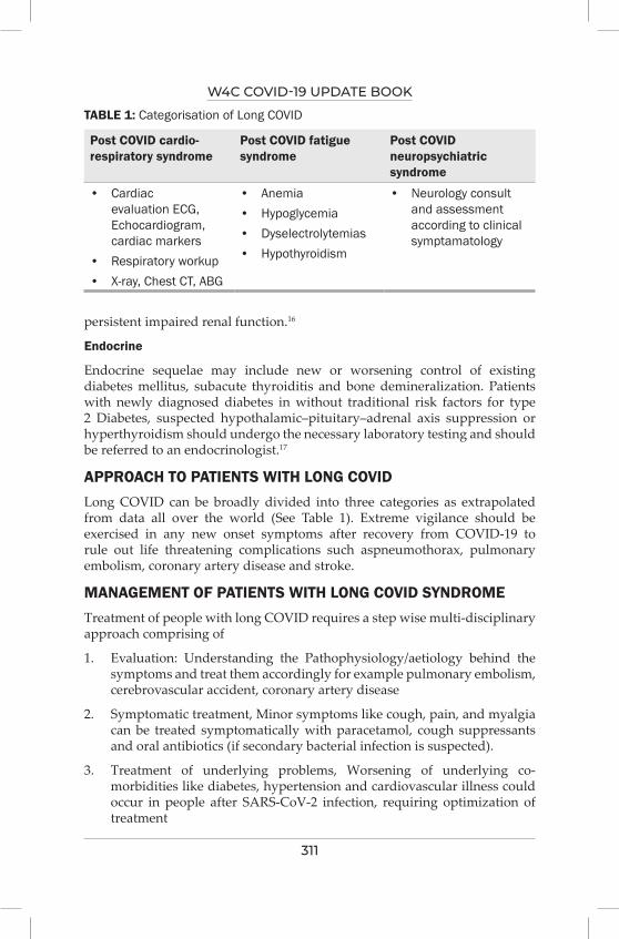
W4C COVID-19 UPDATE BOOK
311
persistent impaired renal function.16
Endocrine
Endocrine sequelae may include new or worsening control of existing diabetes mellitus, subacute thyroiditis and bone demineralization. Patients with newly diagnosed diabetes in without traditional risk factors for type 2 Diabetes, suspected hypothalamic–pituitary–adrenal axis suppression or hyperthyroidism should undergo the necessary laboratory testing and should be referred to an endocrinologist.17
APPROACH TO PATIENTS WITH LONG COVIDLong COVID can be broadly divided into three categories as extrapolated from data all over the world (See Table 1). Extreme vigilance should be exercised in any new onset symptoms after recovery from COVID-19 to rule out life threatening complications such aspneumothorax, pulmonary embolism, coronary artery disease and stroke.
MANAGEMENT OF PATIENTS WITH LONG COVID SYNDROMETreatment of people with long COVID requires a step wise multi-disciplinary approach comprising of
1. Evaluation: Understanding the Pathophysiology/aetiology behind the symptoms and treat them accordingly for example pulmonary embolism, cerebrovascular accident, coronary artery disease
2. Symptomatic treatment, Minor symptoms like cough, pain, and myalgia can be treated symptomatically with paracetamol, cough suppressants and oral antibiotics (if secondary bacterial infection is suspected).
3. Treatment of underlying problems, Worsening of underlying co-morbidities like diabetes, hypertension and cardiovascular illness could occur in people after SARS-CoV-2 infection, requiring optimization of treatment
TABLE 1: Categorisation of Long COVID
Post COVID cardio-respiratory syndrome
Post COVID fatigue syndrome
Post COVID neuropsychiatric syndrome
• Cardiac evaluation ECG, Echocardiogram, cardiac markers
• Respiratory workup• X-ray, Chest CT, ABG
• Anemia• Hypoglycemia• Dyselectrolytemias• Hypothyroidism
• Neurology consult and assessment according to clinical symptamatology
W4C COVID-19 UPDATE BOOK
312
4. Physiotherapy, occupational therapy and psychological support.
5. Severe COVID-19 causes, malnutrition 26–45% catabolic muscle wasting, feeding difficulties and frailty, thereby increasing the likelihood of poor outcome. Treatment should include nutritional protocols individualised to every patient’s needs.18
The ideal frequency and duration of follow upis still not clearly delineated. In people with COVID-19 interstitial pneumonia, in the first 12 months, alongside 4 HRCTs, 4-6MWT, 4 blood tests (including blood count and metabolic panel) and 2 SARS-CoV-2-IgG tests should be conducted.18
CONCLUSION Post COVID 19 Syndrome is a nonspecific entity, recently being researched upon, and lacks evidence-based guidelines and consensus on management. It has been surmised that even when COVID-19 pandemic will be a thing of the past, morbidity will persist due to this syndrome causing heavy burden on medical setup and society. Its management requires a multi-disciplinary approach involving clinicians, social groups, and direct care providers.
SUMMARY1. Covid 19 is a perplexing disease with florid and variegated systemic
manifestations involving respiratory cardiovascular neurological gastrointestinal endocrinological and haematological symptoms predominantly.
2. Even after resolution of primary symptoms , nefarious symptoms may persist and cause serious complications
3. Active and early intervention, regular follow ups and investigations according to the clinical scenario is mandatory
4. Since we are still learning about this disease, no new symptom should be discounted.
REFERENCES1. Wu Z, McGoogan JM. Characteristics of and important lessons from the coronavirus disease
2019 (COVID-19) outbreak in China: summary of a report of 72314 cases from the Chinese Center for Disease Control and Prevention. JAMA 2020; 323:1239-42.
2. McElvaney, OJ, McEvoy NL, McElvaney OF, Carroll TP, Murphy MP, Dunlea DM et al. Characterization of the infammatory response to severe COVID-19 Illness. Am J Respir Crit Care Med 2020; 202:812–21.
3. Tang, N., Li, D., Wang, X., Sun, Z. Abnormal coagulation parameters are associated with poor prognosis in patients with novel coronavirus pneumonia. J Tromb Haemost 2020; 18:844–47.
4. Felicity Callard, Elisa Perego. How and why patients made Long Covid. Soc Sci Med 2021; 268: 113426.
5. Assaf G, Davis H, McCorkell L, et al. What does COVID-19 recovery actually look like? 11 May 2020. https://patientresearchcovid19.com/research/report-1/ (Last access on 1 December 2020)
W4C COVID-19 UPDATE BOOK
313
6. Garg P., Arora U., Kumar A., Wig N. The “post-COVID” syndrome: how deep is the damage? J Med Virol. 2021; 93:673-74.
7. Van Elslande J., Vermeersch P., Vandervoort K., Wawina-Bokalanga T., Vanmechelen B., Wollants E et al. Symptomatic SARS-CoV-2 reinfection by a phylogenetically distinct strain. Clin Infect Dis 2020; 5.
8. Carfì A., Bernabei R., Landi F. Persistent symptoms in patients after acute COVID-19. J Am Med Assoc 2020; 324:603–05.
9. Arnold DT, Hamilton FW, Milne A, Morley AJ, Viner J, Attwood M. Patient outcomes after hospitalisation with COVID-19 and implications for follow-up: results from a prospective UK cohort thorax Published Online First: 03 December 2020.
10. Lopez-Leon S., Wegman-Ostrosky T., Perelman C., Sepulveda R., Rebolledo P.A., Cuapio A., et al. More than 50 Long-term effects of COVID-19: a systematic review and meta-analysis. medRxiv 2021.
11. Becker R.C. Toward understanding the 2019 Coronavirus and its impact on the heart. J Thromb Thrombolysis 2020; 50:33-42.
12. Gerwyn M., Maes M. Mechanisms explaining muscle fatigue and muscle pain in patients with myalgic encephalomyelitis/chronic fatigue syndrome (ME/CFS): a review of recent findings. Curr Rheumatol Rep 2017; 19:1.
13. Townsend L., Dyer A.H., Jones K., Dunne J., Mooney A., Gaffney F. Persistent fatigue following SARS-CoV-2 infection is common and independent of severity of initial infection. PloS One 2020; 15.
14. Bikdeli, B, Madhavan MV, Jimenez D, Chuich T, Dreyfus I, Driggin et al. COVID-19 and thrombotic or thromboembolic disease: implications for prevention, antithrombotic therapy, and follow-up: JACC state-of-the-art review. J. Am. Coll. Cardiol 2020; 75:2950–73.
15. Malik G.R., Wolfe A.R., Soriano R., Rydberg L., Wolfe L.F., Deshmukh S.,et al. Injury-prone: peripheral nerve injuries associated with prone positioning for COVID-19-related acute respiratory distress syndrome. Br J Anaesth 2020; 125:e478–e480.
16. Stevens, JS.,King KL.,Robbins-Juarez SY., Khairallah P., Toma K., Verduzco HA., et al. High rate of renal recovery in survivors of COVID-19 associated acute renal failure requiring renal replacement therapy. PLoS One 2020; 15:e0244131.
17. Suwanwongse, K., Shabarek, N. Newly diagnosed diabetes mellitus, DKA, and COVID-19: causality or coincidence? A report of three cases. J Med Virol 2020.
18. Huang C, Huang L, Wang Y, Li Xia, Ren Lili, Gu X, et al. 6-month consequences of COVID-19 in patients discharged from hospital: a cohort study. Lancet 2021; 397:220–32.
19. Raghu G., Wilson K.C. COVID-19 interstitial pneumonia: monitoring the clinical course in survivors. Lancet Respir Med 2020; 8:839-42.
314
Post-COviD 19 Syndrome 41.Abhijeet Raha, Alisha Chaudhury
INTRODUCTIONCoronavirus disease 19 (COVID 19) disease persists to be a matter of grave concern in multiple aspects. Only about 5% of all COVID patients become critically ill. Recovery time from this illness varies from 2-6 weeks.1 Though the period of infectivity requires utmost attention and care the focus is now shifting to the post COVID phase which has become more protracted and challenging to manage.
DEFINITIONPost-COVID Syndrome is an umbrella term that has been synonymous with long haulers, post-acute sequelae of SARS-CoV-2 infection” (PASC), “long-COVID-19”, “post-acute COVID-19”.2 Defined as symptoms persisting or new occurrence beyond 4 weeks from the initial symptom onset. The definition is still evolving and can be phrased as:3
1. Subacute or ongoing symptomatic COVID 19: symptoms are present 4-12 weeks post the acute phase
2. Chronic or post-COVID 19 syndrome: residual signs and symptoms present beyond 12 weeks which cannot be attributed to an alternative diagnosis
There are no clear guidelines for the management of post COVID syndrome by IDSA and/or CDC as yet. NICE guidelines published in December 2020 are the last updated guidelines for managing post-COVID syndrome.4
PATHOGENESISMechanisms involved in the pathogenesis and progression of post COVID syndrome are still not clear and theoretically could be due to:5
1. Sequelae of organ damage (e.g., pulmonary fibrosis, chronic kidney disease)
2. Prolonged inflammatory and immunological alterations associated with damage
W4C COVID-19 UPDATE BOOK
315
3. Other prolonged critical illness effects – post-intensive care syndrome, hospitalization, and social isolation
Another plausible explanation for the post COVID syndrome is its similarity in presentation to the “myalgic encephalomyelitis/chronic fatigue syndrome” (ME/CFS) seen in the post-infective phase of SARS caused by coronavirus in 2002.6 This syndrome is due to autoantibodies against the Beta 2 receptor and the M3 acetyl cholinic receptors. This leads to reversal of receptor actions, smooth muscle involvement causing extreme fatigue with pain, neurocognitive disability compromised sleep, symptoms suggestive of autonomic dysfunction, and worsening of global symptoms like brain fog.7,8 The reason for this varied neurological presentation is because of the neurotropic property of the virus affecting both the central and peripheral nervous system.9 Therefore, more research is needed to bring light to the pathogenesis of COVID 19 for a better understanding of these entities.
CLINICAL PRESENTATION AND MANAGEMENT PRINCIPLESThe five most common effects based on various systematic reviews and meta-analyses were fatigue (58%), headache (44%), attention disorder (27%), hair loss (25%), and dyspnea (24%).10 There are no specific diagnostic tests for post COVID syndrome. Rapid antigen or RT-PCR can be used to differentiate an acute condition but it’s not specific. Antibody tests could be useful in this case.
1. Constitutional: Most commonly reported were fatigue, loss of smell or taste, giddiness. Females reported more fatigue as compared to males based on case reports from France which was often disproportionate to the symptoms.11
2. Pulmonary:12
• A wide spectrum of symptoms – dyspnea, persistent disabling cough
• Subset of severe COVID (those who required high flow nasal cannula and noninvasive ventilation or invasive) experience more pulmonary complications
• Difficult weaning with ventilatory parameters showing high respiratory rate and minute ventilation
• CT thorax (most common findings): ground-glass opacities interspersed with extensive areas of fibrosis along with traction bronchiectasis
• Pulmonary function tests – poor lung reserves denoted by reduced lung volumes, forced vital capacity, poor performance of 6-minute walk tests
• Echo findings – a gradual increase in pulmonary artery pressures
W4C COVID-19 UPDATE BOOK
316
suggestive of pulmonary hypertension secondary to the chronic hypoxic state
• Long term risks of chronic pulmonary embolism states
• Persistent hypoxia requiring supplemental oxygen and, in some cases, noninvasive support to alleviate the work of breathing
3. Hematologic: Studies have reported rates of venous thromboembolism <5% mostly as segmental emboli, intracardiac thrombus etc13
4. Cardiovascular:14
• Most commonly, chest pain with palpitations
• Stress cardiomyopathy
• Frequent arrhythmias – possibly due to myocardial scarring
5. Neuropsychiatric:15,16
• Depressive state of mind, sleep disturbances
• Chronic malaise, myalgia, fatigue
• Migraine headaches and sometimes late-onset headaches
• Impairment in cognition is seen as “brain fog” – difficulty in concentrating, memory, comprehension, and behavioral disturbances
• ICU delirium and post-traumatic stress disorder (PTSD)
• Critical illness myopathies and neuropathies secondary to prolonged use of neuromuscular blocking agents, steroids and bedridden state, electrolyte imbalances
• Cerebrovascular events were reported in 62% of patients, and31% experienced altered mental status related to encephalopathy with evidence of CNS inflammation17
6. Renal:18
• Severe acute COVID-19 subset accounting up to 5% end up undergoing renal replacement therapy
• A new onset decrease in eGFR has been reported
• High mortality rates have been seen in the subset of patients undergoing RRT but renal recovery has been reported in 84% of the survivors
7. Endocrine:
• Diabetic ketoacidosis can be a late presenting feature post-resolution
W4C COVID-19 UPDATE BOOK
317
of COVID even without any underlying diabetes19
• Subacute thyroiditis with clinical thyrotoxicosis has also been reported post-resolution of symptoms20
8. Gastrointestinal and hepatobiliary:
• Most commonly anorexia is seen with fatigue and malnourishment21
• Can alter the gut microbiome
• Pain abdomen, nausea, diarrhea
• Less commonly reported biliary stasis and irritable bowel syndrome
• Bowel ischemia due to thrombosis
9. Dermatology:
• 5-15% incidence
• Most common complaint was hair loss (20%) – secondary to telogen effluvium (Stress-Induced)
• Skin rashes -Five cutaneous patterns reported are maculopapular, urticarial, pseudo chilblain, vesicular, and livedoid22
• The livedoid pattern denotes underlying thrombosis
10. Nutrition and Rehabilitation:
• Long COVID is frequently associated with severe malnutrition
• Keys are frequent assessment of nutritional status, replenish the deficits, and planned nutritional support
• Detailed information provided in ESPEN guidelines for the nutritional management of COVID 19 patients have also been published23
MANAGEMENTOverall, the key to managing this problem is the promotion of multidisciplinary COVID 19 clinics as well multidisciplinary approaches to critically ill patients. Additional support should be considered for the most vulnerable population groups – children and the elderly. There is a need for a comprehensive management plan to focus on improving the physical, mental, and social wellbeing of patients. The Figure 1 demonstrates a multidisciplinary approach for the management of post COVID syndrome.
MULTISYSTEM INFLAMMATORY SYNDROME24 (MIS)Frequently seen both in adults and children. CDC has separated both entities and defined them. They have been divided based on the age group – MIS

W4C COVID-19 UPDATE BOOK
318
A (>21 years) and MIS C (<21 years). We shall discuss MIS-A here, MIS-C is discussed elsewhere in the book.
MIS -A25 is defined as:
• A patient aged ≥21 years hospitalized for ≥24 hours, or with an illness resulting in death, who meets the following clinical and laboratory criteria. The patient should not have a more likely alternative diagnosis for the illness (e.g., bacterial sepsis, exacerbation of a chronic medical condition).
• Clinical Criteria
Subjective fever or documented fever (≥38.0 C) for ≥24 hours before hospitalization or within the first THREE days of hospitalization* and at least THREE of the following clinical criteria occurring before hospitalization or within the first THREE days of hospitalization*. At least ONE must be a primary clinical criterion.
A. Primary clinical criteria
a. Severe cardiac illness Includes myocarditis, pericarditis, coronary artery dilatation/aneurysm, or new-onset right or left ventricular dysfunction (LVEF<50%), 2nd/3rd degree A-V block, or ventricular tachycardia. (Note: cardiac arrest alone does not meet this criterion)
b. Rash AND non-purulent conjunctivitis
B. Secondary clinical criteria
a. New-onset neurologic signs and symptoms Include
Primary care
Pulmonary
Cardiovascular
Neuropsychiatric
• Physiotherapistguidedrehab programs
• Strengthening, flexibility exercises, aerobic exercises,
• Standardized questionnaire-based assessment regarding quality of life
• Monitor for breathlessness and muscle aches
• Respiratory muscle strengthening exercises, cough assist maneuvers,
• Radiological assessment and pulmonary function tests, 6MWT after 12 weeks
• Vaccination
• HRCT chest if indicated
• Regular ECGs and ECHO at 4-12 weeks
• No clear practice statement for routine cardiac MRI
• RAAS inhibitors safe for use
• Arrhythmias - low dose beta blockers but amiodarone use with caution
• Screening for anxiety, depression, PTSD, sleep disturbances and cognitive impairment
• Referral to the appropriate specialty
• Detailed evaluation and counselling
• Lifestyle changes
Endocrine
• Workup is mandatory in newly detected cases of diabetes without any underlying risk factors
• Steroids may have a role in thyroiditis
Hematologic
• Workup for PE
• LMWH or DOACS preferred
• Early mobilization and physiotherapy
Renal
• Early and close follow up at nephrology clinics
• Rates of renal recovery still unclear
FIG. 1: Multidisciplinary Approach for Management of Post-Covid Syndrome
W4C COVID-19 UPDATE BOOK
319
encephalopathy in a patient without prior cognitive impairment, seizures, meningeal signs, or peripheral neuropathy (including Guillain-Barré syndrome)
b. Shock or hypotension not attributable to medical therapy (e.g., sedation, renal replacement therapy)
c. Abdominal pain, vomiting, or diarrhea
d. Thrombocytopenia (platelet count <150,000/ microliter)
• Laboratory evidence
A. The presence of laboratory evidence of inflammation AND SARS-CoV-2 infection.
B. Elevated levels of at least TWO of the following: C-reactive protein, ferritin, IL-6, erythrocyte sedimentation rate, procalcitonin
C. A positive SARS-CoV-2 test during the current illness by RT-PCR, serology, or antigen detection
Treatment is largely supportive care and control of the inflammatory process. No particular modality has been proven superior. Steroids have been used for anti-inflammatory action along with anticoagulants since they are known to be prothrombotic states. Other immunomodulatory therapies like IvIg have been used. Aspirin has also been used because of the underlying coronary involvement.
PRACTICE POINTS• Long-term effects OF Covid-19 are poorly understood
• Clinical suspicion, vigil, and proactive treatment steps are quintessential
• The burden can be immense on the healthcare delivery systems, the society, and the economy as well as the health of the population
• A multidisciplinary approach is a key to appropriate management
REFERENCES1. Dong E., Du, H. & Gardner, L. An interactive web-based dashboard to track COVID-19 in real-
time. Lancet Infect Dis 2020; 20:533–534.2. Rubin R. As their numbers grow, COVID-19 “long haulers” stump experts. JAMA 2020;
324:1381-1383.3. Shah, W., Hillman, T., Playford, E. D. & Hishmeh, L. Managing the long-term effects of
COVID-19: summary of NICE, SIGN, and RCGP rapid guideline. Brit Med J 2021; 372, n136.4. COVID-19 rapid guideline: managing the long-term effects of COVID-19. www.nice.org.uk/
guidance/ng188.5. Garg P, Arora U, Kumar A, Wig N. The “post-COVID” syndrome: how deep is the damage? J
Med Virol 2021; 93:673-674.
W4C COVID-19 UPDATE BOOK
320
6. National Institutes of Health. NIH intramural study on myalgic encephalomyelitis/chronic fatigue syndrome. Accessed March 20, 2021. https://mecfs.ctss.nih.gov
7. Wirth K, Scheibenbogen C. A unifying hypothesis of the pathophysiology of myalgic encephalomyelitis/chronic fatigue syndrome (ME/CFS): recognitions from the finding of autoantibodies against ß2-adrenergic receptors. Autoimmun Rev 2020; 19:102527.
8. Scheibenbogen C, Loebel M, Freitag H, et al. Immunoadsorption to remove ß2 adrenergic receptor antibodies in chronic fatigue syndrome CFS/ME. PLoS One 2018; 13:e0193672.
9. Iadecola C, Anrather J, Kamel H. Effects of COVID-19on the nervous system. Cell 2020; 183:16-27.
10. Lopez-Leon, S., Wegman-Ostrosky, T., Perelman, C. et al. More than 50 long-term effects of COVID-19: a systematic review and meta-analysis. Sci Rep 2021; 11:16144. https://doi.org/10.1038/s41598-021-95565-8
11. Halpin SJ, McIvor C, Whyatt G, et al. Post-discharge symptoms and rehabilitation needs in survivors of COVID-19 infection: a cross-sectional evaluation. J Med Virol 2021; 93:1013-1022. doi:10.1002/jmv.26368.
12. Garrigues, E. et al. Post-discharge persistent symptoms and health-related quality of life after hospitalization for COVID-19. J Infect 2020; 81: e4–e6.
13. Patell, R. et al. Post-discharge thrombosis and hemorrhage in patients with COVID-19. Blood 2020; 136:1342–1346.
14. Jabri, A. et al. Incidence of stress cardiomyopathy during the coronavirus disease 2019 pandemic. JAMA Netw Open 2020; 3:e2014780.15. Rogers JP, Chesney E, Oliver D, et al. Psychiatric and neuropsychiatric presentations associated
with severe coronavirus infections: a systematic review and metanalyses with comparison to the COVID-19 pandemic. Lancet Psychiatry 2020; 7:611-627.
16. Banerjee D, Viswanath B. Neuropsychiatric manifestations of COVID-19 and possible pathogenic mechanisms: insights from other coronaviruses. Asian J Psychiatr 2020; 54:102350.
17. Varatharaj A, Thomas N, Ellul MA, et al. Neurological and neuropsychiatric complications of COVID-19 in 153 patients: a UK-wide surveillance study. Lancet Psychiatry 2020; 7:875-882.
18. Robbins-Juarez, S. Y. et al. Outcomes for patients with COVID-19 and acute kidney injury: a systematic review and meta-analysis. Kidney Int. Rep.5, 1149–1160 (2020).
19. Rubino, F. et al. New-onset diabetes in COVID-19. N Engl J Med 2020; 383:789–790.20. Ruggeri, R. M., Campenni, A., Siracusa, M., Frazzetto, G. & Gullo, Subacute thyroiditis in
a patient infected with SARS-COV-2: an endocrine complication linked to the COVID-19 pandemic. Hormones (Athens) 2021; 20:219–221.
21. Cheung, K. S. et al. Gastrointestinal manifestations of SARS-CoV-2 infection and virus load in fecal samples from a Hong Kong cohort: systematic review and meta-analysis. Gastroenterology 2020; 159:81–95.
22. Candan SA, Elibol N, Abdullahi A. Consideration of prevention and management of long-term consequences of post-acute respiratory distress syndrome in patients with COVID-19. Physiother Theory Pract 2020; 36:663-668.
23. Barazzoni R, Bischoff SC, Breda J, et al. ESPEN expert statements and practical guidance for nutritional management of individuals with SARS-CoV-2 infection. Clin Nutr 2020; 39:1631-1638. DOI: 10.1016/j.clnu.2020.03.022.
24. Jiang, L. et al. COVID-19 and multisystem inflammatory syndrome in children and adolescents. Lancet Infect Dis 2020; 20:e276–e288.
25. https://www.cdc.gov/mis/mis-a.html. 25 June 2021. Multisystem Inflammatory Syndrome.
321
COviD Associated Mould infection –
a Double Whammy42.
The pandemic of coronavirus disease (COVID-19) continues to be a devastating nightmare for the world today. Since the virus is a novel one, several treatment options have been tried. From among them only glucocorticoids have been shown to improve survival especially in hypoxemic patients. Unfortunately, because of the collapse of health care system especially in the second wave the widespread use of glucocorticoids lead to secondary bacterial or fungal infections. Invasive pulmonary aspergillosis complicating the course of COVID-19 has been reported earlier.1 However, the deluge of mucormycosis in covid patients was totally unanticipated.
Invasive mould infections (invasive aspergillosis and mucormycosis) share similar risk factors, clinical presentation, and radiology and we will confine our discussion to these two entities.
COVID ASSOCIATED INVASIVE PULMONARY ASPERGILLOSIS (CAPA)COVID associated Invasive Pulmonary Aspergillosis (CAPA) is being increasingly reported in patients with COVID-19 disease and increases the risk of mortality several fold.1,2 A similar phenomenon was noticed in Influenza associated Pulmonary Aspergillosis (IAPA).3 In both the conditions the classic host factors for invasive fungal infection were absent. However, in CAPA there is a debate if coronavirus (SARS-CoV-2) infection is the main risk factor, or whether corticosteroid therapy, makes these apparently immunocompetent patients prone to develop the disease.4
Aspergillus spp. usually cause cavitatory lesions in the lungs and brain. Outbreaks of Aspergillus have been linked to poor air filtration, construction, and even contaminated medical equipment. The diagnosis of invasive Aspergillosis in ICU patients is difficult with the reported prevalence ranging from 0.335% to 6.9%.5 This wide range may also be due to lack of post-mortem reports confirming the presence of the disease. The presentation of
Prakash Shastri
W4C COVID-19 UPDATE BOOK
322
invasive pulmonary aspergillosis (IPA) is non-specific usually mimicking bronchopneumonia. The classical ‘halo’ sign seen on CT scan of the thorax is rare in immunocompetent hosts. Absence of ‘halo’ sign and the avoidance of bronchoscopy (being an aerosol producing intervention) makes the diagnosis even more challenging.
Recently a consensus statement has been published on defining and managing COVID-19-associated pulmonary aspergillosis, prepared by experts and endorsed by medical mycology societies.6 COVID-19-associated pulmonary aspergillosis is proposed to be defined as possible, probable, or proven on the basis of sample taken from a sterile site or biopsy.
Radiological and clinical signs of invasive pulmonary aspergillosis in non-neutropenic patients are nonspecific and broncho-alveolar lavage (BAL) is indicated with fungal diagnostic work-up. Even if Aspergillus is recovered, one cannot be certain that patients has invasive disease, the respiratory system being a non-sterile site. Besides Galactomannan (GM), the other commercially available assay is β-(1, 3)-d-glucan (BDG). Molecular techniques using fungal nucleic acid detection, have not been validated and have not reached clinical practice.
Recommended first-line therapy is either voriconazole or isavuconazole. If azole resistance is a concern, then liposomal amphotericin B is the drug of choice.6
During this COVID-19 pandemic, we have realised that aspergillus can cause co-infection with SARS-CoV-2 despite the absence of the traditional risk factor of aspergillus infection. The outcome of COVID-19-associated pulmonary aspergillosis is poor and the recommend antifungal agent – voriconazole should be used with caution because of complicated drug–drug interaction and enhanced cardiovascular toxicity of repurposed drugs used to treat SARS-CoV-2 infection.
COVID ASSOCIATED MUCORMYCOSIS (CAM)A few centres especially from India have also reported an alarming increase in COVID- associated mucormycosis (CAM).7 The clinical presentations of mucormycosis are classified on the basis of anatomic localisation, such as rhino-orbital-cerebral (ROCM), pulmonary, gastro-intestinal, cutaneous, renal, and disseminated mucormycosis. While we know that uncontrolled diabetes is a risk factor for mucormycosis, the entity Covid associated mucormycosis (CAM) was hitherto unknown.
Mucorales are saprophytic fungi and are ubiquitous in the environment. The most common risk factors are diabetes mellitus, immunosuppressive therapy and neutropenia. Diabetic ketoacidosis, hematopoietic stem cell transplantation, iron-overload and immunodeficiency states including HIV/AIDs are some other risk factors. Rhino orbital mucormycosis is the most
W4C COVID-19 UPDATE BOOK
323
commonly encountered form in clinical practice. The genus Rhizopus account for the majority of clinical isolates.
The diagnosis of CAM is even more challenging. The Clinicians are not sensitized enough to suspect it. Furthermore it is difficult to isolate the causative fungi. Finally, biomarkers such as beta-d-glucan and galactomannan, which aid in diagnosing invasive aspergillosis, are not useful for diagnosing mucormycosis. Diabetes mellitus has been associated with severe COVID-19. Poorly controlled diabetic patients may have overt or covert renal dysfunction. The presence of multiple risk factors or comorbid illnesses in severe COVID-19 patients, along with the additional immunosuppression caused by glucocorticoids, increases the net state of immune suppression, thereby predisposing them to invasive mould infections. The current guideline for the management of mucormycosis recommends liposomal amphotericin B at a dose of 5–10 mg/kg per day. In the absence of central nervous system involvement, a dose of 3-5 mg/kg is suggested.8 The optimal duration of therapy in mucormycosis is not clear and is guided by the treatment regimen’s clinical response and tolerability.
In our institution we have devised a system based on MRI findings that helps us in decision making. ‘Fungal Signal’ in the context of mucormycosis is said to be present when any two of the following are present:
a. Hypo intense signal from tissue in T2W images
b. Diffusion restriction in DW images.
c. Non Enhancement or increased enhancement in Contrast enhanced images.
Those cases in whom the criteria of fungal signal was fulfilled for both the sinuses and orbit underwent simultaneous FESS and exenteration of eye.
If clear clinical picture of mucormycosis exists, positive report of nasal biopsy taken endoscopically will give the report from KOH mount within an hour. Culture and HPE reports are not required for making the decision for intervention. Clinical findings and imaging plays an important role in defining the extent of involvement. Liposomal Amphotericin B is the preferred treatment in the dose of 3-5mg/kg/day as slow infusion. After 3-4 weeks of amphotericin B therapy, consolidation therapy with azoles (posaconazole or Isavuconazole) is continued for 3-6 months.7
The incidence of mucormycosis has risen 2.1 times that in the year 2020 as per a recently published Indian study.9 This study concluded that poor sugar control, high dose steroid therapy and compromised immunity due to immune dysregulation and hyper-inflammation in Covid-19 patients predisposes these patients to the deadly fungal infection.
W4C COVID-19 UPDATE BOOK
324
REFERENCES1. Arastehfar A, Carvalho A, van de Veerdonk FL, Jenks JD, Koehler P, Krause R, et al. COVID-19
associated pulmonary aspergillosis (CAPA)-from immunology to treatment. J Fungi (Basel). 2020; 6:91.
2. Koehler P, Cornely OA, Böttiger BW, Dusse F, Eichenauer DA, Fuchs F, et al. COVID-19 associated pulmonary aspergillosis. Mycoses 2020; 63:528–34.
3. Schauwvlieghe AFAD, Rijnders BJA, Philips N, Verwijs R, Vanderbeke L, Van Tienen C, et al. Invasive aspergillosis in patients admitted to the intensive care unit with severe influenza: a retrospective cohort study. Lancet Respir Med 2018; 6:782–92.
4. Wahidi MM, Lamb C, Murgu S, Musani A, Shojaee S, Sachdeva A, et al. American Association for Bronchology and Interventional Pulmonology (AABIP) Statement on the Use of Bronchoscopy and Respiratory Specimen Collection in Patients with Suspected or Confirmed COVID-19 Infection. J Bronchol Interv Pulmonol 2020; 27:e52–4.
5. Patterson TF, Thompson GR, 3rd, Denning DW, et al. Practice Guidelines for the Diagnosis and Management of Aspergillosis: 2016 Update by the Infectious Diseases Society of America. Clin Infect Dis 2016;63
6. Koehler P, Bassetti M, Chakrabarti A, Chen SCA, Colombo AL, Hoenigl M, et al. Defining and managing COVID-19-associated pulmonary aspergillosis: the 2020 ECMM/ISHAM consensus criteria for research and clinical guidance. Lancet Infect Dis 2021;3099.
7. Garg D, Muthu V, Sehgal IS, Ramachandran R, Kaur H, Bhalla A, et al. Coronavirus Disease (Covid-19) Associated Mucormycosis (CAM): Case Report and Systematic Review of Literature. Mycopathologia 2021:1-10.
8. Cornely OA, Alastruey-Izquierdo A, Arenz D, Chen SCA, Dannaoui E, Hochhegger B et al. Mucormycosis ECMM MSG Global Guideline Writing Group. Global guideline for the diagnosis and management of mucormycosis: an initiative of the European Confederation of Medical Mycology in cooperation with the Mycoses Study Group Education and Research Consortium. Lancet Infect Dis 2019; 9:e405-e421.
8. Patel A, Agarwal R, Rudramurthy SM, Shevkani M, Xess I, Sharma R, et al. Multicenter epidemiologic study of corona-virus disease-associated mucormycosis India. Emerg Infect Dis 2021; 27
325
Effects of Prolonged Hospitalization in COviD-19
infection43.
Prolonged length of stay is defined as hospitalization longer than median length of hospital stay.1 Various studies have shown multiple reasons for longer hospital stays in these patients including disease severity, glucocorticoid use, female sex, older age, fever on admission, pre-existing chronic kidney, and liver disease, increasing creatinine levels etc. However, a recent systemic review found little impact of age or disease severity, although older age has been found to be a significant independent factor of mortality.2 Common complications in COVID-19 disease result from presence of a pro-inflammatory state with cytokine release, and include ARDS, acute kidney injury (AKI), hypercoagulable state with thrombotic events, myocardial injury, and sepsis. Cardiopulmonary symptoms range from dyspnea, orthostatic hypotension, arrhythmias to reduced aerobic capacity.3 The average stay of such patient is about three weeks in intensive care unit (ICU).
In addition to the pharmacological treatment, bed rest is often suggested to reduce metabolic demands and fasten recovery. Bed rest and prolonged immobilization can cause muscle wasting, loss of muscle strength and muscle protein. This may result in impaired balance, strength, and sensation.4 The neurological sequelae reported include critical illness polyneuropathy, stroke, cognitive deficits affecting memory, problem solving and delirium. These patients may also suffer from meningitis or encephalitis. Dysphagia may result in prolonged need for enteral feeding. Such patients may remain intubated or get tracheostomized. They may have bed sores not only on sacrum and ankle but also on face and abdomen due to proning. The functional disability and persistence of symptoms negatively impacts the quality of life in these patients.
At present we have limited information on long term effects of Covid-19 infection.4 A retrospective observational 6-month follow up study of hospitalized patients after discharge looked at the presence of sequelae or persistent symptomatology (SPS) during the 6 months after discharge.3 It
Rajesh Kumar Pande, Maitree Pandey
W4C COVID-19 UPDATE BOOK
326
found that the most frequent were respiratory (42.0%), systemic (36.1%), neurological (20.8%), mental health (12.2%) and infectious (7.9%) symptoms and about 20% patients returned to the emergency for persistent fever, dermatological SPS, arrythmia or palpitations, thoracic pain and pneumonia, with 4.4% getting readmitting and 8% dying during follow up.3
Most reported symptoms after 2-months of discharge include fatigue and dyspnea followed by impaired pulmonary function and chest image abnormalities. The general term Post-Intensive Care Syndrome (PICS) is used to describe both complications and functional consequences of long-term hospitalization in critically ill patients. Weakness, deconditioning, cognitive dysfunction, and psychiatric illness that persists after discharge describe PICS.4 The Post COVID Syndrome or long COVID is similar to PICS.
COVID-19 illness has recently been classified as being acute- upto 4 weeks from the onset of symptoms (PCR positive), and the persistence of symptoms and/or delayed or long-term complications beyond 4 weeks (PCR negative) have been classified as Post-acute COVID-19. Post-acute COVID-19 is again labeled as subacute COVID-19 (from 4 to 12 weeks) and chronic COVID-19 (from 3 to 6 months).5 The mechanism of post-acute COVID-19 syndrome is not well understood but is hypothesized to be secondary to virus-specific pathophysiologic changes, prolonged inflammatory response to the acute infection, and sequelae of post-intensive care illness.6
A recent, large, 3-month post Covid discharge follow up study from China found that 76% of patients reported at least one symptom, and a higher percentage was observed in women. The most common symptom after discharge was fatigue or muscle weakness and sleep difficulties. The seropositivity and titres of the neutralizing antibodies were significantly lower compared to the acute phase. Symptoms persisted upto 6-months after symptom onset. A higher severity score (a 7-point severity scale measured, by admission to ICU, and/or requirement for non-invasive and/or invasive mechanical ventilation) during hospitalization was associated with a higher risk of pulmonary diffusion abnormalities, fatigue or muscle weakness, anxiety, or depression, highlighting the need for post discharge care in severe disease.7
A retrospective study looking at functional outcome in hospitalized patients with severe covid-19 infection, who underwent inpatient rehabilitation, found persistent functional deficit in domains of fall risk, gait speed and cognition at rehab discharge necessitating ongoing treatment, suggesting longer term impairments.4
Prolonged immobility, use of corticosteroids, immune-modulatory agents and age-related sarcopenia could probably be responsible for accelerated loss of muscle mass and prolonged weakness and fatigue persisting after discharge following COVID-19 infection.4 The use of corticosteroids for treating ARDS in SARS-CoV infections was found to contribute to muscle weakness and
W4C COVID-19 UPDATE BOOK
327
decreased functional capacity. Corticosteroids impair excitability of muscle fibre, decrease in muscle filaments, and not only reduce protein synthesis but also increase protein degradation.
Muscle weakness is a well-known chronic side effect of long-term glucocorticoids therapy. Use of prednisone or its analogues in dose > 10mg/day for a few weeks can induce steroid myopathy. The muscle weakness and functional decline in severe covid patients post discharge could be multifactorial including the use of high dose methylprednisolone pulse therapy, polymyxins for resistant infections, control or assist control ventilation and the use of neuro muscular blocking agents for prolonged periods.
Early rehabilitation by using passive mobilization exercises and anti-gravity position changes can prevent deconditioning. Early strength interventions in form of resistive exercise programmes targeting lower limb muscles like gluteus, quadriceps, hip flexors and hip adductors and abductors are helpful. For patients who cannot perform resistive exercises, neuromuscular electrical stimulation is recommended. Heat therapy has not been found to change the muscle architecture but can help in alleviating the muscular pain associated with inflammatory processes.8
Follow up studies have found symptom persistence post discharge and have suggested for longitudinal follow-up of individuals with severe COVID-19.9 These patients need follow up visits (in person or virtual) for symptom assessment at 4-6 weeks and at 12 weeks. If they have persistent dyspnea/oxygen requirement, they should undergo endurance (6-minute walk test) and gait speed tests (10-meter walk test), besides chest x-ray, workup for pulmonary embolism, pulmonary function tests (PFT), echocardiogram (2D-ECHO) and high-resolution computed tomography of chest (HRCT) as indicated. Post discharge extended thromboprophylaxis in high-risk patients should be considered on individual basis. During follow-up visits patient should also undergo neuropsychiatric evaluation for anxiety, depression, post traumatic stress disorder (PTSD), cognitive dysfunction and sleep disturbances.
SUMMARYOur knowledge regarding long term effects of COVID-19 infection is very limited. The patients with severe disease should be followed up after discharge to look at their functional status and symptom/organ recovery and to provide them with necessary care. Functional monitoring during rehabilitation in these patients should include fall risk, ambulation independence, transfer independence besides the 6MWT and 10MWT.
W4C COVID-19 UPDATE BOOK
328
PRACTICE POINTS• Patients with severe COVID-19 infection need follow-up post discharge
upto 3-6 months.
• Muscle weakness, PICS should be kept in mind for long stay patients.
• Fatigue, muscle weakness, anxiety, depression, PTSD are common problems
• A multidisciplinary approach during follow-up visit is helpful.
REFERENCES1. Romero-Duarte, Á., Rivera-Izquierdo, M., Guerrero-Fernández de Alba, I. et al. Sequelae,
persistent symptomatology and outcomes after COVID-19 hospitalization: the ANCOHVID multicentre 6-month follow-up study. BMC Med 2021; 19:129. https://doi.org/10.1186/s12916-021-02003-7
2. Gup A, Lu J, Tan H, Kuang Z, Luo Y et al. Risk factors on admission associated with hospital length of stay in patients with COVID-19: a retrospective cohort study. www.nature.com/scientific reports. 2021; 11:7310. doi.org/10.1038/s41598.021.86853.4
3. Carfi A, Bernabei R, Landi F. Persistent symptoms in patients after acute COVID-19, JAMA 2020; 324:603-5. doi:10.1001/jama.2020.12603.
4. Olezene CS, Hansen E, Steere HK, Glacino JT, Polich GR et al. Functional outcomes in the inpatient rehabilitation setting following severe COVID-19 infection. PLoS ONE 16(3): e0248824.doi.org/10.1371/journal.pone.0248824.
5. Nalbandian A, Sehgal K, Gupta A, Madhavan MV, McGroder C et al. Post-acute COVID-19 syndrome. Nat Med 2021; 27:601-615. doi: 10.1038/s41591-021-01283-z.
6. Datta SD, Talwar A, Lee JT. A Proposed Framework and Timeline of the Spectrum of Disease Due to SARS-CoV-2 Infection: Illness Beyond Acute Infection and Public Health Implications. JAMA. 2020; 324:2251–2252. doi:10.1001/jama.2020.22717
7. Huang C, Huang L, Wang Y, Li X, Gu X et al. 6-month consequences of COVID-19 in patients discharged from hospital: a cohort study. Lancet 2021; 397:220-32. Doi.org/10.1016/S0140-6736(20)32656-8.
8. Romero LS, Barros AV. COVID-19: Short and long term effects of hospitalization on muscular weakness in the elderly. Int J Environ Res Public Health 2020; 17:8715.doi:10.3390/ijerph17238715.
9. Ghosn J, PirothL, Epaulard O, Turnier PL, Mentre F et al. Persistent COVID-19 symptoms are highly prevalent 6 months after hospitalization: results from a large prospective cohort. Clin Microbiol Infect 2021; 27:1041.e1e1041.e4.DOI: https://doi.org/10.1016/j.cml.2021.03.012
329
Palliation and End-of-Life issues in COviD-19 44.
INTRODUCTIONA timely switch to palliative care in those not expected to benefit from aggressive interventions has come to be an integral part of the mission of Critical Care Medicine.1 This aspect, that has been inadequately addressed in India,2 was brought into sharp focus against the backdrop of the covid 19 pandemic. This was due to several reasons:3 (1) a sudden surge stressed the capacity of hospital and ICU beds necessitating an informal triaging in favor of those who would be expected to benefit most from interventions. (2) high proportion of the aged, the frail and those with co morbidities among the seriously affected with a high predicted mortality. (3) high mortality among those who require invasive mechanical ventilation raising the question of treatment limitation to be discussed. (4) infection control priorities for the collective competing with the traditional duties of care towards the individual patient.
While in the world treatment limitation and End of Life Care (EOLC) have evolved steadily4 with such decisions being a part of everyday critical care practice, India has lagged far behind.5 There are several reasons for this gap: (1) ethical development not keeping pace with technological advancements (2) conservative paternalistic practices persisting in medical practice (3) legal ambiguities eroding physician confidence in taking treatment limiting decisions mainly based on patient’s best interests.
TAKING END OF LIFE DECISIONSBioethical tenets6 require that end of life decisions incorporate both patient’s Autonomy and physician duties of care ie., Nonmaleficence and Beneficence.
Autonomy: it is based on the patient’s right to accept or reject a proposed medical treatment. This is not confined to taking formal consent but involves empowering the patient with complete information so that he/she can participate in the decision-making. Therefore, ideally, conversations on
Raj Kumar Mani
W4C COVID-19 UPDATE BOOK
330
prognosis, treatment options and goals of care must begin at a time when the patient retains decision-making capacity. In case this is absent, as is the usual scenario in the ICU setting, then the caregiver team must work with surrogates or legally appointed proxies. The values and wishes of the patient must be elicited in order to firstly define the goals of care. Instituting treatments clearly unwanted by the patient violates the principle of Autonomy.
Nonmaleficence and Beneficence are the traditional duties of care of the physician. Physician first of all must do no harm. We must note that harm includes not just the physical body but also the person of the patient. It includes prevention of foreseeable harm, recognizing and mitigating harm. Weighing the benefits vs harm of interventions such as life support is thus a part of the duties of care. Prolonging the dying process or instituting excessive and non-beneficial care go against these tenets.
Justice : The challenge of compliance with statutory infection control requirements and demands outstripping capacity led to new ethical dilemmas. Conventional patient-centricity gave way to issues of preventing transmission of infection to unaffected population and to Health Care Workers (HCW).7 Triaging that may override the individual patient’s Rights was inevitable when there were acute shortages of infrastructure, material and man power.
COMMUNICATIONThe key intervention to improve end of life is quality communication.1,3 It should be early and wherever possible, as a part of advance care planning. Effective communication would open conversations early around unfavorable prognosis, elicit patient’s values, support and reduce emotional stress, provide optimal palliative care and avoid non beneficial or care discordant with patient’s choices. This situation could be anticipated in the very elderly, in the frail and those with serious co morbidities where it was clear that outcomes in terms of longevity and morbidity were poor.7 Once hospitalized, there should be periodic evaluation of inappropriateness/futility of proposed/ongoing interventions.
In the covid setting tele-communication was frequently adopted. Virtual platforms for meetings can include multiple family members. Communication with the patient should be facilitated through electronic devices wherever possible. The principles of end of life decision-making (EOLD) remain the same as with non-covid conditions. The ISCCM-IAPC and other professional guidelines1,8,9 outline the process.
DO NOT ATTEMPT RESUSCITATION (DNAR)By world-wide consensus Cardiopulmonary Resuscitation (CPR) is a medical intervention1 and therefore not an obligatory procedure at the time of death. It is prescribed only based on possible benefit outweighing possible harm. CPR in terminal illness leads to false hope, loss of dignity and may prolong
W4C COVID-19 UPDATE BOOK
331
the dying process or lead to disabled survival with hypoxic brain injury. When discussed openly with patients/family CPR may often be declined. In the ICU setting the chances of successful CPR is estimated to be < 5%.10 In the COVID setting, since chest compression and airway intubation are highly aerosol generating and require multiple healthcare workers, it is fraught with unjustifiable risk of virus transmission to HCWs and other patients.7 DNAR is an anticipatory decision not to perform CPR in the event of cardiac arrest. The current consensus in India is to execute the DNAR directive with prior discussion with patient/family.10 In the event of futility clearly evident to the caregivers with time constraints precluding family discussion, the physician may take a unilateral DNAR decision that is well-documented. In the event of an Advance Medical Directive or refusal of CPR by a capable patient, CPR should not be performed. When surrogates decide for the patient, a shared decision making with caregivers is the norm.
WITHDRAWAL AND WITHHOLDING OF LIFE SUPPORT (WD AND WH):11
Withdrawal of life support is the stopping of a nonbeneficial intervention already started when its continuation is deemed inappropriate by medical opinion together with the refusal of the intervention by an informed patient/surrogate. Withholding is not instituting such life support measures under similar circumstances. Life support measures include invasive mechanical ventilation, vasopressor support, hemodialysis, surgery, transfusions, antibiotics and extraordinary measures such as Extracorporeal Membrane Oxygenation (ECMO).
ISCCM-IAPC8 and the FICCI-ELICIT9 guidelines recommend a decision-making process that includes the following: identifying the exhaustion of reasonable curative options and transition to palliative care; arriving at a consensus within the treating team; conversations with the family sharing complete information and eliciting values and wishes of the patient; a shared decision-making process setting goals of care and treatment limitations; reassurance of continued care of the patient; effective palliative care that includes pre- and post- patient’s death, emotional support to the family.
In the COVID scenario having a dying patient was emotionally traumatic for both the family and the professional caregivers.12 The patient often died alone surrounded only by caregivers covered in PPE. Due to infection control rules and also due to shortage of PPEs the family usually could not be allowed access to the patient. We have learnt that the policy should be to allow at least one of the family members access in the last days or hours, well protected and with protocols in place. Recognizing the dying process early allows discussions, emotional support and preparation of both the patient and family. It would also enable discharge to home care, an option often preferred. Thus, early EOLD prevents a burdensome and expensive dying
W4C COVID-19 UPDATE BOOK
332
process resulting in greater family satisfaction, reduced post-traumatic stress disorder and reduced incidence of caregiver stress and burnout.13
LEGAL ISSUESIn March 2018, the Common Cause vs The Union of India judgment [14] established the constitutional validity of the following principles on which EOLD is based: (1) Advance Medical Directive, based on patient’s rights of Autonomy and Privacy (2) WD and WH, based on the right to refuse treatment (3) shared decision-making between a responsible body of physicians and the patient/surrogates.15
The SC however, recommended a procedure that involved a three-level process- an institutional medical board, the District Collector and the Judicial Magistrate of the First Class. Such procedure is unworkable even under normal circumstances and impossible in the context of the COVID pandemic with the constraints of time and social mobility. Legal opinion and professional consensus recommend keeping to the spirit of the SC judgement that prioritizes patient Autonomy and right to refuse treatment.15,9 The latter is also a Common Law Right. Advocacy is on by the ISCCM and the Vidhi Center for Legal Policy for simplification of the procedure.16
CONCLUSIONSAppropriate limitation is based on patient’s rights and physician duties of care. The COVID pandemic showed clearly that the ethical dilemmas faced by physicians and families during terminal illness must be addressed urgently, in order to mitigate suffering and improve the quality of dying. The by-products of an ethical climate for EOLC according to current best standards are humane care, reduced family and caregiver emotional trauma, reduced costs and optimal utilization of scarce resources.
REFERENCES1. Myburgh J, Abillama F, Chiumello D, Dobb G, Jacobe S, Kleinpell R, et al.End-of-life care
in the intensive care unit: Report from the Task Force of World Federation of Societies of Intensive and Critical Care Medicine. J Crit Care 2016; 34:125-130.
2. Mani RK. End-of-life care in India. Intensive Care Med 2006; 32:1066-8. doi: 10.1007/s00134-006-0185-7
3. Curtis JR, Cross EK, Stapleton RD. The Importance of Addressing Advance Care Planning and Decisions About Do-Not-Resuscitate Orders During Novel Coronavirus 2019 (COVID-19). JAMA 2020; 323:1771-1772
4. Sprung CL Ricou B Hartog CS, Maia P,. Mentzelopoulos SD, Weiss M, et al Changes in End-of-Life Practices in European Intensive Care Units From 1999 to 2016 JAMA 2019; 322:1692-1704. doi:10.1001/jama.2019.14608
5. Gursahani R, Mani RK. India: not a country to die in. Indian J Med Ethics 2016; 1:30-5. doi: 10.20529/IJME.2016.007.
6. Beauchamp TL, Childress JF (eds). Principles of Biomedical Ethics, 7th ed. ISBN: 9780199924585.Oxford University Press; 2013.
7. Cardona M, Anstey M, Lewis ET, Shanmugam S, Hillman K, Psirides A. Appropriateness of
W4C COVID-19 UPDATE BOOK
333
intensive care treatments near the end of life during the COVID-19 pandemic. Breathe 2020; 16:2-10. DOI: 10.1183/20734735.0062-2020
8. Myatra SN, Salins N, Iyer S, Macaden SC, Divatia JV, Muckaden M, Kulkarni P, Simha S, Mani RK. End-of-life care policy: An integrated care plan for the dying. Ind J Crit Care Med 2014; 18:615-635
9. FICCI-ELICIT Guide to improving End of Life Care and Decision-making http://ficci.in/spdocument/23114/FICCI-ELICIT-Guide-for-Doctors-and-Administrators.p
10. Mathur R. ICMR Consensus Guidelines on ‘Do Not Attempt Resuscitation’. Indian J Med Res 2020; 151:303-10
11. Salins N, Gursahani R, Mathur R, Iyer S, Macaden S , Simha SN , Mani RK , Rajagopal et M R . Definition of Terms Used in Limitation of Treatment and Providing Palliative Care at the End of Life: The Indian Council of Medical Research Commission Report. Indian J Crit Care Med. 2018; 22:249-262.
12. Curley MAQ, Broden EG, Meyer EC. Alone, the hardest part. Intensive Care Med 2020; 46:1974–1976 https://doi.org/10.1007/s00134-020-06145-9
13. Van den Bulcke B, Metaxa V, Reyners AK, Rusinova K, Jensen HI, Malmgren J et al.Ethical climate and intention to leave among critical care clinicians: an observational study in 68 intensive care units across Europe and the United States. Intensive Care Med 2020; 46:46–56 (DISPROPICUS Study)
14. Reportable In: The Supreme Court of India Civil Original Jurisdiction. Common Cause Vs The Union of India. Writ Petition(Civil) no. 215 of2005.
15. Vidhi Centre for Legal Policy and End of Life Care in India Task Force, End of Life Care in India:A Model Legal Framework (2019) https://vidhilegalpolicy.in/wp-content/uploads/2019/11/191031_-End-of-Life-Care-in-India-A-Model-Legal-Framework.pdf
16. Miscellaneous Application No. 1699/2019
334
Law and Medical Ethics in Context to Covid 19 Era 45.
‘The enjoyment of the highest attainable standard of health is fundamental right of every human being without distinction of race, religion, political belief, economic or social condition.1 So all countries should give priority to health of citizenS over financial growth and initiate prompt measures to provide quality treatment.
RIGHT TO HEALTH -COVID 19 PANDEMIC Though right to health was not specifically mentioned as fundamental rights in the Constitution of India, the Hon’ble Apex Court, over a period of time, interpreted Article 21 of Constitution of India so as to include therein the right to health. The Hon’ble Apex Court, in case of Paschim Banga Khet Mazdoor Samiti Vs State of West Bengal and the others reported in AIR 1996 SC 2426), while dealing with the issue of availability of facilities in Government Hospitals for treatment of persons sustaining serious injuries, issued following guidelines in the matter of medical emergency:1 Public Health center should have facilities to provide basic treatment to stabilize patient’s condition.2 Special treatment facility must be available at district or sub divisional hospital.3 there should be centralised communication system so that the patient can be sent immediately to the hospital where bed is available in respect of the treatment which is required4 Ambulance should have necessary equipment and health care provide public health center to District /state hospital.2
The following are some of important pieces of legislations, which empower the Government to deal with any exigencies arising in any pandemic. The provisions of following legislations have recently been used extensively in recent time to safeguard health of all citizens.
The Epidemic Act, 1897: It was adopted at the time of British rule with the objective of containing a bubonic plague outbreak in Bombay Presidency. Even the provisions of the aforesaid Act was used recently to deal with Covid 19 by amending the aforesaid Act. By virtue of the aforesaid colonial
Chaitri Shah
W4C COVID-19 UPDATE BOOK
335
legislation, the Government is invested with wide and sweeping powers to prevent or deal with any dangerous epidemic disease.3
Disaster management act 2005: The main objectives of the aforesaid Act were to manage disasters, including preparation of mitigation strategies, capacity-building and more. The meaning of disaster isno only catastrophe, mishap, calamity or grave occurrence in any area, arising from natural or man made causes but the said term is wider in meaning and is capable of brining any pandemic within its purview.4
Indian Penal Code, 1860: The provisions of section 188, 269 and 270 are of help to the executive, while enforcing the measures for prevention of any pandemic. Section 188 provides that any person disobeying any order passed by the public servant shall be liable to be punished with imprisonmentfor a term which may extend to six months, or with fine up to one thousand rupees, or with both. Section 269 and Section 270 provide that any person who is responsible for spreading any infection shall be liable to be punished with imprisonment for a term which may extend to six months and two years respectively, or with fine, or with both. The provisions of section 188, 269 and 270assume significance during the period when any pandemic breaks out.5
The role of/ contribution of judiciary: Despite continuous efforts of central and state governments, there were shortcomings in health delivery system. Various High Courts had to intervene, either suo moto or otherwise, and to issue directions upon the state and central governments to ensure that the hospitals and patients across the nations were supplied with sufficient amount of oxygen and life savings essential medicines such as remdesivir, stock of personal protective equipment, dash board display of vacant bed availability, regulating cost of Covid treatment in private hospital etc.6
MEDICAL ETHICS AND MEDICAL PROFESSION IN COVID 19 ERAAs such the ethics, which doctors are required to exhibit while treating patients, have no relation to any era whether pre or Post Covid. However, the said topic of medical ethics has assumed very much significance in Covid Era in as much as there is no standard and uniform treatment prescribed by any of the authorities including W.H.O. The nature of treatment keeps on changing with the passage of time depending upon the experience of the doctors in treating covid patients. While doctors are required to be protected from unwarranted cases of medical negligence resulting from the treating covid patients, at the same time, they are expected to exhibit as much professionalism as possible while performing their duties so that fine balance is maintained between the rights of patients vis a vis the duty of the doctors towards them.
Public Interest Litigation. In the case of Parmanand Katara V/s Union of India, the Apex Court, while dealing with a case where the accident victim was denied the treatment at hospital, held that every doctor whether at Government hospital or otherwise had the professional obligation to extend
W4C COVID-19 UPDATE BOOK
336
his services with due expertise for protecting life. The aforesaid dictum of law could be said to be landmark judgement to protect right of health of people.7
Ishwar @ Manju vs State (Govt of Nct of Delhi) & Ors. A patient had acute abdominal pain, for which he was operated in nov’2019.he was advised an another surgery to be performed at later stage. On account of imposition of lock down, the hospital where he was operated on earlier, was declared Covid 19 Hospital. When he approached other hospital for having undergone the surgery, he was not operated upon as he was suspected of having contracted Covid 19. Thereafter, he was made to run from one hospital to another one. When he invoked the writ jurisdiction by filing the writ petition before Hon’ble Delhi High Court, the Hon’ble Court directed medical superintendent to conduct fitness test of patient and to perform surgery. The patient was directed to be paid compensation as the Hospitals were found to have failed in their duties, which they owed to the patients.8
Denial of insurance claim to doctor, who lost their life while being on duty, on account of non production of RTPCR report by his wife in support of claim for compensation payable under Central Government Scheme.
B.Varalakshmi vs The Secretary To Government10: The wife of the Doctor, who lost his life, while being on duty, filed a writ petition before Madurai Bench of Madras High Court because she was denied the compensation to be payable in respect of death of his husband on the ground that she had not produced the report of RTPCR in support of her claim. The Hon’ble Madras High Court directed the concerned authority to process the claim lodged by the wife of doctor without insisting for report of RTPCR on the ground that there was clinching evidence available in the form of CT Scan, showing enough evidence of the husband of the petitioner having contracted Covid 19.9
AMENDEMENT IN THE EPIEMIC ACT Despite all efforts from govt, private hospital authorities and medical professionals, there was a huge amount of dissatisfaction which led to incident of violence against doctors and hospital staff. On 22/4/20 ordinance was promulgated, amending the Epidemic Act, whereby health care professionals were given protection against violence against them and damage to their property. Under the Ordinance, the Victim is assured to get compensation. The cases are directed to be investigated by police inspector within 30 days from date of FIR and trial is directed to be completed within one year.
THE ROLE OF/ CONTRIBUTION OF NATIONAL MEDICAL COMMISSION/BOARD OF GOVERNORS1. Telemedicine guideline: This is used widely in covid 19 era ,which not
only increased access and coverage of health care but also proved to be
W4C COVID-19 UPDATE BOOK
337
useful in breaking chain transmission by reducing doctor- patient visit physically.10
2. Amendment in Minimal Standard Requirements of infrastructure in teaching hospital :molecular lab facility, Pressure Swing Adsorption Oxygen plant including piped oxygen facility where critical patients are treated are made mandatory in infrastructure.10
3. Incorporation Pandemic module- This was included in under graduate curriculum to expose students not only to clinical management but also to legal ethical and social aspects of pandemic.10
CONCLUSIONTo provide quality health care to all and to prevent spread of disease, the executive, judiciary and health care professional all put their efforts to overcome crisis and to save patients life. However there are many ethical and legal issues such as consent of the patient for treatment, mandatory vaccination for prevention of disease, acquiring private hospital to treat covid patient by requisition order, confidentiality of data of covid patient, which need to addressed for proper management of pandemic. And to address the aforesaid issues, all the organs of Government should work in co ordination with each other.
REFERENCES1. Preamble of constitution of World Health Organization.2. Paschimbangakhetmazdoorsamitivs state of west Bengal air sc 2426.3. The Epidemic Diseases (Amendment) Act, 2020.4. The Disaster Management Act, 20055. The Indian Penal Code, 1860.6. Sushil Kumar Patel vs Union Of Indiaand OR SW.P. No. 20889/2020, on 19 April, 2021.7. Padmanandkataravs union of india & ors., 1989 (4) scc 286.8. Ishwar @ Manju vs State (Govt of Nct of Delhi) & Ors.9. B.Varalakshmi vs The Secretary to Government of ... on 15 June, 2021 W.P.(MD) No. 7997 of
2021.10. National Medical Commission Website; https://www.nmc.org.in

SECTION 7
COVID-19 with Special Situations


341
Section 7 - COVID-19 with Special SituationsSECTION EDITOR
Dr Abhijeet RahaMD, PDF Senior Consultant Neurocritical Care, Sum Ultimate Medicare (a unit of ‘SOA’), Bhubaneswar
Preface
“To me, strange is just another way of saying unusual. And unusual is just another way of saying special.” - Drew Hayden Taylor
A busy day for the OR staff with 12 listed cases, hustle bustle of a labour room, an Obstetric theatre, OP waiting areas with no unoccupied chairs, seemed to have suddenly halted by the most enormous pandemic we have ever heard of. The SARS-CoV-2 went on such
a frenzy that it had literally paused the world. And along with it came the suffering of people needing medical attention for other reasons. There is a myriad of situations when a patient may require medical attention urgently and those services need to be continued unabated. But there may also be certain non- covid situations that would deserve medical attention despite not being an emergency. This section deals with (though not all), three such important situations. It highlights on all the Do’s and Don’ts in Covid patient undergoing a surgical intervention and a pregnant Covid-19 positive lady. It also has a chapter that elaborates about MISC, which is a relatively newfound entity. The section is meant to help the physicians and/or surgeons in the respective fields to take on their responsibilities “carefully and fearlessly”. It is also meant to set some special practice guidelines in the pandemic times for the safety of the patient and the healthcare professional.

343
Management of a COviD-19 Patient Requiring Surgery
or Urgent invasive intervention(s)
46.
INTRODUCTIONCOVID-19 affects safe conduct of surgeries and invasive procedures worldwide. Exposing patients and hospital staff to SARS-COV-2 has adverse consequences. This chapter focusses on patients requiring immediate/urgent or elective surgery or invasive interventions during the pandemic. Patients may be infected with SARS-COV-2 at presentation, irrespective of symptoms/contact history.
SCIENTIFIC RATIONALEPatients undergoing surgery/invasive procedures, while suffering or recovering from COVID-19, have higher 30-day mortality of 15-24%, and increased perioperative morbidity, particularly Post-operative Pulmonary Complications (PPCs).1,2
Though healthcare workers (HCW) are at higher risk than the general population, anaesthesiologists and intensivists are at lower risk compared to HCWs in other patient facing departments, possibly due to better awareness of infection control protocols, appropriate PPE use or higher air changes in OR/ICUs.3
COVID-19 affects all major systems, especially the cardiorespiratory system. The interaction of pre-existing comorbidities and sequelae of COVID-19 creates challenges for perioperative physicians. Patients could be on multiple medications including anticoagulants and steroids.4
Guidelines/recommendations on perioperative management of COVID-19 patients are summarised below.
SUMMARY OF EVIDENCEEnvironmental Controls to Limit Infection Transmission
SARS-CO-2 is transmitted during exhalation via droplets/aerosol. Operating
Arnab Dasgupta, Ashoo Wadehra
W4C COVID-19 UPDATE BOOK
344
Rooms (OR) and ICUs are ventilated with a minimum of 12 Air Changes-per-Hour (ACH) as Aerosol Generating Procedures (AGP) are performed. Otherwise, minimum requirement is 6 ACH. The airflow is directed away from clean areas. Air exhaust outlets are placed away from air inlets.5
99.9 % airborne contaminant removal is achieved in 35/28/21 minutes with 12/15/20 ACH respectively. For OR/procedure rooms, CDC-USA recommends minimum 15 ACH, three of which are fresh air, with relative humidity of 30-60%.6
Positive air pressure is recommended for ORs, while AGPs can be performed in Airborne Infection Isolation Rooms (AIIR) when feasible. Other strategies include minimisation of staff, using N95 masks, HEPA filters, scheduling positive/suspected cases ‘after hours’, and commencing OR cleaning after adequate air changes following case completion.7
Perioperative Patient Management
Elective surgeries are postponed whenever COVID-19 cases surge and healthcare delivery systems get compromised. Examples of surgeries/procedures that cannot be deferred include childbirth management, handling of polytrauma, percutaneous coronary intervention in acute coronary syndromes, mechanical thrombectomy in stroke, and neurosurgery for intracranial bleeds.
Shared Decision Making: When planning surgery, UK-NHS recommends ‘shared decision making’ process. Here, patient input is sacrosanct. “Benefits, Risks, Alternatives & Doing Nothing (BRAN)” are discussed by multidisciplinary teams with the patient and/or surrogate decision makers.8
Pre-operative anaesthesia screening: At first visit to the surgeon, the patient can fill a pre-operative screening questionnaire. Patients may need early physical examination by anaesthesiologist, or cross referrals for prehabilitation/optimisation. The screening questionnaire can be electronic, or use telemedicine.8
Elective surgery timing: Current recommendations for timing of elective surgery, post covid-19 diagnosis, based on severity of symptoms are:9
Asymptomatic/mild/non-respiratory symptoms 4 weeksSymptomatic non-hospitalized patient 6 weeksSymptomatic patient who is diabetic/immunocompromised/hospitalized
8-10 weeks
Post COVID-19 patient with ICU admission 12 weeks
Perioperative Evaluation and Management: Preoperative assessment is based upon ASA-PS category and surgery type. It includes documentation of history and physical examination, with functional assessment. History of prior COVID-19
W4C COVID-19 UPDATE BOOK
345
status is noted. If positive, information on severity, degree of recovery, drug history, vaccination status and presence of sequelae are documented. Drug history includes use of antiplatelets/LMWH or DOAC’s, and steroids. Cognitive status and nutritional status assessment are recommended.4,8
Assessment of effort tolerance, breath-holding time, ambulatory oxygen saturation measurement and 6-min walk test (6MWT) help categorize functional status. Desaturation exceeding 3% is significant. The STOP-BANG score identifies sleep-disordered breathing. Clinical Frailty Scale is indicated for those over 65 years.[8] Perioperative risk calculators help prognosticate, communicate risk. Validated tools include:
• ACS-NSQIP Surgical Risk Calculator
• Surgical Outcome Risk Tool v.2 (SORT)
If calculated 30-day mortality risk exceeds 1%, enhanced post-operative care is planned.
Patients recovering from COVID-19 may have radiological features of pulmonary fibrosis and interstitial lung disease, impaired gas exchange, elevated cardiac enzymes, arrhythmias, deranged liver enzymes, dysregulated glucose metabolism, deranged coagulation profile, mild thrombocytopenia, prolonged INR with shortened aPTT and raised D-DIMER levels. They may be at risk of peri-operative myocardial infarction, fatal arrhythmias and PPCs.4
Pre-operative investigations in adult patients include CBC, coagulation profile, resting 12-lead ECG, serum electrolytes, blood glucose and renal profile. In those with higher ASA-PS grades, cardiopulmonary symptoms, or those scheduled for intermediate/major procedures, investigations such as LFT, Hba1c, 2D Echocardiography, CXR, HRCT Chest, NT-pro-BNP, D-Dimer, Ferritin, LDH and Troponin-I levels may be added. Risks/benefits of pre-operative ABG need consideration.4
Post covid, patients do not require RTPCR/equivalent test for SARS-COV2 within 90 days of symptom onset. After 90 days, one test within three days prior to procedure is essential. Molecular testing is recommended for all patients undergoing AGPs, irrespective of vaccination status.
Adequate blood/blood products are arranged, especially for those with cardiopulmonary compromise. Antifibrinolytics are avoided as thrombosis is common in COVID-19. For elevated preoperative D-Dimer, thromboprophylaxis is continued, to mitigate pulmonary embolism risk, and discontinued prior to surgery, as per guidelines.
Supplemental perioperative corticosteroids are not indicated in those who have taken corticosteroids for less than three weeks/taken less than 5 mg of prednisolone or equivalent daily/ those undergoing superficial surgical procedures.
W4C COVID-19 UPDATE BOOK
346
Case management: While standard perioperative principles are applied, the following considerations are relevant in positive/ presumed-positive patients:
• Suspected/confirmed COVID-19 patients should not be held in PACU.
• Patient should be directly taken up in designated OR, which is marked to minimise staff risk.
• While shifting, patient wears N95 mask. If intubated, HME filter is applied between ETT and circuit.
• Staff wears level-3 PPE, only necessary staff stay in OR during AGPs such as intubation.
• Patient recovery is achieved in OR/ negative pressure room of ICU.
• Staff wears N95 mask, impermeable fluid resistant gown, eye protection/face shield, double gloves, disposable head cover, protective footwear (Level-3).
• Airway management is performed by the most experienced anaesthesiologist, video laryngoscope used if needed.
• Pre-oxygenation with 100% oxygen.
• (Modified) RSI, with avoidance of manual ventilation. If manual ventilation is necessary, HME filter is used, and small tidal volumes delivered to minimise aerosolization.
• After intubation, ETT cuff is inflated, then mechanical ventilation is started.
• Laryngoscope blades are placed in sealed bag for decontamination.
• Biomedical waste removal, doffing of PPE after case completion with utmost care.
• Avoidance of touching face or mucous membranes after doffing. [10]
ERAS principles help conduct day-care surgery. Multimodal analgesia is supplemented with fascial plane blocks, loco-regional blocks or neuraxial blocks. When possible regional anaesthesia is considered to avoid airway manipulation and mechanical ventilation.4
PRACTICE POINTS• Perioperative management of COVID-19 patients exposes HCWs to risks,
which are minimised by implementing infection control principles.
• Patient outcome can be improved with careful planning, judicious case selection, and pre-operative evaluation/optimisation.
• Higher risk of perioperative risk of morbidity or mortality exists, especially in emergency surgery, which should be communicated clearly.
W4C COVID-19 UPDATE BOOK
347
• Telemedicine and digital screening questionnaires should be used in the pre-operative period.
• In positive/ presumed positive cases, Level-3 or equivalent PPE should be used.
• AGPs require additional precautions, and should be carried out using familiar technique by more experienced anaesthesia providers.
REFERENCES1. Bhangu A, Nepogodiev D, Glasbey JC, Li E, Omar OM, Gujjuri RR, et al. Mortality and
pulmonary complications in patients undergoing surgery with perioperative sars-cov-2 infection: An international cohort study. The Lancet 2020; 396:27–38.
2. Haffner MR, Le H v., Saiz AM, Han G, Fine J, Wolinsky P, et al. Postoperative In-Hospital Morbidity and Mortality of Patients with COVID-19 Infection Compared with Patients without COVID-19 Infection. JAMA Network Open 2021; 4:10–3.
3. Cook TM, Lennane S. Occupational COVID-19 risk for anaesthesia and intensive care staff – low-risk specialties in a high-risk setting. Anaesthesia 2021; 76:295–300.
4. Malhotra N, Bajwa SJS, Joshi M, Mehdiratta L, Hemantkumar I, Rani RA, et al. Perioperative management of post-COVID-19 surgical patients: Indian Society of Anaesthesiologists (ISA National) Advisory and Position Statement. Indian Journal of Anaesthesia [Internet] 2021; 65. Available from: https://journals.lww.com/ijaweb/Fulltext/2021/65070/Perioperative_management_of_post_COVID_19_surgical.1.aspx
5. WHO. Roadmap to improve and ensure good indoor ventilation in the context of COVID-19. 2021; 19:38.
6. CDC. Healthcare Infection Control Practices Advisory Committee (HICPAC): Guidelines for Environmental Infection Control in Health-Care Facilities. US Department of Health and Human Services Centers for Disease Control and Prevention (CDC) Atlanta, GA 30329. 2003; 1–235.
7. Recommendations for OR Ventilation during the SARS COV-2 Pandemic – Staying Positive - Anesthesia Patient Safety Foundation [Internet]. [cited 2021 Aug 27]. Available from: https://www.apsf.org/article/recommendations-for-or-ventilation-during-the-sars-cov-2-pandemic-staying-positive/.
8. Preoperative Assessment and Optimisation for Adult Surgery including consideration of COVID-19 and its implications. 2021;(June).
9. ASA and APSF Joint Statement on Elective Surgery and Anesthesia for Patients after COVID-19 Infection [Internet]. [cited 2021 Aug 26]. Available from: https://www.asahq.org/about-asa/newsroom/news-releases/2021/03/asa-and-apsf-joint-statement-on-elective-surgery-and-anesthesia-for-patients-after-covid-19-infection-rv
10. An Update on the Perioperative Considerations for COVID-19 Severe Acute Respiratory Syndrome Coronavirus-2 (SARS-CoV-2) - Anesthesia Patient Safety Foundation [Internet]. [cited 2021 Aug 27]. Available from: https://www.apsf.org/article/an-update-on-the-perioperative-considerations-for-covid-19-severe-acute-respiratory-syndrome-coronavirus-2-sars-cov-2/.
348
Management of COviD-19 in Pregnant Women 47.
INTRODUCTION Severe acute respiratory syndrome Coronavirus 2 (SARS-CoV-2) originated in Wuhan in December, 2019 and spread rapidly across the globe. WHO declared it as COVID-19 pandemic on March 12, 2020. Pregnant women were included in a high risk group and FIGO recommended to reduce risk of transmission by decreasing the number of antenatal visits (four only) and virtual consultations by video or telephone.
INCIDENCE The risk of pregnant woman to acquire SARS-CoV-2 infection is equivalent to that of the general population. However, once contracted, its ability to cause serious illness is much more.
PATHOPHYSIOLOGYIncubation period of SARS-CoV-2 is approximately 4 to 6 days (range 2-14 days). Infective aerosols enter the respiratory tract and unite with the SARS-CoV-2 receptors, angiotensin converting enzyme 2 (ACE2), triggering its entry into the pulmonary cells. This leads to multiplication of the virus in the host cells leading to severe inflammatory changes in the respiratory tract. The virus acts synergistically, aggravating hyper coagulation, endothelial dysfunction, cardiovascular and immunological changes of pregnancy.
RISK FACTORS FOR SEVERE DISEASERisk factors for severe disease in pregnant women include heart disease, obesity, malnutrition, anemia, hypertension, preeclampsia, eclampsia, gestational diabetes, renal diseases, immunodeficiency states (HIV, SLE), asthma, COPD, sickle cell disease and immunosuppressive medications.
Seema Mehta, Harjinder Kaur

W4C COVID-19 UPDATE BOOK
349
Before risk factor screening, counseling and recording whereabouts of these subjects in their early pregnancy check-ups is extremely important.
LABORATORY INVESTIGATIONS Sampling of suspected cases is done by:
i. Throat and nasal swab for SARS-CoV-2 by RT-PCR or rapid antigen
ii. Complete blood count including NLR & PLR
iii. Liver and renal function tests
iv. Inflammatory markers – CRP, Serum Ferritin, LDH, D-dimer, IL-6, Procalcitonin
v. Sonography for fetal well being & CTG
vi. Chest Imaging – x ray with abdominal shield or high-resolution CT chest, where strongly indicated.
MANAGEMENTAntenatal Clinicians should counsel pregnant woman about the potential risk of Covid 19 and measures required to prevent infection with the virus. They are encouraged to take precautions such as physical distancing, use of facial mask, hand hygiene, avoiding contact with infected persons and surfaces. Covid positive pregnant women are categorized into mild, moderate & severe groups, depending upon the clinical presentation.
MILD CASESIf a pregnant woman develops fever, cough or difficulty in breathing, she shall inform by video or telephone to the local health worker.
As per ACOG Guidelines:
- Pregnant woman should be isolated
- Supportive care (hydration, rest, aspirin, healthy diet, multivitamins) should be given.
- Persons with respiratory difficulty should tele consult healthcare provider
- Symptomatic treatment includes antipyretics, antitussives
- Counseling patients regarding
(a) monitoring of oxygen saturation with pulse oximeter
(b) mental status changes
(c) monitoring of temperature
W4C COVID-19 UPDATE BOOK
350
(d) breathing exercises
Anti-SARS-CoV-2 monoclonal antibody products are recommended in mild to moderate Covid-19 pregnant women who are at high risk of disease progression defined by EUA criteria:
1. Casirivimab plus imdevimab 600 mg IV infusion, single dose or
2. Sotrovimab - 500 mg IV infusion, single dose.
Antenatal visits and ultrasound for fetal growth surveillance are recommended 14 days after resolution of the acute illness.
MODERATE INFECTIONPneumonia confirmed by chest X-ray, without presenting severity signs (basal SO2 > 90%, no need for vasopressors or ventilatory assistance, and CURB score ≤1). The patient should be admitted to an isolation ward (ideally in a negative pressure room) with vital signs monitoring.
SEVERE CASESPregnant women with severe Covid19 should be hospitalized where fetomaternal monitoring and ICU facilities are available.
Admission Criteria of pregnant women with Covid-19
(1) Persistent fever >38degree, unresponsive to paracetamol
(2) Chest X-ray with pneumonia
(3) Pregnant with co-morbidities (risk factors)
(4) CURB severity score >0 (each item gives a score of one patient)
C – Acute confusion
U – Urea >19mg / dl.
R – 30 bpm
B – SBP ≤90mm Hg or DB ≤ 60mm Hg
Admission criteria to the intensive care unit
Major criteria
– Need for invasive mechanical ventilation
– Shock with the need for vasopressors
Minor criteria
– Respiratory rate ≥30 bpm
– PaO2/FiO2 ratio <250
W4C COVID-19 UPDATE BOOK
351
– Multilobar infiltrates
– Confusion/disorientation
– Uremia (blood urea nitrogen >20 mg/dL)
– Leukopenia <4,000 cells/mm3
– Thrombocytopenia <100,000 platelets/mm3
– Hypothermia/central temperature <36° C
– Hypotension in need of aggressive fluid resuscitation
Admission criteria: 1 major criterion or 3 minor criteria.
SALIENT TREATMENT PROTOCOLS• Vitals monitoring (temperature, BP, pulse, oxygen saturation, respiratory
rate)
• Avoid fluid overload
• Oxygen support to keep SO2 > 94%, by nasal cannula, venturi mask followed by continuous positive pressure mask, non-invasive ventilation or invasive ventilation.
• Antivirals Lopinavir/Ritonavir (not proven)
• Hydroxychloroquine (not proven)
• Azithromycin
• Thromboprophylaxis - LMWH (based on body weight) and continued for 2 weeks after discharge.
• If chest imaging reveals alveolar infltrate or elevated procalcitonin, then antibiotic Ceftriaxone + teicoplanin
• Fetal heart rate monitoring by CTG (> 28 weeks)
• Betamethasone/Dexamethasone is advocated as it serves dual function of combating inflammatory changes and promotes fetal lung maturity, as risk of preterm delivery is high.
Other therapies include Tocilizumab (an anti-inflammatory monoclonal antibody with IL-6-inhibitory effect) and Remdesivir (an RNA polymerase inhibitor with in vitro activity against SARS-CoV-2). Still, there are safety concerns regarding their use during pregnancy.
OBSTETRICS MANAGEMENTAll pregnant women with Covid positivity, whether mild or severe, should be delivered at a tertiary centre. A multispecialty team-based (obstetrician,
W4C COVID-19 UPDATE BOOK
352
maternal fetal medicine, pulmonary critical care and pediatric specialist) approach is advised.
Mild Cases – Spontaneous or induced labour depending on obstetric condition, maternal co-morbidity and fetal status.
1. Regular monitoring of the mother’s vital parameters and fetal monitoring by CTG.
2. Oxygen saturation to be maintained at > 94% (by oxygen supplementation, if needed).
3. Vaginal examination minimized and minimal number of professionals should be involved in labour management.
4. Epidural analgesia which can be converted to Epidural anesthesia, if urgent caesarian section is needed.
5. Progress of labour to be monitored carefully and shortening the second stage of labour with selective instrumental birth if woman is exhausted or hypoxic.
6. Caesarean should follow usual obstetric indications.
7. Early cord clamping is preferred (GA> 34 weeks). If GA <34 weeks, risk-benefit decision to be taken by obstetrician.
8. In case of deteriorating condition of the mother, assessment of risks and benefits of continuing labour v/s emergency caesarian birth should be done.
9. Placenta and any other birth material should be disposed off carefully.
Severe cases of compromised respiratory status and maternal hypoxia has its associated fetal risks. With this rationale, a caesarean section to be considered after 32-34 weeks. Before 32 weeks, multidisciplinary team decision to be taken, especially in intubated / maternal prone position mothers. Before 24 weeks gestation, termination of pregnancy to be considered.
POSTPARTUM1. Immediate postpartum and post-anesthetic recovery after caesarean
section to be done in the same delivery/ operation room.
2. Paracetamol is analgesic of choice
3. Postpartum prophylaxis with LMWH (80 kg – 60mg/day) to be continued for 6 weeks.
BREAST FEEDINGTemporary isolation or protected rooming-in is considered. Breast feeding is promoted with mother taking contact and droplet infection precautions (i.e.,
W4C COVID-19 UPDATE BOOK
353
surgical mask, hand and breast hygiene). Breast pump extraction of milk with adequate protective measures can be done and health care provider can feed the baby. SARS-CoV-2 RT-PCR testing of new born is also considered.
DISCHARGE• The mother and baby should be stable.
• Mother should be RT-PCR negative for SARS-CoV-2 infection.
• Tele-health follow-up is recommended.
• Vaccination is recommended soon after delivery of the mother. Vaccine should be deferred for at least 90 days in those who have been treated with anti-COVID-19 monoclonal antibodies or convalescent plasma. Family members are also advised for vaccination.
• Minimum visitors at home.
CONCLUSION The pandemic has lead to reduced and difficult access to reproductive health services. Certain issues like mental health, socioeconomic stress and domestic violence should be addressed. Adoption of measures such as vaccination, social distancing, minimizing outdoor activities, wearing face masks and good personal hygiene have assisted in preventing spread of SARS-CoV-2 infection.
PRACTICE POINTS1. Counseling of pregnant woman in first antenatal visit regarding
protective measures to safeguard against Covid-19 infection.
2. Pregnant woman with mild infection to be home quarantined and managed with general and therapeutic measures. Regular follow up with teleconsultation is emphasized.
3. Pregnant woman with co-morbidity and/or severe infection should be hospitalized and monitored by a multidisciplinary team.
RECOMMENDED READING1. NIH Covid-19 treatment guidelines -Therapeutic management of non-hospitalized adults with
Covid-19: Special considerations in pregnancy- by CDC, American College of Obstetrician and Gynecologists and Society for Maternal and Fetal Medicine July 8, 2021.
2. Pregnancy and Covid-19 by Elizabeth A.N. Wastnedge, Rebecca M. Reynolds, Sara R Van Boeckel, Sarah J Stock et al. Physiological Reviews 2021.
3. Coronavirus Disease 2019 in Pregnancy – A Clinical Management Protocol and Consideration for Practice. Marta Lopez, Anna Gonce, Eva. Meler, Ana. Plaza, S. Hernandez Raigan J.M. Portilla et al. Fetal Diagn Ther 2020; 47:519–528.
4. Guidance for Management of Pregnant woman in Covid-19 Pandemic ICMR (Indian Council of Medical Research) NIRRH (National Institute for Research in Reproductive Health) 2020.
W4C COVID-19 UPDATE BOOK
354
5. ACOG Clinical – General information regarding pregnant individuals and Covid-19 Special Considerations in Pregnancy July 8, 2021.
6. Operational Guidance for COVID-19 Vaccination of Pregnant Women- Ministry of Health and Family Welfare, Government of India.
355
Diagnosis and Management of Multi System
inflammatory Syndrome (MiS-C) in Children
48.
INTRODUCTION Severe acute respiratory Syndrome coronavirus 2 (SARS CoV-2) causing Corona Virus disease (COVID-19) disease from Wuhan led to the Pandemic in March 2020. Earlier it was believed that COVID-19 was almost entirely benign and of little consequence in the pediatric population. Initial reports indicated that children have lower rates of hospitalization and death than adults1 subsequently since the emergence of Multi system Inflammatory syndrome in children(MIS-C), Covid-19 infections can have serious consequences in children as well.2
CLINICAL PRESENTATION AND PATHOPHYSIOLOGYMIS-C is characterized by fever, elevated inflammatory markers, and high levels of both pro- and anti-inflammatory cytokines. According to the available literature till now, the spectrum of MIS-C is a combination of typical/atypical Kawasaki disease(KD), toxic shock syndrome(TSS) and macrophage activation syndrome (MAS)/ hemophagocytic lymphohistiocytosis (HLH), with prominent involvement of mucocutaneous, gastrointestinal, cardiovascular or neurological systems.3 MIS-C predominantly affects older children, cardiac dysfunction is seen at presentation; severe myocarditis and pericarditis are more common, coronary artery aneurisms are usually restricted to mild dilatation and small-sized aneurysms. Hemodynamic instability is commonly found.
Consiglia et al4 found that MIS-C has a qualitatively different inflammatory response than KD. They suggested an autoantibody-mediated immunopathology. One of the hypotheses to explain the immunopathology of MIS-C is autoimmunity triggered by self-reactive antibodies produced in response to SARS-CoV-2. Target antigens for autoantibodies are expressed
Rashmi Kapoor
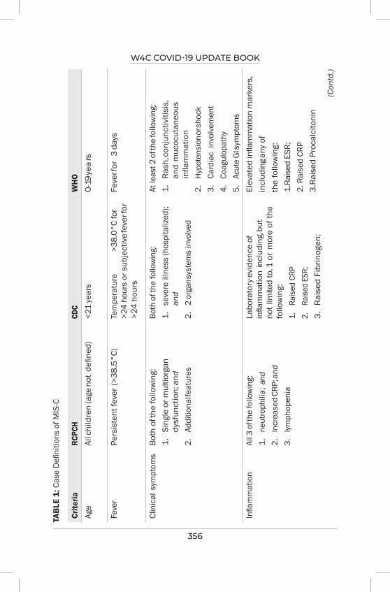
W4C COVID-19 UPDATE BOOK
356
TAB
LE 1
: Cas
e D
efini
tions
of M
IS-C
Crite
riaRC
PCH
CDC
WH
O
Age
All c
hild
ren
(age
not
defi
ned)
<21
year
s0
-19
yea
rs
Feve
rPe
rsis
tent
feve
r (>
38.5
°C)
Tem
pera
ture
>3
8.0°
C fo
r >2
4 ho
urs
or s
ubje
ctiv
e fe
ver f
or
>24
hour
s
Feve
r for
3 d
ays
Clin
ical
sym
ptom
sBo
th o
f the
follo
win
g:1.
Si
ngle
or m
ultio
rgan
dy
sfun
ctio
n; a
nd2.
Ad
ditio
nal fe
atur
es
Both
of t
he fo
llow
ing:
1.
seve
re il
lnes
s (h
ospi
taliz
ed);
and
2.
2 or
gan s
yste
ms
invo
lved
At le
ast 2
of t
he fo
llow
ing:
1.
Ras
h, co
njun
ctiv
itisi
s,
and
muc
ocut
aneo
us
infla
mm
atio
n2.
Hy
pote
nsio
n or s
hock
3.
Card
iac
invo
lvem
ent
4.
Coag
ulop
athy
5.
Acut
e G
I sym
ptom
sIn
flam
mat
ion
All 3
of t
he fo
llow
ing:
1.
neut
roph
ilia;
and
2.
incr
ease
d CR
P; a
nd3.
ly
mph
open
ia
Labo
rato
ry e
vide
nce
of
infla
mm
atio
n in
clud
ing,
but
no
t lim
ited
to, 1
or
mor
e of
the
follo
win
g:1.
Ra
ised
CRP
2.
Raise
d ES
R;3.
R
aise
d Fi
brin
ogen
;
Elev
ated
infla
mm
atio
n m
arke
rs,
incl
udin
g any
of
the
follo
win
g:
1.Ra
ised
ESR
;2.
Rai
sed
CRP
3.R
aise
d Pr
ocal
cito
nin
(Con
td.)
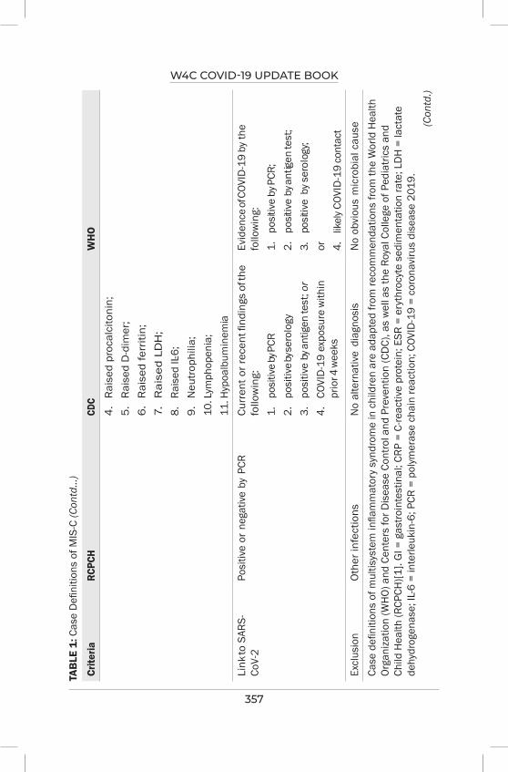
W4C COVID-19 UPDATE BOOK
357
Crite
riaRC
PCH
CDC
WH
O
4.
Rai
sed
proc
alci
toni
n;5.
R
aise
d D
-dim
er;
6.
Rai
sed
ferr
itin;
7.
Rai
sed
LD
H;
8.
Rais
ed IL
-6;
9.
Neu
trop
hilia
;10
. Lym
phop
enia
;11
. Hyp
oalb
umin
emia
Link
to S
ARS-
CoV-
2Po
sitiv
e or
neg
ative
by
PCR
Curr
ent o
r rec
ent fi
ndin
gs o
f the
fo
llow
ing:
1.
po
sitiv
e by
PCR
2.
posi
tive b
y ser
olog
y3.
po
sitiv
e by
ant
igen
test
; or
4.
COVI
D-1
9 ex
posu
re w
ithin
pr
ior 4
wee
ks
Evid
ence
of C
OVID
-19
by th
e
follo
win
g:1.
po
sitiv
e by
PCR
;2.
po
sitiv
e by
antig
en te
st;
3.
posit
ive
by s
erol
ogy;
or
4.
likel
y COV
ID-1
9 co
ntac
tEx
clus
ion
Oth
er in
fect
ions
No
alte
rnat
ive
diag
nosi
sN
o ob
viou
s m
icro
bial
cau
seCa
se d
efini
tions
of m
ultis
yste
m in
flam
mat
ory
synd
rom
e in
chi
ldre
n ar
e ad
apte
d fro
m re
com
men
datio
ns fr
om th
e W
orld
Hea
lth
Orga
niza
tion
(WH
O) a
nd C
ente
rs fo
r Dis
ease
Con
trol a
nd P
reve
ntio
n (C
DC)
, as
wel
l as
the
Roya
l Col
lege
of P
edia
trics
and
Ch
ild H
ealth
(RCP
CH)[1
]. G
I = g
astro
inte
stin
al; C
RP =
C-re
activ
e pr
otei
n; E
SR =
ery
thro
cyte
sed
imen
tatio
n ra
te; L
DH
= la
ctat
e de
hydr
ogen
ase;
IL-6
= in
terle
ukin
-6; P
CR =
pol
ymer
ase
chai
n re
actio
n; C
OVID
-19
= co
rona
viru
s di
seas
e 20
19.
TAB
LE 1
: Cas
e D
efini
tions
of M
IS-C
(Con
td...
)
(Con
td.)

W4C COVID-19 UPDATE BOOK
358
Crite
riaRC
PCH
CDC
WH
O
In th
e RC
PCH
cas
e de
finiti
on, a
dditi
onal
feat
ures
incl
ude
abdo
min
al p
ain,
con
fusi
on, c
onju
nctiv
itis,
cou
gh, d
iarr
hea,
hea
dach
e,
lym
phad
enop
athy
, muc
ous
mem
bran
e ch
ange
s, n
eck
swel
ling,
rash
, res
pira
tory
sym
ptom
s, s
ore
thro
at, s
wol
len
hand
s an
d fe
et, s
ynco
pe, a
nd v
omiti
ng. I
n th
e W
HO
case
defi
nitio
n, c
ardi
ac in
volv
emen
t is
defin
ed a
s th
e pr
esen
ce o
f myo
card
ial
dysf
unct
ion,
per
icar
ditis
, val
vulit
is, o
r cor
onar
y ab
norm
aliti
es (i
nclu
ding
find
ings
on
echo
card
iogr
am o
r ele
vate
d le
vels
of
tropo
nin/
N-te
rmin
al p
ro–B
-type
nat
riure
tic p
eptid
e).
TAB
LE 1
: Cas
e D
efini
tions
of M
IS-C
(Con
td...
)
in mucosal and cardiac tissues, endothelial cells and cytokine molecules. These auto-antigens have also been reported in patients with KD. Immunoglobulin producing antibody secreting cells (ASC) are increased during acute stage of KD and decreased after IVIg administration.5 This hypothesis formed the basis of usage of IVIG in patients of MIS-C.
CASE DEFINITIONSCase definitions of multisystem inflammatory syndrome in children (MIS-C) are taken from guidelines on recommendations for MIS-C from the World Health Organization (WHO), Centers for Disease Control and Prevention (CDC), and the Royal College of Pediatrics and Child Health (RCPCH) [Table 1]. Features that are common to all case definitions include the presence of fever, hyper-inflammatory state and evidence of organ dysfunction. Besides fever other clinical findings include cutaneous manifestations, abdominal symptoms and cardiovascular system involvement. MIS-C is seen more in older children (> 5 years) and median age of patients in various studies has ranged from 7.5 to 10 years.6-7 Hemodynamic instability is present in 60–80% patients.6,9 Gastrointestinal manifestations like abdominal pain, diarrhea and vomiting are commonly seen. Symptoms and signs may mimic acute appendicitis. Neurological features (e.g., headache, meningeal signs, seizures and altered sensorium) are also common.7-9 Cardiovascular complications may be life threatening in patients with MIS-C. Cardiac biomarkers including NT-pro-BNP and troponin levels are extremely high and are pathognomonic of heart failure and myocardial damage. Myocarditis has been reported in 40–80% of patients with MIS-C.6-7
CLASSIFICATION OF MIS-COur understanding of clinical presentations of MIS-C is ever expanding. According to The American College of Rheumatology (ACR), MIS-C appears to be a continuum of disease that has
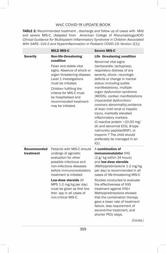
W4C COVID-19 UPDATE BOOK
359
TABLE 2: Recommended treatment , discharge and follow up of cases with Mild and severe MIS-C. (Adapted from American College of Rheumatology(ACR) Clinical Guidance for Multisystem Inflammatory Syndrome in Children Associated With SARS–CoV-2 and Hyperinflammation in Pediatric COVID-19: Version 2[1])
MILD MIS-C Severe MIS-C
Severity Non-life-threatening conditionFever and stable vital signs. Absence of shock or organ threatening disease. Level 1 investigations must be initiated.Children fulfilling the criteria for MIS-C must be hospitalised and recommended treatment may be initiated.
Life threatening conditionAbnormal vital signs (tachycardia, tachypnea), respiratory distress of any severity, shock, neurologic deficits or change in mental status (including subtle manifestations), multiple organ dysfunction syndrome (MODS), cardiac manifestations (myocardial dysfunction/ coronary abnormality),evidence of even mild renal or hepatic injury, markedly elevated inflammatory markers (C-reactive protein >10.00 mg/dl) and abnormal ECG, B-type natriuretic peptide(BNP), or troponin T The child should preferably be managed in an ICU.
Recommended treatment
Patients with MIS-C should undergo di agnostic evaluation for other possible infectious and non-infectious diseases before immunomodulatory treatment is initiatedLow-dose steroids (IV MPS 1-2 mg/kg per day) must be given as first line ther- apy in all cases of non-critical MIS-C.
A combination of immunomodulator IVIG (2 g/ kg within 24 hours) and low-dose steroids (Methylprednisolone 1-2 mg/kg per day) is recommended in all cases of life-threatening MIS-CStudies conducted to evaluate the effectiveness of IVIG treatment against IVIG+ Methylprednisolone showed that the combination therapy gave a lower rate of treatment failure, less requirement of second-line treatment, and shorter PICU stays.
(Contd.)
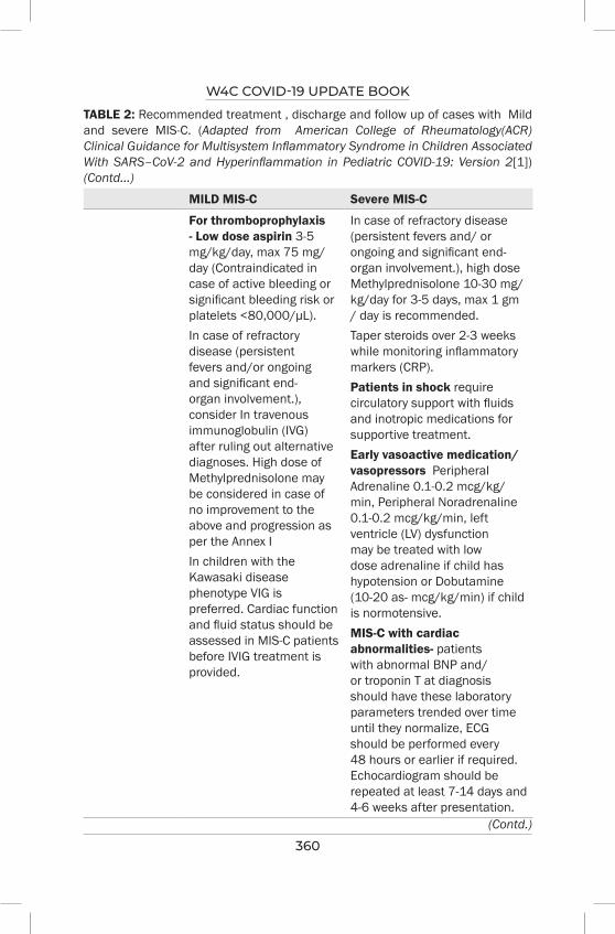
W4C COVID-19 UPDATE BOOK
360
MILD MIS-C Severe MIS-C
For thromboprophylaxis - Low dose aspirin 3-5 mg/kg/day, max 75 mg/ day (Contraindicated in case of active bleeding or significant bleeding risk or platelets <80,000/µL).In case of refractory disease (persistent fevers and/or ongoing and significant end-organ involvement.), consider In travenous immunoglobulin (IVG) after ruling out alternative diagnoses. High dose of Methylprednisolone may be considered in case of no improvement to the above and progression as per the Annex IIn children with the Kawasaki disease phenotype VIG is preferred. Cardiac function and fluid status should be assessed in MIS-C patients before IVIG treatment is provided.
In case of refractory disease (persistent fevers and/ or ongoing and significant end-organ involvement.), high dose Methylprednisolone 10-30 mg/kg/day for 3-5 days, max 1 gm / day is recommended.Taper steroids over 2-3 weeks while monitoring inflammatory markers (CRP).Patients in shock require circulatory support with fluids and inotropic medications for supportive treatment.Early vasoactive medication/vasopressors Peripheral Adrenaline 0.1-0.2 mcg/kg/min, Peripheral Noradrenaline 0.1-0.2 mcg/kg/min, left ventricle (LV) dysfunction may be treated with low dose adrenaline if child has hypotension or Dobutamine (10-20 as- mcg/kg/min) if child is normotensive.MIS-C with cardiac abnormalities- patients with abnormal BNP and/or troponin T at diagnosis should have these laboratory parameters trended over time until they normalize, ECG should be performed every 48 hours or earlier if required. Echocardiogram should be repeated at least 7-14 days and 4-6 weeks after presentation.
TABLE 2: Recommended treatment , discharge and follow up of cases with Mild and severe MIS-C. (Adapted from American College of Rheumatology(ACR) Clinical Guidance for Multisystem Inflammatory Syndrome in Children Associated With SARS–CoV-2 and Hyperinflammation in Pediatric COVID-19: Version 2[1]) (Contd...)
(Contd.)
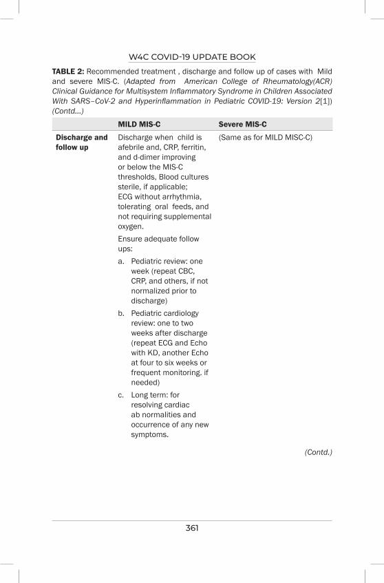
W4C COVID-19 UPDATE BOOK
361
MILD MIS-C Severe MIS-C
Discharge and follow up
Discharge when child is afebrile and, CRP, ferritin, and d-dimer improving or below the MIS-C thresholds, Blood cultures sterile, if applicable; ECG without arrhythmia, tolerating oral feeds, and not requiring supplemental oxygen.Ensure adequate follow ups:a. Pediatric review: one
week (repeat CBC, CRP, and others, if not normalized prior to discharge)
b. Pediatric cardiology review: one to two weeks after discharge (repeat ECG and Echo with KD, another Echo at four to six weeks or frequent monitoring. if needed)
c. Long term: for resolving cardiac ab normalities and occurrence of any new symptoms.
(Same as for MILD MISC-C)
TABLE 2: Recommended treatment , discharge and follow up of cases with Mild and severe MIS-C. (Adapted from American College of Rheumatology(ACR) Clinical Guidance for Multisystem Inflammatory Syndrome in Children Associated With SARS–CoV-2 and Hyperinflammation in Pediatric COVID-19: Version 2[1]) (Contd...)
(Contd.)
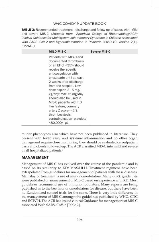
W4C COVID-19 UPDATE BOOK
362
TABLE 2: Recommended treatment , discharge and follow up of cases with Mild and severe MIS-C. (Adapted from American College of Rheumatology(ACR) Clinical Guidance for Multisystem Inflammatory Syndrome in Children Associated With SARS–CoV-2 and Hyperinflammation in Pediatric COVID-19: Version 2[1]) (Contd...)
MILD MIS-C Severe MIS-C
Patients with MIS-C and documented thrombosis or an EF of <35% should receive therapeutic anticoagulation with enoxaparin until at least 2 weeks after discharge from the hospital. Low dose aspirin 3 - 5 mg/kg/day; max 75 mg/day should also be used in MIS-C patients with KD like feature; coronary artery Z score>=2.S; thrombocytosis; contraindication- platelets <80,000/ µL.
milder phenotypes also which have not been published in literature. They present with fever, rash, and systemic inflammation and no other organ damage and require close monitoring, they should be evaluated on outpatient basis and closely followed-up. The ACR classified MIS-C into mild and severe in all hospitalized patients.2
MANAGEMENTManagement of MIS-C has evolved over the course of the pandemic and is based on its similarity to KD/ MAS/HLH. Treatment regimens have been extrapolated from guidelines for management of patients with these diseases. Mainstay of treatment is use of immunomodulators. Many quick guidelines were published on management of MIS-C based on experience with KD. Most guidelines recommend use of immunomodulators. Many reports are being published as to the best immunomodulators for disease, but there have been no Randomized control trials for the same. There is very little difference in the management of MIS-C amongst the guidelines published by WHO, CDC and RCPCH. The ACR has issued clinical Guidance for management of MIS-C associated With SARS–CoV-2 [Table 2].
W4C COVID-19 UPDATE BOOK
363
INVESTIGATIONSTIER-1 investigations
If the child fulfils the case definition of MIS-C and is not in shock then the Tier-1 investigations should be done which includes;
CBC, ESR , CRP, SARS Cov-2 PCR and serology and complete Metabolic panel (sodium, potassium, carbon dioxide, chloride, blood urea nitrogen, creatinine, glucose, calcium, albumin, total protein, aspartate aminotransferase, alanine aminotransferase, alkaline phosphatase, and bilirubin).
TIER-2 Investigations
If CRP >5mg/dl , ESR >40 mm and at least one of the following; 1) ALC <1000/micro liter,2) Platelet counts <150,000/micro liter,3)S sodium < 135 mmols/liters, 4) neutrophilia , 5) hypoalbuminemia, then order for the tier-2 investigations which are; BNP, Troponin T, PCT, D-Dimer, Ferritin, Fibrinogen, LDH, Triglycerides, PT, APTT, Cytokine panel, ECG and Echocardiography.
In tropical countries all efforts should be made to rule out other hyperinflammatory illnesses like Dengue, Rickettsial diseases, malaria, Salmonella-Typhi etc. Quite often these tropical diseases may coexist[10].
TREATMENT [TABLE 2] First Line: should be started in mild hospitalized patients. Low-dose steroids (IV Methylprednisolone (MPS) 1-2 mg/kg per day) must be given as first line therapy in all cases of non-critical MIS-C.
Thromboprophylaxis: Low dose aspirin 3 - 5 mg/kg/day; max 75 mg/ day.
In case of refractory disease, consider Intravenous immunoglobulin (IVIG) after ruling out alternative diagnoses. If there is no improvement or the disease progresses consider high dose of Methylprednisolone. In children with the Kawasaki disease phenotype, IVIG is preferred.
SEVERE DISEASE : [TABLE 2]All hospitalized patients life threatening MIS-C with shock or requiring organ support should receive a combination of immunomodulator IVIG (2 g/ kg within 24 hours) and low-dose steroids (MPS 1-2 mg/kg per day). In case of refractory disease, high dose Methylprednisolone 10-30 mg/kg/day for 3-5 days, max 1 gm / day is recommended. Taper steroids over 2-3 weeks while monitoring inflammatory markers (CRP).
Patients in shock are to be treated as per the guidelines for management of shock.
W4C COVID-19 UPDATE BOOK
364
DISCHARGE AND FOLLOW UP [TABLE 2]Discharge when child is afebrile, the inflammatory markers normal or showing decreasing trend is on no organ support and accepting orally.
Frequent follow up is necessary;
1. Pediatric review: After one week (repeat CBC, CRP, and others).
2. Pediatric cardiology review: one to two weeks after discharge.
3. Long term: For resolving cardiac abnormalities and occurrence of any new symptoms.
CONCLUSIONMIS-C is characterized by hyperinflammation and multiorgan involvement. Cardiac involvement is seen most often and coronary artery involvement is rare, as compared to KD. Tropical disease should be excluded before making a diagnosis of MIS-C. The management of the disease is based on its resemblance to KD/ MAS/HLH. We need more randomized controlled trials for the best treatment for MIS-C.
PRACTICE POINTS1. Clinicians should have a high index of suspicion of MIS-C in a child
having prolonged fever, high inflammatory markers and negative cultures, during the pandemic.
2. Tropical diseases like Dengue, scrub typhus and malaria etc. should be excluded before reaching a diagnosis of MIS-C. Quite often the two diseases may coexist.
REFERENCES1. Dufort EM, Koumans EH, Chow EJ, Rosenthal EM, Muse A, Rowlands J, et al; New York
State and Centers for Disease Control and Prevention Multisystem Inflammatory Syndrome in Children Investigation Team. Multisystem Inflammatory Syndrome in Children in New York State. N Engl J Med 2020; 383:347-358. doi: 10.1056/NEJMoa2021756. Epub 2020 Jun 29. PMID: 32598830; PMCID: PMC7346766.
2. Henderson LA, Canna SW, Friedman KG, Gorelik M, Lapidus SK, Bassiri H, et al. American College of Rheumatology Clinical Guidance for Multisystem Inflammatory Syndrome in Children Associated With SARS-CoV-2 and Hyperinflammation in Pediatric COVID-19: Version 2. Arthritis Rheumatol 2021; 73:e13-e29. doi: 10.1002/art.41616. Epub 2021 Feb 15. PMID: 33277976.
3. Gupta S, Chopra N, Singh A, Gera R, Chellani H, Pandey R, et al. Unusual Clinical Manifestations and Outcome of Multisystem Inflammatory Syndrome in Children (MIS-C) in a Tertiary Care Hospital of North India. J Trop Pediatr 2021; 67:fmaa127. doi: 10.1093/tropej/fmaa127. PMID: 33513240; PMCID: PMC7928672.
4. Consiglio CR, Cotugno N, Sardh F, Pou C, Amodio D, Rodriguez L, et al; CACTUS Study Team, Landegren N, Palma P, Brodin P. The Immunology of Multisystem Inflammatory Syndrome in Children with COVID-19. Cell 2020; 183:968-981.e7. doi: 10.1016/j.cell.2020.09.016. Epub 2020 Sep 6. PMID: 32966765; PMCID: PMC7474869.
W4C COVID-19 UPDATE BOOK
365
5. Kabeerdoss J, Pilania RK, Karkhele R, Kumar TS, Danda D, Singh S. Severe COVID-19, multisystem inflammatory syndrome in children, and Kawasaki disease: immunological mechanisms, clinical manifestations and management. Rheumatol Int 2021; 41:19-32. doi: 10.1007/s00296-020-04749-4. Epub 2020 Nov 21. PMID: 33219837; PMCID: PMC7680080.
6. Feldstein LR, Rose EB, Horwitz SM, Collins JP, Newhams MM, Son MBF, et al; CDC COVID-19 Response Team. Multisystem Inflammatory Syndrome in U.S. Children and Adolescents. N Engl J Med 2020; 383:334-346. doi: 10.1056/NEJMoa2021680. Epub 2020 Jun 29. PMID: 32598831; PMCID: PMC7346765.
7. Verdoni L, Mazza A, Gervasoni A, Martelli L, Ruggeri M, Ciuffreda M, et al. An outbreak of severe Kawasaki-like disease at the Italian epicentre of the SARS-CoV-2 epidemic: an observational cohort study. Lancet 2020; 395:1771-1778. doi: 10.1016/S0140-6736(20)31103-X. Epub 2020 May 13. PMID: 32410760; PMCID: PMC7220177.
8. Riphagen S, Gomez X, Gonzalez-Martinez C, Wilkinson N, Theocharis P. Hyperinflammatory shock in children during COVID-19 pandemic. Lancet 2020; 395:1607-1608. doi: 10.1016/S0140-6736(20)31094-1. Epub 2020 May 7. PMID: 32386565; PMCID: PMC7204765.
9. Whittaker E, Bamford A, Kenny J, Kaforou M, Jones CE, Shah P, et al; PIMS-TS Study Group and EUCLIDS and PERFORM Consortia. Clinical Characteristics of 58 Children With a Pediatric Inflammatory Multisystem Syndrome Temporally Associated With SARS-CoV-2. JAMA 2020; 324:259-269. doi: 10.1001/jama.2020.10369. PMID: 32511692; PMCID: PMC7281356.
10. Samprathi M, Narayanappa S, Sridhar M, Ramachandra P, Vemgal P. Multisystem Inflammatory Syndrome in Children: A Mimicker of Severe Dengue. Indian J Pediatr 2021; 88:486-487. doi: 10.1007/s12098-020-03550-2. Epub 2020 Oct 23. PMID: 33095394; PMCID: PMC7581688.

SECTION 8
Home Care and Rehabilitation

369
Section 8 - Home Care and RehabilitationSECTION EDITOR
Dr D P SamaddarMD, FICCM Director Medical Affairs, Ruby General Hospital, Kolkata
Preface
Outbreak of COVID 19 in China and its global spread has caught the world unaware and left a devastating impact on the general mass irrespective of the geographic divide. The disease is still evolving and our knowledge and armamentarium are not satisfactory to contain the progression of the disease at the desired level.
The second wave in particular had proved that our preparedness to manage these cases was grossly
inadequate. Dearth of infrastructure, trained human resource, medications, and consumables, was more than evident. Process related gaps in infection control practices were being experienced in almost all health care set up. Make shift hospitals to manage large number of patients and home-based care for less serious patients had emerged as the necessity and alternative option.
Possibility of keeping patients restricted to their home environment while supervision, consultation and basic medical support were being offered, had economical and psychological advantages. It could reduce the apprehension among the community and spared much needed hospital and intensive care beds for more deserving cases. Thus, burden on health care system could be minimized to a great extent. Transition to make shift hospital with better support system and ultimately to a hospital in case of need, should be ensured readily with this eccentric approach.
The bitter experience of COVID 19 has left us with no alternative but to develop our resources in offering more advanced care at home and makeshift hospitals while using the technology to provide expert supervision from remote locations. This probably is the only panacea to deal with such pandemics more effectively in future and rehabilitate the victims without burdening the overstressed health care system.

371
Home Oxygen Therapy 49.
INTRODUCTIONMedical Oxygen is a major component of supportive/definitive care in treating Covid 19. Evidence strongly point towards its utility in survival of both acute and severe cases. During the second wave of Covid 19, a large population was affected and oxygen support was required in majority of patients, irrespective of their age group. The extensive spread of the disease within a short time overwhelmed the existing logistics that led to panic amongst the populace. The need of the hour was to administer oxygen in a safe manner without burdening the existing system.
INDICATIONIt was empirically proved that oxygen saturation maintained between 88-92% is adequate in a monitored place and those having oxygen saturation of 92 or 94% need monitoring, but may not need oxygen therapy. Therefore options of oxygen concentrators, portable lightweight cylinders or rechargeable CPAP devices with an appropriate delivery system (cannula, prongs, simple face mask, non-rebreathing mask) made available at home came of good use at a time when it was becoming increasingly difficult to secure a bed in a medical facility. Home oxygen therapy (HOT), a method of community or home based care became a life-saving alternative but one needed to be doubly careful and aware of the risks and dangers as well.
CATEGORIES OF PATIENTSIn Covid-19 there are broadly two categories of patients for home oxygen therapy:
1. Patients who have tested positive for corona virus and are symptomatic and
2. Those suffering from long Covid or Covid sequelae with residual lung changes.
Among the two categories the patient with long COVID may have a respiratory problem even after recovery but these patients have fewer chances of slipping into major respiratory crisis .
Vandana Sinha, Prasanta Kumar Gogoi
W4C COVID-19 UPDATE BOOK
372
Breathing high concentration of oxygen may damage the lungs and on the other hand hypoxia may damage heart, brain and other organs.
METHODS OF SUPPLEMENTATION:A. Oxygen concentrators: A portable device that takes in air from room, filters
out nitrogen and provides oxygen needed for oxygen therapy. Electric pump and uninterrupted power supply are necessary for continuous supply of up to 95% pure oxygen at a flow of 5-10L/minute.
Dos and Don’ts :
1. Do not use near open flame or while smoking.
2. Place in open space for reducing chances of device failure from overheating.
3. Do not block any vents as it may impact device performance.
4. Periodic check-up for functionality.
B. Oxygen cylinders: It is simply a container from which oxygen is supplied to the patient through mask or cannula. This needs refilling and additional attachments eg: flowmeter, humidifier, tubings, and regulator to supply oxygen from cylinder to the patient. Usually 2 types of cylinders are used for home oxygen therapy.
i. B type : It has a water capacity of 10 litres which gives 1200 litres of O2 in gaseous form.
ii. D type : Cylinder has water capacity of around 46 lts and gas capacity of approximately 7000 litres. The humidifier should be filled up with water.
Dos and Don’ts:
1. Due to significant risk of fire associated with smoking or any naked flame cylinders should be kept 5 feet away from the flame.
2. Pay attention to skin around facemask or nasal cannula to prevent irritated or bruised. Irritation and brusing
3. Address drying of nasal mucosa and resultant bleeding.
4. Brief about possibility of morning headaches or tiredness.
Monitoring: The easiest way to monitor oxygen saturation at home is pulse oximeters. It uses beam of light to indirectly measure level of O2 in blood without having to draw blood sample. Multiple factors can influence the readings:
(1) poor circulation (2) skin thickness (3) tobacco use (4) cold extremities (5) use of nail varnish.
W4C COVID-19 UPDATE BOOK
373
Instead of solely relying on pulse oximeters, the patient should also monitor the GIT symptoms ,muscle soreness, fatigue and change in taste & smell as well as more common initial symptoms such as fever, cough& shortness of breath .
Current status: Home Oxygen therapy addresses the pressure created by the COVID 19 pandemic on medical logistics and becomes a critical cog to ameliorate COVID-19 the burden on the healthcare facilities. It emphasises the need for a collective effort and technology that can provide care in and out of hospital.
The MOHFW along with ICMR and AIIMS have jointly issued treatment guidelines for oxygen therapy at home and recommended oxygen concentrators with strict vigilance by trained staff for patients needing higher oxygen supplementation. Use of oxygen cylinders at home is generally not recommended due to potential hazard. Nevertheless, if it is to be used for compelling reasons, then strict supervision of qualified and suitably trained doctor/nurse is mandatory.
Joint ACAIM-WACEM CCMT, a multidisciplinary group with participants from multiple countries and have significant collective expertise in management of COVID 19.They came together to create a pathway for providing safe and effective HOT care in community setting. Effective implementation of this approach requires a combination of excellent clinical judgement on the part of the treating health care personnel (HCP), availability of POC tools, real time remote patient monitoring and ongoing education of HCPs ,patients and their caretakers.
CONCLUSIONDevastation seen during the second wave of the COVID 19 is not only due to contagion and virulence of the disease but perhaps more significantly because of the crunch in life saving resources, especially something that we have taken for granted “Oxygen”. The significance of public awareness along with an integrated approach to triage and treat large populations during future Covid waves will be the lynchpin to control and overcome such epidemics. Home Oxygen Therapy would play a critical role in ongoing battles against COVID.
REFERENCES1. Home oxygen therapy-Eman Shebl,Pranav Modi et al. Updated 2021 :In:Stat pearls.2. Short term home O2 therapy for Covid 19 pts.COVID-HOT algorithm-
Indrani Sardesai, Joydeep Grover,Manish Garg et al. J Family Med Prim Care 2020; 9:3209-32193. www.mohfw.gov.in/pdf/covid104. England N, ImprovementN.Primary care and community respiratory resources pack for use
during Covid 19.Updated 16/04/2020,accessed 20/06/2020.Available from https//www.pcrs-uk.org/sites/pcrs-uk.org/files/resources/Covid19/NHS-London-Primary and Community care respiratory-resource-pack-during Covid 19-V3final-160420.pdf
374
Physical and Mental Rehabilitation 50.
INTRODUCTIONSevere acute respiratory syndrome corona virus 2 (SARS COV2) pandemic has a clinical spectrum ranging from asymptomatic to critical illness. Our understanding of the disease and progress in its treatment has markedly improved the outcome of critically ill COVID patients. However, persisting physical and mental disability in survivors of this critical illness is often associated with a reduction in their quality of life. Physically debilitating illness, extended hospital stay, adverse effects of drug therapy and prolonged mechanical ventilation may all culminate in many late complications like muscle weakness, fatigue, chest pain, persistent cough, mood alterations and poor health related quality of life.
Intensive care unit acquired weakness (ICUAW) is a well-documented problem in critically ill patients. The benefit of early rehabilitation is well documented in survivors of critical illness.1 Rehabilitation of COVID 19 patients is complicated by strict isolation and infection control protocols, severity of the disease and associated social and staffing issues. It is important to understand the need for rehabilitation of critically ill COVID patients throughout the continuum of care.
EPIDEMIOLOGYAmong those infected with COVID 19, approximately 15 to 20 percent will require hospitalisation, about one-fourth of them will need acute care.2 In an analysis of the second wave, lesser proportion of patients required intensive care units (20 vs 38 percent). Males have comprised a disproportionately high number of ICU admissions in many studies.
CLINICAL PRESENTATION IN ACUTE COURSEAlthough fever, cough and dyspnoea are the most common symptoms, patients often have varied presentations. Occasionally, anosmia and dysgeusia were the only complaints.3
Sayi Prasad, Gowri Sayi Prasad

W4C COVID-19 UPDATE BOOK
375
Comorbidities like older age, diabetes, obesity, hypertension, chronic kidney disease and immunosuppression may be associated with severe disease, ARDS and death. Multiple organ failure and shock carries very poor prognosis.
PROBABLE LONG-TERM COMPLICATIONS AND SEQUELAEPatients recovering from this novel virus have experienced significant physiological and psychological impacts. Even months after recovery patients experience a wide range of symptoms.4 A multidisciplinary rehabilitation team is required to combat these problems. Although critical care settings provide high standards of medical care, prolonged isolation from family, financial and social stress, further complicate the recovery. Hence, a psychosocial support will ensure early mental recuperation and eventually the restoration of physical health.
Fatigue is reported as the most frequent symptom in 60 to 70 percent of patients recovering from COVID by first UK based research study.6 Breathlessness (40-60 percent), depression (23-47 percent), insomnia and myalgia are also common.
An extended hospital stay and prolonged mechanical ventilation may lead to muscle wasting and weakness, stubborn infections, low serum hormone levels and nutrition related issues.
This makes a case for a pressing need to bring awareness among health professionals about the long-term sequelae and rehabilitation strategies required in this pandemic.
PULMONARY SEQUELAE AND REHABILITATIONPersistent cough, dyspnoea, decreased exercise capacity and continuing hypoxia are common indications which require longer attention. Pulmonary function tests show restrictive pulmonary physiology and reduced diffusion capacity. CT scan imaging shows fibrotic changes in addition to ground glass opacities in survivors of severe COVID.
In most patients these abnormalities improve without major disability by the end of the first year. Pulmonary rehabilitation targeting inspiratory muscle strengthening, exercise training and breathing exercises will be helpful in many patients.6 The recovery of pulmonary function may be assessed by pulse oximetry, 6 minute walk test(6MWT) and HRCT/PFT as clinically indicated.
Barriers of rehabilitation include severity of illness, risk of infection due to COVID 19 positive status, strained availability of care givers and shortage of PPE.
Treatment with corticosteroids may be beneficial in selected patients with post COVID inflammatory lung disease. Clinical trials of anti fibrotic therapies to prevent pulmonary fibrosis are underway.7
W4C COVID-19 UPDATE BOOK
376
CARDIOVASCULAR SEQUELAE AND REHABILITATIONChest pain was the predominant symptom in 20% of patients during COVID follow up followed by palpitations and dyspnoea in a Chinese study.8 Persistent cardiac symptoms during follow up should warrant evaluation with electrocardiography and echocardiogram at 4 to 12 weeks post disease onset. The decision for advanced cardiac evaluation should be decided on an individualistic basis.
Gradual incremental physical exercise programmes have to be advised. Abstinence from competitive sports and aerobic activities to be advised for three to six months in patients with myocardial inflammation on cardiac MRI. Low dose beta blockers in selected cases may help. Anti arrhythmic drugs like amiodarone to be used with caution in patients with fibrotic pulmonary changes after COVID 19.
NEUROPSYCHIATRIC SEQUELAE AND REHABILITATION COVID 19 survivors have reported a range of symptoms like chronic malaise, diffuse myalgia, depressive symptoms, migrainous headache and varied psychiatric symptoms for months after initial infection.9 Anxiety, depression and sleep disturbances were reported in approximately on quarter of patients at six months follow up in the post acute COVID 19 Chinese study.9 Patho physiology is diverse and involves immune dysregulation, inflammation and micro vascular thrombosis.
Headache needs standard therapy. Patients with cognitive impairment need neuropsychological evaluation. Standard screening tools have to be used for screening of affected populations. Again infectivity and lack of trained counsellors hampers management.
NEUROMUSCULAR COMPLICATIONS/ INTENSIVE CARE UNIT ACQUIRED WEAKNESS (ICUAW)This commonly manifests in survivors as a combination of critical illness poly neuropathy (CIP) and critical illness myopathy (CIM). Neuromuscular complications can be reduced by strict control of sugars, judicious use of steroids and avoiding prolonged neuromuscular blockade.
Randomised trials of physical rehabilitation and mobility in ICU have shown benefit and are recommended by society of critical care medicine guidelines.10
ENDOCRINE AND GASTROINTESTINAL SEQUELAENew onset diabetes and subacute thyroiditis can complicate recovery in COVID survivors. Presence of hyperglycaemia can further predispose to variety of infections including difficult to treat fungal infections. After appropriate lab testing should be referred to endocrinologist.
W4C COVID-19 UPDATE BOOK
377
Prolonged viral fecal shedding is known upto 28 days. Gut microbes may be altered leading to depletion of normal gut commensals and also overgrowth of opportunistic organisms in the gut.
HEMATOLOGICAL AND DERMATOLOGICAL REHABILITATIONThromboembolic events are noted in about five percent of post covid survivors. Newer direct anticoagulants or low molecular weight heparin have to be considered in high risk population. Benefits of early mobilisation can’t be underscored.
Skin rash is seen in many. Hair loss is a predominant symptom and is reported in almost 20% of patients. It may be result of viral infection or may be stress response, although immune mechanism is also postulated.
NUTRITION AND REHABILITATIONMalnutrition has been noted in 26-46 percent of patients with COVID 19, as evaluated by the Malnutrition Universal Screening Tool in an Italian study.11 Rehabilitation units should routinely assess for dysphagia (especially in those who are intubated), nutritional status and diet plans.
CONCLUSIONPhysical and mental rehabilitation of critically ill COVID 19 survivors is of utmost importance to prevent long term sequelae. Rehabilitation should start in the intensive care itself after the initiation of mechanical ventilation. Patients who have recovered from COVID should be routinely screened for signs of fatigue, muscle pain, depression, and mental health irrespective of the severity of illness. Risk of infection, shortage of PPE, availability of trained staff and inclination among professionals for rehabilitation services are some of the limitations.
A comprehensive rehabilitation programme, consisting of multiple disciplines including policy makers is the need of the hour. The recommendations should be tailored to suit the local circumstances, available resources, and expertise to improve the quality of life of patients and to reduce the impact of COVID pandemic globally.
REFERENCES1. Desai SV, Law TJ, Needham DM. Long-term complications of critical care. Crit Care Med 2011;
39:371-9.2. Recovery from Critical Illness: Physical Rehabilitation in the Intensive Care Unit, Timing of
Persistent Critical Illness, and Caregiver Outcomes3. Hassan SA, Sheikh FN, Jamal S, Ezeh JK, Akhtar A. Coronavirus (COVID-19): A review of
clinical features, diagnosis, and treatment. Cureus 2020; 12: e7355. [Doi: 10.7759/cureus. 7355].4. Mardani M. Post COVID syndrome. Arch Clin Infect Dis 2020; 15:1–2. DOI: 10.1001/
jama.2020.126035. Islam MF, Cotler J, Jason LA. Post-viral fatigue and COVID-19: Lessons from past epidemics.
W4C COVID-19 UPDATE BOOK
378
Fatigue Biomed Heal Behav 2020; 8:61–69. DOI: 10.1080/21641846.2020.17782276. Hsieh MJ, Lee WC, Cho HY, Wu MF, Hu HC, Kao KC, et al. Recovery of pulmonary functions,
exercise capacity, and quality of life after pulmonary rehabilitation in survivors of ARDS due to severe influenza A (H1N1) pneumonitis. Influenza Other Respir Viruses 2018; 12:643-8.
7. Raghu, G. & Wilson, K. C.COVID-19 interstitial pneumonia: monitoring the clinical course in survivors. Lancet Respir Med 2020; 8:839–842.
8. Huang, C. et al. 6-month consequences of COVID-19 in patients discharged from hospital: a cohort study. Lancet 2021; 397:220–232.
9. Rogers, J. P. et al. Psychiatric and neuropsychiatric presentations associated with severe coronavirus infections: a systematic review and meta-analysis with comparison to the COVID-19 pandemic. Lancet Psychiatry 2020; 7:611–627.
10. Tipping CJ, Harrold M, Holland A, Romero L, Nisbet T, Hodgson CL. The effects of active mobilisation and rehabilitation in ICU on mortality and function: A systematic review. Intensive Care Med 2017; 43:171-83.
11. Chowkwanyun, M. & Reed, A. L. Racial health disparities and COVID-19— caution and context. N Engl J Med 2020; 383, 201–203.
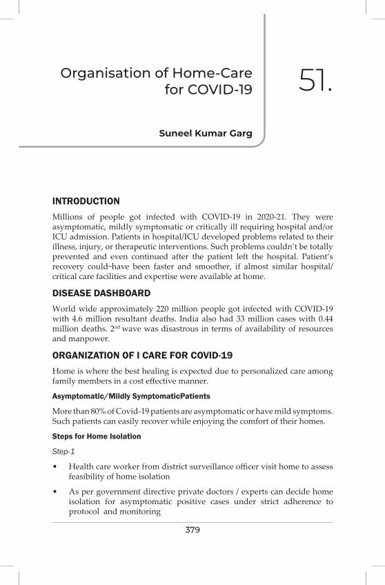
379
Organisation of Home-Care for COviD-19 51.
INTRODUCTIONMillions of people got infected with COVID-19 in 2020-21. They were asymptomatic, mildly symptomatic or critically ill requiring hospital and/or ICU admission. Patients in hospital/ICU developed problems related to their illness, injury, or therapeutic interventions. Such problems couldn’t be totally prevented and even continued after the patient left the hospital. Patient’s recovery could have been faster and smoother, if almost similar hospital/critical care facilities and expertise were available at home.
DISEASE DASHBOARDWorld wide approximately 220 million people got infected with COVID-19 with 4.6 million resultant deaths. India also had 33 million cases with 0.44 million deaths. 2nd wave was disastrous in terms of availability of resources and manpower.
ORGANIZATION OF I CARE FOR COVID-19 Home is where the best healing is expected due to personalized care among family members in a cost effective manner.
Asymptomatic/Mildly SymptomaticPatients
More than 80% of Covid-19 patients are asymptomatic or have mild symptoms. Such patients can easily recover while enjoying the comfort of their homes.
Steps for Home Isolation
Step-1
• Health care worker from district surveillance officer visit home to assess feasibility of home isolation
• As per government directive private doctors / experts can decide home isolation for asymptomatic positive cases under strict adherence to protocol and monitoring
Suneel Kumar Garg
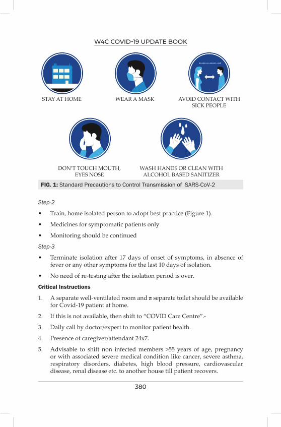
W4C COVID-19 UPDATE BOOK
380
Step-2
• Train, home isolated person to adopt best practice (Figure 1).
• Medicines for symptomatic patients only
• Monitoring should be continued
Step-3
• Terminate isolation after 17 days of onset of symptoms, in absence of fever or any other symptoms for the last 10 days of isolation.
• No need of re-testing after the isolation period is over.
Critical Instructions
1. A separate well-ventilated room and a separate toilet should be available for Covid-19 patient at home.
2. If this is not available, then shift to “COVID Care Centre”.
3. Daily call by doctor/expert to monitor patient health.
4. Presence of caregiver/attendant 24x7.
5. Advisable to shift non infected members >55 years of age, pregnancy or with associated severe medical condition like cancer, severe asthma, respiratory disorders, diabetes, high blood pressure, cardiovascular disease, renal disease etc. to another house till patient recovers.
STAY AT HOME WEAR A MASK AVOID CONTACT WITHSICK PEOPLE
DON’T TOUCH MOUTH,EYES NOSE
WASH HANDS OR CLEAN WITHALCOHOL BASED SANITIZER
FIG. 1: Standard Precautions to Control Transmission of SARS-CoV-2
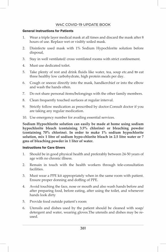
W4C COVID-19 UPDATE BOOK
381
General Instructions for Patients
1. Wear a triple layer medical mask at all times and discard the mask after 8 hours of use. Replace wet or visibly soiled mask.
2. Disinfecte used mask with 1% Sodium Hypochlorite solution before disposal,
3. Stay in well ventilated/ cross ventilated rooms with strict confinement.
4. Must use dedicated toilet.
5. Take plenty of rest and drink fluids like water, tea, soup etc.and to eat three healthy low carbohydrate, high protein meals per day.
6. Cough or sneeze directly into the mask, handkerchief or into the elbow and wash the hands often.
7. Do not share personal items/belongings with the other family members.
8. Clean frequently touched surfaces at regular interval.
9. Strictly follow medication as prescribed by doctor.Consult doctor if you are taking any regular medication.
10. Use emergency number for availing essential services.
Sodium Hypochlorite solution can easily be made at home using sodium hypochlorite bleach (containing 3.5% chlorine) or bleaching powder (containing 70% chlorine). In order to make 1% sodium hypochlorite solution, mix 1 litre of sodium hypo-chlorite bleach in 2.5 litre water or 7 gms of bleaching powder in 1 liter of water.
Instructions for Care Givers
1. Should be in good physical health and preferably between 24-50 years of age with no chronic illness.
2. Remain in touch with the health workers through tele-consultation facilities.
3. Must wear a PPE kit appropriately when in the same room with patient. Ensure proper donning and doffing of PPE.
4. Avoid touching the face, nose or mouth and also wash hands before and after preparing food, before eating, after using the toilet, and whenever hands look dirty
5. Provide food outside patient’s room
6. Utensils and dishes used by the patient should be cleaned with soap/detergent and water, wearing gloves.The utensils and dishes may be re-used.

W4C COVID-19 UPDATE BOOK
382
7. Segregate patient’s clothes, bed linen, bath & hand towels and clean them separately using regular laundry soap and water or machine wash at 60–90 °C (140–194 °F) with common household detergent, and dried thoroughly under the sun.
8. Clean and disinfect patient’s room, bathroom and toilet surfaces at least once daily.
9. Make sure that the patient follows the prescribed treatment.
10. The caregiver and all close contacts should self-monitor their health.
Instructions for Neighbors
1. Common spaces of the building such as lifts or stairs are sanitized twice a day with 1% sodium hypochlorite solution.
2. Help the patients for essential items.
3. Maintain a proper distance from the patient at all times and make sure that children, elderly and pregnant women in particular, keep distance from the patients.
4. Remember, the fight is against the disease, not the sick.
5. Do not trouble the patient or their family members.
Monitoring
Monitoring of pulse, BP, RR, Temperature and SpO2 is mandatory at frequent intervals. If patients can’t be monitored at home, then shift to a medical center.
Moderate/Severely Symptomatic Patients
Care at home can’t be a replacement of hospital/ICU care. If hospital/ICU bed is unavailable then provide immediate care at home under supervision of critical care expert with 1:1 nursing ratio to reduce morbidity/mortality.
Home care for severely symptomatic patients depends on nurses/care takers most of the time as doctor is not available physically round the clock. It’s a concern for emerging emergencies. Extensive training of the nurses/care takers with mandatory courses and emergency protocols, regular vitals update through technology e.g. eSMART ICU, can address these issues.
Critical Care Specialist Driven Hospital/ICU Care at Home
Critical care specialist driven hospital/ICU care at home is entirely different compared to other commercial healthcare facilities. Detailed history, symptomatic evaluation is primarily important. This is followed by physical or virtual assessment by critical care expert or attending nurse to prescribe a treatment guideline. Detailed discussion with all family members about patient’s condition and future expectations should be ensue. Care plan cannot be executed well unless critical care nurse taking care of patient follows the
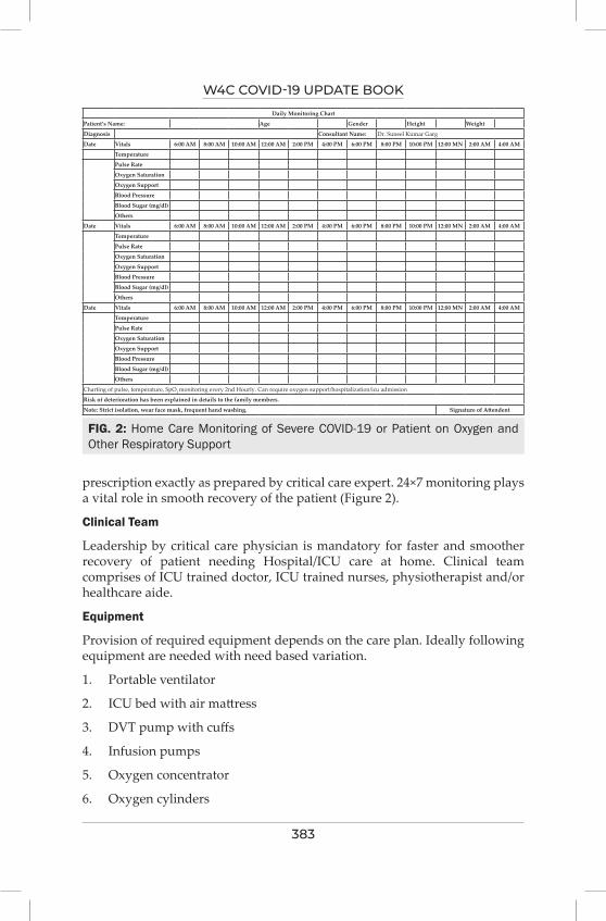
W4C COVID-19 UPDATE BOOK
383
prescription exactly as prepared by critical care expert. 24×7 monitoring plays a vital role in smooth recovery of the patient (Figure 2).
Clinical Team
Leadership by critical care physician is mandatory for faster and smoother recovery of patient needing Hospital/ICU care at home. Clinical team comprises of ICU trained doctor, ICU trained nurses, physiotherapist and/or healthcare aide.
Equipment
Provision of required equipment depends on the care plan. Ideally following equipment are needed with need based variation.
1. Portable ventilator
2. ICU bed with air mattress
3. DVT pump with cuffs
4. Infusion pumps
5. Oxygen concentrator
6. Oxygen cylinders
FIG. 2: Home Care Monitoring of Severe COVID-19 or Patient on Oxygen and Other Respiratory Support
Daily Monitoring Chart
Patient’s Name: Age Gender Height Weight
Diagnosis Consultant Name: Dr. Suneel Kumar Garg
Date Vitals 6:00 AM 8:00 AM 10:00 AM 12:00 AM 2:00 PM 4:00 PM 6:00 PM 8:00 PM 10:00 PM 12:00 MN 2:00 AM 4:00 AM
Temperature
Pulse Rate
Oxygen Saturation
Oxygen Support
Blood Pressure
Blood Sugar (mg/dl)
Others
Date Vitals 6:00 AM 8:00 AM 10:00 AM 12:00 AM 2:00 PM 4:00 PM 6:00 PM 8:00 PM 10:00 PM 12:00 MN 2:00 AM 4:00 AM
Temperature
Pulse Rate
Oxygen Saturation
Oxygen Support
Blood Pressure
Blood Sugar (mg/dl)
Others
Date Vitals 6:00 AM 8:00 AM 10:00 AM 12:00 AM 2:00 PM 4:00 PM 6:00 PM 8:00 PM 10:00 PM 12:00 MN 2:00 AM 4:00 AM
Temperature
Pulse Rate
Oxygen Saturation
Oxygen Support
Blood Pressure
Blood Sugar (mg/dl)
Others
Charting of pulse, temperature, SpO2 monitoring every 2nd Hourly. Can require oxygen support/hospitalization/icu admission
Risk of deterioration has been explained in details to the family members.
Note: Strict isolation, wear face mask, frequent hand washing. Signature of Attendent
W4C COVID-19 UPDATE BOOK
384
7. Cardiac monitor
8. Suction machine
9. IV stand
10. Cardiac table
Monitoring
Monitoring is extremely essential step for managing moderate/severely symptomatic patients at home. Monitoring of pulse, BP, RR, Temperature, blood sugar, oxygen requirement, and SpO2 is mandatory at frequent intervals, otherwise home isolation is not advisable.
OUR EXPERIENCE AT SAIMAN HEALTHCAREWe created a fool proof system for taking care of patient’s infected with COVID-19. In 2020-21, during pandemic our team of specialized critical care doctors, nurses & medical attendants treated 5000+ COVID-19& post COVID-19 patients at home and also provided 24*7 remote monitoring & quarantine services to the patients with almost zero causality. We also managed 1000+ patients who were moderate/severely symptomatic and were not finding beds in hospital/ICU with less than 1% casualty.
Recovery depended on the stage of the patient presented to us.
1. We first made oral medicines prescription by team of critical care doctor.
2. Kept the patient for monitoring (vitals/lab tests).
3. Did regular calls to patient/attendant for getting update on health condition.
4. Revised the prescription if required.
5. Ordered a CT scan chest if required.
6. Revision in prescription for adding/deletion of medicines.
7. Kept the patient for monitoring (vitals/lab tests).
8. Switched to intravenous medications along with oxygen, if required.
High variability in patients’ presentations was observed by us. Recovery was faster in case of early reporting. Home based therapy. in absence of ICU beds, could be initiated even in hypoxic patients to utilize the window for suppressing inflammatory cascade and thwarting its progression. We requested patients to inform us on day-1 of their symptoms onset, to get almost 100% positive results.
Compared to hospitals setup, homecare took minimum time, saved cost, and gave best outcomes (almost 99% recovery) when sick patients were not finding bed in hospital/ICU.

W4C COVID-19 UPDATE BOOK
385
SUMMARY Organization of home care for COVID-19 positive asymptomatic/mildly symptomatic patients is now common. However, it is not well accepted for managing moderate/severely ill symptomatic patients. We strongly recommend treating even moderate/severely symptomatic patients under supervision of critical care team, if patient is not getting bed in hospital/ICU. By adopting this approach , there can be drastic reduction in morbidity and/or mortality of such patients.

Vaccination SECTION 9

389
Section 9 - COVID-19 Vaccination and Breakthrough InfectionsSECTION EDITOR
Dr Diptimala AgarwalMBBS DA FICCM PGDHA Chief Critical Care Services and Consultant Anaesthesiologist, Shantived Institute of Medical Sciences, Agra
Preface
It is a privilege to write the preface of the section on covid 19 vaccination in the update book on covid and critical care. Since its first outbreak in Wuhan, China in dec 2019,SARS-CoV-2 virus pandemic has affected globally with high mortality and prolonged morbidity of the affected population.Covid scare and anxiety has been another important aspect.
One of the worst times the medical fraternity has witnessed where we were fighting an unseen new terror creating havoc in our lives.
As the second second wave of Covid was phasing out, it was conceptualised by Dr.Narender Rungta to assimilate all aspects of covid together.
The responsibility of the mammoth task was shouldered on Dr.Banambar Ray, Dr.Prashant Nasa, Dr.Khushrav Bajan.
There are no words to appreciate the relentless efforts, dedication and hard work which has been put in by the team.
I express my gratitude to Dr.Rakesh Garg, Dr.Indubala Maurya, Dr.Ayush Lohiya for chapter on Covid-19 vaccination and breakthrough infections and Dr.Anand Kawade for types of Covid-19 vaccines and immune escape mechanism.
Covid-19 was declared pandemic in March 2020. The development of vaccines started early. Currently 112 vaccines are in clinical phase of testing while 183 vaccines are in preclinical trials as per WHO.
Some breakthrough infections in vaccinated individuals are expected as
390
no vaccine is 100% effective. The factors can be related to vaccines ,viruses, humans and the environment.
Globally 193 countries are using minimum of one vaccine out of 22 approved vaccines as per the local regulation while WHO has approved only 7 vaccines.
Although SARS-CoV-2 virus possesses low antigenic diversity and has slow evolutionary rate it has acquired significant genetic diversity due to its torrential and rapid spread across the countries during pandemic.
Vaccination is an important tool to curtail the pandemic.
391
Types of COviD-19 vaccines and immune Escape
Mechanisms52.
INTRODUCTIONSince its’ first outbreak in Wuhan, China in DEC 2019, Severe Acute Respiratory Syndrome, Coronavirus-2 (SARS-COV-2) pandemic have affected 228 Million population globally and caused over 4.6Million deaths.1 and India have contributed quite 33 Million cases and 4.4.Lacs deaths.2 Being a unique virus with no preformed immunity in human, various therapeutic approaches are developed for treatment and prevention but basic prevention measures like wearing masks, frequent cleaning of hands, ensuring good ventilation indoors, physically distancing and avoiding crowds; safe and effective vaccine with equitable access is critical to ending the Corona-virus disease-2019 (COVID-19) pandemic. Multiple COVID vaccines being manufactured with extra ordinary speed because of incredible technological progress, huge public sector investments and international scientific collaborations. Various newer vaccine platforms like viral vector, m-RNA, DNA and protein sub-unit are used to develop these vaccines. They have demonstrated their excellent safety and effectiveness in curtailing the pandemic.3,4
COVID VACCINES, CURRENT STATUSCurrently 315 vaccine candidates are under development; 121 are in clinical phase and 194 are in pre-clinical phase. 452 clinical trials are ongoing in 60 countries. Globally, 193 countries are using a minimum of one vaccine out of twenty two approved vaccines as per local regulations, while WHO have approved only seven vaccines.Covishield manufactured by serum institute, Covaxin by Bharat Biotech, Sputnik-V by Reddy’s laboratories, Zycov-D by Zydus-cadila, Ad26 Cov-2 S by Janssen & Janssen and m-RNA 1273 by Moderna are the six vaccines which received Restricted Use in Emergency approval by Indian Government.5 Although these vaccines are developed using different platforms, all of them have demonstrated their excellent efficacy (near 100%) against severe covid illness and death in clinical trials. However, their efficacies against symptomatic illness varies considerably (51-
Anand Shantaram Kawade
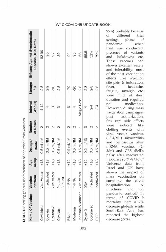
W4C COVID-19 UPDATE BOOK
392
TAB
LE 1
: Sho
win
g ge
nera
l cha
ract
eris
tics
of a
ppro
ved
Covi
d Va
ccin
es
Nam
e O
f Vac
cine
Vacc
ine
Plat
form
Age
Gro
upD
osag
e/R
oute
Num
ber
of D
oses
Inte
rval
(W
eeks
)St
orag
e 0 C
Effic
acy
Agai
nst S
ympt
omat
ic
Dis
ease
(Tria
l Dat
a)
Covi
shie
ldVi
ral V
ecto
r+1
80.
5 m
l /IM
24-
122-
862
-90
Cova
xin
Inac
tivat
ed+1
80.
5 m
l/IM
24
2-8
80Sp
utni
k-V
Vira
l Vec
tor
+18
0.5
ml/
IM2
3-1
892
Cova
vax
Prot
ein
Subu
nit
+18
0.5
ml/
IM2
32-
889
Pfize
rm
-RN
A+1
20.
5 m
l/ IM
23
-70
94M
oder
nam
-RN
A+1
80.
5 m
l/IM
24
-20
95Jo
hnso
n &
John
son
Vira
l Vec
tor
+18
0.5
ml/
IM1
Sing
le D
ose
2-8
66Zy
cov-
DD
NA
+12
0.2
ml/
ID3
42-
866
.6Co
rona
vac
Inac
tivat
ed+1
60.
5 m
l/IM
22-
42-
851
%BI
BPIn
activ
ated
+18
0.5
ml/
IM2
3-4
2-8
79%
95%) probably because of different trial settings, phase of pandemic when trial was conducted, presence of variants and lockdowns etc. These vaccines had shown excellent safety and tolerability; most of the post vaccination effects like injection site pain & induration, fever, headache, fatigue, myalgia etc. were mild, of short duration and required no medication. However, during mass vaccination campaigns, post authorization, few rare side effects were noticed like clotting events with viral vector vaccines ( 3-4/M ), myocarditis and pericarditis after mRNA vaccines (2-3/M) and GBS /Bell’s palsy after inactivated v a c c i n e s . ( 7 - 8 / M ) . 6 Universe data from Israel and UK have shown the impact of mass vaccination on curtailing the covid hospitalization & infections and on pandemic control.7 In terms of COVID-19 mortality there is 7% decrease globally while South-East Asia has reported the highest decrease (27%).1
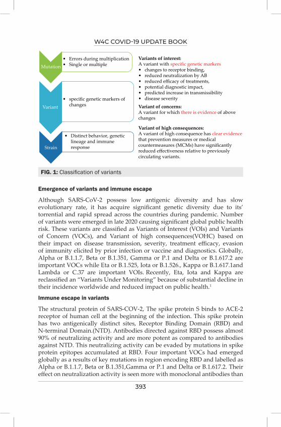
W4C COVID-19 UPDATE BOOK
393
Emergence of variants and immune escape
Although SARS-CoV-2 possess low antigenic diversity and has slow evolutionary rate, it has acquire significant genetic diversity due to its’ torrential and rapid spread across the countries during pandemic. Number of variants were emerged in late 2020 causing significant global public health risk. These variants are classified as Variants of Interest (VOIs) and Variants of Concern (VOCs), and Variant of high consequences(VOHC) based on their impact on disease transmission, severity, treatment efficacy, evasion of immunity elicited by prior infection or vaccine and diagnostics. Globally, Alpha or B.1.1.7, Beta or B.1.351, Gamma or P.1 and Delta or B.1.617.2 are important VOCs while Eta or B.1.525, Iota or B.1.526., Kappa or B.1.617.1and Lambda or C.37 are important VOIs. Recently, Eta, Iota and Kappa are reclassified an “Variants Under Monitoring” because of substantial decline in their incidence worldwide and reduced impact on public health.1
Immune escape in variants
The structural protein of SARS-COV-2, The spike protein S binds to ACE-2 receptor of human cell at the beginning of the infection. This spike protein has two antigenically distinct sites, Receptor Binding Domain (RBD) and N-terminal Domain.(NTD). Antibodies directed against RBD possess almost 90% of neutralizing activity and are more potent as compared to antibodies against NTD. This neutralizing activity can be evaded by mutations in spike protein epitopes accumulated at RBD. Four important VOCs had emerged globally as a results of key mutations in region encoding RBD and labelled as Alpha or B.1.1.7, Beta or B.1.351,Gamma or P.1 and Delta or B.1.617.2. Their effect on neutralization activity is seen more with monoclonal antibodies than
FIG. 1: Classification of variants
Mutation
Variants of interest:A variant with specific genetic markers• changes to receptor binding,• reduced neutralization by AB• reduced efficacy of treatments,• potential diagnostic impact,• predicted increase in transmissibility• disease severity
• Errors during multiplication• Single or multiple
• specific genetic markers of changes
• Distinct behavior, genetic lineage and immune response
Variant of concerns:A variant for which there is evidence of abovechanges
Variant of high consequences:A variant of high consequence has clear evidencethat prevention measures or medicalcountermeasures (MCMs) have significantlyreduced effectiveness relative to previouslycirculating variants.
Variant
Strain
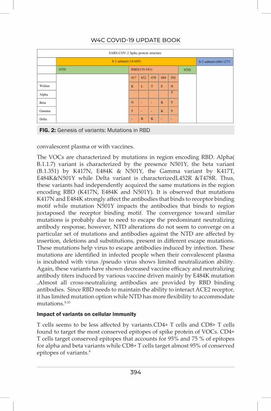
W4C COVID-19 UPDATE BOOK
394
convalescent plasma or with vaccines.
The VOCs are characterized by mutations in region encoding RBD. Alpha( B.1.1.7) variant is characterized by the presence N501Y, the beta variant (B.1.351) by K417N, E484K & N501Y, the Gamma variant by K417T, E484K&N501Y while Delta variant is characterizedL452R &T478R. Thus, these variants had independently acquired the same mutations in the region encoding RBD (K417N, E484K and N501Y). It is observed that mutations K417N and E484K strongly affect the antibodies that binds to receptor binding motif while mutation N501Y impacts the antibodies that binds to region juxtaposed the receptor binding motif. The convergence toward similar mutations is probably due to need to escape the predominant neutralizing antibody response, however, NTD alterations do not seem to converge on a particular set of mutations and antibodies against the NTD are affected by insertion, deletions and substitutions, present in different escape mutations. These mutations help virus to escape antibodies induced by infection. These mutations are identified in infected people when their convalescent plasma is incubated with virus /pseudo virus shows limited neutralization ability. Again, these variants have shown decreased vaccine efficacy and neutralizing antibody titers induced by various vaccine driven mainly by E484K mutation .Almost all cross-neutralizing antibodies are provided by RBD binding antibodies. Since RBD needs to maintain the ability to interact ACE2 receptor, it has limited mutation option while NTD has more flexibility to accommodate mutations.9,10
Impact of variants on cellular immunity
T cells seems to be less affected by variants.CD4+ T cells and CD8+ T cells found to target the most conserved epitopes of spike protein of VOCs. CD4+ T cells target conserved epitopes that accounts for 95% and 75 % of epitopes for alpha and beta variants while CD8+ T cells target almost 95% of conserved epitopes of variants.9
FIG. 2: Genesis of variants: Mutations in RBD
B.1.351,Gamma or P.1 and Delta or B.1.617.2. Their effect on neutralization activity is seen
more with monoclonal antibodies than convalescent plasma or with vaccines.
Fig. 2: Genesis of variants: Mutations in RBD
SARS-COV-2 Spike protein structure
S-1 subunit (14-685) S-2 subunit (686-1273
NTD RBD(319-541) NTD
417 452 478 484 501
Wuhan K L T E N
Alpha Y
Beta N - - K Y
Gamma T - - K Y
Delta - R K - -
The VOCs are characterized by mutations in region encoding RBD. Alpha( B.1.1.7) variant is
characterized by the presence N501Y, the beta variant (B.1.351) by K417N, E484K &
N501Y, the Gamma variant by K417T, E484K&N501Y while Delta variant is
characterizedL452R &T478R. Thus, these variants had independently acquired the same
mutations in the region encoding RBD (K417N, E484K and N501Y). It is observed that
mutations K417N and E484K strongly affect the antibodies that binds to receptor binding
motif while mutation N501Y impacts the antibodies that binds to region juxtaposed the
receptor binding motif. The convergence toward similar mutations is probably due to need to
escape the predominant neutralizing antibody response, however, NTD alterations do not
seem to converge on a particular set of mutations and antibodies against the NTD are affected
by insertion, deletions and substitutions, present in different escape mutations. These
mutations help virus to escape antibodies induced by infection. These mutations are identified
in infected people when their convalescent plasma is incubated with virus /pseudo virus
shows limited neutralization ability. Again, these variants have shown decreased vaccine

W4C COVID-19 UPDATE BOOK
395
TAB
LE 2
: Var
iant
s an
d th
eir p
heno
typi
c im
pact
ALPH
AB
ETA
GAM
MA
DEL
TA
Line
age
B.1.
1.7
B.1.
351
P.1
B.1.
617.
2Pl
ace
of d
etec
tion
UKSA
Braz
ilIn
dia
Tim
e of
det
ectio
nSE
PT 2
0M
AY 2
0OC
T 20
NOV
20
Tran
smis
sibi
lity
++++
++++
and
incr
ease
d se
cond
ary
atta
ck ra
teD
isea
se s
ever
ity++
risk
of i
nfec
tion,
ho
spita
lizat
ion
and
mor
talit
y
++ ri
sk o
f inf
ectio
n,
hosp
italiz
atio
n an
d m
orta
lity
++ ri
sk o
f inf
ectio
n,
hosp
italiz
atio
n an
d m
orta
lity
++ ri
sk o
f inf
ectio
n,
hosp
italiz
atio
n an
d m
orta
lity
Redu
ctio
n in
ne
utra
lizin
g ac
tivity
NO
++++
++
Risk
of r
einf
ectio
n++
++++
++Im
pact
on
diag
nost
ics
No
NO
Not
repo
rted
Not
repo
rted
Vacc
ine
effic
acy
Prot
ectio
n re
tain
ed
agai
nst a
ll ou
tcom
esPr
otec
tion
reta
ined
ag
ains
t sev
ere
dise
ase
redu
ced
prot
ectio
n ag
ains
t sym
ptom
atic
di
seas
e
Uncl
ear i
mpa
ct a
nd
Lim
ited
evid
ence
Prot
ectio
n re
tain
ed
agai
nst s
ever
e di
seas
e re
duce
d pr
otec
tion
agai
nst s
ympt
omat
ic
dise
ase
W4C COVID-19 UPDATE BOOK
396
CONCLUSIONAlthough variants affect the transmission and severity of disease by escaping through the immunity, vaccine induced neutralizing antibodies still protects from severe illness. Monitoring the prevalence of variants and its’ characterization is of paramount importance to detect “potential signal”.
PRACTICE POINTSVaccination is an important tool to curtail the pandemic. Though various SARS-COV-2 variants emerged during pandemic, the extraordinary COVID-19 vaccines still retain their ability to protect albeit low neutralization activity. The variants have less impact on cellular immunity induced by vaccines. Antibodies against RBD, memory B cells and T cell responses elicited by vaccine will help to prevent the infection.
REFERENCES1. WHO, Covid-19 weekly epidemiological update, edition 58, dated 21/09/20212. WHO, coronavirus (covid-19) dashboard dated 24/09/20213. Yang M, Lai CL. COVID-19 Vaccines: A Review of the Safety and Efficacy of Current Clinical
Trials. Pharmaceuticals 2021; 14:406. https://doi.org/10.3390 ph14050406.4. Shahcheragi et al An overview of vaccine development for COVID-19. Ther Deliv 2021; 12:235–
244.5. WHO, COVID-19 vaccine tracker and landscape: updated on 24 Sept 2021.6. CDC: Science Brief: COVID-19 Vaccines and Vaccination, Updated Sept. 15, 20217. Hass et al Impact and effectiveness of mRNA BNT162b2 vaccine against SARS-CoV-2
infections and COVID-19 cases, hospitalisations, and deaths following a nationwide vaccination campaign in Israel: an observational study using national surveillance data. The Lancet 2021; 397:1819-1829.
8. Lazarevic, I.; Pravica, V.Miljanovic, D.; Cupic, M. Immune Evasion of SARS-CoV-2 Emerging Variants: What HaveWe Learnt So Far? Viruses 2021; 13:1192. https://doi.org/10.3390/v13071192
9. Andreano E, Rappuoli R. SARS-CoV-2 escaped natural immunity, raising questions about vaccines and therapies, Nature Medicine 2021; 27:759–765.
10. Wang et al Antibody resistance of SARS-CoV-2 variants B.1.351 and B.1.1.7, 130 |. Nature 2021; 593.
397
COviD-19 vaccination Breakthrough infections 53.
Rakesh Garg, Indubala Maurya, Ayush Lohiya
1. INTRODUCTIONThe outbreak of coronavirus disease (COVID-19), caused by severe acute respiratory syndrome coronavirus 2 (SARS-CoV-2), was declared pandemic in March 2020. The development of vaccines against COVID-19 started in the very early phase of the pandemic. Currently, 112 vaccines are in clinical phase while 183 are in preclinical trials, as per World Health Organization (WHO).1 However, just like vaccines for other diseases, none of the available vaccines are 100% effective. Hence, some breakthrough infections in vaccinated individuals are expected. The Centres for Disease Control and Prevention (CDC) has defined a vaccine breakthrough infection as a positive test (PCR or antigen) on a respiratory specimen (nasal swab, nasopharyngeal swab, oropharyngeal swab, saliva, sputum, bronchoalveolar lavage fluid, pleural fluid etc.) collected ≥14 days after completing recommended doses of COVID-19 vaccine. It does not include those infections where the person has received an unauthorized COVID-19 vaccine or if the sample was collected within 14 days of vaccination as the protection against disease develops in approximately 14 days. As of August 2021, CDC has received reports of more than 9,500 patients with COVID-19 breakthrough infections out of more than 168 million fully vaccinated individuals.2
2. ETIOLOGYBreakthrough infections are reported in a proportion of fully vaccinated individuals. Till date there are no specific patterns seen in individuals with breakthrough infections and various etiologies has been reported (Table 1).
2.1. Vaccine Related Factors
No vaccine is 100% effective. A proportion of breakthrough infections are expected in controlled as well as clinical settings and vary with the type of vaccine. There are many reasons for that. Firstly, many different platforms including inactivated virus, live attenuated viruses, mRNA vaccines,
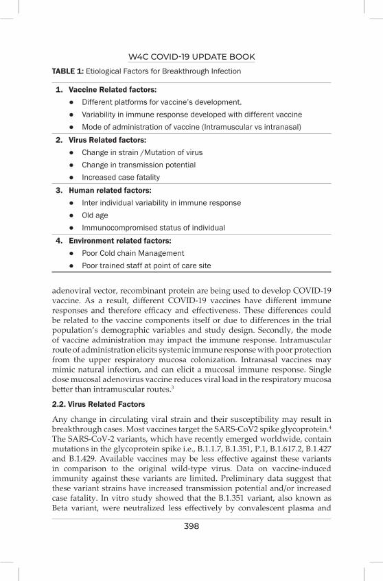
W4C COVID-19 UPDATE BOOK
398
adenoviral vector, recombinant protein are being used to develop COVID-19 vaccine. As a result, different COVID-19 vaccines have different immune responses and therefore efficacy and effectiveness. These differences could be related to the vaccine components itself or due to differences in the trial population’s demographic variables and study design. Secondly, the mode of vaccine administration may impact the immune response. Intramuscular route of administration elicits systemic immune response with poor protection from the upper respiratory mucosa colonization. Intranasal vaccines may mimic natural infection, and can elicit a mucosal immune response. Single dose mucosal adenovirus vaccine reduces viral load in the respiratory mucosa better than intramuscular routes.3
2.2. Virus Related Factors
Any change in circulating viral strain and their susceptibility may result in breakthrough cases. Most vaccines target the SARS-CoV2 spike glycoprotein.4 The SARS-CoV-2 variants, which have recently emerged worldwide, contain mutations in the glycoprotein spike i.e., B.1.1.7, B.1.351, P.1, B.1.617.2, B.1.427 and B.1.429. Available vaccines may be less effective against these variants in comparison to the original wild-type virus. Data on vaccine-induced immunity against these variants are limited. Preliminary data suggest that these variant strains have increased transmission potential and/or increased case fatality. In vitro study showed that the B.1.351 variant, also known as Beta variant, were neutralized less effectively by convalescent plasma and
TABLE 1: Etiological Factors for Breakthrough Infection
1. Vaccine Related factors: ● Different platforms for vaccine’s development. ● Variability in immune response developed with different vaccine ● Mode of administration of vaccine (Intramuscular vs intranasal)2. Virus Related factors: ● Change in strain /Mutation of virus ● Change in transmission potential ● Increased case fatality 3. Human related factors: ● Inter individual variability in immune response ● Old age ● Immunocompromised status of individual 4. Environment related factors: ● Poor Cold chain Management ● Poor trained staff at point of care site

W4C COVID-19 UPDATE BOOK
399
post vaccination sera.5 Delta variant (B.1.617.2) of SARS-CoV-2 virus, first detected in India, showed enhanced transmission and severity of illness.
2.3. Human Related Factors
Not all individuals who have been vaccinated would show a similar immune response to specific vaccines. Any individuals with immunocompromised comorbid condition or on immunosuppressive agents, may have poor immunogenic response to COVID-19 vaccines compared to the general population.6,7 The incidence of a breakthrough infection may be higher in elderly due to weak immune response to vaccines. Post vaccination immune response is complex. There is interplay between Macrophages, B cell and T cell. T cells are important for destroying infected cells, supporting B cell and antibody responses to SARS-CoV-2 infection thus reducing the risk of vaccine-induced severe disease. The T-cell response may vary across all immunocompetent vaccine-recipients as well. Poor T cell response can make individuals susceptible to infection in spite of good neutralizing antibody titres.
2.4. Environment Related Factors
As most of the vaccines need strict temperature control, poor cold chain during shipping/storage/administration can result in partial vaccine degradation. For instance, messenger RNA (mRNA) vaccine needs specific storage temperature of around -70 degree centigrade. Breaking the cold chain may lead to denaturation/degradation of the vaccine and therefore may reduce the effectiveness of the vaccine. Likewise, administration of vaccines should be as per standard protocols so that the immune response is not affected.
3. PATHOGENESIS OF BREAKTHROUGH INFECTIONS The immune response of the human body is different for various vaccines (Table 2). The mRNA-based BNT162b2 (BioNTech/Pfizer) vaccine has demonstrated 95% efficacy after 2 doses in phase III trials. The S1-binding antibodies and significant neutralizing antibodies were present in vaccinated individuals after the second dose.8 The cell mediated immune response was also demonstrated by the increase in antigen-specific CD4 and CD8 T cells after the second dose of this mRNA vaccine.9,10 As a result of immune response developed due to the vaccination, the BioNTech/Pfizer vaccine has 87% effectiveness against hospitalization and 86-92% against any infection after 2 doses of vaccination.4 The other commonly used mRNA-based vaccine is mRNA-1273 (Moderna). It has 95% efficacy after 2 doses and 90% effectiveness against symptomatic infection.11 This vaccine also stimulates the production of S-binding antibodies and neutralizing antibodies. It is also accompanied by the significant increase in the CD4 T cells and low level of CD8 responses. In the HEROES-RECOVER cohort network in the United States, 3 and 2 new infections were reported after the first (31,231 person-days) and second (40,394 person-days) dose of Moderna vaccine. The viral
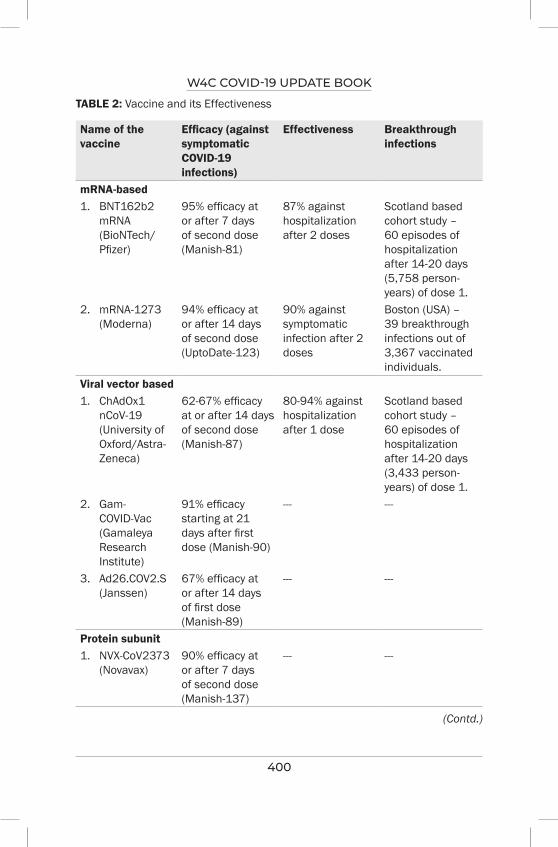
W4C COVID-19 UPDATE BOOK
400
TABLE 2: Vaccine and its Effectiveness
Name of the vaccine
Efficacy (against symptomatic COVID-19 infections)
Effectiveness Breakthrough infections
mRNA-based1. BNT162b2
mRNA (BioNTech/Pfizer)
95% efficacy at or after 7 days of second dose (Manish-81)
87% against hospitalization after 2 doses
Scotland based cohort study – 60 episodes of hospitalization after 14-20 days (5,758 person-years) of dose 1.
2. mRNA-1273 (Moderna)
94% efficacy at or after 14 days of second dose (UptoDate-123)
90% against symptomatic infection after 2 doses
Boston (USA) – 39 breakthrough infections out of 3,367 vaccinated individuals.
Viral vector based1. ChAdOx1
nCoV-19 (University of Oxford/Astra-Zeneca)
62-67% efficacy at or after 14 days of second dose (Manish-87)
80-94% against hospitalization after 1 dose
Scotland based cohort study – 60 episodes of hospitalization after 14-20 days (3,433 person-years) of dose 1.
2. Gam-COVID-Vac (Gamaleya Research Institute)
91% efficacy starting at 21 days after first dose (Manish-90)
--- ---
3. Ad26.COV2.S (Janssen)
67% efficacy at or after 14 days of first dose (Manish-89)
--- ---
Protein subunit1. NVX-CoV2373
(Novavax)90% efficacy at or after 7 days of second dose (Manish-137)
--- ---
(Contd.)
W4C COVID-19 UPDATE BOOK
401
vector-based ChAdOx1nCoV-19 (University of Oxford/Astra-Zeneca) has 62-67% efficacy after 2 doses.12 Effectiveness against hospitalization was 80-94% after 1 dose. This vaccine also works by stimulating the development of antibody- and cell-mediated immunity. Other vaccines for COVID-19 have demonstrated efficacy in trial settings but the data from real world studies are yet to be published.
4. SEVERITY AND MANAGEMENT OF BREAKTHROUGH COVID-19 INFECTIONThe CDC has a system for reporting post-vaccination COVID-19 cases. As of 16th August 2021, the USA has more than 166 million fully vaccinated individuals. During the same duration, 9,176 breakthrough infections have been reported to CDC through their reporting system. Out of these, 7,887 were hospitalized but non-fatal, and 1,829 were fatal infections. Out of hospitalized, non-fatal infections, 70% were among people aged 65 years or more and among fatal infections, 88% were among people aged 65 years or more. Likewise, among hospitalized, non-fatal infections, 23% were reported as asymptomatic or not related to COVID-19 and among fatal infections, 20% were reported as asymptomatic or not related to COVID-19.2 A study done in Israel among healthcare workers also reported 39 breakthrough infections among 1,497 fully vaccinated healthcare workers. Out of 39, 33% were asymptomatic, the rest reported only mild symptoms, and none required hospitalization. Almost one-fifth of the patients reported COVID-19 symptoms for prolonged duration (>6 weeks).13 A few Indian studies have also reported the burden of breakthrough infections. A study from North India reported results of 2-month follow-up after vaccination. Among those who received a single dose (n=65), a total of 30 infections were detected, either laboratory confirmed (n=27) or suspects (n=3). Out of these 30, 21 (70.0%) were mild infections, 5 (16.7%) had moderate infection, and 2 (6.7%) had severe infections. Of those who received both doses (n=1,435), a total of 388 infections were detected, either laboratory confirmed (n=271) or suspects (n=117). Out of these 388,
Name of the vaccine
Efficacy (against symptomatic COVID-19 infections)
Effectiveness Breakthrough infections
Whole-cell inactivated virus1. BBIBP-CorV
(Sinopharm)86% after 2 doses (Manish-145)
--- ---
2. BBV152 (Bharat Biotech)
81% after 2 doses (UpToDate-167)
--- ---
TABLE 2: Vaccine and its Effectiveness (Contd.)
W4C COVID-19 UPDATE BOOK
402
331 (85.3%) were mild infections, 33 (8.5%) had moderate infection, and 6 (1.5%) had severe infections.14 In a study from a tertiary care centre in South India, out of 1,878 HCWs who received a single dose, 200 (10.6%) developed infection, 22 (1.2%) were hospitalized, no needed oxygen therapy and ICU care, and there were no deaths. Likewise, out of 7,080 fully vaccinated HCWs, 679 (9.6%) developed infection, 64 (0.9%) were hospitalized, 4 (0.06%) needed oxygen therapy, 2 (0.03%) needed ICU care, and there were no deaths.15 All breakthrough COVID-19 infection should be treated as per current COVID-19 treatment protocol according to severity.
5. PREVENTION OF BREAKTHROUGH INFECTION Due to dominance of Delta and/or other variant viruses, breakthrough infections seem more frequent than expected. Thus, we must implement possible preventive measures to curb breakthrough infection after vaccination.
5.1 COVID-19 Appropriate Behaviour
All vaccinated individuals should wear masks in public places as they can be a source for transmission following breakthrough infection with the mutant variant (eg. Delta). As immunocompromised individuals have weak responses to COVID-19 vaccination, they must follow personal preventive measures even in areas with low transmission rate. There is a need to raise the awareness among individuals to follow recommended personal hygiene such as using face masks, hands washing, use of alcohol-based hand sanitizers, avoid touching mouth/nose, and social distancing even after vaccination. All individuals must avoid closed, crowded, close contact (3Cs) to prevent transmission of COVID-19. In case of any symptoms suggestive of COVID-19, consider home isolation or medical attention as per severity. Society’s active participation is vital in the prevention of the spread of the pandemic.
All healthcare workers are at risk of breakthrough infection due to direct or indirect occupational exposure to SARS-Co-2. They must take necessary preventive measures to minimise COVID-19 infection even after full vaccination. The workplace risk (lower/medium/high/very high risk) should be taken into consideration for pragmatic use of PPE.
5.2 Strict Cold Chain Management
To maintain potency and prevent degradation of vaccine a robust cold chain management is must. It includes maintenance of appropriate temperature for vaccines during manufacturing process, storage, transportation and distribution till point of care level.
5.3 Need a Booster Dose
Because of the decrease in neutralizing antibodies over the time and poor efficacy against new mutant variant viruses, the vaccinated individuals may need booster doses to enhance acquired immunity.16 Though few studies
W4C COVID-19 UPDATE BOOK
403
have shown promising results, the need for booster dose vaccination is still not known. A group of vaccinated individuals with mRNA- 1273 vaccine received a boosting dose with modified mRNA vaccine six to eight months after full vaccination. The neutralizing antibody levels against wild-type virus and mutant variants that were higher than initial vaccine doses.17 In another preclinical study, intramuscular administration of a chimpanzee adeno viral vectored COVID-19 vaccine candidate triggered both cellular and humoral immunity in adult and old mice. A booster dose of this vaccine showed enhanced spike-specific IgG and neutralizing antibody level in older mice.
5.4 Modification in Vaccine /Schedule/Route
Search is still going on to enhance vaccine induced immune response including against new variants and to decrease incidence of asymptomatic carriers. There are studies going on the possibility of mixing two different types of COVID-19 vaccine in a schedule to get better immune response. Several countries are looking forward to a heterologous prime-boost schedule in which the ChAd COVID-19 vaccine (AstraZeneca) vaccine will be used for the first dose and mRNA vaccines COVID-19 vaccine (Pfizer–BioNTech) vaccine as second dose. The data on the immunogenicity and safety of such schedules still awaited We must also aim to prevent asymptomatic infection to block transmission. Initial analysis has shown that the mRNA vaccines also reduce the incidence of asymptomatic infection and the nasopharyngeal viral load in individuals with breakthrough infection. Intranasal routes have shown to have the potential to control infection in both the upper and lower respiratory tracts thus preventing transmission.3
PRACTICE POINTS• Breakthrough infection after vaccination is inevitable.
• Majority of breakthrough infections are asymptomatic which can lead to transmission of infection specially to vulnerable individuals.
• Take adequate precaution at individual level to curb the infection
• Role of Booster and mixing of vaccine for better immunogenicity needs to be studied.
REFERENCES1. who.int. COVID-19 vaccine tracker and landscape [Internet]. Available from: https://www.
who.int/publications/m/item/draft-landscape-of-covid-19-candidate-vaccines. Accessed on August 17, 2021.
2. cdc.giv.Vaccine and immunizations [Internet]. Available from: https://www.cdc.gov/vaccines/covid-19/health-departments/breakthrough-cases.html. Assessed on 26Aug2021.
3. Hassan AO, Kafai NM, Dmitriev IP, Fox JM, Smith BK, Harvey IB,et al. A single-dose intranasal ChAd vaccine protects upper and lower respiratory tracts against SARS-CoV2. Cell 2020; 183:169–184.
4. Dai L, Gao GF. Viral targets for vaccines against COVID-19. Nature Rev Immunol. 2021; 21:73–82.
W4C COVID-19 UPDATE BOOK
404
5. Wibmer CK, Ayres F, Hermanus T, Madzivhandila M, Kgagudi P, Oosthuysen B et al. SARS-CoV-2 501Y.V2 escapes neutralization by South African COVID-19 donor plasma. Nat Med 2021; 27:622-625.
6. Boyarsky BJ, Werbel WA, Avery RK, Tobian AAR, Massie AB, Segev DL et al. Antibody Response to 2-Dose SARS-CoV-2 mRNA Vaccine Series in Solid Organ Transplant Recipients. JAMA 2021; 1;325:2204-2206.
7. Monin L, Laing AG, Muñoz-Ruiz M, McKenzie DR, Del Molino Del Barrio I, Alaguthurai T et al. Safety and immunogenicity of one versus two doses of the COVID-19 vaccine BNT162b2 for patients with cancer: interim analysis of a prospective observational study. Lancet Oncol 2021; 22:765-778.
8. Walsh EE, Frenck RW Jr, Falsey AR, Kitchin N, Absalon J, Gurtman A et al. Safety and Immunogenicity of Two RNA-Based Covid-19 Vaccine Candidates. N Engl J Med 2020; 383:2439-2450.
9. Hall VJ, Foulkes S, Saei A, Andrews N, Oguti B, Charlett A. COVID-19 vaccine coverage in health-care workers in England and effectiveness of BNT162b2 mRNA vaccine against infection (SIREN): a prospective, multicentre, cohort study. Lancet 2021; 397:1725-1735.
10. Vasileiou E, Simpson CR, Shi T, Kerr S, Agrawal U, Akbari A et al. Interim findings from first-dose mass COVID-19 vaccination roll-out and COVID-19 hospital admissions in Scotland: a national prospective cohort study. Lancet 2021; 397:1646-1657.
11. Baden L, Hana M, Sahly E, Essink B, Kotloff K,Frey S et al. Efficacy and safety of the mRNA-1273 SARS-CoV-2 vaccine. N Engl J Med 2020; 384:403-416.
12. Voysey M, Clemens SAC, Madhi SA, Weckx LY, Folegatti PM, Aley PKet al. Safety and efficacy of the ChAdOx1nCoV-19 vaccine (AZD1222) against SARS-CoV-2: an interim analysis of four randomised controlled trials in Brazil, South Africa, and the UK. Lancet 2021; 397(10269):99-111.
13. Bergwerk M, Gonen T, Lustig Y, Amit S, Lipsitch M, Cohen C, et al. Covid-19 Breakthrough Infections in Vaccinated Health Care Workers. N Engl J Med 2021; 1–11.
14. Kaur U, Bala S, Ojha B, Chakrabarti SS. Occurrence of COVID-19 in priority groups receiving ChAdOx1 nCoV-19 coronavirus vaccine (recombinant): a preliminary analysis from north India.2021.DOI: 10.21203/rs.3.rs-772465/v1.Available from: https://www.researchsquare.com/article/rs-772465/v1. Assessed on 28 Aug 2021.
15. Victor PJ, Mathews KP, Paul H, Mammen JJ, Murugesan M. Protective Effect of COVID-19 Vaccine Among Health Care Workers During the Second Wave of the Pandemic in India. Mayo Clin Proc 2021; 26:S0025-6196(21)00470-5.doi: 10.1016/j.mayocp.2021.06.003. Epub ahead of print.
16. Wajnberg A, Amanat F, Firpo A, Altman DR, Bailey MJ, Mansour M et al. SARS-CoV-2 infection induces robust, neutralizing antibody responses that are stable for at least three months. Science 2020; 370:1227–30.
17. Silva-Cayetano A, Foster WS, Innocentin S, Belij-Rammerstorfer S, Spencer AJ, Burton OT, et al. A booster dose enhances immunogenicity of the COVID-19 vaccine candidate ChAdOx1 nCoV-19 in aged mice. Med 2021; 2:243-262.
Clinical Recurrence of COVID 19 Infection
SECTION 10


407
Section 10 - Clinical Recurrence of Covid19 InfectionSECTION EDITOR
Dr Banambar RayMD, FICCM Head of Critical Care Medicine, SUM Ultimate Medicare, A Unit of SOA,Bhubaneswar
Preface
Autoimmune diseases (AIDs) are now seen following COVID-19 and the cause of this development is not yet ascertained. Long after viral activity has receded, patients come with Inflammatory myositis, IgA vasculitis, GB Syndrome, Rheumatoid arthritis and Multisystem Involvement in adults and children (MIS-A & MIS-C). The authors have explained various possible theories to explain the predisposition and causation of this development. As the AIDs appear in
the post Covid-19 phase, patients suffer for a long time as their immunity deficiency remains at the root of their suffering. Authors, rightly, have taken some examples to explain the present state of our understanding.
Viral relapse / reinfection has been seen not infrequently after a primary COVID-19 infection. Various studies define reinfection differently. Authors have explained how definitions of cure from COVID-19 have influenced the incidence of reinfection / relapse. Use of long term corticosteroids and liberal use of tocilizumab by certain physicians may also be responsible for relapse / reinfection of COVID-19. It is good news that these infections are not serious ones and that patients after a primary infection get a good immunity for 6-12 months.

409
viral Relapse or Reinfection/ inflammatory Rebound 54.
INTRODUCTIONSARS-CoV-2 was initially thought to cause infection only once in an individual. It was hypothesised that since SARS-CoV-2 virus is immunologically cleared from the body, the immunity developed will help in preventing re-infection; however it is well known that re-infections do occur in respiratory viruses. The causes for re-infection are weak immune response or re-infection due to different variants/quasispecies.
There are many case reports/series of relapse and re-infection that have been described during this pandemic. In this chapter, we try to elaborate the so far analysed mechanism behind relapse/re-infection and its management.
VIRAL PERSISTENCE, RELAPSE AND RE-INFECTIONEarly in the pandemic last year Gousseff et al described a case series of 11 virologically confirmed infections in France who had a second episode.¹ This occurred after an interval of 4-27 days in their series. The hypotheses proposed for the recurrences are viral reinfection or viral reactivation which may have occurred due to sub-optimal clearance of the virus during the initial episode. A major lacuna in this study was that initial cure was defined clinically and not confirmed by negative virological report.
Laboratory based studies have reported that viral shedding becomes minimum by 28 days but may last up to 12 weeks. Hence positive RT-PCR report till 3 months post infection may signify persistent viral shedding and clearing from the respiratory tract. These are mostly fragmented RNA particles. Up to 10% of cases may show re-positive RT-PCR following a negative report till 90 days. A Dutch study analysed 41 cases of relapse who had a median symptom free interval of 12 days.² In this study the median Cycle threshold (Ct) value was higher in the re-positive samples with mean Ct value of 34.8. The patients who re-test positive in this period are usually not infectious with evidence of transmission among contacts lacking. Increasing Ct value is associated with decrease in probability of culturing virus with only 8% showing
Mradul Kumar Daga, Siddharth Chand
W4C COVID-19 UPDATE BOOK
410
positive culture with Ct >35; transmissibility of the virus is negligible with Ct > 30. Viral cultures have been found to be negative in patients of suspected reinfection who re-test positive within 90 days.
The clinical recurrence of symptoms may occur due to many reasons. Superinfection by bacteria or fungus may be a factor due to immune suppression by corticosteroids and biological agents. COVID-19 increases risk of pulmonary embolism, myocardial infarction, myocarditis which may present with similar complaints of breathlessness.
Considering all these, Yahav et al proposed re-infection to be defined as recurrence with positive RT-PCR tests more than 90 days after onset of first episode.³ They have further suggested the following definition of re-infection:
• Establishment of true first episode with viral load defined as Ct < 35
• Re-infection confirmed with two RT-PCR tests with Ct < 35 or establishment of viral replication by culture or detection of subgenomic RNA
• Confirmation of infection with different strain established by genomic sequencing
• Negative RT-PCR between the two episodes
COVID-19 relapse has been further defined as symptom complex compatible with infection with COVID-19 with positive or persisting RT-PCR test within 90 days of first episode and absence of any uther possible cause for the symptoms. Demonstration of the same viral genome will differentiate this from a possible case of re-infection. Most cases of relapse occur in the first two weeks itself with cases having mild symptoms and similar neutralizing antibodies compared to primary episode.⁴ Gousseff et al proposed the hypothesis of an inflammatory rebound triggered by an inappropriate immune response as an explanation to the recurrence of clinical symptoms.¹
Corticosteroid is now considered a standard of care since it was shown to reduce mortality in patients requiring respiratory support. However steroid has also shown to increase the time required for viral clearance.⁵ Widespread steroid usage and use of immune suppressants like tocilizumab possibly contribute to persistent viral replication, RT-PCR positivity and COVID relapse. Cases of viral relapse in immune compromised patients have also been reported.
To summarize, any person who retests positive for the second time, could have a
• Viral relapse – ongoing viral replication and disease persistence.
• Viral persistence – detection of fragmented viral genome (dead particles)
• Re-infection – after 90 days of first episode
W4C COVID-19 UPDATE BOOK
411
Re-Infection
Case reports/series of re-infection are few and limited. In a recently published review Pinto et al analysed cases published till October 2020.⁶ They studied 27 cases of which 13 were confirmed cases of re-infection with whole genome sequencing. Most of the cases had mild category severity in the second episode; however the severity was greater than the first in 8 of the 13 cases.
IMMUNITY AND RE-INFECTION
Studies indicate SARS-CoV-2 infection generates both humoral as well as cell mediated immunity. Subsequently patients, who recover from COVID-19, have memory B and T cells. There is lack of clarity regarding persistence of immunity. Specific IgG, IgM and IgA have been detected early in the course of the disease. IgG levels have been shown to remain detectable for at least 6 months and presence of IgG was associated with reduced risk of re-infection.⁷ One study from Italy reported presence of IgG against anti-spike-receptor binding domain (RBD) till 14 months after infection.⁸ Similar observations were made in a study of 63 recovered patients.⁹ Antibody reactivity to the RBD, neutralizing activity and the number of RBD-specific memory B cells remained relatively stable between 6 and 12 months after infection. Nonetheless neutralizing activity against variants was generally lower than against wild type SARS-CoV-2. In this study vaccination with mRNA vaccines increased all the components of humoral immunity. Vaccination also increased the neutralization activity against variants. However a study in China showed decay of neutralizing antibodies with persistence of T cell immune response.¹⁰
This is in contrast to our experience in previous pandemics caused by coronaviruses. Antibodies were detected in survivors of SARS-CoV-1 and MERS-CoV epidemics up to 2 years. The risk of re-infection could not be assessed in these patients as the pandemics were effectively controlled. Re-infection with endemic corona viruses 229E, HKU1, NL63 and OC43 is common probably due to short lasting immunity and development of different variants.
Multiple studies are now confirming that patients who do not seroconvert after their first infection remain susceptible to re-infection.⁷ It is still not clear why some patients do not seroconvert after initial infection. This may be possible because such patients were never truly infected and were labelled positive because of false positive RT-PCR report.
CONCLUSIONViral relapse, recurrence and re-infection remain an active area of interest. Present evidence suggests that re-positive RT-PCR test can occur in about 10% patients. These patients are largely of mild severity and are not infectious to their contacts. Strict isolation may not be necessary for such patients.
W4C COVID-19 UPDATE BOOK
412
Immunity developed from primary infection remains protective at least for 6-12 months. This is important for policy makers in charge for vaccination drive. Such patients can wait six months for their inoculation.
Re-infection remains a risk factor with emergence of different variants. However, cases of genomic proven re-infection are very rare till date. Most cases of re-infection are of milder severity.
PRACTICE POINTS1. Patients may shed viral particles till 12 weeks after primary infection and
may re-test positive during that period. Such patients are usually not infective and need to be isolated.
2. Patients having clinical recurrence within 90 days should be evaluated for secondary causes like super added infection, pulmonary embolism etc.
3. Patients can be labelled as having re-infection only after whole genomic sequencing is done.
REFERENCES 1. Gousseff M, Penot P, Gallay L, Batisse D, Benech N, Bouiller K, et al. Clinical recurrences of
COVID-19 symptoms after recovery: Viral relapse, reinfection or inflammatory rebound? J Infect 2020; 81:816–46.
2. Buskermolen M, tePaske K, van Beek J, Kortbeek T, Götz H, Fanoy E, et al. Relapse in the first 8 weeks after onset of COVID-19 disease in outpatients: Viral reactivation or inflammatory rebound? J Infect 2021; 83:e6–8.
3. Yahav D, Yelin D, Eckerle I, Eberhardt CS, Wang J, Cao B, et al. Definitions for coronavirus disease 2019 reinfection, relapse and PCR re-positivity. Clin Microbiol Infect 2021; 27:315–8.
4. Lu J, Peng J, Xiong Q, Liu Z, Lin H, Tan X, et al. Clinical, immunological and virological characterization of COVID-19 patients that test re-positive for SARS-CoV-2 by RT-PCR. EBioMedicine 2020; 59:102960.
5. Li Q, Li W, Jin Y, Xu W, Huang C, Li L, et al. Efficacy Evaluation of Early, Low-Dose, Short-Term Corticosteroids in Adults Hospitalized with Non-Severe COVID-19 Pneumonia: A Retrospective Cohort Study. Infect Dis Ther 2020; 9:823–36.
6. Pinto LM, Nanda V, Sunavala A, Rodriques C. Reinfection in COVID-19: A scoping review. Med J Armed Forces India 2021; 77:S257–63.
7. Daga MK, Mawari G, Karra VK, Singh M, Chand S, Aarthi J, et al. SARS-CoV-2 Immunoglobulin g (IgG) Kinetics in Healthcare Workers and Their Close Contacts Reduced Risk of Re-infection: South-East Asian Region [Internet]. In Review; 2021 Jun [cited 2021 Aug 18]. Available from: https://www.researchsquare.com/article/rs-540165/v1
8. Dehgani-Mobaraki P, Zaidi AK, Yadav N, Floridi A, Floridi E. Longitudinal observation of antibody responses for 14 months after SARS-CoV-2 infection. Clin Immunol 2021 Sep;230:108814.
9. Wang Z, Muecksch F, Schaefer-Babajew D, Finkin S, Viant C, Gaebler C, et al. Naturally enhanced neutralizing breadth against SARS-CoV-2 one year after infection. Nature 2021; 595:426–31.
10. Chen J, Liu X, Zhang X, Lin Y, Liu D, Xun J, et al. Decline in neutralising antibody responses, but sustained T-cell immunity, in COVID-19 patients at 7 months post-infection. Clin Transl Immunol [Internet]. 2021 Jan [cited 2021 Aug 18];10(7). Available from: https://onlinelibrary.wiley.com/doi/10.1002/cti2.1319
413
COviD and Autoimmunity 55.
BOX: Key Questions1. What predisposes a proportion of patients with COVID to an autoimmune
disease?2. Are there new antibodies? 3. Is it a transient phenomenon? 4. Do they have relevant family history or any risk factors?5. Can we prevent it?6. How should we treat?
INTRODUCTIONSARS-CoV2 has resulted in significant morbidity and mortality since late 2019 including multiple reports showing onset of autoimmune diseases (AIDs) in patients post recovery. Impact is not limited to new onset AIDs but also flare-up of an underlying autoimmune disease due to multiple reasons including lack of access to healthcare during lockdown periods and stopping of immunosuppressive agents. The baffling question is what predisposes these individuals to AIDs. Infectious agents have long been implicated in the pathogenesis for autoimmune and autoinflammatory diseases, particularly by the viruses. There are multiple mechanisms described including molecular mimicry resulting in generation of autoantibodies directed against RNA, DNA, spliceosomes and various other self-antigens, induction of type I interferon (IFN) response, activation of endogenous viruses, Toll-like receptors and B and T cell activation and differentiation.
COVID19 AND AUTOIMMUNITY AT CROSSROADSCOVID-19 shares similarities with autoimmune diseases in clinical manifestations, immune responses, and pathogenic mechanisms. Some of these patients go on to develop AIDs including Rheumatoid Arthritis (RA), Inflammatory myositis (IIM), IgA vasculitis (IgAV), and more commonly Guillain Barre Syndrome (GBS). In children, Kawasaki disease
Sudhir Mehta, Avinash Jain
W4C COVID-19 UPDATE BOOK
414
like manifestations referred to as Multisystem Inflammatory Syndrome in children (MIS-C) has been observed worldwide. A state of multisystem inflammation has been observed in adults lately referred to as MIS-A characterised by high acute phase reactants. Figure 1 reflects the outcome and pathogenic mechanisms in COVID-19 and AIDs highlighting common mechanisms affecting innate and adaptive pathway. However, it is not clear what predisposes these individuals to AIDs. Large scale studies are needed to study HLA associations and genetic polymorphisms affecting the innate and adaptive pathway. This will enable us to understand heightened immune response in these subsets. Hexapeptides from immunoreactive viral epitopes occur across 460 human proteins and it implies the possibility of cross-reactions and, consequently a vast phenotypic constellation of diseases.1 A number of tissues, besides lung, express ACE2 receptor and show extrapulmonary manifestations due to viral injury. SARS-CoV-2 has been shown to infect muscle fibres directly. Increased inflammatory cytokines like IFN-γ, IL-1, IL-6, IL-18, TNF and chemokines like CXCL10, CCL2, IL-8, MCP-1 result in cytokine storm particularly in patients with severe disease and causes perpetuation of inflammatory response, damage and epitope spreading.2 With emergence of a few case reports of small vessel vasculitis, neutrophil extracellular traps (NETs) seen in COVID-19 too has been linked with AIDs. A few studies looking at prevalence of autoantibodies in patients during COVID-19 found ANA positivity in 4-50% but predominantly only in older population.3,4 In a study by Pascolini et al, significantly higher rate of ANA positivity, anti-cardiolipin (aCL) and anti-Beta2-Glycoprotein-1 antibodies were seen in patients with more severe disease and poor outcome. None of the patients had anti-cytoplasmic neutrophilic antibodies (ANCA).5 However, mere presence of anti-phospholipid antibodies needs cautious interpretation as these can occur following infection and it is essential to show their persistence. Derksen et al tested anti-citrullinated peptide antibody (ACPA) in 61 patients following COVID-19 and found no antibodies except in two known cases of RA.6 Whether these autoantibodies were pre-existing or occured transiently or were pathogenic need longer follow-up. Derksen et al also showed similar glycosylation of ACPA to regular RA underlining possible pathogenetic role of these autoantibodies. Experts are exploring and trying to comprehend if COVID-19 could be a potential human model of MDA-5 (melanoma differentiation associated protein-5) associated IIM.7 T helper cell including Th1, Th2 and Th17, Treg cell and TFH may also be altered with increased number of TFH in periphery but whether the imbalance results in autoimmune phenomenon later is not clear8 (Figure 1).
CASE REPORTS OF AIDS FOLLOWING SARS-COV2 INFECTION Multiple autoimmune manifestations associated with COVID-19 have been observed in different specialities (Table1). There are multiple case reports of AIDs described after SARS-CoV-2 infection (Table 2) and post COVID vaccination. Zeidler et al reviewed 13 cases of Reactive Arthritis (ReA)
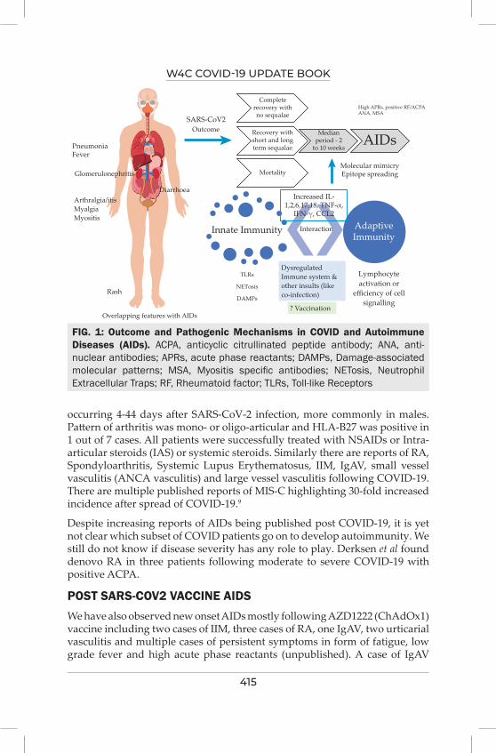
W4C COVID-19 UPDATE BOOK
415
FIG. 1: Outcome and Pathogenic Mechanisms in COVID and Autoimmune Diseases (AIDs). ACPA, anticyclic citrullinated peptide antibody; ANA, anti-nuclear antibodies; APRs, acute phase reactants; DAMPs, Damage-associated molecular patterns; MSA, Myositis specific antibodies; NETosis, Neutrophil Extracellular Traps; RF, Rheumatoid factor; TLRs, Toll-like Receptors
occurring 4-44 days after SARS-CoV-2 infection, more commonly in males. Pattern of arthritis was mono- or oligo-articular and HLA-B27 was positive in 1 out of 7 cases. All patients were successfully treated with NSAIDs or Intra-articular steroids (IAS) or systemic steroids. Similarly there are reports of RA, Spondyloarthritis, Systemic Lupus Erythematosus, IIM, IgAV, small vessel vasculitis (ANCA vasculitis) and large vessel vasculitis following COVID-19. There are multiple published reports of MIS-C highlighting 30-fold increased incidence after spread of COVID-19.9
Despite increasing reports of AIDs being published post COVID-19, it is yet not clear which subset of COVID patients go on to develop autoimmunity. We still do not know if disease severity has any role to play. Derksen et al found denovo RA in three patients following moderate to severe COVID-19 with positive ACPA.
POST SARS-COV2 VACCINE AIDS We have also observed new onset AIDs mostly following AZD1222 (ChAdOx1) vaccine including two cases of IIM, three cases of RA, one IgAV, two urticarial vasculitis and multiple cases of persistent symptoms in form of fatigue, low grade fever and high acute phase reactants (unpublished). A case of IgAV
PneumoniaFever
Rash
Overlapping features with AIDs
SARS-CoV2
Completerecovery withno sequalae
Recovery withshort and longterm sequalae
Mortality
Increased IL-1,2,6,17,18, TNF-α,
IFN-γ, CCL2
AdaptiveImmunity
Innate Immunity Interaction
TLRs
NETosis
DAMPs
DysregulatedImmune system &other insults (likeco-infection)
Lymphocyteactivation or
efficiency of cellsignalling
? Vaccination
Medianperiod - 2
to 10 weeks
High APRs, positive RF/ACPAANA, MSA
AIDsMolecular mimicryEpitope spreading
Outcome
Glomerulonephritis
DiarrhoeaArthralgia/itisMyalgiaMyositis
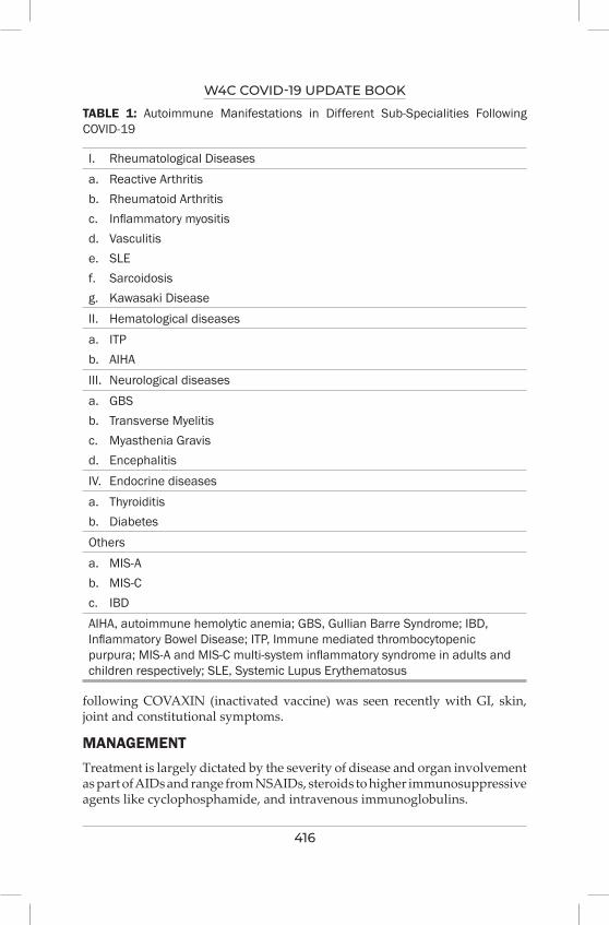
W4C COVID-19 UPDATE BOOK
416
TABLE 1: Autoimmune Manifestations in Different Sub-Specialities Following COVID-19
I. Rheumatological Diseasesa. Reactive Arthritis b. Rheumatoid Arthritisc. Inflammatory myositis d. Vasculitis e. SLE f. Sarcoidosis g. Kawasaki DiseaseII. Hematological diseasesa. ITP b. AIHAIII. Neurological diseasesa. GBSb. Transverse Myelitis c. Myasthenia Gravis d. Encephalitis IV. Endocrine diseasesa. Thyroiditis b. Diabetes Othersa. MIS-Ab. MIS-Cc. IBDAIHA, autoimmune hemolytic anemia; GBS, Gullian Barre Syndrome; IBD, Inflammatory Bowel Disease; ITP, Immune mediated thrombocytopenic purpura; MIS-A and MIS-C multi-system inflammatory syndrome in adults and children respectively; SLE, Systemic Lupus Erythematosus
following COVAXIN (inactivated vaccine) was seen recently with GI, skin, joint and constitutional symptoms.
MANAGEMENT Treatment is largely dictated by the severity of disease and organ involvement as part of AIDs and range from NSAIDs, steroids to higher immunosuppressive agents like cyclophosphamide, and intravenous immunoglobulins.
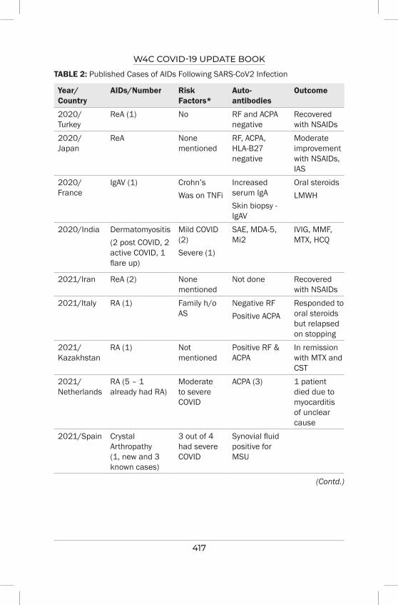
W4C COVID-19 UPDATE BOOK
417
TABLE 2: Published Cases of AIDs Following SARS-CoV2 Infection
Year/Country
AIDs/Number Risk Factors*
Auto-antibodies
Outcome
2020/Turkey
ReA (1) No RF and ACPA negative
Recovered with NSAIDs
2020/Japan
ReA None mentioned
RF, ACPA, HLA-B27 negative
Moderate improvement with NSAIDs, IAS
2020/France
IgAV (1) Crohn’s Was on TNFi
Increased serum IgASkin biopsy - IgAV
Oral steroidsLMWH
2020/India Dermatomyositis (2 post COVID, 2 active COVID, 1 flare up)
Mild COVID (2)Severe (1)
SAE, MDA-5, Mi2
IVIG, MMF, MTX, HCQ
2021/Iran ReA (2) None mentioned
Not done Recovered with NSAIDs
2021/Italy RA (1) Family h/o AS
Negative RFPositive ACPA
Responded to oral steroids but relapsed on stopping
2021/Kazakhstan
RA (1) Not mentioned
Positive RF & ACPA
In remission with MTX and CST
2021/Netherlands
RA (5 – 1 already had RA)
Moderate to severe COVID
ACPA (3) 1 patient died due to myocarditis of unclear cause
2021/Spain Crystal Arthropathy (1, new and 3 known cases)
3 out of 4 had severe COVID
Synovial fluid positive for MSU
(Contd.)
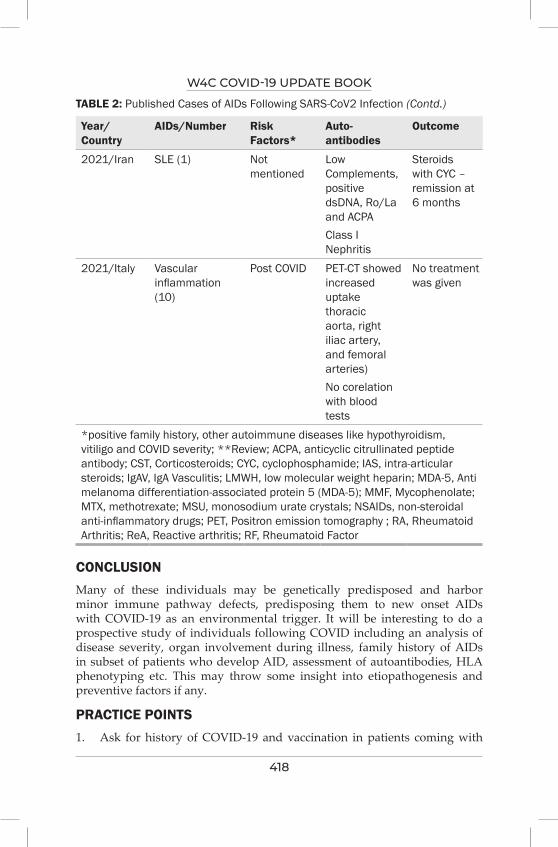
W4C COVID-19 UPDATE BOOK
418
Year/Country
AIDs/Number Risk Factors*
Auto-antibodies
Outcome
2021/Iran SLE (1) Not mentioned
Low Complements, positive dsDNA, Ro/La and ACPAClass I Nephritis
Steroids with CYC – remission at 6 months
2021/Italy Vascular inflammation (10)
Post COVID PET-CT showed increased uptake thoracic aorta, right iliac artery, and femoral arteries) No corelation with blood tests
No treatment was given
*positive family history, other autoimmune diseases like hypothyroidism, vitiligo and COVID severity; **Review; ACPA, anticyclic citrullinated peptide antibody; CST, Corticosteroids; CYC, cyclophosphamide; IAS, intra-articular steroids; IgAV, IgA Vasculitis; LMWH, low molecular weight heparin; MDA-5, Anti melanoma differentiation-associated protein 5 (MDA-5); MMF, Mycophenolate; MTX, methotrexate; MSU, monosodium urate crystals; NSAIDs, non-steroidal anti-inflammatory drugs; PET, Positron emission tomography ; RA, Rheumatoid Arthritis; ReA, Reactive arthritis; RF, Rheumatoid Factor
TABLE 2: Published Cases of AIDs Following SARS-CoV2 Infection (Contd.)
CONCLUSIONMany of these individuals may be genetically predisposed and harbor minor immune pathway defects, predisposing them to new onset AIDs with COVID-19 as an environmental trigger. It will be interesting to do a prospective study of individuals following COVID including an analysis of disease severity, organ involvement during illness, family history of AIDs in subset of patients who develop AID, assessment of autoantibodies, HLA phenotyping etc. This may throw some insight into etiopathogenesis and preventive factors if any.
PRACTICE POINTS 1. Ask for history of COVID-19 and vaccination in patients coming with
W4C COVID-19 UPDATE BOOK
419
new onset joint pain, rash, fever or myalgia or proximal muscle weakness and should be investigated for possible autoimmune disease
2. Mere presence of autoantibodies should not be the criteria for diagnosis of AIDs
3. Treatment depends on extent of disease and organ involvement
REFERENCES1. Kanduc D. From Anti-SARS-CoV-2 Immune Responses to COVID-19 via Molecular Mimicry.
Antibodies 2020; 9:33. 2. Tang Y, Liu J, Zhang D et al. Cytokine Storm in COVID-19: The Current Evidence and
Treatment Strategies. Front Immunol 2020; 11:1708. 3. Zhou, Y., Han, T., Chen, J et al. Clinical and Autoimmune Characteristics of Severe and Critical
Cases of COVID-19. Clin Transl Sci 13: 1077–86. 4. Tang K-T, Hsu B-C, Chen D-Y. Autoimmune and Rheumatic Manifestations Associated With
COVID-19 in Adults: An Updated Systematic Review. Frontiers in Immunology 2021; 12:628. 5. Pascolini, S., Vannini, A., Deleonardi, G et al. COVID-19 and Immunological Dysregulation:
Can Autoantibodies be Useful? Clinical and Translational Science 14: 502–8. 6. Derksen VFAM, Kissel T, Lamers-Karnebeek FBG et al. Onset of rheumatoid arthritis after
COVID-19: coincidence or connected? Ann Rheum Dis 2021; 80:1096–8. 7. De Lorenzis E, Natalello G, Gigante L et al. What can we learn from rapidly progressive
interstitial lung disease related to anti-MDA5 dermatomyositis in the management of COVID-19? Autoimmun Rev 2020; 19:102666.
8. Gil-Etayo FJ, Suàrez-Fernández P, Cabrera-Marante O et al. T-Helper Cell Subset Response Is a Determining Factor in COVID-19 Progression. Frontiers in Cellular and Infection Microbiology 2021; 11:79.
9. Verdoni, L., Mazza, A., Gervasoni, A. et al. An outbreak of severe Kawasaki-like disease at the Italian epicentre of the SARS-CoV-2 epidemic: an observational cohort study. The Lancet 395: 1771–8.

Organization for PandemicSECTION 11

423
Section 11 - Organization for PandemicSECTION EDITOR
Dr Khusrav Beji BajanMD, EDIC Consultant – Intensivist & HOD – Emergency Medicine, Critical Care & Emergency Medicine, P. D. Hinduja Hospital & Medical Reaserch Centre, Mumbai
Preface
“Failing to Plan is Planning to fail” – Benjamin Franklin
The most prudent way to beat a pandemic and a menace like COVID – 19, is to plan and prepare mentally, physically and scientifically. It is also mandatory to plan for an appropriate infrastructure and prepare for stocks of Oxygen, Drugs and Vaccines. Well planned protocols and well prepared teamwork is always a winning combination.
Management of a severe COVID patient has opened our vision more towards what does not work. Oxygen therapy, Proning, LMWH and Antibody cocktail have stood the test of time in proving to be beneficial. A good ICU Practice using the FAST HUG BID protocol would help improve outcomes in COVID patients. Nevertheless since COVID is a highly Transmissible disease a current day ICU needs to incorporate the following mandatory changes such as :
Appropriate use of PPE
Isolation Rooms
Negative pressure ICUs
Electronic ICUs (HUB and SPOKE models)
Counselling sessions
Mental health and Burn out addressal cells
Oxygen and Drug Audits
424
Steroid and Antibiotic Stewardships
Whilst a selfless Healthcare worker sets aside personal fears and grief to manage COVID and Non COVID patients with equal diligence, it is imperative to “PROTECT THE PROTECTORS”. This includes properly structured( negative pressure) ICUs, supply of PPEs, proper Donning and Doffing areas, adequate hands - down time, burn out addressal cells and isolation staying and eating facilities to name a few. All HCWs should not only be vaccinated but with adequate scientific data emerging, should be considered as the priority group for Booster Vaccine Doses as well.
425
Structure, Process Modifications and
Preparedness for Pandemic56.
INTRODUCTIONSince the time COVID -19 has been declared a public health emergency in January 2020 and a pandemic in March 2020, it has spread to all the corners and edges of human civilization including Antarctica. With more than 32 million cases and more than 430,000 deaths, India has been one of the worst affected countries comprising of >15% of total cases and >10% of total deaths reported worldwide respectively. So far, COVID-19 seems to affect people of all ages, genders and ethnicities without discrimination of socioeconomic background. It is essentially, a true pandemic that humanity is suffering from.1
PATHOPHYSIOLOGY AND BIOLOGICAL PLAUSIBILITYCurrent Coronavirus pandemic, a zoonotic disease, is caused by an enveloped positive stranded RNA-beta coronavirus, which enters the cells via ACE-2 receptors via receptor binding domain of spike protein.2 Direct mucosal transfer, airborne/aerosolized respiratory droplets/particles in closed and poorly ventilated spaces; and indirect contact transmissions through surfaces and non-respiratory fluids are all well documented routes of person-to-person transmission.3 The infectivity peaks from two days prior to, till one day after symptoms onset and declines over next 7 days in an immunocompetent host. Once infected, majority of patients will go on to develop neutralizing antibody (humoral response) to the receptor binding domain of spike protein antigen, in conjunction with SARS-CoV-2 specific CD4 and CD8 cellular response that will reduce the short term risk of reinfection in the subsequent 6-8 months by 80-85% in one estimate. Subsequent positive SARS-CoV-2 RT-PCR needs a careful justification to distinguish reinfection versus extended viral RNA shedding, not amounting to infectiveness, except in immunocompromised hosts.4-6
Genetic mutations are commonly observed. Without a known intermediary host (in contrast to earlier coronavirus epidemics), speculations about its
Nimit Ashwinbhai Shah, Pratibha Dileep

W4C COVID-19 UPDATE BOOK
426
artificial origins, whether a bio-weapon or a lab-leak, have been rife, fuelling conspiracy theories.7,8 Despite extensive data collection by ICMR-NIV trying to provide critical insight for the ongoing spill over event, it remains biologically plausible that current pandemic will be entrenched with, endemic-seasonal-perennial epidemics. Very high rates of morbidity and mortality occurred due to rapid household infections and an attenuated vaccine response against Delta variant, thus leading to a massive surge of cases worldwide.9
ISSUES WITH PANDEMIC PREPAREDNESSCOVID-19 pandemic has exposed many weaknesses in pandemic preparedness
• Under reporting, inadequate testing and under estimating the problems;
• Lack of robust and timely national emergency mitigation plans or uniform guidelines;
• Limited resources for diagnosis and treatment of COVID and post-COVID complications such as oxygen, lifesaving equipment, medicines, protective gears;
• Shortage of competent healthcare workers (HCWs) and field epidemiologists;
• Under-utilization of the existing facilities;
• Lack of coordination between public and private health and governmental administrative departments;
• Lack of research and innovation.
STEPS FOR BETTER PANDEMIC MANAGEMENTIt is imperative to prepare for future long term pandemic preparedness strategy in decades to come. The response to such global threat ought not be a knee-jerk reaction with top-down coercive quarantines, but instead needs a careful planning with early and effective warning-alert-response system, transparent containment measures, and community engagement for implementation.
• Community Education Programs about physical distancing; respiratory etiquettes; hand hygiene with soap or sanitizer containing at least 60% alcohol; source control and exposure prevention with masks; role of contact tracing and isolation; and vaccination are indispensable. This can be achieved by collaboration of private-public institutions, social media and telecommunications. Teachers, social activists, Social media, well known public figures must come forward for educating the masses. Entertainment industry can educate masses with stories and plays. Community Distancing Model of micro containments will help
W4C COVID-19 UPDATE BOOK
427
residents some form of movement within community without interaction with other communities. Such strategy shall help traceability of infected cases without spill over but it comes with a requirement of basic needs within its boundaries such as food and healthcare facilities.10
• Developing Rapid Response Public Health Preparedness Clinics (RRPHPCs) which can bridge the gap between communities and healthcare facilities, screen population, physically or by telemedicine.10 Availability of commercial kits for self COVID testing at-home and in-person testing booths will help achieve goals of inclusive testing and self-referral. Socioeconomic and mental health outreach programs can be geared up towards helping community with fallouts and aftershocks of COVID-19.
• Urgent Outbreak Care Facilities (UOCFs) can help patients awaiting transportation to a hospital with infrastructure – such as negative pressure rooms with HEPA filters, testing kits, PPE kits, ventilators and other basic necessities. Such Infra-structure preparation should be in collaboration with public and private facilities with a dedicated wing or building comprising of at least 20% of their beds to accommodate infective patients who need various types of isolation ranging from airborne, droplet and contact isolation. These units must be self sufficient in all facilities including waste disposal.
• Once in an acute Healthcare Facility, patients should undergo triage as per standardized protocols. Patients with suspected or confirmed COVID-19 must be placed in a single room with a closing door, personal bathroom and a restrictive visitor strategy. Dedicated elevator must be used for transporting them. Universal masking is a must in healthcare setting for all patients, visitors and HCWs to reduce transmission from unsuspected presymptomatic or asymptomatic carriers.11 In a healthcare facility, a valve-less N95 respirator (or a medical mask on top of N95 if there is a valve), and a face shield are must while assessing a non-COVID patient. A PPE kit comprising of full length gown and gloves in addition to above is a must while assessing suspected or confirmed COVID-19 patients.
• Easy and clear treatment guidelines must be formulated state wise to guide smaller hospitals and helplines should be created even for medical professionals to seek help before prescribing specialised molecules.
• Vaccines are considered most promising utility to halt the pandemic. Fully vaccinated individuals can be managed differently with less restrictive strategy for testing and quarantine than unvaccinated in case of close contact with a COVID-19 positive patient. Future availability of monoclonal antibody combinations in post exposure prophylaxis for a select group of individuals of high risk cohort, can also help reduce
W4C COVID-19 UPDATE BOOK
428
symptomatic COVID, although its role in Delta variant is still debatable.
• HCWs are at a higher risk of exposure to such highly infectious viral pandemics leading to their high disease related morbidity and mortality. Long term ramifications such as long COVID syndrome, depression-anxiety-PTSD are grave threats to one’s psychological health leading to short and long term disabilities.
• Vaccination to the HCWs must be completed as a priority and vaccinated individuals must be posted on priority basis in high risk areas. HCWs staffing should be adequate to cover for three 8 hour shifts with 1 hour overlay for sign-out to oncoming team. Proper donning and doffing of PPE kits must be ensured. Total number of work hours should not exceed 48 hours in a given week. Adequate rest must be allowed between duties. Staffing can be done in 2 sets of teams alternating every week, 3rd team can fill-in the gaps if any member needs replacement. HCW must be educated to report any symptoms and must be assessed by a senior physician. Ongoing training programs can improve the adherence to the protocols.
• Socio-economical ramification of a pandemic are enormous. Every single individual in small or big way has been affected adversely by this pandemic including front-line HCWs. Hence a comprehensive health and social safety net covers are prudent for them during such dreadful times.
CONCLUSIONAfter a balanced strategy of lockdowns, micro-containments and graded un-lockdowns during the first wave, second wave brought India on its knees by true ferocity of a pandemic unleashed by a highly transmissible Delta variant. With a persistent threat of future spill overs and mutations, there is an ever greater need for investing heavily in building strong, resilient and visionary health system incorporating community, state/regional, national and international governance.
PRACTICE POINTS• A medical expert team comprising of Infectious Disease and Critical
Care Physicians; Epidemiologists and Microbiologists; hospital Administrators along with the Government agencies to take timely decisions in formulating guidelines.
• Use of digital health portals, telemedicine and app based disease surveillance systems should be encouraged.
• Capacity to rapidly expand hospital beds, manufacturing of affordable diagnostic kits, availability of life saving drugs for treatment and vaccine development in a time sensitive manner.
W4C COVID-19 UPDATE BOOK
429
• Greater investment in public health, educating and upskilling of healthcare workers, and timely execution and revisions of regional-national plans.
• Strong leadership with political will to allocate and upscale the public health and social safety net programs.
REFERENCES1. Sarkar A, Liu G, Jin Y, Xie Z, Zheng Z-J. Public health preparedness and responses to the
coronavirus disease 2019 (COVID-19) pandemic in South Asia: a situation and policy analysis. Glob Heal J 2020; 4.
2. Hoffmann M, Kleine-Weber H, Schroeder S, Krüger N, Herrler T, Erichsen S, et al. SARS-CoV-2 Cell Entry Depends on ACE2 and TMPRSS2 and Is Blocked by a Clinically Proven Protease Inhibitor. Cell [Internet] 2020; 181:271-280.e8. Available from: https://linkinghub.elsevier.com/retrieve/pii/S0092867420302294
3. Meyerowitz EA, Richterman A, Gandhi RT, Sax PE. Transmission of SARS-CoV-2: A Review of Viral, Host, and Environmental Factors. Ann Intern Med [Internet] 2021; 174:69–79. Available from: https://www.acpjournals.org/doi/10.7326/M20-5008
4. Letizia AG, Ge Y, Vangeti S, Goforth C, Weir DL, Kuzmina NA, et al. SARS-CoV-2 seropositivity and subsequent infection risk in healthy young adults: a prospective cohort study. Lancet Respir Med [Internet] 2021; 9:712–20. Available from: https://linkinghub.elsevier.com/retrieve/pii/S2213260021001582
5. Hall VJ, Foulkes S, Charlett A, Atti A, Monk EJM, Simmons R, et al. SARS-CoV-2 infection rates of antibody-positive compared with antibody-negative health-care workers in England: a large, multicentre, prospective cohort study (SIREN). Lancet [Internet] 2021 Apr [cited 2021 Aug 23];397(10283):1459–69. Available from: https://www.worldometers.info/coronavirus
6. Harvey RA, Rassen JA, Kabelac CA, Turenne W, Leonard S, Klesh R, et al. Association of SARS-CoV-2 Seropositive Antibody Test With Risk of Future Infection. JAMA Intern Med [Internet] 2021; 181:672–9. Available from: http://www.ncbi.nlm.nih.gov/pubmed/33625463
7. Zhang L, Shen F-M, Chen F, Lin Z. Origin and Evolution of the 2019 Novel Coronavirus. Clin Infect Dis [Internet] 2020; 71:882–3. Available from: http://www.ncbi.nlm.nih.gov/pubmed/32011673
8. Yee S, Tan CS, Khan A, Lee KS, Goh BH, Ming LC. SARS-COV-2 as an artificial creation: scientific arguments and counterarguments. J Med Life [Internet] 14(1):118–20. Available from: http://www.ncbi.nlm.nih.gov/pubmed/33767796
9. Lopez Bernal J, Andrews N, Gower C, Gallagher E, Simmons R, Thelwall S, et al. Effectiveness of Covid-19 Vaccines against the B.1.617.2 (Delta) Variant. N Engl J Med [Internet] 2021; 385:585–94. Available from: http://www.nejm.org/doi/10.1056/NEJMoa2108891
10. Kuguyo O, Kengne AP, Dandara C. Singapore COVID-19 Pandemic Response as a Successful Model Framework for Low-Resource Health Care Settings in Africa? Omi A J Integr Biol [Internet] 2020; 24:470–8. Available from: https://www.liebertpub.com/doi/10.1089/omi.2020.0077
11. Arons MM, Hatfield KM, Reddy SC, Kimball A, James A, Jacobs JR, et al. Presymptomatic SARS-CoV-2 Infections and Transmission in a Skilled Nursing Facility. N Engl J Med [Internet] 2020; 382:2081–90. Available from: http://www.ncbi.nlm.nih.gov/pubmed/32329971.

430
Protection of the Frontliners 57.
INTRODUCTION The COVID -19 pandemic has been devastating world over. Not only has it evolved into a dangerous always mutating threat, but also led to a crippling economy and millions to die. Waves after waves have surprised and shocked the entire healthcare systems globally. The bravest efforts to combat this menace, have been from the frontliners.
In the first wave, without ammunition like vaccinations, poor supply of PPEs and a lack of infrastructure, the Frontliners exhibited selflessness, bravery and humanity to once again roll up their sleeves to combat this war.
It is imperative for the healthcare system to ensure safety of the frontline workers as they grapple 24X7, risking their and their families lives. We need to have guidelines and some protocols which would help enable frontliners safety.
GUIDELINES TO PROTECT THE PROTECTORS1. Appropriate use of PPE
a. Level of PPE to be divided as per the Zone in the Hospital
i. High Risk Zone (Full PPE – HAZMAT)
1. ICU/OT/ER/Holding area/Ambulance/ Scopy Suites
ii. Medium Risk Zone (N95 + Surgical Mask on top and Goggles or Face shield +/- surgical gown)
1. OPD/ Fever clinic/ Oncology/ AKD/ Imaging/ ENT/ Dental
iii. Low Risk Zone (2 surgical Masks)
1. Zone other than above
2. Appropriate Donning and Doffing Procedures and Dedicated Areas
a. To have Donn/Doff Buddies to guide
b. Videos/Posters
Khusrav Beji Bajan
W4C COVID-19 UPDATE BOOK
431
3. For any Aerosol Generating Procedures (AGP) such as, Intubation, Bronchoscopy, Swab collection, CPR.
a. Use Extra gears in form of Personal Air Purifying Respirators (PAPRs) or Honeywell shields over and above full PPE
4. Swab collection to be done only by qualified lab technician anywhere in the hospital and NOT TO BE DONE BY DOCTORS/ INTERNS/ PARAMEDICS
5. Proper and appropriate accommodation to be provided to doctors keeping in mind social distancing in Cafeterias, food halls etc.
6. Appropriate QUARANTINE FACILITIES for the doctors into hotels etc. if similar facilities not available at their homes and to protect the family members from getting affected too.
7. Working hours to be restricted to 6 hours in High Risk Zones. Also Work: Off ratio to be maintained at 7:3 days.
a. Minimum manpower to be utilized and rotation from High to medium to low risk Zones done cyclically.
8. Consider Antibody testing at regular intervals for an insight into the immunity level.
9. Appropriate Environment to be created in the High Risk Zones
a. Negative pressure
i. > 12 to 15 Air exchanges per hour or more than 145 litres / sec / patient achieved
ii. HEPA (High Efficiency Particulate Air) filters deployed where appropriate
iii. Ante room if possible
iv. To use engineering dept. skills to deal with HVAC (Heat Ventilation Air Conditioning)
10. Look after the mental health of the HCWs
a. Periodic consultations
b. Groups to discuss
c. Mentoring and “hearing them out” sessions
11. Treatment of affected HCWs at top priority since severity and mortality is 5 to 10 fold higher amongst positive HCWs across all age groups.
a. Preferably a separate area in the same Institute or any equivalent institute with proper hygiene and treatment facilities.
W4C COVID-19 UPDATE BOOK
432
b. Provide additional leave for up to a month for the severe and prolonged cases.
c. Provide aggressive Post COVID Syndrome Care including Physio and Rehab.
12. Adequate HCQ prophylaxis and Appropriate Nutrition including Zinc, Vitamin C, Vitamin D etc. (only if Evidence justifies its use)
13. Prioritise Vaccination amongst all frontliners. Make efforts to allay Vaccination Hesitancy amongst the group.
14. Have a task force to audit the Breakthrough Infections amongst the fully Vaccinated. Have Genomics done for all such infections to understand the pattern of Variants and Vaccination efficacy
15. Have a specialized team to address the Post Covid Syndrome and the Quality Of Life goals amongst those who have been infected previously.
CONCLUSIONFrontliners need a healthy and safe environment with access to facilities, infrastructure, drugs and updated literature.
Government must provide adequate compensation in terms of remuneration and make periodic efforts towards maintaining the mental and physical health of the frontliners holistically.
Utmost care must be exercised to provide PPEs, Vaccinations (even Booster Doses when appropriate) and prompt and free treatment to all frontliners.
The entire healthcare system needs to unite to be able to avoid fatigue and burnout amongst fellow colleagues and to maintain a healthy work life balance
REFERENCES1. Chen W, Huang Y. To protect health care workers better, to save more lives With covid-19.
Anesthesia & Analgesia 2020; 131:97–101. https://doi.org/10.1213/ane.0000000000004834 2. The Lancet. COVID-19: Protecting health-care workers. The Lancet 2020; 395:922. https://doi.
org/10.1016/s0140-6736(20)30644-9 3. Udwadia ZF, Raju RS. How to protect the Protectors: 10 lessons to learn for Doctors fighting
the Covid-19 coronavirus. Medical Journal Armed Forces India 2020; 76:128–131. https://doi.org/10.1016/j.mjafi.2020.03.009.
433
Changing iCU Practices During COviD-19 58.
INTRODUCTIONThere has been a significant change in the usual clinical and administrative practice over the last two years to contain and treat COVID-19. These changes are governed by the unique pathogenesis, extremely high infectivity and significant impact of the disease burden on the healthcare workers (HCWs) and healthcare systems. The unanticipated surge in demand on healthcare has forced an urgent introspection of our existing clinical practice, resource allocation and manpower management.
CHARACTERISTICS OF COVID-19 AFFECTING CURRENT INTENSIVE CARE PRACTICEA. Pathogen related
The current delta variant (B.1.617.2) has a reproduction number (Ro) of 3.2 to 8 which is almost twice that of the wild variant.1 It represents the increased transmissibility and secondary attack rate. Delta variant has 60% higher rate of household transmission than its predecessor.2
Critically ill and immunocompromised patients continue to shed viable virus for a prolonged period of time.3 As a health care worker, it has resulted in significant risk of contracting the virus.
Disease pathogenesis related
The severe acute respiratory syndrome coronavirus- 2 (SARS-CoV-2) virus distribution and organ involvement is related to the presence of human angiotensin converting enzyme-2 (ACE2) receptor in human body. It can present as dermatological, myocardial, gastrointestinal, neurological, hepatic or renal dysfunction. Patients presenting with a non-respiratory illness, can have COVID-19 infection irrespective of their lung involvement. This makes assessment of exposure risk and stratification based on it of prime importance before attending such patients.4
Sananta Kumar Dash, Tapas Kumar Sahoo

W4C COVID-19 UPDATE BOOK
434
B. Impact on HCWs
In a meta-analysis involving 119,883 patients, more than 51% of HCWs tested positive who were taking care of COVID-19 patients.5 The risk ratio of contracting the infection was more than twice for HCWs working in intensive care setting.6 The impact of COVID-19 on the mental health of the HCWs have been very significant. The prevalence of depression, anxiety, peri-traumatic distress and low well-being are higher among intensive care unit (ICU) staff.
C. Impact on Health care facility
There has been a significant surge in the demand for intensive care facility. One in every five COVID-19 patients are expected to require some ICU support and more than 50% of such patients may require invasive ventilation.7
D. Impact on environment
The environment impact of the treatment and containment measures for COVID-19 have been significant. Majority of the medical waste is going to be generated from the acute care areas including ICUs. Multiple intensive care societies have expressed their concerns regarding judicial use of personal protective equipment (PPE) and their dumping. Medical waste management is going to be one of the biggest challenge in this pandemic.
CHANGES IN INTENSIVE CARE PRACTICE A. Triage of patients
Intensive care facility needs to have a robust triaging system for allocating beds to appropriate patients. It will ensure adequate utilization of resource in a surge situation. The current pandemic has posed a significant thought provocation about use of a suitable triage criteria and method for critically ill patients who need to be admitted to an intensive care facility.8 Frequent re-evaluation and changes in triage criteria should be done based on the evolving situation and evidence.
B. Triage of equipment
As the disease pathogenesis includes respiratory system predominantly, resources should be invested on equipment and facility pertinent to respiratory support such as ventilators (invasive and non-invasive), high flow oxygen equipment, availability of ongoing and increased surge requirement of oxygen etc.
C. Safe Clinical practice
Multiple societies have published their guidelines for safe procedural

W4C COVID-19 UPDATE BOOK
435
environment in ICU. The primary message transmitted is self and co-workers’ safety. Protocol breach should be kept to zero.
D. PPE availability, supply and disposal
The clinical care of such patients should also aim optimal safety of the HCWs. This includes formulation, following standard protocols and ensuring adequate PPE. Anticipation of requirement for PPE in case of a surge should be done in advance. Continuous supply and storage should be maintained. Segregation of medical waste should be done at the site of generation which decreases the contamination. Stringent waste segregation plan should be placed in action.
E. Tele- health
Telehealth has caused a great impact in rendering clinical care to these patients. It avoids preventable transfers of patients who can be managed in a peripheral setting. This has led to minimize contacts and limit spread of infection. Also, it has rendered a major cost saving which has been channelized to better clinical decision making. Remote ICU concept is becoming an essential part of ongoing management of the current pandemic.9 Simulation based learning and education have added value to the surge preparedness.10
F. Rapid and free availability of research data
The volume of research directed to define the virological profile, pathogenesis, clinical management and long term outcome of COVID-19 has been immense. The availability of large volume retrospective data and inference drawn using artificial intelligence system (AIS) have been the torchbearers in management of this pandemic. Intensive care facility should be proactive to share their clinical data and enrol their cohort to available research opportunities.
G. End of life care
A timely “end of life” discussion ensures a dignified dying process, decreases family distress, mitigates work place burnout among all HCWs and ensures appropriate allocation of resources. This requires facility to train the existing physicians. Regular and adequate training arrangement is an important aspect of professional development.
H. Administrative
ICU infrastructure restructuring
The existing infrastructure should be looked into for possible measure to accommodate and change in a surge situation. Various societies have proposed guidelines for such preparedness.

W4C COVID-19 UPDATE BOOK
436
Staffing, Mental health, staff wellbeing, Burnout and fatigue management
Staffing plan needs to be kept in place allowing adequate breaks for the HCWs. The mental health aspects of the HCWs should be of prime consideration. Support groups and measures need to be in place before the surge.
Surge capacity planning and Resource allocation
Validated mathematical model are being opted to estimate the volume of possible surge and equate with the available and expandable resources. The resource consideration should be both at the level of material and manpower. The models used aim at the best, worst and most likely scenario and explore the ability of the system. The predictive model should take into account the viral characteristics (i.e R0 and rate of spread in other countries), data looking at the use of health care resources (hospitalization rate, need for ICU bed, need for mechanical ventilation and other invasive strategies such as Extracorporeal membrane oxygenator (ECMO), duration of mechanical ventilation, mortality rate, ICU and hospital length of stay (LOS) etc. Epidemiologists are being recruited to look at pattern of spread, infectivity and pathogenicity of the different variants of the current virus to be able to identify and act early.
CONCLUSIONWith the ongoing pandemic, it is difficult to predict the timing for surge in a particular health facility. Appropriate preplanning can mitigate the sudden impact of a surge and ensure smooth operations. Changes in ICU practice are happening at different levels of care. As intensive care physicians, we should be aware and ready for such changes.
PRACTICE POINTS- Surge planning based on appropriate mathematical model and
epidemiologist’s input.
- Definitive triage system for assessing appropriateness for ICU admission and early initiation “end of life” discussion.
- Simulation based learning for surge preparedness.
- Dedicated psychological support measures for HCWs in ICU.
- Definite clinical management plans including early institution of certain therapeutic measures (i.e. immunomodulators, anticoagulants) and surveillance for secondary infection in ICU.
- Supply, storage and disposal of PPE is an important component in future planning in healthcare.
REFERENCES1. Campbell F, Archer B, Laurenson-Schafer H, Jinnai Y, Konings F, Batra N, et al. Increased
transmissibility and global spread of SARS-CoV-2 variants of concern as at June 2021.
W4C COVID-19 UPDATE BOOK
437
Euro Surveill [Internet] 2021; 26. Available from: http://dx.doi.org/10.2807/1560-7917.es.2021.26.24.2100509.
2. Mahase E. Delta variant: What is happening with transmission, hospital admissions, and restrictions? BMJ 2021; 373:n1513.
3. Rhee C, Kanjilal S, Baker M, Klompas M. Duration of Severe Acute Respiratory Syndrome Coronavirus 2 (SARS-CoV-2) infectivity: When is it safe to discontinue isolation? Clin Infect Dis [Internet] 2020; Available from: http://dx.doi.org/10.1093/cid/ciaa1249
4. Thakur V, Ratho RK, Kumar P, Bhatia SK, Bora I, Mohi GK, et al. Multi-organ involvement in COVID-19: Beyond pulmonary manifestations. J Clin Med 2021; 10:446.
5. Gholami M, Fawad I, Shadan S, Rowaiee R, Ghanem H, Hassan Khamis A, et al. COVID-19 and healthcare workers: A systematic review and meta-analysis. Int J Infect Dis 2021; 104:335–46.
6. Ran L, Chen X, Wang Y, Wu W, Zhang L, Tan X. Risk factors of healthcare workers with Coronavirus Disease 2019: A retrospective cohort study in a designated hospital of Wuhan in China. Clin Infect Dis 2020; 71:2218–21.
7. Aziz S, Arabi YM, Alhazzani W, Evans L, Citerio G, Fischkoff K, et al. Managing ICU surge during the COVID-19 crisis: rapid guidelines. Intensive Care Med 2020; 46:1303–25.
8. Jöbges S, Vinay R, Luyckx VA, Biller-Andorno N. Recommendations on COVID-19 triage: international comparison and ethical analysis. Bioethics 1111;2020;34(9):948-959:12805.
9. Srinivasan SR. Editorial: Tele-ICU in the age of COVID-19: Built for this challenge. J Nutr Health Aging 2020; 24:536–7.
10. Nair SS, Kaufman B. Simulation-based up-training in response to the COVID-19 pandemic. Simul Healthc 2020; 15:447–8.
438
The Toll of Pandemic- Non-Covid-19 Emergencies
at Bay59.
INTRODUCTION‘There is nothing so strong or safe in an emergency of life as the simple truth.’
-Charles Dickens
This pandemic has taken a toll on everyone in some way or the other, be it in terms of change in working conditions, or on one’s mental health, or even the grief of losing a dear one. It also affected the dynamics of healthcare services provision, from running out of hospital beds to the scarcity of oxygen and even the drugs! One such effect was the changing trend seen in emergency department visits. On one side, the world was facing an extreme burden on healthcare with endless COVID-19 cases and related mortality; on the other side, there was a surprising decrease in non-COVID emergencies.1,2 With the onset of pandemic, there was an inherent decrease in the non-COVID-19 patient load, maybe due to a sense of fear of contracting the disease, but the certitude could also be seen expanding to the, ‘non-COVID emergency cases’.
RATIONALE- OBSERVED TRENDSThe data on decreasing number of emergencies during the pandemic has been reported from many countries.2,3 With a disrupted healthcare system around the globe, due to the indomitable allocation of resources largely to the COVID management areas, it leads to an inevitable negation of the same to other blights. With a sense of fear of contracting COVID, amongst the daily news of someone known dying, even the patients requiring medical attention did not step out. Moreover, the ones who did only did when the severity of symptoms went out of hand. As a result, the presenting symptoms of various conditions were severe, compared to the same before the pandemic.4,5 Despite the significant changes in presenting emergencies, the trend was not apparent because many admissions of COVID-19 cases compensated for the drop in hospitalizations.6 In comparison to 2019, the hospital admission rates due to emergencies decreased up to nearly 35% in the year of pandemic – 2020.3,7
Lalit Singh, Utkarsh Khattri, Geeta Karki
W4C COVID-19 UPDATE BOOK
439
Evidence was obtained for decreased admissions of acute myocardial infarction, acute appendicitis, cerebrovascular accidents including intracranial hemorrhages, emergencies of gastroenterology, and other non-COVID related respiratory emergencies.3,8 Although the trend observed seemed universal with disruption of the healthcare services, barring few institutes.5 A US study conducted in two medical centres, comparing the ‘pre-pandemic’ and ‘during the pandemic’ patient admission trends also show similar findings, reduction in daily presenting cases for acute myocardial infarction, ischemic stroke, non-traumatic subarachnoid hemorrhage, and acute appendicitis.9
WHERE DID THE EMERGENCY CASES GO? – FACTORS AFFECTING THE EMERGENCY VISITS-The havoc of the COVID-19 pandemic made it inexorable to necessitate preventive measures for the general population, including wearing masks, practicing social distancing, and an imposed lockdown. An unsaid role of this lockdown can be one plausible explanation accounting for the delay in patients seeking care. An indigenous study conducted in Delhi also adds solidarity to the observed trend due to the restoration of ‘emergencies pattern’ to the pre-pandemic times once lockdown was waived off in the area.6 Apart from those who did not seek medical attention on time, others who sought consultation were advised to stay at home due to the risk of cross-infection.10 The other aspect is the non-availability of the resources. With unnecessary admissions of COVID patients with mild severity, those who needed the hospital beds and services were denied.
THE INEVITABLE OUTCOMESThe evidence supports that with the delayed visits to the emergency department and lesser hospital admissions, an unnecessary increase in out-patient mortality.8,10 While patients ignored their ‘warning signs’, their symptoms became more prominent and severe.7 If not mortality, there was an increase observed in the morbidity and associated disability post-management of the disease.8
Interestingly, with the onset of the pandemic, the doctors, specifically surgeons, adopted more of a conservative approach. The general surgeries, which were considered emergent in the era of pre-pandemic times, were delayed for an indefinite period. This poses a question on our approach, if those surgeries are emergent, and if not, that necessitates changes in treatment algorithms.4
LESSONS LEARNTThis tale of the pandemic has taught us to utilize our resources judiciously. The mild cases of COVID-19, which did not require hospital admissions, could have been spared. While fighting the Coronavirus disease and scuffling with the paucity of resources, it was essential to allocate the resources to manage
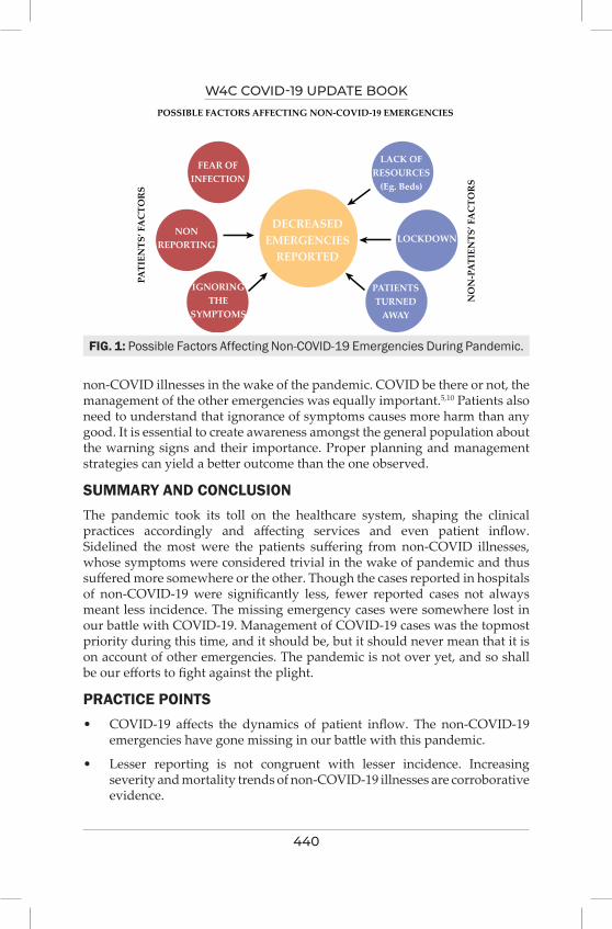
W4C COVID-19 UPDATE BOOK
440
non-COVID illnesses in the wake of the pandemic. COVID be there or not, the management of the other emergencies was equally important.5,10 Patients also need to understand that ignorance of symptoms causes more harm than any good. It is essential to create awareness amongst the general population about the warning signs and their importance. Proper planning and management strategies can yield a better outcome than the one observed.
SUMMARY AND CONCLUSIONThe pandemic took its toll on the healthcare system, shaping the clinical practices accordingly and affecting services and even patient inflow. Sidelined the most were the patients suffering from non-COVID illnesses, whose symptoms were considered trivial in the wake of pandemic and thus suffered more somewhere or the other. Though the cases reported in hospitals of non-COVID-19 were significantly less, fewer reported cases not always meant less incidence. The missing emergency cases were somewhere lost in our battle with COVID-19. Management of COVID-19 cases was the topmost priority during this time, and it should be, but it should never mean that it is on account of other emergencies. The pandemic is not over yet, and so shall be our efforts to fight against the plight.
PRACTICE POINTS• COVID-19 affects the dynamics of patient inflow. The non-COVID-19
emergencies have gone missing in our battle with this pandemic.
• Lesser reporting is not congruent with lesser incidence. Increasing severity and mortality trends of non-COVID-19 illnesses are corroborative evidence.
FIG. 1: Possible Factors Affecting Non-COVID-19 Emergencies During Pandemic.
POSSIBLE FACTORS AFFECTING NON-COVID-19 EMERGENCIES
PATI
ENTS
’ FA
CTO
RS
FEAR OFINFECTION
LACK OFRESOURCES
(Eg. Beds)
LOCKDOWN
PATIENTSTURNED
AWAY
IGNORINGTHE
SYMPTOMS
NONREPORTING
DECREASEDEMERGENCIES
REPORTED
NO
N-P
ATI
ENTS
’ FA
CTO
RS
W4C COVID-19 UPDATE BOOK
441
• Judicious use of resources is more important than the abundance of resources. It is essential to motivate the general population to not neglect their health just out of fear of COVID-19, which can only do more harm than good.
REFERENCES1. Boeken T, Le Berre A, Mebazaa A, Boulay-Coletta I, Hodel J, Zins M. Non-COVID-19
emergencies: where have all the patients gone? Eur Radiol 2020; 30:5220-5221.2. Santi L, Golinelli D, Tampieri A, Farina G, Greco M, Rosa S et al. Non-COVID-19 patients
in times of pandemic: Emergency department visits, hospitalizations and cause-specific mortality in Northern Italy. PLOS One 2021; 16:e0248995.
3. Ojetti V, Covino M, Brigida M, Petruzziello C, Saviano A, Migneco A et al. Non-COVID Diseases during the Pandemic: Where Have All Other Emergencies Gone? Medicina (Kaunas) 2020; 56:512.
4. Surek A, Ferahman S, Gemici E, Dural AC, Donmez T, Karabulut M. Effects of COVID-19 pandemic on general surgical emergencies: are some emergencies really urgent? Level 1 trauma center experience. Eur J Trauma Emerg Surg 2021; 47:647-652.
5. K.D. Lee, S.B. Lee, J.K. Lim, Y.M. Kang, I.B. Kim, H.J. Moon et al. Providing essential clinical care for non-COVID-19 patients in a Seoul metropolitan acute care hospital amidst ongoing treatment of COVID-19 patients, Journal of Hospital Infection 2020; 106:673-677.
6. Vaishya R, Sibal A, Kumar PS. Severe impact of COVID-19 pandemic on non-COVID patient care and health delivery: An observational study from a large multispecialty hospital of India. Indian J Med Sci 2021; 73:159-63.
7. Vollmer, M.A.C., Radhakrishnan, S., Kont, M.D. et al. The impact of the COVID-19 pandemic on patterns of attendance at emergency departments in two large London hospitals: an observational study. BMC Health Serv Res 2021; 21(1008).
8. Hecht N, Wessels L, Werft FO, Schneider UC, Czabanka M, Vajkoczy P. Need for ensuring care for neuro-emergencies-lessons learned from the COVID-19 pandemic. Acta Neurochir (Wien) 2020; 162:1795-1801.
9. Bhambhvani HP, Rodrigues AJ, Yu JS, Carr JB, Hayden Gephart M. Hospital Volumes of 5 Medical Emergencies in the COVID-19 Pandemic in 2 US Medical Centers. JAMA Intern Med 2021; 181:272–274.
10. Rosenbaum L. The untold toll—the pandemic’s effects on patients without Covid-19. New Engl J Med 2020; 382:2368–2371.


MiscellaneousSECTION 12

445
Section 12 - MiscellaneousSECTION EDITOR
Dr Banambar RayMD, FICCM Head of Critical Care Medicine, SUM Ultimate Medicare, A Unit of SOA,Bhubaneswar
Preface
There are three chapters in this section. The first one is on “Autopsy and Necropsy in COVID-19”. The author is a unique researcher who perhaps has no peers in this work. He has brought out many facts which could not have been known without autopsy. He has tried to corelate the autopsy findings with pathological changes and clinical features. The editor did not have much scope to change the language and content of the text for two reasons, i) the author is the pioneer in
the field and ii) he did not have the mandate of the author to do so. Certain portions of the text may be incomprehensible but the author wants us to leave it untouched.
Burn-out among intensivists during COVID-19 pandemic has been a disturbing observation world- wide. Authors have carefully analyzed causes and prescribed the remedies. Fear of getting the disease and not coming in close association with the families, some of them with small kids, drive them to get depressed and dejected. Frontline warriors are the worst hit in getting this syndrome.
The chapter on “ICU Care in non-ICU settings” is an appropriate text that all of us dealing with COVID-19 saw in recent past and to our horror we still could not provide shelter to some of our needy patients. The authors have given an account of how the healthcare providers should go about in expanding facilities when healthcare institutions are stretched to the limits during a pandemic as ferocious as COVID-19, which still has not ended.

447
Autopsies Findings Review in SARS – Cov – 2 Patients 60.
INTRODUCTIONEvery problem has three solutions: my solution, your solution, and the right solution.
PLATONE
I can’t teach anyone anything. I can only make them think
SOCRATE
The analysis of the different organs of the SARS – CoV – 2 Patients may help to understand pathogenesis and clinical outcome and to improve the therapeutic strategies. The need to better define the pathogenesis of coronavirus disease 19 (Covid-19) as well as to provide the correct statistical records concerning deaths related to this virus, inevitably involves the role of forensic pathology and routine autopsy practice. In light of the ongoing health emergency, we have launched a preliminary investigation into the alleged correlation between COVID-19 and all the other clinical manifestations in the early days of February 2019. The first SARS-CoV-2 outbreak in Italy was discovered on February 20, 2020 in Codogno (Lodi). Since then, our country has known millions of infected people and 78,371 deaths (data updated in December 2020). From the 32,792 published articles, it emerged that SARS-CoV-2 not only causes damage to the respiratory system, but also to the cardiovascular system with arrhythmic, thromboembolic complications, heart failure, myocarditis, and disseminated intravascular coagulopathy, dermatitis, and many other neurological complications, like dizziness, headache, ataxia epilepsy, dysgeusia, etc.
Cellular and humoral innate immunity represent a first line of resistance to most infectious agents. Evidence from SARS-CoV-1 suggests that these viruses may block interferon-mediated antiviral immunity. CD8 cytotoxic T cells play a fundamental role in antiviral resistance. Evidence suggests that during COVID-19 infection T cells undergo functional exhaustion as evidenced by
Quirino Piacevoli
W4C COVID-19 UPDATE BOOK
448
lymphopenia, skewing towards a T17 phenotype, inappropriate for antiviral immunity and suppression.
COVID-19: PNEUMONIA PATHOPHYSIOLOGY FINDINGSThe virus enters the epithelial – endothelial receptor and so we have an interstitial inflammation and a space with edema that appears as ground glass opacities.
In the beginning, they are mostly at the periphery, because the different elasticity between pleura and alveoli makes this a weak point to stress and pressure.
The Xray shows characteristic peripheral density of the lungs. VILI starts developing in less than 40 hours.
At this point we have lung “vasoplegia” (undetermined mechanism), i.e., if some part of the lungs are full of gas (well ventilated) with a “normal compliance” and perfusion occurs in another part, then the problem is that of perfusion.
This is indicative of a vascular disease. The lungs are no more able to direct the blood flow where it is necessary, except for gravity. Vasoplegia is increased by nitric oxide that causes vasodilation acting on cGMP receptors and phosphorylated myosin. At the same time, these patients show a decreased level of Angiotensin I and Angiotensin II that are vasoconstrictors. Final result is vasodilation.
Gravity dependent V/Q mismatch means that ventilation goes in one direction and perfusion in a different one. In this case blood goes down into the lungs where there is no ventilation, thus causing hypoxaemia.
This report wants to describe the findings of the autopsies of infected patients so as to improve our understanding of changes there by improving therapy and outcome.
Pathological Anatomy Services have started in March 2020, performing autopsies on COVID-19 positive patients after some initial hesitation. To date, we have totalled about 150 autopsies, almost entirely targeted to the lungs. We have waited for a special suit to be able to perform safely the autoptic examination on the brain. Ours is the largest case study in the world, since the Chinese have published the results of only 3 “minimally invasive” autopsies (+ an isolated case report) and another one from New Orleans whose authors have published only another 3 cases. The only other Italian hospital that performs autopsies is “Sacco” that reported about 20 cases.
Here is what we learnt from the first group of anatomical-pathological
W4C COVID-19 UPDATE BOOK
449
dissertations and the most significant interventions are presented here. Already macroscopically, the lungs appear “spotty”, with hyperemic / hemorrhagic areas alternating with rosy areas. From a histological point of view, some areas are severely emphysematous, with enormously dilated blood vessels (up to 20 times the norm) often full of micro thrombi. In many cases, diffuse alveolar damage (DAD) is evident, with desquamation of pneumocytes, formation of hyaline membranes and a fibrotic exudate (as in ARDS). It appears as a high-flow syndrome, with hepatomegaly and dilated portal vessels and diffuse thrombosis at all levels. Even the heart appears enlarged, always with a hydro pericardium and a marked left ventricular hypertrophy (but some of them were hypertensive patients accounting for those changes partly)
Just in one case, a thrombus was found almost completely obstructing the superior vena cava and the right atrium. It has often been noticed the ascent of the diaphragm, indicating that at a certain point the lungs no longer expanded. There was associated hepatomegaly which would have contributed to the “ascent”. While waiting for pastor data on CNS samples, biopsies of the olfactory mucosa were performed. COVID-19 classically gives anosmia and ageusia; the virus could reach the brain stem trans-synaptically, starting from the peripheral nerve endings of the olfactory or lingual nerve (as well as from the innervation of the lungs). In this scenario, part of the respiratory failure could be due to the direct damage of the nuclei of the brain stem (ambiguous nucleus, of the solitary tract ...) by the virus.
As it concerns the cells of the immune system, many macrophages but very few lymphocytes intervene in the interstitial wall. The pathologists point out that in the blood of patients with COVID-19 infection there is a very high number of endothelial cells (expression of the endothelial damage caused directly by the virus) and that these cells trigger a cytokine storm that mainly recruits macrophages. In conclusion the pathologists tell us to freely ask for the autopsies that we deem appropriate (especially sudden deaths, or deaths in relatively young or otherwise healthy patients), accompanied by as much information as possible (comorbidity, date of onset of symptoms, therapies carried out, O2 support system, transfer to TI). This is to understand how much “past” there is in the disastrous lungs that have been examined and to explain some anomalous findings (for example, a case of amyloidosis or an abnormal thickening of the myocardium which, according to Dr. Senni, cannot be traced back to an acute myocarditis ...)
From the anatomic-pathological data already described, it was possible to deduce some details that well explained why it was so difficult to ventilate lungs apparently affected by pneumonia in the ARDS phase (more than the classic ARDS). It was found, together with widespread alveolar damage with hyaline membranes, that a proliferation and exfoliation of type II pneumocytes and presence of infiltrating inflammation mostly due to the

W4C COVID-19 UPDATE BOOK
450
monocyte-macrophage type early fibrosis phenomena, induced by at least two different factors, were also present: These latter two factors are: on one side transformation in the fibrous sense of the pneumocytes and fibroblastic induction from the monocytes-macrophages; and on the other side, the presence of thrombotic (or thrombotic-hemorrhagic) micro-angiopathy phenomena. The presence of lymphocytes was mostly scarce or poorly represented. This means that patients have a so called “innate immunity”.
This phase is “hyper-inflammation” characterized by an elevation of the inflammation indexes with an impressive release of cytokines, high D-Dimer rates, etc… We could place at this level the invasion and macrophage activation that generated on the one hand, the destruction of functional lung tissue, and, concurrently there was extremely active "repair"-proliferation processes that lead to progressive fibrosis. Progression of obliterating micro-angiopathy towards a form of proliferative thrombus angiitis with promotion of thrombosis of the arterioles and then of the degree vessels themselves. The phenomenon is, at this point (and can also be from the beginning but at times) systemic, so that extensive venous and arterial thrombotic phenomena can occur, concomitantly with changes also in the spontaneous coagulation and platelet profile, unless hindered in some way by direct action of drugs or measures. In short, what we often radiologically (CT) refer to as diffuse interstitial pneumonia is already widely remodelled, functionally inert, often not vascularized due to the presence of multiple previous thromboembolic phenomena observed during the autopsies.
As the Hub of cardiovascular emergencies (STEMI-non STEMI), our hospital has performed numerous autopsies that revealed frequent, even more than expected, coronary thromboses in the absence of significant atherosclerosis, as well as numerous associated peripheral arterial thromboses. DVT and TEP were also found, even though to a lesser extent. Isolated hemorrhagic events, apparently were not related to the disease. In fact, bleeding is easily a macrophage’s direct activity on the mucous membranes after endothelial damage at a relatively early stage (and sudden death was seen immediately after hospitalization for prolonged fever of a 43-year-old young woman due to gastro-duodenal bleeding) or it may be a late manifestation from a more complex thrombo-hemorrhagic vascular process, as we have seen in a case of collateral pancreatic-duodenal bleeding aneurysm of arcuate collateral ligament stenosis of the celiac tripod. at the end of a long COVID pneumonia that mainly occurred at home, this was resolved through embolization in urgency because the patient (a dear and very good Anesthesiologist and Intensivist colleague, one of the best in our hospital) had returned to hospital for intractable abdominal pain.
Just one last consideration, which I consider as extremely important; I think that much of what has been exed is potentially usable at home, with few tools and above all with better understanding of clinical questions; this could

W4C COVID-19 UPDATE BOOK
451
drastically reduce the arrival of patients to hospitals and, also the late arrival that is often without real chances of recovery.
Clinical news about another group of patients: they presented with bilateral interstitial pneumonia from Covid 19, ARDS, kidney failure, severe sepsis, heart failure and hypertension in some. One of these patients returned from Padua on March 6, 2020. Fever worsened on March 9, 2020, admitted to our hospital, a chest CT scan was performed that highlighted multiple areas of bilateral interstitial infiltration. Macroscopic description: asymmetry of the upper limbs with edema of the right upper limb, continuous swelling of the central region of the body as for tracheotomy.
Chest: bilateral pleural effusion of about 1 It on the left and about 500 cc on the right. Pericardium: opaque surface of the parietal sheet of the pericardium with growths referable to fibrin deposits as for fibrous pericarditis.
Heart shape and volume preserved, concentric hypertrophy of both ventricular cavities with reduction of the lumens was observed, thickening of the left ventricle wall by 2 cm and that of the right ventricle wall by of 0.8 cm; the myocardium appeared pale, flabby & diminished in consistency, and atrial cavities appeared dilated. There was no change in the valve system and there was ectasia of the ascending aorta.
Lungs: both lungs appeared increased in volume and consistency on the hilum, the bronchi had hyperemic mucosa with catarrhal content, and pulmonary arteries were free of thrombi. Small bilateral hilar lymph nodes were observed. On the cutting surface, the lung parenchyma of both lungs had a compact appearance, an increased consistency, reddish complexion with areas of thickening of the interstitial bronchial vessel plot; when squeezed, frothy liquid flowed out. In correspondence with the upper portion of the lower lobe of the left lung, a circumscribed area of compact hemorrhagic appearance was observed, well delimited with respect to the neighboring pulmonary parenchyma which normally is closely related with the bronchial structures of the peripheral branches yet to be histologically ascertained.
CONCLUSIONThe fight against SARS-Cov-2” and its different variants is still ongoing and we need to know much more. Post-mortem swabs could be used as a valuable tool in preventive evaluation of the risks-benefits ratio associated with autopsy execution. SARS-CoV-2 RNA post-mortem detection could have a key diagnostic role in deaths due to lack of medical assistance, unattended deaths, and patients with multiple comorbidities. Based on the present report, staged post-mortem swabs should be performed even after a long post-mortem interval.
452
Burn-Out Among intensivists 61.
INTRODUCTIONBurnout syndrome is a common finding among high stress professions and the intensivists are especially prone, due to routine encounters with traumatic and ethical issues. It does have an overwhelming impact on our physical, mental health and emotional wellbeing.
BURNOUT SYNDROME The Burnout syndrome is defined as an occupational syndrome consisting of emotional exhaustion, depersonalization, and a diminished sense of personal accomplishment from work stress.1 While work-related stress may be considered a day-to-day experience, burnout is a severe adverse outcome of the continued build-up of pressure. A collaborative statement by the critical care societies defines it as an “individual response to particular work-related events that manifest in people who do not have baseline psychological problem.” 2
Emotional exhaustion has been defined as generalized fatigue, related to a task or project which is not perceived to culminate into a positive outcome. Depersonalization refers to an outlook characterized by being detached, insensitive and distant from others. It usually manifests as pessimistic behaviour and a lack of empathy towards co-workers and patients. The diminished sense of personal accomplishment reflects as generalized poor professional self-esteem.3
These characteristic patterns have frequently been experienced by the intensivists while delivering care in many ICU admitted patients, who continue to have a long-drawn-out battle with the disease, only to finally succumb to illness.
RECOGNISING SYMPTOMSThe burnout syndrome typically manifests as an ever-present feeling of being tired, with a complete lack of energy, and loss of interest in day-today activities.4 It can result in being disillusioned with everything, disturbed sleep,
Vinay Singhal, Arun Kumar
W4C COVID-19 UPDATE BOOK
453
reduced ability to concentrate, procrastination at work or at home, cognitive problems, anxiety, irritability, and feelings of hopelessness. The affected person may prefer to resist socializing and limit interpersonal interactions. The long-term impacts include chronic health issues, related to pervasive physical and mental stress.
BURNOUT MEASURING TOOLSThere is an unending list of tools used to measure stress and burnout. A few commonly used tools are briefly discussed.4
• Maslach Burnout Inventry-Human services survey for medical personnel (MBI-HSS): It is a 22-item questionnaire and has been considered as a chosen tool for measuring burnout. A score of <18 rules out burnout, while a score between 19-32 represents little risk, 33-49 at risk, 50-59 severe risk, and 60-65 very severe risk of burnout.
• Copenhagen Burnout Inventory (CBI)
• Oldenburg Burnout Inventory
THE MAGNITUDE OF PROBLEM AND ITS IMPACTBurnout is common among practising physicians, with one of the highest rates being reported among intensivists. This, per se is related to the nature of job profile, routine encounters with emotionally challenging situations, ethical issues and stress related to dealing with high morbidity and end of life events.
A pre-pandemic cross-sectional study, assessing the psychological problems and burnout among medical professionals of a tertiary care hospital of North India, had reported depression among 30.1% and suicide ideation in 16.7% of respondents. Burnout was reported by 90% of the participants. Work-related stress was reported as moderate and severe by 67.2% and 13% of participants respectively.5 A one-day national survey conducted at adult ICU in a French public hospital using MBI had suggested a high level of burnout in 50% of intensivists.6 A recent cross-sectional survey among the intensivists of the ESICM evaluating the burnout among those facing the COVID-19 outbreak, reported symptoms of anxiety and depression or severe burnout as 46.5%,30.2%, and 51% respectively.7
As per a recent survey by Medscape on physicians' COVID-19 experience, about two-third of US physicians reported that their burnout had become more intense and had resorted to more episodes of unhealthy eating and excessive drinking. The mandatory lockdowns and social distancing did result in more stressful home relationships for 40% of US physicians.8 Burnout has been linked to a greater likelihood of quitting the job, faltering job performance, and is considered a greater contributor to medical errors than fatigue.9
W4C COVID-19 UPDATE BOOK
454
A prospective, cross-sectional, online survey conducted among the Indian healthcare workers using CBI, had reported a significantly higher mean pandemic-related burnout score (52.8%) as compared to personal (44.6%) and work-related (26.9%) burnout scores.10
FACTORS/DRIVERS OF BURNOUTThe drivers of burnout can be categorized into personal characteristics, interpersonal relationships, organizational challenges, and ICU specific factors.11
Most studies conducted before and during the pandemic have documented a higher psychological burden among younger healthcare providers, female gender, and frontline workers. 3,7,10 The foremost contributor to burnout during the pandemic was the moral and ethical dilemma of obligation towards the call of duty, versus the very real risk of contracting the disease. The social stigma forced upon by the community, diminishing economic opportunities and grief related to the death of a colleague or a family member added to an already grim situation.
The organizational contributors have included heavy workload, rapid patient turnover and lack of control over one’s work schedule. Among the physicians, having more night shifts, more consecutive workdays, and less time since the last non-working week have been shown to contribute to burnout, as is the interpersonal conflict (with nurse, physician, peers, patients, and with families).3,6
The factors specific to the COVID-19 related situation have included, the lack of personal protective equipment (PPE) and lack of support from peers. The other factors involve limitations in the availability of life-saving equipment, including oxygen, feeling powerless against a virus with no effective treatment and uncertainty of outcome. The traumatic experiences, especially witnessing patients dying alone or being involved in end-of-life decisions have all fostered burnout.
SUGGESTIONS TO REDUCE BURNOUT On a personal level, healthcare providers must prioritize the value of self-care. Personal interventions to improve resilience (Cognitive Behavioural therapy) may help. The providers must adopt a healthy lifestyle which includes self-awareness and mindfulness, meditation, listening to music, reading a book, unplugging from social media. Adopting a nutritious meal plan, sleeping well, and engaging in physical activities are strongly recommended. A balanced work-life priority will also help in reducing burnout.
Organizations must strive towards the creation and maintenance of healthy work environments by incorporating team building and communication training. Inefficient work processes must be reduced. Adequate staffing,

W4C COVID-19 UPDATE BOOK
455
flexibility in work hours and communicated hospital policies would help people feel valued and supported.9,11
Healthy and positive collaboration among colleagues can be accomplished by the formation of peer groups and involvement in structured activities to help clinicians and paramedics to cope with grief and stress. Debriefing after high-stress team interactions, while acknowledging and applauding the team’s valuable efforts is also recommended. Collective decision-making and promoting conversations on critical decisions can also help improve the ICU environment and potentially mitigate moral distress.11
CONCLUSIONThe pandemic has taught us the importance of family, family time, greater forbearance, empathy, acceptance, and belief. We have also earned the feeling of gratification for being a physician who is a front line warrior. Institutions must aim at potentially modifiable factors and strive to create an environment of awareness regarding mental health. A collaborative effort by the clinicians, administrators and policymakers is required to rally behind the caregivers and help mitigate this burnout syndrome.
PRACTICE POINTSBurnout syndrome is an underestimated problem and does have serious implications for the physician, patients, and the organization. Increasing awareness and easy availability of support systems to mitigate this syndrome is the need of the hour. We physicians must strive towards maintaining a healthy work-life balance and prioritize self-care.
REFERENCES1. Maslach C, Jackson SE, Leiter MP. Maslach Burnout Inventory Manual. 3rd ed. Consulting
Psychologists Press; Palo Alto, CA, USA: 1996.2. Moss M, Good VS, Gozal D, Kleinpell R, Sessler CN. A Critical Care Societies Collaborative
Statement: Burnout Syndrome in Critical Care Health-care Professionals. A Call for Action. Am J Respir Crit Care Med 2016; 194:106-13. doi: 10.1164/rccm.201604-0708ST.
3. Mealer M, Moss M, Good V, Gozal D, Kleinpell R, Sessler C. What is Burnout Syndrome (BOS)? Am J Respir Crit Care Med 2016; 194:P1-2. doi: 10.1164/rccm.1941P1.
4. De Hert S. Burnout in Healthcare Workers: Prevalence, Impact and Preventative Strategies. Local Reg Anesth 2020; 13:171-183. Published 2020 Oct 28. doi:10.2147/LRA.S240564.
5. Grover S, Sahoo S, Bhalla A, Avasthi A. Psychological problems and burnout among medical professionals of a tertiary care hospital of North India: A cross-sectional study. Indian J Psychiatry 2018; 60:175-188. doi: 10.4103/psychiatry.IndianJPsychiatry_254_17.
6. Embriaco N, Azoulay E, Barrau K, Kentish N, Pochard F, Loundou A, et al. High level of burnout in intensivists: prevalence and associated factors. Am J Respir Crit Care Med 2007 Apr 1;175(7):686-92. doi: 10.1164/rccm.200608-1184OC. Epub 2007 Jan 18. Erratum in: Am J Respir Crit Care Med 2007; 175:1209-10.
7. Azoulay E, De Waele J, Ferrer R, Staudinger T, Borkowska M, Povoa P, et al; ESICM. Symptoms of burnout in intensive care unit specialists facing the COVID-19 outbreak. Ann Intensive Care 2020; 10:110. doi: 10.1186/s13613-020-00722-3.
W4C COVID-19 UPDATE BOOK
456
8. Medscape US and International physicians’ COVID-19 experience report: Risk, Burnout, Loneliness. Available at: https://www.medscape.com/slideshow/2020-physician-covid-experience-6013151?src=wnl_physrep_200916_COVID_Report_int&uac=192169DV&impID=2566410&faf=1#9. Accessed August 26, 2021
9. Khan N, Palepu A, Dodek P, Salmon A, Leitch H, Ruzycki S, et al. Cross-sectional survey on physician burnout during the COVID-19 pandemic in Vancouver, Canada: the role of gender, ethnicity and sexual orientation. BMJ Open 2021; 11:e050380. doi: 10.1136/bmjopen-2021-050380.
10. Khasne RW, Dhakulkar BS, Mahajan HC, Kulkarni AP. Burnout among Healthcare Workers during COVID-19 Pandemic in India: Results of a Questionnaire-based Survey. Indian J Crit Care Med 2020; 24:664–671.
11. Kerlin MP, McPeake J, Mikkelsen ME. Burnout and Joy in the Profession of Critical Care Medicine. Crit Care 2020; 24:98. doi: 10.1186/s13054-020-2784-z.

457
iCU Care in Non-iCU Settings 62.
INTRODUCTIONThe massive surge of sick patients that was observed during COVID-19 pandemic much surpassed the bed availability and the overcrowded emergency room struggled to rejig for prolonged stay. It becomes necessary in such situations to accommodate the critically ill patients in alternate locations (‘makeshift ICUs’) and provide the best possible care till the native ICU becomes decongested.1,2 Therefore, providing ICU care in a non ICU setting is an act of compulsion and not choice. The factors leading to admission of critical patients in alternate locations are enumerated in the table below (Table 1).
THE AREA CHOSEN FOR ‘MAKESHIFT ICU’ DURING COVID MUST FULFILL CERTAIN BASIC PREREQUISITES³,⁴1. Space: The chosen space should be preferably close to the ‘native ICU’
and has negative pressure ventilation; if not available it should have maximum infection control practices with 6-8 air exchanges per hour. The space should have facilities for efficient triaging and rapid testing along with sufficient oxygen ports, compressed air supply, and clean
Anirban Hom Choudhuri, Sandeep Kataria
TABLE 1: Factors leading to admission of critically ill patients in non-ICU areas
• Bed non-availability/ ICU non existent• Patient either ‘too well’ or ‘too sick’ • Lack of consent• Financial constraints• Brain dead (with or without organ harvesting)• Need for strict isolation• End-of- life care patients
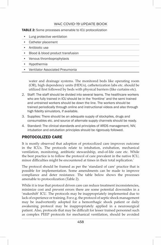
W4C COVID-19 UPDATE BOOK
458
water and drainage systems. The monitored beds like operating room (OR), high dependency units (HDUs), catheterization labs etc. should be utilized first followed by beds with physical barriers (like curtains etc).
2. Staff: The staff should be divided into several teams. The healthcare workers who are fully trained in ICU should be in the ‘frontline’ and the semi trained and untrained workers should be down the line. The workers should be trained periodically through online and instructional videos and also through high fidelity simulators, if available.
3. Supplies: There should be an adequate supply of stockpiles, drugs and consumables etc. and source of alternate supply channels should be ready.
4. Standard: The clinical standards and principles of ARDS management, NIV, intubation and extubation principles should be rigorously followed.
PROTOCOLIZED CAREIt is mostly observed that adoption of protocolized care improves outcome in the ICUs. The protocols relate to intubation, extubation, mechanical ventilation, monitoring, antibiotic stewardship, end-of-life care etc. While the best practice is to follow the protocol of care prevalent in the native ICU, minor difficulties might be encountered at times in their total replication.⁵
The protocol should be framed as per the ‘standard of care’ practice that is possible for implementation. Some amendments can be made to improve compliance and deter resistance. The table below shows the processes amenable to protocolization (Table 2).
While it is true that protocol driven care can reduce treatment inconsistencies, minimize cost and prevent errors there are some potential downsides in a ‘makeshift’ ICU. The protocols may be inappropriately implemented due to lack of experience or training. For e.g. the protocol of septic shock management may be inadvertently adopted for a hemorrhagic shock patient or daily awakening protocol may be inappropriately applied in a neurosurgical patient. Also, protocols that may be difficult for lesser trained personnel such as complex PEEP protocols for mechanical ventilation, should be avoided
TABLE 2: Some processes amenable to ICU protocolization
• Lung protective ventilation• Catheter placement• Antibiotic use• Blood & blood product transfusion • Venous thromboprophylaxis • Hypothermia• Ventilator Associated Pneumonia
W4C COVID-19 UPDATE BOOK
459
in such locations. Lastly, the patients with rare diseases or uncommon presentation of common diseases that may not be amenable to protocolized care should be treated in the native ICU.
FIXING DAILY ROUND GOALS AND CHECKLISTSEvery day after the clinical rounds, the goals of the treatment should be decided and a checklist should be provided for maintenance. The checklist should be filled by the nursing staff and supplemented by the physician at regular intervals. If any shortcomings are observed it should be communicated to the treating physician or the person designated as in-charge.6 The daily goal checklist improves the individualized care goals by structuring a systematic and comprehensive process into targeted objectives. Its daily utilization can overcome the lapses happening due to poor interpersonal communication and facilitates uniformity of practices. The table below shows the different components of daily goal checklist that is useful in ICU (Table 3).
REPORTING ERRORS AND ADVERSE EVENTSSafety is defined as freedom from accidental harm and error is defined as a failure of execution of any planned action as per intention. Since errors occur mostly due to a failure of either human and system factors, every effort should be made to identify and report errors to uphold the safety culture of ICU. An error may not always lead to an adverse event. But it is important
TABLE 3: Components of daily goal checklist and their usefulness
(Compiled by nurses) Patient care plan
(Compiled by physicians) Targets & Achievements
• Spontaneous breathing trial (Y/N) -• Sedation interruption (Y/N) - • Presence of central catheter (Y/N) -• Enteral nutrition assessment
(Y/N)-
Prophylaxis plans• VAP (Y/N) -• Stress ulcer -• DVT(Y/N) -Medications & Investigations• Antibiotic de-escalation (Y/N) -• Blood tests (Y/N) -• Chest X ray(Y/N) -
W4C COVID-19 UPDATE BOOK
460
to be mindful about their possibilities in a ‘makeshift’ ICU and take steps for their primordial prevention.7
The following methods can be adopted to detect errors and prevent adverse events-
Voluntary reporting: This is most useful for inducing changes in the habit and behavior; however there are limitations owing to time constraints, hesitancy in reporting one’s own error and fear of litigations.
Direct observation: This is useful for detecting errors of omission viz. errors during loading drugs, changing catheters, dressings etc. This can overcome many shortcomings of voluntary reporting.
Patient feedback: The patients’ and their relatives’ experience of the safety measures can be known to gain essential inputs about errors which may be missed by the previous methods.
All of these can be combined to identify both latent and manifest errors and adopt corrective measures to improve the quality of care.
QUALITY CONTROL AND AUDITThe ICU services not only entail important clinical decisions but also encompass management decisions based on past performance, current needs and future anticipations. This can be achieved by choosing appropriate quality indicators and observing their trends.
The underlying table (Table 4) shows the common quality indicators that are valuable in such scenario.
Clinical audit is an essential step towards continuous quality improvement process. It can provide tangible solutions for processes that incur a) high costs, b) high risks, c) high variability, d) high complexity and e) high innovations. The greater the strength of evidence of the reference, the better will be the comparison with daily clinical practice.
TABLE 4: Quality indicators and their domains in ‘makeshift’ ICU
• Safety (Unplanned extubation, readmission, VAP, Catheter associated infections etc.)
• Efficiency (Avoidable days in ICU, Patient flow, Ventilator utilization etc.)• Efficacy (ICU LOS, mortality, extubation failure)• Patient satisfaction• Staff satisfaction (Absenteeism, overtime etc.)
W4C COVID-19 UPDATE BOOK
461
CONCLUSIONAn intensivist led multidisciplinary ICU team is an integral part of a health care facility. However certain challenging situations may compel the creation of alternate sites for temporary admission of critically ill patients as ‘makeshift’ ICUs. While setting standards for such ICUs may be an uphill task under normal circumstances, setting standardized protocols including care bundles and initiating measurable processes for quality improvement can assure high quality ICU outcomes.
Key Points
• A ‘makeshift’ ICU should be located at an accessible site but away from public congregation. Sites like operating rooms, ‘step down’ units etc. are more suitable than wards
• A dedicated team consisting of members from both ICU and non-ICU background (if necessary) should be available for patient care round the clock
• The protocols and practice guidelines to be followed should resemble as closely as possible to that of the native ICU
• Quality control and periodic auditing should be conducted• Regular triaging should be performed to shift the ‘sicker’ patients to native
ICU
REFERENCES1. Lu X, Xu S. Intensive care for severe acute respiratory syndrome coronavirus 2 (SARS-CoV-2)
in a makeshift ICU in Wuhan. Crit Care 2020; 24:199. doi: 10.1186/s13054-020-02914-6.2. Swiss Society of Intensive Care Medicine. Recommendations for the admission of patients
with COVID-19 to intensive care and intermediate care units (ICUs and IMCUs). Swiss Med Wkly 2020 Mar 24; 150:w20227. doi: 10.4414/smw.2020.20227
3. Thompson DR, Hamilton DK, Cadenhead CD, Swoboda SM, Schwindel SM, Anderson DC, Schmitz EV, St Andre AC, Axon DC, Harrell JW, Harvey MA, Howard A, Kaufman DC, Petersen C. Guidelines for intensive care unit design. Crit Care Med 2012; 40:1586-600.
4. Guidelines for intensive care unit design. Guidelines/Practice Parameters Committee of the American College of Critical Care Medicine, Society of Critical Care Medicine. Crit Care Med 1995; 23:582-8.
5. Motta E, Luglio M, Delgado AF, Carvalho WB. Importance of the use of protocols for the management of analgesia and sedation in pediatric intensive care unit. Rev Assoc Med Bras (1992). 2016; 62:602-609. doi: 10.1590/1806-9282.62.06.602.
6. Holzmueller CG, Timmel J, Kent PS, Schulick RD, Pronovost PJ. Implementing a team-based daily goals sheet in a non-ICU setting. Jt Comm J Qual Patient Saf 2009; 35:384-8.
7. Camiré E, Moyen E, Stelfox HT. Medication errors in critical care: risk factors, prevention and disclosure. CMAJ 2009; 180:936-943.

Research in COVID 19SECTION 13

465
Landmark Trials That Changed Practice in the
Management of COviD-1963.
1. Horby P, Lim WS, Emberson JR, Mafham M, Bell JL, Linsell L, et al. Dexamethasone in Hospitalized Patients with Covid-19. N Engl J Med 2021; 384:693-704. (The (RECOVERY)
Summary: Multicentre, open-label RCT from United Kingdom (UK), 6425 patients (2104- dexamethasone, 4321-usual care). Treatment arm: 6mg daily dexamethasone for ten days. Mean age 66.1 years, 36% were female, 56% with comorbidities (Diabetes Mellitus (DM) in 24%) Significantly reduced 28-day mortality in dexamethasone group vs usual care [22.9% vs 25.7%, P<0.001, rate ratio 0.83 (0.75-0.93)]. The survival benefit was greatest in patients on IMV at randomization [29.3% vs 41.4%, rate ratio 0.64 (0.51-0.81)]. No survival benefit and a signal towards harm was observed in patients without oxygen [17.8% vs 14.8%, rate ratio 1.19 (0.91–1.55)].
2. Beigel JH, Tomashek KM, Dodd LE, Mehta AK, Zingman BS, Kalil AC, et al. Remdesivir for the Treatment of Covid-19 - Final Report. N Engl J Med 2020; 383:1813-1826. doi: 10.1056/NEJMoa2007764. (ACTT-1)
Summary: Double-blind RCT, 1062 patients (541-remdesivir vs 521 placebo for 10 days).
Significantly shortened median time to recovery [10 days vs 15 days, rate ratio 1.29 (95%CI 1.12-11.49, P<0.001]. Non-significant reduce mortality [11.4% vs 15.2%, Hazard ratio 0.73 (95% CI, 0.52 to 1.03)].
3. Pan H, Peto R, Henao-Restrepo AM, Preziosi MP, Sathiyamoorthy V, Abdool Karim Q, et al. Repurposed Antiviral Drugs for Covid-19 - Interim WHO Solidarity Trial Results. N Engl J Med 2021; 384:497-511. doi: 10.1056/NEJMoa2023184.
Prashant Nasa, Aanchal Singh, Rajesh Chawla
W4C COVID-19 UPDATE BOOK
466
Summary: Multi-national, multi-centre, randomized placebo controlled trial. 11,330 hospitalised adults with COVID-19 underwent randomization [2750 -remdesivir, 954 - hydroxychloroquine, 1411 to lopinavir, 2063 to interferon (including 651 to interferon plus lopinavir), and 4088 to no trial drug]
No or little effect of all studied drugs on overall mortality, need of IMV, and duration of hospital stay between different groups.
4. Goldman JD, Lye DCB, Hui DS, Marks KM, Bruno R, Montejano R, et al. Remdesivir for 5 or 10 Days in Patients with Severe Covid-19. N Engl J Med 2020; 383:1827-1837. doi: 10.1056/NEJMoa2015301.
Summary: Randomized, open-label in 397 patients severe COVID-19 (SpO2 ≤ 94% on room air and pneumonia) and not on IMV. 1:1 randomization (Remdesivir 5 or 10 days). severe No significant difference between a 5-day course or 10-day course of remdesivir.
5. Barratt-Due A, Olsen IC, Nezvalova-Henriksen K, Kåsine T, Lund-Johansen F, Hoel H, et al. Evaluation of the Effects of Remdesivir and Hydroxychloroquine on Viral Clearance in COVID-19 : A Randomized Trial. Ann Intern Med 2021 Jul 13:M21-0653. doi: 10.7326/M21-0653.
Summary: Multicentre, open-label RCT in Norway on 185 patients [remdesivir (n = 42), Hydroxychloroquine (HC)Q (n = 52), or SOC) (n = 87)]. No difference in viral clearance and mortality between the groups.
6. Reis G, Moreira Silva EADS, Medeiros Silva DC, Thabane L, Singh G, Park JJH, et al. Effect of Early Treatment With Hydroxychloroquine or Lopinavir and Ritonavir on Risk of Hospitalization Among Patients With COVID-19: The TOGETHER Randomized Clinical Trial. JAMA Netw Open 2021; 4:e216468. doi: 10.1001/jamanetworkopen.2021.6468.
Summary: RCT from Brazil, 685 patients (stopped prematurely due to futility), three groups [HCQ (n=214), lopinavir-ritonavir (n=244), or placebo (n=227)] for 9 days. No significant difference between the groups on rate of hospitalisation and viral clearance at 14 days.
7. Barnabas RV, Brown ER, Bershteyn A, Stankiewicz Karita HC, Johnston C, Thorpe LE, et al. Hydroxychloroquine as Postexposure Prophylaxis to Prevent Severe Acute Respiratory Syndrome Coronavirus 2 Infection : A Randomized Trial. Ann Intern Med 2021; 174:344-352. doi: 10.7326/M20-6519. Epub 2020 Dec 8
Summary: Double-blind RCT in 671 house-hold close contacts (<96 hours) [337 HCQ and 334 Control group]. No difference between groups on acquiring SARS-CoV-2 infection at day 14 and higher adverse events in HCQ group (16.2 vs 10.9%, p=0.026)
W4C COVID-19 UPDATE BOOK
467
8. RECOVERY Collaborative Group. Lopinavir-ritonavir in patients admitted to hospital with COVID-19 (RECOVERY): a randomised, controlled, open-label, platform trial. Lancet 2020; 396:1345-1352. doi: 10.1016/S0140-6736(20)32013-4.
Summary: Open-label platform RCT, Lopinavir-Ritonavir (400/100 mg) for 10 days or SOC [1:2 randomization, 1616 patients (374 received drug and 767 SOC)]. No difference in 28-day mortality (23% vs 22%), duration of hospital stay, risk of progression to IMV.
9. RECOVERY Collaborative Group. Convalescent plasma in patients admitted to hospital with COVID-19 (RECOVERY): a randomised controlled, open-label, platform trial. Lancet 2021; 397:2049-2059. doi: 10.1016/S0140-6736(21)00897-7
Summary: Open-label platform RCT, 11558 patients (5795 in convalescent plasma and 5763 in SOC). No difference between the groups between 28-day mortality, proportion of patients discharged at day 28, or progression to compositive end point of IMV and death.
10. Libster R, Pérez Marc G, Wappner D, Coviello S, Bianchi A, Braem V, et al. Early High-Titer Plasma Therapy to Prevent Severe Covid-19 in Older Adults. N Engl J Med 2021; 384:610-618. doi: 10.1056/NEJMoa2033700.
Summary: Double blind, RCT, convalescent plasma with high IgG titers to older adult with mild COVID-19 within 72 hours after the onset of symptoms. Stopped prematurely because of decrease in patient load. Early administration of high-titer convalescent plasma reduced the progression of COVID-19.
11. Agarwal A, Mukherjee A, Kumar G, Chatterjee P, Bhatnagar T, Malhotra P; PLACID Trial Collaborators. Convalescent plasma in the management of moderate covid-19 in adults in India: open label phase II multicentre randomised controlled trial (PLACID Trial). BMJ 2020; 371:m3939. doi: 10.1136/bmj.m3939.
Summary: Multicentre, open-label, RCT from India, 464 adults, 39 Hospital. Convalescent plasma was not associated with a reduction in progression to severe covid-19 or all-cause mortality [19% vs 18%, risk ratio 1.04 (95% CI 0.71 to 1.54)]. (Limitation: No measurement of neutralising antibodies in donor or patients)
12. RECOVERY Collaborative Group. Tocilizumab in patients admitted to hospital with COVID-19 (RECOVERY): a randomised, controlled, open-label, platform trial. Lancet 2021; 397:1637-1645. doi: 10.1016/S0140-6736(21)00676-0.
Summary: Open-label multicentre, multi-platform RCT. 4116 patients, 1:1 randomization (82% received systemic corticosteroids). Tocilizumab
W4C COVID-19 UPDATE BOOK
468
reduced mortality (31% vs 35%, rate ratio 0·85 (0·76-0·94); p=0·0028), better discharge rate at day 28, and reduce progression to IMV or death (35% vs 42%) in sub-group not on IMV at randomization.
13. Gordon AC, Mouncey PR, Al-Beidh F, Rowan KM, Nichol AD, Arabi YM, et al. Interleukin-6 Receptor Antagonists in Critically Ill Patients with Covid-19. N Engl J Med 2021; 384:1491-1502. doi: 10.1056/NEJMoa2100433.
Summary: Open-label, multicentre, RCT, platform trial, three groups-tocilizumab (353 patients), sarilumab (48) and placebo (402). Interleukin-6 receptor antagonists tocilizumab and sarilumab improved outcomes, including survival in patients with Covid-19 receiving organ support.
14. Kalil AC, Patterson TF, Mehta AK, Tomashek KM, Wolfe CR, Ghazaryan V, et al. Baricitinib plus Remdesivir for Hospitalized Adults with Covid-19. N Engl J Med 2021; 384:795-807. doi: 10.1056/NEJMoa2031994.
Summary: Double-blind RCT, 1033 patients (515 drug and 518 control). Baricitinib plus remdesivir compared to remdesivir alone in reducing recovery time to recovery in patients with Covid-19, (benefit was higher in patients on high-flow oxygen or non-invasive ventilation). No difference in mortality. Patients on corticosteroids were excluded from the study.
15. Guimarães PO, Quirk D, Furtado RH, Maia LN, Saraiva JF, Antunes MO, et al. Tofacitinib in Patients Hospitalized with Covid-19 Pneumonia. N Engl J Med 2021; 385:406-415. doi: 10.1056/NEJMoa2101643.
Summary: RCT from Brazil with 1:1 randomization with either tofacitinib 10 mg, 12 hourly or placebo till 14 days or discharge in hospitalised adults with COVID-19. 289 patients, 89.3% received corticosteroids. Tofacitinib had a lower risk of death or respiratory in patients with Covid-19.
16. REMAP-CAP Investigators; ACTIV-4a Investigators; ATTACC Investigators, Goligher EC, Bradbury CA, McVerry BJ, Lawler PR, Berger JS, Gong MN, et al. Therapeutic Anticoagulation with Heparin in Critically Ill Patients with Covid-19. N Engl J Med 2021; 385:777-789. doi: 10.1056/NEJMoa2103417.
Summary: Open-label, multi-platform, RCT, 1098 patients with critical COVID-19. 539 patients in therapeutic-dose anticoagulation and 564 with usual thromboprophylaxis, with trial stopped early (futility). Therapeutic-dose anticoagulation compared to usual thromboprophylaxis did not improve survival to hospital discharge or major organ-support free days (cardiovascular or respiratory).
17. ATTACC Investigators; ACTIV-4a Investigators; REMAP-CAP Investigators, Lawler PR, Goligher EC, Berger JS, Neal MD, McVerry BJ, Nicolau JC, et al. Therapeutic Anticoagulation with Heparin in
W4C COVID-19 UPDATE BOOK
469
Noncritically Ill Patients with Covid-19. N Engl J Med 2021; 385:790-802. doi: 10.1056/NEJMoa2105911.
Summary: Open-label, multi-platform, adaptive-RCT, 2219 patients with non-critical COVID-19. Therapeutic-dose anticoagulation compared to usual thromboprophylaxis improve survival to hospital discharge or major organ-support free days at day-21(cardiovascular or respiratory).
OTHER IMPORTANT TRIALS1. PRINCIPLE Trial Collaborative Group. Azithromycin for community treatment of suspected
COVID-19 in people at increased risk of an adverse clinical course in the UK (PRINCIPLE): a randomised, controlled, open-label, adaptive platform trial. Lancet 2021; 397:1063-1074. doi: 10.1016/S0140-6736(21)00461-X.
2. Hinks TSC, Cureton L, Knight R, Wang A, Cane JL, Barber VS, et al. Azithromycin versus standard care in patients with mild-to-moderate COVID-19 (ATOMIC2): an open-label, randomised trial. Lancet Respir Med 2021 Jul 9:S2213-2600(21)00263-0. doi: 10.1016/S2213-2600(21)00263-0.
3. Butler CC, Yu LM, Dorward J, Gbinigie O, Hayward G, Saville BR, et al; PRINCIPLE Trial Collaborative Group. Doxycycline for community treatment of suspected COVID-19 in people at high risk of adverse outcomes in the UK (PRINCIPLE): a randomised, controlled, open-label, adaptive platform trial. Lancet Respir Med 2021 Jul 27:S2213-2600(21)00310-6. doi: 10.1016/S2213-2600(21)00310-6.
4. Patoulias D, Doumas M, Papadopoulos C, Karagiannis A. Janus kinase inhibitors and major COVID-19 outcomes: time to forget the two faces of Janus! A meta-analysis of randomized controlled trials. Clin Rheumatol 2021 Aug 24:1–4. doi: 10.1007/s10067-021-05884-4.
5. Sahu AK, Mathew R, Bhat R, Malhotra C, Nayer J, Aggarwal P, et al. Steroids Use in Non-Oxygen requiring COVID-19 Patients: A Systematic Review and Meta-analysis. QJM 2021 Aug 4:hcab212. doi: 10.1093/qjmed/hcab212.
6. Murai IH, Fernandes AL, Sales LP, Pinto AJ, Goessler KF, Duran CSC, et al. Effect of a Single High Dose of Vitamin D3 on Hospital Length of Stay in Patients With Moderate to Severe COVID-19: A Randomized Clinical Trial. JAMA 2021; 325:1053-1060. doi: 10.1001/jama.2020.26848.


Some Thought Provoking Cases That We Encountered
SECTION 14

473
Section 14 - Some Thought Provoking Cases That We EncounteredSECTION EDITOR
Dr Banambar RayMD, FICCM Head of Critical Care Medicine, SUM Ultimate Medicare, A Unit of SOA,Bhubaneswar
Preface
This section deals with four case reports of pregnancy and COVID-19, broncho pleural fistula and management and pulmonary embolism following COVID-19. The authors describing pregnancy and COVID-19 in three cases have tried to explain the difficulties they faced in managing both pregnancy and COVID-19. One of their patients died and they have tried to explain the reasons. Pregnancy is a immunosuppressant state and COVID-19 only adds to
that state to make it worse. Authors have given an account of the management in such cases.
Broncho-pleural fistula (BPF) developing spontaneously or when patient is on ventilator for bad low compliant lung, is a difficult condition to treat. The authors have explained the management which was conservative in contravention to the existing modality of treatment i.e. generally surgical. COVID-19 induced BPF is a separate condition which deserves a different approach of management. One BPF described by another group of authors was managed by bronchoscopy and spaghetti though details of that case was not available in the text.
There is a case of Pulmonary embolism for whom CT pulmonary angiogram could not be done as patient was in severe septic shock and on three inotropes. This was diagnosed by suspicion, ECG and ECHO findings. The authors have described the complications the patient went through.

475
Delayed Pulmonary Embolism in Covid 19 an
Unexpected And Bitter Foe64.
INTRODUCTIONPulmonary embolism continues to be a dreaded pathology to diagnose and treat since symptoms can mimic other diseases and outcomes are not always favorable despite best of efforts. The COVID-19 pandemic affects invariably the respiratory system but when it comes to sudden death, the cardiopulmonary manifestations gained more attention.1 Pulmonary embolism was reported extensively which led to trials advocating the use of anticoagulation along with steroids as a major treatment modality for all moderate to severe cases. Despite the above treatment there were reports of embolism with a delayed presentation in cases without any obvious source.2 We hereby present a case report with a similar presentation.
CASE REPORTA 55-year-old male presented to our ICU with history of fever followed by breathlessness and generalised weakness for 7-8 days. He had no known co-morbidities. He was tested positive for COVID-19 and his CT severity index was 17/25. Treatment as per standard protocols was initiated. He was initially put on noninvasive ventilation and on subsequent increase in oxygen requirement and deterioration of respiratory pattern, he was intubated and ventilated according to standard ARDS protocol. He underwent multiple sessions of proning. He was a case of difficult prolonged weaning, therefore he was tracheostomised on day 17 from date of admission. Post tracheostomy he had a turbulent course with multiple episodes of sepsis with septic shock which were adequately managed with the help of cultures and detailed review along with change of antibiotics. Weaning off ventilator was a challenge due to extremely poor lung compliance. Repeat CT scan of thorax had shown widespread fibrotic changes with only few patches of normal looking lung. All this while patient was on therapeutic dose of anticoagulation and echocardiogram showed normal heart chambers and left ventricular function. On day 49 of hospitalization, he also underwent emergency cholecystectomy
Banambar Ray, Alisha Chaudhury

W4C COVID-19 UPDATE BOOK
476
for empyema gall bladder detected on scans done for sepsis workup. Post this difficult course in ICU, he was showing an improvement in his clinical parameters with good performance in weaning trials too. On day 72 of his hospitalization he developed sudden onset desaturation with hypotension with fever spikes. Based on the background history our first differential diagnosis was septic shock but due to acute nature of the deteoriation a bedside ultrasound and echo (Figure 1) was done which revealed dilatation of the right atrium and ventricle with tricuspid regurgitation with a dilated inferior vena cava. Bedside lower limb venous doppler study showed no evidence of deep vein thrombosis. Clinically and based on the echo findings, as there was high probability of pulmonary embolism, CT pulmonary angiogram was planned but could not be done due to the poor clinical status and hemodynamic instability. The patient being on high doses of dual vasopressors, could not be shifted to the CT suite. Cardiology opinion was taken regarding the same since thrombolysis was not an option in view of the multiple clinical issues. On diagnosis based on supplementary clinical and imaging data patient was started on heparin infusion as per PE treatment protocol. ECG repeated the next day also showed the classical S1Q3T3 pattern (Figure 2) and D-Dimer levels were raised. Though there was marginal improvement in the vitals and oxygenation, the patient finally succumbed due to associated sepsis with septic shock and multiorgan failure.
DISCUSSIONApart from pneumonia leading to ARDS in COVID-19, there is a need for timely identification of other reversible causes of hypoxia which can be treated in a background of COVID-19 infection. Suh et al in their meta-analysis looked at 27 studies with a total of 3342 patients and reported incidence rates of PE and DVT as 16.5% and 14.8%, respectively, which exceeded 20% in patients admitted to the intensive care unit.3 PE was confined to the peripheral
FIG. 1: ECHO Showing RA and RV Dilatation and Deviation of Septum
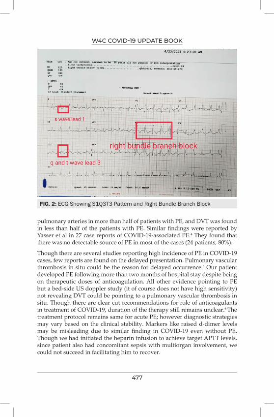
W4C COVID-19 UPDATE BOOK
477
pulmonary arteries in more than half of patients with PE, and DVT was found in less than half of the patients with PE. Similar findings were reported by Yasser et al in 27 case reports of COVID-19-associated PE.4 They found that there was no detectable source of PE in most of the cases (24 patients, 80%).
Though there are several studies reporting high incidence of PE in COVID-19 cases, few reports are found on the delayed presentation. Pulmonary vascular thrombosis in situ could be the reason for delayed occurrence.5 Our patient developed PE following more than two months of hospital stay despite being on therapeutic doses of anticoagulation. All other evidence pointing to PE but a bed-side US doppler study (it of course does not have high sensitivity) not revealing DVT could be pointing to a pulmonary vascular thrombosis in situ. Though there are clear cut recommendations for role of anticoagulants in treatment of COVID-19, duration of the therapy still remains unclear.6 The treatment protocol remains same for acute PE; however diagnostic strategies may vary based on the clinical stability. Markers like raised d-dimer levels may be misleading due to similar finding in COVID-19 even without PE. Though we had initiated the heparin infusion to achieve target APTT levels, since patient also had concomitant sepsis with multiorgan involvement, we could not succeed in facilitating him to recover.
FIG. 2: ECG Showing S1Q3T3 Pattern and Right Bundle Branch Block
W4C COVID-19 UPDATE BOOK
478
CONCLUSIONVenous and pulmonary thromboembolism can be life threatening complications of COVID-19. This can be a part of the disease onset and even be a late presentation. Therefore, a high index of suspicion is quintessential to promptly pick up the diagnosis and initiate treatment of this life threatening condition.
PRACTICE POINTS1. Pulmonary embolism can occur as a part of the disease or a delayed
presentation in COVID-19 pneumonia.
2. Early recognition and immediate treatment of this condition can be life-saving.
3. Investigation of choice is CTPA along with other supplementary findings but it may not be able to detect peripheral emboli.
4. Patient centered and a multidisciplinary team approach is mandatory for managing this disease.
KEYWORDSCOVID-19, pulmonary embolism, anticoagulation, deep vein thrombosis, mortality.
REFERENCES1. World Health Organization. Coronavirus disease (COVID-19): situation report, 162. Geneva,
Switzerland: World Health Organization, 2020.2. Van Dam LF, Kroft LJM, van der Wal LI, et al. Clinical and computed tomography
characteristics of COVID-19 associated acute pulmonary embolism: a different phenotype of thrombotic disease? Thromb Res 2020; 193:86–89.
3. Suh YJ, Hong H, Ohana M et al. Pulmonary Embolism and Deep Vein Thrombosis in COVID-19: A Systematic Review and Meta-Analysis. Radiology 2021; 298:E70–E80.
4. Sakr Y, Giovini M, Leone M, Pizzilli G, Kortgen A, Bauer M et al. Pulmonary embolism in patients with coronavirus disease-2019 (COVID-19) pneumonia: a narrative review. Ann Intensive Care 2020; 10:124.
5. Perez-Girbes A. Acute pulmonary embolism and Covid-19: a common association in seriously ill patients? Arch Bronconeumol 2020; 56:34.
6. Danzi GB, Loffi M, Galeazzi G, Gherbesi E. Acute pulmonary embolism and COVID-19 pneumonia: a random association? Eur Heart J 2020; 41:1858.
479
interesting Cases of Covid-19- Complicated Leaks
in the Lungs65.
CASE SNIPPET A 58-year-old patient was diagnosed with moderate coronavirus disease 2019 (COVID-19). He was hypoxemic at presentation and required oxygen therapy along with other supportive therapy including steroids (dexamethasone 6 mg once a day). He was not a known smoker and had associated diabetes mellitus, well controlled on oral hypoglycaemic drugs. However, he progressively deteriorated and required high flow oxygen therapy. The imaging revealed presence of pneumothorax and pneumomediastinum. Chest tube drainage was instituted. Subsequently lung expanded partially, and oxygen requirement decreased. However, the underwater seal of the chest tube showed continuous bubbling and chest x-ray revealed residual pneumothorax. A diagnosis of bronchopleural fistula was made. A conservative approach was continued. In view of continuous leak, bronchoscopy was planned, and the spaghetti was placed in the affected bronchus. This improved the air leak and oxygen requirement gradually was weaned off.
INTRODUCTION The outbreak of coronavirus disease (COVID-19) is a well-known pandemic now. Though multisystemic involvement is seen, it is primarily a respiratory system related disease and manifestations are mostly related to it. The respiratory manifestations include consolidation, inflammatory changes, and subsequent fibrosis. The reported complications in patient of COVID-19 include pneumothorax, subcutaneous emphysema, and mediastinal emphysema/pneumo-mediastinum.¹ The lung injury in patients of COVID-19 remains much more complicated and is multifactorial. Injury infliction due to barotrauma, volutrauma, atelectrauma, and biotrauma (mechanical ventilator induced) can be grouped under ventilator-induced lung injury (VILI).² The management is not well described and is as such individualized..
Syed Moied Ahmed, Rakesh Garg
W4C COVID-19 UPDATE BOOK
480
AETIOLOGY AND PATHOGENESIS OF RESPIRATORY DYSFUNCTION IN COVID-19 FOR LEAKY LUNGSExact mechanism for spontaneous pneumothorax in patients with COVID-19 disease remains unclear, however the plausible explanation appears to be the unique nature of structural changes in lung parenchyma caused by the virus.³,⁴ The COVID-19 lungs are prone for leakage due to pathological damage leading to weakening of alveolocapillary complex, low compliance and reduced elastance.⁴,⁵
These pathological changes are related to disease severity, prolonged duration of lung inflammation, patient's overall health status and treatment strategies as well. The presence of persistent intractable cough has also been related to the occurrence of lung damage and has been reported to have possible association of patient self-inflicted lung injury (P-SILI). All these pathophysiological changes in lungs due to COVID-19 lead to lung puncture causing pneumothorax, and/or pneumomedistinum, in simpler term labelled as “COVID-19 puncture lung” with an incidence of around 0.9-1.1.²
MANAGEMENT STRATEGIESThe management of the COVID-19 remains challenging as definitive treatment is not yet known. Assessment and prediction of such complication remains a challenge. A high index of suspicion needs to be kept for pneumomediastinum or pneumothorax in patients with confirmed or suspected COVID-19. The occurrence of acute or worsening dyspnea with rapid deterioration in clinical condition warranting respiratory or oxygen support is a good indicator. In such situations, a thorough assessment needs to be done. This includes not just clinical examination and intervention in case of urgent situation like tension pneumothorax but also there is a need of imaging modalities like HRCT to find the underlying condition. CT imaging has been found to identify the findings related to ‘Punctured Lung’.
Pneumothorax is usually managed with conservative approach. This includes optimal monitoring of the patient, providing appropriate oxygen therapy and also managing any reversible underlying cause like bacterial infection, intractable cough, etc.
The need for mechanical ventilation in COVID-19 patients arises in severe condition. The ventilatory management strategies, especially when lung recruitment manoeuvres are used, leads to mismatch of alveolar distension. The normal non-dependent lung regions with relatively higher compliance and less airway resistance have overdistension and there remains a possibility of alveolar injury due to increased shear forces. Patient on mechanical ventilatory support and having features suggestive of ARDS are difficult to manage in case they have lung injury leading to pneumothorax or pneumomediastinum as the exacerbation of leak occurs. In certain situations, it may also lead to fistula
W4C COVID-19 UPDATE BOOK
481
formation with continuous air leak. Protective ventilatory strategy including plateau pressure (Pplat) less than 30 cmH2O along with weight appropriate tidal volume is useful strategy as it reduces the alveolar pressure, preventing overdistension of alveolus. This decreases the risk of further lung injury and occurrence of further pneumothorax and persistent air leak. In addition, the use of optimal sedation and/or neuromuscular blocking drugs are also useful strategy that needs to be considered when managing such clinical situations. This is helpful by reducing negative pressure in pleural cavity and it prevents further lung injury by reducing the shear stress and changes of lung structure.
Patients with COVID-19 also manifest cough as one of the main clinical features. Some patients may have intractable cough and could be one of the triggering factors for occurrence of pneumothorax. Persistent cough may also lead to further lung damage and it prevents spontaneous closure of leaks. Hence appropriate measures for control of cough using cough suppressants and reversal of any underlying condition causing cough, need to be done.
The usual management strategy for pneumothorax remains placement of chest drains. However, in cases of persistent air leak, other interventions including surgical intervention need to be considered. Ideal timing of such interventions remains unclear as spontaneous closure is also observed. In patients with presence of lung bleb, a minimally invasive thoracoscopy with bleb resection may be considered.⁵ At times, when air leak is persistent despite chest drain insertions, bullectomy with or without pleurodesis remains the other option. With the availability of interventional pulmonologist, placement of spaghetti to block the bronchus of a particular lobe may also be attempted for healing of the leaking lung. Use of endobronchial valves (EBV) has also been reported for such conditions.
CONCLUSIONThe lung injury is seen in COVID-19 patients. The pneumothorax or pneumomediastinum can be seen spontaneously or more commonly in patients requiring ventilatory support and may be a forerunner to fistula formation with continuous leak. Such condition may be effectively managed with intrabronchial placement of blockers.
REFERENCES1. Xu Z, Shi L, Wang Y, et al. Pathological findings of COVID-19 associated with acute respiratory
distress syndrome. Lancet Respir Med 2020; 8:420-22. 2. Urigo C, Soin S, Sahu A. Spontaneous pneumomediastinum as a complication of a COVID-19
related pneumonia. Radiology Case Reports 2020;15:2577-81.3. Sabharwal P, Chakraborty S, Tyagi N, Kumar R, Taneja A. Spontaneous Air-leak Syndrome
and COVID-19: A Multifaceted Challenge. Indian J Crit Care Med 2021; 25: 584–587.4. Zantah M, Castillo ED, Townsend R, Dikengil F, Criner GJ. Pneumothorax in COVID-19
disease incidence and clinical characteristics. Respiratory Research 2020; 21:236-39.5. Aiolfi A, Biraghi T, Montisci A, et al. Management of Persistent Pneumothorax With
W4C COVID-19 UPDATE BOOK
482
Thoracoscopy and Bleb Resection in COVID-19 Patients. Ann Thorac Surg 2020; 110:e413-5 6. Szewczyk J, Adkinson BC, Akkineni S, et al. Endobronchial valves: a bridge to definitive
surgical management in COVID-19 recurrent pneumothorax. J Thorac Dis 2021;13:411-413
483
Spontaneous Pneumothorax with
Bronchopleural Fistula in COviD-19 Pneumonia – A
Nightmare
66.
INTRODUCTIONBronchopleural fistula (BPF) is a sinus tract between a bronchus and the pleural cavity.¹ It usually presents as a complication of pneumonectomy or pulmonary resection; however, other contributing factors include necrotizing pulmonary infection, persistent spontaneous pneumothorax, chemotherapy, radiation therapy, invasive malignancy and tuberculosis.1,2 Although alveolar air leaks have been increasingly reported in COVID-19 pneumonia.3 bronchopleural fistula causing persistent pneumothorax in COVID-19 pneumonia is extremely rare.4,5 BPF may be found in COVID-19 pneumonia with or without mechanical ventilation. Treatment of BPF can be medical, bronchoscopic or surgical.1 Here we present 2 case reports of spontaneous pneumothorax with BPF, successfully managed conservatively.
CASE REPORTSCASE 1
A 50 yr female presented with fever and cough for 10 days and RT-PCR for COVID-19 was positive. Her Covid-19 markers were elevated and she was started on Inj.Remdesvir, Enoxaparin and Methylprednisolone. She was initially put on NIV with 100% oxygen and later intubated. She was proned 5 times and then tracheostomised. On Day 39 of hospitalisation, she had sudden desaturation, bradycardia and hypotension while on controlled ventilation. She had subcutaneous emphysema extending to neck and chest. Chest X-Ray showed right Pneumothorax for which ICD was inserted which had continuous air leak even after lung expansion, suggestive of bronchopleural fistula. She was ventilated with low tidal volume (100 ml) and high respiratory rate (80-100/min). She also developed left pneumothorax soon after for which
Arun Kumar Sahu, Abhijeet Raha
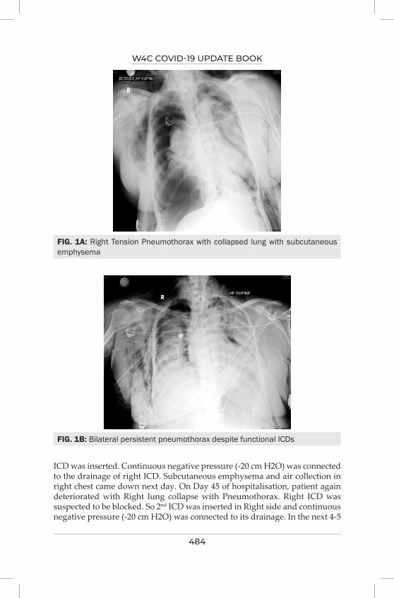
W4C COVID-19 UPDATE BOOK
484
FIG. 1A: Right Tension Pneumothorax with collapsed lung with subcutaneous emphysema
FIG. 1B: Bilateral persistent pneumothorax despite functional ICDs
ICD was inserted. Continuous negative pressure (-20 cm H2O) was connected to the drainage of right ICD. Subcutaneous emphysema and air collection in right chest came down next day. On Day 45 of hospitalisation, patient again deteriorated with Right lung collapse with Pneumothorax. Right ICD was suspected to be blocked. So 2nd ICD was inserted in Right side and continuous negative pressure (-20 cm H2O) was connected to its drainage. In the next 4-5
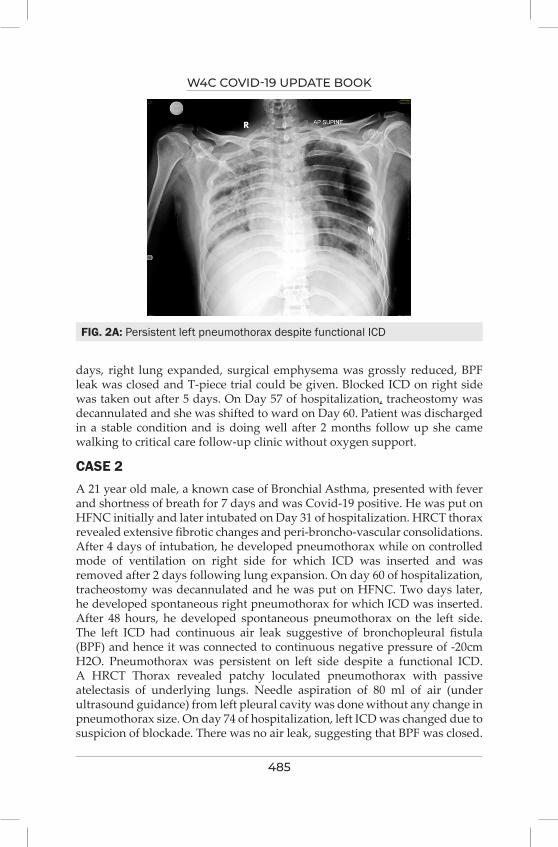
W4C COVID-19 UPDATE BOOK
485
days, right lung expanded, surgical emphysema was grossly reduced, BPF leak was closed and T-piece trial could be given. Blocked ICD on right side was taken out after 5 days. On Day 57 of hospitalization, tracheostomy was decannulated and she was shifted to ward on Day 60. Patient was discharged in a stable condition and is doing well after 2 months follow up she came walking to critical care follow-up clinic without oxygen support.
CASE 2A 21 year old male, a known case of Bronchial Asthma, presented with fever and shortness of breath for 7 days and was Covid-19 positive. He was put on HFNC initially and later intubated on Day 31 of hospitalization. HRCT thorax revealed extensive fibrotic changes and peri-broncho-vascular consolidations. After 4 days of intubation, he developed pneumothorax while on controlled mode of ventilation on right side for which ICD was inserted and was removed after 2 days following lung expansion. On day 60 of hospitalization, tracheostomy was decannulated and he was put on HFNC. Two days later, he developed spontaneous right pneumothorax for which ICD was inserted. After 48 hours, he developed spontaneous pneumothorax on the left side. The left ICD had continuous air leak suggestive of bronchopleural fistula (BPF) and hence it was connected to continuous negative pressure of -20cm H2O. Pneumothorax was persistent on left side despite a functional ICD. A HRCT Thorax revealed patchy loculated pneumothorax with passive atelectasis of underlying lungs. Needle aspiration of 80 ml of air (under ultrasound guidance) from left pleural cavity was done without any change in pneumothorax size. On day 74 of hospitalization, left ICD was changed due to suspicion of blockade. There was no air leak, suggesting that BPF was closed.
FIG. 2A: Persistent left pneumothorax despite functional ICD

W4C COVID-19 UPDATE BOOK
486
FIG. 2B :HRCT Thorax showing left anterior pneumothorax despite functional ICD
FIG. 2C: Complete resolution of pneumothorax with pigtail catheter on left side
Patient was shifted to ward on day 80 with left loculated pneumothorax and ICD in-situ. On repeat HRCT Thorax, on day 88, there was moderate left
W4C COVID-19 UPDATE BOOK
487
anterior hydropneumothorax. Ultrasound guided needle aspiration of air from left pleural cavity was done. ICD was blocked and hence it was removed on day 101. A pigtail chest drain was placed above the entry point of previous chest tube and a gush of air came out. His respiratory distress improved. Patient was discharged with pigtail catheter after 108 days of hospitalization. On first follow up visit after 15 days, chest x-ray showed complete resolution of pneumothorax (Figure 2C) and hence pigtail catheter was removed.
DISCUSSIONThe prevalence of pneumothorax in COVID-19 pneumonia is 1-2%. 6,7 However BPF has been reported only in few cases of COVID-19 pneumonia4,5. In a case report by Habibet al, a 40-year-old COVID-19 pneumonia patient developed bronchopleural fistula with persistent pneumothorax even without any risk factors or mechanical ventilation and he was successfully treated conservatively with prolonged chest tube drainage.4 In another case report by Placik et al, a 49-year-old COVID-19 pneumonia patient had a spontaneous pneumothorax that did not resolve with appropriate intervention and developed a bronchopleural fistula in the presence of mucormycosis.5
In our first case, the patient developed spontaneous pneumothorax with BPF while on mechanical ventilation whereas in second case, the same complication occurred without mechanical ventilation. In both the cases, BPF led to persistent pneumothorax despite functional ICD. The first case, on ventilator, was ventilated with low tidal volume (2ml/kg) and high respiratory rate (80-100/min) and the 2nd case continued to breathe spontaneously.
Although the traditional management of BPF has been surgery or bronchoscopic spaghetti placement approach, our both patients were successfully managed conservatively.⁸ In our first case, BPF closed early (11 days). In our second case, surgerical intervention or bronchoscopy could not have been done for the reason that patient had multiple encysted pneumothoraces.
It is probable that in COVID-19, the course of BPF is different from that in non-COVID cases. Surgery may not be an easy option, especially when there are multiple ruptures of bullae at different times and one can’t be sure whether patient won’t develop further pneumothorax and BPF even after surgery. Surgery could also be the last choice because of pre-existing respiratory compromise and debilitated general condition. Therefore, in COVID-19 cases, a spontaneous trial of healing could be given before contemplating surgical option.
CONCLUSIONSpontaneous pneumothorax and BPF can be a life-threatening complication of COVID-19 pneumonia. High index of suspicion and early recognition of this complication is of utmost importance. Although surgery and

W4C COVID-19 UPDATE BOOK
488
endobronchial valve placement have been traditional treatment modalities for BPF, conservative management may be a better alternative for BPF in COVID-19, like in our two cases. Further clinical research should be carried out to establish the best modality of treatment for BPF in COVID-19.
PRACTICE POINTS1. Spontaneous pneumothorax and BPF can occur in COVID-19 pneumonia
and can be life-threatening
2. Surgery may not be the first choice for BPF in COVID-19 because of pre-existing respiratory compromise, debilitated general condition and the risk of further developing pneumothorax and BPF even after surgery.
3. Perhaps in COVID-19 cases, a spontaneous trial of healing is preferable to the surgical correction of BPF.
4. Conservative management for BPF can be done with the help of continuous low negative pressure applied to ICD drainage and ventilating with low tidal volume and high respiratory rate in patients on positive pressure ventilation.
KEYWORDSCOVID-19 pneumonia, Spontaneous pneumothorax, Persistent pneumothorax, Tension pneumothorax, Bronchopleural fistula.
REFERENCES1. Kiyota Y, Topulos GP, fistula HPMB. Essential Clinical Anesthesia Review: Keywords. Quest.
Answers Boards 2015; 518–9.2. Lois M, Noppen M. Bronchopleural fistulas: an overview of the problem with special focus on
endoscopic management. Chest 2005; 128:3955–3965. doi: 10.1378/chest.128.6.3955.3. Sabharwal P, Chakraborty S, Tyagi N, Kumar R, Taneja A. Spontaneous Air-leak Syndrome
and COVID-19: A Multifaceted Challenge. Indian J Crit Care Med 2021; 25:584-587. doi:10.5005/jp-journals-10071-23819.
4. Mhd Baraa Habib, Ibrahim Mohammad Obeida , Khaled Ali, Mohamed Abdelrazek and Mouhand Mohamed. Bronchopleural fistula causing persistent pneumothorax in COVID-19 pneumonia patient with no risk factors. Authorea June 25, 2021.
5. Placik DA, Taylor WL, Wnuk NM. Bronchopleural fistula development in the setting of novel therapies for acute respiratory distress syndrome in SARS-CoV-2 pneumonia. Radiol Case Rep 2020; 15:2378-2381. doi:10.1016/j.radcr.2020.09.026
6. Noppen M, de Keukeleire T. Pneumothorax. Respiration 2008; 76:121–7. n.d.7. Salehi S, Abedi A, Balakrishnan S, Gholamrezanezhad A. Coronavirus disease 2019
(COVID-19): A systematic review of imaging findings in 919 patients. Am J Roentgenol 2020; 215:87–93.
8. Szewczyk J, Adkinson BC, Akkineni S, Nguyen DM, Arias SA, Villamizar NR. Endobronchial valves: a bridge to definitive surgical management in COVID-19 recurrent pneumothorax. J Thorac Dis 2021; 13:411-413. doi: 10.21037/jtd-20-3248.
489
COviD and Complicated Pregnancy 67.
INTRODUCTION Pregnancy is a challenging and vulnerable period. Corona virus infection can cause severe adverse pregnancy outcomes such as miscarriage, premature delivery, IUGR, and maternal death.¹,² Randomized controlled trials to provide evidence-based treatment guidelines are needed for this unique patient group, especially with a recent CDC report indicating pregnant patients are at higher risk for the severe disease including death.
CASE PRESENTATION During this second wave of COVID pandemic from 1st March 2021 till 15th August 2021, out of a total of 521 COVID-19 cases, there were 13 COVID-19 positive pregnant patients in their second and third trimester admitted in Health City Hospital, Guwahati. All of the cases of SARS-CoV-2 infection were confirmed by positive RT–PCR tests. Amongst the 13 patients, 3 needed aggressive ICU care and were managed with noninvasive / invasive mechanical ventilation owing to the severity of their symptoms, details of which are given below in Table 1. Their inflammatory markers are graphically depicted in the Figures 1-5
Table 1 shows in detail the three cases who had severe symptoms and were admitted to ICU for further management
Abbreviations: SpO2: Oxygen Saturation, WBC: White Blood Cell, SGOT: Serum Glutamic Oxaloacetic Transaminase, SGPT: Serum Glutamic Pyruvic Transaminase, CRP: C Reactive Protein, RAT: Rapid Antigen Test, ICU: Intensive Care Unit, CT: Computed Tomography, CXR: Chest X-Ray, CTSS : Computed Tomography Severity Score
COMPARATIVE ANALYSIS OF THE INFLAMMATORY MARKERS OF THE THREE CASESAll the three patients were treated as per the treatment protocol for moderate COVID-19. Steroid methyl prednisolone was started as per the Royal College
Reshu Gupta Khanikar, Rakhee Baruah

W4C COVID-19 UPDATE BOOK
490
TABLE 1: Patient Parameters
Parameters Case 1 Case 2 Case 3
Age (years) 30 35 35Weeks of gestation (weeks)
26+ 25+ 27+
Symptoms cough , chest discomfort and breathlessness
fever, cough and generalized weakness
Cough, breathlessness
Comorbidities None Hypothyroidism Post LSCS
Psoriasis
Treatment prior to hospitalisation
None Referred from another centre and had received injectable steroids
Was on home isolation and received oral steroids for 6 days
Hospital admission from onset of symptoms(days)
5 10 8
SpO2 level (in room air)
92% 80% 96%
Pulse (beats/min) 84 96 143BP (mmHg) 120/72 120/70 100/70Foetal heart rate (beats/min)
138 165 184
WBC count(×109/μl)
11400 23200 8600
SGOT/SGPT (IU/L)
90/65 82/77 93/70
Inflammatory MarkersCRP (mg/L) 34.2 201 75.6Procalcitonin(ng/ml)
0.14 0.24 0.23
D-Dimer (ng/ml FEU)
728 1118 630
Ferritin (ng/ml) 507 2002 103
(Contd...)
W4C COVID-19 UPDATE BOOK
491
Parameters Case 1 Case 2 Case 3
Radiology CTSS 13/25[Figure 6]
Bilateral patchy ground glass opacities in mid and lower zones of both lungsCTSS not reported[Figures 7,8]
Inhomogeneous radio opacities in b/l lung fields[fig 9]CTSS 23/25 [Figure 10]
Ventilator days 7 days (NIV) 8 days (NIV+IMV) 8 days (NIV+IMV)
RAT negative (days from onset of symptoms)
17 17 17
ICU Days 18 8 15
Maternal outcome
Discharged Expired Discharged
Fetal outcome Healthy male baby with anti SARS Ab 661Placental biopsy not done
IUD Healthy male baby with anti SARS Ab 102Placental biopsy normal
TABLE 1: Patient Parameters (Contd...)
of Obstretics & Gynaecology (RCOG) recommendations. Remdesivir could be completed only in case 1, while in the other two it had to be discontinued due to transaminitis [Figures 4, 5].
Case 1 was successfully managed on NIV support alone but the other two needed invasive ventilatory support.
Case 2 developed spontaneous pneumothorax which was managed by placement of ICWSD [Figure 8]. She went into sepsis and septic shock and needed high vasopressor support finally dying on day 8.
DISCUSSIONPregnant women are more susceptible to COVID-19 as compared to the general population. The predominant features of COVID-19 in pregnancy are fever, cough, dyspnea and lymphopenia with shortness of breath described in up to 18% of patients.¹ Due to heightened metabolism, gestational anemia and fetal oxygen consumption there is an increase in oxygen demands making the
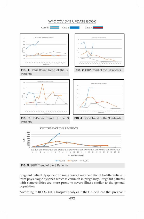
W4C COVID-19 UPDATE BOOK
492
FIG. 1: Total Count Trend of the 3 Patients
FIG. 2: CRP Trend of the 3 Patients
FIG. 3: D-Dimer Trend of the 3 Patients
FIG. 4: SGOT Trend of the 3 Patients
FIG. 5: SGPT Trend of the 3 Patients
pregnant patient dyspnoeic. In some cases it may be difficult to differentiate it from physiologic dyspnea which is common in pregnancy. Pregnant patients with comorbidities are more prone to severe illness similar to the general population.
According to RCOG UK, a hospital analysis in the UK deduced that pregnant
SGPT TREND OF THE 3 PATIENTS140012001000
800600400200
0DAY
0DAY
1DAY
2DAY
3DAY
4DAY
5DAY
6DAY
7DAY
8DAY
9DAY
10DAY
11DAY
12
NUMBER OF DAYS
CASE 2 CASE 3 CASE 1
DAY13
DAY14
DAY15
DAY16
DAY17
DAY18
DAY19
DAY20
DAY21
DAY22
DAY23
SGPT
SGOT TREND OF THE 3 PATIENTS
CRP TREND OF THE 3 PATIENTSTOTAL COUNT TREND OF THE 3 PATIENTS
Case 1: Case 2: Case 3:
D-DIMER TREND OF THE 3 PATIENTS
1200
1000
800
600
400
200
0
2500
2000
1500
1000
500
0
SGO
T
D-D
IMER
DAY
0
DAY 0DAY 1DAY 2DAY 3DAY 4DAY 5DAY 6DAY 7DAY 8DAY 9
DAY 10DAY 11DAY 12DAY 13DAY 14DAY 15DAY 16DAY 17DAY 18DAY 19DAY 20DAY 21DAY 22DAY 23
DAY
1
DAY
2
DAY
3
DAY
4
DAY
5
DAY
6
DAY
7
DAY
8
DAY
9
DAY
10
DAY
11
DAY
12
DAY
13
DAY
14
DAY
15
DAY
16
DAY
17
DAY
18
DAY
19
DAY
20
DAY
21
DAY
22
DAY
23
NUMBER OF DAYSNUMBER OF DAYS
250
200
150
100
50
0
40000
15000
30000
25000
20000
15000
10000
5000
0
CRP
DAY 0DAY 1DAY 2DAY 3DAY 4DAY 5DAY 6DAY 7DAY 8DAY 9
DAY 10DAY 11DAY 12DAY 13DAY 14DAY 15DAY 16DAY 17DAY 18DAY 19DAY 20DAY 21DAY 22DAY 23
NUMBER OF DAYSNUMBER OF DAYS
TOTA
L C
OU
NT
DAY 0DAY 1DAY 2DAY 3DAY 4DAY 5DAY 6DAY 7DAY 8DAY 9
DAY 10DAY 11DAY 12DAY 13DAY 14DAY 15DAY 16DAY 17DAY 18DAY 19DAY 20DAY 21DAY 22DAY 23
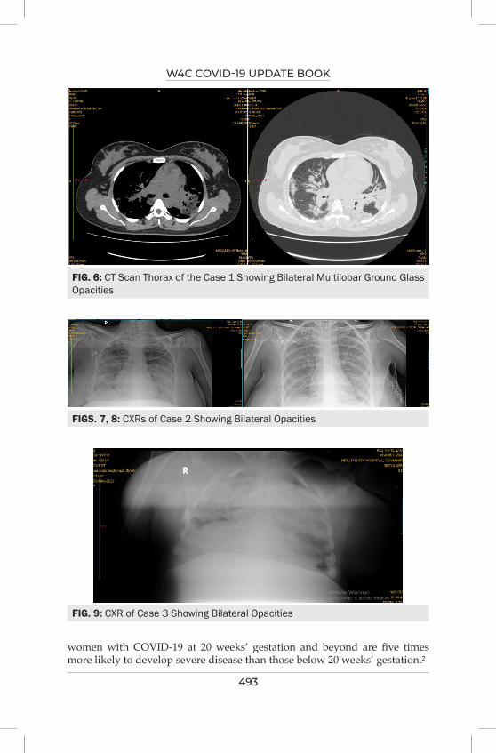
W4C COVID-19 UPDATE BOOK
493
FIG. 6: CT Scan Thorax of the Case 1 Showing Bilateral Multilobar Ground Glass Opacities
FIGS. 7, 8: CXRs of Case 2 Showing Bilateral Opacities
FIG. 9: CXR of Case 3 Showing Bilateral Opacities
women with COVID-19 at 20 weeks’ gestation and beyond are five times more likely to develop severe disease than those below 20 weeks’ gestation.²
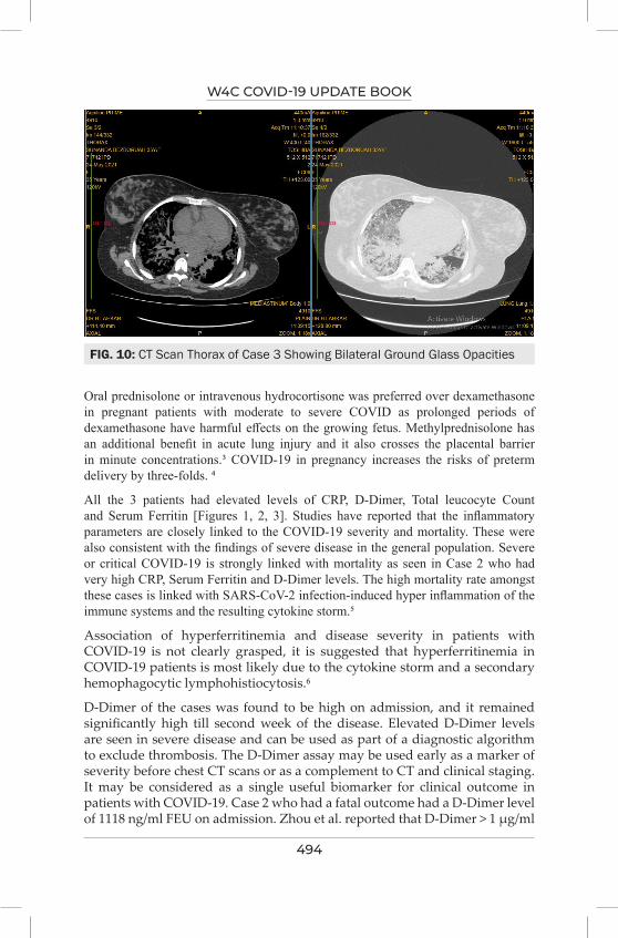
W4C COVID-19 UPDATE BOOK
494
FIG. 10: CT Scan Thorax of Case 3 Showing Bilateral Ground Glass Opacities
Oral prednisolone or intravenous hydrocortisone was preferred over dexamethasone in pregnant patients with moderate to severe COVID as prolonged periods of dexamethasone have harmful effects on the growing fetus. Methylprednisolone has an additional benefit in acute lung injury and it also crosses the placental barrier in minute concentrations.³ COVID-19 in pregnancy increases the risks of preterm delivery by three-folds. ⁴
All the 3 patients had elevated levels of CRP, D-Dimer, Total leucocyte Count and Serum Ferritin [Figures 1, 2, 3]. Studies have reported that the inflammatory parameters are closely linked to the COVID-19 severity and mortality. These were also consistent with the findings of severe disease in the general population. Severe or critical COVID-19 is strongly linked with mortality as seen in Case 2 who had very high CRP, Serum Ferritin and D-Dimer levels. The high mortality rate amongst these cases is linked with SARS-CoV-2 infection-induced hyper inflammation of the immune systems and the resulting cytokine storm.⁵
Association of hyperferritinemia and disease severity in patients with COVID-19 is not clearly grasped, it is suggested that hyperferritinemia in COVID-19 patients is most likely due to the cytokine storm and a secondary hemophagocytic lymphohistiocytosis.⁶
D-Dimer of the cases was found to be high on admission, and it remained significantly high till second week of the disease. Elevated D-Dimer levels are seen in severe disease and can be used as part of a diagnostic algorithm to exclude thrombosis. The D-Dimer assay may be used early as a marker of severity before chest CT scans or as a complement to CT and clinical staging. It may be considered as a single useful biomarker for clinical outcome in patients with COVID-19. Case 2 who had a fatal outcome had a D-Dimer level of 1118 ng/ml FEU on admission. Zhou et al. reported that D-Dimer > 1 µg/ml

W4C COVID-19 UPDATE BOOK
495
is a risk for mortality.⁷
In our case series, Remedesivir was given to the three moderate category cases, but could finish the 5 day course in only one patient successfully. Due to deranged liver function tests we had to discontinue in other two patients. Apart from transaminitis, adverse effects of Remdesivir were not seen. However pregnant patients who have received Remdesivir have shown higher recovery rates with low rates of serious adverse events.⁸
CT (with abdominal shielding) can be used as an optional imaging tool if clinically indicated in pregnant women under moderate risk category (i.e. those with respiratory compromise).⁹ In our case series, we proceeded with CT chest once there was worsening of symptoms.
PRACTICE POINTS1. Oral prednisolone or IV hydrocortisone may be used in covid pregnant
patients requiring oxygen but not for prolonged periods. When preterm delivery is imminent, such a patient can receive the short course of dexamethasone to accelerate foetal lung maturation. She then continues getting methylprednisolone for the duration as stipulated by the country-specific COVID-19 guidelines.
2. High inflammatory markers like CRP, D-Dimer, ferritin are consistent with severe disease and linked to mortality.
3. Elevated D-Dimer assay can alone be used as a biomarker to assess the severity of COVID-19 even in pregnant population. It can be used as a complement to radiological findings.
4. Chest imaging, especially chest CT, is essential for the evaluation of the unwell patient with COVID-19 and should be performed when indicated, and not delayed because of foetal concerns.
REFERENCES1. Gillian A. Ryan, Nikhil C. Purandare, Fionnuala M. McAuliffe et al Clinical update on
COVID-19 in pregnancy: A review article First published: 04 June 2020 https://doi.org/10.1111/jog.14321
2. Royal College of Obstetricians and Gynaecologits. Coronavirus infection in pregnancy. Available from: https://www.rcog.org.uk/globalassets/documents/guidelines/2020-09-21-coronavirus-lit-search.pdf. Accessed March 4, 2021
3. Saad AF, Chappell L, Saade GR, Pacheco LD. Corticosteroids in the management of pregnant patients with Coronavirus Disease (COVID-19). Obstet Gynecol 2020; 136:823–826.
4. John Allotey, Elena Stallings, Mercedes Bonet, et al Clinical manifestations, risk factors, and maternal and perinatal outcomes of coronavirus disease 2019 in pregnancy: living systematic review and meta-analysis BMJ 2020; 370 DOI: https://doi.org/10.1136/bmj.m3320 (Published 01 September 2020)
5. Cummings MJ, Baldwin MR, Abrams D. Epidemiology, clinical course, and outcomes of critically ill adults with COVID-19 in New York City: a prospective cohort study. Lancet 2020; 395:1763–1770. DOI: 10.1016/S0140-6736(20)31189-2. [PMC free article] [PubMed] [CrossRef] [Google Scholar]
W4C COVID-19 UPDATE BOOK
496
6. Mehta P, McAuley DF, Brown M. COVID-19: consider cytokine storm syndromes and immunosuppression. Lancet 2020; 395:1033–1034. DOI: 10.1016/S0140-6736(20)30628-0.
7. Yao Y, Cao J, Wang Q, et al. D-dimer as a biomarker for disease severity and mortality in COVID-19 patients: a case control study. J Intensive Care 2020; 8:49. https://doi.org/10.1186/s40560-020-00466-z.
8. Richard M Burwick, Sigal Yawetz, Kathryn E Stephenson, et al. Compassionate Use of Remdesivir in Pregnant Women with Severe Coronavirus Disease 2019 Clinical Infectious Diseases, ciaa1466, https://doi.org/10.1093/cid/ciaa1466. Published: 08 October 2020 Article history.
9. Outpatient Assessment and Management for Pregnant Women With Suspected or Confirmed Novel Coronavirus (COVID-19). https ://www.acog.org/-/media /proje ct/acog/acogo rg/ files /pdfs/ clini cal-guida nce/pract ice-advis ory/covid -19-algorithm.pdf. Accessed 30 May 2020.
Lessons Learnt and being Learnt from COVID-19
Pandemic
SECTION 15

499
Lessons Learnt and being Learnt from COviD-19
Pandemic68.
SARS -CoV-2 (D.614), pandemic commenced in China (Wuhan) in December 2019, was unprecedented and unanticipated. As on 27th August 214,468,601 confirmed cases including 4,470,969 deaths had been reported globally. Indian statistics show around 1% mortality (32,737,939 cases vs 4,38,387 mortality) as on 30th August 2021.The knowledge gained and measures evolved in the wake of this storm were slow, not full proof, and had left many unanswered questions with continuing uncertainties.A review of lessons learnt is essential to form future strategies. An abridged coverage of learning has been done in this chapter.
CAUSATIVE FACTOR• Infection is caused by SARS -CoV-2.1Females are affected more but
severity is more in males.2
• Newer variants of concern (VOC) are B1.1.7 (alpha), B.1.351 (beta), P.1 (gamma), B1.617.2 (delta), B.1.427 and B.1.429 (both designated as epsilon).2 Variants of interest (VIC) are Eta, Lota, Kappa, and Lambda.
• Transmission (presymptomatic, asymptomatic, prolonged shedding) is mostly by respiratory droplets with strong possibility of airborne and contact transmission.3,4
Pathophysiology: In early phase replication of virus occurs with direct tissue damage. It is followed by immune response, cytokines release (especially IL-6and TNF-α) and storm, resulting in local and systemic hyperinflammatory reaction, immune dysregulation, microembolization, dysregulated RAAS, culminating in pulmonary and other organ involvement.
PRESENTATIONSymptomatic heterogeneity,target organ involvement,and unanticipated sequel are unique to COVID 19 (Table 1). Approximately 5% of confirmed
DP Samaddar, Rimita Dey
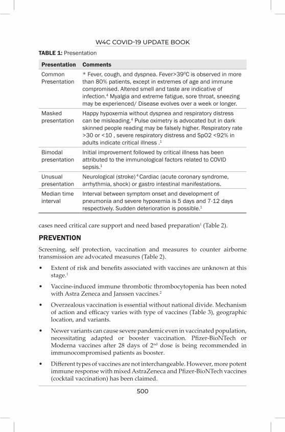
W4C COVID-19 UPDATE BOOK
500
TABLE 1: Presentation
Presentation Comments
Common Presentation
* Fever, cough, and dyspnea. Fever>390C is observed in more than 80% patients, except in extremes of age and immune compromised. Altered smell and taste are indicative of infection.4 Myalgia and extreme fatigue, sore throat, sneezing may be experienced/ Disease evolves over a week or longer.
Masked presentation
Happy hypoxemia without dyspnea and respiratory distress can be misleading.4 Pulse oximetry is advocated but in dark skinned people reading may be falsely higher. Respiratory rate >30 or <10 , severe respiratory distress and SpO2 <92% in adults indicate critical illness .1
Bimodal presentation
Initial improvement followed by critical illness has been attributed to the immunological factors related to COVID sepsis.1
Unusual presentation
Neurological (stroke) 4 Cardiac (acute coronary syndrome, arrhythmia, shock) or gastro intestinal manifestations.
Median time interval
Interval between symptom onset and development of pneumonia and severe hypoxemia is 5 days and 7-12 days respectively. Sudden deterioration is possible.1
cases need critical care support and need based preparation1 (Table 2).
PREVENTION Screening, self protection, vaccination and measures to counter airborne transmission are advocated measures (Table 2).
• Extent of risk and benefits associated with vaccines are unknown at this stage.1
• Vaccine-induced immune thrombotic thrombocytopenia has been noted with Astra Zeneca and Janssen vaccines.2
• Overzealous vaccination is essential without national divide. Mechanism of action and efficacy varies with type of vaccines (Table 3), geographic location, and variants.
• Newer variants can cause severe pandemic even in vaccinated population, necessitating adapted or booster vaccination. Pfizer-BioNTech or Moderna vaccines after 28 days of 2nd dose is being recommended in immunocompromised patients as booster.
• Different types of vaccines are not interchangeable. However, more potent immune response with mixed AstraZeneca and Pfizer-BioNTech vaccines (cocktail vaccination) has been claimed.
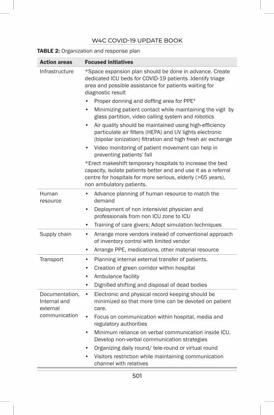
W4C COVID-19 UPDATE BOOK
501
TABLE 2: Organization and response plan
Action areas Focused initiatives
Infrastructure *Space expansion plan should be done in advance. Create dedicated ICU beds for COVID-19 patients .Identify triage area and possible assistance for patients waiting for diagnostic result • Proper donning and doffing area for PPE4 • Minimizing patient contact while maintaining the vigil by
glass partition, video calling system and robotics• Air quality should be maintained using high-efficiency
particulate air filters (HEPA) and UV lights electronic (bipolar ionization) filtration and high fresh air exchange
• Video monitoring of patient movement can help in preventing patients' fall
*Erect makeshift temporary hospitals to increase the bed capacity, isolate patients better and and use it as a referral centre for hospitals for more serious, elderly (>65 years), non ambulatory patients.
Human resource
• Advance planning of human resource to match the demand
• Deployment of non intensivist physician and professionals from non ICU zone to ICU
• Training of care givers; Adopt simulation techniques Supply chain • Arrange more vendors instead of conventional approach
of inventory control with limited vendor • Arrange PPE, medications, other material resource
Transport • Planning internal external transfer of patients. • Creation of green corridor within hospital • Ambulance facility• Dignified shifting and disposal of dead bodies
Documentation, Internal and external communication
• Electronic and physical record keeping should be minimized so that more time can be devoted on patient care.
• Focus on communication within hospital, media and regulatory authorities
• Minimum reliance on verbal communication inside ICU. Develop non-verbal communication strategies
• Organizing daily round/ tele-round or virtual round • Visitors restriction while maintaining communication
channel with relatives
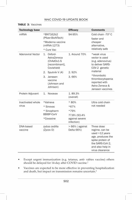
W4C COVID-19 UPDATE BOOK
502
TABLE 3: Vaccines
Technology base Efficacy Comments
mRNA *BNT162b2 (Pfizer-BioNTech) *Moderna vaccine (mRNA-1273)* Cure Vac
94-95% Cold chain -700 Cfaster and cheaper alternative, relatively safe
Adenoviral Vector 1. Oxford-AstraZeneca (ChAdOx1-S [recombinant], Covishield
1. Around 70% *weak virus vector is used (e.g. adenovirus) to deliver SARS-COV-2 genetic material*thrombotic thrombocytopenia reported with Astra Zeneca & Janssen vaccines
2. Sputnik V (4) 2. 92%3. Janssen
vaccine (Johnson and Johnson)
3. 66%
Protein Adjuvant 1. Novavax 1. 89.3% (overall)
Inactivated whole virus
*Valneva* Sinovac * Sinopharm BBIBP-CorV*Covaxine
* 80%*67%*79%77.8% (93.4% against severe infection)
Ultra cold chain not needed
DNA-based vaccine
zydus cedilla (Zycov D)
> 66% ( against Delta 66%)
Three dose regime. can be used >12 years age. produces the spike protein of the SARS-CoV-2, and also help in virus clearance
• Except urgent immunization (e.g. tetanus, anti- rabies vaccine) others should be delayed for 14-day after COVID vaccine.2
• Vaccines are expected to be more effective in preventing hospitalization and death, but impact on transmission remains uncertain.4
W4C COVID-19 UPDATE BOOK
503
Organization, Infrastructure and resource:
• Infrastructure, human and material resource planning should be done in advance2 (Table -2)
• Redeployed medical professionals from other specialties may be needed but associated adverse effects both care seeker and care providers should be considered.
• Remote medical consultation is becoming a new norm.2/3
DIAGNOSTICS• Laboratory findings are usually associated with lymphopenia, raised
lactate dehydrogenase and transaminase levels.2
• Reverse transcription polymerase chain reaction (RT PCR), based on Nucleic Acid Amplification (NAAT) is the gold standard to detect viral RNA. A recent meta-analysis has reported 73.3% sensitivity with RT PCR with nasopharyngeal samples.4
• Antigen testing has similar specificity but slightly lesser sensitivity, particularly at higher PCR cycle-threshold (40% with >30 Ct ).2,3 Antigen testing is not recommended over PCR despite faster cycle time.2
• Viral culture and serological (antibody testing) tests are not recommended for diagnosing active infection.2 but antibody assay can be done in highly suspected cases if RT-PCR is repeatedly negative, to diagnose multisystem inflammatory syndrome (MIS) and retrospective confirmation of infection. 2
• Rapid tests e.g. Lateral flow devices (LFD),Loop-mediated isothermal amplification (LAMP), and next generation sequencing (LamPORE - LAMP with nanopore sequencing)3 are being developed.
• Multiplex panels can be done to detect coexisting viral infections.1
• In symptomatic patients, lungs are always involved.2 CT is more sensitive than Xray chest but does not have diagnostic value.2
Management options: Treatment is mainly supportive (Table 4). Multiple therapeutic options have been suggested as mentioned below with specific indications2 (Table 5).
• Dexamethasone administration had been reported to reduce mortality by one fifth in oxygenated and by one third in ventilated patients.2,5
NIH, Survival Sepsis, IDSA and WHO supported its use in hospitalized patients needing oxygen with or without mechanical ventilation (MV). In absence of dexamethasone equivalent dose of alternative steroid can be used. Optimal dose and timing of administration still remain unanswered.4
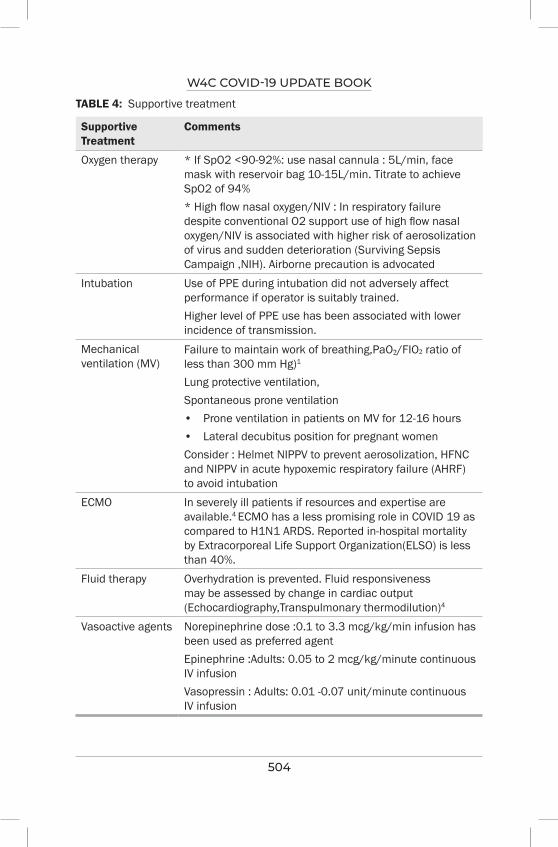
W4C COVID-19 UPDATE BOOK
504
TABLE 4: Supportive treatment
Supportive Treatment
Comments
Oxygen therapy * If SpO2 <90-92%: use nasal cannula : 5L/min, face mask with reservoir bag 10-15L/min. Titrate to achieve SpO2 of 94% * High flow nasal oxygen/NIV : In respiratory failure despite conventional O2 support use of high flow nasal oxygen/NIV is associated with higher risk of aerosolization of virus and sudden deterioration (Surviving Sepsis Campaign ,NIH). Airborne precaution is advocated
Intubation Use of PPE during intubation did not adversely affect performance if operator is suitably trained. Higher level of PPE use has been associated with lower incidence of transmission.
Mechanical ventilation (MV)
Failure to maintain work of breathing,PaO₂/FIO₂ ratio of less than 300 mm Hg)1
Lung protective ventilation, Spontaneous prone ventilation• Prone ventilation in patients on MV for 12-16 hours • Lateral decubitus position for pregnant womenConsider : Helmet NIPPV to prevent aerosolization, HFNC and NIPPV in acute hypoxemic respiratory failure (AHRF) to avoid intubation
ECMO In severely ill patients if resources and expertise are available.4 ECMO has a less promising role in COVID 19 as compared to H1N1 ARDS. Reported in-hospital mortality by Extracorporeal Life Support Organization(ELSO) is less than 40%.
Fluid therapy Overhydration is prevented. Fluid responsiveness may be assessed by change in cardiac output (Echocardiography,Transpulmonary thermodilution)4
Vasoactive agents Norepinephrine dose :0.1 to 3.3 mcg/kg/min infusion has been used as preferred agentEpinephrine :Adults: 0.05 to 2 mcg/kg/minute continuous IV infusionVasopressin : Adults: 0.01 -0.07 unit/minute continuous IV infusion
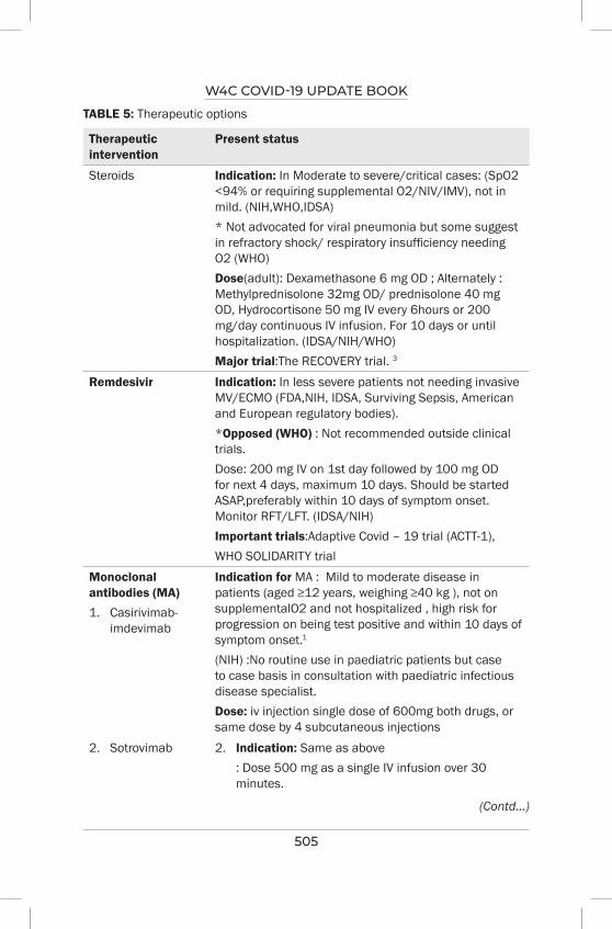
W4C COVID-19 UPDATE BOOK
505
TABLE 5: Therapeutic options
Therapeutic intervention
Present status
Steroids Indication: In Moderate to severe/critical cases: (SpO2 <94% or requiring supplemental O2/NIV/IMV), not in mild. (NIH,WHO,IDSA)* Not advocated for viral pneumonia but some suggest in refractory shock/ respiratory insufficiency needing O2 (WHO)Dose(adult): Dexamethasone 6 mg OD ; Alternately : Methylprednisolone 32mg OD/ prednisolone 40 mg OD, Hydrocortisone 50 mg IV every 6hours or 200 mg/day continuous IV infusion. For 10 days or until hospitalization. (IDSA/NIH/WHO)Major trial:The RECOVERY trial. 3
Remdesivir Indication: In less severe patients not needing invasive MV/ECMO (FDA,NIH, IDSA, Surviving Sepsis, American and European regulatory bodies).*Opposed (WHO) : Not recommended outside clinical trials.Dose: 200 mg IV on 1st day followed by 100 mg OD for next 4 days, maximum 10 days. Should be started ASAP,preferably within 10 days of symptom onset. Monitor RFT/LFT. (IDSA/NIH)Important trials:Adaptive Covid – 19 trial (ACTT-1),WHO SOLIDARITY trial
Monoclonal antibodies (MA)1. Casirivimab-
imdevimab
Indication for MA : Mild to moderate disease in patients (aged ≥12 years, weighing ≥40 kg ), not on supplementalO2 and not hospitalized , high risk for progression on being test positive and within 10 days of symptom onset.1
(NIH) :No routine use in paediatric patients but case to case basis in consultation with paediatric infectious disease specialist. Dose: iv injection single dose of 600mg both drugs, or same dose by 4 subcutaneous injections
2. Sotrovimab 2. Indication: Same as above : Dose 500 mg as a single IV infusion over 30
minutes.
(Contd...)

W4C COVID-19 UPDATE BOOK
506
Therapeutic intervention
Present status
3. Bamlanivimab and etesevimab
3. Indication: Same as above. Both are advised in combination only. :Dose 700 mg of bamlanivimab and 1,400 mg of etesevimab together in a single IV infusion.
4. Sarilumab 4. Indication : In non ICU patients. Not advocated outside clinical trials.
In ICU patient/high flow O2, NIV/MV insufficient data for recommendation for and against (NIH)
* Dose: IV 400 mg IV or sub cutaneous: 200 or 400 once in combination with antiviral therapy is being evaluated.
Immunomodulators interleukin 6 blocker Tocilizumab
Indication : Severe disease to counter act hyperinflammatory responseNIH Guideline : Combination with dexamethasone (equivalent corticosteroid) / + remdesivir in hospitalized patients on high-flow oxygen (>0.4 FiO2/30L/min) or NIV who have clinical (rapid resp. decomposition) or laboratory evidence of progressive disease. Some members advocate only in patients needed higher O2 while on dexamethasone or if CRP > 75mg/L2. IDSA : Suggests against the routine use of
tocilizumab, but advocates with steroid in severe/critical patients with high inflammatory markers.
3. Surviving Sepsis Campaign guideline: Insufficient data to make recommendation
4. WHO: Not recommendedDose adults : IV infusion once, 8 mg/Kg actual body weight. Up to 800 mg
TABLE 5: Therapeutic options (Contd...)
(Contd...)
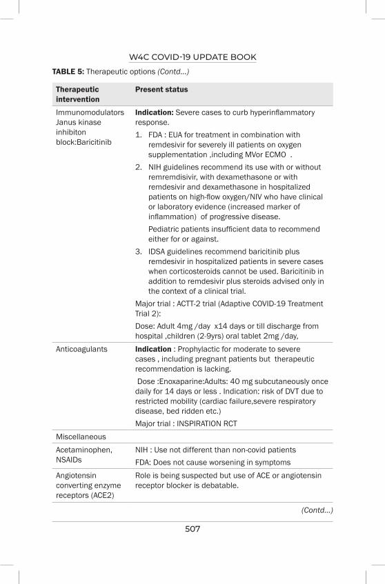
W4C COVID-19 UPDATE BOOK
507
TABLE 5: Therapeutic options (Contd...)
Therapeutic intervention
Present status
Immunomodulators Janus kinase inhibiton block:Baricitinib
Indication: Severe cases to curb hyperinflammatory response.1. FDA : EUA for treatment in combination with
remdesivir for severely ill patients on oxygen supplementation ,including MVor ECMO .
2. NIH guidelines recommend its use with or without remremdisivir, with dexamethasone or with remdesivir and dexamethasone in hospitalized patients on high-flow oxygen/NIV who have clinical or laboratory evidence (increased marker of inflammation) of progressive disease.
Pediatric patients insufficient data to recommend either for or against.
3. IDSA guidelines recommend baricitinib plus remdesivir in hospitalized patients in severe cases when corticosteroids cannot be used. Baricitinib in addition to remdesivir plus steroids advised only in the context of a clinical trial.
Major trial : ACTT-2 trial (Adaptive COVID-19 Treatment Trial 2): Dose: Adult 4mg /day x14 days or till discharge from hospital ,children (2-9yrs) oral tablet 2mg /day,
Anticoagulants Indication : Prophylactic for moderate to severe cases , including pregnant patients but therapeutic recommendation is lacking. Dose :Enoxaparine:Adults: 40 mg subcutaneously once daily for 14 days or less . Indication: risk of DVT due to restricted mobility (cardiac failure,severe respiratory disease, bed ridden etc.)Major trial : INSPIRATION RCT
MiscellaneousAcetaminophen, NSAIDs
NIH : Use not different than non-covid patients FDA: Does not cause worsening in symptoms
Angiotensin converting enzyme receptors (ACE2)
Role is being suspected but use of ACE or angiotensin receptor blocker is debatable.
(Contd...)
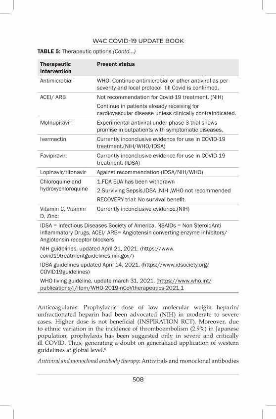
W4C COVID-19 UPDATE BOOK
508
Therapeutic intervention
Present status
Antimicrobial WHO: Continue antimicrobial or other antiviral as per severity and local protocol till Covid is confirmed.
ACEI/ ARB Not recommendation for Covid-19 treatment. (NIH)Continue in patients already receiving for cardiovascular disease unless clinically contraindicated.
Molnupiravir: Experimental antiviral under phase 3 trial shows promise in outpatients with symptomatic diseases.
Ivermectin Currently inconclusive evidence for use in COVID-19 treatment.(NIH/WHO/IDSA)
Favipiravir: Currently inconclusive evidence for use in COVID-19 treatment. (IDSA)
Lopinavir/ritonavir Against recommendation (IDSA/NIH/WHO)Chloroquine and hydroxychloroquine
1.FDA EUA has been withdrawn2.Surviving Sepsis,IDSA ,NIH ,WHO not recommended RECOVERY trial: No survival benefit.
Vitamin C, Vitamin D, Zinc:
Currently inconclusive evidence.(NIH)
IDSA = Infectious Diseases Society of America, NSAIDs = Non SteroidAnti inflammatory Drugs, ACEI/ ARB= Angiotensin converting enzyme inhibitors/ Angiotensin receptor blockersNIH guidelines, updated April 21, 2021. (https://www.covid19treatmentguidelines.nih.gov/)IDSA guidelines updated April 14, 2021. (https://www.idsociety.org/COVID19guidelines)WHO living guideline, update march 31, 2021. (https://www.who.int/publications/i/item/WHO-2019-nCoVtherapeutics-2021.1
TABLE 5: Therapeutic options (Contd...)
Anticoagulants: Prophylactic dose of low molecular weight heparin/unfractionated heparin had been advocated (NIH) in moderate to severe cases. Higher dose is not beneficial (INSPIRATION RCT). Moreover, due to ethnic variation in the incidence of thromboembolism (2.9%) in Japanese population, prophylaxis has been suggested only in severe and critically ill COVID. Thus, generating a doubt on generalized application of western guidelines at global level.6
Antiviral and monoclonal antibody therapy: Antivirals and monoclonal antibodies
W4C COVID-19 UPDATE BOOK
509
(MA) are recommended in early phase to prevent viral entry and replication in human cells (Table 5).
• Remdesivir has been approved by FDA for hospitalized patients (> 12 years of age and > 40 Kg weight)requiring oxygen support.2,7 Emergency use authorization (EUA) has been given for younger and lesser weight patients.1 Either alone or in combination with dexamethasone, it offers maximum benefits in less severe patients (without MV or ECMO).2,4 It is the only antiviral agent which has been licensed by American and European regulatory bodies.3 NIH, IDSA and Surviving Sepsis guidelines supported its use.2,4 NIH guideline further recommends completion of treatment even if patients subsequently need ventilation or ECMO. Joint Monitoring Group, Ministry of health and family welfare, India has declared it as a reserve drug under EUA8 and AGIHO guideline 2021for cancer patients moderately recommended this in cancer patients.3
WHO did not support its use outside clinical trials based on non-placebo controlled Solidarity trial2,9 Currently its role is debatable.
• Monoclonal antibodies prevent viral attachment and entry into the cells.1 They are not used in critical care1 but recommended under EUA, for mild to moderate infection with higher risk of progression. Reduced efficacy of MA against newer variants is being observed.1
Immunomodulators are preferred to manage severe disease and curb the hyperinflammatory response.
• Tocilizumab and baricitinib along with corticosteroids. with or without remdesivir have been advocated for patients needing high oxygen flow or noninvasive ventilation (NIV).1
• Baricitinib, an antirheumatic drug, has been advocated under EUA in severe disease in combination with remdesivir.2 It also has received moderate recommendation by AGIHO guideline for cancer patients.3
Other drugs
Vitamine D: Though, not included in proper trial, has exhibited some preventive advantages, reduced ICU admission and mortality benefit4 but evidence is inconclusive (NIH).
Many other therapies used in the past, currently have limited utility or been proved ineffective.4
COMPLICATIONS AND SEQUEL OF COVID -19 INFECTIONIsolated or multi organ failure, bacterial and fungal infection4, multisystem inflammatory syndrome (MIS-A/C), long covid or post-acute-covid are reported complications. Post infection immune response at 6 months is only 50% more, therefore reinfection is possible, particularly with newer variants.4,2
W4C COVID-19 UPDATE BOOK
510
Mental health and psychological support: Care givers, seekers and relatives are vulnerable due to stress and overpowering fear of death. Striking a balance and maintaining psychological wellbeing is a challenge.10
OUTCOMELonger hospitalization (>20 days) is a matter of concern.2 Mortality is around 3% in general but variable in different countries, as high as 31.7%above 85 years of age2 and around 40% (30-70%) in ICU population.1
CONCLUSION Short memory prevented us from taking lessons from the past until we faced COVID 19 and its stark reality. Retrospective analysis exposes many gaps which necessitate actions to prevent future catastrophe. A global containment approach is desirable focusing on preparation and prevention, as remedy is not full proof. The International Science Council (ISC), believes that short term priorities are important but long-term decisions are crucial. The oversight panel formed by the ISC is evaluating the evolving COVID scenario in next 3-5 years to organize a coherent pandemic response.
PRACTICE POINTS1. COVID19, a beta corona virus infection of respiratory tract, caused by
SARS -CoV-2. 1, may present in mild to severe forms with possibility of sudden deterioration.
2. Preventive measures and mass vaccination are important.
3. Management is mainly supportive. Role of dexamethasone is promising but other therapeutic options have specific indications and are mostly debatable.1,2,7
4. Newer virulent and resistant variants, multi system organ involvement, prolonged COVID and reinfections are major concerns.
REFERENCES1. Clinical Overview COVID-19 Critical Care Elsevier Point of Care, Updated May 6, 2021,
Available from :https://www.clinicalkey.com/#!/content/clinical_overview/67-s2.0-d887e185-8659-4926-b744-e6ec712e9f38
2. Coronavirus: Novel Coronavirus (COVID-19) Infection- ClinicalKey.Elsevier Point of Care.Updated July 30, 2021. Available at:https://www.clinicalkey.com/#!/content/clinical_overview/67-s2.0-0e7112a3-e94d-4136-94ba-cfdf9242ea43?printContent 1/44
3. Giesen N, Sprute R, Ru¨thrich M, Khodamoradi Y, Mellinghoff SC, et al. Gernot Beutel. 2021 update of the AGIHO guideline on evidence-basedmanagement of COVID-19 in patients with cancer regarding diagnostics, viral shedding, vaccination and therapy. European Journal of Cancer 2021; 147: 154-160.
4. Cordelia E.M. Coltart, Luke B. Collet-Fenson. Future developments in the prevention, diagnosis and treatment of COVID-19. Best Practice & Research Clinical Obstetrics & Gynaecology 2021; 3:56-80.
5. RECOVERY trial website. Updated June 16, 2020. Accessed May 3, 2021.https://
W4C COVID-19 UPDATE BOOK
511
www.recoverytr ial .net/news/low-cost-dexamethasone-reduces-death-by-up-to-one-third-in-hospitalised-patients-with-severe-respiratory-complications-of-covid-19View In Article|CrossReference(https://www.recoverytrial.net/news/low-cost-dexamethasonereducesdeath-by-up-to-one-third-in-hospitalised-patients-with-severe-respiratory-complications-of-covid-19
6. Fujiwara S, Nakajima M, Kaszynski RH, Fukushima K, Tanaka M, Yajima K, et al. Prevalence of thromboembolic events and status of prophylactic anticoagulant therapy in hospitalized patients with COVID-19 in Japan. J Infect Chemother 2021; 27:869-875.
7. FDA: FDA Approves First Treatment for COVID-19. FDA News Release. FDA website. PublishedOctober 22, 2020. Accessed May 3, 2021. https://www.fda.gov/news-events/press-announcements/fda-approves-first-treatment-covid-19.CrossReference (https://www.fda.gov/news-events/press-announcements/fdaapproves-first-treatment-covid-19)
8. Advisory for Rational use of Remdesivir for COVID-19 Treatment. Available from:https://www.mohfw.gov.in/pdf/AdvisoryforRationaluseofRemdesivirforCOVID19Treatment.pdf)
9. WHO Solidarity Trial Consortium. Repurposed antiviral drugs for Covid-19 — interim WHO Solidarity trial results. N Engl J Med DOI: 10.1056/NEJMoa2023184)
10. Lotta G, Fernandez M, Pimenta D, Wenham C, Lotta G, Fernandez M, Pimenta D, Wenham C. Gender, race, and health workers in the COVID-19 pandemic. Available from :Published:March 24, 2021DOI:https://doi.org/10.1016/S0140-6736(21)00530-4).


COVID Experience Across Asia
SECTION 16

515
Section 16 - Covid Experience Across AsiaSECTION EDITORS
Dr Banambar RayMD, FICCM Head of Critical Care Medicine, SUM Ultimate Medicare, A Unit of SOA,Bhubaneswar
Dr Abhijeet RahaMD, PDF Senior Consultant Neurocritical Care, Sum Ultimate Medicare (a unit of ‘SOA’), Bhubaneswar
Preface
"Be fast, have no regrets... If you need to be right before you move, you will never win” - Mike Ryan, Epidemiologist, WHO
This Pandemic has taught us that what we think is normal or usual might lead us to situations beyond imagination. It has also imbibed the thought process that we need to be adaptive and “the normal” are constantly changing to the so called “new normal”. As it is said, Change is the only entity that’s permanent. In this section we re share the experiences shared to us by eminent authors about the experience of Covid-19 in their respective countries. It shall give us insights about preparedness, proactiveness of the government and health sector and teach us how quick response and planning are all that is required to handle a disaster. As the waves keep on coming, it has shown that some resource constraint countries have done
exceedingly well compared to their more resourceful counterparts. This section will enlighten us to pre-empt the thought process before things get out of proportion when faced with large scale health disasters such as the Covid-19 pandemic. The section also has three fantastic published articles on the experience of Israel which are complied in the last chapter. As these articles are open and we have shared in this book for the benefit of the readers.

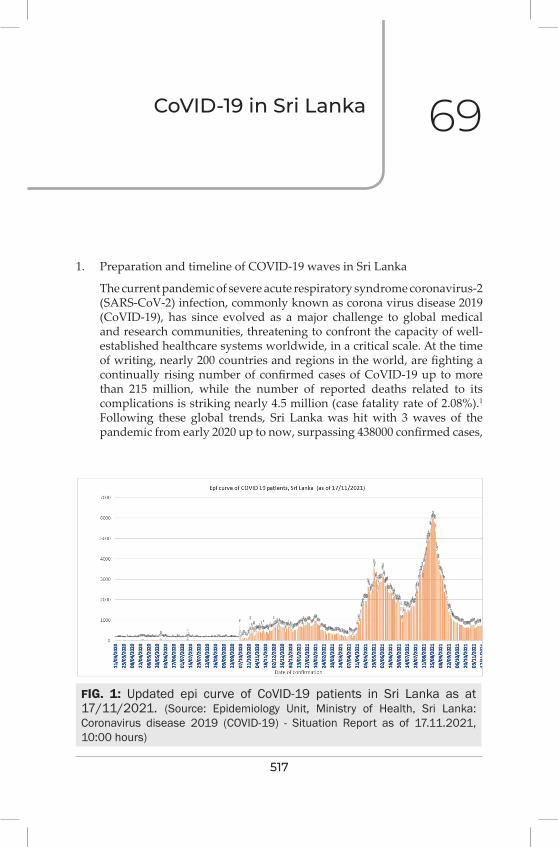
517
CoviD-19 in Sri Lanka 69
1. Preparation and timeline of COVID-19 waves in Sri Lanka
The current pandemic of severe acute respiratory syndrome coronavirus-2 (SARS-CoV-2) infection, commonly known as corona virus disease 2019 (CoVID-19), has since evolved as a major challenge to global medical and research communities, threatening to confront the capacity of well-established healthcare systems worldwide, in a critical scale. At the time of writing, nearly 200 countries and regions in the world, are fighting a continually rising number of confirmed cases of CoVID-19 up to more than 215 million, while the number of reported deaths related to its complications is striking nearly 4.5 million (case fatality rate of 2.08%).1
Following these global trends, Sri Lanka was hit with 3 waves of the pandemic from early 2020 up to now, surpassing 438000 confirmed cases,
FIG. 1: Updated epi curve of CoVID-19 patients in Sri Lanka as at 17/11/2021. (Source: Epidemiology Unit, Ministry of Health, Sri Lanka: Coronavirus disease 2019 (COVID-19) - Situation Report as of 17.11.2021, 10:00 hours)
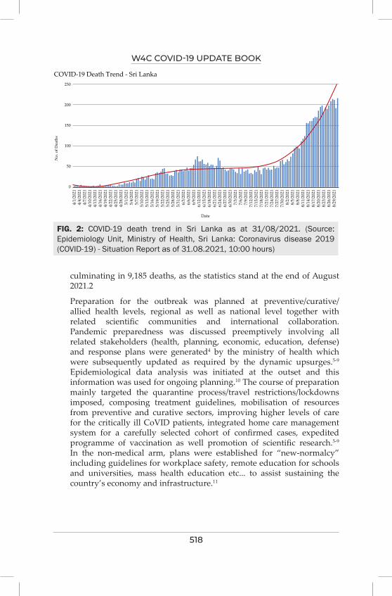
W4C COVID-19 UPDATE BOOK
518
culminating in 9,185 deaths, as the statistics stand at the end of August 2021.2
Preparation for the outbreak was planned at preventive/curative/allied health levels, regional as well as national level together with related scientific communities and international collaboration. Pandemic preparedness was discussed preemptively involving all related stakeholders (health, planning, economic, education, defense) and response plans were generated4 by the ministry of health which were subsequently updated as required by the dynamic upsurges.5-9 Epidemiological data analysis was initiated at the outset and this information was used for ongoing planning.10 The course of preparation mainly targeted the quarantine process/travel restrictions/lockdowns imposed, composing treatment guidelines, mobilisation of resources from preventive and curative sectors, improving higher levels of care for the critically ill CoVID patients, integrated home care management system for a carefully selected cohort of confirmed cases, expedited programme of vaccination as well promotion of scientific research.5-9 In the non-medical arm, plans were established for “new-normalcy” including guidelines for workplace safety, remote education for schools and universities, mass health education etc... to assist sustaining the country’s economy and infrastructure.11
FIG. 2: COVID-19 death trend in Sri Lanka as at 31/08/2021. (Source: Epidemiology Unit, Ministry of Health, Sri Lanka: Coronavirus disease 2019 (COVID-19) - Situation Report as of 31.08.2021, 10:00 hours)
COVID-19 Death Trend - Sri Lanka250
200
150
100
50
0
Date
4/1/
2021
4/4/
2021
4/7/
2021
4/10
/202
14/
13/2
021
4/16
/202
14/
19/2
021
4/22
/202
14/
25/2
021
4/28
/202
15/
1/20
215/
4/20
215/
7/20
215/
10/2
021
5/13
/202
15/
16/2
021
5/19
/202
15/
22/2
021
5/25
/202
15/
28/2
021
5/31
/202
16/
3/20
216/
6/20
216/
9/20
216/
12/2
021
6/15
/202
16/
18/2
021
6/21
/202
16/
24/2
021
6/27
/202
16/
30/2
021
7/3/
2021
7/6/
2021
7/9/
2021
7/12
/202
17/
15/2
021
7/18
/202
17/
21/2
021
7/24
/202
17/
27/2
021
7/30
/202
18/
2/20
218/
5/20
218/
8/20
218/
11/2
021
8/14
/202
18/
17/2
021
8/20
/202
18/
23/2
021
8/26
/202
18/
29/2
021
No.
of D
eath
s
W4C COVID-19 UPDATE BOOK
519
2. Response from hospitals and health regulatory agencies in tackling the pandemic
The country’s response to the “unknown enemy” was multifaceted, integrated and proactive. The Ministry of Health (MOH), Sri Lanka lead the way in decision making, dividing the prioritized tasks among its departments and proffering selected responsibilities to other government as well non-governmental organisations accordingly.5-8 The initial COVID-19 Sri Lanka Strategic Preparedness & Response Plan (SPRP) was updated in April 2021 at the outset of the 3rd wave under three (3) main headings, situation overview, response plan and immediate/medium term needs.12 The response plan categorically illustrated strategic objectives, national level preparedness and response, coordination, planning, financing, monitoring, risk communication, community engagement, infodemic management, surveillance, epidemiological investigation, contact tracing, adjustments of public health, management of point of entry/international travel/transport & mass gatherings, regulation of laboratory diagnostics, infection prevention/control, protection of health workforce, case management, clinical operations & therapeutics, logistics/supply chains, maintenance of essential services and the programme of vaccination.12 Together with its subdivisions (Epidemiolocal unit, Health education bureau, Family health bureau) and relevant specialist colleges, MOH has published activated several circulars and guidelines which are continually being revised, updated and adopted island wide. These documents guide many of the essential arms of managing the pandemic as a whole. They incorporate to form recommendations on the medical aspects including quarantine process, hospital preparedness, case management, healthcare staff safety, vulnerable groups like patients with non-communicable diseases and pregnant mothers, integrated national CoVID-19 surveillance including maintenance of statistics, as well as the non-medical aspects addressing education, examination schedules, sports, work place/airline safety (state organisations, apparel industry, manufacturing industry, economic centers, banks) cultural and religious proceedings etc.5-8 The hospital preparedness was identified as a critical point in the process and was steered to provide the best possible care at 3 levels, incorporating hospitals at each strata. They were upgraded with infrastructure and manpower to accommodate the maximum patient capacity especially for the critically ill.8,13,14 Hospital preparedness plans comprised of key elements, including establishment of CoVID-19 Operational Cell, Outpatient and emergency department care, establishing designated interim CoVID-19 suspected section/ward, provision of critical care to non-CoVID and CoVID-suspected patients, safe transfer of patients to a CoVID-19 designated/isolation hospital, ensuring safety of healthcare staff and managing cured CoVID-19 patients.13 Designated hospitals were established for pregnant CoVID-19
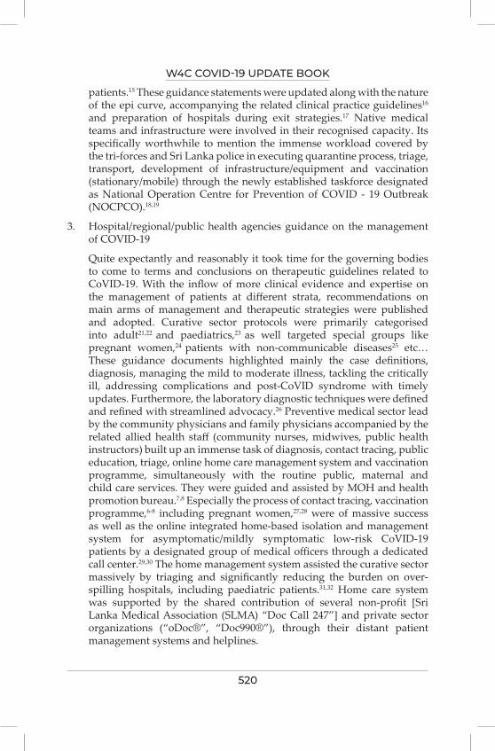
W4C COVID-19 UPDATE BOOK
520
patients.15 These guidance statements were updated along with the nature of the epi curve, accompanying the related clinical practice guidelines16 and preparation of hospitals during exit strategies.17 Native medical teams and infrastructure were involved in their recognised capacity. Its specifically worthwhile to mention the immense workload covered by the tri-forces and Sri Lanka police in executing quarantine process, triage, transport, development of infrastructure/equipment and vaccination (stationary/mobile) through the newly established taskforce designated as National Operation Centre for Prevention of COVID - 19 Outbreak (NOCPCO).18,19
3. Hospital/regional/public health agencies guidance on the management of COVID-19
Quite expectantly and reasonably it took time for the governing bodies to come to terms and conclusions on therapeutic guidelines related to CoVID-19. With the inflow of more clinical evidence and expertise on the management of patients at different strata, recommendations on main arms of management and therapeutic strategies were published and adopted. Curative sector protocols were primarily categorised into adult21,22 and paediatrics,23 as well targeted special groups like pregnant women,24 patients with non-communicable diseases25 etc… These guidance documents highlighted mainly the case definitions, diagnosis, managing the mild to moderate illness, tackling the critically ill, addressing complications and post-CoVID syndrome with timely updates. Furthermore, the laboratory diagnostic techniques were defined and refined with streamlined advocacy.26 Preventive medical sector lead by the community physicians and family physicians accompanied by the related allied health staff (community nurses, midwives, public health instructors) built up an immense task of diagnosis, contact tracing, public education, triage, online home care management system and vaccination programme, simultaneously with the routine public, maternal and child care services. They were guided and assisted by MOH and health promotion bureau.7,8 Especially the process of contact tracing, vaccination programme,6-8 including pregnant women,27,28 were of massive success as well as the online integrated home-based isolation and management system for asymptomatic/mildly symptomatic low-risk CoVID-19 patients by a designated group of medical officers through a dedicated call center.29,30 The home management system assisted the curative sector massively by triaging and significantly reducing the burden on over-spilling hospitals, including paediatric patients.31,32 Home care system was supported by the shared contribution of several non-profit [Sri Lanka Medical Association (SLMA) “Doc Call 247”] and private sector organizations (“oDoc®”, “Doc990®”), through their distant patient management systems and helplines.
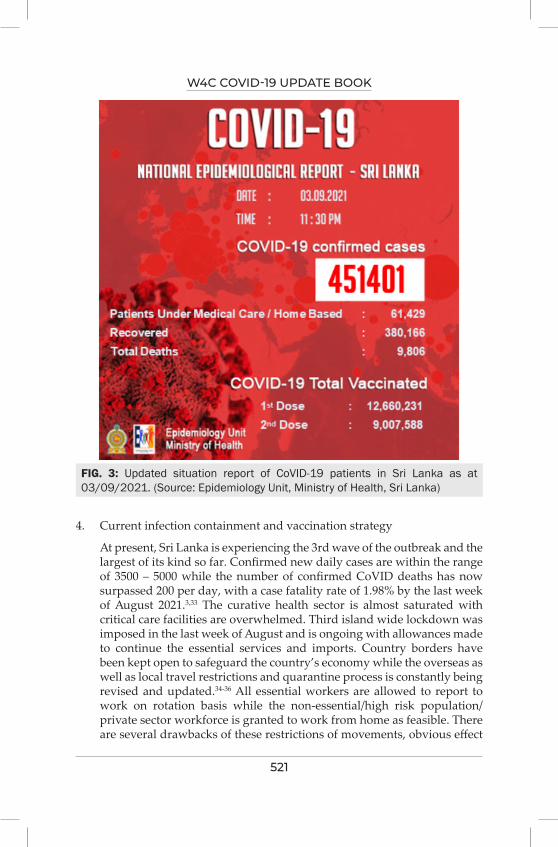
W4C COVID-19 UPDATE BOOK
521
4. Current infection containment and vaccination strategy
At present, Sri Lanka is experiencing the 3rd wave of the outbreak and the largest of its kind so far. Confirmed new daily cases are within the range of 3500 – 5000 while the number of confirmed CoVID deaths has now surpassed 200 per day, with a case fatality rate of 1.98% by the last week of August 2021.3,33 The curative health sector is almost saturated with critical care facilities are overwhelmed. Third island wide lockdown was imposed in the last week of August and is ongoing with allowances made to continue the essential services and imports. Country borders have been kept open to safeguard the country’s economy while the overseas as well as local travel restrictions and quarantine process is constantly being revised and updated.34-36 All essential workers are allowed to report to work on rotation basis while the non-essential/high risk population/private sector workforce is granted to work from home as feasible. There are several drawbacks of these restrictions of movements, obvious effect
FIG. 3: Updated situation report of CoVID-19 patients in Sri Lanka as at 03/09/2021. (Source: Epidemiology Unit, Ministry of Health, Sri Lanka)

W4C COVID-19 UPDATE BOOK
522
on the overall economy and education.
Consolidating public health preventive strategies were considered to be of utmost importance in containing the spread of the infection. Education of the general public is carried out through mass media and public health awareness programmes on electronic media based on recommended guidelines on respiratory etiquettes and precautionary methods.5-7
Vaccination against CoVID-19 in Sri Lanka was initiated in late January 2021. The first vaccine to arrive was Oxford AstraZeneca/Covishield (ChAdOx1nCoV-19 Corona Virus Vaccine-Recombinant), partly as a donation from India and as a part of Covax programme initiated by the World Health Organization (WHO). The batches of different types of vaccines were evaluated through an expert multidisciplinary committee with regard to their efficacy, safety, suitability, indications, adverse effects and contraindications by the use of the limited data available and were approved for use in within the community in a predefined protocol. Priority was given to the frontline health care workers, other frontline staff (forces and police) and the cohort of the elderly population, more than 60 years of age. Vaccination programme was accelerated during the subsequent months mainly through establishment of accessible community vaccine centers/designated hospitals and public health clinics, involvement of the forces to conduct the programme with increased manpower, importation of other vaccines approved by the WHO, commencement of vaccination among special groups of population (pregnant women, people with non-communicable diseases), home vaccination of elderly/disabled and
TABLE 1: CoVID vaccination summary in Sri Lanka (1st and 2nd doses) as at 03/09/2021 (Source: Epidemiology Unit, Ministry of Health, Sri Lanka)
Epidemiology Unit Ministry of Health
231, De Saram Place, Colombo 10, Sri Lanka Tele: (+94 11) 2695112, 2681548, 4740490, 4740491, 4740492 Fax: (+94 11) 2696583 E-mail: [email protected], [email protected] Web: www.epid.gov.lk
මාධය නවේදනය ~ PRESS RELEASE
2021. 09. 03 දන පසවර 8.30 වන වට ව ාවඩ-19 පරතශකතත රණ වැඩ සටහවේ පරගතය Progress of COVID-19 Immunization as of 8.30 pm on 03. 09. 2021
දනය Date
ව ාවෂලඩඩ Covishield සයවනාෆාම Sinopharm සටනනකත - V Sputnik - V Pfizer Moderna
පළම මාතරාව First Dose
වදවන මාතරාව Second Dose
පළම මාතරාව First Dose
වදවන මාතරාව Second Dose
පළම මාතරාව First Dose
වදවන මාතරාව Second Dose
පළම මාතරාව First Dose
වදවන මාතරාව Second Dose
පළම මාතරාව First Dose
වදවන මාතරාව Second Dose
January-2021 59426 February - 2021 406924
March - 2021 453403 April-2021 5489 64589 2,865* 2,435* May-2021 282721 752605 26,821 June -2021 - 30997 1002582 707860 87,974 14427 - July-2021 - 7578 6154033 1027747 44286 76 201495 192 713659 01-08.21 – 31.08.21
460926 499732 1974761 4682106 7 10986 104908 186647 57997 674382
01.09.2021 3584 348 58181 400949 - - 18 180 131 10725 02.09.2021 3055 296 25555 231460 - - 7573 587 237 270 03.08.2021 3381 383 24883 161415 - 7742 26192 2408 145 10785 සමණණ එ තව Cumulative Total
1,396,188 886,644 9,992,600 7,201,537 159,088 33,231 340,186 190,014 772,169 696,162
* සංවධණන ටයත සඳහා ශර ලං ාවට පැමණ සටන චන ජාත යේට සයවනාෆාම එේනත පළම මාතරාව 2,865 කතද, වදවන මාතරාව 2,435 කතද ලබා ද ඇත. * 2,865 doses of the Sinopharm vaccine as the first dose and 2,435 doses as the second dose was administered to Chinese nationals in Sri Lanka engaged in developmental activities. පරධාන වසංගත වරෝග වදයාඥ Chief Epidemiologist
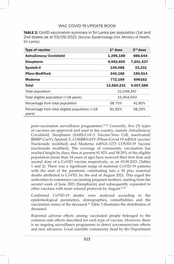
W4C COVID-19 UPDATE BOOK
523
TABLE 2: CoVID vaccination summary in Sri Lanka per population (1st and 2nd doses) as at 03/09/2021 (Source: Epidemiology Unit, Ministry of Health, Sri Lanka)
Type of vaccine 1st dose 2nd dose
AstraZeneca/Covishield 1,396,188 886,644
Sinopharm 9,992,600 7,201,537
Sputnik-V 159,088 33,231
Pfizer-BioNTech 340,186 190,014
Moderna 772,169 696162
Total 12,660,231 9,007,588
Total population 21,548,347Total eligible population (>18 years) 15,454,043Percentage from total population 58.75% 41.80%Percentage from total eligible population (>18 years)
81.92% 58.29%
post-vaccination surveillance programmes.37-40 Currently, five (5) types of vaccines are approved and used in the country, namely AstraZeneca/Covishield, Sinopharm (SARS-CoV-2 Vaccine-Vero Cell, inactivated/BBIBP-CorV), Sputnik V, COMIRNATY (Pfizer-Covid 19 mRNA vaccine/Nucleoside modified) and Moderna mRNA-1273 COVID-19 Vaccine (nucleoside modified). The coverage of community vaccination has reached height by days, thus at present 81.92% and 58.29% of the eligible population (more than 18 years of age) have received their first dose and second dose of a CoVID vaccine respectively, as on 03.09.2021 (Tables 1 and 2). There was a significant surge of maternal CoVID-19 patients with the start of the pandemic culminating into a 30 plus maternal deaths attributed to CoVID, by the end of August 2021. This urged the authorities to commence vaccinating pregnant mothers, starting from the second week of June 2021 (Sinopharm) and subsequently expanded to other vaccines with more relaxed protocols by August.27,28
Confirmed CoVID-19 deaths were analyzed according to the epidemiological parameters, demographics, comorbidities and the vaccination status of the deceased.41 Table 3 illustrates the distribution of deceased.
Reported adverse effects among vaccinated people belonged to the common side effects described for each type of vaccine. However, there is an ongoing surveillance programme to detect uncommon/rare effects and new advances. Local scientific community (lead by the Department
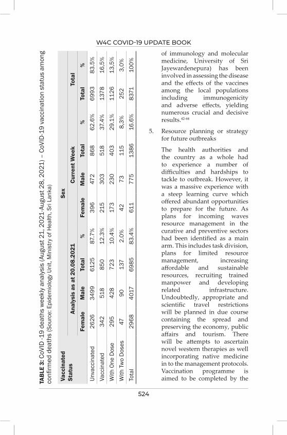
W4C COVID-19 UPDATE BOOK
524
TAB
LE 3
: CoV
ID -1
9 de
aths
wee
kly a
naly
sis
(Aug
ust 2
1, 2
021-
Augu
st 2
8, 2
021)
– C
oVID
-19
vacc
inat
ion
stat
us a
mon
g co
nfirm
ed d
eath
s (S
ourc
e: E
pide
mio
logy
Uni
t, M
inis
try
of H
ealth
, Sri
Lank
a)
Vacc
inat
ed
Stat
usSe
x
Anal
ysis
as
at 2
0.08
.202
1Cu
rren
t Wee
kTo
tal
Fem
ale
Mal
eTo
tal
%Fe
mal
eM
ale
Tota
l%
Tota
l%
Unva
ccin
ated
2626
3499
6125
87.7
%39
647
286
862
.6%
6993
83.5
%Va
ccin
ated
342
518
850
12.3
%21
530
351
837
.4%
1378
16,5
%W
ith O
ne D
ose
295
428
723
10.4
%17
323
040
329
.1%
1126
13,5
%W
ith T
wo
Dos
es47
9013
72.
0%42
7311
58,
3%25
23,
0%To
tal
2968
4017
6985
83.4
%61
177
513
8616
.6%
8371
100%
of immunology and molecular medicine, University of Sri Jayewardenepura) has been involved in assessing the disease and the effects of the vaccines among the local populations including immunogenicity and adverse effects, yielding numerous crucial and decisive results.42-44
5. Resource planning or strategy for future outbreaks
The health authorities and the country as a whole had to experience a number of difficulties and hardships to tackle to outbreak. However, it was a massive experience with a steep learning curve which offered abundant opportunities to prepare for the future. As plans for incoming waves resource management in the curative and preventive sectors had been identified as a main arm. This includes task division, plans for limited resource management, increasing affordable and sustainable resources, recruiting trained manpower and developing related infrastructure. Undoubtedly, appropriate and scientific travel restrictions will be planned in due course containing the spread and preserving the economy, public affairs and tourism. There will be attempts to ascertain novel western therapies as well incorporating native medicine in to the management protocols. Vaccination programme is aimed to be completed by the
W4C COVID-19 UPDATE BOOK
525
end of September 2021 for the currently eligible cohort of population, while it is scheduled to vaccinate the paediatric age groups once approval granted, offer the third booster dose for selected groups (healthcare workers and high-risk groups) as well to draft local vaccine production chains.
6. Recommendations
In retrospect, even with the limited frame of experience, time and evidence we have gathered with regard to CoVID-19 in Sri Lankan patients, there are positive aspects to take forward and to build up on. The main recommendation to be drawn would be proper and timely planning, revision of protocols and updating as required by the dynamic nature of the pandemic. This should involve all stakeholders representing health, planning, economy, education, administration and policy making. Task distribution among the identified groups and monitoring the timelines with set objectives help immensely to track dynamics of waves and response of the situations to the interventions made, at all strata. Collecting and analyzing scientific data with regard to the disease pathology/therapeutics/vaccination is a must, involving the medical scientific community, to take point-of-care decisions and to plan for incoming outbreaks. We recommend all regions have an efficient vaccination programme, prioritizing the high-risk groups and covering the whole eligible population in a stepwise manner. To make community vaccination effective we should empower populations with informed decision making and help them accept the benefit with scientific evidence. It is highly recommended to strengthen the preventive health sector, since it is the arm which is involved in managing the most important aspects of infection containment. In both preventive and curative aspects, the vulnerable groups are recommended to be targeted first. One of the main strategies we would like to recommend the health authorities globally is the establishment of an integrated home-based care system for minimally symptomatic/asymptomatic CoVID patients. This has markedly reduced the burden on curative sector by triage and helped identifying at-risk patients for higher levels of care through regular close monitoring until safe discharge criteria are met. Ensuring safety of healthcare workforce and their immediate families is of utmost importance since they are the frontliners. It is always advocated not to abandon the routine medical and emergency care as far as possible, as it accounts for increased mortality among non-CoVID patients. Implementation of a national CoVID health information system is strongly recommended to allow sharing of timely data on COVID-19 patients among all stakeholders, which is essential for strategizing and evaluating the control measures as well as resource allocation for an effective outbreak response.45
W4C COVID-19 UPDATE BOOK
526
REFERENCES1. World Health Organisation. (2021) WHO Coronavirus (COVID-19
Dashboard) [Online] Available from: https://covid19.who.int/ [Accessed 3rd September 2021].
2. Health Promotion Bureau, Sri Lanka. (2021) COVID-19: Live Situational Analysis Dashboard of Sri Lanka [Online] Available from: https://hpb.health.gov.lk/covid19-dashboard/ [Accessed 3rd September 2021].
3. Epidemiology Unit, Ministry of Health, Sri Lanka (2021) [Online] Available from: http://www.epid.gov.lk/web/ [Accessed 3rd September 2021].
4. Ministry of Health and Indigenous Medical Services, Sri Lanka. (2020). Sri Lanka Preparedness & Response Plan - COVID-19. Version 1. Colombo. Ministry of Health, Sri Lanka.
5. Epidemiology Unit, Ministry of Health, Sri Lanka (2021) COVID – 19 - All Guidelines and Circulars [Online] Available from: https://www.epid.gov.lk/web/index.php?option=com_content&view=article&id=230&lang=en [Accessed 3rd September 2021].
6. Health Promotion Bureau, Sri Lanka. (2021) COVID-19 [Online] Available from: https://www.hpb.health.gov.lk/en/covid-19 [Accessed 3rd September 2021].
7. Family health bureau, Ministry of Health, Nutrition and Indigenous Medicine, Sri Lanka (2021) COVID-19 Guidelines [Online] Available from: https://www.fhb.health.gov.lk/index.php/en/covid-19 [Accessed 3rd September 2021].
8. Ministry of Health, Sri Lanka (2021) New - COVID-19 Related Circulars & Letters [Online] Available from: http://www.health.gov.lk/moh_final/english/news_read_more.php?id=999 [Accessed 4th September 2021].
9. Ministry of Public Services, Provincial Councils and Local Government, Sri Lanka (2021) Circular Manager [Online] Available from: https://www.pubad.gov.lk/web/index.php?option=com_circular&view=circulars&Itemid=176&lang=en [Accessed 3rd September 2021].
10. Arambepola C, Wickramasinghe ND, Jayakody S et al. (2021) Sri Lanka’s early success in the containment of COVID-19 through its rapid response: Clinical & epidemiological evidence from the initial case series. PLoS ONE 16(7): e0255394. Available from: https://doi.org/10.1371/journal.pone.0255394 [Accessed 1st September 2021].
11. Jayathilaka, A.K.K.R. (2021) COVID-19 in Sri Lanka and Work Setting Changes. Open Access Library Journal, 8: e7008. Available from: https://

W4C COVID-19 UPDATE BOOK
527
doi.org/10.4236/oalib.1107008 [Accessed 1st September 2021].
12. Ministry of Health and Indigenous Medical Services, Sri Lanka. (2021). COVID-19 Sri Lanka Strategic Preparedness & Response Plan. Colombo. Ministry of Health, Sri Lanka.
13. Ministry of Health and Indigenous Medical Services, Sri Lanka (April 2020) Hospital Preparedness for COVID-19 Global Pandemic. DDG(MS) I /23/2020. Colombo. Ministry of Health, Sri Lanka Available from: http://www.health.gov.lk/moh_final/english/news_read_more.php?id=999 [Accessed 3rd September 2021].
14. Ministry of Health and Indigenous Medical Services, Sri Lanka (October 2020) Hospital Preparedness for COVID-19 Global Pandemic. DDG(MS) I /23/2020. Colombo. Ministry of Health, Sri Lanka Available from: http://www.health.gov.lk/moh_final/english/news_read_more.php?id=999 [Accessed 3rd September 2021].
15. Ministry of Health and Indigenous Medical Services, Sri Lanka (November 2020) Management of COVID-19 positive pregnant women. FHB/MCU/COVID-19/2020. Colombo. Ministry of Health, Sri Lanka Available from: http://www.health.gov.lk/moh_final/english/news_read_more.php?id=999 [Accessed 3rd September 2021].
16. Ministry of Health and Indigenous Medical Services, Sri Lanka (January 2020) Interim Summary Guidelines for Clinical Management of patients with novel coronavirus (2019- nCoV). DDG{PHS)l /002/ 12-9/2019/10. Colombo. Ministry of Health, Sri Lanka Available from: http://www.health.gov.lk/moh_final/english/news_read_more.php?id=999 [Accessed 3rd September 2021].
17. Ministry of Health and Indigenous Medical Services, Sri Lanka (April 2020) Preparation of hospitals during Covid 19 exit strategy. DDG(MS)l/23/2020. Colombo. Ministry of Health, Sri Lanka Available from: http://www.health.gov.lk/moh_final/english/news_read_more.php?id=999 [Accessed 3rd September 2021].
18. National Operation Centre for Prevention of COVID - 19 Outbreak, Sri Lanka Army (2021) [Online] Available from: https://alt.army.lk/covid19/ [Accessed 3rd September 2021].
19. Presidential Secretariat, Sri Lanka (2021) Corona related guidelines and circulars [Online] Available from: https://covid19.gov.lk/guidelines.html [Accessed 3rd September 2021].
20. Ministry of Health, Ceylon College of Physicians, Epidemiology Unit, Sri Lanka. (March 2020). Provisional Clinical Practice Guidelines on COVID-19 Suspected and Confirmed Patients. Colombo. Ministry of Health, Sri Lanka.
21. Ceylon College of Physicians, Sri Lanka (2021) COVID19 portal [Online]
W4C COVID-19 UPDATE BOOK
528
Available from: https://www.ccp.lk/covid19-portal/ [Accessed 3rd September 2021].
22. College of Anaesthesiologists and Intensivists of Sri Lanka (2021) Guidelines [Online] Available from: https://anaesthesia.lk/guidelines/ [Accessed 3rd September 2021].
23. Sri Lanka College of Paediatricians (March 2020). Clinical Practice Guidelines on the Management of the Children with Suspected or Confirmed COVID-19. Version 01. Colombo. Sri Lanka College of Paediatricians.
24. Sri Lanka College of Obstetricians and Gynaecologists (May 2021) SLCOG Guideline on Management of Covid-19 in Pregnancy. Colombo. Sri Lanka College of Obstetricians and Gynaecologists.
25. Ministry of Health and Indigenous Medical Services, Sri Lanka (May 2021) New guideline for provision of care for patients with Non-Communicable Diseases (NCD) during the COVID-19 outbreak. NCD/47/2020. Colombo. Ministry of Health, Sri Lanka Available from: http://www.health.gov.lk/moh_final/english/news_read_more.php?id=999 [Accessed 3rd September 2021].
26. Ministry of Health and Indigenous Medical Services, Sri Lanka (June 2021) Revised Guidelines on Laboratory Testing Strategy for COVID-19. DDG/LS/ED/Testing-Strategy/106/2021. Colombo. Ministry of Health, Sri Lanka Available from: http://www.health.gov.lk/moh_final/english/news_read_more.php?id=999 [Accessed 3rd September 2021].
27. Family Health Bureau, Ministry of Health, Sri Lanka (June 2021) RE: Vaccinating pregnant mothers against SARS CoV 2 infection. FHB/MCU/COVID/2021. Colombo. Ministry of Health, Sri Lanka Available from: https://www.fhb.health.gov.lk/index.php/en/covid-19 [Accessed 3rd September 2021].
28. Ministry of Health and Indigenous Medical Services, Sri Lanka (August 2021) Vaccination of pregnant women against COVID 19. EPID/400/COVID-1 9/VOL 4. Colombo. Ministry of Health, Sri Lanka Available from: http://www.health.gov.lk/moh_final/english/news_read_more.php?id=999 [Accessed 3rd September 2021].
29. Ministry of Health and Indigenous Medical Services, Sri Lanka (June 2021) Integrated Home-Based Isolation and Management of Asymptomatic & Mildly Symptomatic COVID-19 Infected Individuals - Revised Guidelines for Pilot Project in the Western Province. DGHS/COVID-19/347/2021. Colombo. Ministry of Health, Sri Lanka Available from: http://www.health.gov.lk/moh_final/english/news_read_more.php?id=999 [Accessed 3rd September 2021].
30. Ministry of Health and Indigenous Medical Services, Sri Lanka (August
W4C COVID-19 UPDATE BOOK
529
2021) Guidelines for Integrated Home-Based Isolation and Management of Asymptomatic & Mild Symptomatic COVID-19 Patients. DGHS/COVID-19/347/2021. Colombo. Ministry of Health, Sri Lanka Available from: http://www.health.gov.lk/moh_final/english/news_read_more.php?id=999 [Accessed 3rd September 2021].
31. Ministry of Health, Sri Lanka (June 2021) INTEGRATED MANAGEMENT FOR ASYMPTOMATIC AND MILDLY SYMPTOMATIC COVID-19 PATIENTS. Version 2. Colombo. Ministry of Health, Sri Lanka.
32. Sri Lanka College of Paediatricians (August 2021). HOME-BASED ISOLATION AND MANAGEMENT OF CHILDREN WITH COVID - 19 INFECTION. Colombo. Sri Lanka College of Paediatricians.
33. Epidemiology Unit, Ministry of Health, Sri Lanka (2020). Coronavirus disease 2019 (COVID-19) - Situation Report –03.09.2021– 10 a.m. Colombo. Sri Lanka College of Paediatricians. Available from:https://www.epid.gov.lk/web/index.php?option=com_content&view=article&id=225&lang=en [Accessed 3rd September 2021].
34. Ministry of Health and Indigenous Medical Services, Sri Lanka (August 2021) Quarantine measures for travellers arriving from overseas during the pandemic of COVID-19. DGHS/COVID-19/347/2021. Colombo. Ministry of Health, Sri Lanka Available from: http://www.health.gov.lk/moh_final/english/news_read_more.php?id=999 [Accessed 3rd September 2021].
35. Ministry of Health and Indigenous Medical Services, Sri Lanka (January 2021) Movement restrictions and closing of establishments under the Quarantine and Prevention of Disease Ordinance. DGHS/COVID- 19/2020-347. Colombo. Ministry of Health, Sri Lanka Available from: http://www.health.gov.lk/moh_final/english/news_read_more.php?id=999 [Accessed 3rd September 2021].
36. Ministry of Health and Indigenous Medical Services, Sri Lanka (July 2021) Isolation of areas, closing of establishments, lifting of isolation and re-opening under the Quarantine and Prevention of Disease Ordinance. DGHS/COVID-19/347/2021. Colombo. Ministry of Health, Sri Lanka Available from: http://www.health.gov.lk/moh_final/english/news_read_more.php?id=999 [Accessed 3rd September 2021].
37. Ministry of Health and Indigenous Medical Services, Sri Lanka (June 2021) COVID-19 Vaccination Program at Healthcare Institutions. DDG (PHS)l/Mis/2021. Colombo. Ministry of Health, Sri Lanka Available from: http://www.health.gov.lk/moh_final/english/news_read_more.php?id=999 [Accessed 3rd September 2021].
38. Ministry of Health and Indigenous Medical Services, Sri Lanka (August 2021) Vaccination of persons who have not turned up for COVID-19
W4C COVID-19 UPDATE BOOK
530
vaccination. DGHS/COVID-19/347/2021. Colombo. Ministry of Health, Sri Lanka Available from: http://www.health.gov.lk/moh_final/english/news_read_more.php?id=999 [Accessed 3rd September 2021].
39. Ministry of Health and Indigenous Medical Services, Sri Lanka (August 2021) Vaccination of persons above 60years and persons with co morbidities who have not turned up for vaccination. DDG (PHS)l/Mis/2021. Colombo. Ministry of Health, Sri Lanka Available from: http://www.health.gov.lk/moh_final/english/news_read_more.php?id=999 [Accessed 3rd September 2021].
40. Ministry of Health and Indigenous Medical Services, Sri Lanka (August 2021) Special COVID-19 vaccination programme for vaccination of persons above 60 years who have not turned up for vaccination. DDG (PHS)l/Mis/2021. Colombo. Ministry of Health, Sri Lanka Available from: http://www.health.gov.lk/moh_final/english/news_read_more.php?id=999 [Accessed 3rd September 2021].
41. Epidemiology Unit, Ministry of Health (2021) COVID - 19 confirmed deaths - Weekly analysis [Online] Available from: h t t p s : / / w w w . e p i d . g o v . l k / w e b / i n d e x . p h p ? o p t i o n = c o m _content&view=article&id=233&Itemid=480&lang=en [Accessed 3rd September 2021].
42. Jeewandara C, Aberathna IS, Pushpakumara PD et al. Antibody and T cell responses to Sinopharm/BBIBP-CorV in naïve and previously infected individuals in Sri Lanka. MedRxiv The Preprint Server for Health Sciences 2021. Available from: https://doi.org/10.1101/2021.07.15.21260621.
43. Jeewandara C, Kamaladasa A, Pushpakumara PD et al. Immune responses to a single dose of the
AZD1222/Covishield vaccine in health care workers. Nature Communications 2021. Available from: https://doi.org/10.1038/s41467-021-24579-7.
44. Jeewandara C, Jayathilaka D, Gomes L et al. SARS-CoV-2 neutralizing antibodies in patients with varying severity of acute COVID-19 illness. Nature Research – Scientific Reports 2021; 11: 2062. Available from: https://doi.org/10.1038/s41598-021-81629-2.
45. Ministry of Health and Indigenous Medical Services, Sri Lanka (July 2021) Implementation of the National Covid Health Information System (NCHIS). DDG (PHS)l/Mis/2021. Colombo. Ministry of Health, Sri Lanka Available from: http://www.health.gov.lk/moh_final/english/news_read_more.php?id=999 [Accessed 3rd September 2021].
531
COviD-19; Oman Experience 70.
Oman is located in the south-eastern corner of the Arabian Peninsula with a population of nearly 5 million; out of which 61.3% are males, and 38.7% are females. While children under 10 years of age represent around 19% of total Omani population, 13% of the population is between age 10 and 15 years, 65% between age 15 and 50 years and 3% are above 50 years.
In Oman, the first two cases of covid-19 were reported on February 24, 2020 in Muscat Governorate, the capital of Oman. These cases were travel related, and the first suspected case of local transmission was reported on March 23, 2020. As with other international data, the initial genomic analysis of SARS-CoV-2 variant in Oman was the D614G mutation in the Spike protein which was reported in 116 countries mostly of B1 lineages. It was detected since March 2020 among patients with travel history to Europe and the Middle East. However, In April 2021, Delta variant was detected in Oman which was more severe than the initial variant. As of 1st of September 2021, the country has documented 302,000 cases and 4070 deaths with a fatality rate of 1.3%.
Oman was among the first countries to implement early precautionary measures to prevent and mitigate the impact of SARS-CoV-2. Following detection of the first case on 24 February 2020, a supreme committee for dealing with covid-19 was formed which implemented a number of unprecedented non-pharmaceutical interventions such as case-based control interventions that included early case detection, isolation of suspected and confirmed patients, contact tracing and enhancing laboratory diagnostics. National border interventions were commenced on 12 March 2020. The country implemented travel restrictions, initially to countries with a high burden of disease such as China followed by suspension of all international flights, entry bans through borders and ports, stopping tourist visas and advising all incoming travellers to self-quarantine for 14 days. Community transmission interventions that included intergovernmental travel restrictions were deployed across the country on April 1st 2020, enforcing infection control interventions across the nation such as universal masking, maintaining social
Maher Al Bahrani, Faryal Khamis, Sandeep Kantor
W4C COVID-19 UPDATE BOOK
532
distancing and reducing non-essential national workforce by 30%, closure of amusements, schools, malls, mosques, restrictions on social gatherings and postponement of large public events and mass gatherings such as weddings, conferences and governmental events.
The first covid-19 wave in Oman lasted until November 2020 with a peak in early October 2020, while the second wave with the Delta variant started in April 2021 with a peak in June 2021. The impact of delta variant was more on children, pregnant and young adults compared with the initial variant. The health care sector bore the highest burden in managing the pandemic, necessitating the development of a National Preparedness plan. The priorities were to increase hospital capacity, increase number of intensive care beds, increase supplies and mobilizing human resources to serve the population.
All hospitals in the country developed their own contingency plan to deal with the surge of patients. Most hospitals converted regular wards into covid-19 general wards and ICU. For example, the Royal Hospital which is the largest hospital in the country increased their ICU beds from 16 to 95 beds during the peak period. This was done by converting one of its buildings into a dedicated isolation area for covid-19 accommodating more than 170 general and ICU patients. In September 2020 the Field Hospital in Muscat was commissioned with a bed capacity of 200 general and high dependency beds. This hospital served mainly the capital city but also accepted cases from the adjacent cities. It was fully equipped and staffed with 50 physicians and 300 nurses and paramedical staff.
The main challenge faced the health care system was mobilizing human resources to combat the pandemic. Staff were diverted from primary care to higher care centers and from medical and surgical specialties to the intensive care units. This necessitated stopping elective and non-essential services. Emergency, oncological and cardiac services continued to operate during the crisis. The government supported the hospitals by recruiting more than 40 intensivists and 1000 nurses from abroad to help in managing the crisis. It has also procured more than 230 ventilators and 280 cardiac monitors as well as other essential equipment for the wards and ICU.
In January 2021, the Sultanate has started the vaccination campaign. It has approved five covid-19 vaccines; AstraZeneca/Covishield, AstraZeneca/Oxford, Pfizer/BioNTech, Sinovac and Sputnik V. The campaign is still ongoing and in September 2021, 73% of the target population has been vaccinated with at least one dose. In August 2021 a steady decline in case registration and hospital admissions was seen. This prompted the government to ease the restrictions and ended the lockdown that was previously imposed and opened the border for travelers. However, entry to public and government places as well as entry to the country is only for those who have been vaccinated. The health sector has also started to recover and resumed the elective services from August 2021 onwards.
W4C COVID-19 UPDATE BOOK
533
Ministry of Health in Oman has produced more than 30 policies and guidelines for the management of covid-19 that ranged from infection control policies to medical and intensive care clinical management. There were also more than 240 research publications in Oman on covid-19.
There are many learning lessons in dealing with covid-19 in Oman. The capacities required to prepare and respond to covid-19 should be updated on a regular basis based on the evolving epidemiological situation and risk of spread. Covid-19 strategic planning pillars are:
1. Exchange, dissemination and use of guidelines and procedures
2. Coherent use of information, education and communication materials
3. National training for surveillance, management and emergency operations
4. Engagement in global research, solidarity trial and other partnerships
The main factor for success in the fight against covid-19 is to have an integrated and resilient health system that has all care components which includes government and private health care providers, suppliers and contractors and public health services. In addition, management of human resources needs that include close and transparent communications, welness programs and psychological support and training program for multi tasking are essential during the pandemic. Future plans for hospital design should incorporate innovation in space design that can be of multiple use, ease of conversion, accounting for surges during pandemics and to be compliant with infection control requirements.
REFERENCES1. Khamis F, Al-Zakwani I, Al Naamani H, Al Lawati S, Pandak N, Omar MB, et al. Clinical
characteristics and outcomes of the first 63 adult patients hospitalized with COVID-19: an experience from Oman. J Infect Public Health. 2020;13:906–13.
2. Al Awaidy ST, Khamis F, Al Rashidi B, Al Wahaibi AH, Albahri A, Mahomed O. Epidemiological Characteristics of 69,382 COVID-19 Patients in Oman. J Epidemiol Glob Health. 2021 Aug 4:1–12. doi: 10.1007/s44197-021-00001-9. Epub ahead of print. PMCID: PMC8335985.
3. Khamis F, Memish Z, Bahrani MA, Dowaiki SA, Pandak N, Bolushi ZA, Salmi IA, Al-Zakwani I. Prevalence and predictors of in-hospital mortality of patients hospitalized with COVID-19 infection. J Infect Public Health. 2021 Jun;14(6):759-765. doi: 10.1016/j.jiph.2021.03.016. Epub 2021 Apr 18. PMID: 34022734; PMCID: PMC8053361.
4. Al-Mahruqi S, Al-Wahaibi A, Khan AL, Al-Jardani A, Asaf S, Alkindi H, Al-Kharusi S, Al-Rawahi AN, Al-Rawahi A, Al-Salmani M, Al-Shukri I, Al-Busaidi A, Al-Abri SS, Al-Harrasi A. Molecular epidemiology of COVID-19 in Oman: A molecular and surveillance study for the early transmission of COVID-19 in the country. Int J Infect Dis. 2021 Mar;104:139-149. doi: 10.1016/j.ijid.2020.12.049. Epub 2021 Jan 13. PMID: 33359061; PMCID: PMC7834852.
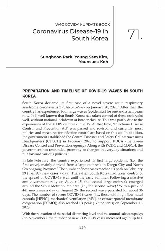
W4C COVID-19 UPDATE BOOK
534
Coronavirus Disease-19 in South Korea 71.
PREPARATION AND TIMELINE OF COVID-19 WAVES IN SOUTH KOREASouth Korea declared its first case of a novel severe acute respiratory syndrome coronavirus 2 (SARS-CoV-2) on January 20, 2020.1 After that, the country has experienced four large waves (epidemics) for one and a half years now. It is well known that South Korea has taken control of these outbreaks well, without national lockdown or border closure. This was partly due to the experiences of the MERS outbreak in 2015. At that time, ‘Infectious Disease Control and Prevention Act’ was passed and revised, and currently, most policies and measures for infection control are based on this act. In addition, the government established the Central Disaster and Safety Countermeasures Headquarters (CDSCH) in February 2020 to support KDCA (the Korea Disease Control and Prevention Agency). Along with KCDC and CDSCH, the government has responded promptly to changes in everyday situations and put forward various policies.2
In late February, the country experienced its first large epidemic (i.e., the first wave), mainly derived from a large outbreak in Daegu City and North Gyeongsang Province.3 The number of new cases reached its peak on February 29 ( i.e., 909 new cases a day). Thereafter, South Korea had taken control of the spread of COVID-19 well until the early summer. Following a massive anti-government rally on August 15, the second large outbreak emerged around the Seoul Metropolitan area (i.e., the second wave).4 With a peak of 441 new cases a day on August 26, the second wave persisted for about 36 days. The number of severe COVID-19 cases (i.e., those with high flow nasal cannula [HFNC], mechanical ventilation [MV], or extracorporeal membrane oxygenation [ECMO]) also reached its peak (175 patients) on September 11, 2020.
With the relaxation of the social distancing level and the annual sale campaign (on November), the number of new COVID-19 cases increased again up to >
Sunghoon Park, Young Sam Kim, Younsuck Koh
W4C COVID-19 UPDATE BOOK
535
100 cases per day. It continued to increase, reaching more than 1,000 cases a day in December (i.e., the third wave). However, contrary to the second wave, where local clustered infections were dominant, the third wave was characterized by a higher rate of personal contact transmission and unknown transmission routes.4 Since then, the number of daily new COVID-19 cases was maintained between 300 to 700 cases during the spring season (Figure 1). However, the incidence surged again from early July and, as of August 25, reached over 2,000 cases a day (i.e., the fourth wave). The number of daily severe COVID-19 cases in hospitals also reached > 400 cases. Contrary to the previous epidemics, this fourth wave was considered to be driven by delta-variants with a high transmission rate5. Many cases were young or had not been vaccinated yet.
RESPONSE FROM HOSPITALS AND HEALTH REGULATORY AGENCIES IN TACKLING THE PANDEMIC.During the early stage of the outbreak, the country has implemented proactive and distinctive interventions such as massive case finding and contact tracing and consistent public message. Of note, huge case finding and contact tracing was possible due to the increased domestic production of test kits and drive-through (> 50 locations) or walk-through screening centers, which boosted the testing capacity and minimized cross-contamination.3,6 The government has undertaken vigorous measures to tract and test contacts of confirmed cases using a global positioning system (GPS), card transaction logs, and medical records. It has provided the public with transparent information on confirmed cases.
FIG. 1: Daily incidence of COVID-19 Patients in South Korea
500
450
400
350
300
250
200
150
100
50
0
4d wave
3d wave2500
2000
1500
1000
500
0
8/6
8/13
8/20
8/27 9/3
9/10
09/1
709
/24
10/0
110
/08
10/1
510
/22
10/2
911
/05
11/1
211
/19
11/2
612
/312
/10
12/1
712
/24
12/3
11/
71/
141/
211/
28 2/4
2/11
2/18
2/25 3/4
3/11
3/18
3/25 4/1
4/8
4/15
4/22
4/29 5/6
5/13
5/20
5/27 6/3
6/10
6/17
6/24
7/01
7/08
7/15
7/22
7/29 8/5
8/12
8/26 9/2
Deaths (right)
Domestic new cases (left)
Severe cases (right)
2020 2021
2nd wave
W4C COVID-19 UPDATE BOOK
536
Based on severity and risk factors, confirmed cases were isolated at home, in a residential treatment center (RTC), or a hospital. Cases were isolated in RTCs (e.g., university dormitories or training institutes) when they had mild symptoms (or asymptomatic), but home isolation was not possible.6 These facilities can reduce the burden on the healthcare system, and currently, a total of 84 RTCs (with 17,901 cases as of August 12, 2021) are being operated. Using the Self-Quarantine Safety Protection Application, medical personnel and public health managers monitored and recorded patients’ symptoms twice a day; for those who were under self-quarantine, it was mandatory to download the application.3
Since the MERS outbreak in 2015, the government designated 59 hospitals as regional centers for infectious diseases to cope with future outbreaks, so confirmed COVID-19 cases with symptoms have been admitted to these regional centers. However, as the number of COVID-19 cases increases, 11 hospitals with critical care capacity were added as the regional center. Severe cases which were likely to require ECMO treatment were transferred to tertiary hospitals with ECMO programs. The CDSCH and local governments collected data on daily available ICU beds and arranged the inter-hospital transports of severe cases to facilitate this process. However, apart from this, the Korean Society of Critical Care Medicine (KSCCM) independently collected data on daily numbers of severe COVID-19 cases receiving critical care resources (i.e., HFNC, MV, and EMCO) and available ICU beds over the country, with the cooperation of the society members (>50 tertiary or university hospitals). These data provided more practical information for available ICU resources to healthcare personnel.
During the first and the third waves, we believed that the country was in a critical care crisis. At that time, severe COVID-19 cases rapidly increased, whereas there was a shortage of ICU resources and medical personnel.7,8 During the third wave (in Seoul Metropolitan City and Gyeonggi-do Province), the government issued an administrative order which forced tertiary hospitals to expand their ICU beds for COVID-cases. And, the KSCCM dispatched again volunteers (intensivists and general physicians) to Namyangju Hyundai Hospital (in Gyeonggi-do Province).10 The government of Gyeonggi-do Province provided financial support for these dispatch services. Currently, the country is experiencing the fourth wave, with a peak incidence of new COVID-19 cases (> 2,000 cases per day), and as expected, the incidence of severe cases is also increasing.11
TREATMENT GUIDANCE ON THE MANAGEMENT OF COVID-19Remdesivir was approved for its use in patients receiving oxygen therapy in July 2020 in South Korea, based on a multi-national trial.16 In South Korea, the Korean Society of Infectious Diseases, in collaboration with the National Evidence-Based Healthcare Collaborating Agency (NECA), released treatment guidelines for COVID-19 patients.19 They only suggested using
W4C COVID-19 UPDATE BOOK
537
remdesivir and dexamethasone (or other alternative steroids) for COVID-19 cases but held their judgment (or do not recommend) on using other drugs such as interleukin-6 inhibitor, interleukin-1 inhibitor, or convalescent plasma for COVID-19 patients.
The KSCCM issued ‘Critical Care Protocols for Regional Centers of Infectious Diseases’.20 According to a report by the relevant authority, there were 9,800 mechanical ventilators and 30 negative-pressure emergency ambulances over the countries.8 Regarding ECMO cases, in a report by the Korean Society for Thoracic & Cardiovascular Surgery, a total of 62 ECMOs (of 409 ECMO devices) are currently being used for COVID-19 patients.21 However, of note, there is no established system to relocate critical care equipment when needed or to transfer a high-risk patient receiving MV or ECMO safely. Given this situation, the establishment of safe transport systems and educations for medical personnel and securing sufficient critical care resources are urgently needed.
CURRENT INFECTION CONTAINMENT AND VACCINATION STRATEGYThe main strategy of COVID-19 infection containment is described as 3-T in Korea. Three T strategy is testing, tracing, and treatment. Early detection is key to preventing the COVID-19 virus from spreading. The Korean government has set up screening stations to increase access to diagnostic tests and conduct fast and wide testing to detect confirmed cases. Screening stations provide consultation to people showing symptoms of COVID-19 such as cough or fever before they visit medical facilities. Diverse forms of screening stations have been introduced such as drive-thru and walk-thru screening stations in Korea.
More than 31 million people here have been partly vaccinated now, and 70% of the Korean population will be fully vaccinated by the end of November. As the country’s vaccination rate picks up, health authorities are considering a shift in its public health strategy, a gradual return to normalcy and living with the virus, rather than suppressing its spread. The Korean government could consider moving into a phase of “living with COVID-19” as early as the end of next month.
STRATEGY FOR FUTURE OUTBREAKSCOVID-19 infection is widespread around the whole world, and a new mutant variant virus is appearing. Globally, the vaccination rate is low, and there is wide variation between countries. It is very likely that COVID-19 infection will become an endemic infection and will not be eradicated. In the last year, infection control strategies focused on preventing infection and spread have been successful in Korea. But infection rate is increased and sustained recently due to a new variant infection. The Korean government is
W4C COVID-19 UPDATE BOOK
538
going to change the strategy from 3-T strategy to “living with COVID-19”. To achieve this change in strategy, the full vaccination rate should be over 70%, and the national health care system has sufficient capacity to treat critically ill COVID-19 patients so that the low fatality rate can be maintained.
RECOMMENDATIONWe live in an era where a pandemic of infectious diseases is not a problem for one region. It seems very likely that we will have to live with the variants of COVID-19. Beyond the health threat, the COVID-19 epidemic has given each country the challenge of how to improve social safety nets. In Korea, many self-employed people and low-income families are suffering a lot from the continuous social distancing for a long time. Considering that vaccines are the most effective way to overcome these challenges, intellectual assets for the vaccine development should be shared so that cheaper and safer vaccines can be globally more available. Additionally, socially agreed triage guidelines for severely ills with COVID-19 are urgently needed to reduce unfair access to care and to maximize benefits for saving more lives.
REFERENCES1. Korea Centers for Disease Control and Prevention (http://www.kdca.go.kr/board/board.
es?mid=a20501010000&bid=0015&act=view&list_no=365864; accessed on August 20, 2021).2. Jeong GH, Lee HJ, Lee J, Lee JY, Lee KH, Han YJ, et al. Effective Control of COVID-19 in South
Korea: Cross-Sectional Study of Epidemiological Data. J Med Internet Res 2020;22:e22103.3. Jeong E, Hagose M, Jung H, Ki M, Flahault A. Understanding South Korea’s Response to the
COVID-19 Outbreak: A Real-Time Analysis. Int J Environ Res Public Health 2020;17.4. Seong H, Hyun HJ, Yun JG, Noh JY, Cheong HJ, Kim WJ, et al. Comparison of the second and
third waves of the COVID-19 pandemic in South Korea: Importance of early public health intervention. Int J Infect Dis 2021;104:742-5.
5. Korea Disease Control and Prevention Agency. Incidence and charateristics of the SARS-CoV-2 delta variants Available at http://www.kdca.go.kr/board/board.es?mid=a20602010000&bid=0034&act=view&list_no=716491. Accessed on August 26, 2021.
6. Dighe A, Cattarino L, Cuomo-Dannenburg G, Skarp J, Imai N, Bhatia S, et al. Response to COVID-19 in South Korea and implications for lifting stringent interventions. BMC Med 2020;18:321.
7. Kim EJ, Lee YH, Park JS, Lee J, Lee SY, Kim Y, et al. Clinical features and prognostic factors of critically ill patients with COVID-19 in Daegu, South Korea: A multi-center retrospective study. Medicine (Baltimore) 2021;100:e24437.
8. Kim JH, Hong SK, Kim Y, Ryu HG, Park CM, Lee YS, et al. Experience of augmenting critical care capacity in Daegu during COVID-19 incident in South Korea. Acute Crit Care 2020;35:110-4.
9. Nearly 500 medical professionals volunteer for Daegu. The Korea Herald. Available at http://www.koreaherald.com/view.php?ud=20200227000698 (Accessed on August 28, 2021)
10. Collaboration of Namyangju Hyundai Hospital and the KCCM. Cheong-nyeon Doctor. Available at http://www.docdocdoc.co.kr/news/articleView.html?idxno=2006019 (Accessed on August 26, 2021).
11. New cases still hover around 2,000; 4th wave of pandemic yet at peak. The Korea Herald. Available at http://www.koreaherald.com/view.php?ud=20210812000163 IAccessed on August 28, 2021).

W4C COVID-19 UPDATE BOOK
539
12. Treatment recommendations for critically ill COVID-19 patients [in Korean]. The Ko-rea Society of Critical Care Medicine. Available at https://www.ksccm.org/html/?p-mode=BBBS0006700044&page=3&smode=view&seq=2126&searchValue=&searchTitle=strTi-tle (Access on August 20, 2021)
13. Coronavirus (COVID-19) Update: FDA Revokes Emergency Use Authorization for Chloro-quine and Hydroxychloroquine. The US Food & Drug Administration. Available at https://www.fda.gov/news-events/press-announcements/coronavirus-covid-19-update-fda-re-vokes-emergency-use-authorization-chloroquine-and Accessed on August 28, 2021.
14. Cao B, Wang Y, Wen D, Liu W, Wang J, Fan G, et al. A Trial of Lopinavir-Ritonavir in Adults Hospitalized with Severe Covid-19. N Engl J Med 2020;382:1787-99.
15. Patel TK, Patel PB, Barvaliya M, Saurabh MK, Bhalla HL, Khosla PP. Efficacy and safety of lopinavir-ritonavir in COVID-19: A systematic review of randomized controlled trials. J Infect Public Health 2021;14:740-8.
16. Beigel JH, Tomashek KM, Dodd LE, Mehta AK, Zingman BS, Kalil AC, et al. Remdesivir for the Treatment of Covid-19 - Final Report. N Engl J Med 2020;383:1813-26.
17. Group RC, Horby P, Lim WS, Emberson JR, Mafham M, Bell JL, et al. Dexamethasone in Hos-pitalized Patients with Covid-19. N Engl J Med 2021;384:693-704.
18. IDSA Guidelines on the Treatment and Management of Patients with COVID-19. Infectious Disease Society of America. Available at https://www.idsociety.org/practice-guideline/covid-19-guideline-treatment-and-management/ (Accessed on August 29, 2021).
19. Clinical practice guidelines for COVID-19 patients [in Korean]. National Evidence-Based Healthcare Collaborating Agency and The Korean Society of Infectious Disease. Available at https://www.ksid.or.kr/rang_board/list.html?num=5089&code=ncov_notice (Accessed on Au-gust 25, 2021).
20. Critical Care Protocols for Regional Centers of Infectious Disease. Available at https://www.ksccm.org/html/?pmode=BBBS0006700044&page=1&smode=view&seq=2341&-searchValue=&searchTitle=strTitle (Accessed on August 26, 2021).
21. Domestic ECMO use for COVID-19 patients. The Korean Society for Thoracic & Cardiovas-cular Surgery. Available at https://www.ktcvs.or.kr/file/ecmo.pdf?v=210829 (Accessed on Au-gust 28, 2021).
540
The Japanese Experience COviD-19 Waves in Japan 72.
First case of COVID-19 patient in Japan was reported on January 15 in 2020. The patient visited Wuhan, China late 2019 and stayed there for several days. He showed mild symptom and recovered soon. The Japanese government did not quarantined travelers from China including Wuhan during the Spring Festival, Chinese New Year in 2020. It was partly due to the festival started from January 24 and ended January 31 in 2020. It was unlucky for Japan, as the festival begins during February in most year. Thus, many tourists from China visited Japan during this period in 2020. The first death case was reported on February 13 in Japan. This patient was one of the passengers on cruise ship known as Diamond Princes. The vessel was quarantined in Yokohama port for about 4 weeks from February 3 resulting 712 infected with SARS-COV2 out of total 3,711 onboard, and 14 elderly passengers died. At this point, COVID-19 was still a storm on the opposite bank in Japan.
The first wave of COVID-19 started from the beginning of April 2020 and lasted for about 2 months. Since then, we have experienced 5 COVID-19 waves so far as of October 2021. The Second one started in August, the third in December, the fourth in March 2021, and the fifth started late July 2021 during the Olympic and Paralympic games in Tokyo. All five waves weaned in about 2 months, meaning we experienced the peak every 4 months. In each time, the government declared the state of emergency that is milder version of lockdown and asked people to quarantine themselves. As it is politically impossible to declare complete lockdown because of the Constitution of Japan, it is not mandatory but many people just obeyed the request.
There are 47 prefectures (states) in Japan and governor of each prefecture would ask Japanese government to declare a state of emergency for each prefecture. For example, 21 prefectures declared the state of emergency during August 25 to September 9 in 2021. In areas with emergency status, various kind of regulations were applied, such as, eating and drinking facilities had been allowed to offer alcoholic beverages until 7 p.m.
Satoru Hashimoto
W4C COVID-19 UPDATE BOOK
541
The so-called medical collapse was barely avoided in each wave. The manpower and system of intensive care in Japan is relatively poor compared to the developed countries of Europe or North America. Thus, if an outbreak like New York or Italy in early 2020 had occurred in Japan, the medical care system would definitely have collapsed and the death toll would have increased dramatically. In that sense, we must say that we were merely lucky. Although more than 8,000 hospitals are registered in Japan, less than 300 hospitals have actually provided critical care measures such as mechanical ventilation and ECMO for COVID-19 patients. Many other hospitals are too small to provide intensive care measures. In addition, the fact that 80% of all hospitals in Japan are private hospitals is also considered to be the cause of the biased acceptance of COVID-19 patients. As various medical supplies were severely restricted, we could not afford to help other countries unfortunately. The dire situation seen in some countries could barely be avoided in Japan.
As of September 30, 2021 about 1.7 million people (1.4% of the total population) have been identified as infected with SARS-CoV2, but there are probably two to three times as many hidden infected people including subclinical infections. This assumption was supported by several antibody investigations in Japan and the fact the PCR tests were not comprehensive in Japan. The total number of deaths is about 17,000 as of September 30 2021. The mortality rate is about 1% which is rather low on a global scale. It is estimated from the statistics collected by us that there are about 10,000 critically ill patients who have undergone ECMO or mechanical ventilation, of which about 3,000 have died. That is, the remaining 14,000 patients were a group of patients who were taken care of without critical care measures mainly because of their old age.
No excess deaths were observed during first and second wave in 2020. This is partly because that from the end of 2019 to the beginning of 2020, the national influenza incidence was almost zero. From the end of 2020 to the beginning of 2021, the RS virus epidemic was prevalent, but the feared influenza and corona co-infection epidemics were not observed. During third wave and fifth wave, 50 to 200 excess deaths per week were observed probably because the increase of death by COVID-19 but the number itself was small.
Vaccines are under development in Japan, and domestic vaccine production has not yet begun. Vaccines made by Pfizer, Moderna and more recently AstraZeneca have begun to use.
Vaccinations were widely given to health care workers after February 2021, and general vaccinations were first started in May for the elderly aged 65 and over. As of the end of August 2021, nearly 90 % of elderly were given vaccinations and the deaths of the elderly decreased dramatically after the 4th wave. In the 5th wave, the largest number of infected people with SARS-CoV-2, 25866 per day was reported on August 20, about 10 days after the closing ceremony of Tokyo Olympic games and 4 days before the opening ceremony of Tokyo Paralympic Games (1). There was a big dispute whether
W4C COVID-19 UPDATE BOOK
542
the games should be canceled. As a result, the games took place without spectators. This surge was thought to be due to the influence of the delta variant of SARS-CoV-2. The accumulated number of infected people in the 5th wave was about 850,000. As the total number of infected was 1,700,000 in Japan, half infected only in the 5th wave. On the other hand, the death toll in the 5th wave was only 15% of the total and mortality rate dropped dramatically. As stated above, we assumed this is none other than the success of vaccination for older people. The government is now discussing whether booster vaccination should be considered. Over 65% of Japanese population fully vaccinated by October 10 and will reach 70% by the end of October, 2021.
On February 12th, 2020, 60 some volunteers from Japanese Society of Intensive Care Medicine (JSICM), Japanese Association for Acute Medicine (JAAM) and Japanese Society of Respiratory Care Medicine (JSRCM) have set up a group named Japan ECMOnet for COVID-19. The purpose of this group is to save critically ill COVID-19 patients by setting up 24/7 telephone consultation by ECMO experts and establishing database registry for these patients covering over 90% of ICU beds in Japan(2,3). This real-time nationwide surveillance database system as a “disaster management-like system” now known as CRISIS [CRoss Icu Searchable Information System] was constructed only in 5 days and got into full operation on February 16, 2020. As of 30th of September 2021, over 7000 cases of mechanically ventilated patients and over 1000 hundred cases of ECMO patients have been registered. These data were utilized by the members of ECMOnet to find appropriate hospital for the transfer. During June of 2021, over 200 person-day doctors, nurses and clinical engineers of ECMOnet were deployed to Okinawa prefecture to help local hospitals. From August 23rd of 2021 for 4 weeks 300 person-day doctors and nurses of ECMOnet were deployed to Tokyo district to help hospitals where no doctors have enough experiences of mechanical ventilation. 50 some primary and secondary transports of ECMO patients were counted(4).
There is no magic method for respiratory management. For the severe hypoxia, nasal high flow therapy would be the first choice and make the patients with awake prone positioning. If this strategy does not work, we recommend the intubation and start mechanical ventilation without delay. Lung protective strategy should be taken in all ventilated cases including the use of muscle relaxant or long time prone positioning. We recommend to start ECMO ASAP if PF ratio drops so rapidly. Proper control of coagulation system and secondary infections are the key for the successful management of seriously ill COVID-19 patients.
As for the drugs for COVID-19 patients, we used Lopinavir /Ritonavir for a while after the spring of 2020, but now quit using this drug invented by Japanese company. Most of the current treatments for severely ill COVID-19 patients are based on treatment with Remdesivir and Dexamethasone, and Baricitinib or Tocilizumab is added in some serious cases. In addition,

W4C COVID-19 UPDATE BOOK
543
continuous administration of heparin is essential to prevent thrombosis. Large dose of Methylprednisolone, usually 1 gram per day for 3 consecutive days, is widely administered under the name of pulse therapy in many facilities in Japan. This should be a unique method not proven to be effective. Exceptionally, there are some facilities that applied 20-80 ppm nitric oxide inhalation on ventilated patients. In mild cases, Casilivimab/Imdevimab is sometimes administered as an antibody cocktail therapy with some success. As future expectations, the introduction of Monoclonal antibody, sotrovimab for COVID-19 patients with mild to moderate symptoms, or oral antiviral Molnupiravir for COVID-19 patients with mild symptoms is expected. We have almost no experience with Hydroxychloroquine and Ivermectin.
Although vaccination is under progressing and the system for medical emergency is being established, in consideration of breakthrough infection we continue to call on the people to practice masking all the time, social distancing, and hand washing, etc. The next wave, the sixth wave, is expected in early 2022, no one can predict whether these measures will work.
REFERENCES1. Academic Consortium on emergency medical service and disaster medical response planning
during the Tokyo Olympic and Paralympic Games in 2020 (AC2020). http://2020ac.com/about_e.html accessed on 5 September 2021.
2. Japan ECMOnet for COVID-19: telephone consultations for cases with severe respiratory failure caused by COVID-19. J Intensive Care 2020; 8: 24.
3. Japan ECMOnet for COVID-19. Save the ICU and save lives during the COVID-19 pandemic. J Intensive Care 2020; 8: 40.
4. Ogura T, Ohshimo S, Liu K, Iwashita Y, Hashimoto S, Takeda S. Establishment of a Disaster Management-like System for COVID-19 Patients Requiring Veno-Venous Extracorporeal Membrane Oxygenation in Japan. Membranes (Basel) 2021; 11.
W4C COVID-19 UPDATE BOOK
544
NATIONAL POSITION PAPER
Open Access Rambam Maimonides Medical Journal
* The language of this document throughout should be taken to refer to both men and women even when employing male pronouns. † To whom correspondence should be addressed. E-mail: [email protected]
‡ Members and affiliations of the study groups are provided in Appendix 1.
Rambam Maimonides Med J | www.rmmj.org.il 1 July 2020 Volume 11 Issue 3 e0019
Special Issue on the COVID-19 Pandemic with Guest Editors Oren Caspi, M.D. and Ami Neuberger, M.D.
Israeli Position Paper: Triage Decisions for Severely Ill Patients During the COVID-19 Pandemic. Joint Commission of the Israel National Bioethics Council, the Ethics Bureau of the Israel Medical Association and Representatives from the Israeli Ministry of Health* Avraham Steinberg, M.D.1,2, Ephrat Levy-Lahad, M.D.1,2,3,4, Tami Karni, M.D., M.H.A.5,6, Noam Zohar, Ph.D.7, Gil Siegal, M.D., Ph.D.8, Charles L. Sprung, M.D.4,9†, and the Israel National Bioethics Council, the Ethics Bureau of the Israel Medical Association, and Representatives from the Israeli Ministry of Health ‡
1Medical Ethics Unit, Shaare Zedek Medical Center, Jerusalem, Israel; 2Co-Chairman, Israel National Bioethics Council, Jerusalem, Israel; 3Medical Genetics Institute, Shaare Zedek Medical Center, Jerusalem, Israel; 4Faculty of Medicine, Hebrew University of Jerusalem, Jerusalem, Israel; 5Head, Breast Care Institute, Assaf Harofe Medical Center, Zerifin, Israel; 6Chairman, Ethics Bureau of the Israel Medical Association, Ramat Gan, Israel; 7Head, Advanced Studies in Bioethics, Faculty of Philosophy, Bar-Ilan University, Ramat Gan, Israel; 8Director, Center for Health Law & Bioethics, Faculty of Law, Ono Academic College, Kiryat Ono, Israel; and 9Department of Anesthesiology, Critical Care Medicine and Pain, Hadassah Medical Center, Jerusalem, Israel
W4C COVID-19 UPDATE BOOK
545
Israeli Position Paper: COVID-19 Triage
Abbreviations: COVID-19, coronavirus disease 2019; ICUs, intensive care units; OSF, organ system failure; SARS-CoV-2, coronavirus infections; SOFA, sequential organ system failure assessment.
Citation: Steinberg A, Levy-Lahad E, Karni T, Zohar N, Siegal G, Sprung CL, and the Israel National Bioethics Council, the Ethics Bureau of the Israel Medical Association, and Representatives from the Israeli Ministry of Health. Israeli Position Paper: Triage Decisions for Severely Ill Patients During the COVID-19 Pandemic. Joint Commission of the Israel National Bioethics Council, the Ethics Bureau of the Israel Medical Association and Representatives from the Israeli Ministry of Health. Rambam Maimonides Med J 2020;11 (3):e0019. doi:10.5041/RMMJ.10411
Copyright: © 2020 Steinberg et al. This is an open-access article. All its content, except where otherwise noted, is distributed under the terms of the Creative Commons Attribution License (http://creativecommons.org/licenses/by/3.0), which permits unrestricted use, distribution, and reproduction in any medium, provided the original work is properly cited.
Conflict of interest: No potential conflict of interest relevant to this article was reported.
Rambam Maimonides Medical Journal 2 July 2020 Volume 11 Issue 3 e0019
ABSTRACT
Objectives: This document provides an English translation of the Israeli Joint Commission’s national guidelines for triaging severely ill patients during the coronavirus disease 2019 (COVID-19) pandemic.
Methods: Four subcommittees of medical, legal, ethical-social, and religious experts developed the general principles and practical medical criteria for triaging scarce life-saving resources.
Results: The guidelines provide an overview of general principles as well as pragmatic medical criteria and a practical triage protocol to be followed should the healthcare system be overwhelmed due to COVID-19. Issues covered include triggers for activating the guidelines, guiding ethical, legal, and religious principles, equity in access, fair distribution, transparency, consistency, palliation, medical policy prioritization, problem-solving mechanisms, and public trust.
Conclusions: The Israeli consensus document and pragmatic medical triage protocol offer a societal and medical roadmap for allocating scarce resources during the COVID-19 pandemic or other disasters.
KEY WORDS: COVID-19 pandemic, intensive care, national policy, triage, ventilators
W4C COVID-19 UPDATE BOOK
546
Israeli Position Paper: COVID-19 Triage
Rambam Maimonides Medical Journal 3 July 2020 Volume 11 Issue 3 e0019
TABLE OF CONTENTS
CHAPTER 1: INTRODUCTION AND PURPOSE ............................................................................... 4 I. Introduction ........................................................................................................................... 4 II. Purpose ................................................................................................................................. 5 III. Establishment of the Commission and Its Composition ........................................................ 5
CHAPTER 2: ETHICAL, LEGAL AND RELIGIOUS PRINCIPLES ...................................................... 5 1. Principles of the Value of Life and Equality in Preserving Human Life .................................... 5 2. Duties of the State and Citizens to Uphold the Values of Life and Equality ............................... 6 3. Partial Inequality in Situations of Emergency and Scarcities ................................................... 7
3A. Principles for determining triage decisions ..................................................................... 7 3B. Principles for implementing triage .................................................................................. 7 3C. The patient’s will ............................................................................................................. 8
CHAPTER 3: APPLIED GUIDELINES ............................................................................................. 9 A. In the Current Emergency with No Scarcity in Israel .............................................................. 9 B. In an Emergency with Scarcities ............................................................................................ 9 C. The Will of the Patient ............................................................................................................ 9 D. Medical Triage Rules ........................................................................................................... 10 E. Medical Assessment Tools for Triage .................................................................................... 10 F. Palliative Care ...................................................................................................................... 13
Figure 1. Flowchart and Priority Tables for Emergencies with Limited Life-saving Therapies. ............................................................................................ 14 Figure 2. The Eastern Cooperative Oncology Group (ECOG) Performance Score. ................. 15 Figure 3. The American Society of Anesthesiologists (ASA) Score. ....................................... 16
G. Transparency ...................................................................................................................... 16 H. Institutional and National Prioritization Committees ........................................................... 17
REFERENCES ............................................................................................................................... 17 APPENDIX 1: COMMISSION MEMBERS ...................................................................................... 18
Co-chairs of the Commission .................................................................................................... 18 Medical Subcommittee ............................................................................................................. 18 Philosophical-Ethical-Social Subcommittee ............................................................................. 18 Legal Subcommittee ................................................................................................................. 18 Halachic/Religious Subcommittee ........................................................................................... 18
W4C COVID-19 UPDATE BOOK
547
Israeli Position Paper: COVID-19 Triage
Rambam Maimonides Medical Journal 4 July 2020 Volume 11 Issue 3 e0019
CHAPTER 1: INTRODUCTION AND PURPOSE
I. Introduction The global Coronavirus pandemic has created a severe, difficult and tragic ethical-social-religious-legal dilemma because of the need to make triage decisions for the treatment of severely ill patients requiring ventilation and intensive care. The significance of these tragic decisions is that in the absence of sufficient life-saving resources and skilled staff to simultaneously treat all patients requiring life-saving treatments some severely ill patients will not receive optimal life-saving treatment. Therefore, these are real and tangible questions of immediate life and death.
Coronavirus infections (SARS-CoV-2) can be asymptomatic or may vary in severity, ranging from mild flu-like symptoms and varying degrees of difficulty in breathing to interstitial pneumonia accompanied by severe hypoxemia. The severely ill require hospitalization in intensive care units (ICUs) and the most severely ill patients need ventilation using respirators and sometimes extracorporeal membrane oxygenation (ECMO; a type of heart-lung machine). In view of the large number of infected people, especially in view of the large number of patients who will concurrently require intensive care and ventilators for prolonged periods of time, several countries have had insufficient health resources and have had to triage the provision of complex respiratory care. These countries differ in their definitions for the criteria for triage.
The issue of establishing criteria for triaging scarce life-saving resources and skilled manpower has long been known in relation to various forms and causes: situations that are defined as multi-casualty events such as motor vehicle accidents with many injured; natural disasters such as earthquakes, floods, tornadoes, tsunamis, etc.; wars with many injured from conventional and unconventional weapons; industrial disasters such as fires, explosions, toxic leaks etc. Pertinent to the current situation: epidemics of the plague, cholera, influenza, Ebola, AIDS, etc.
These commonly involve a sudden and unexpected event in which many people are injured to varying degrees within a short period of time and when there is limited capacity of the medical system to provide complete solutions for all the casualties because of the lack of manpower and sufficient resources and due to the required speed for interventions.
A country can decide that medical teams will have the freedom of flexible conduct in emergencies without publicly-set normative requirements. This Commission, however, believes that there is a clear preference for advance societal determination of ethical-legal-religious norms and of the methods for their medical implementation, so as to facilitate uniform and transparent professional medical practice. There is no intention, nor is there a realistic ability to dictate in advance the conduct for every situation and case as there are always borderline, delicate and complex cases that require specific attention. But in most cases, triage decisions can and must proceed in a structured and transparent manner according to clear ethical, legal, social and religious standards. Proceeding in such a manner can best ensure equality of rights between patients, while also enhancing public trust in the medical professionals who unquestionably work well beyond the call of duty for the sake of their patients, sometimes even at their own risk.
It should be emphasized that the current situation in the State of Israel – at the time of publication of this position paper – is not one of scarcity of resources or manpower for treating patients requiring intensive care or ventilation. Therefore, in the present situation, it is mandatory to provide equal treatment to everyone without restrictive criteria, following the same practices as in routine situations. In particular it should be noted that neither in the current situation nor in a situation of scarce life-saving resources should there be any treatment preference either favoring or disfavoring non-COVID-19 patients versus COVID-19 patients.
The current situation where treatment is provided to everyone in need without restrictions must continue as long as the Ministry of Health has not declared that the health care system has reached insufficiency of resources. It is then and only then that the medical, ethical, legal and religious rules set out in this position paper shall apply.
W4C COVID-19 UPDATE BOOK
548
Israeli Position Paper: COVID-19 Triage
Rambam Maimonides Medical Journal 5 July 2020 Volume 11 Issue 3 e0019
This position paper is thus intended to provide medical, ethical, social, legal and religious responses if and when there arises a significant shortage of resources and manpower to treat all patients who require intensive care and ventilation, whether in the ICU or in a regular ward, producing a need to triage among patients. Such triage decisions involve as noted difficult decisions, but it is important to understand that the very need for intensive care and ventilation is indicative of the severity of the patient’s medical condition. The current information on COVID-19 patients shows that, unfortunately, most of those in need of ventilation are not likely to recover even if provided with mechanical ventilation.
The mandate given to this Commission by the Ministry of Health is to discuss the issue of allocation of life-saving measures and skilled personnel in the event of scarcity of these resources. Therefore, the position paper only addresses this question in its broader context, but not the full range of issues arising from the emergency of the COVID-19 pandemic.
II. Purpose The purpose of this position paper is twofold: a) to establish ethical, social, legal and religious guiding principles; and b) to determine the applicable medical-therapeutic considerations and criteria in a medical emergency during the COVID-19 pandemic – both when there is no shortage of life-saving resources and skilled manpower and when such a shortage exists.
These principles come to preserve a degree of humanity, compassion, justice and love for others even in a time of existential crisis; to assist the medical team in making the most just and transparent decisions in triaging severely ill patients for admission to the ICU or for ventilation, whether in the ICU or in a regular ward; to relieve the heavy emotional burden that may be borne by the medical staff facing this difficult dilemma; and to help the public cope with an extreme condition of having to triage treatment in order to save the most patients while applying principles of justice and equality.
III. Establishment of the Commission and Its Composition This public Commission was established on April 2, 2020 following its appointment by the Director General of the Ministry of Health on April 1, 2020. The Commission combines the Israel National Bioethics Council, the Ethics Bureau of the Israel Medical Association and the Israeli Ministry of Health. This is a multi-disciplinary Commission, represented by senior experts in the fields of medicine, ethics, law, society, Jewish law, Christianity and Islam. The Commission consists of 26 members, a coordinator, research assistant and secretary. The members of the Commission represent the following areas: medicine, ethics, philosophy, sociology, social work, law, halacha (Jewish religious law), Islam and Christianity. In order to streamline the work on the one hand and to exploit the relevant multidisciplinary aspects on the other, the Commission was divided into 4 subcommittees: medical, philosophical-ethical-social, legal, and religious – Jewish, Islam and Christianity. Appendix 1 lists the members of the Commission and of the subcommittees.
The subcommittees and the plenary Commission held regular meetings (via email and video calls due to the ban on congregating) and reached unanimous agreement without reservations on the relevant principles in ethics, law, and religion and on the practical medical guidelines and criteria pertaining to triaging seriously ill patients who require mechanical ventilation and ICU admission. The Commission made a considerable effort in a short period of time to encompass the full range of relevant aspects of this difficult issue and to present the issues clearly and transparently.
CHAPTER 2: ETHICAL, LEGAL AND RELIGIOUS PRINCIPLES 1. Principles of the Value of Life and Equality in Preserving Human Life
1. The principal starting points for treating patients in need of medical resources are the supreme value of life and the basic equality of all human beings.
2. “Therefore Adam was created alone, to teach you that whoever destroys a single life is deemed by Scripture as if he had destroyed a whole world; and whoever saves a single life is deemed by Scripture as if he had saved a whole world.”1 The value of each and every life is paramount and hence there is a moral, legal and religious duty to do as much as possible to save the life of every person.
W4C COVID-19 UPDATE BOOK
549
Israeli Position Paper: COVID-19 Triage
Rambam Maimonides Medical Journal 6 July 2020 Volume 11 Issue 3 e0019
3. This concept is grounded in the State of Israel’s Basic Law: Human Dignity and Liberty, and in various Supreme Court rulings which include the rights to life, health, dignity and equality. In addition, there are laws that stipulate various arrangements for maintaining this principle, such as the Patient Rights Law 1996, the National Health Insurance Law 1994, the Public Health Ordinance 1940, the Equal Rights for People with Disabilities Law 1998, and the Dying Patient Law 2005.
4. There is a presumption that every person wants to continue living, unless he is a dying patient, defined as a patient whose life expectancy is no longer than six months even with medical treatment, and it is proven beyond reasonable doubt that he does not want his life prolonged.
5. The ethical and legal requirement of equality prohibits any discrimination between patients for religious, race, gender, nationality, country of origin, sexual orientation, socioeconomic status, social status, marital status, citizenship, occupation, disability, age, etc.
2. Duties of the State and Citizens to Uphold the Values of Life and Equality 6. During normal times and during an emergency when there is no shortage of life-saving resources and
skilled manpower, the state must do all that is necessary and possible – while maintaining proper and proportionate balance with other vital public needs – to provide all that is necessary to preserve as much as possible the supreme value of life and the value of equality. It will thus prevent or postpone as much as possible a situation requiring tragic and fateful decisions of triage decisions in treatment.
7. Indeed, in the current situation (while this position paper is being published) the State of Israel is making tremendous efforts to quickly build temporary ICUs, acquire and manufacture ventilators, procure and manufacture personal protective equipment for healthcare teams, and train appropriate medical staff to treat ventilated patients.
8. The State must also make efforts to allocate the means for providing palliative care including skilled multidisciplinary staff, pharmaceuticals and equipment.
9. Resources should be directed to COVID-19 patients, including space and personnel, provided that this does not adversely affect the care required for non-COVID-19 patients with the same degree of severity. For example, hospitals should utilize specially adapted areas for ICU services such as procedure areas, recovery rooms, operating rooms, and step-down units in order to receive ventilated patients – COVID-19 and non-COVID-19 patients – at the same time redirecting personnel from routine tasks to treating pandemic patients. Yet, the number of beds or medical staff in other wards should not be reduced in a way that would increase risk to their patients to a degree exceeding the reduction in risk to COVID-19 patients.
10. The State must ensure the nationwide fair distribution of available ICU beds, ventilators and skilled personnel required to operate them according to population density and expected/actual demand for these scarce resources. To this end, the ratio of the number of inpatients in the COVID-19 wards to the number of intensive care units in each medical center should be monitored centrally and the hospitalization of the patients should be adjusted to maintain an equal ratio as far as possible. If in some areas of the country the gap between needs and resources results in unsatisfactory treatment, patients should be transferred to less busy hospitals in order to provide comparable treatment in all areas of the country.
11. Responsibility at the national level must also include responsibility for persons who do not have the status of either citizens or residents of the state.
12. The general moral responsibility of the State of Israel must be recognized regarding the health of the Palestinians in the territories under full or partial control of Israel, as well as its duty to assist the Palestinian Authority in dealing with the danger of the pandemic. The extent of assistance will be determined by the State of Israel authorities.

W4C COVID-19 UPDATE BOOK
550
Israeli Position Paper: COVID-19 Triage
Rambam Maimonides Medical Journal 7 July 2020 Volume 11 Issue 3 e0019
13. As a general rule nursing institutions or mental health departments should not be closed or evacu-ated to prepare inpatient wards for COVID-19 patients. Transfer of patients from such institutions and departments “to the community” or to their homes often returns them to grievous situations of neglect or abuse. At the same time, at an early stage of the pandemic plans should be made for the release of patients not requiring hospitalization and for restricting the admission of patients for whom there is no urgency of hospitalization, in order to increase the number of beds available.
14. The Commission recommends that the Ministry of Health explore appropriate ways for legal protec-tion of medical staff, who work in difficult, high-risk conditions and must make difficult and quick decisions under the pandemic’s unique conditions.
15. The Commission recommends that the Ministry of Health explore the need to introduce regulations or legislative amendments as required by the guidelines in this position paper.
16. There is a duty for every citizen in the country to strictly adhere to all government guidelines to prevent becoming infected and infecting others, so as to prevent severe morbidity that could lead to overwhelming the health care system.
3. Partial Inequality in Situations of Emergency and Scarcities
3A. Principles for determining triage decisions 17. In an emergency, if and when there are many patients in numbers exceeding the resources and
manpower that can provide optimal medical care for each patient, then ethical and religious considerations change compared to the normal situation and the ethical and religious focus shifts to the commitment to the health and lives of the public as a whole, sometimes at the expense of individuals.
18. In such a situation, changes and adjustments in triage considerations that affect the standard of treatment are required, depending on the actual situation.
19. The principal rule for triaging patients in situations of emergency situations and resource scarcity is to save as many people as possible, according to the principle of providing the greatest good to the greatest number. The only deviation from the principle of equality is on the basis of medical considerations for the success of the treatment and the chances of survival and weaning from the respirator or ECMO.
20. Therefore, even in emergencies and shortages, there must be no discrimination among patients based on non-medical characteristics or on the general characteristics of groups of people. Individual triage should be determined according to the specific medical data for each patient.
21. The legal-ethical principle in these cases is the principle of proportionality and the balance between various considerations, according to which the necessary deviation from the principle of equality is considered in a structured, transparent and fair manner.
22. Therefore, if there are patients whose severe medical condition requires life-saving treatment without which they will die on the one hand, and their medical chances of benefiting from the life-saving treatment promise greater chances of survival on the other hand, they have precedence over patients who can survive without life-saving treatments and over patients whose medical prospects, even if provided with life-saving treatment, are slim.
23. To that end, there are medical assessment systems that physicians can employ to assess the likelihood that these treatments will be needed and the likelihood of survival of patients receiving these treatments.
3B. Principles for implementing triage 24. The principles of triage decisions apply to all treatment modalities in short supply – hospitalization
in intensive care versus in a regular ward, use or non-use of ventilators or ECMO, etc.
W4C COVID-19 UPDATE BOOK
551
Israeli Position Paper: COVID-19 Triage
Rambam Maimonides Medical Journal 8 July 2020 Volume 11 Issue 3 e0019
25. During a pandemic, there is a need to restrict intensive care admission to either achieving benefit or improvement in a limited period of time, facilitating discharge of patients to a non-ICU ward, or conversely to limit the intensity of treatment provided in the event of deterioration of the medical condition.
26. Therefore, patients who have been admitted to the ICU, whether breathing spontaneously or on a ventilator, and whose condition has deteriorated so that their chance of survival is very low, should be considered for discharge from the ICU and transfer to a regular ward in order to treat other patients whose chances of survival are greater.
27. In addition, ventilated patients whose condition deteriorated and whose chances of survival are poor, consideration should be given to refraining from further intermittent life-saving treatments, such as dialysis, vasopressor agents, etc.
28. The Commission discussed at length the question of discontinuing a patient from a ventilator if his chances of survival are slim. This question is very sensitive and highly contested in Israeli society; it was likewise contested among the Commission members with opinions roughly equal. The Commis-sion believes that at this stage no decision is required on this question, since the actual situation in Israel at the time of this position paper certainly does not require such a decision. Even if we do reach a situation where there is a shortage of life saving resources and manpower, the Commission believes that this position paper contains sufficient triage mechanisms that should not require the discontinu-ation of ventilators. Moreover, it appears that withdrawing a ventilator from a patient is currently illegal in the State of Israel (according to paragraph 21 of the Dying Patient Act). If and when it be-comes clear that the question is real, the Commission will convene for a swift discussion and decide.
3C. The patient’s will 29. Ethically, one should ascertain the patient’s desire for invasive treatments in general and mechanical
ventilation in particular. This approach is also reflected in the requirement of obtaining consent prior to performing any medical procedure and in the Dying Patient Act, which allows the provision of advance medical directives and the appointment of a durable power of attorney for end-of-life medical decisions. Despite the long time since that law was enacted, to date only a small minority of Israelis have written advance medical directives or appointed a durable power of attorney for medical decisions.
30. It should be noted that relatives who are not the patient’s legal guardians or appointed by a durable power of attorney have no special status regarding decisions for a patient who cannot express his will. However, in the case of a patient who neither wrote an advance medical directive nor recorded a durable power of attorney and cannot express his will and make decisions, as he is unable to communicate (totally unresponsive, unconscious, etc.), family members can be consulted about the patient’s presumed wishes regarding invasive treatments. This is provided they are available – recognizing that locating them in times of emergency and scarcity is not easy.
31. Despite the desire to ascertain the patient’s wishes grounded in the respect for personal autonomy, one must be aware that if the scenario is realized wherein the treatment of all patients is impossible, the medical staff may decide not to treat a particular patient. Failure to provide treatment may be in accordance with the patient’s original request, or contrary to the patient’s original request. In other words, in a time of resource scarcity, concurrence will occur only for the wishes of those patients who asked to avoid invasive treatment. Therefore, there are those who believe that in a state of scarcity there is no point in ascertaining the wishes of patients because under these circumstances the patients’ desires will not be considered, so that ascertaining their wishes is misleading. Conversely, it can be argued that there is inherent value in ascertaining a person’s wishes and that in those cases where the individual’s treatment preference coincides with the therapeutic decision, this knowledge provides relief for both the patient and the medical staff.
W4C COVID-19 UPDATE BOOK
552
Israeli Position Paper: COVID-19 Triage
Rambam Maimonides Medical Journal 9 July 2020 Volume 11 Issue 3 e0019
32. The senior physician or the triage committee representative should inform the patient who can express his wishes, or the relatives of the patient who cannot express his wishes, of any adverse triage decision made for him. The family, however, has no right to oppose such decisions.
CHAPTER 3: APPLIED GUIDELINES
A. In the Current Emergency with No Scarcity in Israel 1. In the present situation – at the time of publication of this document – the State of Israel is not in a
state of insufficient life-saving resources or skilled manpower. Therefore, the conditions for applying the triage guidelines of this position paper do not apply. Rather, applicable at present are the usual rules and principles pertaining to times when there are no shortages of life-saving resources or skilled manpower.
B. In an Emergency with Scarcities 2. If and when – and with prayer and hope that it does not occur – the State of Israel’s healthcare
system reaches insufficiency regarding life-saving resources or skilled manpower, despite all feasible efforts to expand reserves or obtain additional resources, and after the Ministry of Health has declared a transition to an emergency state due to shortage conditions, the triage guidelines outlined in this position paper will apply.
C. The Will of the Patient 3. A patient who can express his will: The attending physician, whether for a COVID-19 patient or a
non-COVID-19 patient, must ascertain whether the patient wrote an advance medical directive or appointed a durable power of attorney for medical decisions. If the answer is no, the doctor should explore with the patient whether the patient wants to clarify his treatment preferences should his condition deteriorate. A patient who is not interested in discussing this issue should have his wishes respected, but if he is interested in expressing his desires, a conversation should be conducted. This should be done with the requisite sensitivity to the situation and its inherent limitations (fears and anxieties, difficulties of understanding and awareness, personal protective equipment worn by the physician encumbering regular interaction and the limitations of contact through telephone or video). It is important to inform the patient about the chances of success of invasive treatment, if he comes to need it, and to note the various alternatives including prevention of suffering by providing palliative care. The patient’s wishes must be respected as much as possible under the circumstances, whether they involve avoidance of certain treatments or the demand for treatment. The conversation and conclusions should be documented.
At the stage of deterioration – as long as the patient is capable of having a discussion, it is appropriate to reevaluate and determine his wishes in relation to the treatments required in the new situation and to present the various alternatives as mentioned above. This must be done with great sensitivity and consideration of the physical and mental state of the patient and his cultural values. If the patient does not wish to express his wishes at this stage, it should be determined whom he wants to make decisions for him.
4. A patient who cannot express his will as he is unable to communicate (totally unresponsive, unconscious, etc.): It is necessary to check for the existence of a written advance medical directive, a durable power of attorney, a guardian or a documented summary in the medical record exist, and if found then to act accordingly. In the absence of such information, it is necessary to ascertain with family members whether the patient’s position regarding invasive treatments in severe medical conditions is known. The wishes of family members are not in themselves an acceptable consideration in this regard.

W4C COVID-19 UPDATE BOOK
553
Israeli Position Paper: COVID-19 Triage
Rambam Maimonides Medical Journal 10 July 2020 Volume 11 Issue 3 e0019
D. Medical Triage Rules 5. The Medical Subcommittee comprised 8 senior and experienced physicians from the following
medical disciplines: Intensive Care, Internal Medicine, Geriatrics, Medical Administration, Trauma, Psychiatry and Palliative Medicine.
6. The medical principles and criteria for triage admissions to the ICU and for ventilation were established only for situations with a scarcity of life-saving resources.
7. The principal starting points of the supreme value of life and the basic equality of all human beings remain in force even in emergencies with a scarcity of life-saving resources.
8. Indeed, when there is scarcity of life-saving resources and when many people need ICU beds, respirators or ECMO, there is no choice but to triage the patients who will benefit from these measures.
9. The principle of triage decisions is only on a medical basis regarding the success of treatment with greater chances of survival and of weaning from the ventilator. The goal in extreme conditions is to save as many people as possible without any other considerations that differentiate people since all people are equal and the value of their lives is equal. Under no circumstances should physiological triage in extreme situations be interpreted as a judgment on the value of any person’s life.
10. The triage decisions will only take place on a medical basis as described above. Therefore, the following factors will not be included in the triage decisions:
Religion, race, gender, nationality, country of origin, sexual orientation, socioeconomic status, social status, family status, citizenship, occupation, etc.
Years of life including age considerations: The chronological age itself is not a legitimate consideration in triaging life-saving treatment but only as a part of the combination of risk factors.
Disability: Disability in itself is not a legitimate consideration in triaging life-saving treatment but only as part of the combination of risk factors.
Circumstances that may be considered to be the patient’s fault, including negligence which may have caused a COVID-19 infection.
Merits to the patient from his past actions to this time.
11. Healthcare professionals, even if infected while treating COVID-19 patients, will not be given priority unless it is necessary to overcome staff shortages, either by facilitating return to work after their recovery or as an incentive to volunteering. When there is medical equality between two patients, healthcare professionals will receive priority.
12. These rules will apply in the same way to all workers who in their job come into contact with COVID-19 patients.
13. These medical triage methods are intended to be used by ICU specialists, as well as by physicians who will treat ventilated patients outside ICUs, who are often inexperienced making decisions in extreme situations.
14. The triage framework must address not only COVID-19 patients but all patients in need of life-saving treatments. Therefore, COVID-19 patients should be in a pool with other patients who require life-saving treatments for any medical condition. Patients with other illnesses should not be deprived of hospitalization and necessary treatment for the benefit of COVID-19 patients.
E. Medical Assessment Tools for Triage 15. In order to provide the physician with efficiency in making triage decisions in emergency situations
under extreme pressure, the assessment tool should meet the following requirements:
W4C COVID-19 UPDATE BOOK
554
Israeli Position Paper: COVID-19 Triage
Rambam Maimonides Medical Journal 11 July 2020 Volume 11 Issue 3 e0019
A. It should contain valid measurements, each of which – according to the medical literature – predicts survivability.
B. It should be complete and concise on a single page, so as to assist physicians in making quick decisions when facing numerous severely ill patients and limited life-saving resources.
C. It should be simple, easy to understand and use, enabling quick assessments.
D. The measurements should be listed according to the amount of time required to evaluate them, with the quickest preceding the more time-consuming and with logical progression, so that if the criteria are not met, the physician can quickly move on to the next candidate. This will serve to optimize the triage process, which is expected to occur under conditions of time pressure and limited medical staff.
16. In order to triage on a medical basis the medical subcommittee evaluated a number of familiar tools for intensive care and ventilator triage during epidemics. The members of the Commission discussed at length various proposals of algorithms.
17. In general, the medical subcommittee thought that assessment tools should be multidimensional rather than based solely on a particular measurement, because multidimensional assessments have been demonstrated to yield better predictability of short-term survival and of weaning from ventilation.
18. The several measurements that predict short-term survival and weaning from ventilation are: background diseases, functioning of the vital organ systems and functional ability.
19. The commission was aware of the social implications of using a functional measure, especially for people with disabilities. As noted above, disability by itself should not be considered as a criterion for triage decisions, as there are many disability situations that have no bearing on short-term survival. However, there are functional deficiencies that are relevant to short-term survival and are only reflected in the evaluation of performance measures, and not in system failure or background diseases, such as functional deficits that affect lung function. Therefore, it is also emphasized in the applied guidelines for physicians that the use of the function index is individual and the degree of function should only be assessed in relation to the chances for short-term survival.
20. The triage criteria according to functional scores relate to all groups, pertaining to both intra-group and inter-group comparisons, and are determined only on the basis of individual indices. Most people who will be assigned functional scores are not people with disabilities. There is, however, a particular sensitivity in defining functioning for people with disabilities. Therefore, it should be emphasized that when evaluating the functional classification, only the functional aspects related to survivability should be considered.
21. In general, the medical rationale for using physical function classifications is based on the medical literature indicating that the lower the daily function, the lower the muscular activity and respiratory function and the likelihood of survival also decreases. Specifically, the attending physician must address the degree of functioning in relation to the likelihood of survival. For example, a Paralympic athlete with amputated legs is likely to have better survival chances than someone suffering from heart failure, even if the athlete needs help with some day-to-day activities.
22. The commission believes that a functional measure cannot be excluded as it is an independent pre-dictor of survival. Therefore, its exclusion would diminish the accuracy of the triage process and may result in incorrect and medically discriminatory prioritizations. In particular, there are certainly cases where there will be equal scores for disease / organ function, with only the assessment of functional ability allowing the choice of a patient with a higher chance of survival.
Some examples include: two patients on chronic dialysis for renal failure whose comorbidities and organ failures are equal, but whose survival is likely to be very different if one is active while the other is bedridden; this information is obtained only from a performance score. Similarly, the chances of
W4C COVID-19 UPDATE BOOK
555
Israeli Position Paper: COVID-19 Triage
Rambam Maimonides Medical Journal 12 July 2020 Volume 11 Issue 3 e0019
survival of an amputee in a good functional state compared to a bedridden amputee. Likewise, the chances of survival of an amputee in a good functional state are equal to a non-amputee in the same functional condition, and both have much higher survival chances than an elderly patient in a low-functioning condition.
23. Candidate medical assessment tools considered for assessing function included:
A. Clinical Frailty Scale (CFS) – This measure was originally developed to assess the functioning of the elderly for life expectancy assessments. It contains the following variables: weakness, exhaustion, weight loss, low physical activity, disturbed equilibrium, decreased walking pace, visual impairment and cognitive disorder. This scale contains 9 variables so it is less effective for a quick assessment in an emergency where immediate decisions are required.
B. Activities of Daily Living (ADL) – This is an accepted measure of function evaluation, also originally intended to assess the functioning of the elderly in order to assess life expectancy. The measurement includes routine tasks that a person performs during his daily routine including basic feeding, bathing, movement (transferring and getting out of bed), sphincter control and using the bathroom. The ADL questionnaire is relatively long, requires counting points and calculating a final score. It is therefore less efficient in an emergency where quick decisions are needed.
24. In light of all the considerations presented above, the Commission decided to use 4 multidimensional triage tools: Performance Score (ECOG), Comorbidities (ASA), Organ System Failure and an overall assessment of the short-term survival prospects. As noted above, the medical literature indicates that using several different tools increases the accuracy of prediction compared to using a single tool. In addition, the selected measurements are those that enable rapid assessment which is essential in emergency situations.
25. The following are the chosen evaluation tools:
A. ECOG Performance Score – This measure was developed some 35 years ago to predict the survival of cancer patients. Since then, this measure has been found to be a significant predictor of survival in many medical conditions including the elderly, patients with pneumonia and ICU patients. The measure has 5 performance levels based on variables that are easy to assess in a short time. Therefore, it was selected as the functional measurement appropriate for use in emergencies.
B. Comorbidities – For the assessment of comorbidities, the American Society of Anesthesiologists (ASA) Score was chosen. It is well known, contains concise descriptions, and is logically organized. This is instead of displaying a long and detailed list of background diseases. In order to identify Priority 4 patients, we chose to use general diagnoses, rather than employing a detailed list of specific diagnoses. A long list of diagnoses makes it difficult to make a quick assessment, while in any case such a list is never exhaustive.
C. Organ System Failure – The approach chosen to determine Organ System Failure (OSF) was the number of systems that failed rather than the Sequential Organ Failure Assessment (SOFA) score which requires scoring and additional calculations. The SOFA score was originally developed for patients with organ failure from sepsis, evaluating ICU patients according to the failure of one or more vital organ systems. This score was not originally intended for triage during a shortage and only refers to the degree of systemic failure, with the aim of assessing illness severity and ICU mortality. It should also be noted that in the past, the SOFA score was not found useful when examined retrospectively in epidemic situations. In epidemics, it was found that most patients who, according to the SOFA score, would not be admitted to the ICU, actually survived. Moreover, specifically regarding the COVID-19 pandemic, most of these hospitalized patients suffer from only one organ system failure, which significantly reduces the ability to triage based on the SOFA score.
W4C COVID-19 UPDATE BOOK
556
Israeli Position Paper: COVID-19 Triage
Rambam Maimonides Medical Journal 13 July 2020 Volume 11 Issue 3 e0019
26. This integrated assessment tool was developed by an international group of ICU triage experts drawing on their experience from previous epidemics and with the help of data obtained from clinicians dealing with severe resource shortages during the current COVID-19 pandemic. Practical applications of this assessment tool and use in practice during the current pandemic have been a major consideration in its construction.
27. The Chair of the Medical Subcommittee, Professor Charles Sprung, was actively involved in developing this algorithm with other professional committees in Israel and around the world. The Israel Society of Critical Care Medicine has adopted the current algorithm for ICUs throughout Israel.
28. The current algorithm (Figure 1),2 the Eastern Cooperative Oncology Group (ECOG) performance score (Figure 2),3 and the American Society of Anesthesiologists (ASA) scores (Figure 3),4 were adapted from the algorithm published in Critical Care Medicine.2
The authors of this publication are world-renowned experts in their medical field and members of well-known professional international groups. They come from Israel, the USA, the UK, Spain and China. Some of the authors have personally experienced triage decisions both in regular times and in pandemics, including the current COVID-19 pandemic.
29. This tool provides a component of objectivity with the aim of helping to maintain a systematic determination of the short-term survival chances. However, it is not a substitute for triage judgment by an experienced intensive care clinician.
30. This assessment tool outlines a recommended process for obtaining individual triage measurements, without any reference to group affiliation.
31. In general, the medical rationale for using degrees of physical function is based on the medical literature that indicates that the lower the daily functioning due to weakness or fragility, the lower the muscular activity and respiratory function and hence the likelihood of survival decreases. Specifically, the attending physician must address the degree of functioning in relation to survival.
32. Each decision utilizes agreed-upon criteria for ventilation and intensive care at a specific time and depends on the resources available and the number of patients awaiting the scarce resource. (For example, a more stringent threshold may be needed during the pandemic’s peak and a less stringent threshold at the beginning and towards the end of the pandemic).
F. Palliative Care 33. Even in emergencies and resource scarcity in which a triage process occurs, when a decision is made
not to admit the patient into the ICU, not to mechanically ventilate him, or to discharge him from the ICU, the patient must continue to receive palliative care according to accepted medical standards, and he should not be neglected physically nor mentally. The Ministry of Health should establish procedures to ensure optimal palliative care, whether as part of existing COVID-19 wards or by assigning palliative COVID-19 wards.
In addition, the Commission recommends that the Ministry of Health examine the advantages and disadvantages of establishing a special COVID-19 hospice subject to the conditions of the previous section with the aim of maximizing palliative care.
34. It is desirable that the severely ill and especially dying patients, shall have the opportunity to say farewell to a close person in a humane fashion including a visit with direct contact. However, it is important to reduce the risk that such contact will infect the visitor and consequently produce a secondary infectious chain. Therefore, it is recommended that the physical farewell be limited to one related person. Whenever possible, a relative who was infected with COVID-19 and has recovered should be preferred, especially if serological examinations – insofar as they are available and reliable – prove him to be immune. However, if there is another family preference or in the absence of such a relative, the direct visit of one relative should be allowed on the condition that he be fully protected, will agree to take the risk, and after the visit will commit to follow the medical instructions recommended (for example, to go into quarantine or undergo testing). In the same way, the physical presence of a member of the clergy should be allowed for patients who so desire.

W4C COVID-19 UPDATE BOOK
557
Israeli Position Paper: COVID-19 Triage
Rambam Maimonides Medical Journal 14 July 2020 Volume 11 Issue 3 e0019
Figure 1. Flowchart and Priority Tables for Emergencies with Limited Life-saving Therapies. *The degree of functioning should only be considered for short-term survival, especially for the disabled.
† An acute cardiac arrest or with significant brain damage.
Tie-breaking: Triage is based on saving the most lives considering acute and chronic illnesses. If there is still a tie, proceed based on medical considerations – use first come, first served.
ASA, American Society of Anesthesiologists; FiO2, fraction of inspired oxygen; ICU, intensive care unit; ECOG, Eastern Cooperative Oncology Group Legend continued on the following page.
Adapted from Figure 1 of Sprung et al.2 Used with permission. The Creative Commons license does not apply to this content. Use of the material in any format is prohibited without written permission from the publisher, Wolters Kluwer Health, Inc. Please contact [email protected] for further information.
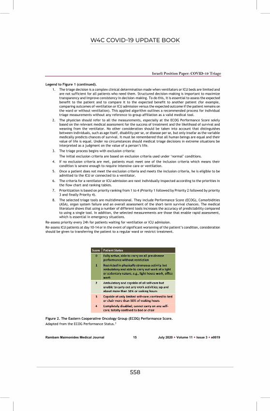
W4C COVID-19 UPDATE BOOK
558
Israeli Position Paper: COVID-19 Triage
Rambam Maimonides Medical Journal 15 July 2020 Volume 11 Issue 3 e0019
Legend to Figure 1 (continued). 1. The triage decision is a complex clinical determination made when ventilators or ICU beds are limited and
are not sufficient for all patients who need them. Structured decision-making is important to maximize transparency and improve consistency in decision-making. To do this, it is essential to assess the expected benefit to the patient and to compare it to the expected benefit to another patient (for example, comparing outcomes of ventilation or ICU admission versus the expected outcome if the patient remains on the ward or without ventilation). This applied algorithm outlines a recommended process for individual triage measurements without any reference to group affiliation as a valid medical tool.
2. The physician should refer to all the measurements, especially at the ECOG Performance Score solely based on the relevant medical assessment for the success of treatment and the likelihood of survival and weaning from the ventilator. No other consideration should be taken into account that distinguishes between individuals, such as age itself, disability per se, or disease per se, but only insofar as the variable medically predicts chances of survival. It must be remembered that all human beings are equal and their value of life is equal. Under no circumstances should medical triage decisions in extreme situations be interpreted as a judgment on the value of a person’s life.
3. The triage process begins with exclusion criteria:
The initial exclusion criteria are based on exclusion criteria used under ‘normal’ conditions. 4. If no exclusion criteria are met, patients must meet one of the inclusion criteria which means their
condition is severe enough to require intensive care or ventilation.
5. Once a patient does not meet the exclusion criteria and meets the inclusion criteria, he is eligible to be admitted to the ICU or connected to a ventilator.
6. The criteria for a ventilator or ICU admission are next individually inspected according to the priorities in the flow chart and ranking tables.
7. Prioritization is based on priority ranking from 1 to 4 (Priority 1 followed by Priority 2 followed by priority 3 and finally Priority 4).
8. The selected triage tools are multidimensional. They include Performance Score (ECOG), Comorbidities (ASA), organ system failure and an overall assessment of the short-term survival chances. The medical literature shows that using a number of different tools increases the accuracy of predictability compared to using a single tool. In addition, the selected measurements are those that enable rapid assessment, which is essential in emergency situations.
Re-assess priority every 24h for patients waiting for ventilation or ICU admission. Re-assess ICU patients at day 10-14 or in the event of significant worsening of the patient’s condition, consideration should be given to transferring the patient to a regular ward or restrict treatment.
Figure 2. The Eastern Cooperative Oncology Group (ECOG) Performance Score. Adapted from the ECOG Performance Status.3
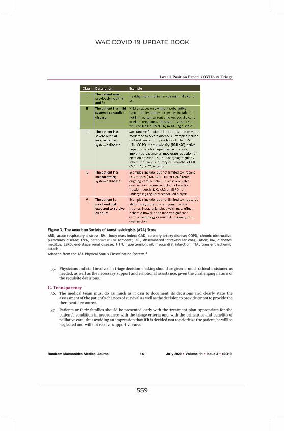
W4C COVID-19 UPDATE BOOK
559
Israeli Position Paper: COVID-19 Triage
Rambam Maimonides Medical Journal 16 July 2020 Volume 11 Issue 3 e0019
35. Physicians and staff involved in triage decision-making should be given as much ethical assistance as needed, as well as the necessary support and emotional assistance, given the challenging nature of the requisite decisions.
G. Transparency 36. The medical team must do as much as it can to document its decisions and clearly state the
assessment of the patient’s chances of survival as well as the decision to provide or not to provide the therapeutic resource.
37. Patients or their families should be presented early with the treatment plan appropriate for the patient’s condition in accordance with the triage criteria and with the principles and benefits of palliative care, thus avoiding an impression that if it is decided not to prioritize the patient, he will be neglected and will not receive supportive care.
Figure 3. The American Society of Anesthesiologists (ASA) Score. ARD, acute respiratory distress; BMI, body mass index; CAD, coronary artery disease; COPD, chronic obstructive pulmonary disease; CVA, cerebrovascular accident; DIC, disseminated intravascular coagulation; DM, diabetes mellitus; ESRD, end-stage renal disease; HTN, hypertension; MI, myocardial infarction; TIA, transient ischemic attack. Adapted from the ASA Physical Status Classification System.4
W4C COVID-19 UPDATE BOOK
560
Israeli Position Paper: COVID-19 Triage
Rambam Maimonides Medical Journal 17 July 2020 Volume 11 Issue 3 e0019
H. Institutional and National Prioritization Committees 38. Every medical institution will appoint a special ad hoc institutional triage committee to be available
to the senior medical specialist for consultation for any case he finds appropriate.
39. This triage committee will consist of 4 people: a senior medical specialist who is not treating COVID-19 patients, a senior nurse, an ethicist, and a member of the clergy. When an urgent decision is needed – two of the four members of the COVID-19 committee are sufficient.
40. This committee can also assist the senior physician in explaining the triage decisions to the patient’s family.
41. This committee has consultative authority but any triage for each individual patient will be made by a senior intensive care specialist after consulting another medical specialist. In the absence of a senior intensive care specialist, by two senior physicians based on the criteria in this position paper.
42. The Ministry of Health will appoint a National triage committee to discuss key issues that arise during the epidemic. Its members will be: Senior Specialist in Intensive Care Medicine, Internal Medicine or Geriatrics Specialist, Senior Nurse, Lawyer, Ethicist, Member of the Clergy, and Community Representative.
REFERENCES
1. Mishnah Sanhedrin 4:5.
2. Sprung CL, Joynt GM, Christian M, Truog RD, Rello J, Nates JL. Adult ICU triage during the coronavirus disease 2019 pandemic: who will live and who will die? Recommendations to improve survival. Crit Care Med 2020;48:1196–202. CrossRef
3. National Palliative Care Research Center. ECOG Performance Status. Available at: http://www.npcrc.org/files/news/ECOG_performance_status.pdf (accessed July 23, 2020).
4. ASA House of Delegates/Executive Committee. ASA Physical status classification system. American Society of Anesthesiologists website. 2019; October 23. Available at: https://www.asahq.org/standards-and-guidelines/asa-physical-status-classification-system (accessed July 23, 2020).
W4C COVID-19 UPDATE BOOK
561
Israeli Position Paper: COVID-19 Triage
Rambam Maimonides Medical Journal 18 July 2020 Volume 11 Issue 3 e0019
APPENDIX 1: COMMISSION MEMBERS
Co-chairs of the Commission Rabbi Prof. Avraham Steinberg – Co-Chair of the National Bioethics Council
Prof. Efrat Levi-Lahad – Co-Chair of the National Bioethics Council
Dr. Tami Karni – Chair of the Ethics Bureau of the Israel Medical Association
Assistants: Ms. Nurit Desau – Research Assistant to the National Bioethics Council; Adv. Rafi Twizer – Health Ministry Coordinator; Ms. Deganit Lahav – Administrative Assistant to the National Bioethics Council.
Medical Subcommittee Prof. Charles Sprung, Chairman – Director Emeritus, General Intensive Care Unit, Department of
Anesthesiology, Intensive Care and Pain, Hadassah Medical Center, Jerusalem
Prof. Jonathan Halevy, Member – former CEO of Shaare Zedek Medical Center, Jerusalem
Prof. Yaron Niv, Member – Senior VP for Quality and Safety, Head of Ventilation Action Team for COVID-19 Patients in hospitals, Ministry of Health
Prof. Ofer Merin, Member – CEO of Shaare Zedek Medical Center, Jerusalem
Prof. Pesach Schwartzman, Member – Chairman of the Israeli Association for Palliative Medicine
Prof. Moshe Sonnenblick, Member – Director of Clinical Geriatric Unit, Shaare Zedek Medical Center, Jerusalem
Dr. Adi Nimrod, Member – Director, General Intensive Care Unit, Ichilov Medical Center, Tel Aviv
Prof. Israel Strouss, Member – Director of Psychiatry, Mayanei HaYeshua Medical Center, Bnei Brak
Philosophical-Ethical-Social Subcommittee Prof. Noam Zohar, Chairman – Director, Graduate Program in Bioethics, Department of
Philosophy, Bar Ilan University, Ramat Gan
Prof. David Heyd, Member – Department of Philosophy, The Hebrew University, Jerusalem
Prof. Yael HaShiloni-Dolev, Member – Department of Sociology and Anthropology, Ben-Gurion University, Beer Sheva
Prof. Ruth Landau, Member – School of Social Work and Social Welfare, The Hebrew University, Jerusalem
Dr. Efrat Ram-Tiktin, Member – Department of Philosophy, Bar Ilan University, Ramat Gan
Legal Subcommittee Dr. Gil Siegal, Chairman – Director, Center for Health Law, Bioethics, and Health Policy, Law
Faculty, Ono Academic College
Judge Salim Jubran, Member – Deputy Chief Justice, Supreme Court, emeritus
Prof. Shai Lavi, Member – Head of the Van Leer Institute, Jerusalem
Attorney Talia Agmon, Member – Deputy Chief Legal Counsel, Ministry of Health
Halachic/Religious Subcommittee Rabbi Prof. Avraham Steinberg, Chairman – Co-Chair of the National Bioethics Council
Rabbi Brigadier General Eyal Krim, Member – Chief Military Rabbi, Israel Defense Forces
W4C COVID-19 UPDATE BOOK
562
Israeli Position Paper: COVID-19 Triage
Rambam Maimonides Medical Journal 19 July 2020 Volume 11 Issue 3 e0019
Rabbi Prof. Yigal Shafran, Member – Head of the Department of Medicine and Halacha, Chief Rabbinate of Jerusalem
Rabbi Dr. Mordechai Halperin, Member – Director of the Schlesinger Institute for Medical Halachah Research, at the Shaare Zedek Medical Center, Jerusalem
Rabbi Avraham Manela, Member – CEO of chevrah kadishah (Burial society), Tel Aviv
Qadi Muhammad Abu-Abeid, Member – Judge of the Sharia Court of Appeal (Islam)
Dr. Etienne Lepicard, Member – Center for Medical Education, Hadassah and the Hebrew University, Jerusalem and Director of “Bet Hagat,” the home for interfaith-artistic encounters in Jerusalem (Christianity)
W4C COVID-19 UPDATE BOOK
563
Since January 2020 Elsevier has created a COVID-19 resource centre with
free information in English and Mandarin on the novel coronavirus COVID-
19. The COVID-19 resource centre is hosted on Elsevier Connect, the
company's public news and information website.
Elsevier hereby grants permission to make all its COVID-19-related
research that is available on the COVID-19 resource centre - including this
research content - immediately available in PubMed Central and other
publicly funded repositories, such as the WHO COVID database with rights
for unrestricted research re-use and analyses in any form or by any means
with acknowledgement of the original source. These permissions are
granted for free by Elsevier for as long as the COVID-19 resource centre
remains active.

W4C COVID-19 UPDATE BOOK
564
Israeli Position PaperTriage Decisions for Severely IllPatients During the COVID-19PandemicAvraham Steinberg, MD
Ephrat Levy-Lahad, MD
Jerusalem, Israel
Tami Karni, MD, MHA
Zerifin, Israel
Charles L. Sprung, MD, JD, FCCP
Jerusalem, Israel
Israeli Triage CommissionThe coronavirus disease 2019 (COVID-19) pandemiccreated the need for triaging scarce life-saving resources,raising medical, ethical, social, legal, and religiousdilemmas. In Israel, a Joint Commission of the IsraelNational Bioethics Council, the Ethics Bureau of theIsrael Medical Association, and the Israeli Ministry ofHealth was appointed by the Director General of theMinistry of Health to develop national guidelines fortriaging severely ill patients if the health-care system wasoverwhelmed. The Commission was composed of seniorexperts in medicine, ethics, law, sociology, Jewish law,Christianity, and Islam. The document produced by theCommission provides an overview of general principles,pragmatic medical criteria, and a practical triage
protocol. Guiding principles included supreme value oflife, equality of all individuals, transparency, consistency,equity in access, fair distribution, greatest good to thegreatest number, palliation, preserving societal humanityand compassion, and promoting public trust. Theguidelines furnish objective, functional clinical criteriaand decision-making mechanisms and prohibitdiscrimination based on race, religion, sex, nationality orcitizenship, sexual orientation, socioeconomic status,age, or disability. The Israeli National policy is the onlynational policy for the COVID-19 pandemic developedby a governmental agency.
Triage Guidelines and ProtocolTriage guidelines apply only after the health-care systemis overwhelmed and the Ministry of Health declares anemergency state. The triage protocol is simple and easyto understand, uses multidimensional assessmentmeasures, and enables quick decisions. The Commissiondiscussed several algorithms with different scoringsystems to triage ICU beds and ventilators, choosing thebest predictors of short-term survival and enabling thequickest decisions. Working with an international expertgroup that developed a triage protocol1 helped provideadditional assessments and consistency adapting theprotocol to Israel.
The first triage decision (ie, inclusion and exclusioncriteria) is the same used under normal circumstances.Only individuals too ill to benefit or those likely torecover without scarce resources are excluded. Thus,everyone has the same opportunity to receive scarceresources as during “normal” conditions. This shouldallay societal fears about individuals not triaged becausethey are considered “unworthy” of being saved.2 Themeasures chosen for prioritization included functionalability (Eastern Cooperative Oncology Groupperformance score), comorbidities (American Society ofAnesthesiologists score), number of organ systemsfailing, and overall estimation of short-term survival.The Sequential Organ Failure Assessment score was notchosen because it requires additional assessments, it wasunhelpful when applied to epidemic data, and mostCOVID-19 patients only have single organ failure.1 Ifmore patients than resources have the same priority,further triage should be based on lives saved and then on
ABBREVIATIONS: COVID-19 = coronavirus disease 2019AFFILIATIONS: From the Medical Ethics Unit (Dr Steinberg), ShaareZedek Medical Center, & Co-Chairman, Israel National BioethicsCouncil, Jerusalem, Israel; the Medical Genetics Institute (Dr Levy-Lahad), Shaare Zedek Medical Center, Hebrew University of Jerusalem,Faculty of Medicine, & Co-Chairman, Israel National BioethicsCouncil; the Breast Care Institute (Dr Karni), Assaf Harofe MedicalCenter, the Ethics Bureau of the Israel Medical Association; and theDepartment of Anesthesiology and Critical Care Medicine (DrSprung), Hadassah Medical Center, Hebrew University of Jerusalem,Faculty of Medicine.FINANCIAL/NONFINANCIAL DISCLOSURES: None declared.CORRESPONDENCE TO: Charles L. Sprung, MD, JD, FCCP, GeneralIntensive Care Unit, Department of Anesthesiology and Critical CareMedicine, Hadassah Hebrew University Medical Center, PO Box12000, Jerusalem, Israel 91120; e-mail: [email protected] � 2020 Published by Elsevier Inc under license from theAmerican College of Chest Physicians.DOI: https://doi.org/10.1016/j.chest.2020.07.052
[ General Interest Commentary and Announcement ]
chestjournal.org 1
EDI 5.6.0 DTD � CHEST3438_proof � 6 October 2020 � 11:00 pm � EO: CHEST-20-3586
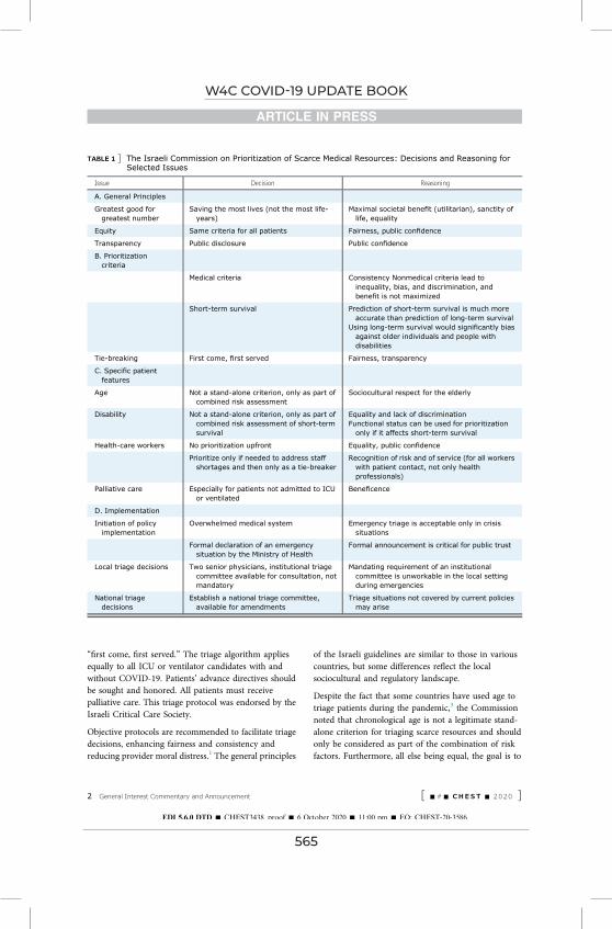
W4C COVID-19 UPDATE BOOK
565
“first come, first served.” The triage algorithm appliesequally to all ICU or ventilator candidates with andwithout COVID-19. Patients’ advance directives shouldbe sought and honored. All patients must receivepalliative care. This triage protocol was endorsed by theIsraeli Critical Care Society.
Objective protocols are recommended to facilitate triagedecisions, enhancing fairness and consistency andreducing provider moral distress.1 The general principles
of the Israeli guidelines are similar to those in variouscountries, but some differences reflect the localsociocultural and regulatory landscape.
Despite the fact that some countries have used age totriage patients during the pandemic,3 the Commissionnoted that chronological age is not a legitimate stand-alone criterion for triaging scarce resources and shouldonly be considered as part of the combination of riskfactors. Furthermore, all else being equal, the goal is to
TABLE 1 ] The Israeli Commission on Prioritization of Scarce Medical Resources: Decisions and Reasoning forSelected Issues
Issue Decision Reasoning
A. General Principles
Greatest good forgreatest number
Saving the most lives (not the most life-years)
Maximal societal benefit (utilitarian), sanctity oflife, equality
Equity Same criteria for all patients Fairness, public confidence
Transparency Public disclosure Public confidence
B. Prioritizationcriteria
Medical criteria Consistency Nonmedical criteria lead toinequality, bias, and discrimination, andbenefit is not maximized
Short-term survival Prediction of short-term survival is much moreaccurate than prediction of long-term survival
Using long-term survival would significantly biasagainst older individuals and people withdisabilities
Tie-breaking First come, first served Fairness, transparency
C. Specific patientfeatures
Age Not a stand-alone criterion, only as part ofcombined risk assessment
Sociocultural respect for the elderly
Disability Not a stand-alone criterion, only as part ofcombined risk assessment of short-termsurvival
Equality and lack of discriminationFunctional status can be used for prioritization
only if it affects short-term survival
Health-care workers No prioritization upfront Equality, public confidence
Prioritize only if needed to address staffshortages and then only as a tie-breaker
Recognition of risk and of service (for all workerswith patient contact, not only healthprofessionals)
Palliative care Especially for patients not admitted to ICUor ventilated
Beneficence
D. Implementation
Initiation of policyimplementation
Overwhelmed medical system Emergency triage is acceptable only in crisissituations
Formal declaration of an emergencysituation by the Ministry of Health
Formal announcement is critical for public trust
Local triage decisions Two senior physicians, institutional triagecommittee available for consultation, notmandatory
Mandating requirement of an institutionalcommittee is unworkable in the local settingduring emergencies
National triagedecisions
Establish a national triage committee,available for amendments
Triage situations not covered by current policiesmay arise
2 General Interest Commentary and Announcement [ -#- CHE ST - 2 0 2 0 ]
EDI 5.6.0 DTD � CHEST3438_proof � 6 October 2020 � 11:00 pm � EO: CHEST-20-3586
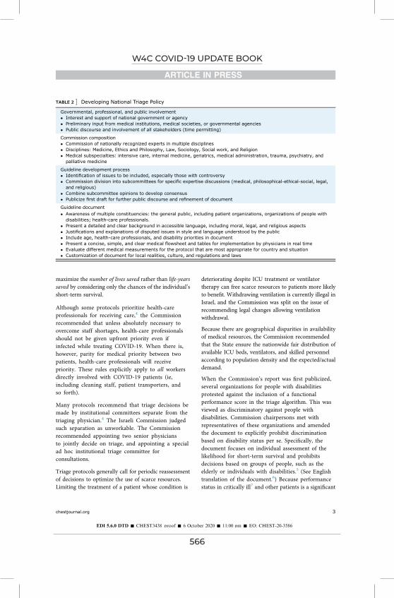
W4C COVID-19 UPDATE BOOK
566
maximize the number of lives saved rather than life-yearssaved by considering only the chances of the individual’sshort-term survival.
Although some protocols prioritize health-careprofessionals for receiving care,4 the Commissionrecommended that unless absolutely necessary toovercome staff shortages, health-care professionalsshould not be given upfront priority even ifinfected while treating COVID-19. When there is,however, parity for medical priority between twopatients, health-care professionals will receivepriority. These rules explicitly apply to all workersdirectly involved with COVID-19 patients (ie,including cleaning staff, patient transporters, andso forth).
Many protocols recommend that triage decisions bemade by institutional committees separate from thetriaging physician.4 The Israeli Commission judgedsuch separation as unworkable. The Commissionrecommended appointing two senior physiciansto jointly decide on triage, and appointing a specialad hoc institutional triage committee forconsultations.
Triage protocols generally call for periodic reassessmentof decisions to optimize the use of scarce resources.Limiting the treatment of a patient whose condition is
deteriorating despite ICU treatment or ventilatortherapy can free scarce resources to patients more likelyto benefit. Withdrawing ventilation is currently illegal inIsrael, and the Commission was split on the issue ofrecommending legal changes allowing ventilationwithdrawal.
Because there are geographical disparities in availabilityof medical resources, the Commission recommendedthat the State ensure the nationwide fair distribution ofavailable ICU beds, ventilators, and skilled personnelaccording to population density and the expected/actualdemand.
When the Commission’s report was first publicized,several organizations for people with disabilitiesprotested against the inclusion of a functionalperformance score in the triage algorithm. This wasviewed as discriminatory against people withdisabilities. Commission chairpersons met withrepresentatives of these organizations and amendedthe document to explicitly prohibit discriminationbased on disability status per se. Specifically, thedocument focuses on individual assessment of thelikelihood for short-term survival and prohibitsdecisions based on groups of people, such as theelderly or individuals with disabilities.5 (See Englishtranslation of the document.6) Because performancestatus in critically ill7 and other patients is a significant
TABLE 2 ] Developing National Triage Policy
Governmental, professional, and public involvement� Interest and support of national government or agency� Preliminary input from medical institutions, medical societies, or governmental agencies� Public discourse and involvement of all stakeholders (time permitting)
Commission composition� Commission of nationally recognized experts in multiple disciplines� Disciplines: Medicine, Ethics and Philosophy, Law, Sociology, Social work, and Religion� Medical subspecialties: intensive care, internal medicine, geriatrics, medical administration, trauma, psychiatry, and
palliative medicine
Guideline development process� Identification of issues to be included, especially those with controversy� Commission division into subcommittees for specific expertise discussions (medical, philosophical-ethical-social, legal,
and religious)� Combine subcommittee opinions to develop consensus� Publicize first draft for further public discourse and refinement of document
Guideline document� Awareness of multiple constituencies: the general public, including patient organizations, organizations of people with
disabilities; health-care professionals.� Present a detailed and clear background in accessible language, including moral, legal, and religious aspects� Justifications and explanations of disputed issues in style and language understood by the public� Include age, health-care professionals, and disability priorities in document� Present a concise, simple, and clear medical flowsheet and tables for implementation by physicians in real time� Evaluate different medical measurements for the protocol that are most appropriate for country and situation� Customization of document for local realities, culture, and regulations and laws
chestjournal.org 3
EDI 5.6.0 DTD � CHEST3438_proof � 6 October 2020 � 11:00 pm � EO: CHEST-20-3586
W4C COVID-19 UPDATE BOOK
567
predictor of in-hospital mortality and is widely used inmedicine, the Commission concluded that it was animportant measurement for triage, and that itselimination would reduce the predictive power of thetriage algorithm and exacerbate inequity.
ConclusionThe Israeli consensus document and pragmatic triageprotocol offer a societal and medical roadmap forallocating scarce resources. They reflect both universalprinciples and local sensitivities. The current summaryof the process and decisions (Table 1) of the Israeli JointCommission provides strategies and approaches forpolicymakers in different countries to develop policies(Table 2) for this pandemic, the next coronavirus wave,or other disasters, to correct previous policies and todevelop objective, transparent, equitable, and consistentnational triage policies.
AcknowledgmentsOther contributions: We thank all members of the Commission:Commission Co-Chairs: Avraham Steinberg, MD, Ephrat Levy-Lahad, MD, Tami Karni, MD, MHA; Medical Subcommittee:Charles Sprung, MD, JD, FCCP (Chair), Jonathan Halevy, MD,Yaron Niv, MD, Ofer Merin, MD, MHA, Pesach Schwartzman, MD,
Moshe Sonnenblick, MD, Adi Nimrod, MD, Israel Strous, MD,MHA; Philosophical-Ethical-Social Subcommittee: Noam Zohar,PhD (Chair), David Heyd, PhD, Yael HaShiloni-Dolev, PhD,Ruth Landau, PhD, Efrat Ram-Tiktin PhD; Legal Subcommittee:Gil Siegal, SJD, MD (chair), Salim Jubran, LLB, Shai Lavi, TaliaAgmon, LLB; Halachic/Religious Subcommittee: Avraham Steinberg,MD (Chair), Eyal Krim, Yigal Shafran, PhD, Mordechai Halperin,MD, LLB, Avraham Manela, Muhammad Abu-Abeid, EtienneLepicard, MD, PhD. We thank Nurit Dessau, MBA, for researchassistance, and Rafi Twizer and Deganit Lahav for administrativeassistance.
References1. Sprung C, Joynt GM, Christian M, Truog RD, Rello J, Nates JL. Adult
ICU triage during the coronavirus disease 2019 pandemic: who willlive and who will die? Recommendations to improve survival. CritCare Med. 2020;48(8):1196-1202.
2. White DB, Lo B. A framework for rationing ventilators and critical carebeds during the COVID-19 pandemic. JAMA. 2020;323:1773-1774.
3. RosenbaumL. FacingCovid-19 in Italy: ethics, logistics, and therapeuticson the epidemic’s front line. N Engl J Med. 2020;382:1873-1875.
4. Emanuel EJ, Persad G, Upshur R, et al. Fair allocation of scarcemedical resources in the time of Covid-19. N Engl J Med. 2020;382:2049-2055.
5. Mello MM, Persad G, White DB. Respecting disability rights: towardimproved crisis standards of care. N Engl J Med. 2020;383(5):e26.
6. https://www.health.gov.il/PublicationsFiles/position-paper-230520.pdf. Accessed August 10, 2020.
7. Park C, Koh Y, Jeon K, et al. Impact of Eastern Cooperative OncologyGroup performance status on hospital mortality in critically illpatients. J Crit Care. 2014;29:409-413.
4 General Interest Commentary and Announcement [ -#- CHE ST - 2 0 2 0 ]
EDI 5.6.0 DTD � CHEST3438_proof � 6 October 2020 � 11:00 pm � EO: CHEST-20-3586
W4C COVID-19 UPDATE BOOK
568
Dow
nloadedfrom
http://journals.lww.com
/ccmjournalby
+Eh0FNkBW
nlDJb4PaH
Ep2J7EY+qKtwfBD
tnz7lDdqk1jjuJ7Tr+plX9SN
uKXzKkuQk6YSnFfV7h17uH
mwAH
whhU
zOepqcELU
5hqD2hlXXhm
GfGPyfw
QrHyA8fLLbN
8x6z9at42P+Hskd1tojruBggQ
==on
07/22/2020
Downloadedfromhttp://journals.lww.com/ccmjournalby+Eh0FNkBWnlDJb4PaHEp2J7EY+qKtwfBDtnz7lDdqk1jjuJ7Tr+plX9SNuKXzKkuQk6YSnFfV7h17uHmwAHwhhUzOepqcELU5hqD2hlXXhmGfGPyfwQrHyA8fLLbN8x6z9at42P+Hskd1tojruBggQ==on07/22/2020
Copyright © 2020 by the Society of Critical Care Medicine and Wolters Kluwer Health, Inc. All Rights Reserved.Copyright © 2020 by the Society of Critical Care Medicine and Wolters Kluwer Health, Inc. All Rights Reserved.
1196 www.ccmjournal.org August 2020 • Volume 48 • Number 8
Objectives: Coronavirus disease 2019 patients are currently over-whelming the world’s healthcare systems. This article provides practical guidance to front-line physicians forced to make critical rationing decisions.Data Sources: PubMed and Medline search for scientific literature, reviews, and guidance documents related to epidemic ICU triage including from professional bodies.Study Selection: Clinical studies, reviews, and guidelines were selected and reviewed by all authors and discussed by internet conference and email.Data Extraction: References and data were based on relevance and author consensus.Data Synthesis: We review key challenges of resource-driven triage and data from affected ICUs. We recommend that once available re-sources are maximally extended, triage is justified utilizing a strategy that provides the greatest good for the greatest number of patients. A triage algorithm based on clinical estimations of the incremental survival benefit (saving the most life-years) provided by ICU care
is proposed. “First come, first served” is used to choose between individuals with equal priorities and benefits. The algorithm provides practical guidance, is easy to follow, rapidly implementable and flex-ible. It has four prioritization categories: performance score, ASA score, number of organ failures, and predicted survival. Individual units can readily adapt the algorithm to meet local requirements for the evolving pandemic. Although the algorithm improves consistency and provides practical and psychologic support to those performing triage, the final decision remains a clinical one. Depending on country and operational circumstances, triage decisions may be made by a triage team or individual doctors. However, an experienced critical care specialist physician should be ultimately responsible for the tri-age decision. Cautious discharge criteria are proposed acknowledg-ing the difficulties to facilitate the admission of queuing patients.Conclusions: Individual institutions may use this guidance to de-velop prospective protocols that assist the implementation of triage decisions to ensure fairness, enhance consistency, and decrease provider moral distress. (Crit Care Med 2020; 48:1196–1202)Key Words: benefit; first come first served; intensive care unit; pandemic; triage
Patients with life-threatening illnesses admitted to ICUs have lower mortalities than patients not admitted (1, 2). Despite international variations in ICU services (3),
demand for ICU beds frequently exceeds their supply (1, 2). Whereas resource-driven triage decisions are uncommon in North America, they are more frequent in Europe (2). During a pandemic or mass disaster medical resources may become desperately inadequate with patients dying because of the lack ventilators or ICU beds as is currently occurring in the corona-virus disease 2019 (COVID-19) pandemic (4). The present rec-ommendations are based on the joint collaboration of several worldwide clinicians who have been involved in ICU triage dur-ing epidemics and other surge conditions for several decades.
ICU TRIAGE UNDER “NORMAL” CONDITIONSOnce patients meet ICU inclusion criteria, the most commonly recommended triage criteria for ICU admission under “normal”
*See also pp. 1241 and 1243.1Department of Anesthesiology, Critical Care Medicine and Pain, Hadas-sah Medical Center, Hebrew University of Jerusalem, Faculty of Medicine, Jerusalem, Israel.
2Department of Anaesthesia and Intensive Care, The Chinese University of Hong Kong, Hong Kong, China.
3London’s Air Ambulance, Barts Health NHS Trust, London, United Kingdom.
4Center for Bioethics, Department of Global Health and Social Medi-cine, Harvard Medical School, Boston, MA.
5Department of Anesthesiology, Critical Care, and Pain Medicine, Boston Children’s Hospital, Boston, MA.
6Clinical Research/epidemiology in pneumonia and sepsis, Vall d’Hebron Institute of Research (VHIR), Barcelona, Spain.
7Centro de Investigacion Biomedica en Red en Efermedades Respirato-rias (CIBERES), Instituto de Salud Carlos III, Barcelona, Spain.
8Clinical Research, CHU Nîmes, NÎmes, France.9Critical Care Department, The University of Texas MD Anderson Cancer Center, Houston, TX.
Copyright © 2020 by the Society of Critical Care Medicine and Wolters Kluwer Health, Inc. All Rights Reserved.
DOI: 10.1097/CCM.0000000000004410
Adult ICU Triage During the Coronavirus Disease 2019 Pandemic: Who Will Live and Who Will Die? Recommendations to Improve Survival*
Charles L. Sprung, MD, MCCM1; Gavin M. Joynt, MBBCh2; Michael D. Christian, MD, FCCM3;
Robert D. Truog, MD4,5; Jordi Rello, MD, PhD6,7,8; Joseph L. Nates, MD, MBA, CMQ, MCCM9
W4C COVID-19 UPDATE BOOK
569
Copyright © 2020 by the Society of Critical Care Medicine and Wolters Kluwer Health, Inc. All Rights Reserved.
Review Articles
Critical Care Medicine www.ccmjournal.org 1197
circumstances are “medical benefit” or “first come, first served” (5, 6). Patients who are “too well” or “too sick” to achieve a sub-stantial benefit threshold would not be part of the potential pool. The now quite old Society of Critical Care Medicine triage consensus (5) stated that “The foremost consideration in tri-age decisions is the expected outcome of the patient in terms of survival and function, which turns on the medical status of the patient. In general, patients with good prognoses for recovery have priority over patients with poor prognoses.” In contrast, the American Thoracic Society recommended that “when demand for ICU beds exceeds supply, medically appropriate patients should be admitted on a first-come, first-served basis” (6).
An updated triage consensus statement unanimously found that triage decisions should give priority for patients with greater benefit and not be made on a “first come, first served basis” (7). Interestingly, in an attempt to develop a survival cutoff for triage decisions, only 77% of respondents agreed to a survival cutoff of 0.1% (7). This may be related to intensiv-ists attempting to rescue with an ICU trial even patients with little chance of surviving knowing that they can later discharge the patient or limit treatments (7). Recent statements on ICU triage also recommended that patients be admitted based on their potential for benefit (8–10). Most statements declared that triage criteria should be explicit, fair, disclosed in advance and not be based on race, ethnicity, sex, sexual preference, fi-nancial status, or social worth (5–10). Unfortunately, current triage tools using objective prioritization, diagnosis, or pa-rameter models are unable to provide ICU admission and ex-clusion criteria with demonstrated improved outcomes (8). Although attempts have been made to develop objective, triage scores (11), none are currently being used. Some triage scor-ing tools have been specifically developed for use in epidemics; however, none have been validated in a crisis setting (12–17).
The question arises as to how should “first come, first served” and “medical benefit” be defined for ICU admission? As only a few patients are admitted to an ICU every day and beds are either available for incoming patients or can be made available by discharging patients, the “first come, first served” criteria can usually be used for one patient after another. Unfortunately, there are occasions where more than one pa-tient is vying for the last ICU bed. How should the “first pa-tient” be defined when determining “first come, first served” criteria? Is it the first patient admitted to the emergency de-partment, the first patient that the triage officer heard about, the patient in the operating room for whom an ICU bed was reserved, or the first patient that the triage physician accepted? In addition, if there is more than one ICU admission candidate how should “medical benefit” be defined? Is it saving the most lives or saving the most life-years with ICU care for potential patients or the incremental medical benefit between ICU care versus ward care in these patients (10)? If the latter, how much larger must the difference in benefit be? The South African consensus guideline recommended a benefit difference of 15–25% (10). Although intensivists prognosticate more accurately than scoring systems (18), uncertainty remains and physicians’ accuracy lacks the consistency and precision patient desire.
TRIAGE DURING A PANDEMIC OR MASS DISASTER WITH OVERWHELMING SHORTAGESThis article deals only with ICU triage. Complete informa-tion related to surge capacity, coordination and collaboration, manpower, essential equipment, pharmaceuticals and sup-plies, protection of patients and staffing, medical procedures, and education for pandemics can be found elsewhere (19). As difficult as triage decisions are during everyday practice, they are even more challenging during pandemics or mass disasters. Pandemics produce countless critically ill patients that overrun healthcare resources (19). In extreme situations, customary interventions and standards of practice may be unachievable leading to avertible deaths (20, 21). Establishing equitable and just strategies for “the greatest good for the greatest number” of patients may demand decreasing ICU therapies to patients who ordinarily would be expected to benefit from them under conditions of adequate resources (20, 21).
Because of these dilemmas several objective, ICU triage protocols, tools, and scores were developed to prioritize lim-ited reserves, reduce additional deaths and help avoid clinical judgments which might be more protracted and less reliable (12–17). Unfortunately, these tools have been shown to have inadequate performance and many patients classified as too sick to require admission survived (14, 16).
ICU triage of patients remains challenging and controversial in pandemics when resources are overwhelmed. Recommendations for ICU triage for appropriate candidates during pandemics have suggested that either “medical benefit” or a “first come, first served” is acceptable (20, 21) or an improved incremental sur-vival rather than a “first come, first served basis” (22). More re-cently proposals have been for saving the most lives (23) or saving the most life-years (24). The current COVID-19 pandemic has witnessed the use of age as a criteria, primarily because advanced age appears strongly associated with poorer outcomes (4).
INFORMATION FROM THE PRESENT COVID-19 PANDEMICChina, Italy, Spain, and the Americas have had major severe acute respiratory syndrome coronavirus 2 outbreaks and mor-tality. Triage data from some ICUs affected by the COVID-19 pandemic are shown in Table 1. ICU beds and daily census increased approximately double from the previous year. It is too early to evaluate mortality which should be relevant at 28 days after admission. Recent reports of moderately high pop-ulation mortality from COVID-19 in China (61.5%) (25) and high ICU mortality in the United States (67%) (26) are wor-risome. It has been suggested that the high mortality maybe related to the large bed expansion without adequate healthcare resources (27). This has implications for expansions that are currently occurring worldwide. Experience from severe acute respiratory syndrome (SARS) showed that rapid and excessive expansion may overwhelm staff leading to excess infections in healthcare workers and compromising care (28). Thus expan-sion should be matched by safe staffing to guarantee an appro-priate quality of care and staff safety which necessarily limits

W4C COVID-19 UPDATE BOOK
570
Copyright © 2020 by the Society of Critical Care Medicine and Wolters Kluwer Health, Inc. All Rights Reserved. Copyright © 2020 by the Society of Critical Care Medicine and Wolters Kluwer Health, Inc. All Rights Reserved.
Sprung et al
1198 www.ccmjournal.org August 2020 • Volume 48 • Number 8
expansion. A consensus group with first-hand experience of outbreak expansion during SARS concluded that safe expan-sion is realistically limited by availability of acceptably trained staff and limited to a maximum expansion of 50–100% of baseline capacity (29). Hospitals must also balance ICU needs and the potential decreasing benefits of increasing ICU ca-pacity due to excess workload with other hospital needs (20).
Compassionate care should be offered to patients with low-level priorities. There are anecdotal reports that they can survive or delay intubation until more resources are available using awake prone position (30) in hospital wards because of the shortage of ventilators or ICU beds (J. Rello, personal com-munication, 2020).
ICU TRIAGE DURING A PANDEMICHow should triage decisions be made during the COVID-19 pandemic? It is strongly recommended that institutions de-velop prospective, objective protocols or algorithms to assist the implementation of their triage decisions to enhance con-sistency (31) and decrease moral distress among providers (24, 32). First, when demand surges hospitals must increase ICU capacity by 100–200% (20, 33) before triage is instituted. Tri-age protocols should only be triggered when resources across a broad geographic area are or will be overwhelmed despite efforts to extend them and systems move from contingency to crisis mode (20). As we can predict a wave of incoming patients from an impending peak in COVID-19, it might be prudent to start rationing prior to expending all resources on early cases with low probabilities for survival when it is clear that maximum surge capacity will certainly be exceeded in the near future. If hospitals cannot provide services, they should consider transferring patients to cities where ICU beds are still available (20). Second, the potential ICU patients should be
those that meet inclusion and no exclusion criteria. There is no perfect tool. In deciding which tool to use, we chose a tool that was simple, easy to understand and use and most impor-tant providing for quick assessments. As different countries and regions have different infrastructures and resources, laws, cultures, and religions, we attempted to offer flexibility in our recommendations along with explanations for the differing opinions so each region or country can choose what is most appropriate for them.
An illustrative example of inclusion and exclusion crite-ria and an algorithm for ICU triage is found in Figure 1 and Supplemental Figure 1 (Supplemental Digital Content 1, http://links.lww.com/CCM/F519; legend, Supplemental Digital Content 2, http://links.lww.com/CCM/F520). Admission pri-ority is given to patients from priority 1 to 4 based on their performance scores, ASA score, number of organ failures, and predicted survival. If there are more priority 1 patients than beds, allocation will be based on incremental ICU benefit de-fined as saving the most life-years (evaluating mortality from both acute and chronic disorders) (24). If there is a tie for ICU candidates, clinicians should use first come, first served (34) which they are accustomed to using and not a random allo-cation with a lottery (which they are not familiar using, los-ing valuable time). We recommend that the first-come patient should be defined as the first patient that the triage officer was informed of. The triage algorithm should apply equally to all ICU candidates with and without COVID-19 (35). When the triage protocol commences, all ICU patients must be reevalu-ated for remaining in the ICU based on these same criteria.
Who Will Perform the Triage?In an ideal situation, we should have a separate triage of-ficer/committee for making admission and discharge triage
TABLE 1. ICU Utilization and Outcomes in Three Countries During the Coronavirus Disease 2019 Pandemic Surge
Country Spain Spain Ecuador The Netherlands
Hospital University Hospital Son Llatzer, Palma de Mallorca
Araba University Hospital, Vitoria
Hospital General Guasmo Sur, Guayaquil
University Medical Center, Utrecht
COVID-19 1 yr prior to COVID-19
COVID-19 1 yr prior to COVID-19
COVID-19 1 yr prior to COVID-19
COVID-19 1 yr prior to COVID-19
Dates March 13, 2020, to March 29, 2020
March 13, 2019, to March 29, 2019
March 4, 2020, to March 24, 2020
March 4, 2019, to March 24, 2019
February 29, 2020, to March 29, 2020
February 28, 2019, to March 29, 2019
March 16, 2020, to April 15, 2020
March 16, 2019, to April 15, 2019
Total ICU beds 32 18 71 35 28 14 68 24
ICU admissions (n) 44 27 115 114 48 25 256 191
ICU census 31.7 17.4 52.1 25.2 26.3 12.5 43.3 22.3
ICU deaths (%) 23.1 11 14 4.5 51.1 28.0 7.0 11.0
Triage method And And MB + agea MB MTS MTS MB MB
And = Andorran model, COVID-19 = coronavirus disease 2019, MB = medical benefit, MTS = Manchester triage system.a During COVID-19 period medical benefit was used for triage and in addition patients > 80 yr old with a Charlson scale > 2 and a Barthel scale < 80 were excluded.

W4C COVID-19 UPDATE BOOK
571
Copyright © 2020 by the Society of Critical Care Medicine and Wolters Kluwer Health, Inc. All Rights Reserved.
Review Articles
Critical Care Medicine www.ccmjournal.org 1199
decisions (5, 35). Whether this is feasible will depend on the country and magnitude of the crisis. Many intensivists be-lieve that only a senior phy-sician with triage experience can make these decisions and be part of such a committee. Are there enough intensivists to provide care and be part of a triage committee? Can other physicians along with other professionals without triage experience triage patients in a pandemic? If an institution creates a committee, it could include doctors (expertise in administration or pallia-tive care maybe particularly helpful), nurses, social work-ers, and ethicists. Decisions should be made by senior physicians with triage experi-ence. As intensivists who have become invested in the care of a patient over time may have difficulties withdrawing ventilation (32) or discharg-ing patients to wards under these difficult conditions, the institutional triage or ethics committee could be helpful in affirming and endorsing the decision taken by a senior intensivist or making the deci-sions themselves. Admission, discharge or limitation deci-sions must be communicated to the patient or family. The public prefers that triage be performed by senior doctors and that predetermined cri-teria be used (36). If patient surge exceeds the number of available critical care trained specialists, intensivists should supervise nonintensivist phy-sicians (20).
Patient Priority
1) Healthcare and other essential workers: There is controversy in the literature and there were differences of opinion among authors about prioritizing front-line
Figure 1. This tool provides an element of objectivity to help enhance consistency. It is not considered, how-ever, a complete substitute for the carefully considered judgment of an experienced intensive care clinician. The presented algorithm is an example and each institution can decide on which performance score, comorbidities, and organ system failures to use based on their experience and what their staff ifs most comfortable using. A triage (prioritization) decision is a complex clinical decision made when ICU beds are limited. A structured deci-sion-making process is important to maximize transparency and improve consistency in decision-making. A clin-ical estimation of likely benefit (outcomes from ICU admission compared with outcomes expected if the patient remained on the ward/other care area) is necessary so that patients who will benefit most from ICU are given priority. Examples of clinical conditions that the expert group believe would likely result in a failure of a patient to meet sufficient priority for admission is provided (priority 4). This conceptual algorithm outlines a recommended process for making an individual triage decision, based on a likelihood that survival without ICU care would be low (5–10% or less), and if admission criteria as laid out are met, survival would be estimated to be in excess of 50% short- to medium-term survival. The initial exclusion criteria are based on exclusion criteria used under “normal” conditions so that the same patients are eligible during pandemic conditions and given priority based on their likelihood to benefit. Then patients must meet one of the inclusion criteria. Priorities are then based on
W4C COVID-19 UPDATE BOOK
572
Copyright © 2020 by the Society of Critical Care Medicine and Wolters Kluwer Health, Inc. All Rights Reserved. Copyright © 2020 by the Society of Critical Care Medicine and Wolters Kluwer Health, Inc. All Rights Reserved.
Sprung et al
1200 www.ccmjournal.org August 2020 • Volume 48 • Number 8
healthcare and other essential workers. A consensus stated that it is unlikely that workers becoming critically ill dur-ing a pandemic will recover sufficiently for them to return to work (31). In addition, conferring priority for healthcare workers could hinder societal trust in the triage mechanism at a time when public trust is essential. On the other hand, healthcare and other essential workers put themselves at high-risk supporting or saving others and are irreplaceable and essential for society and patient care (24, 35). Some workers have been infected because they were not provided with adequate protection (37). As COVID-19 may have a long duration and could recur, it is increasingly plausible that healthcare workers could return later to work. How-ever, as healthcare workers have a clear conflict of interest as “self advocates,” we recommend this prioritization should be decided and implemented by societal and governmental agencies. Defining essential workers at the time of triage poses a practical problem, but consideration could be given to those workers excluded from lockdown as defined by official national, regional, and local orders. Because of the controversy, each country should decide what is most ap-propriate for their citizenry.
2) Younger patients: Age has also been suggested for ICU tri-age decisions. In Italy, patients are dying from COVID-19 because they have been triaged not to receive a ventilator because of their age (4). Advocates suggest that younger patients should be prioritized to have an equal opportunity to pass through the stages of life (24). Despite the fact that elderly patients have a higher ICU mortality than younger patients, the incremental ICU benefit is greater for the eld-erly as patients more than 65 years had a greater difference in mortality between admitted and rejected patients com-pared with younger patients (2). Although age should be taken into consideration along with other variables, age should not be the sole determining factor in triage decisions (7). What is important is physiologic and not chronological
age (7). Younger patients have been taken into considera-tion by using incremental ICU benefit defined as saving the most life-years.
Staff ProtectionStaff will have anxiety about personal and family risks, distress about avoidable deaths and patient limitations, potential fail-ings from working outside areas of normal expertise, or ex-cessive workload and death of family, friends, and colleagues (38). Therefore, institutions must do their utmost to decrease clinical risks, providing adequate protective supplies and ed-ucation, maintain staff confidence and safety by minimizing risks and maintaining appropriate services and reassurance with legal protection so there are adequate staff to man the beds (38).
ICU Discharge CriteriaEven under “normal” circumstances intensivists agree that patients with little or no anticipated benefit from continued ICU interventions may be discharged from the ICU (7). Dur-ing a pandemic, all admitted patients should be admitted with the intention of carrying out a time-limited trial of therapy (39) so that if ICU care does not significantly improve the patient’s condition after a reasonable time, the patient should be discharged and/or therapies limited (7). Reevaluations for admitted and refused patients should be performed when ap-propriate and feasible ideally every 24 hours. As COVID-19 patients tend to have longer ICU durations (25) reassessments for remaining in the ICU should occur later, at days 10–14. When faced with overwhelming resource restrictions, it may be justified to limit life support therapy or discharge a patient with very poor survival prognosis after admission to ICU to allow queuing patients with a much higher probability of ben-efit to be admitted. This process will be difficult to implement, and we recommend that such decisions which are not as time critical be made by broad consensus. A decision by more than one senior ICU clinician, an independent physician and pos-sibly the triage committee could serve to enhance fairness, con-sistency and mitigate the moral distress associated with such decisions.
MonitoringBecause different parts of the world have different views on trust and empowerment, requirements for decisions to be reviewed regularly by monitoring committees to ensure that there are no inappropriate inequities and to regularly review the triage tool will vary (4). It is also essential that the outcome of patients who are triaged is tracked to ensure that triage is effectively targeting resources to those who are most likely to benefit as indicated by improved survival rates overall.
As there can be no universal formula to guide the imple-mentation of ICU triage, each region or country will have to make its own decisions as to what will be best for its system. We do, however, recommend that these protocols be guided by the principles discussed in this article and be flexible based on the severity of the pandemic demands and available resources.
Figure 1. (Continued). those to be “considered first” (priority 1 and then priority 2) and “considered last” (priority 3 and then priority 4) with priority ranking from priority 1 to 4. These exclusions should help foster trust in a fairer triage system with less chance of discrimination. Each decision is assisted on the basis of an agreed criterion thresholds for the particular ICU at a specific time and will be dependent on available resources and the number of patients queuing for admission (e.g., stricter thresholds may be required during the peak of the pandemic, and less strict thresholds at the beginning and toward the end). The performance scores, comor-bidities, and organ system failure variables chosen for this example are those that are the quickest to assess. Performance scores: The premorbid baseline condition can be assessed using the Eastern Cooperative On-cology Group Performance Score, the Clinical Frailty Score, the Karnofsky Performance Scale, or other functional impairment tool the user is accus-tomed. Comorbidities can be assessed using the ASA score, number, or severity of comorbidities. Organ system failures can be assessed using the number of organ system failures or Sequential Organ Failure Assessment. Reevaluations for admitted and refused patients should be performed ideally every 24 hr. At reassessment of patients at days 10–14 or if signif-icant deterioration or lack of improvement in the patient’s condition occurs decreasing the patient’s predicted survival to below the current priority group receiving critical care, reallocation of ventilator/ICU bed (following review by triage committee) should be considered. Status/post (S/P) car-diac arrest refers to patients with a recent cardiac arrest or one leading to significant anoxic brain damage.
W4C COVID-19 UPDATE BOOK
573
Copyright © 2020 by the Society of Critical Care Medicine and Wolters Kluwer Health, Inc. All Rights Reserved.
Review Articles
Critical Care Medicine www.ccmjournal.org 1201
Most importantly prospectively defined criteria and proto-cols should be announced in advance, be explicit, fair and just without biases, and provide maximize consistency in deci-sion-making. Changes may be required in the triage tool and other recommendations as more knowledge about COVID-19 develops. Although intensivists make difficult decisions daily, they pale with the triage decisions currently being made with the COVID-19 pandemic. Courage lies not in making gut-wrenching, triage decisions but living with them. Failing to make and implement necessary triage is certainly worse than making a poor choice. During these difficult times, it is im-portant to maintain our professionalism but also preserve our humanity and sensitivity to suffering patients and families, es-pecially those with different cultures and religions.
ACKNOWLEDGMENTSWe thank our colleagues who provided coronavirus disease 2019 (COVID-19) data while caring for their many patients. Marcio Borges Sa, MD, and Maria Aranda Perez, MD, ICU, University Hospital Son Llatzer, IDISBA, Palma de Mallorca, Spain; Department of Intensive Care Medicine, Araba Univer-sity Hospital, Vitoria, Spain; José Vergara Centeno, MD and Cynthia Campozano Burgos, MD, Hospital General Guasmo Sur, Guayaquil, Ecuador; and Jozef Kesecioglu, MD, PhD, Uni-versity Medical Center, Utrecht, The Netherlands. We offer our heartfelt thanks to healthcare workers around the world for their skill in caring for COVID-19 patients and who also cared for two of the contributors to this article with COVID-19.
Supplemental digital content is available for this article. Direct URL cita-tions appear in the printed text and are provided in the HTML and PDF versions of this article on the journal’s website (http://journals.lww.com/ccmjournal).
Dr. Christian disclosed he is the Medical Advisor for Get Ready; he is a current member of the Executive Committee of the Task Force for Mass Critical Care; he is the Past Chair of Disaster Network, American College of Chest Physicians; and he is the Past Chair of Fundamentals of Disaster Management Committee, Society of Critical Care Medicine. Dr. Truog re-ceived funding from Sanofi and Covance (data safety monitoring boards). The remaining authors have disclosed that they do not have any potential conflicts of interest.
Address requests for reprints to: Charles L. Sprung, MD, General Inten-sive Care Unit, Department of Anesthesiology, Critical Care Medicine and Pain, Hadassah Hebrew University Medical Center, P.O. Box 12000, Jeru-salem, Israel 91120. E-mail: [email protected]
REFERENCES 1. Simchen E, Sprung CL, Galai N, et al: Survival of critically ill patients
hospitalized in and out of intensive care units under paucity of inten-sive care unit beds. Crit Care Med 2004; 32:1654–1661
2. Sprung CL, Artigas A, Kesecioglu J, et al: The Eldicus prospective, observational study of triage decision making in European intensive care units. Part II: Intensive care benefit for the elderly. Crit Care Med 2012; 40:132–138
3. Wunsch H, Angus DC, Harrison DA, et al: Variation in critical care services across North America and Western Europe. Crit Care Med 2008; 36:2787–2793, e1–e9
4. Rosenbaum L: Facing Covid-19 in Italy - ethics, logistics, and thera-peutics on the epidemic’s front line. N Engl J Med 2020 Mar 18. [online ahead of print]
5. Consensus statement on the triage of critically ill patients. Society of Critical Care Medicine. Ethics Committee. JAMA 1994; 271:1200–1203
6. Fair allocation of intensive care unit resources. American Thoracic Society Bioethics Task Force. Am J Respir Crit Care Med 1997; 156:1282–1301
7. Sprung CL, Danis M, Iapichino G, et al: Triage of intensive care patients: Identifying agreement and controversy. Intensive Care Med 2013; 39:1916–1924
8. Nates JL, Nunnally M, Kleinpell R, et al: Intensive care unit admission, discharge, and triage guidelines: A framework to enhance clinical op-erations, development of institutional policies, and further research. Crit Care Med 2016; 44:1553–1602
9. Blanch L, Abillama FF, Amin P, et al: Council of the World Federation of Societies of Intensive and Critical Care Medicine. Triage deci-sions for ICU admission: Report from the Task Force of the World Federation of Societies of Intensive and Critical Care Medicine. J Crit Care 2016; 36:301–305
10. Joynt GM, Gopalan DP, Argent AA, et al: The Critical Care Society of Southern Africa Consensus Statement on ICU Triage and Rationing (ConICTri). S Afr Med J 2019; 109:613–629
11. Sprung CL, Baras M, Iapichino G, et al: The Eldicus prospective, ob-servational study of triage decision making in European intensive care units: Part I–European intensive care admission triage scores. Crit Care Med 2012; 40:125–131
12. Talmor D, Jones AE, Rubinson L, et al: Simple triage scoring system predicting death and the need for critical care resources for use dur-ing epidemics. Crit Care Med 2007; 35:1251–1256
13. Christian MD, Hamielec C, Lazar NM, et al: A retrospective cohort pilot study to evaluate a triage tool for use in a pandemic. Crit Care 2009; 13:R170
14. Guest T, Tantam G, Donlin N, et al: An observational cohort study of triage for critical care provision during pandemic influenza: ‘clipboard physicians’ or ‘evidenced based medicine’? Anaesthesia 2009; 64:1199–1206
15. Adeniji KA, Cusack R: The Simple Triage Scoring System (STSS) successfully predicts mortality and critical care resource utilization in H1N1 pandemic flu: A retrospective analysis. Crit Care 2011; 15:R39
16. Shahpori R, Stelfox HT, Doig CJ, et al: Sequential organ failure assessment in H1N1 pandemic planning. Crit Care Med 2011; 39:827–832
17. Myles PR, Nguyen-Van-Tam JS, Lim WS, et al: Comparison of CATs, CURB-65 and PMEWS as triage tools in pandemic influenza admis-sions to UK hospitals: Case control analysis using retrospective data. PLoS One 2012; 7:e34428
18. Sinuff T, Adhikari NK, Cook DJ, et al: Mortality predictions in the inten-sive care unit: Comparing physicians with scoring systems. Crit Care Med 2006; 34:878–885
19. Sprung CL, Cohen R, Adini B: European Society of Intensive Care Medicine’s Task Force for intensive care unit triage during an influ-enza epidemic or mass disaster: Recommendations and standard op-erating procedures for intensive care unit and hospital preparations for an influenza epidemic or mass disaster. Intensive Care Med 2010; 36(Suppl 1):S4–S79
20. Sprung CL, Zimmerman JL, Christian MD, et al; European Society of Intensive Care Medicine Task Force for Intensive Care Unit Triage during an Influenza Epidemic or Mass Disaster: Recommendations for intensive care unit and hospital preparations for an influenza epi-demic or mass disaster: Summary report of the European Society of Intensive Care Medicine’s Task Force for intensive care unit triage during an influenza epidemic or mass disaster. Intensive Care Med 2010; 36:428–443
21. Christian MD, Joynt GM, Hick JL, et al; European Society of Intensive Care Medicine’s Task Force for intensive care unit triage during an influenza epidemic or mass disaster: Chapter 7. Critical care triage. Recommendations and standard operating procedures for intensive care unit and hospital preparations for an influenza epidemic or mass disaster. Intensive Care Med 2010; 36(Suppl 1):S55–S64
22. Christian MD, Sprung CL, King MA, et al; Task Force for Mass Critical Care: Triage: Care of the critically ill and injured during
W4C COVID-19 UPDATE BOOK
574
Copyright © 2020 by the Society of Critical Care Medicine and Wolters Kluwer Health, Inc. All Rights Reserved.
Sprung et al
1202 www.ccmjournal.org August 2020 • Volume 48 • Number 8
pandemics and disasters: CHEST consensus statement. Chest 2014; 146:e61S–e74S
23. Ventilator Allocation Guidelines: New York State Task Force on Life and the Law New York State Department of Health. 2015 . Available at: https://www.health.ny.gov/regulations/task_force/reports_publi-cations/docs/ventilator_guidelines.pdf. Accessed April 28, 2020
24. White DB, Lo B: A framework for rationing ventilators and critical care beds during the COVID-19 pandemic. JAMA 2020 Mar 27. [online ahead of print]
25. Yang X, Yu Y, Xu J, et al: Clinical course and outcomes of critically ill patients with SARS-CoV-2 pneumonia in Wuhan, China: A single-centered, retrospective, observational study. Lancet Respir Med 2020 Feb 24. [online ahead of print]
26. Arentz M, Yim E, Klaff L, et al: Characteristics and outcomes of 21 critically ill patients with COVID-19 in Washington State. JAMA 2020; 323:1612–1614
27. Ji Y, Peppelenbosch MK Pan Q: Potential association between COVID-19 mortality and health-care resource availability. Lancet Glob Health 2020; 8:e480
28. Report of the Select Committee to Inquire Into the Handling of the Severe Acute Respiratory Syndrome Outbreak by the Government and the Hospital Authority. 2004. Available at: https://www.legco.gov.hk/yr03-04/english/sc/sc_sars/reports/sars_rpt.htm. Accessed April 28, 2020
29. Gomersall CD, Tai DY, Loo S, et al: Expanding ICU facilities in an epidemic: Recommendations based on experience from the SARS epidemic in Hong Kong and Singapore. Intensive Care Med 2006; 32:1004–1013
30. Bamford P, Bentley A, Dean J, et al: Intensive Care Society Guidance for Prone Positioning of the Conscious COVID Patient 2020. Available at: https://emcrit.org/wp-content/uploads/2020/04/2020-04-12-Guidance-for-conscious-proning.pdf. Accessed April 28, 2020
31. Biddison LD, Berkowitz KA, Courtney B, et al; Task Force for Mass Critical Care: Ethical considerations: Care of the critically ill and in-jured during pandemics and disasters: CHEST consensus statement. Chest 2014; 146:e145S–e155S
32. Truog RD, Mitchell C, Daley GQ: The toughest triage - allocating ventila-tors in a pandemic. N Engl J Med 2020 Mar 23. [online ahead of print]
33. Devereaux A, Christian MD, Dichter JR, et al; Task Force for Mass Critical Care: Summary of suggestions from the Task Force for Mass Critical Care summit, January 26–27, 2007. Chest 2008; 133(Suppl 5):1S–7S
34. Winsor S, Bensimon CM, Sibbald R, et al: Identifying prioritization cri-teria to supplement critical care triage protocols for the allocation of ventilators during a pandemic influenza. Healthc Q 2014; 17:44–51
35. Emanuel EJ, Persad G, Upshur R, et al: Fair allocation of scarce med-ical resources in the time of Covid-19. N Engl J Med 2020 Mar 23. [online ahead of print]
36. Cheung W, Myburgh J, McGuinness S, et al: A cross-sectional survey of Australian and New Zealand public opinion on methods to triage intensive care patients in an influenza pandemic. Crit Care Resusc 2017; 19:254–265
37. Ranney ML, Griffeth V, Jha AK: Critical supply shortages - the need for ventilators and personal protective equipment during the Covid-19 pandemic. N Engl J Med 2020 Mar 25. [online ahead of print]
38. Taylor BL, Montgomery HE, Rhodes A, et al; European Society of Intensive Care Medicine’s Task Force for intensive care unit triage during an influenza epidemic or mass disaster: Chapter 6. Protection of patients and staff during a pandemic. Recommendations and standard operating procedures for intensive care unit and hospital preparations for an influenza epidemic or mass disaster. Intensive Care Med 2010; 36(Suppl 1):S45–S54
39. Vink EE, Azoulay E, Caplan A, et al: Time-limited trial of intensive care treatment: An overview of current literature. Intensive Care Med 2018; 44:1369–1377

ON COVID AND CRITICAL CARE
2021
PUBLISHED by
Organising Committee of WORLD CONFERENCE ON COVID AND CRITICAL CARE 2021
C R I T ICAL CA RE FOUN DAT ION A-670, Shiv Marg, Malviya Nagar, Jaipur, Rajasthan
Mobile: +91 93525 45803 • e-mail: [email protected]
DISTRIBUTED by
THE NATIONAL BOOK DEPOT11/1 Rakhangi Mahal, Acharya Donde Marg, Opp Wadia Children Hospital,
Parel, Mumbai - 400 012Tel. 022-2413 1354 • +91 97570 01960 • e-mail : [email protected]