Cortical Spatio-temporal Dynamics Underlying Phonological Target Detection in Humans
10
Cortical Spatio-temporal Dynamics Underlying Phonological Target Detection in Humans Edward F. Chang 1,2 * , Erik Edwards 1,2 * , Srikantan S. Nagarajan 2 , Noa Fogelson 1 , Sarang S. Dalal 2 , Ryan T. Canolty 1 , Heidi E. Kirsch 2 , Nicholas M. Barbaro 2 , and Robert T. Knight 1,2 Abstract ■ Selective processing of task-relevant stimuli is critical for goal- directed behavior. We used electrocorticography to assess the spatio-temporal dynamics of cortical activation during a simple phonological target detection task, in which subjects press a but- ton when a prespecified target syllable sound is heard. Simulta- neous surface potential recordings during this task revealed a highly ordered temporal progression of high gamma (HG, 70– 200 Hz) activity across the lateral hemisphere in less than 1 sec. The sequence demonstrated concurrent regional sensory processing of speech syllables in the posterior superior temporal gyrus (STG) and speech motor cortex, and then transitioned to sequential task-dependent processing from prefrontal cortex (PFC), to the final motor response in the hand sensorimotor cor- tex. STG activation was modestly enhanced for target over non- target sounds, supporting a selective gain mechanism in early sensory processing, whereas PFC was entirely selective to targets, supporting its role in guiding response behavior. These results reveal that target detection is not a single cognitive event, but rather a process of progressive target selectivity that involves large-scale rapid parallel and serial processing in sensory, cog- nitive, and motor structures to support goal-directed human behavior. ■ INTRODUCTION Selective attention is a fundamental neural process that fo- cuses cortical resources on salient information to support goal-directed behavior, often in the presence of compet- ing environmental distractions. A classic method of prob- ing selective attention is a target detection task, wherein a subject is instructed to respond when a prespecified (tar- get) stimulus is encountered. Target selection is often trea- ted as a unitary cognitive function. However, even this simple task requires successful performance of multiple distinct subprocesses including signal monitoring and de- tection, working memory, decision-making, behavioral planning, and response execution. A promising approach for characterizing these compo- nents is to track the temporal cascade of neural signals in a spatially distributed cortical network. Information about the relative onset and duration of physiological activity across discrete regions can provide insight to the direc- tionality of signal propagation, and can also highlight the functional roles of putative regions of interest (Miller & Wilson, 2008; Miller & DʼEsposito, 2005). Surgical epilepsy patients that are implanted with sub- dural electrodes for localization of seizure foci provide a rare setting to study the substrates of cognition in humans by directly recording neural data from the cortical surface (electrocorticography; ECoG). The few intracranial studies of attention or working memory using this approach have examined ERPs (Halgren et al., 1995) that contain phase- locked neural activity extracted by signal averaging. A wider temporal range of neural activity can be captured using time–frequency analysis of the local field potentials recorded in ECoG. Initial studies by Crone and others have demonstrated a highly robust, yet spatially and tem- porally discrete, marker of cortical neural activity in the high gamma (HG) spectral band (70–250 Hz) during audi- tory (Edwards, Soltani, Deouell, Berger, & Knight, 2005; Crone, Boatman, Gordon, & Hao, 2001), motor (Crone, Miglioretti, Gordon, & Lesser, 1998), and language process- ing (Sinai et al., 2005). In the current study, we used ECoG to simultaneously track neural activity across multiple cortical areas during a phonological target detection task. The study was de- signed to sample from areas related to stimulus and re- sponse processing within the typical extent of electrode coverage over the lateral hemispheric cortical surface, in- cluding the superior temporal gyrus (STG; for speech sounds), lateral prefrontal cortex (PFC; for behavioral inte- gration and executive control), and hand–sensorimotor cortex (for motor response). Subjects were presented with a sequence of speech syllables (e.g., / u /, / i /, /e/ ), and were instructed to press a button only when a prespecified tar- get syllable was heard (e.g., /u/) (Figure 1A). 1 University of California, Berkeley, 2 University of California, San Francisco *Contributed equally to this work. © 2011 Massachusetts Institute of Technology Journal of Cognitive Neuroscience 23:6, pp. 1437–1446
-
Upload
independent -
Category
Documents
-
view
3 -
download
0
Transcript of Cortical Spatio-temporal Dynamics Underlying Phonological Target Detection in Humans

Cortical Spatio-temporal Dynamics UnderlyingPhonological Target Detection in Humans
Edward F. Chang1,2*, Erik Edwards1,2*, Srikantan S. Nagarajan2,Noa Fogelson1, Sarang S. Dalal2, Ryan T. Canolty1, Heidi E. Kirsch2,
Nicholas M. Barbaro2, and Robert T. Knight1,2
Abstract
■ Selective processing of task-relevant stimuli is critical for goal-directed behavior. We used electrocorticography to assess thespatio-temporal dynamics of cortical activation during a simplephonological target detection task, in which subjects press a but-ton when a prespecified target syllable sound is heard. Simulta-neous surface potential recordings during this task revealed ahighly ordered temporal progression of high gamma (HG, 70–200 Hz) activity across the lateral hemisphere in less than1 sec. The sequence demonstrated concurrent regional sensoryprocessing of speech syllables in the posterior superior temporalgyrus (STG) and speech motor cortex, and then transitioned to
sequential task-dependent processing from prefrontal cortex(PFC), to the final motor response in the hand sensorimotor cor-tex. STG activation was modestly enhanced for target over non-target sounds, supporting a selective gain mechanism in earlysensory processing, whereas PFCwas entirely selective to targets,supporting its role in guiding response behavior. These resultsreveal that target detection is not a single cognitive event, butrather a process of progressive target selectivity that involveslarge-scale rapid parallel and serial processing in sensory, cog-nitive, and motor structures to support goal-directed humanbehavior. ■
INTRODUCTION
Selective attention is a fundamental neural process that fo-cuses cortical resources on salient information to supportgoal-directed behavior, often in the presence of compet-ing environmental distractions. A classic method of prob-ing selective attention is a target detection task, wherein asubject is instructed to respond when a prespecified (tar-get) stimulus is encountered. Target selection is often trea-ted as a unitary cognitive function. However, even thissimple task requires successful performance of multipledistinct subprocesses including signal monitoring and de-tection, working memory, decision-making, behavioralplanning, and response execution.A promising approach for characterizing these compo-
nents is to track the temporal cascade of neural signals in aspatially distributed cortical network. Information aboutthe relative onset and duration of physiological activityacross discrete regions can provide insight to the direc-tionality of signal propagation, and can also highlight thefunctional roles of putative regions of interest (Miller &Wilson, 2008; Miller & DʼEsposito, 2005).Surgical epilepsy patients that are implanted with sub-
dural electrodes for localization of seizure foci provide arare setting to study the substrates of cognition in humans
by directly recording neural data from the cortical surface(electrocorticography; ECoG). The few intracranial studiesof attention or working memory using this approach haveexamined ERPs (Halgren et al., 1995) that contain phase-locked neural activity extracted by signal averaging. Awider temporal range of neural activity can be capturedusing time–frequency analysis of the local field potentialsrecorded in ECoG. Initial studies by Crone and othershave demonstrated a highly robust, yet spatially and tem-porally discrete, marker of cortical neural activity in thehigh gamma (HG) spectral band (70–250 Hz) during audi-tory (Edwards, Soltani, Deouell, Berger, & Knight, 2005;Crone, Boatman, Gordon, & Hao, 2001), motor (Crone,Miglioretti, Gordon, & Lesser, 1998), and language process-ing (Sinai et al., 2005).
In the current study, we used ECoG to simultaneouslytrack neural activity across multiple cortical areas duringa phonological target detection task. The study was de-signed to sample from areas related to stimulus and re-sponse processing within the typical extent of electrodecoverage over the lateral hemispheric cortical surface, in-cluding the superior temporal gyrus (STG; for speechsounds), lateral prefrontal cortex (PFC; for behavioral inte-gration and executive control), and hand–sensorimotorcortex (formotor response). Subjects were presentedwitha sequence of speech syllables (e.g., /u/, / i /, /e/), and wereinstructed to press a button only when a prespecified tar-get syllable was heard (e.g., /u/) (Figure 1A).
1University of California, Berkeley, 2University of California, SanFrancisco*Contributed equally to this work.
© 2011 Massachusetts Institute of Technology Journal of Cognitive Neuroscience 23:6, pp. 1437–1446
1438 Journal of Cognitive Neuroscience Volume 23, Number 6

With this approach, we sought to answer some basicquestions about the mechanisms of target detection. Forexample, when does target selectivity first appear duringtask behavior? What aspects of processing become exclu-sive to the target condition? Do temporal dynamics sup-port concurrent prefrontal modulation of sensory areas,or does prefrontal activation occur after sensory process-ing? We found that focal increases in HG chronicled theevolution of cortical activity from early stimulus-drivenprocessing in the pSTG and articulatory premotor cortex,to subsequent cognitive-control processing in PFC, and re-sponse execution in hand sensorimotor cortex. Target de-tection is therefore a process marked by parallel neuralengagement in some local cortical areas, whereas activitybetween regions supporting task performance unfolded ina serial manner.
METHODS
Subjects
Four patients underwent surgical placement of a subduralelectrode grid for clinical localization of seizure foci in thesurgical work-up for intractable epilepsy. All recordingswere made from the left hemisphere. All patients wereright-handed and demonstrated left dominance for lan-guage on Wada intra-arterial sodium amytal testing. Re-search was approved by the UC San Francisco and UCBerkeley institutional review boards for human research.
Behavioral Task
Five syllables were used in this experiment, including twoconsonant–vowel sounds: /pa/ and /ba/; a semivowel: /wa/;and two vowels: /aa / and /uu/. Syllables were taken fromfive recordings of each from four male speakers (for a totalof 100 individual phonemes). Each stimulus was normal-ized by amplitude and duration (350 msec). The tokenswere presented to subjects in 2 blocks of 500 stimuli, eachlasting approximately 6.5 min.The syllables were presented binaurally via open-field
speakers in 2 blocks of 500 stimuli, each block lasting∼6.5 min. Syllables were presented at ∼70–75 dB SPL viaportable loudspeakers placed in front and below thepatientʼs head at ∼50 cm distance. The stimulus onsetasynchrony (onset-to-onset) was 775 ± 50 msec. The in-
terstimulus interval (offset-to-onset) was 450 ± 50 msec(Figure 1A).
In themain target detection task (0-back), subjects wereinstructed to press a button when they heard a prespeci-fied target stimulus. In one subject (GP1), passive andworking memory conditions were added. In the passivecondition, the patients were instructed to ignore the sylla-bles and look at a slide presentation of photographs. In theworking memory condition, the subject was instructed torespond when the presented syllable was the same as thesyllable was presented two trials earlier (2-back task). Ac-curacy and response time were recorded during the task.RT differences between conditions were assessed with asimple independent-samples t test. All subjects underwenta training session before recording to ensure they under-stood the task.
A self-paced articulation task was done as an experi-mental control to determine sites related to speech motorproduction. Subjects were instructed to orally repeat aconsonant–vowel syllable, /pa/, or a vowel sound /a/ or/e/, 30 times each.
Recording
The electrodes are embedded in clear silastic in an 8 ×8 array with 1 cm center-to-center spacing (Ad-tech Corp.,Racine, WI). The electrodes are platinum–iridium diskswith 2.3 mm contact diameter (impedance ∼1–5 kW).ECoG was amplified (SA Instrumentation, San Diego,CA) with a gain of 10,000 and a filter bandpass of 0.01–1000 Hz. Data were digitized at 2003 Hz using Datapac2000 software (RUN Technologies, Mission Viejo, CA).
Data were referenced to the common average (CAR)(Crone et al., 2001). The CAR consists of the mean acrossall included electrodes, using only nonrejected samplepoints (rejected for electrode artifacts). The CAR approxi-mates the activity contributed by the original reference,thus subtracting the CAR from each channel largely re-moves contributions of the original reference electrode.
Data Analysis
All data were imported into MATLAB (MathWorks, Natick,MA) for processing. Artifact rejection began with an auto-matic procedure to detect amplifier saturation or excessive
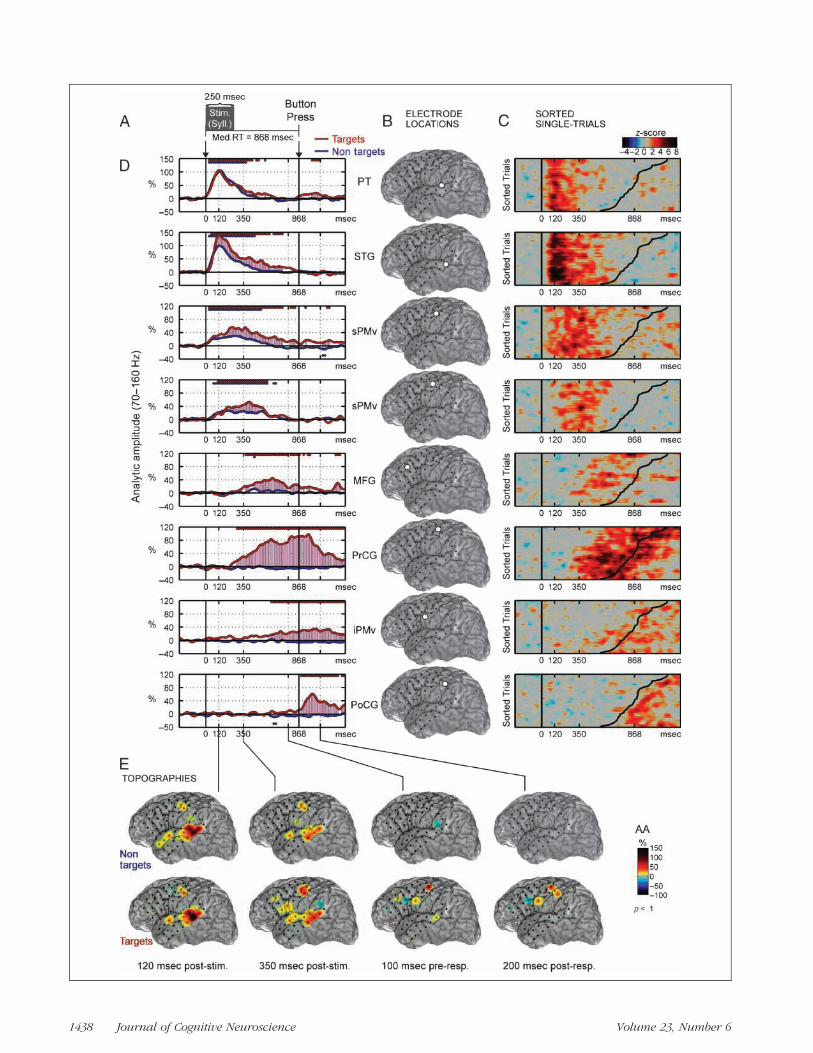
Figure 1. Phonological target detection for Subject 1. (A) The patient heard syllable stimuli (median duration 250 msec) and responded with abutton press (median RT 618 msec) to a prespecified target stimulus (e.g., /u/ ). (B) Locations of eight selected electrodes on the patientʼs MRIreconstruction (see Methods). (C) Single-trial HG analytic amplitude (AA) is shown in z-score units and sorted according to RT. RT is indicated bythe curved black line in each plot (RT range 528–1079 msec). Single-trial activity was smoothed with a three-trial moving window average. (D) HG(70–160 Hz) AA averaged across trials after realigning to the median RT. Straight lines above (below) the traces indicate significant increases(decreases) of HG AA relative to prestimulus baseline ( p < .02; FDR corrected for multiple comparisons). Red traces are for target trials and bluetraces are for nontarget trials. (E) Topographies of HG AA are shown for several latencies taken from D. Snapshots of a movie showing the temporalevolution of activation topography (see Supplementary Materials for complete temporal sequence in movie: http://bil.ucsf.edu/targetdetection).Activations are masked for significance at p < .1. HG = high gamma; AA = analytic amplitude; PT = planum temporale (secondary auditory cortex);STG = superior temporal gyrus; sPMv = superior ventral premotor cortex; MFG = middle frontal gyrus (in prefrontal cortex); iPMv = inferior ventralpremotor cortex; PrCG = precentral gyrus (motor); PoCG = postcentral gyrus (somatosensory).
Chang et al. 1439

power across all frequency bands indicating a transientartifact. The raw signal was also checked by visual inspec-tion, and additional rejections were added manually usingEEGLAB (Delorme & Makeig, 2004).
Time–frequency analyses used a Gaussian filter bank andthe Hilbert transform on each channel separately (Edwardset al., 2005). There is one Gaussian filter (Gaussian-shapedwindow in the frequency domain) in the filter bank for eachof 42 center frequencies (cfs) from 4 to 250 Hz. For each cf,the result is a time series of analytic amplitude (AA) of thesame sampling rate (2003 Hz), duration (∼7min), and units(mV) as the original ECoG recording. The AA is a formalmeans of obtaining the envelope of a signal. The envelopesof the frequency bands from 70 to 160 Hz are averaged to-gether to form the HG AA used in all results shown. Al-though the full HG band ranges from ∼60 to 300 Hz, onlythe range from 70 to 160 Hz was used in order to avoidelectrical artifacts at 60 and 180 Hz, and because this isthe frequency range of maximal response. All single-trialdata (Figure 1D is shown in units of z-score, calculatedby subtracting the baseline mean and dividing by the base-line standard deviation). All other results (Figure 1E) areshown in units of percentage (%) changes from the base-linemean. The baseline for all analyses was−250 to 0msecprestimulus.
Event-related averages ofHGAA are taken relative to stim-ulus and response onsets. In order to show the sensory-locked and motor-locked behavior on one time line, thesingle trials are realigned to the median RT prior to aver-aging. The realignment procedure only affects the epochfrom 500 msec poststimulus to 250 msec preresponse,which is essentially expanded/contracted to match themedian RT. The data before this interval are average-locked to the stimulus, and the data after are average-locked to the response.
To assess if a change in AA is significantly different frombaseline, we used a bootstrap resampling method. Signifi-cant latencies are indicated directly on the plots (Figure 1D)by lines over or under the trace. To assess if AA changesare significantly different between conditions, we used apermutation test and significant latencies are indicatedon the plots (Figure 1D) with pink lines between the twoconditions. Raw p values are corrected for multiple com-parisons using the false discovery rate (FDR) approach(Benjamini & Hochberg, 1995), and these corrected p val-ues are used in all cases. Full details for statistical analysesare included in the Supplemental Materials posted on-lineat http://bil.ucsf.edu/targetdetection.
Coregistration
Localization of implanted electrodes was conducted usinga photograph–MRI–radiograph coregistration technique,described previously (Dalal et al., 2008). A high-resolutionT1 MRI scan was obtained preoperatively for each patientas part of the clinical evaluation of epilepsy. 3-DMRI recon-struction was made using BET2 (www.fmrib.ox.ac.uk /
analysis/research/bet) and MRIcro (www.sph.sc.edu/comd/rorden/mricro.html). Localizations were addition-ally constrained by the requirement to be on the corticalsurface and by the known 1-cm interelectrode spacing.MNI coordinates were obtained for all electrodes usingSPM2. Known control points are used to compute projec-tive transforms that link the different image sets to refinethe location of manually registered visible electrodes. Thefinal result is a set of electrode positions on the patientʼsrendered MRI yielding locations relative to sulcal and gyrallandmarks on individual anatomy.
RESULTS
All four subjects performed the phonological target detec-tion task (Figure 1A) with >97% accuracy, and the medianRT across all subjects was 674 msec (SD ±254 msec).
Time Course of Cortical Activation
The time series of the single-trial and average analytic am-plitude (envelope) of HG for electrodes with significant ac-tivation are shown in Figure 1C and D, respectively. HGfirst robustly increased with short latency in the STG afterstimulus onset (onset 55 msec, peak 120 msec). Event-related HG activity in the STGwas characterized by a sharprise after stimulus onset, and typically had a slow return tobaseline over hundreds of milliseconds.In the dorsal aspect of the STG, overlying the Sylvian fis-
sure, and directly adjacent to the lateral planum temporale(PT), HG activation was equal in response to target andnontarget sounds. In contrast, in the ventral aspect of theSTG, near the superior temporal sulcus, HG activationwas modestly greater in response to target (red) comparedto nontarget (blue) stimuli. In Figure 1D, the significantlydifferent time points in HG activation between target andnontarget conditions are shown by the pink shading.Soon after the onset of STG activation, HG was found
in the precentral gyrus corresponding to superior ventralpremotor cortex (sPMv) (onset 100–110 msec, peak 120–200 msec). Unlike pSTG activation, which peaked quickly,the sPMv response had amore symmetric temporal distribu-tion (i.e., less kurtosis compared to the pSTG responses),suggesting that activation there was more likely inducedthan stimulus evoked. Similar to the STG, greater activa-tion was observed in the sPMv during target comparedto nontarget trials.The same sPMv sites were also activated during articula-
tion of phonemes (e.g., /a /, /pa/, / i / ), confirming the func-tional colocalization to a speech motor area. Duringarticulation, the onset of sPMv activation was observedprior to vocalization, and then followed by activation ofthe STG, representing auditory feedback (Figure 2).For all subjects, the remaining cortical event-related
HG activations occurred only during target trials. Corticalsites over lateral PFC demonstrated a significant increasein HG with delayed latencies (onset 180–320 msec, peak
1440 Journal of Cognitive Neuroscience Volume 23, Number 6

300–600 msec). HG activation was observed only duringresponses to target stimuli. PFC response had durationfrom 100 to 500 msec and engaged dorso- and ventrolat-eral subareas. Inspection of single trials (Figure 1C) sortedby RT revealed that PFC sites were more temporallyaligned with the response than stimulus. The delayed tim-ing and target-selective responsiveness of PFC representsthe decision processes and initiation of motor planningand selection necessary for the button press.Next, in the two subjects with adequate posterior cover-
age, HG was observed in the inferior parietal lobe (IPL)(onset 400–500 msec, peak 100–30 msec before buttonpress response) (Figure 3; one other subject is providedin the Supplemental Materials on-line at http://bil.ucsf.edu/targetdetection). The latencies of peak activationwere better locked to the button press than for the speechstimulus in the single-trial plots.The next site of activation was on the precentral gyrus
(PrCG), which was located one electrode dorsal to speechmotor cortex (onset was 200–300 msec before button press,peak 100 msec after button press; Figure 1B and D). Thesesites were hand movement-related, as determined by low-threshold (3 mA) responses evoked by bipolar electricalcortical stimulation used for clinical mapping. Finally, athumb somatosensory responsewas recorded over the post-central gyrus (PoCG) (onset 10 msec, peak 120–180 msecafter button press). In addition, a separate inferior ventralpremotor cortex (iPMv) site was observed during boththe pre- and postresponse periods, (i.e., overlapping boththe motor and sensory parts of the response).
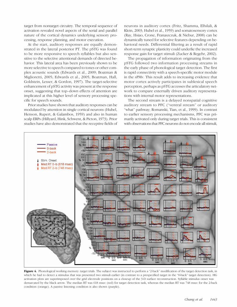
Working Memory Condition
One subject was able to carry out a working memory taskin which she was instructed to respond when the (target)syllable was the same as the one presented two trials pre-viously (also called a 2-back procedure). This condition re-quires continuous updating and activemanipulation of thetarget representation, in contrast to our previous target de-
tection task in which the target stimulus was prespecified(0-back) and subject to rapid and easy template match-ing. The stimulus sequence (pseudorandompresentation)and the behavioral response (button press) were thesame across both conditions, and only the task rules werechanged.
In Figure 4, the HG time series for all electrodes wassuperimposed over the positions of the grid electrodes.The extent of coverage for this array spans from ventrolat-eral PFC to the inferior parietal area, although it is mainlycentered over the lower half of the central sulcus and thelength of the Sylvian fissure. The RTs during the 2-back con-dition were significantly ( p < .001) longer than those dur-ing the 0-back condition [median RT for 2-back (orange) =748 msec, 0-back (red) = 618 msec]. In PFC, the 2-backtarget responses had increased amplitude and peak la-tency in the electrodes that showed a response to 0-backtargets. Seven PFC electrodes not active in the 0-backshowed selective activation in response to the 2-back tar-gets. Responses in the iPMv and PoCG had longer latencybut the same amplitude in the 2-back condition comparedto the 0-back condition. These findings suggest a strongdependency of PFC in working memory.
A passive listening condition (purple) was also added todetermine the attentional changes related to the detectiontasks. In both the pSTG and sPMv, active behavioral condi-tions (0-back and 2-back) had greater activation comparedto the passive condition, suggesting a general attentionaleffect on sensory processing that was not task specific inthose areas. Nontarget trials are shown in Figure 5.
DISCUSSION
We have demonstrated the spatio-temporal sequence ofhuman cortical activation during phonological target de-tection. We tracked real-time neural activity across a largecortical network in the left hemisphere for a simple behav-ioral task that is typically completed in less than 1 sec—while functionally dissecting the underlying physiology of
Figure 2. Self-paced syllablearticulation. HG activationoccurs before vocalization inthe sPMv and then followedby STG activation (auditoryfeedback). Plotting conventionsare the same as in Figure 1.
Chang et al. 1441

Figure 3. Target detection results for Subject 3 (GP7). Note that the grid placement is more posterior in this patient relative to Subject 1, withimproved coverage of the inferior parietal lobe (IPL).
1442 Journal of Cognitive Neuroscience Volume 23, Number 6
target from nontarget circuitry. The temporal sequence ofactivation revealed novel aspects of the serial and parallelnature of the cortical dynamics underlying sensory pro-cessing, response planning, and motor execution.At the start, auditory responses are equally demon-
strated in the lateral posterior PT. The pSTG was foundto be more responsive to speech syllables but also sen-sitive to the selective attentional demands of directed be-havior. This lateral area has been previously shown to bemore selective to speech compared to tones or other com-plex acoustic sounds (Edwards et al., 2009; Boatman &Miglioretti, 2005; Edwards et al., 2005; Boatman, Hall,Goldstein, Lesser, & Gordon, 1997). The target-selectiveenhancement of pSTG activity was present at the responseonset, suggesting that top–down effects of attention areimplicated at this higher level of sensory processing spe-cific for speech sounds.Prior studies have shown that auditory responses can be
modulated by attention in single cortical neurons (Hubel,Henson, Rupert, & Galambos, 1959) and also in humanscalp ERPs (Hillyard, Hink, Schwent, & Picton, 1973). Priorstudies have also demonstrated that the receptive fields of
neurons in auditory cortex (Fritz, Shamma, Elhilali, &Klein, 2003; Hubel et al., 1959) and somatosensory cortex(Ray, Hsiao, Crone, Franaszczuk, & Niebur, 2008) can bedynamically tuned for selective features depending on be-havioral needs. Differential filtering as a result of rapidshort-term synaptic plasticity could underlie the increasedresponse gain for target stimuli (Zucker & Regehr, 2002).
The propagation of information originating from thepSTG followed two information processing streams inthe early phase of phonological target detection. The firstis rapid connectivity with a speech-specific motor modulein the sPMv. This result adds to increasing evidence thatmotor cortex actively participates in sublexical speechperception, perhaps as pSTG accesses the articulatory net-work to compare externally driven auditory representa-tions with internal motor representations.
The second stream is a delayed nonspatial cognitiveauditory stream to PFC (“ventral stream” or auditory“what” pathway; Romanski, Tian, et al., 1999). In contrastto earlier sensory processing mechanisms, PFC was pri-marily activated only during target trials. This is consistentwith observations that PFCneurons do not encode all stimuli,
Figure 4. Phonological working memory: target trials. The subject was instructed to perform a “2-back” modification of the target detection task, inwhich he had to detect a stimulus that was presented two stimuli earlier (in contrast to a prespecified target in the “0-back” target detection). HGactivation plots are superimposed over the grid electrode positions on a closeup of the 3-D surface reconstruction. Syllable stimulus onset wasdemarcated by the black arrow. The median RT was 618 msec (red) for target detection task, whereas the median RT was 748 msec for the 2-backcondition (orange). A passive listening condition is also shown (purple).
Chang et al. 1443

but rather process the decision or outcome in auditory(Russ, Orr, & Cohen, 2008), visual (Freedman, Riesenhuber,Poggio, & Miller, 2003; Kim & Shadlen, 1999; Rainer,Asaad, & Miller, 1998), and somatosensory detection tasks(de Lafuente & Romo, 2005). PFC activity was further in-creased and more sites were recruited when a 2-backworking memory condition was added, which was notthe case for the early sensory and later motor areas.
This load-dependent enhancement could be interpretedto represent executive processing, storage, or rehearsalfunctions involved in verbal working memory. We didnot observe the sustained PFC neuronal activity that hasbeen described as a memory trace in visual tasks (Cohenet al., 1997; Fuster & Alexander, 1971), although no suchanalog in auditory or verbal processing has previouslybeen demonstrated. PFC changes were also unlikely tosupport subvocal rehearsal functions as the premotorareas for articulation were not implicated at that delayedphase of processing and were not task-dependent.
The results support the hypothesis that PFC is activelyinvolved in the “behavioral” aspects of processing thegoals and rules of a given task as well as initiating the out-putmechanisms needed toperforma given task (DʼEsposito,2007; Miller & Cohen, 2001). Taken together, our findingsare supportive of single-unit findings in nonhuman primates
demonstrating that PFC is adaptively responsive to relevantchanges in the environment. Indeed, a hallmark PFC dam-age is increased distractability, resulting in inappropriateupdating of environmental events (Chao & Knight, 1998).A recent study using both fMRI and diffusion tensor
imaging has provided evidence for anatomic structures sup-porting a dorsal-based articulatory pathway and a ventral-based comprehension or cognition pathway for speech.The dorsal connections from the pSTG to the sPMv arelikely mediated through the arcuate and superior longitu-dinal fasciculus, whereas the ventral connections from thepSTG to ventrolateral PFC occur via the extreme capsule(Saur et al., 2008; Romanski, Tian, et al., 1999). Prior stud-ies in nonhuman primates, also using physiologically de-fined rostral auditory belt sites in the pSTG, have shownspecific anterograde tracer connections to PFC areas 12(inferior convexity) and 45 (part of Brocaʼs area) (Romanski,Bates, & Goldman-Rakic, 1999; Romanski, Tian, et al., 1999).These findings delineate new aspects of planning and
execution in humanmotor cortical processing. In this task,hand use in the button press was associated with PFC andIPL activation atmiddle-to-late latencies. This was followedby motor cortex activity and then somatosensory cortexactivity, both related to hand movement and locked tothe timing of the button press response. Furthermore,
Figure 5. Phonological working memory: nontarget trials during 2-back condition (same format and plotting convention for Figure 4).
1444 Journal of Cognitive Neuroscience Volume 23, Number 6

we observed separate hand-related iPMv activity thatspanned the motor and sensory phases of the responses.This area has not been well characterized in humans, butby analogous location and functional properties, it appearsto correlate to F5 in nonhuman primates (Rizzolatti &Luppino, 2001). iPMv receives dense input from prefrontaland parietal cortices, and has direct output to primary mo-tor cortex. This region appears to have a supervisory rolein planning behavioral responses and monitoring the con-sequences of the outcomes.Target detection has been extensively associated
with the scalp EEG-derived P300 event-related potential(Polich, 2007; Sutton, Braren, Zubin, & John, 1965). De-spite the simplicity of the task and reliability of the P300,it is still unclear how and where it is produced. One ques-tion in the P300 literature is whether the various compo-nents (P3a, P3b, etc.) are to be considered more sensory,cognitive, or response-related. We show activations re-lated to all three stages expressed over space and time,demonstrating that target detection is not a unitary cogni-tive event. For example, much of the target-detection ac-tivation appears related to response preparation, and thisis observed in regions (peri-Rolandic cortices) that makesense for motor preparation. These findings are explora-tory, but they do provide a reliable contribution to thespatio-temporal imaging of target detection, which is aclassic and fundamental part of many, perhaps most, psy-chological and cognitive experimental designs.During nontarget conditions, only the sensory-evoked
potentials (i.e., N100) are obtain after signal averaging.During target conditions, attentional resources are allo-cated to the target such that a P300 potential is generatedin addition to the sensory-evoked potentials. It is hypothe-sized to index attentional resources, such that when taskconditions are undemanding, amplitude is enhanced andpeak latency is shortened. From our single-trial results, itcan be seen why response averaging during easy task con-ditions leads to a high-amplitude, short-latency P300 as theactivity is well time-locked. In contrast, as shown by our2-back findings, more difficult tasks engage more frontalareas and peak at longer latencies as there is greater vari-ability in the timing of cognitive events. However, it is diffi-cult to further speculate because the P300 integrates muchlarger sources at the level of the scalp and we were unableto perform simultaneous EEG and ECoG recordings due totechnical constraints related to head bandages.A distinct experimental advantage of ECoG is the ability
to record from multiple sites simultaneously in real-time,in contrast to some of the sampling limitations of single-unit recordings and the temporal constraints of fMRI.Nonetheless, ECoG in this experiment also had specificlimitations. First, the extent of grid coverage in humanswas guided by the clinical indications for their epilepsy lo-calization. In some cases, the standard grid did not coverboth the frontal and parietal areas. Tasks such as target de-tection also likely involve more brain areas, including sitesin the nondominant hemisphere, medial frontal cortex,
hippocampus, and cerebellum. Second, the electrode con-tacts are limited to the gyral cortical surface, and therefore,do not effectively sample the intrasulcal and subcorticalareas of potential interest. Third, the electrode contactsthemselves are likely representing the population re-sponses of thousands of neurons, thus it is difficult to ex-trapolate more spatially discrete processing occurring incortical microcircuits with the currently available gridelectrodes.
Despite these limitations, in this article, we focused onHG oscillations to track spatio-temporal pattern of activa-tion related to target detection. The exact sources for HGoscillations are actively under investigation, although re-cent studies have demonstrated a close correspondenceto interneuron multiunit activity recorded at mid-laminardepths (Ray et al., 2008; Steinschneider, Fishman,&Arezzo,2008). Several reports have observed that HG tracks audi-tory (Crone et al., 2001), motor (Miller et al., 2007), and lan-guage (Canolty et al., 2007) processing, and interacts withfunctional brain rhythms such as theta. Here we extendthese results to the higher-order cognitive processes oc-curring in multiple cortical areas during target detection.
Our findings confirm that HG can effectively track thecascade of activations relevant for a host of cognitive op-erations and reveal an orderly extraction of sensory infor-mation, decision-making, and behavioral output in humancortex.
Acknowledgments
This research was supported by NIH grants NS21135, PO4813(R. T. K.), F32NS061552, K99NS065120 (E. C.), F32NS061616(E. E.), and F31DC004855 (S. S. D.).
Reprint requests should be sent to Edward F. Chang, Departmentof Neurological Surgery, University of California, San Francisco,505 Parnassus Avenue, M779, San Francisco, CA 94143, or viae-mail: [email protected].
REFERENCES
Benjamini, Y., & Hochberg, Y. (1995). Controlling the FalseDiscovery Rate: A practical and powerful approach tomultiple testing. Journal of the Royal Statistical Society:Series B, Methodological, 57, 289–300.
Boatman, D., Hall, C., Goldstein, M. H., Lesser, R., & Gordon, B.(1997). Neuroperceptual differences in consonant and voweldiscrimination: As revealed by direct cortical electricalinterference. Cortex, 33, 83–98.
Boatman, D. F., & Miglioretti, D. L. (2005). Cortical sites criticalfor speech discrimination in normal and impaired listeners.Journal of Neuroscience, 25, 5475–5480.
Canolty, R. T., Soltani, M., Dalal, S. S., Edwards, E., Dronkers,N. F., Nagarajan, S. S., et al. (2007). Spatiotemporal dynamicsof word processing in the human brain. Frontiers inNeuroscience, 1, 185–196.
Chao, L. L., & Knight, R. T. (1998). Contribution of humanprefrontal cortex to delay performance. Journal of CognitiveNeuroscience, 10, 167–177.
Cohen, J. D., Perlstein, W. M., Braver, T. S., Nystrom, L. E., Noll,D. C., Jonides, J., et al. (1997). Temporal dynamics of brain
Chang et al. 1445

activation during a working memory task. Nature, 386,604–608.
Crone, N. E., Boatman, D., Gordon, B., & Hao, L. (2001).Induced electrocorticographic gamma activity duringauditory perception. Brazier Award-winning article. ClinicalNeurophysiology, 112, 565–582.
Crone, N. E., Miglioretti, D. L., Gordon, B., & Lesser, R. P.(1998). Functional mapping of human sensorimotor cortexwith electrocorticographic spectral analysis: II. Event-relatedsynchronization in the gamma band. Brain, 121, 2301–2315.
Dalal, S. S., Edwards, E., Kirsch, H. E., Barbaro, N. M., Knight,R. T., & Nagarajan, S. S. (2008). Localization of neurosurgicallyimplanted electrodes via photograph–MRI-radiographcoregistration. Journal of Neuroscience Methods, 174,106–115.
de Lafuente, V., & Romo, R. (2005). Neuronal correlates ofsubjective sensory experience. Nature Neuroscience, 8,1698–1703.
Delorme, A., & Makeig, S. (2004). EEGLAB: An open sourcetoolbox for analysis of single-trial EEG dynamics includingindependent component analysis. Journal of NeuroscienceMethods, 134, 9–21.
DʼEsposito, M. (2007). From cognitive to neural models ofworking memory. Philosophical Transactions of the RoyalSociety of London, Series B, Biological Sciences, 362,761–772.
Edwards, E., Soltani, M., Deouell, L. Y., Berger, M. S., & Knight,R. T. (2005). High gamma activity in response to deviantauditory stimuli recorded directly from human cortex.Journal of Neurophysiology, 94, 4269–4280.
Edwards, E., Soltani, M., Kim, W., Dalal, S. S., Nagarajan, S. S.,Berger, M. S., et al. (2009). Comparison of time–frequencyresponses and the event-related potential to auditory speechstimuli in human cortex. Journal of Neurophysiology, 102,377–386.
Freedman, D. J., Riesenhuber, M., Poggio, T., & Miller, E. K.(2003). A comparison of primate prefrontal and inferiortemporal cortices during visual categorization. Journal ofNeuroscience, 23, 5235–5246.
Fritz, J., Shamma, S., Elhilali, M., & Klein, D. (2003). Rapid task-related plasticity of spectrotemporal receptive fields inprimary auditory cortex. Nature Neuroscience, 6, 1216–1223.
Fuster, J. M., & Alexander, G. E. (1971). Neuron activity relatedto short-term memory. Science, 173, 652–654.
Halgren, E., Baudena, P., Clarke, J. M., Heit, G., Liegeois, C.,Chauvel, P., et al. (1995). Intracerebral potentials to raretarget and distractor auditory and visual stimuli: I. Superiortemporal plane and parietal lobe. Electroencephalographyand Clinical Neurophysiology, 94, 191–220.
Hillyard, S. A., Hink, R. F., Schwent, V. L., & Picton, T. W. (1973).Electrical signs of selective attention in the human brain.Science, 182, 177–180.
Hubel, D. H., Henson, C. O., Rupert, A., & Galambos, R. (1959).Attention units in the auditory cortex. Science, 129,1279–1280.
Kim, J. N., & Shadlen, M. N. (1999). Neural correlates of adecision in the dorsolateral prefrontal cortex of the macaque.Nature Neuroscience, 2, 176–185.
Miller, B. T., & DʼEsposito, M. (2005). Searching for “the top” intop–down control. Neuron, 48, 535–538.
Miller, E. K., & Cohen, J. D. (2001). An integrative theory ofprefrontal cortex function. Annual Review of Neuroscience,24, 167–202.
Miller, E. K., & Wilson, M. A. (2008). All my circuits: Usingmultiple electrodes to understand functioning neuralnetworks. Neuron, 60, 483–488.
Miller, K. J., denNijs, M., Shenoy, P., Miller, J. W., Rao, R. P.,& Ojemann, J. G. (2007). Real-time functional brainmapping using electrocorticography. Neuroimage, 37,504–507.
Polich, J. (2007). Updating P300: An integrative theory of P3aand P3b. Clinical Neurophysiology, 118, 2128–2148.
Rainer, G., Asaad, W. F., & Miller, E. K. (1998). Selectiverepresentation of relevant information by neurons in theprimate prefrontal cortex. Nature, 393, 577–579.
Ray, S., Hsiao, S. S., Crone, N. E., Franaszczuk, P. J., & Niebur, E.(2008). Effect of stimulus intensity on the spike-local fieldpotential relationship in the secondary somatosensorycortex. Journal of Neuroscience, 28, 7334–7343.
Rizzolatti, G., & Luppino, G. (2001). The cortical motor system.Neuron, 31, 889–901.
Romanski, L. M., Bates, J. F., & Goldman-Rakic, P. S. (1999).Auditory belt and parabelt projections to the prefrontalcortex in the rhesus monkey. Journal of ComparativeNeurology, 403, 141–157.
Romanski, L. M., Tian, B., Fritz, J., Mishkin, M., Goldman-Rakic,P. S., & Rauschecker, J. P. (1999). Dual streams of auditoryafferents target multiple domains in the primate prefrontalcortex. Nature Neuroscience, 2, 1131–1136.
Russ, B. E., Orr, L. E., & Cohen, Y. E. (2008). Prefrontal neuronspredict choices during an auditory same–different task.Current Biology, 18, 1483–1488.
Saur, D., Kreher, B. W., Schnell, S., Kummerer, D., Kellmeyer,P., Vry, M. S., et al. (2008). Ventral and dorsal pathways forlanguage. Proceedings of the National Academy of Sciences,U.S.A., 105, 18035–18040.
Sinai, A., Bowers, C. W., Crainiceanu, C. M., Boatman, D.,Gordon, B., Lesser, R. P., et al. (2005). Electrocorticographichigh gamma activity versus electrical cortical stimulationmapping of naming. Brain, 128, 1556–1570.
Steinschneider, M., Fishman, Y. I., & Arezzo, J. C. (2008).Spectrotemporal analysis of evoked and inducedelectroencephalographic responses in primary auditorycortex (A1) of the awake monkey. Cerebral Cortex, 18,610–625.
Sutton, S., Braren, M., Zubin, J., & John, E. R. (1965). Evoked-potential correlates of stimulus uncertainty. Science, 150,1187–1188.
Zucker, R. S., & Regehr, W. G. (2002). Short-term synapticplasticity. Annual Review of Physiology, 64, 355–405.
1446 Journal of Cognitive Neuroscience Volume 23, Number 6