Copper Modulates the Differentiation of Mouse Hematopoietic Progenitor Cells in Culture
12
Cell Transplantation, Vol. 18, pp. 887–897, 2009 0963-6897/09 $90.00 + .00 Printed in the USA. All rights reserved. DOI: 10.3727/096368909X471152 Copyright 2009 Cognizant Comm. Corp. E-ISSN 1555-3892 www.cognizantcommunication.com Copper Modulates the Differentiation of Mouse Hematopoietic Progenitor Cells in Culture Xiaosong Huang,* 1 L. Jeanne Pierce,† Paul A. Cobine,† 2 Dennis R. Winge,† and Gerald J. Spangrude*† *Department of Pathology, School of Medicine, University of Utah, Salt Lake City, UT, USA †Department of Medicine, Division of Hematology, School of Medicine, University of Utah, Salt Lake City, UT, USA Copper chelation has been shown to favor the expansion of human hematopoietic stem/progenitor cells in vitro. To further understand the effects of copper modulation on defined subsets of stem cells versus progeni- tor cells, we extended the studies in a mouse system. We isolated mouse hematopoietic stem cells (HSCs) or hematopoietic progenitor cells (HPCs) and cultured them with or without the copper chelator tetraethylen- epentamine (TEPA) or CuCl 2 . Cytokine-stimulated HPC cultures treated with TEPA for 7 days generated about two to three times more total and erythroid colony-forming cells (CFCs) compared to control cultures. In contrast, CuCl 2 treatment decreased the CFC numbers. Similar results were seen with HSC after 14, but not 7, days of culture. Transplant studies showed that HPCs cultured for 7 days in TEPA had about twofold higher short-term erythroid repopulation potential compared to control cultures, while CuCl 2 decreased the erythroid potential of cultured HPCs compared to control cultures. HSCs cultured with TEPA for 7 days did not exhibit significantly higher repopulation potential in either leukocyte or erythrocyte lineages compared to control cultures in short-term or long-term assays. Based on JC-1 staining, the mitochondrial membrane potential of HPCs cultured with TEPA was lower relative to control cultures. Our data suggest that decreas- ing the cellular copper content with TEPA results in preferential expansion or maintenance of HPC that are biased for erythroid differentiation in vivo, but does not enhance the maintenance of HSC activity in culture. Key words: Hematopoietic stem cells; Progenitor cells; Adult stem cells; Bone marrow; Erythropoiesis INTRODUCTION HSCs. This process is tightly regulated by the interplay of extrinsic and intrinsic molecular mechanisms in order to both sustain blood production and maintain the HSC Metal elements, such as iron (Fe), calcium (Ca), mag- nesium (Mg), zinc (Zn), and copper (Cu), are fundamen- compartment (28). In vitro studies showed that addition of Cu salts aug- tal and essential for life. Cu is a cofactor for many cellu- lar enzymes, such as cytochrome c oxidase, superoxide mented the retinoic acid-induced differentiation of the human myeloid leukemia cell line, HL-60 (1). Lowering dismutase, tyrosinase, peptidylglycine alpha-amidating mono-oxygenase, and lysyl oxidase (17,43,51). Cu has of the cellular Cu level by the Cu chelator tetraethylene- pentamine (TEPA) blocked 1,25-dihydroxyvitamin D 3 also been shown to regulate gene expression, cellular pro- tein functions, and cell differentiation (3,9,15,23,26,29, or phorbol 12-myristate 13-acetate-induced U937 cell differentiation and this blocking was reversed by adding 38,52). However, excess intracellular Cu is toxic due to its role in catalyzing the generation of reactive oxygen back Cu (22). The importance of Cu in regulating hema- topoiesis is also inferred from inherited or acquired Cu species (ROS) through Fenton chemistry (31). Specifi- cally, Cu is essential for normal hematopoiesis in which deficiency in humans and mice. Cu deficiency due to genetic mutations or malnourishment in humans causes mature blood cells are constantly replenished from he- matopoietic stem cells in the bone marrow. In adult anemia, neutropenia, and thrombocytopenia due to the arrested differentiation at the hematopoietic progenitor mammals, hematopoietic stem cells (HSCs) gradually lose their self-renewal capability and multipotency and cell level (6,13,14,16,19,36,44,46,58). Interestingly, recent studies showed that the high- restrict to specific lineages. HSC must replicate through self-renewing cell divisions to maintain the pool of affinity copper chelator TEPA, together with early act- Received September 11, 2008; final acceptance March 2, 2009. Online prepub date: April 15, 2009. 1 Current address: Department of Pediatrics, University of Rochester Medical Center, Rochester, NY, USA. 2 Current address: Department of Microbiology, Auburn University, Auburn, AL, USA. Address correspondence to Gerald J. Spangrude, Ph.D., Division of Hematology, University of Utah, RM 5C402, 30 N 1900 East, Salt Lake City, UT 84132-2408, USA. Tel: (801) 585 5544; Fax: (801) 585 3778; E-mail: [email protected] 887
-
Upload
hopkinsmedicine -
Category
Documents
-
view
0 -
download
0
Transcript of Copper Modulates the Differentiation of Mouse Hematopoietic Progenitor Cells in Culture
Cell Transplantation, Vol. 18, pp. 887–897, 2009 0963-6897/09 $90.00 + .00Printed in the USA. All rights reserved. DOI: 10.3727/096368909X471152Copyright 2009 Cognizant Comm. Corp. E-ISSN 1555-3892
www.cognizantcommunication.com
Copper Modulates the Differentiation of Mouse HematopoieticProgenitor Cells in Culture
Xiaosong Huang,*1 L. Jeanne Pierce,† Paul A. Cobine,†2 Dennis R. Winge,† and Gerald J. Spangrude*†
*Department of Pathology, School of Medicine, University of Utah, Salt Lake City, UT, USA†Department of Medicine, Division of Hematology, School of Medicine, University of Utah, Salt Lake City, UT, USA
Copper chelation has been shown to favor the expansion of human hematopoietic stem/progenitor cells invitro. To further understand the effects of copper modulation on defined subsets of stem cells versus progeni-tor cells, we extended the studies in a mouse system. We isolated mouse hematopoietic stem cells (HSCs)or hematopoietic progenitor cells (HPCs) and cultured them with or without the copper chelator tetraethylen-epentamine (TEPA) or CuCl2. Cytokine-stimulated HPC cultures treated with TEPA for 7 days generatedabout two to three times more total and erythroid colony-forming cells (CFCs) compared to control cultures.In contrast, CuCl2 treatment decreased the CFC numbers. Similar results were seen with HSC after 14, butnot 7, days of culture. Transplant studies showed that HPCs cultured for 7 days in TEPA had about twofoldhigher short-term erythroid repopulation potential compared to control cultures, while CuCl2 decreased theerythroid potential of cultured HPCs compared to control cultures. HSCs cultured with TEPA for 7 days didnot exhibit significantly higher repopulation potential in either leukocyte or erythrocyte lineages comparedto control cultures in short-term or long-term assays. Based on JC-1 staining, the mitochondrial membranepotential of HPCs cultured with TEPA was lower relative to control cultures. Our data suggest that decreas-ing the cellular copper content with TEPA results in preferential expansion or maintenance of HPC that arebiased for erythroid differentiation in vivo, but does not enhance the maintenance of HSC activity in culture.
Key words: Hematopoietic stem cells; Progenitor cells; Adult stem cells; Bone marrow; Erythropoiesis
INTRODUCTION HSCs. This process is tightly regulated by the interplayof extrinsic and intrinsic molecular mechanisms in orderto both sustain blood production and maintain the HSCMetal elements, such as iron (Fe), calcium (Ca), mag-
nesium (Mg), zinc (Zn), and copper (Cu), are fundamen- compartment (28).In vitro studies showed that addition of Cu salts aug-tal and essential for life. Cu is a cofactor for many cellu-
lar enzymes, such as cytochrome c oxidase, superoxide mented the retinoic acid-induced differentiation of thehuman myeloid leukemia cell line, HL-60 (1). Loweringdismutase, tyrosinase, peptidylglycine alpha-amidating
mono-oxygenase, and lysyl oxidase (17,43,51). Cu has of the cellular Cu level by the Cu chelator tetraethylene-pentamine (TEPA) blocked 1,25-dihydroxyvitamin D3also been shown to regulate gene expression, cellular pro-
tein functions, and cell differentiation (3,9,15,23,26,29, or phorbol 12-myristate 13-acetate-induced U937 celldifferentiation and this blocking was reversed by adding38,52). However, excess intracellular Cu is toxic due to
its role in catalyzing the generation of reactive oxygen back Cu (22). The importance of Cu in regulating hema-topoiesis is also inferred from inherited or acquired Cuspecies (ROS) through Fenton chemistry (31). Specifi-
cally, Cu is essential for normal hematopoiesis in which deficiency in humans and mice. Cu deficiency due togenetic mutations or malnourishment in humans causesmature blood cells are constantly replenished from he-
matopoietic stem cells in the bone marrow. In adult anemia, neutropenia, and thrombocytopenia due to thearrested differentiation at the hematopoietic progenitormammals, hematopoietic stem cells (HSCs) gradually
lose their self-renewal capability and multipotency and cell level (6,13,14,16,19,36,44,46,58).Interestingly, recent studies showed that the high-restrict to specific lineages. HSC must replicate through
self-renewing cell divisions to maintain the pool of affinity copper chelator TEPA, together with early act-
Received September 11, 2008; final acceptance March 2, 2009. Online prepub date: April 15, 2009.1Current address: Department of Pediatrics, University of Rochester Medical Center, Rochester, NY, USA.2Current address: Department of Microbiology, Auburn University, Auburn, AL, USA.Address correspondence to Gerald J. Spangrude, Ph.D., Division of Hematology, University of Utah, RM 5C402, 30 N 1900 East, Salt Lake City,UT 84132-2408, USA. Tel: (801) 585 5544; Fax: (801) 585 3778; E-mail: [email protected]
887

888 HUANG ET AL.
ing cytokines [thrombopoietin (TPO), stem cell factor within the cultured cells. We used in vitro colony-form-ing assays and in vivo mouse competitive repopulation(SCF), interleukin-6 (IL-6), and Flt3 ligand (FL)] can
promote the expansion of both hematopoietic progenitor assays to characterize and quantify the progenitor andstem cells within the cultures.cells (HPCs) and hematopoietic stem cells (HSCs) from
human cord blood-derived CD34+ cells in culture Our results show that TEPA promotes the expansionor maintenance of progenitor cells that are mainly ery-(39,41). The expansion of HPCs was shown by in vitro
colony-forming assay and FACS analysis of the expres- throid lineage biased within the HPC culture. In con-trast, TEPA did not significantly promote the expansionsion of cell surface markers. The expansion of the HSCs
was shown by the repopulation of nonobese diabetic/ or maintenance of HSC in culture. The effects of TEPAon HPC correlate with its effect of reducing the mito-severe combined immunodeficiency (NOD/SCID) mice
with cultured cells (40,42). This finding has justified a chondrial membrane potential of cultured HPC.clinical trial using TEPA together with cytokines for the
MATERIALS AND METHODSex vivo expansion of human HSC/HPCs prior to trans-Miceplantation into patients (11,42). Although the expansion
of HPCs is clearly established by both the in vitro and Mice carrying the Thy-1.1 and Ly-5.1 alleles on ain vivo assays, the expansion of HSCs in these cultures C57BL background were generated in our breeding col-is less convincing. ony by mating the BKa.AK-Thy1a/Ka and B6.SJL-Ptprca
There are still many controversies about the faithful- Pep3b/BoyJ strains and selecting for cosegregation ofness of the NOD/SCID mouse model in detection and Thy-1.1 (CD90a) and Ly-5.1 (CD45a). Breeding pairs ofquantification of human HSCs (7). Because the differ- B6.Cg-Gpi1a Hbbd H1b/DehJ mice (18) were kindly pro-ences of the physiological environment of mouse and vided by David Harrison (Jackson Laboratory, Bar Har-human and the differences of the life span and prolifera- bor, ME, USA). We refer to these mice as B6-Hbbd.tion capacities of mouse and human hematopoietic cells, GFP transgenic mice were provided by Dr. Masaruthe detection of human cells in NOD/SCID mouse bone Okabe (Osaka University, Osaka, Japan). This trans-marrow 8 weeks after the transplantation does not prove genic strain was generated by pronuclear injection ofthe contribution from the human HSCs (20). As a result, fertilized eggs obtained from C57BL/6 matings as pre-it is unclear whether TEPA significantly promotes the viously described (35) and are here referred to as B6-self-renewal and expansion of HSCs in vitro. GFP. Mice carrying both GFP transgene and Hbbd allele
The objective of the present study was to clarify these were generated in our breeding colony by mating B6-issues by extending the observations in the mouse sys- Hbbd mice with B6-GFP mice and selected for cosegreg-tem, where isolation and unequivocal assays for HSC ation of GFP and Hbbd. We refer to these mice as B6-and HPC are better defined. To separate mouse HSCs GFP/Hbbd. All animals were maintained in the animalfrom HPCs for analysis of the effect of Cu modulation resources center at the University of Utah under proto-on each of these two populations, we used cell surface cols approved by the institutional animal care and usemarkers in combination with the mitochondrial dye rho- committee.damine-123 to fractionate mouse bone marrow cells.
AntibodiesThe most primitive HSC and HPC reside in a populationof bone marrow cells that does not express mature blood Monoclonal antibodies against CD2 (Rm2.2), CD3cell surface markers (lineage negative or Lin−) and ex- (KT3-1.1), CD5 (53-7.3), CD8 (53-6.7), CD11b (M1/presses Sca-1 and c-Kit cell surface markers. These bone 70), Ly-6G (RB6-8C5), TER119, CD45R (B220; RA3-marrow cells are commonly referred to as the LSK pop- 6B2), and CD19 (1D3) were purified from media of cul-ulation (37,49). We further divide the LSK population tured hybridoma cell lines. Phycoerythrin (PE)-conju-by rhodamine-123 staining. Rhodamine-123 (Rho) is a gated monoclonal antibodies to Sca-1 and CD19 weremitochondrial dye that stains cells based on their state of purchased from PharMingen (San Diego, CA, USA).activation, in which the more metabolically active cells Anti-c-kit mAb 3C11 and anti-Ly5.1 mAb A20.1 werefluoresce brightly while more quiescent cells fluoresce purified and conjugated to Alexa Fluor 647 in our labo-dimly (2). Segregation of the LSK population using Rho ratory. PE-conjugated monoclonal antibodies to CD4,has demonstrated that the LSKRholow population con- CD8, B220, Mac-1, and Gr-1 were purchased fromtains long-term HSC with a frequency of about 25%, eBioscience (San Diego, CA, USA) or purified and con-while the LSKRhohi population lacks HSC but is highly jugated in our laboratory.enriched for multipotent and lineage-restricted progeni-
Cytokinestor cells (32,50). We cultured these two populations sep-arately with early acting cytokines (SCF, TPO, IL-6, FL) Recombinant mouse SCF was expressed in bacteria
and purified from lysates as previously described (55).and used TEPA or CuCl2 to modulate the Cu content

COPPER MODULATES PROGENITOR CELL DIFFERENTIATION 889
Recombinant mouse FL, IL-3, GM-CSF, and IL-6 were lar Cu content of cultured cells was determined usinginductively coupled plasma-atomic emission spectrome-purchased from Peprotech (Rocky Hill, NJ, USA). The
peptide mimetic of TPO was synthesized as previously try (ICP-AES) (Perkin Elmer Life Sciences) as de-scribed previously (30).described (8). Recombinant human erythropoietin (EPO)
was purchased from Ortho Pharm. Corp. (Raritan, NJ,Colony-Forming Cell (CFC) AssayUSA).
Cells were plated in α-MEM-based methylcelluloseIsolation of Bone Marrow Hematopoietic containing 10% fetal bovine serum (FBS), 10% deion-Stem/Progenitor Cells (HSC/HPCs) ized bovine serum albumen (BSA), antibiotics, L-gluta-
mine, and 2-mercaptoethanol. Colony growth and differ-Mouse bone marrow cells were incubated in a cock-tail of antibodies to CD2, CD3, CD5, CD8, CD11b, Ly- entiation were stimulated with recombinant cytokines in
combination as indicated, including SCF (100 ng/ml),6G, TER119, CD45R, and CD19. Lineage depletion wasperformed by two successive incubations in sheep anti- G-CSF (10 ng/ml), IL-3 (10 ng/ml), IL-6 (20 ng/ml),
TPO mimetic (5 nM), and Epo (4 U/ml). After 7 daysrat Ig-coupled magnetic beads (Dynal, Oslo, Norway).Lineage-negative (Lin−) cells were incubated with 0.2 of culture, quadruplicate plates were stained using ben-
zidine hydrochloride and scored for total colonies andµM rhodomine-123 (Molecular Probes, Eugene, OR,USA) in Hank’s balanced salt solution (HBSS) contain- for colonies containing hemoglobinized erythroid lin-
eage cells colonies based on benzidine staining.ing 5% fetal calf serum at 37°C for 20 min. After wash-ing once, the cells were resuspended in fresh HBSS and
Mouse Transplantation Assayincubated at 37°C for another 20 min to allow for effluxof the dye. After washing, the cells were stained with Radiation was delivered to B6-Hbbd or B6-GFP/Hbbd
recipient mice in a split dose (2 × 6 Gy) with a 3-h inter-PE-conjugated anti-Sca-1 and AlexaFluor 647-conjugatedanti-c-kit antibodies. Sorting was done using FACS val between doses, using a Shepherd Mark I 137Cs source
(JL Shepherd and Associates, Glendale, CA, USA) atVantage or FACS Aria instruments (Becton DickinsonImmunocytometry Systems, San Jose, CA, USA). We a dose rate of 0.8 Gy/min. Cultured cells mixed with
competitor BM cells were transplanted by the retro-sorted Lin−Sca-1+c-kit+ (LSK) bone marrow cells asLSKRholow and LSKRhohi populations using Rho-stained orbital route under isoflurane anesthesia (IsoSol; Vedco
Inc., St. Joseph, MO, USA). Peripheral blood samplesbone marrow cells to determine the placement of theRho gates. This usually resulted in LSKRholow cells were collected from the retro-orbital sinus under isoflur-
ane anesthesia delivered using the E-Z Anesthesia sys-comprising the lower 20% of the Rho distribution andLSKRhohi cells comprising the upper 50% of the Rho tem (Euthanex Corp., Palmer, PA, USA). Blood was
collected in heparinized capillary tubes and mixed withdistribution. An aliquot of each sorted cell populationwas taken for reanalysis to evaluate purity and cell acid citrate dextrose at a 10:1 ratio prior to determina-
tion of the complete blood count using a Serono Systemcount.9010+ CP hematology counter (Serono Diagnostics, Al-
In Vitro Liquid Culture of HSCs and HPCs lentown, PA, USA). Samples were then mixed with anequal volume of 2% Dextran T500 (Amersham Biosci-Purified LSKRholow and LSKRhohi cells were cultured
in 24-well plates at 1000 cells/ml in α-MEM medium ences, Piscataway, NJ, USA) in phosphate-buffered sa-line (PBS) and incubated at 37°C for 30 min. The uppersupplemented with 10% fetal bovine serum (FBS)
(Hyclone), 2 mM L-glutamine, 100 µM 2-mercaptoetha- layer, containing leukocytes, platelets, and residualerythrocytes, was collected for flow cytometry analysis,nol, 1% each sodium pyruvate and pen/strep stock solu-
tions (Invitrogen Corp, Carlsbad, CA, USA), and the whereas the sedimented erythrocytes were used for Hbbanalysis.following mouse recombinant cytokines: SCF at 100 ng/
ml, TPO mimetic at 5 nM, IL-6 at 10 ng/ml, and FL atDetermination of Hbb Variants by HPLC10 ng/ml. Cellular Cu content was modulated by supple-
menting the culture with either TEPA and/or CuCl2 A cation exchange protocol was developed to sepa-rate and quantitate Hbb variant alleles by HPLC (48). A(both obtained from Sigma-Aldrich, St. Louis, MO,
USA) as indicated. At weekly intervals, cell cultures stock solution of 100 mM 5,5′-dithiobis-(2-nitrobenzoicacid) (DTNB) (Sigma-Aldrich) was prepared by dissolv-were demi-depopulated and supplemented with fresh
medium, serum, cytokines, and other components as in- ing 10 mg of DTNB in 250 µl of dimethyl sulfoxide andstored at −20°C. Erythrocytes obtained from heparinizeddicated. The cultures were incubated at 37°C in a hu-
midified atmosphere of 5% CO2 in air. At various time blood were washed three times in dextrose-gelatin-vero-nal buffer and stored as packed pellets at 4°C for up topoints, cells were harvested and counted by hemocytom-
eter following staining with trypan blue. The intracellu- 2 weeks prior to analysis. Samples were derivatized by
890 HUANG ET AL.
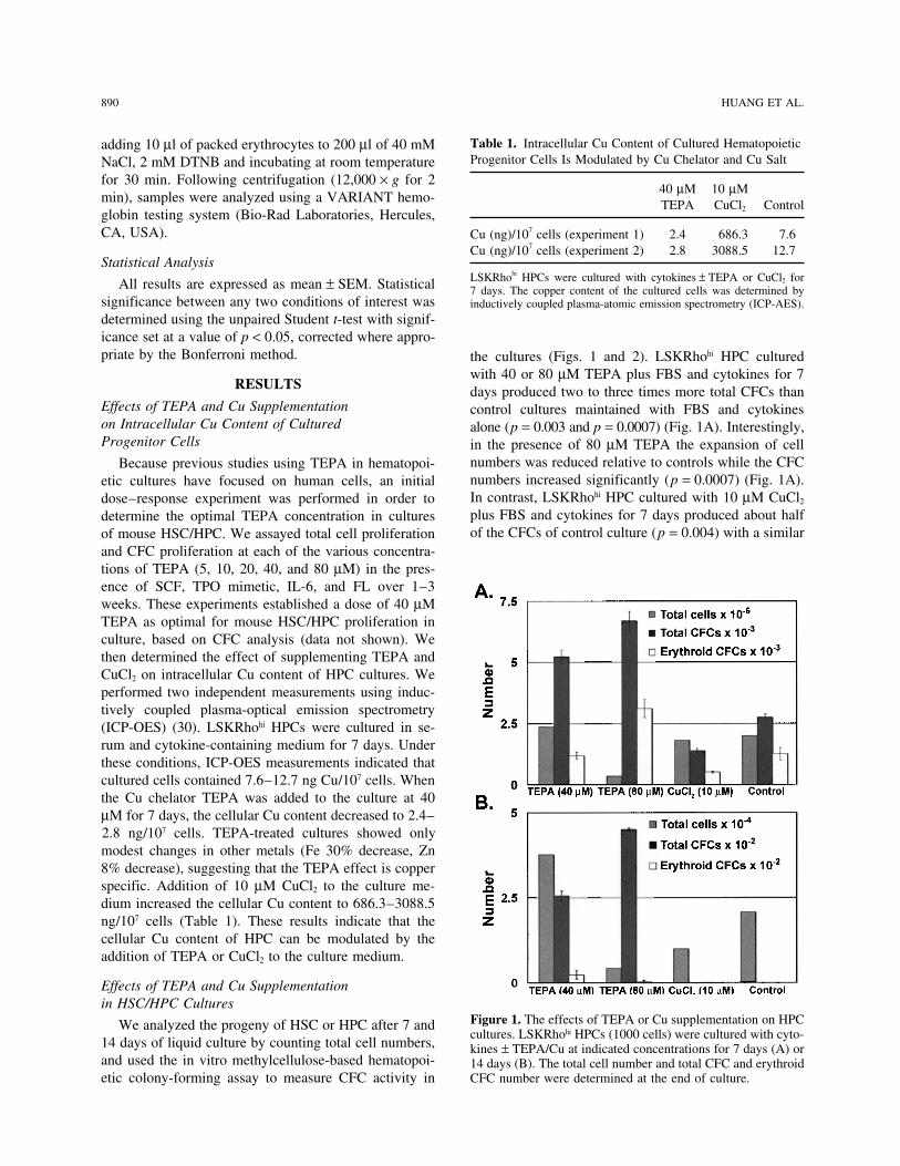
Table 1. Intracellular Cu Content of Cultured Hematopoieticadding 10 µl of packed erythrocytes to 200 µl of 40 mMProgenitor Cells Is Modulated by Cu Chelator and Cu SaltNaCl, 2 mM DTNB and incubating at room temperature
for 30 min. Following centrifugation (12,000 × g for 240 µM 10 µM
min), samples were analyzed using a VARIANT hemo- TEPA CuCl2 Controlglobin testing system (Bio-Rad Laboratories, Hercules,CA, USA). Cu (ng)/107 cells (experiment 1) 2.4 686.3 7.6
Cu (ng)/107 cells (experiment 2) 2.8 3088.5 12.7Statistical Analysis
LSKRhohi HPCs were cultured with cytokines ± TEPA or CuCl2 forAll results are expressed as mean ± SEM. Statistical 7 days. The copper content of the cultured cells was determined bysignificance between any two conditions of interest was inductively coupled plasma-atomic emission spectrometry (ICP-AES).determined using the unpaired Student t-test with signif-icance set at a value of p < 0.05, corrected where appro-priate by the Bonferroni method. the cultures (Figs. 1 and 2). LSKRhohi HPC cultured
with 40 or 80 µM TEPA plus FBS and cytokines for 7RESULTS days produced two to three times more total CFCs than
Effects of TEPA and Cu Supplementation control cultures maintained with FBS and cytokineson Intracellular Cu Content of Cultured alone (p = 0.003 and p = 0.0007) (Fig. 1A). Interestingly,Progenitor Cells in the presence of 80 µM TEPA the expansion of cell
numbers was reduced relative to controls while the CFCBecause previous studies using TEPA in hematopoi-numbers increased significantly (p = 0.0007) (Fig. 1A).etic cultures have focused on human cells, an initialIn contrast, LSKRhohi HPC cultured with 10 µM CuCl2dose–response experiment was performed in order toplus FBS and cytokines for 7 days produced about halfdetermine the optimal TEPA concentration in culturesof the CFCs of control culture (p = 0.004) with a similarof mouse HSC/HPC. We assayed total cell proliferation
and CFC proliferation at each of the various concentra-tions of TEPA (5, 10, 20, 40, and 80 µM) in the pres-ence of SCF, TPO mimetic, IL-6, and FL over 1–3weeks. These experiments established a dose of 40 µMTEPA as optimal for mouse HSC/HPC proliferation inculture, based on CFC analysis (data not shown). Wethen determined the effect of supplementing TEPA andCuCl2 on intracellular Cu content of HPC cultures. Weperformed two independent measurements using induc-tively coupled plasma-optical emission spectrometry(ICP-OES) (30). LSKRhohi HPCs were cultured in se-rum and cytokine-containing medium for 7 days. Underthese conditions, ICP-OES measurements indicated thatcultured cells contained 7.6–12.7 ng Cu/107 cells. Whenthe Cu chelator TEPA was added to the culture at 40µM for 7 days, the cellular Cu content decreased to 2.4–2.8 ng/107 cells. TEPA-treated cultures showed onlymodest changes in other metals (Fe 30% decrease, Zn8% decrease), suggesting that the TEPA effect is copperspecific. Addition of 10 µM CuCl2 to the culture me-dium increased the cellular Cu content to 686.3–3088.5ng/107 cells (Table 1). These results indicate that thecellular Cu content of HPC can be modulated by theaddition of TEPA or CuCl2 to the culture medium.
Effects of TEPA and Cu Supplementationin HSC/HPC Cultures
Figure 1. The effects of TEPA or Cu supplementation on HPCWe analyzed the progeny of HSC or HPC after 7 andcultures. LSKRhohi HPCs (1000 cells) were cultured with cyto-
14 days of liquid culture by counting total cell numbers, kines ± TEPA/Cu at indicated concentrations for 7 days (A) orand used the in vitro methylcellulose-based hematopoi- 14 days (B). The total cell number and total CFC and erythroid
CFC number were determined at the end of culture.etic colony-forming assay to measure CFC activity in

COPPER MODULATES PROGENITOR CELL DIFFERENTIATION 891
entiation (Fig. 3A, B). Supplementation with 10 µMCuCl2 increased the portion of cells with differentiatedcell morphology, while adding TEPA to CuCl2-supple-mented culture reversed the effect of Cu (Fig. 3C, D).
Effects of TEPA and Cu Supplementationon In Vivo Short-Term Trilineage RepopulatingActivity of Cultured HPCs
To evaluate the in vivo repopulation activity of cul-tured cells in the three major blood lineages, we har-vested 1-week cultures of LSKRhohi HPCs and trans-planted these cells together with a fixed number ofcompetitor bone marrow cells into lethally irradiatedcongenic recipient mice. Cultured cells were trackedbased on expression of the Ly5.1 allele on the cell sur-face of white blood cells and the hemoglobin-β single(Hbbs) variant in red blood cells and lack of GFP expres-sion on platelets (48). Competitor bone marrow and re-cipient mice cells expressed the Ly5.2 allele, the hemo-globin-β diffuse (Hbbd) variant, and GFP. This transplantmodel allows us to track the in vivo contribution of cul-
Figure 2. The effects of TEPA or Cu supplementation on HSC tured cells into the leukocyte, platelet, and erythrocytecultures. LSKRholow HSCs (1000 cells) were cultured with cy-
lineages. We transplanted cultured cells obtained from atokines ± TEPA/Cu at indicated concentrations for 7 days (A)7-day culture of 1000 LSKRhohi cells, together with 105or 14 days (B). The total cell number and total CFC and ery-
throid CFC number were determined at the end of culture. freshly obtained competitor bone marrow cells into le-thally irradiated mice and followed the relative contribu-
expansion of cell numbers. TEPA at 80 µM also in-creased the erythroid CFC number compared to control(p = 0.04) while 40 µM had no effect on erythroid CFCs(Fig. 1A). After 14 days of culture, LSKRhhi HPCs haddecreased number of total cell numbers and CFC num-bers compared to day 7 cultures, but cultures supple-mented with 40 or 80 µM TEPA had much more totalCFC and erythroid CFC number than cultures supple-mented with CuCl2 or control cultures (Fig. 1B). In par-allel cultures initiated with LSKRholow HSC, we ob-served no difference in total CFC number between cellcultures with TEPA, CuCl2, and control in 1 week ofculture (Fig. 2A). Relative to control cultures, we ob-served an increase in erythroid CFC in cultures contain-ing 40 µM TEPA (p = 0.0001) and a decrease in ery-throid CFC in cultures containing supplementary Cu(Fig. 2A). After 2 weeks of culture, LSKRholow cells cul-tured with 40 µM TEPA generated about 30% more to-tal CFCs compared to control cultures containing cyto-kines only, while treatment with 10 µM CuCl2 decreasedthe total CFCs in culture compared to control (Fig. 2B).
We examined the morphology of the cells in cultures Figure 3. The effects of TEPA or Cu supplementation on themorphology of HPC cultures. LSKRhohi HPCs were culturedestablished from LSKRhohi HPCs by May-Grunwald-with cytokines ± TEPA/Cu at the indicated concentrations forGiemsa staining of cytospins. Cultures supplemented7 days. Cultured cells were cytospun into slides and stained
with 40 µM TEPA contained cells with a uniform and with May-Grunwald-Giemsa. The culture conditions for cellsblast-like morphology, while control cultures contained shown were: (A) control; (B) 40 µM TEPA; (C) 10 µM CuCl2;
(D) 40 µM TEPA + 10 µM CuCl2. Scale bar: 10 µm.more cells with morphological characteristics of differ-

892 HUANG ET AL.
tions of cultured cells and competitor BM cells into theleukocyte, platelet, and erythrocyte lineages in the pe-ripheral blood of recipient mice over time. LSKRhohi
cells cultured with 40 µM TEPA generated about two-fold higher percentage of reconstitution of the erythroidlineage compared to LSKRhohi cells cultured with cyto-kines alone, in spite of a lower degree of cellularity inthe cultures expanded under this condition (Fig. 4A, B).The red blood cell count of the recipient mice was simi-lar among the groups (data not shown). This enhance-ment of erythroid engraftment persisted over the timecourse of progenitor cell activity in the recipients, andwas limited to the erythroid lineage as these cultures hadabout 30% of the leukocyte lineage repopulation activityof LSKRhohi cells cultured with cytokines alone. Theeffect of TEPA was reversed by addition of CuCl2 intothe culture medium. TEPA and Cu had no obvious ef-fects on platelet lineage repopulation activity of culturedLSKRhohi cells (data not shown). The contribution ofcultured cells to the peripheral blood of recipient micewas limited to 9 weeks posttransplantation, consistentwith the absence of stem cells in the LSKRhohi popula-tion and its cultured progeny. Collectively, our resultssuggest that TEPA preferentially promotes the mainte-nance or expansion of erythroid-biased progenitor cellsin cultures established from primitive progenitor cells.
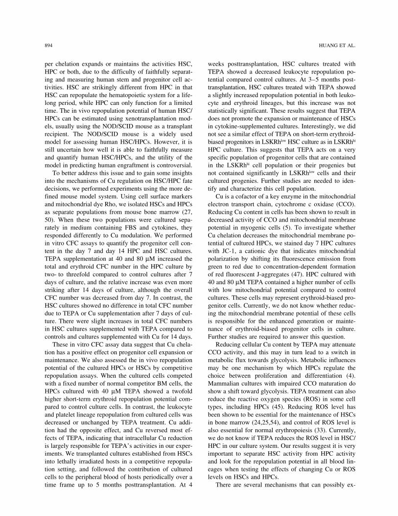
Effects of TEPA and CuCl2 Supplementationon the Short-Term and Long-Term RepopulationCapacity of Cultured HSCs
To evaluate the effects of TEPA on expansion ofHSC number and function, we tested the activity of cul-tured LSKRholow HSC in the long-term competitive re-population assay. The 7-day cultured cells derived from103 LSKRholow BM cells were cotransplanted with 106
fresh competitor BM cells into lethally irradiated recipi-ent mice. The relative contribution of cultured cells andcompetitor cells into peripheral blood leukocyte and
Figure 4. The effects of TEPA or Cu supplementation on theerythrocyte lineages were analyzed using Ly5 and Hbbshort-term erythrocyte and leukocyte repopulation potential ofvariants at 4, 8, 15, and 20 weeks posttransplantation.HPC cultures. LSKRhohi HPCs (1000 cells) were cultured with
At 4 weeks, LSKRhlow HSCs cultured with 40 µM TEPA cytokines ± TEPA/Cu at the indicated concentrations for 7had significantly less repopulation capacity in the leuko- days. The total cell numbers were determined at the end of
culture (A). The total cells were mixed with 105 whole BMcyte lineage than control cultures (p = 0.02) or culturescells and injected intravenously into lethally irradiated mice.supplemented with 10 µM CuCl2 (p = 0.002) (Fig. 5A).The percentage contribution of cultured cells to peripheralAt 15 weeks posttransplantation, there was no signifi-blood erythrocytes (B) and leukocytes (C) were determined at
cant difference of leukocyte and erythrocyte repopula- the various times posttransplantation.tion potential of cultured cells under different conditions(Fig. 5B). At 5 months posttransplantation, HSCs cul-tured with 40 µM TEPA showed a slight increase in the
Effects of TEPA and Cu Supplementationrepopulation in leukocyte and erythrocyte lineages, buton the Mitochondrial Membrane Potentialthis increase was not statistically significant (p = 0.38)of Cultured Progenitor Cells(Fig. 5A, C). Based on these data, we conclude that
TEPA does not significantly increase the maintenance To gain some insights into the mechanisms of TEPA’seffect on LSKRhohi progenitor cells and their progeniesor expansion of short-term or long-term HSCs in culture.
COPPER MODULATES PROGENITOR CELL DIFFERENTIATION 893
in culture, we measured the mitochondrial membranepotential of day 7 cultures of LSKRhohi cells. We usedthe cationic dye JC-1 that indicates increased mitochon-drial polarization by shifting its fluorescence emissionfrom green (525 nm) to red (575 nm). JC-1 staining ofLSKRhohi bone marrow cells cultured for 7 days showedthat 40 µM TEPA increased the number of cells thathave relatively low mitochondrial membrane potentialand decreased the number of cells that have relativelyhigh mitochondrial membrane potential compared tocontrol cultures containing only cytokines (Fig. 6). Theeffect of TEPA on mitochondrial membrane potential ofcultured cells was reversed by adding a low amount ofexogenous Cu into TEPA-supplemented cultures, sug-gesting reduction of Cu concentration within the cell isresponsible for the effect of TEPA (Fig. 6).
DISCUSSION
Copper has long been known to be essential for nor-mal hematopoiesis in mammals from both in vitro andin vivo studies. However, the effects of fine-tuning ofintracellular copper content on the regulation of HSC/HPCs self-renewal and differentiation are relatively un-known. Recently, some interesting findings suggestedthat reducing the intracellular Cu content by Cu chela-tion in human HSC/HPCs is beneficial for their expan-sion or maintenance in culture. While this finding isvery intriguing and promising, it is unclear whether cop-
Figure 5. The effect of TEPA or Cu supplementation on theshort-term and long-term repopulation potential of HSC cellcultures. LSKRholow HSC [1000 cells, equaling approximately100 repopulation unit (RU)] were cultured under the indicatedconditions for 7 days and then transplanted together with 106
competitor BM cells into lethally irradiated recipients. The pe-ripheral blood was sampled at various times and the contribu- Figure 6. The effect of TEPA or Cu supplementation on thetion of cultured cells to leukocyte and erythrocyte pools was mitochondrial membrane potential of cultured HPC. LSKRhohi
determined by flow cytometry and HPLC. RU number was cal- cells were cultured under the indicated conditions for 7 days,culated based on 1RU = 105 competitor BM cells. Results from stained with JC-1, and analyzed by flow cytometry. Fluores-three independent experiments are shown in (A), (B), and (C). cence emission at 525 nm indicates cells with low relative mi-
tochondrial membrane potential, while emission at 575 nm in-dicates high relative mitochondrial membrane potential. Thepercentages of cells in each quadrant are shown in the figure.
894 HUANG ET AL.
per chelation expands or maintains the activities HSC, weeks posttransplantation, HSC cultures treated withTEPA showed a decreased leukocyte repopulation po-HPC or both, due to the difficulty of faithfully separat-
ing and measuring human stem and progenitor cell ac- tential compared control cultures. At 3–5 months post-transplantation, HSC cultures treated with TEPA showedtivities. HSC are strikingly different from HPC in that
HSC can repopulate the hematopoietic system for a life- a slightly increased repopulation potential in both leuko-cyte and erythroid lineages, but this increase was notlong period, while HPC can only function for a limited
time. The in vivo repopulation potential of human HSC/ statistically significant. These results suggest that TEPAdoes not promote the expansion or maintenance of HSCsHPCs can be estimated using xenotransplantation mod-
els, usually using the NOD/SCID mouse as a transplant in cytokine-supplemented cultures. Interestingly, we didnot see a similar effect of TEPA on short-term erythroid-recipient. The NOD/SCID mouse is a widely used
model for assessing human HSC/HPCs. However, it is biased progenitors in LSKRhlow HSC culture as in LSKRhhi
HPC culture. This suggests that TEPA acts on a verystill uncertain how well it is able to faithfully measureand quantify human HSC/HPCs, and the utility of the specific population of progenitor cells that are contained
in the LSKRhhi cell population or their progenies butmodel in predicting human engraftment is controversial.To better address this issue and to gain some insights not contained significantly in LSKRhlow cells and their
cultured progenies. Further studies are needed to iden-into the mechanisms of Cu regulation on HSC/HPC fatedecisions, we performed experiments using the more de- tify and characterize this cell population.
Cu is a cofactor of a key enzyme in the mitochondrialfined mouse model system. Using cell surface markersand mitochondrial dye Rho, we isolated HSCs and HPCs electron transport chain, cytochrome c oxidase (CCO).
Reducing Cu content in cells has been shown to result inas separate populations from mouse bone marrow (27,50). When these two populations were cultured sepa- decreased activity of CCO and mitochondrial membrane
potential in myogenic cells (5). To investigate whetherrately in medium containing FBS and cytokines, theyresponded differently to Cu modulation. We performed Cu chelation decreases the mitochondrial membrane po-
tential of cultured HPCs, we stained day 7 HPC culturesin vitro CFC assays to quantify the progenitor cell con-tent in the day 7 and day 14 HPC and HSC cultures. with JC-1, a cationic dye that indicates mitochondrial
polarization by shifting its fluorescence emission fromTEPA supplementation at 40 and 80 µM increased thetotal and erythroid CFC number in the HPC culture by green to red due to concentration-dependent formation
of red fluorescent J-aggregates (47). HPC cultured withtwo- to threefold compared to control cultures after 7days of culture, and the relative increase was even more 40 and 80 µM TEPA contained a higher number of cells
with low mitochondrial potential compared to controlstriking after 14 days of culture, although the overallCFC number was decreased from day 7. In contrast, the cultures. These cells may represent erythroid-biased pro-
genitor cells. Currently, we do not know whether reduc-HSC cultures showed no difference in total CFC numberdue to TEPA or Cu supplementation after 7 days of cul- ing the mitochondrial membrane potential of these cells
is responsible for the enhanced generation or mainte-ture. There were slight increases in total CFC numbersin HSC cultures supplemented with TEPA compared to nance of erythroid-biased progenitor cells in culture.
Further studies are required to answer this question.controls and cultures supplemented with Cu for 14 days.These in vitro CFC assay data suggest that Cu chela- Reducing cellular Cu content by TEPA may attenuate
CCO activity, and this may in turn lead to a switch intion has a positive effect on progenitor cell expansion ormaintenance. We also assessed the in vivo repopulation metabolic flux towards glycolysis. Metabolic influences
may be one mechanism by which HPCs regulate thepotential of the cultured HPCs or HSCs by competitiverepopulation assays. When the cultured cells competed choice between proliferation and differentiation (4).
Mammalian cultures with impaired CCO maturation dowith a fixed number of normal competitor BM cells, theHPCs cultured with 40 µM TEPA showed a twofold show a shift toward glycolysis. TEPA treatment can also
reduce the reactive oxygen species (ROS) in some cellhigher short-term erythroid repopulation potential com-pared to control culture cells. In contrast, the leukocyte types, including HPCs (45). Reducing ROS level has
been shown to be essential for the maintenance of HSCsand platelet lineage repopulation from cultured cells wasdecreased or unchanged by TEPA treatment. Cu addi- in bone marrow (24,25,54), and control of ROS level is
also essential for normal erythropoiesis (33). Currently,tion had the opposite effect, and Cu reversed most ef-fects of TEPA, indicating that intracellular Cu reduction we do not know if TEPA reduces the ROS level in HSC/
HPC in our culture system. Our results suggest it is veryis largely responsible for TEPA’s activities in our exper-iments. We transplanted cultures established from HSCs important to separate HSC activity from HPC activity
and look for the repopulation potential in all blood lin-into lethally irradiated hosts in a competitive repopula-tion setting, and followed the contribution of cultured eages when testing the effects of changing Cu or ROS
levels on HSCs and HPCs.cells to the peripheral blood of hosts periodically over atime frame up to 5 months posttransplantation. At 4 There are several mechanisms that can possibly ex-

COPPER MODULATES PROGENITOR CELL DIFFERENTIATION 895
5. Chen, X.; Medeiros, D. M.; Jennings, D. Mitochondrialplain the effect of TEPA in hematopoietic cultures. Onemembrane potential is reduced in copper-deficient C2C12possibility is the increased survival of progenitor cells,cells in the absence of apoptosis. Biol. Trace Elem. Res.
especially erythroid progenitor cells in TEPA-supple- 106:51–64; 2005.mented culture. Secondly, the inhibition of differentia- 6. Cordano, A.; Placko, R. P.; Graham, G. G. Hypocupremia
and neutropenia in copper deficiency. Blood 28:280–283;tion of progenitor cells into mature blood cells in TEPA-1966.supplemented cultures may explain our results. TEPA
7. Coulombel, L. Identification of hematopoietic stem/pro-has been shown to have antidifferentiation effects ongenitor cells: Strength and drawbacks of functional assays.
some established hematopoietic progenitor cell lines, Oncogene 23:7210–7222; 2004.and whether it has similar effects on primary hematopoi- 8. Cwirla, S. E.; Balasubramanian, P.; Duffin, D. J.; Wagstrom,
C. R.; Gates, C. M.; Singer, S. C.; Davis, A. M.; Tansik,etic progenitor cells should be investigated. Third, in-R. L.; Mattheakis, L. C.; Boytos, C. M.; Schatz, P. J.;creased proliferation of erythroid progenitor cells in theBaccanari, D. P.; Wrighton, N. C.; Barrett, R. W.; Dower,TEPA-supplemented culture is possibly involved in theW. J. Peptide agonist of the thrombopoietin receptor as
selective increases in this lineage in both in vitro and in potent as the natural cytokine. Science 276:1696–1699;vivo settings. We are currently investigating whether 1997.
9. Dahl, S. L.; Rucker, R. B.; Niklason, L. E. Effects of cop-one or a combination of these mechanisms accounts forper and cross-linking on the extracellular matrix of tissue-TEPA’s effects on hematopoietic progenitor cells.engineered arteries. Cell Transplant. 14:367–374; 2005.Although we did not see a positive effect of TEPA
10. de Haan, G.; Weersing, E.; Dontje, B.; van Os, R.; Bystrykh,on HSC expansion or maintenance in our culture system, L. V.; Vellenga, E.; Miller, G. In vitro generation of long-we cannot rule out the possibility that TEPA may have term repopulating hematopoietic stem cells by fibroblast
growth factor-1. Dev. Cell 4:241–251; 2003.positive effects on stem cell maintenance and expansion11. de Lima, M.; McMannis, J.; Gee, A.; Komanduri, K.;in the context of other culture environments or with hu-
Couriel, D.; Andersson, B. S.; Hosing, C.; Khouri, I.;man cells. It will be very interesting to investigate theJones, R.; Champlin, R.; Karandish, S.; Sadeghi, T.;
effect of TEPA on HSCs together with supportive stro- Peled, T.; Grynspan, F.; Daniely, Y.; Nagler, A.; Shpall,mal cells (21,34). A variety of transcription factors and E. J. Transplantation of ex vivo expanded cord blood cells
using the copper chelator tetraethylenepentamine: A phasesignaling molecules, such as Wnt and Notch proteinsI//II clinical trial. Bone Marrow Transplant. 41:771–778;(12), fibroblast growth factor-1 (10), insulin-like growth2008.factor-2 (57), and angiopoietin-like proteins (56), have
12. Duncan, A. W.; Rattis, F. M.; DiMascio, L. N.; Congdon,been shown to promote the self-renewal of HSCs in cul- K. L.; Pazianos, G.; Zhao, C.; Yoon, K.; Cook, J. M.;ture, and inhibition of apoptosis using caspase inhibitors Willert, K.; Gaiano, N.; Reya, T. Integration of Notch and
Wnt signaling in hematopoietic stem cell maintenance.can help maintain HSC function in culture (53). TheNat. Immunol. 6:314–322; 2005.specific effect of TEPA on erythroid lineage progenitor
13. Fuhrman, M. P.; Herrmann, V.; Masidonski, P.; Eby, C.cells in cultures of HPCs is very intriguing; this effectPancytopenia after removal of copper from total parenteral
may be exploited in clinical situations where rapid ery- nutrition. J. Parenter. Enteral. Nutr. 24:361–366; 2000.throid repopulation is desired. Furthermore, it will be 14. Goyens, P.; Brasseur, D.; Cadranel, S. Copper deficiency
in infants with active celiac disease. J. Pediatr. Gastroent-interesting to study whether Cu modulation has similarerol. Nutr. 4:677–680; 1985.effects on the progenitor cells from other tissues.
15. Hainaut, P.; Rolley, N.; Davies, M.; Milner, J. ModulationACKNOWLEDGMENTS: This project was supported by fund- by copper of p53 conformation and sequence-specificing from a program project grant (P30DK072437) and by DNA binding: Role for Cu(II)/Cu(I) redox mechanism.individual grants to G.J.S. (R01DK057899) and D.R.W. Oncogene 10:27–32; 1995.(R01ES03817). Additional funding was provided by the Brian 16. Halfdanarson, T. R.; Kumar, N.; Li, C. Y.; Phyliky, R. L.;Rooney Fund of the Lymphoma Foundation. Hogan, W. J. Hematological manifestations of copper de-
ficiency: A retrospective review. Eur. J. Haematol. 80:REFERENCES 523–531; 2008.17. Harris, E. D. Cellular copper transport and metabolism.1. Bae, B.; Percival, S. S. Retinoic acid-induced HL-60 cell
differentiation is augmented by copper supplementation. Annu. Rev. Nutr. 20:291–310; 2000.18. Harrison, D. E.; Astle, C. M.; Lerner, C. Number and con-J. Nutr. 123:997–1002; 1993.
2. Bertoncello, I.; Hodgson, G. S.; Bradley, T. R. Multipara- tinuous proliferative pattern of transplanted primitive im-munohematopoietic stem cells. Proc. Natl. Acad. Sci.meter analysis of transplantable hemopoietic stem cells.
II. Stem cells of long-term bone marrow-reconstituted re- USA 85:822–826; 1988.19. Hirase, N.; Abe, Y.; Sadamura, S.; Yufu, Y.; Muta, K.;cipients. Exp. Hematol. 16:245–249; 1988.
3. Birkaya, B.; Aletta, J. M. NGF promotes copper accumu- Umemura, T.; Nishimura, J.; Nawata, H.; Ideguchi, H.Anemia and neutropenia in a case of copper deficiency:lation required for optimum neurite outgrowth and protein
methylation. J. Neurobiol. 63:49–61; 2005. Role of copper in normal hematopoiesis. Acta Haematol.87:195–197; 1992.4. Chen, C. T.; Shih, Y. R.; Kuo, T. K.; Lee, O. K.; Wei, Y.
H. Coordinated changes of mitochondrial biogenesis and 20. Horn, P. A.; Kiem, H. P. Expansion of SCID repopulatingcells does not prove expansion of hematopoietic stemantioxidant enzymes during osteogenic differentiation of
human mesenchymal stem cells. Stem Cells 26:960–968; cells. Blood 108:771–772; 2006.21. Huang, G. P.; Pan, Z. J.; Jia, B. B.; Zheng, Q.; Xie, C. G.;2008.

896 HUANG ET AL.
Gu, J. H.; McNiece, I. K.; Wang, J. F. Ex vivo expansion Miura, Y.; Suda, T. In vivo and in vitro stem cell functionof c-kit- and Sca-1-positive murine hematopoietic cells.and transplantation of hematopoietic stem/progenitor cells
supported by mesenchymal stem cells from human umbili- Blood 80:3044–3050; 1992.38. Ostrakhovitch, E. A.; Lordnejad, M. R.; Schliess, F.; Sies,cal cord blood. Cell Transplant. 16:579–585; 2007.
22. Huang, Z. L.; Failla, M. L.; Reeves, P. G. Differentiation H.; Klotz, L. O. Copper ions strongly activate the phos-phoinositide-3-kinase/Akt pathway independent of theof human U937 promonocytic cells is impaired by moder-
ate copper deficiency. Exp. Biol. Med. 226:222–228; 2001. generation of reactive oxygen species. Arch. Biochem.Biophys. 397:232–239; 2002.23. Iseki, A.; Kambe, F.; Okumura, K.; Hayakawa, T.; Seo,
H. Regulation of thyroid follicular cell function by intra- 39. Peled, T.; Glukhman, E.; Hasson, N.; Adi, S.; Assor, H.;Yudin, D.; Landor, C.; Mandel, J.; Landau, E.; Prus, E.;cellular redox-active copper. Endocrinology 141:4373–
4382; 2000. Nagler, A.; Fibach, E. Chelatable cellular copper modu-lates differentiation and self-renewal of cord blood-derived24. Ito, K.; Hirao, A.; Arai, F.; Matsuoka, S.; Takubo, K.;
Hamaguchi, I.; Nomiyama, K.; Hosokawa, K.; Sakurada, hematopoietic progenitor cells. Exp. Hematol. 33:1092–1100; 2005.K.; Nakagata, N.; Ikeda, Y.; Mak, T. W.; Suda, T. Regula-
tion of oxidative stress by ATM is required for self- 40. Peled, T.; Landau, E.; Mandel, J.; Glukhman, E.; Gouds-mid, N. R.; Nagler, A.; Fibach, E. Linear polyamine cop-renewal of haematopoietic stem cells. Nature 431:997–
1002; 2004. per chelator tetraethylenepentamine augments long-termex vivo expansion of cord blood-derived CD34+ cells and25. Ito, K.; Hirao, A.; Arai, F.; Takubo, K.; Matsuoka, S.;
Miyamoto, K.; Ohmura, M.; Naka, K.; Hosokawa, K.; increases their engraftment potential in NOD/SCID mice.Exp. Hematol. 32:547–555; 2004.Ikeda, Y.; Suda, T. Reactive oxygen species act through
p38 MAPK to limit the lifespan of hematopoietic stem 41. Peled, T.; Landau, E.; Prus, E.; Treves, A. J.; Nagler, A.;Fibach, E. Cellular copper content modulates differentia-cells. Nat. Med. 12:446–451; 2006.
26. Kang, J.; Lin, C.; Chen, J.; Liu, Q. Copper induces histone tion and self-renewal in cultures of cord blood-derivedCD34+ cells. Br. J. Haematol. 116:655–661; 2002.hypoacetylation through directly inhibiting histone acetyl-
transferase activity. Chem. Biol. Interact. 148:115–123; 42. Peled, T.; Mandel, J.; Goudsmid, R. N.; Landor, C.; Hasson,N.; Harati, D.; Austin, M.; Hasson, A.; Fibach, E.; Shpall,2004.
27. Kim, M.; Cooper, D. D.; Hayes, S. F.; Spangrude, G. J. E. J.; Nagler, A. Pre-clinical development of cord blood-derived progenitor cell graft expanded ex vivo with cyto-Rhodamine-123 staining in hematopoietic stem cells of
young mice indicates mitochondrial activation rather than kines and the polyamine copper chelator tetraethylenepen-tamine. Cytotherapy 6:344–355; 2004.dye efflux. Blood 91:4106–4117; 1998.
28. Kondo, M.; Wagers, A. J.; Manz, M. G.; Prohaska, S. S.; 43. Pena, M. M.; Lee, J.; Thiele, D. J. A delicate balance:homeostatic control of copper uptake and distribution. J.Scherer, D. C.; Beilhack, G. F.; Shizuru, J. A.; Weissman,
I. L. Biology of hematopoietic stem cells and progenitors: Nutr. 129:1251–1260; 1999.44. Porea, T. J.; Belmont, J. W.; Mahoney, Jr., D. H. Zinc-Implications for clinical application. Annu. Rev. Immunol.
21:759–806; 2003. induced anemia and neutropenia in an adolescent. J. Pedi-atr. 136:688–690; 2000.29. Kudrin, A. V. Trace elements in regulation of NF-kappaB
activity. J. Trace Elem. Med. Biol. 14:129–142; 2000. 45. Prus, E.; Fibach, E. The effect of the copper chelator te-traethylenepentamine on reactive oxygen species genera-30. Leary, S. C.; Cobine, P. A.; Kaufman, B. A.; Guercin,
G. H.; Mattman, A.; Palaty, J.; Lockitch, G.; Winge, D. R.; tion by human hematopoietic progenitor cells. Stem CellsDev. 16:1053–1056; 2007.Rustin, P.; Horvath, R.; Shoubridge, E. A. The human cy-
tochrome c oxidase assembly factors SCO1 and SCO2 46. Simon, S. R.; Branda, R. F.; Tindle, B. F.; Burns, S. L.Copper deficiency and sideroblastic anemia associatedhave regulatory roles in the maintenance of cellular cop-
per homeostasis. Cell Metab. 5:9–20; 2007. with zinc ingestion. Am. J. Hematol. 28:181–183; 1988.47. Smiley, S. T.; Reers, M.; Mottola-Hartshorn, C.; Lin, M.;31. Leary, S. C.; Winge, D. R. The Janus face of copper: its
expanding roles in biology and the pathophysiology of Chen, A.; Smith, T. W.; Steele, Jr., G. D.; Chen, L. B.Intracellular heterogeneity in mitochondrial membrane po-disease. Meeting on Copper and Related Metals in Biol-
ogy. EMBO Rep. 8:224–227; 2007. tentials revealed by a J-aggregate-forming lipophilic cat-ion JC-1. Proc. Natl. Acad. Sci. USA 88:3671–3675;32. Li, C. L.; Johnson, G. R. Rhodamine123 reveals heteroge-
neity within murine Lin−, Sca-1+ hemopoietic stem cells. 1991.48. Spangrude, G. J.; Cho, S.; Guedelhoefer, O.; Vanwoerkom,J. Exp. Med. 175:1443–1447; 1992.
33. Marinkovic, D.; Zhang, X.; Yalcin, S.; Luciano, J. P.; R. C.; Fleming, W. H. Mouse models of hematopoieticengraftment: Limitations of transgenic green fluorescentBrugnara, C.; Huber, T.; Ghaffari, S. Foxo3 is required
for the regulation of oxidative stress in erythropoiesis. J. protein strains and a high-performance liquid chromatog-raphy approach to analysis of erythroid chimerism. StemClin. Invest. 117:2133–2144; 2007.
34. Monga, S. P.; Tang, Y.; Candotti, F.; Rashid, A.; Wildner, Cells 24:2045–2051; 2006.49. Spangrude, G. J.; Heimfeld, S.; Weissman, I. L. Purifica-O.; Mishra, B.; Iqbal, S.; Mishra, L. Expansion of hepatic
and hematopoietic stem cells utilizing mouse embryonic tion and characterization of mouse hematopoietic stemcells. Science 241:58–62; 1988.liver explants. Cell Transplant. 10:81–89; 2001.
35. Nakanishi, T.; Kuroiwa, A.; Yamada, S.; Isotani, A.; Yama- 50. Spangrude, G. J.; Johnson, G. R. Resting and activatedsubsets of mouse multipotent hematopoietic stem cells.shita, A.; Tairaka, A.; Hayashi, T.; Takagi, T.; Ikawa, M.;
Matsuda, Y.; Okabe, M. FISH analysis of 142 EGFP Proc. Natl. Acad. Sci. USA 87:7433–7437; 1990.51. Uauy, R.; Olivares, M.; Gonzalez, M. Essentiality of cop-transgene integration sites into the mouse genome. Geno-
mics 80:564–574; 2002. per in humans. Am. J. Clin. Nutr. 67:952S–959S; 1998.52. Vanacore, R. M.; Eskew, J. D.; Morales, P. J.; Sung, L.;36. Naveh, Y.; Hazani, A.; Berant, M. Copper deficiency with
cow’s milk diet. Pediatrics 68:397–400; 1981. Smith, A. Role for copper in transient oxidation and nu-clear translocation of MTF-1, but not of NF-kappa B, by37. Okada, S.; Nakauchi, H.; Nagayoshi, K.; Nishikawa, S.;
COPPER MODULATES PROGENITOR CELL DIFFERENTIATION 897
the heme-hemopexin transport system. Antioxid. Redox of c-kit receptors in mast cells. J. Biol. Chem. 268:14189–14201; 1993.Signal. 2:739–752; 2000.
53. Wiesmann, A.; Searles, A. E.; Pierce, L. J.; Spangrude, 56. Zhang, C. C.; Kaba, M.; Ge, G.; Xie, K.; Tong, W.; Hug,C.; Lodish, H. F. Angiopoietin-like proteins stimulate exG. J. Effects of caspase inhibitors on hematopoietic en-
graftment after short-term culture. Cell Transplant. 11: vivo expansion of hematopoietic stem cells. Nat. Med. 12:240–245; 2006.351–358; 2002.
54. Yalcin, S.; Zhang, X.; Luciano, J. P.; Kumar Mungamuri, 57. Zhang, C. C.; Lodish, H. F. Insulin-like growth factor 2expressed in a novel fetal liver cell population is a growthS.; Marinkovic, D.; Vercherat, C.; Sarkar, A.; Grisotto,
M.; Taneja, R.; Ghaffari, S. Foxo3 is essential for the reg- factor for hematopoietic stem cells. Blood 103:2513–2521; 2004.ulation of ATM and oxidative stress-mediated homeosta-
sis of hematopoietic stem cells. J. Biol. Chem. 283: 58. Zidar, B. L.; Shadduck, R. K.; Zeigler, Z.; Winkelstein,A. Observations on the anemia and neutropenia of human25692–25705; 2008.
55. Yee, N. S.; Langen, H.; Besmer, P. Mechanism of kit li- copper deficiency. Am. J. Hematol. 3:177–185; 1977.gand, phorbol ester, and calcium-induced down-regulation