Copper complexes of glycyl-histidyl-lysine and two of its synthetic analogues: chemical behaviour...
12
Copper complexes of glycyl-histidyl-lysine and two of its synthetic analogues : chemical behaviour and biological activity Chiara Conato a , Riccardo Gavioli b , Remo Guerrini c , Henryk KozIowski d , Piotr MIynarz d , Claudia Pasti b , Fernando Pulidori a , Maurizio Remelli a ; * a Department of Chemistry, University of Ferrara, via L. Borsari 46, I-44100 Ferrara, Italy b Department of Biochemistry and Molecular Biology, University of Ferrara, via L. Borsari 46, I-44100 Ferrara, Italy c Department of Pharmaceutical Sciences, University of Ferrara, via Fossato di Mortara 17/19, I-44100 Ferrara, Italy d Faculty of Chemistry, University of WrocIaw, F. Joliot-Curie 14, 50-383 WrocIaw, Poland Received 5 January 2001; received in revised form 5 March 2001; accepted 8 March 2001 Abstract Copper complex formation equilibria of glycyl-L-histidyl-L-lysine (Gly-His-Lys, GHK) and of two synthetic analogues, where the histidine residue was replaced with a synthetic amino acid (L-spinacine or L-1,2,3,4-tetrahydro-isoquinoline-3-carboxylic acid), have been carefully investigated using different experimental techniques : potentiometry, solution calorimetry, UV-VIS spectrophotometry, circular dichroism and electron paramagnetic resonance spectroscopies. All the ligands formed complexes having different stoichiometries and stabilities ; evidence for the formation of binuclear species is also shown. The structures of the main complexes are discussed. It is suggested that the lateral lysine amino group participates in complex formation, but only at alkaline pH values: at physiological pH this group is protonated and available for possible interactions with cellular receptors. The above tripeptides have been tested for their enzymatic stability in human serum : the synthetic compounds showed no significant degradation for at least 3 h. Finally, their activity as growth factor has been studied in vitro. The two synthetic analogues showed an activity comparable to or even higher than that of GHK, thus suggesting their possible use as additives in cell culture media, even in the presence of serum. Relevant information on the GHK action mechanism as cell growth factor has been obtained: the formation of copper complexes, driven by the first (Gly) residue, appears necessary while the second residue (His) does not appear to play a specific role; the presence of the free side chain of the third residue (Lys) appears to be of fundamental importance. ß 2001 Elsevier Science B.V. All rights reserved. Keywords : Glycyl-histidyl-lysine ; Copper complex ; Cell growth 1. Introduction The natural tripeptide glycyl-L-histidyl-L-lysine (Gly- His-Lys, GHK) was ¢rst isolated by Pickart et al. [1] from human plasma, where it is present at a concentration of about 200 ng/ml. GHK was found in the albumin and K-globulin fractions and it co-isolated with an approxi- mately equimolar amount of copper and about 1/5 molar amount of iron [2]. GHK was ¢rst described as a liver cell growth factor [1,3,4]; later, it proved to stimulate the growth and di¡erentiation of a number of cell lines [5^9] and, in general, proved to be a good replacement for se- rum in cell culture media [10,11]. In further studies it was recognised that GHK is endowed with a wide range of biological activities [12] including : angiogenesis [13,14], acceleration of wound healing [15^17] and bone repair [18], inhibition of the growth of some cultured cells [12], stimulation of collagen synthesis by cells [19], superoxide dismutase-like activity [20], binding to heparin [21] and interaction with angiotensin II AT1 receptors [22]. GHK was found to act synergistically with copper and iron ions and it has been suggested that it plays a role in copper ion uptake into cells [2,23^26]. It has also been recognised as a metal ion carrier in biological £uids [24]. A GHK structure^activity study led to the hypothesis that the His-Lys sequence and Lys side chain are essential for its biological activity [27]. A mechanism of action has also been suggested [23] by which the ¢rst two residues of the tripeptide bind a copper ion to form a 1:1 complex while the Lys side chain is involved in the recognition of the 0304-4165 / 01 / $ ^ see front matter ß 2001 Elsevier Science B.V. All rights reserved. PII:S0304-4165(01)00127-1 * Corresponding author. Fax: +39-0532-240709; E-mail : [email protected] Biochimica et Biophysica Acta 1526 (2001) 199^210 www.bba-direct.com
Transcript of Copper complexes of glycyl-histidyl-lysine and two of its synthetic analogues: chemical behaviour...
Copper complexes of glycyl-histidyl-lysine and two of its syntheticanalogues: chemical behaviour and biological activity
Chiara Conato a, Riccardo Gavioli b, Remo Guerrini c, Henryk KozIowski d,Piotr MIynarz d, Claudia Pasti b, Fernando Pulidori a, Maurizio Remelli a;*
a Department of Chemistry, University of Ferrara, via L. Borsari 46, I-44100 Ferrara, Italyb Department of Biochemistry and Molecular Biology, University of Ferrara, via L. Borsari 46, I-44100 Ferrara, Italyc Department of Pharmaceutical Sciences, University of Ferrara, via Fossato di Mortara 17/19, I-44100 Ferrara, Italy
d Faculty of Chemistry, University of WrocIaw, F. Joliot-Curie 14, 50-383 WrocIaw, Poland
Received 5 January 2001; received in revised form 5 March 2001; accepted 8 March 2001
Abstract
Copper complex formation equilibria of glycyl-L-histidyl-L-lysine (Gly-His-Lys, GHK) and of two synthetic analogues, where thehistidine residue was replaced with a synthetic amino acid (L-spinacine or L-1,2,3,4-tetrahydro-isoquinoline-3-carboxylic acid), have beencarefully investigated using different experimental techniques: potentiometry, solution calorimetry, UV-VIS spectrophotometry, circulardichroism and electron paramagnetic resonance spectroscopies. All the ligands formed complexes having different stoichiometries andstabilities ; evidence for the formation of binuclear species is also shown. The structures of the main complexes are discussed. It is suggestedthat the lateral lysine amino group participates in complex formation, but only at alkaline pH values: at physiological pH this group isprotonated and available for possible interactions with cellular receptors. The above tripeptides have been tested for their enzymaticstability in human serum: the synthetic compounds showed no significant degradation for at least 3 h. Finally, their activity as growth factorhas been studied in vitro. The two synthetic analogues showed an activity comparable to or even higher than that of GHK, thus suggestingtheir possible use as additives in cell culture media, even in the presence of serum. Relevant information on the GHK action mechanism ascell growth factor has been obtained: the formation of copper complexes, driven by the first (Gly) residue, appears necessary while thesecond residue (His) does not appear to play a specific role; the presence of the free side chain of the third residue (Lys) appears to be offundamental importance. ß 2001 Elsevier Science B.V. All rights reserved.
Keywords: Glycyl-histidyl-lysine ; Copper complex; Cell growth
1. Introduction
The natural tripeptide glycyl-L-histidyl-L-lysine (Gly-His-Lys, GHK) was ¢rst isolated by Pickart et al. [1]from human plasma, where it is present at a concentrationof about 200 ng/ml. GHK was found in the albumin andK-globulin fractions and it co-isolated with an approxi-mately equimolar amount of copper and about 1/5 molaramount of iron [2]. GHK was ¢rst described as a liver cellgrowth factor [1,3,4] ; later, it proved to stimulate thegrowth and di¡erentiation of a number of cell lines [5^9]and, in general, proved to be a good replacement for se-rum in cell culture media [10,11]. In further studies it was
recognised that GHK is endowed with a wide range ofbiological activities [12] including: angiogenesis [13,14],acceleration of wound healing [15^17] and bone repair[18], inhibition of the growth of some cultured cells [12],stimulation of collagen synthesis by cells [19], superoxidedismutase-like activity [20], binding to heparin [21] andinteraction with angiotensin II AT1 receptors [22].
GHK was found to act synergistically with copper andiron ions and it has been suggested that it plays a role incopper ion uptake into cells [2,23^26]. It has also beenrecognised as a metal ion carrier in biological £uids [24].A GHK structure^activity study led to the hypothesis thatthe His-Lys sequence and Lys side chain are essential forits biological activity [27]. A mechanism of action has alsobeen suggested [23] by which the ¢rst two residues of thetripeptide bind a copper ion to form a 1:1 complex whilethe Lys side chain is involved in the recognition of the
0304-4165 / 01 / $ ^ see front matter ß 2001 Elsevier Science B.V. All rights reserved.PII: S 0 3 0 4 - 4 1 6 5 ( 0 1 ) 0 0 1 2 7 - 1
* Corresponding author. Fax: +39-0532-240709;E-mail : [email protected]
BBAGEN 25173 17-4-01
Biochimica et Biophysica Acta 1526 (2001) 199^210
www.bba-direct.com

hypothetical receptor on cell surface: this is the basis forthe copper uptake into cells and for most of GHK's bio-logical activities. A crystal X-ray di¡raction study of aGHK^Cu complex [23] showed the existence of a triden-tate binding structure involving the glycine N-terminalgroup, the nitrogen atom of the ¢rst amide bond andthe N atom of the histidine imidazole ring. Aqueous solu-tion studies of GHK complex formation equilibria withCu(II) [28^34], Zn(II) [30,35,36] or Pd(II) [37,38] ionsdemonstrated the formation of mono- and bis-complexes,as well as some minor binuclear species. The proposedstructure at neutral pH was in agreement with the crystal-lographic results [23]. It has also been suggested that theLys residue is not involved in complexation.
The presence of GHK in plasma (where it is quicklyhydrolysed) has yet to be thoroughly explained. However,GHK and other correlated small peptides showing angio-genesis activity in vivo were recently isolated from theproteolysis of SPARC, an extracellular matrix glycopro-tein expressed by a number of tissues during embryogen-esis and repair [39].
Simple oligopeptides, like GHK, able to modulate (stim-ulate or inhibit) cellular growth could be very importanttools in the biotechnological development of new drugswith potential applications in tissue regeneration or asanticancer agents. However, the low enzymatic stabilityof GHK in plasma, common to all natural peptides, limitsits extensive use. A synthetic analogue of GHK, in which abackbone modi¢cation of the His-Lys bond was per-formed via the retro^inverso technique, has been investi-gated [40]. This analogue proved more stable to enzymatichydrolysis than GHK; its e¡ects on dorsal skin woundhealing in guinea-pigs and ¢broblasts in culture werestudied as well.
In the present paper two new GHK analogues, Gly-Spi-Lys and Gly-Tic-Lys, have been synthesised by substitut-ing a non-natural amino acid, respectively, either L-4,5,6,7-tetrahydro-1H-imidazo[4,5-c]pyridine-6-carboxylic acid (L-spinacine, Spi) or L-1,2,3,4-tetrahydro-isoquinoline-3-car-
boxylic acid (Tic), for the His in the second position (seeFig. 1). This modi¢cation enabled us to investigate the roleof the His residue imidazole ring, which is either locked ina rigid structure (in Gly-Spi-Lys) or absent (in Gly-Tic-Lys). It is worth noting that the K-amino group of bothSpi and Tic is secondary (as in proline), making themwork as `break-points' [41^43] in oligopeptide co-ordina-tion to copper. A complete investigation on complex for-mation equilibria between the Cu(II) ion and these newligands has been performed in aqueous solution. Protona-tion and complex formation thermodynamic parametershave been potentiometrically and calorimetrically deter-mined; the complex formation model and species stoichi-ometry have been carefully checked by means of VIS ab-sorption, electron paramagnetic resonance (EPR) andcircular dichroism (CD) spectroscopies. The structure hy-potheses for the main complex species are discussed.Cu(II)/GHK equilibria have also been revisited for thesake of comparison: formation enthalpies of Cu(II)/GHK complexes have been determined for the ¢rst time.
Finally, the enzymatic stability of GHK and its ana-logues in human serum has been investigated and theirin vitro biological activity (as growth factors, in a se-rum-free medium) has been studied. The role each GHKresidue plays in determining its biological activity is dis-cussed.
2. Materials and methods
2.1. Reagents and instrumentation
GHK acetate was a high purity product (Sigma) andwas used without further puri¢cation. Gly-Spi-Lys andGly-Tic-Lys were synthesised as described below. Pro-tected amino acids (Nova Biochem), L-His (Sigma), allthe solvents and reagents employed for the syntheses, cop-per and potassium nitrate, KOH and HNO3 were high-purity products; solvents used for high-performance liquidchromatography (HPLC) studies were HPLC grade.
Melting points (m.p.) were determined with a Ko£er(Reicher-Jung, Austria) instrument. Speci¢c optical rota-tion was measured with a polarimeter Perkin Elmer 241,with a sodium lamp (Norwalk, CT, USA), using MeOH asa solvent. Nuclear magnetic resonance (NMR) spectrawere recorded with a spectrometer Varian 300 MHz indeuterium dioxide. For the mass spectra a Hewlett-Pack-ard Model G 2025 A LD-TOF matrix-assisted laser de-sorption-ionisation (MALDI) spectrometer was employed.Elemental analysis was performed with a Carlo ErbaModel 1106 instrument. Copper and iron content in theculture medium was determined by means of a PerkinElmer Model 1100 atomic absorption spectrometer.HPLC analysis, for the enzymatic stability study, was per-formed with a Waters Model 600 multi-solvent deliverysystem, equipped with a Rheodyne Model 7010 injectionFig. 1. Structure of tripeptides under investigation.
BBAGEN 25173 17-4-01
C. Conato et al. / Biochimica et Biophysica Acta 1526 (2001) 199^210200

valve (20 Wl sample loop), and a Waters Model 996 photo-diode array detector (8 Wl cell), coupled with a DigitalVenturis FX personal computer driven by Waters Millen-ium software.
2.2. Synthesis of Gly-Spi-Lys
Spi has been obtained by reaction of L-His with form-aldehyde, as described elsewhere [44]. Benzyloxycarbonyl-glycine-hydroxysuccinimide ester (Z-Gly-Osu) was then re-acted with a Spi excess and Z-Gly-Spi-OH synthesisedfollowing a method previously described [45]. The pro-tected dipeptide was then reacted with H-Lys(Z)-OBzl,using 1-ethyl-3-(3-dimethylaminopropyl)-carbodiimide(WSC) and 1-hydroxybenzotriazole (HOBt) as activatingreagents, to obtain the protected tripeptide Z-Gly-Spi-Lys(Z)-OBzl. The free tripeptide can be obtained by reac-tion with H2 and catalyst (C/Pd, 10%) in glacial aceticacid.
Z-Gly-Spi-Lys(Z)-OBzl : To a stirred solution of 2.2mmol of Z-Gly-Spi-OH and 2.2 mmol of HClWH-Lys(Z)-OBzl in DMF (10 ml), 2.5 mmol of WSC, 2.5 mmol ofHOBt and 3 mmol of TEA were added. After 24 h theDMF was eliminated under vacuum and the residue dis-solved in 100 ml of ethyl acetate. The organic layer wasthen extracted with 5% NaHCO3 (3U30 ml) and withbrine (3U30 ml), anhydri¢ed over Na2SO4, ¢ltered andevaporated to dryness. The product was then puri¢ed bycolumn chromatography on silica gel and crystallised froma 1:1 mixture of ethyl ether/petroleum ether. Yield: 34%.
M.p. 80³C; [K]25D =315.4³ (c = 1%, MeOH); MS:
[M+H]� 711.6.H-Gly-Spi-Lys-OH : The free peptide was obtained by
catalytic hydrogenation, with C/Pd 10%, in the presence ofacetic acid, under stirring at room temperature for 1 h.The catalyst was then ¢ltered o¡ and the solution lyophi-lised in order to eliminate the acetic acid. In the triacetateform, the product is a highly hygroscopic solid and thedetermination of its m.p. proved impossible. Yield: 50%.
[K]25D = +16.6³ (c = 1%, MeOH); elem. anal. for Gly-Spi-
LysW3HOAc, C21H36N6O10 (FW = 532.6) : calc: C 47.4%,H 6.8%, N 15.8%, O 30.0%; found: C 49.0%, H 6.8%,N 15.9%, O 28.2%.
Finally, the product was converted to its hydrochlorideform by repeatedly adding 1 M HCl to its aqueous solu-tion and evaporating. After washing with water, the prod-uct was lyophilised, obtaining a hygroscope powder.
MS: [M+H]� 353.3 (the corresponding sodium and po-tassium adducts were also evident) ; 1H-NMR (deuteriumoxide) : N= 1.24 (m, 2H, H-14), 1.50 (m, 2H, H-15), 1.64(m, 2H, H-13), 2.81 (t, 2H, H-16), 3.04 (dd, 1H, H-7), 3.26(dd, 1H, H-7a), 4.05 (s, 2H, H-10), 4.09 (dd, 1H, H-11),4.35 (dd, 1H, H-6), 4.91 (dd, 1H, H-4), 5.36 (dd, 1H, H-4a), 8.47 (s, 1H, H-2); 13C-NMR (deuterium oxide) :N= 175.0 (C-12), 170.6 (C-8), 167.6 (C-9), 133.4 (C-2),123.9 (C-7a), 122.1 (C-3a), 53.4 (C-6), 52.0 (C-11), 41.0
(C-10), 39.3 (C-4), 37.8 (C-16), 29.2 (C-13), 26.1 (C-14),22.8 (C-7), 22.3 (C-15).
2.3. Synthesis of H-Gly-Tic-Lys-OH
Z-Gly-Tic-OH : H-Tic-OEtWHCl was synthesised follow-ing a previously reported procedure [46]. A mixture of Z-Gly-OH (3 mmol), H-Tic-OEtWHCl (3 mmol), WSC (3.3mmol), HOBt (3.3 mmol) and TEA (4 mmol) in DMF(5 ml) was stirred at room temperature for 24 h; thenDMF was eliminated by evaporation at reduced pressure.The solid was dissolved in ethyl acetate and washed ¢rstwith 1 N HCl, then with an aqueous solution of NaHCO3
(5%) and ¢nally with brine. The organic phase was anhy-dri¢ed with Na2SO4, ¢ltered and evaporated at reducedpressure. Z-Gly-Tic-OEt was crystallised from petroleumether. Yield: 98%.
Z-Gly-Tic-OEt (2.9 mmol) dissolved in EtOH (15 ml)and 4.5 ml of an aqueous NaOH solution (1 N) wasstirred for 2 h at room temperature. Ethanol was theneliminated by evaporation at reduced pressure and theproduct was dissolved in ethyl acetate and washed ¢rstwith 1 N HCl and then with brine. The organic phasewas anhydri¢ed with Na2SO4, ¢ltered and evaporated atreduced pressure. Z-Gly-Tic-OH was then crystallisedfrom petrol ether to obtain a white powder. Yield: 77%.
M.p.: 125^127³C; [K]25D =315.4³ (c = 1%, MeOH).
Z-Gly-Tic-Lys(Z)-OBzl : A solution of Z-Gly-Tic-OH(2.3 mmol), HClWH-Lys(Z)-OBzl (2.0 mmol), WSC (2.6mmol), HOBt (2.6 mmol) and TEA (2.5 mmol) in DMF(5 ml) was stirred at room temperature for 16 h. DMF waseliminated under vacuum and the residue dissolved in eth-yl acetate. The organic layer was extracted with 1 N HCl(3U30 ml), with 0.5 M Na2CO3 (3U30 ml) and with brine(3U30 ml), and then anhydri¢ed with Na2SO4, ¢lteredand evaporated. Z-Gly-Tic-Lys(Z)-OBzl was crystallisedfrom petrol ether and a light pink powder was obtained.Yield: 66%.
M.p.: 42^44³C; [K]25D =324.3³ (c = 1%, MeOH).
H-Gly-Tic-Lys-OH : Z-Gly-Tic-Lys(Z)-OBzl (1.4 mmol),dissolved in 100 ml of acetic acid, was mixed with C/Pd10% (deactivated in water) in a 250 ml £ask and stirred atroom temperature under hydrogenation for about 3 h. Thecatalyst was then ¢ltered o¡ and the solution was lyophi-lised, in order to eliminate the acetic acid. Yield: 86%.
Finally, the product was converted to its hydrochlorideform by repeatedly adding 1 M HCl to its aqueous solu-tion and evaporating. After washing with water, the prod-uct was lyophilised. Yield: 74%.
BBAGEN 25173 17-4-01
C. Conato et al. / Biochimica et Biophysica Acta 1526 (2001) 199^210 201

M.p. = 158^160³C; MS: [M+H]� = 363.6; Elem. Anal.for Gly-Tic-LysWHClWH2O, C18H29N4O5Cl (FW = 416.91):calc: C 51.9%, H 7.0%, N 13.4%, O+Cl 27.7%; found: C51.9%, H 7.2%, N 13.0%, O+Cl 27.8%. 1H-NMR (deute-rium oxide) : N= 0.5 (m, 2H, H-15), 1.22 (m, 2H, H-16),1.33 (m, 2H, H-14), 2.63/2.55 (t, q, 2H, H-17), 3.20 (dd,1H, H-7), 3.12 (dd, 1H, H-7P), 3.86/3.81 (q, 1H, H-12),4.20 (d, 1H, H-6), 4.01 (d, 1H, H-6P), 4.4 (d, 2H, H-10),4.81/4.77 (q, 1H, H-8), 7.09 (1H, H-3), 7.15 (2H, H-2,4),7.17 (1H, H-1). 13C-NMR (deuterium oxide): N= 21.30(d, 1C, C-15), 26.17 (d, 1C, C-16), 31.62 (d, 1C, C-14),39.1 (s, 1C, C-17), 31.62 (d, 1C, C-7), 40.73 (d, 1C, C-6),44.99 (d, 1C, C-10), 54.4 (d, 1C, C-12), 55.9 (d, 1C, C-8),126.7 (d, 1C, C-1), 127.5 (d, 1C, C-4), 127.8 (d, 1C, C-2),128.1 (s, 1C, C-3), 131.95 (d, 1C, C-5), 133.2 (d, 1C, C-5a),167.7 (d, 1C, C-11), 171.7 (d, 1C, C-9), 178.02 (s, 1C,C-13).
2.4. Synthesis of PrA-Tic-Lys-OH
This peptide (PrA = propionic acid) was synthesisedwith a Milligen 9050 synthesiser according to publishedmethods, using a standard solid-phase synthesis technique[47]. On the polyethylene glycol/polystyrene support(Fmoc-Lys(Boc)-PEG-PS) (from Millipore, Waltham,MA, USA) (Fmoc = [(9-£uorenylmethyl)oxycarbonyl]),the lysine-loaded resin was treated with piperidine (20%)in DMF and the NK-Fmoc-Tic-OH (four-fold excess) wascoupled to the resin by using [O-(7-azabenzotriazol-1-yl)-1,1,3,3-tetramethyluronium hexa£uorophosphate] (HATU[48]) (four-fold excess) in DMF; the coupling reactiontime was 1 h. Piperidine (20%) in DMF was used to re-move the Fmoc group and propionic acid (four-fold ex-cess) was coupled to the dipeptide-resin by using HATU(four-fold excess) for 1 h.
The peptide was cleaved from the resin by treatmentwith tri£uoroacetic acid/H2O/triethylsilane (88:5:7, v/v),10 ml/0.2 g of resin, at room temperature for 1 h. After¢ltration of the exhausted resin, the solvent was concen-trated in vacuo and the crude peptide was puri¢ed bypreparative reverse-phase HPLC yielding, after lyophilisa-tion, an oil.
2.5. Complex formation equilibria studies
Aliquots (2 cm3) of sample solution, containing suitableamounts of Cu(II), HNO3 and the ligand, were potentio-metrically titrated with standard NaOH in the pH range2.2^11 (or until precipitate formation was observed in thesolution). The copper concentration ranged from 1U1033
to 5U1033 mol dm33, and the metal-to-ligand ratios em-ployed ranged from 1:1 to 1:3. A minimum of three pairsof titrations were performed for each metal/ligand system.CD, EPR and UV-VIS spectra were recorded on solutionsof the same composition as above. The pH was adjustedby adding suitable amounts of standard NaOH, underpotentiometric control. Spectra were recorded every 0.5pH unit. The ligand concentration for calorimetric mea-surements ranged from 4U1033 to 9U1033 mol dm33.For each system at least three pairs of titrations contain-ing not less than 250 experimental points were utilised tocalculate the thermodynamic quantities. Further experi-mental details, equipment and software used are reportedelsewhere [49]. Throughout, the complex species are indi-cated with reference to the overall formation equilibrium:
pM� qL� rH �MpLqHr
2.6. Enzymatic stability studies
The persistence of GHK and its analogues in humanserum has been tested in vitro following the method sug-gested by Benovitz et al. [50]. 1 mg of tripeptide was in-cubated in 1 ml of human serum (obtained as a pool of 40individuals) and 1 ml of ammonium acetate bu¡er 0.01 M(pH 7.4), at 37³C. At regular intervals 200 Wl aliquots weretaken and the enzymatic reaction was blocked by adding300 Wl of HClO4 0.5 M. The sample was then centrifugedfor 30 min at 4800Ug and neutralised by adding, underpotentiometric control, a suitable amount (10^20 Wl) ofNH4OH (20%). The supernatant was then analysed byHPLC, measuring the ratio between the area of the peakcorresponding to the analyte and the peak area of L-phen-ylalaninol, previously added to the bu¡er as an internalstandard. The wavelength employed for detection was 210nm. The analytical column was a Waters Spherisorb 55SCX (4.6U250 mm, 5 Wm, 80 Aî ). The mobile phase wasa mixture of aqueous phosphate bu¡er (0.2 M, pH 7.6)and MeOH, 60:40; the £ow rate was 1 ml/min; the roomtemperature was 26³C.
2.7. Cell culture assays
The growth promoting activity of GHK and its ana-logues was tested by using a biologic assay system, basedon the .174/T2 cell line (T2), obtained by the fusion of thepeptide transporter mutant .174 LCL with the T-cell lineCEM [51]. The cells were suspended in the AIM-V me-dium, at a concentration of 5U105 cell/ml. The composi-tion of the medium is unknown; the total copper and ironcontents were determined by atomic absorption and were9.53 and 75.8 Wg/l, respectively. 5U104 cells were added toround-bottomed, 96-well plates containing the indicatedcompound concentrations. After 3 days, cell proliferationwas determined by adding 1 WCi [3H]thymidine to eachwell for the last 18 h of culture. Cells were then harvested,
BBAGEN 25173 17-4-01
C. Conato et al. / Biochimica et Biophysica Acta 1526 (2001) 199^210202
and incorporated radioactivity was measured by scintilla-tion counting (Top-count, Packard). Results are expressedas the percentage increase in proliferation calculated ver-sus that of untreated cells.
3. Results and discussion
3.1. Protonations
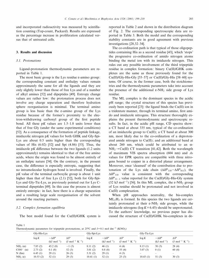
Ligand-protonation thermodynamic parameters are re-ported in Table 1.
The most basic group is the Lys residue O-amino group:the corresponding constant and enthalpy values remainapproximately the same for all the ligands and they areonly slightly lower than those of free Lys and of a numberof alkyl amines [52] and dipeptides [49]. Entropy changevalues are rather low: the protonation process does notinvolve any charge separation and therefore hydrationsphere reorganisation is minimal. The terminal aminogroup is less basic than the O-amino group of the Lysresidue because of the former's proximity to the elec-tron-withdrawing carbonyl group of the ¢rst peptidebond. All these pK values are 1.5^1.8 units lower thanthat of free Gly (under the same experimental conditions)[52]. As a consequence of the formation of peptide linkage,imidazolic nitrogen pK values for both GHK and Gly-Spi-Lys are about 0.5 units higher than the correspondingvalues of His (6.02) [52] and Spi (4.86) [53]. Thus, theimidazole pK di¡erence between the two ligands (1.2 unitsapproximately) remains identical to that of the free aminoacids, where the origin was found to be almost entirely ofan enthalpic nature [54]. On the contrary, in the presentcase, the di¡erence is especially entropic, suggesting thatno intramolecular hydrogen bond is involved. Finally, thepK value of the terminal carboxylic group is about 1 unithigher than that of free Lys (2.1) [52], both for Gly-Spi-Lys and Gly-Tic-Lys, as previously pointed out for Lys C-terminal dipeptides [49]. In this case the process is almostentirely entropic: in fact, here there is a charge separationand a resulting large scale reorganisation of the solventaround the reacting partners.
3.2. Complex formation equilibria
The best model found for the Cu(II)/GHK system is
reported in Table 2 and shown in the distribution diagramof Fig. 2. The corresponding spectroscopic data are re-ported in Table 3. Both the model and the correspondingstability constants are in good agreement with previousinvestigations [28,32^34].
The co-ordination path is that typical of those oligopep-tides containing His as a second residue [43], which `stops'the progressive co-ordination of amido nitrogen atomsbinding the metal ion with its imidazole nitrogen. Thisrules out any possible involvement of the third tripeptideresidue in complex formation: binary Cu(II)/GHK com-plexes are the same as those previously found for theCu(II)/Gly-His-Gly [55^57] or Cu(II)/Gly-His [58^60] sys-tems. Of course, in the former case, both the stoichiome-tries and the thermodynamic parameters take into accountthe presence of the additional O-NH2 side group of Lysresidue.
The ML complex is the dominant species in the acidicpH range; the crystal structure of this species has previ-ously been reported [23]: the ligand binds the Cu(II) ion ina tridentate manner, through its terminal amino, ¢rst ami-do and imidazole nitrogens. This structure thoroughly ex-plains the present thermodynamic and spectroscopic re-sults. In fact, in the acidic pH range, CD spectra show:a CT band at about 335 nm, typical of the co-ordinationof an imidazolic group to Cu(II); a CT band at about 300nm, most likely due to the co-ordination of a deproton-ated amide nitrogen to Cu(II) ; and an additional band atabout 260 nm, which could be attributed to an K-NH2CCu(II) CT transition [41,42]. Both the wavelengthof maximum VIS spectra absorption (604 nm) and Ae
values for EPR spectra are compatible with three nitro-gens bound to copper in a distorted planar arrangement.Moreover, once `cleansed' of the contribution due to pro-tonation of the Lys side chain (vH³1103vH³011), thevH³110 value is consistent with the correspondingvH³11ÿ1 value reported for the Cu(II)/Gly-His-Gly system(32 kJ mol31) [56]. In this ML complex, the O-NH2 groupof Lys residue should be protonated and not involved inCu(II) complexation.
When pH approaches neutrality, the bis-complexML2H2 is formed. In this species the two ligands are cer-tainly protonated at their O-NH2 side groups, while theimidazole nitrogens (log K = 6.45) should be unprotonated.To the authors' knowledge, no previous paper has dis-cussed the structure of Cu(II)/GHK bis-complexes in de-
Table 1Thermodynamic parameters for tripeptide protonations, at 25³C and I = 0.1 mol dm33 (KNO3)
Gly-His-Lys Gly-Spi-Lys Gly-Tic-Lys
log K 3vH³(kJ mol31)
vS³(J mol31 K31)
log K 3vH³(kJ mol31)
vS³(J mol31 K31)
log K 3vH³(kJ mol31)
vS³(J mol31 K31)
NH2 (K) 7.85 (2) 45.2 (8) 31 (3) 8.11 (2) 46 (1) 4 (4) 8.13 (1) 38 (3) 28 (4)COO3 (K) 2.73 (2) 0.1 (8) 52 (3) 3.01 (2) 0.8 (8) 55 (4) 3.07 (3) 0 (1) 60 (4)N (Im) 6.45 (1) 29 (1) 26 (3) 5.31 (2) 29 (1) 4 (3) ^ ^ ^NH2 (O) 10.53 (2) 52 (1) 29 (3) 10.41 (1) 52 (1) 25 (3) 10.43 (1) 51 (3) 30 (3)
BBAGEN 25173 17-4-01
C. Conato et al. / Biochimica et Biophysica Acta 1526 (2001) 199^210 203

tail. The most likely structural hypothesis for the ML2H2
species is the one advanced by Gergely et al. [58], Henry etal. [61] and Daniele et al. [56], respectively, for the corre-sponding ML2 species in the Cu(II)/Gly-His, Cu(II)/Gly-Hm (Hm = histamine) and Cu(II)/Gly-His-Gly systems.According to such hypotheses, the bis-complex formationresults from the binding of one protonated (LH2) ligandmolecule to a pre-formed ML complex at the fourth (free)position in the equatorial plane of the complex, by meansof the imidazole nitrogen of the second ligand. The distri-bution diagram in Fig. 2 shows that the formation of theML2H2 species begins at pH 4.5, where the copper presentin solution is entirely engaged in the ML complex. More-over, as for the Cu(II)/Gly-Hm system [61], the computedvalue of logKML=LH2
ML2H2( = logL ML2H23log LML3logL LH2 =
2.44, Table 2), is that expected for the formation of aCu(II)^Nim bond (2.48) [61]. The same calculation, appliedto the corresponding formation enthalpies, givesvH�ML=LH2
ML2H2= 24 kJ mol31, which is in relatively good
agreement with the expected value of 29 kJ mol31 [56].Unfortunately, the spectra are not very informative, sincethe ML complex is always present in large excess.
When the pH is increased, the bis-complex ML2H2 losesa proton, giving rise to the ML2H species; if the ligand isin excess this is the main complex at pH 8.5. ThelogKML2H2
ML2H value (7.1, Table 2) rules out the possibilitythat this is a hydroxo-complex. The simple deprotonationof one K-NH2 group in the ML2H2 complex has already
been suggested [56,58,61]. However, both the logKML2H2ML2H
and vH�ML2H2ML2H (35 kJ mol31, Table 2) values are notice-
ably lower than those of the free ligand (see Table 1). TheAe values of EPR spectra slightly increase, while the cor-responding ge values tend to decrease, when pH is raisedto pH 8^9. In addition, the wavelength of maximum ab-sorption (596 nm) is considerably higher than that calcu-lated (548 nm) with Sigel and Martin's empirical equation[62] for a Cu(II) complex with four co-ordinate nitrogens(two imidazoles, one amino and one amido) in the equa-
Fig. 2. Distribution diagram for the Cu(II)/GHK system.[Cu(II)] = 3U1033 mol dm33 ; [GHK] = 6U1033 mol dm33. 1: ML;2: MLH31 ; 3: MLH32 ; 4: ML2H2 ; 5: ML2H; 6: ML2 ; 7: ML2H31.Dotted line: wavelengths of maximum absorption in the VIS spectra.
Table 2Thermodynamic parameters of Cu(II)/tripeptide binary complex formation, at 25³C and I = 0.1 mol dm33 (KNO3)
Ligand Species log L 3vH³ (kJ mol31) vS³ (J mol31 K31)
Gly-His-Lys ML 16.48 (1) 94 (2) 0 (7)MLH31 7.32 (2) 61 (3) 365 (9)MLH32 33.02 (2) 9 (3) 387 (9)ML2H2 37.3 (1) 215 (6) 39 (20)ML2H 30.16 (4) 180 (4) 326 (12)ML2 20.62 (8) 125 (7) 324 (22)ML2H31 10.4 (1) 78 (9) 362 (30)
Gly-Spi-Lys MLH2 21.2 (4) ^ ^ML2H4 42.6 (3) ^ ^ML2H3 37.61 (9) ^ ^M2L2H3 40.7 (3) ^ ^M2L2H2 36.2 (2) 210 (20) 0 (8)M2L2H 29.4 (2) ^ ^M2L2 22.8 (1) ^ ^
Gly-Tic-Lys MLH 15.47 (2) 72 (1) 54 (4)ML2H2 30.12 (6) 140 (1) 105 (4)ML2H 21.43 (9) 121 (8) 4 (20)ML2 11.9 (1) ^ ^M2L2 18.6 (2) 70 (10) 110 (40)M2L2H31 9.5 (3) ^ ^
Gly-His-Glya ML 9.35 38 51MLH31 5.66 32 3MLH32 33.70 8 398ML2 15.96 104 342ML2H31 8.32 61 345M4L4H38 37.20 14 3185
a[56].
BBAGEN 25173 17-4-01
C. Conato et al. / Biochimica et Biophysica Acta 1526 (2001) 199^210204

torial plane. All these ¢ndings can be explained by assum-ing that such an unprotonated K-NH2 group interacts ax-ially with the central ion: such an interaction would pro-mote proton loss and also cause the observed `red shift'[41,42,62].
In the pH 9^10 range, the ML2H and the ML specieslose a proton, giving rise to the ML2 and the MLH31
complexes, which then become the main species. The cor-responding log Kstep and vvH³ values suggest that bothare hydroxo-complexes.
Finally, in the most basic pH range, the O-NH2 groupsof the Lys residues begin to lose their protons and thestoichiometry of the mono- and bis-complexes becomesMLH32 and ML2H31, respectively. The respectivelog Kstep and vvH³ values are slightly lower than thosemeasured for the free ligand. Correspondingly, a notice-able `blue shift', in absorption both and CD spectra, wasobserved at pHv10. Similar results have already been de-scribed in the literature [63^65] and they have been as-cribed to the involvement of a further nitrogen atom inCu(II) co-ordination. The same can be suggested here forthe prevailing MLH32 complex, where the deprotonated
O-NH2 side group of Lys residue can weakly interact withthe metal ion. No evidence has been found for the forma-tion of polynuclear species, even at a 1:1 metal-to-ligandratio.
As already noted above, Spi behaves as a `break-point'[41^43,45] in the peptide sequence: its K-amino grouploses its unique proton when forming the peptide linkageand, as a consequence, the ¢rst amido nitrogen of Gly-Spi-Lys cannot bind the metal ion as normally happens foroligopeptides. In addition, due to the rigid structure ofSpi, the imidazole nitrogen cannot bind the same metalion as the terminal amino group. This characteristic opensthe way to the formation of polynuclear species, as pre-viously observed for Gly-Spi [45]. Hence, despite themarked similarity in structure, as Cu(II) ligands, GHKand Gly-Spi-Lys behave di¡erently. Thermodynamic andspectroscopic data on the Cu(II)/Gly-Spi-Lys system arereported in Tables 2 and 4, respectively, while a distribu-tion diagram is shown in Fig. 3.
Over the entire pH range investigated, only faint EPRspectra could be recorded. This result suggests the pres-ence of a strong antiferromagnetic coupling between
Table 3Spectral data for Cu(II)/GHK system (3:6 mmol dm33)
pH Main species VIS CD ESR
V (nm) O V (nm) vO Ae ge
3.5 ML 799 15 259 0.93 184 2.242299 30.24337 0.12502 30.02606 0.34
6.0 ML 604 66 259 1.64 187 2.236299 30.27335 0.18488 30.02599 0.45
7.5 ML, ML2H2; ML2H 598 62 258 1.56 187 2.226299 30.24335 0.18485 30.02593 0.45
8 ML, ML2H2; ML2H 596 62 259 1.67 194 2.220299 30.21334 0.18483 0.03590 0.45
9 ML2H, MLH31 594 63 259 1.75 ^ ^299 30.14333 0.17482 30.03582 0.46
10 MLH31; ML2 586 66 258 2.18 195 2.220331 0.15474 30.05577 0.45
11 MLH32; ML2H31 576 68 257 2.37 195 2.220329 0.15471 30.07575 0.47
BBAGEN 25173 17-4-01
C. Conato et al. / Biochimica et Biophysica Acta 1526 (2001) 199^210 205

Cu(II) ions, as happens when high percentages of binu-clear complexes are present in solution. Potentiometricresults are in agreement with this hypothesis (see Fig. 3).
Starting from acidic pH values, the ¢rst species to formis the MLH2 complex, where the ligand is bound to themetal ion by means of its terminal amino group and theadjacent carbonyl oxygen (`NO' species). Some bis-com-plexes are also present in the system, in the acidic pHrange, but they do not reach high concentration levels.In the ML2H4 complex the two ligands most likely co-ordinate the Cu(II) ion again in a `NO' manner, both theirimidazole and Lys side amino groups being protonated.Once `cleansed' of the protonation contributions, the for-mation constants obtained for these species agree very wellwith literature data referring to ML and ML2 complexesof simple dipeptides like Gly-Gly, Gly-Ala or Gly-Pro [52].The wavelength of maximum absorption agrees with thehypothesis of a `NO' complex. CD spectra show a distinctband at 281 nm, which can be ascribed to the K-NH2-Cu(II) CT transition (see Fig. 4). Going from ML2H4 to
the ML2H3 complex, the computed log Kstep value suggeststhe simple deprotonation of one imidazole nitrogen. Un-fortunately, due to their low concentration levels, itproved impossible to determine formation vH³ and vS³values for these complexes.
The binuclear species found at the most acidic pH val-ues is the M2L2H3. It is reasonable to suggest that it iscomposed of two mononuclear `NO' complexes bridged bythe co-ordination of one imidazole nitrogen to the metalof the other complex unit. The two Lys O-amino groups, aswell as the second imidazole nitrogen, should be proton-ated. This species does not reach high concentration levelsand soon gives rise to the M2L2H2 complex, the mainspecies in the acidic pH range closest to neutrality. Thecorresponding logKM2L2H3
M2L2H2value (4.5) is comparable with,
although lower than, the protonation pK of the imidazolenitrogen in the free ligand (see Table 1). An interactionbetween this Nim and the Cu(II) ion of the other complexunit of the dimer can be suggested. This means that theM2L2H2 complex may exist in a cyclic form, as previouslyfound for the corresponding M2L2 complex in the Cu(II)/Gly-Spi system [45]. Once `cleansed' of the two O-NH2
protonation contributions (210352352 = 106 kJ mol31,Table 2) the corresponding vH³222 value assumes the ex-pected value for four Cu(II)^N bonds. CD and absorptionspectra (Fig. 4) also suggest a 2N environment for eachCu(II) ion. Increasing the pH, the M2L2H2 complex ¢rstloses one proton, giving rise to the M2L2H species, andthen a second one to give M2L2. The M2L2H complexreaches only moderate concentrations, whereas M2L2 isthe main species in the alkaline pH range. The pK valuesfor both these deprotonation steps are too low to corre-spond to a deprotonation process of the co-ordinatedwater molecules. Taking into account that, for each metalcentre, one co-ordination position on the equatorial planeis free, it can be suggested that the two latter species derivefrom deprotonation, in sequence, of the O-amino groups ofthe two Lys residues, promoted by the interaction with theCu(II) ions. Spectroscopic results support this hypothesis.
Fig. 3. Distribution diagram for the Cu(II)/Gly-Spi-Lys system.[Cu(II)] = 3U1033 mol dm33 ; [Gly-Spi-Lys] = 8U1033 mol dm33.1: MLH2 ; 2: ML2H4 ; 3: ML2H3 ; 4: M2L2H3 ; 5: M2L2H2 ; 6: M2L2H;7: M2L2. Dotted line: wavelengths of maximum absorption in the VISspectra.
Table 4Spectral data for Cu(II)/Gly-Spi-Lys system (3:8 mmol dm33)
pH Main species UV-VIS CD
V (nm) O V (nm) vO
3.5 MLH2 765 16 285 30.144.5 MLH2, ML2H4, M2L2H3, M2L2H2 708 32 281 30.20
675 30.067.2 ML2H3, M2L2H2, M2L2H, M2L2 660 65 265 0.42
297 30.20376 30.01505 30.14618 30.09
8.0 M2L2 647 81 264 0.48297 30.24372 30.04510 30.19622 30.17
BBAGEN 25173 17-4-01
C. Conato et al. / Biochimica et Biophysica Acta 1526 (2001) 199^210206

In fact, a relevant `blue shift' takes place in absorptionspectra when pH is raised to 8.0. Moreover, two newbands appear in CD spectra: a positive band at about264 nm which could be ascribed to a CT transition anda negative d^d band near 510 nm, typical of Cu(II) com-plexes with three nitrogens bound to the metal. Unfortu-nately, the corresponding vH³ values could not be mea-sured.
Gly-Tic-Lys has a structure similar to that of Gly-Spi-Lys (see Fig. 1): its second residue (Tic) has a rigid struc-ture and behaves as a `break-point' in the peptide chain, asdoes Spi. However, Tic does not bear the imidazole groupand the only side donor atom of this tripeptide is theO-amino nitrogen of Lys. Gly-Tic-Lys forms both mono-and binuclear complexes with the Cu(II) ion: thermody-namic results are reported in Table 2, while spectroscopicdata are shown in Table 5; a distribution diagram isdrawn in Fig. 5.
The co-ordination starts at pH 3.5 with the MLH com-plex, the main species throughout the entire acidic pHrange. Subtracting log K011 and vH³011, respectively,from log L111 and vH³111, gives `cleansed' values suggest-
ing a `NO' co-ordination. The spectroscopic results like-wise support the same hypothesis. When the pH is in-creased, the ML2H2 complex becomes the main species.Spectroscopic and thermodynamic parameters suggestthat, in this complex, both ligands bind the metal ion inthe basic `NO' manner. In the alkaline pH range theML2H species starts to form. The correspondinglogKML2H2
ML2 H value (8.7, Table 2) is too low to be ascribedto the deprotonation of a water molecule bound to cop-per. Moreover, the `cleansed' vH³121 value is compatiblewith the formation of three Cu(II)^N bonds. When the pHis raised from pH 7 to pH 8.5, both absorption and CDspectra show a noticeable `blue shift' and the Ae valueincreases. Therefore, it appears reasonable to suggestthat the ML2H complex derives from the deprotonationof the lateral amino group of one Lys residue, favoured byits interaction with the copper ion in the apical position.
Above a pH of 9, the bis-complex loses another protonand the ML2 species is formed. The value of logKML2H
ML2
(9.5, Table 2) seems to indicate that ML2 is a hydroxo-complex. No calorimetric or spectroscopic data are avail-able for this species.
Table 5Spectral data for Cu(II)/Gly-Tic-Lys system (3:8 mmol dm33)
pH Main species UV-VIS CD ESR
V (nm) O V (nm) vO Ae ge
3.0 814 13 258 30.41 120 2.4144 MLH 796 16 260 30.28 121 2.4135.5 MLH 741 31 254 0.34 147 2.331
788 30.086.5 MLH, ML2H2 698 41 264 0.54 143 2.332
788 30.127.2 ML2H2 686 47 264 0.62 142 2.282
787 30.118.0 ML2H2 676 57 264 0.60 162 2.265
756 30.118.8 ML2H2, ML2H, M2L2 667 61 256 0.83 162 2.266
726 30.17
Fig. 4. CD spectra of Cu(II)/Gly-Spi-Lys solutions at di¡erent pH.[Cu(II)] = 3U1033 mol dm33 ; [Gly-Spi-Lys] = 8U1033 mol dm33.
Fig. 5. Distribution diagram for the Cu(II)/Gly-Tic-Lys system.[Cu(II)] = 3U1033 mol dm33 ; [Gly-Tic-Lys] = 8U1033 mol dm33.1: MLH; 2: ML2H2 ; 3: ML2H; 4: ML2 ; 5: M2L2 ; 6: M2L2H31. Dot-ted line: wavelengths of maximum absorption in the VIS spectra.
BBAGEN 25173 17-4-01
C. Conato et al. / Biochimica et Biophysica Acta 1526 (2001) 199^210 207

In the same pH range, some binuclear species have beenfound as well. They reach only moderate concentrations(see distribution diagram, Fig. 5) and they are never theonly species present in solution. For this reason, the spec-troscopic data do not provide valuable information ontheir structure, nor does the EPR signal disappear in thepH range where binuclear species exist ; rather only a de-crease in intensity can been noticed. On the basis of ther-modynamic parameters reported in Table 2, the mostlikely hypothesis for the M2L2 structure is that of an equa-torial coupling of two MLH complexes, via two OH3
bridges. In fact, summing up the logL M2H32 value(310.75) [66] and (twice) the logKMLH
ML2H2(i.e. the contribu-
tion for the co-ordination of a protonated ligand to aCu(II) mono-complex) (14.65, Table 2) gives a computedvalue of 18.55 as an estimate of logL M2L2 , in perfect agree-ment with the experimental value (18.6, Table 2). More-over, analogous calculations over the corresponding vH³values give 60 kJ mol31, not far from the experimentalvalue of vH�M2L2 = 70 þ 10 kJ mol31 (Table 2). Finally,as far as the M2L2H31 species is concerned, both the de-protonation of a water molecule from the co-ordinationsphere, and the deprotonation of a side amino group ofLys and its involvement in co-ordination can be suggested,since the only available data is logKM2L2
M2L2H = 9.1.
3.3. Tripeptide enzymatic stability
The enzymatic stability of GHK and related tripeptidesin human serum is reported in Fig. 6. GHK degradation israther fast and its concentration decreases with an approx-imately exponential decay and a half life period of about16 min. This time is higher than, but comparable with, thevalues previously reported [40]. On the contrary, its ana-logues showed excellent stability, their concentration beingunchanged in human serum after some hours of incuba-tion. This result could be attributed to the presence, in thepeptide chain, of `unusual' amino acid residues, not rec-ognised by serum peptidases.
It is worth noting that a MS-MALDI analysis of human
serum containing degraded GHK showed two distinctpeaks at m/z = 212.9 Da and m/z = 235.1 Da, which canbe ascribed to the protonated Gly-His ion and its corre-sponding sodium adduct. This leads to the hypothesis thatthe main degradation mechanism of GHK is the breakingof the His-Lys peptide bond.
3.4. Biological activity of tripeptides as cell growth factors
Fig. 7 shows the experimental results obtained in the cellproliferation tests performed with T2 cells in a serum-freeculture medium. This choice derived from the necessity torender the results independent of enzymatic stability ofeach tripeptide. Data are reported as % of proliferationincrement, with respect to a control where the tripeptideswere absent. GHK shows the expected behaviour of agrowth factor: its activity increases with its concentrationin the medium, reaching a maximum when it is 50^100nM, in agreement with previous results [23]. Under thesame conditions, an even higher activity was obtainedwith Gly-Spi-Lys and Gly-Tic-Lys, suggesting that the rig-id structure of these molecules favours binding to the cellreceptors. In a previous study [27], a series of oligopep-tides containing Gly, His and Lys in di¡erent positionswas investigated in order to gain insight into the role ofthe individual residues on peptide biological activity. Theconclusions were the following [23,27] : (i) when in theN-terminal position, Gly is essential for expression of ac-tivity; (ii) optimal activity appears to be associated to theHis-Lys linkage; (iii) the His residue may be essential forthe chelation of copper and iron ions; (iv) the Lys residuemay participate in charge-dependent interactions betweenthe bioactive peptides and the cell surface (cellular bind-ing). The present results only partially agree with theseconclusions. The ability of the tripeptide to chelate thecopper ion looks essential indeed, independently of thestructure of the complex: in fact, the three active peptidesvery e¡ectively chelate the copper at neutral pH, eventhough they form very di¡erent complex species (seeabove). At the same time, PrA-Tic-Lys, which di¡ers
Fig. 7. Biological activity of tripeptides as cell growth factors.
Fig. 6. Enzymatic stability of tripeptides in human serum.
BBAGEN 25173 17-4-01
C. Conato et al. / Biochimica et Biophysica Acta 1526 (2001) 199^210208

from Gly-Tic-Lys in the absence of the terminal aminogroup and which does not form copper complexes [67],showed negligible biological activity. Taking into accountthat some Cu(II) ion is present in the culture medium (seeSection 2), the present results support the hypothesis thatone of the main biological action mechanisms of thesetripeptides is to facilitate Cu(II) ion uptake into cells[23]. However, it is worth noting that the highest activitywas found for Gly-Tic-Lys, which does not bear the imi-dazole group, suggesting that the presence of His as sec-ond residue does not play any speci¢c role in this activity.
Finally, the excellent stability toward serum peptidasesand the good biological activity both Gly-Spi-Lys andGly-Tic-Lys have shown as T2 cell growth factors makethem good candidates as active principles in the formula-tion of new drugs and as growth factor additives in cellproliferation tests, in the presence or in the absence ofserum.
Acknowledgements
This work was ¢nancially supported by the ItalianMURST (COFIN '98) and by the Polish State Committeefor Scienti¢c Research (KBN 3T09A-10514 and SPUB/COST/T-9/DZ 44/99). The contribution of E. Prenesti,for preliminary CD investigation on Cu(II)/tripeptide sys-tems, is gratefully acknowledged.
References
[1] L. Pickart, M.M. Thaler, Nat. New Biol. 234 (1973) 85^87.[2] L. Pickart, M.M. Thaler, M. Millard, J. Chromatogr. 175 (1979) 65^
73.[3] L. Pickart, M.M. Thaler, J. Cell Physiol. 102 (1980) 129^139.[4] R. Barra, Cytobios 52 (1987) 99^107.[5] L. Pickart, In Vitro 17 (1981) 459^466.[6] J.E. Errick, K.W. Ing, M.C. Eggo, G.N. Burrow, In Vitro Cell. Dev.
Biol. 22 (1986) 28^36.[7] O. Lassila, J.D. Lambris, R.H. Gisler, Cell. Immunol. 122 (1989)
319^328.[8] V. Pesakova, J. Novotna, M. Adam, Biomaterials 16 (1995) 911^915.[9] D. Godet, P.J. Marie, Cell. Mol. Biol. 41 (1995) 1081^1091.
[10] C. Fraslon, G. Rolland, J.R. Bourbon, M. Rieutort, C. Valenza, InVitro Cell. Dev. Biol. 27A (1991) 843^852.
[11] S.Q. Liu, Y. Ito, Y. Imanishi, Enzyme Microb. Technol. 15 (1993)167^172.
[12] L. Pickart, S. Lovejoy, Methods Enzymol. 147 (1987) 314^328.[13] P.M. Gullino, EXS 61 (1992) 125^128.[14] E.H. Sage, R.B. Vernon, J. Hypertens. 12 (Suppl.) (1994) S145^S152.[15] D.M. Miller, D. DeSilva, L. Pickart, S.D. Aust, Adv. Exp. Med.
Biol. 264 (1990) 79^84.[16] F. Bu¡oni, R. Pino, A. Dal Pozzo, Arch. Int. Pharmacodyn. Ther.
330 (1995) 345^360.[17] A. Simeon, F. Monier, H. Emonard, Y. Wegrowski, G. Bellon, J.C.
Monboisse, P. Gillery, W. Hornebeck, F.X. Maquart, Curr. Top.Pathol. 93 (1999) 95^101.
[18] H. Pohunkova, J. Stehlik, J. Vachal, O. Cech, M. Adam, Biomate-rials 17 (1996) 1567^1574.
[19] F.X. Maquart, L. Pickart, M. Laurent, P. Gillery, J.C. Monboisse,J.P. Borel, FEBS Lett. 238 (1988) 343^346.
[20] N. Cotelle, E. Tremolieres, J.L. Bernier, J.P. Catteau, J.P. Henichart,J. Inorg. Biochem. 46 (1992) 7^15.
[21] D.L. Rabenstein, J.M. Robert, S. Hari, FEBS Lett. 376 (1995) 216^220.
[22] J.A. Garciasainz, J.A. Olivaresreyes, Peptides 16 (1995) 1203^1207.[23] L. Pickart, J.H. Freedman, W.J. Loker, J. Peisach, C.M. Perkins,
R.E. Stenkamp, B. Weinstein, Nature 288 (1980) 715^717.[24] H.M. Darwish, J.C. Cheney, R.C. Schmitt, M.J. Ettinger, Am. J.
Physiol. 246 (1984) G72^G79.[25] D.E. Hartter, A. Barnea, J. Biol. Chem. 263 (1988) 799^805.[26] W.E. Antholine, D.H. Petering, L. Pickart, J. Inorg. Biochem. 35
(1989) 215^224.[27] L. Pickart, M.M. Thaler, FEBS Lett. 104 (1979) 119^122.[28] S.J. Lau, B. Sarkar, Biochem. J. 199 (1981) 649^656.[29] J.H. Freedman, L. Pickart, B. Weinstein, W.B. Mims, J. Peisach,
Biochemistry 21 (1982) 4540^4544.[30] P.M. May, J. Whittaker, D.R. Williams, Inorg. Chim. Acta 80 (1983)
L5^L7.[31] J.P. Laussac, R. Haran, B. Sarkar, Biochem. J. 209 (1983) 533^539.[32] M.J.A. Rainer, B.M. Rode, Inorg. Chim. Acta 92 (1984) 1^7.[33] M.J.A. Rainer, B.M. Rode, Inorg. Chim. Acta 107 (1985) 127^132.[34] K. Takehara, I. Yasushi, Inorg. Chim. Acta 183 (1991) 195^202.[35] M.J.A. Rainer, B.M. Rode, Inorg. Chim. Acta 93 (1984) 109^115.[36] S.A. Daignault, A.P. Arnold, A.A. Isab, D.L. Rabenstein, Inorg.
Chem. 24 (1985) 3984^3988.[37] J.P. Laussac, M. Pasdeloup, N. Hadjiliadis, J. Inorg. Biochem. 30
(1987) 227^238.[38] I. Rombeck, B. Lippert, Inorg. Chim. Acta 273 (1998) 31^40.[39] T.F. Lane, M.L. Iruela-Arispe, R.S. Johnson, E.H. Sage, J. Cell Biol.
125 (1994) 929^943.[40] A. Dalpozzo, K. Kanai, G. Kereszturi, G. Calabrese, Int. J. Peptide
Protein Res. 41 (1993) 561^566.[41] L.D. Pettit, J.E. Gregor, H. KozIowski, in: R.W. Hay, J.R. Dilworth,
K.B. Nolan (Eds.), Perspectives on Bioinorganic Chemistry, Vol. 1,JAI Press, London, 1991, pp. 1^41.
[42] I. Sovago, in: K. Burger (Ed.), Biocoordination Chemistry: Coordi-nation Equilibria in Biologically Active Systems, ch. IV, Ellis Hor-wood, New York, 1990.
[43] H. KozIowski, W. Bal, M. Dyba, T. Kowalik-Jankowska, Coord.Chem. Rev. 184 (1999) 319^346.
[44] M. Remelli, F. Pulidori, R. Guerrini, V. Bertolasi, J. Chem. Crystal-logr. 27 (1997) 507^513.
[45] C. Conato, M. Remelli, R. Guerrini, F. Pulidori, Ann. Chim. (Rome)88 (1998) 91^102.
[46] K. Hayashi, Y. Ozaki, K. Nunami, N. Yoneda, Chem. Pharm. Bull.31 (1983) 312^314.
[47] E. Atherton, R.C. Sheppard, in: D. Rickwood, B.D. Hames (Eds.),Solid Phase Peptide Synthesis, IRL Press, Oxford, 1989.
[48] L.A. Carpino, J. Am. Chem. Soc. 115 (1993) 4397^4398.[49] M. Remelli, C. Conato, A. Agarossi, F. Pulidori, P. MIyarz, H.
KozIowski, Polyhedron 19 (2000) 2409^2419.[50] D.E. Benovitz, A.F. Spatola, Peptides 6 (1985) 257^261.[51] R.D. Salter, P. Cresswell, EMBO J. 5 (1986) 943^949.[52] R.M. Smith, A.E. Martell, Critical Stability Constants, Vol. 6, Ple-
num Press, London, 1989.[53] M. Remelli, S. Rossi, R. Guerrini, F. Pulidori, Ann. Chim. (Rome)
85 (1995) 503^518.[54] M. Remelli, E. Pirani, C. Conato, F. Pulidori, unpublished results.[55] R.P. Agarwal, D.D. Perrin, J. Chem. Soc. Dalton Trans. (1975) 268^
272.[56] P.G. Daniele, O. Zerbinati, V. Zelano, G. Ostacoli, J. Chem. Soc.
Dalton Trans. (1991) 2711^2715.[57] E. Farkas, I. Sovago, T. Kiss, A. Gergely, J. Chem. Soc. Dalton
Trans. (1984) 611^614.
BBAGEN 25173 17-4-01
C. Conato et al. / Biochimica et Biophysica Acta 1526 (2001) 199^210 209

[58] I. Sovago, E. Farkas, A. Gergely, J. Chem. Soc. Dalton Trans. (1982)2159^2163.
[59] G. Arena, V. Cucinotta, S. Musumeci, R. Purrello, E. Rizzarelli,Ann. Chim. (Rome) 74 (1984) 399^412.
[60] G. Brookes, L.D. Pettit, J. Chem. Soc. Dalton Trans. (1975) 2112^2117.
[61] T. Gajda, B. Henry, J.-J. Delpuech, J. Chem. Soc. Dalton Trans.(1993) 1301^1306.
[62] H. Sigel, R.B. Martin, Chem. Rev. 82 (1982) 385^426.
[63] B. Radomska, I. Sovago, T. Kiss, J. Chem. Soc. Dalton Trans. (1990)289^292.
[64] B. Radomska, M. Kubiak, T. Glowiak, H. KozIowski, Inorg. Chim.Acta 159 (1989) 111^114.
[65] P.G. Daniele, E. Prenesti, G. Ostacoli, J. Inorg. Biochem. 61 (1996)165^177.
[66] G. Arena, R. Cal|©, E. Rizzarelli, S. Sammartano, Thermochim. Acta16 (1976) 315^321.
[67] M. Remelli, C. Conato, R. Guerrini, unpublished results.
BBAGEN 25173 17-4-01
C. Conato et al. / Biochimica et Biophysica Acta 1526 (2001) 199^210210