Role of reactive oxygen species in the pathophysiology of human reproduction
Upload
khangminh22Category
view
2download
0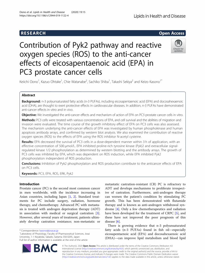
RESEARCH Open Access
Contribution of Pyk2 pathway and reactiveoxygen species (ROS) to the anti-cancereffects of eicosapentaenoic acid (EPA) inPC3 prostate cancer cellsKeiichi Oono1, Kazuo Ohtake1, Chie Watanabe2, Sachiko Shiba1, Takashi Sekiya2 and Keizo Kasono1*
Abstract
Background: n-3 polyunsaturated fatty acids (n-3 PUFAs), including eicosapentaenoic acid (EPA) and docosahexaenoicacid (DHA), are thought to exert protective effects in cardiovascular diseases. In addition, n-3 PUFAs have demonstratedanti-cancer effects in vitro and in vivo.
Objective: We investigated the anti-cancer effects and mechanism of action of EPA on PC3 prostate cancer cells in vitro.
Methods: PC3 cells were treated with various concentrations of EPA, and cell survival and the abilities of migration andinvasion were evaluated. The time course of the growth inhibitory effect of EPA on PC3 cells was also assessed.The mechanism underlying the anti-cancer effects of EPA was investigated by human phosphokinase and humanapoptosis antibody arrays, and confirmed by western blot analysis. We also examined the contribution of reactiveoxygen species (ROS) to the effects of EPA using the ROS inhibitor N-acetyl cysteine.
Results: EPA decreased the survival of PC3 cells in a dose-dependent manner within 3 h of application, with aneffective concentration of 500 μmol/L. EPA inhibited proline-rich tyrosine kinase (Pyk)2 and extracellular signal-regulated kinase 1/2 phosphorylation as determined by western blotting and the antibody arrays. The growth ofPC3 cells was inhibited by EPA, which was dependent on ROS induction, while EPA inhibited Pyk2phosphorylation independent of ROS production.
Conclusions: Inhibition of Pyk2 phosphorylation and ROS production contribute to the anticancer effects of EPAon PC3 cells.
Keywords: PC3, EPA, ROS, ERK, Pyk2
IntroductionProstate cancer (PC) is the second most common cancerin men worldwide, with the incidence increasing inAsian countries, including Japan [1, 2]. Standard treat-ments for PC include surgery, radiation, hormonetherapy, and chemotherapy. Advanced PC with metasta-sis is treated with androgen deprivation therapy (ADT)in association with medical or surgical castration [3].However, after several years of treatment, patients ultim-ately develop castration resistance; non-metastatic or
metastatic castration-resistant (CR) PC is refractory toADT and develops mechanisms to proliferate irrespect-ive of castration. Furthermore, anti-androgen therapycan worsen the patient’s condition by stimulating PCgrowth. This has been demonstrated with flutamidetherapy and is known as anti-androgen withdrawal syn-drome [4]. Only a few chemotherapeutics and radiationhave been developed for the treatment of CRPC [5], andthese have not improved the poor prognosis of thisdisease [6].There is growing evidence that n-3 polyunsaturated
fatty acids (n-3 PUFAs) found in fish oil—especiallyeicosapentaenoic acid (EPA) and docosahexaenoic acid(DHA)—can improve lipid metabolism and blood lipid
© The Author(s). 2020 Open Access This article is distributed under the terms of the Creative Commons Attribution 4.0International License (http://creativecommons.org/licenses/by/4.0/), which permits unrestricted use, distribution, andreproduction in any medium, provided you give appropriate credit to the original author(s) and the source, provide a link tothe Creative Commons license, and indicate if changes were made. The Creative Commons Public Domain Dedication waiver(http://creativecommons.org/publicdomain/zero/1.0/) applies to the data made available in this article, unless otherwise stated.
* Correspondence: [email protected] of Physiology, Faculty of Pharmaceutical Sciences, JosaiUniversity, 1-1 Keyakidai, Sakado, Saitama 350-0295, JapanFull list of author information is available at the end of the article
Oono et al. Lipids in Health and Disease (2020) 19:15 https://doi.org/10.1186/s12944-019-1122-4
profiles, prevent the progression of atherosclerosis, andreduce the incidence of cardiovascular diseases [7] aswell as liver and plasma triglyceride levels [8]. We previ-ously demonstrated that chronic oral administration ofEPA prevented endothelial dysfunction in a mousemodel of type 2 diabetes [9]. n-3 PUFAs have also beenreported to have various anti-cancer effects in severaltypes of malignancy in vitro and in vivo. n-3 PUFAsinhibit extracellular signal-regulated kinase (ERK) andAkt signaling pathways and show anti-cancer effects inbreast cancer [10], reduce the incidence of liver cancerin hepatitis virus-infected patients [11], and lower therisk of pancreatic cancer [12]. Cis-unsaturated fattyacids including EPA are easily incorporated into cancercells and induce free radical generation, inducingtumoricidal action [13–16]. However, other studieshave suggested that there is insufficient evidence for asignificant association between n-3 PUFAs and cancerincidence [17].We previously reported that EPA and DHA have
anti-proliferative, −migratory, and -invasive effects inthe PC3 CRPC cell line [18]. We also observed thatthe combination of anti-cancer drugs and n-3 PUFAssynergistically inhibited the proliferation of PC3 cells(unpublished data). However, the mechanism ofaction of n-3 PUFAs, and specifically, the molecularbasis for the inhibitory effects of EPA on PC3 cellproliferation, migration, and invasion has yet to bedefined.
Fig. 1 Effect of EPA on PC3 cell proliferation. After 24 h of culture inserum-free medium, various concentrations of EPA (0, 100, 300, and500 μM) were added to the cultures. Data represent mean + SEM(n = 3). **P < 0.01 vs. 0 μM
Fig. 2 Effect of EPA on the time course of PC3 cell proliferation. After 24 h of culture in serum-free medium, EPA (500 μM), or vehicle was addedto the cells. Data represent mean ± SEM (n = 3). **P < 0.001 vs 0 h; #P < 0.05, ##P < 0.01 v. vehicle
Oono et al. Lipids in Health and Disease (2020) 19:15 Page 2 of 12
Various signaling pathways control cell proliferation;their dysregulation can lead to over-proliferation andaberrant migration and invasion. Tyrosine kinase (TK)is an important mediator of cell proliferation. Recep-tor (R) TK is a transmembrane receptor with TKactivity, whereas non-receptor (NR) TK is present inthe cytoplasm and comprises Abl, Src, and proline-rich TK (Pyk)2 families. Various NRTKs are expressedin PC3 cells, and Src plays a role in multiple bio-logical processes in PC cells [19]. Activation of theseTKs leads to cell proliferation through activation of
transcription factors that promote cell cycleprogression.NRTKs enhance the proliferation of cancer cells to
promote their survival, so suppressing these pathwayscan lead to cell growth inhibition or death. Mostcancer cells are resistant to apoptosis and activatepathways that suppress the pro-apoptotic active formof caspases [20]. Moderate ROS levels may causetumorigenesis through DNA damage in pro-tumorigenic cells [21]. Thus, eliminating or reducingROS production is a potential strategy for cancertherapy, since ROS also regulate the activation of sev-eral RTKs and NRTKs [22]. On the other hand, EPA-induced ROS overproduction stimulates apoptoticsignals in HepG2 liver cancer cells [23], and oxidativestress causes PC3 cell death by stimulating mitochon-drial ROS production and apoptosis [24]. DHA hasbeen found to cause cell death by inducing ROS pro-duction in PC3 and DU145 PC cells [25].In the present study, we investigated the mechanism
underlying the anti-cancer effects of n-3 PUFA EPA, firstby characterizing the optimal concentration and timerequired for these effects in PC3 cells, and then byanalyzing protein expression by western blotting andantibody arrays. To clarify the effects of ROS productioninduced by EPA, we used the ROS inhibitor N-acetylcysteine (NAC).
Materials and methodsReagentsEPA (Sigma-Aldrich, St. Louis, MO, USA) was dis-solved in 99% isopropanol (Wako, Tokyo, Japan) andstored at − 30 °C. For experiments, EPA was dissolvedin serum-free Dulbecco’s modified Eagle’s medium(DMEM; Gibco, New York, NY, USA) containing 3%bovine serum albumin (BSA) (Sigma-Aldrich), andNAC (Wako) was dissolved in phosphate-bufferedsaline (PBS). Human phospho-kinase array (R&DSystems, Minnesota, MN, USA) and the human apop-tosis antibody Array Membranes (Abcam, Cambridge,UK) were used for protein analysis. Antibodies againstERK1/2, phosphorylated (p)-ERK1/2, Pyk2, p-Pyk2,and β-actin were from Cell Signaling TechnologyJapan (Tokyo, Japan) and were diluted with Can GetSignal reagent (Toyobo, Osaka, Japan).
Cell culturePC3 cells (Riken BRC Cell Bank, Tsukuba, Japan) weregrown in DMEM (Gibco) supplemented with 10% heat-inactivated fetal bovine serum (FBS), 100 U/mL ofpenicillin, and 100 μg/mL of streptomycin (Gibco). Thecells were cultured at 37 °C in a humidified 5% CO2
atmosphere.
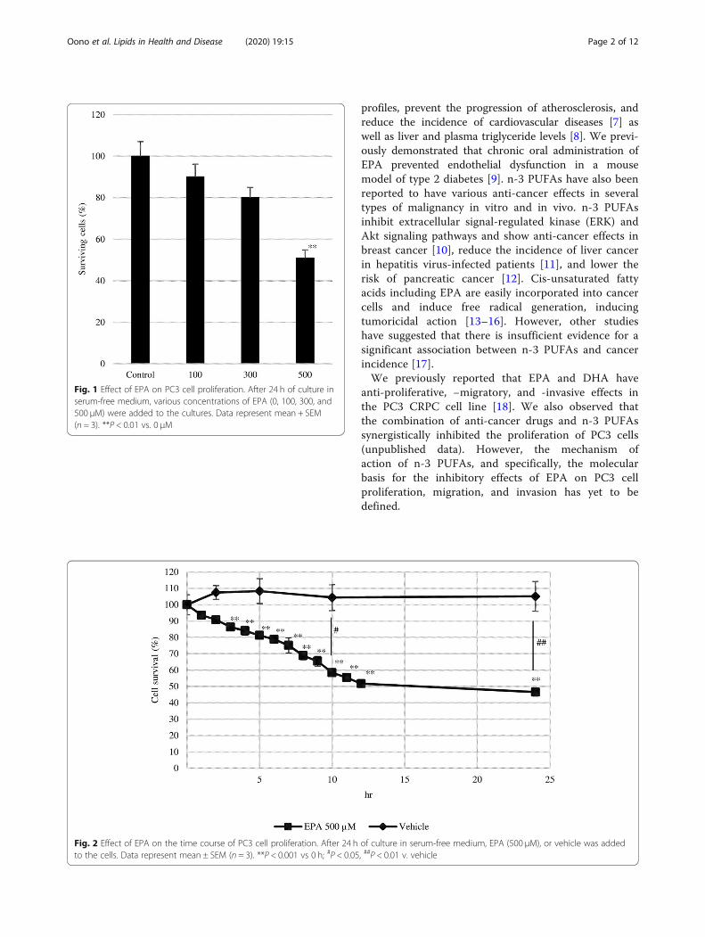
Fig. 3 Effect of EPA on EPA on PC3 (a) cell migration and (b)invasion. Double-chambered cell culture dishes with a transwellinsert separating the two chambers were used to evaluate PC3cell migration and invasion. Cells were seeded in the upperchamber, which was uncoated (migration) or coated (invasion)with Matrigel, while the lower chamber was filled with DMEMcontaining 10% FBS. Data represent mean + SEM(n = 3). *P < 0.05
Oono et al. Lipids in Health and Disease (2020) 19:15 Page 3 of 12
Cell proliferation assayPC3 cells were seeded in 35-mm dishes at a density of3 × 105 cells/dish in DMEM containing 10% FBS andincubated for 24 h, and the culture medium was replacedwith serum-free DMEM after washing with PBS. Afteran additional 24 h, the cells were washed with PBS, andserum-free medium containing 3% BSA and variousconcentrations of EPA (100–500 μM) were added. After24 h, the number of cells was counted with ahemocytometer.
Time course of cell proliferationPC3 cells were seeded in a 35-mm high μ-Dish Grid-500 (Ibidi, Planegg, Germany) at a density of 3 × 10⁵
cells/dish in DMEM containing 10% FBS. After 24 h,the cells were washed with PBS, and serum-freeDMEM was added; after another 24 h, the cells werewashed with PBS, and serum-free medium containing3% BSA with or without EPA (500 μM) was added.Cells were imaged with a microscope and counted atthe indicated times.
Migration and invasion assayMigration and invasion were assessed as described in ourprevious study [18]. Briefly, we used Transwell mem-branes (8-μm pore size; Corning Inc., Corning, NY, USA)with or without Matrigel coating. PC3 cells were culturedin the upper chamber of a Transwell insert in serum-freemedium containing various concentrations of EPA. Thelower chamber was filled with DMEM containing 10%FBS. For the migration assay, Matrigel-uncoated upperchambers were used; cells were seeded at a density of 2 ×10⁵ cells/mL, and after 24 h, the number of cells that hadmigrated into the lower chamber was counted. For the in-vasion assay, Matrigel-coated upper chambers were used.Before seeding the cells, Matrigel was fixed by incubationfor 90min, and cells were seeded at a density of 1 × 10⁵
cells/mL. After 48 h, cells that had invaded into the lowerchamber were counted.
Antibody arraysPC3 cells were cultured in DMEM containing 10%FBS for 24 h, then cultured in serum-free medium for
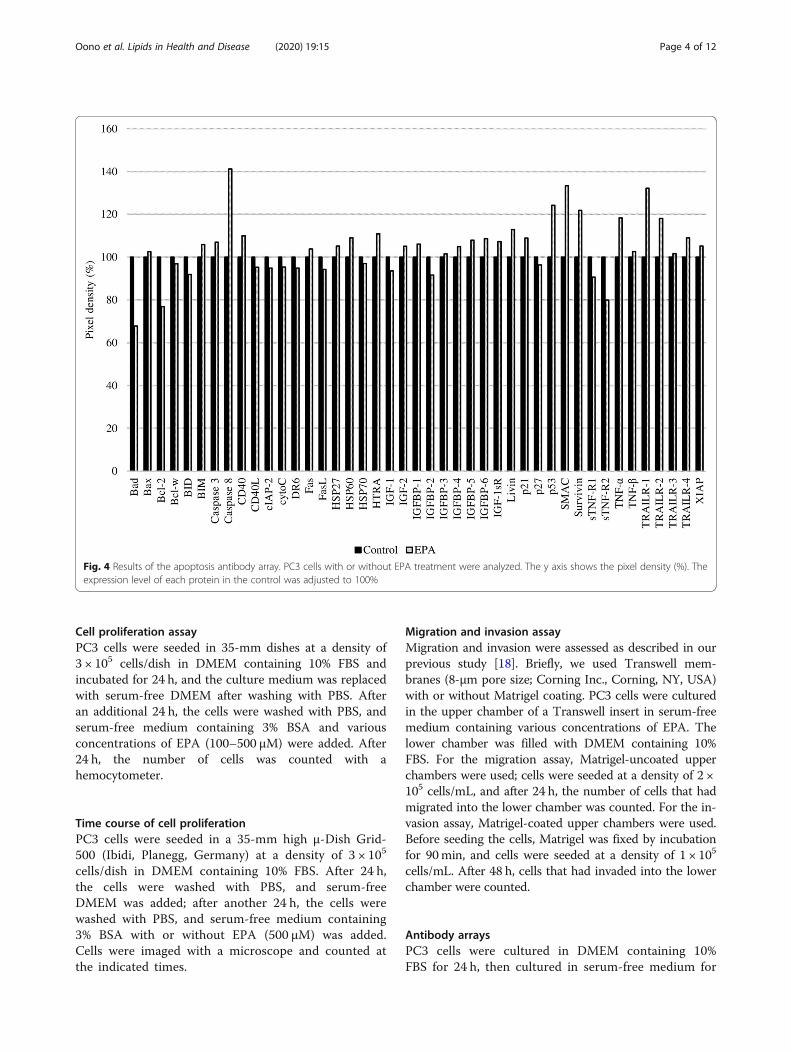
Fig. 4 Results of the apoptosis antibody array. PC3 cells with or without EPA treatment were analyzed. The y axis shows the pixel density (%). Theexpression level of each protein in the control was adjusted to 100%
Oono et al. Lipids in Health and Disease (2020) 19:15 Page 4 of 12

an additional 24 h before treatment with EPA(500 μM) or vehicle as a control. After 2 h of incuba-tion with EPA or vehicle, samples were prepared forthe antibody array according to the manufacturers’protocols. Chemiluminescence detection was per-formed on a Lumi Cube (Liponics, Tokyo, Japan)using Clarity Western ECL Substrate (Bio-Rad,Hercules, CA, USA). Images of the arrays were ana-lyzed using Image J software (National Institutes ofHealth, Bethesda, MD, USA).
Inhibition of ROS productionPC3 cells were cultured in a 35-mm dish at a densityof 3 × 10⁵ cells/dish in DMEM containing 10% FBSfor 24 h, and then washed with PBS and incubated inserum-free DMEM for an additional 24 h. The cellswere washed with PBS, and EPA (500 μM) with orwithout 5 mM of NAC (Wako) was added, cells were
incubated for 24 h, and cell numbers were countedwith a hemocytometer.
Western blot analysisPC3 cells were cultured in 60-mm dishes at a densityof 8 × 10⁵ cells/dish in 10% FBS-containing DMEMfor 24 h. PC3 cells were cultured with serum starva-tion for an additional 24 h, then treated with EPA(500 μM) or vehicle. The cells were lysed in M-PER(Thermo Fisher Scientific, Waltham, MA, USA) andcentrifuged at 13,000 rpm (30,250 g) at 4 °C for 10min. Protein samples were diluted in 2× Laemmlisample buffer (Bio-Rad), then boiled at 100 °C, and20 μg of protein were loaded in each lane of a TGXprecast gel (Bio-Rad) and resolved by electrophoresis(100 V, 0.3 A). The proteins were transferred to anImmun-Blot polyvinylidene difluoride membrane (Bio-Rad) at 100 V and 0.3 A for 1 h. The membrane wasblocked with Block Ace (KAC, Kyoto, Japan) for 1 h
Fig. 5 Results of two experiments using the human phosphokinase array. The y axis shows pixel density (%). For the control groups inexperiments 1 and 2 (Expt1 Control and Expt2 Control, respectively), the expression level of each protein was adjusted to 100%. For the EPA-treated groups in experiments 1 and 2 (Expt1 EPA and Expt2 EPA, respectively), the expression level of each protein was determined relative tothat in the corresponding control group
Oono et al. Lipids in Health and Disease (2020) 19:15 Page 5 of 12
and probed with antibody solution for 1 h at roomtemperature. The membrane was washed with Tris-buffered saline (Bio-Rad) containing 1% Tween 20.Lumi Cube and Clarity Western ECL substrate wereused for signal detection, and protein band intensitywas analyzed using Image J software.
Statistical analysisStatistical analyses were performed using R v.3.4.3(https://www.r-project.org/). Data are presented asmeans ± standard error, and differences between meanswere evaluated with Dunnett’s test. A P value less than0.05 was considered statistically significant.
ResultsEPA suppresses PC3 cell proliferationEPA inhibited the proliferation of PC3 cells in a dose-dependent manner (Fig. 1). However, only the highestconcentration of EPA (500 μM) significantly reduced thenumber of cells to 50% of the control value.In serum-free medium, the number of PC3 cells in the
control group did not change over a 24-h period, butgradually decreased with EPA treatment, with significantdifferences detected after 3 h. After 12 h, the number ofcells was 50% of the control value and remained con-stant up to 24 h (Fig. 2).
EPA inhibits PC3 cell migration and invasionEPA inhibited both PC3 cell migration and invasion in adose-dependent manner, and the rate of migration andinvasion relative to the control was 43% (Fig. 3a) and26% (Fig. 3b), respectively, at 200 μmol/L EPA. Thus,PC3 cell invasion was inhibited to a greater extent thanmigration by EPA.
Proteins associated with phosphokinase and apoptosispathways are activated by EPA treatmentUsing two types of antibody arrays, we determined thatproteins involved in the phosphokinase and apoptosispathways were activated by EPA treatment. Based onthe results shown in Fig. 2, protein samples for anti-body arrays and western blotting were prepared 2 hafter adding 500 μmol/L EPA. More significant resultswere obtained between the EPA-treated and untreated
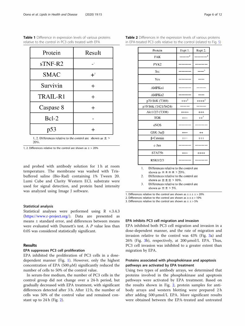
Table 1 Difference in expression levels of various proteinsrelative to the control in PC3 cells treated with EPA
1, 2. Differences relative to the control are shown as ± > 20%
Table 2 Differences in the expression levels of various proteinsin EPA-treated PC3 cells relative to the control (related to Fig. 5)
1. Differences relative to the control are shown as ± ± ± ± > 20%2. Differences relative to the control are shown as ± ± ± > 10%3. Differences relative to the control are shown as ± ± > 5%
Oono et al. Lipids in Health and Disease (2020) 19:15 Page 6 of 12
control groups using the phosphokinase as comparedto the apoptosis array (Figs. 4 and 5). The expression ofapoptosis-related proteins including soluble tumor ne-crosis factor receptor (sTNF-R)2, second mitochondria-derived activators of caspase (SMAC), Survivin, tumornecrosis factor related apoptosis-inducing ligand recep-tor (TRAIL-R)1, caspase 8, B-cell lymphoma (Bcl)-2,and p53 differed by more than 20% between the twogroups (Table 1), with sTNF-R2 and Bcl-2 being down-regulated and the other proteins being upregulated byEPA treatment.The phosphokinase array data showed increased or
decreased phosphorylation of several kinases (Fig. 5 andTable 2). EPA reduced the phosphorylation of severalproteins by over 20% relative to the control, includingPyk2, endothelial nitric oxide synthase (eNOS), c-Jun,and ribosomal S6 kinases (RSK)1/2/3.
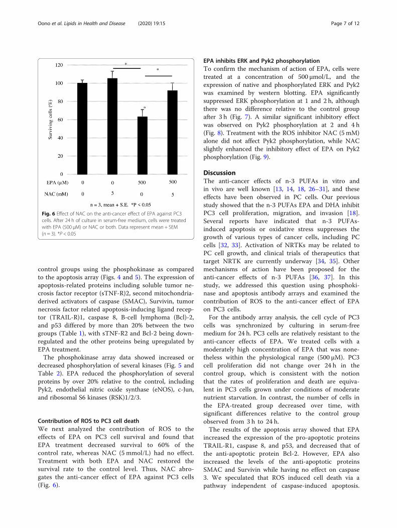
Contribution of ROS to PC3 cell deathWe next analyzed the contribution of ROS to theeffects of EPA on PC3 cell survival and found thatEPA treatment decreased survival to 60% of thecontrol rate, whereas NAC (5 mmol/L) had no effect.Treatment with both EPA and NAC restored thesurvival rate to the control level. Thus, NAC abro-gates the anti-cancer effect of EPA against PC3 cells(Fig. 6).
EPA inhibits ERK and Pyk2 phosphorylationTo confirm the mechanism of action of EPA, cells weretreated at a concentration of 500 μmol/L, and theexpression of native and phosphorylated ERK and Pyk2was examined by western blotting. EPA significantlysuppressed ERK phosphorylation at 1 and 2 h, althoughthere was no difference relative to the control groupafter 3 h (Fig. 7). A similar significant inhibitory effectwas observed on Pyk2 phosphorylation at 2 and 4 h(Fig. 8). Treatment with the ROS inhibitor NAC (5 mM)alone did not affect Pyk2 phosphorylation, while NACslightly enhanced the inhibitory effect of EPA on Pyk2phosphorylation (Fig. 9).
DiscussionThe anti-cancer effects of n-3 PUFAs in vitro andin vivo are well known [13, 14, 18, 26–31], and theseeffects have been observed in PC cells. Our previousstudy showed that the n-3 PUFAs EPA and DHA inhibitPC3 cell proliferation, migration, and invasion [18].Several reports have indicated that n-3 PUFAs-induced apoptosis or oxidative stress suppresses thegrowth of various types of cancer cells, including PCcells [32, 33]. Activation of NRTKs may be related toPC cell growth, and clinical trials of therapeutics thattarget NRTK are currently underway [34, 35]. Othermechanisms of action have been proposed for theanti-cancer effects of n-3 PUFAs [36, 37]. In thisstudy, we addressed this question using phosphoki-nase and apoptosis antibody arrays and examined thecontribution of ROS to the anti-cancer effect of EPAon PC3 cells.For the antibody array analysis, the cell cycle of PC3
cells was synchronized by culturing in serum-freemedium for 24 h. PC3 cells are relatively resistant to theanti-cancer effects of EPA. We treated cells with amoderately high concentration of EPA that was none-theless within the physiological range (500 μM). PC3cell proliferation did not change over 24 h in thecontrol group, which is consistent with the notionthat the rates of proliferation and death are equiva-lent in PC3 cells grown under conditions of moderatenutrient starvation. In contrast, the number of cells inthe EPA-treated group decreased over time, withsignificant differences relative to the control groupobserved from 3 h to 24 h.The results of the apoptosis array showed that EPA
increased the expression of the pro-apoptotic proteinsTRAIL-R1, caspase 8, and p53, and decreased that ofthe anti-apoptotic protein Bcl-2. However, EPA alsoincreased the levels of the anti-apoptotic proteinsSMAC and Survivin while having no effect on caspase3. We speculated that ROS induced cell death via apathway independent of caspase-induced apoptosis.
Fig. 6 Effect of NAC on the anti-cancer effect of EPA against PC3cells. After 24 h of culture in serum-free medium, cells were treatedwith EPA (500 μM) or NAC or both. Data represent mean + SEM(n = 3). *P < 0.05
Oono et al. Lipids in Health and Disease (2020) 19:15 Page 7 of 12

To test this hypothesis, we evaluated the effect of theROS inhibitor NAC on EPA-treated cells. First, wemeasured ROS generation at two time points, 2 and24 h. In these experiments, we observed relevant (notsignificant) ROS generation induced by EPA only at2 h, and NAC reduced the ROS level to the control(data not shown). We observed that the addition ofNAC abrogated the anti-cancer effects of EPA (Fig. 6),suggesting that ROS induced by EPA suppressed PC3cell proliferation. This is supported by previous re-ports showing that the anti-cancer effects of n-3PUFAs are mediated by ROS in liver cancer and PCcells [23, 24].We also investigated the contribution of cell prolif-
eration pathways to the anti-cancer effects of EPAusing a phosphokinase antibody array. The results
showed that Pyk2, eNOS, c-Jun, and RSK1/2/3 weredownregulated by over 20% by EPA treatment ascompared to the control group. Western blot analysisrevealed that ERK and Pyk2 phosphorylation wereinhibited by EPA (Figs. 7 and 8). Suppressing ROSproduction with NAC did not affect the inhibitory ef-fects of EPA on Pyk2 phosphorylation, suggesting thatthe suppression of Pyk2 phosphorylation by EPA doesnot involve modulation of ROS. Pyk2 is a member ofthe focal adhesion kinase (FAK) family of NRTK andplays important roles in cell survival, proliferation,and migration [38–41]. In our experiments, FAKphosphorylation was also suppressed by EPA treat-ment (Table 2). FAK is localized at the site of celladhesion with the extracellular matrix along withintegrins, whereas Pyk2 associates with integrin within
Fig. 7 Effect of EPA on ERK1/2 phosphorylation. Upper panel: Representative results of western blot analysis. Lower panel: Pixel density of thecontrol group at the indicated times was adjust to 100%; relative pixel density of EPA-treated PC3 cells is shown. Data represent mean + SEM(n = 3). *P < 0.05
Oono et al. Lipids in Health and Disease (2020) 19:15 Page 8 of 12

the cell [42, 43]. Since several types of integrins areexpressed in PC3 cells and interact with extracellularmatrix, inhibition of activated FAK at these sites mayprevent cell adhesion and migration [44]. The N-terminal domain of FAK has phosphorylation sites forthe cell proliferation-related factors epidermal growthfactor receptor, c-Met, and Src [45]. The antibodyused in the antibody array detected phosphorylatedtyrosine 402 of Pyk2, which is necessary for Src acti-vation [40] and was slightly reduced in our experi-ment (Table 2). It was reported that Pyk2 activationinduced smooth muscle contraction by activatingRho-associated kinase signaling [46], and FAK andPyk2 show similar activities on this pathway in the
regulation of cell migration [38, 47, 48]. Activation ofboth factors leads to abnormalities in cell morphologyand movement associated with malignant transformation.Our results suggest that EPA negatively regulates theproliferation, migration, and invasion of PC3 cells viainhibition of Pyk2 and FAK [18].Pyk2 is an NRTK that is activated by increased intra-
cellular Ca2+ concentrations [49, 50], which are inturn increased by n-3 PUFAs and ROS [23, 51–53].In our results, the n-3 PUFA EPA inhibited Pyk2phosphorylation, whereas ROS inhibition by NAC didnot affect the inhibitory effects of EPA, suggestingthat EPA blocks Pyk2 activation independent of intra-cellular Ca2+ levels.
Fig. 8 Effect of EPA on Pyk2 phosphorylation. Upper panel: Representative result of western blot analysis. Lower panel: Pixel density of thecontrol group at the indicated times was adjusted to 100%; relative pixel density of EPA-treated PC3 cells is shown. Data represent mean + SEM(n = 3). *P < 0.05
Oono et al. Lipids in Health and Disease (2020) 19:15 Page 9 of 12
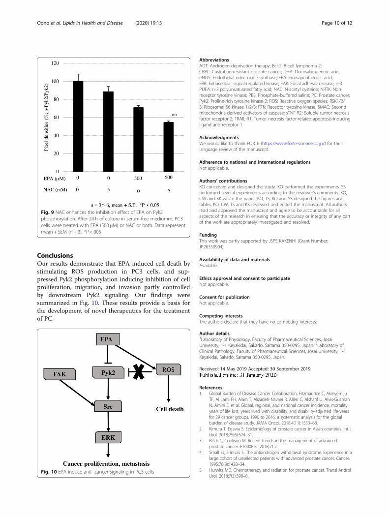
ConclusionsOur results demonstrate that EPA induced cell death bystimulating ROS production in PC3 cells, and sup-pressed Pyk2 phosphorylation inducing inhibition of cellproliferation, migration, and invasion partly controlledby downstream Pyk2 signaling. Our findings weresummarized in Fig. 10. These results provide a basis forthe development of novel therapeutics for the treatmentof PC.
AbbreviationsADT: Androgen deprivation therapy; Bcl-2: B-cell lymphoma 2;CRPC: Castration-resistant prostate cancer; DHA: Docosahexaenoic acid;eNOS: Endothelial nitric oxide synthase; EPA: Eicosapentaenoic acid;ERK: Extracellular signal-regulated kinase; FAK: Focal adhesion kinase; n-3PUFA: n-3 polyunsaturated fatty acid; NAC: N-acetyl cysteine; NRTK: Non-receptor tyrosine kinase; PBS: Phosphate-buffered saline; PC: Prostate cancer;Pyk2: Proline-rich tyrosine kinase-2; ROS: Reactive oxygen species; RSK1/2/3: Ribosomal S6 kinase 1/2/3; RTK: Receptor tyrosine kinase; SMAC: Secondmitochondria-derived activators of caspase; sTNF-R2: Soluble tumor necrosisfactor receptor 2; TRAIL-R1: Tumor necrosis factor-related apoptosis-inducingligand and receptor 1
AcknowledgmentsWe would like to thank FORTE (https://www.forte-science.co.jp/) for theirlanguage review of the manuscript.
Adherence to national and international regulationsNot applicable.
Authors’ contributionsKO conceived and designed the study. KO performed the experiments. SSperformed several experiments according to the reviewer’s comments. KO,CW and KK wrote the paper. KO, TS, KO and SS designed the figures andtables. KO, CW, TS and KK reviewed and edited the manuscript. All authorsread and approved the manuscript and agree to be accountable for allaspects of the research in ensuring that the accuracy or integrity of any partof the work are appropriately investigated and resolved.
FundingThis work was partly supported by JSPS KAKENHI (Grant Number:JP26350904).
Availability of data and materialsAvailable.
Ethics approval and consent to participateNot applicable.
Consent for publicationNot applicable.
Competing interestsThe authors declare that they have no competing interests.
Author details1Laboratory of Physiology, Faculty of Pharmaceutical Sciences, JosaiUniversity, 1-1 Keyakidai, Sakado, Saitama 350-0295, Japan. 2Laboratory ofClinical Pathology, Faculty of Pharmaceutical Sciences, Josai University, 1-1Keyakidai, Sakado, Saitama 350-0295, Japan.
Received: 14 May 2019 Accepted: 30 September 2019
References1. Global Burden of Disease Cancer Collaboration, Fitzmaurice C, Akinyemiju
TF, Al Lami FH, Alam T, Alizadeh-Navaei R, Allen C, Alsharif U, Alvis-GuzmanN, Amini E, et al. Global, regional, and national cancer incidence, mortality,years of life lost, years lived with disability, and disability-adjusted life-yearsfor 29 cancer groups, 1990 to 2016: a systematic analysis for the globalburden of disease study. JAMA Oncol. 2018;4(11):1553–68.
2. Kimura T, Egawa S. Epidemiology of prostate cancer in Asian countries. Int JUrol. 2018;25(6):524–31.
3. Ritch C, Cookson M. Recent trends in the management of advancedprostate cancer. F1000Res. 2018;21:7.
4. Small EJ, Srinivas S. The antiandrogen withdrawal syndrome. Experience in alarge cohort of unselected patients with advanced prostate cancer. Cancer.1995;76(8):1428–34.
5. Hurwitz MD. Chemotherapy and radiation for prostate cancer. Transl AndrolUrol. 2018;7(3):390–8.
Fig. 9 NAC enhances the inhibition effect of EPA on Pyk2phosphorylation. After 24 h of culture in serum-free mediumm, PC3cells were treated with EPA (500 μM) or NAC or both. Data representmean + SEM (n + 3). *P < 005
Fig. 10 EPA-induce anti- cancer signaling in PC3 cells
Oono et al. Lipids in Health and Disease (2020) 19:15 Page 10 of 12
6. Hussain M, Fizazi K, Saad F, Rathenborg P, Shore N, Ferreia U, Ivashchenko P,Demirthan E, Modelska K, Phung D, et al. Enzalutamide in men withnonmetastatic, castration-resistant prostate cancer. N Engl J Med. 2018;378(26):2465–74.
7. Balk EM, Lichtenstein AH. Omega-3 fatty acids and cardiovascular disease:summary of the 2016 agency of healthcare research and quality evidencereview. Nutrients. 2017;9(8). https://doi.org/10.3390/nu9080865.
8. Harris WS. Fish oils and plasma lipid and lipoprotein metabolism in humans:a critical review. J Lipid Res. 1989;30(6):785–807.
9. Takenouchi Y, Ohtake K, Nobe K, Kasono K. Eicosapentaenoic acid ethylester improves endothelial dysfunction in type 2 diabetic mice. LipidsHealth Dis. 2018;17(1):118.
10. Serini S, Calviello G. Modulation of Ras/ERK and phosphoinositide signalingby long-chain n-3 PUFA in breast cancer and their potential complementaryrole in combination with targeted drugs. Nutrients. 2017;9(3). https://doi.org/10.3390/nu9030185.
11. Sawada N, Inoue M, Iwasaki M, Sasazuki S, Shimazu T, Yamaji T, Takachi R,Tanaka Y, Mizokami M, Tsugane S, et al. Consumption of n-3 fatty acids andfish reduces risk of hepatocellular carcinoma. Gastroenterology. 2012;142(7):1468–75.
12. Hidaka A, Shimazu T, Sawada N, Yamaji T, Iwasaki M, Sasazuki S, Inoue M,Tsugane S. Japan public health center-based prospective study group. Fish,n-3 PUFA consumption, and pancreatic cancer risk in Japanese: a large,population-based, prospective cohort study. Am J Clin Nutr. 2015;102(6):1490–7.
13. Das UN. Tumoricidal action of cis-unsaturated fatty acids and theirrelationship to free radicals and lipid peroxidation. Cancer Lett. 1991;56(3):235–43.
14. Bégin ME, Das UN, Ells G, Horrobin DF. Selective killing of human cancercells by polyunsaturated fatty acids. Prostaglandins Leukot Med. 1985;19(2):177–86.
15. Das UN, Begin ME, Ells G, Huang YS, Horrobin DF. Polyunsaturated fattyacids augment free radical generation in tumor cells in vitro. BiochemBiophys Res Commun. 1987;145(1):15–24.
16. Das UN, Huang YS, Begin ME, Ells G, Horrobin DF. Uptake and distributionof cis-unsaturated fatty acids and their effect on free radical generation innormal and tumor cells in vitro. Free Radic Biol Med. 1987;3(1):9–14.
17. MacLean CH, Newberry SJ, Mojica WA, Khanna P, Issa AM, Suttorp MJ, LimYW, Traina SB, Hilton L, Garland R, et al. Effects of omega-3 fatty acids oncancer risk: a systematic review. JAMA. 2006;295(4):403–15.
18. Oono K, Takahashi K, Sukehara S, Kurosawa H, Matsumura T, Taniguchi S,Ohta S. Inhibition of PC3 human prostate cancer cell proliferation, invasionand migration by eicosapentaenoic acid and docosahexaenoic acid. MolClin Oncol. 2017;7(2):217–20.
19. Chang YM, Kung HJ, Evans CP. Nonreceptor tyrosine kinases in prostatecancer. Neoplasia. 2007;9(2):90–100.
20. Watson RW, Fitzpatrick JM. Targeting apoptosis in prostate cancer: focus oncaspases and inhibitors of apoptosis proteins. BJU Int. 2005;96(Suppl 2):30–4.
21. Moloney JN, Cotter TG. ROS signaling in the biology of cancer. Semin CellDev Biol. 2018;80:50–64.
22. Thannickal VJ, Fanburg BL. Reactive oxygen species in cell signaling. Am JPhysiol Lung Cell Mol Physiol. 2000;279(6):1005–28.
23. Zhang Y, Han L, Qi W, Cheng D, Ma X, Hou L, Cao X, Wang C.Eicosapentaenoic acid (EPA) induced apoptosis in HepG2 cells throughROS-Ca2+-JNK mitochondrial pathways. Biochem Biophys Res Commun.2015;456(4):926–32.
24. Ding Y, Ren K, Dong H, Song F, Chen J, Guo Y, Liu Y, Tao W, Zhang Y.Flavonoids from persimmon (Diospyros kaki L.) leaves inhibit proliferationand induce apoptosis in PC-3 cells by activation of oxidative stress andmitochondrial apoptosis. Chem Biol Interact. 2017;275:210–7.
25. Shin S, Jing K, Jeong S, Kim N, Song KS, Heo JY, Park JH, Seo KS, Han J, ParkJI, et al. The omega-3 polyunsaturated fatty acid DHA induces simultaneousapoptosis and autophagy via mitochondrial ROS-mediated Akt-mTORsignaling in prostate cancer cells expressing mutant p53. Biomed Res Int.2013;2013:568671.
26. Bégin ME, Ells G, Das UN, Horrobin DF. Differential killing of humancarcinoma cells supplemented with n-3 and n-6 polyunsaturated fatty acids.J Natl Cancer Inst. 1986;77(5):1053–62.
27. Corsetto PA, Montorfano G, Zava S, Jovenitti IE, Cremona A, Berra B, RizzoAM. Effects of n-3 PUFAs on breast cancer cells through their incorporationin plasma membrane. Lipids Health Dis. 2011;10:73–89.
28. McEntee MF, Ziegler C, Reel D, Tomer K, Shoieb A, Ray M, Li X, Neilsen N,Lih FB, O’Rourke D, et al. Dietary n-3 polyunsaturated fatty acids enhancehormone ablation therapy in androgen therapy in androgen-dependentprostate cancer. Am J Pathol. 2008;173(1):229–41.
29. Altenburg JD, Siddiqui RA. Omega-3 polyunsaturated fatty acids down-modulate CXCR4 expression and function in MDA-MB-231 breast cancercells. Mol Cancer Res. 2009;7(7):1013–20.
30. Berquin IM, Edwards IJ, Chen YQ. Multi-targeted therapy of cancer byomega-3 fatty acids. Cancer Lett. 2008;269(2):363–77.
31. Davidson LA, Nguyen DV, Hokanson RM, Callaway ES, Isett RB, Turner ND,Dougherty ER, Wang N, Lupton JR, Carroll RJ, et al. Chemopreventive n-3polyunsaturated fatty acids reprogram genetic signatures during colon cancerinitiation and progression in the rat. Cancer Res. 2004;64(18):6797–804.
32. Song EA, Kim H. Docosahexaenoic acid induces oxidative DNA damage andapoptosis, and enhances the chemosensitivity of cancer cells. Int J Mol Sci.2016;17(8). https://doi.org/10.3390/ijms17081257.
33. Camargo CQ, Brunetta HS, Nunes EA. Effects of cotreatment with omega-3polyunsaturated fatty acids and anticancer agents on oxidative stressparameters: a systematic review of in vitro, animal, and human studies. NutrRev. 2018;76(10):765–77.
34. Arora A, Scholar EM. Role of tyrosine kinase inhibitors in cancer therapy. JPharmacol Exp Ther. 2005;315(3):971–9.
35. Araujo JC, Trudel GC, Saad F, Armstrong AJ, Yu EY, Bellmunt J, WildingG, McCaffrey J, Serrano SV, Matveev VB, et al. Docetaxel and dasatinibor placebo in men with metastatic castration-resistant prostate cancer(READY): a randomized, double-blind phase 3 trial. Lancet Oncol. 2013;14(13):1307–16.
36. D’Eliseo D, Velotti F. Omega-3 fatty acids and cancer cell cytotoxicity:implications for multi-targeted cancer therapy. J Clin Med. 2016;5(2). https://doi.org/10.3390/jcm5020015.
37. Menedez JA, Lupu R. Fatty acid synthase and the lipogenetic phenotype incancer pathogenesis. Nat Rev Cancer. 2007;7(10):763–77.
38. Avraham H, Park SY, Schinkmann K, Avraham S. RAFTK/Pyk2-mediatedcellular signaling. Call Signal. 2000;12(3):123–33.
39. Sun CK, Man K, Ng KT, Ho JW, Lim ZX, Cheng Q, Lo CM, Poon RT, Fan ST.Proline-rich tyrosine kinase 2 (Pyk2) promotes proliferation and invasivenessof hepatocellular carcinoma cells through c-Src/ERK activation.Carcinogenesis. 2008;29(11):2096–105.
40. Zhao M, Finlay D, Zharkikh I, Vuori K. Novel role of Src in priming Pyk2phosphorylation. PLoS One. 2016;11(2):e0149231.
41. Perez J, Torres RA, Rocic P, Cismowski MJ, Weber DS, Darley-Usmar VM,Lucchesi PA. PYK2 signaling is required for PDGF-dependent vascularsmooth muscle cell proliferation. Am J Physiol Cell Physiol. 2011;301(1):C242–51.
42. Matsuya M, Sasaki H, Aoto H, Mitaka T, Nagura K, Ohba T, Ishino M,Takahashi S, Suzuki R, Sasaki T. Cell adhesion kinase beta forms a complexwith a new member, hic-, of proteins localized at focal adhesions. J BiolChem. 1998;273(2):1003–14.
43. Murphy JM, Park H, Lim STS. FAK and Pyk2 in disease. Front Biol. 2016;11(1):1–9.
44. Juan-Rivera MC, Martinez-Ferrer M. Integrin inhibitors in prostate cancer.Cancers (Basel). 2018;10(2). https://doi.org/10.3390/cancers10020044.
45. Lee BY, Trimpson P, Horvath LG, Daly RJ. FAK signaling in human cancer asa target for therapeutics. Pharmacol Ther. 2015;146:132–49.
46. Mills RD, Mita M, Walsh MP. A role for the Ca2+-dependent tyrosine kinasePyk2 in tonic depolarization-induced vascular smooth muscle contraction. JMuscle Res Cell Motil. 2015;36(6):479–89.
47. Miller NL, Kleinschmidt EG, Schlaepfer DD. Rho GEFs in cell motility: novellinks between Rgnef and focal adhesion kinase. Curr Mol Med. 2014;14(2):221–34.
48. Tornin J, Hermida-Prado F, Padda RS, Gonzalez MV, Alvarez-Fernandez C,Rey V, Martinez-Cruzado L, Estuinan O, Menendez ST, Fernandez-Nevado L,et al. FUS-CHOP promotes invasion in myxoid liposarcoma through a SRC/FAKRHO/ROCK-dependent pathway. Neoplasia. 2018;20(1):44–56.
49. Rozengurt E. Mitogenic signaling pathways induced by G protein-coupledreceptors. J Cell Physiol. 2007;213(3):589–602.
50. Hirschler-Laszkiewica I, Chen SJ, Bao L, Wang J, Zhang XQ, ShanmughapriyaS, Keefer K, Madesh M, Cheung JY, Miller BA. The human ion channelTRPM2 modulates neuroblastoma cell survival and mitochondrial functionthrough Pyk2, CREB, and MCU activation. Am J Physiol Cell Physiol. 2018;315(4):C571–86.
Oono et al. Lipids in Health and Disease (2020) 19:15 Page 11 of 12

51. Jakobsen CH, Størvold GL, Bremseth H, Follestad T, Sand K, Mack M, OlsenKS, Lundemo AG, Iversen JG, Krokan HE, et al. DHA induces ER stress andgrowth arrest in human colon cancer cells: associations with cholesteroland calcium homeostasis. J Lipid Res. 2008;49(10):2089–100.
52. Hempel N, Trebak M. Crosstalk between calcium and reactive oxygenspecies signaling in cancer. Cell Calcium. 2017;63:70–96.
53. Yamamoto S, Shimazu S, Kiyonaka S, Takahashi N, Wajima T, Hara Y, NegoroT, Hiroi T, Kiuchi Y, Okada T, et al. TRPM2-mediated Ca2+ influx induceschemokine production in monocytes that aggravates inflammatoryneutrophil infiltration. Nat Med. 2008;14(7):738.
Publisher’s NoteSpringer Nature remains neutral with regard to jurisdictional claims inpublished maps and institutional affiliations.
Oono et al. Lipids in Health and Disease (2020) 19:15 Page 12 of 12
Copyright © 2022 FDOKUMEN