Contrasting Effects of Vocabulary Knowledge on Temporal and Parietal Brain Structure across Lifespan
41
Contrasting effects of vocabulary knowledge on temporal and parietal brain structure across lifespan Fiona M. Richardson 1,2 , Michael S. C. Thomas 1 , Roberto Filippi 1 , Helen Harth 1 & Cathy J. Price 2 1. Developmental Neurocognition Lab, School of Psychology, Birkbeck College, University of London. 2. Wellcome Trust Centre for Neuroimaging, University College London. Word count (main text): 7223 words Address for correspondence: Dr. Fiona Richardson Wellcome Trust Centre for Neuroimaging, UCL 12 Queen Square London WC1N 3BG Email: [email protected] Tel.: +44 (0)20 7833 7488 Fax: +44 (0)20 7813 1420 Acknowledgements This research was funded by the Wellcome Trust. Fiona Richardson was supported by an MRC Career Establishment Grant G0300188 and British Academy Grant SG- 40400 awarded to Dr Michael Thomas. Our thanks to Janice Glensman, Amanda Brennan and David Bradbury for their assistance during scanning and neuropsychological testing.
Transcript of Contrasting Effects of Vocabulary Knowledge on Temporal and Parietal Brain Structure across Lifespan
Contrasting effects of vocabulary knowledge on temporal and
parietal brain structure across lifespan
Fiona M. Richardson1,2, Michael S. C. Thomas1, Roberto Filippi1,
Helen Harth1 & Cathy J. Price2
1. Developmental Neurocognition Lab, School of Psychology, Birkbeck
College, University of London.
2. Wellcome Trust Centre for Neuroimaging, University College London.
Word count (main text): 7223 words
Address for correspondence:
Dr. Fiona Richardson Wellcome Trust Centre for Neuroimaging, UCL 12 Queen Square London WC1N 3BG Email: [email protected] Tel.: +44 (0)20 7833 7488 Fax: +44 (0)20 7813 1420
Acknowledgements
This research was funded by the Wellcome Trust. Fiona Richardson was supported by
an MRC Career Establishment Grant G0300188 and British Academy Grant SG-
40400 awarded to Dr Michael Thomas. Our thanks to Janice Glensman, Amanda
Brennan and David Bradbury for their assistance during scanning and
neuropsychological testing.
Abstract
Using behavioral, structural and functional imaging techniques, we demonstrate
contrasting effects of vocabulary knowledge on temporal and parietal brain structure
in 47 healthy volunteers who ranged in age from 7 to 73. In the left posterior
supramarginal gyrus, vocabulary knowledge was positively correlated with gray
matter density in teenagers but not adults. This region was not activated during
auditory or visual sentence processing and activation was unrelated to vocabulary
skills. Its gray matter density may reflect the use of an explicit learning strategy that
links new words to lexical or conceptual equivalents, as used in formal education and
second language acquisition. By contrast, in left posterior temporal regions, gray
matter as well as auditory and visual sentence activation correlated with vocabulary
knowledge throughout lifespan. We propose that these effects reflect the acquisition
of vocabulary through context, when new words are learnt within the context of
semantically and syntactically related words.
Introduction
Vocabulary knowledge gradually increases across lifespan and is essential for both
auditory and written sentence comprehension. Previous studies have suggested that
gray matter density in the posterior supramarginal gyri (pSMG) provides a marker for
the number of words learnt (Lee et al., 2007). However the mechanisms underlying
this effect remain poorly understood. The aim of the present study was to investigate
the effect of vocabulary knowledge on gray matter density in multiple brain regions
and to establish whether the observed effects were dependent on age and, therefore,
potentially indicative of different vocabulary learning strategies. In the following, we
describe the background and rationale for our study.
The association of the pSMG with language learning was first reported by
Mechelli et al. (2004) who identified greater gray matter density in this region for
bilinguals than monolinguals. Moreover, increases in gray matter density were found
to be positively correlated with second language proficiency. The significance of this
effect was investigated further by Lee et al. (2007) who found that vocabulary
knowledge in English-speaking monolingual teenagers was positively correlated with
gray matter in the same pSMG region that was implicated in adult second language
learning. In addition, using diffusion tensor imaging (DTI), Lee and colleagues
identified direct anatomical connections from the pSMG to both the anterior
supramarginal gyrus (aSMG) and the angular gyrus. These regions have been
associated with phonological and semantic decisions, respectively (Demonet et al.,
1992; Devlin, Matthews & Rushworth, 2003; Gathercole, 2006; Gathercole, Hitch,
Service & Martin, 1997; Golestani & Pallier, 2006; Price, More, Humphreys & Wise,
1997). Thus Lee et al. proposed that the pSMG is an important site for linking
phonological and semantic information, which is central to vocabulary acquisition in
both a first and second language.
Word knowledge is mainly acquired through exposure to everyday language.
In this instance, vocabulary is increased by reading or hearing new words set within
the context of known words that have different but related meanings from which the
meaning of the new word can be deduced. Here, vocabulary learning occurs via
semantic and syntactic associations and prior knowledge of morphological structure
(see Kilian et al. 1995; and Nagy et al., 1985). However, new words can also be learnt
by forming an explicit link between a new word and a lexical equivalent (e.g.
“purchase” is another word for “buy”) or a definition (e.g. An “ATOM” is the
smallest particle that comprises a chemical element; “GARGANTUAN” has a
similar meaning to “gigantic”; “EGREGIOUS” means very bad). This kind of word-
learning may take place through instruction by a parent or caregiver, in a formal
educational setting, or be self-motivated in a revision-based scenario, in which an
individual is specifically focused on learning a new terminology. This type of
expansion of vocabulary also increases knowledge of morphological relationships
between words (e.g. “bake” and “baker”), that can be generalised when new words
are introduced.
Critically, the findings of Lee et al. (2007) were based on monolingual
adolescents aged between 12 and 15 years. These individuals were in full time
education and were therefore expected to be actively engaged in expanding their
vocabulary, learning new words as labels for new concepts, and lexicalizing existing
concepts. Of the two learning strategies for acquiring vocabulary knowledge, explicit
learning mainly through instruction is more likely to be exploited by children rather
than adults who are no longer in formal education who most probably continue to
learn vocabulary mainly through context with a minimal contribution from explicit
instruction. Therefore, as the influence of different learning strategies alters across
lifespan, we would expect the relationship between vocabulary and brain structure to
be age dependent. To investigate this hypothesis, we combined structural and
functional imaging using a cross-section of participants of different ages. This study
used a different sample of participants and different vocabulary measures to those
reported by Lee et al., (2007). Replication of the vocabulary effects us ing a different
vocabulary test allows us to assess the generalizability of the effect across different
vocabulary measures. Our expectation was that learning vocabulary through context
would be reflected by a positive correlation between vocabulary knowledge and gray
matter density that is: (a) continuous across lifespan and (b) in brain regions that are
activated by semantic and syntactic associations during reading and listening tasks.
By contrast, we expected that learning through an explicit strategy would be reflected
by a positive correlation between vocabulary knowledge and gray matter density that
was (a) limited to those undergoing formal education and (b) in regions that were not
necessarily involved in reading or listening to speech.
To address these hypotheses, we studied a group of 47 healthy participants
between the ages of 7 to 73. We assessed gray matter density using structural
magnetic resonance imaging (MRI) while activation for reading and listening to
language stimuli was assessed using functional MRI in the same sample of
participants. We also tested whether the local differences in gray matter we identified
were susceptible to influence from age-related differences in gray matter. For
instance, numerous studies have reported that the well-known decline in gray matter
post-adolescence (Giedd et al., 1999; Gogtay et al., 2004; Paus, 2005) is characterized
by different trajectories across frontal, temporal and parietal brain regions (Allen et
al., 2005; Giedd et al., 1999, Gogtay et al., 2004; Lenroot & Giedd, 2006; Sowell et
al., 2003; Wilke et al., 2007). Consequently, a decline in gray matter with age may
make it more difficult to detect small increases in gray matter with vocabulary and
this confound may be stronger in some regions than others and change with age. We
therefore investigated whether differences in the trajectory of decline in gray matter
with age could account for any age dependent differences in the effect of vocabulary
knowledge.
To our knowledge, the combination of behavioral, structural and functional
differences has not been tested before in the context of normal development, although
previous studies of abnormal development have suggested that dyslexic individuals
can have atypical activation and gray matter in the same regions (Brambati et al.,
2006, 2004; Chételat et al., 2008; Hoeft et al., 2007; Hyde et al., 2007, 2006; Silani et
al., 2005). Evidence that vocabulary acquisition affects the same regions in the
structural and functional images would also suggest that increased gray matter might
be a consequence of increased neuronal activation, or vice versa.
Materials and Methods
Participants
The participants were 48 right-handed volunteers (24 males) aged between 7 and 73
years, who had English as their first language. Children and teenagers were recruited
through existing contacts in local schools in the London area, and adults were
recruited mainly through advertisement in local further education institutions. All
participants had normal or normal-corrected vision, with no reported hearing
difficulties or disturbances in speech comprehension, speech production, or reading.
None of the participants had a preference, or history of preference, for using their left
hand or left foot, none had right lateralized language activation (see below) and none
were bilingual, multilingual or had any special linguistic training.
One male child was excluded from the functional analyses due to an
incomplete coverage of temporal brain regions. One teenager was excluded from the
structural analyses due to a poor quality image caused by motion artefacts, leaving a
total of 47 participants for each analysis. Participant demographics can be seen in
Table 1. This study was approved by the joint ethical committee of the Institute of
Neurology and the National Hospital for Neurology and Neurosurgery, London, UK.
Informed consent (written consent from a parent or guardian in the case of young
children under 16) was obtained from all participants.
================== Insert Table 1 about here ==================
Behavioral tests
All participants carried out two psychometric tests: (i) the British Picture Vocabulary
Scales II (BPVS II – Dunn et al., 1997), and (ii) the Matrices task from the British
Ability Scale II (BAS II – Elliot et al., 1997). All participants between 7 and 11 years
also carried out the Reading test from the BAS-II to ensure that they had sufficient
reading ability to carry out the functional imaging paradigm. The Reading test
consists of ninety words divided into nine blocks of ten words. Children start the test
at an age appropriate starting point and read aloud a series of words presented on a
card. The words increase in complexity as the test progresses. The test is continued
until the child makes eight or more consecutive errors. An ability score that takes into
account the difficulty of the test items completed is then obtained using a look-up
table supplied with the test. Children with a minimum reading age of seven years were
considered to be at an appropriate level to carry out the reading task used in the fMRI
paradigm. All children who took part in this study had a reading level in line with or
in advance of their chronological age (reading age range of 7:4 to 15:3).
The BPVS-II is a measure of an individual’s receptive vocabulary for Standard
English (Dunn et al., 1997). In this test, participants are asked to select (from four
options) the picture that most accurately matches a word (such as “ladder”, or
“collision”) read aloud by the tester. The test consists of fourteen sets of words of
increasing levels of difficulty, each containing twelve items. Each set has an
approximate age-range indicator, which is used to select the appropriate starting point
of the test. The test is conducted until the participant makes eight or more incorrect
responses within a set. The raw score from this test takes into account the level of
difficulty of the test items administered and the number of incorrect responses over
the duration of the test.
The Matrices task from the BAS-II was used as a measure of general cognitive
ability. In this test, participants are shown an incomplete matrix of black and white
abstract figures, with each matrix consisting of either four or nine cells. Participants
are required to select the most appropriate pattern to complete the matrix from six
potential tiles by pointing to or reading the number of the tile that best completes the
matrix. Participants first complete four practice items, and then begin the test at an
age-appropriate level, which is indicated on the test (previous items are administered
should they fail on the first three test items). The test is discontinued if the participant
makes five failures out of six consecutive items. An ability score, which takes into
account the number and level of difficulty of the test items completed, is then
obtained from a look-up table supplied with the test.
Functional imaging
Experimental paradigm
The experimental paradigm included a sentence processing task that was carried out
in (i) auditory and (ii) visual modalities, where participants listened passively to
auditory sentence stimuli, and silently read visual sentences. They also listened to or
read scrambled sentences (strings of words that did not constitute a meaningful
sentence) – tapping comprehension at the individual word level. The baseline task in
the auditory modality consisted of listening to the same speech recordings after they
had been rendered meaningless by digital reversal. In the visual modality, the baseline
task consisted of viewing the same words presented in an unrecognisable (false) font.
A passive listening/reading paradigm has the advantage of avoiding task-induced
strategies over and above the speech comprehension processes in which we were
interested. Although there was no in-scanner behavior to assess online speech
comprehension, we were able to correlate activation with vocabulary knowledge
measured outside the scanner. In addition, it should be noted that as the power in our
analysis came from inter-subject variation in auditory or written language processing
it was unnecessary to equate in-scanner performance across participants. Instead, we
ensured that each participant was attending to the words, and processing the
sentences, by conducting an unexpected memory task at the end of each scanning
session. The resulting memory scores were then included as covariates in our second
level statistical analyses of vocabulary ability (see below for more details).
Sentence stimuli
Sentence stimuli consisted of 80 sentences with 6-8 words per sentence. Familiar
words were selected to be suitable for children as young as seven years, and had a
Flesch-Kincaid grade level readability score of 1.3. Sentences were constructed using
high frequency (>20 per million) monosyllabic and bisyllabic nouns, verbs and
adjectives. Sentence sets were split into two groups (A and B) of equivalent
composition, for the purpose of presenting one set in an auditory and the other in a
visual format. Sentences were matched at the lexical level in terms of mean
frequency, imageability, and age of acquisition, and at the sentence level in terms of
mean word length and syllable length. These same sentence types were presented
across both visual and auditory modalities to ensure consistency across tasks. No
sentence was repeated across modality. The presentation of subsets A and B in either
an auditory or visual format was counterbalanced across participants. Scrambled
sentences were constructed from the same set of words as meaningful sentences,
consisting of initially grammatical sentences (e.g. “The cow chased the fat horse”),
which were then assigned a pseudo-random word order that did not form a meaningful
sentence (e.g. “Chased the the horse cow fat”). This condition is therefore fully
matched to the sentences at the lexical level. Examples of sentence stimuli can be seen
in the Appendix.
Procedure
Condition order was blocked but each stimulus was analyzed as a separate event.
Within each of four different scanning sessions (runs), there were 2 blocks of auditory
sentences, 2 blocks of visual sentences, 1 block of scrambled auditory sentences, 1
block of scrambled visual sentences, 3 blocks of auditory baseline and 3 blocks of
visual baseline. Within each block there were 37 words/baseline stimuli, grouped into
5 sequences/sentences with 0.5s between sequences/sentences, during which a
fixation cross was presented in the center of the screen. Within each
sequence/sentence, stimuli were presented at a rate of one per 0.4 seconds resulting in
a maximum duration of 3.2s for an eight-stimulus sequence/sentence. The auditory
and visual word presentation rates were equated by recording the auditory stimuli
from a female reading aloud the visual stimuli presented using the same script that
was to be used in the scanner. Each word was presented in a Helvetica font size 20.
Sentence change was indicated by an auditory beep, while in the visual condition the
first word of each sentence started with a capital letter.
Each task, consisting of an activation and baseline pair was preceded by an
appropriate visually displayed instruction (Helvetica, size 80): ‘Listen’ (auditory
sentence processing task) or ‘Read’ (visual sentence processing task). This instruction
was displayed for 2.2s, and was followed by an auditory pure tone, which sounded for
0.3s. Each activation and baseline condition had a total duration of 18s. The
presentation of activation and baseline conditions was separated by a brief auditory
pure tone which sounded for 0.3s, followed by a 0.2s fixation cross. At the end of
each activation and baseline task, there was a 1.5s pause before the onset of the next
task. This resulted in a total duration of 40.5s for an activation (meaningful or
scrambled sentences) and baseline pair. Each session commenced with a visual cue to
‘Get Ready…’ followed by a count down, during which dummy scans were acquired.
Memory test
All participants carried out a pen and paper memory test following scanning in order
to test their memory for sentences presented during the scanning session. Participants
were not informed of this test prior to scanning. The main purpose of this test was to
ensure that participants had been attending to the stimuli whilst in the scanner. These
scores were also used to control for any potential effects that memory for sentences
might have upon sentence processing in subsequent analyses conducted in SPM. This
test consisted of 24 sentences, 12 familiar sentences seen during scanning and 12 new
sentences previously unseen during scanning. There were six familiar sentences for
each of the two presentation modalities (auditory and visual). New sentences
consisted of six sentences constructed from previously unseen words, and six
sentences constructed from novel words. All participants scored above chance on this
test (test score, M = 70%, SD ± 11). These scores were adjusted to account for
incorrect as well as correct responses prior to being entered into our analyses. The
total of false positive responses was subtracted from the number of correct responses
for familiar sentences from each presentation modality. This resulted in two scores –
one for auditory memory for sentences and one for visual memory for sentences.
fMRI Data Acquisition
A Siemans 1.5T Sonata scanner was used to acquire a total of 768 T2*- weighted
echoplanar images with BOLD contrast (192 scans per 4 sessions). Each echoplanar
image comprised 30 axial slices of 2mm thickness with 1mm inter-slice interval and 3
x 3 mm in-plane resolution. Volumes were acquired with an effective repetition time
(TR) of 2.7s/volume and the first six (dummy) volumes of each run were discarded in
order to allow for T1 equilibration effects.
Functional image analysis
Pre-processing was conducted using statistical parametric mapping (SPM 2,
Wellcome Trust Centre for Neuroimaging, London, UK;
http://www.fil.ion.ucl.ac.uk/spm) running under Matlab 6.5.1 (Mathworks Inc.
Sherbon MA, USA). All volumes (excluding dummy scans) from each participant
were realigned using the first as a reference image and unwarped (Jesper et al., 2001),
adjusting for residual motion-related signal changes. The functional images were then
spatially normalised (Friston et al., 1995a) to a standard MNI-305 template using non-
linear basis functions. Functional data were spatially smoothed with a 6mm full-width
half-maximum isotrophic Gaussian kernel to compensate for residual variability after
spatial normalisation and to permit application of Gaussian random-field theory for
corrected statistical inference (Friston et al., 1995b).
Statistical analysis of functional data
At the first level, data for each participant were analyzed with high-pass filtering
using a set of discrete cosine basis functions with a cut-off period of 128 seconds.
Each sentence was modelled as a separate event within each condition and convolved
with a canonical hemodynamic response function (HRF). This resulted in 6 different
conditions at the first level, which were as follows:
A) Auditory sentences
B) Auditory words (scrambled sentences)
C) Auditory baseline (reversed words)
D) Visual sentences
E) Visual words (scrambled sentences)
F) Visual baseline (false font)
The following 4 contrasts were generated for each participant:
1) Auditory sentences: [sentences] – [baseline] (= A – C)
2) Visual sentences: [sentences] – [baseline] (= D – F )
3) Auditory words: [scrambled sentences] – [baseline] (= B – C)
4) Visual words: [scrambled sentences] – [baseline] (= E – F)
In addition, in order to ensure that language processing was predominantly
left- lateralized, two contrasts (one for each modality) combining activation for
sentences and words were generated at the first- level for each participant. These
contrasts were viewed for each participant and showed predominantly left-hemisphere
activation for language processing in both modalities. None of the subjects had right
lateralized language.
Two statistical analyses were conducted at the second- level using SPM 5. The
aim of these analyses was to identify regions where functional activation is predicted
by vocabulary score. Both analyses used the same model, but differed in terms of the
participant groups entered into the analysis:
Functional analysis 1: The effect of vocabulary on functional activation across
lifespan in all 47 participants.
Functional analysis 2: The effect of vocabulary on functional activation in
teenagers only (17 participants, 8 males, 9 females, mean age: 14.7 years, range 12.4-
17.5).
The model used was a second-level ANOVA, which included contrasts 1 to 4 (listed
above), and the following six covariates: (i) vocabulary score (BPVS –II raw score),
(ii) non-verbal problem solving ability (BAS-II: matrices ability score), (iii) auditory
memory for sentences, (iv) visual memory for sentences, (v) linear effects of age, and
(vi) nonlinear effects of age. The inclusion of covariates allowed us to identify the
effect of vocabulary upon the processing of auditory and visual sentences and words
whilst factoring out any potential variance due to the other factors. Significant effects
were corrected for multiple comparisons at p < 0.05, across the whole brain or in the
left and right posterior supramarginal gyrus regions of interest with a 10mm radius
centered on the peak co-ordinates in the left pSMG [x = -44, y = -54, z = 46] and right
pSMG [x = 52, y = -52, z = 44], as reported by Lee et al. (2007).
Structural imaging
Structural image acquisition
Focal gray matter density was estimated on the basis of T1-weighted anatomical
whole brain images acquired using a Siemans Sonata 1.5T MRI scanner (Siemans
Medical Systems, Erlangen, Germany). A T1-weighted Modified Driven Equilibrium
Fourier Transform (MDEFT) sequence (Deichmann, Schwarzbauer & Turner, 2004)
was used to acquire 176 sagittal partitions with an image matrix of 256x224 yielding a
final resolution of 1mm3 (TR/TE/TI = 12.24 ms/3.56 ms/530 ms).
Structural image analysis
Scans were analyzed using SPM 5 (Wellcome Department of Imaging Neuroscience,
http://www.fil.ion.ucl.ac.uk/spm). Pre-processing was performed using a generative
model that combines bias correction, image registration and tissue classification,
using default gray and white matter tissue probability maps (as described in
Ashburner & Friston, 2005). The resulting images were then smoothed using an
isotropic kernel of 8mm at full width half maximum (FWHM). For consistency with
previous VBM studies of language skill (Lee et al., 2007; Mechelli et al., 2004), we
did not modulate the images with a correction for local gray matter but we did correct
for global gray matter signal using proportional scaling in SPM. The information at
each voxel was therefore an indication of the relative concentration of regional gray
matter, typically referred to as gray matter density (for further details see Mechelli et
al., 2005).
Statistical analyses of structural data.
A total of four statistical analyses were conducted on the structural data. The first two
analyses were carried out in SPM 5 and differed only in terms of the participants
entered into each analysis:
Structural analysis 1: The effect of vocabulary on regional gray matter across
lifespan in all 47 participants.
Structural analysis 2: The effect of vocabulary on regional gray matter in
teenagers only (16 participants, 8 males, 8 females, mean age: 14.6 years, range 12.4-
17.5).
These multiple regression analyses included the same cognitive regressors as the
functional imaging analysis, i.e: (i) vocabulary knowledge (BPVS –II), (ii) non-verbal
problem solving ability (BAS-II: matrices), (iii) scores for auditory memory for
sentences, and (iv) scores for visual memory for sentences. To model age effects, we
used proportional scaling that adjusts the signal at each voxel so that the overall
global gray matter signal is the same for all images. This also has the effect of
correcting for the known decrease in global gray matter signal that occurs with
increasing age.
Statistical threshold for Structural analyses 1 and 2.
In these analyses, we examined the effects of vocabulary score that were significant
(p<0.05) after family wise correction for multiple comparisons either across the whole
brain or in our regions of interest; the left and the right pSMG were centred on the
peak co-ordinates from Lee et al. at [x = -44, y = -54, z = 46] in the left and at [x = 52,
y = -52, z = 44] in the right with a 10mm radius. In addition, any region showing an
effect of vocabulary that survived correction for multiple comparisons across the
whole brain in either the structural or functional analyses was used as a region of
interest in the other analyses. Specifically, as described in the results section (for
functional analysis 1), the functional lifespan analysis revealed an effect of
vocabulary that was significant after whole brain correction. We created a mask of
this functional effect that included only voxels where the positive correlation between
vocabulary and activation was significant at p<0.01uncorrected. This functional mask
was then used as a region of interest in the structural analysis with a small volume
correction for multiple comparisons at p<0.05.
Two further analyses were conducted to examine the effect of age in regions
where there was a significant positive correlation of gray matter density and
vocabulary score (i.e. the posterior supramarginal gyrus, the left posterior superior
temporal sulcus and the left posterior temporo-parietal cortex). As these analyses
required the use of multivariate statistics to make comparisons across multiple brain
regions, they were conducted in the statistical package SPSS:
Structural analysis 3: Region by age group interactions on the relationship
between vocabulary and gray matter. ANCOVAs were used to investigate the
effect of region on the interaction between vocabulary score, gray matter density and
age group. Region was modelled as a within-participants factor, group (teenagers vs.
adults) as a between-participants factor and vocabulary score as the covariate.
Children were not included in this analysis due to small participant numbers.
Structural analysis 4: The effect of region on the relationship between age and
gray matter density. This analysis was carried out to determine whether the positive
correlation between vocabulary and pSMG gray matter density was less sensitive in
adults than teenagers due to more age-related inter-subject variability in gray matter
signal during the teenage years. If so, then the effect of age-related gray matter
decline should be greater in pSMG (where no adult correlation was found) than pSTS
or pT-P (where an adult correlation was observed). To test this hypothesis regression
analyses were carried out to examine the main effect of age using raw gray matter
density values (without the voxel by voxel correction for global gray matter that is
implemented when proportional scaling is used) extracted from the peak co-ordinates
of our identified regions of interest. ANCOVAs were then used to compare the
trajectories of these different brain regions with region modelled as a within-
participants factor and age as the covariate.
Results
Functional analyses
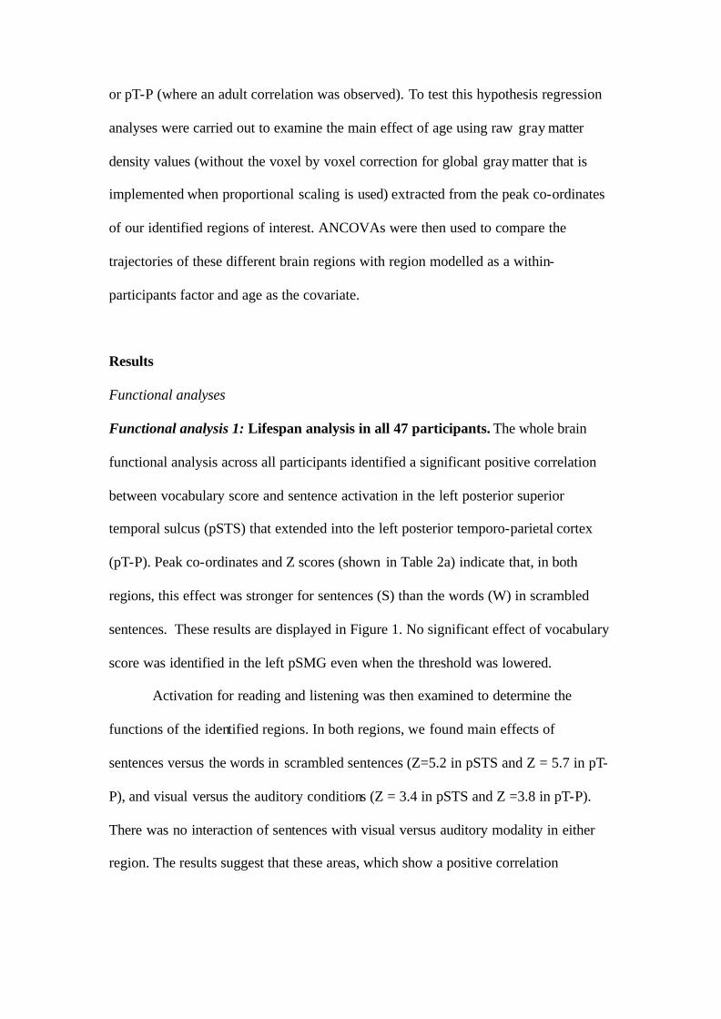
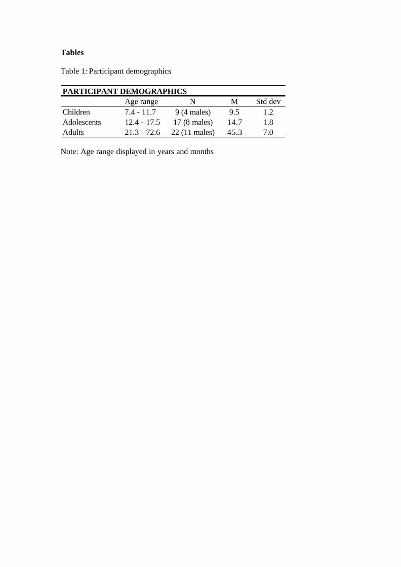
Functional analysis 1: Lifespan analysis in all 47 participants. The whole brain
functional analysis across all participants identified a significant positive correlation
between vocabulary score and sentence activation in the left posterior superior
temporal sulcus (pSTS) that extended into the left posterior temporo-parietal cortex
(pT-P). Peak co-ordinates and Z scores (shown in Table 2a) indicate that, in both
regions, this effect was stronger for sentences (S) than the words (W) in scrambled
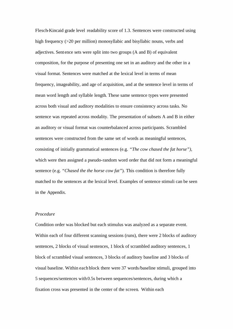
sentences. These results are displayed in Figure 1. No significant effect of vocabulary
score was identified in the left pSMG even when the threshold was lowered.
Activation for reading and listening was then examined to determine the
functions of the identified regions. In both regions, we found main effects of
sentences versus the words in scrambled sentences (Z=5.2 in pSTS and Z = 5.7 in pT-
P), and visual versus the auditory conditions (Z = 3.4 in pSTS and Z =3.8 in pT-P).
There was no interaction of sentences with visual versus auditory modality in either
region. The results suggest that these areas, which show a positive correlation
between functional activation and vocabulary score, are involved in sentence
comprehension.
================== Insert Table 2 about here ==================
Functional analysis 2: Teenagers only. When the analysis was restricted to the
teenage group, a similar pattern of results was observed, see Table 2b.
Structural analyses
Structural analysis 1: The effect of vocabulary on regional gray matter density
across lifespan. Using the results of the functional analysis as a region of interest, we
found a corresponding positive correlation between vocabulary score and gray matter
density in both pSTS and pT-P (peaks at: x = -48, y = -36, z = 6, Z = 4.0; and x = -50,
y = -60, z = 16, Z = 3.2 respectively). Figure 1 illustrates the clear consistency
between the effect of vocabulary on the functional data (significant after a whole brain
correction) and on the structural data (significant after a small volume correction
limited to voxels that were significant in the functional data: see methods for
procedure). However, contrary to expectation, we did not find an effect of vocabulary
in the pSMG regions of interest identified by Lee et al. (2007).
=================== Insert Figure 1 about here ===================
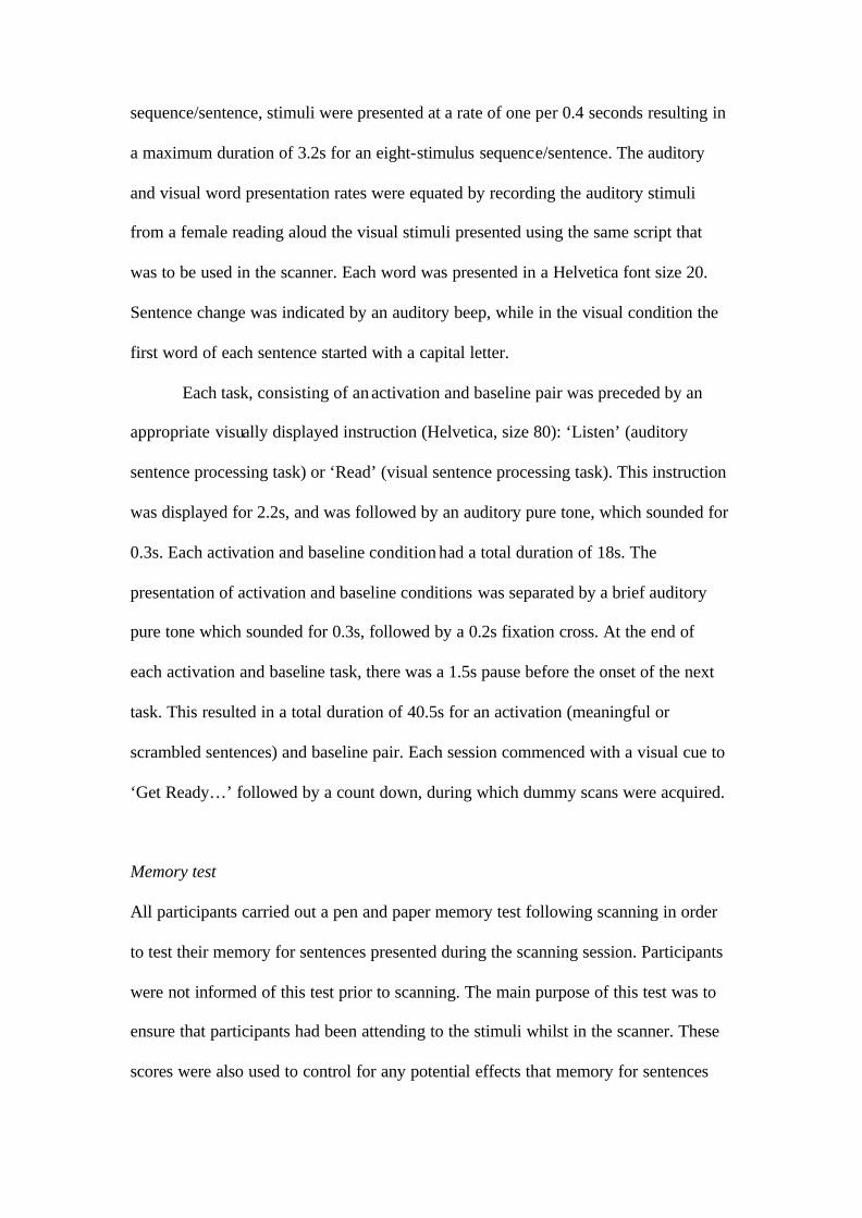
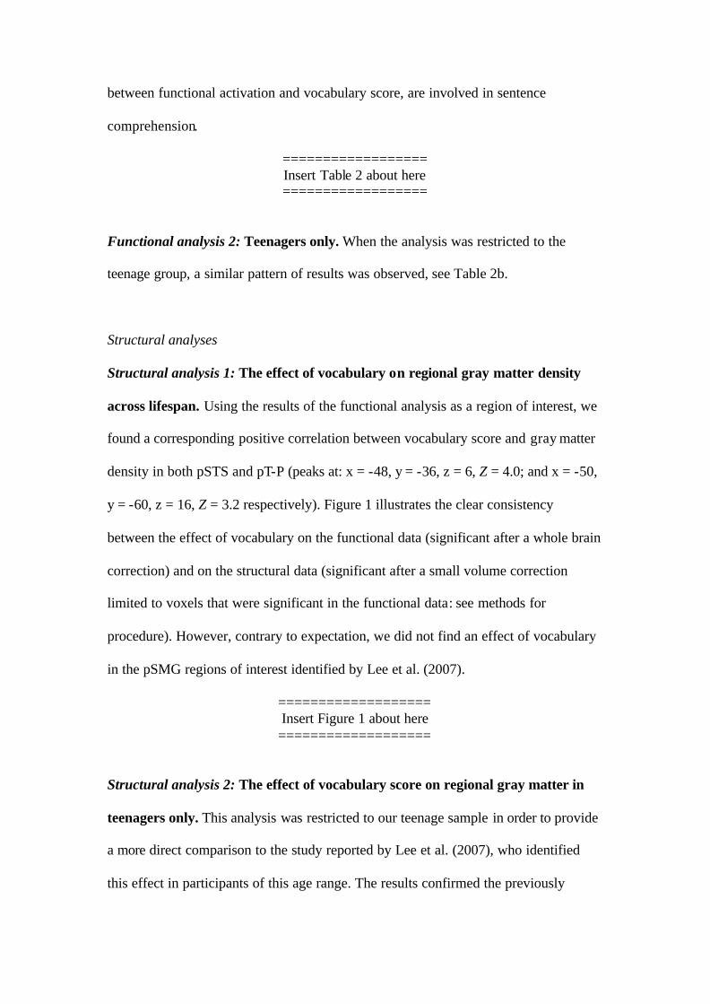
Structural analysis 2: The effect of vocabulary score on regional gray matter in
teenagers only. This analysis was restricted to our teenage sample in order to provide
a more direct comparison to the study reported by Lee et al. (2007), who identified
this effect in participants of this age range. The results confirmed the previously
reported findings that gray matter density in teenagers was indeed significantly
correlated with vocabulary score in both the left and the right pSMG (left: x = -40, y =
-54, z = 52, Z = 3.6; right: x = 48, y = -54, z = 48, Z = 3.4). This result can be seen in
Figure 2.
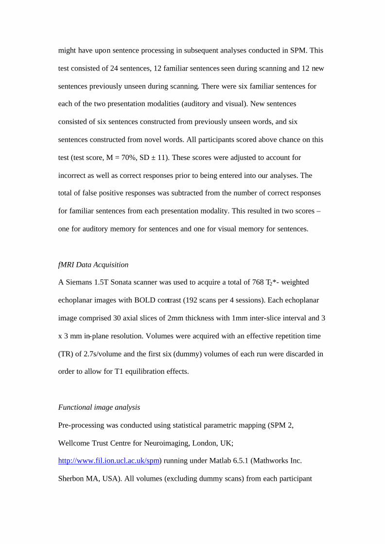
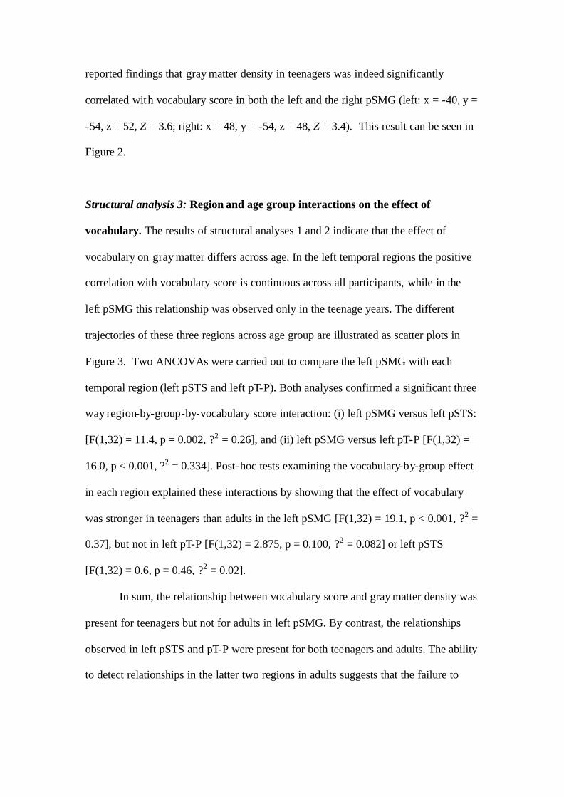
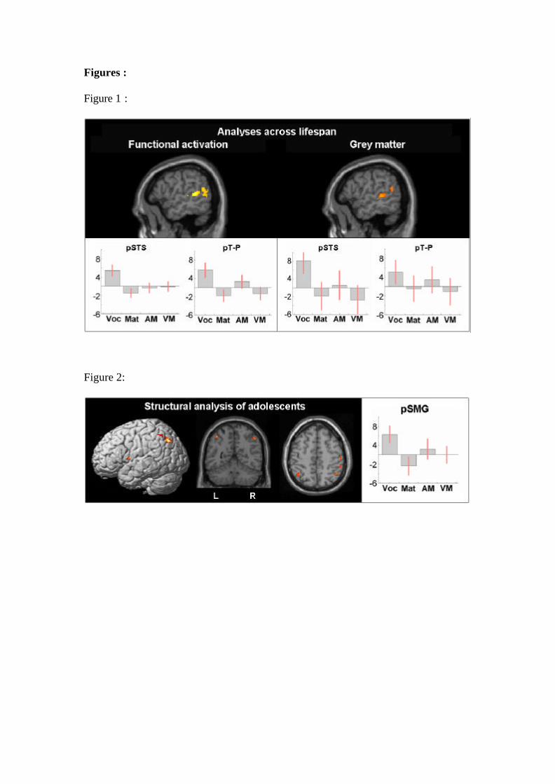
Structural analysis 3: Region and age group interactions on the effect of
vocabulary. The results of structural analyses 1 and 2 indicate that the effect of
vocabulary on gray matter differs across age. In the left temporal regions the positive
correlation with vocabulary score is continuous across all participants, while in the
left pSMG this relationship was observed only in the teenage years. The different
trajectories of these three regions across age group are illustrated as scatter plots in
Figure 3. Two ANCOVAs were carried out to compare the left pSMG with each
temporal region (left pSTS and left pT-P). Both analyses confirmed a significant three
way region-by-group-by-vocabulary score interaction: (i) left pSMG versus left pSTS:
[F(1,32) = 11.4, p = 0.002, ?2 = 0.26], and (ii) left pSMG versus left pT-P [F(1,32) =
16.0, p < 0.001, ?2 = 0.334]. Post-hoc tests examining the vocabulary-by-group effect
in each region explained these interactions by showing that the effect of vocabulary
was stronger in teenagers than adults in the left pSMG [F(1,32) = 19.1, p < 0.001, ?2 =
0.37], but not in left pT-P [F(1,32) = 2.875, p = 0.100, ?2 = 0.082] or left pSTS
[F(1,32) = 0.6, p = 0.46, ?2 = 0.02].
In sum, the relationship between vocabulary score and gray matter density was
present for teenagers but not for adults in left pSMG. By contrast, the relationships
observed in left pSTS and pT-P were present for both teenagers and adults. The ability
to detect relationships in the latter two regions in adults suggests that the failure to
find the relationship for left pSMG did not result from a lack of vocabulary variance
in the adult sample.
=================== Insert Figure 3 about here ===================
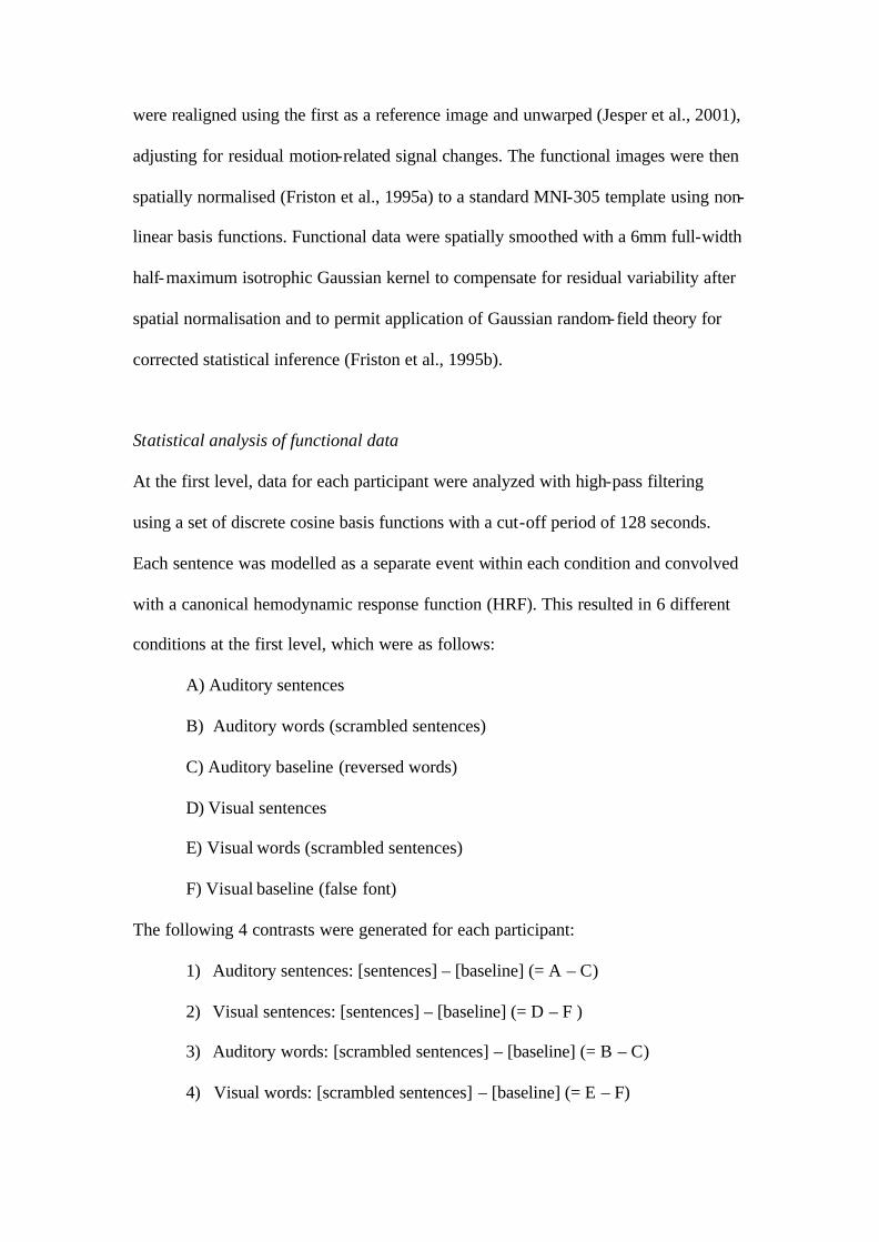
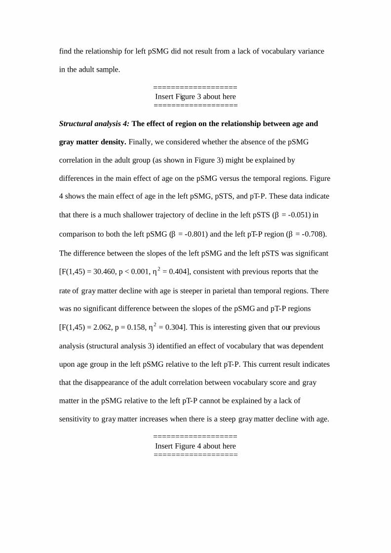
Structural analysis 4: The effect of region on the relationship between age and
gray matter density. Finally, we considered whether the absence of the pSMG
correlation in the adult group (as shown in Figure 3) might be explained by
differences in the main effect of age on the pSMG versus the temporal regions. Figure
4 shows the main effect of age in the left pSMG, pSTS, and pT-P. These data indicate
that there is a much shallower trajectory of decline in the left pSTS (β = -0.051) in
comparison to both the left pSMG (β = -0.801) and the left pT-P region (β = -0.708).
The difference between the slopes of the left pSMG and the left pSTS was significant
[F(1,45) = 30.460, p < 0.001, η2 = 0.404], consistent with previous reports that the
rate of gray matter decline with age is steeper in parietal than temporal regions. There
was no significant difference between the slopes of the pSMG and pT-P regions
[F(1,45) = 2.062, p = 0.158, η2 = 0.304]. This is interesting given that our previous
analysis (structural analysis 3) identified an effect of vocabulary that was dependent
upon age group in the left pSMG relative to the left pT-P. This current result indicates
that the disappearance of the adult correlation between vocabulary score and gray
matter in the pSMG relative to the left pT-P cannot be explained by a lack of
sensitivity to gray matter increases when there is a steep gray matter decline with age.
=================== Insert Figure 4 about here ===================
Discussion
In this study, we investigated the brain regions where local gray matter varies with
vocabulary knowledge. We hypothesized that these effects would be age-dependent
on the basis that strategies for acquiring vocabulary differ across lifespan. Vocabulary
knowledge is generally acquired within the context of everyday experience of
language through conversation and reading, although new words can also be learnt by
forming an explicit link between a new word and an equivalent or a definition, a mode
of learning taking place predominantly by instruction. Our specific predictions were
that brain regions involved in learning vocabulary within the context of everyday
language would show a positive correlation between vocabulary score and gray matter
density which would be continuous across lifespan, and that these regions would also
be activated by reading and listening tasks. By contrast, we anticipated that for brain
regions engaged in learning vocabulary by lexical or conceptual equivalents, the
relationship between local brain structure and vocabulary knowledge would be
discontinuous across lifespan and potentially restricted to age groups of participants
engaged in formal education. Furthermore, these regions were not necessarily
expected to be activated during reading and listening tasks for participants at any age.
To test these hypotheses, we explored how the effect of vocabulary on gray matter
interacted with age; and whether and how the affected regions were activated during
auditory and written sentence processing.
The results of our structural and functional imaging analyses identified three
different regions where vocabulary knowledge correlated with gray matter density.
The first was the posterior supramarginal gyrus (pSMG), consistent with the find ings
of Lee et al. (2007) and replicated with a different vocabulary measure and different
participants. The second region was located deep in the left posterior superior
temporal sulcus (pSTS) and the third was in the left posterior temporo-parietal cortex
(pT-P). The pSTS and pT-P regions were not reported by Lee et al. (2007) and were
only identified in the current study when we used regions of interest where vocabulary
correlated with functional activation during written and auditory sentence processing.
Having identified the brain regions associated with vocabulary knowledge, we
established regional differences in the effect due to age. In the pSMG, the relationship
between vocabulary and gray matter was limited to the teenage years. By contrast, the
positive correlations in both temporal regions were consistent across lifespan. This
provides preliminary evidence that the effect of vocabulary in the pSMG may reflect
learning through explicit instruction of lexical equivalents that occurs during formal
education whereas the effect of vocabulary in the left temporal regions may reflect the
continuous acquisition of vocabulary that occurs from learning in context. The
analysis of the functional activation data offered further evidence for this hypothesis
because the temporal regions (pSTS and pT-P) were maximally activated by sentence
processing but, in the pSMG, there was no activation for either sentences or the words
in scrambled sentences, irrespective of whether they were presented in the auditory or
visual modality.
Our functional imaging results are consistent with previous studies of
language comprehension carried out in both children and adults (Booth et al., 2002;
Constable et al., 2004; Humphries et al., 2006; Turkeltaub et al., 2003; Spitsyna et al.,
2006; Lindenberg & Sheef, 2007). Specifically, previous studies have shown that the
left pT-P area is linked to syntactic and semantic processing (Vandenberge et al.
2002; Thierry et al., 2003; Mechelli et al., 2007) and the pSTS interfaces between
semantic associations and speech production (see Binder and colleagues: Binder et al.,
1997; 2000; 2003, Hickok and colleages: Okada & Hickok, 2001; Hickok & Poeppel,
2004; 2007; also: Humphries et al., 2006; 2007; Price et al., 1997; Scott et al., 2003;
Vandenberghe et al., 1996). Thus, the increased functional activation and gray matter
density in the temporal regions for those with high vocabulary may reflect a larger
number of associations between semantic processing and speech output (pSTS) and
semantic processing and syntactic processing (pT-P). The stronger effect of
vocabulary knowledge at the sentence level in these temporal areas may also indicate
that our vocabulary measure is partially indexing additional sentence- level
comprehension processes that are highly correlated with vocabulary score. Whilst it is
difficult to separate the relative contribution of such highly correlated variables, future
analyses using multiple behavioural measures may be able to determine the relative
influence of a range of linguistic abilities on activation intensity and local brain
structure. In addition, functional imaging studies that manipulate the contextual
learning of new words (such as Metres-Missé et al., 2008) will also help to derive the
role of temporal areas in integrating new lexical items into an existing language
system.
The contrasting effects in the temporal and pSMG regions suggest that the
respective correlations of vocabulary and gray matter density are influenced by
different mechanisms. In the left pSMG, gray matter was only correlated with
vocabulary in the teenage years and this area was not activated during auditory or
written sentence or word processing. However, this region has been found to be active
during word learning tasks in which participants were taught the meaning of new
words (Cornelissen et al., 2004). This finding and the positive correlation between
vocabulary and gray matter density in the pSMG is therefore consistent with a
vocabulary learning strategy that occurs predominantly in formal education: i.e. the
explicit instruction of lexical or conceptual equivalents for new words. After the
teenage years, the relationship between vocabulary and gray matter density may be
lost because vocabulary continues to increase by contextual learning, without using
the pSMG. Nevertheless, greater gray matter in the pSMG in bilingual adults
identified by Mechelli et al. (2004) indicates that increases in gray matter in this
region can occur outside adolescence. Therefore differences in gray matter in the
pSMG in this instance may reflect the acquisition and maintenance of two languages
in the case of bilingualism or second language acquisition. Alternatively, increased
pSMG gray matter in monolingual teenagers and bilingual adults may represent an
active learning mechanism that facilitates language learning in a context of increased
task demands – for instance, learning more complex and abstract vocabulary in
secondary education, or maintaining vocabulary knowledge across multiple
languages.
Our conclusion that different vocabulary learning strategies are supported by
left temporal and parietal neural systems rests on the exclusion of several potentially
confounding factors. Specifically, the effect of vocabulary in the left pSMG might be
greater in teenage years because of increased sensitivity in the correlation by virtue of
(a) the wide range of vocabulary abilities in this age range and/or (b) reduced error
variance because the effect of age, or other unrelated factors, is smaller in the
teenagers than the adult sample. In other words, even if we had been able to match the
range of vocabulary in the teenage and adult groups, there would be an age-range
confound because the age range is smaller in the teenage group (12 to 17 years) than
the adult group (21 to73 years). The importance of matching age range is illustrated
by the well-known effect of age on gray matter, which is characterised in terms of a
pre-adolescent increase followed by a post-adolescent decrease (Giedd et al., 1999;
Gogtay et al., 2004; Paus, 2005). The rate of gray matter loss with age differs from
region to region (as illustrated using our own data in Figure 4). The relevant point
here is that, in the lifespan analysis, it will be more difficult to detect a small
vocabulary-related increase in gray matter in the context of a steeper age-related
decline in gray matter.
Fortunately, we were able to discount the influence of both vocabulary range
and age range on the validity of our conclusions because we observed significantly
different patterns of effect in three different brain regions. For instance, if the greater
effect of vocabulary in teenage years was due to greater sensitivity from either a wider
vocabulary range relative to adults, or a narrower range of age-related gray matter
decline relative to adults, then positive correlations with vocabulary should be
stronger during the teenage years for all three regions. Instead, the increased effect of
vocabulary in teenage years was only observed in pSMG, not in pSTS and pT-P. In
addition, we were able to show that the trajectory of gray matter decline in the pSMG
and pT-P was similar, even though only pSMG demonstrated a differential
relationship with vocabulary with age.
Finally, there are two remaining questions to consider. Firstly, why was no
correlation between vocabulary score and gray matter density detected for young
children? And secondly, why does pSMG gray matter correlate with second language
proficiency in young adults (20 to 30 years)? In consideration of the first question, the
sample of young children within our study was rather small (N=9). Whilst these
participants made a valuable contribution to our lifespan analysis, this sample size
may not be sufficient as a group to detect an effect. However, a marginal trend was
observed for pSTS, suggesting that there was some sensitivity to detect differences in
this age group. This would require an additional explanation of why no similar trend
was observed for pSMG given that these children were in formal education. Were this
interaction to prove reliable in a larger sample, one could speculate that although
vocabulary acquisition in the early years undoubtedly involves definitional learning, it
may take play within the bounds of the gray matter already available to support this
function in younger children. Regarding the role of bilingualism, learning a second
language requires an individual to acquire words and link these with the vocabulary of
their first language and with semantic knowledge. This process requires tha t links be
made between lexical equivalents (Kroll & Potter, 1984), a process that will increase
the demands on pSMG gray matter.
In conclusion, our study has identified contrasting effects of vocabulary
knowledge on brain structure. We propose that the continuous positive correlation
across lifespan observed in the left pSTS and the left pT-P reflects vocabulary
learning within the context of everyday language experience with semantically and
syntactically related words and sentences. Vocabulary knowledge is increased in this
way through continued experience of language across lifespan, whereas the left
pSMG is engaged in learning to link a new word with specific lexical equivalents, a
mode of learning more typically exploited within formal education. In addition, the
convergence of functional and structural correlations in the left temporal regions
suggests that increased gray matter might be a consequence of increased neuronal
activation or vice versa. Longitudinal studies are now required to ascertain how
vocabulary learning changes over time within the same individuals at different points
in the lifespan. This may indicate whether the effect of vocabulary correlates with
learning rate and activation or to a genetic disposition to high gray matter in regions
that enable vocabulary learning, i.e., an inherited ability to acquire more knowledge.
References:
Allen, J.S., Bruss, J., Kice Brown, C., & Damasio, H. (2005). Normal neuroanatomial
variation due to age: the major lobes and a parcellation of the temporal region.
Neurobiology of Ageing, 26, 1245-1260.
Ashburner, J., & Friston, K. J. (2005). Unified segmentation. Neuroimage, 26, 839-
851.
Binder, J.R., Frost, J.A., Hammeke, T.A., Cox, R.W., Rao, S.M., & Prieto, T. (1997).
Human brain language areas identified by functional magnetic resonance
imaging. The Journal of Neuroscience, 17 (1), 353-362.
Binder, J.R., Frost, J.A., Hammeke, P.S.F., Bellgowan, J.A., Springer, J.A., Kaufman,
J.N., & Possing, E.T. (2000). Human temporal lobe activation by speech and
non-speech sounds. Cerebral Cortex, 10, 512-518.
Binder, J.R., McKieman, K.A., Parsons, M., Westbury, C.F., Possing, E.T., Kaufman,
J.N., & Buchanan, L. (2003). Neural correlates of lexical access during visual
word recognition. Journal of Cognitive Neuroscience, 15, 372-393.
Booth, J.R., Burman, D.D., Meyer, J.R., Gitelman, D.R., Parrish, T.B., & Mesulam,
M.M. (2002). Modality independence of word comprehension. Human Brain
Mapping, 16, 251-261.
Brambati, S.M., Termin, C., Ruffino, M., Danna, M., Lanzi, G., Stella, G. et al.
(2006). Neuropsychological deficits and neural dysfunction in familial dyslexia.
Brain Research, 1113, 174-185.
Brambati, S.M., Termin, C., Ruffino, Stella, G, M., Fazio, F., Cappa, S.F., & Perani,
D. (2004). Regional reductions of gray matter volume in familial dyslexia.
Neurology, 63, 742-745.
Chételat, G., Desgranges, B., Landeau, B., Mézenge, F., Poline, J.B., de la Sayette, V.
et al. (2008). Direct voxel-based comparison between grey matter metabolism
and atrophy in Alzheimer’s disease. Brain, 131, 60-71.
Constable, R.T., Pugh, K.R., Berroya, E., Mencl, W.E., Westerveld, M., Ni, W., &
Shankweiler, D. (2004). Sentence complexity and input modality effects in
sentence comprehension: an fMRI study. Neuroimage, 22, 11-21.
Cornelissen, K., Laine, M., Renvall, K., Saarinen, T., Martin, N., & Salmelin, R.
(2004). Learning new names for new objects: cortical effects as measured by
magnetoencephalography. Brain and Language, 89, 617-622.
Deichmann, R., Schwarzbauer, C., & Turner, R. (2004). Optimisation of the 3D
MDEFT sequence for anatomical brain imaging: technical implications at 1.5
and 3T. Neuroimage, 21, 757-767.
Demonet, J-F., Chollet, F., Ramsay, S., Cardebat, D., Nespoulos, J-L, Wise, R., et al.
(1992). Anatomy of physiological and semantic processing in normal subjects.
Brain, 115, 1753-1768.
Devlin, J.T., Matthews, P.M., & Rushworth, M.F.S. (2003). Semantic processing in
the left inferior prefrontal cortex: A combined functional magnetic resonance
imaging and transcranial magnetic stimulation study. Journal of Cognitive
Neuroscience, 15, 71-84.
Dunn, L.M., Dunn, L.M., Whetton, C., & Burley, J. (1997). The British Picture
Vocabulary Scales (2nd Edition). Windsor: NFER – Nelson.
Elliot, C.D., Smith, P., & McCulloch, K. (1997). British Ability Scales Second
Edition (BAS II). London: NFER – Nelson.
Friston, K.J., Ashburner, J., Frith, C.D., Poline, J.B., Heather, J.D., Frankowiak,
R.S.J. (1995a). Spatial registration and normalization of images. Human Brain
Mapping, 2, 1-25.
Friston, K.J., Holmes, A., Worsley, K.J., Poline, J.B., Frith, C.D., Frankowiak, R.S.J.
(1995b). Statistical parametric maps in functional imaging: a general linear
approach. Human Brain Mapping, 2, 189-210.
Gathercole, S.E. (2006) Nonword repetition and word learning: the nature of the
relationship. Applied Psycholinguistics, 27, 513-543.
Gathercole, S.E., Hitch, G.J., Service, E., & Martin, A.J. (1997). Phonological short-
term memory and vocabulary development: further evidence from the nature of
the relationship: Applied Cognitive Psychology, 13, 65-77.
Giedd, J.N. Blumenthal, J., Jeffries, N.O., Castellanos, F.X., Liu, H., Zijdenboss, A. et
al. (1999). Brain development during childhood and adolescence: a longitudinal
MRI study. Nature Neuroscience, 2 (10), 861-863.
Gogtay, N., Giedd, J.N., Lusk, L., Hayashi, K., Greenstein, D., Vaituzis, C. et al.
(2004). Dynamic mapping of human cortical development during childhood
through early adulthood. Proceedings of the National Academy of Science, 101
(21), 8174-8179.
Good, C.D., Johnsrude, I.S., Ashburner, J., Henson, R.N.A., Friston, K.J., &
Frackowiak, R.S.J. (2001). A voxel-based morphometric study of ageing in 465
normal adult human brains. Neuroimage, 14, 21-36.
Hickok, G., & Poeppel, D. (2004). Dorsal and ventral streams: a framework for
understanding aspects of the functional anatomy of language. Cognition, 92, 67-
99.
Hickok, G., & Poeppel, D. (2007). The cortical organization of speech processing.
Nature Reviews Neuroscience, 8, 393-402.
Hoeft, F., Ueno, T., Reiss, A. L., Meyler, A., Whitfield-Gabrielli, S., & Glover, G.H.
et al. (2007). Prediction of children’s reading skills using behavioural,
functional, and structural neuroimaging measures. Behavioural Neuroscience,
121 (3), 602-613.
Humphries, C., Binder, J.R., Medler, D.A., & Liebenthal, E. (2007). Time course of
semantic processes during sentence comprehension: an fMRI study.
Neuroimage, 36, 924-932.
Humphries, C., Binder, J.R., Medler, D.A., & Liebenthal, E. (2006). Syntactic and
semantic modulation of neural activity in auditory sentence comprehension.
Journal of Cognitive Neuroscience, 18 (4), 665-679.
Hyde, K.L., Lerch, J.P., Griffiths, T.D., Evans, A.C., & Peretz, I. (2007). Cortical
thickness in congenital amusia: when less is better than more. Journal of
Neuroscience, 27 (47), 13028-13032.
Hyde, K.L., Zatorre, R.J., Griffiths, T.D., Lerch, J.P., & Peretz, I. (2006).
Morphometry of the amusic brain: a two-site study. Brain, 129, 2562-2570.
Jesper, L.R., Andersson, J.L.R, Hutton, C., Ashburner, J., Turner, R., Friston, K.J.
(2001). Modelling geometric deformations in EPI time series. Neuroimage, 13,
903-919.
Kilian, A. S., Nagy, W., Pearson, P. D., Anderson, R. C., & García, G. E. (1995).
Learning vocabulary from context: effects of focusing attention on individual
words during reading. Technical report: Center for the Study of Reading, No.
619.

Kroll, J.F. & Potter, M.C. (1984). Recognizing words, pictures, and concepts: a
comparison of lexical, object, and reality decisions. Journal of Verbal Learning
and Behaviour, 23, 39-66.
Lee, H.L., Devlin, J.T., Shakeshaft, C., Stewart, L.H., Brennan, A., Glensman, J. et al.
(2007). Anatomical traces of vocabulary acquisition in the adolescent brain.
Journal of Neuroscience, 27, 1184-1189.
Lenroot, R.K., & Giedd, J.N. (2006). Brain development in children and adolescents:
insights from anatomical magnetic resonance imaging. Neuroscience and
Biobehvioural Reviews, 30, 718-729.
Lindenberg, R., & Scheef, L. (2007). Supramodal language comprehension: Role of
the left temporal lobe for listening and reading. Neuropsychologica, 45, 2407-
2415.
Mechelli, A., Crinion, J.T., Noppeney, U., O’Doherty, J., Ashburner, J., Frackowiak,
R.S.J., & Price, C.J. (2004). Neurolinguistics: structural plasticity in the
bilingual brain. Nature, 431, 757.
Mechelli, A., Price, C.J., Friston, K., & Ashburner, J. (2005). Voxel-based
morphometry of the human brain: methods and applications. Current Medical
Imaging Reviews, 1(2), 105-113.
Mechelli A., Josephs O, Lambon Ralph MA, McClelland JA, Price, C.J. (2007)
Dissociating stimulus-driven semantic and phonological effects during reading
and naming. Human Brain Mapping, 3, 205-17.
Mestres-Missé, A., Càmara, E., Rodriguez-Fornells, A., Rotte, M., & Münte, T.F.
(2008). Functional neuroanatomy of meaning acquisition from context.
Journal of Cognnitive Neuroscience, 20 (12), 2153-2166.
Nagy, W., Herman, P. A., & Anderson, R. C. (1985). Learning words from context.
Reading Research Quarterly, 20 , 233-253.
Okada, K. & Hickok, G. (2006). Identification of lexical-phonological networks in the
superior temporal sulcus using fMRI. Neuroreport 17, 1293–1296.
Price, C.J., More, C.J., Humphreys, G.W., & Wise, R.J. (1997). Segregating semantic
from phonological processes during reading. Journal of Cognitive Neuroscience,
9, 3876-3883.
Paus, T. (2005). Mapping brain maturation and cognitive development during
adolescence. Trends in Cognitive Sciences, 9 (2), 60-68.
Scott, S.K., Leff, A., & Wise, R.J.S. (2003). Going beyond the information given: a
neural system supporting semantic interpretation. Neuroimage,19, 870-876.
Silani, G.,Frith, U., Demonet, J.F., Fazio, F., Perani, D., Price, C. et al. (2005) Brain
abnormalities underlying altered activation in dyslexia: a voxel-based
mophometry study. Brain, 128, 2453-2461.
Sowell, E.R., Peterson, B.S., Thompson, P.M., Welcome, S.E.,Henkenius, A.L., &
Toga, A.W. (2003). Mapping cortical change across human life span. Nature
Neuroscience, 6, 309-315.
Spitsyna, G., Warren, J.E., Scott, S.K., Turkheimer, F.E., Wise, R.J.S. (2006).
Converging language streams in the human temporal lobe. Journal of
Neuroscience, 26 (28), 7328-7336.
Thierry, G., Giraud, A.L., & Price, C. (2003). Hemispheric dissociation in access to
the human semantic system. Neuron, 38, 499-506.
Vandenberghe, R.,Price, C., Wise, R., Josephs, O., & Frankowiak, R.S.J. (1996).
Functional anatomy of a common semantic system for words and pictures.
Nature, 383, 254-256.
Vandenberghe, R., Nobre, A.C., & Price, C.J. (2002). The response of the left
temporal cortex to sentences. Journal of Cognitive Neuroscience, 14 (4), 550-
560.
Wechsler, D. (1955). Manual for the Wechsler adult intelligence scale. New York:
Psychological Corporation.
Wilke, M., Krägeloh-Mann, I., & Holland, S.K. (2007). Global and local development
of gray and white matter volume in normal children and adolescents.
Experimental Brain Research, 178, 296-307.
Tables
Table 1: Participant demographics
PARTICIPANT DEMOGRAPHICSAge range N M Std dev
Children 7.4 - 11.7 9 (4 males) 9.5 1.2Adolescents 12.4 - 17.5 17 (8 males) 14.7 1.8Adults 21.3 - 72.6 22 (11 males) 45.3 7.0
Note: Age range displayed in years and months
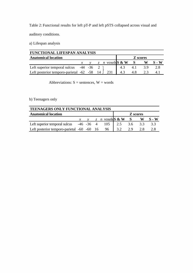
Table 2: Functional results for left pT-P and left pSTS collapsed across visual and
auditory conditions.
a) Lifespan analysis
FUNCTIONAL LIFESPAN ANALYSISAnatomical location
x y z n voxelsS & W S W S - WLeft superior temporal sulcus -44 -36 2 4.3 4.1 3.9 2.8Left posterior temporo-parietal -62 -58 14 231 4.3 4.8 2.3 4.1
Z scores
b) Teenagers only
TEENAGERS ONLY FUNCTIONAL ANALYSISAnatomical location
x y z n voxels S & W S W S - WLeft superior temporal sulcus -46 -36 4 105 2.5 3.6 3.3 3.3Left posterior temporo-parietal -60 -60 16 96 3.2 2.9 2.8 2.8
Z scores
Abbreviations: S = sentences, W = words
Figures :
Figure 1 :
Figure 2:
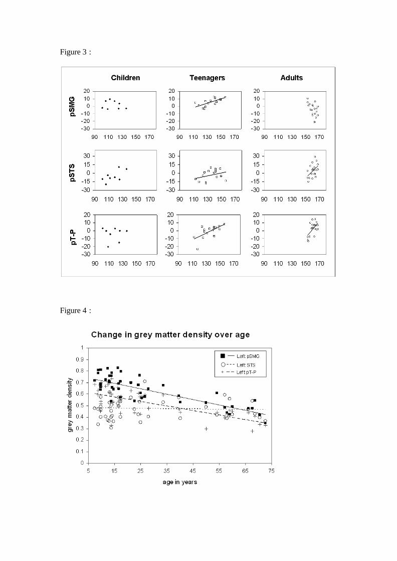
Figure 3 :
Figure 4 :
Figure captions:
Figure 1: Lifespan analyses results showing the left pSTS and left pT-P for functional
and structural analyses respectively. Graphs plot the effect of each of the following
regressors upon brain activation (functional) and grey matter (structural): vocabulary
(voc), matrices (mat), auditory memory for sentences (A mem) and visual memory for
sentences (V mem) on each region. The y-axis shows effect sizes as the percentage
increase relative to the global mean. The red bars are the 90% confidence intervals.
Figure 2: Structural analysis results for the teenage group. Highlighted regions show
an increase in grey matter density as a function of vocabulary score. When the
statistical threshold was lowered to p < 0.001 uncorrected, a positive correlation with
vocabulary was also observed the left ventral precentral gyrus and the left angular
gyrus. Both regions can be seen in this figure in addition to the left pSMG. The graph
shows the effect of each of the four regressors on grey matter in the left pSMG
labelled as follows on the x-axis: vocabulary (voc), matrices (mat), auditory memory
for sentences (A mem) and visual memory for sentences (V mem). The y-axis shows
effect sizes as the percentage increase relative to the global mean. The red bars are the
90% confidence intervals.
Figure 3: Scatter plots according to age group showing the relationship between
vocabulary score and grey matter density. Trajectories are plotted only where a
statistically reliable relationship was observed. (a) Left pSMG [x=-40, y=-54, z=52],
where there is prominent trend for teenagers over and above all other age groups; (b)
the left pSTS [x=-48, y=-36, z=6], where the trajectory for adults appears steeper than
those of children and teenagers, this positive correlation was marginally significant
for children (p = 0.07). It should be noted that the trajectory for adults remains
statistically reliable after the omission of two outliers; (c) the left pT-P [x=-50, y=-60,
z=16], where there is a consistent trend for both teenagers and adults.
Figure 4: Scatter plot comparing change in grey matter density over age in the left
pSTS, left pT-P, and the left pSMG.
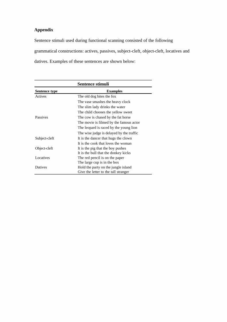
Appendix
Sentence stimuli used during functional scanning consisted of the following
grammatical constructions: actives, passives, subject-cleft, object-cleft, locatives and
datives. Examples of these sentences are shown below:
Sentence type ExamplesActives The old dog bites the fox
The vase smashes the heavy clockThe slim lady drinks the waterThe child chooses the yellow sweet
Passives The cow is chased by the fat horseThe movie is filmed by the famous actorThe leopard is raced by the young lionThe wise judge is delayed by the traffic
Subject-cleft It is the dancer that hugs the clownIt is the cook that loves the woman
Object-cleft It is the pig that the boy pushesIt is the bull that the donkey kicks
Locatives The red pencil is on the paperThe large cup is in the box
Datives Hold the party on the jungle islandGive the letter to the tall stranger
Sentence stimuli