Contesting and transforming psychiatric diagnoses in brain research : some insights from history,...
61
- 1 - Palais des Académies, Brussels, Belgium International Society for the History of the Neurosciences 19 th annual meeting June 30 - July 5, 2014 PROGRAM ABSTRACT BOOK
-
Upload
independentresearcher -
Category
Documents
-
view
1 -
download
0
Transcript of Contesting and transforming psychiatric diagnoses in brain research : some insights from history,...

- 1 -Palais des Académies, Brussels, Belgium
International Society for the History of the Neurosciences19th annual meeting
June 30 - July 5, 2014
PROGRAMABSTRACT BOOK

Palais des Académies, Brussels, BelgiumPalais des Académies, Brussels, Belgium
International Society for the History of the Neurosciences19th annual meeting
June 30 - July 5, 2014
PROGRAMABSTRACT BOOK
- 5 -
© 2014 - Adventures Art & EditionPrinted in Belgium by www.adventures-art.beEdited by Geneviève Abert
Neuroportrait of Arthur Van Gehuchten (1861-1914) on the back cover, courtesy of Nick Wade
The copyright in the contributions to this volume of proceedings rests with the individual authors.
Program & Scientific Committee
Geneviève Aubert, Brussels, BelgiumFrançois Boller, Washington DC, USAStanley Finger, St-Louis, USAPaul Foley, Sydney, AustraliaAxel Karenberg, Köln, GermanyPeter Koehler, Heerlen, The Netherlands
Local Committee
Geneviève Aubert, UCLouvainPatrick Cras, Universiteit AntwerpenAnne Jeanjean, UCLouvainRobert Poirrier, Université de LiègeFrank Van Calenbergh, KULeuvenRick Vandenberghe, KULeuven
Welcome to Brussels!
On behalf of the International society for the History of the Neurosciences (ISHN), I am very happy to welcome you to the 19th Annual Meeting of the society, hosted in the Palais des Académies, Brussels, Belgium.
As always, this meeting gathers scholars from around the world: 4 continents and 15 countries. They will share their latest research through lectures and posters. We have also allowed plenty of time around great Belgian food. We hope this will encourage you to strengthen links with scholars from associated disciplines.
This is a time to commemorate the 100th anniversary of the First World War, with ses-sions dedicated to the consequences of shell-shock as studied, considered and treated by the different countries affected by the Great War. Another session examines the effect WWI had on the development of neurosurgery.
A joint meeting of the ISHN and the Belgian Association for Sleep Research & Sleep Medicine offers a full day dedicated to the History of Sleep and Wakefulness Research and the History of Sleep Medicine.
You are invited to follow in the footsteps of Andreas Vesalius and Arthur Van Gehuchten, in Leuven and Louvain-la-Neuve. Lectures will shed new light on Vesalius, Willis and Van Gehuchten. To commemorate the 100th anniversary of Arthur Van Gehuchten’s death, with my colleagues of the IoNS and in conjunction with the Archive Department of the Université Catholique de Louvain (UCL), we have prepared an exhibition, where you are invited to a reception, courtesy of the UCL.
And finally, the Banquet will take place in the prestigious rooms of the historic building of the Cercle Royal Gaulois Artistique et Littéraire, dating back to 1782 and located in the Parc Royal.
I look forward to welcoming you to Brussels and I wish you a great scientific and cultural experience!
Geneniève AubertPresident

- 6 - - 7 -
PROGRAM
- 8 - - 9 -
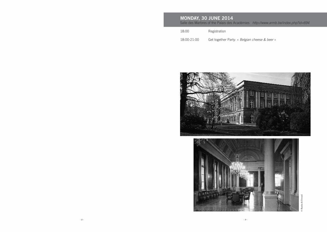
MONDAY, 30 JUNE 2014Salle des Marbres of the Palais des Académies http://www.armb.be/index.php?id=694
18:00 Registration
18:00-21:00 Get together Party: « Belgian cheese & beer »
© B
astin
& E
vrar
d
- 10 - - 11 -
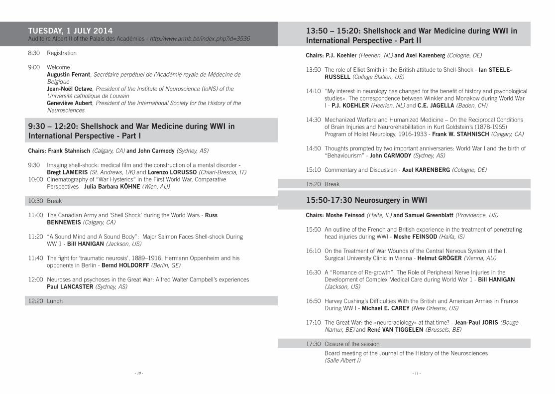
TUESDAY, 1 JULY 2014Auditoire Albert II of the Palais des Académies - http://www.armb.be/index.php?id=3536
8:30 Registration
9:00 Welcome Augustin Ferrant, Secrétaire perpétuel de l’Académie royale de Médecine de
Belgique Jean-Noël Octave, President of the Institute of Neuroscience (IoNS) of the
Université catholique de Louvain Geneviève Aubert, President of the International Society for the History of the
Neurosciences
9:30 – 12:20: Shellshock and War Medicine during WWI in International Perspective - Part I
Chairs: Frank Stahnisch (Calgary, CA) and John Carmody (Sydney, AS)
9:30 Imaging shell-shock: medical film and the construction of a mental disorder - Bregt LAMERIS (St. Andrews, UK) and Lorenzo LORUSSO (Chiari-Brescia, IT)
10:00 Cinematography of “War Hysterics” in the First World War. Comparative Perspectives - Julia Barbara KÖHNE (Wien, AU)
10:30 Break
11:00 The Canadian Army and ‘Shell Shock’ during the World Wars - Russ BENNEWEIS (Calgary, CA)
11:20 “A Sound Mind and A Sound Body”: Major Salmon Faces Shell-shock During WW 1 - Bill HANIGAN (Jackson, US)
11:40 The fight for ‘traumatic neurosis’, 1889–1916: Hermann Oppenheim and his opponents in Berlin - Bernd HOLDORFF (Berlin, GE)
12:00 Neuroses and psychoses in the Great War: Alfred Walter Campbell’s experiences Paul LANCASTER (Sydney, AS)
12:20 Lunch
13:50 – 15:20: Shellshock and War Medicine during WWI in International Perspective - Part II
Chairs: P.J. Koehler (Heerlen, NL) and Axel Karenberg (Cologne, DE)
13:50 The role of Elliot Smith in the British attitude to Shell-Shock - Ian STEELE-RUSSELL (College Station, US)
14:10 “My interest in neurology has changed for the benefit of history and psychological studies». The correspondence between Winkler and Monakow during World War I - P.J. KOEHLER (Heerlen, NL) and C.E. JAGELLA (Baden, CH)
14:30 Mechanized Warfare and Humanized Medicine – On the Reciprocal Conditions of Brain Injuries and Neurorehabilitation in Kurt Goldstein’s (1878-1965) Program of Holist Neurology, 1916-1933 - Frank W. STAHNISCH (Calgary, CA)
14:50 Thoughts prompted by two important anniversaries: World War I and the birth of “Behaviourism” - John CARMODY (Sydney, AS)
15:10 Commentary and Discussion - Axel KARENBERG (Cologne, DE)
15:20 Break
15:50-17:30 Neurosurgery in WWI
Chairs: Moshe Feinsod (Haifa, IL) and Samuel Greenblatt (Providence, US)
15:50 An outline of the French and British experience in the treatment of penetrating head injuries during WWI - Moshe FEINSOD (Haifa, IS)
16:10 On the Treatment of War Wounds of the Central Nervous System at the I. Surgical University Clinic in Vienna - Helmut GRÖGER (Vienna, AU)
16:30 A “Romance of Re-growth”: The Role of Peripheral Nerve Injuries in the Development of Complex Medical Care during World War 1 - Bill HANIGAN (Jackson, US)
16:50 Harvey Cushing’s Difficulties With the British and American Armies in France During WW I - Michael E. CAREY (New Orleans, US)
17:10 The Great War: the «neuroradiology» at that time? - Jean-Paul JORIS (Bouge-Namur, BE) and René VAN TIGGELEN (Brussels, BE)
17:30 Closure of the session
Board meeting of the Journal of the History of the Neurosciences (Salle Albert I)
- 12 - - 13 -
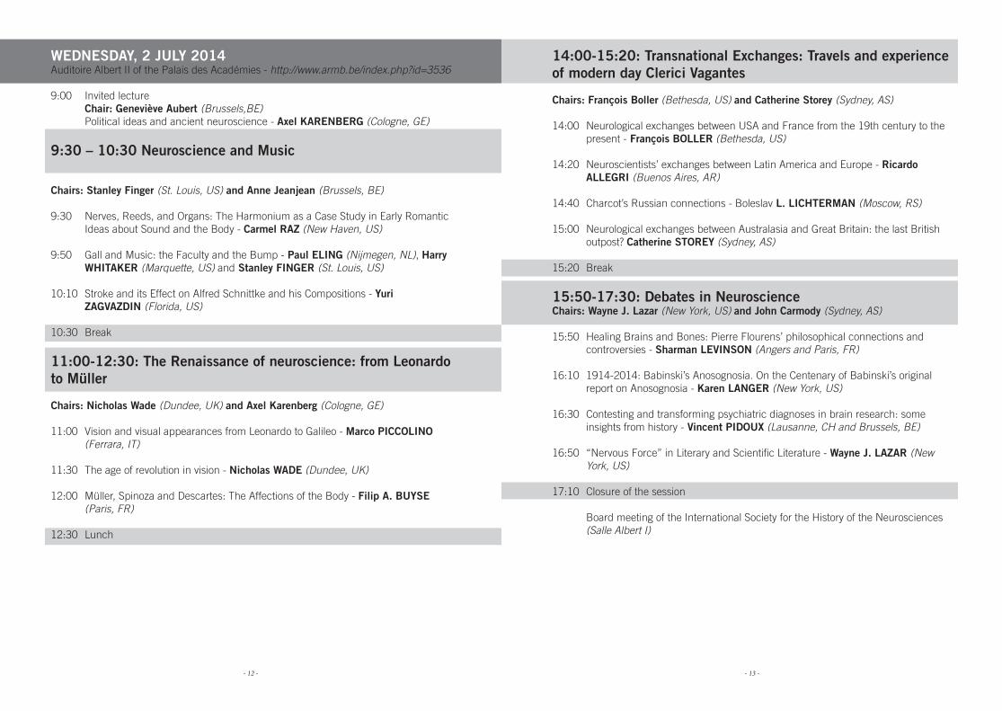
WEDNESDAY, 2 JULY 2014Auditoire Albert II of the Palais des Académies - http://www.armb.be/index.php?id=3536
9:00 Invited lecture Chair: Geneviève Aubert (Brussels,BE) Political ideas and ancient neuroscience - Axel KARENBERG (Cologne, GE)
9:30 – 10:30 Neuroscience and Music
Chairs: Stanley Finger (St. Louis, US) and Anne Jeanjean (Brussels, BE)
9:30 Nerves, Reeds, and Organs: The Harmonium as a Case Study in Early Romantic Ideas about Sound and the Body - Carmel RAZ (New Haven, US)
9:50 Gall and Music: the Faculty and the Bump - Paul ELING (Nijmegen, NL), Harry WHITAKER (Marquette, US) and Stanley FINGER (St. Louis, US)
10:10 Stroke and its Effect on Alfred Schnittke and his Compositions - Yuri ZAGVAZDIN (Florida, US)
10:30 Break
11:00-12:30: The Renaissance of neuroscience: from Leonardo to Müller
Chairs: Nicholas Wade (Dundee, UK) and Axel Karenberg (Cologne, GE)
11:00 Vision and visual appearances from Leonardo to Galileo - Marco PICCOLINO (Ferrara, IT)
11:30 The age of revolution in vision - Nicholas WADE (Dundee, UK)
12:00 Müller, Spinoza and Descartes: The Affections of the Body - Filip A. BUYSE (Paris, FR)
12:30 Lunch
14:00-15:20: Transnational Exchanges: Travels and experience of modern day Clerici Vagantes
Chairs: François Boller (Bethesda, US) and Catherine Storey (Sydney, AS)
14:00 Neurological exchanges between USA and France from the 19th century to the present - François BOLLER (Bethesda, US)
14:20 Neuroscientists’ exchanges between Latin America and Europe - Ricardo ALLEGRI (Buenos Aires, AR)
14:40 Charcot’s Russian connections - Boleslav L. LICHTERMAN (Moscow, RS)
15:00 Neurological exchanges between Australasia and Great Britain: the last British outpost? Catherine STOREY (Sydney, AS)
15:20 Break
15:50-17:30: Debates in NeuroscienceChairs: Wayne J. Lazar (New York, US) and John Carmody (Sydney, AS)
15:50 Healing Brains and Bones: Pierre Flourens’ philosophical connections and controversies - Sharman LEVINSON (Angers and Paris, FR)
16:10 1914-2014: Babinski’s Anosognosia. On the Centenary of Babinski’s original report on Anosognosia - Karen LANGER (New York, US)
16:30 Contesting and transforming psychiatric diagnoses in brain research: some insights from history - Vincent PIDOUX (Lausanne, CH and Brussels, BE)
16:50 “Nervous Force” in Literary and Scientific Literature - Wayne J. LAZAR (New York, US)
17:10 Closure of the session Board meeting of the International Society for the History of the Neurosciences (Salle Albert I)
- 14 - - 15 -
THURSDAY, 3 JULY 2014
Excursion to Leuven and Louvain-la-Neuve: A day with Andreas Vesalius & Arthur Van Gehuchten
9:00 Departure by coach from the Palais des Académies to Leuven (http://www.kuleuven.be/english)
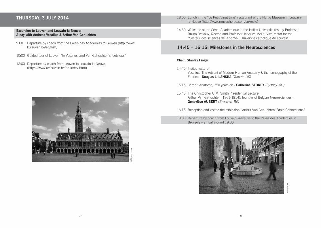
10:00 Guided tour of Leuven “In Vesalius’ and Van Gehuchten’s footsteps”
12:00 Departure by coach from Leuven to Louvain-la-Neuve (https://www.uclouvain.be/en-index.html)
13:00 Lunch in the “Le Petit Vingtième” restaurant of the Hergé Museum in Louvain-la-Neuve (http://www.museeherge.com/en/resto)
14:30 Welcome at the Sénat Académique in the Halles Universitaires, by Professor Bruno Delvaux, Rector, and Professor Jacques Melin, Vice-rector for the “Secteur des sciences de la santé», Université catholique de Louvain.
14:45 – 16:15: Milestones in the Neurosciences
Chair: Stanley Finger
14:45 Invited lecture Vesalius: The Advent of Modern Human Anatomy & the Iconography of the
Fabrica - Douglas J. LANSKA (Tomah, US)
15:15 Cerebri Anatome, 350 years on - Catherine STOREY (Sydney, AU)
15:45 The Christopher U.M. Smith Presidential Lecture Arthur Van Gehuchten (1861-1914), founder of Belgian Neurosciences -
Geneviève AUBERT (Brussels, BE)
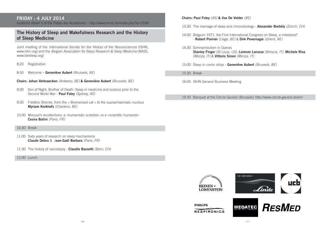
16:15 Reception and visit to the exhibition “Arthur Van Gehuchten: Brain Connections”
18:00 Departure by coach from Louvain-la-Neuve to the Palais des Académies in Brussels – arrival around 19:00
©Li
even
Lem
a
©K
atso
ura
- 16 - - 17 -
FRIDAY : 4 JULY 2014Auditoire Albert II of the Palais des Académies - http://www.armb.be/index.php?id=3536
The History of Sleep and Wakefulness Research and the History of Sleep Medicine
Joint meeting of the International Society for the History of the Neurosciences (ISHN, www.ishn.org) and the Belgian Association for Sleep Research & Sleep Medicine (BASS, www.belsleep.org)
8:20 Registration
8:50 Welcome – Geneviève Aubert (Brussels, BE)
Chairs: Johan Verbraecken (Antwerp, BE) & Geneviève Aubert (Brussels, BE)
9:00 Son of Night, Brother of Death: Sleep in medicine and science prior to the Second World War - Paul Foley (Sydney, AS)
9:30 Frédéric Bremer, from the « Bremerized cat » to the suprachiasmatic nucleus Myriam Kerkhofs (Charleroi, BE)
10:00 Moruzzi’s recollections: a «humanistic scientist» vs a «scientific humanist» Cesira Batini (Paris, FR)
10:30 Break
11:00 Sixty years of research on sleep mechanisms Claude Debru & Jean-Gaël Barbara (Paris, FR)
11:30 The history of narcolepsy - Claudio Bassetti (Bern, CH)
12:00 Lunch
Chairs: Paul Foley (AS) & Ilse De Volder (BE)
13:30 The marriage of sleep and chronobiology - Alexander Borbély (Zürich, CH)
14:00 Belgium 1971: the First International Congress on Sleep, a milestone? - Robert Poirrier (Liège, BE) & Dirk Pevernagie (Ghent, BE)
14:30 Somnambulism in Operas Stanley Finger (St Louis, US), Lorenzo Lorusso (Brescia, IT), Michele Riva (Monza, IT) & Vittorio Sironi (Monza, IT)
15:00 Sleep in comic strips - Geneviève Aubert (Brussels, BE)
15:30 Break
16:00 ISHN General Business Meeting
19:30 Banquet at the Cercle Gaulois (Brussels) http://www.cercle-gaulois.be/en/

- 18 - - 19 -
SATURDAY, 5 JULY 2014Auditoire Albert II of the Palais des Académies - http://www.armb.be/index.php?id=3536
A Celebration of Belgian Neurosciences Joint meeting of the International Society for the History of the Neurosciences (ISHN, www.ishn.org) and the Belgian Neurological Society and the Vlaamse Vereniging voor Neurologie
8:30 Registration
9:00 Welcome – Jacques De Reuck (Ghent, BE)
Chairs: Anne Jeanjean (Brussels, BE) & Patrick Cras (Antwerp, BE)
9:10 Pre-second world war publication ethics - Patrick CRAS (Antwerp, Belgium)
9:30 The Magnus-Rademaker scientific film collection. Ethical issues on animal experimentation (1908-1940) - Peter J KOEHLER (Heerlen, NL) and Bregt LAMERIS (St. Andrews, UK)
9:50 The vision of Joseph Plateau - Nicholas WADE (Dundee, UK)
10:10 Birth of the Neurosciences at the Université catholique de Louvain - Geneviève AUBERT (Brussels, BE)
10:30 Coffee break
11:00 Arthur Van Gehuchten and syphilis - Anne JEANJEAN & Geneviève AUBERT (Brussels, BE)
11:20 The influence of the Great War on the Belgian psychiatry and neurology - Christine VAN EVERBROECK (Brussels, BE)
11:40 From Brussels to the World Federation of Neurology: Ludo Van Bogaert, first president of the WFN - Jacques DE REUCK (Ghent, BE)
12:00 Neurosurgery at the University Hospitals Leuven (Belgium) - Frank VAN CALENBERGH and Christiaan PLETS (Leuven, BE)
12:20 The Frank Clifford Rose Memorial Lecture Magritte transforms Alberti’s ‘Window’: The eye is a false mirror - Gül. A.
RUSSELL (Bryan, US)
13:00 Lunch
14:00 Closure of the meeting
- 20 - - 21 -
POSTERSDisplayed from Monday 30 June
to Saturday 5 July 2014
Salle des Marbres of the Palais des Académies
- 22 - - 23 -
Posters :
1. Brain and language throughout the ages Claude J. BAJADA, Matthew A. LAMBON RALPH and Lauren CLOUTMAN
(Manchester, UK)
2. Psychiatric medicine and the Third Reich Michaël BESSER (Sydney, AS)
3. Jean-Martin Charcot and Art Julien BOGOUSSLAVSKY (Glion/Montreux, CH) and François BOLLER
(Bethesda, US)
4. So near, yet so far: the centenary of Henry Dale’s virtual discovery of the neurotransmitter role of Acetylcholine John CARMODY (Sydney, AS)
5. Neuro-Art History: Exploring the Experience of Art Bruce DOBLIN (Chicago, US)
6. Contributions of James S. Risien Russell to Neurology and Neuro-anatomy Edward J. FINE, Salomi SALINS and Norveen SHAHDAD (Buffalo, US)
7. Conservation and analysis of neurosurgical instruments: an example at the Major Hospital of Milan Antonia Francesca FRANCHINI (Milan, IT), Paolo Maria GALIMBERTI (Milan, IT), Lorenzo LORUSSO (Chiari-Brescia, IT), Bruno FALCONI (Brescia, IT) and Alessandro PORRO (Brescia, IT)
8. James Hill, of Dumfries – Surgeon of Excellence Jeremy C. GANZ (Ulverston, UK)
9. Localization versus integration approaches to human neuroimaging since 1945 Derek J. HUFFMAN (Irvine, US)
10. Caricature as optical illusion: a history of ambiguous figures Lorenzo LORUSSO (Chiari-Brescia, IT), Antonia Francesca FRANCHINI (Milan, IT), Bruno FALCONI (Milan), IT) and Alessandro PORRO (Milan (IT)
11. Mild Traumatic Brain Injuries in the Trenches 1914-18 - A.D. (Sandy) MACLEOD (Christchurch, NZ)
12. Psychopathology of Thomas Mann’s Early Literary Figures Hand-Dieter MENNEL (Marburg, GE)
13. Of Phrenitis and Delirium tremens. History of two syndromes Frederico J. Rodriguez PORCEL and Henry S. SCHUTTA (Maywood, US)
14. Brain, Self, and Environment in the changing definitions of «psychological trauma» Helen SCHÖNBORN (Paris, FR)
15. What is an “Acute Stroke”? History, Epistemology & Ontology Michel C. F. SHAMY (Ottawa, CA)
16. Teaching “nerves” to psychologists in late 19th to early 20th century Japan Miki TAKASUNA (Tokyo, JA)
17. Vincent van Gogh. His brain, his soul, his heart Piet H.A.VOSKUIL (NL)
18. Hans Reese: Olympian, German and American war hero, promoter of peace and pioneer of pyrotherapy with malaria infection for treatment of neurosyphilis in Wisconsin Andrew J. WACLAWIK, Matthew JENSEN, Erika JANIK and Henry SCHUTTA (Wisconsin, US)
19. Henry Duret (1849-1921): a Surgeon and forgotten Neurologist Olivier WALUSINSKI (Brou, FR) and Philippe COURIVAUD (Thélus, FR)
- 24 - - 25 -
Brain and language throughout the ages
Claude J. BAJADA, Matthew A. LAMBON RALPH and Lauren CLOUTMAN
Neuroscience and Aphasia Research Unit (NARU), School of Psychological Sciences, University of Manchester, UK
[email protected]@[email protected]
The neural study of language has its roots in the 19th century European neurology. P. Paul Broca, Jean-Baptiste Bouillaud and Ernest Auburtin brought the concept of ana-tomical localization of higher functions into the scientific forefront. In 1874, Carl Wer-nicke proposed an alternative view. Wernicke proposed that onlyl primary areas can be localized in the brain and set out the first network diagram of language that implicitly proposed that higher function emerged out of the complexity of the anatomical network.
Critics of Broca and Wernicke’s ideas such as John Hughlings Jackson, Pierre Marie and especially Henry Head argued that since all cortical areas are highly interconnected, none can be claimed to be the “seat” of language. In the post-Wernickian period, the holistic viewpoint became dominant and language scienes fell into an anatomical “dark age”. Due to limitations in technology as well as increasing difficulties in obtaining brain specimens, the study of neuroanatomy as a tool to understand language function took a back seat. Language researchers of the time abandoned the brain in favour of building computational models that had no reference to anatomy.
It was not until the advent of modern neuroimaging techniques that researchers had a window into the brains of healthy individuals and, for the first time, scientists could ac-curately localize brain damage caused by disease without having to wait for an autopsy report. Today’s researchers are rediscovering much of what the 19th century anatomists already know. Anatomy is being reintegrated into models of language function in the hope that through the new, non-invasive technologies, new diagnostic and therapeutic tech-niques can be developed for patients who have suffered language impairments through disease.
Psychiatric medicine and the Third Reich
Michaël BESSER, AM MB BS MMedH
FRACS FRCSC FACS, neurosurgery, University of Sydney, Sydney, [email protected]
In the period leading up to the Second World War, and during the war itself, the medical profession in Germany was instrumental in the institution of a system which identified, transported and killed human beings either mentally ill, physically deformed or “racially and cognitively compromised”.
The role of psychiatric medicine was central and critical to the success of Nazi ideolo-gy rather than marginal or incidental. Indeed, doctors provided the technical expertise, supervision and documentation of the Holocaust. Psychiatrists played a central and inti-mate role in the facilitation of crimes against humanity.
How did the medical profession and psychiatrists in particular, reconcile their traditio-nal beneficence role with a mandate of genocide in a country with supposedly the best standard of medicine and ethics in the world at the time? How did, arguably, the finest medical institutions in the early 20th century, which were advancing medicine, medical science and medical education, become part of the worst program of organized mass destruction in the history of mankind?
Critical to the understanding of why psychiatry played a pivotal role in the evolution of the sterilization and euthanasia programs of Nazi Germany is the eugenics movement. Also key is the legal empowerment that came with the perversion of the judicial process under the Third Reich. Two of the major pillars of Western Civilization, the law and the medicine, collaborated to create one of mankind’s bleakest periods.
This paper will explore and explain some of the complex issues involved, trying to unders-tand what seems incomprehensible but from where our modern views of human rights and medical ethics have evolved.
- 26 - - 27 -
Jean-Martin Charcot and Art
1 Julien BOGOUSSLAVSKY1 and 2 François BOLLER2, MD, PhD
1 Genolier Swiss Medical Network Neurocenter, Clinique Valmont, Glion/Montreux, Switzerland
2 Bethesda MD, [email protected]
Jean-Martin Charcot, the “father of neurology” in France and much beyond, was also the man who established academic psychiatry in Paris, differentiating it from clinical alie-nism, which dominated medicine in the first three fourths of the nineteenth century. He was known to be both an authoritative and theatrical man, and while most of this present legacy belongs to classical neurology, his fame at the time was mainly due to his work on hysteria, which attracted the non-medical Parisian intelligentsia. In this field, he used artistic representations with the help of his pupil Paul Richer, whose skills were such that he became a teacher at the Beaux-Arts school in Paris. Charcot himself liked to draw portraits (in particular sketches of colleagues during boring Faculty meetings and stu-dents examinations), self-caricatures, church sculptures, landscapes, soldiers, etc. He also used this gift in his work (histological or anatomic specimens, patients’ features and demeanor) under the influence of his colleague and friend Alfred Vulpian, the founder of modern neurophysiology in France. His most daring artistic experiment was to draw under the influence of hashish, but such attempts were not particularly unusual at the time; Charles Lasègue and other physicians had done it before Charcot. Charcot’s tastes in art were very conventional and he had no connection with the avant-gardes of his time, including impressionism or realism. Indeed, Léon Daudet, son of Charcot’s former friend and famous writer Alphonse Daudet, described Charcot’s home as a pseudo-gothic kitsch accumulation of heteroclite pieces of furniture and materials. However, as Henry Meige wrote a few years after his mentor’s death, Charcot the artist remains “inseparable from Charcot the physician”.
So near, yet so far: the centenary of Henry Dale’s virtual discovery of the neurotransmitter role of Acetylcholine
John CARMODY
University of Sydney, Sydney, Australia [email protected]
When Waldeyer drew on Caja’s superb microscopy to coin the word “neuron” and formu-late his influential “Doctrine” in 1891, that synthesis stimulated ever more questions (in the best scientific tradition). Then, in 1897, in Foster’s textbook, Sherrington devised the term “synapse” to describe the functional surfaces of separation between those neurons. The gauntlet was, thereby, thrown down to neuroscientists at a time when the paradigm of neural activity was an electrical one, but when chemical pharmacology had already be-gun its powerful development: is the operational neuronal linkage electrical or chemical?
The credit for finding the first definitive answer is conventionally given to Loewi, notably for his renowned paper of 1921. As philosophers know, but scientists are prone to over-look, “causes” and “beginning” are often vague and nettlesome matters. This year is the centenary of an excellent paper (“The action of certain esters and ethers of choline, and their relation to muscarine” in the Journal of Pharmacology) in which the British scientist Henry Dale came astonishingly close to enunciating the transmitter function of acetylcho-line. “The question of a possible physiological significance, in the resemblance between the action of choline esters and the effects of certain divisions of the involuntary nervous system, is one of great interest… Acetyl-choline is, of all the substances examined, the one whose action is most suggestive in this direction… the fact that {adrenaline and acetyl-choline} reproduce those effects of involuntary nerves which are absent… gives plenty of scope for speculation.”
Dale was not the only scientist to sail so close to the truth. In 1904, TR Elliott (extending the findings of Langley in a paper to the {British} Physiological Society) stated that “adre-nalin might then be the chemical stimulant liberated on each occasion when the impulse arrives at the periphery”.
- 28 - - 29 -
Neuro-Art History: Exploring the Experience of Art
Bruce DOBLIN, MD, MPH
Northwestern University Medical School and The School of the Art Institute of Chicago, USA
Since the beginning of time, man has created art. For centuries, it has been held that the pur-pose of art is the creation and expression of beauty. The field of aesthetics was developed in an attempt to understand the way that beauty operates on our minds. Early philosophers theorized that beauty had a mimetic aspect and that art that mimicked or replicated life forms were the most appealing, enjoyable and beautiful. The better the artist reproduced the world, they belie-ved, the more the artwork would excite our minds. As we began to understand the working of the brain, these views shifted. In 1775, David Hume wrote in Of the Standard of Taste, “Beauty is no quality in things themselves: it exists merely in the mind which contemplates them and each mind perceives a different beauty”.
During the last few decades, neuroscientists have begun to open up and peer into the black box of the brain that in the past hid all the secrets about its workings. They started to find the connections and associations between neurons and groups of neurons and elucidate the ways in which they influenced each other. They also quickly revealed the fact that our brains are a function of our experiences. And therefore, each of us has a unique and a very personal experience of the world. Scientists began to examine the vast networks of 100 billion neurons for clues about the brain and the impact of arts. FMRIs and other imaging techniques provided even more insight into real time events during the creation and appreciation of art. Soon art critics and art historians began to appreciate this dynamic process of brain development and function. They wondered what it could teach us about the experience of art. In addition, there was an interest in examining the process of creating art and the events that occur in the artist’s brain during the creative process. And, this in turn led to question of comparing the events in the artist’s brain and those triggered in the brain of the viewer or receiver. Should art recreate the same effects in the mind of the artist and the viewer? Given the unique aspects within the brains of the artist and the viewer is there a way to compare the effects? And, how should we study the works of a great artist like Michelangelo today? Can we determine the state of his brain or the how his work stimulated the brain of his contemporaries? How do we understand the period eye seeing the work for the first time? How should we compare that to the effect the art has on our modern brains that have been shaped by centuries of sensory experiences unknown to him?
This presentation will explore these fascinating questions in the evolving field of Neuroarthisto-ry. Neuroarthistory is an approach that challenges us to use neuroscience to answer questions that an art historian might answer about the creative process, the experience of art and art appreciation.
Contribution of James S. Risien Russel to Neurology and Neuro-anatomy
1 Edward JAY FINE1 MD, FAAN, 2 Salomi SALINS, MD and 3 Norveen SHAHDAD3, MD
1 Associate Professor of Neurology, University at Buffalo, The State University, [email protected]
2 Fellow in Clinical Neurophysiology, Department of Neurology, University at Buffalo, The State University, USA
3 Clinical Neurophysiology, Department of Neurology, University at Buffalo, The State University, USA
James S. Risien Russel, MD, FRCP (1863-1939) was born in Guyana, Africa. In 1886, JR graduated from Edinsburgh University Medical School with MB and CM and from which he received the MD with a gold medal in 1888. He was appointed resident physician at the National Hospital for Epileptics in Queen Square (QSH) London in 1888. JR found that hypoxemia, ether and chloroform anesthesia initially increased but then decreased the tendon reflexes of dogs and concluded that anesthesia depressed the nerve cells directly. He proved that the crossed patellar reflex was mediated in the spinal cord and not corpus callosum. JR collaborated with Victor Horsley to discover that extirpation of a dog’s lateral cerebellar lobe would reduce the amount of current needed to stimulate its limbs proving the cerebellum inhibited contralateral motor cortex. Skew deviation of the dog’s eyes followed extirpation of their cerebellar vermis Russell traced degenerating fibers from the cerebellum that left the cerebellum and hooked around the superior cere-bellar peduncle, entered the opposite fastigal nucleus and the descended caudally to the spinal cord in what is called today the hook Bundle of Russell. He also found cerebellar fibers ascended to the contralateral basal ganglia. Gordon Holmes confirmed Russell’s conclusions about cerebellar function in soldiers with cerebellar gunshot wounds. Russell coined the term “subacute combined degeneration” in 1900 to describe the loss of mye-lin in dorsal, posterior-lateral and lateral columns and pyramidal tracts in spinal cords of patients who died with pernicious anemia. Their clinical course was: initial loss of position and greater vibratory impairment, then lower limb spasticity, followed sudden paraple-gia. Death from pneumonia or sepsis proceeded double incontinence. With Batten and Collier, he described axonal loss in peripheral nerves and grouped of atrophy of skeletal muscle fibers. JR advanced knowledge of clinical neurology and neuro-anatomy.

- 30 - - 31 -
Conservation and analysis of neurosurgical instruments: an example at the Major Hospital of Milan
1 Antonia Francesca FRANCHINI, 2 Paola Maria GALIMBERTI, 3 Lorenzo LORUSSO, 4 Bruno FALCONI and 4 Alessandro PORRO
1 Department of Clinical Sciences & Community Health, Univ. of Milan, [email protected]
2 Cultural Heritage Service – Fondazione IRCCS Ca’Granda Ospedale Maggiore Po-liclinico, Milan, Italy
3 Neurology Unit, Mellino Mellini Hospital, Chiari-Brescia, [email protected]
4 Department of Medical and Surgical Specialties, Radiological Sciences and Public Health, Brescia University, Brescia, Italy
[email protected] - [email protected]
In the early 20th century, thanks to the donation of Professor Roberto M. Villani, we have recovered from the neurosurgical Beretta West Pavilion of the Major Hospital of Milan instruments no longer used. The collection, now preserved in the Historical Archives of the Hospital, consists of about 200 instruments (or parts of instruments) contained in a three-compartment suitcase. Some tools had belonged to Professor Paolo Emilio Maspes (1906-1989), who, in the early sixties became Director of the new Institute of Neurosurgery of the University of Milan.The Institute was housed in the premises of the Beretta West Pavilion and quickly became one of the most advanced scientific and specialized clinical centers in Europe.The Institute continued with the Milanese neurosurgical tradition and activities started by Mario Donati (1879-1946), Achille Mario Dogliotti (1897-1966) and Gian Maria Fasiani (1887-1956).Among the instruments collected; emerge those for the treatment of fractures and disloca-tions of the cervical spine (skull calipers, adjustable cervical traction tongs type Crutchfield, self-retaining retractors type Beckmann), tools for brain and cerebrospinal surgery (staples pliers, spatulas, a wide range of plastic retractors type Cushing) and others for the surgery on meninges.The identification of the tools and the understanding of their use are ongoing and represent the first step of their preservation and enhancement. In this phase, the availability of a large number of catalogs of industrial production of neurosurgical tools is fundamental. They allow us to identify not only the various families of instruments, but also the variants of each item often linked to the type of surgical procedure adopted by a single surgeon or com-mercializing of a single company. Moreover, the examination of the plate matrix, together with the examination of the hospital administrative records, can give us information on the characteristics of the supply (national, European and non-European instrument makers).
James Hill, of Dumfries – Surgeon of Excellence
Jeremy C. GANZ MA, PhD, FRCS
Neurosurgeon (retired), Ulverston, [email protected]
Along the northern side of the cemetery of St. Michael’s and South Church in Dumfries in Galloway is a distinguished red sandstone memorial dedicated to James Hill and his family; erected by his only surviving daughter, Ann. Of Mr. Hill it states: “SACRED To the MEMORY of JAMES HILL Late Surgeon in DUMFRIES who died in the year 1776 aged 73. To very superior skill in his profession He joined a taste for Science Which he culti-vated to the latest period of his life. His benevolence to the poor who had not then the re-source of an Infirmary Was unwearied.” The memorial was erected by his daughter Ann.
James Hill was a remarkable surgeon. Whom while not a neurosurgical specialist in fact made a contribution to the neurosurgery of trauma which was recognized for over 100 years after his death. He had by far the best results in the management of serious head injury recorded anywhere in the 18th century or indeed the 19th.
He was undoubtedly a skilful surgeon. However, his major contribution was the result of his willingness to learn from his own experience and while familiar with classical teaching he was not hidebound by it. This is a forgotten colleague of distinction who deserves to be remembered.
- 32 - - 33 -
Localization versus integration approaches to human neuroimaging since 1945
Derek J. HUFFMAN
Department of Neurobiology and Behavior, University of California, Irvine, USA
The goal of this paper is to present a brief historical account of analytic methods for human neuroimaging while providing a theoretical background for each of these deve-lopments. The advent of human neuroimaging has allowed an amazing opportunity for researchers to peer non-invasively into the human brain, representing an extraordinary advancement in the history of brain science. The methods used for data analysis shape the results, and the past 70 years of human neuroimaging have seen several analy-tic techniques -from those that have attempted to localize function to those that have attempted to find interactions between brain regions. These techniques offer different insights into brain organization and operation, with each technique being limited by its assumptions.
The advancement of analytical techniques has required technological developments (e.g. increased computational power, improved data acquisition techniques), however, the in-herent assumptions have been related to those that have been present in neuroscience for centuries. Prior to the development of human neuroimaging, techniques for studying the human brain were largely limited to case studies of patients with circumscribed le-sions. Studies of total cerebral blood flow in the 1940s and 1950s failed to find diffe-rences between cognitive states and between healthy participants and schizophrenic pa-tients. This framework led neuroimaging researchers to focus on localization of function, beginning in the 1960s. Early reports suggested that there was not simple one-function-to-one-structure mapping in the brain, thus providing evidence against strict localization of function. Such results caused a select group of researchers to begin to investigate interactions between brain regions, beginning in the 1980s. Early studies employing in-tegration techniques were met with criticism; however, in recent years, network-based approaches have dramatically increased in prevalence. The implications of results from human neuroimaging will be discussed in the broader theoretical context of brain orga-nization.
Caricature as optical illusion: a history of ambiguous figures
1 Lorenzo LORUSSO, 2 Antonia Francesca FRANCHINI, 3 Bruno FALCONI, 3 Alessandro PORRO
1 Neurology Unit Mellino Millini Hospital, Chiari-Brescia, Italywalton2002 at libero.it
2 Department of Clinical Sciences and Community Health, Univ. of Milan, [email protected]
3 Department of Medical and Surgical Specialties, Radiological Sciences and Public Health, Brescia University, Italy
[email protected]@med.unibs.it
Caricature is a pictorial representation which by means of distortion creates a comic or satirical effect. The humorous use of distortion has been adopted throughout caricature to identify particular individuals and institutions, especially in the late Renaissance.
In the 16th-17th century, caricature treated the human figure in an abstract or schematic way without an evident satiric intention, but it demonstrates how artists could look indivi-duals and objects abstractly so that any event could be rendered by a readymade graphic formula as precursor of Surrealist art.
Many artists had a surrealistic fantasy that became elaborate formed by a conglomeration of different objects such as Giuseppe Arcimbolde (1537-1593). Arcimboldo’s paintings with his famous “composed heads or reversible heads” are considered surrealistic ap-proach to Physiognomy but also ambiguous figure with a perceptual interpretation. Am-biguous figures as double illusions and upside-downs have an illusory effect with vivid descriptions by a psychological impact with current scientific explanations.
Other artists and psychologists used caricatural pictures as ambiguous figures such as the British William Hogarth (1697-1764) with his “Satire on false perspective” (1753), the French Honoré Daumier (1808-1879) with his “Double faces” (1838), the British Reginald John or Rex Whistler (1905-1944) with the drawings “Topsy and Turvys” and others.
Caricature from all periods plays a double role for our mind: an optical illusion on reality by a smiling.
ReferencesC. Faggi (2013) Illusionarum. Florence Art Edizioni, Firenze;R. Gregory (2009) seeing through illusion. Oxford University Press, Oxford;L. Lorusso. Neuroscience by caricature in Europe throughout the ages. www.neurocaricature.eu

- 34 - - 35 -
Mild Traumatic Brain Injuries in the Trenches 1914-18
A.D. (Sandy) MACLEODPsychiatrist, Burwood Hospital, Christchurch, New Zealand
Sixty percent of casualties were inflicted by artillery. Being blown up or buried occurred to 22% of officers (McPherson). Helmets, introduced in 1915, provided little protection. Neurosurgery was in its infancy and most severe TBIs were fatal. Of the cases of head injury treated, 8.6% were fatal, 5.9% were discharged as invalids and 82% returned to duty (Noonan). Undoubtedly PCS was clinically recognized. Clinical descriptions of tran-sient post-concussion symptoms (confusion, hypersomnia, headache, vertigo, visual ab-normalities, amnesia) lasting several days were common. Concussive injuries were likely very common and in hindsight the Prideaux Committee (1922) acknowledged symptoms did occur following concussion.
Officially (in British literature) 5-10% of cases of “shell-shock” were attributable to concussion and 2.5% of psychiatric casualties had organic lesions. Cases were generally considered emotional and rarely commotional (despite Mott). Concussion was actively discredited as a diagnosis. GRO 2384 in June, 1917 instructed that a loss of conscious-ness had to be observed and recorded by an officer (not a priority for most officers in the heat of battle), otherwise a diagnosis of NYDN must be made.
In Wiltshire’s series of 152 cases of shell-shock about a third had been near a bursting shell. It was noted that as the war became mobile in 1918 the cases of neuroses fell. In the compilation of case reports by Southard, about 50% of the cases labelled shell-shock had suffered a period of unconsciousness. Leed considered the trench experience was “nothing if not an experience of radical discontinuity on every level of consciousness” (including the medical). The consensus at the time that “the real trauma was psychical, not physical” might be challenged in view of the modern experience with blast injuries in Iraq and Afghanistan. Unlike the French, British and German medicine of the period struggled to allow organic and psychological symptoms to co-exist.
Psychopathology of Thomas Mann’s Early Literary Figures
Hand-Dieter MENNEL
Abteilung für Neuropathologie, Philipps-Universität Marburg, Marburg, Germany
In his first novel Buddenbrooks and in his short stories as well as in his sole dramatic work Fiorenza, the author Thomas Mann deals with the decadence discourse of the fin-de-siècle around 1900, but the figures in his work at the time might as well be considered in terms of psychopathology. They are nervous in the larger sense of the word, a designa-tion that then covered equally neurasthenic and psychopathic conditions. The psychiatric background of the behavior of most of his fictive characters at the time is degeneration, a term coined by French psychiatry in the 19th century, indicating increasing disability du-ring subsequent generations within one family. Most, but not all of these early characters might be designated as psychopaths following the typology established in the first half of the 20th century by the German psychiatrist Kurt Schneider.
Yet, not all of the figures of the early period of Thomas Mann fit completely into this scheme. There is a second group, literary authors and artists that resent and reflect their outsider position. For both groups, a common denominator might be found in the “loss of immediacy”, a concept developed amongst others within German “Daseinsanalyse”. This “existential analysis” represents a particular trend in medical anthropology, which explains deviant behavior by the suggestive terminology of the phenomenological phi-losophy of Edmund Husserl or existentialistic thinking of the early concepts of martin Heidegger. However, pertinent concepts were not developed before 1900 and Heideg-ger’s important first text – Sein und Zeit i.e. Being and Time – appeared in 1927.
Therefore, pertinent concepts obviously were developed independently.
- 36 - - 37 -
Of Phrenitis and Delirium tremens.History of two syndromes
Federico J. Rodriguez PORCEL and Henry S. SCHUTTA
Loyola University, Chicago, Stritch Medical School, Maywood, USAfrodriguezporce at lumc.edu
The dominant symptom of phrenitis was delirium. Galen held that phrenitis was “connec-ted with inflammation of the parts about the brain” and since then, phrenitis has been viewed as an inflammatory disorder of the brain.
Most delirious states were associated with fevers. Its occasional association with “drinking of fermented liquors” began with Hippocratic writers: “If the patients be in the prime of life, and if his body be strong and brawny, of a melancholic temperament, or if from drinking he has trembling hands it may well be to announce either delirium or convul-sions”.
Until the beginning of the 19th century, all patients with phrenitis were treated with co-pious bloodletting supplemented by additional methods of abstraction.
Thomas Sutton, and a number of his colleagues realized that some patients with the diagnosis of “phrenitis” suffered from a disease with a symptomatology that was different from phrenitis and that was associated with excessive alcohol consumption. In 1813, Sut-ton published a monograph in which he described 14 patients with 16 episodes of what he believed was a new disease, and which he called “Delirium Tremens”.
Sutton and some of his colleagues refrained from bloodletting in such patients, since they observed that abstraction aggravated the disease. Their principal weapon was opium in large doses given repeatedly until sleep was induced. Sutton reported a mortality rate of 18% in his patients and estimated that mortality in patients treated with abstraction was one in three.
Richard Bright (1831) reported five patients with delirium tremens. Two treated with bloodletting died, three treated without abstraction survived.
Sutton could find no enlightenment as to the state of the brain in delirium tremens or the modus operandi of the remedy. Bright speculated that delirium tremens was a type of “arachnitis” modified by “excessive irritability”.
Brain, Self, and Environment in the changing definitions of “psychological trauma”
Helen SCHÖNBORNAmerican University of Paris, Paris, France
This paper explores the historical and theoretical background of the development of the psychological trauma concept with a particular emphasis on the allocation of responsi-bility between society and the individual. Since the 19th century, definitions of psycho-logical trauma have changed in relation with different philosophical conceptualizations of the human body, the advancing neurological understanding of the human brain, as well as political and economic interests concerning medical reimbursement and social control. Originally, trauma referred to reactions to physical injury. However, in the lat-ter part of the 19th century during a politically tenuous period in France, Jean-Martin Charcot brought hysteria to the forefront of neuro-psychological investigations. Freud and Janet further developed the involvement of psychological trauma in the etiology of hysteria. Both agreed that the psyche can be hurt by an external agent, which causes psychological distress. Psychological trauma was later associated with the experience of war, torture and extreme cases of abuse. However, it appears that the interest in trauma grew after each war, so that its definition and recognition developed according to the eco-nomic, political and social interests of the moment. Despite major changes in its concep-tualization and recognition, definitions of psychological trauma have always involved an environmental cause or precipitating factor. Trauma is intrinsically linked to the outside, the environment, the social. Paradoxically, it is generally regarded to be an exclusively individual problem, as it is caused by the subjective experience of an extreme event and, thus, it is the responsibility of the individual to seek treatment. The individualization of trauma is driven further by its more recent forms of neuropsychologicalization, based on the observation that psychological trauma alters brain functioning. This stresses trauma’s affiliation to the particular patient and emphasizes his or her personal responsibility to heal. Diagnoses of psychological trauma have consistently facilitated political recognition of victims’ suffering, but have also turned focus away from societal responsibility.
- 38 - - 39 -
What is an “Acute Stroke”? History, Epistemology & Ontology
Michel C.F. SHAMY, MD FRCPC
Attending Neurologist, The Ottawa Hospital, Ottawa, Ontario, CanadaMA Candidate (History & Philosophy of Science),
University of Calgary, Calgary, [email protected]
In order to study how doctors treat patients with acute stroke, it is necessary to establish how those doctors understand the meaning of “acute stroke”.
This paper intends to ask one question: what is an “acute stroke”? in the contemporary context, the notion of the acute stroke appears to be intimately tied to the use of the clot-busting drug tissue plasminogen activator (tPA), and particularly to its potential for reversing the signs and symptoms of stroke. This potential was first suggested in the results of the National Institutes of Neurological Disease and Stroke (NINDS) clinical trial, published in the New England Journal of Medicine in 1995. Specifically, I propose that “acute stroke” is now understood to mean “a stroke that can be treated success-fully in many cases by properly administered tPA”. I will also seek to show that this recent conceptualization of “acute stroke” rests upon a set of historically-determined epistemic preconditions. I will argue that the definition of acute stroke in relation to tPA is dependent upon six concepts: the description of the brain’s vascular anatomy, the localizability of pathology, the pathological distinction of stroke from hemorrhage, the dissemination of emergency services, the development of computerized tomography (CT) scanning, and the physiological concept of the “ischemic penumbra”. The purpose of this investigation is to clarify our understanding of the contemporary definition of “acute stroke”, in order to be better positioned to explore the ethical and epistemic aspects of acute stroke decision-making.
Teaching “nerves” to psychologists in late 19th to early 20th century Japan
Miki TAKASUNA
School of Human and Social Sciences, Tokyo International University, Tokyo, Japan
Just 140 years have passed since the publication of two important books in the field of physiological psychology: Principles of Mental Physiology (1874), which was written by English physiologist William Benjamin Carpenter (1813-1885), and Grundzüge der physiologischen Psychologie (1873-74), authored by German physiological psychologist Wilhelm Wundt (1832-1920).
Japan’s history of physiological psychology began in 1877 with the founding of the first national university (Tokyo University). Masakazu Toyama (1848-1900), the first professor there to teach psychology, lectured predominantly from the English writings of Alexander Bain (1818-1903), Herbert Spencer (1820-1903), and W. B. Carpenter. Although Toya-ma published no textbooks on psychology, another educator, Enryo Inoue (1858-1919) wrote Tsushin Kyoju Shinrigaku {Corresponding Lectures on Psychology} (1888). As one of the few authors of Japanese textbooks on psychology printed in the late 1880s, Inoue stated in his book: “I should assert that mental action be conducted in the nerves”.
Attributing a mind-nerve connection was typically emphasized in textbooks written during the Meiji era (1868-1912). Thus, I collected a dozen Japanese textbooks on psychology during this period to investigate nerve, brain, or other physiological topics described. Re-sults showed three key resources used: “Principles of Mental Physiology” (1874) by W. B. Carpenter, “Elements of Physiological Psychology” (1887) by George Trumbull Ladd, and the fifth edition of “Grundzüge der physiologischen Psychologie” (1902-03) by W. Wundt. My summary here includes those studies and their emphasis on three aspects of the central nervous system: types of nerves, mechanisms of nerve conduct and the neuron theory.
- 40 - - 41 -
Vincent van Gogh. His brain, his soul, his heart
Piet H. A. VOSKUIL
Retired neurologist/epileptologist, [email protected]
Vincent van Gogh’s medical history is discussed in all kind of disciplines by scholars with mostly small scopes.
In this presentation, I will not try to focus on labeling his diagnosis but on the interaction and chronology of his crises with psychological factors related to art historical develop-ments in the last years of his life.
With Gauguin and Bernard, stressful discussions took place on the issue of painting “by heart” in the studio or “by soul” in the nature.
The concepts of Lombroso, Morel and others on hereditary brain degeneration, “neu-rasthenia”, hysteria and the influence on artistic creativity and geniality were known to van Gogh.
His doctors told him that his crises were caused by “épilepsie larvée” (masked epilepsy) as conceptualized by Falret and Morel. van Gogh knew of family members who were said to have epilepsy as well.
In this presentation, information is given on recent investigations of some family chro-nicles.
In van Gogh’s time sensorial influences on the brain were already measured with dyna-mometric equipment by Charles Feré (1852-1907).
Today we are aware of the complexity of neuromodulating influences of sensorial input in the connectomes of the brain as measured by tractography with techniques like fMRI and DTI (Diffusion Tensor Imaging).
But we are far from understanding the influence on dysfunction of the emotional circuit in the brain.
Hans Reese: Olympian, German and American war hero, promoter of peace and pioneer of pyrotherapy with malaria infection for treatment of neurosyphilis in Wisconsin
Andrew J. WACLAWIK, MD, Matthew JENSEN, MD, Erika JANIK and Henry SCHUTTA, MD
Department of Neurology, University of Wisconsin, School of Medicine and Public Health, Madison, Wisconsin, USA
Hans Reese was an Olympian (German national soccer team, 1912 Olympic Games in Stockholm), a German (WW I) and American (WW II) war hero, an export on Native Ame-rican art, and a promoter of peace and international collaborations between neurologists. He was the first true clinical neurologist in the State of Wisconsin.
The presentation will focus on two areas of his work.
Dr. Reese was recruited from Germany in 1924 to work at the Wisconsin Psychiatric Ins-titute, primarily for his expertise in neurosyphilis. At the beginning of the 20th century, up to 13% of admissions to psychiatric hospitals and mental institutions in Wisconsin were related to neurosyphilis. After the WW I, pyrotherapy with malaria infection for treatment of dementia paralytica caused by neurosyphilis, pioneered by Wagner-Jauregg, was gai-ning popularity in many countries. Dr. Reese used a combination of malaria therapy and a novel arsenic derivative, tryparsamide, which was initially developed at the Rockefeller Institute for the treatment of African sleeping sickness. In 1920s, Drs. Lorens, Loevenhart and Reese pioneered the use of tryparsamide for treatment of neurosyphilis at the Wis-consin Psychiatric Institute. The scientific and ethical standards of conducting clinical re-search with malarial therapy on patients with neurosyphilis in the 1920s will be reviewed.
During WW II, Dr. Reese, as a neurologist and psychiatrist, grew very interested in stu-dying the psychological effects of bombing and the management of soldiers suffering from the mental effects of warfaire. After the war, he actively engaged in international collaborations between neurologists from different countries and continents. In 1963, he was awarded the Cross of merit by the Federal Republic of Germany for his activities in promoting German-American relations.
- 42 - - 43 -
Henry Duret (1849-1921): a Surgeon and forgotten Neurologist
1 Olivier WALUSINSKI and 2 Philippe COURIVAUD
1 Family Physician, Brou, [email protected]
2 Anesthesiologist, Thélus, France
Henri Duret (1849-1921) was as surgeon whose training started in the laboratory of Jean-Martin Charcot and Alfred Vulpian at La Salpêtrière in 1874. (A photo of Duret). Most of his career took place in Lille where he was elected to the professorship of surgery in the Catholic University in 1885. Over 40 years, he authored numerous publications on digestive and gynecological surgery and on teaching in these areas. He was endowed with outstanding vigor for multiple researches in neurosciences that we want to enhance in this poster.
Using injections of coloured gelatin, Duret was the first to describe the distribution of supply arteries in the brainstem, then the cortex. His descriptions correlated irrigated territories, infarcted zones and secondary neurological deficits (Figures in color).
Duret focused his 1878 thesis on experimental studies of brain trauma and localized the origin of disturbances in autonomic function and vigilance to the brainstem. Introducing a new concept in the dynamics of head injury, Duret regarded all the spaces containing cerebrospinal fluid as a single unit, in which the traumatic forces were propagated along the neural axis. He linked this “choc céphalorachidien”, or shockwave theory, to micro-haemorrhages affecting the medulla and pons, which are now known as “Duret haemor-rhages” (figures in color).
Duret studied the localization of voluntary motor functions in the cerebral cortex in expe-rimental animals. He mastered a technique for producing well-circumscribed cortical le-sions and he use faradic instead of galvanic currents to stimulate the cerebral cortex. His depiction supported the concept of an integrated somatosensory cortex and mentioned the role of the caudate nucleus and the brainstem in the coordination of movements (figures in color).
Duret described the phenomenon known as the “Cushing reflex” even though Harvey Cushing wouldn’t describe it until 1901, 24 years after Duret.
At the turn of the century, Duret published a large innovative and comprehensive book, of more than 800 pages, about intracranial tumours, one of the first of its kind. Duret not only meticulously compiled the signs and symptoms for various cerebral locations but also described the diagnostic procedures and reviewed the surgical treatment, including surgical techniques, indications, complications and outcome of 400 cases. It is surpri-sing that this monumental work is not better known. Duret can be referred to as “the first French neurosurgeon” (figures in black and white).
NOTES

- 44 - - 45 -
ABSTRACTS
- 46 - - 47 -
Imaging shell-shock: medical film and the construction of a mental disorder
1 Bregt LAMERIS and 2 Lorenzo LORUSSO
1 University of St Andrews, [email protected]
2 Neurology Department Chiari – Brescia, [email protected]
Over the past two decades, early films made by neuroscientists have surfaced in several European countries, including Belgium (Van Gehuchten), Italy (Negro and Neri) and Netherlands (Magnus and Rademaker). These collections include material that depicts and analyses mental disorders.
One of the films by Camillo Negro shows us a shell-shock patient. The man on the screen is unconsciously repeating his actions in the trenches, such as shooting and taking shel-ter. It is as if his movements in the trenches have been written into his nervous system, as if war took over this man’s body and turned it into a living archive. At the same time, from the moment this was registered on film, the film allows us to re-watch the man repeating his actions at war.
In our presentation, we will position this film within a larger context of shell-shock mate-rial, such as War Neurosis: Netley Hospital, 1917 (Wellcome Institute) and the films in the archive of the EPCAM in Paris. These films seem to show shell-shock patients in a much more hopeful setting than the Negro-film. Most of the films show a before and after effect: first we see the condition of the patients when they first arrived at the hospital, followed by their improved constitution after treatment. The Negro-film only shows the patient in his worst moments.
The various films show us different interpretations and constructions of shell-shock by the use of film in the 1910s. What interests us, is where and how these films were used and shown. Where the images used for research purposes only or was thy shown in front of larger audiences? Were they used as propaganda, entertainment or education? Or was the material used to study shell-shock and its treatment, facilitating knowledge exchange at conferences or other occasions?
A comparison of the international films and the way they were used will provide unders-tanding of the interrelationship between visual representation, investigation and explana-tion of shell-shock in the 1910s.
TUE
SD
AY, 1
JU
LY
TUE
SD
AY, 1
JU
LY
NOTES
- 48 - - 49 -
Cinematography of “War Hysterics” in the First World War. Comparative perspectives
Julia Barbara KÖHNE, PD Dr.
Historisch-Kulturwissenschaftliche, Fakultät Institut für Zeitgeschichte, Universität Wien, Wien, Austria
During the First World War, the male “war hysteric” not only gained an iconic statut within the community of mentally injured and psychically traumatized officers and sol-diers, but also became an important figure symbolizing the weakness, inefficiency and vulnerability of four spheres: modern industrialized warfare, the military collective body (the corps), the nation and masculinity. The relatively new media technology of scientific cinematography was used within German, British and French military-neuropsychiatry in order to visualize “construct” and distribute the disturbing symptoms of so-called “war hysteria” – including heavy shivering, verbal and lower body dysfunctions such as pro-blems with sitting, standing, walking and speaking , as well as tics, paralysis and other disabling factors. This medical visualization technique seemed to “catch” the external appearance of what was considered to be a sign of disempowered and “defeated mas-culinity” and capture it on celluloid. But the shivering and shaking limbs of the “male hysteric” challenged the borders of medical cinematography as they mirrored the flaws of early film technology by emphasizing its own representational limits, twitches, stagnations and aesthetical “hysteria”.
The selected medial films depict the sensitive relation between military psychiatry and its protagonists, visibility and pathologization and the visually objectified, domesticated and remilitarized “deviant” individual. Analyzing the films shows the complexly choreo-graphed medicine spectacle that contains subtle messages from the filmed soldier-pa-tients that reveal frail approaches of disobedience and silent resistance. By exploring the traces of disharmony, dissonance and malfunctioning in the patients’ behavior and acting in front of the camera, an ulterior scope becomes visible that opposes the healing paradigm of most of the films. In some scenes, the patients refused to conduct the com-manded homogeneous choreography of gazes, gaits and movements in order to demons-trate the “hysterical symptoms” in “the right way”. Looking at these willing or unwilling interventions and underminings of the “war hysteric”, a fragile parallel film story can be revealed that is beneath the propagandistic and success-oriented healing-visions of the military physicians. By this, the “war hysteric” can be re-evaluated as a diaphanous per-former of an illness “invented” by military physicians that stigmatized him as a passible and effeminate medical object.
The Canadian Army and “Shell Shock” during the World Wars
Russ BENNEWEIS, PhD Candidate
Department of History, Centre of Military and Strategic Studies, University of Calgary, Calgary, Canada
The Canadian Expeditionary Force suffered a minimum of 15,000 shell shock cases during the Great War according to the postwar testimony of a Canadian Army Medical Corps physician. Canadian military officials were thoroughly unprepared for the novel battlefield affliction, eventually attaching the label, “functional”, “neurasthenic” to shell shock victims. Treatments ranged from complete rest to painful applications of faradic current through a wire brush. Subsequent pension payments to shell shock victims cost a displeased Canadian government millions of dollars during the interwar years. Eager to avoid similar circumstances in the aftermath of the Second World War, the Canadian Army adopted comprehensive psychological and psychiatric examinations that promised to weed out during induction and training those predisposed to nervous breakdown in combat. The consequences included the rejection of thousands of potential riflemen and chronic manpower shortages within infantry formations. Despite these ultimately harmful measures, the rate of shell shock cases exploded during the Second World War.This presentation discusses the subject of shell shock in the Canadian Army during the world wars argues the final incongruity was largely the result of societal and educational changes in Canada during the interwar years.
TUE
SD
AY, 1
JU
LY
TUE
SD
AY, 1
JU
LY
- 50 - - 51 -
“A Sound Mind and A Sound Body”: Major Salmon Faces Shell-shock During WWI
Bill HANIGAN
Central Mississippi Medical Centre, Jackson, MI, [email protected]
In December 1917, Major Thomas Salmon MC, Medical Director of the national Com-mittee for Mental Hygiene (NCMH), went to war. He was 41 years old and had worked as a private physician, public health officer, administrator and clinical statistician. Salomon was a forceful and well-known advocate for “Mental Hygiene”, a progressive medical cause that touted reform and public health education in psychiatric care, increased out-patient services and personnel and above all, a dominant role of psychiatrists in contem-porary medicine. He had travelled to Britain earlier to review Allied experiences with shell-shock. It was a disappointing trip but he returned with comprehensive recommendations for the new Section of Neurology and Psychiatry in the Army Medical Department. For Major Salmon and the NCMH, shell-shock was simply a war neurosis with “endless va-riations and one central theme, escape from intolerable situation in real life to one made tolerable by the neurosis.” Eleven months later Salmon faced almost 10.000 neuropsy-chiatric casualties.
This talk review prewar development of the NCMH, Salmon’s organization of military neu-ropsychiatry and his postwar efforts to direct veteran’s care, research and expansion of psychiatry as a medical specialty. It asserts that his extensive military experience signifi-cantly influenced public and professional viewpoints of early twentieth century American psychiatry.
The fight for “traumatic neurosis”, 1889-1916: Hermann Oppenheim and his opponents in Berlin
Prof. Dr. med Bernd HOLDORFF
Neurology, [email protected]
The concept of traumatic neurosis conceived by Hermann Oppenheim (1858-1919) lo-cated post-traumatic nervous symptoms between hysteria and neurasthenia, considering them as consequence of physical reactions to fright and a cause of molecular tissue changes. As early as 1890, his concept was criticized at an international congress in Berlin. In February 1916, there was a significant debate of the issue in Berlin, and even-tually Oppenheim’s concept was completely defeated with the consequence of his resi-gnation at the war meeting of German neuropsychiatrists in September 1916 in Munich, which will be touched here only briefly. In the Berlin debate, a range of views on war neurosis were presented. Partly as a result of this, but also due to the powerful position of Oppenheim himself, the issue of traumatic neurosis persisted before being excluded from medico-legal assessments after the end of WWI. The differing views of physiological brain-mind-relations from that time do not differ greatly from present concepts. However, Oppenheim’s traumatic neurosis with its more quasi-neurological picture should not be equated with PTSD.
(Already published in History of Psychiatry, 22 (2011), 465-474)
TUE
SD
AY, 1
JU
LY
TUE
SD
AY, 1
JU
LY
- 52 - - 53 -
Neuroses and psychoses in the Great War: Alfred Walter Campbell’s experiences
Paul LANCASTER
Menzies Centre for Health Policy, School of Public Health, University of Sydney, Sydney, Australia
Born in rural New South Wales in 1868, Campbell gained his medical degrees in Edin-burgh at a time when the medical course at the University of Sydney was not yet well esta-blished. After clinical and research experience in Britain and Europe, he was awarded his doctorate in 1892 for his thesis on “Alcoholic neuritis: its clinical features and pathology”. During more than a decade as medical officer and director of pathology at Rainhill County Mental Hospital, near Liverpool in England, his reputation soon flourished. This was en-hanced by his monograph Histological Studies on the Localisation of Cerebral Function, based on his studies of the histological structure of the brain, and “hailed as a landmark”. Returning to Sydney in 1905, he started his private practice as a specialist in neurology and mental diseases and a consultant in major hospitals.
Enlisting aged 46 in the Australian Army Medical Corps in November 1914, he served with the No. 2 General Hospital of the Australian Imperial Force in Egypt until December 1915. Several months later, he published Remarks on some neuroses and psychoses in war in the Medical Journal of Australia, noting that these mental illnesses “contributed to modern war casualty lists more heavily than we had previously supposed”. His classi-fication of mental illness distinguished between various types of neuroses, neurasthenia (including “trench spine”) and varying severity of psychoses.
Acknowledging the appalling conditions of warfare endured by his patients, Campbell concluded that “the fundamental cause of their downfall” was frequently a family or per-sonal history of “neuropathic or psychopathic infirmity”. Because of difficulties in suitably treating these mental disorders in a general hospital, Campbell recommended treatment either in a small special hospital or attached to a general hospital staffed by a medical officer and orderlies or nurses, “skilled in the management of mental cases”.
The role of Elliot Smith in the British attitude to Shell-Shock
Ian STEELE-RUSSELL
Departments of Neuroscience & experimental Therapeutics, Psychiatry & Behavioral Science, College of Medicine, and the Department of Veterinary Integrative Biomedi-
cal Sciences, College of Veterinary Medicine, Texas A&M University, US
There was a long and appalling tradition in the British military, of savage disciplinary treatment for misconduct of any kind ranging from such actions labelled “dumb insolen-ce” to cowardice. Although flogging was discontinued in 1867, after the Crimean War, cowardice remained a military capital crime until the end of the Great War of 1914-1918.
The principal architect for major humane changes both in British military medicine and psychiatric care for the insane was Elliot Smith during the time of his major reforms of British medical education when he was Professor of Anatomy at Manchester University (1909-1919).
In his early years, Elliot Smith’s major emphasis was on the functional implications of structure. At that time, he was strongly against psychology which he regarded as creation of pseudo-scientific charlatans. In particular he regarded psychoanalysis as either bogus or thinly veiled pornography.
In 1913, as Dean of the Manchester University Medical School, he was invited to be a member of the British General Medical Council (1913-1919). In 1915 he was made a fel-low of the Royal College of Physicians. Towards the end of 1914, he helped the war effort by studying the neurological problems of shell-shock. In order to observe the condition at first hand, he served as a voluntary physician during the summer of 1915 at the Military Hospital Maghull, near Liverpool, which was devoted to the treatment of soldiers suffering from the effects of shell-shock. His report was first published in 1917, argued that the shell-shock was a medical condition and mainly psychological in origin. Elliot Smith’s work in this domain has been much neglected considering the enormous influence it subsequently had on both British military medicine and psychiatry in general.
TUE
SD
AY, 1
JU
LY
TUE
SD
AY, 1
JU
LY
- 54 - - 55 -
“My interest in neurology has changed for the benefit of history and psychological studies”: the correspondence between Winkler and Monakow during the World War
1 P.J. KOEHLER and 2 C.E. JAGELLA
1 Atrium Medical Centre, Department of Neurology, Heerlen, [email protected]
2 Kantonspital Baden, Department of Neurology, Baden, Switzerland
The correspondence (1907-1930) between two leading European neurologists, Cornelis Winkler (1855-1941) and Constantin von Monakow (1853-1930) has been preserved in Amsterdam and Zurich. For this paper, letters exchanged during World War I were stu-died. Professional as well as personal issues were discussed. An international neurology meeting in Berne in September 1914 had to be cancelled due to the war. They hoped that (neuro) scientists would remain politically neutral, continue scientific cooperation, and even be able to influence the course of the war. Winkler and Monakow tried to continue their work on the International Brain Atlas. Although living in neutral countries (Nether-lands and Switzerland), they observed that their practice and scientific work suffered from war conditions. Whereas Winkler continued his activities as a neurologist, Mona-kow, affected emotionally, experienced a change in scientific interest towards psychoneu-rology. He used his diaschisis concept, originally an explanation for transient phenomena in stroke, as a metaphor for the social and cultural effects of the war. He directly related cultural development and brain science, bringing in his own emotions and resulting in the first of several publications on the relations between biology, brain science and culture.
Mechanized Warfare and Humanized Medicine – On the Reciprocal Conditions of Brain Injuries and Neurorehabilitation in Kurt Goldstein’s (1878-1965) Program of Holist Neurology, 1916-1933
Dr. Frank W. STAHNISCH
AMF/Hannah Professorship for the History of medicine and Health Care, Faculties of Medicine and Arts, University of Calgary, Calgary, Canada
The famous Frankfurt neurologist Kurt Goldstein has already been the subject of much historiographical research over the last two decades with regard to a wider range of as-pects of his neurological theory, the advancement of clinical psychology, and the deve-lopment of the holist medical tradition in Weimar Germany. However, the way in which he literally slithered into the program of holist neurology through the impact of the conditions of mechanized warfare and the resulting massive amount of brain and nervous injuries of World War One still remains an understudied area. In the 80th year of the first appea-rance of his famous book of “The Organism: a Holistic Approach to Biology” (German, 1934 / English transl., 1939), which the author of this abstract is currently co-publishing in a revised and introduced new German edition (and for the first time with a publisher in Germany itself: Fink Verlag, Munich, 2014, forthcoming), this presentation will focus on the establishment and working conditions of the “Institute for Research into the Long-Term Effects of Brain Injuries” in Frankfurt am Main – established by Ludwig Edinger (1855-1918) and Kurt Goldstein (1878-1965) – in 1916 as an immediate effect of the Battle of Verdun in France. It will then follow the subsequent development of Goldstein’s research program on “holist neurology” further to the Clinical Department of Neurology at the academic hospital of Moabit in Berlin from 1930 to 1933, before Goldstein was ousted of his professorial position in Germany and forced to leave his home country for good, first into exile in Amsterdam in Holland and later to New York City in the United States. This presentation draws on historiographical research in the University Archives of the Johann Wolfgang von Goethe University in Frankfurt am Main (Germany), the Ar-chives of the Society for the Protection of Science and Learning in the Bodleian Library at the University of Oxford (England), as well as the Rockefeller Archive Center in Sleepy Hollow, New York (United States of America).
TUE
SD
AY, 1
JU
LY
TUE
SD
AY, 1
JU
LY
- 56 - - 57 -
Thoughts prompted by two important anniversaries: World War I and the birth of “Behaviourism”
John CARMODY
University of Sydney, Sydney, Australia [email protected]
ISHN is meeting in Brussels a bore month before the centenary of the declaration of Wor-ld War I and the sweep of the modern German army across Belgium. That conflict had many consequences for the entire world – scientific, cultural and educational as well as political and military ones. But what, specifically, of neuroscience and were they related?
This year is also the centenary of the publication of JB Watson’s immensely influential book, Behavior: an introduction to Comparative Psychology which aimed to correct the situation in which (in Watson’s words) “Psychology has failed signally during the fifty odd years of its existence as an experimental discipline to make its place in the world as an undisputed natural science”. This was, he believed , because of the persistence of its philosophical origins (for all that Locke had used the term “mind” in place of “soul” in his famous Essay concerning human understanding of 1689); Watson wanted psychology to “discard all reference to consciousness”. He regarded this substitution of “mind” for “soul” as a compromise with the rational conscience. That was a repudiation of both Wundt and James but, recognized by Watson or not, his own philosophical basis was both limiting and serious: “The behaviorist … recognizes no dividing line between man and brute.”
This language sounds disturbingly consonant with the willingness of all participants in World War I to practice and condone brutality in their treatment of combatants and non-combatants alike. While it would be hyperbole to assert that Watson’s ideas in-fluenced military and political commanders, the contemporary art suggests that he cap-tured something of a Zeitgeist. Curiously, aggression seemed of little or no interest to him.
Behaviorism essentially reduced people to complex machines: to that extent it was of a piece with the thinking which adopted the vast mechanisation of war in the twentieth century.
An outline of the French and British experience in the treatment of penetrating head injuries during WW I
Moshe FEINSOD, MD
Faculty of Medicine, the Technion Israel Institute of Technology, Haifa, [email protected]
During four years of unremitting hostilities, the fighting troops on the “Western Front” suffered a human toll unrecognized before in any previous war. Frontal assaults against machinegun nests and incessant shelling caused sometimes many thousands of ca-sualties per day. Head injuries were quite common in the trenches war as the head was often the only part exposed. The heads of the soldiers remained, however, unprotected throughout the first half of the war. Life in the trenches was far from hygienic and the extremely slow evacuation caused and unstructured triage of the severely wounded or unconscious head injured soldiers.
In both armies, neurosurgery was not recognized yet as a specialty and in most ins-tances it were general surgeons that took care of the head injured soldiers. The state of knowledge of the treatment of head injuries was gained in different conditions, either terrain or weaponry. The experience of the French Thierry de Martel and Sir Victor Hors-ley of the UK was not translated into formal directives to all the medical services. Albeit these handicaps the French and the British had joint meetings exchanging experience and ideas and there was a constant process of learning of the neuropathology of trauma to the nervous system, the diagnosis by the newly introduced X-ray imaging of previously unrecognized lesions, perfection of cerebral localization and improvements in surgical instrumentation.
These labors and the lessons learned from the attempts of the surgeons who tried to save lives from the carnage are appreciated and from a distance of a century and may serve as a distant mirror.
TUE
SD
AY, 1
JU
LY
TUE
SD
AY, 1
JU
LY

- 58 - - 59 -
On the treatment of War Wounds of the Central Nervous System at I. Surgical University Clinic in Vienna
Helmut GRÖGER, MD
Institute for the History of Medicine, Medical University of Vienna, [email protected]
During World War I, Anton von Eiselsberg (1860-1939), head of the I. Surgical University Clinic in Vienna, widened the capacity of his clinic establishing branch departments for injuries to the spinal cord and for bullets. Eiselsberg assembled mobile surgical units for field hospitals. Reporting on bullet wounds of the brain and nervous system at the second conference of war surgeons in Berlin (1916), Eiselsberg presented an overview of his cases. He rejected primary suture of gunshot injuries of the brain created by the Viennese otologist Robert Bárány (1876-1936). Eiselsberg later had tested this method by his mobile surgical units.
During World War I, Eiselsberg visited several field hospitals where he took part in the surgical operations and gave lectures on war surgery.
For this period, Eiselsberg committed the Viennese neuroscientist Otto Marburg (1874-1948) to his clinic. The co-operation between Eiselsberg’s first clinical assistant, Egon Ranzi (1875-1939), and Marburg was created as a unique Viennese establishment. They laid special emphasis on the late onset of brain abscesses occurring after successfully operated bullet wounds of the brain and a period of several healthy months. Marburg and Ranzi recommended the rapid surgical intervention as soon as initial clinical symptoms, which often were not registered, occurred.
Another special field of interest was the diagnosis and the determination of the surgical treatment of war injuries of the spinal cord. They finally succeeded combining neurolo-gical, surgical and radiological assessment. For the radiological determination of foreign bodies, the Viennese pioneer radiologist Guido Holzknecht (1872-1930) developed an important improvement of intraoperative treatment during wartime.
A “Romance of Re-growth”: the Role of Peripheral Nerve Injuries in the Development of Complex Medical Care during World War 1
Bill HANIGAN
Central Mississippi Medical Centre, Jackson, MI, [email protected]
Although the clinical and electrical diagnoses and treatments of peripheral nerve inju-ries (PNIs) had been described prior to World War 1, most reports were fragmented or unclear. Physicians’ experiences were not extensive and in 1914, the patients with a PNI remained a medical curiosity, and were hardly a focus of comprehensive care.
The Great War altered these conditions. Thousands of casualties with septic wounds of the extremities and PNIs swamped the hospitals and in 1916, Sir Robert Jones orga-nized the first “Military Orthopædic Centre” in England. Delayed wound closures required specific neurological diagnoses for surgical intervention as orthopædists created new techniques for nerve sutures or grafts. A cadre of Rehabilitation specialists led by William Mackenzie used muscle stretching and strengthening exercises; electrotherapists mea-sured the quality of failure of nerve regeneration. The Consultant in Neurosurgery for the American Army Medical Department, Charles Frazier, went a step further with the orga-nization of a research laboratory and a Peripheral Nerve Commission and Registry thus ensuring that the Neurosurgeons would control the long-term therapy of PNIs in the US. The unassuming peripheral nerve had become the center of medical attention as well as a focal point for specialists’ turf battles.
Despite all the concern, follow-up for PNIs remained incomplete at best. Records were lost, personnel were transferred and patients discharged from the military without suf-ficient preparations by the Ministry of Pensions or Veterans Bureau. The lack of stan-dardized grading scales seriously impaired the ability to compare outcomes for diffe-rent treatments. Nevertheless, specialized complex care of PNIs during World War 1 coordinated diagnosis, physiology, surgery, rehabilitation and research in a systematized fashion that would influence the medical services throughout the twentieth century.
TUE
SD
AY, 1
JU
LY
TUE
SD
AY, 1
JU
LY
- 60 - - 61 -
Harvey Cushing’s Difficulties With the British and American Armies in France During WW I
Michael E. CAREY, MD
Em. Prof. of Neurosurgery, Louisiana State University HSC, New Orleans LA, USA
This historical review explores Harvey Cushing’s difficulties with both the British and American armies during his World War One (WW I) service to definitively examine the rumor of his possible court martial. It also provides a further understanding of Cushing the man.
While in France during World War I, Cushing was initially assigned to British hospital units. This service began in May 1917 and ended abruptly in May 1918 when the British cashiered him for repeated censorship violations. Returning to American command, he feared court martial. The Army file on this matter (retrieved from the United States Natio-nal Archives) indicates that American army authorities recommended that Cushing be re-primanded and returned to the United States for his violations. The army carried out both recommendation and non-evidence exists that a court martial was considered. Cushing’s army career and possible future academic life were protected by the actions of his sur-gical peers and Merritte Ireland, Chief Surgeon of the American army in France. After this censorship episode, Cushing was made a Neurosurgical consultant but was also sternly warned him that further rule violations would not be tolerated by the American Army. Thereafter, despite the onset of a severe peripheral neuropathy, probably Guillian Barré’s syndrome, Cushing was indefatigable in ministering to neurosurgical needs in the American sector in France. Cushing’s repeated defying censorship regulations reveals poor judgment plus an initial inability to be a “team player”. The explanations he offered for his censorship violations showed an ability to bend the truth. Cushing’s war journal is unclear as to exactly what transpired between him and the British and American armies. It also shows no recognition of the help he received from others who were instrumental in preventing his ignominious removal from service in France. Had that happened, his academic future and ability to train future neurosurgical leaders may have been seriously threatened.
Cushing’s foibles not-withstanding, all realized that he contributed greatly to both Briti-sh and American war neurosurgery. American army surgeons who operated upon brain wounds in France recognized Cushing as their leader.
The Great War: the “neuroradiology” at that time?
1 Jean-Paul JORIS and 2 René VAN TIGGELEN,
1 Radiologist, Chef du Service de radiologie and Directeur Médical, Hôpital St-Luc, Bouge-Namur, Belgium
2 MD, Curator, Belgian Museum for Radiology, Brussels, [email protected]
It turns out that the radiography of the contents of the adult head was very difficult with the equipment that was available before 1900. However, as early as 1896, D. van Duyse (Belgium), G. Contremoulins (France) and W. Sweet (USA) conceived a technique al-lowing the location of intra-ocular foreign bodies. Sir A. Schuster (UK) became a leading X-ray pioneer who applied X-rays to one of the most frequently recounted early forensic and neuroradiological cases. The same year, H. Cushing (USA) used X-rays to locate a bullet in a patient’s neck and the Belgian/American J. Daniel published the first case of radiodermitis of the skull (after an exposure time of one hour!).
But the paternity of neuroradiology is credited to Schüller of Vienna, who published a textbook about this topic in 1905 and who subsequently wrote more than 300 publica-tions about it! In 1913, W. Luckett was the first to demonstrate air in the ventral ventri-cules.
Already in 1902, the Belgian military physician E. Henrard focused on the location and removal of bullets or shrapnels. During the Great War, many others accurate localization radiological techniques were developed.
The growth of neuroradiology happened indeed as a by-product of the Great War. It will further expand during the Golden Sixties … but that is another story...
TUE
SD
AY, 1
JU
LY
TUE
SD
AY, 1
JU
LY
- 62 - - 63 -
TUE
SD
AY, 1
JU
LY
TUE
SD
AY, 1
JU
LY
NOTES NOTES
- 64 - - 65 -
Political ideas and ancient neuroscience
Professor Dr. med Axel KARENBERG
Institute for the History of Medicine and Medical Ethics, University of Cologne, Cologne, Germany
Background: thus far neurohistorical research has not focused on the links between political ideas and emerging concepts related to the understanding of the nervous sys-tem. This talk will examine the close relationship between the emergence of innovative doctrines and social/political/cultural developments in ancient times.
Material & methods: important primary sources such as the writings of physicians and philosophers as well as secondary materials are reviewed.
Results: various interrelations between neurological and political doctrines are demons-trated in detail. F.e. the concept that the brain governs the body corresponds closely to Plato’s idea of philosopher-king who rules over the state. Plato’s student Aristotle took the opposing view and described the heart as the seat of perception, motion and sensation, thus relating this organ to the “acropolis” of a contemporary Greek city. Meanwhile, Hippocratic authors had shaped the doctrine of the four humors, whose harmonious mixture symbolized the democratic interaction of various groups in a Greek community; diseases including epilepsy and apoplexy were believed to occur if and when the excess of one humor provoked a noxious “tyranny” amongst the inner parts of the body. In 2nd century Rome, the physician Galen was able to synthesize various traditions of neurolo-gical thinking, acting like the ruling emperor who integrated various territories into the Roman Empire.
Conclusions: a thorough analysis demonstrates that pre-modern scientific ideas can be viewed as a direct response to cultural and political circumstances. It would be worth examining later epochs on the same lines.
Nerves, Reeds and Organs: the Harmonium as a Case Study in Early Romantic Ideas bout Sound and the Body
Carmel RAZ
Music Department, Yale University, New Haven, [email protected]
The development of harmoniums in the early nineteenth century presents a remarkably rich case study with which to explore the influence of neuroscience upon contempo-raneous musical culture. Romantic audiences prized the instrument’s fine nuances of dynamic expression and multiple timbres, facilitated through the new technology of free reeds. The sounds of the harmonium were often regarded as a domestication of the wind-blown strains of the Aeolian harp, an established metaphor for nervous sensibility. Spe-cific neurophysiological assumptions further influenced the reception of the harmonium within musical, literary and medical culture. This can be seen most notably in the writings of Honoré de Balzac, who evokes free-reed instruments including the physharmonica, orgue expressif, panharmonicon, and accordéon, in order to express conditions affecting the nervous system. A variant of this idea can also be found in René Théophile Laënnec’s introduction of the stethoscope in his treatise, de l’Auscultation Médiate (1819). Using Laënnec’s classifications of “bellows sounds” by timbre, doctors were able to make dia-gnoses based entirely on the quality of sound revealed by the disease, a development that revolutionized the medical practice of the era.
By examining the intersection between conceptualizations of the nervous system and the reception of musical instruments, I explore the extent to which ideas about sympathetic resonance and neurophysiological vibration continued to inform the development and significance of musical instruments in the Romantic era. Inheriting the nervous associa-tions of the glass harmonica as well as the Aeolian harp’s status as a mediator between in-ternal and external mental states, I argue that medical and literary usage of harmoniums reflects a larger cultural context whereby nervous transmission was typically understood as vibration, and musical timbre held the key to unlocking various mental conditions, ranging from catalepsy to delirium and otherworldly visions.
WE
DN
ES
DAY
, 2
JU
LY
WE
DN
ES
DAY
, 2
JU
LY
- 66 - - 67 -
Gall and Music: the Faculty and the Bump
1 Paul ELING, 2 Harry WHITAKER and 3 Stanley FINGER
1 Department of Psychology, Radboud University Nijmegen, Donders Institute for Brain, Cognition and Behaviour, Nijmegen, Netherlands
2 Department of Psychology, Northern Michigan University, Marquette, MI, [email protected]
3 Department of Psychology, Washington University, St Louis, MO, [email protected]
The traditional story is that Franz Joseph Gall’s scientific program began with his observation of the bulging eyes of schoolmates with prominent verbal memories; actually, his search for different faculties was triggered by a young girl with an exceptional talent for music. His major quest was to understand human nature, in particular individual differences in capacities, pas-sions and tendencies. Rejecting current notions of cognition, he argued that behavior results from the interaction of basic faculties, each with its own processes for perception and memory, each with its own territory in the cortex. In this presentation we analyze his views on the rather neglected organ for tone perception and music by examining his original work. The section on the organ of tone perception is identical in both the Anatomie et Physiologie and its revision, Sur les Fonctions (as well as in Spurzheim’s books). In line with his methodological approach for demonstrating the existence of his 27 faculties, Gall provides positive and negative evi-dence, in humans and animals, for the musical faculty. He cites examples of individuals whose extraordinary musical skills were revealed at very young ages, apparently without much prac-tice, like Haydn and Mozart. He also describes cases of exceptional talent for music in mentally handicapped persons. The localization of the cortical faculty for talented musicians, he writes, is evidenced by a “bump” on each side of the skull just above the angle of the eye. Hence, the lower forehead of musicians is broader or squarer than in other individuals. He also points to racial and national differences. Unlike his section on the organ for language, in which he men-tions instances of people who showed marked deficits after brain lesions, he does not mention any cases of amusia. Additionally, he discusses differences in tone perception and production in animals, associated with a broader cranium. Gall’s ideas about the music bump and faculty have been neglected, not just in phrenological works in the mid-to-late 19th century, but also in the subsequent literature on amusia. What Gall wrote about this subject tells us much about his thinking in general and makes for interesting comparisons with newer methods for assessing how specialized brain regions might contribute to music.
Stroke and its Effect on Alfred Schnittke and his Compositions
Yuri ZAGVAZDIN
Department of Physiology, College of Medical Sciences, Nova Southeastern University, Florida, USA
Alfred Schnittke (1934-1998) is an outstanding composer of the 20th century. Like Vis-sarion Shebalin (1902-1963), another prominent Russian composer, he had suffered from several strokes during his career. The first one came in 1985 when he had a lesion which resulted in loss of speech, function of his right hand and consciousness. These functions recovered in three month, and Schnittke composed three operas, ballet, five symphonies and many other pieces after this incident. But his philosophy and music style has changed, especially after the second stroke which occurred in 1991. His com-positions became rather austere in texture, more transparent and introspective. In 1994, the composer was attacked by a 3red stroke and lost his speech permanently. However, he continued to write music using his left hand until his death from the final cerebrovas-cular accident. The focus of this paper will be on the creative evolution of Schnittke’s perception of universe and music stylistics.
WE
DN
ES
DAY
, 2
JU
LY
WE
DN
ES
DAY
, 2
JU
LY
- 68 - - 69 -
Vision and visual appearances from Leonardo to Galileo
Marco PICCOLINO
Centre of Neurosciences, University of Ferrara, Ferrara, [email protected]
Both Leonardo and Galileo were deeply interested in vision and for both of them this interest played an important role in their respective achievements in the artistic and scientific dimensions. However, while Leonardo was deeply concerned with the attempt to investigate the functional anatomy of the eye, Galileo was exclusively concerned with the analysis of the visual appearances, and on their elusive character. A fundamental aspect of Galileo’s endeavor in the field of vision was concerned on the need of extracting from potentially ambiguous and deceptive visual appearance, the information useful to obtain an objective knowledge of the world. Common to both scholars was the interest in the subtleties of light reflection from specular or rough surfaces, and in the nature of visual contract and the play of light and shadows. Of particular importance was also Ga-lileo’s analysis of the deceptively large appearances of the images of intense punctiform light sources, which was instrumental to his interpretation of the angular dimensions of the images of stars and planets and to his battle in support of Copernicanism. As we shall see to a large extent, Galileo developed visual elaborations that had been at the hearth of Leonardo’s interest in visual appearances, but in the hand of Galileo, these elaborations became powerful intellectual tools for the interpretation of the astronomical images seen with and without the aid of telescope.
The age of revolution in vision
Nicholas WADE
School of Psychology, University of Dundee, Dundee, Scotland, [email protected]
The seventeenth century witnessed a revolution in cosmology, due in no small part to the observations and interpretations of Galileo, Kepler and Scheiner. The century also saw a revolution in vision which was stimulated by the same astronomers. Kepler proposed that the crystalline lens served an optical function and that an image is focused on the retina and Scheiner married this to ocular anatomy. He provided an accurate representation of the gross anatomy of the eye, based on dissections of the eyes of many animals, but not of a human: the lens and its curvatures are appropriately represented and the optic nerve left the eye nasally. He appreciated that an equation of artificial and natural image formation could be made and he described how an artificial eye could be constructed. Furthermore, Scheiner described how an image could be seen on the exposed surface of an excised animal’s eye – an experiment he “had often performed”. Kepler conducted ex-periments with a water-filled glass globe in order to examine the clarity of images passing through it and projected onto screens at different distances. Later, he examined refraction in greater detail and raised the problem of how the eye could focus on objects at different distances. He also provided theoretical basis for the use of concave and convex lenses in corrections for myopia and presbyopia. Galileo stood at this threshold of this revolution in visual optics, but he did not step over it. He did not apply his optical knowledge to the eye itself but added to the descriptions and analyses of visual phenomena, particularly those associated with contrast. The senses and perception feature in many of his letters and published works and he seemed to anticipate the epistemological and physiological ap-proaches to sensory mechanisms that we associate with modern studies. Galileo pointed out the importance of both experimentation and direct observation of natural phenomena and he belongs to the observational tradition in the study of vision as contrasted to that based on optics.
WE
DN
ES
DAY
, 2
JU
LY
WE
DN
ES
DAY
, 2
JU
LY
- 70 - - 71 -
Müller, Spinoza and Descartes: The Affections of the Body
Filip A. A. BUYSE
CHSPM Université Paris1 Panthéon – Sorbonne, Paris [email protected]
Johannes Peter Müller (1801-1858) argued in his monumental work Handbuch der Phy-siologie des Meschen für Vorlesungen i (1834) that it is impossible to improve on Spino-za’s (1632-1677) analyses of the passions: “In Hinsicht der statischen VerhPaltnisse der Leidenschafter under sich is es nicht möglich etwas Besseres zu liefern, als was Spinoza mit unübertrefflicher Meisterschaft gelehrt”ii. Müller, considered as the father of contem-porary physiology, in fact refers several times to Spinoza’s philosophy in this work, and in the well-known English translation of this Elements of Physiology (1837-1843), even reprints Spinoza’s aphorisms from the third book of the Ethics.
While much attention is given to Spinoza’s philosophy by neurobiologists today, there is no recent research on the link between Müller, the 19th century physiologist and Spino-za, the 17th century philosopher. Scholars such as Antonio Damasioiii and Jean-Pierre Changeuxiv have still yet to comment on this important connection.
In this paper, I will first attempt to situate Spinoza’s theory of emotions in the 17th century context of Dutch Cartesianism. I will then examine Müller’s ideas about Spinoza and his philosophy. Finally, I will endeavor to figure out why Müller decided to base his theory of the passions on Spinoza’s philosophy, and not on that of other influential philosophers such as Descartes (1596-1650).
This paper will help not only to clarify the relation between Müller and Spinoza, but also that between Müller and the myriad physiologists who were subsequently inspired by his work.
i Handbuch der Physiologie des Menschen für Vorlesungen, 2 vol. (1834-40; Elements of Physiology)ii Handbuch der Physiologie des Menschen für Vorlesungen, 2 vol. (1834-40; Elements of Physiology), vol. 5, 543iii Damasio, A.R., Looking for Spinoza. Joy, Sorrow and the Feeling Brain. London, William Heinemann, 2003iv Changeux, J.P., Ce qui nous fait penser : la nature et la règle. Paris, Editions Odile Jacob, 1998
Neurological exchanges between USA and France from the 19th century to the present
François BOLLER, MD, PhD
Bethesda MD, [email protected]
At the end of the 18th century, in the aftermath of the Revolution, the organization of medical education in France underwent radical innovations prefiguring the training sys-tem which is now almost universally accepted. As a result, Paris became the “Mecca” to which a large number of physicians from all over Europe and the Americas converged. It is said that over thousand physicians came to Paris from the United States between 1815 and 1850. In later years, this movement was partially redirected to other countries, especially Germany and the UK, but, thanks to the attraction and fame of luminaries like Claude Bernard and especially Jean-Martin Charcot and his school, many US neu-rologists came and trained in Paris. They include some of the founders of US neurology, particularly Alexander Hammond, Weir Mitchell and Edward Séguin. A striking example of frequent transatlantic crossings is provided by Edouard Brown-Séquart who is said to have crossed the Atlantic over 60 times. Other prominent “ocean crossers” include Johan Spurzheim, Charles Chaddock, Augusta Klumpke-Dejerine and more recently George Guillain and Henry Hécaen. Currently, the American Academy of Neurology mee-tings attract many hundreds of neurologists from Europe. The European Academy of Neurology scheduled to have its first meeting in 2015 may well partially reverse this trend and provide a renewed attraction for non-Europeans, particularly American neurologists.
WE
DN
ES
DAY
, 2
JU
LY
WE
DN
ES
DAY
, 2
JU
LY
- 72 - - 73 -
Neuroscientists’ exchanges between Latin America and Europe
Ricardo ALLEGRI, MD, PhD
Chair, department of Cognitive Neurology, Neuropsychiatry and Neuropsychology, Neurological Research Institute Raúl Carrea (FLENI), Buenos Aires, Argentina
This presentation provides an overview of the early stages of neurology in Latin America (LA) highlighting exchanges with Europe. The field of neurology in LA emerged towards the end of the nineteenth century, following the advent of the specialty in Europe where the most prominent LA neurologists were trained. Francisco Soca (Uruguay) graduated in Paris at the end of 1888 with his doctoral thesis “Clinical study of Friedreich’s Disease” which was sponsored by Charcot himself. Augusto Orrego Luco, also trained with Char-cot and published in the “Iconographie de la Salpêtrière”. Later in LA he was nick named “the Charcot of America”. Oscar Trelles Montes (1904-1990), considered as the father of neurology of Peru, had worked in Paris from 1930 to 1935 where he acquired neurologi-cal training under Jean Lhermitte. He also underwent a brilliant political career, becoming Prime Minister of Peru while still maintaining very active ties with France where he was ambassador in 1964 and 1965. The German neuropathologist Christfried Jakob (1866-1956) trained with Strumpell and was invited to work at the Neuropsychiatric Hospital in Buenos Aires. He is considered as the establisher of Argentinean neuropathology. He is also known as the founder of the “Folia Neurobiológica Argentina”. More recently, events in Europe compelled fully trained neuroscientists to leave Europe to work in LA. A striking example is the renowned neuroscientist Pio del Rio Hortega (1882-1945), disciple of Santiago Ramon y Cajal and Nicolas Achúcarro who left Spain at the time of the Spanish civil war and worked in Buenos Aires until his death.
Charcot’s Russian connections
Boleslav L. LICHTERMAN, MD
Professor, Chair of Medical History, The IM Sechenov First Moscow State Medical University, Moscow, [email protected]
For several decades Charcot’s clinic at la Salpêtrière attracted and inspired Russian phy-sicians, particularly neurologists. They include Alexei Kozhevnikov (1836-1902), a foun-der of Moscow neurological school, who visited the Salpêtrière in the 1860s. in 1880s it became a favorite destination for Kozhenikov’s pupils such as Lazar Minor (1855-1942), who spent two years in Charcot’s clinic and Vulpian’s lab in 1882-1884, and Livery Dark-shevich (1858-1925), who attended Charcot’s clinic together with Sigmund Freud. A founder of St. Petersburg neurology Vladimir Bekhterev (1857-1927) also visited the Salpêtrière (in 1884). Upon their return, Bekhterev, Darkshevith and Minor became the leaders of Russian and Soviet neurology.
Dr. Alexei Lubimov (?-1898) also established strong links between Russian and French neurologists. For 20 years he was a friend of Jean-Martin Charcot and an author of his first Russian biography published in 1894. Lubimov frequently spent winters in Nice treating Russian émigrés, and permanently settled in France in late 1880s.
Charcot visited Russia twice (in 1881 and 1891) to consult wealthy Russian patients. Details of these visits will be presented. They well illustrate interesting aspects of interna-tional travel at that time; above all, they represent a striking example of the strength and fascination exercised by Charcot’s image all over Europe.
WE
DN
ES
DAY
, 2
JU
LY
WE
DN
ES
DAY
, 2
JU
LY
- 74 - - 75 -
Neurological exchanges between Australasia and Great Britain: the last British outpost?
Catherine STOREY, MB BS MSc FRACP
Clinical Associate Professor, Northern Clinical School, Sydney Medical School, University of Sydney, Sydney, Australia
Australia was first colonized as a British penal settlement in 1788. Despite these illus-trious beginnings, Australians have long maintained a strong link to Britain. In 1901, at the time of Federation, 98% of Australians claimed British heritage. It is not surpri-sing therefore that those seeking to train overseas, or to gain post-graduated experience headed to Britain. Clinical neurologists mainly chose to study at Queen Square, London; basic neuroscientists such as John Eccles trained with Sir Charles Sherrington; while neurosurgeons ofter spent time with the expatriate Australian Hugh Cairns. While most Australians and New Zealanders returned to provide the framework for their developing neuro-specialties such as Alfred Walter Campbell, E. Graeme Robertson; Peter Bishop in basic neuroscience, Gilbert Phillips in neurosurgery amongst many; others remained in the United Kingdom to rise to the top of their specialty fields such as William Adie, Graf-ton Elliot Smith, Hugh Cairns, Derek Denny Brown, and Ian McDonald. From the 1970s however, there was a noticeable shift in these exchanges. Young neurologists saw a need for a more scientific approach to neurological practice and began to seek places in North America and shun the British postings with their clinical emphasis. This move also saw established researchers such as John Eccles and Derek Denny Brown take up positions in American Universities. Australians and New Zealanders have now definitely shed their colonial ties to establish their own neuroscience identity.
Healing Brains and Bones: Pierre Flourens’ philosophical connections and controversies
Sharman LEVINSON
Maître de conférences, Université d’Angers, UFR Lettres, Langues et Sciences Hu-maines, Angers, France
Associate Professor, The American University of Paris, Paris, [email protected]
The memory of Pierre Flourens (1794-1867) as an “extirpator” and opponent of Franz-Jo-seph Gall (1758-1828) has often overshadowed recognition of Flourens’ detailed atten-tion to the healing of brain lesions. During his work in the 1820s on respective cerebral, cerebellar and brain stem functions, Flourens also observed the healing process of brain and skull lesions. Flourens’ claim to precision in his experimental ablations will be pre-sented in detail and in particular with reference to the consequences of his method (and choice of animals) for observing the healing of both the brain and the skull. Flourens also repeated lesion experiments specifically in order to pursue and further systematize his earlier more fortuitous observations of healing. This work on healing was also influential for subsequent research on nerve regeneration.
Following his comparisons between the healing of brain lesions and skull injuries in the 1820s, the foci of Flourens’ research then shifted over the next decades to philosophical preoccupations with “the continuous mutation of matter” (his description of osteogenesis and remodeling), drawing upon the work of 18th century authors. This historical and philosophical interest was facilitated, spurred or legitimated by new academic roles he assumed in the 1830’s and 1840’s. His election to the position of Perpetual Secretary of the Academy of Science (1833) gave him a leading role in constructing the memory of the Academy. And in 1840, Flourens’ election to the Académie Française (1840) en-couraged him to branch out to a more literary public. Flourens’ progressive modification of his interest in “forces” (involved in healing, and in the maintenance of life) to a more general reflection on life and on longevity will also be explored in this context. The paper concludes with a discussion of controversies provoked by Flourens’ presentation of the philosophical and historical implications of his work.
WE
DN
ES
DAY
, 2
JU
LY
WE
DN
ES
DAY
, 2
JU
LY
- 76 - - 77 -
1914-2014: Babinski’s Anosognosia. On the Centenary of Babinksi’s original report on Anosognosia
Karen LANGER, PhD
Rusk Rehabilitation, NYU Langone Medical Center, New York, [email protected]
This year marks the 100-year anniversary of the 1914 paper by Joseph Babinski, who first coined the term “anosognosia” to describe unawareness of hemiplegia after stroke. During a discussion with French Neurology colleagues at the Société de Neurologie in Paris, he reported his observations on the puzzling mystery of the strange phenomenon. This seminal paper planted the seeds of all future neurological and psychological inquiry into the nature of unawareness. Philosophical analyses on the nature of consciousness also draw inspiration from this landmark article.
In this perplexing phenomenon, a patient with a stroke, usually of the right hemisphere of the brain affecting the left side of the body, will minimize or deny the paralysis by which s/he is affected, though it is readily observable to all others. Patients with anosognosia may even attribute their physical difficulties to other maladies or aliments.
In Babinski’s initial discussion with his colleagues, the debate about the origins of ano-sognosia contained a lively exchange regarding multiple and divergent potential etiologic factors. To this day, the distinctness of the manifestation offers a rare opportunity to view the patient’s peculiar tenacity of belief in that which defies the observable. Within this per-plexing contemporary challenge, one experiences the wonder of early founders of French Neurology about how the patients remain unaware of their disability. Remarkably, the ba-sic roots of most current conceptual explanations of anosognosia were actually contained in Babinski’s observations in the meaningful June 1914 report. It is that discussion, trans-cribed into the paper that would become a landmark of Babinski’s contributions, to which we devote our attention and enthusiastic interest.
The Mysteries of Left-Handedness in Historical Perspective
Howard I. KUSHNER, Ph. D
Nat C. Robertson Distinguished ProfessorProgram in Neuroscience & Behavioral Biology & Department of Behavioral Sciences
& Health Education, Rollins School of Public Health, Emory University, Atlanta, GA, USA
Approximately 10 percent of the human population is left-handed and has been since the beginning of our species. For all the attention that left-handedness has received, almost every basic question about its origin, extent, function and consequences remains unanswered. The reasons for this are as much historical as scientific. The modern debate over the causes and consequences of left-handedness was framed in the first decade of the 20th century by two influential researchers who attempted to explain the origin and impact of left-handedness in human history. The first, the Turin physician Cesare Lom-broso nearing the end of his long distinguished career tied left-handedness to criminality, insanity and feeble mindedness. The second, French anthropologist Robert Hertz was at the beginning of a career cut short by his death in combat during the First World War. Hertz challenged Lombroso’s claims insisting that the predominance of right-handed-ness, whatever its biological substrate, ultimately was a cultural artifact driven by a primi-tive human urge to make sense of the world by dividing it into binary oppositions in which the right was viewed as sacred and the left as profane. Ending discrimination against left-handedness would, according to Hertz, unleash access to both hands and thus both hemispheres, allowing repressed talents and creativity to flourish. The conflicting views of Lombroso and Hertz have informed investigations of the causes and consequences of left-handedness until today. While the language of the debate has been reframed in cur-rent scientific discourses, left-handedness continues to be portrayed in the contradictory ways first elaborated by Lombroso and Hertz more than a century ago as either the cause of a variety of learning disabilities or as the key that can unlock creativity and talent.
WE
DN
ES
DAY
, 2
JU
LY
WE
DN
ES
DAY
, 2
JU
LY
- 78 - - 79 -
Contesting and transforming psychiatric diagnoses in brain research: some insights from history
Vincent PIDOUX
Institut Universitaire d’Histoire de la Médecine et de la Santé Publique & Institut des Sciences Sociales, Université de Lausanne, Lausanne, Suisse
Centre de Recherches Interdisciplinaires en Bioéthique, Université Libre de Bruxelles, Bruxelles, Belgique
End of April 2013, a few weeks before the publication of DSM-5, Director of the NIMH, Thomas R. Insel, wrote in his blog that patients suffering from mental disorders deserve better than a diagnostic manual which validity is inadequate because it is not solidly foun-ded on genetics and neuroscience. Widely publicized on the social media, Insel’s post advocated the new method introduced in 2009 with great ambition: “NIMH has launched the Research Domain Criteria (RDoC), project to transform diagnosis by incorporating genetics, imaging, cognitive science and other levels of information to lay the foundation for a new classification system” (Insel, Transforming Diagnosis, 29 April 2013).
In this paper, I will argue that such projects of radical transformation of mental diagnoses as a step toward progress in mental health are not new. Based upon a body of literature from 1950 to 2000, this talk gives some historical insights into the way neuroscientists use classifications, challenge them, criticize them and propose alternative roads. From psychiatrist and EEG specialist Denis Hill (1913-1982) who declared at the first World Congress of Psychiatry (Paris, 1950) that too much time was spent searching for EEG correlates of psychiatric diagnoses, to Dutch psychiatrist Herman Van Praag (1929-) who proposed in the 80s to “denosologize” mental disorders, I review the writings of cli-nicians-researchers who doubted the relevance of mainstream psychiatric classification and proposed to strengthen correlations between basic symptoms, behaviors or percep-tions and neurobiological patterns. I will argue that such lively debates confronting psy-chiatry and brain research are not only epistemological but practical and political. How the new explanations fit the whole clinical picture? Who among the “schizophrenics” or the “depressed” would be part of the research-based new classification systems, and who would not? Who would benefit from these reconfigurations?
“Nervous Force” in Literary and Scientific Literature
Wayne J. LAZAR, Phd
Clinical Neuropsychologist, New York, [email protected]
This study concerns the context of use of a term of particular interest to the history of neuroscience, “nervous force”, as it appears in American and British scientific and lite-rary publications over the course of the nineteenth century and the first two decades of the twentieth century. The context for “nervous force” was examined in 189 “scientific” publications (articles in medical journals and books about medicine, physiology, or ana-tomy) and 105 literary writings (novels, short stories, and poems, memoirs and criticism). The paper is limited to discussion of the two most often occurring contexts of nervous force, namely, (1) the context of use, loss, or waste and (2) the context of expression of an attribute, for example, of a person or a writing style.
Both contexts of nervous force appeared in the literary literature. Nervous force is explicit-ly used to express the attributes of strength, forcefulness, vigor, or energy of a person, a speech, writing, a statue, a painting, a hand, or a hug in every decade except the 1860s. It is also used in the context of use-loss-waste in every decade beginning with the 1840s to explain why someone was tired or needed rest.
In the scientific literature, nervous force was found in the context of use-loss-waste, but not as an expression of an attribute. Diminished nervous force was used to explain va-rious pathologies, for example, neurasthenia. The neurasthenic is listless and weak (ill, debilitated) instead of vigorous and strong (healthy). It is probably not a coincidence that decreased nervous force is used to explain disease since it is simply the opposite of the common meaning of increased nervous force meaning strength, forcefulness, etc. Use-loss-waste in the scientific literature explains both morbid conditions (like epilepsy and neurasthenia) and non-morbid conditions (like effects of old age and physical and mental exhaustion) while use-loss-waste in the literary literature explains only non-morbid condition; for example, normal functions cannot be carried out because of insufficient nervous force after excessive use.
There were more literary than scientific publications referring to “nervous force” before 1830 and after 1900 and there were more scientific than literary publications during the intervening years. Nervous force in the early literary literature refers only to attributes. References to the use-loss-waste of nervous force in literary publications did not occur until after 1830. There is an implication of an influence of the scientific community on the literary community. The first appearance of nervous force in the literary literature occur-red four decades after its first appearance in the scientific literature.
WE
DN
ES
DAY
, 2
JU
LY
WE
DN
ES
DAY
, 2
JU
LY

- 80 - - 81 -
WE
DN
ES
DAY
, 2
JU
LY
WE
DN
ES
DAY
, 2
JU
LY
NOTES NOTES
- 82 - - 83 -
Vesalius: the Advent of Modern Human Anatomy & the Iconography of the Fabrica
Douglas J. LANSKA, MD, MS, MSPH, FAAN
Neurology Service, VA Medical Center, Great Lakes VA Healthcare System, tomah, WI, USA
Flemish-born anatomist and physician Andreas Vesalius (1515-1564) was instrumental in reestablishing anatomy as an observational science after more than a millennium of stagnation under the teachings of Galen. Indeed, the most important milestone in the development of modern human anatomy and anatomical illustration was Vesalius’s publi-cation in 1543 of De humani corporis fabrica (On the Fabric of the Human Body). With this work, Vesalius succeeded in coordinating a publication team that included various artists, block cutters, typesetters and the incomparable humanist printer Johannes Opori-nus to achieve an unprecedented integration of scientific discourse, medical illustration and typography.
The Fabrica included several different types of woodcut illustrations: the elaborate il-lustrated title page, a portrait of Vesalius, the printer’s mark of Oporinus, extensive ana-tomical illustrations executed with unprecedented realism and artistry, and crude and cartoon-like historiated initials that portray inter alia the sordid methods Vesalius used to acquire anatomical knowledge. Study of these images provides considerable insights concerning Vesalius, his anatomical studies and the revolutionary nature of his contribu-tions, including contributions to myology and neuroanatomy.
Despite Vesalius’s efforts, the Fabrica was extensively plagiarized (e.g. by Geminus, Val-verde and Paré), particularly in abridgements or other compendia. For example, Vesa-lius’s prints of a 12-stage brain dissection were copied freehand by craftsmen under Valverde’s direction. Differences in image size and in perspective and fine detail preclude tracing as the means of copying. Because of the method of copying and reproduction, Valverde’s copperplate engravings were necessarily approximate mirror images of the original Vesalian prints.
In succeeding decades, some anatomists made their reputations by making Vesalius’s work more accessible to others. Although Vesalius found such piracy frustrating and annoying, the long-term effect was to make Vesalius’s ideas known to a wider readership and to help solidify his own revolutionary contributions to anatomy.
Cerebri Anatome, 350 years on
Catherine STOREY, MB BS MSc FRACP
Clinical Associate Professor, Northern Clinical School, Sydney Medical School, Uni-versity of Sydney, Sydney, Australia
This year marks the 350th anniversary of the publication of Thomas Willis’ 2nd book, Ce-rebri Anatome. It is now widely acclaimed as the book that gave rise to the famous “Circle of Willis”. Historically this is a very simplistic assumption.
There is much more to the book than the “circle”. Willis was not only an extraordinarily busy clinician; he was also, albeit aided by an exceptional group of friends, an anatomist and physiologist of some renown. All of these talents are reflected in Cerebri Anatome.
In 1965, William Feindel edited a “Tercentenary Edition 1664-1964” on the occasion of the 300th anniversary. Samuel Pordage’s English edition of 1681 The remaining medical works of that famous and renowned physician Dr. Thomas Willis, which includes Cerebri was reproduced, but an accompanying volume provides a comprehensive background to Willis’ life and times; his co-workers and their extensive anatomic output, as well as bibliographic details of the publication. Feindel dedicates this supplement to Charles Sherrington (1857-1952) and Charles Symonds (1890-1978). The “circle of Willis” had hardly rated until Sherrington (1944) highlighted Willis’ contribution to the experimental approach to the nervous system and Symonds (1954) promoted Willis’ descriptions in light of contemporary views on cerebral circulation.
Since 1964, there have been remarkable advances in cerebral arteriography. The ques-tion arises as to whether these technological advances have again revised our apprecia-tion of Willis and his “circle” and perhaps Willis is now enjoying his most popular period in history
THU
RS
DAY
, 3
JU
LY
THU
RS
DAY
, 3
JU
LY

- 84 - - 85 -
Arthur Van Gehuchten (1861-1914), founder of Neurosciences
Geneviève AUBERT, MD, PhD
Centre de Médecine du Sommeil, Cliniques Universitaires Saint-Luc, UCL, Brussels, Belgium
Institut des Neurosciences (IoNS), Université Catholique de Louvain, Brussels, Belgium
This year 2014 marks the centenary of the premature death of Arthur Van Gehuchten. In this talk, I will evoke the life and career of the pioneer of Belgian neurosciences. I would like to underline the European and international network around this anatomist and neurologist at the turn of the 20th century. We will follow the evolution of his thinking and work, from a basic biological study at a cellular level to clinical and therapeutic ap-plications, in a genuine translational research, from bench to bedside. Another aspect of his legacy is his use of all the new illustration techniques of his time which made him an enthusiast pioneer of multimedia teaching.
THU
RS
DAY
, 3
JU
LY
THU
RS
DAY
, 3
JU
LY
NOTES
- 86 - - 87 -
Son of night, Brother of Death: Sleep in medicine and science prior to the Second World War
Paul FOLEY
Neuroscience Research Australia, Randwick (Sydney), AustraliaUnit for History and Philosophy of Science,
University of Sydney 2006, [email protected]
Sleep has always exerted a fascination upon those who have contemplated its nature; as the French neurologist Jean Lhermitte commented in 1927, it is not “devoid of charm or esthetic characteristics.” Numerous hypotheses regarding the process of sleep had circulated during the late 19th century, but none achieve universal acceptance. Amongst the most contentious issues was whether sleep was an active process initiated by a “sleep centre”, or rather a passive process, a state of reduced consciousness reflecting a re-quirement for CNS rest and recuperation. Towards the close of the 19th century, several neurologic disorders involving both disturbed sleep and oculomotor abnormalities sug-gested to the Viennese ophthalmologist Ludwig Mauthner that sleep was regulated from the brainstem: after the turn of the century, Ernst Trömner (Hamburg) and Francesco Veronese (Trieste) independently proposed models in which the thalamus played the decisive role, recalling a similar but forgotten hypothesis published by the French oph-thalmologist Charles Jules Alphonse Gayet in 1875. There is a certain irony in the fact that interest in the neurologic basis of sleep declined rapidly after an intensive discussion at the 1914 conference of the German Society for Internal Medicine; only two years later, encephalitis lethargica appeared in those lands where the “sleep question” had been most avidly discussed. This new disorder included as part of its variegated symptoma-tology panoply of sleep-related abnormalities, the phenomenology of which indicated inter alia that sleep was not a unitary psychophysiological process, and also that previous views of wakefulness, attention and consciousness as necessarily parallel phenomena could no longer be sustained. The various explanatory hypotheses developed during this period, including that of Constantin von Economo, will be discussed in the context of concepts of brainstem regulation vegetative function that also emerged at this time.
Frédéric Bremer, from the “Bremerized cat” to the suprachiasmatic nucleus
Myriam KERKHOFS
Sleep Laboratory CHU de Charleroi, André Vésale Hospital – Université Libre de Bruxelles, Belgium
Frédéric Bremer, Belgian pioneer in the neurophysiology of sleep, is well known for his “encephale isolé” and “cerveau isolé” preparations which lead him to propose that sleep resulted from cortical differentiation. This view of sleep as a passive phenomenon was shared by most of the scientists at this time. However, the conceptions of F. Bremer evolved with the subsequent discoveries in the field such as the reticular formation, REM sleep, neurotransmitters effects on sleep. Using his famous preparation, he performed numerous experiments showing the relationship between brainstem reticular formation and the basal forebrain hypnogenic centres. Among these experiments, he made pionee-ring observations on the effects of light on the hypnogenic structure. These discoveries allowed him to anticipate the role of the suprachiasmatic nucleus on sleep and circadian rhythm regulation.
FRID
AY,
4 J
ULY
FRID
AY,
4 J
ULY
- 88 - - 89 -
Moruzzi’s recollections: a humanistic scientist vs a scientific humanist
Cesira BATINI
CNRS, Paris, [email protected]
“The high respect and esteem of neuroscientists throughout the world for the important role played by Moruzzi in the birth and development of modern neuroscience” (Jasper, 1981) has been celebrated and analyzed many times. Moreover, Moruzzi’s wide interest in humanities has always been noted by colleagues and collaborators and associated to his scientific achievements. Reading his memoirs, written during the last days of his career, we learn that when he was about to start University, his first choice was to follow humanistic studies, however, “con vero dispiacere” he says, with real sorrow, he en-rolled in the medical school benefitting medical science from the very beginning. Starting from this confession, we have tried to understand whether his humanistic oriented mind helped to build up a true scientist or whether the professional scientist simply remained an amateur nostalgique humanist. We favor the first hypothesis which is supported by the following analysis: - The touching affection of Moruzzi for books: having grown up in the family house with a large library collection of literature, he subsequently, all through his professional life, patiently developed a scientific library which became “un gioiello di cui l’Università e la città di pisa dovrebbero andare fieri” (Berlucchi, 1996). – His accurately prepared and updated course of physiology extremely appreciated by students of the scientific faculties was so clearly, logically and esthetically presented that also attracted some special students from the literary faculty. –His interest for history included the his-tory of science to which he contributed with articles on both, biography of distinguished scientists and critical reports on contemporary research. – Finally it still remains to be understood how Moruzzi reached such high scientific achievements in spite of his rather poor performance as an experimentalist.
Sixty years of research on sleep mechanisms
1Claude DEBRU and 2Jean-Gael BARBARA
1 ENS, Paris, [email protected]
2 Pierre et Marie Curie University, Paris, [email protected]
In this presentation, we will examine major developments of neurophysiological research on sleep mechanisms since the seminal observations made by Eugene Aserinsky and Nathaniel Kleitman in 1953, and by Michel Jouvet in 1958, putting some emphasis on the work made by the Jouvet School. We will describe the changes in the picture of biochemical mechanisms since the difficulties of the monoaminergic theory of sleep, and the emergence of a number of previously un-suspected biochemical actors. We will also discuss the putative functions of para-doxical sleep, as well as new psychophysiological correlations regarding dreaming consciousness.
FRID
AY,
4 J
ULY
FRID
AY,
4 J
ULY

- 90 - - 91 -
The Peculiar History of Sleep Apnea Syndrome
Peretz LAVIE, PhD
Technion: Israel Institute of Technology, Haifa, [email protected]
In 1956, Sidney Burwell and his colleagues reported on an obese patient with heart failure and respiratory acidosis who was also extremely sleepy. Because this patient re-minded them of Joe, the sleepy fat boy from Dickens’ book The Posthumous Papers of the Pickwickian Club, they called this syndrome the Pickwickian syndrome. The term was enthusiastically adopted by the medical community and by the mid 70’s a large number of papers on Pickwickian patients had been reported.
Indiscriminately, almost every sleepy patient described in the medical literature was labe-led “a Pickwickian patient”, even in cases were the underlying pathology was narcolepsy. Furthermore, in 2005, the American Thoracic Society, celebrating their centennial anni-versary, declared that Sidney Burwell and his colleagues, who was the Dean of Medicine in Harvard, discovered Sleep Apnea Syndrome in 1956. Had Burwell and his colleagues carefully searched the medical literature, they would have find out that coining the term “Pickwickian syndrome” to describe obese sleepy patients had already been done, not once, but a least 3 times before 1956. Moreover, whereas Burwell and his colleagues did not ascribe their patient’s problem to a disorder of sleep, the earlier descriptions of Pickwickian patients left no doubt to what was the underlying disorder of their patients. The peculiar history of sleep apnea syndrome will be described in this presentation.
The History of narcolepsy
Claudio Bassetti
FRID
AY,
4 J
ULY
FRID
AY,
4 J
ULY
- 92 - - 93 -
The marriage of sleep and chronobiology
Alexander BORBÉLY
University of Zürich, Zürich, [email protected]
For a long time, sleep research and chronobiology were separate disciplines with little interaction. Although the rest-activity cycle was a central measure in circadian rhythm research sleep as a part of the resting period was rarely recorded. This was due to me-thodological limitations and also to the fact that the questions addressed by the sleep research community could be explored by recordings lasting only minutes or hours. The two disciplines started to converge in the 1960s and 1970s with the rise in interest in sleep-related issues in shiftwork and space travel. The potent modulation of sleep pro-pensity by the circadian rhythm was recognized. One of the leading groups in human rhythm research proposed that sleep is regulated by two interacting circadian oscillators. This concept was challenged by the two-process model in which a single circadian oscil-lator interacts with a sleep-wake dependent process. EEG slow-wave activity during sleep and theta activity during waking were shown to be markers of the latter process. Using a forced desynchrony protocol the circadian and sleep-wake dependent components could be separated and their quantitative contribution to sleep variables estimated. In animals species without an EEG, resting behavior was shown to be a marker of the sleep-wake dependent process. This allowed extending the study of sleep regulation to invertebrates. The marriage of sleep research and chronobiology a few decades ago has given rise to numerous offspring despite the fact that the precise way of their interaction still remains shrouded in darkness.
Belgium 1971: the First International Congress on Sleep, a milestone?
1 Robert POIRRIER and 2 Dirk PEVERNAGIE
1 Neurology Unit, University of Liège, [email protected]
2 University of Gent, [email protected]
The congress was held for 5 days, in June 1971, in Bruges, by the Association for Psychophy-siological Study of Sleep (APPSS), a research society which was founded in 1964 in the United States. The number of communications was 288, of which 249 were from the United States (86,5%), 31 from Europe (10,8%), 4 from Canada (1,39%), 2 from Australia (0,69%), 1 from Japan (0,35%) and 1 from Uruguay (0,35%). Among the European countries, France was the most productive, with 15 communications (5,2%), followed by Germany, with 4 communications (1,38%), Switzerland, with 3 communications (1,04%), Italy, with 2 communications (0,69%), Norway, with 2 communications (0,69%) and Belgium, with 2 communications (0,69%). If we want to evaluate the impact of this congress, we have to place ourselves in the era of the Cold War with no communication from the Eastern countries (except that two communications were joint presentations from authors from the United States (with Czechoslovakia) and Canada (with Poland). It must also be emphasized that the world was not globalized as it is today with prac-tically no presentations from Asia, South America and no participation from Africa. From the point of view of sleep discoveries, we have to point out that REM sleep was discovered in human beings in Chicago in 1953 (18 years earlier) and that the Obstructive Sleep Apnea syndrome was discovered at the same time, in 1965, in Marseilles (France) and in Freiburg am Brisgau (Germany) (6 years earlier). The nucleus suprachiasmaticus of the anterior part of the hypothalamus and the retinohypotha-lamic tract were to be discovered respectively one and two years after this congress. The bulk of presentations were about neurochemistry and drug effects on sleep (76), then about neuro-physiology, dreams studies and the effects of sleep deprivation (55), followed by ontogeny and phylogeny studies (24). In clinical medicine, such frequent diseases or disturbances as Obs-tructive Sleep Apnea, Restless Leg Syndrome or Periodic leg movement were not mentioned. The main medical domains which were covered were insomnia, depression, manic disorders, mental retardation, alcohol addiction and schizophrenia. Circadian rhythms were mentioned in 6 papers but only on very basic phenomenological aspects. Ultradian rhythm, similar to the NREM-REM cycle, during wakefulness was investigated in depth. Today, it seems that the phylogenetic and ontogenetic results produced during the Congress still remain sound and valuable. The controversy about the REM and NREM basis of dream production was already raging. Finally, we would like to mention a very original method presented by Mario Bertoni and Corrado Pontalti from Rome, Italy, who developed a conditioning white noise in order to elicit sleep talking during REM sleep. To our knowledge, this method has never been used since their communication but remain certainly a work in progress in order to go deep inside sleep stages associated with dreams.
FRID
AY,
4 J
ULY
FRID
AY,
4 J
ULY
- 94 - - 95 -
Somnambulism as Presented in Operas
1 Stanley FINGER, 2 Lorenzo LORUSSO, 3 Michele A. RIVA and 4 Vittorio A. SIRONI
1 Washington University, St Louis, [email protected]
2 Mellino Mellii Hospital, Chari, Brescia, [email protected]
3 University of Milano Bicocca, Milan, Italy San Gerardo Hospital, Monza, Italy
4 University of Milano Bicocca, Milan, [email protected]
Operas can provide a fascinating window for grasping what some segments of society had been thinking about neurological and related disorders in times past. Further, operas can establish or change societal attitudes about sufferers of speci-fic disorders, their treatments, and medical profession itself. This is especially true when dealing with disorders seemingly affecting the psyche or mind, conditions that have long been perplexing to physicians, the clergy, and the laity. One such condition is somnambulism, which in the past tended to include more than just this intriguing sleep disorder as defined today (e.g., possible cases of epilepsy and hysteria). In 1603, when Shakespeare wrote his play, Macbeth, somnambulism was still frequently associated with witchcraft, demonic possession, and guilt, al-though the Bard also knew it could affect perfectly innocent people. These long-held views appear in Verdi’s mid-19th-century opera of the same name, which is faithful to Shakespeare’s play. In it, the observing physician sees that Lady Mac-beth’s strange condition is not one that he as a man of medicine is able to treat: i.e., her sleepwalking stems from acts committed and a guilty soul. Neverthe-less, societal attitudes about sleepwalking had changed during the Romantic Era, as exemplified by Bellini’s masterpiece, La Sonnambula. In this opera, the main character is a perfectly guiltless girl, whose impending marriage is endangered because she suffers from somnambulism. In contrast to Lady Macbeth, Amina’s plight is not tied to guilt, and it elicits sympathy from the audience. Thus, Macbeth and La Sonnambula provide two distinct views of somnambulism, with the former reflecting the negative, more metaphysical zeitgeist still existing at the start of the seventeenth century, and with the latter exemplifying more accepting Romantic attitudes about this disorder, which seemingly could affect anyone.
“A sight to dream of, not to tell”: Altered States of Consciousness in the Fiction of Sheridan Le Fanu
Sherry GINN
Program in Social Sciences, Rowan-Cabarrus Community College, Concord, USA
Educated in Classics at Trinity College followed by additional study in Law at King’s Inns in London, Sheridan Le Fanu never practiced law, preferring journalism instead. He owned and edited several newspapers and magazines during his life time, including The Dublin University Magazine. In addition to op-ed and other non-fiction works, Le Fanu wrote a number of short stories, plays, poems and novels, many of which were published anony-mously. Fans and scholars of Le Fanu fear that most of his work has not been identified at this point in time. Of those that have been identified or acknowledged as belonging to Le Fanu’s oeuvre, his host stories and mysteries were generally well-received by the public and continue to be read today, although his works are not as well-known in this century as they were in his own.
I have presented on Sheridan Le Fanu at past meetings of the ISHN (Venice, 2012; Syd-ney, 2013) and the present paper proposes to continue my analysis of Le Fanu’s work. A recent publication {“Neurological and Psychological Constructs in Sheridan Le Fanu’s In a Glass Darkly” in Literature, Neurology and Neuroscience: Historical and Literary Connections, Stiles et al, eds. Elsevier, 2013} examined Le Fanu’s use of 18th century scientific knowledge as explanations for his protagonists’ various encounters with male-volent people and spiritual entities. In addition to Le Fanu’s exploration of how the brain and mind might influence and be influenced by the super-natural, Le Fanu also wrote of other ways in which his protagonists’ behavior could be manipulated and influenced. These included sleep and dreams, drugs, suggestion and hypnosis, and various di-sease states. This presentation will focus on Le Fanu’s use of various altered states of consciousness, primarily sleep and dreams, in his ghost stories and mysteries, especially with reference to 19th century theories concerning somnambulism.
FRID
AY,
4 J
ULY
FRID
AY,
4 J
ULY
- 96 - - 97 -
Sleep in comic strips
Geneviève AUBERT, MD, PhD
Centre de Médecine du Sommeil, Cliniques Universitaires Saint-Luc, UCL, Brussels, Belgium
Institut des Neurosciences (IoNS), Université Catholique de Louvain, Brussels, Belgium
In every meeting of the ISHN, several papers offer fascinating discussion on the relationship between neurosciences and art. Painting and sculpture, music and opera, literature and poetry, have been repeatedly scrutinized with a neuroscientific eye. Surprisingly, comic strips – the 9th Art -, known in the francophone world as bandes dessinées, often simply calle BD, has never been discussed in our meetings. I decided to dedicate a paper to sleep in comic strips which are considered as one of Belgian specialties.
Sleep in comic strips has a long international history. Almost contemporary with Sigmund Freud’s The interpretation of dreams, Little Nemo in Slumberland, was first published in the New York Herald by Winsor McCay in 1905. For the scholar fascinated by sleep and dream, Little Nemo can be considered as a masterpiece in the comic strips medium.
Belgium has a huge comic strips heritage. In view of my limited time, I decided to concen-trate this study to one author and I dived into one great classic.
In the first part, I will analyze sleep behavior and sleep disorders as pictured in this series of comic albums. In the second part, I will parallel the evolution of the depiction of sleep with the artistic and psychological development of the author.
FRID
AY,
4 J
ULY
FRID
AY,
4 J
ULY
NOTES

- 98 - - 99 -
FRID
AY,
4 J
ULY
FRID
AY,
4 J
ULY
NOTESNOTES
- 100 - - 101 -
Pre-second world war publication ethics
Patrick CRAS, MD, PhD
Born Bunge Institute and Antwerp University Hospital, University of Antwerp, Belgium
From the early 1900s to the present, the ethics of medical practice and research have changed dramatically. In the early 20th century, the sole author was usually self-suffi-cient in terms of doing research and fully responsible for the content of his publications. Self-and other types of plagiarism were common practice. Even if in the second half of the 19th century, professional ethical codes already existed, the post-WW II Nuremburg Medical Trial is still now regarded as the foundation for legal and bioethical intervention in medicine. Patient’s autonomy has never ceased to increase since then. In pre-WW II, pu-blications of genealogical family trees, patients and affected family members were some-times identified. In some, even family names were revealed in part or as a whole. This was a clear breach of medical confidentiality, while the benefit for medical research remains to be proven. A confrontation between pre-WW II and present publication practices re-flects our societal ethical evolution in terms of patient autonomy and confidentiality.
The Magnus-Rademaker scientific film collection. Ethical issues on animal experimentation (1908-1940)
1 Peter J. KOEHLER1 and 2 Bregt LAMERIS
1 Department of Neurology, Atrium Medical Centre, Heerlen, [email protected]
2 Department of Film Studies, University of St Andrews, [email protected]
In 1876, the English anti-vivisection movement prohibited Charles-Edouard Brow-Sé-quard (as advised by Thomas Huxley) to visit Liverpool to present a lecturei, it also brought David Ferrier into trouble, accusing him of inhuman use of animals (clashing with the Cruelty to Animals Act of 1876)ii. How did these actions influence the experimen-tal work of early 20th century researchers?
In previous presentations and papers, we have demonstrated and discussed recently dis-covered films (1908-1940) made by Dutch physiologists Rudolf Magnus and Gijsbertus Rademakeriii. Ethical issues have not been touched upon, although questions were raised as to whether today these films, containing images of animal experiments, can be shown to anyone who is interested. In the proposed paper, we will investigate ideas on vivisection in the period 1908-1940. We will elaborate on Magnus’ and Rademaker’s personal ideas on this subject. Our sources are the Nederlands Tijdschrift voor Geneeskunde (NTVG), Dutch newspapers, as well as publications by Magnus and Rademaker.
In the period of interest, anti-vivisection movements were active in many countries and new legislation was being prepared. The discourse can be followed in the NTVG that clearly supported the pro-vivisectionist side. This shows that the subject was alive in Netherlands. In their personal publications, we did not find answers about the personal opinions on this topic of Magnus and Rademaker. However, from newspaper reports, we learn that Magnus warned against causing pain to experimental animals, during his lectures, he emphasized the facts for his students so that they would be able to refute the accusations of antivivisectionists. Following initiatives abroad, in Paris in particular, where films were produced to replace experimentation for teaching, Rademaker cooperated in a film for students, produced by the Dutch Society for the Abolition of Vivisesction (active since 1897) to be used in academic teaching.
i Koehler P.J. Brown-Séquard’s spinal epilepsy. Med Hist 1994; 38; 189-203ii Spillane J.D. A memorable decade in the history of neurology 1874-84. I. Br Med J; 1974; 4: 701-706iii Magnus-Rademaker collection. J Hist Neurosci (submitted)
SAT
UR
DAY
, 5
JU
LY
SAT
UR
DAY
, 5
JU
LY

- 102 - - 103 -
The vision of Joseph Plateau
Nicholas WADE
School of Psychology, University of Dundee, Dundee, Scotland, [email protected]
Joseph Antoine Ferdinand Plateau (1801-1883) was born in Brussels and became pro-fessor of physics and astronomy at the University of Ghent in 1834. His father was an artist, and he conducted a number of significant studies using colours. When colours are presented briefly and in succession, their apparent intensity is proportional to the time of presentation. This photometric relationship is now known as the Talbot-Plateau law, as it had earlier been formulated in a less precise way by William Henry Fox Talbot (1800-1877), the pioneer of the negative process in photography. On the basis of expe-riments with painters mixing greys to appear midway between specified white and black papers, Plateau proposed a psychophysical power function as an alternative to Fechner’s logarithmic law. He also showed that accidental colours (afterimages) combined with coloured stimuli and with one another according to the laws of colour mixture. He was encouraged in his research by Lambert Adolphe Jacques Quetelet (1796-1874) and he turned Joseph-Rémi-Léopold Delboeuf (1831-1896) towards the study of vision. Pla-teau’s principal interest throughout his life was visual persistence. In 1832, he invented a means of presenting stimuli briefly and in sequence (the phenakistiscope) to yield apparent motion. At about the same time similar, devices were invented independently in Vienna (the stroboscope) and in Bristol (the daedaleum), and these philosophical toys are the forerunners of motion pictures. The phenakistiscope consisted of a disc with slits at regular intervals around its circumference paired with a sequence of slightly different designs, like a dancing figure; when the disc is rotated, the designs are exposed in sequence through the slits and the figure appears to move. Plateau also conducted experiments on the motion aftereffect using a spiral pattern: “When one rotates the disc in the direction of the arrow for sufficiently long, but not until the eyes are tired, and then turns to another object, e.g. the face of a person, one sees a remarkable phenomenon: namely, it appears as if the head of the person shrinks. If the disc is rotated in the oppo-site direction, the effect is reversed: the head appears to expand.” These were the first systematic experiments on the motion aftereffect, and the Plateau spiral became the principal stimulus used to study motion aftereffects in the latter half of the nineteenth century. Plateau carried out his initial studies of visual persistence before his ill-advised experiment on afterimages: he was temporarily blinded after looking at the sun for 25 seconds in order to generate a long lasting afterimage, and he became permanently blind in 1842 from uveitis. Despite this handicap, he continued to work on a range of subjective visual phenomena until shortly before his death. Towards the end of his life, he published a series of annotated bibliographies of works on subjective vision from antiquity until the end of the 18th century.
The Birth of Neurosciences at the Université Catholique de Louvain
Geneviève AUBERT, MD, PhD
Centre de Médecine du Sommeil, Cliniques Universitaires Saint-Luc, UCL, Brussels, Belgium
Institut des Neurosciences (IoNS), Université Catholique de Louvain, Brussels, Belgium
This year 2014, we celebrate the centenary of Arthur Van Gehuchten’s death. This neu-roanatomist of international fame is certainly the most important figures of the beginnings of the neurosciences at the Université Catholique de Louvain. However, other figures played important roles in the development of neurodisciplines in Louvain.
Jean-Baptiste Carnoy, botanist and cytologist, laid the foundation of the scientific inves-tigation of the nervous system, by creating a laboratory equipped with the latest techno-logical developments in microscopy. Van Gehuchten repeatedly acknowledged his debt to his master. Carnoy’s journal La Cellule was known in most European laboratories, as proven by its publishing some beautifully illustrated studies from Santiago Ramon y Cajal.
Another remarkable pioneer of neurosciences in Louvain is Désiré Mercier (1851-1926), future cardinal of the Roman Catholic Church. Licentiate in theology and doctor in philo-sophy, Mercier insisted that the observation of natural facts was the basis of philosophi-cal speculation. Interested in psychology, he went to Paris, to the Salpêtrière where he attended clinical presentations by Charcot. Mercier also spent time in Van Gehuchten’s laboratory. In 1894, he founded the Revue néoscolastique and in 1897 he published his treatise Les origines de la psychologie contemporaine.
Heir to both Van Gehuchten and Mercier, Albert Michotte will give an international fame to the laboratory of experimental psychology with his works on perception and causality.
SAT
UR
DAY
, 5
JU
LY
SAT
UR
DAY
, 5
JU
LY
- 104 - - 105 -
Arthur Van Gehuchten and Syphilis
1 Anne JEANJEAN, MD, PhD and 2Geneviève AUBERT, MD, PhD
1 Cliniques Universitaires Saint-Luc, UCL, Brussels, Belgium
Institut des Neurosciences (IoNS), Université Catholique de Louvain, Brussels, Belgium
2Centre de Médecine du Sommeil, Cliniques Universitaires Saint-Luc, UCL, Brussels, Belgium
Institut des Neurosciences (IoNS), Université Catholique de Louvain, Brussels, Belgium
Syphilis was first described in Europe in the late 15th century. The causal agent was only identified in 1905, and the serological testing allowing the diagnosis (Wasserman) was available from 1906. Before the era of antibiotics, involvement of the central nervous system was frequently observed, with various clinical presentations, particularly tabes dorsalis and general paresis.
Arthur Van Gehuchten (1861-1914) was a Belgian neuroanatomist and neurologist of worldwide fame. He was also a great teacher who recognized the potential of photography and cinematography in capturing the various signs of neurological diseases.
Tabes was extensively discussed in his textbook “Les Maladies Nerveuses” in the sec-tion devoted to spinal cord diseases. Van Gehuchten started with a short discussion on the functional anatomy of the spinal cord illustrated by drawings and photographs of anatomopathological examination of the spinal cord of a patient who died from tabes. Etiology and antomopathology of tabes were presented. Clinical symptomatology was then extensively reviewed. Nine figures in the book showed different patients, illustrating ophtalmoplegia, reduced muscular tone, arthropathies and perforating ulcer of the foot. Walking ataxia was illustrated in some film clips. The pathogenic mechanism leading to the various clinical signs were discussed in detail. Regarding reduced muscle tone, Van Gehuchten suggested that this sign was related to direct lesions of the posterior nerve roots. He illustrated this hypothesis with an experimental animal model, inducing hypoto-ny of one paw by sectioning posterior roots in birds. This is illustrated with photographs. Causal and symptomatic treatments, available at the time, were discussed, including surgical option.
The influence of the Great War on the Belgian psychiatry and neurology
Dr. Christine VAN EVERBROECK
Royal Museum of the Armed Forces and of Military History, Brussels, [email protected]
During the war, the army health service drafts numerous civilian alienists and neurologists who are going to treat soldiers suffering from mental and nervous illnesses linked to the conditions of war.
The alienists are thoroughly aware of the pathological impact of the war on the mental and nervous equilibrium of the fighters. They stress the disastrous consequences of life in the trenches, of excessive fatigue, of repeated shocks, of mourning, of separation from loved ones.
For the neurology, the hospital l’Océan in De Panne becomes a famous centre of research and progress. The neurology department is led by Dr. Albert Dustin. Publications and conferences spread the new knowledge to Belgian and Allied doctors.
If the war is an important factor to the evolution of neurology, it is not the same situation for the psychiatry.
In spite of the full understanding of the extent of the war phenomenon, Belgian psychiatry does not evolved and its treatments remain unchanged.
After the war, hardly any mention is made, even in specialized publications or in the re-gisters of the psychiatric wards, of the war and its consequences on the mental state of the Belgian population.
It seems as if the war has only been a parenthesis with specific circumstances and once the war and its disturbance over, the troubles will probably vanish as well. The alienists do not consider that new pathologies have arisen. Just as their colleagues in other countries, the Belgian alienists largely allow for predisposition. In that case the war only triggered latent mental troubles.
SAT
UR
DAY
, 5
JU
LY
SAT
UR
DAY
, 5
JU
LY
- 106 - - 107 -
From Brussels to the World Federation of Neurology: Ludo van Bogaert, the first President of the WFN
Jacques L. DE REUCK
Department of Neurology, University Hospital, Ghent, [email protected]
The first “Congress of the Neurological Sciences” was held in Brussels from the 21st until the 28th of July 1957 under the High Patronage of his Majesty Baudouin I, the King of the Belgians. It was the birth of an international federation, grouping six societies invol-ved in clinical brain research. The President was Paul van Gehuchten and the Secreta-ry-General Ludo van Bogaert. The congress was dedicated to the memory of the famous Belgian neurologist and neuroanatomist, Arthur van Gehuchten. The congress took place in the “Palais des Beaux-Arts”. The 100th anniversary of birth of three famous neuros-cientists was commemorated: Charles Sherrington, Joseph Babinski and Victor Horsley. The official languages were English, French, Spanish and German with simultaneous translation for the main sessions. Parallel and common sessions as well as invited reports, discussions and free communications were also part of the program. The registration fee was 15 US dollars. The congress was a great success and Ludo van Bogaert (1897-1989) was elected as the first President of the World Federation of Neurology. He remained in office from 1957 until 1965. The 50th birthday of the WFN was commemorated during the 11th Congress of the European Federation of Neurological Societies held in Brussels in August 2007. Meanwhile the WFN has grown with more than 140 member societies. It organizes a congress every two years in a different continent with an attendance rate of more than 6000 participants. The next congress of the WFN will be held in Santiago, Chili, October 31, until November 5, 2015.
Neurosurgery at the University Hospitals Leuven (Belgium)
Frank VAN CALENBERGH and Christiaan PLETS
Department of Neurosurgery, University Hospitals Leuven, [email protected]
At the University Hospitals in Leuven, neurosurgery originated in the Neurology Institute of Arthur Van Gehuchten, founder of the first chair of neurology in Belgium. After his death in 1914, the department was chaired, from 1927 on by his son, Paul Van Gehuchten, while neurosurgical operations were performed by J. Morelle and by A. Dereymaeker. The intense neuro-anatomical interests of Van Gehuchten were of primordial importance for the development of neurosurgery in our University, where the chair of neuro-anatomy is even today still held by neurosurgeons.
In 2014, we are celebrating the fiftieth anniversary of the division of the Institute of Neuro-logy into a Flemish and a French-speaking department. From 1964 until 1992, the Flemi-sh neurological and neurosurgical department was led by R. Van den Bergh. Pioneering work in functional and stereotactic neurosurgery was done by J. Gybels. We also have to mention the introduction of micro-neurosurgery by C. Plets who became the first chief of the department of neurosurgery, independent of neurology. C. Plets was succeeded in 2004 by J. Goffin, present Dean of the Faculty of Medicine, who made important contributions to cervical spine surgery, and in 2011 by J. van Loon. The francophone de-partment was chaired by G. Stroobandt and subsequently moved to Brussels (Cliniques St-Luc, UCL) after the separation of the Flemish and French-speaking universities in 1968. He was later succeeded by C. Raftopoulos.
SAT
UR
DAY
, 5
JU
LY
SAT
UR
DAY
, 5
JU
LY
- 108 - - 109 -
Magritte Transforms Alberti’s “Window”: “The Eye is a False Mirror”
Gül A. RUSSELL, PhDDepartment of Humanities in Medicine, Texas A&M Health Science Center, Bryan,
Fundamental to Western painting since the Renaissance has been the conception that the painter’s canvas was “window” as if the viewer was looking through into a three dimensional scene. Introduced initially by Brunelleschi’s (1413) well known “mirror” ex-periment, then formally established by Alberti in his treatise “On Painting” (1435/36), it created the expectation that the image on the canvas matches the appearance of the external world. Its powerful influence lasted until the nineteenth century.
René Magritte (1898-1967) radically challenged and undermined that tradition, not (as was previously done) by rejecting its conventions of perspective and veridical represen-tation, but by adhering to them. He brilliantly extended Alberti’s “window” by deliberately exploiting instead key perceptual features that underlie visual mechanisms.
How Magritte has achieved this will be shown by an analysis of his work between 1930 and 1967, where the “window” became central to his paintings. It was obsessively depic-ted with variations, and innovative techniques of “canvas within a canvas”, transparency through opaque objects, occlusion, etc., to deliberately create visual ambiguities and dissonance. He succeeded in destroying the Alberti illusion.
His writings further support the view that his aim was not only “to challenge” how we see paintings, but how we see “the real world” (L’Art dissimulation, 1962). Alberti’s “window” had combined projective geometry, and visual optics of the dark camber (camera obs-cura). It was analogous to the eye, from which one observed and directly experienced the world.
Magritte questioned the underlying assumption of verisimilitude between the visible ob-ject and its image by revealing that the “eye was a false mirror”. As he put it, “We see the world as being outside ourselves even though it is only a representation of what is within (our head)”- a perceptual construct. Magritte’s “window” is an extension into the visual world of perception. His work raises an important question to be considered: to what extent his views reflect or perhaps even foreshadow the growing understanding in neuroscience that the perceived reality is actively “constructed” by the brain, and not “mirrored” directly via the senses.
SAT
UR
DAY
, 5
JU
LY
SAT
UR
DAY
, 5
JU
LY
NOTES
- 110 - - 111 -
SAT
UR
DAY
, 5
JU
LY
SAT
UR
DAY
, 5
JU
LY
NOTES NOTES
- 112 - - 113 -
NAME INDEX
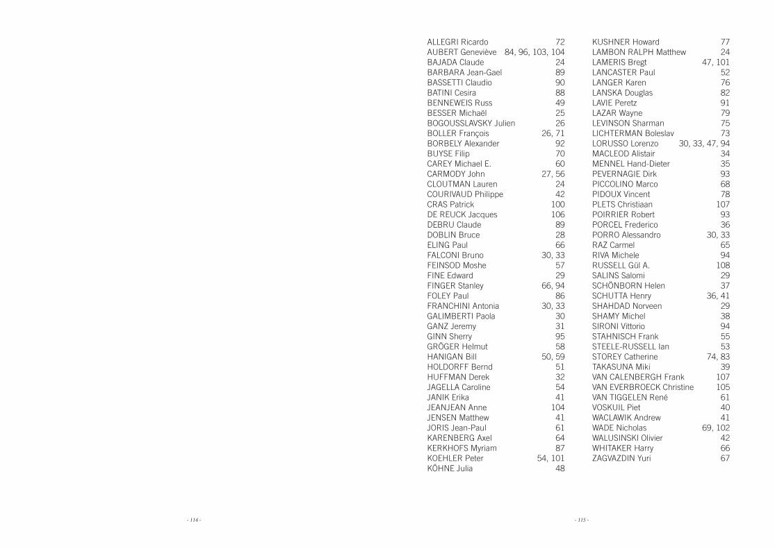
- 114 - - 115 -
ALLEGRI Ricardo 72AUBERT Geneviève 84, 96, 103, 104BAJADA Claude 24BARBARA Jean-Gael 89BASSETTI Claudio 90BATINI Cesira 88BENNEWEIS Russ 49BESSER Michaël 25BOGOUSSLAVSKY Julien 26BOLLER François 26, 71BORBELY Alexander 92BUYSE Filip 70CAREY Michael E. 60CARMODY John 27, 56CLOUTMAN Lauren 24COURIVAUD Philippe 42CRAS Patrick 100DE REUCK Jacques 106DEBRU Claude 89DOBLIN Bruce 28ELING Paul 66FALCONI Bruno 30, 33FEINSOD Moshe 57FINE Edward 29FINGER Stanley 66, 94FOLEY Paul 86FRANCHINI Antonia 30, 33GALIMBERTI Paola 30GANZ Jeremy 31GINN Sherry 95GRÖGER Helmut 58HANIGAN Bill 50, 59HOLDORFF Bernd 51HUFFMAN Derek 32JAGELLA Caroline 54JANIK Erika 41JEANJEAN Anne 104JENSEN Matthew 41JORIS Jean-Paul 61KARENBERG Axel 64KERKHOFS Myriam 87KOEHLER Peter 54, 101KÖHNE Julia 48
KUSHNER Howard 77LAMBON RALPH Matthew 24LAMERIS Bregt 47, 101LANCASTER Paul 52LANGER Karen 76LANSKA Douglas 82LAVIE Peretz 91LAZAR Wayne 79LEVINSON Sharman 75LICHTERMAN Boleslav 73LORUSSO Lorenzo 30, 33, 47, 94MACLEOD Alistair 34MENNEL Hand-Dieter 35PEVERNAGIE Dirk 93PICCOLINO Marco 68PIDOUX Vincent 78PLETS Christiaan 107POIRRIER Robert 93PORCEL Frederico 36PORRO Alessandro 30, 33RAZ Carmel 65RIVA Michele 94RUSSELL Gül A. 108SALINS Salomi 29SCHÖNBORN Helen 37SCHUTTA Henry 36, 41SHAHDAD Norveen 29SHAMY Michel 38SIRONI Vittorio 94STAHNISCH Frank 55STEELE-RUSSELL Ian 53STOREY Catherine 74, 83TAKASUNA Miki 39VAN CALENBERGH Frank 107VAN EVERBROECK Christine 105VAN TIGGELEN René 61VOSKUIL Piet 40WACLAWIK Andrew 41WADE Nicholas 69, 102WALUSINSKI Olivier 42WHITAKER Harry 66ZAGVAZDIN Yuri 67

- 116 - - 117 -

- 118 - - 119 -
- 120 -
Inte
rnat
iona
l Soc
iety
for
the
His
tory
of t
he N
euro
scie
nces
- 1
9th a
nnua
l mee
ting
- B
russ
els
- B
elgi
um
Int
erna
tiona
l Socie
ty for the History of the Neurosciences
19 th annual meeting - Brussels - 20
14
2014