Constitutive and Inducible Expression of B7 Family of Ligands by Human Airway Epithelial Cells
10
Constitutive and Inducible Expression of B7 Family of Ligands by Human Airway Epithelial Cells Jean Kim, Allen C. Myers, Lieping Chen, Drew M. Pardoll, Quynh-Ai Truong-Tran, Andrew P. Lane, John F. McDyer, Lowella Fortuno, and Robert P. Schleimer Departments of Otolaryngology, Head and Neck Surgery, Allergy and Clinical Immunology, Pulmonary Medicine, Dermatology, and Oncology, Johns Hopkins University School of Medicine, Baltimore, Maryland; and Allergy-Immunology Division, Feinberg School of Medicine, Northwestern University, Chicago, Illinois Activated T cells have been implicated in chronic rhinosinusitis (CRS) and asthma and physically interact with epithelial cells in the airways. We now report that human airway epithelial cells display significant constitutive cell-surface expression of costimulatory ligands, B7-H1, B7-H2, B7-H3, and B7-DC. Expression of B7-H1 and B7-DC was selectively induced by stimulation of either BEAS2B or primary nasal epithelial cells (PNEC) with interferon (IFN)- (100 ng/ml). The combi- nation of IFN- and tumor necrosis factor- (100 ng/ml) selectively induced expression better than IFN- alone. Fluticasone treatment (10 7 M) reduced the baseline expression and inhibited the induc- tion of B7-H1 and B7-DC in BEAS2B cells. In vitro exposure of PNEC to IFN- also resulted in selective induction of B7-H1 and B7-DC. Monoclonal antibody blockade of B7-H1 or B7-DC enhanced IFN- expression by purified T cells in co-culture experiments, sug- gesting that these two B7 homologs inhibit T cell responses at the mucosal surface. Immunohistochemical staining of human sinonasal surgical tissue confirmed the presence of B7-H1, B7-H2, and B7-H3 in the epithelial cell layer, especially in samples from patients diag- nosed with Samter’s Triad, a severe form of CRS. Real-time PCR analysis of sinonasal tissue revealed elevated levels of B7-H1 and B7-DC in CRS compared with controls. These results demonstrate that epithelial cells express functional B7 costimulatory molecules and that expression of selected B7 family members is inducible in vitro and in vivo. Epithelial B7 homologs could play a role in regulation of lymphocytic activity at mucosal surfaces. Keywords: cell-surface molecules; costimulation; human airway epithelial cells; inflammation Epithelial cells line the mucosal surface of airways, forming a mechanical barrier that is important in repulsion and removal of particulates and microorganisms by means of the mucociliary escalator. Although it has been clear for some time that airway epithelium plays a role in airway inflammation by virtue of the production of cytokines and chemokines (1–4), recent studies indicate that epithelial cells express Toll-like receptors and can be directly activated to release these mediators by pathogen- associated molecular patterns, suggesting that they may also participate in innate immunity (5, 6). Whether epithelial cells play a role in adaptive immunity is uncertain. Epithelial cells have many of the cell surface molecules associated with antigen presentation, including major histocompatibility complex (MHC) class I and class II molecules (7–10) and CD40 (11–16). The adhesion mole- cule ICAM-1 is expressed on epithelial cells, and could serve as (Received in original form April 22, 2004 and in final form May 18, 2005) This work was supported by National Institutes of Health grants (AI57400, M01-RR-02719, AI37168, AI50530, and HL68546) and FAMRI. Correspondence and requests for reprints should be addressed to Jean Kim, M.D., Ph.D., Johns Hopkins Asthma and Allergy Center, 5501 Hopkins Bayview Circle, Rm 3A65A, Baltimore, MD 21224. E-mail address: [email protected] Am J Respir Cell Mol Biol Vol 33. pp 280–289, 2005 Originally Published in Press as DOI: 10.1165/rcmb.2004-0129OC on June 16, 2005 Internet address: www.atsjournals.org a ligand for leukocyte function–associated molecule-1 (LFA-1) expressed by T cells (17). Unlike peripheral blood T cells, nasal polyp T cells and long-lived intraepithelial T cells express E 7 integrin (CD103), a potential ligand for epithelial E-cadherin, suggesting that this molecule may help retain T cells in the mucosa (18). It is also notable that substantial numbers of T cells are found in association with epithelium in the respiratory and gastrointes- tinal systems (19). Several studies have shown that epithelial cells have the capacity to present antigens to T lymphocytes and to stimulate in vitro (8, 20, 21). The mechanism of this effect has been unclear because epithelial cells lack expression of the important costimulators B7.1 or B7.2, which play critical roles in the prim- ing of T cell responses (22, 23). It is now accepted that optimal activation of T cells requires both costimulation and TCR engagement (24–28). Antigen pre- sentation in the absence of costimulation may lead to T cell anergy. Costimulatory interactions between the B7 family li- gands expressed on antigen-presenting cells (APC) and their receptors on T cells play critical roles in the growth, differentia- tion, and death of T cells (24–28). Engagement of the T cell costimulatory receptor CD28 by its ligands B7-1 (CD80) and B7-2 (CD86) augments activation of T cells and promotes T cell survival. In contrast, binding of B7-1 or B7-2 with CTLA-4, a homolog of CD28, may inhibit T cell responses by delivering a putative negative signal (27, 29–32). Over the last few years, work by many different investigators has identified the existence of numerous homologs of the B7 costimulatory molecules, and it is now clear that the individual members of this family have specialized roles in antigen presentation and T cell stimualtion. Two of these homologs, B7-H1 (33) and B7-DC (34) (also referred to as PDL-1 and PDL-2), are ligands for PD-1, an inhibitory receptor on T cells (35, 36). B7-H2 is detected on B cells and macrophages, and is a ligand for the inducible co- stimulator (ICOS) expressed on antigen-primed T cells. Costi- mulatory signals via ICOS activate memory T cells with some preference for Th2 responses (37–42). B7-H3, which is not usu- ally detectable in normal tissues, can be induced on dendritic cells and monocytes by inflammatory cytokines and costimulates proliferation of both CD4 and CD8 T cells, enhances the induction of cytotoxic T cells, and selectively stimulates the pro- duction of a key Th1-type cytokine, interferon (IFN)- (43). In a recent report, we demonstrated by flow cytometry that one of the recently recognized B7 homologs, B7-H2, was consti- tutively and strongly expressed on both the immortalized bron- chial epithelial cell line BEAS-2B and primary human bronchial epithelial cells (PBEC), whereas B7-1 and B7-2 were undetect- able on either epithelial cell type (23). Expression of B7-H2 was confirmed in vitro by Western blot in cultured epithelial cells and was confirmed in vivo by immunohistochemical analysis of airway tissue derived from autopsies. These findings support the hypothesis that airway epithelial cells may play a role in some instances as APC during the development of airway inflamma- tory and immune responses.
Transcript of Constitutive and Inducible Expression of B7 Family of Ligands by Human Airway Epithelial Cells
Constitutive and Inducible Expression of B7 Family ofLigands by Human Airway Epithelial CellsJean Kim, Allen C. Myers, Lieping Chen, Drew M. Pardoll, Quynh-Ai Truong-Tran, Andrew P. Lane,John F. McDyer, Lowella Fortuno, and Robert P. Schleimer
Departments of Otolaryngology, Head and Neck Surgery, Allergy and Clinical Immunology, Pulmonary Medicine, Dermatology,and Oncology, Johns Hopkins University School of Medicine, Baltimore, Maryland; and Allergy-Immunology Division,Feinberg School of Medicine, Northwestern University, Chicago, Illinois
Activated T cells have been implicated in chronic rhinosinusitis(CRS) and asthma and physically interact with epithelial cells in theairways. We now report that human airway epithelial cells displaysignificant constitutive cell-surface expression of costimulatory ligands,B7-H1, B7-H2, B7-H3, and B7-DC. Expression of B7-H1 and B7-DC wasselectively induced by stimulation of either BEAS2B or primary nasalepithelial cells (PNEC) with interferon (IFN)-� (100 ng/ml). The combi-nation of IFN-� and tumor necrosis factor-� (100 ng/ml) selectivelyinduced expression better than IFN-� alone. Fluticasone treatment(10�7 M) reduced the baseline expression and inhibited the induc-tion of B7-H1 and B7-DC in BEAS2B cells. In vitro exposure of PNECto IFN-� also resulted in selective induction of B7-H1 andB7-DC. Monoclonal antibody blockade of B7-H1 or B7-DC enhancedIFN-� expression by purified T cells in co-culture experiments, sug-gesting that these two B7 homologs inhibit T cell responses at themucosal surface. Immunohistochemical staining of human sinonasalsurgical tissue confirmed the presence of B7-H1, B7-H2, and B7-H3in the epithelial cell layer, especially in samples from patients diag-nosed with Samter’s Triad, a severe form of CRS. Real-time PCRanalysis of sinonasal tissue revealed elevated levels of B7-H1 andB7-DC in CRS compared with controls. These results demonstratethat epithelial cells express functional B7 costimulatory moleculesand that expression of selected B7 family members is induciblein vitro and in vivo. Epithelial B7 homologs could play a role inregulation of lymphocytic activity at mucosal surfaces.
Keywords: cell-surface molecules; costimulation; human airway epithelialcells; inflammation
Epithelial cells line the mucosal surface of airways, forming amechanical barrier that is important in repulsion and removalof particulates and microorganisms by means of the mucociliaryescalator. Although it has been clear for some time that airwayepithelium plays a role in airway inflammation by virtue of theproduction of cytokines and chemokines (1–4), recent studiesindicate that epithelial cells express Toll-like receptors and canbe directly activated to release these mediators by pathogen-associated molecular patterns, suggesting that they may alsoparticipate in innate immunity (5, 6). Whether epithelial cells playa role in adaptive immunity is uncertain. Epithelial cells have manyof the cell surface molecules associated with antigen presentation,including major histocompatibility complex (MHC) class I andclass II molecules (7–10) and CD40 (11–16). The adhesion mole-cule ICAM-1 is expressed on epithelial cells, and could serve as
(Received in original form April 22, 2004 and in final form May 18, 2005)
This work was supported by National Institutes of Health grants (AI57400,M01-RR-02719, AI37168, AI50530, and HL68546) and FAMRI.
Correspondence and requests for reprints should be addressed to Jean Kim, M.D.,Ph.D., Johns Hopkins Asthma and Allergy Center, 5501 Hopkins Bayview Circle,Rm 3A65A, Baltimore, MD 21224. E-mail address: [email protected]
Am J Respir Cell Mol Biol Vol 33. pp 280–289, 2005Originally Published in Press as DOI: 10.1165/rcmb.2004-0129OC on June 16, 2005Internet address: www.atsjournals.org
a ligand for leukocyte function–associated molecule-1 (LFA-1)expressed by T cells (17). Unlike peripheral blood T cells, nasalpolyp T cells and long-lived intraepithelial T cells express �E�7
integrin (CD103), a potential ligand for epithelial E-cadherin,suggesting that this molecule may help retain T cells in themucosa (18).
It is also notable that substantial numbers of T cells are foundin association with epithelium in the respiratory and gastrointes-tinal systems (19). Several studies have shown that epithelial cellshave the capacity to present antigens to T lymphocytes and tostimulate in vitro (8, 20, 21). The mechanism of this effect has beenunclear because epithelial cells lack expression of the importantcostimulators B7.1 or B7.2, which play critical roles in the prim-ing of T cell responses (22, 23).
It is now accepted that optimal activation of T cells requiresboth costimulation and TCR engagement (24–28). Antigen pre-sentation in the absence of costimulation may lead to T cellanergy. Costimulatory interactions between the B7 family li-gands expressed on antigen-presenting cells (APC) and theirreceptors on T cells play critical roles in the growth, differentia-tion, and death of T cells (24–28). Engagement of the T cellcostimulatory receptor CD28 by its ligands B7-1 (CD80) andB7-2 (CD86) augments activation of T cells and promotes T cellsurvival. In contrast, binding of B7-1 or B7-2 with CTLA-4, ahomolog of CD28, may inhibit T cell responses by delivering aputative negative signal (27, 29–32). Over the last few years,work by many different investigators has identified the existenceof numerous homologs of the B7 costimulatory molecules, andit is now clear that the individual members of this family havespecialized roles in antigen presentation and T cell stimualtion.
Two of these homologs, B7-H1 (33) and B7-DC (34) (alsoreferred to as PDL-1 and PDL-2), are ligands for PD-1, aninhibitory receptor on T cells (35, 36). B7-H2 is detected on Bcells and macrophages, and is a ligand for the inducible co-stimulator (ICOS) expressed on antigen-primed T cells. Costi-mulatory signals via ICOS activate memory T cells with somepreference for Th2 responses (37–42). B7-H3, which is not usu-ally detectable in normal tissues, can be induced on dendriticcells and monocytes by inflammatory cytokines and costimulatesproliferation of both CD4� and CD8� T cells, enhances theinduction of cytotoxic T cells, and selectively stimulates the pro-duction of a key Th1-type cytokine, interferon (IFN)-� (43).
In a recent report, we demonstrated by flow cytometry thatone of the recently recognized B7 homologs, B7-H2, was consti-tutively and strongly expressed on both the immortalized bron-chial epithelial cell line BEAS-2B and primary human bronchialepithelial cells (PBEC), whereas B7-1 and B7-2 were undetect-able on either epithelial cell type (23). Expression of B7-H2 wasconfirmed in vitro by Western blot in cultured epithelial cellsand was confirmed in vivo by immunohistochemical analysis ofairway tissue derived from autopsies. These findings support thehypothesis that airway epithelial cells may play a role in someinstances as APC during the development of airway inflamma-tory and immune responses.
Kim, Myers, Chen, et al.: B7 Homolog Expression in Airway Epithelial Cells 281
The purpose of the present study was to further characterizethe phenotype of airway epithelial cells with respect to expres-sion of B7 homologs both in vitro and in human subjects. Wenow report that human airway epithelial cells express B7-H1,B7-H2, B7-H3, and B7-DC in vitro and in vivo. Two of theseB7 homologs, B7-H1 and B7-DC, were found to be inducibleby stimulation with cytokines, especially the combination ofIFN-� and tumor necrosis factor (TNF)-�. Flow cytometric anal-ysis of airway epithelial cells collected by brushing of the upperairways of human subjects revealed expression of B7-H1, B7-H2,B7-H3, and B7-DC. Immunohistochemical analysis of tissuespecimens from patients undergoing nasal surgery confirmedexpression of these B7 homologs in vivo and real-time PCRanalysis demonstrated elevated levels of B7-H1 and B7-DC intissue from patients with chronic rhinosinusitis. These resultssuggest that airway epithelial cells express a variety of costimula-tory molecules in the B7 family and provide a potential mecha-nism by which epithelial cells may have a role in regulatingmucosal responses of T cells.
MATERIALS AND METHODS
Collection of Nasal Scrapings and Surgical Samples
Nasal epithelial cells were collected from the inferior nasal turbinateby curettage with a Rhinoprobe (Arlington Scientific, Inc., Springville,UT) or cytology brush (Wampole, Harrisburg, PA) under a JohnsHopkins Medicine Institutional Review Board (JHMIRB)-approvedhuman subjects research protocol. Care was taken to take 10 uniformpasses of mucosal scrapings. A portion of the specimen was placedimmediately in RNAlater (Ambion, Austin, TX) for subsequent isola-tion of mRNA, and the remainder was placed in Ca2� and Mg2�–freeHam’s F12/Dulbecco’s modified Eagle’s medium (DMEM) containingpenicillin (100 U/ml), streptomycin (100 U/ml), fungizone (1 �g/ml),and L-glutamine (GIBCO-BRL, Gaithersburg, MD) for immediate flowcytometric analysis or for cell culture. Each nasal scraping specimenyielded � 1–2 � 106 cells, of which � 90% were confirmed to beepithelial cells by Wright’s stain and cytokeratin immunofluorescence,as described previously (23). All surgical sinus tissue from CRS andnormal control subjects were obtained through a JHMIRB-approvedhuman subjects research protocol.
Culture of BEAS-2B Cells and Primary Nasal Epithelial Cells
The BEAS-2B cell line, derived from human bronchial epithelium trans-formed by an adenovirus 12-SV40 virus, was kindly supplied by Dr. CurtisHarris of the NIH (44). BEAS-2B cells were cultured in Ham’s F12/DMEM. Primary nasal epithelial cells (PNEC) were cultured in bron-chial epithelial basal medium (Biosource, Camarillo, TX) containingbovine pituitary extract, hydrocortisone, human recombinant epidermalgrowth factor, epinephrine, transferrin, insulin, retinoic acid, triiodothy-ronine, bovine serum albumin–fatty acid free, gentamicin, and ampho-tericin-B. BEAS-2B cells and PNEC were cultured in 6-well culture plateseither uncoated (BEAS-2B) or coated with collagen (PNEC; Vitrogen,Collagen Biomaterials, Palo Alto, CA). BEAS-2B cells were used frompassages 38–43. PNEC were used only at their first passage. Cells werecultured at 37 C with 5% CO2 in humidified air.
When cells reached 80% confluence, they were stimulated with100 ng/ml of human recombinant TNF-� (R&D Systems, Minneapolis,MN), 100 ng/ml of IFN-� (Genzyme, Cambridge, MA), or 50 ng/ml ofinterleukin (IL)-4 (R&D Systems), in the presence or absence of theglucocorticoid fluticasone (10�7 M). The cells were incubated for 24 h,and then washed three times with 0.05% trypsin/0.53 mM EDTA inHBSS (BEAS-2B) or with 0.02% EDTA in HBSS (PNEC), and thenremoved for analysis of mRNA expression or for flow cytometry. Gen-erally, the cell recovery was � 5–8 million cells/6-well plate. The viabilityof both BEAS-2B cells and primary nasal epithelial cells at the timeof cell harvest was assessed by erythrosin B staining or propidiumiodide exclusion and was consistently � 95% of the cells harvested.
Isolation of mRNA
Fresh epithelial cells from nasal scrapings were placed directly into500 �l of RNAlater (Ambion) for isolation of total RNA using RNeasykits (Qiagen, Valencia, CA). Contaminating genomic DNA was re-moved using RNase-Free DNase Set Protocol (Qiagen). Integrity ofisolated RNA was evaluated using 1% agarose gels and levels of inputRNA were calculated based upon absorbance at 260 nm. RNA wasstored at �80 C until use. BEAS-2B cells and PNEC were washedwith ice-cold HBSS and total cellular RNA was extracted using theTRIzol reagent (GIBCO BRL) according to the manufacturer’s instruc-tions. Briefly, cells were lysed in the presence of TRIzol and chloroform.The RNA was precipitated at �20 C in isopropanol overnight. TheRNA pellet was recovered by centrifugation at 4 C, washed once in70% ethanol, dried, and suspended in DEPC-treated water. The re-sulting RNA was quantitated using spectrophotometry. The RNA wasstored at �80 C until use. Total mRNA from sinus tissue was isolatedusing the TRIzol (0.8 ml) reagent (Invitrogen, Carlsbad, CA) as pre-viously described (45). DNA was removed using DNAfree (Ambion)per the manufacturer’s protocol. Isolated mRNA was reverse tran-scribed using a poly(dT)15 primer (Roche, Indianapolis, IN) and Molo-ney murine leukemia virus reverse transcriptase (Applied Biosystems,Foster City, CA) using conditions provided by the manufacturer. Integ-rity of samples was confirmed using agarose gel electrophoresis.
Flow Cytometry
The monoclonal antibodies used for flow cytometry are listed in Table 1. Afluorescein isothiocyanate (FITC)-conjugated goat F (ab)2 anti-mouseIgG (Pharmingen, Palo Alto, CA) or FITC-conjugated goat F (ab)2
anti-hamster IgG (Biosource, Camarillo, CA) was used as the secondaryantibody. Typically, 5–10 � 104 cells were incubated in 60 �l of PBS/0.2% BSA/0.1% NaN3 containing saturating concentrations of eachmonoclonal antibody and 4 mg/ml of human IgG (to reduce nonspecificbinding; Sigma Chemicals, St. Louis, MO) on ice for 30 min. The cellswere washed, resuspended in saturating amounts of secondary antibodyfor another 30 min, and then washed again, resuspended in the buffer,and immediately analyzed with a FACS Calibur flow cytometer (BectonDickinson, Mountainview, CA) using CellQuest software. Fluorescencewas determined on all cells for each sample after debris, dead cells,and aggregates were excluded by forward angle and side scatter gatingafter propidium iodide staining. Mean fluorescence intensity (MFI) wascompared with control staining using an irrelevant isotype-matchedmouse monoclonal antibody. For each sample, at least 10,000 eventswere collected, and histograms were generated. Data were expressedas mean � SEM.
Quantitative Taqman Real-Time PCR
Messenger RNA was reverse transcribed to cDNA using standard pro-tocols (23). PCR amplification in the presence of specific primers anda fluorescently labeled probe selected using Primer Express softwarewas monitored by real-time analysis using an Applied Biosystems Model7700 Sequence Detector. Table 2 lists the genes that were analyzedand the probes and primers used. The probes used were labeled withFAM (reporter dye) on the 5 end and with TAMRA (quencher dye)on the 3 end. A probe and PCR primers were designed for humanGAPDH and 18 s RNA (Applied Biosystems), incorporating JOE asthe reporter dye. Using the Sequence Detector software, the reporterdye emission was compared with the quenching dye emission generatinga peak normalized reporter fluorescence value generated by each cycleof PCR (46). The peak normalized reporter fluorescence values (�Rn)were plotted versus the cycle number. The computer algorithm wasthen used to calculate the threshold cycle (CT), that is, the point atwhich the amplification plot crossed a defined fluorescence threshold,set by the algorithm at 10 times the SD of the baseline value obtainedduring the first 10–15 cycles. Data are expressed as fold change in CT
after normalization to GAPDH with each unit change of CT valuecorresponding to a doubling of the level of target mRNA.
T cell Isolation
Buffy coat fractions were obtained from leukopheresed subjects belong-ing to the NIH (Bethesda, MD) normal donor pool. PBMC were iso-lated from buffy coat fractions by density gradient centrifugation using
282 AMERICAN JOURNAL OF RESPIRATORY CELL AND MOLECULAR BIOLOGY VOL 33 2005
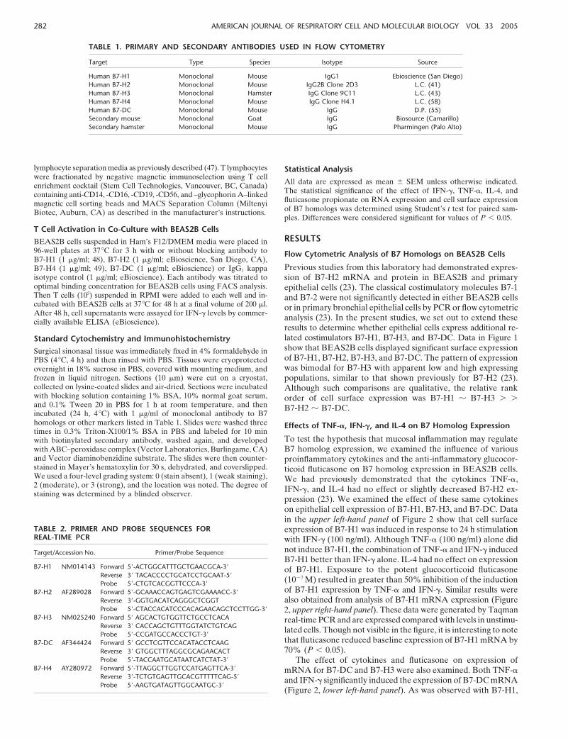
TABLE 1. PRIMARY AND SECONDARY ANTIBODIES USED IN FLOW CYTOMETRY
Target Type Species Isotype Source
Human B7-H1 Monoclonal Mouse IgG1 Ebioscience (San Diego)Human B7-H2 Monoclonal Mouse IgG2B Clone 2D3 L.C. (41)Human B7-H3 Monoclonal Hamster IgG Clone 9C11 L.C. (43)Human B7-H4 Monoclonal Mouse IgG Clone H4.1 L.C. (58)Human B7-DC Monoclonal Mouse IgG D.P. (55)Secondary mouse Monoclonal Goat IgG Biosource (Camarillo)Secondary hamster Monoclonal Mouse IgG Pharmingen (Palo Alto)
lymphocyte separation media as previously described (47). T lymphocyteswere fractionated by negative magnetic immunoselection using T cellenrichment cocktail (Stem Cell Technologies, Vancouver, BC, Canada)containing anti-CD14, -CD16, -CD19, -CD56, and –glycophorin A–linkedmagnetic cell sorting beads and MACS Separation Column (MiltenyiBiotec, Auburn, CA) as described in the manufacturer’s instructions.
T Cell Activation in Co-Culture with BEAS2B Cells
BEAS2B cells suspended in Ham’s F12/DMEM media were placed in96-well plates at 37 C for 3 h with or without blocking antibody toB7-H1 (1 �g/ml; 48), B7-H2 (1 �g/ml; eBioscience, San Diego, CA),B7-H4 (1 �g/ml; 49), B7-DC (1 �g/ml; eBioscience) or IgG1 kappaisotype control (1 �g/ml; eBioscience). Each antibody was titrated tooptimal binding concentration for BEAS2B cells using FACS analysis.Then T cells (105) suspended in RPMI were added to each well and in-cubated with BEAS2B cells at 37 C for 48 h at a final volume of 200 �l.After 48 h, cell supernatants were assayed for IFN-� levels by commer-cially available ELISA (eBioscience).
Standard Cytochemistry and Immunohistochemistry
Surgical sinonasal tissue was immediately fixed in 4% formaldehyde inPBS (4 C, 4 h) and then rinsed with PBS. Tissues were cryoprotectedovernight in 18% sucrose in PBS, covered with mounting medium, andfrozen in liquid nitrogen. Sections (10 �m) were cut on a cryostat,collected on lysine-coated slides and air-dried. Sections were incubatedwith blocking solution containing 1% BSA, 10% normal goat serum,and 0.1% Tween 20 in PBS for 1 h at room temperature, and thenincubated (24 h, 4 C) with 1 �g/ml of monoclonal antibody to B7homologs or other markers listed in Table 1. Slides were washed threetimes in 0.3% Triton-X100/1% BSA in PBS and labeled for 10 minwith biotinylated secondary antibody, washed again, and developedwith ABC–peroxidase complex (Vector Laboratories, Burlingame, CA)and Vector diaminobenzidine substrate. The slides were then counter-stained in Mayer’s hematoxylin for 30 s, dehydrated, and coverslipped.We used a four-level grading system: 0 (stain absent), 1 (weak staining),2 (moderate), or 3 (strong), and the location was noted. The degree ofstaining was determined by a blinded observer.
TABLE 2. PRIMER AND PROBE SEQUENCES FORREAL-TIME PCR
Target/Accession No. Primer/Probe Sequence
B7-H1 NM014143 Forward 5-ACTGGCATTTGCTGAACGCA-3
Reverse 3 TACACCCCTGCATCCTGCAAT-5
Probe 5-CTGTCACGGTTCCCA-3
B7-H2 AF289028 Forward 5-GCAAACCAGTGAGTCGAAAACC-3
Reverse 3-GGTGACATCAGGGCTCGGTProbe 5-CTACCACATCCCACAGAACAGCTCCTTGG-3
B7-H3 NM025240 Forward 5 AGCACTGTGGTTCTGCCTCACAReverse 3 CACCAGCTGTTTGGTATCTGTCAGProbe 5-CCGATGCCACCCTGT-3
B7-DC AF344424 Forward 5 GCCTCGTTCCACATACCTCAAGReverse 3 GTGGCTTTAGGCGCAGAACACTProbe 5-TACCAATGCATAATCATCTAT-3
B7-H4 AY280972 Forward 5-TTAGGCTTGGTCCATGAGTTCA-3
Reverse 3-TCTGTGAGTTGCACGTTTTTCAG-5
Probe 5-AAGTGATAGTTGGCAATGC-3
Statistical Analysis
All data are expressed as mean � SEM unless otherwise indicated.The statistical significance of the effect of IFN-�, TNF-�, IL-4, andfluticasone propionate on RNA expression and cell surface expressionof B7 homologs was determined using Student’s t test for paired sam-ples. Differences were considered significant for values of P 0.05.
RESULTS
Flow Cytometric Analysis of B7 Homologs on BEAS2B Cells
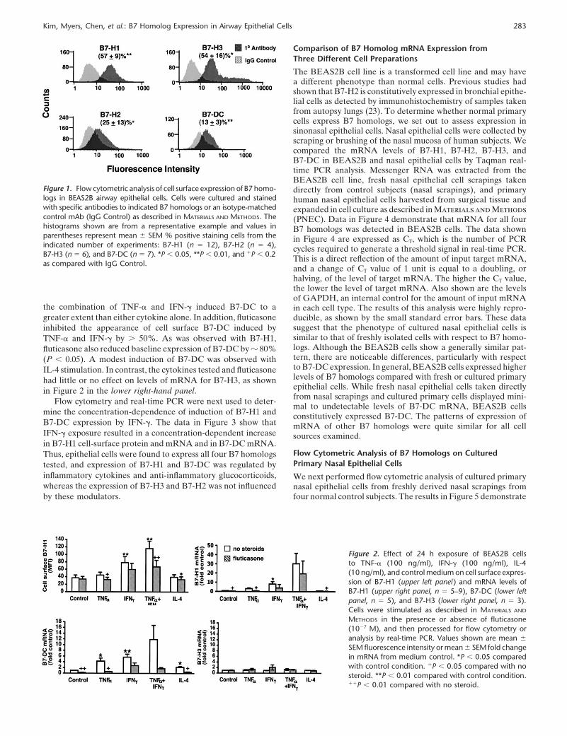
Previous studies from this laboratory had demonstrated expres-sion of B7-H2 mRNA and protein in BEAS2B and primaryepithelial cells (23). The classical costimulatory molecules B7-1and B7-2 were not significantly detected in either BEAS2B cellsor in primary bronchial epithelial cells by PCR or flow cytometricanalysis (23). In the present studies, we set out to extend theseresults to determine whether epithelial cells express additional re-lated costimulators B7-H1, B7-H3, and B7-DC. Data in Figure 1show that BEAS2B cells displayed significant surface expressionof B7-H1, B7-H2, B7-H3, and B7-DC. The pattern of expressionwas bimodal for B7-H3 with apparent low and high expressingpopulations, similar to that shown previously for B7-H2 (23).Although such comparisons are qualitative, the relative rankorder of cell surface expression was B7-H1 � B7-H3 � �B7-H2 � B7-DC.
Effects of TNF-�, IFN-�, and IL-4 on B7 Homolog Expression
To test the hypothesis that mucosal inflammation may regulateB7 homolog expression, we examined the influence of variousproinflammatory cytokines and the anti-inflammatory glucocor-ticoid fluticasone on B7 homolog expression in BEAS2B cells.We had previously demonstrated that the cytokines TNF-�,IFN-�, and IL-4 had no effect or slightly decreased B7-H2 ex-pression (23). We examined the effect of these same cytokineson epithelial cell expression of B7-H1, B7-H3, and B7-DC. Datain the upper left-hand panel of Figure 2 show that cell surfaceexpression of B7-H1 was induced in response to 24 h stimulationwith IFN-� (100 ng/ml). Although TNF-� (100 ng/ml) alone didnot induce B7-H1, the combination of TNF-� and IFN-� inducedB7-H1 better than IFN-� alone. IL-4 had no effect on expressionof B7-H1. Exposure to the potent glucocorticoid fluticasone(10�7 M) resulted in greater than 50% inhibition of the inductionof B7-H1 expression by TNF-� and IFN-�. Similar results werealso obtained from analysis of B7-H1 mRNA expression (Figure2, upper right-hand panel). These data were generated by Taqmanreal-time PCR and are expressed compared with levels in unstimu-lated cells. Though not visible in the figure, it is interesting to notethat fluticasone reduced baseline expression of B7-H1 mRNA by70% (P 0.05).
The effect of cytokines and fluticasone on expression ofmRNA for B7-DC and B7-H3 were also examined. Both TNF-�and IFN-� significantly induced the expression of B7-DC mRNA(Figure 2, lower left-hand panel). As was observed with B7-H1,
Kim, Myers, Chen, et al.: B7 Homolog Expression in Airway Epithelial Cells 283
Figure 1. Flow cytometric analysis of cell surface expression of B7 homo-logs in BEAS2B airway epithelial cells. Cells were cultured and stainedwith specific antibodies to indicated B7 homologs or an isotype-matchedcontrol mAb (IgG Control) as described in MATERIALS AND METHODS. Thehistograms shown are from a representative example and values inparentheses represent mean � SEM % positive staining cells from theindicated number of experiments: B7-H1 (n � 12), B7-H2 (n � 4),B7-H3 (n � 6), and B7-DC (n � 7). *P 0.05, **P 0.01, and �P 0.2as compared with IgG Control.
the combination of TNF-� and IFN-� induced B7-DC to agreater extent than either cytokine alone. In addition, fluticasoneinhibited the appearance of cell surface B7-DC induced byTNF-� and IFN-� by � 50%. As was observed with B7-H1,fluticasone also reduced baseline expression of B7-DC by � 80%(P 0.05). A modest induction of B7-DC was observed withIL-4 stimulation. In contrast, the cytokines tested and fluticasonehad little or no effect on levels of mRNA for B7-H3, as shownin Figure 2 in the lower right-hand panel.
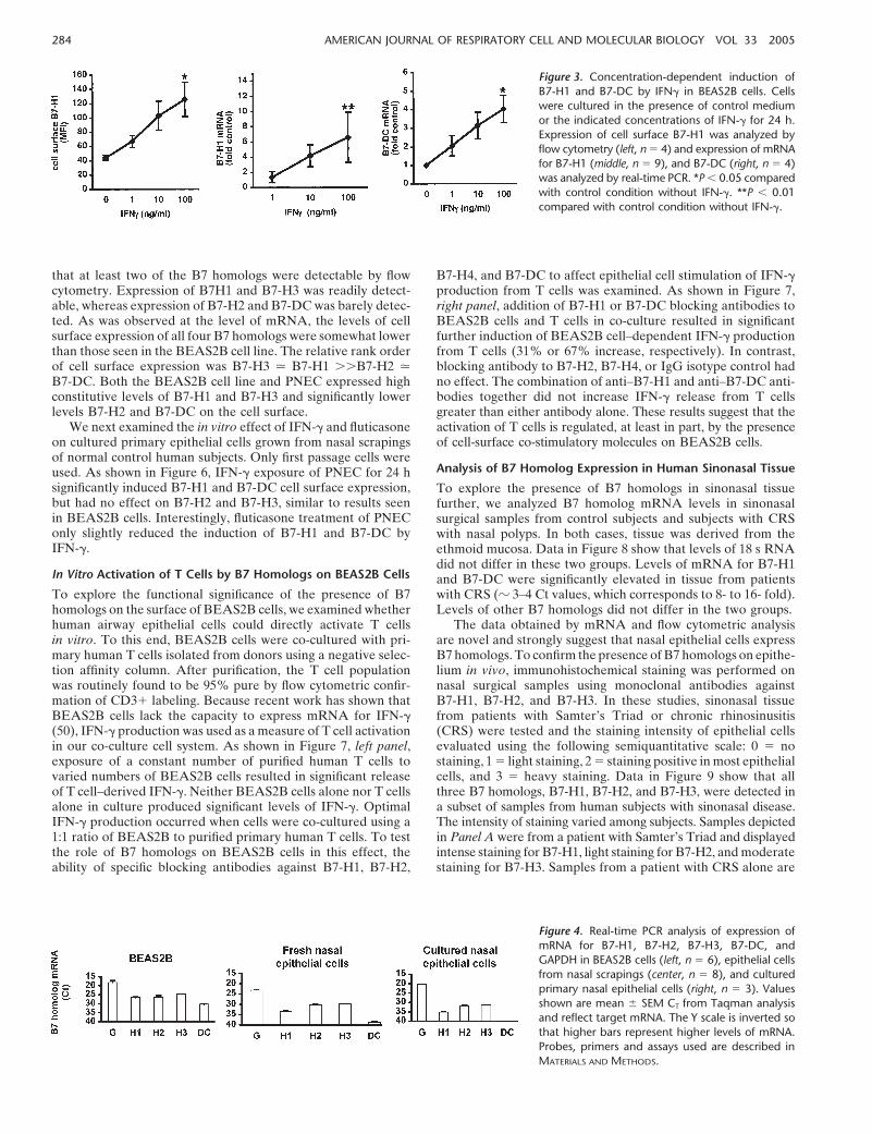
Flow cytometry and real-time PCR were next used to deter-mine the concentration-dependence of induction of B7-H1 andB7-DC expression by IFN-�. The data in Figure 3 show thatIFN-� exposure resulted in a concentration-dependent increasein B7-H1 cell-surface protein and mRNA and in B7-DC mRNA.Thus, epithelial cells were found to express all four B7 homologstested, and expression of B7-H1 and B7-DC was regulated byinflammatory cytokines and anti-inflammatory glucocorticoids,whereas the expression of B7-H3 and B7-H2 was not influencedby these modulators.
Figure 2. Effect of 24 h exposure of BEAS2B cellsto TNF-� (100 ng/ml), IFN-� (100 ng/ml), IL-4(10 ng/ml), and control medium on cell surface expres-sion of B7-H1 (upper left panel ) and mRNA levels ofB7-H1 (upper right panel, n � 5–9), B7-DC (lower leftpanel, n � 5), and B7-H3 (lower right panel, n � 3).Cells were stimulated as described in MATERIALS AND
METHODS in the presence or absence of fluticasone(10�7 M), and then processed for flow cytometry oranalysis by real-time PCR. Values shown are mean �
SEM fluorescence intensity or mean � SEM fold changein mRNA from medium control. *P 0.05 comparedwith control condition. �P 0.05 compared with nosteroid. **P 0.01 compared with control condition.��P 0.01 compared with no steroid.
Comparison of B7 Homolog mRNA Expression fromThree Different Cell Preparations
The BEAS2B cell line is a transformed cell line and may havea different phenotype than normal cells. Previous studies hadshown that B7-H2 is constitutively expressed in bronchial epithe-lial cells as detected by immunohistochemistry of samples takenfrom autopsy lungs (23). To determine whether normal primarycells express B7 homologs, we set out to assess expression insinonasal epithelial cells. Nasal epithelial cells were collected byscraping or brushing of the nasal mucosa of human subjects. Wecompared the mRNA levels of B7-H1, B7-H2, B7-H3, andB7-DC in BEAS2B and nasal epithelial cells by Taqman real-time PCR analysis. Messenger RNA was extracted from theBEAS2B cell line, fresh nasal epithelial cell scrapings takendirectly from control subjects (nasal scrapings), and primaryhuman nasal epithelial cells harvested from surgical tissue andexpanded in cell culture as described in Materials and Methods(PNEC). Data in Figure 4 demonstrate that mRNA for all fourB7 homologs was detected in BEAS2B cells. The data shownin Figure 4 are expressed as CT, which is the number of PCRcycles required to generate a threshold signal in real-time PCR.This is a direct reflection of the amount of input target mRNA,and a change of CT value of 1 unit is equal to a doubling, orhalving, of the level of target mRNA. The higher the CT value,the lower the level of target mRNA. Also shown are the levelsof GAPDH, an internal control for the amount of input mRNAin each cell type. The results of this analysis were highly repro-ducible, as shown by the small standard error bars. These datasuggest that the phenotype of cultured nasal epithelial cells issimilar to that of freshly isolated cells with respect to B7 homo-logs. Although the BEAS2B cells show a generally similar pat-tern, there are noticeable differences, particularly with respectto B7-DC expression. In general, BEAS2B cells expressed higherlevels of B7 homologs compared with fresh or cultured primaryepithelial cells. While fresh nasal epithelial cells taken directlyfrom nasal scrapings and cultured primary cells displayed mini-mal to undetectable levels of B7-DC mRNA, BEAS2B cellsconstitutively expressed B7-DC. The patterns of expression ofmRNA of other B7 homologs were quite similar for all cellsources examined.
Flow Cytometric Analysis of B7 Homologs on CulturedPrimary Nasal Epithelial Cells
We next performed flow cytometric analysis of cultured primarynasal epithelial cells from freshly derived nasal scrapings fromfour normal control subjects. The results in Figure 5 demonstrate
284 AMERICAN JOURNAL OF RESPIRATORY CELL AND MOLECULAR BIOLOGY VOL 33 2005
Figure 3. Concentration-dependent induction ofB7-H1 and B7-DC by IFN� in BEAS2B cells. Cellswere cultured in the presence of control mediumor the indicated concentrations of IFN-� for 24 h.Expression of cell surface B7-H1 was analyzed byflow cytometry (left, n � 4) and expression of mRNAfor B7-H1 (middle, n � 9), and B7-DC (right, n � 4)was analyzed by real-time PCR. *P 0.05 comparedwith control condition without IFN-�. **P 0.01compared with control condition without IFN-�.
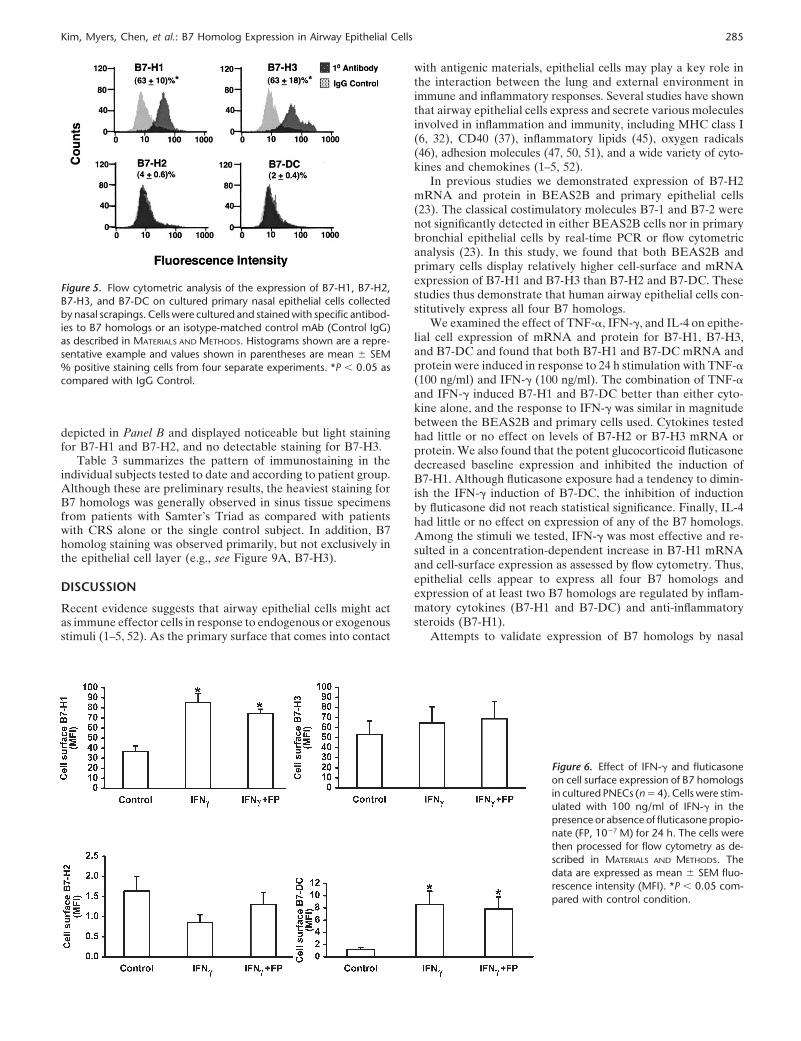
that at least two of the B7 homologs were detectable by flowcytometry. Expression of B7H1 and B7-H3 was readily detect-able, whereas expression of B7-H2 and B7-DC was barely detec-ted. As was observed at the level of mRNA, the levels of cellsurface expression of all four B7 homologs were somewhat lowerthan those seen in the BEAS2B cell line. The relative rank orderof cell surface expression was B7-H3 � B7-H1 ��B7-H2 �B7-DC. Both the BEAS2B cell line and PNEC expressed highconstitutive levels of B7-H1 and B7-H3 and significantly lowerlevels B7-H2 and B7-DC on the cell surface.
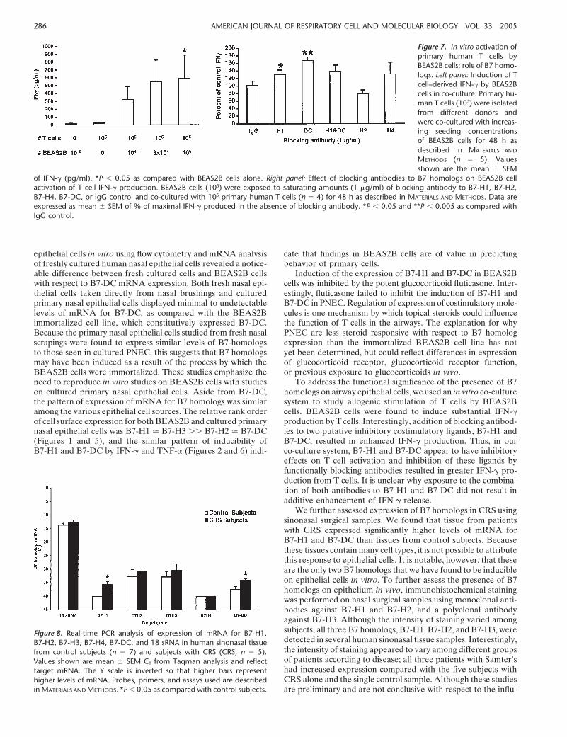
We next examined the in vitro effect of IFN-� and fluticasoneon cultured primary epithelial cells grown from nasal scrapingsof normal control human subjects. Only first passage cells wereused. As shown in Figure 6, IFN-� exposure of PNEC for 24 hsignificantly induced B7-H1 and B7-DC cell surface expression,but had no effect on B7-H2 and B7-H3, similar to results seenin BEAS2B cells. Interestingly, fluticasone treatment of PNEConly slightly reduced the induction of B7-H1 and B7-DC byIFN-�.
In Vitro Activation of T Cells by B7 Homologs on BEAS2B Cells
To explore the functional significance of the presence of B7homologs on the surface of BEAS2B cells, we examined whetherhuman airway epithelial cells could directly activate T cellsin vitro. To this end, BEAS2B cells were co-cultured with pri-mary human T cells isolated from donors using a negative selec-tion affinity column. After purification, the T cell populationwas routinely found to be 95% pure by flow cytometric confir-mation of CD3� labeling. Because recent work has shown thatBEAS2B cells lack the capacity to express mRNA for IFN-�(50), IFN-� production was used as a measure of T cell activationin our co-culture cell system. As shown in Figure 7, left panel,exposure of a constant number of purified human T cells tovaried numbers of BEAS2B cells resulted in significant releaseof T cell–derived IFN-�. Neither BEAS2B cells alone nor T cellsalone in culture produced significant levels of IFN-�. OptimalIFN-� production occurred when cells were co-cultured using a1:1 ratio of BEAS2B to purified primary human T cells. To testthe role of B7 homologs on BEAS2B cells in this effect, theability of specific blocking antibodies against B7-H1, B7-H2,
Figure 4. Real-time PCR analysis of expression ofmRNA for B7-H1, B7-H2, B7-H3, B7-DC, andGAPDH in BEAS2B cells (left, n � 6), epithelial cellsfrom nasal scrapings (center, n � 8), and culturedprimary nasal epithelial cells (right, n � 3). Valuesshown are mean � SEM CT from Taqman analysisand reflect target mRNA. The Y scale is inverted sothat higher bars represent higher levels of mRNA.Probes, primers and assays used are described inMATERIALS AND METHODS.
B7-H4, and B7-DC to affect epithelial cell stimulation of IFN-�production from T cells was examined. As shown in Figure 7,right panel, addition of B7-H1 or B7-DC blocking antibodies toBEAS2B cells and T cells in co-culture resulted in significantfurther induction of BEAS2B cell–dependent IFN-� productionfrom T cells (31% or 67% increase, respectively). In contrast,blocking antibody to B7-H2, B7-H4, or IgG isotype control hadno effect. The combination of anti–B7-H1 and anti–B7-DC anti-bodies together did not increase IFN-� release from T cellsgreater than either antibody alone. These results suggest that theactivation of T cells is regulated, at least in part, by the presenceof cell-surface co-stimulatory molecules on BEAS2B cells.
Analysis of B7 Homolog Expression in Human Sinonasal Tissue
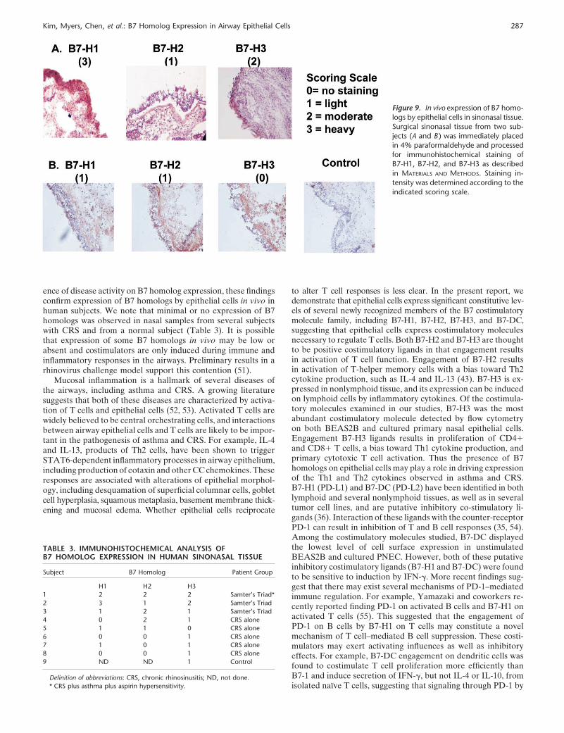
To explore the presence of B7 homologs in sinonasal tissuefurther, we analyzed B7 homolog mRNA levels in sinonasalsurgical samples from control subjects and subjects with CRSwith nasal polyps. In both cases, tissue was derived from theethmoid mucosa. Data in Figure 8 show that levels of 18 s RNAdid not differ in these two groups. Levels of mRNA for B7-H1and B7-DC were significantly elevated in tissue from patientswith CRS (� 3–4 Ct values, which corresponds to 8- to 16- fold).Levels of other B7 homologs did not differ in the two groups.
The data obtained by mRNA and flow cytometric analysisare novel and strongly suggest that nasal epithelial cells expressB7 homologs. To confirm the presence of B7 homologs on epithe-lium in vivo, immunohistochemical staining was performed onnasal surgical samples using monoclonal antibodies againstB7-H1, B7-H2, and B7-H3. In these studies, sinonasal tissuefrom patients with Samter’s Triad or chronic rhinosinusitis(CRS) were tested and the staining intensity of epithelial cellsevaluated using the following semiquantitative scale: 0 � nostaining, 1 � light staining, 2 � staining positive in most epithelialcells, and 3 � heavy staining. Data in Figure 9 show that allthree B7 homologs, B7-H1, B7-H2, and B7-H3, were detected ina subset of samples from human subjects with sinonasal disease.The intensity of staining varied among subjects. Samples depictedin Panel A were from a patient with Samter’s Triad and displayedintense staining for B7-H1, light staining for B7-H2, and moderatestaining for B7-H3. Samples from a patient with CRS alone are
Kim, Myers, Chen, et al.: B7 Homolog Expression in Airway Epithelial Cells 285
Figure 5. Flow cytometric analysis of the expression of B7-H1, B7-H2,B7-H3, and B7-DC on cultured primary nasal epithelial cells collectedby nasal scrapings. Cells were cultured and stained with specific antibod-ies to B7 homologs or an isotype-matched control mAb (Control IgG)as described in MATERIALS AND METHODS. Histograms shown are a repre-sentative example and values shown in parentheses are mean � SEM% positive staining cells from four separate experiments. *P 0.05 ascompared with IgG Control.
depicted in Panel B and displayed noticeable but light stainingfor B7-H1 and B7-H2, and no detectable staining for B7-H3.
Table 3 summarizes the pattern of immunostaining in theindividual subjects tested to date and according to patient group.Although these are preliminary results, the heaviest staining forB7 homologs was generally observed in sinus tissue specimensfrom patients with Samter’s Triad as compared with patientswith CRS alone or the single control subject. In addition, B7homolog staining was observed primarily, but not exclusively inthe epithelial cell layer (e.g., see Figure 9A, B7-H3).
DISCUSSION
Recent evidence suggests that airway epithelial cells might actas immune effector cells in response to endogenous or exogenousstimuli (1–5, 52). As the primary surface that comes into contact
Figure 6. Effect of IFN-� and fluticasoneon cell surface expression of B7 homologsin cultured PNECs (n � 4). Cells were stim-ulated with 100 ng/ml of IFN-� in thepresence or absence of fluticasone propio-nate (FP, 10�7 M) for 24 h. The cells werethen processed for flow cytometry as de-scribed in MATERIALS AND METHODS. Thedata are expressed as mean � SEM fluo-rescence intensity (MFI). *P 0.05 com-pared with control condition.
with antigenic materials, epithelial cells may play a key role inthe interaction between the lung and external environment inimmune and inflammatory responses. Several studies have shownthat airway epithelial cells express and secrete various moleculesinvolved in inflammation and immunity, including MHC class I(6, 32), CD40 (37), inflammatory lipids (45), oxygen radicals(46), adhesion molecules (47, 50, 51), and a wide variety of cyto-kines and chemokines (1–5, 52).
In previous studies we demonstrated expression of B7-H2mRNA and protein in BEAS2B and primary epithelial cells(23). The classical costimulatory molecules B7-1 and B7-2 werenot significantly detected in either BEAS2B cells nor in primarybronchial epithelial cells by real-time PCR or flow cytometricanalysis (23). In this study, we found that both BEAS2B andprimary cells display relatively higher cell-surface and mRNAexpression of B7-H1 and B7-H3 than B7-H2 and B7-DC. Thesestudies thus demonstrate that human airway epithelial cells con-stitutively express all four B7 homologs.
We examined the effect of TNF-�, IFN-�, and IL-4 on epithe-lial cell expression of mRNA and protein for B7-H1, B7-H3,and B7-DC and found that both B7-H1 and B7-DC mRNA andprotein were induced in response to 24 h stimulation with TNF-�(100 ng/ml) and IFN-� (100 ng/ml). The combination of TNF-�and IFN-� induced B7-H1 and B7-DC better than either cyto-kine alone, and the response to IFN-� was similar in magnitudebetween the BEAS2B and primary cells used. Cytokines testedhad little or no effect on levels of B7-H2 or B7-H3 mRNA orprotein. We also found that the potent glucocorticoid fluticasonedecreased baseline expression and inhibited the induction ofB7-H1. Although fluticasone exposure had a tendency to dimin-ish the IFN-� induction of B7-DC, the inhibition of inductionby fluticasone did not reach statistical significance. Finally, IL-4had little or no effect on expression of any of the B7 homologs.Among the stimuli we tested, IFN-� was most effective and re-sulted in a concentration-dependent increase in B7-H1 mRNAand cell-surface expression as assessed by flow cytometry. Thus,epithelial cells appear to express all four B7 homologs andexpression of at least two B7 homologs are regulated by inflam-matory cytokines (B7-H1 and B7-DC) and anti-inflammatorysteroids (B7-H1).
Attempts to validate expression of B7 homologs by nasal
286 AMERICAN JOURNAL OF RESPIRATORY CELL AND MOLECULAR BIOLOGY VOL 33 2005
Figure 7. In vitro activation ofprimary human T cells byBEAS2B cells; role of B7 homo-logs. Left panel: Induction of Tcell–derived IFN-� by BEAS2Bcells in co-culture. Primary hu-man T cells (105) were isolatedfrom different donors andwere co-cultured with increas-ing seeding concentrationsof BEAS2B cells for 48 h asdescribed in MATERIALS AND
METHODS (n � 5). Valuesshown are the mean � SEM
of IFN-� (pg/ml). *P 0.05 as compared with BEAS2B cells alone. Right panel: Effect of blocking antibodies to B7 homologs on BEAS2B cellactivation of T cell IFN-� production. BEAS2B cells (105) were exposed to saturating amounts (1 �g/ml) of blocking antibody to B7-H1, B7-H2,B7-H4, B7-DC, or IgG control and co-cultured with 105 primary human T cells (n � 4) for 48 h as described in MATERIALS AND METHODS. Data areexpressed as mean � SEM of % of maximal IFN-� produced in the absence of blocking antibody. *P 0.05 and **P 0.005 as compared withIgG control.
epithelial cells in vitro using flow cytometry and mRNA analysisof freshly cultured human nasal epithelial cells revealed a notice-able difference between fresh cultured cells and BEAS2B cellswith respect to B7-DC mRNA expression. Both fresh nasal epi-thelial cells taken directly from nasal brushings and culturedprimary nasal epithelial cells displayed minimal to undetectablelevels of mRNA for B7-DC, as compared with the BEAS2Bimmortalized cell line, which constitutively expressed B7-DC.Because the primary nasal epithelial cells studied from fresh nasalscrapings were found to express similar levels of B7-homologsto those seen in cultured PNEC, this suggests that B7 homologsmay have been induced as a result of the process by which theBEAS2B cells were immortalized. These studies emphasize theneed to reproduce in vitro studies on BEAS2B cells with studieson cultured primary nasal epithelial cells. Aside from B7-DC,the pattern of expression of mRNA for B7 homologs was similaramong the various epithelial cell sources. The relative rank orderof cell surface expression for both BEAS2B and cultured primarynasal epithelial cells was B7-H1 � B7-H3 �� B7-H2 � B7-DC(Figures 1 and 5), and the similar pattern of inducibility ofB7-H1 and B7-DC by IFN-� and TNF-� (Figures 2 and 6) indi-
Figure 8. Real-time PCR analysis of expression of mRNA for B7-H1,B7-H2, B7-H3, B7-H4, B7-DC, and 18 sRNA in human sinonasal tissuefrom control subjects (n � 7) and subjects with CRS (CRS, n � 5).Values shown are mean � SEM CT from Taqman analysis and reflecttarget mRNA. The Y scale is inverted so that higher bars representhigher levels of mRNA. Probes, primers, and assays used are describedin MATERIALS AND METHODS. *P 0.05 as compared with control subjects.
cate that findings in BEAS2B cells are of value in predictingbehavior of primary cells.
Induction of the expression of B7-H1 and B7-DC in BEAS2Bcells was inhibited by the potent glucocorticoid fluticasone. Inter-estingly, fluticasone failed to inhibit the induction of B7-H1 andB7-DC in PNEC. Regulation of expression of costimulatory mole-cules is one mechanism by which topical steroids could influencethe function of T cells in the airways. The explanation for whyPNEC are less steroid responsive with respect to B7 homologexpression than the immortalized BEAS2B cell line has notyet been determined, but could reflect differences in expressionof glucocorticoid receptor, glucocorticoid receptor function,or previous exposure to glucocorticoids in vivo.
To address the functional significance of the presence of B7homologs on airway epithelial cells, we used an in vitro co-culturesystem to study allogenic stimulation of T cells by BEAS2Bcells. BEAS2B cells were found to induce substantial IFN-�production by T cells. Interestingly, addition of blocking antibod-ies to two putative inhibitory costimulatory ligands, B7-H1 andB7-DC, resulted in enhanced IFN-� production. Thus, in ourco-culture system, B7-H1 and B7-DC appear to have inhibitoryeffects on T cell activation and inhibition of these ligands byfunctionally blocking antibodies resulted in greater IFN-� pro-duction from T cells. It is unclear why exposure to the combina-tion of both antibodies to B7-H1 and B7-DC did not result inadditive enhancement of IFN-� release.
We further assessed expression of B7 homologs in CRS usingsinonasal surgical samples. We found that tissue from patientswith CRS expressed significantly higher levels of mRNA forB7-H1 and B7-DC than tissues from control subjects. Becausethese tissues contain many cell types, it is not possible to attributethis response to epithelial cells. It is notable, however, that theseare the only two B7 homologs that we have found to be inducibleon epithelial cells in vitro. To further assess the presence of B7homologs on epithelium in vivo, immunohistochemical stainingwas performed on nasal surgical samples using monoclonal anti-bodies against B7-H1 and B7-H2, and a polyclonal antibodyagainst B7-H3. Although the intensity of staining varied amongsubjects, all three B7 homologs, B7-H1, B7-H2, and B7-H3, weredetected in several human sinonasal tissue samples. Interestingly,the intensity of staining appeared to vary among different groupsof patients according to disease; all three patients with Samter’shad increased expression compared with the five subjects withCRS alone and the single control sample. Although these studiesare preliminary and are not conclusive with respect to the influ-
Kim, Myers, Chen, et al.: B7 Homolog Expression in Airway Epithelial Cells 287
Figure 9. In vivo expression of B7 homo-logs by epithelial cells in sinonasal tissue.Surgical sinonasal tissue from two sub-jects (A and B ) was immediately placedin 4% paraformaldehyde and processedfor immunohistochemical staining ofB7-H1, B7-H2, and B7-H3 as describedin MATERIALS AND METHODS. Staining in-tensity was determined according to theindicated scoring scale.
ence of disease activity on B7 homolog expression, these findingsconfirm expression of B7 homologs by epithelial cells in vivo inhuman subjects. We note that minimal or no expression of B7homologs was observed in nasal samples from several subjectswith CRS and from a normal subject (Table 3). It is possiblethat expression of some B7 homologs in vivo may be low orabsent and costimulators are only induced during immune andinflammatory responses in the airways. Preliminary results in arhinovirus challenge model support this contention (51).
Mucosal inflammation is a hallmark of several diseases ofthe airways, including asthma and CRS. A growing literaturesuggests that both of these diseases are characterized by activa-tion of T cells and epithelial cells (52, 53). Activated T cells arewidely believed to be central orchestrating cells, and interactionsbetween airway epithelial cells and T cells are likely to be impor-tant in the pathogenesis of asthma and CRS. For example, IL-4and IL-13, products of Th2 cells, have been shown to triggerSTAT6-dependent inflammatory processes in airway epithelium,including production of eotaxin and other CC chemokines. Theseresponses are associated with alterations of epithelial morphol-ogy, including desquamation of superficial columnar cells, gobletcell hyperplasia, squamous metaplasia, basement membrane thick-ening and mucosal edema. Whether epithelial cells reciprocate
TABLE 3. IMMUNOHISTOCHEMICAL ANALYSIS OFB7 HOMOLOG EXPRESSION IN HUMAN SINONASAL TISSUE
Subject B7 Homolog Patient Group
H1 H2 H31 2 2 2 Samter’s Triad*2 3 1 2 Samter’s Triad3 1 2 1 Samter’s Triad4 0 2 1 CRS alone5 1 1 0 CRS alone6 0 0 1 CRS alone7 1 0 1 CRS alone8 0 0 1 CRS alone9 ND ND 1 Control
Definition of abbreviations: CRS, chronic rhinosinusitis; ND, not done.* CRS plus asthma plus aspirin hypersensitivity.
to alter T cell responses is less clear. In the present report, wedemonstrate that epithelial cells express significant constitutive lev-els of several newly recognized members of the B7 costimulatorymolecule family, including B7-H1, B7-H2, B7-H3, and B7-DC,suggesting that epithelial cells express costimulatory moleculesnecessary to regulate T cells. Both B7-H2 and B7-H3 are thoughtto be positive costimulatory ligands in that engagement resultsin activation of T cell function. Engagement of B7-H2 resultsin activation of T-helper memory cells with a bias toward Th2cytokine production, such as IL-4 and IL-13 (43). B7-H3 is ex-pressed in nonlymphoid tissue, and its expression can be inducedon lymphoid cells by inflammatory cytokines. Of the costimula-tory molecules examined in our studies, B7-H3 was the mostabundant costimulatory molecule detected by flow cytometryon both BEAS2B and cultured primary nasal epithelial cells.Engagement B7-H3 ligands results in proliferation of CD4�and CD8� T cells, a bias toward Th1 cytokine production, andprimary cytotoxic T cell activation. Thus the presence of B7homologs on epithelial cells may play a role in driving expressionof the Th1 and Th2 cytokines observed in asthma and CRS.B7-H1 (PD-L1) and B7-DC (PD-L2) have been identified in bothlymphoid and several nonlymphoid tissues, as well as in severaltumor cell lines, and are putative inhibitory co-stimulatory li-gands (36). Interaction of these ligands with the counter-receptorPD-1 can result in inhibition of T and B cell responses (35, 54).Among the costimulatory molecules studied, B7-DC displayedthe lowest level of cell surface expression in unstimulatedBEAS2B and cultured PNEC. However, both of these putativeinhibitory costimulatory ligands (B7-H1 and B7-DC) were foundto be sensitive to induction by IFN-�. More recent findings sug-gest that there may exist several mechanisms of PD-1–mediatedimmune regulation. For example, Yamazaki and coworkers re-cently reported finding PD-1 on activated B cells and B7-H1 onactivated T cells (55). This suggested that the engagement ofPD-1 on B cells by B7-H1 on T cells may constitute a novelmechanism of T cell–mediated B cell suppression. These costi-mulators may exert activating influences as well as inhibitoryeffects. For example, B7-DC engagement on dendritic cells wasfound to costimulate T cell proliferation more efficiently thanB7-1 and induce secretion of IFN-�, but not IL-4 or IL-10, fromisolated naı̈ve T cells, suggesting that signaling through PD-1 by
288 AMERICAN JOURNAL OF RESPIRATORY CELL AND MOLECULAR BIOLOGY VOL 33 2005
B7-H1 and B7-DC may sometimes be stimulatory (55, 56). Inmonocytes that are treated with IFN-�, B7-H1 expression wasfound to precede B7-DC expression (35, 57). Thus the presenceof B7-H1 and B7-DC on airway epithelial cells may provideeither positive or negative feedback signals to T cells in theairways. Although our functional studies with co-cultured epithe-lial cells and T cells suggest that B7-H1 and B7-DC exert inhibi-tory effects, due to the nature of the in vitro system, it is notpossible to definitively conclude whether these homologs areactivating or inhibitory in vivo. In the context of CRS, we foundincreased expression of these same inhibitory co-stimulatorymolecules in CRS sinus tissue. Our preliminary immunohisto-chemical studies also show that staining intensity increases withseverity of disease. Taken together, these data suggest that CRSmay represent a state in which inhibitory co-stimulatory mole-cules, B7-H1 and B7-DC, are induced as a response to inflamma-tion within the mucosa.
Because T cell infiltration of the epithelium and submucosaare striking features of both asthma and CRS (18), and consider-ing the breadth of evidence for direct physical interactions be-tween T cells and epithelial cells in the airways, further characteri-zation of the phenotype of epithelial cells would be of value. Inlight of the data presented herein, we anticipate that T lympho-cytes, which express ICOS, PD-1, and other costimulatory recep-tors, may encounter and interact with B7 homologs expressedon airway epithelial cells, resulting in regulation of the functionor survival of T cells in the airways in the presence of antigenexposure. Identification of B7 homolog–expressing resident cellswithin the airway tract other than alveolar macrophages anddendritic cells expands the possible mechanisms that underlie Tcell activation in airway inflammation.
Conflict of Interest Statement : J.K. does not have a financial relationship with acommercial entity that has an interest in the subject of this manuscript; A.C.M.does not have a financial relationship with a commercial entity that has an interestin the subject of this manuscript; L.C. does not have a financial relationship witha commercial entity that has an interest in the subject of this manuscript; D.P.does not have a financial relationship with a commercial entity that has an interestin the subject of this manuscript; Q.-A.T.-T. does not have a financial relationshipwith a commercial entity that has an interest in the subject of this manuscript;A.L. does not have a financial relationship with a commercial entity that has aninterest in the subject of this manuscript; J.M. does not have a financial relationshipwith a commercial entity that has an interest in the subject of this manuscript;L.F. does not have a financial relationship with a commercial entity that hasan interest in the subject of this manuscript; R.P.S. has received the followingconsultancy fees: from GlaxoSmithKline, $2,500 in 09/2002 and $4,000 in 06/2003; from Aventis Pharmaceuticals, $3,000 in 03/2002 and $4,000 in 02/2003;from AstraZeneca, $3,000 in 2004; and from Altana, $3,000 in 2005. He alsoreceived a sponsored grant of $60,000 from GlaxoSmithKline in 12/2002, and$48,000 in 06/2003.
Acknowledgments : The authors thank Ms. Bonnie Hebden for her secretarialassistance in the preparation of this manuscript and Mr. Jim Plitt and Mrs. CarolBickel for their excellent technical assistance.
References
1. Holgate ST, Lackie PM, Davies DE, Roche WR, Walls AF. The bronchialepithelium as a key regulator of airway inflammation and remodellingin asthma. Clin Exp Allergy 1999;29:90–95.
2. Schwiebert LM, Stellato C, Schleimer RP. The epithelium as a target ofglucocorticoid action in the treatment of asthma. Am J Respir CritCare Med 1996;154:S16–S19.
3. Standiford TJ, Kunkel SL, Basha MA, Chensue SW, Lynch JP III, ToewsGB, Westwick J, Strieter RM. Interleukin-8 gene expression by apulmonary epithelial cell line: a model for cytokine networks in thelung. J Clin Invest 1990;86:1945–1953.
4. Stellato C, Beck LA, Gorgone GA, Proud D, Schall TJ, Ono SJ, Lich-tenstein LM, Schleimer RP. Expression of the chemokine RANTESby a human bronchial epithelial cell line: modulation by cytokines andglucocorticoids. J Immunol 1995;155:410–418.
5. Diamond G, Legarda D, Ryan LK. The innate immune response of therespiratory epithelium. Immunol Rev 2000;173:27–38.
6. Sha Q, Plitt J, Heller N, Beck LA, Imani F, Schleimer RP. Airway
epithelial cell activation and toll-like receptors (TLR). J Allergy ClinImmunol 2003;111:S212.
7. Glanville AR, Tazelaar HD, Theodore J, Imoto E, Rouse RV, BaldwinJC, Robin ED. The distribution of MHC class I and II antigens onbronchial epithelium. Am Rev Respir Dis 1989;139:330–334.
8. Kalb TH, Chuang MT, Marom Z, Mayer L. Evidence for accessory cellfunction by class II MHC antigen-expressing airway epithelial cells.Am J Respir Cell Mol Biol 1991;4:320–329.
9. Mezzetti M, Soloperto M, Fasoli A, Mattoli S. Human bronchial epithelialcells modulate CD3 and mitogen-induced DNA synthesis in T cellsbut function poorly as antigen-presenting cells compared to pulmonarymacrophages. J Allergy Clin Immunol 1991;87:930–938.
10. Papi A, Stanciu LA, Papadopoulos NG, Teran LM, Holgate ST, JohnstonSL. Rhinovirus infection induces major histocompatibility complexclass I and costimulatory molecule upregulation on respiratory epithe-lial cells. J Infect Dis 2000;181:1780–1784.
11. Atsuta J, Sterbinsky SA, Plitt J, Schwiebert LM, Bochner BS, SchleimerRP. Phenotyping and cytokine regulation of the BEAS-2B humanbronchial epithelial cell: demonstration of inducible expression of theadhesion molecules VCAM-1 and ICAM-1. Am J Respir Cell MolBiol 1997;17:571–582.
12. Companjen AR, van der Wel LI, Boon L, Prens EP, Laman JD. CD40ligation-induced cytokine production in human skin explants is partlymediated via IL-1. Int Immunol 2002;14:669–676.
13. Denfeld RW, Hollenbaugh D, Fehrenbach A, Weiss JM, von LeoprechtingA, Mai B, Voith U, Schopf E, Aruffo A, Simon JC. CD40 is functionallyexpressed on human keratinocytes. Eur J Immunol 1996;26:2329–2334.
14. Propst SM, Estell K, Schwiebert LM. CD40-mediated activation of NF-kappa B in airway epithelial cells. J Biol Chem 2002;277:37054–37063.
15. Propst SM, Denson R, Rothstein E, Estell K, Schwiebert LM. Proin-flammatory and Th2-derived cytokines modulate CD40-mediated ex-pression of inflammatory mediators in airway epithelia: implicationsfor the role of epithelial CD40 in airway inflammation. J Immunol2000;165:2214–2221.
16. van Kooten C, Banchereau J. CD40–CD40 ligand. J Leukoc Biol 2000;67:2–17.
17. Atsuta J, Plitt J, Bochner BS, Schleimer RP. Inhibition of VCAM-1expression in human bronchial epithelial cells by glucocorticoids. AmJ Respir Cell Mol Biol 1999;20:643–650.
18. Sanchez-Segura A, Brieva JA, Rodriguez C. T lymphocytes that infiltratenasal polyps have a specialized phenotype and produce a mixed TH1/TH2 pattern of cytokines. J Allergy Clin Immunol 1998;102:953–960.
19. Jahnsen FL, Farstad IN, Aanesen JP, Brandtzaeg P. Phenotypic distribu-tion of T cells in human nasal mucosa differs from that in the gut.Am J Respir Cell Mol Biol 1998;18:392–401.
20. Kalb TH, Yio XY, Mayer L. Human airway epithelial cells stimulateT-lymphocyte lck and fyn tyrosine kinase. Am J Respir Cell Mol Biol1997;17:561–570.
21. Salik E, Tyorkin M, Mohan S, George I, Becker K, Oei E, Kalb T,Sperber K. Antigen trafficking and accessory cell function in respira-tory epithelial cells. Am J Respir Cell Mol Biol 1999;21:365–379.
22. Cunningham AC, Zhang JG, Moy JV, Ali S, Kirby JA. A comparison ofthe antigen-presenting capabilities of class II MHC-expressing humanlung epithelial and endothelial cells. Immunology 1997;91:458–463.
23. Kurosawa S, Myers AC, Chen L, Wang S, Ni J, Plitt JR, Heller NM,Bochner BS, Schleimer RP. Expression of the costimulatory moleculeB7–H2 (inducible costimulator ligand) by human airway epithelialcells. Am J Respir Cell Mol Biol 2003;28:563–573.
24. Fraser JD, Irving BA, Crabtree GR, Weiss A. Regulation of interleukin-2gene enhancer activity by the T cell accessory molecule CD28. Science1991;251:313–316.
25. Harding FA, McArthur JG, Gross JA, Raulet DH, Allison JP. CD28-mediated signalling co-stimulates murine T cells and prevents induc-tion of anergy in T-cell clones. Nature 1992;356:607–609.
26. June CH, Ledbetter JA, Gillespie MM, Lindsten T, Thompson CB.T-cell proliferation involving the CD28 pathway is associated withcyclosporine-resistant interleukin 2 gene expression. Mol Cell Biol1987;7:4472–4481.
27. Chambers CA, Allison JP. Co-stimulation in T cell responses. Curr OpinImmunol 1997;9:396–404.
28. Gause WC, Mitro V, Via C, Linsley P, Urban JF Jr, Greenwald RJ. Doeffector and memory T helper cells also need B7 ligand costimulatorysignals? J Immunol 1997;159:1055–1058.
29. Bugeon L, Dallman MJ. Costimulation of T cells. Am J Respir Crit CareMed 2000;162:S164–S168.
30. Krummel MF, Allison JP. CD28 and CTLA-4 have opposing effects onthe response of T cells to stimulation. J Exp Med 1995;182:459–465.
Kim, Myers, Chen, et al.: B7 Homolog Expression in Airway Epithelial Cells 289
31. Walunas TL, Bakker CY, Bluestone JA. CTLA-4 ligation blocks CD28-dependent T cell activation. J Exp Med 1996;183:2541–2550.
32. Walunas TL, Lenschow DJ, Bakker CY, Linsley PS, Freeman GJ, GreenJM, Thompson CB, Bluestone JA. CTLA-4 can function as a negativeregulator of T cell activation. Immunity 1994;1:405–413.
33. Dong H, Zhu G, Tamada K, Chen L. B7-H1, a third member of the B7family, co-stimulates T-cell proliferation and interleukin-10 secretion.Nat Med 1999;5:1365–1369.
34. Tseng SY, Otsuji M, Gorski K, Huang X, Slansky JE, Pai SI, Shalabi A,Shin T, Pardoll DM, Tsuchiya H. B7-DC, a new dendritic cell moleculewith potent costimulatory properties for T cells. J. Exp Med 2001;193:839–846.
35. Freeman GJ, Long AJ, Iwai Y, Bourque K, Chernova T, Nishimura H,Fitz LJ, Malenkovich N, Okazaki T, Byrne MC, et al. Engagement ofthe PD-1 immunoinhibitory receptor by a novel B7 family memberleads to negative regulation of lymphocyte activation. J Exp Med2000;192:1027–1034.
36. Latchman Y, Wood CR, Chernova T, Chaudhary D, Borde M, ChernovaI, Iwai Y, Long AJ, Brown JA, Nunes R, et al. PD-L2 is a second ligandfor PD-1 and inhibits T cell activation. Nat Immunol 2001;2:261–268.
37. Coyle AJ, Lehar S, Lloyd C, Tian J, Delaney T, Manning S, Nguyen T,Burwell T, Schneider H, Gonzalo JA, et al. The CD28-related moleculeICOS is required for effective T cell-dependent immune responses.Immunity 2000;13:95–105.
38. McAdam AJ, Chang TT, Lumelsky AE, Greenfield EA, Boussiotis VA,Duke-Cohan JS, Chernova T, Malenkovich N, Jabs C, Kuchroo VK,et al. Mouse inducible costimulatory molecule (ICOS) expression isenhanced by CD28 costimulation and regulates differentiation ofCD4� T cells. J Immunol 2000;165:5035–5040.
39. McAdam AJ, Greenwald RJ, Levin MA, Chernova T, Malenkovich N,Ling V, Freeman GJ, Sharpe AH. ICOS is critical for CD40-mediatedantibody class switching. Nature 2001;409:102–105.
40. Hutloff A, Dittrich AM, Beier KC, Eljaschewitsch B, Kraft R, Anagnos-topoulos I, Kroczek RA. ICOS is an inducible T-cell co-stimulatorstructurally and functionally related to CD28. Nature 1999;397:263–266.
41. Wang S, Zhu G, Chapoval AI, Dong H, Tamada K, Ni J, Chen L.Costimulation of T cells by B7–H2, a B7-like molecule that bindsICOS. Blood 2000;96:2808–2813.
42. Yoshinaga SK, Zhang M, Pistillo J, Horan T, Khare SD, Miner K,Sonnenberg M, Boone T, Brankow D, Dai T, et al. Characterizationof a new human B7-related protein: B7RP-1 is the ligand to the co-stimulatory protein ICOS. Int Immunol 2000;12:1439–1447.
43. Chapoval AI, Ni J, Lau JS, Wilcox RA, Flies DB, Liu D, Dong H, SicaGL, Zhu G, Tamada K, et al. B7–H3: a costimulatory molecule for Tcell activation and IFN-gamma production. Nat Immunol 2001;2:269–274.
44. Reddel RR, Ke Y, Gerwin BI, McMenamin MG, Lechner JF, Su RT,Brash DE, Park JB, Rhim JS, Harris CC. Transformation of humanbronchial epithelial cells by infection with SV40 or adenovirus-12 SV40hybrid virus, or transfection via strontium phosphate coprecipitation
with a plasmid containing SV40 early region genes. Cancer Res 1988;48:1904–1909.
45. Vandermeer J, Sha Q, Lane AP, Schleimer RP. Innate immunity ofthe sinonasal cavity: expression of messenger RNA for complementcascade components and toll-like receptors. Arch Otolaryngol HeadNeck Surg 2004;130:1374–1380.
46. Gibson UE, Heid CA, Williams PM. A novel method for real timequantitative RT-PCR. Genome Res 1996;6:995–1001.
47. McDyer JF, Li Z, John S, Yu X, Wu CY, Ragheb JA. IL-2 receptorblockade inhibits late, but not early, IFN-gamma and CD40 ligandexpression in human T cells: disruption of both IL-12-dependent and-independent pathways of IFN-gamma production. J Immunol 2002;169:2736–2746.
48. Dong H, Strome SE, Salomao DR, Tamura H, Hirano F, Flies DB,Roche PC, Lu J, Zhu G, Tamada K, Lennon VA, Celis E, Chen L.Tumor-associated B7-H1 promotes T-cell apoptosis: a potential mech-anism of immune evasion. Nat Med 2002;8:793–800.
49. Dong H, Zhu G, Tamada K, Flies DB, van Deursen JM, Chen L.B7-H1 determines accumulation and deletion of intrahepatic CD8(�)T lymphocytes. Immunity 2004;20:327–336.
50. Spurrell JCL, Wiehler S, Zaheer RS, Sanders SP, Proud D. Humanairway epithelial cells produce IP-10 (CXCL10) in vitro and in vivoupon rhinovirus infection. Am J Physiol Lung Cell Mol Physiol2005;289:L85–L95.
51. Kim J, Plitt J, Myers A, Schleimer RP. Expression of B7 homolog costi-mulatory molecules in airway epithelial cells. Denver: American Asso-ciation of Immunology; 2003.
52. Berger G, Kattan A, Bernheim J, Ophir D, Finkelstein Y. Acute sinusitis:a histopathological and immunohistochemical study. Laryngoscope2000;110:2089–2094.
53. Grevers G, Klemens A, Menauer F, Sturm C. Involvement of inferiorturbinate mucosa in chronic sinusitis–localization of T-cell subset.Allergy 2000;55:1155–1162.
54. Nishimura H, Honjo T. PD-1: an inhibitory immunoreceptor involved inperipheral tolerance. Trends Immunol 2001;22:265–268.
55. Yamazaki T, Akiba H, Iwai H, Matsuda H, Aoki M, Tanno Y, Shin T,Tsuchiya H, Pardoll DM, Okumura K, et al. Expression of programmeddeath 1 ligands by murine T cells and APC. J Immunol 2002;169:5538–5545.
56. Tseng SY, Otsuji M, Gorski K, Huang X, Slansky JE, Pai SI, Shalabi A,Shin T, Pardoll DM, Tsuchiya H. B7-DC, a new dendritic cell moleculewith potent costimulatory properties for T cells. J Exp Med 2001;193:839–846.
57. Aicher A, Hayden-Ledbetter M, Brady WA, Pezzutto A, Richter G,Magaletti D, Buckwalter S, Ledbetter JA, Clark EA. Characterizationof human inducible costimulator ligand expression and function.J Immunol 2000;164:4689–4696.
58. Choi IH, Zhu G, Sica GL, Strome SE, Cheville JC, Lau JS, Zhu Y, FliesDB, Tamada K, Chen L. Genomic organization and expression analysisof b7-h4, an immune inhibitory molecule of the b7 family. J Immunol2003;171:4650–4654.