Very mild disease phenotype of congenic CftrTgH(neoim)Hgu cystic fibrosis mice
Upload
independentCategory
view
1download
0
Congenic Strain Analysis Reveals Genes That Are RapidlyEvolving Components of a Prezygotic IsolationMechanism Mediating Incipient Reinforcement
Christina M. Laukaitis*, Corina Mauss, Robert C. Karn
Department of Medicine, College of Medicine, University of Arizona, Tucson, Arizona, United States of America
Abstract
Two decades ago, we developed a congenic strain of Mus musculus, called b-congenic, by replacing the androgen-bindingprotein Abpa27a allele in the C3H/HeJ genome with the Abpa27b allele from DBA/2J. We and other researchers used this b-congenic strain and its C3H counterpart, the a-congenic strain, to test the hypothesis that, given the choice between signalsfrom two strains with different a27 alleles on the same genetic background, test subjects would prefer the homosubspecificone. It was our purpose in undertaking this study to characterize the segment transferred from DBA to the C3H backgroundin producing the b-congenic strain on which a role for ABPA27 in behavior has been predicated. We determined the size ofthe chromosome 7 segment transferred from DBA and the genes it contains that might influence preference. We found thatthe ‘‘functional’’ DBA segment is about 1% the size of the mouse haploid genome and contains at least 29 genes expressedin salivary glands, however, only three of these encode proteins identified in the mouse salivary proteome. At least two ofthe three genes Abpa27, Abpbg26 and Abpbg27 encoding the subunits of androgen-binding protein ABP dimers evolvedunder positive selection and the third one may have also. In the sense that they are subunits of the same two functionalentities, the ABP dimers, we propose that their evolutionary histories might not be independent of each other.
Citation: Laukaitis CM, Mauss C, Karn RC (2012) Congenic Strain Analysis Reveals Genes That Are Rapidly Evolving Components of a Prezygotic IsolationMechanism Mediating Incipient Reinforcement. PLoS ONE 7(4): e35898. doi:10.1371/journal.pone.0035898
Editor: Norman Johnson, University of Massachusetts, United States of America
Received January 18, 2012; Accepted March 23, 2012; Published April 25, 2012
Copyright: � 2012 Laukaitis et al. This is an open-access article distributed under the terms of the Creative Commons Attribution License, which permitsunrestricted use, distribution, and reproduction in any medium, provided the original author and source are credited.
Funding: ML received salary support from the University of Arizona SPORE in GI Cancer (P50 CA95060). Laboratory space was supported by Cancer CenterSupport Grant (CA023074). The funders had no role in study design, data collection and analysis, decision to publish, or preparation of the manuscript.
Competing Interests: The authors have declared that no competing interests exist.
* E-mail: [email protected]
Introduction
Strains of mice that differ in only one gene on an otherwise
identical genetic background provide a powerful tool for
investigating the function of the isolated gene. Twenty years ago,
our laboratory developed a mouse strain congenic for a salivary
Androgen-binding protein (Abp) gene in order to test the hypothesis that
mouse salivary androgen-binding protein (ABP) is a signal for
mouse mate recognition. ABP is a dimeric protein, composed of
two subunits originally described as an alpha subunit, encoded by
Abpa, connected by disulfide bridging to either a beta or a gamma
subunit, encoded by Abpb and Abpg, respectively (these are now a
large gene family with a new nomenclature described below). The
dimer is capable of binding male sex steroid hormones and
progesterone with a relatively high specificity and affinity ([1],
reviewed in [2]). Thought at the time to be the only alpha subunit
gene in the mouse genome, Abpa had been shown to have three
different alleles, each fixed in one of the three subspecies of Mus
musculus (Abpaa in M. m. domesticus (western Europe and the
Mediterranean basin), Abpab in M. m. musculus (eastern Europe to
northern China) and Abpac in M. m.castaneus (Southeast Asia and
Malaysia) [3,4]). This unusual Abpa monomorphism, resulting
from a different allele having been fixed in each of three different
subspecies, suggested that ABP might have a role in mediating
subspecies recognition and that idea stimulated our interest in
producing strains congenic for different alleles of Abpa. The
existence of a relatively narrow house mouse hybrid zone between
M. m. domesticus and M. m. musculus in Europe influenced us to
choose the alleles from those two subspecies to produce congenic
strains.
We crossed DBA/2J (the strain donating the Abpab allele) to
C3H (the recipient strain; originally C3H/Strong and later C3H/
HeJ) which possesses the Abpaa allele and then backcrossed the F1to the C3H parent strain, selecting the Abpaa/Abpab heterozygote
for subsequent backcrosses to C3H. Backcrossing was continued
for 16 generations before an intercross was made. The resulting
congenic strain was named ‘‘b-congenic’’ and was used in
conjunction with the C3H strain (by default ‘‘a-congenic’’) to
provide sources of signals in various types of behavioural tests. A
locus such as Abpa, which has been selected for 16 generations, is
expected to retain flanking heterozygosity for an average
combined distance of 12 cM (i.e. 12 map units), with a standard
deviation of 8 cM [5]. In the mouse genome, a cM is ,2 Mb of
DNA, so the region transferred from DBA to the C3H background
to make the b-congenic strain may be as small as 8 Mb or as large
as 40 Mb.
Numerous preference tests in Y-mazes conducted in laborato-
ries in the U.S. and Europe, using salivas of C3H and b-congenic
strains as sources of signals and either inbred strains and wild-
derived, mildly inbred strains [6] or wild-caught individuals [7,8]
as subjects, have confirmed a consistent homosubspecific prefer-
ence, evidently based on Abpa genotype. The most recent of these
studies has produced evidence that ABP constitutes an incipient
PLoS ONE | www.plosone.org 1 April 2012 | Volume 7 | Issue 4 | e35898

system of reinforcement on the verges of the European house
mouse hybrid zone ([8]; this and the foregoing studies are reviewed
in [2]).
One important observation from early studies was that subjects
do not show a preference for urine from the congenic strains [6],
consistent with previous studies showing that ABP is not secreted
into urine [9]. Thus the segment transferred from DBA to produce
the b-congenic strain apparently does not contain genes for
urinary proteins that can influence mate choice. Indeed, the major
urinary protein (MUP) genes, which encode the vast majority of
proteins secreted into urine, are on chromosome 4 while the Abp
genes are on chromosome 7. Because salivas were the sources of
the signals used in these tests, we conclude that the product of the
gene of interest must be secreted into saliva. This has been
previously shown for the three ABP subunits: alpha, beta and
gamma [10].
While these behavioral studies were in progress, genomic studies
were underway that ultimately revealed that the Abp gene region in
the mouse is comprised of 3 Mb of DNA on the proximal (with
respect to the centromere) end of mouse chromosome 7 and
contains 30 alpha (Abpa) paralogs, as well as 34 beta-gamma
(Abpbg) paralogs for a total of 64 Abp genes [2,11]. The chosen
Abpab allele, now called Abpa27b [11], has its nearest proximal
neighbour (Abpbg27, originally Abpb) ,7.6 kb away, while its
nearest distal neighbour, Abpa30 is 76.8 kb away. And what of
other genes in the Abp cluster? Even though the entire family
(3 Mb) comprises 0.1% of the mouse genome, it could nonetheless
easily fit into the smallest estimate for the region transferred from
DBA. When one considers the larger estimate of the segment
transferred (40 Mb), the possibility that other genes on the
transferred chromosomal segment might have some effect on the
mate choice seen in the behavioral experiments has to be
considered.
This project was motivated by two goals: 1) to complement our
studies with congenic strains by identifying the genes transferred
onto the C3H background that might contribute to the preference
seen in mouse behavioural tests; and 2) to investigate the possibility
that the signal that stimulates mate choice might be complex, with
ABPA27 interacting with another protein(s). We used the mouse
genome and genomic sequences of individual strains to determine
the size and gene content of the chromosome 7 segment donated
by DBA to the b-congenic strain. We combined this information
with studies of mouse salivary gland ESTs [12] and proteomes
[13] to identify the genes in the transferred chromosome 7
segment that are transcribed by mouse salivary glands and
translated into protein secreted into saliva. We examined the genes
corresponding to the secreted salivary proteins in the transferred
region and found evidence of rapid evolution and positive selection
in three of them: Abpa27, Abpbg26, and Abpbg27. This alters our
notion of how mouse salivary androgen-binding proteins interact
to influence mouse mate preference with at least two, and perhaps
three, subunits of ABP dimers comprising the salivary signal.
Results
The size of the DBA chromosome 7 segment transferredonto the C3H background in the b-congenic lineThe Perlegen Mouse Ancestry Browser (MAB) and the Mouse
Phylogeny Viewer (MPV) suggest that essentially the entire 3 Mb
Abp gene region spanning coordinates (cds) ,32 to ,35 Mb of
DBA chromosome 7 came from M. m. musculus, with a small
contribution from M. m. castaneus (Fig. 1A; mm9/build 37 cds are
used here and for other map locations in this paper). Hereinafter
Abpa and Abpbg genes will simply be ‘‘a’’ and ‘‘bg’’, respectively,
each followed by a number in italics. To estimate the size of the
DBA chromosome segment transferred onto the C3H back-
ground, we first sequenced the genes on the proximal and distal
ends of the Abp region in the C3H, DBA and b-congenic strains,
including a2, a21p, a24, bg26, bg27, a27 and a30p (see Table S1 for
gene information on these and the genes that follow). All these Abp
genes in the b-congenic strain have the DBA sequence (Fig. 1B).
Three genes on the proximal side of the Abp region, Fkrp, Plaur
and Aplp1 were sequenced in the b-congenic strain and found to
have DBA SNPs (Fig. 1A). The .10 Mb region from the left of
Fkrp extending leftward nearly to the centromere appears to be
derived from M. m. domesticus and is identical by descent (IBD) in
the two strains. Thus it is not possible to determine exactly the left
boundary of the transferred portion. Two genes on the distal side
of the Abp region, Lrp3 and Ccne1 have the DBA SNPs in b-
congenic, while the more distal gene Siglece has C3H SNPs in b-
congenic. This suggests that a crossover occurred between cds
,38.8 and ,50.9 during backcrossing. There is a large gap
between cds ,39.5 and ,46 Mb that begins almost immediately
to the right of Ccne1 (see Table S2 for cds of gaps in this region of
chromosome 7). This gap limits the possibility for making a better
estimate of where the transferred chromosomal segment ended
distal to the Abp region. Nonetheless, the cross-over proximal to
Siglece defines the furthest possible distal reach of the chromosome
segment transferred from DBA.
Regardless of its actual size, the ‘‘functional’’ size of the segment
transferred from DBA to the b-congenic strain, i.e., the region
where the two parent strains differ in the subspecies source of their
segments, is ,34 Mb. This is calculated as the distance from the
first point beyond the chromosome 7 centromere (cd ,17.3),
where the two parental strains differ in their subspecies genetic
contribution, to the crossover somewhere proximal to Siglece at cd
,50.9. While it is not possible to determine the exact left terminus
of the transferred segment, this is irrelevant for our study because
it was derived from M. m. domesticus in both C3H and DBA and
thus their genes will have the same subspecies origin in the region
IBD. These considerations lead to the interesting conclusion that
the segment is nearly as large as the 40 Mb maximal theoretical
estimate (Fig. 1). Gene(s) encoding the signal recognized by female
mouse subjects must lie in this 34 Mb region.
Candidate genesWe used two different analyses to search for candidate genes: 1.
Searching ESTs expressed in mouse salivary glands [12] for those
encoded by genes located on the portion of chromosome 7 that
might have been transferred onto the C3H background. 2.
Searching recently published mouse saliva proteomes [13] for
evidence of proteins expressed by genes in the chromosome 7
region of interest and secreted into saliva. This analysis allows us to
determine which of the apparent expressions from analysis 1
actually produce proteins identified in mouse saliva.
These two analyses are complementary and allowed us to make
the most thorough search possible with the data currently
available. For the first analysis, we downloaded the ESTs reported
from a study of gene expressions in all three major salivary glands,
parotid, sublingual and submandibular, evaluated in both sexes in
the BALB/c strain [12]. We sorted the BALB/c data subsets on
chromosome 7 to retain those with genes located between cd
,17.3 Mb and 50.9 Mb where C3H and DBA differ in the
subspecies origins of their genomes (Fig. 2; Table S3). Other
studies have reported ESTs from only one gland and just one sex
in other inbred strains, however, mouse submandibular glands
show sex-limited developmental differences at puberty. Male mice
elaborate granulated convoluted tubular tissue [13] causing
Rapidly Evolving Mate Preference Genes
PLoS ONE | www.plosone.org 2 April 2012 | Volume 7 | Issue 4 | e35898

submandibular gland enlargement and expression of salivary
proteins not found in female salivas, including 12 members of the
mouse-specific subfamily b kallikreins [13].
There were 26 genes within the segment described above
(Fig. 2). Ten of these (38%) were expressed in all three salivary
glands of the mouse, while seven (27%) were restricted to
expression in the parotid, one to the sublingual (4%) and none
were found only in the submandibular. These 26 genes were
examined to determine which ones encode proteins with strictly
intracellular (e.g. ribosomal proteins) and/or membrane-bound
(e.g. Rab acceptor 1) localization, and which ones encode exocrine
proteins potentially secreted into saliva (File S1). The sequences of
some coding regions which might produce secreted proteins were
compared between C3H and DBA in the Sanger Mouse Genomes
Project data (File S2). Genes were eliminated if they had either no
nonysynonymous substitutions or only one or two that made
conservative amino acid substitutions (File S1).
Data from male and female mouse saliva proteomes [13] were
compared with EST data to determine which genes in this region
of chromosome 7 were candidates for the signal influencing female
choice in preference tests (Fig. 2; Table S4). Reducing identifica-
tion stringency [13] increased the number of salivary protein
identifications from 81 to 730. In spite of that, we did not find
evidence that any of the 26 ESTs gleaned from our analysis
correspond to proteins identified in the mouse saliva proteomes.
For most of the ESTs, this result is not surprising because they
Figure 1. Determining the size and genetic make-up of the chromosome 7 segment transferred from DBA to C3H in producing theb-congenic strain. Panel A: The center of the figure shows the genetic make-up of the chromosome 7 region of interest in the two parent strains,DBA (donor) and C3H (recipient) in terms of the Mus musculus subspecies of origin, Mus musculus domesticus (yellow in the Perlegen Mouse AncestryBrowser [MAB] and blue in the Mouse Phylogeny Viewer [MPV]); M. m. musculus (red in both) and M. m. castaneus (green in both). The various regionsare shifted slightly in the two because they are based on different builds: mm7 for MAB and mm9 for MPV. The histogram of UCSC genes and the barrepresenting identity-by-descent (IBD) was obtained from the MPV based on the mm9 genome build. At the top of this panel are the results of typingsix genes flanking the Abp gene region in the two parent strains and the b-congenic strain. The reduced data sets show only the SNP haplotypes forthe two parent strains and their genotypes in the b-congenic strain. Below the MAB and the MPV representations are bars representing the maximumsize of the transferred segment calculated from the derived plots of expected combined lengths and standard deviation of heterozygouschromosome segments flanking a selected locus [5] with the maximum size represented as an open bar and the minimum size as a green bar andboth centered on the selected locus, Abpa27. The proximal end of the transferred segment extends to the left of Fkrp, into the shared region thatcontinues nearly to the centromere. The distal end extends no farther than a crossover identified at Siglece but might have terminated somewhere inthe large gap to the right of Ccne1. Panel B: The results of typing three Abp genes near the left and right ends of the 3 Mb Abp gene region. Thereduced data sets show only the SNP haplotypes for the two parent strains and their genotypes in the b-congenic strain. The entire Abp gene regionappears to have been transferred in the segment donated by DBA to the b-congenic strain.doi:10.1371/journal.pone.0035898.g001
Rapidly Evolving Mate Preference Genes
PLoS ONE | www.plosone.org 3 April 2012 | Volume 7 | Issue 4 | e35898

have been identified as intracellular and/or membrane-bound in
their localizations and do not have characteristics of exocrine
proteins (File S1). By contrast, the bg26, bg27 and a27 Abp paralogs
that had salivary gland cDNAs identified by [14] were some of the
most highly represented proteins in the Scaffold array under the
most stringent criteria [13]. The only other genes encoded on
chromosome 7 expressed in the mouse saliva proteome were the
genes for nucleobindin and the salivary kallikreins (Klk1 and Klk1b5
in both sexes, and 12 other Klk1b family genes in males only). All
these genes map distally to Siglece and thus outside the region
transferred from DBA to the b-congenic strain.
Evolution of candidate signal genesThe three closely linked Abp genes, a27, bg27 and bg26 map in
the chromosome segment transferred from DBA and encode
proteins found in saliva. C3H and DBA cDNA and protein
sequences of these genes were published previously [15,16]. bg26
has the most nonsynonymous substitutions (40 causing 32 amino
acid substitutions; [16]), a27 has five, causing four amino acid
substitutions [15] and bg27 has the least (three, each causing an
amino acid substitution; [16]). We obtained the sequences of the
a27, bg27 and bg26 genes in five taxa (File S3), including the three
subspecies of Mus musculus, as well as Mus spicilegus and Mus spretus
and constructed gene phylogenies. The bg27 gene phylogeny was
congruent with the species tree but the gene phylogenies for a27
and bg26 were not (Fig. 3; File S4).
The phylogeny of [17] was used for the rodent species guide tree
for the CODEML tests and the three subspecies of M. musculus
were treated as an unresolved polytomy (Fig. 3; [18,19]). Two of
the three genes, a27 and bg26 showed significant signs of positive
Figure 2. Locations of salivary gland ESTs on the chromosome 7 region of interest (cds 7–50.6 Mb). For reference, the subspecies originof both parent strains as determined from the Mouse Phylogeny Viewer (MPV) is shown on the top bars (Mus musculus domesticus in blue; M. m.musculus in red and M. m. castaneus in green) with the density of SNPs above them. The UCSC gene histogram and bar representing identity-by-descent (IBD) is shown below the bars and the salivary gland ESTs from two different studies are arrayed in the bottom two tiers of the figure. Thesalivary gland EST data of [12] was sorted to obtain only the ESTs encoded on the chromosome 7 region of interest. Abp cDNAs amplified by PCRusing paralog-specific primers [14] are arrayed at the bottom of the figure.doi:10.1371/journal.pone.0035898.g002
Rapidly Evolving Mate Preference Genes
PLoS ONE | www.plosone.org 4 April 2012 | Volume 7 | Issue 4 | e35898

selection within these five murid rodents when the species
phylogeny was used as the CODEML guide tree (Table 1). On
the other hand, gene phylogenies for a27 and bg26 suggest that the
evolution of these two genes may not be accurately represented by
the species tree, but are better supported by an alternate tree
(Fig. 3, File S1). While this had been observed previously for a27
[4,15], this is the first report of gene and species trees for the two bg
subunits. Differences in gene and species trees may be explained
by phenomena including one or more introgression events,
incomplete lineage sorting, and/or homoplasies (recurrent muta-
tions). Such introgression or homoplastic mutations might even
have been fixed due to positive selection, and the gene trees for a27
and bg26 are curiously similar insofar as it is the M. m. domesticus
branch that is out of place in both. While some investigators feel
that the evolutionary relationships among the three full Palearctic
species of Mus may be tenuous (e.g. [20–22]), PAML’s author has
suggested that the gene tree should be used when it differs
substantially from the species tree (http://www.ucl.ac.uk/
discussions/viewtopic.php?t = 7850). Thus the gene tree was
the appropriate choice regardless of the status of the species tree.
When a27 and bg26 were analyzed with CODEML using their
gene phylogenies as the guide trees, we again obtained significant
signs of positive selection within these five murid rodents (a27
P=0.01 with an estimated 14.6% of codons showing a dN/dS ratio
of 11.9; bg26 P=0.007 with an estimated 4.6% of codons showing
a dN/dS ratio of 12; Table 1). It seems likely that the proteins
encoded by both a27 and bg26 evolved under positive selection
but, by contrast, the gene tree for bg27 was congruent with the
species tree (Fig. 3) and the CODEML result was non-significant
(P = 0.14 with an estimated 37% of codons showing a dN/dS ratio
of 6.1).
Because of the evidence for positive selection on ABPA27 and
ABPBG26, we predicted positively selected amino acid sites using
a Bayes empirical Bayes (BEB) method ([23]; Table 1). While
selected sites identified by analysis of paralogs as a group have
been reported [24], this is the first time that Abp orthologs from
various taxa have been analyzed to identify specific sites under
selection in each. Four amino acid sites in ABPA27 were identified
as positively selected at a BEB posterior probability threshold of
90%, regardless of which tree was used. In the case of ABPBG26,
however, eleven sites were identified as positively selected at a BEB
posterior probability threshold of 90% when the species tree was
used as the guide tree but only two when the gene tree was used.
Thus, the gene tree produced a more conservative result. In spite
of the non-significant CODEML result for ABPBG27, three
amino acid sites were identified as positively selected at a BEB
posterior probability threshold of 95%. Whether or not to report
positively selected amino acid sites obtained with the BEB method
when the CODEML result is non-significant is controversial (W.
Swanson, personal communication). While the outcome (M8 -
M8a) was not statistically significant, the current test is
underpowered [25]. However, the BEB sites indicated with a
high posterior probability of being under positive selection are still
of interest and represent interesting targets for future functional
analysis and so we report both results here with that caveat.
It is possible that adding taxa to the CODEML analysis would
have tipped the scales from nonsignificant to significant for the
bg27 gene. We attempted to amplify segments from Mus caroli (an
Asian species; the others are Palearctic species) genomic DNA
using the bg26 and bg27 primer sets, and sequenced a candidate
from the bg26 amplification. Attempts to verify orthology of the
bg26 candidate using BLAT suggested that it was more closely
related to bg27. While that seemed to qualify this as a sixth
sequence for the purpose of re-running the bg27 CODEML
analysis, theM. caroli sequence is radically different from any of the
original five. More problematic is the observation in M. caroli of
two, closely related a27 paralogs in the clade containing a26p, a27
and a30p [11]. Thus the orthology of the four genes in the ,bg26-
a26p. and ,bg27-a27. modules in M. caroli with their
counterparts in the other species and subspecies ofMus is obscured
by the possibility that their evolutionary history is different in the
M. caroli genome than in those of Palearctic species used in this
study.
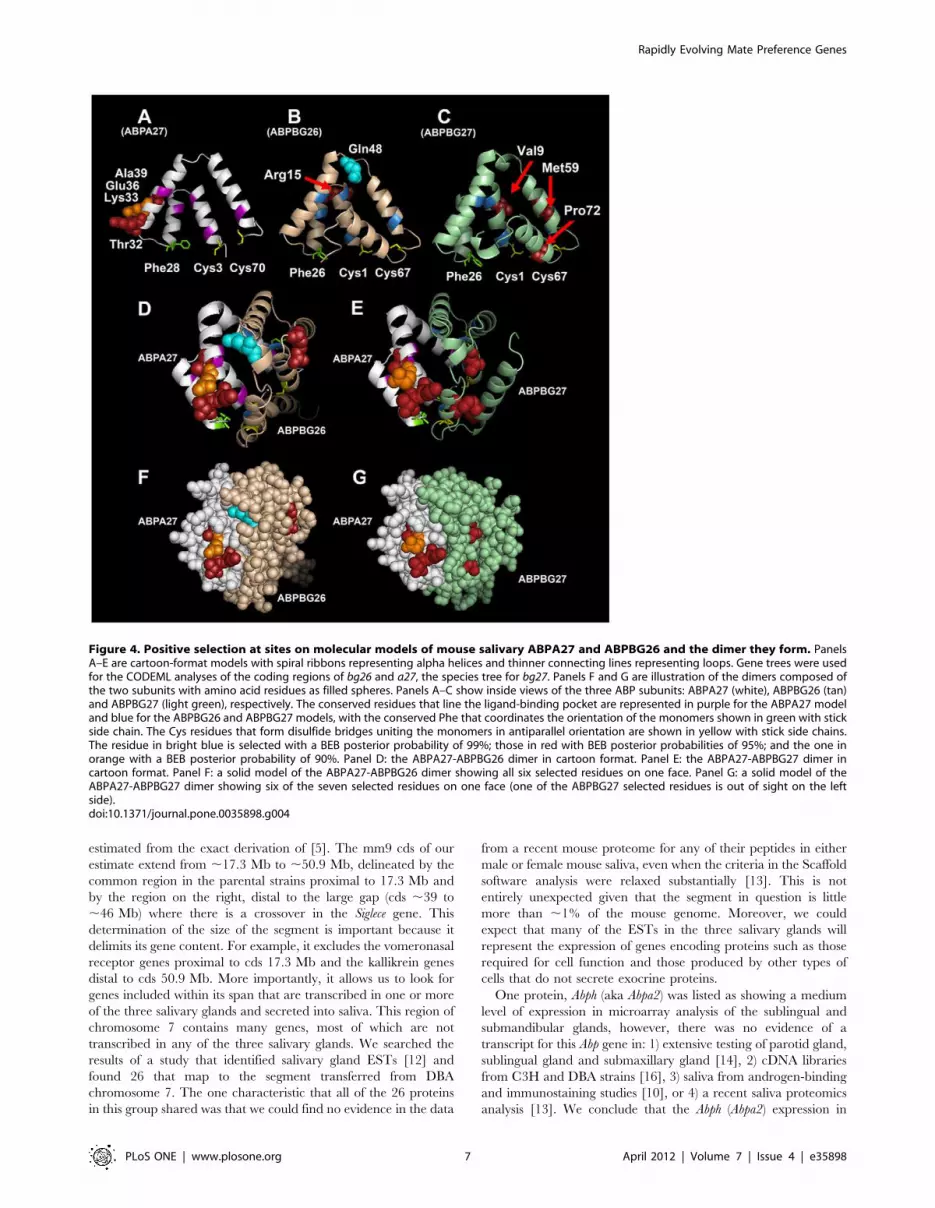
Using the Phyre2 threading program, we predicted three-
dimensional structures for the ABPA27, ABPBG26 and ABPBG27
monomers, as well as the A27-BG26 and A27-BG27 dimers they
form (Table 2). We plotted the sites selected at a BEB posterior
probability threshold of 90% on all three models with Pymol
(Fig. 4), using the sites from the more conservative gene tree
analyses for a27 and bg26, and the species tree analysis for bg27
(Table 1). All the selected sites in the three monomers fall on their
exteriors. The solid-filled models of the two dimers show that all
six selected sites fall on one face of the A27-BG26 dimer, while six
of the seven selected sites fall on the same face of the A27-BG27
dimer.
Figure 3. Gene trees for Abpa27, Abpbg26 and Abpbg27compared to the species tree of the genus Mus. Taxa used inthis study include the three subspecies of Mus musculus (M. m.domesticus, M. m. musculus and M. m. castaneus), M. spicilegus and M.spretus. The species tree is modified from [17] with the three subspeciesof Mus musculus represented as an unresolved polytomy. The genetrees for Abpa27, Abpbg26 and Abpbg27 were made by aligning thesequences of each of the five taxa in Clustal X and using the alignmentsto make neighbor-joining trees in PAUP* (see methods for details). Eachof the four trees was rooted on the M. spretus sequence. Only the genetree for Abpbg27 is congruent with the species tree. The gene trees forAbpa27 and Abpbg26 both show the M. m. domesticus sequence out ofplace relative to those of the other taxa.doi:10.1371/journal.pone.0035898.g003
Rapidly Evolving Mate Preference Genes
PLoS ONE | www.plosone.org 5 April 2012 | Volume 7 | Issue 4 | e35898

Discussion
The b-congenic strain and its counterpart, the C3H parent
strain (a-congenic) served as the foundation of a number of studies
aimed at testing the hypothesis that, given the choice between
signals from two strains with different a27 alleles on the same
genetic background, test subject mice would show a preference for
the homosubspecific one [26]. The most commonly used study
design involved two-way choice testing in a Y-maze and has
confirmed the original hypothesis in laboratory mice [6] and wild
mice [7]. Most recently provided evidence that ABP forms the
basis of incipient reinforcement on the hybrid zone where two
subspecies of M. musculus made secondary contact [8].
Why analyze a congenic strain?We undertook the project reported here to determine the
physical and genetic nature of the chromosome 7 segment
transferred from DBA onto the C3H genome to produce the b-
congenic strain. We employed the information in the mouse
genome [27], an EST database [12] and mouse saliva proteomes
[13] We wished to learn whether the results of the many
behavioral tests that relied on the b-congenic strain were the
direct affect of the a27b allele, as originally intended. The
alternative hypothesis is that they are attributable to the affect of
another, closely linked gene that is also expressed in saliva and that
differs significantly in the two parental strains. Another possibility
that must be considered is that one or more Abp genes (a27 and/or
another Abp gene) is responsible for the preference test results but
those results were also influenced by the expression of an
additional, non-Abp gene that also encodes a protein secreted into
saliva and that differed significantly in the parental strains.
At the time that we began breeding the b-congenic strain, only
three Abp genes had been predicted from biochemical and genetic
studies [10]. This affected the initial behavioral studies that used
the b-congenic strain in the mid-1990s because the assumption
behind the experimental design [26] was that the gene, then called
Abpa, that encoded the alpha subunit of ABP was the only one
encoding that subunit common to two dimeric forms found in
mouse salivas: alpha-beta and alpha-gamma (beta encoded by
Abpb and gamma by Abpg; [10]). The original three-gene
hypothesis was eventually confirmed [28], however, as that study
was in progress in 2002, the draft mouse genome sequence was
being completed [27] and it became apparent that there were
many more Abp genes than just those three. A collaboration
between the laboratories of Karn in the U.S. and Ponting in the
U.K. produced a number of papers (e.g., [14,24]) culminating in
the description of 64 Abp paralogs covering a span of 3 Mb in the
completed Abp region on chromosome 7 [11]. Although this region
encompasses ,0.1% of the mouse genome, it is still small enough
to have been included in the lower estimate (24 Mb) of the region
transferred from DBA onto the C3H genome in producing the b-
congenic strain. Thus it is possible that one or more of the other
Abp genes lying close to the selected gene, a27, contribute to the
preference of subjects in the Y-maze tests. This increased the
complexity of the question regarding just what genes are congenic
in the b-congenic strain we produced.
The size and gene content of the segment contributedby DBA to the b-congenic lineIn spite of a large region of M. m. domesticus contribution to both
strains’ chromosome 7 and a number of gaps, we were able to
narrow down the ‘‘functional’’ size of the segment to ,34 Mb.
This is not appreciably smaller than the upper value of 40 Mb
Table 1. Results of selection test on Abpa27, Abpbg26 and Abpbg27.
Using the Species Phylogeny Using the Gene Phylogeny
Gene
Ratio of dN/dS(%Codons)a
P Value All
Speciesb Codon Sites Under SelectioncRatio of dN/dS(%Codons)a
P Value All
SpeciesbCodon Sites Under
Selectionc
Abpa27 16.8 (12.7%) ,0.0002 32T, 33K, 36E, 39A 11.9 (14.6%) 0.01 32T, 33K, 36E, 39A,
Abpbg26 3.19 (25%) 0.004 5A, 7T, 11I, 14L, 15R, 21G,27Y, 39I, 44R, 48Q, 75L
12 (4.6%) 0.007 15R, 48Q
Abpbg27 6.1 (37%) 0.14 9V, 59M, 72P The gene and species phylogenies are identical for bg27
aThe dN/dS ratio of the class of codons under positive selection is given with the percentage of codon sites predicted to be in that class.bThe p-value rejecting the model of neutral evolution (M8A) over that of selection (M8) is given.cSites with posterior probabilities greater than 0.9 are indicated in regular typeface; p.0.95 indicated in bold typeface and p.0.99 indicated in bold, underlinedtypeface.doi:10.1371/journal.pone.0035898.t001
Table 2. Mouse genes used to produce molecular models.
Monomers/Dimers Accession Number Chromosomal Location (strand)
Structural Modela (% confidence; length; and
% identity)
Abpa27 GU269231 chr7:34,806,587–34,807,768 (+) c1puoA (100%; 70; 51%)
Abpbg26 AY293279 chr7:34,796,951–34,798,961 (2) c1puoA (100%; 84; 28%)
Abpbg27 AY293282 chr7:34,728,016–34,730,004 (2) c1puoA (100%; 84; 28%)
Abpa27- Abpbg26 N/A - constructed from monomers c2ejnB (100%; 136; 43%)
Abpa27- Abpbg27 N/A - constructed from monomers c2ejnB (100%; 135; 42%)
aThe secreted sequences (i.e., signal sequences removed) were threaded for this study.doi:10.1371/journal.pone.0035898.t002
Rapidly Evolving Mate Preference Genes
PLoS ONE | www.plosone.org 6 April 2012 | Volume 7 | Issue 4 | e35898
estimated from the exact derivation of [5]. The mm9 cds of our
estimate extend from ,17.3 Mb to ,50.9 Mb, delineated by the
common region in the parental strains proximal to 17.3 Mb and
by the region on the right, distal to the large gap (cds ,39 to
,46 Mb) where there is a crossover in the Siglece gene. This
determination of the size of the segment is important because it
delimits its gene content. For example, it excludes the vomeronasal
receptor genes proximal to cds 17.3 Mb and the kallikrein genes
distal to cds 50.9 Mb. More importantly, it allows us to look for
genes included within its span that are transcribed in one or more
of the three salivary glands and secreted into saliva. This region of
chromosome 7 contains many genes, most of which are not
transcribed in any of the three salivary glands. We searched the
results of a study that identified salivary gland ESTs [12] and
found 26 that map to the segment transferred from DBA
chromosome 7. The one characteristic that all of the 26 proteins
in this group shared was that we could find no evidence in the data
from a recent mouse proteome for any of their peptides in either
male or female mouse saliva, even when the criteria in the Scaffold
software analysis were relaxed substantially [13]. This is not
entirely unexpected given that the segment in question is little
more than ,1% of the mouse genome. Moreover, we could
expect that many of the ESTs in the three salivary glands will
represent the expression of genes encoding proteins such as those
required for cell function and those produced by other types of
cells that do not secrete exocrine proteins.
One protein, Abph (aka Abpa2) was listed as showing a medium
level of expression in microarray analysis of the sublingual and
submandibular glands, however, there was no evidence of a
transcript for this Abp gene in: 1) extensive testing of parotid gland,
sublingual gland and submaxillary gland [14], 2) cDNA libraries
from C3H and DBA strains [16], 3) saliva from androgen-binding
and immunostaining studies [10], or 4) a recent saliva proteomics
analysis [13]. We conclude that the Abph (Abpa2) expression in
Figure 4. Positive selection at sites on molecular models of mouse salivary ABPA27 and ABPBG26 and the dimer they form. PanelsA–E are cartoon-format models with spiral ribbons representing alpha helices and thinner connecting lines representing loops. Gene trees were usedfor the CODEML analyses of the coding regions of bg26 and a27, the species tree for bg27. Panels F and G are illustration of the dimers composed ofthe two subunits with amino acid residues as filled spheres. Panels A–C show inside views of the three ABP subunits: ABPA27 (white), ABPBG26 (tan)and ABPBG27 (light green), respectively. The conserved residues that line the ligand-binding pocket are represented in purple for the ABPA27 modeland blue for the ABPBG26 and ABPBG27 models, with the conserved Phe that coordinates the orientation of the monomers shown in green with stickside chain. The Cys residues that form disulfide bridges uniting the monomers in antiparallel orientation are shown in yellow with stick side chains.The residue in bright blue is selected with a BEB posterior probability of 99%; those in red with BEB posterior probabilities of 95%; and the one inorange with a BEB posterior probability of 90%. Panel D: the ABPA27-ABPBG26 dimer in cartoon format. Panel E: the ABPA27-ABPBG27 dimer incartoon format. Panel F: a solid model of the ABPA27-ABPBG26 dimer showing all six selected residues on one face. Panel G: a solid model of theABPA27-ABPBG27 dimer showing six of the seven selected residues on one face (one of the ABPBG27 selected residues is out of sight on the leftside).doi:10.1371/journal.pone.0035898.g004
Rapidly Evolving Mate Preference Genes
PLoS ONE | www.plosone.org 7 April 2012 | Volume 7 | Issue 4 | e35898

sublingual and submandibular glands [12] was probably a
misidentification caused by the binding of transcripts from other,
highly expressed Abp genes (see below) to the probes produced
from the Abph transcript for the study. Of the 64 Abp paralogs in
the mouse genome there is only evidence that three of them, bg26,
bg27 and a27, are transcribed in the three salivary glands [14] and
their gene products are ecreted into saliva in significant amounts
[10,13]. There is no evidence for the expression of any of the 61
other Abp paralogs.
The genes encoding the preference signal and theirevolutionary characteristicsIt appears, in light of the proteome data [13], that a27, bg26 and
bg27 are the only genes identified on the segment of chromosome 7
transferred to the b-congenic strain that could have influenced the
results of the Y-maze preference tests [6–8]. While this study
makes it unlikely that other genes may have been involved, we
cannot absolutely rule that out. Each of these three genes has a
substantial number of nonsynonymous substitutions in its coding
region. We investigated the evolutionary history of both of these
Abpbg genes by comparing their gene phylogenies and by using the
CODEML program to look for the footprints of adaptive
evolution.
Our CODEML analysis supports the notion that both subunits,
A27 and BG26, have a history of adaptive evolution driven by
positive selection on a few sites at the surface of one face of the
dimer they form. We suggest that these two genes evolved rapidly
as subunits that form one of the dimers secreted in relatively large
quantity into the salivas of mice and that their evolutionary
histories may not be independent. This is not unexpected given
that at least A27 appears to be involved in incipient reinforcement
on the hybrid zone where M. m. domesticus and M. m. musculus made
secondary contact [8] but it must do so in conjunction with at least
one BG monomer because all ABP subunits described to date are
paired in an alpha-beta/gamma dimer.
Four amino acid sites of A27 apparently evolved under positive
selection as have at least two of BG26. Whether the rapid
evolution of A27 and BG26 has been shared by the BG27 subunit
is less clear. The CODEML result for bg27 was non-significant,
however, there were three sites in the sequence of BG27 identified
as positively selected at a BEB posterior probability threshold of
.95%. As noted above, whether to accept such sites as selected,
given the non-significant CODEML result, is controversial but it is
nonetheless interesting that two of the three sites are at the surface
of one face of the dimer formed with the A27 subunit resulting in a
model that looks very similar to that of the A27-BG26 dimer
(Fig. 4).
The CODEML results for a27, bg26, and bg27 suggest that at
least A27 and BG26 may have coevolved. These are the subunits
of the same functional entity, the A27-BG26 dimer found in
mouse saliva. BG27 may have also coevolved with A27 because
these two subunits form a dimer found in mouse saliva at a level
nearly equal-molar with the A27-BG26 dimer [29]. Both of the
residues under selection in BG26, and two of the three residues
under selection in BG27, share the same exterior face of the dimer
with the four residues under selection in A27. This location of all
but one of these selected residues suggests that these ABP dimers
interact with another molecule(s). It is interesting to compare the
results we report here with those of [29] who showed that the two
different dimers, A27-BG26 and A27-BG27 bind dihydrotestos-
terone (DHT) and testosterone (T), respectively, with different
affinities. Although it is not clear how amino acid variation at sites
on an exterior face of the molecule might make this possible, it is
likely that the different binding affinities result from a synergistic
affect caused by the interaction of A27 with the BG monomer in
each of the two dimers. One possibility is that conformation of the
binding pocket depends on which BG subunit is involved, since
this pocket is created by the formation of the dimer [30]. Indeed
BG26 and BG27 differ at more amino acid sites than the ones
shown here to be evolving under positive selection [16]. Thus,
while coevolution of A27 with one or both BG26 and BG27 can
affect a surface interaction with another molecule, changes at
other amino acid sites independently, or in conjunction with the
positively selected site, can affect ligand binding.
We produced the b-congenic strain to test the hypothesis that,
given the choice between signals from two strains with different
a27 alleles on the same genetic background, the test subject mice
would show a preference for the homosubspecific one [26]. At the
time that project was undertaken, it was clear that whatever affect
A27 has on behavior, it does so as a subunit of a dimer with either
BG26 or BG27, since no free monomers have been observed in
mouse salivas [10]. Thus it is not surprising that these genes,
closely linked to a27 and sharing its evolutionary history [11],
should both also have a role in mediating mate preference
behavior. It seems evident that we can conclude this about the
A27-BG26 dimer at the very least and perhaps about the A27-
BG27 dimer as well.
Our analysis of the segment transferred from DBA in the
process of breeding the b-congenic strain reinforces and extends
the role of ABP in the many studies done with saliva from the
congenic strains. The most exciting finding, however, is that the
gene used for selection in the congenic breeding, Abpa27, encodes
only one component of the salivary proteins that influence
assortative mate choice. We have presented evidence here that
at least one, and perhaps both of the genes, Abpbg26 and Abpbg27,
encoding the other members of the two salivary ABP dimers shows
the characteristics of adaptive evolution. We suggest that the two
different ABP dimers in saliva exist to modulate the stimulus,
especially if their different affinities for the steroid ligands T and
DHT also influence mate choice.
Materials and Methods
All animal manipulation was performed humanely and under
appropriate Animal Welfare Guidelines under University of
Arizona IACUC protocol 08-138. Methods are summarized here;
details appear in File S5.
Polymerase chain reaction (PCR) was run as previously
described [14] using genomic DNAs either purchased (Jackson
Laboratory) or purified from tail tips and the products sequenced
either by the UAGC facility at the University of Arizona or by
MCLAB (http://www.mclab.com/). DNA sequence traces were
edited with Chromas 2.3 (http://www.technelysium.com.au).
DNA sequence alignment, coding region assembly, and in silico
translation were done using the DNAsis Max program 2.0
(Hitachi).
Salivary gland protein expression data sets were obtained by
searching ‘‘mouse salivary gland’’ on the NCBI Gene Expression
Omnibus website (www.ncbi.nlm.nih.gov/geo/). Data was down-
loaded and cross-referenced with Amersham Codelink UniSet
Mouse 1 Bioarray, and protein information for each probe target
was gathered by searching probe accession numbers on MGI
(http://www.informatics.jax.org/), the international database
resource for the laboratory mouse. The data in the region of
interest was also sorted on the sample signal intensity from the
array, which ranged from 0 to 250–400 in the six data sets, and
only those with values of 10 or above were retained for this study.
Rapidly Evolving Mate Preference Genes
PLoS ONE | www.plosone.org 8 April 2012 | Volume 7 | Issue 4 | e35898

Positive selection was assessed in the program CODEML in the
PAML package [31–33]. The three-dimensional structures of
mouse a27, bg26 and bg27 were modeled using the PHYRE2
(version 2.0) threading program (http://www.sbg.bio.ic.ac.uk/
phyre2/html/page.cgi?id = index; [34]), and the resulting models
were visualized using PYMOL (open-source 1.2.8; http://www.
pymol.org/). Sites under positive selection were mapped onto the
structural models using PYMOL and incorporated into a figure.
Supporting Information
Table S1 Gene information.(XLS)
Table S2 Sequence gaps (regions with low/no SNPdensity in the Mouse Diversity array and lacking UCSCgenes).(XLS)
Table S3 ESTs found in one or more of three glands ofthe two sexes of BALB/c mice [12].(DOC)
Table S4 Chromosome 7 encoded proteins found inmouse saliva proteomes [13].(XLS)
File S1 Retained and eliminated proteins (representedby ESTs and cDNAs).(DOC)
File S2 Protein sequences from Sanger Data and from
sequencing.
(TXT)
File S3 Sequences of Abp coding regions used in
CODEML analysis.
(TXT)
File S4 Unrooted trees for Abpa27, Abpbg26 and
Abpbg27. Trees were made as in Fig. 3 but were not rooted.
Rather, the suggested root is shown by a black dot on the branch
to the Mus spretus ortholog.
(TIF)
File S5 Details of Materials and Methods.
(DOC)
Acknowledgments
The authors wish to thank Willie Swanson and Gary Churchill for helpful
discussions.
Author Contributions
Conceived and designed the experiments: CML RCK. Performed the
experiments: CM RCK. Analyzed the data: CML CM RCK. Contributed
reagents/materials/analysis tools: RCK. Wrote the paper: CML CM
RCK.
References
1. Karn RC (1998) Steroid binding by mouse salivary proteins. Biochem Genet 36:
105–117.
2. Laukaitis C, Karn RC (2012) Recognition of subspecies status mediated by
androgen-binding protein (ABP) in the evolution of incipient reinforcement on
the European house mouse hybrid zone. In: Macholan M, Munclinger P,
Baird SJ, Pialek J, eds. Evolution of the House Mouse pp. In Press.
3. Karn RC, Dlouhy SR (1991) Salivary androgen-binding protein variation in
Mus and other rodents. J Hered 82: 453–458.
4. Karn RC, Orth A, Bonhomme F, Boursot P (2002) The complex history of a
gene proposed to participate in a sexual isolation mechanism in house mice. Mol
Biol Evol 19: 462–471.
5. Naveira H, Barbadilla A (1992) The theoretical distribution of lengths of intact
chromosome segments around a locus held heterozygous with backcrossing in a
diploid species. Genetics 130: 205–209.
6. Talley HM, Laukaitis CM, Karn RC (2001) Female preference for male saliva:
implications for sexual isolation of Mus musculus subspecies. Evolution 55:
631–634.
7. Bımova B, Karn RC, Pialek J (2005) The role of salivary androgen-binding
protein in reproductive isolation between two subspecies of house mouse: Mus
musculus musculus and Mus musculus domesticus. Biological Journal of the
Linnean Society 84: 349–361.
8. Voslajerova Bımova B, Macholan M, Baird SEB, Munclinger P, Laukaitis CM,
et al. (2011) Reinforcement selection acting on the European house mouse
hybrid zone. Mol Ecol 20: 2403–2424.
9. Dlouhy SR, Nichols WC, Karn RC (1986) Production of an antibody to mouse
salivary androgen binding protein (ABP) and its use in identifying a prostate
protein produced by a gene distinct from Abp. Biochem Genet 24: 743–763.
10. Dlouhy SR, Taylor BA, Karn RC (1987) The genes for mouse salivary
androgen-binding protein (ABP) subunits alpha and gamma are located on
chromosome 7. Genetics 115: 535–543.
11. Laukaitis CM, Heger A, Blakley TD, Munclinger P, Ponting CP, et al. (2008)
Rapid bursts of androgen-binding protein (Abp) gene duplication occurred
independently in diverse mammals. BMC Evol Biol 8: 46.
12. Treister NS, Richards SM, Lombardi MJ, Rowley P, Jensen RV, et al. (2005)
Sex-related differences in gene expression in salivary glands of BALB/c mice.
Journal of Dental Research 84: 160–165.
13. Karn RC, Laukaitis CM (2011) Positive selection shaped the convergent
evolution of independently expanded kallikrein subfamilies expressed in mouse
and rat saliva proteomes. PLoS ONE 6: e20979.
14. Laukaitis CM, Dlouhy SR, Emes RD, Ponting CP, Karn RC (2005) Diverse
spatial, temporal, and sexual expression of recently duplicated androgen-binding
protein genes in Mus musculus. BMC Evol Biol 5: 40.
15. Hwang JM, Hofstetter JR, Bonhomme F, Karn RC (1997) The microevolution
of mouse salivary androgen-binding protein (ABP) paralleled subspeciation of
Mus musculus. J Hered 88: 93–97.
16. Karn RC, Laukaitis CM (2003) Characterization of two forms of mouse salivary
androgen-binding protein (ABP): implications for evolutionary relationships and
ligand-binding function. Biochemistry 42: 7162–7170.
17. Chevret P, Veyrunes F, Britton-Davidian J (2005) Molecular phylogeny of the
genus Mus (Rodentia:Murinae) based on mitochondrial and nuclear data.
Biol J Linn Soc 84: 417–427.
18. Karn RC, Clark NL, Nguyen ED, Swanson WJ (2008) Adaptive evolution in
rodent seminal vesicle secretion proteins. Mol Biol Evol 25: 2301–2310.
19. Karn RC, Young JM, Laukaitis CM (2010) A candidate subspecies
discrimination system involving a vomeronasal receptor gene with different
alleles fixed in M. m. domesticus and M. m. musculus. PLoS ONE 5: e12638.
20. She JX, Bonhomme F, Boursot P, Thaler L, Catzeflis F (1990) Molecular
Phylogenies in the Genus Mus - Comparative-Analysis of Electrophoretic,
Scndna Hybridization, and Mtdna Rflp Data. Biological Journal of the Linnean
Society 41: 83–103.
21. Suzuki H, Shimada T, Terashima M, Tsuchiya K, Aplin K (2004) Temporal,
spatial, and ecological modes of evolution of Eurasian Mus based on
mitochondrial and nuclear gene sequences. Molecular Phylogenetics and
Evolution 33: 626–646.
22. Tucker PK, Sandstedt SA, Lundrigan BL (2005) Phylogenetic relationships in
the subgenus Mus (genus Mus, family Muridae, subfamily Murinae): examining
gene trees and species trees. Biological Journal of the Linnean Society 84:
653–662.
23. Yang Z, Wong WS, Nielsen R (2005) Bayes empirical bayes inference of amino
acid sites under positive selection. Mol Biol Evol 22: 1107–1118.
24. Emes RD, Riley MC, Laukaitis CM, Goodstadt L, Karn RC, et al. (2004)
Comparative evolutionary genomics of androgen-binding protein genes.
Genome Res 14: 1516–1529.
25. Anisimova M, Bielawski JP, Yang Z (2002) Accuracy and power of bayes
prediction of amino acid sites under positive selection. Mol Biol Evol 19:
950–958.
26. Laukaitis CM, Critser ES, Karn RC (1997) Salivary androgen-binding protein
(ABP) mediates sexual isolation in Mus musculus. Evolution 51: 2000–2005.
27. Waterston RH, Lindblad-Toh K, Birney E, Rogers J, Abril JF, et al. (2002)
Initial sequencing and comparative analysis of the mouse genome. Nature 420:
520–562.
28. Laukaitis CM, Dlouhy SR, Karn RC (2003) The mouse salivary androgen-
binding protein (ABP) gene cluster on Chromosomes 7: characterization and
evolutionary relationships. Mamm Genome 14: 679–691.
Rapidly Evolving Mate Preference Genes
PLoS ONE | www.plosone.org 9 April 2012 | Volume 7 | Issue 4 | e35898

29. Karn RC, Clements MA (1999) A comparison of the structures of the alpha:betaand alpha:gamma dimers of mouse salivary androgen-binding protein (ABP) andtheir differential steroid binding. Biochem Genet 37: 187–199.
30. Callebaut I, Poupon A, Bally R, Demaret JP, Housset D, et al. (2000) Theuteroglobin fold. Ann N Y Acad Sci 923: 90–112.
31. Nielsen R, Yang Z (1998) Likelihood models for detecting positively selectedamino acid sites and applications to the HIV-1 envelope gene. Genetics 148:929–936.
32. Yang Z (1997) PAML: a program package for phylogenetic analysis by
maximum likelihood. Comput Appl Biosci 13: 555–556.
33. Yang Z, Swanson WJ, Vacquier VD (2000) Maximum-likelihood analysis of
molecular adaptation in abalone sperm lysin reveals variable selective pressures
among lineages and sites. Mol Biol Evol 17: 1446–1455.
34. Kelley LA, Sternberg MJ (2009) Protein structure prediction on the Web: a case
study using the Phyre server. Nat Protoc 4: 363–371.
Rapidly Evolving Mate Preference Genes
PLoS ONE | www.plosone.org 10 April 2012 | Volume 7 | Issue 4 | e35898
Copyright © 2022 FDOKUMEN